Notice (8): Undefined variable: solution_of_interest [APP/View/Products/view.ctp, line 755]Code Context<!-- BEGIN: REQUEST_FORM MODAL -->
<div id="request_formModal" class="reveal-modal medium" data-reveal aria-labelledby="modalTitle" aria-hidden="true" role="dialog">
<?= $this->element('Forms/simple_form', array('solution_of_interest' => $solution_of_interest, 'header' => $header, 'message' => $message, 'campaign_id' => $campaign_id)) ?>
$viewFile = '/home/website-server/www/app/View/Products/view.ctp'
$dataForView = array(
'language' => 'jp',
'meta_keywords' => '',
'meta_description' => '',
'meta_title' => '',
'product' => array(
'Product' => array(
'id' => '3186',
'antibody_id' => '644',
'name' => 'H3K9me3 Antibody - ChIP-seq Grade (sample size)',
'description' => '<p class="p1">Polyclonal antibody raised in rabbit against the region of histone H3 containing the trimethylated lysine 9 (H3K9me3), using a KLH-conjugated synthetic peptide.</p>',
'label1' => 'Validation Data',
'info1' => '<div class="row">
<div class="small-6 columns"><center>A.<img src="https://www.diagenode.com/img/product/antibodies/C15410193-ChIP-Fig1-10.png" width="300" /></center><center>B.<img src="https://www.diagenode.com/img/product/antibodies/C15410193-ChIP-Fig1b-10.png" width="300" /></center></div>
<div class="small-6 columns">
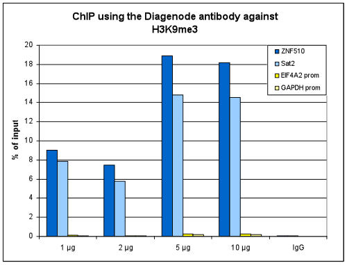
<p><small><strong>Figure 1. ChIP results obtained with the Diagenode antibody directed against H3K9me3</strong><br />ChIP assays were performed using human HeLa cells, the Diagenode antibody against H3K9me3 (Cat. No. C15410193) and optimized PCR primer pairs for qPCR. ChIP was performed with the "iDeal ChIP-seq" kit (Cat. No. C01010051), using sheared chromatin from 1,000,000 cells. A titration consisting of 1, 2, 5 and 10 μg of antibody per ChIP experiment was analyzed. IgG (2 μg/IP) was used as a negative IP control. Quantitative PCR was performed with primers specific for the promoter of the active genes GAPDH and EIF4A2, used as negative controls, and for ZNF510 and the Sat2 satellite repeat, used as positive controls. The figure shows the recovery, expressed as a % of input (the relative amount of immunoprecipitated DNA compared to input DNA after qPCR analysis).</small></p>
</div>
</div>
<div class="row">
<div class="small-12 columns"><center>A.<img src="https://www.diagenode.com/img/product/antibodies/C15410193-ChIP-Fig2a-10.png" width="600" /></center><center>B.<img src="https://www.diagenode.com/img/product/antibodies/C15410193-ChIP-Fig2b-10.png" width="600" /></center><center>C.<img src="https://www.diagenode.com/img/product/antibodies/C15410193-ChIP-Fig2c-10.png" width="600" /></center></div>
</div>
<div class="row">
<div class="small-12 columns">
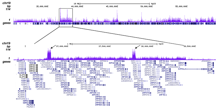
<p><small><strong>Figure 2. ChIP-seq results obtained with the Diagenode antibody directed against H3K9me3</strong><br />ChIP was performed with 1 μg of the Diagenode antibody against H3K9me3 (Cat. No. C15410193) on sheared chromatin from 1,000,000 HeLa cells using the “iDeal ChIP-seq” kit as described above. The IP'd DNA was subsequently analysed on an Illumina HiSeq 2000. Library preparation, cluster generation and sequencing were performed according to the manufacturer's instructions. The 50 bp tags were aligned to the human genome using the BWA algorithm. Figure 2A shows the signal distribution along the long arm of chromosome 19 and a zoomin to an enriched region containing several ZNF repeat genes. The arrows indicate two satellite repeat regions which exhibit a stronger signal. Figure 2C and D show the enrichment at the KCNQ1 and H19 imprinted genes.</small></p>
</div>
</div>
<div class="row">
<div class="small-6 columns"><center><img src="https://www.diagenode.com/img/product/antibodies/C15410193-Elisa-Fig3-10.png" width="250" /></center></div>
<div class="small-6 columns">
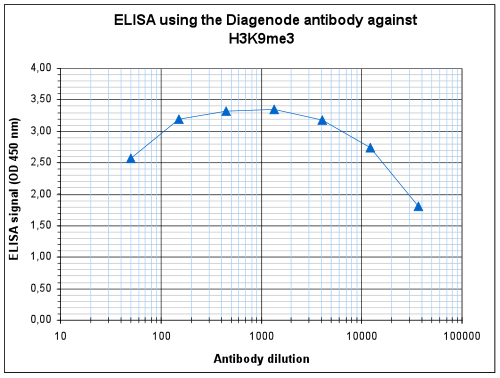
<p><small><strong>Figure 3. Determination of the antibody titer</strong><br />To determine the titer of the antibody, an ELISA was performed using a serial dilution of the Diagenode antibody against H3K9me3 (Cat. No. C15410193). The antigen used was a peptide containing the histone modification of interest. By plotting the absorbance against the antibody dilution (Figure 3), the titer of the antibody was estimated to be 1:42,700.</small></p>
</div>
</div>
<div class="row">
<div class="small-6 columns"><center>A.<img src="https://www.diagenode.com/img/product/antibodies/C15410193-DotBlot-Fig4-10.png" width="300" /></center><center>B.<img src="https://www.diagenode.com/img/product/antibodies/C15410193-DotBlot-Fig4b-10.png" width="300" /></center></div>
<div class="small-6 columns">
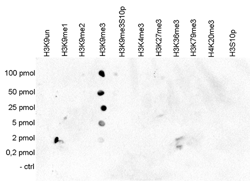
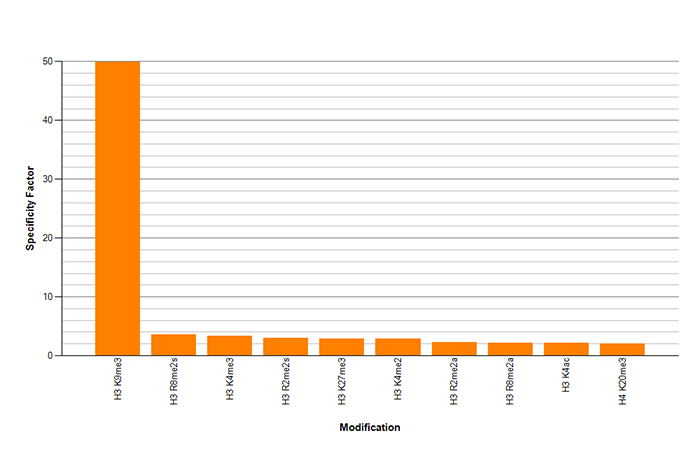
<p><small><strong>Figure 4. Cross reactivity tests using the Diagenode antibody directed against H3K9me3</strong><br />Figure 4A To test the cross reactivity of the Diagenode antibody against H3K9me3 (Cat. No. C15410193), a Dot Blot analysis was performed with peptides containing other histone modifications and the unmodified H3K9. One hundred to 0.2 pmol of the respective peptides were spotted on a membrane. The antibody was used at a dilution of 1:20,000. Figure 4A shows a high specificity of the antibody for the modification of interest. Figure 4B The specificity of the antibody was further demonstrated by peptide array analyses on an array containing 384 peptides with different combinations of modifications from histone H3, H4, H2A and H2B. The antibody was used at a dilution of 1:5,000. Figure 4B shows the specificity factor, calculated as the ratio of the average intensity of all spots containing the mark, divided by the average intensity of all spots not containing the mark.</small></p>
</div>
</div>
<div class="row">
<div class="small-4 columns"><center><img src="https://www.diagenode.com/img/product/antibodies/C15410193-WB-Fig5-10.png" /></center></div>
<div class="small-8 columns">
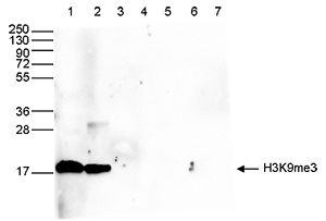
<p><small><strong>Figure 5. Western blot analysis using the Diagenode antibody directed against H3K9me3</strong><br />Western blot was performed on whole cell (50 μg, lane 1) and histone extracts (15 μg, lane 2) from HeLa cells, and on 1 μg of recombinant histone H2A, H2B, H3.1, H3.2 and H4 (lane 3, 4, 5, 6 and 7, respectively) using the Diagenode antibody against H3K9me3 (Cat. No. C15410193). The antibody was diluted 1:1,000 in TBSTween containing 5% skimmed milk. The position of the protein of interest is indicated on the right, the marker (in kDa) is shown on the left.</small></p>
</div>
</div>
<div class="row">
<div class="small-12 columns"><center><img src="https://www.diagenode.com/img/product/antibodies/C15410193-IF-Fig6-10.png" /></center></div>
</div>
<div class="row">
<div class="small-12 columns">
<p><small><strong>Figure 6. Immunofluorescence using the Diagenode antibody directed against H3K9me3</strong><br />HeLa cells were stained with the Diagenode antibody against H3K9me3 (Cat. No. C15410193) and with DAPI. Cells were fixed with 4% formaldehyde for 10’ and blocked with PBS/TX-100 containing 5% normal goat serum and 1% BSA. The cells were immunofluorescently labeled with the H3K9me3 antibody (left) diluted 1:250 in blocking solution followed by an anti-rabbit antibody conjugated to Alexa488. The middle panel shows staining of the nuclei with DAPI. A merge of the two stainings is shown on the right.</small></p>
</div>
</div>',
'label2' => 'Target Description',
'info2' => '<p>Histones are the main constituents of the protein part of chromosomes of eukaryotic cells. They are rich in the amino acids arginine and lysine and have been greatly conserved during evolution. Histones pack the DNA into tight masses of chromatin. Two core histones of each class H2A, H2B, H3 and H4 assemble and are wrapped by 146 base pairs of DNA to form one octameric nucleosome. Histone tails undergo numerous post-translational modifications, which either directly or indirectly alter chromatin structure to facilitate transcriptional activation or repression or other nuclear processes. In addition to the genetic code, combinations of the different histone modifications reveal the so-called “histone code”. Histone methylation and demethylation is dynamically regulated by respectively histone methyl transferases and histone demethylases. Trimethylation of histone H3K9 is associated with inactive genomic regions, satellite repeats and ZNF gene repeats.</p>',
'label3' => '',
'info3' => '',
'format' => '10 µg',
'catalog_number' => 'C15410193-10',
'old_catalog_number' => 'pAb-193-050',
'sf_code' => 'C15410193-D001-000582',
'type' => 'FRE',
'search_order' => '03-Antibody',
'price_EUR' => '125',
'price_USD' => '115',
'price_GBP' => '115',
'price_JPY' => '19580',
'price_CNY' => '15000',
'price_AUD' => '288',
'country' => 'ALL',
'except_countries' => 'None',
'quote' => false,
'in_stock' => false,
'featured' => false,
'no_promo' => false,
'online' => true,
'master' => false,
'last_datasheet_update' => '',
'slug' => 'h3k9me3-polyclonal-antibody-10',
'meta_title' => '',
'meta_keywords' => '',
'meta_description' => '',
'modified' => '2021-10-20 09:56:13',
'created' => '2021-03-03 16:16:04',
'locale' => 'jpn'
),
'Antibody' => array(
'host' => '*****',
'id' => '644',
'name' => 'H3K9me3 Antibody - ChIP-seq Grade (sample size)',
'description' => 'Polyclonal antibody raised in rabbit against the region of histone H3 containing the trimethylated lysine 9
(H3K9me3), using a KLH-conjugated synthetic peptide.',
'clonality' => '',
'isotype' => '',
'lot' => 'A0219P',
'concentration' => '0.9 µg/µl',
'reactivity' => 'Human, mouse, yeast, wide range expected.',
'type' => 'Polyclonal, <strong>ChIP grade, ChIP-seq grade</strong>',
'purity' => 'Affinity purified polyclonal antibody',
'classification' => '',
'application_table' => '<table>
<thead>
<tr>
<th>Applications</th>
<th>Suggested dilution</th>
<th>References</th>
</tr>
</thead>
<tbody>
<tr>
<td>ChIP/ChIP-seq <sup>*</sup></td>
<td>1 µg/ChIP</td>
<td>Fig 1, 2</td>
</tr>
<tr>
<td>ELISA</td>
<td>1:1000</td>
<td>Fig 3</td>
</tr>
<tr>
<td>Dot Blotting/Peptide array</td>
<td>1:20,000/1:5,000</td>
<td>Fig 4</td>
</tr>
<tr>
<td>Western Blotting</td>
<td>1:1,000</td>
<td>Fig 5</td>
</tr>
<tr>
<td>Immunofluorescence</td>
<td>1:250</td>
<td>Fig 6</td>
</tr>
</tbody>
</table>
<p></p>
<p><small>*Please note that the optimal antibody amount per IP should be determined by the end-user. We recommend testing 0.5 - 5 µg per IP.</small></p>',
'storage_conditions' => 'Store at -20°C; for long storage, store at -80°C. Avoid multiple freeze-thaw cycles.',
'storage_buffer' => 'PBS containing 0.05% azide and 0.05% ProClin 300.',
'precautions' => 'This product is for research use only. Not for use in diagnostic or therapeutic procedures.',
'uniprot_acc' => '',
'slug' => 'h3k9me3-polyclonal-antibody-10',
'meta_keywords' => '',
'meta_description' => '',
'modified' => '2021-03-03 15:53:25',
'created' => '2021-03-03 15:53:25',
'select_label' => '644 - H3K9me3 Antibody - ChIP-seq Grade (sample size) (A0219P - 0.9 µg/µl - Human, mouse, yeast, wide range expected. - Affinity purified polyclonal antibody - Rabbit)'
),
'Slave' => array(),
'Group' => array(
'Group' => array(
[maximum depth reached]
),
'Master' => array(
[maximum depth reached]
),
'Product' => array(
[maximum depth reached]
)
),
'Related' => array(),
'Application' => array(
(int) 0 => array(
[maximum depth reached]
),
(int) 1 => array(
[maximum depth reached]
),
(int) 2 => array(
[maximum depth reached]
),
(int) 3 => array(
[maximum depth reached]
),
(int) 4 => array(
[maximum depth reached]
),
(int) 5 => array(
[maximum depth reached]
),
(int) 6 => array(
[maximum depth reached]
)
),
'Category' => array(
(int) 0 => array(
[maximum depth reached]
),
(int) 1 => array(
[maximum depth reached]
),
(int) 2 => array(
[maximum depth reached]
),
(int) 3 => array(
[maximum depth reached]
),
(int) 4 => array(
[maximum depth reached]
)
),
'Document' => array(
(int) 0 => array(
[maximum depth reached]
),
(int) 1 => array(
[maximum depth reached]
),
(int) 2 => array(
[maximum depth reached]
)
),
'Feature' => array(),
'Image' => array(
(int) 0 => array(
[maximum depth reached]
)
),
'Promotion' => array(),
'Protocol' => array(),
'Publication' => array(
(int) 0 => array(
[maximum depth reached]
),
(int) 1 => array(
[maximum depth reached]
),
(int) 2 => array(
[maximum depth reached]
),
(int) 3 => array(
[maximum depth reached]
),
(int) 4 => array(
[maximum depth reached]
),
(int) 5 => array(
[maximum depth reached]
),
(int) 6 => array(
[maximum depth reached]
),
(int) 7 => array(
[maximum depth reached]
),
(int) 8 => array(
[maximum depth reached]
),
(int) 9 => array(
[maximum depth reached]
),
(int) 10 => array(
[maximum depth reached]
),
(int) 11 => array(
[maximum depth reached]
),
(int) 12 => array(
[maximum depth reached]
),
(int) 13 => array(
[maximum depth reached]
),
(int) 14 => array(
[maximum depth reached]
),
(int) 15 => array(
[maximum depth reached]
),
(int) 16 => array(
[maximum depth reached]
),
(int) 17 => array(
[maximum depth reached]
),
(int) 18 => array(
[maximum depth reached]
),
(int) 19 => array(
[maximum depth reached]
),
(int) 20 => array(
[maximum depth reached]
)
),
'Testimonial' => array(
(int) 0 => array(
[maximum depth reached]
)
),
'Area' => array(),
'SafetySheet' => array()
),
'meta_canonical' => 'https://www.diagenode.com/jp/p/h3k9me3-polyclonal-antibody-premium-50-mg'
)
$language = 'jp'
$meta_keywords = ''
$meta_description = ''
$meta_title = ''
$product = array(
'Product' => array(
'id' => '3186',
'antibody_id' => '644',
'name' => 'H3K9me3 Antibody - ChIP-seq Grade (sample size)',
'description' => '<p class="p1">Polyclonal antibody raised in rabbit against the region of histone H3 containing the trimethylated lysine 9 (H3K9me3), using a KLH-conjugated synthetic peptide.</p>',
'label1' => 'Validation Data',
'info1' => '<div class="row">
<div class="small-6 columns"><center>A.<img src="https://www.diagenode.com/img/product/antibodies/C15410193-ChIP-Fig1-10.png" width="300" /></center><center>B.<img src="https://www.diagenode.com/img/product/antibodies/C15410193-ChIP-Fig1b-10.png" width="300" /></center></div>
<div class="small-6 columns">
<p><small><strong>Figure 1. ChIP results obtained with the Diagenode antibody directed against H3K9me3</strong><br />ChIP assays were performed using human HeLa cells, the Diagenode antibody against H3K9me3 (Cat. No. C15410193) and optimized PCR primer pairs for qPCR. ChIP was performed with the "iDeal ChIP-seq" kit (Cat. No. C01010051), using sheared chromatin from 1,000,000 cells. A titration consisting of 1, 2, 5 and 10 μg of antibody per ChIP experiment was analyzed. IgG (2 μg/IP) was used as a negative IP control. Quantitative PCR was performed with primers specific for the promoter of the active genes GAPDH and EIF4A2, used as negative controls, and for ZNF510 and the Sat2 satellite repeat, used as positive controls. The figure shows the recovery, expressed as a % of input (the relative amount of immunoprecipitated DNA compared to input DNA after qPCR analysis).</small></p>
</div>
</div>
<div class="row">
<div class="small-12 columns"><center>A.<img src="https://www.diagenode.com/img/product/antibodies/C15410193-ChIP-Fig2a-10.png" width="600" /></center><center>B.<img src="https://www.diagenode.com/img/product/antibodies/C15410193-ChIP-Fig2b-10.png" width="600" /></center><center>C.<img src="https://www.diagenode.com/img/product/antibodies/C15410193-ChIP-Fig2c-10.png" width="600" /></center></div>
</div>
<div class="row">
<div class="small-12 columns">
<p><small><strong>Figure 2. ChIP-seq results obtained with the Diagenode antibody directed against H3K9me3</strong><br />ChIP was performed with 1 μg of the Diagenode antibody against H3K9me3 (Cat. No. C15410193) on sheared chromatin from 1,000,000 HeLa cells using the “iDeal ChIP-seq” kit as described above. The IP'd DNA was subsequently analysed on an Illumina HiSeq 2000. Library preparation, cluster generation and sequencing were performed according to the manufacturer's instructions. The 50 bp tags were aligned to the human genome using the BWA algorithm. Figure 2A shows the signal distribution along the long arm of chromosome 19 and a zoomin to an enriched region containing several ZNF repeat genes. The arrows indicate two satellite repeat regions which exhibit a stronger signal. Figure 2C and D show the enrichment at the KCNQ1 and H19 imprinted genes.</small></p>
</div>
</div>
<div class="row">
<div class="small-6 columns"><center><img src="https://www.diagenode.com/img/product/antibodies/C15410193-Elisa-Fig3-10.png" width="250" /></center></div>
<div class="small-6 columns">
<p><small><strong>Figure 3. Determination of the antibody titer</strong><br />To determine the titer of the antibody, an ELISA was performed using a serial dilution of the Diagenode antibody against H3K9me3 (Cat. No. C15410193). The antigen used was a peptide containing the histone modification of interest. By plotting the absorbance against the antibody dilution (Figure 3), the titer of the antibody was estimated to be 1:42,700.</small></p>
</div>
</div>
<div class="row">
<div class="small-6 columns"><center>A.<img src="https://www.diagenode.com/img/product/antibodies/C15410193-DotBlot-Fig4-10.png" width="300" /></center><center>B.<img src="https://www.diagenode.com/img/product/antibodies/C15410193-DotBlot-Fig4b-10.png" width="300" /></center></div>
<div class="small-6 columns">
<p><small><strong>Figure 4. Cross reactivity tests using the Diagenode antibody directed against H3K9me3</strong><br />Figure 4A To test the cross reactivity of the Diagenode antibody against H3K9me3 (Cat. No. C15410193), a Dot Blot analysis was performed with peptides containing other histone modifications and the unmodified H3K9. One hundred to 0.2 pmol of the respective peptides were spotted on a membrane. The antibody was used at a dilution of 1:20,000. Figure 4A shows a high specificity of the antibody for the modification of interest. Figure 4B The specificity of the antibody was further demonstrated by peptide array analyses on an array containing 384 peptides with different combinations of modifications from histone H3, H4, H2A and H2B. The antibody was used at a dilution of 1:5,000. Figure 4B shows the specificity factor, calculated as the ratio of the average intensity of all spots containing the mark, divided by the average intensity of all spots not containing the mark.</small></p>
</div>
</div>
<div class="row">
<div class="small-4 columns"><center><img src="https://www.diagenode.com/img/product/antibodies/C15410193-WB-Fig5-10.png" /></center></div>
<div class="small-8 columns">
<p><small><strong>Figure 5. Western blot analysis using the Diagenode antibody directed against H3K9me3</strong><br />Western blot was performed on whole cell (50 μg, lane 1) and histone extracts (15 μg, lane 2) from HeLa cells, and on 1 μg of recombinant histone H2A, H2B, H3.1, H3.2 and H4 (lane 3, 4, 5, 6 and 7, respectively) using the Diagenode antibody against H3K9me3 (Cat. No. C15410193). The antibody was diluted 1:1,000 in TBSTween containing 5% skimmed milk. The position of the protein of interest is indicated on the right, the marker (in kDa) is shown on the left.</small></p>
</div>
</div>
<div class="row">
<div class="small-12 columns"><center><img src="https://www.diagenode.com/img/product/antibodies/C15410193-IF-Fig6-10.png" /></center></div>
</div>
<div class="row">
<div class="small-12 columns">
<p><small><strong>Figure 6. Immunofluorescence using the Diagenode antibody directed against H3K9me3</strong><br />HeLa cells were stained with the Diagenode antibody against H3K9me3 (Cat. No. C15410193) and with DAPI. Cells were fixed with 4% formaldehyde for 10’ and blocked with PBS/TX-100 containing 5% normal goat serum and 1% BSA. The cells were immunofluorescently labeled with the H3K9me3 antibody (left) diluted 1:250 in blocking solution followed by an anti-rabbit antibody conjugated to Alexa488. The middle panel shows staining of the nuclei with DAPI. A merge of the two stainings is shown on the right.</small></p>
</div>
</div>',
'label2' => 'Target Description',
'info2' => '<p>Histones are the main constituents of the protein part of chromosomes of eukaryotic cells. They are rich in the amino acids arginine and lysine and have been greatly conserved during evolution. Histones pack the DNA into tight masses of chromatin. Two core histones of each class H2A, H2B, H3 and H4 assemble and are wrapped by 146 base pairs of DNA to form one octameric nucleosome. Histone tails undergo numerous post-translational modifications, which either directly or indirectly alter chromatin structure to facilitate transcriptional activation or repression or other nuclear processes. In addition to the genetic code, combinations of the different histone modifications reveal the so-called “histone code”. Histone methylation and demethylation is dynamically regulated by respectively histone methyl transferases and histone demethylases. Trimethylation of histone H3K9 is associated with inactive genomic regions, satellite repeats and ZNF gene repeats.</p>',
'label3' => '',
'info3' => '',
'format' => '10 µg',
'catalog_number' => 'C15410193-10',
'old_catalog_number' => 'pAb-193-050',
'sf_code' => 'C15410193-D001-000582',
'type' => 'FRE',
'search_order' => '03-Antibody',
'price_EUR' => '125',
'price_USD' => '115',
'price_GBP' => '115',
'price_JPY' => '19580',
'price_CNY' => '15000',
'price_AUD' => '288',
'country' => 'ALL',
'except_countries' => 'None',
'quote' => false,
'in_stock' => false,
'featured' => false,
'no_promo' => false,
'online' => true,
'master' => false,
'last_datasheet_update' => '',
'slug' => 'h3k9me3-polyclonal-antibody-10',
'meta_title' => '',
'meta_keywords' => '',
'meta_description' => '',
'modified' => '2021-10-20 09:56:13',
'created' => '2021-03-03 16:16:04',
'locale' => 'jpn'
),
'Antibody' => array(
'host' => '*****',
'id' => '644',
'name' => 'H3K9me3 Antibody - ChIP-seq Grade (sample size)',
'description' => 'Polyclonal antibody raised in rabbit against the region of histone H3 containing the trimethylated lysine 9
(H3K9me3), using a KLH-conjugated synthetic peptide.',
'clonality' => '',
'isotype' => '',
'lot' => 'A0219P',
'concentration' => '0.9 µg/µl',
'reactivity' => 'Human, mouse, yeast, wide range expected.',
'type' => 'Polyclonal, <strong>ChIP grade, ChIP-seq grade</strong>',
'purity' => 'Affinity purified polyclonal antibody',
'classification' => '',
'application_table' => '<table>
<thead>
<tr>
<th>Applications</th>
<th>Suggested dilution</th>
<th>References</th>
</tr>
</thead>
<tbody>
<tr>
<td>ChIP/ChIP-seq <sup>*</sup></td>
<td>1 µg/ChIP</td>
<td>Fig 1, 2</td>
</tr>
<tr>
<td>ELISA</td>
<td>1:1000</td>
<td>Fig 3</td>
</tr>
<tr>
<td>Dot Blotting/Peptide array</td>
<td>1:20,000/1:5,000</td>
<td>Fig 4</td>
</tr>
<tr>
<td>Western Blotting</td>
<td>1:1,000</td>
<td>Fig 5</td>
</tr>
<tr>
<td>Immunofluorescence</td>
<td>1:250</td>
<td>Fig 6</td>
</tr>
</tbody>
</table>
<p></p>
<p><small>*Please note that the optimal antibody amount per IP should be determined by the end-user. We recommend testing 0.5 - 5 µg per IP.</small></p>',
'storage_conditions' => 'Store at -20°C; for long storage, store at -80°C. Avoid multiple freeze-thaw cycles.',
'storage_buffer' => 'PBS containing 0.05% azide and 0.05% ProClin 300.',
'precautions' => 'This product is for research use only. Not for use in diagnostic or therapeutic procedures.',
'uniprot_acc' => '',
'slug' => 'h3k9me3-polyclonal-antibody-10',
'meta_keywords' => '',
'meta_description' => '',
'modified' => '2021-03-03 15:53:25',
'created' => '2021-03-03 15:53:25',
'select_label' => '644 - H3K9me3 Antibody - ChIP-seq Grade (sample size) (A0219P - 0.9 µg/µl - Human, mouse, yeast, wide range expected. - Affinity purified polyclonal antibody - Rabbit)'
),
'Slave' => array(),
'Group' => array(
'Group' => array(
'id' => '347',
'name' => 'C15410193',
'product_id' => '2264',
'modified' => '2021-03-03 16:28:34',
'created' => '2021-03-03 16:25:55'
),
'Master' => array(
'id' => '2264',
'antibody_id' => '121',
'name' => 'H3K9me3 Antibody',
'description' => '<p><span>Polyclonal antibody raised in rabbit against the region of histone<strong> H3 containing the trimethylated lysine 9</strong> (<strong>H3K9me3</strong>), using a KLH-conjugated synthetic peptide.</span></p>',
'label1' => 'Validation Data',
'info1' => '<div class="row">
<div class="small-6 columns"><center><img src="https://www.diagenode.com/img/product/antibodies/C15410193-ChIP-Fig1.png" /></center></div>
<div class="small-6 columns">
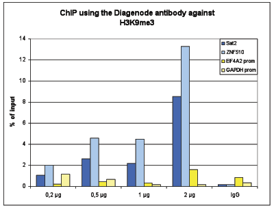
<p><small><strong>Figure 1. ChIP results obtained with the Diagenode antibody directed against H3K9me3</strong><br />ChIP assays were performed using human HeLa cells, the Diagenode antibody against H3K9me3 (cat. No. C15410193) and optimized PCR primer sets for qPCR. ChIP was performed on sheared chromatin from 1 million HeLaS3 cells using the “iDeal ChIP-seq” kit (cat. No. C01010051). A titration of the antibody consisting of 0.5, 1, 2, and 5 µg per ChIP experiment was analysed. IgG (1 µg/IP) was used as negative IP control. QPCR was performed with primers for the heterochromatin marker Sat2 and for the ZNF510 gene, used as positive controls, and for the promoters of the active EIF4A2 and GAPDH genes, used as negative controls. Figure 1 shows the recovery, expressed as a % of input (the relative amount of immunoprecipitated DNA compared to input DNA after qPCR analysis).</small></p>
</div>
</div>
<div class="row">
<div class="small-12 columns"><center>A.<img src="https://www.diagenode.com/img/product/antibodies/C15410193-ChIP-Fig2a.png" width="700" /></center><center>B.<img src="https://www.diagenode.com/img/product/antibodies/C15410193-ChIP-Fig2b.png" width="700" /></center><center>C.<img src="https://www.diagenode.com/img/product/antibodies/C15410193-ChIP-Fig2c.png" width="700" /></center><center>D.<img src="https://www.diagenode.com/img/product/antibodies/C15410193-ChIP-Fig2d.png" width="700" /></center></div>
</div>
<div class="row">
<div class="small-12 columns">
<p><small><strong>Figure 2. ChIP-seq results obtained with the Diagenode antibody directed against H3K9me3</strong><br />ChIP was performed with 0.5 µg of the Diagenode antibody against H3K9me3 (cat. No. C15410193) on sheared chromatin from 1,000,000 HeLa cells using the “iDeal ChIP-seq” kit as described above. The IP'd DNA was subsequently analysed on an Illumina HiSeq 2000. Library preparation, cluster generation and sequencing were performed according to the manufacturer's instructions. The 50 bp tags were aligned to the human genome using the BWA algorithm. Figure 2A shows the signal distribution along the long arm of chromosome 19 and a zoomin to an enriched region containing several ZNF repeat genes. The arrows indicate two satellite repeat regions which exhibit a stronger signal. Figures 2B, 2C and 2D show the enrichment along the ZNF510 positive control target and at the H19 and KCNQ1 imprinted genes.</small></p>
</div>
</div>
<div class="row">
<div class="small-12 columns"><center>A.<img src="https://www.diagenode.com/img/product/antibodies/C15410193-CT-Fig3a.png" width="700" /></center><center>B.<img src="https://www.diagenode.com/img/product/antibodies/C15410193-CT-Fig3b.png" width="700" /></center></div>
</div>
<div class="row">
<div class="small-12 columns">
<p><small><strong>Figure 3. Cut&Tag results obtained with the Diagenode antibody directed against H3K9me3</strong><br />CUT&TAG (Kaya-Okur, H.S., Nat Commun 10, 1930, 2019) was performed on 50,000 K562 cells using 1 µg of the Diagenode antibody against H3K9me3 (cat. No. C15410193) and the Diagenode pA-Tn5 transposase (C01070001). The libraries were subsequently analysed on an Illumina NextSeq 500 sequencer (2x75 paired-end reads) according to the manufacturer's instructions. The tags were aligned to the human genome (hg19) using the BWA algorithm. Figure 3 shows the peak distribution in a genomic regions on chromosome 1 containing several ZNF repeat genes and in a genomic region surrounding the KCNQ1 imprinting control gene on chromosome 11 (figure 3A and B, respectively).</small></p>
</div>
</div>
<div class="row">
<div class="small-6 columns"><center><img src="https://www.diagenode.com/img/product/antibodies/C15410193-Elisa-Fig4.png" /></center></div>
<div class="small-6 columns">
<p><small><strong>Figure 4. Determination of the antibody titer</strong><br />To determine the titer of the antibody, an ELISA was performed using a serial dilution of the antibody directed against human H3K9me3 (cat. No. C15410193) in antigen coated wells. The antigen used was a peptide containing the histone modification of interest. By plotting the absorbance against the antibody dilution (Figure 4), the titer of the antibody was estimated to be 1:87,000.</small></p>
</div>
</div>
<div class="row">
<div class="small-4 columns"><center><img src="https://www.diagenode.com/img/product/antibodies/C15410193-DB-Fig5.png" /></center></div>
<div class="small-8 columns">
<p><small><strong>Figure 5. Cross reactivity tests using the Diagenode antibody directed against H3K9me3</strong><br />A Dot Blot analysis was performed to test the cross reactivity of the Diagenode antibody against H3K9me3 (cat. No. C15410193) with peptides containing other modifications and unmodified sequences of histone H3 and H4. One hundred to 0.2 pmol of the peptide containing the respective histone modification were spotted on a membrane. The antibody was used at a dilution of 1:20,000. Figure 5 shows a high specificity of the antibody for the modification of interest.</small></p>
</div>
</div>
<div class="row">
<div class="small-4 columns"><center><img src="https://www.diagenode.com/img/product/antibodies/C15410193-WB-Fig6.png" /></center></div>
<div class="small-8 columns">
<p><small><strong>Figure 6. Western blot analysis using the Diagenode antibody directed against H3K9me3</strong><br />Western blot was performed on whole cell (25 µg, lane 1) and histone extracts (15 µg, lane 2) from HeLa cells, and on 1 µg of recombinant histone H2A, H2B, H3 and H4 (lane 3, 4, 5 and 6, respectively) using the Diagenode antibody against H3K9me3 (cat. No. C15410193). The antibody was diluted 1:1,000 in TBS-Tween containing 5% skimmed milk. The position of the protein of interest is indicated on the right; the marker (in kDa) is shown on the left.</small></p>
</div>
</div>
<div class="row">
<div class="small-12 columns"><center><img src="https://www.diagenode.com/img/product/antibodies/C15410193-IF-Fig7.png" /></center></div>
</div>
<div class="row">
<div class="small-12 columns">
<p><small><strong>Figure 7. Immunofluorescence using the Diagenode antibody directed against H3K9me3</strong><br />HeLa cells were stained with the Diagenode antibody against H3K9me3 (cat. No. C15410193) and with DAPI. Cells were fixed with 4% formaldehyde for 10’ and blocked with PBS/TX-100 containing 5% normal goat serum and 1% BSA. The cells were immunofluorescently labelled with the H3K9me3 antibody (middle) diluted 1:500 in blocking solution followed by an anti-rabbit antibody conjugated to Alexa488. The left panel shows staining of the nuclei with DAPI. A merge of both stainings is shown on the right.</small></p>
</div>
</div>',
'label2' => 'Target Description',
'info2' => '<p>Histones are the main constituents of the protein part of chromosomes of eukaryotic cells. They are rich in the amino acids arginine and lysine and have been greatly conserved during evolution. Histones pack the DNA into tight masses of chromatin. Two core histones of each class H2A, H2B, H3 and H4 assemble and are wrapped by 146 base pairs of DNA to form one octameric nucleosome. Histone tails undergo numerous post-translational modifications, which either directly or indirectly alter chromatin structure to facilitate transcriptional activation or repression or other nuclear processes. In addition to the genetic code, combinations of the different histone modifications reveal the so-called “histone code”. Histone methylation and demethylation is dynamically regulated by respectively histone methyl transferases and histone demethylases. Trimethylation of histone H3K9 is associated with inactive genomic regions, satellite repeats and ZNF gene repeats.</p>',
'label3' => '',
'info3' => '',
'format' => '50 μg',
'catalog_number' => 'C15410193',
'old_catalog_number' => 'pAb-193-050',
'sf_code' => 'C15410193-D001-000581',
'type' => 'FRE',
'search_order' => '03-Antibody',
'price_EUR' => '480',
'price_USD' => '470',
'price_GBP' => '430',
'price_JPY' => '75190',
'price_CNY' => '0',
'price_AUD' => '1175',
'country' => 'ALL',
'except_countries' => 'None',
'quote' => false,
'in_stock' => false,
'featured' => false,
'no_promo' => false,
'online' => true,
'master' => true,
'last_datasheet_update' => 'December 12, 2017',
'slug' => 'h3k9me3-polyclonal-antibody-premium-50-mg',
'meta_title' => 'H3K9me3 Antibody - ChIP-seq Grade (C15410193) | Diagenode',
'meta_keywords' => '',
'meta_description' => 'H3K9me3 (Histone H3 trimethylated at lysine 9) Polyclonal Antibody validated in ChIP-seq, ChIP-qPCR, CUT&Tag, ELISA, DB, WB and IF. Specificity confirmed by Peptide array assay. Batch-specific data available on the website. Sample size available.',
'modified' => '2021-10-20 09:55:53',
'created' => '2015-06-29 14:08:20'
),
'Product' => array(
(int) 0 => array(
[maximum depth reached]
)
)
),
'Related' => array(),
'Application' => array(
(int) 0 => array(
'id' => '19',
'position' => '10',
'parent_id' => '40',
'name' => 'WB',
'description' => '<p><strong>Western blot</strong> : The quality of antibodies used in this technique is crucial for correct and specific protein identification. Diagenode offers huge selection of highly sensitive and specific western blot-validated antibodies.</p>
<p>Learn more about: <a href="https://www.diagenode.com/applications/western-blot">Loading control, MW marker visualization</a><em>. <br /></em></p>
<p><em></em>Check our selection of antibodies validated in Western blot.</p>',
'in_footer' => false,
'in_menu' => false,
'online' => true,
'tabular' => true,
'slug' => 'western-blot-antibodies',
'meta_keywords' => ' Western Blot Antibodies ,western blot protocol,Western Blotting Products,Polyclonal antibodies ,monoclonal antibodies ',
'meta_description' => 'Diagenode offers a wide range of antibodies and technical support for western blot applications',
'meta_title' => ' Western Blot - Monoclonal antibody - Polyclonal antibody | Diagenode',
'modified' => '2016-04-26 12:44:51',
'created' => '2015-01-07 09:20:00',
'ProductsApplication' => array(
[maximum depth reached]
)
),
(int) 1 => array(
'id' => '20',
'position' => '10',
'parent_id' => '40',
'name' => 'ELISA',
'description' => '<div class="row">
<div class="small-12 medium-12 large-12 columns">Enzyme-linked immunosorbent assay.</div>
</div>',
'in_footer' => false,
'in_menu' => false,
'online' => true,
'tabular' => true,
'slug' => 'elisa-antibodies',
'meta_keywords' => ' ELISA Antibodies,Monoclonal antibody, Polyclonal antibody',
'meta_description' => 'Diagenode offers Monoclonal & Polyclonal antibodies for ELISA applications',
'meta_title' => 'ELISA Antibodies - Monoclonal & Polyclonal antibody | Diagenode',
'modified' => '2016-01-13 12:21:41',
'created' => '2014-07-08 08:13:28',
'ProductsApplication' => array(
[maximum depth reached]
)
),
(int) 2 => array(
'id' => '28',
'position' => '10',
'parent_id' => '40',
'name' => 'DB',
'description' => '<p>Dot blotting</p>',
'in_footer' => false,
'in_menu' => false,
'online' => true,
'tabular' => true,
'slug' => 'dot-blotting',
'meta_keywords' => 'Dot blotting,Monoclonal & Polyclonal antibody,',
'meta_description' => 'Diagenode offers Monoclonal & Polyclonal antibodies for Dot blotting applications',
'meta_title' => 'Dot blotting Antibodies - Monoclonal & Polyclonal antibody | Diagenode',
'modified' => '2016-01-13 14:40:49',
'created' => '2015-07-08 13:45:05',
'ProductsApplication' => array(
[maximum depth reached]
)
),
(int) 3 => array(
'id' => '29',
'position' => '10',
'parent_id' => '40',
'name' => 'IF',
'description' => '<p><strong>Immunofluorescence</strong>:</p>
<p>Diagenode offers huge selection of highly sensitive antibodies validated in IF.</p>
<p><img src="https://www.diagenode.com/img/product/antibodies/C15200229-IF.jpg" alt="" height="245" width="256" /></p>
<p><sup><strong>Immunofluorescence using the Diagenode monoclonal antibody directed against CRISPR/Cas9</strong></sup></p>
<p><sup>HeLa cells transfected with a Cas9 expression vector (left) or untransfected cells (right) were fixed in methanol at -20°C, permeabilized with acetone at -20°C and blocked with PBS containing 2% BSA. The cells were stained with the Cas9 C-terminal antibody (Cat. No. C15200229) diluted 1:400, followed by incubation with an anti-mouse secondary antibody coupled to AF488. The bottom images show counter-staining of the nuclei with Hoechst 33342.</sup></p>
<h5><sup>Check our selection of antibodies validated in IF.</sup></h5>',
'in_footer' => false,
'in_menu' => false,
'online' => true,
'tabular' => true,
'slug' => 'immunofluorescence',
'meta_keywords' => 'Immunofluorescence,Monoclonal antibody,Polyclonal antibody',
'meta_description' => 'Diagenode offers a wide range of antibodies and technical support for Immunofluorescence applications',
'meta_title' => 'Immunofluorescence - Monoclonal antibody - Polyclonal antibody | Diagenode',
'modified' => '2016-04-27 16:23:10',
'created' => '2015-07-08 13:46:02',
'ProductsApplication' => array(
[maximum depth reached]
)
),
(int) 4 => array(
'id' => '37',
'position' => '10',
'parent_id' => '40',
'name' => 'Peptide array',
'description' => '<p>Peptide array</p>',
'in_footer' => false,
'in_menu' => false,
'online' => true,
'tabular' => true,
'slug' => 'peptide-arry',
'meta_keywords' => 'Peptide array antibodies,Histone antibodies,policlonal antibodies',
'meta_description' => 'Diagenode offers a wide range of antibodies and technical support for peptide array applications',
'meta_title' => 'Peptide array antibodies | Diagenode',
'modified' => '2016-01-20 12:24:40',
'created' => '2015-07-08 13:55:25',
'ProductsApplication' => array(
[maximum depth reached]
)
),
(int) 5 => array(
'id' => '42',
'position' => '10',
'parent_id' => '40',
'name' => 'ChIP-seq (ab)',
'description' => '',
'in_footer' => false,
'in_menu' => false,
'online' => true,
'tabular' => true,
'slug' => 'chip-seq-antibodies',
'meta_keywords' => 'Chromatin Immunoprecipitation Sequencing,ChIP-Seq,ChIP-seq grade antibodies,DNA purification,qPCR,Shearing of chromatin',
'meta_description' => 'Diagenode offers a wide range of antibodies and technical support for ChIP Sequencing applications',
'meta_title' => 'ChIP Sequencing Antibodies (ChIP-Seq) | Diagenode',
'modified' => '2016-01-20 11:06:19',
'created' => '2015-10-20 11:44:45',
'ProductsApplication' => array(
[maximum depth reached]
)
),
(int) 6 => array(
'id' => '43',
'position' => '10',
'parent_id' => '40',
'name' => 'ChIP-qPCR (ab)',
'description' => '',
'in_footer' => false,
'in_menu' => false,
'online' => true,
'tabular' => true,
'slug' => 'chip-qpcr-antibodies',
'meta_keywords' => 'Chromatin Immunoprecipitation Sequencing,ChIP-Seq,ChIP-seq grade antibodies,DNA purification,qPCR,Shearing of chromatin',
'meta_description' => 'Diagenode offers a wide range of antibodies and technical support for ChIP-qPCR applications',
'meta_title' => 'ChIP Quantitative PCR Antibodies (ChIP-qPCR) | Diagenode',
'modified' => '2016-01-20 11:30:24',
'created' => '2015-10-20 11:45:36',
'ProductsApplication' => array(
[maximum depth reached]
)
)
),
'Category' => array(
(int) 0 => array(
'id' => '17',
'position' => '10',
'parent_id' => '4',
'name' => 'ChIP-seq grade antibodies',
'description' => '<p><b>Unparalleled ChIP-Seq results with the most rigorously validated antibodies</b></p>
<p><span style="font-weight: 400;">Diagenode provides leading solutions for epigenetic research. Because ChIP-seq is a widely-used technique, we validate our antibodies in ChIP and ChIP-seq experiments (in addition to conventional methods like Western blot, Dot blot, ELISA, and immunofluorescence) to provide the highest quality antibody. We standardize our validation and production to guarantee high product quality without technical bias. Diagenode guarantees ChIP-seq grade antibody performance under our suggested conditions.</span></p>
<div class="row">
<div class="small-12 medium-9 large-9 columns">
<p><strong>ChIP-seq profile</strong> of active (H3K4me3 and H3K36me3) and inactive (H3K27me3) marks using Diagenode antibodies.</p>
<img src="https://www.diagenode.com/img/categories/antibodies/chip-seq-grade-antibodies.png" /></div>
<div class="small-12 medium-3 large-3 columns">
<p><small> ChIP was performed on sheared chromatin from 100,000 K562 cells using iDeal ChIP-seq kit for Histones (cat. No. C01010051) with 1 µg of the Diagenode antibodies against H3K27me3 (cat. No. C15410195) and H3K4me3 (cat. No. C15410003), and 0.5 µg of the antibody against H3K36me3 (cat. No. C15410192). The IP'd DNA was subsequently analysed on an Illumina Genome Analyzer. Library preparation, cluster generation and sequencing were performed according to the manufacturer's instructions. The 36 bp tags were aligned to the human genome using the ELAND algorithm. The figure shows the signal distribution along the complete sequence of human chromosome 3, a zoomin to a 10 Mb region and a further zoomin to a 1.5 Mb region. </small></p>
</div>
</div>
<p>Diagenode’s highly validated antibodies:</p>
<ul>
<li>Highly sensitive and specific</li>
<li>Cost-effective (requires less antibody per reaction)</li>
<li>Batch-specific data is available on the website</li>
<li>Expert technical support</li>
<li>Sample sizes available</li>
<li>100% satisfaction guarantee</li>
</ul>',
'no_promo' => false,
'in_menu' => true,
'online' => true,
'tabular' => false,
'hide' => true,
'all_format' => false,
'is_antibody' => true,
'slug' => 'chip-seq-grade-antibodies',
'cookies_tag_id' => null,
'meta_keywords' => 'ChIP-seq grade antibodies,polyclonal antibody,WB, ELISA, ChIP-seq (ab), ChIP-qPCR (ab)',
'meta_description' => 'Diagenode Offers Wide Range of Validated ChIP-Seq Grade Antibodies for Unparalleled ChIP-Seq Results',
'meta_title' => 'Chromatin Immunoprecipitation ChIP-Seq Grade Antibodies | Diagenode',
'modified' => '2019-07-03 10:57:22',
'created' => '2015-02-16 02:24:01',
'ProductsCategory' => array(
[maximum depth reached]
),
'CookiesTag' => array([maximum depth reached])
),
(int) 1 => array(
'id' => '102',
'position' => '1',
'parent_id' => '4',
'name' => 'Sample size antibodies',
'description' => '<h1><strong>Validated epigenetics antibodies</strong> – care for a sample?<br /> </h1>
<p>Diagenode has partnered with leading epigenetics experts and numerous epigenetics consortiums to bring to you a validated and comprehensive collection of epigenetic antibodies. As an expert in epigenetics, we are committed to offering highly-specific antibodies validated for ChIP/ChIP-seq and many other applications. All batch-specific validation data is available on our website.<br /><a href="../categories/antibodies">Read about our expertise in antibody production</a>.</p>
<ul>
<li><strong>Focused</strong> - Diagenode's selection of antibodies is exclusively dedicated for epigenetic research. <a title="See the full collection." href="../categories/all-antibodies">See the full collection.</a></li>
<li><strong>Strict quality standards</strong> with rigorous QC and validation</li>
<li><strong>Classified</strong> based on level of validation for flexibility of application</li>
</ul>
<p>Existing sample sizes are listed below. We will soon expand our collection. Are you looking for a sample size of another antibody? Just <a href="mailto:agnieszka.zelisko@diagenode.com?Subject=Sample%20Size%20Request" target="_top">Contact us</a>.</p>',
'no_promo' => false,
'in_menu' => true,
'online' => true,
'tabular' => false,
'hide' => true,
'all_format' => true,
'is_antibody' => true,
'slug' => 'sample-size-antibodies',
'cookies_tag_id' => null,
'meta_keywords' => '5-hmC monoclonal antibody,CRISPR/Cas9 polyclonal antibody ,H3K36me3 polyclonal antibody,diagenode',
'meta_description' => 'Diagenode offers sample volume on selected antibodies for researchers to test, validate and provide confidence and flexibility in choosing from our wide range of antibodies ',
'meta_title' => 'Sample-size Antibodies | Diagenode',
'modified' => '2019-07-03 10:57:05',
'created' => '2015-10-27 12:13:34',
'ProductsCategory' => array(
[maximum depth reached]
),
'CookiesTag' => array([maximum depth reached])
),
(int) 2 => array(
'id' => '103',
'position' => '0',
'parent_id' => '4',
'name' => 'All antibodies',
'description' => '<p><span style="font-weight: 400;">All Diagenode’s antibodies are listed below. Please, use our Quick search field to find the antibody of interest by target name, application, purity.</span></p>
<p><span style="font-weight: 400;">Diagenode’s highly validated antibodies:</span></p>
<ul>
<li>Highly sensitive and specific</li>
<li>Cost-effective (requires less antibody per reaction)</li>
<li>Batch-specific data is available on the website</li>
<li>Expert technical support</li>
<li>Sample sizes available</li>
<li>100% satisfaction guarantee</li>
</ul>',
'no_promo' => false,
'in_menu' => true,
'online' => true,
'tabular' => false,
'hide' => true,
'all_format' => false,
'is_antibody' => true,
'slug' => 'all-antibodies',
'cookies_tag_id' => null,
'meta_keywords' => 'Antibodies,Premium Antibodies,Classic,Pioneer',
'meta_description' => 'Diagenode Offers Strict quality standards with Rigorous QC and validated Antibodies. Classified based on level of validation for flexibility of Application. Comprehensive selection of histone and non-histone Antibodies',
'meta_title' => 'Diagenode's selection of Antibodies is exclusively dedicated for Epigenetic Research | Diagenode',
'modified' => '2019-07-03 10:55:44',
'created' => '2015-11-02 14:49:22',
'ProductsCategory' => array(
[maximum depth reached]
),
'CookiesTag' => array([maximum depth reached])
),
(int) 3 => array(
'id' => '111',
'position' => '40',
'parent_id' => '4',
'name' => 'Histone antibodies',
'description' => '<p>Histones are the main protein components of chromatin involved in the compaction of DNA into nucleosomes, the basic units of chromatin. A <strong>nucleosome</strong> consists of one pair of each of the core histones (<strong>H2A</strong>, <strong>H2B</strong>, <strong>H3</strong> and <strong>H4</strong>) forming an octameric structure wrapped by 146 base pairs of DNA. The different nucleosomes are linked by the linker histone<strong> H1, </strong>allowing for further condensation of chromatin.</p>
<p>The core histones have a globular structure with large unstructured N-terminal tails protruding from the nucleosome. They can undergo to multiple post-translational modifications (PTM), mainly at the N-terminal tails. These <strong>post-translational modifications </strong>include methylation, acetylation, phosphorylation, ubiquitinylation, citrullination, sumoylation, deamination and crotonylation. The most well characterized PTMs are <strong>methylation,</strong> <strong>acetylation and phosphorylation</strong>. Histone methylation occurs mainly on lysine (K) residues, which can be mono-, di- or tri-methylated, and on arginines (R), which can be mono-methylated and symmetrically or asymmetrically di-methylated. Histone acetylation occurs on lysines and histone phosphorylation mainly on serines (S), threonines (T) and tyrosines (Y).</p>
<p>The PTMs of the different residues are involved in numerous processes such as DNA repair, DNA replication and chromosome condensation. They influence the chromatin organization and can be positively or negatively associated with gene expression. Trimethylation of H3K4, H3K36 and H3K79, and lysine acetylation generally result in an open chromatin configuration (figure below) and are therefore associated with <strong>euchromatin</strong> and gene activation. Trimethylation of H3K9, K3K27 and H4K20, on the other hand, is enriched in <strong>heterochromatin </strong>and associated with gene silencing. The combination of different histone modifications is called the "<strong>histone code</strong>”, analogous to the genetic code.</p>
<p><img src="https://www.diagenode.com/img/categories/antibodies/histone-marks-illustration.png" /></p>
<p>Diagenode is proud to offer a large range of antibodies against histones and histone modifications. Our antibodies are highly specific and have been validated in many applications, including <strong>ChIP</strong> and <strong>ChIP-seq</strong>.</p>
<p>Diagenode’s collection includes antibodies recognizing:</p>
<ul>
<li><strong>Histone H1 variants</strong></li>
<li><strong>Histone H2A, H2A variants and histone H2A</strong> <strong>modifications</strong> (serine phosphorylation, lysine acetylation, lysine ubiquitinylation)</li>
<li><strong>Histone H2B and H2B</strong> <strong>modifications </strong>(serine phosphorylation, lysine acetylation)</li>
<li><strong>Histone H3 and H3 modifications </strong>(lysine methylation (mono-, di- and tri-methylated), lysine acetylation, serine phosphorylation, threonine phosphorylation, arginine methylation (mono-methylated, symmetrically and asymmetrically di-methylated))</li>
<li><strong>Histone H4 and H4 modifications (</strong>lysine methylation (mono-, di- and tri-methylated), lysine acetylation, arginine methylation (mono-methylated and symmetrically di-methylated), serine phosphorylation )</li>
</ul>
<p><span style="font-weight: 400;"><strong>HDAC's HAT's, HMT's and other</strong> <strong>enzymes</strong> which modify histones can be found in the category <a href="../categories/chromatin-modifying-proteins-histone-transferase">Histone modifying enzymes</a><br /></span></p>
<p><span style="font-weight: 400;"> Diagenode’s highly validated antibodies:</span></p>
<ul>
<li><span style="font-weight: 400;"> Highly sensitive and specific</span></li>
<li><span style="font-weight: 400;"> Cost-effective (requires less antibody per reaction)</span></li>
<li><span style="font-weight: 400;"> Batch-specific data is available on the website</span></li>
<li><span style="font-weight: 400;"> Expert technical support</span></li>
<li><span style="font-weight: 400;"> Sample sizes available</span></li>
<li><span style="font-weight: 400;"> 100% satisfaction guarantee</span></li>
</ul>',
'no_promo' => false,
'in_menu' => false,
'online' => true,
'tabular' => false,
'hide' => true,
'all_format' => false,
'is_antibody' => true,
'slug' => 'histone-antibodies',
'cookies_tag_id' => null,
'meta_keywords' => 'Histone, antibody, histone h1, histone h2, histone h3, histone h4',
'meta_description' => 'Polyclonal and Monoclonal Antibodies against Histones and their modifications validated for many applications, including Chromatin Immunoprecipitation (ChIP) and ChIP-Sequencing (ChIP-seq)',
'meta_title' => 'Histone and Modified Histone Antibodies | Diagenode',
'modified' => '2020-09-17 13:34:56',
'created' => '2016-04-01 16:01:32',
'ProductsCategory' => array(
[maximum depth reached]
),
'CookiesTag' => array([maximum depth reached])
),
(int) 4 => array(
'id' => '127',
'position' => '10',
'parent_id' => '4',
'name' => 'ChIP-grade antibodies',
'description' => '<div class="row">
<div class="small-12 columns"><center></center>
<p><br />Chromatin immunoprecipitation (<b>ChIP</b>) is a technique to study the associations of proteins with the specific genomic regions in intact cells. One of the most important steps of this protocol is the immunoprecipitation of targeted protein using the antibody specifically recognizing it. The quality of antibodies used in ChIP is essential for the success of the experiment. Diagenode offers extensively validated ChIP-grade antibodies, confirmed for their specificity, and high level of performance in ChIP. Each batch is validated, and batch-specific data are available on the website.</p>
<p></p>
</div>
</div>
<p><strong>ChIP results</strong> obtained with the antibody directed against H3K4me3 (Cat. No. <a href="../p/h3k4me3-polyclonal-antibody-premium-50-ug-50-ul">C15410003</a>). </p>
<div class="row">
<div class="small-12 medium-6 large-6 columns"><img src="https://www.diagenode.com/img/product/antibodies/C15410003-fig1-ChIP.jpg" alt="" width="400" height="315" /> </div>
<div class="small-12 medium-6 large-6 columns">
<p></p>
<p></p>
<p></p>
</div>
</div>
<p></p>
<p>Our aim at Diagenode is to offer the largest collection of highly specific <strong>ChIP-grade antibodies</strong>. We add new antibodies monthly. Find your ChIP-grade antibody in the list below and check more information about tested applications, extensive validation data, and product information.</p>',
'no_promo' => false,
'in_menu' => true,
'online' => true,
'tabular' => false,
'hide' => true,
'all_format' => false,
'is_antibody' => true,
'slug' => 'chip-grade-antibodies',
'cookies_tag_id' => null,
'meta_keywords' => 'ChIP-grade antibodies, polyclonal antibody, monoclonal antibody, Diagenode',
'meta_description' => 'Diagenode Offers Extensively Validated ChIP-Grade Antibodies, Confirmed for their Specificity, and high level of Performance in Chromatin Immunoprecipitation ChIP',
'meta_title' => 'Chromatin immunoprecipitation ChIP-grade antibodies | Diagenode',
'modified' => '2024-11-19 17:27:07',
'created' => '2017-02-14 11:16:04',
'ProductsCategory' => array(
[maximum depth reached]
),
'CookiesTag' => array([maximum depth reached])
)
),
'Document' => array(
(int) 0 => array(
'id' => '11',
'name' => 'Antibodies you can trust',
'description' => '<p style="text-align: justify;"><span>Epigenetic research tools have evolved over time from endpoint PCR to qPCR to the analyses of large sets of genome-wide sequencing data. ChIP sequencing (ChIP-seq) has now become the gold standard method for chromatin studies, given the accuracy and coverage scale of the approach over other methods. Successful ChIP-seq, however, requires a higher level of experimental accuracy and consistency in all steps of ChIP than ever before. Particularly crucial is the quality of ChIP antibodies. </span></p>',
'image_id' => null,
'type' => 'Poster',
'url' => 'files/posters/Antibodies_you_can_trust_Poster.pdf',
'slug' => 'antibodies-you-can-trust-poster',
'meta_keywords' => '',
'meta_description' => '',
'modified' => '2015-10-01 20:18:31',
'created' => '2015-07-03 16:05:15',
'ProductsDocument' => array(
[maximum depth reached]
)
),
(int) 1 => array(
'id' => '38',
'name' => 'Epigenetic Antibodies Brochure',
'description' => '<p>More than in any other immuoprecipitation assays, quality antibodies are critical tools in many epigenetics experiments. Since 10 years, Diagenode has developed the most stringent quality production available on the market for antibodies exclusively focused on epigenetic uses. All our antibodies have been qualified to work in epigenetic applications.</p>',
'image_id' => null,
'type' => 'Brochure',
'url' => 'files/brochures/Epigenetic_Antibodies_Brochure.pdf',
'slug' => 'epigenetic-antibodies-brochure',
'meta_keywords' => '',
'meta_description' => '',
'modified' => '2016-06-15 11:24:06',
'created' => '2015-07-03 16:05:27',
'ProductsDocument' => array(
[maximum depth reached]
)
),
(int) 2 => array(
'id' => '663',
'name' => 'Datasheet H3K9me3 pAb-193-050',
'description' => '<p><span>Polyclonal antibody raised in rabbit against the region of histone H3 containing the trimethylated lysine 9 (H3K9me3), using a KLH-conjugated synthetic peptide.</span></p>',
'image_id' => null,
'type' => 'Datasheet',
'url' => 'files/products/antibodies/Datasheet_H3K9me3_pAb-193-050.pdf',
'slug' => 'datasheet-h3k9me3-pab-193-050',
'meta_keywords' => '',
'meta_description' => '',
'modified' => '2015-08-28 23:01:09',
'created' => '2015-07-07 11:47:44',
'ProductsDocument' => array(
[maximum depth reached]
)
)
),
'Feature' => array(),
'Image' => array(
(int) 0 => array(
'id' => '1783',
'name' => 'product/antibodies/chipseq-grade-ab-icon.png',
'alt' => 'ChIP-seq Grade',
'modified' => '2020-11-27 07:04:40',
'created' => '2018-03-15 15:54:09',
'ProductsImage' => array(
[maximum depth reached]
)
)
),
'Promotion' => array(),
'Protocol' => array(),
'Publication' => array(
(int) 0 => array(
'id' => '5006',
'name' => 'Nuclear localization of MTHFD2 is required for correct mitosis progression',
'authors' => 'Natalia Pardo-Lorente et al.',
'description' => '<p><span>Subcellular compartmentalization of metabolic enzymes establishes a unique metabolic environment that elicits specific cellular functions. Indeed, the nuclear translocation of certain metabolic enzymes is required for epigenetic regulation and gene expression control. Here, we show that the nuclear localization of the mitochondrial enzyme methylenetetrahydrofolate dehydrogenase 2 (MTHFD2) ensures mitosis progression. Nuclear MTHFD2 interacts with proteins involved in mitosis regulation and centromere stability, including the methyltransferases KMT5A and DNMT3B. Loss of MTHFD2 induces severe methylation defects and impedes correct mitosis completion. MTHFD2 deficient cells display chromosome congression and segregation defects and accumulate chromosomal aberrations. Blocking the catalytic nuclear function of MTHFD2 recapitulates the phenotype observed in MTHFD2 deficient cells, whereas restricting MTHFD2 to the nucleus is sufficient to ensure correct mitotic progression. Our discovery uncovers a nuclear role for MTHFD2, supporting the notion that translocation of metabolic enzymes to the nucleus is required to meet precise chromatin needs.</span></p>',
'date' => '2024-11-12',
'pmid' => 'https://www.nature.com/articles/s41467-024-51847-z',
'doi' => 'https://doi.org/10.1038/s41467-024-51847-z',
'modified' => '2024-11-29 15:18:47',
'created' => '2024-11-29 15:18:47',
'ProductsPublication' => array(
[maximum depth reached]
)
),
(int) 1 => array(
'id' => '5000',
'name' => 'Claudin-1 as a potential marker of stress-induced premature senescence in vascular smooth muscle cells',
'authors' => 'Agnieszka Gadecka et al.',
'description' => '<p><span>Cellular senescence, a permanent state of cell cycle arrest, can result either from external stress and is then called stress-induced premature senescence (SIPS), or from the exhaustion of cell division potential giving rise to replicative senescence (RS). Despite numerous biomarkers distinguishing SIPS from RS remains challenging. We propose claudin-1 (CLDN1) as a potential cell-specific marker of SIPS in vascular smooth muscle cells (VSMCs). In our study, VSMCs subjected to RS or SIPS exhibited significantly higher levels of CLDN1 expression exclusively in SIPS. Moreover, nuclear accumulation of this protein was also characteristic only of prematurely senescent cells. ChIP-seq results suggest that higher CLDN1 expression in SIPS might be a result of a more open chromatin state, as evidenced by a broader H3K4me3 peak in the gene promoter region. However, the broad H3K4me3 peak and relatively high </span><em>CLDN1</em><span><span> </span>expression in RS did not translate into protein level, which implies a different regulatory mechanism in this type of senescence. Elevated CLDN1 levels were also observed in VSMCs isolated from atherosclerotic plaques, although this was highly donor dependent. These findings indicate that increased CLDN1 level in prematurely senescent cells may serve as a promising cell-specific marker of SIPS in VSMCs, both in vitro and ex vivo.</span></p>',
'date' => '2024-11-07',
'pmid' => 'https://www.researchsquare.com/article/rs-5192437/v1',
'doi' => 'https://doi.org/10.21203/rs.3.rs-5192437/v1',
'modified' => '2024-11-12 09:27:24',
'created' => '2024-11-12 09:27:24',
'ProductsPublication' => array(
[maximum depth reached]
)
),
(int) 2 => array(
'id' => '4954',
'name' => 'A multiomic atlas of the aging hippocampus reveals molecular changes in response to environmental enrichment',
'authors' => 'Perez R. F. at al. ',
'description' => '<p><span>Aging involves the deterioration of organismal function, leading to the emergence of multiple pathologies. Environmental stimuli, including lifestyle, can influence the trajectory of this process and may be used as tools in the pursuit of healthy aging. To evaluate the role of epigenetic mechanisms in this context, we have generated bulk tissue and single cell multi-omic maps of the male mouse dorsal hippocampus in young and old animals exposed to environmental stimulation in the form of enriched environments. We present a molecular atlas of the aging process, highlighting two distinct axes, related to inflammation and to the dysregulation of mRNA metabolism, at the functional RNA and protein level. Additionally, we report the alteration of heterochromatin domains, including the loss of bivalent chromatin and the uncovering of a heterochromatin-switch phenomenon whereby constitutive heterochromatin loss is partially mitigated through gains in facultative heterochromatin. Notably, we observed the multi-omic reversal of a great number of aging-associated alterations in the context of environmental enrichment, which was particularly linked to glial and oligodendrocyte pathways. In conclusion, our work describes the epigenomic landscape of environmental stimulation in the context of aging and reveals how lifestyle intervention can lead to the multi-layered reversal of aging-associated decline.</span></p>',
'date' => '2024-07-16',
'pmid' => 'https://www.nature.com/articles/s41467-024-49608-z',
'doi' => 'https://doi.org/10.1038/s41467-024-49608-z',
'modified' => '2024-07-29 11:33:49',
'created' => '2024-07-29 11:33:49',
'ProductsPublication' => array(
[maximum depth reached]
)
),
(int) 3 => array(
'id' => '4887',
'name' => 'In vitro production of cat-restricted Toxoplasma pre-sexual stages',
'authors' => 'Antunes, A.V. et al.',
'description' => '<p><span>Sexual reproduction of </span><i>Toxoplasma gondii</i><span>, confined to the felid gut, remains largely uncharted owing to ethical concerns regarding the use of cats as model organisms. Chromatin modifiers dictate the developmental fate of the parasite during its multistage life cycle, but their targeting to stage-specific cistromes is poorly described</span><sup><a data-track="click" data-track-action="reference anchor" data-track-label="link" data-test="citation-ref" aria-label="Reference 1" title="Farhat, D. C. et al. A MORC-driven transcriptional switch controls Toxoplasma developmental trajectories and sexual commitment. Nat. Microbiol. 5, 570–583 (2020)." href="https://www.nature.com/articles/s41586-023-06821-y#ref-CR1" id="ref-link-section-d277698175e527">1</a>,<a data-track="click" data-track-action="reference anchor" data-track-label="link" data-test="citation-ref" aria-label="Reference 2" title="Bougdour, A. et al. Drug inhibition of HDAC3 and epigenetic control of differentiation in Apicomplexa parasites. J. Exp. Med. 206, 953–966 (2009)." href="https://www.nature.com/articles/s41586-023-06821-y#ref-CR2" id="ref-link-section-d277698175e530">2</a></sup><span>. Here we found that the transcription factors AP2XII-1 and AP2XI-2 operate during the tachyzoite stage, a hallmark of acute toxoplasmosis, to silence genes necessary for merozoites, a developmental stage critical for subsequent sexual commitment and transmission to the next host, including humans. Their conditional and simultaneous depletion leads to a marked change in the transcriptional program, promoting a full transition from tachyzoites to merozoites. These in vitro-cultured pre-gametes have unique protein markers and undergo typical asexual endopolygenic division cycles. In tachyzoites, AP2XII-1 and AP2XI-2 bind DNA as heterodimers at merozoite promoters and recruit MORC and HDAC3 (ref. </span><sup><a data-track="click" data-track-action="reference anchor" data-track-label="link" data-test="citation-ref" aria-label="Reference 1" title="Farhat, D. C. et al. A MORC-driven transcriptional switch controls Toxoplasma developmental trajectories and sexual commitment. Nat. Microbiol. 5, 570–583 (2020)." href="https://www.nature.com/articles/s41586-023-06821-y#ref-CR1" id="ref-link-section-d277698175e534">1</a></sup><span>), thereby limiting chromatin accessibility and transcription. Consequently, the commitment to merogony stems from a profound epigenetic rewiring orchestrated by AP2XII-1 and AP2XI-2. Successful production of merozoites in vitro paves the way for future studies on<span> </span></span><i>Toxoplasma</i><span><span> </span>sexual development without the need for cat infections and holds promise for the development of therapies to prevent parasite transmission.</span></p>',
'date' => '2023-12-13',
'pmid' => 'https://www.nature.com/articles/s41586-023-06821-y',
'doi' => 'https://doi.org/10.1038/s41586-023-06821-y',
'modified' => '2023-12-18 10:40:50',
'created' => '2023-12-18 10:40:50',
'ProductsPublication' => array(
[maximum depth reached]
)
),
(int) 4 => array(
'id' => '4842',
'name' => 'Alterations in the hepatocyte epigenetic landscape in steatosis.',
'authors' => 'Maji Ranjan K. et al.',
'description' => '<p>Fatty liver disease or the accumulation of fat in the liver, has been reported to affect the global population. This comes with an increased risk for the development of fibrosis, cirrhosis, and hepatocellular carcinoma. Yet, little is known about the effects of a diet containing high fat and alcohol towards epigenetic aging, with respect to changes in transcriptional and epigenomic profiles. In this study, we took up a multi-omics approach and integrated gene expression, methylation signals, and chromatin signals to study the epigenomic effects of a high-fat and alcohol-containing diet on mouse hepatocytes. We identified four relevant gene network clusters that were associated with relevant pathways that promote steatosis. Using a machine learning approach, we predict specific transcription factors that might be responsible to modulate the functionally relevant clusters. Finally, we discover four additional CpG loci and validate aging-related differential CpG methylation. Differential CpG methylation linked to aging showed minimal overlap with altered methylation in steatosis.</p>',
'date' => '2023-07-01',
'pmid' => 'https://www.ncbi.nlm.nih.gov/pubmed/37415213',
'doi' => '10.1186/s13072-023-00504-8',
'modified' => '2023-08-01 14:08:16',
'created' => '2023-08-01 15:59:38',
'ProductsPublication' => array(
[maximum depth reached]
)
),
(int) 5 => array(
'id' => '4763',
'name' => 'Chromatin profiling identifies transcriptional readthrough as a conservedmechanism for piRNA biogenesis in mosquitoes.',
'authors' => 'Qu J. et al.',
'description' => '<p>The piRNA pathway in mosquitoes differs substantially from other model organisms, with an expanded PIWI gene family and functions in antiviral defense. Here, we define core piRNA clusters as genomic loci that show ubiquitous piRNA expression in both somatic and germline tissues. These core piRNA clusters are enriched for non-retroviral endogenous viral elements (nrEVEs) in antisense orientation and depend on key biogenesis factors, Veneno, Tejas, Yb, and Shutdown. Combined transcriptome and chromatin state analyses identify transcriptional readthrough as a conserved mechanism for cluster-derived piRNA biogenesis in the vector mosquitoes Aedes aegypti, Aedes albopictus, Culex quinquefasciatus, and Anopheles gambiae. Comparative analyses between the two Aedes species suggest that piRNA clusters function as traps for nrEVEs, allowing adaptation to environmental challenges such as virus infection. Our systematic transcriptome and chromatin state analyses lay the foundation for studies of gene regulation, genome evolution, and piRNA function in these important vector species.</p>',
'date' => '2023-03-01',
'pmid' => 'https://www.ncbi.nlm.nih.gov/pubmed/36930642',
'doi' => '10.1016/j.celrep.2023.112257',
'modified' => '2023-04-17 09:12:37',
'created' => '2023-04-14 13:41:22',
'ProductsPublication' => array(
[maximum depth reached]
)
),
(int) 6 => array(
'id' => '4765',
'name' => 'Epigenetic dosage identifies two major and functionally distinct beta cells ubtypes.',
'authors' => 'Dror E.et al.',
'description' => '<p>The mechanisms that specify and stabilize cell subtypes remain poorly understood. Here, we identify two major subtypes of pancreatic β cells based on histone mark heterogeneity (beta HI and beta LO). Beta HI cells exhibit 4-fold higher levels of H3K27me3, distinct chromatin organization and compaction, and a specific transcriptional pattern. B<span>eta HI and beta LO</span> cells also differ in size, morphology, cytosolic and nuclear ultrastructure, epigenomes, cell surface marker expression, and function, and can be FACS separated into CD24 and CD24 fractions. Functionally, β cells have increased mitochondrial mass, activity, and insulin secretion in vivo and ex vivo. Partial loss of function indicates that H3K27me3 dosage regulates <span>beta HI/beta LO </span>ratio in vivo, suggesting that control of <span>beta HI </span>cell subtype identity and ratio is at least partially uncoupled. Both subtypes are conserved in humans, with <span>beta HI</span> cells enriched in humans with type 2 diabetes. Thus, epigenetic dosage is a novel regulator of cell subtype specification and identifies two functionally distinct beta cell subtypes.</p>',
'date' => '2023-03-01',
'pmid' => 'https://www.ncbi.nlm.nih.gov/pubmed/36948185',
'doi' => '10.1016/j.cmet.2023.03.008',
'modified' => '2023-04-17 09:26:02',
'created' => '2023-04-14 13:41:22',
'ProductsPublication' => array(
[maximum depth reached]
)
),
(int) 7 => array(
'id' => '4617',
'name' => 'Species-specific regulation of XIST by the JPX/FTX orthologs.',
'authors' => 'Rosspopoff O. et al.',
'description' => '<p>X chromosome inactivation (XCI) is an essential process, yet it initiates with remarkable diversity in various mammalian species. XIST, the main trigger of XCI, is controlled in the mouse by an interplay of lncRNA genes (LRGs), some of which evolved concomitantly to XIST and have orthologues across all placental mammals. Here, we addressed the functional conservation of human orthologues of two such LRGs, FTX and JPX. By combining analysis of single-cell RNA-seq data from early human embryogenesis with various functional assays in matched human and mouse pluripotent stem- or differentiated post-XCI cells, we demonstrate major functional differences for these orthologues between species, independently of primary sequence conservation. While the function of FTX is not conserved in humans, JPX stands as a major regulator of XIST expression in both species. However, we show that different entities of JPX control the production of XIST at various steps depending on the species. Altogether, our study highlights the functional versatility of LRGs across evolution, and reveals that functional conservation of orthologous LRGs may involve diversified mechanisms of action. These findings represent a striking example of how the evolvability of LRGs can provide adaptative flexibility to constrained gene regulatory networks.</p>',
'date' => '2023-02-01',
'pmid' => 'https://www.ncbi.nlm.nih.gov/pubmed/36727460',
'doi' => '10.1093/nar/gkad029',
'modified' => '2023-04-04 08:46:59',
'created' => '2023-02-21 09:59:46',
'ProductsPublication' => array(
[maximum depth reached]
)
),
(int) 8 => array(
'id' => '4618',
'name' => 'Noncanonical regulation of imprinted gene Igf2 by amyloid-beta 1-42 inAlzheimer's disease.',
'authors' => 'Fertan E. et al.',
'description' => '<p>Reduced insulin-like growth factor 2 (IGF2) levels in Alzheimer's disease (AD) may be the mechanism relating age-related metabolic disorders to dementia. Since Igf2 is an imprinted gene, we examined age and sex differences in the relationship between amyloid-beta 1-42 (Aβ) accumulation and epigenetic regulation of the Igf2/H19 gene cluster in cerebrum, liver, and plasma of young and old male and female 5xFAD mice, in frontal cortex of male and female AD and non-AD patients, and in HEK293 cell cultures. We show IGF2 levels, Igf2 expression, histone acetylation, and H19 ICR methylation are lower in females than males. However, elevated Aβ levels are associated with Aβ binding to Igf2 DMR2, increased DNA and histone methylation, and a reduction in Igf2 expression and IGF2 levels in 5xFAD mice and AD patients, independent of H19 ICR methylation. Cell culture results confirmed the binding of Aβ to Igf2 DMR2 increased DNA and histone methylation, and reduced Igf2 expression. These results indicate an age- and sex-related causal relationship among Aβ levels, epigenomic state, and Igf2 expression in AD and provide a potential mechanism for Igf2 regulation in normal and pathological conditions, suggesting IGF2 levels may be a useful diagnostic biomarker for Aβ targeted AD therapies.</p>',
'date' => '2023-02-01',
'pmid' => 'https://www.ncbi.nlm.nih.gov/pubmed/36739453',
'doi' => '10.1038/s41598-023-29248-x',
'modified' => '2023-04-04 08:51:25',
'created' => '2023-02-21 09:59:46',
'ProductsPublication' => array(
[maximum depth reached]
)
),
(int) 9 => array(
'id' => '4669',
'name' => 'Histone remodeling reflects conserved mechanisms of bovine and humanpreimplantation development.',
'authors' => 'Zhou C. et al.',
'description' => '<p>How histone modifications regulate changes in gene expression during preimplantation development in any species remains poorly understood. Using CUT\&Tag to overcome limiting amounts of biological material, we profiled two activating (H3K4me3 and H3K27ac) and two repressive (H3K9me3 and H3K27me3) marks in bovine oocytes, 2-, 4-, and 8-cell embryos, morula, blastocysts, inner cell mass, and trophectoderm. In oocytes, broad bivalent domains mark developmental genes, and prior to embryonic genome activation (EGA), H3K9me3 and H3K27me3 co-occupy gene bodies, suggesting a global mechanism for transcription repression. During EGA, chromatin accessibility is established before canonical H3K4me3 and H3K27ac signatures. Embryonic transcription is required for this remodeling, indicating that maternally provided products alone are insufficient for reprogramming. Last, H3K27me3 plays a major role in restriction of cellular potency, as blastocyst lineages are defined by differential polycomb repression and transcription factor activity. Notably, inferred regulators of EGA and blastocyst formation strongly resemble those described in humans, as opposed to mice. These similarities suggest that cattle are a better model than rodents to investigate the molecular basis of human preimplantation development.</p>',
'date' => '2023-02-01',
'pmid' => 'https://www.ncbi.nlm.nih.gov/pubmed/36779365',
'doi' => '10.15252/embr.202255726',
'modified' => '2023-04-14 09:34:12',
'created' => '2023-02-28 12:19:11',
'ProductsPublication' => array(
[maximum depth reached]
)
),
(int) 10 => array(
'id' => '4788',
'name' => 'Dietary methionine starvation impairs acute myeloid leukemia progression.',
'authors' => 'Cunningham A. et al.',
'description' => '<p>Targeting altered tumor cell metabolism might provide an attractive opportunity for patients with acute myeloid leukemia (AML). An amino acid dropout screen on primary leukemic stem cells and progenitor populations revealed a number of amino acid dependencies, of which methionine was one of the strongest. By using various metabolite rescue experiments, nuclear magnetic resonance-based metabolite quantifications and 13C-tracing, polysomal profiling, and chromatin immunoprecipitation sequencing, we identified that methionine is used predominantly for protein translation and to provide methyl groups to histones via S-adenosylmethionine for epigenetic marking. H3K36me3 was consistently the most heavily impacted mark following loss of methionine. Methionine depletion also reduced total RNA levels, enhanced apoptosis, and induced a cell cycle block. Reactive oxygen species levels were not increased following methionine depletion, and replacement of methionine with glutathione or N-acetylcysteine could not rescue phenotypes, excluding a role for methionine in controlling redox balance control in AML. Although considered to be an essential amino acid, methionine can be recycled from homocysteine. We uncovered that this is primarily performed by the enzyme methionine synthase and only when methionine availability becomes limiting. In vivo, dietary methionine starvation was not only tolerated by mice, but also significantly delayed both cell line and patient-derived AML progression. Finally, we show that inhibition of the H3K36-specific methyltransferase SETD2 phenocopies much of the cytotoxic effects of methionine depletion, providing a more targeted therapeutic approach. In conclusion, we show that methionine depletion is a vulnerability in AML that can be exploited therapeutically, and we provide mechanistic insight into how cells metabolize and recycle methionine.</p>',
'date' => '2022-11-01',
'pmid' => 'https://doi.org/10.33612%2Fdiss.205032978',
'doi' => '10.1182/blood.2022017575',
'modified' => '2023-06-12 09:01:21',
'created' => '2023-05-05 12:34:24',
'ProductsPublication' => array(
[maximum depth reached]
)
),
(int) 11 => array(
'id' => '4496',
'name' => 'Dominant role of DNA methylation over H3K9me3 for IAP silencingin endoderm.',
'authors' => 'Wang Z. et al.',
'description' => '<p>Silencing of endogenous retroviruses (ERVs) is largely mediated by repressive chromatin modifications H3K9me3 and DNA methylation. On ERVs, these modifications are mainly deposited by the histone methyltransferase Setdb1 and by the maintenance DNA methyltransferase Dnmt1. Knock-out of either Setdb1 or Dnmt1 leads to ERV de-repression in various cell types. However, it is currently not known if H3K9me3 and DNA methylation depend on each other for ERV silencing. Here we show that conditional knock-out of Setdb1 in mouse embryonic endoderm results in ERV de-repression in visceral endoderm (VE) descendants and does not occur in definitive endoderm (DE). Deletion of Setdb1 in VE progenitors results in loss of H3K9me3 and reduced DNA methylation of Intracisternal A-particle (IAP) elements, consistent with up-regulation of this ERV family. In DE, loss of Setdb1 does not affect H3K9me3 nor DNA methylation, suggesting Setdb1-independent pathways for maintaining these modifications. Importantly, Dnmt1 knock-out results in IAP de-repression in both visceral and definitive endoderm cells, while H3K9me3 is unaltered. Thus, our data suggest a dominant role of DNA methylation over H3K9me3 for IAP silencing in endoderm cells. Our findings suggest that Setdb1-meditated H3K9me3 is not sufficient for IAP silencing, but rather critical for maintaining high DNA methylation.</p>',
'date' => '2022-09-01',
'pmid' => 'https://www.ncbi.nlm.nih.gov/pubmed/36123357',
'doi' => '10.1038/s41467-022-32978-7',
'modified' => '2022-11-21 10:26:30',
'created' => '2022-11-15 09:26:20',
'ProductsPublication' => array(
[maximum depth reached]
)
),
(int) 12 => array(
'id' => '4451',
'name' => 'bESCs from cloned embryos do not retain transcriptomic or epigenetic memory from somatic donor cells.',
'authors' => 'Navarro M. et al.',
'description' => '<p>Embryonic stem cells (ESC) indefinitely maintain the pluripotent state of the blastocyst epiblast. Stem cells are invaluable for studying development and lineage commitment, and in livestock they constitute a useful tool for genomic improvement and in vitro breeding programs. Although these cells have been recently derived from bovine blastocysts, a detailed characterization of their molecular state is still lacking. Here, we apply cutting-edge technologies to analyze the transcriptomic and epigenomic landscape of bovine ESC (bESC) obtained from in vitro fertilized (IVF) and somatic cell nuclear transfer (SCNT) embryos. Bovine ESC were efficiently derived from SCNT and IVF embryos and expressed pluripotency markers while retaining genome stability. Transcriptome analysis revealed that only 46 genes were differentially expressed between IVF- and SCNT-derived bESC, which did not reflect significant deviation in cellular function. Interrogating the histone marks H3K4me3, H3K9me3 and H3K27me3 with CUT\&Tag, we found that the epigenomes of both bESC groups were virtually indistinguishable. Minor epigenetic differences were randomly distributed throughout the genome and were not associated with differentially expressed or developmentally important genes. Finally, categorization of genomic regions according to their combined histone mark signal demonstrated that all bESC shared the same epigenomic signatures, especially at promoters. Overall, we conclude that bESC derived from SCNT and IVF are transcriptomically and epigenetically analogous, allowing for the production of an unlimited source of pluripotent cells from high genetic merit organisms without resorting to genome editing techniques.</p>',
'date' => '2022-08-01',
'pmid' => 'https://pubmed.ncbi.nlm.nih.gov/35951478/',
'doi' => '10.1530/REP-22-0063',
'modified' => '2022-10-21 09:31:32',
'created' => '2022-09-28 09:53:13',
'ProductsPublication' => array(
[maximum depth reached]
)
),
(int) 13 => array(
'id' => '4221',
'name' => 'Epigenetic Mechanisms Mediating Cell State Transitions in Chondrocytes',
'authors' => 'Wuelling M. et al.',
'description' => '<p><span>Epigenetic modifications play critical roles in regulating cell lineage differentiation, but the epigenetic mechanisms guiding specific differentiation steps within a cell lineage have rarely been investigated. To decipher such mechanisms, we used the defined transition from proliferating (PC) into hypertrophic chondrocytes (HC) during endochondral ossification as a model. We established a map of activating and repressive histone modifications for each cell type. ChromHMM state transition analysis and Pareto-based integration of differential levels of mRNA and epigenetic marks revealed that differentiation-associated gene repression is initiated by the addition of H3K27me3 to promoters still carrying substantial levels of activating marks. Moreover, the integrative analysis identified genes specifically expressed in cells undergoing the transition into hypertrophy. Investigation of enhancer profiles detected surprising differences in enhancer number, location, and transcription factor binding sites between the two closely related cell types. Furthermore, cell type-specific upregulation of gene expression was associated with increased numbers of H3K27ac peaks. Pathway analysis identified PC-specific enhancers associated with chondrogenic genes, whereas HC-specific enhancers mainly control metabolic pathways linking epigenetic signature to biological functions. Since HC-specific enhancers show a higher conservation in postnatal tissues, the switch to metabolic pathways seems to be a hallmark of differentiated tissues. Surprisingly, the analysis of H3K27ac levels at super-enhancers revealed a rapid adaption of H3K27ac occupancy to changes in gene expression, supporting the importance of enhancer modulation for acute alterations in gene expression. © 2021 The Authors. Journal of Bone and Mineral Research published by Wiley Periodicals LLC on behalf of American Society for Bone and Mineral Research (ASBMR).</span></p>',
'date' => '2022-05-01',
'pmid' => 'https://pubmed.ncbi.nlm.nih.gov/33534175/',
'doi' => '10.1002/jbmr.4263',
'modified' => '2022-04-25 11:46:32',
'created' => '2022-04-21 12:00:53',
'ProductsPublication' => array(
[maximum depth reached]
)
),
(int) 14 => array(
'id' => '4214',
'name' => 'Comprehensive characterization of the epigenetic landscape in Multiple Myeloma',
'authors' => 'Elina Alaterre et al.',
'description' => '<p>Background: Human multiple myeloma (MM) cell lines (HMCLs) have been widely used to understand the<br />molecular processes that drive MM biology. Epigenetic modifications are involved in MM development,<br />progression, and drug resistance. A comprehensive characterization of the epigenetic landscape of MM would<br />advance our understanding of MM pathophysiology and may attempt to identify new therapeutic targets.<br />Methods: We performed chromatin immunoprecipitation sequencing to analyze histone mark changes<br />(H3K4me1, H3K4me3, H3K9me3, H3K27ac, H3K27me3 and H3K36me3) on 16 HMCLs.<br />Results: Differential analysis of histone modification profiles highlighted links between histone modifications<br />and cytogenetic abnormalities or recurrent mutations. Using histone modifications associated to enhancer<br />regions, we identified super-enhancers (SE) associated with genes involved in MM biology. We also identified<br />promoters of genes enriched in H3K9me3 and H3K27me3 repressive marks associated to potential tumor<br />suppressor functions. The prognostic value of genes associated with repressive domains and SE was used to<br />build two distinct scores identifying high-risk MM patients in two independent cohorts (CoMMpass cohort; n =<br />674 and Montpellier cohort; n = 69). Finally, we explored H3K4me3 marks comparing drug-resistant and<br />-sensitive HMCLs to identify regions involved in drug resistance. From these data, we developed epigenetic<br />biomarkers based on the H3K4me3 modification predicting MM cell response to lenalidomide and histone<br />deacetylase inhibitors (HDACi).<br />Conclusions: The epigenetic landscape of MM cells represents a unique resource for future biological studies.<br />Furthermore, risk-scores based on SE and repressive regions together with epigenetic biomarkers of drug<br />response could represent new tools for precision medicine in MM.</p>',
'date' => '2022-01-16',
'pmid' => 'https://www.thno.org/v12p1715',
'doi' => '10.7150/thno.54453',
'modified' => '2022-01-27 13:17:28',
'created' => '2022-01-27 13:14:17',
'ProductsPublication' => array(
[maximum depth reached]
)
),
(int) 15 => array(
'id' => '4225',
'name' => 'Comprehensive characterization of the epigenetic landscape in Multiple
Myeloma',
'authors' => 'Alaterre, Elina and Ovejero, Sara and Herviou, Laurie and de
Boussac, Hugues and Papadopoulos, Giorgio and Kulis, Marta and
Boireau, Stéphanie and Robert, Nicolas and Requirand, Guilhem
and Bruyer, Angélique and Cartron, Guillaume and Vincent,
Laure and M',
'description' => 'Background: Human multiple myeloma (MM) cell lines (HMCLs) have
been widely used to understand the molecular processes that drive MM
biology. Epigenetic modifications are involved in MM development,
progression, and drug resistance. A comprehensive characterization of the
epigenetic landscape of MM would advance our understanding of MM
pathophysiology and may attempt to identify new therapeutic
targets.
Methods: We performed chromatin immunoprecipitation
sequencing to analyze histone mark changes (H3K4me1, H3K4me3,
H3K9me3, H3K27ac, H3K27me3 and H3K36me3) on 16
HMCLs.
Results: Differential analysis of histone modification
profiles highlighted links between histone modifications and cytogenetic
abnormalities or recurrent mutations. Using histone modifications
associated to enhancer regions, we identified super-enhancers (SE)
associated with genes involved in MM biology. We also identified
promoters of genes enriched in H3K9me3 and H3K27me3 repressive
marks associated to potential tumor suppressor functions. The prognostic
value of genes associated with repressive domains and SE was used to
build two distinct scores identifying high-risk MM patients in two
independent cohorts (CoMMpass cohort; n = 674 and Montpellier cohort;
n = 69). Finally, we explored H3K4me3 marks comparing drug-resistant
and -sensitive HMCLs to identify regions involved in drug resistance.
From these data, we developed epigenetic biomarkers based on the
H3K4me3 modification predicting MM cell response to lenalidomide and
histone deacetylase inhibitors (HDACi).
Conclusions: The epigenetic
landscape of MM cells represents a unique resource for future biological
studies. Furthermore, risk-scores based on SE and repressive regions
together with epigenetic biomarkers of drug response could represent new
tools for precision medicine in MM.',
'date' => '2022-01-01',
'pmid' => 'https://www.thno.org/v12p1715.htm',
'doi' => '10.7150/thno.54453',
'modified' => '2022-05-19 10:41:50',
'created' => '2022-05-19 10:41:50',
'ProductsPublication' => array(
[maximum depth reached]
)
),
(int) 16 => array(
'id' => '4282',
'name' => 'Enhanced targeted DNA methylation of the CMV and endogenous promoterswith dCas9-DNMT3A3L entails distinct subsequent histonemodification changes in CHO cells.',
'authors' => 'Marx Nicolas et al. ',
'description' => '<p>With the emergence of new CRISPR/dCas9 tools that enable site specific modulation of DNA methylation and histone modifications, more detailed investigations of the contribution of epigenetic regulation to the precise phenotype of cells in culture, including recombinant production subclones, is now possible. These also allow a wide range of applications in metabolic engineering once the impact of such epigenetic modifications on the chromatin state is available. In this study, enhanced DNA methylation tools were targeted to a recombinant viral promoter (CMV), an endogenous promoter that is silenced in its native state in CHO cells, but had been reactivated previously (β-galactoside α-2,6-sialyltransferase 1) and an active endogenous promoter (α-1,6-fucosyltransferase), respectively. Comparative ChIP-analysis of histone modifications revealed a general loss of active promoter histone marks and the acquisition of distinct repressive heterochromatin marks after targeted methylation. On the other hand, targeted demethylation resulted in autologous acquisition of active promoter histone marks and loss of repressive heterochromatin marks. These data suggest that DNA methylation directs the removal or deposition of specific histone marks associated with either active, poised or silenced chromatin. Moreover, we show that de novo methylation of the CMV promoter results in reduced transgene expression in CHO cells. Although targeted DNA methylation is not efficient, the transgene is repressed, thus offering an explanation for seemingly conflicting reports about the source of CMV promoter instability in CHO cells. Importantly, modulation of epigenetic marks enables to nudge the cell into a specific gene expression pattern or phenotype, which is stabilized in the cell by autologous addition of further epigenetic marks. Such engineering strategies have the added advantage of being reversible and potentially tunable to not only turn on or off a targeted gene, but also to achieve the setting of a desirable expression level.</p>',
'date' => '2021-07-01',
'pmid' => 'https://doi.org/10.1016%2Fj.ymben.2021.04.014',
'doi' => '10.1016/j.ymben.2021.04.014',
'modified' => '2022-05-23 10:09:24',
'created' => '2022-05-19 10:41:50',
'ProductsPublication' => array(
[maximum depth reached]
)
),
(int) 17 => array(
'id' => '4160',
'name' => 'Sarcomere function activates a p53-dependent DNA damage response that promotes polyploidization and limits in vivo cell engraftment.',
'authors' => 'Pettinato, Anthony M. et al. ',
'description' => '<p>Human cardiac regeneration is limited by low cardiomyocyte replicative rates and progressive polyploidization by unclear mechanisms. To study this process, we engineer a human cardiomyocyte model to track replication and polyploidization using fluorescently tagged cyclin B1 and cardiac troponin T. Using time-lapse imaging, in vitro cardiomyocyte replication patterns recapitulate the progressive mononuclear polyploidization and replicative arrest observed in vivo. Single-cell transcriptomics and chromatin state analyses reveal that polyploidization is preceded by sarcomere assembly, enhanced oxidative metabolism, a DNA damage response, and p53 activation. CRISPR knockout screening reveals p53 as a driver of cell-cycle arrest and polyploidization. Inhibiting sarcomere function, or scavenging ROS, inhibits cell-cycle arrest and polyploidization. Finally, we show that cardiomyocyte engraftment in infarcted rat hearts is enhanced 4-fold by the increased proliferation of troponin-knockout cardiomyocytes. Thus, the sarcomere inhibits cell division through a DNA damage response that can be targeted to improve cardiomyocyte replacement strategies.</p>',
'date' => '2021-05-01',
'pmid' => 'https://www.ncbi.nlm.nih.gov/pubmed/33951429',
'doi' => '10.1016/j.celrep.2021.109088',
'modified' => '2021-12-16 10:58:59',
'created' => '2021-12-06 15:53:19',
'ProductsPublication' => array(
[maximum depth reached]
)
),
(int) 18 => array(
'id' => '4125',
'name' => 'Androgen and glucocorticoid receptor direct distinct transcriptionalprograms by receptor-specific and shared DNA binding sites.',
'authors' => 'Kulik, Marina et al.',
'description' => '<p>The glucocorticoid (GR) and androgen (AR) receptors execute unique functions in vivo, yet have nearly identical DNA binding specificities. To identify mechanisms that facilitate functional diversification among these transcription factor paralogs, we studied them in an equivalent cellular context. Analysis of chromatin and sequence suggest that divergent binding, and corresponding gene regulation, are driven by different abilities of AR and GR to interact with relatively inaccessible chromatin. Divergent genomic binding patterns can also be the result of subtle differences in DNA binding preference between AR and GR. Furthermore, the sequence composition of large regions (>10 kb) surrounding selectively occupied binding sites differs significantly, indicating a role for the sequence environment in guiding AR and GR to distinct binding sites. The comparison of binding sites that are shared shows that the specificity paradox can also be resolved by differences in the events that occur downstream of receptor binding. Specifically, shared binding sites display receptor-specific enhancer activity, cofactor recruitment and changes in histone modifications. Genomic deletion of shared binding sites demonstrates their contribution to directing receptor-specific gene regulation. Together, these data suggest that differences in genomic occupancy as well as divergence in the events that occur downstream of receptor binding direct functional diversification among transcription factor paralogs.</p>',
'date' => '2021-04-01',
'pmid' => 'https://www.ncbi.nlm.nih.gov/pubmed/33751115',
'doi' => '10.1093/nar/gkab185',
'modified' => '2021-12-07 10:05:59',
'created' => '2021-12-06 15:53:19',
'ProductsPublication' => array(
[maximum depth reached]
)
),
(int) 19 => array(
'id' => '4152',
'name' => 'Environmental enrichment induces epigenomic and genome organization changesrelevant for cognitive function',
'authors' => 'Espeso-Gil, S. et al.',
'description' => '<p>In early development, the environment triggers mnemonic epigenomic programs resulting in memory and learning experiences to confer cognitive phenotypes into adulthood. To uncover how environmental stimulation impacts the epigenome and genome organization, we used the paradigm of environmental enrichment (EE) in young mice constantly receiving novel stimulation. We profiled epigenome and chromatin architecture in whole cortex and sorted neurons by deep-sequencing techniques. Specifically, we studied chromatin accessibility, gene and protein regulation, and 3D genome conformation, combined with predicted enhancer and chromatin interactions. We identified increased chromatin accessibility, transcription factor binding including CTCF-mediated insulation, differential occupancy of H3K36me3 and H3K79me2, and changes in transcriptional programs required for neuronal development. EE stimuli led to local genome re-organization by inducing increased contacts between chromosomes 7 and 17 (inter-chromosomal). Our findings support the notion that EE-induced learning and memory processes are directly associated with the epigenome and genome organization.</p>',
'date' => '2021-02-01',
'pmid' => 'https://doi.org/10.1101%2F2021.01.31.428988',
'doi' => '10.1101/2021.01.31.428988',
'modified' => '2021-12-16 09:56:05',
'created' => '2021-12-06 15:53:19',
'ProductsPublication' => array(
[maximum depth reached]
)
),
(int) 20 => array(
'id' => '4085',
'name' => 'TRF2 Mediates Replication Initiation within Human Telomeres to PreventTelomere Dysfunction.',
'authors' => 'Drosopoulos, William C and Deng, Zhong and Twayana, Shyam and Kosiyatrakul,Settapong T and Vladimirova, Olga and Lieberman, Paul M and Schildkraut,Carl L',
'description' => '<p>The telomeric shelterin protein telomeric repeat-binding factor 2 (TRF2) recruits origin recognition complex (ORC) proteins, the foundational building blocks of DNA replication origins, to telomeres. We seek to determine whether TRF2-recruited ORC proteins give rise to functional origins in telomere repeat tracts. We find that reduction of telomeric recruitment of ORC2 by expression of an ORC interaction-defective TRF2 mutant significantly reduces telomeric initiation events in human cells. This reduction in initiation events is accompanied by telomere repeat loss, telomere aberrations and dysfunction. We demonstrate that telomeric origins are activated by induced replication stress to provide a key rescue mechanism for completing compromised telomere replication. Importantly, our studies also indicate that the chromatin remodeler SNF2H promotes telomeric initiation events by providing access for ORC2. Collectively, our findings reveal that active recruitment of ORC by TRF2 leads to formation of functional origins, providing an important mechanism for avoiding telomere dysfunction and rescuing challenged telomere replication.</p>',
'date' => '2020-11-01',
'pmid' => 'https://www.ncbi.nlm.nih.gov/pubmed/33176153',
'doi' => '10.1016/j.celrep.2020.108379',
'modified' => '2021-03-15 17:09:59',
'created' => '2021-02-18 10:21:53',
'ProductsPublication' => array(
[maximum depth reached]
)
)
),
'Testimonial' => array(
(int) 0 => array(
'id' => '53',
'name' => 'antibodies-florian-heidelberg',
'description' => '<p>In life sciences, epigenetics is nowadays the most rapid developing field with new astonishing discoveries made every day. To keep pace with this field, we are in need of reliable tools to foster our research - tools Diagenode provides us with. From <strong>antibodies</strong> to <strong>automated solutions</strong> - all from one source and with robust support. Antibodies used in our lab: H3K27me3 polyclonal antibody – Premium, H3K4me3 polyclonal antibody – Premium, H3K9me3 polyclonal antibody – Premium, H3K4me1 polyclonal antibody – Premium, CTCF polyclonal antibody – Classic, Rabbit IgG.</p>',
'author' => 'Dr. Florian Uhle, Dept. of Anesthesiology, Heidelberg University Hospital, Germany',
'featured' => false,
'slug' => 'antibodies-florian-heidelberg',
'meta_keywords' => '',
'meta_description' => '',
'modified' => '2016-03-11 10:43:28',
'created' => '2016-03-10 16:56:56',
'ProductsTestimonial' => array(
[maximum depth reached]
)
)
),
'Area' => array(),
'SafetySheet' => array()
)
$meta_canonical = 'https://www.diagenode.com/jp/p/h3k9me3-polyclonal-antibody-premium-50-mg'
$country = 'US'
$countries_allowed = array(
(int) 0 => 'CA',
(int) 1 => 'US',
(int) 2 => 'IE',
(int) 3 => 'GB',
(int) 4 => 'DK',
(int) 5 => 'NO',
(int) 6 => 'SE',
(int) 7 => 'FI',
(int) 8 => 'NL',
(int) 9 => 'BE',
(int) 10 => 'LU',
(int) 11 => 'FR',
(int) 12 => 'DE',
(int) 13 => 'CH',
(int) 14 => 'AT',
(int) 15 => 'ES',
(int) 16 => 'IT',
(int) 17 => 'PT'
)
$outsource = false
$other_formats = array(
(int) 0 => array(
'id' => '2264',
'antibody_id' => '121',
'name' => 'H3K9me3 Antibody',
'description' => '<p><span>Polyclonal antibody raised in rabbit against the region of histone<strong> H3 containing the trimethylated lysine 9</strong> (<strong>H3K9me3</strong>), using a KLH-conjugated synthetic peptide.</span></p>',
'label1' => 'Validation Data',
'info1' => '<div class="row">
<div class="small-6 columns"><center><img src="https://www.diagenode.com/img/product/antibodies/C15410193-ChIP-Fig1.png" /></center></div>
<div class="small-6 columns">
<p><small><strong>Figure 1. ChIP results obtained with the Diagenode antibody directed against H3K9me3</strong><br />ChIP assays were performed using human HeLa cells, the Diagenode antibody against H3K9me3 (cat. No. C15410193) and optimized PCR primer sets for qPCR. ChIP was performed on sheared chromatin from 1 million HeLaS3 cells using the “iDeal ChIP-seq” kit (cat. No. C01010051). A titration of the antibody consisting of 0.5, 1, 2, and 5 µg per ChIP experiment was analysed. IgG (1 µg/IP) was used as negative IP control. QPCR was performed with primers for the heterochromatin marker Sat2 and for the ZNF510 gene, used as positive controls, and for the promoters of the active EIF4A2 and GAPDH genes, used as negative controls. Figure 1 shows the recovery, expressed as a % of input (the relative amount of immunoprecipitated DNA compared to input DNA after qPCR analysis).</small></p>
</div>
</div>
<div class="row">
<div class="small-12 columns"><center>A.<img src="https://www.diagenode.com/img/product/antibodies/C15410193-ChIP-Fig2a.png" width="700" /></center><center>B.<img src="https://www.diagenode.com/img/product/antibodies/C15410193-ChIP-Fig2b.png" width="700" /></center><center>C.<img src="https://www.diagenode.com/img/product/antibodies/C15410193-ChIP-Fig2c.png" width="700" /></center><center>D.<img src="https://www.diagenode.com/img/product/antibodies/C15410193-ChIP-Fig2d.png" width="700" /></center></div>
</div>
<div class="row">
<div class="small-12 columns">
<p><small><strong>Figure 2. ChIP-seq results obtained with the Diagenode antibody directed against H3K9me3</strong><br />ChIP was performed with 0.5 µg of the Diagenode antibody against H3K9me3 (cat. No. C15410193) on sheared chromatin from 1,000,000 HeLa cells using the “iDeal ChIP-seq” kit as described above. The IP'd DNA was subsequently analysed on an Illumina HiSeq 2000. Library preparation, cluster generation and sequencing were performed according to the manufacturer's instructions. The 50 bp tags were aligned to the human genome using the BWA algorithm. Figure 2A shows the signal distribution along the long arm of chromosome 19 and a zoomin to an enriched region containing several ZNF repeat genes. The arrows indicate two satellite repeat regions which exhibit a stronger signal. Figures 2B, 2C and 2D show the enrichment along the ZNF510 positive control target and at the H19 and KCNQ1 imprinted genes.</small></p>
</div>
</div>
<div class="row">
<div class="small-12 columns"><center>A.<img src="https://www.diagenode.com/img/product/antibodies/C15410193-CT-Fig3a.png" width="700" /></center><center>B.<img src="https://www.diagenode.com/img/product/antibodies/C15410193-CT-Fig3b.png" width="700" /></center></div>
</div>
<div class="row">
<div class="small-12 columns">
<p><small><strong>Figure 3. Cut&Tag results obtained with the Diagenode antibody directed against H3K9me3</strong><br />CUT&TAG (Kaya-Okur, H.S., Nat Commun 10, 1930, 2019) was performed on 50,000 K562 cells using 1 µg of the Diagenode antibody against H3K9me3 (cat. No. C15410193) and the Diagenode pA-Tn5 transposase (C01070001). The libraries were subsequently analysed on an Illumina NextSeq 500 sequencer (2x75 paired-end reads) according to the manufacturer's instructions. The tags were aligned to the human genome (hg19) using the BWA algorithm. Figure 3 shows the peak distribution in a genomic regions on chromosome 1 containing several ZNF repeat genes and in a genomic region surrounding the KCNQ1 imprinting control gene on chromosome 11 (figure 3A and B, respectively).</small></p>
</div>
</div>
<div class="row">
<div class="small-6 columns"><center><img src="https://www.diagenode.com/img/product/antibodies/C15410193-Elisa-Fig4.png" /></center></div>
<div class="small-6 columns">
<p><small><strong>Figure 4. Determination of the antibody titer</strong><br />To determine the titer of the antibody, an ELISA was performed using a serial dilution of the antibody directed against human H3K9me3 (cat. No. C15410193) in antigen coated wells. The antigen used was a peptide containing the histone modification of interest. By plotting the absorbance against the antibody dilution (Figure 4), the titer of the antibody was estimated to be 1:87,000.</small></p>
</div>
</div>
<div class="row">
<div class="small-4 columns"><center><img src="https://www.diagenode.com/img/product/antibodies/C15410193-DB-Fig5.png" /></center></div>
<div class="small-8 columns">
<p><small><strong>Figure 5. Cross reactivity tests using the Diagenode antibody directed against H3K9me3</strong><br />A Dot Blot analysis was performed to test the cross reactivity of the Diagenode antibody against H3K9me3 (cat. No. C15410193) with peptides containing other modifications and unmodified sequences of histone H3 and H4. One hundred to 0.2 pmol of the peptide containing the respective histone modification were spotted on a membrane. The antibody was used at a dilution of 1:20,000. Figure 5 shows a high specificity of the antibody for the modification of interest.</small></p>
</div>
</div>
<div class="row">
<div class="small-4 columns"><center><img src="https://www.diagenode.com/img/product/antibodies/C15410193-WB-Fig6.png" /></center></div>
<div class="small-8 columns">
<p><small><strong>Figure 6. Western blot analysis using the Diagenode antibody directed against H3K9me3</strong><br />Western blot was performed on whole cell (25 µg, lane 1) and histone extracts (15 µg, lane 2) from HeLa cells, and on 1 µg of recombinant histone H2A, H2B, H3 and H4 (lane 3, 4, 5 and 6, respectively) using the Diagenode antibody against H3K9me3 (cat. No. C15410193). The antibody was diluted 1:1,000 in TBS-Tween containing 5% skimmed milk. The position of the protein of interest is indicated on the right; the marker (in kDa) is shown on the left.</small></p>
</div>
</div>
<div class="row">
<div class="small-12 columns"><center><img src="https://www.diagenode.com/img/product/antibodies/C15410193-IF-Fig7.png" /></center></div>
</div>
<div class="row">
<div class="small-12 columns">
<p><small><strong>Figure 7. Immunofluorescence using the Diagenode antibody directed against H3K9me3</strong><br />HeLa cells were stained with the Diagenode antibody against H3K9me3 (cat. No. C15410193) and with DAPI. Cells were fixed with 4% formaldehyde for 10’ and blocked with PBS/TX-100 containing 5% normal goat serum and 1% BSA. The cells were immunofluorescently labelled with the H3K9me3 antibody (middle) diluted 1:500 in blocking solution followed by an anti-rabbit antibody conjugated to Alexa488. The left panel shows staining of the nuclei with DAPI. A merge of both stainings is shown on the right.</small></p>
</div>
</div>',
'label2' => 'Target Description',
'info2' => '<p>Histones are the main constituents of the protein part of chromosomes of eukaryotic cells. They are rich in the amino acids arginine and lysine and have been greatly conserved during evolution. Histones pack the DNA into tight masses of chromatin. Two core histones of each class H2A, H2B, H3 and H4 assemble and are wrapped by 146 base pairs of DNA to form one octameric nucleosome. Histone tails undergo numerous post-translational modifications, which either directly or indirectly alter chromatin structure to facilitate transcriptional activation or repression or other nuclear processes. In addition to the genetic code, combinations of the different histone modifications reveal the so-called “histone code”. Histone methylation and demethylation is dynamically regulated by respectively histone methyl transferases and histone demethylases. Trimethylation of histone H3K9 is associated with inactive genomic regions, satellite repeats and ZNF gene repeats.</p>',
'label3' => '',
'info3' => '',
'format' => '50 μg',
'catalog_number' => 'C15410193',
'old_catalog_number' => 'pAb-193-050',
'sf_code' => 'C15410193-D001-000581',
'type' => 'FRE',
'search_order' => '03-Antibody',
'price_EUR' => '480',
'price_USD' => '470',
'price_GBP' => '430',
'price_JPY' => '75190',
'price_CNY' => '0',
'price_AUD' => '1175',
'country' => 'ALL',
'except_countries' => 'None',
'quote' => false,
'in_stock' => false,
'featured' => false,
'no_promo' => false,
'online' => true,
'master' => true,
'last_datasheet_update' => 'December 12, 2017',
'slug' => 'h3k9me3-polyclonal-antibody-premium-50-mg',
'meta_title' => 'H3K9me3 Antibody - ChIP-seq Grade (C15410193) | Diagenode',
'meta_keywords' => '',
'meta_description' => 'H3K9me3 (Histone H3 trimethylated at lysine 9) Polyclonal Antibody validated in ChIP-seq, ChIP-qPCR, CUT&Tag, ELISA, DB, WB and IF. Specificity confirmed by Peptide array assay. Batch-specific data available on the website. Sample size available.',
'modified' => '2021-10-20 09:55:53',
'created' => '2015-06-29 14:08:20'
)
)
$pro = array(
'id' => '3186',
'antibody_id' => '644',
'name' => 'H3K9me3 Antibody (sample size)',
'description' => '<p class="p1">Polyclonal antibody raised in rabbit against the region of histone H3 containing the trimethylated lysine 9 (H3K9me3), using a KLH-conjugated synthetic peptide.</p>',
'label1' => 'Validation Data',
'info1' => '<div class="row">
<div class="small-6 columns"><center>A.<img src="https://www.diagenode.com/img/product/antibodies/C15410193-ChIP-Fig1-10.png" width="300" /></center><center>B.<img src="https://www.diagenode.com/img/product/antibodies/C15410193-ChIP-Fig1b-10.png" width="300" /></center></div>
<div class="small-6 columns">
<p><small><strong>Figure 1. ChIP results obtained with the Diagenode antibody directed against H3K9me3</strong><br />ChIP assays were performed using human HeLa cells, the Diagenode antibody against H3K9me3 (Cat. No. C15410193) and optimized PCR primer pairs for qPCR. ChIP was performed with the "iDeal ChIP-seq" kit (Cat. No. C01010051), using sheared chromatin from 1,000,000 cells. A titration consisting of 1, 2, 5 and 10 μg of antibody per ChIP experiment was analyzed. IgG (2 μg/IP) was used as a negative IP control. Quantitative PCR was performed with primers specific for the promoter of the active genes GAPDH and EIF4A2, used as negative controls, and for ZNF510 and the Sat2 satellite repeat, used as positive controls. The figure shows the recovery, expressed as a % of input (the relative amount of immunoprecipitated DNA compared to input DNA after qPCR analysis).</small></p>
</div>
</div>
<div class="row">
<div class="small-12 columns"><center>A.<img src="https://www.diagenode.com/img/product/antibodies/C15410193-ChIP-Fig2a-10.png" width="600" /></center><center>B.<img src="https://www.diagenode.com/img/product/antibodies/C15410193-ChIP-Fig2b-10.png" width="600" /></center><center>C.<img src="https://www.diagenode.com/img/product/antibodies/C15410193-ChIP-Fig2c-10.png" width="600" /></center></div>
</div>
<div class="row">
<div class="small-12 columns">
<p><small><strong>Figure 2. ChIP-seq results obtained with the Diagenode antibody directed against H3K9me3</strong><br />ChIP was performed with 1 μg of the Diagenode antibody against H3K9me3 (Cat. No. C15410193) on sheared chromatin from 1,000,000 HeLa cells using the “iDeal ChIP-seq” kit as described above. The IP'd DNA was subsequently analysed on an Illumina HiSeq 2000. Library preparation, cluster generation and sequencing were performed according to the manufacturer's instructions. The 50 bp tags were aligned to the human genome using the BWA algorithm. Figure 2A shows the signal distribution along the long arm of chromosome 19 and a zoomin to an enriched region containing several ZNF repeat genes. The arrows indicate two satellite repeat regions which exhibit a stronger signal. Figure 2C and D show the enrichment at the KCNQ1 and H19 imprinted genes.</small></p>
</div>
</div>
<div class="row">
<div class="small-6 columns"><center><img src="https://www.diagenode.com/img/product/antibodies/C15410193-Elisa-Fig3-10.png" width="250" /></center></div>
<div class="small-6 columns">
<p><small><strong>Figure 3. Determination of the antibody titer</strong><br />To determine the titer of the antibody, an ELISA was performed using a serial dilution of the Diagenode antibody against H3K9me3 (Cat. No. C15410193). The antigen used was a peptide containing the histone modification of interest. By plotting the absorbance against the antibody dilution (Figure 3), the titer of the antibody was estimated to be 1:42,700.</small></p>
</div>
</div>
<div class="row">
<div class="small-6 columns"><center>A.<img src="https://www.diagenode.com/img/product/antibodies/C15410193-DotBlot-Fig4-10.png" width="300" /></center><center>B.<img src="https://www.diagenode.com/img/product/antibodies/C15410193-DotBlot-Fig4b-10.png" width="300" /></center></div>
<div class="small-6 columns">
<p><small><strong>Figure 4. Cross reactivity tests using the Diagenode antibody directed against H3K9me3</strong><br />Figure 4A To test the cross reactivity of the Diagenode antibody against H3K9me3 (Cat. No. C15410193), a Dot Blot analysis was performed with peptides containing other histone modifications and the unmodified H3K9. One hundred to 0.2 pmol of the respective peptides were spotted on a membrane. The antibody was used at a dilution of 1:20,000. Figure 4A shows a high specificity of the antibody for the modification of interest. Figure 4B The specificity of the antibody was further demonstrated by peptide array analyses on an array containing 384 peptides with different combinations of modifications from histone H3, H4, H2A and H2B. The antibody was used at a dilution of 1:5,000. Figure 4B shows the specificity factor, calculated as the ratio of the average intensity of all spots containing the mark, divided by the average intensity of all spots not containing the mark.</small></p>
</div>
</div>
<div class="row">
<div class="small-4 columns"><center><img src="https://www.diagenode.com/img/product/antibodies/C15410193-WB-Fig5-10.png" /></center></div>
<div class="small-8 columns">
<p><small><strong>Figure 5. Western blot analysis using the Diagenode antibody directed against H3K9me3</strong><br />Western blot was performed on whole cell (50 μg, lane 1) and histone extracts (15 μg, lane 2) from HeLa cells, and on 1 μg of recombinant histone H2A, H2B, H3.1, H3.2 and H4 (lane 3, 4, 5, 6 and 7, respectively) using the Diagenode antibody against H3K9me3 (Cat. No. C15410193). The antibody was diluted 1:1,000 in TBSTween containing 5% skimmed milk. The position of the protein of interest is indicated on the right, the marker (in kDa) is shown on the left.</small></p>
</div>
</div>
<div class="row">
<div class="small-12 columns"><center><img src="https://www.diagenode.com/img/product/antibodies/C15410193-IF-Fig6-10.png" /></center></div>
</div>
<div class="row">
<div class="small-12 columns">
<p><small><strong>Figure 6. Immunofluorescence using the Diagenode antibody directed against H3K9me3</strong><br />HeLa cells were stained with the Diagenode antibody against H3K9me3 (Cat. No. C15410193) and with DAPI. Cells were fixed with 4% formaldehyde for 10’ and blocked with PBS/TX-100 containing 5% normal goat serum and 1% BSA. The cells were immunofluorescently labeled with the H3K9me3 antibody (left) diluted 1:250 in blocking solution followed by an anti-rabbit antibody conjugated to Alexa488. The middle panel shows staining of the nuclei with DAPI. A merge of the two stainings is shown on the right.</small></p>
</div>
</div>',
'label2' => 'Target Description',
'info2' => '<p>Histones are the main constituents of the protein part of chromosomes of eukaryotic cells. They are rich in the amino acids arginine and lysine and have been greatly conserved during evolution. Histones pack the DNA into tight masses of chromatin. Two core histones of each class H2A, H2B, H3 and H4 assemble and are wrapped by 146 base pairs of DNA to form one octameric nucleosome. Histone tails undergo numerous post-translational modifications, which either directly or indirectly alter chromatin structure to facilitate transcriptional activation or repression or other nuclear processes. In addition to the genetic code, combinations of the different histone modifications reveal the so-called “histone code”. Histone methylation and demethylation is dynamically regulated by respectively histone methyl transferases and histone demethylases. Trimethylation of histone H3K9 is associated with inactive genomic regions, satellite repeats and ZNF gene repeats.</p>',
'label3' => '',
'info3' => '',
'format' => '10 µg',
'catalog_number' => 'C15410193-10',
'old_catalog_number' => 'pAb-193-050',
'sf_code' => 'C15410193-D001-000582',
'type' => 'FRE',
'search_order' => '03-Antibody',
'price_EUR' => '125',
'price_USD' => '115',
'price_GBP' => '115',
'price_JPY' => '19580',
'price_CNY' => '15000',
'price_AUD' => '288',
'country' => 'ALL',
'except_countries' => 'None',
'quote' => false,
'in_stock' => false,
'featured' => false,
'no_promo' => false,
'online' => true,
'master' => false,
'last_datasheet_update' => '',
'slug' => 'h3k9me3-polyclonal-antibody-10',
'meta_title' => '',
'meta_keywords' => '',
'meta_description' => '',
'modified' => '2021-10-20 09:56:13',
'created' => '2021-03-03 16:16:04',
'ProductsGroup' => array(
'id' => '392',
'product_id' => '3186',
'group_id' => '347'
)
)
$edit = ''
$testimonials = '<blockquote><p>In life sciences, epigenetics is nowadays the most rapid developing field with new astonishing discoveries made every day. To keep pace with this field, we are in need of reliable tools to foster our research - tools Diagenode provides us with. From <strong>antibodies</strong> to <strong>automated solutions</strong> - all from one source and with robust support. Antibodies used in our lab: H3K27me3 polyclonal antibody – Premium, H3K4me3 polyclonal antibody – Premium, H3K9me3 polyclonal antibody – Premium, H3K4me1 polyclonal antibody – Premium, CTCF polyclonal antibody – Classic, Rabbit IgG.</p><cite>Dr. Florian Uhle, Dept. of Anesthesiology, Heidelberg University Hospital, Germany</cite></blockquote>
'
$featured_testimonials = ''
$testimonial = array(
'id' => '53',
'name' => 'antibodies-florian-heidelberg',
'description' => '<p>In life sciences, epigenetics is nowadays the most rapid developing field with new astonishing discoveries made every day. To keep pace with this field, we are in need of reliable tools to foster our research - tools Diagenode provides us with. From <strong>antibodies</strong> to <strong>automated solutions</strong> - all from one source and with robust support. Antibodies used in our lab: H3K27me3 polyclonal antibody – Premium, H3K4me3 polyclonal antibody – Premium, H3K9me3 polyclonal antibody – Premium, H3K4me1 polyclonal antibody – Premium, CTCF polyclonal antibody – Classic, Rabbit IgG.</p>',
'author' => 'Dr. Florian Uhle, Dept. of Anesthesiology, Heidelberg University Hospital, Germany',
'featured' => false,
'slug' => 'antibodies-florian-heidelberg',
'meta_keywords' => '',
'meta_description' => '',
'modified' => '2016-03-11 10:43:28',
'created' => '2016-03-10 16:56:56',
'ProductsTestimonial' => array(
'id' => '198',
'product_id' => '3186',
'testimonial_id' => '53'
)
)
$related_products = ''
$rrbs_service = array(
(int) 0 => (int) 1894,
(int) 1 => (int) 1895
)
$chipseq_service = array(
(int) 0 => (int) 2683,
(int) 1 => (int) 1835,
(int) 2 => (int) 1836,
(int) 3 => (int) 2684,
(int) 4 => (int) 1838,
(int) 5 => (int) 1839,
(int) 6 => (int) 1856
)
$labelize = object(Closure) {
}
$old_catalog_number = ' <span style="color:#CCC">(pAb-193-050)</span>'
$country_code = 'US'
$other_format = array(
'id' => '2264',
'antibody_id' => '121',
'name' => 'H3K9me3 Antibody',
'description' => '<p><span>Polyclonal antibody raised in rabbit against the region of histone<strong> H3 containing the trimethylated lysine 9</strong> (<strong>H3K9me3</strong>), using a KLH-conjugated synthetic peptide.</span></p>',
'label1' => 'Validation Data',
'info1' => '<div class="row">
<div class="small-6 columns"><center><img src="https://www.diagenode.com/img/product/antibodies/C15410193-ChIP-Fig1.png" /></center></div>
<div class="small-6 columns">
<p><small><strong>Figure 1. ChIP results obtained with the Diagenode antibody directed against H3K9me3</strong><br />ChIP assays were performed using human HeLa cells, the Diagenode antibody against H3K9me3 (cat. No. C15410193) and optimized PCR primer sets for qPCR. ChIP was performed on sheared chromatin from 1 million HeLaS3 cells using the “iDeal ChIP-seq” kit (cat. No. C01010051). A titration of the antibody consisting of 0.5, 1, 2, and 5 µg per ChIP experiment was analysed. IgG (1 µg/IP) was used as negative IP control. QPCR was performed with primers for the heterochromatin marker Sat2 and for the ZNF510 gene, used as positive controls, and for the promoters of the active EIF4A2 and GAPDH genes, used as negative controls. Figure 1 shows the recovery, expressed as a % of input (the relative amount of immunoprecipitated DNA compared to input DNA after qPCR analysis).</small></p>
</div>
</div>
<div class="row">
<div class="small-12 columns"><center>A.<img src="https://www.diagenode.com/img/product/antibodies/C15410193-ChIP-Fig2a.png" width="700" /></center><center>B.<img src="https://www.diagenode.com/img/product/antibodies/C15410193-ChIP-Fig2b.png" width="700" /></center><center>C.<img src="https://www.diagenode.com/img/product/antibodies/C15410193-ChIP-Fig2c.png" width="700" /></center><center>D.<img src="https://www.diagenode.com/img/product/antibodies/C15410193-ChIP-Fig2d.png" width="700" /></center></div>
</div>
<div class="row">
<div class="small-12 columns">
<p><small><strong>Figure 2. ChIP-seq results obtained with the Diagenode antibody directed against H3K9me3</strong><br />ChIP was performed with 0.5 µg of the Diagenode antibody against H3K9me3 (cat. No. C15410193) on sheared chromatin from 1,000,000 HeLa cells using the “iDeal ChIP-seq” kit as described above. The IP'd DNA was subsequently analysed on an Illumina HiSeq 2000. Library preparation, cluster generation and sequencing were performed according to the manufacturer's instructions. The 50 bp tags were aligned to the human genome using the BWA algorithm. Figure 2A shows the signal distribution along the long arm of chromosome 19 and a zoomin to an enriched region containing several ZNF repeat genes. The arrows indicate two satellite repeat regions which exhibit a stronger signal. Figures 2B, 2C and 2D show the enrichment along the ZNF510 positive control target and at the H19 and KCNQ1 imprinted genes.</small></p>
</div>
</div>
<div class="row">
<div class="small-12 columns"><center>A.<img src="https://www.diagenode.com/img/product/antibodies/C15410193-CT-Fig3a.png" width="700" /></center><center>B.<img src="https://www.diagenode.com/img/product/antibodies/C15410193-CT-Fig3b.png" width="700" /></center></div>
</div>
<div class="row">
<div class="small-12 columns">
<p><small><strong>Figure 3. Cut&Tag results obtained with the Diagenode antibody directed against H3K9me3</strong><br />CUT&TAG (Kaya-Okur, H.S., Nat Commun 10, 1930, 2019) was performed on 50,000 K562 cells using 1 µg of the Diagenode antibody against H3K9me3 (cat. No. C15410193) and the Diagenode pA-Tn5 transposase (C01070001). The libraries were subsequently analysed on an Illumina NextSeq 500 sequencer (2x75 paired-end reads) according to the manufacturer's instructions. The tags were aligned to the human genome (hg19) using the BWA algorithm. Figure 3 shows the peak distribution in a genomic regions on chromosome 1 containing several ZNF repeat genes and in a genomic region surrounding the KCNQ1 imprinting control gene on chromosome 11 (figure 3A and B, respectively).</small></p>
</div>
</div>
<div class="row">
<div class="small-6 columns"><center><img src="https://www.diagenode.com/img/product/antibodies/C15410193-Elisa-Fig4.png" /></center></div>
<div class="small-6 columns">
<p><small><strong>Figure 4. Determination of the antibody titer</strong><br />To determine the titer of the antibody, an ELISA was performed using a serial dilution of the antibody directed against human H3K9me3 (cat. No. C15410193) in antigen coated wells. The antigen used was a peptide containing the histone modification of interest. By plotting the absorbance against the antibody dilution (Figure 4), the titer of the antibody was estimated to be 1:87,000.</small></p>
</div>
</div>
<div class="row">
<div class="small-4 columns"><center><img src="https://www.diagenode.com/img/product/antibodies/C15410193-DB-Fig5.png" /></center></div>
<div class="small-8 columns">
<p><small><strong>Figure 5. Cross reactivity tests using the Diagenode antibody directed against H3K9me3</strong><br />A Dot Blot analysis was performed to test the cross reactivity of the Diagenode antibody against H3K9me3 (cat. No. C15410193) with peptides containing other modifications and unmodified sequences of histone H3 and H4. One hundred to 0.2 pmol of the peptide containing the respective histone modification were spotted on a membrane. The antibody was used at a dilution of 1:20,000. Figure 5 shows a high specificity of the antibody for the modification of interest.</small></p>
</div>
</div>
<div class="row">
<div class="small-4 columns"><center><img src="https://www.diagenode.com/img/product/antibodies/C15410193-WB-Fig6.png" /></center></div>
<div class="small-8 columns">
<p><small><strong>Figure 6. Western blot analysis using the Diagenode antibody directed against H3K9me3</strong><br />Western blot was performed on whole cell (25 µg, lane 1) and histone extracts (15 µg, lane 2) from HeLa cells, and on 1 µg of recombinant histone H2A, H2B, H3 and H4 (lane 3, 4, 5 and 6, respectively) using the Diagenode antibody against H3K9me3 (cat. No. C15410193). The antibody was diluted 1:1,000 in TBS-Tween containing 5% skimmed milk. The position of the protein of interest is indicated on the right; the marker (in kDa) is shown on the left.</small></p>
</div>
</div>
<div class="row">
<div class="small-12 columns"><center><img src="https://www.diagenode.com/img/product/antibodies/C15410193-IF-Fig7.png" /></center></div>
</div>
<div class="row">
<div class="small-12 columns">
<p><small><strong>Figure 7. Immunofluorescence using the Diagenode antibody directed against H3K9me3</strong><br />HeLa cells were stained with the Diagenode antibody against H3K9me3 (cat. No. C15410193) and with DAPI. Cells were fixed with 4% formaldehyde for 10’ and blocked with PBS/TX-100 containing 5% normal goat serum and 1% BSA. The cells were immunofluorescently labelled with the H3K9me3 antibody (middle) diluted 1:500 in blocking solution followed by an anti-rabbit antibody conjugated to Alexa488. The left panel shows staining of the nuclei with DAPI. A merge of both stainings is shown on the right.</small></p>
</div>
</div>',
'label2' => 'Target Description',
'info2' => '<p>Histones are the main constituents of the protein part of chromosomes of eukaryotic cells. They are rich in the amino acids arginine and lysine and have been greatly conserved during evolution. Histones pack the DNA into tight masses of chromatin. Two core histones of each class H2A, H2B, H3 and H4 assemble and are wrapped by 146 base pairs of DNA to form one octameric nucleosome. Histone tails undergo numerous post-translational modifications, which either directly or indirectly alter chromatin structure to facilitate transcriptional activation or repression or other nuclear processes. In addition to the genetic code, combinations of the different histone modifications reveal the so-called “histone code”. Histone methylation and demethylation is dynamically regulated by respectively histone methyl transferases and histone demethylases. Trimethylation of histone H3K9 is associated with inactive genomic regions, satellite repeats and ZNF gene repeats.</p>',
'label3' => '',
'info3' => '',
'format' => '50 μg',
'catalog_number' => 'C15410193',
'old_catalog_number' => 'pAb-193-050',
'sf_code' => 'C15410193-D001-000581',
'type' => 'FRE',
'search_order' => '03-Antibody',
'price_EUR' => '480',
'price_USD' => '470',
'price_GBP' => '430',
'price_JPY' => '75190',
'price_CNY' => '0',
'price_AUD' => '1175',
'country' => 'ALL',
'except_countries' => 'None',
'quote' => false,
'in_stock' => false,
'featured' => false,
'no_promo' => false,
'online' => true,
'master' => true,
'last_datasheet_update' => 'December 12, 2017',
'slug' => 'h3k9me3-polyclonal-antibody-premium-50-mg',
'meta_title' => 'H3K9me3 Antibody - ChIP-seq Grade (C15410193) | Diagenode',
'meta_keywords' => '',
'meta_description' => 'H3K9me3 (Histone H3 trimethylated at lysine 9) Polyclonal Antibody validated in ChIP-seq, ChIP-qPCR, CUT&Tag, ELISA, DB, WB and IF. Specificity confirmed by Peptide array assay. Batch-specific data available on the website. Sample size available.',
'modified' => '2021-10-20 09:55:53',
'created' => '2015-06-29 14:08:20'
)
$img = 'banners/banner-cut_tag-chipmentation-500.jpg'
$label = '<img src="/img/banners/banner-customizer-back.png" alt=""/>'
$application = array(
'id' => '43',
'position' => '10',
'parent_id' => '40',
'name' => 'ChIP-qPCR (ab)',
'description' => '',
'in_footer' => false,
'in_menu' => false,
'online' => true,
'tabular' => true,
'slug' => 'chip-qpcr-antibodies',
'meta_keywords' => 'Chromatin Immunoprecipitation Sequencing,ChIP-Seq,ChIP-seq grade antibodies,DNA purification,qPCR,Shearing of chromatin',
'meta_description' => 'Diagenode offers a wide range of antibodies and technical support for ChIP-qPCR applications',
'meta_title' => 'ChIP Quantitative PCR Antibodies (ChIP-qPCR) | Diagenode',
'modified' => '2016-01-20 11:30:24',
'created' => '2015-10-20 11:45:36',
'ProductsApplication' => array(
'id' => '5443',
'product_id' => '3186',
'application_id' => '43'
)
)
$slugs = array(
(int) 0 => 'chip-qpcr-antibodies'
)
$applications = array(
'id' => '43',
'position' => '10',
'parent_id' => '40',
'name' => 'ChIP-qPCR (ab)',
'description' => '',
'in_footer' => false,
'in_menu' => false,
'online' => true,
'tabular' => true,
'slug' => 'chip-qpcr-antibodies',
'meta_keywords' => 'Chromatin Immunoprecipitation Sequencing,ChIP-Seq,ChIP-seq grade antibodies,DNA purification,qPCR,Shearing of chromatin',
'meta_description' => 'Diagenode offers a wide range of antibodies and technical support for ChIP-qPCR applications',
'meta_title' => 'ChIP Quantitative PCR Antibodies (ChIP-qPCR) | Diagenode',
'modified' => '2016-01-20 11:30:24',
'created' => '2015-10-20 11:45:36',
'locale' => 'jpn'
)
$description = ''
$name = 'ChIP-qPCR (ab)'
$document = array(
'id' => '663',
'name' => 'Datasheet H3K9me3 pAb-193-050',
'description' => '<p><span>Polyclonal antibody raised in rabbit against the region of histone H3 containing the trimethylated lysine 9 (H3K9me3), using a KLH-conjugated synthetic peptide.</span></p>',
'image_id' => null,
'type' => 'Datasheet',
'url' => 'files/products/antibodies/Datasheet_H3K9me3_pAb-193-050.pdf',
'slug' => 'datasheet-h3k9me3-pab-193-050',
'meta_keywords' => '',
'meta_description' => '',
'modified' => '2015-08-28 23:01:09',
'created' => '2015-07-07 11:47:44',
'ProductsDocument' => array(
'id' => '3112',
'product_id' => '3186',
'document_id' => '663'
)
)
$publication = array(
'id' => '4085',
'name' => 'TRF2 Mediates Replication Initiation within Human Telomeres to PreventTelomere Dysfunction.',
'authors' => 'Drosopoulos, William C and Deng, Zhong and Twayana, Shyam and Kosiyatrakul,Settapong T and Vladimirova, Olga and Lieberman, Paul M and Schildkraut,Carl L',
'description' => '<p>The telomeric shelterin protein telomeric repeat-binding factor 2 (TRF2) recruits origin recognition complex (ORC) proteins, the foundational building blocks of DNA replication origins, to telomeres. We seek to determine whether TRF2-recruited ORC proteins give rise to functional origins in telomere repeat tracts. We find that reduction of telomeric recruitment of ORC2 by expression of an ORC interaction-defective TRF2 mutant significantly reduces telomeric initiation events in human cells. This reduction in initiation events is accompanied by telomere repeat loss, telomere aberrations and dysfunction. We demonstrate that telomeric origins are activated by induced replication stress to provide a key rescue mechanism for completing compromised telomere replication. Importantly, our studies also indicate that the chromatin remodeler SNF2H promotes telomeric initiation events by providing access for ORC2. Collectively, our findings reveal that active recruitment of ORC by TRF2 leads to formation of functional origins, providing an important mechanism for avoiding telomere dysfunction and rescuing challenged telomere replication.</p>',
'date' => '2020-11-01',
'pmid' => 'https://www.ncbi.nlm.nih.gov/pubmed/33176153',
'doi' => '10.1016/j.celrep.2020.108379',
'modified' => '2021-03-15 17:09:59',
'created' => '2021-02-18 10:21:53',
'ProductsPublication' => array(
'id' => '5059',
'product_id' => '3186',
'publication_id' => '4085'
)
)
$externalLink = ' <a href="https://www.ncbi.nlm.nih.gov/pubmed/33176153" target="_blank"><i class="fa fa-external-link"></i></a>'
include - APP/View/Products/view.ctp, line 755
View::_evaluate() - CORE/Cake/View/View.php, line 971
View::_render() - CORE/Cake/View/View.php, line 933
View::render() - CORE/Cake/View/View.php, line 473
Controller::render() - CORE/Cake/Controller/Controller.php, line 963
ProductsController::slug() - APP/Controller/ProductsController.php, line 1052
ReflectionMethod::invokeArgs() - [internal], line ??
Controller::invokeAction() - CORE/Cake/Controller/Controller.php, line 491
Dispatcher::_invoke() - CORE/Cake/Routing/Dispatcher.php, line 193
Dispatcher::dispatch() - CORE/Cake/Routing/Dispatcher.php, line 167
[main] - APP/webroot/index.php, line 118
Notice (8): Undefined variable: header [APP/View/Products/view.ctp, line 755]Code Context<!-- BEGIN: REQUEST_FORM MODAL -->
<div id="request_formModal" class="reveal-modal medium" data-reveal aria-labelledby="modalTitle" aria-hidden="true" role="dialog">
<?= $this->element('Forms/simple_form', array('solution_of_interest' => $solution_of_interest, 'header' => $header, 'message' => $message, 'campaign_id' => $campaign_id)) ?>
$viewFile = '/home/website-server/www/app/View/Products/view.ctp'
$dataForView = array(
'language' => 'jp',
'meta_keywords' => '',
'meta_description' => '',
'meta_title' => '',
'product' => array(
'Product' => array(
'id' => '3186',
'antibody_id' => '644',
'name' => 'H3K9me3 Antibody - ChIP-seq Grade (sample size)',
'description' => '<p class="p1">Polyclonal antibody raised in rabbit against the region of histone H3 containing the trimethylated lysine 9 (H3K9me3), using a KLH-conjugated synthetic peptide.</p>',
'label1' => 'Validation Data',
'info1' => '<div class="row">
<div class="small-6 columns"><center>A.<img src="https://www.diagenode.com/img/product/antibodies/C15410193-ChIP-Fig1-10.png" width="300" /></center><center>B.<img src="https://www.diagenode.com/img/product/antibodies/C15410193-ChIP-Fig1b-10.png" width="300" /></center></div>
<div class="small-6 columns">
<p><small><strong>Figure 1. ChIP results obtained with the Diagenode antibody directed against H3K9me3</strong><br />ChIP assays were performed using human HeLa cells, the Diagenode antibody against H3K9me3 (Cat. No. C15410193) and optimized PCR primer pairs for qPCR. ChIP was performed with the "iDeal ChIP-seq" kit (Cat. No. C01010051), using sheared chromatin from 1,000,000 cells. A titration consisting of 1, 2, 5 and 10 μg of antibody per ChIP experiment was analyzed. IgG (2 μg/IP) was used as a negative IP control. Quantitative PCR was performed with primers specific for the promoter of the active genes GAPDH and EIF4A2, used as negative controls, and for ZNF510 and the Sat2 satellite repeat, used as positive controls. The figure shows the recovery, expressed as a % of input (the relative amount of immunoprecipitated DNA compared to input DNA after qPCR analysis).</small></p>
</div>
</div>
<div class="row">
<div class="small-12 columns"><center>A.<img src="https://www.diagenode.com/img/product/antibodies/C15410193-ChIP-Fig2a-10.png" width="600" /></center><center>B.<img src="https://www.diagenode.com/img/product/antibodies/C15410193-ChIP-Fig2b-10.png" width="600" /></center><center>C.<img src="https://www.diagenode.com/img/product/antibodies/C15410193-ChIP-Fig2c-10.png" width="600" /></center></div>
</div>
<div class="row">
<div class="small-12 columns">
<p><small><strong>Figure 2. ChIP-seq results obtained with the Diagenode antibody directed against H3K9me3</strong><br />ChIP was performed with 1 μg of the Diagenode antibody against H3K9me3 (Cat. No. C15410193) on sheared chromatin from 1,000,000 HeLa cells using the “iDeal ChIP-seq” kit as described above. The IP'd DNA was subsequently analysed on an Illumina HiSeq 2000. Library preparation, cluster generation and sequencing were performed according to the manufacturer's instructions. The 50 bp tags were aligned to the human genome using the BWA algorithm. Figure 2A shows the signal distribution along the long arm of chromosome 19 and a zoomin to an enriched region containing several ZNF repeat genes. The arrows indicate two satellite repeat regions which exhibit a stronger signal. Figure 2C and D show the enrichment at the KCNQ1 and H19 imprinted genes.</small></p>
</div>
</div>
<div class="row">
<div class="small-6 columns"><center><img src="https://www.diagenode.com/img/product/antibodies/C15410193-Elisa-Fig3-10.png" width="250" /></center></div>
<div class="small-6 columns">
<p><small><strong>Figure 3. Determination of the antibody titer</strong><br />To determine the titer of the antibody, an ELISA was performed using a serial dilution of the Diagenode antibody against H3K9me3 (Cat. No. C15410193). The antigen used was a peptide containing the histone modification of interest. By plotting the absorbance against the antibody dilution (Figure 3), the titer of the antibody was estimated to be 1:42,700.</small></p>
</div>
</div>
<div class="row">
<div class="small-6 columns"><center>A.<img src="https://www.diagenode.com/img/product/antibodies/C15410193-DotBlot-Fig4-10.png" width="300" /></center><center>B.<img src="https://www.diagenode.com/img/product/antibodies/C15410193-DotBlot-Fig4b-10.png" width="300" /></center></div>
<div class="small-6 columns">
<p><small><strong>Figure 4. Cross reactivity tests using the Diagenode antibody directed against H3K9me3</strong><br />Figure 4A To test the cross reactivity of the Diagenode antibody against H3K9me3 (Cat. No. C15410193), a Dot Blot analysis was performed with peptides containing other histone modifications and the unmodified H3K9. One hundred to 0.2 pmol of the respective peptides were spotted on a membrane. The antibody was used at a dilution of 1:20,000. Figure 4A shows a high specificity of the antibody for the modification of interest. Figure 4B The specificity of the antibody was further demonstrated by peptide array analyses on an array containing 384 peptides with different combinations of modifications from histone H3, H4, H2A and H2B. The antibody was used at a dilution of 1:5,000. Figure 4B shows the specificity factor, calculated as the ratio of the average intensity of all spots containing the mark, divided by the average intensity of all spots not containing the mark.</small></p>
</div>
</div>
<div class="row">
<div class="small-4 columns"><center><img src="https://www.diagenode.com/img/product/antibodies/C15410193-WB-Fig5-10.png" /></center></div>
<div class="small-8 columns">
<p><small><strong>Figure 5. Western blot analysis using the Diagenode antibody directed against H3K9me3</strong><br />Western blot was performed on whole cell (50 μg, lane 1) and histone extracts (15 μg, lane 2) from HeLa cells, and on 1 μg of recombinant histone H2A, H2B, H3.1, H3.2 and H4 (lane 3, 4, 5, 6 and 7, respectively) using the Diagenode antibody against H3K9me3 (Cat. No. C15410193). The antibody was diluted 1:1,000 in TBSTween containing 5% skimmed milk. The position of the protein of interest is indicated on the right, the marker (in kDa) is shown on the left.</small></p>
</div>
</div>
<div class="row">
<div class="small-12 columns"><center><img src="https://www.diagenode.com/img/product/antibodies/C15410193-IF-Fig6-10.png" /></center></div>
</div>
<div class="row">
<div class="small-12 columns">
<p><small><strong>Figure 6. Immunofluorescence using the Diagenode antibody directed against H3K9me3</strong><br />HeLa cells were stained with the Diagenode antibody against H3K9me3 (Cat. No. C15410193) and with DAPI. Cells were fixed with 4% formaldehyde for 10’ and blocked with PBS/TX-100 containing 5% normal goat serum and 1% BSA. The cells were immunofluorescently labeled with the H3K9me3 antibody (left) diluted 1:250 in blocking solution followed by an anti-rabbit antibody conjugated to Alexa488. The middle panel shows staining of the nuclei with DAPI. A merge of the two stainings is shown on the right.</small></p>
</div>
</div>',
'label2' => 'Target Description',
'info2' => '<p>Histones are the main constituents of the protein part of chromosomes of eukaryotic cells. They are rich in the amino acids arginine and lysine and have been greatly conserved during evolution. Histones pack the DNA into tight masses of chromatin. Two core histones of each class H2A, H2B, H3 and H4 assemble and are wrapped by 146 base pairs of DNA to form one octameric nucleosome. Histone tails undergo numerous post-translational modifications, which either directly or indirectly alter chromatin structure to facilitate transcriptional activation or repression or other nuclear processes. In addition to the genetic code, combinations of the different histone modifications reveal the so-called “histone code”. Histone methylation and demethylation is dynamically regulated by respectively histone methyl transferases and histone demethylases. Trimethylation of histone H3K9 is associated with inactive genomic regions, satellite repeats and ZNF gene repeats.</p>',
'label3' => '',
'info3' => '',
'format' => '10 µg',
'catalog_number' => 'C15410193-10',
'old_catalog_number' => 'pAb-193-050',
'sf_code' => 'C15410193-D001-000582',
'type' => 'FRE',
'search_order' => '03-Antibody',
'price_EUR' => '125',
'price_USD' => '115',
'price_GBP' => '115',
'price_JPY' => '19580',
'price_CNY' => '15000',
'price_AUD' => '288',
'country' => 'ALL',
'except_countries' => 'None',
'quote' => false,
'in_stock' => false,
'featured' => false,
'no_promo' => false,
'online' => true,
'master' => false,
'last_datasheet_update' => '',
'slug' => 'h3k9me3-polyclonal-antibody-10',
'meta_title' => '',
'meta_keywords' => '',
'meta_description' => '',
'modified' => '2021-10-20 09:56:13',
'created' => '2021-03-03 16:16:04',
'locale' => 'jpn'
),
'Antibody' => array(
'host' => '*****',
'id' => '644',
'name' => 'H3K9me3 Antibody - ChIP-seq Grade (sample size)',
'description' => 'Polyclonal antibody raised in rabbit against the region of histone H3 containing the trimethylated lysine 9
(H3K9me3), using a KLH-conjugated synthetic peptide.',
'clonality' => '',
'isotype' => '',
'lot' => 'A0219P',
'concentration' => '0.9 µg/µl',
'reactivity' => 'Human, mouse, yeast, wide range expected.',
'type' => 'Polyclonal, <strong>ChIP grade, ChIP-seq grade</strong>',
'purity' => 'Affinity purified polyclonal antibody',
'classification' => '',
'application_table' => '<table>
<thead>
<tr>
<th>Applications</th>
<th>Suggested dilution</th>
<th>References</th>
</tr>
</thead>
<tbody>
<tr>
<td>ChIP/ChIP-seq <sup>*</sup></td>
<td>1 µg/ChIP</td>
<td>Fig 1, 2</td>
</tr>
<tr>
<td>ELISA</td>
<td>1:1000</td>
<td>Fig 3</td>
</tr>
<tr>
<td>Dot Blotting/Peptide array</td>
<td>1:20,000/1:5,000</td>
<td>Fig 4</td>
</tr>
<tr>
<td>Western Blotting</td>
<td>1:1,000</td>
<td>Fig 5</td>
</tr>
<tr>
<td>Immunofluorescence</td>
<td>1:250</td>
<td>Fig 6</td>
</tr>
</tbody>
</table>
<p></p>
<p><small>*Please note that the optimal antibody amount per IP should be determined by the end-user. We recommend testing 0.5 - 5 µg per IP.</small></p>',
'storage_conditions' => 'Store at -20°C; for long storage, store at -80°C. Avoid multiple freeze-thaw cycles.',
'storage_buffer' => 'PBS containing 0.05% azide and 0.05% ProClin 300.',
'precautions' => 'This product is for research use only. Not for use in diagnostic or therapeutic procedures.',
'uniprot_acc' => '',
'slug' => 'h3k9me3-polyclonal-antibody-10',
'meta_keywords' => '',
'meta_description' => '',
'modified' => '2021-03-03 15:53:25',
'created' => '2021-03-03 15:53:25',
'select_label' => '644 - H3K9me3 Antibody - ChIP-seq Grade (sample size) (A0219P - 0.9 µg/µl - Human, mouse, yeast, wide range expected. - Affinity purified polyclonal antibody - Rabbit)'
),
'Slave' => array(),
'Group' => array(
'Group' => array(
[maximum depth reached]
),
'Master' => array(
[maximum depth reached]
),
'Product' => array(
[maximum depth reached]
)
),
'Related' => array(),
'Application' => array(
(int) 0 => array(
[maximum depth reached]
),
(int) 1 => array(
[maximum depth reached]
),
(int) 2 => array(
[maximum depth reached]
),
(int) 3 => array(
[maximum depth reached]
),
(int) 4 => array(
[maximum depth reached]
),
(int) 5 => array(
[maximum depth reached]
),
(int) 6 => array(
[maximum depth reached]
)
),
'Category' => array(
(int) 0 => array(
[maximum depth reached]
),
(int) 1 => array(
[maximum depth reached]
),
(int) 2 => array(
[maximum depth reached]
),
(int) 3 => array(
[maximum depth reached]
),
(int) 4 => array(
[maximum depth reached]
)
),
'Document' => array(
(int) 0 => array(
[maximum depth reached]
),
(int) 1 => array(
[maximum depth reached]
),
(int) 2 => array(
[maximum depth reached]
)
),
'Feature' => array(),
'Image' => array(
(int) 0 => array(
[maximum depth reached]
)
),
'Promotion' => array(),
'Protocol' => array(),
'Publication' => array(
(int) 0 => array(
[maximum depth reached]
),
(int) 1 => array(
[maximum depth reached]
),
(int) 2 => array(
[maximum depth reached]
),
(int) 3 => array(
[maximum depth reached]
),
(int) 4 => array(
[maximum depth reached]
),
(int) 5 => array(
[maximum depth reached]
),
(int) 6 => array(
[maximum depth reached]
),
(int) 7 => array(
[maximum depth reached]
),
(int) 8 => array(
[maximum depth reached]
),
(int) 9 => array(
[maximum depth reached]
),
(int) 10 => array(
[maximum depth reached]
),
(int) 11 => array(
[maximum depth reached]
),
(int) 12 => array(
[maximum depth reached]
),
(int) 13 => array(
[maximum depth reached]
),
(int) 14 => array(
[maximum depth reached]
),
(int) 15 => array(
[maximum depth reached]
),
(int) 16 => array(
[maximum depth reached]
),
(int) 17 => array(
[maximum depth reached]
),
(int) 18 => array(
[maximum depth reached]
),
(int) 19 => array(
[maximum depth reached]
),
(int) 20 => array(
[maximum depth reached]
)
),
'Testimonial' => array(
(int) 0 => array(
[maximum depth reached]
)
),
'Area' => array(),
'SafetySheet' => array()
),
'meta_canonical' => 'https://www.diagenode.com/jp/p/h3k9me3-polyclonal-antibody-premium-50-mg'
)
$language = 'jp'
$meta_keywords = ''
$meta_description = ''
$meta_title = ''
$product = array(
'Product' => array(
'id' => '3186',
'antibody_id' => '644',
'name' => 'H3K9me3 Antibody - ChIP-seq Grade (sample size)',
'description' => '<p class="p1">Polyclonal antibody raised in rabbit against the region of histone H3 containing the trimethylated lysine 9 (H3K9me3), using a KLH-conjugated synthetic peptide.</p>',
'label1' => 'Validation Data',
'info1' => '<div class="row">
<div class="small-6 columns"><center>A.<img src="https://www.diagenode.com/img/product/antibodies/C15410193-ChIP-Fig1-10.png" width="300" /></center><center>B.<img src="https://www.diagenode.com/img/product/antibodies/C15410193-ChIP-Fig1b-10.png" width="300" /></center></div>
<div class="small-6 columns">
<p><small><strong>Figure 1. ChIP results obtained with the Diagenode antibody directed against H3K9me3</strong><br />ChIP assays were performed using human HeLa cells, the Diagenode antibody against H3K9me3 (Cat. No. C15410193) and optimized PCR primer pairs for qPCR. ChIP was performed with the "iDeal ChIP-seq" kit (Cat. No. C01010051), using sheared chromatin from 1,000,000 cells. A titration consisting of 1, 2, 5 and 10 μg of antibody per ChIP experiment was analyzed. IgG (2 μg/IP) was used as a negative IP control. Quantitative PCR was performed with primers specific for the promoter of the active genes GAPDH and EIF4A2, used as negative controls, and for ZNF510 and the Sat2 satellite repeat, used as positive controls. The figure shows the recovery, expressed as a % of input (the relative amount of immunoprecipitated DNA compared to input DNA after qPCR analysis).</small></p>
</div>
</div>
<div class="row">
<div class="small-12 columns"><center>A.<img src="https://www.diagenode.com/img/product/antibodies/C15410193-ChIP-Fig2a-10.png" width="600" /></center><center>B.<img src="https://www.diagenode.com/img/product/antibodies/C15410193-ChIP-Fig2b-10.png" width="600" /></center><center>C.<img src="https://www.diagenode.com/img/product/antibodies/C15410193-ChIP-Fig2c-10.png" width="600" /></center></div>
</div>
<div class="row">
<div class="small-12 columns">
<p><small><strong>Figure 2. ChIP-seq results obtained with the Diagenode antibody directed against H3K9me3</strong><br />ChIP was performed with 1 μg of the Diagenode antibody against H3K9me3 (Cat. No. C15410193) on sheared chromatin from 1,000,000 HeLa cells using the “iDeal ChIP-seq” kit as described above. The IP'd DNA was subsequently analysed on an Illumina HiSeq 2000. Library preparation, cluster generation and sequencing were performed according to the manufacturer's instructions. The 50 bp tags were aligned to the human genome using the BWA algorithm. Figure 2A shows the signal distribution along the long arm of chromosome 19 and a zoomin to an enriched region containing several ZNF repeat genes. The arrows indicate two satellite repeat regions which exhibit a stronger signal. Figure 2C and D show the enrichment at the KCNQ1 and H19 imprinted genes.</small></p>
</div>
</div>
<div class="row">
<div class="small-6 columns"><center><img src="https://www.diagenode.com/img/product/antibodies/C15410193-Elisa-Fig3-10.png" width="250" /></center></div>
<div class="small-6 columns">
<p><small><strong>Figure 3. Determination of the antibody titer</strong><br />To determine the titer of the antibody, an ELISA was performed using a serial dilution of the Diagenode antibody against H3K9me3 (Cat. No. C15410193). The antigen used was a peptide containing the histone modification of interest. By plotting the absorbance against the antibody dilution (Figure 3), the titer of the antibody was estimated to be 1:42,700.</small></p>
</div>
</div>
<div class="row">
<div class="small-6 columns"><center>A.<img src="https://www.diagenode.com/img/product/antibodies/C15410193-DotBlot-Fig4-10.png" width="300" /></center><center>B.<img src="https://www.diagenode.com/img/product/antibodies/C15410193-DotBlot-Fig4b-10.png" width="300" /></center></div>
<div class="small-6 columns">
<p><small><strong>Figure 4. Cross reactivity tests using the Diagenode antibody directed against H3K9me3</strong><br />Figure 4A To test the cross reactivity of the Diagenode antibody against H3K9me3 (Cat. No. C15410193), a Dot Blot analysis was performed with peptides containing other histone modifications and the unmodified H3K9. One hundred to 0.2 pmol of the respective peptides were spotted on a membrane. The antibody was used at a dilution of 1:20,000. Figure 4A shows a high specificity of the antibody for the modification of interest. Figure 4B The specificity of the antibody was further demonstrated by peptide array analyses on an array containing 384 peptides with different combinations of modifications from histone H3, H4, H2A and H2B. The antibody was used at a dilution of 1:5,000. Figure 4B shows the specificity factor, calculated as the ratio of the average intensity of all spots containing the mark, divided by the average intensity of all spots not containing the mark.</small></p>
</div>
</div>
<div class="row">
<div class="small-4 columns"><center><img src="https://www.diagenode.com/img/product/antibodies/C15410193-WB-Fig5-10.png" /></center></div>
<div class="small-8 columns">
<p><small><strong>Figure 5. Western blot analysis using the Diagenode antibody directed against H3K9me3</strong><br />Western blot was performed on whole cell (50 μg, lane 1) and histone extracts (15 μg, lane 2) from HeLa cells, and on 1 μg of recombinant histone H2A, H2B, H3.1, H3.2 and H4 (lane 3, 4, 5, 6 and 7, respectively) using the Diagenode antibody against H3K9me3 (Cat. No. C15410193). The antibody was diluted 1:1,000 in TBSTween containing 5% skimmed milk. The position of the protein of interest is indicated on the right, the marker (in kDa) is shown on the left.</small></p>
</div>
</div>
<div class="row">
<div class="small-12 columns"><center><img src="https://www.diagenode.com/img/product/antibodies/C15410193-IF-Fig6-10.png" /></center></div>
</div>
<div class="row">
<div class="small-12 columns">
<p><small><strong>Figure 6. Immunofluorescence using the Diagenode antibody directed against H3K9me3</strong><br />HeLa cells were stained with the Diagenode antibody against H3K9me3 (Cat. No. C15410193) and with DAPI. Cells were fixed with 4% formaldehyde for 10’ and blocked with PBS/TX-100 containing 5% normal goat serum and 1% BSA. The cells were immunofluorescently labeled with the H3K9me3 antibody (left) diluted 1:250 in blocking solution followed by an anti-rabbit antibody conjugated to Alexa488. The middle panel shows staining of the nuclei with DAPI. A merge of the two stainings is shown on the right.</small></p>
</div>
</div>',
'label2' => 'Target Description',
'info2' => '<p>Histones are the main constituents of the protein part of chromosomes of eukaryotic cells. They are rich in the amino acids arginine and lysine and have been greatly conserved during evolution. Histones pack the DNA into tight masses of chromatin. Two core histones of each class H2A, H2B, H3 and H4 assemble and are wrapped by 146 base pairs of DNA to form one octameric nucleosome. Histone tails undergo numerous post-translational modifications, which either directly or indirectly alter chromatin structure to facilitate transcriptional activation or repression or other nuclear processes. In addition to the genetic code, combinations of the different histone modifications reveal the so-called “histone code”. Histone methylation and demethylation is dynamically regulated by respectively histone methyl transferases and histone demethylases. Trimethylation of histone H3K9 is associated with inactive genomic regions, satellite repeats and ZNF gene repeats.</p>',
'label3' => '',
'info3' => '',
'format' => '10 µg',
'catalog_number' => 'C15410193-10',
'old_catalog_number' => 'pAb-193-050',
'sf_code' => 'C15410193-D001-000582',
'type' => 'FRE',
'search_order' => '03-Antibody',
'price_EUR' => '125',
'price_USD' => '115',
'price_GBP' => '115',
'price_JPY' => '19580',
'price_CNY' => '15000',
'price_AUD' => '288',
'country' => 'ALL',
'except_countries' => 'None',
'quote' => false,
'in_stock' => false,
'featured' => false,
'no_promo' => false,
'online' => true,
'master' => false,
'last_datasheet_update' => '',
'slug' => 'h3k9me3-polyclonal-antibody-10',
'meta_title' => '',
'meta_keywords' => '',
'meta_description' => '',
'modified' => '2021-10-20 09:56:13',
'created' => '2021-03-03 16:16:04',
'locale' => 'jpn'
),
'Antibody' => array(
'host' => '*****',
'id' => '644',
'name' => 'H3K9me3 Antibody - ChIP-seq Grade (sample size)',
'description' => 'Polyclonal antibody raised in rabbit against the region of histone H3 containing the trimethylated lysine 9
(H3K9me3), using a KLH-conjugated synthetic peptide.',
'clonality' => '',
'isotype' => '',
'lot' => 'A0219P',
'concentration' => '0.9 µg/µl',
'reactivity' => 'Human, mouse, yeast, wide range expected.',
'type' => 'Polyclonal, <strong>ChIP grade, ChIP-seq grade</strong>',
'purity' => 'Affinity purified polyclonal antibody',
'classification' => '',
'application_table' => '<table>
<thead>
<tr>
<th>Applications</th>
<th>Suggested dilution</th>
<th>References</th>
</tr>
</thead>
<tbody>
<tr>
<td>ChIP/ChIP-seq <sup>*</sup></td>
<td>1 µg/ChIP</td>
<td>Fig 1, 2</td>
</tr>
<tr>
<td>ELISA</td>
<td>1:1000</td>
<td>Fig 3</td>
</tr>
<tr>
<td>Dot Blotting/Peptide array</td>
<td>1:20,000/1:5,000</td>
<td>Fig 4</td>
</tr>
<tr>
<td>Western Blotting</td>
<td>1:1,000</td>
<td>Fig 5</td>
</tr>
<tr>
<td>Immunofluorescence</td>
<td>1:250</td>
<td>Fig 6</td>
</tr>
</tbody>
</table>
<p></p>
<p><small>*Please note that the optimal antibody amount per IP should be determined by the end-user. We recommend testing 0.5 - 5 µg per IP.</small></p>',
'storage_conditions' => 'Store at -20°C; for long storage, store at -80°C. Avoid multiple freeze-thaw cycles.',
'storage_buffer' => 'PBS containing 0.05% azide and 0.05% ProClin 300.',
'precautions' => 'This product is for research use only. Not for use in diagnostic or therapeutic procedures.',
'uniprot_acc' => '',
'slug' => 'h3k9me3-polyclonal-antibody-10',
'meta_keywords' => '',
'meta_description' => '',
'modified' => '2021-03-03 15:53:25',
'created' => '2021-03-03 15:53:25',
'select_label' => '644 - H3K9me3 Antibody - ChIP-seq Grade (sample size) (A0219P - 0.9 µg/µl - Human, mouse, yeast, wide range expected. - Affinity purified polyclonal antibody - Rabbit)'
),
'Slave' => array(),
'Group' => array(
'Group' => array(
'id' => '347',
'name' => 'C15410193',
'product_id' => '2264',
'modified' => '2021-03-03 16:28:34',
'created' => '2021-03-03 16:25:55'
),
'Master' => array(
'id' => '2264',
'antibody_id' => '121',
'name' => 'H3K9me3 Antibody',
'description' => '<p><span>Polyclonal antibody raised in rabbit against the region of histone<strong> H3 containing the trimethylated lysine 9</strong> (<strong>H3K9me3</strong>), using a KLH-conjugated synthetic peptide.</span></p>',
'label1' => 'Validation Data',
'info1' => '<div class="row">
<div class="small-6 columns"><center><img src="https://www.diagenode.com/img/product/antibodies/C15410193-ChIP-Fig1.png" /></center></div>
<div class="small-6 columns">
<p><small><strong>Figure 1. ChIP results obtained with the Diagenode antibody directed against H3K9me3</strong><br />ChIP assays were performed using human HeLa cells, the Diagenode antibody against H3K9me3 (cat. No. C15410193) and optimized PCR primer sets for qPCR. ChIP was performed on sheared chromatin from 1 million HeLaS3 cells using the “iDeal ChIP-seq” kit (cat. No. C01010051). A titration of the antibody consisting of 0.5, 1, 2, and 5 µg per ChIP experiment was analysed. IgG (1 µg/IP) was used as negative IP control. QPCR was performed with primers for the heterochromatin marker Sat2 and for the ZNF510 gene, used as positive controls, and for the promoters of the active EIF4A2 and GAPDH genes, used as negative controls. Figure 1 shows the recovery, expressed as a % of input (the relative amount of immunoprecipitated DNA compared to input DNA after qPCR analysis).</small></p>
</div>
</div>
<div class="row">
<div class="small-12 columns"><center>A.<img src="https://www.diagenode.com/img/product/antibodies/C15410193-ChIP-Fig2a.png" width="700" /></center><center>B.<img src="https://www.diagenode.com/img/product/antibodies/C15410193-ChIP-Fig2b.png" width="700" /></center><center>C.<img src="https://www.diagenode.com/img/product/antibodies/C15410193-ChIP-Fig2c.png" width="700" /></center><center>D.<img src="https://www.diagenode.com/img/product/antibodies/C15410193-ChIP-Fig2d.png" width="700" /></center></div>
</div>
<div class="row">
<div class="small-12 columns">
<p><small><strong>Figure 2. ChIP-seq results obtained with the Diagenode antibody directed against H3K9me3</strong><br />ChIP was performed with 0.5 µg of the Diagenode antibody against H3K9me3 (cat. No. C15410193) on sheared chromatin from 1,000,000 HeLa cells using the “iDeal ChIP-seq” kit as described above. The IP'd DNA was subsequently analysed on an Illumina HiSeq 2000. Library preparation, cluster generation and sequencing were performed according to the manufacturer's instructions. The 50 bp tags were aligned to the human genome using the BWA algorithm. Figure 2A shows the signal distribution along the long arm of chromosome 19 and a zoomin to an enriched region containing several ZNF repeat genes. The arrows indicate two satellite repeat regions which exhibit a stronger signal. Figures 2B, 2C and 2D show the enrichment along the ZNF510 positive control target and at the H19 and KCNQ1 imprinted genes.</small></p>
</div>
</div>
<div class="row">
<div class="small-12 columns"><center>A.<img src="https://www.diagenode.com/img/product/antibodies/C15410193-CT-Fig3a.png" width="700" /></center><center>B.<img src="https://www.diagenode.com/img/product/antibodies/C15410193-CT-Fig3b.png" width="700" /></center></div>
</div>
<div class="row">
<div class="small-12 columns">
<p><small><strong>Figure 3. Cut&Tag results obtained with the Diagenode antibody directed against H3K9me3</strong><br />CUT&TAG (Kaya-Okur, H.S., Nat Commun 10, 1930, 2019) was performed on 50,000 K562 cells using 1 µg of the Diagenode antibody against H3K9me3 (cat. No. C15410193) and the Diagenode pA-Tn5 transposase (C01070001). The libraries were subsequently analysed on an Illumina NextSeq 500 sequencer (2x75 paired-end reads) according to the manufacturer's instructions. The tags were aligned to the human genome (hg19) using the BWA algorithm. Figure 3 shows the peak distribution in a genomic regions on chromosome 1 containing several ZNF repeat genes and in a genomic region surrounding the KCNQ1 imprinting control gene on chromosome 11 (figure 3A and B, respectively).</small></p>
</div>
</div>
<div class="row">
<div class="small-6 columns"><center><img src="https://www.diagenode.com/img/product/antibodies/C15410193-Elisa-Fig4.png" /></center></div>
<div class="small-6 columns">
<p><small><strong>Figure 4. Determination of the antibody titer</strong><br />To determine the titer of the antibody, an ELISA was performed using a serial dilution of the antibody directed against human H3K9me3 (cat. No. C15410193) in antigen coated wells. The antigen used was a peptide containing the histone modification of interest. By plotting the absorbance against the antibody dilution (Figure 4), the titer of the antibody was estimated to be 1:87,000.</small></p>
</div>
</div>
<div class="row">
<div class="small-4 columns"><center><img src="https://www.diagenode.com/img/product/antibodies/C15410193-DB-Fig5.png" /></center></div>
<div class="small-8 columns">
<p><small><strong>Figure 5. Cross reactivity tests using the Diagenode antibody directed against H3K9me3</strong><br />A Dot Blot analysis was performed to test the cross reactivity of the Diagenode antibody against H3K9me3 (cat. No. C15410193) with peptides containing other modifications and unmodified sequences of histone H3 and H4. One hundred to 0.2 pmol of the peptide containing the respective histone modification were spotted on a membrane. The antibody was used at a dilution of 1:20,000. Figure 5 shows a high specificity of the antibody for the modification of interest.</small></p>
</div>
</div>
<div class="row">
<div class="small-4 columns"><center><img src="https://www.diagenode.com/img/product/antibodies/C15410193-WB-Fig6.png" /></center></div>
<div class="small-8 columns">
<p><small><strong>Figure 6. Western blot analysis using the Diagenode antibody directed against H3K9me3</strong><br />Western blot was performed on whole cell (25 µg, lane 1) and histone extracts (15 µg, lane 2) from HeLa cells, and on 1 µg of recombinant histone H2A, H2B, H3 and H4 (lane 3, 4, 5 and 6, respectively) using the Diagenode antibody against H3K9me3 (cat. No. C15410193). The antibody was diluted 1:1,000 in TBS-Tween containing 5% skimmed milk. The position of the protein of interest is indicated on the right; the marker (in kDa) is shown on the left.</small></p>
</div>
</div>
<div class="row">
<div class="small-12 columns"><center><img src="https://www.diagenode.com/img/product/antibodies/C15410193-IF-Fig7.png" /></center></div>
</div>
<div class="row">
<div class="small-12 columns">
<p><small><strong>Figure 7. Immunofluorescence using the Diagenode antibody directed against H3K9me3</strong><br />HeLa cells were stained with the Diagenode antibody against H3K9me3 (cat. No. C15410193) and with DAPI. Cells were fixed with 4% formaldehyde for 10’ and blocked with PBS/TX-100 containing 5% normal goat serum and 1% BSA. The cells were immunofluorescently labelled with the H3K9me3 antibody (middle) diluted 1:500 in blocking solution followed by an anti-rabbit antibody conjugated to Alexa488. The left panel shows staining of the nuclei with DAPI. A merge of both stainings is shown on the right.</small></p>
</div>
</div>',
'label2' => 'Target Description',
'info2' => '<p>Histones are the main constituents of the protein part of chromosomes of eukaryotic cells. They are rich in the amino acids arginine and lysine and have been greatly conserved during evolution. Histones pack the DNA into tight masses of chromatin. Two core histones of each class H2A, H2B, H3 and H4 assemble and are wrapped by 146 base pairs of DNA to form one octameric nucleosome. Histone tails undergo numerous post-translational modifications, which either directly or indirectly alter chromatin structure to facilitate transcriptional activation or repression or other nuclear processes. In addition to the genetic code, combinations of the different histone modifications reveal the so-called “histone code”. Histone methylation and demethylation is dynamically regulated by respectively histone methyl transferases and histone demethylases. Trimethylation of histone H3K9 is associated with inactive genomic regions, satellite repeats and ZNF gene repeats.</p>',
'label3' => '',
'info3' => '',
'format' => '50 μg',
'catalog_number' => 'C15410193',
'old_catalog_number' => 'pAb-193-050',
'sf_code' => 'C15410193-D001-000581',
'type' => 'FRE',
'search_order' => '03-Antibody',
'price_EUR' => '480',
'price_USD' => '470',
'price_GBP' => '430',
'price_JPY' => '75190',
'price_CNY' => '0',
'price_AUD' => '1175',
'country' => 'ALL',
'except_countries' => 'None',
'quote' => false,
'in_stock' => false,
'featured' => false,
'no_promo' => false,
'online' => true,
'master' => true,
'last_datasheet_update' => 'December 12, 2017',
'slug' => 'h3k9me3-polyclonal-antibody-premium-50-mg',
'meta_title' => 'H3K9me3 Antibody - ChIP-seq Grade (C15410193) | Diagenode',
'meta_keywords' => '',
'meta_description' => 'H3K9me3 (Histone H3 trimethylated at lysine 9) Polyclonal Antibody validated in ChIP-seq, ChIP-qPCR, CUT&Tag, ELISA, DB, WB and IF. Specificity confirmed by Peptide array assay. Batch-specific data available on the website. Sample size available.',
'modified' => '2021-10-20 09:55:53',
'created' => '2015-06-29 14:08:20'
),
'Product' => array(
(int) 0 => array(
[maximum depth reached]
)
)
),
'Related' => array(),
'Application' => array(
(int) 0 => array(
'id' => '19',
'position' => '10',
'parent_id' => '40',
'name' => 'WB',
'description' => '<p><strong>Western blot</strong> : The quality of antibodies used in this technique is crucial for correct and specific protein identification. Diagenode offers huge selection of highly sensitive and specific western blot-validated antibodies.</p>
<p>Learn more about: <a href="https://www.diagenode.com/applications/western-blot">Loading control, MW marker visualization</a><em>. <br /></em></p>
<p><em></em>Check our selection of antibodies validated in Western blot.</p>',
'in_footer' => false,
'in_menu' => false,
'online' => true,
'tabular' => true,
'slug' => 'western-blot-antibodies',
'meta_keywords' => ' Western Blot Antibodies ,western blot protocol,Western Blotting Products,Polyclonal antibodies ,monoclonal antibodies ',
'meta_description' => 'Diagenode offers a wide range of antibodies and technical support for western blot applications',
'meta_title' => ' Western Blot - Monoclonal antibody - Polyclonal antibody | Diagenode',
'modified' => '2016-04-26 12:44:51',
'created' => '2015-01-07 09:20:00',
'ProductsApplication' => array(
[maximum depth reached]
)
),
(int) 1 => array(
'id' => '20',
'position' => '10',
'parent_id' => '40',
'name' => 'ELISA',
'description' => '<div class="row">
<div class="small-12 medium-12 large-12 columns">Enzyme-linked immunosorbent assay.</div>
</div>',
'in_footer' => false,
'in_menu' => false,
'online' => true,
'tabular' => true,
'slug' => 'elisa-antibodies',
'meta_keywords' => ' ELISA Antibodies,Monoclonal antibody, Polyclonal antibody',
'meta_description' => 'Diagenode offers Monoclonal & Polyclonal antibodies for ELISA applications',
'meta_title' => 'ELISA Antibodies - Monoclonal & Polyclonal antibody | Diagenode',
'modified' => '2016-01-13 12:21:41',
'created' => '2014-07-08 08:13:28',
'ProductsApplication' => array(
[maximum depth reached]
)
),
(int) 2 => array(
'id' => '28',
'position' => '10',
'parent_id' => '40',
'name' => 'DB',
'description' => '<p>Dot blotting</p>',
'in_footer' => false,
'in_menu' => false,
'online' => true,
'tabular' => true,
'slug' => 'dot-blotting',
'meta_keywords' => 'Dot blotting,Monoclonal & Polyclonal antibody,',
'meta_description' => 'Diagenode offers Monoclonal & Polyclonal antibodies for Dot blotting applications',
'meta_title' => 'Dot blotting Antibodies - Monoclonal & Polyclonal antibody | Diagenode',
'modified' => '2016-01-13 14:40:49',
'created' => '2015-07-08 13:45:05',
'ProductsApplication' => array(
[maximum depth reached]
)
),
(int) 3 => array(
'id' => '29',
'position' => '10',
'parent_id' => '40',
'name' => 'IF',
'description' => '<p><strong>Immunofluorescence</strong>:</p>
<p>Diagenode offers huge selection of highly sensitive antibodies validated in IF.</p>
<p><img src="https://www.diagenode.com/img/product/antibodies/C15200229-IF.jpg" alt="" height="245" width="256" /></p>
<p><sup><strong>Immunofluorescence using the Diagenode monoclonal antibody directed against CRISPR/Cas9</strong></sup></p>
<p><sup>HeLa cells transfected with a Cas9 expression vector (left) or untransfected cells (right) were fixed in methanol at -20°C, permeabilized with acetone at -20°C and blocked with PBS containing 2% BSA. The cells were stained with the Cas9 C-terminal antibody (Cat. No. C15200229) diluted 1:400, followed by incubation with an anti-mouse secondary antibody coupled to AF488. The bottom images show counter-staining of the nuclei with Hoechst 33342.</sup></p>
<h5><sup>Check our selection of antibodies validated in IF.</sup></h5>',
'in_footer' => false,
'in_menu' => false,
'online' => true,
'tabular' => true,
'slug' => 'immunofluorescence',
'meta_keywords' => 'Immunofluorescence,Monoclonal antibody,Polyclonal antibody',
'meta_description' => 'Diagenode offers a wide range of antibodies and technical support for Immunofluorescence applications',
'meta_title' => 'Immunofluorescence - Monoclonal antibody - Polyclonal antibody | Diagenode',
'modified' => '2016-04-27 16:23:10',
'created' => '2015-07-08 13:46:02',
'ProductsApplication' => array(
[maximum depth reached]
)
),
(int) 4 => array(
'id' => '37',
'position' => '10',
'parent_id' => '40',
'name' => 'Peptide array',
'description' => '<p>Peptide array</p>',
'in_footer' => false,
'in_menu' => false,
'online' => true,
'tabular' => true,
'slug' => 'peptide-arry',
'meta_keywords' => 'Peptide array antibodies,Histone antibodies,policlonal antibodies',
'meta_description' => 'Diagenode offers a wide range of antibodies and technical support for peptide array applications',
'meta_title' => 'Peptide array antibodies | Diagenode',
'modified' => '2016-01-20 12:24:40',
'created' => '2015-07-08 13:55:25',
'ProductsApplication' => array(
[maximum depth reached]
)
),
(int) 5 => array(
'id' => '42',
'position' => '10',
'parent_id' => '40',
'name' => 'ChIP-seq (ab)',
'description' => '',
'in_footer' => false,
'in_menu' => false,
'online' => true,
'tabular' => true,
'slug' => 'chip-seq-antibodies',
'meta_keywords' => 'Chromatin Immunoprecipitation Sequencing,ChIP-Seq,ChIP-seq grade antibodies,DNA purification,qPCR,Shearing of chromatin',
'meta_description' => 'Diagenode offers a wide range of antibodies and technical support for ChIP Sequencing applications',
'meta_title' => 'ChIP Sequencing Antibodies (ChIP-Seq) | Diagenode',
'modified' => '2016-01-20 11:06:19',
'created' => '2015-10-20 11:44:45',
'ProductsApplication' => array(
[maximum depth reached]
)
),
(int) 6 => array(
'id' => '43',
'position' => '10',
'parent_id' => '40',
'name' => 'ChIP-qPCR (ab)',
'description' => '',
'in_footer' => false,
'in_menu' => false,
'online' => true,
'tabular' => true,
'slug' => 'chip-qpcr-antibodies',
'meta_keywords' => 'Chromatin Immunoprecipitation Sequencing,ChIP-Seq,ChIP-seq grade antibodies,DNA purification,qPCR,Shearing of chromatin',
'meta_description' => 'Diagenode offers a wide range of antibodies and technical support for ChIP-qPCR applications',
'meta_title' => 'ChIP Quantitative PCR Antibodies (ChIP-qPCR) | Diagenode',
'modified' => '2016-01-20 11:30:24',
'created' => '2015-10-20 11:45:36',
'ProductsApplication' => array(
[maximum depth reached]
)
)
),
'Category' => array(
(int) 0 => array(
'id' => '17',
'position' => '10',
'parent_id' => '4',
'name' => 'ChIP-seq grade antibodies',
'description' => '<p><b>Unparalleled ChIP-Seq results with the most rigorously validated antibodies</b></p>
<p><span style="font-weight: 400;">Diagenode provides leading solutions for epigenetic research. Because ChIP-seq is a widely-used technique, we validate our antibodies in ChIP and ChIP-seq experiments (in addition to conventional methods like Western blot, Dot blot, ELISA, and immunofluorescence) to provide the highest quality antibody. We standardize our validation and production to guarantee high product quality without technical bias. Diagenode guarantees ChIP-seq grade antibody performance under our suggested conditions.</span></p>
<div class="row">
<div class="small-12 medium-9 large-9 columns">
<p><strong>ChIP-seq profile</strong> of active (H3K4me3 and H3K36me3) and inactive (H3K27me3) marks using Diagenode antibodies.</p>
<img src="https://www.diagenode.com/img/categories/antibodies/chip-seq-grade-antibodies.png" /></div>
<div class="small-12 medium-3 large-3 columns">
<p><small> ChIP was performed on sheared chromatin from 100,000 K562 cells using iDeal ChIP-seq kit for Histones (cat. No. C01010051) with 1 µg of the Diagenode antibodies against H3K27me3 (cat. No. C15410195) and H3K4me3 (cat. No. C15410003), and 0.5 µg of the antibody against H3K36me3 (cat. No. C15410192). The IP'd DNA was subsequently analysed on an Illumina Genome Analyzer. Library preparation, cluster generation and sequencing were performed according to the manufacturer's instructions. The 36 bp tags were aligned to the human genome using the ELAND algorithm. The figure shows the signal distribution along the complete sequence of human chromosome 3, a zoomin to a 10 Mb region and a further zoomin to a 1.5 Mb region. </small></p>
</div>
</div>
<p>Diagenode’s highly validated antibodies:</p>
<ul>
<li>Highly sensitive and specific</li>
<li>Cost-effective (requires less antibody per reaction)</li>
<li>Batch-specific data is available on the website</li>
<li>Expert technical support</li>
<li>Sample sizes available</li>
<li>100% satisfaction guarantee</li>
</ul>',
'no_promo' => false,
'in_menu' => true,
'online' => true,
'tabular' => false,
'hide' => true,
'all_format' => false,
'is_antibody' => true,
'slug' => 'chip-seq-grade-antibodies',
'cookies_tag_id' => null,
'meta_keywords' => 'ChIP-seq grade antibodies,polyclonal antibody,WB, ELISA, ChIP-seq (ab), ChIP-qPCR (ab)',
'meta_description' => 'Diagenode Offers Wide Range of Validated ChIP-Seq Grade Antibodies for Unparalleled ChIP-Seq Results',
'meta_title' => 'Chromatin Immunoprecipitation ChIP-Seq Grade Antibodies | Diagenode',
'modified' => '2019-07-03 10:57:22',
'created' => '2015-02-16 02:24:01',
'ProductsCategory' => array(
[maximum depth reached]
),
'CookiesTag' => array([maximum depth reached])
),
(int) 1 => array(
'id' => '102',
'position' => '1',
'parent_id' => '4',
'name' => 'Sample size antibodies',
'description' => '<h1><strong>Validated epigenetics antibodies</strong> – care for a sample?<br /> </h1>
<p>Diagenode has partnered with leading epigenetics experts and numerous epigenetics consortiums to bring to you a validated and comprehensive collection of epigenetic antibodies. As an expert in epigenetics, we are committed to offering highly-specific antibodies validated for ChIP/ChIP-seq and many other applications. All batch-specific validation data is available on our website.<br /><a href="../categories/antibodies">Read about our expertise in antibody production</a>.</p>
<ul>
<li><strong>Focused</strong> - Diagenode's selection of antibodies is exclusively dedicated for epigenetic research. <a title="See the full collection." href="../categories/all-antibodies">See the full collection.</a></li>
<li><strong>Strict quality standards</strong> with rigorous QC and validation</li>
<li><strong>Classified</strong> based on level of validation for flexibility of application</li>
</ul>
<p>Existing sample sizes are listed below. We will soon expand our collection. Are you looking for a sample size of another antibody? Just <a href="mailto:agnieszka.zelisko@diagenode.com?Subject=Sample%20Size%20Request" target="_top">Contact us</a>.</p>',
'no_promo' => false,
'in_menu' => true,
'online' => true,
'tabular' => false,
'hide' => true,
'all_format' => true,
'is_antibody' => true,
'slug' => 'sample-size-antibodies',
'cookies_tag_id' => null,
'meta_keywords' => '5-hmC monoclonal antibody,CRISPR/Cas9 polyclonal antibody ,H3K36me3 polyclonal antibody,diagenode',
'meta_description' => 'Diagenode offers sample volume on selected antibodies for researchers to test, validate and provide confidence and flexibility in choosing from our wide range of antibodies ',
'meta_title' => 'Sample-size Antibodies | Diagenode',
'modified' => '2019-07-03 10:57:05',
'created' => '2015-10-27 12:13:34',
'ProductsCategory' => array(
[maximum depth reached]
),
'CookiesTag' => array([maximum depth reached])
),
(int) 2 => array(
'id' => '103',
'position' => '0',
'parent_id' => '4',
'name' => 'All antibodies',
'description' => '<p><span style="font-weight: 400;">All Diagenode’s antibodies are listed below. Please, use our Quick search field to find the antibody of interest by target name, application, purity.</span></p>
<p><span style="font-weight: 400;">Diagenode’s highly validated antibodies:</span></p>
<ul>
<li>Highly sensitive and specific</li>
<li>Cost-effective (requires less antibody per reaction)</li>
<li>Batch-specific data is available on the website</li>
<li>Expert technical support</li>
<li>Sample sizes available</li>
<li>100% satisfaction guarantee</li>
</ul>',
'no_promo' => false,
'in_menu' => true,
'online' => true,
'tabular' => false,
'hide' => true,
'all_format' => false,
'is_antibody' => true,
'slug' => 'all-antibodies',
'cookies_tag_id' => null,
'meta_keywords' => 'Antibodies,Premium Antibodies,Classic,Pioneer',
'meta_description' => 'Diagenode Offers Strict quality standards with Rigorous QC and validated Antibodies. Classified based on level of validation for flexibility of Application. Comprehensive selection of histone and non-histone Antibodies',
'meta_title' => 'Diagenode's selection of Antibodies is exclusively dedicated for Epigenetic Research | Diagenode',
'modified' => '2019-07-03 10:55:44',
'created' => '2015-11-02 14:49:22',
'ProductsCategory' => array(
[maximum depth reached]
),
'CookiesTag' => array([maximum depth reached])
),
(int) 3 => array(
'id' => '111',
'position' => '40',
'parent_id' => '4',
'name' => 'Histone antibodies',
'description' => '<p>Histones are the main protein components of chromatin involved in the compaction of DNA into nucleosomes, the basic units of chromatin. A <strong>nucleosome</strong> consists of one pair of each of the core histones (<strong>H2A</strong>, <strong>H2B</strong>, <strong>H3</strong> and <strong>H4</strong>) forming an octameric structure wrapped by 146 base pairs of DNA. The different nucleosomes are linked by the linker histone<strong> H1, </strong>allowing for further condensation of chromatin.</p>
<p>The core histones have a globular structure with large unstructured N-terminal tails protruding from the nucleosome. They can undergo to multiple post-translational modifications (PTM), mainly at the N-terminal tails. These <strong>post-translational modifications </strong>include methylation, acetylation, phosphorylation, ubiquitinylation, citrullination, sumoylation, deamination and crotonylation. The most well characterized PTMs are <strong>methylation,</strong> <strong>acetylation and phosphorylation</strong>. Histone methylation occurs mainly on lysine (K) residues, which can be mono-, di- or tri-methylated, and on arginines (R), which can be mono-methylated and symmetrically or asymmetrically di-methylated. Histone acetylation occurs on lysines and histone phosphorylation mainly on serines (S), threonines (T) and tyrosines (Y).</p>
<p>The PTMs of the different residues are involved in numerous processes such as DNA repair, DNA replication and chromosome condensation. They influence the chromatin organization and can be positively or negatively associated with gene expression. Trimethylation of H3K4, H3K36 and H3K79, and lysine acetylation generally result in an open chromatin configuration (figure below) and are therefore associated with <strong>euchromatin</strong> and gene activation. Trimethylation of H3K9, K3K27 and H4K20, on the other hand, is enriched in <strong>heterochromatin </strong>and associated with gene silencing. The combination of different histone modifications is called the "<strong>histone code</strong>”, analogous to the genetic code.</p>
<p><img src="https://www.diagenode.com/img/categories/antibodies/histone-marks-illustration.png" /></p>
<p>Diagenode is proud to offer a large range of antibodies against histones and histone modifications. Our antibodies are highly specific and have been validated in many applications, including <strong>ChIP</strong> and <strong>ChIP-seq</strong>.</p>
<p>Diagenode’s collection includes antibodies recognizing:</p>
<ul>
<li><strong>Histone H1 variants</strong></li>
<li><strong>Histone H2A, H2A variants and histone H2A</strong> <strong>modifications</strong> (serine phosphorylation, lysine acetylation, lysine ubiquitinylation)</li>
<li><strong>Histone H2B and H2B</strong> <strong>modifications </strong>(serine phosphorylation, lysine acetylation)</li>
<li><strong>Histone H3 and H3 modifications </strong>(lysine methylation (mono-, di- and tri-methylated), lysine acetylation, serine phosphorylation, threonine phosphorylation, arginine methylation (mono-methylated, symmetrically and asymmetrically di-methylated))</li>
<li><strong>Histone H4 and H4 modifications (</strong>lysine methylation (mono-, di- and tri-methylated), lysine acetylation, arginine methylation (mono-methylated and symmetrically di-methylated), serine phosphorylation )</li>
</ul>
<p><span style="font-weight: 400;"><strong>HDAC's HAT's, HMT's and other</strong> <strong>enzymes</strong> which modify histones can be found in the category <a href="../categories/chromatin-modifying-proteins-histone-transferase">Histone modifying enzymes</a><br /></span></p>
<p><span style="font-weight: 400;"> Diagenode’s highly validated antibodies:</span></p>
<ul>
<li><span style="font-weight: 400;"> Highly sensitive and specific</span></li>
<li><span style="font-weight: 400;"> Cost-effective (requires less antibody per reaction)</span></li>
<li><span style="font-weight: 400;"> Batch-specific data is available on the website</span></li>
<li><span style="font-weight: 400;"> Expert technical support</span></li>
<li><span style="font-weight: 400;"> Sample sizes available</span></li>
<li><span style="font-weight: 400;"> 100% satisfaction guarantee</span></li>
</ul>',
'no_promo' => false,
'in_menu' => false,
'online' => true,
'tabular' => false,
'hide' => true,
'all_format' => false,
'is_antibody' => true,
'slug' => 'histone-antibodies',
'cookies_tag_id' => null,
'meta_keywords' => 'Histone, antibody, histone h1, histone h2, histone h3, histone h4',
'meta_description' => 'Polyclonal and Monoclonal Antibodies against Histones and their modifications validated for many applications, including Chromatin Immunoprecipitation (ChIP) and ChIP-Sequencing (ChIP-seq)',
'meta_title' => 'Histone and Modified Histone Antibodies | Diagenode',
'modified' => '2020-09-17 13:34:56',
'created' => '2016-04-01 16:01:32',
'ProductsCategory' => array(
[maximum depth reached]
),
'CookiesTag' => array([maximum depth reached])
),
(int) 4 => array(
'id' => '127',
'position' => '10',
'parent_id' => '4',
'name' => 'ChIP-grade antibodies',
'description' => '<div class="row">
<div class="small-12 columns"><center></center>
<p><br />Chromatin immunoprecipitation (<b>ChIP</b>) is a technique to study the associations of proteins with the specific genomic regions in intact cells. One of the most important steps of this protocol is the immunoprecipitation of targeted protein using the antibody specifically recognizing it. The quality of antibodies used in ChIP is essential for the success of the experiment. Diagenode offers extensively validated ChIP-grade antibodies, confirmed for their specificity, and high level of performance in ChIP. Each batch is validated, and batch-specific data are available on the website.</p>
<p></p>
</div>
</div>
<p><strong>ChIP results</strong> obtained with the antibody directed against H3K4me3 (Cat. No. <a href="../p/h3k4me3-polyclonal-antibody-premium-50-ug-50-ul">C15410003</a>). </p>
<div class="row">
<div class="small-12 medium-6 large-6 columns"><img src="https://www.diagenode.com/img/product/antibodies/C15410003-fig1-ChIP.jpg" alt="" width="400" height="315" /> </div>
<div class="small-12 medium-6 large-6 columns">
<p></p>
<p></p>
<p></p>
</div>
</div>
<p></p>
<p>Our aim at Diagenode is to offer the largest collection of highly specific <strong>ChIP-grade antibodies</strong>. We add new antibodies monthly. Find your ChIP-grade antibody in the list below and check more information about tested applications, extensive validation data, and product information.</p>',
'no_promo' => false,
'in_menu' => true,
'online' => true,
'tabular' => false,
'hide' => true,
'all_format' => false,
'is_antibody' => true,
'slug' => 'chip-grade-antibodies',
'cookies_tag_id' => null,
'meta_keywords' => 'ChIP-grade antibodies, polyclonal antibody, monoclonal antibody, Diagenode',
'meta_description' => 'Diagenode Offers Extensively Validated ChIP-Grade Antibodies, Confirmed for their Specificity, and high level of Performance in Chromatin Immunoprecipitation ChIP',
'meta_title' => 'Chromatin immunoprecipitation ChIP-grade antibodies | Diagenode',
'modified' => '2024-11-19 17:27:07',
'created' => '2017-02-14 11:16:04',
'ProductsCategory' => array(
[maximum depth reached]
),
'CookiesTag' => array([maximum depth reached])
)
),
'Document' => array(
(int) 0 => array(
'id' => '11',
'name' => 'Antibodies you can trust',
'description' => '<p style="text-align: justify;"><span>Epigenetic research tools have evolved over time from endpoint PCR to qPCR to the analyses of large sets of genome-wide sequencing data. ChIP sequencing (ChIP-seq) has now become the gold standard method for chromatin studies, given the accuracy and coverage scale of the approach over other methods. Successful ChIP-seq, however, requires a higher level of experimental accuracy and consistency in all steps of ChIP than ever before. Particularly crucial is the quality of ChIP antibodies. </span></p>',
'image_id' => null,
'type' => 'Poster',
'url' => 'files/posters/Antibodies_you_can_trust_Poster.pdf',
'slug' => 'antibodies-you-can-trust-poster',
'meta_keywords' => '',
'meta_description' => '',
'modified' => '2015-10-01 20:18:31',
'created' => '2015-07-03 16:05:15',
'ProductsDocument' => array(
[maximum depth reached]
)
),
(int) 1 => array(
'id' => '38',
'name' => 'Epigenetic Antibodies Brochure',
'description' => '<p>More than in any other immuoprecipitation assays, quality antibodies are critical tools in many epigenetics experiments. Since 10 years, Diagenode has developed the most stringent quality production available on the market for antibodies exclusively focused on epigenetic uses. All our antibodies have been qualified to work in epigenetic applications.</p>',
'image_id' => null,
'type' => 'Brochure',
'url' => 'files/brochures/Epigenetic_Antibodies_Brochure.pdf',
'slug' => 'epigenetic-antibodies-brochure',
'meta_keywords' => '',
'meta_description' => '',
'modified' => '2016-06-15 11:24:06',
'created' => '2015-07-03 16:05:27',
'ProductsDocument' => array(
[maximum depth reached]
)
),
(int) 2 => array(
'id' => '663',
'name' => 'Datasheet H3K9me3 pAb-193-050',
'description' => '<p><span>Polyclonal antibody raised in rabbit against the region of histone H3 containing the trimethylated lysine 9 (H3K9me3), using a KLH-conjugated synthetic peptide.</span></p>',
'image_id' => null,
'type' => 'Datasheet',
'url' => 'files/products/antibodies/Datasheet_H3K9me3_pAb-193-050.pdf',
'slug' => 'datasheet-h3k9me3-pab-193-050',
'meta_keywords' => '',
'meta_description' => '',
'modified' => '2015-08-28 23:01:09',
'created' => '2015-07-07 11:47:44',
'ProductsDocument' => array(
[maximum depth reached]
)
)
),
'Feature' => array(),
'Image' => array(
(int) 0 => array(
'id' => '1783',
'name' => 'product/antibodies/chipseq-grade-ab-icon.png',
'alt' => 'ChIP-seq Grade',
'modified' => '2020-11-27 07:04:40',
'created' => '2018-03-15 15:54:09',
'ProductsImage' => array(
[maximum depth reached]
)
)
),
'Promotion' => array(),
'Protocol' => array(),
'Publication' => array(
(int) 0 => array(
'id' => '5006',
'name' => 'Nuclear localization of MTHFD2 is required for correct mitosis progression',
'authors' => 'Natalia Pardo-Lorente et al.',
'description' => '<p><span>Subcellular compartmentalization of metabolic enzymes establishes a unique metabolic environment that elicits specific cellular functions. Indeed, the nuclear translocation of certain metabolic enzymes is required for epigenetic regulation and gene expression control. Here, we show that the nuclear localization of the mitochondrial enzyme methylenetetrahydrofolate dehydrogenase 2 (MTHFD2) ensures mitosis progression. Nuclear MTHFD2 interacts with proteins involved in mitosis regulation and centromere stability, including the methyltransferases KMT5A and DNMT3B. Loss of MTHFD2 induces severe methylation defects and impedes correct mitosis completion. MTHFD2 deficient cells display chromosome congression and segregation defects and accumulate chromosomal aberrations. Blocking the catalytic nuclear function of MTHFD2 recapitulates the phenotype observed in MTHFD2 deficient cells, whereas restricting MTHFD2 to the nucleus is sufficient to ensure correct mitotic progression. Our discovery uncovers a nuclear role for MTHFD2, supporting the notion that translocation of metabolic enzymes to the nucleus is required to meet precise chromatin needs.</span></p>',
'date' => '2024-11-12',
'pmid' => 'https://www.nature.com/articles/s41467-024-51847-z',
'doi' => 'https://doi.org/10.1038/s41467-024-51847-z',
'modified' => '2024-11-29 15:18:47',
'created' => '2024-11-29 15:18:47',
'ProductsPublication' => array(
[maximum depth reached]
)
),
(int) 1 => array(
'id' => '5000',
'name' => 'Claudin-1 as a potential marker of stress-induced premature senescence in vascular smooth muscle cells',
'authors' => 'Agnieszka Gadecka et al.',
'description' => '<p><span>Cellular senescence, a permanent state of cell cycle arrest, can result either from external stress and is then called stress-induced premature senescence (SIPS), or from the exhaustion of cell division potential giving rise to replicative senescence (RS). Despite numerous biomarkers distinguishing SIPS from RS remains challenging. We propose claudin-1 (CLDN1) as a potential cell-specific marker of SIPS in vascular smooth muscle cells (VSMCs). In our study, VSMCs subjected to RS or SIPS exhibited significantly higher levels of CLDN1 expression exclusively in SIPS. Moreover, nuclear accumulation of this protein was also characteristic only of prematurely senescent cells. ChIP-seq results suggest that higher CLDN1 expression in SIPS might be a result of a more open chromatin state, as evidenced by a broader H3K4me3 peak in the gene promoter region. However, the broad H3K4me3 peak and relatively high </span><em>CLDN1</em><span><span> </span>expression in RS did not translate into protein level, which implies a different regulatory mechanism in this type of senescence. Elevated CLDN1 levels were also observed in VSMCs isolated from atherosclerotic plaques, although this was highly donor dependent. These findings indicate that increased CLDN1 level in prematurely senescent cells may serve as a promising cell-specific marker of SIPS in VSMCs, both in vitro and ex vivo.</span></p>',
'date' => '2024-11-07',
'pmid' => 'https://www.researchsquare.com/article/rs-5192437/v1',
'doi' => 'https://doi.org/10.21203/rs.3.rs-5192437/v1',
'modified' => '2024-11-12 09:27:24',
'created' => '2024-11-12 09:27:24',
'ProductsPublication' => array(
[maximum depth reached]
)
),
(int) 2 => array(
'id' => '4954',
'name' => 'A multiomic atlas of the aging hippocampus reveals molecular changes in response to environmental enrichment',
'authors' => 'Perez R. F. at al. ',
'description' => '<p><span>Aging involves the deterioration of organismal function, leading to the emergence of multiple pathologies. Environmental stimuli, including lifestyle, can influence the trajectory of this process and may be used as tools in the pursuit of healthy aging. To evaluate the role of epigenetic mechanisms in this context, we have generated bulk tissue and single cell multi-omic maps of the male mouse dorsal hippocampus in young and old animals exposed to environmental stimulation in the form of enriched environments. We present a molecular atlas of the aging process, highlighting two distinct axes, related to inflammation and to the dysregulation of mRNA metabolism, at the functional RNA and protein level. Additionally, we report the alteration of heterochromatin domains, including the loss of bivalent chromatin and the uncovering of a heterochromatin-switch phenomenon whereby constitutive heterochromatin loss is partially mitigated through gains in facultative heterochromatin. Notably, we observed the multi-omic reversal of a great number of aging-associated alterations in the context of environmental enrichment, which was particularly linked to glial and oligodendrocyte pathways. In conclusion, our work describes the epigenomic landscape of environmental stimulation in the context of aging and reveals how lifestyle intervention can lead to the multi-layered reversal of aging-associated decline.</span></p>',
'date' => '2024-07-16',
'pmid' => 'https://www.nature.com/articles/s41467-024-49608-z',
'doi' => 'https://doi.org/10.1038/s41467-024-49608-z',
'modified' => '2024-07-29 11:33:49',
'created' => '2024-07-29 11:33:49',
'ProductsPublication' => array(
[maximum depth reached]
)
),
(int) 3 => array(
'id' => '4887',
'name' => 'In vitro production of cat-restricted Toxoplasma pre-sexual stages',
'authors' => 'Antunes, A.V. et al.',
'description' => '<p><span>Sexual reproduction of </span><i>Toxoplasma gondii</i><span>, confined to the felid gut, remains largely uncharted owing to ethical concerns regarding the use of cats as model organisms. Chromatin modifiers dictate the developmental fate of the parasite during its multistage life cycle, but their targeting to stage-specific cistromes is poorly described</span><sup><a data-track="click" data-track-action="reference anchor" data-track-label="link" data-test="citation-ref" aria-label="Reference 1" title="Farhat, D. C. et al. A MORC-driven transcriptional switch controls Toxoplasma developmental trajectories and sexual commitment. Nat. Microbiol. 5, 570–583 (2020)." href="https://www.nature.com/articles/s41586-023-06821-y#ref-CR1" id="ref-link-section-d277698175e527">1</a>,<a data-track="click" data-track-action="reference anchor" data-track-label="link" data-test="citation-ref" aria-label="Reference 2" title="Bougdour, A. et al. Drug inhibition of HDAC3 and epigenetic control of differentiation in Apicomplexa parasites. J. Exp. Med. 206, 953–966 (2009)." href="https://www.nature.com/articles/s41586-023-06821-y#ref-CR2" id="ref-link-section-d277698175e530">2</a></sup><span>. Here we found that the transcription factors AP2XII-1 and AP2XI-2 operate during the tachyzoite stage, a hallmark of acute toxoplasmosis, to silence genes necessary for merozoites, a developmental stage critical for subsequent sexual commitment and transmission to the next host, including humans. Their conditional and simultaneous depletion leads to a marked change in the transcriptional program, promoting a full transition from tachyzoites to merozoites. These in vitro-cultured pre-gametes have unique protein markers and undergo typical asexual endopolygenic division cycles. In tachyzoites, AP2XII-1 and AP2XI-2 bind DNA as heterodimers at merozoite promoters and recruit MORC and HDAC3 (ref. </span><sup><a data-track="click" data-track-action="reference anchor" data-track-label="link" data-test="citation-ref" aria-label="Reference 1" title="Farhat, D. C. et al. A MORC-driven transcriptional switch controls Toxoplasma developmental trajectories and sexual commitment. Nat. Microbiol. 5, 570–583 (2020)." href="https://www.nature.com/articles/s41586-023-06821-y#ref-CR1" id="ref-link-section-d277698175e534">1</a></sup><span>), thereby limiting chromatin accessibility and transcription. Consequently, the commitment to merogony stems from a profound epigenetic rewiring orchestrated by AP2XII-1 and AP2XI-2. Successful production of merozoites in vitro paves the way for future studies on<span> </span></span><i>Toxoplasma</i><span><span> </span>sexual development without the need for cat infections and holds promise for the development of therapies to prevent parasite transmission.</span></p>',
'date' => '2023-12-13',
'pmid' => 'https://www.nature.com/articles/s41586-023-06821-y',
'doi' => 'https://doi.org/10.1038/s41586-023-06821-y',
'modified' => '2023-12-18 10:40:50',
'created' => '2023-12-18 10:40:50',
'ProductsPublication' => array(
[maximum depth reached]
)
),
(int) 4 => array(
'id' => '4842',
'name' => 'Alterations in the hepatocyte epigenetic landscape in steatosis.',
'authors' => 'Maji Ranjan K. et al.',
'description' => '<p>Fatty liver disease or the accumulation of fat in the liver, has been reported to affect the global population. This comes with an increased risk for the development of fibrosis, cirrhosis, and hepatocellular carcinoma. Yet, little is known about the effects of a diet containing high fat and alcohol towards epigenetic aging, with respect to changes in transcriptional and epigenomic profiles. In this study, we took up a multi-omics approach and integrated gene expression, methylation signals, and chromatin signals to study the epigenomic effects of a high-fat and alcohol-containing diet on mouse hepatocytes. We identified four relevant gene network clusters that were associated with relevant pathways that promote steatosis. Using a machine learning approach, we predict specific transcription factors that might be responsible to modulate the functionally relevant clusters. Finally, we discover four additional CpG loci and validate aging-related differential CpG methylation. Differential CpG methylation linked to aging showed minimal overlap with altered methylation in steatosis.</p>',
'date' => '2023-07-01',
'pmid' => 'https://www.ncbi.nlm.nih.gov/pubmed/37415213',
'doi' => '10.1186/s13072-023-00504-8',
'modified' => '2023-08-01 14:08:16',
'created' => '2023-08-01 15:59:38',
'ProductsPublication' => array(
[maximum depth reached]
)
),
(int) 5 => array(
'id' => '4763',
'name' => 'Chromatin profiling identifies transcriptional readthrough as a conservedmechanism for piRNA biogenesis in mosquitoes.',
'authors' => 'Qu J. et al.',
'description' => '<p>The piRNA pathway in mosquitoes differs substantially from other model organisms, with an expanded PIWI gene family and functions in antiviral defense. Here, we define core piRNA clusters as genomic loci that show ubiquitous piRNA expression in both somatic and germline tissues. These core piRNA clusters are enriched for non-retroviral endogenous viral elements (nrEVEs) in antisense orientation and depend on key biogenesis factors, Veneno, Tejas, Yb, and Shutdown. Combined transcriptome and chromatin state analyses identify transcriptional readthrough as a conserved mechanism for cluster-derived piRNA biogenesis in the vector mosquitoes Aedes aegypti, Aedes albopictus, Culex quinquefasciatus, and Anopheles gambiae. Comparative analyses between the two Aedes species suggest that piRNA clusters function as traps for nrEVEs, allowing adaptation to environmental challenges such as virus infection. Our systematic transcriptome and chromatin state analyses lay the foundation for studies of gene regulation, genome evolution, and piRNA function in these important vector species.</p>',
'date' => '2023-03-01',
'pmid' => 'https://www.ncbi.nlm.nih.gov/pubmed/36930642',
'doi' => '10.1016/j.celrep.2023.112257',
'modified' => '2023-04-17 09:12:37',
'created' => '2023-04-14 13:41:22',
'ProductsPublication' => array(
[maximum depth reached]
)
),
(int) 6 => array(
'id' => '4765',
'name' => 'Epigenetic dosage identifies two major and functionally distinct beta cells ubtypes.',
'authors' => 'Dror E.et al.',
'description' => '<p>The mechanisms that specify and stabilize cell subtypes remain poorly understood. Here, we identify two major subtypes of pancreatic β cells based on histone mark heterogeneity (beta HI and beta LO). Beta HI cells exhibit 4-fold higher levels of H3K27me3, distinct chromatin organization and compaction, and a specific transcriptional pattern. B<span>eta HI and beta LO</span> cells also differ in size, morphology, cytosolic and nuclear ultrastructure, epigenomes, cell surface marker expression, and function, and can be FACS separated into CD24 and CD24 fractions. Functionally, β cells have increased mitochondrial mass, activity, and insulin secretion in vivo and ex vivo. Partial loss of function indicates that H3K27me3 dosage regulates <span>beta HI/beta LO </span>ratio in vivo, suggesting that control of <span>beta HI </span>cell subtype identity and ratio is at least partially uncoupled. Both subtypes are conserved in humans, with <span>beta HI</span> cells enriched in humans with type 2 diabetes. Thus, epigenetic dosage is a novel regulator of cell subtype specification and identifies two functionally distinct beta cell subtypes.</p>',
'date' => '2023-03-01',
'pmid' => 'https://www.ncbi.nlm.nih.gov/pubmed/36948185',
'doi' => '10.1016/j.cmet.2023.03.008',
'modified' => '2023-04-17 09:26:02',
'created' => '2023-04-14 13:41:22',
'ProductsPublication' => array(
[maximum depth reached]
)
),
(int) 7 => array(
'id' => '4617',
'name' => 'Species-specific regulation of XIST by the JPX/FTX orthologs.',
'authors' => 'Rosspopoff O. et al.',
'description' => '<p>X chromosome inactivation (XCI) is an essential process, yet it initiates with remarkable diversity in various mammalian species. XIST, the main trigger of XCI, is controlled in the mouse by an interplay of lncRNA genes (LRGs), some of which evolved concomitantly to XIST and have orthologues across all placental mammals. Here, we addressed the functional conservation of human orthologues of two such LRGs, FTX and JPX. By combining analysis of single-cell RNA-seq data from early human embryogenesis with various functional assays in matched human and mouse pluripotent stem- or differentiated post-XCI cells, we demonstrate major functional differences for these orthologues between species, independently of primary sequence conservation. While the function of FTX is not conserved in humans, JPX stands as a major regulator of XIST expression in both species. However, we show that different entities of JPX control the production of XIST at various steps depending on the species. Altogether, our study highlights the functional versatility of LRGs across evolution, and reveals that functional conservation of orthologous LRGs may involve diversified mechanisms of action. These findings represent a striking example of how the evolvability of LRGs can provide adaptative flexibility to constrained gene regulatory networks.</p>',
'date' => '2023-02-01',
'pmid' => 'https://www.ncbi.nlm.nih.gov/pubmed/36727460',
'doi' => '10.1093/nar/gkad029',
'modified' => '2023-04-04 08:46:59',
'created' => '2023-02-21 09:59:46',
'ProductsPublication' => array(
[maximum depth reached]
)
),
(int) 8 => array(
'id' => '4618',
'name' => 'Noncanonical regulation of imprinted gene Igf2 by amyloid-beta 1-42 inAlzheimer's disease.',
'authors' => 'Fertan E. et al.',
'description' => '<p>Reduced insulin-like growth factor 2 (IGF2) levels in Alzheimer's disease (AD) may be the mechanism relating age-related metabolic disorders to dementia. Since Igf2 is an imprinted gene, we examined age and sex differences in the relationship between amyloid-beta 1-42 (Aβ) accumulation and epigenetic regulation of the Igf2/H19 gene cluster in cerebrum, liver, and plasma of young and old male and female 5xFAD mice, in frontal cortex of male and female AD and non-AD patients, and in HEK293 cell cultures. We show IGF2 levels, Igf2 expression, histone acetylation, and H19 ICR methylation are lower in females than males. However, elevated Aβ levels are associated with Aβ binding to Igf2 DMR2, increased DNA and histone methylation, and a reduction in Igf2 expression and IGF2 levels in 5xFAD mice and AD patients, independent of H19 ICR methylation. Cell culture results confirmed the binding of Aβ to Igf2 DMR2 increased DNA and histone methylation, and reduced Igf2 expression. These results indicate an age- and sex-related causal relationship among Aβ levels, epigenomic state, and Igf2 expression in AD and provide a potential mechanism for Igf2 regulation in normal and pathological conditions, suggesting IGF2 levels may be a useful diagnostic biomarker for Aβ targeted AD therapies.</p>',
'date' => '2023-02-01',
'pmid' => 'https://www.ncbi.nlm.nih.gov/pubmed/36739453',
'doi' => '10.1038/s41598-023-29248-x',
'modified' => '2023-04-04 08:51:25',
'created' => '2023-02-21 09:59:46',
'ProductsPublication' => array(
[maximum depth reached]
)
),
(int) 9 => array(
'id' => '4669',
'name' => 'Histone remodeling reflects conserved mechanisms of bovine and humanpreimplantation development.',
'authors' => 'Zhou C. et al.',
'description' => '<p>How histone modifications regulate changes in gene expression during preimplantation development in any species remains poorly understood. Using CUT\&Tag to overcome limiting amounts of biological material, we profiled two activating (H3K4me3 and H3K27ac) and two repressive (H3K9me3 and H3K27me3) marks in bovine oocytes, 2-, 4-, and 8-cell embryos, morula, blastocysts, inner cell mass, and trophectoderm. In oocytes, broad bivalent domains mark developmental genes, and prior to embryonic genome activation (EGA), H3K9me3 and H3K27me3 co-occupy gene bodies, suggesting a global mechanism for transcription repression. During EGA, chromatin accessibility is established before canonical H3K4me3 and H3K27ac signatures. Embryonic transcription is required for this remodeling, indicating that maternally provided products alone are insufficient for reprogramming. Last, H3K27me3 plays a major role in restriction of cellular potency, as blastocyst lineages are defined by differential polycomb repression and transcription factor activity. Notably, inferred regulators of EGA and blastocyst formation strongly resemble those described in humans, as opposed to mice. These similarities suggest that cattle are a better model than rodents to investigate the molecular basis of human preimplantation development.</p>',
'date' => '2023-02-01',
'pmid' => 'https://www.ncbi.nlm.nih.gov/pubmed/36779365',
'doi' => '10.15252/embr.202255726',
'modified' => '2023-04-14 09:34:12',
'created' => '2023-02-28 12:19:11',
'ProductsPublication' => array(
[maximum depth reached]
)
),
(int) 10 => array(
'id' => '4788',
'name' => 'Dietary methionine starvation impairs acute myeloid leukemia progression.',
'authors' => 'Cunningham A. et al.',
'description' => '<p>Targeting altered tumor cell metabolism might provide an attractive opportunity for patients with acute myeloid leukemia (AML). An amino acid dropout screen on primary leukemic stem cells and progenitor populations revealed a number of amino acid dependencies, of which methionine was one of the strongest. By using various metabolite rescue experiments, nuclear magnetic resonance-based metabolite quantifications and 13C-tracing, polysomal profiling, and chromatin immunoprecipitation sequencing, we identified that methionine is used predominantly for protein translation and to provide methyl groups to histones via S-adenosylmethionine for epigenetic marking. H3K36me3 was consistently the most heavily impacted mark following loss of methionine. Methionine depletion also reduced total RNA levels, enhanced apoptosis, and induced a cell cycle block. Reactive oxygen species levels were not increased following methionine depletion, and replacement of methionine with glutathione or N-acetylcysteine could not rescue phenotypes, excluding a role for methionine in controlling redox balance control in AML. Although considered to be an essential amino acid, methionine can be recycled from homocysteine. We uncovered that this is primarily performed by the enzyme methionine synthase and only when methionine availability becomes limiting. In vivo, dietary methionine starvation was not only tolerated by mice, but also significantly delayed both cell line and patient-derived AML progression. Finally, we show that inhibition of the H3K36-specific methyltransferase SETD2 phenocopies much of the cytotoxic effects of methionine depletion, providing a more targeted therapeutic approach. In conclusion, we show that methionine depletion is a vulnerability in AML that can be exploited therapeutically, and we provide mechanistic insight into how cells metabolize and recycle methionine.</p>',
'date' => '2022-11-01',
'pmid' => 'https://doi.org/10.33612%2Fdiss.205032978',
'doi' => '10.1182/blood.2022017575',
'modified' => '2023-06-12 09:01:21',
'created' => '2023-05-05 12:34:24',
'ProductsPublication' => array(
[maximum depth reached]
)
),
(int) 11 => array(
'id' => '4496',
'name' => 'Dominant role of DNA methylation over H3K9me3 for IAP silencingin endoderm.',
'authors' => 'Wang Z. et al.',
'description' => '<p>Silencing of endogenous retroviruses (ERVs) is largely mediated by repressive chromatin modifications H3K9me3 and DNA methylation. On ERVs, these modifications are mainly deposited by the histone methyltransferase Setdb1 and by the maintenance DNA methyltransferase Dnmt1. Knock-out of either Setdb1 or Dnmt1 leads to ERV de-repression in various cell types. However, it is currently not known if H3K9me3 and DNA methylation depend on each other for ERV silencing. Here we show that conditional knock-out of Setdb1 in mouse embryonic endoderm results in ERV de-repression in visceral endoderm (VE) descendants and does not occur in definitive endoderm (DE). Deletion of Setdb1 in VE progenitors results in loss of H3K9me3 and reduced DNA methylation of Intracisternal A-particle (IAP) elements, consistent with up-regulation of this ERV family. In DE, loss of Setdb1 does not affect H3K9me3 nor DNA methylation, suggesting Setdb1-independent pathways for maintaining these modifications. Importantly, Dnmt1 knock-out results in IAP de-repression in both visceral and definitive endoderm cells, while H3K9me3 is unaltered. Thus, our data suggest a dominant role of DNA methylation over H3K9me3 for IAP silencing in endoderm cells. Our findings suggest that Setdb1-meditated H3K9me3 is not sufficient for IAP silencing, but rather critical for maintaining high DNA methylation.</p>',
'date' => '2022-09-01',
'pmid' => 'https://www.ncbi.nlm.nih.gov/pubmed/36123357',
'doi' => '10.1038/s41467-022-32978-7',
'modified' => '2022-11-21 10:26:30',
'created' => '2022-11-15 09:26:20',
'ProductsPublication' => array(
[maximum depth reached]
)
),
(int) 12 => array(
'id' => '4451',
'name' => 'bESCs from cloned embryos do not retain transcriptomic or epigenetic memory from somatic donor cells.',
'authors' => 'Navarro M. et al.',
'description' => '<p>Embryonic stem cells (ESC) indefinitely maintain the pluripotent state of the blastocyst epiblast. Stem cells are invaluable for studying development and lineage commitment, and in livestock they constitute a useful tool for genomic improvement and in vitro breeding programs. Although these cells have been recently derived from bovine blastocysts, a detailed characterization of their molecular state is still lacking. Here, we apply cutting-edge technologies to analyze the transcriptomic and epigenomic landscape of bovine ESC (bESC) obtained from in vitro fertilized (IVF) and somatic cell nuclear transfer (SCNT) embryos. Bovine ESC were efficiently derived from SCNT and IVF embryos and expressed pluripotency markers while retaining genome stability. Transcriptome analysis revealed that only 46 genes were differentially expressed between IVF- and SCNT-derived bESC, which did not reflect significant deviation in cellular function. Interrogating the histone marks H3K4me3, H3K9me3 and H3K27me3 with CUT\&Tag, we found that the epigenomes of both bESC groups were virtually indistinguishable. Minor epigenetic differences were randomly distributed throughout the genome and were not associated with differentially expressed or developmentally important genes. Finally, categorization of genomic regions according to their combined histone mark signal demonstrated that all bESC shared the same epigenomic signatures, especially at promoters. Overall, we conclude that bESC derived from SCNT and IVF are transcriptomically and epigenetically analogous, allowing for the production of an unlimited source of pluripotent cells from high genetic merit organisms without resorting to genome editing techniques.</p>',
'date' => '2022-08-01',
'pmid' => 'https://pubmed.ncbi.nlm.nih.gov/35951478/',
'doi' => '10.1530/REP-22-0063',
'modified' => '2022-10-21 09:31:32',
'created' => '2022-09-28 09:53:13',
'ProductsPublication' => array(
[maximum depth reached]
)
),
(int) 13 => array(
'id' => '4221',
'name' => 'Epigenetic Mechanisms Mediating Cell State Transitions in Chondrocytes',
'authors' => 'Wuelling M. et al.',
'description' => '<p><span>Epigenetic modifications play critical roles in regulating cell lineage differentiation, but the epigenetic mechanisms guiding specific differentiation steps within a cell lineage have rarely been investigated. To decipher such mechanisms, we used the defined transition from proliferating (PC) into hypertrophic chondrocytes (HC) during endochondral ossification as a model. We established a map of activating and repressive histone modifications for each cell type. ChromHMM state transition analysis and Pareto-based integration of differential levels of mRNA and epigenetic marks revealed that differentiation-associated gene repression is initiated by the addition of H3K27me3 to promoters still carrying substantial levels of activating marks. Moreover, the integrative analysis identified genes specifically expressed in cells undergoing the transition into hypertrophy. Investigation of enhancer profiles detected surprising differences in enhancer number, location, and transcription factor binding sites between the two closely related cell types. Furthermore, cell type-specific upregulation of gene expression was associated with increased numbers of H3K27ac peaks. Pathway analysis identified PC-specific enhancers associated with chondrogenic genes, whereas HC-specific enhancers mainly control metabolic pathways linking epigenetic signature to biological functions. Since HC-specific enhancers show a higher conservation in postnatal tissues, the switch to metabolic pathways seems to be a hallmark of differentiated tissues. Surprisingly, the analysis of H3K27ac levels at super-enhancers revealed a rapid adaption of H3K27ac occupancy to changes in gene expression, supporting the importance of enhancer modulation for acute alterations in gene expression. © 2021 The Authors. Journal of Bone and Mineral Research published by Wiley Periodicals LLC on behalf of American Society for Bone and Mineral Research (ASBMR).</span></p>',
'date' => '2022-05-01',
'pmid' => 'https://pubmed.ncbi.nlm.nih.gov/33534175/',
'doi' => '10.1002/jbmr.4263',
'modified' => '2022-04-25 11:46:32',
'created' => '2022-04-21 12:00:53',
'ProductsPublication' => array(
[maximum depth reached]
)
),
(int) 14 => array(
'id' => '4214',
'name' => 'Comprehensive characterization of the epigenetic landscape in Multiple Myeloma',
'authors' => 'Elina Alaterre et al.',
'description' => '<p>Background: Human multiple myeloma (MM) cell lines (HMCLs) have been widely used to understand the<br />molecular processes that drive MM biology. Epigenetic modifications are involved in MM development,<br />progression, and drug resistance. A comprehensive characterization of the epigenetic landscape of MM would<br />advance our understanding of MM pathophysiology and may attempt to identify new therapeutic targets.<br />Methods: We performed chromatin immunoprecipitation sequencing to analyze histone mark changes<br />(H3K4me1, H3K4me3, H3K9me3, H3K27ac, H3K27me3 and H3K36me3) on 16 HMCLs.<br />Results: Differential analysis of histone modification profiles highlighted links between histone modifications<br />and cytogenetic abnormalities or recurrent mutations. Using histone modifications associated to enhancer<br />regions, we identified super-enhancers (SE) associated with genes involved in MM biology. We also identified<br />promoters of genes enriched in H3K9me3 and H3K27me3 repressive marks associated to potential tumor<br />suppressor functions. The prognostic value of genes associated with repressive domains and SE was used to<br />build two distinct scores identifying high-risk MM patients in two independent cohorts (CoMMpass cohort; n =<br />674 and Montpellier cohort; n = 69). Finally, we explored H3K4me3 marks comparing drug-resistant and<br />-sensitive HMCLs to identify regions involved in drug resistance. From these data, we developed epigenetic<br />biomarkers based on the H3K4me3 modification predicting MM cell response to lenalidomide and histone<br />deacetylase inhibitors (HDACi).<br />Conclusions: The epigenetic landscape of MM cells represents a unique resource for future biological studies.<br />Furthermore, risk-scores based on SE and repressive regions together with epigenetic biomarkers of drug<br />response could represent new tools for precision medicine in MM.</p>',
'date' => '2022-01-16',
'pmid' => 'https://www.thno.org/v12p1715',
'doi' => '10.7150/thno.54453',
'modified' => '2022-01-27 13:17:28',
'created' => '2022-01-27 13:14:17',
'ProductsPublication' => array(
[maximum depth reached]
)
),
(int) 15 => array(
'id' => '4225',
'name' => 'Comprehensive characterization of the epigenetic landscape in Multiple
Myeloma',
'authors' => 'Alaterre, Elina and Ovejero, Sara and Herviou, Laurie and de
Boussac, Hugues and Papadopoulos, Giorgio and Kulis, Marta and
Boireau, Stéphanie and Robert, Nicolas and Requirand, Guilhem
and Bruyer, Angélique and Cartron, Guillaume and Vincent,
Laure and M',
'description' => 'Background: Human multiple myeloma (MM) cell lines (HMCLs) have
been widely used to understand the molecular processes that drive MM
biology. Epigenetic modifications are involved in MM development,
progression, and drug resistance. A comprehensive characterization of the
epigenetic landscape of MM would advance our understanding of MM
pathophysiology and may attempt to identify new therapeutic
targets.
Methods: We performed chromatin immunoprecipitation
sequencing to analyze histone mark changes (H3K4me1, H3K4me3,
H3K9me3, H3K27ac, H3K27me3 and H3K36me3) on 16
HMCLs.
Results: Differential analysis of histone modification
profiles highlighted links between histone modifications and cytogenetic
abnormalities or recurrent mutations. Using histone modifications
associated to enhancer regions, we identified super-enhancers (SE)
associated with genes involved in MM biology. We also identified
promoters of genes enriched in H3K9me3 and H3K27me3 repressive
marks associated to potential tumor suppressor functions. The prognostic
value of genes associated with repressive domains and SE was used to
build two distinct scores identifying high-risk MM patients in two
independent cohorts (CoMMpass cohort; n = 674 and Montpellier cohort;
n = 69). Finally, we explored H3K4me3 marks comparing drug-resistant
and -sensitive HMCLs to identify regions involved in drug resistance.
From these data, we developed epigenetic biomarkers based on the
H3K4me3 modification predicting MM cell response to lenalidomide and
histone deacetylase inhibitors (HDACi).
Conclusions: The epigenetic
landscape of MM cells represents a unique resource for future biological
studies. Furthermore, risk-scores based on SE and repressive regions
together with epigenetic biomarkers of drug response could represent new
tools for precision medicine in MM.',
'date' => '2022-01-01',
'pmid' => 'https://www.thno.org/v12p1715.htm',
'doi' => '10.7150/thno.54453',
'modified' => '2022-05-19 10:41:50',
'created' => '2022-05-19 10:41:50',
'ProductsPublication' => array(
[maximum depth reached]
)
),
(int) 16 => array(
'id' => '4282',
'name' => 'Enhanced targeted DNA methylation of the CMV and endogenous promoterswith dCas9-DNMT3A3L entails distinct subsequent histonemodification changes in CHO cells.',
'authors' => 'Marx Nicolas et al. ',
'description' => '<p>With the emergence of new CRISPR/dCas9 tools that enable site specific modulation of DNA methylation and histone modifications, more detailed investigations of the contribution of epigenetic regulation to the precise phenotype of cells in culture, including recombinant production subclones, is now possible. These also allow a wide range of applications in metabolic engineering once the impact of such epigenetic modifications on the chromatin state is available. In this study, enhanced DNA methylation tools were targeted to a recombinant viral promoter (CMV), an endogenous promoter that is silenced in its native state in CHO cells, but had been reactivated previously (β-galactoside α-2,6-sialyltransferase 1) and an active endogenous promoter (α-1,6-fucosyltransferase), respectively. Comparative ChIP-analysis of histone modifications revealed a general loss of active promoter histone marks and the acquisition of distinct repressive heterochromatin marks after targeted methylation. On the other hand, targeted demethylation resulted in autologous acquisition of active promoter histone marks and loss of repressive heterochromatin marks. These data suggest that DNA methylation directs the removal or deposition of specific histone marks associated with either active, poised or silenced chromatin. Moreover, we show that de novo methylation of the CMV promoter results in reduced transgene expression in CHO cells. Although targeted DNA methylation is not efficient, the transgene is repressed, thus offering an explanation for seemingly conflicting reports about the source of CMV promoter instability in CHO cells. Importantly, modulation of epigenetic marks enables to nudge the cell into a specific gene expression pattern or phenotype, which is stabilized in the cell by autologous addition of further epigenetic marks. Such engineering strategies have the added advantage of being reversible and potentially tunable to not only turn on or off a targeted gene, but also to achieve the setting of a desirable expression level.</p>',
'date' => '2021-07-01',
'pmid' => 'https://doi.org/10.1016%2Fj.ymben.2021.04.014',
'doi' => '10.1016/j.ymben.2021.04.014',
'modified' => '2022-05-23 10:09:24',
'created' => '2022-05-19 10:41:50',
'ProductsPublication' => array(
[maximum depth reached]
)
),
(int) 17 => array(
'id' => '4160',
'name' => 'Sarcomere function activates a p53-dependent DNA damage response that promotes polyploidization and limits in vivo cell engraftment.',
'authors' => 'Pettinato, Anthony M. et al. ',
'description' => '<p>Human cardiac regeneration is limited by low cardiomyocyte replicative rates and progressive polyploidization by unclear mechanisms. To study this process, we engineer a human cardiomyocyte model to track replication and polyploidization using fluorescently tagged cyclin B1 and cardiac troponin T. Using time-lapse imaging, in vitro cardiomyocyte replication patterns recapitulate the progressive mononuclear polyploidization and replicative arrest observed in vivo. Single-cell transcriptomics and chromatin state analyses reveal that polyploidization is preceded by sarcomere assembly, enhanced oxidative metabolism, a DNA damage response, and p53 activation. CRISPR knockout screening reveals p53 as a driver of cell-cycle arrest and polyploidization. Inhibiting sarcomere function, or scavenging ROS, inhibits cell-cycle arrest and polyploidization. Finally, we show that cardiomyocyte engraftment in infarcted rat hearts is enhanced 4-fold by the increased proliferation of troponin-knockout cardiomyocytes. Thus, the sarcomere inhibits cell division through a DNA damage response that can be targeted to improve cardiomyocyte replacement strategies.</p>',
'date' => '2021-05-01',
'pmid' => 'https://www.ncbi.nlm.nih.gov/pubmed/33951429',
'doi' => '10.1016/j.celrep.2021.109088',
'modified' => '2021-12-16 10:58:59',
'created' => '2021-12-06 15:53:19',
'ProductsPublication' => array(
[maximum depth reached]
)
),
(int) 18 => array(
'id' => '4125',
'name' => 'Androgen and glucocorticoid receptor direct distinct transcriptionalprograms by receptor-specific and shared DNA binding sites.',
'authors' => 'Kulik, Marina et al.',
'description' => '<p>The glucocorticoid (GR) and androgen (AR) receptors execute unique functions in vivo, yet have nearly identical DNA binding specificities. To identify mechanisms that facilitate functional diversification among these transcription factor paralogs, we studied them in an equivalent cellular context. Analysis of chromatin and sequence suggest that divergent binding, and corresponding gene regulation, are driven by different abilities of AR and GR to interact with relatively inaccessible chromatin. Divergent genomic binding patterns can also be the result of subtle differences in DNA binding preference between AR and GR. Furthermore, the sequence composition of large regions (>10 kb) surrounding selectively occupied binding sites differs significantly, indicating a role for the sequence environment in guiding AR and GR to distinct binding sites. The comparison of binding sites that are shared shows that the specificity paradox can also be resolved by differences in the events that occur downstream of receptor binding. Specifically, shared binding sites display receptor-specific enhancer activity, cofactor recruitment and changes in histone modifications. Genomic deletion of shared binding sites demonstrates their contribution to directing receptor-specific gene regulation. Together, these data suggest that differences in genomic occupancy as well as divergence in the events that occur downstream of receptor binding direct functional diversification among transcription factor paralogs.</p>',
'date' => '2021-04-01',
'pmid' => 'https://www.ncbi.nlm.nih.gov/pubmed/33751115',
'doi' => '10.1093/nar/gkab185',
'modified' => '2021-12-07 10:05:59',
'created' => '2021-12-06 15:53:19',
'ProductsPublication' => array(
[maximum depth reached]
)
),
(int) 19 => array(
'id' => '4152',
'name' => 'Environmental enrichment induces epigenomic and genome organization changesrelevant for cognitive function',
'authors' => 'Espeso-Gil, S. et al.',
'description' => '<p>In early development, the environment triggers mnemonic epigenomic programs resulting in memory and learning experiences to confer cognitive phenotypes into adulthood. To uncover how environmental stimulation impacts the epigenome and genome organization, we used the paradigm of environmental enrichment (EE) in young mice constantly receiving novel stimulation. We profiled epigenome and chromatin architecture in whole cortex and sorted neurons by deep-sequencing techniques. Specifically, we studied chromatin accessibility, gene and protein regulation, and 3D genome conformation, combined with predicted enhancer and chromatin interactions. We identified increased chromatin accessibility, transcription factor binding including CTCF-mediated insulation, differential occupancy of H3K36me3 and H3K79me2, and changes in transcriptional programs required for neuronal development. EE stimuli led to local genome re-organization by inducing increased contacts between chromosomes 7 and 17 (inter-chromosomal). Our findings support the notion that EE-induced learning and memory processes are directly associated with the epigenome and genome organization.</p>',
'date' => '2021-02-01',
'pmid' => 'https://doi.org/10.1101%2F2021.01.31.428988',
'doi' => '10.1101/2021.01.31.428988',
'modified' => '2021-12-16 09:56:05',
'created' => '2021-12-06 15:53:19',
'ProductsPublication' => array(
[maximum depth reached]
)
),
(int) 20 => array(
'id' => '4085',
'name' => 'TRF2 Mediates Replication Initiation within Human Telomeres to PreventTelomere Dysfunction.',
'authors' => 'Drosopoulos, William C and Deng, Zhong and Twayana, Shyam and Kosiyatrakul,Settapong T and Vladimirova, Olga and Lieberman, Paul M and Schildkraut,Carl L',
'description' => '<p>The telomeric shelterin protein telomeric repeat-binding factor 2 (TRF2) recruits origin recognition complex (ORC) proteins, the foundational building blocks of DNA replication origins, to telomeres. We seek to determine whether TRF2-recruited ORC proteins give rise to functional origins in telomere repeat tracts. We find that reduction of telomeric recruitment of ORC2 by expression of an ORC interaction-defective TRF2 mutant significantly reduces telomeric initiation events in human cells. This reduction in initiation events is accompanied by telomere repeat loss, telomere aberrations and dysfunction. We demonstrate that telomeric origins are activated by induced replication stress to provide a key rescue mechanism for completing compromised telomere replication. Importantly, our studies also indicate that the chromatin remodeler SNF2H promotes telomeric initiation events by providing access for ORC2. Collectively, our findings reveal that active recruitment of ORC by TRF2 leads to formation of functional origins, providing an important mechanism for avoiding telomere dysfunction and rescuing challenged telomere replication.</p>',
'date' => '2020-11-01',
'pmid' => 'https://www.ncbi.nlm.nih.gov/pubmed/33176153',
'doi' => '10.1016/j.celrep.2020.108379',
'modified' => '2021-03-15 17:09:59',
'created' => '2021-02-18 10:21:53',
'ProductsPublication' => array(
[maximum depth reached]
)
)
),
'Testimonial' => array(
(int) 0 => array(
'id' => '53',
'name' => 'antibodies-florian-heidelberg',
'description' => '<p>In life sciences, epigenetics is nowadays the most rapid developing field with new astonishing discoveries made every day. To keep pace with this field, we are in need of reliable tools to foster our research - tools Diagenode provides us with. From <strong>antibodies</strong> to <strong>automated solutions</strong> - all from one source and with robust support. Antibodies used in our lab: H3K27me3 polyclonal antibody – Premium, H3K4me3 polyclonal antibody – Premium, H3K9me3 polyclonal antibody – Premium, H3K4me1 polyclonal antibody – Premium, CTCF polyclonal antibody – Classic, Rabbit IgG.</p>',
'author' => 'Dr. Florian Uhle, Dept. of Anesthesiology, Heidelberg University Hospital, Germany',
'featured' => false,
'slug' => 'antibodies-florian-heidelberg',
'meta_keywords' => '',
'meta_description' => '',
'modified' => '2016-03-11 10:43:28',
'created' => '2016-03-10 16:56:56',
'ProductsTestimonial' => array(
[maximum depth reached]
)
)
),
'Area' => array(),
'SafetySheet' => array()
)
$meta_canonical = 'https://www.diagenode.com/jp/p/h3k9me3-polyclonal-antibody-premium-50-mg'
$country = 'US'
$countries_allowed = array(
(int) 0 => 'CA',
(int) 1 => 'US',
(int) 2 => 'IE',
(int) 3 => 'GB',
(int) 4 => 'DK',
(int) 5 => 'NO',
(int) 6 => 'SE',
(int) 7 => 'FI',
(int) 8 => 'NL',
(int) 9 => 'BE',
(int) 10 => 'LU',
(int) 11 => 'FR',
(int) 12 => 'DE',
(int) 13 => 'CH',
(int) 14 => 'AT',
(int) 15 => 'ES',
(int) 16 => 'IT',
(int) 17 => 'PT'
)
$outsource = false
$other_formats = array(
(int) 0 => array(
'id' => '2264',
'antibody_id' => '121',
'name' => 'H3K9me3 Antibody',
'description' => '<p><span>Polyclonal antibody raised in rabbit against the region of histone<strong> H3 containing the trimethylated lysine 9</strong> (<strong>H3K9me3</strong>), using a KLH-conjugated synthetic peptide.</span></p>',
'label1' => 'Validation Data',
'info1' => '<div class="row">
<div class="small-6 columns"><center><img src="https://www.diagenode.com/img/product/antibodies/C15410193-ChIP-Fig1.png" /></center></div>
<div class="small-6 columns">
<p><small><strong>Figure 1. ChIP results obtained with the Diagenode antibody directed against H3K9me3</strong><br />ChIP assays were performed using human HeLa cells, the Diagenode antibody against H3K9me3 (cat. No. C15410193) and optimized PCR primer sets for qPCR. ChIP was performed on sheared chromatin from 1 million HeLaS3 cells using the “iDeal ChIP-seq” kit (cat. No. C01010051). A titration of the antibody consisting of 0.5, 1, 2, and 5 µg per ChIP experiment was analysed. IgG (1 µg/IP) was used as negative IP control. QPCR was performed with primers for the heterochromatin marker Sat2 and for the ZNF510 gene, used as positive controls, and for the promoters of the active EIF4A2 and GAPDH genes, used as negative controls. Figure 1 shows the recovery, expressed as a % of input (the relative amount of immunoprecipitated DNA compared to input DNA after qPCR analysis).</small></p>
</div>
</div>
<div class="row">
<div class="small-12 columns"><center>A.<img src="https://www.diagenode.com/img/product/antibodies/C15410193-ChIP-Fig2a.png" width="700" /></center><center>B.<img src="https://www.diagenode.com/img/product/antibodies/C15410193-ChIP-Fig2b.png" width="700" /></center><center>C.<img src="https://www.diagenode.com/img/product/antibodies/C15410193-ChIP-Fig2c.png" width="700" /></center><center>D.<img src="https://www.diagenode.com/img/product/antibodies/C15410193-ChIP-Fig2d.png" width="700" /></center></div>
</div>
<div class="row">
<div class="small-12 columns">
<p><small><strong>Figure 2. ChIP-seq results obtained with the Diagenode antibody directed against H3K9me3</strong><br />ChIP was performed with 0.5 µg of the Diagenode antibody against H3K9me3 (cat. No. C15410193) on sheared chromatin from 1,000,000 HeLa cells using the “iDeal ChIP-seq” kit as described above. The IP'd DNA was subsequently analysed on an Illumina HiSeq 2000. Library preparation, cluster generation and sequencing were performed according to the manufacturer's instructions. The 50 bp tags were aligned to the human genome using the BWA algorithm. Figure 2A shows the signal distribution along the long arm of chromosome 19 and a zoomin to an enriched region containing several ZNF repeat genes. The arrows indicate two satellite repeat regions which exhibit a stronger signal. Figures 2B, 2C and 2D show the enrichment along the ZNF510 positive control target and at the H19 and KCNQ1 imprinted genes.</small></p>
</div>
</div>
<div class="row">
<div class="small-12 columns"><center>A.<img src="https://www.diagenode.com/img/product/antibodies/C15410193-CT-Fig3a.png" width="700" /></center><center>B.<img src="https://www.diagenode.com/img/product/antibodies/C15410193-CT-Fig3b.png" width="700" /></center></div>
</div>
<div class="row">
<div class="small-12 columns">
<p><small><strong>Figure 3. Cut&Tag results obtained with the Diagenode antibody directed against H3K9me3</strong><br />CUT&TAG (Kaya-Okur, H.S., Nat Commun 10, 1930, 2019) was performed on 50,000 K562 cells using 1 µg of the Diagenode antibody against H3K9me3 (cat. No. C15410193) and the Diagenode pA-Tn5 transposase (C01070001). The libraries were subsequently analysed on an Illumina NextSeq 500 sequencer (2x75 paired-end reads) according to the manufacturer's instructions. The tags were aligned to the human genome (hg19) using the BWA algorithm. Figure 3 shows the peak distribution in a genomic regions on chromosome 1 containing several ZNF repeat genes and in a genomic region surrounding the KCNQ1 imprinting control gene on chromosome 11 (figure 3A and B, respectively).</small></p>
</div>
</div>
<div class="row">
<div class="small-6 columns"><center><img src="https://www.diagenode.com/img/product/antibodies/C15410193-Elisa-Fig4.png" /></center></div>
<div class="small-6 columns">
<p><small><strong>Figure 4. Determination of the antibody titer</strong><br />To determine the titer of the antibody, an ELISA was performed using a serial dilution of the antibody directed against human H3K9me3 (cat. No. C15410193) in antigen coated wells. The antigen used was a peptide containing the histone modification of interest. By plotting the absorbance against the antibody dilution (Figure 4), the titer of the antibody was estimated to be 1:87,000.</small></p>
</div>
</div>
<div class="row">
<div class="small-4 columns"><center><img src="https://www.diagenode.com/img/product/antibodies/C15410193-DB-Fig5.png" /></center></div>
<div class="small-8 columns">
<p><small><strong>Figure 5. Cross reactivity tests using the Diagenode antibody directed against H3K9me3</strong><br />A Dot Blot analysis was performed to test the cross reactivity of the Diagenode antibody against H3K9me3 (cat. No. C15410193) with peptides containing other modifications and unmodified sequences of histone H3 and H4. One hundred to 0.2 pmol of the peptide containing the respective histone modification were spotted on a membrane. The antibody was used at a dilution of 1:20,000. Figure 5 shows a high specificity of the antibody for the modification of interest.</small></p>
</div>
</div>
<div class="row">
<div class="small-4 columns"><center><img src="https://www.diagenode.com/img/product/antibodies/C15410193-WB-Fig6.png" /></center></div>
<div class="small-8 columns">
<p><small><strong>Figure 6. Western blot analysis using the Diagenode antibody directed against H3K9me3</strong><br />Western blot was performed on whole cell (25 µg, lane 1) and histone extracts (15 µg, lane 2) from HeLa cells, and on 1 µg of recombinant histone H2A, H2B, H3 and H4 (lane 3, 4, 5 and 6, respectively) using the Diagenode antibody against H3K9me3 (cat. No. C15410193). The antibody was diluted 1:1,000 in TBS-Tween containing 5% skimmed milk. The position of the protein of interest is indicated on the right; the marker (in kDa) is shown on the left.</small></p>
</div>
</div>
<div class="row">
<div class="small-12 columns"><center><img src="https://www.diagenode.com/img/product/antibodies/C15410193-IF-Fig7.png" /></center></div>
</div>
<div class="row">
<div class="small-12 columns">
<p><small><strong>Figure 7. Immunofluorescence using the Diagenode antibody directed against H3K9me3</strong><br />HeLa cells were stained with the Diagenode antibody against H3K9me3 (cat. No. C15410193) and with DAPI. Cells were fixed with 4% formaldehyde for 10’ and blocked with PBS/TX-100 containing 5% normal goat serum and 1% BSA. The cells were immunofluorescently labelled with the H3K9me3 antibody (middle) diluted 1:500 in blocking solution followed by an anti-rabbit antibody conjugated to Alexa488. The left panel shows staining of the nuclei with DAPI. A merge of both stainings is shown on the right.</small></p>
</div>
</div>',
'label2' => 'Target Description',
'info2' => '<p>Histones are the main constituents of the protein part of chromosomes of eukaryotic cells. They are rich in the amino acids arginine and lysine and have been greatly conserved during evolution. Histones pack the DNA into tight masses of chromatin. Two core histones of each class H2A, H2B, H3 and H4 assemble and are wrapped by 146 base pairs of DNA to form one octameric nucleosome. Histone tails undergo numerous post-translational modifications, which either directly or indirectly alter chromatin structure to facilitate transcriptional activation or repression or other nuclear processes. In addition to the genetic code, combinations of the different histone modifications reveal the so-called “histone code”. Histone methylation and demethylation is dynamically regulated by respectively histone methyl transferases and histone demethylases. Trimethylation of histone H3K9 is associated with inactive genomic regions, satellite repeats and ZNF gene repeats.</p>',
'label3' => '',
'info3' => '',
'format' => '50 μg',
'catalog_number' => 'C15410193',
'old_catalog_number' => 'pAb-193-050',
'sf_code' => 'C15410193-D001-000581',
'type' => 'FRE',
'search_order' => '03-Antibody',
'price_EUR' => '480',
'price_USD' => '470',
'price_GBP' => '430',
'price_JPY' => '75190',
'price_CNY' => '0',
'price_AUD' => '1175',
'country' => 'ALL',
'except_countries' => 'None',
'quote' => false,
'in_stock' => false,
'featured' => false,
'no_promo' => false,
'online' => true,
'master' => true,
'last_datasheet_update' => 'December 12, 2017',
'slug' => 'h3k9me3-polyclonal-antibody-premium-50-mg',
'meta_title' => 'H3K9me3 Antibody - ChIP-seq Grade (C15410193) | Diagenode',
'meta_keywords' => '',
'meta_description' => 'H3K9me3 (Histone H3 trimethylated at lysine 9) Polyclonal Antibody validated in ChIP-seq, ChIP-qPCR, CUT&Tag, ELISA, DB, WB and IF. Specificity confirmed by Peptide array assay. Batch-specific data available on the website. Sample size available.',
'modified' => '2021-10-20 09:55:53',
'created' => '2015-06-29 14:08:20'
)
)
$pro = array(
'id' => '3186',
'antibody_id' => '644',
'name' => 'H3K9me3 Antibody (sample size)',
'description' => '<p class="p1">Polyclonal antibody raised in rabbit against the region of histone H3 containing the trimethylated lysine 9 (H3K9me3), using a KLH-conjugated synthetic peptide.</p>',
'label1' => 'Validation Data',
'info1' => '<div class="row">
<div class="small-6 columns"><center>A.<img src="https://www.diagenode.com/img/product/antibodies/C15410193-ChIP-Fig1-10.png" width="300" /></center><center>B.<img src="https://www.diagenode.com/img/product/antibodies/C15410193-ChIP-Fig1b-10.png" width="300" /></center></div>
<div class="small-6 columns">
<p><small><strong>Figure 1. ChIP results obtained with the Diagenode antibody directed against H3K9me3</strong><br />ChIP assays were performed using human HeLa cells, the Diagenode antibody against H3K9me3 (Cat. No. C15410193) and optimized PCR primer pairs for qPCR. ChIP was performed with the "iDeal ChIP-seq" kit (Cat. No. C01010051), using sheared chromatin from 1,000,000 cells. A titration consisting of 1, 2, 5 and 10 μg of antibody per ChIP experiment was analyzed. IgG (2 μg/IP) was used as a negative IP control. Quantitative PCR was performed with primers specific for the promoter of the active genes GAPDH and EIF4A2, used as negative controls, and for ZNF510 and the Sat2 satellite repeat, used as positive controls. The figure shows the recovery, expressed as a % of input (the relative amount of immunoprecipitated DNA compared to input DNA after qPCR analysis).</small></p>
</div>
</div>
<div class="row">
<div class="small-12 columns"><center>A.<img src="https://www.diagenode.com/img/product/antibodies/C15410193-ChIP-Fig2a-10.png" width="600" /></center><center>B.<img src="https://www.diagenode.com/img/product/antibodies/C15410193-ChIP-Fig2b-10.png" width="600" /></center><center>C.<img src="https://www.diagenode.com/img/product/antibodies/C15410193-ChIP-Fig2c-10.png" width="600" /></center></div>
</div>
<div class="row">
<div class="small-12 columns">
<p><small><strong>Figure 2. ChIP-seq results obtained with the Diagenode antibody directed against H3K9me3</strong><br />ChIP was performed with 1 μg of the Diagenode antibody against H3K9me3 (Cat. No. C15410193) on sheared chromatin from 1,000,000 HeLa cells using the “iDeal ChIP-seq” kit as described above. The IP'd DNA was subsequently analysed on an Illumina HiSeq 2000. Library preparation, cluster generation and sequencing were performed according to the manufacturer's instructions. The 50 bp tags were aligned to the human genome using the BWA algorithm. Figure 2A shows the signal distribution along the long arm of chromosome 19 and a zoomin to an enriched region containing several ZNF repeat genes. The arrows indicate two satellite repeat regions which exhibit a stronger signal. Figure 2C and D show the enrichment at the KCNQ1 and H19 imprinted genes.</small></p>
</div>
</div>
<div class="row">
<div class="small-6 columns"><center><img src="https://www.diagenode.com/img/product/antibodies/C15410193-Elisa-Fig3-10.png" width="250" /></center></div>
<div class="small-6 columns">
<p><small><strong>Figure 3. Determination of the antibody titer</strong><br />To determine the titer of the antibody, an ELISA was performed using a serial dilution of the Diagenode antibody against H3K9me3 (Cat. No. C15410193). The antigen used was a peptide containing the histone modification of interest. By plotting the absorbance against the antibody dilution (Figure 3), the titer of the antibody was estimated to be 1:42,700.</small></p>
</div>
</div>
<div class="row">
<div class="small-6 columns"><center>A.<img src="https://www.diagenode.com/img/product/antibodies/C15410193-DotBlot-Fig4-10.png" width="300" /></center><center>B.<img src="https://www.diagenode.com/img/product/antibodies/C15410193-DotBlot-Fig4b-10.png" width="300" /></center></div>
<div class="small-6 columns">
<p><small><strong>Figure 4. Cross reactivity tests using the Diagenode antibody directed against H3K9me3</strong><br />Figure 4A To test the cross reactivity of the Diagenode antibody against H3K9me3 (Cat. No. C15410193), a Dot Blot analysis was performed with peptides containing other histone modifications and the unmodified H3K9. One hundred to 0.2 pmol of the respective peptides were spotted on a membrane. The antibody was used at a dilution of 1:20,000. Figure 4A shows a high specificity of the antibody for the modification of interest. Figure 4B The specificity of the antibody was further demonstrated by peptide array analyses on an array containing 384 peptides with different combinations of modifications from histone H3, H4, H2A and H2B. The antibody was used at a dilution of 1:5,000. Figure 4B shows the specificity factor, calculated as the ratio of the average intensity of all spots containing the mark, divided by the average intensity of all spots not containing the mark.</small></p>
</div>
</div>
<div class="row">
<div class="small-4 columns"><center><img src="https://www.diagenode.com/img/product/antibodies/C15410193-WB-Fig5-10.png" /></center></div>
<div class="small-8 columns">
<p><small><strong>Figure 5. Western blot analysis using the Diagenode antibody directed against H3K9me3</strong><br />Western blot was performed on whole cell (50 μg, lane 1) and histone extracts (15 μg, lane 2) from HeLa cells, and on 1 μg of recombinant histone H2A, H2B, H3.1, H3.2 and H4 (lane 3, 4, 5, 6 and 7, respectively) using the Diagenode antibody against H3K9me3 (Cat. No. C15410193). The antibody was diluted 1:1,000 in TBSTween containing 5% skimmed milk. The position of the protein of interest is indicated on the right, the marker (in kDa) is shown on the left.</small></p>
</div>
</div>
<div class="row">
<div class="small-12 columns"><center><img src="https://www.diagenode.com/img/product/antibodies/C15410193-IF-Fig6-10.png" /></center></div>
</div>
<div class="row">
<div class="small-12 columns">
<p><small><strong>Figure 6. Immunofluorescence using the Diagenode antibody directed against H3K9me3</strong><br />HeLa cells were stained with the Diagenode antibody against H3K9me3 (Cat. No. C15410193) and with DAPI. Cells were fixed with 4% formaldehyde for 10’ and blocked with PBS/TX-100 containing 5% normal goat serum and 1% BSA. The cells were immunofluorescently labeled with the H3K9me3 antibody (left) diluted 1:250 in blocking solution followed by an anti-rabbit antibody conjugated to Alexa488. The middle panel shows staining of the nuclei with DAPI. A merge of the two stainings is shown on the right.</small></p>
</div>
</div>',
'label2' => 'Target Description',
'info2' => '<p>Histones are the main constituents of the protein part of chromosomes of eukaryotic cells. They are rich in the amino acids arginine and lysine and have been greatly conserved during evolution. Histones pack the DNA into tight masses of chromatin. Two core histones of each class H2A, H2B, H3 and H4 assemble and are wrapped by 146 base pairs of DNA to form one octameric nucleosome. Histone tails undergo numerous post-translational modifications, which either directly or indirectly alter chromatin structure to facilitate transcriptional activation or repression or other nuclear processes. In addition to the genetic code, combinations of the different histone modifications reveal the so-called “histone code”. Histone methylation and demethylation is dynamically regulated by respectively histone methyl transferases and histone demethylases. Trimethylation of histone H3K9 is associated with inactive genomic regions, satellite repeats and ZNF gene repeats.</p>',
'label3' => '',
'info3' => '',
'format' => '10 µg',
'catalog_number' => 'C15410193-10',
'old_catalog_number' => 'pAb-193-050',
'sf_code' => 'C15410193-D001-000582',
'type' => 'FRE',
'search_order' => '03-Antibody',
'price_EUR' => '125',
'price_USD' => '115',
'price_GBP' => '115',
'price_JPY' => '19580',
'price_CNY' => '15000',
'price_AUD' => '288',
'country' => 'ALL',
'except_countries' => 'None',
'quote' => false,
'in_stock' => false,
'featured' => false,
'no_promo' => false,
'online' => true,
'master' => false,
'last_datasheet_update' => '',
'slug' => 'h3k9me3-polyclonal-antibody-10',
'meta_title' => '',
'meta_keywords' => '',
'meta_description' => '',
'modified' => '2021-10-20 09:56:13',
'created' => '2021-03-03 16:16:04',
'ProductsGroup' => array(
'id' => '392',
'product_id' => '3186',
'group_id' => '347'
)
)
$edit = ''
$testimonials = '<blockquote><p>In life sciences, epigenetics is nowadays the most rapid developing field with new astonishing discoveries made every day. To keep pace with this field, we are in need of reliable tools to foster our research - tools Diagenode provides us with. From <strong>antibodies</strong> to <strong>automated solutions</strong> - all from one source and with robust support. Antibodies used in our lab: H3K27me3 polyclonal antibody – Premium, H3K4me3 polyclonal antibody – Premium, H3K9me3 polyclonal antibody – Premium, H3K4me1 polyclonal antibody – Premium, CTCF polyclonal antibody – Classic, Rabbit IgG.</p><cite>Dr. Florian Uhle, Dept. of Anesthesiology, Heidelberg University Hospital, Germany</cite></blockquote>
'
$featured_testimonials = ''
$testimonial = array(
'id' => '53',
'name' => 'antibodies-florian-heidelberg',
'description' => '<p>In life sciences, epigenetics is nowadays the most rapid developing field with new astonishing discoveries made every day. To keep pace with this field, we are in need of reliable tools to foster our research - tools Diagenode provides us with. From <strong>antibodies</strong> to <strong>automated solutions</strong> - all from one source and with robust support. Antibodies used in our lab: H3K27me3 polyclonal antibody – Premium, H3K4me3 polyclonal antibody – Premium, H3K9me3 polyclonal antibody – Premium, H3K4me1 polyclonal antibody – Premium, CTCF polyclonal antibody – Classic, Rabbit IgG.</p>',
'author' => 'Dr. Florian Uhle, Dept. of Anesthesiology, Heidelberg University Hospital, Germany',
'featured' => false,
'slug' => 'antibodies-florian-heidelberg',
'meta_keywords' => '',
'meta_description' => '',
'modified' => '2016-03-11 10:43:28',
'created' => '2016-03-10 16:56:56',
'ProductsTestimonial' => array(
'id' => '198',
'product_id' => '3186',
'testimonial_id' => '53'
)
)
$related_products = ''
$rrbs_service = array(
(int) 0 => (int) 1894,
(int) 1 => (int) 1895
)
$chipseq_service = array(
(int) 0 => (int) 2683,
(int) 1 => (int) 1835,
(int) 2 => (int) 1836,
(int) 3 => (int) 2684,
(int) 4 => (int) 1838,
(int) 5 => (int) 1839,
(int) 6 => (int) 1856
)
$labelize = object(Closure) {
}
$old_catalog_number = ' <span style="color:#CCC">(pAb-193-050)</span>'
$country_code = 'US'
$other_format = array(
'id' => '2264',
'antibody_id' => '121',
'name' => 'H3K9me3 Antibody',
'description' => '<p><span>Polyclonal antibody raised in rabbit against the region of histone<strong> H3 containing the trimethylated lysine 9</strong> (<strong>H3K9me3</strong>), using a KLH-conjugated synthetic peptide.</span></p>',
'label1' => 'Validation Data',
'info1' => '<div class="row">
<div class="small-6 columns"><center><img src="https://www.diagenode.com/img/product/antibodies/C15410193-ChIP-Fig1.png" /></center></div>
<div class="small-6 columns">
<p><small><strong>Figure 1. ChIP results obtained with the Diagenode antibody directed against H3K9me3</strong><br />ChIP assays were performed using human HeLa cells, the Diagenode antibody against H3K9me3 (cat. No. C15410193) and optimized PCR primer sets for qPCR. ChIP was performed on sheared chromatin from 1 million HeLaS3 cells using the “iDeal ChIP-seq” kit (cat. No. C01010051). A titration of the antibody consisting of 0.5, 1, 2, and 5 µg per ChIP experiment was analysed. IgG (1 µg/IP) was used as negative IP control. QPCR was performed with primers for the heterochromatin marker Sat2 and for the ZNF510 gene, used as positive controls, and for the promoters of the active EIF4A2 and GAPDH genes, used as negative controls. Figure 1 shows the recovery, expressed as a % of input (the relative amount of immunoprecipitated DNA compared to input DNA after qPCR analysis).</small></p>
</div>
</div>
<div class="row">
<div class="small-12 columns"><center>A.<img src="https://www.diagenode.com/img/product/antibodies/C15410193-ChIP-Fig2a.png" width="700" /></center><center>B.<img src="https://www.diagenode.com/img/product/antibodies/C15410193-ChIP-Fig2b.png" width="700" /></center><center>C.<img src="https://www.diagenode.com/img/product/antibodies/C15410193-ChIP-Fig2c.png" width="700" /></center><center>D.<img src="https://www.diagenode.com/img/product/antibodies/C15410193-ChIP-Fig2d.png" width="700" /></center></div>
</div>
<div class="row">
<div class="small-12 columns">
<p><small><strong>Figure 2. ChIP-seq results obtained with the Diagenode antibody directed against H3K9me3</strong><br />ChIP was performed with 0.5 µg of the Diagenode antibody against H3K9me3 (cat. No. C15410193) on sheared chromatin from 1,000,000 HeLa cells using the “iDeal ChIP-seq” kit as described above. The IP'd DNA was subsequently analysed on an Illumina HiSeq 2000. Library preparation, cluster generation and sequencing were performed according to the manufacturer's instructions. The 50 bp tags were aligned to the human genome using the BWA algorithm. Figure 2A shows the signal distribution along the long arm of chromosome 19 and a zoomin to an enriched region containing several ZNF repeat genes. The arrows indicate two satellite repeat regions which exhibit a stronger signal. Figures 2B, 2C and 2D show the enrichment along the ZNF510 positive control target and at the H19 and KCNQ1 imprinted genes.</small></p>
</div>
</div>
<div class="row">
<div class="small-12 columns"><center>A.<img src="https://www.diagenode.com/img/product/antibodies/C15410193-CT-Fig3a.png" width="700" /></center><center>B.<img src="https://www.diagenode.com/img/product/antibodies/C15410193-CT-Fig3b.png" width="700" /></center></div>
</div>
<div class="row">
<div class="small-12 columns">
<p><small><strong>Figure 3. Cut&Tag results obtained with the Diagenode antibody directed against H3K9me3</strong><br />CUT&TAG (Kaya-Okur, H.S., Nat Commun 10, 1930, 2019) was performed on 50,000 K562 cells using 1 µg of the Diagenode antibody against H3K9me3 (cat. No. C15410193) and the Diagenode pA-Tn5 transposase (C01070001). The libraries were subsequently analysed on an Illumina NextSeq 500 sequencer (2x75 paired-end reads) according to the manufacturer's instructions. The tags were aligned to the human genome (hg19) using the BWA algorithm. Figure 3 shows the peak distribution in a genomic regions on chromosome 1 containing several ZNF repeat genes and in a genomic region surrounding the KCNQ1 imprinting control gene on chromosome 11 (figure 3A and B, respectively).</small></p>
</div>
</div>
<div class="row">
<div class="small-6 columns"><center><img src="https://www.diagenode.com/img/product/antibodies/C15410193-Elisa-Fig4.png" /></center></div>
<div class="small-6 columns">
<p><small><strong>Figure 4. Determination of the antibody titer</strong><br />To determine the titer of the antibody, an ELISA was performed using a serial dilution of the antibody directed against human H3K9me3 (cat. No. C15410193) in antigen coated wells. The antigen used was a peptide containing the histone modification of interest. By plotting the absorbance against the antibody dilution (Figure 4), the titer of the antibody was estimated to be 1:87,000.</small></p>
</div>
</div>
<div class="row">
<div class="small-4 columns"><center><img src="https://www.diagenode.com/img/product/antibodies/C15410193-DB-Fig5.png" /></center></div>
<div class="small-8 columns">
<p><small><strong>Figure 5. Cross reactivity tests using the Diagenode antibody directed against H3K9me3</strong><br />A Dot Blot analysis was performed to test the cross reactivity of the Diagenode antibody against H3K9me3 (cat. No. C15410193) with peptides containing other modifications and unmodified sequences of histone H3 and H4. One hundred to 0.2 pmol of the peptide containing the respective histone modification were spotted on a membrane. The antibody was used at a dilution of 1:20,000. Figure 5 shows a high specificity of the antibody for the modification of interest.</small></p>
</div>
</div>
<div class="row">
<div class="small-4 columns"><center><img src="https://www.diagenode.com/img/product/antibodies/C15410193-WB-Fig6.png" /></center></div>
<div class="small-8 columns">
<p><small><strong>Figure 6. Western blot analysis using the Diagenode antibody directed against H3K9me3</strong><br />Western blot was performed on whole cell (25 µg, lane 1) and histone extracts (15 µg, lane 2) from HeLa cells, and on 1 µg of recombinant histone H2A, H2B, H3 and H4 (lane 3, 4, 5 and 6, respectively) using the Diagenode antibody against H3K9me3 (cat. No. C15410193). The antibody was diluted 1:1,000 in TBS-Tween containing 5% skimmed milk. The position of the protein of interest is indicated on the right; the marker (in kDa) is shown on the left.</small></p>
</div>
</div>
<div class="row">
<div class="small-12 columns"><center><img src="https://www.diagenode.com/img/product/antibodies/C15410193-IF-Fig7.png" /></center></div>
</div>
<div class="row">
<div class="small-12 columns">
<p><small><strong>Figure 7. Immunofluorescence using the Diagenode antibody directed against H3K9me3</strong><br />HeLa cells were stained with the Diagenode antibody against H3K9me3 (cat. No. C15410193) and with DAPI. Cells were fixed with 4% formaldehyde for 10’ and blocked with PBS/TX-100 containing 5% normal goat serum and 1% BSA. The cells were immunofluorescently labelled with the H3K9me3 antibody (middle) diluted 1:500 in blocking solution followed by an anti-rabbit antibody conjugated to Alexa488. The left panel shows staining of the nuclei with DAPI. A merge of both stainings is shown on the right.</small></p>
</div>
</div>',
'label2' => 'Target Description',
'info2' => '<p>Histones are the main constituents of the protein part of chromosomes of eukaryotic cells. They are rich in the amino acids arginine and lysine and have been greatly conserved during evolution. Histones pack the DNA into tight masses of chromatin. Two core histones of each class H2A, H2B, H3 and H4 assemble and are wrapped by 146 base pairs of DNA to form one octameric nucleosome. Histone tails undergo numerous post-translational modifications, which either directly or indirectly alter chromatin structure to facilitate transcriptional activation or repression or other nuclear processes. In addition to the genetic code, combinations of the different histone modifications reveal the so-called “histone code”. Histone methylation and demethylation is dynamically regulated by respectively histone methyl transferases and histone demethylases. Trimethylation of histone H3K9 is associated with inactive genomic regions, satellite repeats and ZNF gene repeats.</p>',
'label3' => '',
'info3' => '',
'format' => '50 μg',
'catalog_number' => 'C15410193',
'old_catalog_number' => 'pAb-193-050',
'sf_code' => 'C15410193-D001-000581',
'type' => 'FRE',
'search_order' => '03-Antibody',
'price_EUR' => '480',
'price_USD' => '470',
'price_GBP' => '430',
'price_JPY' => '75190',
'price_CNY' => '0',
'price_AUD' => '1175',
'country' => 'ALL',
'except_countries' => 'None',
'quote' => false,
'in_stock' => false,
'featured' => false,
'no_promo' => false,
'online' => true,
'master' => true,
'last_datasheet_update' => 'December 12, 2017',
'slug' => 'h3k9me3-polyclonal-antibody-premium-50-mg',
'meta_title' => 'H3K9me3 Antibody - ChIP-seq Grade (C15410193) | Diagenode',
'meta_keywords' => '',
'meta_description' => 'H3K9me3 (Histone H3 trimethylated at lysine 9) Polyclonal Antibody validated in ChIP-seq, ChIP-qPCR, CUT&Tag, ELISA, DB, WB and IF. Specificity confirmed by Peptide array assay. Batch-specific data available on the website. Sample size available.',
'modified' => '2021-10-20 09:55:53',
'created' => '2015-06-29 14:08:20'
)
$img = 'banners/banner-cut_tag-chipmentation-500.jpg'
$label = '<img src="/img/banners/banner-customizer-back.png" alt=""/>'
$application = array(
'id' => '43',
'position' => '10',
'parent_id' => '40',
'name' => 'ChIP-qPCR (ab)',
'description' => '',
'in_footer' => false,
'in_menu' => false,
'online' => true,
'tabular' => true,
'slug' => 'chip-qpcr-antibodies',
'meta_keywords' => 'Chromatin Immunoprecipitation Sequencing,ChIP-Seq,ChIP-seq grade antibodies,DNA purification,qPCR,Shearing of chromatin',
'meta_description' => 'Diagenode offers a wide range of antibodies and technical support for ChIP-qPCR applications',
'meta_title' => 'ChIP Quantitative PCR Antibodies (ChIP-qPCR) | Diagenode',
'modified' => '2016-01-20 11:30:24',
'created' => '2015-10-20 11:45:36',
'ProductsApplication' => array(
'id' => '5443',
'product_id' => '3186',
'application_id' => '43'
)
)
$slugs = array(
(int) 0 => 'chip-qpcr-antibodies'
)
$applications = array(
'id' => '43',
'position' => '10',
'parent_id' => '40',
'name' => 'ChIP-qPCR (ab)',
'description' => '',
'in_footer' => false,
'in_menu' => false,
'online' => true,
'tabular' => true,
'slug' => 'chip-qpcr-antibodies',
'meta_keywords' => 'Chromatin Immunoprecipitation Sequencing,ChIP-Seq,ChIP-seq grade antibodies,DNA purification,qPCR,Shearing of chromatin',
'meta_description' => 'Diagenode offers a wide range of antibodies and technical support for ChIP-qPCR applications',
'meta_title' => 'ChIP Quantitative PCR Antibodies (ChIP-qPCR) | Diagenode',
'modified' => '2016-01-20 11:30:24',
'created' => '2015-10-20 11:45:36',
'locale' => 'jpn'
)
$description = ''
$name = 'ChIP-qPCR (ab)'
$document = array(
'id' => '663',
'name' => 'Datasheet H3K9me3 pAb-193-050',
'description' => '<p><span>Polyclonal antibody raised in rabbit against the region of histone H3 containing the trimethylated lysine 9 (H3K9me3), using a KLH-conjugated synthetic peptide.</span></p>',
'image_id' => null,
'type' => 'Datasheet',
'url' => 'files/products/antibodies/Datasheet_H3K9me3_pAb-193-050.pdf',
'slug' => 'datasheet-h3k9me3-pab-193-050',
'meta_keywords' => '',
'meta_description' => '',
'modified' => '2015-08-28 23:01:09',
'created' => '2015-07-07 11:47:44',
'ProductsDocument' => array(
'id' => '3112',
'product_id' => '3186',
'document_id' => '663'
)
)
$publication = array(
'id' => '4085',
'name' => 'TRF2 Mediates Replication Initiation within Human Telomeres to PreventTelomere Dysfunction.',
'authors' => 'Drosopoulos, William C and Deng, Zhong and Twayana, Shyam and Kosiyatrakul,Settapong T and Vladimirova, Olga and Lieberman, Paul M and Schildkraut,Carl L',
'description' => '<p>The telomeric shelterin protein telomeric repeat-binding factor 2 (TRF2) recruits origin recognition complex (ORC) proteins, the foundational building blocks of DNA replication origins, to telomeres. We seek to determine whether TRF2-recruited ORC proteins give rise to functional origins in telomere repeat tracts. We find that reduction of telomeric recruitment of ORC2 by expression of an ORC interaction-defective TRF2 mutant significantly reduces telomeric initiation events in human cells. This reduction in initiation events is accompanied by telomere repeat loss, telomere aberrations and dysfunction. We demonstrate that telomeric origins are activated by induced replication stress to provide a key rescue mechanism for completing compromised telomere replication. Importantly, our studies also indicate that the chromatin remodeler SNF2H promotes telomeric initiation events by providing access for ORC2. Collectively, our findings reveal that active recruitment of ORC by TRF2 leads to formation of functional origins, providing an important mechanism for avoiding telomere dysfunction and rescuing challenged telomere replication.</p>',
'date' => '2020-11-01',
'pmid' => 'https://www.ncbi.nlm.nih.gov/pubmed/33176153',
'doi' => '10.1016/j.celrep.2020.108379',
'modified' => '2021-03-15 17:09:59',
'created' => '2021-02-18 10:21:53',
'ProductsPublication' => array(
'id' => '5059',
'product_id' => '3186',
'publication_id' => '4085'
)
)
$externalLink = ' <a href="https://www.ncbi.nlm.nih.gov/pubmed/33176153" target="_blank"><i class="fa fa-external-link"></i></a>'
include - APP/View/Products/view.ctp, line 755
View::_evaluate() - CORE/Cake/View/View.php, line 971
View::_render() - CORE/Cake/View/View.php, line 933
View::render() - CORE/Cake/View/View.php, line 473
Controller::render() - CORE/Cake/Controller/Controller.php, line 963
ProductsController::slug() - APP/Controller/ProductsController.php, line 1052
ReflectionMethod::invokeArgs() - [internal], line ??
Controller::invokeAction() - CORE/Cake/Controller/Controller.php, line 491
Dispatcher::_invoke() - CORE/Cake/Routing/Dispatcher.php, line 193
Dispatcher::dispatch() - CORE/Cake/Routing/Dispatcher.php, line 167
[main] - APP/webroot/index.php, line 118
Notice (8): Undefined variable: message [APP/View/Products/view.ctp, line 755]Code Context<!-- BEGIN: REQUEST_FORM MODAL -->
<div id="request_formModal" class="reveal-modal medium" data-reveal aria-labelledby="modalTitle" aria-hidden="true" role="dialog">
<?= $this->element('Forms/simple_form', array('solution_of_interest' => $solution_of_interest, 'header' => $header, 'message' => $message, 'campaign_id' => $campaign_id)) ?>
$viewFile = '/home/website-server/www/app/View/Products/view.ctp'
$dataForView = array(
'language' => 'jp',
'meta_keywords' => '',
'meta_description' => '',
'meta_title' => '',
'product' => array(
'Product' => array(
'id' => '3186',
'antibody_id' => '644',
'name' => 'H3K9me3 Antibody - ChIP-seq Grade (sample size)',
'description' => '<p class="p1">Polyclonal antibody raised in rabbit against the region of histone H3 containing the trimethylated lysine 9 (H3K9me3), using a KLH-conjugated synthetic peptide.</p>',
'label1' => 'Validation Data',
'info1' => '<div class="row">
<div class="small-6 columns"><center>A.<img src="https://www.diagenode.com/img/product/antibodies/C15410193-ChIP-Fig1-10.png" width="300" /></center><center>B.<img src="https://www.diagenode.com/img/product/antibodies/C15410193-ChIP-Fig1b-10.png" width="300" /></center></div>
<div class="small-6 columns">
<p><small><strong>Figure 1. ChIP results obtained with the Diagenode antibody directed against H3K9me3</strong><br />ChIP assays were performed using human HeLa cells, the Diagenode antibody against H3K9me3 (Cat. No. C15410193) and optimized PCR primer pairs for qPCR. ChIP was performed with the "iDeal ChIP-seq" kit (Cat. No. C01010051), using sheared chromatin from 1,000,000 cells. A titration consisting of 1, 2, 5 and 10 μg of antibody per ChIP experiment was analyzed. IgG (2 μg/IP) was used as a negative IP control. Quantitative PCR was performed with primers specific for the promoter of the active genes GAPDH and EIF4A2, used as negative controls, and for ZNF510 and the Sat2 satellite repeat, used as positive controls. The figure shows the recovery, expressed as a % of input (the relative amount of immunoprecipitated DNA compared to input DNA after qPCR analysis).</small></p>
</div>
</div>
<div class="row">
<div class="small-12 columns"><center>A.<img src="https://www.diagenode.com/img/product/antibodies/C15410193-ChIP-Fig2a-10.png" width="600" /></center><center>B.<img src="https://www.diagenode.com/img/product/antibodies/C15410193-ChIP-Fig2b-10.png" width="600" /></center><center>C.<img src="https://www.diagenode.com/img/product/antibodies/C15410193-ChIP-Fig2c-10.png" width="600" /></center></div>
</div>
<div class="row">
<div class="small-12 columns">
<p><small><strong>Figure 2. ChIP-seq results obtained with the Diagenode antibody directed against H3K9me3</strong><br />ChIP was performed with 1 μg of the Diagenode antibody against H3K9me3 (Cat. No. C15410193) on sheared chromatin from 1,000,000 HeLa cells using the “iDeal ChIP-seq” kit as described above. The IP'd DNA was subsequently analysed on an Illumina HiSeq 2000. Library preparation, cluster generation and sequencing were performed according to the manufacturer's instructions. The 50 bp tags were aligned to the human genome using the BWA algorithm. Figure 2A shows the signal distribution along the long arm of chromosome 19 and a zoomin to an enriched region containing several ZNF repeat genes. The arrows indicate two satellite repeat regions which exhibit a stronger signal. Figure 2C and D show the enrichment at the KCNQ1 and H19 imprinted genes.</small></p>
</div>
</div>
<div class="row">
<div class="small-6 columns"><center><img src="https://www.diagenode.com/img/product/antibodies/C15410193-Elisa-Fig3-10.png" width="250" /></center></div>
<div class="small-6 columns">
<p><small><strong>Figure 3. Determination of the antibody titer</strong><br />To determine the titer of the antibody, an ELISA was performed using a serial dilution of the Diagenode antibody against H3K9me3 (Cat. No. C15410193). The antigen used was a peptide containing the histone modification of interest. By plotting the absorbance against the antibody dilution (Figure 3), the titer of the antibody was estimated to be 1:42,700.</small></p>
</div>
</div>
<div class="row">
<div class="small-6 columns"><center>A.<img src="https://www.diagenode.com/img/product/antibodies/C15410193-DotBlot-Fig4-10.png" width="300" /></center><center>B.<img src="https://www.diagenode.com/img/product/antibodies/C15410193-DotBlot-Fig4b-10.png" width="300" /></center></div>
<div class="small-6 columns">
<p><small><strong>Figure 4. Cross reactivity tests using the Diagenode antibody directed against H3K9me3</strong><br />Figure 4A To test the cross reactivity of the Diagenode antibody against H3K9me3 (Cat. No. C15410193), a Dot Blot analysis was performed with peptides containing other histone modifications and the unmodified H3K9. One hundred to 0.2 pmol of the respective peptides were spotted on a membrane. The antibody was used at a dilution of 1:20,000. Figure 4A shows a high specificity of the antibody for the modification of interest. Figure 4B The specificity of the antibody was further demonstrated by peptide array analyses on an array containing 384 peptides with different combinations of modifications from histone H3, H4, H2A and H2B. The antibody was used at a dilution of 1:5,000. Figure 4B shows the specificity factor, calculated as the ratio of the average intensity of all spots containing the mark, divided by the average intensity of all spots not containing the mark.</small></p>
</div>
</div>
<div class="row">
<div class="small-4 columns"><center><img src="https://www.diagenode.com/img/product/antibodies/C15410193-WB-Fig5-10.png" /></center></div>
<div class="small-8 columns">
<p><small><strong>Figure 5. Western blot analysis using the Diagenode antibody directed against H3K9me3</strong><br />Western blot was performed on whole cell (50 μg, lane 1) and histone extracts (15 μg, lane 2) from HeLa cells, and on 1 μg of recombinant histone H2A, H2B, H3.1, H3.2 and H4 (lane 3, 4, 5, 6 and 7, respectively) using the Diagenode antibody against H3K9me3 (Cat. No. C15410193). The antibody was diluted 1:1,000 in TBSTween containing 5% skimmed milk. The position of the protein of interest is indicated on the right, the marker (in kDa) is shown on the left.</small></p>
</div>
</div>
<div class="row">
<div class="small-12 columns"><center><img src="https://www.diagenode.com/img/product/antibodies/C15410193-IF-Fig6-10.png" /></center></div>
</div>
<div class="row">
<div class="small-12 columns">
<p><small><strong>Figure 6. Immunofluorescence using the Diagenode antibody directed against H3K9me3</strong><br />HeLa cells were stained with the Diagenode antibody against H3K9me3 (Cat. No. C15410193) and with DAPI. Cells were fixed with 4% formaldehyde for 10’ and blocked with PBS/TX-100 containing 5% normal goat serum and 1% BSA. The cells were immunofluorescently labeled with the H3K9me3 antibody (left) diluted 1:250 in blocking solution followed by an anti-rabbit antibody conjugated to Alexa488. The middle panel shows staining of the nuclei with DAPI. A merge of the two stainings is shown on the right.</small></p>
</div>
</div>',
'label2' => 'Target Description',
'info2' => '<p>Histones are the main constituents of the protein part of chromosomes of eukaryotic cells. They are rich in the amino acids arginine and lysine and have been greatly conserved during evolution. Histones pack the DNA into tight masses of chromatin. Two core histones of each class H2A, H2B, H3 and H4 assemble and are wrapped by 146 base pairs of DNA to form one octameric nucleosome. Histone tails undergo numerous post-translational modifications, which either directly or indirectly alter chromatin structure to facilitate transcriptional activation or repression or other nuclear processes. In addition to the genetic code, combinations of the different histone modifications reveal the so-called “histone code”. Histone methylation and demethylation is dynamically regulated by respectively histone methyl transferases and histone demethylases. Trimethylation of histone H3K9 is associated with inactive genomic regions, satellite repeats and ZNF gene repeats.</p>',
'label3' => '',
'info3' => '',
'format' => '10 µg',
'catalog_number' => 'C15410193-10',
'old_catalog_number' => 'pAb-193-050',
'sf_code' => 'C15410193-D001-000582',
'type' => 'FRE',
'search_order' => '03-Antibody',
'price_EUR' => '125',
'price_USD' => '115',
'price_GBP' => '115',
'price_JPY' => '19580',
'price_CNY' => '15000',
'price_AUD' => '288',
'country' => 'ALL',
'except_countries' => 'None',
'quote' => false,
'in_stock' => false,
'featured' => false,
'no_promo' => false,
'online' => true,
'master' => false,
'last_datasheet_update' => '',
'slug' => 'h3k9me3-polyclonal-antibody-10',
'meta_title' => '',
'meta_keywords' => '',
'meta_description' => '',
'modified' => '2021-10-20 09:56:13',
'created' => '2021-03-03 16:16:04',
'locale' => 'jpn'
),
'Antibody' => array(
'host' => '*****',
'id' => '644',
'name' => 'H3K9me3 Antibody - ChIP-seq Grade (sample size)',
'description' => 'Polyclonal antibody raised in rabbit against the region of histone H3 containing the trimethylated lysine 9
(H3K9me3), using a KLH-conjugated synthetic peptide.',
'clonality' => '',
'isotype' => '',
'lot' => 'A0219P',
'concentration' => '0.9 µg/µl',
'reactivity' => 'Human, mouse, yeast, wide range expected.',
'type' => 'Polyclonal, <strong>ChIP grade, ChIP-seq grade</strong>',
'purity' => 'Affinity purified polyclonal antibody',
'classification' => '',
'application_table' => '<table>
<thead>
<tr>
<th>Applications</th>
<th>Suggested dilution</th>
<th>References</th>
</tr>
</thead>
<tbody>
<tr>
<td>ChIP/ChIP-seq <sup>*</sup></td>
<td>1 µg/ChIP</td>
<td>Fig 1, 2</td>
</tr>
<tr>
<td>ELISA</td>
<td>1:1000</td>
<td>Fig 3</td>
</tr>
<tr>
<td>Dot Blotting/Peptide array</td>
<td>1:20,000/1:5,000</td>
<td>Fig 4</td>
</tr>
<tr>
<td>Western Blotting</td>
<td>1:1,000</td>
<td>Fig 5</td>
</tr>
<tr>
<td>Immunofluorescence</td>
<td>1:250</td>
<td>Fig 6</td>
</tr>
</tbody>
</table>
<p></p>
<p><small>*Please note that the optimal antibody amount per IP should be determined by the end-user. We recommend testing 0.5 - 5 µg per IP.</small></p>',
'storage_conditions' => 'Store at -20°C; for long storage, store at -80°C. Avoid multiple freeze-thaw cycles.',
'storage_buffer' => 'PBS containing 0.05% azide and 0.05% ProClin 300.',
'precautions' => 'This product is for research use only. Not for use in diagnostic or therapeutic procedures.',
'uniprot_acc' => '',
'slug' => 'h3k9me3-polyclonal-antibody-10',
'meta_keywords' => '',
'meta_description' => '',
'modified' => '2021-03-03 15:53:25',
'created' => '2021-03-03 15:53:25',
'select_label' => '644 - H3K9me3 Antibody - ChIP-seq Grade (sample size) (A0219P - 0.9 µg/µl - Human, mouse, yeast, wide range expected. - Affinity purified polyclonal antibody - Rabbit)'
),
'Slave' => array(),
'Group' => array(
'Group' => array(
[maximum depth reached]
),
'Master' => array(
[maximum depth reached]
),
'Product' => array(
[maximum depth reached]
)
),
'Related' => array(),
'Application' => array(
(int) 0 => array(
[maximum depth reached]
),
(int) 1 => array(
[maximum depth reached]
),
(int) 2 => array(
[maximum depth reached]
),
(int) 3 => array(
[maximum depth reached]
),
(int) 4 => array(
[maximum depth reached]
),
(int) 5 => array(
[maximum depth reached]
),
(int) 6 => array(
[maximum depth reached]
)
),
'Category' => array(
(int) 0 => array(
[maximum depth reached]
),
(int) 1 => array(
[maximum depth reached]
),
(int) 2 => array(
[maximum depth reached]
),
(int) 3 => array(
[maximum depth reached]
),
(int) 4 => array(
[maximum depth reached]
)
),
'Document' => array(
(int) 0 => array(
[maximum depth reached]
),
(int) 1 => array(
[maximum depth reached]
),
(int) 2 => array(
[maximum depth reached]
)
),
'Feature' => array(),
'Image' => array(
(int) 0 => array(
[maximum depth reached]
)
),
'Promotion' => array(),
'Protocol' => array(),
'Publication' => array(
(int) 0 => array(
[maximum depth reached]
),
(int) 1 => array(
[maximum depth reached]
),
(int) 2 => array(
[maximum depth reached]
),
(int) 3 => array(
[maximum depth reached]
),
(int) 4 => array(
[maximum depth reached]
),
(int) 5 => array(
[maximum depth reached]
),
(int) 6 => array(
[maximum depth reached]
),
(int) 7 => array(
[maximum depth reached]
),
(int) 8 => array(
[maximum depth reached]
),
(int) 9 => array(
[maximum depth reached]
),
(int) 10 => array(
[maximum depth reached]
),
(int) 11 => array(
[maximum depth reached]
),
(int) 12 => array(
[maximum depth reached]
),
(int) 13 => array(
[maximum depth reached]
),
(int) 14 => array(
[maximum depth reached]
),
(int) 15 => array(
[maximum depth reached]
),
(int) 16 => array(
[maximum depth reached]
),
(int) 17 => array(
[maximum depth reached]
),
(int) 18 => array(
[maximum depth reached]
),
(int) 19 => array(
[maximum depth reached]
),
(int) 20 => array(
[maximum depth reached]
)
),
'Testimonial' => array(
(int) 0 => array(
[maximum depth reached]
)
),
'Area' => array(),
'SafetySheet' => array()
),
'meta_canonical' => 'https://www.diagenode.com/jp/p/h3k9me3-polyclonal-antibody-premium-50-mg'
)
$language = 'jp'
$meta_keywords = ''
$meta_description = ''
$meta_title = ''
$product = array(
'Product' => array(
'id' => '3186',
'antibody_id' => '644',
'name' => 'H3K9me3 Antibody - ChIP-seq Grade (sample size)',
'description' => '<p class="p1">Polyclonal antibody raised in rabbit against the region of histone H3 containing the trimethylated lysine 9 (H3K9me3), using a KLH-conjugated synthetic peptide.</p>',
'label1' => 'Validation Data',
'info1' => '<div class="row">
<div class="small-6 columns"><center>A.<img src="https://www.diagenode.com/img/product/antibodies/C15410193-ChIP-Fig1-10.png" width="300" /></center><center>B.<img src="https://www.diagenode.com/img/product/antibodies/C15410193-ChIP-Fig1b-10.png" width="300" /></center></div>
<div class="small-6 columns">
<p><small><strong>Figure 1. ChIP results obtained with the Diagenode antibody directed against H3K9me3</strong><br />ChIP assays were performed using human HeLa cells, the Diagenode antibody against H3K9me3 (Cat. No. C15410193) and optimized PCR primer pairs for qPCR. ChIP was performed with the "iDeal ChIP-seq" kit (Cat. No. C01010051), using sheared chromatin from 1,000,000 cells. A titration consisting of 1, 2, 5 and 10 μg of antibody per ChIP experiment was analyzed. IgG (2 μg/IP) was used as a negative IP control. Quantitative PCR was performed with primers specific for the promoter of the active genes GAPDH and EIF4A2, used as negative controls, and for ZNF510 and the Sat2 satellite repeat, used as positive controls. The figure shows the recovery, expressed as a % of input (the relative amount of immunoprecipitated DNA compared to input DNA after qPCR analysis).</small></p>
</div>
</div>
<div class="row">
<div class="small-12 columns"><center>A.<img src="https://www.diagenode.com/img/product/antibodies/C15410193-ChIP-Fig2a-10.png" width="600" /></center><center>B.<img src="https://www.diagenode.com/img/product/antibodies/C15410193-ChIP-Fig2b-10.png" width="600" /></center><center>C.<img src="https://www.diagenode.com/img/product/antibodies/C15410193-ChIP-Fig2c-10.png" width="600" /></center></div>
</div>
<div class="row">
<div class="small-12 columns">
<p><small><strong>Figure 2. ChIP-seq results obtained with the Diagenode antibody directed against H3K9me3</strong><br />ChIP was performed with 1 μg of the Diagenode antibody against H3K9me3 (Cat. No. C15410193) on sheared chromatin from 1,000,000 HeLa cells using the “iDeal ChIP-seq” kit as described above. The IP'd DNA was subsequently analysed on an Illumina HiSeq 2000. Library preparation, cluster generation and sequencing were performed according to the manufacturer's instructions. The 50 bp tags were aligned to the human genome using the BWA algorithm. Figure 2A shows the signal distribution along the long arm of chromosome 19 and a zoomin to an enriched region containing several ZNF repeat genes. The arrows indicate two satellite repeat regions which exhibit a stronger signal. Figure 2C and D show the enrichment at the KCNQ1 and H19 imprinted genes.</small></p>
</div>
</div>
<div class="row">
<div class="small-6 columns"><center><img src="https://www.diagenode.com/img/product/antibodies/C15410193-Elisa-Fig3-10.png" width="250" /></center></div>
<div class="small-6 columns">
<p><small><strong>Figure 3. Determination of the antibody titer</strong><br />To determine the titer of the antibody, an ELISA was performed using a serial dilution of the Diagenode antibody against H3K9me3 (Cat. No. C15410193). The antigen used was a peptide containing the histone modification of interest. By plotting the absorbance against the antibody dilution (Figure 3), the titer of the antibody was estimated to be 1:42,700.</small></p>
</div>
</div>
<div class="row">
<div class="small-6 columns"><center>A.<img src="https://www.diagenode.com/img/product/antibodies/C15410193-DotBlot-Fig4-10.png" width="300" /></center><center>B.<img src="https://www.diagenode.com/img/product/antibodies/C15410193-DotBlot-Fig4b-10.png" width="300" /></center></div>
<div class="small-6 columns">
<p><small><strong>Figure 4. Cross reactivity tests using the Diagenode antibody directed against H3K9me3</strong><br />Figure 4A To test the cross reactivity of the Diagenode antibody against H3K9me3 (Cat. No. C15410193), a Dot Blot analysis was performed with peptides containing other histone modifications and the unmodified H3K9. One hundred to 0.2 pmol of the respective peptides were spotted on a membrane. The antibody was used at a dilution of 1:20,000. Figure 4A shows a high specificity of the antibody for the modification of interest. Figure 4B The specificity of the antibody was further demonstrated by peptide array analyses on an array containing 384 peptides with different combinations of modifications from histone H3, H4, H2A and H2B. The antibody was used at a dilution of 1:5,000. Figure 4B shows the specificity factor, calculated as the ratio of the average intensity of all spots containing the mark, divided by the average intensity of all spots not containing the mark.</small></p>
</div>
</div>
<div class="row">
<div class="small-4 columns"><center><img src="https://www.diagenode.com/img/product/antibodies/C15410193-WB-Fig5-10.png" /></center></div>
<div class="small-8 columns">
<p><small><strong>Figure 5. Western blot analysis using the Diagenode antibody directed against H3K9me3</strong><br />Western blot was performed on whole cell (50 μg, lane 1) and histone extracts (15 μg, lane 2) from HeLa cells, and on 1 μg of recombinant histone H2A, H2B, H3.1, H3.2 and H4 (lane 3, 4, 5, 6 and 7, respectively) using the Diagenode antibody against H3K9me3 (Cat. No. C15410193). The antibody was diluted 1:1,000 in TBSTween containing 5% skimmed milk. The position of the protein of interest is indicated on the right, the marker (in kDa) is shown on the left.</small></p>
</div>
</div>
<div class="row">
<div class="small-12 columns"><center><img src="https://www.diagenode.com/img/product/antibodies/C15410193-IF-Fig6-10.png" /></center></div>
</div>
<div class="row">
<div class="small-12 columns">
<p><small><strong>Figure 6. Immunofluorescence using the Diagenode antibody directed against H3K9me3</strong><br />HeLa cells were stained with the Diagenode antibody against H3K9me3 (Cat. No. C15410193) and with DAPI. Cells were fixed with 4% formaldehyde for 10’ and blocked with PBS/TX-100 containing 5% normal goat serum and 1% BSA. The cells were immunofluorescently labeled with the H3K9me3 antibody (left) diluted 1:250 in blocking solution followed by an anti-rabbit antibody conjugated to Alexa488. The middle panel shows staining of the nuclei with DAPI. A merge of the two stainings is shown on the right.</small></p>
</div>
</div>',
'label2' => 'Target Description',
'info2' => '<p>Histones are the main constituents of the protein part of chromosomes of eukaryotic cells. They are rich in the amino acids arginine and lysine and have been greatly conserved during evolution. Histones pack the DNA into tight masses of chromatin. Two core histones of each class H2A, H2B, H3 and H4 assemble and are wrapped by 146 base pairs of DNA to form one octameric nucleosome. Histone tails undergo numerous post-translational modifications, which either directly or indirectly alter chromatin structure to facilitate transcriptional activation or repression or other nuclear processes. In addition to the genetic code, combinations of the different histone modifications reveal the so-called “histone code”. Histone methylation and demethylation is dynamically regulated by respectively histone methyl transferases and histone demethylases. Trimethylation of histone H3K9 is associated with inactive genomic regions, satellite repeats and ZNF gene repeats.</p>',
'label3' => '',
'info3' => '',
'format' => '10 µg',
'catalog_number' => 'C15410193-10',
'old_catalog_number' => 'pAb-193-050',
'sf_code' => 'C15410193-D001-000582',
'type' => 'FRE',
'search_order' => '03-Antibody',
'price_EUR' => '125',
'price_USD' => '115',
'price_GBP' => '115',
'price_JPY' => '19580',
'price_CNY' => '15000',
'price_AUD' => '288',
'country' => 'ALL',
'except_countries' => 'None',
'quote' => false,
'in_stock' => false,
'featured' => false,
'no_promo' => false,
'online' => true,
'master' => false,
'last_datasheet_update' => '',
'slug' => 'h3k9me3-polyclonal-antibody-10',
'meta_title' => '',
'meta_keywords' => '',
'meta_description' => '',
'modified' => '2021-10-20 09:56:13',
'created' => '2021-03-03 16:16:04',
'locale' => 'jpn'
),
'Antibody' => array(
'host' => '*****',
'id' => '644',
'name' => 'H3K9me3 Antibody - ChIP-seq Grade (sample size)',
'description' => 'Polyclonal antibody raised in rabbit against the region of histone H3 containing the trimethylated lysine 9
(H3K9me3), using a KLH-conjugated synthetic peptide.',
'clonality' => '',
'isotype' => '',
'lot' => 'A0219P',
'concentration' => '0.9 µg/µl',
'reactivity' => 'Human, mouse, yeast, wide range expected.',
'type' => 'Polyclonal, <strong>ChIP grade, ChIP-seq grade</strong>',
'purity' => 'Affinity purified polyclonal antibody',
'classification' => '',
'application_table' => '<table>
<thead>
<tr>
<th>Applications</th>
<th>Suggested dilution</th>
<th>References</th>
</tr>
</thead>
<tbody>
<tr>
<td>ChIP/ChIP-seq <sup>*</sup></td>
<td>1 µg/ChIP</td>
<td>Fig 1, 2</td>
</tr>
<tr>
<td>ELISA</td>
<td>1:1000</td>
<td>Fig 3</td>
</tr>
<tr>
<td>Dot Blotting/Peptide array</td>
<td>1:20,000/1:5,000</td>
<td>Fig 4</td>
</tr>
<tr>
<td>Western Blotting</td>
<td>1:1,000</td>
<td>Fig 5</td>
</tr>
<tr>
<td>Immunofluorescence</td>
<td>1:250</td>
<td>Fig 6</td>
</tr>
</tbody>
</table>
<p></p>
<p><small>*Please note that the optimal antibody amount per IP should be determined by the end-user. We recommend testing 0.5 - 5 µg per IP.</small></p>',
'storage_conditions' => 'Store at -20°C; for long storage, store at -80°C. Avoid multiple freeze-thaw cycles.',
'storage_buffer' => 'PBS containing 0.05% azide and 0.05% ProClin 300.',
'precautions' => 'This product is for research use only. Not for use in diagnostic or therapeutic procedures.',
'uniprot_acc' => '',
'slug' => 'h3k9me3-polyclonal-antibody-10',
'meta_keywords' => '',
'meta_description' => '',
'modified' => '2021-03-03 15:53:25',
'created' => '2021-03-03 15:53:25',
'select_label' => '644 - H3K9me3 Antibody - ChIP-seq Grade (sample size) (A0219P - 0.9 µg/µl - Human, mouse, yeast, wide range expected. - Affinity purified polyclonal antibody - Rabbit)'
),
'Slave' => array(),
'Group' => array(
'Group' => array(
'id' => '347',
'name' => 'C15410193',
'product_id' => '2264',
'modified' => '2021-03-03 16:28:34',
'created' => '2021-03-03 16:25:55'
),
'Master' => array(
'id' => '2264',
'antibody_id' => '121',
'name' => 'H3K9me3 Antibody',
'description' => '<p><span>Polyclonal antibody raised in rabbit against the region of histone<strong> H3 containing the trimethylated lysine 9</strong> (<strong>H3K9me3</strong>), using a KLH-conjugated synthetic peptide.</span></p>',
'label1' => 'Validation Data',
'info1' => '<div class="row">
<div class="small-6 columns"><center><img src="https://www.diagenode.com/img/product/antibodies/C15410193-ChIP-Fig1.png" /></center></div>
<div class="small-6 columns">
<p><small><strong>Figure 1. ChIP results obtained with the Diagenode antibody directed against H3K9me3</strong><br />ChIP assays were performed using human HeLa cells, the Diagenode antibody against H3K9me3 (cat. No. C15410193) and optimized PCR primer sets for qPCR. ChIP was performed on sheared chromatin from 1 million HeLaS3 cells using the “iDeal ChIP-seq” kit (cat. No. C01010051). A titration of the antibody consisting of 0.5, 1, 2, and 5 µg per ChIP experiment was analysed. IgG (1 µg/IP) was used as negative IP control. QPCR was performed with primers for the heterochromatin marker Sat2 and for the ZNF510 gene, used as positive controls, and for the promoters of the active EIF4A2 and GAPDH genes, used as negative controls. Figure 1 shows the recovery, expressed as a % of input (the relative amount of immunoprecipitated DNA compared to input DNA after qPCR analysis).</small></p>
</div>
</div>
<div class="row">
<div class="small-12 columns"><center>A.<img src="https://www.diagenode.com/img/product/antibodies/C15410193-ChIP-Fig2a.png" width="700" /></center><center>B.<img src="https://www.diagenode.com/img/product/antibodies/C15410193-ChIP-Fig2b.png" width="700" /></center><center>C.<img src="https://www.diagenode.com/img/product/antibodies/C15410193-ChIP-Fig2c.png" width="700" /></center><center>D.<img src="https://www.diagenode.com/img/product/antibodies/C15410193-ChIP-Fig2d.png" width="700" /></center></div>
</div>
<div class="row">
<div class="small-12 columns">
<p><small><strong>Figure 2. ChIP-seq results obtained with the Diagenode antibody directed against H3K9me3</strong><br />ChIP was performed with 0.5 µg of the Diagenode antibody against H3K9me3 (cat. No. C15410193) on sheared chromatin from 1,000,000 HeLa cells using the “iDeal ChIP-seq” kit as described above. The IP'd DNA was subsequently analysed on an Illumina HiSeq 2000. Library preparation, cluster generation and sequencing were performed according to the manufacturer's instructions. The 50 bp tags were aligned to the human genome using the BWA algorithm. Figure 2A shows the signal distribution along the long arm of chromosome 19 and a zoomin to an enriched region containing several ZNF repeat genes. The arrows indicate two satellite repeat regions which exhibit a stronger signal. Figures 2B, 2C and 2D show the enrichment along the ZNF510 positive control target and at the H19 and KCNQ1 imprinted genes.</small></p>
</div>
</div>
<div class="row">
<div class="small-12 columns"><center>A.<img src="https://www.diagenode.com/img/product/antibodies/C15410193-CT-Fig3a.png" width="700" /></center><center>B.<img src="https://www.diagenode.com/img/product/antibodies/C15410193-CT-Fig3b.png" width="700" /></center></div>
</div>
<div class="row">
<div class="small-12 columns">
<p><small><strong>Figure 3. Cut&Tag results obtained with the Diagenode antibody directed against H3K9me3</strong><br />CUT&TAG (Kaya-Okur, H.S., Nat Commun 10, 1930, 2019) was performed on 50,000 K562 cells using 1 µg of the Diagenode antibody against H3K9me3 (cat. No. C15410193) and the Diagenode pA-Tn5 transposase (C01070001). The libraries were subsequently analysed on an Illumina NextSeq 500 sequencer (2x75 paired-end reads) according to the manufacturer's instructions. The tags were aligned to the human genome (hg19) using the BWA algorithm. Figure 3 shows the peak distribution in a genomic regions on chromosome 1 containing several ZNF repeat genes and in a genomic region surrounding the KCNQ1 imprinting control gene on chromosome 11 (figure 3A and B, respectively).</small></p>
</div>
</div>
<div class="row">
<div class="small-6 columns"><center><img src="https://www.diagenode.com/img/product/antibodies/C15410193-Elisa-Fig4.png" /></center></div>
<div class="small-6 columns">
<p><small><strong>Figure 4. Determination of the antibody titer</strong><br />To determine the titer of the antibody, an ELISA was performed using a serial dilution of the antibody directed against human H3K9me3 (cat. No. C15410193) in antigen coated wells. The antigen used was a peptide containing the histone modification of interest. By plotting the absorbance against the antibody dilution (Figure 4), the titer of the antibody was estimated to be 1:87,000.</small></p>
</div>
</div>
<div class="row">
<div class="small-4 columns"><center><img src="https://www.diagenode.com/img/product/antibodies/C15410193-DB-Fig5.png" /></center></div>
<div class="small-8 columns">
<p><small><strong>Figure 5. Cross reactivity tests using the Diagenode antibody directed against H3K9me3</strong><br />A Dot Blot analysis was performed to test the cross reactivity of the Diagenode antibody against H3K9me3 (cat. No. C15410193) with peptides containing other modifications and unmodified sequences of histone H3 and H4. One hundred to 0.2 pmol of the peptide containing the respective histone modification were spotted on a membrane. The antibody was used at a dilution of 1:20,000. Figure 5 shows a high specificity of the antibody for the modification of interest.</small></p>
</div>
</div>
<div class="row">
<div class="small-4 columns"><center><img src="https://www.diagenode.com/img/product/antibodies/C15410193-WB-Fig6.png" /></center></div>
<div class="small-8 columns">
<p><small><strong>Figure 6. Western blot analysis using the Diagenode antibody directed against H3K9me3</strong><br />Western blot was performed on whole cell (25 µg, lane 1) and histone extracts (15 µg, lane 2) from HeLa cells, and on 1 µg of recombinant histone H2A, H2B, H3 and H4 (lane 3, 4, 5 and 6, respectively) using the Diagenode antibody against H3K9me3 (cat. No. C15410193). The antibody was diluted 1:1,000 in TBS-Tween containing 5% skimmed milk. The position of the protein of interest is indicated on the right; the marker (in kDa) is shown on the left.</small></p>
</div>
</div>
<div class="row">
<div class="small-12 columns"><center><img src="https://www.diagenode.com/img/product/antibodies/C15410193-IF-Fig7.png" /></center></div>
</div>
<div class="row">
<div class="small-12 columns">
<p><small><strong>Figure 7. Immunofluorescence using the Diagenode antibody directed against H3K9me3</strong><br />HeLa cells were stained with the Diagenode antibody against H3K9me3 (cat. No. C15410193) and with DAPI. Cells were fixed with 4% formaldehyde for 10’ and blocked with PBS/TX-100 containing 5% normal goat serum and 1% BSA. The cells were immunofluorescently labelled with the H3K9me3 antibody (middle) diluted 1:500 in blocking solution followed by an anti-rabbit antibody conjugated to Alexa488. The left panel shows staining of the nuclei with DAPI. A merge of both stainings is shown on the right.</small></p>
</div>
</div>',
'label2' => 'Target Description',
'info2' => '<p>Histones are the main constituents of the protein part of chromosomes of eukaryotic cells. They are rich in the amino acids arginine and lysine and have been greatly conserved during evolution. Histones pack the DNA into tight masses of chromatin. Two core histones of each class H2A, H2B, H3 and H4 assemble and are wrapped by 146 base pairs of DNA to form one octameric nucleosome. Histone tails undergo numerous post-translational modifications, which either directly or indirectly alter chromatin structure to facilitate transcriptional activation or repression or other nuclear processes. In addition to the genetic code, combinations of the different histone modifications reveal the so-called “histone code”. Histone methylation and demethylation is dynamically regulated by respectively histone methyl transferases and histone demethylases. Trimethylation of histone H3K9 is associated with inactive genomic regions, satellite repeats and ZNF gene repeats.</p>',
'label3' => '',
'info3' => '',
'format' => '50 μg',
'catalog_number' => 'C15410193',
'old_catalog_number' => 'pAb-193-050',
'sf_code' => 'C15410193-D001-000581',
'type' => 'FRE',
'search_order' => '03-Antibody',
'price_EUR' => '480',
'price_USD' => '470',
'price_GBP' => '430',
'price_JPY' => '75190',
'price_CNY' => '0',
'price_AUD' => '1175',
'country' => 'ALL',
'except_countries' => 'None',
'quote' => false,
'in_stock' => false,
'featured' => false,
'no_promo' => false,
'online' => true,
'master' => true,
'last_datasheet_update' => 'December 12, 2017',
'slug' => 'h3k9me3-polyclonal-antibody-premium-50-mg',
'meta_title' => 'H3K9me3 Antibody - ChIP-seq Grade (C15410193) | Diagenode',
'meta_keywords' => '',
'meta_description' => 'H3K9me3 (Histone H3 trimethylated at lysine 9) Polyclonal Antibody validated in ChIP-seq, ChIP-qPCR, CUT&Tag, ELISA, DB, WB and IF. Specificity confirmed by Peptide array assay. Batch-specific data available on the website. Sample size available.',
'modified' => '2021-10-20 09:55:53',
'created' => '2015-06-29 14:08:20'
),
'Product' => array(
(int) 0 => array(
[maximum depth reached]
)
)
),
'Related' => array(),
'Application' => array(
(int) 0 => array(
'id' => '19',
'position' => '10',
'parent_id' => '40',
'name' => 'WB',
'description' => '<p><strong>Western blot</strong> : The quality of antibodies used in this technique is crucial for correct and specific protein identification. Diagenode offers huge selection of highly sensitive and specific western blot-validated antibodies.</p>
<p>Learn more about: <a href="https://www.diagenode.com/applications/western-blot">Loading control, MW marker visualization</a><em>. <br /></em></p>
<p><em></em>Check our selection of antibodies validated in Western blot.</p>',
'in_footer' => false,
'in_menu' => false,
'online' => true,
'tabular' => true,
'slug' => 'western-blot-antibodies',
'meta_keywords' => ' Western Blot Antibodies ,western blot protocol,Western Blotting Products,Polyclonal antibodies ,monoclonal antibodies ',
'meta_description' => 'Diagenode offers a wide range of antibodies and technical support for western blot applications',
'meta_title' => ' Western Blot - Monoclonal antibody - Polyclonal antibody | Diagenode',
'modified' => '2016-04-26 12:44:51',
'created' => '2015-01-07 09:20:00',
'ProductsApplication' => array(
[maximum depth reached]
)
),
(int) 1 => array(
'id' => '20',
'position' => '10',
'parent_id' => '40',
'name' => 'ELISA',
'description' => '<div class="row">
<div class="small-12 medium-12 large-12 columns">Enzyme-linked immunosorbent assay.</div>
</div>',
'in_footer' => false,
'in_menu' => false,
'online' => true,
'tabular' => true,
'slug' => 'elisa-antibodies',
'meta_keywords' => ' ELISA Antibodies,Monoclonal antibody, Polyclonal antibody',
'meta_description' => 'Diagenode offers Monoclonal & Polyclonal antibodies for ELISA applications',
'meta_title' => 'ELISA Antibodies - Monoclonal & Polyclonal antibody | Diagenode',
'modified' => '2016-01-13 12:21:41',
'created' => '2014-07-08 08:13:28',
'ProductsApplication' => array(
[maximum depth reached]
)
),
(int) 2 => array(
'id' => '28',
'position' => '10',
'parent_id' => '40',
'name' => 'DB',
'description' => '<p>Dot blotting</p>',
'in_footer' => false,
'in_menu' => false,
'online' => true,
'tabular' => true,
'slug' => 'dot-blotting',
'meta_keywords' => 'Dot blotting,Monoclonal & Polyclonal antibody,',
'meta_description' => 'Diagenode offers Monoclonal & Polyclonal antibodies for Dot blotting applications',
'meta_title' => 'Dot blotting Antibodies - Monoclonal & Polyclonal antibody | Diagenode',
'modified' => '2016-01-13 14:40:49',
'created' => '2015-07-08 13:45:05',
'ProductsApplication' => array(
[maximum depth reached]
)
),
(int) 3 => array(
'id' => '29',
'position' => '10',
'parent_id' => '40',
'name' => 'IF',
'description' => '<p><strong>Immunofluorescence</strong>:</p>
<p>Diagenode offers huge selection of highly sensitive antibodies validated in IF.</p>
<p><img src="https://www.diagenode.com/img/product/antibodies/C15200229-IF.jpg" alt="" height="245" width="256" /></p>
<p><sup><strong>Immunofluorescence using the Diagenode monoclonal antibody directed against CRISPR/Cas9</strong></sup></p>
<p><sup>HeLa cells transfected with a Cas9 expression vector (left) or untransfected cells (right) were fixed in methanol at -20°C, permeabilized with acetone at -20°C and blocked with PBS containing 2% BSA. The cells were stained with the Cas9 C-terminal antibody (Cat. No. C15200229) diluted 1:400, followed by incubation with an anti-mouse secondary antibody coupled to AF488. The bottom images show counter-staining of the nuclei with Hoechst 33342.</sup></p>
<h5><sup>Check our selection of antibodies validated in IF.</sup></h5>',
'in_footer' => false,
'in_menu' => false,
'online' => true,
'tabular' => true,
'slug' => 'immunofluorescence',
'meta_keywords' => 'Immunofluorescence,Monoclonal antibody,Polyclonal antibody',
'meta_description' => 'Diagenode offers a wide range of antibodies and technical support for Immunofluorescence applications',
'meta_title' => 'Immunofluorescence - Monoclonal antibody - Polyclonal antibody | Diagenode',
'modified' => '2016-04-27 16:23:10',
'created' => '2015-07-08 13:46:02',
'ProductsApplication' => array(
[maximum depth reached]
)
),
(int) 4 => array(
'id' => '37',
'position' => '10',
'parent_id' => '40',
'name' => 'Peptide array',
'description' => '<p>Peptide array</p>',
'in_footer' => false,
'in_menu' => false,
'online' => true,
'tabular' => true,
'slug' => 'peptide-arry',
'meta_keywords' => 'Peptide array antibodies,Histone antibodies,policlonal antibodies',
'meta_description' => 'Diagenode offers a wide range of antibodies and technical support for peptide array applications',
'meta_title' => 'Peptide array antibodies | Diagenode',
'modified' => '2016-01-20 12:24:40',
'created' => '2015-07-08 13:55:25',
'ProductsApplication' => array(
[maximum depth reached]
)
),
(int) 5 => array(
'id' => '42',
'position' => '10',
'parent_id' => '40',
'name' => 'ChIP-seq (ab)',
'description' => '',
'in_footer' => false,
'in_menu' => false,
'online' => true,
'tabular' => true,
'slug' => 'chip-seq-antibodies',
'meta_keywords' => 'Chromatin Immunoprecipitation Sequencing,ChIP-Seq,ChIP-seq grade antibodies,DNA purification,qPCR,Shearing of chromatin',
'meta_description' => 'Diagenode offers a wide range of antibodies and technical support for ChIP Sequencing applications',
'meta_title' => 'ChIP Sequencing Antibodies (ChIP-Seq) | Diagenode',
'modified' => '2016-01-20 11:06:19',
'created' => '2015-10-20 11:44:45',
'ProductsApplication' => array(
[maximum depth reached]
)
),
(int) 6 => array(
'id' => '43',
'position' => '10',
'parent_id' => '40',
'name' => 'ChIP-qPCR (ab)',
'description' => '',
'in_footer' => false,
'in_menu' => false,
'online' => true,
'tabular' => true,
'slug' => 'chip-qpcr-antibodies',
'meta_keywords' => 'Chromatin Immunoprecipitation Sequencing,ChIP-Seq,ChIP-seq grade antibodies,DNA purification,qPCR,Shearing of chromatin',
'meta_description' => 'Diagenode offers a wide range of antibodies and technical support for ChIP-qPCR applications',
'meta_title' => 'ChIP Quantitative PCR Antibodies (ChIP-qPCR) | Diagenode',
'modified' => '2016-01-20 11:30:24',
'created' => '2015-10-20 11:45:36',
'ProductsApplication' => array(
[maximum depth reached]
)
)
),
'Category' => array(
(int) 0 => array(
'id' => '17',
'position' => '10',
'parent_id' => '4',
'name' => 'ChIP-seq grade antibodies',
'description' => '<p><b>Unparalleled ChIP-Seq results with the most rigorously validated antibodies</b></p>
<p><span style="font-weight: 400;">Diagenode provides leading solutions for epigenetic research. Because ChIP-seq is a widely-used technique, we validate our antibodies in ChIP and ChIP-seq experiments (in addition to conventional methods like Western blot, Dot blot, ELISA, and immunofluorescence) to provide the highest quality antibody. We standardize our validation and production to guarantee high product quality without technical bias. Diagenode guarantees ChIP-seq grade antibody performance under our suggested conditions.</span></p>
<div class="row">
<div class="small-12 medium-9 large-9 columns">
<p><strong>ChIP-seq profile</strong> of active (H3K4me3 and H3K36me3) and inactive (H3K27me3) marks using Diagenode antibodies.</p>
<img src="https://www.diagenode.com/img/categories/antibodies/chip-seq-grade-antibodies.png" /></div>
<div class="small-12 medium-3 large-3 columns">
<p><small> ChIP was performed on sheared chromatin from 100,000 K562 cells using iDeal ChIP-seq kit for Histones (cat. No. C01010051) with 1 µg of the Diagenode antibodies against H3K27me3 (cat. No. C15410195) and H3K4me3 (cat. No. C15410003), and 0.5 µg of the antibody against H3K36me3 (cat. No. C15410192). The IP'd DNA was subsequently analysed on an Illumina Genome Analyzer. Library preparation, cluster generation and sequencing were performed according to the manufacturer's instructions. The 36 bp tags were aligned to the human genome using the ELAND algorithm. The figure shows the signal distribution along the complete sequence of human chromosome 3, a zoomin to a 10 Mb region and a further zoomin to a 1.5 Mb region. </small></p>
</div>
</div>
<p>Diagenode’s highly validated antibodies:</p>
<ul>
<li>Highly sensitive and specific</li>
<li>Cost-effective (requires less antibody per reaction)</li>
<li>Batch-specific data is available on the website</li>
<li>Expert technical support</li>
<li>Sample sizes available</li>
<li>100% satisfaction guarantee</li>
</ul>',
'no_promo' => false,
'in_menu' => true,
'online' => true,
'tabular' => false,
'hide' => true,
'all_format' => false,
'is_antibody' => true,
'slug' => 'chip-seq-grade-antibodies',
'cookies_tag_id' => null,
'meta_keywords' => 'ChIP-seq grade antibodies,polyclonal antibody,WB, ELISA, ChIP-seq (ab), ChIP-qPCR (ab)',
'meta_description' => 'Diagenode Offers Wide Range of Validated ChIP-Seq Grade Antibodies for Unparalleled ChIP-Seq Results',
'meta_title' => 'Chromatin Immunoprecipitation ChIP-Seq Grade Antibodies | Diagenode',
'modified' => '2019-07-03 10:57:22',
'created' => '2015-02-16 02:24:01',
'ProductsCategory' => array(
[maximum depth reached]
),
'CookiesTag' => array([maximum depth reached])
),
(int) 1 => array(
'id' => '102',
'position' => '1',
'parent_id' => '4',
'name' => 'Sample size antibodies',
'description' => '<h1><strong>Validated epigenetics antibodies</strong> – care for a sample?<br /> </h1>
<p>Diagenode has partnered with leading epigenetics experts and numerous epigenetics consortiums to bring to you a validated and comprehensive collection of epigenetic antibodies. As an expert in epigenetics, we are committed to offering highly-specific antibodies validated for ChIP/ChIP-seq and many other applications. All batch-specific validation data is available on our website.<br /><a href="../categories/antibodies">Read about our expertise in antibody production</a>.</p>
<ul>
<li><strong>Focused</strong> - Diagenode's selection of antibodies is exclusively dedicated for epigenetic research. <a title="See the full collection." href="../categories/all-antibodies">See the full collection.</a></li>
<li><strong>Strict quality standards</strong> with rigorous QC and validation</li>
<li><strong>Classified</strong> based on level of validation for flexibility of application</li>
</ul>
<p>Existing sample sizes are listed below. We will soon expand our collection. Are you looking for a sample size of another antibody? Just <a href="mailto:agnieszka.zelisko@diagenode.com?Subject=Sample%20Size%20Request" target="_top">Contact us</a>.</p>',
'no_promo' => false,
'in_menu' => true,
'online' => true,
'tabular' => false,
'hide' => true,
'all_format' => true,
'is_antibody' => true,
'slug' => 'sample-size-antibodies',
'cookies_tag_id' => null,
'meta_keywords' => '5-hmC monoclonal antibody,CRISPR/Cas9 polyclonal antibody ,H3K36me3 polyclonal antibody,diagenode',
'meta_description' => 'Diagenode offers sample volume on selected antibodies for researchers to test, validate and provide confidence and flexibility in choosing from our wide range of antibodies ',
'meta_title' => 'Sample-size Antibodies | Diagenode',
'modified' => '2019-07-03 10:57:05',
'created' => '2015-10-27 12:13:34',
'ProductsCategory' => array(
[maximum depth reached]
),
'CookiesTag' => array([maximum depth reached])
),
(int) 2 => array(
'id' => '103',
'position' => '0',
'parent_id' => '4',
'name' => 'All antibodies',
'description' => '<p><span style="font-weight: 400;">All Diagenode’s antibodies are listed below. Please, use our Quick search field to find the antibody of interest by target name, application, purity.</span></p>
<p><span style="font-weight: 400;">Diagenode’s highly validated antibodies:</span></p>
<ul>
<li>Highly sensitive and specific</li>
<li>Cost-effective (requires less antibody per reaction)</li>
<li>Batch-specific data is available on the website</li>
<li>Expert technical support</li>
<li>Sample sizes available</li>
<li>100% satisfaction guarantee</li>
</ul>',
'no_promo' => false,
'in_menu' => true,
'online' => true,
'tabular' => false,
'hide' => true,
'all_format' => false,
'is_antibody' => true,
'slug' => 'all-antibodies',
'cookies_tag_id' => null,
'meta_keywords' => 'Antibodies,Premium Antibodies,Classic,Pioneer',
'meta_description' => 'Diagenode Offers Strict quality standards with Rigorous QC and validated Antibodies. Classified based on level of validation for flexibility of Application. Comprehensive selection of histone and non-histone Antibodies',
'meta_title' => 'Diagenode's selection of Antibodies is exclusively dedicated for Epigenetic Research | Diagenode',
'modified' => '2019-07-03 10:55:44',
'created' => '2015-11-02 14:49:22',
'ProductsCategory' => array(
[maximum depth reached]
),
'CookiesTag' => array([maximum depth reached])
),
(int) 3 => array(
'id' => '111',
'position' => '40',
'parent_id' => '4',
'name' => 'Histone antibodies',
'description' => '<p>Histones are the main protein components of chromatin involved in the compaction of DNA into nucleosomes, the basic units of chromatin. A <strong>nucleosome</strong> consists of one pair of each of the core histones (<strong>H2A</strong>, <strong>H2B</strong>, <strong>H3</strong> and <strong>H4</strong>) forming an octameric structure wrapped by 146 base pairs of DNA. The different nucleosomes are linked by the linker histone<strong> H1, </strong>allowing for further condensation of chromatin.</p>
<p>The core histones have a globular structure with large unstructured N-terminal tails protruding from the nucleosome. They can undergo to multiple post-translational modifications (PTM), mainly at the N-terminal tails. These <strong>post-translational modifications </strong>include methylation, acetylation, phosphorylation, ubiquitinylation, citrullination, sumoylation, deamination and crotonylation. The most well characterized PTMs are <strong>methylation,</strong> <strong>acetylation and phosphorylation</strong>. Histone methylation occurs mainly on lysine (K) residues, which can be mono-, di- or tri-methylated, and on arginines (R), which can be mono-methylated and symmetrically or asymmetrically di-methylated. Histone acetylation occurs on lysines and histone phosphorylation mainly on serines (S), threonines (T) and tyrosines (Y).</p>
<p>The PTMs of the different residues are involved in numerous processes such as DNA repair, DNA replication and chromosome condensation. They influence the chromatin organization and can be positively or negatively associated with gene expression. Trimethylation of H3K4, H3K36 and H3K79, and lysine acetylation generally result in an open chromatin configuration (figure below) and are therefore associated with <strong>euchromatin</strong> and gene activation. Trimethylation of H3K9, K3K27 and H4K20, on the other hand, is enriched in <strong>heterochromatin </strong>and associated with gene silencing. The combination of different histone modifications is called the "<strong>histone code</strong>”, analogous to the genetic code.</p>
<p><img src="https://www.diagenode.com/img/categories/antibodies/histone-marks-illustration.png" /></p>
<p>Diagenode is proud to offer a large range of antibodies against histones and histone modifications. Our antibodies are highly specific and have been validated in many applications, including <strong>ChIP</strong> and <strong>ChIP-seq</strong>.</p>
<p>Diagenode’s collection includes antibodies recognizing:</p>
<ul>
<li><strong>Histone H1 variants</strong></li>
<li><strong>Histone H2A, H2A variants and histone H2A</strong> <strong>modifications</strong> (serine phosphorylation, lysine acetylation, lysine ubiquitinylation)</li>
<li><strong>Histone H2B and H2B</strong> <strong>modifications </strong>(serine phosphorylation, lysine acetylation)</li>
<li><strong>Histone H3 and H3 modifications </strong>(lysine methylation (mono-, di- and tri-methylated), lysine acetylation, serine phosphorylation, threonine phosphorylation, arginine methylation (mono-methylated, symmetrically and asymmetrically di-methylated))</li>
<li><strong>Histone H4 and H4 modifications (</strong>lysine methylation (mono-, di- and tri-methylated), lysine acetylation, arginine methylation (mono-methylated and symmetrically di-methylated), serine phosphorylation )</li>
</ul>
<p><span style="font-weight: 400;"><strong>HDAC's HAT's, HMT's and other</strong> <strong>enzymes</strong> which modify histones can be found in the category <a href="../categories/chromatin-modifying-proteins-histone-transferase">Histone modifying enzymes</a><br /></span></p>
<p><span style="font-weight: 400;"> Diagenode’s highly validated antibodies:</span></p>
<ul>
<li><span style="font-weight: 400;"> Highly sensitive and specific</span></li>
<li><span style="font-weight: 400;"> Cost-effective (requires less antibody per reaction)</span></li>
<li><span style="font-weight: 400;"> Batch-specific data is available on the website</span></li>
<li><span style="font-weight: 400;"> Expert technical support</span></li>
<li><span style="font-weight: 400;"> Sample sizes available</span></li>
<li><span style="font-weight: 400;"> 100% satisfaction guarantee</span></li>
</ul>',
'no_promo' => false,
'in_menu' => false,
'online' => true,
'tabular' => false,
'hide' => true,
'all_format' => false,
'is_antibody' => true,
'slug' => 'histone-antibodies',
'cookies_tag_id' => null,
'meta_keywords' => 'Histone, antibody, histone h1, histone h2, histone h3, histone h4',
'meta_description' => 'Polyclonal and Monoclonal Antibodies against Histones and their modifications validated for many applications, including Chromatin Immunoprecipitation (ChIP) and ChIP-Sequencing (ChIP-seq)',
'meta_title' => 'Histone and Modified Histone Antibodies | Diagenode',
'modified' => '2020-09-17 13:34:56',
'created' => '2016-04-01 16:01:32',
'ProductsCategory' => array(
[maximum depth reached]
),
'CookiesTag' => array([maximum depth reached])
),
(int) 4 => array(
'id' => '127',
'position' => '10',
'parent_id' => '4',
'name' => 'ChIP-grade antibodies',
'description' => '<div class="row">
<div class="small-12 columns"><center></center>
<p><br />Chromatin immunoprecipitation (<b>ChIP</b>) is a technique to study the associations of proteins with the specific genomic regions in intact cells. One of the most important steps of this protocol is the immunoprecipitation of targeted protein using the antibody specifically recognizing it. The quality of antibodies used in ChIP is essential for the success of the experiment. Diagenode offers extensively validated ChIP-grade antibodies, confirmed for their specificity, and high level of performance in ChIP. Each batch is validated, and batch-specific data are available on the website.</p>
<p></p>
</div>
</div>
<p><strong>ChIP results</strong> obtained with the antibody directed against H3K4me3 (Cat. No. <a href="../p/h3k4me3-polyclonal-antibody-premium-50-ug-50-ul">C15410003</a>). </p>
<div class="row">
<div class="small-12 medium-6 large-6 columns"><img src="https://www.diagenode.com/img/product/antibodies/C15410003-fig1-ChIP.jpg" alt="" width="400" height="315" /> </div>
<div class="small-12 medium-6 large-6 columns">
<p></p>
<p></p>
<p></p>
</div>
</div>
<p></p>
<p>Our aim at Diagenode is to offer the largest collection of highly specific <strong>ChIP-grade antibodies</strong>. We add new antibodies monthly. Find your ChIP-grade antibody in the list below and check more information about tested applications, extensive validation data, and product information.</p>',
'no_promo' => false,
'in_menu' => true,
'online' => true,
'tabular' => false,
'hide' => true,
'all_format' => false,
'is_antibody' => true,
'slug' => 'chip-grade-antibodies',
'cookies_tag_id' => null,
'meta_keywords' => 'ChIP-grade antibodies, polyclonal antibody, monoclonal antibody, Diagenode',
'meta_description' => 'Diagenode Offers Extensively Validated ChIP-Grade Antibodies, Confirmed for their Specificity, and high level of Performance in Chromatin Immunoprecipitation ChIP',
'meta_title' => 'Chromatin immunoprecipitation ChIP-grade antibodies | Diagenode',
'modified' => '2024-11-19 17:27:07',
'created' => '2017-02-14 11:16:04',
'ProductsCategory' => array(
[maximum depth reached]
),
'CookiesTag' => array([maximum depth reached])
)
),
'Document' => array(
(int) 0 => array(
'id' => '11',
'name' => 'Antibodies you can trust',
'description' => '<p style="text-align: justify;"><span>Epigenetic research tools have evolved over time from endpoint PCR to qPCR to the analyses of large sets of genome-wide sequencing data. ChIP sequencing (ChIP-seq) has now become the gold standard method for chromatin studies, given the accuracy and coverage scale of the approach over other methods. Successful ChIP-seq, however, requires a higher level of experimental accuracy and consistency in all steps of ChIP than ever before. Particularly crucial is the quality of ChIP antibodies. </span></p>',
'image_id' => null,
'type' => 'Poster',
'url' => 'files/posters/Antibodies_you_can_trust_Poster.pdf',
'slug' => 'antibodies-you-can-trust-poster',
'meta_keywords' => '',
'meta_description' => '',
'modified' => '2015-10-01 20:18:31',
'created' => '2015-07-03 16:05:15',
'ProductsDocument' => array(
[maximum depth reached]
)
),
(int) 1 => array(
'id' => '38',
'name' => 'Epigenetic Antibodies Brochure',
'description' => '<p>More than in any other immuoprecipitation assays, quality antibodies are critical tools in many epigenetics experiments. Since 10 years, Diagenode has developed the most stringent quality production available on the market for antibodies exclusively focused on epigenetic uses. All our antibodies have been qualified to work in epigenetic applications.</p>',
'image_id' => null,
'type' => 'Brochure',
'url' => 'files/brochures/Epigenetic_Antibodies_Brochure.pdf',
'slug' => 'epigenetic-antibodies-brochure',
'meta_keywords' => '',
'meta_description' => '',
'modified' => '2016-06-15 11:24:06',
'created' => '2015-07-03 16:05:27',
'ProductsDocument' => array(
[maximum depth reached]
)
),
(int) 2 => array(
'id' => '663',
'name' => 'Datasheet H3K9me3 pAb-193-050',
'description' => '<p><span>Polyclonal antibody raised in rabbit against the region of histone H3 containing the trimethylated lysine 9 (H3K9me3), using a KLH-conjugated synthetic peptide.</span></p>',
'image_id' => null,
'type' => 'Datasheet',
'url' => 'files/products/antibodies/Datasheet_H3K9me3_pAb-193-050.pdf',
'slug' => 'datasheet-h3k9me3-pab-193-050',
'meta_keywords' => '',
'meta_description' => '',
'modified' => '2015-08-28 23:01:09',
'created' => '2015-07-07 11:47:44',
'ProductsDocument' => array(
[maximum depth reached]
)
)
),
'Feature' => array(),
'Image' => array(
(int) 0 => array(
'id' => '1783',
'name' => 'product/antibodies/chipseq-grade-ab-icon.png',
'alt' => 'ChIP-seq Grade',
'modified' => '2020-11-27 07:04:40',
'created' => '2018-03-15 15:54:09',
'ProductsImage' => array(
[maximum depth reached]
)
)
),
'Promotion' => array(),
'Protocol' => array(),
'Publication' => array(
(int) 0 => array(
'id' => '5006',
'name' => 'Nuclear localization of MTHFD2 is required for correct mitosis progression',
'authors' => 'Natalia Pardo-Lorente et al.',
'description' => '<p><span>Subcellular compartmentalization of metabolic enzymes establishes a unique metabolic environment that elicits specific cellular functions. Indeed, the nuclear translocation of certain metabolic enzymes is required for epigenetic regulation and gene expression control. Here, we show that the nuclear localization of the mitochondrial enzyme methylenetetrahydrofolate dehydrogenase 2 (MTHFD2) ensures mitosis progression. Nuclear MTHFD2 interacts with proteins involved in mitosis regulation and centromere stability, including the methyltransferases KMT5A and DNMT3B. Loss of MTHFD2 induces severe methylation defects and impedes correct mitosis completion. MTHFD2 deficient cells display chromosome congression and segregation defects and accumulate chromosomal aberrations. Blocking the catalytic nuclear function of MTHFD2 recapitulates the phenotype observed in MTHFD2 deficient cells, whereas restricting MTHFD2 to the nucleus is sufficient to ensure correct mitotic progression. Our discovery uncovers a nuclear role for MTHFD2, supporting the notion that translocation of metabolic enzymes to the nucleus is required to meet precise chromatin needs.</span></p>',
'date' => '2024-11-12',
'pmid' => 'https://www.nature.com/articles/s41467-024-51847-z',
'doi' => 'https://doi.org/10.1038/s41467-024-51847-z',
'modified' => '2024-11-29 15:18:47',
'created' => '2024-11-29 15:18:47',
'ProductsPublication' => array(
[maximum depth reached]
)
),
(int) 1 => array(
'id' => '5000',
'name' => 'Claudin-1 as a potential marker of stress-induced premature senescence in vascular smooth muscle cells',
'authors' => 'Agnieszka Gadecka et al.',
'description' => '<p><span>Cellular senescence, a permanent state of cell cycle arrest, can result either from external stress and is then called stress-induced premature senescence (SIPS), or from the exhaustion of cell division potential giving rise to replicative senescence (RS). Despite numerous biomarkers distinguishing SIPS from RS remains challenging. We propose claudin-1 (CLDN1) as a potential cell-specific marker of SIPS in vascular smooth muscle cells (VSMCs). In our study, VSMCs subjected to RS or SIPS exhibited significantly higher levels of CLDN1 expression exclusively in SIPS. Moreover, nuclear accumulation of this protein was also characteristic only of prematurely senescent cells. ChIP-seq results suggest that higher CLDN1 expression in SIPS might be a result of a more open chromatin state, as evidenced by a broader H3K4me3 peak in the gene promoter region. However, the broad H3K4me3 peak and relatively high </span><em>CLDN1</em><span><span> </span>expression in RS did not translate into protein level, which implies a different regulatory mechanism in this type of senescence. Elevated CLDN1 levels were also observed in VSMCs isolated from atherosclerotic plaques, although this was highly donor dependent. These findings indicate that increased CLDN1 level in prematurely senescent cells may serve as a promising cell-specific marker of SIPS in VSMCs, both in vitro and ex vivo.</span></p>',
'date' => '2024-11-07',
'pmid' => 'https://www.researchsquare.com/article/rs-5192437/v1',
'doi' => 'https://doi.org/10.21203/rs.3.rs-5192437/v1',
'modified' => '2024-11-12 09:27:24',
'created' => '2024-11-12 09:27:24',
'ProductsPublication' => array(
[maximum depth reached]
)
),
(int) 2 => array(
'id' => '4954',
'name' => 'A multiomic atlas of the aging hippocampus reveals molecular changes in response to environmental enrichment',
'authors' => 'Perez R. F. at al. ',
'description' => '<p><span>Aging involves the deterioration of organismal function, leading to the emergence of multiple pathologies. Environmental stimuli, including lifestyle, can influence the trajectory of this process and may be used as tools in the pursuit of healthy aging. To evaluate the role of epigenetic mechanisms in this context, we have generated bulk tissue and single cell multi-omic maps of the male mouse dorsal hippocampus in young and old animals exposed to environmental stimulation in the form of enriched environments. We present a molecular atlas of the aging process, highlighting two distinct axes, related to inflammation and to the dysregulation of mRNA metabolism, at the functional RNA and protein level. Additionally, we report the alteration of heterochromatin domains, including the loss of bivalent chromatin and the uncovering of a heterochromatin-switch phenomenon whereby constitutive heterochromatin loss is partially mitigated through gains in facultative heterochromatin. Notably, we observed the multi-omic reversal of a great number of aging-associated alterations in the context of environmental enrichment, which was particularly linked to glial and oligodendrocyte pathways. In conclusion, our work describes the epigenomic landscape of environmental stimulation in the context of aging and reveals how lifestyle intervention can lead to the multi-layered reversal of aging-associated decline.</span></p>',
'date' => '2024-07-16',
'pmid' => 'https://www.nature.com/articles/s41467-024-49608-z',
'doi' => 'https://doi.org/10.1038/s41467-024-49608-z',
'modified' => '2024-07-29 11:33:49',
'created' => '2024-07-29 11:33:49',
'ProductsPublication' => array(
[maximum depth reached]
)
),
(int) 3 => array(
'id' => '4887',
'name' => 'In vitro production of cat-restricted Toxoplasma pre-sexual stages',
'authors' => 'Antunes, A.V. et al.',
'description' => '<p><span>Sexual reproduction of </span><i>Toxoplasma gondii</i><span>, confined to the felid gut, remains largely uncharted owing to ethical concerns regarding the use of cats as model organisms. Chromatin modifiers dictate the developmental fate of the parasite during its multistage life cycle, but their targeting to stage-specific cistromes is poorly described</span><sup><a data-track="click" data-track-action="reference anchor" data-track-label="link" data-test="citation-ref" aria-label="Reference 1" title="Farhat, D. C. et al. A MORC-driven transcriptional switch controls Toxoplasma developmental trajectories and sexual commitment. Nat. Microbiol. 5, 570–583 (2020)." href="https://www.nature.com/articles/s41586-023-06821-y#ref-CR1" id="ref-link-section-d277698175e527">1</a>,<a data-track="click" data-track-action="reference anchor" data-track-label="link" data-test="citation-ref" aria-label="Reference 2" title="Bougdour, A. et al. Drug inhibition of HDAC3 and epigenetic control of differentiation in Apicomplexa parasites. J. Exp. Med. 206, 953–966 (2009)." href="https://www.nature.com/articles/s41586-023-06821-y#ref-CR2" id="ref-link-section-d277698175e530">2</a></sup><span>. Here we found that the transcription factors AP2XII-1 and AP2XI-2 operate during the tachyzoite stage, a hallmark of acute toxoplasmosis, to silence genes necessary for merozoites, a developmental stage critical for subsequent sexual commitment and transmission to the next host, including humans. Their conditional and simultaneous depletion leads to a marked change in the transcriptional program, promoting a full transition from tachyzoites to merozoites. These in vitro-cultured pre-gametes have unique protein markers and undergo typical asexual endopolygenic division cycles. In tachyzoites, AP2XII-1 and AP2XI-2 bind DNA as heterodimers at merozoite promoters and recruit MORC and HDAC3 (ref. </span><sup><a data-track="click" data-track-action="reference anchor" data-track-label="link" data-test="citation-ref" aria-label="Reference 1" title="Farhat, D. C. et al. A MORC-driven transcriptional switch controls Toxoplasma developmental trajectories and sexual commitment. Nat. Microbiol. 5, 570–583 (2020)." href="https://www.nature.com/articles/s41586-023-06821-y#ref-CR1" id="ref-link-section-d277698175e534">1</a></sup><span>), thereby limiting chromatin accessibility and transcription. Consequently, the commitment to merogony stems from a profound epigenetic rewiring orchestrated by AP2XII-1 and AP2XI-2. Successful production of merozoites in vitro paves the way for future studies on<span> </span></span><i>Toxoplasma</i><span><span> </span>sexual development without the need for cat infections and holds promise for the development of therapies to prevent parasite transmission.</span></p>',
'date' => '2023-12-13',
'pmid' => 'https://www.nature.com/articles/s41586-023-06821-y',
'doi' => 'https://doi.org/10.1038/s41586-023-06821-y',
'modified' => '2023-12-18 10:40:50',
'created' => '2023-12-18 10:40:50',
'ProductsPublication' => array(
[maximum depth reached]
)
),
(int) 4 => array(
'id' => '4842',
'name' => 'Alterations in the hepatocyte epigenetic landscape in steatosis.',
'authors' => 'Maji Ranjan K. et al.',
'description' => '<p>Fatty liver disease or the accumulation of fat in the liver, has been reported to affect the global population. This comes with an increased risk for the development of fibrosis, cirrhosis, and hepatocellular carcinoma. Yet, little is known about the effects of a diet containing high fat and alcohol towards epigenetic aging, with respect to changes in transcriptional and epigenomic profiles. In this study, we took up a multi-omics approach and integrated gene expression, methylation signals, and chromatin signals to study the epigenomic effects of a high-fat and alcohol-containing diet on mouse hepatocytes. We identified four relevant gene network clusters that were associated with relevant pathways that promote steatosis. Using a machine learning approach, we predict specific transcription factors that might be responsible to modulate the functionally relevant clusters. Finally, we discover four additional CpG loci and validate aging-related differential CpG methylation. Differential CpG methylation linked to aging showed minimal overlap with altered methylation in steatosis.</p>',
'date' => '2023-07-01',
'pmid' => 'https://www.ncbi.nlm.nih.gov/pubmed/37415213',
'doi' => '10.1186/s13072-023-00504-8',
'modified' => '2023-08-01 14:08:16',
'created' => '2023-08-01 15:59:38',
'ProductsPublication' => array(
[maximum depth reached]
)
),
(int) 5 => array(
'id' => '4763',
'name' => 'Chromatin profiling identifies transcriptional readthrough as a conservedmechanism for piRNA biogenesis in mosquitoes.',
'authors' => 'Qu J. et al.',
'description' => '<p>The piRNA pathway in mosquitoes differs substantially from other model organisms, with an expanded PIWI gene family and functions in antiviral defense. Here, we define core piRNA clusters as genomic loci that show ubiquitous piRNA expression in both somatic and germline tissues. These core piRNA clusters are enriched for non-retroviral endogenous viral elements (nrEVEs) in antisense orientation and depend on key biogenesis factors, Veneno, Tejas, Yb, and Shutdown. Combined transcriptome and chromatin state analyses identify transcriptional readthrough as a conserved mechanism for cluster-derived piRNA biogenesis in the vector mosquitoes Aedes aegypti, Aedes albopictus, Culex quinquefasciatus, and Anopheles gambiae. Comparative analyses between the two Aedes species suggest that piRNA clusters function as traps for nrEVEs, allowing adaptation to environmental challenges such as virus infection. Our systematic transcriptome and chromatin state analyses lay the foundation for studies of gene regulation, genome evolution, and piRNA function in these important vector species.</p>',
'date' => '2023-03-01',
'pmid' => 'https://www.ncbi.nlm.nih.gov/pubmed/36930642',
'doi' => '10.1016/j.celrep.2023.112257',
'modified' => '2023-04-17 09:12:37',
'created' => '2023-04-14 13:41:22',
'ProductsPublication' => array(
[maximum depth reached]
)
),
(int) 6 => array(
'id' => '4765',
'name' => 'Epigenetic dosage identifies two major and functionally distinct beta cells ubtypes.',
'authors' => 'Dror E.et al.',
'description' => '<p>The mechanisms that specify and stabilize cell subtypes remain poorly understood. Here, we identify two major subtypes of pancreatic β cells based on histone mark heterogeneity (beta HI and beta LO). Beta HI cells exhibit 4-fold higher levels of H3K27me3, distinct chromatin organization and compaction, and a specific transcriptional pattern. B<span>eta HI and beta LO</span> cells also differ in size, morphology, cytosolic and nuclear ultrastructure, epigenomes, cell surface marker expression, and function, and can be FACS separated into CD24 and CD24 fractions. Functionally, β cells have increased mitochondrial mass, activity, and insulin secretion in vivo and ex vivo. Partial loss of function indicates that H3K27me3 dosage regulates <span>beta HI/beta LO </span>ratio in vivo, suggesting that control of <span>beta HI </span>cell subtype identity and ratio is at least partially uncoupled. Both subtypes are conserved in humans, with <span>beta HI</span> cells enriched in humans with type 2 diabetes. Thus, epigenetic dosage is a novel regulator of cell subtype specification and identifies two functionally distinct beta cell subtypes.</p>',
'date' => '2023-03-01',
'pmid' => 'https://www.ncbi.nlm.nih.gov/pubmed/36948185',
'doi' => '10.1016/j.cmet.2023.03.008',
'modified' => '2023-04-17 09:26:02',
'created' => '2023-04-14 13:41:22',
'ProductsPublication' => array(
[maximum depth reached]
)
),
(int) 7 => array(
'id' => '4617',
'name' => 'Species-specific regulation of XIST by the JPX/FTX orthologs.',
'authors' => 'Rosspopoff O. et al.',
'description' => '<p>X chromosome inactivation (XCI) is an essential process, yet it initiates with remarkable diversity in various mammalian species. XIST, the main trigger of XCI, is controlled in the mouse by an interplay of lncRNA genes (LRGs), some of which evolved concomitantly to XIST and have orthologues across all placental mammals. Here, we addressed the functional conservation of human orthologues of two such LRGs, FTX and JPX. By combining analysis of single-cell RNA-seq data from early human embryogenesis with various functional assays in matched human and mouse pluripotent stem- or differentiated post-XCI cells, we demonstrate major functional differences for these orthologues between species, independently of primary sequence conservation. While the function of FTX is not conserved in humans, JPX stands as a major regulator of XIST expression in both species. However, we show that different entities of JPX control the production of XIST at various steps depending on the species. Altogether, our study highlights the functional versatility of LRGs across evolution, and reveals that functional conservation of orthologous LRGs may involve diversified mechanisms of action. These findings represent a striking example of how the evolvability of LRGs can provide adaptative flexibility to constrained gene regulatory networks.</p>',
'date' => '2023-02-01',
'pmid' => 'https://www.ncbi.nlm.nih.gov/pubmed/36727460',
'doi' => '10.1093/nar/gkad029',
'modified' => '2023-04-04 08:46:59',
'created' => '2023-02-21 09:59:46',
'ProductsPublication' => array(
[maximum depth reached]
)
),
(int) 8 => array(
'id' => '4618',
'name' => 'Noncanonical regulation of imprinted gene Igf2 by amyloid-beta 1-42 inAlzheimer's disease.',
'authors' => 'Fertan E. et al.',
'description' => '<p>Reduced insulin-like growth factor 2 (IGF2) levels in Alzheimer's disease (AD) may be the mechanism relating age-related metabolic disorders to dementia. Since Igf2 is an imprinted gene, we examined age and sex differences in the relationship between amyloid-beta 1-42 (Aβ) accumulation and epigenetic regulation of the Igf2/H19 gene cluster in cerebrum, liver, and plasma of young and old male and female 5xFAD mice, in frontal cortex of male and female AD and non-AD patients, and in HEK293 cell cultures. We show IGF2 levels, Igf2 expression, histone acetylation, and H19 ICR methylation are lower in females than males. However, elevated Aβ levels are associated with Aβ binding to Igf2 DMR2, increased DNA and histone methylation, and a reduction in Igf2 expression and IGF2 levels in 5xFAD mice and AD patients, independent of H19 ICR methylation. Cell culture results confirmed the binding of Aβ to Igf2 DMR2 increased DNA and histone methylation, and reduced Igf2 expression. These results indicate an age- and sex-related causal relationship among Aβ levels, epigenomic state, and Igf2 expression in AD and provide a potential mechanism for Igf2 regulation in normal and pathological conditions, suggesting IGF2 levels may be a useful diagnostic biomarker for Aβ targeted AD therapies.</p>',
'date' => '2023-02-01',
'pmid' => 'https://www.ncbi.nlm.nih.gov/pubmed/36739453',
'doi' => '10.1038/s41598-023-29248-x',
'modified' => '2023-04-04 08:51:25',
'created' => '2023-02-21 09:59:46',
'ProductsPublication' => array(
[maximum depth reached]
)
),
(int) 9 => array(
'id' => '4669',
'name' => 'Histone remodeling reflects conserved mechanisms of bovine and humanpreimplantation development.',
'authors' => 'Zhou C. et al.',
'description' => '<p>How histone modifications regulate changes in gene expression during preimplantation development in any species remains poorly understood. Using CUT\&Tag to overcome limiting amounts of biological material, we profiled two activating (H3K4me3 and H3K27ac) and two repressive (H3K9me3 and H3K27me3) marks in bovine oocytes, 2-, 4-, and 8-cell embryos, morula, blastocysts, inner cell mass, and trophectoderm. In oocytes, broad bivalent domains mark developmental genes, and prior to embryonic genome activation (EGA), H3K9me3 and H3K27me3 co-occupy gene bodies, suggesting a global mechanism for transcription repression. During EGA, chromatin accessibility is established before canonical H3K4me3 and H3K27ac signatures. Embryonic transcription is required for this remodeling, indicating that maternally provided products alone are insufficient for reprogramming. Last, H3K27me3 plays a major role in restriction of cellular potency, as blastocyst lineages are defined by differential polycomb repression and transcription factor activity. Notably, inferred regulators of EGA and blastocyst formation strongly resemble those described in humans, as opposed to mice. These similarities suggest that cattle are a better model than rodents to investigate the molecular basis of human preimplantation development.</p>',
'date' => '2023-02-01',
'pmid' => 'https://www.ncbi.nlm.nih.gov/pubmed/36779365',
'doi' => '10.15252/embr.202255726',
'modified' => '2023-04-14 09:34:12',
'created' => '2023-02-28 12:19:11',
'ProductsPublication' => array(
[maximum depth reached]
)
),
(int) 10 => array(
'id' => '4788',
'name' => 'Dietary methionine starvation impairs acute myeloid leukemia progression.',
'authors' => 'Cunningham A. et al.',
'description' => '<p>Targeting altered tumor cell metabolism might provide an attractive opportunity for patients with acute myeloid leukemia (AML). An amino acid dropout screen on primary leukemic stem cells and progenitor populations revealed a number of amino acid dependencies, of which methionine was one of the strongest. By using various metabolite rescue experiments, nuclear magnetic resonance-based metabolite quantifications and 13C-tracing, polysomal profiling, and chromatin immunoprecipitation sequencing, we identified that methionine is used predominantly for protein translation and to provide methyl groups to histones via S-adenosylmethionine for epigenetic marking. H3K36me3 was consistently the most heavily impacted mark following loss of methionine. Methionine depletion also reduced total RNA levels, enhanced apoptosis, and induced a cell cycle block. Reactive oxygen species levels were not increased following methionine depletion, and replacement of methionine with glutathione or N-acetylcysteine could not rescue phenotypes, excluding a role for methionine in controlling redox balance control in AML. Although considered to be an essential amino acid, methionine can be recycled from homocysteine. We uncovered that this is primarily performed by the enzyme methionine synthase and only when methionine availability becomes limiting. In vivo, dietary methionine starvation was not only tolerated by mice, but also significantly delayed both cell line and patient-derived AML progression. Finally, we show that inhibition of the H3K36-specific methyltransferase SETD2 phenocopies much of the cytotoxic effects of methionine depletion, providing a more targeted therapeutic approach. In conclusion, we show that methionine depletion is a vulnerability in AML that can be exploited therapeutically, and we provide mechanistic insight into how cells metabolize and recycle methionine.</p>',
'date' => '2022-11-01',
'pmid' => 'https://doi.org/10.33612%2Fdiss.205032978',
'doi' => '10.1182/blood.2022017575',
'modified' => '2023-06-12 09:01:21',
'created' => '2023-05-05 12:34:24',
'ProductsPublication' => array(
[maximum depth reached]
)
),
(int) 11 => array(
'id' => '4496',
'name' => 'Dominant role of DNA methylation over H3K9me3 for IAP silencingin endoderm.',
'authors' => 'Wang Z. et al.',
'description' => '<p>Silencing of endogenous retroviruses (ERVs) is largely mediated by repressive chromatin modifications H3K9me3 and DNA methylation. On ERVs, these modifications are mainly deposited by the histone methyltransferase Setdb1 and by the maintenance DNA methyltransferase Dnmt1. Knock-out of either Setdb1 or Dnmt1 leads to ERV de-repression in various cell types. However, it is currently not known if H3K9me3 and DNA methylation depend on each other for ERV silencing. Here we show that conditional knock-out of Setdb1 in mouse embryonic endoderm results in ERV de-repression in visceral endoderm (VE) descendants and does not occur in definitive endoderm (DE). Deletion of Setdb1 in VE progenitors results in loss of H3K9me3 and reduced DNA methylation of Intracisternal A-particle (IAP) elements, consistent with up-regulation of this ERV family. In DE, loss of Setdb1 does not affect H3K9me3 nor DNA methylation, suggesting Setdb1-independent pathways for maintaining these modifications. Importantly, Dnmt1 knock-out results in IAP de-repression in both visceral and definitive endoderm cells, while H3K9me3 is unaltered. Thus, our data suggest a dominant role of DNA methylation over H3K9me3 for IAP silencing in endoderm cells. Our findings suggest that Setdb1-meditated H3K9me3 is not sufficient for IAP silencing, but rather critical for maintaining high DNA methylation.</p>',
'date' => '2022-09-01',
'pmid' => 'https://www.ncbi.nlm.nih.gov/pubmed/36123357',
'doi' => '10.1038/s41467-022-32978-7',
'modified' => '2022-11-21 10:26:30',
'created' => '2022-11-15 09:26:20',
'ProductsPublication' => array(
[maximum depth reached]
)
),
(int) 12 => array(
'id' => '4451',
'name' => 'bESCs from cloned embryos do not retain transcriptomic or epigenetic memory from somatic donor cells.',
'authors' => 'Navarro M. et al.',
'description' => '<p>Embryonic stem cells (ESC) indefinitely maintain the pluripotent state of the blastocyst epiblast. Stem cells are invaluable for studying development and lineage commitment, and in livestock they constitute a useful tool for genomic improvement and in vitro breeding programs. Although these cells have been recently derived from bovine blastocysts, a detailed characterization of their molecular state is still lacking. Here, we apply cutting-edge technologies to analyze the transcriptomic and epigenomic landscape of bovine ESC (bESC) obtained from in vitro fertilized (IVF) and somatic cell nuclear transfer (SCNT) embryos. Bovine ESC were efficiently derived from SCNT and IVF embryos and expressed pluripotency markers while retaining genome stability. Transcriptome analysis revealed that only 46 genes were differentially expressed between IVF- and SCNT-derived bESC, which did not reflect significant deviation in cellular function. Interrogating the histone marks H3K4me3, H3K9me3 and H3K27me3 with CUT\&Tag, we found that the epigenomes of both bESC groups were virtually indistinguishable. Minor epigenetic differences were randomly distributed throughout the genome and were not associated with differentially expressed or developmentally important genes. Finally, categorization of genomic regions according to their combined histone mark signal demonstrated that all bESC shared the same epigenomic signatures, especially at promoters. Overall, we conclude that bESC derived from SCNT and IVF are transcriptomically and epigenetically analogous, allowing for the production of an unlimited source of pluripotent cells from high genetic merit organisms without resorting to genome editing techniques.</p>',
'date' => '2022-08-01',
'pmid' => 'https://pubmed.ncbi.nlm.nih.gov/35951478/',
'doi' => '10.1530/REP-22-0063',
'modified' => '2022-10-21 09:31:32',
'created' => '2022-09-28 09:53:13',
'ProductsPublication' => array(
[maximum depth reached]
)
),
(int) 13 => array(
'id' => '4221',
'name' => 'Epigenetic Mechanisms Mediating Cell State Transitions in Chondrocytes',
'authors' => 'Wuelling M. et al.',
'description' => '<p><span>Epigenetic modifications play critical roles in regulating cell lineage differentiation, but the epigenetic mechanisms guiding specific differentiation steps within a cell lineage have rarely been investigated. To decipher such mechanisms, we used the defined transition from proliferating (PC) into hypertrophic chondrocytes (HC) during endochondral ossification as a model. We established a map of activating and repressive histone modifications for each cell type. ChromHMM state transition analysis and Pareto-based integration of differential levels of mRNA and epigenetic marks revealed that differentiation-associated gene repression is initiated by the addition of H3K27me3 to promoters still carrying substantial levels of activating marks. Moreover, the integrative analysis identified genes specifically expressed in cells undergoing the transition into hypertrophy. Investigation of enhancer profiles detected surprising differences in enhancer number, location, and transcription factor binding sites between the two closely related cell types. Furthermore, cell type-specific upregulation of gene expression was associated with increased numbers of H3K27ac peaks. Pathway analysis identified PC-specific enhancers associated with chondrogenic genes, whereas HC-specific enhancers mainly control metabolic pathways linking epigenetic signature to biological functions. Since HC-specific enhancers show a higher conservation in postnatal tissues, the switch to metabolic pathways seems to be a hallmark of differentiated tissues. Surprisingly, the analysis of H3K27ac levels at super-enhancers revealed a rapid adaption of H3K27ac occupancy to changes in gene expression, supporting the importance of enhancer modulation for acute alterations in gene expression. © 2021 The Authors. Journal of Bone and Mineral Research published by Wiley Periodicals LLC on behalf of American Society for Bone and Mineral Research (ASBMR).</span></p>',
'date' => '2022-05-01',
'pmid' => 'https://pubmed.ncbi.nlm.nih.gov/33534175/',
'doi' => '10.1002/jbmr.4263',
'modified' => '2022-04-25 11:46:32',
'created' => '2022-04-21 12:00:53',
'ProductsPublication' => array(
[maximum depth reached]
)
),
(int) 14 => array(
'id' => '4214',
'name' => 'Comprehensive characterization of the epigenetic landscape in Multiple Myeloma',
'authors' => 'Elina Alaterre et al.',
'description' => '<p>Background: Human multiple myeloma (MM) cell lines (HMCLs) have been widely used to understand the<br />molecular processes that drive MM biology. Epigenetic modifications are involved in MM development,<br />progression, and drug resistance. A comprehensive characterization of the epigenetic landscape of MM would<br />advance our understanding of MM pathophysiology and may attempt to identify new therapeutic targets.<br />Methods: We performed chromatin immunoprecipitation sequencing to analyze histone mark changes<br />(H3K4me1, H3K4me3, H3K9me3, H3K27ac, H3K27me3 and H3K36me3) on 16 HMCLs.<br />Results: Differential analysis of histone modification profiles highlighted links between histone modifications<br />and cytogenetic abnormalities or recurrent mutations. Using histone modifications associated to enhancer<br />regions, we identified super-enhancers (SE) associated with genes involved in MM biology. We also identified<br />promoters of genes enriched in H3K9me3 and H3K27me3 repressive marks associated to potential tumor<br />suppressor functions. The prognostic value of genes associated with repressive domains and SE was used to<br />build two distinct scores identifying high-risk MM patients in two independent cohorts (CoMMpass cohort; n =<br />674 and Montpellier cohort; n = 69). Finally, we explored H3K4me3 marks comparing drug-resistant and<br />-sensitive HMCLs to identify regions involved in drug resistance. From these data, we developed epigenetic<br />biomarkers based on the H3K4me3 modification predicting MM cell response to lenalidomide and histone<br />deacetylase inhibitors (HDACi).<br />Conclusions: The epigenetic landscape of MM cells represents a unique resource for future biological studies.<br />Furthermore, risk-scores based on SE and repressive regions together with epigenetic biomarkers of drug<br />response could represent new tools for precision medicine in MM.</p>',
'date' => '2022-01-16',
'pmid' => 'https://www.thno.org/v12p1715',
'doi' => '10.7150/thno.54453',
'modified' => '2022-01-27 13:17:28',
'created' => '2022-01-27 13:14:17',
'ProductsPublication' => array(
[maximum depth reached]
)
),
(int) 15 => array(
'id' => '4225',
'name' => 'Comprehensive characterization of the epigenetic landscape in Multiple
Myeloma',
'authors' => 'Alaterre, Elina and Ovejero, Sara and Herviou, Laurie and de
Boussac, Hugues and Papadopoulos, Giorgio and Kulis, Marta and
Boireau, Stéphanie and Robert, Nicolas and Requirand, Guilhem
and Bruyer, Angélique and Cartron, Guillaume and Vincent,
Laure and M',
'description' => 'Background: Human multiple myeloma (MM) cell lines (HMCLs) have
been widely used to understand the molecular processes that drive MM
biology. Epigenetic modifications are involved in MM development,
progression, and drug resistance. A comprehensive characterization of the
epigenetic landscape of MM would advance our understanding of MM
pathophysiology and may attempt to identify new therapeutic
targets.
Methods: We performed chromatin immunoprecipitation
sequencing to analyze histone mark changes (H3K4me1, H3K4me3,
H3K9me3, H3K27ac, H3K27me3 and H3K36me3) on 16
HMCLs.
Results: Differential analysis of histone modification
profiles highlighted links between histone modifications and cytogenetic
abnormalities or recurrent mutations. Using histone modifications
associated to enhancer regions, we identified super-enhancers (SE)
associated with genes involved in MM biology. We also identified
promoters of genes enriched in H3K9me3 and H3K27me3 repressive
marks associated to potential tumor suppressor functions. The prognostic
value of genes associated with repressive domains and SE was used to
build two distinct scores identifying high-risk MM patients in two
independent cohorts (CoMMpass cohort; n = 674 and Montpellier cohort;
n = 69). Finally, we explored H3K4me3 marks comparing drug-resistant
and -sensitive HMCLs to identify regions involved in drug resistance.
From these data, we developed epigenetic biomarkers based on the
H3K4me3 modification predicting MM cell response to lenalidomide and
histone deacetylase inhibitors (HDACi).
Conclusions: The epigenetic
landscape of MM cells represents a unique resource for future biological
studies. Furthermore, risk-scores based on SE and repressive regions
together with epigenetic biomarkers of drug response could represent new
tools for precision medicine in MM.',
'date' => '2022-01-01',
'pmid' => 'https://www.thno.org/v12p1715.htm',
'doi' => '10.7150/thno.54453',
'modified' => '2022-05-19 10:41:50',
'created' => '2022-05-19 10:41:50',
'ProductsPublication' => array(
[maximum depth reached]
)
),
(int) 16 => array(
'id' => '4282',
'name' => 'Enhanced targeted DNA methylation of the CMV and endogenous promoterswith dCas9-DNMT3A3L entails distinct subsequent histonemodification changes in CHO cells.',
'authors' => 'Marx Nicolas et al. ',
'description' => '<p>With the emergence of new CRISPR/dCas9 tools that enable site specific modulation of DNA methylation and histone modifications, more detailed investigations of the contribution of epigenetic regulation to the precise phenotype of cells in culture, including recombinant production subclones, is now possible. These also allow a wide range of applications in metabolic engineering once the impact of such epigenetic modifications on the chromatin state is available. In this study, enhanced DNA methylation tools were targeted to a recombinant viral promoter (CMV), an endogenous promoter that is silenced in its native state in CHO cells, but had been reactivated previously (β-galactoside α-2,6-sialyltransferase 1) and an active endogenous promoter (α-1,6-fucosyltransferase), respectively. Comparative ChIP-analysis of histone modifications revealed a general loss of active promoter histone marks and the acquisition of distinct repressive heterochromatin marks after targeted methylation. On the other hand, targeted demethylation resulted in autologous acquisition of active promoter histone marks and loss of repressive heterochromatin marks. These data suggest that DNA methylation directs the removal or deposition of specific histone marks associated with either active, poised or silenced chromatin. Moreover, we show that de novo methylation of the CMV promoter results in reduced transgene expression in CHO cells. Although targeted DNA methylation is not efficient, the transgene is repressed, thus offering an explanation for seemingly conflicting reports about the source of CMV promoter instability in CHO cells. Importantly, modulation of epigenetic marks enables to nudge the cell into a specific gene expression pattern or phenotype, which is stabilized in the cell by autologous addition of further epigenetic marks. Such engineering strategies have the added advantage of being reversible and potentially tunable to not only turn on or off a targeted gene, but also to achieve the setting of a desirable expression level.</p>',
'date' => '2021-07-01',
'pmid' => 'https://doi.org/10.1016%2Fj.ymben.2021.04.014',
'doi' => '10.1016/j.ymben.2021.04.014',
'modified' => '2022-05-23 10:09:24',
'created' => '2022-05-19 10:41:50',
'ProductsPublication' => array(
[maximum depth reached]
)
),
(int) 17 => array(
'id' => '4160',
'name' => 'Sarcomere function activates a p53-dependent DNA damage response that promotes polyploidization and limits in vivo cell engraftment.',
'authors' => 'Pettinato, Anthony M. et al. ',
'description' => '<p>Human cardiac regeneration is limited by low cardiomyocyte replicative rates and progressive polyploidization by unclear mechanisms. To study this process, we engineer a human cardiomyocyte model to track replication and polyploidization using fluorescently tagged cyclin B1 and cardiac troponin T. Using time-lapse imaging, in vitro cardiomyocyte replication patterns recapitulate the progressive mononuclear polyploidization and replicative arrest observed in vivo. Single-cell transcriptomics and chromatin state analyses reveal that polyploidization is preceded by sarcomere assembly, enhanced oxidative metabolism, a DNA damage response, and p53 activation. CRISPR knockout screening reveals p53 as a driver of cell-cycle arrest and polyploidization. Inhibiting sarcomere function, or scavenging ROS, inhibits cell-cycle arrest and polyploidization. Finally, we show that cardiomyocyte engraftment in infarcted rat hearts is enhanced 4-fold by the increased proliferation of troponin-knockout cardiomyocytes. Thus, the sarcomere inhibits cell division through a DNA damage response that can be targeted to improve cardiomyocyte replacement strategies.</p>',
'date' => '2021-05-01',
'pmid' => 'https://www.ncbi.nlm.nih.gov/pubmed/33951429',
'doi' => '10.1016/j.celrep.2021.109088',
'modified' => '2021-12-16 10:58:59',
'created' => '2021-12-06 15:53:19',
'ProductsPublication' => array(
[maximum depth reached]
)
),
(int) 18 => array(
'id' => '4125',
'name' => 'Androgen and glucocorticoid receptor direct distinct transcriptionalprograms by receptor-specific and shared DNA binding sites.',
'authors' => 'Kulik, Marina et al.',
'description' => '<p>The glucocorticoid (GR) and androgen (AR) receptors execute unique functions in vivo, yet have nearly identical DNA binding specificities. To identify mechanisms that facilitate functional diversification among these transcription factor paralogs, we studied them in an equivalent cellular context. Analysis of chromatin and sequence suggest that divergent binding, and corresponding gene regulation, are driven by different abilities of AR and GR to interact with relatively inaccessible chromatin. Divergent genomic binding patterns can also be the result of subtle differences in DNA binding preference between AR and GR. Furthermore, the sequence composition of large regions (>10 kb) surrounding selectively occupied binding sites differs significantly, indicating a role for the sequence environment in guiding AR and GR to distinct binding sites. The comparison of binding sites that are shared shows that the specificity paradox can also be resolved by differences in the events that occur downstream of receptor binding. Specifically, shared binding sites display receptor-specific enhancer activity, cofactor recruitment and changes in histone modifications. Genomic deletion of shared binding sites demonstrates their contribution to directing receptor-specific gene regulation. Together, these data suggest that differences in genomic occupancy as well as divergence in the events that occur downstream of receptor binding direct functional diversification among transcription factor paralogs.</p>',
'date' => '2021-04-01',
'pmid' => 'https://www.ncbi.nlm.nih.gov/pubmed/33751115',
'doi' => '10.1093/nar/gkab185',
'modified' => '2021-12-07 10:05:59',
'created' => '2021-12-06 15:53:19',
'ProductsPublication' => array(
[maximum depth reached]
)
),
(int) 19 => array(
'id' => '4152',
'name' => 'Environmental enrichment induces epigenomic and genome organization changesrelevant for cognitive function',
'authors' => 'Espeso-Gil, S. et al.',
'description' => '<p>In early development, the environment triggers mnemonic epigenomic programs resulting in memory and learning experiences to confer cognitive phenotypes into adulthood. To uncover how environmental stimulation impacts the epigenome and genome organization, we used the paradigm of environmental enrichment (EE) in young mice constantly receiving novel stimulation. We profiled epigenome and chromatin architecture in whole cortex and sorted neurons by deep-sequencing techniques. Specifically, we studied chromatin accessibility, gene and protein regulation, and 3D genome conformation, combined with predicted enhancer and chromatin interactions. We identified increased chromatin accessibility, transcription factor binding including CTCF-mediated insulation, differential occupancy of H3K36me3 and H3K79me2, and changes in transcriptional programs required for neuronal development. EE stimuli led to local genome re-organization by inducing increased contacts between chromosomes 7 and 17 (inter-chromosomal). Our findings support the notion that EE-induced learning and memory processes are directly associated with the epigenome and genome organization.</p>',
'date' => '2021-02-01',
'pmid' => 'https://doi.org/10.1101%2F2021.01.31.428988',
'doi' => '10.1101/2021.01.31.428988',
'modified' => '2021-12-16 09:56:05',
'created' => '2021-12-06 15:53:19',
'ProductsPublication' => array(
[maximum depth reached]
)
),
(int) 20 => array(
'id' => '4085',
'name' => 'TRF2 Mediates Replication Initiation within Human Telomeres to PreventTelomere Dysfunction.',
'authors' => 'Drosopoulos, William C and Deng, Zhong and Twayana, Shyam and Kosiyatrakul,Settapong T and Vladimirova, Olga and Lieberman, Paul M and Schildkraut,Carl L',
'description' => '<p>The telomeric shelterin protein telomeric repeat-binding factor 2 (TRF2) recruits origin recognition complex (ORC) proteins, the foundational building blocks of DNA replication origins, to telomeres. We seek to determine whether TRF2-recruited ORC proteins give rise to functional origins in telomere repeat tracts. We find that reduction of telomeric recruitment of ORC2 by expression of an ORC interaction-defective TRF2 mutant significantly reduces telomeric initiation events in human cells. This reduction in initiation events is accompanied by telomere repeat loss, telomere aberrations and dysfunction. We demonstrate that telomeric origins are activated by induced replication stress to provide a key rescue mechanism for completing compromised telomere replication. Importantly, our studies also indicate that the chromatin remodeler SNF2H promotes telomeric initiation events by providing access for ORC2. Collectively, our findings reveal that active recruitment of ORC by TRF2 leads to formation of functional origins, providing an important mechanism for avoiding telomere dysfunction and rescuing challenged telomere replication.</p>',
'date' => '2020-11-01',
'pmid' => 'https://www.ncbi.nlm.nih.gov/pubmed/33176153',
'doi' => '10.1016/j.celrep.2020.108379',
'modified' => '2021-03-15 17:09:59',
'created' => '2021-02-18 10:21:53',
'ProductsPublication' => array(
[maximum depth reached]
)
)
),
'Testimonial' => array(
(int) 0 => array(
'id' => '53',
'name' => 'antibodies-florian-heidelberg',
'description' => '<p>In life sciences, epigenetics is nowadays the most rapid developing field with new astonishing discoveries made every day. To keep pace with this field, we are in need of reliable tools to foster our research - tools Diagenode provides us with. From <strong>antibodies</strong> to <strong>automated solutions</strong> - all from one source and with robust support. Antibodies used in our lab: H3K27me3 polyclonal antibody – Premium, H3K4me3 polyclonal antibody – Premium, H3K9me3 polyclonal antibody – Premium, H3K4me1 polyclonal antibody – Premium, CTCF polyclonal antibody – Classic, Rabbit IgG.</p>',
'author' => 'Dr. Florian Uhle, Dept. of Anesthesiology, Heidelberg University Hospital, Germany',
'featured' => false,
'slug' => 'antibodies-florian-heidelberg',
'meta_keywords' => '',
'meta_description' => '',
'modified' => '2016-03-11 10:43:28',
'created' => '2016-03-10 16:56:56',
'ProductsTestimonial' => array(
[maximum depth reached]
)
)
),
'Area' => array(),
'SafetySheet' => array()
)
$meta_canonical = 'https://www.diagenode.com/jp/p/h3k9me3-polyclonal-antibody-premium-50-mg'
$country = 'US'
$countries_allowed = array(
(int) 0 => 'CA',
(int) 1 => 'US',
(int) 2 => 'IE',
(int) 3 => 'GB',
(int) 4 => 'DK',
(int) 5 => 'NO',
(int) 6 => 'SE',
(int) 7 => 'FI',
(int) 8 => 'NL',
(int) 9 => 'BE',
(int) 10 => 'LU',
(int) 11 => 'FR',
(int) 12 => 'DE',
(int) 13 => 'CH',
(int) 14 => 'AT',
(int) 15 => 'ES',
(int) 16 => 'IT',
(int) 17 => 'PT'
)
$outsource = false
$other_formats = array(
(int) 0 => array(
'id' => '2264',
'antibody_id' => '121',
'name' => 'H3K9me3 Antibody',
'description' => '<p><span>Polyclonal antibody raised in rabbit against the region of histone<strong> H3 containing the trimethylated lysine 9</strong> (<strong>H3K9me3</strong>), using a KLH-conjugated synthetic peptide.</span></p>',
'label1' => 'Validation Data',
'info1' => '<div class="row">
<div class="small-6 columns"><center><img src="https://www.diagenode.com/img/product/antibodies/C15410193-ChIP-Fig1.png" /></center></div>
<div class="small-6 columns">
<p><small><strong>Figure 1. ChIP results obtained with the Diagenode antibody directed against H3K9me3</strong><br />ChIP assays were performed using human HeLa cells, the Diagenode antibody against H3K9me3 (cat. No. C15410193) and optimized PCR primer sets for qPCR. ChIP was performed on sheared chromatin from 1 million HeLaS3 cells using the “iDeal ChIP-seq” kit (cat. No. C01010051). A titration of the antibody consisting of 0.5, 1, 2, and 5 µg per ChIP experiment was analysed. IgG (1 µg/IP) was used as negative IP control. QPCR was performed with primers for the heterochromatin marker Sat2 and for the ZNF510 gene, used as positive controls, and for the promoters of the active EIF4A2 and GAPDH genes, used as negative controls. Figure 1 shows the recovery, expressed as a % of input (the relative amount of immunoprecipitated DNA compared to input DNA after qPCR analysis).</small></p>
</div>
</div>
<div class="row">
<div class="small-12 columns"><center>A.<img src="https://www.diagenode.com/img/product/antibodies/C15410193-ChIP-Fig2a.png" width="700" /></center><center>B.<img src="https://www.diagenode.com/img/product/antibodies/C15410193-ChIP-Fig2b.png" width="700" /></center><center>C.<img src="https://www.diagenode.com/img/product/antibodies/C15410193-ChIP-Fig2c.png" width="700" /></center><center>D.<img src="https://www.diagenode.com/img/product/antibodies/C15410193-ChIP-Fig2d.png" width="700" /></center></div>
</div>
<div class="row">
<div class="small-12 columns">
<p><small><strong>Figure 2. ChIP-seq results obtained with the Diagenode antibody directed against H3K9me3</strong><br />ChIP was performed with 0.5 µg of the Diagenode antibody against H3K9me3 (cat. No. C15410193) on sheared chromatin from 1,000,000 HeLa cells using the “iDeal ChIP-seq” kit as described above. The IP'd DNA was subsequently analysed on an Illumina HiSeq 2000. Library preparation, cluster generation and sequencing were performed according to the manufacturer's instructions. The 50 bp tags were aligned to the human genome using the BWA algorithm. Figure 2A shows the signal distribution along the long arm of chromosome 19 and a zoomin to an enriched region containing several ZNF repeat genes. The arrows indicate two satellite repeat regions which exhibit a stronger signal. Figures 2B, 2C and 2D show the enrichment along the ZNF510 positive control target and at the H19 and KCNQ1 imprinted genes.</small></p>
</div>
</div>
<div class="row">
<div class="small-12 columns"><center>A.<img src="https://www.diagenode.com/img/product/antibodies/C15410193-CT-Fig3a.png" width="700" /></center><center>B.<img src="https://www.diagenode.com/img/product/antibodies/C15410193-CT-Fig3b.png" width="700" /></center></div>
</div>
<div class="row">
<div class="small-12 columns">
<p><small><strong>Figure 3. Cut&Tag results obtained with the Diagenode antibody directed against H3K9me3</strong><br />CUT&TAG (Kaya-Okur, H.S., Nat Commun 10, 1930, 2019) was performed on 50,000 K562 cells using 1 µg of the Diagenode antibody against H3K9me3 (cat. No. C15410193) and the Diagenode pA-Tn5 transposase (C01070001). The libraries were subsequently analysed on an Illumina NextSeq 500 sequencer (2x75 paired-end reads) according to the manufacturer's instructions. The tags were aligned to the human genome (hg19) using the BWA algorithm. Figure 3 shows the peak distribution in a genomic regions on chromosome 1 containing several ZNF repeat genes and in a genomic region surrounding the KCNQ1 imprinting control gene on chromosome 11 (figure 3A and B, respectively).</small></p>
</div>
</div>
<div class="row">
<div class="small-6 columns"><center><img src="https://www.diagenode.com/img/product/antibodies/C15410193-Elisa-Fig4.png" /></center></div>
<div class="small-6 columns">
<p><small><strong>Figure 4. Determination of the antibody titer</strong><br />To determine the titer of the antibody, an ELISA was performed using a serial dilution of the antibody directed against human H3K9me3 (cat. No. C15410193) in antigen coated wells. The antigen used was a peptide containing the histone modification of interest. By plotting the absorbance against the antibody dilution (Figure 4), the titer of the antibody was estimated to be 1:87,000.</small></p>
</div>
</div>
<div class="row">
<div class="small-4 columns"><center><img src="https://www.diagenode.com/img/product/antibodies/C15410193-DB-Fig5.png" /></center></div>
<div class="small-8 columns">
<p><small><strong>Figure 5. Cross reactivity tests using the Diagenode antibody directed against H3K9me3</strong><br />A Dot Blot analysis was performed to test the cross reactivity of the Diagenode antibody against H3K9me3 (cat. No. C15410193) with peptides containing other modifications and unmodified sequences of histone H3 and H4. One hundred to 0.2 pmol of the peptide containing the respective histone modification were spotted on a membrane. The antibody was used at a dilution of 1:20,000. Figure 5 shows a high specificity of the antibody for the modification of interest.</small></p>
</div>
</div>
<div class="row">
<div class="small-4 columns"><center><img src="https://www.diagenode.com/img/product/antibodies/C15410193-WB-Fig6.png" /></center></div>
<div class="small-8 columns">
<p><small><strong>Figure 6. Western blot analysis using the Diagenode antibody directed against H3K9me3</strong><br />Western blot was performed on whole cell (25 µg, lane 1) and histone extracts (15 µg, lane 2) from HeLa cells, and on 1 µg of recombinant histone H2A, H2B, H3 and H4 (lane 3, 4, 5 and 6, respectively) using the Diagenode antibody against H3K9me3 (cat. No. C15410193). The antibody was diluted 1:1,000 in TBS-Tween containing 5% skimmed milk. The position of the protein of interest is indicated on the right; the marker (in kDa) is shown on the left.</small></p>
</div>
</div>
<div class="row">
<div class="small-12 columns"><center><img src="https://www.diagenode.com/img/product/antibodies/C15410193-IF-Fig7.png" /></center></div>
</div>
<div class="row">
<div class="small-12 columns">
<p><small><strong>Figure 7. Immunofluorescence using the Diagenode antibody directed against H3K9me3</strong><br />HeLa cells were stained with the Diagenode antibody against H3K9me3 (cat. No. C15410193) and with DAPI. Cells were fixed with 4% formaldehyde for 10’ and blocked with PBS/TX-100 containing 5% normal goat serum and 1% BSA. The cells were immunofluorescently labelled with the H3K9me3 antibody (middle) diluted 1:500 in blocking solution followed by an anti-rabbit antibody conjugated to Alexa488. The left panel shows staining of the nuclei with DAPI. A merge of both stainings is shown on the right.</small></p>
</div>
</div>',
'label2' => 'Target Description',
'info2' => '<p>Histones are the main constituents of the protein part of chromosomes of eukaryotic cells. They are rich in the amino acids arginine and lysine and have been greatly conserved during evolution. Histones pack the DNA into tight masses of chromatin. Two core histones of each class H2A, H2B, H3 and H4 assemble and are wrapped by 146 base pairs of DNA to form one octameric nucleosome. Histone tails undergo numerous post-translational modifications, which either directly or indirectly alter chromatin structure to facilitate transcriptional activation or repression or other nuclear processes. In addition to the genetic code, combinations of the different histone modifications reveal the so-called “histone code”. Histone methylation and demethylation is dynamically regulated by respectively histone methyl transferases and histone demethylases. Trimethylation of histone H3K9 is associated with inactive genomic regions, satellite repeats and ZNF gene repeats.</p>',
'label3' => '',
'info3' => '',
'format' => '50 μg',
'catalog_number' => 'C15410193',
'old_catalog_number' => 'pAb-193-050',
'sf_code' => 'C15410193-D001-000581',
'type' => 'FRE',
'search_order' => '03-Antibody',
'price_EUR' => '480',
'price_USD' => '470',
'price_GBP' => '430',
'price_JPY' => '75190',
'price_CNY' => '0',
'price_AUD' => '1175',
'country' => 'ALL',
'except_countries' => 'None',
'quote' => false,
'in_stock' => false,
'featured' => false,
'no_promo' => false,
'online' => true,
'master' => true,
'last_datasheet_update' => 'December 12, 2017',
'slug' => 'h3k9me3-polyclonal-antibody-premium-50-mg',
'meta_title' => 'H3K9me3 Antibody - ChIP-seq Grade (C15410193) | Diagenode',
'meta_keywords' => '',
'meta_description' => 'H3K9me3 (Histone H3 trimethylated at lysine 9) Polyclonal Antibody validated in ChIP-seq, ChIP-qPCR, CUT&Tag, ELISA, DB, WB and IF. Specificity confirmed by Peptide array assay. Batch-specific data available on the website. Sample size available.',
'modified' => '2021-10-20 09:55:53',
'created' => '2015-06-29 14:08:20'
)
)
$pro = array(
'id' => '3186',
'antibody_id' => '644',
'name' => 'H3K9me3 Antibody (sample size)',
'description' => '<p class="p1">Polyclonal antibody raised in rabbit against the region of histone H3 containing the trimethylated lysine 9 (H3K9me3), using a KLH-conjugated synthetic peptide.</p>',
'label1' => 'Validation Data',
'info1' => '<div class="row">
<div class="small-6 columns"><center>A.<img src="https://www.diagenode.com/img/product/antibodies/C15410193-ChIP-Fig1-10.png" width="300" /></center><center>B.<img src="https://www.diagenode.com/img/product/antibodies/C15410193-ChIP-Fig1b-10.png" width="300" /></center></div>
<div class="small-6 columns">
<p><small><strong>Figure 1. ChIP results obtained with the Diagenode antibody directed against H3K9me3</strong><br />ChIP assays were performed using human HeLa cells, the Diagenode antibody against H3K9me3 (Cat. No. C15410193) and optimized PCR primer pairs for qPCR. ChIP was performed with the "iDeal ChIP-seq" kit (Cat. No. C01010051), using sheared chromatin from 1,000,000 cells. A titration consisting of 1, 2, 5 and 10 μg of antibody per ChIP experiment was analyzed. IgG (2 μg/IP) was used as a negative IP control. Quantitative PCR was performed with primers specific for the promoter of the active genes GAPDH and EIF4A2, used as negative controls, and for ZNF510 and the Sat2 satellite repeat, used as positive controls. The figure shows the recovery, expressed as a % of input (the relative amount of immunoprecipitated DNA compared to input DNA after qPCR analysis).</small></p>
</div>
</div>
<div class="row">
<div class="small-12 columns"><center>A.<img src="https://www.diagenode.com/img/product/antibodies/C15410193-ChIP-Fig2a-10.png" width="600" /></center><center>B.<img src="https://www.diagenode.com/img/product/antibodies/C15410193-ChIP-Fig2b-10.png" width="600" /></center><center>C.<img src="https://www.diagenode.com/img/product/antibodies/C15410193-ChIP-Fig2c-10.png" width="600" /></center></div>
</div>
<div class="row">
<div class="small-12 columns">
<p><small><strong>Figure 2. ChIP-seq results obtained with the Diagenode antibody directed against H3K9me3</strong><br />ChIP was performed with 1 μg of the Diagenode antibody against H3K9me3 (Cat. No. C15410193) on sheared chromatin from 1,000,000 HeLa cells using the “iDeal ChIP-seq” kit as described above. The IP'd DNA was subsequently analysed on an Illumina HiSeq 2000. Library preparation, cluster generation and sequencing were performed according to the manufacturer's instructions. The 50 bp tags were aligned to the human genome using the BWA algorithm. Figure 2A shows the signal distribution along the long arm of chromosome 19 and a zoomin to an enriched region containing several ZNF repeat genes. The arrows indicate two satellite repeat regions which exhibit a stronger signal. Figure 2C and D show the enrichment at the KCNQ1 and H19 imprinted genes.</small></p>
</div>
</div>
<div class="row">
<div class="small-6 columns"><center><img src="https://www.diagenode.com/img/product/antibodies/C15410193-Elisa-Fig3-10.png" width="250" /></center></div>
<div class="small-6 columns">
<p><small><strong>Figure 3. Determination of the antibody titer</strong><br />To determine the titer of the antibody, an ELISA was performed using a serial dilution of the Diagenode antibody against H3K9me3 (Cat. No. C15410193). The antigen used was a peptide containing the histone modification of interest. By plotting the absorbance against the antibody dilution (Figure 3), the titer of the antibody was estimated to be 1:42,700.</small></p>
</div>
</div>
<div class="row">
<div class="small-6 columns"><center>A.<img src="https://www.diagenode.com/img/product/antibodies/C15410193-DotBlot-Fig4-10.png" width="300" /></center><center>B.<img src="https://www.diagenode.com/img/product/antibodies/C15410193-DotBlot-Fig4b-10.png" width="300" /></center></div>
<div class="small-6 columns">
<p><small><strong>Figure 4. Cross reactivity tests using the Diagenode antibody directed against H3K9me3</strong><br />Figure 4A To test the cross reactivity of the Diagenode antibody against H3K9me3 (Cat. No. C15410193), a Dot Blot analysis was performed with peptides containing other histone modifications and the unmodified H3K9. One hundred to 0.2 pmol of the respective peptides were spotted on a membrane. The antibody was used at a dilution of 1:20,000. Figure 4A shows a high specificity of the antibody for the modification of interest. Figure 4B The specificity of the antibody was further demonstrated by peptide array analyses on an array containing 384 peptides with different combinations of modifications from histone H3, H4, H2A and H2B. The antibody was used at a dilution of 1:5,000. Figure 4B shows the specificity factor, calculated as the ratio of the average intensity of all spots containing the mark, divided by the average intensity of all spots not containing the mark.</small></p>
</div>
</div>
<div class="row">
<div class="small-4 columns"><center><img src="https://www.diagenode.com/img/product/antibodies/C15410193-WB-Fig5-10.png" /></center></div>
<div class="small-8 columns">
<p><small><strong>Figure 5. Western blot analysis using the Diagenode antibody directed against H3K9me3</strong><br />Western blot was performed on whole cell (50 μg, lane 1) and histone extracts (15 μg, lane 2) from HeLa cells, and on 1 μg of recombinant histone H2A, H2B, H3.1, H3.2 and H4 (lane 3, 4, 5, 6 and 7, respectively) using the Diagenode antibody against H3K9me3 (Cat. No. C15410193). The antibody was diluted 1:1,000 in TBSTween containing 5% skimmed milk. The position of the protein of interest is indicated on the right, the marker (in kDa) is shown on the left.</small></p>
</div>
</div>
<div class="row">
<div class="small-12 columns"><center><img src="https://www.diagenode.com/img/product/antibodies/C15410193-IF-Fig6-10.png" /></center></div>
</div>
<div class="row">
<div class="small-12 columns">
<p><small><strong>Figure 6. Immunofluorescence using the Diagenode antibody directed against H3K9me3</strong><br />HeLa cells were stained with the Diagenode antibody against H3K9me3 (Cat. No. C15410193) and with DAPI. Cells were fixed with 4% formaldehyde for 10’ and blocked with PBS/TX-100 containing 5% normal goat serum and 1% BSA. The cells were immunofluorescently labeled with the H3K9me3 antibody (left) diluted 1:250 in blocking solution followed by an anti-rabbit antibody conjugated to Alexa488. The middle panel shows staining of the nuclei with DAPI. A merge of the two stainings is shown on the right.</small></p>
</div>
</div>',
'label2' => 'Target Description',
'info2' => '<p>Histones are the main constituents of the protein part of chromosomes of eukaryotic cells. They are rich in the amino acids arginine and lysine and have been greatly conserved during evolution. Histones pack the DNA into tight masses of chromatin. Two core histones of each class H2A, H2B, H3 and H4 assemble and are wrapped by 146 base pairs of DNA to form one octameric nucleosome. Histone tails undergo numerous post-translational modifications, which either directly or indirectly alter chromatin structure to facilitate transcriptional activation or repression or other nuclear processes. In addition to the genetic code, combinations of the different histone modifications reveal the so-called “histone code”. Histone methylation and demethylation is dynamically regulated by respectively histone methyl transferases and histone demethylases. Trimethylation of histone H3K9 is associated with inactive genomic regions, satellite repeats and ZNF gene repeats.</p>',
'label3' => '',
'info3' => '',
'format' => '10 µg',
'catalog_number' => 'C15410193-10',
'old_catalog_number' => 'pAb-193-050',
'sf_code' => 'C15410193-D001-000582',
'type' => 'FRE',
'search_order' => '03-Antibody',
'price_EUR' => '125',
'price_USD' => '115',
'price_GBP' => '115',
'price_JPY' => '19580',
'price_CNY' => '15000',
'price_AUD' => '288',
'country' => 'ALL',
'except_countries' => 'None',
'quote' => false,
'in_stock' => false,
'featured' => false,
'no_promo' => false,
'online' => true,
'master' => false,
'last_datasheet_update' => '',
'slug' => 'h3k9me3-polyclonal-antibody-10',
'meta_title' => '',
'meta_keywords' => '',
'meta_description' => '',
'modified' => '2021-10-20 09:56:13',
'created' => '2021-03-03 16:16:04',
'ProductsGroup' => array(
'id' => '392',
'product_id' => '3186',
'group_id' => '347'
)
)
$edit = ''
$testimonials = '<blockquote><p>In life sciences, epigenetics is nowadays the most rapid developing field with new astonishing discoveries made every day. To keep pace with this field, we are in need of reliable tools to foster our research - tools Diagenode provides us with. From <strong>antibodies</strong> to <strong>automated solutions</strong> - all from one source and with robust support. Antibodies used in our lab: H3K27me3 polyclonal antibody – Premium, H3K4me3 polyclonal antibody – Premium, H3K9me3 polyclonal antibody – Premium, H3K4me1 polyclonal antibody – Premium, CTCF polyclonal antibody – Classic, Rabbit IgG.</p><cite>Dr. Florian Uhle, Dept. of Anesthesiology, Heidelberg University Hospital, Germany</cite></blockquote>
'
$featured_testimonials = ''
$testimonial = array(
'id' => '53',
'name' => 'antibodies-florian-heidelberg',
'description' => '<p>In life sciences, epigenetics is nowadays the most rapid developing field with new astonishing discoveries made every day. To keep pace with this field, we are in need of reliable tools to foster our research - tools Diagenode provides us with. From <strong>antibodies</strong> to <strong>automated solutions</strong> - all from one source and with robust support. Antibodies used in our lab: H3K27me3 polyclonal antibody – Premium, H3K4me3 polyclonal antibody – Premium, H3K9me3 polyclonal antibody – Premium, H3K4me1 polyclonal antibody – Premium, CTCF polyclonal antibody – Classic, Rabbit IgG.</p>',
'author' => 'Dr. Florian Uhle, Dept. of Anesthesiology, Heidelberg University Hospital, Germany',
'featured' => false,
'slug' => 'antibodies-florian-heidelberg',
'meta_keywords' => '',
'meta_description' => '',
'modified' => '2016-03-11 10:43:28',
'created' => '2016-03-10 16:56:56',
'ProductsTestimonial' => array(
'id' => '198',
'product_id' => '3186',
'testimonial_id' => '53'
)
)
$related_products = ''
$rrbs_service = array(
(int) 0 => (int) 1894,
(int) 1 => (int) 1895
)
$chipseq_service = array(
(int) 0 => (int) 2683,
(int) 1 => (int) 1835,
(int) 2 => (int) 1836,
(int) 3 => (int) 2684,
(int) 4 => (int) 1838,
(int) 5 => (int) 1839,
(int) 6 => (int) 1856
)
$labelize = object(Closure) {
}
$old_catalog_number = ' <span style="color:#CCC">(pAb-193-050)</span>'
$country_code = 'US'
$other_format = array(
'id' => '2264',
'antibody_id' => '121',
'name' => 'H3K9me3 Antibody',
'description' => '<p><span>Polyclonal antibody raised in rabbit against the region of histone<strong> H3 containing the trimethylated lysine 9</strong> (<strong>H3K9me3</strong>), using a KLH-conjugated synthetic peptide.</span></p>',
'label1' => 'Validation Data',
'info1' => '<div class="row">
<div class="small-6 columns"><center><img src="https://www.diagenode.com/img/product/antibodies/C15410193-ChIP-Fig1.png" /></center></div>
<div class="small-6 columns">
<p><small><strong>Figure 1. ChIP results obtained with the Diagenode antibody directed against H3K9me3</strong><br />ChIP assays were performed using human HeLa cells, the Diagenode antibody against H3K9me3 (cat. No. C15410193) and optimized PCR primer sets for qPCR. ChIP was performed on sheared chromatin from 1 million HeLaS3 cells using the “iDeal ChIP-seq” kit (cat. No. C01010051). A titration of the antibody consisting of 0.5, 1, 2, and 5 µg per ChIP experiment was analysed. IgG (1 µg/IP) was used as negative IP control. QPCR was performed with primers for the heterochromatin marker Sat2 and for the ZNF510 gene, used as positive controls, and for the promoters of the active EIF4A2 and GAPDH genes, used as negative controls. Figure 1 shows the recovery, expressed as a % of input (the relative amount of immunoprecipitated DNA compared to input DNA after qPCR analysis).</small></p>
</div>
</div>
<div class="row">
<div class="small-12 columns"><center>A.<img src="https://www.diagenode.com/img/product/antibodies/C15410193-ChIP-Fig2a.png" width="700" /></center><center>B.<img src="https://www.diagenode.com/img/product/antibodies/C15410193-ChIP-Fig2b.png" width="700" /></center><center>C.<img src="https://www.diagenode.com/img/product/antibodies/C15410193-ChIP-Fig2c.png" width="700" /></center><center>D.<img src="https://www.diagenode.com/img/product/antibodies/C15410193-ChIP-Fig2d.png" width="700" /></center></div>
</div>
<div class="row">
<div class="small-12 columns">
<p><small><strong>Figure 2. ChIP-seq results obtained with the Diagenode antibody directed against H3K9me3</strong><br />ChIP was performed with 0.5 µg of the Diagenode antibody against H3K9me3 (cat. No. C15410193) on sheared chromatin from 1,000,000 HeLa cells using the “iDeal ChIP-seq” kit as described above. The IP'd DNA was subsequently analysed on an Illumina HiSeq 2000. Library preparation, cluster generation and sequencing were performed according to the manufacturer's instructions. The 50 bp tags were aligned to the human genome using the BWA algorithm. Figure 2A shows the signal distribution along the long arm of chromosome 19 and a zoomin to an enriched region containing several ZNF repeat genes. The arrows indicate two satellite repeat regions which exhibit a stronger signal. Figures 2B, 2C and 2D show the enrichment along the ZNF510 positive control target and at the H19 and KCNQ1 imprinted genes.</small></p>
</div>
</div>
<div class="row">
<div class="small-12 columns"><center>A.<img src="https://www.diagenode.com/img/product/antibodies/C15410193-CT-Fig3a.png" width="700" /></center><center>B.<img src="https://www.diagenode.com/img/product/antibodies/C15410193-CT-Fig3b.png" width="700" /></center></div>
</div>
<div class="row">
<div class="small-12 columns">
<p><small><strong>Figure 3. Cut&Tag results obtained with the Diagenode antibody directed against H3K9me3</strong><br />CUT&TAG (Kaya-Okur, H.S., Nat Commun 10, 1930, 2019) was performed on 50,000 K562 cells using 1 µg of the Diagenode antibody against H3K9me3 (cat. No. C15410193) and the Diagenode pA-Tn5 transposase (C01070001). The libraries were subsequently analysed on an Illumina NextSeq 500 sequencer (2x75 paired-end reads) according to the manufacturer's instructions. The tags were aligned to the human genome (hg19) using the BWA algorithm. Figure 3 shows the peak distribution in a genomic regions on chromosome 1 containing several ZNF repeat genes and in a genomic region surrounding the KCNQ1 imprinting control gene on chromosome 11 (figure 3A and B, respectively).</small></p>
</div>
</div>
<div class="row">
<div class="small-6 columns"><center><img src="https://www.diagenode.com/img/product/antibodies/C15410193-Elisa-Fig4.png" /></center></div>
<div class="small-6 columns">
<p><small><strong>Figure 4. Determination of the antibody titer</strong><br />To determine the titer of the antibody, an ELISA was performed using a serial dilution of the antibody directed against human H3K9me3 (cat. No. C15410193) in antigen coated wells. The antigen used was a peptide containing the histone modification of interest. By plotting the absorbance against the antibody dilution (Figure 4), the titer of the antibody was estimated to be 1:87,000.</small></p>
</div>
</div>
<div class="row">
<div class="small-4 columns"><center><img src="https://www.diagenode.com/img/product/antibodies/C15410193-DB-Fig5.png" /></center></div>
<div class="small-8 columns">
<p><small><strong>Figure 5. Cross reactivity tests using the Diagenode antibody directed against H3K9me3</strong><br />A Dot Blot analysis was performed to test the cross reactivity of the Diagenode antibody against H3K9me3 (cat. No. C15410193) with peptides containing other modifications and unmodified sequences of histone H3 and H4. One hundred to 0.2 pmol of the peptide containing the respective histone modification were spotted on a membrane. The antibody was used at a dilution of 1:20,000. Figure 5 shows a high specificity of the antibody for the modification of interest.</small></p>
</div>
</div>
<div class="row">
<div class="small-4 columns"><center><img src="https://www.diagenode.com/img/product/antibodies/C15410193-WB-Fig6.png" /></center></div>
<div class="small-8 columns">
<p><small><strong>Figure 6. Western blot analysis using the Diagenode antibody directed against H3K9me3</strong><br />Western blot was performed on whole cell (25 µg, lane 1) and histone extracts (15 µg, lane 2) from HeLa cells, and on 1 µg of recombinant histone H2A, H2B, H3 and H4 (lane 3, 4, 5 and 6, respectively) using the Diagenode antibody against H3K9me3 (cat. No. C15410193). The antibody was diluted 1:1,000 in TBS-Tween containing 5% skimmed milk. The position of the protein of interest is indicated on the right; the marker (in kDa) is shown on the left.</small></p>
</div>
</div>
<div class="row">
<div class="small-12 columns"><center><img src="https://www.diagenode.com/img/product/antibodies/C15410193-IF-Fig7.png" /></center></div>
</div>
<div class="row">
<div class="small-12 columns">
<p><small><strong>Figure 7. Immunofluorescence using the Diagenode antibody directed against H3K9me3</strong><br />HeLa cells were stained with the Diagenode antibody against H3K9me3 (cat. No. C15410193) and with DAPI. Cells were fixed with 4% formaldehyde for 10’ and blocked with PBS/TX-100 containing 5% normal goat serum and 1% BSA. The cells were immunofluorescently labelled with the H3K9me3 antibody (middle) diluted 1:500 in blocking solution followed by an anti-rabbit antibody conjugated to Alexa488. The left panel shows staining of the nuclei with DAPI. A merge of both stainings is shown on the right.</small></p>
</div>
</div>',
'label2' => 'Target Description',
'info2' => '<p>Histones are the main constituents of the protein part of chromosomes of eukaryotic cells. They are rich in the amino acids arginine and lysine and have been greatly conserved during evolution. Histones pack the DNA into tight masses of chromatin. Two core histones of each class H2A, H2B, H3 and H4 assemble and are wrapped by 146 base pairs of DNA to form one octameric nucleosome. Histone tails undergo numerous post-translational modifications, which either directly or indirectly alter chromatin structure to facilitate transcriptional activation or repression or other nuclear processes. In addition to the genetic code, combinations of the different histone modifications reveal the so-called “histone code”. Histone methylation and demethylation is dynamically regulated by respectively histone methyl transferases and histone demethylases. Trimethylation of histone H3K9 is associated with inactive genomic regions, satellite repeats and ZNF gene repeats.</p>',
'label3' => '',
'info3' => '',
'format' => '50 μg',
'catalog_number' => 'C15410193',
'old_catalog_number' => 'pAb-193-050',
'sf_code' => 'C15410193-D001-000581',
'type' => 'FRE',
'search_order' => '03-Antibody',
'price_EUR' => '480',
'price_USD' => '470',
'price_GBP' => '430',
'price_JPY' => '75190',
'price_CNY' => '0',
'price_AUD' => '1175',
'country' => 'ALL',
'except_countries' => 'None',
'quote' => false,
'in_stock' => false,
'featured' => false,
'no_promo' => false,
'online' => true,
'master' => true,
'last_datasheet_update' => 'December 12, 2017',
'slug' => 'h3k9me3-polyclonal-antibody-premium-50-mg',
'meta_title' => 'H3K9me3 Antibody - ChIP-seq Grade (C15410193) | Diagenode',
'meta_keywords' => '',
'meta_description' => 'H3K9me3 (Histone H3 trimethylated at lysine 9) Polyclonal Antibody validated in ChIP-seq, ChIP-qPCR, CUT&Tag, ELISA, DB, WB and IF. Specificity confirmed by Peptide array assay. Batch-specific data available on the website. Sample size available.',
'modified' => '2021-10-20 09:55:53',
'created' => '2015-06-29 14:08:20'
)
$img = 'banners/banner-cut_tag-chipmentation-500.jpg'
$label = '<img src="/img/banners/banner-customizer-back.png" alt=""/>'
$application = array(
'id' => '43',
'position' => '10',
'parent_id' => '40',
'name' => 'ChIP-qPCR (ab)',
'description' => '',
'in_footer' => false,
'in_menu' => false,
'online' => true,
'tabular' => true,
'slug' => 'chip-qpcr-antibodies',
'meta_keywords' => 'Chromatin Immunoprecipitation Sequencing,ChIP-Seq,ChIP-seq grade antibodies,DNA purification,qPCR,Shearing of chromatin',
'meta_description' => 'Diagenode offers a wide range of antibodies and technical support for ChIP-qPCR applications',
'meta_title' => 'ChIP Quantitative PCR Antibodies (ChIP-qPCR) | Diagenode',
'modified' => '2016-01-20 11:30:24',
'created' => '2015-10-20 11:45:36',
'ProductsApplication' => array(
'id' => '5443',
'product_id' => '3186',
'application_id' => '43'
)
)
$slugs = array(
(int) 0 => 'chip-qpcr-antibodies'
)
$applications = array(
'id' => '43',
'position' => '10',
'parent_id' => '40',
'name' => 'ChIP-qPCR (ab)',
'description' => '',
'in_footer' => false,
'in_menu' => false,
'online' => true,
'tabular' => true,
'slug' => 'chip-qpcr-antibodies',
'meta_keywords' => 'Chromatin Immunoprecipitation Sequencing,ChIP-Seq,ChIP-seq grade antibodies,DNA purification,qPCR,Shearing of chromatin',
'meta_description' => 'Diagenode offers a wide range of antibodies and technical support for ChIP-qPCR applications',
'meta_title' => 'ChIP Quantitative PCR Antibodies (ChIP-qPCR) | Diagenode',
'modified' => '2016-01-20 11:30:24',
'created' => '2015-10-20 11:45:36',
'locale' => 'jpn'
)
$description = ''
$name = 'ChIP-qPCR (ab)'
$document = array(
'id' => '663',
'name' => 'Datasheet H3K9me3 pAb-193-050',
'description' => '<p><span>Polyclonal antibody raised in rabbit against the region of histone H3 containing the trimethylated lysine 9 (H3K9me3), using a KLH-conjugated synthetic peptide.</span></p>',
'image_id' => null,
'type' => 'Datasheet',
'url' => 'files/products/antibodies/Datasheet_H3K9me3_pAb-193-050.pdf',
'slug' => 'datasheet-h3k9me3-pab-193-050',
'meta_keywords' => '',
'meta_description' => '',
'modified' => '2015-08-28 23:01:09',
'created' => '2015-07-07 11:47:44',
'ProductsDocument' => array(
'id' => '3112',
'product_id' => '3186',
'document_id' => '663'
)
)
$publication = array(
'id' => '4085',
'name' => 'TRF2 Mediates Replication Initiation within Human Telomeres to PreventTelomere Dysfunction.',
'authors' => 'Drosopoulos, William C and Deng, Zhong and Twayana, Shyam and Kosiyatrakul,Settapong T and Vladimirova, Olga and Lieberman, Paul M and Schildkraut,Carl L',
'description' => '<p>The telomeric shelterin protein telomeric repeat-binding factor 2 (TRF2) recruits origin recognition complex (ORC) proteins, the foundational building blocks of DNA replication origins, to telomeres. We seek to determine whether TRF2-recruited ORC proteins give rise to functional origins in telomere repeat tracts. We find that reduction of telomeric recruitment of ORC2 by expression of an ORC interaction-defective TRF2 mutant significantly reduces telomeric initiation events in human cells. This reduction in initiation events is accompanied by telomere repeat loss, telomere aberrations and dysfunction. We demonstrate that telomeric origins are activated by induced replication stress to provide a key rescue mechanism for completing compromised telomere replication. Importantly, our studies also indicate that the chromatin remodeler SNF2H promotes telomeric initiation events by providing access for ORC2. Collectively, our findings reveal that active recruitment of ORC by TRF2 leads to formation of functional origins, providing an important mechanism for avoiding telomere dysfunction and rescuing challenged telomere replication.</p>',
'date' => '2020-11-01',
'pmid' => 'https://www.ncbi.nlm.nih.gov/pubmed/33176153',
'doi' => '10.1016/j.celrep.2020.108379',
'modified' => '2021-03-15 17:09:59',
'created' => '2021-02-18 10:21:53',
'ProductsPublication' => array(
'id' => '5059',
'product_id' => '3186',
'publication_id' => '4085'
)
)
$externalLink = ' <a href="https://www.ncbi.nlm.nih.gov/pubmed/33176153" target="_blank"><i class="fa fa-external-link"></i></a>'
include - APP/View/Products/view.ctp, line 755
View::_evaluate() - CORE/Cake/View/View.php, line 971
View::_render() - CORE/Cake/View/View.php, line 933
View::render() - CORE/Cake/View/View.php, line 473
Controller::render() - CORE/Cake/Controller/Controller.php, line 963
ProductsController::slug() - APP/Controller/ProductsController.php, line 1052
ReflectionMethod::invokeArgs() - [internal], line ??
Controller::invokeAction() - CORE/Cake/Controller/Controller.php, line 491
Dispatcher::_invoke() - CORE/Cake/Routing/Dispatcher.php, line 193
Dispatcher::dispatch() - CORE/Cake/Routing/Dispatcher.php, line 167
[main] - APP/webroot/index.php, line 118
Notice (8): Undefined variable: campaign_id [APP/View/Products/view.ctp, line 755]Code Context<!-- BEGIN: REQUEST_FORM MODAL -->
<div id="request_formModal" class="reveal-modal medium" data-reveal aria-labelledby="modalTitle" aria-hidden="true" role="dialog">
<?= $this->element('Forms/simple_form', array('solution_of_interest' => $solution_of_interest, 'header' => $header, 'message' => $message, 'campaign_id' => $campaign_id)) ?>
$viewFile = '/home/website-server/www/app/View/Products/view.ctp'
$dataForView = array(
'language' => 'jp',
'meta_keywords' => '',
'meta_description' => '',
'meta_title' => '',
'product' => array(
'Product' => array(
'id' => '3186',
'antibody_id' => '644',
'name' => 'H3K9me3 Antibody - ChIP-seq Grade (sample size)',
'description' => '<p class="p1">Polyclonal antibody raised in rabbit against the region of histone H3 containing the trimethylated lysine 9 (H3K9me3), using a KLH-conjugated synthetic peptide.</p>',
'label1' => 'Validation Data',
'info1' => '<div class="row">
<div class="small-6 columns"><center>A.<img src="https://www.diagenode.com/img/product/antibodies/C15410193-ChIP-Fig1-10.png" width="300" /></center><center>B.<img src="https://www.diagenode.com/img/product/antibodies/C15410193-ChIP-Fig1b-10.png" width="300" /></center></div>
<div class="small-6 columns">
<p><small><strong>Figure 1. ChIP results obtained with the Diagenode antibody directed against H3K9me3</strong><br />ChIP assays were performed using human HeLa cells, the Diagenode antibody against H3K9me3 (Cat. No. C15410193) and optimized PCR primer pairs for qPCR. ChIP was performed with the "iDeal ChIP-seq" kit (Cat. No. C01010051), using sheared chromatin from 1,000,000 cells. A titration consisting of 1, 2, 5 and 10 μg of antibody per ChIP experiment was analyzed. IgG (2 μg/IP) was used as a negative IP control. Quantitative PCR was performed with primers specific for the promoter of the active genes GAPDH and EIF4A2, used as negative controls, and for ZNF510 and the Sat2 satellite repeat, used as positive controls. The figure shows the recovery, expressed as a % of input (the relative amount of immunoprecipitated DNA compared to input DNA after qPCR analysis).</small></p>
</div>
</div>
<div class="row">
<div class="small-12 columns"><center>A.<img src="https://www.diagenode.com/img/product/antibodies/C15410193-ChIP-Fig2a-10.png" width="600" /></center><center>B.<img src="https://www.diagenode.com/img/product/antibodies/C15410193-ChIP-Fig2b-10.png" width="600" /></center><center>C.<img src="https://www.diagenode.com/img/product/antibodies/C15410193-ChIP-Fig2c-10.png" width="600" /></center></div>
</div>
<div class="row">
<div class="small-12 columns">
<p><small><strong>Figure 2. ChIP-seq results obtained with the Diagenode antibody directed against H3K9me3</strong><br />ChIP was performed with 1 μg of the Diagenode antibody against H3K9me3 (Cat. No. C15410193) on sheared chromatin from 1,000,000 HeLa cells using the “iDeal ChIP-seq” kit as described above. The IP'd DNA was subsequently analysed on an Illumina HiSeq 2000. Library preparation, cluster generation and sequencing were performed according to the manufacturer's instructions. The 50 bp tags were aligned to the human genome using the BWA algorithm. Figure 2A shows the signal distribution along the long arm of chromosome 19 and a zoomin to an enriched region containing several ZNF repeat genes. The arrows indicate two satellite repeat regions which exhibit a stronger signal. Figure 2C and D show the enrichment at the KCNQ1 and H19 imprinted genes.</small></p>
</div>
</div>
<div class="row">
<div class="small-6 columns"><center><img src="https://www.diagenode.com/img/product/antibodies/C15410193-Elisa-Fig3-10.png" width="250" /></center></div>
<div class="small-6 columns">
<p><small><strong>Figure 3. Determination of the antibody titer</strong><br />To determine the titer of the antibody, an ELISA was performed using a serial dilution of the Diagenode antibody against H3K9me3 (Cat. No. C15410193). The antigen used was a peptide containing the histone modification of interest. By plotting the absorbance against the antibody dilution (Figure 3), the titer of the antibody was estimated to be 1:42,700.</small></p>
</div>
</div>
<div class="row">
<div class="small-6 columns"><center>A.<img src="https://www.diagenode.com/img/product/antibodies/C15410193-DotBlot-Fig4-10.png" width="300" /></center><center>B.<img src="https://www.diagenode.com/img/product/antibodies/C15410193-DotBlot-Fig4b-10.png" width="300" /></center></div>
<div class="small-6 columns">
<p><small><strong>Figure 4. Cross reactivity tests using the Diagenode antibody directed against H3K9me3</strong><br />Figure 4A To test the cross reactivity of the Diagenode antibody against H3K9me3 (Cat. No. C15410193), a Dot Blot analysis was performed with peptides containing other histone modifications and the unmodified H3K9. One hundred to 0.2 pmol of the respective peptides were spotted on a membrane. The antibody was used at a dilution of 1:20,000. Figure 4A shows a high specificity of the antibody for the modification of interest. Figure 4B The specificity of the antibody was further demonstrated by peptide array analyses on an array containing 384 peptides with different combinations of modifications from histone H3, H4, H2A and H2B. The antibody was used at a dilution of 1:5,000. Figure 4B shows the specificity factor, calculated as the ratio of the average intensity of all spots containing the mark, divided by the average intensity of all spots not containing the mark.</small></p>
</div>
</div>
<div class="row">
<div class="small-4 columns"><center><img src="https://www.diagenode.com/img/product/antibodies/C15410193-WB-Fig5-10.png" /></center></div>
<div class="small-8 columns">
<p><small><strong>Figure 5. Western blot analysis using the Diagenode antibody directed against H3K9me3</strong><br />Western blot was performed on whole cell (50 μg, lane 1) and histone extracts (15 μg, lane 2) from HeLa cells, and on 1 μg of recombinant histone H2A, H2B, H3.1, H3.2 and H4 (lane 3, 4, 5, 6 and 7, respectively) using the Diagenode antibody against H3K9me3 (Cat. No. C15410193). The antibody was diluted 1:1,000 in TBSTween containing 5% skimmed milk. The position of the protein of interest is indicated on the right, the marker (in kDa) is shown on the left.</small></p>
</div>
</div>
<div class="row">
<div class="small-12 columns"><center><img src="https://www.diagenode.com/img/product/antibodies/C15410193-IF-Fig6-10.png" /></center></div>
</div>
<div class="row">
<div class="small-12 columns">
<p><small><strong>Figure 6. Immunofluorescence using the Diagenode antibody directed against H3K9me3</strong><br />HeLa cells were stained with the Diagenode antibody against H3K9me3 (Cat. No. C15410193) and with DAPI. Cells were fixed with 4% formaldehyde for 10’ and blocked with PBS/TX-100 containing 5% normal goat serum and 1% BSA. The cells were immunofluorescently labeled with the H3K9me3 antibody (left) diluted 1:250 in blocking solution followed by an anti-rabbit antibody conjugated to Alexa488. The middle panel shows staining of the nuclei with DAPI. A merge of the two stainings is shown on the right.</small></p>
</div>
</div>',
'label2' => 'Target Description',
'info2' => '<p>Histones are the main constituents of the protein part of chromosomes of eukaryotic cells. They are rich in the amino acids arginine and lysine and have been greatly conserved during evolution. Histones pack the DNA into tight masses of chromatin. Two core histones of each class H2A, H2B, H3 and H4 assemble and are wrapped by 146 base pairs of DNA to form one octameric nucleosome. Histone tails undergo numerous post-translational modifications, which either directly or indirectly alter chromatin structure to facilitate transcriptional activation or repression or other nuclear processes. In addition to the genetic code, combinations of the different histone modifications reveal the so-called “histone code”. Histone methylation and demethylation is dynamically regulated by respectively histone methyl transferases and histone demethylases. Trimethylation of histone H3K9 is associated with inactive genomic regions, satellite repeats and ZNF gene repeats.</p>',
'label3' => '',
'info3' => '',
'format' => '10 µg',
'catalog_number' => 'C15410193-10',
'old_catalog_number' => 'pAb-193-050',
'sf_code' => 'C15410193-D001-000582',
'type' => 'FRE',
'search_order' => '03-Antibody',
'price_EUR' => '125',
'price_USD' => '115',
'price_GBP' => '115',
'price_JPY' => '19580',
'price_CNY' => '15000',
'price_AUD' => '288',
'country' => 'ALL',
'except_countries' => 'None',
'quote' => false,
'in_stock' => false,
'featured' => false,
'no_promo' => false,
'online' => true,
'master' => false,
'last_datasheet_update' => '',
'slug' => 'h3k9me3-polyclonal-antibody-10',
'meta_title' => '',
'meta_keywords' => '',
'meta_description' => '',
'modified' => '2021-10-20 09:56:13',
'created' => '2021-03-03 16:16:04',
'locale' => 'jpn'
),
'Antibody' => array(
'host' => '*****',
'id' => '644',
'name' => 'H3K9me3 Antibody - ChIP-seq Grade (sample size)',
'description' => 'Polyclonal antibody raised in rabbit against the region of histone H3 containing the trimethylated lysine 9
(H3K9me3), using a KLH-conjugated synthetic peptide.',
'clonality' => '',
'isotype' => '',
'lot' => 'A0219P',
'concentration' => '0.9 µg/µl',
'reactivity' => 'Human, mouse, yeast, wide range expected.',
'type' => 'Polyclonal, <strong>ChIP grade, ChIP-seq grade</strong>',
'purity' => 'Affinity purified polyclonal antibody',
'classification' => '',
'application_table' => '<table>
<thead>
<tr>
<th>Applications</th>
<th>Suggested dilution</th>
<th>References</th>
</tr>
</thead>
<tbody>
<tr>
<td>ChIP/ChIP-seq <sup>*</sup></td>
<td>1 µg/ChIP</td>
<td>Fig 1, 2</td>
</tr>
<tr>
<td>ELISA</td>
<td>1:1000</td>
<td>Fig 3</td>
</tr>
<tr>
<td>Dot Blotting/Peptide array</td>
<td>1:20,000/1:5,000</td>
<td>Fig 4</td>
</tr>
<tr>
<td>Western Blotting</td>
<td>1:1,000</td>
<td>Fig 5</td>
</tr>
<tr>
<td>Immunofluorescence</td>
<td>1:250</td>
<td>Fig 6</td>
</tr>
</tbody>
</table>
<p></p>
<p><small>*Please note that the optimal antibody amount per IP should be determined by the end-user. We recommend testing 0.5 - 5 µg per IP.</small></p>',
'storage_conditions' => 'Store at -20°C; for long storage, store at -80°C. Avoid multiple freeze-thaw cycles.',
'storage_buffer' => 'PBS containing 0.05% azide and 0.05% ProClin 300.',
'precautions' => 'This product is for research use only. Not for use in diagnostic or therapeutic procedures.',
'uniprot_acc' => '',
'slug' => 'h3k9me3-polyclonal-antibody-10',
'meta_keywords' => '',
'meta_description' => '',
'modified' => '2021-03-03 15:53:25',
'created' => '2021-03-03 15:53:25',
'select_label' => '644 - H3K9me3 Antibody - ChIP-seq Grade (sample size) (A0219P - 0.9 µg/µl - Human, mouse, yeast, wide range expected. - Affinity purified polyclonal antibody - Rabbit)'
),
'Slave' => array(),
'Group' => array(
'Group' => array(
[maximum depth reached]
),
'Master' => array(
[maximum depth reached]
),
'Product' => array(
[maximum depth reached]
)
),
'Related' => array(),
'Application' => array(
(int) 0 => array(
[maximum depth reached]
),
(int) 1 => array(
[maximum depth reached]
),
(int) 2 => array(
[maximum depth reached]
),
(int) 3 => array(
[maximum depth reached]
),
(int) 4 => array(
[maximum depth reached]
),
(int) 5 => array(
[maximum depth reached]
),
(int) 6 => array(
[maximum depth reached]
)
),
'Category' => array(
(int) 0 => array(
[maximum depth reached]
),
(int) 1 => array(
[maximum depth reached]
),
(int) 2 => array(
[maximum depth reached]
),
(int) 3 => array(
[maximum depth reached]
),
(int) 4 => array(
[maximum depth reached]
)
),
'Document' => array(
(int) 0 => array(
[maximum depth reached]
),
(int) 1 => array(
[maximum depth reached]
),
(int) 2 => array(
[maximum depth reached]
)
),
'Feature' => array(),
'Image' => array(
(int) 0 => array(
[maximum depth reached]
)
),
'Promotion' => array(),
'Protocol' => array(),
'Publication' => array(
(int) 0 => array(
[maximum depth reached]
),
(int) 1 => array(
[maximum depth reached]
),
(int) 2 => array(
[maximum depth reached]
),
(int) 3 => array(
[maximum depth reached]
),
(int) 4 => array(
[maximum depth reached]
),
(int) 5 => array(
[maximum depth reached]
),
(int) 6 => array(
[maximum depth reached]
),
(int) 7 => array(
[maximum depth reached]
),
(int) 8 => array(
[maximum depth reached]
),
(int) 9 => array(
[maximum depth reached]
),
(int) 10 => array(
[maximum depth reached]
),
(int) 11 => array(
[maximum depth reached]
),
(int) 12 => array(
[maximum depth reached]
),
(int) 13 => array(
[maximum depth reached]
),
(int) 14 => array(
[maximum depth reached]
),
(int) 15 => array(
[maximum depth reached]
),
(int) 16 => array(
[maximum depth reached]
),
(int) 17 => array(
[maximum depth reached]
),
(int) 18 => array(
[maximum depth reached]
),
(int) 19 => array(
[maximum depth reached]
),
(int) 20 => array(
[maximum depth reached]
)
),
'Testimonial' => array(
(int) 0 => array(
[maximum depth reached]
)
),
'Area' => array(),
'SafetySheet' => array()
),
'meta_canonical' => 'https://www.diagenode.com/jp/p/h3k9me3-polyclonal-antibody-premium-50-mg'
)
$language = 'jp'
$meta_keywords = ''
$meta_description = ''
$meta_title = ''
$product = array(
'Product' => array(
'id' => '3186',
'antibody_id' => '644',
'name' => 'H3K9me3 Antibody - ChIP-seq Grade (sample size)',
'description' => '<p class="p1">Polyclonal antibody raised in rabbit against the region of histone H3 containing the trimethylated lysine 9 (H3K9me3), using a KLH-conjugated synthetic peptide.</p>',
'label1' => 'Validation Data',
'info1' => '<div class="row">
<div class="small-6 columns"><center>A.<img src="https://www.diagenode.com/img/product/antibodies/C15410193-ChIP-Fig1-10.png" width="300" /></center><center>B.<img src="https://www.diagenode.com/img/product/antibodies/C15410193-ChIP-Fig1b-10.png" width="300" /></center></div>
<div class="small-6 columns">
<p><small><strong>Figure 1. ChIP results obtained with the Diagenode antibody directed against H3K9me3</strong><br />ChIP assays were performed using human HeLa cells, the Diagenode antibody against H3K9me3 (Cat. No. C15410193) and optimized PCR primer pairs for qPCR. ChIP was performed with the "iDeal ChIP-seq" kit (Cat. No. C01010051), using sheared chromatin from 1,000,000 cells. A titration consisting of 1, 2, 5 and 10 μg of antibody per ChIP experiment was analyzed. IgG (2 μg/IP) was used as a negative IP control. Quantitative PCR was performed with primers specific for the promoter of the active genes GAPDH and EIF4A2, used as negative controls, and for ZNF510 and the Sat2 satellite repeat, used as positive controls. The figure shows the recovery, expressed as a % of input (the relative amount of immunoprecipitated DNA compared to input DNA after qPCR analysis).</small></p>
</div>
</div>
<div class="row">
<div class="small-12 columns"><center>A.<img src="https://www.diagenode.com/img/product/antibodies/C15410193-ChIP-Fig2a-10.png" width="600" /></center><center>B.<img src="https://www.diagenode.com/img/product/antibodies/C15410193-ChIP-Fig2b-10.png" width="600" /></center><center>C.<img src="https://www.diagenode.com/img/product/antibodies/C15410193-ChIP-Fig2c-10.png" width="600" /></center></div>
</div>
<div class="row">
<div class="small-12 columns">
<p><small><strong>Figure 2. ChIP-seq results obtained with the Diagenode antibody directed against H3K9me3</strong><br />ChIP was performed with 1 μg of the Diagenode antibody against H3K9me3 (Cat. No. C15410193) on sheared chromatin from 1,000,000 HeLa cells using the “iDeal ChIP-seq” kit as described above. The IP'd DNA was subsequently analysed on an Illumina HiSeq 2000. Library preparation, cluster generation and sequencing were performed according to the manufacturer's instructions. The 50 bp tags were aligned to the human genome using the BWA algorithm. Figure 2A shows the signal distribution along the long arm of chromosome 19 and a zoomin to an enriched region containing several ZNF repeat genes. The arrows indicate two satellite repeat regions which exhibit a stronger signal. Figure 2C and D show the enrichment at the KCNQ1 and H19 imprinted genes.</small></p>
</div>
</div>
<div class="row">
<div class="small-6 columns"><center><img src="https://www.diagenode.com/img/product/antibodies/C15410193-Elisa-Fig3-10.png" width="250" /></center></div>
<div class="small-6 columns">
<p><small><strong>Figure 3. Determination of the antibody titer</strong><br />To determine the titer of the antibody, an ELISA was performed using a serial dilution of the Diagenode antibody against H3K9me3 (Cat. No. C15410193). The antigen used was a peptide containing the histone modification of interest. By plotting the absorbance against the antibody dilution (Figure 3), the titer of the antibody was estimated to be 1:42,700.</small></p>
</div>
</div>
<div class="row">
<div class="small-6 columns"><center>A.<img src="https://www.diagenode.com/img/product/antibodies/C15410193-DotBlot-Fig4-10.png" width="300" /></center><center>B.<img src="https://www.diagenode.com/img/product/antibodies/C15410193-DotBlot-Fig4b-10.png" width="300" /></center></div>
<div class="small-6 columns">
<p><small><strong>Figure 4. Cross reactivity tests using the Diagenode antibody directed against H3K9me3</strong><br />Figure 4A To test the cross reactivity of the Diagenode antibody against H3K9me3 (Cat. No. C15410193), a Dot Blot analysis was performed with peptides containing other histone modifications and the unmodified H3K9. One hundred to 0.2 pmol of the respective peptides were spotted on a membrane. The antibody was used at a dilution of 1:20,000. Figure 4A shows a high specificity of the antibody for the modification of interest. Figure 4B The specificity of the antibody was further demonstrated by peptide array analyses on an array containing 384 peptides with different combinations of modifications from histone H3, H4, H2A and H2B. The antibody was used at a dilution of 1:5,000. Figure 4B shows the specificity factor, calculated as the ratio of the average intensity of all spots containing the mark, divided by the average intensity of all spots not containing the mark.</small></p>
</div>
</div>
<div class="row">
<div class="small-4 columns"><center><img src="https://www.diagenode.com/img/product/antibodies/C15410193-WB-Fig5-10.png" /></center></div>
<div class="small-8 columns">
<p><small><strong>Figure 5. Western blot analysis using the Diagenode antibody directed against H3K9me3</strong><br />Western blot was performed on whole cell (50 μg, lane 1) and histone extracts (15 μg, lane 2) from HeLa cells, and on 1 μg of recombinant histone H2A, H2B, H3.1, H3.2 and H4 (lane 3, 4, 5, 6 and 7, respectively) using the Diagenode antibody against H3K9me3 (Cat. No. C15410193). The antibody was diluted 1:1,000 in TBSTween containing 5% skimmed milk. The position of the protein of interest is indicated on the right, the marker (in kDa) is shown on the left.</small></p>
</div>
</div>
<div class="row">
<div class="small-12 columns"><center><img src="https://www.diagenode.com/img/product/antibodies/C15410193-IF-Fig6-10.png" /></center></div>
</div>
<div class="row">
<div class="small-12 columns">
<p><small><strong>Figure 6. Immunofluorescence using the Diagenode antibody directed against H3K9me3</strong><br />HeLa cells were stained with the Diagenode antibody against H3K9me3 (Cat. No. C15410193) and with DAPI. Cells were fixed with 4% formaldehyde for 10’ and blocked with PBS/TX-100 containing 5% normal goat serum and 1% BSA. The cells were immunofluorescently labeled with the H3K9me3 antibody (left) diluted 1:250 in blocking solution followed by an anti-rabbit antibody conjugated to Alexa488. The middle panel shows staining of the nuclei with DAPI. A merge of the two stainings is shown on the right.</small></p>
</div>
</div>',
'label2' => 'Target Description',
'info2' => '<p>Histones are the main constituents of the protein part of chromosomes of eukaryotic cells. They are rich in the amino acids arginine and lysine and have been greatly conserved during evolution. Histones pack the DNA into tight masses of chromatin. Two core histones of each class H2A, H2B, H3 and H4 assemble and are wrapped by 146 base pairs of DNA to form one octameric nucleosome. Histone tails undergo numerous post-translational modifications, which either directly or indirectly alter chromatin structure to facilitate transcriptional activation or repression or other nuclear processes. In addition to the genetic code, combinations of the different histone modifications reveal the so-called “histone code”. Histone methylation and demethylation is dynamically regulated by respectively histone methyl transferases and histone demethylases. Trimethylation of histone H3K9 is associated with inactive genomic regions, satellite repeats and ZNF gene repeats.</p>',
'label3' => '',
'info3' => '',
'format' => '10 µg',
'catalog_number' => 'C15410193-10',
'old_catalog_number' => 'pAb-193-050',
'sf_code' => 'C15410193-D001-000582',
'type' => 'FRE',
'search_order' => '03-Antibody',
'price_EUR' => '125',
'price_USD' => '115',
'price_GBP' => '115',
'price_JPY' => '19580',
'price_CNY' => '15000',
'price_AUD' => '288',
'country' => 'ALL',
'except_countries' => 'None',
'quote' => false,
'in_stock' => false,
'featured' => false,
'no_promo' => false,
'online' => true,
'master' => false,
'last_datasheet_update' => '',
'slug' => 'h3k9me3-polyclonal-antibody-10',
'meta_title' => '',
'meta_keywords' => '',
'meta_description' => '',
'modified' => '2021-10-20 09:56:13',
'created' => '2021-03-03 16:16:04',
'locale' => 'jpn'
),
'Antibody' => array(
'host' => '*****',
'id' => '644',
'name' => 'H3K9me3 Antibody - ChIP-seq Grade (sample size)',
'description' => 'Polyclonal antibody raised in rabbit against the region of histone H3 containing the trimethylated lysine 9
(H3K9me3), using a KLH-conjugated synthetic peptide.',
'clonality' => '',
'isotype' => '',
'lot' => 'A0219P',
'concentration' => '0.9 µg/µl',
'reactivity' => 'Human, mouse, yeast, wide range expected.',
'type' => 'Polyclonal, <strong>ChIP grade, ChIP-seq grade</strong>',
'purity' => 'Affinity purified polyclonal antibody',
'classification' => '',
'application_table' => '<table>
<thead>
<tr>
<th>Applications</th>
<th>Suggested dilution</th>
<th>References</th>
</tr>
</thead>
<tbody>
<tr>
<td>ChIP/ChIP-seq <sup>*</sup></td>
<td>1 µg/ChIP</td>
<td>Fig 1, 2</td>
</tr>
<tr>
<td>ELISA</td>
<td>1:1000</td>
<td>Fig 3</td>
</tr>
<tr>
<td>Dot Blotting/Peptide array</td>
<td>1:20,000/1:5,000</td>
<td>Fig 4</td>
</tr>
<tr>
<td>Western Blotting</td>
<td>1:1,000</td>
<td>Fig 5</td>
</tr>
<tr>
<td>Immunofluorescence</td>
<td>1:250</td>
<td>Fig 6</td>
</tr>
</tbody>
</table>
<p></p>
<p><small>*Please note that the optimal antibody amount per IP should be determined by the end-user. We recommend testing 0.5 - 5 µg per IP.</small></p>',
'storage_conditions' => 'Store at -20°C; for long storage, store at -80°C. Avoid multiple freeze-thaw cycles.',
'storage_buffer' => 'PBS containing 0.05% azide and 0.05% ProClin 300.',
'precautions' => 'This product is for research use only. Not for use in diagnostic or therapeutic procedures.',
'uniprot_acc' => '',
'slug' => 'h3k9me3-polyclonal-antibody-10',
'meta_keywords' => '',
'meta_description' => '',
'modified' => '2021-03-03 15:53:25',
'created' => '2021-03-03 15:53:25',
'select_label' => '644 - H3K9me3 Antibody - ChIP-seq Grade (sample size) (A0219P - 0.9 µg/µl - Human, mouse, yeast, wide range expected. - Affinity purified polyclonal antibody - Rabbit)'
),
'Slave' => array(),
'Group' => array(
'Group' => array(
'id' => '347',
'name' => 'C15410193',
'product_id' => '2264',
'modified' => '2021-03-03 16:28:34',
'created' => '2021-03-03 16:25:55'
),
'Master' => array(
'id' => '2264',
'antibody_id' => '121',
'name' => 'H3K9me3 Antibody',
'description' => '<p><span>Polyclonal antibody raised in rabbit against the region of histone<strong> H3 containing the trimethylated lysine 9</strong> (<strong>H3K9me3</strong>), using a KLH-conjugated synthetic peptide.</span></p>',
'label1' => 'Validation Data',
'info1' => '<div class="row">
<div class="small-6 columns"><center><img src="https://www.diagenode.com/img/product/antibodies/C15410193-ChIP-Fig1.png" /></center></div>
<div class="small-6 columns">
<p><small><strong>Figure 1. ChIP results obtained with the Diagenode antibody directed against H3K9me3</strong><br />ChIP assays were performed using human HeLa cells, the Diagenode antibody against H3K9me3 (cat. No. C15410193) and optimized PCR primer sets for qPCR. ChIP was performed on sheared chromatin from 1 million HeLaS3 cells using the “iDeal ChIP-seq” kit (cat. No. C01010051). A titration of the antibody consisting of 0.5, 1, 2, and 5 µg per ChIP experiment was analysed. IgG (1 µg/IP) was used as negative IP control. QPCR was performed with primers for the heterochromatin marker Sat2 and for the ZNF510 gene, used as positive controls, and for the promoters of the active EIF4A2 and GAPDH genes, used as negative controls. Figure 1 shows the recovery, expressed as a % of input (the relative amount of immunoprecipitated DNA compared to input DNA after qPCR analysis).</small></p>
</div>
</div>
<div class="row">
<div class="small-12 columns"><center>A.<img src="https://www.diagenode.com/img/product/antibodies/C15410193-ChIP-Fig2a.png" width="700" /></center><center>B.<img src="https://www.diagenode.com/img/product/antibodies/C15410193-ChIP-Fig2b.png" width="700" /></center><center>C.<img src="https://www.diagenode.com/img/product/antibodies/C15410193-ChIP-Fig2c.png" width="700" /></center><center>D.<img src="https://www.diagenode.com/img/product/antibodies/C15410193-ChIP-Fig2d.png" width="700" /></center></div>
</div>
<div class="row">
<div class="small-12 columns">
<p><small><strong>Figure 2. ChIP-seq results obtained with the Diagenode antibody directed against H3K9me3</strong><br />ChIP was performed with 0.5 µg of the Diagenode antibody against H3K9me3 (cat. No. C15410193) on sheared chromatin from 1,000,000 HeLa cells using the “iDeal ChIP-seq” kit as described above. The IP'd DNA was subsequently analysed on an Illumina HiSeq 2000. Library preparation, cluster generation and sequencing were performed according to the manufacturer's instructions. The 50 bp tags were aligned to the human genome using the BWA algorithm. Figure 2A shows the signal distribution along the long arm of chromosome 19 and a zoomin to an enriched region containing several ZNF repeat genes. The arrows indicate two satellite repeat regions which exhibit a stronger signal. Figures 2B, 2C and 2D show the enrichment along the ZNF510 positive control target and at the H19 and KCNQ1 imprinted genes.</small></p>
</div>
</div>
<div class="row">
<div class="small-12 columns"><center>A.<img src="https://www.diagenode.com/img/product/antibodies/C15410193-CT-Fig3a.png" width="700" /></center><center>B.<img src="https://www.diagenode.com/img/product/antibodies/C15410193-CT-Fig3b.png" width="700" /></center></div>
</div>
<div class="row">
<div class="small-12 columns">
<p><small><strong>Figure 3. Cut&Tag results obtained with the Diagenode antibody directed against H3K9me3</strong><br />CUT&TAG (Kaya-Okur, H.S., Nat Commun 10, 1930, 2019) was performed on 50,000 K562 cells using 1 µg of the Diagenode antibody against H3K9me3 (cat. No. C15410193) and the Diagenode pA-Tn5 transposase (C01070001). The libraries were subsequently analysed on an Illumina NextSeq 500 sequencer (2x75 paired-end reads) according to the manufacturer's instructions. The tags were aligned to the human genome (hg19) using the BWA algorithm. Figure 3 shows the peak distribution in a genomic regions on chromosome 1 containing several ZNF repeat genes and in a genomic region surrounding the KCNQ1 imprinting control gene on chromosome 11 (figure 3A and B, respectively).</small></p>
</div>
</div>
<div class="row">
<div class="small-6 columns"><center><img src="https://www.diagenode.com/img/product/antibodies/C15410193-Elisa-Fig4.png" /></center></div>
<div class="small-6 columns">
<p><small><strong>Figure 4. Determination of the antibody titer</strong><br />To determine the titer of the antibody, an ELISA was performed using a serial dilution of the antibody directed against human H3K9me3 (cat. No. C15410193) in antigen coated wells. The antigen used was a peptide containing the histone modification of interest. By plotting the absorbance against the antibody dilution (Figure 4), the titer of the antibody was estimated to be 1:87,000.</small></p>
</div>
</div>
<div class="row">
<div class="small-4 columns"><center><img src="https://www.diagenode.com/img/product/antibodies/C15410193-DB-Fig5.png" /></center></div>
<div class="small-8 columns">
<p><small><strong>Figure 5. Cross reactivity tests using the Diagenode antibody directed against H3K9me3</strong><br />A Dot Blot analysis was performed to test the cross reactivity of the Diagenode antibody against H3K9me3 (cat. No. C15410193) with peptides containing other modifications and unmodified sequences of histone H3 and H4. One hundred to 0.2 pmol of the peptide containing the respective histone modification were spotted on a membrane. The antibody was used at a dilution of 1:20,000. Figure 5 shows a high specificity of the antibody for the modification of interest.</small></p>
</div>
</div>
<div class="row">
<div class="small-4 columns"><center><img src="https://www.diagenode.com/img/product/antibodies/C15410193-WB-Fig6.png" /></center></div>
<div class="small-8 columns">
<p><small><strong>Figure 6. Western blot analysis using the Diagenode antibody directed against H3K9me3</strong><br />Western blot was performed on whole cell (25 µg, lane 1) and histone extracts (15 µg, lane 2) from HeLa cells, and on 1 µg of recombinant histone H2A, H2B, H3 and H4 (lane 3, 4, 5 and 6, respectively) using the Diagenode antibody against H3K9me3 (cat. No. C15410193). The antibody was diluted 1:1,000 in TBS-Tween containing 5% skimmed milk. The position of the protein of interest is indicated on the right; the marker (in kDa) is shown on the left.</small></p>
</div>
</div>
<div class="row">
<div class="small-12 columns"><center><img src="https://www.diagenode.com/img/product/antibodies/C15410193-IF-Fig7.png" /></center></div>
</div>
<div class="row">
<div class="small-12 columns">
<p><small><strong>Figure 7. Immunofluorescence using the Diagenode antibody directed against H3K9me3</strong><br />HeLa cells were stained with the Diagenode antibody against H3K9me3 (cat. No. C15410193) and with DAPI. Cells were fixed with 4% formaldehyde for 10’ and blocked with PBS/TX-100 containing 5% normal goat serum and 1% BSA. The cells were immunofluorescently labelled with the H3K9me3 antibody (middle) diluted 1:500 in blocking solution followed by an anti-rabbit antibody conjugated to Alexa488. The left panel shows staining of the nuclei with DAPI. A merge of both stainings is shown on the right.</small></p>
</div>
</div>',
'label2' => 'Target Description',
'info2' => '<p>Histones are the main constituents of the protein part of chromosomes of eukaryotic cells. They are rich in the amino acids arginine and lysine and have been greatly conserved during evolution. Histones pack the DNA into tight masses of chromatin. Two core histones of each class H2A, H2B, H3 and H4 assemble and are wrapped by 146 base pairs of DNA to form one octameric nucleosome. Histone tails undergo numerous post-translational modifications, which either directly or indirectly alter chromatin structure to facilitate transcriptional activation or repression or other nuclear processes. In addition to the genetic code, combinations of the different histone modifications reveal the so-called “histone code”. Histone methylation and demethylation is dynamically regulated by respectively histone methyl transferases and histone demethylases. Trimethylation of histone H3K9 is associated with inactive genomic regions, satellite repeats and ZNF gene repeats.</p>',
'label3' => '',
'info3' => '',
'format' => '50 μg',
'catalog_number' => 'C15410193',
'old_catalog_number' => 'pAb-193-050',
'sf_code' => 'C15410193-D001-000581',
'type' => 'FRE',
'search_order' => '03-Antibody',
'price_EUR' => '480',
'price_USD' => '470',
'price_GBP' => '430',
'price_JPY' => '75190',
'price_CNY' => '0',
'price_AUD' => '1175',
'country' => 'ALL',
'except_countries' => 'None',
'quote' => false,
'in_stock' => false,
'featured' => false,
'no_promo' => false,
'online' => true,
'master' => true,
'last_datasheet_update' => 'December 12, 2017',
'slug' => 'h3k9me3-polyclonal-antibody-premium-50-mg',
'meta_title' => 'H3K9me3 Antibody - ChIP-seq Grade (C15410193) | Diagenode',
'meta_keywords' => '',
'meta_description' => 'H3K9me3 (Histone H3 trimethylated at lysine 9) Polyclonal Antibody validated in ChIP-seq, ChIP-qPCR, CUT&Tag, ELISA, DB, WB and IF. Specificity confirmed by Peptide array assay. Batch-specific data available on the website. Sample size available.',
'modified' => '2021-10-20 09:55:53',
'created' => '2015-06-29 14:08:20'
),
'Product' => array(
(int) 0 => array(
[maximum depth reached]
)
)
),
'Related' => array(),
'Application' => array(
(int) 0 => array(
'id' => '19',
'position' => '10',
'parent_id' => '40',
'name' => 'WB',
'description' => '<p><strong>Western blot</strong> : The quality of antibodies used in this technique is crucial for correct and specific protein identification. Diagenode offers huge selection of highly sensitive and specific western blot-validated antibodies.</p>
<p>Learn more about: <a href="https://www.diagenode.com/applications/western-blot">Loading control, MW marker visualization</a><em>. <br /></em></p>
<p><em></em>Check our selection of antibodies validated in Western blot.</p>',
'in_footer' => false,
'in_menu' => false,
'online' => true,
'tabular' => true,
'slug' => 'western-blot-antibodies',
'meta_keywords' => ' Western Blot Antibodies ,western blot protocol,Western Blotting Products,Polyclonal antibodies ,monoclonal antibodies ',
'meta_description' => 'Diagenode offers a wide range of antibodies and technical support for western blot applications',
'meta_title' => ' Western Blot - Monoclonal antibody - Polyclonal antibody | Diagenode',
'modified' => '2016-04-26 12:44:51',
'created' => '2015-01-07 09:20:00',
'ProductsApplication' => array(
[maximum depth reached]
)
),
(int) 1 => array(
'id' => '20',
'position' => '10',
'parent_id' => '40',
'name' => 'ELISA',
'description' => '<div class="row">
<div class="small-12 medium-12 large-12 columns">Enzyme-linked immunosorbent assay.</div>
</div>',
'in_footer' => false,
'in_menu' => false,
'online' => true,
'tabular' => true,
'slug' => 'elisa-antibodies',
'meta_keywords' => ' ELISA Antibodies,Monoclonal antibody, Polyclonal antibody',
'meta_description' => 'Diagenode offers Monoclonal & Polyclonal antibodies for ELISA applications',
'meta_title' => 'ELISA Antibodies - Monoclonal & Polyclonal antibody | Diagenode',
'modified' => '2016-01-13 12:21:41',
'created' => '2014-07-08 08:13:28',
'ProductsApplication' => array(
[maximum depth reached]
)
),
(int) 2 => array(
'id' => '28',
'position' => '10',
'parent_id' => '40',
'name' => 'DB',
'description' => '<p>Dot blotting</p>',
'in_footer' => false,
'in_menu' => false,
'online' => true,
'tabular' => true,
'slug' => 'dot-blotting',
'meta_keywords' => 'Dot blotting,Monoclonal & Polyclonal antibody,',
'meta_description' => 'Diagenode offers Monoclonal & Polyclonal antibodies for Dot blotting applications',
'meta_title' => 'Dot blotting Antibodies - Monoclonal & Polyclonal antibody | Diagenode',
'modified' => '2016-01-13 14:40:49',
'created' => '2015-07-08 13:45:05',
'ProductsApplication' => array(
[maximum depth reached]
)
),
(int) 3 => array(
'id' => '29',
'position' => '10',
'parent_id' => '40',
'name' => 'IF',
'description' => '<p><strong>Immunofluorescence</strong>:</p>
<p>Diagenode offers huge selection of highly sensitive antibodies validated in IF.</p>
<p><img src="https://www.diagenode.com/img/product/antibodies/C15200229-IF.jpg" alt="" height="245" width="256" /></p>
<p><sup><strong>Immunofluorescence using the Diagenode monoclonal antibody directed against CRISPR/Cas9</strong></sup></p>
<p><sup>HeLa cells transfected with a Cas9 expression vector (left) or untransfected cells (right) were fixed in methanol at -20°C, permeabilized with acetone at -20°C and blocked with PBS containing 2% BSA. The cells were stained with the Cas9 C-terminal antibody (Cat. No. C15200229) diluted 1:400, followed by incubation with an anti-mouse secondary antibody coupled to AF488. The bottom images show counter-staining of the nuclei with Hoechst 33342.</sup></p>
<h5><sup>Check our selection of antibodies validated in IF.</sup></h5>',
'in_footer' => false,
'in_menu' => false,
'online' => true,
'tabular' => true,
'slug' => 'immunofluorescence',
'meta_keywords' => 'Immunofluorescence,Monoclonal antibody,Polyclonal antibody',
'meta_description' => 'Diagenode offers a wide range of antibodies and technical support for Immunofluorescence applications',
'meta_title' => 'Immunofluorescence - Monoclonal antibody - Polyclonal antibody | Diagenode',
'modified' => '2016-04-27 16:23:10',
'created' => '2015-07-08 13:46:02',
'ProductsApplication' => array(
[maximum depth reached]
)
),
(int) 4 => array(
'id' => '37',
'position' => '10',
'parent_id' => '40',
'name' => 'Peptide array',
'description' => '<p>Peptide array</p>',
'in_footer' => false,
'in_menu' => false,
'online' => true,
'tabular' => true,
'slug' => 'peptide-arry',
'meta_keywords' => 'Peptide array antibodies,Histone antibodies,policlonal antibodies',
'meta_description' => 'Diagenode offers a wide range of antibodies and technical support for peptide array applications',
'meta_title' => 'Peptide array antibodies | Diagenode',
'modified' => '2016-01-20 12:24:40',
'created' => '2015-07-08 13:55:25',
'ProductsApplication' => array(
[maximum depth reached]
)
),
(int) 5 => array(
'id' => '42',
'position' => '10',
'parent_id' => '40',
'name' => 'ChIP-seq (ab)',
'description' => '',
'in_footer' => false,
'in_menu' => false,
'online' => true,
'tabular' => true,
'slug' => 'chip-seq-antibodies',
'meta_keywords' => 'Chromatin Immunoprecipitation Sequencing,ChIP-Seq,ChIP-seq grade antibodies,DNA purification,qPCR,Shearing of chromatin',
'meta_description' => 'Diagenode offers a wide range of antibodies and technical support for ChIP Sequencing applications',
'meta_title' => 'ChIP Sequencing Antibodies (ChIP-Seq) | Diagenode',
'modified' => '2016-01-20 11:06:19',
'created' => '2015-10-20 11:44:45',
'ProductsApplication' => array(
[maximum depth reached]
)
),
(int) 6 => array(
'id' => '43',
'position' => '10',
'parent_id' => '40',
'name' => 'ChIP-qPCR (ab)',
'description' => '',
'in_footer' => false,
'in_menu' => false,
'online' => true,
'tabular' => true,
'slug' => 'chip-qpcr-antibodies',
'meta_keywords' => 'Chromatin Immunoprecipitation Sequencing,ChIP-Seq,ChIP-seq grade antibodies,DNA purification,qPCR,Shearing of chromatin',
'meta_description' => 'Diagenode offers a wide range of antibodies and technical support for ChIP-qPCR applications',
'meta_title' => 'ChIP Quantitative PCR Antibodies (ChIP-qPCR) | Diagenode',
'modified' => '2016-01-20 11:30:24',
'created' => '2015-10-20 11:45:36',
'ProductsApplication' => array(
[maximum depth reached]
)
)
),
'Category' => array(
(int) 0 => array(
'id' => '17',
'position' => '10',
'parent_id' => '4',
'name' => 'ChIP-seq grade antibodies',
'description' => '<p><b>Unparalleled ChIP-Seq results with the most rigorously validated antibodies</b></p>
<p><span style="font-weight: 400;">Diagenode provides leading solutions for epigenetic research. Because ChIP-seq is a widely-used technique, we validate our antibodies in ChIP and ChIP-seq experiments (in addition to conventional methods like Western blot, Dot blot, ELISA, and immunofluorescence) to provide the highest quality antibody. We standardize our validation and production to guarantee high product quality without technical bias. Diagenode guarantees ChIP-seq grade antibody performance under our suggested conditions.</span></p>
<div class="row">
<div class="small-12 medium-9 large-9 columns">
<p><strong>ChIP-seq profile</strong> of active (H3K4me3 and H3K36me3) and inactive (H3K27me3) marks using Diagenode antibodies.</p>
<img src="https://www.diagenode.com/img/categories/antibodies/chip-seq-grade-antibodies.png" /></div>
<div class="small-12 medium-3 large-3 columns">
<p><small> ChIP was performed on sheared chromatin from 100,000 K562 cells using iDeal ChIP-seq kit for Histones (cat. No. C01010051) with 1 µg of the Diagenode antibodies against H3K27me3 (cat. No. C15410195) and H3K4me3 (cat. No. C15410003), and 0.5 µg of the antibody against H3K36me3 (cat. No. C15410192). The IP'd DNA was subsequently analysed on an Illumina Genome Analyzer. Library preparation, cluster generation and sequencing were performed according to the manufacturer's instructions. The 36 bp tags were aligned to the human genome using the ELAND algorithm. The figure shows the signal distribution along the complete sequence of human chromosome 3, a zoomin to a 10 Mb region and a further zoomin to a 1.5 Mb region. </small></p>
</div>
</div>
<p>Diagenode’s highly validated antibodies:</p>
<ul>
<li>Highly sensitive and specific</li>
<li>Cost-effective (requires less antibody per reaction)</li>
<li>Batch-specific data is available on the website</li>
<li>Expert technical support</li>
<li>Sample sizes available</li>
<li>100% satisfaction guarantee</li>
</ul>',
'no_promo' => false,
'in_menu' => true,
'online' => true,
'tabular' => false,
'hide' => true,
'all_format' => false,
'is_antibody' => true,
'slug' => 'chip-seq-grade-antibodies',
'cookies_tag_id' => null,
'meta_keywords' => 'ChIP-seq grade antibodies,polyclonal antibody,WB, ELISA, ChIP-seq (ab), ChIP-qPCR (ab)',
'meta_description' => 'Diagenode Offers Wide Range of Validated ChIP-Seq Grade Antibodies for Unparalleled ChIP-Seq Results',
'meta_title' => 'Chromatin Immunoprecipitation ChIP-Seq Grade Antibodies | Diagenode',
'modified' => '2019-07-03 10:57:22',
'created' => '2015-02-16 02:24:01',
'ProductsCategory' => array(
[maximum depth reached]
),
'CookiesTag' => array([maximum depth reached])
),
(int) 1 => array(
'id' => '102',
'position' => '1',
'parent_id' => '4',
'name' => 'Sample size antibodies',
'description' => '<h1><strong>Validated epigenetics antibodies</strong> – care for a sample?<br /> </h1>
<p>Diagenode has partnered with leading epigenetics experts and numerous epigenetics consortiums to bring to you a validated and comprehensive collection of epigenetic antibodies. As an expert in epigenetics, we are committed to offering highly-specific antibodies validated for ChIP/ChIP-seq and many other applications. All batch-specific validation data is available on our website.<br /><a href="../categories/antibodies">Read about our expertise in antibody production</a>.</p>
<ul>
<li><strong>Focused</strong> - Diagenode's selection of antibodies is exclusively dedicated for epigenetic research. <a title="See the full collection." href="../categories/all-antibodies">See the full collection.</a></li>
<li><strong>Strict quality standards</strong> with rigorous QC and validation</li>
<li><strong>Classified</strong> based on level of validation for flexibility of application</li>
</ul>
<p>Existing sample sizes are listed below. We will soon expand our collection. Are you looking for a sample size of another antibody? Just <a href="mailto:agnieszka.zelisko@diagenode.com?Subject=Sample%20Size%20Request" target="_top">Contact us</a>.</p>',
'no_promo' => false,
'in_menu' => true,
'online' => true,
'tabular' => false,
'hide' => true,
'all_format' => true,
'is_antibody' => true,
'slug' => 'sample-size-antibodies',
'cookies_tag_id' => null,
'meta_keywords' => '5-hmC monoclonal antibody,CRISPR/Cas9 polyclonal antibody ,H3K36me3 polyclonal antibody,diagenode',
'meta_description' => 'Diagenode offers sample volume on selected antibodies for researchers to test, validate and provide confidence and flexibility in choosing from our wide range of antibodies ',
'meta_title' => 'Sample-size Antibodies | Diagenode',
'modified' => '2019-07-03 10:57:05',
'created' => '2015-10-27 12:13:34',
'ProductsCategory' => array(
[maximum depth reached]
),
'CookiesTag' => array([maximum depth reached])
),
(int) 2 => array(
'id' => '103',
'position' => '0',
'parent_id' => '4',
'name' => 'All antibodies',
'description' => '<p><span style="font-weight: 400;">All Diagenode’s antibodies are listed below. Please, use our Quick search field to find the antibody of interest by target name, application, purity.</span></p>
<p><span style="font-weight: 400;">Diagenode’s highly validated antibodies:</span></p>
<ul>
<li>Highly sensitive and specific</li>
<li>Cost-effective (requires less antibody per reaction)</li>
<li>Batch-specific data is available on the website</li>
<li>Expert technical support</li>
<li>Sample sizes available</li>
<li>100% satisfaction guarantee</li>
</ul>',
'no_promo' => false,
'in_menu' => true,
'online' => true,
'tabular' => false,
'hide' => true,
'all_format' => false,
'is_antibody' => true,
'slug' => 'all-antibodies',
'cookies_tag_id' => null,
'meta_keywords' => 'Antibodies,Premium Antibodies,Classic,Pioneer',
'meta_description' => 'Diagenode Offers Strict quality standards with Rigorous QC and validated Antibodies. Classified based on level of validation for flexibility of Application. Comprehensive selection of histone and non-histone Antibodies',
'meta_title' => 'Diagenode's selection of Antibodies is exclusively dedicated for Epigenetic Research | Diagenode',
'modified' => '2019-07-03 10:55:44',
'created' => '2015-11-02 14:49:22',
'ProductsCategory' => array(
[maximum depth reached]
),
'CookiesTag' => array([maximum depth reached])
),
(int) 3 => array(
'id' => '111',
'position' => '40',
'parent_id' => '4',
'name' => 'Histone antibodies',
'description' => '<p>Histones are the main protein components of chromatin involved in the compaction of DNA into nucleosomes, the basic units of chromatin. A <strong>nucleosome</strong> consists of one pair of each of the core histones (<strong>H2A</strong>, <strong>H2B</strong>, <strong>H3</strong> and <strong>H4</strong>) forming an octameric structure wrapped by 146 base pairs of DNA. The different nucleosomes are linked by the linker histone<strong> H1, </strong>allowing for further condensation of chromatin.</p>
<p>The core histones have a globular structure with large unstructured N-terminal tails protruding from the nucleosome. They can undergo to multiple post-translational modifications (PTM), mainly at the N-terminal tails. These <strong>post-translational modifications </strong>include methylation, acetylation, phosphorylation, ubiquitinylation, citrullination, sumoylation, deamination and crotonylation. The most well characterized PTMs are <strong>methylation,</strong> <strong>acetylation and phosphorylation</strong>. Histone methylation occurs mainly on lysine (K) residues, which can be mono-, di- or tri-methylated, and on arginines (R), which can be mono-methylated and symmetrically or asymmetrically di-methylated. Histone acetylation occurs on lysines and histone phosphorylation mainly on serines (S), threonines (T) and tyrosines (Y).</p>
<p>The PTMs of the different residues are involved in numerous processes such as DNA repair, DNA replication and chromosome condensation. They influence the chromatin organization and can be positively or negatively associated with gene expression. Trimethylation of H3K4, H3K36 and H3K79, and lysine acetylation generally result in an open chromatin configuration (figure below) and are therefore associated with <strong>euchromatin</strong> and gene activation. Trimethylation of H3K9, K3K27 and H4K20, on the other hand, is enriched in <strong>heterochromatin </strong>and associated with gene silencing. The combination of different histone modifications is called the "<strong>histone code</strong>”, analogous to the genetic code.</p>
<p><img src="https://www.diagenode.com/img/categories/antibodies/histone-marks-illustration.png" /></p>
<p>Diagenode is proud to offer a large range of antibodies against histones and histone modifications. Our antibodies are highly specific and have been validated in many applications, including <strong>ChIP</strong> and <strong>ChIP-seq</strong>.</p>
<p>Diagenode’s collection includes antibodies recognizing:</p>
<ul>
<li><strong>Histone H1 variants</strong></li>
<li><strong>Histone H2A, H2A variants and histone H2A</strong> <strong>modifications</strong> (serine phosphorylation, lysine acetylation, lysine ubiquitinylation)</li>
<li><strong>Histone H2B and H2B</strong> <strong>modifications </strong>(serine phosphorylation, lysine acetylation)</li>
<li><strong>Histone H3 and H3 modifications </strong>(lysine methylation (mono-, di- and tri-methylated), lysine acetylation, serine phosphorylation, threonine phosphorylation, arginine methylation (mono-methylated, symmetrically and asymmetrically di-methylated))</li>
<li><strong>Histone H4 and H4 modifications (</strong>lysine methylation (mono-, di- and tri-methylated), lysine acetylation, arginine methylation (mono-methylated and symmetrically di-methylated), serine phosphorylation )</li>
</ul>
<p><span style="font-weight: 400;"><strong>HDAC's HAT's, HMT's and other</strong> <strong>enzymes</strong> which modify histones can be found in the category <a href="../categories/chromatin-modifying-proteins-histone-transferase">Histone modifying enzymes</a><br /></span></p>
<p><span style="font-weight: 400;"> Diagenode’s highly validated antibodies:</span></p>
<ul>
<li><span style="font-weight: 400;"> Highly sensitive and specific</span></li>
<li><span style="font-weight: 400;"> Cost-effective (requires less antibody per reaction)</span></li>
<li><span style="font-weight: 400;"> Batch-specific data is available on the website</span></li>
<li><span style="font-weight: 400;"> Expert technical support</span></li>
<li><span style="font-weight: 400;"> Sample sizes available</span></li>
<li><span style="font-weight: 400;"> 100% satisfaction guarantee</span></li>
</ul>',
'no_promo' => false,
'in_menu' => false,
'online' => true,
'tabular' => false,
'hide' => true,
'all_format' => false,
'is_antibody' => true,
'slug' => 'histone-antibodies',
'cookies_tag_id' => null,
'meta_keywords' => 'Histone, antibody, histone h1, histone h2, histone h3, histone h4',
'meta_description' => 'Polyclonal and Monoclonal Antibodies against Histones and their modifications validated for many applications, including Chromatin Immunoprecipitation (ChIP) and ChIP-Sequencing (ChIP-seq)',
'meta_title' => 'Histone and Modified Histone Antibodies | Diagenode',
'modified' => '2020-09-17 13:34:56',
'created' => '2016-04-01 16:01:32',
'ProductsCategory' => array(
[maximum depth reached]
),
'CookiesTag' => array([maximum depth reached])
),
(int) 4 => array(
'id' => '127',
'position' => '10',
'parent_id' => '4',
'name' => 'ChIP-grade antibodies',
'description' => '<div class="row">
<div class="small-12 columns"><center></center>
<p><br />Chromatin immunoprecipitation (<b>ChIP</b>) is a technique to study the associations of proteins with the specific genomic regions in intact cells. One of the most important steps of this protocol is the immunoprecipitation of targeted protein using the antibody specifically recognizing it. The quality of antibodies used in ChIP is essential for the success of the experiment. Diagenode offers extensively validated ChIP-grade antibodies, confirmed for their specificity, and high level of performance in ChIP. Each batch is validated, and batch-specific data are available on the website.</p>
<p></p>
</div>
</div>
<p><strong>ChIP results</strong> obtained with the antibody directed against H3K4me3 (Cat. No. <a href="../p/h3k4me3-polyclonal-antibody-premium-50-ug-50-ul">C15410003</a>). </p>
<div class="row">
<div class="small-12 medium-6 large-6 columns"><img src="https://www.diagenode.com/img/product/antibodies/C15410003-fig1-ChIP.jpg" alt="" width="400" height="315" /> </div>
<div class="small-12 medium-6 large-6 columns">
<p></p>
<p></p>
<p></p>
</div>
</div>
<p></p>
<p>Our aim at Diagenode is to offer the largest collection of highly specific <strong>ChIP-grade antibodies</strong>. We add new antibodies monthly. Find your ChIP-grade antibody in the list below and check more information about tested applications, extensive validation data, and product information.</p>',
'no_promo' => false,
'in_menu' => true,
'online' => true,
'tabular' => false,
'hide' => true,
'all_format' => false,
'is_antibody' => true,
'slug' => 'chip-grade-antibodies',
'cookies_tag_id' => null,
'meta_keywords' => 'ChIP-grade antibodies, polyclonal antibody, monoclonal antibody, Diagenode',
'meta_description' => 'Diagenode Offers Extensively Validated ChIP-Grade Antibodies, Confirmed for their Specificity, and high level of Performance in Chromatin Immunoprecipitation ChIP',
'meta_title' => 'Chromatin immunoprecipitation ChIP-grade antibodies | Diagenode',
'modified' => '2024-11-19 17:27:07',
'created' => '2017-02-14 11:16:04',
'ProductsCategory' => array(
[maximum depth reached]
),
'CookiesTag' => array([maximum depth reached])
)
),
'Document' => array(
(int) 0 => array(
'id' => '11',
'name' => 'Antibodies you can trust',
'description' => '<p style="text-align: justify;"><span>Epigenetic research tools have evolved over time from endpoint PCR to qPCR to the analyses of large sets of genome-wide sequencing data. ChIP sequencing (ChIP-seq) has now become the gold standard method for chromatin studies, given the accuracy and coverage scale of the approach over other methods. Successful ChIP-seq, however, requires a higher level of experimental accuracy and consistency in all steps of ChIP than ever before. Particularly crucial is the quality of ChIP antibodies. </span></p>',
'image_id' => null,
'type' => 'Poster',
'url' => 'files/posters/Antibodies_you_can_trust_Poster.pdf',
'slug' => 'antibodies-you-can-trust-poster',
'meta_keywords' => '',
'meta_description' => '',
'modified' => '2015-10-01 20:18:31',
'created' => '2015-07-03 16:05:15',
'ProductsDocument' => array(
[maximum depth reached]
)
),
(int) 1 => array(
'id' => '38',
'name' => 'Epigenetic Antibodies Brochure',
'description' => '<p>More than in any other immuoprecipitation assays, quality antibodies are critical tools in many epigenetics experiments. Since 10 years, Diagenode has developed the most stringent quality production available on the market for antibodies exclusively focused on epigenetic uses. All our antibodies have been qualified to work in epigenetic applications.</p>',
'image_id' => null,
'type' => 'Brochure',
'url' => 'files/brochures/Epigenetic_Antibodies_Brochure.pdf',
'slug' => 'epigenetic-antibodies-brochure',
'meta_keywords' => '',
'meta_description' => '',
'modified' => '2016-06-15 11:24:06',
'created' => '2015-07-03 16:05:27',
'ProductsDocument' => array(
[maximum depth reached]
)
),
(int) 2 => array(
'id' => '663',
'name' => 'Datasheet H3K9me3 pAb-193-050',
'description' => '<p><span>Polyclonal antibody raised in rabbit against the region of histone H3 containing the trimethylated lysine 9 (H3K9me3), using a KLH-conjugated synthetic peptide.</span></p>',
'image_id' => null,
'type' => 'Datasheet',
'url' => 'files/products/antibodies/Datasheet_H3K9me3_pAb-193-050.pdf',
'slug' => 'datasheet-h3k9me3-pab-193-050',
'meta_keywords' => '',
'meta_description' => '',
'modified' => '2015-08-28 23:01:09',
'created' => '2015-07-07 11:47:44',
'ProductsDocument' => array(
[maximum depth reached]
)
)
),
'Feature' => array(),
'Image' => array(
(int) 0 => array(
'id' => '1783',
'name' => 'product/antibodies/chipseq-grade-ab-icon.png',
'alt' => 'ChIP-seq Grade',
'modified' => '2020-11-27 07:04:40',
'created' => '2018-03-15 15:54:09',
'ProductsImage' => array(
[maximum depth reached]
)
)
),
'Promotion' => array(),
'Protocol' => array(),
'Publication' => array(
(int) 0 => array(
'id' => '5006',
'name' => 'Nuclear localization of MTHFD2 is required for correct mitosis progression',
'authors' => 'Natalia Pardo-Lorente et al.',
'description' => '<p><span>Subcellular compartmentalization of metabolic enzymes establishes a unique metabolic environment that elicits specific cellular functions. Indeed, the nuclear translocation of certain metabolic enzymes is required for epigenetic regulation and gene expression control. Here, we show that the nuclear localization of the mitochondrial enzyme methylenetetrahydrofolate dehydrogenase 2 (MTHFD2) ensures mitosis progression. Nuclear MTHFD2 interacts with proteins involved in mitosis regulation and centromere stability, including the methyltransferases KMT5A and DNMT3B. Loss of MTHFD2 induces severe methylation defects and impedes correct mitosis completion. MTHFD2 deficient cells display chromosome congression and segregation defects and accumulate chromosomal aberrations. Blocking the catalytic nuclear function of MTHFD2 recapitulates the phenotype observed in MTHFD2 deficient cells, whereas restricting MTHFD2 to the nucleus is sufficient to ensure correct mitotic progression. Our discovery uncovers a nuclear role for MTHFD2, supporting the notion that translocation of metabolic enzymes to the nucleus is required to meet precise chromatin needs.</span></p>',
'date' => '2024-11-12',
'pmid' => 'https://www.nature.com/articles/s41467-024-51847-z',
'doi' => 'https://doi.org/10.1038/s41467-024-51847-z',
'modified' => '2024-11-29 15:18:47',
'created' => '2024-11-29 15:18:47',
'ProductsPublication' => array(
[maximum depth reached]
)
),
(int) 1 => array(
'id' => '5000',
'name' => 'Claudin-1 as a potential marker of stress-induced premature senescence in vascular smooth muscle cells',
'authors' => 'Agnieszka Gadecka et al.',
'description' => '<p><span>Cellular senescence, a permanent state of cell cycle arrest, can result either from external stress and is then called stress-induced premature senescence (SIPS), or from the exhaustion of cell division potential giving rise to replicative senescence (RS). Despite numerous biomarkers distinguishing SIPS from RS remains challenging. We propose claudin-1 (CLDN1) as a potential cell-specific marker of SIPS in vascular smooth muscle cells (VSMCs). In our study, VSMCs subjected to RS or SIPS exhibited significantly higher levels of CLDN1 expression exclusively in SIPS. Moreover, nuclear accumulation of this protein was also characteristic only of prematurely senescent cells. ChIP-seq results suggest that higher CLDN1 expression in SIPS might be a result of a more open chromatin state, as evidenced by a broader H3K4me3 peak in the gene promoter region. However, the broad H3K4me3 peak and relatively high </span><em>CLDN1</em><span><span> </span>expression in RS did not translate into protein level, which implies a different regulatory mechanism in this type of senescence. Elevated CLDN1 levels were also observed in VSMCs isolated from atherosclerotic plaques, although this was highly donor dependent. These findings indicate that increased CLDN1 level in prematurely senescent cells may serve as a promising cell-specific marker of SIPS in VSMCs, both in vitro and ex vivo.</span></p>',
'date' => '2024-11-07',
'pmid' => 'https://www.researchsquare.com/article/rs-5192437/v1',
'doi' => 'https://doi.org/10.21203/rs.3.rs-5192437/v1',
'modified' => '2024-11-12 09:27:24',
'created' => '2024-11-12 09:27:24',
'ProductsPublication' => array(
[maximum depth reached]
)
),
(int) 2 => array(
'id' => '4954',
'name' => 'A multiomic atlas of the aging hippocampus reveals molecular changes in response to environmental enrichment',
'authors' => 'Perez R. F. at al. ',
'description' => '<p><span>Aging involves the deterioration of organismal function, leading to the emergence of multiple pathologies. Environmental stimuli, including lifestyle, can influence the trajectory of this process and may be used as tools in the pursuit of healthy aging. To evaluate the role of epigenetic mechanisms in this context, we have generated bulk tissue and single cell multi-omic maps of the male mouse dorsal hippocampus in young and old animals exposed to environmental stimulation in the form of enriched environments. We present a molecular atlas of the aging process, highlighting two distinct axes, related to inflammation and to the dysregulation of mRNA metabolism, at the functional RNA and protein level. Additionally, we report the alteration of heterochromatin domains, including the loss of bivalent chromatin and the uncovering of a heterochromatin-switch phenomenon whereby constitutive heterochromatin loss is partially mitigated through gains in facultative heterochromatin. Notably, we observed the multi-omic reversal of a great number of aging-associated alterations in the context of environmental enrichment, which was particularly linked to glial and oligodendrocyte pathways. In conclusion, our work describes the epigenomic landscape of environmental stimulation in the context of aging and reveals how lifestyle intervention can lead to the multi-layered reversal of aging-associated decline.</span></p>',
'date' => '2024-07-16',
'pmid' => 'https://www.nature.com/articles/s41467-024-49608-z',
'doi' => 'https://doi.org/10.1038/s41467-024-49608-z',
'modified' => '2024-07-29 11:33:49',
'created' => '2024-07-29 11:33:49',
'ProductsPublication' => array(
[maximum depth reached]
)
),
(int) 3 => array(
'id' => '4887',
'name' => 'In vitro production of cat-restricted Toxoplasma pre-sexual stages',
'authors' => 'Antunes, A.V. et al.',
'description' => '<p><span>Sexual reproduction of </span><i>Toxoplasma gondii</i><span>, confined to the felid gut, remains largely uncharted owing to ethical concerns regarding the use of cats as model organisms. Chromatin modifiers dictate the developmental fate of the parasite during its multistage life cycle, but their targeting to stage-specific cistromes is poorly described</span><sup><a data-track="click" data-track-action="reference anchor" data-track-label="link" data-test="citation-ref" aria-label="Reference 1" title="Farhat, D. C. et al. A MORC-driven transcriptional switch controls Toxoplasma developmental trajectories and sexual commitment. Nat. Microbiol. 5, 570–583 (2020)." href="https://www.nature.com/articles/s41586-023-06821-y#ref-CR1" id="ref-link-section-d277698175e527">1</a>,<a data-track="click" data-track-action="reference anchor" data-track-label="link" data-test="citation-ref" aria-label="Reference 2" title="Bougdour, A. et al. Drug inhibition of HDAC3 and epigenetic control of differentiation in Apicomplexa parasites. J. Exp. Med. 206, 953–966 (2009)." href="https://www.nature.com/articles/s41586-023-06821-y#ref-CR2" id="ref-link-section-d277698175e530">2</a></sup><span>. Here we found that the transcription factors AP2XII-1 and AP2XI-2 operate during the tachyzoite stage, a hallmark of acute toxoplasmosis, to silence genes necessary for merozoites, a developmental stage critical for subsequent sexual commitment and transmission to the next host, including humans. Their conditional and simultaneous depletion leads to a marked change in the transcriptional program, promoting a full transition from tachyzoites to merozoites. These in vitro-cultured pre-gametes have unique protein markers and undergo typical asexual endopolygenic division cycles. In tachyzoites, AP2XII-1 and AP2XI-2 bind DNA as heterodimers at merozoite promoters and recruit MORC and HDAC3 (ref. </span><sup><a data-track="click" data-track-action="reference anchor" data-track-label="link" data-test="citation-ref" aria-label="Reference 1" title="Farhat, D. C. et al. A MORC-driven transcriptional switch controls Toxoplasma developmental trajectories and sexual commitment. Nat. Microbiol. 5, 570–583 (2020)." href="https://www.nature.com/articles/s41586-023-06821-y#ref-CR1" id="ref-link-section-d277698175e534">1</a></sup><span>), thereby limiting chromatin accessibility and transcription. Consequently, the commitment to merogony stems from a profound epigenetic rewiring orchestrated by AP2XII-1 and AP2XI-2. Successful production of merozoites in vitro paves the way for future studies on<span> </span></span><i>Toxoplasma</i><span><span> </span>sexual development without the need for cat infections and holds promise for the development of therapies to prevent parasite transmission.</span></p>',
'date' => '2023-12-13',
'pmid' => 'https://www.nature.com/articles/s41586-023-06821-y',
'doi' => 'https://doi.org/10.1038/s41586-023-06821-y',
'modified' => '2023-12-18 10:40:50',
'created' => '2023-12-18 10:40:50',
'ProductsPublication' => array(
[maximum depth reached]
)
),
(int) 4 => array(
'id' => '4842',
'name' => 'Alterations in the hepatocyte epigenetic landscape in steatosis.',
'authors' => 'Maji Ranjan K. et al.',
'description' => '<p>Fatty liver disease or the accumulation of fat in the liver, has been reported to affect the global population. This comes with an increased risk for the development of fibrosis, cirrhosis, and hepatocellular carcinoma. Yet, little is known about the effects of a diet containing high fat and alcohol towards epigenetic aging, with respect to changes in transcriptional and epigenomic profiles. In this study, we took up a multi-omics approach and integrated gene expression, methylation signals, and chromatin signals to study the epigenomic effects of a high-fat and alcohol-containing diet on mouse hepatocytes. We identified four relevant gene network clusters that were associated with relevant pathways that promote steatosis. Using a machine learning approach, we predict specific transcription factors that might be responsible to modulate the functionally relevant clusters. Finally, we discover four additional CpG loci and validate aging-related differential CpG methylation. Differential CpG methylation linked to aging showed minimal overlap with altered methylation in steatosis.</p>',
'date' => '2023-07-01',
'pmid' => 'https://www.ncbi.nlm.nih.gov/pubmed/37415213',
'doi' => '10.1186/s13072-023-00504-8',
'modified' => '2023-08-01 14:08:16',
'created' => '2023-08-01 15:59:38',
'ProductsPublication' => array(
[maximum depth reached]
)
),
(int) 5 => array(
'id' => '4763',
'name' => 'Chromatin profiling identifies transcriptional readthrough as a conservedmechanism for piRNA biogenesis in mosquitoes.',
'authors' => 'Qu J. et al.',
'description' => '<p>The piRNA pathway in mosquitoes differs substantially from other model organisms, with an expanded PIWI gene family and functions in antiviral defense. Here, we define core piRNA clusters as genomic loci that show ubiquitous piRNA expression in both somatic and germline tissues. These core piRNA clusters are enriched for non-retroviral endogenous viral elements (nrEVEs) in antisense orientation and depend on key biogenesis factors, Veneno, Tejas, Yb, and Shutdown. Combined transcriptome and chromatin state analyses identify transcriptional readthrough as a conserved mechanism for cluster-derived piRNA biogenesis in the vector mosquitoes Aedes aegypti, Aedes albopictus, Culex quinquefasciatus, and Anopheles gambiae. Comparative analyses between the two Aedes species suggest that piRNA clusters function as traps for nrEVEs, allowing adaptation to environmental challenges such as virus infection. Our systematic transcriptome and chromatin state analyses lay the foundation for studies of gene regulation, genome evolution, and piRNA function in these important vector species.</p>',
'date' => '2023-03-01',
'pmid' => 'https://www.ncbi.nlm.nih.gov/pubmed/36930642',
'doi' => '10.1016/j.celrep.2023.112257',
'modified' => '2023-04-17 09:12:37',
'created' => '2023-04-14 13:41:22',
'ProductsPublication' => array(
[maximum depth reached]
)
),
(int) 6 => array(
'id' => '4765',
'name' => 'Epigenetic dosage identifies two major and functionally distinct beta cells ubtypes.',
'authors' => 'Dror E.et al.',
'description' => '<p>The mechanisms that specify and stabilize cell subtypes remain poorly understood. Here, we identify two major subtypes of pancreatic β cells based on histone mark heterogeneity (beta HI and beta LO). Beta HI cells exhibit 4-fold higher levels of H3K27me3, distinct chromatin organization and compaction, and a specific transcriptional pattern. B<span>eta HI and beta LO</span> cells also differ in size, morphology, cytosolic and nuclear ultrastructure, epigenomes, cell surface marker expression, and function, and can be FACS separated into CD24 and CD24 fractions. Functionally, β cells have increased mitochondrial mass, activity, and insulin secretion in vivo and ex vivo. Partial loss of function indicates that H3K27me3 dosage regulates <span>beta HI/beta LO </span>ratio in vivo, suggesting that control of <span>beta HI </span>cell subtype identity and ratio is at least partially uncoupled. Both subtypes are conserved in humans, with <span>beta HI</span> cells enriched in humans with type 2 diabetes. Thus, epigenetic dosage is a novel regulator of cell subtype specification and identifies two functionally distinct beta cell subtypes.</p>',
'date' => '2023-03-01',
'pmid' => 'https://www.ncbi.nlm.nih.gov/pubmed/36948185',
'doi' => '10.1016/j.cmet.2023.03.008',
'modified' => '2023-04-17 09:26:02',
'created' => '2023-04-14 13:41:22',
'ProductsPublication' => array(
[maximum depth reached]
)
),
(int) 7 => array(
'id' => '4617',
'name' => 'Species-specific regulation of XIST by the JPX/FTX orthologs.',
'authors' => 'Rosspopoff O. et al.',
'description' => '<p>X chromosome inactivation (XCI) is an essential process, yet it initiates with remarkable diversity in various mammalian species. XIST, the main trigger of XCI, is controlled in the mouse by an interplay of lncRNA genes (LRGs), some of which evolved concomitantly to XIST and have orthologues across all placental mammals. Here, we addressed the functional conservation of human orthologues of two such LRGs, FTX and JPX. By combining analysis of single-cell RNA-seq data from early human embryogenesis with various functional assays in matched human and mouse pluripotent stem- or differentiated post-XCI cells, we demonstrate major functional differences for these orthologues between species, independently of primary sequence conservation. While the function of FTX is not conserved in humans, JPX stands as a major regulator of XIST expression in both species. However, we show that different entities of JPX control the production of XIST at various steps depending on the species. Altogether, our study highlights the functional versatility of LRGs across evolution, and reveals that functional conservation of orthologous LRGs may involve diversified mechanisms of action. These findings represent a striking example of how the evolvability of LRGs can provide adaptative flexibility to constrained gene regulatory networks.</p>',
'date' => '2023-02-01',
'pmid' => 'https://www.ncbi.nlm.nih.gov/pubmed/36727460',
'doi' => '10.1093/nar/gkad029',
'modified' => '2023-04-04 08:46:59',
'created' => '2023-02-21 09:59:46',
'ProductsPublication' => array(
[maximum depth reached]
)
),
(int) 8 => array(
'id' => '4618',
'name' => 'Noncanonical regulation of imprinted gene Igf2 by amyloid-beta 1-42 inAlzheimer's disease.',
'authors' => 'Fertan E. et al.',
'description' => '<p>Reduced insulin-like growth factor 2 (IGF2) levels in Alzheimer's disease (AD) may be the mechanism relating age-related metabolic disorders to dementia. Since Igf2 is an imprinted gene, we examined age and sex differences in the relationship between amyloid-beta 1-42 (Aβ) accumulation and epigenetic regulation of the Igf2/H19 gene cluster in cerebrum, liver, and plasma of young and old male and female 5xFAD mice, in frontal cortex of male and female AD and non-AD patients, and in HEK293 cell cultures. We show IGF2 levels, Igf2 expression, histone acetylation, and H19 ICR methylation are lower in females than males. However, elevated Aβ levels are associated with Aβ binding to Igf2 DMR2, increased DNA and histone methylation, and a reduction in Igf2 expression and IGF2 levels in 5xFAD mice and AD patients, independent of H19 ICR methylation. Cell culture results confirmed the binding of Aβ to Igf2 DMR2 increased DNA and histone methylation, and reduced Igf2 expression. These results indicate an age- and sex-related causal relationship among Aβ levels, epigenomic state, and Igf2 expression in AD and provide a potential mechanism for Igf2 regulation in normal and pathological conditions, suggesting IGF2 levels may be a useful diagnostic biomarker for Aβ targeted AD therapies.</p>',
'date' => '2023-02-01',
'pmid' => 'https://www.ncbi.nlm.nih.gov/pubmed/36739453',
'doi' => '10.1038/s41598-023-29248-x',
'modified' => '2023-04-04 08:51:25',
'created' => '2023-02-21 09:59:46',
'ProductsPublication' => array(
[maximum depth reached]
)
),
(int) 9 => array(
'id' => '4669',
'name' => 'Histone remodeling reflects conserved mechanisms of bovine and humanpreimplantation development.',
'authors' => 'Zhou C. et al.',
'description' => '<p>How histone modifications regulate changes in gene expression during preimplantation development in any species remains poorly understood. Using CUT\&Tag to overcome limiting amounts of biological material, we profiled two activating (H3K4me3 and H3K27ac) and two repressive (H3K9me3 and H3K27me3) marks in bovine oocytes, 2-, 4-, and 8-cell embryos, morula, blastocysts, inner cell mass, and trophectoderm. In oocytes, broad bivalent domains mark developmental genes, and prior to embryonic genome activation (EGA), H3K9me3 and H3K27me3 co-occupy gene bodies, suggesting a global mechanism for transcription repression. During EGA, chromatin accessibility is established before canonical H3K4me3 and H3K27ac signatures. Embryonic transcription is required for this remodeling, indicating that maternally provided products alone are insufficient for reprogramming. Last, H3K27me3 plays a major role in restriction of cellular potency, as blastocyst lineages are defined by differential polycomb repression and transcription factor activity. Notably, inferred regulators of EGA and blastocyst formation strongly resemble those described in humans, as opposed to mice. These similarities suggest that cattle are a better model than rodents to investigate the molecular basis of human preimplantation development.</p>',
'date' => '2023-02-01',
'pmid' => 'https://www.ncbi.nlm.nih.gov/pubmed/36779365',
'doi' => '10.15252/embr.202255726',
'modified' => '2023-04-14 09:34:12',
'created' => '2023-02-28 12:19:11',
'ProductsPublication' => array(
[maximum depth reached]
)
),
(int) 10 => array(
'id' => '4788',
'name' => 'Dietary methionine starvation impairs acute myeloid leukemia progression.',
'authors' => 'Cunningham A. et al.',
'description' => '<p>Targeting altered tumor cell metabolism might provide an attractive opportunity for patients with acute myeloid leukemia (AML). An amino acid dropout screen on primary leukemic stem cells and progenitor populations revealed a number of amino acid dependencies, of which methionine was one of the strongest. By using various metabolite rescue experiments, nuclear magnetic resonance-based metabolite quantifications and 13C-tracing, polysomal profiling, and chromatin immunoprecipitation sequencing, we identified that methionine is used predominantly for protein translation and to provide methyl groups to histones via S-adenosylmethionine for epigenetic marking. H3K36me3 was consistently the most heavily impacted mark following loss of methionine. Methionine depletion also reduced total RNA levels, enhanced apoptosis, and induced a cell cycle block. Reactive oxygen species levels were not increased following methionine depletion, and replacement of methionine with glutathione or N-acetylcysteine could not rescue phenotypes, excluding a role for methionine in controlling redox balance control in AML. Although considered to be an essential amino acid, methionine can be recycled from homocysteine. We uncovered that this is primarily performed by the enzyme methionine synthase and only when methionine availability becomes limiting. In vivo, dietary methionine starvation was not only tolerated by mice, but also significantly delayed both cell line and patient-derived AML progression. Finally, we show that inhibition of the H3K36-specific methyltransferase SETD2 phenocopies much of the cytotoxic effects of methionine depletion, providing a more targeted therapeutic approach. In conclusion, we show that methionine depletion is a vulnerability in AML that can be exploited therapeutically, and we provide mechanistic insight into how cells metabolize and recycle methionine.</p>',
'date' => '2022-11-01',
'pmid' => 'https://doi.org/10.33612%2Fdiss.205032978',
'doi' => '10.1182/blood.2022017575',
'modified' => '2023-06-12 09:01:21',
'created' => '2023-05-05 12:34:24',
'ProductsPublication' => array(
[maximum depth reached]
)
),
(int) 11 => array(
'id' => '4496',
'name' => 'Dominant role of DNA methylation over H3K9me3 for IAP silencingin endoderm.',
'authors' => 'Wang Z. et al.',
'description' => '<p>Silencing of endogenous retroviruses (ERVs) is largely mediated by repressive chromatin modifications H3K9me3 and DNA methylation. On ERVs, these modifications are mainly deposited by the histone methyltransferase Setdb1 and by the maintenance DNA methyltransferase Dnmt1. Knock-out of either Setdb1 or Dnmt1 leads to ERV de-repression in various cell types. However, it is currently not known if H3K9me3 and DNA methylation depend on each other for ERV silencing. Here we show that conditional knock-out of Setdb1 in mouse embryonic endoderm results in ERV de-repression in visceral endoderm (VE) descendants and does not occur in definitive endoderm (DE). Deletion of Setdb1 in VE progenitors results in loss of H3K9me3 and reduced DNA methylation of Intracisternal A-particle (IAP) elements, consistent with up-regulation of this ERV family. In DE, loss of Setdb1 does not affect H3K9me3 nor DNA methylation, suggesting Setdb1-independent pathways for maintaining these modifications. Importantly, Dnmt1 knock-out results in IAP de-repression in both visceral and definitive endoderm cells, while H3K9me3 is unaltered. Thus, our data suggest a dominant role of DNA methylation over H3K9me3 for IAP silencing in endoderm cells. Our findings suggest that Setdb1-meditated H3K9me3 is not sufficient for IAP silencing, but rather critical for maintaining high DNA methylation.</p>',
'date' => '2022-09-01',
'pmid' => 'https://www.ncbi.nlm.nih.gov/pubmed/36123357',
'doi' => '10.1038/s41467-022-32978-7',
'modified' => '2022-11-21 10:26:30',
'created' => '2022-11-15 09:26:20',
'ProductsPublication' => array(
[maximum depth reached]
)
),
(int) 12 => array(
'id' => '4451',
'name' => 'bESCs from cloned embryos do not retain transcriptomic or epigenetic memory from somatic donor cells.',
'authors' => 'Navarro M. et al.',
'description' => '<p>Embryonic stem cells (ESC) indefinitely maintain the pluripotent state of the blastocyst epiblast. Stem cells are invaluable for studying development and lineage commitment, and in livestock they constitute a useful tool for genomic improvement and in vitro breeding programs. Although these cells have been recently derived from bovine blastocysts, a detailed characterization of their molecular state is still lacking. Here, we apply cutting-edge technologies to analyze the transcriptomic and epigenomic landscape of bovine ESC (bESC) obtained from in vitro fertilized (IVF) and somatic cell nuclear transfer (SCNT) embryos. Bovine ESC were efficiently derived from SCNT and IVF embryos and expressed pluripotency markers while retaining genome stability. Transcriptome analysis revealed that only 46 genes were differentially expressed between IVF- and SCNT-derived bESC, which did not reflect significant deviation in cellular function. Interrogating the histone marks H3K4me3, H3K9me3 and H3K27me3 with CUT\&Tag, we found that the epigenomes of both bESC groups were virtually indistinguishable. Minor epigenetic differences were randomly distributed throughout the genome and were not associated with differentially expressed or developmentally important genes. Finally, categorization of genomic regions according to their combined histone mark signal demonstrated that all bESC shared the same epigenomic signatures, especially at promoters. Overall, we conclude that bESC derived from SCNT and IVF are transcriptomically and epigenetically analogous, allowing for the production of an unlimited source of pluripotent cells from high genetic merit organisms without resorting to genome editing techniques.</p>',
'date' => '2022-08-01',
'pmid' => 'https://pubmed.ncbi.nlm.nih.gov/35951478/',
'doi' => '10.1530/REP-22-0063',
'modified' => '2022-10-21 09:31:32',
'created' => '2022-09-28 09:53:13',
'ProductsPublication' => array(
[maximum depth reached]
)
),
(int) 13 => array(
'id' => '4221',
'name' => 'Epigenetic Mechanisms Mediating Cell State Transitions in Chondrocytes',
'authors' => 'Wuelling M. et al.',
'description' => '<p><span>Epigenetic modifications play critical roles in regulating cell lineage differentiation, but the epigenetic mechanisms guiding specific differentiation steps within a cell lineage have rarely been investigated. To decipher such mechanisms, we used the defined transition from proliferating (PC) into hypertrophic chondrocytes (HC) during endochondral ossification as a model. We established a map of activating and repressive histone modifications for each cell type. ChromHMM state transition analysis and Pareto-based integration of differential levels of mRNA and epigenetic marks revealed that differentiation-associated gene repression is initiated by the addition of H3K27me3 to promoters still carrying substantial levels of activating marks. Moreover, the integrative analysis identified genes specifically expressed in cells undergoing the transition into hypertrophy. Investigation of enhancer profiles detected surprising differences in enhancer number, location, and transcription factor binding sites between the two closely related cell types. Furthermore, cell type-specific upregulation of gene expression was associated with increased numbers of H3K27ac peaks. Pathway analysis identified PC-specific enhancers associated with chondrogenic genes, whereas HC-specific enhancers mainly control metabolic pathways linking epigenetic signature to biological functions. Since HC-specific enhancers show a higher conservation in postnatal tissues, the switch to metabolic pathways seems to be a hallmark of differentiated tissues. Surprisingly, the analysis of H3K27ac levels at super-enhancers revealed a rapid adaption of H3K27ac occupancy to changes in gene expression, supporting the importance of enhancer modulation for acute alterations in gene expression. © 2021 The Authors. Journal of Bone and Mineral Research published by Wiley Periodicals LLC on behalf of American Society for Bone and Mineral Research (ASBMR).</span></p>',
'date' => '2022-05-01',
'pmid' => 'https://pubmed.ncbi.nlm.nih.gov/33534175/',
'doi' => '10.1002/jbmr.4263',
'modified' => '2022-04-25 11:46:32',
'created' => '2022-04-21 12:00:53',
'ProductsPublication' => array(
[maximum depth reached]
)
),
(int) 14 => array(
'id' => '4214',
'name' => 'Comprehensive characterization of the epigenetic landscape in Multiple Myeloma',
'authors' => 'Elina Alaterre et al.',
'description' => '<p>Background: Human multiple myeloma (MM) cell lines (HMCLs) have been widely used to understand the<br />molecular processes that drive MM biology. Epigenetic modifications are involved in MM development,<br />progression, and drug resistance. A comprehensive characterization of the epigenetic landscape of MM would<br />advance our understanding of MM pathophysiology and may attempt to identify new therapeutic targets.<br />Methods: We performed chromatin immunoprecipitation sequencing to analyze histone mark changes<br />(H3K4me1, H3K4me3, H3K9me3, H3K27ac, H3K27me3 and H3K36me3) on 16 HMCLs.<br />Results: Differential analysis of histone modification profiles highlighted links between histone modifications<br />and cytogenetic abnormalities or recurrent mutations. Using histone modifications associated to enhancer<br />regions, we identified super-enhancers (SE) associated with genes involved in MM biology. We also identified<br />promoters of genes enriched in H3K9me3 and H3K27me3 repressive marks associated to potential tumor<br />suppressor functions. The prognostic value of genes associated with repressive domains and SE was used to<br />build two distinct scores identifying high-risk MM patients in two independent cohorts (CoMMpass cohort; n =<br />674 and Montpellier cohort; n = 69). Finally, we explored H3K4me3 marks comparing drug-resistant and<br />-sensitive HMCLs to identify regions involved in drug resistance. From these data, we developed epigenetic<br />biomarkers based on the H3K4me3 modification predicting MM cell response to lenalidomide and histone<br />deacetylase inhibitors (HDACi).<br />Conclusions: The epigenetic landscape of MM cells represents a unique resource for future biological studies.<br />Furthermore, risk-scores based on SE and repressive regions together with epigenetic biomarkers of drug<br />response could represent new tools for precision medicine in MM.</p>',
'date' => '2022-01-16',
'pmid' => 'https://www.thno.org/v12p1715',
'doi' => '10.7150/thno.54453',
'modified' => '2022-01-27 13:17:28',
'created' => '2022-01-27 13:14:17',
'ProductsPublication' => array(
[maximum depth reached]
)
),
(int) 15 => array(
'id' => '4225',
'name' => 'Comprehensive characterization of the epigenetic landscape in Multiple
Myeloma',
'authors' => 'Alaterre, Elina and Ovejero, Sara and Herviou, Laurie and de
Boussac, Hugues and Papadopoulos, Giorgio and Kulis, Marta and
Boireau, Stéphanie and Robert, Nicolas and Requirand, Guilhem
and Bruyer, Angélique and Cartron, Guillaume and Vincent,
Laure and M',
'description' => 'Background: Human multiple myeloma (MM) cell lines (HMCLs) have
been widely used to understand the molecular processes that drive MM
biology. Epigenetic modifications are involved in MM development,
progression, and drug resistance. A comprehensive characterization of the
epigenetic landscape of MM would advance our understanding of MM
pathophysiology and may attempt to identify new therapeutic
targets.
Methods: We performed chromatin immunoprecipitation
sequencing to analyze histone mark changes (H3K4me1, H3K4me3,
H3K9me3, H3K27ac, H3K27me3 and H3K36me3) on 16
HMCLs.
Results: Differential analysis of histone modification
profiles highlighted links between histone modifications and cytogenetic
abnormalities or recurrent mutations. Using histone modifications
associated to enhancer regions, we identified super-enhancers (SE)
associated with genes involved in MM biology. We also identified
promoters of genes enriched in H3K9me3 and H3K27me3 repressive
marks associated to potential tumor suppressor functions. The prognostic
value of genes associated with repressive domains and SE was used to
build two distinct scores identifying high-risk MM patients in two
independent cohorts (CoMMpass cohort; n = 674 and Montpellier cohort;
n = 69). Finally, we explored H3K4me3 marks comparing drug-resistant
and -sensitive HMCLs to identify regions involved in drug resistance.
From these data, we developed epigenetic biomarkers based on the
H3K4me3 modification predicting MM cell response to lenalidomide and
histone deacetylase inhibitors (HDACi).
Conclusions: The epigenetic
landscape of MM cells represents a unique resource for future biological
studies. Furthermore, risk-scores based on SE and repressive regions
together with epigenetic biomarkers of drug response could represent new
tools for precision medicine in MM.',
'date' => '2022-01-01',
'pmid' => 'https://www.thno.org/v12p1715.htm',
'doi' => '10.7150/thno.54453',
'modified' => '2022-05-19 10:41:50',
'created' => '2022-05-19 10:41:50',
'ProductsPublication' => array(
[maximum depth reached]
)
),
(int) 16 => array(
'id' => '4282',
'name' => 'Enhanced targeted DNA methylation of the CMV and endogenous promoterswith dCas9-DNMT3A3L entails distinct subsequent histonemodification changes in CHO cells.',
'authors' => 'Marx Nicolas et al. ',
'description' => '<p>With the emergence of new CRISPR/dCas9 tools that enable site specific modulation of DNA methylation and histone modifications, more detailed investigations of the contribution of epigenetic regulation to the precise phenotype of cells in culture, including recombinant production subclones, is now possible. These also allow a wide range of applications in metabolic engineering once the impact of such epigenetic modifications on the chromatin state is available. In this study, enhanced DNA methylation tools were targeted to a recombinant viral promoter (CMV), an endogenous promoter that is silenced in its native state in CHO cells, but had been reactivated previously (β-galactoside α-2,6-sialyltransferase 1) and an active endogenous promoter (α-1,6-fucosyltransferase), respectively. Comparative ChIP-analysis of histone modifications revealed a general loss of active promoter histone marks and the acquisition of distinct repressive heterochromatin marks after targeted methylation. On the other hand, targeted demethylation resulted in autologous acquisition of active promoter histone marks and loss of repressive heterochromatin marks. These data suggest that DNA methylation directs the removal or deposition of specific histone marks associated with either active, poised or silenced chromatin. Moreover, we show that de novo methylation of the CMV promoter results in reduced transgene expression in CHO cells. Although targeted DNA methylation is not efficient, the transgene is repressed, thus offering an explanation for seemingly conflicting reports about the source of CMV promoter instability in CHO cells. Importantly, modulation of epigenetic marks enables to nudge the cell into a specific gene expression pattern or phenotype, which is stabilized in the cell by autologous addition of further epigenetic marks. Such engineering strategies have the added advantage of being reversible and potentially tunable to not only turn on or off a targeted gene, but also to achieve the setting of a desirable expression level.</p>',
'date' => '2021-07-01',
'pmid' => 'https://doi.org/10.1016%2Fj.ymben.2021.04.014',
'doi' => '10.1016/j.ymben.2021.04.014',
'modified' => '2022-05-23 10:09:24',
'created' => '2022-05-19 10:41:50',
'ProductsPublication' => array(
[maximum depth reached]
)
),
(int) 17 => array(
'id' => '4160',
'name' => 'Sarcomere function activates a p53-dependent DNA damage response that promotes polyploidization and limits in vivo cell engraftment.',
'authors' => 'Pettinato, Anthony M. et al. ',
'description' => '<p>Human cardiac regeneration is limited by low cardiomyocyte replicative rates and progressive polyploidization by unclear mechanisms. To study this process, we engineer a human cardiomyocyte model to track replication and polyploidization using fluorescently tagged cyclin B1 and cardiac troponin T. Using time-lapse imaging, in vitro cardiomyocyte replication patterns recapitulate the progressive mononuclear polyploidization and replicative arrest observed in vivo. Single-cell transcriptomics and chromatin state analyses reveal that polyploidization is preceded by sarcomere assembly, enhanced oxidative metabolism, a DNA damage response, and p53 activation. CRISPR knockout screening reveals p53 as a driver of cell-cycle arrest and polyploidization. Inhibiting sarcomere function, or scavenging ROS, inhibits cell-cycle arrest and polyploidization. Finally, we show that cardiomyocyte engraftment in infarcted rat hearts is enhanced 4-fold by the increased proliferation of troponin-knockout cardiomyocytes. Thus, the sarcomere inhibits cell division through a DNA damage response that can be targeted to improve cardiomyocyte replacement strategies.</p>',
'date' => '2021-05-01',
'pmid' => 'https://www.ncbi.nlm.nih.gov/pubmed/33951429',
'doi' => '10.1016/j.celrep.2021.109088',
'modified' => '2021-12-16 10:58:59',
'created' => '2021-12-06 15:53:19',
'ProductsPublication' => array(
[maximum depth reached]
)
),
(int) 18 => array(
'id' => '4125',
'name' => 'Androgen and glucocorticoid receptor direct distinct transcriptionalprograms by receptor-specific and shared DNA binding sites.',
'authors' => 'Kulik, Marina et al.',
'description' => '<p>The glucocorticoid (GR) and androgen (AR) receptors execute unique functions in vivo, yet have nearly identical DNA binding specificities. To identify mechanisms that facilitate functional diversification among these transcription factor paralogs, we studied them in an equivalent cellular context. Analysis of chromatin and sequence suggest that divergent binding, and corresponding gene regulation, are driven by different abilities of AR and GR to interact with relatively inaccessible chromatin. Divergent genomic binding patterns can also be the result of subtle differences in DNA binding preference between AR and GR. Furthermore, the sequence composition of large regions (>10 kb) surrounding selectively occupied binding sites differs significantly, indicating a role for the sequence environment in guiding AR and GR to distinct binding sites. The comparison of binding sites that are shared shows that the specificity paradox can also be resolved by differences in the events that occur downstream of receptor binding. Specifically, shared binding sites display receptor-specific enhancer activity, cofactor recruitment and changes in histone modifications. Genomic deletion of shared binding sites demonstrates their contribution to directing receptor-specific gene regulation. Together, these data suggest that differences in genomic occupancy as well as divergence in the events that occur downstream of receptor binding direct functional diversification among transcription factor paralogs.</p>',
'date' => '2021-04-01',
'pmid' => 'https://www.ncbi.nlm.nih.gov/pubmed/33751115',
'doi' => '10.1093/nar/gkab185',
'modified' => '2021-12-07 10:05:59',
'created' => '2021-12-06 15:53:19',
'ProductsPublication' => array(
[maximum depth reached]
)
),
(int) 19 => array(
'id' => '4152',
'name' => 'Environmental enrichment induces epigenomic and genome organization changesrelevant for cognitive function',
'authors' => 'Espeso-Gil, S. et al.',
'description' => '<p>In early development, the environment triggers mnemonic epigenomic programs resulting in memory and learning experiences to confer cognitive phenotypes into adulthood. To uncover how environmental stimulation impacts the epigenome and genome organization, we used the paradigm of environmental enrichment (EE) in young mice constantly receiving novel stimulation. We profiled epigenome and chromatin architecture in whole cortex and sorted neurons by deep-sequencing techniques. Specifically, we studied chromatin accessibility, gene and protein regulation, and 3D genome conformation, combined with predicted enhancer and chromatin interactions. We identified increased chromatin accessibility, transcription factor binding including CTCF-mediated insulation, differential occupancy of H3K36me3 and H3K79me2, and changes in transcriptional programs required for neuronal development. EE stimuli led to local genome re-organization by inducing increased contacts between chromosomes 7 and 17 (inter-chromosomal). Our findings support the notion that EE-induced learning and memory processes are directly associated with the epigenome and genome organization.</p>',
'date' => '2021-02-01',
'pmid' => 'https://doi.org/10.1101%2F2021.01.31.428988',
'doi' => '10.1101/2021.01.31.428988',
'modified' => '2021-12-16 09:56:05',
'created' => '2021-12-06 15:53:19',
'ProductsPublication' => array(
[maximum depth reached]
)
),
(int) 20 => array(
'id' => '4085',
'name' => 'TRF2 Mediates Replication Initiation within Human Telomeres to PreventTelomere Dysfunction.',
'authors' => 'Drosopoulos, William C and Deng, Zhong and Twayana, Shyam and Kosiyatrakul,Settapong T and Vladimirova, Olga and Lieberman, Paul M and Schildkraut,Carl L',
'description' => '<p>The telomeric shelterin protein telomeric repeat-binding factor 2 (TRF2) recruits origin recognition complex (ORC) proteins, the foundational building blocks of DNA replication origins, to telomeres. We seek to determine whether TRF2-recruited ORC proteins give rise to functional origins in telomere repeat tracts. We find that reduction of telomeric recruitment of ORC2 by expression of an ORC interaction-defective TRF2 mutant significantly reduces telomeric initiation events in human cells. This reduction in initiation events is accompanied by telomere repeat loss, telomere aberrations and dysfunction. We demonstrate that telomeric origins are activated by induced replication stress to provide a key rescue mechanism for completing compromised telomere replication. Importantly, our studies also indicate that the chromatin remodeler SNF2H promotes telomeric initiation events by providing access for ORC2. Collectively, our findings reveal that active recruitment of ORC by TRF2 leads to formation of functional origins, providing an important mechanism for avoiding telomere dysfunction and rescuing challenged telomere replication.</p>',
'date' => '2020-11-01',
'pmid' => 'https://www.ncbi.nlm.nih.gov/pubmed/33176153',
'doi' => '10.1016/j.celrep.2020.108379',
'modified' => '2021-03-15 17:09:59',
'created' => '2021-02-18 10:21:53',
'ProductsPublication' => array(
[maximum depth reached]
)
)
),
'Testimonial' => array(
(int) 0 => array(
'id' => '53',
'name' => 'antibodies-florian-heidelberg',
'description' => '<p>In life sciences, epigenetics is nowadays the most rapid developing field with new astonishing discoveries made every day. To keep pace with this field, we are in need of reliable tools to foster our research - tools Diagenode provides us with. From <strong>antibodies</strong> to <strong>automated solutions</strong> - all from one source and with robust support. Antibodies used in our lab: H3K27me3 polyclonal antibody – Premium, H3K4me3 polyclonal antibody – Premium, H3K9me3 polyclonal antibody – Premium, H3K4me1 polyclonal antibody – Premium, CTCF polyclonal antibody – Classic, Rabbit IgG.</p>',
'author' => 'Dr. Florian Uhle, Dept. of Anesthesiology, Heidelberg University Hospital, Germany',
'featured' => false,
'slug' => 'antibodies-florian-heidelberg',
'meta_keywords' => '',
'meta_description' => '',
'modified' => '2016-03-11 10:43:28',
'created' => '2016-03-10 16:56:56',
'ProductsTestimonial' => array(
[maximum depth reached]
)
)
),
'Area' => array(),
'SafetySheet' => array()
)
$meta_canonical = 'https://www.diagenode.com/jp/p/h3k9me3-polyclonal-antibody-premium-50-mg'
$country = 'US'
$countries_allowed = array(
(int) 0 => 'CA',
(int) 1 => 'US',
(int) 2 => 'IE',
(int) 3 => 'GB',
(int) 4 => 'DK',
(int) 5 => 'NO',
(int) 6 => 'SE',
(int) 7 => 'FI',
(int) 8 => 'NL',
(int) 9 => 'BE',
(int) 10 => 'LU',
(int) 11 => 'FR',
(int) 12 => 'DE',
(int) 13 => 'CH',
(int) 14 => 'AT',
(int) 15 => 'ES',
(int) 16 => 'IT',
(int) 17 => 'PT'
)
$outsource = false
$other_formats = array(
(int) 0 => array(
'id' => '2264',
'antibody_id' => '121',
'name' => 'H3K9me3 Antibody',
'description' => '<p><span>Polyclonal antibody raised in rabbit against the region of histone<strong> H3 containing the trimethylated lysine 9</strong> (<strong>H3K9me3</strong>), using a KLH-conjugated synthetic peptide.</span></p>',
'label1' => 'Validation Data',
'info1' => '<div class="row">
<div class="small-6 columns"><center><img src="https://www.diagenode.com/img/product/antibodies/C15410193-ChIP-Fig1.png" /></center></div>
<div class="small-6 columns">
<p><small><strong>Figure 1. ChIP results obtained with the Diagenode antibody directed against H3K9me3</strong><br />ChIP assays were performed using human HeLa cells, the Diagenode antibody against H3K9me3 (cat. No. C15410193) and optimized PCR primer sets for qPCR. ChIP was performed on sheared chromatin from 1 million HeLaS3 cells using the “iDeal ChIP-seq” kit (cat. No. C01010051). A titration of the antibody consisting of 0.5, 1, 2, and 5 µg per ChIP experiment was analysed. IgG (1 µg/IP) was used as negative IP control. QPCR was performed with primers for the heterochromatin marker Sat2 and for the ZNF510 gene, used as positive controls, and for the promoters of the active EIF4A2 and GAPDH genes, used as negative controls. Figure 1 shows the recovery, expressed as a % of input (the relative amount of immunoprecipitated DNA compared to input DNA after qPCR analysis).</small></p>
</div>
</div>
<div class="row">
<div class="small-12 columns"><center>A.<img src="https://www.diagenode.com/img/product/antibodies/C15410193-ChIP-Fig2a.png" width="700" /></center><center>B.<img src="https://www.diagenode.com/img/product/antibodies/C15410193-ChIP-Fig2b.png" width="700" /></center><center>C.<img src="https://www.diagenode.com/img/product/antibodies/C15410193-ChIP-Fig2c.png" width="700" /></center><center>D.<img src="https://www.diagenode.com/img/product/antibodies/C15410193-ChIP-Fig2d.png" width="700" /></center></div>
</div>
<div class="row">
<div class="small-12 columns">
<p><small><strong>Figure 2. ChIP-seq results obtained with the Diagenode antibody directed against H3K9me3</strong><br />ChIP was performed with 0.5 µg of the Diagenode antibody against H3K9me3 (cat. No. C15410193) on sheared chromatin from 1,000,000 HeLa cells using the “iDeal ChIP-seq” kit as described above. The IP'd DNA was subsequently analysed on an Illumina HiSeq 2000. Library preparation, cluster generation and sequencing were performed according to the manufacturer's instructions. The 50 bp tags were aligned to the human genome using the BWA algorithm. Figure 2A shows the signal distribution along the long arm of chromosome 19 and a zoomin to an enriched region containing several ZNF repeat genes. The arrows indicate two satellite repeat regions which exhibit a stronger signal. Figures 2B, 2C and 2D show the enrichment along the ZNF510 positive control target and at the H19 and KCNQ1 imprinted genes.</small></p>
</div>
</div>
<div class="row">
<div class="small-12 columns"><center>A.<img src="https://www.diagenode.com/img/product/antibodies/C15410193-CT-Fig3a.png" width="700" /></center><center>B.<img src="https://www.diagenode.com/img/product/antibodies/C15410193-CT-Fig3b.png" width="700" /></center></div>
</div>
<div class="row">
<div class="small-12 columns">
<p><small><strong>Figure 3. Cut&Tag results obtained with the Diagenode antibody directed against H3K9me3</strong><br />CUT&TAG (Kaya-Okur, H.S., Nat Commun 10, 1930, 2019) was performed on 50,000 K562 cells using 1 µg of the Diagenode antibody against H3K9me3 (cat. No. C15410193) and the Diagenode pA-Tn5 transposase (C01070001). The libraries were subsequently analysed on an Illumina NextSeq 500 sequencer (2x75 paired-end reads) according to the manufacturer's instructions. The tags were aligned to the human genome (hg19) using the BWA algorithm. Figure 3 shows the peak distribution in a genomic regions on chromosome 1 containing several ZNF repeat genes and in a genomic region surrounding the KCNQ1 imprinting control gene on chromosome 11 (figure 3A and B, respectively).</small></p>
</div>
</div>
<div class="row">
<div class="small-6 columns"><center><img src="https://www.diagenode.com/img/product/antibodies/C15410193-Elisa-Fig4.png" /></center></div>
<div class="small-6 columns">
<p><small><strong>Figure 4. Determination of the antibody titer</strong><br />To determine the titer of the antibody, an ELISA was performed using a serial dilution of the antibody directed against human H3K9me3 (cat. No. C15410193) in antigen coated wells. The antigen used was a peptide containing the histone modification of interest. By plotting the absorbance against the antibody dilution (Figure 4), the titer of the antibody was estimated to be 1:87,000.</small></p>
</div>
</div>
<div class="row">
<div class="small-4 columns"><center><img src="https://www.diagenode.com/img/product/antibodies/C15410193-DB-Fig5.png" /></center></div>
<div class="small-8 columns">
<p><small><strong>Figure 5. Cross reactivity tests using the Diagenode antibody directed against H3K9me3</strong><br />A Dot Blot analysis was performed to test the cross reactivity of the Diagenode antibody against H3K9me3 (cat. No. C15410193) with peptides containing other modifications and unmodified sequences of histone H3 and H4. One hundred to 0.2 pmol of the peptide containing the respective histone modification were spotted on a membrane. The antibody was used at a dilution of 1:20,000. Figure 5 shows a high specificity of the antibody for the modification of interest.</small></p>
</div>
</div>
<div class="row">
<div class="small-4 columns"><center><img src="https://www.diagenode.com/img/product/antibodies/C15410193-WB-Fig6.png" /></center></div>
<div class="small-8 columns">
<p><small><strong>Figure 6. Western blot analysis using the Diagenode antibody directed against H3K9me3</strong><br />Western blot was performed on whole cell (25 µg, lane 1) and histone extracts (15 µg, lane 2) from HeLa cells, and on 1 µg of recombinant histone H2A, H2B, H3 and H4 (lane 3, 4, 5 and 6, respectively) using the Diagenode antibody against H3K9me3 (cat. No. C15410193). The antibody was diluted 1:1,000 in TBS-Tween containing 5% skimmed milk. The position of the protein of interest is indicated on the right; the marker (in kDa) is shown on the left.</small></p>
</div>
</div>
<div class="row">
<div class="small-12 columns"><center><img src="https://www.diagenode.com/img/product/antibodies/C15410193-IF-Fig7.png" /></center></div>
</div>
<div class="row">
<div class="small-12 columns">
<p><small><strong>Figure 7. Immunofluorescence using the Diagenode antibody directed against H3K9me3</strong><br />HeLa cells were stained with the Diagenode antibody against H3K9me3 (cat. No. C15410193) and with DAPI. Cells were fixed with 4% formaldehyde for 10’ and blocked with PBS/TX-100 containing 5% normal goat serum and 1% BSA. The cells were immunofluorescently labelled with the H3K9me3 antibody (middle) diluted 1:500 in blocking solution followed by an anti-rabbit antibody conjugated to Alexa488. The left panel shows staining of the nuclei with DAPI. A merge of both stainings is shown on the right.</small></p>
</div>
</div>',
'label2' => 'Target Description',
'info2' => '<p>Histones are the main constituents of the protein part of chromosomes of eukaryotic cells. They are rich in the amino acids arginine and lysine and have been greatly conserved during evolution. Histones pack the DNA into tight masses of chromatin. Two core histones of each class H2A, H2B, H3 and H4 assemble and are wrapped by 146 base pairs of DNA to form one octameric nucleosome. Histone tails undergo numerous post-translational modifications, which either directly or indirectly alter chromatin structure to facilitate transcriptional activation or repression or other nuclear processes. In addition to the genetic code, combinations of the different histone modifications reveal the so-called “histone code”. Histone methylation and demethylation is dynamically regulated by respectively histone methyl transferases and histone demethylases. Trimethylation of histone H3K9 is associated with inactive genomic regions, satellite repeats and ZNF gene repeats.</p>',
'label3' => '',
'info3' => '',
'format' => '50 μg',
'catalog_number' => 'C15410193',
'old_catalog_number' => 'pAb-193-050',
'sf_code' => 'C15410193-D001-000581',
'type' => 'FRE',
'search_order' => '03-Antibody',
'price_EUR' => '480',
'price_USD' => '470',
'price_GBP' => '430',
'price_JPY' => '75190',
'price_CNY' => '0',
'price_AUD' => '1175',
'country' => 'ALL',
'except_countries' => 'None',
'quote' => false,
'in_stock' => false,
'featured' => false,
'no_promo' => false,
'online' => true,
'master' => true,
'last_datasheet_update' => 'December 12, 2017',
'slug' => 'h3k9me3-polyclonal-antibody-premium-50-mg',
'meta_title' => 'H3K9me3 Antibody - ChIP-seq Grade (C15410193) | Diagenode',
'meta_keywords' => '',
'meta_description' => 'H3K9me3 (Histone H3 trimethylated at lysine 9) Polyclonal Antibody validated in ChIP-seq, ChIP-qPCR, CUT&Tag, ELISA, DB, WB and IF. Specificity confirmed by Peptide array assay. Batch-specific data available on the website. Sample size available.',
'modified' => '2021-10-20 09:55:53',
'created' => '2015-06-29 14:08:20'
)
)
$pro = array(
'id' => '3186',
'antibody_id' => '644',
'name' => 'H3K9me3 Antibody (sample size)',
'description' => '<p class="p1">Polyclonal antibody raised in rabbit against the region of histone H3 containing the trimethylated lysine 9 (H3K9me3), using a KLH-conjugated synthetic peptide.</p>',
'label1' => 'Validation Data',
'info1' => '<div class="row">
<div class="small-6 columns"><center>A.<img src="https://www.diagenode.com/img/product/antibodies/C15410193-ChIP-Fig1-10.png" width="300" /></center><center>B.<img src="https://www.diagenode.com/img/product/antibodies/C15410193-ChIP-Fig1b-10.png" width="300" /></center></div>
<div class="small-6 columns">
<p><small><strong>Figure 1. ChIP results obtained with the Diagenode antibody directed against H3K9me3</strong><br />ChIP assays were performed using human HeLa cells, the Diagenode antibody against H3K9me3 (Cat. No. C15410193) and optimized PCR primer pairs for qPCR. ChIP was performed with the "iDeal ChIP-seq" kit (Cat. No. C01010051), using sheared chromatin from 1,000,000 cells. A titration consisting of 1, 2, 5 and 10 μg of antibody per ChIP experiment was analyzed. IgG (2 μg/IP) was used as a negative IP control. Quantitative PCR was performed with primers specific for the promoter of the active genes GAPDH and EIF4A2, used as negative controls, and for ZNF510 and the Sat2 satellite repeat, used as positive controls. The figure shows the recovery, expressed as a % of input (the relative amount of immunoprecipitated DNA compared to input DNA after qPCR analysis).</small></p>
</div>
</div>
<div class="row">
<div class="small-12 columns"><center>A.<img src="https://www.diagenode.com/img/product/antibodies/C15410193-ChIP-Fig2a-10.png" width="600" /></center><center>B.<img src="https://www.diagenode.com/img/product/antibodies/C15410193-ChIP-Fig2b-10.png" width="600" /></center><center>C.<img src="https://www.diagenode.com/img/product/antibodies/C15410193-ChIP-Fig2c-10.png" width="600" /></center></div>
</div>
<div class="row">
<div class="small-12 columns">
<p><small><strong>Figure 2. ChIP-seq results obtained with the Diagenode antibody directed against H3K9me3</strong><br />ChIP was performed with 1 μg of the Diagenode antibody against H3K9me3 (Cat. No. C15410193) on sheared chromatin from 1,000,000 HeLa cells using the “iDeal ChIP-seq” kit as described above. The IP'd DNA was subsequently analysed on an Illumina HiSeq 2000. Library preparation, cluster generation and sequencing were performed according to the manufacturer's instructions. The 50 bp tags were aligned to the human genome using the BWA algorithm. Figure 2A shows the signal distribution along the long arm of chromosome 19 and a zoomin to an enriched region containing several ZNF repeat genes. The arrows indicate two satellite repeat regions which exhibit a stronger signal. Figure 2C and D show the enrichment at the KCNQ1 and H19 imprinted genes.</small></p>
</div>
</div>
<div class="row">
<div class="small-6 columns"><center><img src="https://www.diagenode.com/img/product/antibodies/C15410193-Elisa-Fig3-10.png" width="250" /></center></div>
<div class="small-6 columns">
<p><small><strong>Figure 3. Determination of the antibody titer</strong><br />To determine the titer of the antibody, an ELISA was performed using a serial dilution of the Diagenode antibody against H3K9me3 (Cat. No. C15410193). The antigen used was a peptide containing the histone modification of interest. By plotting the absorbance against the antibody dilution (Figure 3), the titer of the antibody was estimated to be 1:42,700.</small></p>
</div>
</div>
<div class="row">
<div class="small-6 columns"><center>A.<img src="https://www.diagenode.com/img/product/antibodies/C15410193-DotBlot-Fig4-10.png" width="300" /></center><center>B.<img src="https://www.diagenode.com/img/product/antibodies/C15410193-DotBlot-Fig4b-10.png" width="300" /></center></div>
<div class="small-6 columns">
<p><small><strong>Figure 4. Cross reactivity tests using the Diagenode antibody directed against H3K9me3</strong><br />Figure 4A To test the cross reactivity of the Diagenode antibody against H3K9me3 (Cat. No. C15410193), a Dot Blot analysis was performed with peptides containing other histone modifications and the unmodified H3K9. One hundred to 0.2 pmol of the respective peptides were spotted on a membrane. The antibody was used at a dilution of 1:20,000. Figure 4A shows a high specificity of the antibody for the modification of interest. Figure 4B The specificity of the antibody was further demonstrated by peptide array analyses on an array containing 384 peptides with different combinations of modifications from histone H3, H4, H2A and H2B. The antibody was used at a dilution of 1:5,000. Figure 4B shows the specificity factor, calculated as the ratio of the average intensity of all spots containing the mark, divided by the average intensity of all spots not containing the mark.</small></p>
</div>
</div>
<div class="row">
<div class="small-4 columns"><center><img src="https://www.diagenode.com/img/product/antibodies/C15410193-WB-Fig5-10.png" /></center></div>
<div class="small-8 columns">
<p><small><strong>Figure 5. Western blot analysis using the Diagenode antibody directed against H3K9me3</strong><br />Western blot was performed on whole cell (50 μg, lane 1) and histone extracts (15 μg, lane 2) from HeLa cells, and on 1 μg of recombinant histone H2A, H2B, H3.1, H3.2 and H4 (lane 3, 4, 5, 6 and 7, respectively) using the Diagenode antibody against H3K9me3 (Cat. No. C15410193). The antibody was diluted 1:1,000 in TBSTween containing 5% skimmed milk. The position of the protein of interest is indicated on the right, the marker (in kDa) is shown on the left.</small></p>
</div>
</div>
<div class="row">
<div class="small-12 columns"><center><img src="https://www.diagenode.com/img/product/antibodies/C15410193-IF-Fig6-10.png" /></center></div>
</div>
<div class="row">
<div class="small-12 columns">
<p><small><strong>Figure 6. Immunofluorescence using the Diagenode antibody directed against H3K9me3</strong><br />HeLa cells were stained with the Diagenode antibody against H3K9me3 (Cat. No. C15410193) and with DAPI. Cells were fixed with 4% formaldehyde for 10’ and blocked with PBS/TX-100 containing 5% normal goat serum and 1% BSA. The cells were immunofluorescently labeled with the H3K9me3 antibody (left) diluted 1:250 in blocking solution followed by an anti-rabbit antibody conjugated to Alexa488. The middle panel shows staining of the nuclei with DAPI. A merge of the two stainings is shown on the right.</small></p>
</div>
</div>',
'label2' => 'Target Description',
'info2' => '<p>Histones are the main constituents of the protein part of chromosomes of eukaryotic cells. They are rich in the amino acids arginine and lysine and have been greatly conserved during evolution. Histones pack the DNA into tight masses of chromatin. Two core histones of each class H2A, H2B, H3 and H4 assemble and are wrapped by 146 base pairs of DNA to form one octameric nucleosome. Histone tails undergo numerous post-translational modifications, which either directly or indirectly alter chromatin structure to facilitate transcriptional activation or repression or other nuclear processes. In addition to the genetic code, combinations of the different histone modifications reveal the so-called “histone code”. Histone methylation and demethylation is dynamically regulated by respectively histone methyl transferases and histone demethylases. Trimethylation of histone H3K9 is associated with inactive genomic regions, satellite repeats and ZNF gene repeats.</p>',
'label3' => '',
'info3' => '',
'format' => '10 µg',
'catalog_number' => 'C15410193-10',
'old_catalog_number' => 'pAb-193-050',
'sf_code' => 'C15410193-D001-000582',
'type' => 'FRE',
'search_order' => '03-Antibody',
'price_EUR' => '125',
'price_USD' => '115',
'price_GBP' => '115',
'price_JPY' => '19580',
'price_CNY' => '15000',
'price_AUD' => '288',
'country' => 'ALL',
'except_countries' => 'None',
'quote' => false,
'in_stock' => false,
'featured' => false,
'no_promo' => false,
'online' => true,
'master' => false,
'last_datasheet_update' => '',
'slug' => 'h3k9me3-polyclonal-antibody-10',
'meta_title' => '',
'meta_keywords' => '',
'meta_description' => '',
'modified' => '2021-10-20 09:56:13',
'created' => '2021-03-03 16:16:04',
'ProductsGroup' => array(
'id' => '392',
'product_id' => '3186',
'group_id' => '347'
)
)
$edit = ''
$testimonials = '<blockquote><p>In life sciences, epigenetics is nowadays the most rapid developing field with new astonishing discoveries made every day. To keep pace with this field, we are in need of reliable tools to foster our research - tools Diagenode provides us with. From <strong>antibodies</strong> to <strong>automated solutions</strong> - all from one source and with robust support. Antibodies used in our lab: H3K27me3 polyclonal antibody – Premium, H3K4me3 polyclonal antibody – Premium, H3K9me3 polyclonal antibody – Premium, H3K4me1 polyclonal antibody – Premium, CTCF polyclonal antibody – Classic, Rabbit IgG.</p><cite>Dr. Florian Uhle, Dept. of Anesthesiology, Heidelberg University Hospital, Germany</cite></blockquote>
'
$featured_testimonials = ''
$testimonial = array(
'id' => '53',
'name' => 'antibodies-florian-heidelberg',
'description' => '<p>In life sciences, epigenetics is nowadays the most rapid developing field with new astonishing discoveries made every day. To keep pace with this field, we are in need of reliable tools to foster our research - tools Diagenode provides us with. From <strong>antibodies</strong> to <strong>automated solutions</strong> - all from one source and with robust support. Antibodies used in our lab: H3K27me3 polyclonal antibody – Premium, H3K4me3 polyclonal antibody – Premium, H3K9me3 polyclonal antibody – Premium, H3K4me1 polyclonal antibody – Premium, CTCF polyclonal antibody – Classic, Rabbit IgG.</p>',
'author' => 'Dr. Florian Uhle, Dept. of Anesthesiology, Heidelberg University Hospital, Germany',
'featured' => false,
'slug' => 'antibodies-florian-heidelberg',
'meta_keywords' => '',
'meta_description' => '',
'modified' => '2016-03-11 10:43:28',
'created' => '2016-03-10 16:56:56',
'ProductsTestimonial' => array(
'id' => '198',
'product_id' => '3186',
'testimonial_id' => '53'
)
)
$related_products = ''
$rrbs_service = array(
(int) 0 => (int) 1894,
(int) 1 => (int) 1895
)
$chipseq_service = array(
(int) 0 => (int) 2683,
(int) 1 => (int) 1835,
(int) 2 => (int) 1836,
(int) 3 => (int) 2684,
(int) 4 => (int) 1838,
(int) 5 => (int) 1839,
(int) 6 => (int) 1856
)
$labelize = object(Closure) {
}
$old_catalog_number = ' <span style="color:#CCC">(pAb-193-050)</span>'
$country_code = 'US'
$other_format = array(
'id' => '2264',
'antibody_id' => '121',
'name' => 'H3K9me3 Antibody',
'description' => '<p><span>Polyclonal antibody raised in rabbit against the region of histone<strong> H3 containing the trimethylated lysine 9</strong> (<strong>H3K9me3</strong>), using a KLH-conjugated synthetic peptide.</span></p>',
'label1' => 'Validation Data',
'info1' => '<div class="row">
<div class="small-6 columns"><center><img src="https://www.diagenode.com/img/product/antibodies/C15410193-ChIP-Fig1.png" /></center></div>
<div class="small-6 columns">
<p><small><strong>Figure 1. ChIP results obtained with the Diagenode antibody directed against H3K9me3</strong><br />ChIP assays were performed using human HeLa cells, the Diagenode antibody against H3K9me3 (cat. No. C15410193) and optimized PCR primer sets for qPCR. ChIP was performed on sheared chromatin from 1 million HeLaS3 cells using the “iDeal ChIP-seq” kit (cat. No. C01010051). A titration of the antibody consisting of 0.5, 1, 2, and 5 µg per ChIP experiment was analysed. IgG (1 µg/IP) was used as negative IP control. QPCR was performed with primers for the heterochromatin marker Sat2 and for the ZNF510 gene, used as positive controls, and for the promoters of the active EIF4A2 and GAPDH genes, used as negative controls. Figure 1 shows the recovery, expressed as a % of input (the relative amount of immunoprecipitated DNA compared to input DNA after qPCR analysis).</small></p>
</div>
</div>
<div class="row">
<div class="small-12 columns"><center>A.<img src="https://www.diagenode.com/img/product/antibodies/C15410193-ChIP-Fig2a.png" width="700" /></center><center>B.<img src="https://www.diagenode.com/img/product/antibodies/C15410193-ChIP-Fig2b.png" width="700" /></center><center>C.<img src="https://www.diagenode.com/img/product/antibodies/C15410193-ChIP-Fig2c.png" width="700" /></center><center>D.<img src="https://www.diagenode.com/img/product/antibodies/C15410193-ChIP-Fig2d.png" width="700" /></center></div>
</div>
<div class="row">
<div class="small-12 columns">
<p><small><strong>Figure 2. ChIP-seq results obtained with the Diagenode antibody directed against H3K9me3</strong><br />ChIP was performed with 0.5 µg of the Diagenode antibody against H3K9me3 (cat. No. C15410193) on sheared chromatin from 1,000,000 HeLa cells using the “iDeal ChIP-seq” kit as described above. The IP'd DNA was subsequently analysed on an Illumina HiSeq 2000. Library preparation, cluster generation and sequencing were performed according to the manufacturer's instructions. The 50 bp tags were aligned to the human genome using the BWA algorithm. Figure 2A shows the signal distribution along the long arm of chromosome 19 and a zoomin to an enriched region containing several ZNF repeat genes. The arrows indicate two satellite repeat regions which exhibit a stronger signal. Figures 2B, 2C and 2D show the enrichment along the ZNF510 positive control target and at the H19 and KCNQ1 imprinted genes.</small></p>
</div>
</div>
<div class="row">
<div class="small-12 columns"><center>A.<img src="https://www.diagenode.com/img/product/antibodies/C15410193-CT-Fig3a.png" width="700" /></center><center>B.<img src="https://www.diagenode.com/img/product/antibodies/C15410193-CT-Fig3b.png" width="700" /></center></div>
</div>
<div class="row">
<div class="small-12 columns">
<p><small><strong>Figure 3. Cut&Tag results obtained with the Diagenode antibody directed against H3K9me3</strong><br />CUT&TAG (Kaya-Okur, H.S., Nat Commun 10, 1930, 2019) was performed on 50,000 K562 cells using 1 µg of the Diagenode antibody against H3K9me3 (cat. No. C15410193) and the Diagenode pA-Tn5 transposase (C01070001). The libraries were subsequently analysed on an Illumina NextSeq 500 sequencer (2x75 paired-end reads) according to the manufacturer's instructions. The tags were aligned to the human genome (hg19) using the BWA algorithm. Figure 3 shows the peak distribution in a genomic regions on chromosome 1 containing several ZNF repeat genes and in a genomic region surrounding the KCNQ1 imprinting control gene on chromosome 11 (figure 3A and B, respectively).</small></p>
</div>
</div>
<div class="row">
<div class="small-6 columns"><center><img src="https://www.diagenode.com/img/product/antibodies/C15410193-Elisa-Fig4.png" /></center></div>
<div class="small-6 columns">
<p><small><strong>Figure 4. Determination of the antibody titer</strong><br />To determine the titer of the antibody, an ELISA was performed using a serial dilution of the antibody directed against human H3K9me3 (cat. No. C15410193) in antigen coated wells. The antigen used was a peptide containing the histone modification of interest. By plotting the absorbance against the antibody dilution (Figure 4), the titer of the antibody was estimated to be 1:87,000.</small></p>
</div>
</div>
<div class="row">
<div class="small-4 columns"><center><img src="https://www.diagenode.com/img/product/antibodies/C15410193-DB-Fig5.png" /></center></div>
<div class="small-8 columns">
<p><small><strong>Figure 5. Cross reactivity tests using the Diagenode antibody directed against H3K9me3</strong><br />A Dot Blot analysis was performed to test the cross reactivity of the Diagenode antibody against H3K9me3 (cat. No. C15410193) with peptides containing other modifications and unmodified sequences of histone H3 and H4. One hundred to 0.2 pmol of the peptide containing the respective histone modification were spotted on a membrane. The antibody was used at a dilution of 1:20,000. Figure 5 shows a high specificity of the antibody for the modification of interest.</small></p>
</div>
</div>
<div class="row">
<div class="small-4 columns"><center><img src="https://www.diagenode.com/img/product/antibodies/C15410193-WB-Fig6.png" /></center></div>
<div class="small-8 columns">
<p><small><strong>Figure 6. Western blot analysis using the Diagenode antibody directed against H3K9me3</strong><br />Western blot was performed on whole cell (25 µg, lane 1) and histone extracts (15 µg, lane 2) from HeLa cells, and on 1 µg of recombinant histone H2A, H2B, H3 and H4 (lane 3, 4, 5 and 6, respectively) using the Diagenode antibody against H3K9me3 (cat. No. C15410193). The antibody was diluted 1:1,000 in TBS-Tween containing 5% skimmed milk. The position of the protein of interest is indicated on the right; the marker (in kDa) is shown on the left.</small></p>
</div>
</div>
<div class="row">
<div class="small-12 columns"><center><img src="https://www.diagenode.com/img/product/antibodies/C15410193-IF-Fig7.png" /></center></div>
</div>
<div class="row">
<div class="small-12 columns">
<p><small><strong>Figure 7. Immunofluorescence using the Diagenode antibody directed against H3K9me3</strong><br />HeLa cells were stained with the Diagenode antibody against H3K9me3 (cat. No. C15410193) and with DAPI. Cells were fixed with 4% formaldehyde for 10’ and blocked with PBS/TX-100 containing 5% normal goat serum and 1% BSA. The cells were immunofluorescently labelled with the H3K9me3 antibody (middle) diluted 1:500 in blocking solution followed by an anti-rabbit antibody conjugated to Alexa488. The left panel shows staining of the nuclei with DAPI. A merge of both stainings is shown on the right.</small></p>
</div>
</div>',
'label2' => 'Target Description',
'info2' => '<p>Histones are the main constituents of the protein part of chromosomes of eukaryotic cells. They are rich in the amino acids arginine and lysine and have been greatly conserved during evolution. Histones pack the DNA into tight masses of chromatin. Two core histones of each class H2A, H2B, H3 and H4 assemble and are wrapped by 146 base pairs of DNA to form one octameric nucleosome. Histone tails undergo numerous post-translational modifications, which either directly or indirectly alter chromatin structure to facilitate transcriptional activation or repression or other nuclear processes. In addition to the genetic code, combinations of the different histone modifications reveal the so-called “histone code”. Histone methylation and demethylation is dynamically regulated by respectively histone methyl transferases and histone demethylases. Trimethylation of histone H3K9 is associated with inactive genomic regions, satellite repeats and ZNF gene repeats.</p>',
'label3' => '',
'info3' => '',
'format' => '50 μg',
'catalog_number' => 'C15410193',
'old_catalog_number' => 'pAb-193-050',
'sf_code' => 'C15410193-D001-000581',
'type' => 'FRE',
'search_order' => '03-Antibody',
'price_EUR' => '480',
'price_USD' => '470',
'price_GBP' => '430',
'price_JPY' => '75190',
'price_CNY' => '0',
'price_AUD' => '1175',
'country' => 'ALL',
'except_countries' => 'None',
'quote' => false,
'in_stock' => false,
'featured' => false,
'no_promo' => false,
'online' => true,
'master' => true,
'last_datasheet_update' => 'December 12, 2017',
'slug' => 'h3k9me3-polyclonal-antibody-premium-50-mg',
'meta_title' => 'H3K9me3 Antibody - ChIP-seq Grade (C15410193) | Diagenode',
'meta_keywords' => '',
'meta_description' => 'H3K9me3 (Histone H3 trimethylated at lysine 9) Polyclonal Antibody validated in ChIP-seq, ChIP-qPCR, CUT&Tag, ELISA, DB, WB and IF. Specificity confirmed by Peptide array assay. Batch-specific data available on the website. Sample size available.',
'modified' => '2021-10-20 09:55:53',
'created' => '2015-06-29 14:08:20'
)
$img = 'banners/banner-cut_tag-chipmentation-500.jpg'
$label = '<img src="/img/banners/banner-customizer-back.png" alt=""/>'
$application = array(
'id' => '43',
'position' => '10',
'parent_id' => '40',
'name' => 'ChIP-qPCR (ab)',
'description' => '',
'in_footer' => false,
'in_menu' => false,
'online' => true,
'tabular' => true,
'slug' => 'chip-qpcr-antibodies',
'meta_keywords' => 'Chromatin Immunoprecipitation Sequencing,ChIP-Seq,ChIP-seq grade antibodies,DNA purification,qPCR,Shearing of chromatin',
'meta_description' => 'Diagenode offers a wide range of antibodies and technical support for ChIP-qPCR applications',
'meta_title' => 'ChIP Quantitative PCR Antibodies (ChIP-qPCR) | Diagenode',
'modified' => '2016-01-20 11:30:24',
'created' => '2015-10-20 11:45:36',
'ProductsApplication' => array(
'id' => '5443',
'product_id' => '3186',
'application_id' => '43'
)
)
$slugs = array(
(int) 0 => 'chip-qpcr-antibodies'
)
$applications = array(
'id' => '43',
'position' => '10',
'parent_id' => '40',
'name' => 'ChIP-qPCR (ab)',
'description' => '',
'in_footer' => false,
'in_menu' => false,
'online' => true,
'tabular' => true,
'slug' => 'chip-qpcr-antibodies',
'meta_keywords' => 'Chromatin Immunoprecipitation Sequencing,ChIP-Seq,ChIP-seq grade antibodies,DNA purification,qPCR,Shearing of chromatin',
'meta_description' => 'Diagenode offers a wide range of antibodies and technical support for ChIP-qPCR applications',
'meta_title' => 'ChIP Quantitative PCR Antibodies (ChIP-qPCR) | Diagenode',
'modified' => '2016-01-20 11:30:24',
'created' => '2015-10-20 11:45:36',
'locale' => 'jpn'
)
$description = ''
$name = 'ChIP-qPCR (ab)'
$document = array(
'id' => '663',
'name' => 'Datasheet H3K9me3 pAb-193-050',
'description' => '<p><span>Polyclonal antibody raised in rabbit against the region of histone H3 containing the trimethylated lysine 9 (H3K9me3), using a KLH-conjugated synthetic peptide.</span></p>',
'image_id' => null,
'type' => 'Datasheet',
'url' => 'files/products/antibodies/Datasheet_H3K9me3_pAb-193-050.pdf',
'slug' => 'datasheet-h3k9me3-pab-193-050',
'meta_keywords' => '',
'meta_description' => '',
'modified' => '2015-08-28 23:01:09',
'created' => '2015-07-07 11:47:44',
'ProductsDocument' => array(
'id' => '3112',
'product_id' => '3186',
'document_id' => '663'
)
)
$publication = array(
'id' => '4085',
'name' => 'TRF2 Mediates Replication Initiation within Human Telomeres to PreventTelomere Dysfunction.',
'authors' => 'Drosopoulos, William C and Deng, Zhong and Twayana, Shyam and Kosiyatrakul,Settapong T and Vladimirova, Olga and Lieberman, Paul M and Schildkraut,Carl L',
'description' => '<p>The telomeric shelterin protein telomeric repeat-binding factor 2 (TRF2) recruits origin recognition complex (ORC) proteins, the foundational building blocks of DNA replication origins, to telomeres. We seek to determine whether TRF2-recruited ORC proteins give rise to functional origins in telomere repeat tracts. We find that reduction of telomeric recruitment of ORC2 by expression of an ORC interaction-defective TRF2 mutant significantly reduces telomeric initiation events in human cells. This reduction in initiation events is accompanied by telomere repeat loss, telomere aberrations and dysfunction. We demonstrate that telomeric origins are activated by induced replication stress to provide a key rescue mechanism for completing compromised telomere replication. Importantly, our studies also indicate that the chromatin remodeler SNF2H promotes telomeric initiation events by providing access for ORC2. Collectively, our findings reveal that active recruitment of ORC by TRF2 leads to formation of functional origins, providing an important mechanism for avoiding telomere dysfunction and rescuing challenged telomere replication.</p>',
'date' => '2020-11-01',
'pmid' => 'https://www.ncbi.nlm.nih.gov/pubmed/33176153',
'doi' => '10.1016/j.celrep.2020.108379',
'modified' => '2021-03-15 17:09:59',
'created' => '2021-02-18 10:21:53',
'ProductsPublication' => array(
'id' => '5059',
'product_id' => '3186',
'publication_id' => '4085'
)
)
$externalLink = ' <a href="https://www.ncbi.nlm.nih.gov/pubmed/33176153" target="_blank"><i class="fa fa-external-link"></i></a>'
include - APP/View/Products/view.ctp, line 755
View::_evaluate() - CORE/Cake/View/View.php, line 971
View::_render() - CORE/Cake/View/View.php, line 933
View::render() - CORE/Cake/View/View.php, line 473
Controller::render() - CORE/Cake/Controller/Controller.php, line 963
ProductsController::slug() - APP/Controller/ProductsController.php, line 1052
ReflectionMethod::invokeArgs() - [internal], line ??
Controller::invokeAction() - CORE/Cake/Controller/Controller.php, line 491
Dispatcher::_invoke() - CORE/Cake/Routing/Dispatcher.php, line 193
Dispatcher::dispatch() - CORE/Cake/Routing/Dispatcher.php, line 167
[main] - APP/webroot/index.php, line 118
×