Diagenode

5-caC polyclonal antibody

目录号
格式
价格
C15410204-20
(pAb-caC-020)
20 µg
$120.00
  Bulk order
其他格式


Polyclonal antibody raised in rabbit against 5-Carboxylcytosine (5ca-CMP monophosphate) conjugated to BSA.

Lot001
Concentration1.0 µg/µl
Species reactivityHuman, mouse, other (wide range)
TypePolyclonal
PurityAffinity purified
HostRabbit
PrecautionsThis product is for research use only. Not for use in diagnostic or therapeutic procedures.
Applications Suggested dilution References
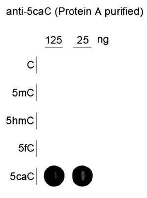
Dot Blotting 1:500 - 1:1,000 Fig 1
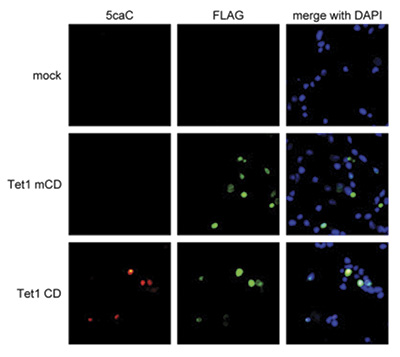
Immunofluorescence 1:500 Fig 2
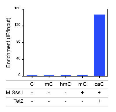
IP * 4 μg/IP (4 μg genomic DNA per IP) Fig 3

* Please note that of the optimal antibody amount per IP should be determined by the end-user. We recommend testing 1-5 μg per IP.

  • Validation Data

    Dot blot

    Fig. 1. Dot blot analysis using the Diagenode antibody directed against 5-caC
    To demonstrate the specificity of the Diagenode antibody against 5-caC (cat. No. pAb-CaC-020/050), a Dot Blot analysis was performed using synthetic oligonucleotides containing different modified C-bases (indicated in red). 125 and 25 ng of the respective oligo’s were bound to a Streptavindin-coated multi-well plate. The antibody was used at a dilution of 1:1,000. The binding of antibody to the DNA was measured by ECL chemiluminescence. Figure 1 shows a high specificity of the antibody for the carboxylated cytosine.

    Immunofluorescence

    Fig. 2. Immunofluorescence assay using the Diagenode antibody directed against 5-caC
    293T cells were transfected with either the mouse FLAG-tagged wild-type Tet1 (Tet1 CD) or the catalytically inactive FLAG-tagged C-terminal domain of Tet1 (Tet1 mCD) and stained with the Diagenode antibody against 5-caC (cat. No. pAb-CaC-020/050), diluted 1:500, and with an anti-FLAG antibody, followed by DAPI counterstaining.

    Immunoprecipitation

    Fig. 3. Immunoprecipitation using the Diagenode antibody directed against 5-caC
    Immunoprecipitation was performed with the Diagenode antibody against 5-caC (cat. No. pAb-CaC-020/050) on 2 μg of J1 ES genomic DNA, spiked with 1 pg of a control DNA fragment (approximately 700 bp from the RFP (Ring finger protein) gene) containing different cytosine modifications. The mC and hmC control DNA was generated by PCR with the corresponding nucleotide. The caC control fragment was obtained by in vitro methylation using M.SssI methyltransferase followed by oxidation with purified Tet2. The IP’d DNA was subsequently anaysed by qPCR using primers specific for the control DNA fragments and for GAPDH, used as a negative control. Figure 3 shows the enrichment calculated as the ratio of the recovery of the control DNA versus the recovery of the GAPDH negative control.

  • Target description

    Until recently, 5-methylcytosine (5-mC) was the only known modification of DNA for epigenetic regulation. In 2009, however, a second methylated cytosine, 5-hydroxymethylcytosine (5-hmC) was discovered. This new modified base (also called the Sixth base) is generated by enzymatic conversion of 5-mC into 5-hmC by the TET family of oxygenases.

    Recent results indicate that 5-hmC plays important roles distinct from 5-mC. Although its precise role has still to be shown, early evidence suggests that 5-hmC may well represent a new pathway to demethylate DNA involving a repair mechanism converting 5-hmC to cytosine. This pathway could involve further oxidation of the hydroxymethyl group to a formyl or carboxyl group followed by either deformylation or decarboxylation. The carboxyl and formyl groups of 5-Formylcytosine (5-fC) and 5-Carboxylcytosine (5-caC) could be enzymatically removed without excision of the base.

    Due to their structural similarity, the different modified cytosine analogues are difficult to discriminate. The development of highly specific affinity-based reagents, such as antibodies, appears to be the most powerful way to differentially and specifically enrich 5-mC and 5-hmC sequences. We previously released highly specific antibodies directed against 5-mC and 5-hmC. Now, we also present a unique rabbit polyclonal antibody against 5-Carboxycytosine.

  •  应用
    DB
    Dot blotting Read more
    IF
    Immunofluorescence: Diagenode offers huge selection of highly sensitive antibodies validated in IF. Immunofluorescence using the Diagenode monoclonal antibody directed against CRISPR/Cas9 HeLa cells transfected with a Cas9 expression vector (... Read more
    IP
    Immunoprecipitation Read more
  •  文档
    Datasheet 5-caC pAb-cac-100 DATASHEET
    Polyclonal antibody raised in rabbit against 5-Carboxylcytosine (5ca-CMP monophosphate) conjugate...
    Download
  •  Safety sheets
    SDS C15410204 5-caC Antibody US en Download
    SDS C15410204 5-caC Antibody GB en Download
    SDS C15410204 5-caC Antibody JP ja Download
    SDS C15410204 5-caC Antibody DE de Download
    SDS C15410204 5-caC Antibody ES es Download
    SDS C15410204 5-caC Antibody BE fr Download
    SDS C15410204 5-caC Antibody FR fr Download
    SDS C15410204 5-caC Antibody BE nl Download
  •  出版物

    How to properly cite this product in your work

    Diagenode strongly recommends using this: 5-caC polyclonal antibody (Diagenode Cat# C15410204-20 Lot# 001). Click here to copy to clipboard.

    Using our products in your publication? Let us know!

    A transition phase in late mouse oogenesis impacts DNA methylation ofthe early embryo.
    Eleftheriou K. et al.
    A well-orchestrated program of oocyte growth and differentiation results in a developmentally competent oocyte. In late oogenesis, germinal vesicle oocytes (GVOs) undergo chromatin remodeling accompanied by transcriptional silencing from an NSN (non-surrounded nucleolus) to an SN (surrounded nucleolus) chromatin sta...

    Regulation of paternal 5mC oxidation and H3K9me2 asymmetry byERK1/2 in mouse zygotes.
    Chen Baobao et al.
    BACKGROUND: Extracellular-signal-regulated kinase (ERK) direct cell fate determination during the early development. The intricate interaction between the deposition of H3K9me2, de novo 5mC, and its oxides affects the remodeling of zygotic epigenetic modification. However, the role of fertilization-dependent ERK in ...

    DNA methylation dynamics underlie metamorphic gene regulation programs in Xenopus tadpole brain.
    Kyono Y, Raj S, Sifuentes CJ, Buisine N, Sachs L, Denver RJ
    Methylation of cytosine residues in DNA influences chromatin structure and gene transcription, and its regulation is crucial for brain development. There is mounting evidence that DNA methylation can be modulated by hormone signaling. We analyzed genome-wide changes in DNA methylation and their relationship to gene ...

    Sodium valproate and 5-aza-2'-deoxycytidine differentially modulate DNA demethylation in G1 phase-arrested and proliferative HeLa cells.
    Rocha MA, Veronezi GMB, Felisbino MB, Gatti MSV, Tamashiro WMSC, Mello MLS
    Sodium valproate/valproic acid (VPA), a histone deacetylase inhibitor, and 5-aza-2-deoxycytidine (5-aza-CdR), a DNA methyltransferase 1 (DNMT1) inhibitor, induce DNA demethylation in several cell types. In HeLa cells, although VPA leads to decreased DNA 5-methylcytosine (5mC) levels, the demethylation pathway involv...

    Active and passive demethylation of male and female pronuclear DNA in the Mammalian zygote.
    Guo F, Li X, Liang D, Li T, Zhu P, Guo H, Wu X, Wen L, Gu TP, Hu B, Walsh CP, Li J, Tang F, Xu GL
    The epigenomes of mammalian sperm and oocytes, characterized by gamete-specific 5-methylcytosine (5mC) patterns, are reprogrammed during early embryogenesis to establish full developmental potential. Previous studies have suggested that the paternal genome is actively demethylated in the zygote while the maternal ge...

  •  相关产品


       Site map   |   Contact us   |   Conditions of sales   |   Conditions of purchase   |   Privacy policy