| ChIP-seq (ab) Read more |
| ChIP-qPCR (ab) Read more |
| DB Dot blotting Read more |
| WB Western blot : The quality of antibodies used in this technique is crucial for correct and specific protein identification. Diagenode offers huge selection of highly sensitive and specific western blot-validated antibodies. Learn more about: Load... Read more |
| IF Immunofluorescence: Diagenode offers huge selection of highly sensitive antibodies validated in IF. Immunofluorescence using the Diagenode monoclonal antibody directed against CRISPR/Cas9 HeLa cells transfected with a Cas9 expression vector (... Read more |
H3K18ac Antibody - ChIP-seq Grade
目录号
格式
价格

Histones are the main constituents of the protein part of chromosomes of eukaryotic cells. They are rich in the amino acids arginine and lysine and have been greatly conserved during evolution. Histones pack the DNA into tight masses of chromatin. Two core histones of each class H2A, H2B, H3 and H4 assemble and are wrapped by 146 base pairs of DNA to form one octameric nucleosome. Histone tails undergo numerous post-translational modifications, which either directly or indirectly alter chromatin structure to facilitate transcriptional activation or repression or other nuclear processes. In addition to the genetic code, combinations of the different histone modifications reveal the so-called "histone code". Histone methylation and demethylation is dynamically regulated by respectively histone methyl transferases and histone demethylases. Acetylation of histone H3K18 is associated with gene activation.
| Lot | 001 |
|---|---|
| Concentration | 1 µg/µl |
| Species reactivity | Human: positive. Other species: not tested |
| Type | Monoclonal ChIP-Grade ChIP-seq-Grade |
| Purity | Affinity purified monoclonal antibody |
| Host | Rabbit |
| Storage Conditions | Store at -20°C; for long storage, store at -80°C. Avoid multiple freeze-thaw cycles. |
| Storage Buffer | PBS containing 50% glycerol, 1% BSA and 0.09% azide |
| Precautions | This product is for research use only. Not for use in diagnostic or therapeutic procedures. |
| Applications | Suggested dilution | References |
|---|---|---|
| ChIP/ChIP-seq * | 0.5 - 1 µg per IP | Fig 1, 2 |
| Dot Blotting | 1:2,000 | Fig 3 |
| Western Blotting | 1:1,000 | Fig 4 |
| Immunofluorescence | 1:500 | Fig 5 |
*Please note that the optimal antibody amount per IP should be determined by the end-user. We recommend testing 0.5 - 5 µg per IP.
- Validation Data
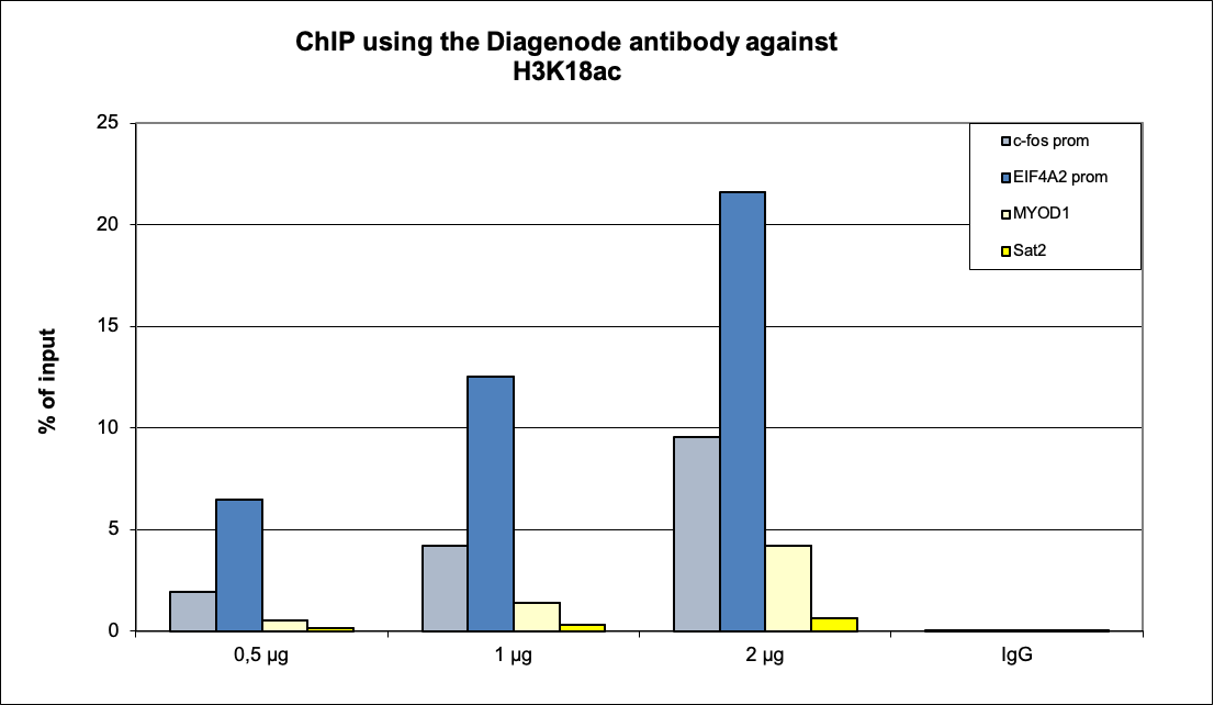
Figure 1. ChIP results obtained with the Diagenode monoclonal antibody directed against H3K18ac
ChIP was performed with the Diagenode antibody against H3K18ac (cat. No. C15210020) on sheared chromatin from 500,000 HeLaS3 cells using the “iDeal ChIP-seq” kit (cat. No. C01010051). A titration of the antibody consisting of 0.5, 1, and 2 µg per ChIP experiment was analysed. IgG (1 µg/IP) was used as negative IP control. Quantitative PCR was performed with primers for the c-fos and EIF4A2 promoters, used as positive controls, and for the MYOD1 genes and the Sat2 satellite repeat, used as negative controls. The graph shows the recovery, expressed as a % of input (the relative amount of immunoprecipitated DNA compared to input DNA after qPCR analysis).A. 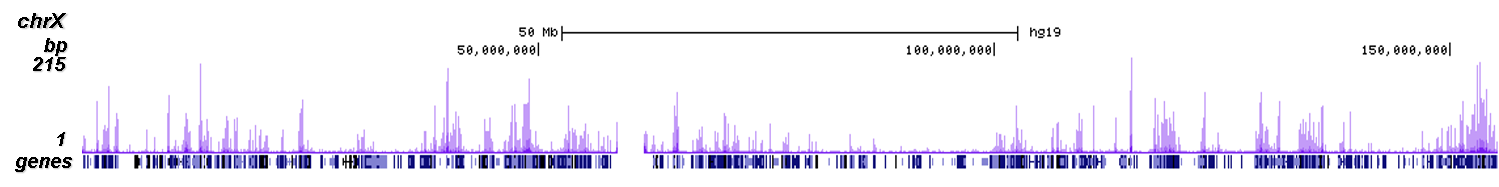
B.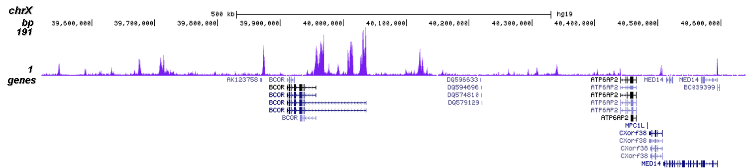
C.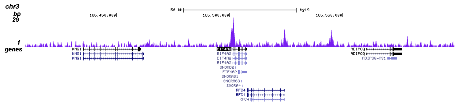
D.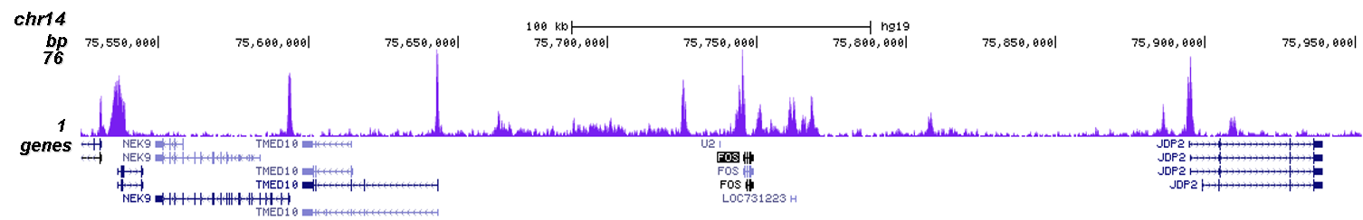
Figure 2. ChIP-seq results obtained with the Diagenode monoclonal antibody directed against H3K18ac
ChIP was performed on sheared chromatin from 500,000 HeLaS3 cells using 0.5 µg of the Diagenode antibody against H3K18ac (cat. No. C15210020) as described above. The IP'd DNA was subsequently analysed on an Illumina NovaSeq. Library preparation, cluster generation and sequencing were performed according to the manufacturer's instructions. The 51 bp tags were aligned to the human genome using the BWA algorithm. Figure 2 shows the H3K18ac signal along the complete sequence and 1.5 Mb region of the human X-chromosome (figure 2A and B), and in two genomic regions surrounding the EIF4A2 and c-fos positive controls (figure 2C and D).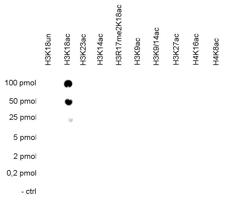
Figure 3. Cross reactivity tests using the Diagenode monoclonal antibody directed against H3K18ac
To test the cross reactivity of the Diagenode antibody against H3K18ac (cat. No. C15210020), a Dot Blot analysis was performed with peptides containing other histone modifications and the unmodified H3K18. One hundred to 0.2 pmol of the respective peptides were spotted on a membrane. The antibody was used at a dilution of 1:2,000. Figure 3 shows a high specificity of the antibody for the modification of interest.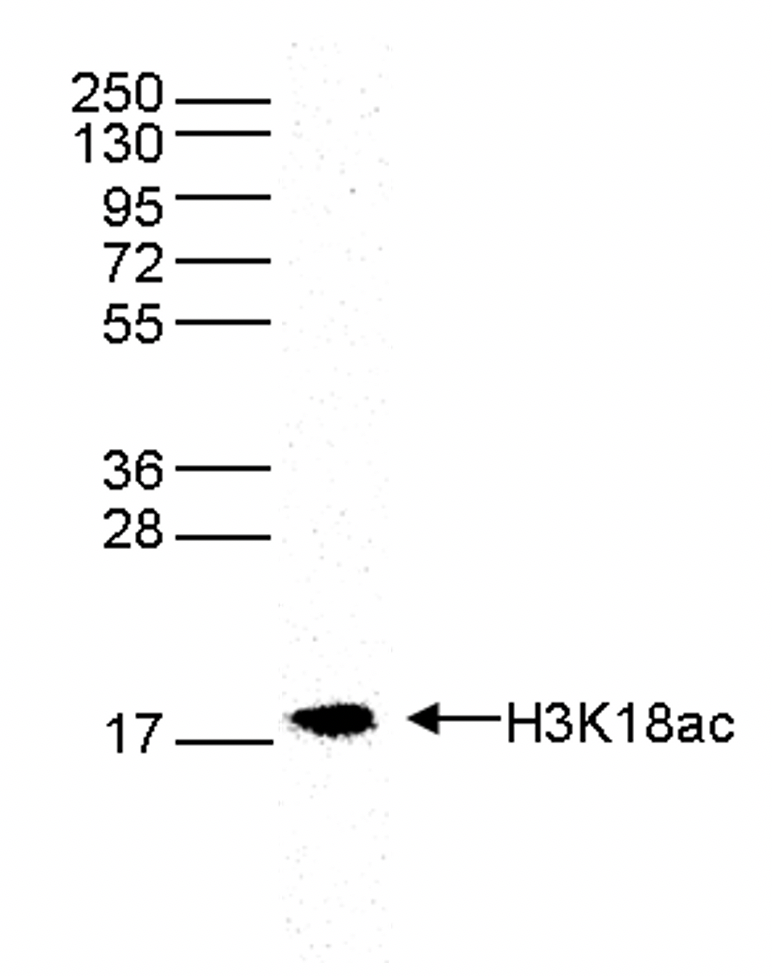
Figure 4. Western blot analysis using the Diagenode monoclonal antibody directed against H3K18ac
Western blot was performed on whole cell extracts (40 µg) from HeLa cells using the Diagenode antibody against H3K18ac (cat. No. C15210020). The antibody was diluted 1:1,000 in TBS-Tween containing 5% skimmed milk. The position of the protein of interest is shown on the right, the marker (in kDa) is shown on the left.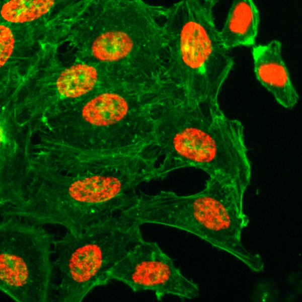
Figure 5. Immunofluorescence using the Diagenode monoclonal antibody directed against H3K18ac
HeLa cells were stained with the Diagenode antibody against H3K18ac (cat. No. C15210020, red) diluted 1:500. Actin was stained with fluorescein phalladoin (green). - 出版物

How to properly cite our product/service in your work
We strongly recommend using this: H3K18ac Antibody - ChIP-seq Grade (Hologic Diagenode Cat# C15210020 Lot# 001). Click here to copy to clipboard.
Using our products or services in your publication? Let us know!