Notice (8): Undefined variable: solution_of_interest [APP/View/Products/view.ctp, line 755]Code Context<!-- BEGIN: REQUEST_FORM MODAL -->
<div id="request_formModal" class="reveal-modal medium" data-reveal aria-labelledby="modalTitle" aria-hidden="true" role="dialog">
<?= $this->element('Forms/simple_form', array('solution_of_interest' => $solution_of_interest, 'header' => $header, 'message' => $message, 'campaign_id' => $campaign_id)) ?>
$viewFile = '/home/website-server/www/app/View/Products/view.ctp'
$dataForView = array(
'language' => 'cn',
'meta_keywords' => '',
'meta_description' => 'CTCF (CCCTC-Binding Factor) Polyclonal Antibody validated in ChIP-seq, ChIP-qPCR, WB, IF and ELISA. Specificity confirmed by siRNA assay. Batch-specific data available on the website. Other names: MRD21. Sample size available.',
'meta_title' => 'CTCF Antibody - ChIP-seq Grade (C15410210) | Diagenode',
'product' => array(
'Product' => array(
'id' => '2288',
'antibody_id' => '250',
'name' => 'CTCF Antibody - ChIP-seq Grade',
'description' => '<p>Alternative name: <strong>MRD21</strong></p>
<p>Polyclonal antibody raised in rabbit against human <strong>CTCF</strong> (<strong>CCCTC-Binding Factor</strong>), using 4 KLH coupled peptides.</p>',
'label1' => 'Validation Data',
'info1' => '<div class="row">
<div class="small-6 columns">
<p><img src="https://www.diagenode.com/img/product/antibodies/C15410210-chip.png" alt="CTCF Antibody ChIP Grade" /></p>
</div>
<div class="small-6 columns">
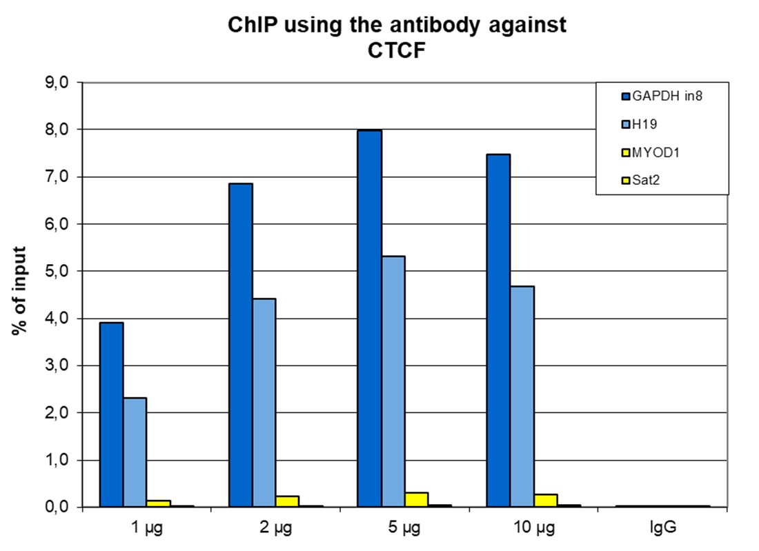
<p><small><strong> Figure 1. ChIP results obtained with the Diagenode antibody directed against CTCF</strong><br />ChIP was performed with the Diagenode antibody against CTCF (cat. No. C15410210) on sheared chromatin from 4,000,000 HeLa cells. A titration consisting of 1, 2, 5 and 10 µg of antibody per ChIP experiment was analyzed. IgG (2 µg/IP) was used as a negative IP control. Quantitative PCR was performed with optimized primers for the H19 imprinting control region, and a specific region in the GAPDH gene, used as positive controls, and for the Sat2 satellite repeat region, used as a negative control. Figure 1 shows the recovery, expressed as a % of input (the relative amount of immunoprecipitated DNA compared to input DNA after qPCR analysis). </small></p>
</div>
</div>
<div class="row">
<div class="small-12 columns">
<p>A.<img src="https://www.diagenode.com/img/product/antibodies/c15410210-chipseq-a.jpg" alt="CTCF Antibody ChIP-seq Grade" /></p>
<p>B.<img src="https://www.diagenode.com/img/product/antibodies/c15410210-chipseq-b.jpg" alt="CTCF Antibody for ChIP-seq " /></p>
<p>C.<img src="https://www.diagenode.com/img/product/antibodies/c15410210-chipseq-c.jpg" alt="CTCF Antibody for ChIP-seq assay" /></p>
<p>D.<img src="https://www.diagenode.com/img/product/antibodies/c15410210-chipseq-d.jpg" alt="CTCF Antibody validated in ChIP-seq" /></p>
</div>
</div>
<div class="row">
<div class="small-12 columns">
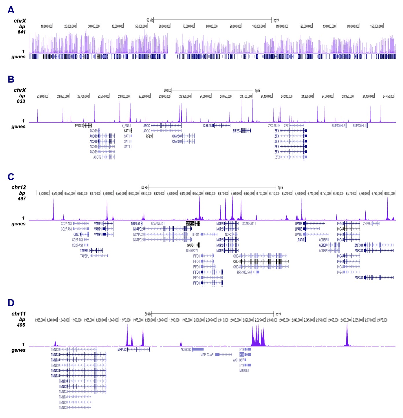
<p><small><strong> Figure 2. ChIP-seq results obtained with the Diagenode antibody directed against CTCF</strong><br /> ChIP was performed on sheared chromatin from 4,000,000 HeLa cells using 1 µg of the Diagenode antibody against CTCF (cat. No. C15410210) as described above. The IP'd DNA was subsequently analysed on an Illumina NovaSeq. Library preparation, cluster generation and sequencing were performed according to the manufacturer's instructions. The 50 bp tags were aligned to the human genome using the BWA algorithm. Figure 2 shows the peak distribution along the complete sequence and a 60 kb region of the human X-chromosome (figure 2A and B) and in two regions surrounding the GAPDH and H19 positive control genes, respectively (figure 2C and D).</small></p>
</div>
</div>
<div class="row">
<div class="small-12 columns">
<p>A.<img src="https://www.diagenode.com/img/product/antibodies/C15410210-cuttag-a.png" alt="CTCF Antibody CUT&Tag" /></p>
<p>B.<img src="https://www.diagenode.com/img/product/antibodies/C15410210-cuttag-b.png" alt="CTCF Antibody CUT&Tag " /></p>
</div>
</div>
<div class="row">
<div class="small-12 columns">
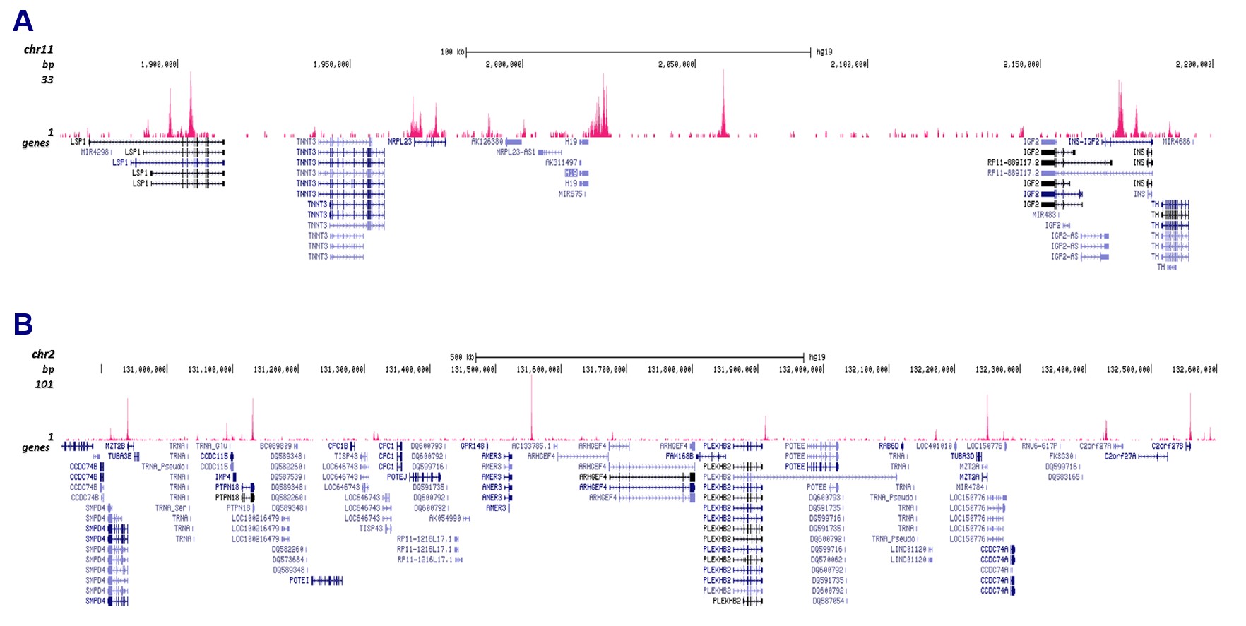
<p><small><strong> Figure 3. Cut&Tag results obtained with the Diagenode antibody directed against CTCF</strong><br /> CUT&TAG (Kaya-Okur, H.S., Nat Commun 10, 1930, 2019) was performed on 50,000 K562 cells using 1 µg of the Diagenode antibody against CTCF (cat. No. C15410210) and the Diagenode pA-Tn5 transposase (C01070001). The libraries were subsequently analysed on an Illumina NextSeq 500 sequencer (2x75 paired-end reads) according to the manufacturer's instructions. The tags were aligned to the human genome (hg19) using the BWA algorithm. Figure 3 shows the peak distribution in 2 genomic regions surrounding the h19 imprinting control gene on chromosome 11 and the AMER3 gene on chromosome 2 (figure 3A and B, respectively).</small></p>
</div>
</div>
<div class="row">
<div class="small-6 columns">
<p><img src="https://www.diagenode.com/img/product/antibodies/C15410210-elisa.png" alt="CTCF Antibody ELISA validation" /></p>
</div>
<div class="small-6 columns">
<p><small><strong> Figure 4. Determination of the antibody titer</strong><br />To determine the titer of the antibody, an ELISA was performed using a serial dilution of the Diagenode antibody against CTCF (cat. No. C15410210). The plates were coated with the peptides used for immunization of the rabbit. By plotting the absorbance against the antibody dilution (Figure 4), the titer of the antibody was estimated to be 1:90,000.</small></p>
</div>
</div>
<div class="row">
<div class="small-3 columns">
<p><img src="https://www.diagenode.com/img/product/antibodies/C15410210-wb.png" alt="CTCF Antibody for Western Blot" /></p>
</div>
<div class="small-9 columns">
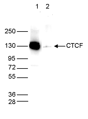
<p><small><strong>Figure 5. Western blot analysis using the Diagenode antibody directed against CTCF</strong><br /> Whole cell extracts (40 µg) from HeLa cells transfected with CTCF siRNA (lane 2) and from an untransfected control (lane 1) were analysed by Western blot using the Diagenode antibody against CTCF (cat. No. C15410210) diluted 1:1,000 in TBS-Tween containing 5% skimmed milk. The position of the protein of interest is indicated on the right; the marker (in kDa) is shown on the left.</small></p>
</div>
</div>',
'label2' => 'Target Description',
'info2' => '<p>CTCF (UniProt/Swiss-Prot entry P49711) is a transcriptional regulator protein with 11 highly conserved zinc finger domains. By using different combinations of the zinc finger domains, CTCF can bind to different DNA sequences and proteins. As such it can act as both a transcriptional repressor and a transcriptional activator. By binding to transcriptional insulator elements, CTCF can also block communication between enhancers and upstream promoters, thereby regulating imprinted gene expression. CTCF also binds to the H19 imprinting control region and mediates maternally inherited higher-order chromatin conformation to restrict enhancer access to IGF2. Mutations in the CTCF gene have been associated with invasive breast cancers, prostate cancers, and Wilms’ tumor.</p>',
'label3' => '',
'info3' => '',
'format' => '50 μg',
'catalog_number' => 'C15410210',
'old_catalog_number' => '',
'sf_code' => 'C15410210-D001-000581',
'type' => 'FRE',
'search_order' => '03-Antibody',
'price_EUR' => '380',
'price_USD' => '380',
'price_GBP' => '340',
'price_JPY' => '59525',
'price_CNY' => '',
'price_AUD' => '950',
'country' => 'ALL',
'except_countries' => 'None',
'quote' => false,
'in_stock' => false,
'featured' => false,
'no_promo' => false,
'online' => true,
'master' => true,
'last_datasheet_update' => 'August 21, 2020',
'slug' => 'ctcf-polyclonal-antibody-classic-50-mg',
'meta_title' => 'CTCF Antibody - ChIP-seq Grade (C15410210) | Diagenode',
'meta_keywords' => '',
'meta_description' => 'CTCF (CCCTC-Binding Factor) Polyclonal Antibody validated in ChIP-seq, ChIP-qPCR, WB, IF and ELISA. Specificity confirmed by siRNA assay. Batch-specific data available on the website. Other names: MRD21. Sample size available.',
'modified' => '2024-11-19 16:36:54',
'created' => '2015-06-29 14:08:20',
'locale' => 'zho'
),
'Antibody' => array(
'host' => '*****',
'id' => '250',
'name' => 'CTCF polyclonal antibody',
'description' => 'CTCF (UniProt/Swiss-Prot entry P49711) is a transcriptional regulator protein with 11 highly conserved zinc finger domains. By using different combinations of the zinc finger domains, CTCF can bind to different DNA sequences and proteins. As such it can act as both a transcriptional repressor and a transcriptional activator. By binding to transcriptional insulator elements, CTCF can also block communication between enhancers and upstream promoters, thereby regulating imprinted gene expression. CTCF also binds to the H19 imprinting control region and mediates maternally inherited higher-order chromatin conformation to restrict enhancer access to IGF2. Mutations in the CTCF gene have been associated with invasive breast cancers, prostate cancers, and Wilms’ tumor.',
'clonality' => '',
'isotype' => '',
'lot' => 'A2354-00234P',
'concentration' => '1.2 µg/µl',
'reactivity' => 'Human, mouse, pig: positive',
'type' => 'Polyclonal <strong>ChIP-seq Grade</strong>',
'purity' => 'Affinity purified',
'classification' => 'Classic',
'application_table' => '<table>
<thead>
<tr>
<th>Applications</th>
<th>Suggested dilution</th>
<th>References</th>
</tr>
</thead>
<tbody>
<tr>
<td>ChIP/ChIP-seq <sup>*</sup></td>
<td>1-2 µg per ChIP</td>
<td>Fig 1, 2</td>
</tr>
<tr>
<td>CUT&Tag</td>
<td>1 µg</td>
<td>Fig 3</td>
</tr>
<tr>
<td>ELISA</td>
<td>1:1,000 - 1:10,000</td>
<td>Fig 4</td>
</tr>
<tr>
<td>Western Blotting</td>
<td>1:1,000</td>
<td>Fig 5</td>
</tr>
</tbody>
</table>
<p></p>
<p><small><sup>*</sup> Please note that the optimal antibody amount per IP should be determined by the end-user. We recommend testing 1-10 µg per IP.</small></p>',
'storage_conditions' => 'Store at -20°C; for long storage, store at -80°C. Avoid multiple freeze-thaw cycles.',
'storage_buffer' => 'PBS containing 0.05% azide.',
'precautions' => 'This product is for research use only. Not for use in diagnostic or therapeutic procedures.',
'uniprot_acc' => '',
'slug' => '',
'meta_keywords' => '',
'meta_description' => '',
'modified' => '2022-10-05 14:59:24',
'created' => '0000-00-00 00:00:00',
'select_label' => '250 - CTCF polyclonal antibody (A2354-00234P - 1.2 µg/µl - Human, mouse, pig: positive - Affinity purified - Rabbit)'
),
'Slave' => array(
(int) 0 => array(
[maximum depth reached]
)
),
'Group' => array(
'Group' => array(
[maximum depth reached]
),
'Master' => array(
[maximum depth reached]
),
'Product' => array(
[maximum depth reached]
)
),
'Related' => array(
(int) 0 => array(
[maximum depth reached]
),
(int) 1 => array(
[maximum depth reached]
),
(int) 2 => array(
[maximum depth reached]
)
),
'Application' => array(
(int) 0 => array(
[maximum depth reached]
),
(int) 1 => array(
[maximum depth reached]
),
(int) 2 => array(
[maximum depth reached]
),
(int) 3 => array(
[maximum depth reached]
),
(int) 4 => array(
[maximum depth reached]
),
(int) 5 => array(
[maximum depth reached]
),
(int) 6 => array(
[maximum depth reached]
)
),
'Category' => array(
(int) 0 => array(
[maximum depth reached]
),
(int) 1 => array(
[maximum depth reached]
),
(int) 2 => array(
[maximum depth reached]
),
(int) 3 => array(
[maximum depth reached]
),
(int) 4 => array(
[maximum depth reached]
)
),
'Document' => array(
(int) 0 => array(
[maximum depth reached]
),
(int) 1 => array(
[maximum depth reached]
),
(int) 2 => array(
[maximum depth reached]
)
),
'Feature' => array(),
'Image' => array(
(int) 0 => array(
[maximum depth reached]
)
),
'Promotion' => array(),
'Protocol' => array(),
'Publication' => array(
(int) 0 => array(
[maximum depth reached]
),
(int) 1 => array(
[maximum depth reached]
),
(int) 2 => array(
[maximum depth reached]
),
(int) 3 => array(
[maximum depth reached]
),
(int) 4 => array(
[maximum depth reached]
),
(int) 5 => array(
[maximum depth reached]
),
(int) 6 => array(
[maximum depth reached]
),
(int) 7 => array(
[maximum depth reached]
),
(int) 8 => array(
[maximum depth reached]
),
(int) 9 => array(
[maximum depth reached]
),
(int) 10 => array(
[maximum depth reached]
),
(int) 11 => array(
[maximum depth reached]
),
(int) 12 => array(
[maximum depth reached]
),
(int) 13 => array(
[maximum depth reached]
),
(int) 14 => array(
[maximum depth reached]
),
(int) 15 => array(
[maximum depth reached]
),
(int) 16 => array(
[maximum depth reached]
),
(int) 17 => array(
[maximum depth reached]
),
(int) 18 => array(
[maximum depth reached]
),
(int) 19 => array(
[maximum depth reached]
),
(int) 20 => array(
[maximum depth reached]
),
(int) 21 => array(
[maximum depth reached]
),
(int) 22 => array(
[maximum depth reached]
),
(int) 23 => array(
[maximum depth reached]
),
(int) 24 => array(
[maximum depth reached]
),
(int) 25 => array(
[maximum depth reached]
),
(int) 26 => array(
[maximum depth reached]
),
(int) 27 => array(
[maximum depth reached]
),
(int) 28 => array(
[maximum depth reached]
),
(int) 29 => array(
[maximum depth reached]
),
(int) 30 => array(
[maximum depth reached]
),
(int) 31 => array(
[maximum depth reached]
),
(int) 32 => array(
[maximum depth reached]
)
),
'Testimonial' => array(
(int) 0 => array(
[maximum depth reached]
)
),
'Area' => array(),
'SafetySheet' => array(
(int) 0 => array(
[maximum depth reached]
),
(int) 1 => array(
[maximum depth reached]
),
(int) 2 => array(
[maximum depth reached]
),
(int) 3 => array(
[maximum depth reached]
),
(int) 4 => array(
[maximum depth reached]
),
(int) 5 => array(
[maximum depth reached]
),
(int) 6 => array(
[maximum depth reached]
),
(int) 7 => array(
[maximum depth reached]
)
)
),
'meta_canonical' => 'https://www.diagenode.com/cn/p/ctcf-polyclonal-antibody-classic-50-mg'
)
$language = 'cn'
$meta_keywords = ''
$meta_description = 'CTCF (CCCTC-Binding Factor) Polyclonal Antibody validated in ChIP-seq, ChIP-qPCR, WB, IF and ELISA. Specificity confirmed by siRNA assay. Batch-specific data available on the website. Other names: MRD21. Sample size available.'
$meta_title = 'CTCF Antibody - ChIP-seq Grade (C15410210) | Diagenode'
$product = array(
'Product' => array(
'id' => '2288',
'antibody_id' => '250',
'name' => 'CTCF Antibody - ChIP-seq Grade',
'description' => '<p>Alternative name: <strong>MRD21</strong></p>
<p>Polyclonal antibody raised in rabbit against human <strong>CTCF</strong> (<strong>CCCTC-Binding Factor</strong>), using 4 KLH coupled peptides.</p>',
'label1' => 'Validation Data',
'info1' => '<div class="row">
<div class="small-6 columns">
<p><img src="https://www.diagenode.com/img/product/antibodies/C15410210-chip.png" alt="CTCF Antibody ChIP Grade" /></p>
</div>
<div class="small-6 columns">
<p><small><strong> Figure 1. ChIP results obtained with the Diagenode antibody directed against CTCF</strong><br />ChIP was performed with the Diagenode antibody against CTCF (cat. No. C15410210) on sheared chromatin from 4,000,000 HeLa cells. A titration consisting of 1, 2, 5 and 10 µg of antibody per ChIP experiment was analyzed. IgG (2 µg/IP) was used as a negative IP control. Quantitative PCR was performed with optimized primers for the H19 imprinting control region, and a specific region in the GAPDH gene, used as positive controls, and for the Sat2 satellite repeat region, used as a negative control. Figure 1 shows the recovery, expressed as a % of input (the relative amount of immunoprecipitated DNA compared to input DNA after qPCR analysis). </small></p>
</div>
</div>
<div class="row">
<div class="small-12 columns">
<p>A.<img src="https://www.diagenode.com/img/product/antibodies/c15410210-chipseq-a.jpg" alt="CTCF Antibody ChIP-seq Grade" /></p>
<p>B.<img src="https://www.diagenode.com/img/product/antibodies/c15410210-chipseq-b.jpg" alt="CTCF Antibody for ChIP-seq " /></p>
<p>C.<img src="https://www.diagenode.com/img/product/antibodies/c15410210-chipseq-c.jpg" alt="CTCF Antibody for ChIP-seq assay" /></p>
<p>D.<img src="https://www.diagenode.com/img/product/antibodies/c15410210-chipseq-d.jpg" alt="CTCF Antibody validated in ChIP-seq" /></p>
</div>
</div>
<div class="row">
<div class="small-12 columns">
<p><small><strong> Figure 2. ChIP-seq results obtained with the Diagenode antibody directed against CTCF</strong><br /> ChIP was performed on sheared chromatin from 4,000,000 HeLa cells using 1 µg of the Diagenode antibody against CTCF (cat. No. C15410210) as described above. The IP'd DNA was subsequently analysed on an Illumina NovaSeq. Library preparation, cluster generation and sequencing were performed according to the manufacturer's instructions. The 50 bp tags were aligned to the human genome using the BWA algorithm. Figure 2 shows the peak distribution along the complete sequence and a 60 kb region of the human X-chromosome (figure 2A and B) and in two regions surrounding the GAPDH and H19 positive control genes, respectively (figure 2C and D).</small></p>
</div>
</div>
<div class="row">
<div class="small-12 columns">
<p>A.<img src="https://www.diagenode.com/img/product/antibodies/C15410210-cuttag-a.png" alt="CTCF Antibody CUT&Tag" /></p>
<p>B.<img src="https://www.diagenode.com/img/product/antibodies/C15410210-cuttag-b.png" alt="CTCF Antibody CUT&Tag " /></p>
</div>
</div>
<div class="row">
<div class="small-12 columns">
<p><small><strong> Figure 3. Cut&Tag results obtained with the Diagenode antibody directed against CTCF</strong><br /> CUT&TAG (Kaya-Okur, H.S., Nat Commun 10, 1930, 2019) was performed on 50,000 K562 cells using 1 µg of the Diagenode antibody against CTCF (cat. No. C15410210) and the Diagenode pA-Tn5 transposase (C01070001). The libraries were subsequently analysed on an Illumina NextSeq 500 sequencer (2x75 paired-end reads) according to the manufacturer's instructions. The tags were aligned to the human genome (hg19) using the BWA algorithm. Figure 3 shows the peak distribution in 2 genomic regions surrounding the h19 imprinting control gene on chromosome 11 and the AMER3 gene on chromosome 2 (figure 3A and B, respectively).</small></p>
</div>
</div>
<div class="row">
<div class="small-6 columns">
<p><img src="https://www.diagenode.com/img/product/antibodies/C15410210-elisa.png" alt="CTCF Antibody ELISA validation" /></p>
</div>
<div class="small-6 columns">
<p><small><strong> Figure 4. Determination of the antibody titer</strong><br />To determine the titer of the antibody, an ELISA was performed using a serial dilution of the Diagenode antibody against CTCF (cat. No. C15410210). The plates were coated with the peptides used for immunization of the rabbit. By plotting the absorbance against the antibody dilution (Figure 4), the titer of the antibody was estimated to be 1:90,000.</small></p>
</div>
</div>
<div class="row">
<div class="small-3 columns">
<p><img src="https://www.diagenode.com/img/product/antibodies/C15410210-wb.png" alt="CTCF Antibody for Western Blot" /></p>
</div>
<div class="small-9 columns">
<p><small><strong>Figure 5. Western blot analysis using the Diagenode antibody directed against CTCF</strong><br /> Whole cell extracts (40 µg) from HeLa cells transfected with CTCF siRNA (lane 2) and from an untransfected control (lane 1) were analysed by Western blot using the Diagenode antibody against CTCF (cat. No. C15410210) diluted 1:1,000 in TBS-Tween containing 5% skimmed milk. The position of the protein of interest is indicated on the right; the marker (in kDa) is shown on the left.</small></p>
</div>
</div>',
'label2' => 'Target Description',
'info2' => '<p>CTCF (UniProt/Swiss-Prot entry P49711) is a transcriptional regulator protein with 11 highly conserved zinc finger domains. By using different combinations of the zinc finger domains, CTCF can bind to different DNA sequences and proteins. As such it can act as both a transcriptional repressor and a transcriptional activator. By binding to transcriptional insulator elements, CTCF can also block communication between enhancers and upstream promoters, thereby regulating imprinted gene expression. CTCF also binds to the H19 imprinting control region and mediates maternally inherited higher-order chromatin conformation to restrict enhancer access to IGF2. Mutations in the CTCF gene have been associated with invasive breast cancers, prostate cancers, and Wilms’ tumor.</p>',
'label3' => '',
'info3' => '',
'format' => '50 μg',
'catalog_number' => 'C15410210',
'old_catalog_number' => '',
'sf_code' => 'C15410210-D001-000581',
'type' => 'FRE',
'search_order' => '03-Antibody',
'price_EUR' => '380',
'price_USD' => '380',
'price_GBP' => '340',
'price_JPY' => '59525',
'price_CNY' => '',
'price_AUD' => '950',
'country' => 'ALL',
'except_countries' => 'None',
'quote' => false,
'in_stock' => false,
'featured' => false,
'no_promo' => false,
'online' => true,
'master' => true,
'last_datasheet_update' => 'August 21, 2020',
'slug' => 'ctcf-polyclonal-antibody-classic-50-mg',
'meta_title' => 'CTCF Antibody - ChIP-seq Grade (C15410210) | Diagenode',
'meta_keywords' => '',
'meta_description' => 'CTCF (CCCTC-Binding Factor) Polyclonal Antibody validated in ChIP-seq, ChIP-qPCR, WB, IF and ELISA. Specificity confirmed by siRNA assay. Batch-specific data available on the website. Other names: MRD21. Sample size available.',
'modified' => '2024-11-19 16:36:54',
'created' => '2015-06-29 14:08:20',
'locale' => 'zho'
),
'Antibody' => array(
'host' => '*****',
'id' => '250',
'name' => 'CTCF polyclonal antibody',
'description' => 'CTCF (UniProt/Swiss-Prot entry P49711) is a transcriptional regulator protein with 11 highly conserved zinc finger domains. By using different combinations of the zinc finger domains, CTCF can bind to different DNA sequences and proteins. As such it can act as both a transcriptional repressor and a transcriptional activator. By binding to transcriptional insulator elements, CTCF can also block communication between enhancers and upstream promoters, thereby regulating imprinted gene expression. CTCF also binds to the H19 imprinting control region and mediates maternally inherited higher-order chromatin conformation to restrict enhancer access to IGF2. Mutations in the CTCF gene have been associated with invasive breast cancers, prostate cancers, and Wilms’ tumor.',
'clonality' => '',
'isotype' => '',
'lot' => 'A2354-00234P',
'concentration' => '1.2 µg/µl',
'reactivity' => 'Human, mouse, pig: positive',
'type' => 'Polyclonal <strong>ChIP-seq Grade</strong>',
'purity' => 'Affinity purified',
'classification' => 'Classic',
'application_table' => '<table>
<thead>
<tr>
<th>Applications</th>
<th>Suggested dilution</th>
<th>References</th>
</tr>
</thead>
<tbody>
<tr>
<td>ChIP/ChIP-seq <sup>*</sup></td>
<td>1-2 µg per ChIP</td>
<td>Fig 1, 2</td>
</tr>
<tr>
<td>CUT&Tag</td>
<td>1 µg</td>
<td>Fig 3</td>
</tr>
<tr>
<td>ELISA</td>
<td>1:1,000 - 1:10,000</td>
<td>Fig 4</td>
</tr>
<tr>
<td>Western Blotting</td>
<td>1:1,000</td>
<td>Fig 5</td>
</tr>
</tbody>
</table>
<p></p>
<p><small><sup>*</sup> Please note that the optimal antibody amount per IP should be determined by the end-user. We recommend testing 1-10 µg per IP.</small></p>',
'storage_conditions' => 'Store at -20°C; for long storage, store at -80°C. Avoid multiple freeze-thaw cycles.',
'storage_buffer' => 'PBS containing 0.05% azide.',
'precautions' => 'This product is for research use only. Not for use in diagnostic or therapeutic procedures.',
'uniprot_acc' => '',
'slug' => '',
'meta_keywords' => '',
'meta_description' => '',
'modified' => '2022-10-05 14:59:24',
'created' => '0000-00-00 00:00:00',
'select_label' => '250 - CTCF polyclonal antibody (A2354-00234P - 1.2 µg/µl - Human, mouse, pig: positive - Affinity purified - Rabbit)'
),
'Slave' => array(
(int) 0 => array(
'id' => '19',
'name' => 'C15410210',
'product_id' => '2288',
'modified' => '2016-02-18 18:01:51',
'created' => '2016-02-18 18:01:51'
)
),
'Group' => array(
'Group' => array(
'id' => '19',
'name' => 'C15410210',
'product_id' => '2288',
'modified' => '2016-02-18 18:01:51',
'created' => '2016-02-18 18:01:51'
),
'Master' => array(
'id' => '2288',
'antibody_id' => '250',
'name' => 'CTCF Antibody ',
'description' => '<p>Alternative name: <strong>MRD21</strong></p>
<p>Polyclonal antibody raised in rabbit against human <strong>CTCF</strong> (<strong>CCCTC-Binding Factor</strong>), using 4 KLH coupled peptides.</p>
<p></p>',
'label1' => 'Validation Data',
'info1' => '<div class="row">
<div class="small-6 columns">
<p><img src="https://www.diagenode.com/img/product/antibodies/C15410210-chip.png" alt="CTCF Antibody ChIP Grade" /></p>
</div>
<div class="small-6 columns">
<p><small><strong> Figure 1. ChIP results obtained with the Diagenode antibody directed against CTCF</strong><br />ChIP was performed with the Diagenode antibody against CTCF (cat. No. C15410210) on sheared chromatin from 4,000,000 HeLa cells. A titration consisting of 1, 2, 5 and 10 µg of antibody per ChIP experiment was analyzed. IgG (2 µg/IP) was used as a negative IP control. Quantitative PCR was performed with optimized primers for the H19 imprinting control region, and a specific region in the GAPDH gene, used as positive controls, and for the Sat2 satellite repeat region, used as a negative control. Figure 1 shows the recovery, expressed as a % of input (the relative amount of immunoprecipitated DNA compared to input DNA after qPCR analysis). </small></p>
</div>
</div>
<div class="row">
<div class="small-12 columns">
<p>A.<img src="https://www.diagenode.com/img/product/antibodies/c15410210-chipseq-a.jpg" alt="CTCF Antibody ChIP-seq Grade" /></p>
<p>B.<img src="https://www.diagenode.com/img/product/antibodies/c15410210-chipseq-b.jpg" alt="CTCF Antibody for ChIP-seq " /></p>
<p>C.<img src="https://www.diagenode.com/img/product/antibodies/c15410210-chipseq-c.jpg" alt="CTCF Antibody for ChIP-seq assay" /></p>
<p>D.<img src="https://www.diagenode.com/img/product/antibodies/c15410210-chipseq-d.jpg" alt="CTCF Antibody validated in ChIP-seq" /></p>
</div>
</div>
<div class="row">
<div class="small-12 columns">
<p><small><strong> Figure 2. ChIP-seq results obtained with the Diagenode antibody directed against CTCF</strong><br /> ChIP was performed on sheared chromatin from 4,000,000 HeLa cells using 1 µg of the Diagenode antibody against CTCF (cat. No. C15410210) as described above. The IP'd DNA was subsequently analysed on an Illumina NovaSeq. Library preparation, cluster generation and sequencing were performed according to the manufacturer's instructions. The 50 bp tags were aligned to the human genome using the BWA algorithm. Figure 2 shows the peak distribution along the complete sequence and a 60 kb region of the human X-chromosome (figure 2A and B) and in two regions surrounding the GAPDH and H19 positive control genes, respectively (figure 2C and D).</small></p>
</div>
</div>
<div class="row">
<div class="small-12 columns">
<p>A.<img src="https://www.diagenode.com/img/product/antibodies/C15410210-cuttag-a.png" alt="CTCF Antibody CUT&Tag" /></p>
<p>B.<img src="https://www.diagenode.com/img/product/antibodies/C15410210-cuttag-b.png" alt="CTCF Antibody CUT&Tag " /></p>
</div>
</div>
<div class="row">
<div class="small-12 columns">
<p><small><strong> Figure 3. Cut&Tag results obtained with the Diagenode antibody directed against CTCF</strong><br /> CUT&TAG (Kaya-Okur, H.S., Nat Commun 10, 1930, 2019) was performed on 50,000 K562 cells using 1 µg of the Diagenode antibody against CTCF (cat. No. C15410210) and the Diagenode pA-Tn5 transposase (C01070001). The libraries were subsequently analysed on an Illumina NextSeq 500 sequencer (2x75 paired-end reads) according to the manufacturer's instructions. The tags were aligned to the human genome (hg19) using the BWA algorithm. Figure 3 shows the peak distribution in 2 genomic regions surrounding the h19 imprinting control gene on chromosome 11 and the AMER3 gene on chromosome 2 (figure 3A and B, respectively).</small></p>
</div>
</div>
<div class="row">
<div class="small-6 columns">
<p><img src="https://www.diagenode.com/img/product/antibodies/C15410210-elisa.png" alt="CTCF Antibody ELISA validation" /></p>
</div>
<div class="small-6 columns">
<p><small><strong> Figure 4. Determination of the antibody titer</strong><br />To determine the titer of the antibody, an ELISA was performed using a serial dilution of the Diagenode antibody against CTCF (cat. No. C15410210). The plates were coated with the peptides used for immunization of the rabbit. By plotting the absorbance against the antibody dilution (Figure 4), the titer of the antibody was estimated to be 1:90,000.</small></p>
</div>
</div>
<div class="row">
<div class="small-3 columns">
<p><img src="https://www.diagenode.com/img/product/antibodies/C15410210-wb.png" alt="CTCF Antibody for Western Blot" /></p>
</div>
<div class="small-9 columns">
<p><small><strong>Figure 5. Western blot analysis using the Diagenode antibody directed against CTCF</strong><br /> Whole cell extracts (40 µg) from HeLa cells transfected with CTCF siRNA (lane 2) and from an untransfected control (lane 1) were analysed by Western blot using the Diagenode antibody against CTCF (cat. No. C15410210) diluted 1:1,000 in TBS-Tween containing 5% skimmed milk. The position of the protein of interest is indicated on the right; the marker (in kDa) is shown on the left.</small></p>
</div>
</div>',
'label2' => 'Target Description',
'info2' => '<p>CTCF (UniProt/Swiss-Prot entry P49711) is a transcriptional regulator protein with 11 highly conserved zinc finger domains. By using different combinations of the zinc finger domains, CTCF can bind to different DNA sequences and proteins. As such it can act as both a transcriptional repressor and a transcriptional activator. By binding to transcriptional insulator elements, CTCF can also block communication between enhancers and upstream promoters, thereby regulating imprinted gene expression. CTCF also binds to the H19 imprinting control region and mediates maternally inherited higher-order chromatin conformation to restrict enhancer access to IGF2. Mutations in the CTCF gene have been associated with invasive breast cancers, prostate cancers, and Wilms’ tumor.</p>',
'label3' => '',
'info3' => '',
'format' => '50 μg',
'catalog_number' => 'C15410210',
'old_catalog_number' => '',
'sf_code' => 'C15410210-D001-000581',
'type' => 'FRE',
'search_order' => '03-Antibody',
'price_EUR' => '380',
'price_USD' => '380',
'price_GBP' => '340',
'price_JPY' => '59525',
'price_CNY' => '',
'price_AUD' => '950',
'country' => 'ALL',
'except_countries' => 'None',
'quote' => false,
'in_stock' => false,
'featured' => false,
'no_promo' => false,
'online' => true,
'master' => true,
'last_datasheet_update' => 'August 21, 2020',
'slug' => 'ctcf-polyclonal-antibody-classic-50-mg',
'meta_title' => 'CTCF Antibody - ChIP-seq grade (C15410210) | Diagenode',
'meta_keywords' => '',
'meta_description' => 'CTCF (CCCTC-Binding Factor) Polyclonal Antibody validated in ChIP-seq, ChIP-qPCR, WB, IF and ELISA. Specificity confirmed by siRNA assay. Batch-specific data available on the website. Other names: MRD21. Sample size available.',
'modified' => '2024-11-19 16:36:54',
'created' => '2015-06-29 14:08:20'
),
'Product' => array(
(int) 0 => array(
[maximum depth reached]
)
)
),
'Related' => array(
(int) 0 => array(
'id' => '2899',
'antibody_id' => null,
'name' => 'iDeal ChIP-qPCR Kit',
'description' => '<p><a href="https://www.diagenode.com/files/products/kits/ideal-chip-qpcr-manual.pdf"><img src="https://www.diagenode.com/img/buttons/bt-manual.png" /></a></p>
<p><br /><span style="font-weight: 400;">Diagenode’s iDeal ChIP-qPCR kit is a highly optimized and validated solution for </span><b>ChIP-qPCR </b><span style="font-weight: 400;">assays for either </span><b>histones </b><span style="font-weight: 400;">or </span><b>transcription factors</b><span style="font-weight: 400;">. The iDeal ChIP-qPCR kit, in conjunction with our validated ChIP-grade antibodies, provides excellent, reproducible results. The </span><b>complete kit</b><span style="font-weight: 400;"> contains everything you need for start-to-finish ChIP including all validated buffers and reagents for chromatin shearing, immunoprecipitation, and DNA isolation for exceptional ChIP-qPCR results.</span></p>
<p><span style="font-weight: 400;">The iDeal ChIP-qPCR kit uses a unique and fast method for DNA isolation and decrosslinking within 30 minutes compared to the standard 4 hours. Overall, easy to use and rapid protocol allows to receive the results within 20 hours with 4 hours hands-on time. </span></p>
<p><span style="font-weight: 400;">Recommended amount of material per IP using the </span><b>iDeal ChIP-qPCR kit:</b></p>
<table style="width: 722px; margin-left: auto; margin-right: auto;">
<tbody>
<tr>
<td style="width: 132px;"></td>
<td style="text-align: center; width: 299px;">
<p style="text-align: center;"><span style="font-weight: 400;">Histones</span></p>
</td>
<td style="text-align: center; width: 281px;">
<p style="text-align: center;"><span style="font-weight: 400;">Transcription factors</span></p>
</td>
</tr>
<tr>
<td style="width: 132px;">
<p><span style="font-weight: 400;">Cells</span></p>
</td>
<td style="text-align: center; width: 299px;">
<p style="text-align: center;"><b>100,000</b><span style="font-weight: 400;"> to </span><b>1 million cells</b><span style="font-weight: 400;"> </span></p>
</td>
<td style="text-align: center; width: 281px;">
<p style="text-align: center;"><b>4 million cells</b></p>
</td>
</tr>
<tr>
<td style="width: 132px;">
<p><span style="font-weight: 400;">Tissue</span></p>
</td>
<td style="text-align: center; width: 299px;">
<p style="text-align: center;"><b>1.5 mg</b><span style="font-weight: 400;"> to </span><b>5 mg</b></p>
</td>
<td style="text-align: center; width: 281px;">
<p style="text-align: center;"><b>30 mg</b></p>
</td>
</tr>
</tbody>
</table>',
'label1' => 'Characteristics',
'info1' => '<ul>
<li><strong><span style="font-weight: 400;"><strong>Fast</strong> and <strong>highly optimized</strong> protocol for ChIP-qPCR from cells and tissues</span></strong></li>
<li><strong><span style="font-weight: 400;"><strong>Easy ChIP</strong> made faster and more <strong>reproducible</strong> with magnetic beads</span></strong></li>
<li><strong><span style="font-weight: 400;"><strong>High yields</strong> with excellent <strong>specificity</strong> and <strong>sensitivity</strong> due to combination of Diagenode ChIP-grade antibodies<br /></span></strong></li>
<li><strong><span style="font-weight: 400;">Eluted DNA suitable for <strong>qPCR analysis</strong></span></strong></li>
</ul>
<h3><strong>Optimized</strong> protocol for <strong>reproducible ChIP-qPCR</strong> results</h3>
<p>Ideal for <strong>histone</strong> and <strong>non-histone proteins</strong> – Use 1 million cells for histone marks or 4 million cells for transcription factors. The protocol allows the use of fewer cells per IP. The minimum number of cells will depend on the abundance of the protein in your sample.</p>
<p>Suitable for <strong>cells</strong> and for <strong>tissues</strong> samples – For tissues, use 7 mg per IP for histone marks or 30 mg per IP for transcription factors. The protocol allows the use of less tissue per IP. The minimum amount of tissue will depend on the abundance of the protein in your sample.</p>
<center><img src="https://www.diagenode.com/img/categories/chip-qpcr/chip-qpcr-figure.jpg" /></center>
<p><small><strong>Chromatin immunoprecipitation analysis using H3K4me3 (A) and CTCF antibodies (B)</strong><br /> ChIP was performed on human HeLa cells using the H3K4me3 (Cat. No. C15410003) and CTCF (Cat. No. C15410210) antibodies. IgG was used as a negative control. The IP’d DNA was analyzed by qPCR with the following primer sets: EIF4A2, used as a positive control, and THS2B and Myoglobin exon 2, used as negative controls for H3K4me3. H19 imprinting control region and GAPDH intron 8, used as positive controls, and Myoglobin exon 2, used as a negative control for CTCF. The figure shows the recovery, expressed as a % of input (the relative amount of immunoprecipitated DNA compared to input DNA after qPCR analysis).</small></p>',
'label2' => 'Additional solutions compatible with iDeal ChIP-qPCR Kit',
'info2' => '<p><span style="font-weight: 400;">The</span> <a href="https://www.diagenode.com/en/p/chromatin-shearing-optimization-kit-low-sds-for-tfs-25-rxns"><span style="font-weight: 400;">Chromatin shearing optimization kit – Low SDS (iDeal Kit for TFs)</span></a><span style="font-weight: 400;"> is the kit compatible with the iDeal ChIP-qPCR kit, recommended for the optimization of chromatin shearing, a critical step for ChIP.</span></p>
<p><a href="https://www.diagenode.com/en/p/chip-cross-link-gold-600-ul"><span style="font-weight: 400;">ChIP Cross-link Gold</span></a> <span style="font-weight: 400;">should be used in combination with formaldehyde when working with higher order and/or dynamic interactions, for efficient protein-protein fixation.</span></p>
<p><span style="font-weight: 400;">Highly specific </span><a href="https://www.diagenode.com/en/categories/chip-grade-antibodies"><span style="font-weight: 400;">ChIP-grade antibodies</span></a><span style="font-weight: 400;">, provided with batch-specific validation data.</span></p>
<p><span style="font-weight: 400;">Check out the list of available </span><a href="https://www.diagenode.com/en/categories/primer-pairs"><span style="font-weight: 400;">Primer pairs</span></a><span style="font-weight: 400;"> designed for high specificity to specific genomic regions.</span></p>
<p><span style="font-weight: 400;">Using our IP-Star Compact, please check out the <a href="https://www.diagenode.com/en/p/auto-ideal-chip-qpcr-kit">automated version</a> of the kit.</span></p>',
'label3' => '',
'info3' => '',
'format' => '24 rxns',
'catalog_number' => 'C01010180',
'old_catalog_number' => '',
'sf_code' => 'C01010180-',
'type' => 'RFR',
'search_order' => '',
'price_EUR' => '560',
'price_USD' => '560',
'price_GBP' => '510',
'price_JPY' => '87725',
'price_CNY' => '',
'price_AUD' => '1400',
'country' => 'ALL',
'except_countries' => 'None',
'quote' => false,
'in_stock' => false,
'featured' => true,
'no_promo' => false,
'online' => true,
'master' => true,
'last_datasheet_update' => '',
'slug' => 'ideal-chip-qpcr-kit',
'meta_title' => 'iDeal ChIP-qPCR kit | Diagenode',
'meta_keywords' => 'iDeal ChIP-qPCR kit ,ChIP-qPCR assays, ChIP-grade antibodies,diagenode',
'meta_description' => 'Diagenode’s iDeal ChIP-qPCR kit is a highly optimized solution for ChIP-qPCR assays. The kit provides high yields with excellent specificity and sensitivity. The iDeal ChIP-qPCR kit used with our highly validated ChIP-grade antibodies provides you with excellent, reproducible results from each experiment.',
'modified' => '2023-04-20 16:07:26',
'created' => '2017-06-07 15:38:54',
'ProductsRelated' => array(
[maximum depth reached]
),
'Image' => array(
[maximum depth reached]
)
),
(int) 1 => array(
'id' => '1839',
'antibody_id' => null,
'name' => 'iDeal ChIP-seq kit for Transcription Factors',
'description' => '<p><a href="https://www.diagenode.com/files/products/kits/ideal-chipseq-transcription-factors-x10-manual.pdf"><img src="https://www.diagenode.com/img/buttons/bt-manual.png" /></a></p>
<div class="row">
<div class="small-12 medium-8 large-8 columns"><br />
<p><span style="font-weight: 400;">Diagenode’s <strong>iDeal ChIP-seq Kit for Transcription Factors</strong> is a highly validated solution for robust transcription factor and other non-histone proteins ChIP-seq results and contains everything you need for start-to-finish </span><b>ChIP </b><span style="font-weight: 400;">prior to </span><b>Next-Generation Sequencing</b><span style="font-weight: 400;">. This complete solution contains all buffers and reagents for cell lysis, chromatin shearing, immunoprecipitation, and DNA purification. In addition, unlike competing solutions, the kit contains positive and negative control antibodies (CTCF and IgG, respectively) as well as positive and negative control PCR primers pairs (H19 and Myoglobin exon 2, respectively) for your convenience and a guarantee of optimal results. <br /></span></p>
</div>
<div class="small-12 medium-4 large-4 columns"><center><br /><br />
<script>// <![CDATA[
var date = new Date(); var heure = date.getHours(); var jour = date.getDay(); var semaine = Math.floor(date.getDate() / 7) + 1; if (jour === 2 && ( (heure >= 9 && heure < 9.5) || (heure >= 18 && heure < 18.5) )) { document.write('<a href="https://us02web.zoom.us/j/85467619762"><img src="https://www.diagenode.com/img/epicafe-ON.gif"></a>'); } else { document.write('<a href="https://go.diagenode.com/l/928883/2023-04-26/3kq1v"><img src="https://www.diagenode.com/img/epicafe-OFF.png"></a>'); }
// ]]></script>
</center></div>
</div>
<p><span style="font-weight: 400;">The </span><b> iDeal ChIP-seq kit for Transcription Factors </b><span style="font-weight: 400;">is compatible for cells or tissues:</span></p>
<table style="width: 419px; margin-left: auto; margin-right: auto;">
<tbody>
<tr>
<td style="width: 144px;"></td>
<td style="width: 267px; text-align: center;"><span style="font-weight: 400;">Amount per IP</span></td>
</tr>
<tr>
<td style="width: 144px;">Cells</td>
<td style="width: 267px; text-align: center;"><strong>4,000,000</strong></td>
</tr>
<tr>
<td style="width: 144px;">Tissues</td>
<td style="width: 267px; text-align: center;"><strong>30 mg</strong></td>
</tr>
</tbody>
</table>
<p><span style="font-weight: 400;">The iDeal ChIP-seq kit is the only kit on the market validated for major sequencing systems. Our expertise in ChIP-seq tools allows reproducible and efficient results every time. </span></p>
<p></p>
<p></p>',
'label1' => 'Characteristics',
'info1' => '<ul>
<li><span style="font-weight: 400;"><strong>Highly optimized protocol</strong> for ChIP-seq from cells and tissues</span></li>
<li><span style="font-weight: 400;"><strong>Validated</strong> for <strong>ChIP-seq</strong> with multiple transcription factors and non-histone targets<br /></span></li>
<li><span style="font-weight: 400;"><strong>Most complete kit</strong> available (covers all steps, including the control antibodies and primers)<br /></span></li>
<li><span style="font-weight: 400;"><strong>Magnetic beads</strong> make ChIP <strong>easy</strong>, <strong>fast</strong> and more <strong>reproducible</strong></span></li>
<li><span style="font-weight: 400;">Combination with Diagenode ChIP-seq antibodies provides <strong>high yields</strong> with excellent <strong>specificity</strong> and <strong>sensitivity</strong><br /></span></li>
<li><span style="font-weight: 400;">Purified DNA suitable for any downstream application</span></li>
<li><span style="font-weight: 400;">Easy-to-follow protocol</span></li>
</ul>
<p><span style="font-weight: 400;"></span></p>
<p> </p>
<h3>ChIP-seq on cells</h3>
<p><img src="https://www.diagenode.com/img/product/kits/ideal-ctcf-diagenode.jpg" alt="CTCF Diagenode" style="display: block; margin-left: auto; margin-right: auto;" /></p>
<p><strong>Figure 1.</strong> (A) Chromatin Immunoprecipitation has been performed using chromatin from HeLa cells, the iDeal ChIP-seq kit for Transcription Factors and the Diagenode ChIP-seq-grade CTCF antibody. The IP'd DNA was subsequently analysed on an Illumina<sup>®</sup> HiSeq. Library preparation, cluster generation and sequencing were performed according to the manufacturer's instructions. This figure shows the peak distribution in a region surrounding the GAPDH positive control gene.</p>
<p><img src="https://www.diagenode.com/img/product/kits/ideal-figure-b-total-diagendoe-peaks.png" alt="CTCF Diagenode" style="display: block; margin-left: auto; margin-right: auto;" /></p>
<p><strong>Figure 1B.</strong> The ChIP-seq dataset from this experiment has been compared with a reference dataset from the Broad Institute. We observed a perfect match between the top 40% of Diagenode peaks and the reference dataset. Based on the NIH Encode project criterion, ChIP-seq results are considered reproducible between an original and reproduced dataset if the top 40% of peaks have at least an 80% overlap ratio with the compared dataset.</p>
<p> </p>
<p><img src="https://www.diagenode.com/img/product/kits/ideal-TF-chip-seq-A.png" alt="ChIP-seq figure A" style="display: block; margin-left: auto; margin-right: auto;" /></p>
<p><img src="https://www.diagenode.com/img/product/kits/ideal-TF-chip-seq-B.png" alt="ChIP-seq figure B" style="display: block; margin-left: auto; margin-right: auto;" /></p>
<p><img src="https://www.diagenode.com/img/product/kits/ideal-TF-chip-seq-C.png" alt="ChIP-seq figure C" style="display: block; margin-left: auto; margin-right: auto;" /></p>
<p><strong>Figure 2.</strong> Chromatin Immunoprecipitation has been performed using chromatin from HeLa cells, the iDeal ChIP-seq kit for Transcription Factors and the Diagenode ChIP-seq-grade HDAC1 (A), LSD1 (B) and p53 antibody (C). The IP'd DNA was subsequently analysed on an Illumina<sup>®</sup> Genome Analyzer. Library preparation, cluster generation and sequencing were performed according to the manufacturer's instructions. This figure shows the peak distribution in regions of chromosome 3 (A), chromosome 12 (B) and chromosome 6 (C) respectively.</p>
<p> </p>
<h3>ChIP-seq on tissue</h3>
<p><img src="https://www.diagenode.com/img/product/kits/ideal-figure-3a.jpg" alt="ChIP-seq figure A" style="display: block; margin-left: auto; margin-right: auto;" /></p>
<p><strong>Figure 3A.</strong> Chromatin Immunoprecipitation has been performed using chromatin from mouse liver tissue, the iDeal ChIP-seq kit for Transcription Factors and the Diagenode ChIP-seq-grade CTCF antibody. The IP'd DNA was subsequently analysed on an Illumina® HiSeq. Library preparation, cluster generation and sequencing were performed according to the manufacturer's instructions. This figure shows the peak distribution in a region surrounding the Vwf positive control gene.</p>
<p><img src="https://www.diagenode.com/img/product/kits/match-of-the-top40-peaks.png" alt="Match of the Top40 peaks" style="display: block; margin-left: auto; margin-right: auto;" /></p>
<p><strong>Figure 3B.</strong> The ChIP-seq dataset from this experiment has been compared with a reference dataset from the Broad Institute. We observed a perfect match between the top 40% of Diagenode peaks and the reference dataset. Based on the NIH Encode project criterion, ChIP-seq results are considered reproducible between an original and reproduced dataset if the top 40% of peaks have at least an 80% overlap ratio with the compared dataset.</p>',
'label2' => 'Species, cell lines, tissues tested',
'info2' => '<p>The iDeal ChIP-seq Kit for Transcription Factors is compatible with a broad variety of cell lines, tissues and species - some examples are shown below. Other species / cell lines / tissues can be used with this kit.</p>
<p><span style="text-decoration: underline;">Cell lines:</span></p>
<p>Human: A549, A673, BT-549, CD4 T, HCC1806, HeLa, HepG2, HFF, HK-GFP-MR, ILC, K562, KYSE-180, LapC4, M14, MCF7, MDA-MB-231, MDA-MB-436, RDES, SKNO1, VCaP, U2-OS, ZR-75-1 </p>
<p>Mouse: ESC, NPCs, BZ, GT1-7, acinar cells, HSPCs, Th2 cells, keratinocytes</p>
<p>Cattle: pbMEC, <span>MAC-T</span></p>
<p><span>Other cell lines / species: compatible, not tested</span></p>
<p><span style="text-decoration: underline;">Tissues:</span></p>
<p>Mouse: kidney, heart, brain, iris, liver, limbs from E10.5 embryos</p>
<p><span>Horse: l</span>iver, brain, heart, lung, skeletal muscle, lamina, ovary</p>
<p>Other tissues: compatible, not tested</p>
<p><span style="text-decoration: underline;">ChIP on yeast</span></p>
<p>The iDeal ChIP-seq kit for TF is compatible with yeast samples. Check out our <strong><a href="https://www.diagenode.com/files/products/kits/Application_Note-ChIP_on_Yeast.pdf">Application Note</a></strong> presenting an optimized detailed protocol for ChIP on yeast.</p>
<p></p>
<p>Did you use the iDeal ChIP-seq for Transcription Factors Kit on other cell line / tissue / species? <a href="mailto:agnieszka.zelisko@diagenode.com?subject=Species, cell lines, tissues tested with the iDeal ChIP-seq Kit for TF&body=Dear Customer,%0D%0A%0D%0APlease, leave below your feedback about the iDeal ChIP-seq for Transcription Factors (cell / tissue type, species, other information...).%0D%0A%0D%0AThank you for sharing with us your experience !%0D%0A%0D%0ABest regards,%0D%0A%0D%0AAgnieszka Zelisko-Schmidt, PhD">Let us know!</a></p>',
'label3' => 'Additional solutions compatible with iDeal ChIP-seq kit for Transcription Factors',
'info3' => '<p><span style="font-weight: 400;">The</span> <a href="https://www.diagenode.com/en/p/chromatin-shearing-optimization-kit-low-sds-for-tfs-25-rxns"><span style="font-weight: 400;">Chromatin EasyShear Kit – Low SDS </span></a><span style="font-weight: 400;">is the kit compatible with the iDeal ChIP-seq kit for TF, recommended for the optimization of chromatin shearing, a critical step for ChIP.</span></p>
<p><a href="https://www.diagenode.com/en/p/chip-cross-link-gold-600-ul"><span style="font-weight: 400;">ChIP Cross-link Gold</span></a> <span style="font-weight: 400;">should be used in combination with formaldehyde when working with higher order and/or dynamic interactions, for efficient protein-protein fixation.</span></p>
<p><span style="font-weight: 400;">For library preparation of immunoprecipitated samples we recommend to use the </span><b> </b><a href="https://www.diagenode.com/en/categories/library-preparation-for-ChIP-seq"><span style="font-weight: 400;">MicroPlex Library Preparation Kit</span></a><span style="font-weight: 400;"> - validated for library preparation from picogram inputs.</span></p>
<p><a href="https://www.diagenode.com/en/categories/chip-seq-grade-antibodies"><span style="font-weight: 400;">ChIP-seq grade antibodies</span></a><span style="font-weight: 400;"> provide high yields with excellent specificity and sensitivity.</span></p>
<p><span style="font-weight: 400;">Check the list of available </span><a href="https://www.diagenode.com/en/categories/primer-pairs"><span style="font-weight: 400;">Primer pairs</span></a><span style="font-weight: 400;"> designed for high specificity to specific genomic regions.</span></p>
<p><span style="font-weight: 400;">Plus, for our <a href="https://www.diagenode.com/en/categories/ip-star">IP-Star Automation</a> users for automated ChIP, check out our <a href="https://www.diagenode.com/en/p/auto-ideal-chip-seq-kit-for-transcription-factors-x24-24-rxns">automated version</a> of this kit.</span></p>',
'format' => '4 chrom. prep./24 IPs',
'catalog_number' => 'C01010055',
'old_catalog_number' => '',
'sf_code' => 'C01010055-',
'type' => 'RFR',
'search_order' => '04-undefined',
'price_EUR' => '915',
'price_USD' => '1130',
'price_GBP' => '840',
'price_JPY' => '143335',
'price_CNY' => '',
'price_AUD' => '2825',
'country' => 'ALL',
'except_countries' => 'None',
'quote' => false,
'in_stock' => false,
'featured' => true,
'no_promo' => false,
'online' => true,
'master' => true,
'last_datasheet_update' => '0000-00-00',
'slug' => 'ideal-chip-seq-kit-for-transcription-factors-x24-24-rxns',
'meta_title' => 'iDeal ChIP-seq kit for Transcription Factors x24',
'meta_keywords' => '',
'meta_description' => 'iDeal ChIP-seq kit for Transcription Factors x24',
'modified' => '2023-06-20 18:27:37',
'created' => '2015-06-29 14:08:20',
'ProductsRelated' => array(
[maximum depth reached]
),
'Image' => array(
[maximum depth reached]
)
),
(int) 2 => array(
'id' => '2283',
'antibody_id' => '327',
'name' => 'Rabbit IgG',
'description' => '<p><span>The negative Ctrl IgG from rabbit has been extensively validated in chromatin immunoprecipitation assays (ChIP). It contains a spectrum of the IgG subclasses present in serum of healthy animals. This IgG preparation is intended for use as a negative control in ChIP experiments for specific antibodies made in rabbit. The negative Ctrl IgG from rabbit should be used for ChIP in parallel with specific antibody at the same concentration as the specific antibody. </span></p>
<p><span><span style="left: 94.4882px; top: 499.039px; font-size: 15px; font-family: sans-serif; transform: scaleX(1.00223);">The negative Ctrl IgG is intended for use </span><span style="left: 94.4882px; top: 519.039px; font-size: 15px; font-family: sans-serif; transform: scaleX(1.02185);">as a negative control in ChIP, CUT&Tag, MeDIP, IF and other experiments performed with specific antibodies made in rabbit.</span></span></p>',
'label1' => 'Validation data',
'info1' => '<div class="row">
<div class="small-4 columns">
<p><img src="https://www.diagenode.com/img/product/antibodies/C15410206-chip.jpg" alt="Rabbit IgG Antibody ChIP Grade" /></p>
</div>
<div class="small-8 columns">
<p><small> <strong>Figure 1. ChIP with the Diagenode rabbit IgG negative control antibody</strong><br />ChIP assays were performed using the Diagenode rabbit polyclonal antibody against H3K4me3 (Cat. No. C15410003) and the “iDeal ChIPseq” kit (Cat. No. C01010051) on sheared chromatin from 1 million HeLa cells. Rabbit IgG (cat. No. C15410206) was used as a negative IP control. One μg of antibody per ChIP experiment was used for both antibodies. Quantitative PCR was performed with primers specific for the promoters of the active GAPDH and EIF4A2 genes, and for the inactive MYOD1 gene and the Sat2 satellite repeat. Figure 1 shows the recovery, expressed as a % of input (the relative amount of immunoprecipitated DNA compared to input DNA after qPCR analysis).</small></p>
</div>
</div>
<div class="row">
<div class="small-4 columns">
<p><img src="https://www.diagenode.com/img/product/antibodies/C15410206-if.jpg" alt="Rabbit IgG validated in Immunofluorescence" /></p>
</div>
<div class="small-8 columns">
<p><small> <strong>Figure 2. Immunofluorescence with the Diagenode rabbit IgG negative control antibody</strong><br />HeLa cells were stained with the Diagenode rabbit polyclonal antibody against H3K4me3 (Cat. No. C15410003) (top) and with DAPI. Rabbit IgG (Cat. No. C15410206) was used as a negative control (bottom). Cells were fixed with 4% formaldehyde for 10’ and blocked with PBS/TX-100 containing 5% normal goat serum and 1% BSA. The cells were immunofluorescently labeled with the H3K4me3 or rabbit IgG negative control antibody (left) diluted 1:200 in blocking solution followed by an anti-rabbit antibody conjugated to Alexa488. The middle panel shows staining of the nuclei with DAPI. A merge of the two stainings is shown on the right.</small></p>
</div>
</div>',
'label2' => 'Target Description',
'info2' => '<p>The negative control IgG from rabbit has been extensively validated in chromatin immunoprecipitation (ChIP). It contains a spectrum of the IgG subclasses present in serum of healthy rabbits. This IgG preparation is intended for use as a negative control in ChIP, MeDIP, IF and other experiments performed with specific antibodies made in rabbit. The negative control IgG from rabbit should be used in parallel with the specific antibody at the same concentration. It is also included in many of our ChIP and MeDIP kits.</p>',
'label3' => '',
'info3' => '',
'format' => '250 µg/250 µl',
'catalog_number' => 'C15410206',
'old_catalog_number' => 'kch-504-250',
'sf_code' => 'C15410206-363',
'type' => 'FRE',
'search_order' => '03-Antibody',
'price_EUR' => '70',
'price_USD' => '85',
'price_GBP' => '70',
'price_JPY' => '10965',
'price_CNY' => '',
'price_AUD' => '212',
'country' => 'ALL',
'except_countries' => 'None',
'quote' => false,
'in_stock' => false,
'featured' => false,
'no_promo' => false,
'online' => true,
'master' => true,
'last_datasheet_update' => '0000-00-00',
'slug' => 'rabbit-igg-250-ug-250-ul',
'meta_title' => 'Rabbit IgG (C15410206) | Diagenode',
'meta_keywords' => '',
'meta_description' => 'Rabbit IgG - for use as a negative control in ChIP, MeDIP, CUT&Tag IF and other experiments performed with specific antibodies made in rabbit. ',
'modified' => '2024-01-17 14:04:06',
'created' => '2015-06-29 14:08:20',
'ProductsRelated' => array(
[maximum depth reached]
),
'Image' => array(
[maximum depth reached]
)
)
),
'Application' => array(
(int) 0 => array(
'id' => '20',
'position' => '10',
'parent_id' => '40',
'name' => 'ELISA',
'description' => '<div class="row">
<div class="small-12 medium-12 large-12 columns">Enzyme-linked immunosorbent assay.</div>
</div>',
'in_footer' => false,
'in_menu' => false,
'online' => true,
'tabular' => true,
'slug' => 'elisa-antibodies',
'meta_keywords' => ' ELISA Antibodies,Monoclonal antibody, Polyclonal antibody',
'meta_description' => 'Diagenode offers Monoclonal & Polyclonal antibodies for ELISA applications',
'meta_title' => 'ELISA Antibodies - Monoclonal & Polyclonal antibody | Diagenode',
'modified' => '2016-01-13 12:21:41',
'created' => '2014-07-08 08:13:28',
'ProductsApplication' => array(
[maximum depth reached]
)
),
(int) 1 => array(
'id' => '19',
'position' => '10',
'parent_id' => '40',
'name' => 'WB',
'description' => '<p><strong>Western blot</strong> : The quality of antibodies used in this technique is crucial for correct and specific protein identification. Diagenode offers huge selection of highly sensitive and specific western blot-validated antibodies.</p>
<p>Learn more about: <a href="https://www.diagenode.com/applications/western-blot">Loading control, MW marker visualization</a><em>. <br /></em></p>
<p><em></em>Check our selection of antibodies validated in Western blot.</p>',
'in_footer' => false,
'in_menu' => false,
'online' => true,
'tabular' => true,
'slug' => 'western-blot-antibodies',
'meta_keywords' => ' Western Blot Antibodies ,western blot protocol,Western Blotting Products,Polyclonal antibodies ,monoclonal antibodies ',
'meta_description' => 'Diagenode offers a wide range of antibodies and technical support for western blot applications',
'meta_title' => ' Western Blot - Monoclonal antibody - Polyclonal antibody | Diagenode',
'modified' => '2016-04-26 12:44:51',
'created' => '2015-01-07 09:20:00',
'ProductsApplication' => array(
[maximum depth reached]
)
),
(int) 2 => array(
'id' => '42',
'position' => '10',
'parent_id' => '40',
'name' => 'ChIP-seq (ab)',
'description' => '',
'in_footer' => false,
'in_menu' => false,
'online' => true,
'tabular' => true,
'slug' => 'chip-seq-antibodies',
'meta_keywords' => 'Chromatin Immunoprecipitation Sequencing,ChIP-Seq,ChIP-seq grade antibodies,DNA purification,qPCR,Shearing of chromatin',
'meta_description' => 'Diagenode offers a wide range of antibodies and technical support for ChIP Sequencing applications',
'meta_title' => 'ChIP Sequencing Antibodies (ChIP-Seq) | Diagenode',
'modified' => '2016-01-20 11:06:19',
'created' => '2015-10-20 11:44:45',
'ProductsApplication' => array(
[maximum depth reached]
)
),
(int) 3 => array(
'id' => '43',
'position' => '10',
'parent_id' => '40',
'name' => 'ChIP-qPCR (ab)',
'description' => '',
'in_footer' => false,
'in_menu' => false,
'online' => true,
'tabular' => true,
'slug' => 'chip-qpcr-antibodies',
'meta_keywords' => 'Chromatin Immunoprecipitation Sequencing,ChIP-Seq,ChIP-seq grade antibodies,DNA purification,qPCR,Shearing of chromatin',
'meta_description' => 'Diagenode offers a wide range of antibodies and technical support for ChIP-qPCR applications',
'meta_title' => 'ChIP Quantitative PCR Antibodies (ChIP-qPCR) | Diagenode',
'modified' => '2016-01-20 11:30:24',
'created' => '2015-10-20 11:45:36',
'ProductsApplication' => array(
[maximum depth reached]
)
),
(int) 4 => array(
'id' => '29',
'position' => '10',
'parent_id' => '40',
'name' => 'IF',
'description' => '<p><strong>Immunofluorescence</strong>:</p>
<p>Diagenode offers huge selection of highly sensitive antibodies validated in IF.</p>
<p><img src="https://www.diagenode.com/img/product/antibodies/C15200229-IF.jpg" alt="" height="245" width="256" /></p>
<p><sup><strong>Immunofluorescence using the Diagenode monoclonal antibody directed against CRISPR/Cas9</strong></sup></p>
<p><sup>HeLa cells transfected with a Cas9 expression vector (left) or untransfected cells (right) were fixed in methanol at -20°C, permeabilized with acetone at -20°C and blocked with PBS containing 2% BSA. The cells were stained with the Cas9 C-terminal antibody (Cat. No. C15200229) diluted 1:400, followed by incubation with an anti-mouse secondary antibody coupled to AF488. The bottom images show counter-staining of the nuclei with Hoechst 33342.</sup></p>
<h5><sup>Check our selection of antibodies validated in IF.</sup></h5>',
'in_footer' => false,
'in_menu' => false,
'online' => true,
'tabular' => true,
'slug' => 'immunofluorescence',
'meta_keywords' => 'Immunofluorescence,Monoclonal antibody,Polyclonal antibody',
'meta_description' => 'Diagenode offers a wide range of antibodies and technical support for Immunofluorescence applications',
'meta_title' => 'Immunofluorescence - Monoclonal antibody - Polyclonal antibody | Diagenode',
'modified' => '2016-04-27 16:23:10',
'created' => '2015-07-08 13:46:02',
'ProductsApplication' => array(
[maximum depth reached]
)
),
(int) 5 => array(
'id' => '45',
'position' => '10',
'parent_id' => '40',
'name' => 'siRNA Knockdown',
'description' => '<div class="row">
<div class="small-10 columns">
<h3>Epigenetic antibodies you can trust!</h3>
<p>Antibody quality is essential for assay success. Diagenode offers antibodies that are actually validated and have been widely used and published by the scientific community. Now we are adding a new level of siRNA knockdown validation to assure the specificity of our non-histone antibodies.</p>
<p><strong>Short interfering RNA (siRNA)</strong> degrades target mRNA, followed by the knock-down of protein production. If the antibody that recognizes the protein of interest is specific, the Western blot of siRNA-treated cells will show a significant reduction of signal vs. untreated cells.</p>
<center><img src="https://www.diagenode.com/emailing/images/C15100144-wb.png" alt="" /></center>
<p class="text-center"><small>WB results obtained with the HDAC1 pAb (Cat. No. C15100144) <br />on siRNA transfected cells (lane 2) and on untransfected control cells (lane 1).</small></p>
</div>
<div class="small-2 columns">
<p><img src="https://www.diagenode.com/emailing/images/epi-success-guaranteed-icon.png" alt="Epigenetic success guaranteed" /></p>
</div>
</div>
<div class="spaced"></div>
<p style="text-align: left;"><span style="font-weight: 400;">The below list shows our first siRNA validated antibodies. More results - coming soon</span>.</p>',
'in_footer' => false,
'in_menu' => false,
'online' => true,
'tabular' => true,
'slug' => 'sirna',
'meta_keywords' => 'siRNA knockdown antibodies',
'meta_description' => 'Antibodies validated by siRNA Knockdown',
'meta_title' => '',
'modified' => '2017-01-26 15:58:23',
'created' => '2017-01-09 10:53:09',
'ProductsApplication' => array(
[maximum depth reached]
)
),
(int) 6 => array(
'id' => '55',
'position' => '10',
'parent_id' => '40',
'name' => 'CUT&Tag',
'description' => '<p>CUT&Tagアッセイを成功させるための重要な要素の1つは使用される抗体の品質です。 特異性高い抗体は、目的のタンパク質のみをターゲットとした確実な結果を可能にします。 CUT&Tagで検証済みの抗体のセレクションはこちらからご覧ください。</p>
<p>Read more:</p>
<p><a href="https://www.diagenode.com/en/categories/cutandtag">Products for CUT&Tag assay</a></p>
<p><a href="https://www.diagenode.com/en/pages/cut-and-tag">Performance of Diagenode's antibodies in CUT&Tag</a></p>
<script src="chrome-extension://hhojmcideegachlhfgfdhailpfhgknjm/web_accessible_resources/index.js"></script>',
'in_footer' => false,
'in_menu' => false,
'online' => true,
'tabular' => true,
'slug' => 'cut-and-tag',
'meta_keywords' => 'CUT&Tag',
'meta_description' => 'CUT&Tag',
'meta_title' => 'CUT&Tag',
'modified' => '2021-04-27 05:17:46',
'created' => '2020-08-20 10:13:47',
'ProductsApplication' => array(
[maximum depth reached]
)
)
),
'Category' => array(
(int) 0 => array(
'id' => '17',
'position' => '10',
'parent_id' => '4',
'name' => 'ChIP-seq grade antibodies',
'description' => '<p><b>Unparalleled ChIP-Seq results with the most rigorously validated antibodies</b></p>
<p><span style="font-weight: 400;">Diagenode provides leading solutions for epigenetic research. Because ChIP-seq is a widely-used technique, we validate our antibodies in ChIP and ChIP-seq experiments (in addition to conventional methods like Western blot, Dot blot, ELISA, and immunofluorescence) to provide the highest quality antibody. We standardize our validation and production to guarantee high product quality without technical bias. Diagenode guarantees ChIP-seq grade antibody performance under our suggested conditions.</span></p>
<div class="row">
<div class="small-12 medium-9 large-9 columns">
<p><strong>ChIP-seq profile</strong> of active (H3K4me3 and H3K36me3) and inactive (H3K27me3) marks using Diagenode antibodies.</p>
<img src="https://www.diagenode.com/img/categories/antibodies/chip-seq-grade-antibodies.png" /></div>
<div class="small-12 medium-3 large-3 columns">
<p><small> ChIP was performed on sheared chromatin from 100,000 K562 cells using iDeal ChIP-seq kit for Histones (cat. No. C01010051) with 1 µg of the Diagenode antibodies against H3K27me3 (cat. No. C15410195) and H3K4me3 (cat. No. C15410003), and 0.5 µg of the antibody against H3K36me3 (cat. No. C15410192). The IP'd DNA was subsequently analysed on an Illumina Genome Analyzer. Library preparation, cluster generation and sequencing were performed according to the manufacturer's instructions. The 36 bp tags were aligned to the human genome using the ELAND algorithm. The figure shows the signal distribution along the complete sequence of human chromosome 3, a zoomin to a 10 Mb region and a further zoomin to a 1.5 Mb region. </small></p>
</div>
</div>
<p>Diagenode’s highly validated antibodies:</p>
<ul>
<li>Highly sensitive and specific</li>
<li>Cost-effective (requires less antibody per reaction)</li>
<li>Batch-specific data is available on the website</li>
<li>Expert technical support</li>
<li>Sample sizes available</li>
<li>100% satisfaction guarantee</li>
</ul>',
'no_promo' => false,
'in_menu' => true,
'online' => true,
'tabular' => false,
'hide' => true,
'all_format' => false,
'is_antibody' => true,
'slug' => 'chip-seq-grade-antibodies',
'cookies_tag_id' => null,
'meta_keywords' => 'ChIP-seq grade antibodies,polyclonal antibody,WB, ELISA, ChIP-seq (ab), ChIP-qPCR (ab)',
'meta_description' => 'Diagenode Offers Wide Range of Validated ChIP-Seq Grade Antibodies for Unparalleled ChIP-Seq Results',
'meta_title' => 'Chromatin Immunoprecipitation ChIP-Seq Grade Antibodies | Diagenode',
'modified' => '2019-07-03 10:57:22',
'created' => '2015-02-16 02:24:01',
'ProductsCategory' => array(
[maximum depth reached]
),
'CookiesTag' => array([maximum depth reached])
),
(int) 1 => array(
'id' => '30',
'position' => '50',
'parent_id' => '4',
'name' => 'Transcription',
'description' => '<p><span style="font-weight: 400;">The list of Diagenode’s highly specific antibodies for transcription studies includes the antibodies against many transcription factors and nuclear receptors. Check the list below to see our targets.</span></p>
<p><span style="font-weight: 400;">Diagenode’s highly validated antibodies:</span></p>
<ul>
<li><span style="font-weight: 400;"> Highly sensitive and specific</span></li>
<li><span style="font-weight: 400;"> Cost-effective (requires less antibody per reaction)</span></li>
<li><span style="font-weight: 400;"> Batch-specific data is available on the website</span></li>
<li><span style="font-weight: 400;"> Expert technical support</span></li>
<li><span style="font-weight: 400;"> Sample sizes available</span></li>
<li><span style="font-weight: 400;"> 100% satisfaction guarantee</span></li>
</ul>',
'no_promo' => false,
'in_menu' => false,
'online' => true,
'tabular' => false,
'hide' => true,
'all_format' => false,
'is_antibody' => true,
'slug' => 'transcription-factor',
'cookies_tag_id' => null,
'meta_keywords' => ' Transcription factor antibodies,monoclonal antibodies,polyclonal antibodies',
'meta_description' => 'Diagenode offers polyclonal and monoclonal antibodies for Transcription studie',
'meta_title' => 'Transcription factor Antibodies | Diagenode',
'modified' => '2020-07-06 12:59:19',
'created' => '2015-03-12 10:20:08',
'ProductsCategory' => array(
[maximum depth reached]
),
'CookiesTag' => array([maximum depth reached])
),
(int) 2 => array(
'id' => '103',
'position' => '0',
'parent_id' => '4',
'name' => 'All antibodies',
'description' => '<p><span style="font-weight: 400;">All Diagenode’s antibodies are listed below. Please, use our Quick search field to find the antibody of interest by target name, application, purity.</span></p>
<p><span style="font-weight: 400;">Diagenode’s highly validated antibodies:</span></p>
<ul>
<li>Highly sensitive and specific</li>
<li>Cost-effective (requires less antibody per reaction)</li>
<li>Batch-specific data is available on the website</li>
<li>Expert technical support</li>
<li>Sample sizes available</li>
<li>100% satisfaction guarantee</li>
</ul>',
'no_promo' => false,
'in_menu' => true,
'online' => true,
'tabular' => false,
'hide' => true,
'all_format' => false,
'is_antibody' => true,
'slug' => 'all-antibodies',
'cookies_tag_id' => null,
'meta_keywords' => 'Antibodies,Premium Antibodies,Classic,Pioneer',
'meta_description' => 'Diagenode Offers Strict quality standards with Rigorous QC and validated Antibodies. Classified based on level of validation for flexibility of Application. Comprehensive selection of histone and non-histone Antibodies',
'meta_title' => 'Diagenode's selection of Antibodies is exclusively dedicated for Epigenetic Research | Diagenode',
'modified' => '2019-07-03 10:55:44',
'created' => '2015-11-02 14:49:22',
'ProductsCategory' => array(
[maximum depth reached]
),
'CookiesTag' => array([maximum depth reached])
),
(int) 3 => array(
'id' => '127',
'position' => '10',
'parent_id' => '4',
'name' => 'ChIP-grade antibodies',
'description' => '<div class="row">
<div class="small-12 columns"><center></center>
<p><br />Chromatin immunoprecipitation (<b>ChIP</b>) is a technique to study the associations of proteins with the specific genomic regions in intact cells. One of the most important steps of this protocol is the immunoprecipitation of targeted protein using the antibody specifically recognizing it. The quality of antibodies used in ChIP is essential for the success of the experiment. Diagenode offers extensively validated ChIP-grade antibodies, confirmed for their specificity, and high level of performance in ChIP. Each batch is validated, and batch-specific data are available on the website.</p>
<p></p>
</div>
</div>
<p><strong>ChIP results</strong> obtained with the antibody directed against H3K4me3 (Cat. No. <a href="../p/h3k4me3-polyclonal-antibody-premium-50-ug-50-ul">C15410003</a>). </p>
<div class="row">
<div class="small-12 medium-6 large-6 columns"><img src="https://www.diagenode.com/img/product/antibodies/C15410003-fig1-ChIP.jpg" alt="" width="400" height="315" /> </div>
<div class="small-12 medium-6 large-6 columns">
<p></p>
<p></p>
<p></p>
</div>
</div>
<p></p>
<p>Our aim at Diagenode is to offer the largest collection of highly specific <strong>ChIP-grade antibodies</strong>. We add new antibodies monthly. Find your ChIP-grade antibody in the list below and check more information about tested applications, extensive validation data, and product information.</p>',
'no_promo' => false,
'in_menu' => true,
'online' => true,
'tabular' => false,
'hide' => true,
'all_format' => false,
'is_antibody' => true,
'slug' => 'chip-grade-antibodies',
'cookies_tag_id' => null,
'meta_keywords' => 'ChIP-grade antibodies, polyclonal antibody, monoclonal antibody, Diagenode',
'meta_description' => 'Diagenode Offers Extensively Validated ChIP-Grade Antibodies, Confirmed for their Specificity, and high level of Performance in Chromatin Immunoprecipitation ChIP',
'meta_title' => 'Chromatin immunoprecipitation ChIP-grade antibodies | Diagenode',
'modified' => '2024-11-19 17:27:07',
'created' => '2017-02-14 11:16:04',
'ProductsCategory' => array(
[maximum depth reached]
),
'CookiesTag' => array([maximum depth reached])
),
(int) 4 => array(
'id' => '149',
'position' => '42',
'parent_id' => '4',
'name' => 'CUT&Tag Antibodies',
'description' => '<p> </p>',
'no_promo' => false,
'in_menu' => true,
'online' => true,
'tabular' => false,
'hide' => false,
'all_format' => false,
'is_antibody' => false,
'slug' => 'cut-and-tag-antibodies',
'cookies_tag_id' => null,
'meta_keywords' => '',
'meta_description' => '',
'meta_title' => '',
'modified' => '2021-07-14 15:30:21',
'created' => '2021-06-17 16:37:44',
'ProductsCategory' => array(
[maximum depth reached]
),
'CookiesTag' => array([maximum depth reached])
)
),
'Document' => array(
(int) 0 => array(
'id' => '572',
'name' => 'Datasheet CTCF C15410210',
'description' => 'Datasheet description',
'image_id' => null,
'type' => 'Datasheet',
'url' => 'files/products/antibodies/Datasheet_CTCF_C15410210.pdf',
'slug' => 'datasheet-ctcf-c15410210',
'meta_keywords' => null,
'meta_description' => null,
'modified' => '2015-07-07 11:47:44',
'created' => '2015-07-07 11:47:44',
'ProductsDocument' => array(
[maximum depth reached]
)
),
(int) 1 => array(
'id' => '11',
'name' => 'Antibodies you can trust',
'description' => '<p style="text-align: justify;"><span>Epigenetic research tools have evolved over time from endpoint PCR to qPCR to the analyses of large sets of genome-wide sequencing data. ChIP sequencing (ChIP-seq) has now become the gold standard method for chromatin studies, given the accuracy and coverage scale of the approach over other methods. Successful ChIP-seq, however, requires a higher level of experimental accuracy and consistency in all steps of ChIP than ever before. Particularly crucial is the quality of ChIP antibodies. </span></p>',
'image_id' => null,
'type' => 'Poster',
'url' => 'files/posters/Antibodies_you_can_trust_Poster.pdf',
'slug' => 'antibodies-you-can-trust-poster',
'meta_keywords' => '',
'meta_description' => '',
'modified' => '2015-10-01 20:18:31',
'created' => '2015-07-03 16:05:15',
'ProductsDocument' => array(
[maximum depth reached]
)
),
(int) 2 => array(
'id' => '38',
'name' => 'Epigenetic Antibodies Brochure',
'description' => '<p>More than in any other immuoprecipitation assays, quality antibodies are critical tools in many epigenetics experiments. Since 10 years, Diagenode has developed the most stringent quality production available on the market for antibodies exclusively focused on epigenetic uses. All our antibodies have been qualified to work in epigenetic applications.</p>',
'image_id' => null,
'type' => 'Brochure',
'url' => 'files/brochures/Epigenetic_Antibodies_Brochure.pdf',
'slug' => 'epigenetic-antibodies-brochure',
'meta_keywords' => '',
'meta_description' => '',
'modified' => '2016-06-15 11:24:06',
'created' => '2015-07-03 16:05:27',
'ProductsDocument' => array(
[maximum depth reached]
)
)
),
'Feature' => array(),
'Image' => array(
(int) 0 => array(
'id' => '1815',
'name' => 'product/antibodies/ab-cuttag-icon.png',
'alt' => 'cut and tag antibody icon',
'modified' => '2021-02-11 12:45:34',
'created' => '2021-02-11 12:45:34',
'ProductsImage' => array(
[maximum depth reached]
)
)
),
'Promotion' => array(),
'Protocol' => array(),
'Publication' => array(
(int) 0 => array(
'id' => '4974',
'name' => 'Systematic prioritization of functional variants and effector genes underlying colorectal cancer risk',
'authors' => 'Law P.J. et al.',
'description' => '<p><span>Genome-wide association studies of colorectal cancer (CRC) have identified 170 autosomal risk loci. However, for most of these, the functional variants and their target genes are unknown. Here, we perform statistical fine-mapping incorporating tissue-specific epigenetic annotations and massively parallel reporter assays to systematically prioritize functional variants for each CRC risk locus. We identify plausible causal variants for the 170 risk loci, with a single variant for 40. We link these variants to 208 target genes by analyzing colon-specific quantitative trait loci and implementing the activity-by-contact model, which integrates epigenomic features and Micro-C data, to predict enhancer–gene connections. By deciphering CRC risk loci, we identify direct links between risk variants and target genes, providing further insight into the molecular basis of CRC susceptibility and highlighting potential pharmaceutical targets for prevention and treatment.</span></p>',
'date' => '2024-09-16',
'pmid' => 'https://www.nature.com/articles/s41588-024-01900-w',
'doi' => 'https://doi.org/10.1038/s41588-024-01900-w',
'modified' => '2024-09-23 10:14:18',
'created' => '2024-09-23 10:14:18',
'ProductsPublication' => array(
[maximum depth reached]
)
),
(int) 1 => array(
'id' => '4845',
'name' => 'DeSUMOylation of chromatin-bound proteins limits the rapidtranscriptional reprogramming induced by daunorubicin in acute myeloidleukemias.',
'authors' => 'Boulanger M. et al.',
'description' => '<p>Genotoxicants have been used for decades as front-line therapies against cancer on the basis of their DNA-damaging actions. However, some of their non-DNA-damaging effects are also instrumental for killing dividing cells. We report here that the anthracycline Daunorubicin (DNR), one of the main drugs used to treat Acute Myeloid Leukemia (AML), induces rapid (3Â h) and broad transcriptional changes in AML cells. The regulated genes are particularly enriched in genes controlling cell proliferation and death, as well as inflammation and immunity. These transcriptional changes are preceded by DNR-dependent deSUMOylation of chromatin proteins, in particular at active promoters and enhancers. Surprisingly, inhibition of SUMOylation with ML-792 (SUMO E1 inhibitor), dampens DNR-induced transcriptional reprogramming. Quantitative proteomics shows that the proteins deSUMOylated in response to DNR are mostly transcription factors, transcriptional co-regulators and chromatin organizers. Among them, the CCCTC-binding factor CTCF is highly enriched at SUMO-binding sites found in cis-regulatory regions. This is notably the case at the promoter of the DNR-induced NFKB2 gene. DNR leads to a reconfiguration of chromatin loops engaging CTCF- and SUMO-bound NFKB2 promoter with a distal cis-regulatory region and inhibition of SUMOylation with ML-792 prevents these changes.</p>',
'date' => '2023-07-01',
'pmid' => 'https://www.ncbi.nlm.nih.gov/pubmed/37462077',
'doi' => '10.1093/nar/gkad581',
'modified' => '2023-08-01 14:16:43',
'created' => '2023-08-01 15:59:38',
'ProductsPublication' => array(
[maximum depth reached]
)
),
(int) 2 => array(
'id' => '4846',
'name' => 'RNA polymerase II CTD is dispensable for transcription and requiredfor termination in human cells.',
'authors' => 'Yahia Y. et al.',
'description' => '<p>The largest subunit of RNA polymerase (Pol) II harbors an evolutionarily conserved C-terminal domain (CTD), composed of heptapeptide repeats, central to the transcriptional process. Here, we analyze the transcriptional phenotypes of a CTD-Δ5 mutant that carries a large CTD truncation in human cells. Our data show that this mutant can transcribe genes in living cells but displays a pervasive phenotype with impaired termination, similar to but more severe than previously characterized mutations of CTD tyrosine residues. The CTD-Δ5 mutant does not interact with the Mediator and Integrator complexes involved in the activation of transcription and processing of RNAs. Examination of long-distance interactions and CTCF-binding patterns in CTD-Δ5 mutant cells reveals no changes in TAD domains or borders. Our data demonstrate that the CTD is largely dispensable for the act of transcription in living cells. We propose a model in which CTD-depleted Pol II has a lower entry rate onto DNA but becomes pervasive once engaged in transcription, resulting in a defect in termination.</p>',
'date' => '2023-07-01',
'pmid' => 'https://www.ncbi.nlm.nih.gov/pubmed/37424514',
'doi' => '10.15252/embr.202256150',
'modified' => '2023-08-01 14:17:54',
'created' => '2023-08-01 15:59:38',
'ProductsPublication' => array(
[maximum depth reached]
)
),
(int) 3 => array(
'id' => '4852',
'name' => 'In skeletal muscle and neural crest cells, SMCHD1 regulates biologicalpathways relevant for Bosma syndrome and facioscapulohumeral dystrophyphenotype.',
'authors' => 'Laberthonnière C. et al.',
'description' => '<p>Many genetic syndromes are linked to mutations in genes encoding factors that guide chromatin organization. Among them, several distinct rare genetic diseases are linked to mutations in SMCHD1 that encodes the structural maintenance of chromosomes flexible hinge domain containing 1 chromatin-associated factor. In humans, its function as well as the impact of its mutations remains poorly defined. To fill this gap, we determined the episignature associated with heterozygous SMCHD1 variants in primary cells and cell lineages derived from induced pluripotent stem cells for Bosma arhinia and microphthalmia syndrome (BAMS) and type 2 facioscapulohumeral dystrophy (FSHD2). In human tissues, SMCHD1 regulates the distribution of methylated CpGs, H3K27 trimethylation and CTCF at repressed chromatin but also at euchromatin. Based on the exploration of tissues affected either in FSHD or in BAMS, i.e. skeletal muscle fibers and neural crest stem cells, respectively, our results emphasize multiple functions for SMCHD1, in chromatin compaction, chromatin insulation and gene regulation with variable targets or phenotypical outcomes. We concluded that in rare genetic diseases, SMCHD1 variants impact gene expression in two ways: (i) by changing the chromatin context at a number of euchromatin loci or (ii) by directly regulating some loci encoding master transcription factors required for cell fate determination and tissue differentiation.</p>',
'date' => '2023-06-01',
'pmid' => 'https://www.ncbi.nlm.nih.gov/pubmed/37334829',
'doi' => '10.1093/nar/gkad523',
'modified' => '2023-08-01 14:35:38',
'created' => '2023-08-01 15:59:38',
'ProductsPublication' => array(
[maximum depth reached]
)
),
(int) 4 => array(
'id' => '4855',
'name' => 'Vitamin D Receptor Cross-talk with p63 Signaling PromotesEpidermal Cell Fate.',
'authors' => 'Oda Y. et al.',
'description' => '<p>The vitamin D receptor with its ligand 1,25 dihydroxy vitamin D (1,25D) regulates epidermal stem cell fate, such that VDR removal from Krt14 expressing keratinocytes delays re-epithelialization of epidermis after wound injury in mice. In this study we deleted Vdr from Lrig1 expressing stem cells in the isthmus of the hair follicle then used lineage tracing to evaluate the impact on re-epithelialization following injury. We showed that Vdr deletion from these cells prevents their migration to and regeneration of the interfollicular epidermis without impairing their ability to repopulate the sebaceous gland. To pursue the molecular basis for these effects of VDR, we performed genome wide transcriptional analysis of keratinocytes from Vdr cKO and control littermate mice. Ingenuity Pathway analysis (IPA) pointed us to the TP53 family including p63 as a partner with VDR, a transcriptional factor that is essential for proliferation and differentiation of epidermal keratinocytes. Epigenetic studies on epidermal keratinocytes derived from interfollicular epidermis showed that VDR is colocalized with p63 within the specific regulatory region of MED1 containing super-enhancers of epidermal fate driven transcription factor genes such as Fos and Jun. Gene ontology analysis further implicated that Vdr and p63 associated genomic regions regulate genes involving stem cell fate and epidermal differentiation. To demonstrate the functional interaction between VDR and p63, we evaluated the response to 1,25(OH)D of keratinocytes lacking p63 and noted a reduction in epidermal cell fate determining transcription factors such as Fos, Jun. We conclude that VDR is required for the epidermal stem cell fate orientation towards interfollicular epidermis. We propose that this role of VDR involves cross-talk with the epidermal master regulator p63 through super-enhancer mediated epigenetic dynamics.</p>',
'date' => '2023-06-01',
'pmid' => 'https://www.ncbi.nlm.nih.gov/pubmed/37330071',
'doi' => '10.1016/j.jsbmb.2023.106352',
'modified' => '2023-08-01 14:41:49',
'created' => '2023-08-01 15:59:38',
'ProductsPublication' => array(
[maximum depth reached]
)
),
(int) 5 => array(
'id' => '4861',
'name' => 'Hypomethylation and overexpression of Th17-associated genes is ahallmark of intestinal CD4+ lymphocytes in Crohn's disease.',
'authors' => 'Sun Z. et al.',
'description' => '<p>BACKGROUND: The development of Crohn's disease (CD) involves immune cell signaling pathways regulated by epigenetic modifications. Aberrant DNA methylation has been identified in peripheral blood and bulk intestinal tissue from CD patients. However, the DNA methylome of disease-associated intestinal CD4 + lymphocytes has not been evaluated. MATERIALS AND METHODS: Genome-wide DNA methylation sequencing was performed from terminal ileum CD4 + cells from 21 CD patients and 12 age and sex matched controls. Data was analyzed for differentially methylated CpGs (DMCs) and methylated regions (DMRs). Integration was performed with RNA-sequencing data to evaluate the functional impact of DNA methylation changes on gene expression. DMRs were overlapped with regions of differentially open chromatin (by ATAC-seq) and CCCTC-binding factor (CTCF) binding sites (by ChIP-seq) between peripherally-derived Th17 and Treg cells. RESULTS: CD4+ cells in CD patients had significantly increased DNA methylation compared to those from the controls. A total of 119,051 DMCs and 8,113 DMRs were detected. While hyper-methylated genes were mostly related to cell metabolism and homeostasis, hypomethylated genes were significantly enriched within the Th17 signaling pathway. The differentially enriched ATAC regions in Th17 cells (compared to Tregs) were hypomethylated in CD patients, suggesting heightened Th17 activity. There was significant overlap between hypomethylated DNA regions and CTCF-associated binding sites. CONCLUSIONS: The methylome of CD patients demonstrate an overall dominant hypermethylation yet hypomethylation is more concentrated in proinflammatory pathways, including Th17 differentiation. Hypomethylation of Th17-related genes associated with areas of open chromatin and CTCF binding sites constitutes a hallmark of CD-associated intestinal CD4 + cells.</p>',
'date' => '2023-06-01',
'pmid' => 'https://www.ncbi.nlm.nih.gov/pubmed/37280154',
'doi' => '10.1093/ecco-jcc/jjad093',
'modified' => '2023-08-01 14:52:39',
'created' => '2023-08-01 15:59:38',
'ProductsPublication' => array(
[maximum depth reached]
)
),
(int) 6 => array(
'id' => '4613',
'name' => 'Low affinity CTCF binding drives transcriptional regulation whereashigh affinity binding encompasses architectural functions',
'authors' => 'Marina-Zárate E. et al. ',
'description' => '<p>CTCF is a DNA-binding protein which plays critical roles in chromatin structure organization and transcriptional regulation; however, little is known about the functional determinants of different CTCF-binding sites (CBS). Using a conditional mouse model, we have identified one set of CBSs that are lost upon CTCF depletion (lost CBSs) and another set that persists (retained CBSs). Retained CBSs are more similar to the consensus CTCF-binding sequence and usually span tandem CTCF peaks. Lost CBSs are enriched at enhancers and promoters and associate with active chromatin marks and higher transcriptional activity. In contrast, retained CBSs are enriched at TAD and loop boundaries. Integration of ChIP-seq and RNA-seq data has revealed that retained CBSs are located at the boundaries between distinct chromatin states, acting as chromatin barriers. Our results provide evidence that transient, lost CBSs are involved in transcriptional regulation, whereas retained CBSs are critical for establishing higher-order chromatin architecture.</p>',
'date' => '2023-02-01',
'pmid' => 'https://doi.org/10.1016%2Fj.isci.2023.106106',
'doi' => '10.1016/j.isci.2023.106106',
'modified' => '2023-04-04 08:38:51',
'created' => '2023-02-21 09:59:46',
'ProductsPublication' => array(
[maximum depth reached]
)
),
(int) 7 => array(
'id' => '4670',
'name' => 'Epigenetic regulation of plastin 3 expression by the macrosatelliteDXZ4 and the transcriptional regulator CHD4.',
'authors' => 'Strathmann E. A. et al.',
'description' => '<p>Dysregulated Plastin 3 (PLS3) levels associate with a wide range of skeletal and neuromuscular disorders and the most common types of solid and hematopoietic cancer. Most importantly, PLS3 overexpression protects against spinal muscular atrophy. Despite its crucial role in F-actin dynamics in healthy cells and its involvement in many diseases, the mechanisms that regulate PLS3 expression are unknown. Interestingly, PLS3 is an X-linked gene and all asymptomatic SMN1-deleted individuals in SMA-discordant families who exhibit PLS3 upregulation are female, suggesting that PLS3 may escape X chromosome inactivation. To elucidate mechanisms contributing to PLS3 regulation, we performed a multi-omics analysis in two SMA-discordant families using lymphoblastoid cell lines and iPSC-derived spinal motor neurons originated from fibroblasts. We show that PLS3 tissue-specifically escapes X-inactivation. PLS3 is located ∼500 kb proximal to the DXZ4 macrosatellite, which is essential for X chromosome inactivation. By applying molecular combing in a total of 25 lymphoblastoid cell lines (asymptomatic individuals, individuals with SMA, control subjects) with variable PLS3 expression, we found a significant correlation between the copy number of DXZ4 monomers and PLS3 levels. Additionally, we identified chromodomain helicase DNA binding protein 4 (CHD4) as an epigenetic transcriptional regulator of PLS3 and validated co-regulation of the two genes by siRNA-mediated knock-down and overexpression of CHD4. We show that CHD4 binds the PLS3 promoter by performing chromatin immunoprecipitation and that CHD4/NuRD activates the transcription of PLS3 by dual-luciferase promoter assays. Thus, we provide evidence for a multilevel epigenetic regulation of PLS3 that may help to understand the protective or disease-associated PLS3 dysregulation.</p>',
'date' => '2023-02-01',
'pmid' => 'https://doi.org/10.1016%2Fj.ajhg.2023.02.004',
'doi' => '10.1016/j.ajhg.2023.02.004',
'modified' => '2023-04-14 09:36:04',
'created' => '2023-02-28 12:19:11',
'ProductsPublication' => array(
[maximum depth reached]
)
),
(int) 8 => array(
'id' => '4719',
'name' => 'Auxin-inducible degron 2 system deciphers functions of CTCF domains intranscriptional regulation.',
'authors' => 'Hyle J. et al.',
'description' => '<p>BACKGROUND: CTCF is a well-established chromatin architectural protein that also plays various roles in transcriptional regulation. While CTCF biology has been extensively studied, how the domains of CTCF function to regulate transcription remains unknown. Additionally, the original auxin-inducible degron 1 (AID1) system has limitations in investigating the function of CTCF. RESULTS: We employ an improved auxin-inducible degron technology, AID2, to facilitate the study of acute depletion of CTCF while overcoming the limitations of the previous AID system. As previously observed through the AID1 system and steady-state RNA analysis, the new AID2 system combined with SLAM-seq confirms that CTCF depletion leads to modest nascent and steady-state transcript changes. A CTCF domain sgRNA library screening identifies the zinc finger (ZF) domain as the region within CTCF with the most functional relevance, including ZFs 1 and 10. Removal of ZFs 1 and 10 reveals genomic regions that independently require these ZFs for DNA binding and transcriptional regulation. Notably, loci regulated by either ZF1 or ZF10 exhibit unique CTCF binding motifs specific to each ZF. CONCLUSIONS: By extensively comparing the AID1 and AID2 systems for CTCF degradation in SEM cells, we confirm that AID2 degradation is superior for achieving miniAID-tagged protein degradation without the limitations of the AID1 system. The model we create that combines AID2 depletion of CTCF with exogenous overexpression of CTCF mutants allows us to demonstrate how peripheral ZFs intricately orchestrate transcriptional regulation in a cellular context for the first time.</p>',
'date' => '2023-01-01',
'pmid' => 'https://www.ncbi.nlm.nih.gov/pubmed/36698211',
'doi' => '10.1186/s13059-022-02843-3',
'modified' => '2023-04-04 08:54:06',
'created' => '2023-02-28 12:19:11',
'ProductsPublication' => array(
[maximum depth reached]
)
),
(int) 9 => array(
'id' => '4584',
'name' => 'DNA dioxygenases Tet2/3 regulate gene promoter accessibility andchromatin topology in lineage-specific loci to control epithelialdifferentiation.',
'authors' => 'Chen G-D et al.',
'description' => '<p>Execution of lineage-specific differentiation programs requires tight coordination between many regulators including Ten-eleven translocation (TET) family enzymes, catalyzing 5-methylcytosine oxidation in DNA. Here, by using --driven ablation of genes in skin epithelial cells, we demonstrate that ablation of results in marked alterations of hair shape and length followed by hair loss. We show that, through DNA demethylation, control chromatin accessibility and Dlx3 binding and promoter activity of the and genes regulating hair shape, as well as regulate interactions between the gene promoter and distal enhancer. Moreover, also control three-dimensional chromatin topology in Keratin type I/II gene loci via DNA methylation-independent mechanisms. These data demonstrate the essential roles for Tet2/3 in establishment of lineage-specific gene expression program and control of Dlx3/Krt25/Krt28 axis in hair follicle epithelial cells and implicate modulation of DNA methylation as a novel approach for hair growth control.</p>',
'date' => '2023-01-01',
'pmid' => 'https://www.ncbi.nlm.nih.gov/pubmed/36630508',
'doi' => '10.1126/sciadv.abo7605',
'modified' => '2023-04-07 15:01:44',
'created' => '2023-02-21 09:59:46',
'ProductsPublication' => array(
[maximum depth reached]
)
),
(int) 10 => array(
'id' => '4731',
'name' => 'K27M in canonical and noncanonical H3 variants occurs in distinctoligodendroglial cell lineages in brain midline gliomas.',
'authors' => 'Jessa Selin et al.',
'description' => '<p>Canonical (H3.1/H3.2) and noncanonical (H3.3) histone 3 K27M-mutant gliomas have unique spatiotemporal distributions, partner alterations and molecular profiles. The contribution of the cell of origin to these differences has been challenging to uncouple from the oncogenic reprogramming induced by the mutation. Here, we perform an integrated analysis of 116 tumors, including single-cell transcriptome and chromatin accessibility, 3D chromatin architecture and epigenomic profiles, and show that K27M-mutant gliomas faithfully maintain chromatin configuration at developmental genes consistent with anatomically distinct oligodendrocyte precursor cells (OPCs). H3.3K27M thalamic gliomas map to prosomere 2-derived lineages. In turn, H3.1K27M ACVR1-mutant pontine gliomas uniformly mirror early ventral NKX6-1/SHH-dependent brainstem OPCs, whereas H3.3K27M gliomas frequently resemble dorsal PAX3/BMP-dependent progenitors. Our data suggest a context-specific vulnerability in H3.1K27M-mutant SHH-dependent ventral OPCs, which rely on acquisition of ACVR1 mutations to drive aberrant BMP signaling required for oncogenesis. The unifying action of K27M mutations is to restrict H3K27me3 at PRC2 landing sites, whereas other epigenetic changes are mainly contingent on the cell of origin chromatin state and cycling rate.</p>',
'date' => '2022-12-01',
'pmid' => 'https://www.ncbi.nlm.nih.gov/pubmed/36471070',
'doi' => '10.1038/s41588-022-01205-w',
'modified' => '2023-03-07 09:23:41',
'created' => '2023-02-28 12:19:11',
'ProductsPublication' => array(
[maximum depth reached]
)
),
(int) 11 => array(
'id' => '4535',
'name' => 'Identification of genomic binding sites and direct target genes for thetranscription factor DDIT3/CHOP.',
'authors' => 'Osman A. et al.',
'description' => '<p>DDIT3 is a tightly regulated basic leucine zipper (bZIP) transcription factor and key regulator in cellular stress responses. It is involved in a variety of pathological conditions and may cause cell cycle block and apoptosis. It is also implicated in differentiation of some specialized cell types and as an oncogene in several types of cancer. DDIT3 is believed to act as a dominant-negative inhibitor by forming heterodimers with other bZIP transcription factors, preventing their DNA binding and transactivating functions. DDIT3 has, however, been reported to bind DNA and regulate target genes. Here, we employed ChIP sequencing combined with microarray-based expression analysis to identify direct binding motifs and target genes of DDIT3. The results reveal DDIT3 binding to motifs similar to other bZIP transcription factors, known to form heterodimers with DDIT3. Binding to a class III satellite DNA repeat sequence was also detected. DDIT3 acted as a DNA-binding transcription factor and bound mainly to the promotor region of regulated genes. ChIP sequencing analysis of histone H3K27 methylation and acetylation showed a strong overlap between H3K27-acetylated marks and DDIT3 binding. These results support a role for DDIT3 as a transcriptional regulator of H3K27ac-marked genes in transcriptionally active chromatin.</p>',
'date' => '2022-11-01',
'pmid' => 'https://www.ncbi.nlm.nih.gov/pubmed/36402425',
'doi' => '10.1016/j.yexcr.2022.113418',
'modified' => '2022-11-25 08:47:49',
'created' => '2022-11-24 08:49:52',
'ProductsPublication' => array(
[maximum depth reached]
)
),
(int) 12 => array(
'id' => '4473',
'name' => 'The telomeric protein TERF2/TRF2 impairs HMGB1-driven autophagy.',
'authors' => 'Iachettini S.et al.',
'description' => '<p>TERF2/TRF2 is a pleiotropic telomeric protein that plays a crucial role in tumor formation and progression through several telomere-dependent and -independent mechanisms. Here, we uncovered a novel function for this protein in regulating the macroautophagic/autophagic process upon different stimuli. By using both biochemical and cell biology approaches, we found that TERF2 binds to the non-histone chromatin-associated protein HMGB1, and this interaction is functional to the nuclear/cytoplasmic protein localization. Specifically, silencing of TERF2 alters the redox status of the cells, further exacerbated upon EBSS nutrient starvation, promoting the cytosolic translocation and the autophagic activity of HMGB1. Conversely, overexpression of wild-type TERF2, but not the mutant unable to bind HMGB1, negatively affects the cytosolic translocation of HMGB1, counteracting the stimulatory effect of EBSS starvation. Moreover, genetic depletion of HMGB1 or treatment with inflachromene, a specific inhibitor of its cytosolic translocation, completely abolished the pro-autophagic activity of TERF2 silencing. In conclusion, our data highlighted a novel mechanism through which TERF2 modulates the autophagic process, thus demonstrating the key role of the telomeric protein in regulating a process that is fundamental, under both physiological and pathological conditions, in defining the fate of the cells. ALs: autolysosomes; ALT: alternative lengthening of telomeres; ATG: autophagy related; ATM: ATM serine/threonine kinase; CQ: Chloroquine; DCFDA: 2',7'-dichlorofluorescein diacetate; DDR: DNA damage response; DHE: dihydroethidium; EBSS: Earle's balanced salt solution; FACS: fluorescence-activated cell sorting; GFP: green fluorescent protein; EGFP: enhanced green fluorescent protein; GSH: reduced glutathione; GSSG: oxidized glutathione; HMGB1: high mobility group box 1; ICM: inflachromene; IF: immunofluorescence; IP: immunoprecipitation; NAC: N-acetyl-L-cysteine; NHEJ: non-homologous end joining; PLA: proximity ligation assay; RFP: red fluorescent protein; ROS: reactive oxygen species; TIF: telomere-induced foci; TERF2/TRF2: telomeric repeat binding factor 2.</p>',
'date' => '2022-10-01',
'pmid' => 'https://www.ncbi.nlm.nih.gov/pubmed/36310382',
'doi' => '10.1080/15548627.2022.2138687',
'modified' => '2022-11-18 12:18:13',
'created' => '2022-11-15 09:26:20',
'ProductsPublication' => array(
[maximum depth reached]
)
),
(int) 13 => array(
'id' => '4436',
'name' => 'Embryonic heat conditioning in chicks induces transgenerationalheat/immunological resilience via methylation on regulatory elements.',
'authors' => 'Rosenberg Tali et al.',
'description' => '<p>The question of whether behavioral traits are heritable is under debate. An obstacle in demonstrating transgenerational inheritance in mammals originates from the maternal environment's effect on offspring phenotype. Here, we used in ovo embryonic heat conditioning (EHC) of first-generation chicks, demonstrating heredity of both heat and immunological resilience, confirmed by a reduced fibril response in their untreated offspring to either heat or LPS challenge. Concordantly, transcriptome analysis confirmed that EHC induces changes in gene expression in the anterior preoptic hypothalamus (APH) that contribute to these phenotypes in the offspring. To study the association between epigenetic mechanisms and trait heritability, DNA-methylation patterns in the APH of offspring of control versus EHC fathers were evaluated. Genome-wide analysis revealed thousands of differentially methylated sites (DMSs), which were highly enriched in enhancers and CCCTC-binding factor (CTCF) sites. Overlap analysis revealed 110 differentially expressed genes that were associated with altered methylation, predominantly on enhancers. Gene-ontology analysis shows pathways associated with immune response, chaperone-mediated protein folding, and stress response. For the proof of concept, we focused on HSP25 and SOCS3, modulators of heat and immune responses, respectively. Chromosome conformational capture (3C) assay identified interactions between their promoters and methylated enhancers, with the strongest frequency on CTCF binding sites. Furthermore, gene expression corresponded with the differential methylation patterns, and presented increased CTCF binding in both hyper- and hypomethylated DMSs. Collectively, we demonstrate that EHC induces transgenerational thermal and immunological resilience traits. We propose that one of the mechanisms underlying inheritance depends on three-dimensional (3D) chromatin reorganization.</p>',
'date' => '2022-07-01',
'pmid' => 'https://www.ncbi.nlm.nih.gov/pubmed/35713935',
'doi' => '10.1096/fj.202101948R',
'modified' => '2022-09-28 09:22:07',
'created' => '2022-09-08 16:32:20',
'ProductsPublication' => array(
[maximum depth reached]
)
),
(int) 14 => array(
'id' => '4377',
'name' => 'TRF2 cooperates with CTCF for controlling the oncomiR-193b-3p incolorectal cancer.',
'authors' => 'Dinami R. et al.',
'description' => '<p>The Telomeric Repeat binding Factor 2 (TRF2), a key protein involved in telomere integrity, is over-expressed in several human cancers and promotes tumor formation and progression. Recently, TRF2 has been also found outside telomeres where it can affect gene expression. Here we provide evidence that TRF2 is able to modulate the expression of microRNAs (miRNAs), small non-coding RNAs altered in human tumors. Among the miRNAs regulated by TRF2, we focused on miR-193b-3p, an oncomiRNA that positively correlates with TRF2 expression in human colorectal cancer patients from The Cancer Genome Atlas dataset. At the mechanistic level, the control of miR-193b-3p expression requires the cooperative activity between TRF2 and the chromatin organization factor CTCF. We found that CTCF physically interacts with TRF2, thus driving the proper positioning of TRF2 on a binding site located upstream the miR-193b-3p host-gene. The binding of TRF2 on the identified region is necessary for promoting the expression of miR-193b3p which, in turn, inhibits the translation of the onco-suppressive methyltransferase SUV39H1 and promotes tumor cell proliferation. The translational relevance of the oncogenic properties of miR-193b-3p was confirmed in patients, in whom the association between TRF2 and miR-193b-3p has a prognostic value.</p>',
'date' => '2022-05-01',
'pmid' => 'https://www.ncbi.nlm.nih.gov/pubmed/35240232',
'doi' => '10.1016/j.canlet.2022.215607',
'modified' => '2022-08-04 16:05:56',
'created' => '2022-08-04 14:55:36',
'ProductsPublication' => array(
[maximum depth reached]
)
),
(int) 15 => array(
'id' => '4446',
'name' => 'Variation in PU.1 binding and chromatin looping at neutrophil enhancersinfluences autoimmune disease susceptibility',
'authors' => 'Watt S. et al. ',
'description' => '<p>Neutrophils play fundamental roles in innate inflammatory response, shape adaptive immunity1, and have been identified as a potentially causal cell type underpinning genetic associations with immune system traits and diseases2,3 The majority of these variants are non-coding and the underlying mechanisms are not fully understood. Here, we profiled the binding of one of the principal myeloid transcriptional regulators, PU.1, in primary neutrophils across nearly a hundred volunteers, and elucidate the coordinated genetic effects of PU.1 binding variation, local chromatin state, promoter-enhancer interactions and gene expression. We show that PU.1 binding and the associated chain of molecular changes underlie genetically-driven differences in cell count and autoimmune disease susceptibility. Our results advance interpretation for genetic loci associated with neutrophil biology and immune disease.</p>',
'date' => '2022-05-01',
'pmid' => 'https://www.biorxiv.org/content/10.1101/620260v1.abstract',
'doi' => '10.1101/620260',
'modified' => '2022-10-14 16:39:03',
'created' => '2022-09-28 09:53:13',
'ProductsPublication' => array(
[maximum depth reached]
)
),
(int) 16 => array(
'id' => '4263',
'name' => 'ATRX regulates glial identity and the tumor microenvironment inIDH-mutant glioma',
'authors' => 'Babikir, Husam and Wang, Lin and Shamardani, Karin andCatalan, Francisca and Sudhir, Sweta and Aghi, Manish K. andRaleigh, David R. and Phillips, Joanna J. and Diaz, AaronA.',
'description' => '<p>Background Recent single-cell transcriptomic studies report that IDH-mutant gliomas share a common hierarchy of cellular phenotypes, independent of genetic subtype. However, the genetic differences between IDH-mutant glioma subtypes are prognostic, predictive of response to chemotherapy, and correlate with distinct tumor microenvironments. Results To reconcile these findings, we profile 22 human IDH-mutant gliomas using scATAC-seq and scRNA-seq. We determine the cell-type-specific differences in transcription factor expression and associated regulatory grammars between IDH-mutant glioma subtypes. We find that while IDH-mutant gliomas do share a common distribution of cell types, there are significant differences in the expression and targeting of transcription factors that regulate glial identity and cytokine elaboration. We knock out the chromatin remodeler ATRX, which suffers loss-of-function alterations in most IDH-mutant astrocytomas, in an IDH-mutant immunocompetent intracranial murine model. We find that both human ATRX-mutant gliomas and murine ATRX-knockout gliomas are more heavily infiltrated by immunosuppressive monocytic-lineage cells derived from circulation than ATRX-intact gliomas, in an IDH-mutant background. ATRX knockout in murine glioma recapitulates gene expression and open chromatin signatures that are specific to human ATRX-mutant astrocytomas, including drivers of astrocytic lineage and immune-cell chemotaxis. Through single-cell cleavage under targets and tagmentation assays and meta-analysis of public data, we show that ATRX loss leads to a global depletion in CCCTC-binding factor association with DNA, gene dysregulation along associated chromatin loops, and protection from therapy-induced senescence. Conclusions These studies explain how IDH-mutant gliomas from different subtypes maintain distinct phenotypes and tumor microenvironments despite a common lineage hierarchy. Supplementary Information The online version contains supplementary material available at 10.1186/s13059-021-02535-4.</p>',
'date' => '2021-11-01',
'pmid' => 'https://www.ncbi.nlm.nih.gov/pubmed/34763709',
'doi' => '10.1186/s13059-021-02535-4',
'modified' => '2022-05-20 09:50:12',
'created' => '2022-05-19 10:41:50',
'ProductsPublication' => array(
[maximum depth reached]
)
),
(int) 17 => array(
'id' => '4355',
'name' => 'Phase separation drives aberrant chromatin looping and cancerdevelopment.',
'authors' => 'Ahn JH et al. ',
'description' => '<p>The development of cancer is intimately associated with genetic abnormalities that target proteins with intrinsically disordered regions (IDRs). In human haematological malignancies, recurrent chromosomal translocation of nucleoporin (NUP98 or NUP214) generates an aberrant chimera that invariably retains the nucleoporin IDR-tandemly dispersed repeats of phenylalanine and glycine residues. However, how unstructured IDRs contribute to oncogenesis remains unclear. Here we show that IDRs contained within NUP98-HOXA9, a homeodomain-containing transcription factor chimera recurrently detected in leukaemias, are essential for establishing liquid-liquid phase separation (LLPS) puncta of chimera and for inducing leukaemic transformation. Notably, LLPS of NUP98-HOXA9 not only promotes chromatin occupancy of chimera transcription factors, but also is required for the formation of a broad 'super-enhancer'-like binding pattern typically seen at leukaemogenic genes, which potentiates transcriptional activation. An artificial HOX chimera, created by replacing the phenylalanine and glycine repeats of NUP98 with an unrelated LLPS-forming IDR of the FUS protein, had similar enhancing effects on the genome-wide binding and target gene activation of the chimera. Deeply sequenced Hi-C revealed that phase-separated NUP98-HOXA9 induces CTCF-independent chromatin loops that are enriched at proto-oncogenes. Together, this report describes a proof-of-principle example in which cancer acquires mutation to establish oncogenic transcription factor condensates via phase separation, which simultaneously enhances their genomic targeting and induces organization of aberrant three-dimensional chromatin structure during tumourous transformation. As LLPS-competent molecules are frequently implicated in diseases, this mechanism can potentially be generalized to many malignant and pathological settings.</p>',
'date' => '2021-07-01',
'pmid' => 'https://doi.org/10.1038%2Fs41586-021-03662-5',
'doi' => '10.1038/s41586-021-03662-5',
'modified' => '2022-08-03 16:51:26',
'created' => '2022-05-19 10:41:50',
'ProductsPublication' => array(
[maximum depth reached]
)
),
(int) 18 => array(
'id' => '4181',
'name' => 'Genetic perturbation of PU.1 binding and chromatin looping at neutrophilenhancers associates with autoimmune disease.',
'authors' => 'Watt, Stephen et al.',
'description' => '<p>Neutrophils play fundamental roles in innate immune response, shape adaptive immunity, and are a potentially causal cell type underpinning genetic associations with immune system traits and diseases. Here, we profile the binding of myeloid master regulator PU.1 in primary neutrophils across nearly a hundred volunteers. We show that variants associated with differential PU.1 binding underlie genetically-driven differences in cell count and susceptibility to autoimmune and inflammatory diseases. We integrate these results with other multi-individual genomic readouts, revealing coordinated effects of PU.1 binding variants on the local chromatin state, enhancer-promoter contacts and downstream gene expression, and providing a functional interpretation for 27 genes underlying immune traits. Collectively, these results demonstrate the functional role of PU.1 and its target enhancers in neutrophil transcriptional control and immune disease susceptibility.</p>',
'date' => '2021-04-01',
'pmid' => 'https://www.ncbi.nlm.nih.gov/pubmed/33863903',
'doi' => '10.1038/s41467-021-22548-8',
'modified' => '2021-12-21 16:50:30',
'created' => '2021-12-06 15:53:19',
'ProductsPublication' => array(
[maximum depth reached]
)
),
(int) 19 => array(
'id' => '4126',
'name' => 'Fra-1 regulates its target genes via binding to remote enhancers withoutexerting major control on chromatin architecture in triple negative breastcancers.',
'authors' => 'Bejjani, Fabienne and Tolza, Claire and Boulanger, Mathias and Downes,Damien and Romero, Raphaël and Maqbool, Muhammad Ahmad and Zine ElAabidine, Amal and Andrau, Jean-Christophe and Lebre, Sophie and Brehelin,Laurent and Parrinello, Hughes and Rohmer,',
'description' => '<p>The ubiquitous family of dimeric transcription factors AP-1 is made up of Fos and Jun family proteins. It has long been thought to operate principally at gene promoters and how it controls transcription is still ill-understood. The Fos family protein Fra-1 is overexpressed in triple negative breast cancers (TNBCs) where it contributes to tumor aggressiveness. To address its transcriptional actions in TNBCs, we combined transcriptomics, ChIP-seqs, machine learning and NG Capture-C. Additionally, we studied its Fos family kin Fra-2 also expressed in TNBCs, albeit much less. Consistently with their pleiotropic effects, Fra-1 and Fra-2 up- and downregulate individually, together or redundantly many genes associated with a wide range of biological processes. Target gene regulation is principally due to binding of Fra-1 and Fra-2 at regulatory elements located distantly from cognate promoters where Fra-1 modulates the recruitment of the transcriptional co-regulator p300/CBP and where differences in AP-1 variant motif recognition can underlie preferential Fra-1- or Fra-2 bindings. Our work also shows no major role for Fra-1 in chromatin architecture control at target gene loci, but suggests collaboration between Fra-1-bound and -unbound enhancers within chromatin hubs sometimes including promoters for other Fra-1-regulated genes. Our work impacts our view of AP-1.</p>',
'date' => '2021-03-01',
'pmid' => 'https://www.ncbi.nlm.nih.gov/pubmed/33533919',
'doi' => '10.1093/nar/gkab053',
'modified' => '2021-12-07 10:09:23',
'created' => '2021-12-06 15:53:19',
'ProductsPublication' => array(
[maximum depth reached]
)
),
(int) 20 => array(
'id' => '4196',
'name' => 'Functional annotations of three domestic animal genomes provide vitalresources for comparative and agricultural research.',
'authors' => 'Kern C. et al.',
'description' => '<p>Gene regulatory elements are central drivers of phenotypic variation and thus of critical importance towards understanding the genetics of complex traits. The Functional Annotation of Animal Genomes consortium was formed to collaboratively annotate the functional elements in animal genomes, starting with domesticated animals. Here we present an expansive collection of datasets from eight diverse tissues in three important agricultural species: chicken (Gallus gallus), pig (Sus scrofa), and cattle (Bos taurus). Comparative analysis of these datasets and those from the human and mouse Encyclopedia of DNA Elements projects reveal that a core set of regulatory elements are functionally conserved independent of divergence between species, and that tissue-specific transcription factor occupancy at regulatory elements and their predicted target genes are also conserved. These datasets represent a unique opportunity for the emerging field of comparative epigenomics, as well as the agricultural research community, including species that are globally important food resources.</p>',
'date' => '2021-03-01',
'pmid' => 'https://doi.org/10.1038%2Fs41467-021-22100-8',
'doi' => '10.1038/s41467-021-22100-8',
'modified' => '2022-01-06 14:30:41',
'created' => '2021-12-06 15:53:19',
'ProductsPublication' => array(
[maximum depth reached]
)
),
(int) 21 => array(
'id' => '4148',
'name' => 'STAG proteins promote cohesin ring loading at R-loops',
'authors' => 'Porter, H. et al.',
'description' => '<p>Most studies of cohesin function consider the Stromalin Antigen (STAG/SA) proteins as core complex members given their ubiquitous interaction with the cohesin ring. Here, we provide functional data to support the notion that the SA subunit is not a mere passenger in this structure, but instead plays a key role in cohesins localization to diverse biological processes and promotes loading of the complex at these sites. We show that in cells acutely depleted for RAD21, SA proteins remain bound to chromatin and interact with CTCF, as well as a wide range of RNA binding proteins involved in multiple RNA processing mechanisms. Accordingly, SA proteins interact with RNA and are localised to endogenous R-loops where they act to suppress R-loop formation. Our results place SA proteins on chromatin upstream of the cohesin complex and reveal a role for SA in cohesin loading at R-loops which is independent of NIPBL, the canonical cohesin loader. We propose that SA takes advantage of this structural R-loop platform to link cohesin loading and chromatin structure with diverse genome functions. Since SA proteins are pan-cancer targets, and R-loops play an increasingly prevalent role in cancer biology, our results have important implications for the mechanistic understanding of SA proteins in cancer and disease.</p>',
'date' => '2021-02-01',
'pmid' => 'https://doi.org/10.1101%2F2021.02.20.432055',
'doi' => '10.1101/2021.02.20.432055',
'modified' => '2021-12-14 09:25:55',
'created' => '2021-12-06 15:53:19',
'ProductsPublication' => array(
[maximum depth reached]
)
),
(int) 22 => array(
'id' => '4152',
'name' => 'Environmental enrichment induces epigenomic and genome organization changesrelevant for cognitive function',
'authors' => 'Espeso-Gil, S. et al.',
'description' => '<p>In early development, the environment triggers mnemonic epigenomic programs resulting in memory and learning experiences to confer cognitive phenotypes into adulthood. To uncover how environmental stimulation impacts the epigenome and genome organization, we used the paradigm of environmental enrichment (EE) in young mice constantly receiving novel stimulation. We profiled epigenome and chromatin architecture in whole cortex and sorted neurons by deep-sequencing techniques. Specifically, we studied chromatin accessibility, gene and protein regulation, and 3D genome conformation, combined with predicted enhancer and chromatin interactions. We identified increased chromatin accessibility, transcription factor binding including CTCF-mediated insulation, differential occupancy of H3K36me3 and H3K79me2, and changes in transcriptional programs required for neuronal development. EE stimuli led to local genome re-organization by inducing increased contacts between chromosomes 7 and 17 (inter-chromosomal). Our findings support the notion that EE-induced learning and memory processes are directly associated with the epigenome and genome organization.</p>',
'date' => '2021-02-01',
'pmid' => 'https://doi.org/10.1101%2F2021.01.31.428988',
'doi' => '10.1101/2021.01.31.428988',
'modified' => '2021-12-16 09:56:05',
'created' => '2021-12-06 15:53:19',
'ProductsPublication' => array(
[maximum depth reached]
)
),
(int) 23 => array(
'id' => '4193',
'name' => 'Postoperative abdominal sepsis induces selective and persistent changes inCTCF binding within the MHC-II region of human monocytes.',
'authors' => 'Siegler B. et al.',
'description' => '<p>BACKGROUND: Postoperative abdominal infections belong to the most common triggers of sepsis and septic shock in intensive care units worldwide. While monocytes play a central role in mediating the initial host response to infections, sepsis-induced immune dysregulation is characterized by a defective antigen presentation to T-cells via loss of Major Histocompatibility Complex Class II DR (HLA-DR) surface expression. Here, we hypothesized a sepsis-induced differential occupancy of the CCCTC-Binding Factor (CTCF), an architectural protein and superordinate regulator of transcription, inside the Major Histocompatibility Complex Class II (MHC-II) region in patients with postoperative sepsis, contributing to an altered monocytic transcriptional response during critical illness. RESULTS: Compared to a matched surgical control cohort, postoperative sepsis was associated with selective and enduring increase in CTCF binding within the MHC-II. In detail, increased CTCF binding was detected at four sites adjacent to classical HLA class II genes coding for proteins expressed on monocyte surface. Gene expression analysis revealed a sepsis-associated decreased transcription of (i) the classical HLA genes HLA-DRA, HLA-DRB1, HLA-DPA1 and HLA-DPB1 and (ii) the gene of the MHC-II master regulator, CIITA (Class II Major Histocompatibility Complex Transactivator). Increased CTCF binding persisted in all sepsis patients, while transcriptional recovery CIITA was exclusively found in long-term survivors. CONCLUSION: Our experiments demonstrate differential and persisting alterations of CTCF occupancy within the MHC-II, accompanied by selective changes in the expression of spatially related HLA class II genes, indicating an important role of CTCF in modulating the transcriptional response of immunocompromised human monocytes during critical illness.</p>',
'date' => '2021-01-01',
'pmid' => 'https://www.ncbi.nlm.nih.gov/pubmed/33939725',
'doi' => '10.1371/journal.pone.0250818',
'modified' => '2022-01-06 14:22:15',
'created' => '2021-12-06 15:53:19',
'ProductsPublication' => array(
[maximum depth reached]
)
),
(int) 24 => array(
'id' => '4203',
'name' => 'Histone H3.3G34-Mutant Interneuron Progenitors Co-opt PDGFRA for Gliomagenesis.',
'authors' => 'Chen C. et al.',
'description' => '<p>Histone H3.3 glycine 34 to arginine/valine (G34R/V) mutations drive deadly gliomas and show exquisite regional and temporal specificity, suggesting a developmental context permissive to their effects. Here we show that 50\% of G34R/V tumors (n = 95) bear activating PDGFRA mutations that display strong selection pressure at recurrence. Although considered gliomas, G34R/V tumors actually arise in GSX2/DLX-expressing interneuron progenitors, where G34R/V mutations impair neuronal differentiation. The lineage of origin may facilitate PDGFRA co-option through a chromatin loop connecting PDGFRA to GSX2 regulatory elements, promoting PDGFRA overexpression and mutation. At the single-cell level, G34R/V tumors harbor dual neuronal/astroglial identity and lack oligodendroglial programs, actively repressed by GSX2/DLX-mediated cell fate specification. G34R/V may become dispensable for tumor maintenance, whereas mutant-PDGFRA is potently oncogenic. Collectively, our results open novel research avenues in deadly tumors. G34R/V gliomas are neuronal malignancies where interneuron progenitors are stalled in differentiation by G34R/V mutations and malignant gliogenesis is promoted by co-option of a potentially targetable pathway, PDGFRA signaling.</p>',
'date' => '2020-12-01',
'pmid' => 'https://www.ncbi.nlm.nih.gov/pubmed/33259802',
'doi' => '10.1016/j.cell.2020.11.012',
'modified' => '2022-01-06 14:57:14',
'created' => '2021-12-06 15:53:19',
'ProductsPublication' => array(
[maximum depth reached]
)
),
(int) 25 => array(
'id' => '4025',
'name' => 'Integrative Omics Analyses Reveal Epigenetic Memory in Diabetic Renal CellsRegulating Genes Associated With Kidney Dysfunction.',
'authors' => 'Bansal, A and Balasubramanian, S and Dhawan, S and Leung, A and Chen, Z andNatarajan, R',
'description' => '<p>Diabetic kidney disease (DKD) is a major complication of diabetes and the leading cause of end-stage renal failure. Epigenetics has been associated with metabolic memory, in which prior periods of hyperglycemia enhance the future risk of developing DKD despite subsequent glycemic control. To understand the mechanistic role of such epigenetic memory in human DKD and identify new therapeutic targets, we profiled gene expression, DNA methylation, and chromatin accessibility in kidney proximal tubule epithelial cells (PTECs) derived from non-diabetic and Type-2 diabetic (T2D) subjects. T2D-PTECs displayed persistent gene expression and epigenetic changes with and without TGFβ1 treatment, even after culturing under similar conditions as non-diabetic PTECs, signified by deregulation of fibrotic and transport associated genes (TAGs). Motif-analysis of differential DNA methylation and chromatin accessibility regions associated with genes differentially regulated in T2D revealed enrichment for SMAD3, HNF4A, and CTCF transcription factor binding sites. Furthermore, the downregulation of several TAGs in T2D (including , , , , ) was associated with promoter hypermethylation, decreased chromatin accessibility and reduced enrichment of HNF4A, histone H3-lysine-27-acetylation, and CTCF. Together, these integrative analyses reveal epigenetic memory underlying the deregulation of key target genes in T2D-PTECs that may contribute to sustained renal dysfunction in DKD.</p>',
'date' => '2020-08-03',
'pmid' => 'http://www.pubmed.gov/32747424',
'doi' => 'https://doi.org/10.2337/db20-0382',
'modified' => '2020-12-16 17:51:04',
'created' => '2020-10-12 14:54:59',
'ProductsPublication' => array(
[maximum depth reached]
)
),
(int) 26 => array(
'id' => '3722',
'name' => 'Preformed chromatin topology assists transcriptional robustness of during limb development.',
'authors' => 'Paliou C, Guckelberger P, Schöpflin R, Heinrich V, Esposito A, Chiariello AM, Bianco S, Annunziatella C, Helmuth J, Haas S, Jerković I, Brieske N, Wittler L, Timmermann B, Nicodemi M, Vingron M, Mundlos S, Andrey G',
'description' => '<p>Long-range gene regulation involves physical proximity between enhancers and promoters to generate precise patterns of gene expression in space and time. However, in some cases, proximity coincides with gene activation, whereas, in others, preformed topologies already exist before activation. In this study, we investigate the preformed configuration underlying the regulation of the gene by its unique limb enhancer, the , in vivo during mouse development. Abrogating the constitutive transcription covering the region led to a shift within the contacts and a moderate reduction in transcription. Deletion of the CTCF binding sites around the resulted in the loss of the preformed interaction and a 50% decrease in expression but no phenotype, suggesting an additional, CTCF-independent mechanism of promoter-enhancer communication. This residual activity, however, was diminished by combining the loss of CTCF binding with a hypomorphic allele, resulting in severe loss of function and digit agenesis. Our results indicate that the preformed chromatin structure of the locus is sustained by multiple components and acts to reinforce enhancer-promoter communication for robust transcription.</p>',
'date' => '2019-05-30',
'pmid' => 'http://www.pubmed.gov/31147463',
'doi' => '10.1101/528877.',
'modified' => '2019-08-07 10:30:01',
'created' => '2019-07-31 13:35:50',
'ProductsPublication' => array(
[maximum depth reached]
)
),
(int) 27 => array(
'id' => '3550',
'name' => 'High-throughput ChIPmentation: freely scalable, single day ChIPseq data generation from very low cell-numbers.',
'authors' => 'Gustafsson C, De Paepe A, Schmidl C, Månsson R',
'description' => '<p>BACKGROUND: Chromatin immunoprecipitation coupled to sequencing (ChIP-seq) is widely used to map histone modifications and transcription factor binding on a genome-wide level. RESULTS: We present high-throughput ChIPmentation (HT-ChIPmentation) that eliminates the need for DNA purification prior to library amplification and reduces reverse-crosslinking time from hours to minutes. CONCLUSIONS: The resulting workflow is easily established, extremely rapid, and compatible with requirements for very low numbers of FACS sorted cells, high-throughput applications and single day data generation.</p>',
'date' => '2019-01-18',
'pmid' => 'http://www.pubmed.gov/30658577',
'doi' => '10.1186/s12864-018-5299-0',
'modified' => '2019-02-27 15:34:27',
'created' => '2019-02-27 12:54:44',
'ProductsPublication' => array(
[maximum depth reached]
)
),
(int) 28 => array(
'id' => '3331',
'name' => 'DNA methylation signatures follow preformed chromatin compartments in cardiac myocytes',
'authors' => 'Nothjunge S. et al.',
'description' => '<p>Storage of chromatin in restricted nuclear space requires dense packing while ensuring DNA accessibility. Thus, different layers of chromatin organization and epigenetic control mechanisms exist. Genome-wide chromatin interaction maps revealed large interaction domains (TADs) and higher order A and B compartments, reflecting active and inactive chromatin, respectively. The mutual dependencies between chromatin organization and patterns of epigenetic marks, including DNA methylation, remain poorly understood. Here, we demonstrate that establishment of A/B compartments precedes and defines DNA methylation signatures during differentiation and maturation of cardiac myocytes. Remarkably, dynamic CpG and non-CpG methylation in cardiac myocytes is confined to A compartments. Furthermore, genetic ablation or reduction of DNA methylation in embryonic stem cells or cardiac myocytes, respectively, does not alter genome-wide chromatin organization. Thus, DNA methylation appears to be established in preformed chromatin compartments and may be dispensable for the formation of higher order chromatin organization.</p>',
'date' => '2017-11-21',
'pmid' => 'https://www.nature.com/articles/s41467-017-01724-9',
'doi' => '',
'modified' => '2018-02-08 10:15:51',
'created' => '2018-02-08 10:15:51',
'ProductsPublication' => array(
[maximum depth reached]
)
),
(int) 29 => array(
'id' => '3275',
'name' => 'High Resolution Mapping of Chromatin Conformation in Cardiac Myocytes Reveals Structural Remodeling of the Epigenome in Heart Failure',
'authors' => 'Rosa-Garrido M. et al.',
'description' => '<p><b><i>Background</i></b> -Cardiovascular disease is associated with epigenomic changes in the heart, however the endogenous structure of cardiac myocyte chromatin has never been determined. <b><i>Methods</i></b> -To investigate the mechanisms of epigenomic function in the heart, genome-wide chromatin conformation capture (Hi-C) and DNA sequencing were performed in adult cardiac myocytes following development of pressure overload-induced hypertrophy. Mice with cardiac-specific deletion of CTCF (a ubiquitous chromatin structural protein) were generated to explore the role of this protein in chromatin structure and cardiac phenotype. Transcriptome analyses by RNA-seq were conducted as a functional readout of the epigenomic structural changes. <b><i>Results</i></b> -Depletion of CTCF was sufficient to induce heart failure in mice and human heart failure patients receiving mechanical unloading via left ventricular assist devices show increased CTCF abundance. Chromatin structural analyses revealed interactions within the cardiac myocyte genome at 5kb resolution, enabling examination of intra- and inter-chromosomal events, and providing a resource for future cardiac epigenomic investigations. Pressure overload or CTCF depletion selectively altered boundary strength between topologically associating domains and A/B compartmentalization, measurements of genome accessibility. Heart failure involved decreased stability of chromatin interactions around disease-causing genes. In addition, pressure overload or CTCF depletion remodeled long-range interactions of cardiac enhancers, resulting in a significant decrease in local chromatin interactions around these functional elements. <b><i>Conclusions</i></b> -These findings provide a high-resolution chromatin architecture resource for cardiac epigenomic investigations and demonstrate that global structural remodeling of chromatin underpins heart failure. The newly identified principles of endogenous chromatin structure have key implications for epigenetic therapy.</p>',
'date' => '2017-08-11',
'pmid' => 'https://www.ncbi.nlm.nih.gov/pubmed/28802249',
'doi' => '',
'modified' => '2017-10-16 10:09:20',
'created' => '2017-10-16 10:09:20',
'ProductsPublication' => array(
[maximum depth reached]
)
),
(int) 30 => array(
'id' => '3339',
'name' => 'Platelet function is modified by common sequence variation in megakaryocyte super enhancers',
'authors' => 'Petersen R. et al.',
'description' => '<p>Linking non-coding genetic variants associated with the risk of diseases or disease-relevant traits to target genes is a crucial step to realize GWAS potential in the introduction of precision medicine. Here we set out to determine the mechanisms underpinning variant association with platelet quantitative traits using cell type-matched epigenomic data and promoter long-range interactions. We identify potential regulatory functions for 423 of 565 (75%) non-coding variants associated with platelet traits and we demonstrate, through <em>ex vivo</em> and proof of principle genome editing validation, that variants in super enhancers play an important role in controlling archetypical platelet functions.</p>',
'date' => '2017-07-13',
'pmid' => 'https://www.ncbi.nlm.nih.gov/pmc/articles/PMC5511350/#S1',
'doi' => '',
'modified' => '2018-02-15 10:25:39',
'created' => '2018-02-15 10:25:39',
'ProductsPublication' => array(
[maximum depth reached]
)
),
(int) 31 => array(
'id' => '3016',
'name' => 'Loss of cohesin complex components STAG2 or STAG3 confers resistance to BRAF inhibition in melanoma',
'authors' => 'Shen CH et al.',
'description' => '<p>The protein kinase B-Raf proto-oncogene, serine/threonine kinase (BRAF) is an oncogenic driver and therapeutic target in melanoma. Inhibitors of BRAF (BRAFi) have shown high response rates and extended survival in patients with melanoma who bear tumors that express mutations encoding BRAF proteins mutant at Val600, but a vast majority of these patients develop drug resistance<sup><a href="http://www.nature.com/nm/journal/vaop/ncurrent/full/nm.4155.html#ref1" title="Ribas, A. & Flaherty, K.T. BRAF-targeted therapy changes the treatment paradigm in melanoma. Nat. Rev. Clin. Oncol. 8, 426-433 (2011)." id="ref-link-1">1</a>, <a href="http://www.nature.com/nm/journal/vaop/ncurrent/full/nm.4155.html#ref2" title="Holderfield, M., Deuker, M.M., McCormick, F. & McMahon, M. Targeting RAF kinases for cancer therapy: BRAF-mutated melanoma and beyond. Nat. Rev. Cancer 14, 455-467 (2014)." id="ref-link-2">2</a></sup>. Here we show that loss of stromal antigen 2 (STAG2) or STAG3, which encode subunits of the cohesin complex, in melanoma cells results in resistance to BRAFi. We identified loss-of-function mutations in <i>STAG2</i>, as well as decreased expression of STAG2 or STAG3 proteins in several tumor samples from patients with acquired resistance to BRAFi and in BRAFi-resistant melanoma cell lines. Knockdown of <i>STAG2</i> or <i>STAG3</i> expression decreased sensitivity of BRAF<sup>Val600Glu</sup>-mutant melanoma cells and xenograft tumors to BRAFi. Loss of STAG2 inhibited CCCTC-binding-factor-mediated expression of dual specificity phosphatase 6 (DUSP6), leading to reactivation of mitogen-activated protein kinase (MAPK) signaling (via the MAPKs ERK1 and ERK2; hereafter referred to as ERK). Our studies unveil a previously unknown genetic mechanism of BRAFi resistance and provide new insights into the tumor suppressor function of STAG2 and STAG3.</p>',
'date' => '2016-08-08',
'pmid' => 'http://www.nature.com/nm/journal/vaop/ncurrent/full/nm.4155.html',
'doi' => '',
'modified' => '2016-08-31 09:29:29',
'created' => '2016-08-31 09:29:29',
'ProductsPublication' => array(
[maximum depth reached]
)
),
(int) 32 => array(
'id' => '4549',
'name' => 'BET protein inhibition sensitizes glioblastoma cells to temozolomidetreatment by attenuating MGMT expression',
'authors' => 'Tancredi A. et al.',
'description' => '<p>Bromodomain and extra-terminal tail (BET) proteins have been identified as potential epigenetic targets in cancer, including glioblastoma. These epigenetic modifiers link the histone code to gene transcription that can be disrupted with small molecule BET inhibitors (BETi). With the aim of developing rational combination treatments for glioblastoma, we analyzed BETi-induced differential gene expression in glioblastoma derived-spheres, and identified 6 distinct response patterns. To uncover emerging actionable vulnerabilities that can be targeted with a second drug, we extracted the 169 significantly disturbed DNA Damage Response genes and inspected their response pattern. The most prominent candidate with consistent downregulation, was the O-6-methylguanine-DNA methyltransferase (MGMT) gene, a known resistance factor for alkylating agent therapy in glioblastoma. BETi not only reduced MGMT expression in GBM cells, but also inhibited its induction, typically observed upon temozolomide treatment. To determine the potential clinical relevance, we evaluated the specificity of the effect on MGMT expression and MGMT mediated treatment resistance to temozolomide. BETi-mediated attenuation of MGMT expression was associated with reduction of BRD4- and Pol II-binding at the MGMT promoter. On the functional level, we demonstrated that ectopic expression of MGMT under an unrelated promoter was not affected by BETi, while under the same conditions, pharmacologic inhibition of MGMT restored the sensitivity to temozolomide, reflected in an increased level of g-H2AX, a proxy for DNA double-strand breaks. Importantly, expression of MSH6 and MSH2, which are required for sensitivity to unrepaired O6-methylGuanin-lesions, was only briefly affected by BETi. Taken together, the addition of BET-inhibitors to the current standard of care, comprising temozolomide treatment, may sensitize the 50\% of patients whose glioblastoma exert an unmethylated MGMT promoter.</p>',
'date' => '0000-00-00',
'pmid' => 'https://www.researchsquare.com/article/rs-1832996/v1',
'doi' => '10.21203/rs.3.rs-1832996/v1',
'modified' => '2022-11-24 10:06:26',
'created' => '2022-11-24 08:49:52',
'ProductsPublication' => array(
[maximum depth reached]
)
)
),
'Testimonial' => array(
(int) 0 => array(
'id' => '53',
'name' => 'antibodies-florian-heidelberg',
'description' => '<p>In life sciences, epigenetics is nowadays the most rapid developing field with new astonishing discoveries made every day. To keep pace with this field, we are in need of reliable tools to foster our research - tools Diagenode provides us with. From <strong>antibodies</strong> to <strong>automated solutions</strong> - all from one source and with robust support. Antibodies used in our lab: H3K27me3 polyclonal antibody – Premium, H3K4me3 polyclonal antibody – Premium, H3K9me3 polyclonal antibody – Premium, H3K4me1 polyclonal antibody – Premium, CTCF polyclonal antibody – Classic, Rabbit IgG.</p>',
'author' => 'Dr. Florian Uhle, Dept. of Anesthesiology, Heidelberg University Hospital, Germany',
'featured' => false,
'slug' => 'antibodies-florian-heidelberg',
'meta_keywords' => '',
'meta_description' => '',
'modified' => '2016-03-11 10:43:28',
'created' => '2016-03-10 16:56:56',
'ProductsTestimonial' => array(
[maximum depth reached]
)
)
),
'Area' => array(),
'SafetySheet' => array(
(int) 0 => array(
'id' => '366',
'name' => 'CTCF antibody SDS GB en',
'language' => 'en',
'url' => 'files/SDS/CTCF/SDS-C15410210-CTCF_Antibody-GB-en-GHS_2_0.pdf',
'countries' => 'GB',
'modified' => '2020-06-30 17:02:17',
'created' => '2020-06-30 17:02:17',
'ProductsSafetySheet' => array(
[maximum depth reached]
)
),
(int) 1 => array(
'id' => '368',
'name' => 'CTCF antibody SDS US en',
'language' => 'en',
'url' => 'files/SDS/CTCF/SDS-C15410210-CTCF_Antibody-US-en-GHS_2_0.pdf',
'countries' => 'US',
'modified' => '2020-06-30 17:05:18',
'created' => '2020-06-30 17:05:18',
'ProductsSafetySheet' => array(
[maximum depth reached]
)
),
(int) 2 => array(
'id' => '363',
'name' => 'CTCF antibody SDS DE de',
'language' => 'de',
'url' => 'files/SDS/CTCF/SDS-C15410210-CTCF_Antibody-DE-de-GHS_2_0.pdf',
'countries' => 'DE',
'modified' => '2020-06-30 16:56:46',
'created' => '2020-06-30 16:56:46',
'ProductsSafetySheet' => array(
[maximum depth reached]
)
),
(int) 3 => array(
'id' => '367',
'name' => 'CTCF antibody SDS JP ja',
'language' => 'ja',
'url' => 'files/SDS/CTCF/SDS-C15410210-CTCF_Antibody-JP-ja-GHS_3_0.pdf',
'countries' => 'JP',
'modified' => '2020-06-30 17:03:31',
'created' => '2020-06-30 17:03:31',
'ProductsSafetySheet' => array(
[maximum depth reached]
)
),
(int) 4 => array(
'id' => '362',
'name' => 'CTCF antibody SDS BE nl',
'language' => 'nl',
'url' => 'files/SDS/CTCF/SDS-C15410210-CTCF_Antibody-BE-nl-GHS_2_0.pdf',
'countries' => 'BE',
'modified' => '2020-06-30 16:55:49',
'created' => '2020-06-30 16:55:49',
'ProductsSafetySheet' => array(
[maximum depth reached]
)
),
(int) 5 => array(
'id' => '361',
'name' => 'CTCF antibody SDS BE fr',
'language' => 'fr',
'url' => 'files/SDS/CTCF/SDS-C15410210-CTCF_Antibody-BE-fr-GHS_2_0.pdf',
'countries' => 'BE',
'modified' => '2020-06-30 16:54:36',
'created' => '2020-06-30 16:54:36',
'ProductsSafetySheet' => array(
[maximum depth reached]
)
),
(int) 6 => array(
'id' => '365',
'name' => 'CTCF antibody SDS FR fr',
'language' => 'fr',
'url' => 'files/SDS/CTCF/SDS-C15410210-CTCF_Antibody-FR-fr-GHS_2_0.pdf',
'countries' => 'FR',
'modified' => '2020-06-30 16:58:42',
'created' => '2020-06-30 16:58:42',
'ProductsSafetySheet' => array(
[maximum depth reached]
)
),
(int) 7 => array(
'id' => '364',
'name' => 'CTCF antibody SDS ES es',
'language' => 'es',
'url' => 'files/SDS/CTCF/SDS-C15410210-CTCF_Antibody-ES-es-GHS_2_0.pdf',
'countries' => 'ES',
'modified' => '2020-06-30 16:57:50',
'created' => '2020-06-30 16:57:50',
'ProductsSafetySheet' => array(
[maximum depth reached]
)
)
)
)
$meta_canonical = 'https://www.diagenode.com/cn/p/ctcf-polyclonal-antibody-classic-50-mg'
$country = 'US'
$countries_allowed = array(
(int) 0 => 'CA',
(int) 1 => 'US',
(int) 2 => 'IE',
(int) 3 => 'GB',
(int) 4 => 'DK',
(int) 5 => 'NO',
(int) 6 => 'SE',
(int) 7 => 'FI',
(int) 8 => 'NL',
(int) 9 => 'BE',
(int) 10 => 'LU',
(int) 11 => 'FR',
(int) 12 => 'DE',
(int) 13 => 'CH',
(int) 14 => 'AT',
(int) 15 => 'ES',
(int) 16 => 'IT',
(int) 17 => 'PT'
)
$outsource = false
$other_formats = array(
(int) 0 => array(
'id' => '2688',
'antibody_id' => '250',
'name' => 'CTCF Antibody (sample size)',
'description' => '<p><span>Alternative name: <strong>MRD21</strong></span></p>
<p><span>Polyclonal antibody raised in rabbit against human <strong>CTCF</strong> (<strong>CCCTC-Binding Factor</strong>), using 4 KLH coupled peptides.</span></p>
<p></p>',
'label1' => 'Validation Data',
'info1' => '',
'label2' => '',
'info2' => '',
'label3' => '',
'info3' => '',
'format' => '10 μg',
'catalog_number' => 'C15410210-10',
'old_catalog_number' => '',
'sf_code' => 'C15410210-D001-000582',
'type' => 'FRE',
'search_order' => '',
'price_EUR' => '120',
'price_USD' => '115',
'price_GBP' => '110',
'price_JPY' => '18800',
'price_CNY' => '',
'price_AUD' => '288',
'country' => 'ALL',
'except_countries' => 'None',
'quote' => false,
'in_stock' => false,
'featured' => false,
'no_promo' => false,
'online' => true,
'master' => false,
'last_datasheet_update' => '0000-00-00',
'slug' => 'ctcf-polyclonal-antibody-classic-10-mg',
'meta_title' => 'CTCF Antibody ChIP-seq Grade (C15410210) | Diagenode',
'meta_keywords' => 'CTCF polyclonal antibody - Classic',
'meta_description' => 'CTCF (CCCTC-Binding Factor) Polyclonal Antibody validated in ChIP-seq, ChIP-qPCR, IF, WB and ELISA. Specificity confirmed by siRNA assay. Batch-specific data available on the website. ',
'modified' => '2024-07-10 10:32:15',
'created' => '2015-09-17 16:22:13',
'ProductsGroup' => array(
'id' => '26',
'product_id' => '2688',
'group_id' => '19'
)
)
)
$pro = array(
'id' => '2688',
'antibody_id' => '250',
'name' => 'CTCF Antibody (sample size)',
'description' => '<p><span>Alternative name: <strong>MRD21</strong></span></p>
<p><span>Polyclonal antibody raised in rabbit against human <strong>CTCF</strong> (<strong>CCCTC-Binding Factor</strong>), using 4 KLH coupled peptides.</span></p>
<p></p>',
'label1' => 'Validation Data',
'info1' => '',
'label2' => '',
'info2' => '',
'label3' => '',
'info3' => '',
'format' => '10 μg',
'catalog_number' => 'C15410210-10',
'old_catalog_number' => '',
'sf_code' => 'C15410210-D001-000582',
'type' => 'FRE',
'search_order' => '',
'price_EUR' => '120',
'price_USD' => '115',
'price_GBP' => '110',
'price_JPY' => '18800',
'price_CNY' => '',
'price_AUD' => '288',
'country' => 'ALL',
'except_countries' => 'None',
'quote' => false,
'in_stock' => false,
'featured' => false,
'no_promo' => false,
'online' => true,
'master' => false,
'last_datasheet_update' => '0000-00-00',
'slug' => 'ctcf-polyclonal-antibody-classic-10-mg',
'meta_title' => 'CTCF Antibody ChIP-seq Grade (C15410210) | Diagenode',
'meta_keywords' => 'CTCF polyclonal antibody - Classic',
'meta_description' => 'CTCF (CCCTC-Binding Factor) Polyclonal Antibody validated in ChIP-seq, ChIP-qPCR, IF, WB and ELISA. Specificity confirmed by siRNA assay. Batch-specific data available on the website. ',
'modified' => '2024-07-10 10:32:15',
'created' => '2015-09-17 16:22:13',
'ProductsGroup' => array(
'id' => '26',
'product_id' => '2688',
'group_id' => '19'
)
)
$edit = ''
$testimonials = '<blockquote><p>In life sciences, epigenetics is nowadays the most rapid developing field with new astonishing discoveries made every day. To keep pace with this field, we are in need of reliable tools to foster our research - tools Diagenode provides us with. From <strong>antibodies</strong> to <strong>automated solutions</strong> - all from one source and with robust support. Antibodies used in our lab: H3K27me3 polyclonal antibody – Premium, H3K4me3 polyclonal antibody – Premium, H3K9me3 polyclonal antibody – Premium, H3K4me1 polyclonal antibody – Premium, CTCF polyclonal antibody – Classic, Rabbit IgG.</p><cite>Dr. Florian Uhle, Dept. of Anesthesiology, Heidelberg University Hospital, Germany</cite></blockquote>
'
$featured_testimonials = ''
$testimonial = array(
'id' => '53',
'name' => 'antibodies-florian-heidelberg',
'description' => '<p>In life sciences, epigenetics is nowadays the most rapid developing field with new astonishing discoveries made every day. To keep pace with this field, we are in need of reliable tools to foster our research - tools Diagenode provides us with. From <strong>antibodies</strong> to <strong>automated solutions</strong> - all from one source and with robust support. Antibodies used in our lab: H3K27me3 polyclonal antibody – Premium, H3K4me3 polyclonal antibody – Premium, H3K9me3 polyclonal antibody – Premium, H3K4me1 polyclonal antibody – Premium, CTCF polyclonal antibody – Classic, Rabbit IgG.</p>',
'author' => 'Dr. Florian Uhle, Dept. of Anesthesiology, Heidelberg University Hospital, Germany',
'featured' => false,
'slug' => 'antibodies-florian-heidelberg',
'meta_keywords' => '',
'meta_description' => '',
'modified' => '2016-03-11 10:43:28',
'created' => '2016-03-10 16:56:56',
'ProductsTestimonial' => array(
'id' => '121',
'product_id' => '2288',
'testimonial_id' => '53'
)
)
$related_products = '<li>
<div class="row">
<div class="small-12 columns">
<a href="/cn/p/ideal-chip-qpcr-kit"><img src="/img/product/kits/chip-kit-icon.png" alt="ChIP kit icon" class="th"/></a> </div>
<div class="small-12 columns">
<div class="small-6 columns" style="padding-left:0px;padding-right:0px;margin-top:-6px;margin-left:-1px">
<span class="success label" style="">C01010180</span>
</div>
<div class="small-6 columns text-right" style="padding-left:0px;padding-right:0px;margin-top:-6px">
<!--a href="#" style="color:#B21329"><i class="fa fa-info-circle"></i></a-->
<!-- BEGIN: ADD TO CART MODAL --><div id="cartModal-2899" class="reveal-modal small" data-reveal aria-labelledby="modalTitle" aria-hidden="true" role="dialog">
<form action="/cn/carts/add/2899" id="CartAdd/2899Form" method="post" accept-charset="utf-8"><div style="display:none;"><input type="hidden" name="_method" value="POST"/></div><input type="hidden" name="data[Cart][product_id]" value="2899" id="CartProductId"/>
<div class="row">
<div class="small-12 medium-12 large-12 columns">
<p>将 <input name="data[Cart][quantity]" placeholder="1" value="1" min="1" style="width:60px;display:inline" type="number" id="CartQuantity" required="required"/> <strong> iDeal ChIP-qPCR Kit</strong> 添加至我的购物车。</p>
<div class="row">
<div class="small-6 medium-6 large-6 columns">
<button class="alert small button expand" onclick="$(this).addToCart('iDeal ChIP-qPCR Kit',
'C01010180',
'560',
$('#CartQuantity').val());" name="checkout" id="checkout" value="checkout" type="submit">结账</button> </div>
<div class="small-6 medium-6 large-6 columns">
<button class="alert small button expand" onclick="$(this).addToCart('iDeal ChIP-qPCR Kit',
'C01010180',
'560',
$('#CartQuantity').val());" name="keepshop" id="keepshop" type="submit">继续购物</button> </div>
</div>
</div>
</div>
</form><a class="close-reveal-modal" aria-label="Close">×</a></div><!-- END: ADD TO CART MODAL --><a href="#" id="ideal-chip-qpcr-kit" data-reveal-id="cartModal-2899" class="" style="color:#B21329"><i class="fa fa-cart-plus"></i></a>
</div>
</div>
<div class="small-12 columns" >
<h6 style="height:60px">iDeal ChIP-qPCR Kit</h6>
</div>
</div>
</li>
<li>
<div class="row">
<div class="small-12 columns">
<a href="/cn/p/ideal-chip-seq-kit-for-transcription-factors-x24-24-rxns"><img src="/img/product/kits/chip-kit-icon.png" alt="ChIP kit icon" class="th"/></a> </div>
<div class="small-12 columns">
<div class="small-6 columns" style="padding-left:0px;padding-right:0px;margin-top:-6px;margin-left:-1px">
<span class="success label" style="">C01010055</span>
</div>
<div class="small-6 columns text-right" style="padding-left:0px;padding-right:0px;margin-top:-6px">
<!--a href="#" style="color:#B21329"><i class="fa fa-info-circle"></i></a-->
<!-- BEGIN: ADD TO CART MODAL --><div id="cartModal-1839" class="reveal-modal small" data-reveal aria-labelledby="modalTitle" aria-hidden="true" role="dialog">
<form action="/cn/carts/add/1839" id="CartAdd/1839Form" method="post" accept-charset="utf-8"><div style="display:none;"><input type="hidden" name="_method" value="POST"/></div><input type="hidden" name="data[Cart][product_id]" value="1839" id="CartProductId"/>
<div class="row">
<div class="small-12 medium-12 large-12 columns">
<p>将 <input name="data[Cart][quantity]" placeholder="1" value="1" min="1" style="width:60px;display:inline" type="number" id="CartQuantity" required="required"/> <strong> iDeal ChIP-seq kit for Transcription Factors</strong> 添加至我的购物车。</p>
<div class="row">
<div class="small-6 medium-6 large-6 columns">
<button class="alert small button expand" onclick="$(this).addToCart('iDeal ChIP-seq kit for Transcription Factors',
'C01010055',
'1130',
$('#CartQuantity').val());" name="checkout" id="checkout" value="checkout" type="submit">结账</button> </div>
<div class="small-6 medium-6 large-6 columns">
<button class="alert small button expand" onclick="$(this).addToCart('iDeal ChIP-seq kit for Transcription Factors',
'C01010055',
'1130',
$('#CartQuantity').val());" name="keepshop" id="keepshop" type="submit">继续购物</button> </div>
</div>
</div>
</div>
</form><a class="close-reveal-modal" aria-label="Close">×</a></div><!-- END: ADD TO CART MODAL --><a href="#" id="ideal-chip-seq-kit-for-transcription-factors-x24-24-rxns" data-reveal-id="cartModal-1839" class="" style="color:#B21329"><i class="fa fa-cart-plus"></i></a>
</div>
</div>
<div class="small-12 columns" >
<h6 style="height:60px">iDeal ChIP-seq kit for Transcription Factors</h6>
</div>
</div>
</li>
<li>
<div class="row">
<div class="small-12 columns">
<a href="/cn/p/rabbit-igg-250-ug-250-ul"><img src="/img/product/antibodies/antibody.png" alt="Mouse IgG" class="th"/></a> </div>
<div class="small-12 columns">
<div class="small-6 columns" style="padding-left:0px;padding-right:0px;margin-top:-6px;margin-left:-1px">
<span class="success label" style="">C15410206</span>
</div>
<div class="small-6 columns text-right" style="padding-left:0px;padding-right:0px;margin-top:-6px">
<!--a href="#" style="color:#B21329"><i class="fa fa-info-circle"></i></a-->
<!-- BEGIN: ADD TO CART MODAL --><div id="cartModal-2283" class="reveal-modal small" data-reveal aria-labelledby="modalTitle" aria-hidden="true" role="dialog">
<form action="/cn/carts/add/2283" id="CartAdd/2283Form" method="post" accept-charset="utf-8"><div style="display:none;"><input type="hidden" name="_method" value="POST"/></div><input type="hidden" name="data[Cart][product_id]" value="2283" id="CartProductId"/>
<div class="row">
<div class="small-12 medium-12 large-12 columns">
<p>将 <input name="data[Cart][quantity]" placeholder="1" value="1" min="1" style="width:60px;display:inline" type="number" id="CartQuantity" required="required"/> <strong> Rabbit IgG</strong> 添加至我的购物车。</p>
<div class="row">
<div class="small-6 medium-6 large-6 columns">
<button class="alert small button expand" onclick="$(this).addToCart('Rabbit IgG',
'C15410206',
'85',
$('#CartQuantity').val());" name="checkout" id="checkout" value="checkout" type="submit">结账</button> </div>
<div class="small-6 medium-6 large-6 columns">
<button class="alert small button expand" onclick="$(this).addToCart('Rabbit IgG',
'C15410206',
'85',
$('#CartQuantity').val());" name="keepshop" id="keepshop" type="submit">继续购物</button> </div>
</div>
</div>
</div>
</form><a class="close-reveal-modal" aria-label="Close">×</a></div><!-- END: ADD TO CART MODAL --><a href="#" id="rabbit-igg-250-ug-250-ul" data-reveal-id="cartModal-2283" class="" style="color:#B21329"><i class="fa fa-cart-plus"></i></a>
</div>
</div>
<div class="small-12 columns" >
<h6 style="height:60px">Rabbit IgG </h6>
</div>
</div>
</li>
'
$related = array(
'id' => '2283',
'antibody_id' => '327',
'name' => 'Rabbit IgG',
'description' => '<p><span>The negative Ctrl IgG from rabbit has been extensively validated in chromatin immunoprecipitation assays (ChIP). It contains a spectrum of the IgG subclasses present in serum of healthy animals. This IgG preparation is intended for use as a negative control in ChIP experiments for specific antibodies made in rabbit. The negative Ctrl IgG from rabbit should be used for ChIP in parallel with specific antibody at the same concentration as the specific antibody. </span></p>
<p><span><span style="left: 94.4882px; top: 499.039px; font-size: 15px; font-family: sans-serif; transform: scaleX(1.00223);">The negative Ctrl IgG is intended for use </span><span style="left: 94.4882px; top: 519.039px; font-size: 15px; font-family: sans-serif; transform: scaleX(1.02185);">as a negative control in ChIP, CUT&Tag, MeDIP, IF and other experiments performed with specific antibodies made in rabbit.</span></span></p>',
'label1' => 'Validation data',
'info1' => '<div class="row">
<div class="small-4 columns">
<p><img src="https://www.diagenode.com/img/product/antibodies/C15410206-chip.jpg" alt="Rabbit IgG Antibody ChIP Grade" /></p>
</div>
<div class="small-8 columns">
<p><small> <strong>Figure 1. ChIP with the Diagenode rabbit IgG negative control antibody</strong><br />ChIP assays were performed using the Diagenode rabbit polyclonal antibody against H3K4me3 (Cat. No. C15410003) and the “iDeal ChIPseq” kit (Cat. No. C01010051) on sheared chromatin from 1 million HeLa cells. Rabbit IgG (cat. No. C15410206) was used as a negative IP control. One μg of antibody per ChIP experiment was used for both antibodies. Quantitative PCR was performed with primers specific for the promoters of the active GAPDH and EIF4A2 genes, and for the inactive MYOD1 gene and the Sat2 satellite repeat. Figure 1 shows the recovery, expressed as a % of input (the relative amount of immunoprecipitated DNA compared to input DNA after qPCR analysis).</small></p>
</div>
</div>
<div class="row">
<div class="small-4 columns">
<p><img src="https://www.diagenode.com/img/product/antibodies/C15410206-if.jpg" alt="Rabbit IgG validated in Immunofluorescence" /></p>
</div>
<div class="small-8 columns">
<p><small> <strong>Figure 2. Immunofluorescence with the Diagenode rabbit IgG negative control antibody</strong><br />HeLa cells were stained with the Diagenode rabbit polyclonal antibody against H3K4me3 (Cat. No. C15410003) (top) and with DAPI. Rabbit IgG (Cat. No. C15410206) was used as a negative control (bottom). Cells were fixed with 4% formaldehyde for 10’ and blocked with PBS/TX-100 containing 5% normal goat serum and 1% BSA. The cells were immunofluorescently labeled with the H3K4me3 or rabbit IgG negative control antibody (left) diluted 1:200 in blocking solution followed by an anti-rabbit antibody conjugated to Alexa488. The middle panel shows staining of the nuclei with DAPI. A merge of the two stainings is shown on the right.</small></p>
</div>
</div>',
'label2' => 'Target Description',
'info2' => '<p>The negative control IgG from rabbit has been extensively validated in chromatin immunoprecipitation (ChIP). It contains a spectrum of the IgG subclasses present in serum of healthy rabbits. This IgG preparation is intended for use as a negative control in ChIP, MeDIP, IF and other experiments performed with specific antibodies made in rabbit. The negative control IgG from rabbit should be used in parallel with the specific antibody at the same concentration. It is also included in many of our ChIP and MeDIP kits.</p>',
'label3' => '',
'info3' => '',
'format' => '250 µg/250 µl',
'catalog_number' => 'C15410206',
'old_catalog_number' => 'kch-504-250',
'sf_code' => 'C15410206-363',
'type' => 'FRE',
'search_order' => '03-Antibody',
'price_EUR' => '70',
'price_USD' => '85',
'price_GBP' => '70',
'price_JPY' => '10965',
'price_CNY' => '',
'price_AUD' => '212',
'country' => 'ALL',
'except_countries' => 'None',
'quote' => false,
'in_stock' => false,
'featured' => false,
'no_promo' => false,
'online' => true,
'master' => true,
'last_datasheet_update' => '0000-00-00',
'slug' => 'rabbit-igg-250-ug-250-ul',
'meta_title' => 'Rabbit IgG (C15410206) | Diagenode',
'meta_keywords' => '',
'meta_description' => 'Rabbit IgG - for use as a negative control in ChIP, MeDIP, CUT&Tag IF and other experiments performed with specific antibodies made in rabbit. ',
'modified' => '2024-01-17 14:04:06',
'created' => '2015-06-29 14:08:20',
'ProductsRelated' => array(
'id' => '4650',
'product_id' => '2288',
'related_id' => '2283'
),
'Image' => array(
(int) 0 => array(
'id' => '250',
'name' => 'product/antibodies/antibody.png',
'alt' => 'Mouse IgG',
'modified' => '2020-11-27 07:00:09',
'created' => '2015-07-17 10:12:18',
'ProductsImage' => array(
[maximum depth reached]
)
)
)
)
$rrbs_service = array(
(int) 0 => (int) 1894,
(int) 1 => (int) 1895
)
$chipseq_service = array(
(int) 0 => (int) 2683,
(int) 1 => (int) 1835,
(int) 2 => (int) 1836,
(int) 3 => (int) 2684,
(int) 4 => (int) 1838,
(int) 5 => (int) 1839,
(int) 6 => (int) 1856
)
$labelize = object(Closure) {
}
$old_catalog_number = ''
$country_code = 'US'
$other_format = array(
'id' => '2688',
'antibody_id' => '250',
'name' => 'CTCF Antibody (sample size)',
'description' => '<p><span>Alternative name: <strong>MRD21</strong></span></p>
<p><span>Polyclonal antibody raised in rabbit against human <strong>CTCF</strong> (<strong>CCCTC-Binding Factor</strong>), using 4 KLH coupled peptides.</span></p>
<p></p>',
'label1' => 'Validation Data',
'info1' => '',
'label2' => '',
'info2' => '',
'label3' => '',
'info3' => '',
'format' => '10 μg',
'catalog_number' => 'C15410210-10',
'old_catalog_number' => '',
'sf_code' => 'C15410210-D001-000582',
'type' => 'FRE',
'search_order' => '',
'price_EUR' => '120',
'price_USD' => '115',
'price_GBP' => '110',
'price_JPY' => '18800',
'price_CNY' => '',
'price_AUD' => '288',
'country' => 'ALL',
'except_countries' => 'None',
'quote' => false,
'in_stock' => false,
'featured' => false,
'no_promo' => false,
'online' => true,
'master' => false,
'last_datasheet_update' => '0000-00-00',
'slug' => 'ctcf-polyclonal-antibody-classic-10-mg',
'meta_title' => 'CTCF Antibody ChIP-seq Grade (C15410210) | Diagenode',
'meta_keywords' => 'CTCF polyclonal antibody - Classic',
'meta_description' => 'CTCF (CCCTC-Binding Factor) Polyclonal Antibody validated in ChIP-seq, ChIP-qPCR, IF, WB and ELISA. Specificity confirmed by siRNA assay. Batch-specific data available on the website. ',
'modified' => '2024-07-10 10:32:15',
'created' => '2015-09-17 16:22:13',
'ProductsGroup' => array(
'id' => '26',
'product_id' => '2688',
'group_id' => '19'
)
)
$img = 'banners/banner-cut_tag-chipmentation-500.jpg'
$label = '<img src="/img/banners/banner-customizer-back.png" alt=""/>'
$application = array(
'id' => '55',
'position' => '10',
'parent_id' => '40',
'name' => 'CUT&Tag',
'description' => '<p>CUT&Tagアッセイを成功させるための重要な要素の1つは使用される抗体の品質です。 特異性高い抗体は、目的のタンパク質のみをターゲットとした確実な結果を可能にします。 CUT&Tagで検証済みの抗体のセレクションはこちらからご覧ください。</p>
<p>Read more:</p>
<p><a href="https://www.diagenode.com/en/categories/cutandtag">Products for CUT&Tag assay</a></p>
<p><a href="https://www.diagenode.com/en/pages/cut-and-tag">Performance of Diagenode's antibodies in CUT&Tag</a></p>
<script src="chrome-extension://hhojmcideegachlhfgfdhailpfhgknjm/web_accessible_resources/index.js"></script>',
'in_footer' => false,
'in_menu' => false,
'online' => true,
'tabular' => true,
'slug' => 'cut-and-tag',
'meta_keywords' => 'CUT&Tag',
'meta_description' => 'CUT&Tag',
'meta_title' => 'CUT&Tag',
'modified' => '2021-04-27 05:17:46',
'created' => '2020-08-20 10:13:47',
'ProductsApplication' => array(
'id' => '5463',
'product_id' => '2288',
'application_id' => '55'
)
)
$slugs = array(
(int) 0 => 'cut-and-tag'
)
$applications = array(
'id' => '55',
'position' => '10',
'parent_id' => '40',
'name' => 'CUT&Tag',
'description' => '',
'in_footer' => false,
'in_menu' => false,
'online' => true,
'tabular' => true,
'slug' => 'cut-and-tag',
'meta_keywords' => 'CUT&Tag',
'meta_description' => 'CUT&Tag',
'meta_title' => 'CUT&Tag',
'modified' => '2021-04-27 05:17:46',
'created' => '2020-08-20 10:13:47',
'locale' => 'zho'
)
$description = ''
$name = 'CUT&Tag'
$document = array(
'id' => '38',
'name' => 'Epigenetic Antibodies Brochure',
'description' => '<p>More than in any other immuoprecipitation assays, quality antibodies are critical tools in many epigenetics experiments. Since 10 years, Diagenode has developed the most stringent quality production available on the market for antibodies exclusively focused on epigenetic uses. All our antibodies have been qualified to work in epigenetic applications.</p>',
'image_id' => null,
'type' => 'Brochure',
'url' => 'files/brochures/Epigenetic_Antibodies_Brochure.pdf',
'slug' => 'epigenetic-antibodies-brochure',
'meta_keywords' => '',
'meta_description' => '',
'modified' => '2016-06-15 11:24:06',
'created' => '2015-07-03 16:05:27',
'ProductsDocument' => array(
'id' => '1282',
'product_id' => '2288',
'document_id' => '38'
)
)
$sds = array(
'id' => '364',
'name' => 'CTCF antibody SDS ES es',
'language' => 'es',
'url' => 'files/SDS/CTCF/SDS-C15410210-CTCF_Antibody-ES-es-GHS_2_0.pdf',
'countries' => 'ES',
'modified' => '2020-06-30 16:57:50',
'created' => '2020-06-30 16:57:50',
'ProductsSafetySheet' => array(
'id' => '718',
'product_id' => '2288',
'safety_sheet_id' => '364'
)
)
$publication = array(
'id' => '4549',
'name' => 'BET protein inhibition sensitizes glioblastoma cells to temozolomidetreatment by attenuating MGMT expression',
'authors' => 'Tancredi A. et al.',
'description' => '<p>Bromodomain and extra-terminal tail (BET) proteins have been identified as potential epigenetic targets in cancer, including glioblastoma. These epigenetic modifiers link the histone code to gene transcription that can be disrupted with small molecule BET inhibitors (BETi). With the aim of developing rational combination treatments for glioblastoma, we analyzed BETi-induced differential gene expression in glioblastoma derived-spheres, and identified 6 distinct response patterns. To uncover emerging actionable vulnerabilities that can be targeted with a second drug, we extracted the 169 significantly disturbed DNA Damage Response genes and inspected their response pattern. The most prominent candidate with consistent downregulation, was the O-6-methylguanine-DNA methyltransferase (MGMT) gene, a known resistance factor for alkylating agent therapy in glioblastoma. BETi not only reduced MGMT expression in GBM cells, but also inhibited its induction, typically observed upon temozolomide treatment. To determine the potential clinical relevance, we evaluated the specificity of the effect on MGMT expression and MGMT mediated treatment resistance to temozolomide. BETi-mediated attenuation of MGMT expression was associated with reduction of BRD4- and Pol II-binding at the MGMT promoter. On the functional level, we demonstrated that ectopic expression of MGMT under an unrelated promoter was not affected by BETi, while under the same conditions, pharmacologic inhibition of MGMT restored the sensitivity to temozolomide, reflected in an increased level of g-H2AX, a proxy for DNA double-strand breaks. Importantly, expression of MSH6 and MSH2, which are required for sensitivity to unrepaired O6-methylGuanin-lesions, was only briefly affected by BETi. Taken together, the addition of BET-inhibitors to the current standard of care, comprising temozolomide treatment, may sensitize the 50\% of patients whose glioblastoma exert an unmethylated MGMT promoter.</p>',
'date' => '0000-00-00',
'pmid' => 'https://www.researchsquare.com/article/rs-1832996/v1',
'doi' => '10.21203/rs.3.rs-1832996/v1',
'modified' => '2022-11-24 10:06:26',
'created' => '2022-11-24 08:49:52',
'ProductsPublication' => array(
'id' => '6420',
'product_id' => '2288',
'publication_id' => '4549'
)
)
$externalLink = ' <a href="https://www.researchsquare.com/article/rs-1832996/v1" target="_blank"><i class="fa fa-external-link"></i></a>'
include - APP/View/Products/view.ctp, line 755
View::_evaluate() - CORE/Cake/View/View.php, line 971
View::_render() - CORE/Cake/View/View.php, line 933
View::render() - CORE/Cake/View/View.php, line 473
Controller::render() - CORE/Cake/Controller/Controller.php, line 963
ProductsController::slug() - APP/Controller/ProductsController.php, line 1052
ReflectionMethod::invokeArgs() - [internal], line ??
Controller::invokeAction() - CORE/Cake/Controller/Controller.php, line 491
Dispatcher::_invoke() - CORE/Cake/Routing/Dispatcher.php, line 193
Dispatcher::dispatch() - CORE/Cake/Routing/Dispatcher.php, line 167
[main] - APP/webroot/index.php, line 118
Notice (8): Undefined variable: header [APP/View/Products/view.ctp, line 755]Code Context<!-- BEGIN: REQUEST_FORM MODAL -->
<div id="request_formModal" class="reveal-modal medium" data-reveal aria-labelledby="modalTitle" aria-hidden="true" role="dialog">
<?= $this->element('Forms/simple_form', array('solution_of_interest' => $solution_of_interest, 'header' => $header, 'message' => $message, 'campaign_id' => $campaign_id)) ?>
$viewFile = '/home/website-server/www/app/View/Products/view.ctp'
$dataForView = array(
'language' => 'cn',
'meta_keywords' => '',
'meta_description' => 'CTCF (CCCTC-Binding Factor) Polyclonal Antibody validated in ChIP-seq, ChIP-qPCR, WB, IF and ELISA. Specificity confirmed by siRNA assay. Batch-specific data available on the website. Other names: MRD21. Sample size available.',
'meta_title' => 'CTCF Antibody - ChIP-seq Grade (C15410210) | Diagenode',
'product' => array(
'Product' => array(
'id' => '2288',
'antibody_id' => '250',
'name' => 'CTCF Antibody - ChIP-seq Grade',
'description' => '<p>Alternative name: <strong>MRD21</strong></p>
<p>Polyclonal antibody raised in rabbit against human <strong>CTCF</strong> (<strong>CCCTC-Binding Factor</strong>), using 4 KLH coupled peptides.</p>',
'label1' => 'Validation Data',
'info1' => '<div class="row">
<div class="small-6 columns">
<p><img src="https://www.diagenode.com/img/product/antibodies/C15410210-chip.png" alt="CTCF Antibody ChIP Grade" /></p>
</div>
<div class="small-6 columns">
<p><small><strong> Figure 1. ChIP results obtained with the Diagenode antibody directed against CTCF</strong><br />ChIP was performed with the Diagenode antibody against CTCF (cat. No. C15410210) on sheared chromatin from 4,000,000 HeLa cells. A titration consisting of 1, 2, 5 and 10 µg of antibody per ChIP experiment was analyzed. IgG (2 µg/IP) was used as a negative IP control. Quantitative PCR was performed with optimized primers for the H19 imprinting control region, and a specific region in the GAPDH gene, used as positive controls, and for the Sat2 satellite repeat region, used as a negative control. Figure 1 shows the recovery, expressed as a % of input (the relative amount of immunoprecipitated DNA compared to input DNA after qPCR analysis). </small></p>
</div>
</div>
<div class="row">
<div class="small-12 columns">
<p>A.<img src="https://www.diagenode.com/img/product/antibodies/c15410210-chipseq-a.jpg" alt="CTCF Antibody ChIP-seq Grade" /></p>
<p>B.<img src="https://www.diagenode.com/img/product/antibodies/c15410210-chipseq-b.jpg" alt="CTCF Antibody for ChIP-seq " /></p>
<p>C.<img src="https://www.diagenode.com/img/product/antibodies/c15410210-chipseq-c.jpg" alt="CTCF Antibody for ChIP-seq assay" /></p>
<p>D.<img src="https://www.diagenode.com/img/product/antibodies/c15410210-chipseq-d.jpg" alt="CTCF Antibody validated in ChIP-seq" /></p>
</div>
</div>
<div class="row">
<div class="small-12 columns">
<p><small><strong> Figure 2. ChIP-seq results obtained with the Diagenode antibody directed against CTCF</strong><br /> ChIP was performed on sheared chromatin from 4,000,000 HeLa cells using 1 µg of the Diagenode antibody against CTCF (cat. No. C15410210) as described above. The IP'd DNA was subsequently analysed on an Illumina NovaSeq. Library preparation, cluster generation and sequencing were performed according to the manufacturer's instructions. The 50 bp tags were aligned to the human genome using the BWA algorithm. Figure 2 shows the peak distribution along the complete sequence and a 60 kb region of the human X-chromosome (figure 2A and B) and in two regions surrounding the GAPDH and H19 positive control genes, respectively (figure 2C and D).</small></p>
</div>
</div>
<div class="row">
<div class="small-12 columns">
<p>A.<img src="https://www.diagenode.com/img/product/antibodies/C15410210-cuttag-a.png" alt="CTCF Antibody CUT&Tag" /></p>
<p>B.<img src="https://www.diagenode.com/img/product/antibodies/C15410210-cuttag-b.png" alt="CTCF Antibody CUT&Tag " /></p>
</div>
</div>
<div class="row">
<div class="small-12 columns">
<p><small><strong> Figure 3. Cut&Tag results obtained with the Diagenode antibody directed against CTCF</strong><br /> CUT&TAG (Kaya-Okur, H.S., Nat Commun 10, 1930, 2019) was performed on 50,000 K562 cells using 1 µg of the Diagenode antibody against CTCF (cat. No. C15410210) and the Diagenode pA-Tn5 transposase (C01070001). The libraries were subsequently analysed on an Illumina NextSeq 500 sequencer (2x75 paired-end reads) according to the manufacturer's instructions. The tags were aligned to the human genome (hg19) using the BWA algorithm. Figure 3 shows the peak distribution in 2 genomic regions surrounding the h19 imprinting control gene on chromosome 11 and the AMER3 gene on chromosome 2 (figure 3A and B, respectively).</small></p>
</div>
</div>
<div class="row">
<div class="small-6 columns">
<p><img src="https://www.diagenode.com/img/product/antibodies/C15410210-elisa.png" alt="CTCF Antibody ELISA validation" /></p>
</div>
<div class="small-6 columns">
<p><small><strong> Figure 4. Determination of the antibody titer</strong><br />To determine the titer of the antibody, an ELISA was performed using a serial dilution of the Diagenode antibody against CTCF (cat. No. C15410210). The plates were coated with the peptides used for immunization of the rabbit. By plotting the absorbance against the antibody dilution (Figure 4), the titer of the antibody was estimated to be 1:90,000.</small></p>
</div>
</div>
<div class="row">
<div class="small-3 columns">
<p><img src="https://www.diagenode.com/img/product/antibodies/C15410210-wb.png" alt="CTCF Antibody for Western Blot" /></p>
</div>
<div class="small-9 columns">
<p><small><strong>Figure 5. Western blot analysis using the Diagenode antibody directed against CTCF</strong><br /> Whole cell extracts (40 µg) from HeLa cells transfected with CTCF siRNA (lane 2) and from an untransfected control (lane 1) were analysed by Western blot using the Diagenode antibody against CTCF (cat. No. C15410210) diluted 1:1,000 in TBS-Tween containing 5% skimmed milk. The position of the protein of interest is indicated on the right; the marker (in kDa) is shown on the left.</small></p>
</div>
</div>',
'label2' => 'Target Description',
'info2' => '<p>CTCF (UniProt/Swiss-Prot entry P49711) is a transcriptional regulator protein with 11 highly conserved zinc finger domains. By using different combinations of the zinc finger domains, CTCF can bind to different DNA sequences and proteins. As such it can act as both a transcriptional repressor and a transcriptional activator. By binding to transcriptional insulator elements, CTCF can also block communication between enhancers and upstream promoters, thereby regulating imprinted gene expression. CTCF also binds to the H19 imprinting control region and mediates maternally inherited higher-order chromatin conformation to restrict enhancer access to IGF2. Mutations in the CTCF gene have been associated with invasive breast cancers, prostate cancers, and Wilms’ tumor.</p>',
'label3' => '',
'info3' => '',
'format' => '50 μg',
'catalog_number' => 'C15410210',
'old_catalog_number' => '',
'sf_code' => 'C15410210-D001-000581',
'type' => 'FRE',
'search_order' => '03-Antibody',
'price_EUR' => '380',
'price_USD' => '380',
'price_GBP' => '340',
'price_JPY' => '59525',
'price_CNY' => '',
'price_AUD' => '950',
'country' => 'ALL',
'except_countries' => 'None',
'quote' => false,
'in_stock' => false,
'featured' => false,
'no_promo' => false,
'online' => true,
'master' => true,
'last_datasheet_update' => 'August 21, 2020',
'slug' => 'ctcf-polyclonal-antibody-classic-50-mg',
'meta_title' => 'CTCF Antibody - ChIP-seq Grade (C15410210) | Diagenode',
'meta_keywords' => '',
'meta_description' => 'CTCF (CCCTC-Binding Factor) Polyclonal Antibody validated in ChIP-seq, ChIP-qPCR, WB, IF and ELISA. Specificity confirmed by siRNA assay. Batch-specific data available on the website. Other names: MRD21. Sample size available.',
'modified' => '2024-11-19 16:36:54',
'created' => '2015-06-29 14:08:20',
'locale' => 'zho'
),
'Antibody' => array(
'host' => '*****',
'id' => '250',
'name' => 'CTCF polyclonal antibody',
'description' => 'CTCF (UniProt/Swiss-Prot entry P49711) is a transcriptional regulator protein with 11 highly conserved zinc finger domains. By using different combinations of the zinc finger domains, CTCF can bind to different DNA sequences and proteins. As such it can act as both a transcriptional repressor and a transcriptional activator. By binding to transcriptional insulator elements, CTCF can also block communication between enhancers and upstream promoters, thereby regulating imprinted gene expression. CTCF also binds to the H19 imprinting control region and mediates maternally inherited higher-order chromatin conformation to restrict enhancer access to IGF2. Mutations in the CTCF gene have been associated with invasive breast cancers, prostate cancers, and Wilms’ tumor.',
'clonality' => '',
'isotype' => '',
'lot' => 'A2354-00234P',
'concentration' => '1.2 µg/µl',
'reactivity' => 'Human, mouse, pig: positive',
'type' => 'Polyclonal <strong>ChIP-seq Grade</strong>',
'purity' => 'Affinity purified',
'classification' => 'Classic',
'application_table' => '<table>
<thead>
<tr>
<th>Applications</th>
<th>Suggested dilution</th>
<th>References</th>
</tr>
</thead>
<tbody>
<tr>
<td>ChIP/ChIP-seq <sup>*</sup></td>
<td>1-2 µg per ChIP</td>
<td>Fig 1, 2</td>
</tr>
<tr>
<td>CUT&Tag</td>
<td>1 µg</td>
<td>Fig 3</td>
</tr>
<tr>
<td>ELISA</td>
<td>1:1,000 - 1:10,000</td>
<td>Fig 4</td>
</tr>
<tr>
<td>Western Blotting</td>
<td>1:1,000</td>
<td>Fig 5</td>
</tr>
</tbody>
</table>
<p></p>
<p><small><sup>*</sup> Please note that the optimal antibody amount per IP should be determined by the end-user. We recommend testing 1-10 µg per IP.</small></p>',
'storage_conditions' => 'Store at -20°C; for long storage, store at -80°C. Avoid multiple freeze-thaw cycles.',
'storage_buffer' => 'PBS containing 0.05% azide.',
'precautions' => 'This product is for research use only. Not for use in diagnostic or therapeutic procedures.',
'uniprot_acc' => '',
'slug' => '',
'meta_keywords' => '',
'meta_description' => '',
'modified' => '2022-10-05 14:59:24',
'created' => '0000-00-00 00:00:00',
'select_label' => '250 - CTCF polyclonal antibody (A2354-00234P - 1.2 µg/µl - Human, mouse, pig: positive - Affinity purified - Rabbit)'
),
'Slave' => array(
(int) 0 => array(
[maximum depth reached]
)
),
'Group' => array(
'Group' => array(
[maximum depth reached]
),
'Master' => array(
[maximum depth reached]
),
'Product' => array(
[maximum depth reached]
)
),
'Related' => array(
(int) 0 => array(
[maximum depth reached]
),
(int) 1 => array(
[maximum depth reached]
),
(int) 2 => array(
[maximum depth reached]
)
),
'Application' => array(
(int) 0 => array(
[maximum depth reached]
),
(int) 1 => array(
[maximum depth reached]
),
(int) 2 => array(
[maximum depth reached]
),
(int) 3 => array(
[maximum depth reached]
),
(int) 4 => array(
[maximum depth reached]
),
(int) 5 => array(
[maximum depth reached]
),
(int) 6 => array(
[maximum depth reached]
)
),
'Category' => array(
(int) 0 => array(
[maximum depth reached]
),
(int) 1 => array(
[maximum depth reached]
),
(int) 2 => array(
[maximum depth reached]
),
(int) 3 => array(
[maximum depth reached]
),
(int) 4 => array(
[maximum depth reached]
)
),
'Document' => array(
(int) 0 => array(
[maximum depth reached]
),
(int) 1 => array(
[maximum depth reached]
),
(int) 2 => array(
[maximum depth reached]
)
),
'Feature' => array(),
'Image' => array(
(int) 0 => array(
[maximum depth reached]
)
),
'Promotion' => array(),
'Protocol' => array(),
'Publication' => array(
(int) 0 => array(
[maximum depth reached]
),
(int) 1 => array(
[maximum depth reached]
),
(int) 2 => array(
[maximum depth reached]
),
(int) 3 => array(
[maximum depth reached]
),
(int) 4 => array(
[maximum depth reached]
),
(int) 5 => array(
[maximum depth reached]
),
(int) 6 => array(
[maximum depth reached]
),
(int) 7 => array(
[maximum depth reached]
),
(int) 8 => array(
[maximum depth reached]
),
(int) 9 => array(
[maximum depth reached]
),
(int) 10 => array(
[maximum depth reached]
),
(int) 11 => array(
[maximum depth reached]
),
(int) 12 => array(
[maximum depth reached]
),
(int) 13 => array(
[maximum depth reached]
),
(int) 14 => array(
[maximum depth reached]
),
(int) 15 => array(
[maximum depth reached]
),
(int) 16 => array(
[maximum depth reached]
),
(int) 17 => array(
[maximum depth reached]
),
(int) 18 => array(
[maximum depth reached]
),
(int) 19 => array(
[maximum depth reached]
),
(int) 20 => array(
[maximum depth reached]
),
(int) 21 => array(
[maximum depth reached]
),
(int) 22 => array(
[maximum depth reached]
),
(int) 23 => array(
[maximum depth reached]
),
(int) 24 => array(
[maximum depth reached]
),
(int) 25 => array(
[maximum depth reached]
),
(int) 26 => array(
[maximum depth reached]
),
(int) 27 => array(
[maximum depth reached]
),
(int) 28 => array(
[maximum depth reached]
),
(int) 29 => array(
[maximum depth reached]
),
(int) 30 => array(
[maximum depth reached]
),
(int) 31 => array(
[maximum depth reached]
),
(int) 32 => array(
[maximum depth reached]
)
),
'Testimonial' => array(
(int) 0 => array(
[maximum depth reached]
)
),
'Area' => array(),
'SafetySheet' => array(
(int) 0 => array(
[maximum depth reached]
),
(int) 1 => array(
[maximum depth reached]
),
(int) 2 => array(
[maximum depth reached]
),
(int) 3 => array(
[maximum depth reached]
),
(int) 4 => array(
[maximum depth reached]
),
(int) 5 => array(
[maximum depth reached]
),
(int) 6 => array(
[maximum depth reached]
),
(int) 7 => array(
[maximum depth reached]
)
)
),
'meta_canonical' => 'https://www.diagenode.com/cn/p/ctcf-polyclonal-antibody-classic-50-mg'
)
$language = 'cn'
$meta_keywords = ''
$meta_description = 'CTCF (CCCTC-Binding Factor) Polyclonal Antibody validated in ChIP-seq, ChIP-qPCR, WB, IF and ELISA. Specificity confirmed by siRNA assay. Batch-specific data available on the website. Other names: MRD21. Sample size available.'
$meta_title = 'CTCF Antibody - ChIP-seq Grade (C15410210) | Diagenode'
$product = array(
'Product' => array(
'id' => '2288',
'antibody_id' => '250',
'name' => 'CTCF Antibody - ChIP-seq Grade',
'description' => '<p>Alternative name: <strong>MRD21</strong></p>
<p>Polyclonal antibody raised in rabbit against human <strong>CTCF</strong> (<strong>CCCTC-Binding Factor</strong>), using 4 KLH coupled peptides.</p>',
'label1' => 'Validation Data',
'info1' => '<div class="row">
<div class="small-6 columns">
<p><img src="https://www.diagenode.com/img/product/antibodies/C15410210-chip.png" alt="CTCF Antibody ChIP Grade" /></p>
</div>
<div class="small-6 columns">
<p><small><strong> Figure 1. ChIP results obtained with the Diagenode antibody directed against CTCF</strong><br />ChIP was performed with the Diagenode antibody against CTCF (cat. No. C15410210) on sheared chromatin from 4,000,000 HeLa cells. A titration consisting of 1, 2, 5 and 10 µg of antibody per ChIP experiment was analyzed. IgG (2 µg/IP) was used as a negative IP control. Quantitative PCR was performed with optimized primers for the H19 imprinting control region, and a specific region in the GAPDH gene, used as positive controls, and for the Sat2 satellite repeat region, used as a negative control. Figure 1 shows the recovery, expressed as a % of input (the relative amount of immunoprecipitated DNA compared to input DNA after qPCR analysis). </small></p>
</div>
</div>
<div class="row">
<div class="small-12 columns">
<p>A.<img src="https://www.diagenode.com/img/product/antibodies/c15410210-chipseq-a.jpg" alt="CTCF Antibody ChIP-seq Grade" /></p>
<p>B.<img src="https://www.diagenode.com/img/product/antibodies/c15410210-chipseq-b.jpg" alt="CTCF Antibody for ChIP-seq " /></p>
<p>C.<img src="https://www.diagenode.com/img/product/antibodies/c15410210-chipseq-c.jpg" alt="CTCF Antibody for ChIP-seq assay" /></p>
<p>D.<img src="https://www.diagenode.com/img/product/antibodies/c15410210-chipseq-d.jpg" alt="CTCF Antibody validated in ChIP-seq" /></p>
</div>
</div>
<div class="row">
<div class="small-12 columns">
<p><small><strong> Figure 2. ChIP-seq results obtained with the Diagenode antibody directed against CTCF</strong><br /> ChIP was performed on sheared chromatin from 4,000,000 HeLa cells using 1 µg of the Diagenode antibody against CTCF (cat. No. C15410210) as described above. The IP'd DNA was subsequently analysed on an Illumina NovaSeq. Library preparation, cluster generation and sequencing were performed according to the manufacturer's instructions. The 50 bp tags were aligned to the human genome using the BWA algorithm. Figure 2 shows the peak distribution along the complete sequence and a 60 kb region of the human X-chromosome (figure 2A and B) and in two regions surrounding the GAPDH and H19 positive control genes, respectively (figure 2C and D).</small></p>
</div>
</div>
<div class="row">
<div class="small-12 columns">
<p>A.<img src="https://www.diagenode.com/img/product/antibodies/C15410210-cuttag-a.png" alt="CTCF Antibody CUT&Tag" /></p>
<p>B.<img src="https://www.diagenode.com/img/product/antibodies/C15410210-cuttag-b.png" alt="CTCF Antibody CUT&Tag " /></p>
</div>
</div>
<div class="row">
<div class="small-12 columns">
<p><small><strong> Figure 3. Cut&Tag results obtained with the Diagenode antibody directed against CTCF</strong><br /> CUT&TAG (Kaya-Okur, H.S., Nat Commun 10, 1930, 2019) was performed on 50,000 K562 cells using 1 µg of the Diagenode antibody against CTCF (cat. No. C15410210) and the Diagenode pA-Tn5 transposase (C01070001). The libraries were subsequently analysed on an Illumina NextSeq 500 sequencer (2x75 paired-end reads) according to the manufacturer's instructions. The tags were aligned to the human genome (hg19) using the BWA algorithm. Figure 3 shows the peak distribution in 2 genomic regions surrounding the h19 imprinting control gene on chromosome 11 and the AMER3 gene on chromosome 2 (figure 3A and B, respectively).</small></p>
</div>
</div>
<div class="row">
<div class="small-6 columns">
<p><img src="https://www.diagenode.com/img/product/antibodies/C15410210-elisa.png" alt="CTCF Antibody ELISA validation" /></p>
</div>
<div class="small-6 columns">
<p><small><strong> Figure 4. Determination of the antibody titer</strong><br />To determine the titer of the antibody, an ELISA was performed using a serial dilution of the Diagenode antibody against CTCF (cat. No. C15410210). The plates were coated with the peptides used for immunization of the rabbit. By plotting the absorbance against the antibody dilution (Figure 4), the titer of the antibody was estimated to be 1:90,000.</small></p>
</div>
</div>
<div class="row">
<div class="small-3 columns">
<p><img src="https://www.diagenode.com/img/product/antibodies/C15410210-wb.png" alt="CTCF Antibody for Western Blot" /></p>
</div>
<div class="small-9 columns">
<p><small><strong>Figure 5. Western blot analysis using the Diagenode antibody directed against CTCF</strong><br /> Whole cell extracts (40 µg) from HeLa cells transfected with CTCF siRNA (lane 2) and from an untransfected control (lane 1) were analysed by Western blot using the Diagenode antibody against CTCF (cat. No. C15410210) diluted 1:1,000 in TBS-Tween containing 5% skimmed milk. The position of the protein of interest is indicated on the right; the marker (in kDa) is shown on the left.</small></p>
</div>
</div>',
'label2' => 'Target Description',
'info2' => '<p>CTCF (UniProt/Swiss-Prot entry P49711) is a transcriptional regulator protein with 11 highly conserved zinc finger domains. By using different combinations of the zinc finger domains, CTCF can bind to different DNA sequences and proteins. As such it can act as both a transcriptional repressor and a transcriptional activator. By binding to transcriptional insulator elements, CTCF can also block communication between enhancers and upstream promoters, thereby regulating imprinted gene expression. CTCF also binds to the H19 imprinting control region and mediates maternally inherited higher-order chromatin conformation to restrict enhancer access to IGF2. Mutations in the CTCF gene have been associated with invasive breast cancers, prostate cancers, and Wilms’ tumor.</p>',
'label3' => '',
'info3' => '',
'format' => '50 μg',
'catalog_number' => 'C15410210',
'old_catalog_number' => '',
'sf_code' => 'C15410210-D001-000581',
'type' => 'FRE',
'search_order' => '03-Antibody',
'price_EUR' => '380',
'price_USD' => '380',
'price_GBP' => '340',
'price_JPY' => '59525',
'price_CNY' => '',
'price_AUD' => '950',
'country' => 'ALL',
'except_countries' => 'None',
'quote' => false,
'in_stock' => false,
'featured' => false,
'no_promo' => false,
'online' => true,
'master' => true,
'last_datasheet_update' => 'August 21, 2020',
'slug' => 'ctcf-polyclonal-antibody-classic-50-mg',
'meta_title' => 'CTCF Antibody - ChIP-seq Grade (C15410210) | Diagenode',
'meta_keywords' => '',
'meta_description' => 'CTCF (CCCTC-Binding Factor) Polyclonal Antibody validated in ChIP-seq, ChIP-qPCR, WB, IF and ELISA. Specificity confirmed by siRNA assay. Batch-specific data available on the website. Other names: MRD21. Sample size available.',
'modified' => '2024-11-19 16:36:54',
'created' => '2015-06-29 14:08:20',
'locale' => 'zho'
),
'Antibody' => array(
'host' => '*****',
'id' => '250',
'name' => 'CTCF polyclonal antibody',
'description' => 'CTCF (UniProt/Swiss-Prot entry P49711) is a transcriptional regulator protein with 11 highly conserved zinc finger domains. By using different combinations of the zinc finger domains, CTCF can bind to different DNA sequences and proteins. As such it can act as both a transcriptional repressor and a transcriptional activator. By binding to transcriptional insulator elements, CTCF can also block communication between enhancers and upstream promoters, thereby regulating imprinted gene expression. CTCF also binds to the H19 imprinting control region and mediates maternally inherited higher-order chromatin conformation to restrict enhancer access to IGF2. Mutations in the CTCF gene have been associated with invasive breast cancers, prostate cancers, and Wilms’ tumor.',
'clonality' => '',
'isotype' => '',
'lot' => 'A2354-00234P',
'concentration' => '1.2 µg/µl',
'reactivity' => 'Human, mouse, pig: positive',
'type' => 'Polyclonal <strong>ChIP-seq Grade</strong>',
'purity' => 'Affinity purified',
'classification' => 'Classic',
'application_table' => '<table>
<thead>
<tr>
<th>Applications</th>
<th>Suggested dilution</th>
<th>References</th>
</tr>
</thead>
<tbody>
<tr>
<td>ChIP/ChIP-seq <sup>*</sup></td>
<td>1-2 µg per ChIP</td>
<td>Fig 1, 2</td>
</tr>
<tr>
<td>CUT&Tag</td>
<td>1 µg</td>
<td>Fig 3</td>
</tr>
<tr>
<td>ELISA</td>
<td>1:1,000 - 1:10,000</td>
<td>Fig 4</td>
</tr>
<tr>
<td>Western Blotting</td>
<td>1:1,000</td>
<td>Fig 5</td>
</tr>
</tbody>
</table>
<p></p>
<p><small><sup>*</sup> Please note that the optimal antibody amount per IP should be determined by the end-user. We recommend testing 1-10 µg per IP.</small></p>',
'storage_conditions' => 'Store at -20°C; for long storage, store at -80°C. Avoid multiple freeze-thaw cycles.',
'storage_buffer' => 'PBS containing 0.05% azide.',
'precautions' => 'This product is for research use only. Not for use in diagnostic or therapeutic procedures.',
'uniprot_acc' => '',
'slug' => '',
'meta_keywords' => '',
'meta_description' => '',
'modified' => '2022-10-05 14:59:24',
'created' => '0000-00-00 00:00:00',
'select_label' => '250 - CTCF polyclonal antibody (A2354-00234P - 1.2 µg/µl - Human, mouse, pig: positive - Affinity purified - Rabbit)'
),
'Slave' => array(
(int) 0 => array(
'id' => '19',
'name' => 'C15410210',
'product_id' => '2288',
'modified' => '2016-02-18 18:01:51',
'created' => '2016-02-18 18:01:51'
)
),
'Group' => array(
'Group' => array(
'id' => '19',
'name' => 'C15410210',
'product_id' => '2288',
'modified' => '2016-02-18 18:01:51',
'created' => '2016-02-18 18:01:51'
),
'Master' => array(
'id' => '2288',
'antibody_id' => '250',
'name' => 'CTCF Antibody ',
'description' => '<p>Alternative name: <strong>MRD21</strong></p>
<p>Polyclonal antibody raised in rabbit against human <strong>CTCF</strong> (<strong>CCCTC-Binding Factor</strong>), using 4 KLH coupled peptides.</p>
<p></p>',
'label1' => 'Validation Data',
'info1' => '<div class="row">
<div class="small-6 columns">
<p><img src="https://www.diagenode.com/img/product/antibodies/C15410210-chip.png" alt="CTCF Antibody ChIP Grade" /></p>
</div>
<div class="small-6 columns">
<p><small><strong> Figure 1. ChIP results obtained with the Diagenode antibody directed against CTCF</strong><br />ChIP was performed with the Diagenode antibody against CTCF (cat. No. C15410210) on sheared chromatin from 4,000,000 HeLa cells. A titration consisting of 1, 2, 5 and 10 µg of antibody per ChIP experiment was analyzed. IgG (2 µg/IP) was used as a negative IP control. Quantitative PCR was performed with optimized primers for the H19 imprinting control region, and a specific region in the GAPDH gene, used as positive controls, and for the Sat2 satellite repeat region, used as a negative control. Figure 1 shows the recovery, expressed as a % of input (the relative amount of immunoprecipitated DNA compared to input DNA after qPCR analysis). </small></p>
</div>
</div>
<div class="row">
<div class="small-12 columns">
<p>A.<img src="https://www.diagenode.com/img/product/antibodies/c15410210-chipseq-a.jpg" alt="CTCF Antibody ChIP-seq Grade" /></p>
<p>B.<img src="https://www.diagenode.com/img/product/antibodies/c15410210-chipseq-b.jpg" alt="CTCF Antibody for ChIP-seq " /></p>
<p>C.<img src="https://www.diagenode.com/img/product/antibodies/c15410210-chipseq-c.jpg" alt="CTCF Antibody for ChIP-seq assay" /></p>
<p>D.<img src="https://www.diagenode.com/img/product/antibodies/c15410210-chipseq-d.jpg" alt="CTCF Antibody validated in ChIP-seq" /></p>
</div>
</div>
<div class="row">
<div class="small-12 columns">
<p><small><strong> Figure 2. ChIP-seq results obtained with the Diagenode antibody directed against CTCF</strong><br /> ChIP was performed on sheared chromatin from 4,000,000 HeLa cells using 1 µg of the Diagenode antibody against CTCF (cat. No. C15410210) as described above. The IP'd DNA was subsequently analysed on an Illumina NovaSeq. Library preparation, cluster generation and sequencing were performed according to the manufacturer's instructions. The 50 bp tags were aligned to the human genome using the BWA algorithm. Figure 2 shows the peak distribution along the complete sequence and a 60 kb region of the human X-chromosome (figure 2A and B) and in two regions surrounding the GAPDH and H19 positive control genes, respectively (figure 2C and D).</small></p>
</div>
</div>
<div class="row">
<div class="small-12 columns">
<p>A.<img src="https://www.diagenode.com/img/product/antibodies/C15410210-cuttag-a.png" alt="CTCF Antibody CUT&Tag" /></p>
<p>B.<img src="https://www.diagenode.com/img/product/antibodies/C15410210-cuttag-b.png" alt="CTCF Antibody CUT&Tag " /></p>
</div>
</div>
<div class="row">
<div class="small-12 columns">
<p><small><strong> Figure 3. Cut&Tag results obtained with the Diagenode antibody directed against CTCF</strong><br /> CUT&TAG (Kaya-Okur, H.S., Nat Commun 10, 1930, 2019) was performed on 50,000 K562 cells using 1 µg of the Diagenode antibody against CTCF (cat. No. C15410210) and the Diagenode pA-Tn5 transposase (C01070001). The libraries were subsequently analysed on an Illumina NextSeq 500 sequencer (2x75 paired-end reads) according to the manufacturer's instructions. The tags were aligned to the human genome (hg19) using the BWA algorithm. Figure 3 shows the peak distribution in 2 genomic regions surrounding the h19 imprinting control gene on chromosome 11 and the AMER3 gene on chromosome 2 (figure 3A and B, respectively).</small></p>
</div>
</div>
<div class="row">
<div class="small-6 columns">
<p><img src="https://www.diagenode.com/img/product/antibodies/C15410210-elisa.png" alt="CTCF Antibody ELISA validation" /></p>
</div>
<div class="small-6 columns">
<p><small><strong> Figure 4. Determination of the antibody titer</strong><br />To determine the titer of the antibody, an ELISA was performed using a serial dilution of the Diagenode antibody against CTCF (cat. No. C15410210). The plates were coated with the peptides used for immunization of the rabbit. By plotting the absorbance against the antibody dilution (Figure 4), the titer of the antibody was estimated to be 1:90,000.</small></p>
</div>
</div>
<div class="row">
<div class="small-3 columns">
<p><img src="https://www.diagenode.com/img/product/antibodies/C15410210-wb.png" alt="CTCF Antibody for Western Blot" /></p>
</div>
<div class="small-9 columns">
<p><small><strong>Figure 5. Western blot analysis using the Diagenode antibody directed against CTCF</strong><br /> Whole cell extracts (40 µg) from HeLa cells transfected with CTCF siRNA (lane 2) and from an untransfected control (lane 1) were analysed by Western blot using the Diagenode antibody against CTCF (cat. No. C15410210) diluted 1:1,000 in TBS-Tween containing 5% skimmed milk. The position of the protein of interest is indicated on the right; the marker (in kDa) is shown on the left.</small></p>
</div>
</div>',
'label2' => 'Target Description',
'info2' => '<p>CTCF (UniProt/Swiss-Prot entry P49711) is a transcriptional regulator protein with 11 highly conserved zinc finger domains. By using different combinations of the zinc finger domains, CTCF can bind to different DNA sequences and proteins. As such it can act as both a transcriptional repressor and a transcriptional activator. By binding to transcriptional insulator elements, CTCF can also block communication between enhancers and upstream promoters, thereby regulating imprinted gene expression. CTCF also binds to the H19 imprinting control region and mediates maternally inherited higher-order chromatin conformation to restrict enhancer access to IGF2. Mutations in the CTCF gene have been associated with invasive breast cancers, prostate cancers, and Wilms’ tumor.</p>',
'label3' => '',
'info3' => '',
'format' => '50 μg',
'catalog_number' => 'C15410210',
'old_catalog_number' => '',
'sf_code' => 'C15410210-D001-000581',
'type' => 'FRE',
'search_order' => '03-Antibody',
'price_EUR' => '380',
'price_USD' => '380',
'price_GBP' => '340',
'price_JPY' => '59525',
'price_CNY' => '',
'price_AUD' => '950',
'country' => 'ALL',
'except_countries' => 'None',
'quote' => false,
'in_stock' => false,
'featured' => false,
'no_promo' => false,
'online' => true,
'master' => true,
'last_datasheet_update' => 'August 21, 2020',
'slug' => 'ctcf-polyclonal-antibody-classic-50-mg',
'meta_title' => 'CTCF Antibody - ChIP-seq grade (C15410210) | Diagenode',
'meta_keywords' => '',
'meta_description' => 'CTCF (CCCTC-Binding Factor) Polyclonal Antibody validated in ChIP-seq, ChIP-qPCR, WB, IF and ELISA. Specificity confirmed by siRNA assay. Batch-specific data available on the website. Other names: MRD21. Sample size available.',
'modified' => '2024-11-19 16:36:54',
'created' => '2015-06-29 14:08:20'
),
'Product' => array(
(int) 0 => array(
[maximum depth reached]
)
)
),
'Related' => array(
(int) 0 => array(
'id' => '2899',
'antibody_id' => null,
'name' => 'iDeal ChIP-qPCR Kit',
'description' => '<p><a href="https://www.diagenode.com/files/products/kits/ideal-chip-qpcr-manual.pdf"><img src="https://www.diagenode.com/img/buttons/bt-manual.png" /></a></p>
<p><br /><span style="font-weight: 400;">Diagenode’s iDeal ChIP-qPCR kit is a highly optimized and validated solution for </span><b>ChIP-qPCR </b><span style="font-weight: 400;">assays for either </span><b>histones </b><span style="font-weight: 400;">or </span><b>transcription factors</b><span style="font-weight: 400;">. The iDeal ChIP-qPCR kit, in conjunction with our validated ChIP-grade antibodies, provides excellent, reproducible results. The </span><b>complete kit</b><span style="font-weight: 400;"> contains everything you need for start-to-finish ChIP including all validated buffers and reagents for chromatin shearing, immunoprecipitation, and DNA isolation for exceptional ChIP-qPCR results.</span></p>
<p><span style="font-weight: 400;">The iDeal ChIP-qPCR kit uses a unique and fast method for DNA isolation and decrosslinking within 30 minutes compared to the standard 4 hours. Overall, easy to use and rapid protocol allows to receive the results within 20 hours with 4 hours hands-on time. </span></p>
<p><span style="font-weight: 400;">Recommended amount of material per IP using the </span><b>iDeal ChIP-qPCR kit:</b></p>
<table style="width: 722px; margin-left: auto; margin-right: auto;">
<tbody>
<tr>
<td style="width: 132px;"></td>
<td style="text-align: center; width: 299px;">
<p style="text-align: center;"><span style="font-weight: 400;">Histones</span></p>
</td>
<td style="text-align: center; width: 281px;">
<p style="text-align: center;"><span style="font-weight: 400;">Transcription factors</span></p>
</td>
</tr>
<tr>
<td style="width: 132px;">
<p><span style="font-weight: 400;">Cells</span></p>
</td>
<td style="text-align: center; width: 299px;">
<p style="text-align: center;"><b>100,000</b><span style="font-weight: 400;"> to </span><b>1 million cells</b><span style="font-weight: 400;"> </span></p>
</td>
<td style="text-align: center; width: 281px;">
<p style="text-align: center;"><b>4 million cells</b></p>
</td>
</tr>
<tr>
<td style="width: 132px;">
<p><span style="font-weight: 400;">Tissue</span></p>
</td>
<td style="text-align: center; width: 299px;">
<p style="text-align: center;"><b>1.5 mg</b><span style="font-weight: 400;"> to </span><b>5 mg</b></p>
</td>
<td style="text-align: center; width: 281px;">
<p style="text-align: center;"><b>30 mg</b></p>
</td>
</tr>
</tbody>
</table>',
'label1' => 'Characteristics',
'info1' => '<ul>
<li><strong><span style="font-weight: 400;"><strong>Fast</strong> and <strong>highly optimized</strong> protocol for ChIP-qPCR from cells and tissues</span></strong></li>
<li><strong><span style="font-weight: 400;"><strong>Easy ChIP</strong> made faster and more <strong>reproducible</strong> with magnetic beads</span></strong></li>
<li><strong><span style="font-weight: 400;"><strong>High yields</strong> with excellent <strong>specificity</strong> and <strong>sensitivity</strong> due to combination of Diagenode ChIP-grade antibodies<br /></span></strong></li>
<li><strong><span style="font-weight: 400;">Eluted DNA suitable for <strong>qPCR analysis</strong></span></strong></li>
</ul>
<h3><strong>Optimized</strong> protocol for <strong>reproducible ChIP-qPCR</strong> results</h3>
<p>Ideal for <strong>histone</strong> and <strong>non-histone proteins</strong> – Use 1 million cells for histone marks or 4 million cells for transcription factors. The protocol allows the use of fewer cells per IP. The minimum number of cells will depend on the abundance of the protein in your sample.</p>
<p>Suitable for <strong>cells</strong> and for <strong>tissues</strong> samples – For tissues, use 7 mg per IP for histone marks or 30 mg per IP for transcription factors. The protocol allows the use of less tissue per IP. The minimum amount of tissue will depend on the abundance of the protein in your sample.</p>
<center><img src="https://www.diagenode.com/img/categories/chip-qpcr/chip-qpcr-figure.jpg" /></center>
<p><small><strong>Chromatin immunoprecipitation analysis using H3K4me3 (A) and CTCF antibodies (B)</strong><br /> ChIP was performed on human HeLa cells using the H3K4me3 (Cat. No. C15410003) and CTCF (Cat. No. C15410210) antibodies. IgG was used as a negative control. The IP’d DNA was analyzed by qPCR with the following primer sets: EIF4A2, used as a positive control, and THS2B and Myoglobin exon 2, used as negative controls for H3K4me3. H19 imprinting control region and GAPDH intron 8, used as positive controls, and Myoglobin exon 2, used as a negative control for CTCF. The figure shows the recovery, expressed as a % of input (the relative amount of immunoprecipitated DNA compared to input DNA after qPCR analysis).</small></p>',
'label2' => 'Additional solutions compatible with iDeal ChIP-qPCR Kit',
'info2' => '<p><span style="font-weight: 400;">The</span> <a href="https://www.diagenode.com/en/p/chromatin-shearing-optimization-kit-low-sds-for-tfs-25-rxns"><span style="font-weight: 400;">Chromatin shearing optimization kit – Low SDS (iDeal Kit for TFs)</span></a><span style="font-weight: 400;"> is the kit compatible with the iDeal ChIP-qPCR kit, recommended for the optimization of chromatin shearing, a critical step for ChIP.</span></p>
<p><a href="https://www.diagenode.com/en/p/chip-cross-link-gold-600-ul"><span style="font-weight: 400;">ChIP Cross-link Gold</span></a> <span style="font-weight: 400;">should be used in combination with formaldehyde when working with higher order and/or dynamic interactions, for efficient protein-protein fixation.</span></p>
<p><span style="font-weight: 400;">Highly specific </span><a href="https://www.diagenode.com/en/categories/chip-grade-antibodies"><span style="font-weight: 400;">ChIP-grade antibodies</span></a><span style="font-weight: 400;">, provided with batch-specific validation data.</span></p>
<p><span style="font-weight: 400;">Check out the list of available </span><a href="https://www.diagenode.com/en/categories/primer-pairs"><span style="font-weight: 400;">Primer pairs</span></a><span style="font-weight: 400;"> designed for high specificity to specific genomic regions.</span></p>
<p><span style="font-weight: 400;">Using our IP-Star Compact, please check out the <a href="https://www.diagenode.com/en/p/auto-ideal-chip-qpcr-kit">automated version</a> of the kit.</span></p>',
'label3' => '',
'info3' => '',
'format' => '24 rxns',
'catalog_number' => 'C01010180',
'old_catalog_number' => '',
'sf_code' => 'C01010180-',
'type' => 'RFR',
'search_order' => '',
'price_EUR' => '560',
'price_USD' => '560',
'price_GBP' => '510',
'price_JPY' => '87725',
'price_CNY' => '',
'price_AUD' => '1400',
'country' => 'ALL',
'except_countries' => 'None',
'quote' => false,
'in_stock' => false,
'featured' => true,
'no_promo' => false,
'online' => true,
'master' => true,
'last_datasheet_update' => '',
'slug' => 'ideal-chip-qpcr-kit',
'meta_title' => 'iDeal ChIP-qPCR kit | Diagenode',
'meta_keywords' => 'iDeal ChIP-qPCR kit ,ChIP-qPCR assays, ChIP-grade antibodies,diagenode',
'meta_description' => 'Diagenode’s iDeal ChIP-qPCR kit is a highly optimized solution for ChIP-qPCR assays. The kit provides high yields with excellent specificity and sensitivity. The iDeal ChIP-qPCR kit used with our highly validated ChIP-grade antibodies provides you with excellent, reproducible results from each experiment.',
'modified' => '2023-04-20 16:07:26',
'created' => '2017-06-07 15:38:54',
'ProductsRelated' => array(
[maximum depth reached]
),
'Image' => array(
[maximum depth reached]
)
),
(int) 1 => array(
'id' => '1839',
'antibody_id' => null,
'name' => 'iDeal ChIP-seq kit for Transcription Factors',
'description' => '<p><a href="https://www.diagenode.com/files/products/kits/ideal-chipseq-transcription-factors-x10-manual.pdf"><img src="https://www.diagenode.com/img/buttons/bt-manual.png" /></a></p>
<div class="row">
<div class="small-12 medium-8 large-8 columns"><br />
<p><span style="font-weight: 400;">Diagenode’s <strong>iDeal ChIP-seq Kit for Transcription Factors</strong> is a highly validated solution for robust transcription factor and other non-histone proteins ChIP-seq results and contains everything you need for start-to-finish </span><b>ChIP </b><span style="font-weight: 400;">prior to </span><b>Next-Generation Sequencing</b><span style="font-weight: 400;">. This complete solution contains all buffers and reagents for cell lysis, chromatin shearing, immunoprecipitation, and DNA purification. In addition, unlike competing solutions, the kit contains positive and negative control antibodies (CTCF and IgG, respectively) as well as positive and negative control PCR primers pairs (H19 and Myoglobin exon 2, respectively) for your convenience and a guarantee of optimal results. <br /></span></p>
</div>
<div class="small-12 medium-4 large-4 columns"><center><br /><br />
<script>// <![CDATA[
var date = new Date(); var heure = date.getHours(); var jour = date.getDay(); var semaine = Math.floor(date.getDate() / 7) + 1; if (jour === 2 && ( (heure >= 9 && heure < 9.5) || (heure >= 18 && heure < 18.5) )) { document.write('<a href="https://us02web.zoom.us/j/85467619762"><img src="https://www.diagenode.com/img/epicafe-ON.gif"></a>'); } else { document.write('<a href="https://go.diagenode.com/l/928883/2023-04-26/3kq1v"><img src="https://www.diagenode.com/img/epicafe-OFF.png"></a>'); }
// ]]></script>
</center></div>
</div>
<p><span style="font-weight: 400;">The </span><b> iDeal ChIP-seq kit for Transcription Factors </b><span style="font-weight: 400;">is compatible for cells or tissues:</span></p>
<table style="width: 419px; margin-left: auto; margin-right: auto;">
<tbody>
<tr>
<td style="width: 144px;"></td>
<td style="width: 267px; text-align: center;"><span style="font-weight: 400;">Amount per IP</span></td>
</tr>
<tr>
<td style="width: 144px;">Cells</td>
<td style="width: 267px; text-align: center;"><strong>4,000,000</strong></td>
</tr>
<tr>
<td style="width: 144px;">Tissues</td>
<td style="width: 267px; text-align: center;"><strong>30 mg</strong></td>
</tr>
</tbody>
</table>
<p><span style="font-weight: 400;">The iDeal ChIP-seq kit is the only kit on the market validated for major sequencing systems. Our expertise in ChIP-seq tools allows reproducible and efficient results every time. </span></p>
<p></p>
<p></p>',
'label1' => 'Characteristics',
'info1' => '<ul>
<li><span style="font-weight: 400;"><strong>Highly optimized protocol</strong> for ChIP-seq from cells and tissues</span></li>
<li><span style="font-weight: 400;"><strong>Validated</strong> for <strong>ChIP-seq</strong> with multiple transcription factors and non-histone targets<br /></span></li>
<li><span style="font-weight: 400;"><strong>Most complete kit</strong> available (covers all steps, including the control antibodies and primers)<br /></span></li>
<li><span style="font-weight: 400;"><strong>Magnetic beads</strong> make ChIP <strong>easy</strong>, <strong>fast</strong> and more <strong>reproducible</strong></span></li>
<li><span style="font-weight: 400;">Combination with Diagenode ChIP-seq antibodies provides <strong>high yields</strong> with excellent <strong>specificity</strong> and <strong>sensitivity</strong><br /></span></li>
<li><span style="font-weight: 400;">Purified DNA suitable for any downstream application</span></li>
<li><span style="font-weight: 400;">Easy-to-follow protocol</span></li>
</ul>
<p><span style="font-weight: 400;"></span></p>
<p> </p>
<h3>ChIP-seq on cells</h3>
<p><img src="https://www.diagenode.com/img/product/kits/ideal-ctcf-diagenode.jpg" alt="CTCF Diagenode" style="display: block; margin-left: auto; margin-right: auto;" /></p>
<p><strong>Figure 1.</strong> (A) Chromatin Immunoprecipitation has been performed using chromatin from HeLa cells, the iDeal ChIP-seq kit for Transcription Factors and the Diagenode ChIP-seq-grade CTCF antibody. The IP'd DNA was subsequently analysed on an Illumina<sup>®</sup> HiSeq. Library preparation, cluster generation and sequencing were performed according to the manufacturer's instructions. This figure shows the peak distribution in a region surrounding the GAPDH positive control gene.</p>
<p><img src="https://www.diagenode.com/img/product/kits/ideal-figure-b-total-diagendoe-peaks.png" alt="CTCF Diagenode" style="display: block; margin-left: auto; margin-right: auto;" /></p>
<p><strong>Figure 1B.</strong> The ChIP-seq dataset from this experiment has been compared with a reference dataset from the Broad Institute. We observed a perfect match between the top 40% of Diagenode peaks and the reference dataset. Based on the NIH Encode project criterion, ChIP-seq results are considered reproducible between an original and reproduced dataset if the top 40% of peaks have at least an 80% overlap ratio with the compared dataset.</p>
<p> </p>
<p><img src="https://www.diagenode.com/img/product/kits/ideal-TF-chip-seq-A.png" alt="ChIP-seq figure A" style="display: block; margin-left: auto; margin-right: auto;" /></p>
<p><img src="https://www.diagenode.com/img/product/kits/ideal-TF-chip-seq-B.png" alt="ChIP-seq figure B" style="display: block; margin-left: auto; margin-right: auto;" /></p>
<p><img src="https://www.diagenode.com/img/product/kits/ideal-TF-chip-seq-C.png" alt="ChIP-seq figure C" style="display: block; margin-left: auto; margin-right: auto;" /></p>
<p><strong>Figure 2.</strong> Chromatin Immunoprecipitation has been performed using chromatin from HeLa cells, the iDeal ChIP-seq kit for Transcription Factors and the Diagenode ChIP-seq-grade HDAC1 (A), LSD1 (B) and p53 antibody (C). The IP'd DNA was subsequently analysed on an Illumina<sup>®</sup> Genome Analyzer. Library preparation, cluster generation and sequencing were performed according to the manufacturer's instructions. This figure shows the peak distribution in regions of chromosome 3 (A), chromosome 12 (B) and chromosome 6 (C) respectively.</p>
<p> </p>
<h3>ChIP-seq on tissue</h3>
<p><img src="https://www.diagenode.com/img/product/kits/ideal-figure-3a.jpg" alt="ChIP-seq figure A" style="display: block; margin-left: auto; margin-right: auto;" /></p>
<p><strong>Figure 3A.</strong> Chromatin Immunoprecipitation has been performed using chromatin from mouse liver tissue, the iDeal ChIP-seq kit for Transcription Factors and the Diagenode ChIP-seq-grade CTCF antibody. The IP'd DNA was subsequently analysed on an Illumina® HiSeq. Library preparation, cluster generation and sequencing were performed according to the manufacturer's instructions. This figure shows the peak distribution in a region surrounding the Vwf positive control gene.</p>
<p><img src="https://www.diagenode.com/img/product/kits/match-of-the-top40-peaks.png" alt="Match of the Top40 peaks" style="display: block; margin-left: auto; margin-right: auto;" /></p>
<p><strong>Figure 3B.</strong> The ChIP-seq dataset from this experiment has been compared with a reference dataset from the Broad Institute. We observed a perfect match between the top 40% of Diagenode peaks and the reference dataset. Based on the NIH Encode project criterion, ChIP-seq results are considered reproducible between an original and reproduced dataset if the top 40% of peaks have at least an 80% overlap ratio with the compared dataset.</p>',
'label2' => 'Species, cell lines, tissues tested',
'info2' => '<p>The iDeal ChIP-seq Kit for Transcription Factors is compatible with a broad variety of cell lines, tissues and species - some examples are shown below. Other species / cell lines / tissues can be used with this kit.</p>
<p><span style="text-decoration: underline;">Cell lines:</span></p>
<p>Human: A549, A673, BT-549, CD4 T, HCC1806, HeLa, HepG2, HFF, HK-GFP-MR, ILC, K562, KYSE-180, LapC4, M14, MCF7, MDA-MB-231, MDA-MB-436, RDES, SKNO1, VCaP, U2-OS, ZR-75-1 </p>
<p>Mouse: ESC, NPCs, BZ, GT1-7, acinar cells, HSPCs, Th2 cells, keratinocytes</p>
<p>Cattle: pbMEC, <span>MAC-T</span></p>
<p><span>Other cell lines / species: compatible, not tested</span></p>
<p><span style="text-decoration: underline;">Tissues:</span></p>
<p>Mouse: kidney, heart, brain, iris, liver, limbs from E10.5 embryos</p>
<p><span>Horse: l</span>iver, brain, heart, lung, skeletal muscle, lamina, ovary</p>
<p>Other tissues: compatible, not tested</p>
<p><span style="text-decoration: underline;">ChIP on yeast</span></p>
<p>The iDeal ChIP-seq kit for TF is compatible with yeast samples. Check out our <strong><a href="https://www.diagenode.com/files/products/kits/Application_Note-ChIP_on_Yeast.pdf">Application Note</a></strong> presenting an optimized detailed protocol for ChIP on yeast.</p>
<p></p>
<p>Did you use the iDeal ChIP-seq for Transcription Factors Kit on other cell line / tissue / species? <a href="mailto:agnieszka.zelisko@diagenode.com?subject=Species, cell lines, tissues tested with the iDeal ChIP-seq Kit for TF&body=Dear Customer,%0D%0A%0D%0APlease, leave below your feedback about the iDeal ChIP-seq for Transcription Factors (cell / tissue type, species, other information...).%0D%0A%0D%0AThank you for sharing with us your experience !%0D%0A%0D%0ABest regards,%0D%0A%0D%0AAgnieszka Zelisko-Schmidt, PhD">Let us know!</a></p>',
'label3' => 'Additional solutions compatible with iDeal ChIP-seq kit for Transcription Factors',
'info3' => '<p><span style="font-weight: 400;">The</span> <a href="https://www.diagenode.com/en/p/chromatin-shearing-optimization-kit-low-sds-for-tfs-25-rxns"><span style="font-weight: 400;">Chromatin EasyShear Kit – Low SDS </span></a><span style="font-weight: 400;">is the kit compatible with the iDeal ChIP-seq kit for TF, recommended for the optimization of chromatin shearing, a critical step for ChIP.</span></p>
<p><a href="https://www.diagenode.com/en/p/chip-cross-link-gold-600-ul"><span style="font-weight: 400;">ChIP Cross-link Gold</span></a> <span style="font-weight: 400;">should be used in combination with formaldehyde when working with higher order and/or dynamic interactions, for efficient protein-protein fixation.</span></p>
<p><span style="font-weight: 400;">For library preparation of immunoprecipitated samples we recommend to use the </span><b> </b><a href="https://www.diagenode.com/en/categories/library-preparation-for-ChIP-seq"><span style="font-weight: 400;">MicroPlex Library Preparation Kit</span></a><span style="font-weight: 400;"> - validated for library preparation from picogram inputs.</span></p>
<p><a href="https://www.diagenode.com/en/categories/chip-seq-grade-antibodies"><span style="font-weight: 400;">ChIP-seq grade antibodies</span></a><span style="font-weight: 400;"> provide high yields with excellent specificity and sensitivity.</span></p>
<p><span style="font-weight: 400;">Check the list of available </span><a href="https://www.diagenode.com/en/categories/primer-pairs"><span style="font-weight: 400;">Primer pairs</span></a><span style="font-weight: 400;"> designed for high specificity to specific genomic regions.</span></p>
<p><span style="font-weight: 400;">Plus, for our <a href="https://www.diagenode.com/en/categories/ip-star">IP-Star Automation</a> users for automated ChIP, check out our <a href="https://www.diagenode.com/en/p/auto-ideal-chip-seq-kit-for-transcription-factors-x24-24-rxns">automated version</a> of this kit.</span></p>',
'format' => '4 chrom. prep./24 IPs',
'catalog_number' => 'C01010055',
'old_catalog_number' => '',
'sf_code' => 'C01010055-',
'type' => 'RFR',
'search_order' => '04-undefined',
'price_EUR' => '915',
'price_USD' => '1130',
'price_GBP' => '840',
'price_JPY' => '143335',
'price_CNY' => '',
'price_AUD' => '2825',
'country' => 'ALL',
'except_countries' => 'None',
'quote' => false,
'in_stock' => false,
'featured' => true,
'no_promo' => false,
'online' => true,
'master' => true,
'last_datasheet_update' => '0000-00-00',
'slug' => 'ideal-chip-seq-kit-for-transcription-factors-x24-24-rxns',
'meta_title' => 'iDeal ChIP-seq kit for Transcription Factors x24',
'meta_keywords' => '',
'meta_description' => 'iDeal ChIP-seq kit for Transcription Factors x24',
'modified' => '2023-06-20 18:27:37',
'created' => '2015-06-29 14:08:20',
'ProductsRelated' => array(
[maximum depth reached]
),
'Image' => array(
[maximum depth reached]
)
),
(int) 2 => array(
'id' => '2283',
'antibody_id' => '327',
'name' => 'Rabbit IgG',
'description' => '<p><span>The negative Ctrl IgG from rabbit has been extensively validated in chromatin immunoprecipitation assays (ChIP). It contains a spectrum of the IgG subclasses present in serum of healthy animals. This IgG preparation is intended for use as a negative control in ChIP experiments for specific antibodies made in rabbit. The negative Ctrl IgG from rabbit should be used for ChIP in parallel with specific antibody at the same concentration as the specific antibody. </span></p>
<p><span><span style="left: 94.4882px; top: 499.039px; font-size: 15px; font-family: sans-serif; transform: scaleX(1.00223);">The negative Ctrl IgG is intended for use </span><span style="left: 94.4882px; top: 519.039px; font-size: 15px; font-family: sans-serif; transform: scaleX(1.02185);">as a negative control in ChIP, CUT&Tag, MeDIP, IF and other experiments performed with specific antibodies made in rabbit.</span></span></p>',
'label1' => 'Validation data',
'info1' => '<div class="row">
<div class="small-4 columns">
<p><img src="https://www.diagenode.com/img/product/antibodies/C15410206-chip.jpg" alt="Rabbit IgG Antibody ChIP Grade" /></p>
</div>
<div class="small-8 columns">
<p><small> <strong>Figure 1. ChIP with the Diagenode rabbit IgG negative control antibody</strong><br />ChIP assays were performed using the Diagenode rabbit polyclonal antibody against H3K4me3 (Cat. No. C15410003) and the “iDeal ChIPseq” kit (Cat. No. C01010051) on sheared chromatin from 1 million HeLa cells. Rabbit IgG (cat. No. C15410206) was used as a negative IP control. One μg of antibody per ChIP experiment was used for both antibodies. Quantitative PCR was performed with primers specific for the promoters of the active GAPDH and EIF4A2 genes, and for the inactive MYOD1 gene and the Sat2 satellite repeat. Figure 1 shows the recovery, expressed as a % of input (the relative amount of immunoprecipitated DNA compared to input DNA after qPCR analysis).</small></p>
</div>
</div>
<div class="row">
<div class="small-4 columns">
<p><img src="https://www.diagenode.com/img/product/antibodies/C15410206-if.jpg" alt="Rabbit IgG validated in Immunofluorescence" /></p>
</div>
<div class="small-8 columns">
<p><small> <strong>Figure 2. Immunofluorescence with the Diagenode rabbit IgG negative control antibody</strong><br />HeLa cells were stained with the Diagenode rabbit polyclonal antibody against H3K4me3 (Cat. No. C15410003) (top) and with DAPI. Rabbit IgG (Cat. No. C15410206) was used as a negative control (bottom). Cells were fixed with 4% formaldehyde for 10’ and blocked with PBS/TX-100 containing 5% normal goat serum and 1% BSA. The cells were immunofluorescently labeled with the H3K4me3 or rabbit IgG negative control antibody (left) diluted 1:200 in blocking solution followed by an anti-rabbit antibody conjugated to Alexa488. The middle panel shows staining of the nuclei with DAPI. A merge of the two stainings is shown on the right.</small></p>
</div>
</div>',
'label2' => 'Target Description',
'info2' => '<p>The negative control IgG from rabbit has been extensively validated in chromatin immunoprecipitation (ChIP). It contains a spectrum of the IgG subclasses present in serum of healthy rabbits. This IgG preparation is intended for use as a negative control in ChIP, MeDIP, IF and other experiments performed with specific antibodies made in rabbit. The negative control IgG from rabbit should be used in parallel with the specific antibody at the same concentration. It is also included in many of our ChIP and MeDIP kits.</p>',
'label3' => '',
'info3' => '',
'format' => '250 µg/250 µl',
'catalog_number' => 'C15410206',
'old_catalog_number' => 'kch-504-250',
'sf_code' => 'C15410206-363',
'type' => 'FRE',
'search_order' => '03-Antibody',
'price_EUR' => '70',
'price_USD' => '85',
'price_GBP' => '70',
'price_JPY' => '10965',
'price_CNY' => '',
'price_AUD' => '212',
'country' => 'ALL',
'except_countries' => 'None',
'quote' => false,
'in_stock' => false,
'featured' => false,
'no_promo' => false,
'online' => true,
'master' => true,
'last_datasheet_update' => '0000-00-00',
'slug' => 'rabbit-igg-250-ug-250-ul',
'meta_title' => 'Rabbit IgG (C15410206) | Diagenode',
'meta_keywords' => '',
'meta_description' => 'Rabbit IgG - for use as a negative control in ChIP, MeDIP, CUT&Tag IF and other experiments performed with specific antibodies made in rabbit. ',
'modified' => '2024-01-17 14:04:06',
'created' => '2015-06-29 14:08:20',
'ProductsRelated' => array(
[maximum depth reached]
),
'Image' => array(
[maximum depth reached]
)
)
),
'Application' => array(
(int) 0 => array(
'id' => '20',
'position' => '10',
'parent_id' => '40',
'name' => 'ELISA',
'description' => '<div class="row">
<div class="small-12 medium-12 large-12 columns">Enzyme-linked immunosorbent assay.</div>
</div>',
'in_footer' => false,
'in_menu' => false,
'online' => true,
'tabular' => true,
'slug' => 'elisa-antibodies',
'meta_keywords' => ' ELISA Antibodies,Monoclonal antibody, Polyclonal antibody',
'meta_description' => 'Diagenode offers Monoclonal & Polyclonal antibodies for ELISA applications',
'meta_title' => 'ELISA Antibodies - Monoclonal & Polyclonal antibody | Diagenode',
'modified' => '2016-01-13 12:21:41',
'created' => '2014-07-08 08:13:28',
'ProductsApplication' => array(
[maximum depth reached]
)
),
(int) 1 => array(
'id' => '19',
'position' => '10',
'parent_id' => '40',
'name' => 'WB',
'description' => '<p><strong>Western blot</strong> : The quality of antibodies used in this technique is crucial for correct and specific protein identification. Diagenode offers huge selection of highly sensitive and specific western blot-validated antibodies.</p>
<p>Learn more about: <a href="https://www.diagenode.com/applications/western-blot">Loading control, MW marker visualization</a><em>. <br /></em></p>
<p><em></em>Check our selection of antibodies validated in Western blot.</p>',
'in_footer' => false,
'in_menu' => false,
'online' => true,
'tabular' => true,
'slug' => 'western-blot-antibodies',
'meta_keywords' => ' Western Blot Antibodies ,western blot protocol,Western Blotting Products,Polyclonal antibodies ,monoclonal antibodies ',
'meta_description' => 'Diagenode offers a wide range of antibodies and technical support for western blot applications',
'meta_title' => ' Western Blot - Monoclonal antibody - Polyclonal antibody | Diagenode',
'modified' => '2016-04-26 12:44:51',
'created' => '2015-01-07 09:20:00',
'ProductsApplication' => array(
[maximum depth reached]
)
),
(int) 2 => array(
'id' => '42',
'position' => '10',
'parent_id' => '40',
'name' => 'ChIP-seq (ab)',
'description' => '',
'in_footer' => false,
'in_menu' => false,
'online' => true,
'tabular' => true,
'slug' => 'chip-seq-antibodies',
'meta_keywords' => 'Chromatin Immunoprecipitation Sequencing,ChIP-Seq,ChIP-seq grade antibodies,DNA purification,qPCR,Shearing of chromatin',
'meta_description' => 'Diagenode offers a wide range of antibodies and technical support for ChIP Sequencing applications',
'meta_title' => 'ChIP Sequencing Antibodies (ChIP-Seq) | Diagenode',
'modified' => '2016-01-20 11:06:19',
'created' => '2015-10-20 11:44:45',
'ProductsApplication' => array(
[maximum depth reached]
)
),
(int) 3 => array(
'id' => '43',
'position' => '10',
'parent_id' => '40',
'name' => 'ChIP-qPCR (ab)',
'description' => '',
'in_footer' => false,
'in_menu' => false,
'online' => true,
'tabular' => true,
'slug' => 'chip-qpcr-antibodies',
'meta_keywords' => 'Chromatin Immunoprecipitation Sequencing,ChIP-Seq,ChIP-seq grade antibodies,DNA purification,qPCR,Shearing of chromatin',
'meta_description' => 'Diagenode offers a wide range of antibodies and technical support for ChIP-qPCR applications',
'meta_title' => 'ChIP Quantitative PCR Antibodies (ChIP-qPCR) | Diagenode',
'modified' => '2016-01-20 11:30:24',
'created' => '2015-10-20 11:45:36',
'ProductsApplication' => array(
[maximum depth reached]
)
),
(int) 4 => array(
'id' => '29',
'position' => '10',
'parent_id' => '40',
'name' => 'IF',
'description' => '<p><strong>Immunofluorescence</strong>:</p>
<p>Diagenode offers huge selection of highly sensitive antibodies validated in IF.</p>
<p><img src="https://www.diagenode.com/img/product/antibodies/C15200229-IF.jpg" alt="" height="245" width="256" /></p>
<p><sup><strong>Immunofluorescence using the Diagenode monoclonal antibody directed against CRISPR/Cas9</strong></sup></p>
<p><sup>HeLa cells transfected with a Cas9 expression vector (left) or untransfected cells (right) were fixed in methanol at -20°C, permeabilized with acetone at -20°C and blocked with PBS containing 2% BSA. The cells were stained with the Cas9 C-terminal antibody (Cat. No. C15200229) diluted 1:400, followed by incubation with an anti-mouse secondary antibody coupled to AF488. The bottom images show counter-staining of the nuclei with Hoechst 33342.</sup></p>
<h5><sup>Check our selection of antibodies validated in IF.</sup></h5>',
'in_footer' => false,
'in_menu' => false,
'online' => true,
'tabular' => true,
'slug' => 'immunofluorescence',
'meta_keywords' => 'Immunofluorescence,Monoclonal antibody,Polyclonal antibody',
'meta_description' => 'Diagenode offers a wide range of antibodies and technical support for Immunofluorescence applications',
'meta_title' => 'Immunofluorescence - Monoclonal antibody - Polyclonal antibody | Diagenode',
'modified' => '2016-04-27 16:23:10',
'created' => '2015-07-08 13:46:02',
'ProductsApplication' => array(
[maximum depth reached]
)
),
(int) 5 => array(
'id' => '45',
'position' => '10',
'parent_id' => '40',
'name' => 'siRNA Knockdown',
'description' => '<div class="row">
<div class="small-10 columns">
<h3>Epigenetic antibodies you can trust!</h3>
<p>Antibody quality is essential for assay success. Diagenode offers antibodies that are actually validated and have been widely used and published by the scientific community. Now we are adding a new level of siRNA knockdown validation to assure the specificity of our non-histone antibodies.</p>
<p><strong>Short interfering RNA (siRNA)</strong> degrades target mRNA, followed by the knock-down of protein production. If the antibody that recognizes the protein of interest is specific, the Western blot of siRNA-treated cells will show a significant reduction of signal vs. untreated cells.</p>
<center><img src="https://www.diagenode.com/emailing/images/C15100144-wb.png" alt="" /></center>
<p class="text-center"><small>WB results obtained with the HDAC1 pAb (Cat. No. C15100144) <br />on siRNA transfected cells (lane 2) and on untransfected control cells (lane 1).</small></p>
</div>
<div class="small-2 columns">
<p><img src="https://www.diagenode.com/emailing/images/epi-success-guaranteed-icon.png" alt="Epigenetic success guaranteed" /></p>
</div>
</div>
<div class="spaced"></div>
<p style="text-align: left;"><span style="font-weight: 400;">The below list shows our first siRNA validated antibodies. More results - coming soon</span>.</p>',
'in_footer' => false,
'in_menu' => false,
'online' => true,
'tabular' => true,
'slug' => 'sirna',
'meta_keywords' => 'siRNA knockdown antibodies',
'meta_description' => 'Antibodies validated by siRNA Knockdown',
'meta_title' => '',
'modified' => '2017-01-26 15:58:23',
'created' => '2017-01-09 10:53:09',
'ProductsApplication' => array(
[maximum depth reached]
)
),
(int) 6 => array(
'id' => '55',
'position' => '10',
'parent_id' => '40',
'name' => 'CUT&Tag',
'description' => '<p>CUT&Tagアッセイを成功させるための重要な要素の1つは使用される抗体の品質です。 特異性高い抗体は、目的のタンパク質のみをターゲットとした確実な結果を可能にします。 CUT&Tagで検証済みの抗体のセレクションはこちらからご覧ください。</p>
<p>Read more:</p>
<p><a href="https://www.diagenode.com/en/categories/cutandtag">Products for CUT&Tag assay</a></p>
<p><a href="https://www.diagenode.com/en/pages/cut-and-tag">Performance of Diagenode's antibodies in CUT&Tag</a></p>
<script src="chrome-extension://hhojmcideegachlhfgfdhailpfhgknjm/web_accessible_resources/index.js"></script>',
'in_footer' => false,
'in_menu' => false,
'online' => true,
'tabular' => true,
'slug' => 'cut-and-tag',
'meta_keywords' => 'CUT&Tag',
'meta_description' => 'CUT&Tag',
'meta_title' => 'CUT&Tag',
'modified' => '2021-04-27 05:17:46',
'created' => '2020-08-20 10:13:47',
'ProductsApplication' => array(
[maximum depth reached]
)
)
),
'Category' => array(
(int) 0 => array(
'id' => '17',
'position' => '10',
'parent_id' => '4',
'name' => 'ChIP-seq grade antibodies',
'description' => '<p><b>Unparalleled ChIP-Seq results with the most rigorously validated antibodies</b></p>
<p><span style="font-weight: 400;">Diagenode provides leading solutions for epigenetic research. Because ChIP-seq is a widely-used technique, we validate our antibodies in ChIP and ChIP-seq experiments (in addition to conventional methods like Western blot, Dot blot, ELISA, and immunofluorescence) to provide the highest quality antibody. We standardize our validation and production to guarantee high product quality without technical bias. Diagenode guarantees ChIP-seq grade antibody performance under our suggested conditions.</span></p>
<div class="row">
<div class="small-12 medium-9 large-9 columns">
<p><strong>ChIP-seq profile</strong> of active (H3K4me3 and H3K36me3) and inactive (H3K27me3) marks using Diagenode antibodies.</p>
<img src="https://www.diagenode.com/img/categories/antibodies/chip-seq-grade-antibodies.png" /></div>
<div class="small-12 medium-3 large-3 columns">
<p><small> ChIP was performed on sheared chromatin from 100,000 K562 cells using iDeal ChIP-seq kit for Histones (cat. No. C01010051) with 1 µg of the Diagenode antibodies against H3K27me3 (cat. No. C15410195) and H3K4me3 (cat. No. C15410003), and 0.5 µg of the antibody against H3K36me3 (cat. No. C15410192). The IP'd DNA was subsequently analysed on an Illumina Genome Analyzer. Library preparation, cluster generation and sequencing were performed according to the manufacturer's instructions. The 36 bp tags were aligned to the human genome using the ELAND algorithm. The figure shows the signal distribution along the complete sequence of human chromosome 3, a zoomin to a 10 Mb region and a further zoomin to a 1.5 Mb region. </small></p>
</div>
</div>
<p>Diagenode’s highly validated antibodies:</p>
<ul>
<li>Highly sensitive and specific</li>
<li>Cost-effective (requires less antibody per reaction)</li>
<li>Batch-specific data is available on the website</li>
<li>Expert technical support</li>
<li>Sample sizes available</li>
<li>100% satisfaction guarantee</li>
</ul>',
'no_promo' => false,
'in_menu' => true,
'online' => true,
'tabular' => false,
'hide' => true,
'all_format' => false,
'is_antibody' => true,
'slug' => 'chip-seq-grade-antibodies',
'cookies_tag_id' => null,
'meta_keywords' => 'ChIP-seq grade antibodies,polyclonal antibody,WB, ELISA, ChIP-seq (ab), ChIP-qPCR (ab)',
'meta_description' => 'Diagenode Offers Wide Range of Validated ChIP-Seq Grade Antibodies for Unparalleled ChIP-Seq Results',
'meta_title' => 'Chromatin Immunoprecipitation ChIP-Seq Grade Antibodies | Diagenode',
'modified' => '2019-07-03 10:57:22',
'created' => '2015-02-16 02:24:01',
'ProductsCategory' => array(
[maximum depth reached]
),
'CookiesTag' => array([maximum depth reached])
),
(int) 1 => array(
'id' => '30',
'position' => '50',
'parent_id' => '4',
'name' => 'Transcription',
'description' => '<p><span style="font-weight: 400;">The list of Diagenode’s highly specific antibodies for transcription studies includes the antibodies against many transcription factors and nuclear receptors. Check the list below to see our targets.</span></p>
<p><span style="font-weight: 400;">Diagenode’s highly validated antibodies:</span></p>
<ul>
<li><span style="font-weight: 400;"> Highly sensitive and specific</span></li>
<li><span style="font-weight: 400;"> Cost-effective (requires less antibody per reaction)</span></li>
<li><span style="font-weight: 400;"> Batch-specific data is available on the website</span></li>
<li><span style="font-weight: 400;"> Expert technical support</span></li>
<li><span style="font-weight: 400;"> Sample sizes available</span></li>
<li><span style="font-weight: 400;"> 100% satisfaction guarantee</span></li>
</ul>',
'no_promo' => false,
'in_menu' => false,
'online' => true,
'tabular' => false,
'hide' => true,
'all_format' => false,
'is_antibody' => true,
'slug' => 'transcription-factor',
'cookies_tag_id' => null,
'meta_keywords' => ' Transcription factor antibodies,monoclonal antibodies,polyclonal antibodies',
'meta_description' => 'Diagenode offers polyclonal and monoclonal antibodies for Transcription studie',
'meta_title' => 'Transcription factor Antibodies | Diagenode',
'modified' => '2020-07-06 12:59:19',
'created' => '2015-03-12 10:20:08',
'ProductsCategory' => array(
[maximum depth reached]
),
'CookiesTag' => array([maximum depth reached])
),
(int) 2 => array(
'id' => '103',
'position' => '0',
'parent_id' => '4',
'name' => 'All antibodies',
'description' => '<p><span style="font-weight: 400;">All Diagenode’s antibodies are listed below. Please, use our Quick search field to find the antibody of interest by target name, application, purity.</span></p>
<p><span style="font-weight: 400;">Diagenode’s highly validated antibodies:</span></p>
<ul>
<li>Highly sensitive and specific</li>
<li>Cost-effective (requires less antibody per reaction)</li>
<li>Batch-specific data is available on the website</li>
<li>Expert technical support</li>
<li>Sample sizes available</li>
<li>100% satisfaction guarantee</li>
</ul>',
'no_promo' => false,
'in_menu' => true,
'online' => true,
'tabular' => false,
'hide' => true,
'all_format' => false,
'is_antibody' => true,
'slug' => 'all-antibodies',
'cookies_tag_id' => null,
'meta_keywords' => 'Antibodies,Premium Antibodies,Classic,Pioneer',
'meta_description' => 'Diagenode Offers Strict quality standards with Rigorous QC and validated Antibodies. Classified based on level of validation for flexibility of Application. Comprehensive selection of histone and non-histone Antibodies',
'meta_title' => 'Diagenode's selection of Antibodies is exclusively dedicated for Epigenetic Research | Diagenode',
'modified' => '2019-07-03 10:55:44',
'created' => '2015-11-02 14:49:22',
'ProductsCategory' => array(
[maximum depth reached]
),
'CookiesTag' => array([maximum depth reached])
),
(int) 3 => array(
'id' => '127',
'position' => '10',
'parent_id' => '4',
'name' => 'ChIP-grade antibodies',
'description' => '<div class="row">
<div class="small-12 columns"><center></center>
<p><br />Chromatin immunoprecipitation (<b>ChIP</b>) is a technique to study the associations of proteins with the specific genomic regions in intact cells. One of the most important steps of this protocol is the immunoprecipitation of targeted protein using the antibody specifically recognizing it. The quality of antibodies used in ChIP is essential for the success of the experiment. Diagenode offers extensively validated ChIP-grade antibodies, confirmed for their specificity, and high level of performance in ChIP. Each batch is validated, and batch-specific data are available on the website.</p>
<p></p>
</div>
</div>
<p><strong>ChIP results</strong> obtained with the antibody directed against H3K4me3 (Cat. No. <a href="../p/h3k4me3-polyclonal-antibody-premium-50-ug-50-ul">C15410003</a>). </p>
<div class="row">
<div class="small-12 medium-6 large-6 columns"><img src="https://www.diagenode.com/img/product/antibodies/C15410003-fig1-ChIP.jpg" alt="" width="400" height="315" /> </div>
<div class="small-12 medium-6 large-6 columns">
<p></p>
<p></p>
<p></p>
</div>
</div>
<p></p>
<p>Our aim at Diagenode is to offer the largest collection of highly specific <strong>ChIP-grade antibodies</strong>. We add new antibodies monthly. Find your ChIP-grade antibody in the list below and check more information about tested applications, extensive validation data, and product information.</p>',
'no_promo' => false,
'in_menu' => true,
'online' => true,
'tabular' => false,
'hide' => true,
'all_format' => false,
'is_antibody' => true,
'slug' => 'chip-grade-antibodies',
'cookies_tag_id' => null,
'meta_keywords' => 'ChIP-grade antibodies, polyclonal antibody, monoclonal antibody, Diagenode',
'meta_description' => 'Diagenode Offers Extensively Validated ChIP-Grade Antibodies, Confirmed for their Specificity, and high level of Performance in Chromatin Immunoprecipitation ChIP',
'meta_title' => 'Chromatin immunoprecipitation ChIP-grade antibodies | Diagenode',
'modified' => '2024-11-19 17:27:07',
'created' => '2017-02-14 11:16:04',
'ProductsCategory' => array(
[maximum depth reached]
),
'CookiesTag' => array([maximum depth reached])
),
(int) 4 => array(
'id' => '149',
'position' => '42',
'parent_id' => '4',
'name' => 'CUT&Tag Antibodies',
'description' => '<p> </p>',
'no_promo' => false,
'in_menu' => true,
'online' => true,
'tabular' => false,
'hide' => false,
'all_format' => false,
'is_antibody' => false,
'slug' => 'cut-and-tag-antibodies',
'cookies_tag_id' => null,
'meta_keywords' => '',
'meta_description' => '',
'meta_title' => '',
'modified' => '2021-07-14 15:30:21',
'created' => '2021-06-17 16:37:44',
'ProductsCategory' => array(
[maximum depth reached]
),
'CookiesTag' => array([maximum depth reached])
)
),
'Document' => array(
(int) 0 => array(
'id' => '572',
'name' => 'Datasheet CTCF C15410210',
'description' => 'Datasheet description',
'image_id' => null,
'type' => 'Datasheet',
'url' => 'files/products/antibodies/Datasheet_CTCF_C15410210.pdf',
'slug' => 'datasheet-ctcf-c15410210',
'meta_keywords' => null,
'meta_description' => null,
'modified' => '2015-07-07 11:47:44',
'created' => '2015-07-07 11:47:44',
'ProductsDocument' => array(
[maximum depth reached]
)
),
(int) 1 => array(
'id' => '11',
'name' => 'Antibodies you can trust',
'description' => '<p style="text-align: justify;"><span>Epigenetic research tools have evolved over time from endpoint PCR to qPCR to the analyses of large sets of genome-wide sequencing data. ChIP sequencing (ChIP-seq) has now become the gold standard method for chromatin studies, given the accuracy and coverage scale of the approach over other methods. Successful ChIP-seq, however, requires a higher level of experimental accuracy and consistency in all steps of ChIP than ever before. Particularly crucial is the quality of ChIP antibodies. </span></p>',
'image_id' => null,
'type' => 'Poster',
'url' => 'files/posters/Antibodies_you_can_trust_Poster.pdf',
'slug' => 'antibodies-you-can-trust-poster',
'meta_keywords' => '',
'meta_description' => '',
'modified' => '2015-10-01 20:18:31',
'created' => '2015-07-03 16:05:15',
'ProductsDocument' => array(
[maximum depth reached]
)
),
(int) 2 => array(
'id' => '38',
'name' => 'Epigenetic Antibodies Brochure',
'description' => '<p>More than in any other immuoprecipitation assays, quality antibodies are critical tools in many epigenetics experiments. Since 10 years, Diagenode has developed the most stringent quality production available on the market for antibodies exclusively focused on epigenetic uses. All our antibodies have been qualified to work in epigenetic applications.</p>',
'image_id' => null,
'type' => 'Brochure',
'url' => 'files/brochures/Epigenetic_Antibodies_Brochure.pdf',
'slug' => 'epigenetic-antibodies-brochure',
'meta_keywords' => '',
'meta_description' => '',
'modified' => '2016-06-15 11:24:06',
'created' => '2015-07-03 16:05:27',
'ProductsDocument' => array(
[maximum depth reached]
)
)
),
'Feature' => array(),
'Image' => array(
(int) 0 => array(
'id' => '1815',
'name' => 'product/antibodies/ab-cuttag-icon.png',
'alt' => 'cut and tag antibody icon',
'modified' => '2021-02-11 12:45:34',
'created' => '2021-02-11 12:45:34',
'ProductsImage' => array(
[maximum depth reached]
)
)
),
'Promotion' => array(),
'Protocol' => array(),
'Publication' => array(
(int) 0 => array(
'id' => '4974',
'name' => 'Systematic prioritization of functional variants and effector genes underlying colorectal cancer risk',
'authors' => 'Law P.J. et al.',
'description' => '<p><span>Genome-wide association studies of colorectal cancer (CRC) have identified 170 autosomal risk loci. However, for most of these, the functional variants and their target genes are unknown. Here, we perform statistical fine-mapping incorporating tissue-specific epigenetic annotations and massively parallel reporter assays to systematically prioritize functional variants for each CRC risk locus. We identify plausible causal variants for the 170 risk loci, with a single variant for 40. We link these variants to 208 target genes by analyzing colon-specific quantitative trait loci and implementing the activity-by-contact model, which integrates epigenomic features and Micro-C data, to predict enhancer–gene connections. By deciphering CRC risk loci, we identify direct links between risk variants and target genes, providing further insight into the molecular basis of CRC susceptibility and highlighting potential pharmaceutical targets for prevention and treatment.</span></p>',
'date' => '2024-09-16',
'pmid' => 'https://www.nature.com/articles/s41588-024-01900-w',
'doi' => 'https://doi.org/10.1038/s41588-024-01900-w',
'modified' => '2024-09-23 10:14:18',
'created' => '2024-09-23 10:14:18',
'ProductsPublication' => array(
[maximum depth reached]
)
),
(int) 1 => array(
'id' => '4845',
'name' => 'DeSUMOylation of chromatin-bound proteins limits the rapidtranscriptional reprogramming induced by daunorubicin in acute myeloidleukemias.',
'authors' => 'Boulanger M. et al.',
'description' => '<p>Genotoxicants have been used for decades as front-line therapies against cancer on the basis of their DNA-damaging actions. However, some of their non-DNA-damaging effects are also instrumental for killing dividing cells. We report here that the anthracycline Daunorubicin (DNR), one of the main drugs used to treat Acute Myeloid Leukemia (AML), induces rapid (3Â h) and broad transcriptional changes in AML cells. The regulated genes are particularly enriched in genes controlling cell proliferation and death, as well as inflammation and immunity. These transcriptional changes are preceded by DNR-dependent deSUMOylation of chromatin proteins, in particular at active promoters and enhancers. Surprisingly, inhibition of SUMOylation with ML-792 (SUMO E1 inhibitor), dampens DNR-induced transcriptional reprogramming. Quantitative proteomics shows that the proteins deSUMOylated in response to DNR are mostly transcription factors, transcriptional co-regulators and chromatin organizers. Among them, the CCCTC-binding factor CTCF is highly enriched at SUMO-binding sites found in cis-regulatory regions. This is notably the case at the promoter of the DNR-induced NFKB2 gene. DNR leads to a reconfiguration of chromatin loops engaging CTCF- and SUMO-bound NFKB2 promoter with a distal cis-regulatory region and inhibition of SUMOylation with ML-792 prevents these changes.</p>',
'date' => '2023-07-01',
'pmid' => 'https://www.ncbi.nlm.nih.gov/pubmed/37462077',
'doi' => '10.1093/nar/gkad581',
'modified' => '2023-08-01 14:16:43',
'created' => '2023-08-01 15:59:38',
'ProductsPublication' => array(
[maximum depth reached]
)
),
(int) 2 => array(
'id' => '4846',
'name' => 'RNA polymerase II CTD is dispensable for transcription and requiredfor termination in human cells.',
'authors' => 'Yahia Y. et al.',
'description' => '<p>The largest subunit of RNA polymerase (Pol) II harbors an evolutionarily conserved C-terminal domain (CTD), composed of heptapeptide repeats, central to the transcriptional process. Here, we analyze the transcriptional phenotypes of a CTD-Δ5 mutant that carries a large CTD truncation in human cells. Our data show that this mutant can transcribe genes in living cells but displays a pervasive phenotype with impaired termination, similar to but more severe than previously characterized mutations of CTD tyrosine residues. The CTD-Δ5 mutant does not interact with the Mediator and Integrator complexes involved in the activation of transcription and processing of RNAs. Examination of long-distance interactions and CTCF-binding patterns in CTD-Δ5 mutant cells reveals no changes in TAD domains or borders. Our data demonstrate that the CTD is largely dispensable for the act of transcription in living cells. We propose a model in which CTD-depleted Pol II has a lower entry rate onto DNA but becomes pervasive once engaged in transcription, resulting in a defect in termination.</p>',
'date' => '2023-07-01',
'pmid' => 'https://www.ncbi.nlm.nih.gov/pubmed/37424514',
'doi' => '10.15252/embr.202256150',
'modified' => '2023-08-01 14:17:54',
'created' => '2023-08-01 15:59:38',
'ProductsPublication' => array(
[maximum depth reached]
)
),
(int) 3 => array(
'id' => '4852',
'name' => 'In skeletal muscle and neural crest cells, SMCHD1 regulates biologicalpathways relevant for Bosma syndrome and facioscapulohumeral dystrophyphenotype.',
'authors' => 'Laberthonnière C. et al.',
'description' => '<p>Many genetic syndromes are linked to mutations in genes encoding factors that guide chromatin organization. Among them, several distinct rare genetic diseases are linked to mutations in SMCHD1 that encodes the structural maintenance of chromosomes flexible hinge domain containing 1 chromatin-associated factor. In humans, its function as well as the impact of its mutations remains poorly defined. To fill this gap, we determined the episignature associated with heterozygous SMCHD1 variants in primary cells and cell lineages derived from induced pluripotent stem cells for Bosma arhinia and microphthalmia syndrome (BAMS) and type 2 facioscapulohumeral dystrophy (FSHD2). In human tissues, SMCHD1 regulates the distribution of methylated CpGs, H3K27 trimethylation and CTCF at repressed chromatin but also at euchromatin. Based on the exploration of tissues affected either in FSHD or in BAMS, i.e. skeletal muscle fibers and neural crest stem cells, respectively, our results emphasize multiple functions for SMCHD1, in chromatin compaction, chromatin insulation and gene regulation with variable targets or phenotypical outcomes. We concluded that in rare genetic diseases, SMCHD1 variants impact gene expression in two ways: (i) by changing the chromatin context at a number of euchromatin loci or (ii) by directly regulating some loci encoding master transcription factors required for cell fate determination and tissue differentiation.</p>',
'date' => '2023-06-01',
'pmid' => 'https://www.ncbi.nlm.nih.gov/pubmed/37334829',
'doi' => '10.1093/nar/gkad523',
'modified' => '2023-08-01 14:35:38',
'created' => '2023-08-01 15:59:38',
'ProductsPublication' => array(
[maximum depth reached]
)
),
(int) 4 => array(
'id' => '4855',
'name' => 'Vitamin D Receptor Cross-talk with p63 Signaling PromotesEpidermal Cell Fate.',
'authors' => 'Oda Y. et al.',
'description' => '<p>The vitamin D receptor with its ligand 1,25 dihydroxy vitamin D (1,25D) regulates epidermal stem cell fate, such that VDR removal from Krt14 expressing keratinocytes delays re-epithelialization of epidermis after wound injury in mice. In this study we deleted Vdr from Lrig1 expressing stem cells in the isthmus of the hair follicle then used lineage tracing to evaluate the impact on re-epithelialization following injury. We showed that Vdr deletion from these cells prevents their migration to and regeneration of the interfollicular epidermis without impairing their ability to repopulate the sebaceous gland. To pursue the molecular basis for these effects of VDR, we performed genome wide transcriptional analysis of keratinocytes from Vdr cKO and control littermate mice. Ingenuity Pathway analysis (IPA) pointed us to the TP53 family including p63 as a partner with VDR, a transcriptional factor that is essential for proliferation and differentiation of epidermal keratinocytes. Epigenetic studies on epidermal keratinocytes derived from interfollicular epidermis showed that VDR is colocalized with p63 within the specific regulatory region of MED1 containing super-enhancers of epidermal fate driven transcription factor genes such as Fos and Jun. Gene ontology analysis further implicated that Vdr and p63 associated genomic regions regulate genes involving stem cell fate and epidermal differentiation. To demonstrate the functional interaction between VDR and p63, we evaluated the response to 1,25(OH)D of keratinocytes lacking p63 and noted a reduction in epidermal cell fate determining transcription factors such as Fos, Jun. We conclude that VDR is required for the epidermal stem cell fate orientation towards interfollicular epidermis. We propose that this role of VDR involves cross-talk with the epidermal master regulator p63 through super-enhancer mediated epigenetic dynamics.</p>',
'date' => '2023-06-01',
'pmid' => 'https://www.ncbi.nlm.nih.gov/pubmed/37330071',
'doi' => '10.1016/j.jsbmb.2023.106352',
'modified' => '2023-08-01 14:41:49',
'created' => '2023-08-01 15:59:38',
'ProductsPublication' => array(
[maximum depth reached]
)
),
(int) 5 => array(
'id' => '4861',
'name' => 'Hypomethylation and overexpression of Th17-associated genes is ahallmark of intestinal CD4+ lymphocytes in Crohn's disease.',
'authors' => 'Sun Z. et al.',
'description' => '<p>BACKGROUND: The development of Crohn's disease (CD) involves immune cell signaling pathways regulated by epigenetic modifications. Aberrant DNA methylation has been identified in peripheral blood and bulk intestinal tissue from CD patients. However, the DNA methylome of disease-associated intestinal CD4 + lymphocytes has not been evaluated. MATERIALS AND METHODS: Genome-wide DNA methylation sequencing was performed from terminal ileum CD4 + cells from 21 CD patients and 12 age and sex matched controls. Data was analyzed for differentially methylated CpGs (DMCs) and methylated regions (DMRs). Integration was performed with RNA-sequencing data to evaluate the functional impact of DNA methylation changes on gene expression. DMRs were overlapped with regions of differentially open chromatin (by ATAC-seq) and CCCTC-binding factor (CTCF) binding sites (by ChIP-seq) between peripherally-derived Th17 and Treg cells. RESULTS: CD4+ cells in CD patients had significantly increased DNA methylation compared to those from the controls. A total of 119,051 DMCs and 8,113 DMRs were detected. While hyper-methylated genes were mostly related to cell metabolism and homeostasis, hypomethylated genes were significantly enriched within the Th17 signaling pathway. The differentially enriched ATAC regions in Th17 cells (compared to Tregs) were hypomethylated in CD patients, suggesting heightened Th17 activity. There was significant overlap between hypomethylated DNA regions and CTCF-associated binding sites. CONCLUSIONS: The methylome of CD patients demonstrate an overall dominant hypermethylation yet hypomethylation is more concentrated in proinflammatory pathways, including Th17 differentiation. Hypomethylation of Th17-related genes associated with areas of open chromatin and CTCF binding sites constitutes a hallmark of CD-associated intestinal CD4 + cells.</p>',
'date' => '2023-06-01',
'pmid' => 'https://www.ncbi.nlm.nih.gov/pubmed/37280154',
'doi' => '10.1093/ecco-jcc/jjad093',
'modified' => '2023-08-01 14:52:39',
'created' => '2023-08-01 15:59:38',
'ProductsPublication' => array(
[maximum depth reached]
)
),
(int) 6 => array(
'id' => '4613',
'name' => 'Low affinity CTCF binding drives transcriptional regulation whereashigh affinity binding encompasses architectural functions',
'authors' => 'Marina-Zárate E. et al. ',
'description' => '<p>CTCF is a DNA-binding protein which plays critical roles in chromatin structure organization and transcriptional regulation; however, little is known about the functional determinants of different CTCF-binding sites (CBS). Using a conditional mouse model, we have identified one set of CBSs that are lost upon CTCF depletion (lost CBSs) and another set that persists (retained CBSs). Retained CBSs are more similar to the consensus CTCF-binding sequence and usually span tandem CTCF peaks. Lost CBSs are enriched at enhancers and promoters and associate with active chromatin marks and higher transcriptional activity. In contrast, retained CBSs are enriched at TAD and loop boundaries. Integration of ChIP-seq and RNA-seq data has revealed that retained CBSs are located at the boundaries between distinct chromatin states, acting as chromatin barriers. Our results provide evidence that transient, lost CBSs are involved in transcriptional regulation, whereas retained CBSs are critical for establishing higher-order chromatin architecture.</p>',
'date' => '2023-02-01',
'pmid' => 'https://doi.org/10.1016%2Fj.isci.2023.106106',
'doi' => '10.1016/j.isci.2023.106106',
'modified' => '2023-04-04 08:38:51',
'created' => '2023-02-21 09:59:46',
'ProductsPublication' => array(
[maximum depth reached]
)
),
(int) 7 => array(
'id' => '4670',
'name' => 'Epigenetic regulation of plastin 3 expression by the macrosatelliteDXZ4 and the transcriptional regulator CHD4.',
'authors' => 'Strathmann E. A. et al.',
'description' => '<p>Dysregulated Plastin 3 (PLS3) levels associate with a wide range of skeletal and neuromuscular disorders and the most common types of solid and hematopoietic cancer. Most importantly, PLS3 overexpression protects against spinal muscular atrophy. Despite its crucial role in F-actin dynamics in healthy cells and its involvement in many diseases, the mechanisms that regulate PLS3 expression are unknown. Interestingly, PLS3 is an X-linked gene and all asymptomatic SMN1-deleted individuals in SMA-discordant families who exhibit PLS3 upregulation are female, suggesting that PLS3 may escape X chromosome inactivation. To elucidate mechanisms contributing to PLS3 regulation, we performed a multi-omics analysis in two SMA-discordant families using lymphoblastoid cell lines and iPSC-derived spinal motor neurons originated from fibroblasts. We show that PLS3 tissue-specifically escapes X-inactivation. PLS3 is located ∼500 kb proximal to the DXZ4 macrosatellite, which is essential for X chromosome inactivation. By applying molecular combing in a total of 25 lymphoblastoid cell lines (asymptomatic individuals, individuals with SMA, control subjects) with variable PLS3 expression, we found a significant correlation between the copy number of DXZ4 monomers and PLS3 levels. Additionally, we identified chromodomain helicase DNA binding protein 4 (CHD4) as an epigenetic transcriptional regulator of PLS3 and validated co-regulation of the two genes by siRNA-mediated knock-down and overexpression of CHD4. We show that CHD4 binds the PLS3 promoter by performing chromatin immunoprecipitation and that CHD4/NuRD activates the transcription of PLS3 by dual-luciferase promoter assays. Thus, we provide evidence for a multilevel epigenetic regulation of PLS3 that may help to understand the protective or disease-associated PLS3 dysregulation.</p>',
'date' => '2023-02-01',
'pmid' => 'https://doi.org/10.1016%2Fj.ajhg.2023.02.004',
'doi' => '10.1016/j.ajhg.2023.02.004',
'modified' => '2023-04-14 09:36:04',
'created' => '2023-02-28 12:19:11',
'ProductsPublication' => array(
[maximum depth reached]
)
),
(int) 8 => array(
'id' => '4719',
'name' => 'Auxin-inducible degron 2 system deciphers functions of CTCF domains intranscriptional regulation.',
'authors' => 'Hyle J. et al.',
'description' => '<p>BACKGROUND: CTCF is a well-established chromatin architectural protein that also plays various roles in transcriptional regulation. While CTCF biology has been extensively studied, how the domains of CTCF function to regulate transcription remains unknown. Additionally, the original auxin-inducible degron 1 (AID1) system has limitations in investigating the function of CTCF. RESULTS: We employ an improved auxin-inducible degron technology, AID2, to facilitate the study of acute depletion of CTCF while overcoming the limitations of the previous AID system. As previously observed through the AID1 system and steady-state RNA analysis, the new AID2 system combined with SLAM-seq confirms that CTCF depletion leads to modest nascent and steady-state transcript changes. A CTCF domain sgRNA library screening identifies the zinc finger (ZF) domain as the region within CTCF with the most functional relevance, including ZFs 1 and 10. Removal of ZFs 1 and 10 reveals genomic regions that independently require these ZFs for DNA binding and transcriptional regulation. Notably, loci regulated by either ZF1 or ZF10 exhibit unique CTCF binding motifs specific to each ZF. CONCLUSIONS: By extensively comparing the AID1 and AID2 systems for CTCF degradation in SEM cells, we confirm that AID2 degradation is superior for achieving miniAID-tagged protein degradation without the limitations of the AID1 system. The model we create that combines AID2 depletion of CTCF with exogenous overexpression of CTCF mutants allows us to demonstrate how peripheral ZFs intricately orchestrate transcriptional regulation in a cellular context for the first time.</p>',
'date' => '2023-01-01',
'pmid' => 'https://www.ncbi.nlm.nih.gov/pubmed/36698211',
'doi' => '10.1186/s13059-022-02843-3',
'modified' => '2023-04-04 08:54:06',
'created' => '2023-02-28 12:19:11',
'ProductsPublication' => array(
[maximum depth reached]
)
),
(int) 9 => array(
'id' => '4584',
'name' => 'DNA dioxygenases Tet2/3 regulate gene promoter accessibility andchromatin topology in lineage-specific loci to control epithelialdifferentiation.',
'authors' => 'Chen G-D et al.',
'description' => '<p>Execution of lineage-specific differentiation programs requires tight coordination between many regulators including Ten-eleven translocation (TET) family enzymes, catalyzing 5-methylcytosine oxidation in DNA. Here, by using --driven ablation of genes in skin epithelial cells, we demonstrate that ablation of results in marked alterations of hair shape and length followed by hair loss. We show that, through DNA demethylation, control chromatin accessibility and Dlx3 binding and promoter activity of the and genes regulating hair shape, as well as regulate interactions between the gene promoter and distal enhancer. Moreover, also control three-dimensional chromatin topology in Keratin type I/II gene loci via DNA methylation-independent mechanisms. These data demonstrate the essential roles for Tet2/3 in establishment of lineage-specific gene expression program and control of Dlx3/Krt25/Krt28 axis in hair follicle epithelial cells and implicate modulation of DNA methylation as a novel approach for hair growth control.</p>',
'date' => '2023-01-01',
'pmid' => 'https://www.ncbi.nlm.nih.gov/pubmed/36630508',
'doi' => '10.1126/sciadv.abo7605',
'modified' => '2023-04-07 15:01:44',
'created' => '2023-02-21 09:59:46',
'ProductsPublication' => array(
[maximum depth reached]
)
),
(int) 10 => array(
'id' => '4731',
'name' => 'K27M in canonical and noncanonical H3 variants occurs in distinctoligodendroglial cell lineages in brain midline gliomas.',
'authors' => 'Jessa Selin et al.',
'description' => '<p>Canonical (H3.1/H3.2) and noncanonical (H3.3) histone 3 K27M-mutant gliomas have unique spatiotemporal distributions, partner alterations and molecular profiles. The contribution of the cell of origin to these differences has been challenging to uncouple from the oncogenic reprogramming induced by the mutation. Here, we perform an integrated analysis of 116 tumors, including single-cell transcriptome and chromatin accessibility, 3D chromatin architecture and epigenomic profiles, and show that K27M-mutant gliomas faithfully maintain chromatin configuration at developmental genes consistent with anatomically distinct oligodendrocyte precursor cells (OPCs). H3.3K27M thalamic gliomas map to prosomere 2-derived lineages. In turn, H3.1K27M ACVR1-mutant pontine gliomas uniformly mirror early ventral NKX6-1/SHH-dependent brainstem OPCs, whereas H3.3K27M gliomas frequently resemble dorsal PAX3/BMP-dependent progenitors. Our data suggest a context-specific vulnerability in H3.1K27M-mutant SHH-dependent ventral OPCs, which rely on acquisition of ACVR1 mutations to drive aberrant BMP signaling required for oncogenesis. The unifying action of K27M mutations is to restrict H3K27me3 at PRC2 landing sites, whereas other epigenetic changes are mainly contingent on the cell of origin chromatin state and cycling rate.</p>',
'date' => '2022-12-01',
'pmid' => 'https://www.ncbi.nlm.nih.gov/pubmed/36471070',
'doi' => '10.1038/s41588-022-01205-w',
'modified' => '2023-03-07 09:23:41',
'created' => '2023-02-28 12:19:11',
'ProductsPublication' => array(
[maximum depth reached]
)
),
(int) 11 => array(
'id' => '4535',
'name' => 'Identification of genomic binding sites and direct target genes for thetranscription factor DDIT3/CHOP.',
'authors' => 'Osman A. et al.',
'description' => '<p>DDIT3 is a tightly regulated basic leucine zipper (bZIP) transcription factor and key regulator in cellular stress responses. It is involved in a variety of pathological conditions and may cause cell cycle block and apoptosis. It is also implicated in differentiation of some specialized cell types and as an oncogene in several types of cancer. DDIT3 is believed to act as a dominant-negative inhibitor by forming heterodimers with other bZIP transcription factors, preventing their DNA binding and transactivating functions. DDIT3 has, however, been reported to bind DNA and regulate target genes. Here, we employed ChIP sequencing combined with microarray-based expression analysis to identify direct binding motifs and target genes of DDIT3. The results reveal DDIT3 binding to motifs similar to other bZIP transcription factors, known to form heterodimers with DDIT3. Binding to a class III satellite DNA repeat sequence was also detected. DDIT3 acted as a DNA-binding transcription factor and bound mainly to the promotor region of regulated genes. ChIP sequencing analysis of histone H3K27 methylation and acetylation showed a strong overlap between H3K27-acetylated marks and DDIT3 binding. These results support a role for DDIT3 as a transcriptional regulator of H3K27ac-marked genes in transcriptionally active chromatin.</p>',
'date' => '2022-11-01',
'pmid' => 'https://www.ncbi.nlm.nih.gov/pubmed/36402425',
'doi' => '10.1016/j.yexcr.2022.113418',
'modified' => '2022-11-25 08:47:49',
'created' => '2022-11-24 08:49:52',
'ProductsPublication' => array(
[maximum depth reached]
)
),
(int) 12 => array(
'id' => '4473',
'name' => 'The telomeric protein TERF2/TRF2 impairs HMGB1-driven autophagy.',
'authors' => 'Iachettini S.et al.',
'description' => '<p>TERF2/TRF2 is a pleiotropic telomeric protein that plays a crucial role in tumor formation and progression through several telomere-dependent and -independent mechanisms. Here, we uncovered a novel function for this protein in regulating the macroautophagic/autophagic process upon different stimuli. By using both biochemical and cell biology approaches, we found that TERF2 binds to the non-histone chromatin-associated protein HMGB1, and this interaction is functional to the nuclear/cytoplasmic protein localization. Specifically, silencing of TERF2 alters the redox status of the cells, further exacerbated upon EBSS nutrient starvation, promoting the cytosolic translocation and the autophagic activity of HMGB1. Conversely, overexpression of wild-type TERF2, but not the mutant unable to bind HMGB1, negatively affects the cytosolic translocation of HMGB1, counteracting the stimulatory effect of EBSS starvation. Moreover, genetic depletion of HMGB1 or treatment with inflachromene, a specific inhibitor of its cytosolic translocation, completely abolished the pro-autophagic activity of TERF2 silencing. In conclusion, our data highlighted a novel mechanism through which TERF2 modulates the autophagic process, thus demonstrating the key role of the telomeric protein in regulating a process that is fundamental, under both physiological and pathological conditions, in defining the fate of the cells. ALs: autolysosomes; ALT: alternative lengthening of telomeres; ATG: autophagy related; ATM: ATM serine/threonine kinase; CQ: Chloroquine; DCFDA: 2',7'-dichlorofluorescein diacetate; DDR: DNA damage response; DHE: dihydroethidium; EBSS: Earle's balanced salt solution; FACS: fluorescence-activated cell sorting; GFP: green fluorescent protein; EGFP: enhanced green fluorescent protein; GSH: reduced glutathione; GSSG: oxidized glutathione; HMGB1: high mobility group box 1; ICM: inflachromene; IF: immunofluorescence; IP: immunoprecipitation; NAC: N-acetyl-L-cysteine; NHEJ: non-homologous end joining; PLA: proximity ligation assay; RFP: red fluorescent protein; ROS: reactive oxygen species; TIF: telomere-induced foci; TERF2/TRF2: telomeric repeat binding factor 2.</p>',
'date' => '2022-10-01',
'pmid' => 'https://www.ncbi.nlm.nih.gov/pubmed/36310382',
'doi' => '10.1080/15548627.2022.2138687',
'modified' => '2022-11-18 12:18:13',
'created' => '2022-11-15 09:26:20',
'ProductsPublication' => array(
[maximum depth reached]
)
),
(int) 13 => array(
'id' => '4436',
'name' => 'Embryonic heat conditioning in chicks induces transgenerationalheat/immunological resilience via methylation on regulatory elements.',
'authors' => 'Rosenberg Tali et al.',
'description' => '<p>The question of whether behavioral traits are heritable is under debate. An obstacle in demonstrating transgenerational inheritance in mammals originates from the maternal environment's effect on offspring phenotype. Here, we used in ovo embryonic heat conditioning (EHC) of first-generation chicks, demonstrating heredity of both heat and immunological resilience, confirmed by a reduced fibril response in their untreated offspring to either heat or LPS challenge. Concordantly, transcriptome analysis confirmed that EHC induces changes in gene expression in the anterior preoptic hypothalamus (APH) that contribute to these phenotypes in the offspring. To study the association between epigenetic mechanisms and trait heritability, DNA-methylation patterns in the APH of offspring of control versus EHC fathers were evaluated. Genome-wide analysis revealed thousands of differentially methylated sites (DMSs), which were highly enriched in enhancers and CCCTC-binding factor (CTCF) sites. Overlap analysis revealed 110 differentially expressed genes that were associated with altered methylation, predominantly on enhancers. Gene-ontology analysis shows pathways associated with immune response, chaperone-mediated protein folding, and stress response. For the proof of concept, we focused on HSP25 and SOCS3, modulators of heat and immune responses, respectively. Chromosome conformational capture (3C) assay identified interactions between their promoters and methylated enhancers, with the strongest frequency on CTCF binding sites. Furthermore, gene expression corresponded with the differential methylation patterns, and presented increased CTCF binding in both hyper- and hypomethylated DMSs. Collectively, we demonstrate that EHC induces transgenerational thermal and immunological resilience traits. We propose that one of the mechanisms underlying inheritance depends on three-dimensional (3D) chromatin reorganization.</p>',
'date' => '2022-07-01',
'pmid' => 'https://www.ncbi.nlm.nih.gov/pubmed/35713935',
'doi' => '10.1096/fj.202101948R',
'modified' => '2022-09-28 09:22:07',
'created' => '2022-09-08 16:32:20',
'ProductsPublication' => array(
[maximum depth reached]
)
),
(int) 14 => array(
'id' => '4377',
'name' => 'TRF2 cooperates with CTCF for controlling the oncomiR-193b-3p incolorectal cancer.',
'authors' => 'Dinami R. et al.',
'description' => '<p>The Telomeric Repeat binding Factor 2 (TRF2), a key protein involved in telomere integrity, is over-expressed in several human cancers and promotes tumor formation and progression. Recently, TRF2 has been also found outside telomeres where it can affect gene expression. Here we provide evidence that TRF2 is able to modulate the expression of microRNAs (miRNAs), small non-coding RNAs altered in human tumors. Among the miRNAs regulated by TRF2, we focused on miR-193b-3p, an oncomiRNA that positively correlates with TRF2 expression in human colorectal cancer patients from The Cancer Genome Atlas dataset. At the mechanistic level, the control of miR-193b-3p expression requires the cooperative activity between TRF2 and the chromatin organization factor CTCF. We found that CTCF physically interacts with TRF2, thus driving the proper positioning of TRF2 on a binding site located upstream the miR-193b-3p host-gene. The binding of TRF2 on the identified region is necessary for promoting the expression of miR-193b3p which, in turn, inhibits the translation of the onco-suppressive methyltransferase SUV39H1 and promotes tumor cell proliferation. The translational relevance of the oncogenic properties of miR-193b-3p was confirmed in patients, in whom the association between TRF2 and miR-193b-3p has a prognostic value.</p>',
'date' => '2022-05-01',
'pmid' => 'https://www.ncbi.nlm.nih.gov/pubmed/35240232',
'doi' => '10.1016/j.canlet.2022.215607',
'modified' => '2022-08-04 16:05:56',
'created' => '2022-08-04 14:55:36',
'ProductsPublication' => array(
[maximum depth reached]
)
),
(int) 15 => array(
'id' => '4446',
'name' => 'Variation in PU.1 binding and chromatin looping at neutrophil enhancersinfluences autoimmune disease susceptibility',
'authors' => 'Watt S. et al. ',
'description' => '<p>Neutrophils play fundamental roles in innate inflammatory response, shape adaptive immunity1, and have been identified as a potentially causal cell type underpinning genetic associations with immune system traits and diseases2,3 The majority of these variants are non-coding and the underlying mechanisms are not fully understood. Here, we profiled the binding of one of the principal myeloid transcriptional regulators, PU.1, in primary neutrophils across nearly a hundred volunteers, and elucidate the coordinated genetic effects of PU.1 binding variation, local chromatin state, promoter-enhancer interactions and gene expression. We show that PU.1 binding and the associated chain of molecular changes underlie genetically-driven differences in cell count and autoimmune disease susceptibility. Our results advance interpretation for genetic loci associated with neutrophil biology and immune disease.</p>',
'date' => '2022-05-01',
'pmid' => 'https://www.biorxiv.org/content/10.1101/620260v1.abstract',
'doi' => '10.1101/620260',
'modified' => '2022-10-14 16:39:03',
'created' => '2022-09-28 09:53:13',
'ProductsPublication' => array(
[maximum depth reached]
)
),
(int) 16 => array(
'id' => '4263',
'name' => 'ATRX regulates glial identity and the tumor microenvironment inIDH-mutant glioma',
'authors' => 'Babikir, Husam and Wang, Lin and Shamardani, Karin andCatalan, Francisca and Sudhir, Sweta and Aghi, Manish K. andRaleigh, David R. and Phillips, Joanna J. and Diaz, AaronA.',
'description' => '<p>Background Recent single-cell transcriptomic studies report that IDH-mutant gliomas share a common hierarchy of cellular phenotypes, independent of genetic subtype. However, the genetic differences between IDH-mutant glioma subtypes are prognostic, predictive of response to chemotherapy, and correlate with distinct tumor microenvironments. Results To reconcile these findings, we profile 22 human IDH-mutant gliomas using scATAC-seq and scRNA-seq. We determine the cell-type-specific differences in transcription factor expression and associated regulatory grammars between IDH-mutant glioma subtypes. We find that while IDH-mutant gliomas do share a common distribution of cell types, there are significant differences in the expression and targeting of transcription factors that regulate glial identity and cytokine elaboration. We knock out the chromatin remodeler ATRX, which suffers loss-of-function alterations in most IDH-mutant astrocytomas, in an IDH-mutant immunocompetent intracranial murine model. We find that both human ATRX-mutant gliomas and murine ATRX-knockout gliomas are more heavily infiltrated by immunosuppressive monocytic-lineage cells derived from circulation than ATRX-intact gliomas, in an IDH-mutant background. ATRX knockout in murine glioma recapitulates gene expression and open chromatin signatures that are specific to human ATRX-mutant astrocytomas, including drivers of astrocytic lineage and immune-cell chemotaxis. Through single-cell cleavage under targets and tagmentation assays and meta-analysis of public data, we show that ATRX loss leads to a global depletion in CCCTC-binding factor association with DNA, gene dysregulation along associated chromatin loops, and protection from therapy-induced senescence. Conclusions These studies explain how IDH-mutant gliomas from different subtypes maintain distinct phenotypes and tumor microenvironments despite a common lineage hierarchy. Supplementary Information The online version contains supplementary material available at 10.1186/s13059-021-02535-4.</p>',
'date' => '2021-11-01',
'pmid' => 'https://www.ncbi.nlm.nih.gov/pubmed/34763709',
'doi' => '10.1186/s13059-021-02535-4',
'modified' => '2022-05-20 09:50:12',
'created' => '2022-05-19 10:41:50',
'ProductsPublication' => array(
[maximum depth reached]
)
),
(int) 17 => array(
'id' => '4355',
'name' => 'Phase separation drives aberrant chromatin looping and cancerdevelopment.',
'authors' => 'Ahn JH et al. ',
'description' => '<p>The development of cancer is intimately associated with genetic abnormalities that target proteins with intrinsically disordered regions (IDRs). In human haematological malignancies, recurrent chromosomal translocation of nucleoporin (NUP98 or NUP214) generates an aberrant chimera that invariably retains the nucleoporin IDR-tandemly dispersed repeats of phenylalanine and glycine residues. However, how unstructured IDRs contribute to oncogenesis remains unclear. Here we show that IDRs contained within NUP98-HOXA9, a homeodomain-containing transcription factor chimera recurrently detected in leukaemias, are essential for establishing liquid-liquid phase separation (LLPS) puncta of chimera and for inducing leukaemic transformation. Notably, LLPS of NUP98-HOXA9 not only promotes chromatin occupancy of chimera transcription factors, but also is required for the formation of a broad 'super-enhancer'-like binding pattern typically seen at leukaemogenic genes, which potentiates transcriptional activation. An artificial HOX chimera, created by replacing the phenylalanine and glycine repeats of NUP98 with an unrelated LLPS-forming IDR of the FUS protein, had similar enhancing effects on the genome-wide binding and target gene activation of the chimera. Deeply sequenced Hi-C revealed that phase-separated NUP98-HOXA9 induces CTCF-independent chromatin loops that are enriched at proto-oncogenes. Together, this report describes a proof-of-principle example in which cancer acquires mutation to establish oncogenic transcription factor condensates via phase separation, which simultaneously enhances their genomic targeting and induces organization of aberrant three-dimensional chromatin structure during tumourous transformation. As LLPS-competent molecules are frequently implicated in diseases, this mechanism can potentially be generalized to many malignant and pathological settings.</p>',
'date' => '2021-07-01',
'pmid' => 'https://doi.org/10.1038%2Fs41586-021-03662-5',
'doi' => '10.1038/s41586-021-03662-5',
'modified' => '2022-08-03 16:51:26',
'created' => '2022-05-19 10:41:50',
'ProductsPublication' => array(
[maximum depth reached]
)
),
(int) 18 => array(
'id' => '4181',
'name' => 'Genetic perturbation of PU.1 binding and chromatin looping at neutrophilenhancers associates with autoimmune disease.',
'authors' => 'Watt, Stephen et al.',
'description' => '<p>Neutrophils play fundamental roles in innate immune response, shape adaptive immunity, and are a potentially causal cell type underpinning genetic associations with immune system traits and diseases. Here, we profile the binding of myeloid master regulator PU.1 in primary neutrophils across nearly a hundred volunteers. We show that variants associated with differential PU.1 binding underlie genetically-driven differences in cell count and susceptibility to autoimmune and inflammatory diseases. We integrate these results with other multi-individual genomic readouts, revealing coordinated effects of PU.1 binding variants on the local chromatin state, enhancer-promoter contacts and downstream gene expression, and providing a functional interpretation for 27 genes underlying immune traits. Collectively, these results demonstrate the functional role of PU.1 and its target enhancers in neutrophil transcriptional control and immune disease susceptibility.</p>',
'date' => '2021-04-01',
'pmid' => 'https://www.ncbi.nlm.nih.gov/pubmed/33863903',
'doi' => '10.1038/s41467-021-22548-8',
'modified' => '2021-12-21 16:50:30',
'created' => '2021-12-06 15:53:19',
'ProductsPublication' => array(
[maximum depth reached]
)
),
(int) 19 => array(
'id' => '4126',
'name' => 'Fra-1 regulates its target genes via binding to remote enhancers withoutexerting major control on chromatin architecture in triple negative breastcancers.',
'authors' => 'Bejjani, Fabienne and Tolza, Claire and Boulanger, Mathias and Downes,Damien and Romero, Raphaël and Maqbool, Muhammad Ahmad and Zine ElAabidine, Amal and Andrau, Jean-Christophe and Lebre, Sophie and Brehelin,Laurent and Parrinello, Hughes and Rohmer,',
'description' => '<p>The ubiquitous family of dimeric transcription factors AP-1 is made up of Fos and Jun family proteins. It has long been thought to operate principally at gene promoters and how it controls transcription is still ill-understood. The Fos family protein Fra-1 is overexpressed in triple negative breast cancers (TNBCs) where it contributes to tumor aggressiveness. To address its transcriptional actions in TNBCs, we combined transcriptomics, ChIP-seqs, machine learning and NG Capture-C. Additionally, we studied its Fos family kin Fra-2 also expressed in TNBCs, albeit much less. Consistently with their pleiotropic effects, Fra-1 and Fra-2 up- and downregulate individually, together or redundantly many genes associated with a wide range of biological processes. Target gene regulation is principally due to binding of Fra-1 and Fra-2 at regulatory elements located distantly from cognate promoters where Fra-1 modulates the recruitment of the transcriptional co-regulator p300/CBP and where differences in AP-1 variant motif recognition can underlie preferential Fra-1- or Fra-2 bindings. Our work also shows no major role for Fra-1 in chromatin architecture control at target gene loci, but suggests collaboration between Fra-1-bound and -unbound enhancers within chromatin hubs sometimes including promoters for other Fra-1-regulated genes. Our work impacts our view of AP-1.</p>',
'date' => '2021-03-01',
'pmid' => 'https://www.ncbi.nlm.nih.gov/pubmed/33533919',
'doi' => '10.1093/nar/gkab053',
'modified' => '2021-12-07 10:09:23',
'created' => '2021-12-06 15:53:19',
'ProductsPublication' => array(
[maximum depth reached]
)
),
(int) 20 => array(
'id' => '4196',
'name' => 'Functional annotations of three domestic animal genomes provide vitalresources for comparative and agricultural research.',
'authors' => 'Kern C. et al.',
'description' => '<p>Gene regulatory elements are central drivers of phenotypic variation and thus of critical importance towards understanding the genetics of complex traits. The Functional Annotation of Animal Genomes consortium was formed to collaboratively annotate the functional elements in animal genomes, starting with domesticated animals. Here we present an expansive collection of datasets from eight diverse tissues in three important agricultural species: chicken (Gallus gallus), pig (Sus scrofa), and cattle (Bos taurus). Comparative analysis of these datasets and those from the human and mouse Encyclopedia of DNA Elements projects reveal that a core set of regulatory elements are functionally conserved independent of divergence between species, and that tissue-specific transcription factor occupancy at regulatory elements and their predicted target genes are also conserved. These datasets represent a unique opportunity for the emerging field of comparative epigenomics, as well as the agricultural research community, including species that are globally important food resources.</p>',
'date' => '2021-03-01',
'pmid' => 'https://doi.org/10.1038%2Fs41467-021-22100-8',
'doi' => '10.1038/s41467-021-22100-8',
'modified' => '2022-01-06 14:30:41',
'created' => '2021-12-06 15:53:19',
'ProductsPublication' => array(
[maximum depth reached]
)
),
(int) 21 => array(
'id' => '4148',
'name' => 'STAG proteins promote cohesin ring loading at R-loops',
'authors' => 'Porter, H. et al.',
'description' => '<p>Most studies of cohesin function consider the Stromalin Antigen (STAG/SA) proteins as core complex members given their ubiquitous interaction with the cohesin ring. Here, we provide functional data to support the notion that the SA subunit is not a mere passenger in this structure, but instead plays a key role in cohesins localization to diverse biological processes and promotes loading of the complex at these sites. We show that in cells acutely depleted for RAD21, SA proteins remain bound to chromatin and interact with CTCF, as well as a wide range of RNA binding proteins involved in multiple RNA processing mechanisms. Accordingly, SA proteins interact with RNA and are localised to endogenous R-loops where they act to suppress R-loop formation. Our results place SA proteins on chromatin upstream of the cohesin complex and reveal a role for SA in cohesin loading at R-loops which is independent of NIPBL, the canonical cohesin loader. We propose that SA takes advantage of this structural R-loop platform to link cohesin loading and chromatin structure with diverse genome functions. Since SA proteins are pan-cancer targets, and R-loops play an increasingly prevalent role in cancer biology, our results have important implications for the mechanistic understanding of SA proteins in cancer and disease.</p>',
'date' => '2021-02-01',
'pmid' => 'https://doi.org/10.1101%2F2021.02.20.432055',
'doi' => '10.1101/2021.02.20.432055',
'modified' => '2021-12-14 09:25:55',
'created' => '2021-12-06 15:53:19',
'ProductsPublication' => array(
[maximum depth reached]
)
),
(int) 22 => array(
'id' => '4152',
'name' => 'Environmental enrichment induces epigenomic and genome organization changesrelevant for cognitive function',
'authors' => 'Espeso-Gil, S. et al.',
'description' => '<p>In early development, the environment triggers mnemonic epigenomic programs resulting in memory and learning experiences to confer cognitive phenotypes into adulthood. To uncover how environmental stimulation impacts the epigenome and genome organization, we used the paradigm of environmental enrichment (EE) in young mice constantly receiving novel stimulation. We profiled epigenome and chromatin architecture in whole cortex and sorted neurons by deep-sequencing techniques. Specifically, we studied chromatin accessibility, gene and protein regulation, and 3D genome conformation, combined with predicted enhancer and chromatin interactions. We identified increased chromatin accessibility, transcription factor binding including CTCF-mediated insulation, differential occupancy of H3K36me3 and H3K79me2, and changes in transcriptional programs required for neuronal development. EE stimuli led to local genome re-organization by inducing increased contacts between chromosomes 7 and 17 (inter-chromosomal). Our findings support the notion that EE-induced learning and memory processes are directly associated with the epigenome and genome organization.</p>',
'date' => '2021-02-01',
'pmid' => 'https://doi.org/10.1101%2F2021.01.31.428988',
'doi' => '10.1101/2021.01.31.428988',
'modified' => '2021-12-16 09:56:05',
'created' => '2021-12-06 15:53:19',
'ProductsPublication' => array(
[maximum depth reached]
)
),
(int) 23 => array(
'id' => '4193',
'name' => 'Postoperative abdominal sepsis induces selective and persistent changes inCTCF binding within the MHC-II region of human monocytes.',
'authors' => 'Siegler B. et al.',
'description' => '<p>BACKGROUND: Postoperative abdominal infections belong to the most common triggers of sepsis and septic shock in intensive care units worldwide. While monocytes play a central role in mediating the initial host response to infections, sepsis-induced immune dysregulation is characterized by a defective antigen presentation to T-cells via loss of Major Histocompatibility Complex Class II DR (HLA-DR) surface expression. Here, we hypothesized a sepsis-induced differential occupancy of the CCCTC-Binding Factor (CTCF), an architectural protein and superordinate regulator of transcription, inside the Major Histocompatibility Complex Class II (MHC-II) region in patients with postoperative sepsis, contributing to an altered monocytic transcriptional response during critical illness. RESULTS: Compared to a matched surgical control cohort, postoperative sepsis was associated with selective and enduring increase in CTCF binding within the MHC-II. In detail, increased CTCF binding was detected at four sites adjacent to classical HLA class II genes coding for proteins expressed on monocyte surface. Gene expression analysis revealed a sepsis-associated decreased transcription of (i) the classical HLA genes HLA-DRA, HLA-DRB1, HLA-DPA1 and HLA-DPB1 and (ii) the gene of the MHC-II master regulator, CIITA (Class II Major Histocompatibility Complex Transactivator). Increased CTCF binding persisted in all sepsis patients, while transcriptional recovery CIITA was exclusively found in long-term survivors. CONCLUSION: Our experiments demonstrate differential and persisting alterations of CTCF occupancy within the MHC-II, accompanied by selective changes in the expression of spatially related HLA class II genes, indicating an important role of CTCF in modulating the transcriptional response of immunocompromised human monocytes during critical illness.</p>',
'date' => '2021-01-01',
'pmid' => 'https://www.ncbi.nlm.nih.gov/pubmed/33939725',
'doi' => '10.1371/journal.pone.0250818',
'modified' => '2022-01-06 14:22:15',
'created' => '2021-12-06 15:53:19',
'ProductsPublication' => array(
[maximum depth reached]
)
),
(int) 24 => array(
'id' => '4203',
'name' => 'Histone H3.3G34-Mutant Interneuron Progenitors Co-opt PDGFRA for Gliomagenesis.',
'authors' => 'Chen C. et al.',
'description' => '<p>Histone H3.3 glycine 34 to arginine/valine (G34R/V) mutations drive deadly gliomas and show exquisite regional and temporal specificity, suggesting a developmental context permissive to their effects. Here we show that 50\% of G34R/V tumors (n = 95) bear activating PDGFRA mutations that display strong selection pressure at recurrence. Although considered gliomas, G34R/V tumors actually arise in GSX2/DLX-expressing interneuron progenitors, where G34R/V mutations impair neuronal differentiation. The lineage of origin may facilitate PDGFRA co-option through a chromatin loop connecting PDGFRA to GSX2 regulatory elements, promoting PDGFRA overexpression and mutation. At the single-cell level, G34R/V tumors harbor dual neuronal/astroglial identity and lack oligodendroglial programs, actively repressed by GSX2/DLX-mediated cell fate specification. G34R/V may become dispensable for tumor maintenance, whereas mutant-PDGFRA is potently oncogenic. Collectively, our results open novel research avenues in deadly tumors. G34R/V gliomas are neuronal malignancies where interneuron progenitors are stalled in differentiation by G34R/V mutations and malignant gliogenesis is promoted by co-option of a potentially targetable pathway, PDGFRA signaling.</p>',
'date' => '2020-12-01',
'pmid' => 'https://www.ncbi.nlm.nih.gov/pubmed/33259802',
'doi' => '10.1016/j.cell.2020.11.012',
'modified' => '2022-01-06 14:57:14',
'created' => '2021-12-06 15:53:19',
'ProductsPublication' => array(
[maximum depth reached]
)
),
(int) 25 => array(
'id' => '4025',
'name' => 'Integrative Omics Analyses Reveal Epigenetic Memory in Diabetic Renal CellsRegulating Genes Associated With Kidney Dysfunction.',
'authors' => 'Bansal, A and Balasubramanian, S and Dhawan, S and Leung, A and Chen, Z andNatarajan, R',
'description' => '<p>Diabetic kidney disease (DKD) is a major complication of diabetes and the leading cause of end-stage renal failure. Epigenetics has been associated with metabolic memory, in which prior periods of hyperglycemia enhance the future risk of developing DKD despite subsequent glycemic control. To understand the mechanistic role of such epigenetic memory in human DKD and identify new therapeutic targets, we profiled gene expression, DNA methylation, and chromatin accessibility in kidney proximal tubule epithelial cells (PTECs) derived from non-diabetic and Type-2 diabetic (T2D) subjects. T2D-PTECs displayed persistent gene expression and epigenetic changes with and without TGFβ1 treatment, even after culturing under similar conditions as non-diabetic PTECs, signified by deregulation of fibrotic and transport associated genes (TAGs). Motif-analysis of differential DNA methylation and chromatin accessibility regions associated with genes differentially regulated in T2D revealed enrichment for SMAD3, HNF4A, and CTCF transcription factor binding sites. Furthermore, the downregulation of several TAGs in T2D (including , , , , ) was associated with promoter hypermethylation, decreased chromatin accessibility and reduced enrichment of HNF4A, histone H3-lysine-27-acetylation, and CTCF. Together, these integrative analyses reveal epigenetic memory underlying the deregulation of key target genes in T2D-PTECs that may contribute to sustained renal dysfunction in DKD.</p>',
'date' => '2020-08-03',
'pmid' => 'http://www.pubmed.gov/32747424',
'doi' => 'https://doi.org/10.2337/db20-0382',
'modified' => '2020-12-16 17:51:04',
'created' => '2020-10-12 14:54:59',
'ProductsPublication' => array(
[maximum depth reached]
)
),
(int) 26 => array(
'id' => '3722',
'name' => 'Preformed chromatin topology assists transcriptional robustness of during limb development.',
'authors' => 'Paliou C, Guckelberger P, Schöpflin R, Heinrich V, Esposito A, Chiariello AM, Bianco S, Annunziatella C, Helmuth J, Haas S, Jerković I, Brieske N, Wittler L, Timmermann B, Nicodemi M, Vingron M, Mundlos S, Andrey G',
'description' => '<p>Long-range gene regulation involves physical proximity between enhancers and promoters to generate precise patterns of gene expression in space and time. However, in some cases, proximity coincides with gene activation, whereas, in others, preformed topologies already exist before activation. In this study, we investigate the preformed configuration underlying the regulation of the gene by its unique limb enhancer, the , in vivo during mouse development. Abrogating the constitutive transcription covering the region led to a shift within the contacts and a moderate reduction in transcription. Deletion of the CTCF binding sites around the resulted in the loss of the preformed interaction and a 50% decrease in expression but no phenotype, suggesting an additional, CTCF-independent mechanism of promoter-enhancer communication. This residual activity, however, was diminished by combining the loss of CTCF binding with a hypomorphic allele, resulting in severe loss of function and digit agenesis. Our results indicate that the preformed chromatin structure of the locus is sustained by multiple components and acts to reinforce enhancer-promoter communication for robust transcription.</p>',
'date' => '2019-05-30',
'pmid' => 'http://www.pubmed.gov/31147463',
'doi' => '10.1101/528877.',
'modified' => '2019-08-07 10:30:01',
'created' => '2019-07-31 13:35:50',
'ProductsPublication' => array(
[maximum depth reached]
)
),
(int) 27 => array(
'id' => '3550',
'name' => 'High-throughput ChIPmentation: freely scalable, single day ChIPseq data generation from very low cell-numbers.',
'authors' => 'Gustafsson C, De Paepe A, Schmidl C, Månsson R',
'description' => '<p>BACKGROUND: Chromatin immunoprecipitation coupled to sequencing (ChIP-seq) is widely used to map histone modifications and transcription factor binding on a genome-wide level. RESULTS: We present high-throughput ChIPmentation (HT-ChIPmentation) that eliminates the need for DNA purification prior to library amplification and reduces reverse-crosslinking time from hours to minutes. CONCLUSIONS: The resulting workflow is easily established, extremely rapid, and compatible with requirements for very low numbers of FACS sorted cells, high-throughput applications and single day data generation.</p>',
'date' => '2019-01-18',
'pmid' => 'http://www.pubmed.gov/30658577',
'doi' => '10.1186/s12864-018-5299-0',
'modified' => '2019-02-27 15:34:27',
'created' => '2019-02-27 12:54:44',
'ProductsPublication' => array(
[maximum depth reached]
)
),
(int) 28 => array(
'id' => '3331',
'name' => 'DNA methylation signatures follow preformed chromatin compartments in cardiac myocytes',
'authors' => 'Nothjunge S. et al.',
'description' => '<p>Storage of chromatin in restricted nuclear space requires dense packing while ensuring DNA accessibility. Thus, different layers of chromatin organization and epigenetic control mechanisms exist. Genome-wide chromatin interaction maps revealed large interaction domains (TADs) and higher order A and B compartments, reflecting active and inactive chromatin, respectively. The mutual dependencies between chromatin organization and patterns of epigenetic marks, including DNA methylation, remain poorly understood. Here, we demonstrate that establishment of A/B compartments precedes and defines DNA methylation signatures during differentiation and maturation of cardiac myocytes. Remarkably, dynamic CpG and non-CpG methylation in cardiac myocytes is confined to A compartments. Furthermore, genetic ablation or reduction of DNA methylation in embryonic stem cells or cardiac myocytes, respectively, does not alter genome-wide chromatin organization. Thus, DNA methylation appears to be established in preformed chromatin compartments and may be dispensable for the formation of higher order chromatin organization.</p>',
'date' => '2017-11-21',
'pmid' => 'https://www.nature.com/articles/s41467-017-01724-9',
'doi' => '',
'modified' => '2018-02-08 10:15:51',
'created' => '2018-02-08 10:15:51',
'ProductsPublication' => array(
[maximum depth reached]
)
),
(int) 29 => array(
'id' => '3275',
'name' => 'High Resolution Mapping of Chromatin Conformation in Cardiac Myocytes Reveals Structural Remodeling of the Epigenome in Heart Failure',
'authors' => 'Rosa-Garrido M. et al.',
'description' => '<p><b><i>Background</i></b> -Cardiovascular disease is associated with epigenomic changes in the heart, however the endogenous structure of cardiac myocyte chromatin has never been determined. <b><i>Methods</i></b> -To investigate the mechanisms of epigenomic function in the heart, genome-wide chromatin conformation capture (Hi-C) and DNA sequencing were performed in adult cardiac myocytes following development of pressure overload-induced hypertrophy. Mice with cardiac-specific deletion of CTCF (a ubiquitous chromatin structural protein) were generated to explore the role of this protein in chromatin structure and cardiac phenotype. Transcriptome analyses by RNA-seq were conducted as a functional readout of the epigenomic structural changes. <b><i>Results</i></b> -Depletion of CTCF was sufficient to induce heart failure in mice and human heart failure patients receiving mechanical unloading via left ventricular assist devices show increased CTCF abundance. Chromatin structural analyses revealed interactions within the cardiac myocyte genome at 5kb resolution, enabling examination of intra- and inter-chromosomal events, and providing a resource for future cardiac epigenomic investigations. Pressure overload or CTCF depletion selectively altered boundary strength between topologically associating domains and A/B compartmentalization, measurements of genome accessibility. Heart failure involved decreased stability of chromatin interactions around disease-causing genes. In addition, pressure overload or CTCF depletion remodeled long-range interactions of cardiac enhancers, resulting in a significant decrease in local chromatin interactions around these functional elements. <b><i>Conclusions</i></b> -These findings provide a high-resolution chromatin architecture resource for cardiac epigenomic investigations and demonstrate that global structural remodeling of chromatin underpins heart failure. The newly identified principles of endogenous chromatin structure have key implications for epigenetic therapy.</p>',
'date' => '2017-08-11',
'pmid' => 'https://www.ncbi.nlm.nih.gov/pubmed/28802249',
'doi' => '',
'modified' => '2017-10-16 10:09:20',
'created' => '2017-10-16 10:09:20',
'ProductsPublication' => array(
[maximum depth reached]
)
),
(int) 30 => array(
'id' => '3339',
'name' => 'Platelet function is modified by common sequence variation in megakaryocyte super enhancers',
'authors' => 'Petersen R. et al.',
'description' => '<p>Linking non-coding genetic variants associated with the risk of diseases or disease-relevant traits to target genes is a crucial step to realize GWAS potential in the introduction of precision medicine. Here we set out to determine the mechanisms underpinning variant association with platelet quantitative traits using cell type-matched epigenomic data and promoter long-range interactions. We identify potential regulatory functions for 423 of 565 (75%) non-coding variants associated with platelet traits and we demonstrate, through <em>ex vivo</em> and proof of principle genome editing validation, that variants in super enhancers play an important role in controlling archetypical platelet functions.</p>',
'date' => '2017-07-13',
'pmid' => 'https://www.ncbi.nlm.nih.gov/pmc/articles/PMC5511350/#S1',
'doi' => '',
'modified' => '2018-02-15 10:25:39',
'created' => '2018-02-15 10:25:39',
'ProductsPublication' => array(
[maximum depth reached]
)
),
(int) 31 => array(
'id' => '3016',
'name' => 'Loss of cohesin complex components STAG2 or STAG3 confers resistance to BRAF inhibition in melanoma',
'authors' => 'Shen CH et al.',
'description' => '<p>The protein kinase B-Raf proto-oncogene, serine/threonine kinase (BRAF) is an oncogenic driver and therapeutic target in melanoma. Inhibitors of BRAF (BRAFi) have shown high response rates and extended survival in patients with melanoma who bear tumors that express mutations encoding BRAF proteins mutant at Val600, but a vast majority of these patients develop drug resistance<sup><a href="http://www.nature.com/nm/journal/vaop/ncurrent/full/nm.4155.html#ref1" title="Ribas, A. & Flaherty, K.T. BRAF-targeted therapy changes the treatment paradigm in melanoma. Nat. Rev. Clin. Oncol. 8, 426-433 (2011)." id="ref-link-1">1</a>, <a href="http://www.nature.com/nm/journal/vaop/ncurrent/full/nm.4155.html#ref2" title="Holderfield, M., Deuker, M.M., McCormick, F. & McMahon, M. Targeting RAF kinases for cancer therapy: BRAF-mutated melanoma and beyond. Nat. Rev. Cancer 14, 455-467 (2014)." id="ref-link-2">2</a></sup>. Here we show that loss of stromal antigen 2 (STAG2) or STAG3, which encode subunits of the cohesin complex, in melanoma cells results in resistance to BRAFi. We identified loss-of-function mutations in <i>STAG2</i>, as well as decreased expression of STAG2 or STAG3 proteins in several tumor samples from patients with acquired resistance to BRAFi and in BRAFi-resistant melanoma cell lines. Knockdown of <i>STAG2</i> or <i>STAG3</i> expression decreased sensitivity of BRAF<sup>Val600Glu</sup>-mutant melanoma cells and xenograft tumors to BRAFi. Loss of STAG2 inhibited CCCTC-binding-factor-mediated expression of dual specificity phosphatase 6 (DUSP6), leading to reactivation of mitogen-activated protein kinase (MAPK) signaling (via the MAPKs ERK1 and ERK2; hereafter referred to as ERK). Our studies unveil a previously unknown genetic mechanism of BRAFi resistance and provide new insights into the tumor suppressor function of STAG2 and STAG3.</p>',
'date' => '2016-08-08',
'pmid' => 'http://www.nature.com/nm/journal/vaop/ncurrent/full/nm.4155.html',
'doi' => '',
'modified' => '2016-08-31 09:29:29',
'created' => '2016-08-31 09:29:29',
'ProductsPublication' => array(
[maximum depth reached]
)
),
(int) 32 => array(
'id' => '4549',
'name' => 'BET protein inhibition sensitizes glioblastoma cells to temozolomidetreatment by attenuating MGMT expression',
'authors' => 'Tancredi A. et al.',
'description' => '<p>Bromodomain and extra-terminal tail (BET) proteins have been identified as potential epigenetic targets in cancer, including glioblastoma. These epigenetic modifiers link the histone code to gene transcription that can be disrupted with small molecule BET inhibitors (BETi). With the aim of developing rational combination treatments for glioblastoma, we analyzed BETi-induced differential gene expression in glioblastoma derived-spheres, and identified 6 distinct response patterns. To uncover emerging actionable vulnerabilities that can be targeted with a second drug, we extracted the 169 significantly disturbed DNA Damage Response genes and inspected their response pattern. The most prominent candidate with consistent downregulation, was the O-6-methylguanine-DNA methyltransferase (MGMT) gene, a known resistance factor for alkylating agent therapy in glioblastoma. BETi not only reduced MGMT expression in GBM cells, but also inhibited its induction, typically observed upon temozolomide treatment. To determine the potential clinical relevance, we evaluated the specificity of the effect on MGMT expression and MGMT mediated treatment resistance to temozolomide. BETi-mediated attenuation of MGMT expression was associated with reduction of BRD4- and Pol II-binding at the MGMT promoter. On the functional level, we demonstrated that ectopic expression of MGMT under an unrelated promoter was not affected by BETi, while under the same conditions, pharmacologic inhibition of MGMT restored the sensitivity to temozolomide, reflected in an increased level of g-H2AX, a proxy for DNA double-strand breaks. Importantly, expression of MSH6 and MSH2, which are required for sensitivity to unrepaired O6-methylGuanin-lesions, was only briefly affected by BETi. Taken together, the addition of BET-inhibitors to the current standard of care, comprising temozolomide treatment, may sensitize the 50\% of patients whose glioblastoma exert an unmethylated MGMT promoter.</p>',
'date' => '0000-00-00',
'pmid' => 'https://www.researchsquare.com/article/rs-1832996/v1',
'doi' => '10.21203/rs.3.rs-1832996/v1',
'modified' => '2022-11-24 10:06:26',
'created' => '2022-11-24 08:49:52',
'ProductsPublication' => array(
[maximum depth reached]
)
)
),
'Testimonial' => array(
(int) 0 => array(
'id' => '53',
'name' => 'antibodies-florian-heidelberg',
'description' => '<p>In life sciences, epigenetics is nowadays the most rapid developing field with new astonishing discoveries made every day. To keep pace with this field, we are in need of reliable tools to foster our research - tools Diagenode provides us with. From <strong>antibodies</strong> to <strong>automated solutions</strong> - all from one source and with robust support. Antibodies used in our lab: H3K27me3 polyclonal antibody – Premium, H3K4me3 polyclonal antibody – Premium, H3K9me3 polyclonal antibody – Premium, H3K4me1 polyclonal antibody – Premium, CTCF polyclonal antibody – Classic, Rabbit IgG.</p>',
'author' => 'Dr. Florian Uhle, Dept. of Anesthesiology, Heidelberg University Hospital, Germany',
'featured' => false,
'slug' => 'antibodies-florian-heidelberg',
'meta_keywords' => '',
'meta_description' => '',
'modified' => '2016-03-11 10:43:28',
'created' => '2016-03-10 16:56:56',
'ProductsTestimonial' => array(
[maximum depth reached]
)
)
),
'Area' => array(),
'SafetySheet' => array(
(int) 0 => array(
'id' => '366',
'name' => 'CTCF antibody SDS GB en',
'language' => 'en',
'url' => 'files/SDS/CTCF/SDS-C15410210-CTCF_Antibody-GB-en-GHS_2_0.pdf',
'countries' => 'GB',
'modified' => '2020-06-30 17:02:17',
'created' => '2020-06-30 17:02:17',
'ProductsSafetySheet' => array(
[maximum depth reached]
)
),
(int) 1 => array(
'id' => '368',
'name' => 'CTCF antibody SDS US en',
'language' => 'en',
'url' => 'files/SDS/CTCF/SDS-C15410210-CTCF_Antibody-US-en-GHS_2_0.pdf',
'countries' => 'US',
'modified' => '2020-06-30 17:05:18',
'created' => '2020-06-30 17:05:18',
'ProductsSafetySheet' => array(
[maximum depth reached]
)
),
(int) 2 => array(
'id' => '363',
'name' => 'CTCF antibody SDS DE de',
'language' => 'de',
'url' => 'files/SDS/CTCF/SDS-C15410210-CTCF_Antibody-DE-de-GHS_2_0.pdf',
'countries' => 'DE',
'modified' => '2020-06-30 16:56:46',
'created' => '2020-06-30 16:56:46',
'ProductsSafetySheet' => array(
[maximum depth reached]
)
),
(int) 3 => array(
'id' => '367',
'name' => 'CTCF antibody SDS JP ja',
'language' => 'ja',
'url' => 'files/SDS/CTCF/SDS-C15410210-CTCF_Antibody-JP-ja-GHS_3_0.pdf',
'countries' => 'JP',
'modified' => '2020-06-30 17:03:31',
'created' => '2020-06-30 17:03:31',
'ProductsSafetySheet' => array(
[maximum depth reached]
)
),
(int) 4 => array(
'id' => '362',
'name' => 'CTCF antibody SDS BE nl',
'language' => 'nl',
'url' => 'files/SDS/CTCF/SDS-C15410210-CTCF_Antibody-BE-nl-GHS_2_0.pdf',
'countries' => 'BE',
'modified' => '2020-06-30 16:55:49',
'created' => '2020-06-30 16:55:49',
'ProductsSafetySheet' => array(
[maximum depth reached]
)
),
(int) 5 => array(
'id' => '361',
'name' => 'CTCF antibody SDS BE fr',
'language' => 'fr',
'url' => 'files/SDS/CTCF/SDS-C15410210-CTCF_Antibody-BE-fr-GHS_2_0.pdf',
'countries' => 'BE',
'modified' => '2020-06-30 16:54:36',
'created' => '2020-06-30 16:54:36',
'ProductsSafetySheet' => array(
[maximum depth reached]
)
),
(int) 6 => array(
'id' => '365',
'name' => 'CTCF antibody SDS FR fr',
'language' => 'fr',
'url' => 'files/SDS/CTCF/SDS-C15410210-CTCF_Antibody-FR-fr-GHS_2_0.pdf',
'countries' => 'FR',
'modified' => '2020-06-30 16:58:42',
'created' => '2020-06-30 16:58:42',
'ProductsSafetySheet' => array(
[maximum depth reached]
)
),
(int) 7 => array(
'id' => '364',
'name' => 'CTCF antibody SDS ES es',
'language' => 'es',
'url' => 'files/SDS/CTCF/SDS-C15410210-CTCF_Antibody-ES-es-GHS_2_0.pdf',
'countries' => 'ES',
'modified' => '2020-06-30 16:57:50',
'created' => '2020-06-30 16:57:50',
'ProductsSafetySheet' => array(
[maximum depth reached]
)
)
)
)
$meta_canonical = 'https://www.diagenode.com/cn/p/ctcf-polyclonal-antibody-classic-50-mg'
$country = 'US'
$countries_allowed = array(
(int) 0 => 'CA',
(int) 1 => 'US',
(int) 2 => 'IE',
(int) 3 => 'GB',
(int) 4 => 'DK',
(int) 5 => 'NO',
(int) 6 => 'SE',
(int) 7 => 'FI',
(int) 8 => 'NL',
(int) 9 => 'BE',
(int) 10 => 'LU',
(int) 11 => 'FR',
(int) 12 => 'DE',
(int) 13 => 'CH',
(int) 14 => 'AT',
(int) 15 => 'ES',
(int) 16 => 'IT',
(int) 17 => 'PT'
)
$outsource = false
$other_formats = array(
(int) 0 => array(
'id' => '2688',
'antibody_id' => '250',
'name' => 'CTCF Antibody (sample size)',
'description' => '<p><span>Alternative name: <strong>MRD21</strong></span></p>
<p><span>Polyclonal antibody raised in rabbit against human <strong>CTCF</strong> (<strong>CCCTC-Binding Factor</strong>), using 4 KLH coupled peptides.</span></p>
<p></p>',
'label1' => 'Validation Data',
'info1' => '',
'label2' => '',
'info2' => '',
'label3' => '',
'info3' => '',
'format' => '10 μg',
'catalog_number' => 'C15410210-10',
'old_catalog_number' => '',
'sf_code' => 'C15410210-D001-000582',
'type' => 'FRE',
'search_order' => '',
'price_EUR' => '120',
'price_USD' => '115',
'price_GBP' => '110',
'price_JPY' => '18800',
'price_CNY' => '',
'price_AUD' => '288',
'country' => 'ALL',
'except_countries' => 'None',
'quote' => false,
'in_stock' => false,
'featured' => false,
'no_promo' => false,
'online' => true,
'master' => false,
'last_datasheet_update' => '0000-00-00',
'slug' => 'ctcf-polyclonal-antibody-classic-10-mg',
'meta_title' => 'CTCF Antibody ChIP-seq Grade (C15410210) | Diagenode',
'meta_keywords' => 'CTCF polyclonal antibody - Classic',
'meta_description' => 'CTCF (CCCTC-Binding Factor) Polyclonal Antibody validated in ChIP-seq, ChIP-qPCR, IF, WB and ELISA. Specificity confirmed by siRNA assay. Batch-specific data available on the website. ',
'modified' => '2024-07-10 10:32:15',
'created' => '2015-09-17 16:22:13',
'ProductsGroup' => array(
'id' => '26',
'product_id' => '2688',
'group_id' => '19'
)
)
)
$pro = array(
'id' => '2688',
'antibody_id' => '250',
'name' => 'CTCF Antibody (sample size)',
'description' => '<p><span>Alternative name: <strong>MRD21</strong></span></p>
<p><span>Polyclonal antibody raised in rabbit against human <strong>CTCF</strong> (<strong>CCCTC-Binding Factor</strong>), using 4 KLH coupled peptides.</span></p>
<p></p>',
'label1' => 'Validation Data',
'info1' => '',
'label2' => '',
'info2' => '',
'label3' => '',
'info3' => '',
'format' => '10 μg',
'catalog_number' => 'C15410210-10',
'old_catalog_number' => '',
'sf_code' => 'C15410210-D001-000582',
'type' => 'FRE',
'search_order' => '',
'price_EUR' => '120',
'price_USD' => '115',
'price_GBP' => '110',
'price_JPY' => '18800',
'price_CNY' => '',
'price_AUD' => '288',
'country' => 'ALL',
'except_countries' => 'None',
'quote' => false,
'in_stock' => false,
'featured' => false,
'no_promo' => false,
'online' => true,
'master' => false,
'last_datasheet_update' => '0000-00-00',
'slug' => 'ctcf-polyclonal-antibody-classic-10-mg',
'meta_title' => 'CTCF Antibody ChIP-seq Grade (C15410210) | Diagenode',
'meta_keywords' => 'CTCF polyclonal antibody - Classic',
'meta_description' => 'CTCF (CCCTC-Binding Factor) Polyclonal Antibody validated in ChIP-seq, ChIP-qPCR, IF, WB and ELISA. Specificity confirmed by siRNA assay. Batch-specific data available on the website. ',
'modified' => '2024-07-10 10:32:15',
'created' => '2015-09-17 16:22:13',
'ProductsGroup' => array(
'id' => '26',
'product_id' => '2688',
'group_id' => '19'
)
)
$edit = ''
$testimonials = '<blockquote><p>In life sciences, epigenetics is nowadays the most rapid developing field with new astonishing discoveries made every day. To keep pace with this field, we are in need of reliable tools to foster our research - tools Diagenode provides us with. From <strong>antibodies</strong> to <strong>automated solutions</strong> - all from one source and with robust support. Antibodies used in our lab: H3K27me3 polyclonal antibody – Premium, H3K4me3 polyclonal antibody – Premium, H3K9me3 polyclonal antibody – Premium, H3K4me1 polyclonal antibody – Premium, CTCF polyclonal antibody – Classic, Rabbit IgG.</p><cite>Dr. Florian Uhle, Dept. of Anesthesiology, Heidelberg University Hospital, Germany</cite></blockquote>
'
$featured_testimonials = ''
$testimonial = array(
'id' => '53',
'name' => 'antibodies-florian-heidelberg',
'description' => '<p>In life sciences, epigenetics is nowadays the most rapid developing field with new astonishing discoveries made every day. To keep pace with this field, we are in need of reliable tools to foster our research - tools Diagenode provides us with. From <strong>antibodies</strong> to <strong>automated solutions</strong> - all from one source and with robust support. Antibodies used in our lab: H3K27me3 polyclonal antibody – Premium, H3K4me3 polyclonal antibody – Premium, H3K9me3 polyclonal antibody – Premium, H3K4me1 polyclonal antibody – Premium, CTCF polyclonal antibody – Classic, Rabbit IgG.</p>',
'author' => 'Dr. Florian Uhle, Dept. of Anesthesiology, Heidelberg University Hospital, Germany',
'featured' => false,
'slug' => 'antibodies-florian-heidelberg',
'meta_keywords' => '',
'meta_description' => '',
'modified' => '2016-03-11 10:43:28',
'created' => '2016-03-10 16:56:56',
'ProductsTestimonial' => array(
'id' => '121',
'product_id' => '2288',
'testimonial_id' => '53'
)
)
$related_products = '<li>
<div class="row">
<div class="small-12 columns">
<a href="/cn/p/ideal-chip-qpcr-kit"><img src="/img/product/kits/chip-kit-icon.png" alt="ChIP kit icon" class="th"/></a> </div>
<div class="small-12 columns">
<div class="small-6 columns" style="padding-left:0px;padding-right:0px;margin-top:-6px;margin-left:-1px">
<span class="success label" style="">C01010180</span>
</div>
<div class="small-6 columns text-right" style="padding-left:0px;padding-right:0px;margin-top:-6px">
<!--a href="#" style="color:#B21329"><i class="fa fa-info-circle"></i></a-->
<!-- BEGIN: ADD TO CART MODAL --><div id="cartModal-2899" class="reveal-modal small" data-reveal aria-labelledby="modalTitle" aria-hidden="true" role="dialog">
<form action="/cn/carts/add/2899" id="CartAdd/2899Form" method="post" accept-charset="utf-8"><div style="display:none;"><input type="hidden" name="_method" value="POST"/></div><input type="hidden" name="data[Cart][product_id]" value="2899" id="CartProductId"/>
<div class="row">
<div class="small-12 medium-12 large-12 columns">
<p>将 <input name="data[Cart][quantity]" placeholder="1" value="1" min="1" style="width:60px;display:inline" type="number" id="CartQuantity" required="required"/> <strong> iDeal ChIP-qPCR Kit</strong> 添加至我的购物车。</p>
<div class="row">
<div class="small-6 medium-6 large-6 columns">
<button class="alert small button expand" onclick="$(this).addToCart('iDeal ChIP-qPCR Kit',
'C01010180',
'560',
$('#CartQuantity').val());" name="checkout" id="checkout" value="checkout" type="submit">结账</button> </div>
<div class="small-6 medium-6 large-6 columns">
<button class="alert small button expand" onclick="$(this).addToCart('iDeal ChIP-qPCR Kit',
'C01010180',
'560',
$('#CartQuantity').val());" name="keepshop" id="keepshop" type="submit">继续购物</button> </div>
</div>
</div>
</div>
</form><a class="close-reveal-modal" aria-label="Close">×</a></div><!-- END: ADD TO CART MODAL --><a href="#" id="ideal-chip-qpcr-kit" data-reveal-id="cartModal-2899" class="" style="color:#B21329"><i class="fa fa-cart-plus"></i></a>
</div>
</div>
<div class="small-12 columns" >
<h6 style="height:60px">iDeal ChIP-qPCR Kit</h6>
</div>
</div>
</li>
<li>
<div class="row">
<div class="small-12 columns">
<a href="/cn/p/ideal-chip-seq-kit-for-transcription-factors-x24-24-rxns"><img src="/img/product/kits/chip-kit-icon.png" alt="ChIP kit icon" class="th"/></a> </div>
<div class="small-12 columns">
<div class="small-6 columns" style="padding-left:0px;padding-right:0px;margin-top:-6px;margin-left:-1px">
<span class="success label" style="">C01010055</span>
</div>
<div class="small-6 columns text-right" style="padding-left:0px;padding-right:0px;margin-top:-6px">
<!--a href="#" style="color:#B21329"><i class="fa fa-info-circle"></i></a-->
<!-- BEGIN: ADD TO CART MODAL --><div id="cartModal-1839" class="reveal-modal small" data-reveal aria-labelledby="modalTitle" aria-hidden="true" role="dialog">
<form action="/cn/carts/add/1839" id="CartAdd/1839Form" method="post" accept-charset="utf-8"><div style="display:none;"><input type="hidden" name="_method" value="POST"/></div><input type="hidden" name="data[Cart][product_id]" value="1839" id="CartProductId"/>
<div class="row">
<div class="small-12 medium-12 large-12 columns">
<p>将 <input name="data[Cart][quantity]" placeholder="1" value="1" min="1" style="width:60px;display:inline" type="number" id="CartQuantity" required="required"/> <strong> iDeal ChIP-seq kit for Transcription Factors</strong> 添加至我的购物车。</p>
<div class="row">
<div class="small-6 medium-6 large-6 columns">
<button class="alert small button expand" onclick="$(this).addToCart('iDeal ChIP-seq kit for Transcription Factors',
'C01010055',
'1130',
$('#CartQuantity').val());" name="checkout" id="checkout" value="checkout" type="submit">结账</button> </div>
<div class="small-6 medium-6 large-6 columns">
<button class="alert small button expand" onclick="$(this).addToCart('iDeal ChIP-seq kit for Transcription Factors',
'C01010055',
'1130',
$('#CartQuantity').val());" name="keepshop" id="keepshop" type="submit">继续购物</button> </div>
</div>
</div>
</div>
</form><a class="close-reveal-modal" aria-label="Close">×</a></div><!-- END: ADD TO CART MODAL --><a href="#" id="ideal-chip-seq-kit-for-transcription-factors-x24-24-rxns" data-reveal-id="cartModal-1839" class="" style="color:#B21329"><i class="fa fa-cart-plus"></i></a>
</div>
</div>
<div class="small-12 columns" >
<h6 style="height:60px">iDeal ChIP-seq kit for Transcription Factors</h6>
</div>
</div>
</li>
<li>
<div class="row">
<div class="small-12 columns">
<a href="/cn/p/rabbit-igg-250-ug-250-ul"><img src="/img/product/antibodies/antibody.png" alt="Mouse IgG" class="th"/></a> </div>
<div class="small-12 columns">
<div class="small-6 columns" style="padding-left:0px;padding-right:0px;margin-top:-6px;margin-left:-1px">
<span class="success label" style="">C15410206</span>
</div>
<div class="small-6 columns text-right" style="padding-left:0px;padding-right:0px;margin-top:-6px">
<!--a href="#" style="color:#B21329"><i class="fa fa-info-circle"></i></a-->
<!-- BEGIN: ADD TO CART MODAL --><div id="cartModal-2283" class="reveal-modal small" data-reveal aria-labelledby="modalTitle" aria-hidden="true" role="dialog">
<form action="/cn/carts/add/2283" id="CartAdd/2283Form" method="post" accept-charset="utf-8"><div style="display:none;"><input type="hidden" name="_method" value="POST"/></div><input type="hidden" name="data[Cart][product_id]" value="2283" id="CartProductId"/>
<div class="row">
<div class="small-12 medium-12 large-12 columns">
<p>将 <input name="data[Cart][quantity]" placeholder="1" value="1" min="1" style="width:60px;display:inline" type="number" id="CartQuantity" required="required"/> <strong> Rabbit IgG</strong> 添加至我的购物车。</p>
<div class="row">
<div class="small-6 medium-6 large-6 columns">
<button class="alert small button expand" onclick="$(this).addToCart('Rabbit IgG',
'C15410206',
'85',
$('#CartQuantity').val());" name="checkout" id="checkout" value="checkout" type="submit">结账</button> </div>
<div class="small-6 medium-6 large-6 columns">
<button class="alert small button expand" onclick="$(this).addToCart('Rabbit IgG',
'C15410206',
'85',
$('#CartQuantity').val());" name="keepshop" id="keepshop" type="submit">继续购物</button> </div>
</div>
</div>
</div>
</form><a class="close-reveal-modal" aria-label="Close">×</a></div><!-- END: ADD TO CART MODAL --><a href="#" id="rabbit-igg-250-ug-250-ul" data-reveal-id="cartModal-2283" class="" style="color:#B21329"><i class="fa fa-cart-plus"></i></a>
</div>
</div>
<div class="small-12 columns" >
<h6 style="height:60px">Rabbit IgG </h6>
</div>
</div>
</li>
'
$related = array(
'id' => '2283',
'antibody_id' => '327',
'name' => 'Rabbit IgG',
'description' => '<p><span>The negative Ctrl IgG from rabbit has been extensively validated in chromatin immunoprecipitation assays (ChIP). It contains a spectrum of the IgG subclasses present in serum of healthy animals. This IgG preparation is intended for use as a negative control in ChIP experiments for specific antibodies made in rabbit. The negative Ctrl IgG from rabbit should be used for ChIP in parallel with specific antibody at the same concentration as the specific antibody. </span></p>
<p><span><span style="left: 94.4882px; top: 499.039px; font-size: 15px; font-family: sans-serif; transform: scaleX(1.00223);">The negative Ctrl IgG is intended for use </span><span style="left: 94.4882px; top: 519.039px; font-size: 15px; font-family: sans-serif; transform: scaleX(1.02185);">as a negative control in ChIP, CUT&Tag, MeDIP, IF and other experiments performed with specific antibodies made in rabbit.</span></span></p>',
'label1' => 'Validation data',
'info1' => '<div class="row">
<div class="small-4 columns">
<p><img src="https://www.diagenode.com/img/product/antibodies/C15410206-chip.jpg" alt="Rabbit IgG Antibody ChIP Grade" /></p>
</div>
<div class="small-8 columns">
<p><small> <strong>Figure 1. ChIP with the Diagenode rabbit IgG negative control antibody</strong><br />ChIP assays were performed using the Diagenode rabbit polyclonal antibody against H3K4me3 (Cat. No. C15410003) and the “iDeal ChIPseq” kit (Cat. No. C01010051) on sheared chromatin from 1 million HeLa cells. Rabbit IgG (cat. No. C15410206) was used as a negative IP control. One μg of antibody per ChIP experiment was used for both antibodies. Quantitative PCR was performed with primers specific for the promoters of the active GAPDH and EIF4A2 genes, and for the inactive MYOD1 gene and the Sat2 satellite repeat. Figure 1 shows the recovery, expressed as a % of input (the relative amount of immunoprecipitated DNA compared to input DNA after qPCR analysis).</small></p>
</div>
</div>
<div class="row">
<div class="small-4 columns">
<p><img src="https://www.diagenode.com/img/product/antibodies/C15410206-if.jpg" alt="Rabbit IgG validated in Immunofluorescence" /></p>
</div>
<div class="small-8 columns">
<p><small> <strong>Figure 2. Immunofluorescence with the Diagenode rabbit IgG negative control antibody</strong><br />HeLa cells were stained with the Diagenode rabbit polyclonal antibody against H3K4me3 (Cat. No. C15410003) (top) and with DAPI. Rabbit IgG (Cat. No. C15410206) was used as a negative control (bottom). Cells were fixed with 4% formaldehyde for 10’ and blocked with PBS/TX-100 containing 5% normal goat serum and 1% BSA. The cells were immunofluorescently labeled with the H3K4me3 or rabbit IgG negative control antibody (left) diluted 1:200 in blocking solution followed by an anti-rabbit antibody conjugated to Alexa488. The middle panel shows staining of the nuclei with DAPI. A merge of the two stainings is shown on the right.</small></p>
</div>
</div>',
'label2' => 'Target Description',
'info2' => '<p>The negative control IgG from rabbit has been extensively validated in chromatin immunoprecipitation (ChIP). It contains a spectrum of the IgG subclasses present in serum of healthy rabbits. This IgG preparation is intended for use as a negative control in ChIP, MeDIP, IF and other experiments performed with specific antibodies made in rabbit. The negative control IgG from rabbit should be used in parallel with the specific antibody at the same concentration. It is also included in many of our ChIP and MeDIP kits.</p>',
'label3' => '',
'info3' => '',
'format' => '250 µg/250 µl',
'catalog_number' => 'C15410206',
'old_catalog_number' => 'kch-504-250',
'sf_code' => 'C15410206-363',
'type' => 'FRE',
'search_order' => '03-Antibody',
'price_EUR' => '70',
'price_USD' => '85',
'price_GBP' => '70',
'price_JPY' => '10965',
'price_CNY' => '',
'price_AUD' => '212',
'country' => 'ALL',
'except_countries' => 'None',
'quote' => false,
'in_stock' => false,
'featured' => false,
'no_promo' => false,
'online' => true,
'master' => true,
'last_datasheet_update' => '0000-00-00',
'slug' => 'rabbit-igg-250-ug-250-ul',
'meta_title' => 'Rabbit IgG (C15410206) | Diagenode',
'meta_keywords' => '',
'meta_description' => 'Rabbit IgG - for use as a negative control in ChIP, MeDIP, CUT&Tag IF and other experiments performed with specific antibodies made in rabbit. ',
'modified' => '2024-01-17 14:04:06',
'created' => '2015-06-29 14:08:20',
'ProductsRelated' => array(
'id' => '4650',
'product_id' => '2288',
'related_id' => '2283'
),
'Image' => array(
(int) 0 => array(
'id' => '250',
'name' => 'product/antibodies/antibody.png',
'alt' => 'Mouse IgG',
'modified' => '2020-11-27 07:00:09',
'created' => '2015-07-17 10:12:18',
'ProductsImage' => array(
[maximum depth reached]
)
)
)
)
$rrbs_service = array(
(int) 0 => (int) 1894,
(int) 1 => (int) 1895
)
$chipseq_service = array(
(int) 0 => (int) 2683,
(int) 1 => (int) 1835,
(int) 2 => (int) 1836,
(int) 3 => (int) 2684,
(int) 4 => (int) 1838,
(int) 5 => (int) 1839,
(int) 6 => (int) 1856
)
$labelize = object(Closure) {
}
$old_catalog_number = ''
$country_code = 'US'
$other_format = array(
'id' => '2688',
'antibody_id' => '250',
'name' => 'CTCF Antibody (sample size)',
'description' => '<p><span>Alternative name: <strong>MRD21</strong></span></p>
<p><span>Polyclonal antibody raised in rabbit against human <strong>CTCF</strong> (<strong>CCCTC-Binding Factor</strong>), using 4 KLH coupled peptides.</span></p>
<p></p>',
'label1' => 'Validation Data',
'info1' => '',
'label2' => '',
'info2' => '',
'label3' => '',
'info3' => '',
'format' => '10 μg',
'catalog_number' => 'C15410210-10',
'old_catalog_number' => '',
'sf_code' => 'C15410210-D001-000582',
'type' => 'FRE',
'search_order' => '',
'price_EUR' => '120',
'price_USD' => '115',
'price_GBP' => '110',
'price_JPY' => '18800',
'price_CNY' => '',
'price_AUD' => '288',
'country' => 'ALL',
'except_countries' => 'None',
'quote' => false,
'in_stock' => false,
'featured' => false,
'no_promo' => false,
'online' => true,
'master' => false,
'last_datasheet_update' => '0000-00-00',
'slug' => 'ctcf-polyclonal-antibody-classic-10-mg',
'meta_title' => 'CTCF Antibody ChIP-seq Grade (C15410210) | Diagenode',
'meta_keywords' => 'CTCF polyclonal antibody - Classic',
'meta_description' => 'CTCF (CCCTC-Binding Factor) Polyclonal Antibody validated in ChIP-seq, ChIP-qPCR, IF, WB and ELISA. Specificity confirmed by siRNA assay. Batch-specific data available on the website. ',
'modified' => '2024-07-10 10:32:15',
'created' => '2015-09-17 16:22:13',
'ProductsGroup' => array(
'id' => '26',
'product_id' => '2688',
'group_id' => '19'
)
)
$img = 'banners/banner-cut_tag-chipmentation-500.jpg'
$label = '<img src="/img/banners/banner-customizer-back.png" alt=""/>'
$application = array(
'id' => '55',
'position' => '10',
'parent_id' => '40',
'name' => 'CUT&Tag',
'description' => '<p>CUT&Tagアッセイを成功させるための重要な要素の1つは使用される抗体の品質です。 特異性高い抗体は、目的のタンパク質のみをターゲットとした確実な結果を可能にします。 CUT&Tagで検証済みの抗体のセレクションはこちらからご覧ください。</p>
<p>Read more:</p>
<p><a href="https://www.diagenode.com/en/categories/cutandtag">Products for CUT&Tag assay</a></p>
<p><a href="https://www.diagenode.com/en/pages/cut-and-tag">Performance of Diagenode's antibodies in CUT&Tag</a></p>
<script src="chrome-extension://hhojmcideegachlhfgfdhailpfhgknjm/web_accessible_resources/index.js"></script>',
'in_footer' => false,
'in_menu' => false,
'online' => true,
'tabular' => true,
'slug' => 'cut-and-tag',
'meta_keywords' => 'CUT&Tag',
'meta_description' => 'CUT&Tag',
'meta_title' => 'CUT&Tag',
'modified' => '2021-04-27 05:17:46',
'created' => '2020-08-20 10:13:47',
'ProductsApplication' => array(
'id' => '5463',
'product_id' => '2288',
'application_id' => '55'
)
)
$slugs = array(
(int) 0 => 'cut-and-tag'
)
$applications = array(
'id' => '55',
'position' => '10',
'parent_id' => '40',
'name' => 'CUT&Tag',
'description' => '',
'in_footer' => false,
'in_menu' => false,
'online' => true,
'tabular' => true,
'slug' => 'cut-and-tag',
'meta_keywords' => 'CUT&Tag',
'meta_description' => 'CUT&Tag',
'meta_title' => 'CUT&Tag',
'modified' => '2021-04-27 05:17:46',
'created' => '2020-08-20 10:13:47',
'locale' => 'zho'
)
$description = ''
$name = 'CUT&Tag'
$document = array(
'id' => '38',
'name' => 'Epigenetic Antibodies Brochure',
'description' => '<p>More than in any other immuoprecipitation assays, quality antibodies are critical tools in many epigenetics experiments. Since 10 years, Diagenode has developed the most stringent quality production available on the market for antibodies exclusively focused on epigenetic uses. All our antibodies have been qualified to work in epigenetic applications.</p>',
'image_id' => null,
'type' => 'Brochure',
'url' => 'files/brochures/Epigenetic_Antibodies_Brochure.pdf',
'slug' => 'epigenetic-antibodies-brochure',
'meta_keywords' => '',
'meta_description' => '',
'modified' => '2016-06-15 11:24:06',
'created' => '2015-07-03 16:05:27',
'ProductsDocument' => array(
'id' => '1282',
'product_id' => '2288',
'document_id' => '38'
)
)
$sds = array(
'id' => '364',
'name' => 'CTCF antibody SDS ES es',
'language' => 'es',
'url' => 'files/SDS/CTCF/SDS-C15410210-CTCF_Antibody-ES-es-GHS_2_0.pdf',
'countries' => 'ES',
'modified' => '2020-06-30 16:57:50',
'created' => '2020-06-30 16:57:50',
'ProductsSafetySheet' => array(
'id' => '718',
'product_id' => '2288',
'safety_sheet_id' => '364'
)
)
$publication = array(
'id' => '4549',
'name' => 'BET protein inhibition sensitizes glioblastoma cells to temozolomidetreatment by attenuating MGMT expression',
'authors' => 'Tancredi A. et al.',
'description' => '<p>Bromodomain and extra-terminal tail (BET) proteins have been identified as potential epigenetic targets in cancer, including glioblastoma. These epigenetic modifiers link the histone code to gene transcription that can be disrupted with small molecule BET inhibitors (BETi). With the aim of developing rational combination treatments for glioblastoma, we analyzed BETi-induced differential gene expression in glioblastoma derived-spheres, and identified 6 distinct response patterns. To uncover emerging actionable vulnerabilities that can be targeted with a second drug, we extracted the 169 significantly disturbed DNA Damage Response genes and inspected their response pattern. The most prominent candidate with consistent downregulation, was the O-6-methylguanine-DNA methyltransferase (MGMT) gene, a known resistance factor for alkylating agent therapy in glioblastoma. BETi not only reduced MGMT expression in GBM cells, but also inhibited its induction, typically observed upon temozolomide treatment. To determine the potential clinical relevance, we evaluated the specificity of the effect on MGMT expression and MGMT mediated treatment resistance to temozolomide. BETi-mediated attenuation of MGMT expression was associated with reduction of BRD4- and Pol II-binding at the MGMT promoter. On the functional level, we demonstrated that ectopic expression of MGMT under an unrelated promoter was not affected by BETi, while under the same conditions, pharmacologic inhibition of MGMT restored the sensitivity to temozolomide, reflected in an increased level of g-H2AX, a proxy for DNA double-strand breaks. Importantly, expression of MSH6 and MSH2, which are required for sensitivity to unrepaired O6-methylGuanin-lesions, was only briefly affected by BETi. Taken together, the addition of BET-inhibitors to the current standard of care, comprising temozolomide treatment, may sensitize the 50\% of patients whose glioblastoma exert an unmethylated MGMT promoter.</p>',
'date' => '0000-00-00',
'pmid' => 'https://www.researchsquare.com/article/rs-1832996/v1',
'doi' => '10.21203/rs.3.rs-1832996/v1',
'modified' => '2022-11-24 10:06:26',
'created' => '2022-11-24 08:49:52',
'ProductsPublication' => array(
'id' => '6420',
'product_id' => '2288',
'publication_id' => '4549'
)
)
$externalLink = ' <a href="https://www.researchsquare.com/article/rs-1832996/v1" target="_blank"><i class="fa fa-external-link"></i></a>'
include - APP/View/Products/view.ctp, line 755
View::_evaluate() - CORE/Cake/View/View.php, line 971
View::_render() - CORE/Cake/View/View.php, line 933
View::render() - CORE/Cake/View/View.php, line 473
Controller::render() - CORE/Cake/Controller/Controller.php, line 963
ProductsController::slug() - APP/Controller/ProductsController.php, line 1052
ReflectionMethod::invokeArgs() - [internal], line ??
Controller::invokeAction() - CORE/Cake/Controller/Controller.php, line 491
Dispatcher::_invoke() - CORE/Cake/Routing/Dispatcher.php, line 193
Dispatcher::dispatch() - CORE/Cake/Routing/Dispatcher.php, line 167
[main] - APP/webroot/index.php, line 118
Notice (8): Undefined variable: message [APP/View/Products/view.ctp, line 755]Code Context<!-- BEGIN: REQUEST_FORM MODAL -->
<div id="request_formModal" class="reveal-modal medium" data-reveal aria-labelledby="modalTitle" aria-hidden="true" role="dialog">
<?= $this->element('Forms/simple_form', array('solution_of_interest' => $solution_of_interest, 'header' => $header, 'message' => $message, 'campaign_id' => $campaign_id)) ?>
$viewFile = '/home/website-server/www/app/View/Products/view.ctp'
$dataForView = array(
'language' => 'cn',
'meta_keywords' => '',
'meta_description' => 'CTCF (CCCTC-Binding Factor) Polyclonal Antibody validated in ChIP-seq, ChIP-qPCR, WB, IF and ELISA. Specificity confirmed by siRNA assay. Batch-specific data available on the website. Other names: MRD21. Sample size available.',
'meta_title' => 'CTCF Antibody - ChIP-seq Grade (C15410210) | Diagenode',
'product' => array(
'Product' => array(
'id' => '2288',
'antibody_id' => '250',
'name' => 'CTCF Antibody - ChIP-seq Grade',
'description' => '<p>Alternative name: <strong>MRD21</strong></p>
<p>Polyclonal antibody raised in rabbit against human <strong>CTCF</strong> (<strong>CCCTC-Binding Factor</strong>), using 4 KLH coupled peptides.</p>',
'label1' => 'Validation Data',
'info1' => '<div class="row">
<div class="small-6 columns">
<p><img src="https://www.diagenode.com/img/product/antibodies/C15410210-chip.png" alt="CTCF Antibody ChIP Grade" /></p>
</div>
<div class="small-6 columns">
<p><small><strong> Figure 1. ChIP results obtained with the Diagenode antibody directed against CTCF</strong><br />ChIP was performed with the Diagenode antibody against CTCF (cat. No. C15410210) on sheared chromatin from 4,000,000 HeLa cells. A titration consisting of 1, 2, 5 and 10 µg of antibody per ChIP experiment was analyzed. IgG (2 µg/IP) was used as a negative IP control. Quantitative PCR was performed with optimized primers for the H19 imprinting control region, and a specific region in the GAPDH gene, used as positive controls, and for the Sat2 satellite repeat region, used as a negative control. Figure 1 shows the recovery, expressed as a % of input (the relative amount of immunoprecipitated DNA compared to input DNA after qPCR analysis). </small></p>
</div>
</div>
<div class="row">
<div class="small-12 columns">
<p>A.<img src="https://www.diagenode.com/img/product/antibodies/c15410210-chipseq-a.jpg" alt="CTCF Antibody ChIP-seq Grade" /></p>
<p>B.<img src="https://www.diagenode.com/img/product/antibodies/c15410210-chipseq-b.jpg" alt="CTCF Antibody for ChIP-seq " /></p>
<p>C.<img src="https://www.diagenode.com/img/product/antibodies/c15410210-chipseq-c.jpg" alt="CTCF Antibody for ChIP-seq assay" /></p>
<p>D.<img src="https://www.diagenode.com/img/product/antibodies/c15410210-chipseq-d.jpg" alt="CTCF Antibody validated in ChIP-seq" /></p>
</div>
</div>
<div class="row">
<div class="small-12 columns">
<p><small><strong> Figure 2. ChIP-seq results obtained with the Diagenode antibody directed against CTCF</strong><br /> ChIP was performed on sheared chromatin from 4,000,000 HeLa cells using 1 µg of the Diagenode antibody against CTCF (cat. No. C15410210) as described above. The IP'd DNA was subsequently analysed on an Illumina NovaSeq. Library preparation, cluster generation and sequencing were performed according to the manufacturer's instructions. The 50 bp tags were aligned to the human genome using the BWA algorithm. Figure 2 shows the peak distribution along the complete sequence and a 60 kb region of the human X-chromosome (figure 2A and B) and in two regions surrounding the GAPDH and H19 positive control genes, respectively (figure 2C and D).</small></p>
</div>
</div>
<div class="row">
<div class="small-12 columns">
<p>A.<img src="https://www.diagenode.com/img/product/antibodies/C15410210-cuttag-a.png" alt="CTCF Antibody CUT&Tag" /></p>
<p>B.<img src="https://www.diagenode.com/img/product/antibodies/C15410210-cuttag-b.png" alt="CTCF Antibody CUT&Tag " /></p>
</div>
</div>
<div class="row">
<div class="small-12 columns">
<p><small><strong> Figure 3. Cut&Tag results obtained with the Diagenode antibody directed against CTCF</strong><br /> CUT&TAG (Kaya-Okur, H.S., Nat Commun 10, 1930, 2019) was performed on 50,000 K562 cells using 1 µg of the Diagenode antibody against CTCF (cat. No. C15410210) and the Diagenode pA-Tn5 transposase (C01070001). The libraries were subsequently analysed on an Illumina NextSeq 500 sequencer (2x75 paired-end reads) according to the manufacturer's instructions. The tags were aligned to the human genome (hg19) using the BWA algorithm. Figure 3 shows the peak distribution in 2 genomic regions surrounding the h19 imprinting control gene on chromosome 11 and the AMER3 gene on chromosome 2 (figure 3A and B, respectively).</small></p>
</div>
</div>
<div class="row">
<div class="small-6 columns">
<p><img src="https://www.diagenode.com/img/product/antibodies/C15410210-elisa.png" alt="CTCF Antibody ELISA validation" /></p>
</div>
<div class="small-6 columns">
<p><small><strong> Figure 4. Determination of the antibody titer</strong><br />To determine the titer of the antibody, an ELISA was performed using a serial dilution of the Diagenode antibody against CTCF (cat. No. C15410210). The plates were coated with the peptides used for immunization of the rabbit. By plotting the absorbance against the antibody dilution (Figure 4), the titer of the antibody was estimated to be 1:90,000.</small></p>
</div>
</div>
<div class="row">
<div class="small-3 columns">
<p><img src="https://www.diagenode.com/img/product/antibodies/C15410210-wb.png" alt="CTCF Antibody for Western Blot" /></p>
</div>
<div class="small-9 columns">
<p><small><strong>Figure 5. Western blot analysis using the Diagenode antibody directed against CTCF</strong><br /> Whole cell extracts (40 µg) from HeLa cells transfected with CTCF siRNA (lane 2) and from an untransfected control (lane 1) were analysed by Western blot using the Diagenode antibody against CTCF (cat. No. C15410210) diluted 1:1,000 in TBS-Tween containing 5% skimmed milk. The position of the protein of interest is indicated on the right; the marker (in kDa) is shown on the left.</small></p>
</div>
</div>',
'label2' => 'Target Description',
'info2' => '<p>CTCF (UniProt/Swiss-Prot entry P49711) is a transcriptional regulator protein with 11 highly conserved zinc finger domains. By using different combinations of the zinc finger domains, CTCF can bind to different DNA sequences and proteins. As such it can act as both a transcriptional repressor and a transcriptional activator. By binding to transcriptional insulator elements, CTCF can also block communication between enhancers and upstream promoters, thereby regulating imprinted gene expression. CTCF also binds to the H19 imprinting control region and mediates maternally inherited higher-order chromatin conformation to restrict enhancer access to IGF2. Mutations in the CTCF gene have been associated with invasive breast cancers, prostate cancers, and Wilms’ tumor.</p>',
'label3' => '',
'info3' => '',
'format' => '50 μg',
'catalog_number' => 'C15410210',
'old_catalog_number' => '',
'sf_code' => 'C15410210-D001-000581',
'type' => 'FRE',
'search_order' => '03-Antibody',
'price_EUR' => '380',
'price_USD' => '380',
'price_GBP' => '340',
'price_JPY' => '59525',
'price_CNY' => '',
'price_AUD' => '950',
'country' => 'ALL',
'except_countries' => 'None',
'quote' => false,
'in_stock' => false,
'featured' => false,
'no_promo' => false,
'online' => true,
'master' => true,
'last_datasheet_update' => 'August 21, 2020',
'slug' => 'ctcf-polyclonal-antibody-classic-50-mg',
'meta_title' => 'CTCF Antibody - ChIP-seq Grade (C15410210) | Diagenode',
'meta_keywords' => '',
'meta_description' => 'CTCF (CCCTC-Binding Factor) Polyclonal Antibody validated in ChIP-seq, ChIP-qPCR, WB, IF and ELISA. Specificity confirmed by siRNA assay. Batch-specific data available on the website. Other names: MRD21. Sample size available.',
'modified' => '2024-11-19 16:36:54',
'created' => '2015-06-29 14:08:20',
'locale' => 'zho'
),
'Antibody' => array(
'host' => '*****',
'id' => '250',
'name' => 'CTCF polyclonal antibody',
'description' => 'CTCF (UniProt/Swiss-Prot entry P49711) is a transcriptional regulator protein with 11 highly conserved zinc finger domains. By using different combinations of the zinc finger domains, CTCF can bind to different DNA sequences and proteins. As such it can act as both a transcriptional repressor and a transcriptional activator. By binding to transcriptional insulator elements, CTCF can also block communication between enhancers and upstream promoters, thereby regulating imprinted gene expression. CTCF also binds to the H19 imprinting control region and mediates maternally inherited higher-order chromatin conformation to restrict enhancer access to IGF2. Mutations in the CTCF gene have been associated with invasive breast cancers, prostate cancers, and Wilms’ tumor.',
'clonality' => '',
'isotype' => '',
'lot' => 'A2354-00234P',
'concentration' => '1.2 µg/µl',
'reactivity' => 'Human, mouse, pig: positive',
'type' => 'Polyclonal <strong>ChIP-seq Grade</strong>',
'purity' => 'Affinity purified',
'classification' => 'Classic',
'application_table' => '<table>
<thead>
<tr>
<th>Applications</th>
<th>Suggested dilution</th>
<th>References</th>
</tr>
</thead>
<tbody>
<tr>
<td>ChIP/ChIP-seq <sup>*</sup></td>
<td>1-2 µg per ChIP</td>
<td>Fig 1, 2</td>
</tr>
<tr>
<td>CUT&Tag</td>
<td>1 µg</td>
<td>Fig 3</td>
</tr>
<tr>
<td>ELISA</td>
<td>1:1,000 - 1:10,000</td>
<td>Fig 4</td>
</tr>
<tr>
<td>Western Blotting</td>
<td>1:1,000</td>
<td>Fig 5</td>
</tr>
</tbody>
</table>
<p></p>
<p><small><sup>*</sup> Please note that the optimal antibody amount per IP should be determined by the end-user. We recommend testing 1-10 µg per IP.</small></p>',
'storage_conditions' => 'Store at -20°C; for long storage, store at -80°C. Avoid multiple freeze-thaw cycles.',
'storage_buffer' => 'PBS containing 0.05% azide.',
'precautions' => 'This product is for research use only. Not for use in diagnostic or therapeutic procedures.',
'uniprot_acc' => '',
'slug' => '',
'meta_keywords' => '',
'meta_description' => '',
'modified' => '2022-10-05 14:59:24',
'created' => '0000-00-00 00:00:00',
'select_label' => '250 - CTCF polyclonal antibody (A2354-00234P - 1.2 µg/µl - Human, mouse, pig: positive - Affinity purified - Rabbit)'
),
'Slave' => array(
(int) 0 => array(
[maximum depth reached]
)
),
'Group' => array(
'Group' => array(
[maximum depth reached]
),
'Master' => array(
[maximum depth reached]
),
'Product' => array(
[maximum depth reached]
)
),
'Related' => array(
(int) 0 => array(
[maximum depth reached]
),
(int) 1 => array(
[maximum depth reached]
),
(int) 2 => array(
[maximum depth reached]
)
),
'Application' => array(
(int) 0 => array(
[maximum depth reached]
),
(int) 1 => array(
[maximum depth reached]
),
(int) 2 => array(
[maximum depth reached]
),
(int) 3 => array(
[maximum depth reached]
),
(int) 4 => array(
[maximum depth reached]
),
(int) 5 => array(
[maximum depth reached]
),
(int) 6 => array(
[maximum depth reached]
)
),
'Category' => array(
(int) 0 => array(
[maximum depth reached]
),
(int) 1 => array(
[maximum depth reached]
),
(int) 2 => array(
[maximum depth reached]
),
(int) 3 => array(
[maximum depth reached]
),
(int) 4 => array(
[maximum depth reached]
)
),
'Document' => array(
(int) 0 => array(
[maximum depth reached]
),
(int) 1 => array(
[maximum depth reached]
),
(int) 2 => array(
[maximum depth reached]
)
),
'Feature' => array(),
'Image' => array(
(int) 0 => array(
[maximum depth reached]
)
),
'Promotion' => array(),
'Protocol' => array(),
'Publication' => array(
(int) 0 => array(
[maximum depth reached]
),
(int) 1 => array(
[maximum depth reached]
),
(int) 2 => array(
[maximum depth reached]
),
(int) 3 => array(
[maximum depth reached]
),
(int) 4 => array(
[maximum depth reached]
),
(int) 5 => array(
[maximum depth reached]
),
(int) 6 => array(
[maximum depth reached]
),
(int) 7 => array(
[maximum depth reached]
),
(int) 8 => array(
[maximum depth reached]
),
(int) 9 => array(
[maximum depth reached]
),
(int) 10 => array(
[maximum depth reached]
),
(int) 11 => array(
[maximum depth reached]
),
(int) 12 => array(
[maximum depth reached]
),
(int) 13 => array(
[maximum depth reached]
),
(int) 14 => array(
[maximum depth reached]
),
(int) 15 => array(
[maximum depth reached]
),
(int) 16 => array(
[maximum depth reached]
),
(int) 17 => array(
[maximum depth reached]
),
(int) 18 => array(
[maximum depth reached]
),
(int) 19 => array(
[maximum depth reached]
),
(int) 20 => array(
[maximum depth reached]
),
(int) 21 => array(
[maximum depth reached]
),
(int) 22 => array(
[maximum depth reached]
),
(int) 23 => array(
[maximum depth reached]
),
(int) 24 => array(
[maximum depth reached]
),
(int) 25 => array(
[maximum depth reached]
),
(int) 26 => array(
[maximum depth reached]
),
(int) 27 => array(
[maximum depth reached]
),
(int) 28 => array(
[maximum depth reached]
),
(int) 29 => array(
[maximum depth reached]
),
(int) 30 => array(
[maximum depth reached]
),
(int) 31 => array(
[maximum depth reached]
),
(int) 32 => array(
[maximum depth reached]
)
),
'Testimonial' => array(
(int) 0 => array(
[maximum depth reached]
)
),
'Area' => array(),
'SafetySheet' => array(
(int) 0 => array(
[maximum depth reached]
),
(int) 1 => array(
[maximum depth reached]
),
(int) 2 => array(
[maximum depth reached]
),
(int) 3 => array(
[maximum depth reached]
),
(int) 4 => array(
[maximum depth reached]
),
(int) 5 => array(
[maximum depth reached]
),
(int) 6 => array(
[maximum depth reached]
),
(int) 7 => array(
[maximum depth reached]
)
)
),
'meta_canonical' => 'https://www.diagenode.com/cn/p/ctcf-polyclonal-antibody-classic-50-mg'
)
$language = 'cn'
$meta_keywords = ''
$meta_description = 'CTCF (CCCTC-Binding Factor) Polyclonal Antibody validated in ChIP-seq, ChIP-qPCR, WB, IF and ELISA. Specificity confirmed by siRNA assay. Batch-specific data available on the website. Other names: MRD21. Sample size available.'
$meta_title = 'CTCF Antibody - ChIP-seq Grade (C15410210) | Diagenode'
$product = array(
'Product' => array(
'id' => '2288',
'antibody_id' => '250',
'name' => 'CTCF Antibody - ChIP-seq Grade',
'description' => '<p>Alternative name: <strong>MRD21</strong></p>
<p>Polyclonal antibody raised in rabbit against human <strong>CTCF</strong> (<strong>CCCTC-Binding Factor</strong>), using 4 KLH coupled peptides.</p>',
'label1' => 'Validation Data',
'info1' => '<div class="row">
<div class="small-6 columns">
<p><img src="https://www.diagenode.com/img/product/antibodies/C15410210-chip.png" alt="CTCF Antibody ChIP Grade" /></p>
</div>
<div class="small-6 columns">
<p><small><strong> Figure 1. ChIP results obtained with the Diagenode antibody directed against CTCF</strong><br />ChIP was performed with the Diagenode antibody against CTCF (cat. No. C15410210) on sheared chromatin from 4,000,000 HeLa cells. A titration consisting of 1, 2, 5 and 10 µg of antibody per ChIP experiment was analyzed. IgG (2 µg/IP) was used as a negative IP control. Quantitative PCR was performed with optimized primers for the H19 imprinting control region, and a specific region in the GAPDH gene, used as positive controls, and for the Sat2 satellite repeat region, used as a negative control. Figure 1 shows the recovery, expressed as a % of input (the relative amount of immunoprecipitated DNA compared to input DNA after qPCR analysis). </small></p>
</div>
</div>
<div class="row">
<div class="small-12 columns">
<p>A.<img src="https://www.diagenode.com/img/product/antibodies/c15410210-chipseq-a.jpg" alt="CTCF Antibody ChIP-seq Grade" /></p>
<p>B.<img src="https://www.diagenode.com/img/product/antibodies/c15410210-chipseq-b.jpg" alt="CTCF Antibody for ChIP-seq " /></p>
<p>C.<img src="https://www.diagenode.com/img/product/antibodies/c15410210-chipseq-c.jpg" alt="CTCF Antibody for ChIP-seq assay" /></p>
<p>D.<img src="https://www.diagenode.com/img/product/antibodies/c15410210-chipseq-d.jpg" alt="CTCF Antibody validated in ChIP-seq" /></p>
</div>
</div>
<div class="row">
<div class="small-12 columns">
<p><small><strong> Figure 2. ChIP-seq results obtained with the Diagenode antibody directed against CTCF</strong><br /> ChIP was performed on sheared chromatin from 4,000,000 HeLa cells using 1 µg of the Diagenode antibody against CTCF (cat. No. C15410210) as described above. The IP'd DNA was subsequently analysed on an Illumina NovaSeq. Library preparation, cluster generation and sequencing were performed according to the manufacturer's instructions. The 50 bp tags were aligned to the human genome using the BWA algorithm. Figure 2 shows the peak distribution along the complete sequence and a 60 kb region of the human X-chromosome (figure 2A and B) and in two regions surrounding the GAPDH and H19 positive control genes, respectively (figure 2C and D).</small></p>
</div>
</div>
<div class="row">
<div class="small-12 columns">
<p>A.<img src="https://www.diagenode.com/img/product/antibodies/C15410210-cuttag-a.png" alt="CTCF Antibody CUT&Tag" /></p>
<p>B.<img src="https://www.diagenode.com/img/product/antibodies/C15410210-cuttag-b.png" alt="CTCF Antibody CUT&Tag " /></p>
</div>
</div>
<div class="row">
<div class="small-12 columns">
<p><small><strong> Figure 3. Cut&Tag results obtained with the Diagenode antibody directed against CTCF</strong><br /> CUT&TAG (Kaya-Okur, H.S., Nat Commun 10, 1930, 2019) was performed on 50,000 K562 cells using 1 µg of the Diagenode antibody against CTCF (cat. No. C15410210) and the Diagenode pA-Tn5 transposase (C01070001). The libraries were subsequently analysed on an Illumina NextSeq 500 sequencer (2x75 paired-end reads) according to the manufacturer's instructions. The tags were aligned to the human genome (hg19) using the BWA algorithm. Figure 3 shows the peak distribution in 2 genomic regions surrounding the h19 imprinting control gene on chromosome 11 and the AMER3 gene on chromosome 2 (figure 3A and B, respectively).</small></p>
</div>
</div>
<div class="row">
<div class="small-6 columns">
<p><img src="https://www.diagenode.com/img/product/antibodies/C15410210-elisa.png" alt="CTCF Antibody ELISA validation" /></p>
</div>
<div class="small-6 columns">
<p><small><strong> Figure 4. Determination of the antibody titer</strong><br />To determine the titer of the antibody, an ELISA was performed using a serial dilution of the Diagenode antibody against CTCF (cat. No. C15410210). The plates were coated with the peptides used for immunization of the rabbit. By plotting the absorbance against the antibody dilution (Figure 4), the titer of the antibody was estimated to be 1:90,000.</small></p>
</div>
</div>
<div class="row">
<div class="small-3 columns">
<p><img src="https://www.diagenode.com/img/product/antibodies/C15410210-wb.png" alt="CTCF Antibody for Western Blot" /></p>
</div>
<div class="small-9 columns">
<p><small><strong>Figure 5. Western blot analysis using the Diagenode antibody directed against CTCF</strong><br /> Whole cell extracts (40 µg) from HeLa cells transfected with CTCF siRNA (lane 2) and from an untransfected control (lane 1) were analysed by Western blot using the Diagenode antibody against CTCF (cat. No. C15410210) diluted 1:1,000 in TBS-Tween containing 5% skimmed milk. The position of the protein of interest is indicated on the right; the marker (in kDa) is shown on the left.</small></p>
</div>
</div>',
'label2' => 'Target Description',
'info2' => '<p>CTCF (UniProt/Swiss-Prot entry P49711) is a transcriptional regulator protein with 11 highly conserved zinc finger domains. By using different combinations of the zinc finger domains, CTCF can bind to different DNA sequences and proteins. As such it can act as both a transcriptional repressor and a transcriptional activator. By binding to transcriptional insulator elements, CTCF can also block communication between enhancers and upstream promoters, thereby regulating imprinted gene expression. CTCF also binds to the H19 imprinting control region and mediates maternally inherited higher-order chromatin conformation to restrict enhancer access to IGF2. Mutations in the CTCF gene have been associated with invasive breast cancers, prostate cancers, and Wilms’ tumor.</p>',
'label3' => '',
'info3' => '',
'format' => '50 μg',
'catalog_number' => 'C15410210',
'old_catalog_number' => '',
'sf_code' => 'C15410210-D001-000581',
'type' => 'FRE',
'search_order' => '03-Antibody',
'price_EUR' => '380',
'price_USD' => '380',
'price_GBP' => '340',
'price_JPY' => '59525',
'price_CNY' => '',
'price_AUD' => '950',
'country' => 'ALL',
'except_countries' => 'None',
'quote' => false,
'in_stock' => false,
'featured' => false,
'no_promo' => false,
'online' => true,
'master' => true,
'last_datasheet_update' => 'August 21, 2020',
'slug' => 'ctcf-polyclonal-antibody-classic-50-mg',
'meta_title' => 'CTCF Antibody - ChIP-seq Grade (C15410210) | Diagenode',
'meta_keywords' => '',
'meta_description' => 'CTCF (CCCTC-Binding Factor) Polyclonal Antibody validated in ChIP-seq, ChIP-qPCR, WB, IF and ELISA. Specificity confirmed by siRNA assay. Batch-specific data available on the website. Other names: MRD21. Sample size available.',
'modified' => '2024-11-19 16:36:54',
'created' => '2015-06-29 14:08:20',
'locale' => 'zho'
),
'Antibody' => array(
'host' => '*****',
'id' => '250',
'name' => 'CTCF polyclonal antibody',
'description' => 'CTCF (UniProt/Swiss-Prot entry P49711) is a transcriptional regulator protein with 11 highly conserved zinc finger domains. By using different combinations of the zinc finger domains, CTCF can bind to different DNA sequences and proteins. As such it can act as both a transcriptional repressor and a transcriptional activator. By binding to transcriptional insulator elements, CTCF can also block communication between enhancers and upstream promoters, thereby regulating imprinted gene expression. CTCF also binds to the H19 imprinting control region and mediates maternally inherited higher-order chromatin conformation to restrict enhancer access to IGF2. Mutations in the CTCF gene have been associated with invasive breast cancers, prostate cancers, and Wilms’ tumor.',
'clonality' => '',
'isotype' => '',
'lot' => 'A2354-00234P',
'concentration' => '1.2 µg/µl',
'reactivity' => 'Human, mouse, pig: positive',
'type' => 'Polyclonal <strong>ChIP-seq Grade</strong>',
'purity' => 'Affinity purified',
'classification' => 'Classic',
'application_table' => '<table>
<thead>
<tr>
<th>Applications</th>
<th>Suggested dilution</th>
<th>References</th>
</tr>
</thead>
<tbody>
<tr>
<td>ChIP/ChIP-seq <sup>*</sup></td>
<td>1-2 µg per ChIP</td>
<td>Fig 1, 2</td>
</tr>
<tr>
<td>CUT&Tag</td>
<td>1 µg</td>
<td>Fig 3</td>
</tr>
<tr>
<td>ELISA</td>
<td>1:1,000 - 1:10,000</td>
<td>Fig 4</td>
</tr>
<tr>
<td>Western Blotting</td>
<td>1:1,000</td>
<td>Fig 5</td>
</tr>
</tbody>
</table>
<p></p>
<p><small><sup>*</sup> Please note that the optimal antibody amount per IP should be determined by the end-user. We recommend testing 1-10 µg per IP.</small></p>',
'storage_conditions' => 'Store at -20°C; for long storage, store at -80°C. Avoid multiple freeze-thaw cycles.',
'storage_buffer' => 'PBS containing 0.05% azide.',
'precautions' => 'This product is for research use only. Not for use in diagnostic or therapeutic procedures.',
'uniprot_acc' => '',
'slug' => '',
'meta_keywords' => '',
'meta_description' => '',
'modified' => '2022-10-05 14:59:24',
'created' => '0000-00-00 00:00:00',
'select_label' => '250 - CTCF polyclonal antibody (A2354-00234P - 1.2 µg/µl - Human, mouse, pig: positive - Affinity purified - Rabbit)'
),
'Slave' => array(
(int) 0 => array(
'id' => '19',
'name' => 'C15410210',
'product_id' => '2288',
'modified' => '2016-02-18 18:01:51',
'created' => '2016-02-18 18:01:51'
)
),
'Group' => array(
'Group' => array(
'id' => '19',
'name' => 'C15410210',
'product_id' => '2288',
'modified' => '2016-02-18 18:01:51',
'created' => '2016-02-18 18:01:51'
),
'Master' => array(
'id' => '2288',
'antibody_id' => '250',
'name' => 'CTCF Antibody ',
'description' => '<p>Alternative name: <strong>MRD21</strong></p>
<p>Polyclonal antibody raised in rabbit against human <strong>CTCF</strong> (<strong>CCCTC-Binding Factor</strong>), using 4 KLH coupled peptides.</p>
<p></p>',
'label1' => 'Validation Data',
'info1' => '<div class="row">
<div class="small-6 columns">
<p><img src="https://www.diagenode.com/img/product/antibodies/C15410210-chip.png" alt="CTCF Antibody ChIP Grade" /></p>
</div>
<div class="small-6 columns">
<p><small><strong> Figure 1. ChIP results obtained with the Diagenode antibody directed against CTCF</strong><br />ChIP was performed with the Diagenode antibody against CTCF (cat. No. C15410210) on sheared chromatin from 4,000,000 HeLa cells. A titration consisting of 1, 2, 5 and 10 µg of antibody per ChIP experiment was analyzed. IgG (2 µg/IP) was used as a negative IP control. Quantitative PCR was performed with optimized primers for the H19 imprinting control region, and a specific region in the GAPDH gene, used as positive controls, and for the Sat2 satellite repeat region, used as a negative control. Figure 1 shows the recovery, expressed as a % of input (the relative amount of immunoprecipitated DNA compared to input DNA after qPCR analysis). </small></p>
</div>
</div>
<div class="row">
<div class="small-12 columns">
<p>A.<img src="https://www.diagenode.com/img/product/antibodies/c15410210-chipseq-a.jpg" alt="CTCF Antibody ChIP-seq Grade" /></p>
<p>B.<img src="https://www.diagenode.com/img/product/antibodies/c15410210-chipseq-b.jpg" alt="CTCF Antibody for ChIP-seq " /></p>
<p>C.<img src="https://www.diagenode.com/img/product/antibodies/c15410210-chipseq-c.jpg" alt="CTCF Antibody for ChIP-seq assay" /></p>
<p>D.<img src="https://www.diagenode.com/img/product/antibodies/c15410210-chipseq-d.jpg" alt="CTCF Antibody validated in ChIP-seq" /></p>
</div>
</div>
<div class="row">
<div class="small-12 columns">
<p><small><strong> Figure 2. ChIP-seq results obtained with the Diagenode antibody directed against CTCF</strong><br /> ChIP was performed on sheared chromatin from 4,000,000 HeLa cells using 1 µg of the Diagenode antibody against CTCF (cat. No. C15410210) as described above. The IP'd DNA was subsequently analysed on an Illumina NovaSeq. Library preparation, cluster generation and sequencing were performed according to the manufacturer's instructions. The 50 bp tags were aligned to the human genome using the BWA algorithm. Figure 2 shows the peak distribution along the complete sequence and a 60 kb region of the human X-chromosome (figure 2A and B) and in two regions surrounding the GAPDH and H19 positive control genes, respectively (figure 2C and D).</small></p>
</div>
</div>
<div class="row">
<div class="small-12 columns">
<p>A.<img src="https://www.diagenode.com/img/product/antibodies/C15410210-cuttag-a.png" alt="CTCF Antibody CUT&Tag" /></p>
<p>B.<img src="https://www.diagenode.com/img/product/antibodies/C15410210-cuttag-b.png" alt="CTCF Antibody CUT&Tag " /></p>
</div>
</div>
<div class="row">
<div class="small-12 columns">
<p><small><strong> Figure 3. Cut&Tag results obtained with the Diagenode antibody directed against CTCF</strong><br /> CUT&TAG (Kaya-Okur, H.S., Nat Commun 10, 1930, 2019) was performed on 50,000 K562 cells using 1 µg of the Diagenode antibody against CTCF (cat. No. C15410210) and the Diagenode pA-Tn5 transposase (C01070001). The libraries were subsequently analysed on an Illumina NextSeq 500 sequencer (2x75 paired-end reads) according to the manufacturer's instructions. The tags were aligned to the human genome (hg19) using the BWA algorithm. Figure 3 shows the peak distribution in 2 genomic regions surrounding the h19 imprinting control gene on chromosome 11 and the AMER3 gene on chromosome 2 (figure 3A and B, respectively).</small></p>
</div>
</div>
<div class="row">
<div class="small-6 columns">
<p><img src="https://www.diagenode.com/img/product/antibodies/C15410210-elisa.png" alt="CTCF Antibody ELISA validation" /></p>
</div>
<div class="small-6 columns">
<p><small><strong> Figure 4. Determination of the antibody titer</strong><br />To determine the titer of the antibody, an ELISA was performed using a serial dilution of the Diagenode antibody against CTCF (cat. No. C15410210). The plates were coated with the peptides used for immunization of the rabbit. By plotting the absorbance against the antibody dilution (Figure 4), the titer of the antibody was estimated to be 1:90,000.</small></p>
</div>
</div>
<div class="row">
<div class="small-3 columns">
<p><img src="https://www.diagenode.com/img/product/antibodies/C15410210-wb.png" alt="CTCF Antibody for Western Blot" /></p>
</div>
<div class="small-9 columns">
<p><small><strong>Figure 5. Western blot analysis using the Diagenode antibody directed against CTCF</strong><br /> Whole cell extracts (40 µg) from HeLa cells transfected with CTCF siRNA (lane 2) and from an untransfected control (lane 1) were analysed by Western blot using the Diagenode antibody against CTCF (cat. No. C15410210) diluted 1:1,000 in TBS-Tween containing 5% skimmed milk. The position of the protein of interest is indicated on the right; the marker (in kDa) is shown on the left.</small></p>
</div>
</div>',
'label2' => 'Target Description',
'info2' => '<p>CTCF (UniProt/Swiss-Prot entry P49711) is a transcriptional regulator protein with 11 highly conserved zinc finger domains. By using different combinations of the zinc finger domains, CTCF can bind to different DNA sequences and proteins. As such it can act as both a transcriptional repressor and a transcriptional activator. By binding to transcriptional insulator elements, CTCF can also block communication between enhancers and upstream promoters, thereby regulating imprinted gene expression. CTCF also binds to the H19 imprinting control region and mediates maternally inherited higher-order chromatin conformation to restrict enhancer access to IGF2. Mutations in the CTCF gene have been associated with invasive breast cancers, prostate cancers, and Wilms’ tumor.</p>',
'label3' => '',
'info3' => '',
'format' => '50 μg',
'catalog_number' => 'C15410210',
'old_catalog_number' => '',
'sf_code' => 'C15410210-D001-000581',
'type' => 'FRE',
'search_order' => '03-Antibody',
'price_EUR' => '380',
'price_USD' => '380',
'price_GBP' => '340',
'price_JPY' => '59525',
'price_CNY' => '',
'price_AUD' => '950',
'country' => 'ALL',
'except_countries' => 'None',
'quote' => false,
'in_stock' => false,
'featured' => false,
'no_promo' => false,
'online' => true,
'master' => true,
'last_datasheet_update' => 'August 21, 2020',
'slug' => 'ctcf-polyclonal-antibody-classic-50-mg',
'meta_title' => 'CTCF Antibody - ChIP-seq grade (C15410210) | Diagenode',
'meta_keywords' => '',
'meta_description' => 'CTCF (CCCTC-Binding Factor) Polyclonal Antibody validated in ChIP-seq, ChIP-qPCR, WB, IF and ELISA. Specificity confirmed by siRNA assay. Batch-specific data available on the website. Other names: MRD21. Sample size available.',
'modified' => '2024-11-19 16:36:54',
'created' => '2015-06-29 14:08:20'
),
'Product' => array(
(int) 0 => array(
[maximum depth reached]
)
)
),
'Related' => array(
(int) 0 => array(
'id' => '2899',
'antibody_id' => null,
'name' => 'iDeal ChIP-qPCR Kit',
'description' => '<p><a href="https://www.diagenode.com/files/products/kits/ideal-chip-qpcr-manual.pdf"><img src="https://www.diagenode.com/img/buttons/bt-manual.png" /></a></p>
<p><br /><span style="font-weight: 400;">Diagenode’s iDeal ChIP-qPCR kit is a highly optimized and validated solution for </span><b>ChIP-qPCR </b><span style="font-weight: 400;">assays for either </span><b>histones </b><span style="font-weight: 400;">or </span><b>transcription factors</b><span style="font-weight: 400;">. The iDeal ChIP-qPCR kit, in conjunction with our validated ChIP-grade antibodies, provides excellent, reproducible results. The </span><b>complete kit</b><span style="font-weight: 400;"> contains everything you need for start-to-finish ChIP including all validated buffers and reagents for chromatin shearing, immunoprecipitation, and DNA isolation for exceptional ChIP-qPCR results.</span></p>
<p><span style="font-weight: 400;">The iDeal ChIP-qPCR kit uses a unique and fast method for DNA isolation and decrosslinking within 30 minutes compared to the standard 4 hours. Overall, easy to use and rapid protocol allows to receive the results within 20 hours with 4 hours hands-on time. </span></p>
<p><span style="font-weight: 400;">Recommended amount of material per IP using the </span><b>iDeal ChIP-qPCR kit:</b></p>
<table style="width: 722px; margin-left: auto; margin-right: auto;">
<tbody>
<tr>
<td style="width: 132px;"></td>
<td style="text-align: center; width: 299px;">
<p style="text-align: center;"><span style="font-weight: 400;">Histones</span></p>
</td>
<td style="text-align: center; width: 281px;">
<p style="text-align: center;"><span style="font-weight: 400;">Transcription factors</span></p>
</td>
</tr>
<tr>
<td style="width: 132px;">
<p><span style="font-weight: 400;">Cells</span></p>
</td>
<td style="text-align: center; width: 299px;">
<p style="text-align: center;"><b>100,000</b><span style="font-weight: 400;"> to </span><b>1 million cells</b><span style="font-weight: 400;"> </span></p>
</td>
<td style="text-align: center; width: 281px;">
<p style="text-align: center;"><b>4 million cells</b></p>
</td>
</tr>
<tr>
<td style="width: 132px;">
<p><span style="font-weight: 400;">Tissue</span></p>
</td>
<td style="text-align: center; width: 299px;">
<p style="text-align: center;"><b>1.5 mg</b><span style="font-weight: 400;"> to </span><b>5 mg</b></p>
</td>
<td style="text-align: center; width: 281px;">
<p style="text-align: center;"><b>30 mg</b></p>
</td>
</tr>
</tbody>
</table>',
'label1' => 'Characteristics',
'info1' => '<ul>
<li><strong><span style="font-weight: 400;"><strong>Fast</strong> and <strong>highly optimized</strong> protocol for ChIP-qPCR from cells and tissues</span></strong></li>
<li><strong><span style="font-weight: 400;"><strong>Easy ChIP</strong> made faster and more <strong>reproducible</strong> with magnetic beads</span></strong></li>
<li><strong><span style="font-weight: 400;"><strong>High yields</strong> with excellent <strong>specificity</strong> and <strong>sensitivity</strong> due to combination of Diagenode ChIP-grade antibodies<br /></span></strong></li>
<li><strong><span style="font-weight: 400;">Eluted DNA suitable for <strong>qPCR analysis</strong></span></strong></li>
</ul>
<h3><strong>Optimized</strong> protocol for <strong>reproducible ChIP-qPCR</strong> results</h3>
<p>Ideal for <strong>histone</strong> and <strong>non-histone proteins</strong> – Use 1 million cells for histone marks or 4 million cells for transcription factors. The protocol allows the use of fewer cells per IP. The minimum number of cells will depend on the abundance of the protein in your sample.</p>
<p>Suitable for <strong>cells</strong> and for <strong>tissues</strong> samples – For tissues, use 7 mg per IP for histone marks or 30 mg per IP for transcription factors. The protocol allows the use of less tissue per IP. The minimum amount of tissue will depend on the abundance of the protein in your sample.</p>
<center><img src="https://www.diagenode.com/img/categories/chip-qpcr/chip-qpcr-figure.jpg" /></center>
<p><small><strong>Chromatin immunoprecipitation analysis using H3K4me3 (A) and CTCF antibodies (B)</strong><br /> ChIP was performed on human HeLa cells using the H3K4me3 (Cat. No. C15410003) and CTCF (Cat. No. C15410210) antibodies. IgG was used as a negative control. The IP’d DNA was analyzed by qPCR with the following primer sets: EIF4A2, used as a positive control, and THS2B and Myoglobin exon 2, used as negative controls for H3K4me3. H19 imprinting control region and GAPDH intron 8, used as positive controls, and Myoglobin exon 2, used as a negative control for CTCF. The figure shows the recovery, expressed as a % of input (the relative amount of immunoprecipitated DNA compared to input DNA after qPCR analysis).</small></p>',
'label2' => 'Additional solutions compatible with iDeal ChIP-qPCR Kit',
'info2' => '<p><span style="font-weight: 400;">The</span> <a href="https://www.diagenode.com/en/p/chromatin-shearing-optimization-kit-low-sds-for-tfs-25-rxns"><span style="font-weight: 400;">Chromatin shearing optimization kit – Low SDS (iDeal Kit for TFs)</span></a><span style="font-weight: 400;"> is the kit compatible with the iDeal ChIP-qPCR kit, recommended for the optimization of chromatin shearing, a critical step for ChIP.</span></p>
<p><a href="https://www.diagenode.com/en/p/chip-cross-link-gold-600-ul"><span style="font-weight: 400;">ChIP Cross-link Gold</span></a> <span style="font-weight: 400;">should be used in combination with formaldehyde when working with higher order and/or dynamic interactions, for efficient protein-protein fixation.</span></p>
<p><span style="font-weight: 400;">Highly specific </span><a href="https://www.diagenode.com/en/categories/chip-grade-antibodies"><span style="font-weight: 400;">ChIP-grade antibodies</span></a><span style="font-weight: 400;">, provided with batch-specific validation data.</span></p>
<p><span style="font-weight: 400;">Check out the list of available </span><a href="https://www.diagenode.com/en/categories/primer-pairs"><span style="font-weight: 400;">Primer pairs</span></a><span style="font-weight: 400;"> designed for high specificity to specific genomic regions.</span></p>
<p><span style="font-weight: 400;">Using our IP-Star Compact, please check out the <a href="https://www.diagenode.com/en/p/auto-ideal-chip-qpcr-kit">automated version</a> of the kit.</span></p>',
'label3' => '',
'info3' => '',
'format' => '24 rxns',
'catalog_number' => 'C01010180',
'old_catalog_number' => '',
'sf_code' => 'C01010180-',
'type' => 'RFR',
'search_order' => '',
'price_EUR' => '560',
'price_USD' => '560',
'price_GBP' => '510',
'price_JPY' => '87725',
'price_CNY' => '',
'price_AUD' => '1400',
'country' => 'ALL',
'except_countries' => 'None',
'quote' => false,
'in_stock' => false,
'featured' => true,
'no_promo' => false,
'online' => true,
'master' => true,
'last_datasheet_update' => '',
'slug' => 'ideal-chip-qpcr-kit',
'meta_title' => 'iDeal ChIP-qPCR kit | Diagenode',
'meta_keywords' => 'iDeal ChIP-qPCR kit ,ChIP-qPCR assays, ChIP-grade antibodies,diagenode',
'meta_description' => 'Diagenode’s iDeal ChIP-qPCR kit is a highly optimized solution for ChIP-qPCR assays. The kit provides high yields with excellent specificity and sensitivity. The iDeal ChIP-qPCR kit used with our highly validated ChIP-grade antibodies provides you with excellent, reproducible results from each experiment.',
'modified' => '2023-04-20 16:07:26',
'created' => '2017-06-07 15:38:54',
'ProductsRelated' => array(
[maximum depth reached]
),
'Image' => array(
[maximum depth reached]
)
),
(int) 1 => array(
'id' => '1839',
'antibody_id' => null,
'name' => 'iDeal ChIP-seq kit for Transcription Factors',
'description' => '<p><a href="https://www.diagenode.com/files/products/kits/ideal-chipseq-transcription-factors-x10-manual.pdf"><img src="https://www.diagenode.com/img/buttons/bt-manual.png" /></a></p>
<div class="row">
<div class="small-12 medium-8 large-8 columns"><br />
<p><span style="font-weight: 400;">Diagenode’s <strong>iDeal ChIP-seq Kit for Transcription Factors</strong> is a highly validated solution for robust transcription factor and other non-histone proteins ChIP-seq results and contains everything you need for start-to-finish </span><b>ChIP </b><span style="font-weight: 400;">prior to </span><b>Next-Generation Sequencing</b><span style="font-weight: 400;">. This complete solution contains all buffers and reagents for cell lysis, chromatin shearing, immunoprecipitation, and DNA purification. In addition, unlike competing solutions, the kit contains positive and negative control antibodies (CTCF and IgG, respectively) as well as positive and negative control PCR primers pairs (H19 and Myoglobin exon 2, respectively) for your convenience and a guarantee of optimal results. <br /></span></p>
</div>
<div class="small-12 medium-4 large-4 columns"><center><br /><br />
<script>// <![CDATA[
var date = new Date(); var heure = date.getHours(); var jour = date.getDay(); var semaine = Math.floor(date.getDate() / 7) + 1; if (jour === 2 && ( (heure >= 9 && heure < 9.5) || (heure >= 18 && heure < 18.5) )) { document.write('<a href="https://us02web.zoom.us/j/85467619762"><img src="https://www.diagenode.com/img/epicafe-ON.gif"></a>'); } else { document.write('<a href="https://go.diagenode.com/l/928883/2023-04-26/3kq1v"><img src="https://www.diagenode.com/img/epicafe-OFF.png"></a>'); }
// ]]></script>
</center></div>
</div>
<p><span style="font-weight: 400;">The </span><b> iDeal ChIP-seq kit for Transcription Factors </b><span style="font-weight: 400;">is compatible for cells or tissues:</span></p>
<table style="width: 419px; margin-left: auto; margin-right: auto;">
<tbody>
<tr>
<td style="width: 144px;"></td>
<td style="width: 267px; text-align: center;"><span style="font-weight: 400;">Amount per IP</span></td>
</tr>
<tr>
<td style="width: 144px;">Cells</td>
<td style="width: 267px; text-align: center;"><strong>4,000,000</strong></td>
</tr>
<tr>
<td style="width: 144px;">Tissues</td>
<td style="width: 267px; text-align: center;"><strong>30 mg</strong></td>
</tr>
</tbody>
</table>
<p><span style="font-weight: 400;">The iDeal ChIP-seq kit is the only kit on the market validated for major sequencing systems. Our expertise in ChIP-seq tools allows reproducible and efficient results every time. </span></p>
<p></p>
<p></p>',
'label1' => 'Characteristics',
'info1' => '<ul>
<li><span style="font-weight: 400;"><strong>Highly optimized protocol</strong> for ChIP-seq from cells and tissues</span></li>
<li><span style="font-weight: 400;"><strong>Validated</strong> for <strong>ChIP-seq</strong> with multiple transcription factors and non-histone targets<br /></span></li>
<li><span style="font-weight: 400;"><strong>Most complete kit</strong> available (covers all steps, including the control antibodies and primers)<br /></span></li>
<li><span style="font-weight: 400;"><strong>Magnetic beads</strong> make ChIP <strong>easy</strong>, <strong>fast</strong> and more <strong>reproducible</strong></span></li>
<li><span style="font-weight: 400;">Combination with Diagenode ChIP-seq antibodies provides <strong>high yields</strong> with excellent <strong>specificity</strong> and <strong>sensitivity</strong><br /></span></li>
<li><span style="font-weight: 400;">Purified DNA suitable for any downstream application</span></li>
<li><span style="font-weight: 400;">Easy-to-follow protocol</span></li>
</ul>
<p><span style="font-weight: 400;"></span></p>
<p> </p>
<h3>ChIP-seq on cells</h3>
<p><img src="https://www.diagenode.com/img/product/kits/ideal-ctcf-diagenode.jpg" alt="CTCF Diagenode" style="display: block; margin-left: auto; margin-right: auto;" /></p>
<p><strong>Figure 1.</strong> (A) Chromatin Immunoprecipitation has been performed using chromatin from HeLa cells, the iDeal ChIP-seq kit for Transcription Factors and the Diagenode ChIP-seq-grade CTCF antibody. The IP'd DNA was subsequently analysed on an Illumina<sup>®</sup> HiSeq. Library preparation, cluster generation and sequencing were performed according to the manufacturer's instructions. This figure shows the peak distribution in a region surrounding the GAPDH positive control gene.</p>
<p><img src="https://www.diagenode.com/img/product/kits/ideal-figure-b-total-diagendoe-peaks.png" alt="CTCF Diagenode" style="display: block; margin-left: auto; margin-right: auto;" /></p>
<p><strong>Figure 1B.</strong> The ChIP-seq dataset from this experiment has been compared with a reference dataset from the Broad Institute. We observed a perfect match between the top 40% of Diagenode peaks and the reference dataset. Based on the NIH Encode project criterion, ChIP-seq results are considered reproducible between an original and reproduced dataset if the top 40% of peaks have at least an 80% overlap ratio with the compared dataset.</p>
<p> </p>
<p><img src="https://www.diagenode.com/img/product/kits/ideal-TF-chip-seq-A.png" alt="ChIP-seq figure A" style="display: block; margin-left: auto; margin-right: auto;" /></p>
<p><img src="https://www.diagenode.com/img/product/kits/ideal-TF-chip-seq-B.png" alt="ChIP-seq figure B" style="display: block; margin-left: auto; margin-right: auto;" /></p>
<p><img src="https://www.diagenode.com/img/product/kits/ideal-TF-chip-seq-C.png" alt="ChIP-seq figure C" style="display: block; margin-left: auto; margin-right: auto;" /></p>
<p><strong>Figure 2.</strong> Chromatin Immunoprecipitation has been performed using chromatin from HeLa cells, the iDeal ChIP-seq kit for Transcription Factors and the Diagenode ChIP-seq-grade HDAC1 (A), LSD1 (B) and p53 antibody (C). The IP'd DNA was subsequently analysed on an Illumina<sup>®</sup> Genome Analyzer. Library preparation, cluster generation and sequencing were performed according to the manufacturer's instructions. This figure shows the peak distribution in regions of chromosome 3 (A), chromosome 12 (B) and chromosome 6 (C) respectively.</p>
<p> </p>
<h3>ChIP-seq on tissue</h3>
<p><img src="https://www.diagenode.com/img/product/kits/ideal-figure-3a.jpg" alt="ChIP-seq figure A" style="display: block; margin-left: auto; margin-right: auto;" /></p>
<p><strong>Figure 3A.</strong> Chromatin Immunoprecipitation has been performed using chromatin from mouse liver tissue, the iDeal ChIP-seq kit for Transcription Factors and the Diagenode ChIP-seq-grade CTCF antibody. The IP'd DNA was subsequently analysed on an Illumina® HiSeq. Library preparation, cluster generation and sequencing were performed according to the manufacturer's instructions. This figure shows the peak distribution in a region surrounding the Vwf positive control gene.</p>
<p><img src="https://www.diagenode.com/img/product/kits/match-of-the-top40-peaks.png" alt="Match of the Top40 peaks" style="display: block; margin-left: auto; margin-right: auto;" /></p>
<p><strong>Figure 3B.</strong> The ChIP-seq dataset from this experiment has been compared with a reference dataset from the Broad Institute. We observed a perfect match between the top 40% of Diagenode peaks and the reference dataset. Based on the NIH Encode project criterion, ChIP-seq results are considered reproducible between an original and reproduced dataset if the top 40% of peaks have at least an 80% overlap ratio with the compared dataset.</p>',
'label2' => 'Species, cell lines, tissues tested',
'info2' => '<p>The iDeal ChIP-seq Kit for Transcription Factors is compatible with a broad variety of cell lines, tissues and species - some examples are shown below. Other species / cell lines / tissues can be used with this kit.</p>
<p><span style="text-decoration: underline;">Cell lines:</span></p>
<p>Human: A549, A673, BT-549, CD4 T, HCC1806, HeLa, HepG2, HFF, HK-GFP-MR, ILC, K562, KYSE-180, LapC4, M14, MCF7, MDA-MB-231, MDA-MB-436, RDES, SKNO1, VCaP, U2-OS, ZR-75-1 </p>
<p>Mouse: ESC, NPCs, BZ, GT1-7, acinar cells, HSPCs, Th2 cells, keratinocytes</p>
<p>Cattle: pbMEC, <span>MAC-T</span></p>
<p><span>Other cell lines / species: compatible, not tested</span></p>
<p><span style="text-decoration: underline;">Tissues:</span></p>
<p>Mouse: kidney, heart, brain, iris, liver, limbs from E10.5 embryos</p>
<p><span>Horse: l</span>iver, brain, heart, lung, skeletal muscle, lamina, ovary</p>
<p>Other tissues: compatible, not tested</p>
<p><span style="text-decoration: underline;">ChIP on yeast</span></p>
<p>The iDeal ChIP-seq kit for TF is compatible with yeast samples. Check out our <strong><a href="https://www.diagenode.com/files/products/kits/Application_Note-ChIP_on_Yeast.pdf">Application Note</a></strong> presenting an optimized detailed protocol for ChIP on yeast.</p>
<p></p>
<p>Did you use the iDeal ChIP-seq for Transcription Factors Kit on other cell line / tissue / species? <a href="mailto:agnieszka.zelisko@diagenode.com?subject=Species, cell lines, tissues tested with the iDeal ChIP-seq Kit for TF&body=Dear Customer,%0D%0A%0D%0APlease, leave below your feedback about the iDeal ChIP-seq for Transcription Factors (cell / tissue type, species, other information...).%0D%0A%0D%0AThank you for sharing with us your experience !%0D%0A%0D%0ABest regards,%0D%0A%0D%0AAgnieszka Zelisko-Schmidt, PhD">Let us know!</a></p>',
'label3' => 'Additional solutions compatible with iDeal ChIP-seq kit for Transcription Factors',
'info3' => '<p><span style="font-weight: 400;">The</span> <a href="https://www.diagenode.com/en/p/chromatin-shearing-optimization-kit-low-sds-for-tfs-25-rxns"><span style="font-weight: 400;">Chromatin EasyShear Kit – Low SDS </span></a><span style="font-weight: 400;">is the kit compatible with the iDeal ChIP-seq kit for TF, recommended for the optimization of chromatin shearing, a critical step for ChIP.</span></p>
<p><a href="https://www.diagenode.com/en/p/chip-cross-link-gold-600-ul"><span style="font-weight: 400;">ChIP Cross-link Gold</span></a> <span style="font-weight: 400;">should be used in combination with formaldehyde when working with higher order and/or dynamic interactions, for efficient protein-protein fixation.</span></p>
<p><span style="font-weight: 400;">For library preparation of immunoprecipitated samples we recommend to use the </span><b> </b><a href="https://www.diagenode.com/en/categories/library-preparation-for-ChIP-seq"><span style="font-weight: 400;">MicroPlex Library Preparation Kit</span></a><span style="font-weight: 400;"> - validated for library preparation from picogram inputs.</span></p>
<p><a href="https://www.diagenode.com/en/categories/chip-seq-grade-antibodies"><span style="font-weight: 400;">ChIP-seq grade antibodies</span></a><span style="font-weight: 400;"> provide high yields with excellent specificity and sensitivity.</span></p>
<p><span style="font-weight: 400;">Check the list of available </span><a href="https://www.diagenode.com/en/categories/primer-pairs"><span style="font-weight: 400;">Primer pairs</span></a><span style="font-weight: 400;"> designed for high specificity to specific genomic regions.</span></p>
<p><span style="font-weight: 400;">Plus, for our <a href="https://www.diagenode.com/en/categories/ip-star">IP-Star Automation</a> users for automated ChIP, check out our <a href="https://www.diagenode.com/en/p/auto-ideal-chip-seq-kit-for-transcription-factors-x24-24-rxns">automated version</a> of this kit.</span></p>',
'format' => '4 chrom. prep./24 IPs',
'catalog_number' => 'C01010055',
'old_catalog_number' => '',
'sf_code' => 'C01010055-',
'type' => 'RFR',
'search_order' => '04-undefined',
'price_EUR' => '915',
'price_USD' => '1130',
'price_GBP' => '840',
'price_JPY' => '143335',
'price_CNY' => '',
'price_AUD' => '2825',
'country' => 'ALL',
'except_countries' => 'None',
'quote' => false,
'in_stock' => false,
'featured' => true,
'no_promo' => false,
'online' => true,
'master' => true,
'last_datasheet_update' => '0000-00-00',
'slug' => 'ideal-chip-seq-kit-for-transcription-factors-x24-24-rxns',
'meta_title' => 'iDeal ChIP-seq kit for Transcription Factors x24',
'meta_keywords' => '',
'meta_description' => 'iDeal ChIP-seq kit for Transcription Factors x24',
'modified' => '2023-06-20 18:27:37',
'created' => '2015-06-29 14:08:20',
'ProductsRelated' => array(
[maximum depth reached]
),
'Image' => array(
[maximum depth reached]
)
),
(int) 2 => array(
'id' => '2283',
'antibody_id' => '327',
'name' => 'Rabbit IgG',
'description' => '<p><span>The negative Ctrl IgG from rabbit has been extensively validated in chromatin immunoprecipitation assays (ChIP). It contains a spectrum of the IgG subclasses present in serum of healthy animals. This IgG preparation is intended for use as a negative control in ChIP experiments for specific antibodies made in rabbit. The negative Ctrl IgG from rabbit should be used for ChIP in parallel with specific antibody at the same concentration as the specific antibody. </span></p>
<p><span><span style="left: 94.4882px; top: 499.039px; font-size: 15px; font-family: sans-serif; transform: scaleX(1.00223);">The negative Ctrl IgG is intended for use </span><span style="left: 94.4882px; top: 519.039px; font-size: 15px; font-family: sans-serif; transform: scaleX(1.02185);">as a negative control in ChIP, CUT&Tag, MeDIP, IF and other experiments performed with specific antibodies made in rabbit.</span></span></p>',
'label1' => 'Validation data',
'info1' => '<div class="row">
<div class="small-4 columns">
<p><img src="https://www.diagenode.com/img/product/antibodies/C15410206-chip.jpg" alt="Rabbit IgG Antibody ChIP Grade" /></p>
</div>
<div class="small-8 columns">
<p><small> <strong>Figure 1. ChIP with the Diagenode rabbit IgG negative control antibody</strong><br />ChIP assays were performed using the Diagenode rabbit polyclonal antibody against H3K4me3 (Cat. No. C15410003) and the “iDeal ChIPseq” kit (Cat. No. C01010051) on sheared chromatin from 1 million HeLa cells. Rabbit IgG (cat. No. C15410206) was used as a negative IP control. One μg of antibody per ChIP experiment was used for both antibodies. Quantitative PCR was performed with primers specific for the promoters of the active GAPDH and EIF4A2 genes, and for the inactive MYOD1 gene and the Sat2 satellite repeat. Figure 1 shows the recovery, expressed as a % of input (the relative amount of immunoprecipitated DNA compared to input DNA after qPCR analysis).</small></p>
</div>
</div>
<div class="row">
<div class="small-4 columns">
<p><img src="https://www.diagenode.com/img/product/antibodies/C15410206-if.jpg" alt="Rabbit IgG validated in Immunofluorescence" /></p>
</div>
<div class="small-8 columns">
<p><small> <strong>Figure 2. Immunofluorescence with the Diagenode rabbit IgG negative control antibody</strong><br />HeLa cells were stained with the Diagenode rabbit polyclonal antibody against H3K4me3 (Cat. No. C15410003) (top) and with DAPI. Rabbit IgG (Cat. No. C15410206) was used as a negative control (bottom). Cells were fixed with 4% formaldehyde for 10’ and blocked with PBS/TX-100 containing 5% normal goat serum and 1% BSA. The cells were immunofluorescently labeled with the H3K4me3 or rabbit IgG negative control antibody (left) diluted 1:200 in blocking solution followed by an anti-rabbit antibody conjugated to Alexa488. The middle panel shows staining of the nuclei with DAPI. A merge of the two stainings is shown on the right.</small></p>
</div>
</div>',
'label2' => 'Target Description',
'info2' => '<p>The negative control IgG from rabbit has been extensively validated in chromatin immunoprecipitation (ChIP). It contains a spectrum of the IgG subclasses present in serum of healthy rabbits. This IgG preparation is intended for use as a negative control in ChIP, MeDIP, IF and other experiments performed with specific antibodies made in rabbit. The negative control IgG from rabbit should be used in parallel with the specific antibody at the same concentration. It is also included in many of our ChIP and MeDIP kits.</p>',
'label3' => '',
'info3' => '',
'format' => '250 µg/250 µl',
'catalog_number' => 'C15410206',
'old_catalog_number' => 'kch-504-250',
'sf_code' => 'C15410206-363',
'type' => 'FRE',
'search_order' => '03-Antibody',
'price_EUR' => '70',
'price_USD' => '85',
'price_GBP' => '70',
'price_JPY' => '10965',
'price_CNY' => '',
'price_AUD' => '212',
'country' => 'ALL',
'except_countries' => 'None',
'quote' => false,
'in_stock' => false,
'featured' => false,
'no_promo' => false,
'online' => true,
'master' => true,
'last_datasheet_update' => '0000-00-00',
'slug' => 'rabbit-igg-250-ug-250-ul',
'meta_title' => 'Rabbit IgG (C15410206) | Diagenode',
'meta_keywords' => '',
'meta_description' => 'Rabbit IgG - for use as a negative control in ChIP, MeDIP, CUT&Tag IF and other experiments performed with specific antibodies made in rabbit. ',
'modified' => '2024-01-17 14:04:06',
'created' => '2015-06-29 14:08:20',
'ProductsRelated' => array(
[maximum depth reached]
),
'Image' => array(
[maximum depth reached]
)
)
),
'Application' => array(
(int) 0 => array(
'id' => '20',
'position' => '10',
'parent_id' => '40',
'name' => 'ELISA',
'description' => '<div class="row">
<div class="small-12 medium-12 large-12 columns">Enzyme-linked immunosorbent assay.</div>
</div>',
'in_footer' => false,
'in_menu' => false,
'online' => true,
'tabular' => true,
'slug' => 'elisa-antibodies',
'meta_keywords' => ' ELISA Antibodies,Monoclonal antibody, Polyclonal antibody',
'meta_description' => 'Diagenode offers Monoclonal & Polyclonal antibodies for ELISA applications',
'meta_title' => 'ELISA Antibodies - Monoclonal & Polyclonal antibody | Diagenode',
'modified' => '2016-01-13 12:21:41',
'created' => '2014-07-08 08:13:28',
'ProductsApplication' => array(
[maximum depth reached]
)
),
(int) 1 => array(
'id' => '19',
'position' => '10',
'parent_id' => '40',
'name' => 'WB',
'description' => '<p><strong>Western blot</strong> : The quality of antibodies used in this technique is crucial for correct and specific protein identification. Diagenode offers huge selection of highly sensitive and specific western blot-validated antibodies.</p>
<p>Learn more about: <a href="https://www.diagenode.com/applications/western-blot">Loading control, MW marker visualization</a><em>. <br /></em></p>
<p><em></em>Check our selection of antibodies validated in Western blot.</p>',
'in_footer' => false,
'in_menu' => false,
'online' => true,
'tabular' => true,
'slug' => 'western-blot-antibodies',
'meta_keywords' => ' Western Blot Antibodies ,western blot protocol,Western Blotting Products,Polyclonal antibodies ,monoclonal antibodies ',
'meta_description' => 'Diagenode offers a wide range of antibodies and technical support for western blot applications',
'meta_title' => ' Western Blot - Monoclonal antibody - Polyclonal antibody | Diagenode',
'modified' => '2016-04-26 12:44:51',
'created' => '2015-01-07 09:20:00',
'ProductsApplication' => array(
[maximum depth reached]
)
),
(int) 2 => array(
'id' => '42',
'position' => '10',
'parent_id' => '40',
'name' => 'ChIP-seq (ab)',
'description' => '',
'in_footer' => false,
'in_menu' => false,
'online' => true,
'tabular' => true,
'slug' => 'chip-seq-antibodies',
'meta_keywords' => 'Chromatin Immunoprecipitation Sequencing,ChIP-Seq,ChIP-seq grade antibodies,DNA purification,qPCR,Shearing of chromatin',
'meta_description' => 'Diagenode offers a wide range of antibodies and technical support for ChIP Sequencing applications',
'meta_title' => 'ChIP Sequencing Antibodies (ChIP-Seq) | Diagenode',
'modified' => '2016-01-20 11:06:19',
'created' => '2015-10-20 11:44:45',
'ProductsApplication' => array(
[maximum depth reached]
)
),
(int) 3 => array(
'id' => '43',
'position' => '10',
'parent_id' => '40',
'name' => 'ChIP-qPCR (ab)',
'description' => '',
'in_footer' => false,
'in_menu' => false,
'online' => true,
'tabular' => true,
'slug' => 'chip-qpcr-antibodies',
'meta_keywords' => 'Chromatin Immunoprecipitation Sequencing,ChIP-Seq,ChIP-seq grade antibodies,DNA purification,qPCR,Shearing of chromatin',
'meta_description' => 'Diagenode offers a wide range of antibodies and technical support for ChIP-qPCR applications',
'meta_title' => 'ChIP Quantitative PCR Antibodies (ChIP-qPCR) | Diagenode',
'modified' => '2016-01-20 11:30:24',
'created' => '2015-10-20 11:45:36',
'ProductsApplication' => array(
[maximum depth reached]
)
),
(int) 4 => array(
'id' => '29',
'position' => '10',
'parent_id' => '40',
'name' => 'IF',
'description' => '<p><strong>Immunofluorescence</strong>:</p>
<p>Diagenode offers huge selection of highly sensitive antibodies validated in IF.</p>
<p><img src="https://www.diagenode.com/img/product/antibodies/C15200229-IF.jpg" alt="" height="245" width="256" /></p>
<p><sup><strong>Immunofluorescence using the Diagenode monoclonal antibody directed against CRISPR/Cas9</strong></sup></p>
<p><sup>HeLa cells transfected with a Cas9 expression vector (left) or untransfected cells (right) were fixed in methanol at -20°C, permeabilized with acetone at -20°C and blocked with PBS containing 2% BSA. The cells were stained with the Cas9 C-terminal antibody (Cat. No. C15200229) diluted 1:400, followed by incubation with an anti-mouse secondary antibody coupled to AF488. The bottom images show counter-staining of the nuclei with Hoechst 33342.</sup></p>
<h5><sup>Check our selection of antibodies validated in IF.</sup></h5>',
'in_footer' => false,
'in_menu' => false,
'online' => true,
'tabular' => true,
'slug' => 'immunofluorescence',
'meta_keywords' => 'Immunofluorescence,Monoclonal antibody,Polyclonal antibody',
'meta_description' => 'Diagenode offers a wide range of antibodies and technical support for Immunofluorescence applications',
'meta_title' => 'Immunofluorescence - Monoclonal antibody - Polyclonal antibody | Diagenode',
'modified' => '2016-04-27 16:23:10',
'created' => '2015-07-08 13:46:02',
'ProductsApplication' => array(
[maximum depth reached]
)
),
(int) 5 => array(
'id' => '45',
'position' => '10',
'parent_id' => '40',
'name' => 'siRNA Knockdown',
'description' => '<div class="row">
<div class="small-10 columns">
<h3>Epigenetic antibodies you can trust!</h3>
<p>Antibody quality is essential for assay success. Diagenode offers antibodies that are actually validated and have been widely used and published by the scientific community. Now we are adding a new level of siRNA knockdown validation to assure the specificity of our non-histone antibodies.</p>
<p><strong>Short interfering RNA (siRNA)</strong> degrades target mRNA, followed by the knock-down of protein production. If the antibody that recognizes the protein of interest is specific, the Western blot of siRNA-treated cells will show a significant reduction of signal vs. untreated cells.</p>
<center><img src="https://www.diagenode.com/emailing/images/C15100144-wb.png" alt="" /></center>
<p class="text-center"><small>WB results obtained with the HDAC1 pAb (Cat. No. C15100144) <br />on siRNA transfected cells (lane 2) and on untransfected control cells (lane 1).</small></p>
</div>
<div class="small-2 columns">
<p><img src="https://www.diagenode.com/emailing/images/epi-success-guaranteed-icon.png" alt="Epigenetic success guaranteed" /></p>
</div>
</div>
<div class="spaced"></div>
<p style="text-align: left;"><span style="font-weight: 400;">The below list shows our first siRNA validated antibodies. More results - coming soon</span>.</p>',
'in_footer' => false,
'in_menu' => false,
'online' => true,
'tabular' => true,
'slug' => 'sirna',
'meta_keywords' => 'siRNA knockdown antibodies',
'meta_description' => 'Antibodies validated by siRNA Knockdown',
'meta_title' => '',
'modified' => '2017-01-26 15:58:23',
'created' => '2017-01-09 10:53:09',
'ProductsApplication' => array(
[maximum depth reached]
)
),
(int) 6 => array(
'id' => '55',
'position' => '10',
'parent_id' => '40',
'name' => 'CUT&Tag',
'description' => '<p>CUT&Tagアッセイを成功させるための重要な要素の1つは使用される抗体の品質です。 特異性高い抗体は、目的のタンパク質のみをターゲットとした確実な結果を可能にします。 CUT&Tagで検証済みの抗体のセレクションはこちらからご覧ください。</p>
<p>Read more:</p>
<p><a href="https://www.diagenode.com/en/categories/cutandtag">Products for CUT&Tag assay</a></p>
<p><a href="https://www.diagenode.com/en/pages/cut-and-tag">Performance of Diagenode's antibodies in CUT&Tag</a></p>
<script src="chrome-extension://hhojmcideegachlhfgfdhailpfhgknjm/web_accessible_resources/index.js"></script>',
'in_footer' => false,
'in_menu' => false,
'online' => true,
'tabular' => true,
'slug' => 'cut-and-tag',
'meta_keywords' => 'CUT&Tag',
'meta_description' => 'CUT&Tag',
'meta_title' => 'CUT&Tag',
'modified' => '2021-04-27 05:17:46',
'created' => '2020-08-20 10:13:47',
'ProductsApplication' => array(
[maximum depth reached]
)
)
),
'Category' => array(
(int) 0 => array(
'id' => '17',
'position' => '10',
'parent_id' => '4',
'name' => 'ChIP-seq grade antibodies',
'description' => '<p><b>Unparalleled ChIP-Seq results with the most rigorously validated antibodies</b></p>
<p><span style="font-weight: 400;">Diagenode provides leading solutions for epigenetic research. Because ChIP-seq is a widely-used technique, we validate our antibodies in ChIP and ChIP-seq experiments (in addition to conventional methods like Western blot, Dot blot, ELISA, and immunofluorescence) to provide the highest quality antibody. We standardize our validation and production to guarantee high product quality without technical bias. Diagenode guarantees ChIP-seq grade antibody performance under our suggested conditions.</span></p>
<div class="row">
<div class="small-12 medium-9 large-9 columns">
<p><strong>ChIP-seq profile</strong> of active (H3K4me3 and H3K36me3) and inactive (H3K27me3) marks using Diagenode antibodies.</p>
<img src="https://www.diagenode.com/img/categories/antibodies/chip-seq-grade-antibodies.png" /></div>
<div class="small-12 medium-3 large-3 columns">
<p><small> ChIP was performed on sheared chromatin from 100,000 K562 cells using iDeal ChIP-seq kit for Histones (cat. No. C01010051) with 1 µg of the Diagenode antibodies against H3K27me3 (cat. No. C15410195) and H3K4me3 (cat. No. C15410003), and 0.5 µg of the antibody against H3K36me3 (cat. No. C15410192). The IP'd DNA was subsequently analysed on an Illumina Genome Analyzer. Library preparation, cluster generation and sequencing were performed according to the manufacturer's instructions. The 36 bp tags were aligned to the human genome using the ELAND algorithm. The figure shows the signal distribution along the complete sequence of human chromosome 3, a zoomin to a 10 Mb region and a further zoomin to a 1.5 Mb region. </small></p>
</div>
</div>
<p>Diagenode’s highly validated antibodies:</p>
<ul>
<li>Highly sensitive and specific</li>
<li>Cost-effective (requires less antibody per reaction)</li>
<li>Batch-specific data is available on the website</li>
<li>Expert technical support</li>
<li>Sample sizes available</li>
<li>100% satisfaction guarantee</li>
</ul>',
'no_promo' => false,
'in_menu' => true,
'online' => true,
'tabular' => false,
'hide' => true,
'all_format' => false,
'is_antibody' => true,
'slug' => 'chip-seq-grade-antibodies',
'cookies_tag_id' => null,
'meta_keywords' => 'ChIP-seq grade antibodies,polyclonal antibody,WB, ELISA, ChIP-seq (ab), ChIP-qPCR (ab)',
'meta_description' => 'Diagenode Offers Wide Range of Validated ChIP-Seq Grade Antibodies for Unparalleled ChIP-Seq Results',
'meta_title' => 'Chromatin Immunoprecipitation ChIP-Seq Grade Antibodies | Diagenode',
'modified' => '2019-07-03 10:57:22',
'created' => '2015-02-16 02:24:01',
'ProductsCategory' => array(
[maximum depth reached]
),
'CookiesTag' => array([maximum depth reached])
),
(int) 1 => array(
'id' => '30',
'position' => '50',
'parent_id' => '4',
'name' => 'Transcription',
'description' => '<p><span style="font-weight: 400;">The list of Diagenode’s highly specific antibodies for transcription studies includes the antibodies against many transcription factors and nuclear receptors. Check the list below to see our targets.</span></p>
<p><span style="font-weight: 400;">Diagenode’s highly validated antibodies:</span></p>
<ul>
<li><span style="font-weight: 400;"> Highly sensitive and specific</span></li>
<li><span style="font-weight: 400;"> Cost-effective (requires less antibody per reaction)</span></li>
<li><span style="font-weight: 400;"> Batch-specific data is available on the website</span></li>
<li><span style="font-weight: 400;"> Expert technical support</span></li>
<li><span style="font-weight: 400;"> Sample sizes available</span></li>
<li><span style="font-weight: 400;"> 100% satisfaction guarantee</span></li>
</ul>',
'no_promo' => false,
'in_menu' => false,
'online' => true,
'tabular' => false,
'hide' => true,
'all_format' => false,
'is_antibody' => true,
'slug' => 'transcription-factor',
'cookies_tag_id' => null,
'meta_keywords' => ' Transcription factor antibodies,monoclonal antibodies,polyclonal antibodies',
'meta_description' => 'Diagenode offers polyclonal and monoclonal antibodies for Transcription studie',
'meta_title' => 'Transcription factor Antibodies | Diagenode',
'modified' => '2020-07-06 12:59:19',
'created' => '2015-03-12 10:20:08',
'ProductsCategory' => array(
[maximum depth reached]
),
'CookiesTag' => array([maximum depth reached])
),
(int) 2 => array(
'id' => '103',
'position' => '0',
'parent_id' => '4',
'name' => 'All antibodies',
'description' => '<p><span style="font-weight: 400;">All Diagenode’s antibodies are listed below. Please, use our Quick search field to find the antibody of interest by target name, application, purity.</span></p>
<p><span style="font-weight: 400;">Diagenode’s highly validated antibodies:</span></p>
<ul>
<li>Highly sensitive and specific</li>
<li>Cost-effective (requires less antibody per reaction)</li>
<li>Batch-specific data is available on the website</li>
<li>Expert technical support</li>
<li>Sample sizes available</li>
<li>100% satisfaction guarantee</li>
</ul>',
'no_promo' => false,
'in_menu' => true,
'online' => true,
'tabular' => false,
'hide' => true,
'all_format' => false,
'is_antibody' => true,
'slug' => 'all-antibodies',
'cookies_tag_id' => null,
'meta_keywords' => 'Antibodies,Premium Antibodies,Classic,Pioneer',
'meta_description' => 'Diagenode Offers Strict quality standards with Rigorous QC and validated Antibodies. Classified based on level of validation for flexibility of Application. Comprehensive selection of histone and non-histone Antibodies',
'meta_title' => 'Diagenode's selection of Antibodies is exclusively dedicated for Epigenetic Research | Diagenode',
'modified' => '2019-07-03 10:55:44',
'created' => '2015-11-02 14:49:22',
'ProductsCategory' => array(
[maximum depth reached]
),
'CookiesTag' => array([maximum depth reached])
),
(int) 3 => array(
'id' => '127',
'position' => '10',
'parent_id' => '4',
'name' => 'ChIP-grade antibodies',
'description' => '<div class="row">
<div class="small-12 columns"><center></center>
<p><br />Chromatin immunoprecipitation (<b>ChIP</b>) is a technique to study the associations of proteins with the specific genomic regions in intact cells. One of the most important steps of this protocol is the immunoprecipitation of targeted protein using the antibody specifically recognizing it. The quality of antibodies used in ChIP is essential for the success of the experiment. Diagenode offers extensively validated ChIP-grade antibodies, confirmed for their specificity, and high level of performance in ChIP. Each batch is validated, and batch-specific data are available on the website.</p>
<p></p>
</div>
</div>
<p><strong>ChIP results</strong> obtained with the antibody directed against H3K4me3 (Cat. No. <a href="../p/h3k4me3-polyclonal-antibody-premium-50-ug-50-ul">C15410003</a>). </p>
<div class="row">
<div class="small-12 medium-6 large-6 columns"><img src="https://www.diagenode.com/img/product/antibodies/C15410003-fig1-ChIP.jpg" alt="" width="400" height="315" /> </div>
<div class="small-12 medium-6 large-6 columns">
<p></p>
<p></p>
<p></p>
</div>
</div>
<p></p>
<p>Our aim at Diagenode is to offer the largest collection of highly specific <strong>ChIP-grade antibodies</strong>. We add new antibodies monthly. Find your ChIP-grade antibody in the list below and check more information about tested applications, extensive validation data, and product information.</p>',
'no_promo' => false,
'in_menu' => true,
'online' => true,
'tabular' => false,
'hide' => true,
'all_format' => false,
'is_antibody' => true,
'slug' => 'chip-grade-antibodies',
'cookies_tag_id' => null,
'meta_keywords' => 'ChIP-grade antibodies, polyclonal antibody, monoclonal antibody, Diagenode',
'meta_description' => 'Diagenode Offers Extensively Validated ChIP-Grade Antibodies, Confirmed for their Specificity, and high level of Performance in Chromatin Immunoprecipitation ChIP',
'meta_title' => 'Chromatin immunoprecipitation ChIP-grade antibodies | Diagenode',
'modified' => '2024-11-19 17:27:07',
'created' => '2017-02-14 11:16:04',
'ProductsCategory' => array(
[maximum depth reached]
),
'CookiesTag' => array([maximum depth reached])
),
(int) 4 => array(
'id' => '149',
'position' => '42',
'parent_id' => '4',
'name' => 'CUT&Tag Antibodies',
'description' => '<p> </p>',
'no_promo' => false,
'in_menu' => true,
'online' => true,
'tabular' => false,
'hide' => false,
'all_format' => false,
'is_antibody' => false,
'slug' => 'cut-and-tag-antibodies',
'cookies_tag_id' => null,
'meta_keywords' => '',
'meta_description' => '',
'meta_title' => '',
'modified' => '2021-07-14 15:30:21',
'created' => '2021-06-17 16:37:44',
'ProductsCategory' => array(
[maximum depth reached]
),
'CookiesTag' => array([maximum depth reached])
)
),
'Document' => array(
(int) 0 => array(
'id' => '572',
'name' => 'Datasheet CTCF C15410210',
'description' => 'Datasheet description',
'image_id' => null,
'type' => 'Datasheet',
'url' => 'files/products/antibodies/Datasheet_CTCF_C15410210.pdf',
'slug' => 'datasheet-ctcf-c15410210',
'meta_keywords' => null,
'meta_description' => null,
'modified' => '2015-07-07 11:47:44',
'created' => '2015-07-07 11:47:44',
'ProductsDocument' => array(
[maximum depth reached]
)
),
(int) 1 => array(
'id' => '11',
'name' => 'Antibodies you can trust',
'description' => '<p style="text-align: justify;"><span>Epigenetic research tools have evolved over time from endpoint PCR to qPCR to the analyses of large sets of genome-wide sequencing data. ChIP sequencing (ChIP-seq) has now become the gold standard method for chromatin studies, given the accuracy and coverage scale of the approach over other methods. Successful ChIP-seq, however, requires a higher level of experimental accuracy and consistency in all steps of ChIP than ever before. Particularly crucial is the quality of ChIP antibodies. </span></p>',
'image_id' => null,
'type' => 'Poster',
'url' => 'files/posters/Antibodies_you_can_trust_Poster.pdf',
'slug' => 'antibodies-you-can-trust-poster',
'meta_keywords' => '',
'meta_description' => '',
'modified' => '2015-10-01 20:18:31',
'created' => '2015-07-03 16:05:15',
'ProductsDocument' => array(
[maximum depth reached]
)
),
(int) 2 => array(
'id' => '38',
'name' => 'Epigenetic Antibodies Brochure',
'description' => '<p>More than in any other immuoprecipitation assays, quality antibodies are critical tools in many epigenetics experiments. Since 10 years, Diagenode has developed the most stringent quality production available on the market for antibodies exclusively focused on epigenetic uses. All our antibodies have been qualified to work in epigenetic applications.</p>',
'image_id' => null,
'type' => 'Brochure',
'url' => 'files/brochures/Epigenetic_Antibodies_Brochure.pdf',
'slug' => 'epigenetic-antibodies-brochure',
'meta_keywords' => '',
'meta_description' => '',
'modified' => '2016-06-15 11:24:06',
'created' => '2015-07-03 16:05:27',
'ProductsDocument' => array(
[maximum depth reached]
)
)
),
'Feature' => array(),
'Image' => array(
(int) 0 => array(
'id' => '1815',
'name' => 'product/antibodies/ab-cuttag-icon.png',
'alt' => 'cut and tag antibody icon',
'modified' => '2021-02-11 12:45:34',
'created' => '2021-02-11 12:45:34',
'ProductsImage' => array(
[maximum depth reached]
)
)
),
'Promotion' => array(),
'Protocol' => array(),
'Publication' => array(
(int) 0 => array(
'id' => '4974',
'name' => 'Systematic prioritization of functional variants and effector genes underlying colorectal cancer risk',
'authors' => 'Law P.J. et al.',
'description' => '<p><span>Genome-wide association studies of colorectal cancer (CRC) have identified 170 autosomal risk loci. However, for most of these, the functional variants and their target genes are unknown. Here, we perform statistical fine-mapping incorporating tissue-specific epigenetic annotations and massively parallel reporter assays to systematically prioritize functional variants for each CRC risk locus. We identify plausible causal variants for the 170 risk loci, with a single variant for 40. We link these variants to 208 target genes by analyzing colon-specific quantitative trait loci and implementing the activity-by-contact model, which integrates epigenomic features and Micro-C data, to predict enhancer–gene connections. By deciphering CRC risk loci, we identify direct links between risk variants and target genes, providing further insight into the molecular basis of CRC susceptibility and highlighting potential pharmaceutical targets for prevention and treatment.</span></p>',
'date' => '2024-09-16',
'pmid' => 'https://www.nature.com/articles/s41588-024-01900-w',
'doi' => 'https://doi.org/10.1038/s41588-024-01900-w',
'modified' => '2024-09-23 10:14:18',
'created' => '2024-09-23 10:14:18',
'ProductsPublication' => array(
[maximum depth reached]
)
),
(int) 1 => array(
'id' => '4845',
'name' => 'DeSUMOylation of chromatin-bound proteins limits the rapidtranscriptional reprogramming induced by daunorubicin in acute myeloidleukemias.',
'authors' => 'Boulanger M. et al.',
'description' => '<p>Genotoxicants have been used for decades as front-line therapies against cancer on the basis of their DNA-damaging actions. However, some of their non-DNA-damaging effects are also instrumental for killing dividing cells. We report here that the anthracycline Daunorubicin (DNR), one of the main drugs used to treat Acute Myeloid Leukemia (AML), induces rapid (3Â h) and broad transcriptional changes in AML cells. The regulated genes are particularly enriched in genes controlling cell proliferation and death, as well as inflammation and immunity. These transcriptional changes are preceded by DNR-dependent deSUMOylation of chromatin proteins, in particular at active promoters and enhancers. Surprisingly, inhibition of SUMOylation with ML-792 (SUMO E1 inhibitor), dampens DNR-induced transcriptional reprogramming. Quantitative proteomics shows that the proteins deSUMOylated in response to DNR are mostly transcription factors, transcriptional co-regulators and chromatin organizers. Among them, the CCCTC-binding factor CTCF is highly enriched at SUMO-binding sites found in cis-regulatory regions. This is notably the case at the promoter of the DNR-induced NFKB2 gene. DNR leads to a reconfiguration of chromatin loops engaging CTCF- and SUMO-bound NFKB2 promoter with a distal cis-regulatory region and inhibition of SUMOylation with ML-792 prevents these changes.</p>',
'date' => '2023-07-01',
'pmid' => 'https://www.ncbi.nlm.nih.gov/pubmed/37462077',
'doi' => '10.1093/nar/gkad581',
'modified' => '2023-08-01 14:16:43',
'created' => '2023-08-01 15:59:38',
'ProductsPublication' => array(
[maximum depth reached]
)
),
(int) 2 => array(
'id' => '4846',
'name' => 'RNA polymerase II CTD is dispensable for transcription and requiredfor termination in human cells.',
'authors' => 'Yahia Y. et al.',
'description' => '<p>The largest subunit of RNA polymerase (Pol) II harbors an evolutionarily conserved C-terminal domain (CTD), composed of heptapeptide repeats, central to the transcriptional process. Here, we analyze the transcriptional phenotypes of a CTD-Δ5 mutant that carries a large CTD truncation in human cells. Our data show that this mutant can transcribe genes in living cells but displays a pervasive phenotype with impaired termination, similar to but more severe than previously characterized mutations of CTD tyrosine residues. The CTD-Δ5 mutant does not interact with the Mediator and Integrator complexes involved in the activation of transcription and processing of RNAs. Examination of long-distance interactions and CTCF-binding patterns in CTD-Δ5 mutant cells reveals no changes in TAD domains or borders. Our data demonstrate that the CTD is largely dispensable for the act of transcription in living cells. We propose a model in which CTD-depleted Pol II has a lower entry rate onto DNA but becomes pervasive once engaged in transcription, resulting in a defect in termination.</p>',
'date' => '2023-07-01',
'pmid' => 'https://www.ncbi.nlm.nih.gov/pubmed/37424514',
'doi' => '10.15252/embr.202256150',
'modified' => '2023-08-01 14:17:54',
'created' => '2023-08-01 15:59:38',
'ProductsPublication' => array(
[maximum depth reached]
)
),
(int) 3 => array(
'id' => '4852',
'name' => 'In skeletal muscle and neural crest cells, SMCHD1 regulates biologicalpathways relevant for Bosma syndrome and facioscapulohumeral dystrophyphenotype.',
'authors' => 'Laberthonnière C. et al.',
'description' => '<p>Many genetic syndromes are linked to mutations in genes encoding factors that guide chromatin organization. Among them, several distinct rare genetic diseases are linked to mutations in SMCHD1 that encodes the structural maintenance of chromosomes flexible hinge domain containing 1 chromatin-associated factor. In humans, its function as well as the impact of its mutations remains poorly defined. To fill this gap, we determined the episignature associated with heterozygous SMCHD1 variants in primary cells and cell lineages derived from induced pluripotent stem cells for Bosma arhinia and microphthalmia syndrome (BAMS) and type 2 facioscapulohumeral dystrophy (FSHD2). In human tissues, SMCHD1 regulates the distribution of methylated CpGs, H3K27 trimethylation and CTCF at repressed chromatin but also at euchromatin. Based on the exploration of tissues affected either in FSHD or in BAMS, i.e. skeletal muscle fibers and neural crest stem cells, respectively, our results emphasize multiple functions for SMCHD1, in chromatin compaction, chromatin insulation and gene regulation with variable targets or phenotypical outcomes. We concluded that in rare genetic diseases, SMCHD1 variants impact gene expression in two ways: (i) by changing the chromatin context at a number of euchromatin loci or (ii) by directly regulating some loci encoding master transcription factors required for cell fate determination and tissue differentiation.</p>',
'date' => '2023-06-01',
'pmid' => 'https://www.ncbi.nlm.nih.gov/pubmed/37334829',
'doi' => '10.1093/nar/gkad523',
'modified' => '2023-08-01 14:35:38',
'created' => '2023-08-01 15:59:38',
'ProductsPublication' => array(
[maximum depth reached]
)
),
(int) 4 => array(
'id' => '4855',
'name' => 'Vitamin D Receptor Cross-talk with p63 Signaling PromotesEpidermal Cell Fate.',
'authors' => 'Oda Y. et al.',
'description' => '<p>The vitamin D receptor with its ligand 1,25 dihydroxy vitamin D (1,25D) regulates epidermal stem cell fate, such that VDR removal from Krt14 expressing keratinocytes delays re-epithelialization of epidermis after wound injury in mice. In this study we deleted Vdr from Lrig1 expressing stem cells in the isthmus of the hair follicle then used lineage tracing to evaluate the impact on re-epithelialization following injury. We showed that Vdr deletion from these cells prevents their migration to and regeneration of the interfollicular epidermis without impairing their ability to repopulate the sebaceous gland. To pursue the molecular basis for these effects of VDR, we performed genome wide transcriptional analysis of keratinocytes from Vdr cKO and control littermate mice. Ingenuity Pathway analysis (IPA) pointed us to the TP53 family including p63 as a partner with VDR, a transcriptional factor that is essential for proliferation and differentiation of epidermal keratinocytes. Epigenetic studies on epidermal keratinocytes derived from interfollicular epidermis showed that VDR is colocalized with p63 within the specific regulatory region of MED1 containing super-enhancers of epidermal fate driven transcription factor genes such as Fos and Jun. Gene ontology analysis further implicated that Vdr and p63 associated genomic regions regulate genes involving stem cell fate and epidermal differentiation. To demonstrate the functional interaction between VDR and p63, we evaluated the response to 1,25(OH)D of keratinocytes lacking p63 and noted a reduction in epidermal cell fate determining transcription factors such as Fos, Jun. We conclude that VDR is required for the epidermal stem cell fate orientation towards interfollicular epidermis. We propose that this role of VDR involves cross-talk with the epidermal master regulator p63 through super-enhancer mediated epigenetic dynamics.</p>',
'date' => '2023-06-01',
'pmid' => 'https://www.ncbi.nlm.nih.gov/pubmed/37330071',
'doi' => '10.1016/j.jsbmb.2023.106352',
'modified' => '2023-08-01 14:41:49',
'created' => '2023-08-01 15:59:38',
'ProductsPublication' => array(
[maximum depth reached]
)
),
(int) 5 => array(
'id' => '4861',
'name' => 'Hypomethylation and overexpression of Th17-associated genes is ahallmark of intestinal CD4+ lymphocytes in Crohn's disease.',
'authors' => 'Sun Z. et al.',
'description' => '<p>BACKGROUND: The development of Crohn's disease (CD) involves immune cell signaling pathways regulated by epigenetic modifications. Aberrant DNA methylation has been identified in peripheral blood and bulk intestinal tissue from CD patients. However, the DNA methylome of disease-associated intestinal CD4 + lymphocytes has not been evaluated. MATERIALS AND METHODS: Genome-wide DNA methylation sequencing was performed from terminal ileum CD4 + cells from 21 CD patients and 12 age and sex matched controls. Data was analyzed for differentially methylated CpGs (DMCs) and methylated regions (DMRs). Integration was performed with RNA-sequencing data to evaluate the functional impact of DNA methylation changes on gene expression. DMRs were overlapped with regions of differentially open chromatin (by ATAC-seq) and CCCTC-binding factor (CTCF) binding sites (by ChIP-seq) between peripherally-derived Th17 and Treg cells. RESULTS: CD4+ cells in CD patients had significantly increased DNA methylation compared to those from the controls. A total of 119,051 DMCs and 8,113 DMRs were detected. While hyper-methylated genes were mostly related to cell metabolism and homeostasis, hypomethylated genes were significantly enriched within the Th17 signaling pathway. The differentially enriched ATAC regions in Th17 cells (compared to Tregs) were hypomethylated in CD patients, suggesting heightened Th17 activity. There was significant overlap between hypomethylated DNA regions and CTCF-associated binding sites. CONCLUSIONS: The methylome of CD patients demonstrate an overall dominant hypermethylation yet hypomethylation is more concentrated in proinflammatory pathways, including Th17 differentiation. Hypomethylation of Th17-related genes associated with areas of open chromatin and CTCF binding sites constitutes a hallmark of CD-associated intestinal CD4 + cells.</p>',
'date' => '2023-06-01',
'pmid' => 'https://www.ncbi.nlm.nih.gov/pubmed/37280154',
'doi' => '10.1093/ecco-jcc/jjad093',
'modified' => '2023-08-01 14:52:39',
'created' => '2023-08-01 15:59:38',
'ProductsPublication' => array(
[maximum depth reached]
)
),
(int) 6 => array(
'id' => '4613',
'name' => 'Low affinity CTCF binding drives transcriptional regulation whereashigh affinity binding encompasses architectural functions',
'authors' => 'Marina-Zárate E. et al. ',
'description' => '<p>CTCF is a DNA-binding protein which plays critical roles in chromatin structure organization and transcriptional regulation; however, little is known about the functional determinants of different CTCF-binding sites (CBS). Using a conditional mouse model, we have identified one set of CBSs that are lost upon CTCF depletion (lost CBSs) and another set that persists (retained CBSs). Retained CBSs are more similar to the consensus CTCF-binding sequence and usually span tandem CTCF peaks. Lost CBSs are enriched at enhancers and promoters and associate with active chromatin marks and higher transcriptional activity. In contrast, retained CBSs are enriched at TAD and loop boundaries. Integration of ChIP-seq and RNA-seq data has revealed that retained CBSs are located at the boundaries between distinct chromatin states, acting as chromatin barriers. Our results provide evidence that transient, lost CBSs are involved in transcriptional regulation, whereas retained CBSs are critical for establishing higher-order chromatin architecture.</p>',
'date' => '2023-02-01',
'pmid' => 'https://doi.org/10.1016%2Fj.isci.2023.106106',
'doi' => '10.1016/j.isci.2023.106106',
'modified' => '2023-04-04 08:38:51',
'created' => '2023-02-21 09:59:46',
'ProductsPublication' => array(
[maximum depth reached]
)
),
(int) 7 => array(
'id' => '4670',
'name' => 'Epigenetic regulation of plastin 3 expression by the macrosatelliteDXZ4 and the transcriptional regulator CHD4.',
'authors' => 'Strathmann E. A. et al.',
'description' => '<p>Dysregulated Plastin 3 (PLS3) levels associate with a wide range of skeletal and neuromuscular disorders and the most common types of solid and hematopoietic cancer. Most importantly, PLS3 overexpression protects against spinal muscular atrophy. Despite its crucial role in F-actin dynamics in healthy cells and its involvement in many diseases, the mechanisms that regulate PLS3 expression are unknown. Interestingly, PLS3 is an X-linked gene and all asymptomatic SMN1-deleted individuals in SMA-discordant families who exhibit PLS3 upregulation are female, suggesting that PLS3 may escape X chromosome inactivation. To elucidate mechanisms contributing to PLS3 regulation, we performed a multi-omics analysis in two SMA-discordant families using lymphoblastoid cell lines and iPSC-derived spinal motor neurons originated from fibroblasts. We show that PLS3 tissue-specifically escapes X-inactivation. PLS3 is located ∼500 kb proximal to the DXZ4 macrosatellite, which is essential for X chromosome inactivation. By applying molecular combing in a total of 25 lymphoblastoid cell lines (asymptomatic individuals, individuals with SMA, control subjects) with variable PLS3 expression, we found a significant correlation between the copy number of DXZ4 monomers and PLS3 levels. Additionally, we identified chromodomain helicase DNA binding protein 4 (CHD4) as an epigenetic transcriptional regulator of PLS3 and validated co-regulation of the two genes by siRNA-mediated knock-down and overexpression of CHD4. We show that CHD4 binds the PLS3 promoter by performing chromatin immunoprecipitation and that CHD4/NuRD activates the transcription of PLS3 by dual-luciferase promoter assays. Thus, we provide evidence for a multilevel epigenetic regulation of PLS3 that may help to understand the protective or disease-associated PLS3 dysregulation.</p>',
'date' => '2023-02-01',
'pmid' => 'https://doi.org/10.1016%2Fj.ajhg.2023.02.004',
'doi' => '10.1016/j.ajhg.2023.02.004',
'modified' => '2023-04-14 09:36:04',
'created' => '2023-02-28 12:19:11',
'ProductsPublication' => array(
[maximum depth reached]
)
),
(int) 8 => array(
'id' => '4719',
'name' => 'Auxin-inducible degron 2 system deciphers functions of CTCF domains intranscriptional regulation.',
'authors' => 'Hyle J. et al.',
'description' => '<p>BACKGROUND: CTCF is a well-established chromatin architectural protein that also plays various roles in transcriptional regulation. While CTCF biology has been extensively studied, how the domains of CTCF function to regulate transcription remains unknown. Additionally, the original auxin-inducible degron 1 (AID1) system has limitations in investigating the function of CTCF. RESULTS: We employ an improved auxin-inducible degron technology, AID2, to facilitate the study of acute depletion of CTCF while overcoming the limitations of the previous AID system. As previously observed through the AID1 system and steady-state RNA analysis, the new AID2 system combined with SLAM-seq confirms that CTCF depletion leads to modest nascent and steady-state transcript changes. A CTCF domain sgRNA library screening identifies the zinc finger (ZF) domain as the region within CTCF with the most functional relevance, including ZFs 1 and 10. Removal of ZFs 1 and 10 reveals genomic regions that independently require these ZFs for DNA binding and transcriptional regulation. Notably, loci regulated by either ZF1 or ZF10 exhibit unique CTCF binding motifs specific to each ZF. CONCLUSIONS: By extensively comparing the AID1 and AID2 systems for CTCF degradation in SEM cells, we confirm that AID2 degradation is superior for achieving miniAID-tagged protein degradation without the limitations of the AID1 system. The model we create that combines AID2 depletion of CTCF with exogenous overexpression of CTCF mutants allows us to demonstrate how peripheral ZFs intricately orchestrate transcriptional regulation in a cellular context for the first time.</p>',
'date' => '2023-01-01',
'pmid' => 'https://www.ncbi.nlm.nih.gov/pubmed/36698211',
'doi' => '10.1186/s13059-022-02843-3',
'modified' => '2023-04-04 08:54:06',
'created' => '2023-02-28 12:19:11',
'ProductsPublication' => array(
[maximum depth reached]
)
),
(int) 9 => array(
'id' => '4584',
'name' => 'DNA dioxygenases Tet2/3 regulate gene promoter accessibility andchromatin topology in lineage-specific loci to control epithelialdifferentiation.',
'authors' => 'Chen G-D et al.',
'description' => '<p>Execution of lineage-specific differentiation programs requires tight coordination between many regulators including Ten-eleven translocation (TET) family enzymes, catalyzing 5-methylcytosine oxidation in DNA. Here, by using --driven ablation of genes in skin epithelial cells, we demonstrate that ablation of results in marked alterations of hair shape and length followed by hair loss. We show that, through DNA demethylation, control chromatin accessibility and Dlx3 binding and promoter activity of the and genes regulating hair shape, as well as regulate interactions between the gene promoter and distal enhancer. Moreover, also control three-dimensional chromatin topology in Keratin type I/II gene loci via DNA methylation-independent mechanisms. These data demonstrate the essential roles for Tet2/3 in establishment of lineage-specific gene expression program and control of Dlx3/Krt25/Krt28 axis in hair follicle epithelial cells and implicate modulation of DNA methylation as a novel approach for hair growth control.</p>',
'date' => '2023-01-01',
'pmid' => 'https://www.ncbi.nlm.nih.gov/pubmed/36630508',
'doi' => '10.1126/sciadv.abo7605',
'modified' => '2023-04-07 15:01:44',
'created' => '2023-02-21 09:59:46',
'ProductsPublication' => array(
[maximum depth reached]
)
),
(int) 10 => array(
'id' => '4731',
'name' => 'K27M in canonical and noncanonical H3 variants occurs in distinctoligodendroglial cell lineages in brain midline gliomas.',
'authors' => 'Jessa Selin et al.',
'description' => '<p>Canonical (H3.1/H3.2) and noncanonical (H3.3) histone 3 K27M-mutant gliomas have unique spatiotemporal distributions, partner alterations and molecular profiles. The contribution of the cell of origin to these differences has been challenging to uncouple from the oncogenic reprogramming induced by the mutation. Here, we perform an integrated analysis of 116 tumors, including single-cell transcriptome and chromatin accessibility, 3D chromatin architecture and epigenomic profiles, and show that K27M-mutant gliomas faithfully maintain chromatin configuration at developmental genes consistent with anatomically distinct oligodendrocyte precursor cells (OPCs). H3.3K27M thalamic gliomas map to prosomere 2-derived lineages. In turn, H3.1K27M ACVR1-mutant pontine gliomas uniformly mirror early ventral NKX6-1/SHH-dependent brainstem OPCs, whereas H3.3K27M gliomas frequently resemble dorsal PAX3/BMP-dependent progenitors. Our data suggest a context-specific vulnerability in H3.1K27M-mutant SHH-dependent ventral OPCs, which rely on acquisition of ACVR1 mutations to drive aberrant BMP signaling required for oncogenesis. The unifying action of K27M mutations is to restrict H3K27me3 at PRC2 landing sites, whereas other epigenetic changes are mainly contingent on the cell of origin chromatin state and cycling rate.</p>',
'date' => '2022-12-01',
'pmid' => 'https://www.ncbi.nlm.nih.gov/pubmed/36471070',
'doi' => '10.1038/s41588-022-01205-w',
'modified' => '2023-03-07 09:23:41',
'created' => '2023-02-28 12:19:11',
'ProductsPublication' => array(
[maximum depth reached]
)
),
(int) 11 => array(
'id' => '4535',
'name' => 'Identification of genomic binding sites and direct target genes for thetranscription factor DDIT3/CHOP.',
'authors' => 'Osman A. et al.',
'description' => '<p>DDIT3 is a tightly regulated basic leucine zipper (bZIP) transcription factor and key regulator in cellular stress responses. It is involved in a variety of pathological conditions and may cause cell cycle block and apoptosis. It is also implicated in differentiation of some specialized cell types and as an oncogene in several types of cancer. DDIT3 is believed to act as a dominant-negative inhibitor by forming heterodimers with other bZIP transcription factors, preventing their DNA binding and transactivating functions. DDIT3 has, however, been reported to bind DNA and regulate target genes. Here, we employed ChIP sequencing combined with microarray-based expression analysis to identify direct binding motifs and target genes of DDIT3. The results reveal DDIT3 binding to motifs similar to other bZIP transcription factors, known to form heterodimers with DDIT3. Binding to a class III satellite DNA repeat sequence was also detected. DDIT3 acted as a DNA-binding transcription factor and bound mainly to the promotor region of regulated genes. ChIP sequencing analysis of histone H3K27 methylation and acetylation showed a strong overlap between H3K27-acetylated marks and DDIT3 binding. These results support a role for DDIT3 as a transcriptional regulator of H3K27ac-marked genes in transcriptionally active chromatin.</p>',
'date' => '2022-11-01',
'pmid' => 'https://www.ncbi.nlm.nih.gov/pubmed/36402425',
'doi' => '10.1016/j.yexcr.2022.113418',
'modified' => '2022-11-25 08:47:49',
'created' => '2022-11-24 08:49:52',
'ProductsPublication' => array(
[maximum depth reached]
)
),
(int) 12 => array(
'id' => '4473',
'name' => 'The telomeric protein TERF2/TRF2 impairs HMGB1-driven autophagy.',
'authors' => 'Iachettini S.et al.',
'description' => '<p>TERF2/TRF2 is a pleiotropic telomeric protein that plays a crucial role in tumor formation and progression through several telomere-dependent and -independent mechanisms. Here, we uncovered a novel function for this protein in regulating the macroautophagic/autophagic process upon different stimuli. By using both biochemical and cell biology approaches, we found that TERF2 binds to the non-histone chromatin-associated protein HMGB1, and this interaction is functional to the nuclear/cytoplasmic protein localization. Specifically, silencing of TERF2 alters the redox status of the cells, further exacerbated upon EBSS nutrient starvation, promoting the cytosolic translocation and the autophagic activity of HMGB1. Conversely, overexpression of wild-type TERF2, but not the mutant unable to bind HMGB1, negatively affects the cytosolic translocation of HMGB1, counteracting the stimulatory effect of EBSS starvation. Moreover, genetic depletion of HMGB1 or treatment with inflachromene, a specific inhibitor of its cytosolic translocation, completely abolished the pro-autophagic activity of TERF2 silencing. In conclusion, our data highlighted a novel mechanism through which TERF2 modulates the autophagic process, thus demonstrating the key role of the telomeric protein in regulating a process that is fundamental, under both physiological and pathological conditions, in defining the fate of the cells. ALs: autolysosomes; ALT: alternative lengthening of telomeres; ATG: autophagy related; ATM: ATM serine/threonine kinase; CQ: Chloroquine; DCFDA: 2',7'-dichlorofluorescein diacetate; DDR: DNA damage response; DHE: dihydroethidium; EBSS: Earle's balanced salt solution; FACS: fluorescence-activated cell sorting; GFP: green fluorescent protein; EGFP: enhanced green fluorescent protein; GSH: reduced glutathione; GSSG: oxidized glutathione; HMGB1: high mobility group box 1; ICM: inflachromene; IF: immunofluorescence; IP: immunoprecipitation; NAC: N-acetyl-L-cysteine; NHEJ: non-homologous end joining; PLA: proximity ligation assay; RFP: red fluorescent protein; ROS: reactive oxygen species; TIF: telomere-induced foci; TERF2/TRF2: telomeric repeat binding factor 2.</p>',
'date' => '2022-10-01',
'pmid' => 'https://www.ncbi.nlm.nih.gov/pubmed/36310382',
'doi' => '10.1080/15548627.2022.2138687',
'modified' => '2022-11-18 12:18:13',
'created' => '2022-11-15 09:26:20',
'ProductsPublication' => array(
[maximum depth reached]
)
),
(int) 13 => array(
'id' => '4436',
'name' => 'Embryonic heat conditioning in chicks induces transgenerationalheat/immunological resilience via methylation on regulatory elements.',
'authors' => 'Rosenberg Tali et al.',
'description' => '<p>The question of whether behavioral traits are heritable is under debate. An obstacle in demonstrating transgenerational inheritance in mammals originates from the maternal environment's effect on offspring phenotype. Here, we used in ovo embryonic heat conditioning (EHC) of first-generation chicks, demonstrating heredity of both heat and immunological resilience, confirmed by a reduced fibril response in their untreated offspring to either heat or LPS challenge. Concordantly, transcriptome analysis confirmed that EHC induces changes in gene expression in the anterior preoptic hypothalamus (APH) that contribute to these phenotypes in the offspring. To study the association between epigenetic mechanisms and trait heritability, DNA-methylation patterns in the APH of offspring of control versus EHC fathers were evaluated. Genome-wide analysis revealed thousands of differentially methylated sites (DMSs), which were highly enriched in enhancers and CCCTC-binding factor (CTCF) sites. Overlap analysis revealed 110 differentially expressed genes that were associated with altered methylation, predominantly on enhancers. Gene-ontology analysis shows pathways associated with immune response, chaperone-mediated protein folding, and stress response. For the proof of concept, we focused on HSP25 and SOCS3, modulators of heat and immune responses, respectively. Chromosome conformational capture (3C) assay identified interactions between their promoters and methylated enhancers, with the strongest frequency on CTCF binding sites. Furthermore, gene expression corresponded with the differential methylation patterns, and presented increased CTCF binding in both hyper- and hypomethylated DMSs. Collectively, we demonstrate that EHC induces transgenerational thermal and immunological resilience traits. We propose that one of the mechanisms underlying inheritance depends on three-dimensional (3D) chromatin reorganization.</p>',
'date' => '2022-07-01',
'pmid' => 'https://www.ncbi.nlm.nih.gov/pubmed/35713935',
'doi' => '10.1096/fj.202101948R',
'modified' => '2022-09-28 09:22:07',
'created' => '2022-09-08 16:32:20',
'ProductsPublication' => array(
[maximum depth reached]
)
),
(int) 14 => array(
'id' => '4377',
'name' => 'TRF2 cooperates with CTCF for controlling the oncomiR-193b-3p incolorectal cancer.',
'authors' => 'Dinami R. et al.',
'description' => '<p>The Telomeric Repeat binding Factor 2 (TRF2), a key protein involved in telomere integrity, is over-expressed in several human cancers and promotes tumor formation and progression. Recently, TRF2 has been also found outside telomeres where it can affect gene expression. Here we provide evidence that TRF2 is able to modulate the expression of microRNAs (miRNAs), small non-coding RNAs altered in human tumors. Among the miRNAs regulated by TRF2, we focused on miR-193b-3p, an oncomiRNA that positively correlates with TRF2 expression in human colorectal cancer patients from The Cancer Genome Atlas dataset. At the mechanistic level, the control of miR-193b-3p expression requires the cooperative activity between TRF2 and the chromatin organization factor CTCF. We found that CTCF physically interacts with TRF2, thus driving the proper positioning of TRF2 on a binding site located upstream the miR-193b-3p host-gene. The binding of TRF2 on the identified region is necessary for promoting the expression of miR-193b3p which, in turn, inhibits the translation of the onco-suppressive methyltransferase SUV39H1 and promotes tumor cell proliferation. The translational relevance of the oncogenic properties of miR-193b-3p was confirmed in patients, in whom the association between TRF2 and miR-193b-3p has a prognostic value.</p>',
'date' => '2022-05-01',
'pmid' => 'https://www.ncbi.nlm.nih.gov/pubmed/35240232',
'doi' => '10.1016/j.canlet.2022.215607',
'modified' => '2022-08-04 16:05:56',
'created' => '2022-08-04 14:55:36',
'ProductsPublication' => array(
[maximum depth reached]
)
),
(int) 15 => array(
'id' => '4446',
'name' => 'Variation in PU.1 binding and chromatin looping at neutrophil enhancersinfluences autoimmune disease susceptibility',
'authors' => 'Watt S. et al. ',
'description' => '<p>Neutrophils play fundamental roles in innate inflammatory response, shape adaptive immunity1, and have been identified as a potentially causal cell type underpinning genetic associations with immune system traits and diseases2,3 The majority of these variants are non-coding and the underlying mechanisms are not fully understood. Here, we profiled the binding of one of the principal myeloid transcriptional regulators, PU.1, in primary neutrophils across nearly a hundred volunteers, and elucidate the coordinated genetic effects of PU.1 binding variation, local chromatin state, promoter-enhancer interactions and gene expression. We show that PU.1 binding and the associated chain of molecular changes underlie genetically-driven differences in cell count and autoimmune disease susceptibility. Our results advance interpretation for genetic loci associated with neutrophil biology and immune disease.</p>',
'date' => '2022-05-01',
'pmid' => 'https://www.biorxiv.org/content/10.1101/620260v1.abstract',
'doi' => '10.1101/620260',
'modified' => '2022-10-14 16:39:03',
'created' => '2022-09-28 09:53:13',
'ProductsPublication' => array(
[maximum depth reached]
)
),
(int) 16 => array(
'id' => '4263',
'name' => 'ATRX regulates glial identity and the tumor microenvironment inIDH-mutant glioma',
'authors' => 'Babikir, Husam and Wang, Lin and Shamardani, Karin andCatalan, Francisca and Sudhir, Sweta and Aghi, Manish K. andRaleigh, David R. and Phillips, Joanna J. and Diaz, AaronA.',
'description' => '<p>Background Recent single-cell transcriptomic studies report that IDH-mutant gliomas share a common hierarchy of cellular phenotypes, independent of genetic subtype. However, the genetic differences between IDH-mutant glioma subtypes are prognostic, predictive of response to chemotherapy, and correlate with distinct tumor microenvironments. Results To reconcile these findings, we profile 22 human IDH-mutant gliomas using scATAC-seq and scRNA-seq. We determine the cell-type-specific differences in transcription factor expression and associated regulatory grammars between IDH-mutant glioma subtypes. We find that while IDH-mutant gliomas do share a common distribution of cell types, there are significant differences in the expression and targeting of transcription factors that regulate glial identity and cytokine elaboration. We knock out the chromatin remodeler ATRX, which suffers loss-of-function alterations in most IDH-mutant astrocytomas, in an IDH-mutant immunocompetent intracranial murine model. We find that both human ATRX-mutant gliomas and murine ATRX-knockout gliomas are more heavily infiltrated by immunosuppressive monocytic-lineage cells derived from circulation than ATRX-intact gliomas, in an IDH-mutant background. ATRX knockout in murine glioma recapitulates gene expression and open chromatin signatures that are specific to human ATRX-mutant astrocytomas, including drivers of astrocytic lineage and immune-cell chemotaxis. Through single-cell cleavage under targets and tagmentation assays and meta-analysis of public data, we show that ATRX loss leads to a global depletion in CCCTC-binding factor association with DNA, gene dysregulation along associated chromatin loops, and protection from therapy-induced senescence. Conclusions These studies explain how IDH-mutant gliomas from different subtypes maintain distinct phenotypes and tumor microenvironments despite a common lineage hierarchy. Supplementary Information The online version contains supplementary material available at 10.1186/s13059-021-02535-4.</p>',
'date' => '2021-11-01',
'pmid' => 'https://www.ncbi.nlm.nih.gov/pubmed/34763709',
'doi' => '10.1186/s13059-021-02535-4',
'modified' => '2022-05-20 09:50:12',
'created' => '2022-05-19 10:41:50',
'ProductsPublication' => array(
[maximum depth reached]
)
),
(int) 17 => array(
'id' => '4355',
'name' => 'Phase separation drives aberrant chromatin looping and cancerdevelopment.',
'authors' => 'Ahn JH et al. ',
'description' => '<p>The development of cancer is intimately associated with genetic abnormalities that target proteins with intrinsically disordered regions (IDRs). In human haematological malignancies, recurrent chromosomal translocation of nucleoporin (NUP98 or NUP214) generates an aberrant chimera that invariably retains the nucleoporin IDR-tandemly dispersed repeats of phenylalanine and glycine residues. However, how unstructured IDRs contribute to oncogenesis remains unclear. Here we show that IDRs contained within NUP98-HOXA9, a homeodomain-containing transcription factor chimera recurrently detected in leukaemias, are essential for establishing liquid-liquid phase separation (LLPS) puncta of chimera and for inducing leukaemic transformation. Notably, LLPS of NUP98-HOXA9 not only promotes chromatin occupancy of chimera transcription factors, but also is required for the formation of a broad 'super-enhancer'-like binding pattern typically seen at leukaemogenic genes, which potentiates transcriptional activation. An artificial HOX chimera, created by replacing the phenylalanine and glycine repeats of NUP98 with an unrelated LLPS-forming IDR of the FUS protein, had similar enhancing effects on the genome-wide binding and target gene activation of the chimera. Deeply sequenced Hi-C revealed that phase-separated NUP98-HOXA9 induces CTCF-independent chromatin loops that are enriched at proto-oncogenes. Together, this report describes a proof-of-principle example in which cancer acquires mutation to establish oncogenic transcription factor condensates via phase separation, which simultaneously enhances their genomic targeting and induces organization of aberrant three-dimensional chromatin structure during tumourous transformation. As LLPS-competent molecules are frequently implicated in diseases, this mechanism can potentially be generalized to many malignant and pathological settings.</p>',
'date' => '2021-07-01',
'pmid' => 'https://doi.org/10.1038%2Fs41586-021-03662-5',
'doi' => '10.1038/s41586-021-03662-5',
'modified' => '2022-08-03 16:51:26',
'created' => '2022-05-19 10:41:50',
'ProductsPublication' => array(
[maximum depth reached]
)
),
(int) 18 => array(
'id' => '4181',
'name' => 'Genetic perturbation of PU.1 binding and chromatin looping at neutrophilenhancers associates with autoimmune disease.',
'authors' => 'Watt, Stephen et al.',
'description' => '<p>Neutrophils play fundamental roles in innate immune response, shape adaptive immunity, and are a potentially causal cell type underpinning genetic associations with immune system traits and diseases. Here, we profile the binding of myeloid master regulator PU.1 in primary neutrophils across nearly a hundred volunteers. We show that variants associated with differential PU.1 binding underlie genetically-driven differences in cell count and susceptibility to autoimmune and inflammatory diseases. We integrate these results with other multi-individual genomic readouts, revealing coordinated effects of PU.1 binding variants on the local chromatin state, enhancer-promoter contacts and downstream gene expression, and providing a functional interpretation for 27 genes underlying immune traits. Collectively, these results demonstrate the functional role of PU.1 and its target enhancers in neutrophil transcriptional control and immune disease susceptibility.</p>',
'date' => '2021-04-01',
'pmid' => 'https://www.ncbi.nlm.nih.gov/pubmed/33863903',
'doi' => '10.1038/s41467-021-22548-8',
'modified' => '2021-12-21 16:50:30',
'created' => '2021-12-06 15:53:19',
'ProductsPublication' => array(
[maximum depth reached]
)
),
(int) 19 => array(
'id' => '4126',
'name' => 'Fra-1 regulates its target genes via binding to remote enhancers withoutexerting major control on chromatin architecture in triple negative breastcancers.',
'authors' => 'Bejjani, Fabienne and Tolza, Claire and Boulanger, Mathias and Downes,Damien and Romero, Raphaël and Maqbool, Muhammad Ahmad and Zine ElAabidine, Amal and Andrau, Jean-Christophe and Lebre, Sophie and Brehelin,Laurent and Parrinello, Hughes and Rohmer,',
'description' => '<p>The ubiquitous family of dimeric transcription factors AP-1 is made up of Fos and Jun family proteins. It has long been thought to operate principally at gene promoters and how it controls transcription is still ill-understood. The Fos family protein Fra-1 is overexpressed in triple negative breast cancers (TNBCs) where it contributes to tumor aggressiveness. To address its transcriptional actions in TNBCs, we combined transcriptomics, ChIP-seqs, machine learning and NG Capture-C. Additionally, we studied its Fos family kin Fra-2 also expressed in TNBCs, albeit much less. Consistently with their pleiotropic effects, Fra-1 and Fra-2 up- and downregulate individually, together or redundantly many genes associated with a wide range of biological processes. Target gene regulation is principally due to binding of Fra-1 and Fra-2 at regulatory elements located distantly from cognate promoters where Fra-1 modulates the recruitment of the transcriptional co-regulator p300/CBP and where differences in AP-1 variant motif recognition can underlie preferential Fra-1- or Fra-2 bindings. Our work also shows no major role for Fra-1 in chromatin architecture control at target gene loci, but suggests collaboration between Fra-1-bound and -unbound enhancers within chromatin hubs sometimes including promoters for other Fra-1-regulated genes. Our work impacts our view of AP-1.</p>',
'date' => '2021-03-01',
'pmid' => 'https://www.ncbi.nlm.nih.gov/pubmed/33533919',
'doi' => '10.1093/nar/gkab053',
'modified' => '2021-12-07 10:09:23',
'created' => '2021-12-06 15:53:19',
'ProductsPublication' => array(
[maximum depth reached]
)
),
(int) 20 => array(
'id' => '4196',
'name' => 'Functional annotations of three domestic animal genomes provide vitalresources for comparative and agricultural research.',
'authors' => 'Kern C. et al.',
'description' => '<p>Gene regulatory elements are central drivers of phenotypic variation and thus of critical importance towards understanding the genetics of complex traits. The Functional Annotation of Animal Genomes consortium was formed to collaboratively annotate the functional elements in animal genomes, starting with domesticated animals. Here we present an expansive collection of datasets from eight diverse tissues in three important agricultural species: chicken (Gallus gallus), pig (Sus scrofa), and cattle (Bos taurus). Comparative analysis of these datasets and those from the human and mouse Encyclopedia of DNA Elements projects reveal that a core set of regulatory elements are functionally conserved independent of divergence between species, and that tissue-specific transcription factor occupancy at regulatory elements and their predicted target genes are also conserved. These datasets represent a unique opportunity for the emerging field of comparative epigenomics, as well as the agricultural research community, including species that are globally important food resources.</p>',
'date' => '2021-03-01',
'pmid' => 'https://doi.org/10.1038%2Fs41467-021-22100-8',
'doi' => '10.1038/s41467-021-22100-8',
'modified' => '2022-01-06 14:30:41',
'created' => '2021-12-06 15:53:19',
'ProductsPublication' => array(
[maximum depth reached]
)
),
(int) 21 => array(
'id' => '4148',
'name' => 'STAG proteins promote cohesin ring loading at R-loops',
'authors' => 'Porter, H. et al.',
'description' => '<p>Most studies of cohesin function consider the Stromalin Antigen (STAG/SA) proteins as core complex members given their ubiquitous interaction with the cohesin ring. Here, we provide functional data to support the notion that the SA subunit is not a mere passenger in this structure, but instead plays a key role in cohesins localization to diverse biological processes and promotes loading of the complex at these sites. We show that in cells acutely depleted for RAD21, SA proteins remain bound to chromatin and interact with CTCF, as well as a wide range of RNA binding proteins involved in multiple RNA processing mechanisms. Accordingly, SA proteins interact with RNA and are localised to endogenous R-loops where they act to suppress R-loop formation. Our results place SA proteins on chromatin upstream of the cohesin complex and reveal a role for SA in cohesin loading at R-loops which is independent of NIPBL, the canonical cohesin loader. We propose that SA takes advantage of this structural R-loop platform to link cohesin loading and chromatin structure with diverse genome functions. Since SA proteins are pan-cancer targets, and R-loops play an increasingly prevalent role in cancer biology, our results have important implications for the mechanistic understanding of SA proteins in cancer and disease.</p>',
'date' => '2021-02-01',
'pmid' => 'https://doi.org/10.1101%2F2021.02.20.432055',
'doi' => '10.1101/2021.02.20.432055',
'modified' => '2021-12-14 09:25:55',
'created' => '2021-12-06 15:53:19',
'ProductsPublication' => array(
[maximum depth reached]
)
),
(int) 22 => array(
'id' => '4152',
'name' => 'Environmental enrichment induces epigenomic and genome organization changesrelevant for cognitive function',
'authors' => 'Espeso-Gil, S. et al.',
'description' => '<p>In early development, the environment triggers mnemonic epigenomic programs resulting in memory and learning experiences to confer cognitive phenotypes into adulthood. To uncover how environmental stimulation impacts the epigenome and genome organization, we used the paradigm of environmental enrichment (EE) in young mice constantly receiving novel stimulation. We profiled epigenome and chromatin architecture in whole cortex and sorted neurons by deep-sequencing techniques. Specifically, we studied chromatin accessibility, gene and protein regulation, and 3D genome conformation, combined with predicted enhancer and chromatin interactions. We identified increased chromatin accessibility, transcription factor binding including CTCF-mediated insulation, differential occupancy of H3K36me3 and H3K79me2, and changes in transcriptional programs required for neuronal development. EE stimuli led to local genome re-organization by inducing increased contacts between chromosomes 7 and 17 (inter-chromosomal). Our findings support the notion that EE-induced learning and memory processes are directly associated with the epigenome and genome organization.</p>',
'date' => '2021-02-01',
'pmid' => 'https://doi.org/10.1101%2F2021.01.31.428988',
'doi' => '10.1101/2021.01.31.428988',
'modified' => '2021-12-16 09:56:05',
'created' => '2021-12-06 15:53:19',
'ProductsPublication' => array(
[maximum depth reached]
)
),
(int) 23 => array(
'id' => '4193',
'name' => 'Postoperative abdominal sepsis induces selective and persistent changes inCTCF binding within the MHC-II region of human monocytes.',
'authors' => 'Siegler B. et al.',
'description' => '<p>BACKGROUND: Postoperative abdominal infections belong to the most common triggers of sepsis and septic shock in intensive care units worldwide. While monocytes play a central role in mediating the initial host response to infections, sepsis-induced immune dysregulation is characterized by a defective antigen presentation to T-cells via loss of Major Histocompatibility Complex Class II DR (HLA-DR) surface expression. Here, we hypothesized a sepsis-induced differential occupancy of the CCCTC-Binding Factor (CTCF), an architectural protein and superordinate regulator of transcription, inside the Major Histocompatibility Complex Class II (MHC-II) region in patients with postoperative sepsis, contributing to an altered monocytic transcriptional response during critical illness. RESULTS: Compared to a matched surgical control cohort, postoperative sepsis was associated with selective and enduring increase in CTCF binding within the MHC-II. In detail, increased CTCF binding was detected at four sites adjacent to classical HLA class II genes coding for proteins expressed on monocyte surface. Gene expression analysis revealed a sepsis-associated decreased transcription of (i) the classical HLA genes HLA-DRA, HLA-DRB1, HLA-DPA1 and HLA-DPB1 and (ii) the gene of the MHC-II master regulator, CIITA (Class II Major Histocompatibility Complex Transactivator). Increased CTCF binding persisted in all sepsis patients, while transcriptional recovery CIITA was exclusively found in long-term survivors. CONCLUSION: Our experiments demonstrate differential and persisting alterations of CTCF occupancy within the MHC-II, accompanied by selective changes in the expression of spatially related HLA class II genes, indicating an important role of CTCF in modulating the transcriptional response of immunocompromised human monocytes during critical illness.</p>',
'date' => '2021-01-01',
'pmid' => 'https://www.ncbi.nlm.nih.gov/pubmed/33939725',
'doi' => '10.1371/journal.pone.0250818',
'modified' => '2022-01-06 14:22:15',
'created' => '2021-12-06 15:53:19',
'ProductsPublication' => array(
[maximum depth reached]
)
),
(int) 24 => array(
'id' => '4203',
'name' => 'Histone H3.3G34-Mutant Interneuron Progenitors Co-opt PDGFRA for Gliomagenesis.',
'authors' => 'Chen C. et al.',
'description' => '<p>Histone H3.3 glycine 34 to arginine/valine (G34R/V) mutations drive deadly gliomas and show exquisite regional and temporal specificity, suggesting a developmental context permissive to their effects. Here we show that 50\% of G34R/V tumors (n = 95) bear activating PDGFRA mutations that display strong selection pressure at recurrence. Although considered gliomas, G34R/V tumors actually arise in GSX2/DLX-expressing interneuron progenitors, where G34R/V mutations impair neuronal differentiation. The lineage of origin may facilitate PDGFRA co-option through a chromatin loop connecting PDGFRA to GSX2 regulatory elements, promoting PDGFRA overexpression and mutation. At the single-cell level, G34R/V tumors harbor dual neuronal/astroglial identity and lack oligodendroglial programs, actively repressed by GSX2/DLX-mediated cell fate specification. G34R/V may become dispensable for tumor maintenance, whereas mutant-PDGFRA is potently oncogenic. Collectively, our results open novel research avenues in deadly tumors. G34R/V gliomas are neuronal malignancies where interneuron progenitors are stalled in differentiation by G34R/V mutations and malignant gliogenesis is promoted by co-option of a potentially targetable pathway, PDGFRA signaling.</p>',
'date' => '2020-12-01',
'pmid' => 'https://www.ncbi.nlm.nih.gov/pubmed/33259802',
'doi' => '10.1016/j.cell.2020.11.012',
'modified' => '2022-01-06 14:57:14',
'created' => '2021-12-06 15:53:19',
'ProductsPublication' => array(
[maximum depth reached]
)
),
(int) 25 => array(
'id' => '4025',
'name' => 'Integrative Omics Analyses Reveal Epigenetic Memory in Diabetic Renal CellsRegulating Genes Associated With Kidney Dysfunction.',
'authors' => 'Bansal, A and Balasubramanian, S and Dhawan, S and Leung, A and Chen, Z andNatarajan, R',
'description' => '<p>Diabetic kidney disease (DKD) is a major complication of diabetes and the leading cause of end-stage renal failure. Epigenetics has been associated with metabolic memory, in which prior periods of hyperglycemia enhance the future risk of developing DKD despite subsequent glycemic control. To understand the mechanistic role of such epigenetic memory in human DKD and identify new therapeutic targets, we profiled gene expression, DNA methylation, and chromatin accessibility in kidney proximal tubule epithelial cells (PTECs) derived from non-diabetic and Type-2 diabetic (T2D) subjects. T2D-PTECs displayed persistent gene expression and epigenetic changes with and without TGFβ1 treatment, even after culturing under similar conditions as non-diabetic PTECs, signified by deregulation of fibrotic and transport associated genes (TAGs). Motif-analysis of differential DNA methylation and chromatin accessibility regions associated with genes differentially regulated in T2D revealed enrichment for SMAD3, HNF4A, and CTCF transcription factor binding sites. Furthermore, the downregulation of several TAGs in T2D (including , , , , ) was associated with promoter hypermethylation, decreased chromatin accessibility and reduced enrichment of HNF4A, histone H3-lysine-27-acetylation, and CTCF. Together, these integrative analyses reveal epigenetic memory underlying the deregulation of key target genes in T2D-PTECs that may contribute to sustained renal dysfunction in DKD.</p>',
'date' => '2020-08-03',
'pmid' => 'http://www.pubmed.gov/32747424',
'doi' => 'https://doi.org/10.2337/db20-0382',
'modified' => '2020-12-16 17:51:04',
'created' => '2020-10-12 14:54:59',
'ProductsPublication' => array(
[maximum depth reached]
)
),
(int) 26 => array(
'id' => '3722',
'name' => 'Preformed chromatin topology assists transcriptional robustness of during limb development.',
'authors' => 'Paliou C, Guckelberger P, Schöpflin R, Heinrich V, Esposito A, Chiariello AM, Bianco S, Annunziatella C, Helmuth J, Haas S, Jerković I, Brieske N, Wittler L, Timmermann B, Nicodemi M, Vingron M, Mundlos S, Andrey G',
'description' => '<p>Long-range gene regulation involves physical proximity between enhancers and promoters to generate precise patterns of gene expression in space and time. However, in some cases, proximity coincides with gene activation, whereas, in others, preformed topologies already exist before activation. In this study, we investigate the preformed configuration underlying the regulation of the gene by its unique limb enhancer, the , in vivo during mouse development. Abrogating the constitutive transcription covering the region led to a shift within the contacts and a moderate reduction in transcription. Deletion of the CTCF binding sites around the resulted in the loss of the preformed interaction and a 50% decrease in expression but no phenotype, suggesting an additional, CTCF-independent mechanism of promoter-enhancer communication. This residual activity, however, was diminished by combining the loss of CTCF binding with a hypomorphic allele, resulting in severe loss of function and digit agenesis. Our results indicate that the preformed chromatin structure of the locus is sustained by multiple components and acts to reinforce enhancer-promoter communication for robust transcription.</p>',
'date' => '2019-05-30',
'pmid' => 'http://www.pubmed.gov/31147463',
'doi' => '10.1101/528877.',
'modified' => '2019-08-07 10:30:01',
'created' => '2019-07-31 13:35:50',
'ProductsPublication' => array(
[maximum depth reached]
)
),
(int) 27 => array(
'id' => '3550',
'name' => 'High-throughput ChIPmentation: freely scalable, single day ChIPseq data generation from very low cell-numbers.',
'authors' => 'Gustafsson C, De Paepe A, Schmidl C, Månsson R',
'description' => '<p>BACKGROUND: Chromatin immunoprecipitation coupled to sequencing (ChIP-seq) is widely used to map histone modifications and transcription factor binding on a genome-wide level. RESULTS: We present high-throughput ChIPmentation (HT-ChIPmentation) that eliminates the need for DNA purification prior to library amplification and reduces reverse-crosslinking time from hours to minutes. CONCLUSIONS: The resulting workflow is easily established, extremely rapid, and compatible with requirements for very low numbers of FACS sorted cells, high-throughput applications and single day data generation.</p>',
'date' => '2019-01-18',
'pmid' => 'http://www.pubmed.gov/30658577',
'doi' => '10.1186/s12864-018-5299-0',
'modified' => '2019-02-27 15:34:27',
'created' => '2019-02-27 12:54:44',
'ProductsPublication' => array(
[maximum depth reached]
)
),
(int) 28 => array(
'id' => '3331',
'name' => 'DNA methylation signatures follow preformed chromatin compartments in cardiac myocytes',
'authors' => 'Nothjunge S. et al.',
'description' => '<p>Storage of chromatin in restricted nuclear space requires dense packing while ensuring DNA accessibility. Thus, different layers of chromatin organization and epigenetic control mechanisms exist. Genome-wide chromatin interaction maps revealed large interaction domains (TADs) and higher order A and B compartments, reflecting active and inactive chromatin, respectively. The mutual dependencies between chromatin organization and patterns of epigenetic marks, including DNA methylation, remain poorly understood. Here, we demonstrate that establishment of A/B compartments precedes and defines DNA methylation signatures during differentiation and maturation of cardiac myocytes. Remarkably, dynamic CpG and non-CpG methylation in cardiac myocytes is confined to A compartments. Furthermore, genetic ablation or reduction of DNA methylation in embryonic stem cells or cardiac myocytes, respectively, does not alter genome-wide chromatin organization. Thus, DNA methylation appears to be established in preformed chromatin compartments and may be dispensable for the formation of higher order chromatin organization.</p>',
'date' => '2017-11-21',
'pmid' => 'https://www.nature.com/articles/s41467-017-01724-9',
'doi' => '',
'modified' => '2018-02-08 10:15:51',
'created' => '2018-02-08 10:15:51',
'ProductsPublication' => array(
[maximum depth reached]
)
),
(int) 29 => array(
'id' => '3275',
'name' => 'High Resolution Mapping of Chromatin Conformation in Cardiac Myocytes Reveals Structural Remodeling of the Epigenome in Heart Failure',
'authors' => 'Rosa-Garrido M. et al.',
'description' => '<p><b><i>Background</i></b> -Cardiovascular disease is associated with epigenomic changes in the heart, however the endogenous structure of cardiac myocyte chromatin has never been determined. <b><i>Methods</i></b> -To investigate the mechanisms of epigenomic function in the heart, genome-wide chromatin conformation capture (Hi-C) and DNA sequencing were performed in adult cardiac myocytes following development of pressure overload-induced hypertrophy. Mice with cardiac-specific deletion of CTCF (a ubiquitous chromatin structural protein) were generated to explore the role of this protein in chromatin structure and cardiac phenotype. Transcriptome analyses by RNA-seq were conducted as a functional readout of the epigenomic structural changes. <b><i>Results</i></b> -Depletion of CTCF was sufficient to induce heart failure in mice and human heart failure patients receiving mechanical unloading via left ventricular assist devices show increased CTCF abundance. Chromatin structural analyses revealed interactions within the cardiac myocyte genome at 5kb resolution, enabling examination of intra- and inter-chromosomal events, and providing a resource for future cardiac epigenomic investigations. Pressure overload or CTCF depletion selectively altered boundary strength between topologically associating domains and A/B compartmentalization, measurements of genome accessibility. Heart failure involved decreased stability of chromatin interactions around disease-causing genes. In addition, pressure overload or CTCF depletion remodeled long-range interactions of cardiac enhancers, resulting in a significant decrease in local chromatin interactions around these functional elements. <b><i>Conclusions</i></b> -These findings provide a high-resolution chromatin architecture resource for cardiac epigenomic investigations and demonstrate that global structural remodeling of chromatin underpins heart failure. The newly identified principles of endogenous chromatin structure have key implications for epigenetic therapy.</p>',
'date' => '2017-08-11',
'pmid' => 'https://www.ncbi.nlm.nih.gov/pubmed/28802249',
'doi' => '',
'modified' => '2017-10-16 10:09:20',
'created' => '2017-10-16 10:09:20',
'ProductsPublication' => array(
[maximum depth reached]
)
),
(int) 30 => array(
'id' => '3339',
'name' => 'Platelet function is modified by common sequence variation in megakaryocyte super enhancers',
'authors' => 'Petersen R. et al.',
'description' => '<p>Linking non-coding genetic variants associated with the risk of diseases or disease-relevant traits to target genes is a crucial step to realize GWAS potential in the introduction of precision medicine. Here we set out to determine the mechanisms underpinning variant association with platelet quantitative traits using cell type-matched epigenomic data and promoter long-range interactions. We identify potential regulatory functions for 423 of 565 (75%) non-coding variants associated with platelet traits and we demonstrate, through <em>ex vivo</em> and proof of principle genome editing validation, that variants in super enhancers play an important role in controlling archetypical platelet functions.</p>',
'date' => '2017-07-13',
'pmid' => 'https://www.ncbi.nlm.nih.gov/pmc/articles/PMC5511350/#S1',
'doi' => '',
'modified' => '2018-02-15 10:25:39',
'created' => '2018-02-15 10:25:39',
'ProductsPublication' => array(
[maximum depth reached]
)
),
(int) 31 => array(
'id' => '3016',
'name' => 'Loss of cohesin complex components STAG2 or STAG3 confers resistance to BRAF inhibition in melanoma',
'authors' => 'Shen CH et al.',
'description' => '<p>The protein kinase B-Raf proto-oncogene, serine/threonine kinase (BRAF) is an oncogenic driver and therapeutic target in melanoma. Inhibitors of BRAF (BRAFi) have shown high response rates and extended survival in patients with melanoma who bear tumors that express mutations encoding BRAF proteins mutant at Val600, but a vast majority of these patients develop drug resistance<sup><a href="http://www.nature.com/nm/journal/vaop/ncurrent/full/nm.4155.html#ref1" title="Ribas, A. & Flaherty, K.T. BRAF-targeted therapy changes the treatment paradigm in melanoma. Nat. Rev. Clin. Oncol. 8, 426-433 (2011)." id="ref-link-1">1</a>, <a href="http://www.nature.com/nm/journal/vaop/ncurrent/full/nm.4155.html#ref2" title="Holderfield, M., Deuker, M.M., McCormick, F. & McMahon, M. Targeting RAF kinases for cancer therapy: BRAF-mutated melanoma and beyond. Nat. Rev. Cancer 14, 455-467 (2014)." id="ref-link-2">2</a></sup>. Here we show that loss of stromal antigen 2 (STAG2) or STAG3, which encode subunits of the cohesin complex, in melanoma cells results in resistance to BRAFi. We identified loss-of-function mutations in <i>STAG2</i>, as well as decreased expression of STAG2 or STAG3 proteins in several tumor samples from patients with acquired resistance to BRAFi and in BRAFi-resistant melanoma cell lines. Knockdown of <i>STAG2</i> or <i>STAG3</i> expression decreased sensitivity of BRAF<sup>Val600Glu</sup>-mutant melanoma cells and xenograft tumors to BRAFi. Loss of STAG2 inhibited CCCTC-binding-factor-mediated expression of dual specificity phosphatase 6 (DUSP6), leading to reactivation of mitogen-activated protein kinase (MAPK) signaling (via the MAPKs ERK1 and ERK2; hereafter referred to as ERK). Our studies unveil a previously unknown genetic mechanism of BRAFi resistance and provide new insights into the tumor suppressor function of STAG2 and STAG3.</p>',
'date' => '2016-08-08',
'pmid' => 'http://www.nature.com/nm/journal/vaop/ncurrent/full/nm.4155.html',
'doi' => '',
'modified' => '2016-08-31 09:29:29',
'created' => '2016-08-31 09:29:29',
'ProductsPublication' => array(
[maximum depth reached]
)
),
(int) 32 => array(
'id' => '4549',
'name' => 'BET protein inhibition sensitizes glioblastoma cells to temozolomidetreatment by attenuating MGMT expression',
'authors' => 'Tancredi A. et al.',
'description' => '<p>Bromodomain and extra-terminal tail (BET) proteins have been identified as potential epigenetic targets in cancer, including glioblastoma. These epigenetic modifiers link the histone code to gene transcription that can be disrupted with small molecule BET inhibitors (BETi). With the aim of developing rational combination treatments for glioblastoma, we analyzed BETi-induced differential gene expression in glioblastoma derived-spheres, and identified 6 distinct response patterns. To uncover emerging actionable vulnerabilities that can be targeted with a second drug, we extracted the 169 significantly disturbed DNA Damage Response genes and inspected their response pattern. The most prominent candidate with consistent downregulation, was the O-6-methylguanine-DNA methyltransferase (MGMT) gene, a known resistance factor for alkylating agent therapy in glioblastoma. BETi not only reduced MGMT expression in GBM cells, but also inhibited its induction, typically observed upon temozolomide treatment. To determine the potential clinical relevance, we evaluated the specificity of the effect on MGMT expression and MGMT mediated treatment resistance to temozolomide. BETi-mediated attenuation of MGMT expression was associated with reduction of BRD4- and Pol II-binding at the MGMT promoter. On the functional level, we demonstrated that ectopic expression of MGMT under an unrelated promoter was not affected by BETi, while under the same conditions, pharmacologic inhibition of MGMT restored the sensitivity to temozolomide, reflected in an increased level of g-H2AX, a proxy for DNA double-strand breaks. Importantly, expression of MSH6 and MSH2, which are required for sensitivity to unrepaired O6-methylGuanin-lesions, was only briefly affected by BETi. Taken together, the addition of BET-inhibitors to the current standard of care, comprising temozolomide treatment, may sensitize the 50\% of patients whose glioblastoma exert an unmethylated MGMT promoter.</p>',
'date' => '0000-00-00',
'pmid' => 'https://www.researchsquare.com/article/rs-1832996/v1',
'doi' => '10.21203/rs.3.rs-1832996/v1',
'modified' => '2022-11-24 10:06:26',
'created' => '2022-11-24 08:49:52',
'ProductsPublication' => array(
[maximum depth reached]
)
)
),
'Testimonial' => array(
(int) 0 => array(
'id' => '53',
'name' => 'antibodies-florian-heidelberg',
'description' => '<p>In life sciences, epigenetics is nowadays the most rapid developing field with new astonishing discoveries made every day. To keep pace with this field, we are in need of reliable tools to foster our research - tools Diagenode provides us with. From <strong>antibodies</strong> to <strong>automated solutions</strong> - all from one source and with robust support. Antibodies used in our lab: H3K27me3 polyclonal antibody – Premium, H3K4me3 polyclonal antibody – Premium, H3K9me3 polyclonal antibody – Premium, H3K4me1 polyclonal antibody – Premium, CTCF polyclonal antibody – Classic, Rabbit IgG.</p>',
'author' => 'Dr. Florian Uhle, Dept. of Anesthesiology, Heidelberg University Hospital, Germany',
'featured' => false,
'slug' => 'antibodies-florian-heidelberg',
'meta_keywords' => '',
'meta_description' => '',
'modified' => '2016-03-11 10:43:28',
'created' => '2016-03-10 16:56:56',
'ProductsTestimonial' => array(
[maximum depth reached]
)
)
),
'Area' => array(),
'SafetySheet' => array(
(int) 0 => array(
'id' => '366',
'name' => 'CTCF antibody SDS GB en',
'language' => 'en',
'url' => 'files/SDS/CTCF/SDS-C15410210-CTCF_Antibody-GB-en-GHS_2_0.pdf',
'countries' => 'GB',
'modified' => '2020-06-30 17:02:17',
'created' => '2020-06-30 17:02:17',
'ProductsSafetySheet' => array(
[maximum depth reached]
)
),
(int) 1 => array(
'id' => '368',
'name' => 'CTCF antibody SDS US en',
'language' => 'en',
'url' => 'files/SDS/CTCF/SDS-C15410210-CTCF_Antibody-US-en-GHS_2_0.pdf',
'countries' => 'US',
'modified' => '2020-06-30 17:05:18',
'created' => '2020-06-30 17:05:18',
'ProductsSafetySheet' => array(
[maximum depth reached]
)
),
(int) 2 => array(
'id' => '363',
'name' => 'CTCF antibody SDS DE de',
'language' => 'de',
'url' => 'files/SDS/CTCF/SDS-C15410210-CTCF_Antibody-DE-de-GHS_2_0.pdf',
'countries' => 'DE',
'modified' => '2020-06-30 16:56:46',
'created' => '2020-06-30 16:56:46',
'ProductsSafetySheet' => array(
[maximum depth reached]
)
),
(int) 3 => array(
'id' => '367',
'name' => 'CTCF antibody SDS JP ja',
'language' => 'ja',
'url' => 'files/SDS/CTCF/SDS-C15410210-CTCF_Antibody-JP-ja-GHS_3_0.pdf',
'countries' => 'JP',
'modified' => '2020-06-30 17:03:31',
'created' => '2020-06-30 17:03:31',
'ProductsSafetySheet' => array(
[maximum depth reached]
)
),
(int) 4 => array(
'id' => '362',
'name' => 'CTCF antibody SDS BE nl',
'language' => 'nl',
'url' => 'files/SDS/CTCF/SDS-C15410210-CTCF_Antibody-BE-nl-GHS_2_0.pdf',
'countries' => 'BE',
'modified' => '2020-06-30 16:55:49',
'created' => '2020-06-30 16:55:49',
'ProductsSafetySheet' => array(
[maximum depth reached]
)
),
(int) 5 => array(
'id' => '361',
'name' => 'CTCF antibody SDS BE fr',
'language' => 'fr',
'url' => 'files/SDS/CTCF/SDS-C15410210-CTCF_Antibody-BE-fr-GHS_2_0.pdf',
'countries' => 'BE',
'modified' => '2020-06-30 16:54:36',
'created' => '2020-06-30 16:54:36',
'ProductsSafetySheet' => array(
[maximum depth reached]
)
),
(int) 6 => array(
'id' => '365',
'name' => 'CTCF antibody SDS FR fr',
'language' => 'fr',
'url' => 'files/SDS/CTCF/SDS-C15410210-CTCF_Antibody-FR-fr-GHS_2_0.pdf',
'countries' => 'FR',
'modified' => '2020-06-30 16:58:42',
'created' => '2020-06-30 16:58:42',
'ProductsSafetySheet' => array(
[maximum depth reached]
)
),
(int) 7 => array(
'id' => '364',
'name' => 'CTCF antibody SDS ES es',
'language' => 'es',
'url' => 'files/SDS/CTCF/SDS-C15410210-CTCF_Antibody-ES-es-GHS_2_0.pdf',
'countries' => 'ES',
'modified' => '2020-06-30 16:57:50',
'created' => '2020-06-30 16:57:50',
'ProductsSafetySheet' => array(
[maximum depth reached]
)
)
)
)
$meta_canonical = 'https://www.diagenode.com/cn/p/ctcf-polyclonal-antibody-classic-50-mg'
$country = 'US'
$countries_allowed = array(
(int) 0 => 'CA',
(int) 1 => 'US',
(int) 2 => 'IE',
(int) 3 => 'GB',
(int) 4 => 'DK',
(int) 5 => 'NO',
(int) 6 => 'SE',
(int) 7 => 'FI',
(int) 8 => 'NL',
(int) 9 => 'BE',
(int) 10 => 'LU',
(int) 11 => 'FR',
(int) 12 => 'DE',
(int) 13 => 'CH',
(int) 14 => 'AT',
(int) 15 => 'ES',
(int) 16 => 'IT',
(int) 17 => 'PT'
)
$outsource = false
$other_formats = array(
(int) 0 => array(
'id' => '2688',
'antibody_id' => '250',
'name' => 'CTCF Antibody (sample size)',
'description' => '<p><span>Alternative name: <strong>MRD21</strong></span></p>
<p><span>Polyclonal antibody raised in rabbit against human <strong>CTCF</strong> (<strong>CCCTC-Binding Factor</strong>), using 4 KLH coupled peptides.</span></p>
<p></p>',
'label1' => 'Validation Data',
'info1' => '',
'label2' => '',
'info2' => '',
'label3' => '',
'info3' => '',
'format' => '10 μg',
'catalog_number' => 'C15410210-10',
'old_catalog_number' => '',
'sf_code' => 'C15410210-D001-000582',
'type' => 'FRE',
'search_order' => '',
'price_EUR' => '120',
'price_USD' => '115',
'price_GBP' => '110',
'price_JPY' => '18800',
'price_CNY' => '',
'price_AUD' => '288',
'country' => 'ALL',
'except_countries' => 'None',
'quote' => false,
'in_stock' => false,
'featured' => false,
'no_promo' => false,
'online' => true,
'master' => false,
'last_datasheet_update' => '0000-00-00',
'slug' => 'ctcf-polyclonal-antibody-classic-10-mg',
'meta_title' => 'CTCF Antibody ChIP-seq Grade (C15410210) | Diagenode',
'meta_keywords' => 'CTCF polyclonal antibody - Classic',
'meta_description' => 'CTCF (CCCTC-Binding Factor) Polyclonal Antibody validated in ChIP-seq, ChIP-qPCR, IF, WB and ELISA. Specificity confirmed by siRNA assay. Batch-specific data available on the website. ',
'modified' => '2024-07-10 10:32:15',
'created' => '2015-09-17 16:22:13',
'ProductsGroup' => array(
'id' => '26',
'product_id' => '2688',
'group_id' => '19'
)
)
)
$pro = array(
'id' => '2688',
'antibody_id' => '250',
'name' => 'CTCF Antibody (sample size)',
'description' => '<p><span>Alternative name: <strong>MRD21</strong></span></p>
<p><span>Polyclonal antibody raised in rabbit against human <strong>CTCF</strong> (<strong>CCCTC-Binding Factor</strong>), using 4 KLH coupled peptides.</span></p>
<p></p>',
'label1' => 'Validation Data',
'info1' => '',
'label2' => '',
'info2' => '',
'label3' => '',
'info3' => '',
'format' => '10 μg',
'catalog_number' => 'C15410210-10',
'old_catalog_number' => '',
'sf_code' => 'C15410210-D001-000582',
'type' => 'FRE',
'search_order' => '',
'price_EUR' => '120',
'price_USD' => '115',
'price_GBP' => '110',
'price_JPY' => '18800',
'price_CNY' => '',
'price_AUD' => '288',
'country' => 'ALL',
'except_countries' => 'None',
'quote' => false,
'in_stock' => false,
'featured' => false,
'no_promo' => false,
'online' => true,
'master' => false,
'last_datasheet_update' => '0000-00-00',
'slug' => 'ctcf-polyclonal-antibody-classic-10-mg',
'meta_title' => 'CTCF Antibody ChIP-seq Grade (C15410210) | Diagenode',
'meta_keywords' => 'CTCF polyclonal antibody - Classic',
'meta_description' => 'CTCF (CCCTC-Binding Factor) Polyclonal Antibody validated in ChIP-seq, ChIP-qPCR, IF, WB and ELISA. Specificity confirmed by siRNA assay. Batch-specific data available on the website. ',
'modified' => '2024-07-10 10:32:15',
'created' => '2015-09-17 16:22:13',
'ProductsGroup' => array(
'id' => '26',
'product_id' => '2688',
'group_id' => '19'
)
)
$edit = ''
$testimonials = '<blockquote><p>In life sciences, epigenetics is nowadays the most rapid developing field with new astonishing discoveries made every day. To keep pace with this field, we are in need of reliable tools to foster our research - tools Diagenode provides us with. From <strong>antibodies</strong> to <strong>automated solutions</strong> - all from one source and with robust support. Antibodies used in our lab: H3K27me3 polyclonal antibody – Premium, H3K4me3 polyclonal antibody – Premium, H3K9me3 polyclonal antibody – Premium, H3K4me1 polyclonal antibody – Premium, CTCF polyclonal antibody – Classic, Rabbit IgG.</p><cite>Dr. Florian Uhle, Dept. of Anesthesiology, Heidelberg University Hospital, Germany</cite></blockquote>
'
$featured_testimonials = ''
$testimonial = array(
'id' => '53',
'name' => 'antibodies-florian-heidelberg',
'description' => '<p>In life sciences, epigenetics is nowadays the most rapid developing field with new astonishing discoveries made every day. To keep pace with this field, we are in need of reliable tools to foster our research - tools Diagenode provides us with. From <strong>antibodies</strong> to <strong>automated solutions</strong> - all from one source and with robust support. Antibodies used in our lab: H3K27me3 polyclonal antibody – Premium, H3K4me3 polyclonal antibody – Premium, H3K9me3 polyclonal antibody – Premium, H3K4me1 polyclonal antibody – Premium, CTCF polyclonal antibody – Classic, Rabbit IgG.</p>',
'author' => 'Dr. Florian Uhle, Dept. of Anesthesiology, Heidelberg University Hospital, Germany',
'featured' => false,
'slug' => 'antibodies-florian-heidelberg',
'meta_keywords' => '',
'meta_description' => '',
'modified' => '2016-03-11 10:43:28',
'created' => '2016-03-10 16:56:56',
'ProductsTestimonial' => array(
'id' => '121',
'product_id' => '2288',
'testimonial_id' => '53'
)
)
$related_products = '<li>
<div class="row">
<div class="small-12 columns">
<a href="/cn/p/ideal-chip-qpcr-kit"><img src="/img/product/kits/chip-kit-icon.png" alt="ChIP kit icon" class="th"/></a> </div>
<div class="small-12 columns">
<div class="small-6 columns" style="padding-left:0px;padding-right:0px;margin-top:-6px;margin-left:-1px">
<span class="success label" style="">C01010180</span>
</div>
<div class="small-6 columns text-right" style="padding-left:0px;padding-right:0px;margin-top:-6px">
<!--a href="#" style="color:#B21329"><i class="fa fa-info-circle"></i></a-->
<!-- BEGIN: ADD TO CART MODAL --><div id="cartModal-2899" class="reveal-modal small" data-reveal aria-labelledby="modalTitle" aria-hidden="true" role="dialog">
<form action="/cn/carts/add/2899" id="CartAdd/2899Form" method="post" accept-charset="utf-8"><div style="display:none;"><input type="hidden" name="_method" value="POST"/></div><input type="hidden" name="data[Cart][product_id]" value="2899" id="CartProductId"/>
<div class="row">
<div class="small-12 medium-12 large-12 columns">
<p>将 <input name="data[Cart][quantity]" placeholder="1" value="1" min="1" style="width:60px;display:inline" type="number" id="CartQuantity" required="required"/> <strong> iDeal ChIP-qPCR Kit</strong> 添加至我的购物车。</p>
<div class="row">
<div class="small-6 medium-6 large-6 columns">
<button class="alert small button expand" onclick="$(this).addToCart('iDeal ChIP-qPCR Kit',
'C01010180',
'560',
$('#CartQuantity').val());" name="checkout" id="checkout" value="checkout" type="submit">结账</button> </div>
<div class="small-6 medium-6 large-6 columns">
<button class="alert small button expand" onclick="$(this).addToCart('iDeal ChIP-qPCR Kit',
'C01010180',
'560',
$('#CartQuantity').val());" name="keepshop" id="keepshop" type="submit">继续购物</button> </div>
</div>
</div>
</div>
</form><a class="close-reveal-modal" aria-label="Close">×</a></div><!-- END: ADD TO CART MODAL --><a href="#" id="ideal-chip-qpcr-kit" data-reveal-id="cartModal-2899" class="" style="color:#B21329"><i class="fa fa-cart-plus"></i></a>
</div>
</div>
<div class="small-12 columns" >
<h6 style="height:60px">iDeal ChIP-qPCR Kit</h6>
</div>
</div>
</li>
<li>
<div class="row">
<div class="small-12 columns">
<a href="/cn/p/ideal-chip-seq-kit-for-transcription-factors-x24-24-rxns"><img src="/img/product/kits/chip-kit-icon.png" alt="ChIP kit icon" class="th"/></a> </div>
<div class="small-12 columns">
<div class="small-6 columns" style="padding-left:0px;padding-right:0px;margin-top:-6px;margin-left:-1px">
<span class="success label" style="">C01010055</span>
</div>
<div class="small-6 columns text-right" style="padding-left:0px;padding-right:0px;margin-top:-6px">
<!--a href="#" style="color:#B21329"><i class="fa fa-info-circle"></i></a-->
<!-- BEGIN: ADD TO CART MODAL --><div id="cartModal-1839" class="reveal-modal small" data-reveal aria-labelledby="modalTitle" aria-hidden="true" role="dialog">
<form action="/cn/carts/add/1839" id="CartAdd/1839Form" method="post" accept-charset="utf-8"><div style="display:none;"><input type="hidden" name="_method" value="POST"/></div><input type="hidden" name="data[Cart][product_id]" value="1839" id="CartProductId"/>
<div class="row">
<div class="small-12 medium-12 large-12 columns">
<p>将 <input name="data[Cart][quantity]" placeholder="1" value="1" min="1" style="width:60px;display:inline" type="number" id="CartQuantity" required="required"/> <strong> iDeal ChIP-seq kit for Transcription Factors</strong> 添加至我的购物车。</p>
<div class="row">
<div class="small-6 medium-6 large-6 columns">
<button class="alert small button expand" onclick="$(this).addToCart('iDeal ChIP-seq kit for Transcription Factors',
'C01010055',
'1130',
$('#CartQuantity').val());" name="checkout" id="checkout" value="checkout" type="submit">结账</button> </div>
<div class="small-6 medium-6 large-6 columns">
<button class="alert small button expand" onclick="$(this).addToCart('iDeal ChIP-seq kit for Transcription Factors',
'C01010055',
'1130',
$('#CartQuantity').val());" name="keepshop" id="keepshop" type="submit">继续购物</button> </div>
</div>
</div>
</div>
</form><a class="close-reveal-modal" aria-label="Close">×</a></div><!-- END: ADD TO CART MODAL --><a href="#" id="ideal-chip-seq-kit-for-transcription-factors-x24-24-rxns" data-reveal-id="cartModal-1839" class="" style="color:#B21329"><i class="fa fa-cart-plus"></i></a>
</div>
</div>
<div class="small-12 columns" >
<h6 style="height:60px">iDeal ChIP-seq kit for Transcription Factors</h6>
</div>
</div>
</li>
<li>
<div class="row">
<div class="small-12 columns">
<a href="/cn/p/rabbit-igg-250-ug-250-ul"><img src="/img/product/antibodies/antibody.png" alt="Mouse IgG" class="th"/></a> </div>
<div class="small-12 columns">
<div class="small-6 columns" style="padding-left:0px;padding-right:0px;margin-top:-6px;margin-left:-1px">
<span class="success label" style="">C15410206</span>
</div>
<div class="small-6 columns text-right" style="padding-left:0px;padding-right:0px;margin-top:-6px">
<!--a href="#" style="color:#B21329"><i class="fa fa-info-circle"></i></a-->
<!-- BEGIN: ADD TO CART MODAL --><div id="cartModal-2283" class="reveal-modal small" data-reveal aria-labelledby="modalTitle" aria-hidden="true" role="dialog">
<form action="/cn/carts/add/2283" id="CartAdd/2283Form" method="post" accept-charset="utf-8"><div style="display:none;"><input type="hidden" name="_method" value="POST"/></div><input type="hidden" name="data[Cart][product_id]" value="2283" id="CartProductId"/>
<div class="row">
<div class="small-12 medium-12 large-12 columns">
<p>将 <input name="data[Cart][quantity]" placeholder="1" value="1" min="1" style="width:60px;display:inline" type="number" id="CartQuantity" required="required"/> <strong> Rabbit IgG</strong> 添加至我的购物车。</p>
<div class="row">
<div class="small-6 medium-6 large-6 columns">
<button class="alert small button expand" onclick="$(this).addToCart('Rabbit IgG',
'C15410206',
'85',
$('#CartQuantity').val());" name="checkout" id="checkout" value="checkout" type="submit">结账</button> </div>
<div class="small-6 medium-6 large-6 columns">
<button class="alert small button expand" onclick="$(this).addToCart('Rabbit IgG',
'C15410206',
'85',
$('#CartQuantity').val());" name="keepshop" id="keepshop" type="submit">继续购物</button> </div>
</div>
</div>
</div>
</form><a class="close-reveal-modal" aria-label="Close">×</a></div><!-- END: ADD TO CART MODAL --><a href="#" id="rabbit-igg-250-ug-250-ul" data-reveal-id="cartModal-2283" class="" style="color:#B21329"><i class="fa fa-cart-plus"></i></a>
</div>
</div>
<div class="small-12 columns" >
<h6 style="height:60px">Rabbit IgG </h6>
</div>
</div>
</li>
'
$related = array(
'id' => '2283',
'antibody_id' => '327',
'name' => 'Rabbit IgG',
'description' => '<p><span>The negative Ctrl IgG from rabbit has been extensively validated in chromatin immunoprecipitation assays (ChIP). It contains a spectrum of the IgG subclasses present in serum of healthy animals. This IgG preparation is intended for use as a negative control in ChIP experiments for specific antibodies made in rabbit. The negative Ctrl IgG from rabbit should be used for ChIP in parallel with specific antibody at the same concentration as the specific antibody. </span></p>
<p><span><span style="left: 94.4882px; top: 499.039px; font-size: 15px; font-family: sans-serif; transform: scaleX(1.00223);">The negative Ctrl IgG is intended for use </span><span style="left: 94.4882px; top: 519.039px; font-size: 15px; font-family: sans-serif; transform: scaleX(1.02185);">as a negative control in ChIP, CUT&Tag, MeDIP, IF and other experiments performed with specific antibodies made in rabbit.</span></span></p>',
'label1' => 'Validation data',
'info1' => '<div class="row">
<div class="small-4 columns">
<p><img src="https://www.diagenode.com/img/product/antibodies/C15410206-chip.jpg" alt="Rabbit IgG Antibody ChIP Grade" /></p>
</div>
<div class="small-8 columns">
<p><small> <strong>Figure 1. ChIP with the Diagenode rabbit IgG negative control antibody</strong><br />ChIP assays were performed using the Diagenode rabbit polyclonal antibody against H3K4me3 (Cat. No. C15410003) and the “iDeal ChIPseq” kit (Cat. No. C01010051) on sheared chromatin from 1 million HeLa cells. Rabbit IgG (cat. No. C15410206) was used as a negative IP control. One μg of antibody per ChIP experiment was used for both antibodies. Quantitative PCR was performed with primers specific for the promoters of the active GAPDH and EIF4A2 genes, and for the inactive MYOD1 gene and the Sat2 satellite repeat. Figure 1 shows the recovery, expressed as a % of input (the relative amount of immunoprecipitated DNA compared to input DNA after qPCR analysis).</small></p>
</div>
</div>
<div class="row">
<div class="small-4 columns">
<p><img src="https://www.diagenode.com/img/product/antibodies/C15410206-if.jpg" alt="Rabbit IgG validated in Immunofluorescence" /></p>
</div>
<div class="small-8 columns">
<p><small> <strong>Figure 2. Immunofluorescence with the Diagenode rabbit IgG negative control antibody</strong><br />HeLa cells were stained with the Diagenode rabbit polyclonal antibody against H3K4me3 (Cat. No. C15410003) (top) and with DAPI. Rabbit IgG (Cat. No. C15410206) was used as a negative control (bottom). Cells were fixed with 4% formaldehyde for 10’ and blocked with PBS/TX-100 containing 5% normal goat serum and 1% BSA. The cells were immunofluorescently labeled with the H3K4me3 or rabbit IgG negative control antibody (left) diluted 1:200 in blocking solution followed by an anti-rabbit antibody conjugated to Alexa488. The middle panel shows staining of the nuclei with DAPI. A merge of the two stainings is shown on the right.</small></p>
</div>
</div>',
'label2' => 'Target Description',
'info2' => '<p>The negative control IgG from rabbit has been extensively validated in chromatin immunoprecipitation (ChIP). It contains a spectrum of the IgG subclasses present in serum of healthy rabbits. This IgG preparation is intended for use as a negative control in ChIP, MeDIP, IF and other experiments performed with specific antibodies made in rabbit. The negative control IgG from rabbit should be used in parallel with the specific antibody at the same concentration. It is also included in many of our ChIP and MeDIP kits.</p>',
'label3' => '',
'info3' => '',
'format' => '250 µg/250 µl',
'catalog_number' => 'C15410206',
'old_catalog_number' => 'kch-504-250',
'sf_code' => 'C15410206-363',
'type' => 'FRE',
'search_order' => '03-Antibody',
'price_EUR' => '70',
'price_USD' => '85',
'price_GBP' => '70',
'price_JPY' => '10965',
'price_CNY' => '',
'price_AUD' => '212',
'country' => 'ALL',
'except_countries' => 'None',
'quote' => false,
'in_stock' => false,
'featured' => false,
'no_promo' => false,
'online' => true,
'master' => true,
'last_datasheet_update' => '0000-00-00',
'slug' => 'rabbit-igg-250-ug-250-ul',
'meta_title' => 'Rabbit IgG (C15410206) | Diagenode',
'meta_keywords' => '',
'meta_description' => 'Rabbit IgG - for use as a negative control in ChIP, MeDIP, CUT&Tag IF and other experiments performed with specific antibodies made in rabbit. ',
'modified' => '2024-01-17 14:04:06',
'created' => '2015-06-29 14:08:20',
'ProductsRelated' => array(
'id' => '4650',
'product_id' => '2288',
'related_id' => '2283'
),
'Image' => array(
(int) 0 => array(
'id' => '250',
'name' => 'product/antibodies/antibody.png',
'alt' => 'Mouse IgG',
'modified' => '2020-11-27 07:00:09',
'created' => '2015-07-17 10:12:18',
'ProductsImage' => array(
[maximum depth reached]
)
)
)
)
$rrbs_service = array(
(int) 0 => (int) 1894,
(int) 1 => (int) 1895
)
$chipseq_service = array(
(int) 0 => (int) 2683,
(int) 1 => (int) 1835,
(int) 2 => (int) 1836,
(int) 3 => (int) 2684,
(int) 4 => (int) 1838,
(int) 5 => (int) 1839,
(int) 6 => (int) 1856
)
$labelize = object(Closure) {
}
$old_catalog_number = ''
$country_code = 'US'
$other_format = array(
'id' => '2688',
'antibody_id' => '250',
'name' => 'CTCF Antibody (sample size)',
'description' => '<p><span>Alternative name: <strong>MRD21</strong></span></p>
<p><span>Polyclonal antibody raised in rabbit against human <strong>CTCF</strong> (<strong>CCCTC-Binding Factor</strong>), using 4 KLH coupled peptides.</span></p>
<p></p>',
'label1' => 'Validation Data',
'info1' => '',
'label2' => '',
'info2' => '',
'label3' => '',
'info3' => '',
'format' => '10 μg',
'catalog_number' => 'C15410210-10',
'old_catalog_number' => '',
'sf_code' => 'C15410210-D001-000582',
'type' => 'FRE',
'search_order' => '',
'price_EUR' => '120',
'price_USD' => '115',
'price_GBP' => '110',
'price_JPY' => '18800',
'price_CNY' => '',
'price_AUD' => '288',
'country' => 'ALL',
'except_countries' => 'None',
'quote' => false,
'in_stock' => false,
'featured' => false,
'no_promo' => false,
'online' => true,
'master' => false,
'last_datasheet_update' => '0000-00-00',
'slug' => 'ctcf-polyclonal-antibody-classic-10-mg',
'meta_title' => 'CTCF Antibody ChIP-seq Grade (C15410210) | Diagenode',
'meta_keywords' => 'CTCF polyclonal antibody - Classic',
'meta_description' => 'CTCF (CCCTC-Binding Factor) Polyclonal Antibody validated in ChIP-seq, ChIP-qPCR, IF, WB and ELISA. Specificity confirmed by siRNA assay. Batch-specific data available on the website. ',
'modified' => '2024-07-10 10:32:15',
'created' => '2015-09-17 16:22:13',
'ProductsGroup' => array(
'id' => '26',
'product_id' => '2688',
'group_id' => '19'
)
)
$img = 'banners/banner-cut_tag-chipmentation-500.jpg'
$label = '<img src="/img/banners/banner-customizer-back.png" alt=""/>'
$application = array(
'id' => '55',
'position' => '10',
'parent_id' => '40',
'name' => 'CUT&Tag',
'description' => '<p>CUT&Tagアッセイを成功させるための重要な要素の1つは使用される抗体の品質です。 特異性高い抗体は、目的のタンパク質のみをターゲットとした確実な結果を可能にします。 CUT&Tagで検証済みの抗体のセレクションはこちらからご覧ください。</p>
<p>Read more:</p>
<p><a href="https://www.diagenode.com/en/categories/cutandtag">Products for CUT&Tag assay</a></p>
<p><a href="https://www.diagenode.com/en/pages/cut-and-tag">Performance of Diagenode's antibodies in CUT&Tag</a></p>
<script src="chrome-extension://hhojmcideegachlhfgfdhailpfhgknjm/web_accessible_resources/index.js"></script>',
'in_footer' => false,
'in_menu' => false,
'online' => true,
'tabular' => true,
'slug' => 'cut-and-tag',
'meta_keywords' => 'CUT&Tag',
'meta_description' => 'CUT&Tag',
'meta_title' => 'CUT&Tag',
'modified' => '2021-04-27 05:17:46',
'created' => '2020-08-20 10:13:47',
'ProductsApplication' => array(
'id' => '5463',
'product_id' => '2288',
'application_id' => '55'
)
)
$slugs = array(
(int) 0 => 'cut-and-tag'
)
$applications = array(
'id' => '55',
'position' => '10',
'parent_id' => '40',
'name' => 'CUT&Tag',
'description' => '',
'in_footer' => false,
'in_menu' => false,
'online' => true,
'tabular' => true,
'slug' => 'cut-and-tag',
'meta_keywords' => 'CUT&Tag',
'meta_description' => 'CUT&Tag',
'meta_title' => 'CUT&Tag',
'modified' => '2021-04-27 05:17:46',
'created' => '2020-08-20 10:13:47',
'locale' => 'zho'
)
$description = ''
$name = 'CUT&Tag'
$document = array(
'id' => '38',
'name' => 'Epigenetic Antibodies Brochure',
'description' => '<p>More than in any other immuoprecipitation assays, quality antibodies are critical tools in many epigenetics experiments. Since 10 years, Diagenode has developed the most stringent quality production available on the market for antibodies exclusively focused on epigenetic uses. All our antibodies have been qualified to work in epigenetic applications.</p>',
'image_id' => null,
'type' => 'Brochure',
'url' => 'files/brochures/Epigenetic_Antibodies_Brochure.pdf',
'slug' => 'epigenetic-antibodies-brochure',
'meta_keywords' => '',
'meta_description' => '',
'modified' => '2016-06-15 11:24:06',
'created' => '2015-07-03 16:05:27',
'ProductsDocument' => array(
'id' => '1282',
'product_id' => '2288',
'document_id' => '38'
)
)
$sds = array(
'id' => '364',
'name' => 'CTCF antibody SDS ES es',
'language' => 'es',
'url' => 'files/SDS/CTCF/SDS-C15410210-CTCF_Antibody-ES-es-GHS_2_0.pdf',
'countries' => 'ES',
'modified' => '2020-06-30 16:57:50',
'created' => '2020-06-30 16:57:50',
'ProductsSafetySheet' => array(
'id' => '718',
'product_id' => '2288',
'safety_sheet_id' => '364'
)
)
$publication = array(
'id' => '4549',
'name' => 'BET protein inhibition sensitizes glioblastoma cells to temozolomidetreatment by attenuating MGMT expression',
'authors' => 'Tancredi A. et al.',
'description' => '<p>Bromodomain and extra-terminal tail (BET) proteins have been identified as potential epigenetic targets in cancer, including glioblastoma. These epigenetic modifiers link the histone code to gene transcription that can be disrupted with small molecule BET inhibitors (BETi). With the aim of developing rational combination treatments for glioblastoma, we analyzed BETi-induced differential gene expression in glioblastoma derived-spheres, and identified 6 distinct response patterns. To uncover emerging actionable vulnerabilities that can be targeted with a second drug, we extracted the 169 significantly disturbed DNA Damage Response genes and inspected their response pattern. The most prominent candidate with consistent downregulation, was the O-6-methylguanine-DNA methyltransferase (MGMT) gene, a known resistance factor for alkylating agent therapy in glioblastoma. BETi not only reduced MGMT expression in GBM cells, but also inhibited its induction, typically observed upon temozolomide treatment. To determine the potential clinical relevance, we evaluated the specificity of the effect on MGMT expression and MGMT mediated treatment resistance to temozolomide. BETi-mediated attenuation of MGMT expression was associated with reduction of BRD4- and Pol II-binding at the MGMT promoter. On the functional level, we demonstrated that ectopic expression of MGMT under an unrelated promoter was not affected by BETi, while under the same conditions, pharmacologic inhibition of MGMT restored the sensitivity to temozolomide, reflected in an increased level of g-H2AX, a proxy for DNA double-strand breaks. Importantly, expression of MSH6 and MSH2, which are required for sensitivity to unrepaired O6-methylGuanin-lesions, was only briefly affected by BETi. Taken together, the addition of BET-inhibitors to the current standard of care, comprising temozolomide treatment, may sensitize the 50\% of patients whose glioblastoma exert an unmethylated MGMT promoter.</p>',
'date' => '0000-00-00',
'pmid' => 'https://www.researchsquare.com/article/rs-1832996/v1',
'doi' => '10.21203/rs.3.rs-1832996/v1',
'modified' => '2022-11-24 10:06:26',
'created' => '2022-11-24 08:49:52',
'ProductsPublication' => array(
'id' => '6420',
'product_id' => '2288',
'publication_id' => '4549'
)
)
$externalLink = ' <a href="https://www.researchsquare.com/article/rs-1832996/v1" target="_blank"><i class="fa fa-external-link"></i></a>'
include - APP/View/Products/view.ctp, line 755
View::_evaluate() - CORE/Cake/View/View.php, line 971
View::_render() - CORE/Cake/View/View.php, line 933
View::render() - CORE/Cake/View/View.php, line 473
Controller::render() - CORE/Cake/Controller/Controller.php, line 963
ProductsController::slug() - APP/Controller/ProductsController.php, line 1052
ReflectionMethod::invokeArgs() - [internal], line ??
Controller::invokeAction() - CORE/Cake/Controller/Controller.php, line 491
Dispatcher::_invoke() - CORE/Cake/Routing/Dispatcher.php, line 193
Dispatcher::dispatch() - CORE/Cake/Routing/Dispatcher.php, line 167
[main] - APP/webroot/index.php, line 118
Notice (8): Undefined variable: campaign_id [APP/View/Products/view.ctp, line 755]Code Context<!-- BEGIN: REQUEST_FORM MODAL -->
<div id="request_formModal" class="reveal-modal medium" data-reveal aria-labelledby="modalTitle" aria-hidden="true" role="dialog">
<?= $this->element('Forms/simple_form', array('solution_of_interest' => $solution_of_interest, 'header' => $header, 'message' => $message, 'campaign_id' => $campaign_id)) ?>
$viewFile = '/home/website-server/www/app/View/Products/view.ctp'
$dataForView = array(
'language' => 'cn',
'meta_keywords' => '',
'meta_description' => 'CTCF (CCCTC-Binding Factor) Polyclonal Antibody validated in ChIP-seq, ChIP-qPCR, WB, IF and ELISA. Specificity confirmed by siRNA assay. Batch-specific data available on the website. Other names: MRD21. Sample size available.',
'meta_title' => 'CTCF Antibody - ChIP-seq Grade (C15410210) | Diagenode',
'product' => array(
'Product' => array(
'id' => '2288',
'antibody_id' => '250',
'name' => 'CTCF Antibody - ChIP-seq Grade',
'description' => '<p>Alternative name: <strong>MRD21</strong></p>
<p>Polyclonal antibody raised in rabbit against human <strong>CTCF</strong> (<strong>CCCTC-Binding Factor</strong>), using 4 KLH coupled peptides.</p>',
'label1' => 'Validation Data',
'info1' => '<div class="row">
<div class="small-6 columns">
<p><img src="https://www.diagenode.com/img/product/antibodies/C15410210-chip.png" alt="CTCF Antibody ChIP Grade" /></p>
</div>
<div class="small-6 columns">
<p><small><strong> Figure 1. ChIP results obtained with the Diagenode antibody directed against CTCF</strong><br />ChIP was performed with the Diagenode antibody against CTCF (cat. No. C15410210) on sheared chromatin from 4,000,000 HeLa cells. A titration consisting of 1, 2, 5 and 10 µg of antibody per ChIP experiment was analyzed. IgG (2 µg/IP) was used as a negative IP control. Quantitative PCR was performed with optimized primers for the H19 imprinting control region, and a specific region in the GAPDH gene, used as positive controls, and for the Sat2 satellite repeat region, used as a negative control. Figure 1 shows the recovery, expressed as a % of input (the relative amount of immunoprecipitated DNA compared to input DNA after qPCR analysis). </small></p>
</div>
</div>
<div class="row">
<div class="small-12 columns">
<p>A.<img src="https://www.diagenode.com/img/product/antibodies/c15410210-chipseq-a.jpg" alt="CTCF Antibody ChIP-seq Grade" /></p>
<p>B.<img src="https://www.diagenode.com/img/product/antibodies/c15410210-chipseq-b.jpg" alt="CTCF Antibody for ChIP-seq " /></p>
<p>C.<img src="https://www.diagenode.com/img/product/antibodies/c15410210-chipseq-c.jpg" alt="CTCF Antibody for ChIP-seq assay" /></p>
<p>D.<img src="https://www.diagenode.com/img/product/antibodies/c15410210-chipseq-d.jpg" alt="CTCF Antibody validated in ChIP-seq" /></p>
</div>
</div>
<div class="row">
<div class="small-12 columns">
<p><small><strong> Figure 2. ChIP-seq results obtained with the Diagenode antibody directed against CTCF</strong><br /> ChIP was performed on sheared chromatin from 4,000,000 HeLa cells using 1 µg of the Diagenode antibody against CTCF (cat. No. C15410210) as described above. The IP'd DNA was subsequently analysed on an Illumina NovaSeq. Library preparation, cluster generation and sequencing were performed according to the manufacturer's instructions. The 50 bp tags were aligned to the human genome using the BWA algorithm. Figure 2 shows the peak distribution along the complete sequence and a 60 kb region of the human X-chromosome (figure 2A and B) and in two regions surrounding the GAPDH and H19 positive control genes, respectively (figure 2C and D).</small></p>
</div>
</div>
<div class="row">
<div class="small-12 columns">
<p>A.<img src="https://www.diagenode.com/img/product/antibodies/C15410210-cuttag-a.png" alt="CTCF Antibody CUT&Tag" /></p>
<p>B.<img src="https://www.diagenode.com/img/product/antibodies/C15410210-cuttag-b.png" alt="CTCF Antibody CUT&Tag " /></p>
</div>
</div>
<div class="row">
<div class="small-12 columns">
<p><small><strong> Figure 3. Cut&Tag results obtained with the Diagenode antibody directed against CTCF</strong><br /> CUT&TAG (Kaya-Okur, H.S., Nat Commun 10, 1930, 2019) was performed on 50,000 K562 cells using 1 µg of the Diagenode antibody against CTCF (cat. No. C15410210) and the Diagenode pA-Tn5 transposase (C01070001). The libraries were subsequently analysed on an Illumina NextSeq 500 sequencer (2x75 paired-end reads) according to the manufacturer's instructions. The tags were aligned to the human genome (hg19) using the BWA algorithm. Figure 3 shows the peak distribution in 2 genomic regions surrounding the h19 imprinting control gene on chromosome 11 and the AMER3 gene on chromosome 2 (figure 3A and B, respectively).</small></p>
</div>
</div>
<div class="row">
<div class="small-6 columns">
<p><img src="https://www.diagenode.com/img/product/antibodies/C15410210-elisa.png" alt="CTCF Antibody ELISA validation" /></p>
</div>
<div class="small-6 columns">
<p><small><strong> Figure 4. Determination of the antibody titer</strong><br />To determine the titer of the antibody, an ELISA was performed using a serial dilution of the Diagenode antibody against CTCF (cat. No. C15410210). The plates were coated with the peptides used for immunization of the rabbit. By plotting the absorbance against the antibody dilution (Figure 4), the titer of the antibody was estimated to be 1:90,000.</small></p>
</div>
</div>
<div class="row">
<div class="small-3 columns">
<p><img src="https://www.diagenode.com/img/product/antibodies/C15410210-wb.png" alt="CTCF Antibody for Western Blot" /></p>
</div>
<div class="small-9 columns">
<p><small><strong>Figure 5. Western blot analysis using the Diagenode antibody directed against CTCF</strong><br /> Whole cell extracts (40 µg) from HeLa cells transfected with CTCF siRNA (lane 2) and from an untransfected control (lane 1) were analysed by Western blot using the Diagenode antibody against CTCF (cat. No. C15410210) diluted 1:1,000 in TBS-Tween containing 5% skimmed milk. The position of the protein of interest is indicated on the right; the marker (in kDa) is shown on the left.</small></p>
</div>
</div>',
'label2' => 'Target Description',
'info2' => '<p>CTCF (UniProt/Swiss-Prot entry P49711) is a transcriptional regulator protein with 11 highly conserved zinc finger domains. By using different combinations of the zinc finger domains, CTCF can bind to different DNA sequences and proteins. As such it can act as both a transcriptional repressor and a transcriptional activator. By binding to transcriptional insulator elements, CTCF can also block communication between enhancers and upstream promoters, thereby regulating imprinted gene expression. CTCF also binds to the H19 imprinting control region and mediates maternally inherited higher-order chromatin conformation to restrict enhancer access to IGF2. Mutations in the CTCF gene have been associated with invasive breast cancers, prostate cancers, and Wilms’ tumor.</p>',
'label3' => '',
'info3' => '',
'format' => '50 μg',
'catalog_number' => 'C15410210',
'old_catalog_number' => '',
'sf_code' => 'C15410210-D001-000581',
'type' => 'FRE',
'search_order' => '03-Antibody',
'price_EUR' => '380',
'price_USD' => '380',
'price_GBP' => '340',
'price_JPY' => '59525',
'price_CNY' => '',
'price_AUD' => '950',
'country' => 'ALL',
'except_countries' => 'None',
'quote' => false,
'in_stock' => false,
'featured' => false,
'no_promo' => false,
'online' => true,
'master' => true,
'last_datasheet_update' => 'August 21, 2020',
'slug' => 'ctcf-polyclonal-antibody-classic-50-mg',
'meta_title' => 'CTCF Antibody - ChIP-seq Grade (C15410210) | Diagenode',
'meta_keywords' => '',
'meta_description' => 'CTCF (CCCTC-Binding Factor) Polyclonal Antibody validated in ChIP-seq, ChIP-qPCR, WB, IF and ELISA. Specificity confirmed by siRNA assay. Batch-specific data available on the website. Other names: MRD21. Sample size available.',
'modified' => '2024-11-19 16:36:54',
'created' => '2015-06-29 14:08:20',
'locale' => 'zho'
),
'Antibody' => array(
'host' => '*****',
'id' => '250',
'name' => 'CTCF polyclonal antibody',
'description' => 'CTCF (UniProt/Swiss-Prot entry P49711) is a transcriptional regulator protein with 11 highly conserved zinc finger domains. By using different combinations of the zinc finger domains, CTCF can bind to different DNA sequences and proteins. As such it can act as both a transcriptional repressor and a transcriptional activator. By binding to transcriptional insulator elements, CTCF can also block communication between enhancers and upstream promoters, thereby regulating imprinted gene expression. CTCF also binds to the H19 imprinting control region and mediates maternally inherited higher-order chromatin conformation to restrict enhancer access to IGF2. Mutations in the CTCF gene have been associated with invasive breast cancers, prostate cancers, and Wilms’ tumor.',
'clonality' => '',
'isotype' => '',
'lot' => 'A2354-00234P',
'concentration' => '1.2 µg/µl',
'reactivity' => 'Human, mouse, pig: positive',
'type' => 'Polyclonal <strong>ChIP-seq Grade</strong>',
'purity' => 'Affinity purified',
'classification' => 'Classic',
'application_table' => '<table>
<thead>
<tr>
<th>Applications</th>
<th>Suggested dilution</th>
<th>References</th>
</tr>
</thead>
<tbody>
<tr>
<td>ChIP/ChIP-seq <sup>*</sup></td>
<td>1-2 µg per ChIP</td>
<td>Fig 1, 2</td>
</tr>
<tr>
<td>CUT&Tag</td>
<td>1 µg</td>
<td>Fig 3</td>
</tr>
<tr>
<td>ELISA</td>
<td>1:1,000 - 1:10,000</td>
<td>Fig 4</td>
</tr>
<tr>
<td>Western Blotting</td>
<td>1:1,000</td>
<td>Fig 5</td>
</tr>
</tbody>
</table>
<p></p>
<p><small><sup>*</sup> Please note that the optimal antibody amount per IP should be determined by the end-user. We recommend testing 1-10 µg per IP.</small></p>',
'storage_conditions' => 'Store at -20°C; for long storage, store at -80°C. Avoid multiple freeze-thaw cycles.',
'storage_buffer' => 'PBS containing 0.05% azide.',
'precautions' => 'This product is for research use only. Not for use in diagnostic or therapeutic procedures.',
'uniprot_acc' => '',
'slug' => '',
'meta_keywords' => '',
'meta_description' => '',
'modified' => '2022-10-05 14:59:24',
'created' => '0000-00-00 00:00:00',
'select_label' => '250 - CTCF polyclonal antibody (A2354-00234P - 1.2 µg/µl - Human, mouse, pig: positive - Affinity purified - Rabbit)'
),
'Slave' => array(
(int) 0 => array(
[maximum depth reached]
)
),
'Group' => array(
'Group' => array(
[maximum depth reached]
),
'Master' => array(
[maximum depth reached]
),
'Product' => array(
[maximum depth reached]
)
),
'Related' => array(
(int) 0 => array(
[maximum depth reached]
),
(int) 1 => array(
[maximum depth reached]
),
(int) 2 => array(
[maximum depth reached]
)
),
'Application' => array(
(int) 0 => array(
[maximum depth reached]
),
(int) 1 => array(
[maximum depth reached]
),
(int) 2 => array(
[maximum depth reached]
),
(int) 3 => array(
[maximum depth reached]
),
(int) 4 => array(
[maximum depth reached]
),
(int) 5 => array(
[maximum depth reached]
),
(int) 6 => array(
[maximum depth reached]
)
),
'Category' => array(
(int) 0 => array(
[maximum depth reached]
),
(int) 1 => array(
[maximum depth reached]
),
(int) 2 => array(
[maximum depth reached]
),
(int) 3 => array(
[maximum depth reached]
),
(int) 4 => array(
[maximum depth reached]
)
),
'Document' => array(
(int) 0 => array(
[maximum depth reached]
),
(int) 1 => array(
[maximum depth reached]
),
(int) 2 => array(
[maximum depth reached]
)
),
'Feature' => array(),
'Image' => array(
(int) 0 => array(
[maximum depth reached]
)
),
'Promotion' => array(),
'Protocol' => array(),
'Publication' => array(
(int) 0 => array(
[maximum depth reached]
),
(int) 1 => array(
[maximum depth reached]
),
(int) 2 => array(
[maximum depth reached]
),
(int) 3 => array(
[maximum depth reached]
),
(int) 4 => array(
[maximum depth reached]
),
(int) 5 => array(
[maximum depth reached]
),
(int) 6 => array(
[maximum depth reached]
),
(int) 7 => array(
[maximum depth reached]
),
(int) 8 => array(
[maximum depth reached]
),
(int) 9 => array(
[maximum depth reached]
),
(int) 10 => array(
[maximum depth reached]
),
(int) 11 => array(
[maximum depth reached]
),
(int) 12 => array(
[maximum depth reached]
),
(int) 13 => array(
[maximum depth reached]
),
(int) 14 => array(
[maximum depth reached]
),
(int) 15 => array(
[maximum depth reached]
),
(int) 16 => array(
[maximum depth reached]
),
(int) 17 => array(
[maximum depth reached]
),
(int) 18 => array(
[maximum depth reached]
),
(int) 19 => array(
[maximum depth reached]
),
(int) 20 => array(
[maximum depth reached]
),
(int) 21 => array(
[maximum depth reached]
),
(int) 22 => array(
[maximum depth reached]
),
(int) 23 => array(
[maximum depth reached]
),
(int) 24 => array(
[maximum depth reached]
),
(int) 25 => array(
[maximum depth reached]
),
(int) 26 => array(
[maximum depth reached]
),
(int) 27 => array(
[maximum depth reached]
),
(int) 28 => array(
[maximum depth reached]
),
(int) 29 => array(
[maximum depth reached]
),
(int) 30 => array(
[maximum depth reached]
),
(int) 31 => array(
[maximum depth reached]
),
(int) 32 => array(
[maximum depth reached]
)
),
'Testimonial' => array(
(int) 0 => array(
[maximum depth reached]
)
),
'Area' => array(),
'SafetySheet' => array(
(int) 0 => array(
[maximum depth reached]
),
(int) 1 => array(
[maximum depth reached]
),
(int) 2 => array(
[maximum depth reached]
),
(int) 3 => array(
[maximum depth reached]
),
(int) 4 => array(
[maximum depth reached]
),
(int) 5 => array(
[maximum depth reached]
),
(int) 6 => array(
[maximum depth reached]
),
(int) 7 => array(
[maximum depth reached]
)
)
),
'meta_canonical' => 'https://www.diagenode.com/cn/p/ctcf-polyclonal-antibody-classic-50-mg'
)
$language = 'cn'
$meta_keywords = ''
$meta_description = 'CTCF (CCCTC-Binding Factor) Polyclonal Antibody validated in ChIP-seq, ChIP-qPCR, WB, IF and ELISA. Specificity confirmed by siRNA assay. Batch-specific data available on the website. Other names: MRD21. Sample size available.'
$meta_title = 'CTCF Antibody - ChIP-seq Grade (C15410210) | Diagenode'
$product = array(
'Product' => array(
'id' => '2288',
'antibody_id' => '250',
'name' => 'CTCF Antibody - ChIP-seq Grade',
'description' => '<p>Alternative name: <strong>MRD21</strong></p>
<p>Polyclonal antibody raised in rabbit against human <strong>CTCF</strong> (<strong>CCCTC-Binding Factor</strong>), using 4 KLH coupled peptides.</p>',
'label1' => 'Validation Data',
'info1' => '<div class="row">
<div class="small-6 columns">
<p><img src="https://www.diagenode.com/img/product/antibodies/C15410210-chip.png" alt="CTCF Antibody ChIP Grade" /></p>
</div>
<div class="small-6 columns">
<p><small><strong> Figure 1. ChIP results obtained with the Diagenode antibody directed against CTCF</strong><br />ChIP was performed with the Diagenode antibody against CTCF (cat. No. C15410210) on sheared chromatin from 4,000,000 HeLa cells. A titration consisting of 1, 2, 5 and 10 µg of antibody per ChIP experiment was analyzed. IgG (2 µg/IP) was used as a negative IP control. Quantitative PCR was performed with optimized primers for the H19 imprinting control region, and a specific region in the GAPDH gene, used as positive controls, and for the Sat2 satellite repeat region, used as a negative control. Figure 1 shows the recovery, expressed as a % of input (the relative amount of immunoprecipitated DNA compared to input DNA after qPCR analysis). </small></p>
</div>
</div>
<div class="row">
<div class="small-12 columns">
<p>A.<img src="https://www.diagenode.com/img/product/antibodies/c15410210-chipseq-a.jpg" alt="CTCF Antibody ChIP-seq Grade" /></p>
<p>B.<img src="https://www.diagenode.com/img/product/antibodies/c15410210-chipseq-b.jpg" alt="CTCF Antibody for ChIP-seq " /></p>
<p>C.<img src="https://www.diagenode.com/img/product/antibodies/c15410210-chipseq-c.jpg" alt="CTCF Antibody for ChIP-seq assay" /></p>
<p>D.<img src="https://www.diagenode.com/img/product/antibodies/c15410210-chipseq-d.jpg" alt="CTCF Antibody validated in ChIP-seq" /></p>
</div>
</div>
<div class="row">
<div class="small-12 columns">
<p><small><strong> Figure 2. ChIP-seq results obtained with the Diagenode antibody directed against CTCF</strong><br /> ChIP was performed on sheared chromatin from 4,000,000 HeLa cells using 1 µg of the Diagenode antibody against CTCF (cat. No. C15410210) as described above. The IP'd DNA was subsequently analysed on an Illumina NovaSeq. Library preparation, cluster generation and sequencing were performed according to the manufacturer's instructions. The 50 bp tags were aligned to the human genome using the BWA algorithm. Figure 2 shows the peak distribution along the complete sequence and a 60 kb region of the human X-chromosome (figure 2A and B) and in two regions surrounding the GAPDH and H19 positive control genes, respectively (figure 2C and D).</small></p>
</div>
</div>
<div class="row">
<div class="small-12 columns">
<p>A.<img src="https://www.diagenode.com/img/product/antibodies/C15410210-cuttag-a.png" alt="CTCF Antibody CUT&Tag" /></p>
<p>B.<img src="https://www.diagenode.com/img/product/antibodies/C15410210-cuttag-b.png" alt="CTCF Antibody CUT&Tag " /></p>
</div>
</div>
<div class="row">
<div class="small-12 columns">
<p><small><strong> Figure 3. Cut&Tag results obtained with the Diagenode antibody directed against CTCF</strong><br /> CUT&TAG (Kaya-Okur, H.S., Nat Commun 10, 1930, 2019) was performed on 50,000 K562 cells using 1 µg of the Diagenode antibody against CTCF (cat. No. C15410210) and the Diagenode pA-Tn5 transposase (C01070001). The libraries were subsequently analysed on an Illumina NextSeq 500 sequencer (2x75 paired-end reads) according to the manufacturer's instructions. The tags were aligned to the human genome (hg19) using the BWA algorithm. Figure 3 shows the peak distribution in 2 genomic regions surrounding the h19 imprinting control gene on chromosome 11 and the AMER3 gene on chromosome 2 (figure 3A and B, respectively).</small></p>
</div>
</div>
<div class="row">
<div class="small-6 columns">
<p><img src="https://www.diagenode.com/img/product/antibodies/C15410210-elisa.png" alt="CTCF Antibody ELISA validation" /></p>
</div>
<div class="small-6 columns">
<p><small><strong> Figure 4. Determination of the antibody titer</strong><br />To determine the titer of the antibody, an ELISA was performed using a serial dilution of the Diagenode antibody against CTCF (cat. No. C15410210). The plates were coated with the peptides used for immunization of the rabbit. By plotting the absorbance against the antibody dilution (Figure 4), the titer of the antibody was estimated to be 1:90,000.</small></p>
</div>
</div>
<div class="row">
<div class="small-3 columns">
<p><img src="https://www.diagenode.com/img/product/antibodies/C15410210-wb.png" alt="CTCF Antibody for Western Blot" /></p>
</div>
<div class="small-9 columns">
<p><small><strong>Figure 5. Western blot analysis using the Diagenode antibody directed against CTCF</strong><br /> Whole cell extracts (40 µg) from HeLa cells transfected with CTCF siRNA (lane 2) and from an untransfected control (lane 1) were analysed by Western blot using the Diagenode antibody against CTCF (cat. No. C15410210) diluted 1:1,000 in TBS-Tween containing 5% skimmed milk. The position of the protein of interest is indicated on the right; the marker (in kDa) is shown on the left.</small></p>
</div>
</div>',
'label2' => 'Target Description',
'info2' => '<p>CTCF (UniProt/Swiss-Prot entry P49711) is a transcriptional regulator protein with 11 highly conserved zinc finger domains. By using different combinations of the zinc finger domains, CTCF can bind to different DNA sequences and proteins. As such it can act as both a transcriptional repressor and a transcriptional activator. By binding to transcriptional insulator elements, CTCF can also block communication between enhancers and upstream promoters, thereby regulating imprinted gene expression. CTCF also binds to the H19 imprinting control region and mediates maternally inherited higher-order chromatin conformation to restrict enhancer access to IGF2. Mutations in the CTCF gene have been associated with invasive breast cancers, prostate cancers, and Wilms’ tumor.</p>',
'label3' => '',
'info3' => '',
'format' => '50 μg',
'catalog_number' => 'C15410210',
'old_catalog_number' => '',
'sf_code' => 'C15410210-D001-000581',
'type' => 'FRE',
'search_order' => '03-Antibody',
'price_EUR' => '380',
'price_USD' => '380',
'price_GBP' => '340',
'price_JPY' => '59525',
'price_CNY' => '',
'price_AUD' => '950',
'country' => 'ALL',
'except_countries' => 'None',
'quote' => false,
'in_stock' => false,
'featured' => false,
'no_promo' => false,
'online' => true,
'master' => true,
'last_datasheet_update' => 'August 21, 2020',
'slug' => 'ctcf-polyclonal-antibody-classic-50-mg',
'meta_title' => 'CTCF Antibody - ChIP-seq Grade (C15410210) | Diagenode',
'meta_keywords' => '',
'meta_description' => 'CTCF (CCCTC-Binding Factor) Polyclonal Antibody validated in ChIP-seq, ChIP-qPCR, WB, IF and ELISA. Specificity confirmed by siRNA assay. Batch-specific data available on the website. Other names: MRD21. Sample size available.',
'modified' => '2024-11-19 16:36:54',
'created' => '2015-06-29 14:08:20',
'locale' => 'zho'
),
'Antibody' => array(
'host' => '*****',
'id' => '250',
'name' => 'CTCF polyclonal antibody',
'description' => 'CTCF (UniProt/Swiss-Prot entry P49711) is a transcriptional regulator protein with 11 highly conserved zinc finger domains. By using different combinations of the zinc finger domains, CTCF can bind to different DNA sequences and proteins. As such it can act as both a transcriptional repressor and a transcriptional activator. By binding to transcriptional insulator elements, CTCF can also block communication between enhancers and upstream promoters, thereby regulating imprinted gene expression. CTCF also binds to the H19 imprinting control region and mediates maternally inherited higher-order chromatin conformation to restrict enhancer access to IGF2. Mutations in the CTCF gene have been associated with invasive breast cancers, prostate cancers, and Wilms’ tumor.',
'clonality' => '',
'isotype' => '',
'lot' => 'A2354-00234P',
'concentration' => '1.2 µg/µl',
'reactivity' => 'Human, mouse, pig: positive',
'type' => 'Polyclonal <strong>ChIP-seq Grade</strong>',
'purity' => 'Affinity purified',
'classification' => 'Classic',
'application_table' => '<table>
<thead>
<tr>
<th>Applications</th>
<th>Suggested dilution</th>
<th>References</th>
</tr>
</thead>
<tbody>
<tr>
<td>ChIP/ChIP-seq <sup>*</sup></td>
<td>1-2 µg per ChIP</td>
<td>Fig 1, 2</td>
</tr>
<tr>
<td>CUT&Tag</td>
<td>1 µg</td>
<td>Fig 3</td>
</tr>
<tr>
<td>ELISA</td>
<td>1:1,000 - 1:10,000</td>
<td>Fig 4</td>
</tr>
<tr>
<td>Western Blotting</td>
<td>1:1,000</td>
<td>Fig 5</td>
</tr>
</tbody>
</table>
<p></p>
<p><small><sup>*</sup> Please note that the optimal antibody amount per IP should be determined by the end-user. We recommend testing 1-10 µg per IP.</small></p>',
'storage_conditions' => 'Store at -20°C; for long storage, store at -80°C. Avoid multiple freeze-thaw cycles.',
'storage_buffer' => 'PBS containing 0.05% azide.',
'precautions' => 'This product is for research use only. Not for use in diagnostic or therapeutic procedures.',
'uniprot_acc' => '',
'slug' => '',
'meta_keywords' => '',
'meta_description' => '',
'modified' => '2022-10-05 14:59:24',
'created' => '0000-00-00 00:00:00',
'select_label' => '250 - CTCF polyclonal antibody (A2354-00234P - 1.2 µg/µl - Human, mouse, pig: positive - Affinity purified - Rabbit)'
),
'Slave' => array(
(int) 0 => array(
'id' => '19',
'name' => 'C15410210',
'product_id' => '2288',
'modified' => '2016-02-18 18:01:51',
'created' => '2016-02-18 18:01:51'
)
),
'Group' => array(
'Group' => array(
'id' => '19',
'name' => 'C15410210',
'product_id' => '2288',
'modified' => '2016-02-18 18:01:51',
'created' => '2016-02-18 18:01:51'
),
'Master' => array(
'id' => '2288',
'antibody_id' => '250',
'name' => 'CTCF Antibody ',
'description' => '<p>Alternative name: <strong>MRD21</strong></p>
<p>Polyclonal antibody raised in rabbit against human <strong>CTCF</strong> (<strong>CCCTC-Binding Factor</strong>), using 4 KLH coupled peptides.</p>
<p></p>',
'label1' => 'Validation Data',
'info1' => '<div class="row">
<div class="small-6 columns">
<p><img src="https://www.diagenode.com/img/product/antibodies/C15410210-chip.png" alt="CTCF Antibody ChIP Grade" /></p>
</div>
<div class="small-6 columns">
<p><small><strong> Figure 1. ChIP results obtained with the Diagenode antibody directed against CTCF</strong><br />ChIP was performed with the Diagenode antibody against CTCF (cat. No. C15410210) on sheared chromatin from 4,000,000 HeLa cells. A titration consisting of 1, 2, 5 and 10 µg of antibody per ChIP experiment was analyzed. IgG (2 µg/IP) was used as a negative IP control. Quantitative PCR was performed with optimized primers for the H19 imprinting control region, and a specific region in the GAPDH gene, used as positive controls, and for the Sat2 satellite repeat region, used as a negative control. Figure 1 shows the recovery, expressed as a % of input (the relative amount of immunoprecipitated DNA compared to input DNA after qPCR analysis). </small></p>
</div>
</div>
<div class="row">
<div class="small-12 columns">
<p>A.<img src="https://www.diagenode.com/img/product/antibodies/c15410210-chipseq-a.jpg" alt="CTCF Antibody ChIP-seq Grade" /></p>
<p>B.<img src="https://www.diagenode.com/img/product/antibodies/c15410210-chipseq-b.jpg" alt="CTCF Antibody for ChIP-seq " /></p>
<p>C.<img src="https://www.diagenode.com/img/product/antibodies/c15410210-chipseq-c.jpg" alt="CTCF Antibody for ChIP-seq assay" /></p>
<p>D.<img src="https://www.diagenode.com/img/product/antibodies/c15410210-chipseq-d.jpg" alt="CTCF Antibody validated in ChIP-seq" /></p>
</div>
</div>
<div class="row">
<div class="small-12 columns">
<p><small><strong> Figure 2. ChIP-seq results obtained with the Diagenode antibody directed against CTCF</strong><br /> ChIP was performed on sheared chromatin from 4,000,000 HeLa cells using 1 µg of the Diagenode antibody against CTCF (cat. No. C15410210) as described above. The IP'd DNA was subsequently analysed on an Illumina NovaSeq. Library preparation, cluster generation and sequencing were performed according to the manufacturer's instructions. The 50 bp tags were aligned to the human genome using the BWA algorithm. Figure 2 shows the peak distribution along the complete sequence and a 60 kb region of the human X-chromosome (figure 2A and B) and in two regions surrounding the GAPDH and H19 positive control genes, respectively (figure 2C and D).</small></p>
</div>
</div>
<div class="row">
<div class="small-12 columns">
<p>A.<img src="https://www.diagenode.com/img/product/antibodies/C15410210-cuttag-a.png" alt="CTCF Antibody CUT&Tag" /></p>
<p>B.<img src="https://www.diagenode.com/img/product/antibodies/C15410210-cuttag-b.png" alt="CTCF Antibody CUT&Tag " /></p>
</div>
</div>
<div class="row">
<div class="small-12 columns">
<p><small><strong> Figure 3. Cut&Tag results obtained with the Diagenode antibody directed against CTCF</strong><br /> CUT&TAG (Kaya-Okur, H.S., Nat Commun 10, 1930, 2019) was performed on 50,000 K562 cells using 1 µg of the Diagenode antibody against CTCF (cat. No. C15410210) and the Diagenode pA-Tn5 transposase (C01070001). The libraries were subsequently analysed on an Illumina NextSeq 500 sequencer (2x75 paired-end reads) according to the manufacturer's instructions. The tags were aligned to the human genome (hg19) using the BWA algorithm. Figure 3 shows the peak distribution in 2 genomic regions surrounding the h19 imprinting control gene on chromosome 11 and the AMER3 gene on chromosome 2 (figure 3A and B, respectively).</small></p>
</div>
</div>
<div class="row">
<div class="small-6 columns">
<p><img src="https://www.diagenode.com/img/product/antibodies/C15410210-elisa.png" alt="CTCF Antibody ELISA validation" /></p>
</div>
<div class="small-6 columns">
<p><small><strong> Figure 4. Determination of the antibody titer</strong><br />To determine the titer of the antibody, an ELISA was performed using a serial dilution of the Diagenode antibody against CTCF (cat. No. C15410210). The plates were coated with the peptides used for immunization of the rabbit. By plotting the absorbance against the antibody dilution (Figure 4), the titer of the antibody was estimated to be 1:90,000.</small></p>
</div>
</div>
<div class="row">
<div class="small-3 columns">
<p><img src="https://www.diagenode.com/img/product/antibodies/C15410210-wb.png" alt="CTCF Antibody for Western Blot" /></p>
</div>
<div class="small-9 columns">
<p><small><strong>Figure 5. Western blot analysis using the Diagenode antibody directed against CTCF</strong><br /> Whole cell extracts (40 µg) from HeLa cells transfected with CTCF siRNA (lane 2) and from an untransfected control (lane 1) were analysed by Western blot using the Diagenode antibody against CTCF (cat. No. C15410210) diluted 1:1,000 in TBS-Tween containing 5% skimmed milk. The position of the protein of interest is indicated on the right; the marker (in kDa) is shown on the left.</small></p>
</div>
</div>',
'label2' => 'Target Description',
'info2' => '<p>CTCF (UniProt/Swiss-Prot entry P49711) is a transcriptional regulator protein with 11 highly conserved zinc finger domains. By using different combinations of the zinc finger domains, CTCF can bind to different DNA sequences and proteins. As such it can act as both a transcriptional repressor and a transcriptional activator. By binding to transcriptional insulator elements, CTCF can also block communication between enhancers and upstream promoters, thereby regulating imprinted gene expression. CTCF also binds to the H19 imprinting control region and mediates maternally inherited higher-order chromatin conformation to restrict enhancer access to IGF2. Mutations in the CTCF gene have been associated with invasive breast cancers, prostate cancers, and Wilms’ tumor.</p>',
'label3' => '',
'info3' => '',
'format' => '50 μg',
'catalog_number' => 'C15410210',
'old_catalog_number' => '',
'sf_code' => 'C15410210-D001-000581',
'type' => 'FRE',
'search_order' => '03-Antibody',
'price_EUR' => '380',
'price_USD' => '380',
'price_GBP' => '340',
'price_JPY' => '59525',
'price_CNY' => '',
'price_AUD' => '950',
'country' => 'ALL',
'except_countries' => 'None',
'quote' => false,
'in_stock' => false,
'featured' => false,
'no_promo' => false,
'online' => true,
'master' => true,
'last_datasheet_update' => 'August 21, 2020',
'slug' => 'ctcf-polyclonal-antibody-classic-50-mg',
'meta_title' => 'CTCF Antibody - ChIP-seq grade (C15410210) | Diagenode',
'meta_keywords' => '',
'meta_description' => 'CTCF (CCCTC-Binding Factor) Polyclonal Antibody validated in ChIP-seq, ChIP-qPCR, WB, IF and ELISA. Specificity confirmed by siRNA assay. Batch-specific data available on the website. Other names: MRD21. Sample size available.',
'modified' => '2024-11-19 16:36:54',
'created' => '2015-06-29 14:08:20'
),
'Product' => array(
(int) 0 => array(
[maximum depth reached]
)
)
),
'Related' => array(
(int) 0 => array(
'id' => '2899',
'antibody_id' => null,
'name' => 'iDeal ChIP-qPCR Kit',
'description' => '<p><a href="https://www.diagenode.com/files/products/kits/ideal-chip-qpcr-manual.pdf"><img src="https://www.diagenode.com/img/buttons/bt-manual.png" /></a></p>
<p><br /><span style="font-weight: 400;">Diagenode’s iDeal ChIP-qPCR kit is a highly optimized and validated solution for </span><b>ChIP-qPCR </b><span style="font-weight: 400;">assays for either </span><b>histones </b><span style="font-weight: 400;">or </span><b>transcription factors</b><span style="font-weight: 400;">. The iDeal ChIP-qPCR kit, in conjunction with our validated ChIP-grade antibodies, provides excellent, reproducible results. The </span><b>complete kit</b><span style="font-weight: 400;"> contains everything you need for start-to-finish ChIP including all validated buffers and reagents for chromatin shearing, immunoprecipitation, and DNA isolation for exceptional ChIP-qPCR results.</span></p>
<p><span style="font-weight: 400;">The iDeal ChIP-qPCR kit uses a unique and fast method for DNA isolation and decrosslinking within 30 minutes compared to the standard 4 hours. Overall, easy to use and rapid protocol allows to receive the results within 20 hours with 4 hours hands-on time. </span></p>
<p><span style="font-weight: 400;">Recommended amount of material per IP using the </span><b>iDeal ChIP-qPCR kit:</b></p>
<table style="width: 722px; margin-left: auto; margin-right: auto;">
<tbody>
<tr>
<td style="width: 132px;"></td>
<td style="text-align: center; width: 299px;">
<p style="text-align: center;"><span style="font-weight: 400;">Histones</span></p>
</td>
<td style="text-align: center; width: 281px;">
<p style="text-align: center;"><span style="font-weight: 400;">Transcription factors</span></p>
</td>
</tr>
<tr>
<td style="width: 132px;">
<p><span style="font-weight: 400;">Cells</span></p>
</td>
<td style="text-align: center; width: 299px;">
<p style="text-align: center;"><b>100,000</b><span style="font-weight: 400;"> to </span><b>1 million cells</b><span style="font-weight: 400;"> </span></p>
</td>
<td style="text-align: center; width: 281px;">
<p style="text-align: center;"><b>4 million cells</b></p>
</td>
</tr>
<tr>
<td style="width: 132px;">
<p><span style="font-weight: 400;">Tissue</span></p>
</td>
<td style="text-align: center; width: 299px;">
<p style="text-align: center;"><b>1.5 mg</b><span style="font-weight: 400;"> to </span><b>5 mg</b></p>
</td>
<td style="text-align: center; width: 281px;">
<p style="text-align: center;"><b>30 mg</b></p>
</td>
</tr>
</tbody>
</table>',
'label1' => 'Characteristics',
'info1' => '<ul>
<li><strong><span style="font-weight: 400;"><strong>Fast</strong> and <strong>highly optimized</strong> protocol for ChIP-qPCR from cells and tissues</span></strong></li>
<li><strong><span style="font-weight: 400;"><strong>Easy ChIP</strong> made faster and more <strong>reproducible</strong> with magnetic beads</span></strong></li>
<li><strong><span style="font-weight: 400;"><strong>High yields</strong> with excellent <strong>specificity</strong> and <strong>sensitivity</strong> due to combination of Diagenode ChIP-grade antibodies<br /></span></strong></li>
<li><strong><span style="font-weight: 400;">Eluted DNA suitable for <strong>qPCR analysis</strong></span></strong></li>
</ul>
<h3><strong>Optimized</strong> protocol for <strong>reproducible ChIP-qPCR</strong> results</h3>
<p>Ideal for <strong>histone</strong> and <strong>non-histone proteins</strong> – Use 1 million cells for histone marks or 4 million cells for transcription factors. The protocol allows the use of fewer cells per IP. The minimum number of cells will depend on the abundance of the protein in your sample.</p>
<p>Suitable for <strong>cells</strong> and for <strong>tissues</strong> samples – For tissues, use 7 mg per IP for histone marks or 30 mg per IP for transcription factors. The protocol allows the use of less tissue per IP. The minimum amount of tissue will depend on the abundance of the protein in your sample.</p>
<center><img src="https://www.diagenode.com/img/categories/chip-qpcr/chip-qpcr-figure.jpg" /></center>
<p><small><strong>Chromatin immunoprecipitation analysis using H3K4me3 (A) and CTCF antibodies (B)</strong><br /> ChIP was performed on human HeLa cells using the H3K4me3 (Cat. No. C15410003) and CTCF (Cat. No. C15410210) antibodies. IgG was used as a negative control. The IP’d DNA was analyzed by qPCR with the following primer sets: EIF4A2, used as a positive control, and THS2B and Myoglobin exon 2, used as negative controls for H3K4me3. H19 imprinting control region and GAPDH intron 8, used as positive controls, and Myoglobin exon 2, used as a negative control for CTCF. The figure shows the recovery, expressed as a % of input (the relative amount of immunoprecipitated DNA compared to input DNA after qPCR analysis).</small></p>',
'label2' => 'Additional solutions compatible with iDeal ChIP-qPCR Kit',
'info2' => '<p><span style="font-weight: 400;">The</span> <a href="https://www.diagenode.com/en/p/chromatin-shearing-optimization-kit-low-sds-for-tfs-25-rxns"><span style="font-weight: 400;">Chromatin shearing optimization kit – Low SDS (iDeal Kit for TFs)</span></a><span style="font-weight: 400;"> is the kit compatible with the iDeal ChIP-qPCR kit, recommended for the optimization of chromatin shearing, a critical step for ChIP.</span></p>
<p><a href="https://www.diagenode.com/en/p/chip-cross-link-gold-600-ul"><span style="font-weight: 400;">ChIP Cross-link Gold</span></a> <span style="font-weight: 400;">should be used in combination with formaldehyde when working with higher order and/or dynamic interactions, for efficient protein-protein fixation.</span></p>
<p><span style="font-weight: 400;">Highly specific </span><a href="https://www.diagenode.com/en/categories/chip-grade-antibodies"><span style="font-weight: 400;">ChIP-grade antibodies</span></a><span style="font-weight: 400;">, provided with batch-specific validation data.</span></p>
<p><span style="font-weight: 400;">Check out the list of available </span><a href="https://www.diagenode.com/en/categories/primer-pairs"><span style="font-weight: 400;">Primer pairs</span></a><span style="font-weight: 400;"> designed for high specificity to specific genomic regions.</span></p>
<p><span style="font-weight: 400;">Using our IP-Star Compact, please check out the <a href="https://www.diagenode.com/en/p/auto-ideal-chip-qpcr-kit">automated version</a> of the kit.</span></p>',
'label3' => '',
'info3' => '',
'format' => '24 rxns',
'catalog_number' => 'C01010180',
'old_catalog_number' => '',
'sf_code' => 'C01010180-',
'type' => 'RFR',
'search_order' => '',
'price_EUR' => '560',
'price_USD' => '560',
'price_GBP' => '510',
'price_JPY' => '87725',
'price_CNY' => '',
'price_AUD' => '1400',
'country' => 'ALL',
'except_countries' => 'None',
'quote' => false,
'in_stock' => false,
'featured' => true,
'no_promo' => false,
'online' => true,
'master' => true,
'last_datasheet_update' => '',
'slug' => 'ideal-chip-qpcr-kit',
'meta_title' => 'iDeal ChIP-qPCR kit | Diagenode',
'meta_keywords' => 'iDeal ChIP-qPCR kit ,ChIP-qPCR assays, ChIP-grade antibodies,diagenode',
'meta_description' => 'Diagenode’s iDeal ChIP-qPCR kit is a highly optimized solution for ChIP-qPCR assays. The kit provides high yields with excellent specificity and sensitivity. The iDeal ChIP-qPCR kit used with our highly validated ChIP-grade antibodies provides you with excellent, reproducible results from each experiment.',
'modified' => '2023-04-20 16:07:26',
'created' => '2017-06-07 15:38:54',
'ProductsRelated' => array(
[maximum depth reached]
),
'Image' => array(
[maximum depth reached]
)
),
(int) 1 => array(
'id' => '1839',
'antibody_id' => null,
'name' => 'iDeal ChIP-seq kit for Transcription Factors',
'description' => '<p><a href="https://www.diagenode.com/files/products/kits/ideal-chipseq-transcription-factors-x10-manual.pdf"><img src="https://www.diagenode.com/img/buttons/bt-manual.png" /></a></p>
<div class="row">
<div class="small-12 medium-8 large-8 columns"><br />
<p><span style="font-weight: 400;">Diagenode’s <strong>iDeal ChIP-seq Kit for Transcription Factors</strong> is a highly validated solution for robust transcription factor and other non-histone proteins ChIP-seq results and contains everything you need for start-to-finish </span><b>ChIP </b><span style="font-weight: 400;">prior to </span><b>Next-Generation Sequencing</b><span style="font-weight: 400;">. This complete solution contains all buffers and reagents for cell lysis, chromatin shearing, immunoprecipitation, and DNA purification. In addition, unlike competing solutions, the kit contains positive and negative control antibodies (CTCF and IgG, respectively) as well as positive and negative control PCR primers pairs (H19 and Myoglobin exon 2, respectively) for your convenience and a guarantee of optimal results. <br /></span></p>
</div>
<div class="small-12 medium-4 large-4 columns"><center><br /><br />
<script>// <![CDATA[
var date = new Date(); var heure = date.getHours(); var jour = date.getDay(); var semaine = Math.floor(date.getDate() / 7) + 1; if (jour === 2 && ( (heure >= 9 && heure < 9.5) || (heure >= 18 && heure < 18.5) )) { document.write('<a href="https://us02web.zoom.us/j/85467619762"><img src="https://www.diagenode.com/img/epicafe-ON.gif"></a>'); } else { document.write('<a href="https://go.diagenode.com/l/928883/2023-04-26/3kq1v"><img src="https://www.diagenode.com/img/epicafe-OFF.png"></a>'); }
// ]]></script>
</center></div>
</div>
<p><span style="font-weight: 400;">The </span><b> iDeal ChIP-seq kit for Transcription Factors </b><span style="font-weight: 400;">is compatible for cells or tissues:</span></p>
<table style="width: 419px; margin-left: auto; margin-right: auto;">
<tbody>
<tr>
<td style="width: 144px;"></td>
<td style="width: 267px; text-align: center;"><span style="font-weight: 400;">Amount per IP</span></td>
</tr>
<tr>
<td style="width: 144px;">Cells</td>
<td style="width: 267px; text-align: center;"><strong>4,000,000</strong></td>
</tr>
<tr>
<td style="width: 144px;">Tissues</td>
<td style="width: 267px; text-align: center;"><strong>30 mg</strong></td>
</tr>
</tbody>
</table>
<p><span style="font-weight: 400;">The iDeal ChIP-seq kit is the only kit on the market validated for major sequencing systems. Our expertise in ChIP-seq tools allows reproducible and efficient results every time. </span></p>
<p></p>
<p></p>',
'label1' => 'Characteristics',
'info1' => '<ul>
<li><span style="font-weight: 400;"><strong>Highly optimized protocol</strong> for ChIP-seq from cells and tissues</span></li>
<li><span style="font-weight: 400;"><strong>Validated</strong> for <strong>ChIP-seq</strong> with multiple transcription factors and non-histone targets<br /></span></li>
<li><span style="font-weight: 400;"><strong>Most complete kit</strong> available (covers all steps, including the control antibodies and primers)<br /></span></li>
<li><span style="font-weight: 400;"><strong>Magnetic beads</strong> make ChIP <strong>easy</strong>, <strong>fast</strong> and more <strong>reproducible</strong></span></li>
<li><span style="font-weight: 400;">Combination with Diagenode ChIP-seq antibodies provides <strong>high yields</strong> with excellent <strong>specificity</strong> and <strong>sensitivity</strong><br /></span></li>
<li><span style="font-weight: 400;">Purified DNA suitable for any downstream application</span></li>
<li><span style="font-weight: 400;">Easy-to-follow protocol</span></li>
</ul>
<p><span style="font-weight: 400;"></span></p>
<p> </p>
<h3>ChIP-seq on cells</h3>
<p><img src="https://www.diagenode.com/img/product/kits/ideal-ctcf-diagenode.jpg" alt="CTCF Diagenode" style="display: block; margin-left: auto; margin-right: auto;" /></p>
<p><strong>Figure 1.</strong> (A) Chromatin Immunoprecipitation has been performed using chromatin from HeLa cells, the iDeal ChIP-seq kit for Transcription Factors and the Diagenode ChIP-seq-grade CTCF antibody. The IP'd DNA was subsequently analysed on an Illumina<sup>®</sup> HiSeq. Library preparation, cluster generation and sequencing were performed according to the manufacturer's instructions. This figure shows the peak distribution in a region surrounding the GAPDH positive control gene.</p>
<p><img src="https://www.diagenode.com/img/product/kits/ideal-figure-b-total-diagendoe-peaks.png" alt="CTCF Diagenode" style="display: block; margin-left: auto; margin-right: auto;" /></p>
<p><strong>Figure 1B.</strong> The ChIP-seq dataset from this experiment has been compared with a reference dataset from the Broad Institute. We observed a perfect match between the top 40% of Diagenode peaks and the reference dataset. Based on the NIH Encode project criterion, ChIP-seq results are considered reproducible between an original and reproduced dataset if the top 40% of peaks have at least an 80% overlap ratio with the compared dataset.</p>
<p> </p>
<p><img src="https://www.diagenode.com/img/product/kits/ideal-TF-chip-seq-A.png" alt="ChIP-seq figure A" style="display: block; margin-left: auto; margin-right: auto;" /></p>
<p><img src="https://www.diagenode.com/img/product/kits/ideal-TF-chip-seq-B.png" alt="ChIP-seq figure B" style="display: block; margin-left: auto; margin-right: auto;" /></p>
<p><img src="https://www.diagenode.com/img/product/kits/ideal-TF-chip-seq-C.png" alt="ChIP-seq figure C" style="display: block; margin-left: auto; margin-right: auto;" /></p>
<p><strong>Figure 2.</strong> Chromatin Immunoprecipitation has been performed using chromatin from HeLa cells, the iDeal ChIP-seq kit for Transcription Factors and the Diagenode ChIP-seq-grade HDAC1 (A), LSD1 (B) and p53 antibody (C). The IP'd DNA was subsequently analysed on an Illumina<sup>®</sup> Genome Analyzer. Library preparation, cluster generation and sequencing were performed according to the manufacturer's instructions. This figure shows the peak distribution in regions of chromosome 3 (A), chromosome 12 (B) and chromosome 6 (C) respectively.</p>
<p> </p>
<h3>ChIP-seq on tissue</h3>
<p><img src="https://www.diagenode.com/img/product/kits/ideal-figure-3a.jpg" alt="ChIP-seq figure A" style="display: block; margin-left: auto; margin-right: auto;" /></p>
<p><strong>Figure 3A.</strong> Chromatin Immunoprecipitation has been performed using chromatin from mouse liver tissue, the iDeal ChIP-seq kit for Transcription Factors and the Diagenode ChIP-seq-grade CTCF antibody. The IP'd DNA was subsequently analysed on an Illumina® HiSeq. Library preparation, cluster generation and sequencing were performed according to the manufacturer's instructions. This figure shows the peak distribution in a region surrounding the Vwf positive control gene.</p>
<p><img src="https://www.diagenode.com/img/product/kits/match-of-the-top40-peaks.png" alt="Match of the Top40 peaks" style="display: block; margin-left: auto; margin-right: auto;" /></p>
<p><strong>Figure 3B.</strong> The ChIP-seq dataset from this experiment has been compared with a reference dataset from the Broad Institute. We observed a perfect match between the top 40% of Diagenode peaks and the reference dataset. Based on the NIH Encode project criterion, ChIP-seq results are considered reproducible between an original and reproduced dataset if the top 40% of peaks have at least an 80% overlap ratio with the compared dataset.</p>',
'label2' => 'Species, cell lines, tissues tested',
'info2' => '<p>The iDeal ChIP-seq Kit for Transcription Factors is compatible with a broad variety of cell lines, tissues and species - some examples are shown below. Other species / cell lines / tissues can be used with this kit.</p>
<p><span style="text-decoration: underline;">Cell lines:</span></p>
<p>Human: A549, A673, BT-549, CD4 T, HCC1806, HeLa, HepG2, HFF, HK-GFP-MR, ILC, K562, KYSE-180, LapC4, M14, MCF7, MDA-MB-231, MDA-MB-436, RDES, SKNO1, VCaP, U2-OS, ZR-75-1 </p>
<p>Mouse: ESC, NPCs, BZ, GT1-7, acinar cells, HSPCs, Th2 cells, keratinocytes</p>
<p>Cattle: pbMEC, <span>MAC-T</span></p>
<p><span>Other cell lines / species: compatible, not tested</span></p>
<p><span style="text-decoration: underline;">Tissues:</span></p>
<p>Mouse: kidney, heart, brain, iris, liver, limbs from E10.5 embryos</p>
<p><span>Horse: l</span>iver, brain, heart, lung, skeletal muscle, lamina, ovary</p>
<p>Other tissues: compatible, not tested</p>
<p><span style="text-decoration: underline;">ChIP on yeast</span></p>
<p>The iDeal ChIP-seq kit for TF is compatible with yeast samples. Check out our <strong><a href="https://www.diagenode.com/files/products/kits/Application_Note-ChIP_on_Yeast.pdf">Application Note</a></strong> presenting an optimized detailed protocol for ChIP on yeast.</p>
<p></p>
<p>Did you use the iDeal ChIP-seq for Transcription Factors Kit on other cell line / tissue / species? <a href="mailto:agnieszka.zelisko@diagenode.com?subject=Species, cell lines, tissues tested with the iDeal ChIP-seq Kit for TF&body=Dear Customer,%0D%0A%0D%0APlease, leave below your feedback about the iDeal ChIP-seq for Transcription Factors (cell / tissue type, species, other information...).%0D%0A%0D%0AThank you for sharing with us your experience !%0D%0A%0D%0ABest regards,%0D%0A%0D%0AAgnieszka Zelisko-Schmidt, PhD">Let us know!</a></p>',
'label3' => 'Additional solutions compatible with iDeal ChIP-seq kit for Transcription Factors',
'info3' => '<p><span style="font-weight: 400;">The</span> <a href="https://www.diagenode.com/en/p/chromatin-shearing-optimization-kit-low-sds-for-tfs-25-rxns"><span style="font-weight: 400;">Chromatin EasyShear Kit – Low SDS </span></a><span style="font-weight: 400;">is the kit compatible with the iDeal ChIP-seq kit for TF, recommended for the optimization of chromatin shearing, a critical step for ChIP.</span></p>
<p><a href="https://www.diagenode.com/en/p/chip-cross-link-gold-600-ul"><span style="font-weight: 400;">ChIP Cross-link Gold</span></a> <span style="font-weight: 400;">should be used in combination with formaldehyde when working with higher order and/or dynamic interactions, for efficient protein-protein fixation.</span></p>
<p><span style="font-weight: 400;">For library preparation of immunoprecipitated samples we recommend to use the </span><b> </b><a href="https://www.diagenode.com/en/categories/library-preparation-for-ChIP-seq"><span style="font-weight: 400;">MicroPlex Library Preparation Kit</span></a><span style="font-weight: 400;"> - validated for library preparation from picogram inputs.</span></p>
<p><a href="https://www.diagenode.com/en/categories/chip-seq-grade-antibodies"><span style="font-weight: 400;">ChIP-seq grade antibodies</span></a><span style="font-weight: 400;"> provide high yields with excellent specificity and sensitivity.</span></p>
<p><span style="font-weight: 400;">Check the list of available </span><a href="https://www.diagenode.com/en/categories/primer-pairs"><span style="font-weight: 400;">Primer pairs</span></a><span style="font-weight: 400;"> designed for high specificity to specific genomic regions.</span></p>
<p><span style="font-weight: 400;">Plus, for our <a href="https://www.diagenode.com/en/categories/ip-star">IP-Star Automation</a> users for automated ChIP, check out our <a href="https://www.diagenode.com/en/p/auto-ideal-chip-seq-kit-for-transcription-factors-x24-24-rxns">automated version</a> of this kit.</span></p>',
'format' => '4 chrom. prep./24 IPs',
'catalog_number' => 'C01010055',
'old_catalog_number' => '',
'sf_code' => 'C01010055-',
'type' => 'RFR',
'search_order' => '04-undefined',
'price_EUR' => '915',
'price_USD' => '1130',
'price_GBP' => '840',
'price_JPY' => '143335',
'price_CNY' => '',
'price_AUD' => '2825',
'country' => 'ALL',
'except_countries' => 'None',
'quote' => false,
'in_stock' => false,
'featured' => true,
'no_promo' => false,
'online' => true,
'master' => true,
'last_datasheet_update' => '0000-00-00',
'slug' => 'ideal-chip-seq-kit-for-transcription-factors-x24-24-rxns',
'meta_title' => 'iDeal ChIP-seq kit for Transcription Factors x24',
'meta_keywords' => '',
'meta_description' => 'iDeal ChIP-seq kit for Transcription Factors x24',
'modified' => '2023-06-20 18:27:37',
'created' => '2015-06-29 14:08:20',
'ProductsRelated' => array(
[maximum depth reached]
),
'Image' => array(
[maximum depth reached]
)
),
(int) 2 => array(
'id' => '2283',
'antibody_id' => '327',
'name' => 'Rabbit IgG',
'description' => '<p><span>The negative Ctrl IgG from rabbit has been extensively validated in chromatin immunoprecipitation assays (ChIP). It contains a spectrum of the IgG subclasses present in serum of healthy animals. This IgG preparation is intended for use as a negative control in ChIP experiments for specific antibodies made in rabbit. The negative Ctrl IgG from rabbit should be used for ChIP in parallel with specific antibody at the same concentration as the specific antibody. </span></p>
<p><span><span style="left: 94.4882px; top: 499.039px; font-size: 15px; font-family: sans-serif; transform: scaleX(1.00223);">The negative Ctrl IgG is intended for use </span><span style="left: 94.4882px; top: 519.039px; font-size: 15px; font-family: sans-serif; transform: scaleX(1.02185);">as a negative control in ChIP, CUT&Tag, MeDIP, IF and other experiments performed with specific antibodies made in rabbit.</span></span></p>',
'label1' => 'Validation data',
'info1' => '<div class="row">
<div class="small-4 columns">
<p><img src="https://www.diagenode.com/img/product/antibodies/C15410206-chip.jpg" alt="Rabbit IgG Antibody ChIP Grade" /></p>
</div>
<div class="small-8 columns">
<p><small> <strong>Figure 1. ChIP with the Diagenode rabbit IgG negative control antibody</strong><br />ChIP assays were performed using the Diagenode rabbit polyclonal antibody against H3K4me3 (Cat. No. C15410003) and the “iDeal ChIPseq” kit (Cat. No. C01010051) on sheared chromatin from 1 million HeLa cells. Rabbit IgG (cat. No. C15410206) was used as a negative IP control. One μg of antibody per ChIP experiment was used for both antibodies. Quantitative PCR was performed with primers specific for the promoters of the active GAPDH and EIF4A2 genes, and for the inactive MYOD1 gene and the Sat2 satellite repeat. Figure 1 shows the recovery, expressed as a % of input (the relative amount of immunoprecipitated DNA compared to input DNA after qPCR analysis).</small></p>
</div>
</div>
<div class="row">
<div class="small-4 columns">
<p><img src="https://www.diagenode.com/img/product/antibodies/C15410206-if.jpg" alt="Rabbit IgG validated in Immunofluorescence" /></p>
</div>
<div class="small-8 columns">
<p><small> <strong>Figure 2. Immunofluorescence with the Diagenode rabbit IgG negative control antibody</strong><br />HeLa cells were stained with the Diagenode rabbit polyclonal antibody against H3K4me3 (Cat. No. C15410003) (top) and with DAPI. Rabbit IgG (Cat. No. C15410206) was used as a negative control (bottom). Cells were fixed with 4% formaldehyde for 10’ and blocked with PBS/TX-100 containing 5% normal goat serum and 1% BSA. The cells were immunofluorescently labeled with the H3K4me3 or rabbit IgG negative control antibody (left) diluted 1:200 in blocking solution followed by an anti-rabbit antibody conjugated to Alexa488. The middle panel shows staining of the nuclei with DAPI. A merge of the two stainings is shown on the right.</small></p>
</div>
</div>',
'label2' => 'Target Description',
'info2' => '<p>The negative control IgG from rabbit has been extensively validated in chromatin immunoprecipitation (ChIP). It contains a spectrum of the IgG subclasses present in serum of healthy rabbits. This IgG preparation is intended for use as a negative control in ChIP, MeDIP, IF and other experiments performed with specific antibodies made in rabbit. The negative control IgG from rabbit should be used in parallel with the specific antibody at the same concentration. It is also included in many of our ChIP and MeDIP kits.</p>',
'label3' => '',
'info3' => '',
'format' => '250 µg/250 µl',
'catalog_number' => 'C15410206',
'old_catalog_number' => 'kch-504-250',
'sf_code' => 'C15410206-363',
'type' => 'FRE',
'search_order' => '03-Antibody',
'price_EUR' => '70',
'price_USD' => '85',
'price_GBP' => '70',
'price_JPY' => '10965',
'price_CNY' => '',
'price_AUD' => '212',
'country' => 'ALL',
'except_countries' => 'None',
'quote' => false,
'in_stock' => false,
'featured' => false,
'no_promo' => false,
'online' => true,
'master' => true,
'last_datasheet_update' => '0000-00-00',
'slug' => 'rabbit-igg-250-ug-250-ul',
'meta_title' => 'Rabbit IgG (C15410206) | Diagenode',
'meta_keywords' => '',
'meta_description' => 'Rabbit IgG - for use as a negative control in ChIP, MeDIP, CUT&Tag IF and other experiments performed with specific antibodies made in rabbit. ',
'modified' => '2024-01-17 14:04:06',
'created' => '2015-06-29 14:08:20',
'ProductsRelated' => array(
[maximum depth reached]
),
'Image' => array(
[maximum depth reached]
)
)
),
'Application' => array(
(int) 0 => array(
'id' => '20',
'position' => '10',
'parent_id' => '40',
'name' => 'ELISA',
'description' => '<div class="row">
<div class="small-12 medium-12 large-12 columns">Enzyme-linked immunosorbent assay.</div>
</div>',
'in_footer' => false,
'in_menu' => false,
'online' => true,
'tabular' => true,
'slug' => 'elisa-antibodies',
'meta_keywords' => ' ELISA Antibodies,Monoclonal antibody, Polyclonal antibody',
'meta_description' => 'Diagenode offers Monoclonal & Polyclonal antibodies for ELISA applications',
'meta_title' => 'ELISA Antibodies - Monoclonal & Polyclonal antibody | Diagenode',
'modified' => '2016-01-13 12:21:41',
'created' => '2014-07-08 08:13:28',
'ProductsApplication' => array(
[maximum depth reached]
)
),
(int) 1 => array(
'id' => '19',
'position' => '10',
'parent_id' => '40',
'name' => 'WB',
'description' => '<p><strong>Western blot</strong> : The quality of antibodies used in this technique is crucial for correct and specific protein identification. Diagenode offers huge selection of highly sensitive and specific western blot-validated antibodies.</p>
<p>Learn more about: <a href="https://www.diagenode.com/applications/western-blot">Loading control, MW marker visualization</a><em>. <br /></em></p>
<p><em></em>Check our selection of antibodies validated in Western blot.</p>',
'in_footer' => false,
'in_menu' => false,
'online' => true,
'tabular' => true,
'slug' => 'western-blot-antibodies',
'meta_keywords' => ' Western Blot Antibodies ,western blot protocol,Western Blotting Products,Polyclonal antibodies ,monoclonal antibodies ',
'meta_description' => 'Diagenode offers a wide range of antibodies and technical support for western blot applications',
'meta_title' => ' Western Blot - Monoclonal antibody - Polyclonal antibody | Diagenode',
'modified' => '2016-04-26 12:44:51',
'created' => '2015-01-07 09:20:00',
'ProductsApplication' => array(
[maximum depth reached]
)
),
(int) 2 => array(
'id' => '42',
'position' => '10',
'parent_id' => '40',
'name' => 'ChIP-seq (ab)',
'description' => '',
'in_footer' => false,
'in_menu' => false,
'online' => true,
'tabular' => true,
'slug' => 'chip-seq-antibodies',
'meta_keywords' => 'Chromatin Immunoprecipitation Sequencing,ChIP-Seq,ChIP-seq grade antibodies,DNA purification,qPCR,Shearing of chromatin',
'meta_description' => 'Diagenode offers a wide range of antibodies and technical support for ChIP Sequencing applications',
'meta_title' => 'ChIP Sequencing Antibodies (ChIP-Seq) | Diagenode',
'modified' => '2016-01-20 11:06:19',
'created' => '2015-10-20 11:44:45',
'ProductsApplication' => array(
[maximum depth reached]
)
),
(int) 3 => array(
'id' => '43',
'position' => '10',
'parent_id' => '40',
'name' => 'ChIP-qPCR (ab)',
'description' => '',
'in_footer' => false,
'in_menu' => false,
'online' => true,
'tabular' => true,
'slug' => 'chip-qpcr-antibodies',
'meta_keywords' => 'Chromatin Immunoprecipitation Sequencing,ChIP-Seq,ChIP-seq grade antibodies,DNA purification,qPCR,Shearing of chromatin',
'meta_description' => 'Diagenode offers a wide range of antibodies and technical support for ChIP-qPCR applications',
'meta_title' => 'ChIP Quantitative PCR Antibodies (ChIP-qPCR) | Diagenode',
'modified' => '2016-01-20 11:30:24',
'created' => '2015-10-20 11:45:36',
'ProductsApplication' => array(
[maximum depth reached]
)
),
(int) 4 => array(
'id' => '29',
'position' => '10',
'parent_id' => '40',
'name' => 'IF',
'description' => '<p><strong>Immunofluorescence</strong>:</p>
<p>Diagenode offers huge selection of highly sensitive antibodies validated in IF.</p>
<p><img src="https://www.diagenode.com/img/product/antibodies/C15200229-IF.jpg" alt="" height="245" width="256" /></p>
<p><sup><strong>Immunofluorescence using the Diagenode monoclonal antibody directed against CRISPR/Cas9</strong></sup></p>
<p><sup>HeLa cells transfected with a Cas9 expression vector (left) or untransfected cells (right) were fixed in methanol at -20°C, permeabilized with acetone at -20°C and blocked with PBS containing 2% BSA. The cells were stained with the Cas9 C-terminal antibody (Cat. No. C15200229) diluted 1:400, followed by incubation with an anti-mouse secondary antibody coupled to AF488. The bottom images show counter-staining of the nuclei with Hoechst 33342.</sup></p>
<h5><sup>Check our selection of antibodies validated in IF.</sup></h5>',
'in_footer' => false,
'in_menu' => false,
'online' => true,
'tabular' => true,
'slug' => 'immunofluorescence',
'meta_keywords' => 'Immunofluorescence,Monoclonal antibody,Polyclonal antibody',
'meta_description' => 'Diagenode offers a wide range of antibodies and technical support for Immunofluorescence applications',
'meta_title' => 'Immunofluorescence - Monoclonal antibody - Polyclonal antibody | Diagenode',
'modified' => '2016-04-27 16:23:10',
'created' => '2015-07-08 13:46:02',
'ProductsApplication' => array(
[maximum depth reached]
)
),
(int) 5 => array(
'id' => '45',
'position' => '10',
'parent_id' => '40',
'name' => 'siRNA Knockdown',
'description' => '<div class="row">
<div class="small-10 columns">
<h3>Epigenetic antibodies you can trust!</h3>
<p>Antibody quality is essential for assay success. Diagenode offers antibodies that are actually validated and have been widely used and published by the scientific community. Now we are adding a new level of siRNA knockdown validation to assure the specificity of our non-histone antibodies.</p>
<p><strong>Short interfering RNA (siRNA)</strong> degrades target mRNA, followed by the knock-down of protein production. If the antibody that recognizes the protein of interest is specific, the Western blot of siRNA-treated cells will show a significant reduction of signal vs. untreated cells.</p>
<center><img src="https://www.diagenode.com/emailing/images/C15100144-wb.png" alt="" /></center>
<p class="text-center"><small>WB results obtained with the HDAC1 pAb (Cat. No. C15100144) <br />on siRNA transfected cells (lane 2) and on untransfected control cells (lane 1).</small></p>
</div>
<div class="small-2 columns">
<p><img src="https://www.diagenode.com/emailing/images/epi-success-guaranteed-icon.png" alt="Epigenetic success guaranteed" /></p>
</div>
</div>
<div class="spaced"></div>
<p style="text-align: left;"><span style="font-weight: 400;">The below list shows our first siRNA validated antibodies. More results - coming soon</span>.</p>',
'in_footer' => false,
'in_menu' => false,
'online' => true,
'tabular' => true,
'slug' => 'sirna',
'meta_keywords' => 'siRNA knockdown antibodies',
'meta_description' => 'Antibodies validated by siRNA Knockdown',
'meta_title' => '',
'modified' => '2017-01-26 15:58:23',
'created' => '2017-01-09 10:53:09',
'ProductsApplication' => array(
[maximum depth reached]
)
),
(int) 6 => array(
'id' => '55',
'position' => '10',
'parent_id' => '40',
'name' => 'CUT&Tag',
'description' => '<p>CUT&Tagアッセイを成功させるための重要な要素の1つは使用される抗体の品質です。 特異性高い抗体は、目的のタンパク質のみをターゲットとした確実な結果を可能にします。 CUT&Tagで検証済みの抗体のセレクションはこちらからご覧ください。</p>
<p>Read more:</p>
<p><a href="https://www.diagenode.com/en/categories/cutandtag">Products for CUT&Tag assay</a></p>
<p><a href="https://www.diagenode.com/en/pages/cut-and-tag">Performance of Diagenode's antibodies in CUT&Tag</a></p>
<script src="chrome-extension://hhojmcideegachlhfgfdhailpfhgknjm/web_accessible_resources/index.js"></script>',
'in_footer' => false,
'in_menu' => false,
'online' => true,
'tabular' => true,
'slug' => 'cut-and-tag',
'meta_keywords' => 'CUT&Tag',
'meta_description' => 'CUT&Tag',
'meta_title' => 'CUT&Tag',
'modified' => '2021-04-27 05:17:46',
'created' => '2020-08-20 10:13:47',
'ProductsApplication' => array(
[maximum depth reached]
)
)
),
'Category' => array(
(int) 0 => array(
'id' => '17',
'position' => '10',
'parent_id' => '4',
'name' => 'ChIP-seq grade antibodies',
'description' => '<p><b>Unparalleled ChIP-Seq results with the most rigorously validated antibodies</b></p>
<p><span style="font-weight: 400;">Diagenode provides leading solutions for epigenetic research. Because ChIP-seq is a widely-used technique, we validate our antibodies in ChIP and ChIP-seq experiments (in addition to conventional methods like Western blot, Dot blot, ELISA, and immunofluorescence) to provide the highest quality antibody. We standardize our validation and production to guarantee high product quality without technical bias. Diagenode guarantees ChIP-seq grade antibody performance under our suggested conditions.</span></p>
<div class="row">
<div class="small-12 medium-9 large-9 columns">
<p><strong>ChIP-seq profile</strong> of active (H3K4me3 and H3K36me3) and inactive (H3K27me3) marks using Diagenode antibodies.</p>
<img src="https://www.diagenode.com/img/categories/antibodies/chip-seq-grade-antibodies.png" /></div>
<div class="small-12 medium-3 large-3 columns">
<p><small> ChIP was performed on sheared chromatin from 100,000 K562 cells using iDeal ChIP-seq kit for Histones (cat. No. C01010051) with 1 µg of the Diagenode antibodies against H3K27me3 (cat. No. C15410195) and H3K4me3 (cat. No. C15410003), and 0.5 µg of the antibody against H3K36me3 (cat. No. C15410192). The IP'd DNA was subsequently analysed on an Illumina Genome Analyzer. Library preparation, cluster generation and sequencing were performed according to the manufacturer's instructions. The 36 bp tags were aligned to the human genome using the ELAND algorithm. The figure shows the signal distribution along the complete sequence of human chromosome 3, a zoomin to a 10 Mb region and a further zoomin to a 1.5 Mb region. </small></p>
</div>
</div>
<p>Diagenode’s highly validated antibodies:</p>
<ul>
<li>Highly sensitive and specific</li>
<li>Cost-effective (requires less antibody per reaction)</li>
<li>Batch-specific data is available on the website</li>
<li>Expert technical support</li>
<li>Sample sizes available</li>
<li>100% satisfaction guarantee</li>
</ul>',
'no_promo' => false,
'in_menu' => true,
'online' => true,
'tabular' => false,
'hide' => true,
'all_format' => false,
'is_antibody' => true,
'slug' => 'chip-seq-grade-antibodies',
'cookies_tag_id' => null,
'meta_keywords' => 'ChIP-seq grade antibodies,polyclonal antibody,WB, ELISA, ChIP-seq (ab), ChIP-qPCR (ab)',
'meta_description' => 'Diagenode Offers Wide Range of Validated ChIP-Seq Grade Antibodies for Unparalleled ChIP-Seq Results',
'meta_title' => 'Chromatin Immunoprecipitation ChIP-Seq Grade Antibodies | Diagenode',
'modified' => '2019-07-03 10:57:22',
'created' => '2015-02-16 02:24:01',
'ProductsCategory' => array(
[maximum depth reached]
),
'CookiesTag' => array([maximum depth reached])
),
(int) 1 => array(
'id' => '30',
'position' => '50',
'parent_id' => '4',
'name' => 'Transcription',
'description' => '<p><span style="font-weight: 400;">The list of Diagenode’s highly specific antibodies for transcription studies includes the antibodies against many transcription factors and nuclear receptors. Check the list below to see our targets.</span></p>
<p><span style="font-weight: 400;">Diagenode’s highly validated antibodies:</span></p>
<ul>
<li><span style="font-weight: 400;"> Highly sensitive and specific</span></li>
<li><span style="font-weight: 400;"> Cost-effective (requires less antibody per reaction)</span></li>
<li><span style="font-weight: 400;"> Batch-specific data is available on the website</span></li>
<li><span style="font-weight: 400;"> Expert technical support</span></li>
<li><span style="font-weight: 400;"> Sample sizes available</span></li>
<li><span style="font-weight: 400;"> 100% satisfaction guarantee</span></li>
</ul>',
'no_promo' => false,
'in_menu' => false,
'online' => true,
'tabular' => false,
'hide' => true,
'all_format' => false,
'is_antibody' => true,
'slug' => 'transcription-factor',
'cookies_tag_id' => null,
'meta_keywords' => ' Transcription factor antibodies,monoclonal antibodies,polyclonal antibodies',
'meta_description' => 'Diagenode offers polyclonal and monoclonal antibodies for Transcription studie',
'meta_title' => 'Transcription factor Antibodies | Diagenode',
'modified' => '2020-07-06 12:59:19',
'created' => '2015-03-12 10:20:08',
'ProductsCategory' => array(
[maximum depth reached]
),
'CookiesTag' => array([maximum depth reached])
),
(int) 2 => array(
'id' => '103',
'position' => '0',
'parent_id' => '4',
'name' => 'All antibodies',
'description' => '<p><span style="font-weight: 400;">All Diagenode’s antibodies are listed below. Please, use our Quick search field to find the antibody of interest by target name, application, purity.</span></p>
<p><span style="font-weight: 400;">Diagenode’s highly validated antibodies:</span></p>
<ul>
<li>Highly sensitive and specific</li>
<li>Cost-effective (requires less antibody per reaction)</li>
<li>Batch-specific data is available on the website</li>
<li>Expert technical support</li>
<li>Sample sizes available</li>
<li>100% satisfaction guarantee</li>
</ul>',
'no_promo' => false,
'in_menu' => true,
'online' => true,
'tabular' => false,
'hide' => true,
'all_format' => false,
'is_antibody' => true,
'slug' => 'all-antibodies',
'cookies_tag_id' => null,
'meta_keywords' => 'Antibodies,Premium Antibodies,Classic,Pioneer',
'meta_description' => 'Diagenode Offers Strict quality standards with Rigorous QC and validated Antibodies. Classified based on level of validation for flexibility of Application. Comprehensive selection of histone and non-histone Antibodies',
'meta_title' => 'Diagenode's selection of Antibodies is exclusively dedicated for Epigenetic Research | Diagenode',
'modified' => '2019-07-03 10:55:44',
'created' => '2015-11-02 14:49:22',
'ProductsCategory' => array(
[maximum depth reached]
),
'CookiesTag' => array([maximum depth reached])
),
(int) 3 => array(
'id' => '127',
'position' => '10',
'parent_id' => '4',
'name' => 'ChIP-grade antibodies',
'description' => '<div class="row">
<div class="small-12 columns"><center></center>
<p><br />Chromatin immunoprecipitation (<b>ChIP</b>) is a technique to study the associations of proteins with the specific genomic regions in intact cells. One of the most important steps of this protocol is the immunoprecipitation of targeted protein using the antibody specifically recognizing it. The quality of antibodies used in ChIP is essential for the success of the experiment. Diagenode offers extensively validated ChIP-grade antibodies, confirmed for their specificity, and high level of performance in ChIP. Each batch is validated, and batch-specific data are available on the website.</p>
<p></p>
</div>
</div>
<p><strong>ChIP results</strong> obtained with the antibody directed against H3K4me3 (Cat. No. <a href="../p/h3k4me3-polyclonal-antibody-premium-50-ug-50-ul">C15410003</a>). </p>
<div class="row">
<div class="small-12 medium-6 large-6 columns"><img src="https://www.diagenode.com/img/product/antibodies/C15410003-fig1-ChIP.jpg" alt="" width="400" height="315" /> </div>
<div class="small-12 medium-6 large-6 columns">
<p></p>
<p></p>
<p></p>
</div>
</div>
<p></p>
<p>Our aim at Diagenode is to offer the largest collection of highly specific <strong>ChIP-grade antibodies</strong>. We add new antibodies monthly. Find your ChIP-grade antibody in the list below and check more information about tested applications, extensive validation data, and product information.</p>',
'no_promo' => false,
'in_menu' => true,
'online' => true,
'tabular' => false,
'hide' => true,
'all_format' => false,
'is_antibody' => true,
'slug' => 'chip-grade-antibodies',
'cookies_tag_id' => null,
'meta_keywords' => 'ChIP-grade antibodies, polyclonal antibody, monoclonal antibody, Diagenode',
'meta_description' => 'Diagenode Offers Extensively Validated ChIP-Grade Antibodies, Confirmed for their Specificity, and high level of Performance in Chromatin Immunoprecipitation ChIP',
'meta_title' => 'Chromatin immunoprecipitation ChIP-grade antibodies | Diagenode',
'modified' => '2024-11-19 17:27:07',
'created' => '2017-02-14 11:16:04',
'ProductsCategory' => array(
[maximum depth reached]
),
'CookiesTag' => array([maximum depth reached])
),
(int) 4 => array(
'id' => '149',
'position' => '42',
'parent_id' => '4',
'name' => 'CUT&Tag Antibodies',
'description' => '<p> </p>',
'no_promo' => false,
'in_menu' => true,
'online' => true,
'tabular' => false,
'hide' => false,
'all_format' => false,
'is_antibody' => false,
'slug' => 'cut-and-tag-antibodies',
'cookies_tag_id' => null,
'meta_keywords' => '',
'meta_description' => '',
'meta_title' => '',
'modified' => '2021-07-14 15:30:21',
'created' => '2021-06-17 16:37:44',
'ProductsCategory' => array(
[maximum depth reached]
),
'CookiesTag' => array([maximum depth reached])
)
),
'Document' => array(
(int) 0 => array(
'id' => '572',
'name' => 'Datasheet CTCF C15410210',
'description' => 'Datasheet description',
'image_id' => null,
'type' => 'Datasheet',
'url' => 'files/products/antibodies/Datasheet_CTCF_C15410210.pdf',
'slug' => 'datasheet-ctcf-c15410210',
'meta_keywords' => null,
'meta_description' => null,
'modified' => '2015-07-07 11:47:44',
'created' => '2015-07-07 11:47:44',
'ProductsDocument' => array(
[maximum depth reached]
)
),
(int) 1 => array(
'id' => '11',
'name' => 'Antibodies you can trust',
'description' => '<p style="text-align: justify;"><span>Epigenetic research tools have evolved over time from endpoint PCR to qPCR to the analyses of large sets of genome-wide sequencing data. ChIP sequencing (ChIP-seq) has now become the gold standard method for chromatin studies, given the accuracy and coverage scale of the approach over other methods. Successful ChIP-seq, however, requires a higher level of experimental accuracy and consistency in all steps of ChIP than ever before. Particularly crucial is the quality of ChIP antibodies. </span></p>',
'image_id' => null,
'type' => 'Poster',
'url' => 'files/posters/Antibodies_you_can_trust_Poster.pdf',
'slug' => 'antibodies-you-can-trust-poster',
'meta_keywords' => '',
'meta_description' => '',
'modified' => '2015-10-01 20:18:31',
'created' => '2015-07-03 16:05:15',
'ProductsDocument' => array(
[maximum depth reached]
)
),
(int) 2 => array(
'id' => '38',
'name' => 'Epigenetic Antibodies Brochure',
'description' => '<p>More than in any other immuoprecipitation assays, quality antibodies are critical tools in many epigenetics experiments. Since 10 years, Diagenode has developed the most stringent quality production available on the market for antibodies exclusively focused on epigenetic uses. All our antibodies have been qualified to work in epigenetic applications.</p>',
'image_id' => null,
'type' => 'Brochure',
'url' => 'files/brochures/Epigenetic_Antibodies_Brochure.pdf',
'slug' => 'epigenetic-antibodies-brochure',
'meta_keywords' => '',
'meta_description' => '',
'modified' => '2016-06-15 11:24:06',
'created' => '2015-07-03 16:05:27',
'ProductsDocument' => array(
[maximum depth reached]
)
)
),
'Feature' => array(),
'Image' => array(
(int) 0 => array(
'id' => '1815',
'name' => 'product/antibodies/ab-cuttag-icon.png',
'alt' => 'cut and tag antibody icon',
'modified' => '2021-02-11 12:45:34',
'created' => '2021-02-11 12:45:34',
'ProductsImage' => array(
[maximum depth reached]
)
)
),
'Promotion' => array(),
'Protocol' => array(),
'Publication' => array(
(int) 0 => array(
'id' => '4974',
'name' => 'Systematic prioritization of functional variants and effector genes underlying colorectal cancer risk',
'authors' => 'Law P.J. et al.',
'description' => '<p><span>Genome-wide association studies of colorectal cancer (CRC) have identified 170 autosomal risk loci. However, for most of these, the functional variants and their target genes are unknown. Here, we perform statistical fine-mapping incorporating tissue-specific epigenetic annotations and massively parallel reporter assays to systematically prioritize functional variants for each CRC risk locus. We identify plausible causal variants for the 170 risk loci, with a single variant for 40. We link these variants to 208 target genes by analyzing colon-specific quantitative trait loci and implementing the activity-by-contact model, which integrates epigenomic features and Micro-C data, to predict enhancer–gene connections. By deciphering CRC risk loci, we identify direct links between risk variants and target genes, providing further insight into the molecular basis of CRC susceptibility and highlighting potential pharmaceutical targets for prevention and treatment.</span></p>',
'date' => '2024-09-16',
'pmid' => 'https://www.nature.com/articles/s41588-024-01900-w',
'doi' => 'https://doi.org/10.1038/s41588-024-01900-w',
'modified' => '2024-09-23 10:14:18',
'created' => '2024-09-23 10:14:18',
'ProductsPublication' => array(
[maximum depth reached]
)
),
(int) 1 => array(
'id' => '4845',
'name' => 'DeSUMOylation of chromatin-bound proteins limits the rapidtranscriptional reprogramming induced by daunorubicin in acute myeloidleukemias.',
'authors' => 'Boulanger M. et al.',
'description' => '<p>Genotoxicants have been used for decades as front-line therapies against cancer on the basis of their DNA-damaging actions. However, some of their non-DNA-damaging effects are also instrumental for killing dividing cells. We report here that the anthracycline Daunorubicin (DNR), one of the main drugs used to treat Acute Myeloid Leukemia (AML), induces rapid (3Â h) and broad transcriptional changes in AML cells. The regulated genes are particularly enriched in genes controlling cell proliferation and death, as well as inflammation and immunity. These transcriptional changes are preceded by DNR-dependent deSUMOylation of chromatin proteins, in particular at active promoters and enhancers. Surprisingly, inhibition of SUMOylation with ML-792 (SUMO E1 inhibitor), dampens DNR-induced transcriptional reprogramming. Quantitative proteomics shows that the proteins deSUMOylated in response to DNR are mostly transcription factors, transcriptional co-regulators and chromatin organizers. Among them, the CCCTC-binding factor CTCF is highly enriched at SUMO-binding sites found in cis-regulatory regions. This is notably the case at the promoter of the DNR-induced NFKB2 gene. DNR leads to a reconfiguration of chromatin loops engaging CTCF- and SUMO-bound NFKB2 promoter with a distal cis-regulatory region and inhibition of SUMOylation with ML-792 prevents these changes.</p>',
'date' => '2023-07-01',
'pmid' => 'https://www.ncbi.nlm.nih.gov/pubmed/37462077',
'doi' => '10.1093/nar/gkad581',
'modified' => '2023-08-01 14:16:43',
'created' => '2023-08-01 15:59:38',
'ProductsPublication' => array(
[maximum depth reached]
)
),
(int) 2 => array(
'id' => '4846',
'name' => 'RNA polymerase II CTD is dispensable for transcription and requiredfor termination in human cells.',
'authors' => 'Yahia Y. et al.',
'description' => '<p>The largest subunit of RNA polymerase (Pol) II harbors an evolutionarily conserved C-terminal domain (CTD), composed of heptapeptide repeats, central to the transcriptional process. Here, we analyze the transcriptional phenotypes of a CTD-Δ5 mutant that carries a large CTD truncation in human cells. Our data show that this mutant can transcribe genes in living cells but displays a pervasive phenotype with impaired termination, similar to but more severe than previously characterized mutations of CTD tyrosine residues. The CTD-Δ5 mutant does not interact with the Mediator and Integrator complexes involved in the activation of transcription and processing of RNAs. Examination of long-distance interactions and CTCF-binding patterns in CTD-Δ5 mutant cells reveals no changes in TAD domains or borders. Our data demonstrate that the CTD is largely dispensable for the act of transcription in living cells. We propose a model in which CTD-depleted Pol II has a lower entry rate onto DNA but becomes pervasive once engaged in transcription, resulting in a defect in termination.</p>',
'date' => '2023-07-01',
'pmid' => 'https://www.ncbi.nlm.nih.gov/pubmed/37424514',
'doi' => '10.15252/embr.202256150',
'modified' => '2023-08-01 14:17:54',
'created' => '2023-08-01 15:59:38',
'ProductsPublication' => array(
[maximum depth reached]
)
),
(int) 3 => array(
'id' => '4852',
'name' => 'In skeletal muscle and neural crest cells, SMCHD1 regulates biologicalpathways relevant for Bosma syndrome and facioscapulohumeral dystrophyphenotype.',
'authors' => 'Laberthonnière C. et al.',
'description' => '<p>Many genetic syndromes are linked to mutations in genes encoding factors that guide chromatin organization. Among them, several distinct rare genetic diseases are linked to mutations in SMCHD1 that encodes the structural maintenance of chromosomes flexible hinge domain containing 1 chromatin-associated factor. In humans, its function as well as the impact of its mutations remains poorly defined. To fill this gap, we determined the episignature associated with heterozygous SMCHD1 variants in primary cells and cell lineages derived from induced pluripotent stem cells for Bosma arhinia and microphthalmia syndrome (BAMS) and type 2 facioscapulohumeral dystrophy (FSHD2). In human tissues, SMCHD1 regulates the distribution of methylated CpGs, H3K27 trimethylation and CTCF at repressed chromatin but also at euchromatin. Based on the exploration of tissues affected either in FSHD or in BAMS, i.e. skeletal muscle fibers and neural crest stem cells, respectively, our results emphasize multiple functions for SMCHD1, in chromatin compaction, chromatin insulation and gene regulation with variable targets or phenotypical outcomes. We concluded that in rare genetic diseases, SMCHD1 variants impact gene expression in two ways: (i) by changing the chromatin context at a number of euchromatin loci or (ii) by directly regulating some loci encoding master transcription factors required for cell fate determination and tissue differentiation.</p>',
'date' => '2023-06-01',
'pmid' => 'https://www.ncbi.nlm.nih.gov/pubmed/37334829',
'doi' => '10.1093/nar/gkad523',
'modified' => '2023-08-01 14:35:38',
'created' => '2023-08-01 15:59:38',
'ProductsPublication' => array(
[maximum depth reached]
)
),
(int) 4 => array(
'id' => '4855',
'name' => 'Vitamin D Receptor Cross-talk with p63 Signaling PromotesEpidermal Cell Fate.',
'authors' => 'Oda Y. et al.',
'description' => '<p>The vitamin D receptor with its ligand 1,25 dihydroxy vitamin D (1,25D) regulates epidermal stem cell fate, such that VDR removal from Krt14 expressing keratinocytes delays re-epithelialization of epidermis after wound injury in mice. In this study we deleted Vdr from Lrig1 expressing stem cells in the isthmus of the hair follicle then used lineage tracing to evaluate the impact on re-epithelialization following injury. We showed that Vdr deletion from these cells prevents their migration to and regeneration of the interfollicular epidermis without impairing their ability to repopulate the sebaceous gland. To pursue the molecular basis for these effects of VDR, we performed genome wide transcriptional analysis of keratinocytes from Vdr cKO and control littermate mice. Ingenuity Pathway analysis (IPA) pointed us to the TP53 family including p63 as a partner with VDR, a transcriptional factor that is essential for proliferation and differentiation of epidermal keratinocytes. Epigenetic studies on epidermal keratinocytes derived from interfollicular epidermis showed that VDR is colocalized with p63 within the specific regulatory region of MED1 containing super-enhancers of epidermal fate driven transcription factor genes such as Fos and Jun. Gene ontology analysis further implicated that Vdr and p63 associated genomic regions regulate genes involving stem cell fate and epidermal differentiation. To demonstrate the functional interaction between VDR and p63, we evaluated the response to 1,25(OH)D of keratinocytes lacking p63 and noted a reduction in epidermal cell fate determining transcription factors such as Fos, Jun. We conclude that VDR is required for the epidermal stem cell fate orientation towards interfollicular epidermis. We propose that this role of VDR involves cross-talk with the epidermal master regulator p63 through super-enhancer mediated epigenetic dynamics.</p>',
'date' => '2023-06-01',
'pmid' => 'https://www.ncbi.nlm.nih.gov/pubmed/37330071',
'doi' => '10.1016/j.jsbmb.2023.106352',
'modified' => '2023-08-01 14:41:49',
'created' => '2023-08-01 15:59:38',
'ProductsPublication' => array(
[maximum depth reached]
)
),
(int) 5 => array(
'id' => '4861',
'name' => 'Hypomethylation and overexpression of Th17-associated genes is ahallmark of intestinal CD4+ lymphocytes in Crohn's disease.',
'authors' => 'Sun Z. et al.',
'description' => '<p>BACKGROUND: The development of Crohn's disease (CD) involves immune cell signaling pathways regulated by epigenetic modifications. Aberrant DNA methylation has been identified in peripheral blood and bulk intestinal tissue from CD patients. However, the DNA methylome of disease-associated intestinal CD4 + lymphocytes has not been evaluated. MATERIALS AND METHODS: Genome-wide DNA methylation sequencing was performed from terminal ileum CD4 + cells from 21 CD patients and 12 age and sex matched controls. Data was analyzed for differentially methylated CpGs (DMCs) and methylated regions (DMRs). Integration was performed with RNA-sequencing data to evaluate the functional impact of DNA methylation changes on gene expression. DMRs were overlapped with regions of differentially open chromatin (by ATAC-seq) and CCCTC-binding factor (CTCF) binding sites (by ChIP-seq) between peripherally-derived Th17 and Treg cells. RESULTS: CD4+ cells in CD patients had significantly increased DNA methylation compared to those from the controls. A total of 119,051 DMCs and 8,113 DMRs were detected. While hyper-methylated genes were mostly related to cell metabolism and homeostasis, hypomethylated genes were significantly enriched within the Th17 signaling pathway. The differentially enriched ATAC regions in Th17 cells (compared to Tregs) were hypomethylated in CD patients, suggesting heightened Th17 activity. There was significant overlap between hypomethylated DNA regions and CTCF-associated binding sites. CONCLUSIONS: The methylome of CD patients demonstrate an overall dominant hypermethylation yet hypomethylation is more concentrated in proinflammatory pathways, including Th17 differentiation. Hypomethylation of Th17-related genes associated with areas of open chromatin and CTCF binding sites constitutes a hallmark of CD-associated intestinal CD4 + cells.</p>',
'date' => '2023-06-01',
'pmid' => 'https://www.ncbi.nlm.nih.gov/pubmed/37280154',
'doi' => '10.1093/ecco-jcc/jjad093',
'modified' => '2023-08-01 14:52:39',
'created' => '2023-08-01 15:59:38',
'ProductsPublication' => array(
[maximum depth reached]
)
),
(int) 6 => array(
'id' => '4613',
'name' => 'Low affinity CTCF binding drives transcriptional regulation whereashigh affinity binding encompasses architectural functions',
'authors' => 'Marina-Zárate E. et al. ',
'description' => '<p>CTCF is a DNA-binding protein which plays critical roles in chromatin structure organization and transcriptional regulation; however, little is known about the functional determinants of different CTCF-binding sites (CBS). Using a conditional mouse model, we have identified one set of CBSs that are lost upon CTCF depletion (lost CBSs) and another set that persists (retained CBSs). Retained CBSs are more similar to the consensus CTCF-binding sequence and usually span tandem CTCF peaks. Lost CBSs are enriched at enhancers and promoters and associate with active chromatin marks and higher transcriptional activity. In contrast, retained CBSs are enriched at TAD and loop boundaries. Integration of ChIP-seq and RNA-seq data has revealed that retained CBSs are located at the boundaries between distinct chromatin states, acting as chromatin barriers. Our results provide evidence that transient, lost CBSs are involved in transcriptional regulation, whereas retained CBSs are critical for establishing higher-order chromatin architecture.</p>',
'date' => '2023-02-01',
'pmid' => 'https://doi.org/10.1016%2Fj.isci.2023.106106',
'doi' => '10.1016/j.isci.2023.106106',
'modified' => '2023-04-04 08:38:51',
'created' => '2023-02-21 09:59:46',
'ProductsPublication' => array(
[maximum depth reached]
)
),
(int) 7 => array(
'id' => '4670',
'name' => 'Epigenetic regulation of plastin 3 expression by the macrosatelliteDXZ4 and the transcriptional regulator CHD4.',
'authors' => 'Strathmann E. A. et al.',
'description' => '<p>Dysregulated Plastin 3 (PLS3) levels associate with a wide range of skeletal and neuromuscular disorders and the most common types of solid and hematopoietic cancer. Most importantly, PLS3 overexpression protects against spinal muscular atrophy. Despite its crucial role in F-actin dynamics in healthy cells and its involvement in many diseases, the mechanisms that regulate PLS3 expression are unknown. Interestingly, PLS3 is an X-linked gene and all asymptomatic SMN1-deleted individuals in SMA-discordant families who exhibit PLS3 upregulation are female, suggesting that PLS3 may escape X chromosome inactivation. To elucidate mechanisms contributing to PLS3 regulation, we performed a multi-omics analysis in two SMA-discordant families using lymphoblastoid cell lines and iPSC-derived spinal motor neurons originated from fibroblasts. We show that PLS3 tissue-specifically escapes X-inactivation. PLS3 is located ∼500 kb proximal to the DXZ4 macrosatellite, which is essential for X chromosome inactivation. By applying molecular combing in a total of 25 lymphoblastoid cell lines (asymptomatic individuals, individuals with SMA, control subjects) with variable PLS3 expression, we found a significant correlation between the copy number of DXZ4 monomers and PLS3 levels. Additionally, we identified chromodomain helicase DNA binding protein 4 (CHD4) as an epigenetic transcriptional regulator of PLS3 and validated co-regulation of the two genes by siRNA-mediated knock-down and overexpression of CHD4. We show that CHD4 binds the PLS3 promoter by performing chromatin immunoprecipitation and that CHD4/NuRD activates the transcription of PLS3 by dual-luciferase promoter assays. Thus, we provide evidence for a multilevel epigenetic regulation of PLS3 that may help to understand the protective or disease-associated PLS3 dysregulation.</p>',
'date' => '2023-02-01',
'pmid' => 'https://doi.org/10.1016%2Fj.ajhg.2023.02.004',
'doi' => '10.1016/j.ajhg.2023.02.004',
'modified' => '2023-04-14 09:36:04',
'created' => '2023-02-28 12:19:11',
'ProductsPublication' => array(
[maximum depth reached]
)
),
(int) 8 => array(
'id' => '4719',
'name' => 'Auxin-inducible degron 2 system deciphers functions of CTCF domains intranscriptional regulation.',
'authors' => 'Hyle J. et al.',
'description' => '<p>BACKGROUND: CTCF is a well-established chromatin architectural protein that also plays various roles in transcriptional regulation. While CTCF biology has been extensively studied, how the domains of CTCF function to regulate transcription remains unknown. Additionally, the original auxin-inducible degron 1 (AID1) system has limitations in investigating the function of CTCF. RESULTS: We employ an improved auxin-inducible degron technology, AID2, to facilitate the study of acute depletion of CTCF while overcoming the limitations of the previous AID system. As previously observed through the AID1 system and steady-state RNA analysis, the new AID2 system combined with SLAM-seq confirms that CTCF depletion leads to modest nascent and steady-state transcript changes. A CTCF domain sgRNA library screening identifies the zinc finger (ZF) domain as the region within CTCF with the most functional relevance, including ZFs 1 and 10. Removal of ZFs 1 and 10 reveals genomic regions that independently require these ZFs for DNA binding and transcriptional regulation. Notably, loci regulated by either ZF1 or ZF10 exhibit unique CTCF binding motifs specific to each ZF. CONCLUSIONS: By extensively comparing the AID1 and AID2 systems for CTCF degradation in SEM cells, we confirm that AID2 degradation is superior for achieving miniAID-tagged protein degradation without the limitations of the AID1 system. The model we create that combines AID2 depletion of CTCF with exogenous overexpression of CTCF mutants allows us to demonstrate how peripheral ZFs intricately orchestrate transcriptional regulation in a cellular context for the first time.</p>',
'date' => '2023-01-01',
'pmid' => 'https://www.ncbi.nlm.nih.gov/pubmed/36698211',
'doi' => '10.1186/s13059-022-02843-3',
'modified' => '2023-04-04 08:54:06',
'created' => '2023-02-28 12:19:11',
'ProductsPublication' => array(
[maximum depth reached]
)
),
(int) 9 => array(
'id' => '4584',
'name' => 'DNA dioxygenases Tet2/3 regulate gene promoter accessibility andchromatin topology in lineage-specific loci to control epithelialdifferentiation.',
'authors' => 'Chen G-D et al.',
'description' => '<p>Execution of lineage-specific differentiation programs requires tight coordination between many regulators including Ten-eleven translocation (TET) family enzymes, catalyzing 5-methylcytosine oxidation in DNA. Here, by using --driven ablation of genes in skin epithelial cells, we demonstrate that ablation of results in marked alterations of hair shape and length followed by hair loss. We show that, through DNA demethylation, control chromatin accessibility and Dlx3 binding and promoter activity of the and genes regulating hair shape, as well as regulate interactions between the gene promoter and distal enhancer. Moreover, also control three-dimensional chromatin topology in Keratin type I/II gene loci via DNA methylation-independent mechanisms. These data demonstrate the essential roles for Tet2/3 in establishment of lineage-specific gene expression program and control of Dlx3/Krt25/Krt28 axis in hair follicle epithelial cells and implicate modulation of DNA methylation as a novel approach for hair growth control.</p>',
'date' => '2023-01-01',
'pmid' => 'https://www.ncbi.nlm.nih.gov/pubmed/36630508',
'doi' => '10.1126/sciadv.abo7605',
'modified' => '2023-04-07 15:01:44',
'created' => '2023-02-21 09:59:46',
'ProductsPublication' => array(
[maximum depth reached]
)
),
(int) 10 => array(
'id' => '4731',
'name' => 'K27M in canonical and noncanonical H3 variants occurs in distinctoligodendroglial cell lineages in brain midline gliomas.',
'authors' => 'Jessa Selin et al.',
'description' => '<p>Canonical (H3.1/H3.2) and noncanonical (H3.3) histone 3 K27M-mutant gliomas have unique spatiotemporal distributions, partner alterations and molecular profiles. The contribution of the cell of origin to these differences has been challenging to uncouple from the oncogenic reprogramming induced by the mutation. Here, we perform an integrated analysis of 116 tumors, including single-cell transcriptome and chromatin accessibility, 3D chromatin architecture and epigenomic profiles, and show that K27M-mutant gliomas faithfully maintain chromatin configuration at developmental genes consistent with anatomically distinct oligodendrocyte precursor cells (OPCs). H3.3K27M thalamic gliomas map to prosomere 2-derived lineages. In turn, H3.1K27M ACVR1-mutant pontine gliomas uniformly mirror early ventral NKX6-1/SHH-dependent brainstem OPCs, whereas H3.3K27M gliomas frequently resemble dorsal PAX3/BMP-dependent progenitors. Our data suggest a context-specific vulnerability in H3.1K27M-mutant SHH-dependent ventral OPCs, which rely on acquisition of ACVR1 mutations to drive aberrant BMP signaling required for oncogenesis. The unifying action of K27M mutations is to restrict H3K27me3 at PRC2 landing sites, whereas other epigenetic changes are mainly contingent on the cell of origin chromatin state and cycling rate.</p>',
'date' => '2022-12-01',
'pmid' => 'https://www.ncbi.nlm.nih.gov/pubmed/36471070',
'doi' => '10.1038/s41588-022-01205-w',
'modified' => '2023-03-07 09:23:41',
'created' => '2023-02-28 12:19:11',
'ProductsPublication' => array(
[maximum depth reached]
)
),
(int) 11 => array(
'id' => '4535',
'name' => 'Identification of genomic binding sites and direct target genes for thetranscription factor DDIT3/CHOP.',
'authors' => 'Osman A. et al.',
'description' => '<p>DDIT3 is a tightly regulated basic leucine zipper (bZIP) transcription factor and key regulator in cellular stress responses. It is involved in a variety of pathological conditions and may cause cell cycle block and apoptosis. It is also implicated in differentiation of some specialized cell types and as an oncogene in several types of cancer. DDIT3 is believed to act as a dominant-negative inhibitor by forming heterodimers with other bZIP transcription factors, preventing their DNA binding and transactivating functions. DDIT3 has, however, been reported to bind DNA and regulate target genes. Here, we employed ChIP sequencing combined with microarray-based expression analysis to identify direct binding motifs and target genes of DDIT3. The results reveal DDIT3 binding to motifs similar to other bZIP transcription factors, known to form heterodimers with DDIT3. Binding to a class III satellite DNA repeat sequence was also detected. DDIT3 acted as a DNA-binding transcription factor and bound mainly to the promotor region of regulated genes. ChIP sequencing analysis of histone H3K27 methylation and acetylation showed a strong overlap between H3K27-acetylated marks and DDIT3 binding. These results support a role for DDIT3 as a transcriptional regulator of H3K27ac-marked genes in transcriptionally active chromatin.</p>',
'date' => '2022-11-01',
'pmid' => 'https://www.ncbi.nlm.nih.gov/pubmed/36402425',
'doi' => '10.1016/j.yexcr.2022.113418',
'modified' => '2022-11-25 08:47:49',
'created' => '2022-11-24 08:49:52',
'ProductsPublication' => array(
[maximum depth reached]
)
),
(int) 12 => array(
'id' => '4473',
'name' => 'The telomeric protein TERF2/TRF2 impairs HMGB1-driven autophagy.',
'authors' => 'Iachettini S.et al.',
'description' => '<p>TERF2/TRF2 is a pleiotropic telomeric protein that plays a crucial role in tumor formation and progression through several telomere-dependent and -independent mechanisms. Here, we uncovered a novel function for this protein in regulating the macroautophagic/autophagic process upon different stimuli. By using both biochemical and cell biology approaches, we found that TERF2 binds to the non-histone chromatin-associated protein HMGB1, and this interaction is functional to the nuclear/cytoplasmic protein localization. Specifically, silencing of TERF2 alters the redox status of the cells, further exacerbated upon EBSS nutrient starvation, promoting the cytosolic translocation and the autophagic activity of HMGB1. Conversely, overexpression of wild-type TERF2, but not the mutant unable to bind HMGB1, negatively affects the cytosolic translocation of HMGB1, counteracting the stimulatory effect of EBSS starvation. Moreover, genetic depletion of HMGB1 or treatment with inflachromene, a specific inhibitor of its cytosolic translocation, completely abolished the pro-autophagic activity of TERF2 silencing. In conclusion, our data highlighted a novel mechanism through which TERF2 modulates the autophagic process, thus demonstrating the key role of the telomeric protein in regulating a process that is fundamental, under both physiological and pathological conditions, in defining the fate of the cells. ALs: autolysosomes; ALT: alternative lengthening of telomeres; ATG: autophagy related; ATM: ATM serine/threonine kinase; CQ: Chloroquine; DCFDA: 2',7'-dichlorofluorescein diacetate; DDR: DNA damage response; DHE: dihydroethidium; EBSS: Earle's balanced salt solution; FACS: fluorescence-activated cell sorting; GFP: green fluorescent protein; EGFP: enhanced green fluorescent protein; GSH: reduced glutathione; GSSG: oxidized glutathione; HMGB1: high mobility group box 1; ICM: inflachromene; IF: immunofluorescence; IP: immunoprecipitation; NAC: N-acetyl-L-cysteine; NHEJ: non-homologous end joining; PLA: proximity ligation assay; RFP: red fluorescent protein; ROS: reactive oxygen species; TIF: telomere-induced foci; TERF2/TRF2: telomeric repeat binding factor 2.</p>',
'date' => '2022-10-01',
'pmid' => 'https://www.ncbi.nlm.nih.gov/pubmed/36310382',
'doi' => '10.1080/15548627.2022.2138687',
'modified' => '2022-11-18 12:18:13',
'created' => '2022-11-15 09:26:20',
'ProductsPublication' => array(
[maximum depth reached]
)
),
(int) 13 => array(
'id' => '4436',
'name' => 'Embryonic heat conditioning in chicks induces transgenerationalheat/immunological resilience via methylation on regulatory elements.',
'authors' => 'Rosenberg Tali et al.',
'description' => '<p>The question of whether behavioral traits are heritable is under debate. An obstacle in demonstrating transgenerational inheritance in mammals originates from the maternal environment's effect on offspring phenotype. Here, we used in ovo embryonic heat conditioning (EHC) of first-generation chicks, demonstrating heredity of both heat and immunological resilience, confirmed by a reduced fibril response in their untreated offspring to either heat or LPS challenge. Concordantly, transcriptome analysis confirmed that EHC induces changes in gene expression in the anterior preoptic hypothalamus (APH) that contribute to these phenotypes in the offspring. To study the association between epigenetic mechanisms and trait heritability, DNA-methylation patterns in the APH of offspring of control versus EHC fathers were evaluated. Genome-wide analysis revealed thousands of differentially methylated sites (DMSs), which were highly enriched in enhancers and CCCTC-binding factor (CTCF) sites. Overlap analysis revealed 110 differentially expressed genes that were associated with altered methylation, predominantly on enhancers. Gene-ontology analysis shows pathways associated with immune response, chaperone-mediated protein folding, and stress response. For the proof of concept, we focused on HSP25 and SOCS3, modulators of heat and immune responses, respectively. Chromosome conformational capture (3C) assay identified interactions between their promoters and methylated enhancers, with the strongest frequency on CTCF binding sites. Furthermore, gene expression corresponded with the differential methylation patterns, and presented increased CTCF binding in both hyper- and hypomethylated DMSs. Collectively, we demonstrate that EHC induces transgenerational thermal and immunological resilience traits. We propose that one of the mechanisms underlying inheritance depends on three-dimensional (3D) chromatin reorganization.</p>',
'date' => '2022-07-01',
'pmid' => 'https://www.ncbi.nlm.nih.gov/pubmed/35713935',
'doi' => '10.1096/fj.202101948R',
'modified' => '2022-09-28 09:22:07',
'created' => '2022-09-08 16:32:20',
'ProductsPublication' => array(
[maximum depth reached]
)
),
(int) 14 => array(
'id' => '4377',
'name' => 'TRF2 cooperates with CTCF for controlling the oncomiR-193b-3p incolorectal cancer.',
'authors' => 'Dinami R. et al.',
'description' => '<p>The Telomeric Repeat binding Factor 2 (TRF2), a key protein involved in telomere integrity, is over-expressed in several human cancers and promotes tumor formation and progression. Recently, TRF2 has been also found outside telomeres where it can affect gene expression. Here we provide evidence that TRF2 is able to modulate the expression of microRNAs (miRNAs), small non-coding RNAs altered in human tumors. Among the miRNAs regulated by TRF2, we focused on miR-193b-3p, an oncomiRNA that positively correlates with TRF2 expression in human colorectal cancer patients from The Cancer Genome Atlas dataset. At the mechanistic level, the control of miR-193b-3p expression requires the cooperative activity between TRF2 and the chromatin organization factor CTCF. We found that CTCF physically interacts with TRF2, thus driving the proper positioning of TRF2 on a binding site located upstream the miR-193b-3p host-gene. The binding of TRF2 on the identified region is necessary for promoting the expression of miR-193b3p which, in turn, inhibits the translation of the onco-suppressive methyltransferase SUV39H1 and promotes tumor cell proliferation. The translational relevance of the oncogenic properties of miR-193b-3p was confirmed in patients, in whom the association between TRF2 and miR-193b-3p has a prognostic value.</p>',
'date' => '2022-05-01',
'pmid' => 'https://www.ncbi.nlm.nih.gov/pubmed/35240232',
'doi' => '10.1016/j.canlet.2022.215607',
'modified' => '2022-08-04 16:05:56',
'created' => '2022-08-04 14:55:36',
'ProductsPublication' => array(
[maximum depth reached]
)
),
(int) 15 => array(
'id' => '4446',
'name' => 'Variation in PU.1 binding and chromatin looping at neutrophil enhancersinfluences autoimmune disease susceptibility',
'authors' => 'Watt S. et al. ',
'description' => '<p>Neutrophils play fundamental roles in innate inflammatory response, shape adaptive immunity1, and have been identified as a potentially causal cell type underpinning genetic associations with immune system traits and diseases2,3 The majority of these variants are non-coding and the underlying mechanisms are not fully understood. Here, we profiled the binding of one of the principal myeloid transcriptional regulators, PU.1, in primary neutrophils across nearly a hundred volunteers, and elucidate the coordinated genetic effects of PU.1 binding variation, local chromatin state, promoter-enhancer interactions and gene expression. We show that PU.1 binding and the associated chain of molecular changes underlie genetically-driven differences in cell count and autoimmune disease susceptibility. Our results advance interpretation for genetic loci associated with neutrophil biology and immune disease.</p>',
'date' => '2022-05-01',
'pmid' => 'https://www.biorxiv.org/content/10.1101/620260v1.abstract',
'doi' => '10.1101/620260',
'modified' => '2022-10-14 16:39:03',
'created' => '2022-09-28 09:53:13',
'ProductsPublication' => array(
[maximum depth reached]
)
),
(int) 16 => array(
'id' => '4263',
'name' => 'ATRX regulates glial identity and the tumor microenvironment inIDH-mutant glioma',
'authors' => 'Babikir, Husam and Wang, Lin and Shamardani, Karin andCatalan, Francisca and Sudhir, Sweta and Aghi, Manish K. andRaleigh, David R. and Phillips, Joanna J. and Diaz, AaronA.',
'description' => '<p>Background Recent single-cell transcriptomic studies report that IDH-mutant gliomas share a common hierarchy of cellular phenotypes, independent of genetic subtype. However, the genetic differences between IDH-mutant glioma subtypes are prognostic, predictive of response to chemotherapy, and correlate with distinct tumor microenvironments. Results To reconcile these findings, we profile 22 human IDH-mutant gliomas using scATAC-seq and scRNA-seq. We determine the cell-type-specific differences in transcription factor expression and associated regulatory grammars between IDH-mutant glioma subtypes. We find that while IDH-mutant gliomas do share a common distribution of cell types, there are significant differences in the expression and targeting of transcription factors that regulate glial identity and cytokine elaboration. We knock out the chromatin remodeler ATRX, which suffers loss-of-function alterations in most IDH-mutant astrocytomas, in an IDH-mutant immunocompetent intracranial murine model. We find that both human ATRX-mutant gliomas and murine ATRX-knockout gliomas are more heavily infiltrated by immunosuppressive monocytic-lineage cells derived from circulation than ATRX-intact gliomas, in an IDH-mutant background. ATRX knockout in murine glioma recapitulates gene expression and open chromatin signatures that are specific to human ATRX-mutant astrocytomas, including drivers of astrocytic lineage and immune-cell chemotaxis. Through single-cell cleavage under targets and tagmentation assays and meta-analysis of public data, we show that ATRX loss leads to a global depletion in CCCTC-binding factor association with DNA, gene dysregulation along associated chromatin loops, and protection from therapy-induced senescence. Conclusions These studies explain how IDH-mutant gliomas from different subtypes maintain distinct phenotypes and tumor microenvironments despite a common lineage hierarchy. Supplementary Information The online version contains supplementary material available at 10.1186/s13059-021-02535-4.</p>',
'date' => '2021-11-01',
'pmid' => 'https://www.ncbi.nlm.nih.gov/pubmed/34763709',
'doi' => '10.1186/s13059-021-02535-4',
'modified' => '2022-05-20 09:50:12',
'created' => '2022-05-19 10:41:50',
'ProductsPublication' => array(
[maximum depth reached]
)
),
(int) 17 => array(
'id' => '4355',
'name' => 'Phase separation drives aberrant chromatin looping and cancerdevelopment.',
'authors' => 'Ahn JH et al. ',
'description' => '<p>The development of cancer is intimately associated with genetic abnormalities that target proteins with intrinsically disordered regions (IDRs). In human haematological malignancies, recurrent chromosomal translocation of nucleoporin (NUP98 or NUP214) generates an aberrant chimera that invariably retains the nucleoporin IDR-tandemly dispersed repeats of phenylalanine and glycine residues. However, how unstructured IDRs contribute to oncogenesis remains unclear. Here we show that IDRs contained within NUP98-HOXA9, a homeodomain-containing transcription factor chimera recurrently detected in leukaemias, are essential for establishing liquid-liquid phase separation (LLPS) puncta of chimera and for inducing leukaemic transformation. Notably, LLPS of NUP98-HOXA9 not only promotes chromatin occupancy of chimera transcription factors, but also is required for the formation of a broad 'super-enhancer'-like binding pattern typically seen at leukaemogenic genes, which potentiates transcriptional activation. An artificial HOX chimera, created by replacing the phenylalanine and glycine repeats of NUP98 with an unrelated LLPS-forming IDR of the FUS protein, had similar enhancing effects on the genome-wide binding and target gene activation of the chimera. Deeply sequenced Hi-C revealed that phase-separated NUP98-HOXA9 induces CTCF-independent chromatin loops that are enriched at proto-oncogenes. Together, this report describes a proof-of-principle example in which cancer acquires mutation to establish oncogenic transcription factor condensates via phase separation, which simultaneously enhances their genomic targeting and induces organization of aberrant three-dimensional chromatin structure during tumourous transformation. As LLPS-competent molecules are frequently implicated in diseases, this mechanism can potentially be generalized to many malignant and pathological settings.</p>',
'date' => '2021-07-01',
'pmid' => 'https://doi.org/10.1038%2Fs41586-021-03662-5',
'doi' => '10.1038/s41586-021-03662-5',
'modified' => '2022-08-03 16:51:26',
'created' => '2022-05-19 10:41:50',
'ProductsPublication' => array(
[maximum depth reached]
)
),
(int) 18 => array(
'id' => '4181',
'name' => 'Genetic perturbation of PU.1 binding and chromatin looping at neutrophilenhancers associates with autoimmune disease.',
'authors' => 'Watt, Stephen et al.',
'description' => '<p>Neutrophils play fundamental roles in innate immune response, shape adaptive immunity, and are a potentially causal cell type underpinning genetic associations with immune system traits and diseases. Here, we profile the binding of myeloid master regulator PU.1 in primary neutrophils across nearly a hundred volunteers. We show that variants associated with differential PU.1 binding underlie genetically-driven differences in cell count and susceptibility to autoimmune and inflammatory diseases. We integrate these results with other multi-individual genomic readouts, revealing coordinated effects of PU.1 binding variants on the local chromatin state, enhancer-promoter contacts and downstream gene expression, and providing a functional interpretation for 27 genes underlying immune traits. Collectively, these results demonstrate the functional role of PU.1 and its target enhancers in neutrophil transcriptional control and immune disease susceptibility.</p>',
'date' => '2021-04-01',
'pmid' => 'https://www.ncbi.nlm.nih.gov/pubmed/33863903',
'doi' => '10.1038/s41467-021-22548-8',
'modified' => '2021-12-21 16:50:30',
'created' => '2021-12-06 15:53:19',
'ProductsPublication' => array(
[maximum depth reached]
)
),
(int) 19 => array(
'id' => '4126',
'name' => 'Fra-1 regulates its target genes via binding to remote enhancers withoutexerting major control on chromatin architecture in triple negative breastcancers.',
'authors' => 'Bejjani, Fabienne and Tolza, Claire and Boulanger, Mathias and Downes,Damien and Romero, Raphaël and Maqbool, Muhammad Ahmad and Zine ElAabidine, Amal and Andrau, Jean-Christophe and Lebre, Sophie and Brehelin,Laurent and Parrinello, Hughes and Rohmer,',
'description' => '<p>The ubiquitous family of dimeric transcription factors AP-1 is made up of Fos and Jun family proteins. It has long been thought to operate principally at gene promoters and how it controls transcription is still ill-understood. The Fos family protein Fra-1 is overexpressed in triple negative breast cancers (TNBCs) where it contributes to tumor aggressiveness. To address its transcriptional actions in TNBCs, we combined transcriptomics, ChIP-seqs, machine learning and NG Capture-C. Additionally, we studied its Fos family kin Fra-2 also expressed in TNBCs, albeit much less. Consistently with their pleiotropic effects, Fra-1 and Fra-2 up- and downregulate individually, together or redundantly many genes associated with a wide range of biological processes. Target gene regulation is principally due to binding of Fra-1 and Fra-2 at regulatory elements located distantly from cognate promoters where Fra-1 modulates the recruitment of the transcriptional co-regulator p300/CBP and where differences in AP-1 variant motif recognition can underlie preferential Fra-1- or Fra-2 bindings. Our work also shows no major role for Fra-1 in chromatin architecture control at target gene loci, but suggests collaboration between Fra-1-bound and -unbound enhancers within chromatin hubs sometimes including promoters for other Fra-1-regulated genes. Our work impacts our view of AP-1.</p>',
'date' => '2021-03-01',
'pmid' => 'https://www.ncbi.nlm.nih.gov/pubmed/33533919',
'doi' => '10.1093/nar/gkab053',
'modified' => '2021-12-07 10:09:23',
'created' => '2021-12-06 15:53:19',
'ProductsPublication' => array(
[maximum depth reached]
)
),
(int) 20 => array(
'id' => '4196',
'name' => 'Functional annotations of three domestic animal genomes provide vitalresources for comparative and agricultural research.',
'authors' => 'Kern C. et al.',
'description' => '<p>Gene regulatory elements are central drivers of phenotypic variation and thus of critical importance towards understanding the genetics of complex traits. The Functional Annotation of Animal Genomes consortium was formed to collaboratively annotate the functional elements in animal genomes, starting with domesticated animals. Here we present an expansive collection of datasets from eight diverse tissues in three important agricultural species: chicken (Gallus gallus), pig (Sus scrofa), and cattle (Bos taurus). Comparative analysis of these datasets and those from the human and mouse Encyclopedia of DNA Elements projects reveal that a core set of regulatory elements are functionally conserved independent of divergence between species, and that tissue-specific transcription factor occupancy at regulatory elements and their predicted target genes are also conserved. These datasets represent a unique opportunity for the emerging field of comparative epigenomics, as well as the agricultural research community, including species that are globally important food resources.</p>',
'date' => '2021-03-01',
'pmid' => 'https://doi.org/10.1038%2Fs41467-021-22100-8',
'doi' => '10.1038/s41467-021-22100-8',
'modified' => '2022-01-06 14:30:41',
'created' => '2021-12-06 15:53:19',
'ProductsPublication' => array(
[maximum depth reached]
)
),
(int) 21 => array(
'id' => '4148',
'name' => 'STAG proteins promote cohesin ring loading at R-loops',
'authors' => 'Porter, H. et al.',
'description' => '<p>Most studies of cohesin function consider the Stromalin Antigen (STAG/SA) proteins as core complex members given their ubiquitous interaction with the cohesin ring. Here, we provide functional data to support the notion that the SA subunit is not a mere passenger in this structure, but instead plays a key role in cohesins localization to diverse biological processes and promotes loading of the complex at these sites. We show that in cells acutely depleted for RAD21, SA proteins remain bound to chromatin and interact with CTCF, as well as a wide range of RNA binding proteins involved in multiple RNA processing mechanisms. Accordingly, SA proteins interact with RNA and are localised to endogenous R-loops where they act to suppress R-loop formation. Our results place SA proteins on chromatin upstream of the cohesin complex and reveal a role for SA in cohesin loading at R-loops which is independent of NIPBL, the canonical cohesin loader. We propose that SA takes advantage of this structural R-loop platform to link cohesin loading and chromatin structure with diverse genome functions. Since SA proteins are pan-cancer targets, and R-loops play an increasingly prevalent role in cancer biology, our results have important implications for the mechanistic understanding of SA proteins in cancer and disease.</p>',
'date' => '2021-02-01',
'pmid' => 'https://doi.org/10.1101%2F2021.02.20.432055',
'doi' => '10.1101/2021.02.20.432055',
'modified' => '2021-12-14 09:25:55',
'created' => '2021-12-06 15:53:19',
'ProductsPublication' => array(
[maximum depth reached]
)
),
(int) 22 => array(
'id' => '4152',
'name' => 'Environmental enrichment induces epigenomic and genome organization changesrelevant for cognitive function',
'authors' => 'Espeso-Gil, S. et al.',
'description' => '<p>In early development, the environment triggers mnemonic epigenomic programs resulting in memory and learning experiences to confer cognitive phenotypes into adulthood. To uncover how environmental stimulation impacts the epigenome and genome organization, we used the paradigm of environmental enrichment (EE) in young mice constantly receiving novel stimulation. We profiled epigenome and chromatin architecture in whole cortex and sorted neurons by deep-sequencing techniques. Specifically, we studied chromatin accessibility, gene and protein regulation, and 3D genome conformation, combined with predicted enhancer and chromatin interactions. We identified increased chromatin accessibility, transcription factor binding including CTCF-mediated insulation, differential occupancy of H3K36me3 and H3K79me2, and changes in transcriptional programs required for neuronal development. EE stimuli led to local genome re-organization by inducing increased contacts between chromosomes 7 and 17 (inter-chromosomal). Our findings support the notion that EE-induced learning and memory processes are directly associated with the epigenome and genome organization.</p>',
'date' => '2021-02-01',
'pmid' => 'https://doi.org/10.1101%2F2021.01.31.428988',
'doi' => '10.1101/2021.01.31.428988',
'modified' => '2021-12-16 09:56:05',
'created' => '2021-12-06 15:53:19',
'ProductsPublication' => array(
[maximum depth reached]
)
),
(int) 23 => array(
'id' => '4193',
'name' => 'Postoperative abdominal sepsis induces selective and persistent changes inCTCF binding within the MHC-II region of human monocytes.',
'authors' => 'Siegler B. et al.',
'description' => '<p>BACKGROUND: Postoperative abdominal infections belong to the most common triggers of sepsis and septic shock in intensive care units worldwide. While monocytes play a central role in mediating the initial host response to infections, sepsis-induced immune dysregulation is characterized by a defective antigen presentation to T-cells via loss of Major Histocompatibility Complex Class II DR (HLA-DR) surface expression. Here, we hypothesized a sepsis-induced differential occupancy of the CCCTC-Binding Factor (CTCF), an architectural protein and superordinate regulator of transcription, inside the Major Histocompatibility Complex Class II (MHC-II) region in patients with postoperative sepsis, contributing to an altered monocytic transcriptional response during critical illness. RESULTS: Compared to a matched surgical control cohort, postoperative sepsis was associated with selective and enduring increase in CTCF binding within the MHC-II. In detail, increased CTCF binding was detected at four sites adjacent to classical HLA class II genes coding for proteins expressed on monocyte surface. Gene expression analysis revealed a sepsis-associated decreased transcription of (i) the classical HLA genes HLA-DRA, HLA-DRB1, HLA-DPA1 and HLA-DPB1 and (ii) the gene of the MHC-II master regulator, CIITA (Class II Major Histocompatibility Complex Transactivator). Increased CTCF binding persisted in all sepsis patients, while transcriptional recovery CIITA was exclusively found in long-term survivors. CONCLUSION: Our experiments demonstrate differential and persisting alterations of CTCF occupancy within the MHC-II, accompanied by selective changes in the expression of spatially related HLA class II genes, indicating an important role of CTCF in modulating the transcriptional response of immunocompromised human monocytes during critical illness.</p>',
'date' => '2021-01-01',
'pmid' => 'https://www.ncbi.nlm.nih.gov/pubmed/33939725',
'doi' => '10.1371/journal.pone.0250818',
'modified' => '2022-01-06 14:22:15',
'created' => '2021-12-06 15:53:19',
'ProductsPublication' => array(
[maximum depth reached]
)
),
(int) 24 => array(
'id' => '4203',
'name' => 'Histone H3.3G34-Mutant Interneuron Progenitors Co-opt PDGFRA for Gliomagenesis.',
'authors' => 'Chen C. et al.',
'description' => '<p>Histone H3.3 glycine 34 to arginine/valine (G34R/V) mutations drive deadly gliomas and show exquisite regional and temporal specificity, suggesting a developmental context permissive to their effects. Here we show that 50\% of G34R/V tumors (n = 95) bear activating PDGFRA mutations that display strong selection pressure at recurrence. Although considered gliomas, G34R/V tumors actually arise in GSX2/DLX-expressing interneuron progenitors, where G34R/V mutations impair neuronal differentiation. The lineage of origin may facilitate PDGFRA co-option through a chromatin loop connecting PDGFRA to GSX2 regulatory elements, promoting PDGFRA overexpression and mutation. At the single-cell level, G34R/V tumors harbor dual neuronal/astroglial identity and lack oligodendroglial programs, actively repressed by GSX2/DLX-mediated cell fate specification. G34R/V may become dispensable for tumor maintenance, whereas mutant-PDGFRA is potently oncogenic. Collectively, our results open novel research avenues in deadly tumors. G34R/V gliomas are neuronal malignancies where interneuron progenitors are stalled in differentiation by G34R/V mutations and malignant gliogenesis is promoted by co-option of a potentially targetable pathway, PDGFRA signaling.</p>',
'date' => '2020-12-01',
'pmid' => 'https://www.ncbi.nlm.nih.gov/pubmed/33259802',
'doi' => '10.1016/j.cell.2020.11.012',
'modified' => '2022-01-06 14:57:14',
'created' => '2021-12-06 15:53:19',
'ProductsPublication' => array(
[maximum depth reached]
)
),
(int) 25 => array(
'id' => '4025',
'name' => 'Integrative Omics Analyses Reveal Epigenetic Memory in Diabetic Renal CellsRegulating Genes Associated With Kidney Dysfunction.',
'authors' => 'Bansal, A and Balasubramanian, S and Dhawan, S and Leung, A and Chen, Z andNatarajan, R',
'description' => '<p>Diabetic kidney disease (DKD) is a major complication of diabetes and the leading cause of end-stage renal failure. Epigenetics has been associated with metabolic memory, in which prior periods of hyperglycemia enhance the future risk of developing DKD despite subsequent glycemic control. To understand the mechanistic role of such epigenetic memory in human DKD and identify new therapeutic targets, we profiled gene expression, DNA methylation, and chromatin accessibility in kidney proximal tubule epithelial cells (PTECs) derived from non-diabetic and Type-2 diabetic (T2D) subjects. T2D-PTECs displayed persistent gene expression and epigenetic changes with and without TGFβ1 treatment, even after culturing under similar conditions as non-diabetic PTECs, signified by deregulation of fibrotic and transport associated genes (TAGs). Motif-analysis of differential DNA methylation and chromatin accessibility regions associated with genes differentially regulated in T2D revealed enrichment for SMAD3, HNF4A, and CTCF transcription factor binding sites. Furthermore, the downregulation of several TAGs in T2D (including , , , , ) was associated with promoter hypermethylation, decreased chromatin accessibility and reduced enrichment of HNF4A, histone H3-lysine-27-acetylation, and CTCF. Together, these integrative analyses reveal epigenetic memory underlying the deregulation of key target genes in T2D-PTECs that may contribute to sustained renal dysfunction in DKD.</p>',
'date' => '2020-08-03',
'pmid' => 'http://www.pubmed.gov/32747424',
'doi' => 'https://doi.org/10.2337/db20-0382',
'modified' => '2020-12-16 17:51:04',
'created' => '2020-10-12 14:54:59',
'ProductsPublication' => array(
[maximum depth reached]
)
),
(int) 26 => array(
'id' => '3722',
'name' => 'Preformed chromatin topology assists transcriptional robustness of during limb development.',
'authors' => 'Paliou C, Guckelberger P, Schöpflin R, Heinrich V, Esposito A, Chiariello AM, Bianco S, Annunziatella C, Helmuth J, Haas S, Jerković I, Brieske N, Wittler L, Timmermann B, Nicodemi M, Vingron M, Mundlos S, Andrey G',
'description' => '<p>Long-range gene regulation involves physical proximity between enhancers and promoters to generate precise patterns of gene expression in space and time. However, in some cases, proximity coincides with gene activation, whereas, in others, preformed topologies already exist before activation. In this study, we investigate the preformed configuration underlying the regulation of the gene by its unique limb enhancer, the , in vivo during mouse development. Abrogating the constitutive transcription covering the region led to a shift within the contacts and a moderate reduction in transcription. Deletion of the CTCF binding sites around the resulted in the loss of the preformed interaction and a 50% decrease in expression but no phenotype, suggesting an additional, CTCF-independent mechanism of promoter-enhancer communication. This residual activity, however, was diminished by combining the loss of CTCF binding with a hypomorphic allele, resulting in severe loss of function and digit agenesis. Our results indicate that the preformed chromatin structure of the locus is sustained by multiple components and acts to reinforce enhancer-promoter communication for robust transcription.</p>',
'date' => '2019-05-30',
'pmid' => 'http://www.pubmed.gov/31147463',
'doi' => '10.1101/528877.',
'modified' => '2019-08-07 10:30:01',
'created' => '2019-07-31 13:35:50',
'ProductsPublication' => array(
[maximum depth reached]
)
),
(int) 27 => array(
'id' => '3550',
'name' => 'High-throughput ChIPmentation: freely scalable, single day ChIPseq data generation from very low cell-numbers.',
'authors' => 'Gustafsson C, De Paepe A, Schmidl C, Månsson R',
'description' => '<p>BACKGROUND: Chromatin immunoprecipitation coupled to sequencing (ChIP-seq) is widely used to map histone modifications and transcription factor binding on a genome-wide level. RESULTS: We present high-throughput ChIPmentation (HT-ChIPmentation) that eliminates the need for DNA purification prior to library amplification and reduces reverse-crosslinking time from hours to minutes. CONCLUSIONS: The resulting workflow is easily established, extremely rapid, and compatible with requirements for very low numbers of FACS sorted cells, high-throughput applications and single day data generation.</p>',
'date' => '2019-01-18',
'pmid' => 'http://www.pubmed.gov/30658577',
'doi' => '10.1186/s12864-018-5299-0',
'modified' => '2019-02-27 15:34:27',
'created' => '2019-02-27 12:54:44',
'ProductsPublication' => array(
[maximum depth reached]
)
),
(int) 28 => array(
'id' => '3331',
'name' => 'DNA methylation signatures follow preformed chromatin compartments in cardiac myocytes',
'authors' => 'Nothjunge S. et al.',
'description' => '<p>Storage of chromatin in restricted nuclear space requires dense packing while ensuring DNA accessibility. Thus, different layers of chromatin organization and epigenetic control mechanisms exist. Genome-wide chromatin interaction maps revealed large interaction domains (TADs) and higher order A and B compartments, reflecting active and inactive chromatin, respectively. The mutual dependencies between chromatin organization and patterns of epigenetic marks, including DNA methylation, remain poorly understood. Here, we demonstrate that establishment of A/B compartments precedes and defines DNA methylation signatures during differentiation and maturation of cardiac myocytes. Remarkably, dynamic CpG and non-CpG methylation in cardiac myocytes is confined to A compartments. Furthermore, genetic ablation or reduction of DNA methylation in embryonic stem cells or cardiac myocytes, respectively, does not alter genome-wide chromatin organization. Thus, DNA methylation appears to be established in preformed chromatin compartments and may be dispensable for the formation of higher order chromatin organization.</p>',
'date' => '2017-11-21',
'pmid' => 'https://www.nature.com/articles/s41467-017-01724-9',
'doi' => '',
'modified' => '2018-02-08 10:15:51',
'created' => '2018-02-08 10:15:51',
'ProductsPublication' => array(
[maximum depth reached]
)
),
(int) 29 => array(
'id' => '3275',
'name' => 'High Resolution Mapping of Chromatin Conformation in Cardiac Myocytes Reveals Structural Remodeling of the Epigenome in Heart Failure',
'authors' => 'Rosa-Garrido M. et al.',
'description' => '<p><b><i>Background</i></b> -Cardiovascular disease is associated with epigenomic changes in the heart, however the endogenous structure of cardiac myocyte chromatin has never been determined. <b><i>Methods</i></b> -To investigate the mechanisms of epigenomic function in the heart, genome-wide chromatin conformation capture (Hi-C) and DNA sequencing were performed in adult cardiac myocytes following development of pressure overload-induced hypertrophy. Mice with cardiac-specific deletion of CTCF (a ubiquitous chromatin structural protein) were generated to explore the role of this protein in chromatin structure and cardiac phenotype. Transcriptome analyses by RNA-seq were conducted as a functional readout of the epigenomic structural changes. <b><i>Results</i></b> -Depletion of CTCF was sufficient to induce heart failure in mice and human heart failure patients receiving mechanical unloading via left ventricular assist devices show increased CTCF abundance. Chromatin structural analyses revealed interactions within the cardiac myocyte genome at 5kb resolution, enabling examination of intra- and inter-chromosomal events, and providing a resource for future cardiac epigenomic investigations. Pressure overload or CTCF depletion selectively altered boundary strength between topologically associating domains and A/B compartmentalization, measurements of genome accessibility. Heart failure involved decreased stability of chromatin interactions around disease-causing genes. In addition, pressure overload or CTCF depletion remodeled long-range interactions of cardiac enhancers, resulting in a significant decrease in local chromatin interactions around these functional elements. <b><i>Conclusions</i></b> -These findings provide a high-resolution chromatin architecture resource for cardiac epigenomic investigations and demonstrate that global structural remodeling of chromatin underpins heart failure. The newly identified principles of endogenous chromatin structure have key implications for epigenetic therapy.</p>',
'date' => '2017-08-11',
'pmid' => 'https://www.ncbi.nlm.nih.gov/pubmed/28802249',
'doi' => '',
'modified' => '2017-10-16 10:09:20',
'created' => '2017-10-16 10:09:20',
'ProductsPublication' => array(
[maximum depth reached]
)
),
(int) 30 => array(
'id' => '3339',
'name' => 'Platelet function is modified by common sequence variation in megakaryocyte super enhancers',
'authors' => 'Petersen R. et al.',
'description' => '<p>Linking non-coding genetic variants associated with the risk of diseases or disease-relevant traits to target genes is a crucial step to realize GWAS potential in the introduction of precision medicine. Here we set out to determine the mechanisms underpinning variant association with platelet quantitative traits using cell type-matched epigenomic data and promoter long-range interactions. We identify potential regulatory functions for 423 of 565 (75%) non-coding variants associated with platelet traits and we demonstrate, through <em>ex vivo</em> and proof of principle genome editing validation, that variants in super enhancers play an important role in controlling archetypical platelet functions.</p>',
'date' => '2017-07-13',
'pmid' => 'https://www.ncbi.nlm.nih.gov/pmc/articles/PMC5511350/#S1',
'doi' => '',
'modified' => '2018-02-15 10:25:39',
'created' => '2018-02-15 10:25:39',
'ProductsPublication' => array(
[maximum depth reached]
)
),
(int) 31 => array(
'id' => '3016',
'name' => 'Loss of cohesin complex components STAG2 or STAG3 confers resistance to BRAF inhibition in melanoma',
'authors' => 'Shen CH et al.',
'description' => '<p>The protein kinase B-Raf proto-oncogene, serine/threonine kinase (BRAF) is an oncogenic driver and therapeutic target in melanoma. Inhibitors of BRAF (BRAFi) have shown high response rates and extended survival in patients with melanoma who bear tumors that express mutations encoding BRAF proteins mutant at Val600, but a vast majority of these patients develop drug resistance<sup><a href="http://www.nature.com/nm/journal/vaop/ncurrent/full/nm.4155.html#ref1" title="Ribas, A. & Flaherty, K.T. BRAF-targeted therapy changes the treatment paradigm in melanoma. Nat. Rev. Clin. Oncol. 8, 426-433 (2011)." id="ref-link-1">1</a>, <a href="http://www.nature.com/nm/journal/vaop/ncurrent/full/nm.4155.html#ref2" title="Holderfield, M., Deuker, M.M., McCormick, F. & McMahon, M. Targeting RAF kinases for cancer therapy: BRAF-mutated melanoma and beyond. Nat. Rev. Cancer 14, 455-467 (2014)." id="ref-link-2">2</a></sup>. Here we show that loss of stromal antigen 2 (STAG2) or STAG3, which encode subunits of the cohesin complex, in melanoma cells results in resistance to BRAFi. We identified loss-of-function mutations in <i>STAG2</i>, as well as decreased expression of STAG2 or STAG3 proteins in several tumor samples from patients with acquired resistance to BRAFi and in BRAFi-resistant melanoma cell lines. Knockdown of <i>STAG2</i> or <i>STAG3</i> expression decreased sensitivity of BRAF<sup>Val600Glu</sup>-mutant melanoma cells and xenograft tumors to BRAFi. Loss of STAG2 inhibited CCCTC-binding-factor-mediated expression of dual specificity phosphatase 6 (DUSP6), leading to reactivation of mitogen-activated protein kinase (MAPK) signaling (via the MAPKs ERK1 and ERK2; hereafter referred to as ERK). Our studies unveil a previously unknown genetic mechanism of BRAFi resistance and provide new insights into the tumor suppressor function of STAG2 and STAG3.</p>',
'date' => '2016-08-08',
'pmid' => 'http://www.nature.com/nm/journal/vaop/ncurrent/full/nm.4155.html',
'doi' => '',
'modified' => '2016-08-31 09:29:29',
'created' => '2016-08-31 09:29:29',
'ProductsPublication' => array(
[maximum depth reached]
)
),
(int) 32 => array(
'id' => '4549',
'name' => 'BET protein inhibition sensitizes glioblastoma cells to temozolomidetreatment by attenuating MGMT expression',
'authors' => 'Tancredi A. et al.',
'description' => '<p>Bromodomain and extra-terminal tail (BET) proteins have been identified as potential epigenetic targets in cancer, including glioblastoma. These epigenetic modifiers link the histone code to gene transcription that can be disrupted with small molecule BET inhibitors (BETi). With the aim of developing rational combination treatments for glioblastoma, we analyzed BETi-induced differential gene expression in glioblastoma derived-spheres, and identified 6 distinct response patterns. To uncover emerging actionable vulnerabilities that can be targeted with a second drug, we extracted the 169 significantly disturbed DNA Damage Response genes and inspected their response pattern. The most prominent candidate with consistent downregulation, was the O-6-methylguanine-DNA methyltransferase (MGMT) gene, a known resistance factor for alkylating agent therapy in glioblastoma. BETi not only reduced MGMT expression in GBM cells, but also inhibited its induction, typically observed upon temozolomide treatment. To determine the potential clinical relevance, we evaluated the specificity of the effect on MGMT expression and MGMT mediated treatment resistance to temozolomide. BETi-mediated attenuation of MGMT expression was associated with reduction of BRD4- and Pol II-binding at the MGMT promoter. On the functional level, we demonstrated that ectopic expression of MGMT under an unrelated promoter was not affected by BETi, while under the same conditions, pharmacologic inhibition of MGMT restored the sensitivity to temozolomide, reflected in an increased level of g-H2AX, a proxy for DNA double-strand breaks. Importantly, expression of MSH6 and MSH2, which are required for sensitivity to unrepaired O6-methylGuanin-lesions, was only briefly affected by BETi. Taken together, the addition of BET-inhibitors to the current standard of care, comprising temozolomide treatment, may sensitize the 50\% of patients whose glioblastoma exert an unmethylated MGMT promoter.</p>',
'date' => '0000-00-00',
'pmid' => 'https://www.researchsquare.com/article/rs-1832996/v1',
'doi' => '10.21203/rs.3.rs-1832996/v1',
'modified' => '2022-11-24 10:06:26',
'created' => '2022-11-24 08:49:52',
'ProductsPublication' => array(
[maximum depth reached]
)
)
),
'Testimonial' => array(
(int) 0 => array(
'id' => '53',
'name' => 'antibodies-florian-heidelberg',
'description' => '<p>In life sciences, epigenetics is nowadays the most rapid developing field with new astonishing discoveries made every day. To keep pace with this field, we are in need of reliable tools to foster our research - tools Diagenode provides us with. From <strong>antibodies</strong> to <strong>automated solutions</strong> - all from one source and with robust support. Antibodies used in our lab: H3K27me3 polyclonal antibody – Premium, H3K4me3 polyclonal antibody – Premium, H3K9me3 polyclonal antibody – Premium, H3K4me1 polyclonal antibody – Premium, CTCF polyclonal antibody – Classic, Rabbit IgG.</p>',
'author' => 'Dr. Florian Uhle, Dept. of Anesthesiology, Heidelberg University Hospital, Germany',
'featured' => false,
'slug' => 'antibodies-florian-heidelberg',
'meta_keywords' => '',
'meta_description' => '',
'modified' => '2016-03-11 10:43:28',
'created' => '2016-03-10 16:56:56',
'ProductsTestimonial' => array(
[maximum depth reached]
)
)
),
'Area' => array(),
'SafetySheet' => array(
(int) 0 => array(
'id' => '366',
'name' => 'CTCF antibody SDS GB en',
'language' => 'en',
'url' => 'files/SDS/CTCF/SDS-C15410210-CTCF_Antibody-GB-en-GHS_2_0.pdf',
'countries' => 'GB',
'modified' => '2020-06-30 17:02:17',
'created' => '2020-06-30 17:02:17',
'ProductsSafetySheet' => array(
[maximum depth reached]
)
),
(int) 1 => array(
'id' => '368',
'name' => 'CTCF antibody SDS US en',
'language' => 'en',
'url' => 'files/SDS/CTCF/SDS-C15410210-CTCF_Antibody-US-en-GHS_2_0.pdf',
'countries' => 'US',
'modified' => '2020-06-30 17:05:18',
'created' => '2020-06-30 17:05:18',
'ProductsSafetySheet' => array(
[maximum depth reached]
)
),
(int) 2 => array(
'id' => '363',
'name' => 'CTCF antibody SDS DE de',
'language' => 'de',
'url' => 'files/SDS/CTCF/SDS-C15410210-CTCF_Antibody-DE-de-GHS_2_0.pdf',
'countries' => 'DE',
'modified' => '2020-06-30 16:56:46',
'created' => '2020-06-30 16:56:46',
'ProductsSafetySheet' => array(
[maximum depth reached]
)
),
(int) 3 => array(
'id' => '367',
'name' => 'CTCF antibody SDS JP ja',
'language' => 'ja',
'url' => 'files/SDS/CTCF/SDS-C15410210-CTCF_Antibody-JP-ja-GHS_3_0.pdf',
'countries' => 'JP',
'modified' => '2020-06-30 17:03:31',
'created' => '2020-06-30 17:03:31',
'ProductsSafetySheet' => array(
[maximum depth reached]
)
),
(int) 4 => array(
'id' => '362',
'name' => 'CTCF antibody SDS BE nl',
'language' => 'nl',
'url' => 'files/SDS/CTCF/SDS-C15410210-CTCF_Antibody-BE-nl-GHS_2_0.pdf',
'countries' => 'BE',
'modified' => '2020-06-30 16:55:49',
'created' => '2020-06-30 16:55:49',
'ProductsSafetySheet' => array(
[maximum depth reached]
)
),
(int) 5 => array(
'id' => '361',
'name' => 'CTCF antibody SDS BE fr',
'language' => 'fr',
'url' => 'files/SDS/CTCF/SDS-C15410210-CTCF_Antibody-BE-fr-GHS_2_0.pdf',
'countries' => 'BE',
'modified' => '2020-06-30 16:54:36',
'created' => '2020-06-30 16:54:36',
'ProductsSafetySheet' => array(
[maximum depth reached]
)
),
(int) 6 => array(
'id' => '365',
'name' => 'CTCF antibody SDS FR fr',
'language' => 'fr',
'url' => 'files/SDS/CTCF/SDS-C15410210-CTCF_Antibody-FR-fr-GHS_2_0.pdf',
'countries' => 'FR',
'modified' => '2020-06-30 16:58:42',
'created' => '2020-06-30 16:58:42',
'ProductsSafetySheet' => array(
[maximum depth reached]
)
),
(int) 7 => array(
'id' => '364',
'name' => 'CTCF antibody SDS ES es',
'language' => 'es',
'url' => 'files/SDS/CTCF/SDS-C15410210-CTCF_Antibody-ES-es-GHS_2_0.pdf',
'countries' => 'ES',
'modified' => '2020-06-30 16:57:50',
'created' => '2020-06-30 16:57:50',
'ProductsSafetySheet' => array(
[maximum depth reached]
)
)
)
)
$meta_canonical = 'https://www.diagenode.com/cn/p/ctcf-polyclonal-antibody-classic-50-mg'
$country = 'US'
$countries_allowed = array(
(int) 0 => 'CA',
(int) 1 => 'US',
(int) 2 => 'IE',
(int) 3 => 'GB',
(int) 4 => 'DK',
(int) 5 => 'NO',
(int) 6 => 'SE',
(int) 7 => 'FI',
(int) 8 => 'NL',
(int) 9 => 'BE',
(int) 10 => 'LU',
(int) 11 => 'FR',
(int) 12 => 'DE',
(int) 13 => 'CH',
(int) 14 => 'AT',
(int) 15 => 'ES',
(int) 16 => 'IT',
(int) 17 => 'PT'
)
$outsource = false
$other_formats = array(
(int) 0 => array(
'id' => '2688',
'antibody_id' => '250',
'name' => 'CTCF Antibody (sample size)',
'description' => '<p><span>Alternative name: <strong>MRD21</strong></span></p>
<p><span>Polyclonal antibody raised in rabbit against human <strong>CTCF</strong> (<strong>CCCTC-Binding Factor</strong>), using 4 KLH coupled peptides.</span></p>
<p></p>',
'label1' => 'Validation Data',
'info1' => '',
'label2' => '',
'info2' => '',
'label3' => '',
'info3' => '',
'format' => '10 μg',
'catalog_number' => 'C15410210-10',
'old_catalog_number' => '',
'sf_code' => 'C15410210-D001-000582',
'type' => 'FRE',
'search_order' => '',
'price_EUR' => '120',
'price_USD' => '115',
'price_GBP' => '110',
'price_JPY' => '18800',
'price_CNY' => '',
'price_AUD' => '288',
'country' => 'ALL',
'except_countries' => 'None',
'quote' => false,
'in_stock' => false,
'featured' => false,
'no_promo' => false,
'online' => true,
'master' => false,
'last_datasheet_update' => '0000-00-00',
'slug' => 'ctcf-polyclonal-antibody-classic-10-mg',
'meta_title' => 'CTCF Antibody ChIP-seq Grade (C15410210) | Diagenode',
'meta_keywords' => 'CTCF polyclonal antibody - Classic',
'meta_description' => 'CTCF (CCCTC-Binding Factor) Polyclonal Antibody validated in ChIP-seq, ChIP-qPCR, IF, WB and ELISA. Specificity confirmed by siRNA assay. Batch-specific data available on the website. ',
'modified' => '2024-07-10 10:32:15',
'created' => '2015-09-17 16:22:13',
'ProductsGroup' => array(
'id' => '26',
'product_id' => '2688',
'group_id' => '19'
)
)
)
$pro = array(
'id' => '2688',
'antibody_id' => '250',
'name' => 'CTCF Antibody (sample size)',
'description' => '<p><span>Alternative name: <strong>MRD21</strong></span></p>
<p><span>Polyclonal antibody raised in rabbit against human <strong>CTCF</strong> (<strong>CCCTC-Binding Factor</strong>), using 4 KLH coupled peptides.</span></p>
<p></p>',
'label1' => 'Validation Data',
'info1' => '',
'label2' => '',
'info2' => '',
'label3' => '',
'info3' => '',
'format' => '10 μg',
'catalog_number' => 'C15410210-10',
'old_catalog_number' => '',
'sf_code' => 'C15410210-D001-000582',
'type' => 'FRE',
'search_order' => '',
'price_EUR' => '120',
'price_USD' => '115',
'price_GBP' => '110',
'price_JPY' => '18800',
'price_CNY' => '',
'price_AUD' => '288',
'country' => 'ALL',
'except_countries' => 'None',
'quote' => false,
'in_stock' => false,
'featured' => false,
'no_promo' => false,
'online' => true,
'master' => false,
'last_datasheet_update' => '0000-00-00',
'slug' => 'ctcf-polyclonal-antibody-classic-10-mg',
'meta_title' => 'CTCF Antibody ChIP-seq Grade (C15410210) | Diagenode',
'meta_keywords' => 'CTCF polyclonal antibody - Classic',
'meta_description' => 'CTCF (CCCTC-Binding Factor) Polyclonal Antibody validated in ChIP-seq, ChIP-qPCR, IF, WB and ELISA. Specificity confirmed by siRNA assay. Batch-specific data available on the website. ',
'modified' => '2024-07-10 10:32:15',
'created' => '2015-09-17 16:22:13',
'ProductsGroup' => array(
'id' => '26',
'product_id' => '2688',
'group_id' => '19'
)
)
$edit = ''
$testimonials = '<blockquote><p>In life sciences, epigenetics is nowadays the most rapid developing field with new astonishing discoveries made every day. To keep pace with this field, we are in need of reliable tools to foster our research - tools Diagenode provides us with. From <strong>antibodies</strong> to <strong>automated solutions</strong> - all from one source and with robust support. Antibodies used in our lab: H3K27me3 polyclonal antibody – Premium, H3K4me3 polyclonal antibody – Premium, H3K9me3 polyclonal antibody – Premium, H3K4me1 polyclonal antibody – Premium, CTCF polyclonal antibody – Classic, Rabbit IgG.</p><cite>Dr. Florian Uhle, Dept. of Anesthesiology, Heidelberg University Hospital, Germany</cite></blockquote>
'
$featured_testimonials = ''
$testimonial = array(
'id' => '53',
'name' => 'antibodies-florian-heidelberg',
'description' => '<p>In life sciences, epigenetics is nowadays the most rapid developing field with new astonishing discoveries made every day. To keep pace with this field, we are in need of reliable tools to foster our research - tools Diagenode provides us with. From <strong>antibodies</strong> to <strong>automated solutions</strong> - all from one source and with robust support. Antibodies used in our lab: H3K27me3 polyclonal antibody – Premium, H3K4me3 polyclonal antibody – Premium, H3K9me3 polyclonal antibody – Premium, H3K4me1 polyclonal antibody – Premium, CTCF polyclonal antibody – Classic, Rabbit IgG.</p>',
'author' => 'Dr. Florian Uhle, Dept. of Anesthesiology, Heidelberg University Hospital, Germany',
'featured' => false,
'slug' => 'antibodies-florian-heidelberg',
'meta_keywords' => '',
'meta_description' => '',
'modified' => '2016-03-11 10:43:28',
'created' => '2016-03-10 16:56:56',
'ProductsTestimonial' => array(
'id' => '121',
'product_id' => '2288',
'testimonial_id' => '53'
)
)
$related_products = '<li>
<div class="row">
<div class="small-12 columns">
<a href="/cn/p/ideal-chip-qpcr-kit"><img src="/img/product/kits/chip-kit-icon.png" alt="ChIP kit icon" class="th"/></a> </div>
<div class="small-12 columns">
<div class="small-6 columns" style="padding-left:0px;padding-right:0px;margin-top:-6px;margin-left:-1px">
<span class="success label" style="">C01010180</span>
</div>
<div class="small-6 columns text-right" style="padding-left:0px;padding-right:0px;margin-top:-6px">
<!--a href="#" style="color:#B21329"><i class="fa fa-info-circle"></i></a-->
<!-- BEGIN: ADD TO CART MODAL --><div id="cartModal-2899" class="reveal-modal small" data-reveal aria-labelledby="modalTitle" aria-hidden="true" role="dialog">
<form action="/cn/carts/add/2899" id="CartAdd/2899Form" method="post" accept-charset="utf-8"><div style="display:none;"><input type="hidden" name="_method" value="POST"/></div><input type="hidden" name="data[Cart][product_id]" value="2899" id="CartProductId"/>
<div class="row">
<div class="small-12 medium-12 large-12 columns">
<p>将 <input name="data[Cart][quantity]" placeholder="1" value="1" min="1" style="width:60px;display:inline" type="number" id="CartQuantity" required="required"/> <strong> iDeal ChIP-qPCR Kit</strong> 添加至我的购物车。</p>
<div class="row">
<div class="small-6 medium-6 large-6 columns">
<button class="alert small button expand" onclick="$(this).addToCart('iDeal ChIP-qPCR Kit',
'C01010180',
'560',
$('#CartQuantity').val());" name="checkout" id="checkout" value="checkout" type="submit">结账</button> </div>
<div class="small-6 medium-6 large-6 columns">
<button class="alert small button expand" onclick="$(this).addToCart('iDeal ChIP-qPCR Kit',
'C01010180',
'560',
$('#CartQuantity').val());" name="keepshop" id="keepshop" type="submit">继续购物</button> </div>
</div>
</div>
</div>
</form><a class="close-reveal-modal" aria-label="Close">×</a></div><!-- END: ADD TO CART MODAL --><a href="#" id="ideal-chip-qpcr-kit" data-reveal-id="cartModal-2899" class="" style="color:#B21329"><i class="fa fa-cart-plus"></i></a>
</div>
</div>
<div class="small-12 columns" >
<h6 style="height:60px">iDeal ChIP-qPCR Kit</h6>
</div>
</div>
</li>
<li>
<div class="row">
<div class="small-12 columns">
<a href="/cn/p/ideal-chip-seq-kit-for-transcription-factors-x24-24-rxns"><img src="/img/product/kits/chip-kit-icon.png" alt="ChIP kit icon" class="th"/></a> </div>
<div class="small-12 columns">
<div class="small-6 columns" style="padding-left:0px;padding-right:0px;margin-top:-6px;margin-left:-1px">
<span class="success label" style="">C01010055</span>
</div>
<div class="small-6 columns text-right" style="padding-left:0px;padding-right:0px;margin-top:-6px">
<!--a href="#" style="color:#B21329"><i class="fa fa-info-circle"></i></a-->
<!-- BEGIN: ADD TO CART MODAL --><div id="cartModal-1839" class="reveal-modal small" data-reveal aria-labelledby="modalTitle" aria-hidden="true" role="dialog">
<form action="/cn/carts/add/1839" id="CartAdd/1839Form" method="post" accept-charset="utf-8"><div style="display:none;"><input type="hidden" name="_method" value="POST"/></div><input type="hidden" name="data[Cart][product_id]" value="1839" id="CartProductId"/>
<div class="row">
<div class="small-12 medium-12 large-12 columns">
<p>将 <input name="data[Cart][quantity]" placeholder="1" value="1" min="1" style="width:60px;display:inline" type="number" id="CartQuantity" required="required"/> <strong> iDeal ChIP-seq kit for Transcription Factors</strong> 添加至我的购物车。</p>
<div class="row">
<div class="small-6 medium-6 large-6 columns">
<button class="alert small button expand" onclick="$(this).addToCart('iDeal ChIP-seq kit for Transcription Factors',
'C01010055',
'1130',
$('#CartQuantity').val());" name="checkout" id="checkout" value="checkout" type="submit">结账</button> </div>
<div class="small-6 medium-6 large-6 columns">
<button class="alert small button expand" onclick="$(this).addToCart('iDeal ChIP-seq kit for Transcription Factors',
'C01010055',
'1130',
$('#CartQuantity').val());" name="keepshop" id="keepshop" type="submit">继续购物</button> </div>
</div>
</div>
</div>
</form><a class="close-reveal-modal" aria-label="Close">×</a></div><!-- END: ADD TO CART MODAL --><a href="#" id="ideal-chip-seq-kit-for-transcription-factors-x24-24-rxns" data-reveal-id="cartModal-1839" class="" style="color:#B21329"><i class="fa fa-cart-plus"></i></a>
</div>
</div>
<div class="small-12 columns" >
<h6 style="height:60px">iDeal ChIP-seq kit for Transcription Factors</h6>
</div>
</div>
</li>
<li>
<div class="row">
<div class="small-12 columns">
<a href="/cn/p/rabbit-igg-250-ug-250-ul"><img src="/img/product/antibodies/antibody.png" alt="Mouse IgG" class="th"/></a> </div>
<div class="small-12 columns">
<div class="small-6 columns" style="padding-left:0px;padding-right:0px;margin-top:-6px;margin-left:-1px">
<span class="success label" style="">C15410206</span>
</div>
<div class="small-6 columns text-right" style="padding-left:0px;padding-right:0px;margin-top:-6px">
<!--a href="#" style="color:#B21329"><i class="fa fa-info-circle"></i></a-->
<!-- BEGIN: ADD TO CART MODAL --><div id="cartModal-2283" class="reveal-modal small" data-reveal aria-labelledby="modalTitle" aria-hidden="true" role="dialog">
<form action="/cn/carts/add/2283" id="CartAdd/2283Form" method="post" accept-charset="utf-8"><div style="display:none;"><input type="hidden" name="_method" value="POST"/></div><input type="hidden" name="data[Cart][product_id]" value="2283" id="CartProductId"/>
<div class="row">
<div class="small-12 medium-12 large-12 columns">
<p>将 <input name="data[Cart][quantity]" placeholder="1" value="1" min="1" style="width:60px;display:inline" type="number" id="CartQuantity" required="required"/> <strong> Rabbit IgG</strong> 添加至我的购物车。</p>
<div class="row">
<div class="small-6 medium-6 large-6 columns">
<button class="alert small button expand" onclick="$(this).addToCart('Rabbit IgG',
'C15410206',
'85',
$('#CartQuantity').val());" name="checkout" id="checkout" value="checkout" type="submit">结账</button> </div>
<div class="small-6 medium-6 large-6 columns">
<button class="alert small button expand" onclick="$(this).addToCart('Rabbit IgG',
'C15410206',
'85',
$('#CartQuantity').val());" name="keepshop" id="keepshop" type="submit">继续购物</button> </div>
</div>
</div>
</div>
</form><a class="close-reveal-modal" aria-label="Close">×</a></div><!-- END: ADD TO CART MODAL --><a href="#" id="rabbit-igg-250-ug-250-ul" data-reveal-id="cartModal-2283" class="" style="color:#B21329"><i class="fa fa-cart-plus"></i></a>
</div>
</div>
<div class="small-12 columns" >
<h6 style="height:60px">Rabbit IgG </h6>
</div>
</div>
</li>
'
$related = array(
'id' => '2283',
'antibody_id' => '327',
'name' => 'Rabbit IgG',
'description' => '<p><span>The negative Ctrl IgG from rabbit has been extensively validated in chromatin immunoprecipitation assays (ChIP). It contains a spectrum of the IgG subclasses present in serum of healthy animals. This IgG preparation is intended for use as a negative control in ChIP experiments for specific antibodies made in rabbit. The negative Ctrl IgG from rabbit should be used for ChIP in parallel with specific antibody at the same concentration as the specific antibody. </span></p>
<p><span><span style="left: 94.4882px; top: 499.039px; font-size: 15px; font-family: sans-serif; transform: scaleX(1.00223);">The negative Ctrl IgG is intended for use </span><span style="left: 94.4882px; top: 519.039px; font-size: 15px; font-family: sans-serif; transform: scaleX(1.02185);">as a negative control in ChIP, CUT&Tag, MeDIP, IF and other experiments performed with specific antibodies made in rabbit.</span></span></p>',
'label1' => 'Validation data',
'info1' => '<div class="row">
<div class="small-4 columns">
<p><img src="https://www.diagenode.com/img/product/antibodies/C15410206-chip.jpg" alt="Rabbit IgG Antibody ChIP Grade" /></p>
</div>
<div class="small-8 columns">
<p><small> <strong>Figure 1. ChIP with the Diagenode rabbit IgG negative control antibody</strong><br />ChIP assays were performed using the Diagenode rabbit polyclonal antibody against H3K4me3 (Cat. No. C15410003) and the “iDeal ChIPseq” kit (Cat. No. C01010051) on sheared chromatin from 1 million HeLa cells. Rabbit IgG (cat. No. C15410206) was used as a negative IP control. One μg of antibody per ChIP experiment was used for both antibodies. Quantitative PCR was performed with primers specific for the promoters of the active GAPDH and EIF4A2 genes, and for the inactive MYOD1 gene and the Sat2 satellite repeat. Figure 1 shows the recovery, expressed as a % of input (the relative amount of immunoprecipitated DNA compared to input DNA after qPCR analysis).</small></p>
</div>
</div>
<div class="row">
<div class="small-4 columns">
<p><img src="https://www.diagenode.com/img/product/antibodies/C15410206-if.jpg" alt="Rabbit IgG validated in Immunofluorescence" /></p>
</div>
<div class="small-8 columns">
<p><small> <strong>Figure 2. Immunofluorescence with the Diagenode rabbit IgG negative control antibody</strong><br />HeLa cells were stained with the Diagenode rabbit polyclonal antibody against H3K4me3 (Cat. No. C15410003) (top) and with DAPI. Rabbit IgG (Cat. No. C15410206) was used as a negative control (bottom). Cells were fixed with 4% formaldehyde for 10’ and blocked with PBS/TX-100 containing 5% normal goat serum and 1% BSA. The cells were immunofluorescently labeled with the H3K4me3 or rabbit IgG negative control antibody (left) diluted 1:200 in blocking solution followed by an anti-rabbit antibody conjugated to Alexa488. The middle panel shows staining of the nuclei with DAPI. A merge of the two stainings is shown on the right.</small></p>
</div>
</div>',
'label2' => 'Target Description',
'info2' => '<p>The negative control IgG from rabbit has been extensively validated in chromatin immunoprecipitation (ChIP). It contains a spectrum of the IgG subclasses present in serum of healthy rabbits. This IgG preparation is intended for use as a negative control in ChIP, MeDIP, IF and other experiments performed with specific antibodies made in rabbit. The negative control IgG from rabbit should be used in parallel with the specific antibody at the same concentration. It is also included in many of our ChIP and MeDIP kits.</p>',
'label3' => '',
'info3' => '',
'format' => '250 µg/250 µl',
'catalog_number' => 'C15410206',
'old_catalog_number' => 'kch-504-250',
'sf_code' => 'C15410206-363',
'type' => 'FRE',
'search_order' => '03-Antibody',
'price_EUR' => '70',
'price_USD' => '85',
'price_GBP' => '70',
'price_JPY' => '10965',
'price_CNY' => '',
'price_AUD' => '212',
'country' => 'ALL',
'except_countries' => 'None',
'quote' => false,
'in_stock' => false,
'featured' => false,
'no_promo' => false,
'online' => true,
'master' => true,
'last_datasheet_update' => '0000-00-00',
'slug' => 'rabbit-igg-250-ug-250-ul',
'meta_title' => 'Rabbit IgG (C15410206) | Diagenode',
'meta_keywords' => '',
'meta_description' => 'Rabbit IgG - for use as a negative control in ChIP, MeDIP, CUT&Tag IF and other experiments performed with specific antibodies made in rabbit. ',
'modified' => '2024-01-17 14:04:06',
'created' => '2015-06-29 14:08:20',
'ProductsRelated' => array(
'id' => '4650',
'product_id' => '2288',
'related_id' => '2283'
),
'Image' => array(
(int) 0 => array(
'id' => '250',
'name' => 'product/antibodies/antibody.png',
'alt' => 'Mouse IgG',
'modified' => '2020-11-27 07:00:09',
'created' => '2015-07-17 10:12:18',
'ProductsImage' => array(
[maximum depth reached]
)
)
)
)
$rrbs_service = array(
(int) 0 => (int) 1894,
(int) 1 => (int) 1895
)
$chipseq_service = array(
(int) 0 => (int) 2683,
(int) 1 => (int) 1835,
(int) 2 => (int) 1836,
(int) 3 => (int) 2684,
(int) 4 => (int) 1838,
(int) 5 => (int) 1839,
(int) 6 => (int) 1856
)
$labelize = object(Closure) {
}
$old_catalog_number = ''
$country_code = 'US'
$other_format = array(
'id' => '2688',
'antibody_id' => '250',
'name' => 'CTCF Antibody (sample size)',
'description' => '<p><span>Alternative name: <strong>MRD21</strong></span></p>
<p><span>Polyclonal antibody raised in rabbit against human <strong>CTCF</strong> (<strong>CCCTC-Binding Factor</strong>), using 4 KLH coupled peptides.</span></p>
<p></p>',
'label1' => 'Validation Data',
'info1' => '',
'label2' => '',
'info2' => '',
'label3' => '',
'info3' => '',
'format' => '10 μg',
'catalog_number' => 'C15410210-10',
'old_catalog_number' => '',
'sf_code' => 'C15410210-D001-000582',
'type' => 'FRE',
'search_order' => '',
'price_EUR' => '120',
'price_USD' => '115',
'price_GBP' => '110',
'price_JPY' => '18800',
'price_CNY' => '',
'price_AUD' => '288',
'country' => 'ALL',
'except_countries' => 'None',
'quote' => false,
'in_stock' => false,
'featured' => false,
'no_promo' => false,
'online' => true,
'master' => false,
'last_datasheet_update' => '0000-00-00',
'slug' => 'ctcf-polyclonal-antibody-classic-10-mg',
'meta_title' => 'CTCF Antibody ChIP-seq Grade (C15410210) | Diagenode',
'meta_keywords' => 'CTCF polyclonal antibody - Classic',
'meta_description' => 'CTCF (CCCTC-Binding Factor) Polyclonal Antibody validated in ChIP-seq, ChIP-qPCR, IF, WB and ELISA. Specificity confirmed by siRNA assay. Batch-specific data available on the website. ',
'modified' => '2024-07-10 10:32:15',
'created' => '2015-09-17 16:22:13',
'ProductsGroup' => array(
'id' => '26',
'product_id' => '2688',
'group_id' => '19'
)
)
$img = 'banners/banner-cut_tag-chipmentation-500.jpg'
$label = '<img src="/img/banners/banner-customizer-back.png" alt=""/>'
$application = array(
'id' => '55',
'position' => '10',
'parent_id' => '40',
'name' => 'CUT&Tag',
'description' => '<p>CUT&Tagアッセイを成功させるための重要な要素の1つは使用される抗体の品質です。 特異性高い抗体は、目的のタンパク質のみをターゲットとした確実な結果を可能にします。 CUT&Tagで検証済みの抗体のセレクションはこちらからご覧ください。</p>
<p>Read more:</p>
<p><a href="https://www.diagenode.com/en/categories/cutandtag">Products for CUT&Tag assay</a></p>
<p><a href="https://www.diagenode.com/en/pages/cut-and-tag">Performance of Diagenode's antibodies in CUT&Tag</a></p>
<script src="chrome-extension://hhojmcideegachlhfgfdhailpfhgknjm/web_accessible_resources/index.js"></script>',
'in_footer' => false,
'in_menu' => false,
'online' => true,
'tabular' => true,
'slug' => 'cut-and-tag',
'meta_keywords' => 'CUT&Tag',
'meta_description' => 'CUT&Tag',
'meta_title' => 'CUT&Tag',
'modified' => '2021-04-27 05:17:46',
'created' => '2020-08-20 10:13:47',
'ProductsApplication' => array(
'id' => '5463',
'product_id' => '2288',
'application_id' => '55'
)
)
$slugs = array(
(int) 0 => 'cut-and-tag'
)
$applications = array(
'id' => '55',
'position' => '10',
'parent_id' => '40',
'name' => 'CUT&Tag',
'description' => '',
'in_footer' => false,
'in_menu' => false,
'online' => true,
'tabular' => true,
'slug' => 'cut-and-tag',
'meta_keywords' => 'CUT&Tag',
'meta_description' => 'CUT&Tag',
'meta_title' => 'CUT&Tag',
'modified' => '2021-04-27 05:17:46',
'created' => '2020-08-20 10:13:47',
'locale' => 'zho'
)
$description = ''
$name = 'CUT&Tag'
$document = array(
'id' => '38',
'name' => 'Epigenetic Antibodies Brochure',
'description' => '<p>More than in any other immuoprecipitation assays, quality antibodies are critical tools in many epigenetics experiments. Since 10 years, Diagenode has developed the most stringent quality production available on the market for antibodies exclusively focused on epigenetic uses. All our antibodies have been qualified to work in epigenetic applications.</p>',
'image_id' => null,
'type' => 'Brochure',
'url' => 'files/brochures/Epigenetic_Antibodies_Brochure.pdf',
'slug' => 'epigenetic-antibodies-brochure',
'meta_keywords' => '',
'meta_description' => '',
'modified' => '2016-06-15 11:24:06',
'created' => '2015-07-03 16:05:27',
'ProductsDocument' => array(
'id' => '1282',
'product_id' => '2288',
'document_id' => '38'
)
)
$sds = array(
'id' => '364',
'name' => 'CTCF antibody SDS ES es',
'language' => 'es',
'url' => 'files/SDS/CTCF/SDS-C15410210-CTCF_Antibody-ES-es-GHS_2_0.pdf',
'countries' => 'ES',
'modified' => '2020-06-30 16:57:50',
'created' => '2020-06-30 16:57:50',
'ProductsSafetySheet' => array(
'id' => '718',
'product_id' => '2288',
'safety_sheet_id' => '364'
)
)
$publication = array(
'id' => '4549',
'name' => 'BET protein inhibition sensitizes glioblastoma cells to temozolomidetreatment by attenuating MGMT expression',
'authors' => 'Tancredi A. et al.',
'description' => '<p>Bromodomain and extra-terminal tail (BET) proteins have been identified as potential epigenetic targets in cancer, including glioblastoma. These epigenetic modifiers link the histone code to gene transcription that can be disrupted with small molecule BET inhibitors (BETi). With the aim of developing rational combination treatments for glioblastoma, we analyzed BETi-induced differential gene expression in glioblastoma derived-spheres, and identified 6 distinct response patterns. To uncover emerging actionable vulnerabilities that can be targeted with a second drug, we extracted the 169 significantly disturbed DNA Damage Response genes and inspected their response pattern. The most prominent candidate with consistent downregulation, was the O-6-methylguanine-DNA methyltransferase (MGMT) gene, a known resistance factor for alkylating agent therapy in glioblastoma. BETi not only reduced MGMT expression in GBM cells, but also inhibited its induction, typically observed upon temozolomide treatment. To determine the potential clinical relevance, we evaluated the specificity of the effect on MGMT expression and MGMT mediated treatment resistance to temozolomide. BETi-mediated attenuation of MGMT expression was associated with reduction of BRD4- and Pol II-binding at the MGMT promoter. On the functional level, we demonstrated that ectopic expression of MGMT under an unrelated promoter was not affected by BETi, while under the same conditions, pharmacologic inhibition of MGMT restored the sensitivity to temozolomide, reflected in an increased level of g-H2AX, a proxy for DNA double-strand breaks. Importantly, expression of MSH6 and MSH2, which are required for sensitivity to unrepaired O6-methylGuanin-lesions, was only briefly affected by BETi. Taken together, the addition of BET-inhibitors to the current standard of care, comprising temozolomide treatment, may sensitize the 50\% of patients whose glioblastoma exert an unmethylated MGMT promoter.</p>',
'date' => '0000-00-00',
'pmid' => 'https://www.researchsquare.com/article/rs-1832996/v1',
'doi' => '10.21203/rs.3.rs-1832996/v1',
'modified' => '2022-11-24 10:06:26',
'created' => '2022-11-24 08:49:52',
'ProductsPublication' => array(
'id' => '6420',
'product_id' => '2288',
'publication_id' => '4549'
)
)
$externalLink = ' <a href="https://www.researchsquare.com/article/rs-1832996/v1" target="_blank"><i class="fa fa-external-link"></i></a>'
include - APP/View/Products/view.ctp, line 755
View::_evaluate() - CORE/Cake/View/View.php, line 971
View::_render() - CORE/Cake/View/View.php, line 933
View::render() - CORE/Cake/View/View.php, line 473
Controller::render() - CORE/Cake/Controller/Controller.php, line 963
ProductsController::slug() - APP/Controller/ProductsController.php, line 1052
ReflectionMethod::invokeArgs() - [internal], line ??
Controller::invokeAction() - CORE/Cake/Controller/Controller.php, line 491
Dispatcher::_invoke() - CORE/Cake/Routing/Dispatcher.php, line 193
Dispatcher::dispatch() - CORE/Cake/Routing/Dispatcher.php, line 167
[main] - APP/webroot/index.php, line 118
×