Request a quote for a bulk order for H3K18me1 polyclonal antibody . Please fill out the form here below. Your local sales account manager will get in touch with you
shortly and send you a quotation based on your requirements.
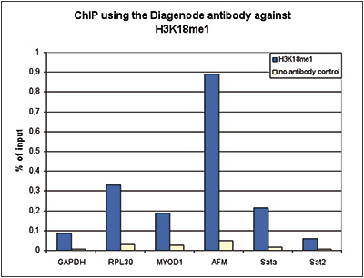
Figure 1. ChIP results obtained with the Diagenode antibody directed against H3K18me1 ChIP assays were performed using 2 μg of the Diagenode antibody against H3K18me1 (cat. No. C15410290) on sheared chromatin from 1 million cells. A no antibody negative IP control was also included. QPCR was performed with primers for the indicated genes or genomic regions. Figure 1 shows the recovery, expressed as a % of input (the relative amount of immunoprecipitated DNA compared to input DNA after qPCR analysis).
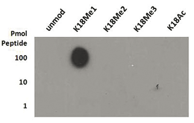
Figure 2. Cross reactivity test of the Diagenode antibody directed against H3K18me1 A Dot Blot analysis was performed to test the cross reactivity of the Diagenode antibody against H3K18me1 (cat. No. C15410290) with peptides containing other modifications of H3K18 and the unmodified sequence. One hundred, 10 and 1 pmol of the peptide containing the respective histone modification were spotted on a membrane. The antibody was used at a dilution of 1:1,000. Figure 2 shows a high specificity of the antibody for the modification of interest.
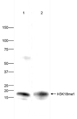
Figure 3. Western blot analysis using the Diagenode antibody directed against H3K18me1 Western blot was performed on histone extracts (30 μg) from HeLa (lane 1) and NIH3T3 cells (lane 2) using the Diagenode antibody against H3K18me1 (cat. No. C15410290) diluted 1:500 in TBS-Tween containing 5% skimmed milk. The position of the protein of interest is indicated on the right; the marker (in kDa) is shown on the left.
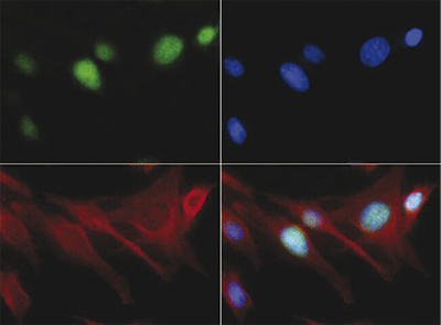
Figure 4. Immunofluorescence using the Diagenode antibody directed against H3K18me1 HeLa cells were stained with the Diagenode antibody against H3K18me1 (cat. No. C15410190) (green), with an anti-actin antibody (red) and with DAPI (blue). Cells were fixed with 0.5% formaldehyde. The cells were incubated with the H3K18me1 antibody diluted 1:50 in blocking solution for 1h at RT, followed by an anti-rabbit antibody conjugated to FITC. The lower right panel shows a merge of the three stainings.
IF Immunofluorescence:
Diagenode offers huge selection of highly sensitive antibodies validated in IF.
Immunofluorescence using the Diagenode monoclonal antibody directed against CRISPR/Cas9
HeLa cells transfected with a Cas9 expression vector (... Read more
WB Western blot : The quality of antibodies used in this technique is crucial for correct and specific protein identification. Diagenode offers huge selection of highly sensitive and specific western blot-validated antibodies.
Learn more about: Load... Read more