| ELISA Enzyme-linked immunosorbent assay. Read more |
| DB Dot blotting Read more |
| WB Western blot : The quality of antibodies used in this technique is crucial for correct and specific protein identification. Diagenode offers huge selection of highly sensitive and specific western blot-validated antibodies. Learn more about: Load... Read more |
| ChIP-seq (ab) Read more |
| ChIP-qPCR (ab) Read more |
H3K27me3S28p Antibody - ChIP-seq Grade
目录号
格式
价格

Polyclonal antibody raised in rabbit against against histone H3, trimethylated at lysine 27 and phosphorylated at serine 28 (H3K27me3S28p), using a KLH-conjugated synthetic peptide.
| Lot | A154-0011 |
|---|---|
| Concentration | 0.17 µg/µl |
| Species reactivity | Human |
| Type | Polyclonal |
| Purity | Affinity purified |
| Host | Rabbit |
| Precautions | This product is for research use only. Not for use in diagnostic or therapeutic procedures. |
| Applications | Suggested dilution | References |
|---|---|---|
| ChIP/ChIP-seq * | 1-2 μg/ChIP | Fig 1, 2 |
| ELISA | 1:200 | Fig 3 |
| Dot Blotting | 1:2,000 | Fig 4 |
| Western Blotting | 1:1,000 | Fig 5 |
* Please note that the optimal antibody amount per IP should be determined by the end-user. We recommend testing 1-5 μg per IP.
- Validation Data
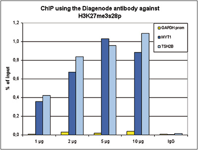
Figure 1. ChIP results obtained with the Diagenode antibody directed against H3K27me3S28p
ChIP assays were performed using human HeLa cells treated with colcemid, the Diagenode antibody against H3K27me3S28p (Cat. No. C15410091) and optimized PCR primer sets for qPCR. ChIP was performed with the “iDeal ChIP-seq” kit (Cat. No. C01010051), using sheared chromatin from 1 million cells. A titration of the antibody consisting of 1, 2, 5, and 10 μg per ChIP experiment was analysed. IgG (2 μg/IP) was used as negative IP control. QPCR was performed with primers for the promoter of the active gene GAPDH as a negative control, and for the coding regions of the inactive genes MYT1 and TSH2B as positive controls. Figure 1 shows the recovery, expressed as a % of input (the relative amount of immunoprecipitated DNA compared to input DNA after qPCR analysis). These results are in accordance with the observation that H3K27me3S28p is preferably present at inactive genes.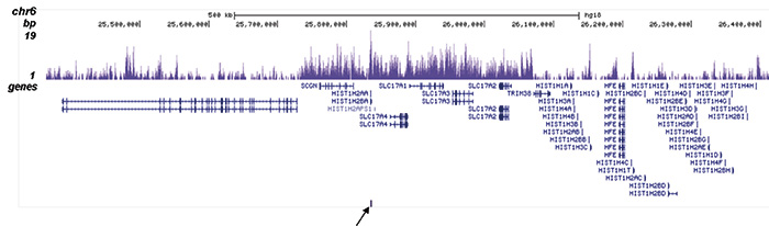
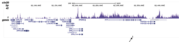
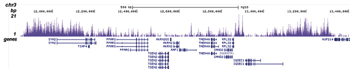
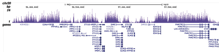
Figure 2. ChIP-seq results obtained with the Diagenode antibody directed against H3K27me3S28p
ChIP was performed on sheared chromatin from 1.5 million colcemid treated HeLaS3 cells using 2 μg of the Diagenode antibody against H3K27me3S28p (Cat. No. C15410091) as described above. The IP’d DNA was subsequently analysed on an Illumina HiSeq. Library preparation, cluster generation and sequencing were performed according to the manufacturer’s instructions. The 51 bp tags were aligned to the human genome using the BWA algorithm. Figure 2 shows the enrichment in genomic regions surrounding the TSH2B and MYT1 positive control genes (fig 2A and B, indicated by an arrow), and of chromosome 3 and 20 (figure 2C and D).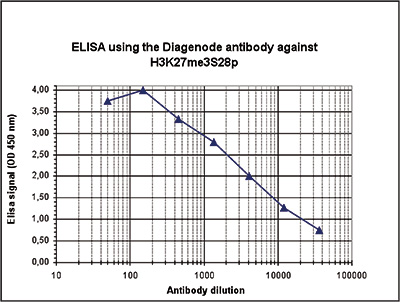
Figure 3. Determination of the antibody titer
To determine the titer of the antibody, an ELISA was performed using a serial dilution of the Diagenode antibody directed against H3K27me3S28p (Cat. No. C15410091). The antigen used was a peptide containing the histone modification of interest. By plotting the absorbance against the antibody dilution (Figure 2), the titer of the antibody was estimated to be 1:4,300.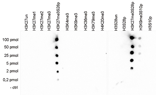
Figure 4. Cross reactivity test of the Diagenode antibody directed against H3K27me3S28p
A Dot Blot analysis was performed to test the cross reactivity of the Diagenode antibody against H3K27me3S28p (Cat. No. C15410091) with peptides containing other modifications of histone H3 and H4 and the unmodified H3K27S28 sequence. One hundred to 0.2 pmol of the peptide containing the respective histone modification were spotted on a membrane. The antibody was used at a dilution of 1:2,000. Figure 3 shows a high specificity of the antibody for the modification of interest.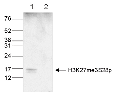
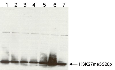
Figure 5. Western blot analysis using the Diagenode antibody directed against H3K27me3S28p
A. Western blot was performed on histone extracts (15 μg, lane 1) from HeLa cells, and on 1 μg of recombinant histone H3 (lane 2) using the Diagenode antibody against H3K27me3S28p (Cat. No. C15410091) diluted 1:100 in TBS-Tween containing 5% skimmed milk. The position of the protein of interest is indicated on the right; the marker (in kDa) is shown on the left. B. Cell cycle experiment using the Diagenode antibody against H3K27me3S28p. WB was performed on cells blocked in different stages of the cell cycle (1=asynchronous population, 2=G1/Sa, 3=G1/Sb, 4=S phase, 5=G2/M, 6=M, 7=G1). The picture shows a nice peak of H3K27me3S28p expression during mitosis. - 出版物

How to properly cite our product/service in your work
We strongly recommend using this: H3K27me3S28p Antibody - ChIP-seq Grade (Hologic Diagenode Cat# C15410091 Lot# A154-0011). Click here to copy to clipboard.
Using our products or services in your publication? Let us know!