Notice (8): Undefined variable: solution_of_interest [APP/View/Products/view.ctp, line 755]Code Context<!-- BEGIN: REQUEST_FORM MODAL -->
<div id="request_formModal" class="reveal-modal medium" data-reveal aria-labelledby="modalTitle" aria-hidden="true" role="dialog">
<?= $this->element('Forms/simple_form', array('solution_of_interest' => $solution_of_interest, 'header' => $header, 'message' => $message, 'campaign_id' => $campaign_id)) ?>
$viewFile = '/home/website-server/www/app/View/Products/view.ctp'
$dataForView = array(
'language' => 'cn',
'meta_keywords' => '',
'meta_description' => 'H3K9/14ac (Histone acetylated at lysine 9 and 14) Polyclonal Antibody validated in ChIP-seq, ChIP-qPCR, ELISA, DB, WB and IF. Batch-specific data available on the website. Sample size available',
'meta_title' => 'H3K9/14ac Antibody - ChIP-seq Grade (C15410005) | Diagenode',
'product' => array(
'Product' => array(
'id' => '2176',
'antibody_id' => '119',
'name' => 'H3K9/14ac Antibody - ChIP-seq Grade (sample size)',
'description' => '<p><span>Polyclonal antibody raised in rabbit against histone H3 acetylated at lysines 9 and 14 (H3K9/14ac), using a KLH-conjugated synthetic peptide.</span></p>',
'label1' => '',
'info1' => '',
'label2' => '',
'info2' => '',
'label3' => '',
'info3' => '',
'format' => '10 μg',
'catalog_number' => 'C15410005-10',
'old_catalog_number' => 'pAb-005-050',
'sf_code' => 'C15410005-D001-000582',
'type' => 'FRE',
'search_order' => '03-Antibody',
'price_EUR' => '105',
'price_USD' => '120',
'price_GBP' => '100',
'price_JPY' => '17205',
'price_CNY' => '',
'price_AUD' => '300',
'country' => 'ALL',
'except_countries' => 'None',
'quote' => false,
'in_stock' => false,
'featured' => false,
'no_promo' => false,
'online' => true,
'master' => false,
'last_datasheet_update' => '0000-00-00',
'slug' => 'h3k9-14ac-polyclonal-antibody-classic-sample-size-10-mg',
'meta_title' => 'H3K9/14ac Antibody - ChIP-seq Grade (C15410005) | Diagenode',
'meta_keywords' => '',
'meta_description' => 'H3K9/14ac (Histone acetylated at lysine 9 and 14) Polyclonal Antibody validated in ChIP-seq, ChIP-qPCR, ELISA, DB, WB and IF. Batch-specific data available on the website. Sample size available',
'modified' => '2021-10-20 09:31:12',
'created' => '2015-06-29 14:08:20',
'locale' => 'zho'
),
'Antibody' => array(
'host' => '*****',
'id' => '119',
'name' => 'H3K9/14ac polyclonal antibody',
'description' => 'Histones are the main constituents of the protein part of chromosomes of eukaryotic cells. They are rich in the amino acids arginine and lysine and have been greatly conserved during evolution. Histones pack the DNA into tight masses of chromatin. Two core histones of each class H2A, H2B, H3 and H4 assemble and are wrapped by 146 base pairs of DNA to form one octameric nucleosome. Histone tails undergo numerous post-translational modifications, which either directly or indirectly alter chromatin structure to facilitate transcriptional activation or repression or other nuclear processes. In addition to the genetic code, combinations of the different histone modifications reveal the so-called “histone code”. Histone methylation and demethylation is dynamically regulated by respectively histone methyl transferases and histone demethylases. Acetylation of H3K9/14 is enriched near the promoters of active genes.active genes.',
'clonality' => '',
'isotype' => '',
'lot' => 'A381-004',
'concentration' => '1.39 µg/µl',
'reactivity' => 'Human, mouse, zebrafish, Nematodes, A. Nidulans, Arabidopsis',
'type' => 'Polyclonal',
'purity' => 'Affinity purified',
'classification' => 'Premium',
'application_table' => '<table>
<thead>
<tr>
<th>Applications</th>
<th>Suggested dilution</th>
<th>References</th>
</tr>
</thead>
<tbody>
<tr>
<td>ChIP/ChIP-seq <sup>*</sup></td>
<td>1-2 μg/ChIP</td>
<td>Fig 1, 2</td>
</tr>
<tr>
<td>CUT&TAG</td>
<td>1 μg</td>
<td>Fig 3</td>
</tr>
<tr>
<td>ELISA</td>
<td>1:100</td>
<td>Fig 4</td>
</tr>
<tr>
<td>Dot Blotting</td>
<td>1:20,000</td>
<td>Fig 5</td>
</tr>
<tr>
<td>Western Blotting</td>
<td>1:1,000</td>
<td>Fig 6</td>
</tr>
<tr>
<td>Immunofluorescence</td>
<td>1:500</td>
<td>Fig 7</td>
</tr>
</tbody>
</table>
<p><small><sup>*</sup> Please note that the optimal antibody amount per IP should be determined by the end-user. We recommend testing 1-5 μg per IP.</small></p>',
'storage_conditions' => '',
'storage_buffer' => '',
'precautions' => 'This product is for research use only. Not for use in diagnostic or therapeutic procedures.',
'uniprot_acc' => '',
'slug' => '',
'meta_keywords' => '',
'meta_description' => '',
'modified' => '2021-12-23 10:47:07',
'created' => '0000-00-00 00:00:00',
'select_label' => '119 - H3K9/14ac polyclonal antibody (A381-004 - 1.39 µg/µl - Human, mouse, zebrafish, Nematodes, A. Nidulans, Arabidopsis - Affinity purified - Rabbit)'
),
'Slave' => array(),
'Group' => array(
'Group' => array(
[maximum depth reached]
),
'Master' => array(
[maximum depth reached]
),
'Product' => array(
[maximum depth reached]
)
),
'Related' => array(
(int) 0 => array(
[maximum depth reached]
)
),
'Application' => array(
(int) 0 => array(
[maximum depth reached]
),
(int) 1 => array(
[maximum depth reached]
),
(int) 2 => array(
[maximum depth reached]
),
(int) 3 => array(
[maximum depth reached]
),
(int) 4 => array(
[maximum depth reached]
),
(int) 5 => array(
[maximum depth reached]
)
),
'Category' => array(
(int) 0 => array(
[maximum depth reached]
),
(int) 1 => array(
[maximum depth reached]
),
(int) 2 => array(
[maximum depth reached]
),
(int) 3 => array(
[maximum depth reached]
),
(int) 4 => array(
[maximum depth reached]
)
),
'Document' => array(
(int) 0 => array(
[maximum depth reached]
),
(int) 1 => array(
[maximum depth reached]
),
(int) 2 => array(
[maximum depth reached]
)
),
'Feature' => array(),
'Image' => array(
(int) 0 => array(
[maximum depth reached]
)
),
'Promotion' => array(),
'Protocol' => array(),
'Publication' => array(
(int) 0 => array(
[maximum depth reached]
),
(int) 1 => array(
[maximum depth reached]
),
(int) 2 => array(
[maximum depth reached]
),
(int) 3 => array(
[maximum depth reached]
),
(int) 4 => array(
[maximum depth reached]
),
(int) 5 => array(
[maximum depth reached]
),
(int) 6 => array(
[maximum depth reached]
),
(int) 7 => array(
[maximum depth reached]
),
(int) 8 => array(
[maximum depth reached]
),
(int) 9 => array(
[maximum depth reached]
),
(int) 10 => array(
[maximum depth reached]
),
(int) 11 => array(
[maximum depth reached]
),
(int) 12 => array(
[maximum depth reached]
),
(int) 13 => array(
[maximum depth reached]
),
(int) 14 => array(
[maximum depth reached]
),
(int) 15 => array(
[maximum depth reached]
),
(int) 16 => array(
[maximum depth reached]
),
(int) 17 => array(
[maximum depth reached]
),
(int) 18 => array(
[maximum depth reached]
),
(int) 19 => array(
[maximum depth reached]
)
),
'Testimonial' => array(),
'Area' => array(),
'SafetySheet' => array(
(int) 0 => array(
[maximum depth reached]
),
(int) 1 => array(
[maximum depth reached]
),
(int) 2 => array(
[maximum depth reached]
),
(int) 3 => array(
[maximum depth reached]
),
(int) 4 => array(
[maximum depth reached]
),
(int) 5 => array(
[maximum depth reached]
),
(int) 6 => array(
[maximum depth reached]
),
(int) 7 => array(
[maximum depth reached]
)
)
),
'meta_canonical' => 'https://www.diagenode.com/cn/p/h3k9-14ac-polyclonal-antibody-classic-50-mg-36-ml'
)
$language = 'cn'
$meta_keywords = ''
$meta_description = 'H3K9/14ac (Histone acetylated at lysine 9 and 14) Polyclonal Antibody validated in ChIP-seq, ChIP-qPCR, ELISA, DB, WB and IF. Batch-specific data available on the website. Sample size available'
$meta_title = 'H3K9/14ac Antibody - ChIP-seq Grade (C15410005) | Diagenode'
$product = array(
'Product' => array(
'id' => '2176',
'antibody_id' => '119',
'name' => 'H3K9/14ac Antibody - ChIP-seq Grade (sample size)',
'description' => '<p><span>Polyclonal antibody raised in rabbit against histone H3 acetylated at lysines 9 and 14 (H3K9/14ac), using a KLH-conjugated synthetic peptide.</span></p>',
'label1' => 'Validation Data',
'info1' => '<div class="row">
<div class="small-4 columns">
<p><img src="https://www.diagenode.com/img/product/antibodies/C15410005-ChIP-Fig1.png" alt="H3K9/14ac Antibody ChIP Grade" caption="false" width="288" height="212" /></p>
</div>
<div class="small-8 columns">
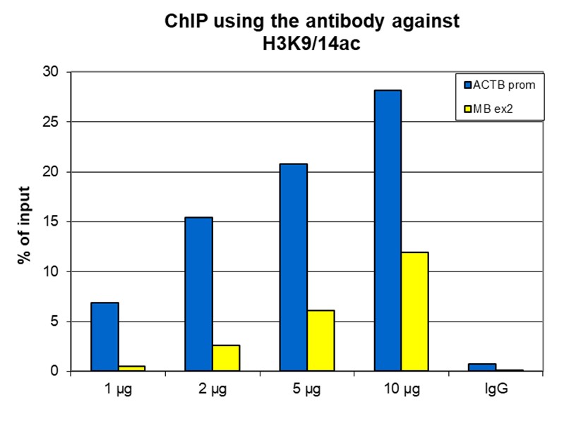
<p><small><strong> Figure 1. ChIP results obtained with the Diagenode antibody directed against H3K9/14ac. </strong><br />ChIP assays were performed using HeLa cells, the Diagenode antibody against H3K9/14ac (Cat. No. pAb-005-044) and optimized primer pairs for qPCR. ChIP was performed with the “HighCell# ChIP” kit (Cat. No. kch- mahigh-A16), using sheared chromatin from 1.5 million cells. A titration of the antibody consisting of 1, 2, 5 and 10 μg per ChIP experiment was analysed. IgG (5 μg/IP) was used as negative IP control.QPCR was performed using primers specific for the promoter of the ACTB gene (Cat. No. pp-1005- 050) as a positive control target and for exon 2 of the MB gene (Cat. No. pp-1006-050) as a negative control target. Figure 1 shows the recovery (the relative amount of immunoprecipitated DNA compared to input DNA). These results confirm the observation that acetylation of H3K9/14 is present at active promoters. </small></p>
</div>
</div>
<div class="row">
<div class="small-4 columns">
<p><img src="https://www.diagenode.com/img/product/antibodies/C15410005-ChIP-Fig2.png" alt="H3K9/14ac Antibody for ChIP-seq" caption="false" width="288" height="273" /></p>
</div>
<div class="small-8 columns">
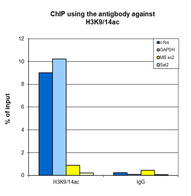
<p><small><strong> Figure 2. ChIP-seq results obtained with the Diagenode antibody directed against H3K9/14ac; </strong><br />ChIP was performed with 1 μg of the Diagenode antibody against H3K9/14ac (Cat. No. pAb-005-044) on sheared chromatin from 1 million HeLaS3 cells using the “Auto Histone ChIP-seq” kit (Cat. No. AB-Auto02-A100) on the IP-Star automated system. IgG (2 μg/IP) was used as a negative IP control. The IP’d DNA was analysed by QPCR with optimized PCR primer pairs for the promoters of the active GAPDH and c-fos genes, used as positive control targets, and the coding region of the inactive MB gene and the Sat2 satellite repeat, used as negative control targets (figure 2A). The IP’d DNA was subsequently analysed with an Illumina Genome Analyzer. Library preparation, cluster generation and sequencing were performed according to the manufacturer’s instructions. The 36 bp tags were aligned to the human genome using the ELAND algorithm. Figure 2 shows the peak distribution along the complete sequence and a 800 kb region of the X-chromosome (figure 2B and C) and in 100 kb regions surrounding the RBM3, GAPDH and c-fos genes (figure 2D, E and F). These results clearly show an enrichment of the H3K9/14 double acetylation at the promoters of active genes. </small></p>
</div>
</div>
<div class="row">
<div class="small-12 columns">
<p><img src="https://www.diagenode.com/img/product/antibodies/C15410005-ChIP-Fig2A.png" alt="H3K9/14ac Antibody ChIP-seq Grade" caption="false" width="700" height="534" /></p>
</div>
</div>
<div class="row">
<div class="small-4 columns">
<p><img src="https://www.diagenode.com/img/product/antibodies/C15410005-ELISA-Fig3.png" alt="H3K9/14ac Antibody ELISA validation" caption="false" width="288" height="263" /></p>
</div>
<div class="small-8 columns">
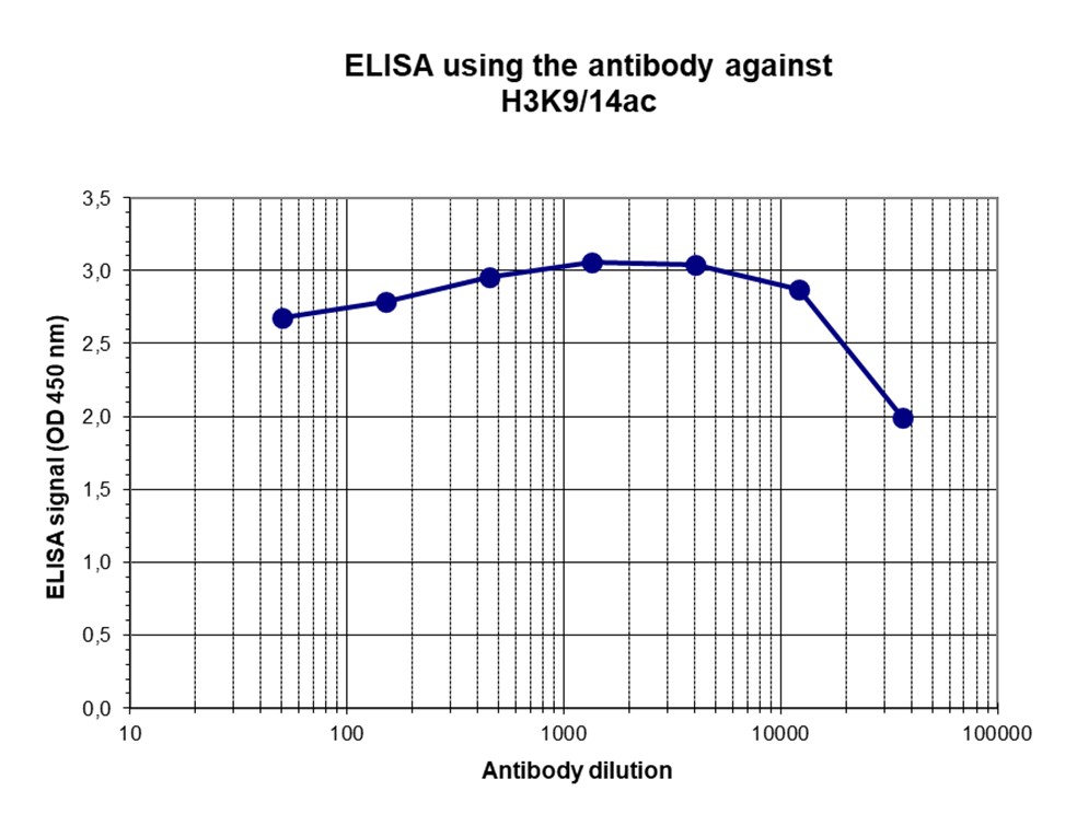
<p><small><strong> Figure 3. Determination of the antibody titer. </strong><br />To determine the titer of the antibody, an ELISA was performed using a serial dilution of the Diagenode antibody directed against H3K9/14ac (Cat. No. pAb-005-044), crude serum and flow through in antigen coated wells. The antigen used was a peptide containing the histone modification of interest. By plotting the absorbance against the antibody dilution (Figure 3), the titer of the purified antibody was estimated to be 1:5,900. </small></p>
</div>
</div>
<div class="row">
<div class="small-4 columns">
<p><img src="https://www.diagenode.com/img/product/antibodies/C15410005-CrossReactivity-Fig4.png" alt="H3K9/14ac Antibody Dot Blot validation" caption="false" width="288" height="204" /></p>
</div>
<div class="small-8 columns">
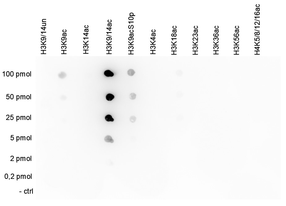
<p><small><strong> Figure 4. Cross reactivity tests using the Diagenode antibody directed against H3K9/14ac </strong><br />A Dot Blot analysis was performed to test the cross reactivity of the Diagenode antibody against H3K9/14ac (Cat. No. pAb-005- 044) with peptides containing other histone modifications and the unmodified H3K9/14 sequence. One hundred to 0.2 pmol of the respective peptides were spotted on a membrane. The antibody was used at a dilution of 1:20,000. Figure 4 shows a high specificity of the antibody for the modification of interest. </small></p>
</div>
</div>
<div class="row">
<div class="small-4 columns">
<p><img src="https://www.diagenode.com/img/product/antibodies/C15410005-WB-Fig5.png" alt="H3K9/14ac Antibody validated in Western Blot" caption="false" width="288" height="291" /></p>
</div>
<div class="small-8 columns">
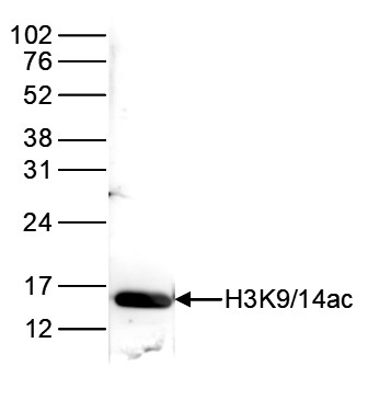
<p><small><strong> Figure 5. Western blot analysis using the Diagenode antibody directed against H3K9/14ac. </strong><br />Histone extracts of HeLa cells (15 μg) were analysed by Western blot using the Diagenode antibody directed against H3K9/14ac (Cat. No. pAb- 005-044) diluted 1:1,000 in TBS-Tween containing 5% skimmed milk. The position of the protein of interest is indicated on the right; the marker (in kDa) is shown on the left. </small></p>
</div>
</div>
<div class="row">
<div class="small-5 columns">
<p><img src="https://www.diagenode.com/img/product/antibodies/C15410005-IF-Fig6.png" alt="H3K9/14ac Antibody validated in Immunofluorescence" caption="false" width="367" height="91" /></p>
</div>
<div class="small-7 columns">
<p><small><strong> Figure 6. Immunofluorescence using the Diagenode antibody directed against H3K9/14ac </strong><br />Mouse NIH3T3 cells were stained with the Diagenode antibody against H3K9/14ac (Cat. No. pAb-005-044) and with DAPI. Cells were fixed with 4% formaldehyde for 10’ and blocked with PBS/TX-100 containing 5% normal goat serum and 1% BSA. The cells were immunofluorescently labelled with the H3K9/14ac antibody (left) diluted 1:500 in blocking solution followed by an anti- rabbit antibody conjugated to Alexa488. The middle panel shows staining of the nuclei with DAPI. A merge of the two stainings is shown on the right. </small></p>
</div>
</div>',
'label2' => 'Target Description',
'info2' => '<p>Histones are the main constituents of the protein part of chromosomes of eukaryotic cells. They are rich in the amino acids arginine and lysine and have been greatly conserved during evolution. Histones pack the DNA into tight masses of chromatin. Two core histones of each class H2A, H2B, H3 and H4 assemble and are wrapped by 146 base pairs of DNA to form one octameric nucleosome. Histone tails undergo numerous post-translational modifications, which either directly or indirectly alter chromatin structure to facilitate transcriptional activation or repression or other nuclear processes. In addition to the genetic code, combinations of the different histone modifications reveal the so-called “histone code”. Histone methylation and demethylation is dynamically regulated by respectively histone methyl transferases and histone demethylases. Acetylation of H3K9/14 is enriched near the promoters of active genes.active genes.</p>',
'label3' => '',
'info3' => '',
'format' => '10 μg',
'catalog_number' => 'C15410005-10',
'old_catalog_number' => 'pAb-005-050',
'sf_code' => 'C15410005-D001-000582',
'type' => 'FRE',
'search_order' => '03-Antibody',
'price_EUR' => '105',
'price_USD' => '120',
'price_GBP' => '100',
'price_JPY' => '17205',
'price_CNY' => '',
'price_AUD' => '300',
'country' => 'ALL',
'except_countries' => 'None',
'quote' => false,
'in_stock' => false,
'featured' => false,
'no_promo' => false,
'online' => true,
'master' => false,
'last_datasheet_update' => '0000-00-00',
'slug' => 'h3k9-14ac-polyclonal-antibody-classic-sample-size-10-mg',
'meta_title' => 'H3K9/14ac Antibody - ChIP-seq Grade (C15410005) | Diagenode',
'meta_keywords' => '',
'meta_description' => 'H3K9/14ac (Histone acetylated at lysine 9 and 14) Polyclonal Antibody validated in ChIP-seq, ChIP-qPCR, ELISA, DB, WB and IF. Batch-specific data available on the website. Sample size available',
'modified' => '2021-10-20 09:31:12',
'created' => '2015-06-29 14:08:20',
'locale' => 'zho'
),
'Antibody' => array(
'host' => '*****',
'id' => '119',
'name' => 'H3K9/14ac polyclonal antibody',
'description' => 'Histones are the main constituents of the protein part of chromosomes of eukaryotic cells. They are rich in the amino acids arginine and lysine and have been greatly conserved during evolution. Histones pack the DNA into tight masses of chromatin. Two core histones of each class H2A, H2B, H3 and H4 assemble and are wrapped by 146 base pairs of DNA to form one octameric nucleosome. Histone tails undergo numerous post-translational modifications, which either directly or indirectly alter chromatin structure to facilitate transcriptional activation or repression or other nuclear processes. In addition to the genetic code, combinations of the different histone modifications reveal the so-called “histone code”. Histone methylation and demethylation is dynamically regulated by respectively histone methyl transferases and histone demethylases. Acetylation of H3K9/14 is enriched near the promoters of active genes.active genes.',
'clonality' => '',
'isotype' => '',
'lot' => 'A381-004',
'concentration' => '1.39 µg/µl',
'reactivity' => 'Human, mouse, zebrafish, Nematodes, A. Nidulans, Arabidopsis',
'type' => 'Polyclonal',
'purity' => 'Affinity purified',
'classification' => 'Premium',
'application_table' => '<table>
<thead>
<tr>
<th>Applications</th>
<th>Suggested dilution</th>
<th>References</th>
</tr>
</thead>
<tbody>
<tr>
<td>ChIP/ChIP-seq <sup>*</sup></td>
<td>1-2 μg/ChIP</td>
<td>Fig 1, 2</td>
</tr>
<tr>
<td>CUT&TAG</td>
<td>1 μg</td>
<td>Fig 3</td>
</tr>
<tr>
<td>ELISA</td>
<td>1:100</td>
<td>Fig 4</td>
</tr>
<tr>
<td>Dot Blotting</td>
<td>1:20,000</td>
<td>Fig 5</td>
</tr>
<tr>
<td>Western Blotting</td>
<td>1:1,000</td>
<td>Fig 6</td>
</tr>
<tr>
<td>Immunofluorescence</td>
<td>1:500</td>
<td>Fig 7</td>
</tr>
</tbody>
</table>
<p><small><sup>*</sup> Please note that the optimal antibody amount per IP should be determined by the end-user. We recommend testing 1-5 μg per IP.</small></p>',
'storage_conditions' => '',
'storage_buffer' => '',
'precautions' => 'This product is for research use only. Not for use in diagnostic or therapeutic procedures.',
'uniprot_acc' => '',
'slug' => '',
'meta_keywords' => '',
'meta_description' => '',
'modified' => '2021-12-23 10:47:07',
'created' => '0000-00-00 00:00:00',
'select_label' => '119 - H3K9/14ac polyclonal antibody (A381-004 - 1.39 µg/µl - Human, mouse, zebrafish, Nematodes, A. Nidulans, Arabidopsis - Affinity purified - Rabbit)'
),
'Slave' => array(),
'Group' => array(
'Group' => array(
'id' => '50',
'name' => 'C15410005',
'product_id' => '2175',
'modified' => '2016-02-18 20:51:42',
'created' => '2016-02-18 20:51:42'
),
'Master' => array(
'id' => '2175',
'antibody_id' => '119',
'name' => 'H3K9/14ac Antibody',
'description' => '<p><span>Polyclonal antibody raised in rabbit against histone <strong>H3 acetylated at lysines 9 and 14 (H3K9/14ac)</strong>, using a KLH-conjugated synthetic peptide.</span></p>',
'label1' => 'Validation Data',
'info1' => '<div class="row">
<div class="small-4 columns">
<p><img src="https://www.diagenode.com/img/product/antibodies/C15410005-ChIP-Fig1.png" alt="H3K9/14ac Antibody ChIP Grade" caption="false" width="288" height="212" /></p>
</div>
<div class="small-8 columns">
<p><small><strong> Figure 1. ChIP results obtained with the Diagenode antibody directed against H3K9/14ac. </strong><br />ChIP assays were performed using HeLa cells, the Diagenode antibody against H3K9/14ac (Cat. No. pAb-005-044) and optimized primer pairs for qPCR. ChIP was performed with the “HighCell# ChIP” kit (Cat. No. kch- mahigh-A16), using sheared chromatin from 1.5 million cells. A titration of the antibody consisting of 1, 2, 5 and 10 μg per ChIP experiment was analysed. IgG (5 μg/IP) was used as negative IP control.QPCR was performed using primers specific for the promoter of the ACTB gene (Cat. No. pp-1005- 050) as a positive control target and for exon 2 of the MB gene (Cat. No. pp-1006-050) as a negative control target. Figure 1 shows the recovery (the relative amount of immunoprecipitated DNA compared to input DNA). These results confirm the observation that acetylation of H3K9/14 is present at active promoters. </small></p>
</div>
</div>
<div class="row">
<div class="small-4 columns">
<p><img src="https://www.diagenode.com/img/product/antibodies/C15410005-ChIP-Fig2.png" alt="H3K9/14ac Antibody for ChIP-seq" caption="false" width="288" height="273" /></p>
</div>
<div class="small-8 columns">
<p><small><strong> Figure 2. ChIP-seq results obtained with the Diagenode antibody directed against H3K9/14ac; </strong><br />ChIP was performed with 1 μg of the Diagenode antibody against H3K9/14ac (Cat. No. pAb-005-044) on sheared chromatin from 1 million HeLaS3 cells using the “Auto Histone ChIP-seq” kit (Cat. No. AB-Auto02-A100) on the IP-Star automated system. IgG (2 μg/IP) was used as a negative IP control. The IP’d DNA was analysed by QPCR with optimized PCR primer pairs for the promoters of the active GAPDH and c-fos genes, used as positive control targets, and the coding region of the inactive MB gene and the Sat2 satellite repeat, used as negative control targets (figure 2A). The IP’d DNA was subsequently analysed with an Illumina Genome Analyzer. Library preparation, cluster generation and sequencing were performed according to the manufacturer’s instructions. The 36 bp tags were aligned to the human genome using the ELAND algorithm. Figure 2 shows the peak distribution along the complete sequence and a 800 kb region of the X-chromosome (figure 2B and C) and in 100 kb regions surrounding the RBM3, GAPDH and c-fos genes (figure 2D, E and F). These results clearly show an enrichment of the H3K9/14 double acetylation at the promoters of active genes. </small></p>
</div>
</div>
<div class="row">
<div class="small-12 columns">
<p><img src="https://www.diagenode.com/img/product/antibodies/C15410005-ChIP-Fig2A.png" alt="H3K9/14ac Antibody ChIP-seq Grade" caption="false" width="700" height="534" /></p>
</div>
</div>
<div class="row">
<div class="small-4 columns">
<p><img src="https://www.diagenode.com/img/product/antibodies/C15410005-ELISA-Fig3.png" alt="H3K9/14ac Antibody ELISA validation" caption="false" width="288" height="263" /></p>
</div>
<div class="small-8 columns">
<p><small><strong> Figure 3. Determination of the antibody titer. </strong><br />To determine the titer of the antibody, an ELISA was performed using a serial dilution of the Diagenode antibody directed against H3K9/14ac (Cat. No. pAb-005-044), crude serum and flow through in antigen coated wells. The antigen used was a peptide containing the histone modification of interest. By plotting the absorbance against the antibody dilution (Figure 3), the titer of the purified antibody was estimated to be 1:5,900. </small></p>
</div>
</div>
<div class="row">
<div class="small-4 columns">
<p><img src="https://www.diagenode.com/img/product/antibodies/C15410005-CrossReactivity-Fig4.png" alt="H3K9/14ac Antibody Dot Blot validation" caption="false" width="288" height="204" /></p>
</div>
<div class="small-8 columns">
<p><small><strong> Figure 4. Cross reactivity tests using the Diagenode antibody directed against H3K9/14ac </strong><br />A Dot Blot analysis was performed to test the cross reactivity of the Diagenode antibody against H3K9/14ac (Cat. No. pAb-005- 044) with peptides containing other histone modifications and the unmodified H3K9/14 sequence. One hundred to 0.2 pmol of the respective peptides were spotted on a membrane. The antibody was used at a dilution of 1:20,000. Figure 4 shows a high specificity of the antibody for the modification of interest. </small></p>
</div>
</div>
<div class="row">
<div class="small-4 columns">
<p><img src="https://www.diagenode.com/img/product/antibodies/C15410005-WB-Fig5.png" alt="H3K9/14ac Antibody validated in Western Blot" caption="false" width="288" height="291" /></p>
</div>
<div class="small-8 columns">
<p><small><strong> Figure 5. Western blot analysis using the Diagenode antibody directed against H3K9/14ac. </strong><br />Histone extracts of HeLa cells (15 μg) were analysed by Western blot using the Diagenode antibody directed against H3K9/14ac (Cat. No. pAb- 005-044) diluted 1:1,000 in TBS-Tween containing 5% skimmed milk. The position of the protein of interest is indicated on the right; the marker (in kDa) is shown on the left. </small></p>
</div>
</div>
<div class="row">
<div class="small-5 columns">
<p><img src="https://www.diagenode.com/img/product/antibodies/C15410005-IF-Fig6.png" alt="H3K9/14ac Antibody validated in Immunofluorescence" caption="false" width="367" height="91" /></p>
</div>
<div class="small-7 columns">
<p><small><strong> Figure 6. Immunofluorescence using the Diagenode antibody directed against H3K9/14ac </strong><br />Mouse NIH3T3 cells were stained with the Diagenode antibody against H3K9/14ac (Cat. No. pAb-005-044) and with DAPI. Cells were fixed with 4% formaldehyde for 10’ and blocked with PBS/TX-100 containing 5% normal goat serum and 1% BSA. The cells were immunofluorescently labelled with the H3K9/14ac antibody (left) diluted 1:500 in blocking solution followed by an anti- rabbit antibody conjugated to Alexa488. The middle panel shows staining of the nuclei with DAPI. A merge of the two stainings is shown on the right. </small></p>
</div>
</div>',
'label2' => 'Target Description',
'info2' => '<p>Histones are the main constituents of the protein part of chromosomes of eukaryotic cells. They are rich in the amino acids arginine and lysine and have been greatly conserved during evolution. Histones pack the DNA into tight masses of chromatin. Two core histones of each class H2A, H2B, H3 and H4 assemble and are wrapped by 146 base pairs of DNA to form one octameric nucleosome. Histone tails undergo numerous post-translational modifications, which either directly or indirectly alter chromatin structure to facilitate transcriptional activation or repression or other nuclear processes. In addition to the genetic code, combinations of the different histone modifications reveal the so-called “histone code”. Histone methylation and demethylation is dynamically regulated by respectively histone methyl transferases and histone demethylases. Acetylation of H3K9/14 is enriched near the promoters of active genes.active genes.</p>',
'label3' => '',
'info3' => '',
'format' => '50 μg',
'catalog_number' => 'C15410005',
'old_catalog_number' => 'pAb-005-050',
'sf_code' => 'C15410005-D001-000581',
'type' => 'FRE',
'search_order' => '03-Antibody',
'price_EUR' => '390',
'price_USD' => '390',
'price_GBP' => '345',
'price_JPY' => '63900',
'price_CNY' => '',
'price_AUD' => '975',
'country' => 'ALL',
'except_countries' => 'None',
'quote' => false,
'in_stock' => false,
'featured' => false,
'no_promo' => false,
'online' => true,
'master' => true,
'last_datasheet_update' => '0000-00-00',
'slug' => 'h3k9-14ac-polyclonal-antibody-classic-50-mg-36-ml',
'meta_title' => 'H3K9/14ac Antibody - ChIP-seq Grade (C15410005) | Diagenode',
'meta_keywords' => '',
'meta_description' => 'H3K9/14ac (Histone H3 acetylated at lysines 9 and 14) Polyclonal Antibody validated in ChIP-seq, ChIP-qPCR, ELISA, DB, WB and IF. Batch-specific data available on the website. Sample size available.',
'modified' => '2021-10-20 09:30:54',
'created' => '2015-06-29 14:08:20'
),
'Product' => array(
(int) 0 => array(
[maximum depth reached]
)
)
),
'Related' => array(
(int) 0 => array(
'id' => '2175',
'antibody_id' => '119',
'name' => 'H3K9/14ac Antibody',
'description' => '<p><span>Polyclonal antibody raised in rabbit against histone <strong>H3 acetylated at lysines 9 and 14 (H3K9/14ac)</strong>, using a KLH-conjugated synthetic peptide.</span></p>',
'label1' => 'Validation Data',
'info1' => '<div class="row">
<div class="small-4 columns">
<p><img src="https://www.diagenode.com/img/product/antibodies/C15410005-ChIP-Fig1.png" alt="H3K9/14ac Antibody ChIP Grade" caption="false" width="288" height="212" /></p>
</div>
<div class="small-8 columns">
<p><small><strong> Figure 1. ChIP results obtained with the Diagenode antibody directed against H3K9/14ac. </strong><br />ChIP assays were performed using HeLa cells, the Diagenode antibody against H3K9/14ac (Cat. No. pAb-005-044) and optimized primer pairs for qPCR. ChIP was performed with the “HighCell# ChIP” kit (Cat. No. kch- mahigh-A16), using sheared chromatin from 1.5 million cells. A titration of the antibody consisting of 1, 2, 5 and 10 μg per ChIP experiment was analysed. IgG (5 μg/IP) was used as negative IP control.QPCR was performed using primers specific for the promoter of the ACTB gene (Cat. No. pp-1005- 050) as a positive control target and for exon 2 of the MB gene (Cat. No. pp-1006-050) as a negative control target. Figure 1 shows the recovery (the relative amount of immunoprecipitated DNA compared to input DNA). These results confirm the observation that acetylation of H3K9/14 is present at active promoters. </small></p>
</div>
</div>
<div class="row">
<div class="small-4 columns">
<p><img src="https://www.diagenode.com/img/product/antibodies/C15410005-ChIP-Fig2.png" alt="H3K9/14ac Antibody for ChIP-seq" caption="false" width="288" height="273" /></p>
</div>
<div class="small-8 columns">
<p><small><strong> Figure 2. ChIP-seq results obtained with the Diagenode antibody directed against H3K9/14ac; </strong><br />ChIP was performed with 1 μg of the Diagenode antibody against H3K9/14ac (Cat. No. pAb-005-044) on sheared chromatin from 1 million HeLaS3 cells using the “Auto Histone ChIP-seq” kit (Cat. No. AB-Auto02-A100) on the IP-Star automated system. IgG (2 μg/IP) was used as a negative IP control. The IP’d DNA was analysed by QPCR with optimized PCR primer pairs for the promoters of the active GAPDH and c-fos genes, used as positive control targets, and the coding region of the inactive MB gene and the Sat2 satellite repeat, used as negative control targets (figure 2A). The IP’d DNA was subsequently analysed with an Illumina Genome Analyzer. Library preparation, cluster generation and sequencing were performed according to the manufacturer’s instructions. The 36 bp tags were aligned to the human genome using the ELAND algorithm. Figure 2 shows the peak distribution along the complete sequence and a 800 kb region of the X-chromosome (figure 2B and C) and in 100 kb regions surrounding the RBM3, GAPDH and c-fos genes (figure 2D, E and F). These results clearly show an enrichment of the H3K9/14 double acetylation at the promoters of active genes. </small></p>
</div>
</div>
<div class="row">
<div class="small-12 columns">
<p><img src="https://www.diagenode.com/img/product/antibodies/C15410005-ChIP-Fig2A.png" alt="H3K9/14ac Antibody ChIP-seq Grade" caption="false" width="700" height="534" /></p>
</div>
</div>
<div class="row">
<div class="small-4 columns">
<p><img src="https://www.diagenode.com/img/product/antibodies/C15410005-ELISA-Fig3.png" alt="H3K9/14ac Antibody ELISA validation" caption="false" width="288" height="263" /></p>
</div>
<div class="small-8 columns">
<p><small><strong> Figure 3. Determination of the antibody titer. </strong><br />To determine the titer of the antibody, an ELISA was performed using a serial dilution of the Diagenode antibody directed against H3K9/14ac (Cat. No. pAb-005-044), crude serum and flow through in antigen coated wells. The antigen used was a peptide containing the histone modification of interest. By plotting the absorbance against the antibody dilution (Figure 3), the titer of the purified antibody was estimated to be 1:5,900. </small></p>
</div>
</div>
<div class="row">
<div class="small-4 columns">
<p><img src="https://www.diagenode.com/img/product/antibodies/C15410005-CrossReactivity-Fig4.png" alt="H3K9/14ac Antibody Dot Blot validation" caption="false" width="288" height="204" /></p>
</div>
<div class="small-8 columns">
<p><small><strong> Figure 4. Cross reactivity tests using the Diagenode antibody directed against H3K9/14ac </strong><br />A Dot Blot analysis was performed to test the cross reactivity of the Diagenode antibody against H3K9/14ac (Cat. No. pAb-005- 044) with peptides containing other histone modifications and the unmodified H3K9/14 sequence. One hundred to 0.2 pmol of the respective peptides were spotted on a membrane. The antibody was used at a dilution of 1:20,000. Figure 4 shows a high specificity of the antibody for the modification of interest. </small></p>
</div>
</div>
<div class="row">
<div class="small-4 columns">
<p><img src="https://www.diagenode.com/img/product/antibodies/C15410005-WB-Fig5.png" alt="H3K9/14ac Antibody validated in Western Blot" caption="false" width="288" height="291" /></p>
</div>
<div class="small-8 columns">
<p><small><strong> Figure 5. Western blot analysis using the Diagenode antibody directed against H3K9/14ac. </strong><br />Histone extracts of HeLa cells (15 μg) were analysed by Western blot using the Diagenode antibody directed against H3K9/14ac (Cat. No. pAb- 005-044) diluted 1:1,000 in TBS-Tween containing 5% skimmed milk. The position of the protein of interest is indicated on the right; the marker (in kDa) is shown on the left. </small></p>
</div>
</div>
<div class="row">
<div class="small-5 columns">
<p><img src="https://www.diagenode.com/img/product/antibodies/C15410005-IF-Fig6.png" alt="H3K9/14ac Antibody validated in Immunofluorescence" caption="false" width="367" height="91" /></p>
</div>
<div class="small-7 columns">
<p><small><strong> Figure 6. Immunofluorescence using the Diagenode antibody directed against H3K9/14ac </strong><br />Mouse NIH3T3 cells were stained with the Diagenode antibody against H3K9/14ac (Cat. No. pAb-005-044) and with DAPI. Cells were fixed with 4% formaldehyde for 10’ and blocked with PBS/TX-100 containing 5% normal goat serum and 1% BSA. The cells were immunofluorescently labelled with the H3K9/14ac antibody (left) diluted 1:500 in blocking solution followed by an anti- rabbit antibody conjugated to Alexa488. The middle panel shows staining of the nuclei with DAPI. A merge of the two stainings is shown on the right. </small></p>
</div>
</div>',
'label2' => 'Target Description',
'info2' => '<p>Histones are the main constituents of the protein part of chromosomes of eukaryotic cells. They are rich in the amino acids arginine and lysine and have been greatly conserved during evolution. Histones pack the DNA into tight masses of chromatin. Two core histones of each class H2A, H2B, H3 and H4 assemble and are wrapped by 146 base pairs of DNA to form one octameric nucleosome. Histone tails undergo numerous post-translational modifications, which either directly or indirectly alter chromatin structure to facilitate transcriptional activation or repression or other nuclear processes. In addition to the genetic code, combinations of the different histone modifications reveal the so-called “histone code”. Histone methylation and demethylation is dynamically regulated by respectively histone methyl transferases and histone demethylases. Acetylation of H3K9/14 is enriched near the promoters of active genes.active genes.</p>',
'label3' => '',
'info3' => '',
'format' => '50 μg',
'catalog_number' => 'C15410005',
'old_catalog_number' => 'pAb-005-050',
'sf_code' => 'C15410005-D001-000581',
'type' => 'FRE',
'search_order' => '03-Antibody',
'price_EUR' => '390',
'price_USD' => '390',
'price_GBP' => '345',
'price_JPY' => '63900',
'price_CNY' => '',
'price_AUD' => '975',
'country' => 'ALL',
'except_countries' => 'None',
'quote' => false,
'in_stock' => false,
'featured' => false,
'no_promo' => false,
'online' => true,
'master' => true,
'last_datasheet_update' => '0000-00-00',
'slug' => 'h3k9-14ac-polyclonal-antibody-classic-50-mg-36-ml',
'meta_title' => 'H3K9/14ac Antibody - ChIP-seq Grade (C15410005) | Diagenode',
'meta_keywords' => '',
'meta_description' => 'H3K9/14ac (Histone H3 acetylated at lysines 9 and 14) Polyclonal Antibody validated in ChIP-seq, ChIP-qPCR, ELISA, DB, WB and IF. Batch-specific data available on the website. Sample size available.',
'modified' => '2021-10-20 09:30:54',
'created' => '2015-06-29 14:08:20',
'ProductsRelated' => array(
[maximum depth reached]
),
'Image' => array(
[maximum depth reached]
)
)
),
'Application' => array(
(int) 0 => array(
'id' => '20',
'position' => '10',
'parent_id' => '40',
'name' => 'ELISA',
'description' => '<div class="row">
<div class="small-12 medium-12 large-12 columns">Enzyme-linked immunosorbent assay.</div>
</div>',
'in_footer' => false,
'in_menu' => false,
'online' => true,
'tabular' => true,
'slug' => 'elisa-antibodies',
'meta_keywords' => ' ELISA Antibodies,Monoclonal antibody, Polyclonal antibody',
'meta_description' => 'Diagenode offers Monoclonal & Polyclonal antibodies for ELISA applications',
'meta_title' => 'ELISA Antibodies - Monoclonal & Polyclonal antibody | Diagenode',
'modified' => '2016-01-13 12:21:41',
'created' => '2014-07-08 08:13:28',
'ProductsApplication' => array(
[maximum depth reached]
)
),
(int) 1 => array(
'id' => '28',
'position' => '10',
'parent_id' => '40',
'name' => 'DB',
'description' => '<p>Dot blotting</p>',
'in_footer' => false,
'in_menu' => false,
'online' => true,
'tabular' => true,
'slug' => 'dot-blotting',
'meta_keywords' => 'Dot blotting,Monoclonal & Polyclonal antibody,',
'meta_description' => 'Diagenode offers Monoclonal & Polyclonal antibodies for Dot blotting applications',
'meta_title' => 'Dot blotting Antibodies - Monoclonal & Polyclonal antibody | Diagenode',
'modified' => '2016-01-13 14:40:49',
'created' => '2015-07-08 13:45:05',
'ProductsApplication' => array(
[maximum depth reached]
)
),
(int) 2 => array(
'id' => '19',
'position' => '10',
'parent_id' => '40',
'name' => 'WB',
'description' => '<p><strong>Western blot</strong> : The quality of antibodies used in this technique is crucial for correct and specific protein identification. Diagenode offers huge selection of highly sensitive and specific western blot-validated antibodies.</p>
<p>Learn more about: <a href="https://www.diagenode.com/applications/western-blot">Loading control, MW marker visualization</a><em>. <br /></em></p>
<p><em></em>Check our selection of antibodies validated in Western blot.</p>',
'in_footer' => false,
'in_menu' => false,
'online' => true,
'tabular' => true,
'slug' => 'western-blot-antibodies',
'meta_keywords' => ' Western Blot Antibodies ,western blot protocol,Western Blotting Products,Polyclonal antibodies ,monoclonal antibodies ',
'meta_description' => 'Diagenode offers a wide range of antibodies and technical support for western blot applications',
'meta_title' => ' Western Blot - Monoclonal antibody - Polyclonal antibody | Diagenode',
'modified' => '2016-04-26 12:44:51',
'created' => '2015-01-07 09:20:00',
'ProductsApplication' => array(
[maximum depth reached]
)
),
(int) 3 => array(
'id' => '29',
'position' => '10',
'parent_id' => '40',
'name' => 'IF',
'description' => '<p><strong>Immunofluorescence</strong>:</p>
<p>Diagenode offers huge selection of highly sensitive antibodies validated in IF.</p>
<p><img src="https://www.diagenode.com/img/product/antibodies/C15200229-IF.jpg" alt="" height="245" width="256" /></p>
<p><sup><strong>Immunofluorescence using the Diagenode monoclonal antibody directed against CRISPR/Cas9</strong></sup></p>
<p><sup>HeLa cells transfected with a Cas9 expression vector (left) or untransfected cells (right) were fixed in methanol at -20°C, permeabilized with acetone at -20°C and blocked with PBS containing 2% BSA. The cells were stained with the Cas9 C-terminal antibody (Cat. No. C15200229) diluted 1:400, followed by incubation with an anti-mouse secondary antibody coupled to AF488. The bottom images show counter-staining of the nuclei with Hoechst 33342.</sup></p>
<h5><sup>Check our selection of antibodies validated in IF.</sup></h5>',
'in_footer' => false,
'in_menu' => false,
'online' => true,
'tabular' => true,
'slug' => 'immunofluorescence',
'meta_keywords' => 'Immunofluorescence,Monoclonal antibody,Polyclonal antibody',
'meta_description' => 'Diagenode offers a wide range of antibodies and technical support for Immunofluorescence applications',
'meta_title' => 'Immunofluorescence - Monoclonal antibody - Polyclonal antibody | Diagenode',
'modified' => '2016-04-27 16:23:10',
'created' => '2015-07-08 13:46:02',
'ProductsApplication' => array(
[maximum depth reached]
)
),
(int) 4 => array(
'id' => '42',
'position' => '10',
'parent_id' => '40',
'name' => 'ChIP-seq (ab)',
'description' => '',
'in_footer' => false,
'in_menu' => false,
'online' => true,
'tabular' => true,
'slug' => 'chip-seq-antibodies',
'meta_keywords' => 'Chromatin Immunoprecipitation Sequencing,ChIP-Seq,ChIP-seq grade antibodies,DNA purification,qPCR,Shearing of chromatin',
'meta_description' => 'Diagenode offers a wide range of antibodies and technical support for ChIP Sequencing applications',
'meta_title' => 'ChIP Sequencing Antibodies (ChIP-Seq) | Diagenode',
'modified' => '2016-01-20 11:06:19',
'created' => '2015-10-20 11:44:45',
'ProductsApplication' => array(
[maximum depth reached]
)
),
(int) 5 => array(
'id' => '43',
'position' => '10',
'parent_id' => '40',
'name' => 'ChIP-qPCR (ab)',
'description' => '',
'in_footer' => false,
'in_menu' => false,
'online' => true,
'tabular' => true,
'slug' => 'chip-qpcr-antibodies',
'meta_keywords' => 'Chromatin Immunoprecipitation Sequencing,ChIP-Seq,ChIP-seq grade antibodies,DNA purification,qPCR,Shearing of chromatin',
'meta_description' => 'Diagenode offers a wide range of antibodies and technical support for ChIP-qPCR applications',
'meta_title' => 'ChIP Quantitative PCR Antibodies (ChIP-qPCR) | Diagenode',
'modified' => '2016-01-20 11:30:24',
'created' => '2015-10-20 11:45:36',
'ProductsApplication' => array(
[maximum depth reached]
)
)
),
'Category' => array(
(int) 0 => array(
'id' => '17',
'position' => '10',
'parent_id' => '4',
'name' => 'ChIP-seq grade antibodies',
'description' => '<p><b>Unparalleled ChIP-Seq results with the most rigorously validated antibodies</b></p>
<p><span style="font-weight: 400;">Diagenode provides leading solutions for epigenetic research. Because ChIP-seq is a widely-used technique, we validate our antibodies in ChIP and ChIP-seq experiments (in addition to conventional methods like Western blot, Dot blot, ELISA, and immunofluorescence) to provide the highest quality antibody. We standardize our validation and production to guarantee high product quality without technical bias. Diagenode guarantees ChIP-seq grade antibody performance under our suggested conditions.</span></p>
<div class="row">
<div class="small-12 medium-9 large-9 columns">
<p><strong>ChIP-seq profile</strong> of active (H3K4me3 and H3K36me3) and inactive (H3K27me3) marks using Diagenode antibodies.</p>
<img src="https://www.diagenode.com/img/categories/antibodies/chip-seq-grade-antibodies.png" /></div>
<div class="small-12 medium-3 large-3 columns">
<p><small> ChIP was performed on sheared chromatin from 100,000 K562 cells using iDeal ChIP-seq kit for Histones (cat. No. C01010051) with 1 µg of the Diagenode antibodies against H3K27me3 (cat. No. C15410195) and H3K4me3 (cat. No. C15410003), and 0.5 µg of the antibody against H3K36me3 (cat. No. C15410192). The IP'd DNA was subsequently analysed on an Illumina Genome Analyzer. Library preparation, cluster generation and sequencing were performed according to the manufacturer's instructions. The 36 bp tags were aligned to the human genome using the ELAND algorithm. The figure shows the signal distribution along the complete sequence of human chromosome 3, a zoomin to a 10 Mb region and a further zoomin to a 1.5 Mb region. </small></p>
</div>
</div>
<p>Diagenode’s highly validated antibodies:</p>
<ul>
<li>Highly sensitive and specific</li>
<li>Cost-effective (requires less antibody per reaction)</li>
<li>Batch-specific data is available on the website</li>
<li>Expert technical support</li>
<li>Sample sizes available</li>
<li>100% satisfaction guarantee</li>
</ul>',
'no_promo' => false,
'in_menu' => true,
'online' => true,
'tabular' => false,
'hide' => true,
'all_format' => false,
'is_antibody' => true,
'slug' => 'chip-seq-grade-antibodies',
'cookies_tag_id' => null,
'meta_keywords' => 'ChIP-seq grade antibodies,polyclonal antibody,WB, ELISA, ChIP-seq (ab), ChIP-qPCR (ab)',
'meta_description' => 'Diagenode Offers Wide Range of Validated ChIP-Seq Grade Antibodies for Unparalleled ChIP-Seq Results',
'meta_title' => 'Chromatin Immunoprecipitation ChIP-Seq Grade Antibodies | Diagenode',
'modified' => '2019-07-03 10:57:22',
'created' => '2015-02-16 02:24:01',
'ProductsCategory' => array(
[maximum depth reached]
),
'CookiesTag' => array([maximum depth reached])
),
(int) 1 => array(
'id' => '111',
'position' => '40',
'parent_id' => '4',
'name' => 'Histone antibodies',
'description' => '<p>Histones are the main protein components of chromatin involved in the compaction of DNA into nucleosomes, the basic units of chromatin. A <strong>nucleosome</strong> consists of one pair of each of the core histones (<strong>H2A</strong>, <strong>H2B</strong>, <strong>H3</strong> and <strong>H4</strong>) forming an octameric structure wrapped by 146 base pairs of DNA. The different nucleosomes are linked by the linker histone<strong> H1, </strong>allowing for further condensation of chromatin.</p>
<p>The core histones have a globular structure with large unstructured N-terminal tails protruding from the nucleosome. They can undergo to multiple post-translational modifications (PTM), mainly at the N-terminal tails. These <strong>post-translational modifications </strong>include methylation, acetylation, phosphorylation, ubiquitinylation, citrullination, sumoylation, deamination and crotonylation. The most well characterized PTMs are <strong>methylation,</strong> <strong>acetylation and phosphorylation</strong>. Histone methylation occurs mainly on lysine (K) residues, which can be mono-, di- or tri-methylated, and on arginines (R), which can be mono-methylated and symmetrically or asymmetrically di-methylated. Histone acetylation occurs on lysines and histone phosphorylation mainly on serines (S), threonines (T) and tyrosines (Y).</p>
<p>The PTMs of the different residues are involved in numerous processes such as DNA repair, DNA replication and chromosome condensation. They influence the chromatin organization and can be positively or negatively associated with gene expression. Trimethylation of H3K4, H3K36 and H3K79, and lysine acetylation generally result in an open chromatin configuration (figure below) and are therefore associated with <strong>euchromatin</strong> and gene activation. Trimethylation of H3K9, K3K27 and H4K20, on the other hand, is enriched in <strong>heterochromatin </strong>and associated with gene silencing. The combination of different histone modifications is called the "<strong>histone code</strong>”, analogous to the genetic code.</p>
<p><img src="https://www.diagenode.com/img/categories/antibodies/histone-marks-illustration.png" /></p>
<p>Diagenode is proud to offer a large range of antibodies against histones and histone modifications. Our antibodies are highly specific and have been validated in many applications, including <strong>ChIP</strong> and <strong>ChIP-seq</strong>.</p>
<p>Diagenode’s collection includes antibodies recognizing:</p>
<ul>
<li><strong>Histone H1 variants</strong></li>
<li><strong>Histone H2A, H2A variants and histone H2A</strong> <strong>modifications</strong> (serine phosphorylation, lysine acetylation, lysine ubiquitinylation)</li>
<li><strong>Histone H2B and H2B</strong> <strong>modifications </strong>(serine phosphorylation, lysine acetylation)</li>
<li><strong>Histone H3 and H3 modifications </strong>(lysine methylation (mono-, di- and tri-methylated), lysine acetylation, serine phosphorylation, threonine phosphorylation, arginine methylation (mono-methylated, symmetrically and asymmetrically di-methylated))</li>
<li><strong>Histone H4 and H4 modifications (</strong>lysine methylation (mono-, di- and tri-methylated), lysine acetylation, arginine methylation (mono-methylated and symmetrically di-methylated), serine phosphorylation )</li>
</ul>
<p><span style="font-weight: 400;"><strong>HDAC's HAT's, HMT's and other</strong> <strong>enzymes</strong> which modify histones can be found in the category <a href="../categories/chromatin-modifying-proteins-histone-transferase">Histone modifying enzymes</a><br /></span></p>
<p><span style="font-weight: 400;"> Diagenode’s highly validated antibodies:</span></p>
<ul>
<li><span style="font-weight: 400;"> Highly sensitive and specific</span></li>
<li><span style="font-weight: 400;"> Cost-effective (requires less antibody per reaction)</span></li>
<li><span style="font-weight: 400;"> Batch-specific data is available on the website</span></li>
<li><span style="font-weight: 400;"> Expert technical support</span></li>
<li><span style="font-weight: 400;"> Sample sizes available</span></li>
<li><span style="font-weight: 400;"> 100% satisfaction guarantee</span></li>
</ul>',
'no_promo' => false,
'in_menu' => false,
'online' => true,
'tabular' => false,
'hide' => true,
'all_format' => false,
'is_antibody' => true,
'slug' => 'histone-antibodies',
'cookies_tag_id' => null,
'meta_keywords' => 'Histone, antibody, histone h1, histone h2, histone h3, histone h4',
'meta_description' => 'Polyclonal and Monoclonal Antibodies against Histones and their modifications validated for many applications, including Chromatin Immunoprecipitation (ChIP) and ChIP-Sequencing (ChIP-seq)',
'meta_title' => 'Histone and Modified Histone Antibodies | Diagenode',
'modified' => '2020-09-17 13:34:56',
'created' => '2016-04-01 16:01:32',
'ProductsCategory' => array(
[maximum depth reached]
),
'CookiesTag' => array([maximum depth reached])
),
(int) 2 => array(
'id' => '102',
'position' => '1',
'parent_id' => '4',
'name' => 'Sample size antibodies',
'description' => '<h1><strong>Validated epigenetics antibodies</strong> – care for a sample?<br /> </h1>
<p>Diagenode has partnered with leading epigenetics experts and numerous epigenetics consortiums to bring to you a validated and comprehensive collection of epigenetic antibodies. As an expert in epigenetics, we are committed to offering highly-specific antibodies validated for ChIP/ChIP-seq and many other applications. All batch-specific validation data is available on our website.<br /><a href="../categories/antibodies">Read about our expertise in antibody production</a>.</p>
<ul>
<li><strong>Focused</strong> - Diagenode's selection of antibodies is exclusively dedicated for epigenetic research. <a title="See the full collection." href="../categories/all-antibodies">See the full collection.</a></li>
<li><strong>Strict quality standards</strong> with rigorous QC and validation</li>
<li><strong>Classified</strong> based on level of validation for flexibility of application</li>
</ul>
<p>Existing sample sizes are listed below. We will soon expand our collection. Are you looking for a sample size of another antibody? Just <a href="mailto:agnieszka.zelisko@diagenode.com?Subject=Sample%20Size%20Request" target="_top">Contact us</a>.</p>',
'no_promo' => false,
'in_menu' => true,
'online' => true,
'tabular' => false,
'hide' => true,
'all_format' => true,
'is_antibody' => true,
'slug' => 'sample-size-antibodies',
'cookies_tag_id' => null,
'meta_keywords' => '5-hmC monoclonal antibody,CRISPR/Cas9 polyclonal antibody ,H3K36me3 polyclonal antibody,diagenode',
'meta_description' => 'Diagenode offers sample volume on selected antibodies for researchers to test, validate and provide confidence and flexibility in choosing from our wide range of antibodies ',
'meta_title' => 'Sample-size Antibodies | Diagenode',
'modified' => '2019-07-03 10:57:05',
'created' => '2015-10-27 12:13:34',
'ProductsCategory' => array(
[maximum depth reached]
),
'CookiesTag' => array([maximum depth reached])
),
(int) 3 => array(
'id' => '103',
'position' => '0',
'parent_id' => '4',
'name' => 'All antibodies',
'description' => '<p><span style="font-weight: 400;">All Diagenode’s antibodies are listed below. Please, use our Quick search field to find the antibody of interest by target name, application, purity.</span></p>
<p><span style="font-weight: 400;">Diagenode’s highly validated antibodies:</span></p>
<ul>
<li>Highly sensitive and specific</li>
<li>Cost-effective (requires less antibody per reaction)</li>
<li>Batch-specific data is available on the website</li>
<li>Expert technical support</li>
<li>Sample sizes available</li>
<li>100% satisfaction guarantee</li>
</ul>',
'no_promo' => false,
'in_menu' => true,
'online' => true,
'tabular' => false,
'hide' => true,
'all_format' => false,
'is_antibody' => true,
'slug' => 'all-antibodies',
'cookies_tag_id' => null,
'meta_keywords' => 'Antibodies,Premium Antibodies,Classic,Pioneer',
'meta_description' => 'Diagenode Offers Strict quality standards with Rigorous QC and validated Antibodies. Classified based on level of validation for flexibility of Application. Comprehensive selection of histone and non-histone Antibodies',
'meta_title' => 'Diagenode's selection of Antibodies is exclusively dedicated for Epigenetic Research | Diagenode',
'modified' => '2019-07-03 10:55:44',
'created' => '2015-11-02 14:49:22',
'ProductsCategory' => array(
[maximum depth reached]
),
'CookiesTag' => array([maximum depth reached])
),
(int) 4 => array(
'id' => '127',
'position' => '10',
'parent_id' => '4',
'name' => 'ChIP-grade antibodies',
'description' => '<div class="row">
<div class="small-12 columns"><center></center>
<p><br />Chromatin immunoprecipitation (<b>ChIP</b>) is a technique to study the associations of proteins with the specific genomic regions in intact cells. One of the most important steps of this protocol is the immunoprecipitation of targeted protein using the antibody specifically recognizing it. The quality of antibodies used in ChIP is essential for the success of the experiment. Diagenode offers extensively validated ChIP-grade antibodies, confirmed for their specificity, and high level of performance in ChIP. Each batch is validated, and batch-specific data are available on the website.</p>
<p></p>
</div>
</div>
<p><strong>ChIP results</strong> obtained with the antibody directed against H3K4me3 (Cat. No. <a href="../p/h3k4me3-polyclonal-antibody-premium-50-ug-50-ul">C15410003</a>). </p>
<div class="row">
<div class="small-12 medium-6 large-6 columns"><img src="https://www.diagenode.com/img/product/antibodies/C15410003-fig1-ChIP.jpg" alt="" width="400" height="315" /> </div>
<div class="small-12 medium-6 large-6 columns">
<p></p>
<p></p>
<p></p>
</div>
</div>
<p></p>
<p>Our aim at Diagenode is to offer the largest collection of highly specific <strong>ChIP-grade antibodies</strong>. We add new antibodies monthly. Find your ChIP-grade antibody in the list below and check more information about tested applications, extensive validation data, and product information.</p>',
'no_promo' => false,
'in_menu' => true,
'online' => true,
'tabular' => false,
'hide' => true,
'all_format' => false,
'is_antibody' => true,
'slug' => 'chip-grade-antibodies',
'cookies_tag_id' => null,
'meta_keywords' => 'ChIP-grade antibodies, polyclonal antibody, monoclonal antibody, Diagenode',
'meta_description' => 'Diagenode Offers Extensively Validated ChIP-Grade Antibodies, Confirmed for their Specificity, and high level of Performance in Chromatin Immunoprecipitation ChIP',
'meta_title' => 'Chromatin immunoprecipitation ChIP-grade antibodies | Diagenode',
'modified' => '2024-11-19 17:27:07',
'created' => '2017-02-14 11:16:04',
'ProductsCategory' => array(
[maximum depth reached]
),
'CookiesTag' => array([maximum depth reached])
)
),
'Document' => array(
(int) 0 => array(
'id' => '329',
'name' => 'Datasheet H3K914ac C15410005',
'description' => '<p>Datasheet description</p>',
'image_id' => null,
'type' => 'Datasheet',
'url' => 'files/products/antibodies/Datasheet_H3K914ac_C15410005.pdf',
'slug' => 'datasheet-h3k914ac-C15410005',
'meta_keywords' => '',
'meta_description' => '',
'modified' => '2015-11-20 17:42:10',
'created' => '2015-07-07 11:47:43',
'ProductsDocument' => array(
[maximum depth reached]
)
),
(int) 1 => array(
'id' => '11',
'name' => 'Antibodies you can trust',
'description' => '<p style="text-align: justify;"><span>Epigenetic research tools have evolved over time from endpoint PCR to qPCR to the analyses of large sets of genome-wide sequencing data. ChIP sequencing (ChIP-seq) has now become the gold standard method for chromatin studies, given the accuracy and coverage scale of the approach over other methods. Successful ChIP-seq, however, requires a higher level of experimental accuracy and consistency in all steps of ChIP than ever before. Particularly crucial is the quality of ChIP antibodies. </span></p>',
'image_id' => null,
'type' => 'Poster',
'url' => 'files/posters/Antibodies_you_can_trust_Poster.pdf',
'slug' => 'antibodies-you-can-trust-poster',
'meta_keywords' => '',
'meta_description' => '',
'modified' => '2015-10-01 20:18:31',
'created' => '2015-07-03 16:05:15',
'ProductsDocument' => array(
[maximum depth reached]
)
),
(int) 2 => array(
'id' => '38',
'name' => 'Epigenetic Antibodies Brochure',
'description' => '<p>More than in any other immuoprecipitation assays, quality antibodies are critical tools in many epigenetics experiments. Since 10 years, Diagenode has developed the most stringent quality production available on the market for antibodies exclusively focused on epigenetic uses. All our antibodies have been qualified to work in epigenetic applications.</p>',
'image_id' => null,
'type' => 'Brochure',
'url' => 'files/brochures/Epigenetic_Antibodies_Brochure.pdf',
'slug' => 'epigenetic-antibodies-brochure',
'meta_keywords' => '',
'meta_description' => '',
'modified' => '2016-06-15 11:24:06',
'created' => '2015-07-03 16:05:27',
'ProductsDocument' => array(
[maximum depth reached]
)
)
),
'Feature' => array(),
'Image' => array(
(int) 0 => array(
'id' => '1783',
'name' => 'product/antibodies/chipseq-grade-ab-icon.png',
'alt' => 'ChIP-seq Grade',
'modified' => '2020-11-27 07:04:40',
'created' => '2018-03-15 15:54:09',
'ProductsImage' => array(
[maximum depth reached]
)
)
),
'Promotion' => array(),
'Protocol' => array(),
'Publication' => array(
(int) 0 => array(
'id' => '4692',
'name' => 'Temporal modification of H3K9/14ac and H3K4me3 histone marksmediates mechano-responsive gene expression during the accommodationprocess in poplar',
'authors' => 'Ghosh R. et al.',
'description' => '<p>Plants can attenuate their molecular response to repetitive mechanical stimulation as a function of their mechanical history. For instance, a single bending of stem is sufficient to attenuate the gene expression in poplar plants to the subsequent mechanical stimulation, and the state of desensitization can last for several days. The role of histone modifications in memory gene expression and modulating plant response to abiotic or biotic signals is well known. However, such information is still lacking to explain the attenuated expression pattern of mechano-responsive genes in plants under repetitive stimulation. Using poplar as a model plant in this study, we first measured the global level of H3K9/14ac and H3K4me3 marks in the bent stem. The result shows that a single mild bending of the stem for 6 seconds is sufficient to alter the global level of the H3K9/14ac mark in poplar, highlighting the fact that plants are extremely sensitive to mechanical signals. Next, we analyzed the temporal dynamics of these two active histone marks at attenuated (PtaZFP2, PtaXET6, and PtaACA13) and non-attenuated (PtaHRD) mechano-responsive loci during the desensitization and resensitization phases. Enrichment of H3K9/14ac and H3K4me3 in the regulatory region of attenuated genes correlates well with their transient expression pattern after the first bending. Moreover, the levels of H3K4me3 correlate well with their expression pattern after the second bending at desensitization (3 days after the first bending) as well as resensitization (5 days after the first bending) phases. On the other hand, H3K9/14ac status correlates only with their attenuated expression pattern at the desensitization phase. The expression efficiency of the attenuated genes was restored after the second bending in the histone deacetylase inhibitor-treated plants. While both histone modifications contribute to the expression of attenuated genes, mechanostimulated expression of the non-attenuated PtaHRD gene seems to be H3K4me3 dependent.</p>',
'date' => '2023-02-01',
'pmid' => 'https://doi.org/10.1101%2F2023.02.12.526104',
'doi' => '10.1101/2023.02.12.526104',
'modified' => '2023-04-14 09:20:38',
'created' => '2023-02-28 12:19:11',
'ProductsPublication' => array(
[maximum depth reached]
)
),
(int) 1 => array(
'id' => '4472',
'name' => 'Efficacy of selective histone deacetylase 6 inhibition in mouse models ofPseudomonas aeruginosa infection: A new glimpse for reducinginflammation and infection in cystic fibrosis.',
'authors' => 'Brindisi M.et al.',
'description' => '<p>The latest studies identified the histone deacetylase (HDAC) class of enzymes as strategic components of the complex molecular machinery underlying inflammation in cystic fibrosis (CF). Compelling new support has been provided for HDAC6 isoform as a key player in the generation of the dysregulated proinflammatory phenotype in CF, as well as in the immune response to the persistent bacterial infection accompanying CF patients. We herein provide in vivo proof-of-concept (PoC) of the efficacy of selective HDAC6 inhibition in contrasting the pro-inflammatory phenotype in a mouse model of chronic P. aeruginosa respiratory infection. Upon careful selection and in-house re-profiling (in vitro and cell-based assessment of acetylated tubulin level through Western blot analysis) of three potent and selective HDAC6 inhibitors as putative candidates for the PoC, we engaged the best performing compound 2 for pre-clinical studies. Compound 2 demonstrated no toxicity and robust anti-inflammatory profile in a mouse model of chronic P. aeruginosa respiratory infection upon repeated aerosol administration. A significant reduction of leukocyte recruitment in the airways, in particular neutrophils, was observed in compound 2-treated mice in comparison with the vehicle; moreover, quantitative immunoassays confirmed a significant reduction of chemokines and cytokines in lung homogenate. This effect was also associated with a modest reduced bacterial load after compound 2-treatment in mice compared to the vehicle. Our study is of particular significance since it demonstrates for the first time the utility of selective drug-like HDAC6 inhibitors in a relevant in vivo model of chronic P. aeruginosa infection, thus supporting their potential application for reverting CF phenotype.</p>',
'date' => '2022-10-01',
'pmid' => 'https://www.ncbi.nlm.nih.gov/pubmed/36309047',
'doi' => '10.1016/j.ejphar.2022.175349',
'modified' => '2022-11-18 12:17:12',
'created' => '2022-11-15 09:26:20',
'ProductsPublication' => array(
[maximum depth reached]
)
),
(int) 2 => array(
'id' => '4447',
'name' => 'Histone demethylase KDM2A suppresses EGF-TSPAN8 pathway toinhibit breast cancer cell migration and invasion in vitro.',
'authors' => 'Zhang Haomiao et al. ',
'description' => '<p>Metastasis is a major cause of breast cancer mortality and the current study found histone demethylase, KDM2A, expression to be negatively correlated with breast cancer metastasis. KDM2A knockdown greatly promoted migration and invasion of breast cancer cells. The histone demethylase activity of KDM2A downregulated EGF transcription and suppressed the EGF-TSPAN8 pathway. Inhibition of breast cancer cell migration was also dependent on the histone demethylase activity of KDM2A. A novel mechanism of KDM2A-suppression of the EGF-TSPAN8 pathway which inhibited breast cancer cell migration and invasion is reported.</p>',
'date' => '2022-08-01',
'pmid' => 'https://www.ncbi.nlm.nih.gov/pubmed/36084547',
'doi' => '10.1016/j.bbrc.2022.08.057',
'modified' => '2022-10-14 16:41:15',
'created' => '2022-09-28 09:53:13',
'ProductsPublication' => array(
[maximum depth reached]
)
),
(int) 3 => array(
'id' => '3931',
'name' => 'Transferrin Receptor 1 Regulates Thermogenic Capacity and Cell Fate in Brown/Beige Adipocytes',
'authors' => 'Jin Li, Xiaohan Pan, Guihua Pan, Zijun Song, Yao He, Susu Zhang, Xueru Ye, Xiang Yang, Enjun Xie, Xinhui Wang, Xudong Mai, Xiangju Yin, Biyao Tang, Xuan Shu, Pengyu Chen, Xiaoshuang Dai, Ye Tian, Liheng Yao, Mulan Han, Guohuan Xu, Huijie Zhang, Jia Sun, H',
'description' => '<p>Iron homeostasis is essential for maintaining cellular function in a wide range of cell types. However, whether iron affects the thermogenic properties of adipocytes is currently unknown. Using integrative analyses of multi-omics data, transferrin receptor 1 (Tfr1) is identified as a candidate for regulating thermogenesis in beige adipocytes. Furthermore, it is shown that mice lacking Tfr1 specifically in adipocytes have impaired thermogenesis, increased insulin resistance, and low-grade inflammation accompanied by iron deficiency and mitochondrial dysfunction. Mechanistically, the cold treatment in beige adipocytes selectively stabilizes hypoxia-inducible factor 1-alpha (HIF1α), upregulating the Tfr1 gene, and thermogenic adipocyte-specific Hif1α deletion reduces thermogenic gene expression in beige fat without altering core body temperature. Notably, Tfr1 deficiency in interscapular brown adipose tissue (iBAT) leads to the transdifferentiation of brown preadipocytes into white adipocytes and muscle cells; in contrast, long-term exposure to a low-iron diet fails to phenocopy the transdifferentiation effect found in Tfr1-deficient mice. Moreover, mice lacking transmembrane serine protease 6 (Tmprss6) develop iron deficiency in both inguinal white adipose tissue (iWAT) and iBAT, and have impaired cold-induced beige adipocyte formation and brown fat thermogenesis. Taken together, these findings indicate that Tfr1 plays an essential role in thermogenic adipocytes via both iron-dependent and iron-independent mechanisms.</p>',
'date' => '2020-02-24',
'pmid' => 'https://onlinelibrary.wiley.com/doi/10.1002/advs.201903366',
'doi' => 'https://doi.org/10.1002/advs.201903366',
'modified' => '2020-08-17 10:42:09',
'created' => '2020-08-10 12:12:25',
'ProductsPublication' => array(
[maximum depth reached]
)
),
(int) 4 => array(
'id' => '3875',
'name' => 'Alteration of CTCF-associated chromatin neighborhood inhibits TAL1-driven oncogenic transcription program and leukemogenesis.',
'authors' => 'Li Y, Liao Z, Luo H, Benyoucef A, Kang Y, Lai Q, Dovat S, Miller B, Chepelev I, Li Y, Zhao K, Brand M, Huang S',
'description' => '<p>Aberrant activation of the TAL1 is associated with up to 60% of T-ALL cases and is involved in CTCF-mediated genome organization within the TAL1 locus, suggesting that CTCF boundary plays a pathogenic role in T-ALL. Here, we show that -31-Kb CTCF binding site (-31CBS) serves as chromatin boundary that defines topologically associating domain (TAD) and enhancer/promoter interaction required for TAL1 activation. Deleted or inverted -31CBS impairs TAL1 expression in a context-dependent manner. Deletion of -31CBS reduces chromatin accessibility and blocks long-range interaction between the +51 erythroid enhancer and TAL1 promoter-1 leading to inhibition of TAL1 expression in erythroid cells, but not T-ALL cells. However, in TAL1-expressing T-ALL cells, the leukemia-prone TAL1 promoter-IV specifically interacts with the +19 stem cell enhancer located 19 Kb downstream of TAL1 and this interaction is disrupted by the -31CBS inversion in T-ALL cells. Inversion of -31CBS in Jurkat cells alters chromatin accessibility, histone modifications and CTCF-mediated TAD leading to inhibition of TAL1 expression and TAL1-driven leukemogenesis. Thus, our data reveal that -31CBS acts as critical regulator to define +19-enhancer and the leukemic prone promoter IV interaction for TAL1 activation in T-ALL. Manipulation of CTCF boundary can alter TAL1 TAD and oncogenic transcription networks in leukemogenesis.</p>',
'date' => '2020-02-22',
'pmid' => 'http://www.pubmed.gov/32086528',
'doi' => '10.1093/nar/gkaa098',
'modified' => '2020-03-20 17:38:12',
'created' => '2020-03-13 13:45:54',
'ProductsPublication' => array(
[maximum depth reached]
)
),
(int) 5 => array(
'id' => '3456',
'name' => 'Integrative Proteomic Profiling Reveals PRC2-Dependent Epigenetic Crosstalk Maintains Ground-State Pluripotency.',
'authors' => 'van Mierlo G, Dirks RAM, De Clerck L, Brinkman AB, Huth M, Kloet SL, Saksouk N, Kroeze LI, Willems S, Farlik M, Bock C, Jansen JH, Deforce D, Vermeulen M, Déjardin J, Dhaenens M, Marks H',
'description' => '<p>The pluripotent ground state is defined as a basal state free of epigenetic restrictions, which influence lineage specification. While naive embryonic stem cells (ESCs) can be maintained in a hypomethylated state with open chromatin when grown using two small-molecule inhibitors (2i)/leukemia inhibitory factor (LIF), in contrast to serum/LIF-grown ESCs that resemble early post-implantation embryos, broader features of the ground-state pluripotent epigenome are not well understood. We identified epigenetic features of mouse ESCs cultured using 2i/LIF or serum/LIF by proteomic profiling of chromatin-associated complexes and histone modifications. Polycomb-repressive complex 2 (PRC2) and its product H3K27me3 are highly abundant in 2i/LIF ESCs, and H3K27me3 is distributed genome-wide in a CpG-dependent fashion. Consistently, PRC2-deficient ESCs showed increased DNA methylation at sites normally occupied by H3K27me3 and increased H4 acetylation. Inhibiting DNA methylation in PRC2-deficient ESCs did not affect their viability or transcriptome. Our findings suggest a unique H3K27me3 configuration protects naive ESCs from lineage priming, and they reveal widespread epigenetic crosstalk in ground-state pluripotency.</p>',
'date' => '2018-11-14',
'pmid' => 'http://www.pubmed.gov/30472157',
'doi' => '10.1016/j.stem.2018.10.017',
'modified' => '2019-02-15 20:40:52',
'created' => '2019-02-14 15:01:22',
'ProductsPublication' => array(
[maximum depth reached]
)
),
(int) 6 => array(
'id' => '3087',
'name' => 'The Hematopoietic Transcription Factors RUNX1 and ERG Prevent AML1-ETO Oncogene Overexpression and Onset of the Apoptosis Program in t(8;21) AMLs',
'authors' => 'Mandoli A. et al.',
'description' => '<p>The t(8;21) acute myeloid leukemia (AML)-associated oncoprotein AML1-ETO disrupts normal hematopoietic differentiation. Here, we have investigated its effects on the transcriptome and epigenome in t(8,21) patient cells. AML1-ETO binding was found at promoter regions of active genes with high levels of histone acetylation but also at distal elements characterized by low acetylation levels and binding of the hematopoietic transcription factors LYL1 and LMO2. In contrast, ERG, FLI1, TAL1, and RUNX1 bind at all AML1-ETO-occupied regulatory regions, including those of the AML1-ETO gene itself, suggesting their involvement in regulating AML1-ETO expression levels. While expression of AML1-ETO in myeloid differentiated induced pluripotent stem cells (iPSCs) induces leukemic characteristics, overexpression increases cell death. We find that expression of wild-type transcription factors RUNX1 and ERG in AML is required to prevent this oncogene overexpression. Together our results show that the interplay of the epigenome and transcription factors prevents apoptosis in t(8;21) AML cells.</p>',
'date' => '2016-11-15',
'pmid' => 'https://www.ncbi.nlm.nih.gov/pubmed/27851970',
'doi' => '',
'modified' => '2017-01-02 11:07:24',
'created' => '2017-01-02 11:07:24',
'ProductsPublication' => array(
[maximum depth reached]
)
),
(int) 7 => array(
'id' => '2886',
'name' => 'Role of Annexin gene and its regulation during zebrafish caudal fin regeneration',
'authors' => 'Saxena S, Purushothaman S, Meghah V, Bhatti B, Poruri A, Meena Lakshmi MG, Sarath Babu N, Murthy CL, Mandal KK, Kumar A, Idris MM',
'description' => '<p>The molecular mechanism of epimorphic regeneration is elusive due to its complexity and limitation in mammals. Epigenetic regulatory mechanisms play a crucial role in development and regeneration. This investigation attempted to reveal the role of epigenetic regulatory mechanisms, such as histone H3 and H4 lysine acetylation and methylation during zebrafish caudal fin regeneration. It was intriguing to observe that H3K9,14 acetylation, H4K20 trimethylation, H3K4 trimethylation and H3K9 dimethylation along with their respective regulatory genes, such as <em>GCN5, SETd8b, SETD7/9</em> and <em>SUV39h1</em>, were differentially regulated in the regenerating fin at various time points of post-amputation. Annexin genes have been associated with regeneration; this study reveals the significant upregulation of <em>ANXA2a</em> and <em>ANXA2b</em> transcripts and their protein products during the regeneration process. Chromatin Immunoprecipitation (ChIP) and PCR analysis of the regulatory regions of the <em>ANXA2a</em> and <em>ANXA2b</em> genes demonstrated the ability to repress two histone methylations, H3K27me3 and H4K20me3, in transcriptional regulation during regeneration. It is hypothesized that this novel insight into the diverse epigenetic mechanisms that play a critical role during the regeneration process may help to strategize the translational efforts, in addition to identifying the molecules involved in vertebrate regeneration.</p>',
'date' => '2016-03-12',
'pmid' => 'http://onlinelibrary.wiley.com/doi/10.1111/wrr.12429/abstract',
'doi' => '10.1111/wrr.12429',
'modified' => '2016-04-08 17:24:06',
'created' => '2016-04-08 17:24:06',
'ProductsPublication' => array(
[maximum depth reached]
)
),
(int) 8 => array(
'id' => '2960',
'name' => 'Germline organization in Strongyloides nematodes reveals alternative differentiation and regulation mechanisms.',
'authors' => 'Kulkarni A et al.',
'description' => '<p>Nematodes of the genus Strongyloides are important parasites of vertebrates including man. Currently, little is known about their germline organization or reproductive biology and how this influences their parasitic life strategies. Here, we analyze the structure of the germline in several Strongyloides and closely related species and uncover striking differences in the development, germline organization, and fluid dynamics compared to the model organism Caenorhabditis elegans. With a focus on Strongyloides ratti, we reveal that the proliferation of germ cells is restricted to early and mid-larval development, thus limiting the number of progeny. In order to understand key germline events (specifically germ cell progression and the transcriptional status of the germline), we monitored conserved histone modifications, in particular H3Pser10 and H3K4me3. The evolutionary significance of these events is subsequently highlighted through comparisons with six other nematode species, revealing underlying complexities and variations in the development of the germline among nematodes</p>',
'date' => '2015-12-12',
'pmid' => 'http://www.ncbi.nlm.nih.gov/pubmed/26661737',
'doi' => '',
'modified' => '2016-06-23 10:57:22',
'created' => '2016-06-23 10:57:22',
'ProductsPublication' => array(
[maximum depth reached]
)
),
(int) 9 => array(
'id' => '2962',
'name' => 'VEGF-mediated cell survival in non-small-cell lung cancer: implications for epigenetic targeting of VEGF receptors as a therapeutic approach',
'authors' => 'Barr MP et al.',
'description' => '<div class="">
<h4>AIMS:</h4>
<p><abstracttext label="AIMS" nlmcategory="OBJECTIVE">To evaluate the potential therapeutic utility of histone deacetylase inhibitors (HDACi) in targeting VEGF receptors in non-small-cell lung cancer.</abstracttext></p>
<h4>MATERIALS & METHODS:</h4>
<p><abstracttext label="MATERIALS & METHODS" nlmcategory="METHODS">Non-small-cell lung cancer cells were screened for the VEGF receptors at the mRNA and protein levels, while cellular responses to various HDACi were examined.</abstracttext></p>
<h4>RESULTS:</h4>
<p><abstracttext label="RESULTS" nlmcategory="RESULTS">Significant effects on the regulation of the VEGF receptors were observed in response to HDACi. These were associated with decreased secretion of VEGF, decreased cellular proliferation and increased apoptosis which could not be rescued by addition of exogenous recombinant VEGF. Direct remodeling of the VEGFR1 and VEGFR2 promoters was observed. In contrast, HDACi treatments resulted in significant downregulation of the Neuropilin receptors.</abstracttext></p>
<h4>CONCLUSION:</h4>
<p><abstracttext label="CONCLUSION" nlmcategory="CONCLUSIONS">Epigenetic targeting of the Neuropilin receptors may offer an effective treatment for lung cancer patients in the clinical setting.</abstracttext></p>
</div>',
'date' => '2015-10-07',
'pmid' => 'http://www.ncbi.nlm.nih.gov/pubmed/26479311',
'doi' => '10.2217/epi.15.51',
'modified' => '2016-06-23 15:24:41',
'created' => '2016-06-23 15:24:41',
'ProductsPublication' => array(
[maximum depth reached]
)
),
(int) 10 => array(
'id' => '2861',
'name' => 'The oncofusion protein FUS-ERG targets key hematopoietic regulators and modulates the all-trans retinoic acid signaling pathway in t(16;21) acute myeloid leukemia.',
'authors' => 'Sotoca AM1, Prange KH1, Reijnders B1, Mandoli A1, Nguyen LN1, Stunnenberg HG1, Martens JH',
'description' => '<p>The ETS transcription factor ERG has been implicated as a major regulator of both normal and aberrant hematopoiesis. In acute myeloid leukemias harboring t(16;21), ERG function is deregulated due to a fusion with FUS/TLS resulting in the expression of a FUS-ERG oncofusion protein. How this oncofusion protein deregulates the normal ERG transcription program is unclear. Here, we show that FUS-ERG acts in the context of a heptad of proteins (ERG, FLI1, GATA2, LYL1, LMO2, RUNX1 and TAL1) central to proper expression of genes involved in maintaining a stem cell hematopoietic phenotype. Moreover, in t(16;21) FUS-ERG co-occupies genomic regions bound by the nuclear receptor heterodimer RXR:RARA inhibiting target gene expression and interfering with hematopoietic differentiation. All-trans retinoic acid treatment of t(16;21) cells as well as FUS-ERG knockdown alleviate the myeloid-differentiation block. Together, the results suggest that FUS-ERG acts as a transcriptional repressor of the retinoic acid signaling pathway.</p>',
'date' => '2015-07-06',
'pmid' => 'http://www.ncbi.nlm.nih.gov/pubmed/26148230',
'doi' => '10.1038/onc.2015.261',
'modified' => '2016-03-17 10:01:30',
'created' => '2016-03-17 10:01:30',
'ProductsPublication' => array(
[maximum depth reached]
)
),
(int) 11 => array(
'id' => '2080',
'name' => 'Membrane-Bound Methyltransferase Complex VapA-VipC-VapB Guides Epigenetic Control of Fungal Development.',
'authors' => 'Sarikaya-Bayram O, Bayram O, Feussner K, Kim JH, Kim HS, Kaever A, Feussner I, Chae KS, Han DM, Han KH, Braus GH',
'description' => 'Epigenetic and transcriptional control of gene expression must be coordinated in response to external signals to promote alternative multicellular developmental programs. The membrane-associated trimeric complex VapA-VipC-VapB controls a signal transduction pathway for fungal differentiation. The VipC-VapB methyltransferases are tethered to the membrane by the FYVE-like zinc finger protein VapA, allowing the nuclear VelB-VeA-LaeA complex to activate transcription for sexual development. Once the release from VapA is triggered, VipC-VapB is transported into the nucleus. VipC-VapB physically interacts with VeA and reduces its nuclear import and protein stability, thereby reducing the nuclear VelB-VeA-LaeA complex. Nuclear VapB methyltransferase diminishes the establishment of facultative heterochromatin by decreasing histone 3 lysine 9 trimethylation (H3K9me3). This favors activation of the regulatory genes brlA and abaA, which promote the asexual program. The VapA-VipC-VapB methyltransferase pathway combines control of nuclear import and stability of transcription factors with histone modification to foster appropriate differentiation responses.',
'date' => '2014-05-27',
'pmid' => 'https://www.ncbi.nlm.nih.gov/pubmed/24871947',
'doi' => '',
'modified' => '2015-07-24 15:39:02',
'created' => '2015-07-24 15:39:02',
'ProductsPublication' => array(
[maximum depth reached]
)
),
(int) 12 => array(
'id' => '1676',
'name' => 'Histone deacetylase complex1 expression level titrates plant growth and abscisic Acid sensitivity in Arabidopsis.',
'authors' => 'Perrella G, Lopez-Vernaza MA, Carr C, Sani E, Gosselé V, Verduyn C, Kellermeier F, Hannah MA, Amtmann A',
'description' => 'Histone deacetylation regulates gene expression during plant stress responses and is therefore an interesting target for epigenetic manipulation of stress sensitivity in plants. Unfortunately, overexpression of the core enzymes (histone deacetylases [HDACs]) has either been ineffective or has caused pleiotropic morphological abnormalities. In yeast and mammals, HDACs operate within multiprotein complexes. Searching for putative components of plant HDAC complexes, we identified a gene with partial homology to a functionally uncharacterized member of the yeast complex, which we called Histone Deacetylation Complex1 (HDC1). HDC1 is encoded by a single-copy gene in the genomes of model plants and crops and therefore presents an attractive target for biotechnology. Here, we present a functional characterization of HDC1 in Arabidopsis thaliana. We show that HDC1 is a ubiquitously expressed nuclear protein that interacts with at least two deacetylases (HDA6 and HDA19), promotes histone deacetylation, and attenuates derepression of genes under water stress. The fast-growing HDC1-overexpressing plants outperformed wild-type plants not only on well-watered soil but also when water supply was reduced. Our findings identify HDC1 as a rate-limiting component of the histone deacetylation machinery and as an attractive tool for increasing germination rate and biomass production of plants.',
'date' => '2013-09-01',
'pmid' => 'https://www.ncbi.nlm.nih.gov/pubmed/24058159',
'doi' => '',
'modified' => '2015-07-24 15:39:01',
'created' => '2015-07-24 15:39:01',
'ProductsPublication' => array(
[maximum depth reached]
)
),
(int) 13 => array(
'id' => '1137',
'name' => 'IL-23 is pro-proliferative, epigenetically regulated and modulated by chemotherapy in non-small cell lung cancer.',
'authors' => 'Baird AM, Leonard J, Naicker KM, Kilmartin L, O'Byrne KJ, Gray SG',
'description' => 'BACKGROUND: IL-23 is a member of the IL-6 super-family and plays key roles in cancer. Very little is currently known about the role of IL-23 in non-small cell lung cancer (NSCLC). METHODS: RT-PCR and chromatin immunopreciptiation (ChIP) were used to examine the levels, epigenetic regulation and effects of various drugs (DNA methyltransferase inhibitors, Histone Deacetylase inhibitors and Gemcitabine) on IL-23 expression in NSCLC cells and macrophages. The effects of recombinant IL-23 protein on cellular proliferation were examined by MTT assay. Statistical analysis consisted of Student's t-test or one way analysis of variance (ANOVA) where groups in the experiment were three or more. RESULTS: In a cohort of primary non-small cell lung cancer (NSCLC) tumours, IL-23A expression was significantly elevated in patient tumour samples (p<0.05). IL-23A expression is epigenetically regulated through histone post-translational modifications and DNA CpG methylation. Gemcitabine, a chemotherapy drug indicated for first-line treatment of NSCLC also induced IL-23A expression. Recombinant IL-23 significantly increased cellular proliferation in NSCLC cell lines. CONCLUSIONS: These results may therefore have important implications for treating NSCLC patients with either epigenetic targeted therapies or Gemcitabine.',
'date' => '2012-10-29',
'pmid' => 'https://www.ncbi.nlm.nih.gov/pubmed/23116756',
'doi' => '',
'modified' => '2015-07-24 15:38:59',
'created' => '2015-07-24 15:38:59',
'ProductsPublication' => array(
[maximum depth reached]
)
),
(int) 14 => array(
'id' => '997',
'name' => 'ERG and FLI1 binding sites demarcate targets for aberrant epigenetic regulation by AML1-ETO in acute myeloid leukemia.',
'authors' => 'Martens JH, Mandoli A, Simmer F, Wierenga BJ, Saeed S, Singh AA, Altucci L, Vellenga E, Stunnenberg HG',
'description' => '<p>ERG and FLI1 are closely related members of the ETS family of transcription factors and have been identified as essential factors for the function and maintenance of normal hematopoietic stem cells. Here, genome-wide analysis revealed that both ERG and FLI1 occupy similar genomic regions as AML1-ETO in t(8;21) AMLs and identified ERG/FLI1 as proteins that facilitate binding of oncofusion protein complexes. In addition, we demonstrate that ERG and FLI1 bind the RUNX1 promoter and that shRNA mediated silencing of ERG leads to reduced expression of RUNX1 and AML1-ETO, consistent with a role of ERG in transcriptional activation of these proteins. Finally, we identify H3 acetylation as the epigenetic mark preferentially associated with ETS factor binding. This intimate connection between ERG/FLI1 binding and H3 acetylation implies that one of the molecular strategies of oncofusion proteins such as AML1-ETO and PML-RARα involves the targeting of histone deacetylase activities to ERG/FLI1 bound hematopoietic regulatory sites. Together these results highlight the dual importance of ETS factors in t(8;21) leukemogenesis, both as transcriptional regulators of the oncofusion protein itself as well as proteins that facilitate AML1-ETO binding.</p>',
'date' => '2012-09-14',
'pmid' => 'https://www.ncbi.nlm.nih.gov/pubmed/22983443',
'doi' => '',
'modified' => '2016-05-03 12:14:08',
'created' => '2015-07-24 15:38:59',
'ProductsPublication' => array(
[maximum depth reached]
)
),
(int) 15 => array(
'id' => '336',
'name' => 'Pre-B cell to macrophage transdifferentiation without significant promoter DNA methylation changes.',
'authors' => 'Rodríguez-Ubreva J, Ciudad L, Gómez-Cabrero D, Parra M, Bussmann LH, di Tullio A, Kallin EM, Tegnér J, Graf T, Ballestar E',
'description' => 'Transcription factor-induced lineage reprogramming or transdifferentiation experiments are essential for understanding the plasticity of differentiated cells. These experiments helped to define the specific role of transcription factors in conferring cell identity and played a key role in the development of the regenerative medicine field. We here investigated the acquisition of DNA methylation changes during C/EBPα-induced pre-B cell to macrophage transdifferentiation. Unexpectedly, cell lineage conversion occurred without significant changes in DNA methylation not only in key B cell- and macrophage-specific genes but also throughout the entire set of genes differentially methylated between the two parental cell types. In contrast, active and repressive histone modification marks changed according to the expression levels of these genes. We also demonstrated that C/EBPα and RNA Pol II are associated with the methylated promoters of macrophage-specific genes in reprogrammed macrophages without inducing methylation changes. Our findings not only provide insights about the extent and hierarchy of epigenetic events in pre-B cell to macrophage transdifferentiation but also show an important difference to reprogramming towards pluripotency where promoter DNA demethylation plays a pivotal role.',
'date' => '2011-11-15',
'pmid' => 'https://www.ncbi.nlm.nih.gov/pubmed/22086955',
'doi' => '',
'modified' => '2015-07-24 15:38:57',
'created' => '2015-07-24 15:38:57',
'ProductsPublication' => array(
[maximum depth reached]
)
),
(int) 16 => array(
'id' => '913',
'name' => 'IL-20 is epigenetically regulated in NSCLC and down regulates the expression of VEGF.',
'authors' => 'Baird AM, Gray SG, O'Byrne KJ',
'description' => 'BACKGROUND: IL-20 is a pleiotrophic member of the IL-10 family and plays a role in skin biology and the development of haematopoietic cells. Recently, IL-20 has been demonstrated to have potential anti-angiogenic effects in non-small cell lung cancer (NSCLC) by down regulating COX-2. METHODS: The expression of IL-20 and its cognate receptors (IL-20RA/B and IL-22R1) was examined in a series of resected fresh frozen NSCLC tumours. Additionally, the expression and epigenetic regulation of this family was examined in normal bronchial epithelial and NSCLC cell lines. Furthermore, the effect of IL-20 on VEGF family members was examined. RESULTS: The expression of IL-20 and its receptors are frequently dysregulated in NSCLC. IL-20RB mRNA was significantly elevated in NSCLC tumours (p<0.01). Protein levels of the receptors, IL-20RB and IL-22R1, were significantly increased (p<0.01) in the tumours of NSCLC patients. IL-20 and its receptors were found to be epigenetically regulated through histone post-translational modifications and DNA CpG residue methylation. In addition, treatment with recombinant IL-20 resulted in decreased expression of the VEGF family members at the mRNA level. CONCLUSIONS: This family of genes are dysregulated in NSCLC and are subject to epigenetic regulation. Whilst the anti-angiogenic properties of IL-20 require further clarification, targeting this family via epigenetic means may be a viable therapeutic option in lung cancer treatment.',
'date' => '2011-08-01',
'pmid' => 'https://www.ncbi.nlm.nih.gov/pubmed/21565488',
'doi' => '',
'modified' => '2015-07-24 15:38:58',
'created' => '2015-07-24 15:38:58',
'ProductsPublication' => array(
[maximum depth reached]
)
),
(int) 17 => array(
'id' => '256',
'name' => 'Epigenetic Regulation of Glucose Transporters in Non-Small Cell Lung Cancer',
'authors' => 'O'Byrne KJ, Baird AM, Kilmartin L, Leonard J, Sacevich C, Gray SG.',
'description' => 'Due to their inherently hypoxic environment, cancer cells often resort to glycolysis, or the anaerobic breakdown of glucose to form ATP to provide for their energy needs, known as the Warburg effect. At the same time, overexpression of the insulin receptor in non-small cell lung cancer (NSCLC) is associated with an increased risk of metastasis and decreased survival. The uptake of glucose into cells is carried out via glucose transporters or GLUTs. Of these, GLUT-4 is essential for insulin-stimulated glucose uptake. Following treatment with the epigenetic targeting agents histone deacetylase inhibitors (HDACi), GLUT-3 and GLUT-4 expression were found to be induced in NSCLC cell lines, with minimal responses in transformed normal human bronchial epithelial cells (HBECs). Similar results for GLUT-4 were observed in cells derived from liver, muscle, kidney and pre-adipocytes. Bioinformatic analysis of the promoter for GLUT-4 indicates that it may also be regulated by several chromatin binding factors or complexes including CTCF, SP1 and SMYD3. Chromatin immunoprecipitation studies demonstrate that the promoter for GLUT-4 is dynamically remodeled in response to HDACi. Overall, these results may have value within the clinical setting as (a) it may be possible to use this to enhance fluorodeoxyglucose (18F) positron emission tomography (FDG-PET) imaging sensitivity; (b) it may be possible to target NSCLC through the use of HDACi and insulin mediated uptake of the metabolic targeting drugs such as 2-deoxyglucose (2-DG); or (c) enhance or sensitize NSCLC to chemotherapy. ',
'date' => '2011-03-25',
'pmid' => 'http://www.ncbi.nlm.nih.gov/pubmed/24212773',
'doi' => '',
'modified' => '2015-07-24 15:38:57',
'created' => '2015-07-24 15:38:57',
'ProductsPublication' => array(
[maximum depth reached]
)
),
(int) 18 => array(
'id' => '497',
'name' => 'Epigenetic control of vascular smooth muscle cells in Marfan and non-Marfan thoracic aortic aneurysms.',
'authors' => 'Gomez D, Coyet A, Ollivier V, Jeunemaitre X, Jondeau G, Michel JB, Vranckx R',
'description' => 'AIMS: Human thoracic aortic aneurysms (TAAs) are characterized by extracellular matrix breakdown associated with progressive smooth muscle cell (SMC) rarefaction. These features are present in all types of TAA: monogenic forms [mainly Marfan syndrome (MFS)], forms associated with bicuspid aortic valve (BAV), and degenerative forms. Initially described in a mouse model of MFS, the transforming growth factor-β1 (TGF-β1)/Smad2 signalling pathway is now assumed to play a role in TAA of various aetiologies. However, the relation between the aetiological diversity and the common cell phenotype with respect to TGF-β signalling remains unexplained. METHODS AND RESULTS: This study was performed on human aortic samples, including TAA [MFS, n = 14; BAV, n = 15; and degenerative, n = 19] and normal aortas (n = 10) from which tissue extracts and human SMCs and fibroblasts were obtained. We show that all types of TAA share a complex dysregulation of Smad2 signalling, independent of TGF-β1 in TAA-derived SMCs (pharmacological study, qPCR). The Smad2 dysregulation is characterized by an SMC-specific, heritable activation and overexpression of Smad2, compared with normal aortas. The cell specificity and heritability of this overexpression strongly suggest the implication of epigenetic control of Smad2 expression. By chromatin immunoprecipitation, we demonstrate that the increases in H3K9/14 acetylation and H3K4 methylation are involved in Smad2 overexpression in TAA, in a cell-specific and transcription start site-specific manner. CONCLUSION: Our results demonstrate the heritability, the cell specificity, and the independence with regard to TGF-β1 and genetic backgrounds of the Smad2 dysregulation in human thoracic aneurysms and the involvement of epigenetic mechanisms regulating histone marks in this process.',
'date' => '2011-02-01',
'pmid' => 'https://www.ncbi.nlm.nih.gov/pubmed/20829218',
'doi' => '',
'modified' => '2015-07-24 15:38:57',
'created' => '2015-07-24 15:38:57',
'ProductsPublication' => array(
[maximum depth reached]
)
),
(int) 19 => array(
'id' => '823',
'name' => 'Regulation of EP receptors in non-small cell lung cancer by epigenetic modifications.',
'authors' => 'Gray SG, Al-Sarraf N, Baird AM, Cathcart MC, McGovern E, O'Byrne KJ.',
'description' => 'BACKGROUND: Cyclooxygenase (COX)-2 is frequently overexpressed in non-small cell lung cancer (NSCLC) and results in increased levels of prostaglandin E2 (PGE(2)), an important signalling molecule implicated in tumourigenesis. PGE(2) exerts its effects through the E prostanoid (EP) receptors (EPs1-4). METHODS: The expression and epigenetic regulation of the EPs were evaluated in a series of resected fresh frozen NSCLC tumours and cell lines. RESULTS: EP expression was dysregulated in NSCLC being up and downregulated compared to matched control samples. For EPs1, 3 and 4 no discernible pattern emerged. EP2 mRNA however was frequently downregulated, with low levels being observed in 13/20 samples as compared to upregulation in 5/20 samples examined. In NSCLC cell lines DNA CpG methylation was found to be important for the regulation of EP3 expression, the demethylating agent decitabine upregulating expression. Histone acetylation was also found to be a critical regulator of EP expression, with the histone deacteylase inhibitors trichostatin A, phenylbutyrate and suberoylanilide hydroxamic acid inducing increased expression of EPs2-4. Direct chromatin remodelling was demonstrated at the promoters for EPs2-4. CONCLUSIONS: These results indicate that EP expression is variably altered from tumour to tumour in NSCLC. EP2 expression appears to be predominantly downregulated and may have an important role in the pathogenesis of the disease. Epigenetic regulation of the EPs may be central to the precise role COX-2 may play in the evolution of individual tumours.',
'date' => '0000-00-00',
'pmid' => 'http://www.ncbi.nlm.nih.gov/pubmed/19818596',
'doi' => '',
'modified' => '2015-07-24 15:38:58',
'created' => '2015-07-24 15:38:58',
'ProductsPublication' => array(
[maximum depth reached]
)
)
),
'Testimonial' => array(),
'Area' => array(),
'SafetySheet' => array(
(int) 0 => array(
'id' => '200',
'name' => 'H3K9/14ac antibody SDS US en',
'language' => 'en',
'url' => 'files/SDS/H3K9_14ac/SDS-C15410005-H3K9_14ac_Antibody-US-en-GHS_2_0.pdf',
'countries' => 'US',
'modified' => '2020-06-08 15:53:42',
'created' => '2020-06-08 15:53:42',
'ProductsSafetySheet' => array(
[maximum depth reached]
)
),
(int) 1 => array(
'id' => '198',
'name' => 'H3K9/14ac antibody SDS GB en',
'language' => 'en',
'url' => 'files/SDS/H3K9_14ac/SDS-C15410005-H3K9_14ac_Antibody-GB-en-GHS_2_0.pdf',
'countries' => 'GB',
'modified' => '2020-06-08 15:52:20',
'created' => '2020-06-08 15:52:20',
'ProductsSafetySheet' => array(
[maximum depth reached]
)
),
(int) 2 => array(
'id' => '196',
'name' => 'H3K9/14ac antibody SDS ES es',
'language' => 'es',
'url' => 'files/SDS/H3K9_14ac/SDS-C15410005-H3K9_14ac_Antibody-ES-es-GHS_2_0.pdf',
'countries' => 'ES',
'modified' => '2020-06-08 15:50:08',
'created' => '2020-06-08 15:50:08',
'ProductsSafetySheet' => array(
[maximum depth reached]
)
),
(int) 3 => array(
'id' => '199',
'name' => 'H3K9/14ac antibody SDS JP ja',
'language' => 'ja',
'url' => 'files/SDS/H3K9_14ac/SDS-C15410005-H3K9_14ac_Antibody-JP-ja-GHS_2_0.pdf',
'countries' => 'JP',
'modified' => '2020-06-08 15:52:56',
'created' => '2020-06-08 15:52:56',
'ProductsSafetySheet' => array(
[maximum depth reached]
)
),
(int) 4 => array(
'id' => '195',
'name' => 'H3K9/14ac antibody SDS DE de',
'language' => 'de',
'url' => 'files/SDS/H3K9_14ac/SDS-C15410005-H3K9_14ac_Antibody-DE-de-GHS_2_0.pdf',
'countries' => 'DE',
'modified' => '2020-06-08 15:49:24',
'created' => '2020-06-08 15:49:24',
'ProductsSafetySheet' => array(
[maximum depth reached]
)
),
(int) 5 => array(
'id' => '194',
'name' => 'H3K9/14ac antibody SDS BE nl',
'language' => 'nl',
'url' => 'files/SDS/H3K9_14ac/SDS-C15410005-H3K9_14ac_Antibody-BE-nl-GHS_2_0.pdf',
'countries' => 'BE',
'modified' => '2020-06-08 15:48:40',
'created' => '2020-06-08 15:48:40',
'ProductsSafetySheet' => array(
[maximum depth reached]
)
),
(int) 6 => array(
'id' => '197',
'name' => 'H3K9/14ac antibody SDS FR fr',
'language' => 'fr',
'url' => 'files/SDS/H3K9_14ac/SDS-C15410005-H3K9_14ac_Antibody-FR-fr-GHS_2_0.pdf',
'countries' => 'FR',
'modified' => '2020-06-08 15:51:35',
'created' => '2020-06-08 15:51:35',
'ProductsSafetySheet' => array(
[maximum depth reached]
)
),
(int) 7 => array(
'id' => '193',
'name' => 'H3K9/14ac antibody SDS BE fr',
'language' => 'fr',
'url' => 'files/SDS/H3K9_14ac/SDS-C15410005-H3K9_14ac_Antibody-BE-fr-GHS_2_0.pdf',
'countries' => 'BE',
'modified' => '2020-06-08 15:47:50',
'created' => '2020-06-08 15:47:50',
'ProductsSafetySheet' => array(
[maximum depth reached]
)
)
)
)
$meta_canonical = 'https://www.diagenode.com/cn/p/h3k9-14ac-polyclonal-antibody-classic-50-mg-36-ml'
$country = 'US'
$countries_allowed = array(
(int) 0 => 'CA',
(int) 1 => 'US',
(int) 2 => 'IE',
(int) 3 => 'GB',
(int) 4 => 'DK',
(int) 5 => 'NO',
(int) 6 => 'SE',
(int) 7 => 'FI',
(int) 8 => 'NL',
(int) 9 => 'BE',
(int) 10 => 'LU',
(int) 11 => 'FR',
(int) 12 => 'DE',
(int) 13 => 'CH',
(int) 14 => 'AT',
(int) 15 => 'ES',
(int) 16 => 'IT',
(int) 17 => 'PT'
)
$outsource = true
$other_formats = array(
(int) 0 => array(
'id' => '2175',
'antibody_id' => '119',
'name' => 'H3K9/14ac Antibody',
'description' => '<p><span>Polyclonal antibody raised in rabbit against histone <strong>H3 acetylated at lysines 9 and 14 (H3K9/14ac)</strong>, using a KLH-conjugated synthetic peptide.</span></p>',
'label1' => 'Validation Data',
'info1' => '<div class="row">
<div class="small-4 columns">
<p><img src="https://www.diagenode.com/img/product/antibodies/C15410005-ChIP-Fig1.png" alt="H3K9/14ac Antibody ChIP Grade" caption="false" width="288" height="212" /></p>
</div>
<div class="small-8 columns">
<p><small><strong> Figure 1. ChIP results obtained with the Diagenode antibody directed against H3K9/14ac. </strong><br />ChIP assays were performed using HeLa cells, the Diagenode antibody against H3K9/14ac (Cat. No. pAb-005-044) and optimized primer pairs for qPCR. ChIP was performed with the “HighCell# ChIP” kit (Cat. No. kch- mahigh-A16), using sheared chromatin from 1.5 million cells. A titration of the antibody consisting of 1, 2, 5 and 10 μg per ChIP experiment was analysed. IgG (5 μg/IP) was used as negative IP control.QPCR was performed using primers specific for the promoter of the ACTB gene (Cat. No. pp-1005- 050) as a positive control target and for exon 2 of the MB gene (Cat. No. pp-1006-050) as a negative control target. Figure 1 shows the recovery (the relative amount of immunoprecipitated DNA compared to input DNA). These results confirm the observation that acetylation of H3K9/14 is present at active promoters. </small></p>
</div>
</div>
<div class="row">
<div class="small-4 columns">
<p><img src="https://www.diagenode.com/img/product/antibodies/C15410005-ChIP-Fig2.png" alt="H3K9/14ac Antibody for ChIP-seq" caption="false" width="288" height="273" /></p>
</div>
<div class="small-8 columns">
<p><small><strong> Figure 2. ChIP-seq results obtained with the Diagenode antibody directed against H3K9/14ac; </strong><br />ChIP was performed with 1 μg of the Diagenode antibody against H3K9/14ac (Cat. No. pAb-005-044) on sheared chromatin from 1 million HeLaS3 cells using the “Auto Histone ChIP-seq” kit (Cat. No. AB-Auto02-A100) on the IP-Star automated system. IgG (2 μg/IP) was used as a negative IP control. The IP’d DNA was analysed by QPCR with optimized PCR primer pairs for the promoters of the active GAPDH and c-fos genes, used as positive control targets, and the coding region of the inactive MB gene and the Sat2 satellite repeat, used as negative control targets (figure 2A). The IP’d DNA was subsequently analysed with an Illumina Genome Analyzer. Library preparation, cluster generation and sequencing were performed according to the manufacturer’s instructions. The 36 bp tags were aligned to the human genome using the ELAND algorithm. Figure 2 shows the peak distribution along the complete sequence and a 800 kb region of the X-chromosome (figure 2B and C) and in 100 kb regions surrounding the RBM3, GAPDH and c-fos genes (figure 2D, E and F). These results clearly show an enrichment of the H3K9/14 double acetylation at the promoters of active genes. </small></p>
</div>
</div>
<div class="row">
<div class="small-12 columns">
<p><img src="https://www.diagenode.com/img/product/antibodies/C15410005-ChIP-Fig2A.png" alt="H3K9/14ac Antibody ChIP-seq Grade" caption="false" width="700" height="534" /></p>
</div>
</div>
<div class="row">
<div class="small-4 columns">
<p><img src="https://www.diagenode.com/img/product/antibodies/C15410005-ELISA-Fig3.png" alt="H3K9/14ac Antibody ELISA validation" caption="false" width="288" height="263" /></p>
</div>
<div class="small-8 columns">
<p><small><strong> Figure 3. Determination of the antibody titer. </strong><br />To determine the titer of the antibody, an ELISA was performed using a serial dilution of the Diagenode antibody directed against H3K9/14ac (Cat. No. pAb-005-044), crude serum and flow through in antigen coated wells. The antigen used was a peptide containing the histone modification of interest. By plotting the absorbance against the antibody dilution (Figure 3), the titer of the purified antibody was estimated to be 1:5,900. </small></p>
</div>
</div>
<div class="row">
<div class="small-4 columns">
<p><img src="https://www.diagenode.com/img/product/antibodies/C15410005-CrossReactivity-Fig4.png" alt="H3K9/14ac Antibody Dot Blot validation" caption="false" width="288" height="204" /></p>
</div>
<div class="small-8 columns">
<p><small><strong> Figure 4. Cross reactivity tests using the Diagenode antibody directed against H3K9/14ac </strong><br />A Dot Blot analysis was performed to test the cross reactivity of the Diagenode antibody against H3K9/14ac (Cat. No. pAb-005- 044) with peptides containing other histone modifications and the unmodified H3K9/14 sequence. One hundred to 0.2 pmol of the respective peptides were spotted on a membrane. The antibody was used at a dilution of 1:20,000. Figure 4 shows a high specificity of the antibody for the modification of interest. </small></p>
</div>
</div>
<div class="row">
<div class="small-4 columns">
<p><img src="https://www.diagenode.com/img/product/antibodies/C15410005-WB-Fig5.png" alt="H3K9/14ac Antibody validated in Western Blot" caption="false" width="288" height="291" /></p>
</div>
<div class="small-8 columns">
<p><small><strong> Figure 5. Western blot analysis using the Diagenode antibody directed against H3K9/14ac. </strong><br />Histone extracts of HeLa cells (15 μg) were analysed by Western blot using the Diagenode antibody directed against H3K9/14ac (Cat. No. pAb- 005-044) diluted 1:1,000 in TBS-Tween containing 5% skimmed milk. The position of the protein of interest is indicated on the right; the marker (in kDa) is shown on the left. </small></p>
</div>
</div>
<div class="row">
<div class="small-5 columns">
<p><img src="https://www.diagenode.com/img/product/antibodies/C15410005-IF-Fig6.png" alt="H3K9/14ac Antibody validated in Immunofluorescence" caption="false" width="367" height="91" /></p>
</div>
<div class="small-7 columns">
<p><small><strong> Figure 6. Immunofluorescence using the Diagenode antibody directed against H3K9/14ac </strong><br />Mouse NIH3T3 cells were stained with the Diagenode antibody against H3K9/14ac (Cat. No. pAb-005-044) and with DAPI. Cells were fixed with 4% formaldehyde for 10’ and blocked with PBS/TX-100 containing 5% normal goat serum and 1% BSA. The cells were immunofluorescently labelled with the H3K9/14ac antibody (left) diluted 1:500 in blocking solution followed by an anti- rabbit antibody conjugated to Alexa488. The middle panel shows staining of the nuclei with DAPI. A merge of the two stainings is shown on the right. </small></p>
</div>
</div>',
'label2' => 'Target Description',
'info2' => '<p>Histones are the main constituents of the protein part of chromosomes of eukaryotic cells. They are rich in the amino acids arginine and lysine and have been greatly conserved during evolution. Histones pack the DNA into tight masses of chromatin. Two core histones of each class H2A, H2B, H3 and H4 assemble and are wrapped by 146 base pairs of DNA to form one octameric nucleosome. Histone tails undergo numerous post-translational modifications, which either directly or indirectly alter chromatin structure to facilitate transcriptional activation or repression or other nuclear processes. In addition to the genetic code, combinations of the different histone modifications reveal the so-called “histone code”. Histone methylation and demethylation is dynamically regulated by respectively histone methyl transferases and histone demethylases. Acetylation of H3K9/14 is enriched near the promoters of active genes.active genes.</p>',
'label3' => '',
'info3' => '',
'format' => '50 μg',
'catalog_number' => 'C15410005',
'old_catalog_number' => 'pAb-005-050',
'sf_code' => 'C15410005-D001-000581',
'type' => 'FRE',
'search_order' => '03-Antibody',
'price_EUR' => '390',
'price_USD' => '390',
'price_GBP' => '345',
'price_JPY' => '63900',
'price_CNY' => '',
'price_AUD' => '975',
'country' => 'ALL',
'except_countries' => 'None',
'quote' => false,
'in_stock' => false,
'featured' => false,
'no_promo' => false,
'online' => true,
'master' => true,
'last_datasheet_update' => '0000-00-00',
'slug' => 'h3k9-14ac-polyclonal-antibody-classic-50-mg-36-ml',
'meta_title' => 'H3K9/14ac Antibody - ChIP-seq Grade (C15410005) | Diagenode',
'meta_keywords' => '',
'meta_description' => 'H3K9/14ac (Histone H3 acetylated at lysines 9 and 14) Polyclonal Antibody validated in ChIP-seq, ChIP-qPCR, ELISA, DB, WB and IF. Batch-specific data available on the website. Sample size available.',
'modified' => '2021-10-20 09:30:54',
'created' => '2015-06-29 14:08:20'
)
)
$pro = array(
'id' => '2176',
'antibody_id' => '119',
'name' => 'H3K9/14ac Antibody (sample size)',
'description' => '<p><span>Polyclonal antibody raised in rabbit against histone <strong>H3 acetylated at lysines 9 and 14 (H3K9/14ac),</strong> using a KLH-conjugated synthetic peptide.</span></p>',
'label1' => '',
'info1' => '',
'label2' => '',
'info2' => '',
'label3' => '',
'info3' => '',
'format' => '10 μg',
'catalog_number' => 'C15410005-10',
'old_catalog_number' => 'pAb-005-050',
'sf_code' => 'C15410005-D001-000582',
'type' => 'FRE',
'search_order' => '03-Antibody',
'price_EUR' => '105',
'price_USD' => '120',
'price_GBP' => '100',
'price_JPY' => '17205',
'price_CNY' => '',
'price_AUD' => '300',
'country' => 'ALL',
'except_countries' => 'None',
'quote' => false,
'in_stock' => false,
'featured' => false,
'no_promo' => false,
'online' => true,
'master' => false,
'last_datasheet_update' => '0000-00-00',
'slug' => 'h3k9-14ac-polyclonal-antibody-classic-sample-size-10-mg',
'meta_title' => 'H3K9/14ac Antibody - ChIP-seq Grade (C15410005) | Diagenode',
'meta_keywords' => '',
'meta_description' => 'H3K9/14ac (Histone acetylated at lysine 9 and 14) Polyclonal Antibody validated in ChIP-seq, ChIP-qPCR, ELISA, DB, WB and IF. Batch-specific data available on the website. Sample size available',
'modified' => '2021-10-20 09:31:12',
'created' => '2015-06-29 14:08:20',
'ProductsGroup' => array(
'id' => '57',
'product_id' => '2176',
'group_id' => '50'
)
)
$edit = ''
$testimonials = ''
$featured_testimonials = ''
$related_products = '<li>
<div class="row">
<div class="small-12 columns">
<a href="/cn/p/h3k9-14ac-polyclonal-antibody-classic-50-mg-36-ml"><img src="/img/product/antibodies/chipseq-grade-ab-icon.png" alt="ChIP-seq Grade" class="th"/></a> </div>
<div class="small-12 columns">
<div class="small-6 columns" style="padding-left:0px;padding-right:0px;margin-top:-6px;margin-left:-1px">
<span class="success label" style="">C15410005</span>
</div>
<div class="small-6 columns text-right" style="padding-left:0px;padding-right:0px;margin-top:-6px">
<!--a href="#" style="color:#B21329"><i class="fa fa-info-circle"></i></a-->
<!-- BEGIN: ADD TO CART MODAL --><div id="cartModal-2175" class="reveal-modal small" data-reveal aria-labelledby="modalTitle" aria-hidden="true" role="dialog">
<form action="/cn/carts/add/2175" id="CartAdd/2175Form" method="post" accept-charset="utf-8"><div style="display:none;"><input type="hidden" name="_method" value="POST"/></div><input type="hidden" name="data[Cart][product_id]" value="2175" id="CartProductId"/>
<div class="row">
<div class="small-12 medium-12 large-12 columns">
<p>将 <input name="data[Cart][quantity]" placeholder="1" value="1" min="1" style="width:60px;display:inline" type="number" id="CartQuantity" required="required"/> <strong> H3K9/14ac Antibody</strong> 添加至我的购物车。</p>
<div class="row">
<div class="small-6 medium-6 large-6 columns">
<button class="alert small button expand" onclick="$(this).addToCart('H3K9/14ac Antibody',
'C15410005',
'390',
$('#CartQuantity').val());" name="checkout" id="checkout" value="checkout" type="submit">结账</button> </div>
<div class="small-6 medium-6 large-6 columns">
<button class="alert small button expand" onclick="$(this).addToCart('H3K9/14ac Antibody',
'C15410005',
'390',
$('#CartQuantity').val());" name="keepshop" id="keepshop" type="submit">继续购物</button> </div>
</div>
</div>
</div>
</form><a class="close-reveal-modal" aria-label="Close">×</a></div><!-- END: ADD TO CART MODAL --><a href="#" id="h3k9-14ac-polyclonal-antibody-classic-50-mg-36-ml" data-reveal-id="cartModal-2175" class="" style="color:#B21329"><i class="fa fa-cart-plus"></i></a>
</div>
</div>
<div class="small-12 columns" >
<h6 style="height:60px">H3K9/14ac Antibody - ChIP-seq Grade</h6>
</div>
</div>
</li>
'
$related = array(
'id' => '2175',
'antibody_id' => '119',
'name' => 'H3K9/14ac Antibody',
'description' => '<p><span>Polyclonal antibody raised in rabbit against histone <strong>H3 acetylated at lysines 9 and 14 (H3K9/14ac)</strong>, using a KLH-conjugated synthetic peptide.</span></p>',
'label1' => 'Validation Data',
'info1' => '<div class="row">
<div class="small-4 columns">
<p><img src="https://www.diagenode.com/img/product/antibodies/C15410005-ChIP-Fig1.png" alt="H3K9/14ac Antibody ChIP Grade" caption="false" width="288" height="212" /></p>
</div>
<div class="small-8 columns">
<p><small><strong> Figure 1. ChIP results obtained with the Diagenode antibody directed against H3K9/14ac. </strong><br />ChIP assays were performed using HeLa cells, the Diagenode antibody against H3K9/14ac (Cat. No. pAb-005-044) and optimized primer pairs for qPCR. ChIP was performed with the “HighCell# ChIP” kit (Cat. No. kch- mahigh-A16), using sheared chromatin from 1.5 million cells. A titration of the antibody consisting of 1, 2, 5 and 10 μg per ChIP experiment was analysed. IgG (5 μg/IP) was used as negative IP control.QPCR was performed using primers specific for the promoter of the ACTB gene (Cat. No. pp-1005- 050) as a positive control target and for exon 2 of the MB gene (Cat. No. pp-1006-050) as a negative control target. Figure 1 shows the recovery (the relative amount of immunoprecipitated DNA compared to input DNA). These results confirm the observation that acetylation of H3K9/14 is present at active promoters. </small></p>
</div>
</div>
<div class="row">
<div class="small-4 columns">
<p><img src="https://www.diagenode.com/img/product/antibodies/C15410005-ChIP-Fig2.png" alt="H3K9/14ac Antibody for ChIP-seq" caption="false" width="288" height="273" /></p>
</div>
<div class="small-8 columns">
<p><small><strong> Figure 2. ChIP-seq results obtained with the Diagenode antibody directed against H3K9/14ac; </strong><br />ChIP was performed with 1 μg of the Diagenode antibody against H3K9/14ac (Cat. No. pAb-005-044) on sheared chromatin from 1 million HeLaS3 cells using the “Auto Histone ChIP-seq” kit (Cat. No. AB-Auto02-A100) on the IP-Star automated system. IgG (2 μg/IP) was used as a negative IP control. The IP’d DNA was analysed by QPCR with optimized PCR primer pairs for the promoters of the active GAPDH and c-fos genes, used as positive control targets, and the coding region of the inactive MB gene and the Sat2 satellite repeat, used as negative control targets (figure 2A). The IP’d DNA was subsequently analysed with an Illumina Genome Analyzer. Library preparation, cluster generation and sequencing were performed according to the manufacturer’s instructions. The 36 bp tags were aligned to the human genome using the ELAND algorithm. Figure 2 shows the peak distribution along the complete sequence and a 800 kb region of the X-chromosome (figure 2B and C) and in 100 kb regions surrounding the RBM3, GAPDH and c-fos genes (figure 2D, E and F). These results clearly show an enrichment of the H3K9/14 double acetylation at the promoters of active genes. </small></p>
</div>
</div>
<div class="row">
<div class="small-12 columns">
<p><img src="https://www.diagenode.com/img/product/antibodies/C15410005-ChIP-Fig2A.png" alt="H3K9/14ac Antibody ChIP-seq Grade" caption="false" width="700" height="534" /></p>
</div>
</div>
<div class="row">
<div class="small-4 columns">
<p><img src="https://www.diagenode.com/img/product/antibodies/C15410005-ELISA-Fig3.png" alt="H3K9/14ac Antibody ELISA validation" caption="false" width="288" height="263" /></p>
</div>
<div class="small-8 columns">
<p><small><strong> Figure 3. Determination of the antibody titer. </strong><br />To determine the titer of the antibody, an ELISA was performed using a serial dilution of the Diagenode antibody directed against H3K9/14ac (Cat. No. pAb-005-044), crude serum and flow through in antigen coated wells. The antigen used was a peptide containing the histone modification of interest. By plotting the absorbance against the antibody dilution (Figure 3), the titer of the purified antibody was estimated to be 1:5,900. </small></p>
</div>
</div>
<div class="row">
<div class="small-4 columns">
<p><img src="https://www.diagenode.com/img/product/antibodies/C15410005-CrossReactivity-Fig4.png" alt="H3K9/14ac Antibody Dot Blot validation" caption="false" width="288" height="204" /></p>
</div>
<div class="small-8 columns">
<p><small><strong> Figure 4. Cross reactivity tests using the Diagenode antibody directed against H3K9/14ac </strong><br />A Dot Blot analysis was performed to test the cross reactivity of the Diagenode antibody against H3K9/14ac (Cat. No. pAb-005- 044) with peptides containing other histone modifications and the unmodified H3K9/14 sequence. One hundred to 0.2 pmol of the respective peptides were spotted on a membrane. The antibody was used at a dilution of 1:20,000. Figure 4 shows a high specificity of the antibody for the modification of interest. </small></p>
</div>
</div>
<div class="row">
<div class="small-4 columns">
<p><img src="https://www.diagenode.com/img/product/antibodies/C15410005-WB-Fig5.png" alt="H3K9/14ac Antibody validated in Western Blot" caption="false" width="288" height="291" /></p>
</div>
<div class="small-8 columns">
<p><small><strong> Figure 5. Western blot analysis using the Diagenode antibody directed against H3K9/14ac. </strong><br />Histone extracts of HeLa cells (15 μg) were analysed by Western blot using the Diagenode antibody directed against H3K9/14ac (Cat. No. pAb- 005-044) diluted 1:1,000 in TBS-Tween containing 5% skimmed milk. The position of the protein of interest is indicated on the right; the marker (in kDa) is shown on the left. </small></p>
</div>
</div>
<div class="row">
<div class="small-5 columns">
<p><img src="https://www.diagenode.com/img/product/antibodies/C15410005-IF-Fig6.png" alt="H3K9/14ac Antibody validated in Immunofluorescence" caption="false" width="367" height="91" /></p>
</div>
<div class="small-7 columns">
<p><small><strong> Figure 6. Immunofluorescence using the Diagenode antibody directed against H3K9/14ac </strong><br />Mouse NIH3T3 cells were stained with the Diagenode antibody against H3K9/14ac (Cat. No. pAb-005-044) and with DAPI. Cells were fixed with 4% formaldehyde for 10’ and blocked with PBS/TX-100 containing 5% normal goat serum and 1% BSA. The cells were immunofluorescently labelled with the H3K9/14ac antibody (left) diluted 1:500 in blocking solution followed by an anti- rabbit antibody conjugated to Alexa488. The middle panel shows staining of the nuclei with DAPI. A merge of the two stainings is shown on the right. </small></p>
</div>
</div>',
'label2' => 'Target Description',
'info2' => '<p>Histones are the main constituents of the protein part of chromosomes of eukaryotic cells. They are rich in the amino acids arginine and lysine and have been greatly conserved during evolution. Histones pack the DNA into tight masses of chromatin. Two core histones of each class H2A, H2B, H3 and H4 assemble and are wrapped by 146 base pairs of DNA to form one octameric nucleosome. Histone tails undergo numerous post-translational modifications, which either directly or indirectly alter chromatin structure to facilitate transcriptional activation or repression or other nuclear processes. In addition to the genetic code, combinations of the different histone modifications reveal the so-called “histone code”. Histone methylation and demethylation is dynamically regulated by respectively histone methyl transferases and histone demethylases. Acetylation of H3K9/14 is enriched near the promoters of active genes.active genes.</p>',
'label3' => '',
'info3' => '',
'format' => '50 μg',
'catalog_number' => 'C15410005',
'old_catalog_number' => 'pAb-005-050',
'sf_code' => 'C15410005-D001-000581',
'type' => 'FRE',
'search_order' => '03-Antibody',
'price_EUR' => '390',
'price_USD' => '390',
'price_GBP' => '345',
'price_JPY' => '63900',
'price_CNY' => '',
'price_AUD' => '975',
'country' => 'ALL',
'except_countries' => 'None',
'quote' => false,
'in_stock' => false,
'featured' => false,
'no_promo' => false,
'online' => true,
'master' => true,
'last_datasheet_update' => '0000-00-00',
'slug' => 'h3k9-14ac-polyclonal-antibody-classic-50-mg-36-ml',
'meta_title' => 'H3K9/14ac Antibody - ChIP-seq Grade (C15410005) | Diagenode',
'meta_keywords' => '',
'meta_description' => 'H3K9/14ac (Histone H3 acetylated at lysines 9 and 14) Polyclonal Antibody validated in ChIP-seq, ChIP-qPCR, ELISA, DB, WB and IF. Batch-specific data available on the website. Sample size available.',
'modified' => '2021-10-20 09:30:54',
'created' => '2015-06-29 14:08:20',
'ProductsRelated' => array(
'id' => '3202',
'product_id' => '2176',
'related_id' => '2175'
),
'Image' => array(
(int) 0 => array(
'id' => '1783',
'name' => 'product/antibodies/chipseq-grade-ab-icon.png',
'alt' => 'ChIP-seq Grade',
'modified' => '2020-11-27 07:04:40',
'created' => '2018-03-15 15:54:09',
'ProductsImage' => array(
[maximum depth reached]
)
)
)
)
$rrbs_service = array(
(int) 0 => (int) 1894,
(int) 1 => (int) 1895
)
$chipseq_service = array(
(int) 0 => (int) 2683,
(int) 1 => (int) 1835,
(int) 2 => (int) 1836,
(int) 3 => (int) 2684,
(int) 4 => (int) 1838,
(int) 5 => (int) 1839,
(int) 6 => (int) 1856
)
$labelize = object(Closure) {
}
$old_catalog_number = ' <span style="color:#CCC">(pAb-005-050)</span>'
$country_code = 'US'
$other_format = array(
'id' => '2175',
'antibody_id' => '119',
'name' => 'H3K9/14ac Antibody',
'description' => '<p><span>Polyclonal antibody raised in rabbit against histone <strong>H3 acetylated at lysines 9 and 14 (H3K9/14ac)</strong>, using a KLH-conjugated synthetic peptide.</span></p>',
'label1' => 'Validation Data',
'info1' => '<div class="row">
<div class="small-4 columns">
<p><img src="https://www.diagenode.com/img/product/antibodies/C15410005-ChIP-Fig1.png" alt="H3K9/14ac Antibody ChIP Grade" caption="false" width="288" height="212" /></p>
</div>
<div class="small-8 columns">
<p><small><strong> Figure 1. ChIP results obtained with the Diagenode antibody directed against H3K9/14ac. </strong><br />ChIP assays were performed using HeLa cells, the Diagenode antibody against H3K9/14ac (Cat. No. pAb-005-044) and optimized primer pairs for qPCR. ChIP was performed with the “HighCell# ChIP” kit (Cat. No. kch- mahigh-A16), using sheared chromatin from 1.5 million cells. A titration of the antibody consisting of 1, 2, 5 and 10 μg per ChIP experiment was analysed. IgG (5 μg/IP) was used as negative IP control.QPCR was performed using primers specific for the promoter of the ACTB gene (Cat. No. pp-1005- 050) as a positive control target and for exon 2 of the MB gene (Cat. No. pp-1006-050) as a negative control target. Figure 1 shows the recovery (the relative amount of immunoprecipitated DNA compared to input DNA). These results confirm the observation that acetylation of H3K9/14 is present at active promoters. </small></p>
</div>
</div>
<div class="row">
<div class="small-4 columns">
<p><img src="https://www.diagenode.com/img/product/antibodies/C15410005-ChIP-Fig2.png" alt="H3K9/14ac Antibody for ChIP-seq" caption="false" width="288" height="273" /></p>
</div>
<div class="small-8 columns">
<p><small><strong> Figure 2. ChIP-seq results obtained with the Diagenode antibody directed against H3K9/14ac; </strong><br />ChIP was performed with 1 μg of the Diagenode antibody against H3K9/14ac (Cat. No. pAb-005-044) on sheared chromatin from 1 million HeLaS3 cells using the “Auto Histone ChIP-seq” kit (Cat. No. AB-Auto02-A100) on the IP-Star automated system. IgG (2 μg/IP) was used as a negative IP control. The IP’d DNA was analysed by QPCR with optimized PCR primer pairs for the promoters of the active GAPDH and c-fos genes, used as positive control targets, and the coding region of the inactive MB gene and the Sat2 satellite repeat, used as negative control targets (figure 2A). The IP’d DNA was subsequently analysed with an Illumina Genome Analyzer. Library preparation, cluster generation and sequencing were performed according to the manufacturer’s instructions. The 36 bp tags were aligned to the human genome using the ELAND algorithm. Figure 2 shows the peak distribution along the complete sequence and a 800 kb region of the X-chromosome (figure 2B and C) and in 100 kb regions surrounding the RBM3, GAPDH and c-fos genes (figure 2D, E and F). These results clearly show an enrichment of the H3K9/14 double acetylation at the promoters of active genes. </small></p>
</div>
</div>
<div class="row">
<div class="small-12 columns">
<p><img src="https://www.diagenode.com/img/product/antibodies/C15410005-ChIP-Fig2A.png" alt="H3K9/14ac Antibody ChIP-seq Grade" caption="false" width="700" height="534" /></p>
</div>
</div>
<div class="row">
<div class="small-4 columns">
<p><img src="https://www.diagenode.com/img/product/antibodies/C15410005-ELISA-Fig3.png" alt="H3K9/14ac Antibody ELISA validation" caption="false" width="288" height="263" /></p>
</div>
<div class="small-8 columns">
<p><small><strong> Figure 3. Determination of the antibody titer. </strong><br />To determine the titer of the antibody, an ELISA was performed using a serial dilution of the Diagenode antibody directed against H3K9/14ac (Cat. No. pAb-005-044), crude serum and flow through in antigen coated wells. The antigen used was a peptide containing the histone modification of interest. By plotting the absorbance against the antibody dilution (Figure 3), the titer of the purified antibody was estimated to be 1:5,900. </small></p>
</div>
</div>
<div class="row">
<div class="small-4 columns">
<p><img src="https://www.diagenode.com/img/product/antibodies/C15410005-CrossReactivity-Fig4.png" alt="H3K9/14ac Antibody Dot Blot validation" caption="false" width="288" height="204" /></p>
</div>
<div class="small-8 columns">
<p><small><strong> Figure 4. Cross reactivity tests using the Diagenode antibody directed against H3K9/14ac </strong><br />A Dot Blot analysis was performed to test the cross reactivity of the Diagenode antibody against H3K9/14ac (Cat. No. pAb-005- 044) with peptides containing other histone modifications and the unmodified H3K9/14 sequence. One hundred to 0.2 pmol of the respective peptides were spotted on a membrane. The antibody was used at a dilution of 1:20,000. Figure 4 shows a high specificity of the antibody for the modification of interest. </small></p>
</div>
</div>
<div class="row">
<div class="small-4 columns">
<p><img src="https://www.diagenode.com/img/product/antibodies/C15410005-WB-Fig5.png" alt="H3K9/14ac Antibody validated in Western Blot" caption="false" width="288" height="291" /></p>
</div>
<div class="small-8 columns">
<p><small><strong> Figure 5. Western blot analysis using the Diagenode antibody directed against H3K9/14ac. </strong><br />Histone extracts of HeLa cells (15 μg) were analysed by Western blot using the Diagenode antibody directed against H3K9/14ac (Cat. No. pAb- 005-044) diluted 1:1,000 in TBS-Tween containing 5% skimmed milk. The position of the protein of interest is indicated on the right; the marker (in kDa) is shown on the left. </small></p>
</div>
</div>
<div class="row">
<div class="small-5 columns">
<p><img src="https://www.diagenode.com/img/product/antibodies/C15410005-IF-Fig6.png" alt="H3K9/14ac Antibody validated in Immunofluorescence" caption="false" width="367" height="91" /></p>
</div>
<div class="small-7 columns">
<p><small><strong> Figure 6. Immunofluorescence using the Diagenode antibody directed against H3K9/14ac </strong><br />Mouse NIH3T3 cells were stained with the Diagenode antibody against H3K9/14ac (Cat. No. pAb-005-044) and with DAPI. Cells were fixed with 4% formaldehyde for 10’ and blocked with PBS/TX-100 containing 5% normal goat serum and 1% BSA. The cells were immunofluorescently labelled with the H3K9/14ac antibody (left) diluted 1:500 in blocking solution followed by an anti- rabbit antibody conjugated to Alexa488. The middle panel shows staining of the nuclei with DAPI. A merge of the two stainings is shown on the right. </small></p>
</div>
</div>',
'label2' => 'Target Description',
'info2' => '<p>Histones are the main constituents of the protein part of chromosomes of eukaryotic cells. They are rich in the amino acids arginine and lysine and have been greatly conserved during evolution. Histones pack the DNA into tight masses of chromatin. Two core histones of each class H2A, H2B, H3 and H4 assemble and are wrapped by 146 base pairs of DNA to form one octameric nucleosome. Histone tails undergo numerous post-translational modifications, which either directly or indirectly alter chromatin structure to facilitate transcriptional activation or repression or other nuclear processes. In addition to the genetic code, combinations of the different histone modifications reveal the so-called “histone code”. Histone methylation and demethylation is dynamically regulated by respectively histone methyl transferases and histone demethylases. Acetylation of H3K9/14 is enriched near the promoters of active genes.active genes.</p>',
'label3' => '',
'info3' => '',
'format' => '50 μg',
'catalog_number' => 'C15410005',
'old_catalog_number' => 'pAb-005-050',
'sf_code' => 'C15410005-D001-000581',
'type' => 'FRE',
'search_order' => '03-Antibody',
'price_EUR' => '390',
'price_USD' => '390',
'price_GBP' => '345',
'price_JPY' => '63900',
'price_CNY' => '',
'price_AUD' => '975',
'country' => 'ALL',
'except_countries' => 'None',
'quote' => false,
'in_stock' => false,
'featured' => false,
'no_promo' => false,
'online' => true,
'master' => true,
'last_datasheet_update' => '0000-00-00',
'slug' => 'h3k9-14ac-polyclonal-antibody-classic-50-mg-36-ml',
'meta_title' => 'H3K9/14ac Antibody - ChIP-seq Grade (C15410005) | Diagenode',
'meta_keywords' => '',
'meta_description' => 'H3K9/14ac (Histone H3 acetylated at lysines 9 and 14) Polyclonal Antibody validated in ChIP-seq, ChIP-qPCR, ELISA, DB, WB and IF. Batch-specific data available on the website. Sample size available.',
'modified' => '2021-10-20 09:30:54',
'created' => '2015-06-29 14:08:20'
)
$img = 'banners/banner-cut_tag-chipmentation-500.jpg'
$label = '<img src="/img/banners/banner-customizer-back.png" alt=""/>'
$application = array(
'id' => '43',
'position' => '10',
'parent_id' => '40',
'name' => 'ChIP-qPCR (ab)',
'description' => '',
'in_footer' => false,
'in_menu' => false,
'online' => true,
'tabular' => true,
'slug' => 'chip-qpcr-antibodies',
'meta_keywords' => 'Chromatin Immunoprecipitation Sequencing,ChIP-Seq,ChIP-seq grade antibodies,DNA purification,qPCR,Shearing of chromatin',
'meta_description' => 'Diagenode offers a wide range of antibodies and technical support for ChIP-qPCR applications',
'meta_title' => 'ChIP Quantitative PCR Antibodies (ChIP-qPCR) | Diagenode',
'modified' => '2016-01-20 11:30:24',
'created' => '2015-10-20 11:45:36',
'ProductsApplication' => array(
'id' => '4074',
'product_id' => '2176',
'application_id' => '43'
)
)
$slugs = array(
(int) 0 => 'chip-qpcr-antibodies'
)
$applications = array(
'id' => '43',
'position' => '10',
'parent_id' => '40',
'name' => 'ChIP-qPCR (ab)',
'description' => '',
'in_footer' => false,
'in_menu' => false,
'online' => true,
'tabular' => true,
'slug' => 'chip-qpcr-antibodies',
'meta_keywords' => 'Chromatin Immunoprecipitation Sequencing,ChIP-Seq,ChIP-seq grade antibodies,DNA purification,qPCR,Shearing of chromatin',
'meta_description' => 'Diagenode offers a wide range of antibodies and technical support for ChIP-qPCR applications',
'meta_title' => 'ChIP Quantitative PCR Antibodies (ChIP-qPCR) | Diagenode',
'modified' => '2016-01-20 11:30:24',
'created' => '2015-10-20 11:45:36',
'locale' => 'zho'
)
$description = ''
$name = 'ChIP-qPCR (ab)'
$document = array(
'id' => '38',
'name' => 'Epigenetic Antibodies Brochure',
'description' => '<p>More than in any other immuoprecipitation assays, quality antibodies are critical tools in many epigenetics experiments. Since 10 years, Diagenode has developed the most stringent quality production available on the market for antibodies exclusively focused on epigenetic uses. All our antibodies have been qualified to work in epigenetic applications.</p>',
'image_id' => null,
'type' => 'Brochure',
'url' => 'files/brochures/Epigenetic_Antibodies_Brochure.pdf',
'slug' => 'epigenetic-antibodies-brochure',
'meta_keywords' => '',
'meta_description' => '',
'modified' => '2016-06-15 11:24:06',
'created' => '2015-07-03 16:05:27',
'ProductsDocument' => array(
'id' => '1372',
'product_id' => '2176',
'document_id' => '38'
)
)
$sds = array(
'id' => '193',
'name' => 'H3K9/14ac antibody SDS BE fr',
'language' => 'fr',
'url' => 'files/SDS/H3K9_14ac/SDS-C15410005-H3K9_14ac_Antibody-BE-fr-GHS_2_0.pdf',
'countries' => 'BE',
'modified' => '2020-06-08 15:47:50',
'created' => '2020-06-08 15:47:50',
'ProductsSafetySheet' => array(
'id' => '370',
'product_id' => '2176',
'safety_sheet_id' => '193'
)
)
$publication = array(
'id' => '823',
'name' => 'Regulation of EP receptors in non-small cell lung cancer by epigenetic modifications.',
'authors' => 'Gray SG, Al-Sarraf N, Baird AM, Cathcart MC, McGovern E, O'Byrne KJ.',
'description' => 'BACKGROUND: Cyclooxygenase (COX)-2 is frequently overexpressed in non-small cell lung cancer (NSCLC) and results in increased levels of prostaglandin E2 (PGE(2)), an important signalling molecule implicated in tumourigenesis. PGE(2) exerts its effects through the E prostanoid (EP) receptors (EPs1-4). METHODS: The expression and epigenetic regulation of the EPs were evaluated in a series of resected fresh frozen NSCLC tumours and cell lines. RESULTS: EP expression was dysregulated in NSCLC being up and downregulated compared to matched control samples. For EPs1, 3 and 4 no discernible pattern emerged. EP2 mRNA however was frequently downregulated, with low levels being observed in 13/20 samples as compared to upregulation in 5/20 samples examined. In NSCLC cell lines DNA CpG methylation was found to be important for the regulation of EP3 expression, the demethylating agent decitabine upregulating expression. Histone acetylation was also found to be a critical regulator of EP expression, with the histone deacteylase inhibitors trichostatin A, phenylbutyrate and suberoylanilide hydroxamic acid inducing increased expression of EPs2-4. Direct chromatin remodelling was demonstrated at the promoters for EPs2-4. CONCLUSIONS: These results indicate that EP expression is variably altered from tumour to tumour in NSCLC. EP2 expression appears to be predominantly downregulated and may have an important role in the pathogenesis of the disease. Epigenetic regulation of the EPs may be central to the precise role COX-2 may play in the evolution of individual tumours.',
'date' => '0000-00-00',
'pmid' => 'http://www.ncbi.nlm.nih.gov/pubmed/19818596',
'doi' => '',
'modified' => '2015-07-24 15:38:58',
'created' => '2015-07-24 15:38:58',
'ProductsPublication' => array(
'id' => '967',
'product_id' => '2176',
'publication_id' => '823'
)
)
$externalLink = ' <a href="http://www.ncbi.nlm.nih.gov/pubmed/19818596" target="_blank"><i class="fa fa-external-link"></i></a>'
include - APP/View/Products/view.ctp, line 755
View::_evaluate() - CORE/Cake/View/View.php, line 971
View::_render() - CORE/Cake/View/View.php, line 933
View::render() - CORE/Cake/View/View.php, line 473
Controller::render() - CORE/Cake/Controller/Controller.php, line 963
ProductsController::slug() - APP/Controller/ProductsController.php, line 1052
ReflectionMethod::invokeArgs() - [internal], line ??
Controller::invokeAction() - CORE/Cake/Controller/Controller.php, line 491
Dispatcher::_invoke() - CORE/Cake/Routing/Dispatcher.php, line 193
Dispatcher::dispatch() - CORE/Cake/Routing/Dispatcher.php, line 167
[main] - APP/webroot/index.php, line 118
Notice (8): Undefined variable: header [APP/View/Products/view.ctp, line 755]Code Context<!-- BEGIN: REQUEST_FORM MODAL -->
<div id="request_formModal" class="reveal-modal medium" data-reveal aria-labelledby="modalTitle" aria-hidden="true" role="dialog">
<?= $this->element('Forms/simple_form', array('solution_of_interest' => $solution_of_interest, 'header' => $header, 'message' => $message, 'campaign_id' => $campaign_id)) ?>
$viewFile = '/home/website-server/www/app/View/Products/view.ctp'
$dataForView = array(
'language' => 'cn',
'meta_keywords' => '',
'meta_description' => 'H3K9/14ac (Histone acetylated at lysine 9 and 14) Polyclonal Antibody validated in ChIP-seq, ChIP-qPCR, ELISA, DB, WB and IF. Batch-specific data available on the website. Sample size available',
'meta_title' => 'H3K9/14ac Antibody - ChIP-seq Grade (C15410005) | Diagenode',
'product' => array(
'Product' => array(
'id' => '2176',
'antibody_id' => '119',
'name' => 'H3K9/14ac Antibody - ChIP-seq Grade (sample size)',
'description' => '<p><span>Polyclonal antibody raised in rabbit against histone H3 acetylated at lysines 9 and 14 (H3K9/14ac), using a KLH-conjugated synthetic peptide.</span></p>',
'label1' => '',
'info1' => '',
'label2' => '',
'info2' => '',
'label3' => '',
'info3' => '',
'format' => '10 μg',
'catalog_number' => 'C15410005-10',
'old_catalog_number' => 'pAb-005-050',
'sf_code' => 'C15410005-D001-000582',
'type' => 'FRE',
'search_order' => '03-Antibody',
'price_EUR' => '105',
'price_USD' => '120',
'price_GBP' => '100',
'price_JPY' => '17205',
'price_CNY' => '',
'price_AUD' => '300',
'country' => 'ALL',
'except_countries' => 'None',
'quote' => false,
'in_stock' => false,
'featured' => false,
'no_promo' => false,
'online' => true,
'master' => false,
'last_datasheet_update' => '0000-00-00',
'slug' => 'h3k9-14ac-polyclonal-antibody-classic-sample-size-10-mg',
'meta_title' => 'H3K9/14ac Antibody - ChIP-seq Grade (C15410005) | Diagenode',
'meta_keywords' => '',
'meta_description' => 'H3K9/14ac (Histone acetylated at lysine 9 and 14) Polyclonal Antibody validated in ChIP-seq, ChIP-qPCR, ELISA, DB, WB and IF. Batch-specific data available on the website. Sample size available',
'modified' => '2021-10-20 09:31:12',
'created' => '2015-06-29 14:08:20',
'locale' => 'zho'
),
'Antibody' => array(
'host' => '*****',
'id' => '119',
'name' => 'H3K9/14ac polyclonal antibody',
'description' => 'Histones are the main constituents of the protein part of chromosomes of eukaryotic cells. They are rich in the amino acids arginine and lysine and have been greatly conserved during evolution. Histones pack the DNA into tight masses of chromatin. Two core histones of each class H2A, H2B, H3 and H4 assemble and are wrapped by 146 base pairs of DNA to form one octameric nucleosome. Histone tails undergo numerous post-translational modifications, which either directly or indirectly alter chromatin structure to facilitate transcriptional activation or repression or other nuclear processes. In addition to the genetic code, combinations of the different histone modifications reveal the so-called “histone code”. Histone methylation and demethylation is dynamically regulated by respectively histone methyl transferases and histone demethylases. Acetylation of H3K9/14 is enriched near the promoters of active genes.active genes.',
'clonality' => '',
'isotype' => '',
'lot' => 'A381-004',
'concentration' => '1.39 µg/µl',
'reactivity' => 'Human, mouse, zebrafish, Nematodes, A. Nidulans, Arabidopsis',
'type' => 'Polyclonal',
'purity' => 'Affinity purified',
'classification' => 'Premium',
'application_table' => '<table>
<thead>
<tr>
<th>Applications</th>
<th>Suggested dilution</th>
<th>References</th>
</tr>
</thead>
<tbody>
<tr>
<td>ChIP/ChIP-seq <sup>*</sup></td>
<td>1-2 μg/ChIP</td>
<td>Fig 1, 2</td>
</tr>
<tr>
<td>CUT&TAG</td>
<td>1 μg</td>
<td>Fig 3</td>
</tr>
<tr>
<td>ELISA</td>
<td>1:100</td>
<td>Fig 4</td>
</tr>
<tr>
<td>Dot Blotting</td>
<td>1:20,000</td>
<td>Fig 5</td>
</tr>
<tr>
<td>Western Blotting</td>
<td>1:1,000</td>
<td>Fig 6</td>
</tr>
<tr>
<td>Immunofluorescence</td>
<td>1:500</td>
<td>Fig 7</td>
</tr>
</tbody>
</table>
<p><small><sup>*</sup> Please note that the optimal antibody amount per IP should be determined by the end-user. We recommend testing 1-5 μg per IP.</small></p>',
'storage_conditions' => '',
'storage_buffer' => '',
'precautions' => 'This product is for research use only. Not for use in diagnostic or therapeutic procedures.',
'uniprot_acc' => '',
'slug' => '',
'meta_keywords' => '',
'meta_description' => '',
'modified' => '2021-12-23 10:47:07',
'created' => '0000-00-00 00:00:00',
'select_label' => '119 - H3K9/14ac polyclonal antibody (A381-004 - 1.39 µg/µl - Human, mouse, zebrafish, Nematodes, A. Nidulans, Arabidopsis - Affinity purified - Rabbit)'
),
'Slave' => array(),
'Group' => array(
'Group' => array(
[maximum depth reached]
),
'Master' => array(
[maximum depth reached]
),
'Product' => array(
[maximum depth reached]
)
),
'Related' => array(
(int) 0 => array(
[maximum depth reached]
)
),
'Application' => array(
(int) 0 => array(
[maximum depth reached]
),
(int) 1 => array(
[maximum depth reached]
),
(int) 2 => array(
[maximum depth reached]
),
(int) 3 => array(
[maximum depth reached]
),
(int) 4 => array(
[maximum depth reached]
),
(int) 5 => array(
[maximum depth reached]
)
),
'Category' => array(
(int) 0 => array(
[maximum depth reached]
),
(int) 1 => array(
[maximum depth reached]
),
(int) 2 => array(
[maximum depth reached]
),
(int) 3 => array(
[maximum depth reached]
),
(int) 4 => array(
[maximum depth reached]
)
),
'Document' => array(
(int) 0 => array(
[maximum depth reached]
),
(int) 1 => array(
[maximum depth reached]
),
(int) 2 => array(
[maximum depth reached]
)
),
'Feature' => array(),
'Image' => array(
(int) 0 => array(
[maximum depth reached]
)
),
'Promotion' => array(),
'Protocol' => array(),
'Publication' => array(
(int) 0 => array(
[maximum depth reached]
),
(int) 1 => array(
[maximum depth reached]
),
(int) 2 => array(
[maximum depth reached]
),
(int) 3 => array(
[maximum depth reached]
),
(int) 4 => array(
[maximum depth reached]
),
(int) 5 => array(
[maximum depth reached]
),
(int) 6 => array(
[maximum depth reached]
),
(int) 7 => array(
[maximum depth reached]
),
(int) 8 => array(
[maximum depth reached]
),
(int) 9 => array(
[maximum depth reached]
),
(int) 10 => array(
[maximum depth reached]
),
(int) 11 => array(
[maximum depth reached]
),
(int) 12 => array(
[maximum depth reached]
),
(int) 13 => array(
[maximum depth reached]
),
(int) 14 => array(
[maximum depth reached]
),
(int) 15 => array(
[maximum depth reached]
),
(int) 16 => array(
[maximum depth reached]
),
(int) 17 => array(
[maximum depth reached]
),
(int) 18 => array(
[maximum depth reached]
),
(int) 19 => array(
[maximum depth reached]
)
),
'Testimonial' => array(),
'Area' => array(),
'SafetySheet' => array(
(int) 0 => array(
[maximum depth reached]
),
(int) 1 => array(
[maximum depth reached]
),
(int) 2 => array(
[maximum depth reached]
),
(int) 3 => array(
[maximum depth reached]
),
(int) 4 => array(
[maximum depth reached]
),
(int) 5 => array(
[maximum depth reached]
),
(int) 6 => array(
[maximum depth reached]
),
(int) 7 => array(
[maximum depth reached]
)
)
),
'meta_canonical' => 'https://www.diagenode.com/cn/p/h3k9-14ac-polyclonal-antibody-classic-50-mg-36-ml'
)
$language = 'cn'
$meta_keywords = ''
$meta_description = 'H3K9/14ac (Histone acetylated at lysine 9 and 14) Polyclonal Antibody validated in ChIP-seq, ChIP-qPCR, ELISA, DB, WB and IF. Batch-specific data available on the website. Sample size available'
$meta_title = 'H3K9/14ac Antibody - ChIP-seq Grade (C15410005) | Diagenode'
$product = array(
'Product' => array(
'id' => '2176',
'antibody_id' => '119',
'name' => 'H3K9/14ac Antibody - ChIP-seq Grade (sample size)',
'description' => '<p><span>Polyclonal antibody raised in rabbit against histone H3 acetylated at lysines 9 and 14 (H3K9/14ac), using a KLH-conjugated synthetic peptide.</span></p>',
'label1' => 'Validation Data',
'info1' => '<div class="row">
<div class="small-4 columns">
<p><img src="https://www.diagenode.com/img/product/antibodies/C15410005-ChIP-Fig1.png" alt="H3K9/14ac Antibody ChIP Grade" caption="false" width="288" height="212" /></p>
</div>
<div class="small-8 columns">
<p><small><strong> Figure 1. ChIP results obtained with the Diagenode antibody directed against H3K9/14ac. </strong><br />ChIP assays were performed using HeLa cells, the Diagenode antibody against H3K9/14ac (Cat. No. pAb-005-044) and optimized primer pairs for qPCR. ChIP was performed with the “HighCell# ChIP” kit (Cat. No. kch- mahigh-A16), using sheared chromatin from 1.5 million cells. A titration of the antibody consisting of 1, 2, 5 and 10 μg per ChIP experiment was analysed. IgG (5 μg/IP) was used as negative IP control.QPCR was performed using primers specific for the promoter of the ACTB gene (Cat. No. pp-1005- 050) as a positive control target and for exon 2 of the MB gene (Cat. No. pp-1006-050) as a negative control target. Figure 1 shows the recovery (the relative amount of immunoprecipitated DNA compared to input DNA). These results confirm the observation that acetylation of H3K9/14 is present at active promoters. </small></p>
</div>
</div>
<div class="row">
<div class="small-4 columns">
<p><img src="https://www.diagenode.com/img/product/antibodies/C15410005-ChIP-Fig2.png" alt="H3K9/14ac Antibody for ChIP-seq" caption="false" width="288" height="273" /></p>
</div>
<div class="small-8 columns">
<p><small><strong> Figure 2. ChIP-seq results obtained with the Diagenode antibody directed against H3K9/14ac; </strong><br />ChIP was performed with 1 μg of the Diagenode antibody against H3K9/14ac (Cat. No. pAb-005-044) on sheared chromatin from 1 million HeLaS3 cells using the “Auto Histone ChIP-seq” kit (Cat. No. AB-Auto02-A100) on the IP-Star automated system. IgG (2 μg/IP) was used as a negative IP control. The IP’d DNA was analysed by QPCR with optimized PCR primer pairs for the promoters of the active GAPDH and c-fos genes, used as positive control targets, and the coding region of the inactive MB gene and the Sat2 satellite repeat, used as negative control targets (figure 2A). The IP’d DNA was subsequently analysed with an Illumina Genome Analyzer. Library preparation, cluster generation and sequencing were performed according to the manufacturer’s instructions. The 36 bp tags were aligned to the human genome using the ELAND algorithm. Figure 2 shows the peak distribution along the complete sequence and a 800 kb region of the X-chromosome (figure 2B and C) and in 100 kb regions surrounding the RBM3, GAPDH and c-fos genes (figure 2D, E and F). These results clearly show an enrichment of the H3K9/14 double acetylation at the promoters of active genes. </small></p>
</div>
</div>
<div class="row">
<div class="small-12 columns">
<p><img src="https://www.diagenode.com/img/product/antibodies/C15410005-ChIP-Fig2A.png" alt="H3K9/14ac Antibody ChIP-seq Grade" caption="false" width="700" height="534" /></p>
</div>
</div>
<div class="row">
<div class="small-4 columns">
<p><img src="https://www.diagenode.com/img/product/antibodies/C15410005-ELISA-Fig3.png" alt="H3K9/14ac Antibody ELISA validation" caption="false" width="288" height="263" /></p>
</div>
<div class="small-8 columns">
<p><small><strong> Figure 3. Determination of the antibody titer. </strong><br />To determine the titer of the antibody, an ELISA was performed using a serial dilution of the Diagenode antibody directed against H3K9/14ac (Cat. No. pAb-005-044), crude serum and flow through in antigen coated wells. The antigen used was a peptide containing the histone modification of interest. By plotting the absorbance against the antibody dilution (Figure 3), the titer of the purified antibody was estimated to be 1:5,900. </small></p>
</div>
</div>
<div class="row">
<div class="small-4 columns">
<p><img src="https://www.diagenode.com/img/product/antibodies/C15410005-CrossReactivity-Fig4.png" alt="H3K9/14ac Antibody Dot Blot validation" caption="false" width="288" height="204" /></p>
</div>
<div class="small-8 columns">
<p><small><strong> Figure 4. Cross reactivity tests using the Diagenode antibody directed against H3K9/14ac </strong><br />A Dot Blot analysis was performed to test the cross reactivity of the Diagenode antibody against H3K9/14ac (Cat. No. pAb-005- 044) with peptides containing other histone modifications and the unmodified H3K9/14 sequence. One hundred to 0.2 pmol of the respective peptides were spotted on a membrane. The antibody was used at a dilution of 1:20,000. Figure 4 shows a high specificity of the antibody for the modification of interest. </small></p>
</div>
</div>
<div class="row">
<div class="small-4 columns">
<p><img src="https://www.diagenode.com/img/product/antibodies/C15410005-WB-Fig5.png" alt="H3K9/14ac Antibody validated in Western Blot" caption="false" width="288" height="291" /></p>
</div>
<div class="small-8 columns">
<p><small><strong> Figure 5. Western blot analysis using the Diagenode antibody directed against H3K9/14ac. </strong><br />Histone extracts of HeLa cells (15 μg) were analysed by Western blot using the Diagenode antibody directed against H3K9/14ac (Cat. No. pAb- 005-044) diluted 1:1,000 in TBS-Tween containing 5% skimmed milk. The position of the protein of interest is indicated on the right; the marker (in kDa) is shown on the left. </small></p>
</div>
</div>
<div class="row">
<div class="small-5 columns">
<p><img src="https://www.diagenode.com/img/product/antibodies/C15410005-IF-Fig6.png" alt="H3K9/14ac Antibody validated in Immunofluorescence" caption="false" width="367" height="91" /></p>
</div>
<div class="small-7 columns">
<p><small><strong> Figure 6. Immunofluorescence using the Diagenode antibody directed against H3K9/14ac </strong><br />Mouse NIH3T3 cells were stained with the Diagenode antibody against H3K9/14ac (Cat. No. pAb-005-044) and with DAPI. Cells were fixed with 4% formaldehyde for 10’ and blocked with PBS/TX-100 containing 5% normal goat serum and 1% BSA. The cells were immunofluorescently labelled with the H3K9/14ac antibody (left) diluted 1:500 in blocking solution followed by an anti- rabbit antibody conjugated to Alexa488. The middle panel shows staining of the nuclei with DAPI. A merge of the two stainings is shown on the right. </small></p>
</div>
</div>',
'label2' => 'Target Description',
'info2' => '<p>Histones are the main constituents of the protein part of chromosomes of eukaryotic cells. They are rich in the amino acids arginine and lysine and have been greatly conserved during evolution. Histones pack the DNA into tight masses of chromatin. Two core histones of each class H2A, H2B, H3 and H4 assemble and are wrapped by 146 base pairs of DNA to form one octameric nucleosome. Histone tails undergo numerous post-translational modifications, which either directly or indirectly alter chromatin structure to facilitate transcriptional activation or repression or other nuclear processes. In addition to the genetic code, combinations of the different histone modifications reveal the so-called “histone code”. Histone methylation and demethylation is dynamically regulated by respectively histone methyl transferases and histone demethylases. Acetylation of H3K9/14 is enriched near the promoters of active genes.active genes.</p>',
'label3' => '',
'info3' => '',
'format' => '10 μg',
'catalog_number' => 'C15410005-10',
'old_catalog_number' => 'pAb-005-050',
'sf_code' => 'C15410005-D001-000582',
'type' => 'FRE',
'search_order' => '03-Antibody',
'price_EUR' => '105',
'price_USD' => '120',
'price_GBP' => '100',
'price_JPY' => '17205',
'price_CNY' => '',
'price_AUD' => '300',
'country' => 'ALL',
'except_countries' => 'None',
'quote' => false,
'in_stock' => false,
'featured' => false,
'no_promo' => false,
'online' => true,
'master' => false,
'last_datasheet_update' => '0000-00-00',
'slug' => 'h3k9-14ac-polyclonal-antibody-classic-sample-size-10-mg',
'meta_title' => 'H3K9/14ac Antibody - ChIP-seq Grade (C15410005) | Diagenode',
'meta_keywords' => '',
'meta_description' => 'H3K9/14ac (Histone acetylated at lysine 9 and 14) Polyclonal Antibody validated in ChIP-seq, ChIP-qPCR, ELISA, DB, WB and IF. Batch-specific data available on the website. Sample size available',
'modified' => '2021-10-20 09:31:12',
'created' => '2015-06-29 14:08:20',
'locale' => 'zho'
),
'Antibody' => array(
'host' => '*****',
'id' => '119',
'name' => 'H3K9/14ac polyclonal antibody',
'description' => 'Histones are the main constituents of the protein part of chromosomes of eukaryotic cells. They are rich in the amino acids arginine and lysine and have been greatly conserved during evolution. Histones pack the DNA into tight masses of chromatin. Two core histones of each class H2A, H2B, H3 and H4 assemble and are wrapped by 146 base pairs of DNA to form one octameric nucleosome. Histone tails undergo numerous post-translational modifications, which either directly or indirectly alter chromatin structure to facilitate transcriptional activation or repression or other nuclear processes. In addition to the genetic code, combinations of the different histone modifications reveal the so-called “histone code”. Histone methylation and demethylation is dynamically regulated by respectively histone methyl transferases and histone demethylases. Acetylation of H3K9/14 is enriched near the promoters of active genes.active genes.',
'clonality' => '',
'isotype' => '',
'lot' => 'A381-004',
'concentration' => '1.39 µg/µl',
'reactivity' => 'Human, mouse, zebrafish, Nematodes, A. Nidulans, Arabidopsis',
'type' => 'Polyclonal',
'purity' => 'Affinity purified',
'classification' => 'Premium',
'application_table' => '<table>
<thead>
<tr>
<th>Applications</th>
<th>Suggested dilution</th>
<th>References</th>
</tr>
</thead>
<tbody>
<tr>
<td>ChIP/ChIP-seq <sup>*</sup></td>
<td>1-2 μg/ChIP</td>
<td>Fig 1, 2</td>
</tr>
<tr>
<td>CUT&TAG</td>
<td>1 μg</td>
<td>Fig 3</td>
</tr>
<tr>
<td>ELISA</td>
<td>1:100</td>
<td>Fig 4</td>
</tr>
<tr>
<td>Dot Blotting</td>
<td>1:20,000</td>
<td>Fig 5</td>
</tr>
<tr>
<td>Western Blotting</td>
<td>1:1,000</td>
<td>Fig 6</td>
</tr>
<tr>
<td>Immunofluorescence</td>
<td>1:500</td>
<td>Fig 7</td>
</tr>
</tbody>
</table>
<p><small><sup>*</sup> Please note that the optimal antibody amount per IP should be determined by the end-user. We recommend testing 1-5 μg per IP.</small></p>',
'storage_conditions' => '',
'storage_buffer' => '',
'precautions' => 'This product is for research use only. Not for use in diagnostic or therapeutic procedures.',
'uniprot_acc' => '',
'slug' => '',
'meta_keywords' => '',
'meta_description' => '',
'modified' => '2021-12-23 10:47:07',
'created' => '0000-00-00 00:00:00',
'select_label' => '119 - H3K9/14ac polyclonal antibody (A381-004 - 1.39 µg/µl - Human, mouse, zebrafish, Nematodes, A. Nidulans, Arabidopsis - Affinity purified - Rabbit)'
),
'Slave' => array(),
'Group' => array(
'Group' => array(
'id' => '50',
'name' => 'C15410005',
'product_id' => '2175',
'modified' => '2016-02-18 20:51:42',
'created' => '2016-02-18 20:51:42'
),
'Master' => array(
'id' => '2175',
'antibody_id' => '119',
'name' => 'H3K9/14ac Antibody',
'description' => '<p><span>Polyclonal antibody raised in rabbit against histone <strong>H3 acetylated at lysines 9 and 14 (H3K9/14ac)</strong>, using a KLH-conjugated synthetic peptide.</span></p>',
'label1' => 'Validation Data',
'info1' => '<div class="row">
<div class="small-4 columns">
<p><img src="https://www.diagenode.com/img/product/antibodies/C15410005-ChIP-Fig1.png" alt="H3K9/14ac Antibody ChIP Grade" caption="false" width="288" height="212" /></p>
</div>
<div class="small-8 columns">
<p><small><strong> Figure 1. ChIP results obtained with the Diagenode antibody directed against H3K9/14ac. </strong><br />ChIP assays were performed using HeLa cells, the Diagenode antibody against H3K9/14ac (Cat. No. pAb-005-044) and optimized primer pairs for qPCR. ChIP was performed with the “HighCell# ChIP” kit (Cat. No. kch- mahigh-A16), using sheared chromatin from 1.5 million cells. A titration of the antibody consisting of 1, 2, 5 and 10 μg per ChIP experiment was analysed. IgG (5 μg/IP) was used as negative IP control.QPCR was performed using primers specific for the promoter of the ACTB gene (Cat. No. pp-1005- 050) as a positive control target and for exon 2 of the MB gene (Cat. No. pp-1006-050) as a negative control target. Figure 1 shows the recovery (the relative amount of immunoprecipitated DNA compared to input DNA). These results confirm the observation that acetylation of H3K9/14 is present at active promoters. </small></p>
</div>
</div>
<div class="row">
<div class="small-4 columns">
<p><img src="https://www.diagenode.com/img/product/antibodies/C15410005-ChIP-Fig2.png" alt="H3K9/14ac Antibody for ChIP-seq" caption="false" width="288" height="273" /></p>
</div>
<div class="small-8 columns">
<p><small><strong> Figure 2. ChIP-seq results obtained with the Diagenode antibody directed against H3K9/14ac; </strong><br />ChIP was performed with 1 μg of the Diagenode antibody against H3K9/14ac (Cat. No. pAb-005-044) on sheared chromatin from 1 million HeLaS3 cells using the “Auto Histone ChIP-seq” kit (Cat. No. AB-Auto02-A100) on the IP-Star automated system. IgG (2 μg/IP) was used as a negative IP control. The IP’d DNA was analysed by QPCR with optimized PCR primer pairs for the promoters of the active GAPDH and c-fos genes, used as positive control targets, and the coding region of the inactive MB gene and the Sat2 satellite repeat, used as negative control targets (figure 2A). The IP’d DNA was subsequently analysed with an Illumina Genome Analyzer. Library preparation, cluster generation and sequencing were performed according to the manufacturer’s instructions. The 36 bp tags were aligned to the human genome using the ELAND algorithm. Figure 2 shows the peak distribution along the complete sequence and a 800 kb region of the X-chromosome (figure 2B and C) and in 100 kb regions surrounding the RBM3, GAPDH and c-fos genes (figure 2D, E and F). These results clearly show an enrichment of the H3K9/14 double acetylation at the promoters of active genes. </small></p>
</div>
</div>
<div class="row">
<div class="small-12 columns">
<p><img src="https://www.diagenode.com/img/product/antibodies/C15410005-ChIP-Fig2A.png" alt="H3K9/14ac Antibody ChIP-seq Grade" caption="false" width="700" height="534" /></p>
</div>
</div>
<div class="row">
<div class="small-4 columns">
<p><img src="https://www.diagenode.com/img/product/antibodies/C15410005-ELISA-Fig3.png" alt="H3K9/14ac Antibody ELISA validation" caption="false" width="288" height="263" /></p>
</div>
<div class="small-8 columns">
<p><small><strong> Figure 3. Determination of the antibody titer. </strong><br />To determine the titer of the antibody, an ELISA was performed using a serial dilution of the Diagenode antibody directed against H3K9/14ac (Cat. No. pAb-005-044), crude serum and flow through in antigen coated wells. The antigen used was a peptide containing the histone modification of interest. By plotting the absorbance against the antibody dilution (Figure 3), the titer of the purified antibody was estimated to be 1:5,900. </small></p>
</div>
</div>
<div class="row">
<div class="small-4 columns">
<p><img src="https://www.diagenode.com/img/product/antibodies/C15410005-CrossReactivity-Fig4.png" alt="H3K9/14ac Antibody Dot Blot validation" caption="false" width="288" height="204" /></p>
</div>
<div class="small-8 columns">
<p><small><strong> Figure 4. Cross reactivity tests using the Diagenode antibody directed against H3K9/14ac </strong><br />A Dot Blot analysis was performed to test the cross reactivity of the Diagenode antibody against H3K9/14ac (Cat. No. pAb-005- 044) with peptides containing other histone modifications and the unmodified H3K9/14 sequence. One hundred to 0.2 pmol of the respective peptides were spotted on a membrane. The antibody was used at a dilution of 1:20,000. Figure 4 shows a high specificity of the antibody for the modification of interest. </small></p>
</div>
</div>
<div class="row">
<div class="small-4 columns">
<p><img src="https://www.diagenode.com/img/product/antibodies/C15410005-WB-Fig5.png" alt="H3K9/14ac Antibody validated in Western Blot" caption="false" width="288" height="291" /></p>
</div>
<div class="small-8 columns">
<p><small><strong> Figure 5. Western blot analysis using the Diagenode antibody directed against H3K9/14ac. </strong><br />Histone extracts of HeLa cells (15 μg) were analysed by Western blot using the Diagenode antibody directed against H3K9/14ac (Cat. No. pAb- 005-044) diluted 1:1,000 in TBS-Tween containing 5% skimmed milk. The position of the protein of interest is indicated on the right; the marker (in kDa) is shown on the left. </small></p>
</div>
</div>
<div class="row">
<div class="small-5 columns">
<p><img src="https://www.diagenode.com/img/product/antibodies/C15410005-IF-Fig6.png" alt="H3K9/14ac Antibody validated in Immunofluorescence" caption="false" width="367" height="91" /></p>
</div>
<div class="small-7 columns">
<p><small><strong> Figure 6. Immunofluorescence using the Diagenode antibody directed against H3K9/14ac </strong><br />Mouse NIH3T3 cells were stained with the Diagenode antibody against H3K9/14ac (Cat. No. pAb-005-044) and with DAPI. Cells were fixed with 4% formaldehyde for 10’ and blocked with PBS/TX-100 containing 5% normal goat serum and 1% BSA. The cells were immunofluorescently labelled with the H3K9/14ac antibody (left) diluted 1:500 in blocking solution followed by an anti- rabbit antibody conjugated to Alexa488. The middle panel shows staining of the nuclei with DAPI. A merge of the two stainings is shown on the right. </small></p>
</div>
</div>',
'label2' => 'Target Description',
'info2' => '<p>Histones are the main constituents of the protein part of chromosomes of eukaryotic cells. They are rich in the amino acids arginine and lysine and have been greatly conserved during evolution. Histones pack the DNA into tight masses of chromatin. Two core histones of each class H2A, H2B, H3 and H4 assemble and are wrapped by 146 base pairs of DNA to form one octameric nucleosome. Histone tails undergo numerous post-translational modifications, which either directly or indirectly alter chromatin structure to facilitate transcriptional activation or repression or other nuclear processes. In addition to the genetic code, combinations of the different histone modifications reveal the so-called “histone code”. Histone methylation and demethylation is dynamically regulated by respectively histone methyl transferases and histone demethylases. Acetylation of H3K9/14 is enriched near the promoters of active genes.active genes.</p>',
'label3' => '',
'info3' => '',
'format' => '50 μg',
'catalog_number' => 'C15410005',
'old_catalog_number' => 'pAb-005-050',
'sf_code' => 'C15410005-D001-000581',
'type' => 'FRE',
'search_order' => '03-Antibody',
'price_EUR' => '390',
'price_USD' => '390',
'price_GBP' => '345',
'price_JPY' => '63900',
'price_CNY' => '',
'price_AUD' => '975',
'country' => 'ALL',
'except_countries' => 'None',
'quote' => false,
'in_stock' => false,
'featured' => false,
'no_promo' => false,
'online' => true,
'master' => true,
'last_datasheet_update' => '0000-00-00',
'slug' => 'h3k9-14ac-polyclonal-antibody-classic-50-mg-36-ml',
'meta_title' => 'H3K9/14ac Antibody - ChIP-seq Grade (C15410005) | Diagenode',
'meta_keywords' => '',
'meta_description' => 'H3K9/14ac (Histone H3 acetylated at lysines 9 and 14) Polyclonal Antibody validated in ChIP-seq, ChIP-qPCR, ELISA, DB, WB and IF. Batch-specific data available on the website. Sample size available.',
'modified' => '2021-10-20 09:30:54',
'created' => '2015-06-29 14:08:20'
),
'Product' => array(
(int) 0 => array(
[maximum depth reached]
)
)
),
'Related' => array(
(int) 0 => array(
'id' => '2175',
'antibody_id' => '119',
'name' => 'H3K9/14ac Antibody',
'description' => '<p><span>Polyclonal antibody raised in rabbit against histone <strong>H3 acetylated at lysines 9 and 14 (H3K9/14ac)</strong>, using a KLH-conjugated synthetic peptide.</span></p>',
'label1' => 'Validation Data',
'info1' => '<div class="row">
<div class="small-4 columns">
<p><img src="https://www.diagenode.com/img/product/antibodies/C15410005-ChIP-Fig1.png" alt="H3K9/14ac Antibody ChIP Grade" caption="false" width="288" height="212" /></p>
</div>
<div class="small-8 columns">
<p><small><strong> Figure 1. ChIP results obtained with the Diagenode antibody directed against H3K9/14ac. </strong><br />ChIP assays were performed using HeLa cells, the Diagenode antibody against H3K9/14ac (Cat. No. pAb-005-044) and optimized primer pairs for qPCR. ChIP was performed with the “HighCell# ChIP” kit (Cat. No. kch- mahigh-A16), using sheared chromatin from 1.5 million cells. A titration of the antibody consisting of 1, 2, 5 and 10 μg per ChIP experiment was analysed. IgG (5 μg/IP) was used as negative IP control.QPCR was performed using primers specific for the promoter of the ACTB gene (Cat. No. pp-1005- 050) as a positive control target and for exon 2 of the MB gene (Cat. No. pp-1006-050) as a negative control target. Figure 1 shows the recovery (the relative amount of immunoprecipitated DNA compared to input DNA). These results confirm the observation that acetylation of H3K9/14 is present at active promoters. </small></p>
</div>
</div>
<div class="row">
<div class="small-4 columns">
<p><img src="https://www.diagenode.com/img/product/antibodies/C15410005-ChIP-Fig2.png" alt="H3K9/14ac Antibody for ChIP-seq" caption="false" width="288" height="273" /></p>
</div>
<div class="small-8 columns">
<p><small><strong> Figure 2. ChIP-seq results obtained with the Diagenode antibody directed against H3K9/14ac; </strong><br />ChIP was performed with 1 μg of the Diagenode antibody against H3K9/14ac (Cat. No. pAb-005-044) on sheared chromatin from 1 million HeLaS3 cells using the “Auto Histone ChIP-seq” kit (Cat. No. AB-Auto02-A100) on the IP-Star automated system. IgG (2 μg/IP) was used as a negative IP control. The IP’d DNA was analysed by QPCR with optimized PCR primer pairs for the promoters of the active GAPDH and c-fos genes, used as positive control targets, and the coding region of the inactive MB gene and the Sat2 satellite repeat, used as negative control targets (figure 2A). The IP’d DNA was subsequently analysed with an Illumina Genome Analyzer. Library preparation, cluster generation and sequencing were performed according to the manufacturer’s instructions. The 36 bp tags were aligned to the human genome using the ELAND algorithm. Figure 2 shows the peak distribution along the complete sequence and a 800 kb region of the X-chromosome (figure 2B and C) and in 100 kb regions surrounding the RBM3, GAPDH and c-fos genes (figure 2D, E and F). These results clearly show an enrichment of the H3K9/14 double acetylation at the promoters of active genes. </small></p>
</div>
</div>
<div class="row">
<div class="small-12 columns">
<p><img src="https://www.diagenode.com/img/product/antibodies/C15410005-ChIP-Fig2A.png" alt="H3K9/14ac Antibody ChIP-seq Grade" caption="false" width="700" height="534" /></p>
</div>
</div>
<div class="row">
<div class="small-4 columns">
<p><img src="https://www.diagenode.com/img/product/antibodies/C15410005-ELISA-Fig3.png" alt="H3K9/14ac Antibody ELISA validation" caption="false" width="288" height="263" /></p>
</div>
<div class="small-8 columns">
<p><small><strong> Figure 3. Determination of the antibody titer. </strong><br />To determine the titer of the antibody, an ELISA was performed using a serial dilution of the Diagenode antibody directed against H3K9/14ac (Cat. No. pAb-005-044), crude serum and flow through in antigen coated wells. The antigen used was a peptide containing the histone modification of interest. By plotting the absorbance against the antibody dilution (Figure 3), the titer of the purified antibody was estimated to be 1:5,900. </small></p>
</div>
</div>
<div class="row">
<div class="small-4 columns">
<p><img src="https://www.diagenode.com/img/product/antibodies/C15410005-CrossReactivity-Fig4.png" alt="H3K9/14ac Antibody Dot Blot validation" caption="false" width="288" height="204" /></p>
</div>
<div class="small-8 columns">
<p><small><strong> Figure 4. Cross reactivity tests using the Diagenode antibody directed against H3K9/14ac </strong><br />A Dot Blot analysis was performed to test the cross reactivity of the Diagenode antibody against H3K9/14ac (Cat. No. pAb-005- 044) with peptides containing other histone modifications and the unmodified H3K9/14 sequence. One hundred to 0.2 pmol of the respective peptides were spotted on a membrane. The antibody was used at a dilution of 1:20,000. Figure 4 shows a high specificity of the antibody for the modification of interest. </small></p>
</div>
</div>
<div class="row">
<div class="small-4 columns">
<p><img src="https://www.diagenode.com/img/product/antibodies/C15410005-WB-Fig5.png" alt="H3K9/14ac Antibody validated in Western Blot" caption="false" width="288" height="291" /></p>
</div>
<div class="small-8 columns">
<p><small><strong> Figure 5. Western blot analysis using the Diagenode antibody directed against H3K9/14ac. </strong><br />Histone extracts of HeLa cells (15 μg) were analysed by Western blot using the Diagenode antibody directed against H3K9/14ac (Cat. No. pAb- 005-044) diluted 1:1,000 in TBS-Tween containing 5% skimmed milk. The position of the protein of interest is indicated on the right; the marker (in kDa) is shown on the left. </small></p>
</div>
</div>
<div class="row">
<div class="small-5 columns">
<p><img src="https://www.diagenode.com/img/product/antibodies/C15410005-IF-Fig6.png" alt="H3K9/14ac Antibody validated in Immunofluorescence" caption="false" width="367" height="91" /></p>
</div>
<div class="small-7 columns">
<p><small><strong> Figure 6. Immunofluorescence using the Diagenode antibody directed against H3K9/14ac </strong><br />Mouse NIH3T3 cells were stained with the Diagenode antibody against H3K9/14ac (Cat. No. pAb-005-044) and with DAPI. Cells were fixed with 4% formaldehyde for 10’ and blocked with PBS/TX-100 containing 5% normal goat serum and 1% BSA. The cells were immunofluorescently labelled with the H3K9/14ac antibody (left) diluted 1:500 in blocking solution followed by an anti- rabbit antibody conjugated to Alexa488. The middle panel shows staining of the nuclei with DAPI. A merge of the two stainings is shown on the right. </small></p>
</div>
</div>',
'label2' => 'Target Description',
'info2' => '<p>Histones are the main constituents of the protein part of chromosomes of eukaryotic cells. They are rich in the amino acids arginine and lysine and have been greatly conserved during evolution. Histones pack the DNA into tight masses of chromatin. Two core histones of each class H2A, H2B, H3 and H4 assemble and are wrapped by 146 base pairs of DNA to form one octameric nucleosome. Histone tails undergo numerous post-translational modifications, which either directly or indirectly alter chromatin structure to facilitate transcriptional activation or repression or other nuclear processes. In addition to the genetic code, combinations of the different histone modifications reveal the so-called “histone code”. Histone methylation and demethylation is dynamically regulated by respectively histone methyl transferases and histone demethylases. Acetylation of H3K9/14 is enriched near the promoters of active genes.active genes.</p>',
'label3' => '',
'info3' => '',
'format' => '50 μg',
'catalog_number' => 'C15410005',
'old_catalog_number' => 'pAb-005-050',
'sf_code' => 'C15410005-D001-000581',
'type' => 'FRE',
'search_order' => '03-Antibody',
'price_EUR' => '390',
'price_USD' => '390',
'price_GBP' => '345',
'price_JPY' => '63900',
'price_CNY' => '',
'price_AUD' => '975',
'country' => 'ALL',
'except_countries' => 'None',
'quote' => false,
'in_stock' => false,
'featured' => false,
'no_promo' => false,
'online' => true,
'master' => true,
'last_datasheet_update' => '0000-00-00',
'slug' => 'h3k9-14ac-polyclonal-antibody-classic-50-mg-36-ml',
'meta_title' => 'H3K9/14ac Antibody - ChIP-seq Grade (C15410005) | Diagenode',
'meta_keywords' => '',
'meta_description' => 'H3K9/14ac (Histone H3 acetylated at lysines 9 and 14) Polyclonal Antibody validated in ChIP-seq, ChIP-qPCR, ELISA, DB, WB and IF. Batch-specific data available on the website. Sample size available.',
'modified' => '2021-10-20 09:30:54',
'created' => '2015-06-29 14:08:20',
'ProductsRelated' => array(
[maximum depth reached]
),
'Image' => array(
[maximum depth reached]
)
)
),
'Application' => array(
(int) 0 => array(
'id' => '20',
'position' => '10',
'parent_id' => '40',
'name' => 'ELISA',
'description' => '<div class="row">
<div class="small-12 medium-12 large-12 columns">Enzyme-linked immunosorbent assay.</div>
</div>',
'in_footer' => false,
'in_menu' => false,
'online' => true,
'tabular' => true,
'slug' => 'elisa-antibodies',
'meta_keywords' => ' ELISA Antibodies,Monoclonal antibody, Polyclonal antibody',
'meta_description' => 'Diagenode offers Monoclonal & Polyclonal antibodies for ELISA applications',
'meta_title' => 'ELISA Antibodies - Monoclonal & Polyclonal antibody | Diagenode',
'modified' => '2016-01-13 12:21:41',
'created' => '2014-07-08 08:13:28',
'ProductsApplication' => array(
[maximum depth reached]
)
),
(int) 1 => array(
'id' => '28',
'position' => '10',
'parent_id' => '40',
'name' => 'DB',
'description' => '<p>Dot blotting</p>',
'in_footer' => false,
'in_menu' => false,
'online' => true,
'tabular' => true,
'slug' => 'dot-blotting',
'meta_keywords' => 'Dot blotting,Monoclonal & Polyclonal antibody,',
'meta_description' => 'Diagenode offers Monoclonal & Polyclonal antibodies for Dot blotting applications',
'meta_title' => 'Dot blotting Antibodies - Monoclonal & Polyclonal antibody | Diagenode',
'modified' => '2016-01-13 14:40:49',
'created' => '2015-07-08 13:45:05',
'ProductsApplication' => array(
[maximum depth reached]
)
),
(int) 2 => array(
'id' => '19',
'position' => '10',
'parent_id' => '40',
'name' => 'WB',
'description' => '<p><strong>Western blot</strong> : The quality of antibodies used in this technique is crucial for correct and specific protein identification. Diagenode offers huge selection of highly sensitive and specific western blot-validated antibodies.</p>
<p>Learn more about: <a href="https://www.diagenode.com/applications/western-blot">Loading control, MW marker visualization</a><em>. <br /></em></p>
<p><em></em>Check our selection of antibodies validated in Western blot.</p>',
'in_footer' => false,
'in_menu' => false,
'online' => true,
'tabular' => true,
'slug' => 'western-blot-antibodies',
'meta_keywords' => ' Western Blot Antibodies ,western blot protocol,Western Blotting Products,Polyclonal antibodies ,monoclonal antibodies ',
'meta_description' => 'Diagenode offers a wide range of antibodies and technical support for western blot applications',
'meta_title' => ' Western Blot - Monoclonal antibody - Polyclonal antibody | Diagenode',
'modified' => '2016-04-26 12:44:51',
'created' => '2015-01-07 09:20:00',
'ProductsApplication' => array(
[maximum depth reached]
)
),
(int) 3 => array(
'id' => '29',
'position' => '10',
'parent_id' => '40',
'name' => 'IF',
'description' => '<p><strong>Immunofluorescence</strong>:</p>
<p>Diagenode offers huge selection of highly sensitive antibodies validated in IF.</p>
<p><img src="https://www.diagenode.com/img/product/antibodies/C15200229-IF.jpg" alt="" height="245" width="256" /></p>
<p><sup><strong>Immunofluorescence using the Diagenode monoclonal antibody directed against CRISPR/Cas9</strong></sup></p>
<p><sup>HeLa cells transfected with a Cas9 expression vector (left) or untransfected cells (right) were fixed in methanol at -20°C, permeabilized with acetone at -20°C and blocked with PBS containing 2% BSA. The cells were stained with the Cas9 C-terminal antibody (Cat. No. C15200229) diluted 1:400, followed by incubation with an anti-mouse secondary antibody coupled to AF488. The bottom images show counter-staining of the nuclei with Hoechst 33342.</sup></p>
<h5><sup>Check our selection of antibodies validated in IF.</sup></h5>',
'in_footer' => false,
'in_menu' => false,
'online' => true,
'tabular' => true,
'slug' => 'immunofluorescence',
'meta_keywords' => 'Immunofluorescence,Monoclonal antibody,Polyclonal antibody',
'meta_description' => 'Diagenode offers a wide range of antibodies and technical support for Immunofluorescence applications',
'meta_title' => 'Immunofluorescence - Monoclonal antibody - Polyclonal antibody | Diagenode',
'modified' => '2016-04-27 16:23:10',
'created' => '2015-07-08 13:46:02',
'ProductsApplication' => array(
[maximum depth reached]
)
),
(int) 4 => array(
'id' => '42',
'position' => '10',
'parent_id' => '40',
'name' => 'ChIP-seq (ab)',
'description' => '',
'in_footer' => false,
'in_menu' => false,
'online' => true,
'tabular' => true,
'slug' => 'chip-seq-antibodies',
'meta_keywords' => 'Chromatin Immunoprecipitation Sequencing,ChIP-Seq,ChIP-seq grade antibodies,DNA purification,qPCR,Shearing of chromatin',
'meta_description' => 'Diagenode offers a wide range of antibodies and technical support for ChIP Sequencing applications',
'meta_title' => 'ChIP Sequencing Antibodies (ChIP-Seq) | Diagenode',
'modified' => '2016-01-20 11:06:19',
'created' => '2015-10-20 11:44:45',
'ProductsApplication' => array(
[maximum depth reached]
)
),
(int) 5 => array(
'id' => '43',
'position' => '10',
'parent_id' => '40',
'name' => 'ChIP-qPCR (ab)',
'description' => '',
'in_footer' => false,
'in_menu' => false,
'online' => true,
'tabular' => true,
'slug' => 'chip-qpcr-antibodies',
'meta_keywords' => 'Chromatin Immunoprecipitation Sequencing,ChIP-Seq,ChIP-seq grade antibodies,DNA purification,qPCR,Shearing of chromatin',
'meta_description' => 'Diagenode offers a wide range of antibodies and technical support for ChIP-qPCR applications',
'meta_title' => 'ChIP Quantitative PCR Antibodies (ChIP-qPCR) | Diagenode',
'modified' => '2016-01-20 11:30:24',
'created' => '2015-10-20 11:45:36',
'ProductsApplication' => array(
[maximum depth reached]
)
)
),
'Category' => array(
(int) 0 => array(
'id' => '17',
'position' => '10',
'parent_id' => '4',
'name' => 'ChIP-seq grade antibodies',
'description' => '<p><b>Unparalleled ChIP-Seq results with the most rigorously validated antibodies</b></p>
<p><span style="font-weight: 400;">Diagenode provides leading solutions for epigenetic research. Because ChIP-seq is a widely-used technique, we validate our antibodies in ChIP and ChIP-seq experiments (in addition to conventional methods like Western blot, Dot blot, ELISA, and immunofluorescence) to provide the highest quality antibody. We standardize our validation and production to guarantee high product quality without technical bias. Diagenode guarantees ChIP-seq grade antibody performance under our suggested conditions.</span></p>
<div class="row">
<div class="small-12 medium-9 large-9 columns">
<p><strong>ChIP-seq profile</strong> of active (H3K4me3 and H3K36me3) and inactive (H3K27me3) marks using Diagenode antibodies.</p>
<img src="https://www.diagenode.com/img/categories/antibodies/chip-seq-grade-antibodies.png" /></div>
<div class="small-12 medium-3 large-3 columns">
<p><small> ChIP was performed on sheared chromatin from 100,000 K562 cells using iDeal ChIP-seq kit for Histones (cat. No. C01010051) with 1 µg of the Diagenode antibodies against H3K27me3 (cat. No. C15410195) and H3K4me3 (cat. No. C15410003), and 0.5 µg of the antibody against H3K36me3 (cat. No. C15410192). The IP'd DNA was subsequently analysed on an Illumina Genome Analyzer. Library preparation, cluster generation and sequencing were performed according to the manufacturer's instructions. The 36 bp tags were aligned to the human genome using the ELAND algorithm. The figure shows the signal distribution along the complete sequence of human chromosome 3, a zoomin to a 10 Mb region and a further zoomin to a 1.5 Mb region. </small></p>
</div>
</div>
<p>Diagenode’s highly validated antibodies:</p>
<ul>
<li>Highly sensitive and specific</li>
<li>Cost-effective (requires less antibody per reaction)</li>
<li>Batch-specific data is available on the website</li>
<li>Expert technical support</li>
<li>Sample sizes available</li>
<li>100% satisfaction guarantee</li>
</ul>',
'no_promo' => false,
'in_menu' => true,
'online' => true,
'tabular' => false,
'hide' => true,
'all_format' => false,
'is_antibody' => true,
'slug' => 'chip-seq-grade-antibodies',
'cookies_tag_id' => null,
'meta_keywords' => 'ChIP-seq grade antibodies,polyclonal antibody,WB, ELISA, ChIP-seq (ab), ChIP-qPCR (ab)',
'meta_description' => 'Diagenode Offers Wide Range of Validated ChIP-Seq Grade Antibodies for Unparalleled ChIP-Seq Results',
'meta_title' => 'Chromatin Immunoprecipitation ChIP-Seq Grade Antibodies | Diagenode',
'modified' => '2019-07-03 10:57:22',
'created' => '2015-02-16 02:24:01',
'ProductsCategory' => array(
[maximum depth reached]
),
'CookiesTag' => array([maximum depth reached])
),
(int) 1 => array(
'id' => '111',
'position' => '40',
'parent_id' => '4',
'name' => 'Histone antibodies',
'description' => '<p>Histones are the main protein components of chromatin involved in the compaction of DNA into nucleosomes, the basic units of chromatin. A <strong>nucleosome</strong> consists of one pair of each of the core histones (<strong>H2A</strong>, <strong>H2B</strong>, <strong>H3</strong> and <strong>H4</strong>) forming an octameric structure wrapped by 146 base pairs of DNA. The different nucleosomes are linked by the linker histone<strong> H1, </strong>allowing for further condensation of chromatin.</p>
<p>The core histones have a globular structure with large unstructured N-terminal tails protruding from the nucleosome. They can undergo to multiple post-translational modifications (PTM), mainly at the N-terminal tails. These <strong>post-translational modifications </strong>include methylation, acetylation, phosphorylation, ubiquitinylation, citrullination, sumoylation, deamination and crotonylation. The most well characterized PTMs are <strong>methylation,</strong> <strong>acetylation and phosphorylation</strong>. Histone methylation occurs mainly on lysine (K) residues, which can be mono-, di- or tri-methylated, and on arginines (R), which can be mono-methylated and symmetrically or asymmetrically di-methylated. Histone acetylation occurs on lysines and histone phosphorylation mainly on serines (S), threonines (T) and tyrosines (Y).</p>
<p>The PTMs of the different residues are involved in numerous processes such as DNA repair, DNA replication and chromosome condensation. They influence the chromatin organization and can be positively or negatively associated with gene expression. Trimethylation of H3K4, H3K36 and H3K79, and lysine acetylation generally result in an open chromatin configuration (figure below) and are therefore associated with <strong>euchromatin</strong> and gene activation. Trimethylation of H3K9, K3K27 and H4K20, on the other hand, is enriched in <strong>heterochromatin </strong>and associated with gene silencing. The combination of different histone modifications is called the "<strong>histone code</strong>”, analogous to the genetic code.</p>
<p><img src="https://www.diagenode.com/img/categories/antibodies/histone-marks-illustration.png" /></p>
<p>Diagenode is proud to offer a large range of antibodies against histones and histone modifications. Our antibodies are highly specific and have been validated in many applications, including <strong>ChIP</strong> and <strong>ChIP-seq</strong>.</p>
<p>Diagenode’s collection includes antibodies recognizing:</p>
<ul>
<li><strong>Histone H1 variants</strong></li>
<li><strong>Histone H2A, H2A variants and histone H2A</strong> <strong>modifications</strong> (serine phosphorylation, lysine acetylation, lysine ubiquitinylation)</li>
<li><strong>Histone H2B and H2B</strong> <strong>modifications </strong>(serine phosphorylation, lysine acetylation)</li>
<li><strong>Histone H3 and H3 modifications </strong>(lysine methylation (mono-, di- and tri-methylated), lysine acetylation, serine phosphorylation, threonine phosphorylation, arginine methylation (mono-methylated, symmetrically and asymmetrically di-methylated))</li>
<li><strong>Histone H4 and H4 modifications (</strong>lysine methylation (mono-, di- and tri-methylated), lysine acetylation, arginine methylation (mono-methylated and symmetrically di-methylated), serine phosphorylation )</li>
</ul>
<p><span style="font-weight: 400;"><strong>HDAC's HAT's, HMT's and other</strong> <strong>enzymes</strong> which modify histones can be found in the category <a href="../categories/chromatin-modifying-proteins-histone-transferase">Histone modifying enzymes</a><br /></span></p>
<p><span style="font-weight: 400;"> Diagenode’s highly validated antibodies:</span></p>
<ul>
<li><span style="font-weight: 400;"> Highly sensitive and specific</span></li>
<li><span style="font-weight: 400;"> Cost-effective (requires less antibody per reaction)</span></li>
<li><span style="font-weight: 400;"> Batch-specific data is available on the website</span></li>
<li><span style="font-weight: 400;"> Expert technical support</span></li>
<li><span style="font-weight: 400;"> Sample sizes available</span></li>
<li><span style="font-weight: 400;"> 100% satisfaction guarantee</span></li>
</ul>',
'no_promo' => false,
'in_menu' => false,
'online' => true,
'tabular' => false,
'hide' => true,
'all_format' => false,
'is_antibody' => true,
'slug' => 'histone-antibodies',
'cookies_tag_id' => null,
'meta_keywords' => 'Histone, antibody, histone h1, histone h2, histone h3, histone h4',
'meta_description' => 'Polyclonal and Monoclonal Antibodies against Histones and their modifications validated for many applications, including Chromatin Immunoprecipitation (ChIP) and ChIP-Sequencing (ChIP-seq)',
'meta_title' => 'Histone and Modified Histone Antibodies | Diagenode',
'modified' => '2020-09-17 13:34:56',
'created' => '2016-04-01 16:01:32',
'ProductsCategory' => array(
[maximum depth reached]
),
'CookiesTag' => array([maximum depth reached])
),
(int) 2 => array(
'id' => '102',
'position' => '1',
'parent_id' => '4',
'name' => 'Sample size antibodies',
'description' => '<h1><strong>Validated epigenetics antibodies</strong> – care for a sample?<br /> </h1>
<p>Diagenode has partnered with leading epigenetics experts and numerous epigenetics consortiums to bring to you a validated and comprehensive collection of epigenetic antibodies. As an expert in epigenetics, we are committed to offering highly-specific antibodies validated for ChIP/ChIP-seq and many other applications. All batch-specific validation data is available on our website.<br /><a href="../categories/antibodies">Read about our expertise in antibody production</a>.</p>
<ul>
<li><strong>Focused</strong> - Diagenode's selection of antibodies is exclusively dedicated for epigenetic research. <a title="See the full collection." href="../categories/all-antibodies">See the full collection.</a></li>
<li><strong>Strict quality standards</strong> with rigorous QC and validation</li>
<li><strong>Classified</strong> based on level of validation for flexibility of application</li>
</ul>
<p>Existing sample sizes are listed below. We will soon expand our collection. Are you looking for a sample size of another antibody? Just <a href="mailto:agnieszka.zelisko@diagenode.com?Subject=Sample%20Size%20Request" target="_top">Contact us</a>.</p>',
'no_promo' => false,
'in_menu' => true,
'online' => true,
'tabular' => false,
'hide' => true,
'all_format' => true,
'is_antibody' => true,
'slug' => 'sample-size-antibodies',
'cookies_tag_id' => null,
'meta_keywords' => '5-hmC monoclonal antibody,CRISPR/Cas9 polyclonal antibody ,H3K36me3 polyclonal antibody,diagenode',
'meta_description' => 'Diagenode offers sample volume on selected antibodies for researchers to test, validate and provide confidence and flexibility in choosing from our wide range of antibodies ',
'meta_title' => 'Sample-size Antibodies | Diagenode',
'modified' => '2019-07-03 10:57:05',
'created' => '2015-10-27 12:13:34',
'ProductsCategory' => array(
[maximum depth reached]
),
'CookiesTag' => array([maximum depth reached])
),
(int) 3 => array(
'id' => '103',
'position' => '0',
'parent_id' => '4',
'name' => 'All antibodies',
'description' => '<p><span style="font-weight: 400;">All Diagenode’s antibodies are listed below. Please, use our Quick search field to find the antibody of interest by target name, application, purity.</span></p>
<p><span style="font-weight: 400;">Diagenode’s highly validated antibodies:</span></p>
<ul>
<li>Highly sensitive and specific</li>
<li>Cost-effective (requires less antibody per reaction)</li>
<li>Batch-specific data is available on the website</li>
<li>Expert technical support</li>
<li>Sample sizes available</li>
<li>100% satisfaction guarantee</li>
</ul>',
'no_promo' => false,
'in_menu' => true,
'online' => true,
'tabular' => false,
'hide' => true,
'all_format' => false,
'is_antibody' => true,
'slug' => 'all-antibodies',
'cookies_tag_id' => null,
'meta_keywords' => 'Antibodies,Premium Antibodies,Classic,Pioneer',
'meta_description' => 'Diagenode Offers Strict quality standards with Rigorous QC and validated Antibodies. Classified based on level of validation for flexibility of Application. Comprehensive selection of histone and non-histone Antibodies',
'meta_title' => 'Diagenode's selection of Antibodies is exclusively dedicated for Epigenetic Research | Diagenode',
'modified' => '2019-07-03 10:55:44',
'created' => '2015-11-02 14:49:22',
'ProductsCategory' => array(
[maximum depth reached]
),
'CookiesTag' => array([maximum depth reached])
),
(int) 4 => array(
'id' => '127',
'position' => '10',
'parent_id' => '4',
'name' => 'ChIP-grade antibodies',
'description' => '<div class="row">
<div class="small-12 columns"><center></center>
<p><br />Chromatin immunoprecipitation (<b>ChIP</b>) is a technique to study the associations of proteins with the specific genomic regions in intact cells. One of the most important steps of this protocol is the immunoprecipitation of targeted protein using the antibody specifically recognizing it. The quality of antibodies used in ChIP is essential for the success of the experiment. Diagenode offers extensively validated ChIP-grade antibodies, confirmed for their specificity, and high level of performance in ChIP. Each batch is validated, and batch-specific data are available on the website.</p>
<p></p>
</div>
</div>
<p><strong>ChIP results</strong> obtained with the antibody directed against H3K4me3 (Cat. No. <a href="../p/h3k4me3-polyclonal-antibody-premium-50-ug-50-ul">C15410003</a>). </p>
<div class="row">
<div class="small-12 medium-6 large-6 columns"><img src="https://www.diagenode.com/img/product/antibodies/C15410003-fig1-ChIP.jpg" alt="" width="400" height="315" /> </div>
<div class="small-12 medium-6 large-6 columns">
<p></p>
<p></p>
<p></p>
</div>
</div>
<p></p>
<p>Our aim at Diagenode is to offer the largest collection of highly specific <strong>ChIP-grade antibodies</strong>. We add new antibodies monthly. Find your ChIP-grade antibody in the list below and check more information about tested applications, extensive validation data, and product information.</p>',
'no_promo' => false,
'in_menu' => true,
'online' => true,
'tabular' => false,
'hide' => true,
'all_format' => false,
'is_antibody' => true,
'slug' => 'chip-grade-antibodies',
'cookies_tag_id' => null,
'meta_keywords' => 'ChIP-grade antibodies, polyclonal antibody, monoclonal antibody, Diagenode',
'meta_description' => 'Diagenode Offers Extensively Validated ChIP-Grade Antibodies, Confirmed for their Specificity, and high level of Performance in Chromatin Immunoprecipitation ChIP',
'meta_title' => 'Chromatin immunoprecipitation ChIP-grade antibodies | Diagenode',
'modified' => '2024-11-19 17:27:07',
'created' => '2017-02-14 11:16:04',
'ProductsCategory' => array(
[maximum depth reached]
),
'CookiesTag' => array([maximum depth reached])
)
),
'Document' => array(
(int) 0 => array(
'id' => '329',
'name' => 'Datasheet H3K914ac C15410005',
'description' => '<p>Datasheet description</p>',
'image_id' => null,
'type' => 'Datasheet',
'url' => 'files/products/antibodies/Datasheet_H3K914ac_C15410005.pdf',
'slug' => 'datasheet-h3k914ac-C15410005',
'meta_keywords' => '',
'meta_description' => '',
'modified' => '2015-11-20 17:42:10',
'created' => '2015-07-07 11:47:43',
'ProductsDocument' => array(
[maximum depth reached]
)
),
(int) 1 => array(
'id' => '11',
'name' => 'Antibodies you can trust',
'description' => '<p style="text-align: justify;"><span>Epigenetic research tools have evolved over time from endpoint PCR to qPCR to the analyses of large sets of genome-wide sequencing data. ChIP sequencing (ChIP-seq) has now become the gold standard method for chromatin studies, given the accuracy and coverage scale of the approach over other methods. Successful ChIP-seq, however, requires a higher level of experimental accuracy and consistency in all steps of ChIP than ever before. Particularly crucial is the quality of ChIP antibodies. </span></p>',
'image_id' => null,
'type' => 'Poster',
'url' => 'files/posters/Antibodies_you_can_trust_Poster.pdf',
'slug' => 'antibodies-you-can-trust-poster',
'meta_keywords' => '',
'meta_description' => '',
'modified' => '2015-10-01 20:18:31',
'created' => '2015-07-03 16:05:15',
'ProductsDocument' => array(
[maximum depth reached]
)
),
(int) 2 => array(
'id' => '38',
'name' => 'Epigenetic Antibodies Brochure',
'description' => '<p>More than in any other immuoprecipitation assays, quality antibodies are critical tools in many epigenetics experiments. Since 10 years, Diagenode has developed the most stringent quality production available on the market for antibodies exclusively focused on epigenetic uses. All our antibodies have been qualified to work in epigenetic applications.</p>',
'image_id' => null,
'type' => 'Brochure',
'url' => 'files/brochures/Epigenetic_Antibodies_Brochure.pdf',
'slug' => 'epigenetic-antibodies-brochure',
'meta_keywords' => '',
'meta_description' => '',
'modified' => '2016-06-15 11:24:06',
'created' => '2015-07-03 16:05:27',
'ProductsDocument' => array(
[maximum depth reached]
)
)
),
'Feature' => array(),
'Image' => array(
(int) 0 => array(
'id' => '1783',
'name' => 'product/antibodies/chipseq-grade-ab-icon.png',
'alt' => 'ChIP-seq Grade',
'modified' => '2020-11-27 07:04:40',
'created' => '2018-03-15 15:54:09',
'ProductsImage' => array(
[maximum depth reached]
)
)
),
'Promotion' => array(),
'Protocol' => array(),
'Publication' => array(
(int) 0 => array(
'id' => '4692',
'name' => 'Temporal modification of H3K9/14ac and H3K4me3 histone marksmediates mechano-responsive gene expression during the accommodationprocess in poplar',
'authors' => 'Ghosh R. et al.',
'description' => '<p>Plants can attenuate their molecular response to repetitive mechanical stimulation as a function of their mechanical history. For instance, a single bending of stem is sufficient to attenuate the gene expression in poplar plants to the subsequent mechanical stimulation, and the state of desensitization can last for several days. The role of histone modifications in memory gene expression and modulating plant response to abiotic or biotic signals is well known. However, such information is still lacking to explain the attenuated expression pattern of mechano-responsive genes in plants under repetitive stimulation. Using poplar as a model plant in this study, we first measured the global level of H3K9/14ac and H3K4me3 marks in the bent stem. The result shows that a single mild bending of the stem for 6 seconds is sufficient to alter the global level of the H3K9/14ac mark in poplar, highlighting the fact that plants are extremely sensitive to mechanical signals. Next, we analyzed the temporal dynamics of these two active histone marks at attenuated (PtaZFP2, PtaXET6, and PtaACA13) and non-attenuated (PtaHRD) mechano-responsive loci during the desensitization and resensitization phases. Enrichment of H3K9/14ac and H3K4me3 in the regulatory region of attenuated genes correlates well with their transient expression pattern after the first bending. Moreover, the levels of H3K4me3 correlate well with their expression pattern after the second bending at desensitization (3 days after the first bending) as well as resensitization (5 days after the first bending) phases. On the other hand, H3K9/14ac status correlates only with their attenuated expression pattern at the desensitization phase. The expression efficiency of the attenuated genes was restored after the second bending in the histone deacetylase inhibitor-treated plants. While both histone modifications contribute to the expression of attenuated genes, mechanostimulated expression of the non-attenuated PtaHRD gene seems to be H3K4me3 dependent.</p>',
'date' => '2023-02-01',
'pmid' => 'https://doi.org/10.1101%2F2023.02.12.526104',
'doi' => '10.1101/2023.02.12.526104',
'modified' => '2023-04-14 09:20:38',
'created' => '2023-02-28 12:19:11',
'ProductsPublication' => array(
[maximum depth reached]
)
),
(int) 1 => array(
'id' => '4472',
'name' => 'Efficacy of selective histone deacetylase 6 inhibition in mouse models ofPseudomonas aeruginosa infection: A new glimpse for reducinginflammation and infection in cystic fibrosis.',
'authors' => 'Brindisi M.et al.',
'description' => '<p>The latest studies identified the histone deacetylase (HDAC) class of enzymes as strategic components of the complex molecular machinery underlying inflammation in cystic fibrosis (CF). Compelling new support has been provided for HDAC6 isoform as a key player in the generation of the dysregulated proinflammatory phenotype in CF, as well as in the immune response to the persistent bacterial infection accompanying CF patients. We herein provide in vivo proof-of-concept (PoC) of the efficacy of selective HDAC6 inhibition in contrasting the pro-inflammatory phenotype in a mouse model of chronic P. aeruginosa respiratory infection. Upon careful selection and in-house re-profiling (in vitro and cell-based assessment of acetylated tubulin level through Western blot analysis) of three potent and selective HDAC6 inhibitors as putative candidates for the PoC, we engaged the best performing compound 2 for pre-clinical studies. Compound 2 demonstrated no toxicity and robust anti-inflammatory profile in a mouse model of chronic P. aeruginosa respiratory infection upon repeated aerosol administration. A significant reduction of leukocyte recruitment in the airways, in particular neutrophils, was observed in compound 2-treated mice in comparison with the vehicle; moreover, quantitative immunoassays confirmed a significant reduction of chemokines and cytokines in lung homogenate. This effect was also associated with a modest reduced bacterial load after compound 2-treatment in mice compared to the vehicle. Our study is of particular significance since it demonstrates for the first time the utility of selective drug-like HDAC6 inhibitors in a relevant in vivo model of chronic P. aeruginosa infection, thus supporting their potential application for reverting CF phenotype.</p>',
'date' => '2022-10-01',
'pmid' => 'https://www.ncbi.nlm.nih.gov/pubmed/36309047',
'doi' => '10.1016/j.ejphar.2022.175349',
'modified' => '2022-11-18 12:17:12',
'created' => '2022-11-15 09:26:20',
'ProductsPublication' => array(
[maximum depth reached]
)
),
(int) 2 => array(
'id' => '4447',
'name' => 'Histone demethylase KDM2A suppresses EGF-TSPAN8 pathway toinhibit breast cancer cell migration and invasion in vitro.',
'authors' => 'Zhang Haomiao et al. ',
'description' => '<p>Metastasis is a major cause of breast cancer mortality and the current study found histone demethylase, KDM2A, expression to be negatively correlated with breast cancer metastasis. KDM2A knockdown greatly promoted migration and invasion of breast cancer cells. The histone demethylase activity of KDM2A downregulated EGF transcription and suppressed the EGF-TSPAN8 pathway. Inhibition of breast cancer cell migration was also dependent on the histone demethylase activity of KDM2A. A novel mechanism of KDM2A-suppression of the EGF-TSPAN8 pathway which inhibited breast cancer cell migration and invasion is reported.</p>',
'date' => '2022-08-01',
'pmid' => 'https://www.ncbi.nlm.nih.gov/pubmed/36084547',
'doi' => '10.1016/j.bbrc.2022.08.057',
'modified' => '2022-10-14 16:41:15',
'created' => '2022-09-28 09:53:13',
'ProductsPublication' => array(
[maximum depth reached]
)
),
(int) 3 => array(
'id' => '3931',
'name' => 'Transferrin Receptor 1 Regulates Thermogenic Capacity and Cell Fate in Brown/Beige Adipocytes',
'authors' => 'Jin Li, Xiaohan Pan, Guihua Pan, Zijun Song, Yao He, Susu Zhang, Xueru Ye, Xiang Yang, Enjun Xie, Xinhui Wang, Xudong Mai, Xiangju Yin, Biyao Tang, Xuan Shu, Pengyu Chen, Xiaoshuang Dai, Ye Tian, Liheng Yao, Mulan Han, Guohuan Xu, Huijie Zhang, Jia Sun, H',
'description' => '<p>Iron homeostasis is essential for maintaining cellular function in a wide range of cell types. However, whether iron affects the thermogenic properties of adipocytes is currently unknown. Using integrative analyses of multi-omics data, transferrin receptor 1 (Tfr1) is identified as a candidate for regulating thermogenesis in beige adipocytes. Furthermore, it is shown that mice lacking Tfr1 specifically in adipocytes have impaired thermogenesis, increased insulin resistance, and low-grade inflammation accompanied by iron deficiency and mitochondrial dysfunction. Mechanistically, the cold treatment in beige adipocytes selectively stabilizes hypoxia-inducible factor 1-alpha (HIF1α), upregulating the Tfr1 gene, and thermogenic adipocyte-specific Hif1α deletion reduces thermogenic gene expression in beige fat without altering core body temperature. Notably, Tfr1 deficiency in interscapular brown adipose tissue (iBAT) leads to the transdifferentiation of brown preadipocytes into white adipocytes and muscle cells; in contrast, long-term exposure to a low-iron diet fails to phenocopy the transdifferentiation effect found in Tfr1-deficient mice. Moreover, mice lacking transmembrane serine protease 6 (Tmprss6) develop iron deficiency in both inguinal white adipose tissue (iWAT) and iBAT, and have impaired cold-induced beige adipocyte formation and brown fat thermogenesis. Taken together, these findings indicate that Tfr1 plays an essential role in thermogenic adipocytes via both iron-dependent and iron-independent mechanisms.</p>',
'date' => '2020-02-24',
'pmid' => 'https://onlinelibrary.wiley.com/doi/10.1002/advs.201903366',
'doi' => 'https://doi.org/10.1002/advs.201903366',
'modified' => '2020-08-17 10:42:09',
'created' => '2020-08-10 12:12:25',
'ProductsPublication' => array(
[maximum depth reached]
)
),
(int) 4 => array(
'id' => '3875',
'name' => 'Alteration of CTCF-associated chromatin neighborhood inhibits TAL1-driven oncogenic transcription program and leukemogenesis.',
'authors' => 'Li Y, Liao Z, Luo H, Benyoucef A, Kang Y, Lai Q, Dovat S, Miller B, Chepelev I, Li Y, Zhao K, Brand M, Huang S',
'description' => '<p>Aberrant activation of the TAL1 is associated with up to 60% of T-ALL cases and is involved in CTCF-mediated genome organization within the TAL1 locus, suggesting that CTCF boundary plays a pathogenic role in T-ALL. Here, we show that -31-Kb CTCF binding site (-31CBS) serves as chromatin boundary that defines topologically associating domain (TAD) and enhancer/promoter interaction required for TAL1 activation. Deleted or inverted -31CBS impairs TAL1 expression in a context-dependent manner. Deletion of -31CBS reduces chromatin accessibility and blocks long-range interaction between the +51 erythroid enhancer and TAL1 promoter-1 leading to inhibition of TAL1 expression in erythroid cells, but not T-ALL cells. However, in TAL1-expressing T-ALL cells, the leukemia-prone TAL1 promoter-IV specifically interacts with the +19 stem cell enhancer located 19 Kb downstream of TAL1 and this interaction is disrupted by the -31CBS inversion in T-ALL cells. Inversion of -31CBS in Jurkat cells alters chromatin accessibility, histone modifications and CTCF-mediated TAD leading to inhibition of TAL1 expression and TAL1-driven leukemogenesis. Thus, our data reveal that -31CBS acts as critical regulator to define +19-enhancer and the leukemic prone promoter IV interaction for TAL1 activation in T-ALL. Manipulation of CTCF boundary can alter TAL1 TAD and oncogenic transcription networks in leukemogenesis.</p>',
'date' => '2020-02-22',
'pmid' => 'http://www.pubmed.gov/32086528',
'doi' => '10.1093/nar/gkaa098',
'modified' => '2020-03-20 17:38:12',
'created' => '2020-03-13 13:45:54',
'ProductsPublication' => array(
[maximum depth reached]
)
),
(int) 5 => array(
'id' => '3456',
'name' => 'Integrative Proteomic Profiling Reveals PRC2-Dependent Epigenetic Crosstalk Maintains Ground-State Pluripotency.',
'authors' => 'van Mierlo G, Dirks RAM, De Clerck L, Brinkman AB, Huth M, Kloet SL, Saksouk N, Kroeze LI, Willems S, Farlik M, Bock C, Jansen JH, Deforce D, Vermeulen M, Déjardin J, Dhaenens M, Marks H',
'description' => '<p>The pluripotent ground state is defined as a basal state free of epigenetic restrictions, which influence lineage specification. While naive embryonic stem cells (ESCs) can be maintained in a hypomethylated state with open chromatin when grown using two small-molecule inhibitors (2i)/leukemia inhibitory factor (LIF), in contrast to serum/LIF-grown ESCs that resemble early post-implantation embryos, broader features of the ground-state pluripotent epigenome are not well understood. We identified epigenetic features of mouse ESCs cultured using 2i/LIF or serum/LIF by proteomic profiling of chromatin-associated complexes and histone modifications. Polycomb-repressive complex 2 (PRC2) and its product H3K27me3 are highly abundant in 2i/LIF ESCs, and H3K27me3 is distributed genome-wide in a CpG-dependent fashion. Consistently, PRC2-deficient ESCs showed increased DNA methylation at sites normally occupied by H3K27me3 and increased H4 acetylation. Inhibiting DNA methylation in PRC2-deficient ESCs did not affect their viability or transcriptome. Our findings suggest a unique H3K27me3 configuration protects naive ESCs from lineage priming, and they reveal widespread epigenetic crosstalk in ground-state pluripotency.</p>',
'date' => '2018-11-14',
'pmid' => 'http://www.pubmed.gov/30472157',
'doi' => '10.1016/j.stem.2018.10.017',
'modified' => '2019-02-15 20:40:52',
'created' => '2019-02-14 15:01:22',
'ProductsPublication' => array(
[maximum depth reached]
)
),
(int) 6 => array(
'id' => '3087',
'name' => 'The Hematopoietic Transcription Factors RUNX1 and ERG Prevent AML1-ETO Oncogene Overexpression and Onset of the Apoptosis Program in t(8;21) AMLs',
'authors' => 'Mandoli A. et al.',
'description' => '<p>The t(8;21) acute myeloid leukemia (AML)-associated oncoprotein AML1-ETO disrupts normal hematopoietic differentiation. Here, we have investigated its effects on the transcriptome and epigenome in t(8,21) patient cells. AML1-ETO binding was found at promoter regions of active genes with high levels of histone acetylation but also at distal elements characterized by low acetylation levels and binding of the hematopoietic transcription factors LYL1 and LMO2. In contrast, ERG, FLI1, TAL1, and RUNX1 bind at all AML1-ETO-occupied regulatory regions, including those of the AML1-ETO gene itself, suggesting their involvement in regulating AML1-ETO expression levels. While expression of AML1-ETO in myeloid differentiated induced pluripotent stem cells (iPSCs) induces leukemic characteristics, overexpression increases cell death. We find that expression of wild-type transcription factors RUNX1 and ERG in AML is required to prevent this oncogene overexpression. Together our results show that the interplay of the epigenome and transcription factors prevents apoptosis in t(8;21) AML cells.</p>',
'date' => '2016-11-15',
'pmid' => 'https://www.ncbi.nlm.nih.gov/pubmed/27851970',
'doi' => '',
'modified' => '2017-01-02 11:07:24',
'created' => '2017-01-02 11:07:24',
'ProductsPublication' => array(
[maximum depth reached]
)
),
(int) 7 => array(
'id' => '2886',
'name' => 'Role of Annexin gene and its regulation during zebrafish caudal fin regeneration',
'authors' => 'Saxena S, Purushothaman S, Meghah V, Bhatti B, Poruri A, Meena Lakshmi MG, Sarath Babu N, Murthy CL, Mandal KK, Kumar A, Idris MM',
'description' => '<p>The molecular mechanism of epimorphic regeneration is elusive due to its complexity and limitation in mammals. Epigenetic regulatory mechanisms play a crucial role in development and regeneration. This investigation attempted to reveal the role of epigenetic regulatory mechanisms, such as histone H3 and H4 lysine acetylation and methylation during zebrafish caudal fin regeneration. It was intriguing to observe that H3K9,14 acetylation, H4K20 trimethylation, H3K4 trimethylation and H3K9 dimethylation along with their respective regulatory genes, such as <em>GCN5, SETd8b, SETD7/9</em> and <em>SUV39h1</em>, were differentially regulated in the regenerating fin at various time points of post-amputation. Annexin genes have been associated with regeneration; this study reveals the significant upregulation of <em>ANXA2a</em> and <em>ANXA2b</em> transcripts and their protein products during the regeneration process. Chromatin Immunoprecipitation (ChIP) and PCR analysis of the regulatory regions of the <em>ANXA2a</em> and <em>ANXA2b</em> genes demonstrated the ability to repress two histone methylations, H3K27me3 and H4K20me3, in transcriptional regulation during regeneration. It is hypothesized that this novel insight into the diverse epigenetic mechanisms that play a critical role during the regeneration process may help to strategize the translational efforts, in addition to identifying the molecules involved in vertebrate regeneration.</p>',
'date' => '2016-03-12',
'pmid' => 'http://onlinelibrary.wiley.com/doi/10.1111/wrr.12429/abstract',
'doi' => '10.1111/wrr.12429',
'modified' => '2016-04-08 17:24:06',
'created' => '2016-04-08 17:24:06',
'ProductsPublication' => array(
[maximum depth reached]
)
),
(int) 8 => array(
'id' => '2960',
'name' => 'Germline organization in Strongyloides nematodes reveals alternative differentiation and regulation mechanisms.',
'authors' => 'Kulkarni A et al.',
'description' => '<p>Nematodes of the genus Strongyloides are important parasites of vertebrates including man. Currently, little is known about their germline organization or reproductive biology and how this influences their parasitic life strategies. Here, we analyze the structure of the germline in several Strongyloides and closely related species and uncover striking differences in the development, germline organization, and fluid dynamics compared to the model organism Caenorhabditis elegans. With a focus on Strongyloides ratti, we reveal that the proliferation of germ cells is restricted to early and mid-larval development, thus limiting the number of progeny. In order to understand key germline events (specifically germ cell progression and the transcriptional status of the germline), we monitored conserved histone modifications, in particular H3Pser10 and H3K4me3. The evolutionary significance of these events is subsequently highlighted through comparisons with six other nematode species, revealing underlying complexities and variations in the development of the germline among nematodes</p>',
'date' => '2015-12-12',
'pmid' => 'http://www.ncbi.nlm.nih.gov/pubmed/26661737',
'doi' => '',
'modified' => '2016-06-23 10:57:22',
'created' => '2016-06-23 10:57:22',
'ProductsPublication' => array(
[maximum depth reached]
)
),
(int) 9 => array(
'id' => '2962',
'name' => 'VEGF-mediated cell survival in non-small-cell lung cancer: implications for epigenetic targeting of VEGF receptors as a therapeutic approach',
'authors' => 'Barr MP et al.',
'description' => '<div class="">
<h4>AIMS:</h4>
<p><abstracttext label="AIMS" nlmcategory="OBJECTIVE">To evaluate the potential therapeutic utility of histone deacetylase inhibitors (HDACi) in targeting VEGF receptors in non-small-cell lung cancer.</abstracttext></p>
<h4>MATERIALS & METHODS:</h4>
<p><abstracttext label="MATERIALS & METHODS" nlmcategory="METHODS">Non-small-cell lung cancer cells were screened for the VEGF receptors at the mRNA and protein levels, while cellular responses to various HDACi were examined.</abstracttext></p>
<h4>RESULTS:</h4>
<p><abstracttext label="RESULTS" nlmcategory="RESULTS">Significant effects on the regulation of the VEGF receptors were observed in response to HDACi. These were associated with decreased secretion of VEGF, decreased cellular proliferation and increased apoptosis which could not be rescued by addition of exogenous recombinant VEGF. Direct remodeling of the VEGFR1 and VEGFR2 promoters was observed. In contrast, HDACi treatments resulted in significant downregulation of the Neuropilin receptors.</abstracttext></p>
<h4>CONCLUSION:</h4>
<p><abstracttext label="CONCLUSION" nlmcategory="CONCLUSIONS">Epigenetic targeting of the Neuropilin receptors may offer an effective treatment for lung cancer patients in the clinical setting.</abstracttext></p>
</div>',
'date' => '2015-10-07',
'pmid' => 'http://www.ncbi.nlm.nih.gov/pubmed/26479311',
'doi' => '10.2217/epi.15.51',
'modified' => '2016-06-23 15:24:41',
'created' => '2016-06-23 15:24:41',
'ProductsPublication' => array(
[maximum depth reached]
)
),
(int) 10 => array(
'id' => '2861',
'name' => 'The oncofusion protein FUS-ERG targets key hematopoietic regulators and modulates the all-trans retinoic acid signaling pathway in t(16;21) acute myeloid leukemia.',
'authors' => 'Sotoca AM1, Prange KH1, Reijnders B1, Mandoli A1, Nguyen LN1, Stunnenberg HG1, Martens JH',
'description' => '<p>The ETS transcription factor ERG has been implicated as a major regulator of both normal and aberrant hematopoiesis. In acute myeloid leukemias harboring t(16;21), ERG function is deregulated due to a fusion with FUS/TLS resulting in the expression of a FUS-ERG oncofusion protein. How this oncofusion protein deregulates the normal ERG transcription program is unclear. Here, we show that FUS-ERG acts in the context of a heptad of proteins (ERG, FLI1, GATA2, LYL1, LMO2, RUNX1 and TAL1) central to proper expression of genes involved in maintaining a stem cell hematopoietic phenotype. Moreover, in t(16;21) FUS-ERG co-occupies genomic regions bound by the nuclear receptor heterodimer RXR:RARA inhibiting target gene expression and interfering with hematopoietic differentiation. All-trans retinoic acid treatment of t(16;21) cells as well as FUS-ERG knockdown alleviate the myeloid-differentiation block. Together, the results suggest that FUS-ERG acts as a transcriptional repressor of the retinoic acid signaling pathway.</p>',
'date' => '2015-07-06',
'pmid' => 'http://www.ncbi.nlm.nih.gov/pubmed/26148230',
'doi' => '10.1038/onc.2015.261',
'modified' => '2016-03-17 10:01:30',
'created' => '2016-03-17 10:01:30',
'ProductsPublication' => array(
[maximum depth reached]
)
),
(int) 11 => array(
'id' => '2080',
'name' => 'Membrane-Bound Methyltransferase Complex VapA-VipC-VapB Guides Epigenetic Control of Fungal Development.',
'authors' => 'Sarikaya-Bayram O, Bayram O, Feussner K, Kim JH, Kim HS, Kaever A, Feussner I, Chae KS, Han DM, Han KH, Braus GH',
'description' => 'Epigenetic and transcriptional control of gene expression must be coordinated in response to external signals to promote alternative multicellular developmental programs. The membrane-associated trimeric complex VapA-VipC-VapB controls a signal transduction pathway for fungal differentiation. The VipC-VapB methyltransferases are tethered to the membrane by the FYVE-like zinc finger protein VapA, allowing the nuclear VelB-VeA-LaeA complex to activate transcription for sexual development. Once the release from VapA is triggered, VipC-VapB is transported into the nucleus. VipC-VapB physically interacts with VeA and reduces its nuclear import and protein stability, thereby reducing the nuclear VelB-VeA-LaeA complex. Nuclear VapB methyltransferase diminishes the establishment of facultative heterochromatin by decreasing histone 3 lysine 9 trimethylation (H3K9me3). This favors activation of the regulatory genes brlA and abaA, which promote the asexual program. The VapA-VipC-VapB methyltransferase pathway combines control of nuclear import and stability of transcription factors with histone modification to foster appropriate differentiation responses.',
'date' => '2014-05-27',
'pmid' => 'https://www.ncbi.nlm.nih.gov/pubmed/24871947',
'doi' => '',
'modified' => '2015-07-24 15:39:02',
'created' => '2015-07-24 15:39:02',
'ProductsPublication' => array(
[maximum depth reached]
)
),
(int) 12 => array(
'id' => '1676',
'name' => 'Histone deacetylase complex1 expression level titrates plant growth and abscisic Acid sensitivity in Arabidopsis.',
'authors' => 'Perrella G, Lopez-Vernaza MA, Carr C, Sani E, Gosselé V, Verduyn C, Kellermeier F, Hannah MA, Amtmann A',
'description' => 'Histone deacetylation regulates gene expression during plant stress responses and is therefore an interesting target for epigenetic manipulation of stress sensitivity in plants. Unfortunately, overexpression of the core enzymes (histone deacetylases [HDACs]) has either been ineffective or has caused pleiotropic morphological abnormalities. In yeast and mammals, HDACs operate within multiprotein complexes. Searching for putative components of plant HDAC complexes, we identified a gene with partial homology to a functionally uncharacterized member of the yeast complex, which we called Histone Deacetylation Complex1 (HDC1). HDC1 is encoded by a single-copy gene in the genomes of model plants and crops and therefore presents an attractive target for biotechnology. Here, we present a functional characterization of HDC1 in Arabidopsis thaliana. We show that HDC1 is a ubiquitously expressed nuclear protein that interacts with at least two deacetylases (HDA6 and HDA19), promotes histone deacetylation, and attenuates derepression of genes under water stress. The fast-growing HDC1-overexpressing plants outperformed wild-type plants not only on well-watered soil but also when water supply was reduced. Our findings identify HDC1 as a rate-limiting component of the histone deacetylation machinery and as an attractive tool for increasing germination rate and biomass production of plants.',
'date' => '2013-09-01',
'pmid' => 'https://www.ncbi.nlm.nih.gov/pubmed/24058159',
'doi' => '',
'modified' => '2015-07-24 15:39:01',
'created' => '2015-07-24 15:39:01',
'ProductsPublication' => array(
[maximum depth reached]
)
),
(int) 13 => array(
'id' => '1137',
'name' => 'IL-23 is pro-proliferative, epigenetically regulated and modulated by chemotherapy in non-small cell lung cancer.',
'authors' => 'Baird AM, Leonard J, Naicker KM, Kilmartin L, O'Byrne KJ, Gray SG',
'description' => 'BACKGROUND: IL-23 is a member of the IL-6 super-family and plays key roles in cancer. Very little is currently known about the role of IL-23 in non-small cell lung cancer (NSCLC). METHODS: RT-PCR and chromatin immunopreciptiation (ChIP) were used to examine the levels, epigenetic regulation and effects of various drugs (DNA methyltransferase inhibitors, Histone Deacetylase inhibitors and Gemcitabine) on IL-23 expression in NSCLC cells and macrophages. The effects of recombinant IL-23 protein on cellular proliferation were examined by MTT assay. Statistical analysis consisted of Student's t-test or one way analysis of variance (ANOVA) where groups in the experiment were three or more. RESULTS: In a cohort of primary non-small cell lung cancer (NSCLC) tumours, IL-23A expression was significantly elevated in patient tumour samples (p<0.05). IL-23A expression is epigenetically regulated through histone post-translational modifications and DNA CpG methylation. Gemcitabine, a chemotherapy drug indicated for first-line treatment of NSCLC also induced IL-23A expression. Recombinant IL-23 significantly increased cellular proliferation in NSCLC cell lines. CONCLUSIONS: These results may therefore have important implications for treating NSCLC patients with either epigenetic targeted therapies or Gemcitabine.',
'date' => '2012-10-29',
'pmid' => 'https://www.ncbi.nlm.nih.gov/pubmed/23116756',
'doi' => '',
'modified' => '2015-07-24 15:38:59',
'created' => '2015-07-24 15:38:59',
'ProductsPublication' => array(
[maximum depth reached]
)
),
(int) 14 => array(
'id' => '997',
'name' => 'ERG and FLI1 binding sites demarcate targets for aberrant epigenetic regulation by AML1-ETO in acute myeloid leukemia.',
'authors' => 'Martens JH, Mandoli A, Simmer F, Wierenga BJ, Saeed S, Singh AA, Altucci L, Vellenga E, Stunnenberg HG',
'description' => '<p>ERG and FLI1 are closely related members of the ETS family of transcription factors and have been identified as essential factors for the function and maintenance of normal hematopoietic stem cells. Here, genome-wide analysis revealed that both ERG and FLI1 occupy similar genomic regions as AML1-ETO in t(8;21) AMLs and identified ERG/FLI1 as proteins that facilitate binding of oncofusion protein complexes. In addition, we demonstrate that ERG and FLI1 bind the RUNX1 promoter and that shRNA mediated silencing of ERG leads to reduced expression of RUNX1 and AML1-ETO, consistent with a role of ERG in transcriptional activation of these proteins. Finally, we identify H3 acetylation as the epigenetic mark preferentially associated with ETS factor binding. This intimate connection between ERG/FLI1 binding and H3 acetylation implies that one of the molecular strategies of oncofusion proteins such as AML1-ETO and PML-RARα involves the targeting of histone deacetylase activities to ERG/FLI1 bound hematopoietic regulatory sites. Together these results highlight the dual importance of ETS factors in t(8;21) leukemogenesis, both as transcriptional regulators of the oncofusion protein itself as well as proteins that facilitate AML1-ETO binding.</p>',
'date' => '2012-09-14',
'pmid' => 'https://www.ncbi.nlm.nih.gov/pubmed/22983443',
'doi' => '',
'modified' => '2016-05-03 12:14:08',
'created' => '2015-07-24 15:38:59',
'ProductsPublication' => array(
[maximum depth reached]
)
),
(int) 15 => array(
'id' => '336',
'name' => 'Pre-B cell to macrophage transdifferentiation without significant promoter DNA methylation changes.',
'authors' => 'Rodríguez-Ubreva J, Ciudad L, Gómez-Cabrero D, Parra M, Bussmann LH, di Tullio A, Kallin EM, Tegnér J, Graf T, Ballestar E',
'description' => 'Transcription factor-induced lineage reprogramming or transdifferentiation experiments are essential for understanding the plasticity of differentiated cells. These experiments helped to define the specific role of transcription factors in conferring cell identity and played a key role in the development of the regenerative medicine field. We here investigated the acquisition of DNA methylation changes during C/EBPα-induced pre-B cell to macrophage transdifferentiation. Unexpectedly, cell lineage conversion occurred without significant changes in DNA methylation not only in key B cell- and macrophage-specific genes but also throughout the entire set of genes differentially methylated between the two parental cell types. In contrast, active and repressive histone modification marks changed according to the expression levels of these genes. We also demonstrated that C/EBPα and RNA Pol II are associated with the methylated promoters of macrophage-specific genes in reprogrammed macrophages without inducing methylation changes. Our findings not only provide insights about the extent and hierarchy of epigenetic events in pre-B cell to macrophage transdifferentiation but also show an important difference to reprogramming towards pluripotency where promoter DNA demethylation plays a pivotal role.',
'date' => '2011-11-15',
'pmid' => 'https://www.ncbi.nlm.nih.gov/pubmed/22086955',
'doi' => '',
'modified' => '2015-07-24 15:38:57',
'created' => '2015-07-24 15:38:57',
'ProductsPublication' => array(
[maximum depth reached]
)
),
(int) 16 => array(
'id' => '913',
'name' => 'IL-20 is epigenetically regulated in NSCLC and down regulates the expression of VEGF.',
'authors' => 'Baird AM, Gray SG, O'Byrne KJ',
'description' => 'BACKGROUND: IL-20 is a pleiotrophic member of the IL-10 family and plays a role in skin biology and the development of haematopoietic cells. Recently, IL-20 has been demonstrated to have potential anti-angiogenic effects in non-small cell lung cancer (NSCLC) by down regulating COX-2. METHODS: The expression of IL-20 and its cognate receptors (IL-20RA/B and IL-22R1) was examined in a series of resected fresh frozen NSCLC tumours. Additionally, the expression and epigenetic regulation of this family was examined in normal bronchial epithelial and NSCLC cell lines. Furthermore, the effect of IL-20 on VEGF family members was examined. RESULTS: The expression of IL-20 and its receptors are frequently dysregulated in NSCLC. IL-20RB mRNA was significantly elevated in NSCLC tumours (p<0.01). Protein levels of the receptors, IL-20RB and IL-22R1, were significantly increased (p<0.01) in the tumours of NSCLC patients. IL-20 and its receptors were found to be epigenetically regulated through histone post-translational modifications and DNA CpG residue methylation. In addition, treatment with recombinant IL-20 resulted in decreased expression of the VEGF family members at the mRNA level. CONCLUSIONS: This family of genes are dysregulated in NSCLC and are subject to epigenetic regulation. Whilst the anti-angiogenic properties of IL-20 require further clarification, targeting this family via epigenetic means may be a viable therapeutic option in lung cancer treatment.',
'date' => '2011-08-01',
'pmid' => 'https://www.ncbi.nlm.nih.gov/pubmed/21565488',
'doi' => '',
'modified' => '2015-07-24 15:38:58',
'created' => '2015-07-24 15:38:58',
'ProductsPublication' => array(
[maximum depth reached]
)
),
(int) 17 => array(
'id' => '256',
'name' => 'Epigenetic Regulation of Glucose Transporters in Non-Small Cell Lung Cancer',
'authors' => 'O'Byrne KJ, Baird AM, Kilmartin L, Leonard J, Sacevich C, Gray SG.',
'description' => 'Due to their inherently hypoxic environment, cancer cells often resort to glycolysis, or the anaerobic breakdown of glucose to form ATP to provide for their energy needs, known as the Warburg effect. At the same time, overexpression of the insulin receptor in non-small cell lung cancer (NSCLC) is associated with an increased risk of metastasis and decreased survival. The uptake of glucose into cells is carried out via glucose transporters or GLUTs. Of these, GLUT-4 is essential for insulin-stimulated glucose uptake. Following treatment with the epigenetic targeting agents histone deacetylase inhibitors (HDACi), GLUT-3 and GLUT-4 expression were found to be induced in NSCLC cell lines, with minimal responses in transformed normal human bronchial epithelial cells (HBECs). Similar results for GLUT-4 were observed in cells derived from liver, muscle, kidney and pre-adipocytes. Bioinformatic analysis of the promoter for GLUT-4 indicates that it may also be regulated by several chromatin binding factors or complexes including CTCF, SP1 and SMYD3. Chromatin immunoprecipitation studies demonstrate that the promoter for GLUT-4 is dynamically remodeled in response to HDACi. Overall, these results may have value within the clinical setting as (a) it may be possible to use this to enhance fluorodeoxyglucose (18F) positron emission tomography (FDG-PET) imaging sensitivity; (b) it may be possible to target NSCLC through the use of HDACi and insulin mediated uptake of the metabolic targeting drugs such as 2-deoxyglucose (2-DG); or (c) enhance or sensitize NSCLC to chemotherapy. ',
'date' => '2011-03-25',
'pmid' => 'http://www.ncbi.nlm.nih.gov/pubmed/24212773',
'doi' => '',
'modified' => '2015-07-24 15:38:57',
'created' => '2015-07-24 15:38:57',
'ProductsPublication' => array(
[maximum depth reached]
)
),
(int) 18 => array(
'id' => '497',
'name' => 'Epigenetic control of vascular smooth muscle cells in Marfan and non-Marfan thoracic aortic aneurysms.',
'authors' => 'Gomez D, Coyet A, Ollivier V, Jeunemaitre X, Jondeau G, Michel JB, Vranckx R',
'description' => 'AIMS: Human thoracic aortic aneurysms (TAAs) are characterized by extracellular matrix breakdown associated with progressive smooth muscle cell (SMC) rarefaction. These features are present in all types of TAA: monogenic forms [mainly Marfan syndrome (MFS)], forms associated with bicuspid aortic valve (BAV), and degenerative forms. Initially described in a mouse model of MFS, the transforming growth factor-β1 (TGF-β1)/Smad2 signalling pathway is now assumed to play a role in TAA of various aetiologies. However, the relation between the aetiological diversity and the common cell phenotype with respect to TGF-β signalling remains unexplained. METHODS AND RESULTS: This study was performed on human aortic samples, including TAA [MFS, n = 14; BAV, n = 15; and degenerative, n = 19] and normal aortas (n = 10) from which tissue extracts and human SMCs and fibroblasts were obtained. We show that all types of TAA share a complex dysregulation of Smad2 signalling, independent of TGF-β1 in TAA-derived SMCs (pharmacological study, qPCR). The Smad2 dysregulation is characterized by an SMC-specific, heritable activation and overexpression of Smad2, compared with normal aortas. The cell specificity and heritability of this overexpression strongly suggest the implication of epigenetic control of Smad2 expression. By chromatin immunoprecipitation, we demonstrate that the increases in H3K9/14 acetylation and H3K4 methylation are involved in Smad2 overexpression in TAA, in a cell-specific and transcription start site-specific manner. CONCLUSION: Our results demonstrate the heritability, the cell specificity, and the independence with regard to TGF-β1 and genetic backgrounds of the Smad2 dysregulation in human thoracic aneurysms and the involvement of epigenetic mechanisms regulating histone marks in this process.',
'date' => '2011-02-01',
'pmid' => 'https://www.ncbi.nlm.nih.gov/pubmed/20829218',
'doi' => '',
'modified' => '2015-07-24 15:38:57',
'created' => '2015-07-24 15:38:57',
'ProductsPublication' => array(
[maximum depth reached]
)
),
(int) 19 => array(
'id' => '823',
'name' => 'Regulation of EP receptors in non-small cell lung cancer by epigenetic modifications.',
'authors' => 'Gray SG, Al-Sarraf N, Baird AM, Cathcart MC, McGovern E, O'Byrne KJ.',
'description' => 'BACKGROUND: Cyclooxygenase (COX)-2 is frequently overexpressed in non-small cell lung cancer (NSCLC) and results in increased levels of prostaglandin E2 (PGE(2)), an important signalling molecule implicated in tumourigenesis. PGE(2) exerts its effects through the E prostanoid (EP) receptors (EPs1-4). METHODS: The expression and epigenetic regulation of the EPs were evaluated in a series of resected fresh frozen NSCLC tumours and cell lines. RESULTS: EP expression was dysregulated in NSCLC being up and downregulated compared to matched control samples. For EPs1, 3 and 4 no discernible pattern emerged. EP2 mRNA however was frequently downregulated, with low levels being observed in 13/20 samples as compared to upregulation in 5/20 samples examined. In NSCLC cell lines DNA CpG methylation was found to be important for the regulation of EP3 expression, the demethylating agent decitabine upregulating expression. Histone acetylation was also found to be a critical regulator of EP expression, with the histone deacteylase inhibitors trichostatin A, phenylbutyrate and suberoylanilide hydroxamic acid inducing increased expression of EPs2-4. Direct chromatin remodelling was demonstrated at the promoters for EPs2-4. CONCLUSIONS: These results indicate that EP expression is variably altered from tumour to tumour in NSCLC. EP2 expression appears to be predominantly downregulated and may have an important role in the pathogenesis of the disease. Epigenetic regulation of the EPs may be central to the precise role COX-2 may play in the evolution of individual tumours.',
'date' => '0000-00-00',
'pmid' => 'http://www.ncbi.nlm.nih.gov/pubmed/19818596',
'doi' => '',
'modified' => '2015-07-24 15:38:58',
'created' => '2015-07-24 15:38:58',
'ProductsPublication' => array(
[maximum depth reached]
)
)
),
'Testimonial' => array(),
'Area' => array(),
'SafetySheet' => array(
(int) 0 => array(
'id' => '200',
'name' => 'H3K9/14ac antibody SDS US en',
'language' => 'en',
'url' => 'files/SDS/H3K9_14ac/SDS-C15410005-H3K9_14ac_Antibody-US-en-GHS_2_0.pdf',
'countries' => 'US',
'modified' => '2020-06-08 15:53:42',
'created' => '2020-06-08 15:53:42',
'ProductsSafetySheet' => array(
[maximum depth reached]
)
),
(int) 1 => array(
'id' => '198',
'name' => 'H3K9/14ac antibody SDS GB en',
'language' => 'en',
'url' => 'files/SDS/H3K9_14ac/SDS-C15410005-H3K9_14ac_Antibody-GB-en-GHS_2_0.pdf',
'countries' => 'GB',
'modified' => '2020-06-08 15:52:20',
'created' => '2020-06-08 15:52:20',
'ProductsSafetySheet' => array(
[maximum depth reached]
)
),
(int) 2 => array(
'id' => '196',
'name' => 'H3K9/14ac antibody SDS ES es',
'language' => 'es',
'url' => 'files/SDS/H3K9_14ac/SDS-C15410005-H3K9_14ac_Antibody-ES-es-GHS_2_0.pdf',
'countries' => 'ES',
'modified' => '2020-06-08 15:50:08',
'created' => '2020-06-08 15:50:08',
'ProductsSafetySheet' => array(
[maximum depth reached]
)
),
(int) 3 => array(
'id' => '199',
'name' => 'H3K9/14ac antibody SDS JP ja',
'language' => 'ja',
'url' => 'files/SDS/H3K9_14ac/SDS-C15410005-H3K9_14ac_Antibody-JP-ja-GHS_2_0.pdf',
'countries' => 'JP',
'modified' => '2020-06-08 15:52:56',
'created' => '2020-06-08 15:52:56',
'ProductsSafetySheet' => array(
[maximum depth reached]
)
),
(int) 4 => array(
'id' => '195',
'name' => 'H3K9/14ac antibody SDS DE de',
'language' => 'de',
'url' => 'files/SDS/H3K9_14ac/SDS-C15410005-H3K9_14ac_Antibody-DE-de-GHS_2_0.pdf',
'countries' => 'DE',
'modified' => '2020-06-08 15:49:24',
'created' => '2020-06-08 15:49:24',
'ProductsSafetySheet' => array(
[maximum depth reached]
)
),
(int) 5 => array(
'id' => '194',
'name' => 'H3K9/14ac antibody SDS BE nl',
'language' => 'nl',
'url' => 'files/SDS/H3K9_14ac/SDS-C15410005-H3K9_14ac_Antibody-BE-nl-GHS_2_0.pdf',
'countries' => 'BE',
'modified' => '2020-06-08 15:48:40',
'created' => '2020-06-08 15:48:40',
'ProductsSafetySheet' => array(
[maximum depth reached]
)
),
(int) 6 => array(
'id' => '197',
'name' => 'H3K9/14ac antibody SDS FR fr',
'language' => 'fr',
'url' => 'files/SDS/H3K9_14ac/SDS-C15410005-H3K9_14ac_Antibody-FR-fr-GHS_2_0.pdf',
'countries' => 'FR',
'modified' => '2020-06-08 15:51:35',
'created' => '2020-06-08 15:51:35',
'ProductsSafetySheet' => array(
[maximum depth reached]
)
),
(int) 7 => array(
'id' => '193',
'name' => 'H3K9/14ac antibody SDS BE fr',
'language' => 'fr',
'url' => 'files/SDS/H3K9_14ac/SDS-C15410005-H3K9_14ac_Antibody-BE-fr-GHS_2_0.pdf',
'countries' => 'BE',
'modified' => '2020-06-08 15:47:50',
'created' => '2020-06-08 15:47:50',
'ProductsSafetySheet' => array(
[maximum depth reached]
)
)
)
)
$meta_canonical = 'https://www.diagenode.com/cn/p/h3k9-14ac-polyclonal-antibody-classic-50-mg-36-ml'
$country = 'US'
$countries_allowed = array(
(int) 0 => 'CA',
(int) 1 => 'US',
(int) 2 => 'IE',
(int) 3 => 'GB',
(int) 4 => 'DK',
(int) 5 => 'NO',
(int) 6 => 'SE',
(int) 7 => 'FI',
(int) 8 => 'NL',
(int) 9 => 'BE',
(int) 10 => 'LU',
(int) 11 => 'FR',
(int) 12 => 'DE',
(int) 13 => 'CH',
(int) 14 => 'AT',
(int) 15 => 'ES',
(int) 16 => 'IT',
(int) 17 => 'PT'
)
$outsource = true
$other_formats = array(
(int) 0 => array(
'id' => '2175',
'antibody_id' => '119',
'name' => 'H3K9/14ac Antibody',
'description' => '<p><span>Polyclonal antibody raised in rabbit against histone <strong>H3 acetylated at lysines 9 and 14 (H3K9/14ac)</strong>, using a KLH-conjugated synthetic peptide.</span></p>',
'label1' => 'Validation Data',
'info1' => '<div class="row">
<div class="small-4 columns">
<p><img src="https://www.diagenode.com/img/product/antibodies/C15410005-ChIP-Fig1.png" alt="H3K9/14ac Antibody ChIP Grade" caption="false" width="288" height="212" /></p>
</div>
<div class="small-8 columns">
<p><small><strong> Figure 1. ChIP results obtained with the Diagenode antibody directed against H3K9/14ac. </strong><br />ChIP assays were performed using HeLa cells, the Diagenode antibody against H3K9/14ac (Cat. No. pAb-005-044) and optimized primer pairs for qPCR. ChIP was performed with the “HighCell# ChIP” kit (Cat. No. kch- mahigh-A16), using sheared chromatin from 1.5 million cells. A titration of the antibody consisting of 1, 2, 5 and 10 μg per ChIP experiment was analysed. IgG (5 μg/IP) was used as negative IP control.QPCR was performed using primers specific for the promoter of the ACTB gene (Cat. No. pp-1005- 050) as a positive control target and for exon 2 of the MB gene (Cat. No. pp-1006-050) as a negative control target. Figure 1 shows the recovery (the relative amount of immunoprecipitated DNA compared to input DNA). These results confirm the observation that acetylation of H3K9/14 is present at active promoters. </small></p>
</div>
</div>
<div class="row">
<div class="small-4 columns">
<p><img src="https://www.diagenode.com/img/product/antibodies/C15410005-ChIP-Fig2.png" alt="H3K9/14ac Antibody for ChIP-seq" caption="false" width="288" height="273" /></p>
</div>
<div class="small-8 columns">
<p><small><strong> Figure 2. ChIP-seq results obtained with the Diagenode antibody directed against H3K9/14ac; </strong><br />ChIP was performed with 1 μg of the Diagenode antibody against H3K9/14ac (Cat. No. pAb-005-044) on sheared chromatin from 1 million HeLaS3 cells using the “Auto Histone ChIP-seq” kit (Cat. No. AB-Auto02-A100) on the IP-Star automated system. IgG (2 μg/IP) was used as a negative IP control. The IP’d DNA was analysed by QPCR with optimized PCR primer pairs for the promoters of the active GAPDH and c-fos genes, used as positive control targets, and the coding region of the inactive MB gene and the Sat2 satellite repeat, used as negative control targets (figure 2A). The IP’d DNA was subsequently analysed with an Illumina Genome Analyzer. Library preparation, cluster generation and sequencing were performed according to the manufacturer’s instructions. The 36 bp tags were aligned to the human genome using the ELAND algorithm. Figure 2 shows the peak distribution along the complete sequence and a 800 kb region of the X-chromosome (figure 2B and C) and in 100 kb regions surrounding the RBM3, GAPDH and c-fos genes (figure 2D, E and F). These results clearly show an enrichment of the H3K9/14 double acetylation at the promoters of active genes. </small></p>
</div>
</div>
<div class="row">
<div class="small-12 columns">
<p><img src="https://www.diagenode.com/img/product/antibodies/C15410005-ChIP-Fig2A.png" alt="H3K9/14ac Antibody ChIP-seq Grade" caption="false" width="700" height="534" /></p>
</div>
</div>
<div class="row">
<div class="small-4 columns">
<p><img src="https://www.diagenode.com/img/product/antibodies/C15410005-ELISA-Fig3.png" alt="H3K9/14ac Antibody ELISA validation" caption="false" width="288" height="263" /></p>
</div>
<div class="small-8 columns">
<p><small><strong> Figure 3. Determination of the antibody titer. </strong><br />To determine the titer of the antibody, an ELISA was performed using a serial dilution of the Diagenode antibody directed against H3K9/14ac (Cat. No. pAb-005-044), crude serum and flow through in antigen coated wells. The antigen used was a peptide containing the histone modification of interest. By plotting the absorbance against the antibody dilution (Figure 3), the titer of the purified antibody was estimated to be 1:5,900. </small></p>
</div>
</div>
<div class="row">
<div class="small-4 columns">
<p><img src="https://www.diagenode.com/img/product/antibodies/C15410005-CrossReactivity-Fig4.png" alt="H3K9/14ac Antibody Dot Blot validation" caption="false" width="288" height="204" /></p>
</div>
<div class="small-8 columns">
<p><small><strong> Figure 4. Cross reactivity tests using the Diagenode antibody directed against H3K9/14ac </strong><br />A Dot Blot analysis was performed to test the cross reactivity of the Diagenode antibody against H3K9/14ac (Cat. No. pAb-005- 044) with peptides containing other histone modifications and the unmodified H3K9/14 sequence. One hundred to 0.2 pmol of the respective peptides were spotted on a membrane. The antibody was used at a dilution of 1:20,000. Figure 4 shows a high specificity of the antibody for the modification of interest. </small></p>
</div>
</div>
<div class="row">
<div class="small-4 columns">
<p><img src="https://www.diagenode.com/img/product/antibodies/C15410005-WB-Fig5.png" alt="H3K9/14ac Antibody validated in Western Blot" caption="false" width="288" height="291" /></p>
</div>
<div class="small-8 columns">
<p><small><strong> Figure 5. Western blot analysis using the Diagenode antibody directed against H3K9/14ac. </strong><br />Histone extracts of HeLa cells (15 μg) were analysed by Western blot using the Diagenode antibody directed against H3K9/14ac (Cat. No. pAb- 005-044) diluted 1:1,000 in TBS-Tween containing 5% skimmed milk. The position of the protein of interest is indicated on the right; the marker (in kDa) is shown on the left. </small></p>
</div>
</div>
<div class="row">
<div class="small-5 columns">
<p><img src="https://www.diagenode.com/img/product/antibodies/C15410005-IF-Fig6.png" alt="H3K9/14ac Antibody validated in Immunofluorescence" caption="false" width="367" height="91" /></p>
</div>
<div class="small-7 columns">
<p><small><strong> Figure 6. Immunofluorescence using the Diagenode antibody directed against H3K9/14ac </strong><br />Mouse NIH3T3 cells were stained with the Diagenode antibody against H3K9/14ac (Cat. No. pAb-005-044) and with DAPI. Cells were fixed with 4% formaldehyde for 10’ and blocked with PBS/TX-100 containing 5% normal goat serum and 1% BSA. The cells were immunofluorescently labelled with the H3K9/14ac antibody (left) diluted 1:500 in blocking solution followed by an anti- rabbit antibody conjugated to Alexa488. The middle panel shows staining of the nuclei with DAPI. A merge of the two stainings is shown on the right. </small></p>
</div>
</div>',
'label2' => 'Target Description',
'info2' => '<p>Histones are the main constituents of the protein part of chromosomes of eukaryotic cells. They are rich in the amino acids arginine and lysine and have been greatly conserved during evolution. Histones pack the DNA into tight masses of chromatin. Two core histones of each class H2A, H2B, H3 and H4 assemble and are wrapped by 146 base pairs of DNA to form one octameric nucleosome. Histone tails undergo numerous post-translational modifications, which either directly or indirectly alter chromatin structure to facilitate transcriptional activation or repression or other nuclear processes. In addition to the genetic code, combinations of the different histone modifications reveal the so-called “histone code”. Histone methylation and demethylation is dynamically regulated by respectively histone methyl transferases and histone demethylases. Acetylation of H3K9/14 is enriched near the promoters of active genes.active genes.</p>',
'label3' => '',
'info3' => '',
'format' => '50 μg',
'catalog_number' => 'C15410005',
'old_catalog_number' => 'pAb-005-050',
'sf_code' => 'C15410005-D001-000581',
'type' => 'FRE',
'search_order' => '03-Antibody',
'price_EUR' => '390',
'price_USD' => '390',
'price_GBP' => '345',
'price_JPY' => '63900',
'price_CNY' => '',
'price_AUD' => '975',
'country' => 'ALL',
'except_countries' => 'None',
'quote' => false,
'in_stock' => false,
'featured' => false,
'no_promo' => false,
'online' => true,
'master' => true,
'last_datasheet_update' => '0000-00-00',
'slug' => 'h3k9-14ac-polyclonal-antibody-classic-50-mg-36-ml',
'meta_title' => 'H3K9/14ac Antibody - ChIP-seq Grade (C15410005) | Diagenode',
'meta_keywords' => '',
'meta_description' => 'H3K9/14ac (Histone H3 acetylated at lysines 9 and 14) Polyclonal Antibody validated in ChIP-seq, ChIP-qPCR, ELISA, DB, WB and IF. Batch-specific data available on the website. Sample size available.',
'modified' => '2021-10-20 09:30:54',
'created' => '2015-06-29 14:08:20'
)
)
$pro = array(
'id' => '2176',
'antibody_id' => '119',
'name' => 'H3K9/14ac Antibody (sample size)',
'description' => '<p><span>Polyclonal antibody raised in rabbit against histone <strong>H3 acetylated at lysines 9 and 14 (H3K9/14ac),</strong> using a KLH-conjugated synthetic peptide.</span></p>',
'label1' => '',
'info1' => '',
'label2' => '',
'info2' => '',
'label3' => '',
'info3' => '',
'format' => '10 μg',
'catalog_number' => 'C15410005-10',
'old_catalog_number' => 'pAb-005-050',
'sf_code' => 'C15410005-D001-000582',
'type' => 'FRE',
'search_order' => '03-Antibody',
'price_EUR' => '105',
'price_USD' => '120',
'price_GBP' => '100',
'price_JPY' => '17205',
'price_CNY' => '',
'price_AUD' => '300',
'country' => 'ALL',
'except_countries' => 'None',
'quote' => false,
'in_stock' => false,
'featured' => false,
'no_promo' => false,
'online' => true,
'master' => false,
'last_datasheet_update' => '0000-00-00',
'slug' => 'h3k9-14ac-polyclonal-antibody-classic-sample-size-10-mg',
'meta_title' => 'H3K9/14ac Antibody - ChIP-seq Grade (C15410005) | Diagenode',
'meta_keywords' => '',
'meta_description' => 'H3K9/14ac (Histone acetylated at lysine 9 and 14) Polyclonal Antibody validated in ChIP-seq, ChIP-qPCR, ELISA, DB, WB and IF. Batch-specific data available on the website. Sample size available',
'modified' => '2021-10-20 09:31:12',
'created' => '2015-06-29 14:08:20',
'ProductsGroup' => array(
'id' => '57',
'product_id' => '2176',
'group_id' => '50'
)
)
$edit = ''
$testimonials = ''
$featured_testimonials = ''
$related_products = '<li>
<div class="row">
<div class="small-12 columns">
<a href="/cn/p/h3k9-14ac-polyclonal-antibody-classic-50-mg-36-ml"><img src="/img/product/antibodies/chipseq-grade-ab-icon.png" alt="ChIP-seq Grade" class="th"/></a> </div>
<div class="small-12 columns">
<div class="small-6 columns" style="padding-left:0px;padding-right:0px;margin-top:-6px;margin-left:-1px">
<span class="success label" style="">C15410005</span>
</div>
<div class="small-6 columns text-right" style="padding-left:0px;padding-right:0px;margin-top:-6px">
<!--a href="#" style="color:#B21329"><i class="fa fa-info-circle"></i></a-->
<!-- BEGIN: ADD TO CART MODAL --><div id="cartModal-2175" class="reveal-modal small" data-reveal aria-labelledby="modalTitle" aria-hidden="true" role="dialog">
<form action="/cn/carts/add/2175" id="CartAdd/2175Form" method="post" accept-charset="utf-8"><div style="display:none;"><input type="hidden" name="_method" value="POST"/></div><input type="hidden" name="data[Cart][product_id]" value="2175" id="CartProductId"/>
<div class="row">
<div class="small-12 medium-12 large-12 columns">
<p>将 <input name="data[Cart][quantity]" placeholder="1" value="1" min="1" style="width:60px;display:inline" type="number" id="CartQuantity" required="required"/> <strong> H3K9/14ac Antibody</strong> 添加至我的购物车。</p>
<div class="row">
<div class="small-6 medium-6 large-6 columns">
<button class="alert small button expand" onclick="$(this).addToCart('H3K9/14ac Antibody',
'C15410005',
'390',
$('#CartQuantity').val());" name="checkout" id="checkout" value="checkout" type="submit">结账</button> </div>
<div class="small-6 medium-6 large-6 columns">
<button class="alert small button expand" onclick="$(this).addToCart('H3K9/14ac Antibody',
'C15410005',
'390',
$('#CartQuantity').val());" name="keepshop" id="keepshop" type="submit">继续购物</button> </div>
</div>
</div>
</div>
</form><a class="close-reveal-modal" aria-label="Close">×</a></div><!-- END: ADD TO CART MODAL --><a href="#" id="h3k9-14ac-polyclonal-antibody-classic-50-mg-36-ml" data-reveal-id="cartModal-2175" class="" style="color:#B21329"><i class="fa fa-cart-plus"></i></a>
</div>
</div>
<div class="small-12 columns" >
<h6 style="height:60px">H3K9/14ac Antibody - ChIP-seq Grade</h6>
</div>
</div>
</li>
'
$related = array(
'id' => '2175',
'antibody_id' => '119',
'name' => 'H3K9/14ac Antibody',
'description' => '<p><span>Polyclonal antibody raised in rabbit against histone <strong>H3 acetylated at lysines 9 and 14 (H3K9/14ac)</strong>, using a KLH-conjugated synthetic peptide.</span></p>',
'label1' => 'Validation Data',
'info1' => '<div class="row">
<div class="small-4 columns">
<p><img src="https://www.diagenode.com/img/product/antibodies/C15410005-ChIP-Fig1.png" alt="H3K9/14ac Antibody ChIP Grade" caption="false" width="288" height="212" /></p>
</div>
<div class="small-8 columns">
<p><small><strong> Figure 1. ChIP results obtained with the Diagenode antibody directed against H3K9/14ac. </strong><br />ChIP assays were performed using HeLa cells, the Diagenode antibody against H3K9/14ac (Cat. No. pAb-005-044) and optimized primer pairs for qPCR. ChIP was performed with the “HighCell# ChIP” kit (Cat. No. kch- mahigh-A16), using sheared chromatin from 1.5 million cells. A titration of the antibody consisting of 1, 2, 5 and 10 μg per ChIP experiment was analysed. IgG (5 μg/IP) was used as negative IP control.QPCR was performed using primers specific for the promoter of the ACTB gene (Cat. No. pp-1005- 050) as a positive control target and for exon 2 of the MB gene (Cat. No. pp-1006-050) as a negative control target. Figure 1 shows the recovery (the relative amount of immunoprecipitated DNA compared to input DNA). These results confirm the observation that acetylation of H3K9/14 is present at active promoters. </small></p>
</div>
</div>
<div class="row">
<div class="small-4 columns">
<p><img src="https://www.diagenode.com/img/product/antibodies/C15410005-ChIP-Fig2.png" alt="H3K9/14ac Antibody for ChIP-seq" caption="false" width="288" height="273" /></p>
</div>
<div class="small-8 columns">
<p><small><strong> Figure 2. ChIP-seq results obtained with the Diagenode antibody directed against H3K9/14ac; </strong><br />ChIP was performed with 1 μg of the Diagenode antibody against H3K9/14ac (Cat. No. pAb-005-044) on sheared chromatin from 1 million HeLaS3 cells using the “Auto Histone ChIP-seq” kit (Cat. No. AB-Auto02-A100) on the IP-Star automated system. IgG (2 μg/IP) was used as a negative IP control. The IP’d DNA was analysed by QPCR with optimized PCR primer pairs for the promoters of the active GAPDH and c-fos genes, used as positive control targets, and the coding region of the inactive MB gene and the Sat2 satellite repeat, used as negative control targets (figure 2A). The IP’d DNA was subsequently analysed with an Illumina Genome Analyzer. Library preparation, cluster generation and sequencing were performed according to the manufacturer’s instructions. The 36 bp tags were aligned to the human genome using the ELAND algorithm. Figure 2 shows the peak distribution along the complete sequence and a 800 kb region of the X-chromosome (figure 2B and C) and in 100 kb regions surrounding the RBM3, GAPDH and c-fos genes (figure 2D, E and F). These results clearly show an enrichment of the H3K9/14 double acetylation at the promoters of active genes. </small></p>
</div>
</div>
<div class="row">
<div class="small-12 columns">
<p><img src="https://www.diagenode.com/img/product/antibodies/C15410005-ChIP-Fig2A.png" alt="H3K9/14ac Antibody ChIP-seq Grade" caption="false" width="700" height="534" /></p>
</div>
</div>
<div class="row">
<div class="small-4 columns">
<p><img src="https://www.diagenode.com/img/product/antibodies/C15410005-ELISA-Fig3.png" alt="H3K9/14ac Antibody ELISA validation" caption="false" width="288" height="263" /></p>
</div>
<div class="small-8 columns">
<p><small><strong> Figure 3. Determination of the antibody titer. </strong><br />To determine the titer of the antibody, an ELISA was performed using a serial dilution of the Diagenode antibody directed against H3K9/14ac (Cat. No. pAb-005-044), crude serum and flow through in antigen coated wells. The antigen used was a peptide containing the histone modification of interest. By plotting the absorbance against the antibody dilution (Figure 3), the titer of the purified antibody was estimated to be 1:5,900. </small></p>
</div>
</div>
<div class="row">
<div class="small-4 columns">
<p><img src="https://www.diagenode.com/img/product/antibodies/C15410005-CrossReactivity-Fig4.png" alt="H3K9/14ac Antibody Dot Blot validation" caption="false" width="288" height="204" /></p>
</div>
<div class="small-8 columns">
<p><small><strong> Figure 4. Cross reactivity tests using the Diagenode antibody directed against H3K9/14ac </strong><br />A Dot Blot analysis was performed to test the cross reactivity of the Diagenode antibody against H3K9/14ac (Cat. No. pAb-005- 044) with peptides containing other histone modifications and the unmodified H3K9/14 sequence. One hundred to 0.2 pmol of the respective peptides were spotted on a membrane. The antibody was used at a dilution of 1:20,000. Figure 4 shows a high specificity of the antibody for the modification of interest. </small></p>
</div>
</div>
<div class="row">
<div class="small-4 columns">
<p><img src="https://www.diagenode.com/img/product/antibodies/C15410005-WB-Fig5.png" alt="H3K9/14ac Antibody validated in Western Blot" caption="false" width="288" height="291" /></p>
</div>
<div class="small-8 columns">
<p><small><strong> Figure 5. Western blot analysis using the Diagenode antibody directed against H3K9/14ac. </strong><br />Histone extracts of HeLa cells (15 μg) were analysed by Western blot using the Diagenode antibody directed against H3K9/14ac (Cat. No. pAb- 005-044) diluted 1:1,000 in TBS-Tween containing 5% skimmed milk. The position of the protein of interest is indicated on the right; the marker (in kDa) is shown on the left. </small></p>
</div>
</div>
<div class="row">
<div class="small-5 columns">
<p><img src="https://www.diagenode.com/img/product/antibodies/C15410005-IF-Fig6.png" alt="H3K9/14ac Antibody validated in Immunofluorescence" caption="false" width="367" height="91" /></p>
</div>
<div class="small-7 columns">
<p><small><strong> Figure 6. Immunofluorescence using the Diagenode antibody directed against H3K9/14ac </strong><br />Mouse NIH3T3 cells were stained with the Diagenode antibody against H3K9/14ac (Cat. No. pAb-005-044) and with DAPI. Cells were fixed with 4% formaldehyde for 10’ and blocked with PBS/TX-100 containing 5% normal goat serum and 1% BSA. The cells were immunofluorescently labelled with the H3K9/14ac antibody (left) diluted 1:500 in blocking solution followed by an anti- rabbit antibody conjugated to Alexa488. The middle panel shows staining of the nuclei with DAPI. A merge of the two stainings is shown on the right. </small></p>
</div>
</div>',
'label2' => 'Target Description',
'info2' => '<p>Histones are the main constituents of the protein part of chromosomes of eukaryotic cells. They are rich in the amino acids arginine and lysine and have been greatly conserved during evolution. Histones pack the DNA into tight masses of chromatin. Two core histones of each class H2A, H2B, H3 and H4 assemble and are wrapped by 146 base pairs of DNA to form one octameric nucleosome. Histone tails undergo numerous post-translational modifications, which either directly or indirectly alter chromatin structure to facilitate transcriptional activation or repression or other nuclear processes. In addition to the genetic code, combinations of the different histone modifications reveal the so-called “histone code”. Histone methylation and demethylation is dynamically regulated by respectively histone methyl transferases and histone demethylases. Acetylation of H3K9/14 is enriched near the promoters of active genes.active genes.</p>',
'label3' => '',
'info3' => '',
'format' => '50 μg',
'catalog_number' => 'C15410005',
'old_catalog_number' => 'pAb-005-050',
'sf_code' => 'C15410005-D001-000581',
'type' => 'FRE',
'search_order' => '03-Antibody',
'price_EUR' => '390',
'price_USD' => '390',
'price_GBP' => '345',
'price_JPY' => '63900',
'price_CNY' => '',
'price_AUD' => '975',
'country' => 'ALL',
'except_countries' => 'None',
'quote' => false,
'in_stock' => false,
'featured' => false,
'no_promo' => false,
'online' => true,
'master' => true,
'last_datasheet_update' => '0000-00-00',
'slug' => 'h3k9-14ac-polyclonal-antibody-classic-50-mg-36-ml',
'meta_title' => 'H3K9/14ac Antibody - ChIP-seq Grade (C15410005) | Diagenode',
'meta_keywords' => '',
'meta_description' => 'H3K9/14ac (Histone H3 acetylated at lysines 9 and 14) Polyclonal Antibody validated in ChIP-seq, ChIP-qPCR, ELISA, DB, WB and IF. Batch-specific data available on the website. Sample size available.',
'modified' => '2021-10-20 09:30:54',
'created' => '2015-06-29 14:08:20',
'ProductsRelated' => array(
'id' => '3202',
'product_id' => '2176',
'related_id' => '2175'
),
'Image' => array(
(int) 0 => array(
'id' => '1783',
'name' => 'product/antibodies/chipseq-grade-ab-icon.png',
'alt' => 'ChIP-seq Grade',
'modified' => '2020-11-27 07:04:40',
'created' => '2018-03-15 15:54:09',
'ProductsImage' => array(
[maximum depth reached]
)
)
)
)
$rrbs_service = array(
(int) 0 => (int) 1894,
(int) 1 => (int) 1895
)
$chipseq_service = array(
(int) 0 => (int) 2683,
(int) 1 => (int) 1835,
(int) 2 => (int) 1836,
(int) 3 => (int) 2684,
(int) 4 => (int) 1838,
(int) 5 => (int) 1839,
(int) 6 => (int) 1856
)
$labelize = object(Closure) {
}
$old_catalog_number = ' <span style="color:#CCC">(pAb-005-050)</span>'
$country_code = 'US'
$other_format = array(
'id' => '2175',
'antibody_id' => '119',
'name' => 'H3K9/14ac Antibody',
'description' => '<p><span>Polyclonal antibody raised in rabbit against histone <strong>H3 acetylated at lysines 9 and 14 (H3K9/14ac)</strong>, using a KLH-conjugated synthetic peptide.</span></p>',
'label1' => 'Validation Data',
'info1' => '<div class="row">
<div class="small-4 columns">
<p><img src="https://www.diagenode.com/img/product/antibodies/C15410005-ChIP-Fig1.png" alt="H3K9/14ac Antibody ChIP Grade" caption="false" width="288" height="212" /></p>
</div>
<div class="small-8 columns">
<p><small><strong> Figure 1. ChIP results obtained with the Diagenode antibody directed against H3K9/14ac. </strong><br />ChIP assays were performed using HeLa cells, the Diagenode antibody against H3K9/14ac (Cat. No. pAb-005-044) and optimized primer pairs for qPCR. ChIP was performed with the “HighCell# ChIP” kit (Cat. No. kch- mahigh-A16), using sheared chromatin from 1.5 million cells. A titration of the antibody consisting of 1, 2, 5 and 10 μg per ChIP experiment was analysed. IgG (5 μg/IP) was used as negative IP control.QPCR was performed using primers specific for the promoter of the ACTB gene (Cat. No. pp-1005- 050) as a positive control target and for exon 2 of the MB gene (Cat. No. pp-1006-050) as a negative control target. Figure 1 shows the recovery (the relative amount of immunoprecipitated DNA compared to input DNA). These results confirm the observation that acetylation of H3K9/14 is present at active promoters. </small></p>
</div>
</div>
<div class="row">
<div class="small-4 columns">
<p><img src="https://www.diagenode.com/img/product/antibodies/C15410005-ChIP-Fig2.png" alt="H3K9/14ac Antibody for ChIP-seq" caption="false" width="288" height="273" /></p>
</div>
<div class="small-8 columns">
<p><small><strong> Figure 2. ChIP-seq results obtained with the Diagenode antibody directed against H3K9/14ac; </strong><br />ChIP was performed with 1 μg of the Diagenode antibody against H3K9/14ac (Cat. No. pAb-005-044) on sheared chromatin from 1 million HeLaS3 cells using the “Auto Histone ChIP-seq” kit (Cat. No. AB-Auto02-A100) on the IP-Star automated system. IgG (2 μg/IP) was used as a negative IP control. The IP’d DNA was analysed by QPCR with optimized PCR primer pairs for the promoters of the active GAPDH and c-fos genes, used as positive control targets, and the coding region of the inactive MB gene and the Sat2 satellite repeat, used as negative control targets (figure 2A). The IP’d DNA was subsequently analysed with an Illumina Genome Analyzer. Library preparation, cluster generation and sequencing were performed according to the manufacturer’s instructions. The 36 bp tags were aligned to the human genome using the ELAND algorithm. Figure 2 shows the peak distribution along the complete sequence and a 800 kb region of the X-chromosome (figure 2B and C) and in 100 kb regions surrounding the RBM3, GAPDH and c-fos genes (figure 2D, E and F). These results clearly show an enrichment of the H3K9/14 double acetylation at the promoters of active genes. </small></p>
</div>
</div>
<div class="row">
<div class="small-12 columns">
<p><img src="https://www.diagenode.com/img/product/antibodies/C15410005-ChIP-Fig2A.png" alt="H3K9/14ac Antibody ChIP-seq Grade" caption="false" width="700" height="534" /></p>
</div>
</div>
<div class="row">
<div class="small-4 columns">
<p><img src="https://www.diagenode.com/img/product/antibodies/C15410005-ELISA-Fig3.png" alt="H3K9/14ac Antibody ELISA validation" caption="false" width="288" height="263" /></p>
</div>
<div class="small-8 columns">
<p><small><strong> Figure 3. Determination of the antibody titer. </strong><br />To determine the titer of the antibody, an ELISA was performed using a serial dilution of the Diagenode antibody directed against H3K9/14ac (Cat. No. pAb-005-044), crude serum and flow through in antigen coated wells. The antigen used was a peptide containing the histone modification of interest. By plotting the absorbance against the antibody dilution (Figure 3), the titer of the purified antibody was estimated to be 1:5,900. </small></p>
</div>
</div>
<div class="row">
<div class="small-4 columns">
<p><img src="https://www.diagenode.com/img/product/antibodies/C15410005-CrossReactivity-Fig4.png" alt="H3K9/14ac Antibody Dot Blot validation" caption="false" width="288" height="204" /></p>
</div>
<div class="small-8 columns">
<p><small><strong> Figure 4. Cross reactivity tests using the Diagenode antibody directed against H3K9/14ac </strong><br />A Dot Blot analysis was performed to test the cross reactivity of the Diagenode antibody against H3K9/14ac (Cat. No. pAb-005- 044) with peptides containing other histone modifications and the unmodified H3K9/14 sequence. One hundred to 0.2 pmol of the respective peptides were spotted on a membrane. The antibody was used at a dilution of 1:20,000. Figure 4 shows a high specificity of the antibody for the modification of interest. </small></p>
</div>
</div>
<div class="row">
<div class="small-4 columns">
<p><img src="https://www.diagenode.com/img/product/antibodies/C15410005-WB-Fig5.png" alt="H3K9/14ac Antibody validated in Western Blot" caption="false" width="288" height="291" /></p>
</div>
<div class="small-8 columns">
<p><small><strong> Figure 5. Western blot analysis using the Diagenode antibody directed against H3K9/14ac. </strong><br />Histone extracts of HeLa cells (15 μg) were analysed by Western blot using the Diagenode antibody directed against H3K9/14ac (Cat. No. pAb- 005-044) diluted 1:1,000 in TBS-Tween containing 5% skimmed milk. The position of the protein of interest is indicated on the right; the marker (in kDa) is shown on the left. </small></p>
</div>
</div>
<div class="row">
<div class="small-5 columns">
<p><img src="https://www.diagenode.com/img/product/antibodies/C15410005-IF-Fig6.png" alt="H3K9/14ac Antibody validated in Immunofluorescence" caption="false" width="367" height="91" /></p>
</div>
<div class="small-7 columns">
<p><small><strong> Figure 6. Immunofluorescence using the Diagenode antibody directed against H3K9/14ac </strong><br />Mouse NIH3T3 cells were stained with the Diagenode antibody against H3K9/14ac (Cat. No. pAb-005-044) and with DAPI. Cells were fixed with 4% formaldehyde for 10’ and blocked with PBS/TX-100 containing 5% normal goat serum and 1% BSA. The cells were immunofluorescently labelled with the H3K9/14ac antibody (left) diluted 1:500 in blocking solution followed by an anti- rabbit antibody conjugated to Alexa488. The middle panel shows staining of the nuclei with DAPI. A merge of the two stainings is shown on the right. </small></p>
</div>
</div>',
'label2' => 'Target Description',
'info2' => '<p>Histones are the main constituents of the protein part of chromosomes of eukaryotic cells. They are rich in the amino acids arginine and lysine and have been greatly conserved during evolution. Histones pack the DNA into tight masses of chromatin. Two core histones of each class H2A, H2B, H3 and H4 assemble and are wrapped by 146 base pairs of DNA to form one octameric nucleosome. Histone tails undergo numerous post-translational modifications, which either directly or indirectly alter chromatin structure to facilitate transcriptional activation or repression or other nuclear processes. In addition to the genetic code, combinations of the different histone modifications reveal the so-called “histone code”. Histone methylation and demethylation is dynamically regulated by respectively histone methyl transferases and histone demethylases. Acetylation of H3K9/14 is enriched near the promoters of active genes.active genes.</p>',
'label3' => '',
'info3' => '',
'format' => '50 μg',
'catalog_number' => 'C15410005',
'old_catalog_number' => 'pAb-005-050',
'sf_code' => 'C15410005-D001-000581',
'type' => 'FRE',
'search_order' => '03-Antibody',
'price_EUR' => '390',
'price_USD' => '390',
'price_GBP' => '345',
'price_JPY' => '63900',
'price_CNY' => '',
'price_AUD' => '975',
'country' => 'ALL',
'except_countries' => 'None',
'quote' => false,
'in_stock' => false,
'featured' => false,
'no_promo' => false,
'online' => true,
'master' => true,
'last_datasheet_update' => '0000-00-00',
'slug' => 'h3k9-14ac-polyclonal-antibody-classic-50-mg-36-ml',
'meta_title' => 'H3K9/14ac Antibody - ChIP-seq Grade (C15410005) | Diagenode',
'meta_keywords' => '',
'meta_description' => 'H3K9/14ac (Histone H3 acetylated at lysines 9 and 14) Polyclonal Antibody validated in ChIP-seq, ChIP-qPCR, ELISA, DB, WB and IF. Batch-specific data available on the website. Sample size available.',
'modified' => '2021-10-20 09:30:54',
'created' => '2015-06-29 14:08:20'
)
$img = 'banners/banner-cut_tag-chipmentation-500.jpg'
$label = '<img src="/img/banners/banner-customizer-back.png" alt=""/>'
$application = array(
'id' => '43',
'position' => '10',
'parent_id' => '40',
'name' => 'ChIP-qPCR (ab)',
'description' => '',
'in_footer' => false,
'in_menu' => false,
'online' => true,
'tabular' => true,
'slug' => 'chip-qpcr-antibodies',
'meta_keywords' => 'Chromatin Immunoprecipitation Sequencing,ChIP-Seq,ChIP-seq grade antibodies,DNA purification,qPCR,Shearing of chromatin',
'meta_description' => 'Diagenode offers a wide range of antibodies and technical support for ChIP-qPCR applications',
'meta_title' => 'ChIP Quantitative PCR Antibodies (ChIP-qPCR) | Diagenode',
'modified' => '2016-01-20 11:30:24',
'created' => '2015-10-20 11:45:36',
'ProductsApplication' => array(
'id' => '4074',
'product_id' => '2176',
'application_id' => '43'
)
)
$slugs = array(
(int) 0 => 'chip-qpcr-antibodies'
)
$applications = array(
'id' => '43',
'position' => '10',
'parent_id' => '40',
'name' => 'ChIP-qPCR (ab)',
'description' => '',
'in_footer' => false,
'in_menu' => false,
'online' => true,
'tabular' => true,
'slug' => 'chip-qpcr-antibodies',
'meta_keywords' => 'Chromatin Immunoprecipitation Sequencing,ChIP-Seq,ChIP-seq grade antibodies,DNA purification,qPCR,Shearing of chromatin',
'meta_description' => 'Diagenode offers a wide range of antibodies and technical support for ChIP-qPCR applications',
'meta_title' => 'ChIP Quantitative PCR Antibodies (ChIP-qPCR) | Diagenode',
'modified' => '2016-01-20 11:30:24',
'created' => '2015-10-20 11:45:36',
'locale' => 'zho'
)
$description = ''
$name = 'ChIP-qPCR (ab)'
$document = array(
'id' => '38',
'name' => 'Epigenetic Antibodies Brochure',
'description' => '<p>More than in any other immuoprecipitation assays, quality antibodies are critical tools in many epigenetics experiments. Since 10 years, Diagenode has developed the most stringent quality production available on the market for antibodies exclusively focused on epigenetic uses. All our antibodies have been qualified to work in epigenetic applications.</p>',
'image_id' => null,
'type' => 'Brochure',
'url' => 'files/brochures/Epigenetic_Antibodies_Brochure.pdf',
'slug' => 'epigenetic-antibodies-brochure',
'meta_keywords' => '',
'meta_description' => '',
'modified' => '2016-06-15 11:24:06',
'created' => '2015-07-03 16:05:27',
'ProductsDocument' => array(
'id' => '1372',
'product_id' => '2176',
'document_id' => '38'
)
)
$sds = array(
'id' => '193',
'name' => 'H3K9/14ac antibody SDS BE fr',
'language' => 'fr',
'url' => 'files/SDS/H3K9_14ac/SDS-C15410005-H3K9_14ac_Antibody-BE-fr-GHS_2_0.pdf',
'countries' => 'BE',
'modified' => '2020-06-08 15:47:50',
'created' => '2020-06-08 15:47:50',
'ProductsSafetySheet' => array(
'id' => '370',
'product_id' => '2176',
'safety_sheet_id' => '193'
)
)
$publication = array(
'id' => '823',
'name' => 'Regulation of EP receptors in non-small cell lung cancer by epigenetic modifications.',
'authors' => 'Gray SG, Al-Sarraf N, Baird AM, Cathcart MC, McGovern E, O'Byrne KJ.',
'description' => 'BACKGROUND: Cyclooxygenase (COX)-2 is frequently overexpressed in non-small cell lung cancer (NSCLC) and results in increased levels of prostaglandin E2 (PGE(2)), an important signalling molecule implicated in tumourigenesis. PGE(2) exerts its effects through the E prostanoid (EP) receptors (EPs1-4). METHODS: The expression and epigenetic regulation of the EPs were evaluated in a series of resected fresh frozen NSCLC tumours and cell lines. RESULTS: EP expression was dysregulated in NSCLC being up and downregulated compared to matched control samples. For EPs1, 3 and 4 no discernible pattern emerged. EP2 mRNA however was frequently downregulated, with low levels being observed in 13/20 samples as compared to upregulation in 5/20 samples examined. In NSCLC cell lines DNA CpG methylation was found to be important for the regulation of EP3 expression, the demethylating agent decitabine upregulating expression. Histone acetylation was also found to be a critical regulator of EP expression, with the histone deacteylase inhibitors trichostatin A, phenylbutyrate and suberoylanilide hydroxamic acid inducing increased expression of EPs2-4. Direct chromatin remodelling was demonstrated at the promoters for EPs2-4. CONCLUSIONS: These results indicate that EP expression is variably altered from tumour to tumour in NSCLC. EP2 expression appears to be predominantly downregulated and may have an important role in the pathogenesis of the disease. Epigenetic regulation of the EPs may be central to the precise role COX-2 may play in the evolution of individual tumours.',
'date' => '0000-00-00',
'pmid' => 'http://www.ncbi.nlm.nih.gov/pubmed/19818596',
'doi' => '',
'modified' => '2015-07-24 15:38:58',
'created' => '2015-07-24 15:38:58',
'ProductsPublication' => array(
'id' => '967',
'product_id' => '2176',
'publication_id' => '823'
)
)
$externalLink = ' <a href="http://www.ncbi.nlm.nih.gov/pubmed/19818596" target="_blank"><i class="fa fa-external-link"></i></a>'
include - APP/View/Products/view.ctp, line 755
View::_evaluate() - CORE/Cake/View/View.php, line 971
View::_render() - CORE/Cake/View/View.php, line 933
View::render() - CORE/Cake/View/View.php, line 473
Controller::render() - CORE/Cake/Controller/Controller.php, line 963
ProductsController::slug() - APP/Controller/ProductsController.php, line 1052
ReflectionMethod::invokeArgs() - [internal], line ??
Controller::invokeAction() - CORE/Cake/Controller/Controller.php, line 491
Dispatcher::_invoke() - CORE/Cake/Routing/Dispatcher.php, line 193
Dispatcher::dispatch() - CORE/Cake/Routing/Dispatcher.php, line 167
[main] - APP/webroot/index.php, line 118
Notice (8): Undefined variable: message [APP/View/Products/view.ctp, line 755]Code Context<!-- BEGIN: REQUEST_FORM MODAL -->
<div id="request_formModal" class="reveal-modal medium" data-reveal aria-labelledby="modalTitle" aria-hidden="true" role="dialog">
<?= $this->element('Forms/simple_form', array('solution_of_interest' => $solution_of_interest, 'header' => $header, 'message' => $message, 'campaign_id' => $campaign_id)) ?>
$viewFile = '/home/website-server/www/app/View/Products/view.ctp'
$dataForView = array(
'language' => 'cn',
'meta_keywords' => '',
'meta_description' => 'H3K9/14ac (Histone acetylated at lysine 9 and 14) Polyclonal Antibody validated in ChIP-seq, ChIP-qPCR, ELISA, DB, WB and IF. Batch-specific data available on the website. Sample size available',
'meta_title' => 'H3K9/14ac Antibody - ChIP-seq Grade (C15410005) | Diagenode',
'product' => array(
'Product' => array(
'id' => '2176',
'antibody_id' => '119',
'name' => 'H3K9/14ac Antibody - ChIP-seq Grade (sample size)',
'description' => '<p><span>Polyclonal antibody raised in rabbit against histone H3 acetylated at lysines 9 and 14 (H3K9/14ac), using a KLH-conjugated synthetic peptide.</span></p>',
'label1' => '',
'info1' => '',
'label2' => '',
'info2' => '',
'label3' => '',
'info3' => '',
'format' => '10 μg',
'catalog_number' => 'C15410005-10',
'old_catalog_number' => 'pAb-005-050',
'sf_code' => 'C15410005-D001-000582',
'type' => 'FRE',
'search_order' => '03-Antibody',
'price_EUR' => '105',
'price_USD' => '120',
'price_GBP' => '100',
'price_JPY' => '17205',
'price_CNY' => '',
'price_AUD' => '300',
'country' => 'ALL',
'except_countries' => 'None',
'quote' => false,
'in_stock' => false,
'featured' => false,
'no_promo' => false,
'online' => true,
'master' => false,
'last_datasheet_update' => '0000-00-00',
'slug' => 'h3k9-14ac-polyclonal-antibody-classic-sample-size-10-mg',
'meta_title' => 'H3K9/14ac Antibody - ChIP-seq Grade (C15410005) | Diagenode',
'meta_keywords' => '',
'meta_description' => 'H3K9/14ac (Histone acetylated at lysine 9 and 14) Polyclonal Antibody validated in ChIP-seq, ChIP-qPCR, ELISA, DB, WB and IF. Batch-specific data available on the website. Sample size available',
'modified' => '2021-10-20 09:31:12',
'created' => '2015-06-29 14:08:20',
'locale' => 'zho'
),
'Antibody' => array(
'host' => '*****',
'id' => '119',
'name' => 'H3K9/14ac polyclonal antibody',
'description' => 'Histones are the main constituents of the protein part of chromosomes of eukaryotic cells. They are rich in the amino acids arginine and lysine and have been greatly conserved during evolution. Histones pack the DNA into tight masses of chromatin. Two core histones of each class H2A, H2B, H3 and H4 assemble and are wrapped by 146 base pairs of DNA to form one octameric nucleosome. Histone tails undergo numerous post-translational modifications, which either directly or indirectly alter chromatin structure to facilitate transcriptional activation or repression or other nuclear processes. In addition to the genetic code, combinations of the different histone modifications reveal the so-called “histone code”. Histone methylation and demethylation is dynamically regulated by respectively histone methyl transferases and histone demethylases. Acetylation of H3K9/14 is enriched near the promoters of active genes.active genes.',
'clonality' => '',
'isotype' => '',
'lot' => 'A381-004',
'concentration' => '1.39 µg/µl',
'reactivity' => 'Human, mouse, zebrafish, Nematodes, A. Nidulans, Arabidopsis',
'type' => 'Polyclonal',
'purity' => 'Affinity purified',
'classification' => 'Premium',
'application_table' => '<table>
<thead>
<tr>
<th>Applications</th>
<th>Suggested dilution</th>
<th>References</th>
</tr>
</thead>
<tbody>
<tr>
<td>ChIP/ChIP-seq <sup>*</sup></td>
<td>1-2 μg/ChIP</td>
<td>Fig 1, 2</td>
</tr>
<tr>
<td>CUT&TAG</td>
<td>1 μg</td>
<td>Fig 3</td>
</tr>
<tr>
<td>ELISA</td>
<td>1:100</td>
<td>Fig 4</td>
</tr>
<tr>
<td>Dot Blotting</td>
<td>1:20,000</td>
<td>Fig 5</td>
</tr>
<tr>
<td>Western Blotting</td>
<td>1:1,000</td>
<td>Fig 6</td>
</tr>
<tr>
<td>Immunofluorescence</td>
<td>1:500</td>
<td>Fig 7</td>
</tr>
</tbody>
</table>
<p><small><sup>*</sup> Please note that the optimal antibody amount per IP should be determined by the end-user. We recommend testing 1-5 μg per IP.</small></p>',
'storage_conditions' => '',
'storage_buffer' => '',
'precautions' => 'This product is for research use only. Not for use in diagnostic or therapeutic procedures.',
'uniprot_acc' => '',
'slug' => '',
'meta_keywords' => '',
'meta_description' => '',
'modified' => '2021-12-23 10:47:07',
'created' => '0000-00-00 00:00:00',
'select_label' => '119 - H3K9/14ac polyclonal antibody (A381-004 - 1.39 µg/µl - Human, mouse, zebrafish, Nematodes, A. Nidulans, Arabidopsis - Affinity purified - Rabbit)'
),
'Slave' => array(),
'Group' => array(
'Group' => array(
[maximum depth reached]
),
'Master' => array(
[maximum depth reached]
),
'Product' => array(
[maximum depth reached]
)
),
'Related' => array(
(int) 0 => array(
[maximum depth reached]
)
),
'Application' => array(
(int) 0 => array(
[maximum depth reached]
),
(int) 1 => array(
[maximum depth reached]
),
(int) 2 => array(
[maximum depth reached]
),
(int) 3 => array(
[maximum depth reached]
),
(int) 4 => array(
[maximum depth reached]
),
(int) 5 => array(
[maximum depth reached]
)
),
'Category' => array(
(int) 0 => array(
[maximum depth reached]
),
(int) 1 => array(
[maximum depth reached]
),
(int) 2 => array(
[maximum depth reached]
),
(int) 3 => array(
[maximum depth reached]
),
(int) 4 => array(
[maximum depth reached]
)
),
'Document' => array(
(int) 0 => array(
[maximum depth reached]
),
(int) 1 => array(
[maximum depth reached]
),
(int) 2 => array(
[maximum depth reached]
)
),
'Feature' => array(),
'Image' => array(
(int) 0 => array(
[maximum depth reached]
)
),
'Promotion' => array(),
'Protocol' => array(),
'Publication' => array(
(int) 0 => array(
[maximum depth reached]
),
(int) 1 => array(
[maximum depth reached]
),
(int) 2 => array(
[maximum depth reached]
),
(int) 3 => array(
[maximum depth reached]
),
(int) 4 => array(
[maximum depth reached]
),
(int) 5 => array(
[maximum depth reached]
),
(int) 6 => array(
[maximum depth reached]
),
(int) 7 => array(
[maximum depth reached]
),
(int) 8 => array(
[maximum depth reached]
),
(int) 9 => array(
[maximum depth reached]
),
(int) 10 => array(
[maximum depth reached]
),
(int) 11 => array(
[maximum depth reached]
),
(int) 12 => array(
[maximum depth reached]
),
(int) 13 => array(
[maximum depth reached]
),
(int) 14 => array(
[maximum depth reached]
),
(int) 15 => array(
[maximum depth reached]
),
(int) 16 => array(
[maximum depth reached]
),
(int) 17 => array(
[maximum depth reached]
),
(int) 18 => array(
[maximum depth reached]
),
(int) 19 => array(
[maximum depth reached]
)
),
'Testimonial' => array(),
'Area' => array(),
'SafetySheet' => array(
(int) 0 => array(
[maximum depth reached]
),
(int) 1 => array(
[maximum depth reached]
),
(int) 2 => array(
[maximum depth reached]
),
(int) 3 => array(
[maximum depth reached]
),
(int) 4 => array(
[maximum depth reached]
),
(int) 5 => array(
[maximum depth reached]
),
(int) 6 => array(
[maximum depth reached]
),
(int) 7 => array(
[maximum depth reached]
)
)
),
'meta_canonical' => 'https://www.diagenode.com/cn/p/h3k9-14ac-polyclonal-antibody-classic-50-mg-36-ml'
)
$language = 'cn'
$meta_keywords = ''
$meta_description = 'H3K9/14ac (Histone acetylated at lysine 9 and 14) Polyclonal Antibody validated in ChIP-seq, ChIP-qPCR, ELISA, DB, WB and IF. Batch-specific data available on the website. Sample size available'
$meta_title = 'H3K9/14ac Antibody - ChIP-seq Grade (C15410005) | Diagenode'
$product = array(
'Product' => array(
'id' => '2176',
'antibody_id' => '119',
'name' => 'H3K9/14ac Antibody - ChIP-seq Grade (sample size)',
'description' => '<p><span>Polyclonal antibody raised in rabbit against histone H3 acetylated at lysines 9 and 14 (H3K9/14ac), using a KLH-conjugated synthetic peptide.</span></p>',
'label1' => 'Validation Data',
'info1' => '<div class="row">
<div class="small-4 columns">
<p><img src="https://www.diagenode.com/img/product/antibodies/C15410005-ChIP-Fig1.png" alt="H3K9/14ac Antibody ChIP Grade" caption="false" width="288" height="212" /></p>
</div>
<div class="small-8 columns">
<p><small><strong> Figure 1. ChIP results obtained with the Diagenode antibody directed against H3K9/14ac. </strong><br />ChIP assays were performed using HeLa cells, the Diagenode antibody against H3K9/14ac (Cat. No. pAb-005-044) and optimized primer pairs for qPCR. ChIP was performed with the “HighCell# ChIP” kit (Cat. No. kch- mahigh-A16), using sheared chromatin from 1.5 million cells. A titration of the antibody consisting of 1, 2, 5 and 10 μg per ChIP experiment was analysed. IgG (5 μg/IP) was used as negative IP control.QPCR was performed using primers specific for the promoter of the ACTB gene (Cat. No. pp-1005- 050) as a positive control target and for exon 2 of the MB gene (Cat. No. pp-1006-050) as a negative control target. Figure 1 shows the recovery (the relative amount of immunoprecipitated DNA compared to input DNA). These results confirm the observation that acetylation of H3K9/14 is present at active promoters. </small></p>
</div>
</div>
<div class="row">
<div class="small-4 columns">
<p><img src="https://www.diagenode.com/img/product/antibodies/C15410005-ChIP-Fig2.png" alt="H3K9/14ac Antibody for ChIP-seq" caption="false" width="288" height="273" /></p>
</div>
<div class="small-8 columns">
<p><small><strong> Figure 2. ChIP-seq results obtained with the Diagenode antibody directed against H3K9/14ac; </strong><br />ChIP was performed with 1 μg of the Diagenode antibody against H3K9/14ac (Cat. No. pAb-005-044) on sheared chromatin from 1 million HeLaS3 cells using the “Auto Histone ChIP-seq” kit (Cat. No. AB-Auto02-A100) on the IP-Star automated system. IgG (2 μg/IP) was used as a negative IP control. The IP’d DNA was analysed by QPCR with optimized PCR primer pairs for the promoters of the active GAPDH and c-fos genes, used as positive control targets, and the coding region of the inactive MB gene and the Sat2 satellite repeat, used as negative control targets (figure 2A). The IP’d DNA was subsequently analysed with an Illumina Genome Analyzer. Library preparation, cluster generation and sequencing were performed according to the manufacturer’s instructions. The 36 bp tags were aligned to the human genome using the ELAND algorithm. Figure 2 shows the peak distribution along the complete sequence and a 800 kb region of the X-chromosome (figure 2B and C) and in 100 kb regions surrounding the RBM3, GAPDH and c-fos genes (figure 2D, E and F). These results clearly show an enrichment of the H3K9/14 double acetylation at the promoters of active genes. </small></p>
</div>
</div>
<div class="row">
<div class="small-12 columns">
<p><img src="https://www.diagenode.com/img/product/antibodies/C15410005-ChIP-Fig2A.png" alt="H3K9/14ac Antibody ChIP-seq Grade" caption="false" width="700" height="534" /></p>
</div>
</div>
<div class="row">
<div class="small-4 columns">
<p><img src="https://www.diagenode.com/img/product/antibodies/C15410005-ELISA-Fig3.png" alt="H3K9/14ac Antibody ELISA validation" caption="false" width="288" height="263" /></p>
</div>
<div class="small-8 columns">
<p><small><strong> Figure 3. Determination of the antibody titer. </strong><br />To determine the titer of the antibody, an ELISA was performed using a serial dilution of the Diagenode antibody directed against H3K9/14ac (Cat. No. pAb-005-044), crude serum and flow through in antigen coated wells. The antigen used was a peptide containing the histone modification of interest. By plotting the absorbance against the antibody dilution (Figure 3), the titer of the purified antibody was estimated to be 1:5,900. </small></p>
</div>
</div>
<div class="row">
<div class="small-4 columns">
<p><img src="https://www.diagenode.com/img/product/antibodies/C15410005-CrossReactivity-Fig4.png" alt="H3K9/14ac Antibody Dot Blot validation" caption="false" width="288" height="204" /></p>
</div>
<div class="small-8 columns">
<p><small><strong> Figure 4. Cross reactivity tests using the Diagenode antibody directed against H3K9/14ac </strong><br />A Dot Blot analysis was performed to test the cross reactivity of the Diagenode antibody against H3K9/14ac (Cat. No. pAb-005- 044) with peptides containing other histone modifications and the unmodified H3K9/14 sequence. One hundred to 0.2 pmol of the respective peptides were spotted on a membrane. The antibody was used at a dilution of 1:20,000. Figure 4 shows a high specificity of the antibody for the modification of interest. </small></p>
</div>
</div>
<div class="row">
<div class="small-4 columns">
<p><img src="https://www.diagenode.com/img/product/antibodies/C15410005-WB-Fig5.png" alt="H3K9/14ac Antibody validated in Western Blot" caption="false" width="288" height="291" /></p>
</div>
<div class="small-8 columns">
<p><small><strong> Figure 5. Western blot analysis using the Diagenode antibody directed against H3K9/14ac. </strong><br />Histone extracts of HeLa cells (15 μg) were analysed by Western blot using the Diagenode antibody directed against H3K9/14ac (Cat. No. pAb- 005-044) diluted 1:1,000 in TBS-Tween containing 5% skimmed milk. The position of the protein of interest is indicated on the right; the marker (in kDa) is shown on the left. </small></p>
</div>
</div>
<div class="row">
<div class="small-5 columns">
<p><img src="https://www.diagenode.com/img/product/antibodies/C15410005-IF-Fig6.png" alt="H3K9/14ac Antibody validated in Immunofluorescence" caption="false" width="367" height="91" /></p>
</div>
<div class="small-7 columns">
<p><small><strong> Figure 6. Immunofluorescence using the Diagenode antibody directed against H3K9/14ac </strong><br />Mouse NIH3T3 cells were stained with the Diagenode antibody against H3K9/14ac (Cat. No. pAb-005-044) and with DAPI. Cells were fixed with 4% formaldehyde for 10’ and blocked with PBS/TX-100 containing 5% normal goat serum and 1% BSA. The cells were immunofluorescently labelled with the H3K9/14ac antibody (left) diluted 1:500 in blocking solution followed by an anti- rabbit antibody conjugated to Alexa488. The middle panel shows staining of the nuclei with DAPI. A merge of the two stainings is shown on the right. </small></p>
</div>
</div>',
'label2' => 'Target Description',
'info2' => '<p>Histones are the main constituents of the protein part of chromosomes of eukaryotic cells. They are rich in the amino acids arginine and lysine and have been greatly conserved during evolution. Histones pack the DNA into tight masses of chromatin. Two core histones of each class H2A, H2B, H3 and H4 assemble and are wrapped by 146 base pairs of DNA to form one octameric nucleosome. Histone tails undergo numerous post-translational modifications, which either directly or indirectly alter chromatin structure to facilitate transcriptional activation or repression or other nuclear processes. In addition to the genetic code, combinations of the different histone modifications reveal the so-called “histone code”. Histone methylation and demethylation is dynamically regulated by respectively histone methyl transferases and histone demethylases. Acetylation of H3K9/14 is enriched near the promoters of active genes.active genes.</p>',
'label3' => '',
'info3' => '',
'format' => '10 μg',
'catalog_number' => 'C15410005-10',
'old_catalog_number' => 'pAb-005-050',
'sf_code' => 'C15410005-D001-000582',
'type' => 'FRE',
'search_order' => '03-Antibody',
'price_EUR' => '105',
'price_USD' => '120',
'price_GBP' => '100',
'price_JPY' => '17205',
'price_CNY' => '',
'price_AUD' => '300',
'country' => 'ALL',
'except_countries' => 'None',
'quote' => false,
'in_stock' => false,
'featured' => false,
'no_promo' => false,
'online' => true,
'master' => false,
'last_datasheet_update' => '0000-00-00',
'slug' => 'h3k9-14ac-polyclonal-antibody-classic-sample-size-10-mg',
'meta_title' => 'H3K9/14ac Antibody - ChIP-seq Grade (C15410005) | Diagenode',
'meta_keywords' => '',
'meta_description' => 'H3K9/14ac (Histone acetylated at lysine 9 and 14) Polyclonal Antibody validated in ChIP-seq, ChIP-qPCR, ELISA, DB, WB and IF. Batch-specific data available on the website. Sample size available',
'modified' => '2021-10-20 09:31:12',
'created' => '2015-06-29 14:08:20',
'locale' => 'zho'
),
'Antibody' => array(
'host' => '*****',
'id' => '119',
'name' => 'H3K9/14ac polyclonal antibody',
'description' => 'Histones are the main constituents of the protein part of chromosomes of eukaryotic cells. They are rich in the amino acids arginine and lysine and have been greatly conserved during evolution. Histones pack the DNA into tight masses of chromatin. Two core histones of each class H2A, H2B, H3 and H4 assemble and are wrapped by 146 base pairs of DNA to form one octameric nucleosome. Histone tails undergo numerous post-translational modifications, which either directly or indirectly alter chromatin structure to facilitate transcriptional activation or repression or other nuclear processes. In addition to the genetic code, combinations of the different histone modifications reveal the so-called “histone code”. Histone methylation and demethylation is dynamically regulated by respectively histone methyl transferases and histone demethylases. Acetylation of H3K9/14 is enriched near the promoters of active genes.active genes.',
'clonality' => '',
'isotype' => '',
'lot' => 'A381-004',
'concentration' => '1.39 µg/µl',
'reactivity' => 'Human, mouse, zebrafish, Nematodes, A. Nidulans, Arabidopsis',
'type' => 'Polyclonal',
'purity' => 'Affinity purified',
'classification' => 'Premium',
'application_table' => '<table>
<thead>
<tr>
<th>Applications</th>
<th>Suggested dilution</th>
<th>References</th>
</tr>
</thead>
<tbody>
<tr>
<td>ChIP/ChIP-seq <sup>*</sup></td>
<td>1-2 μg/ChIP</td>
<td>Fig 1, 2</td>
</tr>
<tr>
<td>CUT&TAG</td>
<td>1 μg</td>
<td>Fig 3</td>
</tr>
<tr>
<td>ELISA</td>
<td>1:100</td>
<td>Fig 4</td>
</tr>
<tr>
<td>Dot Blotting</td>
<td>1:20,000</td>
<td>Fig 5</td>
</tr>
<tr>
<td>Western Blotting</td>
<td>1:1,000</td>
<td>Fig 6</td>
</tr>
<tr>
<td>Immunofluorescence</td>
<td>1:500</td>
<td>Fig 7</td>
</tr>
</tbody>
</table>
<p><small><sup>*</sup> Please note that the optimal antibody amount per IP should be determined by the end-user. We recommend testing 1-5 μg per IP.</small></p>',
'storage_conditions' => '',
'storage_buffer' => '',
'precautions' => 'This product is for research use only. Not for use in diagnostic or therapeutic procedures.',
'uniprot_acc' => '',
'slug' => '',
'meta_keywords' => '',
'meta_description' => '',
'modified' => '2021-12-23 10:47:07',
'created' => '0000-00-00 00:00:00',
'select_label' => '119 - H3K9/14ac polyclonal antibody (A381-004 - 1.39 µg/µl - Human, mouse, zebrafish, Nematodes, A. Nidulans, Arabidopsis - Affinity purified - Rabbit)'
),
'Slave' => array(),
'Group' => array(
'Group' => array(
'id' => '50',
'name' => 'C15410005',
'product_id' => '2175',
'modified' => '2016-02-18 20:51:42',
'created' => '2016-02-18 20:51:42'
),
'Master' => array(
'id' => '2175',
'antibody_id' => '119',
'name' => 'H3K9/14ac Antibody',
'description' => '<p><span>Polyclonal antibody raised in rabbit against histone <strong>H3 acetylated at lysines 9 and 14 (H3K9/14ac)</strong>, using a KLH-conjugated synthetic peptide.</span></p>',
'label1' => 'Validation Data',
'info1' => '<div class="row">
<div class="small-4 columns">
<p><img src="https://www.diagenode.com/img/product/antibodies/C15410005-ChIP-Fig1.png" alt="H3K9/14ac Antibody ChIP Grade" caption="false" width="288" height="212" /></p>
</div>
<div class="small-8 columns">
<p><small><strong> Figure 1. ChIP results obtained with the Diagenode antibody directed against H3K9/14ac. </strong><br />ChIP assays were performed using HeLa cells, the Diagenode antibody against H3K9/14ac (Cat. No. pAb-005-044) and optimized primer pairs for qPCR. ChIP was performed with the “HighCell# ChIP” kit (Cat. No. kch- mahigh-A16), using sheared chromatin from 1.5 million cells. A titration of the antibody consisting of 1, 2, 5 and 10 μg per ChIP experiment was analysed. IgG (5 μg/IP) was used as negative IP control.QPCR was performed using primers specific for the promoter of the ACTB gene (Cat. No. pp-1005- 050) as a positive control target and for exon 2 of the MB gene (Cat. No. pp-1006-050) as a negative control target. Figure 1 shows the recovery (the relative amount of immunoprecipitated DNA compared to input DNA). These results confirm the observation that acetylation of H3K9/14 is present at active promoters. </small></p>
</div>
</div>
<div class="row">
<div class="small-4 columns">
<p><img src="https://www.diagenode.com/img/product/antibodies/C15410005-ChIP-Fig2.png" alt="H3K9/14ac Antibody for ChIP-seq" caption="false" width="288" height="273" /></p>
</div>
<div class="small-8 columns">
<p><small><strong> Figure 2. ChIP-seq results obtained with the Diagenode antibody directed against H3K9/14ac; </strong><br />ChIP was performed with 1 μg of the Diagenode antibody against H3K9/14ac (Cat. No. pAb-005-044) on sheared chromatin from 1 million HeLaS3 cells using the “Auto Histone ChIP-seq” kit (Cat. No. AB-Auto02-A100) on the IP-Star automated system. IgG (2 μg/IP) was used as a negative IP control. The IP’d DNA was analysed by QPCR with optimized PCR primer pairs for the promoters of the active GAPDH and c-fos genes, used as positive control targets, and the coding region of the inactive MB gene and the Sat2 satellite repeat, used as negative control targets (figure 2A). The IP’d DNA was subsequently analysed with an Illumina Genome Analyzer. Library preparation, cluster generation and sequencing were performed according to the manufacturer’s instructions. The 36 bp tags were aligned to the human genome using the ELAND algorithm. Figure 2 shows the peak distribution along the complete sequence and a 800 kb region of the X-chromosome (figure 2B and C) and in 100 kb regions surrounding the RBM3, GAPDH and c-fos genes (figure 2D, E and F). These results clearly show an enrichment of the H3K9/14 double acetylation at the promoters of active genes. </small></p>
</div>
</div>
<div class="row">
<div class="small-12 columns">
<p><img src="https://www.diagenode.com/img/product/antibodies/C15410005-ChIP-Fig2A.png" alt="H3K9/14ac Antibody ChIP-seq Grade" caption="false" width="700" height="534" /></p>
</div>
</div>
<div class="row">
<div class="small-4 columns">
<p><img src="https://www.diagenode.com/img/product/antibodies/C15410005-ELISA-Fig3.png" alt="H3K9/14ac Antibody ELISA validation" caption="false" width="288" height="263" /></p>
</div>
<div class="small-8 columns">
<p><small><strong> Figure 3. Determination of the antibody titer. </strong><br />To determine the titer of the antibody, an ELISA was performed using a serial dilution of the Diagenode antibody directed against H3K9/14ac (Cat. No. pAb-005-044), crude serum and flow through in antigen coated wells. The antigen used was a peptide containing the histone modification of interest. By plotting the absorbance against the antibody dilution (Figure 3), the titer of the purified antibody was estimated to be 1:5,900. </small></p>
</div>
</div>
<div class="row">
<div class="small-4 columns">
<p><img src="https://www.diagenode.com/img/product/antibodies/C15410005-CrossReactivity-Fig4.png" alt="H3K9/14ac Antibody Dot Blot validation" caption="false" width="288" height="204" /></p>
</div>
<div class="small-8 columns">
<p><small><strong> Figure 4. Cross reactivity tests using the Diagenode antibody directed against H3K9/14ac </strong><br />A Dot Blot analysis was performed to test the cross reactivity of the Diagenode antibody against H3K9/14ac (Cat. No. pAb-005- 044) with peptides containing other histone modifications and the unmodified H3K9/14 sequence. One hundred to 0.2 pmol of the respective peptides were spotted on a membrane. The antibody was used at a dilution of 1:20,000. Figure 4 shows a high specificity of the antibody for the modification of interest. </small></p>
</div>
</div>
<div class="row">
<div class="small-4 columns">
<p><img src="https://www.diagenode.com/img/product/antibodies/C15410005-WB-Fig5.png" alt="H3K9/14ac Antibody validated in Western Blot" caption="false" width="288" height="291" /></p>
</div>
<div class="small-8 columns">
<p><small><strong> Figure 5. Western blot analysis using the Diagenode antibody directed against H3K9/14ac. </strong><br />Histone extracts of HeLa cells (15 μg) were analysed by Western blot using the Diagenode antibody directed against H3K9/14ac (Cat. No. pAb- 005-044) diluted 1:1,000 in TBS-Tween containing 5% skimmed milk. The position of the protein of interest is indicated on the right; the marker (in kDa) is shown on the left. </small></p>
</div>
</div>
<div class="row">
<div class="small-5 columns">
<p><img src="https://www.diagenode.com/img/product/antibodies/C15410005-IF-Fig6.png" alt="H3K9/14ac Antibody validated in Immunofluorescence" caption="false" width="367" height="91" /></p>
</div>
<div class="small-7 columns">
<p><small><strong> Figure 6. Immunofluorescence using the Diagenode antibody directed against H3K9/14ac </strong><br />Mouse NIH3T3 cells were stained with the Diagenode antibody against H3K9/14ac (Cat. No. pAb-005-044) and with DAPI. Cells were fixed with 4% formaldehyde for 10’ and blocked with PBS/TX-100 containing 5% normal goat serum and 1% BSA. The cells were immunofluorescently labelled with the H3K9/14ac antibody (left) diluted 1:500 in blocking solution followed by an anti- rabbit antibody conjugated to Alexa488. The middle panel shows staining of the nuclei with DAPI. A merge of the two stainings is shown on the right. </small></p>
</div>
</div>',
'label2' => 'Target Description',
'info2' => '<p>Histones are the main constituents of the protein part of chromosomes of eukaryotic cells. They are rich in the amino acids arginine and lysine and have been greatly conserved during evolution. Histones pack the DNA into tight masses of chromatin. Two core histones of each class H2A, H2B, H3 and H4 assemble and are wrapped by 146 base pairs of DNA to form one octameric nucleosome. Histone tails undergo numerous post-translational modifications, which either directly or indirectly alter chromatin structure to facilitate transcriptional activation or repression or other nuclear processes. In addition to the genetic code, combinations of the different histone modifications reveal the so-called “histone code”. Histone methylation and demethylation is dynamically regulated by respectively histone methyl transferases and histone demethylases. Acetylation of H3K9/14 is enriched near the promoters of active genes.active genes.</p>',
'label3' => '',
'info3' => '',
'format' => '50 μg',
'catalog_number' => 'C15410005',
'old_catalog_number' => 'pAb-005-050',
'sf_code' => 'C15410005-D001-000581',
'type' => 'FRE',
'search_order' => '03-Antibody',
'price_EUR' => '390',
'price_USD' => '390',
'price_GBP' => '345',
'price_JPY' => '63900',
'price_CNY' => '',
'price_AUD' => '975',
'country' => 'ALL',
'except_countries' => 'None',
'quote' => false,
'in_stock' => false,
'featured' => false,
'no_promo' => false,
'online' => true,
'master' => true,
'last_datasheet_update' => '0000-00-00',
'slug' => 'h3k9-14ac-polyclonal-antibody-classic-50-mg-36-ml',
'meta_title' => 'H3K9/14ac Antibody - ChIP-seq Grade (C15410005) | Diagenode',
'meta_keywords' => '',
'meta_description' => 'H3K9/14ac (Histone H3 acetylated at lysines 9 and 14) Polyclonal Antibody validated in ChIP-seq, ChIP-qPCR, ELISA, DB, WB and IF. Batch-specific data available on the website. Sample size available.',
'modified' => '2021-10-20 09:30:54',
'created' => '2015-06-29 14:08:20'
),
'Product' => array(
(int) 0 => array(
[maximum depth reached]
)
)
),
'Related' => array(
(int) 0 => array(
'id' => '2175',
'antibody_id' => '119',
'name' => 'H3K9/14ac Antibody',
'description' => '<p><span>Polyclonal antibody raised in rabbit against histone <strong>H3 acetylated at lysines 9 and 14 (H3K9/14ac)</strong>, using a KLH-conjugated synthetic peptide.</span></p>',
'label1' => 'Validation Data',
'info1' => '<div class="row">
<div class="small-4 columns">
<p><img src="https://www.diagenode.com/img/product/antibodies/C15410005-ChIP-Fig1.png" alt="H3K9/14ac Antibody ChIP Grade" caption="false" width="288" height="212" /></p>
</div>
<div class="small-8 columns">
<p><small><strong> Figure 1. ChIP results obtained with the Diagenode antibody directed against H3K9/14ac. </strong><br />ChIP assays were performed using HeLa cells, the Diagenode antibody against H3K9/14ac (Cat. No. pAb-005-044) and optimized primer pairs for qPCR. ChIP was performed with the “HighCell# ChIP” kit (Cat. No. kch- mahigh-A16), using sheared chromatin from 1.5 million cells. A titration of the antibody consisting of 1, 2, 5 and 10 μg per ChIP experiment was analysed. IgG (5 μg/IP) was used as negative IP control.QPCR was performed using primers specific for the promoter of the ACTB gene (Cat. No. pp-1005- 050) as a positive control target and for exon 2 of the MB gene (Cat. No. pp-1006-050) as a negative control target. Figure 1 shows the recovery (the relative amount of immunoprecipitated DNA compared to input DNA). These results confirm the observation that acetylation of H3K9/14 is present at active promoters. </small></p>
</div>
</div>
<div class="row">
<div class="small-4 columns">
<p><img src="https://www.diagenode.com/img/product/antibodies/C15410005-ChIP-Fig2.png" alt="H3K9/14ac Antibody for ChIP-seq" caption="false" width="288" height="273" /></p>
</div>
<div class="small-8 columns">
<p><small><strong> Figure 2. ChIP-seq results obtained with the Diagenode antibody directed against H3K9/14ac; </strong><br />ChIP was performed with 1 μg of the Diagenode antibody against H3K9/14ac (Cat. No. pAb-005-044) on sheared chromatin from 1 million HeLaS3 cells using the “Auto Histone ChIP-seq” kit (Cat. No. AB-Auto02-A100) on the IP-Star automated system. IgG (2 μg/IP) was used as a negative IP control. The IP’d DNA was analysed by QPCR with optimized PCR primer pairs for the promoters of the active GAPDH and c-fos genes, used as positive control targets, and the coding region of the inactive MB gene and the Sat2 satellite repeat, used as negative control targets (figure 2A). The IP’d DNA was subsequently analysed with an Illumina Genome Analyzer. Library preparation, cluster generation and sequencing were performed according to the manufacturer’s instructions. The 36 bp tags were aligned to the human genome using the ELAND algorithm. Figure 2 shows the peak distribution along the complete sequence and a 800 kb region of the X-chromosome (figure 2B and C) and in 100 kb regions surrounding the RBM3, GAPDH and c-fos genes (figure 2D, E and F). These results clearly show an enrichment of the H3K9/14 double acetylation at the promoters of active genes. </small></p>
</div>
</div>
<div class="row">
<div class="small-12 columns">
<p><img src="https://www.diagenode.com/img/product/antibodies/C15410005-ChIP-Fig2A.png" alt="H3K9/14ac Antibody ChIP-seq Grade" caption="false" width="700" height="534" /></p>
</div>
</div>
<div class="row">
<div class="small-4 columns">
<p><img src="https://www.diagenode.com/img/product/antibodies/C15410005-ELISA-Fig3.png" alt="H3K9/14ac Antibody ELISA validation" caption="false" width="288" height="263" /></p>
</div>
<div class="small-8 columns">
<p><small><strong> Figure 3. Determination of the antibody titer. </strong><br />To determine the titer of the antibody, an ELISA was performed using a serial dilution of the Diagenode antibody directed against H3K9/14ac (Cat. No. pAb-005-044), crude serum and flow through in antigen coated wells. The antigen used was a peptide containing the histone modification of interest. By plotting the absorbance against the antibody dilution (Figure 3), the titer of the purified antibody was estimated to be 1:5,900. </small></p>
</div>
</div>
<div class="row">
<div class="small-4 columns">
<p><img src="https://www.diagenode.com/img/product/antibodies/C15410005-CrossReactivity-Fig4.png" alt="H3K9/14ac Antibody Dot Blot validation" caption="false" width="288" height="204" /></p>
</div>
<div class="small-8 columns">
<p><small><strong> Figure 4. Cross reactivity tests using the Diagenode antibody directed against H3K9/14ac </strong><br />A Dot Blot analysis was performed to test the cross reactivity of the Diagenode antibody against H3K9/14ac (Cat. No. pAb-005- 044) with peptides containing other histone modifications and the unmodified H3K9/14 sequence. One hundred to 0.2 pmol of the respective peptides were spotted on a membrane. The antibody was used at a dilution of 1:20,000. Figure 4 shows a high specificity of the antibody for the modification of interest. </small></p>
</div>
</div>
<div class="row">
<div class="small-4 columns">
<p><img src="https://www.diagenode.com/img/product/antibodies/C15410005-WB-Fig5.png" alt="H3K9/14ac Antibody validated in Western Blot" caption="false" width="288" height="291" /></p>
</div>
<div class="small-8 columns">
<p><small><strong> Figure 5. Western blot analysis using the Diagenode antibody directed against H3K9/14ac. </strong><br />Histone extracts of HeLa cells (15 μg) were analysed by Western blot using the Diagenode antibody directed against H3K9/14ac (Cat. No. pAb- 005-044) diluted 1:1,000 in TBS-Tween containing 5% skimmed milk. The position of the protein of interest is indicated on the right; the marker (in kDa) is shown on the left. </small></p>
</div>
</div>
<div class="row">
<div class="small-5 columns">
<p><img src="https://www.diagenode.com/img/product/antibodies/C15410005-IF-Fig6.png" alt="H3K9/14ac Antibody validated in Immunofluorescence" caption="false" width="367" height="91" /></p>
</div>
<div class="small-7 columns">
<p><small><strong> Figure 6. Immunofluorescence using the Diagenode antibody directed against H3K9/14ac </strong><br />Mouse NIH3T3 cells were stained with the Diagenode antibody against H3K9/14ac (Cat. No. pAb-005-044) and with DAPI. Cells were fixed with 4% formaldehyde for 10’ and blocked with PBS/TX-100 containing 5% normal goat serum and 1% BSA. The cells were immunofluorescently labelled with the H3K9/14ac antibody (left) diluted 1:500 in blocking solution followed by an anti- rabbit antibody conjugated to Alexa488. The middle panel shows staining of the nuclei with DAPI. A merge of the two stainings is shown on the right. </small></p>
</div>
</div>',
'label2' => 'Target Description',
'info2' => '<p>Histones are the main constituents of the protein part of chromosomes of eukaryotic cells. They are rich in the amino acids arginine and lysine and have been greatly conserved during evolution. Histones pack the DNA into tight masses of chromatin. Two core histones of each class H2A, H2B, H3 and H4 assemble and are wrapped by 146 base pairs of DNA to form one octameric nucleosome. Histone tails undergo numerous post-translational modifications, which either directly or indirectly alter chromatin structure to facilitate transcriptional activation or repression or other nuclear processes. In addition to the genetic code, combinations of the different histone modifications reveal the so-called “histone code”. Histone methylation and demethylation is dynamically regulated by respectively histone methyl transferases and histone demethylases. Acetylation of H3K9/14 is enriched near the promoters of active genes.active genes.</p>',
'label3' => '',
'info3' => '',
'format' => '50 μg',
'catalog_number' => 'C15410005',
'old_catalog_number' => 'pAb-005-050',
'sf_code' => 'C15410005-D001-000581',
'type' => 'FRE',
'search_order' => '03-Antibody',
'price_EUR' => '390',
'price_USD' => '390',
'price_GBP' => '345',
'price_JPY' => '63900',
'price_CNY' => '',
'price_AUD' => '975',
'country' => 'ALL',
'except_countries' => 'None',
'quote' => false,
'in_stock' => false,
'featured' => false,
'no_promo' => false,
'online' => true,
'master' => true,
'last_datasheet_update' => '0000-00-00',
'slug' => 'h3k9-14ac-polyclonal-antibody-classic-50-mg-36-ml',
'meta_title' => 'H3K9/14ac Antibody - ChIP-seq Grade (C15410005) | Diagenode',
'meta_keywords' => '',
'meta_description' => 'H3K9/14ac (Histone H3 acetylated at lysines 9 and 14) Polyclonal Antibody validated in ChIP-seq, ChIP-qPCR, ELISA, DB, WB and IF. Batch-specific data available on the website. Sample size available.',
'modified' => '2021-10-20 09:30:54',
'created' => '2015-06-29 14:08:20',
'ProductsRelated' => array(
[maximum depth reached]
),
'Image' => array(
[maximum depth reached]
)
)
),
'Application' => array(
(int) 0 => array(
'id' => '20',
'position' => '10',
'parent_id' => '40',
'name' => 'ELISA',
'description' => '<div class="row">
<div class="small-12 medium-12 large-12 columns">Enzyme-linked immunosorbent assay.</div>
</div>',
'in_footer' => false,
'in_menu' => false,
'online' => true,
'tabular' => true,
'slug' => 'elisa-antibodies',
'meta_keywords' => ' ELISA Antibodies,Monoclonal antibody, Polyclonal antibody',
'meta_description' => 'Diagenode offers Monoclonal & Polyclonal antibodies for ELISA applications',
'meta_title' => 'ELISA Antibodies - Monoclonal & Polyclonal antibody | Diagenode',
'modified' => '2016-01-13 12:21:41',
'created' => '2014-07-08 08:13:28',
'ProductsApplication' => array(
[maximum depth reached]
)
),
(int) 1 => array(
'id' => '28',
'position' => '10',
'parent_id' => '40',
'name' => 'DB',
'description' => '<p>Dot blotting</p>',
'in_footer' => false,
'in_menu' => false,
'online' => true,
'tabular' => true,
'slug' => 'dot-blotting',
'meta_keywords' => 'Dot blotting,Monoclonal & Polyclonal antibody,',
'meta_description' => 'Diagenode offers Monoclonal & Polyclonal antibodies for Dot blotting applications',
'meta_title' => 'Dot blotting Antibodies - Monoclonal & Polyclonal antibody | Diagenode',
'modified' => '2016-01-13 14:40:49',
'created' => '2015-07-08 13:45:05',
'ProductsApplication' => array(
[maximum depth reached]
)
),
(int) 2 => array(
'id' => '19',
'position' => '10',
'parent_id' => '40',
'name' => 'WB',
'description' => '<p><strong>Western blot</strong> : The quality of antibodies used in this technique is crucial for correct and specific protein identification. Diagenode offers huge selection of highly sensitive and specific western blot-validated antibodies.</p>
<p>Learn more about: <a href="https://www.diagenode.com/applications/western-blot">Loading control, MW marker visualization</a><em>. <br /></em></p>
<p><em></em>Check our selection of antibodies validated in Western blot.</p>',
'in_footer' => false,
'in_menu' => false,
'online' => true,
'tabular' => true,
'slug' => 'western-blot-antibodies',
'meta_keywords' => ' Western Blot Antibodies ,western blot protocol,Western Blotting Products,Polyclonal antibodies ,monoclonal antibodies ',
'meta_description' => 'Diagenode offers a wide range of antibodies and technical support for western blot applications',
'meta_title' => ' Western Blot - Monoclonal antibody - Polyclonal antibody | Diagenode',
'modified' => '2016-04-26 12:44:51',
'created' => '2015-01-07 09:20:00',
'ProductsApplication' => array(
[maximum depth reached]
)
),
(int) 3 => array(
'id' => '29',
'position' => '10',
'parent_id' => '40',
'name' => 'IF',
'description' => '<p><strong>Immunofluorescence</strong>:</p>
<p>Diagenode offers huge selection of highly sensitive antibodies validated in IF.</p>
<p><img src="https://www.diagenode.com/img/product/antibodies/C15200229-IF.jpg" alt="" height="245" width="256" /></p>
<p><sup><strong>Immunofluorescence using the Diagenode monoclonal antibody directed against CRISPR/Cas9</strong></sup></p>
<p><sup>HeLa cells transfected with a Cas9 expression vector (left) or untransfected cells (right) were fixed in methanol at -20°C, permeabilized with acetone at -20°C and blocked with PBS containing 2% BSA. The cells were stained with the Cas9 C-terminal antibody (Cat. No. C15200229) diluted 1:400, followed by incubation with an anti-mouse secondary antibody coupled to AF488. The bottom images show counter-staining of the nuclei with Hoechst 33342.</sup></p>
<h5><sup>Check our selection of antibodies validated in IF.</sup></h5>',
'in_footer' => false,
'in_menu' => false,
'online' => true,
'tabular' => true,
'slug' => 'immunofluorescence',
'meta_keywords' => 'Immunofluorescence,Monoclonal antibody,Polyclonal antibody',
'meta_description' => 'Diagenode offers a wide range of antibodies and technical support for Immunofluorescence applications',
'meta_title' => 'Immunofluorescence - Monoclonal antibody - Polyclonal antibody | Diagenode',
'modified' => '2016-04-27 16:23:10',
'created' => '2015-07-08 13:46:02',
'ProductsApplication' => array(
[maximum depth reached]
)
),
(int) 4 => array(
'id' => '42',
'position' => '10',
'parent_id' => '40',
'name' => 'ChIP-seq (ab)',
'description' => '',
'in_footer' => false,
'in_menu' => false,
'online' => true,
'tabular' => true,
'slug' => 'chip-seq-antibodies',
'meta_keywords' => 'Chromatin Immunoprecipitation Sequencing,ChIP-Seq,ChIP-seq grade antibodies,DNA purification,qPCR,Shearing of chromatin',
'meta_description' => 'Diagenode offers a wide range of antibodies and technical support for ChIP Sequencing applications',
'meta_title' => 'ChIP Sequencing Antibodies (ChIP-Seq) | Diagenode',
'modified' => '2016-01-20 11:06:19',
'created' => '2015-10-20 11:44:45',
'ProductsApplication' => array(
[maximum depth reached]
)
),
(int) 5 => array(
'id' => '43',
'position' => '10',
'parent_id' => '40',
'name' => 'ChIP-qPCR (ab)',
'description' => '',
'in_footer' => false,
'in_menu' => false,
'online' => true,
'tabular' => true,
'slug' => 'chip-qpcr-antibodies',
'meta_keywords' => 'Chromatin Immunoprecipitation Sequencing,ChIP-Seq,ChIP-seq grade antibodies,DNA purification,qPCR,Shearing of chromatin',
'meta_description' => 'Diagenode offers a wide range of antibodies and technical support for ChIP-qPCR applications',
'meta_title' => 'ChIP Quantitative PCR Antibodies (ChIP-qPCR) | Diagenode',
'modified' => '2016-01-20 11:30:24',
'created' => '2015-10-20 11:45:36',
'ProductsApplication' => array(
[maximum depth reached]
)
)
),
'Category' => array(
(int) 0 => array(
'id' => '17',
'position' => '10',
'parent_id' => '4',
'name' => 'ChIP-seq grade antibodies',
'description' => '<p><b>Unparalleled ChIP-Seq results with the most rigorously validated antibodies</b></p>
<p><span style="font-weight: 400;">Diagenode provides leading solutions for epigenetic research. Because ChIP-seq is a widely-used technique, we validate our antibodies in ChIP and ChIP-seq experiments (in addition to conventional methods like Western blot, Dot blot, ELISA, and immunofluorescence) to provide the highest quality antibody. We standardize our validation and production to guarantee high product quality without technical bias. Diagenode guarantees ChIP-seq grade antibody performance under our suggested conditions.</span></p>
<div class="row">
<div class="small-12 medium-9 large-9 columns">
<p><strong>ChIP-seq profile</strong> of active (H3K4me3 and H3K36me3) and inactive (H3K27me3) marks using Diagenode antibodies.</p>
<img src="https://www.diagenode.com/img/categories/antibodies/chip-seq-grade-antibodies.png" /></div>
<div class="small-12 medium-3 large-3 columns">
<p><small> ChIP was performed on sheared chromatin from 100,000 K562 cells using iDeal ChIP-seq kit for Histones (cat. No. C01010051) with 1 µg of the Diagenode antibodies against H3K27me3 (cat. No. C15410195) and H3K4me3 (cat. No. C15410003), and 0.5 µg of the antibody against H3K36me3 (cat. No. C15410192). The IP'd DNA was subsequently analysed on an Illumina Genome Analyzer. Library preparation, cluster generation and sequencing were performed according to the manufacturer's instructions. The 36 bp tags were aligned to the human genome using the ELAND algorithm. The figure shows the signal distribution along the complete sequence of human chromosome 3, a zoomin to a 10 Mb region and a further zoomin to a 1.5 Mb region. </small></p>
</div>
</div>
<p>Diagenode’s highly validated antibodies:</p>
<ul>
<li>Highly sensitive and specific</li>
<li>Cost-effective (requires less antibody per reaction)</li>
<li>Batch-specific data is available on the website</li>
<li>Expert technical support</li>
<li>Sample sizes available</li>
<li>100% satisfaction guarantee</li>
</ul>',
'no_promo' => false,
'in_menu' => true,
'online' => true,
'tabular' => false,
'hide' => true,
'all_format' => false,
'is_antibody' => true,
'slug' => 'chip-seq-grade-antibodies',
'cookies_tag_id' => null,
'meta_keywords' => 'ChIP-seq grade antibodies,polyclonal antibody,WB, ELISA, ChIP-seq (ab), ChIP-qPCR (ab)',
'meta_description' => 'Diagenode Offers Wide Range of Validated ChIP-Seq Grade Antibodies for Unparalleled ChIP-Seq Results',
'meta_title' => 'Chromatin Immunoprecipitation ChIP-Seq Grade Antibodies | Diagenode',
'modified' => '2019-07-03 10:57:22',
'created' => '2015-02-16 02:24:01',
'ProductsCategory' => array(
[maximum depth reached]
),
'CookiesTag' => array([maximum depth reached])
),
(int) 1 => array(
'id' => '111',
'position' => '40',
'parent_id' => '4',
'name' => 'Histone antibodies',
'description' => '<p>Histones are the main protein components of chromatin involved in the compaction of DNA into nucleosomes, the basic units of chromatin. A <strong>nucleosome</strong> consists of one pair of each of the core histones (<strong>H2A</strong>, <strong>H2B</strong>, <strong>H3</strong> and <strong>H4</strong>) forming an octameric structure wrapped by 146 base pairs of DNA. The different nucleosomes are linked by the linker histone<strong> H1, </strong>allowing for further condensation of chromatin.</p>
<p>The core histones have a globular structure with large unstructured N-terminal tails protruding from the nucleosome. They can undergo to multiple post-translational modifications (PTM), mainly at the N-terminal tails. These <strong>post-translational modifications </strong>include methylation, acetylation, phosphorylation, ubiquitinylation, citrullination, sumoylation, deamination and crotonylation. The most well characterized PTMs are <strong>methylation,</strong> <strong>acetylation and phosphorylation</strong>. Histone methylation occurs mainly on lysine (K) residues, which can be mono-, di- or tri-methylated, and on arginines (R), which can be mono-methylated and symmetrically or asymmetrically di-methylated. Histone acetylation occurs on lysines and histone phosphorylation mainly on serines (S), threonines (T) and tyrosines (Y).</p>
<p>The PTMs of the different residues are involved in numerous processes such as DNA repair, DNA replication and chromosome condensation. They influence the chromatin organization and can be positively or negatively associated with gene expression. Trimethylation of H3K4, H3K36 and H3K79, and lysine acetylation generally result in an open chromatin configuration (figure below) and are therefore associated with <strong>euchromatin</strong> and gene activation. Trimethylation of H3K9, K3K27 and H4K20, on the other hand, is enriched in <strong>heterochromatin </strong>and associated with gene silencing. The combination of different histone modifications is called the "<strong>histone code</strong>”, analogous to the genetic code.</p>
<p><img src="https://www.diagenode.com/img/categories/antibodies/histone-marks-illustration.png" /></p>
<p>Diagenode is proud to offer a large range of antibodies against histones and histone modifications. Our antibodies are highly specific and have been validated in many applications, including <strong>ChIP</strong> and <strong>ChIP-seq</strong>.</p>
<p>Diagenode’s collection includes antibodies recognizing:</p>
<ul>
<li><strong>Histone H1 variants</strong></li>
<li><strong>Histone H2A, H2A variants and histone H2A</strong> <strong>modifications</strong> (serine phosphorylation, lysine acetylation, lysine ubiquitinylation)</li>
<li><strong>Histone H2B and H2B</strong> <strong>modifications </strong>(serine phosphorylation, lysine acetylation)</li>
<li><strong>Histone H3 and H3 modifications </strong>(lysine methylation (mono-, di- and tri-methylated), lysine acetylation, serine phosphorylation, threonine phosphorylation, arginine methylation (mono-methylated, symmetrically and asymmetrically di-methylated))</li>
<li><strong>Histone H4 and H4 modifications (</strong>lysine methylation (mono-, di- and tri-methylated), lysine acetylation, arginine methylation (mono-methylated and symmetrically di-methylated), serine phosphorylation )</li>
</ul>
<p><span style="font-weight: 400;"><strong>HDAC's HAT's, HMT's and other</strong> <strong>enzymes</strong> which modify histones can be found in the category <a href="../categories/chromatin-modifying-proteins-histone-transferase">Histone modifying enzymes</a><br /></span></p>
<p><span style="font-weight: 400;"> Diagenode’s highly validated antibodies:</span></p>
<ul>
<li><span style="font-weight: 400;"> Highly sensitive and specific</span></li>
<li><span style="font-weight: 400;"> Cost-effective (requires less antibody per reaction)</span></li>
<li><span style="font-weight: 400;"> Batch-specific data is available on the website</span></li>
<li><span style="font-weight: 400;"> Expert technical support</span></li>
<li><span style="font-weight: 400;"> Sample sizes available</span></li>
<li><span style="font-weight: 400;"> 100% satisfaction guarantee</span></li>
</ul>',
'no_promo' => false,
'in_menu' => false,
'online' => true,
'tabular' => false,
'hide' => true,
'all_format' => false,
'is_antibody' => true,
'slug' => 'histone-antibodies',
'cookies_tag_id' => null,
'meta_keywords' => 'Histone, antibody, histone h1, histone h2, histone h3, histone h4',
'meta_description' => 'Polyclonal and Monoclonal Antibodies against Histones and their modifications validated for many applications, including Chromatin Immunoprecipitation (ChIP) and ChIP-Sequencing (ChIP-seq)',
'meta_title' => 'Histone and Modified Histone Antibodies | Diagenode',
'modified' => '2020-09-17 13:34:56',
'created' => '2016-04-01 16:01:32',
'ProductsCategory' => array(
[maximum depth reached]
),
'CookiesTag' => array([maximum depth reached])
),
(int) 2 => array(
'id' => '102',
'position' => '1',
'parent_id' => '4',
'name' => 'Sample size antibodies',
'description' => '<h1><strong>Validated epigenetics antibodies</strong> – care for a sample?<br /> </h1>
<p>Diagenode has partnered with leading epigenetics experts and numerous epigenetics consortiums to bring to you a validated and comprehensive collection of epigenetic antibodies. As an expert in epigenetics, we are committed to offering highly-specific antibodies validated for ChIP/ChIP-seq and many other applications. All batch-specific validation data is available on our website.<br /><a href="../categories/antibodies">Read about our expertise in antibody production</a>.</p>
<ul>
<li><strong>Focused</strong> - Diagenode's selection of antibodies is exclusively dedicated for epigenetic research. <a title="See the full collection." href="../categories/all-antibodies">See the full collection.</a></li>
<li><strong>Strict quality standards</strong> with rigorous QC and validation</li>
<li><strong>Classified</strong> based on level of validation for flexibility of application</li>
</ul>
<p>Existing sample sizes are listed below. We will soon expand our collection. Are you looking for a sample size of another antibody? Just <a href="mailto:agnieszka.zelisko@diagenode.com?Subject=Sample%20Size%20Request" target="_top">Contact us</a>.</p>',
'no_promo' => false,
'in_menu' => true,
'online' => true,
'tabular' => false,
'hide' => true,
'all_format' => true,
'is_antibody' => true,
'slug' => 'sample-size-antibodies',
'cookies_tag_id' => null,
'meta_keywords' => '5-hmC monoclonal antibody,CRISPR/Cas9 polyclonal antibody ,H3K36me3 polyclonal antibody,diagenode',
'meta_description' => 'Diagenode offers sample volume on selected antibodies for researchers to test, validate and provide confidence and flexibility in choosing from our wide range of antibodies ',
'meta_title' => 'Sample-size Antibodies | Diagenode',
'modified' => '2019-07-03 10:57:05',
'created' => '2015-10-27 12:13:34',
'ProductsCategory' => array(
[maximum depth reached]
),
'CookiesTag' => array([maximum depth reached])
),
(int) 3 => array(
'id' => '103',
'position' => '0',
'parent_id' => '4',
'name' => 'All antibodies',
'description' => '<p><span style="font-weight: 400;">All Diagenode’s antibodies are listed below. Please, use our Quick search field to find the antibody of interest by target name, application, purity.</span></p>
<p><span style="font-weight: 400;">Diagenode’s highly validated antibodies:</span></p>
<ul>
<li>Highly sensitive and specific</li>
<li>Cost-effective (requires less antibody per reaction)</li>
<li>Batch-specific data is available on the website</li>
<li>Expert technical support</li>
<li>Sample sizes available</li>
<li>100% satisfaction guarantee</li>
</ul>',
'no_promo' => false,
'in_menu' => true,
'online' => true,
'tabular' => false,
'hide' => true,
'all_format' => false,
'is_antibody' => true,
'slug' => 'all-antibodies',
'cookies_tag_id' => null,
'meta_keywords' => 'Antibodies,Premium Antibodies,Classic,Pioneer',
'meta_description' => 'Diagenode Offers Strict quality standards with Rigorous QC and validated Antibodies. Classified based on level of validation for flexibility of Application. Comprehensive selection of histone and non-histone Antibodies',
'meta_title' => 'Diagenode's selection of Antibodies is exclusively dedicated for Epigenetic Research | Diagenode',
'modified' => '2019-07-03 10:55:44',
'created' => '2015-11-02 14:49:22',
'ProductsCategory' => array(
[maximum depth reached]
),
'CookiesTag' => array([maximum depth reached])
),
(int) 4 => array(
'id' => '127',
'position' => '10',
'parent_id' => '4',
'name' => 'ChIP-grade antibodies',
'description' => '<div class="row">
<div class="small-12 columns"><center></center>
<p><br />Chromatin immunoprecipitation (<b>ChIP</b>) is a technique to study the associations of proteins with the specific genomic regions in intact cells. One of the most important steps of this protocol is the immunoprecipitation of targeted protein using the antibody specifically recognizing it. The quality of antibodies used in ChIP is essential for the success of the experiment. Diagenode offers extensively validated ChIP-grade antibodies, confirmed for their specificity, and high level of performance in ChIP. Each batch is validated, and batch-specific data are available on the website.</p>
<p></p>
</div>
</div>
<p><strong>ChIP results</strong> obtained with the antibody directed against H3K4me3 (Cat. No. <a href="../p/h3k4me3-polyclonal-antibody-premium-50-ug-50-ul">C15410003</a>). </p>
<div class="row">
<div class="small-12 medium-6 large-6 columns"><img src="https://www.diagenode.com/img/product/antibodies/C15410003-fig1-ChIP.jpg" alt="" width="400" height="315" /> </div>
<div class="small-12 medium-6 large-6 columns">
<p></p>
<p></p>
<p></p>
</div>
</div>
<p></p>
<p>Our aim at Diagenode is to offer the largest collection of highly specific <strong>ChIP-grade antibodies</strong>. We add new antibodies monthly. Find your ChIP-grade antibody in the list below and check more information about tested applications, extensive validation data, and product information.</p>',
'no_promo' => false,
'in_menu' => true,
'online' => true,
'tabular' => false,
'hide' => true,
'all_format' => false,
'is_antibody' => true,
'slug' => 'chip-grade-antibodies',
'cookies_tag_id' => null,
'meta_keywords' => 'ChIP-grade antibodies, polyclonal antibody, monoclonal antibody, Diagenode',
'meta_description' => 'Diagenode Offers Extensively Validated ChIP-Grade Antibodies, Confirmed for their Specificity, and high level of Performance in Chromatin Immunoprecipitation ChIP',
'meta_title' => 'Chromatin immunoprecipitation ChIP-grade antibodies | Diagenode',
'modified' => '2024-11-19 17:27:07',
'created' => '2017-02-14 11:16:04',
'ProductsCategory' => array(
[maximum depth reached]
),
'CookiesTag' => array([maximum depth reached])
)
),
'Document' => array(
(int) 0 => array(
'id' => '329',
'name' => 'Datasheet H3K914ac C15410005',
'description' => '<p>Datasheet description</p>',
'image_id' => null,
'type' => 'Datasheet',
'url' => 'files/products/antibodies/Datasheet_H3K914ac_C15410005.pdf',
'slug' => 'datasheet-h3k914ac-C15410005',
'meta_keywords' => '',
'meta_description' => '',
'modified' => '2015-11-20 17:42:10',
'created' => '2015-07-07 11:47:43',
'ProductsDocument' => array(
[maximum depth reached]
)
),
(int) 1 => array(
'id' => '11',
'name' => 'Antibodies you can trust',
'description' => '<p style="text-align: justify;"><span>Epigenetic research tools have evolved over time from endpoint PCR to qPCR to the analyses of large sets of genome-wide sequencing data. ChIP sequencing (ChIP-seq) has now become the gold standard method for chromatin studies, given the accuracy and coverage scale of the approach over other methods. Successful ChIP-seq, however, requires a higher level of experimental accuracy and consistency in all steps of ChIP than ever before. Particularly crucial is the quality of ChIP antibodies. </span></p>',
'image_id' => null,
'type' => 'Poster',
'url' => 'files/posters/Antibodies_you_can_trust_Poster.pdf',
'slug' => 'antibodies-you-can-trust-poster',
'meta_keywords' => '',
'meta_description' => '',
'modified' => '2015-10-01 20:18:31',
'created' => '2015-07-03 16:05:15',
'ProductsDocument' => array(
[maximum depth reached]
)
),
(int) 2 => array(
'id' => '38',
'name' => 'Epigenetic Antibodies Brochure',
'description' => '<p>More than in any other immuoprecipitation assays, quality antibodies are critical tools in many epigenetics experiments. Since 10 years, Diagenode has developed the most stringent quality production available on the market for antibodies exclusively focused on epigenetic uses. All our antibodies have been qualified to work in epigenetic applications.</p>',
'image_id' => null,
'type' => 'Brochure',
'url' => 'files/brochures/Epigenetic_Antibodies_Brochure.pdf',
'slug' => 'epigenetic-antibodies-brochure',
'meta_keywords' => '',
'meta_description' => '',
'modified' => '2016-06-15 11:24:06',
'created' => '2015-07-03 16:05:27',
'ProductsDocument' => array(
[maximum depth reached]
)
)
),
'Feature' => array(),
'Image' => array(
(int) 0 => array(
'id' => '1783',
'name' => 'product/antibodies/chipseq-grade-ab-icon.png',
'alt' => 'ChIP-seq Grade',
'modified' => '2020-11-27 07:04:40',
'created' => '2018-03-15 15:54:09',
'ProductsImage' => array(
[maximum depth reached]
)
)
),
'Promotion' => array(),
'Protocol' => array(),
'Publication' => array(
(int) 0 => array(
'id' => '4692',
'name' => 'Temporal modification of H3K9/14ac and H3K4me3 histone marksmediates mechano-responsive gene expression during the accommodationprocess in poplar',
'authors' => 'Ghosh R. et al.',
'description' => '<p>Plants can attenuate their molecular response to repetitive mechanical stimulation as a function of their mechanical history. For instance, a single bending of stem is sufficient to attenuate the gene expression in poplar plants to the subsequent mechanical stimulation, and the state of desensitization can last for several days. The role of histone modifications in memory gene expression and modulating plant response to abiotic or biotic signals is well known. However, such information is still lacking to explain the attenuated expression pattern of mechano-responsive genes in plants under repetitive stimulation. Using poplar as a model plant in this study, we first measured the global level of H3K9/14ac and H3K4me3 marks in the bent stem. The result shows that a single mild bending of the stem for 6 seconds is sufficient to alter the global level of the H3K9/14ac mark in poplar, highlighting the fact that plants are extremely sensitive to mechanical signals. Next, we analyzed the temporal dynamics of these two active histone marks at attenuated (PtaZFP2, PtaXET6, and PtaACA13) and non-attenuated (PtaHRD) mechano-responsive loci during the desensitization and resensitization phases. Enrichment of H3K9/14ac and H3K4me3 in the regulatory region of attenuated genes correlates well with their transient expression pattern after the first bending. Moreover, the levels of H3K4me3 correlate well with their expression pattern after the second bending at desensitization (3 days after the first bending) as well as resensitization (5 days after the first bending) phases. On the other hand, H3K9/14ac status correlates only with their attenuated expression pattern at the desensitization phase. The expression efficiency of the attenuated genes was restored after the second bending in the histone deacetylase inhibitor-treated plants. While both histone modifications contribute to the expression of attenuated genes, mechanostimulated expression of the non-attenuated PtaHRD gene seems to be H3K4me3 dependent.</p>',
'date' => '2023-02-01',
'pmid' => 'https://doi.org/10.1101%2F2023.02.12.526104',
'doi' => '10.1101/2023.02.12.526104',
'modified' => '2023-04-14 09:20:38',
'created' => '2023-02-28 12:19:11',
'ProductsPublication' => array(
[maximum depth reached]
)
),
(int) 1 => array(
'id' => '4472',
'name' => 'Efficacy of selective histone deacetylase 6 inhibition in mouse models ofPseudomonas aeruginosa infection: A new glimpse for reducinginflammation and infection in cystic fibrosis.',
'authors' => 'Brindisi M.et al.',
'description' => '<p>The latest studies identified the histone deacetylase (HDAC) class of enzymes as strategic components of the complex molecular machinery underlying inflammation in cystic fibrosis (CF). Compelling new support has been provided for HDAC6 isoform as a key player in the generation of the dysregulated proinflammatory phenotype in CF, as well as in the immune response to the persistent bacterial infection accompanying CF patients. We herein provide in vivo proof-of-concept (PoC) of the efficacy of selective HDAC6 inhibition in contrasting the pro-inflammatory phenotype in a mouse model of chronic P. aeruginosa respiratory infection. Upon careful selection and in-house re-profiling (in vitro and cell-based assessment of acetylated tubulin level through Western blot analysis) of three potent and selective HDAC6 inhibitors as putative candidates for the PoC, we engaged the best performing compound 2 for pre-clinical studies. Compound 2 demonstrated no toxicity and robust anti-inflammatory profile in a mouse model of chronic P. aeruginosa respiratory infection upon repeated aerosol administration. A significant reduction of leukocyte recruitment in the airways, in particular neutrophils, was observed in compound 2-treated mice in comparison with the vehicle; moreover, quantitative immunoassays confirmed a significant reduction of chemokines and cytokines in lung homogenate. This effect was also associated with a modest reduced bacterial load after compound 2-treatment in mice compared to the vehicle. Our study is of particular significance since it demonstrates for the first time the utility of selective drug-like HDAC6 inhibitors in a relevant in vivo model of chronic P. aeruginosa infection, thus supporting their potential application for reverting CF phenotype.</p>',
'date' => '2022-10-01',
'pmid' => 'https://www.ncbi.nlm.nih.gov/pubmed/36309047',
'doi' => '10.1016/j.ejphar.2022.175349',
'modified' => '2022-11-18 12:17:12',
'created' => '2022-11-15 09:26:20',
'ProductsPublication' => array(
[maximum depth reached]
)
),
(int) 2 => array(
'id' => '4447',
'name' => 'Histone demethylase KDM2A suppresses EGF-TSPAN8 pathway toinhibit breast cancer cell migration and invasion in vitro.',
'authors' => 'Zhang Haomiao et al. ',
'description' => '<p>Metastasis is a major cause of breast cancer mortality and the current study found histone demethylase, KDM2A, expression to be negatively correlated with breast cancer metastasis. KDM2A knockdown greatly promoted migration and invasion of breast cancer cells. The histone demethylase activity of KDM2A downregulated EGF transcription and suppressed the EGF-TSPAN8 pathway. Inhibition of breast cancer cell migration was also dependent on the histone demethylase activity of KDM2A. A novel mechanism of KDM2A-suppression of the EGF-TSPAN8 pathway which inhibited breast cancer cell migration and invasion is reported.</p>',
'date' => '2022-08-01',
'pmid' => 'https://www.ncbi.nlm.nih.gov/pubmed/36084547',
'doi' => '10.1016/j.bbrc.2022.08.057',
'modified' => '2022-10-14 16:41:15',
'created' => '2022-09-28 09:53:13',
'ProductsPublication' => array(
[maximum depth reached]
)
),
(int) 3 => array(
'id' => '3931',
'name' => 'Transferrin Receptor 1 Regulates Thermogenic Capacity and Cell Fate in Brown/Beige Adipocytes',
'authors' => 'Jin Li, Xiaohan Pan, Guihua Pan, Zijun Song, Yao He, Susu Zhang, Xueru Ye, Xiang Yang, Enjun Xie, Xinhui Wang, Xudong Mai, Xiangju Yin, Biyao Tang, Xuan Shu, Pengyu Chen, Xiaoshuang Dai, Ye Tian, Liheng Yao, Mulan Han, Guohuan Xu, Huijie Zhang, Jia Sun, H',
'description' => '<p>Iron homeostasis is essential for maintaining cellular function in a wide range of cell types. However, whether iron affects the thermogenic properties of adipocytes is currently unknown. Using integrative analyses of multi-omics data, transferrin receptor 1 (Tfr1) is identified as a candidate for regulating thermogenesis in beige adipocytes. Furthermore, it is shown that mice lacking Tfr1 specifically in adipocytes have impaired thermogenesis, increased insulin resistance, and low-grade inflammation accompanied by iron deficiency and mitochondrial dysfunction. Mechanistically, the cold treatment in beige adipocytes selectively stabilizes hypoxia-inducible factor 1-alpha (HIF1α), upregulating the Tfr1 gene, and thermogenic adipocyte-specific Hif1α deletion reduces thermogenic gene expression in beige fat without altering core body temperature. Notably, Tfr1 deficiency in interscapular brown adipose tissue (iBAT) leads to the transdifferentiation of brown preadipocytes into white adipocytes and muscle cells; in contrast, long-term exposure to a low-iron diet fails to phenocopy the transdifferentiation effect found in Tfr1-deficient mice. Moreover, mice lacking transmembrane serine protease 6 (Tmprss6) develop iron deficiency in both inguinal white adipose tissue (iWAT) and iBAT, and have impaired cold-induced beige adipocyte formation and brown fat thermogenesis. Taken together, these findings indicate that Tfr1 plays an essential role in thermogenic adipocytes via both iron-dependent and iron-independent mechanisms.</p>',
'date' => '2020-02-24',
'pmid' => 'https://onlinelibrary.wiley.com/doi/10.1002/advs.201903366',
'doi' => 'https://doi.org/10.1002/advs.201903366',
'modified' => '2020-08-17 10:42:09',
'created' => '2020-08-10 12:12:25',
'ProductsPublication' => array(
[maximum depth reached]
)
),
(int) 4 => array(
'id' => '3875',
'name' => 'Alteration of CTCF-associated chromatin neighborhood inhibits TAL1-driven oncogenic transcription program and leukemogenesis.',
'authors' => 'Li Y, Liao Z, Luo H, Benyoucef A, Kang Y, Lai Q, Dovat S, Miller B, Chepelev I, Li Y, Zhao K, Brand M, Huang S',
'description' => '<p>Aberrant activation of the TAL1 is associated with up to 60% of T-ALL cases and is involved in CTCF-mediated genome organization within the TAL1 locus, suggesting that CTCF boundary plays a pathogenic role in T-ALL. Here, we show that -31-Kb CTCF binding site (-31CBS) serves as chromatin boundary that defines topologically associating domain (TAD) and enhancer/promoter interaction required for TAL1 activation. Deleted or inverted -31CBS impairs TAL1 expression in a context-dependent manner. Deletion of -31CBS reduces chromatin accessibility and blocks long-range interaction between the +51 erythroid enhancer and TAL1 promoter-1 leading to inhibition of TAL1 expression in erythroid cells, but not T-ALL cells. However, in TAL1-expressing T-ALL cells, the leukemia-prone TAL1 promoter-IV specifically interacts with the +19 stem cell enhancer located 19 Kb downstream of TAL1 and this interaction is disrupted by the -31CBS inversion in T-ALL cells. Inversion of -31CBS in Jurkat cells alters chromatin accessibility, histone modifications and CTCF-mediated TAD leading to inhibition of TAL1 expression and TAL1-driven leukemogenesis. Thus, our data reveal that -31CBS acts as critical regulator to define +19-enhancer and the leukemic prone promoter IV interaction for TAL1 activation in T-ALL. Manipulation of CTCF boundary can alter TAL1 TAD and oncogenic transcription networks in leukemogenesis.</p>',
'date' => '2020-02-22',
'pmid' => 'http://www.pubmed.gov/32086528',
'doi' => '10.1093/nar/gkaa098',
'modified' => '2020-03-20 17:38:12',
'created' => '2020-03-13 13:45:54',
'ProductsPublication' => array(
[maximum depth reached]
)
),
(int) 5 => array(
'id' => '3456',
'name' => 'Integrative Proteomic Profiling Reveals PRC2-Dependent Epigenetic Crosstalk Maintains Ground-State Pluripotency.',
'authors' => 'van Mierlo G, Dirks RAM, De Clerck L, Brinkman AB, Huth M, Kloet SL, Saksouk N, Kroeze LI, Willems S, Farlik M, Bock C, Jansen JH, Deforce D, Vermeulen M, Déjardin J, Dhaenens M, Marks H',
'description' => '<p>The pluripotent ground state is defined as a basal state free of epigenetic restrictions, which influence lineage specification. While naive embryonic stem cells (ESCs) can be maintained in a hypomethylated state with open chromatin when grown using two small-molecule inhibitors (2i)/leukemia inhibitory factor (LIF), in contrast to serum/LIF-grown ESCs that resemble early post-implantation embryos, broader features of the ground-state pluripotent epigenome are not well understood. We identified epigenetic features of mouse ESCs cultured using 2i/LIF or serum/LIF by proteomic profiling of chromatin-associated complexes and histone modifications. Polycomb-repressive complex 2 (PRC2) and its product H3K27me3 are highly abundant in 2i/LIF ESCs, and H3K27me3 is distributed genome-wide in a CpG-dependent fashion. Consistently, PRC2-deficient ESCs showed increased DNA methylation at sites normally occupied by H3K27me3 and increased H4 acetylation. Inhibiting DNA methylation in PRC2-deficient ESCs did not affect their viability or transcriptome. Our findings suggest a unique H3K27me3 configuration protects naive ESCs from lineage priming, and they reveal widespread epigenetic crosstalk in ground-state pluripotency.</p>',
'date' => '2018-11-14',
'pmid' => 'http://www.pubmed.gov/30472157',
'doi' => '10.1016/j.stem.2018.10.017',
'modified' => '2019-02-15 20:40:52',
'created' => '2019-02-14 15:01:22',
'ProductsPublication' => array(
[maximum depth reached]
)
),
(int) 6 => array(
'id' => '3087',
'name' => 'The Hematopoietic Transcription Factors RUNX1 and ERG Prevent AML1-ETO Oncogene Overexpression and Onset of the Apoptosis Program in t(8;21) AMLs',
'authors' => 'Mandoli A. et al.',
'description' => '<p>The t(8;21) acute myeloid leukemia (AML)-associated oncoprotein AML1-ETO disrupts normal hematopoietic differentiation. Here, we have investigated its effects on the transcriptome and epigenome in t(8,21) patient cells. AML1-ETO binding was found at promoter regions of active genes with high levels of histone acetylation but also at distal elements characterized by low acetylation levels and binding of the hematopoietic transcription factors LYL1 and LMO2. In contrast, ERG, FLI1, TAL1, and RUNX1 bind at all AML1-ETO-occupied regulatory regions, including those of the AML1-ETO gene itself, suggesting their involvement in regulating AML1-ETO expression levels. While expression of AML1-ETO in myeloid differentiated induced pluripotent stem cells (iPSCs) induces leukemic characteristics, overexpression increases cell death. We find that expression of wild-type transcription factors RUNX1 and ERG in AML is required to prevent this oncogene overexpression. Together our results show that the interplay of the epigenome and transcription factors prevents apoptosis in t(8;21) AML cells.</p>',
'date' => '2016-11-15',
'pmid' => 'https://www.ncbi.nlm.nih.gov/pubmed/27851970',
'doi' => '',
'modified' => '2017-01-02 11:07:24',
'created' => '2017-01-02 11:07:24',
'ProductsPublication' => array(
[maximum depth reached]
)
),
(int) 7 => array(
'id' => '2886',
'name' => 'Role of Annexin gene and its regulation during zebrafish caudal fin regeneration',
'authors' => 'Saxena S, Purushothaman S, Meghah V, Bhatti B, Poruri A, Meena Lakshmi MG, Sarath Babu N, Murthy CL, Mandal KK, Kumar A, Idris MM',
'description' => '<p>The molecular mechanism of epimorphic regeneration is elusive due to its complexity and limitation in mammals. Epigenetic regulatory mechanisms play a crucial role in development and regeneration. This investigation attempted to reveal the role of epigenetic regulatory mechanisms, such as histone H3 and H4 lysine acetylation and methylation during zebrafish caudal fin regeneration. It was intriguing to observe that H3K9,14 acetylation, H4K20 trimethylation, H3K4 trimethylation and H3K9 dimethylation along with their respective regulatory genes, such as <em>GCN5, SETd8b, SETD7/9</em> and <em>SUV39h1</em>, were differentially regulated in the regenerating fin at various time points of post-amputation. Annexin genes have been associated with regeneration; this study reveals the significant upregulation of <em>ANXA2a</em> and <em>ANXA2b</em> transcripts and their protein products during the regeneration process. Chromatin Immunoprecipitation (ChIP) and PCR analysis of the regulatory regions of the <em>ANXA2a</em> and <em>ANXA2b</em> genes demonstrated the ability to repress two histone methylations, H3K27me3 and H4K20me3, in transcriptional regulation during regeneration. It is hypothesized that this novel insight into the diverse epigenetic mechanisms that play a critical role during the regeneration process may help to strategize the translational efforts, in addition to identifying the molecules involved in vertebrate regeneration.</p>',
'date' => '2016-03-12',
'pmid' => 'http://onlinelibrary.wiley.com/doi/10.1111/wrr.12429/abstract',
'doi' => '10.1111/wrr.12429',
'modified' => '2016-04-08 17:24:06',
'created' => '2016-04-08 17:24:06',
'ProductsPublication' => array(
[maximum depth reached]
)
),
(int) 8 => array(
'id' => '2960',
'name' => 'Germline organization in Strongyloides nematodes reveals alternative differentiation and regulation mechanisms.',
'authors' => 'Kulkarni A et al.',
'description' => '<p>Nematodes of the genus Strongyloides are important parasites of vertebrates including man. Currently, little is known about their germline organization or reproductive biology and how this influences their parasitic life strategies. Here, we analyze the structure of the germline in several Strongyloides and closely related species and uncover striking differences in the development, germline organization, and fluid dynamics compared to the model organism Caenorhabditis elegans. With a focus on Strongyloides ratti, we reveal that the proliferation of germ cells is restricted to early and mid-larval development, thus limiting the number of progeny. In order to understand key germline events (specifically germ cell progression and the transcriptional status of the germline), we monitored conserved histone modifications, in particular H3Pser10 and H3K4me3. The evolutionary significance of these events is subsequently highlighted through comparisons with six other nematode species, revealing underlying complexities and variations in the development of the germline among nematodes</p>',
'date' => '2015-12-12',
'pmid' => 'http://www.ncbi.nlm.nih.gov/pubmed/26661737',
'doi' => '',
'modified' => '2016-06-23 10:57:22',
'created' => '2016-06-23 10:57:22',
'ProductsPublication' => array(
[maximum depth reached]
)
),
(int) 9 => array(
'id' => '2962',
'name' => 'VEGF-mediated cell survival in non-small-cell lung cancer: implications for epigenetic targeting of VEGF receptors as a therapeutic approach',
'authors' => 'Barr MP et al.',
'description' => '<div class="">
<h4>AIMS:</h4>
<p><abstracttext label="AIMS" nlmcategory="OBJECTIVE">To evaluate the potential therapeutic utility of histone deacetylase inhibitors (HDACi) in targeting VEGF receptors in non-small-cell lung cancer.</abstracttext></p>
<h4>MATERIALS & METHODS:</h4>
<p><abstracttext label="MATERIALS & METHODS" nlmcategory="METHODS">Non-small-cell lung cancer cells were screened for the VEGF receptors at the mRNA and protein levels, while cellular responses to various HDACi were examined.</abstracttext></p>
<h4>RESULTS:</h4>
<p><abstracttext label="RESULTS" nlmcategory="RESULTS">Significant effects on the regulation of the VEGF receptors were observed in response to HDACi. These were associated with decreased secretion of VEGF, decreased cellular proliferation and increased apoptosis which could not be rescued by addition of exogenous recombinant VEGF. Direct remodeling of the VEGFR1 and VEGFR2 promoters was observed. In contrast, HDACi treatments resulted in significant downregulation of the Neuropilin receptors.</abstracttext></p>
<h4>CONCLUSION:</h4>
<p><abstracttext label="CONCLUSION" nlmcategory="CONCLUSIONS">Epigenetic targeting of the Neuropilin receptors may offer an effective treatment for lung cancer patients in the clinical setting.</abstracttext></p>
</div>',
'date' => '2015-10-07',
'pmid' => 'http://www.ncbi.nlm.nih.gov/pubmed/26479311',
'doi' => '10.2217/epi.15.51',
'modified' => '2016-06-23 15:24:41',
'created' => '2016-06-23 15:24:41',
'ProductsPublication' => array(
[maximum depth reached]
)
),
(int) 10 => array(
'id' => '2861',
'name' => 'The oncofusion protein FUS-ERG targets key hematopoietic regulators and modulates the all-trans retinoic acid signaling pathway in t(16;21) acute myeloid leukemia.',
'authors' => 'Sotoca AM1, Prange KH1, Reijnders B1, Mandoli A1, Nguyen LN1, Stunnenberg HG1, Martens JH',
'description' => '<p>The ETS transcription factor ERG has been implicated as a major regulator of both normal and aberrant hematopoiesis. In acute myeloid leukemias harboring t(16;21), ERG function is deregulated due to a fusion with FUS/TLS resulting in the expression of a FUS-ERG oncofusion protein. How this oncofusion protein deregulates the normal ERG transcription program is unclear. Here, we show that FUS-ERG acts in the context of a heptad of proteins (ERG, FLI1, GATA2, LYL1, LMO2, RUNX1 and TAL1) central to proper expression of genes involved in maintaining a stem cell hematopoietic phenotype. Moreover, in t(16;21) FUS-ERG co-occupies genomic regions bound by the nuclear receptor heterodimer RXR:RARA inhibiting target gene expression and interfering with hematopoietic differentiation. All-trans retinoic acid treatment of t(16;21) cells as well as FUS-ERG knockdown alleviate the myeloid-differentiation block. Together, the results suggest that FUS-ERG acts as a transcriptional repressor of the retinoic acid signaling pathway.</p>',
'date' => '2015-07-06',
'pmid' => 'http://www.ncbi.nlm.nih.gov/pubmed/26148230',
'doi' => '10.1038/onc.2015.261',
'modified' => '2016-03-17 10:01:30',
'created' => '2016-03-17 10:01:30',
'ProductsPublication' => array(
[maximum depth reached]
)
),
(int) 11 => array(
'id' => '2080',
'name' => 'Membrane-Bound Methyltransferase Complex VapA-VipC-VapB Guides Epigenetic Control of Fungal Development.',
'authors' => 'Sarikaya-Bayram O, Bayram O, Feussner K, Kim JH, Kim HS, Kaever A, Feussner I, Chae KS, Han DM, Han KH, Braus GH',
'description' => 'Epigenetic and transcriptional control of gene expression must be coordinated in response to external signals to promote alternative multicellular developmental programs. The membrane-associated trimeric complex VapA-VipC-VapB controls a signal transduction pathway for fungal differentiation. The VipC-VapB methyltransferases are tethered to the membrane by the FYVE-like zinc finger protein VapA, allowing the nuclear VelB-VeA-LaeA complex to activate transcription for sexual development. Once the release from VapA is triggered, VipC-VapB is transported into the nucleus. VipC-VapB physically interacts with VeA and reduces its nuclear import and protein stability, thereby reducing the nuclear VelB-VeA-LaeA complex. Nuclear VapB methyltransferase diminishes the establishment of facultative heterochromatin by decreasing histone 3 lysine 9 trimethylation (H3K9me3). This favors activation of the regulatory genes brlA and abaA, which promote the asexual program. The VapA-VipC-VapB methyltransferase pathway combines control of nuclear import and stability of transcription factors with histone modification to foster appropriate differentiation responses.',
'date' => '2014-05-27',
'pmid' => 'https://www.ncbi.nlm.nih.gov/pubmed/24871947',
'doi' => '',
'modified' => '2015-07-24 15:39:02',
'created' => '2015-07-24 15:39:02',
'ProductsPublication' => array(
[maximum depth reached]
)
),
(int) 12 => array(
'id' => '1676',
'name' => 'Histone deacetylase complex1 expression level titrates plant growth and abscisic Acid sensitivity in Arabidopsis.',
'authors' => 'Perrella G, Lopez-Vernaza MA, Carr C, Sani E, Gosselé V, Verduyn C, Kellermeier F, Hannah MA, Amtmann A',
'description' => 'Histone deacetylation regulates gene expression during plant stress responses and is therefore an interesting target for epigenetic manipulation of stress sensitivity in plants. Unfortunately, overexpression of the core enzymes (histone deacetylases [HDACs]) has either been ineffective or has caused pleiotropic morphological abnormalities. In yeast and mammals, HDACs operate within multiprotein complexes. Searching for putative components of plant HDAC complexes, we identified a gene with partial homology to a functionally uncharacterized member of the yeast complex, which we called Histone Deacetylation Complex1 (HDC1). HDC1 is encoded by a single-copy gene in the genomes of model plants and crops and therefore presents an attractive target for biotechnology. Here, we present a functional characterization of HDC1 in Arabidopsis thaliana. We show that HDC1 is a ubiquitously expressed nuclear protein that interacts with at least two deacetylases (HDA6 and HDA19), promotes histone deacetylation, and attenuates derepression of genes under water stress. The fast-growing HDC1-overexpressing plants outperformed wild-type plants not only on well-watered soil but also when water supply was reduced. Our findings identify HDC1 as a rate-limiting component of the histone deacetylation machinery and as an attractive tool for increasing germination rate and biomass production of plants.',
'date' => '2013-09-01',
'pmid' => 'https://www.ncbi.nlm.nih.gov/pubmed/24058159',
'doi' => '',
'modified' => '2015-07-24 15:39:01',
'created' => '2015-07-24 15:39:01',
'ProductsPublication' => array(
[maximum depth reached]
)
),
(int) 13 => array(
'id' => '1137',
'name' => 'IL-23 is pro-proliferative, epigenetically regulated and modulated by chemotherapy in non-small cell lung cancer.',
'authors' => 'Baird AM, Leonard J, Naicker KM, Kilmartin L, O'Byrne KJ, Gray SG',
'description' => 'BACKGROUND: IL-23 is a member of the IL-6 super-family and plays key roles in cancer. Very little is currently known about the role of IL-23 in non-small cell lung cancer (NSCLC). METHODS: RT-PCR and chromatin immunopreciptiation (ChIP) were used to examine the levels, epigenetic regulation and effects of various drugs (DNA methyltransferase inhibitors, Histone Deacetylase inhibitors and Gemcitabine) on IL-23 expression in NSCLC cells and macrophages. The effects of recombinant IL-23 protein on cellular proliferation were examined by MTT assay. Statistical analysis consisted of Student's t-test or one way analysis of variance (ANOVA) where groups in the experiment were three or more. RESULTS: In a cohort of primary non-small cell lung cancer (NSCLC) tumours, IL-23A expression was significantly elevated in patient tumour samples (p<0.05). IL-23A expression is epigenetically regulated through histone post-translational modifications and DNA CpG methylation. Gemcitabine, a chemotherapy drug indicated for first-line treatment of NSCLC also induced IL-23A expression. Recombinant IL-23 significantly increased cellular proliferation in NSCLC cell lines. CONCLUSIONS: These results may therefore have important implications for treating NSCLC patients with either epigenetic targeted therapies or Gemcitabine.',
'date' => '2012-10-29',
'pmid' => 'https://www.ncbi.nlm.nih.gov/pubmed/23116756',
'doi' => '',
'modified' => '2015-07-24 15:38:59',
'created' => '2015-07-24 15:38:59',
'ProductsPublication' => array(
[maximum depth reached]
)
),
(int) 14 => array(
'id' => '997',
'name' => 'ERG and FLI1 binding sites demarcate targets for aberrant epigenetic regulation by AML1-ETO in acute myeloid leukemia.',
'authors' => 'Martens JH, Mandoli A, Simmer F, Wierenga BJ, Saeed S, Singh AA, Altucci L, Vellenga E, Stunnenberg HG',
'description' => '<p>ERG and FLI1 are closely related members of the ETS family of transcription factors and have been identified as essential factors for the function and maintenance of normal hematopoietic stem cells. Here, genome-wide analysis revealed that both ERG and FLI1 occupy similar genomic regions as AML1-ETO in t(8;21) AMLs and identified ERG/FLI1 as proteins that facilitate binding of oncofusion protein complexes. In addition, we demonstrate that ERG and FLI1 bind the RUNX1 promoter and that shRNA mediated silencing of ERG leads to reduced expression of RUNX1 and AML1-ETO, consistent with a role of ERG in transcriptional activation of these proteins. Finally, we identify H3 acetylation as the epigenetic mark preferentially associated with ETS factor binding. This intimate connection between ERG/FLI1 binding and H3 acetylation implies that one of the molecular strategies of oncofusion proteins such as AML1-ETO and PML-RARα involves the targeting of histone deacetylase activities to ERG/FLI1 bound hematopoietic regulatory sites. Together these results highlight the dual importance of ETS factors in t(8;21) leukemogenesis, both as transcriptional regulators of the oncofusion protein itself as well as proteins that facilitate AML1-ETO binding.</p>',
'date' => '2012-09-14',
'pmid' => 'https://www.ncbi.nlm.nih.gov/pubmed/22983443',
'doi' => '',
'modified' => '2016-05-03 12:14:08',
'created' => '2015-07-24 15:38:59',
'ProductsPublication' => array(
[maximum depth reached]
)
),
(int) 15 => array(
'id' => '336',
'name' => 'Pre-B cell to macrophage transdifferentiation without significant promoter DNA methylation changes.',
'authors' => 'Rodríguez-Ubreva J, Ciudad L, Gómez-Cabrero D, Parra M, Bussmann LH, di Tullio A, Kallin EM, Tegnér J, Graf T, Ballestar E',
'description' => 'Transcription factor-induced lineage reprogramming or transdifferentiation experiments are essential for understanding the plasticity of differentiated cells. These experiments helped to define the specific role of transcription factors in conferring cell identity and played a key role in the development of the regenerative medicine field. We here investigated the acquisition of DNA methylation changes during C/EBPα-induced pre-B cell to macrophage transdifferentiation. Unexpectedly, cell lineage conversion occurred without significant changes in DNA methylation not only in key B cell- and macrophage-specific genes but also throughout the entire set of genes differentially methylated between the two parental cell types. In contrast, active and repressive histone modification marks changed according to the expression levels of these genes. We also demonstrated that C/EBPα and RNA Pol II are associated with the methylated promoters of macrophage-specific genes in reprogrammed macrophages without inducing methylation changes. Our findings not only provide insights about the extent and hierarchy of epigenetic events in pre-B cell to macrophage transdifferentiation but also show an important difference to reprogramming towards pluripotency where promoter DNA demethylation plays a pivotal role.',
'date' => '2011-11-15',
'pmid' => 'https://www.ncbi.nlm.nih.gov/pubmed/22086955',
'doi' => '',
'modified' => '2015-07-24 15:38:57',
'created' => '2015-07-24 15:38:57',
'ProductsPublication' => array(
[maximum depth reached]
)
),
(int) 16 => array(
'id' => '913',
'name' => 'IL-20 is epigenetically regulated in NSCLC and down regulates the expression of VEGF.',
'authors' => 'Baird AM, Gray SG, O'Byrne KJ',
'description' => 'BACKGROUND: IL-20 is a pleiotrophic member of the IL-10 family and plays a role in skin biology and the development of haematopoietic cells. Recently, IL-20 has been demonstrated to have potential anti-angiogenic effects in non-small cell lung cancer (NSCLC) by down regulating COX-2. METHODS: The expression of IL-20 and its cognate receptors (IL-20RA/B and IL-22R1) was examined in a series of resected fresh frozen NSCLC tumours. Additionally, the expression and epigenetic regulation of this family was examined in normal bronchial epithelial and NSCLC cell lines. Furthermore, the effect of IL-20 on VEGF family members was examined. RESULTS: The expression of IL-20 and its receptors are frequently dysregulated in NSCLC. IL-20RB mRNA was significantly elevated in NSCLC tumours (p<0.01). Protein levels of the receptors, IL-20RB and IL-22R1, were significantly increased (p<0.01) in the tumours of NSCLC patients. IL-20 and its receptors were found to be epigenetically regulated through histone post-translational modifications and DNA CpG residue methylation. In addition, treatment with recombinant IL-20 resulted in decreased expression of the VEGF family members at the mRNA level. CONCLUSIONS: This family of genes are dysregulated in NSCLC and are subject to epigenetic regulation. Whilst the anti-angiogenic properties of IL-20 require further clarification, targeting this family via epigenetic means may be a viable therapeutic option in lung cancer treatment.',
'date' => '2011-08-01',
'pmid' => 'https://www.ncbi.nlm.nih.gov/pubmed/21565488',
'doi' => '',
'modified' => '2015-07-24 15:38:58',
'created' => '2015-07-24 15:38:58',
'ProductsPublication' => array(
[maximum depth reached]
)
),
(int) 17 => array(
'id' => '256',
'name' => 'Epigenetic Regulation of Glucose Transporters in Non-Small Cell Lung Cancer',
'authors' => 'O'Byrne KJ, Baird AM, Kilmartin L, Leonard J, Sacevich C, Gray SG.',
'description' => 'Due to their inherently hypoxic environment, cancer cells often resort to glycolysis, or the anaerobic breakdown of glucose to form ATP to provide for their energy needs, known as the Warburg effect. At the same time, overexpression of the insulin receptor in non-small cell lung cancer (NSCLC) is associated with an increased risk of metastasis and decreased survival. The uptake of glucose into cells is carried out via glucose transporters or GLUTs. Of these, GLUT-4 is essential for insulin-stimulated glucose uptake. Following treatment with the epigenetic targeting agents histone deacetylase inhibitors (HDACi), GLUT-3 and GLUT-4 expression were found to be induced in NSCLC cell lines, with minimal responses in transformed normal human bronchial epithelial cells (HBECs). Similar results for GLUT-4 were observed in cells derived from liver, muscle, kidney and pre-adipocytes. Bioinformatic analysis of the promoter for GLUT-4 indicates that it may also be regulated by several chromatin binding factors or complexes including CTCF, SP1 and SMYD3. Chromatin immunoprecipitation studies demonstrate that the promoter for GLUT-4 is dynamically remodeled in response to HDACi. Overall, these results may have value within the clinical setting as (a) it may be possible to use this to enhance fluorodeoxyglucose (18F) positron emission tomography (FDG-PET) imaging sensitivity; (b) it may be possible to target NSCLC through the use of HDACi and insulin mediated uptake of the metabolic targeting drugs such as 2-deoxyglucose (2-DG); or (c) enhance or sensitize NSCLC to chemotherapy. ',
'date' => '2011-03-25',
'pmid' => 'http://www.ncbi.nlm.nih.gov/pubmed/24212773',
'doi' => '',
'modified' => '2015-07-24 15:38:57',
'created' => '2015-07-24 15:38:57',
'ProductsPublication' => array(
[maximum depth reached]
)
),
(int) 18 => array(
'id' => '497',
'name' => 'Epigenetic control of vascular smooth muscle cells in Marfan and non-Marfan thoracic aortic aneurysms.',
'authors' => 'Gomez D, Coyet A, Ollivier V, Jeunemaitre X, Jondeau G, Michel JB, Vranckx R',
'description' => 'AIMS: Human thoracic aortic aneurysms (TAAs) are characterized by extracellular matrix breakdown associated with progressive smooth muscle cell (SMC) rarefaction. These features are present in all types of TAA: monogenic forms [mainly Marfan syndrome (MFS)], forms associated with bicuspid aortic valve (BAV), and degenerative forms. Initially described in a mouse model of MFS, the transforming growth factor-β1 (TGF-β1)/Smad2 signalling pathway is now assumed to play a role in TAA of various aetiologies. However, the relation between the aetiological diversity and the common cell phenotype with respect to TGF-β signalling remains unexplained. METHODS AND RESULTS: This study was performed on human aortic samples, including TAA [MFS, n = 14; BAV, n = 15; and degenerative, n = 19] and normal aortas (n = 10) from which tissue extracts and human SMCs and fibroblasts were obtained. We show that all types of TAA share a complex dysregulation of Smad2 signalling, independent of TGF-β1 in TAA-derived SMCs (pharmacological study, qPCR). The Smad2 dysregulation is characterized by an SMC-specific, heritable activation and overexpression of Smad2, compared with normal aortas. The cell specificity and heritability of this overexpression strongly suggest the implication of epigenetic control of Smad2 expression. By chromatin immunoprecipitation, we demonstrate that the increases in H3K9/14 acetylation and H3K4 methylation are involved in Smad2 overexpression in TAA, in a cell-specific and transcription start site-specific manner. CONCLUSION: Our results demonstrate the heritability, the cell specificity, and the independence with regard to TGF-β1 and genetic backgrounds of the Smad2 dysregulation in human thoracic aneurysms and the involvement of epigenetic mechanisms regulating histone marks in this process.',
'date' => '2011-02-01',
'pmid' => 'https://www.ncbi.nlm.nih.gov/pubmed/20829218',
'doi' => '',
'modified' => '2015-07-24 15:38:57',
'created' => '2015-07-24 15:38:57',
'ProductsPublication' => array(
[maximum depth reached]
)
),
(int) 19 => array(
'id' => '823',
'name' => 'Regulation of EP receptors in non-small cell lung cancer by epigenetic modifications.',
'authors' => 'Gray SG, Al-Sarraf N, Baird AM, Cathcart MC, McGovern E, O'Byrne KJ.',
'description' => 'BACKGROUND: Cyclooxygenase (COX)-2 is frequently overexpressed in non-small cell lung cancer (NSCLC) and results in increased levels of prostaglandin E2 (PGE(2)), an important signalling molecule implicated in tumourigenesis. PGE(2) exerts its effects through the E prostanoid (EP) receptors (EPs1-4). METHODS: The expression and epigenetic regulation of the EPs were evaluated in a series of resected fresh frozen NSCLC tumours and cell lines. RESULTS: EP expression was dysregulated in NSCLC being up and downregulated compared to matched control samples. For EPs1, 3 and 4 no discernible pattern emerged. EP2 mRNA however was frequently downregulated, with low levels being observed in 13/20 samples as compared to upregulation in 5/20 samples examined. In NSCLC cell lines DNA CpG methylation was found to be important for the regulation of EP3 expression, the demethylating agent decitabine upregulating expression. Histone acetylation was also found to be a critical regulator of EP expression, with the histone deacteylase inhibitors trichostatin A, phenylbutyrate and suberoylanilide hydroxamic acid inducing increased expression of EPs2-4. Direct chromatin remodelling was demonstrated at the promoters for EPs2-4. CONCLUSIONS: These results indicate that EP expression is variably altered from tumour to tumour in NSCLC. EP2 expression appears to be predominantly downregulated and may have an important role in the pathogenesis of the disease. Epigenetic regulation of the EPs may be central to the precise role COX-2 may play in the evolution of individual tumours.',
'date' => '0000-00-00',
'pmid' => 'http://www.ncbi.nlm.nih.gov/pubmed/19818596',
'doi' => '',
'modified' => '2015-07-24 15:38:58',
'created' => '2015-07-24 15:38:58',
'ProductsPublication' => array(
[maximum depth reached]
)
)
),
'Testimonial' => array(),
'Area' => array(),
'SafetySheet' => array(
(int) 0 => array(
'id' => '200',
'name' => 'H3K9/14ac antibody SDS US en',
'language' => 'en',
'url' => 'files/SDS/H3K9_14ac/SDS-C15410005-H3K9_14ac_Antibody-US-en-GHS_2_0.pdf',
'countries' => 'US',
'modified' => '2020-06-08 15:53:42',
'created' => '2020-06-08 15:53:42',
'ProductsSafetySheet' => array(
[maximum depth reached]
)
),
(int) 1 => array(
'id' => '198',
'name' => 'H3K9/14ac antibody SDS GB en',
'language' => 'en',
'url' => 'files/SDS/H3K9_14ac/SDS-C15410005-H3K9_14ac_Antibody-GB-en-GHS_2_0.pdf',
'countries' => 'GB',
'modified' => '2020-06-08 15:52:20',
'created' => '2020-06-08 15:52:20',
'ProductsSafetySheet' => array(
[maximum depth reached]
)
),
(int) 2 => array(
'id' => '196',
'name' => 'H3K9/14ac antibody SDS ES es',
'language' => 'es',
'url' => 'files/SDS/H3K9_14ac/SDS-C15410005-H3K9_14ac_Antibody-ES-es-GHS_2_0.pdf',
'countries' => 'ES',
'modified' => '2020-06-08 15:50:08',
'created' => '2020-06-08 15:50:08',
'ProductsSafetySheet' => array(
[maximum depth reached]
)
),
(int) 3 => array(
'id' => '199',
'name' => 'H3K9/14ac antibody SDS JP ja',
'language' => 'ja',
'url' => 'files/SDS/H3K9_14ac/SDS-C15410005-H3K9_14ac_Antibody-JP-ja-GHS_2_0.pdf',
'countries' => 'JP',
'modified' => '2020-06-08 15:52:56',
'created' => '2020-06-08 15:52:56',
'ProductsSafetySheet' => array(
[maximum depth reached]
)
),
(int) 4 => array(
'id' => '195',
'name' => 'H3K9/14ac antibody SDS DE de',
'language' => 'de',
'url' => 'files/SDS/H3K9_14ac/SDS-C15410005-H3K9_14ac_Antibody-DE-de-GHS_2_0.pdf',
'countries' => 'DE',
'modified' => '2020-06-08 15:49:24',
'created' => '2020-06-08 15:49:24',
'ProductsSafetySheet' => array(
[maximum depth reached]
)
),
(int) 5 => array(
'id' => '194',
'name' => 'H3K9/14ac antibody SDS BE nl',
'language' => 'nl',
'url' => 'files/SDS/H3K9_14ac/SDS-C15410005-H3K9_14ac_Antibody-BE-nl-GHS_2_0.pdf',
'countries' => 'BE',
'modified' => '2020-06-08 15:48:40',
'created' => '2020-06-08 15:48:40',
'ProductsSafetySheet' => array(
[maximum depth reached]
)
),
(int) 6 => array(
'id' => '197',
'name' => 'H3K9/14ac antibody SDS FR fr',
'language' => 'fr',
'url' => 'files/SDS/H3K9_14ac/SDS-C15410005-H3K9_14ac_Antibody-FR-fr-GHS_2_0.pdf',
'countries' => 'FR',
'modified' => '2020-06-08 15:51:35',
'created' => '2020-06-08 15:51:35',
'ProductsSafetySheet' => array(
[maximum depth reached]
)
),
(int) 7 => array(
'id' => '193',
'name' => 'H3K9/14ac antibody SDS BE fr',
'language' => 'fr',
'url' => 'files/SDS/H3K9_14ac/SDS-C15410005-H3K9_14ac_Antibody-BE-fr-GHS_2_0.pdf',
'countries' => 'BE',
'modified' => '2020-06-08 15:47:50',
'created' => '2020-06-08 15:47:50',
'ProductsSafetySheet' => array(
[maximum depth reached]
)
)
)
)
$meta_canonical = 'https://www.diagenode.com/cn/p/h3k9-14ac-polyclonal-antibody-classic-50-mg-36-ml'
$country = 'US'
$countries_allowed = array(
(int) 0 => 'CA',
(int) 1 => 'US',
(int) 2 => 'IE',
(int) 3 => 'GB',
(int) 4 => 'DK',
(int) 5 => 'NO',
(int) 6 => 'SE',
(int) 7 => 'FI',
(int) 8 => 'NL',
(int) 9 => 'BE',
(int) 10 => 'LU',
(int) 11 => 'FR',
(int) 12 => 'DE',
(int) 13 => 'CH',
(int) 14 => 'AT',
(int) 15 => 'ES',
(int) 16 => 'IT',
(int) 17 => 'PT'
)
$outsource = true
$other_formats = array(
(int) 0 => array(
'id' => '2175',
'antibody_id' => '119',
'name' => 'H3K9/14ac Antibody',
'description' => '<p><span>Polyclonal antibody raised in rabbit against histone <strong>H3 acetylated at lysines 9 and 14 (H3K9/14ac)</strong>, using a KLH-conjugated synthetic peptide.</span></p>',
'label1' => 'Validation Data',
'info1' => '<div class="row">
<div class="small-4 columns">
<p><img src="https://www.diagenode.com/img/product/antibodies/C15410005-ChIP-Fig1.png" alt="H3K9/14ac Antibody ChIP Grade" caption="false" width="288" height="212" /></p>
</div>
<div class="small-8 columns">
<p><small><strong> Figure 1. ChIP results obtained with the Diagenode antibody directed against H3K9/14ac. </strong><br />ChIP assays were performed using HeLa cells, the Diagenode antibody against H3K9/14ac (Cat. No. pAb-005-044) and optimized primer pairs for qPCR. ChIP was performed with the “HighCell# ChIP” kit (Cat. No. kch- mahigh-A16), using sheared chromatin from 1.5 million cells. A titration of the antibody consisting of 1, 2, 5 and 10 μg per ChIP experiment was analysed. IgG (5 μg/IP) was used as negative IP control.QPCR was performed using primers specific for the promoter of the ACTB gene (Cat. No. pp-1005- 050) as a positive control target and for exon 2 of the MB gene (Cat. No. pp-1006-050) as a negative control target. Figure 1 shows the recovery (the relative amount of immunoprecipitated DNA compared to input DNA). These results confirm the observation that acetylation of H3K9/14 is present at active promoters. </small></p>
</div>
</div>
<div class="row">
<div class="small-4 columns">
<p><img src="https://www.diagenode.com/img/product/antibodies/C15410005-ChIP-Fig2.png" alt="H3K9/14ac Antibody for ChIP-seq" caption="false" width="288" height="273" /></p>
</div>
<div class="small-8 columns">
<p><small><strong> Figure 2. ChIP-seq results obtained with the Diagenode antibody directed against H3K9/14ac; </strong><br />ChIP was performed with 1 μg of the Diagenode antibody against H3K9/14ac (Cat. No. pAb-005-044) on sheared chromatin from 1 million HeLaS3 cells using the “Auto Histone ChIP-seq” kit (Cat. No. AB-Auto02-A100) on the IP-Star automated system. IgG (2 μg/IP) was used as a negative IP control. The IP’d DNA was analysed by QPCR with optimized PCR primer pairs for the promoters of the active GAPDH and c-fos genes, used as positive control targets, and the coding region of the inactive MB gene and the Sat2 satellite repeat, used as negative control targets (figure 2A). The IP’d DNA was subsequently analysed with an Illumina Genome Analyzer. Library preparation, cluster generation and sequencing were performed according to the manufacturer’s instructions. The 36 bp tags were aligned to the human genome using the ELAND algorithm. Figure 2 shows the peak distribution along the complete sequence and a 800 kb region of the X-chromosome (figure 2B and C) and in 100 kb regions surrounding the RBM3, GAPDH and c-fos genes (figure 2D, E and F). These results clearly show an enrichment of the H3K9/14 double acetylation at the promoters of active genes. </small></p>
</div>
</div>
<div class="row">
<div class="small-12 columns">
<p><img src="https://www.diagenode.com/img/product/antibodies/C15410005-ChIP-Fig2A.png" alt="H3K9/14ac Antibody ChIP-seq Grade" caption="false" width="700" height="534" /></p>
</div>
</div>
<div class="row">
<div class="small-4 columns">
<p><img src="https://www.diagenode.com/img/product/antibodies/C15410005-ELISA-Fig3.png" alt="H3K9/14ac Antibody ELISA validation" caption="false" width="288" height="263" /></p>
</div>
<div class="small-8 columns">
<p><small><strong> Figure 3. Determination of the antibody titer. </strong><br />To determine the titer of the antibody, an ELISA was performed using a serial dilution of the Diagenode antibody directed against H3K9/14ac (Cat. No. pAb-005-044), crude serum and flow through in antigen coated wells. The antigen used was a peptide containing the histone modification of interest. By plotting the absorbance against the antibody dilution (Figure 3), the titer of the purified antibody was estimated to be 1:5,900. </small></p>
</div>
</div>
<div class="row">
<div class="small-4 columns">
<p><img src="https://www.diagenode.com/img/product/antibodies/C15410005-CrossReactivity-Fig4.png" alt="H3K9/14ac Antibody Dot Blot validation" caption="false" width="288" height="204" /></p>
</div>
<div class="small-8 columns">
<p><small><strong> Figure 4. Cross reactivity tests using the Diagenode antibody directed against H3K9/14ac </strong><br />A Dot Blot analysis was performed to test the cross reactivity of the Diagenode antibody against H3K9/14ac (Cat. No. pAb-005- 044) with peptides containing other histone modifications and the unmodified H3K9/14 sequence. One hundred to 0.2 pmol of the respective peptides were spotted on a membrane. The antibody was used at a dilution of 1:20,000. Figure 4 shows a high specificity of the antibody for the modification of interest. </small></p>
</div>
</div>
<div class="row">
<div class="small-4 columns">
<p><img src="https://www.diagenode.com/img/product/antibodies/C15410005-WB-Fig5.png" alt="H3K9/14ac Antibody validated in Western Blot" caption="false" width="288" height="291" /></p>
</div>
<div class="small-8 columns">
<p><small><strong> Figure 5. Western blot analysis using the Diagenode antibody directed against H3K9/14ac. </strong><br />Histone extracts of HeLa cells (15 μg) were analysed by Western blot using the Diagenode antibody directed against H3K9/14ac (Cat. No. pAb- 005-044) diluted 1:1,000 in TBS-Tween containing 5% skimmed milk. The position of the protein of interest is indicated on the right; the marker (in kDa) is shown on the left. </small></p>
</div>
</div>
<div class="row">
<div class="small-5 columns">
<p><img src="https://www.diagenode.com/img/product/antibodies/C15410005-IF-Fig6.png" alt="H3K9/14ac Antibody validated in Immunofluorescence" caption="false" width="367" height="91" /></p>
</div>
<div class="small-7 columns">
<p><small><strong> Figure 6. Immunofluorescence using the Diagenode antibody directed against H3K9/14ac </strong><br />Mouse NIH3T3 cells were stained with the Diagenode antibody against H3K9/14ac (Cat. No. pAb-005-044) and with DAPI. Cells were fixed with 4% formaldehyde for 10’ and blocked with PBS/TX-100 containing 5% normal goat serum and 1% BSA. The cells were immunofluorescently labelled with the H3K9/14ac antibody (left) diluted 1:500 in blocking solution followed by an anti- rabbit antibody conjugated to Alexa488. The middle panel shows staining of the nuclei with DAPI. A merge of the two stainings is shown on the right. </small></p>
</div>
</div>',
'label2' => 'Target Description',
'info2' => '<p>Histones are the main constituents of the protein part of chromosomes of eukaryotic cells. They are rich in the amino acids arginine and lysine and have been greatly conserved during evolution. Histones pack the DNA into tight masses of chromatin. Two core histones of each class H2A, H2B, H3 and H4 assemble and are wrapped by 146 base pairs of DNA to form one octameric nucleosome. Histone tails undergo numerous post-translational modifications, which either directly or indirectly alter chromatin structure to facilitate transcriptional activation or repression or other nuclear processes. In addition to the genetic code, combinations of the different histone modifications reveal the so-called “histone code”. Histone methylation and demethylation is dynamically regulated by respectively histone methyl transferases and histone demethylases. Acetylation of H3K9/14 is enriched near the promoters of active genes.active genes.</p>',
'label3' => '',
'info3' => '',
'format' => '50 μg',
'catalog_number' => 'C15410005',
'old_catalog_number' => 'pAb-005-050',
'sf_code' => 'C15410005-D001-000581',
'type' => 'FRE',
'search_order' => '03-Antibody',
'price_EUR' => '390',
'price_USD' => '390',
'price_GBP' => '345',
'price_JPY' => '63900',
'price_CNY' => '',
'price_AUD' => '975',
'country' => 'ALL',
'except_countries' => 'None',
'quote' => false,
'in_stock' => false,
'featured' => false,
'no_promo' => false,
'online' => true,
'master' => true,
'last_datasheet_update' => '0000-00-00',
'slug' => 'h3k9-14ac-polyclonal-antibody-classic-50-mg-36-ml',
'meta_title' => 'H3K9/14ac Antibody - ChIP-seq Grade (C15410005) | Diagenode',
'meta_keywords' => '',
'meta_description' => 'H3K9/14ac (Histone H3 acetylated at lysines 9 and 14) Polyclonal Antibody validated in ChIP-seq, ChIP-qPCR, ELISA, DB, WB and IF. Batch-specific data available on the website. Sample size available.',
'modified' => '2021-10-20 09:30:54',
'created' => '2015-06-29 14:08:20'
)
)
$pro = array(
'id' => '2176',
'antibody_id' => '119',
'name' => 'H3K9/14ac Antibody (sample size)',
'description' => '<p><span>Polyclonal antibody raised in rabbit against histone <strong>H3 acetylated at lysines 9 and 14 (H3K9/14ac),</strong> using a KLH-conjugated synthetic peptide.</span></p>',
'label1' => '',
'info1' => '',
'label2' => '',
'info2' => '',
'label3' => '',
'info3' => '',
'format' => '10 μg',
'catalog_number' => 'C15410005-10',
'old_catalog_number' => 'pAb-005-050',
'sf_code' => 'C15410005-D001-000582',
'type' => 'FRE',
'search_order' => '03-Antibody',
'price_EUR' => '105',
'price_USD' => '120',
'price_GBP' => '100',
'price_JPY' => '17205',
'price_CNY' => '',
'price_AUD' => '300',
'country' => 'ALL',
'except_countries' => 'None',
'quote' => false,
'in_stock' => false,
'featured' => false,
'no_promo' => false,
'online' => true,
'master' => false,
'last_datasheet_update' => '0000-00-00',
'slug' => 'h3k9-14ac-polyclonal-antibody-classic-sample-size-10-mg',
'meta_title' => 'H3K9/14ac Antibody - ChIP-seq Grade (C15410005) | Diagenode',
'meta_keywords' => '',
'meta_description' => 'H3K9/14ac (Histone acetylated at lysine 9 and 14) Polyclonal Antibody validated in ChIP-seq, ChIP-qPCR, ELISA, DB, WB and IF. Batch-specific data available on the website. Sample size available',
'modified' => '2021-10-20 09:31:12',
'created' => '2015-06-29 14:08:20',
'ProductsGroup' => array(
'id' => '57',
'product_id' => '2176',
'group_id' => '50'
)
)
$edit = ''
$testimonials = ''
$featured_testimonials = ''
$related_products = '<li>
<div class="row">
<div class="small-12 columns">
<a href="/cn/p/h3k9-14ac-polyclonal-antibody-classic-50-mg-36-ml"><img src="/img/product/antibodies/chipseq-grade-ab-icon.png" alt="ChIP-seq Grade" class="th"/></a> </div>
<div class="small-12 columns">
<div class="small-6 columns" style="padding-left:0px;padding-right:0px;margin-top:-6px;margin-left:-1px">
<span class="success label" style="">C15410005</span>
</div>
<div class="small-6 columns text-right" style="padding-left:0px;padding-right:0px;margin-top:-6px">
<!--a href="#" style="color:#B21329"><i class="fa fa-info-circle"></i></a-->
<!-- BEGIN: ADD TO CART MODAL --><div id="cartModal-2175" class="reveal-modal small" data-reveal aria-labelledby="modalTitle" aria-hidden="true" role="dialog">
<form action="/cn/carts/add/2175" id="CartAdd/2175Form" method="post" accept-charset="utf-8"><div style="display:none;"><input type="hidden" name="_method" value="POST"/></div><input type="hidden" name="data[Cart][product_id]" value="2175" id="CartProductId"/>
<div class="row">
<div class="small-12 medium-12 large-12 columns">
<p>将 <input name="data[Cart][quantity]" placeholder="1" value="1" min="1" style="width:60px;display:inline" type="number" id="CartQuantity" required="required"/> <strong> H3K9/14ac Antibody</strong> 添加至我的购物车。</p>
<div class="row">
<div class="small-6 medium-6 large-6 columns">
<button class="alert small button expand" onclick="$(this).addToCart('H3K9/14ac Antibody',
'C15410005',
'390',
$('#CartQuantity').val());" name="checkout" id="checkout" value="checkout" type="submit">结账</button> </div>
<div class="small-6 medium-6 large-6 columns">
<button class="alert small button expand" onclick="$(this).addToCart('H3K9/14ac Antibody',
'C15410005',
'390',
$('#CartQuantity').val());" name="keepshop" id="keepshop" type="submit">继续购物</button> </div>
</div>
</div>
</div>
</form><a class="close-reveal-modal" aria-label="Close">×</a></div><!-- END: ADD TO CART MODAL --><a href="#" id="h3k9-14ac-polyclonal-antibody-classic-50-mg-36-ml" data-reveal-id="cartModal-2175" class="" style="color:#B21329"><i class="fa fa-cart-plus"></i></a>
</div>
</div>
<div class="small-12 columns" >
<h6 style="height:60px">H3K9/14ac Antibody - ChIP-seq Grade</h6>
</div>
</div>
</li>
'
$related = array(
'id' => '2175',
'antibody_id' => '119',
'name' => 'H3K9/14ac Antibody',
'description' => '<p><span>Polyclonal antibody raised in rabbit against histone <strong>H3 acetylated at lysines 9 and 14 (H3K9/14ac)</strong>, using a KLH-conjugated synthetic peptide.</span></p>',
'label1' => 'Validation Data',
'info1' => '<div class="row">
<div class="small-4 columns">
<p><img src="https://www.diagenode.com/img/product/antibodies/C15410005-ChIP-Fig1.png" alt="H3K9/14ac Antibody ChIP Grade" caption="false" width="288" height="212" /></p>
</div>
<div class="small-8 columns">
<p><small><strong> Figure 1. ChIP results obtained with the Diagenode antibody directed against H3K9/14ac. </strong><br />ChIP assays were performed using HeLa cells, the Diagenode antibody against H3K9/14ac (Cat. No. pAb-005-044) and optimized primer pairs for qPCR. ChIP was performed with the “HighCell# ChIP” kit (Cat. No. kch- mahigh-A16), using sheared chromatin from 1.5 million cells. A titration of the antibody consisting of 1, 2, 5 and 10 μg per ChIP experiment was analysed. IgG (5 μg/IP) was used as negative IP control.QPCR was performed using primers specific for the promoter of the ACTB gene (Cat. No. pp-1005- 050) as a positive control target and for exon 2 of the MB gene (Cat. No. pp-1006-050) as a negative control target. Figure 1 shows the recovery (the relative amount of immunoprecipitated DNA compared to input DNA). These results confirm the observation that acetylation of H3K9/14 is present at active promoters. </small></p>
</div>
</div>
<div class="row">
<div class="small-4 columns">
<p><img src="https://www.diagenode.com/img/product/antibodies/C15410005-ChIP-Fig2.png" alt="H3K9/14ac Antibody for ChIP-seq" caption="false" width="288" height="273" /></p>
</div>
<div class="small-8 columns">
<p><small><strong> Figure 2. ChIP-seq results obtained with the Diagenode antibody directed against H3K9/14ac; </strong><br />ChIP was performed with 1 μg of the Diagenode antibody against H3K9/14ac (Cat. No. pAb-005-044) on sheared chromatin from 1 million HeLaS3 cells using the “Auto Histone ChIP-seq” kit (Cat. No. AB-Auto02-A100) on the IP-Star automated system. IgG (2 μg/IP) was used as a negative IP control. The IP’d DNA was analysed by QPCR with optimized PCR primer pairs for the promoters of the active GAPDH and c-fos genes, used as positive control targets, and the coding region of the inactive MB gene and the Sat2 satellite repeat, used as negative control targets (figure 2A). The IP’d DNA was subsequently analysed with an Illumina Genome Analyzer. Library preparation, cluster generation and sequencing were performed according to the manufacturer’s instructions. The 36 bp tags were aligned to the human genome using the ELAND algorithm. Figure 2 shows the peak distribution along the complete sequence and a 800 kb region of the X-chromosome (figure 2B and C) and in 100 kb regions surrounding the RBM3, GAPDH and c-fos genes (figure 2D, E and F). These results clearly show an enrichment of the H3K9/14 double acetylation at the promoters of active genes. </small></p>
</div>
</div>
<div class="row">
<div class="small-12 columns">
<p><img src="https://www.diagenode.com/img/product/antibodies/C15410005-ChIP-Fig2A.png" alt="H3K9/14ac Antibody ChIP-seq Grade" caption="false" width="700" height="534" /></p>
</div>
</div>
<div class="row">
<div class="small-4 columns">
<p><img src="https://www.diagenode.com/img/product/antibodies/C15410005-ELISA-Fig3.png" alt="H3K9/14ac Antibody ELISA validation" caption="false" width="288" height="263" /></p>
</div>
<div class="small-8 columns">
<p><small><strong> Figure 3. Determination of the antibody titer. </strong><br />To determine the titer of the antibody, an ELISA was performed using a serial dilution of the Diagenode antibody directed against H3K9/14ac (Cat. No. pAb-005-044), crude serum and flow through in antigen coated wells. The antigen used was a peptide containing the histone modification of interest. By plotting the absorbance against the antibody dilution (Figure 3), the titer of the purified antibody was estimated to be 1:5,900. </small></p>
</div>
</div>
<div class="row">
<div class="small-4 columns">
<p><img src="https://www.diagenode.com/img/product/antibodies/C15410005-CrossReactivity-Fig4.png" alt="H3K9/14ac Antibody Dot Blot validation" caption="false" width="288" height="204" /></p>
</div>
<div class="small-8 columns">
<p><small><strong> Figure 4. Cross reactivity tests using the Diagenode antibody directed against H3K9/14ac </strong><br />A Dot Blot analysis was performed to test the cross reactivity of the Diagenode antibody against H3K9/14ac (Cat. No. pAb-005- 044) with peptides containing other histone modifications and the unmodified H3K9/14 sequence. One hundred to 0.2 pmol of the respective peptides were spotted on a membrane. The antibody was used at a dilution of 1:20,000. Figure 4 shows a high specificity of the antibody for the modification of interest. </small></p>
</div>
</div>
<div class="row">
<div class="small-4 columns">
<p><img src="https://www.diagenode.com/img/product/antibodies/C15410005-WB-Fig5.png" alt="H3K9/14ac Antibody validated in Western Blot" caption="false" width="288" height="291" /></p>
</div>
<div class="small-8 columns">
<p><small><strong> Figure 5. Western blot analysis using the Diagenode antibody directed against H3K9/14ac. </strong><br />Histone extracts of HeLa cells (15 μg) were analysed by Western blot using the Diagenode antibody directed against H3K9/14ac (Cat. No. pAb- 005-044) diluted 1:1,000 in TBS-Tween containing 5% skimmed milk. The position of the protein of interest is indicated on the right; the marker (in kDa) is shown on the left. </small></p>
</div>
</div>
<div class="row">
<div class="small-5 columns">
<p><img src="https://www.diagenode.com/img/product/antibodies/C15410005-IF-Fig6.png" alt="H3K9/14ac Antibody validated in Immunofluorescence" caption="false" width="367" height="91" /></p>
</div>
<div class="small-7 columns">
<p><small><strong> Figure 6. Immunofluorescence using the Diagenode antibody directed against H3K9/14ac </strong><br />Mouse NIH3T3 cells were stained with the Diagenode antibody against H3K9/14ac (Cat. No. pAb-005-044) and with DAPI. Cells were fixed with 4% formaldehyde for 10’ and blocked with PBS/TX-100 containing 5% normal goat serum and 1% BSA. The cells were immunofluorescently labelled with the H3K9/14ac antibody (left) diluted 1:500 in blocking solution followed by an anti- rabbit antibody conjugated to Alexa488. The middle panel shows staining of the nuclei with DAPI. A merge of the two stainings is shown on the right. </small></p>
</div>
</div>',
'label2' => 'Target Description',
'info2' => '<p>Histones are the main constituents of the protein part of chromosomes of eukaryotic cells. They are rich in the amino acids arginine and lysine and have been greatly conserved during evolution. Histones pack the DNA into tight masses of chromatin. Two core histones of each class H2A, H2B, H3 and H4 assemble and are wrapped by 146 base pairs of DNA to form one octameric nucleosome. Histone tails undergo numerous post-translational modifications, which either directly or indirectly alter chromatin structure to facilitate transcriptional activation or repression or other nuclear processes. In addition to the genetic code, combinations of the different histone modifications reveal the so-called “histone code”. Histone methylation and demethylation is dynamically regulated by respectively histone methyl transferases and histone demethylases. Acetylation of H3K9/14 is enriched near the promoters of active genes.active genes.</p>',
'label3' => '',
'info3' => '',
'format' => '50 μg',
'catalog_number' => 'C15410005',
'old_catalog_number' => 'pAb-005-050',
'sf_code' => 'C15410005-D001-000581',
'type' => 'FRE',
'search_order' => '03-Antibody',
'price_EUR' => '390',
'price_USD' => '390',
'price_GBP' => '345',
'price_JPY' => '63900',
'price_CNY' => '',
'price_AUD' => '975',
'country' => 'ALL',
'except_countries' => 'None',
'quote' => false,
'in_stock' => false,
'featured' => false,
'no_promo' => false,
'online' => true,
'master' => true,
'last_datasheet_update' => '0000-00-00',
'slug' => 'h3k9-14ac-polyclonal-antibody-classic-50-mg-36-ml',
'meta_title' => 'H3K9/14ac Antibody - ChIP-seq Grade (C15410005) | Diagenode',
'meta_keywords' => '',
'meta_description' => 'H3K9/14ac (Histone H3 acetylated at lysines 9 and 14) Polyclonal Antibody validated in ChIP-seq, ChIP-qPCR, ELISA, DB, WB and IF. Batch-specific data available on the website. Sample size available.',
'modified' => '2021-10-20 09:30:54',
'created' => '2015-06-29 14:08:20',
'ProductsRelated' => array(
'id' => '3202',
'product_id' => '2176',
'related_id' => '2175'
),
'Image' => array(
(int) 0 => array(
'id' => '1783',
'name' => 'product/antibodies/chipseq-grade-ab-icon.png',
'alt' => 'ChIP-seq Grade',
'modified' => '2020-11-27 07:04:40',
'created' => '2018-03-15 15:54:09',
'ProductsImage' => array(
[maximum depth reached]
)
)
)
)
$rrbs_service = array(
(int) 0 => (int) 1894,
(int) 1 => (int) 1895
)
$chipseq_service = array(
(int) 0 => (int) 2683,
(int) 1 => (int) 1835,
(int) 2 => (int) 1836,
(int) 3 => (int) 2684,
(int) 4 => (int) 1838,
(int) 5 => (int) 1839,
(int) 6 => (int) 1856
)
$labelize = object(Closure) {
}
$old_catalog_number = ' <span style="color:#CCC">(pAb-005-050)</span>'
$country_code = 'US'
$other_format = array(
'id' => '2175',
'antibody_id' => '119',
'name' => 'H3K9/14ac Antibody',
'description' => '<p><span>Polyclonal antibody raised in rabbit against histone <strong>H3 acetylated at lysines 9 and 14 (H3K9/14ac)</strong>, using a KLH-conjugated synthetic peptide.</span></p>',
'label1' => 'Validation Data',
'info1' => '<div class="row">
<div class="small-4 columns">
<p><img src="https://www.diagenode.com/img/product/antibodies/C15410005-ChIP-Fig1.png" alt="H3K9/14ac Antibody ChIP Grade" caption="false" width="288" height="212" /></p>
</div>
<div class="small-8 columns">
<p><small><strong> Figure 1. ChIP results obtained with the Diagenode antibody directed against H3K9/14ac. </strong><br />ChIP assays were performed using HeLa cells, the Diagenode antibody against H3K9/14ac (Cat. No. pAb-005-044) and optimized primer pairs for qPCR. ChIP was performed with the “HighCell# ChIP” kit (Cat. No. kch- mahigh-A16), using sheared chromatin from 1.5 million cells. A titration of the antibody consisting of 1, 2, 5 and 10 μg per ChIP experiment was analysed. IgG (5 μg/IP) was used as negative IP control.QPCR was performed using primers specific for the promoter of the ACTB gene (Cat. No. pp-1005- 050) as a positive control target and for exon 2 of the MB gene (Cat. No. pp-1006-050) as a negative control target. Figure 1 shows the recovery (the relative amount of immunoprecipitated DNA compared to input DNA). These results confirm the observation that acetylation of H3K9/14 is present at active promoters. </small></p>
</div>
</div>
<div class="row">
<div class="small-4 columns">
<p><img src="https://www.diagenode.com/img/product/antibodies/C15410005-ChIP-Fig2.png" alt="H3K9/14ac Antibody for ChIP-seq" caption="false" width="288" height="273" /></p>
</div>
<div class="small-8 columns">
<p><small><strong> Figure 2. ChIP-seq results obtained with the Diagenode antibody directed against H3K9/14ac; </strong><br />ChIP was performed with 1 μg of the Diagenode antibody against H3K9/14ac (Cat. No. pAb-005-044) on sheared chromatin from 1 million HeLaS3 cells using the “Auto Histone ChIP-seq” kit (Cat. No. AB-Auto02-A100) on the IP-Star automated system. IgG (2 μg/IP) was used as a negative IP control. The IP’d DNA was analysed by QPCR with optimized PCR primer pairs for the promoters of the active GAPDH and c-fos genes, used as positive control targets, and the coding region of the inactive MB gene and the Sat2 satellite repeat, used as negative control targets (figure 2A). The IP’d DNA was subsequently analysed with an Illumina Genome Analyzer. Library preparation, cluster generation and sequencing were performed according to the manufacturer’s instructions. The 36 bp tags were aligned to the human genome using the ELAND algorithm. Figure 2 shows the peak distribution along the complete sequence and a 800 kb region of the X-chromosome (figure 2B and C) and in 100 kb regions surrounding the RBM3, GAPDH and c-fos genes (figure 2D, E and F). These results clearly show an enrichment of the H3K9/14 double acetylation at the promoters of active genes. </small></p>
</div>
</div>
<div class="row">
<div class="small-12 columns">
<p><img src="https://www.diagenode.com/img/product/antibodies/C15410005-ChIP-Fig2A.png" alt="H3K9/14ac Antibody ChIP-seq Grade" caption="false" width="700" height="534" /></p>
</div>
</div>
<div class="row">
<div class="small-4 columns">
<p><img src="https://www.diagenode.com/img/product/antibodies/C15410005-ELISA-Fig3.png" alt="H3K9/14ac Antibody ELISA validation" caption="false" width="288" height="263" /></p>
</div>
<div class="small-8 columns">
<p><small><strong> Figure 3. Determination of the antibody titer. </strong><br />To determine the titer of the antibody, an ELISA was performed using a serial dilution of the Diagenode antibody directed against H3K9/14ac (Cat. No. pAb-005-044), crude serum and flow through in antigen coated wells. The antigen used was a peptide containing the histone modification of interest. By plotting the absorbance against the antibody dilution (Figure 3), the titer of the purified antibody was estimated to be 1:5,900. </small></p>
</div>
</div>
<div class="row">
<div class="small-4 columns">
<p><img src="https://www.diagenode.com/img/product/antibodies/C15410005-CrossReactivity-Fig4.png" alt="H3K9/14ac Antibody Dot Blot validation" caption="false" width="288" height="204" /></p>
</div>
<div class="small-8 columns">
<p><small><strong> Figure 4. Cross reactivity tests using the Diagenode antibody directed against H3K9/14ac </strong><br />A Dot Blot analysis was performed to test the cross reactivity of the Diagenode antibody against H3K9/14ac (Cat. No. pAb-005- 044) with peptides containing other histone modifications and the unmodified H3K9/14 sequence. One hundred to 0.2 pmol of the respective peptides were spotted on a membrane. The antibody was used at a dilution of 1:20,000. Figure 4 shows a high specificity of the antibody for the modification of interest. </small></p>
</div>
</div>
<div class="row">
<div class="small-4 columns">
<p><img src="https://www.diagenode.com/img/product/antibodies/C15410005-WB-Fig5.png" alt="H3K9/14ac Antibody validated in Western Blot" caption="false" width="288" height="291" /></p>
</div>
<div class="small-8 columns">
<p><small><strong> Figure 5. Western blot analysis using the Diagenode antibody directed against H3K9/14ac. </strong><br />Histone extracts of HeLa cells (15 μg) were analysed by Western blot using the Diagenode antibody directed against H3K9/14ac (Cat. No. pAb- 005-044) diluted 1:1,000 in TBS-Tween containing 5% skimmed milk. The position of the protein of interest is indicated on the right; the marker (in kDa) is shown on the left. </small></p>
</div>
</div>
<div class="row">
<div class="small-5 columns">
<p><img src="https://www.diagenode.com/img/product/antibodies/C15410005-IF-Fig6.png" alt="H3K9/14ac Antibody validated in Immunofluorescence" caption="false" width="367" height="91" /></p>
</div>
<div class="small-7 columns">
<p><small><strong> Figure 6. Immunofluorescence using the Diagenode antibody directed against H3K9/14ac </strong><br />Mouse NIH3T3 cells were stained with the Diagenode antibody against H3K9/14ac (Cat. No. pAb-005-044) and with DAPI. Cells were fixed with 4% formaldehyde for 10’ and blocked with PBS/TX-100 containing 5% normal goat serum and 1% BSA. The cells were immunofluorescently labelled with the H3K9/14ac antibody (left) diluted 1:500 in blocking solution followed by an anti- rabbit antibody conjugated to Alexa488. The middle panel shows staining of the nuclei with DAPI. A merge of the two stainings is shown on the right. </small></p>
</div>
</div>',
'label2' => 'Target Description',
'info2' => '<p>Histones are the main constituents of the protein part of chromosomes of eukaryotic cells. They are rich in the amino acids arginine and lysine and have been greatly conserved during evolution. Histones pack the DNA into tight masses of chromatin. Two core histones of each class H2A, H2B, H3 and H4 assemble and are wrapped by 146 base pairs of DNA to form one octameric nucleosome. Histone tails undergo numerous post-translational modifications, which either directly or indirectly alter chromatin structure to facilitate transcriptional activation or repression or other nuclear processes. In addition to the genetic code, combinations of the different histone modifications reveal the so-called “histone code”. Histone methylation and demethylation is dynamically regulated by respectively histone methyl transferases and histone demethylases. Acetylation of H3K9/14 is enriched near the promoters of active genes.active genes.</p>',
'label3' => '',
'info3' => '',
'format' => '50 μg',
'catalog_number' => 'C15410005',
'old_catalog_number' => 'pAb-005-050',
'sf_code' => 'C15410005-D001-000581',
'type' => 'FRE',
'search_order' => '03-Antibody',
'price_EUR' => '390',
'price_USD' => '390',
'price_GBP' => '345',
'price_JPY' => '63900',
'price_CNY' => '',
'price_AUD' => '975',
'country' => 'ALL',
'except_countries' => 'None',
'quote' => false,
'in_stock' => false,
'featured' => false,
'no_promo' => false,
'online' => true,
'master' => true,
'last_datasheet_update' => '0000-00-00',
'slug' => 'h3k9-14ac-polyclonal-antibody-classic-50-mg-36-ml',
'meta_title' => 'H3K9/14ac Antibody - ChIP-seq Grade (C15410005) | Diagenode',
'meta_keywords' => '',
'meta_description' => 'H3K9/14ac (Histone H3 acetylated at lysines 9 and 14) Polyclonal Antibody validated in ChIP-seq, ChIP-qPCR, ELISA, DB, WB and IF. Batch-specific data available on the website. Sample size available.',
'modified' => '2021-10-20 09:30:54',
'created' => '2015-06-29 14:08:20'
)
$img = 'banners/banner-cut_tag-chipmentation-500.jpg'
$label = '<img src="/img/banners/banner-customizer-back.png" alt=""/>'
$application = array(
'id' => '43',
'position' => '10',
'parent_id' => '40',
'name' => 'ChIP-qPCR (ab)',
'description' => '',
'in_footer' => false,
'in_menu' => false,
'online' => true,
'tabular' => true,
'slug' => 'chip-qpcr-antibodies',
'meta_keywords' => 'Chromatin Immunoprecipitation Sequencing,ChIP-Seq,ChIP-seq grade antibodies,DNA purification,qPCR,Shearing of chromatin',
'meta_description' => 'Diagenode offers a wide range of antibodies and technical support for ChIP-qPCR applications',
'meta_title' => 'ChIP Quantitative PCR Antibodies (ChIP-qPCR) | Diagenode',
'modified' => '2016-01-20 11:30:24',
'created' => '2015-10-20 11:45:36',
'ProductsApplication' => array(
'id' => '4074',
'product_id' => '2176',
'application_id' => '43'
)
)
$slugs = array(
(int) 0 => 'chip-qpcr-antibodies'
)
$applications = array(
'id' => '43',
'position' => '10',
'parent_id' => '40',
'name' => 'ChIP-qPCR (ab)',
'description' => '',
'in_footer' => false,
'in_menu' => false,
'online' => true,
'tabular' => true,
'slug' => 'chip-qpcr-antibodies',
'meta_keywords' => 'Chromatin Immunoprecipitation Sequencing,ChIP-Seq,ChIP-seq grade antibodies,DNA purification,qPCR,Shearing of chromatin',
'meta_description' => 'Diagenode offers a wide range of antibodies and technical support for ChIP-qPCR applications',
'meta_title' => 'ChIP Quantitative PCR Antibodies (ChIP-qPCR) | Diagenode',
'modified' => '2016-01-20 11:30:24',
'created' => '2015-10-20 11:45:36',
'locale' => 'zho'
)
$description = ''
$name = 'ChIP-qPCR (ab)'
$document = array(
'id' => '38',
'name' => 'Epigenetic Antibodies Brochure',
'description' => '<p>More than in any other immuoprecipitation assays, quality antibodies are critical tools in many epigenetics experiments. Since 10 years, Diagenode has developed the most stringent quality production available on the market for antibodies exclusively focused on epigenetic uses. All our antibodies have been qualified to work in epigenetic applications.</p>',
'image_id' => null,
'type' => 'Brochure',
'url' => 'files/brochures/Epigenetic_Antibodies_Brochure.pdf',
'slug' => 'epigenetic-antibodies-brochure',
'meta_keywords' => '',
'meta_description' => '',
'modified' => '2016-06-15 11:24:06',
'created' => '2015-07-03 16:05:27',
'ProductsDocument' => array(
'id' => '1372',
'product_id' => '2176',
'document_id' => '38'
)
)
$sds = array(
'id' => '193',
'name' => 'H3K9/14ac antibody SDS BE fr',
'language' => 'fr',
'url' => 'files/SDS/H3K9_14ac/SDS-C15410005-H3K9_14ac_Antibody-BE-fr-GHS_2_0.pdf',
'countries' => 'BE',
'modified' => '2020-06-08 15:47:50',
'created' => '2020-06-08 15:47:50',
'ProductsSafetySheet' => array(
'id' => '370',
'product_id' => '2176',
'safety_sheet_id' => '193'
)
)
$publication = array(
'id' => '823',
'name' => 'Regulation of EP receptors in non-small cell lung cancer by epigenetic modifications.',
'authors' => 'Gray SG, Al-Sarraf N, Baird AM, Cathcart MC, McGovern E, O'Byrne KJ.',
'description' => 'BACKGROUND: Cyclooxygenase (COX)-2 is frequently overexpressed in non-small cell lung cancer (NSCLC) and results in increased levels of prostaglandin E2 (PGE(2)), an important signalling molecule implicated in tumourigenesis. PGE(2) exerts its effects through the E prostanoid (EP) receptors (EPs1-4). METHODS: The expression and epigenetic regulation of the EPs were evaluated in a series of resected fresh frozen NSCLC tumours and cell lines. RESULTS: EP expression was dysregulated in NSCLC being up and downregulated compared to matched control samples. For EPs1, 3 and 4 no discernible pattern emerged. EP2 mRNA however was frequently downregulated, with low levels being observed in 13/20 samples as compared to upregulation in 5/20 samples examined. In NSCLC cell lines DNA CpG methylation was found to be important for the regulation of EP3 expression, the demethylating agent decitabine upregulating expression. Histone acetylation was also found to be a critical regulator of EP expression, with the histone deacteylase inhibitors trichostatin A, phenylbutyrate and suberoylanilide hydroxamic acid inducing increased expression of EPs2-4. Direct chromatin remodelling was demonstrated at the promoters for EPs2-4. CONCLUSIONS: These results indicate that EP expression is variably altered from tumour to tumour in NSCLC. EP2 expression appears to be predominantly downregulated and may have an important role in the pathogenesis of the disease. Epigenetic regulation of the EPs may be central to the precise role COX-2 may play in the evolution of individual tumours.',
'date' => '0000-00-00',
'pmid' => 'http://www.ncbi.nlm.nih.gov/pubmed/19818596',
'doi' => '',
'modified' => '2015-07-24 15:38:58',
'created' => '2015-07-24 15:38:58',
'ProductsPublication' => array(
'id' => '967',
'product_id' => '2176',
'publication_id' => '823'
)
)
$externalLink = ' <a href="http://www.ncbi.nlm.nih.gov/pubmed/19818596" target="_blank"><i class="fa fa-external-link"></i></a>'
include - APP/View/Products/view.ctp, line 755
View::_evaluate() - CORE/Cake/View/View.php, line 971
View::_render() - CORE/Cake/View/View.php, line 933
View::render() - CORE/Cake/View/View.php, line 473
Controller::render() - CORE/Cake/Controller/Controller.php, line 963
ProductsController::slug() - APP/Controller/ProductsController.php, line 1052
ReflectionMethod::invokeArgs() - [internal], line ??
Controller::invokeAction() - CORE/Cake/Controller/Controller.php, line 491
Dispatcher::_invoke() - CORE/Cake/Routing/Dispatcher.php, line 193
Dispatcher::dispatch() - CORE/Cake/Routing/Dispatcher.php, line 167
[main] - APP/webroot/index.php, line 118
Notice (8): Undefined variable: campaign_id [APP/View/Products/view.ctp, line 755]Code Context<!-- BEGIN: REQUEST_FORM MODAL -->
<div id="request_formModal" class="reveal-modal medium" data-reveal aria-labelledby="modalTitle" aria-hidden="true" role="dialog">
<?= $this->element('Forms/simple_form', array('solution_of_interest' => $solution_of_interest, 'header' => $header, 'message' => $message, 'campaign_id' => $campaign_id)) ?>
$viewFile = '/home/website-server/www/app/View/Products/view.ctp'
$dataForView = array(
'language' => 'cn',
'meta_keywords' => '',
'meta_description' => 'H3K9/14ac (Histone acetylated at lysine 9 and 14) Polyclonal Antibody validated in ChIP-seq, ChIP-qPCR, ELISA, DB, WB and IF. Batch-specific data available on the website. Sample size available',
'meta_title' => 'H3K9/14ac Antibody - ChIP-seq Grade (C15410005) | Diagenode',
'product' => array(
'Product' => array(
'id' => '2176',
'antibody_id' => '119',
'name' => 'H3K9/14ac Antibody - ChIP-seq Grade (sample size)',
'description' => '<p><span>Polyclonal antibody raised in rabbit against histone H3 acetylated at lysines 9 and 14 (H3K9/14ac), using a KLH-conjugated synthetic peptide.</span></p>',
'label1' => '',
'info1' => '',
'label2' => '',
'info2' => '',
'label3' => '',
'info3' => '',
'format' => '10 μg',
'catalog_number' => 'C15410005-10',
'old_catalog_number' => 'pAb-005-050',
'sf_code' => 'C15410005-D001-000582',
'type' => 'FRE',
'search_order' => '03-Antibody',
'price_EUR' => '105',
'price_USD' => '120',
'price_GBP' => '100',
'price_JPY' => '17205',
'price_CNY' => '',
'price_AUD' => '300',
'country' => 'ALL',
'except_countries' => 'None',
'quote' => false,
'in_stock' => false,
'featured' => false,
'no_promo' => false,
'online' => true,
'master' => false,
'last_datasheet_update' => '0000-00-00',
'slug' => 'h3k9-14ac-polyclonal-antibody-classic-sample-size-10-mg',
'meta_title' => 'H3K9/14ac Antibody - ChIP-seq Grade (C15410005) | Diagenode',
'meta_keywords' => '',
'meta_description' => 'H3K9/14ac (Histone acetylated at lysine 9 and 14) Polyclonal Antibody validated in ChIP-seq, ChIP-qPCR, ELISA, DB, WB and IF. Batch-specific data available on the website. Sample size available',
'modified' => '2021-10-20 09:31:12',
'created' => '2015-06-29 14:08:20',
'locale' => 'zho'
),
'Antibody' => array(
'host' => '*****',
'id' => '119',
'name' => 'H3K9/14ac polyclonal antibody',
'description' => 'Histones are the main constituents of the protein part of chromosomes of eukaryotic cells. They are rich in the amino acids arginine and lysine and have been greatly conserved during evolution. Histones pack the DNA into tight masses of chromatin. Two core histones of each class H2A, H2B, H3 and H4 assemble and are wrapped by 146 base pairs of DNA to form one octameric nucleosome. Histone tails undergo numerous post-translational modifications, which either directly or indirectly alter chromatin structure to facilitate transcriptional activation or repression or other nuclear processes. In addition to the genetic code, combinations of the different histone modifications reveal the so-called “histone code”. Histone methylation and demethylation is dynamically regulated by respectively histone methyl transferases and histone demethylases. Acetylation of H3K9/14 is enriched near the promoters of active genes.active genes.',
'clonality' => '',
'isotype' => '',
'lot' => 'A381-004',
'concentration' => '1.39 µg/µl',
'reactivity' => 'Human, mouse, zebrafish, Nematodes, A. Nidulans, Arabidopsis',
'type' => 'Polyclonal',
'purity' => 'Affinity purified',
'classification' => 'Premium',
'application_table' => '<table>
<thead>
<tr>
<th>Applications</th>
<th>Suggested dilution</th>
<th>References</th>
</tr>
</thead>
<tbody>
<tr>
<td>ChIP/ChIP-seq <sup>*</sup></td>
<td>1-2 μg/ChIP</td>
<td>Fig 1, 2</td>
</tr>
<tr>
<td>CUT&TAG</td>
<td>1 μg</td>
<td>Fig 3</td>
</tr>
<tr>
<td>ELISA</td>
<td>1:100</td>
<td>Fig 4</td>
</tr>
<tr>
<td>Dot Blotting</td>
<td>1:20,000</td>
<td>Fig 5</td>
</tr>
<tr>
<td>Western Blotting</td>
<td>1:1,000</td>
<td>Fig 6</td>
</tr>
<tr>
<td>Immunofluorescence</td>
<td>1:500</td>
<td>Fig 7</td>
</tr>
</tbody>
</table>
<p><small><sup>*</sup> Please note that the optimal antibody amount per IP should be determined by the end-user. We recommend testing 1-5 μg per IP.</small></p>',
'storage_conditions' => '',
'storage_buffer' => '',
'precautions' => 'This product is for research use only. Not for use in diagnostic or therapeutic procedures.',
'uniprot_acc' => '',
'slug' => '',
'meta_keywords' => '',
'meta_description' => '',
'modified' => '2021-12-23 10:47:07',
'created' => '0000-00-00 00:00:00',
'select_label' => '119 - H3K9/14ac polyclonal antibody (A381-004 - 1.39 µg/µl - Human, mouse, zebrafish, Nematodes, A. Nidulans, Arabidopsis - Affinity purified - Rabbit)'
),
'Slave' => array(),
'Group' => array(
'Group' => array(
[maximum depth reached]
),
'Master' => array(
[maximum depth reached]
),
'Product' => array(
[maximum depth reached]
)
),
'Related' => array(
(int) 0 => array(
[maximum depth reached]
)
),
'Application' => array(
(int) 0 => array(
[maximum depth reached]
),
(int) 1 => array(
[maximum depth reached]
),
(int) 2 => array(
[maximum depth reached]
),
(int) 3 => array(
[maximum depth reached]
),
(int) 4 => array(
[maximum depth reached]
),
(int) 5 => array(
[maximum depth reached]
)
),
'Category' => array(
(int) 0 => array(
[maximum depth reached]
),
(int) 1 => array(
[maximum depth reached]
),
(int) 2 => array(
[maximum depth reached]
),
(int) 3 => array(
[maximum depth reached]
),
(int) 4 => array(
[maximum depth reached]
)
),
'Document' => array(
(int) 0 => array(
[maximum depth reached]
),
(int) 1 => array(
[maximum depth reached]
),
(int) 2 => array(
[maximum depth reached]
)
),
'Feature' => array(),
'Image' => array(
(int) 0 => array(
[maximum depth reached]
)
),
'Promotion' => array(),
'Protocol' => array(),
'Publication' => array(
(int) 0 => array(
[maximum depth reached]
),
(int) 1 => array(
[maximum depth reached]
),
(int) 2 => array(
[maximum depth reached]
),
(int) 3 => array(
[maximum depth reached]
),
(int) 4 => array(
[maximum depth reached]
),
(int) 5 => array(
[maximum depth reached]
),
(int) 6 => array(
[maximum depth reached]
),
(int) 7 => array(
[maximum depth reached]
),
(int) 8 => array(
[maximum depth reached]
),
(int) 9 => array(
[maximum depth reached]
),
(int) 10 => array(
[maximum depth reached]
),
(int) 11 => array(
[maximum depth reached]
),
(int) 12 => array(
[maximum depth reached]
),
(int) 13 => array(
[maximum depth reached]
),
(int) 14 => array(
[maximum depth reached]
),
(int) 15 => array(
[maximum depth reached]
),
(int) 16 => array(
[maximum depth reached]
),
(int) 17 => array(
[maximum depth reached]
),
(int) 18 => array(
[maximum depth reached]
),
(int) 19 => array(
[maximum depth reached]
)
),
'Testimonial' => array(),
'Area' => array(),
'SafetySheet' => array(
(int) 0 => array(
[maximum depth reached]
),
(int) 1 => array(
[maximum depth reached]
),
(int) 2 => array(
[maximum depth reached]
),
(int) 3 => array(
[maximum depth reached]
),
(int) 4 => array(
[maximum depth reached]
),
(int) 5 => array(
[maximum depth reached]
),
(int) 6 => array(
[maximum depth reached]
),
(int) 7 => array(
[maximum depth reached]
)
)
),
'meta_canonical' => 'https://www.diagenode.com/cn/p/h3k9-14ac-polyclonal-antibody-classic-50-mg-36-ml'
)
$language = 'cn'
$meta_keywords = ''
$meta_description = 'H3K9/14ac (Histone acetylated at lysine 9 and 14) Polyclonal Antibody validated in ChIP-seq, ChIP-qPCR, ELISA, DB, WB and IF. Batch-specific data available on the website. Sample size available'
$meta_title = 'H3K9/14ac Antibody - ChIP-seq Grade (C15410005) | Diagenode'
$product = array(
'Product' => array(
'id' => '2176',
'antibody_id' => '119',
'name' => 'H3K9/14ac Antibody - ChIP-seq Grade (sample size)',
'description' => '<p><span>Polyclonal antibody raised in rabbit against histone H3 acetylated at lysines 9 and 14 (H3K9/14ac), using a KLH-conjugated synthetic peptide.</span></p>',
'label1' => 'Validation Data',
'info1' => '<div class="row">
<div class="small-4 columns">
<p><img src="https://www.diagenode.com/img/product/antibodies/C15410005-ChIP-Fig1.png" alt="H3K9/14ac Antibody ChIP Grade" caption="false" width="288" height="212" /></p>
</div>
<div class="small-8 columns">
<p><small><strong> Figure 1. ChIP results obtained with the Diagenode antibody directed against H3K9/14ac. </strong><br />ChIP assays were performed using HeLa cells, the Diagenode antibody against H3K9/14ac (Cat. No. pAb-005-044) and optimized primer pairs for qPCR. ChIP was performed with the “HighCell# ChIP” kit (Cat. No. kch- mahigh-A16), using sheared chromatin from 1.5 million cells. A titration of the antibody consisting of 1, 2, 5 and 10 μg per ChIP experiment was analysed. IgG (5 μg/IP) was used as negative IP control.QPCR was performed using primers specific for the promoter of the ACTB gene (Cat. No. pp-1005- 050) as a positive control target and for exon 2 of the MB gene (Cat. No. pp-1006-050) as a negative control target. Figure 1 shows the recovery (the relative amount of immunoprecipitated DNA compared to input DNA). These results confirm the observation that acetylation of H3K9/14 is present at active promoters. </small></p>
</div>
</div>
<div class="row">
<div class="small-4 columns">
<p><img src="https://www.diagenode.com/img/product/antibodies/C15410005-ChIP-Fig2.png" alt="H3K9/14ac Antibody for ChIP-seq" caption="false" width="288" height="273" /></p>
</div>
<div class="small-8 columns">
<p><small><strong> Figure 2. ChIP-seq results obtained with the Diagenode antibody directed against H3K9/14ac; </strong><br />ChIP was performed with 1 μg of the Diagenode antibody against H3K9/14ac (Cat. No. pAb-005-044) on sheared chromatin from 1 million HeLaS3 cells using the “Auto Histone ChIP-seq” kit (Cat. No. AB-Auto02-A100) on the IP-Star automated system. IgG (2 μg/IP) was used as a negative IP control. The IP’d DNA was analysed by QPCR with optimized PCR primer pairs for the promoters of the active GAPDH and c-fos genes, used as positive control targets, and the coding region of the inactive MB gene and the Sat2 satellite repeat, used as negative control targets (figure 2A). The IP’d DNA was subsequently analysed with an Illumina Genome Analyzer. Library preparation, cluster generation and sequencing were performed according to the manufacturer’s instructions. The 36 bp tags were aligned to the human genome using the ELAND algorithm. Figure 2 shows the peak distribution along the complete sequence and a 800 kb region of the X-chromosome (figure 2B and C) and in 100 kb regions surrounding the RBM3, GAPDH and c-fos genes (figure 2D, E and F). These results clearly show an enrichment of the H3K9/14 double acetylation at the promoters of active genes. </small></p>
</div>
</div>
<div class="row">
<div class="small-12 columns">
<p><img src="https://www.diagenode.com/img/product/antibodies/C15410005-ChIP-Fig2A.png" alt="H3K9/14ac Antibody ChIP-seq Grade" caption="false" width="700" height="534" /></p>
</div>
</div>
<div class="row">
<div class="small-4 columns">
<p><img src="https://www.diagenode.com/img/product/antibodies/C15410005-ELISA-Fig3.png" alt="H3K9/14ac Antibody ELISA validation" caption="false" width="288" height="263" /></p>
</div>
<div class="small-8 columns">
<p><small><strong> Figure 3. Determination of the antibody titer. </strong><br />To determine the titer of the antibody, an ELISA was performed using a serial dilution of the Diagenode antibody directed against H3K9/14ac (Cat. No. pAb-005-044), crude serum and flow through in antigen coated wells. The antigen used was a peptide containing the histone modification of interest. By plotting the absorbance against the antibody dilution (Figure 3), the titer of the purified antibody was estimated to be 1:5,900. </small></p>
</div>
</div>
<div class="row">
<div class="small-4 columns">
<p><img src="https://www.diagenode.com/img/product/antibodies/C15410005-CrossReactivity-Fig4.png" alt="H3K9/14ac Antibody Dot Blot validation" caption="false" width="288" height="204" /></p>
</div>
<div class="small-8 columns">
<p><small><strong> Figure 4. Cross reactivity tests using the Diagenode antibody directed against H3K9/14ac </strong><br />A Dot Blot analysis was performed to test the cross reactivity of the Diagenode antibody against H3K9/14ac (Cat. No. pAb-005- 044) with peptides containing other histone modifications and the unmodified H3K9/14 sequence. One hundred to 0.2 pmol of the respective peptides were spotted on a membrane. The antibody was used at a dilution of 1:20,000. Figure 4 shows a high specificity of the antibody for the modification of interest. </small></p>
</div>
</div>
<div class="row">
<div class="small-4 columns">
<p><img src="https://www.diagenode.com/img/product/antibodies/C15410005-WB-Fig5.png" alt="H3K9/14ac Antibody validated in Western Blot" caption="false" width="288" height="291" /></p>
</div>
<div class="small-8 columns">
<p><small><strong> Figure 5. Western blot analysis using the Diagenode antibody directed against H3K9/14ac. </strong><br />Histone extracts of HeLa cells (15 μg) were analysed by Western blot using the Diagenode antibody directed against H3K9/14ac (Cat. No. pAb- 005-044) diluted 1:1,000 in TBS-Tween containing 5% skimmed milk. The position of the protein of interest is indicated on the right; the marker (in kDa) is shown on the left. </small></p>
</div>
</div>
<div class="row">
<div class="small-5 columns">
<p><img src="https://www.diagenode.com/img/product/antibodies/C15410005-IF-Fig6.png" alt="H3K9/14ac Antibody validated in Immunofluorescence" caption="false" width="367" height="91" /></p>
</div>
<div class="small-7 columns">
<p><small><strong> Figure 6. Immunofluorescence using the Diagenode antibody directed against H3K9/14ac </strong><br />Mouse NIH3T3 cells were stained with the Diagenode antibody against H3K9/14ac (Cat. No. pAb-005-044) and with DAPI. Cells were fixed with 4% formaldehyde for 10’ and blocked with PBS/TX-100 containing 5% normal goat serum and 1% BSA. The cells were immunofluorescently labelled with the H3K9/14ac antibody (left) diluted 1:500 in blocking solution followed by an anti- rabbit antibody conjugated to Alexa488. The middle panel shows staining of the nuclei with DAPI. A merge of the two stainings is shown on the right. </small></p>
</div>
</div>',
'label2' => 'Target Description',
'info2' => '<p>Histones are the main constituents of the protein part of chromosomes of eukaryotic cells. They are rich in the amino acids arginine and lysine and have been greatly conserved during evolution. Histones pack the DNA into tight masses of chromatin. Two core histones of each class H2A, H2B, H3 and H4 assemble and are wrapped by 146 base pairs of DNA to form one octameric nucleosome. Histone tails undergo numerous post-translational modifications, which either directly or indirectly alter chromatin structure to facilitate transcriptional activation or repression or other nuclear processes. In addition to the genetic code, combinations of the different histone modifications reveal the so-called “histone code”. Histone methylation and demethylation is dynamically regulated by respectively histone methyl transferases and histone demethylases. Acetylation of H3K9/14 is enriched near the promoters of active genes.active genes.</p>',
'label3' => '',
'info3' => '',
'format' => '10 μg',
'catalog_number' => 'C15410005-10',
'old_catalog_number' => 'pAb-005-050',
'sf_code' => 'C15410005-D001-000582',
'type' => 'FRE',
'search_order' => '03-Antibody',
'price_EUR' => '105',
'price_USD' => '120',
'price_GBP' => '100',
'price_JPY' => '17205',
'price_CNY' => '',
'price_AUD' => '300',
'country' => 'ALL',
'except_countries' => 'None',
'quote' => false,
'in_stock' => false,
'featured' => false,
'no_promo' => false,
'online' => true,
'master' => false,
'last_datasheet_update' => '0000-00-00',
'slug' => 'h3k9-14ac-polyclonal-antibody-classic-sample-size-10-mg',
'meta_title' => 'H3K9/14ac Antibody - ChIP-seq Grade (C15410005) | Diagenode',
'meta_keywords' => '',
'meta_description' => 'H3K9/14ac (Histone acetylated at lysine 9 and 14) Polyclonal Antibody validated in ChIP-seq, ChIP-qPCR, ELISA, DB, WB and IF. Batch-specific data available on the website. Sample size available',
'modified' => '2021-10-20 09:31:12',
'created' => '2015-06-29 14:08:20',
'locale' => 'zho'
),
'Antibody' => array(
'host' => '*****',
'id' => '119',
'name' => 'H3K9/14ac polyclonal antibody',
'description' => 'Histones are the main constituents of the protein part of chromosomes of eukaryotic cells. They are rich in the amino acids arginine and lysine and have been greatly conserved during evolution. Histones pack the DNA into tight masses of chromatin. Two core histones of each class H2A, H2B, H3 and H4 assemble and are wrapped by 146 base pairs of DNA to form one octameric nucleosome. Histone tails undergo numerous post-translational modifications, which either directly or indirectly alter chromatin structure to facilitate transcriptional activation or repression or other nuclear processes. In addition to the genetic code, combinations of the different histone modifications reveal the so-called “histone code”. Histone methylation and demethylation is dynamically regulated by respectively histone methyl transferases and histone demethylases. Acetylation of H3K9/14 is enriched near the promoters of active genes.active genes.',
'clonality' => '',
'isotype' => '',
'lot' => 'A381-004',
'concentration' => '1.39 µg/µl',
'reactivity' => 'Human, mouse, zebrafish, Nematodes, A. Nidulans, Arabidopsis',
'type' => 'Polyclonal',
'purity' => 'Affinity purified',
'classification' => 'Premium',
'application_table' => '<table>
<thead>
<tr>
<th>Applications</th>
<th>Suggested dilution</th>
<th>References</th>
</tr>
</thead>
<tbody>
<tr>
<td>ChIP/ChIP-seq <sup>*</sup></td>
<td>1-2 μg/ChIP</td>
<td>Fig 1, 2</td>
</tr>
<tr>
<td>CUT&TAG</td>
<td>1 μg</td>
<td>Fig 3</td>
</tr>
<tr>
<td>ELISA</td>
<td>1:100</td>
<td>Fig 4</td>
</tr>
<tr>
<td>Dot Blotting</td>
<td>1:20,000</td>
<td>Fig 5</td>
</tr>
<tr>
<td>Western Blotting</td>
<td>1:1,000</td>
<td>Fig 6</td>
</tr>
<tr>
<td>Immunofluorescence</td>
<td>1:500</td>
<td>Fig 7</td>
</tr>
</tbody>
</table>
<p><small><sup>*</sup> Please note that the optimal antibody amount per IP should be determined by the end-user. We recommend testing 1-5 μg per IP.</small></p>',
'storage_conditions' => '',
'storage_buffer' => '',
'precautions' => 'This product is for research use only. Not for use in diagnostic or therapeutic procedures.',
'uniprot_acc' => '',
'slug' => '',
'meta_keywords' => '',
'meta_description' => '',
'modified' => '2021-12-23 10:47:07',
'created' => '0000-00-00 00:00:00',
'select_label' => '119 - H3K9/14ac polyclonal antibody (A381-004 - 1.39 µg/µl - Human, mouse, zebrafish, Nematodes, A. Nidulans, Arabidopsis - Affinity purified - Rabbit)'
),
'Slave' => array(),
'Group' => array(
'Group' => array(
'id' => '50',
'name' => 'C15410005',
'product_id' => '2175',
'modified' => '2016-02-18 20:51:42',
'created' => '2016-02-18 20:51:42'
),
'Master' => array(
'id' => '2175',
'antibody_id' => '119',
'name' => 'H3K9/14ac Antibody',
'description' => '<p><span>Polyclonal antibody raised in rabbit against histone <strong>H3 acetylated at lysines 9 and 14 (H3K9/14ac)</strong>, using a KLH-conjugated synthetic peptide.</span></p>',
'label1' => 'Validation Data',
'info1' => '<div class="row">
<div class="small-4 columns">
<p><img src="https://www.diagenode.com/img/product/antibodies/C15410005-ChIP-Fig1.png" alt="H3K9/14ac Antibody ChIP Grade" caption="false" width="288" height="212" /></p>
</div>
<div class="small-8 columns">
<p><small><strong> Figure 1. ChIP results obtained with the Diagenode antibody directed against H3K9/14ac. </strong><br />ChIP assays were performed using HeLa cells, the Diagenode antibody against H3K9/14ac (Cat. No. pAb-005-044) and optimized primer pairs for qPCR. ChIP was performed with the “HighCell# ChIP” kit (Cat. No. kch- mahigh-A16), using sheared chromatin from 1.5 million cells. A titration of the antibody consisting of 1, 2, 5 and 10 μg per ChIP experiment was analysed. IgG (5 μg/IP) was used as negative IP control.QPCR was performed using primers specific for the promoter of the ACTB gene (Cat. No. pp-1005- 050) as a positive control target and for exon 2 of the MB gene (Cat. No. pp-1006-050) as a negative control target. Figure 1 shows the recovery (the relative amount of immunoprecipitated DNA compared to input DNA). These results confirm the observation that acetylation of H3K9/14 is present at active promoters. </small></p>
</div>
</div>
<div class="row">
<div class="small-4 columns">
<p><img src="https://www.diagenode.com/img/product/antibodies/C15410005-ChIP-Fig2.png" alt="H3K9/14ac Antibody for ChIP-seq" caption="false" width="288" height="273" /></p>
</div>
<div class="small-8 columns">
<p><small><strong> Figure 2. ChIP-seq results obtained with the Diagenode antibody directed against H3K9/14ac; </strong><br />ChIP was performed with 1 μg of the Diagenode antibody against H3K9/14ac (Cat. No. pAb-005-044) on sheared chromatin from 1 million HeLaS3 cells using the “Auto Histone ChIP-seq” kit (Cat. No. AB-Auto02-A100) on the IP-Star automated system. IgG (2 μg/IP) was used as a negative IP control. The IP’d DNA was analysed by QPCR with optimized PCR primer pairs for the promoters of the active GAPDH and c-fos genes, used as positive control targets, and the coding region of the inactive MB gene and the Sat2 satellite repeat, used as negative control targets (figure 2A). The IP’d DNA was subsequently analysed with an Illumina Genome Analyzer. Library preparation, cluster generation and sequencing were performed according to the manufacturer’s instructions. The 36 bp tags were aligned to the human genome using the ELAND algorithm. Figure 2 shows the peak distribution along the complete sequence and a 800 kb region of the X-chromosome (figure 2B and C) and in 100 kb regions surrounding the RBM3, GAPDH and c-fos genes (figure 2D, E and F). These results clearly show an enrichment of the H3K9/14 double acetylation at the promoters of active genes. </small></p>
</div>
</div>
<div class="row">
<div class="small-12 columns">
<p><img src="https://www.diagenode.com/img/product/antibodies/C15410005-ChIP-Fig2A.png" alt="H3K9/14ac Antibody ChIP-seq Grade" caption="false" width="700" height="534" /></p>
</div>
</div>
<div class="row">
<div class="small-4 columns">
<p><img src="https://www.diagenode.com/img/product/antibodies/C15410005-ELISA-Fig3.png" alt="H3K9/14ac Antibody ELISA validation" caption="false" width="288" height="263" /></p>
</div>
<div class="small-8 columns">
<p><small><strong> Figure 3. Determination of the antibody titer. </strong><br />To determine the titer of the antibody, an ELISA was performed using a serial dilution of the Diagenode antibody directed against H3K9/14ac (Cat. No. pAb-005-044), crude serum and flow through in antigen coated wells. The antigen used was a peptide containing the histone modification of interest. By plotting the absorbance against the antibody dilution (Figure 3), the titer of the purified antibody was estimated to be 1:5,900. </small></p>
</div>
</div>
<div class="row">
<div class="small-4 columns">
<p><img src="https://www.diagenode.com/img/product/antibodies/C15410005-CrossReactivity-Fig4.png" alt="H3K9/14ac Antibody Dot Blot validation" caption="false" width="288" height="204" /></p>
</div>
<div class="small-8 columns">
<p><small><strong> Figure 4. Cross reactivity tests using the Diagenode antibody directed against H3K9/14ac </strong><br />A Dot Blot analysis was performed to test the cross reactivity of the Diagenode antibody against H3K9/14ac (Cat. No. pAb-005- 044) with peptides containing other histone modifications and the unmodified H3K9/14 sequence. One hundred to 0.2 pmol of the respective peptides were spotted on a membrane. The antibody was used at a dilution of 1:20,000. Figure 4 shows a high specificity of the antibody for the modification of interest. </small></p>
</div>
</div>
<div class="row">
<div class="small-4 columns">
<p><img src="https://www.diagenode.com/img/product/antibodies/C15410005-WB-Fig5.png" alt="H3K9/14ac Antibody validated in Western Blot" caption="false" width="288" height="291" /></p>
</div>
<div class="small-8 columns">
<p><small><strong> Figure 5. Western blot analysis using the Diagenode antibody directed against H3K9/14ac. </strong><br />Histone extracts of HeLa cells (15 μg) were analysed by Western blot using the Diagenode antibody directed against H3K9/14ac (Cat. No. pAb- 005-044) diluted 1:1,000 in TBS-Tween containing 5% skimmed milk. The position of the protein of interest is indicated on the right; the marker (in kDa) is shown on the left. </small></p>
</div>
</div>
<div class="row">
<div class="small-5 columns">
<p><img src="https://www.diagenode.com/img/product/antibodies/C15410005-IF-Fig6.png" alt="H3K9/14ac Antibody validated in Immunofluorescence" caption="false" width="367" height="91" /></p>
</div>
<div class="small-7 columns">
<p><small><strong> Figure 6. Immunofluorescence using the Diagenode antibody directed against H3K9/14ac </strong><br />Mouse NIH3T3 cells were stained with the Diagenode antibody against H3K9/14ac (Cat. No. pAb-005-044) and with DAPI. Cells were fixed with 4% formaldehyde for 10’ and blocked with PBS/TX-100 containing 5% normal goat serum and 1% BSA. The cells were immunofluorescently labelled with the H3K9/14ac antibody (left) diluted 1:500 in blocking solution followed by an anti- rabbit antibody conjugated to Alexa488. The middle panel shows staining of the nuclei with DAPI. A merge of the two stainings is shown on the right. </small></p>
</div>
</div>',
'label2' => 'Target Description',
'info2' => '<p>Histones are the main constituents of the protein part of chromosomes of eukaryotic cells. They are rich in the amino acids arginine and lysine and have been greatly conserved during evolution. Histones pack the DNA into tight masses of chromatin. Two core histones of each class H2A, H2B, H3 and H4 assemble and are wrapped by 146 base pairs of DNA to form one octameric nucleosome. Histone tails undergo numerous post-translational modifications, which either directly or indirectly alter chromatin structure to facilitate transcriptional activation or repression or other nuclear processes. In addition to the genetic code, combinations of the different histone modifications reveal the so-called “histone code”. Histone methylation and demethylation is dynamically regulated by respectively histone methyl transferases and histone demethylases. Acetylation of H3K9/14 is enriched near the promoters of active genes.active genes.</p>',
'label3' => '',
'info3' => '',
'format' => '50 μg',
'catalog_number' => 'C15410005',
'old_catalog_number' => 'pAb-005-050',
'sf_code' => 'C15410005-D001-000581',
'type' => 'FRE',
'search_order' => '03-Antibody',
'price_EUR' => '390',
'price_USD' => '390',
'price_GBP' => '345',
'price_JPY' => '63900',
'price_CNY' => '',
'price_AUD' => '975',
'country' => 'ALL',
'except_countries' => 'None',
'quote' => false,
'in_stock' => false,
'featured' => false,
'no_promo' => false,
'online' => true,
'master' => true,
'last_datasheet_update' => '0000-00-00',
'slug' => 'h3k9-14ac-polyclonal-antibody-classic-50-mg-36-ml',
'meta_title' => 'H3K9/14ac Antibody - ChIP-seq Grade (C15410005) | Diagenode',
'meta_keywords' => '',
'meta_description' => 'H3K9/14ac (Histone H3 acetylated at lysines 9 and 14) Polyclonal Antibody validated in ChIP-seq, ChIP-qPCR, ELISA, DB, WB and IF. Batch-specific data available on the website. Sample size available.',
'modified' => '2021-10-20 09:30:54',
'created' => '2015-06-29 14:08:20'
),
'Product' => array(
(int) 0 => array(
[maximum depth reached]
)
)
),
'Related' => array(
(int) 0 => array(
'id' => '2175',
'antibody_id' => '119',
'name' => 'H3K9/14ac Antibody',
'description' => '<p><span>Polyclonal antibody raised in rabbit against histone <strong>H3 acetylated at lysines 9 and 14 (H3K9/14ac)</strong>, using a KLH-conjugated synthetic peptide.</span></p>',
'label1' => 'Validation Data',
'info1' => '<div class="row">
<div class="small-4 columns">
<p><img src="https://www.diagenode.com/img/product/antibodies/C15410005-ChIP-Fig1.png" alt="H3K9/14ac Antibody ChIP Grade" caption="false" width="288" height="212" /></p>
</div>
<div class="small-8 columns">
<p><small><strong> Figure 1. ChIP results obtained with the Diagenode antibody directed against H3K9/14ac. </strong><br />ChIP assays were performed using HeLa cells, the Diagenode antibody against H3K9/14ac (Cat. No. pAb-005-044) and optimized primer pairs for qPCR. ChIP was performed with the “HighCell# ChIP” kit (Cat. No. kch- mahigh-A16), using sheared chromatin from 1.5 million cells. A titration of the antibody consisting of 1, 2, 5 and 10 μg per ChIP experiment was analysed. IgG (5 μg/IP) was used as negative IP control.QPCR was performed using primers specific for the promoter of the ACTB gene (Cat. No. pp-1005- 050) as a positive control target and for exon 2 of the MB gene (Cat. No. pp-1006-050) as a negative control target. Figure 1 shows the recovery (the relative amount of immunoprecipitated DNA compared to input DNA). These results confirm the observation that acetylation of H3K9/14 is present at active promoters. </small></p>
</div>
</div>
<div class="row">
<div class="small-4 columns">
<p><img src="https://www.diagenode.com/img/product/antibodies/C15410005-ChIP-Fig2.png" alt="H3K9/14ac Antibody for ChIP-seq" caption="false" width="288" height="273" /></p>
</div>
<div class="small-8 columns">
<p><small><strong> Figure 2. ChIP-seq results obtained with the Diagenode antibody directed against H3K9/14ac; </strong><br />ChIP was performed with 1 μg of the Diagenode antibody against H3K9/14ac (Cat. No. pAb-005-044) on sheared chromatin from 1 million HeLaS3 cells using the “Auto Histone ChIP-seq” kit (Cat. No. AB-Auto02-A100) on the IP-Star automated system. IgG (2 μg/IP) was used as a negative IP control. The IP’d DNA was analysed by QPCR with optimized PCR primer pairs for the promoters of the active GAPDH and c-fos genes, used as positive control targets, and the coding region of the inactive MB gene and the Sat2 satellite repeat, used as negative control targets (figure 2A). The IP’d DNA was subsequently analysed with an Illumina Genome Analyzer. Library preparation, cluster generation and sequencing were performed according to the manufacturer’s instructions. The 36 bp tags were aligned to the human genome using the ELAND algorithm. Figure 2 shows the peak distribution along the complete sequence and a 800 kb region of the X-chromosome (figure 2B and C) and in 100 kb regions surrounding the RBM3, GAPDH and c-fos genes (figure 2D, E and F). These results clearly show an enrichment of the H3K9/14 double acetylation at the promoters of active genes. </small></p>
</div>
</div>
<div class="row">
<div class="small-12 columns">
<p><img src="https://www.diagenode.com/img/product/antibodies/C15410005-ChIP-Fig2A.png" alt="H3K9/14ac Antibody ChIP-seq Grade" caption="false" width="700" height="534" /></p>
</div>
</div>
<div class="row">
<div class="small-4 columns">
<p><img src="https://www.diagenode.com/img/product/antibodies/C15410005-ELISA-Fig3.png" alt="H3K9/14ac Antibody ELISA validation" caption="false" width="288" height="263" /></p>
</div>
<div class="small-8 columns">
<p><small><strong> Figure 3. Determination of the antibody titer. </strong><br />To determine the titer of the antibody, an ELISA was performed using a serial dilution of the Diagenode antibody directed against H3K9/14ac (Cat. No. pAb-005-044), crude serum and flow through in antigen coated wells. The antigen used was a peptide containing the histone modification of interest. By plotting the absorbance against the antibody dilution (Figure 3), the titer of the purified antibody was estimated to be 1:5,900. </small></p>
</div>
</div>
<div class="row">
<div class="small-4 columns">
<p><img src="https://www.diagenode.com/img/product/antibodies/C15410005-CrossReactivity-Fig4.png" alt="H3K9/14ac Antibody Dot Blot validation" caption="false" width="288" height="204" /></p>
</div>
<div class="small-8 columns">
<p><small><strong> Figure 4. Cross reactivity tests using the Diagenode antibody directed against H3K9/14ac </strong><br />A Dot Blot analysis was performed to test the cross reactivity of the Diagenode antibody against H3K9/14ac (Cat. No. pAb-005- 044) with peptides containing other histone modifications and the unmodified H3K9/14 sequence. One hundred to 0.2 pmol of the respective peptides were spotted on a membrane. The antibody was used at a dilution of 1:20,000. Figure 4 shows a high specificity of the antibody for the modification of interest. </small></p>
</div>
</div>
<div class="row">
<div class="small-4 columns">
<p><img src="https://www.diagenode.com/img/product/antibodies/C15410005-WB-Fig5.png" alt="H3K9/14ac Antibody validated in Western Blot" caption="false" width="288" height="291" /></p>
</div>
<div class="small-8 columns">
<p><small><strong> Figure 5. Western blot analysis using the Diagenode antibody directed against H3K9/14ac. </strong><br />Histone extracts of HeLa cells (15 μg) were analysed by Western blot using the Diagenode antibody directed against H3K9/14ac (Cat. No. pAb- 005-044) diluted 1:1,000 in TBS-Tween containing 5% skimmed milk. The position of the protein of interest is indicated on the right; the marker (in kDa) is shown on the left. </small></p>
</div>
</div>
<div class="row">
<div class="small-5 columns">
<p><img src="https://www.diagenode.com/img/product/antibodies/C15410005-IF-Fig6.png" alt="H3K9/14ac Antibody validated in Immunofluorescence" caption="false" width="367" height="91" /></p>
</div>
<div class="small-7 columns">
<p><small><strong> Figure 6. Immunofluorescence using the Diagenode antibody directed against H3K9/14ac </strong><br />Mouse NIH3T3 cells were stained with the Diagenode antibody against H3K9/14ac (Cat. No. pAb-005-044) and with DAPI. Cells were fixed with 4% formaldehyde for 10’ and blocked with PBS/TX-100 containing 5% normal goat serum and 1% BSA. The cells were immunofluorescently labelled with the H3K9/14ac antibody (left) diluted 1:500 in blocking solution followed by an anti- rabbit antibody conjugated to Alexa488. The middle panel shows staining of the nuclei with DAPI. A merge of the two stainings is shown on the right. </small></p>
</div>
</div>',
'label2' => 'Target Description',
'info2' => '<p>Histones are the main constituents of the protein part of chromosomes of eukaryotic cells. They are rich in the amino acids arginine and lysine and have been greatly conserved during evolution. Histones pack the DNA into tight masses of chromatin. Two core histones of each class H2A, H2B, H3 and H4 assemble and are wrapped by 146 base pairs of DNA to form one octameric nucleosome. Histone tails undergo numerous post-translational modifications, which either directly or indirectly alter chromatin structure to facilitate transcriptional activation or repression or other nuclear processes. In addition to the genetic code, combinations of the different histone modifications reveal the so-called “histone code”. Histone methylation and demethylation is dynamically regulated by respectively histone methyl transferases and histone demethylases. Acetylation of H3K9/14 is enriched near the promoters of active genes.active genes.</p>',
'label3' => '',
'info3' => '',
'format' => '50 μg',
'catalog_number' => 'C15410005',
'old_catalog_number' => 'pAb-005-050',
'sf_code' => 'C15410005-D001-000581',
'type' => 'FRE',
'search_order' => '03-Antibody',
'price_EUR' => '390',
'price_USD' => '390',
'price_GBP' => '345',
'price_JPY' => '63900',
'price_CNY' => '',
'price_AUD' => '975',
'country' => 'ALL',
'except_countries' => 'None',
'quote' => false,
'in_stock' => false,
'featured' => false,
'no_promo' => false,
'online' => true,
'master' => true,
'last_datasheet_update' => '0000-00-00',
'slug' => 'h3k9-14ac-polyclonal-antibody-classic-50-mg-36-ml',
'meta_title' => 'H3K9/14ac Antibody - ChIP-seq Grade (C15410005) | Diagenode',
'meta_keywords' => '',
'meta_description' => 'H3K9/14ac (Histone H3 acetylated at lysines 9 and 14) Polyclonal Antibody validated in ChIP-seq, ChIP-qPCR, ELISA, DB, WB and IF. Batch-specific data available on the website. Sample size available.',
'modified' => '2021-10-20 09:30:54',
'created' => '2015-06-29 14:08:20',
'ProductsRelated' => array(
[maximum depth reached]
),
'Image' => array(
[maximum depth reached]
)
)
),
'Application' => array(
(int) 0 => array(
'id' => '20',
'position' => '10',
'parent_id' => '40',
'name' => 'ELISA',
'description' => '<div class="row">
<div class="small-12 medium-12 large-12 columns">Enzyme-linked immunosorbent assay.</div>
</div>',
'in_footer' => false,
'in_menu' => false,
'online' => true,
'tabular' => true,
'slug' => 'elisa-antibodies',
'meta_keywords' => ' ELISA Antibodies,Monoclonal antibody, Polyclonal antibody',
'meta_description' => 'Diagenode offers Monoclonal & Polyclonal antibodies for ELISA applications',
'meta_title' => 'ELISA Antibodies - Monoclonal & Polyclonal antibody | Diagenode',
'modified' => '2016-01-13 12:21:41',
'created' => '2014-07-08 08:13:28',
'ProductsApplication' => array(
[maximum depth reached]
)
),
(int) 1 => array(
'id' => '28',
'position' => '10',
'parent_id' => '40',
'name' => 'DB',
'description' => '<p>Dot blotting</p>',
'in_footer' => false,
'in_menu' => false,
'online' => true,
'tabular' => true,
'slug' => 'dot-blotting',
'meta_keywords' => 'Dot blotting,Monoclonal & Polyclonal antibody,',
'meta_description' => 'Diagenode offers Monoclonal & Polyclonal antibodies for Dot blotting applications',
'meta_title' => 'Dot blotting Antibodies - Monoclonal & Polyclonal antibody | Diagenode',
'modified' => '2016-01-13 14:40:49',
'created' => '2015-07-08 13:45:05',
'ProductsApplication' => array(
[maximum depth reached]
)
),
(int) 2 => array(
'id' => '19',
'position' => '10',
'parent_id' => '40',
'name' => 'WB',
'description' => '<p><strong>Western blot</strong> : The quality of antibodies used in this technique is crucial for correct and specific protein identification. Diagenode offers huge selection of highly sensitive and specific western blot-validated antibodies.</p>
<p>Learn more about: <a href="https://www.diagenode.com/applications/western-blot">Loading control, MW marker visualization</a><em>. <br /></em></p>
<p><em></em>Check our selection of antibodies validated in Western blot.</p>',
'in_footer' => false,
'in_menu' => false,
'online' => true,
'tabular' => true,
'slug' => 'western-blot-antibodies',
'meta_keywords' => ' Western Blot Antibodies ,western blot protocol,Western Blotting Products,Polyclonal antibodies ,monoclonal antibodies ',
'meta_description' => 'Diagenode offers a wide range of antibodies and technical support for western blot applications',
'meta_title' => ' Western Blot - Monoclonal antibody - Polyclonal antibody | Diagenode',
'modified' => '2016-04-26 12:44:51',
'created' => '2015-01-07 09:20:00',
'ProductsApplication' => array(
[maximum depth reached]
)
),
(int) 3 => array(
'id' => '29',
'position' => '10',
'parent_id' => '40',
'name' => 'IF',
'description' => '<p><strong>Immunofluorescence</strong>:</p>
<p>Diagenode offers huge selection of highly sensitive antibodies validated in IF.</p>
<p><img src="https://www.diagenode.com/img/product/antibodies/C15200229-IF.jpg" alt="" height="245" width="256" /></p>
<p><sup><strong>Immunofluorescence using the Diagenode monoclonal antibody directed against CRISPR/Cas9</strong></sup></p>
<p><sup>HeLa cells transfected with a Cas9 expression vector (left) or untransfected cells (right) were fixed in methanol at -20°C, permeabilized with acetone at -20°C and blocked with PBS containing 2% BSA. The cells were stained with the Cas9 C-terminal antibody (Cat. No. C15200229) diluted 1:400, followed by incubation with an anti-mouse secondary antibody coupled to AF488. The bottom images show counter-staining of the nuclei with Hoechst 33342.</sup></p>
<h5><sup>Check our selection of antibodies validated in IF.</sup></h5>',
'in_footer' => false,
'in_menu' => false,
'online' => true,
'tabular' => true,
'slug' => 'immunofluorescence',
'meta_keywords' => 'Immunofluorescence,Monoclonal antibody,Polyclonal antibody',
'meta_description' => 'Diagenode offers a wide range of antibodies and technical support for Immunofluorescence applications',
'meta_title' => 'Immunofluorescence - Monoclonal antibody - Polyclonal antibody | Diagenode',
'modified' => '2016-04-27 16:23:10',
'created' => '2015-07-08 13:46:02',
'ProductsApplication' => array(
[maximum depth reached]
)
),
(int) 4 => array(
'id' => '42',
'position' => '10',
'parent_id' => '40',
'name' => 'ChIP-seq (ab)',
'description' => '',
'in_footer' => false,
'in_menu' => false,
'online' => true,
'tabular' => true,
'slug' => 'chip-seq-antibodies',
'meta_keywords' => 'Chromatin Immunoprecipitation Sequencing,ChIP-Seq,ChIP-seq grade antibodies,DNA purification,qPCR,Shearing of chromatin',
'meta_description' => 'Diagenode offers a wide range of antibodies and technical support for ChIP Sequencing applications',
'meta_title' => 'ChIP Sequencing Antibodies (ChIP-Seq) | Diagenode',
'modified' => '2016-01-20 11:06:19',
'created' => '2015-10-20 11:44:45',
'ProductsApplication' => array(
[maximum depth reached]
)
),
(int) 5 => array(
'id' => '43',
'position' => '10',
'parent_id' => '40',
'name' => 'ChIP-qPCR (ab)',
'description' => '',
'in_footer' => false,
'in_menu' => false,
'online' => true,
'tabular' => true,
'slug' => 'chip-qpcr-antibodies',
'meta_keywords' => 'Chromatin Immunoprecipitation Sequencing,ChIP-Seq,ChIP-seq grade antibodies,DNA purification,qPCR,Shearing of chromatin',
'meta_description' => 'Diagenode offers a wide range of antibodies and technical support for ChIP-qPCR applications',
'meta_title' => 'ChIP Quantitative PCR Antibodies (ChIP-qPCR) | Diagenode',
'modified' => '2016-01-20 11:30:24',
'created' => '2015-10-20 11:45:36',
'ProductsApplication' => array(
[maximum depth reached]
)
)
),
'Category' => array(
(int) 0 => array(
'id' => '17',
'position' => '10',
'parent_id' => '4',
'name' => 'ChIP-seq grade antibodies',
'description' => '<p><b>Unparalleled ChIP-Seq results with the most rigorously validated antibodies</b></p>
<p><span style="font-weight: 400;">Diagenode provides leading solutions for epigenetic research. Because ChIP-seq is a widely-used technique, we validate our antibodies in ChIP and ChIP-seq experiments (in addition to conventional methods like Western blot, Dot blot, ELISA, and immunofluorescence) to provide the highest quality antibody. We standardize our validation and production to guarantee high product quality without technical bias. Diagenode guarantees ChIP-seq grade antibody performance under our suggested conditions.</span></p>
<div class="row">
<div class="small-12 medium-9 large-9 columns">
<p><strong>ChIP-seq profile</strong> of active (H3K4me3 and H3K36me3) and inactive (H3K27me3) marks using Diagenode antibodies.</p>
<img src="https://www.diagenode.com/img/categories/antibodies/chip-seq-grade-antibodies.png" /></div>
<div class="small-12 medium-3 large-3 columns">
<p><small> ChIP was performed on sheared chromatin from 100,000 K562 cells using iDeal ChIP-seq kit for Histones (cat. No. C01010051) with 1 µg of the Diagenode antibodies against H3K27me3 (cat. No. C15410195) and H3K4me3 (cat. No. C15410003), and 0.5 µg of the antibody against H3K36me3 (cat. No. C15410192). The IP'd DNA was subsequently analysed on an Illumina Genome Analyzer. Library preparation, cluster generation and sequencing were performed according to the manufacturer's instructions. The 36 bp tags were aligned to the human genome using the ELAND algorithm. The figure shows the signal distribution along the complete sequence of human chromosome 3, a zoomin to a 10 Mb region and a further zoomin to a 1.5 Mb region. </small></p>
</div>
</div>
<p>Diagenode’s highly validated antibodies:</p>
<ul>
<li>Highly sensitive and specific</li>
<li>Cost-effective (requires less antibody per reaction)</li>
<li>Batch-specific data is available on the website</li>
<li>Expert technical support</li>
<li>Sample sizes available</li>
<li>100% satisfaction guarantee</li>
</ul>',
'no_promo' => false,
'in_menu' => true,
'online' => true,
'tabular' => false,
'hide' => true,
'all_format' => false,
'is_antibody' => true,
'slug' => 'chip-seq-grade-antibodies',
'cookies_tag_id' => null,
'meta_keywords' => 'ChIP-seq grade antibodies,polyclonal antibody,WB, ELISA, ChIP-seq (ab), ChIP-qPCR (ab)',
'meta_description' => 'Diagenode Offers Wide Range of Validated ChIP-Seq Grade Antibodies for Unparalleled ChIP-Seq Results',
'meta_title' => 'Chromatin Immunoprecipitation ChIP-Seq Grade Antibodies | Diagenode',
'modified' => '2019-07-03 10:57:22',
'created' => '2015-02-16 02:24:01',
'ProductsCategory' => array(
[maximum depth reached]
),
'CookiesTag' => array([maximum depth reached])
),
(int) 1 => array(
'id' => '111',
'position' => '40',
'parent_id' => '4',
'name' => 'Histone antibodies',
'description' => '<p>Histones are the main protein components of chromatin involved in the compaction of DNA into nucleosomes, the basic units of chromatin. A <strong>nucleosome</strong> consists of one pair of each of the core histones (<strong>H2A</strong>, <strong>H2B</strong>, <strong>H3</strong> and <strong>H4</strong>) forming an octameric structure wrapped by 146 base pairs of DNA. The different nucleosomes are linked by the linker histone<strong> H1, </strong>allowing for further condensation of chromatin.</p>
<p>The core histones have a globular structure with large unstructured N-terminal tails protruding from the nucleosome. They can undergo to multiple post-translational modifications (PTM), mainly at the N-terminal tails. These <strong>post-translational modifications </strong>include methylation, acetylation, phosphorylation, ubiquitinylation, citrullination, sumoylation, deamination and crotonylation. The most well characterized PTMs are <strong>methylation,</strong> <strong>acetylation and phosphorylation</strong>. Histone methylation occurs mainly on lysine (K) residues, which can be mono-, di- or tri-methylated, and on arginines (R), which can be mono-methylated and symmetrically or asymmetrically di-methylated. Histone acetylation occurs on lysines and histone phosphorylation mainly on serines (S), threonines (T) and tyrosines (Y).</p>
<p>The PTMs of the different residues are involved in numerous processes such as DNA repair, DNA replication and chromosome condensation. They influence the chromatin organization and can be positively or negatively associated with gene expression. Trimethylation of H3K4, H3K36 and H3K79, and lysine acetylation generally result in an open chromatin configuration (figure below) and are therefore associated with <strong>euchromatin</strong> and gene activation. Trimethylation of H3K9, K3K27 and H4K20, on the other hand, is enriched in <strong>heterochromatin </strong>and associated with gene silencing. The combination of different histone modifications is called the "<strong>histone code</strong>”, analogous to the genetic code.</p>
<p><img src="https://www.diagenode.com/img/categories/antibodies/histone-marks-illustration.png" /></p>
<p>Diagenode is proud to offer a large range of antibodies against histones and histone modifications. Our antibodies are highly specific and have been validated in many applications, including <strong>ChIP</strong> and <strong>ChIP-seq</strong>.</p>
<p>Diagenode’s collection includes antibodies recognizing:</p>
<ul>
<li><strong>Histone H1 variants</strong></li>
<li><strong>Histone H2A, H2A variants and histone H2A</strong> <strong>modifications</strong> (serine phosphorylation, lysine acetylation, lysine ubiquitinylation)</li>
<li><strong>Histone H2B and H2B</strong> <strong>modifications </strong>(serine phosphorylation, lysine acetylation)</li>
<li><strong>Histone H3 and H3 modifications </strong>(lysine methylation (mono-, di- and tri-methylated), lysine acetylation, serine phosphorylation, threonine phosphorylation, arginine methylation (mono-methylated, symmetrically and asymmetrically di-methylated))</li>
<li><strong>Histone H4 and H4 modifications (</strong>lysine methylation (mono-, di- and tri-methylated), lysine acetylation, arginine methylation (mono-methylated and symmetrically di-methylated), serine phosphorylation )</li>
</ul>
<p><span style="font-weight: 400;"><strong>HDAC's HAT's, HMT's and other</strong> <strong>enzymes</strong> which modify histones can be found in the category <a href="../categories/chromatin-modifying-proteins-histone-transferase">Histone modifying enzymes</a><br /></span></p>
<p><span style="font-weight: 400;"> Diagenode’s highly validated antibodies:</span></p>
<ul>
<li><span style="font-weight: 400;"> Highly sensitive and specific</span></li>
<li><span style="font-weight: 400;"> Cost-effective (requires less antibody per reaction)</span></li>
<li><span style="font-weight: 400;"> Batch-specific data is available on the website</span></li>
<li><span style="font-weight: 400;"> Expert technical support</span></li>
<li><span style="font-weight: 400;"> Sample sizes available</span></li>
<li><span style="font-weight: 400;"> 100% satisfaction guarantee</span></li>
</ul>',
'no_promo' => false,
'in_menu' => false,
'online' => true,
'tabular' => false,
'hide' => true,
'all_format' => false,
'is_antibody' => true,
'slug' => 'histone-antibodies',
'cookies_tag_id' => null,
'meta_keywords' => 'Histone, antibody, histone h1, histone h2, histone h3, histone h4',
'meta_description' => 'Polyclonal and Monoclonal Antibodies against Histones and their modifications validated for many applications, including Chromatin Immunoprecipitation (ChIP) and ChIP-Sequencing (ChIP-seq)',
'meta_title' => 'Histone and Modified Histone Antibodies | Diagenode',
'modified' => '2020-09-17 13:34:56',
'created' => '2016-04-01 16:01:32',
'ProductsCategory' => array(
[maximum depth reached]
),
'CookiesTag' => array([maximum depth reached])
),
(int) 2 => array(
'id' => '102',
'position' => '1',
'parent_id' => '4',
'name' => 'Sample size antibodies',
'description' => '<h1><strong>Validated epigenetics antibodies</strong> – care for a sample?<br /> </h1>
<p>Diagenode has partnered with leading epigenetics experts and numerous epigenetics consortiums to bring to you a validated and comprehensive collection of epigenetic antibodies. As an expert in epigenetics, we are committed to offering highly-specific antibodies validated for ChIP/ChIP-seq and many other applications. All batch-specific validation data is available on our website.<br /><a href="../categories/antibodies">Read about our expertise in antibody production</a>.</p>
<ul>
<li><strong>Focused</strong> - Diagenode's selection of antibodies is exclusively dedicated for epigenetic research. <a title="See the full collection." href="../categories/all-antibodies">See the full collection.</a></li>
<li><strong>Strict quality standards</strong> with rigorous QC and validation</li>
<li><strong>Classified</strong> based on level of validation for flexibility of application</li>
</ul>
<p>Existing sample sizes are listed below. We will soon expand our collection. Are you looking for a sample size of another antibody? Just <a href="mailto:agnieszka.zelisko@diagenode.com?Subject=Sample%20Size%20Request" target="_top">Contact us</a>.</p>',
'no_promo' => false,
'in_menu' => true,
'online' => true,
'tabular' => false,
'hide' => true,
'all_format' => true,
'is_antibody' => true,
'slug' => 'sample-size-antibodies',
'cookies_tag_id' => null,
'meta_keywords' => '5-hmC monoclonal antibody,CRISPR/Cas9 polyclonal antibody ,H3K36me3 polyclonal antibody,diagenode',
'meta_description' => 'Diagenode offers sample volume on selected antibodies for researchers to test, validate and provide confidence and flexibility in choosing from our wide range of antibodies ',
'meta_title' => 'Sample-size Antibodies | Diagenode',
'modified' => '2019-07-03 10:57:05',
'created' => '2015-10-27 12:13:34',
'ProductsCategory' => array(
[maximum depth reached]
),
'CookiesTag' => array([maximum depth reached])
),
(int) 3 => array(
'id' => '103',
'position' => '0',
'parent_id' => '4',
'name' => 'All antibodies',
'description' => '<p><span style="font-weight: 400;">All Diagenode’s antibodies are listed below. Please, use our Quick search field to find the antibody of interest by target name, application, purity.</span></p>
<p><span style="font-weight: 400;">Diagenode’s highly validated antibodies:</span></p>
<ul>
<li>Highly sensitive and specific</li>
<li>Cost-effective (requires less antibody per reaction)</li>
<li>Batch-specific data is available on the website</li>
<li>Expert technical support</li>
<li>Sample sizes available</li>
<li>100% satisfaction guarantee</li>
</ul>',
'no_promo' => false,
'in_menu' => true,
'online' => true,
'tabular' => false,
'hide' => true,
'all_format' => false,
'is_antibody' => true,
'slug' => 'all-antibodies',
'cookies_tag_id' => null,
'meta_keywords' => 'Antibodies,Premium Antibodies,Classic,Pioneer',
'meta_description' => 'Diagenode Offers Strict quality standards with Rigorous QC and validated Antibodies. Classified based on level of validation for flexibility of Application. Comprehensive selection of histone and non-histone Antibodies',
'meta_title' => 'Diagenode's selection of Antibodies is exclusively dedicated for Epigenetic Research | Diagenode',
'modified' => '2019-07-03 10:55:44',
'created' => '2015-11-02 14:49:22',
'ProductsCategory' => array(
[maximum depth reached]
),
'CookiesTag' => array([maximum depth reached])
),
(int) 4 => array(
'id' => '127',
'position' => '10',
'parent_id' => '4',
'name' => 'ChIP-grade antibodies',
'description' => '<div class="row">
<div class="small-12 columns"><center></center>
<p><br />Chromatin immunoprecipitation (<b>ChIP</b>) is a technique to study the associations of proteins with the specific genomic regions in intact cells. One of the most important steps of this protocol is the immunoprecipitation of targeted protein using the antibody specifically recognizing it. The quality of antibodies used in ChIP is essential for the success of the experiment. Diagenode offers extensively validated ChIP-grade antibodies, confirmed for their specificity, and high level of performance in ChIP. Each batch is validated, and batch-specific data are available on the website.</p>
<p></p>
</div>
</div>
<p><strong>ChIP results</strong> obtained with the antibody directed against H3K4me3 (Cat. No. <a href="../p/h3k4me3-polyclonal-antibody-premium-50-ug-50-ul">C15410003</a>). </p>
<div class="row">
<div class="small-12 medium-6 large-6 columns"><img src="https://www.diagenode.com/img/product/antibodies/C15410003-fig1-ChIP.jpg" alt="" width="400" height="315" /> </div>
<div class="small-12 medium-6 large-6 columns">
<p></p>
<p></p>
<p></p>
</div>
</div>
<p></p>
<p>Our aim at Diagenode is to offer the largest collection of highly specific <strong>ChIP-grade antibodies</strong>. We add new antibodies monthly. Find your ChIP-grade antibody in the list below and check more information about tested applications, extensive validation data, and product information.</p>',
'no_promo' => false,
'in_menu' => true,
'online' => true,
'tabular' => false,
'hide' => true,
'all_format' => false,
'is_antibody' => true,
'slug' => 'chip-grade-antibodies',
'cookies_tag_id' => null,
'meta_keywords' => 'ChIP-grade antibodies, polyclonal antibody, monoclonal antibody, Diagenode',
'meta_description' => 'Diagenode Offers Extensively Validated ChIP-Grade Antibodies, Confirmed for their Specificity, and high level of Performance in Chromatin Immunoprecipitation ChIP',
'meta_title' => 'Chromatin immunoprecipitation ChIP-grade antibodies | Diagenode',
'modified' => '2024-11-19 17:27:07',
'created' => '2017-02-14 11:16:04',
'ProductsCategory' => array(
[maximum depth reached]
),
'CookiesTag' => array([maximum depth reached])
)
),
'Document' => array(
(int) 0 => array(
'id' => '329',
'name' => 'Datasheet H3K914ac C15410005',
'description' => '<p>Datasheet description</p>',
'image_id' => null,
'type' => 'Datasheet',
'url' => 'files/products/antibodies/Datasheet_H3K914ac_C15410005.pdf',
'slug' => 'datasheet-h3k914ac-C15410005',
'meta_keywords' => '',
'meta_description' => '',
'modified' => '2015-11-20 17:42:10',
'created' => '2015-07-07 11:47:43',
'ProductsDocument' => array(
[maximum depth reached]
)
),
(int) 1 => array(
'id' => '11',
'name' => 'Antibodies you can trust',
'description' => '<p style="text-align: justify;"><span>Epigenetic research tools have evolved over time from endpoint PCR to qPCR to the analyses of large sets of genome-wide sequencing data. ChIP sequencing (ChIP-seq) has now become the gold standard method for chromatin studies, given the accuracy and coverage scale of the approach over other methods. Successful ChIP-seq, however, requires a higher level of experimental accuracy and consistency in all steps of ChIP than ever before. Particularly crucial is the quality of ChIP antibodies. </span></p>',
'image_id' => null,
'type' => 'Poster',
'url' => 'files/posters/Antibodies_you_can_trust_Poster.pdf',
'slug' => 'antibodies-you-can-trust-poster',
'meta_keywords' => '',
'meta_description' => '',
'modified' => '2015-10-01 20:18:31',
'created' => '2015-07-03 16:05:15',
'ProductsDocument' => array(
[maximum depth reached]
)
),
(int) 2 => array(
'id' => '38',
'name' => 'Epigenetic Antibodies Brochure',
'description' => '<p>More than in any other immuoprecipitation assays, quality antibodies are critical tools in many epigenetics experiments. Since 10 years, Diagenode has developed the most stringent quality production available on the market for antibodies exclusively focused on epigenetic uses. All our antibodies have been qualified to work in epigenetic applications.</p>',
'image_id' => null,
'type' => 'Brochure',
'url' => 'files/brochures/Epigenetic_Antibodies_Brochure.pdf',
'slug' => 'epigenetic-antibodies-brochure',
'meta_keywords' => '',
'meta_description' => '',
'modified' => '2016-06-15 11:24:06',
'created' => '2015-07-03 16:05:27',
'ProductsDocument' => array(
[maximum depth reached]
)
)
),
'Feature' => array(),
'Image' => array(
(int) 0 => array(
'id' => '1783',
'name' => 'product/antibodies/chipseq-grade-ab-icon.png',
'alt' => 'ChIP-seq Grade',
'modified' => '2020-11-27 07:04:40',
'created' => '2018-03-15 15:54:09',
'ProductsImage' => array(
[maximum depth reached]
)
)
),
'Promotion' => array(),
'Protocol' => array(),
'Publication' => array(
(int) 0 => array(
'id' => '4692',
'name' => 'Temporal modification of H3K9/14ac and H3K4me3 histone marksmediates mechano-responsive gene expression during the accommodationprocess in poplar',
'authors' => 'Ghosh R. et al.',
'description' => '<p>Plants can attenuate their molecular response to repetitive mechanical stimulation as a function of their mechanical history. For instance, a single bending of stem is sufficient to attenuate the gene expression in poplar plants to the subsequent mechanical stimulation, and the state of desensitization can last for several days. The role of histone modifications in memory gene expression and modulating plant response to abiotic or biotic signals is well known. However, such information is still lacking to explain the attenuated expression pattern of mechano-responsive genes in plants under repetitive stimulation. Using poplar as a model plant in this study, we first measured the global level of H3K9/14ac and H3K4me3 marks in the bent stem. The result shows that a single mild bending of the stem for 6 seconds is sufficient to alter the global level of the H3K9/14ac mark in poplar, highlighting the fact that plants are extremely sensitive to mechanical signals. Next, we analyzed the temporal dynamics of these two active histone marks at attenuated (PtaZFP2, PtaXET6, and PtaACA13) and non-attenuated (PtaHRD) mechano-responsive loci during the desensitization and resensitization phases. Enrichment of H3K9/14ac and H3K4me3 in the regulatory region of attenuated genes correlates well with their transient expression pattern after the first bending. Moreover, the levels of H3K4me3 correlate well with their expression pattern after the second bending at desensitization (3 days after the first bending) as well as resensitization (5 days after the first bending) phases. On the other hand, H3K9/14ac status correlates only with their attenuated expression pattern at the desensitization phase. The expression efficiency of the attenuated genes was restored after the second bending in the histone deacetylase inhibitor-treated plants. While both histone modifications contribute to the expression of attenuated genes, mechanostimulated expression of the non-attenuated PtaHRD gene seems to be H3K4me3 dependent.</p>',
'date' => '2023-02-01',
'pmid' => 'https://doi.org/10.1101%2F2023.02.12.526104',
'doi' => '10.1101/2023.02.12.526104',
'modified' => '2023-04-14 09:20:38',
'created' => '2023-02-28 12:19:11',
'ProductsPublication' => array(
[maximum depth reached]
)
),
(int) 1 => array(
'id' => '4472',
'name' => 'Efficacy of selective histone deacetylase 6 inhibition in mouse models ofPseudomonas aeruginosa infection: A new glimpse for reducinginflammation and infection in cystic fibrosis.',
'authors' => 'Brindisi M.et al.',
'description' => '<p>The latest studies identified the histone deacetylase (HDAC) class of enzymes as strategic components of the complex molecular machinery underlying inflammation in cystic fibrosis (CF). Compelling new support has been provided for HDAC6 isoform as a key player in the generation of the dysregulated proinflammatory phenotype in CF, as well as in the immune response to the persistent bacterial infection accompanying CF patients. We herein provide in vivo proof-of-concept (PoC) of the efficacy of selective HDAC6 inhibition in contrasting the pro-inflammatory phenotype in a mouse model of chronic P. aeruginosa respiratory infection. Upon careful selection and in-house re-profiling (in vitro and cell-based assessment of acetylated tubulin level through Western blot analysis) of three potent and selective HDAC6 inhibitors as putative candidates for the PoC, we engaged the best performing compound 2 for pre-clinical studies. Compound 2 demonstrated no toxicity and robust anti-inflammatory profile in a mouse model of chronic P. aeruginosa respiratory infection upon repeated aerosol administration. A significant reduction of leukocyte recruitment in the airways, in particular neutrophils, was observed in compound 2-treated mice in comparison with the vehicle; moreover, quantitative immunoassays confirmed a significant reduction of chemokines and cytokines in lung homogenate. This effect was also associated with a modest reduced bacterial load after compound 2-treatment in mice compared to the vehicle. Our study is of particular significance since it demonstrates for the first time the utility of selective drug-like HDAC6 inhibitors in a relevant in vivo model of chronic P. aeruginosa infection, thus supporting their potential application for reverting CF phenotype.</p>',
'date' => '2022-10-01',
'pmid' => 'https://www.ncbi.nlm.nih.gov/pubmed/36309047',
'doi' => '10.1016/j.ejphar.2022.175349',
'modified' => '2022-11-18 12:17:12',
'created' => '2022-11-15 09:26:20',
'ProductsPublication' => array(
[maximum depth reached]
)
),
(int) 2 => array(
'id' => '4447',
'name' => 'Histone demethylase KDM2A suppresses EGF-TSPAN8 pathway toinhibit breast cancer cell migration and invasion in vitro.',
'authors' => 'Zhang Haomiao et al. ',
'description' => '<p>Metastasis is a major cause of breast cancer mortality and the current study found histone demethylase, KDM2A, expression to be negatively correlated with breast cancer metastasis. KDM2A knockdown greatly promoted migration and invasion of breast cancer cells. The histone demethylase activity of KDM2A downregulated EGF transcription and suppressed the EGF-TSPAN8 pathway. Inhibition of breast cancer cell migration was also dependent on the histone demethylase activity of KDM2A. A novel mechanism of KDM2A-suppression of the EGF-TSPAN8 pathway which inhibited breast cancer cell migration and invasion is reported.</p>',
'date' => '2022-08-01',
'pmid' => 'https://www.ncbi.nlm.nih.gov/pubmed/36084547',
'doi' => '10.1016/j.bbrc.2022.08.057',
'modified' => '2022-10-14 16:41:15',
'created' => '2022-09-28 09:53:13',
'ProductsPublication' => array(
[maximum depth reached]
)
),
(int) 3 => array(
'id' => '3931',
'name' => 'Transferrin Receptor 1 Regulates Thermogenic Capacity and Cell Fate in Brown/Beige Adipocytes',
'authors' => 'Jin Li, Xiaohan Pan, Guihua Pan, Zijun Song, Yao He, Susu Zhang, Xueru Ye, Xiang Yang, Enjun Xie, Xinhui Wang, Xudong Mai, Xiangju Yin, Biyao Tang, Xuan Shu, Pengyu Chen, Xiaoshuang Dai, Ye Tian, Liheng Yao, Mulan Han, Guohuan Xu, Huijie Zhang, Jia Sun, H',
'description' => '<p>Iron homeostasis is essential for maintaining cellular function in a wide range of cell types. However, whether iron affects the thermogenic properties of adipocytes is currently unknown. Using integrative analyses of multi-omics data, transferrin receptor 1 (Tfr1) is identified as a candidate for regulating thermogenesis in beige adipocytes. Furthermore, it is shown that mice lacking Tfr1 specifically in adipocytes have impaired thermogenesis, increased insulin resistance, and low-grade inflammation accompanied by iron deficiency and mitochondrial dysfunction. Mechanistically, the cold treatment in beige adipocytes selectively stabilizes hypoxia-inducible factor 1-alpha (HIF1α), upregulating the Tfr1 gene, and thermogenic adipocyte-specific Hif1α deletion reduces thermogenic gene expression in beige fat without altering core body temperature. Notably, Tfr1 deficiency in interscapular brown adipose tissue (iBAT) leads to the transdifferentiation of brown preadipocytes into white adipocytes and muscle cells; in contrast, long-term exposure to a low-iron diet fails to phenocopy the transdifferentiation effect found in Tfr1-deficient mice. Moreover, mice lacking transmembrane serine protease 6 (Tmprss6) develop iron deficiency in both inguinal white adipose tissue (iWAT) and iBAT, and have impaired cold-induced beige adipocyte formation and brown fat thermogenesis. Taken together, these findings indicate that Tfr1 plays an essential role in thermogenic adipocytes via both iron-dependent and iron-independent mechanisms.</p>',
'date' => '2020-02-24',
'pmid' => 'https://onlinelibrary.wiley.com/doi/10.1002/advs.201903366',
'doi' => 'https://doi.org/10.1002/advs.201903366',
'modified' => '2020-08-17 10:42:09',
'created' => '2020-08-10 12:12:25',
'ProductsPublication' => array(
[maximum depth reached]
)
),
(int) 4 => array(
'id' => '3875',
'name' => 'Alteration of CTCF-associated chromatin neighborhood inhibits TAL1-driven oncogenic transcription program and leukemogenesis.',
'authors' => 'Li Y, Liao Z, Luo H, Benyoucef A, Kang Y, Lai Q, Dovat S, Miller B, Chepelev I, Li Y, Zhao K, Brand M, Huang S',
'description' => '<p>Aberrant activation of the TAL1 is associated with up to 60% of T-ALL cases and is involved in CTCF-mediated genome organization within the TAL1 locus, suggesting that CTCF boundary plays a pathogenic role in T-ALL. Here, we show that -31-Kb CTCF binding site (-31CBS) serves as chromatin boundary that defines topologically associating domain (TAD) and enhancer/promoter interaction required for TAL1 activation. Deleted or inverted -31CBS impairs TAL1 expression in a context-dependent manner. Deletion of -31CBS reduces chromatin accessibility and blocks long-range interaction between the +51 erythroid enhancer and TAL1 promoter-1 leading to inhibition of TAL1 expression in erythroid cells, but not T-ALL cells. However, in TAL1-expressing T-ALL cells, the leukemia-prone TAL1 promoter-IV specifically interacts with the +19 stem cell enhancer located 19 Kb downstream of TAL1 and this interaction is disrupted by the -31CBS inversion in T-ALL cells. Inversion of -31CBS in Jurkat cells alters chromatin accessibility, histone modifications and CTCF-mediated TAD leading to inhibition of TAL1 expression and TAL1-driven leukemogenesis. Thus, our data reveal that -31CBS acts as critical regulator to define +19-enhancer and the leukemic prone promoter IV interaction for TAL1 activation in T-ALL. Manipulation of CTCF boundary can alter TAL1 TAD and oncogenic transcription networks in leukemogenesis.</p>',
'date' => '2020-02-22',
'pmid' => 'http://www.pubmed.gov/32086528',
'doi' => '10.1093/nar/gkaa098',
'modified' => '2020-03-20 17:38:12',
'created' => '2020-03-13 13:45:54',
'ProductsPublication' => array(
[maximum depth reached]
)
),
(int) 5 => array(
'id' => '3456',
'name' => 'Integrative Proteomic Profiling Reveals PRC2-Dependent Epigenetic Crosstalk Maintains Ground-State Pluripotency.',
'authors' => 'van Mierlo G, Dirks RAM, De Clerck L, Brinkman AB, Huth M, Kloet SL, Saksouk N, Kroeze LI, Willems S, Farlik M, Bock C, Jansen JH, Deforce D, Vermeulen M, Déjardin J, Dhaenens M, Marks H',
'description' => '<p>The pluripotent ground state is defined as a basal state free of epigenetic restrictions, which influence lineage specification. While naive embryonic stem cells (ESCs) can be maintained in a hypomethylated state with open chromatin when grown using two small-molecule inhibitors (2i)/leukemia inhibitory factor (LIF), in contrast to serum/LIF-grown ESCs that resemble early post-implantation embryos, broader features of the ground-state pluripotent epigenome are not well understood. We identified epigenetic features of mouse ESCs cultured using 2i/LIF or serum/LIF by proteomic profiling of chromatin-associated complexes and histone modifications. Polycomb-repressive complex 2 (PRC2) and its product H3K27me3 are highly abundant in 2i/LIF ESCs, and H3K27me3 is distributed genome-wide in a CpG-dependent fashion. Consistently, PRC2-deficient ESCs showed increased DNA methylation at sites normally occupied by H3K27me3 and increased H4 acetylation. Inhibiting DNA methylation in PRC2-deficient ESCs did not affect their viability or transcriptome. Our findings suggest a unique H3K27me3 configuration protects naive ESCs from lineage priming, and they reveal widespread epigenetic crosstalk in ground-state pluripotency.</p>',
'date' => '2018-11-14',
'pmid' => 'http://www.pubmed.gov/30472157',
'doi' => '10.1016/j.stem.2018.10.017',
'modified' => '2019-02-15 20:40:52',
'created' => '2019-02-14 15:01:22',
'ProductsPublication' => array(
[maximum depth reached]
)
),
(int) 6 => array(
'id' => '3087',
'name' => 'The Hematopoietic Transcription Factors RUNX1 and ERG Prevent AML1-ETO Oncogene Overexpression and Onset of the Apoptosis Program in t(8;21) AMLs',
'authors' => 'Mandoli A. et al.',
'description' => '<p>The t(8;21) acute myeloid leukemia (AML)-associated oncoprotein AML1-ETO disrupts normal hematopoietic differentiation. Here, we have investigated its effects on the transcriptome and epigenome in t(8,21) patient cells. AML1-ETO binding was found at promoter regions of active genes with high levels of histone acetylation but also at distal elements characterized by low acetylation levels and binding of the hematopoietic transcription factors LYL1 and LMO2. In contrast, ERG, FLI1, TAL1, and RUNX1 bind at all AML1-ETO-occupied regulatory regions, including those of the AML1-ETO gene itself, suggesting their involvement in regulating AML1-ETO expression levels. While expression of AML1-ETO in myeloid differentiated induced pluripotent stem cells (iPSCs) induces leukemic characteristics, overexpression increases cell death. We find that expression of wild-type transcription factors RUNX1 and ERG in AML is required to prevent this oncogene overexpression. Together our results show that the interplay of the epigenome and transcription factors prevents apoptosis in t(8;21) AML cells.</p>',
'date' => '2016-11-15',
'pmid' => 'https://www.ncbi.nlm.nih.gov/pubmed/27851970',
'doi' => '',
'modified' => '2017-01-02 11:07:24',
'created' => '2017-01-02 11:07:24',
'ProductsPublication' => array(
[maximum depth reached]
)
),
(int) 7 => array(
'id' => '2886',
'name' => 'Role of Annexin gene and its regulation during zebrafish caudal fin regeneration',
'authors' => 'Saxena S, Purushothaman S, Meghah V, Bhatti B, Poruri A, Meena Lakshmi MG, Sarath Babu N, Murthy CL, Mandal KK, Kumar A, Idris MM',
'description' => '<p>The molecular mechanism of epimorphic regeneration is elusive due to its complexity and limitation in mammals. Epigenetic regulatory mechanisms play a crucial role in development and regeneration. This investigation attempted to reveal the role of epigenetic regulatory mechanisms, such as histone H3 and H4 lysine acetylation and methylation during zebrafish caudal fin regeneration. It was intriguing to observe that H3K9,14 acetylation, H4K20 trimethylation, H3K4 trimethylation and H3K9 dimethylation along with their respective regulatory genes, such as <em>GCN5, SETd8b, SETD7/9</em> and <em>SUV39h1</em>, were differentially regulated in the regenerating fin at various time points of post-amputation. Annexin genes have been associated with regeneration; this study reveals the significant upregulation of <em>ANXA2a</em> and <em>ANXA2b</em> transcripts and their protein products during the regeneration process. Chromatin Immunoprecipitation (ChIP) and PCR analysis of the regulatory regions of the <em>ANXA2a</em> and <em>ANXA2b</em> genes demonstrated the ability to repress two histone methylations, H3K27me3 and H4K20me3, in transcriptional regulation during regeneration. It is hypothesized that this novel insight into the diverse epigenetic mechanisms that play a critical role during the regeneration process may help to strategize the translational efforts, in addition to identifying the molecules involved in vertebrate regeneration.</p>',
'date' => '2016-03-12',
'pmid' => 'http://onlinelibrary.wiley.com/doi/10.1111/wrr.12429/abstract',
'doi' => '10.1111/wrr.12429',
'modified' => '2016-04-08 17:24:06',
'created' => '2016-04-08 17:24:06',
'ProductsPublication' => array(
[maximum depth reached]
)
),
(int) 8 => array(
'id' => '2960',
'name' => 'Germline organization in Strongyloides nematodes reveals alternative differentiation and regulation mechanisms.',
'authors' => 'Kulkarni A et al.',
'description' => '<p>Nematodes of the genus Strongyloides are important parasites of vertebrates including man. Currently, little is known about their germline organization or reproductive biology and how this influences their parasitic life strategies. Here, we analyze the structure of the germline in several Strongyloides and closely related species and uncover striking differences in the development, germline organization, and fluid dynamics compared to the model organism Caenorhabditis elegans. With a focus on Strongyloides ratti, we reveal that the proliferation of germ cells is restricted to early and mid-larval development, thus limiting the number of progeny. In order to understand key germline events (specifically germ cell progression and the transcriptional status of the germline), we monitored conserved histone modifications, in particular H3Pser10 and H3K4me3. The evolutionary significance of these events is subsequently highlighted through comparisons with six other nematode species, revealing underlying complexities and variations in the development of the germline among nematodes</p>',
'date' => '2015-12-12',
'pmid' => 'http://www.ncbi.nlm.nih.gov/pubmed/26661737',
'doi' => '',
'modified' => '2016-06-23 10:57:22',
'created' => '2016-06-23 10:57:22',
'ProductsPublication' => array(
[maximum depth reached]
)
),
(int) 9 => array(
'id' => '2962',
'name' => 'VEGF-mediated cell survival in non-small-cell lung cancer: implications for epigenetic targeting of VEGF receptors as a therapeutic approach',
'authors' => 'Barr MP et al.',
'description' => '<div class="">
<h4>AIMS:</h4>
<p><abstracttext label="AIMS" nlmcategory="OBJECTIVE">To evaluate the potential therapeutic utility of histone deacetylase inhibitors (HDACi) in targeting VEGF receptors in non-small-cell lung cancer.</abstracttext></p>
<h4>MATERIALS & METHODS:</h4>
<p><abstracttext label="MATERIALS & METHODS" nlmcategory="METHODS">Non-small-cell lung cancer cells were screened for the VEGF receptors at the mRNA and protein levels, while cellular responses to various HDACi were examined.</abstracttext></p>
<h4>RESULTS:</h4>
<p><abstracttext label="RESULTS" nlmcategory="RESULTS">Significant effects on the regulation of the VEGF receptors were observed in response to HDACi. These were associated with decreased secretion of VEGF, decreased cellular proliferation and increased apoptosis which could not be rescued by addition of exogenous recombinant VEGF. Direct remodeling of the VEGFR1 and VEGFR2 promoters was observed. In contrast, HDACi treatments resulted in significant downregulation of the Neuropilin receptors.</abstracttext></p>
<h4>CONCLUSION:</h4>
<p><abstracttext label="CONCLUSION" nlmcategory="CONCLUSIONS">Epigenetic targeting of the Neuropilin receptors may offer an effective treatment for lung cancer patients in the clinical setting.</abstracttext></p>
</div>',
'date' => '2015-10-07',
'pmid' => 'http://www.ncbi.nlm.nih.gov/pubmed/26479311',
'doi' => '10.2217/epi.15.51',
'modified' => '2016-06-23 15:24:41',
'created' => '2016-06-23 15:24:41',
'ProductsPublication' => array(
[maximum depth reached]
)
),
(int) 10 => array(
'id' => '2861',
'name' => 'The oncofusion protein FUS-ERG targets key hematopoietic regulators and modulates the all-trans retinoic acid signaling pathway in t(16;21) acute myeloid leukemia.',
'authors' => 'Sotoca AM1, Prange KH1, Reijnders B1, Mandoli A1, Nguyen LN1, Stunnenberg HG1, Martens JH',
'description' => '<p>The ETS transcription factor ERG has been implicated as a major regulator of both normal and aberrant hematopoiesis. In acute myeloid leukemias harboring t(16;21), ERG function is deregulated due to a fusion with FUS/TLS resulting in the expression of a FUS-ERG oncofusion protein. How this oncofusion protein deregulates the normal ERG transcription program is unclear. Here, we show that FUS-ERG acts in the context of a heptad of proteins (ERG, FLI1, GATA2, LYL1, LMO2, RUNX1 and TAL1) central to proper expression of genes involved in maintaining a stem cell hematopoietic phenotype. Moreover, in t(16;21) FUS-ERG co-occupies genomic regions bound by the nuclear receptor heterodimer RXR:RARA inhibiting target gene expression and interfering with hematopoietic differentiation. All-trans retinoic acid treatment of t(16;21) cells as well as FUS-ERG knockdown alleviate the myeloid-differentiation block. Together, the results suggest that FUS-ERG acts as a transcriptional repressor of the retinoic acid signaling pathway.</p>',
'date' => '2015-07-06',
'pmid' => 'http://www.ncbi.nlm.nih.gov/pubmed/26148230',
'doi' => '10.1038/onc.2015.261',
'modified' => '2016-03-17 10:01:30',
'created' => '2016-03-17 10:01:30',
'ProductsPublication' => array(
[maximum depth reached]
)
),
(int) 11 => array(
'id' => '2080',
'name' => 'Membrane-Bound Methyltransferase Complex VapA-VipC-VapB Guides Epigenetic Control of Fungal Development.',
'authors' => 'Sarikaya-Bayram O, Bayram O, Feussner K, Kim JH, Kim HS, Kaever A, Feussner I, Chae KS, Han DM, Han KH, Braus GH',
'description' => 'Epigenetic and transcriptional control of gene expression must be coordinated in response to external signals to promote alternative multicellular developmental programs. The membrane-associated trimeric complex VapA-VipC-VapB controls a signal transduction pathway for fungal differentiation. The VipC-VapB methyltransferases are tethered to the membrane by the FYVE-like zinc finger protein VapA, allowing the nuclear VelB-VeA-LaeA complex to activate transcription for sexual development. Once the release from VapA is triggered, VipC-VapB is transported into the nucleus. VipC-VapB physically interacts with VeA and reduces its nuclear import and protein stability, thereby reducing the nuclear VelB-VeA-LaeA complex. Nuclear VapB methyltransferase diminishes the establishment of facultative heterochromatin by decreasing histone 3 lysine 9 trimethylation (H3K9me3). This favors activation of the regulatory genes brlA and abaA, which promote the asexual program. The VapA-VipC-VapB methyltransferase pathway combines control of nuclear import and stability of transcription factors with histone modification to foster appropriate differentiation responses.',
'date' => '2014-05-27',
'pmid' => 'https://www.ncbi.nlm.nih.gov/pubmed/24871947',
'doi' => '',
'modified' => '2015-07-24 15:39:02',
'created' => '2015-07-24 15:39:02',
'ProductsPublication' => array(
[maximum depth reached]
)
),
(int) 12 => array(
'id' => '1676',
'name' => 'Histone deacetylase complex1 expression level titrates plant growth and abscisic Acid sensitivity in Arabidopsis.',
'authors' => 'Perrella G, Lopez-Vernaza MA, Carr C, Sani E, Gosselé V, Verduyn C, Kellermeier F, Hannah MA, Amtmann A',
'description' => 'Histone deacetylation regulates gene expression during plant stress responses and is therefore an interesting target for epigenetic manipulation of stress sensitivity in plants. Unfortunately, overexpression of the core enzymes (histone deacetylases [HDACs]) has either been ineffective or has caused pleiotropic morphological abnormalities. In yeast and mammals, HDACs operate within multiprotein complexes. Searching for putative components of plant HDAC complexes, we identified a gene with partial homology to a functionally uncharacterized member of the yeast complex, which we called Histone Deacetylation Complex1 (HDC1). HDC1 is encoded by a single-copy gene in the genomes of model plants and crops and therefore presents an attractive target for biotechnology. Here, we present a functional characterization of HDC1 in Arabidopsis thaliana. We show that HDC1 is a ubiquitously expressed nuclear protein that interacts with at least two deacetylases (HDA6 and HDA19), promotes histone deacetylation, and attenuates derepression of genes under water stress. The fast-growing HDC1-overexpressing plants outperformed wild-type plants not only on well-watered soil but also when water supply was reduced. Our findings identify HDC1 as a rate-limiting component of the histone deacetylation machinery and as an attractive tool for increasing germination rate and biomass production of plants.',
'date' => '2013-09-01',
'pmid' => 'https://www.ncbi.nlm.nih.gov/pubmed/24058159',
'doi' => '',
'modified' => '2015-07-24 15:39:01',
'created' => '2015-07-24 15:39:01',
'ProductsPublication' => array(
[maximum depth reached]
)
),
(int) 13 => array(
'id' => '1137',
'name' => 'IL-23 is pro-proliferative, epigenetically regulated and modulated by chemotherapy in non-small cell lung cancer.',
'authors' => 'Baird AM, Leonard J, Naicker KM, Kilmartin L, O'Byrne KJ, Gray SG',
'description' => 'BACKGROUND: IL-23 is a member of the IL-6 super-family and plays key roles in cancer. Very little is currently known about the role of IL-23 in non-small cell lung cancer (NSCLC). METHODS: RT-PCR and chromatin immunopreciptiation (ChIP) were used to examine the levels, epigenetic regulation and effects of various drugs (DNA methyltransferase inhibitors, Histone Deacetylase inhibitors and Gemcitabine) on IL-23 expression in NSCLC cells and macrophages. The effects of recombinant IL-23 protein on cellular proliferation were examined by MTT assay. Statistical analysis consisted of Student's t-test or one way analysis of variance (ANOVA) where groups in the experiment were three or more. RESULTS: In a cohort of primary non-small cell lung cancer (NSCLC) tumours, IL-23A expression was significantly elevated in patient tumour samples (p<0.05). IL-23A expression is epigenetically regulated through histone post-translational modifications and DNA CpG methylation. Gemcitabine, a chemotherapy drug indicated for first-line treatment of NSCLC also induced IL-23A expression. Recombinant IL-23 significantly increased cellular proliferation in NSCLC cell lines. CONCLUSIONS: These results may therefore have important implications for treating NSCLC patients with either epigenetic targeted therapies or Gemcitabine.',
'date' => '2012-10-29',
'pmid' => 'https://www.ncbi.nlm.nih.gov/pubmed/23116756',
'doi' => '',
'modified' => '2015-07-24 15:38:59',
'created' => '2015-07-24 15:38:59',
'ProductsPublication' => array(
[maximum depth reached]
)
),
(int) 14 => array(
'id' => '997',
'name' => 'ERG and FLI1 binding sites demarcate targets for aberrant epigenetic regulation by AML1-ETO in acute myeloid leukemia.',
'authors' => 'Martens JH, Mandoli A, Simmer F, Wierenga BJ, Saeed S, Singh AA, Altucci L, Vellenga E, Stunnenberg HG',
'description' => '<p>ERG and FLI1 are closely related members of the ETS family of transcription factors and have been identified as essential factors for the function and maintenance of normal hematopoietic stem cells. Here, genome-wide analysis revealed that both ERG and FLI1 occupy similar genomic regions as AML1-ETO in t(8;21) AMLs and identified ERG/FLI1 as proteins that facilitate binding of oncofusion protein complexes. In addition, we demonstrate that ERG and FLI1 bind the RUNX1 promoter and that shRNA mediated silencing of ERG leads to reduced expression of RUNX1 and AML1-ETO, consistent with a role of ERG in transcriptional activation of these proteins. Finally, we identify H3 acetylation as the epigenetic mark preferentially associated with ETS factor binding. This intimate connection between ERG/FLI1 binding and H3 acetylation implies that one of the molecular strategies of oncofusion proteins such as AML1-ETO and PML-RARα involves the targeting of histone deacetylase activities to ERG/FLI1 bound hematopoietic regulatory sites. Together these results highlight the dual importance of ETS factors in t(8;21) leukemogenesis, both as transcriptional regulators of the oncofusion protein itself as well as proteins that facilitate AML1-ETO binding.</p>',
'date' => '2012-09-14',
'pmid' => 'https://www.ncbi.nlm.nih.gov/pubmed/22983443',
'doi' => '',
'modified' => '2016-05-03 12:14:08',
'created' => '2015-07-24 15:38:59',
'ProductsPublication' => array(
[maximum depth reached]
)
),
(int) 15 => array(
'id' => '336',
'name' => 'Pre-B cell to macrophage transdifferentiation without significant promoter DNA methylation changes.',
'authors' => 'Rodríguez-Ubreva J, Ciudad L, Gómez-Cabrero D, Parra M, Bussmann LH, di Tullio A, Kallin EM, Tegnér J, Graf T, Ballestar E',
'description' => 'Transcription factor-induced lineage reprogramming or transdifferentiation experiments are essential for understanding the plasticity of differentiated cells. These experiments helped to define the specific role of transcription factors in conferring cell identity and played a key role in the development of the regenerative medicine field. We here investigated the acquisition of DNA methylation changes during C/EBPα-induced pre-B cell to macrophage transdifferentiation. Unexpectedly, cell lineage conversion occurred without significant changes in DNA methylation not only in key B cell- and macrophage-specific genes but also throughout the entire set of genes differentially methylated between the two parental cell types. In contrast, active and repressive histone modification marks changed according to the expression levels of these genes. We also demonstrated that C/EBPα and RNA Pol II are associated with the methylated promoters of macrophage-specific genes in reprogrammed macrophages without inducing methylation changes. Our findings not only provide insights about the extent and hierarchy of epigenetic events in pre-B cell to macrophage transdifferentiation but also show an important difference to reprogramming towards pluripotency where promoter DNA demethylation plays a pivotal role.',
'date' => '2011-11-15',
'pmid' => 'https://www.ncbi.nlm.nih.gov/pubmed/22086955',
'doi' => '',
'modified' => '2015-07-24 15:38:57',
'created' => '2015-07-24 15:38:57',
'ProductsPublication' => array(
[maximum depth reached]
)
),
(int) 16 => array(
'id' => '913',
'name' => 'IL-20 is epigenetically regulated in NSCLC and down regulates the expression of VEGF.',
'authors' => 'Baird AM, Gray SG, O'Byrne KJ',
'description' => 'BACKGROUND: IL-20 is a pleiotrophic member of the IL-10 family and plays a role in skin biology and the development of haematopoietic cells. Recently, IL-20 has been demonstrated to have potential anti-angiogenic effects in non-small cell lung cancer (NSCLC) by down regulating COX-2. METHODS: The expression of IL-20 and its cognate receptors (IL-20RA/B and IL-22R1) was examined in a series of resected fresh frozen NSCLC tumours. Additionally, the expression and epigenetic regulation of this family was examined in normal bronchial epithelial and NSCLC cell lines. Furthermore, the effect of IL-20 on VEGF family members was examined. RESULTS: The expression of IL-20 and its receptors are frequently dysregulated in NSCLC. IL-20RB mRNA was significantly elevated in NSCLC tumours (p<0.01). Protein levels of the receptors, IL-20RB and IL-22R1, were significantly increased (p<0.01) in the tumours of NSCLC patients. IL-20 and its receptors were found to be epigenetically regulated through histone post-translational modifications and DNA CpG residue methylation. In addition, treatment with recombinant IL-20 resulted in decreased expression of the VEGF family members at the mRNA level. CONCLUSIONS: This family of genes are dysregulated in NSCLC and are subject to epigenetic regulation. Whilst the anti-angiogenic properties of IL-20 require further clarification, targeting this family via epigenetic means may be a viable therapeutic option in lung cancer treatment.',
'date' => '2011-08-01',
'pmid' => 'https://www.ncbi.nlm.nih.gov/pubmed/21565488',
'doi' => '',
'modified' => '2015-07-24 15:38:58',
'created' => '2015-07-24 15:38:58',
'ProductsPublication' => array(
[maximum depth reached]
)
),
(int) 17 => array(
'id' => '256',
'name' => 'Epigenetic Regulation of Glucose Transporters in Non-Small Cell Lung Cancer',
'authors' => 'O'Byrne KJ, Baird AM, Kilmartin L, Leonard J, Sacevich C, Gray SG.',
'description' => 'Due to their inherently hypoxic environment, cancer cells often resort to glycolysis, or the anaerobic breakdown of glucose to form ATP to provide for their energy needs, known as the Warburg effect. At the same time, overexpression of the insulin receptor in non-small cell lung cancer (NSCLC) is associated with an increased risk of metastasis and decreased survival. The uptake of glucose into cells is carried out via glucose transporters or GLUTs. Of these, GLUT-4 is essential for insulin-stimulated glucose uptake. Following treatment with the epigenetic targeting agents histone deacetylase inhibitors (HDACi), GLUT-3 and GLUT-4 expression were found to be induced in NSCLC cell lines, with minimal responses in transformed normal human bronchial epithelial cells (HBECs). Similar results for GLUT-4 were observed in cells derived from liver, muscle, kidney and pre-adipocytes. Bioinformatic analysis of the promoter for GLUT-4 indicates that it may also be regulated by several chromatin binding factors or complexes including CTCF, SP1 and SMYD3. Chromatin immunoprecipitation studies demonstrate that the promoter for GLUT-4 is dynamically remodeled in response to HDACi. Overall, these results may have value within the clinical setting as (a) it may be possible to use this to enhance fluorodeoxyglucose (18F) positron emission tomography (FDG-PET) imaging sensitivity; (b) it may be possible to target NSCLC through the use of HDACi and insulin mediated uptake of the metabolic targeting drugs such as 2-deoxyglucose (2-DG); or (c) enhance or sensitize NSCLC to chemotherapy. ',
'date' => '2011-03-25',
'pmid' => 'http://www.ncbi.nlm.nih.gov/pubmed/24212773',
'doi' => '',
'modified' => '2015-07-24 15:38:57',
'created' => '2015-07-24 15:38:57',
'ProductsPublication' => array(
[maximum depth reached]
)
),
(int) 18 => array(
'id' => '497',
'name' => 'Epigenetic control of vascular smooth muscle cells in Marfan and non-Marfan thoracic aortic aneurysms.',
'authors' => 'Gomez D, Coyet A, Ollivier V, Jeunemaitre X, Jondeau G, Michel JB, Vranckx R',
'description' => 'AIMS: Human thoracic aortic aneurysms (TAAs) are characterized by extracellular matrix breakdown associated with progressive smooth muscle cell (SMC) rarefaction. These features are present in all types of TAA: monogenic forms [mainly Marfan syndrome (MFS)], forms associated with bicuspid aortic valve (BAV), and degenerative forms. Initially described in a mouse model of MFS, the transforming growth factor-β1 (TGF-β1)/Smad2 signalling pathway is now assumed to play a role in TAA of various aetiologies. However, the relation between the aetiological diversity and the common cell phenotype with respect to TGF-β signalling remains unexplained. METHODS AND RESULTS: This study was performed on human aortic samples, including TAA [MFS, n = 14; BAV, n = 15; and degenerative, n = 19] and normal aortas (n = 10) from which tissue extracts and human SMCs and fibroblasts were obtained. We show that all types of TAA share a complex dysregulation of Smad2 signalling, independent of TGF-β1 in TAA-derived SMCs (pharmacological study, qPCR). The Smad2 dysregulation is characterized by an SMC-specific, heritable activation and overexpression of Smad2, compared with normal aortas. The cell specificity and heritability of this overexpression strongly suggest the implication of epigenetic control of Smad2 expression. By chromatin immunoprecipitation, we demonstrate that the increases in H3K9/14 acetylation and H3K4 methylation are involved in Smad2 overexpression in TAA, in a cell-specific and transcription start site-specific manner. CONCLUSION: Our results demonstrate the heritability, the cell specificity, and the independence with regard to TGF-β1 and genetic backgrounds of the Smad2 dysregulation in human thoracic aneurysms and the involvement of epigenetic mechanisms regulating histone marks in this process.',
'date' => '2011-02-01',
'pmid' => 'https://www.ncbi.nlm.nih.gov/pubmed/20829218',
'doi' => '',
'modified' => '2015-07-24 15:38:57',
'created' => '2015-07-24 15:38:57',
'ProductsPublication' => array(
[maximum depth reached]
)
),
(int) 19 => array(
'id' => '823',
'name' => 'Regulation of EP receptors in non-small cell lung cancer by epigenetic modifications.',
'authors' => 'Gray SG, Al-Sarraf N, Baird AM, Cathcart MC, McGovern E, O'Byrne KJ.',
'description' => 'BACKGROUND: Cyclooxygenase (COX)-2 is frequently overexpressed in non-small cell lung cancer (NSCLC) and results in increased levels of prostaglandin E2 (PGE(2)), an important signalling molecule implicated in tumourigenesis. PGE(2) exerts its effects through the E prostanoid (EP) receptors (EPs1-4). METHODS: The expression and epigenetic regulation of the EPs were evaluated in a series of resected fresh frozen NSCLC tumours and cell lines. RESULTS: EP expression was dysregulated in NSCLC being up and downregulated compared to matched control samples. For EPs1, 3 and 4 no discernible pattern emerged. EP2 mRNA however was frequently downregulated, with low levels being observed in 13/20 samples as compared to upregulation in 5/20 samples examined. In NSCLC cell lines DNA CpG methylation was found to be important for the regulation of EP3 expression, the demethylating agent decitabine upregulating expression. Histone acetylation was also found to be a critical regulator of EP expression, with the histone deacteylase inhibitors trichostatin A, phenylbutyrate and suberoylanilide hydroxamic acid inducing increased expression of EPs2-4. Direct chromatin remodelling was demonstrated at the promoters for EPs2-4. CONCLUSIONS: These results indicate that EP expression is variably altered from tumour to tumour in NSCLC. EP2 expression appears to be predominantly downregulated and may have an important role in the pathogenesis of the disease. Epigenetic regulation of the EPs may be central to the precise role COX-2 may play in the evolution of individual tumours.',
'date' => '0000-00-00',
'pmid' => 'http://www.ncbi.nlm.nih.gov/pubmed/19818596',
'doi' => '',
'modified' => '2015-07-24 15:38:58',
'created' => '2015-07-24 15:38:58',
'ProductsPublication' => array(
[maximum depth reached]
)
)
),
'Testimonial' => array(),
'Area' => array(),
'SafetySheet' => array(
(int) 0 => array(
'id' => '200',
'name' => 'H3K9/14ac antibody SDS US en',
'language' => 'en',
'url' => 'files/SDS/H3K9_14ac/SDS-C15410005-H3K9_14ac_Antibody-US-en-GHS_2_0.pdf',
'countries' => 'US',
'modified' => '2020-06-08 15:53:42',
'created' => '2020-06-08 15:53:42',
'ProductsSafetySheet' => array(
[maximum depth reached]
)
),
(int) 1 => array(
'id' => '198',
'name' => 'H3K9/14ac antibody SDS GB en',
'language' => 'en',
'url' => 'files/SDS/H3K9_14ac/SDS-C15410005-H3K9_14ac_Antibody-GB-en-GHS_2_0.pdf',
'countries' => 'GB',
'modified' => '2020-06-08 15:52:20',
'created' => '2020-06-08 15:52:20',
'ProductsSafetySheet' => array(
[maximum depth reached]
)
),
(int) 2 => array(
'id' => '196',
'name' => 'H3K9/14ac antibody SDS ES es',
'language' => 'es',
'url' => 'files/SDS/H3K9_14ac/SDS-C15410005-H3K9_14ac_Antibody-ES-es-GHS_2_0.pdf',
'countries' => 'ES',
'modified' => '2020-06-08 15:50:08',
'created' => '2020-06-08 15:50:08',
'ProductsSafetySheet' => array(
[maximum depth reached]
)
),
(int) 3 => array(
'id' => '199',
'name' => 'H3K9/14ac antibody SDS JP ja',
'language' => 'ja',
'url' => 'files/SDS/H3K9_14ac/SDS-C15410005-H3K9_14ac_Antibody-JP-ja-GHS_2_0.pdf',
'countries' => 'JP',
'modified' => '2020-06-08 15:52:56',
'created' => '2020-06-08 15:52:56',
'ProductsSafetySheet' => array(
[maximum depth reached]
)
),
(int) 4 => array(
'id' => '195',
'name' => 'H3K9/14ac antibody SDS DE de',
'language' => 'de',
'url' => 'files/SDS/H3K9_14ac/SDS-C15410005-H3K9_14ac_Antibody-DE-de-GHS_2_0.pdf',
'countries' => 'DE',
'modified' => '2020-06-08 15:49:24',
'created' => '2020-06-08 15:49:24',
'ProductsSafetySheet' => array(
[maximum depth reached]
)
),
(int) 5 => array(
'id' => '194',
'name' => 'H3K9/14ac antibody SDS BE nl',
'language' => 'nl',
'url' => 'files/SDS/H3K9_14ac/SDS-C15410005-H3K9_14ac_Antibody-BE-nl-GHS_2_0.pdf',
'countries' => 'BE',
'modified' => '2020-06-08 15:48:40',
'created' => '2020-06-08 15:48:40',
'ProductsSafetySheet' => array(
[maximum depth reached]
)
),
(int) 6 => array(
'id' => '197',
'name' => 'H3K9/14ac antibody SDS FR fr',
'language' => 'fr',
'url' => 'files/SDS/H3K9_14ac/SDS-C15410005-H3K9_14ac_Antibody-FR-fr-GHS_2_0.pdf',
'countries' => 'FR',
'modified' => '2020-06-08 15:51:35',
'created' => '2020-06-08 15:51:35',
'ProductsSafetySheet' => array(
[maximum depth reached]
)
),
(int) 7 => array(
'id' => '193',
'name' => 'H3K9/14ac antibody SDS BE fr',
'language' => 'fr',
'url' => 'files/SDS/H3K9_14ac/SDS-C15410005-H3K9_14ac_Antibody-BE-fr-GHS_2_0.pdf',
'countries' => 'BE',
'modified' => '2020-06-08 15:47:50',
'created' => '2020-06-08 15:47:50',
'ProductsSafetySheet' => array(
[maximum depth reached]
)
)
)
)
$meta_canonical = 'https://www.diagenode.com/cn/p/h3k9-14ac-polyclonal-antibody-classic-50-mg-36-ml'
$country = 'US'
$countries_allowed = array(
(int) 0 => 'CA',
(int) 1 => 'US',
(int) 2 => 'IE',
(int) 3 => 'GB',
(int) 4 => 'DK',
(int) 5 => 'NO',
(int) 6 => 'SE',
(int) 7 => 'FI',
(int) 8 => 'NL',
(int) 9 => 'BE',
(int) 10 => 'LU',
(int) 11 => 'FR',
(int) 12 => 'DE',
(int) 13 => 'CH',
(int) 14 => 'AT',
(int) 15 => 'ES',
(int) 16 => 'IT',
(int) 17 => 'PT'
)
$outsource = true
$other_formats = array(
(int) 0 => array(
'id' => '2175',
'antibody_id' => '119',
'name' => 'H3K9/14ac Antibody',
'description' => '<p><span>Polyclonal antibody raised in rabbit against histone <strong>H3 acetylated at lysines 9 and 14 (H3K9/14ac)</strong>, using a KLH-conjugated synthetic peptide.</span></p>',
'label1' => 'Validation Data',
'info1' => '<div class="row">
<div class="small-4 columns">
<p><img src="https://www.diagenode.com/img/product/antibodies/C15410005-ChIP-Fig1.png" alt="H3K9/14ac Antibody ChIP Grade" caption="false" width="288" height="212" /></p>
</div>
<div class="small-8 columns">
<p><small><strong> Figure 1. ChIP results obtained with the Diagenode antibody directed against H3K9/14ac. </strong><br />ChIP assays were performed using HeLa cells, the Diagenode antibody against H3K9/14ac (Cat. No. pAb-005-044) and optimized primer pairs for qPCR. ChIP was performed with the “HighCell# ChIP” kit (Cat. No. kch- mahigh-A16), using sheared chromatin from 1.5 million cells. A titration of the antibody consisting of 1, 2, 5 and 10 μg per ChIP experiment was analysed. IgG (5 μg/IP) was used as negative IP control.QPCR was performed using primers specific for the promoter of the ACTB gene (Cat. No. pp-1005- 050) as a positive control target and for exon 2 of the MB gene (Cat. No. pp-1006-050) as a negative control target. Figure 1 shows the recovery (the relative amount of immunoprecipitated DNA compared to input DNA). These results confirm the observation that acetylation of H3K9/14 is present at active promoters. </small></p>
</div>
</div>
<div class="row">
<div class="small-4 columns">
<p><img src="https://www.diagenode.com/img/product/antibodies/C15410005-ChIP-Fig2.png" alt="H3K9/14ac Antibody for ChIP-seq" caption="false" width="288" height="273" /></p>
</div>
<div class="small-8 columns">
<p><small><strong> Figure 2. ChIP-seq results obtained with the Diagenode antibody directed against H3K9/14ac; </strong><br />ChIP was performed with 1 μg of the Diagenode antibody against H3K9/14ac (Cat. No. pAb-005-044) on sheared chromatin from 1 million HeLaS3 cells using the “Auto Histone ChIP-seq” kit (Cat. No. AB-Auto02-A100) on the IP-Star automated system. IgG (2 μg/IP) was used as a negative IP control. The IP’d DNA was analysed by QPCR with optimized PCR primer pairs for the promoters of the active GAPDH and c-fos genes, used as positive control targets, and the coding region of the inactive MB gene and the Sat2 satellite repeat, used as negative control targets (figure 2A). The IP’d DNA was subsequently analysed with an Illumina Genome Analyzer. Library preparation, cluster generation and sequencing were performed according to the manufacturer’s instructions. The 36 bp tags were aligned to the human genome using the ELAND algorithm. Figure 2 shows the peak distribution along the complete sequence and a 800 kb region of the X-chromosome (figure 2B and C) and in 100 kb regions surrounding the RBM3, GAPDH and c-fos genes (figure 2D, E and F). These results clearly show an enrichment of the H3K9/14 double acetylation at the promoters of active genes. </small></p>
</div>
</div>
<div class="row">
<div class="small-12 columns">
<p><img src="https://www.diagenode.com/img/product/antibodies/C15410005-ChIP-Fig2A.png" alt="H3K9/14ac Antibody ChIP-seq Grade" caption="false" width="700" height="534" /></p>
</div>
</div>
<div class="row">
<div class="small-4 columns">
<p><img src="https://www.diagenode.com/img/product/antibodies/C15410005-ELISA-Fig3.png" alt="H3K9/14ac Antibody ELISA validation" caption="false" width="288" height="263" /></p>
</div>
<div class="small-8 columns">
<p><small><strong> Figure 3. Determination of the antibody titer. </strong><br />To determine the titer of the antibody, an ELISA was performed using a serial dilution of the Diagenode antibody directed against H3K9/14ac (Cat. No. pAb-005-044), crude serum and flow through in antigen coated wells. The antigen used was a peptide containing the histone modification of interest. By plotting the absorbance against the antibody dilution (Figure 3), the titer of the purified antibody was estimated to be 1:5,900. </small></p>
</div>
</div>
<div class="row">
<div class="small-4 columns">
<p><img src="https://www.diagenode.com/img/product/antibodies/C15410005-CrossReactivity-Fig4.png" alt="H3K9/14ac Antibody Dot Blot validation" caption="false" width="288" height="204" /></p>
</div>
<div class="small-8 columns">
<p><small><strong> Figure 4. Cross reactivity tests using the Diagenode antibody directed against H3K9/14ac </strong><br />A Dot Blot analysis was performed to test the cross reactivity of the Diagenode antibody against H3K9/14ac (Cat. No. pAb-005- 044) with peptides containing other histone modifications and the unmodified H3K9/14 sequence. One hundred to 0.2 pmol of the respective peptides were spotted on a membrane. The antibody was used at a dilution of 1:20,000. Figure 4 shows a high specificity of the antibody for the modification of interest. </small></p>
</div>
</div>
<div class="row">
<div class="small-4 columns">
<p><img src="https://www.diagenode.com/img/product/antibodies/C15410005-WB-Fig5.png" alt="H3K9/14ac Antibody validated in Western Blot" caption="false" width="288" height="291" /></p>
</div>
<div class="small-8 columns">
<p><small><strong> Figure 5. Western blot analysis using the Diagenode antibody directed against H3K9/14ac. </strong><br />Histone extracts of HeLa cells (15 μg) were analysed by Western blot using the Diagenode antibody directed against H3K9/14ac (Cat. No. pAb- 005-044) diluted 1:1,000 in TBS-Tween containing 5% skimmed milk. The position of the protein of interest is indicated on the right; the marker (in kDa) is shown on the left. </small></p>
</div>
</div>
<div class="row">
<div class="small-5 columns">
<p><img src="https://www.diagenode.com/img/product/antibodies/C15410005-IF-Fig6.png" alt="H3K9/14ac Antibody validated in Immunofluorescence" caption="false" width="367" height="91" /></p>
</div>
<div class="small-7 columns">
<p><small><strong> Figure 6. Immunofluorescence using the Diagenode antibody directed against H3K9/14ac </strong><br />Mouse NIH3T3 cells were stained with the Diagenode antibody against H3K9/14ac (Cat. No. pAb-005-044) and with DAPI. Cells were fixed with 4% formaldehyde for 10’ and blocked with PBS/TX-100 containing 5% normal goat serum and 1% BSA. The cells were immunofluorescently labelled with the H3K9/14ac antibody (left) diluted 1:500 in blocking solution followed by an anti- rabbit antibody conjugated to Alexa488. The middle panel shows staining of the nuclei with DAPI. A merge of the two stainings is shown on the right. </small></p>
</div>
</div>',
'label2' => 'Target Description',
'info2' => '<p>Histones are the main constituents of the protein part of chromosomes of eukaryotic cells. They are rich in the amino acids arginine and lysine and have been greatly conserved during evolution. Histones pack the DNA into tight masses of chromatin. Two core histones of each class H2A, H2B, H3 and H4 assemble and are wrapped by 146 base pairs of DNA to form one octameric nucleosome. Histone tails undergo numerous post-translational modifications, which either directly or indirectly alter chromatin structure to facilitate transcriptional activation or repression or other nuclear processes. In addition to the genetic code, combinations of the different histone modifications reveal the so-called “histone code”. Histone methylation and demethylation is dynamically regulated by respectively histone methyl transferases and histone demethylases. Acetylation of H3K9/14 is enriched near the promoters of active genes.active genes.</p>',
'label3' => '',
'info3' => '',
'format' => '50 μg',
'catalog_number' => 'C15410005',
'old_catalog_number' => 'pAb-005-050',
'sf_code' => 'C15410005-D001-000581',
'type' => 'FRE',
'search_order' => '03-Antibody',
'price_EUR' => '390',
'price_USD' => '390',
'price_GBP' => '345',
'price_JPY' => '63900',
'price_CNY' => '',
'price_AUD' => '975',
'country' => 'ALL',
'except_countries' => 'None',
'quote' => false,
'in_stock' => false,
'featured' => false,
'no_promo' => false,
'online' => true,
'master' => true,
'last_datasheet_update' => '0000-00-00',
'slug' => 'h3k9-14ac-polyclonal-antibody-classic-50-mg-36-ml',
'meta_title' => 'H3K9/14ac Antibody - ChIP-seq Grade (C15410005) | Diagenode',
'meta_keywords' => '',
'meta_description' => 'H3K9/14ac (Histone H3 acetylated at lysines 9 and 14) Polyclonal Antibody validated in ChIP-seq, ChIP-qPCR, ELISA, DB, WB and IF. Batch-specific data available on the website. Sample size available.',
'modified' => '2021-10-20 09:30:54',
'created' => '2015-06-29 14:08:20'
)
)
$pro = array(
'id' => '2176',
'antibody_id' => '119',
'name' => 'H3K9/14ac Antibody (sample size)',
'description' => '<p><span>Polyclonal antibody raised in rabbit against histone <strong>H3 acetylated at lysines 9 and 14 (H3K9/14ac),</strong> using a KLH-conjugated synthetic peptide.</span></p>',
'label1' => '',
'info1' => '',
'label2' => '',
'info2' => '',
'label3' => '',
'info3' => '',
'format' => '10 μg',
'catalog_number' => 'C15410005-10',
'old_catalog_number' => 'pAb-005-050',
'sf_code' => 'C15410005-D001-000582',
'type' => 'FRE',
'search_order' => '03-Antibody',
'price_EUR' => '105',
'price_USD' => '120',
'price_GBP' => '100',
'price_JPY' => '17205',
'price_CNY' => '',
'price_AUD' => '300',
'country' => 'ALL',
'except_countries' => 'None',
'quote' => false,
'in_stock' => false,
'featured' => false,
'no_promo' => false,
'online' => true,
'master' => false,
'last_datasheet_update' => '0000-00-00',
'slug' => 'h3k9-14ac-polyclonal-antibody-classic-sample-size-10-mg',
'meta_title' => 'H3K9/14ac Antibody - ChIP-seq Grade (C15410005) | Diagenode',
'meta_keywords' => '',
'meta_description' => 'H3K9/14ac (Histone acetylated at lysine 9 and 14) Polyclonal Antibody validated in ChIP-seq, ChIP-qPCR, ELISA, DB, WB and IF. Batch-specific data available on the website. Sample size available',
'modified' => '2021-10-20 09:31:12',
'created' => '2015-06-29 14:08:20',
'ProductsGroup' => array(
'id' => '57',
'product_id' => '2176',
'group_id' => '50'
)
)
$edit = ''
$testimonials = ''
$featured_testimonials = ''
$related_products = '<li>
<div class="row">
<div class="small-12 columns">
<a href="/cn/p/h3k9-14ac-polyclonal-antibody-classic-50-mg-36-ml"><img src="/img/product/antibodies/chipseq-grade-ab-icon.png" alt="ChIP-seq Grade" class="th"/></a> </div>
<div class="small-12 columns">
<div class="small-6 columns" style="padding-left:0px;padding-right:0px;margin-top:-6px;margin-left:-1px">
<span class="success label" style="">C15410005</span>
</div>
<div class="small-6 columns text-right" style="padding-left:0px;padding-right:0px;margin-top:-6px">
<!--a href="#" style="color:#B21329"><i class="fa fa-info-circle"></i></a-->
<!-- BEGIN: ADD TO CART MODAL --><div id="cartModal-2175" class="reveal-modal small" data-reveal aria-labelledby="modalTitle" aria-hidden="true" role="dialog">
<form action="/cn/carts/add/2175" id="CartAdd/2175Form" method="post" accept-charset="utf-8"><div style="display:none;"><input type="hidden" name="_method" value="POST"/></div><input type="hidden" name="data[Cart][product_id]" value="2175" id="CartProductId"/>
<div class="row">
<div class="small-12 medium-12 large-12 columns">
<p>将 <input name="data[Cart][quantity]" placeholder="1" value="1" min="1" style="width:60px;display:inline" type="number" id="CartQuantity" required="required"/> <strong> H3K9/14ac Antibody</strong> 添加至我的购物车。</p>
<div class="row">
<div class="small-6 medium-6 large-6 columns">
<button class="alert small button expand" onclick="$(this).addToCart('H3K9/14ac Antibody',
'C15410005',
'390',
$('#CartQuantity').val());" name="checkout" id="checkout" value="checkout" type="submit">结账</button> </div>
<div class="small-6 medium-6 large-6 columns">
<button class="alert small button expand" onclick="$(this).addToCart('H3K9/14ac Antibody',
'C15410005',
'390',
$('#CartQuantity').val());" name="keepshop" id="keepshop" type="submit">继续购物</button> </div>
</div>
</div>
</div>
</form><a class="close-reveal-modal" aria-label="Close">×</a></div><!-- END: ADD TO CART MODAL --><a href="#" id="h3k9-14ac-polyclonal-antibody-classic-50-mg-36-ml" data-reveal-id="cartModal-2175" class="" style="color:#B21329"><i class="fa fa-cart-plus"></i></a>
</div>
</div>
<div class="small-12 columns" >
<h6 style="height:60px">H3K9/14ac Antibody - ChIP-seq Grade</h6>
</div>
</div>
</li>
'
$related = array(
'id' => '2175',
'antibody_id' => '119',
'name' => 'H3K9/14ac Antibody',
'description' => '<p><span>Polyclonal antibody raised in rabbit against histone <strong>H3 acetylated at lysines 9 and 14 (H3K9/14ac)</strong>, using a KLH-conjugated synthetic peptide.</span></p>',
'label1' => 'Validation Data',
'info1' => '<div class="row">
<div class="small-4 columns">
<p><img src="https://www.diagenode.com/img/product/antibodies/C15410005-ChIP-Fig1.png" alt="H3K9/14ac Antibody ChIP Grade" caption="false" width="288" height="212" /></p>
</div>
<div class="small-8 columns">
<p><small><strong> Figure 1. ChIP results obtained with the Diagenode antibody directed against H3K9/14ac. </strong><br />ChIP assays were performed using HeLa cells, the Diagenode antibody against H3K9/14ac (Cat. No. pAb-005-044) and optimized primer pairs for qPCR. ChIP was performed with the “HighCell# ChIP” kit (Cat. No. kch- mahigh-A16), using sheared chromatin from 1.5 million cells. A titration of the antibody consisting of 1, 2, 5 and 10 μg per ChIP experiment was analysed. IgG (5 μg/IP) was used as negative IP control.QPCR was performed using primers specific for the promoter of the ACTB gene (Cat. No. pp-1005- 050) as a positive control target and for exon 2 of the MB gene (Cat. No. pp-1006-050) as a negative control target. Figure 1 shows the recovery (the relative amount of immunoprecipitated DNA compared to input DNA). These results confirm the observation that acetylation of H3K9/14 is present at active promoters. </small></p>
</div>
</div>
<div class="row">
<div class="small-4 columns">
<p><img src="https://www.diagenode.com/img/product/antibodies/C15410005-ChIP-Fig2.png" alt="H3K9/14ac Antibody for ChIP-seq" caption="false" width="288" height="273" /></p>
</div>
<div class="small-8 columns">
<p><small><strong> Figure 2. ChIP-seq results obtained with the Diagenode antibody directed against H3K9/14ac; </strong><br />ChIP was performed with 1 μg of the Diagenode antibody against H3K9/14ac (Cat. No. pAb-005-044) on sheared chromatin from 1 million HeLaS3 cells using the “Auto Histone ChIP-seq” kit (Cat. No. AB-Auto02-A100) on the IP-Star automated system. IgG (2 μg/IP) was used as a negative IP control. The IP’d DNA was analysed by QPCR with optimized PCR primer pairs for the promoters of the active GAPDH and c-fos genes, used as positive control targets, and the coding region of the inactive MB gene and the Sat2 satellite repeat, used as negative control targets (figure 2A). The IP’d DNA was subsequently analysed with an Illumina Genome Analyzer. Library preparation, cluster generation and sequencing were performed according to the manufacturer’s instructions. The 36 bp tags were aligned to the human genome using the ELAND algorithm. Figure 2 shows the peak distribution along the complete sequence and a 800 kb region of the X-chromosome (figure 2B and C) and in 100 kb regions surrounding the RBM3, GAPDH and c-fos genes (figure 2D, E and F). These results clearly show an enrichment of the H3K9/14 double acetylation at the promoters of active genes. </small></p>
</div>
</div>
<div class="row">
<div class="small-12 columns">
<p><img src="https://www.diagenode.com/img/product/antibodies/C15410005-ChIP-Fig2A.png" alt="H3K9/14ac Antibody ChIP-seq Grade" caption="false" width="700" height="534" /></p>
</div>
</div>
<div class="row">
<div class="small-4 columns">
<p><img src="https://www.diagenode.com/img/product/antibodies/C15410005-ELISA-Fig3.png" alt="H3K9/14ac Antibody ELISA validation" caption="false" width="288" height="263" /></p>
</div>
<div class="small-8 columns">
<p><small><strong> Figure 3. Determination of the antibody titer. </strong><br />To determine the titer of the antibody, an ELISA was performed using a serial dilution of the Diagenode antibody directed against H3K9/14ac (Cat. No. pAb-005-044), crude serum and flow through in antigen coated wells. The antigen used was a peptide containing the histone modification of interest. By plotting the absorbance against the antibody dilution (Figure 3), the titer of the purified antibody was estimated to be 1:5,900. </small></p>
</div>
</div>
<div class="row">
<div class="small-4 columns">
<p><img src="https://www.diagenode.com/img/product/antibodies/C15410005-CrossReactivity-Fig4.png" alt="H3K9/14ac Antibody Dot Blot validation" caption="false" width="288" height="204" /></p>
</div>
<div class="small-8 columns">
<p><small><strong> Figure 4. Cross reactivity tests using the Diagenode antibody directed against H3K9/14ac </strong><br />A Dot Blot analysis was performed to test the cross reactivity of the Diagenode antibody against H3K9/14ac (Cat. No. pAb-005- 044) with peptides containing other histone modifications and the unmodified H3K9/14 sequence. One hundred to 0.2 pmol of the respective peptides were spotted on a membrane. The antibody was used at a dilution of 1:20,000. Figure 4 shows a high specificity of the antibody for the modification of interest. </small></p>
</div>
</div>
<div class="row">
<div class="small-4 columns">
<p><img src="https://www.diagenode.com/img/product/antibodies/C15410005-WB-Fig5.png" alt="H3K9/14ac Antibody validated in Western Blot" caption="false" width="288" height="291" /></p>
</div>
<div class="small-8 columns">
<p><small><strong> Figure 5. Western blot analysis using the Diagenode antibody directed against H3K9/14ac. </strong><br />Histone extracts of HeLa cells (15 μg) were analysed by Western blot using the Diagenode antibody directed against H3K9/14ac (Cat. No. pAb- 005-044) diluted 1:1,000 in TBS-Tween containing 5% skimmed milk. The position of the protein of interest is indicated on the right; the marker (in kDa) is shown on the left. </small></p>
</div>
</div>
<div class="row">
<div class="small-5 columns">
<p><img src="https://www.diagenode.com/img/product/antibodies/C15410005-IF-Fig6.png" alt="H3K9/14ac Antibody validated in Immunofluorescence" caption="false" width="367" height="91" /></p>
</div>
<div class="small-7 columns">
<p><small><strong> Figure 6. Immunofluorescence using the Diagenode antibody directed against H3K9/14ac </strong><br />Mouse NIH3T3 cells were stained with the Diagenode antibody against H3K9/14ac (Cat. No. pAb-005-044) and with DAPI. Cells were fixed with 4% formaldehyde for 10’ and blocked with PBS/TX-100 containing 5% normal goat serum and 1% BSA. The cells were immunofluorescently labelled with the H3K9/14ac antibody (left) diluted 1:500 in blocking solution followed by an anti- rabbit antibody conjugated to Alexa488. The middle panel shows staining of the nuclei with DAPI. A merge of the two stainings is shown on the right. </small></p>
</div>
</div>',
'label2' => 'Target Description',
'info2' => '<p>Histones are the main constituents of the protein part of chromosomes of eukaryotic cells. They are rich in the amino acids arginine and lysine and have been greatly conserved during evolution. Histones pack the DNA into tight masses of chromatin. Two core histones of each class H2A, H2B, H3 and H4 assemble and are wrapped by 146 base pairs of DNA to form one octameric nucleosome. Histone tails undergo numerous post-translational modifications, which either directly or indirectly alter chromatin structure to facilitate transcriptional activation or repression or other nuclear processes. In addition to the genetic code, combinations of the different histone modifications reveal the so-called “histone code”. Histone methylation and demethylation is dynamically regulated by respectively histone methyl transferases and histone demethylases. Acetylation of H3K9/14 is enriched near the promoters of active genes.active genes.</p>',
'label3' => '',
'info3' => '',
'format' => '50 μg',
'catalog_number' => 'C15410005',
'old_catalog_number' => 'pAb-005-050',
'sf_code' => 'C15410005-D001-000581',
'type' => 'FRE',
'search_order' => '03-Antibody',
'price_EUR' => '390',
'price_USD' => '390',
'price_GBP' => '345',
'price_JPY' => '63900',
'price_CNY' => '',
'price_AUD' => '975',
'country' => 'ALL',
'except_countries' => 'None',
'quote' => false,
'in_stock' => false,
'featured' => false,
'no_promo' => false,
'online' => true,
'master' => true,
'last_datasheet_update' => '0000-00-00',
'slug' => 'h3k9-14ac-polyclonal-antibody-classic-50-mg-36-ml',
'meta_title' => 'H3K9/14ac Antibody - ChIP-seq Grade (C15410005) | Diagenode',
'meta_keywords' => '',
'meta_description' => 'H3K9/14ac (Histone H3 acetylated at lysines 9 and 14) Polyclonal Antibody validated in ChIP-seq, ChIP-qPCR, ELISA, DB, WB and IF. Batch-specific data available on the website. Sample size available.',
'modified' => '2021-10-20 09:30:54',
'created' => '2015-06-29 14:08:20',
'ProductsRelated' => array(
'id' => '3202',
'product_id' => '2176',
'related_id' => '2175'
),
'Image' => array(
(int) 0 => array(
'id' => '1783',
'name' => 'product/antibodies/chipseq-grade-ab-icon.png',
'alt' => 'ChIP-seq Grade',
'modified' => '2020-11-27 07:04:40',
'created' => '2018-03-15 15:54:09',
'ProductsImage' => array(
[maximum depth reached]
)
)
)
)
$rrbs_service = array(
(int) 0 => (int) 1894,
(int) 1 => (int) 1895
)
$chipseq_service = array(
(int) 0 => (int) 2683,
(int) 1 => (int) 1835,
(int) 2 => (int) 1836,
(int) 3 => (int) 2684,
(int) 4 => (int) 1838,
(int) 5 => (int) 1839,
(int) 6 => (int) 1856
)
$labelize = object(Closure) {
}
$old_catalog_number = ' <span style="color:#CCC">(pAb-005-050)</span>'
$country_code = 'US'
$other_format = array(
'id' => '2175',
'antibody_id' => '119',
'name' => 'H3K9/14ac Antibody',
'description' => '<p><span>Polyclonal antibody raised in rabbit against histone <strong>H3 acetylated at lysines 9 and 14 (H3K9/14ac)</strong>, using a KLH-conjugated synthetic peptide.</span></p>',
'label1' => 'Validation Data',
'info1' => '<div class="row">
<div class="small-4 columns">
<p><img src="https://www.diagenode.com/img/product/antibodies/C15410005-ChIP-Fig1.png" alt="H3K9/14ac Antibody ChIP Grade" caption="false" width="288" height="212" /></p>
</div>
<div class="small-8 columns">
<p><small><strong> Figure 1. ChIP results obtained with the Diagenode antibody directed against H3K9/14ac. </strong><br />ChIP assays were performed using HeLa cells, the Diagenode antibody against H3K9/14ac (Cat. No. pAb-005-044) and optimized primer pairs for qPCR. ChIP was performed with the “HighCell# ChIP” kit (Cat. No. kch- mahigh-A16), using sheared chromatin from 1.5 million cells. A titration of the antibody consisting of 1, 2, 5 and 10 μg per ChIP experiment was analysed. IgG (5 μg/IP) was used as negative IP control.QPCR was performed using primers specific for the promoter of the ACTB gene (Cat. No. pp-1005- 050) as a positive control target and for exon 2 of the MB gene (Cat. No. pp-1006-050) as a negative control target. Figure 1 shows the recovery (the relative amount of immunoprecipitated DNA compared to input DNA). These results confirm the observation that acetylation of H3K9/14 is present at active promoters. </small></p>
</div>
</div>
<div class="row">
<div class="small-4 columns">
<p><img src="https://www.diagenode.com/img/product/antibodies/C15410005-ChIP-Fig2.png" alt="H3K9/14ac Antibody for ChIP-seq" caption="false" width="288" height="273" /></p>
</div>
<div class="small-8 columns">
<p><small><strong> Figure 2. ChIP-seq results obtained with the Diagenode antibody directed against H3K9/14ac; </strong><br />ChIP was performed with 1 μg of the Diagenode antibody against H3K9/14ac (Cat. No. pAb-005-044) on sheared chromatin from 1 million HeLaS3 cells using the “Auto Histone ChIP-seq” kit (Cat. No. AB-Auto02-A100) on the IP-Star automated system. IgG (2 μg/IP) was used as a negative IP control. The IP’d DNA was analysed by QPCR with optimized PCR primer pairs for the promoters of the active GAPDH and c-fos genes, used as positive control targets, and the coding region of the inactive MB gene and the Sat2 satellite repeat, used as negative control targets (figure 2A). The IP’d DNA was subsequently analysed with an Illumina Genome Analyzer. Library preparation, cluster generation and sequencing were performed according to the manufacturer’s instructions. The 36 bp tags were aligned to the human genome using the ELAND algorithm. Figure 2 shows the peak distribution along the complete sequence and a 800 kb region of the X-chromosome (figure 2B and C) and in 100 kb regions surrounding the RBM3, GAPDH and c-fos genes (figure 2D, E and F). These results clearly show an enrichment of the H3K9/14 double acetylation at the promoters of active genes. </small></p>
</div>
</div>
<div class="row">
<div class="small-12 columns">
<p><img src="https://www.diagenode.com/img/product/antibodies/C15410005-ChIP-Fig2A.png" alt="H3K9/14ac Antibody ChIP-seq Grade" caption="false" width="700" height="534" /></p>
</div>
</div>
<div class="row">
<div class="small-4 columns">
<p><img src="https://www.diagenode.com/img/product/antibodies/C15410005-ELISA-Fig3.png" alt="H3K9/14ac Antibody ELISA validation" caption="false" width="288" height="263" /></p>
</div>
<div class="small-8 columns">
<p><small><strong> Figure 3. Determination of the antibody titer. </strong><br />To determine the titer of the antibody, an ELISA was performed using a serial dilution of the Diagenode antibody directed against H3K9/14ac (Cat. No. pAb-005-044), crude serum and flow through in antigen coated wells. The antigen used was a peptide containing the histone modification of interest. By plotting the absorbance against the antibody dilution (Figure 3), the titer of the purified antibody was estimated to be 1:5,900. </small></p>
</div>
</div>
<div class="row">
<div class="small-4 columns">
<p><img src="https://www.diagenode.com/img/product/antibodies/C15410005-CrossReactivity-Fig4.png" alt="H3K9/14ac Antibody Dot Blot validation" caption="false" width="288" height="204" /></p>
</div>
<div class="small-8 columns">
<p><small><strong> Figure 4. Cross reactivity tests using the Diagenode antibody directed against H3K9/14ac </strong><br />A Dot Blot analysis was performed to test the cross reactivity of the Diagenode antibody against H3K9/14ac (Cat. No. pAb-005- 044) with peptides containing other histone modifications and the unmodified H3K9/14 sequence. One hundred to 0.2 pmol of the respective peptides were spotted on a membrane. The antibody was used at a dilution of 1:20,000. Figure 4 shows a high specificity of the antibody for the modification of interest. </small></p>
</div>
</div>
<div class="row">
<div class="small-4 columns">
<p><img src="https://www.diagenode.com/img/product/antibodies/C15410005-WB-Fig5.png" alt="H3K9/14ac Antibody validated in Western Blot" caption="false" width="288" height="291" /></p>
</div>
<div class="small-8 columns">
<p><small><strong> Figure 5. Western blot analysis using the Diagenode antibody directed against H3K9/14ac. </strong><br />Histone extracts of HeLa cells (15 μg) were analysed by Western blot using the Diagenode antibody directed against H3K9/14ac (Cat. No. pAb- 005-044) diluted 1:1,000 in TBS-Tween containing 5% skimmed milk. The position of the protein of interest is indicated on the right; the marker (in kDa) is shown on the left. </small></p>
</div>
</div>
<div class="row">
<div class="small-5 columns">
<p><img src="https://www.diagenode.com/img/product/antibodies/C15410005-IF-Fig6.png" alt="H3K9/14ac Antibody validated in Immunofluorescence" caption="false" width="367" height="91" /></p>
</div>
<div class="small-7 columns">
<p><small><strong> Figure 6. Immunofluorescence using the Diagenode antibody directed against H3K9/14ac </strong><br />Mouse NIH3T3 cells were stained with the Diagenode antibody against H3K9/14ac (Cat. No. pAb-005-044) and with DAPI. Cells were fixed with 4% formaldehyde for 10’ and blocked with PBS/TX-100 containing 5% normal goat serum and 1% BSA. The cells were immunofluorescently labelled with the H3K9/14ac antibody (left) diluted 1:500 in blocking solution followed by an anti- rabbit antibody conjugated to Alexa488. The middle panel shows staining of the nuclei with DAPI. A merge of the two stainings is shown on the right. </small></p>
</div>
</div>',
'label2' => 'Target Description',
'info2' => '<p>Histones are the main constituents of the protein part of chromosomes of eukaryotic cells. They are rich in the amino acids arginine and lysine and have been greatly conserved during evolution. Histones pack the DNA into tight masses of chromatin. Two core histones of each class H2A, H2B, H3 and H4 assemble and are wrapped by 146 base pairs of DNA to form one octameric nucleosome. Histone tails undergo numerous post-translational modifications, which either directly or indirectly alter chromatin structure to facilitate transcriptional activation or repression or other nuclear processes. In addition to the genetic code, combinations of the different histone modifications reveal the so-called “histone code”. Histone methylation and demethylation is dynamically regulated by respectively histone methyl transferases and histone demethylases. Acetylation of H3K9/14 is enriched near the promoters of active genes.active genes.</p>',
'label3' => '',
'info3' => '',
'format' => '50 μg',
'catalog_number' => 'C15410005',
'old_catalog_number' => 'pAb-005-050',
'sf_code' => 'C15410005-D001-000581',
'type' => 'FRE',
'search_order' => '03-Antibody',
'price_EUR' => '390',
'price_USD' => '390',
'price_GBP' => '345',
'price_JPY' => '63900',
'price_CNY' => '',
'price_AUD' => '975',
'country' => 'ALL',
'except_countries' => 'None',
'quote' => false,
'in_stock' => false,
'featured' => false,
'no_promo' => false,
'online' => true,
'master' => true,
'last_datasheet_update' => '0000-00-00',
'slug' => 'h3k9-14ac-polyclonal-antibody-classic-50-mg-36-ml',
'meta_title' => 'H3K9/14ac Antibody - ChIP-seq Grade (C15410005) | Diagenode',
'meta_keywords' => '',
'meta_description' => 'H3K9/14ac (Histone H3 acetylated at lysines 9 and 14) Polyclonal Antibody validated in ChIP-seq, ChIP-qPCR, ELISA, DB, WB and IF. Batch-specific data available on the website. Sample size available.',
'modified' => '2021-10-20 09:30:54',
'created' => '2015-06-29 14:08:20'
)
$img = 'banners/banner-cut_tag-chipmentation-500.jpg'
$label = '<img src="/img/banners/banner-customizer-back.png" alt=""/>'
$application = array(
'id' => '43',
'position' => '10',
'parent_id' => '40',
'name' => 'ChIP-qPCR (ab)',
'description' => '',
'in_footer' => false,
'in_menu' => false,
'online' => true,
'tabular' => true,
'slug' => 'chip-qpcr-antibodies',
'meta_keywords' => 'Chromatin Immunoprecipitation Sequencing,ChIP-Seq,ChIP-seq grade antibodies,DNA purification,qPCR,Shearing of chromatin',
'meta_description' => 'Diagenode offers a wide range of antibodies and technical support for ChIP-qPCR applications',
'meta_title' => 'ChIP Quantitative PCR Antibodies (ChIP-qPCR) | Diagenode',
'modified' => '2016-01-20 11:30:24',
'created' => '2015-10-20 11:45:36',
'ProductsApplication' => array(
'id' => '4074',
'product_id' => '2176',
'application_id' => '43'
)
)
$slugs = array(
(int) 0 => 'chip-qpcr-antibodies'
)
$applications = array(
'id' => '43',
'position' => '10',
'parent_id' => '40',
'name' => 'ChIP-qPCR (ab)',
'description' => '',
'in_footer' => false,
'in_menu' => false,
'online' => true,
'tabular' => true,
'slug' => 'chip-qpcr-antibodies',
'meta_keywords' => 'Chromatin Immunoprecipitation Sequencing,ChIP-Seq,ChIP-seq grade antibodies,DNA purification,qPCR,Shearing of chromatin',
'meta_description' => 'Diagenode offers a wide range of antibodies and technical support for ChIP-qPCR applications',
'meta_title' => 'ChIP Quantitative PCR Antibodies (ChIP-qPCR) | Diagenode',
'modified' => '2016-01-20 11:30:24',
'created' => '2015-10-20 11:45:36',
'locale' => 'zho'
)
$description = ''
$name = 'ChIP-qPCR (ab)'
$document = array(
'id' => '38',
'name' => 'Epigenetic Antibodies Brochure',
'description' => '<p>More than in any other immuoprecipitation assays, quality antibodies are critical tools in many epigenetics experiments. Since 10 years, Diagenode has developed the most stringent quality production available on the market for antibodies exclusively focused on epigenetic uses. All our antibodies have been qualified to work in epigenetic applications.</p>',
'image_id' => null,
'type' => 'Brochure',
'url' => 'files/brochures/Epigenetic_Antibodies_Brochure.pdf',
'slug' => 'epigenetic-antibodies-brochure',
'meta_keywords' => '',
'meta_description' => '',
'modified' => '2016-06-15 11:24:06',
'created' => '2015-07-03 16:05:27',
'ProductsDocument' => array(
'id' => '1372',
'product_id' => '2176',
'document_id' => '38'
)
)
$sds = array(
'id' => '193',
'name' => 'H3K9/14ac antibody SDS BE fr',
'language' => 'fr',
'url' => 'files/SDS/H3K9_14ac/SDS-C15410005-H3K9_14ac_Antibody-BE-fr-GHS_2_0.pdf',
'countries' => 'BE',
'modified' => '2020-06-08 15:47:50',
'created' => '2020-06-08 15:47:50',
'ProductsSafetySheet' => array(
'id' => '370',
'product_id' => '2176',
'safety_sheet_id' => '193'
)
)
$publication = array(
'id' => '823',
'name' => 'Regulation of EP receptors in non-small cell lung cancer by epigenetic modifications.',
'authors' => 'Gray SG, Al-Sarraf N, Baird AM, Cathcart MC, McGovern E, O'Byrne KJ.',
'description' => 'BACKGROUND: Cyclooxygenase (COX)-2 is frequently overexpressed in non-small cell lung cancer (NSCLC) and results in increased levels of prostaglandin E2 (PGE(2)), an important signalling molecule implicated in tumourigenesis. PGE(2) exerts its effects through the E prostanoid (EP) receptors (EPs1-4). METHODS: The expression and epigenetic regulation of the EPs were evaluated in a series of resected fresh frozen NSCLC tumours and cell lines. RESULTS: EP expression was dysregulated in NSCLC being up and downregulated compared to matched control samples. For EPs1, 3 and 4 no discernible pattern emerged. EP2 mRNA however was frequently downregulated, with low levels being observed in 13/20 samples as compared to upregulation in 5/20 samples examined. In NSCLC cell lines DNA CpG methylation was found to be important for the regulation of EP3 expression, the demethylating agent decitabine upregulating expression. Histone acetylation was also found to be a critical regulator of EP expression, with the histone deacteylase inhibitors trichostatin A, phenylbutyrate and suberoylanilide hydroxamic acid inducing increased expression of EPs2-4. Direct chromatin remodelling was demonstrated at the promoters for EPs2-4. CONCLUSIONS: These results indicate that EP expression is variably altered from tumour to tumour in NSCLC. EP2 expression appears to be predominantly downregulated and may have an important role in the pathogenesis of the disease. Epigenetic regulation of the EPs may be central to the precise role COX-2 may play in the evolution of individual tumours.',
'date' => '0000-00-00',
'pmid' => 'http://www.ncbi.nlm.nih.gov/pubmed/19818596',
'doi' => '',
'modified' => '2015-07-24 15:38:58',
'created' => '2015-07-24 15:38:58',
'ProductsPublication' => array(
'id' => '967',
'product_id' => '2176',
'publication_id' => '823'
)
)
$externalLink = ' <a href="http://www.ncbi.nlm.nih.gov/pubmed/19818596" target="_blank"><i class="fa fa-external-link"></i></a>'
include - APP/View/Products/view.ctp, line 755
View::_evaluate() - CORE/Cake/View/View.php, line 971
View::_render() - CORE/Cake/View/View.php, line 933
View::render() - CORE/Cake/View/View.php, line 473
Controller::render() - CORE/Cake/Controller/Controller.php, line 963
ProductsController::slug() - APP/Controller/ProductsController.php, line 1052
ReflectionMethod::invokeArgs() - [internal], line ??
Controller::invokeAction() - CORE/Cake/Controller/Controller.php, line 491
Dispatcher::_invoke() - CORE/Cake/Routing/Dispatcher.php, line 193
Dispatcher::dispatch() - CORE/Cake/Routing/Dispatcher.php, line 167
[main] - APP/webroot/index.php, line 118
×