Notice (8): Undefined variable: solution_of_interest [APP/View/Products/view.ctp, line 755]Code Context<!-- BEGIN: REQUEST_FORM MODAL -->
<div id="request_formModal" class="reveal-modal medium" data-reveal aria-labelledby="modalTitle" aria-hidden="true" role="dialog">
<?= $this->element('Forms/simple_form', array('solution_of_interest' => $solution_of_interest, 'header' => $header, 'message' => $message, 'campaign_id' => $campaign_id)) ?>
$viewFile = '/home/website-server/www/app/View/Products/view.ctp'
$dataForView = array(
'language' => 'cn',
'meta_keywords' => 'H3pan monoclonal antibody,Western blot,Diagenode',
'meta_description' => 'H3pan (Histone H3) Monoclonal Antibody, clone 1B1B2 validated in ChIP-qPCR, WB and IF. Batch-specific data available on the website.',
'meta_title' => 'H3pan Antibody 1B1B2 - ChIP (C15200011) | Diagenode ',
'product' => array(
'Product' => array(
'id' => '2812',
'antibody_id' => '236',
'name' => 'H3pan Antibody 1B1B2 - ChIP (sample size)',
'description' => '<p>Monoclonal antibody raised in mouse against histone H3, using a KLH-conjugated synthetic peptide containing an unmodified sequence from the C-terminus of the protein. This antibody can be used as a <strong>loading control</strong> in both <strong>ChIP</strong> and <strong>WB</strong> experiments.</p>',
'label1' => 'Validation Data',
'info1' => '',
'label2' => '',
'info2' => '',
'label3' => '',
'info3' => '',
'format' => '10 μg',
'catalog_number' => 'C15200011-10',
'old_catalog_number' => '',
'sf_code' => 'C15200011-D001-000582',
'type' => 'FRE',
'search_order' => '',
'price_EUR' => '105',
'price_USD' => '120',
'price_GBP' => '100',
'price_JPY' => '17205',
'price_CNY' => '',
'price_AUD' => '300',
'country' => 'ALL',
'except_countries' => 'None',
'quote' => false,
'in_stock' => false,
'featured' => false,
'no_promo' => false,
'online' => true,
'master' => false,
'last_datasheet_update' => '0000-00-00',
'slug' => 'h3pan-monoclonal-antibody-classic-10-mg-100-ml',
'meta_title' => 'H3pan Antibody 1B1B2 - ChIP (C15200011) | Diagenode ',
'meta_keywords' => 'H3pan monoclonal antibody,Western blot,Diagenode',
'meta_description' => 'H3pan (Histone H3) Monoclonal Antibody, clone 1B1B2 validated in ChIP-qPCR, WB and IF. Batch-specific data available on the website.',
'modified' => '2021-10-25 14:08:30',
'created' => '2016-05-20 14:47:06',
'locale' => 'zho'
),
'Antibody' => array(
'host' => '*****',
'id' => '236',
'name' => 'H3pan monoclonal antibody',
'description' => 'Histones are the main constituents of the protein part of chromosomes of eukaryotic cells. They are rich in the amino acids arginine and lysine and have been greatly conserved during evolution. Histones pack the DNA into tight masses of chromatin. Two core histones of each class H2A, H2B, H3 and H4 assemble and are wrapped by 146 base pairs of DNA to form one octameric nucleosome. Histones play a central role in the regulation of transcription, DNA repair, DNA replication and chromosomal stability. These different functions are established via a complex set of post-translational modifications which either directly or indirectly alter chromatin structure and DNA accessibility to facilitate transcriptional activation or repression or other nuclear processes.',
'clonality' => '',
'isotype' => 'IgG3',
'lot' => '003',
'concentration' => '1.58 µg/µl',
'reactivity' => 'Human, mouse, maize, tomato, poplar, arabidopsis: positive. Other species: not tested.',
'type' => 'Monoclonal',
'purity' => 'Protein A purified monoclonal antibody.',
'classification' => '',
'application_table' => '<table>
<thead>
<tr>
<th>Applications</th>
<th>Suggested dilution</th>
<th>References</th>
</tr>
</thead>
<tbody>
<tr>
<td>ChIP</td>
<td>1-2 µg/ChIP</td>
<td>Fig 1</td>
</tr>
<tr>
<td>Western Blotting</td>
<td>1:1,000 - 1:5,000</td>
<td>Fig 2, 3</td>
</tr>
<tr>
<td>Immunofluorescence</td>
<td>1:500</td>
<td>Fig 4</td>
</tr>
</tbody>
</table>
<p></p>
<p><small><sup>*</sup> Please note that the optimal antibody amount per IP should be determined by the end-user. We recommend testing 1-5 µg per IP.</small></p>',
'storage_conditions' => 'Store at -20°C; for long storage, store at -80°C. Avoid multiple freeze-thaw cycles.',
'storage_buffer' => 'PBS containing 0.05% azide.',
'precautions' => 'This product is for research use only. Not for use in diagnostic or therapeutic procedures.',
'uniprot_acc' => '',
'slug' => '',
'meta_keywords' => '',
'meta_description' => '',
'modified' => '2021-09-16 16:05:05',
'created' => '0000-00-00 00:00:00',
'select_label' => '236 - H3pan monoclonal antibody (003 - 1.58 µg/µl - Human, mouse, maize, tomato, poplar, arabidopsis: positive. Other species: not tested. - Protein A purified monoclonal antibody. - Mouse)'
),
'Slave' => array(),
'Group' => array(
'Group' => array(
[maximum depth reached]
),
'Master' => array(
[maximum depth reached]
),
'Product' => array(
[maximum depth reached]
)
),
'Related' => array(),
'Application' => array(
(int) 0 => array(
[maximum depth reached]
),
(int) 1 => array(
[maximum depth reached]
),
(int) 2 => array(
[maximum depth reached]
)
),
'Category' => array(
(int) 0 => array(
[maximum depth reached]
),
(int) 1 => array(
[maximum depth reached]
),
(int) 2 => array(
[maximum depth reached]
),
(int) 3 => array(
[maximum depth reached]
)
),
'Document' => array(
(int) 0 => array(
[maximum depth reached]
),
(int) 1 => array(
[maximum depth reached]
),
(int) 2 => array(
[maximum depth reached]
),
(int) 3 => array(
[maximum depth reached]
)
),
'Feature' => array(),
'Image' => array(),
'Promotion' => array(),
'Protocol' => array(),
'Publication' => array(
(int) 0 => array(
[maximum depth reached]
),
(int) 1 => array(
[maximum depth reached]
),
(int) 2 => array(
[maximum depth reached]
),
(int) 3 => array(
[maximum depth reached]
),
(int) 4 => array(
[maximum depth reached]
),
(int) 5 => array(
[maximum depth reached]
),
(int) 6 => array(
[maximum depth reached]
),
(int) 7 => array(
[maximum depth reached]
),
(int) 8 => array(
[maximum depth reached]
),
(int) 9 => array(
[maximum depth reached]
),
(int) 10 => array(
[maximum depth reached]
),
(int) 11 => array(
[maximum depth reached]
)
),
'Testimonial' => array(),
'Area' => array(),
'SafetySheet' => array(
(int) 0 => array(
[maximum depth reached]
),
(int) 1 => array(
[maximum depth reached]
),
(int) 2 => array(
[maximum depth reached]
),
(int) 3 => array(
[maximum depth reached]
),
(int) 4 => array(
[maximum depth reached]
),
(int) 5 => array(
[maximum depth reached]
),
(int) 6 => array(
[maximum depth reached]
),
(int) 7 => array(
[maximum depth reached]
)
)
),
'meta_canonical' => 'https://www.diagenode.com/cn/p/h3pan-monoclonal-antibody-classic-50-mg-100-ml'
)
$language = 'cn'
$meta_keywords = 'H3pan monoclonal antibody,Western blot,Diagenode'
$meta_description = 'H3pan (Histone H3) Monoclonal Antibody, clone 1B1B2 validated in ChIP-qPCR, WB and IF. Batch-specific data available on the website.'
$meta_title = 'H3pan Antibody 1B1B2 - ChIP (C15200011) | Diagenode '
$product = array(
'Product' => array(
'id' => '2812',
'antibody_id' => '236',
'name' => 'H3pan Antibody 1B1B2 - ChIP (sample size)',
'description' => '<p>Monoclonal antibody raised in mouse against histone H3, using a KLH-conjugated synthetic peptide containing an unmodified sequence from the C-terminus of the protein. This antibody can be used as a <strong>loading control</strong> in both <strong>ChIP</strong> and <strong>WB</strong> experiments.</p>',
'label1' => 'Validation Data',
'info1' => '<div class="row">
<div class="small-5 columns">
<p><img src="https://www.diagenode.com/img/product/antibodies/C15200011-ChIP.png" alt="H3pan Antibody ChIP Grade" style="display: block; margin-left: auto; margin-right: auto;" /></p>
</div>
<div class="small-7 columns">
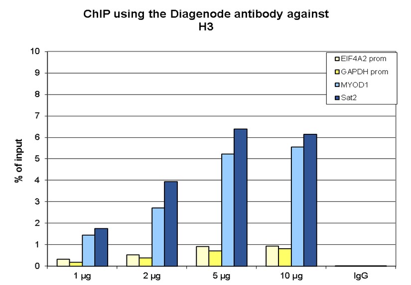
<p><small><strong> Figure 1. ChIP results obtained with the Diagenode monoclonal antibody directed against H3 </strong><br />ChIP assays were performed using human K562 cells, the Diagenode monoclonal antibody against H3 (cat. No. C15200011) and optimized PCR primer pairs for qPCR. ChIP was performed on sheared chromatin from 1 million cells. A titration consisting of 1, 2, 5 and 10 µg of antibody per ChIP experiment was analyzed. IgG (1 µg/IP) was used as a negative IP control. Quantitative PCR was performed with primers specific for the promoters of the active GAPDH and EIF4A2 genes, and for the inactive MYOD1 gene and the Sat2 satellite repeat. Figure 1 shows the recovery, expressed as a % of input (the relative amount of immunoprecipitated DNA compared to input DNA after qPCR analysis).</small></p>
</div>
</div>
<div class="row">
<div class="small-5 columns">
<p><img src="https://www.diagenode.com/img/product/antibodies/C15200011-WBlot.png" alt="H3pan Antibody validated in Western Blot" style="display: block; margin-left: auto; margin-right: auto; height: 300px;" /></p>
</div>
<div class="small-7 columns">
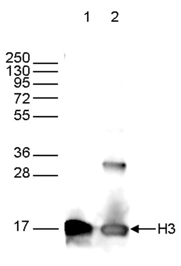
<p><small><strong> Figure 2. Western blot analysis using the Diagenode monoclonal antibody directed against H3 </strong><br />Western blot was performed on whole cell extracts from HeLa cells (40 µg, lane 1) and on 1 µg of recombinant histone H3 (lane 2) using the Diagenode monoclonal antibody against H3 (cat. No. C15200011). The antibody was diluted 1:2,000 in TBS-Tween containing 5% skimmed milk. The marker (in kDa) is shown on the left, the position of the protein of interest is indicated on the right.</small></p>
</div>
</div>
<div class="row">
<div class="small-5 columns">
<p><img src="https://www.diagenode.com/img/product/antibodies/C15200011-wb.jpg" alt="H3pan Antibody validated in Western Blot" style="display: block; margin-left: auto; margin-right: auto;" /></p>
</div>
<div class="small-7 columns">
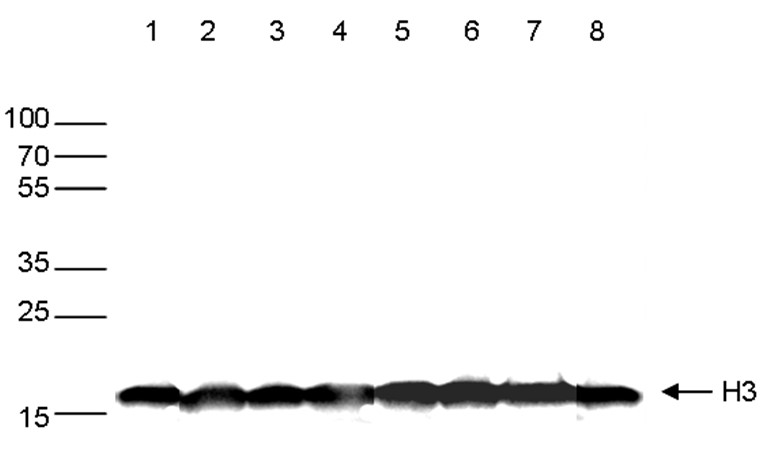
<p><small><strong> Figure 3. Western blot analysis using the Diagenode monoclonal antibody directed against H3</strong><br />Western blot was performed on whole cell extracts (30 µg) from different celltypes (lane 1: HeLa, lane 2: K562, lane 3: MCF7, lane 4: U2OS, lane 5: HepG2, lane 6: Jurkat, lane 7: NIH3T3, lane 8: E14Tg2a mouse ES cells) using the Diagenode monoclonal antibody against H3 (Cat. No. C15200011). The antibody was diluted 1:1,000 in TBS-Tween containing 5% skimmed milk. The marker (in kDa) is shown on the left, the position of the protein of interest is indicated on the right.</small></p>
</div>
</div>
<div class="row">
<div class="small-5 columns">
<p><img src="https://www.diagenode.com/img/product/antibodies/C15200011-if.png" alt="H3pan Antibody validated in Immunofluorescence" style="display: block; margin-left: auto; margin-right: auto;" /></p>
</div>
<div class="small-7 columns">
<p><small><strong> Figure 4. Immunofluorescence using the Diagenode monoclonal antibody directed against H3</strong><br />HeLa cells were stained with the Diagenode monoclonal antibody against H3 (Cat. No. C15200011) and with DAPI. Cells were fixed with 4% formaldehyde for 10’ and blocked with PBS/TX-100 containing 1% BSA. The cells were immunofluorescently labeled with the H3 antibody (middle) diluted 1:500 in blocking solution followed by an anti-mouse antibody conjugated to Alexa594. The left panel shows staining of the nuclei with DAPI. A merge of the two stainings is shown on the right.</small></p>
</div>
</div>',
'label2' => 'Loading Control',
'info2' => '<p><span style="font-weight: 400;"><strong>H3pan monoclonal antibody</strong> can be used as a <strong>loading control</strong> for <strong>nuclear samples</strong> to compare the protein expression level between different samples. </span></p>
<p><b>Why do you need a loading control?</b></p>
<ul>
<li style="font-weight: 400;"><span style="font-weight: 400;">To verify that equal sample amount was loaded on the gel</span></li>
<li style="font-weight: 400;"><span style="font-weight: 400;">To confirm that the electrotransfer was done equally across the lines</span></li>
<li style="font-weight: 400;"><span style="font-weight: 400;">To check that the signal detection after antibody incubation is homogenous across the lines</span></li>
</ul>
<p><b></b></p>
<p><b>What is the loading control?</b></p>
<p><span style="font-weight: 400;">The loading control is an antibody against a protein which is highly expressed in certain types of samples. The level of protein stays constant in different conditions and in different organisms. The molecular weights of the protein of interest and loading control are different, allowing differentiation of both proteins as two different bands.</span></p>',
'label3' => '',
'info3' => '',
'format' => '10 μg',
'catalog_number' => 'C15200011-10',
'old_catalog_number' => '',
'sf_code' => 'C15200011-D001-000582',
'type' => 'FRE',
'search_order' => '',
'price_EUR' => '105',
'price_USD' => '120',
'price_GBP' => '100',
'price_JPY' => '17205',
'price_CNY' => '',
'price_AUD' => '300',
'country' => 'ALL',
'except_countries' => 'None',
'quote' => false,
'in_stock' => false,
'featured' => false,
'no_promo' => false,
'online' => true,
'master' => false,
'last_datasheet_update' => '0000-00-00',
'slug' => 'h3pan-monoclonal-antibody-classic-10-mg-100-ml',
'meta_title' => 'H3pan Antibody 1B1B2 - ChIP (C15200011) | Diagenode ',
'meta_keywords' => 'H3pan monoclonal antibody,Western blot,Diagenode',
'meta_description' => 'H3pan (Histone H3) Monoclonal Antibody, clone 1B1B2 validated in ChIP-qPCR, WB and IF. Batch-specific data available on the website.',
'modified' => '2021-10-25 14:08:30',
'created' => '2016-05-20 14:47:06',
'locale' => 'zho'
),
'Antibody' => array(
'host' => '*****',
'id' => '236',
'name' => 'H3pan monoclonal antibody',
'description' => 'Histones are the main constituents of the protein part of chromosomes of eukaryotic cells. They are rich in the amino acids arginine and lysine and have been greatly conserved during evolution. Histones pack the DNA into tight masses of chromatin. Two core histones of each class H2A, H2B, H3 and H4 assemble and are wrapped by 146 base pairs of DNA to form one octameric nucleosome. Histones play a central role in the regulation of transcription, DNA repair, DNA replication and chromosomal stability. These different functions are established via a complex set of post-translational modifications which either directly or indirectly alter chromatin structure and DNA accessibility to facilitate transcriptional activation or repression or other nuclear processes.',
'clonality' => '',
'isotype' => 'IgG3',
'lot' => '003',
'concentration' => '1.58 µg/µl',
'reactivity' => 'Human, mouse, maize, tomato, poplar, arabidopsis: positive. Other species: not tested.',
'type' => 'Monoclonal',
'purity' => 'Protein A purified monoclonal antibody.',
'classification' => '',
'application_table' => '<table>
<thead>
<tr>
<th>Applications</th>
<th>Suggested dilution</th>
<th>References</th>
</tr>
</thead>
<tbody>
<tr>
<td>ChIP</td>
<td>1-2 µg/ChIP</td>
<td>Fig 1</td>
</tr>
<tr>
<td>Western Blotting</td>
<td>1:1,000 - 1:5,000</td>
<td>Fig 2, 3</td>
</tr>
<tr>
<td>Immunofluorescence</td>
<td>1:500</td>
<td>Fig 4</td>
</tr>
</tbody>
</table>
<p></p>
<p><small><sup>*</sup> Please note that the optimal antibody amount per IP should be determined by the end-user. We recommend testing 1-5 µg per IP.</small></p>',
'storage_conditions' => 'Store at -20°C; for long storage, store at -80°C. Avoid multiple freeze-thaw cycles.',
'storage_buffer' => 'PBS containing 0.05% azide.',
'precautions' => 'This product is for research use only. Not for use in diagnostic or therapeutic procedures.',
'uniprot_acc' => '',
'slug' => '',
'meta_keywords' => '',
'meta_description' => '',
'modified' => '2021-09-16 16:05:05',
'created' => '0000-00-00 00:00:00',
'select_label' => '236 - H3pan monoclonal antibody (003 - 1.58 µg/µl - Human, mouse, maize, tomato, poplar, arabidopsis: positive. Other species: not tested. - Protein A purified monoclonal antibody. - Mouse)'
),
'Slave' => array(),
'Group' => array(
'Group' => array(
'id' => '182',
'name' => 'H3pan monoclonal C15200011',
'product_id' => '1972',
'modified' => '2016-05-20 14:49:37',
'created' => '2016-05-20 14:49:37'
),
'Master' => array(
'id' => '1972',
'antibody_id' => '236',
'name' => 'H3pan Antibody 1B1B2',
'description' => '<p>Monoclonal antibody raised in mouse against <strong>histone H3</strong>, using a KLH-conjugated synthetic peptide containing an unmodified sequence from the C-terminus of the protein. This antibody can be used as a <strong>loading control</strong> in both <strong>ChIP</strong> and <strong>WB</strong> experiments.</p>',
'label1' => 'Validation Data',
'info1' => '<div class="row">
<div class="small-5 columns">
<p><img src="https://www.diagenode.com/img/product/antibodies/C15200011-ChIP.png" alt="H3pan Antibody ChIP Grade" style="display: block; margin-left: auto; margin-right: auto;" /></p>
</div>
<div class="small-7 columns">
<p><small><strong> Figure 1. ChIP results obtained with the Diagenode monoclonal antibody directed against H3 </strong><br />ChIP assays were performed using human K562 cells, the Diagenode monoclonal antibody against H3 (cat. No. C15200011) and optimized PCR primer pairs for qPCR. ChIP was performed on sheared chromatin from 1 million cells. A titration consisting of 1, 2, 5 and 10 µg of antibody per ChIP experiment was analyzed. IgG (1 µg/IP) was used as a negative IP control. Quantitative PCR was performed with primers specific for the promoters of the active GAPDH and EIF4A2 genes, and for the inactive MYOD1 gene and the Sat2 satellite repeat. Figure 1 shows the recovery, expressed as a % of input (the relative amount of immunoprecipitated DNA compared to input DNA after qPCR analysis).</small></p>
</div>
</div>
<div class="row">
<div class="small-5 columns">
<p><img src="https://www.diagenode.com/img/product/antibodies/C15200011-WBlot.png" alt="H3pan Antibody validated in Western Blot" style="display: block; margin-left: auto; margin-right: auto; height: 300px;" /></p>
</div>
<div class="small-7 columns">
<p><small><strong> Figure 2. Western blot analysis using the Diagenode monoclonal antibody directed against H3 </strong><br />Western blot was performed on whole cell extracts from HeLa cells (40 µg, lane 1) and on 1 µg of recombinant histone H3 (lane 2) using the Diagenode monoclonal antibody against H3 (cat. No. C15200011). The antibody was diluted 1:2,000 in TBS-Tween containing 5% skimmed milk. The marker (in kDa) is shown on the left, the position of the protein of interest is indicated on the right.</small></p>
</div>
</div>
<div class="row">
<div class="small-5 columns">
<p><img src="https://www.diagenode.com/img/product/antibodies/C15200011-wb.jpg" alt="H3pan Antibody validated in Western Blot" style="display: block; margin-left: auto; margin-right: auto;" /></p>
</div>
<div class="small-7 columns">
<p><small><strong> Figure 3. Western blot analysis using the Diagenode monoclonal antibody directed against H3</strong><br />Western blot was performed on whole cell extracts (30 µg) from different celltypes (lane 1: HeLa, lane 2: K562, lane 3: MCF7, lane 4: U2OS, lane 5: HepG2, lane 6: Jurkat, lane 7: NIH3T3, lane 8: E14Tg2a mouse ES cells) using the Diagenode monoclonal antibody against H3 (Cat. No. C15200011). The antibody was diluted 1:1,000 in TBS-Tween containing 5% skimmed milk. The marker (in kDa) is shown on the left, the position of the protein of interest is indicated on the right.</small></p>
</div>
</div>
<div class="row">
<div class="small-5 columns">
<p><img src="https://www.diagenode.com/img/product/antibodies/C15200011-if.png" alt="H3pan Antibody validated in Immunofluorescence" style="display: block; margin-left: auto; margin-right: auto;" /></p>
</div>
<div class="small-7 columns">
<p><small><strong> Figure 4. Immunofluorescence using the Diagenode monoclonal antibody directed against H3</strong><br />HeLa cells were stained with the Diagenode monoclonal antibody against H3 (Cat. No. C15200011) and with DAPI. Cells were fixed with 4% formaldehyde for 10’ and blocked with PBS/TX-100 containing 1% BSA. The cells were immunofluorescently labeled with the H3 antibody (middle) diluted 1:500 in blocking solution followed by an anti-mouse antibody conjugated to Alexa594. The left panel shows staining of the nuclei with DAPI. A merge of the two stainings is shown on the right.</small></p>
</div>
</div>',
'label2' => 'Loading Control',
'info2' => '<p><span style="font-weight: 400;"><strong>H3pan monoclonal antibody</strong> can be used as a <strong>loading control</strong> for <strong>nuclear samples</strong> to compare the protein expression level between different samples. </span></p>
<p><b>Why do you need a loading control?</b></p>
<ul>
<li style="font-weight: 400;"><span style="font-weight: 400;">To verify that equal sample amount was loaded on the gel</span></li>
<li style="font-weight: 400;"><span style="font-weight: 400;">To confirm that the electrotransfer was done equally across the lines</span></li>
<li style="font-weight: 400;"><span style="font-weight: 400;">To check that the signal detection after antibody incubation is homogenous across the lines</span></li>
</ul>
<p><b></b></p>
<p><b>What is the loading control?</b></p>
<p><span style="font-weight: 400;">The loading control is an antibody against a protein which is highly expressed in certain types of samples. The level of protein stays constant in different conditions and in different organisms. The molecular weights of the protein of interest and loading control are different, allowing differentiation of both proteins as two different bands.</span></p>',
'label3' => 'Target Description',
'info3' => '<p>Histones are the main constituents of the protein part of chromosomes of eukaryotic cells. They are rich in the amino acids arginine and lysine and have been greatly conserved during evolution. Histones pack the DNA into tight masses of chromatin. Two core histones of each class H2A, H2B, H3 and H4 assemble and are wrapped by 146 base pairs of DNA to form one octameric nucleosome. Histones play a central role in the regulation of transcription, DNA repair, DNA replication and chromosomal stability. These different functions are established via a complex set of post-translational modifications which either directly or indirectly alter chromatin structure and DNA accessibility to facilitate transcriptional activation or repression or other nuclear processes.</p>',
'format' => '50 μg',
'catalog_number' => 'C15200011',
'old_catalog_number' => '',
'sf_code' => 'C15200011-D001-000581',
'type' => 'FRE',
'search_order' => '03-Antibody',
'price_EUR' => '350',
'price_USD' => '350',
'price_GBP' => '310',
'price_JPY' => '57350',
'price_CNY' => '',
'price_AUD' => '875',
'country' => 'ALL',
'except_countries' => 'None',
'quote' => false,
'in_stock' => false,
'featured' => false,
'no_promo' => false,
'online' => true,
'master' => true,
'last_datasheet_update' => 'September 7, 2021',
'slug' => 'h3pan-monoclonal-antibody-classic-50-mg-100-ml',
'meta_title' => 'H3pan Antibody - ChIP Grade (C15200011) | Diagenode',
'meta_keywords' => '',
'meta_description' => 'H3pan (Histone H3) Monoclonal Antibody (clone 1B1B2) shows highest performance in ChIP and highest sensitivity in WB. Validated in ChIP-qPCR, WB and IF. Batch-specific data available on the website. Sample size available.',
'modified' => '2021-10-25 14:07:58',
'created' => '2015-06-29 14:08:20'
),
'Product' => array(
(int) 0 => array(
[maximum depth reached]
)
)
),
'Related' => array(),
'Application' => array(
(int) 0 => array(
'id' => '43',
'position' => '10',
'parent_id' => '40',
'name' => 'ChIP-qPCR (ab)',
'description' => '',
'in_footer' => false,
'in_menu' => false,
'online' => true,
'tabular' => true,
'slug' => 'chip-qpcr-antibodies',
'meta_keywords' => 'Chromatin Immunoprecipitation Sequencing,ChIP-Seq,ChIP-seq grade antibodies,DNA purification,qPCR,Shearing of chromatin',
'meta_description' => 'Diagenode offers a wide range of antibodies and technical support for ChIP-qPCR applications',
'meta_title' => 'ChIP Quantitative PCR Antibodies (ChIP-qPCR) | Diagenode',
'modified' => '2016-01-20 11:30:24',
'created' => '2015-10-20 11:45:36',
'ProductsApplication' => array(
[maximum depth reached]
)
),
(int) 1 => array(
'id' => '19',
'position' => '10',
'parent_id' => '40',
'name' => 'WB',
'description' => '<p><strong>Western blot</strong> : The quality of antibodies used in this technique is crucial for correct and specific protein identification. Diagenode offers huge selection of highly sensitive and specific western blot-validated antibodies.</p>
<p>Learn more about: <a href="https://www.diagenode.com/applications/western-blot">Loading control, MW marker visualization</a><em>. <br /></em></p>
<p><em></em>Check our selection of antibodies validated in Western blot.</p>',
'in_footer' => false,
'in_menu' => false,
'online' => true,
'tabular' => true,
'slug' => 'western-blot-antibodies',
'meta_keywords' => ' Western Blot Antibodies ,western blot protocol,Western Blotting Products,Polyclonal antibodies ,monoclonal antibodies ',
'meta_description' => 'Diagenode offers a wide range of antibodies and technical support for western blot applications',
'meta_title' => ' Western Blot - Monoclonal antibody - Polyclonal antibody | Diagenode',
'modified' => '2016-04-26 12:44:51',
'created' => '2015-01-07 09:20:00',
'ProductsApplication' => array(
[maximum depth reached]
)
),
(int) 2 => array(
'id' => '29',
'position' => '10',
'parent_id' => '40',
'name' => 'IF',
'description' => '<p><strong>Immunofluorescence</strong>:</p>
<p>Diagenode offers huge selection of highly sensitive antibodies validated in IF.</p>
<p><img src="https://www.diagenode.com/img/product/antibodies/C15200229-IF.jpg" alt="" height="245" width="256" /></p>
<p><sup><strong>Immunofluorescence using the Diagenode monoclonal antibody directed against CRISPR/Cas9</strong></sup></p>
<p><sup>HeLa cells transfected with a Cas9 expression vector (left) or untransfected cells (right) were fixed in methanol at -20°C, permeabilized with acetone at -20°C and blocked with PBS containing 2% BSA. The cells were stained with the Cas9 C-terminal antibody (Cat. No. C15200229) diluted 1:400, followed by incubation with an anti-mouse secondary antibody coupled to AF488. The bottom images show counter-staining of the nuclei with Hoechst 33342.</sup></p>
<h5><sup>Check our selection of antibodies validated in IF.</sup></h5>',
'in_footer' => false,
'in_menu' => false,
'online' => true,
'tabular' => true,
'slug' => 'immunofluorescence',
'meta_keywords' => 'Immunofluorescence,Monoclonal antibody,Polyclonal antibody',
'meta_description' => 'Diagenode offers a wide range of antibodies and technical support for Immunofluorescence applications',
'meta_title' => 'Immunofluorescence - Monoclonal antibody - Polyclonal antibody | Diagenode',
'modified' => '2016-04-27 16:23:10',
'created' => '2015-07-08 13:46:02',
'ProductsApplication' => array(
[maximum depth reached]
)
)
),
'Category' => array(
(int) 0 => array(
'id' => '111',
'position' => '40',
'parent_id' => '4',
'name' => 'Histone antibodies',
'description' => '<p>Histones are the main protein components of chromatin involved in the compaction of DNA into nucleosomes, the basic units of chromatin. A <strong>nucleosome</strong> consists of one pair of each of the core histones (<strong>H2A</strong>, <strong>H2B</strong>, <strong>H3</strong> and <strong>H4</strong>) forming an octameric structure wrapped by 146 base pairs of DNA. The different nucleosomes are linked by the linker histone<strong> H1, </strong>allowing for further condensation of chromatin.</p>
<p>The core histones have a globular structure with large unstructured N-terminal tails protruding from the nucleosome. They can undergo to multiple post-translational modifications (PTM), mainly at the N-terminal tails. These <strong>post-translational modifications </strong>include methylation, acetylation, phosphorylation, ubiquitinylation, citrullination, sumoylation, deamination and crotonylation. The most well characterized PTMs are <strong>methylation,</strong> <strong>acetylation and phosphorylation</strong>. Histone methylation occurs mainly on lysine (K) residues, which can be mono-, di- or tri-methylated, and on arginines (R), which can be mono-methylated and symmetrically or asymmetrically di-methylated. Histone acetylation occurs on lysines and histone phosphorylation mainly on serines (S), threonines (T) and tyrosines (Y).</p>
<p>The PTMs of the different residues are involved in numerous processes such as DNA repair, DNA replication and chromosome condensation. They influence the chromatin organization and can be positively or negatively associated with gene expression. Trimethylation of H3K4, H3K36 and H3K79, and lysine acetylation generally result in an open chromatin configuration (figure below) and are therefore associated with <strong>euchromatin</strong> and gene activation. Trimethylation of H3K9, K3K27 and H4K20, on the other hand, is enriched in <strong>heterochromatin </strong>and associated with gene silencing. The combination of different histone modifications is called the "<strong>histone code</strong>”, analogous to the genetic code.</p>
<p><img src="https://www.diagenode.com/img/categories/antibodies/histone-marks-illustration.png" /></p>
<p>Diagenode is proud to offer a large range of antibodies against histones and histone modifications. Our antibodies are highly specific and have been validated in many applications, including <strong>ChIP</strong> and <strong>ChIP-seq</strong>.</p>
<p>Diagenode’s collection includes antibodies recognizing:</p>
<ul>
<li><strong>Histone H1 variants</strong></li>
<li><strong>Histone H2A, H2A variants and histone H2A</strong> <strong>modifications</strong> (serine phosphorylation, lysine acetylation, lysine ubiquitinylation)</li>
<li><strong>Histone H2B and H2B</strong> <strong>modifications </strong>(serine phosphorylation, lysine acetylation)</li>
<li><strong>Histone H3 and H3 modifications </strong>(lysine methylation (mono-, di- and tri-methylated), lysine acetylation, serine phosphorylation, threonine phosphorylation, arginine methylation (mono-methylated, symmetrically and asymmetrically di-methylated))</li>
<li><strong>Histone H4 and H4 modifications (</strong>lysine methylation (mono-, di- and tri-methylated), lysine acetylation, arginine methylation (mono-methylated and symmetrically di-methylated), serine phosphorylation )</li>
</ul>
<p><span style="font-weight: 400;"><strong>HDAC's HAT's, HMT's and other</strong> <strong>enzymes</strong> which modify histones can be found in the category <a href="../categories/chromatin-modifying-proteins-histone-transferase">Histone modifying enzymes</a><br /></span></p>
<p><span style="font-weight: 400;"> Diagenode’s highly validated antibodies:</span></p>
<ul>
<li><span style="font-weight: 400;"> Highly sensitive and specific</span></li>
<li><span style="font-weight: 400;"> Cost-effective (requires less antibody per reaction)</span></li>
<li><span style="font-weight: 400;"> Batch-specific data is available on the website</span></li>
<li><span style="font-weight: 400;"> Expert technical support</span></li>
<li><span style="font-weight: 400;"> Sample sizes available</span></li>
<li><span style="font-weight: 400;"> 100% satisfaction guarantee</span></li>
</ul>',
'no_promo' => false,
'in_menu' => false,
'online' => true,
'tabular' => false,
'hide' => true,
'all_format' => false,
'is_antibody' => true,
'slug' => 'histone-antibodies',
'cookies_tag_id' => null,
'meta_keywords' => 'Histone, antibody, histone h1, histone h2, histone h3, histone h4',
'meta_description' => 'Polyclonal and Monoclonal Antibodies against Histones and their modifications validated for many applications, including Chromatin Immunoprecipitation (ChIP) and ChIP-Sequencing (ChIP-seq)',
'meta_title' => 'Histone and Modified Histone Antibodies | Diagenode',
'modified' => '2020-09-17 13:34:56',
'created' => '2016-04-01 16:01:32',
'ProductsCategory' => array(
[maximum depth reached]
),
'CookiesTag' => array([maximum depth reached])
),
(int) 1 => array(
'id' => '103',
'position' => '0',
'parent_id' => '4',
'name' => 'All antibodies',
'description' => '<p><span style="font-weight: 400;">All Diagenode’s antibodies are listed below. Please, use our Quick search field to find the antibody of interest by target name, application, purity.</span></p>
<p><span style="font-weight: 400;">Diagenode’s highly validated antibodies:</span></p>
<ul>
<li>Highly sensitive and specific</li>
<li>Cost-effective (requires less antibody per reaction)</li>
<li>Batch-specific data is available on the website</li>
<li>Expert technical support</li>
<li>Sample sizes available</li>
<li>100% satisfaction guarantee</li>
</ul>',
'no_promo' => false,
'in_menu' => true,
'online' => true,
'tabular' => false,
'hide' => true,
'all_format' => false,
'is_antibody' => true,
'slug' => 'all-antibodies',
'cookies_tag_id' => null,
'meta_keywords' => 'Antibodies,Premium Antibodies,Classic,Pioneer',
'meta_description' => 'Diagenode Offers Strict quality standards with Rigorous QC and validated Antibodies. Classified based on level of validation for flexibility of Application. Comprehensive selection of histone and non-histone Antibodies',
'meta_title' => 'Diagenode's selection of Antibodies is exclusively dedicated for Epigenetic Research | Diagenode',
'modified' => '2019-07-03 10:55:44',
'created' => '2015-11-02 14:49:22',
'ProductsCategory' => array(
[maximum depth reached]
),
'CookiesTag' => array([maximum depth reached])
),
(int) 2 => array(
'id' => '102',
'position' => '1',
'parent_id' => '4',
'name' => 'Sample size antibodies',
'description' => '<h1><strong>Validated epigenetics antibodies</strong> – care for a sample?<br /> </h1>
<p>Diagenode has partnered with leading epigenetics experts and numerous epigenetics consortiums to bring to you a validated and comprehensive collection of epigenetic antibodies. As an expert in epigenetics, we are committed to offering highly-specific antibodies validated for ChIP/ChIP-seq and many other applications. All batch-specific validation data is available on our website.<br /><a href="../categories/antibodies">Read about our expertise in antibody production</a>.</p>
<ul>
<li><strong>Focused</strong> - Diagenode's selection of antibodies is exclusively dedicated for epigenetic research. <a title="See the full collection." href="../categories/all-antibodies">See the full collection.</a></li>
<li><strong>Strict quality standards</strong> with rigorous QC and validation</li>
<li><strong>Classified</strong> based on level of validation for flexibility of application</li>
</ul>
<p>Existing sample sizes are listed below. We will soon expand our collection. Are you looking for a sample size of another antibody? Just <a href="mailto:agnieszka.zelisko@diagenode.com?Subject=Sample%20Size%20Request" target="_top">Contact us</a>.</p>',
'no_promo' => false,
'in_menu' => true,
'online' => true,
'tabular' => false,
'hide' => true,
'all_format' => true,
'is_antibody' => true,
'slug' => 'sample-size-antibodies',
'cookies_tag_id' => null,
'meta_keywords' => '5-hmC monoclonal antibody,CRISPR/Cas9 polyclonal antibody ,H3K36me3 polyclonal antibody,diagenode',
'meta_description' => 'Diagenode offers sample volume on selected antibodies for researchers to test, validate and provide confidence and flexibility in choosing from our wide range of antibodies ',
'meta_title' => 'Sample-size Antibodies | Diagenode',
'modified' => '2019-07-03 10:57:05',
'created' => '2015-10-27 12:13:34',
'ProductsCategory' => array(
[maximum depth reached]
),
'CookiesTag' => array([maximum depth reached])
),
(int) 3 => array(
'id' => '127',
'position' => '10',
'parent_id' => '4',
'name' => 'ChIP-grade antibodies',
'description' => '<div class="row">
<div class="small-12 columns"><center></center>
<p><br />Chromatin immunoprecipitation (<b>ChIP</b>) is a technique to study the associations of proteins with the specific genomic regions in intact cells. One of the most important steps of this protocol is the immunoprecipitation of targeted protein using the antibody specifically recognizing it. The quality of antibodies used in ChIP is essential for the success of the experiment. Diagenode offers extensively validated ChIP-grade antibodies, confirmed for their specificity, and high level of performance in ChIP. Each batch is validated, and batch-specific data are available on the website.</p>
<p></p>
</div>
</div>
<p><strong>ChIP results</strong> obtained with the antibody directed against H3K4me3 (Cat. No. <a href="../p/h3k4me3-polyclonal-antibody-premium-50-ug-50-ul">C15410003</a>). </p>
<div class="row">
<div class="small-12 medium-6 large-6 columns"><img src="https://www.diagenode.com/img/product/antibodies/C15410003-fig1-ChIP.jpg" alt="" width="400" height="315" /> </div>
<div class="small-12 medium-6 large-6 columns">
<p></p>
<p></p>
<p></p>
</div>
</div>
<p></p>
<p>Our aim at Diagenode is to offer the largest collection of highly specific <strong>ChIP-grade antibodies</strong>. We add new antibodies monthly. Find your ChIP-grade antibody in the list below and check more information about tested applications, extensive validation data, and product information.</p>',
'no_promo' => false,
'in_menu' => true,
'online' => true,
'tabular' => false,
'hide' => true,
'all_format' => false,
'is_antibody' => true,
'slug' => 'chip-grade-antibodies',
'cookies_tag_id' => null,
'meta_keywords' => 'ChIP-grade antibodies, polyclonal antibody, monoclonal antibody, Diagenode',
'meta_description' => 'Diagenode Offers Extensively Validated ChIP-Grade Antibodies, Confirmed for their Specificity, and high level of Performance in Chromatin Immunoprecipitation ChIP',
'meta_title' => 'Chromatin immunoprecipitation ChIP-grade antibodies | Diagenode',
'modified' => '2024-11-19 17:27:07',
'created' => '2017-02-14 11:16:04',
'ProductsCategory' => array(
[maximum depth reached]
),
'CookiesTag' => array([maximum depth reached])
)
),
'Document' => array(
(int) 0 => array(
'id' => '11',
'name' => 'Antibodies you can trust',
'description' => '<p style="text-align: justify;"><span>Epigenetic research tools have evolved over time from endpoint PCR to qPCR to the analyses of large sets of genome-wide sequencing data. ChIP sequencing (ChIP-seq) has now become the gold standard method for chromatin studies, given the accuracy and coverage scale of the approach over other methods. Successful ChIP-seq, however, requires a higher level of experimental accuracy and consistency in all steps of ChIP than ever before. Particularly crucial is the quality of ChIP antibodies. </span></p>',
'image_id' => null,
'type' => 'Poster',
'url' => 'files/posters/Antibodies_you_can_trust_Poster.pdf',
'slug' => 'antibodies-you-can-trust-poster',
'meta_keywords' => '',
'meta_description' => '',
'modified' => '2015-10-01 20:18:31',
'created' => '2015-07-03 16:05:15',
'ProductsDocument' => array(
[maximum depth reached]
)
),
(int) 1 => array(
'id' => '38',
'name' => 'Epigenetic Antibodies Brochure',
'description' => '<p>More than in any other immuoprecipitation assays, quality antibodies are critical tools in many epigenetics experiments. Since 10 years, Diagenode has developed the most stringent quality production available on the market for antibodies exclusively focused on epigenetic uses. All our antibodies have been qualified to work in epigenetic applications.</p>',
'image_id' => null,
'type' => 'Brochure',
'url' => 'files/brochures/Epigenetic_Antibodies_Brochure.pdf',
'slug' => 'epigenetic-antibodies-brochure',
'meta_keywords' => '',
'meta_description' => '',
'modified' => '2016-06-15 11:24:06',
'created' => '2015-07-03 16:05:27',
'ProductsDocument' => array(
[maximum depth reached]
)
),
(int) 2 => array(
'id' => '1009',
'name' => 'H3pan monoclonal antibody 1B1B2 datasheet - lot 002',
'description' => '<p>Monoclonal antibody raised in mouse against histone H3, using a KLH-conjugated synthetic peptide containing an unmodified sequence from the C-terminus of the protein. This antibody can be used as a loading control in both ChIP and WB experiments.</p>',
'image_id' => null,
'type' => 'Datasheet',
'url' => 'files/products/antibodies/h3pan-monoclonal-antibody-classic-lot-002.pdf',
'slug' => 'h3pan-monoclonal-antibody-1b1b2-tds -lot-002',
'meta_keywords' => '',
'meta_description' => '',
'modified' => '2018-07-03 16:15:19',
'created' => '2018-07-03 16:15:19',
'ProductsDocument' => array(
[maximum depth reached]
)
),
(int) 3 => array(
'id' => '728',
'name' => 'Datasheet H3pan C15200011',
'description' => '<p>Monoclonal antibody raised in mouse against histone H3, using a KLH-conjugated synthetic peptide containing an unmodified sequence from the C-terminus of the protein. This antibody can be used as a loading control in both ChIP and WB experiments.</p>',
'image_id' => null,
'type' => 'Datasheet',
'url' => 'files/products/antibodies/Datasheet_H3pan_C15200011.pdf',
'slug' => 'datasheet-h3pan-c15200011',
'meta_keywords' => '',
'meta_description' => '',
'modified' => '2021-10-20 15:00:59',
'created' => '2015-07-07 11:47:44',
'ProductsDocument' => array(
[maximum depth reached]
)
)
),
'Feature' => array(),
'Image' => array(),
'Promotion' => array(),
'Protocol' => array(),
'Publication' => array(
(int) 0 => array(
'id' => '4692',
'name' => 'Temporal modification of H3K9/14ac and H3K4me3 histone marksmediates mechano-responsive gene expression during the accommodationprocess in poplar',
'authors' => 'Ghosh R. et al.',
'description' => '<p>Plants can attenuate their molecular response to repetitive mechanical stimulation as a function of their mechanical history. For instance, a single bending of stem is sufficient to attenuate the gene expression in poplar plants to the subsequent mechanical stimulation, and the state of desensitization can last for several days. The role of histone modifications in memory gene expression and modulating plant response to abiotic or biotic signals is well known. However, such information is still lacking to explain the attenuated expression pattern of mechano-responsive genes in plants under repetitive stimulation. Using poplar as a model plant in this study, we first measured the global level of H3K9/14ac and H3K4me3 marks in the bent stem. The result shows that a single mild bending of the stem for 6 seconds is sufficient to alter the global level of the H3K9/14ac mark in poplar, highlighting the fact that plants are extremely sensitive to mechanical signals. Next, we analyzed the temporal dynamics of these two active histone marks at attenuated (PtaZFP2, PtaXET6, and PtaACA13) and non-attenuated (PtaHRD) mechano-responsive loci during the desensitization and resensitization phases. Enrichment of H3K9/14ac and H3K4me3 in the regulatory region of attenuated genes correlates well with their transient expression pattern after the first bending. Moreover, the levels of H3K4me3 correlate well with their expression pattern after the second bending at desensitization (3 days after the first bending) as well as resensitization (5 days after the first bending) phases. On the other hand, H3K9/14ac status correlates only with their attenuated expression pattern at the desensitization phase. The expression efficiency of the attenuated genes was restored after the second bending in the histone deacetylase inhibitor-treated plants. While both histone modifications contribute to the expression of attenuated genes, mechanostimulated expression of the non-attenuated PtaHRD gene seems to be H3K4me3 dependent.</p>',
'date' => '2023-02-01',
'pmid' => 'https://doi.org/10.1101%2F2023.02.12.526104',
'doi' => '10.1101/2023.02.12.526104',
'modified' => '2023-04-14 09:20:38',
'created' => '2023-02-28 12:19:11',
'ProductsPublication' => array(
[maximum depth reached]
)
),
(int) 1 => array(
'id' => '4574',
'name' => 'Trichoderma root colonization triggers epigenetic changes in jasmonic andsalicylic acid pathway-related genes.',
'authors' => 'Agostini R. B. et al.',
'description' => '<p>Beneficial interactions between plant-roots and Trichoderma spp. lead to a local and systemic enhancement of the plant immune system through a mechanism known as priming of defenses. In recent reports, we outlined a repertoire of genes and proteins differentially regulated in distant tissues of maize plants previously inoculated with Trichoderma atroviride. To further investigate the mechanisms involved in the systemic activation of plant responses, we continued evaluating the regulatory aspects of a selected group of genes when priming is triggered in maize plants. We conducted a time-course expression experiment from the beginning of the interaction between T. atroviride and maize roots, along plant vegetative growth and during Colletotrichum graminicola leaf infection. In addition to gene expression studies, the levels of jasmonic and salicylic acid were determined in the same samples for a comprehensive understanding of the gene expression results. Lastly, chromatin structure and modification assays were designed to evaluate the role of epigenetic marks during the long-lasting activation of the primed state of maize plants. The overall analysis of the results allowed us to shed some light on the interplay between the phytohormones and epigenetic regulatory events in the systemic and long-lasting regulation of maize plant defenses after Trichoderma inoculation.</p>',
'date' => '2022-12-01',
'pmid' => 'https://www.ncbi.nlm.nih.gov/pubmed/36575905',
'doi' => '10.1093/jxb/erac518',
'modified' => '2023-04-14 09:08:14',
'created' => '2023-02-21 09:59:46',
'ProductsPublication' => array(
[maximum depth reached]
)
),
(int) 2 => array(
'id' => '4459',
'name' => 'Nox4 promotes endothelial differentiation through chromatin remodeling.',
'authors' => 'Hahner F. et al.',
'description' => '<p>RATIONALE: Nox4 is a constitutively active NADPH oxidase that constantly produces low levels of HO. Thereby, Nox4 contributes to cell homeostasis and long-term processes, such as differentiation. The high expression of Nox4 seen in endothelial cells contrasts with the low abundance of Nox4 in stem cells, which are accordingly characterized by low levels of HO. We hypothesize that Nox4 is a major contributor to endothelial differentiation, is induced during the process of differentiation, and facilitates homeostasis of the resulting endothelial cells. OBJECTIVE: To determine the role of No×4 in differentiation of murine inducible pluripotent stem cells (miPSC) into endothelial cells (ECs). METHODS AND RESULTS: miPSC, generated from mouse embryonic wildtype (WT) and Nox4 fibroblasts, were differentiated into endothelial cells (miPSC-EC) by stimulation with BMP4 and VEGF. During this process, Nox4 expression increased and knockout of Nox4 prolonged the abundance of pluripotency markers, while expression of endothelial markers was delayed in differentiating Nox4-depleted iPSCs. Eventually, angiogenic capacity of iPSC-ECs is reduced in Nox4 deficient cells, indicating that an absence of Nox4 diminishes stability of the reached phenotype. As an underlying mechanism, we identified JmjD3 as a redox target of Nox4. iPSC-ECs lacking Nox4 display a lower nuclear abundance of the histone demethylase JmjD3, resulting in an increased triple methylation of histone 3 (H3K27me3), which serves as a repressive mark for several genes involved in differentiation. CONCLUSIONS: Nox4 promotes differentiation of miPSCs into ECs by oxidation of JmjD3 and subsequent demethylation of H3K27me3, which forced endothelial differentiation and stability.</p>',
'date' => '2022-07-01',
'pmid' => 'https://www.ncbi.nlm.nih.gov/pubmed/35810713',
'doi' => '10.1016/j.redox.2022.102381',
'modified' => '2022-10-21 09:45:35',
'created' => '2022-09-28 09:53:13',
'ProductsPublication' => array(
[maximum depth reached]
)
),
(int) 3 => array(
'id' => '4510',
'name' => 'Single amino-acid mutation in a Drosoph ila melanogaster ribosomalprotein: An insight in uL11 transcriptional activity.',
'authors' => 'Grunchec H. et al.',
'description' => '<p>The ribosomal protein uL11 is located at the basis of the ribosome P-stalk and plays a paramount role in translational efficiency. In addition, no mutant for uL11 is available suggesting that this gene is haplo-insufficient as many other Ribosomal Protein Genes (RPGs). We have previously shown that overexpression of Drosophila melanogaster uL11 enhances the transcription of many RPGs and Ribosomal Biogenesis genes (RiBis) suggesting that uL11 might globally regulate the level of translation through its transcriptional activity. Moreover, uL11 trimethylated on lysine 3 (uL11K3me3) interacts with the chromodomain of the Enhancer of Polycomb and Trithorax Corto, and both proteins co-localize with RNA Polymerase II at many sites on polytene chromosomes. These data have led to the hypothesis that the N-terminal end of uL11, and more particularly the trimethylation of lysine 3, supports the extra-ribosomal activity of uL11 in transcription. To address this question, we mutated the lysine 3 codon using a CRISPR/Cas9 strategy and obtained several lysine 3 mutants. We describe here the first mutants of D. melanogaster uL11. Unexpectedly, the uL11K3A mutant, in which the lysine 3 codon is replaced by an alanine, displays a genuine Minute phenotype known to be characteristic of RPG deletions (longer development, low fertility, high lethality, thin and short bristles) whereas the uL11K3Y mutant, in which the lysine 3 codon is replaced by a tyrosine, is unaffected. In agreement, the rate of translation decreases in uL11K3A but not in uL11K3Y. Co-immunoprecipitation experiments show that the interaction between uL11 and the Corto chromodomain is impaired by both mutations. However, Histone Association Assays indicate that the mutant proteins still bind chromatin. RNA-seq analyses from wing imaginal discs show that Corto represses RPG expression whereas very few genes are deregulated in uL11 mutants. We propose that Corto, by repressing RPG expression, ensures that all ribosomal proteins are present at the correct stoichiometry, and that uL11 fine-tunes its transcriptional regulation of RPGs.</p>',
'date' => '2022-01-01',
'pmid' => 'https://pubmed.ncbi.nlm.nih.gov/35981051/',
'doi' => '10.1371/journal.pone.0273198',
'modified' => '2022-11-21 10:40:47',
'created' => '2022-11-15 09:26:20',
'ProductsPublication' => array(
[maximum depth reached]
)
),
(int) 4 => array(
'id' => '4300',
'name' => 'Activated Histone Acetyltransferase p300/CBP-Related SignallingPathways Mediate Up-Regulation of NADPH Oxidase,Inflammation, and Fibrosis in Diabetic Kidney',
'authors' => 'Alexandra-Gela Lazar et al.',
'description' => '<p>Accumulating evidence implicates the histone acetylation-based epigenetic mechanisms in the pathoetiology of diabetes-associated micro-/macrovascular complications. Diabetic kidney disease (DKD) is a progressive chronic inflammatory microvascular disorder ultimately leading to glomerulosclerosis and kidney failure. We hypothesized that histone acetyltransferase p300/CBP may be involved in mediating diabetes-accelerated renal damage. In this study, we aimed at investigating the potential role of p300/CBP in the up-regulation of renal NADPH oxidase (Nox), reactive oxygen species (ROS) production, inflammation, and fibrosis in diabetic mice. Diabetic C57BL/6J mice were randomized to receive 10 mg/kg C646, a selective p300/CBP inhibitor, or its vehicle for 4 weeks. We found that in the kidney of C646-treated diabetic mice, the level of H3K27ac, an epigenetic mark of active gene expression, was significantly reduced. Pharmacological inhibition of p300/CBP significantly down-regulated the diabetes-induced enhanced expression of Nox subtypes, pro-inflammatory, and pro-fibrotic molecules in the kidney of mice, and the glomerular ROS overproduction. Our study provides evidence that the activation of p300/CBP enhances ROS production, potentially generated by up-regulated Nox, inflammation, and the production of extracellular matrix proteins in the diabetic kidney. The data suggest that p300/CBP-pharmacological inhibitors may be attractive tools to modulate diabetes-associated pathological processes to efficiently reduce the burden of DKD.</p>',
'date' => '2021-08-01',
'pmid' => 'https://www.mdpi.com/2076-3921/10/9/1356',
'doi' => '10.3390/antiox10091356',
'modified' => '2022-06-20 09:06:40',
'created' => '2022-05-19 10:41:50',
'ProductsPublication' => array(
[maximum depth reached]
)
),
(int) 5 => array(
'id' => '4095',
'name' => 'ZNF354C is a transcriptional repressor that inhibits endothelialangiogenic sprouting.',
'authors' => 'Oo, James A and Irmer, Barnabas and Günther, Stefan and Warwick, Timothyand Pálfi, Katalin and Izquierdo Ponce, Judit and Teichmann, Tom andPflüger-Müller, Beatrice and Gilsbach, Ralf and Brandes, Ralf P andLeisegang, Matthias S',
'description' => '<p>Zinc finger proteins (ZNF) are a large group of transcription factors with diverse functions. We recently discovered that endothelial cells harbour a specific mechanism to limit the action of ZNF354C, whose function in endothelial cells is unknown. Given that ZNF354C has so far only been studied in bone and tumour, its function was determined in endothelial cells. ZNF354C is expressed in vascular cells and localises to the nucleus and cytoplasm. Overexpression of ZNF354C in human endothelial cells results in a marked inhibition of endothelial sprouting. RNA-sequencing of human microvascular endothelial cells with and without overexpression of ZNF354C revealed that the protein is a potent transcriptional repressor. ZNF354C contains an active KRAB domain which mediates this suppression as shown by mutagenesis analysis. ZNF354C interacts with dsDNA, TRIM28 and histones, as observed by proximity ligation and immunoprecipitation. Moreover, chromatin immunoprecipitation revealed that the ZNF binds to specific endothelial-relevant target-gene promoters. ZNF354C suppresses these genes as shown by CRISPR/Cas knockout and RNAi. Inhibition of endothelial sprouting by ZNF354C is dependent on the amino acids DV and MLE of the KRAB domain. These results demonstrate that ZNF354C is a repressive transcription factor which acts through a KRAB domain to inhibit endothelial angiogenic sprouting.</p>',
'date' => '2020-11-01',
'pmid' => 'https://www.ncbi.nlm.nih.gov/pubmed/33154469',
'doi' => '10.1038/s41598-020-76193-0',
'modified' => '2021-03-17 17:19:53',
'created' => '2021-02-18 10:21:53',
'ProductsPublication' => array(
[maximum depth reached]
)
),
(int) 6 => array(
'id' => '3947',
'name' => 'Revisiting promyelocytic leukemia protein targeting by human cytomegalovirus immediate-early protein 1.',
'authors' => 'Paulus C, Harwardt T, Walter B, Marxreiter A, Zenger M, Reuschel E, Nevels MM',
'description' => '<p>Promyelocytic leukemia (PML) bodies are nuclear organelles implicated in intrinsic and innate antiviral defense. The eponymous PML proteins, central to the self-organization of PML bodies, and other restriction factors found in these organelles are common targets of viral antagonism. The 72-kDa immediate-early protein 1 (IE1) is the principal antagonist of PML bodies encoded by the human cytomegalovirus (hCMV). IE1 is believed to disrupt PML bodies by inhibiting PML SUMOylation, while PML was proposed to act as an E3 ligase for IE1 SUMOylation. PML targeting by IE1 is considered to be crucial for hCMV replication at low multiplicities of infection, in part via counteracting antiviral gene induction linked to the cellular interferon (IFN) response. However, current concepts of IE1-PML interaction are largely derived from mutant IE1 proteins known or predicted to be metabolically unstable and globally misfolded. We performed systematic clustered charge-to-alanine scanning mutagenesis and identified a stable IE1 mutant protein (IE1cc172-176) with wild-type characteristics except for neither interacting with PML proteins nor inhibiting PML SUMOylation. Consequently, IE1cc172-176 does not associate with PML bodies and is selectively impaired for disrupting these organelles. Surprisingly, functional analysis of IE1cc172-176 revealed that the protein is hypermodified by mixed SUMO chains and that IE1 SUMOylation depends on nucleosome rather than PML binding. Furthermore, a mutant hCMV expressing IE1cc172-176 was only slightly attenuated compared to an IE1-null virus even at low multiplicities of infection. Finally, hCMV-induced expression of cytokine and IFN-stimulated genes turned out to be reduced rather than increased in the presence of IE1cc172-176 relative to wild-type IE1. Our findings challenge present views on the relationship of IE1 with PML and the role of PML in hCMV replication. This study also provides initial evidence for the idea that disruption of PML bodies upon viral infection is linked to activation rather than inhibition of innate immunity.</p>',
'date' => '2020-05-01',
'pmid' => 'http://www.pubmed.gov/32365141',
'doi' => '10.1371/journal.ppat.1008537',
'modified' => '2020-08-17 10:09:46',
'created' => '2020-08-10 12:12:25',
'ProductsPublication' => array(
[maximum depth reached]
)
),
(int) 7 => array(
'id' => '3881',
'name' => 'Widespread loss of the silencing epigenetic mark H3K9me3 in astrocytes and neurons along with hippocampal-dependent cognitive impairment in C9orf72 BAC transgenic mice.',
'authors' => 'Jury N, Abarzua S, Diaz I, Guerra MV, Ampuero E, Cubillos P, Martinez P, Herrera-Soto A, Arredondo C, Rojas F, Manterola M, Rojas A, Montecino M, Varela-Nallar L, van Zundert B',
'description' => '<p>BACKGROUND: Hexanucleotide repeat expansions of the GC motif in a non-coding region of the C9ORF72 gene are the most common genetic cause of amyotrophic lateral sclerosis (ALS) and frontotemporal dementia (FTD). Tissues from C9ALS/FTD patients and from mouse models of ALS show RNA foci, dipeptide-repeat proteins, and notably, widespread alterations in the transcriptome. Epigenetic processes regulate gene expression without changing DNA sequences and therefore could account for the altered transcriptome profiles in C9ALS/FTD; here, we explore whether the critical repressive marks H3K9me2 and H3K9me3 are altered in a recently developed C9ALS/FTD BAC mouse model (C9BAC). RESULTS: Chromocenters that constitute pericentric constitutive heterochromatin were visualized as DAPI- or Nucblue-dense foci in nuclei. Cultured C9BAC astrocytes exhibited a reduced staining signal for H3K9me3 (but not for H3K9me2) at chromocenters that was accompanied by a marked decline in the global nuclear level of this mark. Similar depletion of H3K9me3 at chromocenters was detected in astrocytes and neurons of the spinal cord, motor cortex, and hippocampus of C9BAC mice. The alterations of H3K9me3 in the hippocampus of C9BAC mice led us to identify previously undetected neuronal loss in CA1, CA3, and dentate gyrus, as well as hippocampal-dependent cognitive deficits. CONCLUSIONS: Our data indicate that a loss of the repressive mark H3K9me3 in astrocytes and neurons in the central nervous system of C9BAC mice represents a signature during neurodegeneration and memory deficit of C9ALS/FTD.</p>',
'date' => '2020-02-18',
'pmid' => 'http://www.pubmed.gov/32070418',
'doi' => '10.1186/s13148-020-0816-9',
'modified' => '2020-03-20 17:31:40',
'created' => '2020-03-13 13:45:54',
'ProductsPublication' => array(
[maximum depth reached]
)
),
(int) 8 => array(
'id' => '3747',
'name' => 'Class I TCP transcription factors target the gibberellin biosynthesis gene GA20ox1 and the growth promoting genes HBI1 and PRE6 during thermomorphogenic growth in Arabidopsis.',
'authors' => 'Ferrero LV, Viola IL, Ariel FD, Gonzalez DH',
'description' => '<p>Plants respond to a rise in ambient temperature by increasing the growth of petioles and hypocotyls. In this work, we show that Arabidopsis thaliana class I TEOSINTE BRANCHED 1, CYCLOIDEA, PCF (TCP) transcription factors TCP14 and TCP15 are required for optimal petiole and hypocotyl elongation under high ambient temperature. These TCPs influence the levels of the DELLA protein RGA and the expression of growth-related genes which are induced in response to an increase in temperature. However, the class I TCPs are not required for the induction of the auxin biosynthesis gene YUCCA8 or for auxin-dependent gene expression responses. TCP15 directly targets the gibberellin biosynthesis gene GA20ox1 and the growth regulatory genes HBI1 and PRE6. Several of the genes regulated by TCP15 are also targets of the growth regulator PIF4 and show an enrichment of PIF4 and TCP binding motifs in their promoters. PIF4 binding to GA20ox1 and HBI1 is enhanced in the presence of the TCPs, indicating that TCP14 and TCP15 directly participate in the induction of genes involved in gibberellin biosynthesis and cell expansion by high temperature functionally interacting with PIF4. In addition, overexpression of HBI1 rescues the growth defects of tcp14 tcp15 double mutants, suggesting that this gene is a major outcome of regulation by both class I TCPs during thermomorphogenesis.</p>',
'date' => '2019-07-11',
'pmid' => 'http://www.pubmed.gov/31292642',
'doi' => '10.1093/pcp/pcz137/5530963',
'modified' => '2019-08-06 16:13:22',
'created' => '2019-07-31 13:35:50',
'ProductsPublication' => array(
[maximum depth reached]
)
),
(int) 9 => array(
'id' => '3629',
'name' => 'Comprehensive Analysis of Chromatin States in Atypical Teratoid/Rhabdoid Tumor Identifies Diverging Roles for SWI/SNF and Polycomb in Gene Regulation.',
'authors' => 'Erkek S, Johann PD, Finetti MA, Drosos Y, Chou HC, Zapatka M, Sturm D, Jones DTW, Korshunov A, Rhyzova M, Wolf S, Mallm JP, Beck K, Witt O, Kulozik AE, Frühwald MC, Northcott PA, Korbel JO, Lichter P, Eils R, Gajjar A, Roberts CWM, Williamson D, Hasselbla',
'description' => '<p>Biallelic inactivation of SMARCB1, encoding a member of the SWI/SNF chromatin remodeling complex, is the hallmark genetic aberration of atypical teratoid rhabdoid tumors (ATRT). Here, we report how loss of SMARCB1 affects the epigenome in these tumors. Using chromatin immunoprecipitation sequencing (ChIP-seq) on primary tumors for a series of active and repressive histone marks, we identified the chromatin states differentially represented in ATRTs compared with other brain tumors and non-neoplastic brain. Re-expression of SMARCB1 in ATRT cell lines enabled confirmation of our genome-wide findings for the chromatin states. Additional generation of ChIP-seq data for SWI/SNF and Polycomb group proteins and the transcriptional repressor protein REST determined differential dependencies of SWI/SNF and Polycomb complexes in regulation of diverse gene sets in ATRTs.</p>',
'date' => '2019-01-14',
'pmid' => 'http://www.pubmed.gov/30595504',
'doi' => '10.1016/j.ccell.2018.11.014',
'modified' => '2019-05-08 12:27:57',
'created' => '2019-04-25 11:11:44',
'ProductsPublication' => array(
[maximum depth reached]
)
),
(int) 10 => array(
'id' => '3428',
'name' => 'Epigenetic regulation of vascular NADPH oxidase expression and reactive oxygen species production by histone deacetylase-dependent mechanisms in experimental diabetes.',
'authors' => 'Manea SA, Antonescu ML, Fenyo IM, Raicu M, Simionescu M, Manea A',
'description' => '<p>Reactive oxygen species (ROS) generated by up-regulated NADPH oxidase (Nox) contribute to structural-functional alterations of the vascular wall in diabetes. Epigenetic mechanisms, such as histone acetylation, emerged as important regulators of gene expression in cardiovascular disorders. Since their role in diabetes is still elusive we hypothesized that histone deacetylase (HDAC)-dependent mechanisms could mediate vascular Nox overexpression in diabetic conditions. Non-diabetic and streptozotocin-induced diabetic C57BL/6J mice were randomized to receive vehicle or suberoylanilide hydroxamic acid (SAHA), a pan-HDAC inhibitor. In vitro studies were performed on a human aortic smooth muscle cell (SMC) line. Aortic SMCs typically express Nox1, Nox4, and Nox5 subtypes. HDAC1 and HDAC2 proteins along with Nox1, Nox2, and Nox4 levels were found significantly elevated in the aortas of diabetic mice compared to non-diabetic animals. Treatment of diabetic mice with SAHA mitigated the aortic expression of Nox1, Nox2, and Nox4 subtypes and NADPH-stimulated ROS production. High concentrations of glucose increased HDAC1 and HDAC2 protein levels in cultured SMCs. SAHA significantly reduced the high glucose-induced Nox1/4/5 expression, ROS production, and the formation malondialdehyde-protein adducts in SMCs. Overexpression of HDAC2 up-regulated the Nox1/4/5 gene promoter activities in SMCs. Physical interactions of HDAC1/2 and p300 proteins with Nox1/4/5 promoters were detected at the sites of active transcription. High glucose induced histone H3K27 acetylation enrichment at the promoters of Nox1/4/5 genes in SMCs. The novel data of this study indicate that HDACs mediate vascular Nox up-regulation in diabetes. HDAC inhibition reduces vascular ROS production in experimental diabetes, possibly by a mechanism involving negative regulation of Nox expression.</p>',
'date' => '2018-06-01',
'pmid' => 'http://www.pubmed.gov/29587244',
'doi' => '10.1016/j.redox.2018.03.011',
'modified' => '2018-12-31 11:46:31',
'created' => '2018-12-04 09:51:07',
'ProductsPublication' => array(
[maximum depth reached]
)
),
(int) 11 => array(
'id' => '3594',
'name' => 'The histone demethylase Phf2 acts as a molecular checkpoint to prevent NAFLD progression during obesity.',
'authors' => 'Bricambert J, Alves-Guerra MC, Esteves P, Prip-Buus C, Bertrand-Michel J, Guillou H, Chang CJ, Vander Wal MN, Canonne-Hergaux F, Mathurin P, Raverdy V, Pattou F, Girard J, Postic C, Dentin R',
'description' => '<p>Aberrant histone methylation profile is reported to correlate with the development and progression of NAFLD during obesity. However, the identification of specific epigenetic modifiers involved in this process remains poorly understood. Here, we identify the histone demethylase Plant Homeodomain Finger 2 (Phf2) as a new transcriptional co-activator of the transcription factor Carbohydrate Responsive Element Binding Protein (ChREBP). By specifically erasing H3K9me2 methyl-marks on the promoter of ChREBP-regulated genes, Phf2 facilitates incorporation of metabolic precursors into mono-unsaturated fatty acids, leading to hepatosteatosis development in the absence of inflammation and insulin resistance. Moreover, the Phf2-mediated activation of the transcription factor NF-E2-related factor 2 (Nrf2) further reroutes glucose fluxes toward the pentose phosphate pathway and glutathione biosynthesis, protecting the liver from oxidative stress and fibrogenesis in response to diet-induced obesity. Overall, our findings establish a downstream epigenetic checkpoint, whereby Phf2, through facilitating H3K9me2 demethylation at specific gene promoters, protects liver from the pathogenesis progression of NAFLD.</p>',
'date' => '2018-05-29',
'pmid' => 'http://www.pubmed.gov/29844386',
'doi' => '10.1038/s41467-018-04361-y',
'modified' => '2019-04-17 15:14:20',
'created' => '2019-04-16 12:25:30',
'ProductsPublication' => array(
[maximum depth reached]
)
)
),
'Testimonial' => array(),
'Area' => array(),
'SafetySheet' => array(
(int) 0 => array(
'id' => '112',
'name' => 'H3pan antibody SDS US en',
'language' => 'en',
'url' => 'files/SDS/H3pan/SDS-C15200011-H3pan_Antibody-US-en-GHS_2_0.pdf',
'countries' => 'US',
'modified' => '2020-03-13 15:32:37',
'created' => '2020-03-13 15:32:37',
'ProductsSafetySheet' => array(
[maximum depth reached]
)
),
(int) 1 => array(
'id' => '110',
'name' => 'H3pan antibody SDS GB en',
'language' => 'en',
'url' => 'files/SDS/H3pan/SDS-C15200011-H3pan_Antibody-GB-en-GHS_2_0.pdf',
'countries' => 'GB',
'modified' => '2020-03-13 15:31:15',
'created' => '2020-03-13 15:31:15',
'ProductsSafetySheet' => array(
[maximum depth reached]
)
),
(int) 2 => array(
'id' => '108',
'name' => 'H3pan antibody SDS ES es',
'language' => 'es',
'url' => 'files/SDS/H3pan/SDS-C15200011-H3pan_Antibody-ES-es-GHS_2_0.pdf',
'countries' => 'ES',
'modified' => '2020-03-13 15:29:15',
'created' => '2020-03-13 15:29:15',
'ProductsSafetySheet' => array(
[maximum depth reached]
)
),
(int) 3 => array(
'id' => '111',
'name' => 'H3pan antibody SDS JP ja',
'language' => 'ja',
'url' => 'files/SDS/H3pan/SDS-C15200011-H3pan_Antibody-JP-ja-GHS_2_0.pdf',
'countries' => 'JP',
'modified' => '2020-03-13 15:31:58',
'created' => '2020-03-13 15:31:58',
'ProductsSafetySheet' => array(
[maximum depth reached]
)
),
(int) 4 => array(
'id' => '107',
'name' => 'H3pan antibody SDS DE de',
'language' => 'de',
'url' => 'files/SDS/H3pan/SDS-C15200011-H3pan_Antibody-DE-de-GHS_2_0.pdf',
'countries' => 'DE',
'modified' => '2020-03-13 15:28:24',
'created' => '2020-03-13 15:28:24',
'ProductsSafetySheet' => array(
[maximum depth reached]
)
),
(int) 5 => array(
'id' => '106',
'name' => 'H3pan antibody SDS BE nl',
'language' => 'nl',
'url' => 'files/SDS/H3pan/SDS-C15200011-H3pan_Antibody-BE-nl-GHS_2_0.pdf',
'countries' => 'BE',
'modified' => '2020-03-13 15:27:32',
'created' => '2020-03-13 15:27:32',
'ProductsSafetySheet' => array(
[maximum depth reached]
)
),
(int) 6 => array(
'id' => '109',
'name' => 'H3pan antibody SDS FR fr',
'language' => 'fr',
'url' => 'files/SDS/H3pan/SDS-C15200011-H3pan_Antibody-FR-fr-GHS_2_0.pdf',
'countries' => 'FR',
'modified' => '2020-03-13 15:30:26',
'created' => '2020-03-13 15:30:26',
'ProductsSafetySheet' => array(
[maximum depth reached]
)
),
(int) 7 => array(
'id' => '105',
'name' => 'H3pan antibody SDS BE fr',
'language' => 'fr',
'url' => 'files/SDS/H3pan/SDS-C15200011-H3pan_Antibody-BE-fr-GHS_2_0.pdf',
'countries' => 'BE',
'modified' => '2020-03-13 15:26:08',
'created' => '2020-03-13 15:26:08',
'ProductsSafetySheet' => array(
[maximum depth reached]
)
)
)
)
$meta_canonical = 'https://www.diagenode.com/cn/p/h3pan-monoclonal-antibody-classic-50-mg-100-ml'
$country = 'US'
$countries_allowed = array(
(int) 0 => 'CA',
(int) 1 => 'US',
(int) 2 => 'IE',
(int) 3 => 'GB',
(int) 4 => 'DK',
(int) 5 => 'NO',
(int) 6 => 'SE',
(int) 7 => 'FI',
(int) 8 => 'NL',
(int) 9 => 'BE',
(int) 10 => 'LU',
(int) 11 => 'FR',
(int) 12 => 'DE',
(int) 13 => 'CH',
(int) 14 => 'AT',
(int) 15 => 'ES',
(int) 16 => 'IT',
(int) 17 => 'PT'
)
$outsource = true
$other_formats = array(
(int) 0 => array(
'id' => '1972',
'antibody_id' => '236',
'name' => 'H3pan Antibody 1B1B2',
'description' => '<p>Monoclonal antibody raised in mouse against <strong>histone H3</strong>, using a KLH-conjugated synthetic peptide containing an unmodified sequence from the C-terminus of the protein. This antibody can be used as a <strong>loading control</strong> in both <strong>ChIP</strong> and <strong>WB</strong> experiments.</p>',
'label1' => 'Validation Data',
'info1' => '<div class="row">
<div class="small-5 columns">
<p><img src="https://www.diagenode.com/img/product/antibodies/C15200011-ChIP.png" alt="H3pan Antibody ChIP Grade" style="display: block; margin-left: auto; margin-right: auto;" /></p>
</div>
<div class="small-7 columns">
<p><small><strong> Figure 1. ChIP results obtained with the Diagenode monoclonal antibody directed against H3 </strong><br />ChIP assays were performed using human K562 cells, the Diagenode monoclonal antibody against H3 (cat. No. C15200011) and optimized PCR primer pairs for qPCR. ChIP was performed on sheared chromatin from 1 million cells. A titration consisting of 1, 2, 5 and 10 µg of antibody per ChIP experiment was analyzed. IgG (1 µg/IP) was used as a negative IP control. Quantitative PCR was performed with primers specific for the promoters of the active GAPDH and EIF4A2 genes, and for the inactive MYOD1 gene and the Sat2 satellite repeat. Figure 1 shows the recovery, expressed as a % of input (the relative amount of immunoprecipitated DNA compared to input DNA after qPCR analysis).</small></p>
</div>
</div>
<div class="row">
<div class="small-5 columns">
<p><img src="https://www.diagenode.com/img/product/antibodies/C15200011-WBlot.png" alt="H3pan Antibody validated in Western Blot" style="display: block; margin-left: auto; margin-right: auto; height: 300px;" /></p>
</div>
<div class="small-7 columns">
<p><small><strong> Figure 2. Western blot analysis using the Diagenode monoclonal antibody directed against H3 </strong><br />Western blot was performed on whole cell extracts from HeLa cells (40 µg, lane 1) and on 1 µg of recombinant histone H3 (lane 2) using the Diagenode monoclonal antibody against H3 (cat. No. C15200011). The antibody was diluted 1:2,000 in TBS-Tween containing 5% skimmed milk. The marker (in kDa) is shown on the left, the position of the protein of interest is indicated on the right.</small></p>
</div>
</div>
<div class="row">
<div class="small-5 columns">
<p><img src="https://www.diagenode.com/img/product/antibodies/C15200011-wb.jpg" alt="H3pan Antibody validated in Western Blot" style="display: block; margin-left: auto; margin-right: auto;" /></p>
</div>
<div class="small-7 columns">
<p><small><strong> Figure 3. Western blot analysis using the Diagenode monoclonal antibody directed against H3</strong><br />Western blot was performed on whole cell extracts (30 µg) from different celltypes (lane 1: HeLa, lane 2: K562, lane 3: MCF7, lane 4: U2OS, lane 5: HepG2, lane 6: Jurkat, lane 7: NIH3T3, lane 8: E14Tg2a mouse ES cells) using the Diagenode monoclonal antibody against H3 (Cat. No. C15200011). The antibody was diluted 1:1,000 in TBS-Tween containing 5% skimmed milk. The marker (in kDa) is shown on the left, the position of the protein of interest is indicated on the right.</small></p>
</div>
</div>
<div class="row">
<div class="small-5 columns">
<p><img src="https://www.diagenode.com/img/product/antibodies/C15200011-if.png" alt="H3pan Antibody validated in Immunofluorescence" style="display: block; margin-left: auto; margin-right: auto;" /></p>
</div>
<div class="small-7 columns">
<p><small><strong> Figure 4. Immunofluorescence using the Diagenode monoclonal antibody directed against H3</strong><br />HeLa cells were stained with the Diagenode monoclonal antibody against H3 (Cat. No. C15200011) and with DAPI. Cells were fixed with 4% formaldehyde for 10’ and blocked with PBS/TX-100 containing 1% BSA. The cells were immunofluorescently labeled with the H3 antibody (middle) diluted 1:500 in blocking solution followed by an anti-mouse antibody conjugated to Alexa594. The left panel shows staining of the nuclei with DAPI. A merge of the two stainings is shown on the right.</small></p>
</div>
</div>',
'label2' => 'Loading Control',
'info2' => '<p><span style="font-weight: 400;"><strong>H3pan monoclonal antibody</strong> can be used as a <strong>loading control</strong> for <strong>nuclear samples</strong> to compare the protein expression level between different samples. </span></p>
<p><b>Why do you need a loading control?</b></p>
<ul>
<li style="font-weight: 400;"><span style="font-weight: 400;">To verify that equal sample amount was loaded on the gel</span></li>
<li style="font-weight: 400;"><span style="font-weight: 400;">To confirm that the electrotransfer was done equally across the lines</span></li>
<li style="font-weight: 400;"><span style="font-weight: 400;">To check that the signal detection after antibody incubation is homogenous across the lines</span></li>
</ul>
<p><b></b></p>
<p><b>What is the loading control?</b></p>
<p><span style="font-weight: 400;">The loading control is an antibody against a protein which is highly expressed in certain types of samples. The level of protein stays constant in different conditions and in different organisms. The molecular weights of the protein of interest and loading control are different, allowing differentiation of both proteins as two different bands.</span></p>',
'label3' => 'Target Description',
'info3' => '<p>Histones are the main constituents of the protein part of chromosomes of eukaryotic cells. They are rich in the amino acids arginine and lysine and have been greatly conserved during evolution. Histones pack the DNA into tight masses of chromatin. Two core histones of each class H2A, H2B, H3 and H4 assemble and are wrapped by 146 base pairs of DNA to form one octameric nucleosome. Histones play a central role in the regulation of transcription, DNA repair, DNA replication and chromosomal stability. These different functions are established via a complex set of post-translational modifications which either directly or indirectly alter chromatin structure and DNA accessibility to facilitate transcriptional activation or repression or other nuclear processes.</p>',
'format' => '50 μg',
'catalog_number' => 'C15200011',
'old_catalog_number' => '',
'sf_code' => 'C15200011-D001-000581',
'type' => 'FRE',
'search_order' => '03-Antibody',
'price_EUR' => '350',
'price_USD' => '350',
'price_GBP' => '310',
'price_JPY' => '57350',
'price_CNY' => '',
'price_AUD' => '875',
'country' => 'ALL',
'except_countries' => 'None',
'quote' => false,
'in_stock' => false,
'featured' => false,
'no_promo' => false,
'online' => true,
'master' => true,
'last_datasheet_update' => 'September 7, 2021',
'slug' => 'h3pan-monoclonal-antibody-classic-50-mg-100-ml',
'meta_title' => 'H3pan Antibody - ChIP Grade (C15200011) | Diagenode',
'meta_keywords' => '',
'meta_description' => 'H3pan (Histone H3) Monoclonal Antibody (clone 1B1B2) shows highest performance in ChIP and highest sensitivity in WB. Validated in ChIP-qPCR, WB and IF. Batch-specific data available on the website. Sample size available.',
'modified' => '2021-10-25 14:07:58',
'created' => '2015-06-29 14:08:20'
)
)
$pro = array(
'id' => '2812',
'antibody_id' => '236',
'name' => 'H3pan Antibody 1B1B2 (sample size)',
'description' => '<p>Monoclonal antibody raised in mouse against histone H3, using a KLH-conjugated synthetic peptide containing an unmodified sequence from the C-terminus of the protein. This antibody can be used as a <strong>loading control</strong> in both <strong>ChIP</strong> and <strong>WB</strong> experiments.</p>',
'label1' => 'Validation Data',
'info1' => '',
'label2' => '',
'info2' => '',
'label3' => '',
'info3' => '',
'format' => '10 μg',
'catalog_number' => 'C15200011-10',
'old_catalog_number' => '',
'sf_code' => 'C15200011-D001-000582',
'type' => 'FRE',
'search_order' => '',
'price_EUR' => '105',
'price_USD' => '120',
'price_GBP' => '100',
'price_JPY' => '17205',
'price_CNY' => '',
'price_AUD' => '300',
'country' => 'ALL',
'except_countries' => 'None',
'quote' => false,
'in_stock' => false,
'featured' => false,
'no_promo' => false,
'online' => true,
'master' => false,
'last_datasheet_update' => '0000-00-00',
'slug' => 'h3pan-monoclonal-antibody-classic-10-mg-100-ml',
'meta_title' => 'H3pan Antibody 1B1B2 - ChIP (C15200011) | Diagenode',
'meta_keywords' => 'H3pan monoclonal antibody,Western blot,Diagenode',
'meta_description' => 'H3pan (Histone H3) Monoclonal Antibody, clone 1B1B2 validated in ChIP-qPCR, WB and IF. Batch-specific data available on the website.',
'modified' => '2021-10-25 14:08:30',
'created' => '2016-05-20 14:47:06',
'ProductsGroup' => array(
'id' => '198',
'product_id' => '2812',
'group_id' => '182'
)
)
$edit = ''
$testimonials = ''
$featured_testimonials = ''
$related_products = ''
$rrbs_service = array(
(int) 0 => (int) 1894,
(int) 1 => (int) 1895
)
$chipseq_service = array(
(int) 0 => (int) 2683,
(int) 1 => (int) 1835,
(int) 2 => (int) 1836,
(int) 3 => (int) 2684,
(int) 4 => (int) 1838,
(int) 5 => (int) 1839,
(int) 6 => (int) 1856
)
$label = '<img src="/img/banners/banner-customizer-back.png" alt=""/>'
$labelize = object(Closure) {
}
$old_catalog_number = ''
$country_code = 'US'
$other_format = array(
'id' => '1972',
'antibody_id' => '236',
'name' => 'H3pan Antibody 1B1B2',
'description' => '<p>Monoclonal antibody raised in mouse against <strong>histone H3</strong>, using a KLH-conjugated synthetic peptide containing an unmodified sequence from the C-terminus of the protein. This antibody can be used as a <strong>loading control</strong> in both <strong>ChIP</strong> and <strong>WB</strong> experiments.</p>',
'label1' => 'Validation Data',
'info1' => '<div class="row">
<div class="small-5 columns">
<p><img src="https://www.diagenode.com/img/product/antibodies/C15200011-ChIP.png" alt="H3pan Antibody ChIP Grade" style="display: block; margin-left: auto; margin-right: auto;" /></p>
</div>
<div class="small-7 columns">
<p><small><strong> Figure 1. ChIP results obtained with the Diagenode monoclonal antibody directed against H3 </strong><br />ChIP assays were performed using human K562 cells, the Diagenode monoclonal antibody against H3 (cat. No. C15200011) and optimized PCR primer pairs for qPCR. ChIP was performed on sheared chromatin from 1 million cells. A titration consisting of 1, 2, 5 and 10 µg of antibody per ChIP experiment was analyzed. IgG (1 µg/IP) was used as a negative IP control. Quantitative PCR was performed with primers specific for the promoters of the active GAPDH and EIF4A2 genes, and for the inactive MYOD1 gene and the Sat2 satellite repeat. Figure 1 shows the recovery, expressed as a % of input (the relative amount of immunoprecipitated DNA compared to input DNA after qPCR analysis).</small></p>
</div>
</div>
<div class="row">
<div class="small-5 columns">
<p><img src="https://www.diagenode.com/img/product/antibodies/C15200011-WBlot.png" alt="H3pan Antibody validated in Western Blot" style="display: block; margin-left: auto; margin-right: auto; height: 300px;" /></p>
</div>
<div class="small-7 columns">
<p><small><strong> Figure 2. Western blot analysis using the Diagenode monoclonal antibody directed against H3 </strong><br />Western blot was performed on whole cell extracts from HeLa cells (40 µg, lane 1) and on 1 µg of recombinant histone H3 (lane 2) using the Diagenode monoclonal antibody against H3 (cat. No. C15200011). The antibody was diluted 1:2,000 in TBS-Tween containing 5% skimmed milk. The marker (in kDa) is shown on the left, the position of the protein of interest is indicated on the right.</small></p>
</div>
</div>
<div class="row">
<div class="small-5 columns">
<p><img src="https://www.diagenode.com/img/product/antibodies/C15200011-wb.jpg" alt="H3pan Antibody validated in Western Blot" style="display: block; margin-left: auto; margin-right: auto;" /></p>
</div>
<div class="small-7 columns">
<p><small><strong> Figure 3. Western blot analysis using the Diagenode monoclonal antibody directed against H3</strong><br />Western blot was performed on whole cell extracts (30 µg) from different celltypes (lane 1: HeLa, lane 2: K562, lane 3: MCF7, lane 4: U2OS, lane 5: HepG2, lane 6: Jurkat, lane 7: NIH3T3, lane 8: E14Tg2a mouse ES cells) using the Diagenode monoclonal antibody against H3 (Cat. No. C15200011). The antibody was diluted 1:1,000 in TBS-Tween containing 5% skimmed milk. The marker (in kDa) is shown on the left, the position of the protein of interest is indicated on the right.</small></p>
</div>
</div>
<div class="row">
<div class="small-5 columns">
<p><img src="https://www.diagenode.com/img/product/antibodies/C15200011-if.png" alt="H3pan Antibody validated in Immunofluorescence" style="display: block; margin-left: auto; margin-right: auto;" /></p>
</div>
<div class="small-7 columns">
<p><small><strong> Figure 4. Immunofluorescence using the Diagenode monoclonal antibody directed against H3</strong><br />HeLa cells were stained with the Diagenode monoclonal antibody against H3 (Cat. No. C15200011) and with DAPI. Cells were fixed with 4% formaldehyde for 10’ and blocked with PBS/TX-100 containing 1% BSA. The cells were immunofluorescently labeled with the H3 antibody (middle) diluted 1:500 in blocking solution followed by an anti-mouse antibody conjugated to Alexa594. The left panel shows staining of the nuclei with DAPI. A merge of the two stainings is shown on the right.</small></p>
</div>
</div>',
'label2' => 'Loading Control',
'info2' => '<p><span style="font-weight: 400;"><strong>H3pan monoclonal antibody</strong> can be used as a <strong>loading control</strong> for <strong>nuclear samples</strong> to compare the protein expression level between different samples. </span></p>
<p><b>Why do you need a loading control?</b></p>
<ul>
<li style="font-weight: 400;"><span style="font-weight: 400;">To verify that equal sample amount was loaded on the gel</span></li>
<li style="font-weight: 400;"><span style="font-weight: 400;">To confirm that the electrotransfer was done equally across the lines</span></li>
<li style="font-weight: 400;"><span style="font-weight: 400;">To check that the signal detection after antibody incubation is homogenous across the lines</span></li>
</ul>
<p><b></b></p>
<p><b>What is the loading control?</b></p>
<p><span style="font-weight: 400;">The loading control is an antibody against a protein which is highly expressed in certain types of samples. The level of protein stays constant in different conditions and in different organisms. The molecular weights of the protein of interest and loading control are different, allowing differentiation of both proteins as two different bands.</span></p>',
'label3' => 'Target Description',
'info3' => '<p>Histones are the main constituents of the protein part of chromosomes of eukaryotic cells. They are rich in the amino acids arginine and lysine and have been greatly conserved during evolution. Histones pack the DNA into tight masses of chromatin. Two core histones of each class H2A, H2B, H3 and H4 assemble and are wrapped by 146 base pairs of DNA to form one octameric nucleosome. Histones play a central role in the regulation of transcription, DNA repair, DNA replication and chromosomal stability. These different functions are established via a complex set of post-translational modifications which either directly or indirectly alter chromatin structure and DNA accessibility to facilitate transcriptional activation or repression or other nuclear processes.</p>',
'format' => '50 μg',
'catalog_number' => 'C15200011',
'old_catalog_number' => '',
'sf_code' => 'C15200011-D001-000581',
'type' => 'FRE',
'search_order' => '03-Antibody',
'price_EUR' => '350',
'price_USD' => '350',
'price_GBP' => '310',
'price_JPY' => '57350',
'price_CNY' => '',
'price_AUD' => '875',
'country' => 'ALL',
'except_countries' => 'None',
'quote' => false,
'in_stock' => false,
'featured' => false,
'no_promo' => false,
'online' => true,
'master' => true,
'last_datasheet_update' => 'September 7, 2021',
'slug' => 'h3pan-monoclonal-antibody-classic-50-mg-100-ml',
'meta_title' => 'H3pan Antibody - ChIP Grade (C15200011) | Diagenode',
'meta_keywords' => '',
'meta_description' => 'H3pan (Histone H3) Monoclonal Antibody (clone 1B1B2) shows highest performance in ChIP and highest sensitivity in WB. Validated in ChIP-qPCR, WB and IF. Batch-specific data available on the website. Sample size available.',
'modified' => '2021-10-25 14:07:58',
'created' => '2015-06-29 14:08:20'
)
$img = 'banners/banner-cut_tag-chipmentation-500.jpg'
$application = array(
'id' => '29',
'position' => '10',
'parent_id' => '40',
'name' => 'IF',
'description' => '<p><strong>Immunofluorescence</strong>:</p>
<p>Diagenode offers huge selection of highly sensitive antibodies validated in IF.</p>
<p><img src="https://www.diagenode.com/img/product/antibodies/C15200229-IF.jpg" alt="" height="245" width="256" /></p>
<p><sup><strong>Immunofluorescence using the Diagenode monoclonal antibody directed against CRISPR/Cas9</strong></sup></p>
<p><sup>HeLa cells transfected with a Cas9 expression vector (left) or untransfected cells (right) were fixed in methanol at -20°C, permeabilized with acetone at -20°C and blocked with PBS containing 2% BSA. The cells were stained with the Cas9 C-terminal antibody (Cat. No. C15200229) diluted 1:400, followed by incubation with an anti-mouse secondary antibody coupled to AF488. The bottom images show counter-staining of the nuclei with Hoechst 33342.</sup></p>
<h5><sup>Check our selection of antibodies validated in IF.</sup></h5>',
'in_footer' => false,
'in_menu' => false,
'online' => true,
'tabular' => true,
'slug' => 'immunofluorescence',
'meta_keywords' => 'Immunofluorescence,Monoclonal antibody,Polyclonal antibody',
'meta_description' => 'Diagenode offers a wide range of antibodies and technical support for Immunofluorescence applications',
'meta_title' => 'Immunofluorescence - Monoclonal antibody - Polyclonal antibody | Diagenode',
'modified' => '2016-04-27 16:23:10',
'created' => '2015-07-08 13:46:02',
'ProductsApplication' => array(
'id' => '4417',
'product_id' => '2812',
'application_id' => '29'
)
)
$slugs = array(
(int) 0 => 'immunofluorescence'
)
$applications = array(
'id' => '29',
'position' => '10',
'parent_id' => '40',
'name' => 'IF',
'description' => '<p><strong>Immunofluorescence</strong>:</p>
<p>Diagenode offers huge selection of highly sensitive antibodies validated in IF.</p>
<p><img src="https://www.diagenode.com/img/product/antibodies/C15200229-IF.jpg" alt="" height="245" width="256" /></p>
<p><sup><strong>Immunofluorescence using the Diagenode monoclonal antibody directed against CRISPR/Cas9</strong></sup></p>
<p><sup>HeLa cells transfected with a Cas9 expression vector (left) or untransfected cells (right) were fixed in methanol at -20°C, permeabilized with acetone at -20°C and blocked with PBS containing 2% BSA. The cells were stained with the Cas9 C-terminal antibody (Cat. No. C15200229) diluted 1:400, followed by incubation with an anti-mouse secondary antibody coupled to AF488. The bottom images show counter-staining of the nuclei with Hoechst 33342.</sup></p>
<h5><sup>Check our selection of antibodies validated in IF.</sup></h5>',
'in_footer' => false,
'in_menu' => false,
'online' => true,
'tabular' => true,
'slug' => 'immunofluorescence',
'meta_keywords' => 'Immunofluorescence,Monoclonal antibody,Polyclonal antibody',
'meta_description' => 'Diagenode offers a wide range of antibodies and technical support for Immunofluorescence applications',
'meta_title' => 'Immunofluorescence - Monoclonal antibody - Polyclonal antibody | Diagenode',
'modified' => '2016-04-27 16:23:10',
'created' => '2015-07-08 13:46:02',
'locale' => 'zho'
)
$description = '<p><strong>Immunofluorescence</strong>:</p>
<p>Diagenode offers huge selection of highly sensitive antibodies validated in IF.</p>
<p><img src="https://www.diagenode.com/img/product/antibodies/C15200229-IF.jpg" alt="" height="245" width="256" /></p>
<p><sup><strong>Immunofluorescence using the Diagenode monoclonal antibody directed against CRISPR/Cas9</strong></sup></p>
<p><sup>HeLa cells transfected with a Cas9 expression vector (left) or untransfected cells (right) were fixed in methanol at -20°C, permeabilized with acetone at -20°C and blocked with PBS containing 2% BSA. The cells were stained with the Cas9 C-terminal antibody (Cat. No. C15200229) diluted 1:400, followed by incubation with an anti-mouse secondary antibody coupled to AF488. The bottom images show counter-staining of the nuclei with Hoechst 33342.</sup></p>
<h5><sup>Check our selection of antibodies validated in IF.</sup></h5>'
$name = 'IF'
$document = array(
'id' => '728',
'name' => 'Datasheet H3pan C15200011',
'description' => '<p>Monoclonal antibody raised in mouse against histone H3, using a KLH-conjugated synthetic peptide containing an unmodified sequence from the C-terminus of the protein. This antibody can be used as a loading control in both ChIP and WB experiments.</p>',
'image_id' => null,
'type' => 'Datasheet',
'url' => 'files/products/antibodies/Datasheet_H3pan_C15200011.pdf',
'slug' => 'datasheet-h3pan-c15200011',
'meta_keywords' => '',
'meta_description' => '',
'modified' => '2021-10-20 15:00:59',
'created' => '2015-07-07 11:47:44',
'ProductsDocument' => array(
'id' => '3145',
'product_id' => '2812',
'document_id' => '728'
)
)
$sds = array(
'id' => '105',
'name' => 'H3pan antibody SDS BE fr',
'language' => 'fr',
'url' => 'files/SDS/H3pan/SDS-C15200011-H3pan_Antibody-BE-fr-GHS_2_0.pdf',
'countries' => 'BE',
'modified' => '2020-03-13 15:26:08',
'created' => '2020-03-13 15:26:08',
'ProductsSafetySheet' => array(
'id' => '202',
'product_id' => '2812',
'safety_sheet_id' => '105'
)
)
$publication = array(
'id' => '3594',
'name' => 'The histone demethylase Phf2 acts as a molecular checkpoint to prevent NAFLD progression during obesity.',
'authors' => 'Bricambert J, Alves-Guerra MC, Esteves P, Prip-Buus C, Bertrand-Michel J, Guillou H, Chang CJ, Vander Wal MN, Canonne-Hergaux F, Mathurin P, Raverdy V, Pattou F, Girard J, Postic C, Dentin R',
'description' => '<p>Aberrant histone methylation profile is reported to correlate with the development and progression of NAFLD during obesity. However, the identification of specific epigenetic modifiers involved in this process remains poorly understood. Here, we identify the histone demethylase Plant Homeodomain Finger 2 (Phf2) as a new transcriptional co-activator of the transcription factor Carbohydrate Responsive Element Binding Protein (ChREBP). By specifically erasing H3K9me2 methyl-marks on the promoter of ChREBP-regulated genes, Phf2 facilitates incorporation of metabolic precursors into mono-unsaturated fatty acids, leading to hepatosteatosis development in the absence of inflammation and insulin resistance. Moreover, the Phf2-mediated activation of the transcription factor NF-E2-related factor 2 (Nrf2) further reroutes glucose fluxes toward the pentose phosphate pathway and glutathione biosynthesis, protecting the liver from oxidative stress and fibrogenesis in response to diet-induced obesity. Overall, our findings establish a downstream epigenetic checkpoint, whereby Phf2, through facilitating H3K9me2 demethylation at specific gene promoters, protects liver from the pathogenesis progression of NAFLD.</p>',
'date' => '2018-05-29',
'pmid' => 'http://www.pubmed.gov/29844386',
'doi' => '10.1038/s41467-018-04361-y',
'modified' => '2019-04-17 15:14:20',
'created' => '2019-04-16 12:25:30',
'ProductsPublication' => array(
'id' => '3487',
'product_id' => '2812',
'publication_id' => '3594'
)
)
$externalLink = ' <a href="http://www.pubmed.gov/29844386" target="_blank"><i class="fa fa-external-link"></i></a>'
include - APP/View/Products/view.ctp, line 755
View::_evaluate() - CORE/Cake/View/View.php, line 971
View::_render() - CORE/Cake/View/View.php, line 933
View::render() - CORE/Cake/View/View.php, line 473
Controller::render() - CORE/Cake/Controller/Controller.php, line 963
ProductsController::slug() - APP/Controller/ProductsController.php, line 1052
ReflectionMethod::invokeArgs() - [internal], line ??
Controller::invokeAction() - CORE/Cake/Controller/Controller.php, line 491
Dispatcher::_invoke() - CORE/Cake/Routing/Dispatcher.php, line 193
Dispatcher::dispatch() - CORE/Cake/Routing/Dispatcher.php, line 167
[main] - APP/webroot/index.php, line 118
Notice (8): Undefined variable: header [APP/View/Products/view.ctp, line 755]Code Context<!-- BEGIN: REQUEST_FORM MODAL -->
<div id="request_formModal" class="reveal-modal medium" data-reveal aria-labelledby="modalTitle" aria-hidden="true" role="dialog">
<?= $this->element('Forms/simple_form', array('solution_of_interest' => $solution_of_interest, 'header' => $header, 'message' => $message, 'campaign_id' => $campaign_id)) ?>
$viewFile = '/home/website-server/www/app/View/Products/view.ctp'
$dataForView = array(
'language' => 'cn',
'meta_keywords' => 'H3pan monoclonal antibody,Western blot,Diagenode',
'meta_description' => 'H3pan (Histone H3) Monoclonal Antibody, clone 1B1B2 validated in ChIP-qPCR, WB and IF. Batch-specific data available on the website.',
'meta_title' => 'H3pan Antibody 1B1B2 - ChIP (C15200011) | Diagenode ',
'product' => array(
'Product' => array(
'id' => '2812',
'antibody_id' => '236',
'name' => 'H3pan Antibody 1B1B2 - ChIP (sample size)',
'description' => '<p>Monoclonal antibody raised in mouse against histone H3, using a KLH-conjugated synthetic peptide containing an unmodified sequence from the C-terminus of the protein. This antibody can be used as a <strong>loading control</strong> in both <strong>ChIP</strong> and <strong>WB</strong> experiments.</p>',
'label1' => 'Validation Data',
'info1' => '',
'label2' => '',
'info2' => '',
'label3' => '',
'info3' => '',
'format' => '10 μg',
'catalog_number' => 'C15200011-10',
'old_catalog_number' => '',
'sf_code' => 'C15200011-D001-000582',
'type' => 'FRE',
'search_order' => '',
'price_EUR' => '105',
'price_USD' => '120',
'price_GBP' => '100',
'price_JPY' => '17205',
'price_CNY' => '',
'price_AUD' => '300',
'country' => 'ALL',
'except_countries' => 'None',
'quote' => false,
'in_stock' => false,
'featured' => false,
'no_promo' => false,
'online' => true,
'master' => false,
'last_datasheet_update' => '0000-00-00',
'slug' => 'h3pan-monoclonal-antibody-classic-10-mg-100-ml',
'meta_title' => 'H3pan Antibody 1B1B2 - ChIP (C15200011) | Diagenode ',
'meta_keywords' => 'H3pan monoclonal antibody,Western blot,Diagenode',
'meta_description' => 'H3pan (Histone H3) Monoclonal Antibody, clone 1B1B2 validated in ChIP-qPCR, WB and IF. Batch-specific data available on the website.',
'modified' => '2021-10-25 14:08:30',
'created' => '2016-05-20 14:47:06',
'locale' => 'zho'
),
'Antibody' => array(
'host' => '*****',
'id' => '236',
'name' => 'H3pan monoclonal antibody',
'description' => 'Histones are the main constituents of the protein part of chromosomes of eukaryotic cells. They are rich in the amino acids arginine and lysine and have been greatly conserved during evolution. Histones pack the DNA into tight masses of chromatin. Two core histones of each class H2A, H2B, H3 and H4 assemble and are wrapped by 146 base pairs of DNA to form one octameric nucleosome. Histones play a central role in the regulation of transcription, DNA repair, DNA replication and chromosomal stability. These different functions are established via a complex set of post-translational modifications which either directly or indirectly alter chromatin structure and DNA accessibility to facilitate transcriptional activation or repression or other nuclear processes.',
'clonality' => '',
'isotype' => 'IgG3',
'lot' => '003',
'concentration' => '1.58 µg/µl',
'reactivity' => 'Human, mouse, maize, tomato, poplar, arabidopsis: positive. Other species: not tested.',
'type' => 'Monoclonal',
'purity' => 'Protein A purified monoclonal antibody.',
'classification' => '',
'application_table' => '<table>
<thead>
<tr>
<th>Applications</th>
<th>Suggested dilution</th>
<th>References</th>
</tr>
</thead>
<tbody>
<tr>
<td>ChIP</td>
<td>1-2 µg/ChIP</td>
<td>Fig 1</td>
</tr>
<tr>
<td>Western Blotting</td>
<td>1:1,000 - 1:5,000</td>
<td>Fig 2, 3</td>
</tr>
<tr>
<td>Immunofluorescence</td>
<td>1:500</td>
<td>Fig 4</td>
</tr>
</tbody>
</table>
<p></p>
<p><small><sup>*</sup> Please note that the optimal antibody amount per IP should be determined by the end-user. We recommend testing 1-5 µg per IP.</small></p>',
'storage_conditions' => 'Store at -20°C; for long storage, store at -80°C. Avoid multiple freeze-thaw cycles.',
'storage_buffer' => 'PBS containing 0.05% azide.',
'precautions' => 'This product is for research use only. Not for use in diagnostic or therapeutic procedures.',
'uniprot_acc' => '',
'slug' => '',
'meta_keywords' => '',
'meta_description' => '',
'modified' => '2021-09-16 16:05:05',
'created' => '0000-00-00 00:00:00',
'select_label' => '236 - H3pan monoclonal antibody (003 - 1.58 µg/µl - Human, mouse, maize, tomato, poplar, arabidopsis: positive. Other species: not tested. - Protein A purified monoclonal antibody. - Mouse)'
),
'Slave' => array(),
'Group' => array(
'Group' => array(
[maximum depth reached]
),
'Master' => array(
[maximum depth reached]
),
'Product' => array(
[maximum depth reached]
)
),
'Related' => array(),
'Application' => array(
(int) 0 => array(
[maximum depth reached]
),
(int) 1 => array(
[maximum depth reached]
),
(int) 2 => array(
[maximum depth reached]
)
),
'Category' => array(
(int) 0 => array(
[maximum depth reached]
),
(int) 1 => array(
[maximum depth reached]
),
(int) 2 => array(
[maximum depth reached]
),
(int) 3 => array(
[maximum depth reached]
)
),
'Document' => array(
(int) 0 => array(
[maximum depth reached]
),
(int) 1 => array(
[maximum depth reached]
),
(int) 2 => array(
[maximum depth reached]
),
(int) 3 => array(
[maximum depth reached]
)
),
'Feature' => array(),
'Image' => array(),
'Promotion' => array(),
'Protocol' => array(),
'Publication' => array(
(int) 0 => array(
[maximum depth reached]
),
(int) 1 => array(
[maximum depth reached]
),
(int) 2 => array(
[maximum depth reached]
),
(int) 3 => array(
[maximum depth reached]
),
(int) 4 => array(
[maximum depth reached]
),
(int) 5 => array(
[maximum depth reached]
),
(int) 6 => array(
[maximum depth reached]
),
(int) 7 => array(
[maximum depth reached]
),
(int) 8 => array(
[maximum depth reached]
),
(int) 9 => array(
[maximum depth reached]
),
(int) 10 => array(
[maximum depth reached]
),
(int) 11 => array(
[maximum depth reached]
)
),
'Testimonial' => array(),
'Area' => array(),
'SafetySheet' => array(
(int) 0 => array(
[maximum depth reached]
),
(int) 1 => array(
[maximum depth reached]
),
(int) 2 => array(
[maximum depth reached]
),
(int) 3 => array(
[maximum depth reached]
),
(int) 4 => array(
[maximum depth reached]
),
(int) 5 => array(
[maximum depth reached]
),
(int) 6 => array(
[maximum depth reached]
),
(int) 7 => array(
[maximum depth reached]
)
)
),
'meta_canonical' => 'https://www.diagenode.com/cn/p/h3pan-monoclonal-antibody-classic-50-mg-100-ml'
)
$language = 'cn'
$meta_keywords = 'H3pan monoclonal antibody,Western blot,Diagenode'
$meta_description = 'H3pan (Histone H3) Monoclonal Antibody, clone 1B1B2 validated in ChIP-qPCR, WB and IF. Batch-specific data available on the website.'
$meta_title = 'H3pan Antibody 1B1B2 - ChIP (C15200011) | Diagenode '
$product = array(
'Product' => array(
'id' => '2812',
'antibody_id' => '236',
'name' => 'H3pan Antibody 1B1B2 - ChIP (sample size)',
'description' => '<p>Monoclonal antibody raised in mouse against histone H3, using a KLH-conjugated synthetic peptide containing an unmodified sequence from the C-terminus of the protein. This antibody can be used as a <strong>loading control</strong> in both <strong>ChIP</strong> and <strong>WB</strong> experiments.</p>',
'label1' => 'Validation Data',
'info1' => '<div class="row">
<div class="small-5 columns">
<p><img src="https://www.diagenode.com/img/product/antibodies/C15200011-ChIP.png" alt="H3pan Antibody ChIP Grade" style="display: block; margin-left: auto; margin-right: auto;" /></p>
</div>
<div class="small-7 columns">
<p><small><strong> Figure 1. ChIP results obtained with the Diagenode monoclonal antibody directed against H3 </strong><br />ChIP assays were performed using human K562 cells, the Diagenode monoclonal antibody against H3 (cat. No. C15200011) and optimized PCR primer pairs for qPCR. ChIP was performed on sheared chromatin from 1 million cells. A titration consisting of 1, 2, 5 and 10 µg of antibody per ChIP experiment was analyzed. IgG (1 µg/IP) was used as a negative IP control. Quantitative PCR was performed with primers specific for the promoters of the active GAPDH and EIF4A2 genes, and for the inactive MYOD1 gene and the Sat2 satellite repeat. Figure 1 shows the recovery, expressed as a % of input (the relative amount of immunoprecipitated DNA compared to input DNA after qPCR analysis).</small></p>
</div>
</div>
<div class="row">
<div class="small-5 columns">
<p><img src="https://www.diagenode.com/img/product/antibodies/C15200011-WBlot.png" alt="H3pan Antibody validated in Western Blot" style="display: block; margin-left: auto; margin-right: auto; height: 300px;" /></p>
</div>
<div class="small-7 columns">
<p><small><strong> Figure 2. Western blot analysis using the Diagenode monoclonal antibody directed against H3 </strong><br />Western blot was performed on whole cell extracts from HeLa cells (40 µg, lane 1) and on 1 µg of recombinant histone H3 (lane 2) using the Diagenode monoclonal antibody against H3 (cat. No. C15200011). The antibody was diluted 1:2,000 in TBS-Tween containing 5% skimmed milk. The marker (in kDa) is shown on the left, the position of the protein of interest is indicated on the right.</small></p>
</div>
</div>
<div class="row">
<div class="small-5 columns">
<p><img src="https://www.diagenode.com/img/product/antibodies/C15200011-wb.jpg" alt="H3pan Antibody validated in Western Blot" style="display: block; margin-left: auto; margin-right: auto;" /></p>
</div>
<div class="small-7 columns">
<p><small><strong> Figure 3. Western blot analysis using the Diagenode monoclonal antibody directed against H3</strong><br />Western blot was performed on whole cell extracts (30 µg) from different celltypes (lane 1: HeLa, lane 2: K562, lane 3: MCF7, lane 4: U2OS, lane 5: HepG2, lane 6: Jurkat, lane 7: NIH3T3, lane 8: E14Tg2a mouse ES cells) using the Diagenode monoclonal antibody against H3 (Cat. No. C15200011). The antibody was diluted 1:1,000 in TBS-Tween containing 5% skimmed milk. The marker (in kDa) is shown on the left, the position of the protein of interest is indicated on the right.</small></p>
</div>
</div>
<div class="row">
<div class="small-5 columns">
<p><img src="https://www.diagenode.com/img/product/antibodies/C15200011-if.png" alt="H3pan Antibody validated in Immunofluorescence" style="display: block; margin-left: auto; margin-right: auto;" /></p>
</div>
<div class="small-7 columns">
<p><small><strong> Figure 4. Immunofluorescence using the Diagenode monoclonal antibody directed against H3</strong><br />HeLa cells were stained with the Diagenode monoclonal antibody against H3 (Cat. No. C15200011) and with DAPI. Cells were fixed with 4% formaldehyde for 10’ and blocked with PBS/TX-100 containing 1% BSA. The cells were immunofluorescently labeled with the H3 antibody (middle) diluted 1:500 in blocking solution followed by an anti-mouse antibody conjugated to Alexa594. The left panel shows staining of the nuclei with DAPI. A merge of the two stainings is shown on the right.</small></p>
</div>
</div>',
'label2' => 'Loading Control',
'info2' => '<p><span style="font-weight: 400;"><strong>H3pan monoclonal antibody</strong> can be used as a <strong>loading control</strong> for <strong>nuclear samples</strong> to compare the protein expression level between different samples. </span></p>
<p><b>Why do you need a loading control?</b></p>
<ul>
<li style="font-weight: 400;"><span style="font-weight: 400;">To verify that equal sample amount was loaded on the gel</span></li>
<li style="font-weight: 400;"><span style="font-weight: 400;">To confirm that the electrotransfer was done equally across the lines</span></li>
<li style="font-weight: 400;"><span style="font-weight: 400;">To check that the signal detection after antibody incubation is homogenous across the lines</span></li>
</ul>
<p><b></b></p>
<p><b>What is the loading control?</b></p>
<p><span style="font-weight: 400;">The loading control is an antibody against a protein which is highly expressed in certain types of samples. The level of protein stays constant in different conditions and in different organisms. The molecular weights of the protein of interest and loading control are different, allowing differentiation of both proteins as two different bands.</span></p>',
'label3' => '',
'info3' => '',
'format' => '10 μg',
'catalog_number' => 'C15200011-10',
'old_catalog_number' => '',
'sf_code' => 'C15200011-D001-000582',
'type' => 'FRE',
'search_order' => '',
'price_EUR' => '105',
'price_USD' => '120',
'price_GBP' => '100',
'price_JPY' => '17205',
'price_CNY' => '',
'price_AUD' => '300',
'country' => 'ALL',
'except_countries' => 'None',
'quote' => false,
'in_stock' => false,
'featured' => false,
'no_promo' => false,
'online' => true,
'master' => false,
'last_datasheet_update' => '0000-00-00',
'slug' => 'h3pan-monoclonal-antibody-classic-10-mg-100-ml',
'meta_title' => 'H3pan Antibody 1B1B2 - ChIP (C15200011) | Diagenode ',
'meta_keywords' => 'H3pan monoclonal antibody,Western blot,Diagenode',
'meta_description' => 'H3pan (Histone H3) Monoclonal Antibody, clone 1B1B2 validated in ChIP-qPCR, WB and IF. Batch-specific data available on the website.',
'modified' => '2021-10-25 14:08:30',
'created' => '2016-05-20 14:47:06',
'locale' => 'zho'
),
'Antibody' => array(
'host' => '*****',
'id' => '236',
'name' => 'H3pan monoclonal antibody',
'description' => 'Histones are the main constituents of the protein part of chromosomes of eukaryotic cells. They are rich in the amino acids arginine and lysine and have been greatly conserved during evolution. Histones pack the DNA into tight masses of chromatin. Two core histones of each class H2A, H2B, H3 and H4 assemble and are wrapped by 146 base pairs of DNA to form one octameric nucleosome. Histones play a central role in the regulation of transcription, DNA repair, DNA replication and chromosomal stability. These different functions are established via a complex set of post-translational modifications which either directly or indirectly alter chromatin structure and DNA accessibility to facilitate transcriptional activation or repression or other nuclear processes.',
'clonality' => '',
'isotype' => 'IgG3',
'lot' => '003',
'concentration' => '1.58 µg/µl',
'reactivity' => 'Human, mouse, maize, tomato, poplar, arabidopsis: positive. Other species: not tested.',
'type' => 'Monoclonal',
'purity' => 'Protein A purified monoclonal antibody.',
'classification' => '',
'application_table' => '<table>
<thead>
<tr>
<th>Applications</th>
<th>Suggested dilution</th>
<th>References</th>
</tr>
</thead>
<tbody>
<tr>
<td>ChIP</td>
<td>1-2 µg/ChIP</td>
<td>Fig 1</td>
</tr>
<tr>
<td>Western Blotting</td>
<td>1:1,000 - 1:5,000</td>
<td>Fig 2, 3</td>
</tr>
<tr>
<td>Immunofluorescence</td>
<td>1:500</td>
<td>Fig 4</td>
</tr>
</tbody>
</table>
<p></p>
<p><small><sup>*</sup> Please note that the optimal antibody amount per IP should be determined by the end-user. We recommend testing 1-5 µg per IP.</small></p>',
'storage_conditions' => 'Store at -20°C; for long storage, store at -80°C. Avoid multiple freeze-thaw cycles.',
'storage_buffer' => 'PBS containing 0.05% azide.',
'precautions' => 'This product is for research use only. Not for use in diagnostic or therapeutic procedures.',
'uniprot_acc' => '',
'slug' => '',
'meta_keywords' => '',
'meta_description' => '',
'modified' => '2021-09-16 16:05:05',
'created' => '0000-00-00 00:00:00',
'select_label' => '236 - H3pan monoclonal antibody (003 - 1.58 µg/µl - Human, mouse, maize, tomato, poplar, arabidopsis: positive. Other species: not tested. - Protein A purified monoclonal antibody. - Mouse)'
),
'Slave' => array(),
'Group' => array(
'Group' => array(
'id' => '182',
'name' => 'H3pan monoclonal C15200011',
'product_id' => '1972',
'modified' => '2016-05-20 14:49:37',
'created' => '2016-05-20 14:49:37'
),
'Master' => array(
'id' => '1972',
'antibody_id' => '236',
'name' => 'H3pan Antibody 1B1B2',
'description' => '<p>Monoclonal antibody raised in mouse against <strong>histone H3</strong>, using a KLH-conjugated synthetic peptide containing an unmodified sequence from the C-terminus of the protein. This antibody can be used as a <strong>loading control</strong> in both <strong>ChIP</strong> and <strong>WB</strong> experiments.</p>',
'label1' => 'Validation Data',
'info1' => '<div class="row">
<div class="small-5 columns">
<p><img src="https://www.diagenode.com/img/product/antibodies/C15200011-ChIP.png" alt="H3pan Antibody ChIP Grade" style="display: block; margin-left: auto; margin-right: auto;" /></p>
</div>
<div class="small-7 columns">
<p><small><strong> Figure 1. ChIP results obtained with the Diagenode monoclonal antibody directed against H3 </strong><br />ChIP assays were performed using human K562 cells, the Diagenode monoclonal antibody against H3 (cat. No. C15200011) and optimized PCR primer pairs for qPCR. ChIP was performed on sheared chromatin from 1 million cells. A titration consisting of 1, 2, 5 and 10 µg of antibody per ChIP experiment was analyzed. IgG (1 µg/IP) was used as a negative IP control. Quantitative PCR was performed with primers specific for the promoters of the active GAPDH and EIF4A2 genes, and for the inactive MYOD1 gene and the Sat2 satellite repeat. Figure 1 shows the recovery, expressed as a % of input (the relative amount of immunoprecipitated DNA compared to input DNA after qPCR analysis).</small></p>
</div>
</div>
<div class="row">
<div class="small-5 columns">
<p><img src="https://www.diagenode.com/img/product/antibodies/C15200011-WBlot.png" alt="H3pan Antibody validated in Western Blot" style="display: block; margin-left: auto; margin-right: auto; height: 300px;" /></p>
</div>
<div class="small-7 columns">
<p><small><strong> Figure 2. Western blot analysis using the Diagenode monoclonal antibody directed against H3 </strong><br />Western blot was performed on whole cell extracts from HeLa cells (40 µg, lane 1) and on 1 µg of recombinant histone H3 (lane 2) using the Diagenode monoclonal antibody against H3 (cat. No. C15200011). The antibody was diluted 1:2,000 in TBS-Tween containing 5% skimmed milk. The marker (in kDa) is shown on the left, the position of the protein of interest is indicated on the right.</small></p>
</div>
</div>
<div class="row">
<div class="small-5 columns">
<p><img src="https://www.diagenode.com/img/product/antibodies/C15200011-wb.jpg" alt="H3pan Antibody validated in Western Blot" style="display: block; margin-left: auto; margin-right: auto;" /></p>
</div>
<div class="small-7 columns">
<p><small><strong> Figure 3. Western blot analysis using the Diagenode monoclonal antibody directed against H3</strong><br />Western blot was performed on whole cell extracts (30 µg) from different celltypes (lane 1: HeLa, lane 2: K562, lane 3: MCF7, lane 4: U2OS, lane 5: HepG2, lane 6: Jurkat, lane 7: NIH3T3, lane 8: E14Tg2a mouse ES cells) using the Diagenode monoclonal antibody against H3 (Cat. No. C15200011). The antibody was diluted 1:1,000 in TBS-Tween containing 5% skimmed milk. The marker (in kDa) is shown on the left, the position of the protein of interest is indicated on the right.</small></p>
</div>
</div>
<div class="row">
<div class="small-5 columns">
<p><img src="https://www.diagenode.com/img/product/antibodies/C15200011-if.png" alt="H3pan Antibody validated in Immunofluorescence" style="display: block; margin-left: auto; margin-right: auto;" /></p>
</div>
<div class="small-7 columns">
<p><small><strong> Figure 4. Immunofluorescence using the Diagenode monoclonal antibody directed against H3</strong><br />HeLa cells were stained with the Diagenode monoclonal antibody against H3 (Cat. No. C15200011) and with DAPI. Cells were fixed with 4% formaldehyde for 10’ and blocked with PBS/TX-100 containing 1% BSA. The cells were immunofluorescently labeled with the H3 antibody (middle) diluted 1:500 in blocking solution followed by an anti-mouse antibody conjugated to Alexa594. The left panel shows staining of the nuclei with DAPI. A merge of the two stainings is shown on the right.</small></p>
</div>
</div>',
'label2' => 'Loading Control',
'info2' => '<p><span style="font-weight: 400;"><strong>H3pan monoclonal antibody</strong> can be used as a <strong>loading control</strong> for <strong>nuclear samples</strong> to compare the protein expression level between different samples. </span></p>
<p><b>Why do you need a loading control?</b></p>
<ul>
<li style="font-weight: 400;"><span style="font-weight: 400;">To verify that equal sample amount was loaded on the gel</span></li>
<li style="font-weight: 400;"><span style="font-weight: 400;">To confirm that the electrotransfer was done equally across the lines</span></li>
<li style="font-weight: 400;"><span style="font-weight: 400;">To check that the signal detection after antibody incubation is homogenous across the lines</span></li>
</ul>
<p><b></b></p>
<p><b>What is the loading control?</b></p>
<p><span style="font-weight: 400;">The loading control is an antibody against a protein which is highly expressed in certain types of samples. The level of protein stays constant in different conditions and in different organisms. The molecular weights of the protein of interest and loading control are different, allowing differentiation of both proteins as two different bands.</span></p>',
'label3' => 'Target Description',
'info3' => '<p>Histones are the main constituents of the protein part of chromosomes of eukaryotic cells. They are rich in the amino acids arginine and lysine and have been greatly conserved during evolution. Histones pack the DNA into tight masses of chromatin. Two core histones of each class H2A, H2B, H3 and H4 assemble and are wrapped by 146 base pairs of DNA to form one octameric nucleosome. Histones play a central role in the regulation of transcription, DNA repair, DNA replication and chromosomal stability. These different functions are established via a complex set of post-translational modifications which either directly or indirectly alter chromatin structure and DNA accessibility to facilitate transcriptional activation or repression or other nuclear processes.</p>',
'format' => '50 μg',
'catalog_number' => 'C15200011',
'old_catalog_number' => '',
'sf_code' => 'C15200011-D001-000581',
'type' => 'FRE',
'search_order' => '03-Antibody',
'price_EUR' => '350',
'price_USD' => '350',
'price_GBP' => '310',
'price_JPY' => '57350',
'price_CNY' => '',
'price_AUD' => '875',
'country' => 'ALL',
'except_countries' => 'None',
'quote' => false,
'in_stock' => false,
'featured' => false,
'no_promo' => false,
'online' => true,
'master' => true,
'last_datasheet_update' => 'September 7, 2021',
'slug' => 'h3pan-monoclonal-antibody-classic-50-mg-100-ml',
'meta_title' => 'H3pan Antibody - ChIP Grade (C15200011) | Diagenode',
'meta_keywords' => '',
'meta_description' => 'H3pan (Histone H3) Monoclonal Antibody (clone 1B1B2) shows highest performance in ChIP and highest sensitivity in WB. Validated in ChIP-qPCR, WB and IF. Batch-specific data available on the website. Sample size available.',
'modified' => '2021-10-25 14:07:58',
'created' => '2015-06-29 14:08:20'
),
'Product' => array(
(int) 0 => array(
[maximum depth reached]
)
)
),
'Related' => array(),
'Application' => array(
(int) 0 => array(
'id' => '43',
'position' => '10',
'parent_id' => '40',
'name' => 'ChIP-qPCR (ab)',
'description' => '',
'in_footer' => false,
'in_menu' => false,
'online' => true,
'tabular' => true,
'slug' => 'chip-qpcr-antibodies',
'meta_keywords' => 'Chromatin Immunoprecipitation Sequencing,ChIP-Seq,ChIP-seq grade antibodies,DNA purification,qPCR,Shearing of chromatin',
'meta_description' => 'Diagenode offers a wide range of antibodies and technical support for ChIP-qPCR applications',
'meta_title' => 'ChIP Quantitative PCR Antibodies (ChIP-qPCR) | Diagenode',
'modified' => '2016-01-20 11:30:24',
'created' => '2015-10-20 11:45:36',
'ProductsApplication' => array(
[maximum depth reached]
)
),
(int) 1 => array(
'id' => '19',
'position' => '10',
'parent_id' => '40',
'name' => 'WB',
'description' => '<p><strong>Western blot</strong> : The quality of antibodies used in this technique is crucial for correct and specific protein identification. Diagenode offers huge selection of highly sensitive and specific western blot-validated antibodies.</p>
<p>Learn more about: <a href="https://www.diagenode.com/applications/western-blot">Loading control, MW marker visualization</a><em>. <br /></em></p>
<p><em></em>Check our selection of antibodies validated in Western blot.</p>',
'in_footer' => false,
'in_menu' => false,
'online' => true,
'tabular' => true,
'slug' => 'western-blot-antibodies',
'meta_keywords' => ' Western Blot Antibodies ,western blot protocol,Western Blotting Products,Polyclonal antibodies ,monoclonal antibodies ',
'meta_description' => 'Diagenode offers a wide range of antibodies and technical support for western blot applications',
'meta_title' => ' Western Blot - Monoclonal antibody - Polyclonal antibody | Diagenode',
'modified' => '2016-04-26 12:44:51',
'created' => '2015-01-07 09:20:00',
'ProductsApplication' => array(
[maximum depth reached]
)
),
(int) 2 => array(
'id' => '29',
'position' => '10',
'parent_id' => '40',
'name' => 'IF',
'description' => '<p><strong>Immunofluorescence</strong>:</p>
<p>Diagenode offers huge selection of highly sensitive antibodies validated in IF.</p>
<p><img src="https://www.diagenode.com/img/product/antibodies/C15200229-IF.jpg" alt="" height="245" width="256" /></p>
<p><sup><strong>Immunofluorescence using the Diagenode monoclonal antibody directed against CRISPR/Cas9</strong></sup></p>
<p><sup>HeLa cells transfected with a Cas9 expression vector (left) or untransfected cells (right) were fixed in methanol at -20°C, permeabilized with acetone at -20°C and blocked with PBS containing 2% BSA. The cells were stained with the Cas9 C-terminal antibody (Cat. No. C15200229) diluted 1:400, followed by incubation with an anti-mouse secondary antibody coupled to AF488. The bottom images show counter-staining of the nuclei with Hoechst 33342.</sup></p>
<h5><sup>Check our selection of antibodies validated in IF.</sup></h5>',
'in_footer' => false,
'in_menu' => false,
'online' => true,
'tabular' => true,
'slug' => 'immunofluorescence',
'meta_keywords' => 'Immunofluorescence,Monoclonal antibody,Polyclonal antibody',
'meta_description' => 'Diagenode offers a wide range of antibodies and technical support for Immunofluorescence applications',
'meta_title' => 'Immunofluorescence - Monoclonal antibody - Polyclonal antibody | Diagenode',
'modified' => '2016-04-27 16:23:10',
'created' => '2015-07-08 13:46:02',
'ProductsApplication' => array(
[maximum depth reached]
)
)
),
'Category' => array(
(int) 0 => array(
'id' => '111',
'position' => '40',
'parent_id' => '4',
'name' => 'Histone antibodies',
'description' => '<p>Histones are the main protein components of chromatin involved in the compaction of DNA into nucleosomes, the basic units of chromatin. A <strong>nucleosome</strong> consists of one pair of each of the core histones (<strong>H2A</strong>, <strong>H2B</strong>, <strong>H3</strong> and <strong>H4</strong>) forming an octameric structure wrapped by 146 base pairs of DNA. The different nucleosomes are linked by the linker histone<strong> H1, </strong>allowing for further condensation of chromatin.</p>
<p>The core histones have a globular structure with large unstructured N-terminal tails protruding from the nucleosome. They can undergo to multiple post-translational modifications (PTM), mainly at the N-terminal tails. These <strong>post-translational modifications </strong>include methylation, acetylation, phosphorylation, ubiquitinylation, citrullination, sumoylation, deamination and crotonylation. The most well characterized PTMs are <strong>methylation,</strong> <strong>acetylation and phosphorylation</strong>. Histone methylation occurs mainly on lysine (K) residues, which can be mono-, di- or tri-methylated, and on arginines (R), which can be mono-methylated and symmetrically or asymmetrically di-methylated. Histone acetylation occurs on lysines and histone phosphorylation mainly on serines (S), threonines (T) and tyrosines (Y).</p>
<p>The PTMs of the different residues are involved in numerous processes such as DNA repair, DNA replication and chromosome condensation. They influence the chromatin organization and can be positively or negatively associated with gene expression. Trimethylation of H3K4, H3K36 and H3K79, and lysine acetylation generally result in an open chromatin configuration (figure below) and are therefore associated with <strong>euchromatin</strong> and gene activation. Trimethylation of H3K9, K3K27 and H4K20, on the other hand, is enriched in <strong>heterochromatin </strong>and associated with gene silencing. The combination of different histone modifications is called the "<strong>histone code</strong>”, analogous to the genetic code.</p>
<p><img src="https://www.diagenode.com/img/categories/antibodies/histone-marks-illustration.png" /></p>
<p>Diagenode is proud to offer a large range of antibodies against histones and histone modifications. Our antibodies are highly specific and have been validated in many applications, including <strong>ChIP</strong> and <strong>ChIP-seq</strong>.</p>
<p>Diagenode’s collection includes antibodies recognizing:</p>
<ul>
<li><strong>Histone H1 variants</strong></li>
<li><strong>Histone H2A, H2A variants and histone H2A</strong> <strong>modifications</strong> (serine phosphorylation, lysine acetylation, lysine ubiquitinylation)</li>
<li><strong>Histone H2B and H2B</strong> <strong>modifications </strong>(serine phosphorylation, lysine acetylation)</li>
<li><strong>Histone H3 and H3 modifications </strong>(lysine methylation (mono-, di- and tri-methylated), lysine acetylation, serine phosphorylation, threonine phosphorylation, arginine methylation (mono-methylated, symmetrically and asymmetrically di-methylated))</li>
<li><strong>Histone H4 and H4 modifications (</strong>lysine methylation (mono-, di- and tri-methylated), lysine acetylation, arginine methylation (mono-methylated and symmetrically di-methylated), serine phosphorylation )</li>
</ul>
<p><span style="font-weight: 400;"><strong>HDAC's HAT's, HMT's and other</strong> <strong>enzymes</strong> which modify histones can be found in the category <a href="../categories/chromatin-modifying-proteins-histone-transferase">Histone modifying enzymes</a><br /></span></p>
<p><span style="font-weight: 400;"> Diagenode’s highly validated antibodies:</span></p>
<ul>
<li><span style="font-weight: 400;"> Highly sensitive and specific</span></li>
<li><span style="font-weight: 400;"> Cost-effective (requires less antibody per reaction)</span></li>
<li><span style="font-weight: 400;"> Batch-specific data is available on the website</span></li>
<li><span style="font-weight: 400;"> Expert technical support</span></li>
<li><span style="font-weight: 400;"> Sample sizes available</span></li>
<li><span style="font-weight: 400;"> 100% satisfaction guarantee</span></li>
</ul>',
'no_promo' => false,
'in_menu' => false,
'online' => true,
'tabular' => false,
'hide' => true,
'all_format' => false,
'is_antibody' => true,
'slug' => 'histone-antibodies',
'cookies_tag_id' => null,
'meta_keywords' => 'Histone, antibody, histone h1, histone h2, histone h3, histone h4',
'meta_description' => 'Polyclonal and Monoclonal Antibodies against Histones and their modifications validated for many applications, including Chromatin Immunoprecipitation (ChIP) and ChIP-Sequencing (ChIP-seq)',
'meta_title' => 'Histone and Modified Histone Antibodies | Diagenode',
'modified' => '2020-09-17 13:34:56',
'created' => '2016-04-01 16:01:32',
'ProductsCategory' => array(
[maximum depth reached]
),
'CookiesTag' => array([maximum depth reached])
),
(int) 1 => array(
'id' => '103',
'position' => '0',
'parent_id' => '4',
'name' => 'All antibodies',
'description' => '<p><span style="font-weight: 400;">All Diagenode’s antibodies are listed below. Please, use our Quick search field to find the antibody of interest by target name, application, purity.</span></p>
<p><span style="font-weight: 400;">Diagenode’s highly validated antibodies:</span></p>
<ul>
<li>Highly sensitive and specific</li>
<li>Cost-effective (requires less antibody per reaction)</li>
<li>Batch-specific data is available on the website</li>
<li>Expert technical support</li>
<li>Sample sizes available</li>
<li>100% satisfaction guarantee</li>
</ul>',
'no_promo' => false,
'in_menu' => true,
'online' => true,
'tabular' => false,
'hide' => true,
'all_format' => false,
'is_antibody' => true,
'slug' => 'all-antibodies',
'cookies_tag_id' => null,
'meta_keywords' => 'Antibodies,Premium Antibodies,Classic,Pioneer',
'meta_description' => 'Diagenode Offers Strict quality standards with Rigorous QC and validated Antibodies. Classified based on level of validation for flexibility of Application. Comprehensive selection of histone and non-histone Antibodies',
'meta_title' => 'Diagenode's selection of Antibodies is exclusively dedicated for Epigenetic Research | Diagenode',
'modified' => '2019-07-03 10:55:44',
'created' => '2015-11-02 14:49:22',
'ProductsCategory' => array(
[maximum depth reached]
),
'CookiesTag' => array([maximum depth reached])
),
(int) 2 => array(
'id' => '102',
'position' => '1',
'parent_id' => '4',
'name' => 'Sample size antibodies',
'description' => '<h1><strong>Validated epigenetics antibodies</strong> – care for a sample?<br /> </h1>
<p>Diagenode has partnered with leading epigenetics experts and numerous epigenetics consortiums to bring to you a validated and comprehensive collection of epigenetic antibodies. As an expert in epigenetics, we are committed to offering highly-specific antibodies validated for ChIP/ChIP-seq and many other applications. All batch-specific validation data is available on our website.<br /><a href="../categories/antibodies">Read about our expertise in antibody production</a>.</p>
<ul>
<li><strong>Focused</strong> - Diagenode's selection of antibodies is exclusively dedicated for epigenetic research. <a title="See the full collection." href="../categories/all-antibodies">See the full collection.</a></li>
<li><strong>Strict quality standards</strong> with rigorous QC and validation</li>
<li><strong>Classified</strong> based on level of validation for flexibility of application</li>
</ul>
<p>Existing sample sizes are listed below. We will soon expand our collection. Are you looking for a sample size of another antibody? Just <a href="mailto:agnieszka.zelisko@diagenode.com?Subject=Sample%20Size%20Request" target="_top">Contact us</a>.</p>',
'no_promo' => false,
'in_menu' => true,
'online' => true,
'tabular' => false,
'hide' => true,
'all_format' => true,
'is_antibody' => true,
'slug' => 'sample-size-antibodies',
'cookies_tag_id' => null,
'meta_keywords' => '5-hmC monoclonal antibody,CRISPR/Cas9 polyclonal antibody ,H3K36me3 polyclonal antibody,diagenode',
'meta_description' => 'Diagenode offers sample volume on selected antibodies for researchers to test, validate and provide confidence and flexibility in choosing from our wide range of antibodies ',
'meta_title' => 'Sample-size Antibodies | Diagenode',
'modified' => '2019-07-03 10:57:05',
'created' => '2015-10-27 12:13:34',
'ProductsCategory' => array(
[maximum depth reached]
),
'CookiesTag' => array([maximum depth reached])
),
(int) 3 => array(
'id' => '127',
'position' => '10',
'parent_id' => '4',
'name' => 'ChIP-grade antibodies',
'description' => '<div class="row">
<div class="small-12 columns"><center></center>
<p><br />Chromatin immunoprecipitation (<b>ChIP</b>) is a technique to study the associations of proteins with the specific genomic regions in intact cells. One of the most important steps of this protocol is the immunoprecipitation of targeted protein using the antibody specifically recognizing it. The quality of antibodies used in ChIP is essential for the success of the experiment. Diagenode offers extensively validated ChIP-grade antibodies, confirmed for their specificity, and high level of performance in ChIP. Each batch is validated, and batch-specific data are available on the website.</p>
<p></p>
</div>
</div>
<p><strong>ChIP results</strong> obtained with the antibody directed against H3K4me3 (Cat. No. <a href="../p/h3k4me3-polyclonal-antibody-premium-50-ug-50-ul">C15410003</a>). </p>
<div class="row">
<div class="small-12 medium-6 large-6 columns"><img src="https://www.diagenode.com/img/product/antibodies/C15410003-fig1-ChIP.jpg" alt="" width="400" height="315" /> </div>
<div class="small-12 medium-6 large-6 columns">
<p></p>
<p></p>
<p></p>
</div>
</div>
<p></p>
<p>Our aim at Diagenode is to offer the largest collection of highly specific <strong>ChIP-grade antibodies</strong>. We add new antibodies monthly. Find your ChIP-grade antibody in the list below and check more information about tested applications, extensive validation data, and product information.</p>',
'no_promo' => false,
'in_menu' => true,
'online' => true,
'tabular' => false,
'hide' => true,
'all_format' => false,
'is_antibody' => true,
'slug' => 'chip-grade-antibodies',
'cookies_tag_id' => null,
'meta_keywords' => 'ChIP-grade antibodies, polyclonal antibody, monoclonal antibody, Diagenode',
'meta_description' => 'Diagenode Offers Extensively Validated ChIP-Grade Antibodies, Confirmed for their Specificity, and high level of Performance in Chromatin Immunoprecipitation ChIP',
'meta_title' => 'Chromatin immunoprecipitation ChIP-grade antibodies | Diagenode',
'modified' => '2024-11-19 17:27:07',
'created' => '2017-02-14 11:16:04',
'ProductsCategory' => array(
[maximum depth reached]
),
'CookiesTag' => array([maximum depth reached])
)
),
'Document' => array(
(int) 0 => array(
'id' => '11',
'name' => 'Antibodies you can trust',
'description' => '<p style="text-align: justify;"><span>Epigenetic research tools have evolved over time from endpoint PCR to qPCR to the analyses of large sets of genome-wide sequencing data. ChIP sequencing (ChIP-seq) has now become the gold standard method for chromatin studies, given the accuracy and coverage scale of the approach over other methods. Successful ChIP-seq, however, requires a higher level of experimental accuracy and consistency in all steps of ChIP than ever before. Particularly crucial is the quality of ChIP antibodies. </span></p>',
'image_id' => null,
'type' => 'Poster',
'url' => 'files/posters/Antibodies_you_can_trust_Poster.pdf',
'slug' => 'antibodies-you-can-trust-poster',
'meta_keywords' => '',
'meta_description' => '',
'modified' => '2015-10-01 20:18:31',
'created' => '2015-07-03 16:05:15',
'ProductsDocument' => array(
[maximum depth reached]
)
),
(int) 1 => array(
'id' => '38',
'name' => 'Epigenetic Antibodies Brochure',
'description' => '<p>More than in any other immuoprecipitation assays, quality antibodies are critical tools in many epigenetics experiments. Since 10 years, Diagenode has developed the most stringent quality production available on the market for antibodies exclusively focused on epigenetic uses. All our antibodies have been qualified to work in epigenetic applications.</p>',
'image_id' => null,
'type' => 'Brochure',
'url' => 'files/brochures/Epigenetic_Antibodies_Brochure.pdf',
'slug' => 'epigenetic-antibodies-brochure',
'meta_keywords' => '',
'meta_description' => '',
'modified' => '2016-06-15 11:24:06',
'created' => '2015-07-03 16:05:27',
'ProductsDocument' => array(
[maximum depth reached]
)
),
(int) 2 => array(
'id' => '1009',
'name' => 'H3pan monoclonal antibody 1B1B2 datasheet - lot 002',
'description' => '<p>Monoclonal antibody raised in mouse against histone H3, using a KLH-conjugated synthetic peptide containing an unmodified sequence from the C-terminus of the protein. This antibody can be used as a loading control in both ChIP and WB experiments.</p>',
'image_id' => null,
'type' => 'Datasheet',
'url' => 'files/products/antibodies/h3pan-monoclonal-antibody-classic-lot-002.pdf',
'slug' => 'h3pan-monoclonal-antibody-1b1b2-tds -lot-002',
'meta_keywords' => '',
'meta_description' => '',
'modified' => '2018-07-03 16:15:19',
'created' => '2018-07-03 16:15:19',
'ProductsDocument' => array(
[maximum depth reached]
)
),
(int) 3 => array(
'id' => '728',
'name' => 'Datasheet H3pan C15200011',
'description' => '<p>Monoclonal antibody raised in mouse against histone H3, using a KLH-conjugated synthetic peptide containing an unmodified sequence from the C-terminus of the protein. This antibody can be used as a loading control in both ChIP and WB experiments.</p>',
'image_id' => null,
'type' => 'Datasheet',
'url' => 'files/products/antibodies/Datasheet_H3pan_C15200011.pdf',
'slug' => 'datasheet-h3pan-c15200011',
'meta_keywords' => '',
'meta_description' => '',
'modified' => '2021-10-20 15:00:59',
'created' => '2015-07-07 11:47:44',
'ProductsDocument' => array(
[maximum depth reached]
)
)
),
'Feature' => array(),
'Image' => array(),
'Promotion' => array(),
'Protocol' => array(),
'Publication' => array(
(int) 0 => array(
'id' => '4692',
'name' => 'Temporal modification of H3K9/14ac and H3K4me3 histone marksmediates mechano-responsive gene expression during the accommodationprocess in poplar',
'authors' => 'Ghosh R. et al.',
'description' => '<p>Plants can attenuate their molecular response to repetitive mechanical stimulation as a function of their mechanical history. For instance, a single bending of stem is sufficient to attenuate the gene expression in poplar plants to the subsequent mechanical stimulation, and the state of desensitization can last for several days. The role of histone modifications in memory gene expression and modulating plant response to abiotic or biotic signals is well known. However, such information is still lacking to explain the attenuated expression pattern of mechano-responsive genes in plants under repetitive stimulation. Using poplar as a model plant in this study, we first measured the global level of H3K9/14ac and H3K4me3 marks in the bent stem. The result shows that a single mild bending of the stem for 6 seconds is sufficient to alter the global level of the H3K9/14ac mark in poplar, highlighting the fact that plants are extremely sensitive to mechanical signals. Next, we analyzed the temporal dynamics of these two active histone marks at attenuated (PtaZFP2, PtaXET6, and PtaACA13) and non-attenuated (PtaHRD) mechano-responsive loci during the desensitization and resensitization phases. Enrichment of H3K9/14ac and H3K4me3 in the regulatory region of attenuated genes correlates well with their transient expression pattern after the first bending. Moreover, the levels of H3K4me3 correlate well with their expression pattern after the second bending at desensitization (3 days after the first bending) as well as resensitization (5 days after the first bending) phases. On the other hand, H3K9/14ac status correlates only with their attenuated expression pattern at the desensitization phase. The expression efficiency of the attenuated genes was restored after the second bending in the histone deacetylase inhibitor-treated plants. While both histone modifications contribute to the expression of attenuated genes, mechanostimulated expression of the non-attenuated PtaHRD gene seems to be H3K4me3 dependent.</p>',
'date' => '2023-02-01',
'pmid' => 'https://doi.org/10.1101%2F2023.02.12.526104',
'doi' => '10.1101/2023.02.12.526104',
'modified' => '2023-04-14 09:20:38',
'created' => '2023-02-28 12:19:11',
'ProductsPublication' => array(
[maximum depth reached]
)
),
(int) 1 => array(
'id' => '4574',
'name' => 'Trichoderma root colonization triggers epigenetic changes in jasmonic andsalicylic acid pathway-related genes.',
'authors' => 'Agostini R. B. et al.',
'description' => '<p>Beneficial interactions between plant-roots and Trichoderma spp. lead to a local and systemic enhancement of the plant immune system through a mechanism known as priming of defenses. In recent reports, we outlined a repertoire of genes and proteins differentially regulated in distant tissues of maize plants previously inoculated with Trichoderma atroviride. To further investigate the mechanisms involved in the systemic activation of plant responses, we continued evaluating the regulatory aspects of a selected group of genes when priming is triggered in maize plants. We conducted a time-course expression experiment from the beginning of the interaction between T. atroviride and maize roots, along plant vegetative growth and during Colletotrichum graminicola leaf infection. In addition to gene expression studies, the levels of jasmonic and salicylic acid were determined in the same samples for a comprehensive understanding of the gene expression results. Lastly, chromatin structure and modification assays were designed to evaluate the role of epigenetic marks during the long-lasting activation of the primed state of maize plants. The overall analysis of the results allowed us to shed some light on the interplay between the phytohormones and epigenetic regulatory events in the systemic and long-lasting regulation of maize plant defenses after Trichoderma inoculation.</p>',
'date' => '2022-12-01',
'pmid' => 'https://www.ncbi.nlm.nih.gov/pubmed/36575905',
'doi' => '10.1093/jxb/erac518',
'modified' => '2023-04-14 09:08:14',
'created' => '2023-02-21 09:59:46',
'ProductsPublication' => array(
[maximum depth reached]
)
),
(int) 2 => array(
'id' => '4459',
'name' => 'Nox4 promotes endothelial differentiation through chromatin remodeling.',
'authors' => 'Hahner F. et al.',
'description' => '<p>RATIONALE: Nox4 is a constitutively active NADPH oxidase that constantly produces low levels of HO. Thereby, Nox4 contributes to cell homeostasis and long-term processes, such as differentiation. The high expression of Nox4 seen in endothelial cells contrasts with the low abundance of Nox4 in stem cells, which are accordingly characterized by low levels of HO. We hypothesize that Nox4 is a major contributor to endothelial differentiation, is induced during the process of differentiation, and facilitates homeostasis of the resulting endothelial cells. OBJECTIVE: To determine the role of No×4 in differentiation of murine inducible pluripotent stem cells (miPSC) into endothelial cells (ECs). METHODS AND RESULTS: miPSC, generated from mouse embryonic wildtype (WT) and Nox4 fibroblasts, were differentiated into endothelial cells (miPSC-EC) by stimulation with BMP4 and VEGF. During this process, Nox4 expression increased and knockout of Nox4 prolonged the abundance of pluripotency markers, while expression of endothelial markers was delayed in differentiating Nox4-depleted iPSCs. Eventually, angiogenic capacity of iPSC-ECs is reduced in Nox4 deficient cells, indicating that an absence of Nox4 diminishes stability of the reached phenotype. As an underlying mechanism, we identified JmjD3 as a redox target of Nox4. iPSC-ECs lacking Nox4 display a lower nuclear abundance of the histone demethylase JmjD3, resulting in an increased triple methylation of histone 3 (H3K27me3), which serves as a repressive mark for several genes involved in differentiation. CONCLUSIONS: Nox4 promotes differentiation of miPSCs into ECs by oxidation of JmjD3 and subsequent demethylation of H3K27me3, which forced endothelial differentiation and stability.</p>',
'date' => '2022-07-01',
'pmid' => 'https://www.ncbi.nlm.nih.gov/pubmed/35810713',
'doi' => '10.1016/j.redox.2022.102381',
'modified' => '2022-10-21 09:45:35',
'created' => '2022-09-28 09:53:13',
'ProductsPublication' => array(
[maximum depth reached]
)
),
(int) 3 => array(
'id' => '4510',
'name' => 'Single amino-acid mutation in a Drosoph ila melanogaster ribosomalprotein: An insight in uL11 transcriptional activity.',
'authors' => 'Grunchec H. et al.',
'description' => '<p>The ribosomal protein uL11 is located at the basis of the ribosome P-stalk and plays a paramount role in translational efficiency. In addition, no mutant for uL11 is available suggesting that this gene is haplo-insufficient as many other Ribosomal Protein Genes (RPGs). We have previously shown that overexpression of Drosophila melanogaster uL11 enhances the transcription of many RPGs and Ribosomal Biogenesis genes (RiBis) suggesting that uL11 might globally regulate the level of translation through its transcriptional activity. Moreover, uL11 trimethylated on lysine 3 (uL11K3me3) interacts with the chromodomain of the Enhancer of Polycomb and Trithorax Corto, and both proteins co-localize with RNA Polymerase II at many sites on polytene chromosomes. These data have led to the hypothesis that the N-terminal end of uL11, and more particularly the trimethylation of lysine 3, supports the extra-ribosomal activity of uL11 in transcription. To address this question, we mutated the lysine 3 codon using a CRISPR/Cas9 strategy and obtained several lysine 3 mutants. We describe here the first mutants of D. melanogaster uL11. Unexpectedly, the uL11K3A mutant, in which the lysine 3 codon is replaced by an alanine, displays a genuine Minute phenotype known to be characteristic of RPG deletions (longer development, low fertility, high lethality, thin and short bristles) whereas the uL11K3Y mutant, in which the lysine 3 codon is replaced by a tyrosine, is unaffected. In agreement, the rate of translation decreases in uL11K3A but not in uL11K3Y. Co-immunoprecipitation experiments show that the interaction between uL11 and the Corto chromodomain is impaired by both mutations. However, Histone Association Assays indicate that the mutant proteins still bind chromatin. RNA-seq analyses from wing imaginal discs show that Corto represses RPG expression whereas very few genes are deregulated in uL11 mutants. We propose that Corto, by repressing RPG expression, ensures that all ribosomal proteins are present at the correct stoichiometry, and that uL11 fine-tunes its transcriptional regulation of RPGs.</p>',
'date' => '2022-01-01',
'pmid' => 'https://pubmed.ncbi.nlm.nih.gov/35981051/',
'doi' => '10.1371/journal.pone.0273198',
'modified' => '2022-11-21 10:40:47',
'created' => '2022-11-15 09:26:20',
'ProductsPublication' => array(
[maximum depth reached]
)
),
(int) 4 => array(
'id' => '4300',
'name' => 'Activated Histone Acetyltransferase p300/CBP-Related SignallingPathways Mediate Up-Regulation of NADPH Oxidase,Inflammation, and Fibrosis in Diabetic Kidney',
'authors' => 'Alexandra-Gela Lazar et al.',
'description' => '<p>Accumulating evidence implicates the histone acetylation-based epigenetic mechanisms in the pathoetiology of diabetes-associated micro-/macrovascular complications. Diabetic kidney disease (DKD) is a progressive chronic inflammatory microvascular disorder ultimately leading to glomerulosclerosis and kidney failure. We hypothesized that histone acetyltransferase p300/CBP may be involved in mediating diabetes-accelerated renal damage. In this study, we aimed at investigating the potential role of p300/CBP in the up-regulation of renal NADPH oxidase (Nox), reactive oxygen species (ROS) production, inflammation, and fibrosis in diabetic mice. Diabetic C57BL/6J mice were randomized to receive 10 mg/kg C646, a selective p300/CBP inhibitor, or its vehicle for 4 weeks. We found that in the kidney of C646-treated diabetic mice, the level of H3K27ac, an epigenetic mark of active gene expression, was significantly reduced. Pharmacological inhibition of p300/CBP significantly down-regulated the diabetes-induced enhanced expression of Nox subtypes, pro-inflammatory, and pro-fibrotic molecules in the kidney of mice, and the glomerular ROS overproduction. Our study provides evidence that the activation of p300/CBP enhances ROS production, potentially generated by up-regulated Nox, inflammation, and the production of extracellular matrix proteins in the diabetic kidney. The data suggest that p300/CBP-pharmacological inhibitors may be attractive tools to modulate diabetes-associated pathological processes to efficiently reduce the burden of DKD.</p>',
'date' => '2021-08-01',
'pmid' => 'https://www.mdpi.com/2076-3921/10/9/1356',
'doi' => '10.3390/antiox10091356',
'modified' => '2022-06-20 09:06:40',
'created' => '2022-05-19 10:41:50',
'ProductsPublication' => array(
[maximum depth reached]
)
),
(int) 5 => array(
'id' => '4095',
'name' => 'ZNF354C is a transcriptional repressor that inhibits endothelialangiogenic sprouting.',
'authors' => 'Oo, James A and Irmer, Barnabas and Günther, Stefan and Warwick, Timothyand Pálfi, Katalin and Izquierdo Ponce, Judit and Teichmann, Tom andPflüger-Müller, Beatrice and Gilsbach, Ralf and Brandes, Ralf P andLeisegang, Matthias S',
'description' => '<p>Zinc finger proteins (ZNF) are a large group of transcription factors with diverse functions. We recently discovered that endothelial cells harbour a specific mechanism to limit the action of ZNF354C, whose function in endothelial cells is unknown. Given that ZNF354C has so far only been studied in bone and tumour, its function was determined in endothelial cells. ZNF354C is expressed in vascular cells and localises to the nucleus and cytoplasm. Overexpression of ZNF354C in human endothelial cells results in a marked inhibition of endothelial sprouting. RNA-sequencing of human microvascular endothelial cells with and without overexpression of ZNF354C revealed that the protein is a potent transcriptional repressor. ZNF354C contains an active KRAB domain which mediates this suppression as shown by mutagenesis analysis. ZNF354C interacts with dsDNA, TRIM28 and histones, as observed by proximity ligation and immunoprecipitation. Moreover, chromatin immunoprecipitation revealed that the ZNF binds to specific endothelial-relevant target-gene promoters. ZNF354C suppresses these genes as shown by CRISPR/Cas knockout and RNAi. Inhibition of endothelial sprouting by ZNF354C is dependent on the amino acids DV and MLE of the KRAB domain. These results demonstrate that ZNF354C is a repressive transcription factor which acts through a KRAB domain to inhibit endothelial angiogenic sprouting.</p>',
'date' => '2020-11-01',
'pmid' => 'https://www.ncbi.nlm.nih.gov/pubmed/33154469',
'doi' => '10.1038/s41598-020-76193-0',
'modified' => '2021-03-17 17:19:53',
'created' => '2021-02-18 10:21:53',
'ProductsPublication' => array(
[maximum depth reached]
)
),
(int) 6 => array(
'id' => '3947',
'name' => 'Revisiting promyelocytic leukemia protein targeting by human cytomegalovirus immediate-early protein 1.',
'authors' => 'Paulus C, Harwardt T, Walter B, Marxreiter A, Zenger M, Reuschel E, Nevels MM',
'description' => '<p>Promyelocytic leukemia (PML) bodies are nuclear organelles implicated in intrinsic and innate antiviral defense. The eponymous PML proteins, central to the self-organization of PML bodies, and other restriction factors found in these organelles are common targets of viral antagonism. The 72-kDa immediate-early protein 1 (IE1) is the principal antagonist of PML bodies encoded by the human cytomegalovirus (hCMV). IE1 is believed to disrupt PML bodies by inhibiting PML SUMOylation, while PML was proposed to act as an E3 ligase for IE1 SUMOylation. PML targeting by IE1 is considered to be crucial for hCMV replication at low multiplicities of infection, in part via counteracting antiviral gene induction linked to the cellular interferon (IFN) response. However, current concepts of IE1-PML interaction are largely derived from mutant IE1 proteins known or predicted to be metabolically unstable and globally misfolded. We performed systematic clustered charge-to-alanine scanning mutagenesis and identified a stable IE1 mutant protein (IE1cc172-176) with wild-type characteristics except for neither interacting with PML proteins nor inhibiting PML SUMOylation. Consequently, IE1cc172-176 does not associate with PML bodies and is selectively impaired for disrupting these organelles. Surprisingly, functional analysis of IE1cc172-176 revealed that the protein is hypermodified by mixed SUMO chains and that IE1 SUMOylation depends on nucleosome rather than PML binding. Furthermore, a mutant hCMV expressing IE1cc172-176 was only slightly attenuated compared to an IE1-null virus even at low multiplicities of infection. Finally, hCMV-induced expression of cytokine and IFN-stimulated genes turned out to be reduced rather than increased in the presence of IE1cc172-176 relative to wild-type IE1. Our findings challenge present views on the relationship of IE1 with PML and the role of PML in hCMV replication. This study also provides initial evidence for the idea that disruption of PML bodies upon viral infection is linked to activation rather than inhibition of innate immunity.</p>',
'date' => '2020-05-01',
'pmid' => 'http://www.pubmed.gov/32365141',
'doi' => '10.1371/journal.ppat.1008537',
'modified' => '2020-08-17 10:09:46',
'created' => '2020-08-10 12:12:25',
'ProductsPublication' => array(
[maximum depth reached]
)
),
(int) 7 => array(
'id' => '3881',
'name' => 'Widespread loss of the silencing epigenetic mark H3K9me3 in astrocytes and neurons along with hippocampal-dependent cognitive impairment in C9orf72 BAC transgenic mice.',
'authors' => 'Jury N, Abarzua S, Diaz I, Guerra MV, Ampuero E, Cubillos P, Martinez P, Herrera-Soto A, Arredondo C, Rojas F, Manterola M, Rojas A, Montecino M, Varela-Nallar L, van Zundert B',
'description' => '<p>BACKGROUND: Hexanucleotide repeat expansions of the GC motif in a non-coding region of the C9ORF72 gene are the most common genetic cause of amyotrophic lateral sclerosis (ALS) and frontotemporal dementia (FTD). Tissues from C9ALS/FTD patients and from mouse models of ALS show RNA foci, dipeptide-repeat proteins, and notably, widespread alterations in the transcriptome. Epigenetic processes regulate gene expression without changing DNA sequences and therefore could account for the altered transcriptome profiles in C9ALS/FTD; here, we explore whether the critical repressive marks H3K9me2 and H3K9me3 are altered in a recently developed C9ALS/FTD BAC mouse model (C9BAC). RESULTS: Chromocenters that constitute pericentric constitutive heterochromatin were visualized as DAPI- or Nucblue-dense foci in nuclei. Cultured C9BAC astrocytes exhibited a reduced staining signal for H3K9me3 (but not for H3K9me2) at chromocenters that was accompanied by a marked decline in the global nuclear level of this mark. Similar depletion of H3K9me3 at chromocenters was detected in astrocytes and neurons of the spinal cord, motor cortex, and hippocampus of C9BAC mice. The alterations of H3K9me3 in the hippocampus of C9BAC mice led us to identify previously undetected neuronal loss in CA1, CA3, and dentate gyrus, as well as hippocampal-dependent cognitive deficits. CONCLUSIONS: Our data indicate that a loss of the repressive mark H3K9me3 in astrocytes and neurons in the central nervous system of C9BAC mice represents a signature during neurodegeneration and memory deficit of C9ALS/FTD.</p>',
'date' => '2020-02-18',
'pmid' => 'http://www.pubmed.gov/32070418',
'doi' => '10.1186/s13148-020-0816-9',
'modified' => '2020-03-20 17:31:40',
'created' => '2020-03-13 13:45:54',
'ProductsPublication' => array(
[maximum depth reached]
)
),
(int) 8 => array(
'id' => '3747',
'name' => 'Class I TCP transcription factors target the gibberellin biosynthesis gene GA20ox1 and the growth promoting genes HBI1 and PRE6 during thermomorphogenic growth in Arabidopsis.',
'authors' => 'Ferrero LV, Viola IL, Ariel FD, Gonzalez DH',
'description' => '<p>Plants respond to a rise in ambient temperature by increasing the growth of petioles and hypocotyls. In this work, we show that Arabidopsis thaliana class I TEOSINTE BRANCHED 1, CYCLOIDEA, PCF (TCP) transcription factors TCP14 and TCP15 are required for optimal petiole and hypocotyl elongation under high ambient temperature. These TCPs influence the levels of the DELLA protein RGA and the expression of growth-related genes which are induced in response to an increase in temperature. However, the class I TCPs are not required for the induction of the auxin biosynthesis gene YUCCA8 or for auxin-dependent gene expression responses. TCP15 directly targets the gibberellin biosynthesis gene GA20ox1 and the growth regulatory genes HBI1 and PRE6. Several of the genes regulated by TCP15 are also targets of the growth regulator PIF4 and show an enrichment of PIF4 and TCP binding motifs in their promoters. PIF4 binding to GA20ox1 and HBI1 is enhanced in the presence of the TCPs, indicating that TCP14 and TCP15 directly participate in the induction of genes involved in gibberellin biosynthesis and cell expansion by high temperature functionally interacting with PIF4. In addition, overexpression of HBI1 rescues the growth defects of tcp14 tcp15 double mutants, suggesting that this gene is a major outcome of regulation by both class I TCPs during thermomorphogenesis.</p>',
'date' => '2019-07-11',
'pmid' => 'http://www.pubmed.gov/31292642',
'doi' => '10.1093/pcp/pcz137/5530963',
'modified' => '2019-08-06 16:13:22',
'created' => '2019-07-31 13:35:50',
'ProductsPublication' => array(
[maximum depth reached]
)
),
(int) 9 => array(
'id' => '3629',
'name' => 'Comprehensive Analysis of Chromatin States in Atypical Teratoid/Rhabdoid Tumor Identifies Diverging Roles for SWI/SNF and Polycomb in Gene Regulation.',
'authors' => 'Erkek S, Johann PD, Finetti MA, Drosos Y, Chou HC, Zapatka M, Sturm D, Jones DTW, Korshunov A, Rhyzova M, Wolf S, Mallm JP, Beck K, Witt O, Kulozik AE, Frühwald MC, Northcott PA, Korbel JO, Lichter P, Eils R, Gajjar A, Roberts CWM, Williamson D, Hasselbla',
'description' => '<p>Biallelic inactivation of SMARCB1, encoding a member of the SWI/SNF chromatin remodeling complex, is the hallmark genetic aberration of atypical teratoid rhabdoid tumors (ATRT). Here, we report how loss of SMARCB1 affects the epigenome in these tumors. Using chromatin immunoprecipitation sequencing (ChIP-seq) on primary tumors for a series of active and repressive histone marks, we identified the chromatin states differentially represented in ATRTs compared with other brain tumors and non-neoplastic brain. Re-expression of SMARCB1 in ATRT cell lines enabled confirmation of our genome-wide findings for the chromatin states. Additional generation of ChIP-seq data for SWI/SNF and Polycomb group proteins and the transcriptional repressor protein REST determined differential dependencies of SWI/SNF and Polycomb complexes in regulation of diverse gene sets in ATRTs.</p>',
'date' => '2019-01-14',
'pmid' => 'http://www.pubmed.gov/30595504',
'doi' => '10.1016/j.ccell.2018.11.014',
'modified' => '2019-05-08 12:27:57',
'created' => '2019-04-25 11:11:44',
'ProductsPublication' => array(
[maximum depth reached]
)
),
(int) 10 => array(
'id' => '3428',
'name' => 'Epigenetic regulation of vascular NADPH oxidase expression and reactive oxygen species production by histone deacetylase-dependent mechanisms in experimental diabetes.',
'authors' => 'Manea SA, Antonescu ML, Fenyo IM, Raicu M, Simionescu M, Manea A',
'description' => '<p>Reactive oxygen species (ROS) generated by up-regulated NADPH oxidase (Nox) contribute to structural-functional alterations of the vascular wall in diabetes. Epigenetic mechanisms, such as histone acetylation, emerged as important regulators of gene expression in cardiovascular disorders. Since their role in diabetes is still elusive we hypothesized that histone deacetylase (HDAC)-dependent mechanisms could mediate vascular Nox overexpression in diabetic conditions. Non-diabetic and streptozotocin-induced diabetic C57BL/6J mice were randomized to receive vehicle or suberoylanilide hydroxamic acid (SAHA), a pan-HDAC inhibitor. In vitro studies were performed on a human aortic smooth muscle cell (SMC) line. Aortic SMCs typically express Nox1, Nox4, and Nox5 subtypes. HDAC1 and HDAC2 proteins along with Nox1, Nox2, and Nox4 levels were found significantly elevated in the aortas of diabetic mice compared to non-diabetic animals. Treatment of diabetic mice with SAHA mitigated the aortic expression of Nox1, Nox2, and Nox4 subtypes and NADPH-stimulated ROS production. High concentrations of glucose increased HDAC1 and HDAC2 protein levels in cultured SMCs. SAHA significantly reduced the high glucose-induced Nox1/4/5 expression, ROS production, and the formation malondialdehyde-protein adducts in SMCs. Overexpression of HDAC2 up-regulated the Nox1/4/5 gene promoter activities in SMCs. Physical interactions of HDAC1/2 and p300 proteins with Nox1/4/5 promoters were detected at the sites of active transcription. High glucose induced histone H3K27 acetylation enrichment at the promoters of Nox1/4/5 genes in SMCs. The novel data of this study indicate that HDACs mediate vascular Nox up-regulation in diabetes. HDAC inhibition reduces vascular ROS production in experimental diabetes, possibly by a mechanism involving negative regulation of Nox expression.</p>',
'date' => '2018-06-01',
'pmid' => 'http://www.pubmed.gov/29587244',
'doi' => '10.1016/j.redox.2018.03.011',
'modified' => '2018-12-31 11:46:31',
'created' => '2018-12-04 09:51:07',
'ProductsPublication' => array(
[maximum depth reached]
)
),
(int) 11 => array(
'id' => '3594',
'name' => 'The histone demethylase Phf2 acts as a molecular checkpoint to prevent NAFLD progression during obesity.',
'authors' => 'Bricambert J, Alves-Guerra MC, Esteves P, Prip-Buus C, Bertrand-Michel J, Guillou H, Chang CJ, Vander Wal MN, Canonne-Hergaux F, Mathurin P, Raverdy V, Pattou F, Girard J, Postic C, Dentin R',
'description' => '<p>Aberrant histone methylation profile is reported to correlate with the development and progression of NAFLD during obesity. However, the identification of specific epigenetic modifiers involved in this process remains poorly understood. Here, we identify the histone demethylase Plant Homeodomain Finger 2 (Phf2) as a new transcriptional co-activator of the transcription factor Carbohydrate Responsive Element Binding Protein (ChREBP). By specifically erasing H3K9me2 methyl-marks on the promoter of ChREBP-regulated genes, Phf2 facilitates incorporation of metabolic precursors into mono-unsaturated fatty acids, leading to hepatosteatosis development in the absence of inflammation and insulin resistance. Moreover, the Phf2-mediated activation of the transcription factor NF-E2-related factor 2 (Nrf2) further reroutes glucose fluxes toward the pentose phosphate pathway and glutathione biosynthesis, protecting the liver from oxidative stress and fibrogenesis in response to diet-induced obesity. Overall, our findings establish a downstream epigenetic checkpoint, whereby Phf2, through facilitating H3K9me2 demethylation at specific gene promoters, protects liver from the pathogenesis progression of NAFLD.</p>',
'date' => '2018-05-29',
'pmid' => 'http://www.pubmed.gov/29844386',
'doi' => '10.1038/s41467-018-04361-y',
'modified' => '2019-04-17 15:14:20',
'created' => '2019-04-16 12:25:30',
'ProductsPublication' => array(
[maximum depth reached]
)
)
),
'Testimonial' => array(),
'Area' => array(),
'SafetySheet' => array(
(int) 0 => array(
'id' => '112',
'name' => 'H3pan antibody SDS US en',
'language' => 'en',
'url' => 'files/SDS/H3pan/SDS-C15200011-H3pan_Antibody-US-en-GHS_2_0.pdf',
'countries' => 'US',
'modified' => '2020-03-13 15:32:37',
'created' => '2020-03-13 15:32:37',
'ProductsSafetySheet' => array(
[maximum depth reached]
)
),
(int) 1 => array(
'id' => '110',
'name' => 'H3pan antibody SDS GB en',
'language' => 'en',
'url' => 'files/SDS/H3pan/SDS-C15200011-H3pan_Antibody-GB-en-GHS_2_0.pdf',
'countries' => 'GB',
'modified' => '2020-03-13 15:31:15',
'created' => '2020-03-13 15:31:15',
'ProductsSafetySheet' => array(
[maximum depth reached]
)
),
(int) 2 => array(
'id' => '108',
'name' => 'H3pan antibody SDS ES es',
'language' => 'es',
'url' => 'files/SDS/H3pan/SDS-C15200011-H3pan_Antibody-ES-es-GHS_2_0.pdf',
'countries' => 'ES',
'modified' => '2020-03-13 15:29:15',
'created' => '2020-03-13 15:29:15',
'ProductsSafetySheet' => array(
[maximum depth reached]
)
),
(int) 3 => array(
'id' => '111',
'name' => 'H3pan antibody SDS JP ja',
'language' => 'ja',
'url' => 'files/SDS/H3pan/SDS-C15200011-H3pan_Antibody-JP-ja-GHS_2_0.pdf',
'countries' => 'JP',
'modified' => '2020-03-13 15:31:58',
'created' => '2020-03-13 15:31:58',
'ProductsSafetySheet' => array(
[maximum depth reached]
)
),
(int) 4 => array(
'id' => '107',
'name' => 'H3pan antibody SDS DE de',
'language' => 'de',
'url' => 'files/SDS/H3pan/SDS-C15200011-H3pan_Antibody-DE-de-GHS_2_0.pdf',
'countries' => 'DE',
'modified' => '2020-03-13 15:28:24',
'created' => '2020-03-13 15:28:24',
'ProductsSafetySheet' => array(
[maximum depth reached]
)
),
(int) 5 => array(
'id' => '106',
'name' => 'H3pan antibody SDS BE nl',
'language' => 'nl',
'url' => 'files/SDS/H3pan/SDS-C15200011-H3pan_Antibody-BE-nl-GHS_2_0.pdf',
'countries' => 'BE',
'modified' => '2020-03-13 15:27:32',
'created' => '2020-03-13 15:27:32',
'ProductsSafetySheet' => array(
[maximum depth reached]
)
),
(int) 6 => array(
'id' => '109',
'name' => 'H3pan antibody SDS FR fr',
'language' => 'fr',
'url' => 'files/SDS/H3pan/SDS-C15200011-H3pan_Antibody-FR-fr-GHS_2_0.pdf',
'countries' => 'FR',
'modified' => '2020-03-13 15:30:26',
'created' => '2020-03-13 15:30:26',
'ProductsSafetySheet' => array(
[maximum depth reached]
)
),
(int) 7 => array(
'id' => '105',
'name' => 'H3pan antibody SDS BE fr',
'language' => 'fr',
'url' => 'files/SDS/H3pan/SDS-C15200011-H3pan_Antibody-BE-fr-GHS_2_0.pdf',
'countries' => 'BE',
'modified' => '2020-03-13 15:26:08',
'created' => '2020-03-13 15:26:08',
'ProductsSafetySheet' => array(
[maximum depth reached]
)
)
)
)
$meta_canonical = 'https://www.diagenode.com/cn/p/h3pan-monoclonal-antibody-classic-50-mg-100-ml'
$country = 'US'
$countries_allowed = array(
(int) 0 => 'CA',
(int) 1 => 'US',
(int) 2 => 'IE',
(int) 3 => 'GB',
(int) 4 => 'DK',
(int) 5 => 'NO',
(int) 6 => 'SE',
(int) 7 => 'FI',
(int) 8 => 'NL',
(int) 9 => 'BE',
(int) 10 => 'LU',
(int) 11 => 'FR',
(int) 12 => 'DE',
(int) 13 => 'CH',
(int) 14 => 'AT',
(int) 15 => 'ES',
(int) 16 => 'IT',
(int) 17 => 'PT'
)
$outsource = true
$other_formats = array(
(int) 0 => array(
'id' => '1972',
'antibody_id' => '236',
'name' => 'H3pan Antibody 1B1B2',
'description' => '<p>Monoclonal antibody raised in mouse against <strong>histone H3</strong>, using a KLH-conjugated synthetic peptide containing an unmodified sequence from the C-terminus of the protein. This antibody can be used as a <strong>loading control</strong> in both <strong>ChIP</strong> and <strong>WB</strong> experiments.</p>',
'label1' => 'Validation Data',
'info1' => '<div class="row">
<div class="small-5 columns">
<p><img src="https://www.diagenode.com/img/product/antibodies/C15200011-ChIP.png" alt="H3pan Antibody ChIP Grade" style="display: block; margin-left: auto; margin-right: auto;" /></p>
</div>
<div class="small-7 columns">
<p><small><strong> Figure 1. ChIP results obtained with the Diagenode monoclonal antibody directed against H3 </strong><br />ChIP assays were performed using human K562 cells, the Diagenode monoclonal antibody against H3 (cat. No. C15200011) and optimized PCR primer pairs for qPCR. ChIP was performed on sheared chromatin from 1 million cells. A titration consisting of 1, 2, 5 and 10 µg of antibody per ChIP experiment was analyzed. IgG (1 µg/IP) was used as a negative IP control. Quantitative PCR was performed with primers specific for the promoters of the active GAPDH and EIF4A2 genes, and for the inactive MYOD1 gene and the Sat2 satellite repeat. Figure 1 shows the recovery, expressed as a % of input (the relative amount of immunoprecipitated DNA compared to input DNA after qPCR analysis).</small></p>
</div>
</div>
<div class="row">
<div class="small-5 columns">
<p><img src="https://www.diagenode.com/img/product/antibodies/C15200011-WBlot.png" alt="H3pan Antibody validated in Western Blot" style="display: block; margin-left: auto; margin-right: auto; height: 300px;" /></p>
</div>
<div class="small-7 columns">
<p><small><strong> Figure 2. Western blot analysis using the Diagenode monoclonal antibody directed against H3 </strong><br />Western blot was performed on whole cell extracts from HeLa cells (40 µg, lane 1) and on 1 µg of recombinant histone H3 (lane 2) using the Diagenode monoclonal antibody against H3 (cat. No. C15200011). The antibody was diluted 1:2,000 in TBS-Tween containing 5% skimmed milk. The marker (in kDa) is shown on the left, the position of the protein of interest is indicated on the right.</small></p>
</div>
</div>
<div class="row">
<div class="small-5 columns">
<p><img src="https://www.diagenode.com/img/product/antibodies/C15200011-wb.jpg" alt="H3pan Antibody validated in Western Blot" style="display: block; margin-left: auto; margin-right: auto;" /></p>
</div>
<div class="small-7 columns">
<p><small><strong> Figure 3. Western blot analysis using the Diagenode monoclonal antibody directed against H3</strong><br />Western blot was performed on whole cell extracts (30 µg) from different celltypes (lane 1: HeLa, lane 2: K562, lane 3: MCF7, lane 4: U2OS, lane 5: HepG2, lane 6: Jurkat, lane 7: NIH3T3, lane 8: E14Tg2a mouse ES cells) using the Diagenode monoclonal antibody against H3 (Cat. No. C15200011). The antibody was diluted 1:1,000 in TBS-Tween containing 5% skimmed milk. The marker (in kDa) is shown on the left, the position of the protein of interest is indicated on the right.</small></p>
</div>
</div>
<div class="row">
<div class="small-5 columns">
<p><img src="https://www.diagenode.com/img/product/antibodies/C15200011-if.png" alt="H3pan Antibody validated in Immunofluorescence" style="display: block; margin-left: auto; margin-right: auto;" /></p>
</div>
<div class="small-7 columns">
<p><small><strong> Figure 4. Immunofluorescence using the Diagenode monoclonal antibody directed against H3</strong><br />HeLa cells were stained with the Diagenode monoclonal antibody against H3 (Cat. No. C15200011) and with DAPI. Cells were fixed with 4% formaldehyde for 10’ and blocked with PBS/TX-100 containing 1% BSA. The cells were immunofluorescently labeled with the H3 antibody (middle) diluted 1:500 in blocking solution followed by an anti-mouse antibody conjugated to Alexa594. The left panel shows staining of the nuclei with DAPI. A merge of the two stainings is shown on the right.</small></p>
</div>
</div>',
'label2' => 'Loading Control',
'info2' => '<p><span style="font-weight: 400;"><strong>H3pan monoclonal antibody</strong> can be used as a <strong>loading control</strong> for <strong>nuclear samples</strong> to compare the protein expression level between different samples. </span></p>
<p><b>Why do you need a loading control?</b></p>
<ul>
<li style="font-weight: 400;"><span style="font-weight: 400;">To verify that equal sample amount was loaded on the gel</span></li>
<li style="font-weight: 400;"><span style="font-weight: 400;">To confirm that the electrotransfer was done equally across the lines</span></li>
<li style="font-weight: 400;"><span style="font-weight: 400;">To check that the signal detection after antibody incubation is homogenous across the lines</span></li>
</ul>
<p><b></b></p>
<p><b>What is the loading control?</b></p>
<p><span style="font-weight: 400;">The loading control is an antibody against a protein which is highly expressed in certain types of samples. The level of protein stays constant in different conditions and in different organisms. The molecular weights of the protein of interest and loading control are different, allowing differentiation of both proteins as two different bands.</span></p>',
'label3' => 'Target Description',
'info3' => '<p>Histones are the main constituents of the protein part of chromosomes of eukaryotic cells. They are rich in the amino acids arginine and lysine and have been greatly conserved during evolution. Histones pack the DNA into tight masses of chromatin. Two core histones of each class H2A, H2B, H3 and H4 assemble and are wrapped by 146 base pairs of DNA to form one octameric nucleosome. Histones play a central role in the regulation of transcription, DNA repair, DNA replication and chromosomal stability. These different functions are established via a complex set of post-translational modifications which either directly or indirectly alter chromatin structure and DNA accessibility to facilitate transcriptional activation or repression or other nuclear processes.</p>',
'format' => '50 μg',
'catalog_number' => 'C15200011',
'old_catalog_number' => '',
'sf_code' => 'C15200011-D001-000581',
'type' => 'FRE',
'search_order' => '03-Antibody',
'price_EUR' => '350',
'price_USD' => '350',
'price_GBP' => '310',
'price_JPY' => '57350',
'price_CNY' => '',
'price_AUD' => '875',
'country' => 'ALL',
'except_countries' => 'None',
'quote' => false,
'in_stock' => false,
'featured' => false,
'no_promo' => false,
'online' => true,
'master' => true,
'last_datasheet_update' => 'September 7, 2021',
'slug' => 'h3pan-monoclonal-antibody-classic-50-mg-100-ml',
'meta_title' => 'H3pan Antibody - ChIP Grade (C15200011) | Diagenode',
'meta_keywords' => '',
'meta_description' => 'H3pan (Histone H3) Monoclonal Antibody (clone 1B1B2) shows highest performance in ChIP and highest sensitivity in WB. Validated in ChIP-qPCR, WB and IF. Batch-specific data available on the website. Sample size available.',
'modified' => '2021-10-25 14:07:58',
'created' => '2015-06-29 14:08:20'
)
)
$pro = array(
'id' => '2812',
'antibody_id' => '236',
'name' => 'H3pan Antibody 1B1B2 (sample size)',
'description' => '<p>Monoclonal antibody raised in mouse against histone H3, using a KLH-conjugated synthetic peptide containing an unmodified sequence from the C-terminus of the protein. This antibody can be used as a <strong>loading control</strong> in both <strong>ChIP</strong> and <strong>WB</strong> experiments.</p>',
'label1' => 'Validation Data',
'info1' => '',
'label2' => '',
'info2' => '',
'label3' => '',
'info3' => '',
'format' => '10 μg',
'catalog_number' => 'C15200011-10',
'old_catalog_number' => '',
'sf_code' => 'C15200011-D001-000582',
'type' => 'FRE',
'search_order' => '',
'price_EUR' => '105',
'price_USD' => '120',
'price_GBP' => '100',
'price_JPY' => '17205',
'price_CNY' => '',
'price_AUD' => '300',
'country' => 'ALL',
'except_countries' => 'None',
'quote' => false,
'in_stock' => false,
'featured' => false,
'no_promo' => false,
'online' => true,
'master' => false,
'last_datasheet_update' => '0000-00-00',
'slug' => 'h3pan-monoclonal-antibody-classic-10-mg-100-ml',
'meta_title' => 'H3pan Antibody 1B1B2 - ChIP (C15200011) | Diagenode',
'meta_keywords' => 'H3pan monoclonal antibody,Western blot,Diagenode',
'meta_description' => 'H3pan (Histone H3) Monoclonal Antibody, clone 1B1B2 validated in ChIP-qPCR, WB and IF. Batch-specific data available on the website.',
'modified' => '2021-10-25 14:08:30',
'created' => '2016-05-20 14:47:06',
'ProductsGroup' => array(
'id' => '198',
'product_id' => '2812',
'group_id' => '182'
)
)
$edit = ''
$testimonials = ''
$featured_testimonials = ''
$related_products = ''
$rrbs_service = array(
(int) 0 => (int) 1894,
(int) 1 => (int) 1895
)
$chipseq_service = array(
(int) 0 => (int) 2683,
(int) 1 => (int) 1835,
(int) 2 => (int) 1836,
(int) 3 => (int) 2684,
(int) 4 => (int) 1838,
(int) 5 => (int) 1839,
(int) 6 => (int) 1856
)
$label = '<img src="/img/banners/banner-customizer-back.png" alt=""/>'
$labelize = object(Closure) {
}
$old_catalog_number = ''
$country_code = 'US'
$other_format = array(
'id' => '1972',
'antibody_id' => '236',
'name' => 'H3pan Antibody 1B1B2',
'description' => '<p>Monoclonal antibody raised in mouse against <strong>histone H3</strong>, using a KLH-conjugated synthetic peptide containing an unmodified sequence from the C-terminus of the protein. This antibody can be used as a <strong>loading control</strong> in both <strong>ChIP</strong> and <strong>WB</strong> experiments.</p>',
'label1' => 'Validation Data',
'info1' => '<div class="row">
<div class="small-5 columns">
<p><img src="https://www.diagenode.com/img/product/antibodies/C15200011-ChIP.png" alt="H3pan Antibody ChIP Grade" style="display: block; margin-left: auto; margin-right: auto;" /></p>
</div>
<div class="small-7 columns">
<p><small><strong> Figure 1. ChIP results obtained with the Diagenode monoclonal antibody directed against H3 </strong><br />ChIP assays were performed using human K562 cells, the Diagenode monoclonal antibody against H3 (cat. No. C15200011) and optimized PCR primer pairs for qPCR. ChIP was performed on sheared chromatin from 1 million cells. A titration consisting of 1, 2, 5 and 10 µg of antibody per ChIP experiment was analyzed. IgG (1 µg/IP) was used as a negative IP control. Quantitative PCR was performed with primers specific for the promoters of the active GAPDH and EIF4A2 genes, and for the inactive MYOD1 gene and the Sat2 satellite repeat. Figure 1 shows the recovery, expressed as a % of input (the relative amount of immunoprecipitated DNA compared to input DNA after qPCR analysis).</small></p>
</div>
</div>
<div class="row">
<div class="small-5 columns">
<p><img src="https://www.diagenode.com/img/product/antibodies/C15200011-WBlot.png" alt="H3pan Antibody validated in Western Blot" style="display: block; margin-left: auto; margin-right: auto; height: 300px;" /></p>
</div>
<div class="small-7 columns">
<p><small><strong> Figure 2. Western blot analysis using the Diagenode monoclonal antibody directed against H3 </strong><br />Western blot was performed on whole cell extracts from HeLa cells (40 µg, lane 1) and on 1 µg of recombinant histone H3 (lane 2) using the Diagenode monoclonal antibody against H3 (cat. No. C15200011). The antibody was diluted 1:2,000 in TBS-Tween containing 5% skimmed milk. The marker (in kDa) is shown on the left, the position of the protein of interest is indicated on the right.</small></p>
</div>
</div>
<div class="row">
<div class="small-5 columns">
<p><img src="https://www.diagenode.com/img/product/antibodies/C15200011-wb.jpg" alt="H3pan Antibody validated in Western Blot" style="display: block; margin-left: auto; margin-right: auto;" /></p>
</div>
<div class="small-7 columns">
<p><small><strong> Figure 3. Western blot analysis using the Diagenode monoclonal antibody directed against H3</strong><br />Western blot was performed on whole cell extracts (30 µg) from different celltypes (lane 1: HeLa, lane 2: K562, lane 3: MCF7, lane 4: U2OS, lane 5: HepG2, lane 6: Jurkat, lane 7: NIH3T3, lane 8: E14Tg2a mouse ES cells) using the Diagenode monoclonal antibody against H3 (Cat. No. C15200011). The antibody was diluted 1:1,000 in TBS-Tween containing 5% skimmed milk. The marker (in kDa) is shown on the left, the position of the protein of interest is indicated on the right.</small></p>
</div>
</div>
<div class="row">
<div class="small-5 columns">
<p><img src="https://www.diagenode.com/img/product/antibodies/C15200011-if.png" alt="H3pan Antibody validated in Immunofluorescence" style="display: block; margin-left: auto; margin-right: auto;" /></p>
</div>
<div class="small-7 columns">
<p><small><strong> Figure 4. Immunofluorescence using the Diagenode monoclonal antibody directed against H3</strong><br />HeLa cells were stained with the Diagenode monoclonal antibody against H3 (Cat. No. C15200011) and with DAPI. Cells were fixed with 4% formaldehyde for 10’ and blocked with PBS/TX-100 containing 1% BSA. The cells were immunofluorescently labeled with the H3 antibody (middle) diluted 1:500 in blocking solution followed by an anti-mouse antibody conjugated to Alexa594. The left panel shows staining of the nuclei with DAPI. A merge of the two stainings is shown on the right.</small></p>
</div>
</div>',
'label2' => 'Loading Control',
'info2' => '<p><span style="font-weight: 400;"><strong>H3pan monoclonal antibody</strong> can be used as a <strong>loading control</strong> for <strong>nuclear samples</strong> to compare the protein expression level between different samples. </span></p>
<p><b>Why do you need a loading control?</b></p>
<ul>
<li style="font-weight: 400;"><span style="font-weight: 400;">To verify that equal sample amount was loaded on the gel</span></li>
<li style="font-weight: 400;"><span style="font-weight: 400;">To confirm that the electrotransfer was done equally across the lines</span></li>
<li style="font-weight: 400;"><span style="font-weight: 400;">To check that the signal detection after antibody incubation is homogenous across the lines</span></li>
</ul>
<p><b></b></p>
<p><b>What is the loading control?</b></p>
<p><span style="font-weight: 400;">The loading control is an antibody against a protein which is highly expressed in certain types of samples. The level of protein stays constant in different conditions and in different organisms. The molecular weights of the protein of interest and loading control are different, allowing differentiation of both proteins as two different bands.</span></p>',
'label3' => 'Target Description',
'info3' => '<p>Histones are the main constituents of the protein part of chromosomes of eukaryotic cells. They are rich in the amino acids arginine and lysine and have been greatly conserved during evolution. Histones pack the DNA into tight masses of chromatin. Two core histones of each class H2A, H2B, H3 and H4 assemble and are wrapped by 146 base pairs of DNA to form one octameric nucleosome. Histones play a central role in the regulation of transcription, DNA repair, DNA replication and chromosomal stability. These different functions are established via a complex set of post-translational modifications which either directly or indirectly alter chromatin structure and DNA accessibility to facilitate transcriptional activation or repression or other nuclear processes.</p>',
'format' => '50 μg',
'catalog_number' => 'C15200011',
'old_catalog_number' => '',
'sf_code' => 'C15200011-D001-000581',
'type' => 'FRE',
'search_order' => '03-Antibody',
'price_EUR' => '350',
'price_USD' => '350',
'price_GBP' => '310',
'price_JPY' => '57350',
'price_CNY' => '',
'price_AUD' => '875',
'country' => 'ALL',
'except_countries' => 'None',
'quote' => false,
'in_stock' => false,
'featured' => false,
'no_promo' => false,
'online' => true,
'master' => true,
'last_datasheet_update' => 'September 7, 2021',
'slug' => 'h3pan-monoclonal-antibody-classic-50-mg-100-ml',
'meta_title' => 'H3pan Antibody - ChIP Grade (C15200011) | Diagenode',
'meta_keywords' => '',
'meta_description' => 'H3pan (Histone H3) Monoclonal Antibody (clone 1B1B2) shows highest performance in ChIP and highest sensitivity in WB. Validated in ChIP-qPCR, WB and IF. Batch-specific data available on the website. Sample size available.',
'modified' => '2021-10-25 14:07:58',
'created' => '2015-06-29 14:08:20'
)
$img = 'banners/banner-cut_tag-chipmentation-500.jpg'
$application = array(
'id' => '29',
'position' => '10',
'parent_id' => '40',
'name' => 'IF',
'description' => '<p><strong>Immunofluorescence</strong>:</p>
<p>Diagenode offers huge selection of highly sensitive antibodies validated in IF.</p>
<p><img src="https://www.diagenode.com/img/product/antibodies/C15200229-IF.jpg" alt="" height="245" width="256" /></p>
<p><sup><strong>Immunofluorescence using the Diagenode monoclonal antibody directed against CRISPR/Cas9</strong></sup></p>
<p><sup>HeLa cells transfected with a Cas9 expression vector (left) or untransfected cells (right) were fixed in methanol at -20°C, permeabilized with acetone at -20°C and blocked with PBS containing 2% BSA. The cells were stained with the Cas9 C-terminal antibody (Cat. No. C15200229) diluted 1:400, followed by incubation with an anti-mouse secondary antibody coupled to AF488. The bottom images show counter-staining of the nuclei with Hoechst 33342.</sup></p>
<h5><sup>Check our selection of antibodies validated in IF.</sup></h5>',
'in_footer' => false,
'in_menu' => false,
'online' => true,
'tabular' => true,
'slug' => 'immunofluorescence',
'meta_keywords' => 'Immunofluorescence,Monoclonal antibody,Polyclonal antibody',
'meta_description' => 'Diagenode offers a wide range of antibodies and technical support for Immunofluorescence applications',
'meta_title' => 'Immunofluorescence - Monoclonal antibody - Polyclonal antibody | Diagenode',
'modified' => '2016-04-27 16:23:10',
'created' => '2015-07-08 13:46:02',
'ProductsApplication' => array(
'id' => '4417',
'product_id' => '2812',
'application_id' => '29'
)
)
$slugs = array(
(int) 0 => 'immunofluorescence'
)
$applications = array(
'id' => '29',
'position' => '10',
'parent_id' => '40',
'name' => 'IF',
'description' => '<p><strong>Immunofluorescence</strong>:</p>
<p>Diagenode offers huge selection of highly sensitive antibodies validated in IF.</p>
<p><img src="https://www.diagenode.com/img/product/antibodies/C15200229-IF.jpg" alt="" height="245" width="256" /></p>
<p><sup><strong>Immunofluorescence using the Diagenode monoclonal antibody directed against CRISPR/Cas9</strong></sup></p>
<p><sup>HeLa cells transfected with a Cas9 expression vector (left) or untransfected cells (right) were fixed in methanol at -20°C, permeabilized with acetone at -20°C and blocked with PBS containing 2% BSA. The cells were stained with the Cas9 C-terminal antibody (Cat. No. C15200229) diluted 1:400, followed by incubation with an anti-mouse secondary antibody coupled to AF488. The bottom images show counter-staining of the nuclei with Hoechst 33342.</sup></p>
<h5><sup>Check our selection of antibodies validated in IF.</sup></h5>',
'in_footer' => false,
'in_menu' => false,
'online' => true,
'tabular' => true,
'slug' => 'immunofluorescence',
'meta_keywords' => 'Immunofluorescence,Monoclonal antibody,Polyclonal antibody',
'meta_description' => 'Diagenode offers a wide range of antibodies and technical support for Immunofluorescence applications',
'meta_title' => 'Immunofluorescence - Monoclonal antibody - Polyclonal antibody | Diagenode',
'modified' => '2016-04-27 16:23:10',
'created' => '2015-07-08 13:46:02',
'locale' => 'zho'
)
$description = '<p><strong>Immunofluorescence</strong>:</p>
<p>Diagenode offers huge selection of highly sensitive antibodies validated in IF.</p>
<p><img src="https://www.diagenode.com/img/product/antibodies/C15200229-IF.jpg" alt="" height="245" width="256" /></p>
<p><sup><strong>Immunofluorescence using the Diagenode monoclonal antibody directed against CRISPR/Cas9</strong></sup></p>
<p><sup>HeLa cells transfected with a Cas9 expression vector (left) or untransfected cells (right) were fixed in methanol at -20°C, permeabilized with acetone at -20°C and blocked with PBS containing 2% BSA. The cells were stained with the Cas9 C-terminal antibody (Cat. No. C15200229) diluted 1:400, followed by incubation with an anti-mouse secondary antibody coupled to AF488. The bottom images show counter-staining of the nuclei with Hoechst 33342.</sup></p>
<h5><sup>Check our selection of antibodies validated in IF.</sup></h5>'
$name = 'IF'
$document = array(
'id' => '728',
'name' => 'Datasheet H3pan C15200011',
'description' => '<p>Monoclonal antibody raised in mouse against histone H3, using a KLH-conjugated synthetic peptide containing an unmodified sequence from the C-terminus of the protein. This antibody can be used as a loading control in both ChIP and WB experiments.</p>',
'image_id' => null,
'type' => 'Datasheet',
'url' => 'files/products/antibodies/Datasheet_H3pan_C15200011.pdf',
'slug' => 'datasheet-h3pan-c15200011',
'meta_keywords' => '',
'meta_description' => '',
'modified' => '2021-10-20 15:00:59',
'created' => '2015-07-07 11:47:44',
'ProductsDocument' => array(
'id' => '3145',
'product_id' => '2812',
'document_id' => '728'
)
)
$sds = array(
'id' => '105',
'name' => 'H3pan antibody SDS BE fr',
'language' => 'fr',
'url' => 'files/SDS/H3pan/SDS-C15200011-H3pan_Antibody-BE-fr-GHS_2_0.pdf',
'countries' => 'BE',
'modified' => '2020-03-13 15:26:08',
'created' => '2020-03-13 15:26:08',
'ProductsSafetySheet' => array(
'id' => '202',
'product_id' => '2812',
'safety_sheet_id' => '105'
)
)
$publication = array(
'id' => '3594',
'name' => 'The histone demethylase Phf2 acts as a molecular checkpoint to prevent NAFLD progression during obesity.',
'authors' => 'Bricambert J, Alves-Guerra MC, Esteves P, Prip-Buus C, Bertrand-Michel J, Guillou H, Chang CJ, Vander Wal MN, Canonne-Hergaux F, Mathurin P, Raverdy V, Pattou F, Girard J, Postic C, Dentin R',
'description' => '<p>Aberrant histone methylation profile is reported to correlate with the development and progression of NAFLD during obesity. However, the identification of specific epigenetic modifiers involved in this process remains poorly understood. Here, we identify the histone demethylase Plant Homeodomain Finger 2 (Phf2) as a new transcriptional co-activator of the transcription factor Carbohydrate Responsive Element Binding Protein (ChREBP). By specifically erasing H3K9me2 methyl-marks on the promoter of ChREBP-regulated genes, Phf2 facilitates incorporation of metabolic precursors into mono-unsaturated fatty acids, leading to hepatosteatosis development in the absence of inflammation and insulin resistance. Moreover, the Phf2-mediated activation of the transcription factor NF-E2-related factor 2 (Nrf2) further reroutes glucose fluxes toward the pentose phosphate pathway and glutathione biosynthesis, protecting the liver from oxidative stress and fibrogenesis in response to diet-induced obesity. Overall, our findings establish a downstream epigenetic checkpoint, whereby Phf2, through facilitating H3K9me2 demethylation at specific gene promoters, protects liver from the pathogenesis progression of NAFLD.</p>',
'date' => '2018-05-29',
'pmid' => 'http://www.pubmed.gov/29844386',
'doi' => '10.1038/s41467-018-04361-y',
'modified' => '2019-04-17 15:14:20',
'created' => '2019-04-16 12:25:30',
'ProductsPublication' => array(
'id' => '3487',
'product_id' => '2812',
'publication_id' => '3594'
)
)
$externalLink = ' <a href="http://www.pubmed.gov/29844386" target="_blank"><i class="fa fa-external-link"></i></a>'
include - APP/View/Products/view.ctp, line 755
View::_evaluate() - CORE/Cake/View/View.php, line 971
View::_render() - CORE/Cake/View/View.php, line 933
View::render() - CORE/Cake/View/View.php, line 473
Controller::render() - CORE/Cake/Controller/Controller.php, line 963
ProductsController::slug() - APP/Controller/ProductsController.php, line 1052
ReflectionMethod::invokeArgs() - [internal], line ??
Controller::invokeAction() - CORE/Cake/Controller/Controller.php, line 491
Dispatcher::_invoke() - CORE/Cake/Routing/Dispatcher.php, line 193
Dispatcher::dispatch() - CORE/Cake/Routing/Dispatcher.php, line 167
[main] - APP/webroot/index.php, line 118
Notice (8): Undefined variable: message [APP/View/Products/view.ctp, line 755]Code Context<!-- BEGIN: REQUEST_FORM MODAL -->
<div id="request_formModal" class="reveal-modal medium" data-reveal aria-labelledby="modalTitle" aria-hidden="true" role="dialog">
<?= $this->element('Forms/simple_form', array('solution_of_interest' => $solution_of_interest, 'header' => $header, 'message' => $message, 'campaign_id' => $campaign_id)) ?>
$viewFile = '/home/website-server/www/app/View/Products/view.ctp'
$dataForView = array(
'language' => 'cn',
'meta_keywords' => 'H3pan monoclonal antibody,Western blot,Diagenode',
'meta_description' => 'H3pan (Histone H3) Monoclonal Antibody, clone 1B1B2 validated in ChIP-qPCR, WB and IF. Batch-specific data available on the website.',
'meta_title' => 'H3pan Antibody 1B1B2 - ChIP (C15200011) | Diagenode ',
'product' => array(
'Product' => array(
'id' => '2812',
'antibody_id' => '236',
'name' => 'H3pan Antibody 1B1B2 - ChIP (sample size)',
'description' => '<p>Monoclonal antibody raised in mouse against histone H3, using a KLH-conjugated synthetic peptide containing an unmodified sequence from the C-terminus of the protein. This antibody can be used as a <strong>loading control</strong> in both <strong>ChIP</strong> and <strong>WB</strong> experiments.</p>',
'label1' => 'Validation Data',
'info1' => '',
'label2' => '',
'info2' => '',
'label3' => '',
'info3' => '',
'format' => '10 μg',
'catalog_number' => 'C15200011-10',
'old_catalog_number' => '',
'sf_code' => 'C15200011-D001-000582',
'type' => 'FRE',
'search_order' => '',
'price_EUR' => '105',
'price_USD' => '120',
'price_GBP' => '100',
'price_JPY' => '17205',
'price_CNY' => '',
'price_AUD' => '300',
'country' => 'ALL',
'except_countries' => 'None',
'quote' => false,
'in_stock' => false,
'featured' => false,
'no_promo' => false,
'online' => true,
'master' => false,
'last_datasheet_update' => '0000-00-00',
'slug' => 'h3pan-monoclonal-antibody-classic-10-mg-100-ml',
'meta_title' => 'H3pan Antibody 1B1B2 - ChIP (C15200011) | Diagenode ',
'meta_keywords' => 'H3pan monoclonal antibody,Western blot,Diagenode',
'meta_description' => 'H3pan (Histone H3) Monoclonal Antibody, clone 1B1B2 validated in ChIP-qPCR, WB and IF. Batch-specific data available on the website.',
'modified' => '2021-10-25 14:08:30',
'created' => '2016-05-20 14:47:06',
'locale' => 'zho'
),
'Antibody' => array(
'host' => '*****',
'id' => '236',
'name' => 'H3pan monoclonal antibody',
'description' => 'Histones are the main constituents of the protein part of chromosomes of eukaryotic cells. They are rich in the amino acids arginine and lysine and have been greatly conserved during evolution. Histones pack the DNA into tight masses of chromatin. Two core histones of each class H2A, H2B, H3 and H4 assemble and are wrapped by 146 base pairs of DNA to form one octameric nucleosome. Histones play a central role in the regulation of transcription, DNA repair, DNA replication and chromosomal stability. These different functions are established via a complex set of post-translational modifications which either directly or indirectly alter chromatin structure and DNA accessibility to facilitate transcriptional activation or repression or other nuclear processes.',
'clonality' => '',
'isotype' => 'IgG3',
'lot' => '003',
'concentration' => '1.58 µg/µl',
'reactivity' => 'Human, mouse, maize, tomato, poplar, arabidopsis: positive. Other species: not tested.',
'type' => 'Monoclonal',
'purity' => 'Protein A purified monoclonal antibody.',
'classification' => '',
'application_table' => '<table>
<thead>
<tr>
<th>Applications</th>
<th>Suggested dilution</th>
<th>References</th>
</tr>
</thead>
<tbody>
<tr>
<td>ChIP</td>
<td>1-2 µg/ChIP</td>
<td>Fig 1</td>
</tr>
<tr>
<td>Western Blotting</td>
<td>1:1,000 - 1:5,000</td>
<td>Fig 2, 3</td>
</tr>
<tr>
<td>Immunofluorescence</td>
<td>1:500</td>
<td>Fig 4</td>
</tr>
</tbody>
</table>
<p></p>
<p><small><sup>*</sup> Please note that the optimal antibody amount per IP should be determined by the end-user. We recommend testing 1-5 µg per IP.</small></p>',
'storage_conditions' => 'Store at -20°C; for long storage, store at -80°C. Avoid multiple freeze-thaw cycles.',
'storage_buffer' => 'PBS containing 0.05% azide.',
'precautions' => 'This product is for research use only. Not for use in diagnostic or therapeutic procedures.',
'uniprot_acc' => '',
'slug' => '',
'meta_keywords' => '',
'meta_description' => '',
'modified' => '2021-09-16 16:05:05',
'created' => '0000-00-00 00:00:00',
'select_label' => '236 - H3pan monoclonal antibody (003 - 1.58 µg/µl - Human, mouse, maize, tomato, poplar, arabidopsis: positive. Other species: not tested. - Protein A purified monoclonal antibody. - Mouse)'
),
'Slave' => array(),
'Group' => array(
'Group' => array(
[maximum depth reached]
),
'Master' => array(
[maximum depth reached]
),
'Product' => array(
[maximum depth reached]
)
),
'Related' => array(),
'Application' => array(
(int) 0 => array(
[maximum depth reached]
),
(int) 1 => array(
[maximum depth reached]
),
(int) 2 => array(
[maximum depth reached]
)
),
'Category' => array(
(int) 0 => array(
[maximum depth reached]
),
(int) 1 => array(
[maximum depth reached]
),
(int) 2 => array(
[maximum depth reached]
),
(int) 3 => array(
[maximum depth reached]
)
),
'Document' => array(
(int) 0 => array(
[maximum depth reached]
),
(int) 1 => array(
[maximum depth reached]
),
(int) 2 => array(
[maximum depth reached]
),
(int) 3 => array(
[maximum depth reached]
)
),
'Feature' => array(),
'Image' => array(),
'Promotion' => array(),
'Protocol' => array(),
'Publication' => array(
(int) 0 => array(
[maximum depth reached]
),
(int) 1 => array(
[maximum depth reached]
),
(int) 2 => array(
[maximum depth reached]
),
(int) 3 => array(
[maximum depth reached]
),
(int) 4 => array(
[maximum depth reached]
),
(int) 5 => array(
[maximum depth reached]
),
(int) 6 => array(
[maximum depth reached]
),
(int) 7 => array(
[maximum depth reached]
),
(int) 8 => array(
[maximum depth reached]
),
(int) 9 => array(
[maximum depth reached]
),
(int) 10 => array(
[maximum depth reached]
),
(int) 11 => array(
[maximum depth reached]
)
),
'Testimonial' => array(),
'Area' => array(),
'SafetySheet' => array(
(int) 0 => array(
[maximum depth reached]
),
(int) 1 => array(
[maximum depth reached]
),
(int) 2 => array(
[maximum depth reached]
),
(int) 3 => array(
[maximum depth reached]
),
(int) 4 => array(
[maximum depth reached]
),
(int) 5 => array(
[maximum depth reached]
),
(int) 6 => array(
[maximum depth reached]
),
(int) 7 => array(
[maximum depth reached]
)
)
),
'meta_canonical' => 'https://www.diagenode.com/cn/p/h3pan-monoclonal-antibody-classic-50-mg-100-ml'
)
$language = 'cn'
$meta_keywords = 'H3pan monoclonal antibody,Western blot,Diagenode'
$meta_description = 'H3pan (Histone H3) Monoclonal Antibody, clone 1B1B2 validated in ChIP-qPCR, WB and IF. Batch-specific data available on the website.'
$meta_title = 'H3pan Antibody 1B1B2 - ChIP (C15200011) | Diagenode '
$product = array(
'Product' => array(
'id' => '2812',
'antibody_id' => '236',
'name' => 'H3pan Antibody 1B1B2 - ChIP (sample size)',
'description' => '<p>Monoclonal antibody raised in mouse against histone H3, using a KLH-conjugated synthetic peptide containing an unmodified sequence from the C-terminus of the protein. This antibody can be used as a <strong>loading control</strong> in both <strong>ChIP</strong> and <strong>WB</strong> experiments.</p>',
'label1' => 'Validation Data',
'info1' => '<div class="row">
<div class="small-5 columns">
<p><img src="https://www.diagenode.com/img/product/antibodies/C15200011-ChIP.png" alt="H3pan Antibody ChIP Grade" style="display: block; margin-left: auto; margin-right: auto;" /></p>
</div>
<div class="small-7 columns">
<p><small><strong> Figure 1. ChIP results obtained with the Diagenode monoclonal antibody directed against H3 </strong><br />ChIP assays were performed using human K562 cells, the Diagenode monoclonal antibody against H3 (cat. No. C15200011) and optimized PCR primer pairs for qPCR. ChIP was performed on sheared chromatin from 1 million cells. A titration consisting of 1, 2, 5 and 10 µg of antibody per ChIP experiment was analyzed. IgG (1 µg/IP) was used as a negative IP control. Quantitative PCR was performed with primers specific for the promoters of the active GAPDH and EIF4A2 genes, and for the inactive MYOD1 gene and the Sat2 satellite repeat. Figure 1 shows the recovery, expressed as a % of input (the relative amount of immunoprecipitated DNA compared to input DNA after qPCR analysis).</small></p>
</div>
</div>
<div class="row">
<div class="small-5 columns">
<p><img src="https://www.diagenode.com/img/product/antibodies/C15200011-WBlot.png" alt="H3pan Antibody validated in Western Blot" style="display: block; margin-left: auto; margin-right: auto; height: 300px;" /></p>
</div>
<div class="small-7 columns">
<p><small><strong> Figure 2. Western blot analysis using the Diagenode monoclonal antibody directed against H3 </strong><br />Western blot was performed on whole cell extracts from HeLa cells (40 µg, lane 1) and on 1 µg of recombinant histone H3 (lane 2) using the Diagenode monoclonal antibody against H3 (cat. No. C15200011). The antibody was diluted 1:2,000 in TBS-Tween containing 5% skimmed milk. The marker (in kDa) is shown on the left, the position of the protein of interest is indicated on the right.</small></p>
</div>
</div>
<div class="row">
<div class="small-5 columns">
<p><img src="https://www.diagenode.com/img/product/antibodies/C15200011-wb.jpg" alt="H3pan Antibody validated in Western Blot" style="display: block; margin-left: auto; margin-right: auto;" /></p>
</div>
<div class="small-7 columns">
<p><small><strong> Figure 3. Western blot analysis using the Diagenode monoclonal antibody directed against H3</strong><br />Western blot was performed on whole cell extracts (30 µg) from different celltypes (lane 1: HeLa, lane 2: K562, lane 3: MCF7, lane 4: U2OS, lane 5: HepG2, lane 6: Jurkat, lane 7: NIH3T3, lane 8: E14Tg2a mouse ES cells) using the Diagenode monoclonal antibody against H3 (Cat. No. C15200011). The antibody was diluted 1:1,000 in TBS-Tween containing 5% skimmed milk. The marker (in kDa) is shown on the left, the position of the protein of interest is indicated on the right.</small></p>
</div>
</div>
<div class="row">
<div class="small-5 columns">
<p><img src="https://www.diagenode.com/img/product/antibodies/C15200011-if.png" alt="H3pan Antibody validated in Immunofluorescence" style="display: block; margin-left: auto; margin-right: auto;" /></p>
</div>
<div class="small-7 columns">
<p><small><strong> Figure 4. Immunofluorescence using the Diagenode monoclonal antibody directed against H3</strong><br />HeLa cells were stained with the Diagenode monoclonal antibody against H3 (Cat. No. C15200011) and with DAPI. Cells were fixed with 4% formaldehyde for 10’ and blocked with PBS/TX-100 containing 1% BSA. The cells were immunofluorescently labeled with the H3 antibody (middle) diluted 1:500 in blocking solution followed by an anti-mouse antibody conjugated to Alexa594. The left panel shows staining of the nuclei with DAPI. A merge of the two stainings is shown on the right.</small></p>
</div>
</div>',
'label2' => 'Loading Control',
'info2' => '<p><span style="font-weight: 400;"><strong>H3pan monoclonal antibody</strong> can be used as a <strong>loading control</strong> for <strong>nuclear samples</strong> to compare the protein expression level between different samples. </span></p>
<p><b>Why do you need a loading control?</b></p>
<ul>
<li style="font-weight: 400;"><span style="font-weight: 400;">To verify that equal sample amount was loaded on the gel</span></li>
<li style="font-weight: 400;"><span style="font-weight: 400;">To confirm that the electrotransfer was done equally across the lines</span></li>
<li style="font-weight: 400;"><span style="font-weight: 400;">To check that the signal detection after antibody incubation is homogenous across the lines</span></li>
</ul>
<p><b></b></p>
<p><b>What is the loading control?</b></p>
<p><span style="font-weight: 400;">The loading control is an antibody against a protein which is highly expressed in certain types of samples. The level of protein stays constant in different conditions and in different organisms. The molecular weights of the protein of interest and loading control are different, allowing differentiation of both proteins as two different bands.</span></p>',
'label3' => '',
'info3' => '',
'format' => '10 μg',
'catalog_number' => 'C15200011-10',
'old_catalog_number' => '',
'sf_code' => 'C15200011-D001-000582',
'type' => 'FRE',
'search_order' => '',
'price_EUR' => '105',
'price_USD' => '120',
'price_GBP' => '100',
'price_JPY' => '17205',
'price_CNY' => '',
'price_AUD' => '300',
'country' => 'ALL',
'except_countries' => 'None',
'quote' => false,
'in_stock' => false,
'featured' => false,
'no_promo' => false,
'online' => true,
'master' => false,
'last_datasheet_update' => '0000-00-00',
'slug' => 'h3pan-monoclonal-antibody-classic-10-mg-100-ml',
'meta_title' => 'H3pan Antibody 1B1B2 - ChIP (C15200011) | Diagenode ',
'meta_keywords' => 'H3pan monoclonal antibody,Western blot,Diagenode',
'meta_description' => 'H3pan (Histone H3) Monoclonal Antibody, clone 1B1B2 validated in ChIP-qPCR, WB and IF. Batch-specific data available on the website.',
'modified' => '2021-10-25 14:08:30',
'created' => '2016-05-20 14:47:06',
'locale' => 'zho'
),
'Antibody' => array(
'host' => '*****',
'id' => '236',
'name' => 'H3pan monoclonal antibody',
'description' => 'Histones are the main constituents of the protein part of chromosomes of eukaryotic cells. They are rich in the amino acids arginine and lysine and have been greatly conserved during evolution. Histones pack the DNA into tight masses of chromatin. Two core histones of each class H2A, H2B, H3 and H4 assemble and are wrapped by 146 base pairs of DNA to form one octameric nucleosome. Histones play a central role in the regulation of transcription, DNA repair, DNA replication and chromosomal stability. These different functions are established via a complex set of post-translational modifications which either directly or indirectly alter chromatin structure and DNA accessibility to facilitate transcriptional activation or repression or other nuclear processes.',
'clonality' => '',
'isotype' => 'IgG3',
'lot' => '003',
'concentration' => '1.58 µg/µl',
'reactivity' => 'Human, mouse, maize, tomato, poplar, arabidopsis: positive. Other species: not tested.',
'type' => 'Monoclonal',
'purity' => 'Protein A purified monoclonal antibody.',
'classification' => '',
'application_table' => '<table>
<thead>
<tr>
<th>Applications</th>
<th>Suggested dilution</th>
<th>References</th>
</tr>
</thead>
<tbody>
<tr>
<td>ChIP</td>
<td>1-2 µg/ChIP</td>
<td>Fig 1</td>
</tr>
<tr>
<td>Western Blotting</td>
<td>1:1,000 - 1:5,000</td>
<td>Fig 2, 3</td>
</tr>
<tr>
<td>Immunofluorescence</td>
<td>1:500</td>
<td>Fig 4</td>
</tr>
</tbody>
</table>
<p></p>
<p><small><sup>*</sup> Please note that the optimal antibody amount per IP should be determined by the end-user. We recommend testing 1-5 µg per IP.</small></p>',
'storage_conditions' => 'Store at -20°C; for long storage, store at -80°C. Avoid multiple freeze-thaw cycles.',
'storage_buffer' => 'PBS containing 0.05% azide.',
'precautions' => 'This product is for research use only. Not for use in diagnostic or therapeutic procedures.',
'uniprot_acc' => '',
'slug' => '',
'meta_keywords' => '',
'meta_description' => '',
'modified' => '2021-09-16 16:05:05',
'created' => '0000-00-00 00:00:00',
'select_label' => '236 - H3pan monoclonal antibody (003 - 1.58 µg/µl - Human, mouse, maize, tomato, poplar, arabidopsis: positive. Other species: not tested. - Protein A purified monoclonal antibody. - Mouse)'
),
'Slave' => array(),
'Group' => array(
'Group' => array(
'id' => '182',
'name' => 'H3pan monoclonal C15200011',
'product_id' => '1972',
'modified' => '2016-05-20 14:49:37',
'created' => '2016-05-20 14:49:37'
),
'Master' => array(
'id' => '1972',
'antibody_id' => '236',
'name' => 'H3pan Antibody 1B1B2',
'description' => '<p>Monoclonal antibody raised in mouse against <strong>histone H3</strong>, using a KLH-conjugated synthetic peptide containing an unmodified sequence from the C-terminus of the protein. This antibody can be used as a <strong>loading control</strong> in both <strong>ChIP</strong> and <strong>WB</strong> experiments.</p>',
'label1' => 'Validation Data',
'info1' => '<div class="row">
<div class="small-5 columns">
<p><img src="https://www.diagenode.com/img/product/antibodies/C15200011-ChIP.png" alt="H3pan Antibody ChIP Grade" style="display: block; margin-left: auto; margin-right: auto;" /></p>
</div>
<div class="small-7 columns">
<p><small><strong> Figure 1. ChIP results obtained with the Diagenode monoclonal antibody directed against H3 </strong><br />ChIP assays were performed using human K562 cells, the Diagenode monoclonal antibody against H3 (cat. No. C15200011) and optimized PCR primer pairs for qPCR. ChIP was performed on sheared chromatin from 1 million cells. A titration consisting of 1, 2, 5 and 10 µg of antibody per ChIP experiment was analyzed. IgG (1 µg/IP) was used as a negative IP control. Quantitative PCR was performed with primers specific for the promoters of the active GAPDH and EIF4A2 genes, and for the inactive MYOD1 gene and the Sat2 satellite repeat. Figure 1 shows the recovery, expressed as a % of input (the relative amount of immunoprecipitated DNA compared to input DNA after qPCR analysis).</small></p>
</div>
</div>
<div class="row">
<div class="small-5 columns">
<p><img src="https://www.diagenode.com/img/product/antibodies/C15200011-WBlot.png" alt="H3pan Antibody validated in Western Blot" style="display: block; margin-left: auto; margin-right: auto; height: 300px;" /></p>
</div>
<div class="small-7 columns">
<p><small><strong> Figure 2. Western blot analysis using the Diagenode monoclonal antibody directed against H3 </strong><br />Western blot was performed on whole cell extracts from HeLa cells (40 µg, lane 1) and on 1 µg of recombinant histone H3 (lane 2) using the Diagenode monoclonal antibody against H3 (cat. No. C15200011). The antibody was diluted 1:2,000 in TBS-Tween containing 5% skimmed milk. The marker (in kDa) is shown on the left, the position of the protein of interest is indicated on the right.</small></p>
</div>
</div>
<div class="row">
<div class="small-5 columns">
<p><img src="https://www.diagenode.com/img/product/antibodies/C15200011-wb.jpg" alt="H3pan Antibody validated in Western Blot" style="display: block; margin-left: auto; margin-right: auto;" /></p>
</div>
<div class="small-7 columns">
<p><small><strong> Figure 3. Western blot analysis using the Diagenode monoclonal antibody directed against H3</strong><br />Western blot was performed on whole cell extracts (30 µg) from different celltypes (lane 1: HeLa, lane 2: K562, lane 3: MCF7, lane 4: U2OS, lane 5: HepG2, lane 6: Jurkat, lane 7: NIH3T3, lane 8: E14Tg2a mouse ES cells) using the Diagenode monoclonal antibody against H3 (Cat. No. C15200011). The antibody was diluted 1:1,000 in TBS-Tween containing 5% skimmed milk. The marker (in kDa) is shown on the left, the position of the protein of interest is indicated on the right.</small></p>
</div>
</div>
<div class="row">
<div class="small-5 columns">
<p><img src="https://www.diagenode.com/img/product/antibodies/C15200011-if.png" alt="H3pan Antibody validated in Immunofluorescence" style="display: block; margin-left: auto; margin-right: auto;" /></p>
</div>
<div class="small-7 columns">
<p><small><strong> Figure 4. Immunofluorescence using the Diagenode monoclonal antibody directed against H3</strong><br />HeLa cells were stained with the Diagenode monoclonal antibody against H3 (Cat. No. C15200011) and with DAPI. Cells were fixed with 4% formaldehyde for 10’ and blocked with PBS/TX-100 containing 1% BSA. The cells were immunofluorescently labeled with the H3 antibody (middle) diluted 1:500 in blocking solution followed by an anti-mouse antibody conjugated to Alexa594. The left panel shows staining of the nuclei with DAPI. A merge of the two stainings is shown on the right.</small></p>
</div>
</div>',
'label2' => 'Loading Control',
'info2' => '<p><span style="font-weight: 400;"><strong>H3pan monoclonal antibody</strong> can be used as a <strong>loading control</strong> for <strong>nuclear samples</strong> to compare the protein expression level between different samples. </span></p>
<p><b>Why do you need a loading control?</b></p>
<ul>
<li style="font-weight: 400;"><span style="font-weight: 400;">To verify that equal sample amount was loaded on the gel</span></li>
<li style="font-weight: 400;"><span style="font-weight: 400;">To confirm that the electrotransfer was done equally across the lines</span></li>
<li style="font-weight: 400;"><span style="font-weight: 400;">To check that the signal detection after antibody incubation is homogenous across the lines</span></li>
</ul>
<p><b></b></p>
<p><b>What is the loading control?</b></p>
<p><span style="font-weight: 400;">The loading control is an antibody against a protein which is highly expressed in certain types of samples. The level of protein stays constant in different conditions and in different organisms. The molecular weights of the protein of interest and loading control are different, allowing differentiation of both proteins as two different bands.</span></p>',
'label3' => 'Target Description',
'info3' => '<p>Histones are the main constituents of the protein part of chromosomes of eukaryotic cells. They are rich in the amino acids arginine and lysine and have been greatly conserved during evolution. Histones pack the DNA into tight masses of chromatin. Two core histones of each class H2A, H2B, H3 and H4 assemble and are wrapped by 146 base pairs of DNA to form one octameric nucleosome. Histones play a central role in the regulation of transcription, DNA repair, DNA replication and chromosomal stability. These different functions are established via a complex set of post-translational modifications which either directly or indirectly alter chromatin structure and DNA accessibility to facilitate transcriptional activation or repression or other nuclear processes.</p>',
'format' => '50 μg',
'catalog_number' => 'C15200011',
'old_catalog_number' => '',
'sf_code' => 'C15200011-D001-000581',
'type' => 'FRE',
'search_order' => '03-Antibody',
'price_EUR' => '350',
'price_USD' => '350',
'price_GBP' => '310',
'price_JPY' => '57350',
'price_CNY' => '',
'price_AUD' => '875',
'country' => 'ALL',
'except_countries' => 'None',
'quote' => false,
'in_stock' => false,
'featured' => false,
'no_promo' => false,
'online' => true,
'master' => true,
'last_datasheet_update' => 'September 7, 2021',
'slug' => 'h3pan-monoclonal-antibody-classic-50-mg-100-ml',
'meta_title' => 'H3pan Antibody - ChIP Grade (C15200011) | Diagenode',
'meta_keywords' => '',
'meta_description' => 'H3pan (Histone H3) Monoclonal Antibody (clone 1B1B2) shows highest performance in ChIP and highest sensitivity in WB. Validated in ChIP-qPCR, WB and IF. Batch-specific data available on the website. Sample size available.',
'modified' => '2021-10-25 14:07:58',
'created' => '2015-06-29 14:08:20'
),
'Product' => array(
(int) 0 => array(
[maximum depth reached]
)
)
),
'Related' => array(),
'Application' => array(
(int) 0 => array(
'id' => '43',
'position' => '10',
'parent_id' => '40',
'name' => 'ChIP-qPCR (ab)',
'description' => '',
'in_footer' => false,
'in_menu' => false,
'online' => true,
'tabular' => true,
'slug' => 'chip-qpcr-antibodies',
'meta_keywords' => 'Chromatin Immunoprecipitation Sequencing,ChIP-Seq,ChIP-seq grade antibodies,DNA purification,qPCR,Shearing of chromatin',
'meta_description' => 'Diagenode offers a wide range of antibodies and technical support for ChIP-qPCR applications',
'meta_title' => 'ChIP Quantitative PCR Antibodies (ChIP-qPCR) | Diagenode',
'modified' => '2016-01-20 11:30:24',
'created' => '2015-10-20 11:45:36',
'ProductsApplication' => array(
[maximum depth reached]
)
),
(int) 1 => array(
'id' => '19',
'position' => '10',
'parent_id' => '40',
'name' => 'WB',
'description' => '<p><strong>Western blot</strong> : The quality of antibodies used in this technique is crucial for correct and specific protein identification. Diagenode offers huge selection of highly sensitive and specific western blot-validated antibodies.</p>
<p>Learn more about: <a href="https://www.diagenode.com/applications/western-blot">Loading control, MW marker visualization</a><em>. <br /></em></p>
<p><em></em>Check our selection of antibodies validated in Western blot.</p>',
'in_footer' => false,
'in_menu' => false,
'online' => true,
'tabular' => true,
'slug' => 'western-blot-antibodies',
'meta_keywords' => ' Western Blot Antibodies ,western blot protocol,Western Blotting Products,Polyclonal antibodies ,monoclonal antibodies ',
'meta_description' => 'Diagenode offers a wide range of antibodies and technical support for western blot applications',
'meta_title' => ' Western Blot - Monoclonal antibody - Polyclonal antibody | Diagenode',
'modified' => '2016-04-26 12:44:51',
'created' => '2015-01-07 09:20:00',
'ProductsApplication' => array(
[maximum depth reached]
)
),
(int) 2 => array(
'id' => '29',
'position' => '10',
'parent_id' => '40',
'name' => 'IF',
'description' => '<p><strong>Immunofluorescence</strong>:</p>
<p>Diagenode offers huge selection of highly sensitive antibodies validated in IF.</p>
<p><img src="https://www.diagenode.com/img/product/antibodies/C15200229-IF.jpg" alt="" height="245" width="256" /></p>
<p><sup><strong>Immunofluorescence using the Diagenode monoclonal antibody directed against CRISPR/Cas9</strong></sup></p>
<p><sup>HeLa cells transfected with a Cas9 expression vector (left) or untransfected cells (right) were fixed in methanol at -20°C, permeabilized with acetone at -20°C and blocked with PBS containing 2% BSA. The cells were stained with the Cas9 C-terminal antibody (Cat. No. C15200229) diluted 1:400, followed by incubation with an anti-mouse secondary antibody coupled to AF488. The bottom images show counter-staining of the nuclei with Hoechst 33342.</sup></p>
<h5><sup>Check our selection of antibodies validated in IF.</sup></h5>',
'in_footer' => false,
'in_menu' => false,
'online' => true,
'tabular' => true,
'slug' => 'immunofluorescence',
'meta_keywords' => 'Immunofluorescence,Monoclonal antibody,Polyclonal antibody',
'meta_description' => 'Diagenode offers a wide range of antibodies and technical support for Immunofluorescence applications',
'meta_title' => 'Immunofluorescence - Monoclonal antibody - Polyclonal antibody | Diagenode',
'modified' => '2016-04-27 16:23:10',
'created' => '2015-07-08 13:46:02',
'ProductsApplication' => array(
[maximum depth reached]
)
)
),
'Category' => array(
(int) 0 => array(
'id' => '111',
'position' => '40',
'parent_id' => '4',
'name' => 'Histone antibodies',
'description' => '<p>Histones are the main protein components of chromatin involved in the compaction of DNA into nucleosomes, the basic units of chromatin. A <strong>nucleosome</strong> consists of one pair of each of the core histones (<strong>H2A</strong>, <strong>H2B</strong>, <strong>H3</strong> and <strong>H4</strong>) forming an octameric structure wrapped by 146 base pairs of DNA. The different nucleosomes are linked by the linker histone<strong> H1, </strong>allowing for further condensation of chromatin.</p>
<p>The core histones have a globular structure with large unstructured N-terminal tails protruding from the nucleosome. They can undergo to multiple post-translational modifications (PTM), mainly at the N-terminal tails. These <strong>post-translational modifications </strong>include methylation, acetylation, phosphorylation, ubiquitinylation, citrullination, sumoylation, deamination and crotonylation. The most well characterized PTMs are <strong>methylation,</strong> <strong>acetylation and phosphorylation</strong>. Histone methylation occurs mainly on lysine (K) residues, which can be mono-, di- or tri-methylated, and on arginines (R), which can be mono-methylated and symmetrically or asymmetrically di-methylated. Histone acetylation occurs on lysines and histone phosphorylation mainly on serines (S), threonines (T) and tyrosines (Y).</p>
<p>The PTMs of the different residues are involved in numerous processes such as DNA repair, DNA replication and chromosome condensation. They influence the chromatin organization and can be positively or negatively associated with gene expression. Trimethylation of H3K4, H3K36 and H3K79, and lysine acetylation generally result in an open chromatin configuration (figure below) and are therefore associated with <strong>euchromatin</strong> and gene activation. Trimethylation of H3K9, K3K27 and H4K20, on the other hand, is enriched in <strong>heterochromatin </strong>and associated with gene silencing. The combination of different histone modifications is called the "<strong>histone code</strong>”, analogous to the genetic code.</p>
<p><img src="https://www.diagenode.com/img/categories/antibodies/histone-marks-illustration.png" /></p>
<p>Diagenode is proud to offer a large range of antibodies against histones and histone modifications. Our antibodies are highly specific and have been validated in many applications, including <strong>ChIP</strong> and <strong>ChIP-seq</strong>.</p>
<p>Diagenode’s collection includes antibodies recognizing:</p>
<ul>
<li><strong>Histone H1 variants</strong></li>
<li><strong>Histone H2A, H2A variants and histone H2A</strong> <strong>modifications</strong> (serine phosphorylation, lysine acetylation, lysine ubiquitinylation)</li>
<li><strong>Histone H2B and H2B</strong> <strong>modifications </strong>(serine phosphorylation, lysine acetylation)</li>
<li><strong>Histone H3 and H3 modifications </strong>(lysine methylation (mono-, di- and tri-methylated), lysine acetylation, serine phosphorylation, threonine phosphorylation, arginine methylation (mono-methylated, symmetrically and asymmetrically di-methylated))</li>
<li><strong>Histone H4 and H4 modifications (</strong>lysine methylation (mono-, di- and tri-methylated), lysine acetylation, arginine methylation (mono-methylated and symmetrically di-methylated), serine phosphorylation )</li>
</ul>
<p><span style="font-weight: 400;"><strong>HDAC's HAT's, HMT's and other</strong> <strong>enzymes</strong> which modify histones can be found in the category <a href="../categories/chromatin-modifying-proteins-histone-transferase">Histone modifying enzymes</a><br /></span></p>
<p><span style="font-weight: 400;"> Diagenode’s highly validated antibodies:</span></p>
<ul>
<li><span style="font-weight: 400;"> Highly sensitive and specific</span></li>
<li><span style="font-weight: 400;"> Cost-effective (requires less antibody per reaction)</span></li>
<li><span style="font-weight: 400;"> Batch-specific data is available on the website</span></li>
<li><span style="font-weight: 400;"> Expert technical support</span></li>
<li><span style="font-weight: 400;"> Sample sizes available</span></li>
<li><span style="font-weight: 400;"> 100% satisfaction guarantee</span></li>
</ul>',
'no_promo' => false,
'in_menu' => false,
'online' => true,
'tabular' => false,
'hide' => true,
'all_format' => false,
'is_antibody' => true,
'slug' => 'histone-antibodies',
'cookies_tag_id' => null,
'meta_keywords' => 'Histone, antibody, histone h1, histone h2, histone h3, histone h4',
'meta_description' => 'Polyclonal and Monoclonal Antibodies against Histones and their modifications validated for many applications, including Chromatin Immunoprecipitation (ChIP) and ChIP-Sequencing (ChIP-seq)',
'meta_title' => 'Histone and Modified Histone Antibodies | Diagenode',
'modified' => '2020-09-17 13:34:56',
'created' => '2016-04-01 16:01:32',
'ProductsCategory' => array(
[maximum depth reached]
),
'CookiesTag' => array([maximum depth reached])
),
(int) 1 => array(
'id' => '103',
'position' => '0',
'parent_id' => '4',
'name' => 'All antibodies',
'description' => '<p><span style="font-weight: 400;">All Diagenode’s antibodies are listed below. Please, use our Quick search field to find the antibody of interest by target name, application, purity.</span></p>
<p><span style="font-weight: 400;">Diagenode’s highly validated antibodies:</span></p>
<ul>
<li>Highly sensitive and specific</li>
<li>Cost-effective (requires less antibody per reaction)</li>
<li>Batch-specific data is available on the website</li>
<li>Expert technical support</li>
<li>Sample sizes available</li>
<li>100% satisfaction guarantee</li>
</ul>',
'no_promo' => false,
'in_menu' => true,
'online' => true,
'tabular' => false,
'hide' => true,
'all_format' => false,
'is_antibody' => true,
'slug' => 'all-antibodies',
'cookies_tag_id' => null,
'meta_keywords' => 'Antibodies,Premium Antibodies,Classic,Pioneer',
'meta_description' => 'Diagenode Offers Strict quality standards with Rigorous QC and validated Antibodies. Classified based on level of validation for flexibility of Application. Comprehensive selection of histone and non-histone Antibodies',
'meta_title' => 'Diagenode's selection of Antibodies is exclusively dedicated for Epigenetic Research | Diagenode',
'modified' => '2019-07-03 10:55:44',
'created' => '2015-11-02 14:49:22',
'ProductsCategory' => array(
[maximum depth reached]
),
'CookiesTag' => array([maximum depth reached])
),
(int) 2 => array(
'id' => '102',
'position' => '1',
'parent_id' => '4',
'name' => 'Sample size antibodies',
'description' => '<h1><strong>Validated epigenetics antibodies</strong> – care for a sample?<br /> </h1>
<p>Diagenode has partnered with leading epigenetics experts and numerous epigenetics consortiums to bring to you a validated and comprehensive collection of epigenetic antibodies. As an expert in epigenetics, we are committed to offering highly-specific antibodies validated for ChIP/ChIP-seq and many other applications. All batch-specific validation data is available on our website.<br /><a href="../categories/antibodies">Read about our expertise in antibody production</a>.</p>
<ul>
<li><strong>Focused</strong> - Diagenode's selection of antibodies is exclusively dedicated for epigenetic research. <a title="See the full collection." href="../categories/all-antibodies">See the full collection.</a></li>
<li><strong>Strict quality standards</strong> with rigorous QC and validation</li>
<li><strong>Classified</strong> based on level of validation for flexibility of application</li>
</ul>
<p>Existing sample sizes are listed below. We will soon expand our collection. Are you looking for a sample size of another antibody? Just <a href="mailto:agnieszka.zelisko@diagenode.com?Subject=Sample%20Size%20Request" target="_top">Contact us</a>.</p>',
'no_promo' => false,
'in_menu' => true,
'online' => true,
'tabular' => false,
'hide' => true,
'all_format' => true,
'is_antibody' => true,
'slug' => 'sample-size-antibodies',
'cookies_tag_id' => null,
'meta_keywords' => '5-hmC monoclonal antibody,CRISPR/Cas9 polyclonal antibody ,H3K36me3 polyclonal antibody,diagenode',
'meta_description' => 'Diagenode offers sample volume on selected antibodies for researchers to test, validate and provide confidence and flexibility in choosing from our wide range of antibodies ',
'meta_title' => 'Sample-size Antibodies | Diagenode',
'modified' => '2019-07-03 10:57:05',
'created' => '2015-10-27 12:13:34',
'ProductsCategory' => array(
[maximum depth reached]
),
'CookiesTag' => array([maximum depth reached])
),
(int) 3 => array(
'id' => '127',
'position' => '10',
'parent_id' => '4',
'name' => 'ChIP-grade antibodies',
'description' => '<div class="row">
<div class="small-12 columns"><center></center>
<p><br />Chromatin immunoprecipitation (<b>ChIP</b>) is a technique to study the associations of proteins with the specific genomic regions in intact cells. One of the most important steps of this protocol is the immunoprecipitation of targeted protein using the antibody specifically recognizing it. The quality of antibodies used in ChIP is essential for the success of the experiment. Diagenode offers extensively validated ChIP-grade antibodies, confirmed for their specificity, and high level of performance in ChIP. Each batch is validated, and batch-specific data are available on the website.</p>
<p></p>
</div>
</div>
<p><strong>ChIP results</strong> obtained with the antibody directed against H3K4me3 (Cat. No. <a href="../p/h3k4me3-polyclonal-antibody-premium-50-ug-50-ul">C15410003</a>). </p>
<div class="row">
<div class="small-12 medium-6 large-6 columns"><img src="https://www.diagenode.com/img/product/antibodies/C15410003-fig1-ChIP.jpg" alt="" width="400" height="315" /> </div>
<div class="small-12 medium-6 large-6 columns">
<p></p>
<p></p>
<p></p>
</div>
</div>
<p></p>
<p>Our aim at Diagenode is to offer the largest collection of highly specific <strong>ChIP-grade antibodies</strong>. We add new antibodies monthly. Find your ChIP-grade antibody in the list below and check more information about tested applications, extensive validation data, and product information.</p>',
'no_promo' => false,
'in_menu' => true,
'online' => true,
'tabular' => false,
'hide' => true,
'all_format' => false,
'is_antibody' => true,
'slug' => 'chip-grade-antibodies',
'cookies_tag_id' => null,
'meta_keywords' => 'ChIP-grade antibodies, polyclonal antibody, monoclonal antibody, Diagenode',
'meta_description' => 'Diagenode Offers Extensively Validated ChIP-Grade Antibodies, Confirmed for their Specificity, and high level of Performance in Chromatin Immunoprecipitation ChIP',
'meta_title' => 'Chromatin immunoprecipitation ChIP-grade antibodies | Diagenode',
'modified' => '2024-11-19 17:27:07',
'created' => '2017-02-14 11:16:04',
'ProductsCategory' => array(
[maximum depth reached]
),
'CookiesTag' => array([maximum depth reached])
)
),
'Document' => array(
(int) 0 => array(
'id' => '11',
'name' => 'Antibodies you can trust',
'description' => '<p style="text-align: justify;"><span>Epigenetic research tools have evolved over time from endpoint PCR to qPCR to the analyses of large sets of genome-wide sequencing data. ChIP sequencing (ChIP-seq) has now become the gold standard method for chromatin studies, given the accuracy and coverage scale of the approach over other methods. Successful ChIP-seq, however, requires a higher level of experimental accuracy and consistency in all steps of ChIP than ever before. Particularly crucial is the quality of ChIP antibodies. </span></p>',
'image_id' => null,
'type' => 'Poster',
'url' => 'files/posters/Antibodies_you_can_trust_Poster.pdf',
'slug' => 'antibodies-you-can-trust-poster',
'meta_keywords' => '',
'meta_description' => '',
'modified' => '2015-10-01 20:18:31',
'created' => '2015-07-03 16:05:15',
'ProductsDocument' => array(
[maximum depth reached]
)
),
(int) 1 => array(
'id' => '38',
'name' => 'Epigenetic Antibodies Brochure',
'description' => '<p>More than in any other immuoprecipitation assays, quality antibodies are critical tools in many epigenetics experiments. Since 10 years, Diagenode has developed the most stringent quality production available on the market for antibodies exclusively focused on epigenetic uses. All our antibodies have been qualified to work in epigenetic applications.</p>',
'image_id' => null,
'type' => 'Brochure',
'url' => 'files/brochures/Epigenetic_Antibodies_Brochure.pdf',
'slug' => 'epigenetic-antibodies-brochure',
'meta_keywords' => '',
'meta_description' => '',
'modified' => '2016-06-15 11:24:06',
'created' => '2015-07-03 16:05:27',
'ProductsDocument' => array(
[maximum depth reached]
)
),
(int) 2 => array(
'id' => '1009',
'name' => 'H3pan monoclonal antibody 1B1B2 datasheet - lot 002',
'description' => '<p>Monoclonal antibody raised in mouse against histone H3, using a KLH-conjugated synthetic peptide containing an unmodified sequence from the C-terminus of the protein. This antibody can be used as a loading control in both ChIP and WB experiments.</p>',
'image_id' => null,
'type' => 'Datasheet',
'url' => 'files/products/antibodies/h3pan-monoclonal-antibody-classic-lot-002.pdf',
'slug' => 'h3pan-monoclonal-antibody-1b1b2-tds -lot-002',
'meta_keywords' => '',
'meta_description' => '',
'modified' => '2018-07-03 16:15:19',
'created' => '2018-07-03 16:15:19',
'ProductsDocument' => array(
[maximum depth reached]
)
),
(int) 3 => array(
'id' => '728',
'name' => 'Datasheet H3pan C15200011',
'description' => '<p>Monoclonal antibody raised in mouse against histone H3, using a KLH-conjugated synthetic peptide containing an unmodified sequence from the C-terminus of the protein. This antibody can be used as a loading control in both ChIP and WB experiments.</p>',
'image_id' => null,
'type' => 'Datasheet',
'url' => 'files/products/antibodies/Datasheet_H3pan_C15200011.pdf',
'slug' => 'datasheet-h3pan-c15200011',
'meta_keywords' => '',
'meta_description' => '',
'modified' => '2021-10-20 15:00:59',
'created' => '2015-07-07 11:47:44',
'ProductsDocument' => array(
[maximum depth reached]
)
)
),
'Feature' => array(),
'Image' => array(),
'Promotion' => array(),
'Protocol' => array(),
'Publication' => array(
(int) 0 => array(
'id' => '4692',
'name' => 'Temporal modification of H3K9/14ac and H3K4me3 histone marksmediates mechano-responsive gene expression during the accommodationprocess in poplar',
'authors' => 'Ghosh R. et al.',
'description' => '<p>Plants can attenuate their molecular response to repetitive mechanical stimulation as a function of their mechanical history. For instance, a single bending of stem is sufficient to attenuate the gene expression in poplar plants to the subsequent mechanical stimulation, and the state of desensitization can last for several days. The role of histone modifications in memory gene expression and modulating plant response to abiotic or biotic signals is well known. However, such information is still lacking to explain the attenuated expression pattern of mechano-responsive genes in plants under repetitive stimulation. Using poplar as a model plant in this study, we first measured the global level of H3K9/14ac and H3K4me3 marks in the bent stem. The result shows that a single mild bending of the stem for 6 seconds is sufficient to alter the global level of the H3K9/14ac mark in poplar, highlighting the fact that plants are extremely sensitive to mechanical signals. Next, we analyzed the temporal dynamics of these two active histone marks at attenuated (PtaZFP2, PtaXET6, and PtaACA13) and non-attenuated (PtaHRD) mechano-responsive loci during the desensitization and resensitization phases. Enrichment of H3K9/14ac and H3K4me3 in the regulatory region of attenuated genes correlates well with their transient expression pattern after the first bending. Moreover, the levels of H3K4me3 correlate well with their expression pattern after the second bending at desensitization (3 days after the first bending) as well as resensitization (5 days after the first bending) phases. On the other hand, H3K9/14ac status correlates only with their attenuated expression pattern at the desensitization phase. The expression efficiency of the attenuated genes was restored after the second bending in the histone deacetylase inhibitor-treated plants. While both histone modifications contribute to the expression of attenuated genes, mechanostimulated expression of the non-attenuated PtaHRD gene seems to be H3K4me3 dependent.</p>',
'date' => '2023-02-01',
'pmid' => 'https://doi.org/10.1101%2F2023.02.12.526104',
'doi' => '10.1101/2023.02.12.526104',
'modified' => '2023-04-14 09:20:38',
'created' => '2023-02-28 12:19:11',
'ProductsPublication' => array(
[maximum depth reached]
)
),
(int) 1 => array(
'id' => '4574',
'name' => 'Trichoderma root colonization triggers epigenetic changes in jasmonic andsalicylic acid pathway-related genes.',
'authors' => 'Agostini R. B. et al.',
'description' => '<p>Beneficial interactions between plant-roots and Trichoderma spp. lead to a local and systemic enhancement of the plant immune system through a mechanism known as priming of defenses. In recent reports, we outlined a repertoire of genes and proteins differentially regulated in distant tissues of maize plants previously inoculated with Trichoderma atroviride. To further investigate the mechanisms involved in the systemic activation of plant responses, we continued evaluating the regulatory aspects of a selected group of genes when priming is triggered in maize plants. We conducted a time-course expression experiment from the beginning of the interaction between T. atroviride and maize roots, along plant vegetative growth and during Colletotrichum graminicola leaf infection. In addition to gene expression studies, the levels of jasmonic and salicylic acid were determined in the same samples for a comprehensive understanding of the gene expression results. Lastly, chromatin structure and modification assays were designed to evaluate the role of epigenetic marks during the long-lasting activation of the primed state of maize plants. The overall analysis of the results allowed us to shed some light on the interplay between the phytohormones and epigenetic regulatory events in the systemic and long-lasting regulation of maize plant defenses after Trichoderma inoculation.</p>',
'date' => '2022-12-01',
'pmid' => 'https://www.ncbi.nlm.nih.gov/pubmed/36575905',
'doi' => '10.1093/jxb/erac518',
'modified' => '2023-04-14 09:08:14',
'created' => '2023-02-21 09:59:46',
'ProductsPublication' => array(
[maximum depth reached]
)
),
(int) 2 => array(
'id' => '4459',
'name' => 'Nox4 promotes endothelial differentiation through chromatin remodeling.',
'authors' => 'Hahner F. et al.',
'description' => '<p>RATIONALE: Nox4 is a constitutively active NADPH oxidase that constantly produces low levels of HO. Thereby, Nox4 contributes to cell homeostasis and long-term processes, such as differentiation. The high expression of Nox4 seen in endothelial cells contrasts with the low abundance of Nox4 in stem cells, which are accordingly characterized by low levels of HO. We hypothesize that Nox4 is a major contributor to endothelial differentiation, is induced during the process of differentiation, and facilitates homeostasis of the resulting endothelial cells. OBJECTIVE: To determine the role of No×4 in differentiation of murine inducible pluripotent stem cells (miPSC) into endothelial cells (ECs). METHODS AND RESULTS: miPSC, generated from mouse embryonic wildtype (WT) and Nox4 fibroblasts, were differentiated into endothelial cells (miPSC-EC) by stimulation with BMP4 and VEGF. During this process, Nox4 expression increased and knockout of Nox4 prolonged the abundance of pluripotency markers, while expression of endothelial markers was delayed in differentiating Nox4-depleted iPSCs. Eventually, angiogenic capacity of iPSC-ECs is reduced in Nox4 deficient cells, indicating that an absence of Nox4 diminishes stability of the reached phenotype. As an underlying mechanism, we identified JmjD3 as a redox target of Nox4. iPSC-ECs lacking Nox4 display a lower nuclear abundance of the histone demethylase JmjD3, resulting in an increased triple methylation of histone 3 (H3K27me3), which serves as a repressive mark for several genes involved in differentiation. CONCLUSIONS: Nox4 promotes differentiation of miPSCs into ECs by oxidation of JmjD3 and subsequent demethylation of H3K27me3, which forced endothelial differentiation and stability.</p>',
'date' => '2022-07-01',
'pmid' => 'https://www.ncbi.nlm.nih.gov/pubmed/35810713',
'doi' => '10.1016/j.redox.2022.102381',
'modified' => '2022-10-21 09:45:35',
'created' => '2022-09-28 09:53:13',
'ProductsPublication' => array(
[maximum depth reached]
)
),
(int) 3 => array(
'id' => '4510',
'name' => 'Single amino-acid mutation in a Drosoph ila melanogaster ribosomalprotein: An insight in uL11 transcriptional activity.',
'authors' => 'Grunchec H. et al.',
'description' => '<p>The ribosomal protein uL11 is located at the basis of the ribosome P-stalk and plays a paramount role in translational efficiency. In addition, no mutant for uL11 is available suggesting that this gene is haplo-insufficient as many other Ribosomal Protein Genes (RPGs). We have previously shown that overexpression of Drosophila melanogaster uL11 enhances the transcription of many RPGs and Ribosomal Biogenesis genes (RiBis) suggesting that uL11 might globally regulate the level of translation through its transcriptional activity. Moreover, uL11 trimethylated on lysine 3 (uL11K3me3) interacts with the chromodomain of the Enhancer of Polycomb and Trithorax Corto, and both proteins co-localize with RNA Polymerase II at many sites on polytene chromosomes. These data have led to the hypothesis that the N-terminal end of uL11, and more particularly the trimethylation of lysine 3, supports the extra-ribosomal activity of uL11 in transcription. To address this question, we mutated the lysine 3 codon using a CRISPR/Cas9 strategy and obtained several lysine 3 mutants. We describe here the first mutants of D. melanogaster uL11. Unexpectedly, the uL11K3A mutant, in which the lysine 3 codon is replaced by an alanine, displays a genuine Minute phenotype known to be characteristic of RPG deletions (longer development, low fertility, high lethality, thin and short bristles) whereas the uL11K3Y mutant, in which the lysine 3 codon is replaced by a tyrosine, is unaffected. In agreement, the rate of translation decreases in uL11K3A but not in uL11K3Y. Co-immunoprecipitation experiments show that the interaction between uL11 and the Corto chromodomain is impaired by both mutations. However, Histone Association Assays indicate that the mutant proteins still bind chromatin. RNA-seq analyses from wing imaginal discs show that Corto represses RPG expression whereas very few genes are deregulated in uL11 mutants. We propose that Corto, by repressing RPG expression, ensures that all ribosomal proteins are present at the correct stoichiometry, and that uL11 fine-tunes its transcriptional regulation of RPGs.</p>',
'date' => '2022-01-01',
'pmid' => 'https://pubmed.ncbi.nlm.nih.gov/35981051/',
'doi' => '10.1371/journal.pone.0273198',
'modified' => '2022-11-21 10:40:47',
'created' => '2022-11-15 09:26:20',
'ProductsPublication' => array(
[maximum depth reached]
)
),
(int) 4 => array(
'id' => '4300',
'name' => 'Activated Histone Acetyltransferase p300/CBP-Related SignallingPathways Mediate Up-Regulation of NADPH Oxidase,Inflammation, and Fibrosis in Diabetic Kidney',
'authors' => 'Alexandra-Gela Lazar et al.',
'description' => '<p>Accumulating evidence implicates the histone acetylation-based epigenetic mechanisms in the pathoetiology of diabetes-associated micro-/macrovascular complications. Diabetic kidney disease (DKD) is a progressive chronic inflammatory microvascular disorder ultimately leading to glomerulosclerosis and kidney failure. We hypothesized that histone acetyltransferase p300/CBP may be involved in mediating diabetes-accelerated renal damage. In this study, we aimed at investigating the potential role of p300/CBP in the up-regulation of renal NADPH oxidase (Nox), reactive oxygen species (ROS) production, inflammation, and fibrosis in diabetic mice. Diabetic C57BL/6J mice were randomized to receive 10 mg/kg C646, a selective p300/CBP inhibitor, or its vehicle for 4 weeks. We found that in the kidney of C646-treated diabetic mice, the level of H3K27ac, an epigenetic mark of active gene expression, was significantly reduced. Pharmacological inhibition of p300/CBP significantly down-regulated the diabetes-induced enhanced expression of Nox subtypes, pro-inflammatory, and pro-fibrotic molecules in the kidney of mice, and the glomerular ROS overproduction. Our study provides evidence that the activation of p300/CBP enhances ROS production, potentially generated by up-regulated Nox, inflammation, and the production of extracellular matrix proteins in the diabetic kidney. The data suggest that p300/CBP-pharmacological inhibitors may be attractive tools to modulate diabetes-associated pathological processes to efficiently reduce the burden of DKD.</p>',
'date' => '2021-08-01',
'pmid' => 'https://www.mdpi.com/2076-3921/10/9/1356',
'doi' => '10.3390/antiox10091356',
'modified' => '2022-06-20 09:06:40',
'created' => '2022-05-19 10:41:50',
'ProductsPublication' => array(
[maximum depth reached]
)
),
(int) 5 => array(
'id' => '4095',
'name' => 'ZNF354C is a transcriptional repressor that inhibits endothelialangiogenic sprouting.',
'authors' => 'Oo, James A and Irmer, Barnabas and Günther, Stefan and Warwick, Timothyand Pálfi, Katalin and Izquierdo Ponce, Judit and Teichmann, Tom andPflüger-Müller, Beatrice and Gilsbach, Ralf and Brandes, Ralf P andLeisegang, Matthias S',
'description' => '<p>Zinc finger proteins (ZNF) are a large group of transcription factors with diverse functions. We recently discovered that endothelial cells harbour a specific mechanism to limit the action of ZNF354C, whose function in endothelial cells is unknown. Given that ZNF354C has so far only been studied in bone and tumour, its function was determined in endothelial cells. ZNF354C is expressed in vascular cells and localises to the nucleus and cytoplasm. Overexpression of ZNF354C in human endothelial cells results in a marked inhibition of endothelial sprouting. RNA-sequencing of human microvascular endothelial cells with and without overexpression of ZNF354C revealed that the protein is a potent transcriptional repressor. ZNF354C contains an active KRAB domain which mediates this suppression as shown by mutagenesis analysis. ZNF354C interacts with dsDNA, TRIM28 and histones, as observed by proximity ligation and immunoprecipitation. Moreover, chromatin immunoprecipitation revealed that the ZNF binds to specific endothelial-relevant target-gene promoters. ZNF354C suppresses these genes as shown by CRISPR/Cas knockout and RNAi. Inhibition of endothelial sprouting by ZNF354C is dependent on the amino acids DV and MLE of the KRAB domain. These results demonstrate that ZNF354C is a repressive transcription factor which acts through a KRAB domain to inhibit endothelial angiogenic sprouting.</p>',
'date' => '2020-11-01',
'pmid' => 'https://www.ncbi.nlm.nih.gov/pubmed/33154469',
'doi' => '10.1038/s41598-020-76193-0',
'modified' => '2021-03-17 17:19:53',
'created' => '2021-02-18 10:21:53',
'ProductsPublication' => array(
[maximum depth reached]
)
),
(int) 6 => array(
'id' => '3947',
'name' => 'Revisiting promyelocytic leukemia protein targeting by human cytomegalovirus immediate-early protein 1.',
'authors' => 'Paulus C, Harwardt T, Walter B, Marxreiter A, Zenger M, Reuschel E, Nevels MM',
'description' => '<p>Promyelocytic leukemia (PML) bodies are nuclear organelles implicated in intrinsic and innate antiviral defense. The eponymous PML proteins, central to the self-organization of PML bodies, and other restriction factors found in these organelles are common targets of viral antagonism. The 72-kDa immediate-early protein 1 (IE1) is the principal antagonist of PML bodies encoded by the human cytomegalovirus (hCMV). IE1 is believed to disrupt PML bodies by inhibiting PML SUMOylation, while PML was proposed to act as an E3 ligase for IE1 SUMOylation. PML targeting by IE1 is considered to be crucial for hCMV replication at low multiplicities of infection, in part via counteracting antiviral gene induction linked to the cellular interferon (IFN) response. However, current concepts of IE1-PML interaction are largely derived from mutant IE1 proteins known or predicted to be metabolically unstable and globally misfolded. We performed systematic clustered charge-to-alanine scanning mutagenesis and identified a stable IE1 mutant protein (IE1cc172-176) with wild-type characteristics except for neither interacting with PML proteins nor inhibiting PML SUMOylation. Consequently, IE1cc172-176 does not associate with PML bodies and is selectively impaired for disrupting these organelles. Surprisingly, functional analysis of IE1cc172-176 revealed that the protein is hypermodified by mixed SUMO chains and that IE1 SUMOylation depends on nucleosome rather than PML binding. Furthermore, a mutant hCMV expressing IE1cc172-176 was only slightly attenuated compared to an IE1-null virus even at low multiplicities of infection. Finally, hCMV-induced expression of cytokine and IFN-stimulated genes turned out to be reduced rather than increased in the presence of IE1cc172-176 relative to wild-type IE1. Our findings challenge present views on the relationship of IE1 with PML and the role of PML in hCMV replication. This study also provides initial evidence for the idea that disruption of PML bodies upon viral infection is linked to activation rather than inhibition of innate immunity.</p>',
'date' => '2020-05-01',
'pmid' => 'http://www.pubmed.gov/32365141',
'doi' => '10.1371/journal.ppat.1008537',
'modified' => '2020-08-17 10:09:46',
'created' => '2020-08-10 12:12:25',
'ProductsPublication' => array(
[maximum depth reached]
)
),
(int) 7 => array(
'id' => '3881',
'name' => 'Widespread loss of the silencing epigenetic mark H3K9me3 in astrocytes and neurons along with hippocampal-dependent cognitive impairment in C9orf72 BAC transgenic mice.',
'authors' => 'Jury N, Abarzua S, Diaz I, Guerra MV, Ampuero E, Cubillos P, Martinez P, Herrera-Soto A, Arredondo C, Rojas F, Manterola M, Rojas A, Montecino M, Varela-Nallar L, van Zundert B',
'description' => '<p>BACKGROUND: Hexanucleotide repeat expansions of the GC motif in a non-coding region of the C9ORF72 gene are the most common genetic cause of amyotrophic lateral sclerosis (ALS) and frontotemporal dementia (FTD). Tissues from C9ALS/FTD patients and from mouse models of ALS show RNA foci, dipeptide-repeat proteins, and notably, widespread alterations in the transcriptome. Epigenetic processes regulate gene expression without changing DNA sequences and therefore could account for the altered transcriptome profiles in C9ALS/FTD; here, we explore whether the critical repressive marks H3K9me2 and H3K9me3 are altered in a recently developed C9ALS/FTD BAC mouse model (C9BAC). RESULTS: Chromocenters that constitute pericentric constitutive heterochromatin were visualized as DAPI- or Nucblue-dense foci in nuclei. Cultured C9BAC astrocytes exhibited a reduced staining signal for H3K9me3 (but not for H3K9me2) at chromocenters that was accompanied by a marked decline in the global nuclear level of this mark. Similar depletion of H3K9me3 at chromocenters was detected in astrocytes and neurons of the spinal cord, motor cortex, and hippocampus of C9BAC mice. The alterations of H3K9me3 in the hippocampus of C9BAC mice led us to identify previously undetected neuronal loss in CA1, CA3, and dentate gyrus, as well as hippocampal-dependent cognitive deficits. CONCLUSIONS: Our data indicate that a loss of the repressive mark H3K9me3 in astrocytes and neurons in the central nervous system of C9BAC mice represents a signature during neurodegeneration and memory deficit of C9ALS/FTD.</p>',
'date' => '2020-02-18',
'pmid' => 'http://www.pubmed.gov/32070418',
'doi' => '10.1186/s13148-020-0816-9',
'modified' => '2020-03-20 17:31:40',
'created' => '2020-03-13 13:45:54',
'ProductsPublication' => array(
[maximum depth reached]
)
),
(int) 8 => array(
'id' => '3747',
'name' => 'Class I TCP transcription factors target the gibberellin biosynthesis gene GA20ox1 and the growth promoting genes HBI1 and PRE6 during thermomorphogenic growth in Arabidopsis.',
'authors' => 'Ferrero LV, Viola IL, Ariel FD, Gonzalez DH',
'description' => '<p>Plants respond to a rise in ambient temperature by increasing the growth of petioles and hypocotyls. In this work, we show that Arabidopsis thaliana class I TEOSINTE BRANCHED 1, CYCLOIDEA, PCF (TCP) transcription factors TCP14 and TCP15 are required for optimal petiole and hypocotyl elongation under high ambient temperature. These TCPs influence the levels of the DELLA protein RGA and the expression of growth-related genes which are induced in response to an increase in temperature. However, the class I TCPs are not required for the induction of the auxin biosynthesis gene YUCCA8 or for auxin-dependent gene expression responses. TCP15 directly targets the gibberellin biosynthesis gene GA20ox1 and the growth regulatory genes HBI1 and PRE6. Several of the genes regulated by TCP15 are also targets of the growth regulator PIF4 and show an enrichment of PIF4 and TCP binding motifs in their promoters. PIF4 binding to GA20ox1 and HBI1 is enhanced in the presence of the TCPs, indicating that TCP14 and TCP15 directly participate in the induction of genes involved in gibberellin biosynthesis and cell expansion by high temperature functionally interacting with PIF4. In addition, overexpression of HBI1 rescues the growth defects of tcp14 tcp15 double mutants, suggesting that this gene is a major outcome of regulation by both class I TCPs during thermomorphogenesis.</p>',
'date' => '2019-07-11',
'pmid' => 'http://www.pubmed.gov/31292642',
'doi' => '10.1093/pcp/pcz137/5530963',
'modified' => '2019-08-06 16:13:22',
'created' => '2019-07-31 13:35:50',
'ProductsPublication' => array(
[maximum depth reached]
)
),
(int) 9 => array(
'id' => '3629',
'name' => 'Comprehensive Analysis of Chromatin States in Atypical Teratoid/Rhabdoid Tumor Identifies Diverging Roles for SWI/SNF and Polycomb in Gene Regulation.',
'authors' => 'Erkek S, Johann PD, Finetti MA, Drosos Y, Chou HC, Zapatka M, Sturm D, Jones DTW, Korshunov A, Rhyzova M, Wolf S, Mallm JP, Beck K, Witt O, Kulozik AE, Frühwald MC, Northcott PA, Korbel JO, Lichter P, Eils R, Gajjar A, Roberts CWM, Williamson D, Hasselbla',
'description' => '<p>Biallelic inactivation of SMARCB1, encoding a member of the SWI/SNF chromatin remodeling complex, is the hallmark genetic aberration of atypical teratoid rhabdoid tumors (ATRT). Here, we report how loss of SMARCB1 affects the epigenome in these tumors. Using chromatin immunoprecipitation sequencing (ChIP-seq) on primary tumors for a series of active and repressive histone marks, we identified the chromatin states differentially represented in ATRTs compared with other brain tumors and non-neoplastic brain. Re-expression of SMARCB1 in ATRT cell lines enabled confirmation of our genome-wide findings for the chromatin states. Additional generation of ChIP-seq data for SWI/SNF and Polycomb group proteins and the transcriptional repressor protein REST determined differential dependencies of SWI/SNF and Polycomb complexes in regulation of diverse gene sets in ATRTs.</p>',
'date' => '2019-01-14',
'pmid' => 'http://www.pubmed.gov/30595504',
'doi' => '10.1016/j.ccell.2018.11.014',
'modified' => '2019-05-08 12:27:57',
'created' => '2019-04-25 11:11:44',
'ProductsPublication' => array(
[maximum depth reached]
)
),
(int) 10 => array(
'id' => '3428',
'name' => 'Epigenetic regulation of vascular NADPH oxidase expression and reactive oxygen species production by histone deacetylase-dependent mechanisms in experimental diabetes.',
'authors' => 'Manea SA, Antonescu ML, Fenyo IM, Raicu M, Simionescu M, Manea A',
'description' => '<p>Reactive oxygen species (ROS) generated by up-regulated NADPH oxidase (Nox) contribute to structural-functional alterations of the vascular wall in diabetes. Epigenetic mechanisms, such as histone acetylation, emerged as important regulators of gene expression in cardiovascular disorders. Since their role in diabetes is still elusive we hypothesized that histone deacetylase (HDAC)-dependent mechanisms could mediate vascular Nox overexpression in diabetic conditions. Non-diabetic and streptozotocin-induced diabetic C57BL/6J mice were randomized to receive vehicle or suberoylanilide hydroxamic acid (SAHA), a pan-HDAC inhibitor. In vitro studies were performed on a human aortic smooth muscle cell (SMC) line. Aortic SMCs typically express Nox1, Nox4, and Nox5 subtypes. HDAC1 and HDAC2 proteins along with Nox1, Nox2, and Nox4 levels were found significantly elevated in the aortas of diabetic mice compared to non-diabetic animals. Treatment of diabetic mice with SAHA mitigated the aortic expression of Nox1, Nox2, and Nox4 subtypes and NADPH-stimulated ROS production. High concentrations of glucose increased HDAC1 and HDAC2 protein levels in cultured SMCs. SAHA significantly reduced the high glucose-induced Nox1/4/5 expression, ROS production, and the formation malondialdehyde-protein adducts in SMCs. Overexpression of HDAC2 up-regulated the Nox1/4/5 gene promoter activities in SMCs. Physical interactions of HDAC1/2 and p300 proteins with Nox1/4/5 promoters were detected at the sites of active transcription. High glucose induced histone H3K27 acetylation enrichment at the promoters of Nox1/4/5 genes in SMCs. The novel data of this study indicate that HDACs mediate vascular Nox up-regulation in diabetes. HDAC inhibition reduces vascular ROS production in experimental diabetes, possibly by a mechanism involving negative regulation of Nox expression.</p>',
'date' => '2018-06-01',
'pmid' => 'http://www.pubmed.gov/29587244',
'doi' => '10.1016/j.redox.2018.03.011',
'modified' => '2018-12-31 11:46:31',
'created' => '2018-12-04 09:51:07',
'ProductsPublication' => array(
[maximum depth reached]
)
),
(int) 11 => array(
'id' => '3594',
'name' => 'The histone demethylase Phf2 acts as a molecular checkpoint to prevent NAFLD progression during obesity.',
'authors' => 'Bricambert J, Alves-Guerra MC, Esteves P, Prip-Buus C, Bertrand-Michel J, Guillou H, Chang CJ, Vander Wal MN, Canonne-Hergaux F, Mathurin P, Raverdy V, Pattou F, Girard J, Postic C, Dentin R',
'description' => '<p>Aberrant histone methylation profile is reported to correlate with the development and progression of NAFLD during obesity. However, the identification of specific epigenetic modifiers involved in this process remains poorly understood. Here, we identify the histone demethylase Plant Homeodomain Finger 2 (Phf2) as a new transcriptional co-activator of the transcription factor Carbohydrate Responsive Element Binding Protein (ChREBP). By specifically erasing H3K9me2 methyl-marks on the promoter of ChREBP-regulated genes, Phf2 facilitates incorporation of metabolic precursors into mono-unsaturated fatty acids, leading to hepatosteatosis development in the absence of inflammation and insulin resistance. Moreover, the Phf2-mediated activation of the transcription factor NF-E2-related factor 2 (Nrf2) further reroutes glucose fluxes toward the pentose phosphate pathway and glutathione biosynthesis, protecting the liver from oxidative stress and fibrogenesis in response to diet-induced obesity. Overall, our findings establish a downstream epigenetic checkpoint, whereby Phf2, through facilitating H3K9me2 demethylation at specific gene promoters, protects liver from the pathogenesis progression of NAFLD.</p>',
'date' => '2018-05-29',
'pmid' => 'http://www.pubmed.gov/29844386',
'doi' => '10.1038/s41467-018-04361-y',
'modified' => '2019-04-17 15:14:20',
'created' => '2019-04-16 12:25:30',
'ProductsPublication' => array(
[maximum depth reached]
)
)
),
'Testimonial' => array(),
'Area' => array(),
'SafetySheet' => array(
(int) 0 => array(
'id' => '112',
'name' => 'H3pan antibody SDS US en',
'language' => 'en',
'url' => 'files/SDS/H3pan/SDS-C15200011-H3pan_Antibody-US-en-GHS_2_0.pdf',
'countries' => 'US',
'modified' => '2020-03-13 15:32:37',
'created' => '2020-03-13 15:32:37',
'ProductsSafetySheet' => array(
[maximum depth reached]
)
),
(int) 1 => array(
'id' => '110',
'name' => 'H3pan antibody SDS GB en',
'language' => 'en',
'url' => 'files/SDS/H3pan/SDS-C15200011-H3pan_Antibody-GB-en-GHS_2_0.pdf',
'countries' => 'GB',
'modified' => '2020-03-13 15:31:15',
'created' => '2020-03-13 15:31:15',
'ProductsSafetySheet' => array(
[maximum depth reached]
)
),
(int) 2 => array(
'id' => '108',
'name' => 'H3pan antibody SDS ES es',
'language' => 'es',
'url' => 'files/SDS/H3pan/SDS-C15200011-H3pan_Antibody-ES-es-GHS_2_0.pdf',
'countries' => 'ES',
'modified' => '2020-03-13 15:29:15',
'created' => '2020-03-13 15:29:15',
'ProductsSafetySheet' => array(
[maximum depth reached]
)
),
(int) 3 => array(
'id' => '111',
'name' => 'H3pan antibody SDS JP ja',
'language' => 'ja',
'url' => 'files/SDS/H3pan/SDS-C15200011-H3pan_Antibody-JP-ja-GHS_2_0.pdf',
'countries' => 'JP',
'modified' => '2020-03-13 15:31:58',
'created' => '2020-03-13 15:31:58',
'ProductsSafetySheet' => array(
[maximum depth reached]
)
),
(int) 4 => array(
'id' => '107',
'name' => 'H3pan antibody SDS DE de',
'language' => 'de',
'url' => 'files/SDS/H3pan/SDS-C15200011-H3pan_Antibody-DE-de-GHS_2_0.pdf',
'countries' => 'DE',
'modified' => '2020-03-13 15:28:24',
'created' => '2020-03-13 15:28:24',
'ProductsSafetySheet' => array(
[maximum depth reached]
)
),
(int) 5 => array(
'id' => '106',
'name' => 'H3pan antibody SDS BE nl',
'language' => 'nl',
'url' => 'files/SDS/H3pan/SDS-C15200011-H3pan_Antibody-BE-nl-GHS_2_0.pdf',
'countries' => 'BE',
'modified' => '2020-03-13 15:27:32',
'created' => '2020-03-13 15:27:32',
'ProductsSafetySheet' => array(
[maximum depth reached]
)
),
(int) 6 => array(
'id' => '109',
'name' => 'H3pan antibody SDS FR fr',
'language' => 'fr',
'url' => 'files/SDS/H3pan/SDS-C15200011-H3pan_Antibody-FR-fr-GHS_2_0.pdf',
'countries' => 'FR',
'modified' => '2020-03-13 15:30:26',
'created' => '2020-03-13 15:30:26',
'ProductsSafetySheet' => array(
[maximum depth reached]
)
),
(int) 7 => array(
'id' => '105',
'name' => 'H3pan antibody SDS BE fr',
'language' => 'fr',
'url' => 'files/SDS/H3pan/SDS-C15200011-H3pan_Antibody-BE-fr-GHS_2_0.pdf',
'countries' => 'BE',
'modified' => '2020-03-13 15:26:08',
'created' => '2020-03-13 15:26:08',
'ProductsSafetySheet' => array(
[maximum depth reached]
)
)
)
)
$meta_canonical = 'https://www.diagenode.com/cn/p/h3pan-monoclonal-antibody-classic-50-mg-100-ml'
$country = 'US'
$countries_allowed = array(
(int) 0 => 'CA',
(int) 1 => 'US',
(int) 2 => 'IE',
(int) 3 => 'GB',
(int) 4 => 'DK',
(int) 5 => 'NO',
(int) 6 => 'SE',
(int) 7 => 'FI',
(int) 8 => 'NL',
(int) 9 => 'BE',
(int) 10 => 'LU',
(int) 11 => 'FR',
(int) 12 => 'DE',
(int) 13 => 'CH',
(int) 14 => 'AT',
(int) 15 => 'ES',
(int) 16 => 'IT',
(int) 17 => 'PT'
)
$outsource = true
$other_formats = array(
(int) 0 => array(
'id' => '1972',
'antibody_id' => '236',
'name' => 'H3pan Antibody 1B1B2',
'description' => '<p>Monoclonal antibody raised in mouse against <strong>histone H3</strong>, using a KLH-conjugated synthetic peptide containing an unmodified sequence from the C-terminus of the protein. This antibody can be used as a <strong>loading control</strong> in both <strong>ChIP</strong> and <strong>WB</strong> experiments.</p>',
'label1' => 'Validation Data',
'info1' => '<div class="row">
<div class="small-5 columns">
<p><img src="https://www.diagenode.com/img/product/antibodies/C15200011-ChIP.png" alt="H3pan Antibody ChIP Grade" style="display: block; margin-left: auto; margin-right: auto;" /></p>
</div>
<div class="small-7 columns">
<p><small><strong> Figure 1. ChIP results obtained with the Diagenode monoclonal antibody directed against H3 </strong><br />ChIP assays were performed using human K562 cells, the Diagenode monoclonal antibody against H3 (cat. No. C15200011) and optimized PCR primer pairs for qPCR. ChIP was performed on sheared chromatin from 1 million cells. A titration consisting of 1, 2, 5 and 10 µg of antibody per ChIP experiment was analyzed. IgG (1 µg/IP) was used as a negative IP control. Quantitative PCR was performed with primers specific for the promoters of the active GAPDH and EIF4A2 genes, and for the inactive MYOD1 gene and the Sat2 satellite repeat. Figure 1 shows the recovery, expressed as a % of input (the relative amount of immunoprecipitated DNA compared to input DNA after qPCR analysis).</small></p>
</div>
</div>
<div class="row">
<div class="small-5 columns">
<p><img src="https://www.diagenode.com/img/product/antibodies/C15200011-WBlot.png" alt="H3pan Antibody validated in Western Blot" style="display: block; margin-left: auto; margin-right: auto; height: 300px;" /></p>
</div>
<div class="small-7 columns">
<p><small><strong> Figure 2. Western blot analysis using the Diagenode monoclonal antibody directed against H3 </strong><br />Western blot was performed on whole cell extracts from HeLa cells (40 µg, lane 1) and on 1 µg of recombinant histone H3 (lane 2) using the Diagenode monoclonal antibody against H3 (cat. No. C15200011). The antibody was diluted 1:2,000 in TBS-Tween containing 5% skimmed milk. The marker (in kDa) is shown on the left, the position of the protein of interest is indicated on the right.</small></p>
</div>
</div>
<div class="row">
<div class="small-5 columns">
<p><img src="https://www.diagenode.com/img/product/antibodies/C15200011-wb.jpg" alt="H3pan Antibody validated in Western Blot" style="display: block; margin-left: auto; margin-right: auto;" /></p>
</div>
<div class="small-7 columns">
<p><small><strong> Figure 3. Western blot analysis using the Diagenode monoclonal antibody directed against H3</strong><br />Western blot was performed on whole cell extracts (30 µg) from different celltypes (lane 1: HeLa, lane 2: K562, lane 3: MCF7, lane 4: U2OS, lane 5: HepG2, lane 6: Jurkat, lane 7: NIH3T3, lane 8: E14Tg2a mouse ES cells) using the Diagenode monoclonal antibody against H3 (Cat. No. C15200011). The antibody was diluted 1:1,000 in TBS-Tween containing 5% skimmed milk. The marker (in kDa) is shown on the left, the position of the protein of interest is indicated on the right.</small></p>
</div>
</div>
<div class="row">
<div class="small-5 columns">
<p><img src="https://www.diagenode.com/img/product/antibodies/C15200011-if.png" alt="H3pan Antibody validated in Immunofluorescence" style="display: block; margin-left: auto; margin-right: auto;" /></p>
</div>
<div class="small-7 columns">
<p><small><strong> Figure 4. Immunofluorescence using the Diagenode monoclonal antibody directed against H3</strong><br />HeLa cells were stained with the Diagenode monoclonal antibody against H3 (Cat. No. C15200011) and with DAPI. Cells were fixed with 4% formaldehyde for 10’ and blocked with PBS/TX-100 containing 1% BSA. The cells were immunofluorescently labeled with the H3 antibody (middle) diluted 1:500 in blocking solution followed by an anti-mouse antibody conjugated to Alexa594. The left panel shows staining of the nuclei with DAPI. A merge of the two stainings is shown on the right.</small></p>
</div>
</div>',
'label2' => 'Loading Control',
'info2' => '<p><span style="font-weight: 400;"><strong>H3pan monoclonal antibody</strong> can be used as a <strong>loading control</strong> for <strong>nuclear samples</strong> to compare the protein expression level between different samples. </span></p>
<p><b>Why do you need a loading control?</b></p>
<ul>
<li style="font-weight: 400;"><span style="font-weight: 400;">To verify that equal sample amount was loaded on the gel</span></li>
<li style="font-weight: 400;"><span style="font-weight: 400;">To confirm that the electrotransfer was done equally across the lines</span></li>
<li style="font-weight: 400;"><span style="font-weight: 400;">To check that the signal detection after antibody incubation is homogenous across the lines</span></li>
</ul>
<p><b></b></p>
<p><b>What is the loading control?</b></p>
<p><span style="font-weight: 400;">The loading control is an antibody against a protein which is highly expressed in certain types of samples. The level of protein stays constant in different conditions and in different organisms. The molecular weights of the protein of interest and loading control are different, allowing differentiation of both proteins as two different bands.</span></p>',
'label3' => 'Target Description',
'info3' => '<p>Histones are the main constituents of the protein part of chromosomes of eukaryotic cells. They are rich in the amino acids arginine and lysine and have been greatly conserved during evolution. Histones pack the DNA into tight masses of chromatin. Two core histones of each class H2A, H2B, H3 and H4 assemble and are wrapped by 146 base pairs of DNA to form one octameric nucleosome. Histones play a central role in the regulation of transcription, DNA repair, DNA replication and chromosomal stability. These different functions are established via a complex set of post-translational modifications which either directly or indirectly alter chromatin structure and DNA accessibility to facilitate transcriptional activation or repression or other nuclear processes.</p>',
'format' => '50 μg',
'catalog_number' => 'C15200011',
'old_catalog_number' => '',
'sf_code' => 'C15200011-D001-000581',
'type' => 'FRE',
'search_order' => '03-Antibody',
'price_EUR' => '350',
'price_USD' => '350',
'price_GBP' => '310',
'price_JPY' => '57350',
'price_CNY' => '',
'price_AUD' => '875',
'country' => 'ALL',
'except_countries' => 'None',
'quote' => false,
'in_stock' => false,
'featured' => false,
'no_promo' => false,
'online' => true,
'master' => true,
'last_datasheet_update' => 'September 7, 2021',
'slug' => 'h3pan-monoclonal-antibody-classic-50-mg-100-ml',
'meta_title' => 'H3pan Antibody - ChIP Grade (C15200011) | Diagenode',
'meta_keywords' => '',
'meta_description' => 'H3pan (Histone H3) Monoclonal Antibody (clone 1B1B2) shows highest performance in ChIP and highest sensitivity in WB. Validated in ChIP-qPCR, WB and IF. Batch-specific data available on the website. Sample size available.',
'modified' => '2021-10-25 14:07:58',
'created' => '2015-06-29 14:08:20'
)
)
$pro = array(
'id' => '2812',
'antibody_id' => '236',
'name' => 'H3pan Antibody 1B1B2 (sample size)',
'description' => '<p>Monoclonal antibody raised in mouse against histone H3, using a KLH-conjugated synthetic peptide containing an unmodified sequence from the C-terminus of the protein. This antibody can be used as a <strong>loading control</strong> in both <strong>ChIP</strong> and <strong>WB</strong> experiments.</p>',
'label1' => 'Validation Data',
'info1' => '',
'label2' => '',
'info2' => '',
'label3' => '',
'info3' => '',
'format' => '10 μg',
'catalog_number' => 'C15200011-10',
'old_catalog_number' => '',
'sf_code' => 'C15200011-D001-000582',
'type' => 'FRE',
'search_order' => '',
'price_EUR' => '105',
'price_USD' => '120',
'price_GBP' => '100',
'price_JPY' => '17205',
'price_CNY' => '',
'price_AUD' => '300',
'country' => 'ALL',
'except_countries' => 'None',
'quote' => false,
'in_stock' => false,
'featured' => false,
'no_promo' => false,
'online' => true,
'master' => false,
'last_datasheet_update' => '0000-00-00',
'slug' => 'h3pan-monoclonal-antibody-classic-10-mg-100-ml',
'meta_title' => 'H3pan Antibody 1B1B2 - ChIP (C15200011) | Diagenode',
'meta_keywords' => 'H3pan monoclonal antibody,Western blot,Diagenode',
'meta_description' => 'H3pan (Histone H3) Monoclonal Antibody, clone 1B1B2 validated in ChIP-qPCR, WB and IF. Batch-specific data available on the website.',
'modified' => '2021-10-25 14:08:30',
'created' => '2016-05-20 14:47:06',
'ProductsGroup' => array(
'id' => '198',
'product_id' => '2812',
'group_id' => '182'
)
)
$edit = ''
$testimonials = ''
$featured_testimonials = ''
$related_products = ''
$rrbs_service = array(
(int) 0 => (int) 1894,
(int) 1 => (int) 1895
)
$chipseq_service = array(
(int) 0 => (int) 2683,
(int) 1 => (int) 1835,
(int) 2 => (int) 1836,
(int) 3 => (int) 2684,
(int) 4 => (int) 1838,
(int) 5 => (int) 1839,
(int) 6 => (int) 1856
)
$label = '<img src="/img/banners/banner-customizer-back.png" alt=""/>'
$labelize = object(Closure) {
}
$old_catalog_number = ''
$country_code = 'US'
$other_format = array(
'id' => '1972',
'antibody_id' => '236',
'name' => 'H3pan Antibody 1B1B2',
'description' => '<p>Monoclonal antibody raised in mouse against <strong>histone H3</strong>, using a KLH-conjugated synthetic peptide containing an unmodified sequence from the C-terminus of the protein. This antibody can be used as a <strong>loading control</strong> in both <strong>ChIP</strong> and <strong>WB</strong> experiments.</p>',
'label1' => 'Validation Data',
'info1' => '<div class="row">
<div class="small-5 columns">
<p><img src="https://www.diagenode.com/img/product/antibodies/C15200011-ChIP.png" alt="H3pan Antibody ChIP Grade" style="display: block; margin-left: auto; margin-right: auto;" /></p>
</div>
<div class="small-7 columns">
<p><small><strong> Figure 1. ChIP results obtained with the Diagenode monoclonal antibody directed against H3 </strong><br />ChIP assays were performed using human K562 cells, the Diagenode monoclonal antibody against H3 (cat. No. C15200011) and optimized PCR primer pairs for qPCR. ChIP was performed on sheared chromatin from 1 million cells. A titration consisting of 1, 2, 5 and 10 µg of antibody per ChIP experiment was analyzed. IgG (1 µg/IP) was used as a negative IP control. Quantitative PCR was performed with primers specific for the promoters of the active GAPDH and EIF4A2 genes, and for the inactive MYOD1 gene and the Sat2 satellite repeat. Figure 1 shows the recovery, expressed as a % of input (the relative amount of immunoprecipitated DNA compared to input DNA after qPCR analysis).</small></p>
</div>
</div>
<div class="row">
<div class="small-5 columns">
<p><img src="https://www.diagenode.com/img/product/antibodies/C15200011-WBlot.png" alt="H3pan Antibody validated in Western Blot" style="display: block; margin-left: auto; margin-right: auto; height: 300px;" /></p>
</div>
<div class="small-7 columns">
<p><small><strong> Figure 2. Western blot analysis using the Diagenode monoclonal antibody directed against H3 </strong><br />Western blot was performed on whole cell extracts from HeLa cells (40 µg, lane 1) and on 1 µg of recombinant histone H3 (lane 2) using the Diagenode monoclonal antibody against H3 (cat. No. C15200011). The antibody was diluted 1:2,000 in TBS-Tween containing 5% skimmed milk. The marker (in kDa) is shown on the left, the position of the protein of interest is indicated on the right.</small></p>
</div>
</div>
<div class="row">
<div class="small-5 columns">
<p><img src="https://www.diagenode.com/img/product/antibodies/C15200011-wb.jpg" alt="H3pan Antibody validated in Western Blot" style="display: block; margin-left: auto; margin-right: auto;" /></p>
</div>
<div class="small-7 columns">
<p><small><strong> Figure 3. Western blot analysis using the Diagenode monoclonal antibody directed against H3</strong><br />Western blot was performed on whole cell extracts (30 µg) from different celltypes (lane 1: HeLa, lane 2: K562, lane 3: MCF7, lane 4: U2OS, lane 5: HepG2, lane 6: Jurkat, lane 7: NIH3T3, lane 8: E14Tg2a mouse ES cells) using the Diagenode monoclonal antibody against H3 (Cat. No. C15200011). The antibody was diluted 1:1,000 in TBS-Tween containing 5% skimmed milk. The marker (in kDa) is shown on the left, the position of the protein of interest is indicated on the right.</small></p>
</div>
</div>
<div class="row">
<div class="small-5 columns">
<p><img src="https://www.diagenode.com/img/product/antibodies/C15200011-if.png" alt="H3pan Antibody validated in Immunofluorescence" style="display: block; margin-left: auto; margin-right: auto;" /></p>
</div>
<div class="small-7 columns">
<p><small><strong> Figure 4. Immunofluorescence using the Diagenode monoclonal antibody directed against H3</strong><br />HeLa cells were stained with the Diagenode monoclonal antibody against H3 (Cat. No. C15200011) and with DAPI. Cells were fixed with 4% formaldehyde for 10’ and blocked with PBS/TX-100 containing 1% BSA. The cells were immunofluorescently labeled with the H3 antibody (middle) diluted 1:500 in blocking solution followed by an anti-mouse antibody conjugated to Alexa594. The left panel shows staining of the nuclei with DAPI. A merge of the two stainings is shown on the right.</small></p>
</div>
</div>',
'label2' => 'Loading Control',
'info2' => '<p><span style="font-weight: 400;"><strong>H3pan monoclonal antibody</strong> can be used as a <strong>loading control</strong> for <strong>nuclear samples</strong> to compare the protein expression level between different samples. </span></p>
<p><b>Why do you need a loading control?</b></p>
<ul>
<li style="font-weight: 400;"><span style="font-weight: 400;">To verify that equal sample amount was loaded on the gel</span></li>
<li style="font-weight: 400;"><span style="font-weight: 400;">To confirm that the electrotransfer was done equally across the lines</span></li>
<li style="font-weight: 400;"><span style="font-weight: 400;">To check that the signal detection after antibody incubation is homogenous across the lines</span></li>
</ul>
<p><b></b></p>
<p><b>What is the loading control?</b></p>
<p><span style="font-weight: 400;">The loading control is an antibody against a protein which is highly expressed in certain types of samples. The level of protein stays constant in different conditions and in different organisms. The molecular weights of the protein of interest and loading control are different, allowing differentiation of both proteins as two different bands.</span></p>',
'label3' => 'Target Description',
'info3' => '<p>Histones are the main constituents of the protein part of chromosomes of eukaryotic cells. They are rich in the amino acids arginine and lysine and have been greatly conserved during evolution. Histones pack the DNA into tight masses of chromatin. Two core histones of each class H2A, H2B, H3 and H4 assemble and are wrapped by 146 base pairs of DNA to form one octameric nucleosome. Histones play a central role in the regulation of transcription, DNA repair, DNA replication and chromosomal stability. These different functions are established via a complex set of post-translational modifications which either directly or indirectly alter chromatin structure and DNA accessibility to facilitate transcriptional activation or repression or other nuclear processes.</p>',
'format' => '50 μg',
'catalog_number' => 'C15200011',
'old_catalog_number' => '',
'sf_code' => 'C15200011-D001-000581',
'type' => 'FRE',
'search_order' => '03-Antibody',
'price_EUR' => '350',
'price_USD' => '350',
'price_GBP' => '310',
'price_JPY' => '57350',
'price_CNY' => '',
'price_AUD' => '875',
'country' => 'ALL',
'except_countries' => 'None',
'quote' => false,
'in_stock' => false,
'featured' => false,
'no_promo' => false,
'online' => true,
'master' => true,
'last_datasheet_update' => 'September 7, 2021',
'slug' => 'h3pan-monoclonal-antibody-classic-50-mg-100-ml',
'meta_title' => 'H3pan Antibody - ChIP Grade (C15200011) | Diagenode',
'meta_keywords' => '',
'meta_description' => 'H3pan (Histone H3) Monoclonal Antibody (clone 1B1B2) shows highest performance in ChIP and highest sensitivity in WB. Validated in ChIP-qPCR, WB and IF. Batch-specific data available on the website. Sample size available.',
'modified' => '2021-10-25 14:07:58',
'created' => '2015-06-29 14:08:20'
)
$img = 'banners/banner-cut_tag-chipmentation-500.jpg'
$application = array(
'id' => '29',
'position' => '10',
'parent_id' => '40',
'name' => 'IF',
'description' => '<p><strong>Immunofluorescence</strong>:</p>
<p>Diagenode offers huge selection of highly sensitive antibodies validated in IF.</p>
<p><img src="https://www.diagenode.com/img/product/antibodies/C15200229-IF.jpg" alt="" height="245" width="256" /></p>
<p><sup><strong>Immunofluorescence using the Diagenode monoclonal antibody directed against CRISPR/Cas9</strong></sup></p>
<p><sup>HeLa cells transfected with a Cas9 expression vector (left) or untransfected cells (right) were fixed in methanol at -20°C, permeabilized with acetone at -20°C and blocked with PBS containing 2% BSA. The cells were stained with the Cas9 C-terminal antibody (Cat. No. C15200229) diluted 1:400, followed by incubation with an anti-mouse secondary antibody coupled to AF488. The bottom images show counter-staining of the nuclei with Hoechst 33342.</sup></p>
<h5><sup>Check our selection of antibodies validated in IF.</sup></h5>',
'in_footer' => false,
'in_menu' => false,
'online' => true,
'tabular' => true,
'slug' => 'immunofluorescence',
'meta_keywords' => 'Immunofluorescence,Monoclonal antibody,Polyclonal antibody',
'meta_description' => 'Diagenode offers a wide range of antibodies and technical support for Immunofluorescence applications',
'meta_title' => 'Immunofluorescence - Monoclonal antibody - Polyclonal antibody | Diagenode',
'modified' => '2016-04-27 16:23:10',
'created' => '2015-07-08 13:46:02',
'ProductsApplication' => array(
'id' => '4417',
'product_id' => '2812',
'application_id' => '29'
)
)
$slugs = array(
(int) 0 => 'immunofluorescence'
)
$applications = array(
'id' => '29',
'position' => '10',
'parent_id' => '40',
'name' => 'IF',
'description' => '<p><strong>Immunofluorescence</strong>:</p>
<p>Diagenode offers huge selection of highly sensitive antibodies validated in IF.</p>
<p><img src="https://www.diagenode.com/img/product/antibodies/C15200229-IF.jpg" alt="" height="245" width="256" /></p>
<p><sup><strong>Immunofluorescence using the Diagenode monoclonal antibody directed against CRISPR/Cas9</strong></sup></p>
<p><sup>HeLa cells transfected with a Cas9 expression vector (left) or untransfected cells (right) were fixed in methanol at -20°C, permeabilized with acetone at -20°C and blocked with PBS containing 2% BSA. The cells were stained with the Cas9 C-terminal antibody (Cat. No. C15200229) diluted 1:400, followed by incubation with an anti-mouse secondary antibody coupled to AF488. The bottom images show counter-staining of the nuclei with Hoechst 33342.</sup></p>
<h5><sup>Check our selection of antibodies validated in IF.</sup></h5>',
'in_footer' => false,
'in_menu' => false,
'online' => true,
'tabular' => true,
'slug' => 'immunofluorescence',
'meta_keywords' => 'Immunofluorescence,Monoclonal antibody,Polyclonal antibody',
'meta_description' => 'Diagenode offers a wide range of antibodies and technical support for Immunofluorescence applications',
'meta_title' => 'Immunofluorescence - Monoclonal antibody - Polyclonal antibody | Diagenode',
'modified' => '2016-04-27 16:23:10',
'created' => '2015-07-08 13:46:02',
'locale' => 'zho'
)
$description = '<p><strong>Immunofluorescence</strong>:</p>
<p>Diagenode offers huge selection of highly sensitive antibodies validated in IF.</p>
<p><img src="https://www.diagenode.com/img/product/antibodies/C15200229-IF.jpg" alt="" height="245" width="256" /></p>
<p><sup><strong>Immunofluorescence using the Diagenode monoclonal antibody directed against CRISPR/Cas9</strong></sup></p>
<p><sup>HeLa cells transfected with a Cas9 expression vector (left) or untransfected cells (right) were fixed in methanol at -20°C, permeabilized with acetone at -20°C and blocked with PBS containing 2% BSA. The cells were stained with the Cas9 C-terminal antibody (Cat. No. C15200229) diluted 1:400, followed by incubation with an anti-mouse secondary antibody coupled to AF488. The bottom images show counter-staining of the nuclei with Hoechst 33342.</sup></p>
<h5><sup>Check our selection of antibodies validated in IF.</sup></h5>'
$name = 'IF'
$document = array(
'id' => '728',
'name' => 'Datasheet H3pan C15200011',
'description' => '<p>Monoclonal antibody raised in mouse against histone H3, using a KLH-conjugated synthetic peptide containing an unmodified sequence from the C-terminus of the protein. This antibody can be used as a loading control in both ChIP and WB experiments.</p>',
'image_id' => null,
'type' => 'Datasheet',
'url' => 'files/products/antibodies/Datasheet_H3pan_C15200011.pdf',
'slug' => 'datasheet-h3pan-c15200011',
'meta_keywords' => '',
'meta_description' => '',
'modified' => '2021-10-20 15:00:59',
'created' => '2015-07-07 11:47:44',
'ProductsDocument' => array(
'id' => '3145',
'product_id' => '2812',
'document_id' => '728'
)
)
$sds = array(
'id' => '105',
'name' => 'H3pan antibody SDS BE fr',
'language' => 'fr',
'url' => 'files/SDS/H3pan/SDS-C15200011-H3pan_Antibody-BE-fr-GHS_2_0.pdf',
'countries' => 'BE',
'modified' => '2020-03-13 15:26:08',
'created' => '2020-03-13 15:26:08',
'ProductsSafetySheet' => array(
'id' => '202',
'product_id' => '2812',
'safety_sheet_id' => '105'
)
)
$publication = array(
'id' => '3594',
'name' => 'The histone demethylase Phf2 acts as a molecular checkpoint to prevent NAFLD progression during obesity.',
'authors' => 'Bricambert J, Alves-Guerra MC, Esteves P, Prip-Buus C, Bertrand-Michel J, Guillou H, Chang CJ, Vander Wal MN, Canonne-Hergaux F, Mathurin P, Raverdy V, Pattou F, Girard J, Postic C, Dentin R',
'description' => '<p>Aberrant histone methylation profile is reported to correlate with the development and progression of NAFLD during obesity. However, the identification of specific epigenetic modifiers involved in this process remains poorly understood. Here, we identify the histone demethylase Plant Homeodomain Finger 2 (Phf2) as a new transcriptional co-activator of the transcription factor Carbohydrate Responsive Element Binding Protein (ChREBP). By specifically erasing H3K9me2 methyl-marks on the promoter of ChREBP-regulated genes, Phf2 facilitates incorporation of metabolic precursors into mono-unsaturated fatty acids, leading to hepatosteatosis development in the absence of inflammation and insulin resistance. Moreover, the Phf2-mediated activation of the transcription factor NF-E2-related factor 2 (Nrf2) further reroutes glucose fluxes toward the pentose phosphate pathway and glutathione biosynthesis, protecting the liver from oxidative stress and fibrogenesis in response to diet-induced obesity. Overall, our findings establish a downstream epigenetic checkpoint, whereby Phf2, through facilitating H3K9me2 demethylation at specific gene promoters, protects liver from the pathogenesis progression of NAFLD.</p>',
'date' => '2018-05-29',
'pmid' => 'http://www.pubmed.gov/29844386',
'doi' => '10.1038/s41467-018-04361-y',
'modified' => '2019-04-17 15:14:20',
'created' => '2019-04-16 12:25:30',
'ProductsPublication' => array(
'id' => '3487',
'product_id' => '2812',
'publication_id' => '3594'
)
)
$externalLink = ' <a href="http://www.pubmed.gov/29844386" target="_blank"><i class="fa fa-external-link"></i></a>'
include - APP/View/Products/view.ctp, line 755
View::_evaluate() - CORE/Cake/View/View.php, line 971
View::_render() - CORE/Cake/View/View.php, line 933
View::render() - CORE/Cake/View/View.php, line 473
Controller::render() - CORE/Cake/Controller/Controller.php, line 963
ProductsController::slug() - APP/Controller/ProductsController.php, line 1052
ReflectionMethod::invokeArgs() - [internal], line ??
Controller::invokeAction() - CORE/Cake/Controller/Controller.php, line 491
Dispatcher::_invoke() - CORE/Cake/Routing/Dispatcher.php, line 193
Dispatcher::dispatch() - CORE/Cake/Routing/Dispatcher.php, line 167
[main] - APP/webroot/index.php, line 118
Notice (8): Undefined variable: campaign_id [APP/View/Products/view.ctp, line 755]Code Context<!-- BEGIN: REQUEST_FORM MODAL -->
<div id="request_formModal" class="reveal-modal medium" data-reveal aria-labelledby="modalTitle" aria-hidden="true" role="dialog">
<?= $this->element('Forms/simple_form', array('solution_of_interest' => $solution_of_interest, 'header' => $header, 'message' => $message, 'campaign_id' => $campaign_id)) ?>
$viewFile = '/home/website-server/www/app/View/Products/view.ctp'
$dataForView = array(
'language' => 'cn',
'meta_keywords' => 'H3pan monoclonal antibody,Western blot,Diagenode',
'meta_description' => 'H3pan (Histone H3) Monoclonal Antibody, clone 1B1B2 validated in ChIP-qPCR, WB and IF. Batch-specific data available on the website.',
'meta_title' => 'H3pan Antibody 1B1B2 - ChIP (C15200011) | Diagenode ',
'product' => array(
'Product' => array(
'id' => '2812',
'antibody_id' => '236',
'name' => 'H3pan Antibody 1B1B2 - ChIP (sample size)',
'description' => '<p>Monoclonal antibody raised in mouse against histone H3, using a KLH-conjugated synthetic peptide containing an unmodified sequence from the C-terminus of the protein. This antibody can be used as a <strong>loading control</strong> in both <strong>ChIP</strong> and <strong>WB</strong> experiments.</p>',
'label1' => 'Validation Data',
'info1' => '',
'label2' => '',
'info2' => '',
'label3' => '',
'info3' => '',
'format' => '10 μg',
'catalog_number' => 'C15200011-10',
'old_catalog_number' => '',
'sf_code' => 'C15200011-D001-000582',
'type' => 'FRE',
'search_order' => '',
'price_EUR' => '105',
'price_USD' => '120',
'price_GBP' => '100',
'price_JPY' => '17205',
'price_CNY' => '',
'price_AUD' => '300',
'country' => 'ALL',
'except_countries' => 'None',
'quote' => false,
'in_stock' => false,
'featured' => false,
'no_promo' => false,
'online' => true,
'master' => false,
'last_datasheet_update' => '0000-00-00',
'slug' => 'h3pan-monoclonal-antibody-classic-10-mg-100-ml',
'meta_title' => 'H3pan Antibody 1B1B2 - ChIP (C15200011) | Diagenode ',
'meta_keywords' => 'H3pan monoclonal antibody,Western blot,Diagenode',
'meta_description' => 'H3pan (Histone H3) Monoclonal Antibody, clone 1B1B2 validated in ChIP-qPCR, WB and IF. Batch-specific data available on the website.',
'modified' => '2021-10-25 14:08:30',
'created' => '2016-05-20 14:47:06',
'locale' => 'zho'
),
'Antibody' => array(
'host' => '*****',
'id' => '236',
'name' => 'H3pan monoclonal antibody',
'description' => 'Histones are the main constituents of the protein part of chromosomes of eukaryotic cells. They are rich in the amino acids arginine and lysine and have been greatly conserved during evolution. Histones pack the DNA into tight masses of chromatin. Two core histones of each class H2A, H2B, H3 and H4 assemble and are wrapped by 146 base pairs of DNA to form one octameric nucleosome. Histones play a central role in the regulation of transcription, DNA repair, DNA replication and chromosomal stability. These different functions are established via a complex set of post-translational modifications which either directly or indirectly alter chromatin structure and DNA accessibility to facilitate transcriptional activation or repression or other nuclear processes.',
'clonality' => '',
'isotype' => 'IgG3',
'lot' => '003',
'concentration' => '1.58 µg/µl',
'reactivity' => 'Human, mouse, maize, tomato, poplar, arabidopsis: positive. Other species: not tested.',
'type' => 'Monoclonal',
'purity' => 'Protein A purified monoclonal antibody.',
'classification' => '',
'application_table' => '<table>
<thead>
<tr>
<th>Applications</th>
<th>Suggested dilution</th>
<th>References</th>
</tr>
</thead>
<tbody>
<tr>
<td>ChIP</td>
<td>1-2 µg/ChIP</td>
<td>Fig 1</td>
</tr>
<tr>
<td>Western Blotting</td>
<td>1:1,000 - 1:5,000</td>
<td>Fig 2, 3</td>
</tr>
<tr>
<td>Immunofluorescence</td>
<td>1:500</td>
<td>Fig 4</td>
</tr>
</tbody>
</table>
<p></p>
<p><small><sup>*</sup> Please note that the optimal antibody amount per IP should be determined by the end-user. We recommend testing 1-5 µg per IP.</small></p>',
'storage_conditions' => 'Store at -20°C; for long storage, store at -80°C. Avoid multiple freeze-thaw cycles.',
'storage_buffer' => 'PBS containing 0.05% azide.',
'precautions' => 'This product is for research use only. Not for use in diagnostic or therapeutic procedures.',
'uniprot_acc' => '',
'slug' => '',
'meta_keywords' => '',
'meta_description' => '',
'modified' => '2021-09-16 16:05:05',
'created' => '0000-00-00 00:00:00',
'select_label' => '236 - H3pan monoclonal antibody (003 - 1.58 µg/µl - Human, mouse, maize, tomato, poplar, arabidopsis: positive. Other species: not tested. - Protein A purified monoclonal antibody. - Mouse)'
),
'Slave' => array(),
'Group' => array(
'Group' => array(
[maximum depth reached]
),
'Master' => array(
[maximum depth reached]
),
'Product' => array(
[maximum depth reached]
)
),
'Related' => array(),
'Application' => array(
(int) 0 => array(
[maximum depth reached]
),
(int) 1 => array(
[maximum depth reached]
),
(int) 2 => array(
[maximum depth reached]
)
),
'Category' => array(
(int) 0 => array(
[maximum depth reached]
),
(int) 1 => array(
[maximum depth reached]
),
(int) 2 => array(
[maximum depth reached]
),
(int) 3 => array(
[maximum depth reached]
)
),
'Document' => array(
(int) 0 => array(
[maximum depth reached]
),
(int) 1 => array(
[maximum depth reached]
),
(int) 2 => array(
[maximum depth reached]
),
(int) 3 => array(
[maximum depth reached]
)
),
'Feature' => array(),
'Image' => array(),
'Promotion' => array(),
'Protocol' => array(),
'Publication' => array(
(int) 0 => array(
[maximum depth reached]
),
(int) 1 => array(
[maximum depth reached]
),
(int) 2 => array(
[maximum depth reached]
),
(int) 3 => array(
[maximum depth reached]
),
(int) 4 => array(
[maximum depth reached]
),
(int) 5 => array(
[maximum depth reached]
),
(int) 6 => array(
[maximum depth reached]
),
(int) 7 => array(
[maximum depth reached]
),
(int) 8 => array(
[maximum depth reached]
),
(int) 9 => array(
[maximum depth reached]
),
(int) 10 => array(
[maximum depth reached]
),
(int) 11 => array(
[maximum depth reached]
)
),
'Testimonial' => array(),
'Area' => array(),
'SafetySheet' => array(
(int) 0 => array(
[maximum depth reached]
),
(int) 1 => array(
[maximum depth reached]
),
(int) 2 => array(
[maximum depth reached]
),
(int) 3 => array(
[maximum depth reached]
),
(int) 4 => array(
[maximum depth reached]
),
(int) 5 => array(
[maximum depth reached]
),
(int) 6 => array(
[maximum depth reached]
),
(int) 7 => array(
[maximum depth reached]
)
)
),
'meta_canonical' => 'https://www.diagenode.com/cn/p/h3pan-monoclonal-antibody-classic-50-mg-100-ml'
)
$language = 'cn'
$meta_keywords = 'H3pan monoclonal antibody,Western blot,Diagenode'
$meta_description = 'H3pan (Histone H3) Monoclonal Antibody, clone 1B1B2 validated in ChIP-qPCR, WB and IF. Batch-specific data available on the website.'
$meta_title = 'H3pan Antibody 1B1B2 - ChIP (C15200011) | Diagenode '
$product = array(
'Product' => array(
'id' => '2812',
'antibody_id' => '236',
'name' => 'H3pan Antibody 1B1B2 - ChIP (sample size)',
'description' => '<p>Monoclonal antibody raised in mouse against histone H3, using a KLH-conjugated synthetic peptide containing an unmodified sequence from the C-terminus of the protein. This antibody can be used as a <strong>loading control</strong> in both <strong>ChIP</strong> and <strong>WB</strong> experiments.</p>',
'label1' => 'Validation Data',
'info1' => '<div class="row">
<div class="small-5 columns">
<p><img src="https://www.diagenode.com/img/product/antibodies/C15200011-ChIP.png" alt="H3pan Antibody ChIP Grade" style="display: block; margin-left: auto; margin-right: auto;" /></p>
</div>
<div class="small-7 columns">
<p><small><strong> Figure 1. ChIP results obtained with the Diagenode monoclonal antibody directed against H3 </strong><br />ChIP assays were performed using human K562 cells, the Diagenode monoclonal antibody against H3 (cat. No. C15200011) and optimized PCR primer pairs for qPCR. ChIP was performed on sheared chromatin from 1 million cells. A titration consisting of 1, 2, 5 and 10 µg of antibody per ChIP experiment was analyzed. IgG (1 µg/IP) was used as a negative IP control. Quantitative PCR was performed with primers specific for the promoters of the active GAPDH and EIF4A2 genes, and for the inactive MYOD1 gene and the Sat2 satellite repeat. Figure 1 shows the recovery, expressed as a % of input (the relative amount of immunoprecipitated DNA compared to input DNA after qPCR analysis).</small></p>
</div>
</div>
<div class="row">
<div class="small-5 columns">
<p><img src="https://www.diagenode.com/img/product/antibodies/C15200011-WBlot.png" alt="H3pan Antibody validated in Western Blot" style="display: block; margin-left: auto; margin-right: auto; height: 300px;" /></p>
</div>
<div class="small-7 columns">
<p><small><strong> Figure 2. Western blot analysis using the Diagenode monoclonal antibody directed against H3 </strong><br />Western blot was performed on whole cell extracts from HeLa cells (40 µg, lane 1) and on 1 µg of recombinant histone H3 (lane 2) using the Diagenode monoclonal antibody against H3 (cat. No. C15200011). The antibody was diluted 1:2,000 in TBS-Tween containing 5% skimmed milk. The marker (in kDa) is shown on the left, the position of the protein of interest is indicated on the right.</small></p>
</div>
</div>
<div class="row">
<div class="small-5 columns">
<p><img src="https://www.diagenode.com/img/product/antibodies/C15200011-wb.jpg" alt="H3pan Antibody validated in Western Blot" style="display: block; margin-left: auto; margin-right: auto;" /></p>
</div>
<div class="small-7 columns">
<p><small><strong> Figure 3. Western blot analysis using the Diagenode monoclonal antibody directed against H3</strong><br />Western blot was performed on whole cell extracts (30 µg) from different celltypes (lane 1: HeLa, lane 2: K562, lane 3: MCF7, lane 4: U2OS, lane 5: HepG2, lane 6: Jurkat, lane 7: NIH3T3, lane 8: E14Tg2a mouse ES cells) using the Diagenode monoclonal antibody against H3 (Cat. No. C15200011). The antibody was diluted 1:1,000 in TBS-Tween containing 5% skimmed milk. The marker (in kDa) is shown on the left, the position of the protein of interest is indicated on the right.</small></p>
</div>
</div>
<div class="row">
<div class="small-5 columns">
<p><img src="https://www.diagenode.com/img/product/antibodies/C15200011-if.png" alt="H3pan Antibody validated in Immunofluorescence" style="display: block; margin-left: auto; margin-right: auto;" /></p>
</div>
<div class="small-7 columns">
<p><small><strong> Figure 4. Immunofluorescence using the Diagenode monoclonal antibody directed against H3</strong><br />HeLa cells were stained with the Diagenode monoclonal antibody against H3 (Cat. No. C15200011) and with DAPI. Cells were fixed with 4% formaldehyde for 10’ and blocked with PBS/TX-100 containing 1% BSA. The cells were immunofluorescently labeled with the H3 antibody (middle) diluted 1:500 in blocking solution followed by an anti-mouse antibody conjugated to Alexa594. The left panel shows staining of the nuclei with DAPI. A merge of the two stainings is shown on the right.</small></p>
</div>
</div>',
'label2' => 'Loading Control',
'info2' => '<p><span style="font-weight: 400;"><strong>H3pan monoclonal antibody</strong> can be used as a <strong>loading control</strong> for <strong>nuclear samples</strong> to compare the protein expression level between different samples. </span></p>
<p><b>Why do you need a loading control?</b></p>
<ul>
<li style="font-weight: 400;"><span style="font-weight: 400;">To verify that equal sample amount was loaded on the gel</span></li>
<li style="font-weight: 400;"><span style="font-weight: 400;">To confirm that the electrotransfer was done equally across the lines</span></li>
<li style="font-weight: 400;"><span style="font-weight: 400;">To check that the signal detection after antibody incubation is homogenous across the lines</span></li>
</ul>
<p><b></b></p>
<p><b>What is the loading control?</b></p>
<p><span style="font-weight: 400;">The loading control is an antibody against a protein which is highly expressed in certain types of samples. The level of protein stays constant in different conditions and in different organisms. The molecular weights of the protein of interest and loading control are different, allowing differentiation of both proteins as two different bands.</span></p>',
'label3' => '',
'info3' => '',
'format' => '10 μg',
'catalog_number' => 'C15200011-10',
'old_catalog_number' => '',
'sf_code' => 'C15200011-D001-000582',
'type' => 'FRE',
'search_order' => '',
'price_EUR' => '105',
'price_USD' => '120',
'price_GBP' => '100',
'price_JPY' => '17205',
'price_CNY' => '',
'price_AUD' => '300',
'country' => 'ALL',
'except_countries' => 'None',
'quote' => false,
'in_stock' => false,
'featured' => false,
'no_promo' => false,
'online' => true,
'master' => false,
'last_datasheet_update' => '0000-00-00',
'slug' => 'h3pan-monoclonal-antibody-classic-10-mg-100-ml',
'meta_title' => 'H3pan Antibody 1B1B2 - ChIP (C15200011) | Diagenode ',
'meta_keywords' => 'H3pan monoclonal antibody,Western blot,Diagenode',
'meta_description' => 'H3pan (Histone H3) Monoclonal Antibody, clone 1B1B2 validated in ChIP-qPCR, WB and IF. Batch-specific data available on the website.',
'modified' => '2021-10-25 14:08:30',
'created' => '2016-05-20 14:47:06',
'locale' => 'zho'
),
'Antibody' => array(
'host' => '*****',
'id' => '236',
'name' => 'H3pan monoclonal antibody',
'description' => 'Histones are the main constituents of the protein part of chromosomes of eukaryotic cells. They are rich in the amino acids arginine and lysine and have been greatly conserved during evolution. Histones pack the DNA into tight masses of chromatin. Two core histones of each class H2A, H2B, H3 and H4 assemble and are wrapped by 146 base pairs of DNA to form one octameric nucleosome. Histones play a central role in the regulation of transcription, DNA repair, DNA replication and chromosomal stability. These different functions are established via a complex set of post-translational modifications which either directly or indirectly alter chromatin structure and DNA accessibility to facilitate transcriptional activation or repression or other nuclear processes.',
'clonality' => '',
'isotype' => 'IgG3',
'lot' => '003',
'concentration' => '1.58 µg/µl',
'reactivity' => 'Human, mouse, maize, tomato, poplar, arabidopsis: positive. Other species: not tested.',
'type' => 'Monoclonal',
'purity' => 'Protein A purified monoclonal antibody.',
'classification' => '',
'application_table' => '<table>
<thead>
<tr>
<th>Applications</th>
<th>Suggested dilution</th>
<th>References</th>
</tr>
</thead>
<tbody>
<tr>
<td>ChIP</td>
<td>1-2 µg/ChIP</td>
<td>Fig 1</td>
</tr>
<tr>
<td>Western Blotting</td>
<td>1:1,000 - 1:5,000</td>
<td>Fig 2, 3</td>
</tr>
<tr>
<td>Immunofluorescence</td>
<td>1:500</td>
<td>Fig 4</td>
</tr>
</tbody>
</table>
<p></p>
<p><small><sup>*</sup> Please note that the optimal antibody amount per IP should be determined by the end-user. We recommend testing 1-5 µg per IP.</small></p>',
'storage_conditions' => 'Store at -20°C; for long storage, store at -80°C. Avoid multiple freeze-thaw cycles.',
'storage_buffer' => 'PBS containing 0.05% azide.',
'precautions' => 'This product is for research use only. Not for use in diagnostic or therapeutic procedures.',
'uniprot_acc' => '',
'slug' => '',
'meta_keywords' => '',
'meta_description' => '',
'modified' => '2021-09-16 16:05:05',
'created' => '0000-00-00 00:00:00',
'select_label' => '236 - H3pan monoclonal antibody (003 - 1.58 µg/µl - Human, mouse, maize, tomato, poplar, arabidopsis: positive. Other species: not tested. - Protein A purified monoclonal antibody. - Mouse)'
),
'Slave' => array(),
'Group' => array(
'Group' => array(
'id' => '182',
'name' => 'H3pan monoclonal C15200011',
'product_id' => '1972',
'modified' => '2016-05-20 14:49:37',
'created' => '2016-05-20 14:49:37'
),
'Master' => array(
'id' => '1972',
'antibody_id' => '236',
'name' => 'H3pan Antibody 1B1B2',
'description' => '<p>Monoclonal antibody raised in mouse against <strong>histone H3</strong>, using a KLH-conjugated synthetic peptide containing an unmodified sequence from the C-terminus of the protein. This antibody can be used as a <strong>loading control</strong> in both <strong>ChIP</strong> and <strong>WB</strong> experiments.</p>',
'label1' => 'Validation Data',
'info1' => '<div class="row">
<div class="small-5 columns">
<p><img src="https://www.diagenode.com/img/product/antibodies/C15200011-ChIP.png" alt="H3pan Antibody ChIP Grade" style="display: block; margin-left: auto; margin-right: auto;" /></p>
</div>
<div class="small-7 columns">
<p><small><strong> Figure 1. ChIP results obtained with the Diagenode monoclonal antibody directed against H3 </strong><br />ChIP assays were performed using human K562 cells, the Diagenode monoclonal antibody against H3 (cat. No. C15200011) and optimized PCR primer pairs for qPCR. ChIP was performed on sheared chromatin from 1 million cells. A titration consisting of 1, 2, 5 and 10 µg of antibody per ChIP experiment was analyzed. IgG (1 µg/IP) was used as a negative IP control. Quantitative PCR was performed with primers specific for the promoters of the active GAPDH and EIF4A2 genes, and for the inactive MYOD1 gene and the Sat2 satellite repeat. Figure 1 shows the recovery, expressed as a % of input (the relative amount of immunoprecipitated DNA compared to input DNA after qPCR analysis).</small></p>
</div>
</div>
<div class="row">
<div class="small-5 columns">
<p><img src="https://www.diagenode.com/img/product/antibodies/C15200011-WBlot.png" alt="H3pan Antibody validated in Western Blot" style="display: block; margin-left: auto; margin-right: auto; height: 300px;" /></p>
</div>
<div class="small-7 columns">
<p><small><strong> Figure 2. Western blot analysis using the Diagenode monoclonal antibody directed against H3 </strong><br />Western blot was performed on whole cell extracts from HeLa cells (40 µg, lane 1) and on 1 µg of recombinant histone H3 (lane 2) using the Diagenode monoclonal antibody against H3 (cat. No. C15200011). The antibody was diluted 1:2,000 in TBS-Tween containing 5% skimmed milk. The marker (in kDa) is shown on the left, the position of the protein of interest is indicated on the right.</small></p>
</div>
</div>
<div class="row">
<div class="small-5 columns">
<p><img src="https://www.diagenode.com/img/product/antibodies/C15200011-wb.jpg" alt="H3pan Antibody validated in Western Blot" style="display: block; margin-left: auto; margin-right: auto;" /></p>
</div>
<div class="small-7 columns">
<p><small><strong> Figure 3. Western blot analysis using the Diagenode monoclonal antibody directed against H3</strong><br />Western blot was performed on whole cell extracts (30 µg) from different celltypes (lane 1: HeLa, lane 2: K562, lane 3: MCF7, lane 4: U2OS, lane 5: HepG2, lane 6: Jurkat, lane 7: NIH3T3, lane 8: E14Tg2a mouse ES cells) using the Diagenode monoclonal antibody against H3 (Cat. No. C15200011). The antibody was diluted 1:1,000 in TBS-Tween containing 5% skimmed milk. The marker (in kDa) is shown on the left, the position of the protein of interest is indicated on the right.</small></p>
</div>
</div>
<div class="row">
<div class="small-5 columns">
<p><img src="https://www.diagenode.com/img/product/antibodies/C15200011-if.png" alt="H3pan Antibody validated in Immunofluorescence" style="display: block; margin-left: auto; margin-right: auto;" /></p>
</div>
<div class="small-7 columns">
<p><small><strong> Figure 4. Immunofluorescence using the Diagenode monoclonal antibody directed against H3</strong><br />HeLa cells were stained with the Diagenode monoclonal antibody against H3 (Cat. No. C15200011) and with DAPI. Cells were fixed with 4% formaldehyde for 10’ and blocked with PBS/TX-100 containing 1% BSA. The cells were immunofluorescently labeled with the H3 antibody (middle) diluted 1:500 in blocking solution followed by an anti-mouse antibody conjugated to Alexa594. The left panel shows staining of the nuclei with DAPI. A merge of the two stainings is shown on the right.</small></p>
</div>
</div>',
'label2' => 'Loading Control',
'info2' => '<p><span style="font-weight: 400;"><strong>H3pan monoclonal antibody</strong> can be used as a <strong>loading control</strong> for <strong>nuclear samples</strong> to compare the protein expression level between different samples. </span></p>
<p><b>Why do you need a loading control?</b></p>
<ul>
<li style="font-weight: 400;"><span style="font-weight: 400;">To verify that equal sample amount was loaded on the gel</span></li>
<li style="font-weight: 400;"><span style="font-weight: 400;">To confirm that the electrotransfer was done equally across the lines</span></li>
<li style="font-weight: 400;"><span style="font-weight: 400;">To check that the signal detection after antibody incubation is homogenous across the lines</span></li>
</ul>
<p><b></b></p>
<p><b>What is the loading control?</b></p>
<p><span style="font-weight: 400;">The loading control is an antibody against a protein which is highly expressed in certain types of samples. The level of protein stays constant in different conditions and in different organisms. The molecular weights of the protein of interest and loading control are different, allowing differentiation of both proteins as two different bands.</span></p>',
'label3' => 'Target Description',
'info3' => '<p>Histones are the main constituents of the protein part of chromosomes of eukaryotic cells. They are rich in the amino acids arginine and lysine and have been greatly conserved during evolution. Histones pack the DNA into tight masses of chromatin. Two core histones of each class H2A, H2B, H3 and H4 assemble and are wrapped by 146 base pairs of DNA to form one octameric nucleosome. Histones play a central role in the regulation of transcription, DNA repair, DNA replication and chromosomal stability. These different functions are established via a complex set of post-translational modifications which either directly or indirectly alter chromatin structure and DNA accessibility to facilitate transcriptional activation or repression or other nuclear processes.</p>',
'format' => '50 μg',
'catalog_number' => 'C15200011',
'old_catalog_number' => '',
'sf_code' => 'C15200011-D001-000581',
'type' => 'FRE',
'search_order' => '03-Antibody',
'price_EUR' => '350',
'price_USD' => '350',
'price_GBP' => '310',
'price_JPY' => '57350',
'price_CNY' => '',
'price_AUD' => '875',
'country' => 'ALL',
'except_countries' => 'None',
'quote' => false,
'in_stock' => false,
'featured' => false,
'no_promo' => false,
'online' => true,
'master' => true,
'last_datasheet_update' => 'September 7, 2021',
'slug' => 'h3pan-monoclonal-antibody-classic-50-mg-100-ml',
'meta_title' => 'H3pan Antibody - ChIP Grade (C15200011) | Diagenode',
'meta_keywords' => '',
'meta_description' => 'H3pan (Histone H3) Monoclonal Antibody (clone 1B1B2) shows highest performance in ChIP and highest sensitivity in WB. Validated in ChIP-qPCR, WB and IF. Batch-specific data available on the website. Sample size available.',
'modified' => '2021-10-25 14:07:58',
'created' => '2015-06-29 14:08:20'
),
'Product' => array(
(int) 0 => array(
[maximum depth reached]
)
)
),
'Related' => array(),
'Application' => array(
(int) 0 => array(
'id' => '43',
'position' => '10',
'parent_id' => '40',
'name' => 'ChIP-qPCR (ab)',
'description' => '',
'in_footer' => false,
'in_menu' => false,
'online' => true,
'tabular' => true,
'slug' => 'chip-qpcr-antibodies',
'meta_keywords' => 'Chromatin Immunoprecipitation Sequencing,ChIP-Seq,ChIP-seq grade antibodies,DNA purification,qPCR,Shearing of chromatin',
'meta_description' => 'Diagenode offers a wide range of antibodies and technical support for ChIP-qPCR applications',
'meta_title' => 'ChIP Quantitative PCR Antibodies (ChIP-qPCR) | Diagenode',
'modified' => '2016-01-20 11:30:24',
'created' => '2015-10-20 11:45:36',
'ProductsApplication' => array(
[maximum depth reached]
)
),
(int) 1 => array(
'id' => '19',
'position' => '10',
'parent_id' => '40',
'name' => 'WB',
'description' => '<p><strong>Western blot</strong> : The quality of antibodies used in this technique is crucial for correct and specific protein identification. Diagenode offers huge selection of highly sensitive and specific western blot-validated antibodies.</p>
<p>Learn more about: <a href="https://www.diagenode.com/applications/western-blot">Loading control, MW marker visualization</a><em>. <br /></em></p>
<p><em></em>Check our selection of antibodies validated in Western blot.</p>',
'in_footer' => false,
'in_menu' => false,
'online' => true,
'tabular' => true,
'slug' => 'western-blot-antibodies',
'meta_keywords' => ' Western Blot Antibodies ,western blot protocol,Western Blotting Products,Polyclonal antibodies ,monoclonal antibodies ',
'meta_description' => 'Diagenode offers a wide range of antibodies and technical support for western blot applications',
'meta_title' => ' Western Blot - Monoclonal antibody - Polyclonal antibody | Diagenode',
'modified' => '2016-04-26 12:44:51',
'created' => '2015-01-07 09:20:00',
'ProductsApplication' => array(
[maximum depth reached]
)
),
(int) 2 => array(
'id' => '29',
'position' => '10',
'parent_id' => '40',
'name' => 'IF',
'description' => '<p><strong>Immunofluorescence</strong>:</p>
<p>Diagenode offers huge selection of highly sensitive antibodies validated in IF.</p>
<p><img src="https://www.diagenode.com/img/product/antibodies/C15200229-IF.jpg" alt="" height="245" width="256" /></p>
<p><sup><strong>Immunofluorescence using the Diagenode monoclonal antibody directed against CRISPR/Cas9</strong></sup></p>
<p><sup>HeLa cells transfected with a Cas9 expression vector (left) or untransfected cells (right) were fixed in methanol at -20°C, permeabilized with acetone at -20°C and blocked with PBS containing 2% BSA. The cells were stained with the Cas9 C-terminal antibody (Cat. No. C15200229) diluted 1:400, followed by incubation with an anti-mouse secondary antibody coupled to AF488. The bottom images show counter-staining of the nuclei with Hoechst 33342.</sup></p>
<h5><sup>Check our selection of antibodies validated in IF.</sup></h5>',
'in_footer' => false,
'in_menu' => false,
'online' => true,
'tabular' => true,
'slug' => 'immunofluorescence',
'meta_keywords' => 'Immunofluorescence,Monoclonal antibody,Polyclonal antibody',
'meta_description' => 'Diagenode offers a wide range of antibodies and technical support for Immunofluorescence applications',
'meta_title' => 'Immunofluorescence - Monoclonal antibody - Polyclonal antibody | Diagenode',
'modified' => '2016-04-27 16:23:10',
'created' => '2015-07-08 13:46:02',
'ProductsApplication' => array(
[maximum depth reached]
)
)
),
'Category' => array(
(int) 0 => array(
'id' => '111',
'position' => '40',
'parent_id' => '4',
'name' => 'Histone antibodies',
'description' => '<p>Histones are the main protein components of chromatin involved in the compaction of DNA into nucleosomes, the basic units of chromatin. A <strong>nucleosome</strong> consists of one pair of each of the core histones (<strong>H2A</strong>, <strong>H2B</strong>, <strong>H3</strong> and <strong>H4</strong>) forming an octameric structure wrapped by 146 base pairs of DNA. The different nucleosomes are linked by the linker histone<strong> H1, </strong>allowing for further condensation of chromatin.</p>
<p>The core histones have a globular structure with large unstructured N-terminal tails protruding from the nucleosome. They can undergo to multiple post-translational modifications (PTM), mainly at the N-terminal tails. These <strong>post-translational modifications </strong>include methylation, acetylation, phosphorylation, ubiquitinylation, citrullination, sumoylation, deamination and crotonylation. The most well characterized PTMs are <strong>methylation,</strong> <strong>acetylation and phosphorylation</strong>. Histone methylation occurs mainly on lysine (K) residues, which can be mono-, di- or tri-methylated, and on arginines (R), which can be mono-methylated and symmetrically or asymmetrically di-methylated. Histone acetylation occurs on lysines and histone phosphorylation mainly on serines (S), threonines (T) and tyrosines (Y).</p>
<p>The PTMs of the different residues are involved in numerous processes such as DNA repair, DNA replication and chromosome condensation. They influence the chromatin organization and can be positively or negatively associated with gene expression. Trimethylation of H3K4, H3K36 and H3K79, and lysine acetylation generally result in an open chromatin configuration (figure below) and are therefore associated with <strong>euchromatin</strong> and gene activation. Trimethylation of H3K9, K3K27 and H4K20, on the other hand, is enriched in <strong>heterochromatin </strong>and associated with gene silencing. The combination of different histone modifications is called the "<strong>histone code</strong>”, analogous to the genetic code.</p>
<p><img src="https://www.diagenode.com/img/categories/antibodies/histone-marks-illustration.png" /></p>
<p>Diagenode is proud to offer a large range of antibodies against histones and histone modifications. Our antibodies are highly specific and have been validated in many applications, including <strong>ChIP</strong> and <strong>ChIP-seq</strong>.</p>
<p>Diagenode’s collection includes antibodies recognizing:</p>
<ul>
<li><strong>Histone H1 variants</strong></li>
<li><strong>Histone H2A, H2A variants and histone H2A</strong> <strong>modifications</strong> (serine phosphorylation, lysine acetylation, lysine ubiquitinylation)</li>
<li><strong>Histone H2B and H2B</strong> <strong>modifications </strong>(serine phosphorylation, lysine acetylation)</li>
<li><strong>Histone H3 and H3 modifications </strong>(lysine methylation (mono-, di- and tri-methylated), lysine acetylation, serine phosphorylation, threonine phosphorylation, arginine methylation (mono-methylated, symmetrically and asymmetrically di-methylated))</li>
<li><strong>Histone H4 and H4 modifications (</strong>lysine methylation (mono-, di- and tri-methylated), lysine acetylation, arginine methylation (mono-methylated and symmetrically di-methylated), serine phosphorylation )</li>
</ul>
<p><span style="font-weight: 400;"><strong>HDAC's HAT's, HMT's and other</strong> <strong>enzymes</strong> which modify histones can be found in the category <a href="../categories/chromatin-modifying-proteins-histone-transferase">Histone modifying enzymes</a><br /></span></p>
<p><span style="font-weight: 400;"> Diagenode’s highly validated antibodies:</span></p>
<ul>
<li><span style="font-weight: 400;"> Highly sensitive and specific</span></li>
<li><span style="font-weight: 400;"> Cost-effective (requires less antibody per reaction)</span></li>
<li><span style="font-weight: 400;"> Batch-specific data is available on the website</span></li>
<li><span style="font-weight: 400;"> Expert technical support</span></li>
<li><span style="font-weight: 400;"> Sample sizes available</span></li>
<li><span style="font-weight: 400;"> 100% satisfaction guarantee</span></li>
</ul>',
'no_promo' => false,
'in_menu' => false,
'online' => true,
'tabular' => false,
'hide' => true,
'all_format' => false,
'is_antibody' => true,
'slug' => 'histone-antibodies',
'cookies_tag_id' => null,
'meta_keywords' => 'Histone, antibody, histone h1, histone h2, histone h3, histone h4',
'meta_description' => 'Polyclonal and Monoclonal Antibodies against Histones and their modifications validated for many applications, including Chromatin Immunoprecipitation (ChIP) and ChIP-Sequencing (ChIP-seq)',
'meta_title' => 'Histone and Modified Histone Antibodies | Diagenode',
'modified' => '2020-09-17 13:34:56',
'created' => '2016-04-01 16:01:32',
'ProductsCategory' => array(
[maximum depth reached]
),
'CookiesTag' => array([maximum depth reached])
),
(int) 1 => array(
'id' => '103',
'position' => '0',
'parent_id' => '4',
'name' => 'All antibodies',
'description' => '<p><span style="font-weight: 400;">All Diagenode’s antibodies are listed below. Please, use our Quick search field to find the antibody of interest by target name, application, purity.</span></p>
<p><span style="font-weight: 400;">Diagenode’s highly validated antibodies:</span></p>
<ul>
<li>Highly sensitive and specific</li>
<li>Cost-effective (requires less antibody per reaction)</li>
<li>Batch-specific data is available on the website</li>
<li>Expert technical support</li>
<li>Sample sizes available</li>
<li>100% satisfaction guarantee</li>
</ul>',
'no_promo' => false,
'in_menu' => true,
'online' => true,
'tabular' => false,
'hide' => true,
'all_format' => false,
'is_antibody' => true,
'slug' => 'all-antibodies',
'cookies_tag_id' => null,
'meta_keywords' => 'Antibodies,Premium Antibodies,Classic,Pioneer',
'meta_description' => 'Diagenode Offers Strict quality standards with Rigorous QC and validated Antibodies. Classified based on level of validation for flexibility of Application. Comprehensive selection of histone and non-histone Antibodies',
'meta_title' => 'Diagenode's selection of Antibodies is exclusively dedicated for Epigenetic Research | Diagenode',
'modified' => '2019-07-03 10:55:44',
'created' => '2015-11-02 14:49:22',
'ProductsCategory' => array(
[maximum depth reached]
),
'CookiesTag' => array([maximum depth reached])
),
(int) 2 => array(
'id' => '102',
'position' => '1',
'parent_id' => '4',
'name' => 'Sample size antibodies',
'description' => '<h1><strong>Validated epigenetics antibodies</strong> – care for a sample?<br /> </h1>
<p>Diagenode has partnered with leading epigenetics experts and numerous epigenetics consortiums to bring to you a validated and comprehensive collection of epigenetic antibodies. As an expert in epigenetics, we are committed to offering highly-specific antibodies validated for ChIP/ChIP-seq and many other applications. All batch-specific validation data is available on our website.<br /><a href="../categories/antibodies">Read about our expertise in antibody production</a>.</p>
<ul>
<li><strong>Focused</strong> - Diagenode's selection of antibodies is exclusively dedicated for epigenetic research. <a title="See the full collection." href="../categories/all-antibodies">See the full collection.</a></li>
<li><strong>Strict quality standards</strong> with rigorous QC and validation</li>
<li><strong>Classified</strong> based on level of validation for flexibility of application</li>
</ul>
<p>Existing sample sizes are listed below. We will soon expand our collection. Are you looking for a sample size of another antibody? Just <a href="mailto:agnieszka.zelisko@diagenode.com?Subject=Sample%20Size%20Request" target="_top">Contact us</a>.</p>',
'no_promo' => false,
'in_menu' => true,
'online' => true,
'tabular' => false,
'hide' => true,
'all_format' => true,
'is_antibody' => true,
'slug' => 'sample-size-antibodies',
'cookies_tag_id' => null,
'meta_keywords' => '5-hmC monoclonal antibody,CRISPR/Cas9 polyclonal antibody ,H3K36me3 polyclonal antibody,diagenode',
'meta_description' => 'Diagenode offers sample volume on selected antibodies for researchers to test, validate and provide confidence and flexibility in choosing from our wide range of antibodies ',
'meta_title' => 'Sample-size Antibodies | Diagenode',
'modified' => '2019-07-03 10:57:05',
'created' => '2015-10-27 12:13:34',
'ProductsCategory' => array(
[maximum depth reached]
),
'CookiesTag' => array([maximum depth reached])
),
(int) 3 => array(
'id' => '127',
'position' => '10',
'parent_id' => '4',
'name' => 'ChIP-grade antibodies',
'description' => '<div class="row">
<div class="small-12 columns"><center></center>
<p><br />Chromatin immunoprecipitation (<b>ChIP</b>) is a technique to study the associations of proteins with the specific genomic regions in intact cells. One of the most important steps of this protocol is the immunoprecipitation of targeted protein using the antibody specifically recognizing it. The quality of antibodies used in ChIP is essential for the success of the experiment. Diagenode offers extensively validated ChIP-grade antibodies, confirmed for their specificity, and high level of performance in ChIP. Each batch is validated, and batch-specific data are available on the website.</p>
<p></p>
</div>
</div>
<p><strong>ChIP results</strong> obtained with the antibody directed against H3K4me3 (Cat. No. <a href="../p/h3k4me3-polyclonal-antibody-premium-50-ug-50-ul">C15410003</a>). </p>
<div class="row">
<div class="small-12 medium-6 large-6 columns"><img src="https://www.diagenode.com/img/product/antibodies/C15410003-fig1-ChIP.jpg" alt="" width="400" height="315" /> </div>
<div class="small-12 medium-6 large-6 columns">
<p></p>
<p></p>
<p></p>
</div>
</div>
<p></p>
<p>Our aim at Diagenode is to offer the largest collection of highly specific <strong>ChIP-grade antibodies</strong>. We add new antibodies monthly. Find your ChIP-grade antibody in the list below and check more information about tested applications, extensive validation data, and product information.</p>',
'no_promo' => false,
'in_menu' => true,
'online' => true,
'tabular' => false,
'hide' => true,
'all_format' => false,
'is_antibody' => true,
'slug' => 'chip-grade-antibodies',
'cookies_tag_id' => null,
'meta_keywords' => 'ChIP-grade antibodies, polyclonal antibody, monoclonal antibody, Diagenode',
'meta_description' => 'Diagenode Offers Extensively Validated ChIP-Grade Antibodies, Confirmed for their Specificity, and high level of Performance in Chromatin Immunoprecipitation ChIP',
'meta_title' => 'Chromatin immunoprecipitation ChIP-grade antibodies | Diagenode',
'modified' => '2024-11-19 17:27:07',
'created' => '2017-02-14 11:16:04',
'ProductsCategory' => array(
[maximum depth reached]
),
'CookiesTag' => array([maximum depth reached])
)
),
'Document' => array(
(int) 0 => array(
'id' => '11',
'name' => 'Antibodies you can trust',
'description' => '<p style="text-align: justify;"><span>Epigenetic research tools have evolved over time from endpoint PCR to qPCR to the analyses of large sets of genome-wide sequencing data. ChIP sequencing (ChIP-seq) has now become the gold standard method for chromatin studies, given the accuracy and coverage scale of the approach over other methods. Successful ChIP-seq, however, requires a higher level of experimental accuracy and consistency in all steps of ChIP than ever before. Particularly crucial is the quality of ChIP antibodies. </span></p>',
'image_id' => null,
'type' => 'Poster',
'url' => 'files/posters/Antibodies_you_can_trust_Poster.pdf',
'slug' => 'antibodies-you-can-trust-poster',
'meta_keywords' => '',
'meta_description' => '',
'modified' => '2015-10-01 20:18:31',
'created' => '2015-07-03 16:05:15',
'ProductsDocument' => array(
[maximum depth reached]
)
),
(int) 1 => array(
'id' => '38',
'name' => 'Epigenetic Antibodies Brochure',
'description' => '<p>More than in any other immuoprecipitation assays, quality antibodies are critical tools in many epigenetics experiments. Since 10 years, Diagenode has developed the most stringent quality production available on the market for antibodies exclusively focused on epigenetic uses. All our antibodies have been qualified to work in epigenetic applications.</p>',
'image_id' => null,
'type' => 'Brochure',
'url' => 'files/brochures/Epigenetic_Antibodies_Brochure.pdf',
'slug' => 'epigenetic-antibodies-brochure',
'meta_keywords' => '',
'meta_description' => '',
'modified' => '2016-06-15 11:24:06',
'created' => '2015-07-03 16:05:27',
'ProductsDocument' => array(
[maximum depth reached]
)
),
(int) 2 => array(
'id' => '1009',
'name' => 'H3pan monoclonal antibody 1B1B2 datasheet - lot 002',
'description' => '<p>Monoclonal antibody raised in mouse against histone H3, using a KLH-conjugated synthetic peptide containing an unmodified sequence from the C-terminus of the protein. This antibody can be used as a loading control in both ChIP and WB experiments.</p>',
'image_id' => null,
'type' => 'Datasheet',
'url' => 'files/products/antibodies/h3pan-monoclonal-antibody-classic-lot-002.pdf',
'slug' => 'h3pan-monoclonal-antibody-1b1b2-tds -lot-002',
'meta_keywords' => '',
'meta_description' => '',
'modified' => '2018-07-03 16:15:19',
'created' => '2018-07-03 16:15:19',
'ProductsDocument' => array(
[maximum depth reached]
)
),
(int) 3 => array(
'id' => '728',
'name' => 'Datasheet H3pan C15200011',
'description' => '<p>Monoclonal antibody raised in mouse against histone H3, using a KLH-conjugated synthetic peptide containing an unmodified sequence from the C-terminus of the protein. This antibody can be used as a loading control in both ChIP and WB experiments.</p>',
'image_id' => null,
'type' => 'Datasheet',
'url' => 'files/products/antibodies/Datasheet_H3pan_C15200011.pdf',
'slug' => 'datasheet-h3pan-c15200011',
'meta_keywords' => '',
'meta_description' => '',
'modified' => '2021-10-20 15:00:59',
'created' => '2015-07-07 11:47:44',
'ProductsDocument' => array(
[maximum depth reached]
)
)
),
'Feature' => array(),
'Image' => array(),
'Promotion' => array(),
'Protocol' => array(),
'Publication' => array(
(int) 0 => array(
'id' => '4692',
'name' => 'Temporal modification of H3K9/14ac and H3K4me3 histone marksmediates mechano-responsive gene expression during the accommodationprocess in poplar',
'authors' => 'Ghosh R. et al.',
'description' => '<p>Plants can attenuate their molecular response to repetitive mechanical stimulation as a function of their mechanical history. For instance, a single bending of stem is sufficient to attenuate the gene expression in poplar plants to the subsequent mechanical stimulation, and the state of desensitization can last for several days. The role of histone modifications in memory gene expression and modulating plant response to abiotic or biotic signals is well known. However, such information is still lacking to explain the attenuated expression pattern of mechano-responsive genes in plants under repetitive stimulation. Using poplar as a model plant in this study, we first measured the global level of H3K9/14ac and H3K4me3 marks in the bent stem. The result shows that a single mild bending of the stem for 6 seconds is sufficient to alter the global level of the H3K9/14ac mark in poplar, highlighting the fact that plants are extremely sensitive to mechanical signals. Next, we analyzed the temporal dynamics of these two active histone marks at attenuated (PtaZFP2, PtaXET6, and PtaACA13) and non-attenuated (PtaHRD) mechano-responsive loci during the desensitization and resensitization phases. Enrichment of H3K9/14ac and H3K4me3 in the regulatory region of attenuated genes correlates well with their transient expression pattern after the first bending. Moreover, the levels of H3K4me3 correlate well with their expression pattern after the second bending at desensitization (3 days after the first bending) as well as resensitization (5 days after the first bending) phases. On the other hand, H3K9/14ac status correlates only with their attenuated expression pattern at the desensitization phase. The expression efficiency of the attenuated genes was restored after the second bending in the histone deacetylase inhibitor-treated plants. While both histone modifications contribute to the expression of attenuated genes, mechanostimulated expression of the non-attenuated PtaHRD gene seems to be H3K4me3 dependent.</p>',
'date' => '2023-02-01',
'pmid' => 'https://doi.org/10.1101%2F2023.02.12.526104',
'doi' => '10.1101/2023.02.12.526104',
'modified' => '2023-04-14 09:20:38',
'created' => '2023-02-28 12:19:11',
'ProductsPublication' => array(
[maximum depth reached]
)
),
(int) 1 => array(
'id' => '4574',
'name' => 'Trichoderma root colonization triggers epigenetic changes in jasmonic andsalicylic acid pathway-related genes.',
'authors' => 'Agostini R. B. et al.',
'description' => '<p>Beneficial interactions between plant-roots and Trichoderma spp. lead to a local and systemic enhancement of the plant immune system through a mechanism known as priming of defenses. In recent reports, we outlined a repertoire of genes and proteins differentially regulated in distant tissues of maize plants previously inoculated with Trichoderma atroviride. To further investigate the mechanisms involved in the systemic activation of plant responses, we continued evaluating the regulatory aspects of a selected group of genes when priming is triggered in maize plants. We conducted a time-course expression experiment from the beginning of the interaction between T. atroviride and maize roots, along plant vegetative growth and during Colletotrichum graminicola leaf infection. In addition to gene expression studies, the levels of jasmonic and salicylic acid were determined in the same samples for a comprehensive understanding of the gene expression results. Lastly, chromatin structure and modification assays were designed to evaluate the role of epigenetic marks during the long-lasting activation of the primed state of maize plants. The overall analysis of the results allowed us to shed some light on the interplay between the phytohormones and epigenetic regulatory events in the systemic and long-lasting regulation of maize plant defenses after Trichoderma inoculation.</p>',
'date' => '2022-12-01',
'pmid' => 'https://www.ncbi.nlm.nih.gov/pubmed/36575905',
'doi' => '10.1093/jxb/erac518',
'modified' => '2023-04-14 09:08:14',
'created' => '2023-02-21 09:59:46',
'ProductsPublication' => array(
[maximum depth reached]
)
),
(int) 2 => array(
'id' => '4459',
'name' => 'Nox4 promotes endothelial differentiation through chromatin remodeling.',
'authors' => 'Hahner F. et al.',
'description' => '<p>RATIONALE: Nox4 is a constitutively active NADPH oxidase that constantly produces low levels of HO. Thereby, Nox4 contributes to cell homeostasis and long-term processes, such as differentiation. The high expression of Nox4 seen in endothelial cells contrasts with the low abundance of Nox4 in stem cells, which are accordingly characterized by low levels of HO. We hypothesize that Nox4 is a major contributor to endothelial differentiation, is induced during the process of differentiation, and facilitates homeostasis of the resulting endothelial cells. OBJECTIVE: To determine the role of No×4 in differentiation of murine inducible pluripotent stem cells (miPSC) into endothelial cells (ECs). METHODS AND RESULTS: miPSC, generated from mouse embryonic wildtype (WT) and Nox4 fibroblasts, were differentiated into endothelial cells (miPSC-EC) by stimulation with BMP4 and VEGF. During this process, Nox4 expression increased and knockout of Nox4 prolonged the abundance of pluripotency markers, while expression of endothelial markers was delayed in differentiating Nox4-depleted iPSCs. Eventually, angiogenic capacity of iPSC-ECs is reduced in Nox4 deficient cells, indicating that an absence of Nox4 diminishes stability of the reached phenotype. As an underlying mechanism, we identified JmjD3 as a redox target of Nox4. iPSC-ECs lacking Nox4 display a lower nuclear abundance of the histone demethylase JmjD3, resulting in an increased triple methylation of histone 3 (H3K27me3), which serves as a repressive mark for several genes involved in differentiation. CONCLUSIONS: Nox4 promotes differentiation of miPSCs into ECs by oxidation of JmjD3 and subsequent demethylation of H3K27me3, which forced endothelial differentiation and stability.</p>',
'date' => '2022-07-01',
'pmid' => 'https://www.ncbi.nlm.nih.gov/pubmed/35810713',
'doi' => '10.1016/j.redox.2022.102381',
'modified' => '2022-10-21 09:45:35',
'created' => '2022-09-28 09:53:13',
'ProductsPublication' => array(
[maximum depth reached]
)
),
(int) 3 => array(
'id' => '4510',
'name' => 'Single amino-acid mutation in a Drosoph ila melanogaster ribosomalprotein: An insight in uL11 transcriptional activity.',
'authors' => 'Grunchec H. et al.',
'description' => '<p>The ribosomal protein uL11 is located at the basis of the ribosome P-stalk and plays a paramount role in translational efficiency. In addition, no mutant for uL11 is available suggesting that this gene is haplo-insufficient as many other Ribosomal Protein Genes (RPGs). We have previously shown that overexpression of Drosophila melanogaster uL11 enhances the transcription of many RPGs and Ribosomal Biogenesis genes (RiBis) suggesting that uL11 might globally regulate the level of translation through its transcriptional activity. Moreover, uL11 trimethylated on lysine 3 (uL11K3me3) interacts with the chromodomain of the Enhancer of Polycomb and Trithorax Corto, and both proteins co-localize with RNA Polymerase II at many sites on polytene chromosomes. These data have led to the hypothesis that the N-terminal end of uL11, and more particularly the trimethylation of lysine 3, supports the extra-ribosomal activity of uL11 in transcription. To address this question, we mutated the lysine 3 codon using a CRISPR/Cas9 strategy and obtained several lysine 3 mutants. We describe here the first mutants of D. melanogaster uL11. Unexpectedly, the uL11K3A mutant, in which the lysine 3 codon is replaced by an alanine, displays a genuine Minute phenotype known to be characteristic of RPG deletions (longer development, low fertility, high lethality, thin and short bristles) whereas the uL11K3Y mutant, in which the lysine 3 codon is replaced by a tyrosine, is unaffected. In agreement, the rate of translation decreases in uL11K3A but not in uL11K3Y. Co-immunoprecipitation experiments show that the interaction between uL11 and the Corto chromodomain is impaired by both mutations. However, Histone Association Assays indicate that the mutant proteins still bind chromatin. RNA-seq analyses from wing imaginal discs show that Corto represses RPG expression whereas very few genes are deregulated in uL11 mutants. We propose that Corto, by repressing RPG expression, ensures that all ribosomal proteins are present at the correct stoichiometry, and that uL11 fine-tunes its transcriptional regulation of RPGs.</p>',
'date' => '2022-01-01',
'pmid' => 'https://pubmed.ncbi.nlm.nih.gov/35981051/',
'doi' => '10.1371/journal.pone.0273198',
'modified' => '2022-11-21 10:40:47',
'created' => '2022-11-15 09:26:20',
'ProductsPublication' => array(
[maximum depth reached]
)
),
(int) 4 => array(
'id' => '4300',
'name' => 'Activated Histone Acetyltransferase p300/CBP-Related SignallingPathways Mediate Up-Regulation of NADPH Oxidase,Inflammation, and Fibrosis in Diabetic Kidney',
'authors' => 'Alexandra-Gela Lazar et al.',
'description' => '<p>Accumulating evidence implicates the histone acetylation-based epigenetic mechanisms in the pathoetiology of diabetes-associated micro-/macrovascular complications. Diabetic kidney disease (DKD) is a progressive chronic inflammatory microvascular disorder ultimately leading to glomerulosclerosis and kidney failure. We hypothesized that histone acetyltransferase p300/CBP may be involved in mediating diabetes-accelerated renal damage. In this study, we aimed at investigating the potential role of p300/CBP in the up-regulation of renal NADPH oxidase (Nox), reactive oxygen species (ROS) production, inflammation, and fibrosis in diabetic mice. Diabetic C57BL/6J mice were randomized to receive 10 mg/kg C646, a selective p300/CBP inhibitor, or its vehicle for 4 weeks. We found that in the kidney of C646-treated diabetic mice, the level of H3K27ac, an epigenetic mark of active gene expression, was significantly reduced. Pharmacological inhibition of p300/CBP significantly down-regulated the diabetes-induced enhanced expression of Nox subtypes, pro-inflammatory, and pro-fibrotic molecules in the kidney of mice, and the glomerular ROS overproduction. Our study provides evidence that the activation of p300/CBP enhances ROS production, potentially generated by up-regulated Nox, inflammation, and the production of extracellular matrix proteins in the diabetic kidney. The data suggest that p300/CBP-pharmacological inhibitors may be attractive tools to modulate diabetes-associated pathological processes to efficiently reduce the burden of DKD.</p>',
'date' => '2021-08-01',
'pmid' => 'https://www.mdpi.com/2076-3921/10/9/1356',
'doi' => '10.3390/antiox10091356',
'modified' => '2022-06-20 09:06:40',
'created' => '2022-05-19 10:41:50',
'ProductsPublication' => array(
[maximum depth reached]
)
),
(int) 5 => array(
'id' => '4095',
'name' => 'ZNF354C is a transcriptional repressor that inhibits endothelialangiogenic sprouting.',
'authors' => 'Oo, James A and Irmer, Barnabas and Günther, Stefan and Warwick, Timothyand Pálfi, Katalin and Izquierdo Ponce, Judit and Teichmann, Tom andPflüger-Müller, Beatrice and Gilsbach, Ralf and Brandes, Ralf P andLeisegang, Matthias S',
'description' => '<p>Zinc finger proteins (ZNF) are a large group of transcription factors with diverse functions. We recently discovered that endothelial cells harbour a specific mechanism to limit the action of ZNF354C, whose function in endothelial cells is unknown. Given that ZNF354C has so far only been studied in bone and tumour, its function was determined in endothelial cells. ZNF354C is expressed in vascular cells and localises to the nucleus and cytoplasm. Overexpression of ZNF354C in human endothelial cells results in a marked inhibition of endothelial sprouting. RNA-sequencing of human microvascular endothelial cells with and without overexpression of ZNF354C revealed that the protein is a potent transcriptional repressor. ZNF354C contains an active KRAB domain which mediates this suppression as shown by mutagenesis analysis. ZNF354C interacts with dsDNA, TRIM28 and histones, as observed by proximity ligation and immunoprecipitation. Moreover, chromatin immunoprecipitation revealed that the ZNF binds to specific endothelial-relevant target-gene promoters. ZNF354C suppresses these genes as shown by CRISPR/Cas knockout and RNAi. Inhibition of endothelial sprouting by ZNF354C is dependent on the amino acids DV and MLE of the KRAB domain. These results demonstrate that ZNF354C is a repressive transcription factor which acts through a KRAB domain to inhibit endothelial angiogenic sprouting.</p>',
'date' => '2020-11-01',
'pmid' => 'https://www.ncbi.nlm.nih.gov/pubmed/33154469',
'doi' => '10.1038/s41598-020-76193-0',
'modified' => '2021-03-17 17:19:53',
'created' => '2021-02-18 10:21:53',
'ProductsPublication' => array(
[maximum depth reached]
)
),
(int) 6 => array(
'id' => '3947',
'name' => 'Revisiting promyelocytic leukemia protein targeting by human cytomegalovirus immediate-early protein 1.',
'authors' => 'Paulus C, Harwardt T, Walter B, Marxreiter A, Zenger M, Reuschel E, Nevels MM',
'description' => '<p>Promyelocytic leukemia (PML) bodies are nuclear organelles implicated in intrinsic and innate antiviral defense. The eponymous PML proteins, central to the self-organization of PML bodies, and other restriction factors found in these organelles are common targets of viral antagonism. The 72-kDa immediate-early protein 1 (IE1) is the principal antagonist of PML bodies encoded by the human cytomegalovirus (hCMV). IE1 is believed to disrupt PML bodies by inhibiting PML SUMOylation, while PML was proposed to act as an E3 ligase for IE1 SUMOylation. PML targeting by IE1 is considered to be crucial for hCMV replication at low multiplicities of infection, in part via counteracting antiviral gene induction linked to the cellular interferon (IFN) response. However, current concepts of IE1-PML interaction are largely derived from mutant IE1 proteins known or predicted to be metabolically unstable and globally misfolded. We performed systematic clustered charge-to-alanine scanning mutagenesis and identified a stable IE1 mutant protein (IE1cc172-176) with wild-type characteristics except for neither interacting with PML proteins nor inhibiting PML SUMOylation. Consequently, IE1cc172-176 does not associate with PML bodies and is selectively impaired for disrupting these organelles. Surprisingly, functional analysis of IE1cc172-176 revealed that the protein is hypermodified by mixed SUMO chains and that IE1 SUMOylation depends on nucleosome rather than PML binding. Furthermore, a mutant hCMV expressing IE1cc172-176 was only slightly attenuated compared to an IE1-null virus even at low multiplicities of infection. Finally, hCMV-induced expression of cytokine and IFN-stimulated genes turned out to be reduced rather than increased in the presence of IE1cc172-176 relative to wild-type IE1. Our findings challenge present views on the relationship of IE1 with PML and the role of PML in hCMV replication. This study also provides initial evidence for the idea that disruption of PML bodies upon viral infection is linked to activation rather than inhibition of innate immunity.</p>',
'date' => '2020-05-01',
'pmid' => 'http://www.pubmed.gov/32365141',
'doi' => '10.1371/journal.ppat.1008537',
'modified' => '2020-08-17 10:09:46',
'created' => '2020-08-10 12:12:25',
'ProductsPublication' => array(
[maximum depth reached]
)
),
(int) 7 => array(
'id' => '3881',
'name' => 'Widespread loss of the silencing epigenetic mark H3K9me3 in astrocytes and neurons along with hippocampal-dependent cognitive impairment in C9orf72 BAC transgenic mice.',
'authors' => 'Jury N, Abarzua S, Diaz I, Guerra MV, Ampuero E, Cubillos P, Martinez P, Herrera-Soto A, Arredondo C, Rojas F, Manterola M, Rojas A, Montecino M, Varela-Nallar L, van Zundert B',
'description' => '<p>BACKGROUND: Hexanucleotide repeat expansions of the GC motif in a non-coding region of the C9ORF72 gene are the most common genetic cause of amyotrophic lateral sclerosis (ALS) and frontotemporal dementia (FTD). Tissues from C9ALS/FTD patients and from mouse models of ALS show RNA foci, dipeptide-repeat proteins, and notably, widespread alterations in the transcriptome. Epigenetic processes regulate gene expression without changing DNA sequences and therefore could account for the altered transcriptome profiles in C9ALS/FTD; here, we explore whether the critical repressive marks H3K9me2 and H3K9me3 are altered in a recently developed C9ALS/FTD BAC mouse model (C9BAC). RESULTS: Chromocenters that constitute pericentric constitutive heterochromatin were visualized as DAPI- or Nucblue-dense foci in nuclei. Cultured C9BAC astrocytes exhibited a reduced staining signal for H3K9me3 (but not for H3K9me2) at chromocenters that was accompanied by a marked decline in the global nuclear level of this mark. Similar depletion of H3K9me3 at chromocenters was detected in astrocytes and neurons of the spinal cord, motor cortex, and hippocampus of C9BAC mice. The alterations of H3K9me3 in the hippocampus of C9BAC mice led us to identify previously undetected neuronal loss in CA1, CA3, and dentate gyrus, as well as hippocampal-dependent cognitive deficits. CONCLUSIONS: Our data indicate that a loss of the repressive mark H3K9me3 in astrocytes and neurons in the central nervous system of C9BAC mice represents a signature during neurodegeneration and memory deficit of C9ALS/FTD.</p>',
'date' => '2020-02-18',
'pmid' => 'http://www.pubmed.gov/32070418',
'doi' => '10.1186/s13148-020-0816-9',
'modified' => '2020-03-20 17:31:40',
'created' => '2020-03-13 13:45:54',
'ProductsPublication' => array(
[maximum depth reached]
)
),
(int) 8 => array(
'id' => '3747',
'name' => 'Class I TCP transcription factors target the gibberellin biosynthesis gene GA20ox1 and the growth promoting genes HBI1 and PRE6 during thermomorphogenic growth in Arabidopsis.',
'authors' => 'Ferrero LV, Viola IL, Ariel FD, Gonzalez DH',
'description' => '<p>Plants respond to a rise in ambient temperature by increasing the growth of petioles and hypocotyls. In this work, we show that Arabidopsis thaliana class I TEOSINTE BRANCHED 1, CYCLOIDEA, PCF (TCP) transcription factors TCP14 and TCP15 are required for optimal petiole and hypocotyl elongation under high ambient temperature. These TCPs influence the levels of the DELLA protein RGA and the expression of growth-related genes which are induced in response to an increase in temperature. However, the class I TCPs are not required for the induction of the auxin biosynthesis gene YUCCA8 or for auxin-dependent gene expression responses. TCP15 directly targets the gibberellin biosynthesis gene GA20ox1 and the growth regulatory genes HBI1 and PRE6. Several of the genes regulated by TCP15 are also targets of the growth regulator PIF4 and show an enrichment of PIF4 and TCP binding motifs in their promoters. PIF4 binding to GA20ox1 and HBI1 is enhanced in the presence of the TCPs, indicating that TCP14 and TCP15 directly participate in the induction of genes involved in gibberellin biosynthesis and cell expansion by high temperature functionally interacting with PIF4. In addition, overexpression of HBI1 rescues the growth defects of tcp14 tcp15 double mutants, suggesting that this gene is a major outcome of regulation by both class I TCPs during thermomorphogenesis.</p>',
'date' => '2019-07-11',
'pmid' => 'http://www.pubmed.gov/31292642',
'doi' => '10.1093/pcp/pcz137/5530963',
'modified' => '2019-08-06 16:13:22',
'created' => '2019-07-31 13:35:50',
'ProductsPublication' => array(
[maximum depth reached]
)
),
(int) 9 => array(
'id' => '3629',
'name' => 'Comprehensive Analysis of Chromatin States in Atypical Teratoid/Rhabdoid Tumor Identifies Diverging Roles for SWI/SNF and Polycomb in Gene Regulation.',
'authors' => 'Erkek S, Johann PD, Finetti MA, Drosos Y, Chou HC, Zapatka M, Sturm D, Jones DTW, Korshunov A, Rhyzova M, Wolf S, Mallm JP, Beck K, Witt O, Kulozik AE, Frühwald MC, Northcott PA, Korbel JO, Lichter P, Eils R, Gajjar A, Roberts CWM, Williamson D, Hasselbla',
'description' => '<p>Biallelic inactivation of SMARCB1, encoding a member of the SWI/SNF chromatin remodeling complex, is the hallmark genetic aberration of atypical teratoid rhabdoid tumors (ATRT). Here, we report how loss of SMARCB1 affects the epigenome in these tumors. Using chromatin immunoprecipitation sequencing (ChIP-seq) on primary tumors for a series of active and repressive histone marks, we identified the chromatin states differentially represented in ATRTs compared with other brain tumors and non-neoplastic brain. Re-expression of SMARCB1 in ATRT cell lines enabled confirmation of our genome-wide findings for the chromatin states. Additional generation of ChIP-seq data for SWI/SNF and Polycomb group proteins and the transcriptional repressor protein REST determined differential dependencies of SWI/SNF and Polycomb complexes in regulation of diverse gene sets in ATRTs.</p>',
'date' => '2019-01-14',
'pmid' => 'http://www.pubmed.gov/30595504',
'doi' => '10.1016/j.ccell.2018.11.014',
'modified' => '2019-05-08 12:27:57',
'created' => '2019-04-25 11:11:44',
'ProductsPublication' => array(
[maximum depth reached]
)
),
(int) 10 => array(
'id' => '3428',
'name' => 'Epigenetic regulation of vascular NADPH oxidase expression and reactive oxygen species production by histone deacetylase-dependent mechanisms in experimental diabetes.',
'authors' => 'Manea SA, Antonescu ML, Fenyo IM, Raicu M, Simionescu M, Manea A',
'description' => '<p>Reactive oxygen species (ROS) generated by up-regulated NADPH oxidase (Nox) contribute to structural-functional alterations of the vascular wall in diabetes. Epigenetic mechanisms, such as histone acetylation, emerged as important regulators of gene expression in cardiovascular disorders. Since their role in diabetes is still elusive we hypothesized that histone deacetylase (HDAC)-dependent mechanisms could mediate vascular Nox overexpression in diabetic conditions. Non-diabetic and streptozotocin-induced diabetic C57BL/6J mice were randomized to receive vehicle or suberoylanilide hydroxamic acid (SAHA), a pan-HDAC inhibitor. In vitro studies were performed on a human aortic smooth muscle cell (SMC) line. Aortic SMCs typically express Nox1, Nox4, and Nox5 subtypes. HDAC1 and HDAC2 proteins along with Nox1, Nox2, and Nox4 levels were found significantly elevated in the aortas of diabetic mice compared to non-diabetic animals. Treatment of diabetic mice with SAHA mitigated the aortic expression of Nox1, Nox2, and Nox4 subtypes and NADPH-stimulated ROS production. High concentrations of glucose increased HDAC1 and HDAC2 protein levels in cultured SMCs. SAHA significantly reduced the high glucose-induced Nox1/4/5 expression, ROS production, and the formation malondialdehyde-protein adducts in SMCs. Overexpression of HDAC2 up-regulated the Nox1/4/5 gene promoter activities in SMCs. Physical interactions of HDAC1/2 and p300 proteins with Nox1/4/5 promoters were detected at the sites of active transcription. High glucose induced histone H3K27 acetylation enrichment at the promoters of Nox1/4/5 genes in SMCs. The novel data of this study indicate that HDACs mediate vascular Nox up-regulation in diabetes. HDAC inhibition reduces vascular ROS production in experimental diabetes, possibly by a mechanism involving negative regulation of Nox expression.</p>',
'date' => '2018-06-01',
'pmid' => 'http://www.pubmed.gov/29587244',
'doi' => '10.1016/j.redox.2018.03.011',
'modified' => '2018-12-31 11:46:31',
'created' => '2018-12-04 09:51:07',
'ProductsPublication' => array(
[maximum depth reached]
)
),
(int) 11 => array(
'id' => '3594',
'name' => 'The histone demethylase Phf2 acts as a molecular checkpoint to prevent NAFLD progression during obesity.',
'authors' => 'Bricambert J, Alves-Guerra MC, Esteves P, Prip-Buus C, Bertrand-Michel J, Guillou H, Chang CJ, Vander Wal MN, Canonne-Hergaux F, Mathurin P, Raverdy V, Pattou F, Girard J, Postic C, Dentin R',
'description' => '<p>Aberrant histone methylation profile is reported to correlate with the development and progression of NAFLD during obesity. However, the identification of specific epigenetic modifiers involved in this process remains poorly understood. Here, we identify the histone demethylase Plant Homeodomain Finger 2 (Phf2) as a new transcriptional co-activator of the transcription factor Carbohydrate Responsive Element Binding Protein (ChREBP). By specifically erasing H3K9me2 methyl-marks on the promoter of ChREBP-regulated genes, Phf2 facilitates incorporation of metabolic precursors into mono-unsaturated fatty acids, leading to hepatosteatosis development in the absence of inflammation and insulin resistance. Moreover, the Phf2-mediated activation of the transcription factor NF-E2-related factor 2 (Nrf2) further reroutes glucose fluxes toward the pentose phosphate pathway and glutathione biosynthesis, protecting the liver from oxidative stress and fibrogenesis in response to diet-induced obesity. Overall, our findings establish a downstream epigenetic checkpoint, whereby Phf2, through facilitating H3K9me2 demethylation at specific gene promoters, protects liver from the pathogenesis progression of NAFLD.</p>',
'date' => '2018-05-29',
'pmid' => 'http://www.pubmed.gov/29844386',
'doi' => '10.1038/s41467-018-04361-y',
'modified' => '2019-04-17 15:14:20',
'created' => '2019-04-16 12:25:30',
'ProductsPublication' => array(
[maximum depth reached]
)
)
),
'Testimonial' => array(),
'Area' => array(),
'SafetySheet' => array(
(int) 0 => array(
'id' => '112',
'name' => 'H3pan antibody SDS US en',
'language' => 'en',
'url' => 'files/SDS/H3pan/SDS-C15200011-H3pan_Antibody-US-en-GHS_2_0.pdf',
'countries' => 'US',
'modified' => '2020-03-13 15:32:37',
'created' => '2020-03-13 15:32:37',
'ProductsSafetySheet' => array(
[maximum depth reached]
)
),
(int) 1 => array(
'id' => '110',
'name' => 'H3pan antibody SDS GB en',
'language' => 'en',
'url' => 'files/SDS/H3pan/SDS-C15200011-H3pan_Antibody-GB-en-GHS_2_0.pdf',
'countries' => 'GB',
'modified' => '2020-03-13 15:31:15',
'created' => '2020-03-13 15:31:15',
'ProductsSafetySheet' => array(
[maximum depth reached]
)
),
(int) 2 => array(
'id' => '108',
'name' => 'H3pan antibody SDS ES es',
'language' => 'es',
'url' => 'files/SDS/H3pan/SDS-C15200011-H3pan_Antibody-ES-es-GHS_2_0.pdf',
'countries' => 'ES',
'modified' => '2020-03-13 15:29:15',
'created' => '2020-03-13 15:29:15',
'ProductsSafetySheet' => array(
[maximum depth reached]
)
),
(int) 3 => array(
'id' => '111',
'name' => 'H3pan antibody SDS JP ja',
'language' => 'ja',
'url' => 'files/SDS/H3pan/SDS-C15200011-H3pan_Antibody-JP-ja-GHS_2_0.pdf',
'countries' => 'JP',
'modified' => '2020-03-13 15:31:58',
'created' => '2020-03-13 15:31:58',
'ProductsSafetySheet' => array(
[maximum depth reached]
)
),
(int) 4 => array(
'id' => '107',
'name' => 'H3pan antibody SDS DE de',
'language' => 'de',
'url' => 'files/SDS/H3pan/SDS-C15200011-H3pan_Antibody-DE-de-GHS_2_0.pdf',
'countries' => 'DE',
'modified' => '2020-03-13 15:28:24',
'created' => '2020-03-13 15:28:24',
'ProductsSafetySheet' => array(
[maximum depth reached]
)
),
(int) 5 => array(
'id' => '106',
'name' => 'H3pan antibody SDS BE nl',
'language' => 'nl',
'url' => 'files/SDS/H3pan/SDS-C15200011-H3pan_Antibody-BE-nl-GHS_2_0.pdf',
'countries' => 'BE',
'modified' => '2020-03-13 15:27:32',
'created' => '2020-03-13 15:27:32',
'ProductsSafetySheet' => array(
[maximum depth reached]
)
),
(int) 6 => array(
'id' => '109',
'name' => 'H3pan antibody SDS FR fr',
'language' => 'fr',
'url' => 'files/SDS/H3pan/SDS-C15200011-H3pan_Antibody-FR-fr-GHS_2_0.pdf',
'countries' => 'FR',
'modified' => '2020-03-13 15:30:26',
'created' => '2020-03-13 15:30:26',
'ProductsSafetySheet' => array(
[maximum depth reached]
)
),
(int) 7 => array(
'id' => '105',
'name' => 'H3pan antibody SDS BE fr',
'language' => 'fr',
'url' => 'files/SDS/H3pan/SDS-C15200011-H3pan_Antibody-BE-fr-GHS_2_0.pdf',
'countries' => 'BE',
'modified' => '2020-03-13 15:26:08',
'created' => '2020-03-13 15:26:08',
'ProductsSafetySheet' => array(
[maximum depth reached]
)
)
)
)
$meta_canonical = 'https://www.diagenode.com/cn/p/h3pan-monoclonal-antibody-classic-50-mg-100-ml'
$country = 'US'
$countries_allowed = array(
(int) 0 => 'CA',
(int) 1 => 'US',
(int) 2 => 'IE',
(int) 3 => 'GB',
(int) 4 => 'DK',
(int) 5 => 'NO',
(int) 6 => 'SE',
(int) 7 => 'FI',
(int) 8 => 'NL',
(int) 9 => 'BE',
(int) 10 => 'LU',
(int) 11 => 'FR',
(int) 12 => 'DE',
(int) 13 => 'CH',
(int) 14 => 'AT',
(int) 15 => 'ES',
(int) 16 => 'IT',
(int) 17 => 'PT'
)
$outsource = true
$other_formats = array(
(int) 0 => array(
'id' => '1972',
'antibody_id' => '236',
'name' => 'H3pan Antibody 1B1B2',
'description' => '<p>Monoclonal antibody raised in mouse against <strong>histone H3</strong>, using a KLH-conjugated synthetic peptide containing an unmodified sequence from the C-terminus of the protein. This antibody can be used as a <strong>loading control</strong> in both <strong>ChIP</strong> and <strong>WB</strong> experiments.</p>',
'label1' => 'Validation Data',
'info1' => '<div class="row">
<div class="small-5 columns">
<p><img src="https://www.diagenode.com/img/product/antibodies/C15200011-ChIP.png" alt="H3pan Antibody ChIP Grade" style="display: block; margin-left: auto; margin-right: auto;" /></p>
</div>
<div class="small-7 columns">
<p><small><strong> Figure 1. ChIP results obtained with the Diagenode monoclonal antibody directed against H3 </strong><br />ChIP assays were performed using human K562 cells, the Diagenode monoclonal antibody against H3 (cat. No. C15200011) and optimized PCR primer pairs for qPCR. ChIP was performed on sheared chromatin from 1 million cells. A titration consisting of 1, 2, 5 and 10 µg of antibody per ChIP experiment was analyzed. IgG (1 µg/IP) was used as a negative IP control. Quantitative PCR was performed with primers specific for the promoters of the active GAPDH and EIF4A2 genes, and for the inactive MYOD1 gene and the Sat2 satellite repeat. Figure 1 shows the recovery, expressed as a % of input (the relative amount of immunoprecipitated DNA compared to input DNA after qPCR analysis).</small></p>
</div>
</div>
<div class="row">
<div class="small-5 columns">
<p><img src="https://www.diagenode.com/img/product/antibodies/C15200011-WBlot.png" alt="H3pan Antibody validated in Western Blot" style="display: block; margin-left: auto; margin-right: auto; height: 300px;" /></p>
</div>
<div class="small-7 columns">
<p><small><strong> Figure 2. Western blot analysis using the Diagenode monoclonal antibody directed against H3 </strong><br />Western blot was performed on whole cell extracts from HeLa cells (40 µg, lane 1) and on 1 µg of recombinant histone H3 (lane 2) using the Diagenode monoclonal antibody against H3 (cat. No. C15200011). The antibody was diluted 1:2,000 in TBS-Tween containing 5% skimmed milk. The marker (in kDa) is shown on the left, the position of the protein of interest is indicated on the right.</small></p>
</div>
</div>
<div class="row">
<div class="small-5 columns">
<p><img src="https://www.diagenode.com/img/product/antibodies/C15200011-wb.jpg" alt="H3pan Antibody validated in Western Blot" style="display: block; margin-left: auto; margin-right: auto;" /></p>
</div>
<div class="small-7 columns">
<p><small><strong> Figure 3. Western blot analysis using the Diagenode monoclonal antibody directed against H3</strong><br />Western blot was performed on whole cell extracts (30 µg) from different celltypes (lane 1: HeLa, lane 2: K562, lane 3: MCF7, lane 4: U2OS, lane 5: HepG2, lane 6: Jurkat, lane 7: NIH3T3, lane 8: E14Tg2a mouse ES cells) using the Diagenode monoclonal antibody against H3 (Cat. No. C15200011). The antibody was diluted 1:1,000 in TBS-Tween containing 5% skimmed milk. The marker (in kDa) is shown on the left, the position of the protein of interest is indicated on the right.</small></p>
</div>
</div>
<div class="row">
<div class="small-5 columns">
<p><img src="https://www.diagenode.com/img/product/antibodies/C15200011-if.png" alt="H3pan Antibody validated in Immunofluorescence" style="display: block; margin-left: auto; margin-right: auto;" /></p>
</div>
<div class="small-7 columns">
<p><small><strong> Figure 4. Immunofluorescence using the Diagenode monoclonal antibody directed against H3</strong><br />HeLa cells were stained with the Diagenode monoclonal antibody against H3 (Cat. No. C15200011) and with DAPI. Cells were fixed with 4% formaldehyde for 10’ and blocked with PBS/TX-100 containing 1% BSA. The cells were immunofluorescently labeled with the H3 antibody (middle) diluted 1:500 in blocking solution followed by an anti-mouse antibody conjugated to Alexa594. The left panel shows staining of the nuclei with DAPI. A merge of the two stainings is shown on the right.</small></p>
</div>
</div>',
'label2' => 'Loading Control',
'info2' => '<p><span style="font-weight: 400;"><strong>H3pan monoclonal antibody</strong> can be used as a <strong>loading control</strong> for <strong>nuclear samples</strong> to compare the protein expression level between different samples. </span></p>
<p><b>Why do you need a loading control?</b></p>
<ul>
<li style="font-weight: 400;"><span style="font-weight: 400;">To verify that equal sample amount was loaded on the gel</span></li>
<li style="font-weight: 400;"><span style="font-weight: 400;">To confirm that the electrotransfer was done equally across the lines</span></li>
<li style="font-weight: 400;"><span style="font-weight: 400;">To check that the signal detection after antibody incubation is homogenous across the lines</span></li>
</ul>
<p><b></b></p>
<p><b>What is the loading control?</b></p>
<p><span style="font-weight: 400;">The loading control is an antibody against a protein which is highly expressed in certain types of samples. The level of protein stays constant in different conditions and in different organisms. The molecular weights of the protein of interest and loading control are different, allowing differentiation of both proteins as two different bands.</span></p>',
'label3' => 'Target Description',
'info3' => '<p>Histones are the main constituents of the protein part of chromosomes of eukaryotic cells. They are rich in the amino acids arginine and lysine and have been greatly conserved during evolution. Histones pack the DNA into tight masses of chromatin. Two core histones of each class H2A, H2B, H3 and H4 assemble and are wrapped by 146 base pairs of DNA to form one octameric nucleosome. Histones play a central role in the regulation of transcription, DNA repair, DNA replication and chromosomal stability. These different functions are established via a complex set of post-translational modifications which either directly or indirectly alter chromatin structure and DNA accessibility to facilitate transcriptional activation or repression or other nuclear processes.</p>',
'format' => '50 μg',
'catalog_number' => 'C15200011',
'old_catalog_number' => '',
'sf_code' => 'C15200011-D001-000581',
'type' => 'FRE',
'search_order' => '03-Antibody',
'price_EUR' => '350',
'price_USD' => '350',
'price_GBP' => '310',
'price_JPY' => '57350',
'price_CNY' => '',
'price_AUD' => '875',
'country' => 'ALL',
'except_countries' => 'None',
'quote' => false,
'in_stock' => false,
'featured' => false,
'no_promo' => false,
'online' => true,
'master' => true,
'last_datasheet_update' => 'September 7, 2021',
'slug' => 'h3pan-monoclonal-antibody-classic-50-mg-100-ml',
'meta_title' => 'H3pan Antibody - ChIP Grade (C15200011) | Diagenode',
'meta_keywords' => '',
'meta_description' => 'H3pan (Histone H3) Monoclonal Antibody (clone 1B1B2) shows highest performance in ChIP and highest sensitivity in WB. Validated in ChIP-qPCR, WB and IF. Batch-specific data available on the website. Sample size available.',
'modified' => '2021-10-25 14:07:58',
'created' => '2015-06-29 14:08:20'
)
)
$pro = array(
'id' => '2812',
'antibody_id' => '236',
'name' => 'H3pan Antibody 1B1B2 (sample size)',
'description' => '<p>Monoclonal antibody raised in mouse against histone H3, using a KLH-conjugated synthetic peptide containing an unmodified sequence from the C-terminus of the protein. This antibody can be used as a <strong>loading control</strong> in both <strong>ChIP</strong> and <strong>WB</strong> experiments.</p>',
'label1' => 'Validation Data',
'info1' => '',
'label2' => '',
'info2' => '',
'label3' => '',
'info3' => '',
'format' => '10 μg',
'catalog_number' => 'C15200011-10',
'old_catalog_number' => '',
'sf_code' => 'C15200011-D001-000582',
'type' => 'FRE',
'search_order' => '',
'price_EUR' => '105',
'price_USD' => '120',
'price_GBP' => '100',
'price_JPY' => '17205',
'price_CNY' => '',
'price_AUD' => '300',
'country' => 'ALL',
'except_countries' => 'None',
'quote' => false,
'in_stock' => false,
'featured' => false,
'no_promo' => false,
'online' => true,
'master' => false,
'last_datasheet_update' => '0000-00-00',
'slug' => 'h3pan-monoclonal-antibody-classic-10-mg-100-ml',
'meta_title' => 'H3pan Antibody 1B1B2 - ChIP (C15200011) | Diagenode',
'meta_keywords' => 'H3pan monoclonal antibody,Western blot,Diagenode',
'meta_description' => 'H3pan (Histone H3) Monoclonal Antibody, clone 1B1B2 validated in ChIP-qPCR, WB and IF. Batch-specific data available on the website.',
'modified' => '2021-10-25 14:08:30',
'created' => '2016-05-20 14:47:06',
'ProductsGroup' => array(
'id' => '198',
'product_id' => '2812',
'group_id' => '182'
)
)
$edit = ''
$testimonials = ''
$featured_testimonials = ''
$related_products = ''
$rrbs_service = array(
(int) 0 => (int) 1894,
(int) 1 => (int) 1895
)
$chipseq_service = array(
(int) 0 => (int) 2683,
(int) 1 => (int) 1835,
(int) 2 => (int) 1836,
(int) 3 => (int) 2684,
(int) 4 => (int) 1838,
(int) 5 => (int) 1839,
(int) 6 => (int) 1856
)
$label = '<img src="/img/banners/banner-customizer-back.png" alt=""/>'
$labelize = object(Closure) {
}
$old_catalog_number = ''
$country_code = 'US'
$other_format = array(
'id' => '1972',
'antibody_id' => '236',
'name' => 'H3pan Antibody 1B1B2',
'description' => '<p>Monoclonal antibody raised in mouse against <strong>histone H3</strong>, using a KLH-conjugated synthetic peptide containing an unmodified sequence from the C-terminus of the protein. This antibody can be used as a <strong>loading control</strong> in both <strong>ChIP</strong> and <strong>WB</strong> experiments.</p>',
'label1' => 'Validation Data',
'info1' => '<div class="row">
<div class="small-5 columns">
<p><img src="https://www.diagenode.com/img/product/antibodies/C15200011-ChIP.png" alt="H3pan Antibody ChIP Grade" style="display: block; margin-left: auto; margin-right: auto;" /></p>
</div>
<div class="small-7 columns">
<p><small><strong> Figure 1. ChIP results obtained with the Diagenode monoclonal antibody directed against H3 </strong><br />ChIP assays were performed using human K562 cells, the Diagenode monoclonal antibody against H3 (cat. No. C15200011) and optimized PCR primer pairs for qPCR. ChIP was performed on sheared chromatin from 1 million cells. A titration consisting of 1, 2, 5 and 10 µg of antibody per ChIP experiment was analyzed. IgG (1 µg/IP) was used as a negative IP control. Quantitative PCR was performed with primers specific for the promoters of the active GAPDH and EIF4A2 genes, and for the inactive MYOD1 gene and the Sat2 satellite repeat. Figure 1 shows the recovery, expressed as a % of input (the relative amount of immunoprecipitated DNA compared to input DNA after qPCR analysis).</small></p>
</div>
</div>
<div class="row">
<div class="small-5 columns">
<p><img src="https://www.diagenode.com/img/product/antibodies/C15200011-WBlot.png" alt="H3pan Antibody validated in Western Blot" style="display: block; margin-left: auto; margin-right: auto; height: 300px;" /></p>
</div>
<div class="small-7 columns">
<p><small><strong> Figure 2. Western blot analysis using the Diagenode monoclonal antibody directed against H3 </strong><br />Western blot was performed on whole cell extracts from HeLa cells (40 µg, lane 1) and on 1 µg of recombinant histone H3 (lane 2) using the Diagenode monoclonal antibody against H3 (cat. No. C15200011). The antibody was diluted 1:2,000 in TBS-Tween containing 5% skimmed milk. The marker (in kDa) is shown on the left, the position of the protein of interest is indicated on the right.</small></p>
</div>
</div>
<div class="row">
<div class="small-5 columns">
<p><img src="https://www.diagenode.com/img/product/antibodies/C15200011-wb.jpg" alt="H3pan Antibody validated in Western Blot" style="display: block; margin-left: auto; margin-right: auto;" /></p>
</div>
<div class="small-7 columns">
<p><small><strong> Figure 3. Western blot analysis using the Diagenode monoclonal antibody directed against H3</strong><br />Western blot was performed on whole cell extracts (30 µg) from different celltypes (lane 1: HeLa, lane 2: K562, lane 3: MCF7, lane 4: U2OS, lane 5: HepG2, lane 6: Jurkat, lane 7: NIH3T3, lane 8: E14Tg2a mouse ES cells) using the Diagenode monoclonal antibody against H3 (Cat. No. C15200011). The antibody was diluted 1:1,000 in TBS-Tween containing 5% skimmed milk. The marker (in kDa) is shown on the left, the position of the protein of interest is indicated on the right.</small></p>
</div>
</div>
<div class="row">
<div class="small-5 columns">
<p><img src="https://www.diagenode.com/img/product/antibodies/C15200011-if.png" alt="H3pan Antibody validated in Immunofluorescence" style="display: block; margin-left: auto; margin-right: auto;" /></p>
</div>
<div class="small-7 columns">
<p><small><strong> Figure 4. Immunofluorescence using the Diagenode monoclonal antibody directed against H3</strong><br />HeLa cells were stained with the Diagenode monoclonal antibody against H3 (Cat. No. C15200011) and with DAPI. Cells were fixed with 4% formaldehyde for 10’ and blocked with PBS/TX-100 containing 1% BSA. The cells were immunofluorescently labeled with the H3 antibody (middle) diluted 1:500 in blocking solution followed by an anti-mouse antibody conjugated to Alexa594. The left panel shows staining of the nuclei with DAPI. A merge of the two stainings is shown on the right.</small></p>
</div>
</div>',
'label2' => 'Loading Control',
'info2' => '<p><span style="font-weight: 400;"><strong>H3pan monoclonal antibody</strong> can be used as a <strong>loading control</strong> for <strong>nuclear samples</strong> to compare the protein expression level between different samples. </span></p>
<p><b>Why do you need a loading control?</b></p>
<ul>
<li style="font-weight: 400;"><span style="font-weight: 400;">To verify that equal sample amount was loaded on the gel</span></li>
<li style="font-weight: 400;"><span style="font-weight: 400;">To confirm that the electrotransfer was done equally across the lines</span></li>
<li style="font-weight: 400;"><span style="font-weight: 400;">To check that the signal detection after antibody incubation is homogenous across the lines</span></li>
</ul>
<p><b></b></p>
<p><b>What is the loading control?</b></p>
<p><span style="font-weight: 400;">The loading control is an antibody against a protein which is highly expressed in certain types of samples. The level of protein stays constant in different conditions and in different organisms. The molecular weights of the protein of interest and loading control are different, allowing differentiation of both proteins as two different bands.</span></p>',
'label3' => 'Target Description',
'info3' => '<p>Histones are the main constituents of the protein part of chromosomes of eukaryotic cells. They are rich in the amino acids arginine and lysine and have been greatly conserved during evolution. Histones pack the DNA into tight masses of chromatin. Two core histones of each class H2A, H2B, H3 and H4 assemble and are wrapped by 146 base pairs of DNA to form one octameric nucleosome. Histones play a central role in the regulation of transcription, DNA repair, DNA replication and chromosomal stability. These different functions are established via a complex set of post-translational modifications which either directly or indirectly alter chromatin structure and DNA accessibility to facilitate transcriptional activation or repression or other nuclear processes.</p>',
'format' => '50 μg',
'catalog_number' => 'C15200011',
'old_catalog_number' => '',
'sf_code' => 'C15200011-D001-000581',
'type' => 'FRE',
'search_order' => '03-Antibody',
'price_EUR' => '350',
'price_USD' => '350',
'price_GBP' => '310',
'price_JPY' => '57350',
'price_CNY' => '',
'price_AUD' => '875',
'country' => 'ALL',
'except_countries' => 'None',
'quote' => false,
'in_stock' => false,
'featured' => false,
'no_promo' => false,
'online' => true,
'master' => true,
'last_datasheet_update' => 'September 7, 2021',
'slug' => 'h3pan-monoclonal-antibody-classic-50-mg-100-ml',
'meta_title' => 'H3pan Antibody - ChIP Grade (C15200011) | Diagenode',
'meta_keywords' => '',
'meta_description' => 'H3pan (Histone H3) Monoclonal Antibody (clone 1B1B2) shows highest performance in ChIP and highest sensitivity in WB. Validated in ChIP-qPCR, WB and IF. Batch-specific data available on the website. Sample size available.',
'modified' => '2021-10-25 14:07:58',
'created' => '2015-06-29 14:08:20'
)
$img = 'banners/banner-cut_tag-chipmentation-500.jpg'
$application = array(
'id' => '29',
'position' => '10',
'parent_id' => '40',
'name' => 'IF',
'description' => '<p><strong>Immunofluorescence</strong>:</p>
<p>Diagenode offers huge selection of highly sensitive antibodies validated in IF.</p>
<p><img src="https://www.diagenode.com/img/product/antibodies/C15200229-IF.jpg" alt="" height="245" width="256" /></p>
<p><sup><strong>Immunofluorescence using the Diagenode monoclonal antibody directed against CRISPR/Cas9</strong></sup></p>
<p><sup>HeLa cells transfected with a Cas9 expression vector (left) or untransfected cells (right) were fixed in methanol at -20°C, permeabilized with acetone at -20°C and blocked with PBS containing 2% BSA. The cells were stained with the Cas9 C-terminal antibody (Cat. No. C15200229) diluted 1:400, followed by incubation with an anti-mouse secondary antibody coupled to AF488. The bottom images show counter-staining of the nuclei with Hoechst 33342.</sup></p>
<h5><sup>Check our selection of antibodies validated in IF.</sup></h5>',
'in_footer' => false,
'in_menu' => false,
'online' => true,
'tabular' => true,
'slug' => 'immunofluorescence',
'meta_keywords' => 'Immunofluorescence,Monoclonal antibody,Polyclonal antibody',
'meta_description' => 'Diagenode offers a wide range of antibodies and technical support for Immunofluorescence applications',
'meta_title' => 'Immunofluorescence - Monoclonal antibody - Polyclonal antibody | Diagenode',
'modified' => '2016-04-27 16:23:10',
'created' => '2015-07-08 13:46:02',
'ProductsApplication' => array(
'id' => '4417',
'product_id' => '2812',
'application_id' => '29'
)
)
$slugs = array(
(int) 0 => 'immunofluorescence'
)
$applications = array(
'id' => '29',
'position' => '10',
'parent_id' => '40',
'name' => 'IF',
'description' => '<p><strong>Immunofluorescence</strong>:</p>
<p>Diagenode offers huge selection of highly sensitive antibodies validated in IF.</p>
<p><img src="https://www.diagenode.com/img/product/antibodies/C15200229-IF.jpg" alt="" height="245" width="256" /></p>
<p><sup><strong>Immunofluorescence using the Diagenode monoclonal antibody directed against CRISPR/Cas9</strong></sup></p>
<p><sup>HeLa cells transfected with a Cas9 expression vector (left) or untransfected cells (right) were fixed in methanol at -20°C, permeabilized with acetone at -20°C and blocked with PBS containing 2% BSA. The cells were stained with the Cas9 C-terminal antibody (Cat. No. C15200229) diluted 1:400, followed by incubation with an anti-mouse secondary antibody coupled to AF488. The bottom images show counter-staining of the nuclei with Hoechst 33342.</sup></p>
<h5><sup>Check our selection of antibodies validated in IF.</sup></h5>',
'in_footer' => false,
'in_menu' => false,
'online' => true,
'tabular' => true,
'slug' => 'immunofluorescence',
'meta_keywords' => 'Immunofluorescence,Monoclonal antibody,Polyclonal antibody',
'meta_description' => 'Diagenode offers a wide range of antibodies and technical support for Immunofluorescence applications',
'meta_title' => 'Immunofluorescence - Monoclonal antibody - Polyclonal antibody | Diagenode',
'modified' => '2016-04-27 16:23:10',
'created' => '2015-07-08 13:46:02',
'locale' => 'zho'
)
$description = '<p><strong>Immunofluorescence</strong>:</p>
<p>Diagenode offers huge selection of highly sensitive antibodies validated in IF.</p>
<p><img src="https://www.diagenode.com/img/product/antibodies/C15200229-IF.jpg" alt="" height="245" width="256" /></p>
<p><sup><strong>Immunofluorescence using the Diagenode monoclonal antibody directed against CRISPR/Cas9</strong></sup></p>
<p><sup>HeLa cells transfected with a Cas9 expression vector (left) or untransfected cells (right) were fixed in methanol at -20°C, permeabilized with acetone at -20°C and blocked with PBS containing 2% BSA. The cells were stained with the Cas9 C-terminal antibody (Cat. No. C15200229) diluted 1:400, followed by incubation with an anti-mouse secondary antibody coupled to AF488. The bottom images show counter-staining of the nuclei with Hoechst 33342.</sup></p>
<h5><sup>Check our selection of antibodies validated in IF.</sup></h5>'
$name = 'IF'
$document = array(
'id' => '728',
'name' => 'Datasheet H3pan C15200011',
'description' => '<p>Monoclonal antibody raised in mouse against histone H3, using a KLH-conjugated synthetic peptide containing an unmodified sequence from the C-terminus of the protein. This antibody can be used as a loading control in both ChIP and WB experiments.</p>',
'image_id' => null,
'type' => 'Datasheet',
'url' => 'files/products/antibodies/Datasheet_H3pan_C15200011.pdf',
'slug' => 'datasheet-h3pan-c15200011',
'meta_keywords' => '',
'meta_description' => '',
'modified' => '2021-10-20 15:00:59',
'created' => '2015-07-07 11:47:44',
'ProductsDocument' => array(
'id' => '3145',
'product_id' => '2812',
'document_id' => '728'
)
)
$sds = array(
'id' => '105',
'name' => 'H3pan antibody SDS BE fr',
'language' => 'fr',
'url' => 'files/SDS/H3pan/SDS-C15200011-H3pan_Antibody-BE-fr-GHS_2_0.pdf',
'countries' => 'BE',
'modified' => '2020-03-13 15:26:08',
'created' => '2020-03-13 15:26:08',
'ProductsSafetySheet' => array(
'id' => '202',
'product_id' => '2812',
'safety_sheet_id' => '105'
)
)
$publication = array(
'id' => '3594',
'name' => 'The histone demethylase Phf2 acts as a molecular checkpoint to prevent NAFLD progression during obesity.',
'authors' => 'Bricambert J, Alves-Guerra MC, Esteves P, Prip-Buus C, Bertrand-Michel J, Guillou H, Chang CJ, Vander Wal MN, Canonne-Hergaux F, Mathurin P, Raverdy V, Pattou F, Girard J, Postic C, Dentin R',
'description' => '<p>Aberrant histone methylation profile is reported to correlate with the development and progression of NAFLD during obesity. However, the identification of specific epigenetic modifiers involved in this process remains poorly understood. Here, we identify the histone demethylase Plant Homeodomain Finger 2 (Phf2) as a new transcriptional co-activator of the transcription factor Carbohydrate Responsive Element Binding Protein (ChREBP). By specifically erasing H3K9me2 methyl-marks on the promoter of ChREBP-regulated genes, Phf2 facilitates incorporation of metabolic precursors into mono-unsaturated fatty acids, leading to hepatosteatosis development in the absence of inflammation and insulin resistance. Moreover, the Phf2-mediated activation of the transcription factor NF-E2-related factor 2 (Nrf2) further reroutes glucose fluxes toward the pentose phosphate pathway and glutathione biosynthesis, protecting the liver from oxidative stress and fibrogenesis in response to diet-induced obesity. Overall, our findings establish a downstream epigenetic checkpoint, whereby Phf2, through facilitating H3K9me2 demethylation at specific gene promoters, protects liver from the pathogenesis progression of NAFLD.</p>',
'date' => '2018-05-29',
'pmid' => 'http://www.pubmed.gov/29844386',
'doi' => '10.1038/s41467-018-04361-y',
'modified' => '2019-04-17 15:14:20',
'created' => '2019-04-16 12:25:30',
'ProductsPublication' => array(
'id' => '3487',
'product_id' => '2812',
'publication_id' => '3594'
)
)
$externalLink = ' <a href="http://www.pubmed.gov/29844386" target="_blank"><i class="fa fa-external-link"></i></a>'
include - APP/View/Products/view.ctp, line 755
View::_evaluate() - CORE/Cake/View/View.php, line 971
View::_render() - CORE/Cake/View/View.php, line 933
View::render() - CORE/Cake/View/View.php, line 473
Controller::render() - CORE/Cake/Controller/Controller.php, line 963
ProductsController::slug() - APP/Controller/ProductsController.php, line 1052
ReflectionMethod::invokeArgs() - [internal], line ??
Controller::invokeAction() - CORE/Cake/Controller/Controller.php, line 491
Dispatcher::_invoke() - CORE/Cake/Routing/Dispatcher.php, line 193
Dispatcher::dispatch() - CORE/Cake/Routing/Dispatcher.php, line 167
[main] - APP/webroot/index.php, line 118
×