| ELISA Enzyme-linked immunosorbent assay. Read more |
| DB Dot blotting Read more |
| WB Western blot : The quality of antibodies used in this technique is crucial for correct and specific protein identification. Diagenode offers huge selection of highly sensitive and specific western blot-validated antibodies. Learn more about: Load... Read more |
| IF Immunofluorescence: Diagenode offers huge selection of highly sensitive antibodies validated in IF. Immunofluorescence using the Diagenode monoclonal antibody directed against CRISPR/Cas9 HeLa cells transfected with a Cas9 expression vector (... Read more |
| IP Immunoprecipitation Read more |
| ChIP-qPCR (ab) Read more |
H3S10p polyclonal antibody
目录号
格式
价格

Polyclonal antibody raised in rabbit against histone H3 containing the phosphorylated serine 10 (H3S10p), using a KLH-conjugated synthetic peptide.
| Lot | A161-001 |
|---|---|
| Concentration | not determined |
| Species reactivity | Human |
| Type | Polyclonal |
| Purity | Whole antiserum |
| Host | Rabbit |
| Precautions | This product is for research use only. Not for use in diagnostic or therapeutic procedures. |
| Applications | Suggested dilution | References |
|---|---|---|
| ChIP * | 1 μl/ChIP | Fig 1 |
| ELISA | 1:1,000 - 1:5,000 | Fig 2 |
| Dot Blotting | 1:20,000 | Fig 3 |
| Western Blotting | 1:500 | Fig 4, 6 |
| Immunofluorescence | 1:200 | Fig 5 |
| Immunoprecipitation | 5 μl/IP | Fig 6 |
* Please note that of the optimal antibody amount per IP should be determined by the end-user. We recommend testing 1-10 μl per IP.
- Validation Data
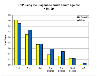
Figure 1. ChIP results obtained with the Diagenode antibody directed against H3S10p
ChIP assays were performed using human HeLa cells treated with colcemid, the Diagenode antibody against H3S10p (cat. No. CS-116-100) and optimized PCR primer sets for qPCR. ChIP was performed with the “LowCell# ChIP” kit (cat. No. kch-maglow-016), using sheared chromatin from 10,000 cells. A titration of the antibody consisting of 1, 5, and 10 μl per ChIP experiment was analysed. Additionally, ChIP was performed after incubation of the antibody with 5 nmol blocking peptide (cat. No. sp-116-050) for 1 hour at room temperature. IgG (5 μg/IP) was used as negative IP control. QPCR was performed with primers for the promoter of the active genes c-fos (cat. No. pp-1004-050) and RPL30. Figure 1 shows the recovery, expressed as a % of input (the relative amount of immunoprecipitated DNA compared to input DNA after qPCR analysis).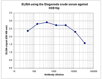
Figure 2. Determination of the titer
To determine the titer, an ELISA was performed using a serial dilution of the Diagenode antibody directed against human H3S10p (cat. No. CS-116-100). The antigen used was a peptide containing the histone modification of interest. By plotting the absorbance against the antibody dilution (Figure 2), the titer of the antibody was estimated to be 1:35,000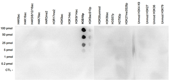
Figure 3. Cross reactivity test with the Diagenode antibody directed against H3S10p
A Dot Blot analysis was performed to test the cross reactivity of the Diagenode antibody against H3S10p (cat. No. CS-116-100) with peptides containing other modifications of histone H3 and H4 and with peptides containing unmodified sequences from histone H3. One hundred to 0.2 pmol of the peptide containing the respective histone modification were spotted on a membrane. The antibody was used at a dilution of 1:20,000. Figure 3 shows a high specificity of the antibody for the modification of interest. Note that the antibody does not recognize the H3S10p modification if the H3K9ac modification is present.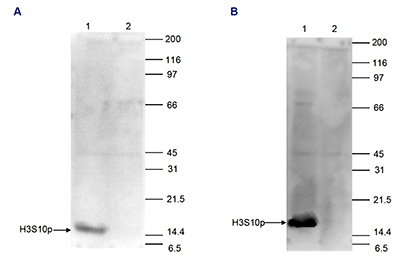
Figure 4. Western blot analysis using the Diagenode antibody directed against H3S10p
HeLa cells were treated with TSA (figure 4A) or with colcemid (figure 4B), and 15 μg of histone extracts of these cells were analysed by Western blot using the Diagenode antibody against H3S10p (cat. No. CS-116-100) diluted 1:500 in TBS-Tween containing 5% skimmed milk. The position of the protein of interest is indicated on the left; the marker (in kDa) is shown on the right. The result of the Western analysis with the antibody is shown in lane 1; lane 2 shows the same analysis after incubation of the antibody with 750 pmol blocking peptide (cat. No. sp- 116-050) for 1 hour at room temperature.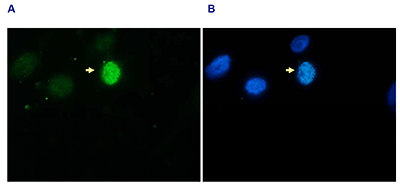
Figure 5. Immunofluorescence with the Diagenode antibody directed against H3S10p
Hela asynchronous cells were stained with the Diagenode antibody against H3S10p (cat. No. CS-116-100) and with DAPI. Cells were fixed with formaldehyde, permeabilized with sodium citrate and Triton X100 and blocked with PBS containing 2.5% BSA. (A) Cells were immunofluorescently labelled with the H3S10p antibody (diluted 1:200 and incubated for 1 hour at room temperature) followed by goat anti-rabbit antibody conjugated to DyLight 488. (B) The nuclei were stained with DAPI, which specifically labels DNA. Phosphorylation of H3 on serine 10 occurs on condensed chromosomes during mitosis. This explains the dense staining of one of the cells (indicated with an arrow) in Figure 5.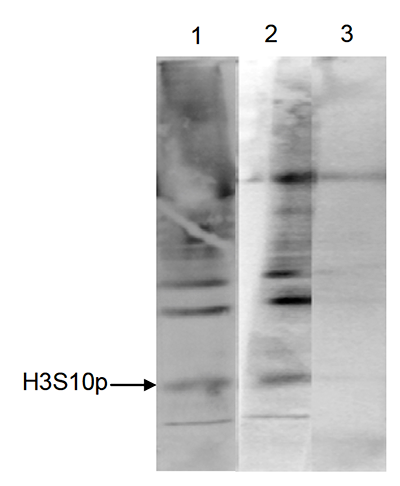
Figure 6. Immunoprecipitation with the Diagenode antibody directed against H3S10p
HeLa cells were treated with colcemid to block the cell cycle in metaphase and were fixed with formaldehyde. Chromatin from 10,000 cells was sheared and used for immunoprecipitation (IP). IP was performed with 5 μl of the Diagenode antibody against H3S10p (cat. No. CS-116-100). The immunoprecipitated proteins were analysed by Western blot with the antibody diluted 1:500 in TBS-Tween containing 5% skimmed milk. Lane 1 shows the result of the IP; a positive control (sheared chromatin from 10,000 cells) and a negative IP control (no antibody added) are shown in lane 2 and 3, respectively. - 出版物

How to properly cite our product/service in your work
We strongly recommend using this: H3S10p polyclonal antibody (Hologic Diagenode Cat# C15310116 Lot# A161-001 ). Click here to copy to clipboard.
Using our products or services in your publication? Let us know!