
| DB Dot blotting Read more |
N6-methyladenosine (m6A) antibody - RIP-seq Grade (sample size)

Polyclonal antibody raised in rabbit against N6-methyladenosine (m6A) conjugated to LPH.
| Lot | A2125-0010 |
|---|---|
| Concentration | 1.2 µg/µl |
| Species reactivity | Human, mouse, other (wide range): positive. |
| Type | Polyclonal |
| Purity | Protein G purified polyclonal antibody. |
| Host | Rabbit |
| Storage Conditions | Store at -20°C; for long storage, store at -80°C. Avoid multiple freeze-thaw cycles. |
| Storage Buffer | PBS containing 0.05% azide and 0.05% ProClin 300. |
| Precautions | This product is for research use only. Not for use in diagnostic or therapeutic procedures. |
| Applications | Suggested dilution | References |
|---|---|---|
| RIP/RIP-seq* | 1-2 µg per IP | Fig 1, 2, 3 |
| Dot Blotting | 1:400 | Fig 4 |
*Please note that the optimal antibody amount per IP should be determined by the end-user. We recommend testing 1-10 µg per IP.
- Validation data
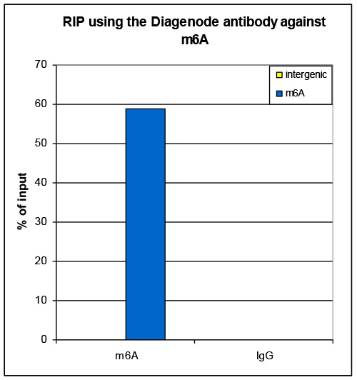
Figure 1. RNA immunoprecipitation using the Diagenode antibody directed against m6A
RNA Immunoprecipitation was performed on 40 µg HeLa total RNA spiked with 0.5 µg of an in vitro prepared transcript containing m6A nucleotides, using 2 µg of the Diagenode m6A antibody (Cat. No. C15410208). An equal amount of IgG was used as negative control. The immunoprecipitated RNA was subsequently analyzed by qRT-PCR with primers specific for the transcript and for an intergenic region, used as negative control. Figure 1 shows the recovery, expressed as a % of input (the relative amount of immunoprecipitated DNA compared to input DNA after qPCR analysis).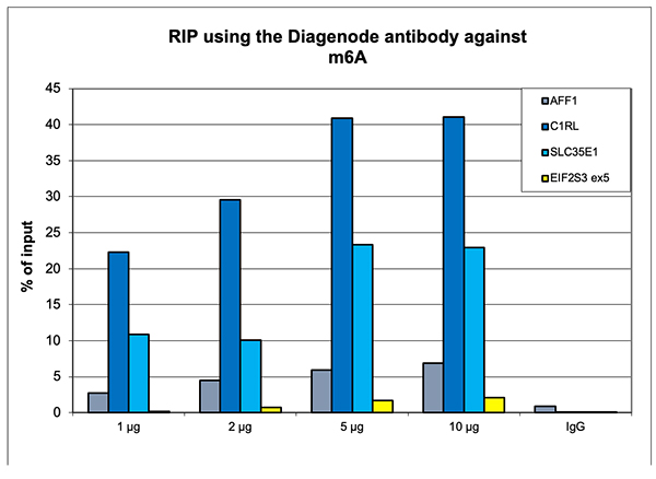
Figure 2. RNA immunoprecipitation using the Diagenode antibody directed against m6A
RNA Immunoprecipitation was performed on 40 µg HeLa total RNA fragmented to a mean size of ~500 bases. A titration consisting of 1, 2, 5 and 10 µg of antibody per RIP experiment was analyzed. IgG (2 µg/IP) was used as a negative IP control. Quantitative RT-PCR was performed with primers for 3’ UTR of the C1RL and SLC35E1 genes, and for exon 12 of the AFF1 gene, used as positive controls, and for exon 5 of the EIF2S3 gene, used as negative control.
Figure 1 shows the recovery, expressed as a % of input (the relative amount of immunoprecipitated DNA compared to input DNA after qPCR analysis).A.

B.
C.
D.
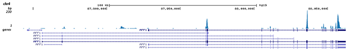
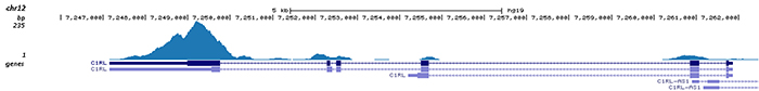
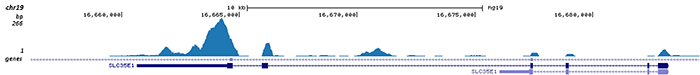
Figure 3. RIP-seq results obtained with the Diagenode antibody directed against m6A
RIP was performed with 2 µg of the Diagenode antibody against m6A (Cat. No. C15410208). The IP'd DNA was subsequently analysed on an Illumina HiSeq 4000. The 50 bp tags were aligned to the human genome using the BWA algorithm. Figure 3 shows the signal distribution along the complete sequence of the human X-chromosome (figure 3A) and in three genomic regions surrounding the C1RL, SLC35E1 and AFF1 positive control genes (figure 3B, C and D).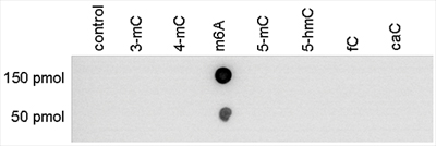
Figure 2. Dot blot analysis using the Diagenode antibody directed against m6A
To demonstrate the specificity of the Diagenode antibody against m6A (Cat. No. C15410208), a Dot Blot analysis was performed using synthetic oligonucleotides containing different modified bases. 150 and 50 pmol of the respective oligo’s were spotted on the membrane. The antibody was diluted 1:400 in PBS-T containing 10 % skimmed milk and 1% BSA. Figure 1 shows a high specificity of the antibody for the oligonucleotide with the N6-methyladenosine modification. - 出版物

How to properly cite our product/service in your work
We strongly recommend using this: N6-methyladenosine (m6A) antibody - RIP-seq Grade (sample size) (Hologic Diagenode Cat# C15410208-10 Lot# A2125-0010). Click here to copy to clipboard.
Using our products or services in your publication? Let us know!
Genomewide analysis of 6-methyladenine DNA in peripheral blood mononuclear cells of systemic lupus erythematosus.
Zheng F, Tang D, Xu H, Xu Y, Dai W, Zhang X, Hong X, Liu D, Dai Y
AIM: The aim of this paper is to explore the expression of 6-methyladenine (6mA) DNA and to elucidate its gene regulation role in systemic lupus erythematosus (SLE). METHODS: Twenty SLE patients and 20 normal control healthy individuals (HCs) were included in this study. Genomic DNA was isolated from peripheral bloo...