Notice (8): Undefined variable: solution_of_interest [APP/View/Products/view.ctp, line 755]Code Context<!-- BEGIN: REQUEST_FORM MODAL -->
<div id="request_formModal" class="reveal-modal medium" data-reveal aria-labelledby="modalTitle" aria-hidden="true" role="dialog">
<?= $this->element('Forms/simple_form', array('solution_of_interest' => $solution_of_interest, 'header' => $header, 'message' => $message, 'campaign_id' => $campaign_id)) ?>
$viewFile = '/home/website-server/www/app/View/Products/view.ctp'
$dataForView = array(
'language' => 'en',
'meta_keywords' => '',
'meta_description' => 'EzH2 (Enhancer of zeste homolog 2) Polyclonal Antibody validated in ChIP-seq, ChIP-qPCR, WB and IF. Specificity confirmed by siRNA assay. Batch-specific data available on the website. Alternative names: ENX-1, ENX1, KMT6, KMT6A, WVS, WVS2. Sample size available.',
'meta_title' => 'EzH2 Antibody - ChIP-seq Grade (C15410039) | Diagenode',
'product' => array(
'Product' => array(
'id' => '2204',
'antibody_id' => '293',
'name' => 'EZH2 Antibody',
'description' => '<p><strong>Other names: </strong>ENX-1, ENX1, KMT6, KMT6A, WVS, WVS2</p>
<p>Polyclonal antibody raised in rabbit against the N-terminus (aa1-343) of the mouse <strong>EZH2</strong> protein (<strong>Enhancer of zeste homolog 2</strong>).</p>',
'label1' => 'Validation Data',
'info1' => '<div class="row">
<div class="small-6 columns">
<p><img src="https://www.diagenode.com/img/product/antibodies/C15410039-ChIP.png" alt="EZH2 Antibody ChIP Grade " /></p>
</div>
<div class="small-6 columns">
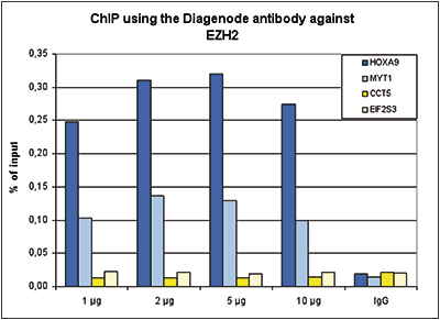
<p><small><strong>Figure 1. ChIP results obtained with the Diagenode antibody directed against EZH2</strong><br />ChIP assays were performed using K562 cells, the Diagenode antibody against EZH2 (Cat. No. C15410039) and optimized PCR primer sets for qPCR. ChIP was performed with the “iDeal ChIP-seq” kit (Cat. No. C01010055), using sheared chromatin from 4 million cells. A titration of the antibody consisting of 1, 2. 5 and 10 µg per ChIP experiment was analysed. IgG (2 µg/IP) was used as negative IP control. Quantitative PCR was performed with primers for MYT1 and HOXA9, used as positive control targets, and for the coding regions of the active CCT5 and EIF2S3 genes, used as negative controls. Figure 1 shows the recovery, expressed as a % of input (the relative amount of immunoprecipitated DNA compared to input DNA after qPCR analysis). </small></p>
</div>
</div>
<div class="row">
<div class="small-12 columns">
<p>A. <img src="https://www.diagenode.com/img/product/antibodies/C15410039-ChIPseq-figA.png" alt="EZH2 Antibody ChIP-seq Grade " /></p>
<p>B. <img src="https://www.diagenode.com/img/product/antibodies/C15410039-ChIPseq-figB.png" alt="EZH2 Antibody for ChIP-seq " /></p>
<p>C. <img src="https://www.diagenode.com/img/product/antibodies/C15410039-ChIPseq-figC.png" alt="EZH2 Antibody for ChIP-seq assay" /></p>
<div class="extra-spaced"></div>
<p>D. <img src="https://www.diagenode.com/img/product/antibodies/C15410039-ChIPseq-figD.png" alt="EZH2 Antibody validated in ChIP-seq " /></p>
</div>
</div>
<div class="row">
<div class="small-12 columns">
<p><small><strong>Figure 2. ChIP-seq results obtained with the Diagenode antibody directed against EZH2</strong><br /> ChIP was performed on sheared chromatin from 4 million K562 cells using 2 µg of the Diagenode antibody against EZH2 (Cat. No. C15410039) as described above. The IP’d DNA was subsequently analysed on an Illumina HiSeq. Library preparation, cluster generation and sequencing were performed according to the manufacturer’s instructions. The 51 bp tags were aligned to the human genome using the BWA algorithm. Figure 2 shows the peak distribution along the short arm and a 6 Mb region containing several enriched regions of human chromosome 3 (figure 2A and B, respectively), and in two genomic regions containing the MYT1 gene on chromosome 20 and the HOX cluster on chromosome 7 (figure 2C and D).</small></p>
</div>
</div>
<div class="row">
<div class="small-4 columns">
<p><img src="https://www.diagenode.com/img/product/antibodies/C15410039-WB.png" alt="EZH2 Antibody validated in Western Blot" /></p>
</div>
<div class="small-8 columns">
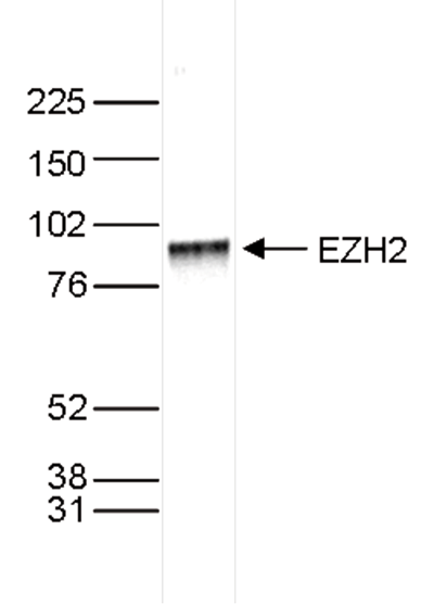
<p><small><strong>Figure 3. Western blot analysis using the Diagenode antibody directed against EZH2</strong><br /> Nuclear extracts of HeLa cells (40 µg) were analysed by Western blot using the Diagenode antibody against EZH2 (Cat. No. C15410039) diluted 1:1,000 in TBS-Tween containing 5% skimmed milk. The position of the protein of interest (expected size 85 kDa) is indicated on the right; the marker (in kDa) is shown on the left. </small></p>
</div>
</div>
<div class="extra-spaced"></div>
<div class="row">
<div class="small-4 columns">
<p><img src="https://www.diagenode.com/img/product/antibodies/C15410039-WB-2.png" alt="EZH2 Antibody validated for Western Blot" /></p>
</div>
<div class="small-8 columns">
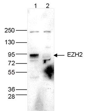
<p><small><strong>Figure 4. Western blot analysis using the Diagenode antibody directed against EZH2</strong><br /> Whole cell extracts (40 µg) from HeLa cells transfected with EZH2 siRNA (lane 2) and from an untransfected control (lane 1) were analysed by Western blot using the Diagenode antibody against EZH2 (Cat. No. C15410039) diluted 1:1,000 in TBS-Tween containing 5% skimmed milk. The position of the protein of interest is indicated on the right; the marker (in kDa) is shown on the left.</small></p>
</div>
</div>
<div class="row">
<div class="small-6 columns">
<p><img src="https://www.diagenode.com/img/product/antibodies/C15410039-if.jpg" alt="EZH2 Antibody validated for Immunofluorescence" /></p>
</div>
<div class="small-6 columns">
<p><small><strong>Figure 5. Immunofluorescence using the Diagenode antibody directed against EZH2</strong><br /> HeLa cells were stained with the Diagenode antibody against EZH2 (cat. No. C15410039) and with DAPI. Cells were fixed with 4% formaldehyde for 10’ and blocked with PBS/TX-100 containing 1% BSA. The cells were immunofluorescently labelled with the EZH2 antibody (left) diluted 1:1,000 in blocking solution followed by an anti-mouse antibody conjugated to Alexa488. The middle panel shows staining of the nuclei with DAPI. A merge of the two stainings is shown on the right. </small></p>
</div>
</div>',
'label2' => 'Target Description',
'info2' => '<p>EZH2 (UniProt/Swiss-Prot entry Q15910) is a histone-lysine methyltransferase which methylates ‘Lys-9’ and ‘Lys-27’ of histone H3, leading to transcriptional repression. It is a member of the polycomb group (PcG) family which form multimeric protein complexes and are involved in maintaining the transcriptional repressive state of genes over successive cell generations. The EZH2 activity is dependent on the association with other components of the PRC2 complex (EED, EZH2, SUZ12/JJAZ1, RBBP4 and RBBP7). EZH2 may play a role in the hematopoietic and central nervous systems. Over-expression of EZH2 is observed during advanced stages of prostate cancer and breast cancer.</p>',
'label3' => '',
'info3' => '',
'format' => '50 µg',
'catalog_number' => 'C15410039',
'old_catalog_number' => 'pAb-039-050',
'sf_code' => 'C15410039-D001-000581',
'type' => 'FRE',
'search_order' => '03-Antibody',
'price_EUR' => '390',
'price_USD' => '390',
'price_GBP' => '345',
'price_JPY' => '63900',
'price_CNY' => '',
'price_AUD' => '975',
'country' => 'ALL',
'except_countries' => 'None',
'quote' => false,
'in_stock' => false,
'featured' => false,
'no_promo' => false,
'online' => true,
'master' => true,
'last_datasheet_update' => 'January 17, 2017',
'slug' => 'ezh2-polyclonal-antibody-classic-50-ug',
'meta_title' => 'EzH2 Antibody - ChIP-seq Grade (C15410039) | Diagenode',
'meta_keywords' => '',
'meta_description' => 'EzH2 (Enhancer of zeste homolog 2) Polyclonal Antibody validated in ChIP-seq, ChIP-qPCR, WB and IF. Specificity confirmed by siRNA assay. Batch-specific data available on the website. Alternative names: ENX-1, ENX1, KMT6, KMT6A, WVS, WVS2. Sample size available.',
'modified' => '2024-11-19 16:57:04',
'created' => '2015-06-29 14:08:20',
'locale' => 'eng'
),
'Antibody' => array(
'host' => '*****',
'id' => '293',
'name' => 'EZH2 polyclonal antibody',
'description' => 'EZH2 (UniProt/Swiss-Prot entry Q15910) is a histone-lysine methyltransferase which methylates ‘Lys-9’ and ‘Lys-27’ of histone H3, leading to transcriptional repression. It is a member of the polycomb group (PcG) family which form multimeric protein complexes and are involved in maintaining the transcriptional repressive state of genes over successive cell generations. The EZH2 activity is dependent on the association with other components of the PRC2 complex (EED, EZH2, SUZ12/JJAZ1, RBBP4 and RBBP7). EZH2 may play a role in the hematopoietic and central nervous systems. Over-expression of EZH2 is observed during advanced stages of prostate cancer and breast cancer.',
'clonality' => '',
'isotype' => '',
'lot' => '003',
'concentration' => '1.0 µg/µl',
'reactivity' => 'Human, mouse',
'type' => 'Polyclonal',
'purity' => 'Protein G purified',
'classification' => '',
'application_table' => '<table>
<thead>
<tr>
<th>Applications</th>
<th>Suggested dilution</th>
<th>References</th>
</tr>
</thead>
<tbody>
<tr>
<td>ChIP/ChIP-seq <sup>*</sup></td>
<td>2 μg/ChIP</td>
<td>Fig 1, 2</td>
</tr>
<tr>
<td>Western Blotting</td>
<td>1:1,000</td>
<td>Fig 3, 4</td>
</tr>
<tr>
<td>I<span>mmunofluorescence</span></td>
<td>1:1000</td>
<td>Fig 5</td>
</tr>
</tbody>
</table>
<p><small><sup>*</sup> Please note that of the optimal antibody amount per IP should be determined by the end-user. We recommend testing 1-5 μg per IP.</small></p>',
'storage_conditions' => '',
'storage_buffer' => '',
'precautions' => 'This product is for research use only. Not for use in diagnostic or therapeutic procedures.',
'uniprot_acc' => '',
'slug' => '',
'meta_keywords' => '',
'meta_description' => '',
'modified' => '2021-05-27 11:35:48',
'created' => '0000-00-00 00:00:00',
'select_label' => '293 - EZH2 polyclonal antibody (003 - 1.0 µg/µl - Human, mouse - Protein G purified - Rabbit)'
),
'Slave' => array(
(int) 0 => array(
[maximum depth reached]
)
),
'Group' => array(
'Group' => array(
[maximum depth reached]
),
'Master' => array(
[maximum depth reached]
),
'Product' => array(
[maximum depth reached]
)
),
'Related' => array(
(int) 0 => array(
[maximum depth reached]
),
(int) 1 => array(
[maximum depth reached]
),
(int) 2 => array(
[maximum depth reached]
),
(int) 3 => array(
[maximum depth reached]
)
),
'Application' => array(
(int) 0 => array(
[maximum depth reached]
),
(int) 1 => array(
[maximum depth reached]
),
(int) 2 => array(
[maximum depth reached]
),
(int) 3 => array(
[maximum depth reached]
),
(int) 4 => array(
[maximum depth reached]
)
),
'Category' => array(
(int) 0 => array(
[maximum depth reached]
),
(int) 1 => array(
[maximum depth reached]
),
(int) 2 => array(
[maximum depth reached]
),
(int) 3 => array(
[maximum depth reached]
)
),
'Document' => array(
(int) 0 => array(
[maximum depth reached]
),
(int) 1 => array(
[maximum depth reached]
),
(int) 2 => array(
[maximum depth reached]
)
),
'Feature' => array(),
'Image' => array(
(int) 0 => array(
[maximum depth reached]
)
),
'Promotion' => array(),
'Protocol' => array(),
'Publication' => array(
(int) 0 => array(
[maximum depth reached]
),
(int) 1 => array(
[maximum depth reached]
),
(int) 2 => array(
[maximum depth reached]
),
(int) 3 => array(
[maximum depth reached]
),
(int) 4 => array(
[maximum depth reached]
),
(int) 5 => array(
[maximum depth reached]
),
(int) 6 => array(
[maximum depth reached]
),
(int) 7 => array(
[maximum depth reached]
),
(int) 8 => array(
[maximum depth reached]
),
(int) 9 => array(
[maximum depth reached]
),
(int) 10 => array(
[maximum depth reached]
),
(int) 11 => array(
[maximum depth reached]
),
(int) 12 => array(
[maximum depth reached]
),
(int) 13 => array(
[maximum depth reached]
),
(int) 14 => array(
[maximum depth reached]
),
(int) 15 => array(
[maximum depth reached]
),
(int) 16 => array(
[maximum depth reached]
),
(int) 17 => array(
[maximum depth reached]
),
(int) 18 => array(
[maximum depth reached]
),
(int) 19 => array(
[maximum depth reached]
),
(int) 20 => array(
[maximum depth reached]
),
(int) 21 => array(
[maximum depth reached]
),
(int) 22 => array(
[maximum depth reached]
),
(int) 23 => array(
[maximum depth reached]
),
(int) 24 => array(
[maximum depth reached]
),
(int) 25 => array(
[maximum depth reached]
),
(int) 26 => array(
[maximum depth reached]
),
(int) 27 => array(
[maximum depth reached]
),
(int) 28 => array(
[maximum depth reached]
),
(int) 29 => array(
[maximum depth reached]
),
(int) 30 => array(
[maximum depth reached]
),
(int) 31 => array(
[maximum depth reached]
),
(int) 32 => array(
[maximum depth reached]
),
(int) 33 => array(
[maximum depth reached]
)
),
'Testimonial' => array(),
'Area' => array(),
'SafetySheet' => array(
(int) 0 => array(
[maximum depth reached]
),
(int) 1 => array(
[maximum depth reached]
),
(int) 2 => array(
[maximum depth reached]
),
(int) 3 => array(
[maximum depth reached]
),
(int) 4 => array(
[maximum depth reached]
),
(int) 5 => array(
[maximum depth reached]
),
(int) 6 => array(
[maximum depth reached]
),
(int) 7 => array(
[maximum depth reached]
)
)
),
'meta_canonical' => 'https://www.diagenode.com/en/p/ezh2-polyclonal-antibody-classic-50-ug'
)
$language = 'en'
$meta_keywords = ''
$meta_description = 'EzH2 (Enhancer of zeste homolog 2) Polyclonal Antibody validated in ChIP-seq, ChIP-qPCR, WB and IF. Specificity confirmed by siRNA assay. Batch-specific data available on the website. Alternative names: ENX-1, ENX1, KMT6, KMT6A, WVS, WVS2. Sample size available.'
$meta_title = 'EzH2 Antibody - ChIP-seq Grade (C15410039) | Diagenode'
$product = array(
'Product' => array(
'id' => '2204',
'antibody_id' => '293',
'name' => 'EZH2 Antibody',
'description' => '<p><strong>Other names: </strong>ENX-1, ENX1, KMT6, KMT6A, WVS, WVS2</p>
<p>Polyclonal antibody raised in rabbit against the N-terminus (aa1-343) of the mouse <strong>EZH2</strong> protein (<strong>Enhancer of zeste homolog 2</strong>).</p>',
'label1' => 'Validation Data',
'info1' => '<div class="row">
<div class="small-6 columns">
<p><img src="https://www.diagenode.com/img/product/antibodies/C15410039-ChIP.png" alt="EZH2 Antibody ChIP Grade " /></p>
</div>
<div class="small-6 columns">
<p><small><strong>Figure 1. ChIP results obtained with the Diagenode antibody directed against EZH2</strong><br />ChIP assays were performed using K562 cells, the Diagenode antibody against EZH2 (Cat. No. C15410039) and optimized PCR primer sets for qPCR. ChIP was performed with the “iDeal ChIP-seq” kit (Cat. No. C01010055), using sheared chromatin from 4 million cells. A titration of the antibody consisting of 1, 2. 5 and 10 µg per ChIP experiment was analysed. IgG (2 µg/IP) was used as negative IP control. Quantitative PCR was performed with primers for MYT1 and HOXA9, used as positive control targets, and for the coding regions of the active CCT5 and EIF2S3 genes, used as negative controls. Figure 1 shows the recovery, expressed as a % of input (the relative amount of immunoprecipitated DNA compared to input DNA after qPCR analysis). </small></p>
</div>
</div>
<div class="row">
<div class="small-12 columns">
<p>A. <img src="https://www.diagenode.com/img/product/antibodies/C15410039-ChIPseq-figA.png" alt="EZH2 Antibody ChIP-seq Grade " /></p>
<p>B. <img src="https://www.diagenode.com/img/product/antibodies/C15410039-ChIPseq-figB.png" alt="EZH2 Antibody for ChIP-seq " /></p>
<p>C. <img src="https://www.diagenode.com/img/product/antibodies/C15410039-ChIPseq-figC.png" alt="EZH2 Antibody for ChIP-seq assay" /></p>
<div class="extra-spaced"></div>
<p>D. <img src="https://www.diagenode.com/img/product/antibodies/C15410039-ChIPseq-figD.png" alt="EZH2 Antibody validated in ChIP-seq " /></p>
</div>
</div>
<div class="row">
<div class="small-12 columns">
<p><small><strong>Figure 2. ChIP-seq results obtained with the Diagenode antibody directed against EZH2</strong><br /> ChIP was performed on sheared chromatin from 4 million K562 cells using 2 µg of the Diagenode antibody against EZH2 (Cat. No. C15410039) as described above. The IP’d DNA was subsequently analysed on an Illumina HiSeq. Library preparation, cluster generation and sequencing were performed according to the manufacturer’s instructions. The 51 bp tags were aligned to the human genome using the BWA algorithm. Figure 2 shows the peak distribution along the short arm and a 6 Mb region containing several enriched regions of human chromosome 3 (figure 2A and B, respectively), and in two genomic regions containing the MYT1 gene on chromosome 20 and the HOX cluster on chromosome 7 (figure 2C and D).</small></p>
</div>
</div>
<div class="row">
<div class="small-4 columns">
<p><img src="https://www.diagenode.com/img/product/antibodies/C15410039-WB.png" alt="EZH2 Antibody validated in Western Blot" /></p>
</div>
<div class="small-8 columns">
<p><small><strong>Figure 3. Western blot analysis using the Diagenode antibody directed against EZH2</strong><br /> Nuclear extracts of HeLa cells (40 µg) were analysed by Western blot using the Diagenode antibody against EZH2 (Cat. No. C15410039) diluted 1:1,000 in TBS-Tween containing 5% skimmed milk. The position of the protein of interest (expected size 85 kDa) is indicated on the right; the marker (in kDa) is shown on the left. </small></p>
</div>
</div>
<div class="extra-spaced"></div>
<div class="row">
<div class="small-4 columns">
<p><img src="https://www.diagenode.com/img/product/antibodies/C15410039-WB-2.png" alt="EZH2 Antibody validated for Western Blot" /></p>
</div>
<div class="small-8 columns">
<p><small><strong>Figure 4. Western blot analysis using the Diagenode antibody directed against EZH2</strong><br /> Whole cell extracts (40 µg) from HeLa cells transfected with EZH2 siRNA (lane 2) and from an untransfected control (lane 1) were analysed by Western blot using the Diagenode antibody against EZH2 (Cat. No. C15410039) diluted 1:1,000 in TBS-Tween containing 5% skimmed milk. The position of the protein of interest is indicated on the right; the marker (in kDa) is shown on the left.</small></p>
</div>
</div>
<div class="row">
<div class="small-6 columns">
<p><img src="https://www.diagenode.com/img/product/antibodies/C15410039-if.jpg" alt="EZH2 Antibody validated for Immunofluorescence" /></p>
</div>
<div class="small-6 columns">
<p><small><strong>Figure 5. Immunofluorescence using the Diagenode antibody directed against EZH2</strong><br /> HeLa cells were stained with the Diagenode antibody against EZH2 (cat. No. C15410039) and with DAPI. Cells were fixed with 4% formaldehyde for 10’ and blocked with PBS/TX-100 containing 1% BSA. The cells were immunofluorescently labelled with the EZH2 antibody (left) diluted 1:1,000 in blocking solution followed by an anti-mouse antibody conjugated to Alexa488. The middle panel shows staining of the nuclei with DAPI. A merge of the two stainings is shown on the right. </small></p>
</div>
</div>',
'label2' => 'Target Description',
'info2' => '<p>EZH2 (UniProt/Swiss-Prot entry Q15910) is a histone-lysine methyltransferase which methylates ‘Lys-9’ and ‘Lys-27’ of histone H3, leading to transcriptional repression. It is a member of the polycomb group (PcG) family which form multimeric protein complexes and are involved in maintaining the transcriptional repressive state of genes over successive cell generations. The EZH2 activity is dependent on the association with other components of the PRC2 complex (EED, EZH2, SUZ12/JJAZ1, RBBP4 and RBBP7). EZH2 may play a role in the hematopoietic and central nervous systems. Over-expression of EZH2 is observed during advanced stages of prostate cancer and breast cancer.</p>',
'label3' => '',
'info3' => '',
'format' => '50 µg',
'catalog_number' => 'C15410039',
'old_catalog_number' => 'pAb-039-050',
'sf_code' => 'C15410039-D001-000581',
'type' => 'FRE',
'search_order' => '03-Antibody',
'price_EUR' => '390',
'price_USD' => '390',
'price_GBP' => '345',
'price_JPY' => '63900',
'price_CNY' => '',
'price_AUD' => '975',
'country' => 'ALL',
'except_countries' => 'None',
'quote' => false,
'in_stock' => false,
'featured' => false,
'no_promo' => false,
'online' => true,
'master' => true,
'last_datasheet_update' => 'January 17, 2017',
'slug' => 'ezh2-polyclonal-antibody-classic-50-ug',
'meta_title' => 'EzH2 Antibody - ChIP-seq Grade (C15410039) | Diagenode',
'meta_keywords' => '',
'meta_description' => 'EzH2 (Enhancer of zeste homolog 2) Polyclonal Antibody validated in ChIP-seq, ChIP-qPCR, WB and IF. Specificity confirmed by siRNA assay. Batch-specific data available on the website. Alternative names: ENX-1, ENX1, KMT6, KMT6A, WVS, WVS2. Sample size available.',
'modified' => '2024-11-19 16:57:04',
'created' => '2015-06-29 14:08:20',
'locale' => 'eng'
),
'Antibody' => array(
'host' => '*****',
'id' => '293',
'name' => 'EZH2 polyclonal antibody',
'description' => 'EZH2 (UniProt/Swiss-Prot entry Q15910) is a histone-lysine methyltransferase which methylates ‘Lys-9’ and ‘Lys-27’ of histone H3, leading to transcriptional repression. It is a member of the polycomb group (PcG) family which form multimeric protein complexes and are involved in maintaining the transcriptional repressive state of genes over successive cell generations. The EZH2 activity is dependent on the association with other components of the PRC2 complex (EED, EZH2, SUZ12/JJAZ1, RBBP4 and RBBP7). EZH2 may play a role in the hematopoietic and central nervous systems. Over-expression of EZH2 is observed during advanced stages of prostate cancer and breast cancer.',
'clonality' => '',
'isotype' => '',
'lot' => '003',
'concentration' => '1.0 µg/µl',
'reactivity' => 'Human, mouse',
'type' => 'Polyclonal',
'purity' => 'Protein G purified',
'classification' => '',
'application_table' => '<table>
<thead>
<tr>
<th>Applications</th>
<th>Suggested dilution</th>
<th>References</th>
</tr>
</thead>
<tbody>
<tr>
<td>ChIP/ChIP-seq <sup>*</sup></td>
<td>2 μg/ChIP</td>
<td>Fig 1, 2</td>
</tr>
<tr>
<td>Western Blotting</td>
<td>1:1,000</td>
<td>Fig 3, 4</td>
</tr>
<tr>
<td>I<span>mmunofluorescence</span></td>
<td>1:1000</td>
<td>Fig 5</td>
</tr>
</tbody>
</table>
<p><small><sup>*</sup> Please note that of the optimal antibody amount per IP should be determined by the end-user. We recommend testing 1-5 μg per IP.</small></p>',
'storage_conditions' => '',
'storage_buffer' => '',
'precautions' => 'This product is for research use only. Not for use in diagnostic or therapeutic procedures.',
'uniprot_acc' => '',
'slug' => '',
'meta_keywords' => '',
'meta_description' => '',
'modified' => '2021-05-27 11:35:48',
'created' => '0000-00-00 00:00:00',
'select_label' => '293 - EZH2 polyclonal antibody (003 - 1.0 µg/µl - Human, mouse - Protein G purified - Rabbit)'
),
'Slave' => array(
(int) 0 => array(
'id' => '192',
'name' => 'C15410039',
'product_id' => '2204',
'modified' => '2016-11-04 15:53:29',
'created' => '2016-11-04 15:53:13'
)
),
'Group' => array(
'Group' => array(
'id' => '192',
'name' => 'C15410039',
'product_id' => '2204',
'modified' => '2016-11-04 15:53:29',
'created' => '2016-11-04 15:53:13'
),
'Master' => array(
'id' => '2204',
'antibody_id' => '293',
'name' => 'EZH2 Antibody',
'description' => '<p><strong>Other names: </strong>ENX-1, ENX1, KMT6, KMT6A, WVS, WVS2</p>
<p>Polyclonal antibody raised in rabbit against the N-terminus (aa1-343) of the mouse <strong>EZH2</strong> protein (<strong>Enhancer of zeste homolog 2</strong>).</p>',
'label1' => 'Validation Data',
'info1' => '<div class="row">
<div class="small-6 columns">
<p><img src="https://www.diagenode.com/img/product/antibodies/C15410039-ChIP.png" alt="EZH2 Antibody ChIP Grade " /></p>
</div>
<div class="small-6 columns">
<p><small><strong>Figure 1. ChIP results obtained with the Diagenode antibody directed against EZH2</strong><br />ChIP assays were performed using K562 cells, the Diagenode antibody against EZH2 (Cat. No. C15410039) and optimized PCR primer sets for qPCR. ChIP was performed with the “iDeal ChIP-seq” kit (Cat. No. C01010055), using sheared chromatin from 4 million cells. A titration of the antibody consisting of 1, 2. 5 and 10 µg per ChIP experiment was analysed. IgG (2 µg/IP) was used as negative IP control. Quantitative PCR was performed with primers for MYT1 and HOXA9, used as positive control targets, and for the coding regions of the active CCT5 and EIF2S3 genes, used as negative controls. Figure 1 shows the recovery, expressed as a % of input (the relative amount of immunoprecipitated DNA compared to input DNA after qPCR analysis). </small></p>
</div>
</div>
<div class="row">
<div class="small-12 columns">
<p>A. <img src="https://www.diagenode.com/img/product/antibodies/C15410039-ChIPseq-figA.png" alt="EZH2 Antibody ChIP-seq Grade " /></p>
<p>B. <img src="https://www.diagenode.com/img/product/antibodies/C15410039-ChIPseq-figB.png" alt="EZH2 Antibody for ChIP-seq " /></p>
<p>C. <img src="https://www.diagenode.com/img/product/antibodies/C15410039-ChIPseq-figC.png" alt="EZH2 Antibody for ChIP-seq assay" /></p>
<div class="extra-spaced"></div>
<p>D. <img src="https://www.diagenode.com/img/product/antibodies/C15410039-ChIPseq-figD.png" alt="EZH2 Antibody validated in ChIP-seq " /></p>
</div>
</div>
<div class="row">
<div class="small-12 columns">
<p><small><strong>Figure 2. ChIP-seq results obtained with the Diagenode antibody directed against EZH2</strong><br /> ChIP was performed on sheared chromatin from 4 million K562 cells using 2 µg of the Diagenode antibody against EZH2 (Cat. No. C15410039) as described above. The IP’d DNA was subsequently analysed on an Illumina HiSeq. Library preparation, cluster generation and sequencing were performed according to the manufacturer’s instructions. The 51 bp tags were aligned to the human genome using the BWA algorithm. Figure 2 shows the peak distribution along the short arm and a 6 Mb region containing several enriched regions of human chromosome 3 (figure 2A and B, respectively), and in two genomic regions containing the MYT1 gene on chromosome 20 and the HOX cluster on chromosome 7 (figure 2C and D).</small></p>
</div>
</div>
<div class="row">
<div class="small-4 columns">
<p><img src="https://www.diagenode.com/img/product/antibodies/C15410039-WB.png" alt="EZH2 Antibody validated in Western Blot" /></p>
</div>
<div class="small-8 columns">
<p><small><strong>Figure 3. Western blot analysis using the Diagenode antibody directed against EZH2</strong><br /> Nuclear extracts of HeLa cells (40 µg) were analysed by Western blot using the Diagenode antibody against EZH2 (Cat. No. C15410039) diluted 1:1,000 in TBS-Tween containing 5% skimmed milk. The position of the protein of interest (expected size 85 kDa) is indicated on the right; the marker (in kDa) is shown on the left. </small></p>
</div>
</div>
<div class="extra-spaced"></div>
<div class="row">
<div class="small-4 columns">
<p><img src="https://www.diagenode.com/img/product/antibodies/C15410039-WB-2.png" alt="EZH2 Antibody validated for Western Blot" /></p>
</div>
<div class="small-8 columns">
<p><small><strong>Figure 4. Western blot analysis using the Diagenode antibody directed against EZH2</strong><br /> Whole cell extracts (40 µg) from HeLa cells transfected with EZH2 siRNA (lane 2) and from an untransfected control (lane 1) were analysed by Western blot using the Diagenode antibody against EZH2 (Cat. No. C15410039) diluted 1:1,000 in TBS-Tween containing 5% skimmed milk. The position of the protein of interest is indicated on the right; the marker (in kDa) is shown on the left.</small></p>
</div>
</div>
<div class="row">
<div class="small-6 columns">
<p><img src="https://www.diagenode.com/img/product/antibodies/C15410039-if.jpg" alt="EZH2 Antibody validated for Immunofluorescence" /></p>
</div>
<div class="small-6 columns">
<p><small><strong>Figure 5. Immunofluorescence using the Diagenode antibody directed against EZH2</strong><br /> HeLa cells were stained with the Diagenode antibody against EZH2 (cat. No. C15410039) and with DAPI. Cells were fixed with 4% formaldehyde for 10’ and blocked with PBS/TX-100 containing 1% BSA. The cells were immunofluorescently labelled with the EZH2 antibody (left) diluted 1:1,000 in blocking solution followed by an anti-mouse antibody conjugated to Alexa488. The middle panel shows staining of the nuclei with DAPI. A merge of the two stainings is shown on the right. </small></p>
</div>
</div>',
'label2' => 'Target Description',
'info2' => '<p>EZH2 (UniProt/Swiss-Prot entry Q15910) is a histone-lysine methyltransferase which methylates ‘Lys-9’ and ‘Lys-27’ of histone H3, leading to transcriptional repression. It is a member of the polycomb group (PcG) family which form multimeric protein complexes and are involved in maintaining the transcriptional repressive state of genes over successive cell generations. The EZH2 activity is dependent on the association with other components of the PRC2 complex (EED, EZH2, SUZ12/JJAZ1, RBBP4 and RBBP7). EZH2 may play a role in the hematopoietic and central nervous systems. Over-expression of EZH2 is observed during advanced stages of prostate cancer and breast cancer.</p>',
'label3' => '',
'info3' => '',
'format' => '50 µg',
'catalog_number' => 'C15410039',
'old_catalog_number' => 'pAb-039-050',
'sf_code' => 'C15410039-D001-000581',
'type' => 'FRE',
'search_order' => '03-Antibody',
'price_EUR' => '390',
'price_USD' => '390',
'price_GBP' => '345',
'price_JPY' => '63900',
'price_CNY' => '',
'price_AUD' => '975',
'country' => 'ALL',
'except_countries' => 'None',
'quote' => false,
'in_stock' => false,
'featured' => false,
'no_promo' => false,
'online' => true,
'master' => true,
'last_datasheet_update' => 'January 17, 2017',
'slug' => 'ezh2-polyclonal-antibody-classic-50-ug',
'meta_title' => 'EzH2 Antibody - ChIP-seq Grade (C15410039) | Diagenode',
'meta_keywords' => '',
'meta_description' => 'EzH2 (Enhancer of zeste homolog 2) Polyclonal Antibody validated in ChIP-seq, ChIP-qPCR, WB and IF. Specificity confirmed by siRNA assay. Batch-specific data available on the website. Alternative names: ENX-1, ENX1, KMT6, KMT6A, WVS, WVS2. Sample size available.',
'modified' => '2024-11-19 16:57:04',
'created' => '2015-06-29 14:08:20'
),
'Product' => array(
(int) 0 => array(
[maximum depth reached]
)
)
),
'Related' => array(
(int) 0 => array(
'id' => '1839',
'antibody_id' => null,
'name' => 'iDeal ChIP-seq kit for Transcription Factors',
'description' => '<p><a href="https://www.diagenode.com/files/products/kits/ideal-chipseq-transcription-factors-x10-manual.pdf"><img src="https://www.diagenode.com/img/buttons/bt-manual.png" /></a></p>
<div class="row">
<div class="small-12 medium-8 large-8 columns"><br />
<p><span style="font-weight: 400;">Diagenode’s <strong>iDeal ChIP-seq Kit for Transcription Factors</strong> is a highly validated solution for robust transcription factor and other non-histone proteins ChIP-seq results and contains everything you need for start-to-finish </span><b>ChIP </b><span style="font-weight: 400;">prior to </span><b>Next-Generation Sequencing</b><span style="font-weight: 400;">. This complete solution contains all buffers and reagents for cell lysis, chromatin shearing, immunoprecipitation, and DNA purification. In addition, unlike competing solutions, the kit contains positive and negative control antibodies (CTCF and IgG, respectively) as well as positive and negative control PCR primers pairs (H19 and Myoglobin exon 2, respectively) for your convenience and a guarantee of optimal results. <br /></span></p>
</div>
<div class="small-12 medium-4 large-4 columns"><center><br /><br />
<script>// <![CDATA[
var date = new Date(); var heure = date.getHours(); var jour = date.getDay(); var semaine = Math.floor(date.getDate() / 7) + 1; if (jour === 2 && ( (heure >= 9 && heure < 9.5) || (heure >= 18 && heure < 18.5) )) { document.write('<a href="https://us02web.zoom.us/j/85467619762"><img src="https://www.diagenode.com/img/epicafe-ON.gif"></a>'); } else { document.write('<a href="https://go.diagenode.com/l/928883/2023-04-26/3kq1v"><img src="https://www.diagenode.com/img/epicafe-OFF.png"></a>'); }
// ]]></script>
</center></div>
</div>
<p><span style="font-weight: 400;">The </span><b> iDeal ChIP-seq kit for Transcription Factors </b><span style="font-weight: 400;">is compatible for cells or tissues:</span></p>
<table style="width: 419px; margin-left: auto; margin-right: auto;">
<tbody>
<tr>
<td style="width: 144px;"></td>
<td style="width: 267px; text-align: center;"><span style="font-weight: 400;">Amount per IP</span></td>
</tr>
<tr>
<td style="width: 144px;">Cells</td>
<td style="width: 267px; text-align: center;"><strong>4,000,000</strong></td>
</tr>
<tr>
<td style="width: 144px;">Tissues</td>
<td style="width: 267px; text-align: center;"><strong>30 mg</strong></td>
</tr>
</tbody>
</table>
<p><span style="font-weight: 400;">The iDeal ChIP-seq kit is the only kit on the market validated for major sequencing systems. Our expertise in ChIP-seq tools allows reproducible and efficient results every time. </span></p>
<p></p>
<p></p>',
'label1' => 'Characteristics',
'info1' => '<ul>
<li><span style="font-weight: 400;"><strong>Highly optimized protocol</strong> for ChIP-seq from cells and tissues</span></li>
<li><span style="font-weight: 400;"><strong>Validated</strong> for <strong>ChIP-seq</strong> with multiple transcription factors and non-histone targets<br /></span></li>
<li><span style="font-weight: 400;"><strong>Most complete kit</strong> available (covers all steps, including the control antibodies and primers)<br /></span></li>
<li><span style="font-weight: 400;"><strong>Magnetic beads</strong> make ChIP <strong>easy</strong>, <strong>fast</strong> and more <strong>reproducible</strong></span></li>
<li><span style="font-weight: 400;">Combination with Diagenode ChIP-seq antibodies provides <strong>high yields</strong> with excellent <strong>specificity</strong> and <strong>sensitivity</strong><br /></span></li>
<li><span style="font-weight: 400;">Purified DNA suitable for any downstream application</span></li>
<li><span style="font-weight: 400;">Easy-to-follow protocol</span></li>
</ul>
<p><span style="font-weight: 400;"></span></p>
<p> </p>
<h3>ChIP-seq on cells</h3>
<p><img src="https://www.diagenode.com/img/product/kits/ideal-ctcf-diagenode.jpg" alt="CTCF Diagenode" style="display: block; margin-left: auto; margin-right: auto;" /></p>
<p><strong>Figure 1.</strong> (A) Chromatin Immunoprecipitation has been performed using chromatin from HeLa cells, the iDeal ChIP-seq kit for Transcription Factors and the Diagenode ChIP-seq-grade CTCF antibody. The IP'd DNA was subsequently analysed on an Illumina<sup>®</sup> HiSeq. Library preparation, cluster generation and sequencing were performed according to the manufacturer's instructions. This figure shows the peak distribution in a region surrounding the GAPDH positive control gene.</p>
<p><img src="https://www.diagenode.com/img/product/kits/ideal-figure-b-total-diagendoe-peaks.png" alt="CTCF Diagenode" style="display: block; margin-left: auto; margin-right: auto;" /></p>
<p><strong>Figure 1B.</strong> The ChIP-seq dataset from this experiment has been compared with a reference dataset from the Broad Institute. We observed a perfect match between the top 40% of Diagenode peaks and the reference dataset. Based on the NIH Encode project criterion, ChIP-seq results are considered reproducible between an original and reproduced dataset if the top 40% of peaks have at least an 80% overlap ratio with the compared dataset.</p>
<p> </p>
<p><img src="https://www.diagenode.com/img/product/kits/ideal-TF-chip-seq-A.png" alt="ChIP-seq figure A" style="display: block; margin-left: auto; margin-right: auto;" /></p>
<p><img src="https://www.diagenode.com/img/product/kits/ideal-TF-chip-seq-B.png" alt="ChIP-seq figure B" style="display: block; margin-left: auto; margin-right: auto;" /></p>
<p><img src="https://www.diagenode.com/img/product/kits/ideal-TF-chip-seq-C.png" alt="ChIP-seq figure C" style="display: block; margin-left: auto; margin-right: auto;" /></p>
<p><strong>Figure 2.</strong> Chromatin Immunoprecipitation has been performed using chromatin from HeLa cells, the iDeal ChIP-seq kit for Transcription Factors and the Diagenode ChIP-seq-grade HDAC1 (A), LSD1 (B) and p53 antibody (C). The IP'd DNA was subsequently analysed on an Illumina<sup>®</sup> Genome Analyzer. Library preparation, cluster generation and sequencing were performed according to the manufacturer's instructions. This figure shows the peak distribution in regions of chromosome 3 (A), chromosome 12 (B) and chromosome 6 (C) respectively.</p>
<p> </p>
<h3>ChIP-seq on tissue</h3>
<p><img src="https://www.diagenode.com/img/product/kits/ideal-figure-3a.jpg" alt="ChIP-seq figure A" style="display: block; margin-left: auto; margin-right: auto;" /></p>
<p><strong>Figure 3A.</strong> Chromatin Immunoprecipitation has been performed using chromatin from mouse liver tissue, the iDeal ChIP-seq kit for Transcription Factors and the Diagenode ChIP-seq-grade CTCF antibody. The IP'd DNA was subsequently analysed on an Illumina® HiSeq. Library preparation, cluster generation and sequencing were performed according to the manufacturer's instructions. This figure shows the peak distribution in a region surrounding the Vwf positive control gene.</p>
<p><img src="https://www.diagenode.com/img/product/kits/match-of-the-top40-peaks.png" alt="Match of the Top40 peaks" style="display: block; margin-left: auto; margin-right: auto;" /></p>
<p><strong>Figure 3B.</strong> The ChIP-seq dataset from this experiment has been compared with a reference dataset from the Broad Institute. We observed a perfect match between the top 40% of Diagenode peaks and the reference dataset. Based on the NIH Encode project criterion, ChIP-seq results are considered reproducible between an original and reproduced dataset if the top 40% of peaks have at least an 80% overlap ratio with the compared dataset.</p>',
'label2' => 'Species, cell lines, tissues tested',
'info2' => '<p>The iDeal ChIP-seq Kit for Transcription Factors is compatible with a broad variety of cell lines, tissues and species - some examples are shown below. Other species / cell lines / tissues can be used with this kit.</p>
<p><span style="text-decoration: underline;">Cell lines:</span></p>
<p>Human: A549, A673, BT-549, CD4 T, HCC1806, HeLa, HepG2, HFF, HK-GFP-MR, ILC, K562, KYSE-180, LapC4, M14, MCF7, MDA-MB-231, MDA-MB-436, RDES, SKNO1, VCaP, U2-OS, ZR-75-1 </p>
<p>Mouse: ESC, NPCs, BZ, GT1-7, acinar cells, HSPCs, Th2 cells, keratinocytes</p>
<p>Cattle: pbMEC, <span>MAC-T</span></p>
<p><span>Other cell lines / species: compatible, not tested</span></p>
<p><span style="text-decoration: underline;">Tissues:</span></p>
<p>Mouse: kidney, heart, brain, iris, liver, limbs from E10.5 embryos</p>
<p><span>Horse: l</span>iver, brain, heart, lung, skeletal muscle, lamina, ovary</p>
<p>Other tissues: compatible, not tested</p>
<p><span style="text-decoration: underline;">ChIP on yeast</span></p>
<p>The iDeal ChIP-seq kit for TF is compatible with yeast samples. Check out our <strong><a href="https://www.diagenode.com/files/products/kits/Application_Note-ChIP_on_Yeast.pdf">Application Note</a></strong> presenting an optimized detailed protocol for ChIP on yeast.</p>
<p></p>
<p>Did you use the iDeal ChIP-seq for Transcription Factors Kit on other cell line / tissue / species? <a href="mailto:agnieszka.zelisko@diagenode.com?subject=Species, cell lines, tissues tested with the iDeal ChIP-seq Kit for TF&body=Dear Customer,%0D%0A%0D%0APlease, leave below your feedback about the iDeal ChIP-seq for Transcription Factors (cell / tissue type, species, other information...).%0D%0A%0D%0AThank you for sharing with us your experience !%0D%0A%0D%0ABest regards,%0D%0A%0D%0AAgnieszka Zelisko-Schmidt, PhD">Let us know!</a></p>',
'label3' => 'Additional solutions compatible with iDeal ChIP-seq kit for Transcription Factors',
'info3' => '<p><span style="font-weight: 400;">The</span> <a href="https://www.diagenode.com/en/p/chromatin-shearing-optimization-kit-low-sds-for-tfs-25-rxns"><span style="font-weight: 400;">Chromatin EasyShear Kit – Low SDS </span></a><span style="font-weight: 400;">is the kit compatible with the iDeal ChIP-seq kit for TF, recommended for the optimization of chromatin shearing, a critical step for ChIP.</span></p>
<p><a href="https://www.diagenode.com/en/p/chip-cross-link-gold-600-ul"><span style="font-weight: 400;">ChIP Cross-link Gold</span></a> <span style="font-weight: 400;">should be used in combination with formaldehyde when working with higher order and/or dynamic interactions, for efficient protein-protein fixation.</span></p>
<p><span style="font-weight: 400;">For library preparation of immunoprecipitated samples we recommend to use the </span><b> </b><a href="https://www.diagenode.com/en/categories/library-preparation-for-ChIP-seq"><span style="font-weight: 400;">MicroPlex Library Preparation Kit</span></a><span style="font-weight: 400;"> - validated for library preparation from picogram inputs.</span></p>
<p><a href="https://www.diagenode.com/en/categories/chip-seq-grade-antibodies"><span style="font-weight: 400;">ChIP-seq grade antibodies</span></a><span style="font-weight: 400;"> provide high yields with excellent specificity and sensitivity.</span></p>
<p><span style="font-weight: 400;">Check the list of available </span><a href="https://www.diagenode.com/en/categories/primer-pairs"><span style="font-weight: 400;">Primer pairs</span></a><span style="font-weight: 400;"> designed for high specificity to specific genomic regions.</span></p>
<p><span style="font-weight: 400;">Plus, for our <a href="https://www.diagenode.com/en/categories/ip-star">IP-Star Automation</a> users for automated ChIP, check out our <a href="https://www.diagenode.com/en/p/auto-ideal-chip-seq-kit-for-transcription-factors-x24-24-rxns">automated version</a> of this kit.</span></p>',
'format' => '4 chrom. prep./24 IPs',
'catalog_number' => 'C01010055',
'old_catalog_number' => '',
'sf_code' => 'C01010055-',
'type' => 'RFR',
'search_order' => '04-undefined',
'price_EUR' => '915',
'price_USD' => '1130',
'price_GBP' => '840',
'price_JPY' => '149925',
'price_CNY' => '',
'price_AUD' => '2825',
'country' => 'ALL',
'except_countries' => 'None',
'quote' => false,
'in_stock' => false,
'featured' => true,
'no_promo' => false,
'online' => true,
'master' => true,
'last_datasheet_update' => '0000-00-00',
'slug' => 'ideal-chip-seq-kit-for-transcription-factors-x24-24-rxns',
'meta_title' => 'iDeal ChIP-seq kit for Transcription Factors x24',
'meta_keywords' => '',
'meta_description' => 'iDeal ChIP-seq kit for Transcription Factors x24',
'modified' => '2023-06-20 18:27:37',
'created' => '2015-06-29 14:08:20',
'ProductsRelated' => array(
[maximum depth reached]
),
'Image' => array(
[maximum depth reached]
)
),
(int) 1 => array(
'id' => '1842',
'antibody_id' => null,
'name' => 'Auto iDeal ChIP-seq Kit for Transcription Factors',
'description' => '<p><span><strong>This product must be used with the <a href="https://www.diagenode.com/en/p/sx-8g-ip-star-compact-automated-system-1-unit">IP-Star Compact Automated System</a>.</strong></span></p>
<p><span>Diagenode’s </span><strong>Auto iDeal ChIP-seq Kit for Transcription Factors</strong><span> is a highly specialized solution for robust Transcription Factor ChIP-seq results. Unlike competing solutions, our kit utilizes a highly optimized protocol and is backed by validation with a broad number and range of transcription factors. The kit provides high yields with excellent specificity and sensitivity.</span></p>',
'label1' => 'Characteristics',
'info1' => '<ul>
<li><strong>Confidence in results:</strong> Validated for ChIP-seq with multiple transcription factors</li>
<li><strong>Proven:</strong> Validated by the epigenetics community, including the BLUEPRINT consortium</li>
<li><strong>Most complete kit available</strong> for highest quality data - includes control antibodies and primers</li>
<li>Validated with Diagenode's <a href="https://www.diagenode.com/en/p/microplex-library-preparation-kit-v2-x12-12-indices-12-rxns"><span>MicroPlex Library Preparation™ kit</span></a> and <a href="https://www.diagenode.com/categories/ip-star" title="IP-Star Automated System">IP-Star<sup>®</sup></a> Automation System</li>
</ul>
<p> </p>
<h3>ChIP-seq on cells</h3>
<p><img src="https://www.diagenode.com/img/product/kits/ideal-ctcf-diagenode.jpg" alt="CTCF Diagenode" style="display: block; margin-left: auto; margin-right: auto;" /></p>
<p><strong>Figure 1.</strong> (A) Chromatin Immunoprecipitation has been performed using chromatin from HeLa cells, the iDeal ChIP-seq kit for Transcription Factors and the Diagenode ChIP-seq-grade CTCF antibody. The IP'd DNA was subsequently analysed on an Illumina<sup>®</sup> HiSeq. Library preparation, cluster generation and sequencing were performed according to the manufacturer's instructions. This figure shows the peak distribution in a region surrounding the GAPDH positive control gene.</p>
<p><img src="https://www.diagenode.com/img/product/kits/ideal-figure-b-total-diagendoe-peaks.png" alt="CTCF Diagenode" style="display: block; margin-left: auto; margin-right: auto;" /></p>
<p><strong>Figure 1B.</strong> The ChIP-seq dataset from this experiment has been compared with a reference dataset from the Broad Institute. We observed a perfect match between the top 40% of Diagenode peaks and the reference dataset. Based on the NIH Encode project criterion, ChIP-seq results are considered reproducible between an original and reproduced dataset if the top 40% of peaks have at least an 80% overlap ratio with the compared dataset.</p>
<p> </p>
<p><img src="https://www.diagenode.com/img/product/kits/ideal-TF-chip-seq-A.png" alt="ChIP-seq figure A" style="display: block; margin-left: auto; margin-right: auto;" /></p>
<p><img src="https://www.diagenode.com/img/product/kits/ideal-TF-chip-seq-B.png" alt="ChIP-seq figure B" style="display: block; margin-left: auto; margin-right: auto;" /></p>
<p><img src="https://www.diagenode.com/img/product/kits/ideal-TF-chip-seq-C.png" alt="ChIP-seq figure C" style="display: block; margin-left: auto; margin-right: auto;" /></p>
<p><strong>Figure 2.</strong> Chromatin Immunoprecipitation has been performed using chromatin from HeLa cells, the iDeal ChIP-seq kit for Transcription Factors and the Diagenode ChIP-seq-grade HDAC1 (A), LSD1 (B) and p53 antibody (C). The IP'd DNA was subsequently analysed on an Illumina<sup>®</sup> Genome Analyzer. Library preparation, cluster generation and sequencing were performed according to the manufacturer's instructions. This figure shows the peak distribution in regions of chromosome 3 (A), chromosome 12 (B) and chromosome 6 (C) respectively.</p>
<p> </p>
<h3>ChIP-seq on tissue</h3>
<p><img src="https://www.diagenode.com/img/product/kits/ideal-figure-3a.jpg" alt="ChIP-seq figure A" style="display: block; margin-left: auto; margin-right: auto;" /></p>
<p><strong>Figure 3A.</strong> Chromatin Immunoprecipitation has been performed using chromatin from mouse liver tissue, the iDeal ChIP-seq kit for Transcription Factors and the Diagenode ChIP-seq-grade CTCF antibody. The IP'd DNA was subsequently analysed on an Illumina® HiSeq. Library preparation, cluster generation and sequencing were performed according to the manufacturer's instructions. This figure shows the peak distribution in a region surrounding the Vwf positive control gene.</p>
<p><img src="https://www.diagenode.com/img/product/kits/match-of-the-top40-peaks.png" alt="Match of the Top40 peaks" style="display: block; margin-left: auto; margin-right: auto;" /></p>
<p><strong>Figure 3B.</strong> The ChIP-seq dataset from this experiment has been compared with a reference dataset from the Broad Institute. We observed a perfect match between the top 40% of Diagenode peaks and the reference dataset. Based on the NIH Encode project criterion, ChIP-seq results are considered reproducible between an original and reproduced dataset if the top 40% of peaks have at least an 80% overlap ratio with the compared dataset.</p>',
'label2' => 'Species, cell lines, tissues tested',
'info2' => '<p>The iDeal ChIP-seq Kit for Transcription Factors is compatible with a broad variety of cell lines, tissues and species, as shown below. Other species / cell lines / tissues can be used with this kit.</p>
<p><span style="text-decoration: underline;">Cell lines:</span></p>
<p>Human: A549, A673, BT-549, CD4 T, HCC1806, HeLa, HepG2, HFF, HK-GFP-MR, ILC, K562, KYSE-180, LapC4, M14, MCF7, MDA-MB-231, MDA-MB-436, RDES, SKNO1, VCaP, U2-OS, ZR-75-1 </p>
<p>Mouse: ESC, NPCs, BZ, GT1-7, acinar cells, HSPCs, Th2 cells, keratinocytes</p>
<p>Cattle: pbMEC, <span>MAC-T</span></p>
<p><span style="text-decoration: underline;">Tissues:</span></p>
<p>Mouse: kidney, heart, brain, iris, liver, limbs from E10.5 embryos</p>
<p><span>Horse: l</span>iver, brain, heart, lung, skeletal muscle, lamina, ovary</p>
<p><span style="text-decoration: underline;">ChIP on yeast</span></p>
<p>The iDeal ChIP-seq kit for TF is compatible with yeast samples. Check out our <strong><a href="https://www.diagenode.com/files/products/kits/Application_Note-ChIP_on_Yeast.pdf">Application Note</a></strong> presenting an optimized detailed protocol for ChIP on yeast.</p>
<p></p>
<p>Did you use the iDeal ChIP-seq for Transcription Factors Kit on other cell line / tissue / species? <a href="mailto:agnieszka.zelisko@diagenode.com?subject=Species, cell lines, tissues tested with the iDeal ChIP-seq Kit for TF&body=Dear Customer,%0D%0A%0D%0APlease, leave below your feedback about the iDeal ChIP-seq for Transcription Factors (cell / tissue type, species, other information...).%0D%0A%0D%0AThank you for sharing with us your experience !%0D%0A%0D%0ABest regards,%0D%0A%0D%0AAgnieszka Zelisko-Schmidt, PhD">Let us know!</a></p>',
'label3' => 'Additional solutions compatible with Auto iDeal ChIP-seq kit for Transcription Factors',
'info3' => '<p><span style="font-weight: 400;">The</span> <a href="https://www.diagenode.com/en/p/chromatin-shearing-optimization-kit-low-sds-for-tfs-25-rxns"><span style="font-weight: 400;">Chromatin shearing optimization kit – Low SDS (iDeal Kit for TFs)</span></a><span style="font-weight: 400;"> is the kit compatible with the iDeal ChIP-seq kit for TF, recommended for the optimization of chromatin shearing, a critical step for ChIP.</span></p>
<p><a href="https://www.diagenode.com/en/p/chip-cross-link-gold-600-ul"><span style="font-weight: 400;">ChIP Cross-link Gold</span></a> <span style="font-weight: 400;">should be used in combination with formaldehyde when working with higher order and/or dynamic interactions, for efficient protein-protein fixation.</span></p>
<p><span style="font-weight: 400;">For library preparation of immunoprecipitated samples we recommend to use the </span><b> </b><a href="https://www.diagenode.com/en/categories/library-preparation-for-ChIP-seq"><span style="font-weight: 400;">MicroPlex Library Preparation Kit</span></a><span style="font-weight: 400;"> - validated for library preparation from picogram inputs.</span></p>
<p><a href="https://www.diagenode.com/en/categories/chip-seq-grade-antibodies"><span style="font-weight: 400;">ChIP-seq grade antibodies</span></a><span style="font-weight: 400;"> provide high yields with excellent specificity and sensitivity.</span></p>
<p><span style="font-weight: 400;">Check the list of available </span><a href="https://www.diagenode.com/en/categories/primer-pairs"><span style="font-weight: 400;">Primer pairs</span></a><span style="font-weight: 400;"> designed for high specificity to specific genomic regions.</span></p>',
'format' => '24 rxns',
'catalog_number' => 'C01010058',
'old_catalog_number' => '',
'sf_code' => 'C01010058-',
'type' => 'RFR',
'search_order' => '01-Accessory',
'price_EUR' => '915',
'price_USD' => '1130',
'price_GBP' => '840',
'price_JPY' => '149925',
'price_CNY' => '',
'price_AUD' => '2825',
'country' => 'ALL',
'except_countries' => 'Japan',
'quote' => false,
'in_stock' => false,
'featured' => false,
'no_promo' => false,
'online' => true,
'master' => true,
'last_datasheet_update' => '0000-00-00',
'slug' => 'auto-ideal-chip-seq-kit-for-transcription-factors-x24-24-rxns',
'meta_title' => 'Auto iDeal ChIP-seq Kit for Transcription Factors x24',
'meta_keywords' => '',
'meta_description' => 'Auto iDeal ChIP-seq Kit for Transcription Factors x24',
'modified' => '2021-11-23 10:51:46',
'created' => '2015-06-29 14:08:20',
'ProductsRelated' => array(
[maximum depth reached]
),
'Image' => array(
[maximum depth reached]
)
),
(int) 2 => array(
'id' => '1927',
'antibody_id' => null,
'name' => 'MicroPlex Library Preparation Kit v2 (12 indexes)',
'description' => '<p><a href="https://www.diagenode.com/files/products/kits/MicroPlex-Libary-Prep-Kit-v2-manual.pdf"><img src="https://www.diagenode.com/img/buttons/bt-manual.png" /></a></p>
<p><span><strong>Specifically optimized for ChIP-seq</strong></span><br /><br /><span>The MicroPlex Library Preparation™ kit is the only kit on the market which is validated for ChIP-seq and which allows the preparation of indexed libraries from just picogram inputs. In combination with the </span><a href="./true-microchip-kit-x16-16-rxns">True MicroChIP kit</a><span>, it allows for performing ChIP-seq on as few as 10,000 cells. Less input, fewer steps, fewer supplies, faster time to results! </span></p>
<p>The MicroPlex v2 kit (Cat. No. C05010012) contains all necessary reagents including single indexes for multiplexing up to 12 samples using single barcoding. For higher multiplexing (using dual indexes) check <a href="https://www.diagenode.com/en/p/microplex-lib-prep-kit-v3-48-rxns">MicroPlex Library Preparation Kits v3</a>.</p>',
'label1' => 'Characteristics',
'info1' => '<ul>
<li><strong>1 tube, 2 hours, 3 steps</strong> protocol</li>
<li><strong>Input: </strong>50 pg – 50 ng</li>
<li><strong>Reduce potential bias</strong> - few PCR amplification cycles needed</li>
<li><strong>High sensitivity ChIP-seq</strong> - low PCR duplication rate</li>
<li><strong>Great multiplexing flexibility</strong> with 12 barcodes (8 nt) included</li>
<li><strong>Validated with the <a href="https://www.diagenode.com/p/sx-8g-ip-star-compact-automated-system-1-unit" title="IP-Star Automated System">IP-Star<sup>®</sup> Automated Platform</a></strong></li>
</ul>
<h3>How it works</h3>
<center><img src="https://www.diagenode.com/img/product/kits/microplex-method-overview-v2.png" /></center>
<p style="margin-bottom: 0;"><small><strong>Microplex workflow - protocol with single indexes</strong><br />An input of 50 pg to 50 ng of fragmented dsDNA is converted into sequencing-ready libraries for Illumina® NGS platforms using a fast and simple 3-step protocol</small></p>
<ul class="accordion" data-accordion="" id="readmore" style="margin-left: 0;">
<li class="accordion-navigation"><a href="#first" style="background: #ffffff; padding: 0rem; margin: 0rem; color: #13b2a2;"><small>Read more about MicroPlex workflow</small></a>
<div id="first" class="content">
<p><small><strong>Step 1. Template Preparation</strong> provides efficient repair of the fragmented double-stranded DNA input.</small></p>
<p><small>In this step, the DNA is repaired and yields molecules with blunt ends.</small></p>
<p><small><strong>Step 2. Library Synthesis.</strong> enables ligation of MicroPlex patented stem- loop adapters.</small></p>
<p><small>In the next step, stem-loop adaptors with blocked 5’ ends are ligated with high efficiency to the 5’ end of the genomic DNA, leaving a nick at the 3’ end. The adaptors cannot ligate to each other and do not have single- strand tails, both of which contribute to non-specific background found with many other NGS preparations.</small></p>
<p><small><strong>Step 3. Library Amplification</strong> enables extension of the template, cleavage of the stem-loop adaptors, and amplification of the library. Illumina- compatible indexes are also introduced using a high-fidelity, highly- processive, low-bias DNA polymerase.</small></p>
<p><small>In the final step, the 3’ ends of the genomic DNA are extended to complete library synthesis and Illumina-compatible indexes are added through a high-fidelity amplification. Any remaining free adaptors are destroyed. Hands-on time and the risk of contamination are minimized by using a single tube and eliminating intermediate purifications.</small></p>
<p><small>Obtained libraries are purified, quantified and sized. The libraries pooling can be performed as well before sequencing.</small></p>
</div>
</li>
</ul>
<p></p>
<h3>Reliable detection of enrichments in ChIP-seq</h3>
<p><img src="https://www.diagenode.com/img/product/kits/microplex-library-prep-kit-figure-a.png" alt="Reliable detection of enrichments in ChIP-seq figure 1" style="display: block; margin-left: auto; margin-right: auto;" /></p>
<p><strong>Figure A.</strong> ChIP has been peformed with H3K4me3 antibody, amplification of 17 pg of DNA ChIP'd from 10.000 cells and amplification of 35 pg of DNA ChIP'd from 100.000 cells (control experiment). The IP'd DNA was amplified and transformed into a sequencing-ready preparation for the Illumina plateform with the MicroPlex Library Preparation kit. The library was then analysed on an Illumina<sup>®</sup> Genome Analyzer. Cluster generation and sequencing were performed according to the manufacturer's instructions.</p>
<p><img src="https://www.diagenode.com/img/product/kits/microplex-library-prep-kit-figure-b.png" alt="Reliable detection of enrichments in ChIP-seq figure 2" style="display: block; margin-left: auto; margin-right: auto;" /></p>
<p><strong>Figure B.</strong> We observed a perfect match between the top 40% of True MicroChIP peaks and the reference dataset. Based on the NIH Encode project criterion, ChIP-seq results are considered reproducible between an original and reproduced dataset if the top 40% of peaks have at least an 80% overlap ratio with the compared dataset.</p>',
'label2' => '',
'info2' => '',
'label3' => '',
'info3' => '',
'format' => '12 rxns',
'catalog_number' => 'C05010012',
'old_catalog_number' => 'C05010010',
'sf_code' => 'C05010012-',
'type' => 'FRE',
'search_order' => '04-undefined',
'price_EUR' => '955',
'price_USD' => '1250',
'price_GBP' => '855',
'price_JPY' => '156475',
'price_CNY' => '',
'price_AUD' => '3125',
'country' => 'ALL',
'except_countries' => 'None',
'quote' => false,
'in_stock' => false,
'featured' => false,
'no_promo' => false,
'online' => true,
'master' => true,
'last_datasheet_update' => '0000-00-00',
'slug' => 'microplex-library-preparation-kit-v2-x12-12-indices-12-rxns',
'meta_title' => 'MicroPlex Library Preparation Kit v2 x12 (12 indices)',
'meta_keywords' => '',
'meta_description' => 'MicroPlex Library Preparation Kit v2 x12 (12 indices)',
'modified' => '2023-04-20 15:01:16',
'created' => '2015-06-29 14:08:20',
'ProductsRelated' => array(
[maximum depth reached]
),
'Image' => array(
[maximum depth reached]
)
),
(int) 3 => array(
'id' => '1856',
'antibody_id' => null,
'name' => 'True MicroChIP-seq Kit',
'description' => '<p><a href="https://www.diagenode.com/files/products/kits/truemicrochipseq-kit-manual.pdf"><img src="https://www.diagenode.com/img/buttons/bt-manual.png" /></a></p>
<p>The <b>True </b><b>MicroChIP-seq</b><b> kit </b>provides a robust ChIP protocol suitable for the investigation of histone modifications within chromatin from as few as <b>10 000 cells</b>, including <b>FACS sorted cells</b>. The kit can be used for chromatin preparation for downstream ChIP-qPCR or ChIP-seq analysis. The <b>complete kit</b> contains everything you need for start-to-finish ChIP including all validated buffers and reagents for chromatin shearing, immunoprecipitation and DNA purification for exceptional <strong>ChIP-qPCR</strong> or <strong>ChIP-seq</strong> results. In addition, positive control antibodies and negative control PCR primers are included for your convenience and assurance of result sensitivity and specificity.</p>
<p>The True MicroChIP-seq kit offers unique benefits:</p>
<ul>
<li>An <b>optimized chromatin preparation </b>protocol compatible with low number of cells (<b>10.000</b>) in combination with the Bioruptor™ shearing device</li>
<li>Most <b>complete kit </b>available (covers all steps and includes control antibodies and primers)</li>
<li><b>Magnetic beads </b>make ChIP easy, fast, and more reproducible</li>
<li>MicroChIP DiaPure columns (included in the kit) enable the <b>maximum recovery </b>of immunoprecipitation DNA suitable for any downstream application</li>
<li><b>Excellent </b><b>ChIP</b><b>-seq </b>result when combined with <a href="https://www.diagenode.com/en/categories/library-preparation-for-ChIP-seq">MicroPlex</a><a href="https://www.diagenode.com/en/categories/library-preparation-for-ChIP-seq"> Library Preparation kit </a>adapted for low input</li>
</ul>
<p>For fast ChIP-seq on low input – check out Diagenode’s <a href="https://www.diagenode.com/en/p/uchipmentation-for-histones-24-rxns">µ</a><a href="https://www.diagenode.com/en/p/uchipmentation-for-histones-24-rxns">ChIPmentation</a><a href="https://www.diagenode.com/en/p/uchipmentation-for-histones-24-rxns"> for histones</a>.</p>
<p><sub>The True MicroChIP-seq kit, Cat. No. C01010132 is an upgraded version of the kit True MicroChIP, Cat. No. C01010130, with the new validated protocols (e.g. FACS sorted cells) and MicroChIP DiaPure columns included in the kit.</sub></p>',
'label1' => 'Characteristics',
'info1' => '<ul>
<li><b>Revolutionary:</b> Only 10,000 cells needed for complete ChIP-seq procedure</li>
<li><b>Validated on</b> studies for histone marks</li>
<li><b>Automated protocol </b>for the IP-Star<sup>®</sup> Compact Automated Platform available</li>
</ul>
<p></p>
<p>The True MicroChIP-seq kit protocol has been optimized for the use of 10,000 - 100,000 cells per immunoprecipitation reaction. Regarding chromatin immunoprecipitation, three protocol variants have been optimized:<br />starting with a batch, starting with an individual sample and starting with the FACS-sorted cells.</p>
<div><button id="readmorebtn" style="background-color: #b02736; color: white; border-radius: 5px; border: none; padding: 5px;">Show Workflow</button></div>
<p><br /> <img src="https://www.diagenode.com/img/product/kits/workflow-microchip.png" id="workflowchip" class="hidden" width="600px" /></p>
<p>
<script type="text/javascript">// <![CDATA[
const bouton = document.querySelector('#readmorebtn');
const workflow = document.getElementById('workflowchip');
bouton.addEventListener('click', () => workflow.classList.toggle('hidden'))
// ]]></script>
</p>
<div class="extra-spaced" align="center"></div>
<div class="row">
<div class="carrousel" style="background-position: center;">
<div class="container">
<div class="row" style="background: rgba(255,255,255,0.1);">
<div class="large-12 columns truemicro-slider" id="truemicro-slider">
<div>
<h3>High efficiency ChIP on 10,000 cells</h3>
<div class="large-10 small-12 medium-10 large-centered medium-centered small-centered columns"><img src="https://www.diagenode.com/img/product/kits/true-micro-chip-histone-results.png" width="800px" /></div>
<div class="large-10 small-12 medium-10 large-centered medium-centered small-centered columns"><center>
<p><small><strong>Figure 1. </strong>ChIP efficiency on 10,000 cells. ChIP was performed on human Hela cells using the Diagenode antibodies <a href="https://www.diagenode.com/en/p/h3k4me3-polyclonal-antibody-premium-50-ug-50-ul">H3K4me3</a> (Cat. No. C15410003), <a href="https://www.diagenode.com/en/p/h3k27ac-polyclonal-antibody-classic-50-mg-42-ml">H3K27ac</a> (C15410174), <a href="https://www.diagenode.com/en/p/h3k9me3-polyclonal-antibody-classic-50-ug">H3K9me3</a> (C15410056) and <a href="https://www.diagenode.com/en/p/h3k27me3-polyclonal-antibody-classic-50-mg-34-ml">H3K27me3</a> (C15410069). Sheared chromatin from 10,000 cells and 0.1 µg (H3K27ac), 0.25 µg (H3K4me3 and H3K27me3) or 0.5 µg (H3K9me3) of the antibody were used per IP. Corresponding amount of IgG was used as control. Quantitative PCR was performed with primers for corresponding positive and negative loci. Figure shows the recovery, expressed as a % of input (the relative amount of immunoprecipitated DNA compared to input DNA after qPCR analysis).</small></p>
</center></div>
</div>
<div>
<h3>True MicroChIP-seq protocol in a combination with MicroPlex library preparation kit results in reliable and accurate sequencing data</h3>
<div class="large-10 small-12 medium-10 large-centered medium-centered small-centered columns"><img src="https://www.diagenode.com/img/product/kits/fig2-truemicro.jpg" alt="True MicroChip results" width="800px" /></div>
<div class="large-10 small-12 medium-10 large-centered medium-centered small-centered columns"><center>
<p><small><strong>Figure 2.</strong> Integrative genomics viewer (IGV) visualization of ChIP-seq experiments using 50.000 of K562 cells. ChIP has been performed accordingly to True MicroChIP protocol followed by the library preparation using MicroPlex Library Preparation Kit (C05010001). The above figure shows the peaks from ChIP-seq experiments using the following antibodies: H3K4me1 (C15410194), H3K9/14ac (C15410200), H3K27ac (C15410196) and H3K36me3 (C15410192).</small></p>
</center></div>
</div>
<div>
<h3>Successful chromatin profiling from 10.000 of FACS-sorted cells</h3>
<div class="large-10 small-12 medium-10 large-centered medium-centered small-centered columns"><img src="https://www.diagenode.com/img/product/kits/fig3ab-truemicro.jpg" alt="small non coding RNA" width="800px" /></div>
<div class="large-10 small-12 medium-10 large-centered medium-centered small-centered columns"><center>
<p><small><strong>Figure 3.</strong> (A) Integrative genomics viewer (IGV) visualization of ChIP-seq experiments and heatmap 3kb upstream and downstream of the TSS (B) for H3K4me3. ChIP has been performed using 10.000 of FACS-sorted cells (K562) and H3K4me3 antibody (C15410003) accordingly to True MicroChIP protocol followed by the library preparation using MicroPlex Library Preparation Kit (C05010001). Data were compared to ENCODE standards.</small></p>
</center></div>
</div>
</div>
</div>
</div>
</div>
</div>
<p>
<script type="text/javascript">// <![CDATA[
$('.truemicro-slider').slick({
arrows: true,
dots: true,
autoplay:true,
autoplaySpeed: 3000
});
// ]]></script>
</p>',
'label2' => 'Additional solutions compatible with the True MicroChIP-seq Kit',
'info2' => '<p><span style="font-weight: 400;">The <a href="https://www.diagenode.com/en/p/chromatin-shearing-optimization-kit-high-sds-100-million-cells">Chromatin EasyShear Kit – High SDS</a></span><span style="font-weight: 400;"> Recommended for the optimizing chromatin shearing.</span></p>
<p><a href="https://www.diagenode.com/en/categories/chip-seq-grade-antibodies"><span style="font-weight: 400;">ChIP-seq grade antibodies</span></a><span style="font-weight: 400;"> for high yields, specificity, and sensitivity.</span></p>
<p><span style="font-weight: 400;">Check the list of available </span><a href="https://www.diagenode.com/en/categories/primer-pairs"><span style="font-weight: 400;">primer pairs</span></a><span style="font-weight: 400;"> designed for high specificity to specific genomic regions.</span></p>
<p><span style="font-weight: 400;">For library preparation of immunoprecipitated samples we recommend to use the </span><b> </b><a href="https://www.diagenode.com/en/categories/library-preparation-for-ChIP-seq"><span style="font-weight: 400;">MicroPlex Library Preparation Kit</span></a><span style="font-weight: 400;"> - validated for library preparation from picogram inputs.</span></p>
<p><span style="font-weight: 400;">For IP-Star Automation users, check out the </span><a href="https://www.diagenode.com/en/p/auto-true-microchip-kit-16-rxns"><span style="font-weight: 400;">automated version</span></a><span style="font-weight: 400;"> of this kit.</span></p>
<p><span style="font-weight: 400;">Application note: </span><a href="https://www.diagenode.com/files/application_notes/Diagenode_AATI_Joint.pdf"><span style="font-weight: 400;">Best Workflow Practices for ChIP-seq Analysis with Small Samples</span></a></p>
<p></p>',
'label3' => 'Species, cell lines, tissues tested',
'info3' => '<p>The True MicroChIP-seq kit is compatible with a broad variety of cell lines, tissues and species - some examples are shown below. Other species / cell lines / tissues can be used with this kit.</p>
<p><strong>Cell lines:</strong></p>
<p>Bovine: blastocysts,<br />Drosophila: embryos, salivary glands<br />Human: EndoC-ẞH1 cells, HeLa cells, PBMC, urothelial cells<br />Mouse: adipocytes, B cells, blastocysts, pre-B cells, BMDM cells, chondrocytes, embryonic stem cells, KH2 cells, LSK cells, macrophages, MEP cells, microglia, NK cells, oocytes, pancreatic cells, P19Cl6 cells, RPE cells,</p>
<p>Other cell lines / species: compatible, not tested</p>
<p><strong>Tissues:</strong></p>
<p>Horse: adipose tissue</p>
<p>Mice: intestine tissue</p>
<p>Other tissues: not tested</p>',
'format' => '20 rxns',
'catalog_number' => 'C01010132',
'old_catalog_number' => 'C01010130',
'sf_code' => 'C01010132-',
'type' => 'RFR',
'search_order' => '04-undefined',
'price_EUR' => '625',
'price_USD' => '680',
'price_GBP' => '575',
'price_JPY' => '102405',
'price_CNY' => '',
'price_AUD' => '1700',
'country' => 'ALL',
'except_countries' => 'None',
'quote' => false,
'in_stock' => false,
'featured' => true,
'no_promo' => false,
'online' => true,
'master' => true,
'last_datasheet_update' => '0000-00-00',
'slug' => 'true-microchip-kit-x16-16-rxns',
'meta_title' => 'True MicroChIP-seq Kit | Diagenode C01010132',
'meta_keywords' => '',
'meta_description' => 'True MicroChIP-seq Kit provides a robust ChIP protocol suitable for the investigation of histone modifications within chromatin from as few as 10 000 cells, including FACS sorted cells. Compatible with ChIP-qPCR as well as ChIP-seq.',
'modified' => '2023-04-20 16:06:10',
'created' => '2015-06-29 14:08:20',
'ProductsRelated' => array(
[maximum depth reached]
),
'Image' => array(
[maximum depth reached]
)
)
),
'Application' => array(
(int) 0 => array(
'id' => '19',
'position' => '10',
'parent_id' => '40',
'name' => 'WB',
'description' => '<p><strong>Western blot</strong> : The quality of antibodies used in this technique is crucial for correct and specific protein identification. Diagenode offers huge selection of highly sensitive and specific western blot-validated antibodies.</p>
<p>Learn more about: <a href="https://www.diagenode.com/applications/western-blot">Loading control, MW marker visualization</a><em>. <br /></em></p>
<p><em></em>Check our selection of antibodies validated in Western blot.</p>',
'in_footer' => false,
'in_menu' => false,
'online' => true,
'tabular' => true,
'slug' => 'western-blot-antibodies',
'meta_keywords' => ' Western Blot Antibodies ,western blot protocol,Western Blotting Products,Polyclonal antibodies ,monoclonal antibodies ',
'meta_description' => 'Diagenode offers a wide range of antibodies and technical support for western blot applications',
'meta_title' => ' Western Blot - Monoclonal antibody - Polyclonal antibody | Diagenode',
'modified' => '2016-04-26 12:44:51',
'created' => '2015-01-07 09:20:00',
'ProductsApplication' => array(
[maximum depth reached]
)
),
(int) 1 => array(
'id' => '42',
'position' => '10',
'parent_id' => '40',
'name' => 'ChIP-seq (ab)',
'description' => '',
'in_footer' => false,
'in_menu' => false,
'online' => true,
'tabular' => true,
'slug' => 'chip-seq-antibodies',
'meta_keywords' => 'Chromatin Immunoprecipitation Sequencing,ChIP-Seq,ChIP-seq grade antibodies,DNA purification,qPCR,Shearing of chromatin',
'meta_description' => 'Diagenode offers a wide range of antibodies and technical support for ChIP Sequencing applications',
'meta_title' => 'ChIP Sequencing Antibodies (ChIP-Seq) | Diagenode',
'modified' => '2016-01-20 11:06:19',
'created' => '2015-10-20 11:44:45',
'ProductsApplication' => array(
[maximum depth reached]
)
),
(int) 2 => array(
'id' => '43',
'position' => '10',
'parent_id' => '40',
'name' => 'ChIP-qPCR (ab)',
'description' => '',
'in_footer' => false,
'in_menu' => false,
'online' => true,
'tabular' => true,
'slug' => 'chip-qpcr-antibodies',
'meta_keywords' => 'Chromatin Immunoprecipitation Sequencing,ChIP-Seq,ChIP-seq grade antibodies,DNA purification,qPCR,Shearing of chromatin',
'meta_description' => 'Diagenode offers a wide range of antibodies and technical support for ChIP-qPCR applications',
'meta_title' => 'ChIP Quantitative PCR Antibodies (ChIP-qPCR) | Diagenode',
'modified' => '2016-01-20 11:30:24',
'created' => '2015-10-20 11:45:36',
'ProductsApplication' => array(
[maximum depth reached]
)
),
(int) 3 => array(
'id' => '45',
'position' => '10',
'parent_id' => '40',
'name' => 'siRNA Knockdown',
'description' => '<div class="row">
<div class="small-10 columns">
<h3>Epigenetic antibodies you can trust!</h3>
<p>Antibody quality is essential for assay success. Diagenode offers antibodies that are actually validated and have been widely used and published by the scientific community. Now we are adding a new level of siRNA knockdown validation to assure the specificity of our non-histone antibodies.</p>
<p><strong>Short interfering RNA (siRNA)</strong> degrades target mRNA, followed by the knock-down of protein production. If the antibody that recognizes the protein of interest is specific, the Western blot of siRNA-treated cells will show a significant reduction of signal vs. untreated cells.</p>
<center><img src="https://www.diagenode.com/emailing/images/C15100144-wb.png" alt="" /></center>
<p class="text-center"><small>WB results obtained with the HDAC1 pAb (Cat. No. C15100144) <br />on siRNA transfected cells (lane 2) and on untransfected control cells (lane 1).</small></p>
</div>
<div class="small-2 columns">
<p><img src="https://www.diagenode.com/emailing/images/epi-success-guaranteed-icon.png" alt="Epigenetic success guaranteed" /></p>
</div>
</div>
<div class="spaced"></div>
<p style="text-align: left;"><span style="font-weight: 400;">The below list shows our first siRNA validated antibodies. More results - coming soon</span>.</p>',
'in_footer' => false,
'in_menu' => false,
'online' => true,
'tabular' => true,
'slug' => 'sirna',
'meta_keywords' => 'siRNA knockdown antibodies',
'meta_description' => 'Antibodies validated by siRNA Knockdown',
'meta_title' => '',
'modified' => '2017-01-26 15:58:23',
'created' => '2017-01-09 10:53:09',
'ProductsApplication' => array(
[maximum depth reached]
)
),
(int) 4 => array(
'id' => '29',
'position' => '10',
'parent_id' => '40',
'name' => 'IF',
'description' => '<p><strong>Immunofluorescence</strong>:</p>
<p>Diagenode offers huge selection of highly sensitive antibodies validated in IF.</p>
<p><img src="https://www.diagenode.com/img/product/antibodies/C15200229-IF.jpg" alt="" height="245" width="256" /></p>
<p><sup><strong>Immunofluorescence using the Diagenode monoclonal antibody directed against CRISPR/Cas9</strong></sup></p>
<p><sup>HeLa cells transfected with a Cas9 expression vector (left) or untransfected cells (right) were fixed in methanol at -20°C, permeabilized with acetone at -20°C and blocked with PBS containing 2% BSA. The cells were stained with the Cas9 C-terminal antibody (Cat. No. C15200229) diluted 1:400, followed by incubation with an anti-mouse secondary antibody coupled to AF488. The bottom images show counter-staining of the nuclei with Hoechst 33342.</sup></p>
<h5><sup>Check our selection of antibodies validated in IF.</sup></h5>',
'in_footer' => false,
'in_menu' => false,
'online' => true,
'tabular' => true,
'slug' => 'immunofluorescence',
'meta_keywords' => 'Immunofluorescence,Monoclonal antibody,Polyclonal antibody',
'meta_description' => 'Diagenode offers a wide range of antibodies and technical support for Immunofluorescence applications',
'meta_title' => 'Immunofluorescence - Monoclonal antibody - Polyclonal antibody | Diagenode',
'modified' => '2016-04-27 16:23:10',
'created' => '2015-07-08 13:46:02',
'ProductsApplication' => array(
[maximum depth reached]
)
)
),
'Category' => array(
(int) 0 => array(
'id' => '17',
'position' => '10',
'parent_id' => '4',
'name' => 'ChIP-seq grade antibodies',
'description' => '<p><b>Unparalleled ChIP-Seq results with the most rigorously validated antibodies</b></p>
<p><span style="font-weight: 400;">Diagenode provides leading solutions for epigenetic research. Because ChIP-seq is a widely-used technique, we validate our antibodies in ChIP and ChIP-seq experiments (in addition to conventional methods like Western blot, Dot blot, ELISA, and immunofluorescence) to provide the highest quality antibody. We standardize our validation and production to guarantee high product quality without technical bias. Diagenode guarantees ChIP-seq grade antibody performance under our suggested conditions.</span></p>
<div class="row">
<div class="small-12 medium-9 large-9 columns">
<p><strong>ChIP-seq profile</strong> of active (H3K4me3 and H3K36me3) and inactive (H3K27me3) marks using Diagenode antibodies.</p>
<img src="https://www.diagenode.com/img/categories/antibodies/chip-seq-grade-antibodies.png" /></div>
<div class="small-12 medium-3 large-3 columns">
<p><small> ChIP was performed on sheared chromatin from 100,000 K562 cells using iDeal ChIP-seq kit for Histones (cat. No. C01010051) with 1 µg of the Diagenode antibodies against H3K27me3 (cat. No. C15410195) and H3K4me3 (cat. No. C15410003), and 0.5 µg of the antibody against H3K36me3 (cat. No. C15410192). The IP'd DNA was subsequently analysed on an Illumina Genome Analyzer. Library preparation, cluster generation and sequencing were performed according to the manufacturer's instructions. The 36 bp tags were aligned to the human genome using the ELAND algorithm. The figure shows the signal distribution along the complete sequence of human chromosome 3, a zoomin to a 10 Mb region and a further zoomin to a 1.5 Mb region. </small></p>
</div>
</div>
<p>Diagenode’s highly validated antibodies:</p>
<ul>
<li>Highly sensitive and specific</li>
<li>Cost-effective (requires less antibody per reaction)</li>
<li>Batch-specific data is available on the website</li>
<li>Expert technical support</li>
<li>Sample sizes available</li>
<li>100% satisfaction guarantee</li>
</ul>',
'no_promo' => false,
'in_menu' => true,
'online' => true,
'tabular' => false,
'hide' => true,
'all_format' => false,
'is_antibody' => true,
'slug' => 'chip-seq-grade-antibodies',
'cookies_tag_id' => null,
'meta_keywords' => 'ChIP-seq grade antibodies,polyclonal antibody,WB, ELISA, ChIP-seq (ab), ChIP-qPCR (ab)',
'meta_description' => 'Diagenode Offers Wide Range of Validated ChIP-Seq Grade Antibodies for Unparalleled ChIP-Seq Results',
'meta_title' => 'Chromatin Immunoprecipitation ChIP-Seq Grade Antibodies | Diagenode',
'modified' => '2019-07-03 10:57:22',
'created' => '2015-02-16 02:24:01',
'ProductsCategory' => array(
[maximum depth reached]
),
'CookiesTag' => array([maximum depth reached])
),
(int) 1 => array(
'id' => '21',
'position' => '20',
'parent_id' => '4',
'name' => 'Histone modifying enzymes',
'description' => '<p><span style="font-weight: 400;">Diagenode offers the large number of antibodies raised against histone modifying enzymes. The list below includes the antibodies against enzymes like: histone deacetylases, histone demethylases, histone transferases.</span></p>
<p><span style="font-weight: 400;">Diagenode’s highly validated antibodies:</span></p>
<ul>
<li>Highly sensitive and specific</li>
<li>Cost-effective (requires less antibody per reaction)</li>
<li>Batch-specific data is available on the website</li>
<li>Expert technical support</li>
<li>Sample sizes available</li>
<li>100% satisfaction guarantee</li>
</ul>',
'no_promo' => false,
'in_menu' => false,
'online' => true,
'tabular' => false,
'hide' => true,
'all_format' => false,
'is_antibody' => true,
'slug' => 'chromatin-modifying-proteins-histone-transferase',
'cookies_tag_id' => null,
'meta_keywords' => 'monoclonal antibodies,policlonal antibodies,Histone modifying enzymes',
'meta_description' => 'Diagenode Offers Polyclonal and Monoclonal Antibodies Against Histone Modifying Enzymes like: Histone Deacetylases, Histone Demethylases, Histone Transferases.',
'meta_title' => 'Histone modifying enzymes - Antibodies | Diagenode',
'modified' => '2019-07-04 16:19:15',
'created' => '2014-06-26 13:18:41',
'ProductsCategory' => array(
[maximum depth reached]
),
'CookiesTag' => array([maximum depth reached])
),
(int) 2 => array(
'id' => '103',
'position' => '0',
'parent_id' => '4',
'name' => 'All antibodies',
'description' => '<p><span style="font-weight: 400;">All Diagenode’s antibodies are listed below. Please, use our Quick search field to find the antibody of interest by target name, application, purity.</span></p>
<p><span style="font-weight: 400;">Diagenode’s highly validated antibodies:</span></p>
<ul>
<li>Highly sensitive and specific</li>
<li>Cost-effective (requires less antibody per reaction)</li>
<li>Batch-specific data is available on the website</li>
<li>Expert technical support</li>
<li>Sample sizes available</li>
<li>100% satisfaction guarantee</li>
</ul>',
'no_promo' => false,
'in_menu' => true,
'online' => true,
'tabular' => false,
'hide' => true,
'all_format' => false,
'is_antibody' => true,
'slug' => 'all-antibodies',
'cookies_tag_id' => null,
'meta_keywords' => 'Antibodies,Premium Antibodies,Classic,Pioneer',
'meta_description' => 'Diagenode Offers Strict quality standards with Rigorous QC and validated Antibodies. Classified based on level of validation for flexibility of Application. Comprehensive selection of histone and non-histone Antibodies',
'meta_title' => 'Diagenode's selection of Antibodies is exclusively dedicated for Epigenetic Research | Diagenode',
'modified' => '2019-07-03 10:55:44',
'created' => '2015-11-02 14:49:22',
'ProductsCategory' => array(
[maximum depth reached]
),
'CookiesTag' => array([maximum depth reached])
),
(int) 3 => array(
'id' => '127',
'position' => '10',
'parent_id' => '4',
'name' => 'ChIP-grade antibodies',
'description' => '<div class="row">
<div class="small-12 columns"><center></center>
<p><br />Chromatin immunoprecipitation (<b>ChIP</b>) is a technique to study the associations of proteins with the specific genomic regions in intact cells. One of the most important steps of this protocol is the immunoprecipitation of targeted protein using the antibody specifically recognizing it. The quality of antibodies used in ChIP is essential for the success of the experiment. Diagenode offers extensively validated ChIP-grade antibodies, confirmed for their specificity, and high level of performance in ChIP. Each batch is validated, and batch-specific data are available on the website.</p>
<p></p>
</div>
</div>
<p><strong>ChIP results</strong> obtained with the antibody directed against H3K4me3 (Cat. No. <a href="../p/h3k4me3-polyclonal-antibody-premium-50-ug-50-ul">C15410003</a>). </p>
<div class="row">
<div class="small-12 medium-6 large-6 columns"><img src="https://www.diagenode.com/img/product/antibodies/C15410003-fig1-ChIP.jpg" alt="" width="400" height="315" /> </div>
<div class="small-12 medium-6 large-6 columns">
<p></p>
<p></p>
<p></p>
</div>
</div>
<p></p>
<p>Our aim at Diagenode is to offer the largest collection of highly specific <strong>ChIP-grade antibodies</strong>. We add new antibodies monthly. Find your ChIP-grade antibody in the list below and check more information about tested applications, extensive validation data, and product information.</p>',
'no_promo' => false,
'in_menu' => true,
'online' => true,
'tabular' => false,
'hide' => true,
'all_format' => false,
'is_antibody' => true,
'slug' => 'chip-grade-antibodies',
'cookies_tag_id' => null,
'meta_keywords' => 'ChIP-grade antibodies, polyclonal antibody, monoclonal antibody, Diagenode',
'meta_description' => 'Diagenode Offers Extensively Validated ChIP-Grade Antibodies, Confirmed for their Specificity, and high level of Performance in Chromatin Immunoprecipitation ChIP',
'meta_title' => 'Chromatin immunoprecipitation ChIP-grade antibodies | Diagenode',
'modified' => '2024-11-19 17:27:07',
'created' => '2017-02-14 11:16:04',
'ProductsCategory' => array(
[maximum depth reached]
),
'CookiesTag' => array([maximum depth reached])
)
),
'Document' => array(
(int) 0 => array(
'id' => '465',
'name' => 'Datasheet EZH2 pAb-039-050',
'description' => '<p>Polyclonal antibody raised in rabbit against the N-terminus (aa1-343) of the mouse EZH2 protein (Enhancer of zeste homolog 2).</p>',
'image_id' => null,
'type' => 'Datasheet',
'url' => 'files/products/antibodies/Datasheet_EZH2_pAb-039-050.pdf',
'slug' => 'datasheet-ezh2-pab-039-050',
'meta_keywords' => '',
'meta_description' => '',
'modified' => '2015-08-28 23:24:50',
'created' => '2015-07-07 11:47:44',
'ProductsDocument' => array(
[maximum depth reached]
)
),
(int) 1 => array(
'id' => '38',
'name' => 'Epigenetic Antibodies Brochure',
'description' => '<p>More than in any other immuoprecipitation assays, quality antibodies are critical tools in many epigenetics experiments. Since 10 years, Diagenode has developed the most stringent quality production available on the market for antibodies exclusively focused on epigenetic uses. All our antibodies have been qualified to work in epigenetic applications.</p>',
'image_id' => null,
'type' => 'Brochure',
'url' => 'files/brochures/Epigenetic_Antibodies_Brochure.pdf',
'slug' => 'epigenetic-antibodies-brochure',
'meta_keywords' => '',
'meta_description' => '',
'modified' => '2016-06-15 11:24:06',
'created' => '2015-07-03 16:05:27',
'ProductsDocument' => array(
[maximum depth reached]
)
),
(int) 2 => array(
'id' => '11',
'name' => 'Antibodies you can trust',
'description' => '<p style="text-align: justify;"><span>Epigenetic research tools have evolved over time from endpoint PCR to qPCR to the analyses of large sets of genome-wide sequencing data. ChIP sequencing (ChIP-seq) has now become the gold standard method for chromatin studies, given the accuracy and coverage scale of the approach over other methods. Successful ChIP-seq, however, requires a higher level of experimental accuracy and consistency in all steps of ChIP than ever before. Particularly crucial is the quality of ChIP antibodies. </span></p>',
'image_id' => null,
'type' => 'Poster',
'url' => 'files/posters/Antibodies_you_can_trust_Poster.pdf',
'slug' => 'antibodies-you-can-trust-poster',
'meta_keywords' => '',
'meta_description' => '',
'modified' => '2015-10-01 20:18:31',
'created' => '2015-07-03 16:05:15',
'ProductsDocument' => array(
[maximum depth reached]
)
)
),
'Feature' => array(),
'Image' => array(
(int) 0 => array(
'id' => '1783',
'name' => 'product/antibodies/chipseq-grade-ab-icon.png',
'alt' => 'ChIP-seq Grade',
'modified' => '2020-11-27 07:04:40',
'created' => '2018-03-15 15:54:09',
'ProductsImage' => array(
[maximum depth reached]
)
)
),
'Promotion' => array(),
'Protocol' => array(),
'Publication' => array(
(int) 0 => array(
'id' => '4945',
'name' => 'Master corepressor inactivation through multivalent SLiM-induced polymerization mediated by the oncogene suppressor RAI2',
'authors' => 'Goradia N. et al.',
'description' => '<p><span>While the elucidation of regulatory mechanisms of folded proteins is facilitated due to their amenability to high-resolution structural characterization, investigation of these mechanisms in disordered proteins is more challenging due to their structural heterogeneity, which can be captured by a variety of biophysical approaches. Here, we used the transcriptional master corepressor CtBP, which binds the putative metastasis suppressor RAI2 through repetitive SLiMs, as a model system. Using cryo-electron microscopy embedded in an integrative structural biology approach, we show that RAI2 unexpectedly induces CtBP polymerization through filaments of stacked tetrameric CtBP layers. These filaments lead to RAI2-mediated CtBP nuclear foci and relieve its corepressor function in RAI2-expressing cancer cells. The impact of RAI2-mediated CtBP loss-of-function is illustrated by the analysis of a diverse cohort of prostate cancer patients, which reveals a substantial decrease in RAI2 in advanced treatment-resistant cancer subtypes. As RAI2-like SLiM motifs are found in a wide range of organisms, including pathogenic viruses, our findings serve as a paradigm for diverse functional effects through multivalent interaction-mediated polymerization by disordered proteins in healthy and diseased conditions.</span></p>',
'date' => '2024-06-19',
'pmid' => 'https://www.nature.com/articles/s41467-024-49488-3',
'doi' => 'https://doi.org/10.1038/s41467-024-49488-3',
'modified' => '2024-06-24 17:11:37',
'created' => '2024-06-24 17:11:37',
'ProductsPublication' => array(
[maximum depth reached]
)
),
(int) 1 => array(
'id' => '4950',
'name' => 'Master corepressor inactivation through multivalent SLiM-induced polymerization mediated by the oncogene suppressor RAI2',
'authors' => 'Nishit Goradia et al.',
'description' => '<p><span>While the elucidation of regulatory mechanisms of folded proteins is facilitated due to their amenability to high-resolution structural characterization, investigation of these mechanisms in disordered proteins is more challenging due to their structural heterogeneity, which can be captured by a variety of biophysical approaches. Here, we used the transcriptional master corepressor CtBP, which binds the putative metastasis suppressor RAI2 through repetitive SLiMs, as a model system. Using cryo-electron microscopy embedded in an integrative structural biology approach, we show that RAI2 unexpectedly induces CtBP polymerization through filaments of stacked tetrameric CtBP layers. These filaments lead to RAI2-mediated CtBP nuclear foci and relieve its corepressor function in RAI2-expressing cancer cells. The impact of RAI2-mediated CtBP loss-of-function is illustrated by the analysis of a diverse cohort of prostate cancer patients, which reveals a substantial decrease in RAI2 in advanced treatment-resistant cancer subtypes. As RAI2-like SLiM motifs are found in a wide range of organisms, including pathogenic viruses, our findings serve as a paradigm for diverse functional effects through multivalent interaction-mediated polymerization by disordered proteins in healthy and diseased conditions.</span></p>',
'date' => '2024-06-19',
'pmid' => 'https://www.nature.com/articles/s41467-024-49488-3',
'doi' => ' https://doi.org/10.1038/s41467-024-49488-3',
'modified' => '2024-07-04 15:50:54',
'created' => '2024-07-04 15:50:54',
'ProductsPublication' => array(
[maximum depth reached]
)
),
(int) 2 => array(
'id' => '4343',
'name' => 'The SAM domain-containing protein 1 (SAMD1) acts as a repressivechromatin regulator at unmethylated CpG islands',
'authors' => 'Stielow B. et al. ',
'description' => '<p>CpG islands (CGIs) are key regulatory DNA elements at most promoters, but how they influence the chromatin status and transcription remains elusive. Here, we identify and characterize SAMD1 (SAM domain-containing protein 1) as an unmethylated CGI-binding protein. SAMD1 has an atypical winged-helix domain that directly recognizes unmethylated CpG-containing DNA via simultaneous interactions with both the major and the minor groove. The SAM domain interacts with L3MBTL3, but it can also homopolymerize into a closed pentameric ring. At a genome-wide level, SAMD1 localizes to H3K4me3-decorated CGIs, where it acts as a repressor. SAMD1 tethers L3MBTL3 to chromatin and interacts with the KDM1A histone demethylase complex to modulate H3K4me2 and H3K4me3 levels at CGIs, thereby providing a mechanism for SAMD1-mediated transcriptional repression. The absence of SAMD1 impairs ES cell differentiation processes, leading to misregulation of key biological pathways. Together, our work establishes SAMD1 as a newly identified chromatin regulator acting at unmethylated CGIs.</p>',
'date' => '2021-05-01',
'pmid' => 'https://www.ncbi.nlm.nih.gov/pubmed/33980486',
'doi' => '10.1126/sciadv.abf2229',
'modified' => '2022-08-03 16:34:24',
'created' => '2022-05-19 10:41:50',
'ProductsPublication' => array(
[maximum depth reached]
)
),
(int) 3 => array(
'id' => '4207',
'name' => 'EZH2 and KDM6B Expressions Are Associated with Specific EpigeneticSignatures during EMT in Non Small Cell Lung Carcinomas.',
'authors' => 'Lachat C. et al. ',
'description' => '<p>The role of Epigenetics in Epithelial Mesenchymal Transition (EMT) has recently emerged. Two epigenetic enzymes with paradoxical roles have previously been associated to EMT, EZH2 (Enhancer of Zeste 2 Polycomb Repressive Complex 2 (PRC2) Subunit), a lysine methyltranserase able to add the H3K27me3 mark, and the histone demethylase KDM6B (Lysine Demethylase 6B), which can remove the H3K27me3 mark. Nevertheless, it still remains unclear how these enzymes, with apparent opposite activities, could both promote EMT. In this study, we evaluated the function of these two enzymes using an EMT-inducible model, the lung cancer A549 cell line. ChIP-seq coupled with transcriptomic analysis showed that EZH2 and KDM6B were able to target and modulate the expression of different genes during EMT. Based on this analysis, we described INHBB, WTN5B, and ADAMTS6 as new EMT markers regulated by epigenetic modifications and directly implicated in EMT induction.</p>',
'date' => '2020-12-01',
'pmid' => 'https://www.ncbi.nlm.nih.gov/pubmed/33291363',
'doi' => '10.3390/cancers12123649',
'modified' => '2022-01-13 14:50:18',
'created' => '2021-12-06 15:53:19',
'ProductsPublication' => array(
[maximum depth reached]
)
),
(int) 4 => array(
'id' => '3999',
'name' => 'The CDK4/6-EZH2 pathway is a potential therapeutic target for psoriasis.',
'authors' => 'Müller A, Dickmanns A, Resch C, Schäkel K, Hailfinger S, Dobbelstein M, Schulze-Osthoff K, Kramer D',
'description' => '<p>Psoriasis is a frequent inflammatory skin disease characterized by keratinocyte hyperproliferation and a disease-related infiltration of immune cells. Here, we identified a novel pro-inflammatory signaling pathway driven by the cyclin-dependent kinases (CDK) 4 and 6 and the methyltransferase EZH2 as a valid target for psoriasis therapy. Delineation of the pathway revealed that CDK4/6 phosphorylated EZH2 in keratinocytes, thereby triggering a methylation-induced activation of STAT3. Subsequently, active STAT3 resulted in the induction of IκBζ (IkappaBzeta), which is a key pro-inflammatory transcription factor required for cytokine synthesis in psoriasis. Pharmacological or genetic inhibition of CDK4/6 or EZH2 abrogated psoriasis-related pro-inflammatory gene expression by suppressing IκBζ induction in keratinocytes. Importantly, topical application of CDK4/6 or EZH2 inhibitors on the skin was sufficient to fully prevent the development of psoriasis in various mouse models by suppressing STAT3-mediated IκBζ expression. Moreover, we found a hyperactivation of the CDK4/6-EZH2 pathway in human and mouse psoriatic skin lesions. Thus, this study not only identifies a novel psoriasis-relevant pro-inflammatory pathway, but also proposes the repurposing of CDK4/6 or EZH2 inhibitors as a new therapeutic option for psoriasis patients.</p>',
'date' => '2020-07-23',
'pmid' => 'http://www.pubmed.gov/32701505',
'doi' => '10.1172/JCI134217',
'modified' => '2020-09-01 14:42:01',
'created' => '2020-08-21 16:41:39',
'ProductsPublication' => array(
[maximum depth reached]
)
),
(int) 5 => array(
'id' => '3847',
'name' => 'The Inhibition of the Histone Methyltransferase EZH2 by DZNEP or SiRNA Demonstrates Its Involvement in MGMT, TRA2A, RPS6KA2, and U2AF1 Gene Regulation in Prostate Cancer.',
'authors' => 'El Ouardi D, Idrissou M, Sanchez A, Penault-Llorca F, Bignon YJ, Guy L, Bernard-Gallon D',
'description' => '<p>In France, prostate cancer is the most common cancer in men (Bray et al., 2018). Previously, our team has reported the involvement of epigenetic factors in prostate cancer (Ngollo et al., 2014, 2017). The histone 3 lysine 27 trimethylation (H3K27me3) is a repressive mark that induces chromatin compaction and thus gene inactivation. This mark is regulated positively by the methyltransferase EZH2 that found to be overexpressed in prostate cancer.</p>',
'date' => '2019-12-31',
'pmid' => 'http://www.pubmed.gov/31895624',
'doi' => '10.1089/omi.2019.0162',
'modified' => '2020-02-20 11:10:06',
'created' => '2020-02-13 10:02:44',
'ProductsPublication' => array(
[maximum depth reached]
)
),
(int) 6 => array(
'id' => '3603',
'name' => 'R-Loops Enhance Polycomb Repression at a Subset of Developmental Regulator Genes.',
'authors' => 'Skourti-Stathaki K, Torlai Triglia E, Warburton M, Voigt P, Bird A, Pombo A',
'description' => '<p>R-loops are three-stranded nucleic acid structures that form during transcription, especially over unmethylated CpG-rich promoters of active genes. In mouse embryonic stem cells (mESCs), CpG-rich developmental regulator genes are repressed by the Polycomb complexes PRC1 and PRC2. Here, we show that R-loops form at a subset of Polycomb target genes, and we investigate their contribution to Polycomb repression. At R-loop-positive genes, R-loop removal leads to decreased PRC1 and PRC2 recruitment and Pol II activation into a productive elongation state, accompanied by gene derepression at nascent and processed transcript levels. Stable removal of PRC2 derepresses R-loop-negative genes, as expected, but does not affect R-loops, PRC1 recruitment, or transcriptional repression of R-loop-positive genes. Our results highlight that Polycomb repression does not occur via one mechanism but consists of different layers of repression, some of which are gene specific. We uncover that one such mechanism is mediated by an interplay between R-loops and RING1B recruitment.</p>',
'date' => '2019-03-07',
'pmid' => 'http://www.pubmed.gov/30709709',
'doi' => '10.1016/j.molcel.2018.12.016',
'modified' => '2019-04-17 14:56:15',
'created' => '2019-04-16 12:25:30',
'ProductsPublication' => array(
[maximum depth reached]
)
),
(int) 7 => array(
'id' => '3701',
'name' => 'Ezh2 controls development of natural killer T cells, which cause spontaneous asthma-like pathology.',
'authors' => 'Tumes D, Hirahara K, Papadopoulos M, Shinoda K, Onodera A, Kumagai J, Yip KH, Pant H, Kokubo K, Kiuchi M, Aoki A, Obata-Ninomiya K, Tokoyoda K, Endo Y, Kimura MY, Nakayama T',
'description' => '<p>BACKGROUND: Natural killer T (NKT) cells express a T-cell receptor that recognizes endogenous and environmental glycolipid antigens. Several subsets of NKT cells have been identified, including IFN-γ-producing NKT1 cells, IL-4-producing NKT2 cells, and IL-17-producing NKT17 cells. However, little is known about the factors that regulate their differentiation and respective functions within the immune system. OBJECTIVE: We sought to determine whether the polycomb repressive complex 2 protein enhancer of zeste homolog 2 (Ezh2) restrains pathogenicity of NKT cells in the context of asthma-like lung disease. METHODS: Numbers of invariant natural killer T (iNKT) 1, iNKT2, and iNKT17 cells and tissue distribution, cytokine production, lymphoid tissue localization, and transcriptional profiles of iNKT cells from wild-type and Ezh2 knockout (KO) iNKT mice were determined. The contribution of NKT cells to development of spontaneous and house dust mite-induced airways pathology, including airways hyperreactivity (AHR) to methacholine, was also assessed in wild-type, Ezh2 KO, and Ezh2 KO mice lacking NKT cells. RESULTS: Ezh2 restrains development of pathogenic NKT cells, which induce spontaneous asthma-like disease in mice. Deletion of Ezh2 increased production of IL-4 and IL-13 and induced spontaneous AHR, lung inflammation, mucus production, and IgE. Increased IL-4 and IL-13 levels, AHR, lung inflammation, and IgE levels were all dependent on iNKT cells. In house dust mite-exposed animals Ezh2 KO resulted in enhanced AHR that was also dependent on iNKT cells. CONCLUSION: Ezh2 is a central regulator of iNKT pathogenicity and suppresses the ability of iNKT cells to induce asthma-like pathology.</p>',
'date' => '2019-03-07',
'pmid' => 'http://www.pubmed.gov/30851295',
'doi' => '10.1016/j.jaci.2019.02.024',
'modified' => '2019-07-05 14:45:18',
'created' => '2019-07-04 10:42:34',
'ProductsPublication' => array(
[maximum depth reached]
)
),
(int) 8 => array(
'id' => '3936',
'name' => 'Multi-axial self-organization properties of mouse embryonic stem cells into gastruloids.',
'authors' => 'Beccari L, Moris N, Girgin M, Turner DA, Baillie-Johnson P, Cossy AC, Lutolf MP, Duboule D, Arias AM',
'description' => '<p>The emergence of multiple axes is an essential element in the establishment of the mammalian body plan. This process takes place shortly after implantation of the embryo within the uterus and relies on the activity of gene regulatory networks that coordinate transcription in space and time. Whereas genetic approaches have revealed important aspects of these processes, a mechanistic understanding is hampered by the poor experimental accessibility of early post-implantation stages. Here we show that small aggregates of mouse embryonic stem cells (ESCs), when stimulated to undergo gastrulation-like events and elongation in vitro, can organize a post-occipital pattern of neural, mesodermal and endodermal derivatives that mimic embryonic spatial and temporal gene expression. The establishment of the three major body axes in these 'gastruloids' suggests that the mechanisms involved are interdependent. Specifically, gastruloids display the hallmarks of axial gene regulatory systems as exemplified by the implementation of collinear Hox transcriptional patterns along an extending antero-posterior axis. These results reveal an unanticipated self-organizing capacity of aggregated ESCs and suggest that gastruloids could be used as a complementary system to study early developmental events in the mammalian embryo.</p>',
'date' => '2018-10-01',
'pmid' => 'http://www.pubmed.gov/30283134',
'doi' => '10.1038/s41586-018-0578-0',
'modified' => '2020-08-17 10:35:35',
'created' => '2020-08-10 12:12:25',
'ProductsPublication' => array(
[maximum depth reached]
)
),
(int) 9 => array(
'id' => '3593',
'name' => 'MTF2 recruits Polycomb Repressive Complex 2 by helical-shape-selective DNA binding.',
'authors' => 'Perino M, van Mierlo G, Karemaker ID, van Genesen S, Vermeulen M, Marks H, van Heeringen SJ, Veenstra GJC',
'description' => '<p>ABSTACT: Polycomb-mediated repression of gene expression is essential for development, with a pivotal role played by trimethylation of histone H3 lysine 27 (H3K27me3), which is deposited by Polycomb Repressive Complex 2 (PRC2). The mechanism by which PRC2 is recruited to target genes has remained largely elusive, particularly in vertebrates. Here we demonstrate that MTF2, one of the three vertebrate homologs of Drosophila melanogaster Polycomblike, is a DNA-binding, methylation-sensitive PRC2 recruiter in mouse embryonic stem cells. MTF2 directly binds to DNA and is essential for recruitment of PRC2 both in vitro and in vivo. Genome-wide recruitment of the PRC2 catalytic subunit EZH2 is abrogated in Mtf2 knockout cells, resulting in greatly reduced H3K27me3 deposition. MTF2 selectively binds regions with a high density of unmethylated CpGs in a context of reduced helix twist, which distinguishes target from non-target CpG islands. These results demonstrate instructive recruitment of PRC2 to genomic targets by MTF2.</p>',
'date' => '2018-07-28',
'pmid' => 'http://www.pubmed.gov/29808031',
'doi' => '10.1038/s41588-018-0134-8',
'modified' => '2019-04-17 15:15:43',
'created' => '2019-04-16 12:25:30',
'ProductsPublication' => array(
[maximum depth reached]
)
),
(int) 10 => array(
'id' => '3589',
'name' => 'A new metabolic gene signature in prostate cancer regulated by JMJD3 and EZH2.',
'authors' => 'Daures M, Idrissou M, Judes G, Rifaï K, Penault-Llorca F, Bignon YJ, Guy L, Bernard-Gallon D',
'description' => '<p>Histone methylation is essential for gene expression control. Trimethylated lysine 27 of histone 3 (H3K27me3) is controlled by the balance between the activities of JMJD3 demethylase and EZH2 methyltransferase. This epigenetic mark has been shown to be deregulated in prostate cancer, and evidence shows H3K27me3 enrichment on gene promoters in prostate cancer. To study the impact of this enrichment, a transcriptomic analysis with TaqMan Low Density Array (TLDA) of several genes was studied on prostate biopsies divided into three clinical grades: normal ( = 23) and two tumor groups that differed in their aggressiveness (Gleason score ≤ 7 ( = 20) and >7 ( = 19)). ANOVA demonstrated that expression of the gene set was upregulated in tumors and correlated with Gleason score, thus discriminating between the three clinical groups. Six genes involved in key cellular processes stood out: , , , , and . Chromatin immunoprecipitation demonstrated collocation of EZH2 and JMJD3 on gene promoters that was dependent on disease stage. Gene set expression was also evaluated on prostate cancer cell lines (DU 145, PC-3 and LNCaP) treated with an inhibitor of JMJD3 (GSK-J4) or EZH2 (DZNeP) to study their involvement in gene regulation. Results showed a difference in GSK-J4 sensitivity under PTEN status of cell lines and an opposite gene expression profile according to androgen status of cells. In summary, our data describe the impacts of JMJD3 and EZH2 on a new gene signature involved in prostate cancer that may help identify diagnostic and therapeutic targets in prostate cancer.</p>',
'date' => '2018-05-04',
'pmid' => 'http://www.pubmed.gov/29805743',
'doi' => '10.18632/oncotarget.25182',
'modified' => '2019-04-17 15:21:33',
'created' => '2019-04-16 12:25:30',
'ProductsPublication' => array(
[maximum depth reached]
)
),
(int) 11 => array(
'id' => '3356',
'name' => 'STAT5BN642H is a driver mutation for T cell neoplasia',
'authors' => 'Pham H.T.T. et al.',
'description' => '<p>STAT5B is often mutated in hematopoietic malignancies. The most frequent STAT5B mutation, Asp642His (N642H), has been found in over 90 leukemia and lymphoma patients. Here, we used the Vav1 promoter to generate transgenic mouse models that expressed either human STAT5B or STAT5BN642H in the hematopoietic compartment. While STAT5B-expressing mice lacked a hematopoietic phenotype, the STAT5BN642H-expressing mice rapidly developed T cell neoplasms. Neoplasia manifested as transplantable CD8+ lymphoma or leukemia, indicating that the STAT5BN642H mutation drives cancer development. Persistent and enhanced levels of STAT5BN642H tyrosine phosphorylation in transformed CD8+ T cells led to profound changes in gene expression that were accompanied by alterations in DNA methylation at potential histone methyltransferase EZH2-binding sites. Aurora kinase genes were enriched in STAT5BN642H-expressing CD8+ T cells, which were exquisitely sensitive to JAK and Aurora kinase inhibitors. Together, our data suggest that JAK and Aurora kinase inhibitors should be further explored as potential therapeutics for lymphoma and leukemia patients with the STAT5BN642H mutation who respond poorly to conventional chemotherapy.</p>',
'date' => '2018-01-02',
'pmid' => 'https://www.ncbi.nlm.nih.gov/pubmed/29200404',
'doi' => '',
'modified' => '2018-04-05 12:42:57',
'created' => '2018-04-05 12:42:57',
'ProductsPublication' => array(
[maximum depth reached]
)
),
(int) 12 => array(
'id' => '3329',
'name' => 'EZH2 Histone Methyltransferase and JMJD3 Histone Demethylase Implications in Prostate Cancer',
'authors' => 'Idrissou M. et al.',
'description' => '',
'date' => '2017-12-01',
'pmid' => 'https://www.ncbi.nlm.nih.gov/pubmed/29161520',
'doi' => '',
'modified' => '2018-02-07 10:14:18',
'created' => '2018-02-07 10:14:18',
'ProductsPublication' => array(
[maximum depth reached]
)
),
(int) 13 => array(
'id' => '3140',
'name' => 'Menin regulates Inhbb expression through an Akt/Ezh2-mediated H3K27 histone modification',
'authors' => 'Gherardi S. et al.',
'description' => '<p>Although Men1 is a well-known tumour suppressor gene, little is known about the functions of Menin, the protein it encodes for. Since few years, numerous publications support a major role of Menin in the control of epigenetics gene regulation. While Menin interaction with MLL complex favours transcriptional activation of target genes through H3K4me3 marks, Menin also represses gene expression via mechanisms involving the Polycomb repressing complex (PRC). Interestingly, Ezh2, the PRC-methyltransferase that catalyses H3K27me3 repressive marks and Menin have been shown to co-occupy a large number of promoters. However, lack of binding between Menin and Ezh2 suggests that another member of the PRC complex is mediating this indirect interaction. Having found that ActivinB - a TGFβ superfamily member encoded by the Inhbb gene - is upregulated in insulinoma tumours caused by Men1 invalidation, we hypothesize that Menin could directly participate in the epigenetic-repression of Inhbb gene expression. Using Animal model and cell lines, we report that loss of Menin is directly associated with ActivinB-induced expression both in vivo and in vitro. Our work further reveals that ActivinB expression is mediated through a direct modulation of H3K27me3 marks on the Inhbb locus in Menin-KO cell lines. More importantly, we show that Menin binds on the promoter of Inhbb gene where it favours the recruitment of Ezh2 via an indirect mechanism involving Akt-phosphorylation. Our data suggests therefore that Menin could take an important part to the Ezh2-epigenetic repressive landscape in many cells and tissues through its capacity to modulate Akt phosphorylation.</p>',
'date' => '2017-02-12',
'pmid' => 'https://www.ncbi.nlm.nih.gov/pubmed/28215965',
'doi' => '',
'modified' => '2017-03-22 12:07:48',
'created' => '2017-03-22 12:07:48',
'ProductsPublication' => array(
[maximum depth reached]
)
),
(int) 14 => array(
'id' => '3122',
'name' => 'Praja1 E3 ubiquitin ligase promotes skeletal myogenesis through degradation of EZH2 upon p38α activation',
'authors' => 'Consalvi S. et al.',
'description' => '<p>Polycomb proteins are critical chromatin modifiers that regulate stem cell differentiation via transcriptional repression. In skeletal muscle progenitors Enhancer of zeste homologue 2 (EZH2), the catalytic subunit of Polycomb Repressive Complex 2 (PRC2), contributes to maintain the chromatin of muscle genes in a repressive conformation, whereas its down-regulation allows the progression through the myogenic programme. Here, we show that p38α kinase promotes EZH2 degradation in differentiating muscle cells through phosphorylation of threonine 372. Biochemical and genetic evidence demonstrates that the MYOD-induced E3 ubiquitin ligase Praja1 (PJA1) is involved in regulating EZH2 levels upon p38α activation. EZH2 premature degradation in proliferating myoblasts is prevented by low levels of PJA1, its cytoplasmic localization and the lower activity towards unphosphorylated EZH2. Our results indicate that signal-dependent degradation of EZH2 is a prerequisite for satellite cells differentiation and identify PJA1 as a new player in the epigenetic control of muscle gene expression.</p>',
'date' => '2017-01-09',
'pmid' => 'http://www.nature.com/articles/ncomms13956',
'doi' => '',
'modified' => '2017-02-15 17:09:00',
'created' => '2017-02-15 17:09:00',
'ProductsPublication' => array(
[maximum depth reached]
)
),
(int) 15 => array(
'id' => '2988',
'name' => 'H3K4 acetylation, H3K9 acetylation and H3K27 methylation in breast tumor molecular subtypes',
'authors' => 'Judes G et al.',
'description' => '<div class="">
<h4>AIM:</h4>
<p><abstracttext label="AIM" nlmcategory="OBJECTIVE">Here, we investigated how the St Gallen breast molecular subtypes displayed distinct histone H3 profiles.</abstracttext></p>
<h4>PATIENTS & METHODS:</h4>
<p><abstracttext label="PATIENTS & METHODS" nlmcategory="METHODS">192 breast tumors divided into five St Gallen molecular subtypes (luminal A, luminal B HER2-, luminal B HER2+, HER2+ and basal-like) were evaluated for their histone H3 modifications on gene promoters.</abstracttext></p>
<h4>RESULTS:</h4>
<p><abstracttext label="RESULTS" nlmcategory="RESULTS">ANOVA analysis allowed to identify specific H3 signatures according to three groups of genes: hormonal receptor genes (ERS1, ERS2, PGR), genes modifying histones (EZH2, P300, SRC3) and tumor suppressor gene (BRCA1). A similar profile inside high-risk cancers (luminal B [HER2+], HER2+ and basal-like) compared with low-risk cancers including luminal A and luminal B (HER2-) were demonstrated.</abstracttext></p>
<h4>CONCLUSION:</h4>
<p><abstracttext label="CONCLUSION" nlmcategory="CONCLUSIONS">The H3 modifications might contribute to clarify the differences between breast cancer subtypes.</abstracttext></p>
</div>',
'date' => '2016-07-18',
'pmid' => 'http://www.ncbi.nlm.nih.gov/pubmed/27424567',
'doi' => '10.2217/epi-2016-0015',
'modified' => '2016-07-28 10:36:20',
'created' => '2016-07-28 10:36:20',
'ProductsPublication' => array(
[maximum depth reached]
)
),
(int) 16 => array(
'id' => '2993',
'name' => 'Premalignant SOX2 overexpression in the fallopian tubes of ovarian cancer patients: Discovery and validation studies',
'authors' => 'Hellner K et al.',
'description' => '<p>Current screening methods for ovarian cancer can only detect advanced disease. Earlier detection has proved difficult because the molecular precursors involved in the natural history of the disease are unknown. To identify early driver mutations in ovarian cancer cells, we used dense whole genome sequencing of micrometastases and microscopic residual disease collected at three time points over three years from a single patient during treatment for high-grade serous ovarian cancer (HGSOC). The functional and clinical significance of the identified mutations was examined using a combination of population-based whole genome sequencing, targeted deep sequencing, multi-center analysis of protein expression, loss of function experiments in an in-vivo reporter assay and mammalian models, and gain of function experiments in primary cultured fallopian tube epithelial (FTE) cells. We identified frequent mutations involving a 40kb distal repressor region for the key stem cell differentiation gene SOX2. In the apparently normal FTE, the region was also mutated. This was associated with a profound increase in SOX2 expression (p<2<sup>-16</sup>), which was not found in patients without cancer (n=108). Importantly, we show that SOX2 overexpression in FTE is nearly ubiquitous in patients with HGSOCs (n=100), and common in BRCA1-BRCA2 mutation carriers (n=71) who underwent prophylactic salpingo-oophorectomy. We propose that the finding of SOX2 overexpression in FTE could be exploited to develop biomarkers for detecting disease at a premalignant stage, which would reduce mortality from this devastating disease.</p>',
'date' => '2016-07-02',
'pmid' => 'http://www.ncbi.nlm.nih.gov/pubmed/27492892',
'doi' => '10.1016/j.ebiom.2016.06.048',
'modified' => '2016-08-23 10:06:07',
'created' => '2016-08-23 10:06:07',
'ProductsPublication' => array(
[maximum depth reached]
)
),
(int) 17 => array(
'id' => '3078',
'name' => 'The dynamic interactome and genomic targets of Polycomb complexes during stem-cell differentiation',
'authors' => 'Kloet S.L. et al.',
'description' => '<p>Although the core subunits of Polycomb group (PcG) complexes are well characterized, little is known about the dynamics of these protein complexes during cellular differentiation. We used quantitative interaction proteomics and genome-wide profiling to study PcG proteins in mouse embryonic stem cells (ESCs) and neural progenitor cells (NPCs). We found that the stoichiometry and genome-wide binding of PRC1 and PRC2 were highly dynamic during neural differentiation. Intriguingly, we observed a downregulation and loss of PRC2 from chromatin marked with trimethylated histone H3 K27 (H3K27me3) during differentiation, whereas PRC1 was retained at these sites. Additionally, we found PRC1 at enhancer and promoter regions independently of PRC2 binding and H3K27me3. Finally, overexpression of NPC-specific PRC1 interactors in ESCs led to increased Ring1b binding to, and decreased expression of, NPC-enriched Ring1b-target genes. In summary, our integrative analyses uncovered dynamic PcG subcomplexes and their widespread colocalization with active chromatin marks during differentiation.</p>',
'date' => '2016-07-01',
'pmid' => 'https://www.ncbi.nlm.nih.gov/pubmed/27294783',
'doi' => '',
'modified' => '2016-12-09 17:02:06',
'created' => '2016-12-09 17:02:06',
'ProductsPublication' => array(
[maximum depth reached]
)
),
(int) 18 => array(
'id' => '2995',
'name' => 'MicroRNAs of the miR-290-295 Family Maintain Bivalency in Mouse Embryonic Stem Cells',
'authors' => 'Graham B et al.',
'description' => '<p>Numerous developmentally regulated genes in mouse embryonic stem cells (ESCs) are marked by both active (H3K4me3)- and polycomb group (PcG)-mediated repressive (H3K27me3) histone modifications. This bivalent state is thought to be important for transcriptional poising, but the mechanisms that regulate bivalent genes and the bivalent state remain incompletely understood. Examining the contribution of microRNAs (miRNAs) to the regulation of bivalent genes, we found that the miRNA biogenesis enzyme DICER was required for the binding of the PRC2 core components EZH2 and SUZ12, and for the presence of the PRC2-mediated histone modification H3K27me3 at many bivalent genes. Genes that lost bivalency were preferentially upregulated at the mRNA and protein levels. Finally, reconstituting Dicer-deficient ESCs with ESC miRNAs restored bivalent gene repression and PRC2 binding at formerly bivalent genes. Therefore, miRNAs regulate bivalent genes and the bivalent state itself.</p>',
'date' => '2016-05-10',
'pmid' => 'http://www.ncbi.nlm.nih.gov/pubmed/27150236',
'doi' => '10.1016/j.stemcr.2016.03.005',
'modified' => '2016-08-23 16:49:12',
'created' => '2016-08-23 16:49:12',
'ProductsPublication' => array(
[maximum depth reached]
)
),
(int) 19 => array(
'id' => '2883',
'name' => 'BRCA1 positively regulates FOXO3 expression by restricting FOXO3 gene methylation and epigenetic silencing through targeting EZH2 in breast cancer',
'authors' => 'C Gong, S Yao, A R Gomes, E P S Man, H J Lee, G Gong, S Chang, S-B Kim, K Fujino, S-W Kim, S K Park, J W Lee, M H Lee, KOHBRA study group, U S Khoo and E W-F Lam',
'description' => '<p>BRCA1 mutation or depletion correlates with basal-like phenotype and poor prognosis in breast cancer but the underlying reason remains elusive. RNA and protein analysis of a panel of breast cancer cell lines revealed that BRCA1 deficiency is associated with downregulation of the expression of the pleiotropic tumour suppressor FOXO3. Knockdown of BRCA1 by small interfering RNA (siRNA) resulted in downregulation of FOXO3 expression in the BRCA1-competent MCF-7, whereas expression of BRCA1 restored FOXO3 expression in BRCA1-defective HCC70 and MDA-MB-468 cells, suggesting a role of BRCA1 in the control of FOXO3 expression. Treatment of HCC70 and MDA-MB-468 cells with either the DNA methylation inhibitor 5-aza-2'-deoxycitydine, the <i>N</i>-methyltransferase enhancer of zeste homologue 2 (EZH2) inhibitor GSK126 or EZH2 siRNA induced FOXO3 mRNA and protein expression, but had no effect on the BRCA1-competent MCF-7 cells. Chromatin immunoprecipitation (ChIP) analysis demonstrated that BRCA1, EZH2, DNMT1<span class="mb">/</span>3a<span class="mb">/</span>b and histone H3 lysine 27 trimethylation (H3K27me3) are recruited to the endogenous <i>FOXO3</i> promoter, further advocating that these proteins interact to modulate <i>FOXO3</i> methylation and expression. In addition, ChIP results also revealed that BRCA1 depletion promoted the recruitment of the DNA methyltransferases DNMT1<span class="mb">/</span>3a<span class="mb">/</span>3b and the enrichment of the EZH2-mediated transcriptional repressive epigenetic marks H3K27me3 on the <i>FOXO3</i> promoter. Methylated DNA immunoprecipitation assays also confirmed increased CpG methylation of the <i>FOXO3</i> gene on BRCA1 depletion. Analysis of the global gene methylation profiles of a cohort of 33 familial breast tumours revealed that <i>FOXO3</i> promoter methylation is significantly associated with BRCA1 mutation. Furthermore, immunohistochemistry further suggested that FOXO3 expression was significantly associated with BRCA1 status in EZH2-positive breast cancer. Consistently, high FOXO3 and EZH2 mRNA levels were significantly associated with good and poor prognosis in breast cancer, respectively. Together, these data suggest that BRCA1 can prevent and reverse FOXO3 suppression via inhibiting EZH2 and, consequently, its ability to recruit the transcriptional repressive H3K27me3 histone marks and the DNA methylases DNMT1<span class="mb">/</span>3a<span class="mb">/</span>3b, to induce DNA methylation and gene silencing on the <i>FOXO3</i> promoter.</p>',
'date' => '2016-04-04',
'pmid' => 'http://www.nature.com/oncsis/journal/v5/n4/full/oncsis201623a.html',
'doi' => '10.1038/oncsis.2016.23',
'modified' => '2016-04-06 11:27:10',
'created' => '2016-04-06 11:27:10',
'ProductsPublication' => array(
[maximum depth reached]
)
),
(int) 20 => array(
'id' => '2824',
'name' => 'The JMJD3 Histone Demethylase and the EZH2 Histone Methyltransferase in Prostate Cancer',
'authors' => 'Daures M, Ngollo M, Judes G, Rifaï K, Kemeny JL, Penault-Llorca F, Bignon YJ, Guy L, Bernard-Gallon D',
'description' => '<p>Prostate cancer is themost common cancer in men. It has been clearly established that genetic and epigenetic alterations of histone 3 lysine 27 trimethylation (H3K27me3) are common events in prostate cancer. This mark is deregulated in prostate cancer (Ngollo et al., 2014). Furthermore, H3K27me3 levels are determined by the balance between activities of histone methyltransferase EZH2 (enhancer of zeste homolog 2) and histone demethylase JMJD3 (jumonji domain containing 3). It is well known that EZH2 is upregulated in prostate cancer (Varambally et al., 2002) but only one study has shown overexpression of JMJD3 at the protein level in prostate cancer (Xiang et al., 2007). <br />Here, the analysis of JMJD3 and EZH2 were performed at mRNA and protein levels in prostate cancer cell lines (LNCaP and PC-3), normal cell line (PWR-1E), and as well as prostate biopsies.</p>',
'date' => '2016-02-12',
'pmid' => 'http://www.ncbi.nlm.nih.gov/pubmed/26871869',
'doi' => '10.1089/omi.2015.0113',
'modified' => '2016-02-17 11:42:08',
'created' => '2016-02-17 11:39:07',
'ProductsPublication' => array(
[maximum depth reached]
)
),
(int) 21 => array(
'id' => '2881',
'name' => 'Spatial Interplay between Polycomb and Trithorax Complexes Controls Transcriptional Activity in T Lymphocytes',
'authors' => 'Onodera A, Tumes DJ, Watanabe Y, Hirahara K, Kaneda A, Sugiyama F, Suzuki Y, Nakayama T',
'description' => '<p>Trithorax group (TrxG) and Polycomb group (PcG) proteins are two mutually antagonistic chromatin modifying complexes, however, how they together mediate transcriptional counter-regulation remains unknown. Genome-wide analysis revealed that binding of Ezh2 and menin, central members of the PcG and TrxG complexes, respectively, were reciprocally correlated. Moreover, we identified a developmental change in the positioning of Ezh2 and menin in differentiated T lymphocytes compared to embryonic stem cells. Ezh2-binding upstream and menin-binding downstream of the transcription start site was frequently found at genes with higher transcriptional levels, and Ezh2-binding downstream and menin-binding upstream was found at genes with lower expression in T lymphocytes. Interestingly, of the Ezh2 and menin cooccupied genes, those exhibiting occupancy at the same position displayed greatly enhanced sensitivity to loss of Ezh2. Finally, we also found that different combinations of Ezh2 and menin occupancy were associated with expression of specific functional gene groups important for T cell development. Therefore, spatial cooperative gene regulation by the PcG and TrxG complexes may represent a novel mechanism regulating the transcriptional identity of differentiated cells.</p>',
'date' => '2015-11-01',
'pmid' => 'http://www.ncbi.nlm.nih.gov/pubmed/26324324',
'doi' => ' 10.1128/MCB.00677-15',
'modified' => '2016-04-06 10:37:25',
'created' => '2016-04-06 10:37:25',
'ProductsPublication' => array(
[maximum depth reached]
)
),
(int) 22 => array(
'id' => '2871',
'name' => 'Loss of EZH2 results in precocious mammary gland development and activation of STAT5-dependent genes',
'authors' => 'Yoo KH, Oh S, Kang K, Hensel T, Robinson GW, Hennighausen L',
'description' => '<p>Establishment and differentiation of mammary alveoli during pregnancy are controlled by prolactin through the transcription factors STAT5A and STAT5B (STAT5), which also regulate temporal activation of mammary signature genes. This study addressed the question whether the methyltransferase and transcriptional co-activator EZH2 controls the differentiation clock of mammary epithelium. Ablation of Ezh2 from mammary stem cells resulted in precocious differentiation of alveolar epithelium during pregnancy and the activation of mammary-specific STAT5 target genes. This coincided with enhanced occupancy of these loci by STAT5, EZH1 and RNA Pol II. Limited activation of differentiation-specific genes was observed in mammary epithelium lacking both EZH2 and STAT5, suggesting a modulating but not mandatory role for STAT5. Loss of EZH2 did not result in overt changes in genome-wide and gene-specific H3K27me3 profiles, suggesting compensation through enhanced EZH1 recruitment. Differentiated mammary epithelia did not form in the combined absence of EZH1 and EZH2. Transplantation experiments failed to demonstrate a role for EZH2 in the activity of mammary stem and progenitor cells. In summary, while EZH1 and EZH2 serve redundant functions in the establishment of H3K27me3 marks and the formation of mammary alveoli, the presence of EZH2 is required to control progressive differentiation of milk secreting epithelium during pregnancy.</p>',
'date' => '2015-10-15',
'pmid' => 'http://www.ncbi.nlm.nih.gov/pubmed/26250110',
'doi' => '10.1093/nar/gkv776',
'modified' => '2016-03-25 10:43:07',
'created' => '2016-03-25 10:43:07',
'ProductsPublication' => array(
[maximum depth reached]
)
),
(int) 23 => array(
'id' => '2921',
'name' => 'Polycomb repressive complex PRC1 spatially constrains the mouse embryonic stem cell genome',
'authors' => 'Schoenfelder S et al.',
'description' => '<p>The Polycomb repressive complexes PRC1 and PRC2 maintain embryonic stem cell (ESC) pluripotency by silencing lineage-specifying developmental regulator genes. Emerging evidence suggests that Polycomb complexes act through controlling spatial genome organization. We show that PRC1 functions as a master regulator of mouse ESC genome architecture by organizing genes in three-dimensional interaction networks. The strongest spatial network is composed of the four Hox gene clusters and early developmental transcription factor genes, the majority of which contact poised enhancers. Removal of Polycomb repression leads to disruption of promoter-promoter contacts in the Hox gene network. In contrast, promoter-enhancer contacts are maintained in the absence of Polycomb repression, with accompanying widespread acquisition of active chromatin signatures at network enhancers and pronounced transcriptional upregulation of network genes. Thus, PRC1 physically constrains developmental transcription factor genes and their enhancers in a silenced but poised spatial network. We propose that the selective release of genes from this spatial network underlies cell fate specification during early embryonic development.</p>',
'date' => '2015-10-01',
'pmid' => 'http://www.ncbi.nlm.nih.gov/pubmed/26323060',
'doi' => ' 10.1038/ng.3393',
'modified' => '2016-05-13 14:10:13',
'created' => '2016-05-13 14:10:13',
'ProductsPublication' => array(
[maximum depth reached]
)
),
(int) 24 => array(
'id' => '1736',
'name' => 'H19 lncRNA controls gene expression of the Imprinted Gene Network by recruiting MBD1.',
'authors' => 'Monnier P, Martinet C, Pontis J, Stancheva I, Ait-Si-Ali S, Dandolo L',
'description' => '<p>The H19 gene controls the expression of several genes within the Imprinted Gene Network (IGN), involved in growth control of the embryo. However, the underlying mechanisms of this control remain elusive. Here, we identified the methyl-CpG-binding domain protein 1 MBD1 as a physical and functional partner of the H19 long noncoding RNA (lncRNA). The H19 lncRNA-MBD1 complex is required for the control of five genes of the IGN. For three of these genes-Igf2 (insulin-like growth factor 2), Slc38a4 (solute carrier family 38 member 4), and Peg1 (paternally expressed gene 1)-both MBD1 and H3K9me3 binding were detected on their differentially methylated regions. The H19 lncRNA-MBD1 complex, through its interaction with histone lysine methyltransferases, therefore acts by bringing repressive histone marks on the differentially methylated regions of these three direct targets of the H19 gene. Our data suggest that, besides the differential DNA methylation found on the differentially methylated regions of imprinted genes, an additional fine tuning of the expressed allele is achieved by a modulation of the H3K9me3 marks, mediated by the association of the H19 lncRNA with chromatin-modifying complexes, such as MBD1. This results in a precise control of the level of expression of growth factors in the embryo.</p>',
'date' => '2013-12-17',
'pmid' => 'https://www.ncbi.nlm.nih.gov/pubmed/24297921',
'doi' => '10.1073/pnas.1310201110',
'modified' => '2016-03-20 11:32:54',
'created' => '2015-07-24 15:39:01',
'ProductsPublication' => array(
[maximum depth reached]
)
),
(int) 25 => array(
'id' => '1933',
'name' => 'A key role for EZH2 in epigenetic silencing of HOX genes in mantle cell lymphoma.',
'authors' => 'Kanduri M, Sander B, Ntoufa S, Papakonstantinou N, Sutton LA, Stamatopoulos K, Kanduri C, Rosenquist R',
'description' => 'The chromatin modifier EZH2 is overexpressed and associated with inferior outcome in mantle cell lymphoma (MCL). Recently, we demonstrated preferential DNA methylation of HOX genes in MCL compared with chronic lymphocytic leukemia (CLL), despite these genes not being expressed in either entity. Since EZH2 has been shown to regulate HOX gene expression, to gain further insight into its possible role in differential silencing of HOX genes in MCL vs. CLL, we performed detailed epigenetic characterization using representative cell lines and primary samples. We observed significant overexpression of EZH2 in MCL vs. CLL. Chromatin immune precipitation (ChIP) assays revealed that EZH2 catalyzed repressive H3 lysine 27 trimethylation (H3K27me3), which was sufficient to silence HOX genes in CLL, whereas in MCL H3K27me3 is accompanied by DNA methylation for a more stable repression. More importantly, hypermethylation of the HOX genes in MCL resulted from EZH2 overexpression and subsequent recruitment of the DNA methylation machinery onto HOX gene promoters. The importance of EZH2 upregulation in this process was further underscored by siRNA transfection and EZH2 inhibitor experiments. Altogether, these observations implicate EZH2 in the long-term silencing of HOX genes in MCL, and allude to its potential as a therapeutic target with clinical impact.',
'date' => '2013-12-01',
'pmid' => 'https://www.ncbi.nlm.nih.gov/pubmed/24107828',
'doi' => '',
'modified' => '2015-07-24 15:39:02',
'created' => '2015-07-24 15:39:02',
'ProductsPublication' => array(
[maximum depth reached]
)
),
(int) 26 => array(
'id' => '1741',
'name' => 'The polycomb protein Ezh2 regulates differentiation and plasticity of CD4(+) T helper type 1 and type 2 cells.',
'authors' => 'Tumes DJ, Onodera A, Suzuki A, Shinoda K, Endo Y, Iwamura C, Hosokawa H, Koseki H, Tokoyoda K, Suzuki Y, Motohashi S, Nakayama T',
'description' => 'After antigen encounter by CD4(+) T cells, polarizing cytokines induce the expression of master regulators that control differentiation. Inactivation of the histone methyltransferase Ezh2 was found to specifically enhance T helper 1 (Th1) and Th2 cell differentiation and plasticity. Ezh2 directly bound and facilitated correct expression of Tbx21 and Gata3 in differentiating Th1 and Th2 cells, accompanied by substantial trimethylation at lysine 27 of histone 3 (H3K27me3). In addition, Ezh2 deficiency resulted in spontaneous generation of discrete IFN-γ and Th2 cytokine-producing populations in nonpolarizing cultures, and under these conditions IFN-γ expression was largely dependent on enhanced expression of the transcription factor Eomesodermin. In vivo, loss of Ezh2 caused increased pathology in a model of allergic asthma and resulted in progressive accumulation of memory phenotype Th2 cells. This study establishes a functional link between Ezh2 and transcriptional regulation of lineage-specifying genes in terminally differentiated CD4(+) T cells.',
'date' => '2013-11-14',
'pmid' => 'https://www.ncbi.nlm.nih.gov/pubmed/24238339',
'doi' => '',
'modified' => '2015-07-24 15:39:01',
'created' => '2015-07-24 15:39:01',
'ProductsPublication' => array(
[maximum depth reached]
)
),
(int) 27 => array(
'id' => '1497',
'name' => 'Histone lysine trimethylation or acetylation can be modulated by phytoestrogen, estrogen or anti-HDAC in breast cancer cell lines.',
'authors' => 'Dagdemir A, Durif J, Ngollo M, Bignon YJ, Bernard-Gallon D',
'description' => '<p>AIM: The isoflavones genistein, daidzein and equol (daidzein metabolite) have been reported to interact with epigenetic modifications, specifically hypermethylation of tumor suppressor genes. The objective of this study was to analyze and understand the mechanisms by which phytoestrogens act on chromatin in breast cancer cell lines. MATERIALS & METHODS: Two breast cancer cell lines, MCF-7 and MDA-MB 231, were treated with genistein (18.5 µM), daidzein (78.5 µM), equol (12.8 µM), 17β-estradiol (10 nM) and suberoylanilide hydroxamic acid (1 µM) for 48 h. A control with untreated cells was performed. 17β-estradiol and an anti-HDAC were used to compare their actions with phytoestrogens. The chromatin immunoprecipitation coupled with quantitative PCR was used to follow soy phytoestrogen effects on H3 and H4 histones on H3K27me3, H3K9me3, H3K4me3, H4K8ac and H3K4ac marks, and we selected six genes (EZH2, BRCA1, ERα, ERβ, SRC3 and P300) for analysis. RESULTS: Soy phytoestrogens induced a decrease in trimethylated marks and an increase in acetylating marks studied at six selected genes. CONCLUSION: We demonstrated that soy phytoestrogens tend to modify transcription through the demethylation and acetylation of histones in breast cancer cell lines.</p>',
'date' => '2013-02-01',
'pmid' => 'https://www.ncbi.nlm.nih.gov/pubmed/23414320',
'doi' => '',
'modified' => '2016-05-03 12:17:35',
'created' => '2015-07-24 15:39:00',
'ProductsPublication' => array(
[maximum depth reached]
)
),
(int) 28 => array(
'id' => '888',
'name' => 'Dynamic Changes in Ezh2 Gene Occupancy Underlie Its Involvement in Neural Stem Cell Self-Renewal and Differentiation towards Oligodendrocytes',
'authors' => 'Sher F, Boddeke E, Olah M, Copray S',
'description' => '<p>Background: The polycomb group protein Ezh2 is an epigenetic repressor of transcription originally found to prevent untimely differentiation of pluripotent embryonic stem cells. We previously demonstrated that Ezh2 is also expressed in multipotent neural stem cells (NSCs). We showed that Ezh2 expression is downregulated during NSC differentiation into astrocytes or neurons. However, high levels of Ezh2 remained present in differentiating oligodendrocytes until myelinating. This study aimed to elucidate the target genes of Ezh2 in NSCs and in premyelinating oligodendrocytes (pOLs). Methodology/Principal Findings: We performed chromatin immunoprecipitation followed by high-throughput sequencing to detect the target genes of Ezh2 in NSCs and pOLs. We found 1532 target genes of Ezh2 in NSCs. During NSC differentiation, the occupancy of these genes by Ezh2 was alleviated. However, when the NSCs differentiated into oligodendrocytes, 393 of these genes remained targets of Ezh2. Analysis of the target genes indicated that the repressive activity of Ezh2 in NSCs concerns genes involved in stem cell maintenance, in cell cycle control and in preventing neural differentiation. Among the genes in pOLs that were still repressed by Ezh2 were most prominently those associated with neuronal and astrocytic committed cell lineages. Suppression of Ezh2 activity in NSCs caused loss of stem cell characteristics, blocked their proliferation and ultimately induced apoptosis. Suppression of Ezh2 activity in pOLs resulted in derangement of the oligodendrocytic phenotype, due to re-expression of neuronal and astrocytic genes, and ultimately in apoptosis. Conclusions/Significance: Our data indicate that the epigenetic repressor Ezh2 in NSCs is crucial for proliferative activity and maintenance of neural stemness. During differentiation towards oligodendrocytes, Ezh2 repression continues particularly to suppress other neural fate choices. Ezh2 is completely downregulated during differentiation towards neurons and astrocytes allowing transcription of these differentiation programs. The specific fate choice towards astrocytes or neurons is apparently controlled by epigenetic regulators other than Ezh2.</p>',
'date' => '2012-07-12',
'pmid' => 'http://www.plosone.org/article/info%3Adoi%2F10.1371%2Fjournal.pone.0040399',
'doi' => '',
'modified' => '2016-04-08 09:57:44',
'created' => '2015-07-24 15:38:58',
'ProductsPublication' => array(
[maximum depth reached]
)
),
(int) 29 => array(
'id' => '446',
'name' => 'RYBP-PRC1 Complexes Mediate H2A Ubiquitylation at Polycomb Target Sites Independently of PRC2 and H3K27me3.',
'authors' => 'Tavares L, Dimitrova E, Oxley D, Webster J, Poot R, Demmers J, Bezstarosti K, Taylor S, Ura H, Koide H, Wutz A, Vidal M, Elderkin S, Brockdorff N',
'description' => '<p>Polycomb-repressive complex 1 (PRC1) has a central role in the regulation of heritable gene silencing during differentiation and development. PRC1 recruitment is generally attributed to interaction of the chromodomain of the core protein Polycomb with trimethyl histone H3K27 (H3K27me3), catalyzed by a second complex, PRC2. Unexpectedly we find that RING1B, the catalytic subunit of PRC1, and associated monoubiquitylation of histone H2A are targeted to closely overlapping sites in wild-type and PRC2-deficient mouse embryonic stem cells (mESCs), demonstrating an H3K27me3-independent pathway for recruitment of PRC1 activity. We show that this pathway is mediated by RYBP-PRC1, a complex comprising catalytic subunits of PRC1 and the protein RYBP. RYBP-PRC1 is recruited to target loci in mESCs and is also involved in Xist RNA-mediated silencing, the latter suggesting a wider role in Polycomb silencing. We discuss the implications of these findings for understanding recruitment and function of Polycomb repressors.</p>',
'date' => '2012-02-08',
'pmid' => 'https://www.ncbi.nlm.nih.gov/pubmed/22325148',
'doi' => '',
'modified' => '2016-04-08 09:55:22',
'created' => '2015-07-24 15:38:57',
'ProductsPublication' => array(
[maximum depth reached]
)
),
(int) 30 => array(
'id' => '601',
'name' => 'PcG complexes set the stage for epigenetic inheritance of gene silencing in early S phase before replication.',
'authors' => 'Lanzuolo C, Lo Sardo F, Diamantini A, Orlando V',
'description' => '<p>Polycomb group (PcG) proteins are part of a conserved cell memory system that conveys epigenetic inheritance of silenced transcriptional states through cell division. Despite the considerable amount of information about PcG mechanisms controlling gene silencing, how PcG proteins maintain repressive chromatin during epigenome duplication is still unclear. Here we identified a specific time window, the early S phase, in which PcG proteins are recruited at BX-C PRE target sites in concomitance with H3K27me3 repressive mark deposition. Notably, these events precede and are uncoupled from PRE replication timing, which occurs in late S phase when most epigenetic signatures are reduced. These findings shed light on one of the key mechanisms for PcG-mediated epigenetic inheritance during S phase, suggesting a conserved model in which the PcG-dependent H3K27me3 mark is inherited by dilution and not by de novo methylation occurring at the time of replication.</p>',
'date' => '2011-11-01',
'pmid' => 'https://www.ncbi.nlm.nih.gov/pubmed/22072989',
'doi' => '',
'modified' => '2016-04-08 09:56:17',
'created' => '2015-07-24 15:38:58',
'ProductsPublication' => array(
[maximum depth reached]
)
),
(int) 31 => array(
'id' => '350',
'name' => 'Silencing of Kruppel-like factor 2 by the histone methyltransferase EZH2 in human cancer.',
'authors' => 'Taniguchi H, Jacinto FV, Villanueva A, Fernandez AF, Yamamoto H, Carmona FJ, Puertas S, Marquez VE, Shinomura Y, Imai K, Esteller M',
'description' => '<p>The Kruppel-like factor (KLF) proteins are multitasked transcriptional regulators with an expanding tumor suppressor function. KLF2 is one of the prominent members of the family because of its diminished expression in malignancies and its growth-inhibitory, pro-apoptotic and anti-angiogenic roles. In this study, we show that epigenetic silencing of KLF2 occurs in cancer cells through direct transcriptional repression mediated by the Polycomb group protein Enhancer of Zeste Homolog 2 (EZH2). Binding of EZH2 to the 5'-end of KLF2 is also associated with a gain of trimethylated lysine 27 histone H3 and a depletion of phosphorylated serine 2 of RNA polymerase. Upon depletion of EZH2 by RNA interference, short hairpin RNA or use of the small molecule 3-Deazaneplanocin A, the expression of KLF2 was restored. The transfection of KLF2 in cells with EZH2-associated silencing showed a significant anti-tumoral effect, both in culture and in xenografted nude mice. In this last setting, KLF2 transfection was also associated with decreased dissemination and lower mortality rate. In EZH2-depleted cells, which characteristically have lower tumorigenicity, the induction of KLF2 depletion 'rescued' partially the oncogenic phenotype, suggesting that KLF2 repression has an important role in EZH2 oncogenesis. Most importantly, the translation of the described results to human primary samples demonstrated that patients with prostate or breast tumors with low levels of KLF2 and high expression of EZH2 had a shorter overall survival.Oncogene advance online publication, 5 September 2011; doi:10.1038/onc.2011.387.</p>',
'date' => '2011-09-05',
'pmid' => 'https://www.ncbi.nlm.nih.gov/pubmed/21892211',
'doi' => '',
'modified' => '2016-04-08 09:54:37',
'created' => '2015-07-24 15:38:57',
'ProductsPublication' => array(
[maximum depth reached]
)
),
(int) 32 => array(
'id' => '816',
'name' => 'Enhancer of Zeste 2 (EZH2) is up-regulated in malignant gliomas and in glioma stem-like cells.',
'authors' => 'Orzan F, Pellegatta S, Poliani PL, Pisati F, Caldera V, Menghi F, Kapetis D, Marras C, Schiffer D, Finocchiaro G',
'description' => 'AIMS: Proteins of the Polycomb repressive complex 2 (PRC2) are epigenetic gene silencers and are involved in tumour development. Their oncogenic function might be associated with their role in stem cell maintenance. The histone methyltransferase Enhancer of Zeste 2 (EZH2) is a key member of PRC2 function: we have investigated its expression and function in gliomas. METHODS: EZH2 expression was studied in grade II-IV gliomas and in glioma stem-like cells (GSC) by quantitative PCR and immunohistochemistry. Effects of EZH2 down-regulation were analysed by treating GSC with the histone deacetylase (HDAC) inhibitor suberoylanide hydroxamic acid (SAHA) and by shRNA. RESULTS: DNA microarray analysis showed that EZH2 is highly expressed in murine and human GSC. Real-time PCR on gliomas of different grade (n = 66) indicated that EZH2 is more expressed in glioblastoma multiforme (GBM) than in low-grade gliomas (P = 0.0013). This was confirmed by immunohistochemistry on an independent set of 106 gliomas. Treatment with SAHA caused significant up-regulation of PRC2 predicted target genes, GSC disruption and decreased expression of EZH2 and of the stem cell marker CD133. Inhibition of EZH2 expression by shRNA was associated with a significant decrease of glioma proliferation. CONCLUSION: The data suggest that EZH2 plays a role in glioma progression and encourage the therapeutic targeting of these malignancies by HDAC inhibitors.',
'date' => '2011-06-01',
'pmid' => 'https://www.ncbi.nlm.nih.gov/pubmed/20946108',
'doi' => '',
'modified' => '2015-07-24 15:38:58',
'created' => '2015-07-24 15:38:58',
'ProductsPublication' => array(
[maximum depth reached]
)
),
(int) 33 => array(
'id' => '915',
'name' => 'Promoter-exon relationship of H3 lysine 9, 27, 36 and 79 methylation on pluripotency-associated genes.',
'authors' => 'Barrand S, Andersen IS, Collas P',
'description' => 'Evidence links pluripotency to a gene regulatory network organized by the transcription factors Oct4, Nanog and Sox2. Expression of these genes is controlled by epigenetic modifications on regulatory regions. However, little is known on profiles of trimethylated H3 lysine residues on coding regions of these genes in pluripotent and differentiated cells, and on the interdependence between promoter and exon occupancy of modified H3. Here, we determine how H3K9, H3K27, H3K36 and H3K79 methylation profiles on exons of OCT4, NANOG and SOX2 correlate with expression and promoter occupancy. Expression of OCT4, SOX2 and NANOG in embryonal carcinoma cells is associated with a looser chromatin configuration than mesenchymal progenitors or fibroblasts, determined by H3 occupancy. Promoter H3K27 trimethylation extends into the first exon of repressed OCT4, NANOG and SOX2, while H3K9me3 occupies the first exon of these genes irrespective of expression. Both H3K36me3 and H3K79me3 are enriched on exons of expressed genes, yet with a distinct pattern: H3K36me3 increases towards the 3' end of genes, while H3K79me3 is preferentially enriched on first exons. Down-regulation of the H3K36 methyltransferase SetD2 by siRNA causes global and gene-specific H3K36 demethylation and global H3K27 hypermethylation; however it does not affect promoter levels of H3K27me3, suggesting for the genes examined independence of occupancy of H3K27me3 on promoters and H3K36me3 on exons. mRNA levels are however affected, raising the hypothesis of a role of SetD2 on transcription elongation and/or termination.',
'date' => '2010-10-29',
'pmid' => 'https://www.ncbi.nlm.nih.gov/pubmed/20920475',
'doi' => '',
'modified' => '2015-07-24 15:38:58',
'created' => '2015-07-24 15:38:58',
'ProductsPublication' => array(
[maximum depth reached]
)
)
),
'Testimonial' => array(),
'Area' => array(),
'SafetySheet' => array(
(int) 0 => array(
'id' => '528',
'name' => 'EZH2 antibody SDS US en',
'language' => 'en',
'url' => 'files/SDS/EZH2/SDS-C15410039-EZH2_Antibody-US-en-GHS_2_0.pdf',
'countries' => 'US',
'modified' => '2020-07-01 12:50:07',
'created' => '2020-07-01 12:50:07',
'ProductsSafetySheet' => array(
[maximum depth reached]
)
),
(int) 1 => array(
'id' => '526',
'name' => 'EZH2 antibody SDS GB en',
'language' => 'en',
'url' => 'files/SDS/EZH2/SDS-C15410039-EZH2_Antibody-GB-en-GHS_2_0.pdf',
'countries' => 'GB',
'modified' => '2020-07-01 12:47:48',
'created' => '2020-07-01 12:47:48',
'ProductsSafetySheet' => array(
[maximum depth reached]
)
),
(int) 2 => array(
'id' => '524',
'name' => 'EZH2 antibody SDS ES es',
'language' => 'es',
'url' => 'files/SDS/EZH2/SDS-C15410039-EZH2_Antibody-ES-es-GHS_2_0.pdf',
'countries' => 'ES',
'modified' => '2020-07-01 12:46:53',
'created' => '2020-07-01 12:46:53',
'ProductsSafetySheet' => array(
[maximum depth reached]
)
),
(int) 3 => array(
'id' => '527',
'name' => 'EZH2 antibody SDS JP ja',
'language' => 'ja',
'url' => 'files/SDS/EZH2/SDS-C15410039-EZH2_Antibody-JP-ja-GHS_2_0.pdf',
'countries' => 'JP',
'modified' => '2020-07-01 12:48:17',
'created' => '2020-07-01 12:48:17',
'ProductsSafetySheet' => array(
[maximum depth reached]
)
),
(int) 4 => array(
'id' => '523',
'name' => 'EZH2 antibody SDS DE de',
'language' => 'de',
'url' => 'files/SDS/EZH2/SDS-C15410039-EZH2_Antibody-DE-de-GHS_2_0.pdf',
'countries' => 'DE',
'modified' => '2020-07-01 12:46:28',
'created' => '2020-07-01 12:46:28',
'ProductsSafetySheet' => array(
[maximum depth reached]
)
),
(int) 5 => array(
'id' => '522',
'name' => 'EZH2 antibody SDS BE nl',
'language' => 'nl',
'url' => 'files/SDS/EZH2/SDS-C15410039-EZH2_Antibody-BE-nl-GHS_2_0.pdf',
'countries' => 'BE',
'modified' => '2020-07-01 12:45:43',
'created' => '2020-07-01 12:45:43',
'ProductsSafetySheet' => array(
[maximum depth reached]
)
),
(int) 6 => array(
'id' => '525',
'name' => 'EZH2 antibody SDS FR fr',
'language' => 'fr',
'url' => 'files/SDS/EZH2/SDS-C15410039-EZH2_Antibody-FR-fr-GHS_2_0.pdf',
'countries' => 'FR',
'modified' => '2020-07-01 12:47:20',
'created' => '2020-07-01 12:47:20',
'ProductsSafetySheet' => array(
[maximum depth reached]
)
),
(int) 7 => array(
'id' => '521',
'name' => 'EZH2 antibody SDS BE fr',
'language' => 'fr',
'url' => 'files/SDS/EZH2/SDS-C15410039-EZH2_Antibody-BE-fr-GHS_2_0.pdf',
'countries' => 'BE',
'modified' => '2020-07-01 12:44:56',
'created' => '2020-07-01 12:44:56',
'ProductsSafetySheet' => array(
[maximum depth reached]
)
)
)
)
$meta_canonical = 'https://www.diagenode.com/en/p/ezh2-polyclonal-antibody-classic-50-ug'
$country = 'US'
$countries_allowed = array(
(int) 0 => 'CA',
(int) 1 => 'US',
(int) 2 => 'IE',
(int) 3 => 'GB',
(int) 4 => 'DK',
(int) 5 => 'NO',
(int) 6 => 'SE',
(int) 7 => 'FI',
(int) 8 => 'NL',
(int) 9 => 'BE',
(int) 10 => 'LU',
(int) 11 => 'FR',
(int) 12 => 'DE',
(int) 13 => 'CH',
(int) 14 => 'AT',
(int) 15 => 'ES',
(int) 16 => 'IT',
(int) 17 => 'PT'
)
$outsource = false
$other_formats = array(
(int) 0 => array(
'id' => '2850',
'antibody_id' => '293',
'name' => 'EZH2 Antibody (sample size)',
'description' => '',
'label1' => 'Validation Data',
'info1' => '',
'label2' => '',
'info2' => '',
'label3' => '',
'info3' => '',
'format' => '10 µg',
'catalog_number' => 'C15410039-10',
'old_catalog_number' => 'pAb-039-050',
'sf_code' => 'C15410039-D001-000582',
'type' => 'FRE',
'search_order' => '',
'price_EUR' => '105',
'price_USD' => '120',
'price_GBP' => '100',
'price_JPY' => '17205',
'price_CNY' => '',
'price_AUD' => '300',
'country' => 'ALL',
'except_countries' => 'None',
'quote' => false,
'in_stock' => false,
'featured' => false,
'no_promo' => false,
'online' => true,
'master' => false,
'last_datasheet_update' => '0000-00-00',
'slug' => 'ezh2-polyclonal-antibody-classic-sample-size',
'meta_title' => 'EZH2 Antibody - ChIP-seq Grade (C15410039) | Diagenode',
'meta_keywords' => 'Immunofluorescence,Western blot,EZH2 polyclonal antibody',
'meta_description' => 'EZH2 (Enhancer of zeste homolog 2) Polyclonal Antibody validated in ChIP-seq, ChIP-qPCR, IF and WB. Batch-specific data available on the website. Other names: ENX-1, ENX1, KMT6, KMT6A, WVS, WVS2. Sample size available.',
'modified' => '2021-10-20 09:33:14',
'created' => '2016-11-04 15:52:01',
'ProductsGroup' => array(
'id' => '208',
'product_id' => '2850',
'group_id' => '192'
)
)
)
$pro = array(
'id' => '2850',
'antibody_id' => '293',
'name' => 'EZH2 Antibody (sample size)',
'description' => '',
'label1' => 'Validation Data',
'info1' => '',
'label2' => '',
'info2' => '',
'label3' => '',
'info3' => '',
'format' => '10 µg',
'catalog_number' => 'C15410039-10',
'old_catalog_number' => 'pAb-039-050',
'sf_code' => 'C15410039-D001-000582',
'type' => 'FRE',
'search_order' => '',
'price_EUR' => '105',
'price_USD' => '120',
'price_GBP' => '100',
'price_JPY' => '17205',
'price_CNY' => '',
'price_AUD' => '300',
'country' => 'ALL',
'except_countries' => 'None',
'quote' => false,
'in_stock' => false,
'featured' => false,
'no_promo' => false,
'online' => true,
'master' => false,
'last_datasheet_update' => '0000-00-00',
'slug' => 'ezh2-polyclonal-antibody-classic-sample-size',
'meta_title' => 'EZH2 Antibody - ChIP-seq Grade (C15410039) | Diagenode',
'meta_keywords' => 'Immunofluorescence,Western blot,EZH2 polyclonal antibody',
'meta_description' => 'EZH2 (Enhancer of zeste homolog 2) Polyclonal Antibody validated in ChIP-seq, ChIP-qPCR, IF and WB. Batch-specific data available on the website. Other names: ENX-1, ENX1, KMT6, KMT6A, WVS, WVS2. Sample size available.',
'modified' => '2021-10-20 09:33:14',
'created' => '2016-11-04 15:52:01',
'ProductsGroup' => array(
'id' => '208',
'product_id' => '2850',
'group_id' => '192'
)
)
$edit = ''
$testimonials = ''
$featured_testimonials = ''
$related_products = '<li>
<div class="row">
<div class="small-12 columns">
<a href="/en/p/ideal-chip-seq-kit-for-transcription-factors-x24-24-rxns"><img src="/img/product/kits/chip-kit-icon.png" alt="ChIP kit icon" class="th"/></a> </div>
<div class="small-12 columns">
<div class="small-6 columns" style="padding-left:0px;padding-right:0px;margin-top:-6px;margin-left:-1px">
<span class="success label" style="">C01010055</span>
</div>
<div class="small-6 columns text-right" style="padding-left:0px;padding-right:0px;margin-top:-6px">
<!--a href="#" style="color:#B21329"><i class="fa fa-info-circle"></i></a-->
<!-- BEGIN: ADD TO CART MODAL --><div id="cartModal-1839" class="reveal-modal small" data-reveal aria-labelledby="modalTitle" aria-hidden="true" role="dialog">
<form action="/en/carts/add/1839" id="CartAdd/1839Form" method="post" accept-charset="utf-8"><div style="display:none;"><input type="hidden" name="_method" value="POST"/></div><input type="hidden" name="data[Cart][product_id]" value="1839" id="CartProductId"/>
<div class="row">
<div class="small-12 medium-12 large-12 columns">
<p>Add <input name="data[Cart][quantity]" placeholder="1" value="1" min="1" style="width:60px;display:inline" type="number" id="CartQuantity" required="required"/> <strong> iDeal ChIP-seq kit for Transcription Factors</strong> to my shopping cart.</p>
<div class="row">
<div class="small-6 medium-6 large-6 columns">
<button class="alert small button expand" onclick="$(this).addToCart('iDeal ChIP-seq kit for Transcription Factors',
'C01010055',
'1130',
$('#CartQuantity').val());" name="checkout" id="checkout" value="checkout" type="submit">Checkout</button> </div>
<div class="small-6 medium-6 large-6 columns">
<button class="alert small button expand" onclick="$(this).addToCart('iDeal ChIP-seq kit for Transcription Factors',
'C01010055',
'1130',
$('#CartQuantity').val());" name="keepshop" id="keepshop" type="submit">Keep shopping</button> </div>
</div>
</div>
</div>
</form><a class="close-reveal-modal" aria-label="Close">×</a></div><!-- END: ADD TO CART MODAL --><a href="#" id="ideal-chip-seq-kit-for-transcription-factors-x24-24-rxns" data-reveal-id="cartModal-1839" class="" style="color:#B21329"><i class="fa fa-cart-plus"></i></a>
</div>
</div>
<div class="small-12 columns" >
<h6 style="height:60px">iDeal ChIP-seq kit for Transcription Factors</h6>
</div>
</div>
</li>
<li>
<div class="row">
<div class="small-12 columns">
<a href="/en/p/auto-ideal-chip-seq-kit-for-transcription-factors-x24-24-rxns"><img src="/img/product/kits/chip-kit-icon.png" alt="ChIP kit icon" class="th"/></a> </div>
<div class="small-12 columns">
<div class="small-6 columns" style="padding-left:0px;padding-right:0px;margin-top:-6px;margin-left:-1px">
<span class="success label" style="">C01010058</span>
</div>
<div class="small-6 columns text-right" style="padding-left:0px;padding-right:0px;margin-top:-6px">
<!--a href="#" style="color:#B21329"><i class="fa fa-info-circle"></i></a-->
<!-- BEGIN: ADD TO CART MODAL --><div id="cartModal-1842" class="reveal-modal small" data-reveal aria-labelledby="modalTitle" aria-hidden="true" role="dialog">
<form action="/en/carts/add/1842" id="CartAdd/1842Form" method="post" accept-charset="utf-8"><div style="display:none;"><input type="hidden" name="_method" value="POST"/></div><input type="hidden" name="data[Cart][product_id]" value="1842" id="CartProductId"/>
<div class="row">
<div class="small-12 medium-12 large-12 columns">
<p>Add <input name="data[Cart][quantity]" placeholder="1" value="1" min="1" style="width:60px;display:inline" type="number" id="CartQuantity" required="required"/> <strong> Auto iDeal ChIP-seq Kit for Transcription Factors</strong> to my shopping cart.</p>
<div class="row">
<div class="small-6 medium-6 large-6 columns">
<button class="alert small button expand" onclick="$(this).addToCart('Auto iDeal ChIP-seq Kit for Transcription Factors',
'C01010058',
'1130',
$('#CartQuantity').val());" name="checkout" id="checkout" value="checkout" type="submit">Checkout</button> </div>
<div class="small-6 medium-6 large-6 columns">
<button class="alert small button expand" onclick="$(this).addToCart('Auto iDeal ChIP-seq Kit for Transcription Factors',
'C01010058',
'1130',
$('#CartQuantity').val());" name="keepshop" id="keepshop" type="submit">Keep shopping</button> </div>
</div>
</div>
</div>
</form><a class="close-reveal-modal" aria-label="Close">×</a></div><!-- END: ADD TO CART MODAL --><a href="#" id="auto-ideal-chip-seq-kit-for-transcription-factors-x24-24-rxns" data-reveal-id="cartModal-1842" class="" style="color:#B21329"><i class="fa fa-cart-plus"></i></a>
</div>
</div>
<div class="small-12 columns" >
<h6 style="height:60px">Auto iDeal ChIP-seq Kit for Transcription Factors</h6>
</div>
</div>
</li>
<li>
<div class="row">
<div class="small-12 columns">
<a href="/en/p/microplex-library-preparation-kit-v2-x12-12-indices-12-rxns"><img src="/img/product/kits/chip-kit-icon.png" alt="ChIP kit icon" class="th"/></a> </div>
<div class="small-12 columns">
<div class="small-6 columns" style="padding-left:0px;padding-right:0px;margin-top:-6px;margin-left:-1px">
<span class="success label" style="">C05010012</span>
</div>
<div class="small-6 columns text-right" style="padding-left:0px;padding-right:0px;margin-top:-6px">
<!--a href="#" style="color:#B21329"><i class="fa fa-info-circle"></i></a-->
<!-- BEGIN: ADD TO CART MODAL --><div id="cartModal-1927" class="reveal-modal small" data-reveal aria-labelledby="modalTitle" aria-hidden="true" role="dialog">
<form action="/en/carts/add/1927" id="CartAdd/1927Form" method="post" accept-charset="utf-8"><div style="display:none;"><input type="hidden" name="_method" value="POST"/></div><input type="hidden" name="data[Cart][product_id]" value="1927" id="CartProductId"/>
<div class="row">
<div class="small-12 medium-12 large-12 columns">
<p>Add <input name="data[Cart][quantity]" placeholder="1" value="1" min="1" style="width:60px;display:inline" type="number" id="CartQuantity" required="required"/> <strong> MicroPlex Library Preparation Kit v2 (12 indexes)</strong> to my shopping cart.</p>
<div class="row">
<div class="small-6 medium-6 large-6 columns">
<button class="alert small button expand" onclick="$(this).addToCart('MicroPlex Library Preparation Kit v2 (12 indexes)',
'C05010012',
'1250',
$('#CartQuantity').val());" name="checkout" id="checkout" value="checkout" type="submit">Checkout</button> </div>
<div class="small-6 medium-6 large-6 columns">
<button class="alert small button expand" onclick="$(this).addToCart('MicroPlex Library Preparation Kit v2 (12 indexes)',
'C05010012',
'1250',
$('#CartQuantity').val());" name="keepshop" id="keepshop" type="submit">Keep shopping</button> </div>
</div>
</div>
</div>
</form><a class="close-reveal-modal" aria-label="Close">×</a></div><!-- END: ADD TO CART MODAL --><a href="#" id="microplex-library-preparation-kit-v2-x12-12-indices-12-rxns" data-reveal-id="cartModal-1927" class="" style="color:#B21329"><i class="fa fa-cart-plus"></i></a>
</div>
</div>
<div class="small-12 columns" >
<h6 style="height:60px">MicroPlex Library Preparation Kit v2 (12 indexes)</h6>
</div>
</div>
</li>
<li>
<div class="row">
<div class="small-12 columns">
<a href="/en/p/true-microchip-kit-x16-16-rxns"><img src="/img/product/kits/chip-kit-icon.png" alt="ChIP kit icon" class="th"/></a> </div>
<div class="small-12 columns">
<div class="small-6 columns" style="padding-left:0px;padding-right:0px;margin-top:-6px;margin-left:-1px">
<span class="success label" style="">C01010132</span>
</div>
<div class="small-6 columns text-right" style="padding-left:0px;padding-right:0px;margin-top:-6px">
<!--a href="#" style="color:#B21329"><i class="fa fa-info-circle"></i></a-->
<!-- BEGIN: ADD TO CART MODAL --><div id="cartModal-1856" class="reveal-modal small" data-reveal aria-labelledby="modalTitle" aria-hidden="true" role="dialog">
<form action="/en/carts/add/1856" id="CartAdd/1856Form" method="post" accept-charset="utf-8"><div style="display:none;"><input type="hidden" name="_method" value="POST"/></div><input type="hidden" name="data[Cart][product_id]" value="1856" id="CartProductId"/>
<div class="row">
<div class="small-12 medium-12 large-12 columns">
<p>Add <input name="data[Cart][quantity]" placeholder="1" value="1" min="1" style="width:60px;display:inline" type="number" id="CartQuantity" required="required"/> <strong> True MicroChIP-seq Kit</strong> to my shopping cart.</p>
<div class="row">
<div class="small-6 medium-6 large-6 columns">
<button class="alert small button expand" onclick="$(this).addToCart('True MicroChIP-seq Kit',
'C01010132',
'680',
$('#CartQuantity').val());" name="checkout" id="checkout" value="checkout" type="submit">Checkout</button> </div>
<div class="small-6 medium-6 large-6 columns">
<button class="alert small button expand" onclick="$(this).addToCart('True MicroChIP-seq Kit',
'C01010132',
'680',
$('#CartQuantity').val());" name="keepshop" id="keepshop" type="submit">Keep shopping</button> </div>
</div>
</div>
</div>
</form><a class="close-reveal-modal" aria-label="Close">×</a></div><!-- END: ADD TO CART MODAL --><a href="#" id="true-microchip-kit-x16-16-rxns" data-reveal-id="cartModal-1856" class="" style="color:#B21329"><i class="fa fa-cart-plus"></i></a>
</div>
</div>
<div class="small-12 columns" >
<h6 style="height:60px">True MicroChIP-seq Kit</h6>
</div>
</div>
</li>
'
$related = array(
'id' => '1856',
'antibody_id' => null,
'name' => 'True MicroChIP-seq Kit',
'description' => '<p><a href="https://www.diagenode.com/files/products/kits/truemicrochipseq-kit-manual.pdf"><img src="https://www.diagenode.com/img/buttons/bt-manual.png" /></a></p>
<p>The <b>True </b><b>MicroChIP-seq</b><b> kit </b>provides a robust ChIP protocol suitable for the investigation of histone modifications within chromatin from as few as <b>10 000 cells</b>, including <b>FACS sorted cells</b>. The kit can be used for chromatin preparation for downstream ChIP-qPCR or ChIP-seq analysis. The <b>complete kit</b> contains everything you need for start-to-finish ChIP including all validated buffers and reagents for chromatin shearing, immunoprecipitation and DNA purification for exceptional <strong>ChIP-qPCR</strong> or <strong>ChIP-seq</strong> results. In addition, positive control antibodies and negative control PCR primers are included for your convenience and assurance of result sensitivity and specificity.</p>
<p>The True MicroChIP-seq kit offers unique benefits:</p>
<ul>
<li>An <b>optimized chromatin preparation </b>protocol compatible with low number of cells (<b>10.000</b>) in combination with the Bioruptor™ shearing device</li>
<li>Most <b>complete kit </b>available (covers all steps and includes control antibodies and primers)</li>
<li><b>Magnetic beads </b>make ChIP easy, fast, and more reproducible</li>
<li>MicroChIP DiaPure columns (included in the kit) enable the <b>maximum recovery </b>of immunoprecipitation DNA suitable for any downstream application</li>
<li><b>Excellent </b><b>ChIP</b><b>-seq </b>result when combined with <a href="https://www.diagenode.com/en/categories/library-preparation-for-ChIP-seq">MicroPlex</a><a href="https://www.diagenode.com/en/categories/library-preparation-for-ChIP-seq"> Library Preparation kit </a>adapted for low input</li>
</ul>
<p>For fast ChIP-seq on low input – check out Diagenode’s <a href="https://www.diagenode.com/en/p/uchipmentation-for-histones-24-rxns">µ</a><a href="https://www.diagenode.com/en/p/uchipmentation-for-histones-24-rxns">ChIPmentation</a><a href="https://www.diagenode.com/en/p/uchipmentation-for-histones-24-rxns"> for histones</a>.</p>
<p><sub>The True MicroChIP-seq kit, Cat. No. C01010132 is an upgraded version of the kit True MicroChIP, Cat. No. C01010130, with the new validated protocols (e.g. FACS sorted cells) and MicroChIP DiaPure columns included in the kit.</sub></p>',
'label1' => 'Characteristics',
'info1' => '<ul>
<li><b>Revolutionary:</b> Only 10,000 cells needed for complete ChIP-seq procedure</li>
<li><b>Validated on</b> studies for histone marks</li>
<li><b>Automated protocol </b>for the IP-Star<sup>®</sup> Compact Automated Platform available</li>
</ul>
<p></p>
<p>The True MicroChIP-seq kit protocol has been optimized for the use of 10,000 - 100,000 cells per immunoprecipitation reaction. Regarding chromatin immunoprecipitation, three protocol variants have been optimized:<br />starting with a batch, starting with an individual sample and starting with the FACS-sorted cells.</p>
<div><button id="readmorebtn" style="background-color: #b02736; color: white; border-radius: 5px; border: none; padding: 5px;">Show Workflow</button></div>
<p><br /> <img src="https://www.diagenode.com/img/product/kits/workflow-microchip.png" id="workflowchip" class="hidden" width="600px" /></p>
<p>
<script type="text/javascript">// <![CDATA[
const bouton = document.querySelector('#readmorebtn');
const workflow = document.getElementById('workflowchip');
bouton.addEventListener('click', () => workflow.classList.toggle('hidden'))
// ]]></script>
</p>
<div class="extra-spaced" align="center"></div>
<div class="row">
<div class="carrousel" style="background-position: center;">
<div class="container">
<div class="row" style="background: rgba(255,255,255,0.1);">
<div class="large-12 columns truemicro-slider" id="truemicro-slider">
<div>
<h3>High efficiency ChIP on 10,000 cells</h3>
<div class="large-10 small-12 medium-10 large-centered medium-centered small-centered columns"><img src="https://www.diagenode.com/img/product/kits/true-micro-chip-histone-results.png" width="800px" /></div>
<div class="large-10 small-12 medium-10 large-centered medium-centered small-centered columns"><center>
<p><small><strong>Figure 1. </strong>ChIP efficiency on 10,000 cells. ChIP was performed on human Hela cells using the Diagenode antibodies <a href="https://www.diagenode.com/en/p/h3k4me3-polyclonal-antibody-premium-50-ug-50-ul">H3K4me3</a> (Cat. No. C15410003), <a href="https://www.diagenode.com/en/p/h3k27ac-polyclonal-antibody-classic-50-mg-42-ml">H3K27ac</a> (C15410174), <a href="https://www.diagenode.com/en/p/h3k9me3-polyclonal-antibody-classic-50-ug">H3K9me3</a> (C15410056) and <a href="https://www.diagenode.com/en/p/h3k27me3-polyclonal-antibody-classic-50-mg-34-ml">H3K27me3</a> (C15410069). Sheared chromatin from 10,000 cells and 0.1 µg (H3K27ac), 0.25 µg (H3K4me3 and H3K27me3) or 0.5 µg (H3K9me3) of the antibody were used per IP. Corresponding amount of IgG was used as control. Quantitative PCR was performed with primers for corresponding positive and negative loci. Figure shows the recovery, expressed as a % of input (the relative amount of immunoprecipitated DNA compared to input DNA after qPCR analysis).</small></p>
</center></div>
</div>
<div>
<h3>True MicroChIP-seq protocol in a combination with MicroPlex library preparation kit results in reliable and accurate sequencing data</h3>
<div class="large-10 small-12 medium-10 large-centered medium-centered small-centered columns"><img src="https://www.diagenode.com/img/product/kits/fig2-truemicro.jpg" alt="True MicroChip results" width="800px" /></div>
<div class="large-10 small-12 medium-10 large-centered medium-centered small-centered columns"><center>
<p><small><strong>Figure 2.</strong> Integrative genomics viewer (IGV) visualization of ChIP-seq experiments using 50.000 of K562 cells. ChIP has been performed accordingly to True MicroChIP protocol followed by the library preparation using MicroPlex Library Preparation Kit (C05010001). The above figure shows the peaks from ChIP-seq experiments using the following antibodies: H3K4me1 (C15410194), H3K9/14ac (C15410200), H3K27ac (C15410196) and H3K36me3 (C15410192).</small></p>
</center></div>
</div>
<div>
<h3>Successful chromatin profiling from 10.000 of FACS-sorted cells</h3>
<div class="large-10 small-12 medium-10 large-centered medium-centered small-centered columns"><img src="https://www.diagenode.com/img/product/kits/fig3ab-truemicro.jpg" alt="small non coding RNA" width="800px" /></div>
<div class="large-10 small-12 medium-10 large-centered medium-centered small-centered columns"><center>
<p><small><strong>Figure 3.</strong> (A) Integrative genomics viewer (IGV) visualization of ChIP-seq experiments and heatmap 3kb upstream and downstream of the TSS (B) for H3K4me3. ChIP has been performed using 10.000 of FACS-sorted cells (K562) and H3K4me3 antibody (C15410003) accordingly to True MicroChIP protocol followed by the library preparation using MicroPlex Library Preparation Kit (C05010001). Data were compared to ENCODE standards.</small></p>
</center></div>
</div>
</div>
</div>
</div>
</div>
</div>
<p>
<script type="text/javascript">// <![CDATA[
$('.truemicro-slider').slick({
arrows: true,
dots: true,
autoplay:true,
autoplaySpeed: 3000
});
// ]]></script>
</p>',
'label2' => 'Additional solutions compatible with the True MicroChIP-seq Kit',
'info2' => '<p><span style="font-weight: 400;">The <a href="https://www.diagenode.com/en/p/chromatin-shearing-optimization-kit-high-sds-100-million-cells">Chromatin EasyShear Kit – High SDS</a></span><span style="font-weight: 400;"> Recommended for the optimizing chromatin shearing.</span></p>
<p><a href="https://www.diagenode.com/en/categories/chip-seq-grade-antibodies"><span style="font-weight: 400;">ChIP-seq grade antibodies</span></a><span style="font-weight: 400;"> for high yields, specificity, and sensitivity.</span></p>
<p><span style="font-weight: 400;">Check the list of available </span><a href="https://www.diagenode.com/en/categories/primer-pairs"><span style="font-weight: 400;">primer pairs</span></a><span style="font-weight: 400;"> designed for high specificity to specific genomic regions.</span></p>
<p><span style="font-weight: 400;">For library preparation of immunoprecipitated samples we recommend to use the </span><b> </b><a href="https://www.diagenode.com/en/categories/library-preparation-for-ChIP-seq"><span style="font-weight: 400;">MicroPlex Library Preparation Kit</span></a><span style="font-weight: 400;"> - validated for library preparation from picogram inputs.</span></p>
<p><span style="font-weight: 400;">For IP-Star Automation users, check out the </span><a href="https://www.diagenode.com/en/p/auto-true-microchip-kit-16-rxns"><span style="font-weight: 400;">automated version</span></a><span style="font-weight: 400;"> of this kit.</span></p>
<p><span style="font-weight: 400;">Application note: </span><a href="https://www.diagenode.com/files/application_notes/Diagenode_AATI_Joint.pdf"><span style="font-weight: 400;">Best Workflow Practices for ChIP-seq Analysis with Small Samples</span></a></p>
<p></p>',
'label3' => 'Species, cell lines, tissues tested',
'info3' => '<p>The True MicroChIP-seq kit is compatible with a broad variety of cell lines, tissues and species - some examples are shown below. Other species / cell lines / tissues can be used with this kit.</p>
<p><strong>Cell lines:</strong></p>
<p>Bovine: blastocysts,<br />Drosophila: embryos, salivary glands<br />Human: EndoC-ẞH1 cells, HeLa cells, PBMC, urothelial cells<br />Mouse: adipocytes, B cells, blastocysts, pre-B cells, BMDM cells, chondrocytes, embryonic stem cells, KH2 cells, LSK cells, macrophages, MEP cells, microglia, NK cells, oocytes, pancreatic cells, P19Cl6 cells, RPE cells,</p>
<p>Other cell lines / species: compatible, not tested</p>
<p><strong>Tissues:</strong></p>
<p>Horse: adipose tissue</p>
<p>Mice: intestine tissue</p>
<p>Other tissues: not tested</p>',
'format' => '20 rxns',
'catalog_number' => 'C01010132',
'old_catalog_number' => 'C01010130',
'sf_code' => 'C01010132-',
'type' => 'RFR',
'search_order' => '04-undefined',
'price_EUR' => '625',
'price_USD' => '680',
'price_GBP' => '575',
'price_JPY' => '102405',
'price_CNY' => '',
'price_AUD' => '1700',
'country' => 'ALL',
'except_countries' => 'None',
'quote' => false,
'in_stock' => false,
'featured' => true,
'no_promo' => false,
'online' => true,
'master' => true,
'last_datasheet_update' => '0000-00-00',
'slug' => 'true-microchip-kit-x16-16-rxns',
'meta_title' => 'True MicroChIP-seq Kit | Diagenode C01010132',
'meta_keywords' => '',
'meta_description' => 'True MicroChIP-seq Kit provides a robust ChIP protocol suitable for the investigation of histone modifications within chromatin from as few as 10 000 cells, including FACS sorted cells. Compatible with ChIP-qPCR as well as ChIP-seq.',
'modified' => '2023-04-20 16:06:10',
'created' => '2015-06-29 14:08:20',
'ProductsRelated' => array(
'id' => '2589',
'product_id' => '2204',
'related_id' => '1856'
),
'Image' => array(
(int) 0 => array(
'id' => '1775',
'name' => 'product/kits/chip-kit-icon.png',
'alt' => 'ChIP kit icon',
'modified' => '2018-04-17 11:52:29',
'created' => '2018-03-15 15:50:34',
'ProductsImage' => array(
[maximum depth reached]
)
)
)
)
$rrbs_service = array(
(int) 0 => (int) 1894,
(int) 1 => (int) 1895
)
$chipseq_service = array(
(int) 0 => (int) 2683,
(int) 1 => (int) 1835,
(int) 2 => (int) 1836,
(int) 3 => (int) 2684,
(int) 4 => (int) 1838,
(int) 5 => (int) 1839,
(int) 6 => (int) 1856
)
$labelize = object(Closure) {
}
$old_catalog_number = ' <span style="color:#CCC">(pAb-039-050)</span>'
$country_code = 'US'
$other_format = array(
'id' => '2850',
'antibody_id' => '293',
'name' => 'EZH2 Antibody (sample size)',
'description' => '',
'label1' => 'Validation Data',
'info1' => '',
'label2' => '',
'info2' => '',
'label3' => '',
'info3' => '',
'format' => '10 µg',
'catalog_number' => 'C15410039-10',
'old_catalog_number' => 'pAb-039-050',
'sf_code' => 'C15410039-D001-000582',
'type' => 'FRE',
'search_order' => '',
'price_EUR' => '105',
'price_USD' => '120',
'price_GBP' => '100',
'price_JPY' => '17205',
'price_CNY' => '',
'price_AUD' => '300',
'country' => 'ALL',
'except_countries' => 'None',
'quote' => false,
'in_stock' => false,
'featured' => false,
'no_promo' => false,
'online' => true,
'master' => false,
'last_datasheet_update' => '0000-00-00',
'slug' => 'ezh2-polyclonal-antibody-classic-sample-size',
'meta_title' => 'EZH2 Antibody - ChIP-seq Grade (C15410039) | Diagenode',
'meta_keywords' => 'Immunofluorescence,Western blot,EZH2 polyclonal antibody',
'meta_description' => 'EZH2 (Enhancer of zeste homolog 2) Polyclonal Antibody validated in ChIP-seq, ChIP-qPCR, IF and WB. Batch-specific data available on the website. Other names: ENX-1, ENX1, KMT6, KMT6A, WVS, WVS2. Sample size available.',
'modified' => '2021-10-20 09:33:14',
'created' => '2016-11-04 15:52:01',
'ProductsGroup' => array(
'id' => '208',
'product_id' => '2850',
'group_id' => '192'
)
)
$img = 'banners/banner-cut_tag-chipmentation-500.jpg'
$label = '<img src="/img/banners/banner-customizer-back.png" alt=""/>'
$application = array(
'id' => '29',
'position' => '10',
'parent_id' => '40',
'name' => 'IF',
'description' => '<p><strong>Immunofluorescence</strong>:</p>
<p>Diagenode offers huge selection of highly sensitive antibodies validated in IF.</p>
<p><img src="https://www.diagenode.com/img/product/antibodies/C15200229-IF.jpg" alt="" height="245" width="256" /></p>
<p><sup><strong>Immunofluorescence using the Diagenode monoclonal antibody directed against CRISPR/Cas9</strong></sup></p>
<p><sup>HeLa cells transfected with a Cas9 expression vector (left) or untransfected cells (right) were fixed in methanol at -20°C, permeabilized with acetone at -20°C and blocked with PBS containing 2% BSA. The cells were stained with the Cas9 C-terminal antibody (Cat. No. C15200229) diluted 1:400, followed by incubation with an anti-mouse secondary antibody coupled to AF488. The bottom images show counter-staining of the nuclei with Hoechst 33342.</sup></p>
<h5><sup>Check our selection of antibodies validated in IF.</sup></h5>',
'in_footer' => false,
'in_menu' => false,
'online' => true,
'tabular' => true,
'slug' => 'immunofluorescence',
'meta_keywords' => 'Immunofluorescence,Monoclonal antibody,Polyclonal antibody',
'meta_description' => 'Diagenode offers a wide range of antibodies and technical support for Immunofluorescence applications',
'meta_title' => 'Immunofluorescence - Monoclonal antibody - Polyclonal antibody | Diagenode',
'modified' => '2016-04-27 16:23:10',
'created' => '2015-07-08 13:46:02',
'ProductsApplication' => array(
'id' => '5054',
'product_id' => '2204',
'application_id' => '29'
)
)
$slugs = array(
(int) 0 => 'immunofluorescence'
)
$applications = array(
'id' => '29',
'position' => '10',
'parent_id' => '40',
'name' => 'IF',
'description' => '<p><strong>Immunofluorescence</strong>:</p>
<p>Diagenode offers huge selection of highly sensitive antibodies validated in IF.</p>
<p><img src="https://www.diagenode.com/img/product/antibodies/C15200229-IF.jpg" alt="" height="245" width="256" /></p>
<p><sup><strong>Immunofluorescence using the Diagenode monoclonal antibody directed against CRISPR/Cas9</strong></sup></p>
<p><sup>HeLa cells transfected with a Cas9 expression vector (left) or untransfected cells (right) were fixed in methanol at -20°C, permeabilized with acetone at -20°C and blocked with PBS containing 2% BSA. The cells were stained with the Cas9 C-terminal antibody (Cat. No. C15200229) diluted 1:400, followed by incubation with an anti-mouse secondary antibody coupled to AF488. The bottom images show counter-staining of the nuclei with Hoechst 33342.</sup></p>
<h5><sup>Check our selection of antibodies validated in IF.</sup></h5>',
'in_footer' => false,
'in_menu' => false,
'online' => true,
'tabular' => true,
'slug' => 'immunofluorescence',
'meta_keywords' => 'Immunofluorescence,Monoclonal antibody,Polyclonal antibody',
'meta_description' => 'Diagenode offers a wide range of antibodies and technical support for Immunofluorescence applications',
'meta_title' => 'Immunofluorescence - Monoclonal antibody - Polyclonal antibody | Diagenode',
'modified' => '2016-04-27 16:23:10',
'created' => '2015-07-08 13:46:02',
'locale' => 'eng'
)
$description = '<p><strong>Immunofluorescence</strong>:</p>
<p>Diagenode offers huge selection of highly sensitive antibodies validated in IF.</p>
<p><img src="https://www.diagenode.com/img/product/antibodies/C15200229-IF.jpg" alt="" height="245" width="256" /></p>
<p><sup><strong>Immunofluorescence using the Diagenode monoclonal antibody directed against CRISPR/Cas9</strong></sup></p>
<p><sup>HeLa cells transfected with a Cas9 expression vector (left) or untransfected cells (right) were fixed in methanol at -20°C, permeabilized with acetone at -20°C and blocked with PBS containing 2% BSA. The cells were stained with the Cas9 C-terminal antibody (Cat. No. C15200229) diluted 1:400, followed by incubation with an anti-mouse secondary antibody coupled to AF488. The bottom images show counter-staining of the nuclei with Hoechst 33342.</sup></p>
<h5><sup>Check our selection of antibodies validated in IF.</sup></h5>'
$name = 'IF'
$document = array(
'id' => '11',
'name' => 'Antibodies you can trust',
'description' => '<p style="text-align: justify;"><span>Epigenetic research tools have evolved over time from endpoint PCR to qPCR to the analyses of large sets of genome-wide sequencing data. ChIP sequencing (ChIP-seq) has now become the gold standard method for chromatin studies, given the accuracy and coverage scale of the approach over other methods. Successful ChIP-seq, however, requires a higher level of experimental accuracy and consistency in all steps of ChIP than ever before. Particularly crucial is the quality of ChIP antibodies. </span></p>',
'image_id' => null,
'type' => 'Poster',
'url' => 'files/posters/Antibodies_you_can_trust_Poster.pdf',
'slug' => 'antibodies-you-can-trust-poster',
'meta_keywords' => '',
'meta_description' => '',
'modified' => '2015-10-01 20:18:31',
'created' => '2015-07-03 16:05:15',
'ProductsDocument' => array(
'id' => '1293',
'product_id' => '2204',
'document_id' => '11'
)
)
$sds = array(
'id' => '521',
'name' => 'EZH2 antibody SDS BE fr',
'language' => 'fr',
'url' => 'files/SDS/EZH2/SDS-C15410039-EZH2_Antibody-BE-fr-GHS_2_0.pdf',
'countries' => 'BE',
'modified' => '2020-07-01 12:44:56',
'created' => '2020-07-01 12:44:56',
'ProductsSafetySheet' => array(
'id' => '1000',
'product_id' => '2204',
'safety_sheet_id' => '521'
)
)
$publication = array(
'id' => '915',
'name' => 'Promoter-exon relationship of H3 lysine 9, 27, 36 and 79 methylation on pluripotency-associated genes.',
'authors' => 'Barrand S, Andersen IS, Collas P',
'description' => 'Evidence links pluripotency to a gene regulatory network organized by the transcription factors Oct4, Nanog and Sox2. Expression of these genes is controlled by epigenetic modifications on regulatory regions. However, little is known on profiles of trimethylated H3 lysine residues on coding regions of these genes in pluripotent and differentiated cells, and on the interdependence between promoter and exon occupancy of modified H3. Here, we determine how H3K9, H3K27, H3K36 and H3K79 methylation profiles on exons of OCT4, NANOG and SOX2 correlate with expression and promoter occupancy. Expression of OCT4, SOX2 and NANOG in embryonal carcinoma cells is associated with a looser chromatin configuration than mesenchymal progenitors or fibroblasts, determined by H3 occupancy. Promoter H3K27 trimethylation extends into the first exon of repressed OCT4, NANOG and SOX2, while H3K9me3 occupies the first exon of these genes irrespective of expression. Both H3K36me3 and H3K79me3 are enriched on exons of expressed genes, yet with a distinct pattern: H3K36me3 increases towards the 3' end of genes, while H3K79me3 is preferentially enriched on first exons. Down-regulation of the H3K36 methyltransferase SetD2 by siRNA causes global and gene-specific H3K36 demethylation and global H3K27 hypermethylation; however it does not affect promoter levels of H3K27me3, suggesting for the genes examined independence of occupancy of H3K27me3 on promoters and H3K36me3 on exons. mRNA levels are however affected, raising the hypothesis of a role of SetD2 on transcription elongation and/or termination.',
'date' => '2010-10-29',
'pmid' => 'https://www.ncbi.nlm.nih.gov/pubmed/20920475',
'doi' => '',
'modified' => '2015-07-24 15:38:58',
'created' => '2015-07-24 15:38:58',
'ProductsPublication' => array(
'id' => '244',
'product_id' => '2204',
'publication_id' => '915'
)
)
$externalLink = ' <a href="https://www.ncbi.nlm.nih.gov/pubmed/20920475" target="_blank"><i class="fa fa-external-link"></i></a>'
include - APP/View/Products/view.ctp, line 755
View::_evaluate() - CORE/Cake/View/View.php, line 971
View::_render() - CORE/Cake/View/View.php, line 933
View::render() - CORE/Cake/View/View.php, line 473
Controller::render() - CORE/Cake/Controller/Controller.php, line 963
ProductsController::slug() - APP/Controller/ProductsController.php, line 1052
ReflectionMethod::invokeArgs() - [internal], line ??
Controller::invokeAction() - CORE/Cake/Controller/Controller.php, line 491
Dispatcher::_invoke() - CORE/Cake/Routing/Dispatcher.php, line 193
Dispatcher::dispatch() - CORE/Cake/Routing/Dispatcher.php, line 167
[main] - APP/webroot/index.php, line 118
Notice (8): Undefined variable: header [APP/View/Products/view.ctp, line 755]Code Context<!-- BEGIN: REQUEST_FORM MODAL -->
<div id="request_formModal" class="reveal-modal medium" data-reveal aria-labelledby="modalTitle" aria-hidden="true" role="dialog">
<?= $this->element('Forms/simple_form', array('solution_of_interest' => $solution_of_interest, 'header' => $header, 'message' => $message, 'campaign_id' => $campaign_id)) ?>
$viewFile = '/home/website-server/www/app/View/Products/view.ctp'
$dataForView = array(
'language' => 'en',
'meta_keywords' => '',
'meta_description' => 'EzH2 (Enhancer of zeste homolog 2) Polyclonal Antibody validated in ChIP-seq, ChIP-qPCR, WB and IF. Specificity confirmed by siRNA assay. Batch-specific data available on the website. Alternative names: ENX-1, ENX1, KMT6, KMT6A, WVS, WVS2. Sample size available.',
'meta_title' => 'EzH2 Antibody - ChIP-seq Grade (C15410039) | Diagenode',
'product' => array(
'Product' => array(
'id' => '2204',
'antibody_id' => '293',
'name' => 'EZH2 Antibody',
'description' => '<p><strong>Other names: </strong>ENX-1, ENX1, KMT6, KMT6A, WVS, WVS2</p>
<p>Polyclonal antibody raised in rabbit against the N-terminus (aa1-343) of the mouse <strong>EZH2</strong> protein (<strong>Enhancer of zeste homolog 2</strong>).</p>',
'label1' => 'Validation Data',
'info1' => '<div class="row">
<div class="small-6 columns">
<p><img src="https://www.diagenode.com/img/product/antibodies/C15410039-ChIP.png" alt="EZH2 Antibody ChIP Grade " /></p>
</div>
<div class="small-6 columns">
<p><small><strong>Figure 1. ChIP results obtained with the Diagenode antibody directed against EZH2</strong><br />ChIP assays were performed using K562 cells, the Diagenode antibody against EZH2 (Cat. No. C15410039) and optimized PCR primer sets for qPCR. ChIP was performed with the “iDeal ChIP-seq” kit (Cat. No. C01010055), using sheared chromatin from 4 million cells. A titration of the antibody consisting of 1, 2. 5 and 10 µg per ChIP experiment was analysed. IgG (2 µg/IP) was used as negative IP control. Quantitative PCR was performed with primers for MYT1 and HOXA9, used as positive control targets, and for the coding regions of the active CCT5 and EIF2S3 genes, used as negative controls. Figure 1 shows the recovery, expressed as a % of input (the relative amount of immunoprecipitated DNA compared to input DNA after qPCR analysis). </small></p>
</div>
</div>
<div class="row">
<div class="small-12 columns">
<p>A. <img src="https://www.diagenode.com/img/product/antibodies/C15410039-ChIPseq-figA.png" alt="EZH2 Antibody ChIP-seq Grade " /></p>
<p>B. <img src="https://www.diagenode.com/img/product/antibodies/C15410039-ChIPseq-figB.png" alt="EZH2 Antibody for ChIP-seq " /></p>
<p>C. <img src="https://www.diagenode.com/img/product/antibodies/C15410039-ChIPseq-figC.png" alt="EZH2 Antibody for ChIP-seq assay" /></p>
<div class="extra-spaced"></div>
<p>D. <img src="https://www.diagenode.com/img/product/antibodies/C15410039-ChIPseq-figD.png" alt="EZH2 Antibody validated in ChIP-seq " /></p>
</div>
</div>
<div class="row">
<div class="small-12 columns">
<p><small><strong>Figure 2. ChIP-seq results obtained with the Diagenode antibody directed against EZH2</strong><br /> ChIP was performed on sheared chromatin from 4 million K562 cells using 2 µg of the Diagenode antibody against EZH2 (Cat. No. C15410039) as described above. The IP’d DNA was subsequently analysed on an Illumina HiSeq. Library preparation, cluster generation and sequencing were performed according to the manufacturer’s instructions. The 51 bp tags were aligned to the human genome using the BWA algorithm. Figure 2 shows the peak distribution along the short arm and a 6 Mb region containing several enriched regions of human chromosome 3 (figure 2A and B, respectively), and in two genomic regions containing the MYT1 gene on chromosome 20 and the HOX cluster on chromosome 7 (figure 2C and D).</small></p>
</div>
</div>
<div class="row">
<div class="small-4 columns">
<p><img src="https://www.diagenode.com/img/product/antibodies/C15410039-WB.png" alt="EZH2 Antibody validated in Western Blot" /></p>
</div>
<div class="small-8 columns">
<p><small><strong>Figure 3. Western blot analysis using the Diagenode antibody directed against EZH2</strong><br /> Nuclear extracts of HeLa cells (40 µg) were analysed by Western blot using the Diagenode antibody against EZH2 (Cat. No. C15410039) diluted 1:1,000 in TBS-Tween containing 5% skimmed milk. The position of the protein of interest (expected size 85 kDa) is indicated on the right; the marker (in kDa) is shown on the left. </small></p>
</div>
</div>
<div class="extra-spaced"></div>
<div class="row">
<div class="small-4 columns">
<p><img src="https://www.diagenode.com/img/product/antibodies/C15410039-WB-2.png" alt="EZH2 Antibody validated for Western Blot" /></p>
</div>
<div class="small-8 columns">
<p><small><strong>Figure 4. Western blot analysis using the Diagenode antibody directed against EZH2</strong><br /> Whole cell extracts (40 µg) from HeLa cells transfected with EZH2 siRNA (lane 2) and from an untransfected control (lane 1) were analysed by Western blot using the Diagenode antibody against EZH2 (Cat. No. C15410039) diluted 1:1,000 in TBS-Tween containing 5% skimmed milk. The position of the protein of interest is indicated on the right; the marker (in kDa) is shown on the left.</small></p>
</div>
</div>
<div class="row">
<div class="small-6 columns">
<p><img src="https://www.diagenode.com/img/product/antibodies/C15410039-if.jpg" alt="EZH2 Antibody validated for Immunofluorescence" /></p>
</div>
<div class="small-6 columns">
<p><small><strong>Figure 5. Immunofluorescence using the Diagenode antibody directed against EZH2</strong><br /> HeLa cells were stained with the Diagenode antibody against EZH2 (cat. No. C15410039) and with DAPI. Cells were fixed with 4% formaldehyde for 10’ and blocked with PBS/TX-100 containing 1% BSA. The cells were immunofluorescently labelled with the EZH2 antibody (left) diluted 1:1,000 in blocking solution followed by an anti-mouse antibody conjugated to Alexa488. The middle panel shows staining of the nuclei with DAPI. A merge of the two stainings is shown on the right. </small></p>
</div>
</div>',
'label2' => 'Target Description',
'info2' => '<p>EZH2 (UniProt/Swiss-Prot entry Q15910) is a histone-lysine methyltransferase which methylates ‘Lys-9’ and ‘Lys-27’ of histone H3, leading to transcriptional repression. It is a member of the polycomb group (PcG) family which form multimeric protein complexes and are involved in maintaining the transcriptional repressive state of genes over successive cell generations. The EZH2 activity is dependent on the association with other components of the PRC2 complex (EED, EZH2, SUZ12/JJAZ1, RBBP4 and RBBP7). EZH2 may play a role in the hematopoietic and central nervous systems. Over-expression of EZH2 is observed during advanced stages of prostate cancer and breast cancer.</p>',
'label3' => '',
'info3' => '',
'format' => '50 µg',
'catalog_number' => 'C15410039',
'old_catalog_number' => 'pAb-039-050',
'sf_code' => 'C15410039-D001-000581',
'type' => 'FRE',
'search_order' => '03-Antibody',
'price_EUR' => '390',
'price_USD' => '390',
'price_GBP' => '345',
'price_JPY' => '63900',
'price_CNY' => '',
'price_AUD' => '975',
'country' => 'ALL',
'except_countries' => 'None',
'quote' => false,
'in_stock' => false,
'featured' => false,
'no_promo' => false,
'online' => true,
'master' => true,
'last_datasheet_update' => 'January 17, 2017',
'slug' => 'ezh2-polyclonal-antibody-classic-50-ug',
'meta_title' => 'EzH2 Antibody - ChIP-seq Grade (C15410039) | Diagenode',
'meta_keywords' => '',
'meta_description' => 'EzH2 (Enhancer of zeste homolog 2) Polyclonal Antibody validated in ChIP-seq, ChIP-qPCR, WB and IF. Specificity confirmed by siRNA assay. Batch-specific data available on the website. Alternative names: ENX-1, ENX1, KMT6, KMT6A, WVS, WVS2. Sample size available.',
'modified' => '2024-11-19 16:57:04',
'created' => '2015-06-29 14:08:20',
'locale' => 'eng'
),
'Antibody' => array(
'host' => '*****',
'id' => '293',
'name' => 'EZH2 polyclonal antibody',
'description' => 'EZH2 (UniProt/Swiss-Prot entry Q15910) is a histone-lysine methyltransferase which methylates ‘Lys-9’ and ‘Lys-27’ of histone H3, leading to transcriptional repression. It is a member of the polycomb group (PcG) family which form multimeric protein complexes and are involved in maintaining the transcriptional repressive state of genes over successive cell generations. The EZH2 activity is dependent on the association with other components of the PRC2 complex (EED, EZH2, SUZ12/JJAZ1, RBBP4 and RBBP7). EZH2 may play a role in the hematopoietic and central nervous systems. Over-expression of EZH2 is observed during advanced stages of prostate cancer and breast cancer.',
'clonality' => '',
'isotype' => '',
'lot' => '003',
'concentration' => '1.0 µg/µl',
'reactivity' => 'Human, mouse',
'type' => 'Polyclonal',
'purity' => 'Protein G purified',
'classification' => '',
'application_table' => '<table>
<thead>
<tr>
<th>Applications</th>
<th>Suggested dilution</th>
<th>References</th>
</tr>
</thead>
<tbody>
<tr>
<td>ChIP/ChIP-seq <sup>*</sup></td>
<td>2 μg/ChIP</td>
<td>Fig 1, 2</td>
</tr>
<tr>
<td>Western Blotting</td>
<td>1:1,000</td>
<td>Fig 3, 4</td>
</tr>
<tr>
<td>I<span>mmunofluorescence</span></td>
<td>1:1000</td>
<td>Fig 5</td>
</tr>
</tbody>
</table>
<p><small><sup>*</sup> Please note that of the optimal antibody amount per IP should be determined by the end-user. We recommend testing 1-5 μg per IP.</small></p>',
'storage_conditions' => '',
'storage_buffer' => '',
'precautions' => 'This product is for research use only. Not for use in diagnostic or therapeutic procedures.',
'uniprot_acc' => '',
'slug' => '',
'meta_keywords' => '',
'meta_description' => '',
'modified' => '2021-05-27 11:35:48',
'created' => '0000-00-00 00:00:00',
'select_label' => '293 - EZH2 polyclonal antibody (003 - 1.0 µg/µl - Human, mouse - Protein G purified - Rabbit)'
),
'Slave' => array(
(int) 0 => array(
[maximum depth reached]
)
),
'Group' => array(
'Group' => array(
[maximum depth reached]
),
'Master' => array(
[maximum depth reached]
),
'Product' => array(
[maximum depth reached]
)
),
'Related' => array(
(int) 0 => array(
[maximum depth reached]
),
(int) 1 => array(
[maximum depth reached]
),
(int) 2 => array(
[maximum depth reached]
),
(int) 3 => array(
[maximum depth reached]
)
),
'Application' => array(
(int) 0 => array(
[maximum depth reached]
),
(int) 1 => array(
[maximum depth reached]
),
(int) 2 => array(
[maximum depth reached]
),
(int) 3 => array(
[maximum depth reached]
),
(int) 4 => array(
[maximum depth reached]
)
),
'Category' => array(
(int) 0 => array(
[maximum depth reached]
),
(int) 1 => array(
[maximum depth reached]
),
(int) 2 => array(
[maximum depth reached]
),
(int) 3 => array(
[maximum depth reached]
)
),
'Document' => array(
(int) 0 => array(
[maximum depth reached]
),
(int) 1 => array(
[maximum depth reached]
),
(int) 2 => array(
[maximum depth reached]
)
),
'Feature' => array(),
'Image' => array(
(int) 0 => array(
[maximum depth reached]
)
),
'Promotion' => array(),
'Protocol' => array(),
'Publication' => array(
(int) 0 => array(
[maximum depth reached]
),
(int) 1 => array(
[maximum depth reached]
),
(int) 2 => array(
[maximum depth reached]
),
(int) 3 => array(
[maximum depth reached]
),
(int) 4 => array(
[maximum depth reached]
),
(int) 5 => array(
[maximum depth reached]
),
(int) 6 => array(
[maximum depth reached]
),
(int) 7 => array(
[maximum depth reached]
),
(int) 8 => array(
[maximum depth reached]
),
(int) 9 => array(
[maximum depth reached]
),
(int) 10 => array(
[maximum depth reached]
),
(int) 11 => array(
[maximum depth reached]
),
(int) 12 => array(
[maximum depth reached]
),
(int) 13 => array(
[maximum depth reached]
),
(int) 14 => array(
[maximum depth reached]
),
(int) 15 => array(
[maximum depth reached]
),
(int) 16 => array(
[maximum depth reached]
),
(int) 17 => array(
[maximum depth reached]
),
(int) 18 => array(
[maximum depth reached]
),
(int) 19 => array(
[maximum depth reached]
),
(int) 20 => array(
[maximum depth reached]
),
(int) 21 => array(
[maximum depth reached]
),
(int) 22 => array(
[maximum depth reached]
),
(int) 23 => array(
[maximum depth reached]
),
(int) 24 => array(
[maximum depth reached]
),
(int) 25 => array(
[maximum depth reached]
),
(int) 26 => array(
[maximum depth reached]
),
(int) 27 => array(
[maximum depth reached]
),
(int) 28 => array(
[maximum depth reached]
),
(int) 29 => array(
[maximum depth reached]
),
(int) 30 => array(
[maximum depth reached]
),
(int) 31 => array(
[maximum depth reached]
),
(int) 32 => array(
[maximum depth reached]
),
(int) 33 => array(
[maximum depth reached]
)
),
'Testimonial' => array(),
'Area' => array(),
'SafetySheet' => array(
(int) 0 => array(
[maximum depth reached]
),
(int) 1 => array(
[maximum depth reached]
),
(int) 2 => array(
[maximum depth reached]
),
(int) 3 => array(
[maximum depth reached]
),
(int) 4 => array(
[maximum depth reached]
),
(int) 5 => array(
[maximum depth reached]
),
(int) 6 => array(
[maximum depth reached]
),
(int) 7 => array(
[maximum depth reached]
)
)
),
'meta_canonical' => 'https://www.diagenode.com/en/p/ezh2-polyclonal-antibody-classic-50-ug'
)
$language = 'en'
$meta_keywords = ''
$meta_description = 'EzH2 (Enhancer of zeste homolog 2) Polyclonal Antibody validated in ChIP-seq, ChIP-qPCR, WB and IF. Specificity confirmed by siRNA assay. Batch-specific data available on the website. Alternative names: ENX-1, ENX1, KMT6, KMT6A, WVS, WVS2. Sample size available.'
$meta_title = 'EzH2 Antibody - ChIP-seq Grade (C15410039) | Diagenode'
$product = array(
'Product' => array(
'id' => '2204',
'antibody_id' => '293',
'name' => 'EZH2 Antibody',
'description' => '<p><strong>Other names: </strong>ENX-1, ENX1, KMT6, KMT6A, WVS, WVS2</p>
<p>Polyclonal antibody raised in rabbit against the N-terminus (aa1-343) of the mouse <strong>EZH2</strong> protein (<strong>Enhancer of zeste homolog 2</strong>).</p>',
'label1' => 'Validation Data',
'info1' => '<div class="row">
<div class="small-6 columns">
<p><img src="https://www.diagenode.com/img/product/antibodies/C15410039-ChIP.png" alt="EZH2 Antibody ChIP Grade " /></p>
</div>
<div class="small-6 columns">
<p><small><strong>Figure 1. ChIP results obtained with the Diagenode antibody directed against EZH2</strong><br />ChIP assays were performed using K562 cells, the Diagenode antibody against EZH2 (Cat. No. C15410039) and optimized PCR primer sets for qPCR. ChIP was performed with the “iDeal ChIP-seq” kit (Cat. No. C01010055), using sheared chromatin from 4 million cells. A titration of the antibody consisting of 1, 2. 5 and 10 µg per ChIP experiment was analysed. IgG (2 µg/IP) was used as negative IP control. Quantitative PCR was performed with primers for MYT1 and HOXA9, used as positive control targets, and for the coding regions of the active CCT5 and EIF2S3 genes, used as negative controls. Figure 1 shows the recovery, expressed as a % of input (the relative amount of immunoprecipitated DNA compared to input DNA after qPCR analysis). </small></p>
</div>
</div>
<div class="row">
<div class="small-12 columns">
<p>A. <img src="https://www.diagenode.com/img/product/antibodies/C15410039-ChIPseq-figA.png" alt="EZH2 Antibody ChIP-seq Grade " /></p>
<p>B. <img src="https://www.diagenode.com/img/product/antibodies/C15410039-ChIPseq-figB.png" alt="EZH2 Antibody for ChIP-seq " /></p>
<p>C. <img src="https://www.diagenode.com/img/product/antibodies/C15410039-ChIPseq-figC.png" alt="EZH2 Antibody for ChIP-seq assay" /></p>
<div class="extra-spaced"></div>
<p>D. <img src="https://www.diagenode.com/img/product/antibodies/C15410039-ChIPseq-figD.png" alt="EZH2 Antibody validated in ChIP-seq " /></p>
</div>
</div>
<div class="row">
<div class="small-12 columns">
<p><small><strong>Figure 2. ChIP-seq results obtained with the Diagenode antibody directed against EZH2</strong><br /> ChIP was performed on sheared chromatin from 4 million K562 cells using 2 µg of the Diagenode antibody against EZH2 (Cat. No. C15410039) as described above. The IP’d DNA was subsequently analysed on an Illumina HiSeq. Library preparation, cluster generation and sequencing were performed according to the manufacturer’s instructions. The 51 bp tags were aligned to the human genome using the BWA algorithm. Figure 2 shows the peak distribution along the short arm and a 6 Mb region containing several enriched regions of human chromosome 3 (figure 2A and B, respectively), and in two genomic regions containing the MYT1 gene on chromosome 20 and the HOX cluster on chromosome 7 (figure 2C and D).</small></p>
</div>
</div>
<div class="row">
<div class="small-4 columns">
<p><img src="https://www.diagenode.com/img/product/antibodies/C15410039-WB.png" alt="EZH2 Antibody validated in Western Blot" /></p>
</div>
<div class="small-8 columns">
<p><small><strong>Figure 3. Western blot analysis using the Diagenode antibody directed against EZH2</strong><br /> Nuclear extracts of HeLa cells (40 µg) were analysed by Western blot using the Diagenode antibody against EZH2 (Cat. No. C15410039) diluted 1:1,000 in TBS-Tween containing 5% skimmed milk. The position of the protein of interest (expected size 85 kDa) is indicated on the right; the marker (in kDa) is shown on the left. </small></p>
</div>
</div>
<div class="extra-spaced"></div>
<div class="row">
<div class="small-4 columns">
<p><img src="https://www.diagenode.com/img/product/antibodies/C15410039-WB-2.png" alt="EZH2 Antibody validated for Western Blot" /></p>
</div>
<div class="small-8 columns">
<p><small><strong>Figure 4. Western blot analysis using the Diagenode antibody directed against EZH2</strong><br /> Whole cell extracts (40 µg) from HeLa cells transfected with EZH2 siRNA (lane 2) and from an untransfected control (lane 1) were analysed by Western blot using the Diagenode antibody against EZH2 (Cat. No. C15410039) diluted 1:1,000 in TBS-Tween containing 5% skimmed milk. The position of the protein of interest is indicated on the right; the marker (in kDa) is shown on the left.</small></p>
</div>
</div>
<div class="row">
<div class="small-6 columns">
<p><img src="https://www.diagenode.com/img/product/antibodies/C15410039-if.jpg" alt="EZH2 Antibody validated for Immunofluorescence" /></p>
</div>
<div class="small-6 columns">
<p><small><strong>Figure 5. Immunofluorescence using the Diagenode antibody directed against EZH2</strong><br /> HeLa cells were stained with the Diagenode antibody against EZH2 (cat. No. C15410039) and with DAPI. Cells were fixed with 4% formaldehyde for 10’ and blocked with PBS/TX-100 containing 1% BSA. The cells were immunofluorescently labelled with the EZH2 antibody (left) diluted 1:1,000 in blocking solution followed by an anti-mouse antibody conjugated to Alexa488. The middle panel shows staining of the nuclei with DAPI. A merge of the two stainings is shown on the right. </small></p>
</div>
</div>',
'label2' => 'Target Description',
'info2' => '<p>EZH2 (UniProt/Swiss-Prot entry Q15910) is a histone-lysine methyltransferase which methylates ‘Lys-9’ and ‘Lys-27’ of histone H3, leading to transcriptional repression. It is a member of the polycomb group (PcG) family which form multimeric protein complexes and are involved in maintaining the transcriptional repressive state of genes over successive cell generations. The EZH2 activity is dependent on the association with other components of the PRC2 complex (EED, EZH2, SUZ12/JJAZ1, RBBP4 and RBBP7). EZH2 may play a role in the hematopoietic and central nervous systems. Over-expression of EZH2 is observed during advanced stages of prostate cancer and breast cancer.</p>',
'label3' => '',
'info3' => '',
'format' => '50 µg',
'catalog_number' => 'C15410039',
'old_catalog_number' => 'pAb-039-050',
'sf_code' => 'C15410039-D001-000581',
'type' => 'FRE',
'search_order' => '03-Antibody',
'price_EUR' => '390',
'price_USD' => '390',
'price_GBP' => '345',
'price_JPY' => '63900',
'price_CNY' => '',
'price_AUD' => '975',
'country' => 'ALL',
'except_countries' => 'None',
'quote' => false,
'in_stock' => false,
'featured' => false,
'no_promo' => false,
'online' => true,
'master' => true,
'last_datasheet_update' => 'January 17, 2017',
'slug' => 'ezh2-polyclonal-antibody-classic-50-ug',
'meta_title' => 'EzH2 Antibody - ChIP-seq Grade (C15410039) | Diagenode',
'meta_keywords' => '',
'meta_description' => 'EzH2 (Enhancer of zeste homolog 2) Polyclonal Antibody validated in ChIP-seq, ChIP-qPCR, WB and IF. Specificity confirmed by siRNA assay. Batch-specific data available on the website. Alternative names: ENX-1, ENX1, KMT6, KMT6A, WVS, WVS2. Sample size available.',
'modified' => '2024-11-19 16:57:04',
'created' => '2015-06-29 14:08:20',
'locale' => 'eng'
),
'Antibody' => array(
'host' => '*****',
'id' => '293',
'name' => 'EZH2 polyclonal antibody',
'description' => 'EZH2 (UniProt/Swiss-Prot entry Q15910) is a histone-lysine methyltransferase which methylates ‘Lys-9’ and ‘Lys-27’ of histone H3, leading to transcriptional repression. It is a member of the polycomb group (PcG) family which form multimeric protein complexes and are involved in maintaining the transcriptional repressive state of genes over successive cell generations. The EZH2 activity is dependent on the association with other components of the PRC2 complex (EED, EZH2, SUZ12/JJAZ1, RBBP4 and RBBP7). EZH2 may play a role in the hematopoietic and central nervous systems. Over-expression of EZH2 is observed during advanced stages of prostate cancer and breast cancer.',
'clonality' => '',
'isotype' => '',
'lot' => '003',
'concentration' => '1.0 µg/µl',
'reactivity' => 'Human, mouse',
'type' => 'Polyclonal',
'purity' => 'Protein G purified',
'classification' => '',
'application_table' => '<table>
<thead>
<tr>
<th>Applications</th>
<th>Suggested dilution</th>
<th>References</th>
</tr>
</thead>
<tbody>
<tr>
<td>ChIP/ChIP-seq <sup>*</sup></td>
<td>2 μg/ChIP</td>
<td>Fig 1, 2</td>
</tr>
<tr>
<td>Western Blotting</td>
<td>1:1,000</td>
<td>Fig 3, 4</td>
</tr>
<tr>
<td>I<span>mmunofluorescence</span></td>
<td>1:1000</td>
<td>Fig 5</td>
</tr>
</tbody>
</table>
<p><small><sup>*</sup> Please note that of the optimal antibody amount per IP should be determined by the end-user. We recommend testing 1-5 μg per IP.</small></p>',
'storage_conditions' => '',
'storage_buffer' => '',
'precautions' => 'This product is for research use only. Not for use in diagnostic or therapeutic procedures.',
'uniprot_acc' => '',
'slug' => '',
'meta_keywords' => '',
'meta_description' => '',
'modified' => '2021-05-27 11:35:48',
'created' => '0000-00-00 00:00:00',
'select_label' => '293 - EZH2 polyclonal antibody (003 - 1.0 µg/µl - Human, mouse - Protein G purified - Rabbit)'
),
'Slave' => array(
(int) 0 => array(
'id' => '192',
'name' => 'C15410039',
'product_id' => '2204',
'modified' => '2016-11-04 15:53:29',
'created' => '2016-11-04 15:53:13'
)
),
'Group' => array(
'Group' => array(
'id' => '192',
'name' => 'C15410039',
'product_id' => '2204',
'modified' => '2016-11-04 15:53:29',
'created' => '2016-11-04 15:53:13'
),
'Master' => array(
'id' => '2204',
'antibody_id' => '293',
'name' => 'EZH2 Antibody',
'description' => '<p><strong>Other names: </strong>ENX-1, ENX1, KMT6, KMT6A, WVS, WVS2</p>
<p>Polyclonal antibody raised in rabbit against the N-terminus (aa1-343) of the mouse <strong>EZH2</strong> protein (<strong>Enhancer of zeste homolog 2</strong>).</p>',
'label1' => 'Validation Data',
'info1' => '<div class="row">
<div class="small-6 columns">
<p><img src="https://www.diagenode.com/img/product/antibodies/C15410039-ChIP.png" alt="EZH2 Antibody ChIP Grade " /></p>
</div>
<div class="small-6 columns">
<p><small><strong>Figure 1. ChIP results obtained with the Diagenode antibody directed against EZH2</strong><br />ChIP assays were performed using K562 cells, the Diagenode antibody against EZH2 (Cat. No. C15410039) and optimized PCR primer sets for qPCR. ChIP was performed with the “iDeal ChIP-seq” kit (Cat. No. C01010055), using sheared chromatin from 4 million cells. A titration of the antibody consisting of 1, 2. 5 and 10 µg per ChIP experiment was analysed. IgG (2 µg/IP) was used as negative IP control. Quantitative PCR was performed with primers for MYT1 and HOXA9, used as positive control targets, and for the coding regions of the active CCT5 and EIF2S3 genes, used as negative controls. Figure 1 shows the recovery, expressed as a % of input (the relative amount of immunoprecipitated DNA compared to input DNA after qPCR analysis). </small></p>
</div>
</div>
<div class="row">
<div class="small-12 columns">
<p>A. <img src="https://www.diagenode.com/img/product/antibodies/C15410039-ChIPseq-figA.png" alt="EZH2 Antibody ChIP-seq Grade " /></p>
<p>B. <img src="https://www.diagenode.com/img/product/antibodies/C15410039-ChIPseq-figB.png" alt="EZH2 Antibody for ChIP-seq " /></p>
<p>C. <img src="https://www.diagenode.com/img/product/antibodies/C15410039-ChIPseq-figC.png" alt="EZH2 Antibody for ChIP-seq assay" /></p>
<div class="extra-spaced"></div>
<p>D. <img src="https://www.diagenode.com/img/product/antibodies/C15410039-ChIPseq-figD.png" alt="EZH2 Antibody validated in ChIP-seq " /></p>
</div>
</div>
<div class="row">
<div class="small-12 columns">
<p><small><strong>Figure 2. ChIP-seq results obtained with the Diagenode antibody directed against EZH2</strong><br /> ChIP was performed on sheared chromatin from 4 million K562 cells using 2 µg of the Diagenode antibody against EZH2 (Cat. No. C15410039) as described above. The IP’d DNA was subsequently analysed on an Illumina HiSeq. Library preparation, cluster generation and sequencing were performed according to the manufacturer’s instructions. The 51 bp tags were aligned to the human genome using the BWA algorithm. Figure 2 shows the peak distribution along the short arm and a 6 Mb region containing several enriched regions of human chromosome 3 (figure 2A and B, respectively), and in two genomic regions containing the MYT1 gene on chromosome 20 and the HOX cluster on chromosome 7 (figure 2C and D).</small></p>
</div>
</div>
<div class="row">
<div class="small-4 columns">
<p><img src="https://www.diagenode.com/img/product/antibodies/C15410039-WB.png" alt="EZH2 Antibody validated in Western Blot" /></p>
</div>
<div class="small-8 columns">
<p><small><strong>Figure 3. Western blot analysis using the Diagenode antibody directed against EZH2</strong><br /> Nuclear extracts of HeLa cells (40 µg) were analysed by Western blot using the Diagenode antibody against EZH2 (Cat. No. C15410039) diluted 1:1,000 in TBS-Tween containing 5% skimmed milk. The position of the protein of interest (expected size 85 kDa) is indicated on the right; the marker (in kDa) is shown on the left. </small></p>
</div>
</div>
<div class="extra-spaced"></div>
<div class="row">
<div class="small-4 columns">
<p><img src="https://www.diagenode.com/img/product/antibodies/C15410039-WB-2.png" alt="EZH2 Antibody validated for Western Blot" /></p>
</div>
<div class="small-8 columns">
<p><small><strong>Figure 4. Western blot analysis using the Diagenode antibody directed against EZH2</strong><br /> Whole cell extracts (40 µg) from HeLa cells transfected with EZH2 siRNA (lane 2) and from an untransfected control (lane 1) were analysed by Western blot using the Diagenode antibody against EZH2 (Cat. No. C15410039) diluted 1:1,000 in TBS-Tween containing 5% skimmed milk. The position of the protein of interest is indicated on the right; the marker (in kDa) is shown on the left.</small></p>
</div>
</div>
<div class="row">
<div class="small-6 columns">
<p><img src="https://www.diagenode.com/img/product/antibodies/C15410039-if.jpg" alt="EZH2 Antibody validated for Immunofluorescence" /></p>
</div>
<div class="small-6 columns">
<p><small><strong>Figure 5. Immunofluorescence using the Diagenode antibody directed against EZH2</strong><br /> HeLa cells were stained with the Diagenode antibody against EZH2 (cat. No. C15410039) and with DAPI. Cells were fixed with 4% formaldehyde for 10’ and blocked with PBS/TX-100 containing 1% BSA. The cells were immunofluorescently labelled with the EZH2 antibody (left) diluted 1:1,000 in blocking solution followed by an anti-mouse antibody conjugated to Alexa488. The middle panel shows staining of the nuclei with DAPI. A merge of the two stainings is shown on the right. </small></p>
</div>
</div>',
'label2' => 'Target Description',
'info2' => '<p>EZH2 (UniProt/Swiss-Prot entry Q15910) is a histone-lysine methyltransferase which methylates ‘Lys-9’ and ‘Lys-27’ of histone H3, leading to transcriptional repression. It is a member of the polycomb group (PcG) family which form multimeric protein complexes and are involved in maintaining the transcriptional repressive state of genes over successive cell generations. The EZH2 activity is dependent on the association with other components of the PRC2 complex (EED, EZH2, SUZ12/JJAZ1, RBBP4 and RBBP7). EZH2 may play a role in the hematopoietic and central nervous systems. Over-expression of EZH2 is observed during advanced stages of prostate cancer and breast cancer.</p>',
'label3' => '',
'info3' => '',
'format' => '50 µg',
'catalog_number' => 'C15410039',
'old_catalog_number' => 'pAb-039-050',
'sf_code' => 'C15410039-D001-000581',
'type' => 'FRE',
'search_order' => '03-Antibody',
'price_EUR' => '390',
'price_USD' => '390',
'price_GBP' => '345',
'price_JPY' => '63900',
'price_CNY' => '',
'price_AUD' => '975',
'country' => 'ALL',
'except_countries' => 'None',
'quote' => false,
'in_stock' => false,
'featured' => false,
'no_promo' => false,
'online' => true,
'master' => true,
'last_datasheet_update' => 'January 17, 2017',
'slug' => 'ezh2-polyclonal-antibody-classic-50-ug',
'meta_title' => 'EzH2 Antibody - ChIP-seq Grade (C15410039) | Diagenode',
'meta_keywords' => '',
'meta_description' => 'EzH2 (Enhancer of zeste homolog 2) Polyclonal Antibody validated in ChIP-seq, ChIP-qPCR, WB and IF. Specificity confirmed by siRNA assay. Batch-specific data available on the website. Alternative names: ENX-1, ENX1, KMT6, KMT6A, WVS, WVS2. Sample size available.',
'modified' => '2024-11-19 16:57:04',
'created' => '2015-06-29 14:08:20'
),
'Product' => array(
(int) 0 => array(
[maximum depth reached]
)
)
),
'Related' => array(
(int) 0 => array(
'id' => '1839',
'antibody_id' => null,
'name' => 'iDeal ChIP-seq kit for Transcription Factors',
'description' => '<p><a href="https://www.diagenode.com/files/products/kits/ideal-chipseq-transcription-factors-x10-manual.pdf"><img src="https://www.diagenode.com/img/buttons/bt-manual.png" /></a></p>
<div class="row">
<div class="small-12 medium-8 large-8 columns"><br />
<p><span style="font-weight: 400;">Diagenode’s <strong>iDeal ChIP-seq Kit for Transcription Factors</strong> is a highly validated solution for robust transcription factor and other non-histone proteins ChIP-seq results and contains everything you need for start-to-finish </span><b>ChIP </b><span style="font-weight: 400;">prior to </span><b>Next-Generation Sequencing</b><span style="font-weight: 400;">. This complete solution contains all buffers and reagents for cell lysis, chromatin shearing, immunoprecipitation, and DNA purification. In addition, unlike competing solutions, the kit contains positive and negative control antibodies (CTCF and IgG, respectively) as well as positive and negative control PCR primers pairs (H19 and Myoglobin exon 2, respectively) for your convenience and a guarantee of optimal results. <br /></span></p>
</div>
<div class="small-12 medium-4 large-4 columns"><center><br /><br />
<script>// <![CDATA[
var date = new Date(); var heure = date.getHours(); var jour = date.getDay(); var semaine = Math.floor(date.getDate() / 7) + 1; if (jour === 2 && ( (heure >= 9 && heure < 9.5) || (heure >= 18 && heure < 18.5) )) { document.write('<a href="https://us02web.zoom.us/j/85467619762"><img src="https://www.diagenode.com/img/epicafe-ON.gif"></a>'); } else { document.write('<a href="https://go.diagenode.com/l/928883/2023-04-26/3kq1v"><img src="https://www.diagenode.com/img/epicafe-OFF.png"></a>'); }
// ]]></script>
</center></div>
</div>
<p><span style="font-weight: 400;">The </span><b> iDeal ChIP-seq kit for Transcription Factors </b><span style="font-weight: 400;">is compatible for cells or tissues:</span></p>
<table style="width: 419px; margin-left: auto; margin-right: auto;">
<tbody>
<tr>
<td style="width: 144px;"></td>
<td style="width: 267px; text-align: center;"><span style="font-weight: 400;">Amount per IP</span></td>
</tr>
<tr>
<td style="width: 144px;">Cells</td>
<td style="width: 267px; text-align: center;"><strong>4,000,000</strong></td>
</tr>
<tr>
<td style="width: 144px;">Tissues</td>
<td style="width: 267px; text-align: center;"><strong>30 mg</strong></td>
</tr>
</tbody>
</table>
<p><span style="font-weight: 400;">The iDeal ChIP-seq kit is the only kit on the market validated for major sequencing systems. Our expertise in ChIP-seq tools allows reproducible and efficient results every time. </span></p>
<p></p>
<p></p>',
'label1' => 'Characteristics',
'info1' => '<ul>
<li><span style="font-weight: 400;"><strong>Highly optimized protocol</strong> for ChIP-seq from cells and tissues</span></li>
<li><span style="font-weight: 400;"><strong>Validated</strong> for <strong>ChIP-seq</strong> with multiple transcription factors and non-histone targets<br /></span></li>
<li><span style="font-weight: 400;"><strong>Most complete kit</strong> available (covers all steps, including the control antibodies and primers)<br /></span></li>
<li><span style="font-weight: 400;"><strong>Magnetic beads</strong> make ChIP <strong>easy</strong>, <strong>fast</strong> and more <strong>reproducible</strong></span></li>
<li><span style="font-weight: 400;">Combination with Diagenode ChIP-seq antibodies provides <strong>high yields</strong> with excellent <strong>specificity</strong> and <strong>sensitivity</strong><br /></span></li>
<li><span style="font-weight: 400;">Purified DNA suitable for any downstream application</span></li>
<li><span style="font-weight: 400;">Easy-to-follow protocol</span></li>
</ul>
<p><span style="font-weight: 400;"></span></p>
<p> </p>
<h3>ChIP-seq on cells</h3>
<p><img src="https://www.diagenode.com/img/product/kits/ideal-ctcf-diagenode.jpg" alt="CTCF Diagenode" style="display: block; margin-left: auto; margin-right: auto;" /></p>
<p><strong>Figure 1.</strong> (A) Chromatin Immunoprecipitation has been performed using chromatin from HeLa cells, the iDeal ChIP-seq kit for Transcription Factors and the Diagenode ChIP-seq-grade CTCF antibody. The IP'd DNA was subsequently analysed on an Illumina<sup>®</sup> HiSeq. Library preparation, cluster generation and sequencing were performed according to the manufacturer's instructions. This figure shows the peak distribution in a region surrounding the GAPDH positive control gene.</p>
<p><img src="https://www.diagenode.com/img/product/kits/ideal-figure-b-total-diagendoe-peaks.png" alt="CTCF Diagenode" style="display: block; margin-left: auto; margin-right: auto;" /></p>
<p><strong>Figure 1B.</strong> The ChIP-seq dataset from this experiment has been compared with a reference dataset from the Broad Institute. We observed a perfect match between the top 40% of Diagenode peaks and the reference dataset. Based on the NIH Encode project criterion, ChIP-seq results are considered reproducible between an original and reproduced dataset if the top 40% of peaks have at least an 80% overlap ratio with the compared dataset.</p>
<p> </p>
<p><img src="https://www.diagenode.com/img/product/kits/ideal-TF-chip-seq-A.png" alt="ChIP-seq figure A" style="display: block; margin-left: auto; margin-right: auto;" /></p>
<p><img src="https://www.diagenode.com/img/product/kits/ideal-TF-chip-seq-B.png" alt="ChIP-seq figure B" style="display: block; margin-left: auto; margin-right: auto;" /></p>
<p><img src="https://www.diagenode.com/img/product/kits/ideal-TF-chip-seq-C.png" alt="ChIP-seq figure C" style="display: block; margin-left: auto; margin-right: auto;" /></p>
<p><strong>Figure 2.</strong> Chromatin Immunoprecipitation has been performed using chromatin from HeLa cells, the iDeal ChIP-seq kit for Transcription Factors and the Diagenode ChIP-seq-grade HDAC1 (A), LSD1 (B) and p53 antibody (C). The IP'd DNA was subsequently analysed on an Illumina<sup>®</sup> Genome Analyzer. Library preparation, cluster generation and sequencing were performed according to the manufacturer's instructions. This figure shows the peak distribution in regions of chromosome 3 (A), chromosome 12 (B) and chromosome 6 (C) respectively.</p>
<p> </p>
<h3>ChIP-seq on tissue</h3>
<p><img src="https://www.diagenode.com/img/product/kits/ideal-figure-3a.jpg" alt="ChIP-seq figure A" style="display: block; margin-left: auto; margin-right: auto;" /></p>
<p><strong>Figure 3A.</strong> Chromatin Immunoprecipitation has been performed using chromatin from mouse liver tissue, the iDeal ChIP-seq kit for Transcription Factors and the Diagenode ChIP-seq-grade CTCF antibody. The IP'd DNA was subsequently analysed on an Illumina® HiSeq. Library preparation, cluster generation and sequencing were performed according to the manufacturer's instructions. This figure shows the peak distribution in a region surrounding the Vwf positive control gene.</p>
<p><img src="https://www.diagenode.com/img/product/kits/match-of-the-top40-peaks.png" alt="Match of the Top40 peaks" style="display: block; margin-left: auto; margin-right: auto;" /></p>
<p><strong>Figure 3B.</strong> The ChIP-seq dataset from this experiment has been compared with a reference dataset from the Broad Institute. We observed a perfect match between the top 40% of Diagenode peaks and the reference dataset. Based on the NIH Encode project criterion, ChIP-seq results are considered reproducible between an original and reproduced dataset if the top 40% of peaks have at least an 80% overlap ratio with the compared dataset.</p>',
'label2' => 'Species, cell lines, tissues tested',
'info2' => '<p>The iDeal ChIP-seq Kit for Transcription Factors is compatible with a broad variety of cell lines, tissues and species - some examples are shown below. Other species / cell lines / tissues can be used with this kit.</p>
<p><span style="text-decoration: underline;">Cell lines:</span></p>
<p>Human: A549, A673, BT-549, CD4 T, HCC1806, HeLa, HepG2, HFF, HK-GFP-MR, ILC, K562, KYSE-180, LapC4, M14, MCF7, MDA-MB-231, MDA-MB-436, RDES, SKNO1, VCaP, U2-OS, ZR-75-1 </p>
<p>Mouse: ESC, NPCs, BZ, GT1-7, acinar cells, HSPCs, Th2 cells, keratinocytes</p>
<p>Cattle: pbMEC, <span>MAC-T</span></p>
<p><span>Other cell lines / species: compatible, not tested</span></p>
<p><span style="text-decoration: underline;">Tissues:</span></p>
<p>Mouse: kidney, heart, brain, iris, liver, limbs from E10.5 embryos</p>
<p><span>Horse: l</span>iver, brain, heart, lung, skeletal muscle, lamina, ovary</p>
<p>Other tissues: compatible, not tested</p>
<p><span style="text-decoration: underline;">ChIP on yeast</span></p>
<p>The iDeal ChIP-seq kit for TF is compatible with yeast samples. Check out our <strong><a href="https://www.diagenode.com/files/products/kits/Application_Note-ChIP_on_Yeast.pdf">Application Note</a></strong> presenting an optimized detailed protocol for ChIP on yeast.</p>
<p></p>
<p>Did you use the iDeal ChIP-seq for Transcription Factors Kit on other cell line / tissue / species? <a href="mailto:agnieszka.zelisko@diagenode.com?subject=Species, cell lines, tissues tested with the iDeal ChIP-seq Kit for TF&body=Dear Customer,%0D%0A%0D%0APlease, leave below your feedback about the iDeal ChIP-seq for Transcription Factors (cell / tissue type, species, other information...).%0D%0A%0D%0AThank you for sharing with us your experience !%0D%0A%0D%0ABest regards,%0D%0A%0D%0AAgnieszka Zelisko-Schmidt, PhD">Let us know!</a></p>',
'label3' => 'Additional solutions compatible with iDeal ChIP-seq kit for Transcription Factors',
'info3' => '<p><span style="font-weight: 400;">The</span> <a href="https://www.diagenode.com/en/p/chromatin-shearing-optimization-kit-low-sds-for-tfs-25-rxns"><span style="font-weight: 400;">Chromatin EasyShear Kit – Low SDS </span></a><span style="font-weight: 400;">is the kit compatible with the iDeal ChIP-seq kit for TF, recommended for the optimization of chromatin shearing, a critical step for ChIP.</span></p>
<p><a href="https://www.diagenode.com/en/p/chip-cross-link-gold-600-ul"><span style="font-weight: 400;">ChIP Cross-link Gold</span></a> <span style="font-weight: 400;">should be used in combination with formaldehyde when working with higher order and/or dynamic interactions, for efficient protein-protein fixation.</span></p>
<p><span style="font-weight: 400;">For library preparation of immunoprecipitated samples we recommend to use the </span><b> </b><a href="https://www.diagenode.com/en/categories/library-preparation-for-ChIP-seq"><span style="font-weight: 400;">MicroPlex Library Preparation Kit</span></a><span style="font-weight: 400;"> - validated for library preparation from picogram inputs.</span></p>
<p><a href="https://www.diagenode.com/en/categories/chip-seq-grade-antibodies"><span style="font-weight: 400;">ChIP-seq grade antibodies</span></a><span style="font-weight: 400;"> provide high yields with excellent specificity and sensitivity.</span></p>
<p><span style="font-weight: 400;">Check the list of available </span><a href="https://www.diagenode.com/en/categories/primer-pairs"><span style="font-weight: 400;">Primer pairs</span></a><span style="font-weight: 400;"> designed for high specificity to specific genomic regions.</span></p>
<p><span style="font-weight: 400;">Plus, for our <a href="https://www.diagenode.com/en/categories/ip-star">IP-Star Automation</a> users for automated ChIP, check out our <a href="https://www.diagenode.com/en/p/auto-ideal-chip-seq-kit-for-transcription-factors-x24-24-rxns">automated version</a> of this kit.</span></p>',
'format' => '4 chrom. prep./24 IPs',
'catalog_number' => 'C01010055',
'old_catalog_number' => '',
'sf_code' => 'C01010055-',
'type' => 'RFR',
'search_order' => '04-undefined',
'price_EUR' => '915',
'price_USD' => '1130',
'price_GBP' => '840',
'price_JPY' => '149925',
'price_CNY' => '',
'price_AUD' => '2825',
'country' => 'ALL',
'except_countries' => 'None',
'quote' => false,
'in_stock' => false,
'featured' => true,
'no_promo' => false,
'online' => true,
'master' => true,
'last_datasheet_update' => '0000-00-00',
'slug' => 'ideal-chip-seq-kit-for-transcription-factors-x24-24-rxns',
'meta_title' => 'iDeal ChIP-seq kit for Transcription Factors x24',
'meta_keywords' => '',
'meta_description' => 'iDeal ChIP-seq kit for Transcription Factors x24',
'modified' => '2023-06-20 18:27:37',
'created' => '2015-06-29 14:08:20',
'ProductsRelated' => array(
[maximum depth reached]
),
'Image' => array(
[maximum depth reached]
)
),
(int) 1 => array(
'id' => '1842',
'antibody_id' => null,
'name' => 'Auto iDeal ChIP-seq Kit for Transcription Factors',
'description' => '<p><span><strong>This product must be used with the <a href="https://www.diagenode.com/en/p/sx-8g-ip-star-compact-automated-system-1-unit">IP-Star Compact Automated System</a>.</strong></span></p>
<p><span>Diagenode’s </span><strong>Auto iDeal ChIP-seq Kit for Transcription Factors</strong><span> is a highly specialized solution for robust Transcription Factor ChIP-seq results. Unlike competing solutions, our kit utilizes a highly optimized protocol and is backed by validation with a broad number and range of transcription factors. The kit provides high yields with excellent specificity and sensitivity.</span></p>',
'label1' => 'Characteristics',
'info1' => '<ul>
<li><strong>Confidence in results:</strong> Validated for ChIP-seq with multiple transcription factors</li>
<li><strong>Proven:</strong> Validated by the epigenetics community, including the BLUEPRINT consortium</li>
<li><strong>Most complete kit available</strong> for highest quality data - includes control antibodies and primers</li>
<li>Validated with Diagenode's <a href="https://www.diagenode.com/en/p/microplex-library-preparation-kit-v2-x12-12-indices-12-rxns"><span>MicroPlex Library Preparation™ kit</span></a> and <a href="https://www.diagenode.com/categories/ip-star" title="IP-Star Automated System">IP-Star<sup>®</sup></a> Automation System</li>
</ul>
<p> </p>
<h3>ChIP-seq on cells</h3>
<p><img src="https://www.diagenode.com/img/product/kits/ideal-ctcf-diagenode.jpg" alt="CTCF Diagenode" style="display: block; margin-left: auto; margin-right: auto;" /></p>
<p><strong>Figure 1.</strong> (A) Chromatin Immunoprecipitation has been performed using chromatin from HeLa cells, the iDeal ChIP-seq kit for Transcription Factors and the Diagenode ChIP-seq-grade CTCF antibody. The IP'd DNA was subsequently analysed on an Illumina<sup>®</sup> HiSeq. Library preparation, cluster generation and sequencing were performed according to the manufacturer's instructions. This figure shows the peak distribution in a region surrounding the GAPDH positive control gene.</p>
<p><img src="https://www.diagenode.com/img/product/kits/ideal-figure-b-total-diagendoe-peaks.png" alt="CTCF Diagenode" style="display: block; margin-left: auto; margin-right: auto;" /></p>
<p><strong>Figure 1B.</strong> The ChIP-seq dataset from this experiment has been compared with a reference dataset from the Broad Institute. We observed a perfect match between the top 40% of Diagenode peaks and the reference dataset. Based on the NIH Encode project criterion, ChIP-seq results are considered reproducible between an original and reproduced dataset if the top 40% of peaks have at least an 80% overlap ratio with the compared dataset.</p>
<p> </p>
<p><img src="https://www.diagenode.com/img/product/kits/ideal-TF-chip-seq-A.png" alt="ChIP-seq figure A" style="display: block; margin-left: auto; margin-right: auto;" /></p>
<p><img src="https://www.diagenode.com/img/product/kits/ideal-TF-chip-seq-B.png" alt="ChIP-seq figure B" style="display: block; margin-left: auto; margin-right: auto;" /></p>
<p><img src="https://www.diagenode.com/img/product/kits/ideal-TF-chip-seq-C.png" alt="ChIP-seq figure C" style="display: block; margin-left: auto; margin-right: auto;" /></p>
<p><strong>Figure 2.</strong> Chromatin Immunoprecipitation has been performed using chromatin from HeLa cells, the iDeal ChIP-seq kit for Transcription Factors and the Diagenode ChIP-seq-grade HDAC1 (A), LSD1 (B) and p53 antibody (C). The IP'd DNA was subsequently analysed on an Illumina<sup>®</sup> Genome Analyzer. Library preparation, cluster generation and sequencing were performed according to the manufacturer's instructions. This figure shows the peak distribution in regions of chromosome 3 (A), chromosome 12 (B) and chromosome 6 (C) respectively.</p>
<p> </p>
<h3>ChIP-seq on tissue</h3>
<p><img src="https://www.diagenode.com/img/product/kits/ideal-figure-3a.jpg" alt="ChIP-seq figure A" style="display: block; margin-left: auto; margin-right: auto;" /></p>
<p><strong>Figure 3A.</strong> Chromatin Immunoprecipitation has been performed using chromatin from mouse liver tissue, the iDeal ChIP-seq kit for Transcription Factors and the Diagenode ChIP-seq-grade CTCF antibody. The IP'd DNA was subsequently analysed on an Illumina® HiSeq. Library preparation, cluster generation and sequencing were performed according to the manufacturer's instructions. This figure shows the peak distribution in a region surrounding the Vwf positive control gene.</p>
<p><img src="https://www.diagenode.com/img/product/kits/match-of-the-top40-peaks.png" alt="Match of the Top40 peaks" style="display: block; margin-left: auto; margin-right: auto;" /></p>
<p><strong>Figure 3B.</strong> The ChIP-seq dataset from this experiment has been compared with a reference dataset from the Broad Institute. We observed a perfect match between the top 40% of Diagenode peaks and the reference dataset. Based on the NIH Encode project criterion, ChIP-seq results are considered reproducible between an original and reproduced dataset if the top 40% of peaks have at least an 80% overlap ratio with the compared dataset.</p>',
'label2' => 'Species, cell lines, tissues tested',
'info2' => '<p>The iDeal ChIP-seq Kit for Transcription Factors is compatible with a broad variety of cell lines, tissues and species, as shown below. Other species / cell lines / tissues can be used with this kit.</p>
<p><span style="text-decoration: underline;">Cell lines:</span></p>
<p>Human: A549, A673, BT-549, CD4 T, HCC1806, HeLa, HepG2, HFF, HK-GFP-MR, ILC, K562, KYSE-180, LapC4, M14, MCF7, MDA-MB-231, MDA-MB-436, RDES, SKNO1, VCaP, U2-OS, ZR-75-1 </p>
<p>Mouse: ESC, NPCs, BZ, GT1-7, acinar cells, HSPCs, Th2 cells, keratinocytes</p>
<p>Cattle: pbMEC, <span>MAC-T</span></p>
<p><span style="text-decoration: underline;">Tissues:</span></p>
<p>Mouse: kidney, heart, brain, iris, liver, limbs from E10.5 embryos</p>
<p><span>Horse: l</span>iver, brain, heart, lung, skeletal muscle, lamina, ovary</p>
<p><span style="text-decoration: underline;">ChIP on yeast</span></p>
<p>The iDeal ChIP-seq kit for TF is compatible with yeast samples. Check out our <strong><a href="https://www.diagenode.com/files/products/kits/Application_Note-ChIP_on_Yeast.pdf">Application Note</a></strong> presenting an optimized detailed protocol for ChIP on yeast.</p>
<p></p>
<p>Did you use the iDeal ChIP-seq for Transcription Factors Kit on other cell line / tissue / species? <a href="mailto:agnieszka.zelisko@diagenode.com?subject=Species, cell lines, tissues tested with the iDeal ChIP-seq Kit for TF&body=Dear Customer,%0D%0A%0D%0APlease, leave below your feedback about the iDeal ChIP-seq for Transcription Factors (cell / tissue type, species, other information...).%0D%0A%0D%0AThank you for sharing with us your experience !%0D%0A%0D%0ABest regards,%0D%0A%0D%0AAgnieszka Zelisko-Schmidt, PhD">Let us know!</a></p>',
'label3' => 'Additional solutions compatible with Auto iDeal ChIP-seq kit for Transcription Factors',
'info3' => '<p><span style="font-weight: 400;">The</span> <a href="https://www.diagenode.com/en/p/chromatin-shearing-optimization-kit-low-sds-for-tfs-25-rxns"><span style="font-weight: 400;">Chromatin shearing optimization kit – Low SDS (iDeal Kit for TFs)</span></a><span style="font-weight: 400;"> is the kit compatible with the iDeal ChIP-seq kit for TF, recommended for the optimization of chromatin shearing, a critical step for ChIP.</span></p>
<p><a href="https://www.diagenode.com/en/p/chip-cross-link-gold-600-ul"><span style="font-weight: 400;">ChIP Cross-link Gold</span></a> <span style="font-weight: 400;">should be used in combination with formaldehyde when working with higher order and/or dynamic interactions, for efficient protein-protein fixation.</span></p>
<p><span style="font-weight: 400;">For library preparation of immunoprecipitated samples we recommend to use the </span><b> </b><a href="https://www.diagenode.com/en/categories/library-preparation-for-ChIP-seq"><span style="font-weight: 400;">MicroPlex Library Preparation Kit</span></a><span style="font-weight: 400;"> - validated for library preparation from picogram inputs.</span></p>
<p><a href="https://www.diagenode.com/en/categories/chip-seq-grade-antibodies"><span style="font-weight: 400;">ChIP-seq grade antibodies</span></a><span style="font-weight: 400;"> provide high yields with excellent specificity and sensitivity.</span></p>
<p><span style="font-weight: 400;">Check the list of available </span><a href="https://www.diagenode.com/en/categories/primer-pairs"><span style="font-weight: 400;">Primer pairs</span></a><span style="font-weight: 400;"> designed for high specificity to specific genomic regions.</span></p>',
'format' => '24 rxns',
'catalog_number' => 'C01010058',
'old_catalog_number' => '',
'sf_code' => 'C01010058-',
'type' => 'RFR',
'search_order' => '01-Accessory',
'price_EUR' => '915',
'price_USD' => '1130',
'price_GBP' => '840',
'price_JPY' => '149925',
'price_CNY' => '',
'price_AUD' => '2825',
'country' => 'ALL',
'except_countries' => 'Japan',
'quote' => false,
'in_stock' => false,
'featured' => false,
'no_promo' => false,
'online' => true,
'master' => true,
'last_datasheet_update' => '0000-00-00',
'slug' => 'auto-ideal-chip-seq-kit-for-transcription-factors-x24-24-rxns',
'meta_title' => 'Auto iDeal ChIP-seq Kit for Transcription Factors x24',
'meta_keywords' => '',
'meta_description' => 'Auto iDeal ChIP-seq Kit for Transcription Factors x24',
'modified' => '2021-11-23 10:51:46',
'created' => '2015-06-29 14:08:20',
'ProductsRelated' => array(
[maximum depth reached]
),
'Image' => array(
[maximum depth reached]
)
),
(int) 2 => array(
'id' => '1927',
'antibody_id' => null,
'name' => 'MicroPlex Library Preparation Kit v2 (12 indexes)',
'description' => '<p><a href="https://www.diagenode.com/files/products/kits/MicroPlex-Libary-Prep-Kit-v2-manual.pdf"><img src="https://www.diagenode.com/img/buttons/bt-manual.png" /></a></p>
<p><span><strong>Specifically optimized for ChIP-seq</strong></span><br /><br /><span>The MicroPlex Library Preparation™ kit is the only kit on the market which is validated for ChIP-seq and which allows the preparation of indexed libraries from just picogram inputs. In combination with the </span><a href="./true-microchip-kit-x16-16-rxns">True MicroChIP kit</a><span>, it allows for performing ChIP-seq on as few as 10,000 cells. Less input, fewer steps, fewer supplies, faster time to results! </span></p>
<p>The MicroPlex v2 kit (Cat. No. C05010012) contains all necessary reagents including single indexes for multiplexing up to 12 samples using single barcoding. For higher multiplexing (using dual indexes) check <a href="https://www.diagenode.com/en/p/microplex-lib-prep-kit-v3-48-rxns">MicroPlex Library Preparation Kits v3</a>.</p>',
'label1' => 'Characteristics',
'info1' => '<ul>
<li><strong>1 tube, 2 hours, 3 steps</strong> protocol</li>
<li><strong>Input: </strong>50 pg – 50 ng</li>
<li><strong>Reduce potential bias</strong> - few PCR amplification cycles needed</li>
<li><strong>High sensitivity ChIP-seq</strong> - low PCR duplication rate</li>
<li><strong>Great multiplexing flexibility</strong> with 12 barcodes (8 nt) included</li>
<li><strong>Validated with the <a href="https://www.diagenode.com/p/sx-8g-ip-star-compact-automated-system-1-unit" title="IP-Star Automated System">IP-Star<sup>®</sup> Automated Platform</a></strong></li>
</ul>
<h3>How it works</h3>
<center><img src="https://www.diagenode.com/img/product/kits/microplex-method-overview-v2.png" /></center>
<p style="margin-bottom: 0;"><small><strong>Microplex workflow - protocol with single indexes</strong><br />An input of 50 pg to 50 ng of fragmented dsDNA is converted into sequencing-ready libraries for Illumina® NGS platforms using a fast and simple 3-step protocol</small></p>
<ul class="accordion" data-accordion="" id="readmore" style="margin-left: 0;">
<li class="accordion-navigation"><a href="#first" style="background: #ffffff; padding: 0rem; margin: 0rem; color: #13b2a2;"><small>Read more about MicroPlex workflow</small></a>
<div id="first" class="content">
<p><small><strong>Step 1. Template Preparation</strong> provides efficient repair of the fragmented double-stranded DNA input.</small></p>
<p><small>In this step, the DNA is repaired and yields molecules with blunt ends.</small></p>
<p><small><strong>Step 2. Library Synthesis.</strong> enables ligation of MicroPlex patented stem- loop adapters.</small></p>
<p><small>In the next step, stem-loop adaptors with blocked 5’ ends are ligated with high efficiency to the 5’ end of the genomic DNA, leaving a nick at the 3’ end. The adaptors cannot ligate to each other and do not have single- strand tails, both of which contribute to non-specific background found with many other NGS preparations.</small></p>
<p><small><strong>Step 3. Library Amplification</strong> enables extension of the template, cleavage of the stem-loop adaptors, and amplification of the library. Illumina- compatible indexes are also introduced using a high-fidelity, highly- processive, low-bias DNA polymerase.</small></p>
<p><small>In the final step, the 3’ ends of the genomic DNA are extended to complete library synthesis and Illumina-compatible indexes are added through a high-fidelity amplification. Any remaining free adaptors are destroyed. Hands-on time and the risk of contamination are minimized by using a single tube and eliminating intermediate purifications.</small></p>
<p><small>Obtained libraries are purified, quantified and sized. The libraries pooling can be performed as well before sequencing.</small></p>
</div>
</li>
</ul>
<p></p>
<h3>Reliable detection of enrichments in ChIP-seq</h3>
<p><img src="https://www.diagenode.com/img/product/kits/microplex-library-prep-kit-figure-a.png" alt="Reliable detection of enrichments in ChIP-seq figure 1" style="display: block; margin-left: auto; margin-right: auto;" /></p>
<p><strong>Figure A.</strong> ChIP has been peformed with H3K4me3 antibody, amplification of 17 pg of DNA ChIP'd from 10.000 cells and amplification of 35 pg of DNA ChIP'd from 100.000 cells (control experiment). The IP'd DNA was amplified and transformed into a sequencing-ready preparation for the Illumina plateform with the MicroPlex Library Preparation kit. The library was then analysed on an Illumina<sup>®</sup> Genome Analyzer. Cluster generation and sequencing were performed according to the manufacturer's instructions.</p>
<p><img src="https://www.diagenode.com/img/product/kits/microplex-library-prep-kit-figure-b.png" alt="Reliable detection of enrichments in ChIP-seq figure 2" style="display: block; margin-left: auto; margin-right: auto;" /></p>
<p><strong>Figure B.</strong> We observed a perfect match between the top 40% of True MicroChIP peaks and the reference dataset. Based on the NIH Encode project criterion, ChIP-seq results are considered reproducible between an original and reproduced dataset if the top 40% of peaks have at least an 80% overlap ratio with the compared dataset.</p>',
'label2' => '',
'info2' => '',
'label3' => '',
'info3' => '',
'format' => '12 rxns',
'catalog_number' => 'C05010012',
'old_catalog_number' => 'C05010010',
'sf_code' => 'C05010012-',
'type' => 'FRE',
'search_order' => '04-undefined',
'price_EUR' => '955',
'price_USD' => '1250',
'price_GBP' => '855',
'price_JPY' => '156475',
'price_CNY' => '',
'price_AUD' => '3125',
'country' => 'ALL',
'except_countries' => 'None',
'quote' => false,
'in_stock' => false,
'featured' => false,
'no_promo' => false,
'online' => true,
'master' => true,
'last_datasheet_update' => '0000-00-00',
'slug' => 'microplex-library-preparation-kit-v2-x12-12-indices-12-rxns',
'meta_title' => 'MicroPlex Library Preparation Kit v2 x12 (12 indices)',
'meta_keywords' => '',
'meta_description' => 'MicroPlex Library Preparation Kit v2 x12 (12 indices)',
'modified' => '2023-04-20 15:01:16',
'created' => '2015-06-29 14:08:20',
'ProductsRelated' => array(
[maximum depth reached]
),
'Image' => array(
[maximum depth reached]
)
),
(int) 3 => array(
'id' => '1856',
'antibody_id' => null,
'name' => 'True MicroChIP-seq Kit',
'description' => '<p><a href="https://www.diagenode.com/files/products/kits/truemicrochipseq-kit-manual.pdf"><img src="https://www.diagenode.com/img/buttons/bt-manual.png" /></a></p>
<p>The <b>True </b><b>MicroChIP-seq</b><b> kit </b>provides a robust ChIP protocol suitable for the investigation of histone modifications within chromatin from as few as <b>10 000 cells</b>, including <b>FACS sorted cells</b>. The kit can be used for chromatin preparation for downstream ChIP-qPCR or ChIP-seq analysis. The <b>complete kit</b> contains everything you need for start-to-finish ChIP including all validated buffers and reagents for chromatin shearing, immunoprecipitation and DNA purification for exceptional <strong>ChIP-qPCR</strong> or <strong>ChIP-seq</strong> results. In addition, positive control antibodies and negative control PCR primers are included for your convenience and assurance of result sensitivity and specificity.</p>
<p>The True MicroChIP-seq kit offers unique benefits:</p>
<ul>
<li>An <b>optimized chromatin preparation </b>protocol compatible with low number of cells (<b>10.000</b>) in combination with the Bioruptor™ shearing device</li>
<li>Most <b>complete kit </b>available (covers all steps and includes control antibodies and primers)</li>
<li><b>Magnetic beads </b>make ChIP easy, fast, and more reproducible</li>
<li>MicroChIP DiaPure columns (included in the kit) enable the <b>maximum recovery </b>of immunoprecipitation DNA suitable for any downstream application</li>
<li><b>Excellent </b><b>ChIP</b><b>-seq </b>result when combined with <a href="https://www.diagenode.com/en/categories/library-preparation-for-ChIP-seq">MicroPlex</a><a href="https://www.diagenode.com/en/categories/library-preparation-for-ChIP-seq"> Library Preparation kit </a>adapted for low input</li>
</ul>
<p>For fast ChIP-seq on low input – check out Diagenode’s <a href="https://www.diagenode.com/en/p/uchipmentation-for-histones-24-rxns">µ</a><a href="https://www.diagenode.com/en/p/uchipmentation-for-histones-24-rxns">ChIPmentation</a><a href="https://www.diagenode.com/en/p/uchipmentation-for-histones-24-rxns"> for histones</a>.</p>
<p><sub>The True MicroChIP-seq kit, Cat. No. C01010132 is an upgraded version of the kit True MicroChIP, Cat. No. C01010130, with the new validated protocols (e.g. FACS sorted cells) and MicroChIP DiaPure columns included in the kit.</sub></p>',
'label1' => 'Characteristics',
'info1' => '<ul>
<li><b>Revolutionary:</b> Only 10,000 cells needed for complete ChIP-seq procedure</li>
<li><b>Validated on</b> studies for histone marks</li>
<li><b>Automated protocol </b>for the IP-Star<sup>®</sup> Compact Automated Platform available</li>
</ul>
<p></p>
<p>The True MicroChIP-seq kit protocol has been optimized for the use of 10,000 - 100,000 cells per immunoprecipitation reaction. Regarding chromatin immunoprecipitation, three protocol variants have been optimized:<br />starting with a batch, starting with an individual sample and starting with the FACS-sorted cells.</p>
<div><button id="readmorebtn" style="background-color: #b02736; color: white; border-radius: 5px; border: none; padding: 5px;">Show Workflow</button></div>
<p><br /> <img src="https://www.diagenode.com/img/product/kits/workflow-microchip.png" id="workflowchip" class="hidden" width="600px" /></p>
<p>
<script type="text/javascript">// <![CDATA[
const bouton = document.querySelector('#readmorebtn');
const workflow = document.getElementById('workflowchip');
bouton.addEventListener('click', () => workflow.classList.toggle('hidden'))
// ]]></script>
</p>
<div class="extra-spaced" align="center"></div>
<div class="row">
<div class="carrousel" style="background-position: center;">
<div class="container">
<div class="row" style="background: rgba(255,255,255,0.1);">
<div class="large-12 columns truemicro-slider" id="truemicro-slider">
<div>
<h3>High efficiency ChIP on 10,000 cells</h3>
<div class="large-10 small-12 medium-10 large-centered medium-centered small-centered columns"><img src="https://www.diagenode.com/img/product/kits/true-micro-chip-histone-results.png" width="800px" /></div>
<div class="large-10 small-12 medium-10 large-centered medium-centered small-centered columns"><center>
<p><small><strong>Figure 1. </strong>ChIP efficiency on 10,000 cells. ChIP was performed on human Hela cells using the Diagenode antibodies <a href="https://www.diagenode.com/en/p/h3k4me3-polyclonal-antibody-premium-50-ug-50-ul">H3K4me3</a> (Cat. No. C15410003), <a href="https://www.diagenode.com/en/p/h3k27ac-polyclonal-antibody-classic-50-mg-42-ml">H3K27ac</a> (C15410174), <a href="https://www.diagenode.com/en/p/h3k9me3-polyclonal-antibody-classic-50-ug">H3K9me3</a> (C15410056) and <a href="https://www.diagenode.com/en/p/h3k27me3-polyclonal-antibody-classic-50-mg-34-ml">H3K27me3</a> (C15410069). Sheared chromatin from 10,000 cells and 0.1 µg (H3K27ac), 0.25 µg (H3K4me3 and H3K27me3) or 0.5 µg (H3K9me3) of the antibody were used per IP. Corresponding amount of IgG was used as control. Quantitative PCR was performed with primers for corresponding positive and negative loci. Figure shows the recovery, expressed as a % of input (the relative amount of immunoprecipitated DNA compared to input DNA after qPCR analysis).</small></p>
</center></div>
</div>
<div>
<h3>True MicroChIP-seq protocol in a combination with MicroPlex library preparation kit results in reliable and accurate sequencing data</h3>
<div class="large-10 small-12 medium-10 large-centered medium-centered small-centered columns"><img src="https://www.diagenode.com/img/product/kits/fig2-truemicro.jpg" alt="True MicroChip results" width="800px" /></div>
<div class="large-10 small-12 medium-10 large-centered medium-centered small-centered columns"><center>
<p><small><strong>Figure 2.</strong> Integrative genomics viewer (IGV) visualization of ChIP-seq experiments using 50.000 of K562 cells. ChIP has been performed accordingly to True MicroChIP protocol followed by the library preparation using MicroPlex Library Preparation Kit (C05010001). The above figure shows the peaks from ChIP-seq experiments using the following antibodies: H3K4me1 (C15410194), H3K9/14ac (C15410200), H3K27ac (C15410196) and H3K36me3 (C15410192).</small></p>
</center></div>
</div>
<div>
<h3>Successful chromatin profiling from 10.000 of FACS-sorted cells</h3>
<div class="large-10 small-12 medium-10 large-centered medium-centered small-centered columns"><img src="https://www.diagenode.com/img/product/kits/fig3ab-truemicro.jpg" alt="small non coding RNA" width="800px" /></div>
<div class="large-10 small-12 medium-10 large-centered medium-centered small-centered columns"><center>
<p><small><strong>Figure 3.</strong> (A) Integrative genomics viewer (IGV) visualization of ChIP-seq experiments and heatmap 3kb upstream and downstream of the TSS (B) for H3K4me3. ChIP has been performed using 10.000 of FACS-sorted cells (K562) and H3K4me3 antibody (C15410003) accordingly to True MicroChIP protocol followed by the library preparation using MicroPlex Library Preparation Kit (C05010001). Data were compared to ENCODE standards.</small></p>
</center></div>
</div>
</div>
</div>
</div>
</div>
</div>
<p>
<script type="text/javascript">// <![CDATA[
$('.truemicro-slider').slick({
arrows: true,
dots: true,
autoplay:true,
autoplaySpeed: 3000
});
// ]]></script>
</p>',
'label2' => 'Additional solutions compatible with the True MicroChIP-seq Kit',
'info2' => '<p><span style="font-weight: 400;">The <a href="https://www.diagenode.com/en/p/chromatin-shearing-optimization-kit-high-sds-100-million-cells">Chromatin EasyShear Kit – High SDS</a></span><span style="font-weight: 400;"> Recommended for the optimizing chromatin shearing.</span></p>
<p><a href="https://www.diagenode.com/en/categories/chip-seq-grade-antibodies"><span style="font-weight: 400;">ChIP-seq grade antibodies</span></a><span style="font-weight: 400;"> for high yields, specificity, and sensitivity.</span></p>
<p><span style="font-weight: 400;">Check the list of available </span><a href="https://www.diagenode.com/en/categories/primer-pairs"><span style="font-weight: 400;">primer pairs</span></a><span style="font-weight: 400;"> designed for high specificity to specific genomic regions.</span></p>
<p><span style="font-weight: 400;">For library preparation of immunoprecipitated samples we recommend to use the </span><b> </b><a href="https://www.diagenode.com/en/categories/library-preparation-for-ChIP-seq"><span style="font-weight: 400;">MicroPlex Library Preparation Kit</span></a><span style="font-weight: 400;"> - validated for library preparation from picogram inputs.</span></p>
<p><span style="font-weight: 400;">For IP-Star Automation users, check out the </span><a href="https://www.diagenode.com/en/p/auto-true-microchip-kit-16-rxns"><span style="font-weight: 400;">automated version</span></a><span style="font-weight: 400;"> of this kit.</span></p>
<p><span style="font-weight: 400;">Application note: </span><a href="https://www.diagenode.com/files/application_notes/Diagenode_AATI_Joint.pdf"><span style="font-weight: 400;">Best Workflow Practices for ChIP-seq Analysis with Small Samples</span></a></p>
<p></p>',
'label3' => 'Species, cell lines, tissues tested',
'info3' => '<p>The True MicroChIP-seq kit is compatible with a broad variety of cell lines, tissues and species - some examples are shown below. Other species / cell lines / tissues can be used with this kit.</p>
<p><strong>Cell lines:</strong></p>
<p>Bovine: blastocysts,<br />Drosophila: embryos, salivary glands<br />Human: EndoC-ẞH1 cells, HeLa cells, PBMC, urothelial cells<br />Mouse: adipocytes, B cells, blastocysts, pre-B cells, BMDM cells, chondrocytes, embryonic stem cells, KH2 cells, LSK cells, macrophages, MEP cells, microglia, NK cells, oocytes, pancreatic cells, P19Cl6 cells, RPE cells,</p>
<p>Other cell lines / species: compatible, not tested</p>
<p><strong>Tissues:</strong></p>
<p>Horse: adipose tissue</p>
<p>Mice: intestine tissue</p>
<p>Other tissues: not tested</p>',
'format' => '20 rxns',
'catalog_number' => 'C01010132',
'old_catalog_number' => 'C01010130',
'sf_code' => 'C01010132-',
'type' => 'RFR',
'search_order' => '04-undefined',
'price_EUR' => '625',
'price_USD' => '680',
'price_GBP' => '575',
'price_JPY' => '102405',
'price_CNY' => '',
'price_AUD' => '1700',
'country' => 'ALL',
'except_countries' => 'None',
'quote' => false,
'in_stock' => false,
'featured' => true,
'no_promo' => false,
'online' => true,
'master' => true,
'last_datasheet_update' => '0000-00-00',
'slug' => 'true-microchip-kit-x16-16-rxns',
'meta_title' => 'True MicroChIP-seq Kit | Diagenode C01010132',
'meta_keywords' => '',
'meta_description' => 'True MicroChIP-seq Kit provides a robust ChIP protocol suitable for the investigation of histone modifications within chromatin from as few as 10 000 cells, including FACS sorted cells. Compatible with ChIP-qPCR as well as ChIP-seq.',
'modified' => '2023-04-20 16:06:10',
'created' => '2015-06-29 14:08:20',
'ProductsRelated' => array(
[maximum depth reached]
),
'Image' => array(
[maximum depth reached]
)
)
),
'Application' => array(
(int) 0 => array(
'id' => '19',
'position' => '10',
'parent_id' => '40',
'name' => 'WB',
'description' => '<p><strong>Western blot</strong> : The quality of antibodies used in this technique is crucial for correct and specific protein identification. Diagenode offers huge selection of highly sensitive and specific western blot-validated antibodies.</p>
<p>Learn more about: <a href="https://www.diagenode.com/applications/western-blot">Loading control, MW marker visualization</a><em>. <br /></em></p>
<p><em></em>Check our selection of antibodies validated in Western blot.</p>',
'in_footer' => false,
'in_menu' => false,
'online' => true,
'tabular' => true,
'slug' => 'western-blot-antibodies',
'meta_keywords' => ' Western Blot Antibodies ,western blot protocol,Western Blotting Products,Polyclonal antibodies ,monoclonal antibodies ',
'meta_description' => 'Diagenode offers a wide range of antibodies and technical support for western blot applications',
'meta_title' => ' Western Blot - Monoclonal antibody - Polyclonal antibody | Diagenode',
'modified' => '2016-04-26 12:44:51',
'created' => '2015-01-07 09:20:00',
'ProductsApplication' => array(
[maximum depth reached]
)
),
(int) 1 => array(
'id' => '42',
'position' => '10',
'parent_id' => '40',
'name' => 'ChIP-seq (ab)',
'description' => '',
'in_footer' => false,
'in_menu' => false,
'online' => true,
'tabular' => true,
'slug' => 'chip-seq-antibodies',
'meta_keywords' => 'Chromatin Immunoprecipitation Sequencing,ChIP-Seq,ChIP-seq grade antibodies,DNA purification,qPCR,Shearing of chromatin',
'meta_description' => 'Diagenode offers a wide range of antibodies and technical support for ChIP Sequencing applications',
'meta_title' => 'ChIP Sequencing Antibodies (ChIP-Seq) | Diagenode',
'modified' => '2016-01-20 11:06:19',
'created' => '2015-10-20 11:44:45',
'ProductsApplication' => array(
[maximum depth reached]
)
),
(int) 2 => array(
'id' => '43',
'position' => '10',
'parent_id' => '40',
'name' => 'ChIP-qPCR (ab)',
'description' => '',
'in_footer' => false,
'in_menu' => false,
'online' => true,
'tabular' => true,
'slug' => 'chip-qpcr-antibodies',
'meta_keywords' => 'Chromatin Immunoprecipitation Sequencing,ChIP-Seq,ChIP-seq grade antibodies,DNA purification,qPCR,Shearing of chromatin',
'meta_description' => 'Diagenode offers a wide range of antibodies and technical support for ChIP-qPCR applications',
'meta_title' => 'ChIP Quantitative PCR Antibodies (ChIP-qPCR) | Diagenode',
'modified' => '2016-01-20 11:30:24',
'created' => '2015-10-20 11:45:36',
'ProductsApplication' => array(
[maximum depth reached]
)
),
(int) 3 => array(
'id' => '45',
'position' => '10',
'parent_id' => '40',
'name' => 'siRNA Knockdown',
'description' => '<div class="row">
<div class="small-10 columns">
<h3>Epigenetic antibodies you can trust!</h3>
<p>Antibody quality is essential for assay success. Diagenode offers antibodies that are actually validated and have been widely used and published by the scientific community. Now we are adding a new level of siRNA knockdown validation to assure the specificity of our non-histone antibodies.</p>
<p><strong>Short interfering RNA (siRNA)</strong> degrades target mRNA, followed by the knock-down of protein production. If the antibody that recognizes the protein of interest is specific, the Western blot of siRNA-treated cells will show a significant reduction of signal vs. untreated cells.</p>
<center><img src="https://www.diagenode.com/emailing/images/C15100144-wb.png" alt="" /></center>
<p class="text-center"><small>WB results obtained with the HDAC1 pAb (Cat. No. C15100144) <br />on siRNA transfected cells (lane 2) and on untransfected control cells (lane 1).</small></p>
</div>
<div class="small-2 columns">
<p><img src="https://www.diagenode.com/emailing/images/epi-success-guaranteed-icon.png" alt="Epigenetic success guaranteed" /></p>
</div>
</div>
<div class="spaced"></div>
<p style="text-align: left;"><span style="font-weight: 400;">The below list shows our first siRNA validated antibodies. More results - coming soon</span>.</p>',
'in_footer' => false,
'in_menu' => false,
'online' => true,
'tabular' => true,
'slug' => 'sirna',
'meta_keywords' => 'siRNA knockdown antibodies',
'meta_description' => 'Antibodies validated by siRNA Knockdown',
'meta_title' => '',
'modified' => '2017-01-26 15:58:23',
'created' => '2017-01-09 10:53:09',
'ProductsApplication' => array(
[maximum depth reached]
)
),
(int) 4 => array(
'id' => '29',
'position' => '10',
'parent_id' => '40',
'name' => 'IF',
'description' => '<p><strong>Immunofluorescence</strong>:</p>
<p>Diagenode offers huge selection of highly sensitive antibodies validated in IF.</p>
<p><img src="https://www.diagenode.com/img/product/antibodies/C15200229-IF.jpg" alt="" height="245" width="256" /></p>
<p><sup><strong>Immunofluorescence using the Diagenode monoclonal antibody directed against CRISPR/Cas9</strong></sup></p>
<p><sup>HeLa cells transfected with a Cas9 expression vector (left) or untransfected cells (right) were fixed in methanol at -20°C, permeabilized with acetone at -20°C and blocked with PBS containing 2% BSA. The cells were stained with the Cas9 C-terminal antibody (Cat. No. C15200229) diluted 1:400, followed by incubation with an anti-mouse secondary antibody coupled to AF488. The bottom images show counter-staining of the nuclei with Hoechst 33342.</sup></p>
<h5><sup>Check our selection of antibodies validated in IF.</sup></h5>',
'in_footer' => false,
'in_menu' => false,
'online' => true,
'tabular' => true,
'slug' => 'immunofluorescence',
'meta_keywords' => 'Immunofluorescence,Monoclonal antibody,Polyclonal antibody',
'meta_description' => 'Diagenode offers a wide range of antibodies and technical support for Immunofluorescence applications',
'meta_title' => 'Immunofluorescence - Monoclonal antibody - Polyclonal antibody | Diagenode',
'modified' => '2016-04-27 16:23:10',
'created' => '2015-07-08 13:46:02',
'ProductsApplication' => array(
[maximum depth reached]
)
)
),
'Category' => array(
(int) 0 => array(
'id' => '17',
'position' => '10',
'parent_id' => '4',
'name' => 'ChIP-seq grade antibodies',
'description' => '<p><b>Unparalleled ChIP-Seq results with the most rigorously validated antibodies</b></p>
<p><span style="font-weight: 400;">Diagenode provides leading solutions for epigenetic research. Because ChIP-seq is a widely-used technique, we validate our antibodies in ChIP and ChIP-seq experiments (in addition to conventional methods like Western blot, Dot blot, ELISA, and immunofluorescence) to provide the highest quality antibody. We standardize our validation and production to guarantee high product quality without technical bias. Diagenode guarantees ChIP-seq grade antibody performance under our suggested conditions.</span></p>
<div class="row">
<div class="small-12 medium-9 large-9 columns">
<p><strong>ChIP-seq profile</strong> of active (H3K4me3 and H3K36me3) and inactive (H3K27me3) marks using Diagenode antibodies.</p>
<img src="https://www.diagenode.com/img/categories/antibodies/chip-seq-grade-antibodies.png" /></div>
<div class="small-12 medium-3 large-3 columns">
<p><small> ChIP was performed on sheared chromatin from 100,000 K562 cells using iDeal ChIP-seq kit for Histones (cat. No. C01010051) with 1 µg of the Diagenode antibodies against H3K27me3 (cat. No. C15410195) and H3K4me3 (cat. No. C15410003), and 0.5 µg of the antibody against H3K36me3 (cat. No. C15410192). The IP'd DNA was subsequently analysed on an Illumina Genome Analyzer. Library preparation, cluster generation and sequencing were performed according to the manufacturer's instructions. The 36 bp tags were aligned to the human genome using the ELAND algorithm. The figure shows the signal distribution along the complete sequence of human chromosome 3, a zoomin to a 10 Mb region and a further zoomin to a 1.5 Mb region. </small></p>
</div>
</div>
<p>Diagenode’s highly validated antibodies:</p>
<ul>
<li>Highly sensitive and specific</li>
<li>Cost-effective (requires less antibody per reaction)</li>
<li>Batch-specific data is available on the website</li>
<li>Expert technical support</li>
<li>Sample sizes available</li>
<li>100% satisfaction guarantee</li>
</ul>',
'no_promo' => false,
'in_menu' => true,
'online' => true,
'tabular' => false,
'hide' => true,
'all_format' => false,
'is_antibody' => true,
'slug' => 'chip-seq-grade-antibodies',
'cookies_tag_id' => null,
'meta_keywords' => 'ChIP-seq grade antibodies,polyclonal antibody,WB, ELISA, ChIP-seq (ab), ChIP-qPCR (ab)',
'meta_description' => 'Diagenode Offers Wide Range of Validated ChIP-Seq Grade Antibodies for Unparalleled ChIP-Seq Results',
'meta_title' => 'Chromatin Immunoprecipitation ChIP-Seq Grade Antibodies | Diagenode',
'modified' => '2019-07-03 10:57:22',
'created' => '2015-02-16 02:24:01',
'ProductsCategory' => array(
[maximum depth reached]
),
'CookiesTag' => array([maximum depth reached])
),
(int) 1 => array(
'id' => '21',
'position' => '20',
'parent_id' => '4',
'name' => 'Histone modifying enzymes',
'description' => '<p><span style="font-weight: 400;">Diagenode offers the large number of antibodies raised against histone modifying enzymes. The list below includes the antibodies against enzymes like: histone deacetylases, histone demethylases, histone transferases.</span></p>
<p><span style="font-weight: 400;">Diagenode’s highly validated antibodies:</span></p>
<ul>
<li>Highly sensitive and specific</li>
<li>Cost-effective (requires less antibody per reaction)</li>
<li>Batch-specific data is available on the website</li>
<li>Expert technical support</li>
<li>Sample sizes available</li>
<li>100% satisfaction guarantee</li>
</ul>',
'no_promo' => false,
'in_menu' => false,
'online' => true,
'tabular' => false,
'hide' => true,
'all_format' => false,
'is_antibody' => true,
'slug' => 'chromatin-modifying-proteins-histone-transferase',
'cookies_tag_id' => null,
'meta_keywords' => 'monoclonal antibodies,policlonal antibodies,Histone modifying enzymes',
'meta_description' => 'Diagenode Offers Polyclonal and Monoclonal Antibodies Against Histone Modifying Enzymes like: Histone Deacetylases, Histone Demethylases, Histone Transferases.',
'meta_title' => 'Histone modifying enzymes - Antibodies | Diagenode',
'modified' => '2019-07-04 16:19:15',
'created' => '2014-06-26 13:18:41',
'ProductsCategory' => array(
[maximum depth reached]
),
'CookiesTag' => array([maximum depth reached])
),
(int) 2 => array(
'id' => '103',
'position' => '0',
'parent_id' => '4',
'name' => 'All antibodies',
'description' => '<p><span style="font-weight: 400;">All Diagenode’s antibodies are listed below. Please, use our Quick search field to find the antibody of interest by target name, application, purity.</span></p>
<p><span style="font-weight: 400;">Diagenode’s highly validated antibodies:</span></p>
<ul>
<li>Highly sensitive and specific</li>
<li>Cost-effective (requires less antibody per reaction)</li>
<li>Batch-specific data is available on the website</li>
<li>Expert technical support</li>
<li>Sample sizes available</li>
<li>100% satisfaction guarantee</li>
</ul>',
'no_promo' => false,
'in_menu' => true,
'online' => true,
'tabular' => false,
'hide' => true,
'all_format' => false,
'is_antibody' => true,
'slug' => 'all-antibodies',
'cookies_tag_id' => null,
'meta_keywords' => 'Antibodies,Premium Antibodies,Classic,Pioneer',
'meta_description' => 'Diagenode Offers Strict quality standards with Rigorous QC and validated Antibodies. Classified based on level of validation for flexibility of Application. Comprehensive selection of histone and non-histone Antibodies',
'meta_title' => 'Diagenode's selection of Antibodies is exclusively dedicated for Epigenetic Research | Diagenode',
'modified' => '2019-07-03 10:55:44',
'created' => '2015-11-02 14:49:22',
'ProductsCategory' => array(
[maximum depth reached]
),
'CookiesTag' => array([maximum depth reached])
),
(int) 3 => array(
'id' => '127',
'position' => '10',
'parent_id' => '4',
'name' => 'ChIP-grade antibodies',
'description' => '<div class="row">
<div class="small-12 columns"><center></center>
<p><br />Chromatin immunoprecipitation (<b>ChIP</b>) is a technique to study the associations of proteins with the specific genomic regions in intact cells. One of the most important steps of this protocol is the immunoprecipitation of targeted protein using the antibody specifically recognizing it. The quality of antibodies used in ChIP is essential for the success of the experiment. Diagenode offers extensively validated ChIP-grade antibodies, confirmed for their specificity, and high level of performance in ChIP. Each batch is validated, and batch-specific data are available on the website.</p>
<p></p>
</div>
</div>
<p><strong>ChIP results</strong> obtained with the antibody directed against H3K4me3 (Cat. No. <a href="../p/h3k4me3-polyclonal-antibody-premium-50-ug-50-ul">C15410003</a>). </p>
<div class="row">
<div class="small-12 medium-6 large-6 columns"><img src="https://www.diagenode.com/img/product/antibodies/C15410003-fig1-ChIP.jpg" alt="" width="400" height="315" /> </div>
<div class="small-12 medium-6 large-6 columns">
<p></p>
<p></p>
<p></p>
</div>
</div>
<p></p>
<p>Our aim at Diagenode is to offer the largest collection of highly specific <strong>ChIP-grade antibodies</strong>. We add new antibodies monthly. Find your ChIP-grade antibody in the list below and check more information about tested applications, extensive validation data, and product information.</p>',
'no_promo' => false,
'in_menu' => true,
'online' => true,
'tabular' => false,
'hide' => true,
'all_format' => false,
'is_antibody' => true,
'slug' => 'chip-grade-antibodies',
'cookies_tag_id' => null,
'meta_keywords' => 'ChIP-grade antibodies, polyclonal antibody, monoclonal antibody, Diagenode',
'meta_description' => 'Diagenode Offers Extensively Validated ChIP-Grade Antibodies, Confirmed for their Specificity, and high level of Performance in Chromatin Immunoprecipitation ChIP',
'meta_title' => 'Chromatin immunoprecipitation ChIP-grade antibodies | Diagenode',
'modified' => '2024-11-19 17:27:07',
'created' => '2017-02-14 11:16:04',
'ProductsCategory' => array(
[maximum depth reached]
),
'CookiesTag' => array([maximum depth reached])
)
),
'Document' => array(
(int) 0 => array(
'id' => '465',
'name' => 'Datasheet EZH2 pAb-039-050',
'description' => '<p>Polyclonal antibody raised in rabbit against the N-terminus (aa1-343) of the mouse EZH2 protein (Enhancer of zeste homolog 2).</p>',
'image_id' => null,
'type' => 'Datasheet',
'url' => 'files/products/antibodies/Datasheet_EZH2_pAb-039-050.pdf',
'slug' => 'datasheet-ezh2-pab-039-050',
'meta_keywords' => '',
'meta_description' => '',
'modified' => '2015-08-28 23:24:50',
'created' => '2015-07-07 11:47:44',
'ProductsDocument' => array(
[maximum depth reached]
)
),
(int) 1 => array(
'id' => '38',
'name' => 'Epigenetic Antibodies Brochure',
'description' => '<p>More than in any other immuoprecipitation assays, quality antibodies are critical tools in many epigenetics experiments. Since 10 years, Diagenode has developed the most stringent quality production available on the market for antibodies exclusively focused on epigenetic uses. All our antibodies have been qualified to work in epigenetic applications.</p>',
'image_id' => null,
'type' => 'Brochure',
'url' => 'files/brochures/Epigenetic_Antibodies_Brochure.pdf',
'slug' => 'epigenetic-antibodies-brochure',
'meta_keywords' => '',
'meta_description' => '',
'modified' => '2016-06-15 11:24:06',
'created' => '2015-07-03 16:05:27',
'ProductsDocument' => array(
[maximum depth reached]
)
),
(int) 2 => array(
'id' => '11',
'name' => 'Antibodies you can trust',
'description' => '<p style="text-align: justify;"><span>Epigenetic research tools have evolved over time from endpoint PCR to qPCR to the analyses of large sets of genome-wide sequencing data. ChIP sequencing (ChIP-seq) has now become the gold standard method for chromatin studies, given the accuracy and coverage scale of the approach over other methods. Successful ChIP-seq, however, requires a higher level of experimental accuracy and consistency in all steps of ChIP than ever before. Particularly crucial is the quality of ChIP antibodies. </span></p>',
'image_id' => null,
'type' => 'Poster',
'url' => 'files/posters/Antibodies_you_can_trust_Poster.pdf',
'slug' => 'antibodies-you-can-trust-poster',
'meta_keywords' => '',
'meta_description' => '',
'modified' => '2015-10-01 20:18:31',
'created' => '2015-07-03 16:05:15',
'ProductsDocument' => array(
[maximum depth reached]
)
)
),
'Feature' => array(),
'Image' => array(
(int) 0 => array(
'id' => '1783',
'name' => 'product/antibodies/chipseq-grade-ab-icon.png',
'alt' => 'ChIP-seq Grade',
'modified' => '2020-11-27 07:04:40',
'created' => '2018-03-15 15:54:09',
'ProductsImage' => array(
[maximum depth reached]
)
)
),
'Promotion' => array(),
'Protocol' => array(),
'Publication' => array(
(int) 0 => array(
'id' => '4945',
'name' => 'Master corepressor inactivation through multivalent SLiM-induced polymerization mediated by the oncogene suppressor RAI2',
'authors' => 'Goradia N. et al.',
'description' => '<p><span>While the elucidation of regulatory mechanisms of folded proteins is facilitated due to their amenability to high-resolution structural characterization, investigation of these mechanisms in disordered proteins is more challenging due to their structural heterogeneity, which can be captured by a variety of biophysical approaches. Here, we used the transcriptional master corepressor CtBP, which binds the putative metastasis suppressor RAI2 through repetitive SLiMs, as a model system. Using cryo-electron microscopy embedded in an integrative structural biology approach, we show that RAI2 unexpectedly induces CtBP polymerization through filaments of stacked tetrameric CtBP layers. These filaments lead to RAI2-mediated CtBP nuclear foci and relieve its corepressor function in RAI2-expressing cancer cells. The impact of RAI2-mediated CtBP loss-of-function is illustrated by the analysis of a diverse cohort of prostate cancer patients, which reveals a substantial decrease in RAI2 in advanced treatment-resistant cancer subtypes. As RAI2-like SLiM motifs are found in a wide range of organisms, including pathogenic viruses, our findings serve as a paradigm for diverse functional effects through multivalent interaction-mediated polymerization by disordered proteins in healthy and diseased conditions.</span></p>',
'date' => '2024-06-19',
'pmid' => 'https://www.nature.com/articles/s41467-024-49488-3',
'doi' => 'https://doi.org/10.1038/s41467-024-49488-3',
'modified' => '2024-06-24 17:11:37',
'created' => '2024-06-24 17:11:37',
'ProductsPublication' => array(
[maximum depth reached]
)
),
(int) 1 => array(
'id' => '4950',
'name' => 'Master corepressor inactivation through multivalent SLiM-induced polymerization mediated by the oncogene suppressor RAI2',
'authors' => 'Nishit Goradia et al.',
'description' => '<p><span>While the elucidation of regulatory mechanisms of folded proteins is facilitated due to their amenability to high-resolution structural characterization, investigation of these mechanisms in disordered proteins is more challenging due to their structural heterogeneity, which can be captured by a variety of biophysical approaches. Here, we used the transcriptional master corepressor CtBP, which binds the putative metastasis suppressor RAI2 through repetitive SLiMs, as a model system. Using cryo-electron microscopy embedded in an integrative structural biology approach, we show that RAI2 unexpectedly induces CtBP polymerization through filaments of stacked tetrameric CtBP layers. These filaments lead to RAI2-mediated CtBP nuclear foci and relieve its corepressor function in RAI2-expressing cancer cells. The impact of RAI2-mediated CtBP loss-of-function is illustrated by the analysis of a diverse cohort of prostate cancer patients, which reveals a substantial decrease in RAI2 in advanced treatment-resistant cancer subtypes. As RAI2-like SLiM motifs are found in a wide range of organisms, including pathogenic viruses, our findings serve as a paradigm for diverse functional effects through multivalent interaction-mediated polymerization by disordered proteins in healthy and diseased conditions.</span></p>',
'date' => '2024-06-19',
'pmid' => 'https://www.nature.com/articles/s41467-024-49488-3',
'doi' => ' https://doi.org/10.1038/s41467-024-49488-3',
'modified' => '2024-07-04 15:50:54',
'created' => '2024-07-04 15:50:54',
'ProductsPublication' => array(
[maximum depth reached]
)
),
(int) 2 => array(
'id' => '4343',
'name' => 'The SAM domain-containing protein 1 (SAMD1) acts as a repressivechromatin regulator at unmethylated CpG islands',
'authors' => 'Stielow B. et al. ',
'description' => '<p>CpG islands (CGIs) are key regulatory DNA elements at most promoters, but how they influence the chromatin status and transcription remains elusive. Here, we identify and characterize SAMD1 (SAM domain-containing protein 1) as an unmethylated CGI-binding protein. SAMD1 has an atypical winged-helix domain that directly recognizes unmethylated CpG-containing DNA via simultaneous interactions with both the major and the minor groove. The SAM domain interacts with L3MBTL3, but it can also homopolymerize into a closed pentameric ring. At a genome-wide level, SAMD1 localizes to H3K4me3-decorated CGIs, where it acts as a repressor. SAMD1 tethers L3MBTL3 to chromatin and interacts with the KDM1A histone demethylase complex to modulate H3K4me2 and H3K4me3 levels at CGIs, thereby providing a mechanism for SAMD1-mediated transcriptional repression. The absence of SAMD1 impairs ES cell differentiation processes, leading to misregulation of key biological pathways. Together, our work establishes SAMD1 as a newly identified chromatin regulator acting at unmethylated CGIs.</p>',
'date' => '2021-05-01',
'pmid' => 'https://www.ncbi.nlm.nih.gov/pubmed/33980486',
'doi' => '10.1126/sciadv.abf2229',
'modified' => '2022-08-03 16:34:24',
'created' => '2022-05-19 10:41:50',
'ProductsPublication' => array(
[maximum depth reached]
)
),
(int) 3 => array(
'id' => '4207',
'name' => 'EZH2 and KDM6B Expressions Are Associated with Specific EpigeneticSignatures during EMT in Non Small Cell Lung Carcinomas.',
'authors' => 'Lachat C. et al. ',
'description' => '<p>The role of Epigenetics in Epithelial Mesenchymal Transition (EMT) has recently emerged. Two epigenetic enzymes with paradoxical roles have previously been associated to EMT, EZH2 (Enhancer of Zeste 2 Polycomb Repressive Complex 2 (PRC2) Subunit), a lysine methyltranserase able to add the H3K27me3 mark, and the histone demethylase KDM6B (Lysine Demethylase 6B), which can remove the H3K27me3 mark. Nevertheless, it still remains unclear how these enzymes, with apparent opposite activities, could both promote EMT. In this study, we evaluated the function of these two enzymes using an EMT-inducible model, the lung cancer A549 cell line. ChIP-seq coupled with transcriptomic analysis showed that EZH2 and KDM6B were able to target and modulate the expression of different genes during EMT. Based on this analysis, we described INHBB, WTN5B, and ADAMTS6 as new EMT markers regulated by epigenetic modifications and directly implicated in EMT induction.</p>',
'date' => '2020-12-01',
'pmid' => 'https://www.ncbi.nlm.nih.gov/pubmed/33291363',
'doi' => '10.3390/cancers12123649',
'modified' => '2022-01-13 14:50:18',
'created' => '2021-12-06 15:53:19',
'ProductsPublication' => array(
[maximum depth reached]
)
),
(int) 4 => array(
'id' => '3999',
'name' => 'The CDK4/6-EZH2 pathway is a potential therapeutic target for psoriasis.',
'authors' => 'Müller A, Dickmanns A, Resch C, Schäkel K, Hailfinger S, Dobbelstein M, Schulze-Osthoff K, Kramer D',
'description' => '<p>Psoriasis is a frequent inflammatory skin disease characterized by keratinocyte hyperproliferation and a disease-related infiltration of immune cells. Here, we identified a novel pro-inflammatory signaling pathway driven by the cyclin-dependent kinases (CDK) 4 and 6 and the methyltransferase EZH2 as a valid target for psoriasis therapy. Delineation of the pathway revealed that CDK4/6 phosphorylated EZH2 in keratinocytes, thereby triggering a methylation-induced activation of STAT3. Subsequently, active STAT3 resulted in the induction of IκBζ (IkappaBzeta), which is a key pro-inflammatory transcription factor required for cytokine synthesis in psoriasis. Pharmacological or genetic inhibition of CDK4/6 or EZH2 abrogated psoriasis-related pro-inflammatory gene expression by suppressing IκBζ induction in keratinocytes. Importantly, topical application of CDK4/6 or EZH2 inhibitors on the skin was sufficient to fully prevent the development of psoriasis in various mouse models by suppressing STAT3-mediated IκBζ expression. Moreover, we found a hyperactivation of the CDK4/6-EZH2 pathway in human and mouse psoriatic skin lesions. Thus, this study not only identifies a novel psoriasis-relevant pro-inflammatory pathway, but also proposes the repurposing of CDK4/6 or EZH2 inhibitors as a new therapeutic option for psoriasis patients.</p>',
'date' => '2020-07-23',
'pmid' => 'http://www.pubmed.gov/32701505',
'doi' => '10.1172/JCI134217',
'modified' => '2020-09-01 14:42:01',
'created' => '2020-08-21 16:41:39',
'ProductsPublication' => array(
[maximum depth reached]
)
),
(int) 5 => array(
'id' => '3847',
'name' => 'The Inhibition of the Histone Methyltransferase EZH2 by DZNEP or SiRNA Demonstrates Its Involvement in MGMT, TRA2A, RPS6KA2, and U2AF1 Gene Regulation in Prostate Cancer.',
'authors' => 'El Ouardi D, Idrissou M, Sanchez A, Penault-Llorca F, Bignon YJ, Guy L, Bernard-Gallon D',
'description' => '<p>In France, prostate cancer is the most common cancer in men (Bray et al., 2018). Previously, our team has reported the involvement of epigenetic factors in prostate cancer (Ngollo et al., 2014, 2017). The histone 3 lysine 27 trimethylation (H3K27me3) is a repressive mark that induces chromatin compaction and thus gene inactivation. This mark is regulated positively by the methyltransferase EZH2 that found to be overexpressed in prostate cancer.</p>',
'date' => '2019-12-31',
'pmid' => 'http://www.pubmed.gov/31895624',
'doi' => '10.1089/omi.2019.0162',
'modified' => '2020-02-20 11:10:06',
'created' => '2020-02-13 10:02:44',
'ProductsPublication' => array(
[maximum depth reached]
)
),
(int) 6 => array(
'id' => '3603',
'name' => 'R-Loops Enhance Polycomb Repression at a Subset of Developmental Regulator Genes.',
'authors' => 'Skourti-Stathaki K, Torlai Triglia E, Warburton M, Voigt P, Bird A, Pombo A',
'description' => '<p>R-loops are three-stranded nucleic acid structures that form during transcription, especially over unmethylated CpG-rich promoters of active genes. In mouse embryonic stem cells (mESCs), CpG-rich developmental regulator genes are repressed by the Polycomb complexes PRC1 and PRC2. Here, we show that R-loops form at a subset of Polycomb target genes, and we investigate their contribution to Polycomb repression. At R-loop-positive genes, R-loop removal leads to decreased PRC1 and PRC2 recruitment and Pol II activation into a productive elongation state, accompanied by gene derepression at nascent and processed transcript levels. Stable removal of PRC2 derepresses R-loop-negative genes, as expected, but does not affect R-loops, PRC1 recruitment, or transcriptional repression of R-loop-positive genes. Our results highlight that Polycomb repression does not occur via one mechanism but consists of different layers of repression, some of which are gene specific. We uncover that one such mechanism is mediated by an interplay between R-loops and RING1B recruitment.</p>',
'date' => '2019-03-07',
'pmid' => 'http://www.pubmed.gov/30709709',
'doi' => '10.1016/j.molcel.2018.12.016',
'modified' => '2019-04-17 14:56:15',
'created' => '2019-04-16 12:25:30',
'ProductsPublication' => array(
[maximum depth reached]
)
),
(int) 7 => array(
'id' => '3701',
'name' => 'Ezh2 controls development of natural killer T cells, which cause spontaneous asthma-like pathology.',
'authors' => 'Tumes D, Hirahara K, Papadopoulos M, Shinoda K, Onodera A, Kumagai J, Yip KH, Pant H, Kokubo K, Kiuchi M, Aoki A, Obata-Ninomiya K, Tokoyoda K, Endo Y, Kimura MY, Nakayama T',
'description' => '<p>BACKGROUND: Natural killer T (NKT) cells express a T-cell receptor that recognizes endogenous and environmental glycolipid antigens. Several subsets of NKT cells have been identified, including IFN-γ-producing NKT1 cells, IL-4-producing NKT2 cells, and IL-17-producing NKT17 cells. However, little is known about the factors that regulate their differentiation and respective functions within the immune system. OBJECTIVE: We sought to determine whether the polycomb repressive complex 2 protein enhancer of zeste homolog 2 (Ezh2) restrains pathogenicity of NKT cells in the context of asthma-like lung disease. METHODS: Numbers of invariant natural killer T (iNKT) 1, iNKT2, and iNKT17 cells and tissue distribution, cytokine production, lymphoid tissue localization, and transcriptional profiles of iNKT cells from wild-type and Ezh2 knockout (KO) iNKT mice were determined. The contribution of NKT cells to development of spontaneous and house dust mite-induced airways pathology, including airways hyperreactivity (AHR) to methacholine, was also assessed in wild-type, Ezh2 KO, and Ezh2 KO mice lacking NKT cells. RESULTS: Ezh2 restrains development of pathogenic NKT cells, which induce spontaneous asthma-like disease in mice. Deletion of Ezh2 increased production of IL-4 and IL-13 and induced spontaneous AHR, lung inflammation, mucus production, and IgE. Increased IL-4 and IL-13 levels, AHR, lung inflammation, and IgE levels were all dependent on iNKT cells. In house dust mite-exposed animals Ezh2 KO resulted in enhanced AHR that was also dependent on iNKT cells. CONCLUSION: Ezh2 is a central regulator of iNKT pathogenicity and suppresses the ability of iNKT cells to induce asthma-like pathology.</p>',
'date' => '2019-03-07',
'pmid' => 'http://www.pubmed.gov/30851295',
'doi' => '10.1016/j.jaci.2019.02.024',
'modified' => '2019-07-05 14:45:18',
'created' => '2019-07-04 10:42:34',
'ProductsPublication' => array(
[maximum depth reached]
)
),
(int) 8 => array(
'id' => '3936',
'name' => 'Multi-axial self-organization properties of mouse embryonic stem cells into gastruloids.',
'authors' => 'Beccari L, Moris N, Girgin M, Turner DA, Baillie-Johnson P, Cossy AC, Lutolf MP, Duboule D, Arias AM',
'description' => '<p>The emergence of multiple axes is an essential element in the establishment of the mammalian body plan. This process takes place shortly after implantation of the embryo within the uterus and relies on the activity of gene regulatory networks that coordinate transcription in space and time. Whereas genetic approaches have revealed important aspects of these processes, a mechanistic understanding is hampered by the poor experimental accessibility of early post-implantation stages. Here we show that small aggregates of mouse embryonic stem cells (ESCs), when stimulated to undergo gastrulation-like events and elongation in vitro, can organize a post-occipital pattern of neural, mesodermal and endodermal derivatives that mimic embryonic spatial and temporal gene expression. The establishment of the three major body axes in these 'gastruloids' suggests that the mechanisms involved are interdependent. Specifically, gastruloids display the hallmarks of axial gene regulatory systems as exemplified by the implementation of collinear Hox transcriptional patterns along an extending antero-posterior axis. These results reveal an unanticipated self-organizing capacity of aggregated ESCs and suggest that gastruloids could be used as a complementary system to study early developmental events in the mammalian embryo.</p>',
'date' => '2018-10-01',
'pmid' => 'http://www.pubmed.gov/30283134',
'doi' => '10.1038/s41586-018-0578-0',
'modified' => '2020-08-17 10:35:35',
'created' => '2020-08-10 12:12:25',
'ProductsPublication' => array(
[maximum depth reached]
)
),
(int) 9 => array(
'id' => '3593',
'name' => 'MTF2 recruits Polycomb Repressive Complex 2 by helical-shape-selective DNA binding.',
'authors' => 'Perino M, van Mierlo G, Karemaker ID, van Genesen S, Vermeulen M, Marks H, van Heeringen SJ, Veenstra GJC',
'description' => '<p>ABSTACT: Polycomb-mediated repression of gene expression is essential for development, with a pivotal role played by trimethylation of histone H3 lysine 27 (H3K27me3), which is deposited by Polycomb Repressive Complex 2 (PRC2). The mechanism by which PRC2 is recruited to target genes has remained largely elusive, particularly in vertebrates. Here we demonstrate that MTF2, one of the three vertebrate homologs of Drosophila melanogaster Polycomblike, is a DNA-binding, methylation-sensitive PRC2 recruiter in mouse embryonic stem cells. MTF2 directly binds to DNA and is essential for recruitment of PRC2 both in vitro and in vivo. Genome-wide recruitment of the PRC2 catalytic subunit EZH2 is abrogated in Mtf2 knockout cells, resulting in greatly reduced H3K27me3 deposition. MTF2 selectively binds regions with a high density of unmethylated CpGs in a context of reduced helix twist, which distinguishes target from non-target CpG islands. These results demonstrate instructive recruitment of PRC2 to genomic targets by MTF2.</p>',
'date' => '2018-07-28',
'pmid' => 'http://www.pubmed.gov/29808031',
'doi' => '10.1038/s41588-018-0134-8',
'modified' => '2019-04-17 15:15:43',
'created' => '2019-04-16 12:25:30',
'ProductsPublication' => array(
[maximum depth reached]
)
),
(int) 10 => array(
'id' => '3589',
'name' => 'A new metabolic gene signature in prostate cancer regulated by JMJD3 and EZH2.',
'authors' => 'Daures M, Idrissou M, Judes G, Rifaï K, Penault-Llorca F, Bignon YJ, Guy L, Bernard-Gallon D',
'description' => '<p>Histone methylation is essential for gene expression control. Trimethylated lysine 27 of histone 3 (H3K27me3) is controlled by the balance between the activities of JMJD3 demethylase and EZH2 methyltransferase. This epigenetic mark has been shown to be deregulated in prostate cancer, and evidence shows H3K27me3 enrichment on gene promoters in prostate cancer. To study the impact of this enrichment, a transcriptomic analysis with TaqMan Low Density Array (TLDA) of several genes was studied on prostate biopsies divided into three clinical grades: normal ( = 23) and two tumor groups that differed in their aggressiveness (Gleason score ≤ 7 ( = 20) and >7 ( = 19)). ANOVA demonstrated that expression of the gene set was upregulated in tumors and correlated with Gleason score, thus discriminating between the three clinical groups. Six genes involved in key cellular processes stood out: , , , , and . Chromatin immunoprecipitation demonstrated collocation of EZH2 and JMJD3 on gene promoters that was dependent on disease stage. Gene set expression was also evaluated on prostate cancer cell lines (DU 145, PC-3 and LNCaP) treated with an inhibitor of JMJD3 (GSK-J4) or EZH2 (DZNeP) to study their involvement in gene regulation. Results showed a difference in GSK-J4 sensitivity under PTEN status of cell lines and an opposite gene expression profile according to androgen status of cells. In summary, our data describe the impacts of JMJD3 and EZH2 on a new gene signature involved in prostate cancer that may help identify diagnostic and therapeutic targets in prostate cancer.</p>',
'date' => '2018-05-04',
'pmid' => 'http://www.pubmed.gov/29805743',
'doi' => '10.18632/oncotarget.25182',
'modified' => '2019-04-17 15:21:33',
'created' => '2019-04-16 12:25:30',
'ProductsPublication' => array(
[maximum depth reached]
)
),
(int) 11 => array(
'id' => '3356',
'name' => 'STAT5BN642H is a driver mutation for T cell neoplasia',
'authors' => 'Pham H.T.T. et al.',
'description' => '<p>STAT5B is often mutated in hematopoietic malignancies. The most frequent STAT5B mutation, Asp642His (N642H), has been found in over 90 leukemia and lymphoma patients. Here, we used the Vav1 promoter to generate transgenic mouse models that expressed either human STAT5B or STAT5BN642H in the hematopoietic compartment. While STAT5B-expressing mice lacked a hematopoietic phenotype, the STAT5BN642H-expressing mice rapidly developed T cell neoplasms. Neoplasia manifested as transplantable CD8+ lymphoma or leukemia, indicating that the STAT5BN642H mutation drives cancer development. Persistent and enhanced levels of STAT5BN642H tyrosine phosphorylation in transformed CD8+ T cells led to profound changes in gene expression that were accompanied by alterations in DNA methylation at potential histone methyltransferase EZH2-binding sites. Aurora kinase genes were enriched in STAT5BN642H-expressing CD8+ T cells, which were exquisitely sensitive to JAK and Aurora kinase inhibitors. Together, our data suggest that JAK and Aurora kinase inhibitors should be further explored as potential therapeutics for lymphoma and leukemia patients with the STAT5BN642H mutation who respond poorly to conventional chemotherapy.</p>',
'date' => '2018-01-02',
'pmid' => 'https://www.ncbi.nlm.nih.gov/pubmed/29200404',
'doi' => '',
'modified' => '2018-04-05 12:42:57',
'created' => '2018-04-05 12:42:57',
'ProductsPublication' => array(
[maximum depth reached]
)
),
(int) 12 => array(
'id' => '3329',
'name' => 'EZH2 Histone Methyltransferase and JMJD3 Histone Demethylase Implications in Prostate Cancer',
'authors' => 'Idrissou M. et al.',
'description' => '',
'date' => '2017-12-01',
'pmid' => 'https://www.ncbi.nlm.nih.gov/pubmed/29161520',
'doi' => '',
'modified' => '2018-02-07 10:14:18',
'created' => '2018-02-07 10:14:18',
'ProductsPublication' => array(
[maximum depth reached]
)
),
(int) 13 => array(
'id' => '3140',
'name' => 'Menin regulates Inhbb expression through an Akt/Ezh2-mediated H3K27 histone modification',
'authors' => 'Gherardi S. et al.',
'description' => '<p>Although Men1 is a well-known tumour suppressor gene, little is known about the functions of Menin, the protein it encodes for. Since few years, numerous publications support a major role of Menin in the control of epigenetics gene regulation. While Menin interaction with MLL complex favours transcriptional activation of target genes through H3K4me3 marks, Menin also represses gene expression via mechanisms involving the Polycomb repressing complex (PRC). Interestingly, Ezh2, the PRC-methyltransferase that catalyses H3K27me3 repressive marks and Menin have been shown to co-occupy a large number of promoters. However, lack of binding between Menin and Ezh2 suggests that another member of the PRC complex is mediating this indirect interaction. Having found that ActivinB - a TGFβ superfamily member encoded by the Inhbb gene - is upregulated in insulinoma tumours caused by Men1 invalidation, we hypothesize that Menin could directly participate in the epigenetic-repression of Inhbb gene expression. Using Animal model and cell lines, we report that loss of Menin is directly associated with ActivinB-induced expression both in vivo and in vitro. Our work further reveals that ActivinB expression is mediated through a direct modulation of H3K27me3 marks on the Inhbb locus in Menin-KO cell lines. More importantly, we show that Menin binds on the promoter of Inhbb gene where it favours the recruitment of Ezh2 via an indirect mechanism involving Akt-phosphorylation. Our data suggests therefore that Menin could take an important part to the Ezh2-epigenetic repressive landscape in many cells and tissues through its capacity to modulate Akt phosphorylation.</p>',
'date' => '2017-02-12',
'pmid' => 'https://www.ncbi.nlm.nih.gov/pubmed/28215965',
'doi' => '',
'modified' => '2017-03-22 12:07:48',
'created' => '2017-03-22 12:07:48',
'ProductsPublication' => array(
[maximum depth reached]
)
),
(int) 14 => array(
'id' => '3122',
'name' => 'Praja1 E3 ubiquitin ligase promotes skeletal myogenesis through degradation of EZH2 upon p38α activation',
'authors' => 'Consalvi S. et al.',
'description' => '<p>Polycomb proteins are critical chromatin modifiers that regulate stem cell differentiation via transcriptional repression. In skeletal muscle progenitors Enhancer of zeste homologue 2 (EZH2), the catalytic subunit of Polycomb Repressive Complex 2 (PRC2), contributes to maintain the chromatin of muscle genes in a repressive conformation, whereas its down-regulation allows the progression through the myogenic programme. Here, we show that p38α kinase promotes EZH2 degradation in differentiating muscle cells through phosphorylation of threonine 372. Biochemical and genetic evidence demonstrates that the MYOD-induced E3 ubiquitin ligase Praja1 (PJA1) is involved in regulating EZH2 levels upon p38α activation. EZH2 premature degradation in proliferating myoblasts is prevented by low levels of PJA1, its cytoplasmic localization and the lower activity towards unphosphorylated EZH2. Our results indicate that signal-dependent degradation of EZH2 is a prerequisite for satellite cells differentiation and identify PJA1 as a new player in the epigenetic control of muscle gene expression.</p>',
'date' => '2017-01-09',
'pmid' => 'http://www.nature.com/articles/ncomms13956',
'doi' => '',
'modified' => '2017-02-15 17:09:00',
'created' => '2017-02-15 17:09:00',
'ProductsPublication' => array(
[maximum depth reached]
)
),
(int) 15 => array(
'id' => '2988',
'name' => 'H3K4 acetylation, H3K9 acetylation and H3K27 methylation in breast tumor molecular subtypes',
'authors' => 'Judes G et al.',
'description' => '<div class="">
<h4>AIM:</h4>
<p><abstracttext label="AIM" nlmcategory="OBJECTIVE">Here, we investigated how the St Gallen breast molecular subtypes displayed distinct histone H3 profiles.</abstracttext></p>
<h4>PATIENTS & METHODS:</h4>
<p><abstracttext label="PATIENTS & METHODS" nlmcategory="METHODS">192 breast tumors divided into five St Gallen molecular subtypes (luminal A, luminal B HER2-, luminal B HER2+, HER2+ and basal-like) were evaluated for their histone H3 modifications on gene promoters.</abstracttext></p>
<h4>RESULTS:</h4>
<p><abstracttext label="RESULTS" nlmcategory="RESULTS">ANOVA analysis allowed to identify specific H3 signatures according to three groups of genes: hormonal receptor genes (ERS1, ERS2, PGR), genes modifying histones (EZH2, P300, SRC3) and tumor suppressor gene (BRCA1). A similar profile inside high-risk cancers (luminal B [HER2+], HER2+ and basal-like) compared with low-risk cancers including luminal A and luminal B (HER2-) were demonstrated.</abstracttext></p>
<h4>CONCLUSION:</h4>
<p><abstracttext label="CONCLUSION" nlmcategory="CONCLUSIONS">The H3 modifications might contribute to clarify the differences between breast cancer subtypes.</abstracttext></p>
</div>',
'date' => '2016-07-18',
'pmid' => 'http://www.ncbi.nlm.nih.gov/pubmed/27424567',
'doi' => '10.2217/epi-2016-0015',
'modified' => '2016-07-28 10:36:20',
'created' => '2016-07-28 10:36:20',
'ProductsPublication' => array(
[maximum depth reached]
)
),
(int) 16 => array(
'id' => '2993',
'name' => 'Premalignant SOX2 overexpression in the fallopian tubes of ovarian cancer patients: Discovery and validation studies',
'authors' => 'Hellner K et al.',
'description' => '<p>Current screening methods for ovarian cancer can only detect advanced disease. Earlier detection has proved difficult because the molecular precursors involved in the natural history of the disease are unknown. To identify early driver mutations in ovarian cancer cells, we used dense whole genome sequencing of micrometastases and microscopic residual disease collected at three time points over three years from a single patient during treatment for high-grade serous ovarian cancer (HGSOC). The functional and clinical significance of the identified mutations was examined using a combination of population-based whole genome sequencing, targeted deep sequencing, multi-center analysis of protein expression, loss of function experiments in an in-vivo reporter assay and mammalian models, and gain of function experiments in primary cultured fallopian tube epithelial (FTE) cells. We identified frequent mutations involving a 40kb distal repressor region for the key stem cell differentiation gene SOX2. In the apparently normal FTE, the region was also mutated. This was associated with a profound increase in SOX2 expression (p<2<sup>-16</sup>), which was not found in patients without cancer (n=108). Importantly, we show that SOX2 overexpression in FTE is nearly ubiquitous in patients with HGSOCs (n=100), and common in BRCA1-BRCA2 mutation carriers (n=71) who underwent prophylactic salpingo-oophorectomy. We propose that the finding of SOX2 overexpression in FTE could be exploited to develop biomarkers for detecting disease at a premalignant stage, which would reduce mortality from this devastating disease.</p>',
'date' => '2016-07-02',
'pmid' => 'http://www.ncbi.nlm.nih.gov/pubmed/27492892',
'doi' => '10.1016/j.ebiom.2016.06.048',
'modified' => '2016-08-23 10:06:07',
'created' => '2016-08-23 10:06:07',
'ProductsPublication' => array(
[maximum depth reached]
)
),
(int) 17 => array(
'id' => '3078',
'name' => 'The dynamic interactome and genomic targets of Polycomb complexes during stem-cell differentiation',
'authors' => 'Kloet S.L. et al.',
'description' => '<p>Although the core subunits of Polycomb group (PcG) complexes are well characterized, little is known about the dynamics of these protein complexes during cellular differentiation. We used quantitative interaction proteomics and genome-wide profiling to study PcG proteins in mouse embryonic stem cells (ESCs) and neural progenitor cells (NPCs). We found that the stoichiometry and genome-wide binding of PRC1 and PRC2 were highly dynamic during neural differentiation. Intriguingly, we observed a downregulation and loss of PRC2 from chromatin marked with trimethylated histone H3 K27 (H3K27me3) during differentiation, whereas PRC1 was retained at these sites. Additionally, we found PRC1 at enhancer and promoter regions independently of PRC2 binding and H3K27me3. Finally, overexpression of NPC-specific PRC1 interactors in ESCs led to increased Ring1b binding to, and decreased expression of, NPC-enriched Ring1b-target genes. In summary, our integrative analyses uncovered dynamic PcG subcomplexes and their widespread colocalization with active chromatin marks during differentiation.</p>',
'date' => '2016-07-01',
'pmid' => 'https://www.ncbi.nlm.nih.gov/pubmed/27294783',
'doi' => '',
'modified' => '2016-12-09 17:02:06',
'created' => '2016-12-09 17:02:06',
'ProductsPublication' => array(
[maximum depth reached]
)
),
(int) 18 => array(
'id' => '2995',
'name' => 'MicroRNAs of the miR-290-295 Family Maintain Bivalency in Mouse Embryonic Stem Cells',
'authors' => 'Graham B et al.',
'description' => '<p>Numerous developmentally regulated genes in mouse embryonic stem cells (ESCs) are marked by both active (H3K4me3)- and polycomb group (PcG)-mediated repressive (H3K27me3) histone modifications. This bivalent state is thought to be important for transcriptional poising, but the mechanisms that regulate bivalent genes and the bivalent state remain incompletely understood. Examining the contribution of microRNAs (miRNAs) to the regulation of bivalent genes, we found that the miRNA biogenesis enzyme DICER was required for the binding of the PRC2 core components EZH2 and SUZ12, and for the presence of the PRC2-mediated histone modification H3K27me3 at many bivalent genes. Genes that lost bivalency were preferentially upregulated at the mRNA and protein levels. Finally, reconstituting Dicer-deficient ESCs with ESC miRNAs restored bivalent gene repression and PRC2 binding at formerly bivalent genes. Therefore, miRNAs regulate bivalent genes and the bivalent state itself.</p>',
'date' => '2016-05-10',
'pmid' => 'http://www.ncbi.nlm.nih.gov/pubmed/27150236',
'doi' => '10.1016/j.stemcr.2016.03.005',
'modified' => '2016-08-23 16:49:12',
'created' => '2016-08-23 16:49:12',
'ProductsPublication' => array(
[maximum depth reached]
)
),
(int) 19 => array(
'id' => '2883',
'name' => 'BRCA1 positively regulates FOXO3 expression by restricting FOXO3 gene methylation and epigenetic silencing through targeting EZH2 in breast cancer',
'authors' => 'C Gong, S Yao, A R Gomes, E P S Man, H J Lee, G Gong, S Chang, S-B Kim, K Fujino, S-W Kim, S K Park, J W Lee, M H Lee, KOHBRA study group, U S Khoo and E W-F Lam',
'description' => '<p>BRCA1 mutation or depletion correlates with basal-like phenotype and poor prognosis in breast cancer but the underlying reason remains elusive. RNA and protein analysis of a panel of breast cancer cell lines revealed that BRCA1 deficiency is associated with downregulation of the expression of the pleiotropic tumour suppressor FOXO3. Knockdown of BRCA1 by small interfering RNA (siRNA) resulted in downregulation of FOXO3 expression in the BRCA1-competent MCF-7, whereas expression of BRCA1 restored FOXO3 expression in BRCA1-defective HCC70 and MDA-MB-468 cells, suggesting a role of BRCA1 in the control of FOXO3 expression. Treatment of HCC70 and MDA-MB-468 cells with either the DNA methylation inhibitor 5-aza-2'-deoxycitydine, the <i>N</i>-methyltransferase enhancer of zeste homologue 2 (EZH2) inhibitor GSK126 or EZH2 siRNA induced FOXO3 mRNA and protein expression, but had no effect on the BRCA1-competent MCF-7 cells. Chromatin immunoprecipitation (ChIP) analysis demonstrated that BRCA1, EZH2, DNMT1<span class="mb">/</span>3a<span class="mb">/</span>b and histone H3 lysine 27 trimethylation (H3K27me3) are recruited to the endogenous <i>FOXO3</i> promoter, further advocating that these proteins interact to modulate <i>FOXO3</i> methylation and expression. In addition, ChIP results also revealed that BRCA1 depletion promoted the recruitment of the DNA methyltransferases DNMT1<span class="mb">/</span>3a<span class="mb">/</span>3b and the enrichment of the EZH2-mediated transcriptional repressive epigenetic marks H3K27me3 on the <i>FOXO3</i> promoter. Methylated DNA immunoprecipitation assays also confirmed increased CpG methylation of the <i>FOXO3</i> gene on BRCA1 depletion. Analysis of the global gene methylation profiles of a cohort of 33 familial breast tumours revealed that <i>FOXO3</i> promoter methylation is significantly associated with BRCA1 mutation. Furthermore, immunohistochemistry further suggested that FOXO3 expression was significantly associated with BRCA1 status in EZH2-positive breast cancer. Consistently, high FOXO3 and EZH2 mRNA levels were significantly associated with good and poor prognosis in breast cancer, respectively. Together, these data suggest that BRCA1 can prevent and reverse FOXO3 suppression via inhibiting EZH2 and, consequently, its ability to recruit the transcriptional repressive H3K27me3 histone marks and the DNA methylases DNMT1<span class="mb">/</span>3a<span class="mb">/</span>3b, to induce DNA methylation and gene silencing on the <i>FOXO3</i> promoter.</p>',
'date' => '2016-04-04',
'pmid' => 'http://www.nature.com/oncsis/journal/v5/n4/full/oncsis201623a.html',
'doi' => '10.1038/oncsis.2016.23',
'modified' => '2016-04-06 11:27:10',
'created' => '2016-04-06 11:27:10',
'ProductsPublication' => array(
[maximum depth reached]
)
),
(int) 20 => array(
'id' => '2824',
'name' => 'The JMJD3 Histone Demethylase and the EZH2 Histone Methyltransferase in Prostate Cancer',
'authors' => 'Daures M, Ngollo M, Judes G, Rifaï K, Kemeny JL, Penault-Llorca F, Bignon YJ, Guy L, Bernard-Gallon D',
'description' => '<p>Prostate cancer is themost common cancer in men. It has been clearly established that genetic and epigenetic alterations of histone 3 lysine 27 trimethylation (H3K27me3) are common events in prostate cancer. This mark is deregulated in prostate cancer (Ngollo et al., 2014). Furthermore, H3K27me3 levels are determined by the balance between activities of histone methyltransferase EZH2 (enhancer of zeste homolog 2) and histone demethylase JMJD3 (jumonji domain containing 3). It is well known that EZH2 is upregulated in prostate cancer (Varambally et al., 2002) but only one study has shown overexpression of JMJD3 at the protein level in prostate cancer (Xiang et al., 2007). <br />Here, the analysis of JMJD3 and EZH2 were performed at mRNA and protein levels in prostate cancer cell lines (LNCaP and PC-3), normal cell line (PWR-1E), and as well as prostate biopsies.</p>',
'date' => '2016-02-12',
'pmid' => 'http://www.ncbi.nlm.nih.gov/pubmed/26871869',
'doi' => '10.1089/omi.2015.0113',
'modified' => '2016-02-17 11:42:08',
'created' => '2016-02-17 11:39:07',
'ProductsPublication' => array(
[maximum depth reached]
)
),
(int) 21 => array(
'id' => '2881',
'name' => 'Spatial Interplay between Polycomb and Trithorax Complexes Controls Transcriptional Activity in T Lymphocytes',
'authors' => 'Onodera A, Tumes DJ, Watanabe Y, Hirahara K, Kaneda A, Sugiyama F, Suzuki Y, Nakayama T',
'description' => '<p>Trithorax group (TrxG) and Polycomb group (PcG) proteins are two mutually antagonistic chromatin modifying complexes, however, how they together mediate transcriptional counter-regulation remains unknown. Genome-wide analysis revealed that binding of Ezh2 and menin, central members of the PcG and TrxG complexes, respectively, were reciprocally correlated. Moreover, we identified a developmental change in the positioning of Ezh2 and menin in differentiated T lymphocytes compared to embryonic stem cells. Ezh2-binding upstream and menin-binding downstream of the transcription start site was frequently found at genes with higher transcriptional levels, and Ezh2-binding downstream and menin-binding upstream was found at genes with lower expression in T lymphocytes. Interestingly, of the Ezh2 and menin cooccupied genes, those exhibiting occupancy at the same position displayed greatly enhanced sensitivity to loss of Ezh2. Finally, we also found that different combinations of Ezh2 and menin occupancy were associated with expression of specific functional gene groups important for T cell development. Therefore, spatial cooperative gene regulation by the PcG and TrxG complexes may represent a novel mechanism regulating the transcriptional identity of differentiated cells.</p>',
'date' => '2015-11-01',
'pmid' => 'http://www.ncbi.nlm.nih.gov/pubmed/26324324',
'doi' => ' 10.1128/MCB.00677-15',
'modified' => '2016-04-06 10:37:25',
'created' => '2016-04-06 10:37:25',
'ProductsPublication' => array(
[maximum depth reached]
)
),
(int) 22 => array(
'id' => '2871',
'name' => 'Loss of EZH2 results in precocious mammary gland development and activation of STAT5-dependent genes',
'authors' => 'Yoo KH, Oh S, Kang K, Hensel T, Robinson GW, Hennighausen L',
'description' => '<p>Establishment and differentiation of mammary alveoli during pregnancy are controlled by prolactin through the transcription factors STAT5A and STAT5B (STAT5), which also regulate temporal activation of mammary signature genes. This study addressed the question whether the methyltransferase and transcriptional co-activator EZH2 controls the differentiation clock of mammary epithelium. Ablation of Ezh2 from mammary stem cells resulted in precocious differentiation of alveolar epithelium during pregnancy and the activation of mammary-specific STAT5 target genes. This coincided with enhanced occupancy of these loci by STAT5, EZH1 and RNA Pol II. Limited activation of differentiation-specific genes was observed in mammary epithelium lacking both EZH2 and STAT5, suggesting a modulating but not mandatory role for STAT5. Loss of EZH2 did not result in overt changes in genome-wide and gene-specific H3K27me3 profiles, suggesting compensation through enhanced EZH1 recruitment. Differentiated mammary epithelia did not form in the combined absence of EZH1 and EZH2. Transplantation experiments failed to demonstrate a role for EZH2 in the activity of mammary stem and progenitor cells. In summary, while EZH1 and EZH2 serve redundant functions in the establishment of H3K27me3 marks and the formation of mammary alveoli, the presence of EZH2 is required to control progressive differentiation of milk secreting epithelium during pregnancy.</p>',
'date' => '2015-10-15',
'pmid' => 'http://www.ncbi.nlm.nih.gov/pubmed/26250110',
'doi' => '10.1093/nar/gkv776',
'modified' => '2016-03-25 10:43:07',
'created' => '2016-03-25 10:43:07',
'ProductsPublication' => array(
[maximum depth reached]
)
),
(int) 23 => array(
'id' => '2921',
'name' => 'Polycomb repressive complex PRC1 spatially constrains the mouse embryonic stem cell genome',
'authors' => 'Schoenfelder S et al.',
'description' => '<p>The Polycomb repressive complexes PRC1 and PRC2 maintain embryonic stem cell (ESC) pluripotency by silencing lineage-specifying developmental regulator genes. Emerging evidence suggests that Polycomb complexes act through controlling spatial genome organization. We show that PRC1 functions as a master regulator of mouse ESC genome architecture by organizing genes in three-dimensional interaction networks. The strongest spatial network is composed of the four Hox gene clusters and early developmental transcription factor genes, the majority of which contact poised enhancers. Removal of Polycomb repression leads to disruption of promoter-promoter contacts in the Hox gene network. In contrast, promoter-enhancer contacts are maintained in the absence of Polycomb repression, with accompanying widespread acquisition of active chromatin signatures at network enhancers and pronounced transcriptional upregulation of network genes. Thus, PRC1 physically constrains developmental transcription factor genes and their enhancers in a silenced but poised spatial network. We propose that the selective release of genes from this spatial network underlies cell fate specification during early embryonic development.</p>',
'date' => '2015-10-01',
'pmid' => 'http://www.ncbi.nlm.nih.gov/pubmed/26323060',
'doi' => ' 10.1038/ng.3393',
'modified' => '2016-05-13 14:10:13',
'created' => '2016-05-13 14:10:13',
'ProductsPublication' => array(
[maximum depth reached]
)
),
(int) 24 => array(
'id' => '1736',
'name' => 'H19 lncRNA controls gene expression of the Imprinted Gene Network by recruiting MBD1.',
'authors' => 'Monnier P, Martinet C, Pontis J, Stancheva I, Ait-Si-Ali S, Dandolo L',
'description' => '<p>The H19 gene controls the expression of several genes within the Imprinted Gene Network (IGN), involved in growth control of the embryo. However, the underlying mechanisms of this control remain elusive. Here, we identified the methyl-CpG-binding domain protein 1 MBD1 as a physical and functional partner of the H19 long noncoding RNA (lncRNA). The H19 lncRNA-MBD1 complex is required for the control of five genes of the IGN. For three of these genes-Igf2 (insulin-like growth factor 2), Slc38a4 (solute carrier family 38 member 4), and Peg1 (paternally expressed gene 1)-both MBD1 and H3K9me3 binding were detected on their differentially methylated regions. The H19 lncRNA-MBD1 complex, through its interaction with histone lysine methyltransferases, therefore acts by bringing repressive histone marks on the differentially methylated regions of these three direct targets of the H19 gene. Our data suggest that, besides the differential DNA methylation found on the differentially methylated regions of imprinted genes, an additional fine tuning of the expressed allele is achieved by a modulation of the H3K9me3 marks, mediated by the association of the H19 lncRNA with chromatin-modifying complexes, such as MBD1. This results in a precise control of the level of expression of growth factors in the embryo.</p>',
'date' => '2013-12-17',
'pmid' => 'https://www.ncbi.nlm.nih.gov/pubmed/24297921',
'doi' => '10.1073/pnas.1310201110',
'modified' => '2016-03-20 11:32:54',
'created' => '2015-07-24 15:39:01',
'ProductsPublication' => array(
[maximum depth reached]
)
),
(int) 25 => array(
'id' => '1933',
'name' => 'A key role for EZH2 in epigenetic silencing of HOX genes in mantle cell lymphoma.',
'authors' => 'Kanduri M, Sander B, Ntoufa S, Papakonstantinou N, Sutton LA, Stamatopoulos K, Kanduri C, Rosenquist R',
'description' => 'The chromatin modifier EZH2 is overexpressed and associated with inferior outcome in mantle cell lymphoma (MCL). Recently, we demonstrated preferential DNA methylation of HOX genes in MCL compared with chronic lymphocytic leukemia (CLL), despite these genes not being expressed in either entity. Since EZH2 has been shown to regulate HOX gene expression, to gain further insight into its possible role in differential silencing of HOX genes in MCL vs. CLL, we performed detailed epigenetic characterization using representative cell lines and primary samples. We observed significant overexpression of EZH2 in MCL vs. CLL. Chromatin immune precipitation (ChIP) assays revealed that EZH2 catalyzed repressive H3 lysine 27 trimethylation (H3K27me3), which was sufficient to silence HOX genes in CLL, whereas in MCL H3K27me3 is accompanied by DNA methylation for a more stable repression. More importantly, hypermethylation of the HOX genes in MCL resulted from EZH2 overexpression and subsequent recruitment of the DNA methylation machinery onto HOX gene promoters. The importance of EZH2 upregulation in this process was further underscored by siRNA transfection and EZH2 inhibitor experiments. Altogether, these observations implicate EZH2 in the long-term silencing of HOX genes in MCL, and allude to its potential as a therapeutic target with clinical impact.',
'date' => '2013-12-01',
'pmid' => 'https://www.ncbi.nlm.nih.gov/pubmed/24107828',
'doi' => '',
'modified' => '2015-07-24 15:39:02',
'created' => '2015-07-24 15:39:02',
'ProductsPublication' => array(
[maximum depth reached]
)
),
(int) 26 => array(
'id' => '1741',
'name' => 'The polycomb protein Ezh2 regulates differentiation and plasticity of CD4(+) T helper type 1 and type 2 cells.',
'authors' => 'Tumes DJ, Onodera A, Suzuki A, Shinoda K, Endo Y, Iwamura C, Hosokawa H, Koseki H, Tokoyoda K, Suzuki Y, Motohashi S, Nakayama T',
'description' => 'After antigen encounter by CD4(+) T cells, polarizing cytokines induce the expression of master regulators that control differentiation. Inactivation of the histone methyltransferase Ezh2 was found to specifically enhance T helper 1 (Th1) and Th2 cell differentiation and plasticity. Ezh2 directly bound and facilitated correct expression of Tbx21 and Gata3 in differentiating Th1 and Th2 cells, accompanied by substantial trimethylation at lysine 27 of histone 3 (H3K27me3). In addition, Ezh2 deficiency resulted in spontaneous generation of discrete IFN-γ and Th2 cytokine-producing populations in nonpolarizing cultures, and under these conditions IFN-γ expression was largely dependent on enhanced expression of the transcription factor Eomesodermin. In vivo, loss of Ezh2 caused increased pathology in a model of allergic asthma and resulted in progressive accumulation of memory phenotype Th2 cells. This study establishes a functional link between Ezh2 and transcriptional regulation of lineage-specifying genes in terminally differentiated CD4(+) T cells.',
'date' => '2013-11-14',
'pmid' => 'https://www.ncbi.nlm.nih.gov/pubmed/24238339',
'doi' => '',
'modified' => '2015-07-24 15:39:01',
'created' => '2015-07-24 15:39:01',
'ProductsPublication' => array(
[maximum depth reached]
)
),
(int) 27 => array(
'id' => '1497',
'name' => 'Histone lysine trimethylation or acetylation can be modulated by phytoestrogen, estrogen or anti-HDAC in breast cancer cell lines.',
'authors' => 'Dagdemir A, Durif J, Ngollo M, Bignon YJ, Bernard-Gallon D',
'description' => '<p>AIM: The isoflavones genistein, daidzein and equol (daidzein metabolite) have been reported to interact with epigenetic modifications, specifically hypermethylation of tumor suppressor genes. The objective of this study was to analyze and understand the mechanisms by which phytoestrogens act on chromatin in breast cancer cell lines. MATERIALS & METHODS: Two breast cancer cell lines, MCF-7 and MDA-MB 231, were treated with genistein (18.5 µM), daidzein (78.5 µM), equol (12.8 µM), 17β-estradiol (10 nM) and suberoylanilide hydroxamic acid (1 µM) for 48 h. A control with untreated cells was performed. 17β-estradiol and an anti-HDAC were used to compare their actions with phytoestrogens. The chromatin immunoprecipitation coupled with quantitative PCR was used to follow soy phytoestrogen effects on H3 and H4 histones on H3K27me3, H3K9me3, H3K4me3, H4K8ac and H3K4ac marks, and we selected six genes (EZH2, BRCA1, ERα, ERβ, SRC3 and P300) for analysis. RESULTS: Soy phytoestrogens induced a decrease in trimethylated marks and an increase in acetylating marks studied at six selected genes. CONCLUSION: We demonstrated that soy phytoestrogens tend to modify transcription through the demethylation and acetylation of histones in breast cancer cell lines.</p>',
'date' => '2013-02-01',
'pmid' => 'https://www.ncbi.nlm.nih.gov/pubmed/23414320',
'doi' => '',
'modified' => '2016-05-03 12:17:35',
'created' => '2015-07-24 15:39:00',
'ProductsPublication' => array(
[maximum depth reached]
)
),
(int) 28 => array(
'id' => '888',
'name' => 'Dynamic Changes in Ezh2 Gene Occupancy Underlie Its Involvement in Neural Stem Cell Self-Renewal and Differentiation towards Oligodendrocytes',
'authors' => 'Sher F, Boddeke E, Olah M, Copray S',
'description' => '<p>Background: The polycomb group protein Ezh2 is an epigenetic repressor of transcription originally found to prevent untimely differentiation of pluripotent embryonic stem cells. We previously demonstrated that Ezh2 is also expressed in multipotent neural stem cells (NSCs). We showed that Ezh2 expression is downregulated during NSC differentiation into astrocytes or neurons. However, high levels of Ezh2 remained present in differentiating oligodendrocytes until myelinating. This study aimed to elucidate the target genes of Ezh2 in NSCs and in premyelinating oligodendrocytes (pOLs). Methodology/Principal Findings: We performed chromatin immunoprecipitation followed by high-throughput sequencing to detect the target genes of Ezh2 in NSCs and pOLs. We found 1532 target genes of Ezh2 in NSCs. During NSC differentiation, the occupancy of these genes by Ezh2 was alleviated. However, when the NSCs differentiated into oligodendrocytes, 393 of these genes remained targets of Ezh2. Analysis of the target genes indicated that the repressive activity of Ezh2 in NSCs concerns genes involved in stem cell maintenance, in cell cycle control and in preventing neural differentiation. Among the genes in pOLs that were still repressed by Ezh2 were most prominently those associated with neuronal and astrocytic committed cell lineages. Suppression of Ezh2 activity in NSCs caused loss of stem cell characteristics, blocked their proliferation and ultimately induced apoptosis. Suppression of Ezh2 activity in pOLs resulted in derangement of the oligodendrocytic phenotype, due to re-expression of neuronal and astrocytic genes, and ultimately in apoptosis. Conclusions/Significance: Our data indicate that the epigenetic repressor Ezh2 in NSCs is crucial for proliferative activity and maintenance of neural stemness. During differentiation towards oligodendrocytes, Ezh2 repression continues particularly to suppress other neural fate choices. Ezh2 is completely downregulated during differentiation towards neurons and astrocytes allowing transcription of these differentiation programs. The specific fate choice towards astrocytes or neurons is apparently controlled by epigenetic regulators other than Ezh2.</p>',
'date' => '2012-07-12',
'pmid' => 'http://www.plosone.org/article/info%3Adoi%2F10.1371%2Fjournal.pone.0040399',
'doi' => '',
'modified' => '2016-04-08 09:57:44',
'created' => '2015-07-24 15:38:58',
'ProductsPublication' => array(
[maximum depth reached]
)
),
(int) 29 => array(
'id' => '446',
'name' => 'RYBP-PRC1 Complexes Mediate H2A Ubiquitylation at Polycomb Target Sites Independently of PRC2 and H3K27me3.',
'authors' => 'Tavares L, Dimitrova E, Oxley D, Webster J, Poot R, Demmers J, Bezstarosti K, Taylor S, Ura H, Koide H, Wutz A, Vidal M, Elderkin S, Brockdorff N',
'description' => '<p>Polycomb-repressive complex 1 (PRC1) has a central role in the regulation of heritable gene silencing during differentiation and development. PRC1 recruitment is generally attributed to interaction of the chromodomain of the core protein Polycomb with trimethyl histone H3K27 (H3K27me3), catalyzed by a second complex, PRC2. Unexpectedly we find that RING1B, the catalytic subunit of PRC1, and associated monoubiquitylation of histone H2A are targeted to closely overlapping sites in wild-type and PRC2-deficient mouse embryonic stem cells (mESCs), demonstrating an H3K27me3-independent pathway for recruitment of PRC1 activity. We show that this pathway is mediated by RYBP-PRC1, a complex comprising catalytic subunits of PRC1 and the protein RYBP. RYBP-PRC1 is recruited to target loci in mESCs and is also involved in Xist RNA-mediated silencing, the latter suggesting a wider role in Polycomb silencing. We discuss the implications of these findings for understanding recruitment and function of Polycomb repressors.</p>',
'date' => '2012-02-08',
'pmid' => 'https://www.ncbi.nlm.nih.gov/pubmed/22325148',
'doi' => '',
'modified' => '2016-04-08 09:55:22',
'created' => '2015-07-24 15:38:57',
'ProductsPublication' => array(
[maximum depth reached]
)
),
(int) 30 => array(
'id' => '601',
'name' => 'PcG complexes set the stage for epigenetic inheritance of gene silencing in early S phase before replication.',
'authors' => 'Lanzuolo C, Lo Sardo F, Diamantini A, Orlando V',
'description' => '<p>Polycomb group (PcG) proteins are part of a conserved cell memory system that conveys epigenetic inheritance of silenced transcriptional states through cell division. Despite the considerable amount of information about PcG mechanisms controlling gene silencing, how PcG proteins maintain repressive chromatin during epigenome duplication is still unclear. Here we identified a specific time window, the early S phase, in which PcG proteins are recruited at BX-C PRE target sites in concomitance with H3K27me3 repressive mark deposition. Notably, these events precede and are uncoupled from PRE replication timing, which occurs in late S phase when most epigenetic signatures are reduced. These findings shed light on one of the key mechanisms for PcG-mediated epigenetic inheritance during S phase, suggesting a conserved model in which the PcG-dependent H3K27me3 mark is inherited by dilution and not by de novo methylation occurring at the time of replication.</p>',
'date' => '2011-11-01',
'pmid' => 'https://www.ncbi.nlm.nih.gov/pubmed/22072989',
'doi' => '',
'modified' => '2016-04-08 09:56:17',
'created' => '2015-07-24 15:38:58',
'ProductsPublication' => array(
[maximum depth reached]
)
),
(int) 31 => array(
'id' => '350',
'name' => 'Silencing of Kruppel-like factor 2 by the histone methyltransferase EZH2 in human cancer.',
'authors' => 'Taniguchi H, Jacinto FV, Villanueva A, Fernandez AF, Yamamoto H, Carmona FJ, Puertas S, Marquez VE, Shinomura Y, Imai K, Esteller M',
'description' => '<p>The Kruppel-like factor (KLF) proteins are multitasked transcriptional regulators with an expanding tumor suppressor function. KLF2 is one of the prominent members of the family because of its diminished expression in malignancies and its growth-inhibitory, pro-apoptotic and anti-angiogenic roles. In this study, we show that epigenetic silencing of KLF2 occurs in cancer cells through direct transcriptional repression mediated by the Polycomb group protein Enhancer of Zeste Homolog 2 (EZH2). Binding of EZH2 to the 5'-end of KLF2 is also associated with a gain of trimethylated lysine 27 histone H3 and a depletion of phosphorylated serine 2 of RNA polymerase. Upon depletion of EZH2 by RNA interference, short hairpin RNA or use of the small molecule 3-Deazaneplanocin A, the expression of KLF2 was restored. The transfection of KLF2 in cells with EZH2-associated silencing showed a significant anti-tumoral effect, both in culture and in xenografted nude mice. In this last setting, KLF2 transfection was also associated with decreased dissemination and lower mortality rate. In EZH2-depleted cells, which characteristically have lower tumorigenicity, the induction of KLF2 depletion 'rescued' partially the oncogenic phenotype, suggesting that KLF2 repression has an important role in EZH2 oncogenesis. Most importantly, the translation of the described results to human primary samples demonstrated that patients with prostate or breast tumors with low levels of KLF2 and high expression of EZH2 had a shorter overall survival.Oncogene advance online publication, 5 September 2011; doi:10.1038/onc.2011.387.</p>',
'date' => '2011-09-05',
'pmid' => 'https://www.ncbi.nlm.nih.gov/pubmed/21892211',
'doi' => '',
'modified' => '2016-04-08 09:54:37',
'created' => '2015-07-24 15:38:57',
'ProductsPublication' => array(
[maximum depth reached]
)
),
(int) 32 => array(
'id' => '816',
'name' => 'Enhancer of Zeste 2 (EZH2) is up-regulated in malignant gliomas and in glioma stem-like cells.',
'authors' => 'Orzan F, Pellegatta S, Poliani PL, Pisati F, Caldera V, Menghi F, Kapetis D, Marras C, Schiffer D, Finocchiaro G',
'description' => 'AIMS: Proteins of the Polycomb repressive complex 2 (PRC2) are epigenetic gene silencers and are involved in tumour development. Their oncogenic function might be associated with their role in stem cell maintenance. The histone methyltransferase Enhancer of Zeste 2 (EZH2) is a key member of PRC2 function: we have investigated its expression and function in gliomas. METHODS: EZH2 expression was studied in grade II-IV gliomas and in glioma stem-like cells (GSC) by quantitative PCR and immunohistochemistry. Effects of EZH2 down-regulation were analysed by treating GSC with the histone deacetylase (HDAC) inhibitor suberoylanide hydroxamic acid (SAHA) and by shRNA. RESULTS: DNA microarray analysis showed that EZH2 is highly expressed in murine and human GSC. Real-time PCR on gliomas of different grade (n = 66) indicated that EZH2 is more expressed in glioblastoma multiforme (GBM) than in low-grade gliomas (P = 0.0013). This was confirmed by immunohistochemistry on an independent set of 106 gliomas. Treatment with SAHA caused significant up-regulation of PRC2 predicted target genes, GSC disruption and decreased expression of EZH2 and of the stem cell marker CD133. Inhibition of EZH2 expression by shRNA was associated with a significant decrease of glioma proliferation. CONCLUSION: The data suggest that EZH2 plays a role in glioma progression and encourage the therapeutic targeting of these malignancies by HDAC inhibitors.',
'date' => '2011-06-01',
'pmid' => 'https://www.ncbi.nlm.nih.gov/pubmed/20946108',
'doi' => '',
'modified' => '2015-07-24 15:38:58',
'created' => '2015-07-24 15:38:58',
'ProductsPublication' => array(
[maximum depth reached]
)
),
(int) 33 => array(
'id' => '915',
'name' => 'Promoter-exon relationship of H3 lysine 9, 27, 36 and 79 methylation on pluripotency-associated genes.',
'authors' => 'Barrand S, Andersen IS, Collas P',
'description' => 'Evidence links pluripotency to a gene regulatory network organized by the transcription factors Oct4, Nanog and Sox2. Expression of these genes is controlled by epigenetic modifications on regulatory regions. However, little is known on profiles of trimethylated H3 lysine residues on coding regions of these genes in pluripotent and differentiated cells, and on the interdependence between promoter and exon occupancy of modified H3. Here, we determine how H3K9, H3K27, H3K36 and H3K79 methylation profiles on exons of OCT4, NANOG and SOX2 correlate with expression and promoter occupancy. Expression of OCT4, SOX2 and NANOG in embryonal carcinoma cells is associated with a looser chromatin configuration than mesenchymal progenitors or fibroblasts, determined by H3 occupancy. Promoter H3K27 trimethylation extends into the first exon of repressed OCT4, NANOG and SOX2, while H3K9me3 occupies the first exon of these genes irrespective of expression. Both H3K36me3 and H3K79me3 are enriched on exons of expressed genes, yet with a distinct pattern: H3K36me3 increases towards the 3' end of genes, while H3K79me3 is preferentially enriched on first exons. Down-regulation of the H3K36 methyltransferase SetD2 by siRNA causes global and gene-specific H3K36 demethylation and global H3K27 hypermethylation; however it does not affect promoter levels of H3K27me3, suggesting for the genes examined independence of occupancy of H3K27me3 on promoters and H3K36me3 on exons. mRNA levels are however affected, raising the hypothesis of a role of SetD2 on transcription elongation and/or termination.',
'date' => '2010-10-29',
'pmid' => 'https://www.ncbi.nlm.nih.gov/pubmed/20920475',
'doi' => '',
'modified' => '2015-07-24 15:38:58',
'created' => '2015-07-24 15:38:58',
'ProductsPublication' => array(
[maximum depth reached]
)
)
),
'Testimonial' => array(),
'Area' => array(),
'SafetySheet' => array(
(int) 0 => array(
'id' => '528',
'name' => 'EZH2 antibody SDS US en',
'language' => 'en',
'url' => 'files/SDS/EZH2/SDS-C15410039-EZH2_Antibody-US-en-GHS_2_0.pdf',
'countries' => 'US',
'modified' => '2020-07-01 12:50:07',
'created' => '2020-07-01 12:50:07',
'ProductsSafetySheet' => array(
[maximum depth reached]
)
),
(int) 1 => array(
'id' => '526',
'name' => 'EZH2 antibody SDS GB en',
'language' => 'en',
'url' => 'files/SDS/EZH2/SDS-C15410039-EZH2_Antibody-GB-en-GHS_2_0.pdf',
'countries' => 'GB',
'modified' => '2020-07-01 12:47:48',
'created' => '2020-07-01 12:47:48',
'ProductsSafetySheet' => array(
[maximum depth reached]
)
),
(int) 2 => array(
'id' => '524',
'name' => 'EZH2 antibody SDS ES es',
'language' => 'es',
'url' => 'files/SDS/EZH2/SDS-C15410039-EZH2_Antibody-ES-es-GHS_2_0.pdf',
'countries' => 'ES',
'modified' => '2020-07-01 12:46:53',
'created' => '2020-07-01 12:46:53',
'ProductsSafetySheet' => array(
[maximum depth reached]
)
),
(int) 3 => array(
'id' => '527',
'name' => 'EZH2 antibody SDS JP ja',
'language' => 'ja',
'url' => 'files/SDS/EZH2/SDS-C15410039-EZH2_Antibody-JP-ja-GHS_2_0.pdf',
'countries' => 'JP',
'modified' => '2020-07-01 12:48:17',
'created' => '2020-07-01 12:48:17',
'ProductsSafetySheet' => array(
[maximum depth reached]
)
),
(int) 4 => array(
'id' => '523',
'name' => 'EZH2 antibody SDS DE de',
'language' => 'de',
'url' => 'files/SDS/EZH2/SDS-C15410039-EZH2_Antibody-DE-de-GHS_2_0.pdf',
'countries' => 'DE',
'modified' => '2020-07-01 12:46:28',
'created' => '2020-07-01 12:46:28',
'ProductsSafetySheet' => array(
[maximum depth reached]
)
),
(int) 5 => array(
'id' => '522',
'name' => 'EZH2 antibody SDS BE nl',
'language' => 'nl',
'url' => 'files/SDS/EZH2/SDS-C15410039-EZH2_Antibody-BE-nl-GHS_2_0.pdf',
'countries' => 'BE',
'modified' => '2020-07-01 12:45:43',
'created' => '2020-07-01 12:45:43',
'ProductsSafetySheet' => array(
[maximum depth reached]
)
),
(int) 6 => array(
'id' => '525',
'name' => 'EZH2 antibody SDS FR fr',
'language' => 'fr',
'url' => 'files/SDS/EZH2/SDS-C15410039-EZH2_Antibody-FR-fr-GHS_2_0.pdf',
'countries' => 'FR',
'modified' => '2020-07-01 12:47:20',
'created' => '2020-07-01 12:47:20',
'ProductsSafetySheet' => array(
[maximum depth reached]
)
),
(int) 7 => array(
'id' => '521',
'name' => 'EZH2 antibody SDS BE fr',
'language' => 'fr',
'url' => 'files/SDS/EZH2/SDS-C15410039-EZH2_Antibody-BE-fr-GHS_2_0.pdf',
'countries' => 'BE',
'modified' => '2020-07-01 12:44:56',
'created' => '2020-07-01 12:44:56',
'ProductsSafetySheet' => array(
[maximum depth reached]
)
)
)
)
$meta_canonical = 'https://www.diagenode.com/en/p/ezh2-polyclonal-antibody-classic-50-ug'
$country = 'US'
$countries_allowed = array(
(int) 0 => 'CA',
(int) 1 => 'US',
(int) 2 => 'IE',
(int) 3 => 'GB',
(int) 4 => 'DK',
(int) 5 => 'NO',
(int) 6 => 'SE',
(int) 7 => 'FI',
(int) 8 => 'NL',
(int) 9 => 'BE',
(int) 10 => 'LU',
(int) 11 => 'FR',
(int) 12 => 'DE',
(int) 13 => 'CH',
(int) 14 => 'AT',
(int) 15 => 'ES',
(int) 16 => 'IT',
(int) 17 => 'PT'
)
$outsource = false
$other_formats = array(
(int) 0 => array(
'id' => '2850',
'antibody_id' => '293',
'name' => 'EZH2 Antibody (sample size)',
'description' => '',
'label1' => 'Validation Data',
'info1' => '',
'label2' => '',
'info2' => '',
'label3' => '',
'info3' => '',
'format' => '10 µg',
'catalog_number' => 'C15410039-10',
'old_catalog_number' => 'pAb-039-050',
'sf_code' => 'C15410039-D001-000582',
'type' => 'FRE',
'search_order' => '',
'price_EUR' => '105',
'price_USD' => '120',
'price_GBP' => '100',
'price_JPY' => '17205',
'price_CNY' => '',
'price_AUD' => '300',
'country' => 'ALL',
'except_countries' => 'None',
'quote' => false,
'in_stock' => false,
'featured' => false,
'no_promo' => false,
'online' => true,
'master' => false,
'last_datasheet_update' => '0000-00-00',
'slug' => 'ezh2-polyclonal-antibody-classic-sample-size',
'meta_title' => 'EZH2 Antibody - ChIP-seq Grade (C15410039) | Diagenode',
'meta_keywords' => 'Immunofluorescence,Western blot,EZH2 polyclonal antibody',
'meta_description' => 'EZH2 (Enhancer of zeste homolog 2) Polyclonal Antibody validated in ChIP-seq, ChIP-qPCR, IF and WB. Batch-specific data available on the website. Other names: ENX-1, ENX1, KMT6, KMT6A, WVS, WVS2. Sample size available.',
'modified' => '2021-10-20 09:33:14',
'created' => '2016-11-04 15:52:01',
'ProductsGroup' => array(
'id' => '208',
'product_id' => '2850',
'group_id' => '192'
)
)
)
$pro = array(
'id' => '2850',
'antibody_id' => '293',
'name' => 'EZH2 Antibody (sample size)',
'description' => '',
'label1' => 'Validation Data',
'info1' => '',
'label2' => '',
'info2' => '',
'label3' => '',
'info3' => '',
'format' => '10 µg',
'catalog_number' => 'C15410039-10',
'old_catalog_number' => 'pAb-039-050',
'sf_code' => 'C15410039-D001-000582',
'type' => 'FRE',
'search_order' => '',
'price_EUR' => '105',
'price_USD' => '120',
'price_GBP' => '100',
'price_JPY' => '17205',
'price_CNY' => '',
'price_AUD' => '300',
'country' => 'ALL',
'except_countries' => 'None',
'quote' => false,
'in_stock' => false,
'featured' => false,
'no_promo' => false,
'online' => true,
'master' => false,
'last_datasheet_update' => '0000-00-00',
'slug' => 'ezh2-polyclonal-antibody-classic-sample-size',
'meta_title' => 'EZH2 Antibody - ChIP-seq Grade (C15410039) | Diagenode',
'meta_keywords' => 'Immunofluorescence,Western blot,EZH2 polyclonal antibody',
'meta_description' => 'EZH2 (Enhancer of zeste homolog 2) Polyclonal Antibody validated in ChIP-seq, ChIP-qPCR, IF and WB. Batch-specific data available on the website. Other names: ENX-1, ENX1, KMT6, KMT6A, WVS, WVS2. Sample size available.',
'modified' => '2021-10-20 09:33:14',
'created' => '2016-11-04 15:52:01',
'ProductsGroup' => array(
'id' => '208',
'product_id' => '2850',
'group_id' => '192'
)
)
$edit = ''
$testimonials = ''
$featured_testimonials = ''
$related_products = '<li>
<div class="row">
<div class="small-12 columns">
<a href="/en/p/ideal-chip-seq-kit-for-transcription-factors-x24-24-rxns"><img src="/img/product/kits/chip-kit-icon.png" alt="ChIP kit icon" class="th"/></a> </div>
<div class="small-12 columns">
<div class="small-6 columns" style="padding-left:0px;padding-right:0px;margin-top:-6px;margin-left:-1px">
<span class="success label" style="">C01010055</span>
</div>
<div class="small-6 columns text-right" style="padding-left:0px;padding-right:0px;margin-top:-6px">
<!--a href="#" style="color:#B21329"><i class="fa fa-info-circle"></i></a-->
<!-- BEGIN: ADD TO CART MODAL --><div id="cartModal-1839" class="reveal-modal small" data-reveal aria-labelledby="modalTitle" aria-hidden="true" role="dialog">
<form action="/en/carts/add/1839" id="CartAdd/1839Form" method="post" accept-charset="utf-8"><div style="display:none;"><input type="hidden" name="_method" value="POST"/></div><input type="hidden" name="data[Cart][product_id]" value="1839" id="CartProductId"/>
<div class="row">
<div class="small-12 medium-12 large-12 columns">
<p>Add <input name="data[Cart][quantity]" placeholder="1" value="1" min="1" style="width:60px;display:inline" type="number" id="CartQuantity" required="required"/> <strong> iDeal ChIP-seq kit for Transcription Factors</strong> to my shopping cart.</p>
<div class="row">
<div class="small-6 medium-6 large-6 columns">
<button class="alert small button expand" onclick="$(this).addToCart('iDeal ChIP-seq kit for Transcription Factors',
'C01010055',
'1130',
$('#CartQuantity').val());" name="checkout" id="checkout" value="checkout" type="submit">Checkout</button> </div>
<div class="small-6 medium-6 large-6 columns">
<button class="alert small button expand" onclick="$(this).addToCart('iDeal ChIP-seq kit for Transcription Factors',
'C01010055',
'1130',
$('#CartQuantity').val());" name="keepshop" id="keepshop" type="submit">Keep shopping</button> </div>
</div>
</div>
</div>
</form><a class="close-reveal-modal" aria-label="Close">×</a></div><!-- END: ADD TO CART MODAL --><a href="#" id="ideal-chip-seq-kit-for-transcription-factors-x24-24-rxns" data-reveal-id="cartModal-1839" class="" style="color:#B21329"><i class="fa fa-cart-plus"></i></a>
</div>
</div>
<div class="small-12 columns" >
<h6 style="height:60px">iDeal ChIP-seq kit for Transcription Factors</h6>
</div>
</div>
</li>
<li>
<div class="row">
<div class="small-12 columns">
<a href="/en/p/auto-ideal-chip-seq-kit-for-transcription-factors-x24-24-rxns"><img src="/img/product/kits/chip-kit-icon.png" alt="ChIP kit icon" class="th"/></a> </div>
<div class="small-12 columns">
<div class="small-6 columns" style="padding-left:0px;padding-right:0px;margin-top:-6px;margin-left:-1px">
<span class="success label" style="">C01010058</span>
</div>
<div class="small-6 columns text-right" style="padding-left:0px;padding-right:0px;margin-top:-6px">
<!--a href="#" style="color:#B21329"><i class="fa fa-info-circle"></i></a-->
<!-- BEGIN: ADD TO CART MODAL --><div id="cartModal-1842" class="reveal-modal small" data-reveal aria-labelledby="modalTitle" aria-hidden="true" role="dialog">
<form action="/en/carts/add/1842" id="CartAdd/1842Form" method="post" accept-charset="utf-8"><div style="display:none;"><input type="hidden" name="_method" value="POST"/></div><input type="hidden" name="data[Cart][product_id]" value="1842" id="CartProductId"/>
<div class="row">
<div class="small-12 medium-12 large-12 columns">
<p>Add <input name="data[Cart][quantity]" placeholder="1" value="1" min="1" style="width:60px;display:inline" type="number" id="CartQuantity" required="required"/> <strong> Auto iDeal ChIP-seq Kit for Transcription Factors</strong> to my shopping cart.</p>
<div class="row">
<div class="small-6 medium-6 large-6 columns">
<button class="alert small button expand" onclick="$(this).addToCart('Auto iDeal ChIP-seq Kit for Transcription Factors',
'C01010058',
'1130',
$('#CartQuantity').val());" name="checkout" id="checkout" value="checkout" type="submit">Checkout</button> </div>
<div class="small-6 medium-6 large-6 columns">
<button class="alert small button expand" onclick="$(this).addToCart('Auto iDeal ChIP-seq Kit for Transcription Factors',
'C01010058',
'1130',
$('#CartQuantity').val());" name="keepshop" id="keepshop" type="submit">Keep shopping</button> </div>
</div>
</div>
</div>
</form><a class="close-reveal-modal" aria-label="Close">×</a></div><!-- END: ADD TO CART MODAL --><a href="#" id="auto-ideal-chip-seq-kit-for-transcription-factors-x24-24-rxns" data-reveal-id="cartModal-1842" class="" style="color:#B21329"><i class="fa fa-cart-plus"></i></a>
</div>
</div>
<div class="small-12 columns" >
<h6 style="height:60px">Auto iDeal ChIP-seq Kit for Transcription Factors</h6>
</div>
</div>
</li>
<li>
<div class="row">
<div class="small-12 columns">
<a href="/en/p/microplex-library-preparation-kit-v2-x12-12-indices-12-rxns"><img src="/img/product/kits/chip-kit-icon.png" alt="ChIP kit icon" class="th"/></a> </div>
<div class="small-12 columns">
<div class="small-6 columns" style="padding-left:0px;padding-right:0px;margin-top:-6px;margin-left:-1px">
<span class="success label" style="">C05010012</span>
</div>
<div class="small-6 columns text-right" style="padding-left:0px;padding-right:0px;margin-top:-6px">
<!--a href="#" style="color:#B21329"><i class="fa fa-info-circle"></i></a-->
<!-- BEGIN: ADD TO CART MODAL --><div id="cartModal-1927" class="reveal-modal small" data-reveal aria-labelledby="modalTitle" aria-hidden="true" role="dialog">
<form action="/en/carts/add/1927" id="CartAdd/1927Form" method="post" accept-charset="utf-8"><div style="display:none;"><input type="hidden" name="_method" value="POST"/></div><input type="hidden" name="data[Cart][product_id]" value="1927" id="CartProductId"/>
<div class="row">
<div class="small-12 medium-12 large-12 columns">
<p>Add <input name="data[Cart][quantity]" placeholder="1" value="1" min="1" style="width:60px;display:inline" type="number" id="CartQuantity" required="required"/> <strong> MicroPlex Library Preparation Kit v2 (12 indexes)</strong> to my shopping cart.</p>
<div class="row">
<div class="small-6 medium-6 large-6 columns">
<button class="alert small button expand" onclick="$(this).addToCart('MicroPlex Library Preparation Kit v2 (12 indexes)',
'C05010012',
'1250',
$('#CartQuantity').val());" name="checkout" id="checkout" value="checkout" type="submit">Checkout</button> </div>
<div class="small-6 medium-6 large-6 columns">
<button class="alert small button expand" onclick="$(this).addToCart('MicroPlex Library Preparation Kit v2 (12 indexes)',
'C05010012',
'1250',
$('#CartQuantity').val());" name="keepshop" id="keepshop" type="submit">Keep shopping</button> </div>
</div>
</div>
</div>
</form><a class="close-reveal-modal" aria-label="Close">×</a></div><!-- END: ADD TO CART MODAL --><a href="#" id="microplex-library-preparation-kit-v2-x12-12-indices-12-rxns" data-reveal-id="cartModal-1927" class="" style="color:#B21329"><i class="fa fa-cart-plus"></i></a>
</div>
</div>
<div class="small-12 columns" >
<h6 style="height:60px">MicroPlex Library Preparation Kit v2 (12 indexes)</h6>
</div>
</div>
</li>
<li>
<div class="row">
<div class="small-12 columns">
<a href="/en/p/true-microchip-kit-x16-16-rxns"><img src="/img/product/kits/chip-kit-icon.png" alt="ChIP kit icon" class="th"/></a> </div>
<div class="small-12 columns">
<div class="small-6 columns" style="padding-left:0px;padding-right:0px;margin-top:-6px;margin-left:-1px">
<span class="success label" style="">C01010132</span>
</div>
<div class="small-6 columns text-right" style="padding-left:0px;padding-right:0px;margin-top:-6px">
<!--a href="#" style="color:#B21329"><i class="fa fa-info-circle"></i></a-->
<!-- BEGIN: ADD TO CART MODAL --><div id="cartModal-1856" class="reveal-modal small" data-reveal aria-labelledby="modalTitle" aria-hidden="true" role="dialog">
<form action="/en/carts/add/1856" id="CartAdd/1856Form" method="post" accept-charset="utf-8"><div style="display:none;"><input type="hidden" name="_method" value="POST"/></div><input type="hidden" name="data[Cart][product_id]" value="1856" id="CartProductId"/>
<div class="row">
<div class="small-12 medium-12 large-12 columns">
<p>Add <input name="data[Cart][quantity]" placeholder="1" value="1" min="1" style="width:60px;display:inline" type="number" id="CartQuantity" required="required"/> <strong> True MicroChIP-seq Kit</strong> to my shopping cart.</p>
<div class="row">
<div class="small-6 medium-6 large-6 columns">
<button class="alert small button expand" onclick="$(this).addToCart('True MicroChIP-seq Kit',
'C01010132',
'680',
$('#CartQuantity').val());" name="checkout" id="checkout" value="checkout" type="submit">Checkout</button> </div>
<div class="small-6 medium-6 large-6 columns">
<button class="alert small button expand" onclick="$(this).addToCart('True MicroChIP-seq Kit',
'C01010132',
'680',
$('#CartQuantity').val());" name="keepshop" id="keepshop" type="submit">Keep shopping</button> </div>
</div>
</div>
</div>
</form><a class="close-reveal-modal" aria-label="Close">×</a></div><!-- END: ADD TO CART MODAL --><a href="#" id="true-microchip-kit-x16-16-rxns" data-reveal-id="cartModal-1856" class="" style="color:#B21329"><i class="fa fa-cart-plus"></i></a>
</div>
</div>
<div class="small-12 columns" >
<h6 style="height:60px">True MicroChIP-seq Kit</h6>
</div>
</div>
</li>
'
$related = array(
'id' => '1856',
'antibody_id' => null,
'name' => 'True MicroChIP-seq Kit',
'description' => '<p><a href="https://www.diagenode.com/files/products/kits/truemicrochipseq-kit-manual.pdf"><img src="https://www.diagenode.com/img/buttons/bt-manual.png" /></a></p>
<p>The <b>True </b><b>MicroChIP-seq</b><b> kit </b>provides a robust ChIP protocol suitable for the investigation of histone modifications within chromatin from as few as <b>10 000 cells</b>, including <b>FACS sorted cells</b>. The kit can be used for chromatin preparation for downstream ChIP-qPCR or ChIP-seq analysis. The <b>complete kit</b> contains everything you need for start-to-finish ChIP including all validated buffers and reagents for chromatin shearing, immunoprecipitation and DNA purification for exceptional <strong>ChIP-qPCR</strong> or <strong>ChIP-seq</strong> results. In addition, positive control antibodies and negative control PCR primers are included for your convenience and assurance of result sensitivity and specificity.</p>
<p>The True MicroChIP-seq kit offers unique benefits:</p>
<ul>
<li>An <b>optimized chromatin preparation </b>protocol compatible with low number of cells (<b>10.000</b>) in combination with the Bioruptor™ shearing device</li>
<li>Most <b>complete kit </b>available (covers all steps and includes control antibodies and primers)</li>
<li><b>Magnetic beads </b>make ChIP easy, fast, and more reproducible</li>
<li>MicroChIP DiaPure columns (included in the kit) enable the <b>maximum recovery </b>of immunoprecipitation DNA suitable for any downstream application</li>
<li><b>Excellent </b><b>ChIP</b><b>-seq </b>result when combined with <a href="https://www.diagenode.com/en/categories/library-preparation-for-ChIP-seq">MicroPlex</a><a href="https://www.diagenode.com/en/categories/library-preparation-for-ChIP-seq"> Library Preparation kit </a>adapted for low input</li>
</ul>
<p>For fast ChIP-seq on low input – check out Diagenode’s <a href="https://www.diagenode.com/en/p/uchipmentation-for-histones-24-rxns">µ</a><a href="https://www.diagenode.com/en/p/uchipmentation-for-histones-24-rxns">ChIPmentation</a><a href="https://www.diagenode.com/en/p/uchipmentation-for-histones-24-rxns"> for histones</a>.</p>
<p><sub>The True MicroChIP-seq kit, Cat. No. C01010132 is an upgraded version of the kit True MicroChIP, Cat. No. C01010130, with the new validated protocols (e.g. FACS sorted cells) and MicroChIP DiaPure columns included in the kit.</sub></p>',
'label1' => 'Characteristics',
'info1' => '<ul>
<li><b>Revolutionary:</b> Only 10,000 cells needed for complete ChIP-seq procedure</li>
<li><b>Validated on</b> studies for histone marks</li>
<li><b>Automated protocol </b>for the IP-Star<sup>®</sup> Compact Automated Platform available</li>
</ul>
<p></p>
<p>The True MicroChIP-seq kit protocol has been optimized for the use of 10,000 - 100,000 cells per immunoprecipitation reaction. Regarding chromatin immunoprecipitation, three protocol variants have been optimized:<br />starting with a batch, starting with an individual sample and starting with the FACS-sorted cells.</p>
<div><button id="readmorebtn" style="background-color: #b02736; color: white; border-radius: 5px; border: none; padding: 5px;">Show Workflow</button></div>
<p><br /> <img src="https://www.diagenode.com/img/product/kits/workflow-microchip.png" id="workflowchip" class="hidden" width="600px" /></p>
<p>
<script type="text/javascript">// <![CDATA[
const bouton = document.querySelector('#readmorebtn');
const workflow = document.getElementById('workflowchip');
bouton.addEventListener('click', () => workflow.classList.toggle('hidden'))
// ]]></script>
</p>
<div class="extra-spaced" align="center"></div>
<div class="row">
<div class="carrousel" style="background-position: center;">
<div class="container">
<div class="row" style="background: rgba(255,255,255,0.1);">
<div class="large-12 columns truemicro-slider" id="truemicro-slider">
<div>
<h3>High efficiency ChIP on 10,000 cells</h3>
<div class="large-10 small-12 medium-10 large-centered medium-centered small-centered columns"><img src="https://www.diagenode.com/img/product/kits/true-micro-chip-histone-results.png" width="800px" /></div>
<div class="large-10 small-12 medium-10 large-centered medium-centered small-centered columns"><center>
<p><small><strong>Figure 1. </strong>ChIP efficiency on 10,000 cells. ChIP was performed on human Hela cells using the Diagenode antibodies <a href="https://www.diagenode.com/en/p/h3k4me3-polyclonal-antibody-premium-50-ug-50-ul">H3K4me3</a> (Cat. No. C15410003), <a href="https://www.diagenode.com/en/p/h3k27ac-polyclonal-antibody-classic-50-mg-42-ml">H3K27ac</a> (C15410174), <a href="https://www.diagenode.com/en/p/h3k9me3-polyclonal-antibody-classic-50-ug">H3K9me3</a> (C15410056) and <a href="https://www.diagenode.com/en/p/h3k27me3-polyclonal-antibody-classic-50-mg-34-ml">H3K27me3</a> (C15410069). Sheared chromatin from 10,000 cells and 0.1 µg (H3K27ac), 0.25 µg (H3K4me3 and H3K27me3) or 0.5 µg (H3K9me3) of the antibody were used per IP. Corresponding amount of IgG was used as control. Quantitative PCR was performed with primers for corresponding positive and negative loci. Figure shows the recovery, expressed as a % of input (the relative amount of immunoprecipitated DNA compared to input DNA after qPCR analysis).</small></p>
</center></div>
</div>
<div>
<h3>True MicroChIP-seq protocol in a combination with MicroPlex library preparation kit results in reliable and accurate sequencing data</h3>
<div class="large-10 small-12 medium-10 large-centered medium-centered small-centered columns"><img src="https://www.diagenode.com/img/product/kits/fig2-truemicro.jpg" alt="True MicroChip results" width="800px" /></div>
<div class="large-10 small-12 medium-10 large-centered medium-centered small-centered columns"><center>
<p><small><strong>Figure 2.</strong> Integrative genomics viewer (IGV) visualization of ChIP-seq experiments using 50.000 of K562 cells. ChIP has been performed accordingly to True MicroChIP protocol followed by the library preparation using MicroPlex Library Preparation Kit (C05010001). The above figure shows the peaks from ChIP-seq experiments using the following antibodies: H3K4me1 (C15410194), H3K9/14ac (C15410200), H3K27ac (C15410196) and H3K36me3 (C15410192).</small></p>
</center></div>
</div>
<div>
<h3>Successful chromatin profiling from 10.000 of FACS-sorted cells</h3>
<div class="large-10 small-12 medium-10 large-centered medium-centered small-centered columns"><img src="https://www.diagenode.com/img/product/kits/fig3ab-truemicro.jpg" alt="small non coding RNA" width="800px" /></div>
<div class="large-10 small-12 medium-10 large-centered medium-centered small-centered columns"><center>
<p><small><strong>Figure 3.</strong> (A) Integrative genomics viewer (IGV) visualization of ChIP-seq experiments and heatmap 3kb upstream and downstream of the TSS (B) for H3K4me3. ChIP has been performed using 10.000 of FACS-sorted cells (K562) and H3K4me3 antibody (C15410003) accordingly to True MicroChIP protocol followed by the library preparation using MicroPlex Library Preparation Kit (C05010001). Data were compared to ENCODE standards.</small></p>
</center></div>
</div>
</div>
</div>
</div>
</div>
</div>
<p>
<script type="text/javascript">// <![CDATA[
$('.truemicro-slider').slick({
arrows: true,
dots: true,
autoplay:true,
autoplaySpeed: 3000
});
// ]]></script>
</p>',
'label2' => 'Additional solutions compatible with the True MicroChIP-seq Kit',
'info2' => '<p><span style="font-weight: 400;">The <a href="https://www.diagenode.com/en/p/chromatin-shearing-optimization-kit-high-sds-100-million-cells">Chromatin EasyShear Kit – High SDS</a></span><span style="font-weight: 400;"> Recommended for the optimizing chromatin shearing.</span></p>
<p><a href="https://www.diagenode.com/en/categories/chip-seq-grade-antibodies"><span style="font-weight: 400;">ChIP-seq grade antibodies</span></a><span style="font-weight: 400;"> for high yields, specificity, and sensitivity.</span></p>
<p><span style="font-weight: 400;">Check the list of available </span><a href="https://www.diagenode.com/en/categories/primer-pairs"><span style="font-weight: 400;">primer pairs</span></a><span style="font-weight: 400;"> designed for high specificity to specific genomic regions.</span></p>
<p><span style="font-weight: 400;">For library preparation of immunoprecipitated samples we recommend to use the </span><b> </b><a href="https://www.diagenode.com/en/categories/library-preparation-for-ChIP-seq"><span style="font-weight: 400;">MicroPlex Library Preparation Kit</span></a><span style="font-weight: 400;"> - validated for library preparation from picogram inputs.</span></p>
<p><span style="font-weight: 400;">For IP-Star Automation users, check out the </span><a href="https://www.diagenode.com/en/p/auto-true-microchip-kit-16-rxns"><span style="font-weight: 400;">automated version</span></a><span style="font-weight: 400;"> of this kit.</span></p>
<p><span style="font-weight: 400;">Application note: </span><a href="https://www.diagenode.com/files/application_notes/Diagenode_AATI_Joint.pdf"><span style="font-weight: 400;">Best Workflow Practices for ChIP-seq Analysis with Small Samples</span></a></p>
<p></p>',
'label3' => 'Species, cell lines, tissues tested',
'info3' => '<p>The True MicroChIP-seq kit is compatible with a broad variety of cell lines, tissues and species - some examples are shown below. Other species / cell lines / tissues can be used with this kit.</p>
<p><strong>Cell lines:</strong></p>
<p>Bovine: blastocysts,<br />Drosophila: embryos, salivary glands<br />Human: EndoC-ẞH1 cells, HeLa cells, PBMC, urothelial cells<br />Mouse: adipocytes, B cells, blastocysts, pre-B cells, BMDM cells, chondrocytes, embryonic stem cells, KH2 cells, LSK cells, macrophages, MEP cells, microglia, NK cells, oocytes, pancreatic cells, P19Cl6 cells, RPE cells,</p>
<p>Other cell lines / species: compatible, not tested</p>
<p><strong>Tissues:</strong></p>
<p>Horse: adipose tissue</p>
<p>Mice: intestine tissue</p>
<p>Other tissues: not tested</p>',
'format' => '20 rxns',
'catalog_number' => 'C01010132',
'old_catalog_number' => 'C01010130',
'sf_code' => 'C01010132-',
'type' => 'RFR',
'search_order' => '04-undefined',
'price_EUR' => '625',
'price_USD' => '680',
'price_GBP' => '575',
'price_JPY' => '102405',
'price_CNY' => '',
'price_AUD' => '1700',
'country' => 'ALL',
'except_countries' => 'None',
'quote' => false,
'in_stock' => false,
'featured' => true,
'no_promo' => false,
'online' => true,
'master' => true,
'last_datasheet_update' => '0000-00-00',
'slug' => 'true-microchip-kit-x16-16-rxns',
'meta_title' => 'True MicroChIP-seq Kit | Diagenode C01010132',
'meta_keywords' => '',
'meta_description' => 'True MicroChIP-seq Kit provides a robust ChIP protocol suitable for the investigation of histone modifications within chromatin from as few as 10 000 cells, including FACS sorted cells. Compatible with ChIP-qPCR as well as ChIP-seq.',
'modified' => '2023-04-20 16:06:10',
'created' => '2015-06-29 14:08:20',
'ProductsRelated' => array(
'id' => '2589',
'product_id' => '2204',
'related_id' => '1856'
),
'Image' => array(
(int) 0 => array(
'id' => '1775',
'name' => 'product/kits/chip-kit-icon.png',
'alt' => 'ChIP kit icon',
'modified' => '2018-04-17 11:52:29',
'created' => '2018-03-15 15:50:34',
'ProductsImage' => array(
[maximum depth reached]
)
)
)
)
$rrbs_service = array(
(int) 0 => (int) 1894,
(int) 1 => (int) 1895
)
$chipseq_service = array(
(int) 0 => (int) 2683,
(int) 1 => (int) 1835,
(int) 2 => (int) 1836,
(int) 3 => (int) 2684,
(int) 4 => (int) 1838,
(int) 5 => (int) 1839,
(int) 6 => (int) 1856
)
$labelize = object(Closure) {
}
$old_catalog_number = ' <span style="color:#CCC">(pAb-039-050)</span>'
$country_code = 'US'
$other_format = array(
'id' => '2850',
'antibody_id' => '293',
'name' => 'EZH2 Antibody (sample size)',
'description' => '',
'label1' => 'Validation Data',
'info1' => '',
'label2' => '',
'info2' => '',
'label3' => '',
'info3' => '',
'format' => '10 µg',
'catalog_number' => 'C15410039-10',
'old_catalog_number' => 'pAb-039-050',
'sf_code' => 'C15410039-D001-000582',
'type' => 'FRE',
'search_order' => '',
'price_EUR' => '105',
'price_USD' => '120',
'price_GBP' => '100',
'price_JPY' => '17205',
'price_CNY' => '',
'price_AUD' => '300',
'country' => 'ALL',
'except_countries' => 'None',
'quote' => false,
'in_stock' => false,
'featured' => false,
'no_promo' => false,
'online' => true,
'master' => false,
'last_datasheet_update' => '0000-00-00',
'slug' => 'ezh2-polyclonal-antibody-classic-sample-size',
'meta_title' => 'EZH2 Antibody - ChIP-seq Grade (C15410039) | Diagenode',
'meta_keywords' => 'Immunofluorescence,Western blot,EZH2 polyclonal antibody',
'meta_description' => 'EZH2 (Enhancer of zeste homolog 2) Polyclonal Antibody validated in ChIP-seq, ChIP-qPCR, IF and WB. Batch-specific data available on the website. Other names: ENX-1, ENX1, KMT6, KMT6A, WVS, WVS2. Sample size available.',
'modified' => '2021-10-20 09:33:14',
'created' => '2016-11-04 15:52:01',
'ProductsGroup' => array(
'id' => '208',
'product_id' => '2850',
'group_id' => '192'
)
)
$img = 'banners/banner-cut_tag-chipmentation-500.jpg'
$label = '<img src="/img/banners/banner-customizer-back.png" alt=""/>'
$application = array(
'id' => '29',
'position' => '10',
'parent_id' => '40',
'name' => 'IF',
'description' => '<p><strong>Immunofluorescence</strong>:</p>
<p>Diagenode offers huge selection of highly sensitive antibodies validated in IF.</p>
<p><img src="https://www.diagenode.com/img/product/antibodies/C15200229-IF.jpg" alt="" height="245" width="256" /></p>
<p><sup><strong>Immunofluorescence using the Diagenode monoclonal antibody directed against CRISPR/Cas9</strong></sup></p>
<p><sup>HeLa cells transfected with a Cas9 expression vector (left) or untransfected cells (right) were fixed in methanol at -20°C, permeabilized with acetone at -20°C and blocked with PBS containing 2% BSA. The cells were stained with the Cas9 C-terminal antibody (Cat. No. C15200229) diluted 1:400, followed by incubation with an anti-mouse secondary antibody coupled to AF488. The bottom images show counter-staining of the nuclei with Hoechst 33342.</sup></p>
<h5><sup>Check our selection of antibodies validated in IF.</sup></h5>',
'in_footer' => false,
'in_menu' => false,
'online' => true,
'tabular' => true,
'slug' => 'immunofluorescence',
'meta_keywords' => 'Immunofluorescence,Monoclonal antibody,Polyclonal antibody',
'meta_description' => 'Diagenode offers a wide range of antibodies and technical support for Immunofluorescence applications',
'meta_title' => 'Immunofluorescence - Monoclonal antibody - Polyclonal antibody | Diagenode',
'modified' => '2016-04-27 16:23:10',
'created' => '2015-07-08 13:46:02',
'ProductsApplication' => array(
'id' => '5054',
'product_id' => '2204',
'application_id' => '29'
)
)
$slugs = array(
(int) 0 => 'immunofluorescence'
)
$applications = array(
'id' => '29',
'position' => '10',
'parent_id' => '40',
'name' => 'IF',
'description' => '<p><strong>Immunofluorescence</strong>:</p>
<p>Diagenode offers huge selection of highly sensitive antibodies validated in IF.</p>
<p><img src="https://www.diagenode.com/img/product/antibodies/C15200229-IF.jpg" alt="" height="245" width="256" /></p>
<p><sup><strong>Immunofluorescence using the Diagenode monoclonal antibody directed against CRISPR/Cas9</strong></sup></p>
<p><sup>HeLa cells transfected with a Cas9 expression vector (left) or untransfected cells (right) were fixed in methanol at -20°C, permeabilized with acetone at -20°C and blocked with PBS containing 2% BSA. The cells were stained with the Cas9 C-terminal antibody (Cat. No. C15200229) diluted 1:400, followed by incubation with an anti-mouse secondary antibody coupled to AF488. The bottom images show counter-staining of the nuclei with Hoechst 33342.</sup></p>
<h5><sup>Check our selection of antibodies validated in IF.</sup></h5>',
'in_footer' => false,
'in_menu' => false,
'online' => true,
'tabular' => true,
'slug' => 'immunofluorescence',
'meta_keywords' => 'Immunofluorescence,Monoclonal antibody,Polyclonal antibody',
'meta_description' => 'Diagenode offers a wide range of antibodies and technical support for Immunofluorescence applications',
'meta_title' => 'Immunofluorescence - Monoclonal antibody - Polyclonal antibody | Diagenode',
'modified' => '2016-04-27 16:23:10',
'created' => '2015-07-08 13:46:02',
'locale' => 'eng'
)
$description = '<p><strong>Immunofluorescence</strong>:</p>
<p>Diagenode offers huge selection of highly sensitive antibodies validated in IF.</p>
<p><img src="https://www.diagenode.com/img/product/antibodies/C15200229-IF.jpg" alt="" height="245" width="256" /></p>
<p><sup><strong>Immunofluorescence using the Diagenode monoclonal antibody directed against CRISPR/Cas9</strong></sup></p>
<p><sup>HeLa cells transfected with a Cas9 expression vector (left) or untransfected cells (right) were fixed in methanol at -20°C, permeabilized with acetone at -20°C and blocked with PBS containing 2% BSA. The cells were stained with the Cas9 C-terminal antibody (Cat. No. C15200229) diluted 1:400, followed by incubation with an anti-mouse secondary antibody coupled to AF488. The bottom images show counter-staining of the nuclei with Hoechst 33342.</sup></p>
<h5><sup>Check our selection of antibodies validated in IF.</sup></h5>'
$name = 'IF'
$document = array(
'id' => '11',
'name' => 'Antibodies you can trust',
'description' => '<p style="text-align: justify;"><span>Epigenetic research tools have evolved over time from endpoint PCR to qPCR to the analyses of large sets of genome-wide sequencing data. ChIP sequencing (ChIP-seq) has now become the gold standard method for chromatin studies, given the accuracy and coverage scale of the approach over other methods. Successful ChIP-seq, however, requires a higher level of experimental accuracy and consistency in all steps of ChIP than ever before. Particularly crucial is the quality of ChIP antibodies. </span></p>',
'image_id' => null,
'type' => 'Poster',
'url' => 'files/posters/Antibodies_you_can_trust_Poster.pdf',
'slug' => 'antibodies-you-can-trust-poster',
'meta_keywords' => '',
'meta_description' => '',
'modified' => '2015-10-01 20:18:31',
'created' => '2015-07-03 16:05:15',
'ProductsDocument' => array(
'id' => '1293',
'product_id' => '2204',
'document_id' => '11'
)
)
$sds = array(
'id' => '521',
'name' => 'EZH2 antibody SDS BE fr',
'language' => 'fr',
'url' => 'files/SDS/EZH2/SDS-C15410039-EZH2_Antibody-BE-fr-GHS_2_0.pdf',
'countries' => 'BE',
'modified' => '2020-07-01 12:44:56',
'created' => '2020-07-01 12:44:56',
'ProductsSafetySheet' => array(
'id' => '1000',
'product_id' => '2204',
'safety_sheet_id' => '521'
)
)
$publication = array(
'id' => '915',
'name' => 'Promoter-exon relationship of H3 lysine 9, 27, 36 and 79 methylation on pluripotency-associated genes.',
'authors' => 'Barrand S, Andersen IS, Collas P',
'description' => 'Evidence links pluripotency to a gene regulatory network organized by the transcription factors Oct4, Nanog and Sox2. Expression of these genes is controlled by epigenetic modifications on regulatory regions. However, little is known on profiles of trimethylated H3 lysine residues on coding regions of these genes in pluripotent and differentiated cells, and on the interdependence between promoter and exon occupancy of modified H3. Here, we determine how H3K9, H3K27, H3K36 and H3K79 methylation profiles on exons of OCT4, NANOG and SOX2 correlate with expression and promoter occupancy. Expression of OCT4, SOX2 and NANOG in embryonal carcinoma cells is associated with a looser chromatin configuration than mesenchymal progenitors or fibroblasts, determined by H3 occupancy. Promoter H3K27 trimethylation extends into the first exon of repressed OCT4, NANOG and SOX2, while H3K9me3 occupies the first exon of these genes irrespective of expression. Both H3K36me3 and H3K79me3 are enriched on exons of expressed genes, yet with a distinct pattern: H3K36me3 increases towards the 3' end of genes, while H3K79me3 is preferentially enriched on first exons. Down-regulation of the H3K36 methyltransferase SetD2 by siRNA causes global and gene-specific H3K36 demethylation and global H3K27 hypermethylation; however it does not affect promoter levels of H3K27me3, suggesting for the genes examined independence of occupancy of H3K27me3 on promoters and H3K36me3 on exons. mRNA levels are however affected, raising the hypothesis of a role of SetD2 on transcription elongation and/or termination.',
'date' => '2010-10-29',
'pmid' => 'https://www.ncbi.nlm.nih.gov/pubmed/20920475',
'doi' => '',
'modified' => '2015-07-24 15:38:58',
'created' => '2015-07-24 15:38:58',
'ProductsPublication' => array(
'id' => '244',
'product_id' => '2204',
'publication_id' => '915'
)
)
$externalLink = ' <a href="https://www.ncbi.nlm.nih.gov/pubmed/20920475" target="_blank"><i class="fa fa-external-link"></i></a>'
include - APP/View/Products/view.ctp, line 755
View::_evaluate() - CORE/Cake/View/View.php, line 971
View::_render() - CORE/Cake/View/View.php, line 933
View::render() - CORE/Cake/View/View.php, line 473
Controller::render() - CORE/Cake/Controller/Controller.php, line 963
ProductsController::slug() - APP/Controller/ProductsController.php, line 1052
ReflectionMethod::invokeArgs() - [internal], line ??
Controller::invokeAction() - CORE/Cake/Controller/Controller.php, line 491
Dispatcher::_invoke() - CORE/Cake/Routing/Dispatcher.php, line 193
Dispatcher::dispatch() - CORE/Cake/Routing/Dispatcher.php, line 167
[main] - APP/webroot/index.php, line 118
Notice (8): Undefined variable: message [APP/View/Products/view.ctp, line 755]Code Context<!-- BEGIN: REQUEST_FORM MODAL -->
<div id="request_formModal" class="reveal-modal medium" data-reveal aria-labelledby="modalTitle" aria-hidden="true" role="dialog">
<?= $this->element('Forms/simple_form', array('solution_of_interest' => $solution_of_interest, 'header' => $header, 'message' => $message, 'campaign_id' => $campaign_id)) ?>
$viewFile = '/home/website-server/www/app/View/Products/view.ctp'
$dataForView = array(
'language' => 'en',
'meta_keywords' => '',
'meta_description' => 'EzH2 (Enhancer of zeste homolog 2) Polyclonal Antibody validated in ChIP-seq, ChIP-qPCR, WB and IF. Specificity confirmed by siRNA assay. Batch-specific data available on the website. Alternative names: ENX-1, ENX1, KMT6, KMT6A, WVS, WVS2. Sample size available.',
'meta_title' => 'EzH2 Antibody - ChIP-seq Grade (C15410039) | Diagenode',
'product' => array(
'Product' => array(
'id' => '2204',
'antibody_id' => '293',
'name' => 'EZH2 Antibody',
'description' => '<p><strong>Other names: </strong>ENX-1, ENX1, KMT6, KMT6A, WVS, WVS2</p>
<p>Polyclonal antibody raised in rabbit against the N-terminus (aa1-343) of the mouse <strong>EZH2</strong> protein (<strong>Enhancer of zeste homolog 2</strong>).</p>',
'label1' => 'Validation Data',
'info1' => '<div class="row">
<div class="small-6 columns">
<p><img src="https://www.diagenode.com/img/product/antibodies/C15410039-ChIP.png" alt="EZH2 Antibody ChIP Grade " /></p>
</div>
<div class="small-6 columns">
<p><small><strong>Figure 1. ChIP results obtained with the Diagenode antibody directed against EZH2</strong><br />ChIP assays were performed using K562 cells, the Diagenode antibody against EZH2 (Cat. No. C15410039) and optimized PCR primer sets for qPCR. ChIP was performed with the “iDeal ChIP-seq” kit (Cat. No. C01010055), using sheared chromatin from 4 million cells. A titration of the antibody consisting of 1, 2. 5 and 10 µg per ChIP experiment was analysed. IgG (2 µg/IP) was used as negative IP control. Quantitative PCR was performed with primers for MYT1 and HOXA9, used as positive control targets, and for the coding regions of the active CCT5 and EIF2S3 genes, used as negative controls. Figure 1 shows the recovery, expressed as a % of input (the relative amount of immunoprecipitated DNA compared to input DNA after qPCR analysis). </small></p>
</div>
</div>
<div class="row">
<div class="small-12 columns">
<p>A. <img src="https://www.diagenode.com/img/product/antibodies/C15410039-ChIPseq-figA.png" alt="EZH2 Antibody ChIP-seq Grade " /></p>
<p>B. <img src="https://www.diagenode.com/img/product/antibodies/C15410039-ChIPseq-figB.png" alt="EZH2 Antibody for ChIP-seq " /></p>
<p>C. <img src="https://www.diagenode.com/img/product/antibodies/C15410039-ChIPseq-figC.png" alt="EZH2 Antibody for ChIP-seq assay" /></p>
<div class="extra-spaced"></div>
<p>D. <img src="https://www.diagenode.com/img/product/antibodies/C15410039-ChIPseq-figD.png" alt="EZH2 Antibody validated in ChIP-seq " /></p>
</div>
</div>
<div class="row">
<div class="small-12 columns">
<p><small><strong>Figure 2. ChIP-seq results obtained with the Diagenode antibody directed against EZH2</strong><br /> ChIP was performed on sheared chromatin from 4 million K562 cells using 2 µg of the Diagenode antibody against EZH2 (Cat. No. C15410039) as described above. The IP’d DNA was subsequently analysed on an Illumina HiSeq. Library preparation, cluster generation and sequencing were performed according to the manufacturer’s instructions. The 51 bp tags were aligned to the human genome using the BWA algorithm. Figure 2 shows the peak distribution along the short arm and a 6 Mb region containing several enriched regions of human chromosome 3 (figure 2A and B, respectively), and in two genomic regions containing the MYT1 gene on chromosome 20 and the HOX cluster on chromosome 7 (figure 2C and D).</small></p>
</div>
</div>
<div class="row">
<div class="small-4 columns">
<p><img src="https://www.diagenode.com/img/product/antibodies/C15410039-WB.png" alt="EZH2 Antibody validated in Western Blot" /></p>
</div>
<div class="small-8 columns">
<p><small><strong>Figure 3. Western blot analysis using the Diagenode antibody directed against EZH2</strong><br /> Nuclear extracts of HeLa cells (40 µg) were analysed by Western blot using the Diagenode antibody against EZH2 (Cat. No. C15410039) diluted 1:1,000 in TBS-Tween containing 5% skimmed milk. The position of the protein of interest (expected size 85 kDa) is indicated on the right; the marker (in kDa) is shown on the left. </small></p>
</div>
</div>
<div class="extra-spaced"></div>
<div class="row">
<div class="small-4 columns">
<p><img src="https://www.diagenode.com/img/product/antibodies/C15410039-WB-2.png" alt="EZH2 Antibody validated for Western Blot" /></p>
</div>
<div class="small-8 columns">
<p><small><strong>Figure 4. Western blot analysis using the Diagenode antibody directed against EZH2</strong><br /> Whole cell extracts (40 µg) from HeLa cells transfected with EZH2 siRNA (lane 2) and from an untransfected control (lane 1) were analysed by Western blot using the Diagenode antibody against EZH2 (Cat. No. C15410039) diluted 1:1,000 in TBS-Tween containing 5% skimmed milk. The position of the protein of interest is indicated on the right; the marker (in kDa) is shown on the left.</small></p>
</div>
</div>
<div class="row">
<div class="small-6 columns">
<p><img src="https://www.diagenode.com/img/product/antibodies/C15410039-if.jpg" alt="EZH2 Antibody validated for Immunofluorescence" /></p>
</div>
<div class="small-6 columns">
<p><small><strong>Figure 5. Immunofluorescence using the Diagenode antibody directed against EZH2</strong><br /> HeLa cells were stained with the Diagenode antibody against EZH2 (cat. No. C15410039) and with DAPI. Cells were fixed with 4% formaldehyde for 10’ and blocked with PBS/TX-100 containing 1% BSA. The cells were immunofluorescently labelled with the EZH2 antibody (left) diluted 1:1,000 in blocking solution followed by an anti-mouse antibody conjugated to Alexa488. The middle panel shows staining of the nuclei with DAPI. A merge of the two stainings is shown on the right. </small></p>
</div>
</div>',
'label2' => 'Target Description',
'info2' => '<p>EZH2 (UniProt/Swiss-Prot entry Q15910) is a histone-lysine methyltransferase which methylates ‘Lys-9’ and ‘Lys-27’ of histone H3, leading to transcriptional repression. It is a member of the polycomb group (PcG) family which form multimeric protein complexes and are involved in maintaining the transcriptional repressive state of genes over successive cell generations. The EZH2 activity is dependent on the association with other components of the PRC2 complex (EED, EZH2, SUZ12/JJAZ1, RBBP4 and RBBP7). EZH2 may play a role in the hematopoietic and central nervous systems. Over-expression of EZH2 is observed during advanced stages of prostate cancer and breast cancer.</p>',
'label3' => '',
'info3' => '',
'format' => '50 µg',
'catalog_number' => 'C15410039',
'old_catalog_number' => 'pAb-039-050',
'sf_code' => 'C15410039-D001-000581',
'type' => 'FRE',
'search_order' => '03-Antibody',
'price_EUR' => '390',
'price_USD' => '390',
'price_GBP' => '345',
'price_JPY' => '63900',
'price_CNY' => '',
'price_AUD' => '975',
'country' => 'ALL',
'except_countries' => 'None',
'quote' => false,
'in_stock' => false,
'featured' => false,
'no_promo' => false,
'online' => true,
'master' => true,
'last_datasheet_update' => 'January 17, 2017',
'slug' => 'ezh2-polyclonal-antibody-classic-50-ug',
'meta_title' => 'EzH2 Antibody - ChIP-seq Grade (C15410039) | Diagenode',
'meta_keywords' => '',
'meta_description' => 'EzH2 (Enhancer of zeste homolog 2) Polyclonal Antibody validated in ChIP-seq, ChIP-qPCR, WB and IF. Specificity confirmed by siRNA assay. Batch-specific data available on the website. Alternative names: ENX-1, ENX1, KMT6, KMT6A, WVS, WVS2. Sample size available.',
'modified' => '2024-11-19 16:57:04',
'created' => '2015-06-29 14:08:20',
'locale' => 'eng'
),
'Antibody' => array(
'host' => '*****',
'id' => '293',
'name' => 'EZH2 polyclonal antibody',
'description' => 'EZH2 (UniProt/Swiss-Prot entry Q15910) is a histone-lysine methyltransferase which methylates ‘Lys-9’ and ‘Lys-27’ of histone H3, leading to transcriptional repression. It is a member of the polycomb group (PcG) family which form multimeric protein complexes and are involved in maintaining the transcriptional repressive state of genes over successive cell generations. The EZH2 activity is dependent on the association with other components of the PRC2 complex (EED, EZH2, SUZ12/JJAZ1, RBBP4 and RBBP7). EZH2 may play a role in the hematopoietic and central nervous systems. Over-expression of EZH2 is observed during advanced stages of prostate cancer and breast cancer.',
'clonality' => '',
'isotype' => '',
'lot' => '003',
'concentration' => '1.0 µg/µl',
'reactivity' => 'Human, mouse',
'type' => 'Polyclonal',
'purity' => 'Protein G purified',
'classification' => '',
'application_table' => '<table>
<thead>
<tr>
<th>Applications</th>
<th>Suggested dilution</th>
<th>References</th>
</tr>
</thead>
<tbody>
<tr>
<td>ChIP/ChIP-seq <sup>*</sup></td>
<td>2 μg/ChIP</td>
<td>Fig 1, 2</td>
</tr>
<tr>
<td>Western Blotting</td>
<td>1:1,000</td>
<td>Fig 3, 4</td>
</tr>
<tr>
<td>I<span>mmunofluorescence</span></td>
<td>1:1000</td>
<td>Fig 5</td>
</tr>
</tbody>
</table>
<p><small><sup>*</sup> Please note that of the optimal antibody amount per IP should be determined by the end-user. We recommend testing 1-5 μg per IP.</small></p>',
'storage_conditions' => '',
'storage_buffer' => '',
'precautions' => 'This product is for research use only. Not for use in diagnostic or therapeutic procedures.',
'uniprot_acc' => '',
'slug' => '',
'meta_keywords' => '',
'meta_description' => '',
'modified' => '2021-05-27 11:35:48',
'created' => '0000-00-00 00:00:00',
'select_label' => '293 - EZH2 polyclonal antibody (003 - 1.0 µg/µl - Human, mouse - Protein G purified - Rabbit)'
),
'Slave' => array(
(int) 0 => array(
[maximum depth reached]
)
),
'Group' => array(
'Group' => array(
[maximum depth reached]
),
'Master' => array(
[maximum depth reached]
),
'Product' => array(
[maximum depth reached]
)
),
'Related' => array(
(int) 0 => array(
[maximum depth reached]
),
(int) 1 => array(
[maximum depth reached]
),
(int) 2 => array(
[maximum depth reached]
),
(int) 3 => array(
[maximum depth reached]
)
),
'Application' => array(
(int) 0 => array(
[maximum depth reached]
),
(int) 1 => array(
[maximum depth reached]
),
(int) 2 => array(
[maximum depth reached]
),
(int) 3 => array(
[maximum depth reached]
),
(int) 4 => array(
[maximum depth reached]
)
),
'Category' => array(
(int) 0 => array(
[maximum depth reached]
),
(int) 1 => array(
[maximum depth reached]
),
(int) 2 => array(
[maximum depth reached]
),
(int) 3 => array(
[maximum depth reached]
)
),
'Document' => array(
(int) 0 => array(
[maximum depth reached]
),
(int) 1 => array(
[maximum depth reached]
),
(int) 2 => array(
[maximum depth reached]
)
),
'Feature' => array(),
'Image' => array(
(int) 0 => array(
[maximum depth reached]
)
),
'Promotion' => array(),
'Protocol' => array(),
'Publication' => array(
(int) 0 => array(
[maximum depth reached]
),
(int) 1 => array(
[maximum depth reached]
),
(int) 2 => array(
[maximum depth reached]
),
(int) 3 => array(
[maximum depth reached]
),
(int) 4 => array(
[maximum depth reached]
),
(int) 5 => array(
[maximum depth reached]
),
(int) 6 => array(
[maximum depth reached]
),
(int) 7 => array(
[maximum depth reached]
),
(int) 8 => array(
[maximum depth reached]
),
(int) 9 => array(
[maximum depth reached]
),
(int) 10 => array(
[maximum depth reached]
),
(int) 11 => array(
[maximum depth reached]
),
(int) 12 => array(
[maximum depth reached]
),
(int) 13 => array(
[maximum depth reached]
),
(int) 14 => array(
[maximum depth reached]
),
(int) 15 => array(
[maximum depth reached]
),
(int) 16 => array(
[maximum depth reached]
),
(int) 17 => array(
[maximum depth reached]
),
(int) 18 => array(
[maximum depth reached]
),
(int) 19 => array(
[maximum depth reached]
),
(int) 20 => array(
[maximum depth reached]
),
(int) 21 => array(
[maximum depth reached]
),
(int) 22 => array(
[maximum depth reached]
),
(int) 23 => array(
[maximum depth reached]
),
(int) 24 => array(
[maximum depth reached]
),
(int) 25 => array(
[maximum depth reached]
),
(int) 26 => array(
[maximum depth reached]
),
(int) 27 => array(
[maximum depth reached]
),
(int) 28 => array(
[maximum depth reached]
),
(int) 29 => array(
[maximum depth reached]
),
(int) 30 => array(
[maximum depth reached]
),
(int) 31 => array(
[maximum depth reached]
),
(int) 32 => array(
[maximum depth reached]
),
(int) 33 => array(
[maximum depth reached]
)
),
'Testimonial' => array(),
'Area' => array(),
'SafetySheet' => array(
(int) 0 => array(
[maximum depth reached]
),
(int) 1 => array(
[maximum depth reached]
),
(int) 2 => array(
[maximum depth reached]
),
(int) 3 => array(
[maximum depth reached]
),
(int) 4 => array(
[maximum depth reached]
),
(int) 5 => array(
[maximum depth reached]
),
(int) 6 => array(
[maximum depth reached]
),
(int) 7 => array(
[maximum depth reached]
)
)
),
'meta_canonical' => 'https://www.diagenode.com/en/p/ezh2-polyclonal-antibody-classic-50-ug'
)
$language = 'en'
$meta_keywords = ''
$meta_description = 'EzH2 (Enhancer of zeste homolog 2) Polyclonal Antibody validated in ChIP-seq, ChIP-qPCR, WB and IF. Specificity confirmed by siRNA assay. Batch-specific data available on the website. Alternative names: ENX-1, ENX1, KMT6, KMT6A, WVS, WVS2. Sample size available.'
$meta_title = 'EzH2 Antibody - ChIP-seq Grade (C15410039) | Diagenode'
$product = array(
'Product' => array(
'id' => '2204',
'antibody_id' => '293',
'name' => 'EZH2 Antibody',
'description' => '<p><strong>Other names: </strong>ENX-1, ENX1, KMT6, KMT6A, WVS, WVS2</p>
<p>Polyclonal antibody raised in rabbit against the N-terminus (aa1-343) of the mouse <strong>EZH2</strong> protein (<strong>Enhancer of zeste homolog 2</strong>).</p>',
'label1' => 'Validation Data',
'info1' => '<div class="row">
<div class="small-6 columns">
<p><img src="https://www.diagenode.com/img/product/antibodies/C15410039-ChIP.png" alt="EZH2 Antibody ChIP Grade " /></p>
</div>
<div class="small-6 columns">
<p><small><strong>Figure 1. ChIP results obtained with the Diagenode antibody directed against EZH2</strong><br />ChIP assays were performed using K562 cells, the Diagenode antibody against EZH2 (Cat. No. C15410039) and optimized PCR primer sets for qPCR. ChIP was performed with the “iDeal ChIP-seq” kit (Cat. No. C01010055), using sheared chromatin from 4 million cells. A titration of the antibody consisting of 1, 2. 5 and 10 µg per ChIP experiment was analysed. IgG (2 µg/IP) was used as negative IP control. Quantitative PCR was performed with primers for MYT1 and HOXA9, used as positive control targets, and for the coding regions of the active CCT5 and EIF2S3 genes, used as negative controls. Figure 1 shows the recovery, expressed as a % of input (the relative amount of immunoprecipitated DNA compared to input DNA after qPCR analysis). </small></p>
</div>
</div>
<div class="row">
<div class="small-12 columns">
<p>A. <img src="https://www.diagenode.com/img/product/antibodies/C15410039-ChIPseq-figA.png" alt="EZH2 Antibody ChIP-seq Grade " /></p>
<p>B. <img src="https://www.diagenode.com/img/product/antibodies/C15410039-ChIPseq-figB.png" alt="EZH2 Antibody for ChIP-seq " /></p>
<p>C. <img src="https://www.diagenode.com/img/product/antibodies/C15410039-ChIPseq-figC.png" alt="EZH2 Antibody for ChIP-seq assay" /></p>
<div class="extra-spaced"></div>
<p>D. <img src="https://www.diagenode.com/img/product/antibodies/C15410039-ChIPseq-figD.png" alt="EZH2 Antibody validated in ChIP-seq " /></p>
</div>
</div>
<div class="row">
<div class="small-12 columns">
<p><small><strong>Figure 2. ChIP-seq results obtained with the Diagenode antibody directed against EZH2</strong><br /> ChIP was performed on sheared chromatin from 4 million K562 cells using 2 µg of the Diagenode antibody against EZH2 (Cat. No. C15410039) as described above. The IP’d DNA was subsequently analysed on an Illumina HiSeq. Library preparation, cluster generation and sequencing were performed according to the manufacturer’s instructions. The 51 bp tags were aligned to the human genome using the BWA algorithm. Figure 2 shows the peak distribution along the short arm and a 6 Mb region containing several enriched regions of human chromosome 3 (figure 2A and B, respectively), and in two genomic regions containing the MYT1 gene on chromosome 20 and the HOX cluster on chromosome 7 (figure 2C and D).</small></p>
</div>
</div>
<div class="row">
<div class="small-4 columns">
<p><img src="https://www.diagenode.com/img/product/antibodies/C15410039-WB.png" alt="EZH2 Antibody validated in Western Blot" /></p>
</div>
<div class="small-8 columns">
<p><small><strong>Figure 3. Western blot analysis using the Diagenode antibody directed against EZH2</strong><br /> Nuclear extracts of HeLa cells (40 µg) were analysed by Western blot using the Diagenode antibody against EZH2 (Cat. No. C15410039) diluted 1:1,000 in TBS-Tween containing 5% skimmed milk. The position of the protein of interest (expected size 85 kDa) is indicated on the right; the marker (in kDa) is shown on the left. </small></p>
</div>
</div>
<div class="extra-spaced"></div>
<div class="row">
<div class="small-4 columns">
<p><img src="https://www.diagenode.com/img/product/antibodies/C15410039-WB-2.png" alt="EZH2 Antibody validated for Western Blot" /></p>
</div>
<div class="small-8 columns">
<p><small><strong>Figure 4. Western blot analysis using the Diagenode antibody directed against EZH2</strong><br /> Whole cell extracts (40 µg) from HeLa cells transfected with EZH2 siRNA (lane 2) and from an untransfected control (lane 1) were analysed by Western blot using the Diagenode antibody against EZH2 (Cat. No. C15410039) diluted 1:1,000 in TBS-Tween containing 5% skimmed milk. The position of the protein of interest is indicated on the right; the marker (in kDa) is shown on the left.</small></p>
</div>
</div>
<div class="row">
<div class="small-6 columns">
<p><img src="https://www.diagenode.com/img/product/antibodies/C15410039-if.jpg" alt="EZH2 Antibody validated for Immunofluorescence" /></p>
</div>
<div class="small-6 columns">
<p><small><strong>Figure 5. Immunofluorescence using the Diagenode antibody directed against EZH2</strong><br /> HeLa cells were stained with the Diagenode antibody against EZH2 (cat. No. C15410039) and with DAPI. Cells were fixed with 4% formaldehyde for 10’ and blocked with PBS/TX-100 containing 1% BSA. The cells were immunofluorescently labelled with the EZH2 antibody (left) diluted 1:1,000 in blocking solution followed by an anti-mouse antibody conjugated to Alexa488. The middle panel shows staining of the nuclei with DAPI. A merge of the two stainings is shown on the right. </small></p>
</div>
</div>',
'label2' => 'Target Description',
'info2' => '<p>EZH2 (UniProt/Swiss-Prot entry Q15910) is a histone-lysine methyltransferase which methylates ‘Lys-9’ and ‘Lys-27’ of histone H3, leading to transcriptional repression. It is a member of the polycomb group (PcG) family which form multimeric protein complexes and are involved in maintaining the transcriptional repressive state of genes over successive cell generations. The EZH2 activity is dependent on the association with other components of the PRC2 complex (EED, EZH2, SUZ12/JJAZ1, RBBP4 and RBBP7). EZH2 may play a role in the hematopoietic and central nervous systems. Over-expression of EZH2 is observed during advanced stages of prostate cancer and breast cancer.</p>',
'label3' => '',
'info3' => '',
'format' => '50 µg',
'catalog_number' => 'C15410039',
'old_catalog_number' => 'pAb-039-050',
'sf_code' => 'C15410039-D001-000581',
'type' => 'FRE',
'search_order' => '03-Antibody',
'price_EUR' => '390',
'price_USD' => '390',
'price_GBP' => '345',
'price_JPY' => '63900',
'price_CNY' => '',
'price_AUD' => '975',
'country' => 'ALL',
'except_countries' => 'None',
'quote' => false,
'in_stock' => false,
'featured' => false,
'no_promo' => false,
'online' => true,
'master' => true,
'last_datasheet_update' => 'January 17, 2017',
'slug' => 'ezh2-polyclonal-antibody-classic-50-ug',
'meta_title' => 'EzH2 Antibody - ChIP-seq Grade (C15410039) | Diagenode',
'meta_keywords' => '',
'meta_description' => 'EzH2 (Enhancer of zeste homolog 2) Polyclonal Antibody validated in ChIP-seq, ChIP-qPCR, WB and IF. Specificity confirmed by siRNA assay. Batch-specific data available on the website. Alternative names: ENX-1, ENX1, KMT6, KMT6A, WVS, WVS2. Sample size available.',
'modified' => '2024-11-19 16:57:04',
'created' => '2015-06-29 14:08:20',
'locale' => 'eng'
),
'Antibody' => array(
'host' => '*****',
'id' => '293',
'name' => 'EZH2 polyclonal antibody',
'description' => 'EZH2 (UniProt/Swiss-Prot entry Q15910) is a histone-lysine methyltransferase which methylates ‘Lys-9’ and ‘Lys-27’ of histone H3, leading to transcriptional repression. It is a member of the polycomb group (PcG) family which form multimeric protein complexes and are involved in maintaining the transcriptional repressive state of genes over successive cell generations. The EZH2 activity is dependent on the association with other components of the PRC2 complex (EED, EZH2, SUZ12/JJAZ1, RBBP4 and RBBP7). EZH2 may play a role in the hematopoietic and central nervous systems. Over-expression of EZH2 is observed during advanced stages of prostate cancer and breast cancer.',
'clonality' => '',
'isotype' => '',
'lot' => '003',
'concentration' => '1.0 µg/µl',
'reactivity' => 'Human, mouse',
'type' => 'Polyclonal',
'purity' => 'Protein G purified',
'classification' => '',
'application_table' => '<table>
<thead>
<tr>
<th>Applications</th>
<th>Suggested dilution</th>
<th>References</th>
</tr>
</thead>
<tbody>
<tr>
<td>ChIP/ChIP-seq <sup>*</sup></td>
<td>2 μg/ChIP</td>
<td>Fig 1, 2</td>
</tr>
<tr>
<td>Western Blotting</td>
<td>1:1,000</td>
<td>Fig 3, 4</td>
</tr>
<tr>
<td>I<span>mmunofluorescence</span></td>
<td>1:1000</td>
<td>Fig 5</td>
</tr>
</tbody>
</table>
<p><small><sup>*</sup> Please note that of the optimal antibody amount per IP should be determined by the end-user. We recommend testing 1-5 μg per IP.</small></p>',
'storage_conditions' => '',
'storage_buffer' => '',
'precautions' => 'This product is for research use only. Not for use in diagnostic or therapeutic procedures.',
'uniprot_acc' => '',
'slug' => '',
'meta_keywords' => '',
'meta_description' => '',
'modified' => '2021-05-27 11:35:48',
'created' => '0000-00-00 00:00:00',
'select_label' => '293 - EZH2 polyclonal antibody (003 - 1.0 µg/µl - Human, mouse - Protein G purified - Rabbit)'
),
'Slave' => array(
(int) 0 => array(
'id' => '192',
'name' => 'C15410039',
'product_id' => '2204',
'modified' => '2016-11-04 15:53:29',
'created' => '2016-11-04 15:53:13'
)
),
'Group' => array(
'Group' => array(
'id' => '192',
'name' => 'C15410039',
'product_id' => '2204',
'modified' => '2016-11-04 15:53:29',
'created' => '2016-11-04 15:53:13'
),
'Master' => array(
'id' => '2204',
'antibody_id' => '293',
'name' => 'EZH2 Antibody',
'description' => '<p><strong>Other names: </strong>ENX-1, ENX1, KMT6, KMT6A, WVS, WVS2</p>
<p>Polyclonal antibody raised in rabbit against the N-terminus (aa1-343) of the mouse <strong>EZH2</strong> protein (<strong>Enhancer of zeste homolog 2</strong>).</p>',
'label1' => 'Validation Data',
'info1' => '<div class="row">
<div class="small-6 columns">
<p><img src="https://www.diagenode.com/img/product/antibodies/C15410039-ChIP.png" alt="EZH2 Antibody ChIP Grade " /></p>
</div>
<div class="small-6 columns">
<p><small><strong>Figure 1. ChIP results obtained with the Diagenode antibody directed against EZH2</strong><br />ChIP assays were performed using K562 cells, the Diagenode antibody against EZH2 (Cat. No. C15410039) and optimized PCR primer sets for qPCR. ChIP was performed with the “iDeal ChIP-seq” kit (Cat. No. C01010055), using sheared chromatin from 4 million cells. A titration of the antibody consisting of 1, 2. 5 and 10 µg per ChIP experiment was analysed. IgG (2 µg/IP) was used as negative IP control. Quantitative PCR was performed with primers for MYT1 and HOXA9, used as positive control targets, and for the coding regions of the active CCT5 and EIF2S3 genes, used as negative controls. Figure 1 shows the recovery, expressed as a % of input (the relative amount of immunoprecipitated DNA compared to input DNA after qPCR analysis). </small></p>
</div>
</div>
<div class="row">
<div class="small-12 columns">
<p>A. <img src="https://www.diagenode.com/img/product/antibodies/C15410039-ChIPseq-figA.png" alt="EZH2 Antibody ChIP-seq Grade " /></p>
<p>B. <img src="https://www.diagenode.com/img/product/antibodies/C15410039-ChIPseq-figB.png" alt="EZH2 Antibody for ChIP-seq " /></p>
<p>C. <img src="https://www.diagenode.com/img/product/antibodies/C15410039-ChIPseq-figC.png" alt="EZH2 Antibody for ChIP-seq assay" /></p>
<div class="extra-spaced"></div>
<p>D. <img src="https://www.diagenode.com/img/product/antibodies/C15410039-ChIPseq-figD.png" alt="EZH2 Antibody validated in ChIP-seq " /></p>
</div>
</div>
<div class="row">
<div class="small-12 columns">
<p><small><strong>Figure 2. ChIP-seq results obtained with the Diagenode antibody directed against EZH2</strong><br /> ChIP was performed on sheared chromatin from 4 million K562 cells using 2 µg of the Diagenode antibody against EZH2 (Cat. No. C15410039) as described above. The IP’d DNA was subsequently analysed on an Illumina HiSeq. Library preparation, cluster generation and sequencing were performed according to the manufacturer’s instructions. The 51 bp tags were aligned to the human genome using the BWA algorithm. Figure 2 shows the peak distribution along the short arm and a 6 Mb region containing several enriched regions of human chromosome 3 (figure 2A and B, respectively), and in two genomic regions containing the MYT1 gene on chromosome 20 and the HOX cluster on chromosome 7 (figure 2C and D).</small></p>
</div>
</div>
<div class="row">
<div class="small-4 columns">
<p><img src="https://www.diagenode.com/img/product/antibodies/C15410039-WB.png" alt="EZH2 Antibody validated in Western Blot" /></p>
</div>
<div class="small-8 columns">
<p><small><strong>Figure 3. Western blot analysis using the Diagenode antibody directed against EZH2</strong><br /> Nuclear extracts of HeLa cells (40 µg) were analysed by Western blot using the Diagenode antibody against EZH2 (Cat. No. C15410039) diluted 1:1,000 in TBS-Tween containing 5% skimmed milk. The position of the protein of interest (expected size 85 kDa) is indicated on the right; the marker (in kDa) is shown on the left. </small></p>
</div>
</div>
<div class="extra-spaced"></div>
<div class="row">
<div class="small-4 columns">
<p><img src="https://www.diagenode.com/img/product/antibodies/C15410039-WB-2.png" alt="EZH2 Antibody validated for Western Blot" /></p>
</div>
<div class="small-8 columns">
<p><small><strong>Figure 4. Western blot analysis using the Diagenode antibody directed against EZH2</strong><br /> Whole cell extracts (40 µg) from HeLa cells transfected with EZH2 siRNA (lane 2) and from an untransfected control (lane 1) were analysed by Western blot using the Diagenode antibody against EZH2 (Cat. No. C15410039) diluted 1:1,000 in TBS-Tween containing 5% skimmed milk. The position of the protein of interest is indicated on the right; the marker (in kDa) is shown on the left.</small></p>
</div>
</div>
<div class="row">
<div class="small-6 columns">
<p><img src="https://www.diagenode.com/img/product/antibodies/C15410039-if.jpg" alt="EZH2 Antibody validated for Immunofluorescence" /></p>
</div>
<div class="small-6 columns">
<p><small><strong>Figure 5. Immunofluorescence using the Diagenode antibody directed against EZH2</strong><br /> HeLa cells were stained with the Diagenode antibody against EZH2 (cat. No. C15410039) and with DAPI. Cells were fixed with 4% formaldehyde for 10’ and blocked with PBS/TX-100 containing 1% BSA. The cells were immunofluorescently labelled with the EZH2 antibody (left) diluted 1:1,000 in blocking solution followed by an anti-mouse antibody conjugated to Alexa488. The middle panel shows staining of the nuclei with DAPI. A merge of the two stainings is shown on the right. </small></p>
</div>
</div>',
'label2' => 'Target Description',
'info2' => '<p>EZH2 (UniProt/Swiss-Prot entry Q15910) is a histone-lysine methyltransferase which methylates ‘Lys-9’ and ‘Lys-27’ of histone H3, leading to transcriptional repression. It is a member of the polycomb group (PcG) family which form multimeric protein complexes and are involved in maintaining the transcriptional repressive state of genes over successive cell generations. The EZH2 activity is dependent on the association with other components of the PRC2 complex (EED, EZH2, SUZ12/JJAZ1, RBBP4 and RBBP7). EZH2 may play a role in the hematopoietic and central nervous systems. Over-expression of EZH2 is observed during advanced stages of prostate cancer and breast cancer.</p>',
'label3' => '',
'info3' => '',
'format' => '50 µg',
'catalog_number' => 'C15410039',
'old_catalog_number' => 'pAb-039-050',
'sf_code' => 'C15410039-D001-000581',
'type' => 'FRE',
'search_order' => '03-Antibody',
'price_EUR' => '390',
'price_USD' => '390',
'price_GBP' => '345',
'price_JPY' => '63900',
'price_CNY' => '',
'price_AUD' => '975',
'country' => 'ALL',
'except_countries' => 'None',
'quote' => false,
'in_stock' => false,
'featured' => false,
'no_promo' => false,
'online' => true,
'master' => true,
'last_datasheet_update' => 'January 17, 2017',
'slug' => 'ezh2-polyclonal-antibody-classic-50-ug',
'meta_title' => 'EzH2 Antibody - ChIP-seq Grade (C15410039) | Diagenode',
'meta_keywords' => '',
'meta_description' => 'EzH2 (Enhancer of zeste homolog 2) Polyclonal Antibody validated in ChIP-seq, ChIP-qPCR, WB and IF. Specificity confirmed by siRNA assay. Batch-specific data available on the website. Alternative names: ENX-1, ENX1, KMT6, KMT6A, WVS, WVS2. Sample size available.',
'modified' => '2024-11-19 16:57:04',
'created' => '2015-06-29 14:08:20'
),
'Product' => array(
(int) 0 => array(
[maximum depth reached]
)
)
),
'Related' => array(
(int) 0 => array(
'id' => '1839',
'antibody_id' => null,
'name' => 'iDeal ChIP-seq kit for Transcription Factors',
'description' => '<p><a href="https://www.diagenode.com/files/products/kits/ideal-chipseq-transcription-factors-x10-manual.pdf"><img src="https://www.diagenode.com/img/buttons/bt-manual.png" /></a></p>
<div class="row">
<div class="small-12 medium-8 large-8 columns"><br />
<p><span style="font-weight: 400;">Diagenode’s <strong>iDeal ChIP-seq Kit for Transcription Factors</strong> is a highly validated solution for robust transcription factor and other non-histone proteins ChIP-seq results and contains everything you need for start-to-finish </span><b>ChIP </b><span style="font-weight: 400;">prior to </span><b>Next-Generation Sequencing</b><span style="font-weight: 400;">. This complete solution contains all buffers and reagents for cell lysis, chromatin shearing, immunoprecipitation, and DNA purification. In addition, unlike competing solutions, the kit contains positive and negative control antibodies (CTCF and IgG, respectively) as well as positive and negative control PCR primers pairs (H19 and Myoglobin exon 2, respectively) for your convenience and a guarantee of optimal results. <br /></span></p>
</div>
<div class="small-12 medium-4 large-4 columns"><center><br /><br />
<script>// <![CDATA[
var date = new Date(); var heure = date.getHours(); var jour = date.getDay(); var semaine = Math.floor(date.getDate() / 7) + 1; if (jour === 2 && ( (heure >= 9 && heure < 9.5) || (heure >= 18 && heure < 18.5) )) { document.write('<a href="https://us02web.zoom.us/j/85467619762"><img src="https://www.diagenode.com/img/epicafe-ON.gif"></a>'); } else { document.write('<a href="https://go.diagenode.com/l/928883/2023-04-26/3kq1v"><img src="https://www.diagenode.com/img/epicafe-OFF.png"></a>'); }
// ]]></script>
</center></div>
</div>
<p><span style="font-weight: 400;">The </span><b> iDeal ChIP-seq kit for Transcription Factors </b><span style="font-weight: 400;">is compatible for cells or tissues:</span></p>
<table style="width: 419px; margin-left: auto; margin-right: auto;">
<tbody>
<tr>
<td style="width: 144px;"></td>
<td style="width: 267px; text-align: center;"><span style="font-weight: 400;">Amount per IP</span></td>
</tr>
<tr>
<td style="width: 144px;">Cells</td>
<td style="width: 267px; text-align: center;"><strong>4,000,000</strong></td>
</tr>
<tr>
<td style="width: 144px;">Tissues</td>
<td style="width: 267px; text-align: center;"><strong>30 mg</strong></td>
</tr>
</tbody>
</table>
<p><span style="font-weight: 400;">The iDeal ChIP-seq kit is the only kit on the market validated for major sequencing systems. Our expertise in ChIP-seq tools allows reproducible and efficient results every time. </span></p>
<p></p>
<p></p>',
'label1' => 'Characteristics',
'info1' => '<ul>
<li><span style="font-weight: 400;"><strong>Highly optimized protocol</strong> for ChIP-seq from cells and tissues</span></li>
<li><span style="font-weight: 400;"><strong>Validated</strong> for <strong>ChIP-seq</strong> with multiple transcription factors and non-histone targets<br /></span></li>
<li><span style="font-weight: 400;"><strong>Most complete kit</strong> available (covers all steps, including the control antibodies and primers)<br /></span></li>
<li><span style="font-weight: 400;"><strong>Magnetic beads</strong> make ChIP <strong>easy</strong>, <strong>fast</strong> and more <strong>reproducible</strong></span></li>
<li><span style="font-weight: 400;">Combination with Diagenode ChIP-seq antibodies provides <strong>high yields</strong> with excellent <strong>specificity</strong> and <strong>sensitivity</strong><br /></span></li>
<li><span style="font-weight: 400;">Purified DNA suitable for any downstream application</span></li>
<li><span style="font-weight: 400;">Easy-to-follow protocol</span></li>
</ul>
<p><span style="font-weight: 400;"></span></p>
<p> </p>
<h3>ChIP-seq on cells</h3>
<p><img src="https://www.diagenode.com/img/product/kits/ideal-ctcf-diagenode.jpg" alt="CTCF Diagenode" style="display: block; margin-left: auto; margin-right: auto;" /></p>
<p><strong>Figure 1.</strong> (A) Chromatin Immunoprecipitation has been performed using chromatin from HeLa cells, the iDeal ChIP-seq kit for Transcription Factors and the Diagenode ChIP-seq-grade CTCF antibody. The IP'd DNA was subsequently analysed on an Illumina<sup>®</sup> HiSeq. Library preparation, cluster generation and sequencing were performed according to the manufacturer's instructions. This figure shows the peak distribution in a region surrounding the GAPDH positive control gene.</p>
<p><img src="https://www.diagenode.com/img/product/kits/ideal-figure-b-total-diagendoe-peaks.png" alt="CTCF Diagenode" style="display: block; margin-left: auto; margin-right: auto;" /></p>
<p><strong>Figure 1B.</strong> The ChIP-seq dataset from this experiment has been compared with a reference dataset from the Broad Institute. We observed a perfect match between the top 40% of Diagenode peaks and the reference dataset. Based on the NIH Encode project criterion, ChIP-seq results are considered reproducible between an original and reproduced dataset if the top 40% of peaks have at least an 80% overlap ratio with the compared dataset.</p>
<p> </p>
<p><img src="https://www.diagenode.com/img/product/kits/ideal-TF-chip-seq-A.png" alt="ChIP-seq figure A" style="display: block; margin-left: auto; margin-right: auto;" /></p>
<p><img src="https://www.diagenode.com/img/product/kits/ideal-TF-chip-seq-B.png" alt="ChIP-seq figure B" style="display: block; margin-left: auto; margin-right: auto;" /></p>
<p><img src="https://www.diagenode.com/img/product/kits/ideal-TF-chip-seq-C.png" alt="ChIP-seq figure C" style="display: block; margin-left: auto; margin-right: auto;" /></p>
<p><strong>Figure 2.</strong> Chromatin Immunoprecipitation has been performed using chromatin from HeLa cells, the iDeal ChIP-seq kit for Transcription Factors and the Diagenode ChIP-seq-grade HDAC1 (A), LSD1 (B) and p53 antibody (C). The IP'd DNA was subsequently analysed on an Illumina<sup>®</sup> Genome Analyzer. Library preparation, cluster generation and sequencing were performed according to the manufacturer's instructions. This figure shows the peak distribution in regions of chromosome 3 (A), chromosome 12 (B) and chromosome 6 (C) respectively.</p>
<p> </p>
<h3>ChIP-seq on tissue</h3>
<p><img src="https://www.diagenode.com/img/product/kits/ideal-figure-3a.jpg" alt="ChIP-seq figure A" style="display: block; margin-left: auto; margin-right: auto;" /></p>
<p><strong>Figure 3A.</strong> Chromatin Immunoprecipitation has been performed using chromatin from mouse liver tissue, the iDeal ChIP-seq kit for Transcription Factors and the Diagenode ChIP-seq-grade CTCF antibody. The IP'd DNA was subsequently analysed on an Illumina® HiSeq. Library preparation, cluster generation and sequencing were performed according to the manufacturer's instructions. This figure shows the peak distribution in a region surrounding the Vwf positive control gene.</p>
<p><img src="https://www.diagenode.com/img/product/kits/match-of-the-top40-peaks.png" alt="Match of the Top40 peaks" style="display: block; margin-left: auto; margin-right: auto;" /></p>
<p><strong>Figure 3B.</strong> The ChIP-seq dataset from this experiment has been compared with a reference dataset from the Broad Institute. We observed a perfect match between the top 40% of Diagenode peaks and the reference dataset. Based on the NIH Encode project criterion, ChIP-seq results are considered reproducible between an original and reproduced dataset if the top 40% of peaks have at least an 80% overlap ratio with the compared dataset.</p>',
'label2' => 'Species, cell lines, tissues tested',
'info2' => '<p>The iDeal ChIP-seq Kit for Transcription Factors is compatible with a broad variety of cell lines, tissues and species - some examples are shown below. Other species / cell lines / tissues can be used with this kit.</p>
<p><span style="text-decoration: underline;">Cell lines:</span></p>
<p>Human: A549, A673, BT-549, CD4 T, HCC1806, HeLa, HepG2, HFF, HK-GFP-MR, ILC, K562, KYSE-180, LapC4, M14, MCF7, MDA-MB-231, MDA-MB-436, RDES, SKNO1, VCaP, U2-OS, ZR-75-1 </p>
<p>Mouse: ESC, NPCs, BZ, GT1-7, acinar cells, HSPCs, Th2 cells, keratinocytes</p>
<p>Cattle: pbMEC, <span>MAC-T</span></p>
<p><span>Other cell lines / species: compatible, not tested</span></p>
<p><span style="text-decoration: underline;">Tissues:</span></p>
<p>Mouse: kidney, heart, brain, iris, liver, limbs from E10.5 embryos</p>
<p><span>Horse: l</span>iver, brain, heart, lung, skeletal muscle, lamina, ovary</p>
<p>Other tissues: compatible, not tested</p>
<p><span style="text-decoration: underline;">ChIP on yeast</span></p>
<p>The iDeal ChIP-seq kit for TF is compatible with yeast samples. Check out our <strong><a href="https://www.diagenode.com/files/products/kits/Application_Note-ChIP_on_Yeast.pdf">Application Note</a></strong> presenting an optimized detailed protocol for ChIP on yeast.</p>
<p></p>
<p>Did you use the iDeal ChIP-seq for Transcription Factors Kit on other cell line / tissue / species? <a href="mailto:agnieszka.zelisko@diagenode.com?subject=Species, cell lines, tissues tested with the iDeal ChIP-seq Kit for TF&body=Dear Customer,%0D%0A%0D%0APlease, leave below your feedback about the iDeal ChIP-seq for Transcription Factors (cell / tissue type, species, other information...).%0D%0A%0D%0AThank you for sharing with us your experience !%0D%0A%0D%0ABest regards,%0D%0A%0D%0AAgnieszka Zelisko-Schmidt, PhD">Let us know!</a></p>',
'label3' => 'Additional solutions compatible with iDeal ChIP-seq kit for Transcription Factors',
'info3' => '<p><span style="font-weight: 400;">The</span> <a href="https://www.diagenode.com/en/p/chromatin-shearing-optimization-kit-low-sds-for-tfs-25-rxns"><span style="font-weight: 400;">Chromatin EasyShear Kit – Low SDS </span></a><span style="font-weight: 400;">is the kit compatible with the iDeal ChIP-seq kit for TF, recommended for the optimization of chromatin shearing, a critical step for ChIP.</span></p>
<p><a href="https://www.diagenode.com/en/p/chip-cross-link-gold-600-ul"><span style="font-weight: 400;">ChIP Cross-link Gold</span></a> <span style="font-weight: 400;">should be used in combination with formaldehyde when working with higher order and/or dynamic interactions, for efficient protein-protein fixation.</span></p>
<p><span style="font-weight: 400;">For library preparation of immunoprecipitated samples we recommend to use the </span><b> </b><a href="https://www.diagenode.com/en/categories/library-preparation-for-ChIP-seq"><span style="font-weight: 400;">MicroPlex Library Preparation Kit</span></a><span style="font-weight: 400;"> - validated for library preparation from picogram inputs.</span></p>
<p><a href="https://www.diagenode.com/en/categories/chip-seq-grade-antibodies"><span style="font-weight: 400;">ChIP-seq grade antibodies</span></a><span style="font-weight: 400;"> provide high yields with excellent specificity and sensitivity.</span></p>
<p><span style="font-weight: 400;">Check the list of available </span><a href="https://www.diagenode.com/en/categories/primer-pairs"><span style="font-weight: 400;">Primer pairs</span></a><span style="font-weight: 400;"> designed for high specificity to specific genomic regions.</span></p>
<p><span style="font-weight: 400;">Plus, for our <a href="https://www.diagenode.com/en/categories/ip-star">IP-Star Automation</a> users for automated ChIP, check out our <a href="https://www.diagenode.com/en/p/auto-ideal-chip-seq-kit-for-transcription-factors-x24-24-rxns">automated version</a> of this kit.</span></p>',
'format' => '4 chrom. prep./24 IPs',
'catalog_number' => 'C01010055',
'old_catalog_number' => '',
'sf_code' => 'C01010055-',
'type' => 'RFR',
'search_order' => '04-undefined',
'price_EUR' => '915',
'price_USD' => '1130',
'price_GBP' => '840',
'price_JPY' => '149925',
'price_CNY' => '',
'price_AUD' => '2825',
'country' => 'ALL',
'except_countries' => 'None',
'quote' => false,
'in_stock' => false,
'featured' => true,
'no_promo' => false,
'online' => true,
'master' => true,
'last_datasheet_update' => '0000-00-00',
'slug' => 'ideal-chip-seq-kit-for-transcription-factors-x24-24-rxns',
'meta_title' => 'iDeal ChIP-seq kit for Transcription Factors x24',
'meta_keywords' => '',
'meta_description' => 'iDeal ChIP-seq kit for Transcription Factors x24',
'modified' => '2023-06-20 18:27:37',
'created' => '2015-06-29 14:08:20',
'ProductsRelated' => array(
[maximum depth reached]
),
'Image' => array(
[maximum depth reached]
)
),
(int) 1 => array(
'id' => '1842',
'antibody_id' => null,
'name' => 'Auto iDeal ChIP-seq Kit for Transcription Factors',
'description' => '<p><span><strong>This product must be used with the <a href="https://www.diagenode.com/en/p/sx-8g-ip-star-compact-automated-system-1-unit">IP-Star Compact Automated System</a>.</strong></span></p>
<p><span>Diagenode’s </span><strong>Auto iDeal ChIP-seq Kit for Transcription Factors</strong><span> is a highly specialized solution for robust Transcription Factor ChIP-seq results. Unlike competing solutions, our kit utilizes a highly optimized protocol and is backed by validation with a broad number and range of transcription factors. The kit provides high yields with excellent specificity and sensitivity.</span></p>',
'label1' => 'Characteristics',
'info1' => '<ul>
<li><strong>Confidence in results:</strong> Validated for ChIP-seq with multiple transcription factors</li>
<li><strong>Proven:</strong> Validated by the epigenetics community, including the BLUEPRINT consortium</li>
<li><strong>Most complete kit available</strong> for highest quality data - includes control antibodies and primers</li>
<li>Validated with Diagenode's <a href="https://www.diagenode.com/en/p/microplex-library-preparation-kit-v2-x12-12-indices-12-rxns"><span>MicroPlex Library Preparation™ kit</span></a> and <a href="https://www.diagenode.com/categories/ip-star" title="IP-Star Automated System">IP-Star<sup>®</sup></a> Automation System</li>
</ul>
<p> </p>
<h3>ChIP-seq on cells</h3>
<p><img src="https://www.diagenode.com/img/product/kits/ideal-ctcf-diagenode.jpg" alt="CTCF Diagenode" style="display: block; margin-left: auto; margin-right: auto;" /></p>
<p><strong>Figure 1.</strong> (A) Chromatin Immunoprecipitation has been performed using chromatin from HeLa cells, the iDeal ChIP-seq kit for Transcription Factors and the Diagenode ChIP-seq-grade CTCF antibody. The IP'd DNA was subsequently analysed on an Illumina<sup>®</sup> HiSeq. Library preparation, cluster generation and sequencing were performed according to the manufacturer's instructions. This figure shows the peak distribution in a region surrounding the GAPDH positive control gene.</p>
<p><img src="https://www.diagenode.com/img/product/kits/ideal-figure-b-total-diagendoe-peaks.png" alt="CTCF Diagenode" style="display: block; margin-left: auto; margin-right: auto;" /></p>
<p><strong>Figure 1B.</strong> The ChIP-seq dataset from this experiment has been compared with a reference dataset from the Broad Institute. We observed a perfect match between the top 40% of Diagenode peaks and the reference dataset. Based on the NIH Encode project criterion, ChIP-seq results are considered reproducible between an original and reproduced dataset if the top 40% of peaks have at least an 80% overlap ratio with the compared dataset.</p>
<p> </p>
<p><img src="https://www.diagenode.com/img/product/kits/ideal-TF-chip-seq-A.png" alt="ChIP-seq figure A" style="display: block; margin-left: auto; margin-right: auto;" /></p>
<p><img src="https://www.diagenode.com/img/product/kits/ideal-TF-chip-seq-B.png" alt="ChIP-seq figure B" style="display: block; margin-left: auto; margin-right: auto;" /></p>
<p><img src="https://www.diagenode.com/img/product/kits/ideal-TF-chip-seq-C.png" alt="ChIP-seq figure C" style="display: block; margin-left: auto; margin-right: auto;" /></p>
<p><strong>Figure 2.</strong> Chromatin Immunoprecipitation has been performed using chromatin from HeLa cells, the iDeal ChIP-seq kit for Transcription Factors and the Diagenode ChIP-seq-grade HDAC1 (A), LSD1 (B) and p53 antibody (C). The IP'd DNA was subsequently analysed on an Illumina<sup>®</sup> Genome Analyzer. Library preparation, cluster generation and sequencing were performed according to the manufacturer's instructions. This figure shows the peak distribution in regions of chromosome 3 (A), chromosome 12 (B) and chromosome 6 (C) respectively.</p>
<p> </p>
<h3>ChIP-seq on tissue</h3>
<p><img src="https://www.diagenode.com/img/product/kits/ideal-figure-3a.jpg" alt="ChIP-seq figure A" style="display: block; margin-left: auto; margin-right: auto;" /></p>
<p><strong>Figure 3A.</strong> Chromatin Immunoprecipitation has been performed using chromatin from mouse liver tissue, the iDeal ChIP-seq kit for Transcription Factors and the Diagenode ChIP-seq-grade CTCF antibody. The IP'd DNA was subsequently analysed on an Illumina® HiSeq. Library preparation, cluster generation and sequencing were performed according to the manufacturer's instructions. This figure shows the peak distribution in a region surrounding the Vwf positive control gene.</p>
<p><img src="https://www.diagenode.com/img/product/kits/match-of-the-top40-peaks.png" alt="Match of the Top40 peaks" style="display: block; margin-left: auto; margin-right: auto;" /></p>
<p><strong>Figure 3B.</strong> The ChIP-seq dataset from this experiment has been compared with a reference dataset from the Broad Institute. We observed a perfect match between the top 40% of Diagenode peaks and the reference dataset. Based on the NIH Encode project criterion, ChIP-seq results are considered reproducible between an original and reproduced dataset if the top 40% of peaks have at least an 80% overlap ratio with the compared dataset.</p>',
'label2' => 'Species, cell lines, tissues tested',
'info2' => '<p>The iDeal ChIP-seq Kit for Transcription Factors is compatible with a broad variety of cell lines, tissues and species, as shown below. Other species / cell lines / tissues can be used with this kit.</p>
<p><span style="text-decoration: underline;">Cell lines:</span></p>
<p>Human: A549, A673, BT-549, CD4 T, HCC1806, HeLa, HepG2, HFF, HK-GFP-MR, ILC, K562, KYSE-180, LapC4, M14, MCF7, MDA-MB-231, MDA-MB-436, RDES, SKNO1, VCaP, U2-OS, ZR-75-1 </p>
<p>Mouse: ESC, NPCs, BZ, GT1-7, acinar cells, HSPCs, Th2 cells, keratinocytes</p>
<p>Cattle: pbMEC, <span>MAC-T</span></p>
<p><span style="text-decoration: underline;">Tissues:</span></p>
<p>Mouse: kidney, heart, brain, iris, liver, limbs from E10.5 embryos</p>
<p><span>Horse: l</span>iver, brain, heart, lung, skeletal muscle, lamina, ovary</p>
<p><span style="text-decoration: underline;">ChIP on yeast</span></p>
<p>The iDeal ChIP-seq kit for TF is compatible with yeast samples. Check out our <strong><a href="https://www.diagenode.com/files/products/kits/Application_Note-ChIP_on_Yeast.pdf">Application Note</a></strong> presenting an optimized detailed protocol for ChIP on yeast.</p>
<p></p>
<p>Did you use the iDeal ChIP-seq for Transcription Factors Kit on other cell line / tissue / species? <a href="mailto:agnieszka.zelisko@diagenode.com?subject=Species, cell lines, tissues tested with the iDeal ChIP-seq Kit for TF&body=Dear Customer,%0D%0A%0D%0APlease, leave below your feedback about the iDeal ChIP-seq for Transcription Factors (cell / tissue type, species, other information...).%0D%0A%0D%0AThank you for sharing with us your experience !%0D%0A%0D%0ABest regards,%0D%0A%0D%0AAgnieszka Zelisko-Schmidt, PhD">Let us know!</a></p>',
'label3' => 'Additional solutions compatible with Auto iDeal ChIP-seq kit for Transcription Factors',
'info3' => '<p><span style="font-weight: 400;">The</span> <a href="https://www.diagenode.com/en/p/chromatin-shearing-optimization-kit-low-sds-for-tfs-25-rxns"><span style="font-weight: 400;">Chromatin shearing optimization kit – Low SDS (iDeal Kit for TFs)</span></a><span style="font-weight: 400;"> is the kit compatible with the iDeal ChIP-seq kit for TF, recommended for the optimization of chromatin shearing, a critical step for ChIP.</span></p>
<p><a href="https://www.diagenode.com/en/p/chip-cross-link-gold-600-ul"><span style="font-weight: 400;">ChIP Cross-link Gold</span></a> <span style="font-weight: 400;">should be used in combination with formaldehyde when working with higher order and/or dynamic interactions, for efficient protein-protein fixation.</span></p>
<p><span style="font-weight: 400;">For library preparation of immunoprecipitated samples we recommend to use the </span><b> </b><a href="https://www.diagenode.com/en/categories/library-preparation-for-ChIP-seq"><span style="font-weight: 400;">MicroPlex Library Preparation Kit</span></a><span style="font-weight: 400;"> - validated for library preparation from picogram inputs.</span></p>
<p><a href="https://www.diagenode.com/en/categories/chip-seq-grade-antibodies"><span style="font-weight: 400;">ChIP-seq grade antibodies</span></a><span style="font-weight: 400;"> provide high yields with excellent specificity and sensitivity.</span></p>
<p><span style="font-weight: 400;">Check the list of available </span><a href="https://www.diagenode.com/en/categories/primer-pairs"><span style="font-weight: 400;">Primer pairs</span></a><span style="font-weight: 400;"> designed for high specificity to specific genomic regions.</span></p>',
'format' => '24 rxns',
'catalog_number' => 'C01010058',
'old_catalog_number' => '',
'sf_code' => 'C01010058-',
'type' => 'RFR',
'search_order' => '01-Accessory',
'price_EUR' => '915',
'price_USD' => '1130',
'price_GBP' => '840',
'price_JPY' => '149925',
'price_CNY' => '',
'price_AUD' => '2825',
'country' => 'ALL',
'except_countries' => 'Japan',
'quote' => false,
'in_stock' => false,
'featured' => false,
'no_promo' => false,
'online' => true,
'master' => true,
'last_datasheet_update' => '0000-00-00',
'slug' => 'auto-ideal-chip-seq-kit-for-transcription-factors-x24-24-rxns',
'meta_title' => 'Auto iDeal ChIP-seq Kit for Transcription Factors x24',
'meta_keywords' => '',
'meta_description' => 'Auto iDeal ChIP-seq Kit for Transcription Factors x24',
'modified' => '2021-11-23 10:51:46',
'created' => '2015-06-29 14:08:20',
'ProductsRelated' => array(
[maximum depth reached]
),
'Image' => array(
[maximum depth reached]
)
),
(int) 2 => array(
'id' => '1927',
'antibody_id' => null,
'name' => 'MicroPlex Library Preparation Kit v2 (12 indexes)',
'description' => '<p><a href="https://www.diagenode.com/files/products/kits/MicroPlex-Libary-Prep-Kit-v2-manual.pdf"><img src="https://www.diagenode.com/img/buttons/bt-manual.png" /></a></p>
<p><span><strong>Specifically optimized for ChIP-seq</strong></span><br /><br /><span>The MicroPlex Library Preparation™ kit is the only kit on the market which is validated for ChIP-seq and which allows the preparation of indexed libraries from just picogram inputs. In combination with the </span><a href="./true-microchip-kit-x16-16-rxns">True MicroChIP kit</a><span>, it allows for performing ChIP-seq on as few as 10,000 cells. Less input, fewer steps, fewer supplies, faster time to results! </span></p>
<p>The MicroPlex v2 kit (Cat. No. C05010012) contains all necessary reagents including single indexes for multiplexing up to 12 samples using single barcoding. For higher multiplexing (using dual indexes) check <a href="https://www.diagenode.com/en/p/microplex-lib-prep-kit-v3-48-rxns">MicroPlex Library Preparation Kits v3</a>.</p>',
'label1' => 'Characteristics',
'info1' => '<ul>
<li><strong>1 tube, 2 hours, 3 steps</strong> protocol</li>
<li><strong>Input: </strong>50 pg – 50 ng</li>
<li><strong>Reduce potential bias</strong> - few PCR amplification cycles needed</li>
<li><strong>High sensitivity ChIP-seq</strong> - low PCR duplication rate</li>
<li><strong>Great multiplexing flexibility</strong> with 12 barcodes (8 nt) included</li>
<li><strong>Validated with the <a href="https://www.diagenode.com/p/sx-8g-ip-star-compact-automated-system-1-unit" title="IP-Star Automated System">IP-Star<sup>®</sup> Automated Platform</a></strong></li>
</ul>
<h3>How it works</h3>
<center><img src="https://www.diagenode.com/img/product/kits/microplex-method-overview-v2.png" /></center>
<p style="margin-bottom: 0;"><small><strong>Microplex workflow - protocol with single indexes</strong><br />An input of 50 pg to 50 ng of fragmented dsDNA is converted into sequencing-ready libraries for Illumina® NGS platforms using a fast and simple 3-step protocol</small></p>
<ul class="accordion" data-accordion="" id="readmore" style="margin-left: 0;">
<li class="accordion-navigation"><a href="#first" style="background: #ffffff; padding: 0rem; margin: 0rem; color: #13b2a2;"><small>Read more about MicroPlex workflow</small></a>
<div id="first" class="content">
<p><small><strong>Step 1. Template Preparation</strong> provides efficient repair of the fragmented double-stranded DNA input.</small></p>
<p><small>In this step, the DNA is repaired and yields molecules with blunt ends.</small></p>
<p><small><strong>Step 2. Library Synthesis.</strong> enables ligation of MicroPlex patented stem- loop adapters.</small></p>
<p><small>In the next step, stem-loop adaptors with blocked 5’ ends are ligated with high efficiency to the 5’ end of the genomic DNA, leaving a nick at the 3’ end. The adaptors cannot ligate to each other and do not have single- strand tails, both of which contribute to non-specific background found with many other NGS preparations.</small></p>
<p><small><strong>Step 3. Library Amplification</strong> enables extension of the template, cleavage of the stem-loop adaptors, and amplification of the library. Illumina- compatible indexes are also introduced using a high-fidelity, highly- processive, low-bias DNA polymerase.</small></p>
<p><small>In the final step, the 3’ ends of the genomic DNA are extended to complete library synthesis and Illumina-compatible indexes are added through a high-fidelity amplification. Any remaining free adaptors are destroyed. Hands-on time and the risk of contamination are minimized by using a single tube and eliminating intermediate purifications.</small></p>
<p><small>Obtained libraries are purified, quantified and sized. The libraries pooling can be performed as well before sequencing.</small></p>
</div>
</li>
</ul>
<p></p>
<h3>Reliable detection of enrichments in ChIP-seq</h3>
<p><img src="https://www.diagenode.com/img/product/kits/microplex-library-prep-kit-figure-a.png" alt="Reliable detection of enrichments in ChIP-seq figure 1" style="display: block; margin-left: auto; margin-right: auto;" /></p>
<p><strong>Figure A.</strong> ChIP has been peformed with H3K4me3 antibody, amplification of 17 pg of DNA ChIP'd from 10.000 cells and amplification of 35 pg of DNA ChIP'd from 100.000 cells (control experiment). The IP'd DNA was amplified and transformed into a sequencing-ready preparation for the Illumina plateform with the MicroPlex Library Preparation kit. The library was then analysed on an Illumina<sup>®</sup> Genome Analyzer. Cluster generation and sequencing were performed according to the manufacturer's instructions.</p>
<p><img src="https://www.diagenode.com/img/product/kits/microplex-library-prep-kit-figure-b.png" alt="Reliable detection of enrichments in ChIP-seq figure 2" style="display: block; margin-left: auto; margin-right: auto;" /></p>
<p><strong>Figure B.</strong> We observed a perfect match between the top 40% of True MicroChIP peaks and the reference dataset. Based on the NIH Encode project criterion, ChIP-seq results are considered reproducible between an original and reproduced dataset if the top 40% of peaks have at least an 80% overlap ratio with the compared dataset.</p>',
'label2' => '',
'info2' => '',
'label3' => '',
'info3' => '',
'format' => '12 rxns',
'catalog_number' => 'C05010012',
'old_catalog_number' => 'C05010010',
'sf_code' => 'C05010012-',
'type' => 'FRE',
'search_order' => '04-undefined',
'price_EUR' => '955',
'price_USD' => '1250',
'price_GBP' => '855',
'price_JPY' => '156475',
'price_CNY' => '',
'price_AUD' => '3125',
'country' => 'ALL',
'except_countries' => 'None',
'quote' => false,
'in_stock' => false,
'featured' => false,
'no_promo' => false,
'online' => true,
'master' => true,
'last_datasheet_update' => '0000-00-00',
'slug' => 'microplex-library-preparation-kit-v2-x12-12-indices-12-rxns',
'meta_title' => 'MicroPlex Library Preparation Kit v2 x12 (12 indices)',
'meta_keywords' => '',
'meta_description' => 'MicroPlex Library Preparation Kit v2 x12 (12 indices)',
'modified' => '2023-04-20 15:01:16',
'created' => '2015-06-29 14:08:20',
'ProductsRelated' => array(
[maximum depth reached]
),
'Image' => array(
[maximum depth reached]
)
),
(int) 3 => array(
'id' => '1856',
'antibody_id' => null,
'name' => 'True MicroChIP-seq Kit',
'description' => '<p><a href="https://www.diagenode.com/files/products/kits/truemicrochipseq-kit-manual.pdf"><img src="https://www.diagenode.com/img/buttons/bt-manual.png" /></a></p>
<p>The <b>True </b><b>MicroChIP-seq</b><b> kit </b>provides a robust ChIP protocol suitable for the investigation of histone modifications within chromatin from as few as <b>10 000 cells</b>, including <b>FACS sorted cells</b>. The kit can be used for chromatin preparation for downstream ChIP-qPCR or ChIP-seq analysis. The <b>complete kit</b> contains everything you need for start-to-finish ChIP including all validated buffers and reagents for chromatin shearing, immunoprecipitation and DNA purification for exceptional <strong>ChIP-qPCR</strong> or <strong>ChIP-seq</strong> results. In addition, positive control antibodies and negative control PCR primers are included for your convenience and assurance of result sensitivity and specificity.</p>
<p>The True MicroChIP-seq kit offers unique benefits:</p>
<ul>
<li>An <b>optimized chromatin preparation </b>protocol compatible with low number of cells (<b>10.000</b>) in combination with the Bioruptor™ shearing device</li>
<li>Most <b>complete kit </b>available (covers all steps and includes control antibodies and primers)</li>
<li><b>Magnetic beads </b>make ChIP easy, fast, and more reproducible</li>
<li>MicroChIP DiaPure columns (included in the kit) enable the <b>maximum recovery </b>of immunoprecipitation DNA suitable for any downstream application</li>
<li><b>Excellent </b><b>ChIP</b><b>-seq </b>result when combined with <a href="https://www.diagenode.com/en/categories/library-preparation-for-ChIP-seq">MicroPlex</a><a href="https://www.diagenode.com/en/categories/library-preparation-for-ChIP-seq"> Library Preparation kit </a>adapted for low input</li>
</ul>
<p>For fast ChIP-seq on low input – check out Diagenode’s <a href="https://www.diagenode.com/en/p/uchipmentation-for-histones-24-rxns">µ</a><a href="https://www.diagenode.com/en/p/uchipmentation-for-histones-24-rxns">ChIPmentation</a><a href="https://www.diagenode.com/en/p/uchipmentation-for-histones-24-rxns"> for histones</a>.</p>
<p><sub>The True MicroChIP-seq kit, Cat. No. C01010132 is an upgraded version of the kit True MicroChIP, Cat. No. C01010130, with the new validated protocols (e.g. FACS sorted cells) and MicroChIP DiaPure columns included in the kit.</sub></p>',
'label1' => 'Characteristics',
'info1' => '<ul>
<li><b>Revolutionary:</b> Only 10,000 cells needed for complete ChIP-seq procedure</li>
<li><b>Validated on</b> studies for histone marks</li>
<li><b>Automated protocol </b>for the IP-Star<sup>®</sup> Compact Automated Platform available</li>
</ul>
<p></p>
<p>The True MicroChIP-seq kit protocol has been optimized for the use of 10,000 - 100,000 cells per immunoprecipitation reaction. Regarding chromatin immunoprecipitation, three protocol variants have been optimized:<br />starting with a batch, starting with an individual sample and starting with the FACS-sorted cells.</p>
<div><button id="readmorebtn" style="background-color: #b02736; color: white; border-radius: 5px; border: none; padding: 5px;">Show Workflow</button></div>
<p><br /> <img src="https://www.diagenode.com/img/product/kits/workflow-microchip.png" id="workflowchip" class="hidden" width="600px" /></p>
<p>
<script type="text/javascript">// <![CDATA[
const bouton = document.querySelector('#readmorebtn');
const workflow = document.getElementById('workflowchip');
bouton.addEventListener('click', () => workflow.classList.toggle('hidden'))
// ]]></script>
</p>
<div class="extra-spaced" align="center"></div>
<div class="row">
<div class="carrousel" style="background-position: center;">
<div class="container">
<div class="row" style="background: rgba(255,255,255,0.1);">
<div class="large-12 columns truemicro-slider" id="truemicro-slider">
<div>
<h3>High efficiency ChIP on 10,000 cells</h3>
<div class="large-10 small-12 medium-10 large-centered medium-centered small-centered columns"><img src="https://www.diagenode.com/img/product/kits/true-micro-chip-histone-results.png" width="800px" /></div>
<div class="large-10 small-12 medium-10 large-centered medium-centered small-centered columns"><center>
<p><small><strong>Figure 1. </strong>ChIP efficiency on 10,000 cells. ChIP was performed on human Hela cells using the Diagenode antibodies <a href="https://www.diagenode.com/en/p/h3k4me3-polyclonal-antibody-premium-50-ug-50-ul">H3K4me3</a> (Cat. No. C15410003), <a href="https://www.diagenode.com/en/p/h3k27ac-polyclonal-antibody-classic-50-mg-42-ml">H3K27ac</a> (C15410174), <a href="https://www.diagenode.com/en/p/h3k9me3-polyclonal-antibody-classic-50-ug">H3K9me3</a> (C15410056) and <a href="https://www.diagenode.com/en/p/h3k27me3-polyclonal-antibody-classic-50-mg-34-ml">H3K27me3</a> (C15410069). Sheared chromatin from 10,000 cells and 0.1 µg (H3K27ac), 0.25 µg (H3K4me3 and H3K27me3) or 0.5 µg (H3K9me3) of the antibody were used per IP. Corresponding amount of IgG was used as control. Quantitative PCR was performed with primers for corresponding positive and negative loci. Figure shows the recovery, expressed as a % of input (the relative amount of immunoprecipitated DNA compared to input DNA after qPCR analysis).</small></p>
</center></div>
</div>
<div>
<h3>True MicroChIP-seq protocol in a combination with MicroPlex library preparation kit results in reliable and accurate sequencing data</h3>
<div class="large-10 small-12 medium-10 large-centered medium-centered small-centered columns"><img src="https://www.diagenode.com/img/product/kits/fig2-truemicro.jpg" alt="True MicroChip results" width="800px" /></div>
<div class="large-10 small-12 medium-10 large-centered medium-centered small-centered columns"><center>
<p><small><strong>Figure 2.</strong> Integrative genomics viewer (IGV) visualization of ChIP-seq experiments using 50.000 of K562 cells. ChIP has been performed accordingly to True MicroChIP protocol followed by the library preparation using MicroPlex Library Preparation Kit (C05010001). The above figure shows the peaks from ChIP-seq experiments using the following antibodies: H3K4me1 (C15410194), H3K9/14ac (C15410200), H3K27ac (C15410196) and H3K36me3 (C15410192).</small></p>
</center></div>
</div>
<div>
<h3>Successful chromatin profiling from 10.000 of FACS-sorted cells</h3>
<div class="large-10 small-12 medium-10 large-centered medium-centered small-centered columns"><img src="https://www.diagenode.com/img/product/kits/fig3ab-truemicro.jpg" alt="small non coding RNA" width="800px" /></div>
<div class="large-10 small-12 medium-10 large-centered medium-centered small-centered columns"><center>
<p><small><strong>Figure 3.</strong> (A) Integrative genomics viewer (IGV) visualization of ChIP-seq experiments and heatmap 3kb upstream and downstream of the TSS (B) for H3K4me3. ChIP has been performed using 10.000 of FACS-sorted cells (K562) and H3K4me3 antibody (C15410003) accordingly to True MicroChIP protocol followed by the library preparation using MicroPlex Library Preparation Kit (C05010001). Data were compared to ENCODE standards.</small></p>
</center></div>
</div>
</div>
</div>
</div>
</div>
</div>
<p>
<script type="text/javascript">// <![CDATA[
$('.truemicro-slider').slick({
arrows: true,
dots: true,
autoplay:true,
autoplaySpeed: 3000
});
// ]]></script>
</p>',
'label2' => 'Additional solutions compatible with the True MicroChIP-seq Kit',
'info2' => '<p><span style="font-weight: 400;">The <a href="https://www.diagenode.com/en/p/chromatin-shearing-optimization-kit-high-sds-100-million-cells">Chromatin EasyShear Kit – High SDS</a></span><span style="font-weight: 400;"> Recommended for the optimizing chromatin shearing.</span></p>
<p><a href="https://www.diagenode.com/en/categories/chip-seq-grade-antibodies"><span style="font-weight: 400;">ChIP-seq grade antibodies</span></a><span style="font-weight: 400;"> for high yields, specificity, and sensitivity.</span></p>
<p><span style="font-weight: 400;">Check the list of available </span><a href="https://www.diagenode.com/en/categories/primer-pairs"><span style="font-weight: 400;">primer pairs</span></a><span style="font-weight: 400;"> designed for high specificity to specific genomic regions.</span></p>
<p><span style="font-weight: 400;">For library preparation of immunoprecipitated samples we recommend to use the </span><b> </b><a href="https://www.diagenode.com/en/categories/library-preparation-for-ChIP-seq"><span style="font-weight: 400;">MicroPlex Library Preparation Kit</span></a><span style="font-weight: 400;"> - validated for library preparation from picogram inputs.</span></p>
<p><span style="font-weight: 400;">For IP-Star Automation users, check out the </span><a href="https://www.diagenode.com/en/p/auto-true-microchip-kit-16-rxns"><span style="font-weight: 400;">automated version</span></a><span style="font-weight: 400;"> of this kit.</span></p>
<p><span style="font-weight: 400;">Application note: </span><a href="https://www.diagenode.com/files/application_notes/Diagenode_AATI_Joint.pdf"><span style="font-weight: 400;">Best Workflow Practices for ChIP-seq Analysis with Small Samples</span></a></p>
<p></p>',
'label3' => 'Species, cell lines, tissues tested',
'info3' => '<p>The True MicroChIP-seq kit is compatible with a broad variety of cell lines, tissues and species - some examples are shown below. Other species / cell lines / tissues can be used with this kit.</p>
<p><strong>Cell lines:</strong></p>
<p>Bovine: blastocysts,<br />Drosophila: embryos, salivary glands<br />Human: EndoC-ẞH1 cells, HeLa cells, PBMC, urothelial cells<br />Mouse: adipocytes, B cells, blastocysts, pre-B cells, BMDM cells, chondrocytes, embryonic stem cells, KH2 cells, LSK cells, macrophages, MEP cells, microglia, NK cells, oocytes, pancreatic cells, P19Cl6 cells, RPE cells,</p>
<p>Other cell lines / species: compatible, not tested</p>
<p><strong>Tissues:</strong></p>
<p>Horse: adipose tissue</p>
<p>Mice: intestine tissue</p>
<p>Other tissues: not tested</p>',
'format' => '20 rxns',
'catalog_number' => 'C01010132',
'old_catalog_number' => 'C01010130',
'sf_code' => 'C01010132-',
'type' => 'RFR',
'search_order' => '04-undefined',
'price_EUR' => '625',
'price_USD' => '680',
'price_GBP' => '575',
'price_JPY' => '102405',
'price_CNY' => '',
'price_AUD' => '1700',
'country' => 'ALL',
'except_countries' => 'None',
'quote' => false,
'in_stock' => false,
'featured' => true,
'no_promo' => false,
'online' => true,
'master' => true,
'last_datasheet_update' => '0000-00-00',
'slug' => 'true-microchip-kit-x16-16-rxns',
'meta_title' => 'True MicroChIP-seq Kit | Diagenode C01010132',
'meta_keywords' => '',
'meta_description' => 'True MicroChIP-seq Kit provides a robust ChIP protocol suitable for the investigation of histone modifications within chromatin from as few as 10 000 cells, including FACS sorted cells. Compatible with ChIP-qPCR as well as ChIP-seq.',
'modified' => '2023-04-20 16:06:10',
'created' => '2015-06-29 14:08:20',
'ProductsRelated' => array(
[maximum depth reached]
),
'Image' => array(
[maximum depth reached]
)
)
),
'Application' => array(
(int) 0 => array(
'id' => '19',
'position' => '10',
'parent_id' => '40',
'name' => 'WB',
'description' => '<p><strong>Western blot</strong> : The quality of antibodies used in this technique is crucial for correct and specific protein identification. Diagenode offers huge selection of highly sensitive and specific western blot-validated antibodies.</p>
<p>Learn more about: <a href="https://www.diagenode.com/applications/western-blot">Loading control, MW marker visualization</a><em>. <br /></em></p>
<p><em></em>Check our selection of antibodies validated in Western blot.</p>',
'in_footer' => false,
'in_menu' => false,
'online' => true,
'tabular' => true,
'slug' => 'western-blot-antibodies',
'meta_keywords' => ' Western Blot Antibodies ,western blot protocol,Western Blotting Products,Polyclonal antibodies ,monoclonal antibodies ',
'meta_description' => 'Diagenode offers a wide range of antibodies and technical support for western blot applications',
'meta_title' => ' Western Blot - Monoclonal antibody - Polyclonal antibody | Diagenode',
'modified' => '2016-04-26 12:44:51',
'created' => '2015-01-07 09:20:00',
'ProductsApplication' => array(
[maximum depth reached]
)
),
(int) 1 => array(
'id' => '42',
'position' => '10',
'parent_id' => '40',
'name' => 'ChIP-seq (ab)',
'description' => '',
'in_footer' => false,
'in_menu' => false,
'online' => true,
'tabular' => true,
'slug' => 'chip-seq-antibodies',
'meta_keywords' => 'Chromatin Immunoprecipitation Sequencing,ChIP-Seq,ChIP-seq grade antibodies,DNA purification,qPCR,Shearing of chromatin',
'meta_description' => 'Diagenode offers a wide range of antibodies and technical support for ChIP Sequencing applications',
'meta_title' => 'ChIP Sequencing Antibodies (ChIP-Seq) | Diagenode',
'modified' => '2016-01-20 11:06:19',
'created' => '2015-10-20 11:44:45',
'ProductsApplication' => array(
[maximum depth reached]
)
),
(int) 2 => array(
'id' => '43',
'position' => '10',
'parent_id' => '40',
'name' => 'ChIP-qPCR (ab)',
'description' => '',
'in_footer' => false,
'in_menu' => false,
'online' => true,
'tabular' => true,
'slug' => 'chip-qpcr-antibodies',
'meta_keywords' => 'Chromatin Immunoprecipitation Sequencing,ChIP-Seq,ChIP-seq grade antibodies,DNA purification,qPCR,Shearing of chromatin',
'meta_description' => 'Diagenode offers a wide range of antibodies and technical support for ChIP-qPCR applications',
'meta_title' => 'ChIP Quantitative PCR Antibodies (ChIP-qPCR) | Diagenode',
'modified' => '2016-01-20 11:30:24',
'created' => '2015-10-20 11:45:36',
'ProductsApplication' => array(
[maximum depth reached]
)
),
(int) 3 => array(
'id' => '45',
'position' => '10',
'parent_id' => '40',
'name' => 'siRNA Knockdown',
'description' => '<div class="row">
<div class="small-10 columns">
<h3>Epigenetic antibodies you can trust!</h3>
<p>Antibody quality is essential for assay success. Diagenode offers antibodies that are actually validated and have been widely used and published by the scientific community. Now we are adding a new level of siRNA knockdown validation to assure the specificity of our non-histone antibodies.</p>
<p><strong>Short interfering RNA (siRNA)</strong> degrades target mRNA, followed by the knock-down of protein production. If the antibody that recognizes the protein of interest is specific, the Western blot of siRNA-treated cells will show a significant reduction of signal vs. untreated cells.</p>
<center><img src="https://www.diagenode.com/emailing/images/C15100144-wb.png" alt="" /></center>
<p class="text-center"><small>WB results obtained with the HDAC1 pAb (Cat. No. C15100144) <br />on siRNA transfected cells (lane 2) and on untransfected control cells (lane 1).</small></p>
</div>
<div class="small-2 columns">
<p><img src="https://www.diagenode.com/emailing/images/epi-success-guaranteed-icon.png" alt="Epigenetic success guaranteed" /></p>
</div>
</div>
<div class="spaced"></div>
<p style="text-align: left;"><span style="font-weight: 400;">The below list shows our first siRNA validated antibodies. More results - coming soon</span>.</p>',
'in_footer' => false,
'in_menu' => false,
'online' => true,
'tabular' => true,
'slug' => 'sirna',
'meta_keywords' => 'siRNA knockdown antibodies',
'meta_description' => 'Antibodies validated by siRNA Knockdown',
'meta_title' => '',
'modified' => '2017-01-26 15:58:23',
'created' => '2017-01-09 10:53:09',
'ProductsApplication' => array(
[maximum depth reached]
)
),
(int) 4 => array(
'id' => '29',
'position' => '10',
'parent_id' => '40',
'name' => 'IF',
'description' => '<p><strong>Immunofluorescence</strong>:</p>
<p>Diagenode offers huge selection of highly sensitive antibodies validated in IF.</p>
<p><img src="https://www.diagenode.com/img/product/antibodies/C15200229-IF.jpg" alt="" height="245" width="256" /></p>
<p><sup><strong>Immunofluorescence using the Diagenode monoclonal antibody directed against CRISPR/Cas9</strong></sup></p>
<p><sup>HeLa cells transfected with a Cas9 expression vector (left) or untransfected cells (right) were fixed in methanol at -20°C, permeabilized with acetone at -20°C and blocked with PBS containing 2% BSA. The cells were stained with the Cas9 C-terminal antibody (Cat. No. C15200229) diluted 1:400, followed by incubation with an anti-mouse secondary antibody coupled to AF488. The bottom images show counter-staining of the nuclei with Hoechst 33342.</sup></p>
<h5><sup>Check our selection of antibodies validated in IF.</sup></h5>',
'in_footer' => false,
'in_menu' => false,
'online' => true,
'tabular' => true,
'slug' => 'immunofluorescence',
'meta_keywords' => 'Immunofluorescence,Monoclonal antibody,Polyclonal antibody',
'meta_description' => 'Diagenode offers a wide range of antibodies and technical support for Immunofluorescence applications',
'meta_title' => 'Immunofluorescence - Monoclonal antibody - Polyclonal antibody | Diagenode',
'modified' => '2016-04-27 16:23:10',
'created' => '2015-07-08 13:46:02',
'ProductsApplication' => array(
[maximum depth reached]
)
)
),
'Category' => array(
(int) 0 => array(
'id' => '17',
'position' => '10',
'parent_id' => '4',
'name' => 'ChIP-seq grade antibodies',
'description' => '<p><b>Unparalleled ChIP-Seq results with the most rigorously validated antibodies</b></p>
<p><span style="font-weight: 400;">Diagenode provides leading solutions for epigenetic research. Because ChIP-seq is a widely-used technique, we validate our antibodies in ChIP and ChIP-seq experiments (in addition to conventional methods like Western blot, Dot blot, ELISA, and immunofluorescence) to provide the highest quality antibody. We standardize our validation and production to guarantee high product quality without technical bias. Diagenode guarantees ChIP-seq grade antibody performance under our suggested conditions.</span></p>
<div class="row">
<div class="small-12 medium-9 large-9 columns">
<p><strong>ChIP-seq profile</strong> of active (H3K4me3 and H3K36me3) and inactive (H3K27me3) marks using Diagenode antibodies.</p>
<img src="https://www.diagenode.com/img/categories/antibodies/chip-seq-grade-antibodies.png" /></div>
<div class="small-12 medium-3 large-3 columns">
<p><small> ChIP was performed on sheared chromatin from 100,000 K562 cells using iDeal ChIP-seq kit for Histones (cat. No. C01010051) with 1 µg of the Diagenode antibodies against H3K27me3 (cat. No. C15410195) and H3K4me3 (cat. No. C15410003), and 0.5 µg of the antibody against H3K36me3 (cat. No. C15410192). The IP'd DNA was subsequently analysed on an Illumina Genome Analyzer. Library preparation, cluster generation and sequencing were performed according to the manufacturer's instructions. The 36 bp tags were aligned to the human genome using the ELAND algorithm. The figure shows the signal distribution along the complete sequence of human chromosome 3, a zoomin to a 10 Mb region and a further zoomin to a 1.5 Mb region. </small></p>
</div>
</div>
<p>Diagenode’s highly validated antibodies:</p>
<ul>
<li>Highly sensitive and specific</li>
<li>Cost-effective (requires less antibody per reaction)</li>
<li>Batch-specific data is available on the website</li>
<li>Expert technical support</li>
<li>Sample sizes available</li>
<li>100% satisfaction guarantee</li>
</ul>',
'no_promo' => false,
'in_menu' => true,
'online' => true,
'tabular' => false,
'hide' => true,
'all_format' => false,
'is_antibody' => true,
'slug' => 'chip-seq-grade-antibodies',
'cookies_tag_id' => null,
'meta_keywords' => 'ChIP-seq grade antibodies,polyclonal antibody,WB, ELISA, ChIP-seq (ab), ChIP-qPCR (ab)',
'meta_description' => 'Diagenode Offers Wide Range of Validated ChIP-Seq Grade Antibodies for Unparalleled ChIP-Seq Results',
'meta_title' => 'Chromatin Immunoprecipitation ChIP-Seq Grade Antibodies | Diagenode',
'modified' => '2019-07-03 10:57:22',
'created' => '2015-02-16 02:24:01',
'ProductsCategory' => array(
[maximum depth reached]
),
'CookiesTag' => array([maximum depth reached])
),
(int) 1 => array(
'id' => '21',
'position' => '20',
'parent_id' => '4',
'name' => 'Histone modifying enzymes',
'description' => '<p><span style="font-weight: 400;">Diagenode offers the large number of antibodies raised against histone modifying enzymes. The list below includes the antibodies against enzymes like: histone deacetylases, histone demethylases, histone transferases.</span></p>
<p><span style="font-weight: 400;">Diagenode’s highly validated antibodies:</span></p>
<ul>
<li>Highly sensitive and specific</li>
<li>Cost-effective (requires less antibody per reaction)</li>
<li>Batch-specific data is available on the website</li>
<li>Expert technical support</li>
<li>Sample sizes available</li>
<li>100% satisfaction guarantee</li>
</ul>',
'no_promo' => false,
'in_menu' => false,
'online' => true,
'tabular' => false,
'hide' => true,
'all_format' => false,
'is_antibody' => true,
'slug' => 'chromatin-modifying-proteins-histone-transferase',
'cookies_tag_id' => null,
'meta_keywords' => 'monoclonal antibodies,policlonal antibodies,Histone modifying enzymes',
'meta_description' => 'Diagenode Offers Polyclonal and Monoclonal Antibodies Against Histone Modifying Enzymes like: Histone Deacetylases, Histone Demethylases, Histone Transferases.',
'meta_title' => 'Histone modifying enzymes - Antibodies | Diagenode',
'modified' => '2019-07-04 16:19:15',
'created' => '2014-06-26 13:18:41',
'ProductsCategory' => array(
[maximum depth reached]
),
'CookiesTag' => array([maximum depth reached])
),
(int) 2 => array(
'id' => '103',
'position' => '0',
'parent_id' => '4',
'name' => 'All antibodies',
'description' => '<p><span style="font-weight: 400;">All Diagenode’s antibodies are listed below. Please, use our Quick search field to find the antibody of interest by target name, application, purity.</span></p>
<p><span style="font-weight: 400;">Diagenode’s highly validated antibodies:</span></p>
<ul>
<li>Highly sensitive and specific</li>
<li>Cost-effective (requires less antibody per reaction)</li>
<li>Batch-specific data is available on the website</li>
<li>Expert technical support</li>
<li>Sample sizes available</li>
<li>100% satisfaction guarantee</li>
</ul>',
'no_promo' => false,
'in_menu' => true,
'online' => true,
'tabular' => false,
'hide' => true,
'all_format' => false,
'is_antibody' => true,
'slug' => 'all-antibodies',
'cookies_tag_id' => null,
'meta_keywords' => 'Antibodies,Premium Antibodies,Classic,Pioneer',
'meta_description' => 'Diagenode Offers Strict quality standards with Rigorous QC and validated Antibodies. Classified based on level of validation for flexibility of Application. Comprehensive selection of histone and non-histone Antibodies',
'meta_title' => 'Diagenode's selection of Antibodies is exclusively dedicated for Epigenetic Research | Diagenode',
'modified' => '2019-07-03 10:55:44',
'created' => '2015-11-02 14:49:22',
'ProductsCategory' => array(
[maximum depth reached]
),
'CookiesTag' => array([maximum depth reached])
),
(int) 3 => array(
'id' => '127',
'position' => '10',
'parent_id' => '4',
'name' => 'ChIP-grade antibodies',
'description' => '<div class="row">
<div class="small-12 columns"><center></center>
<p><br />Chromatin immunoprecipitation (<b>ChIP</b>) is a technique to study the associations of proteins with the specific genomic regions in intact cells. One of the most important steps of this protocol is the immunoprecipitation of targeted protein using the antibody specifically recognizing it. The quality of antibodies used in ChIP is essential for the success of the experiment. Diagenode offers extensively validated ChIP-grade antibodies, confirmed for their specificity, and high level of performance in ChIP. Each batch is validated, and batch-specific data are available on the website.</p>
<p></p>
</div>
</div>
<p><strong>ChIP results</strong> obtained with the antibody directed against H3K4me3 (Cat. No. <a href="../p/h3k4me3-polyclonal-antibody-premium-50-ug-50-ul">C15410003</a>). </p>
<div class="row">
<div class="small-12 medium-6 large-6 columns"><img src="https://www.diagenode.com/img/product/antibodies/C15410003-fig1-ChIP.jpg" alt="" width="400" height="315" /> </div>
<div class="small-12 medium-6 large-6 columns">
<p></p>
<p></p>
<p></p>
</div>
</div>
<p></p>
<p>Our aim at Diagenode is to offer the largest collection of highly specific <strong>ChIP-grade antibodies</strong>. We add new antibodies monthly. Find your ChIP-grade antibody in the list below and check more information about tested applications, extensive validation data, and product information.</p>',
'no_promo' => false,
'in_menu' => true,
'online' => true,
'tabular' => false,
'hide' => true,
'all_format' => false,
'is_antibody' => true,
'slug' => 'chip-grade-antibodies',
'cookies_tag_id' => null,
'meta_keywords' => 'ChIP-grade antibodies, polyclonal antibody, monoclonal antibody, Diagenode',
'meta_description' => 'Diagenode Offers Extensively Validated ChIP-Grade Antibodies, Confirmed for their Specificity, and high level of Performance in Chromatin Immunoprecipitation ChIP',
'meta_title' => 'Chromatin immunoprecipitation ChIP-grade antibodies | Diagenode',
'modified' => '2024-11-19 17:27:07',
'created' => '2017-02-14 11:16:04',
'ProductsCategory' => array(
[maximum depth reached]
),
'CookiesTag' => array([maximum depth reached])
)
),
'Document' => array(
(int) 0 => array(
'id' => '465',
'name' => 'Datasheet EZH2 pAb-039-050',
'description' => '<p>Polyclonal antibody raised in rabbit against the N-terminus (aa1-343) of the mouse EZH2 protein (Enhancer of zeste homolog 2).</p>',
'image_id' => null,
'type' => 'Datasheet',
'url' => 'files/products/antibodies/Datasheet_EZH2_pAb-039-050.pdf',
'slug' => 'datasheet-ezh2-pab-039-050',
'meta_keywords' => '',
'meta_description' => '',
'modified' => '2015-08-28 23:24:50',
'created' => '2015-07-07 11:47:44',
'ProductsDocument' => array(
[maximum depth reached]
)
),
(int) 1 => array(
'id' => '38',
'name' => 'Epigenetic Antibodies Brochure',
'description' => '<p>More than in any other immuoprecipitation assays, quality antibodies are critical tools in many epigenetics experiments. Since 10 years, Diagenode has developed the most stringent quality production available on the market for antibodies exclusively focused on epigenetic uses. All our antibodies have been qualified to work in epigenetic applications.</p>',
'image_id' => null,
'type' => 'Brochure',
'url' => 'files/brochures/Epigenetic_Antibodies_Brochure.pdf',
'slug' => 'epigenetic-antibodies-brochure',
'meta_keywords' => '',
'meta_description' => '',
'modified' => '2016-06-15 11:24:06',
'created' => '2015-07-03 16:05:27',
'ProductsDocument' => array(
[maximum depth reached]
)
),
(int) 2 => array(
'id' => '11',
'name' => 'Antibodies you can trust',
'description' => '<p style="text-align: justify;"><span>Epigenetic research tools have evolved over time from endpoint PCR to qPCR to the analyses of large sets of genome-wide sequencing data. ChIP sequencing (ChIP-seq) has now become the gold standard method for chromatin studies, given the accuracy and coverage scale of the approach over other methods. Successful ChIP-seq, however, requires a higher level of experimental accuracy and consistency in all steps of ChIP than ever before. Particularly crucial is the quality of ChIP antibodies. </span></p>',
'image_id' => null,
'type' => 'Poster',
'url' => 'files/posters/Antibodies_you_can_trust_Poster.pdf',
'slug' => 'antibodies-you-can-trust-poster',
'meta_keywords' => '',
'meta_description' => '',
'modified' => '2015-10-01 20:18:31',
'created' => '2015-07-03 16:05:15',
'ProductsDocument' => array(
[maximum depth reached]
)
)
),
'Feature' => array(),
'Image' => array(
(int) 0 => array(
'id' => '1783',
'name' => 'product/antibodies/chipseq-grade-ab-icon.png',
'alt' => 'ChIP-seq Grade',
'modified' => '2020-11-27 07:04:40',
'created' => '2018-03-15 15:54:09',
'ProductsImage' => array(
[maximum depth reached]
)
)
),
'Promotion' => array(),
'Protocol' => array(),
'Publication' => array(
(int) 0 => array(
'id' => '4945',
'name' => 'Master corepressor inactivation through multivalent SLiM-induced polymerization mediated by the oncogene suppressor RAI2',
'authors' => 'Goradia N. et al.',
'description' => '<p><span>While the elucidation of regulatory mechanisms of folded proteins is facilitated due to their amenability to high-resolution structural characterization, investigation of these mechanisms in disordered proteins is more challenging due to their structural heterogeneity, which can be captured by a variety of biophysical approaches. Here, we used the transcriptional master corepressor CtBP, which binds the putative metastasis suppressor RAI2 through repetitive SLiMs, as a model system. Using cryo-electron microscopy embedded in an integrative structural biology approach, we show that RAI2 unexpectedly induces CtBP polymerization through filaments of stacked tetrameric CtBP layers. These filaments lead to RAI2-mediated CtBP nuclear foci and relieve its corepressor function in RAI2-expressing cancer cells. The impact of RAI2-mediated CtBP loss-of-function is illustrated by the analysis of a diverse cohort of prostate cancer patients, which reveals a substantial decrease in RAI2 in advanced treatment-resistant cancer subtypes. As RAI2-like SLiM motifs are found in a wide range of organisms, including pathogenic viruses, our findings serve as a paradigm for diverse functional effects through multivalent interaction-mediated polymerization by disordered proteins in healthy and diseased conditions.</span></p>',
'date' => '2024-06-19',
'pmid' => 'https://www.nature.com/articles/s41467-024-49488-3',
'doi' => 'https://doi.org/10.1038/s41467-024-49488-3',
'modified' => '2024-06-24 17:11:37',
'created' => '2024-06-24 17:11:37',
'ProductsPublication' => array(
[maximum depth reached]
)
),
(int) 1 => array(
'id' => '4950',
'name' => 'Master corepressor inactivation through multivalent SLiM-induced polymerization mediated by the oncogene suppressor RAI2',
'authors' => 'Nishit Goradia et al.',
'description' => '<p><span>While the elucidation of regulatory mechanisms of folded proteins is facilitated due to their amenability to high-resolution structural characterization, investigation of these mechanisms in disordered proteins is more challenging due to their structural heterogeneity, which can be captured by a variety of biophysical approaches. Here, we used the transcriptional master corepressor CtBP, which binds the putative metastasis suppressor RAI2 through repetitive SLiMs, as a model system. Using cryo-electron microscopy embedded in an integrative structural biology approach, we show that RAI2 unexpectedly induces CtBP polymerization through filaments of stacked tetrameric CtBP layers. These filaments lead to RAI2-mediated CtBP nuclear foci and relieve its corepressor function in RAI2-expressing cancer cells. The impact of RAI2-mediated CtBP loss-of-function is illustrated by the analysis of a diverse cohort of prostate cancer patients, which reveals a substantial decrease in RAI2 in advanced treatment-resistant cancer subtypes. As RAI2-like SLiM motifs are found in a wide range of organisms, including pathogenic viruses, our findings serve as a paradigm for diverse functional effects through multivalent interaction-mediated polymerization by disordered proteins in healthy and diseased conditions.</span></p>',
'date' => '2024-06-19',
'pmid' => 'https://www.nature.com/articles/s41467-024-49488-3',
'doi' => ' https://doi.org/10.1038/s41467-024-49488-3',
'modified' => '2024-07-04 15:50:54',
'created' => '2024-07-04 15:50:54',
'ProductsPublication' => array(
[maximum depth reached]
)
),
(int) 2 => array(
'id' => '4343',
'name' => 'The SAM domain-containing protein 1 (SAMD1) acts as a repressivechromatin regulator at unmethylated CpG islands',
'authors' => 'Stielow B. et al. ',
'description' => '<p>CpG islands (CGIs) are key regulatory DNA elements at most promoters, but how they influence the chromatin status and transcription remains elusive. Here, we identify and characterize SAMD1 (SAM domain-containing protein 1) as an unmethylated CGI-binding protein. SAMD1 has an atypical winged-helix domain that directly recognizes unmethylated CpG-containing DNA via simultaneous interactions with both the major and the minor groove. The SAM domain interacts with L3MBTL3, but it can also homopolymerize into a closed pentameric ring. At a genome-wide level, SAMD1 localizes to H3K4me3-decorated CGIs, where it acts as a repressor. SAMD1 tethers L3MBTL3 to chromatin and interacts with the KDM1A histone demethylase complex to modulate H3K4me2 and H3K4me3 levels at CGIs, thereby providing a mechanism for SAMD1-mediated transcriptional repression. The absence of SAMD1 impairs ES cell differentiation processes, leading to misregulation of key biological pathways. Together, our work establishes SAMD1 as a newly identified chromatin regulator acting at unmethylated CGIs.</p>',
'date' => '2021-05-01',
'pmid' => 'https://www.ncbi.nlm.nih.gov/pubmed/33980486',
'doi' => '10.1126/sciadv.abf2229',
'modified' => '2022-08-03 16:34:24',
'created' => '2022-05-19 10:41:50',
'ProductsPublication' => array(
[maximum depth reached]
)
),
(int) 3 => array(
'id' => '4207',
'name' => 'EZH2 and KDM6B Expressions Are Associated with Specific EpigeneticSignatures during EMT in Non Small Cell Lung Carcinomas.',
'authors' => 'Lachat C. et al. ',
'description' => '<p>The role of Epigenetics in Epithelial Mesenchymal Transition (EMT) has recently emerged. Two epigenetic enzymes with paradoxical roles have previously been associated to EMT, EZH2 (Enhancer of Zeste 2 Polycomb Repressive Complex 2 (PRC2) Subunit), a lysine methyltranserase able to add the H3K27me3 mark, and the histone demethylase KDM6B (Lysine Demethylase 6B), which can remove the H3K27me3 mark. Nevertheless, it still remains unclear how these enzymes, with apparent opposite activities, could both promote EMT. In this study, we evaluated the function of these two enzymes using an EMT-inducible model, the lung cancer A549 cell line. ChIP-seq coupled with transcriptomic analysis showed that EZH2 and KDM6B were able to target and modulate the expression of different genes during EMT. Based on this analysis, we described INHBB, WTN5B, and ADAMTS6 as new EMT markers regulated by epigenetic modifications and directly implicated in EMT induction.</p>',
'date' => '2020-12-01',
'pmid' => 'https://www.ncbi.nlm.nih.gov/pubmed/33291363',
'doi' => '10.3390/cancers12123649',
'modified' => '2022-01-13 14:50:18',
'created' => '2021-12-06 15:53:19',
'ProductsPublication' => array(
[maximum depth reached]
)
),
(int) 4 => array(
'id' => '3999',
'name' => 'The CDK4/6-EZH2 pathway is a potential therapeutic target for psoriasis.',
'authors' => 'Müller A, Dickmanns A, Resch C, Schäkel K, Hailfinger S, Dobbelstein M, Schulze-Osthoff K, Kramer D',
'description' => '<p>Psoriasis is a frequent inflammatory skin disease characterized by keratinocyte hyperproliferation and a disease-related infiltration of immune cells. Here, we identified a novel pro-inflammatory signaling pathway driven by the cyclin-dependent kinases (CDK) 4 and 6 and the methyltransferase EZH2 as a valid target for psoriasis therapy. Delineation of the pathway revealed that CDK4/6 phosphorylated EZH2 in keratinocytes, thereby triggering a methylation-induced activation of STAT3. Subsequently, active STAT3 resulted in the induction of IκBζ (IkappaBzeta), which is a key pro-inflammatory transcription factor required for cytokine synthesis in psoriasis. Pharmacological or genetic inhibition of CDK4/6 or EZH2 abrogated psoriasis-related pro-inflammatory gene expression by suppressing IκBζ induction in keratinocytes. Importantly, topical application of CDK4/6 or EZH2 inhibitors on the skin was sufficient to fully prevent the development of psoriasis in various mouse models by suppressing STAT3-mediated IκBζ expression. Moreover, we found a hyperactivation of the CDK4/6-EZH2 pathway in human and mouse psoriatic skin lesions. Thus, this study not only identifies a novel psoriasis-relevant pro-inflammatory pathway, but also proposes the repurposing of CDK4/6 or EZH2 inhibitors as a new therapeutic option for psoriasis patients.</p>',
'date' => '2020-07-23',
'pmid' => 'http://www.pubmed.gov/32701505',
'doi' => '10.1172/JCI134217',
'modified' => '2020-09-01 14:42:01',
'created' => '2020-08-21 16:41:39',
'ProductsPublication' => array(
[maximum depth reached]
)
),
(int) 5 => array(
'id' => '3847',
'name' => 'The Inhibition of the Histone Methyltransferase EZH2 by DZNEP or SiRNA Demonstrates Its Involvement in MGMT, TRA2A, RPS6KA2, and U2AF1 Gene Regulation in Prostate Cancer.',
'authors' => 'El Ouardi D, Idrissou M, Sanchez A, Penault-Llorca F, Bignon YJ, Guy L, Bernard-Gallon D',
'description' => '<p>In France, prostate cancer is the most common cancer in men (Bray et al., 2018). Previously, our team has reported the involvement of epigenetic factors in prostate cancer (Ngollo et al., 2014, 2017). The histone 3 lysine 27 trimethylation (H3K27me3) is a repressive mark that induces chromatin compaction and thus gene inactivation. This mark is regulated positively by the methyltransferase EZH2 that found to be overexpressed in prostate cancer.</p>',
'date' => '2019-12-31',
'pmid' => 'http://www.pubmed.gov/31895624',
'doi' => '10.1089/omi.2019.0162',
'modified' => '2020-02-20 11:10:06',
'created' => '2020-02-13 10:02:44',
'ProductsPublication' => array(
[maximum depth reached]
)
),
(int) 6 => array(
'id' => '3603',
'name' => 'R-Loops Enhance Polycomb Repression at a Subset of Developmental Regulator Genes.',
'authors' => 'Skourti-Stathaki K, Torlai Triglia E, Warburton M, Voigt P, Bird A, Pombo A',
'description' => '<p>R-loops are three-stranded nucleic acid structures that form during transcription, especially over unmethylated CpG-rich promoters of active genes. In mouse embryonic stem cells (mESCs), CpG-rich developmental regulator genes are repressed by the Polycomb complexes PRC1 and PRC2. Here, we show that R-loops form at a subset of Polycomb target genes, and we investigate their contribution to Polycomb repression. At R-loop-positive genes, R-loop removal leads to decreased PRC1 and PRC2 recruitment and Pol II activation into a productive elongation state, accompanied by gene derepression at nascent and processed transcript levels. Stable removal of PRC2 derepresses R-loop-negative genes, as expected, but does not affect R-loops, PRC1 recruitment, or transcriptional repression of R-loop-positive genes. Our results highlight that Polycomb repression does not occur via one mechanism but consists of different layers of repression, some of which are gene specific. We uncover that one such mechanism is mediated by an interplay between R-loops and RING1B recruitment.</p>',
'date' => '2019-03-07',
'pmid' => 'http://www.pubmed.gov/30709709',
'doi' => '10.1016/j.molcel.2018.12.016',
'modified' => '2019-04-17 14:56:15',
'created' => '2019-04-16 12:25:30',
'ProductsPublication' => array(
[maximum depth reached]
)
),
(int) 7 => array(
'id' => '3701',
'name' => 'Ezh2 controls development of natural killer T cells, which cause spontaneous asthma-like pathology.',
'authors' => 'Tumes D, Hirahara K, Papadopoulos M, Shinoda K, Onodera A, Kumagai J, Yip KH, Pant H, Kokubo K, Kiuchi M, Aoki A, Obata-Ninomiya K, Tokoyoda K, Endo Y, Kimura MY, Nakayama T',
'description' => '<p>BACKGROUND: Natural killer T (NKT) cells express a T-cell receptor that recognizes endogenous and environmental glycolipid antigens. Several subsets of NKT cells have been identified, including IFN-γ-producing NKT1 cells, IL-4-producing NKT2 cells, and IL-17-producing NKT17 cells. However, little is known about the factors that regulate their differentiation and respective functions within the immune system. OBJECTIVE: We sought to determine whether the polycomb repressive complex 2 protein enhancer of zeste homolog 2 (Ezh2) restrains pathogenicity of NKT cells in the context of asthma-like lung disease. METHODS: Numbers of invariant natural killer T (iNKT) 1, iNKT2, and iNKT17 cells and tissue distribution, cytokine production, lymphoid tissue localization, and transcriptional profiles of iNKT cells from wild-type and Ezh2 knockout (KO) iNKT mice were determined. The contribution of NKT cells to development of spontaneous and house dust mite-induced airways pathology, including airways hyperreactivity (AHR) to methacholine, was also assessed in wild-type, Ezh2 KO, and Ezh2 KO mice lacking NKT cells. RESULTS: Ezh2 restrains development of pathogenic NKT cells, which induce spontaneous asthma-like disease in mice. Deletion of Ezh2 increased production of IL-4 and IL-13 and induced spontaneous AHR, lung inflammation, mucus production, and IgE. Increased IL-4 and IL-13 levels, AHR, lung inflammation, and IgE levels were all dependent on iNKT cells. In house dust mite-exposed animals Ezh2 KO resulted in enhanced AHR that was also dependent on iNKT cells. CONCLUSION: Ezh2 is a central regulator of iNKT pathogenicity and suppresses the ability of iNKT cells to induce asthma-like pathology.</p>',
'date' => '2019-03-07',
'pmid' => 'http://www.pubmed.gov/30851295',
'doi' => '10.1016/j.jaci.2019.02.024',
'modified' => '2019-07-05 14:45:18',
'created' => '2019-07-04 10:42:34',
'ProductsPublication' => array(
[maximum depth reached]
)
),
(int) 8 => array(
'id' => '3936',
'name' => 'Multi-axial self-organization properties of mouse embryonic stem cells into gastruloids.',
'authors' => 'Beccari L, Moris N, Girgin M, Turner DA, Baillie-Johnson P, Cossy AC, Lutolf MP, Duboule D, Arias AM',
'description' => '<p>The emergence of multiple axes is an essential element in the establishment of the mammalian body plan. This process takes place shortly after implantation of the embryo within the uterus and relies on the activity of gene regulatory networks that coordinate transcription in space and time. Whereas genetic approaches have revealed important aspects of these processes, a mechanistic understanding is hampered by the poor experimental accessibility of early post-implantation stages. Here we show that small aggregates of mouse embryonic stem cells (ESCs), when stimulated to undergo gastrulation-like events and elongation in vitro, can organize a post-occipital pattern of neural, mesodermal and endodermal derivatives that mimic embryonic spatial and temporal gene expression. The establishment of the three major body axes in these 'gastruloids' suggests that the mechanisms involved are interdependent. Specifically, gastruloids display the hallmarks of axial gene regulatory systems as exemplified by the implementation of collinear Hox transcriptional patterns along an extending antero-posterior axis. These results reveal an unanticipated self-organizing capacity of aggregated ESCs and suggest that gastruloids could be used as a complementary system to study early developmental events in the mammalian embryo.</p>',
'date' => '2018-10-01',
'pmid' => 'http://www.pubmed.gov/30283134',
'doi' => '10.1038/s41586-018-0578-0',
'modified' => '2020-08-17 10:35:35',
'created' => '2020-08-10 12:12:25',
'ProductsPublication' => array(
[maximum depth reached]
)
),
(int) 9 => array(
'id' => '3593',
'name' => 'MTF2 recruits Polycomb Repressive Complex 2 by helical-shape-selective DNA binding.',
'authors' => 'Perino M, van Mierlo G, Karemaker ID, van Genesen S, Vermeulen M, Marks H, van Heeringen SJ, Veenstra GJC',
'description' => '<p>ABSTACT: Polycomb-mediated repression of gene expression is essential for development, with a pivotal role played by trimethylation of histone H3 lysine 27 (H3K27me3), which is deposited by Polycomb Repressive Complex 2 (PRC2). The mechanism by which PRC2 is recruited to target genes has remained largely elusive, particularly in vertebrates. Here we demonstrate that MTF2, one of the three vertebrate homologs of Drosophila melanogaster Polycomblike, is a DNA-binding, methylation-sensitive PRC2 recruiter in mouse embryonic stem cells. MTF2 directly binds to DNA and is essential for recruitment of PRC2 both in vitro and in vivo. Genome-wide recruitment of the PRC2 catalytic subunit EZH2 is abrogated in Mtf2 knockout cells, resulting in greatly reduced H3K27me3 deposition. MTF2 selectively binds regions with a high density of unmethylated CpGs in a context of reduced helix twist, which distinguishes target from non-target CpG islands. These results demonstrate instructive recruitment of PRC2 to genomic targets by MTF2.</p>',
'date' => '2018-07-28',
'pmid' => 'http://www.pubmed.gov/29808031',
'doi' => '10.1038/s41588-018-0134-8',
'modified' => '2019-04-17 15:15:43',
'created' => '2019-04-16 12:25:30',
'ProductsPublication' => array(
[maximum depth reached]
)
),
(int) 10 => array(
'id' => '3589',
'name' => 'A new metabolic gene signature in prostate cancer regulated by JMJD3 and EZH2.',
'authors' => 'Daures M, Idrissou M, Judes G, Rifaï K, Penault-Llorca F, Bignon YJ, Guy L, Bernard-Gallon D',
'description' => '<p>Histone methylation is essential for gene expression control. Trimethylated lysine 27 of histone 3 (H3K27me3) is controlled by the balance between the activities of JMJD3 demethylase and EZH2 methyltransferase. This epigenetic mark has been shown to be deregulated in prostate cancer, and evidence shows H3K27me3 enrichment on gene promoters in prostate cancer. To study the impact of this enrichment, a transcriptomic analysis with TaqMan Low Density Array (TLDA) of several genes was studied on prostate biopsies divided into three clinical grades: normal ( = 23) and two tumor groups that differed in their aggressiveness (Gleason score ≤ 7 ( = 20) and >7 ( = 19)). ANOVA demonstrated that expression of the gene set was upregulated in tumors and correlated with Gleason score, thus discriminating between the three clinical groups. Six genes involved in key cellular processes stood out: , , , , and . Chromatin immunoprecipitation demonstrated collocation of EZH2 and JMJD3 on gene promoters that was dependent on disease stage. Gene set expression was also evaluated on prostate cancer cell lines (DU 145, PC-3 and LNCaP) treated with an inhibitor of JMJD3 (GSK-J4) or EZH2 (DZNeP) to study their involvement in gene regulation. Results showed a difference in GSK-J4 sensitivity under PTEN status of cell lines and an opposite gene expression profile according to androgen status of cells. In summary, our data describe the impacts of JMJD3 and EZH2 on a new gene signature involved in prostate cancer that may help identify diagnostic and therapeutic targets in prostate cancer.</p>',
'date' => '2018-05-04',
'pmid' => 'http://www.pubmed.gov/29805743',
'doi' => '10.18632/oncotarget.25182',
'modified' => '2019-04-17 15:21:33',
'created' => '2019-04-16 12:25:30',
'ProductsPublication' => array(
[maximum depth reached]
)
),
(int) 11 => array(
'id' => '3356',
'name' => 'STAT5BN642H is a driver mutation for T cell neoplasia',
'authors' => 'Pham H.T.T. et al.',
'description' => '<p>STAT5B is often mutated in hematopoietic malignancies. The most frequent STAT5B mutation, Asp642His (N642H), has been found in over 90 leukemia and lymphoma patients. Here, we used the Vav1 promoter to generate transgenic mouse models that expressed either human STAT5B or STAT5BN642H in the hematopoietic compartment. While STAT5B-expressing mice lacked a hematopoietic phenotype, the STAT5BN642H-expressing mice rapidly developed T cell neoplasms. Neoplasia manifested as transplantable CD8+ lymphoma or leukemia, indicating that the STAT5BN642H mutation drives cancer development. Persistent and enhanced levels of STAT5BN642H tyrosine phosphorylation in transformed CD8+ T cells led to profound changes in gene expression that were accompanied by alterations in DNA methylation at potential histone methyltransferase EZH2-binding sites. Aurora kinase genes were enriched in STAT5BN642H-expressing CD8+ T cells, which were exquisitely sensitive to JAK and Aurora kinase inhibitors. Together, our data suggest that JAK and Aurora kinase inhibitors should be further explored as potential therapeutics for lymphoma and leukemia patients with the STAT5BN642H mutation who respond poorly to conventional chemotherapy.</p>',
'date' => '2018-01-02',
'pmid' => 'https://www.ncbi.nlm.nih.gov/pubmed/29200404',
'doi' => '',
'modified' => '2018-04-05 12:42:57',
'created' => '2018-04-05 12:42:57',
'ProductsPublication' => array(
[maximum depth reached]
)
),
(int) 12 => array(
'id' => '3329',
'name' => 'EZH2 Histone Methyltransferase and JMJD3 Histone Demethylase Implications in Prostate Cancer',
'authors' => 'Idrissou M. et al.',
'description' => '',
'date' => '2017-12-01',
'pmid' => 'https://www.ncbi.nlm.nih.gov/pubmed/29161520',
'doi' => '',
'modified' => '2018-02-07 10:14:18',
'created' => '2018-02-07 10:14:18',
'ProductsPublication' => array(
[maximum depth reached]
)
),
(int) 13 => array(
'id' => '3140',
'name' => 'Menin regulates Inhbb expression through an Akt/Ezh2-mediated H3K27 histone modification',
'authors' => 'Gherardi S. et al.',
'description' => '<p>Although Men1 is a well-known tumour suppressor gene, little is known about the functions of Menin, the protein it encodes for. Since few years, numerous publications support a major role of Menin in the control of epigenetics gene regulation. While Menin interaction with MLL complex favours transcriptional activation of target genes through H3K4me3 marks, Menin also represses gene expression via mechanisms involving the Polycomb repressing complex (PRC). Interestingly, Ezh2, the PRC-methyltransferase that catalyses H3K27me3 repressive marks and Menin have been shown to co-occupy a large number of promoters. However, lack of binding between Menin and Ezh2 suggests that another member of the PRC complex is mediating this indirect interaction. Having found that ActivinB - a TGFβ superfamily member encoded by the Inhbb gene - is upregulated in insulinoma tumours caused by Men1 invalidation, we hypothesize that Menin could directly participate in the epigenetic-repression of Inhbb gene expression. Using Animal model and cell lines, we report that loss of Menin is directly associated with ActivinB-induced expression both in vivo and in vitro. Our work further reveals that ActivinB expression is mediated through a direct modulation of H3K27me3 marks on the Inhbb locus in Menin-KO cell lines. More importantly, we show that Menin binds on the promoter of Inhbb gene where it favours the recruitment of Ezh2 via an indirect mechanism involving Akt-phosphorylation. Our data suggests therefore that Menin could take an important part to the Ezh2-epigenetic repressive landscape in many cells and tissues through its capacity to modulate Akt phosphorylation.</p>',
'date' => '2017-02-12',
'pmid' => 'https://www.ncbi.nlm.nih.gov/pubmed/28215965',
'doi' => '',
'modified' => '2017-03-22 12:07:48',
'created' => '2017-03-22 12:07:48',
'ProductsPublication' => array(
[maximum depth reached]
)
),
(int) 14 => array(
'id' => '3122',
'name' => 'Praja1 E3 ubiquitin ligase promotes skeletal myogenesis through degradation of EZH2 upon p38α activation',
'authors' => 'Consalvi S. et al.',
'description' => '<p>Polycomb proteins are critical chromatin modifiers that regulate stem cell differentiation via transcriptional repression. In skeletal muscle progenitors Enhancer of zeste homologue 2 (EZH2), the catalytic subunit of Polycomb Repressive Complex 2 (PRC2), contributes to maintain the chromatin of muscle genes in a repressive conformation, whereas its down-regulation allows the progression through the myogenic programme. Here, we show that p38α kinase promotes EZH2 degradation in differentiating muscle cells through phosphorylation of threonine 372. Biochemical and genetic evidence demonstrates that the MYOD-induced E3 ubiquitin ligase Praja1 (PJA1) is involved in regulating EZH2 levels upon p38α activation. EZH2 premature degradation in proliferating myoblasts is prevented by low levels of PJA1, its cytoplasmic localization and the lower activity towards unphosphorylated EZH2. Our results indicate that signal-dependent degradation of EZH2 is a prerequisite for satellite cells differentiation and identify PJA1 as a new player in the epigenetic control of muscle gene expression.</p>',
'date' => '2017-01-09',
'pmid' => 'http://www.nature.com/articles/ncomms13956',
'doi' => '',
'modified' => '2017-02-15 17:09:00',
'created' => '2017-02-15 17:09:00',
'ProductsPublication' => array(
[maximum depth reached]
)
),
(int) 15 => array(
'id' => '2988',
'name' => 'H3K4 acetylation, H3K9 acetylation and H3K27 methylation in breast tumor molecular subtypes',
'authors' => 'Judes G et al.',
'description' => '<div class="">
<h4>AIM:</h4>
<p><abstracttext label="AIM" nlmcategory="OBJECTIVE">Here, we investigated how the St Gallen breast molecular subtypes displayed distinct histone H3 profiles.</abstracttext></p>
<h4>PATIENTS & METHODS:</h4>
<p><abstracttext label="PATIENTS & METHODS" nlmcategory="METHODS">192 breast tumors divided into five St Gallen molecular subtypes (luminal A, luminal B HER2-, luminal B HER2+, HER2+ and basal-like) were evaluated for their histone H3 modifications on gene promoters.</abstracttext></p>
<h4>RESULTS:</h4>
<p><abstracttext label="RESULTS" nlmcategory="RESULTS">ANOVA analysis allowed to identify specific H3 signatures according to three groups of genes: hormonal receptor genes (ERS1, ERS2, PGR), genes modifying histones (EZH2, P300, SRC3) and tumor suppressor gene (BRCA1). A similar profile inside high-risk cancers (luminal B [HER2+], HER2+ and basal-like) compared with low-risk cancers including luminal A and luminal B (HER2-) were demonstrated.</abstracttext></p>
<h4>CONCLUSION:</h4>
<p><abstracttext label="CONCLUSION" nlmcategory="CONCLUSIONS">The H3 modifications might contribute to clarify the differences between breast cancer subtypes.</abstracttext></p>
</div>',
'date' => '2016-07-18',
'pmid' => 'http://www.ncbi.nlm.nih.gov/pubmed/27424567',
'doi' => '10.2217/epi-2016-0015',
'modified' => '2016-07-28 10:36:20',
'created' => '2016-07-28 10:36:20',
'ProductsPublication' => array(
[maximum depth reached]
)
),
(int) 16 => array(
'id' => '2993',
'name' => 'Premalignant SOX2 overexpression in the fallopian tubes of ovarian cancer patients: Discovery and validation studies',
'authors' => 'Hellner K et al.',
'description' => '<p>Current screening methods for ovarian cancer can only detect advanced disease. Earlier detection has proved difficult because the molecular precursors involved in the natural history of the disease are unknown. To identify early driver mutations in ovarian cancer cells, we used dense whole genome sequencing of micrometastases and microscopic residual disease collected at three time points over three years from a single patient during treatment for high-grade serous ovarian cancer (HGSOC). The functional and clinical significance of the identified mutations was examined using a combination of population-based whole genome sequencing, targeted deep sequencing, multi-center analysis of protein expression, loss of function experiments in an in-vivo reporter assay and mammalian models, and gain of function experiments in primary cultured fallopian tube epithelial (FTE) cells. We identified frequent mutations involving a 40kb distal repressor region for the key stem cell differentiation gene SOX2. In the apparently normal FTE, the region was also mutated. This was associated with a profound increase in SOX2 expression (p<2<sup>-16</sup>), which was not found in patients without cancer (n=108). Importantly, we show that SOX2 overexpression in FTE is nearly ubiquitous in patients with HGSOCs (n=100), and common in BRCA1-BRCA2 mutation carriers (n=71) who underwent prophylactic salpingo-oophorectomy. We propose that the finding of SOX2 overexpression in FTE could be exploited to develop biomarkers for detecting disease at a premalignant stage, which would reduce mortality from this devastating disease.</p>',
'date' => '2016-07-02',
'pmid' => 'http://www.ncbi.nlm.nih.gov/pubmed/27492892',
'doi' => '10.1016/j.ebiom.2016.06.048',
'modified' => '2016-08-23 10:06:07',
'created' => '2016-08-23 10:06:07',
'ProductsPublication' => array(
[maximum depth reached]
)
),
(int) 17 => array(
'id' => '3078',
'name' => 'The dynamic interactome and genomic targets of Polycomb complexes during stem-cell differentiation',
'authors' => 'Kloet S.L. et al.',
'description' => '<p>Although the core subunits of Polycomb group (PcG) complexes are well characterized, little is known about the dynamics of these protein complexes during cellular differentiation. We used quantitative interaction proteomics and genome-wide profiling to study PcG proteins in mouse embryonic stem cells (ESCs) and neural progenitor cells (NPCs). We found that the stoichiometry and genome-wide binding of PRC1 and PRC2 were highly dynamic during neural differentiation. Intriguingly, we observed a downregulation and loss of PRC2 from chromatin marked with trimethylated histone H3 K27 (H3K27me3) during differentiation, whereas PRC1 was retained at these sites. Additionally, we found PRC1 at enhancer and promoter regions independently of PRC2 binding and H3K27me3. Finally, overexpression of NPC-specific PRC1 interactors in ESCs led to increased Ring1b binding to, and decreased expression of, NPC-enriched Ring1b-target genes. In summary, our integrative analyses uncovered dynamic PcG subcomplexes and their widespread colocalization with active chromatin marks during differentiation.</p>',
'date' => '2016-07-01',
'pmid' => 'https://www.ncbi.nlm.nih.gov/pubmed/27294783',
'doi' => '',
'modified' => '2016-12-09 17:02:06',
'created' => '2016-12-09 17:02:06',
'ProductsPublication' => array(
[maximum depth reached]
)
),
(int) 18 => array(
'id' => '2995',
'name' => 'MicroRNAs of the miR-290-295 Family Maintain Bivalency in Mouse Embryonic Stem Cells',
'authors' => 'Graham B et al.',
'description' => '<p>Numerous developmentally regulated genes in mouse embryonic stem cells (ESCs) are marked by both active (H3K4me3)- and polycomb group (PcG)-mediated repressive (H3K27me3) histone modifications. This bivalent state is thought to be important for transcriptional poising, but the mechanisms that regulate bivalent genes and the bivalent state remain incompletely understood. Examining the contribution of microRNAs (miRNAs) to the regulation of bivalent genes, we found that the miRNA biogenesis enzyme DICER was required for the binding of the PRC2 core components EZH2 and SUZ12, and for the presence of the PRC2-mediated histone modification H3K27me3 at many bivalent genes. Genes that lost bivalency were preferentially upregulated at the mRNA and protein levels. Finally, reconstituting Dicer-deficient ESCs with ESC miRNAs restored bivalent gene repression and PRC2 binding at formerly bivalent genes. Therefore, miRNAs regulate bivalent genes and the bivalent state itself.</p>',
'date' => '2016-05-10',
'pmid' => 'http://www.ncbi.nlm.nih.gov/pubmed/27150236',
'doi' => '10.1016/j.stemcr.2016.03.005',
'modified' => '2016-08-23 16:49:12',
'created' => '2016-08-23 16:49:12',
'ProductsPublication' => array(
[maximum depth reached]
)
),
(int) 19 => array(
'id' => '2883',
'name' => 'BRCA1 positively regulates FOXO3 expression by restricting FOXO3 gene methylation and epigenetic silencing through targeting EZH2 in breast cancer',
'authors' => 'C Gong, S Yao, A R Gomes, E P S Man, H J Lee, G Gong, S Chang, S-B Kim, K Fujino, S-W Kim, S K Park, J W Lee, M H Lee, KOHBRA study group, U S Khoo and E W-F Lam',
'description' => '<p>BRCA1 mutation or depletion correlates with basal-like phenotype and poor prognosis in breast cancer but the underlying reason remains elusive. RNA and protein analysis of a panel of breast cancer cell lines revealed that BRCA1 deficiency is associated with downregulation of the expression of the pleiotropic tumour suppressor FOXO3. Knockdown of BRCA1 by small interfering RNA (siRNA) resulted in downregulation of FOXO3 expression in the BRCA1-competent MCF-7, whereas expression of BRCA1 restored FOXO3 expression in BRCA1-defective HCC70 and MDA-MB-468 cells, suggesting a role of BRCA1 in the control of FOXO3 expression. Treatment of HCC70 and MDA-MB-468 cells with either the DNA methylation inhibitor 5-aza-2'-deoxycitydine, the <i>N</i>-methyltransferase enhancer of zeste homologue 2 (EZH2) inhibitor GSK126 or EZH2 siRNA induced FOXO3 mRNA and protein expression, but had no effect on the BRCA1-competent MCF-7 cells. Chromatin immunoprecipitation (ChIP) analysis demonstrated that BRCA1, EZH2, DNMT1<span class="mb">/</span>3a<span class="mb">/</span>b and histone H3 lysine 27 trimethylation (H3K27me3) are recruited to the endogenous <i>FOXO3</i> promoter, further advocating that these proteins interact to modulate <i>FOXO3</i> methylation and expression. In addition, ChIP results also revealed that BRCA1 depletion promoted the recruitment of the DNA methyltransferases DNMT1<span class="mb">/</span>3a<span class="mb">/</span>3b and the enrichment of the EZH2-mediated transcriptional repressive epigenetic marks H3K27me3 on the <i>FOXO3</i> promoter. Methylated DNA immunoprecipitation assays also confirmed increased CpG methylation of the <i>FOXO3</i> gene on BRCA1 depletion. Analysis of the global gene methylation profiles of a cohort of 33 familial breast tumours revealed that <i>FOXO3</i> promoter methylation is significantly associated with BRCA1 mutation. Furthermore, immunohistochemistry further suggested that FOXO3 expression was significantly associated with BRCA1 status in EZH2-positive breast cancer. Consistently, high FOXO3 and EZH2 mRNA levels were significantly associated with good and poor prognosis in breast cancer, respectively. Together, these data suggest that BRCA1 can prevent and reverse FOXO3 suppression via inhibiting EZH2 and, consequently, its ability to recruit the transcriptional repressive H3K27me3 histone marks and the DNA methylases DNMT1<span class="mb">/</span>3a<span class="mb">/</span>3b, to induce DNA methylation and gene silencing on the <i>FOXO3</i> promoter.</p>',
'date' => '2016-04-04',
'pmid' => 'http://www.nature.com/oncsis/journal/v5/n4/full/oncsis201623a.html',
'doi' => '10.1038/oncsis.2016.23',
'modified' => '2016-04-06 11:27:10',
'created' => '2016-04-06 11:27:10',
'ProductsPublication' => array(
[maximum depth reached]
)
),
(int) 20 => array(
'id' => '2824',
'name' => 'The JMJD3 Histone Demethylase and the EZH2 Histone Methyltransferase in Prostate Cancer',
'authors' => 'Daures M, Ngollo M, Judes G, Rifaï K, Kemeny JL, Penault-Llorca F, Bignon YJ, Guy L, Bernard-Gallon D',
'description' => '<p>Prostate cancer is themost common cancer in men. It has been clearly established that genetic and epigenetic alterations of histone 3 lysine 27 trimethylation (H3K27me3) are common events in prostate cancer. This mark is deregulated in prostate cancer (Ngollo et al., 2014). Furthermore, H3K27me3 levels are determined by the balance between activities of histone methyltransferase EZH2 (enhancer of zeste homolog 2) and histone demethylase JMJD3 (jumonji domain containing 3). It is well known that EZH2 is upregulated in prostate cancer (Varambally et al., 2002) but only one study has shown overexpression of JMJD3 at the protein level in prostate cancer (Xiang et al., 2007). <br />Here, the analysis of JMJD3 and EZH2 were performed at mRNA and protein levels in prostate cancer cell lines (LNCaP and PC-3), normal cell line (PWR-1E), and as well as prostate biopsies.</p>',
'date' => '2016-02-12',
'pmid' => 'http://www.ncbi.nlm.nih.gov/pubmed/26871869',
'doi' => '10.1089/omi.2015.0113',
'modified' => '2016-02-17 11:42:08',
'created' => '2016-02-17 11:39:07',
'ProductsPublication' => array(
[maximum depth reached]
)
),
(int) 21 => array(
'id' => '2881',
'name' => 'Spatial Interplay between Polycomb and Trithorax Complexes Controls Transcriptional Activity in T Lymphocytes',
'authors' => 'Onodera A, Tumes DJ, Watanabe Y, Hirahara K, Kaneda A, Sugiyama F, Suzuki Y, Nakayama T',
'description' => '<p>Trithorax group (TrxG) and Polycomb group (PcG) proteins are two mutually antagonistic chromatin modifying complexes, however, how they together mediate transcriptional counter-regulation remains unknown. Genome-wide analysis revealed that binding of Ezh2 and menin, central members of the PcG and TrxG complexes, respectively, were reciprocally correlated. Moreover, we identified a developmental change in the positioning of Ezh2 and menin in differentiated T lymphocytes compared to embryonic stem cells. Ezh2-binding upstream and menin-binding downstream of the transcription start site was frequently found at genes with higher transcriptional levels, and Ezh2-binding downstream and menin-binding upstream was found at genes with lower expression in T lymphocytes. Interestingly, of the Ezh2 and menin cooccupied genes, those exhibiting occupancy at the same position displayed greatly enhanced sensitivity to loss of Ezh2. Finally, we also found that different combinations of Ezh2 and menin occupancy were associated with expression of specific functional gene groups important for T cell development. Therefore, spatial cooperative gene regulation by the PcG and TrxG complexes may represent a novel mechanism regulating the transcriptional identity of differentiated cells.</p>',
'date' => '2015-11-01',
'pmid' => 'http://www.ncbi.nlm.nih.gov/pubmed/26324324',
'doi' => ' 10.1128/MCB.00677-15',
'modified' => '2016-04-06 10:37:25',
'created' => '2016-04-06 10:37:25',
'ProductsPublication' => array(
[maximum depth reached]
)
),
(int) 22 => array(
'id' => '2871',
'name' => 'Loss of EZH2 results in precocious mammary gland development and activation of STAT5-dependent genes',
'authors' => 'Yoo KH, Oh S, Kang K, Hensel T, Robinson GW, Hennighausen L',
'description' => '<p>Establishment and differentiation of mammary alveoli during pregnancy are controlled by prolactin through the transcription factors STAT5A and STAT5B (STAT5), which also regulate temporal activation of mammary signature genes. This study addressed the question whether the methyltransferase and transcriptional co-activator EZH2 controls the differentiation clock of mammary epithelium. Ablation of Ezh2 from mammary stem cells resulted in precocious differentiation of alveolar epithelium during pregnancy and the activation of mammary-specific STAT5 target genes. This coincided with enhanced occupancy of these loci by STAT5, EZH1 and RNA Pol II. Limited activation of differentiation-specific genes was observed in mammary epithelium lacking both EZH2 and STAT5, suggesting a modulating but not mandatory role for STAT5. Loss of EZH2 did not result in overt changes in genome-wide and gene-specific H3K27me3 profiles, suggesting compensation through enhanced EZH1 recruitment. Differentiated mammary epithelia did not form in the combined absence of EZH1 and EZH2. Transplantation experiments failed to demonstrate a role for EZH2 in the activity of mammary stem and progenitor cells. In summary, while EZH1 and EZH2 serve redundant functions in the establishment of H3K27me3 marks and the formation of mammary alveoli, the presence of EZH2 is required to control progressive differentiation of milk secreting epithelium during pregnancy.</p>',
'date' => '2015-10-15',
'pmid' => 'http://www.ncbi.nlm.nih.gov/pubmed/26250110',
'doi' => '10.1093/nar/gkv776',
'modified' => '2016-03-25 10:43:07',
'created' => '2016-03-25 10:43:07',
'ProductsPublication' => array(
[maximum depth reached]
)
),
(int) 23 => array(
'id' => '2921',
'name' => 'Polycomb repressive complex PRC1 spatially constrains the mouse embryonic stem cell genome',
'authors' => 'Schoenfelder S et al.',
'description' => '<p>The Polycomb repressive complexes PRC1 and PRC2 maintain embryonic stem cell (ESC) pluripotency by silencing lineage-specifying developmental regulator genes. Emerging evidence suggests that Polycomb complexes act through controlling spatial genome organization. We show that PRC1 functions as a master regulator of mouse ESC genome architecture by organizing genes in three-dimensional interaction networks. The strongest spatial network is composed of the four Hox gene clusters and early developmental transcription factor genes, the majority of which contact poised enhancers. Removal of Polycomb repression leads to disruption of promoter-promoter contacts in the Hox gene network. In contrast, promoter-enhancer contacts are maintained in the absence of Polycomb repression, with accompanying widespread acquisition of active chromatin signatures at network enhancers and pronounced transcriptional upregulation of network genes. Thus, PRC1 physically constrains developmental transcription factor genes and their enhancers in a silenced but poised spatial network. We propose that the selective release of genes from this spatial network underlies cell fate specification during early embryonic development.</p>',
'date' => '2015-10-01',
'pmid' => 'http://www.ncbi.nlm.nih.gov/pubmed/26323060',
'doi' => ' 10.1038/ng.3393',
'modified' => '2016-05-13 14:10:13',
'created' => '2016-05-13 14:10:13',
'ProductsPublication' => array(
[maximum depth reached]
)
),
(int) 24 => array(
'id' => '1736',
'name' => 'H19 lncRNA controls gene expression of the Imprinted Gene Network by recruiting MBD1.',
'authors' => 'Monnier P, Martinet C, Pontis J, Stancheva I, Ait-Si-Ali S, Dandolo L',
'description' => '<p>The H19 gene controls the expression of several genes within the Imprinted Gene Network (IGN), involved in growth control of the embryo. However, the underlying mechanisms of this control remain elusive. Here, we identified the methyl-CpG-binding domain protein 1 MBD1 as a physical and functional partner of the H19 long noncoding RNA (lncRNA). The H19 lncRNA-MBD1 complex is required for the control of five genes of the IGN. For three of these genes-Igf2 (insulin-like growth factor 2), Slc38a4 (solute carrier family 38 member 4), and Peg1 (paternally expressed gene 1)-both MBD1 and H3K9me3 binding were detected on their differentially methylated regions. The H19 lncRNA-MBD1 complex, through its interaction with histone lysine methyltransferases, therefore acts by bringing repressive histone marks on the differentially methylated regions of these three direct targets of the H19 gene. Our data suggest that, besides the differential DNA methylation found on the differentially methylated regions of imprinted genes, an additional fine tuning of the expressed allele is achieved by a modulation of the H3K9me3 marks, mediated by the association of the H19 lncRNA with chromatin-modifying complexes, such as MBD1. This results in a precise control of the level of expression of growth factors in the embryo.</p>',
'date' => '2013-12-17',
'pmid' => 'https://www.ncbi.nlm.nih.gov/pubmed/24297921',
'doi' => '10.1073/pnas.1310201110',
'modified' => '2016-03-20 11:32:54',
'created' => '2015-07-24 15:39:01',
'ProductsPublication' => array(
[maximum depth reached]
)
),
(int) 25 => array(
'id' => '1933',
'name' => 'A key role for EZH2 in epigenetic silencing of HOX genes in mantle cell lymphoma.',
'authors' => 'Kanduri M, Sander B, Ntoufa S, Papakonstantinou N, Sutton LA, Stamatopoulos K, Kanduri C, Rosenquist R',
'description' => 'The chromatin modifier EZH2 is overexpressed and associated with inferior outcome in mantle cell lymphoma (MCL). Recently, we demonstrated preferential DNA methylation of HOX genes in MCL compared with chronic lymphocytic leukemia (CLL), despite these genes not being expressed in either entity. Since EZH2 has been shown to regulate HOX gene expression, to gain further insight into its possible role in differential silencing of HOX genes in MCL vs. CLL, we performed detailed epigenetic characterization using representative cell lines and primary samples. We observed significant overexpression of EZH2 in MCL vs. CLL. Chromatin immune precipitation (ChIP) assays revealed that EZH2 catalyzed repressive H3 lysine 27 trimethylation (H3K27me3), which was sufficient to silence HOX genes in CLL, whereas in MCL H3K27me3 is accompanied by DNA methylation for a more stable repression. More importantly, hypermethylation of the HOX genes in MCL resulted from EZH2 overexpression and subsequent recruitment of the DNA methylation machinery onto HOX gene promoters. The importance of EZH2 upregulation in this process was further underscored by siRNA transfection and EZH2 inhibitor experiments. Altogether, these observations implicate EZH2 in the long-term silencing of HOX genes in MCL, and allude to its potential as a therapeutic target with clinical impact.',
'date' => '2013-12-01',
'pmid' => 'https://www.ncbi.nlm.nih.gov/pubmed/24107828',
'doi' => '',
'modified' => '2015-07-24 15:39:02',
'created' => '2015-07-24 15:39:02',
'ProductsPublication' => array(
[maximum depth reached]
)
),
(int) 26 => array(
'id' => '1741',
'name' => 'The polycomb protein Ezh2 regulates differentiation and plasticity of CD4(+) T helper type 1 and type 2 cells.',
'authors' => 'Tumes DJ, Onodera A, Suzuki A, Shinoda K, Endo Y, Iwamura C, Hosokawa H, Koseki H, Tokoyoda K, Suzuki Y, Motohashi S, Nakayama T',
'description' => 'After antigen encounter by CD4(+) T cells, polarizing cytokines induce the expression of master regulators that control differentiation. Inactivation of the histone methyltransferase Ezh2 was found to specifically enhance T helper 1 (Th1) and Th2 cell differentiation and plasticity. Ezh2 directly bound and facilitated correct expression of Tbx21 and Gata3 in differentiating Th1 and Th2 cells, accompanied by substantial trimethylation at lysine 27 of histone 3 (H3K27me3). In addition, Ezh2 deficiency resulted in spontaneous generation of discrete IFN-γ and Th2 cytokine-producing populations in nonpolarizing cultures, and under these conditions IFN-γ expression was largely dependent on enhanced expression of the transcription factor Eomesodermin. In vivo, loss of Ezh2 caused increased pathology in a model of allergic asthma and resulted in progressive accumulation of memory phenotype Th2 cells. This study establishes a functional link between Ezh2 and transcriptional regulation of lineage-specifying genes in terminally differentiated CD4(+) T cells.',
'date' => '2013-11-14',
'pmid' => 'https://www.ncbi.nlm.nih.gov/pubmed/24238339',
'doi' => '',
'modified' => '2015-07-24 15:39:01',
'created' => '2015-07-24 15:39:01',
'ProductsPublication' => array(
[maximum depth reached]
)
),
(int) 27 => array(
'id' => '1497',
'name' => 'Histone lysine trimethylation or acetylation can be modulated by phytoestrogen, estrogen or anti-HDAC in breast cancer cell lines.',
'authors' => 'Dagdemir A, Durif J, Ngollo M, Bignon YJ, Bernard-Gallon D',
'description' => '<p>AIM: The isoflavones genistein, daidzein and equol (daidzein metabolite) have been reported to interact with epigenetic modifications, specifically hypermethylation of tumor suppressor genes. The objective of this study was to analyze and understand the mechanisms by which phytoestrogens act on chromatin in breast cancer cell lines. MATERIALS & METHODS: Two breast cancer cell lines, MCF-7 and MDA-MB 231, were treated with genistein (18.5 µM), daidzein (78.5 µM), equol (12.8 µM), 17β-estradiol (10 nM) and suberoylanilide hydroxamic acid (1 µM) for 48 h. A control with untreated cells was performed. 17β-estradiol and an anti-HDAC were used to compare their actions with phytoestrogens. The chromatin immunoprecipitation coupled with quantitative PCR was used to follow soy phytoestrogen effects on H3 and H4 histones on H3K27me3, H3K9me3, H3K4me3, H4K8ac and H3K4ac marks, and we selected six genes (EZH2, BRCA1, ERα, ERβ, SRC3 and P300) for analysis. RESULTS: Soy phytoestrogens induced a decrease in trimethylated marks and an increase in acetylating marks studied at six selected genes. CONCLUSION: We demonstrated that soy phytoestrogens tend to modify transcription through the demethylation and acetylation of histones in breast cancer cell lines.</p>',
'date' => '2013-02-01',
'pmid' => 'https://www.ncbi.nlm.nih.gov/pubmed/23414320',
'doi' => '',
'modified' => '2016-05-03 12:17:35',
'created' => '2015-07-24 15:39:00',
'ProductsPublication' => array(
[maximum depth reached]
)
),
(int) 28 => array(
'id' => '888',
'name' => 'Dynamic Changes in Ezh2 Gene Occupancy Underlie Its Involvement in Neural Stem Cell Self-Renewal and Differentiation towards Oligodendrocytes',
'authors' => 'Sher F, Boddeke E, Olah M, Copray S',
'description' => '<p>Background: The polycomb group protein Ezh2 is an epigenetic repressor of transcription originally found to prevent untimely differentiation of pluripotent embryonic stem cells. We previously demonstrated that Ezh2 is also expressed in multipotent neural stem cells (NSCs). We showed that Ezh2 expression is downregulated during NSC differentiation into astrocytes or neurons. However, high levels of Ezh2 remained present in differentiating oligodendrocytes until myelinating. This study aimed to elucidate the target genes of Ezh2 in NSCs and in premyelinating oligodendrocytes (pOLs). Methodology/Principal Findings: We performed chromatin immunoprecipitation followed by high-throughput sequencing to detect the target genes of Ezh2 in NSCs and pOLs. We found 1532 target genes of Ezh2 in NSCs. During NSC differentiation, the occupancy of these genes by Ezh2 was alleviated. However, when the NSCs differentiated into oligodendrocytes, 393 of these genes remained targets of Ezh2. Analysis of the target genes indicated that the repressive activity of Ezh2 in NSCs concerns genes involved in stem cell maintenance, in cell cycle control and in preventing neural differentiation. Among the genes in pOLs that were still repressed by Ezh2 were most prominently those associated with neuronal and astrocytic committed cell lineages. Suppression of Ezh2 activity in NSCs caused loss of stem cell characteristics, blocked their proliferation and ultimately induced apoptosis. Suppression of Ezh2 activity in pOLs resulted in derangement of the oligodendrocytic phenotype, due to re-expression of neuronal and astrocytic genes, and ultimately in apoptosis. Conclusions/Significance: Our data indicate that the epigenetic repressor Ezh2 in NSCs is crucial for proliferative activity and maintenance of neural stemness. During differentiation towards oligodendrocytes, Ezh2 repression continues particularly to suppress other neural fate choices. Ezh2 is completely downregulated during differentiation towards neurons and astrocytes allowing transcription of these differentiation programs. The specific fate choice towards astrocytes or neurons is apparently controlled by epigenetic regulators other than Ezh2.</p>',
'date' => '2012-07-12',
'pmid' => 'http://www.plosone.org/article/info%3Adoi%2F10.1371%2Fjournal.pone.0040399',
'doi' => '',
'modified' => '2016-04-08 09:57:44',
'created' => '2015-07-24 15:38:58',
'ProductsPublication' => array(
[maximum depth reached]
)
),
(int) 29 => array(
'id' => '446',
'name' => 'RYBP-PRC1 Complexes Mediate H2A Ubiquitylation at Polycomb Target Sites Independently of PRC2 and H3K27me3.',
'authors' => 'Tavares L, Dimitrova E, Oxley D, Webster J, Poot R, Demmers J, Bezstarosti K, Taylor S, Ura H, Koide H, Wutz A, Vidal M, Elderkin S, Brockdorff N',
'description' => '<p>Polycomb-repressive complex 1 (PRC1) has a central role in the regulation of heritable gene silencing during differentiation and development. PRC1 recruitment is generally attributed to interaction of the chromodomain of the core protein Polycomb with trimethyl histone H3K27 (H3K27me3), catalyzed by a second complex, PRC2. Unexpectedly we find that RING1B, the catalytic subunit of PRC1, and associated monoubiquitylation of histone H2A are targeted to closely overlapping sites in wild-type and PRC2-deficient mouse embryonic stem cells (mESCs), demonstrating an H3K27me3-independent pathway for recruitment of PRC1 activity. We show that this pathway is mediated by RYBP-PRC1, a complex comprising catalytic subunits of PRC1 and the protein RYBP. RYBP-PRC1 is recruited to target loci in mESCs and is also involved in Xist RNA-mediated silencing, the latter suggesting a wider role in Polycomb silencing. We discuss the implications of these findings for understanding recruitment and function of Polycomb repressors.</p>',
'date' => '2012-02-08',
'pmid' => 'https://www.ncbi.nlm.nih.gov/pubmed/22325148',
'doi' => '',
'modified' => '2016-04-08 09:55:22',
'created' => '2015-07-24 15:38:57',
'ProductsPublication' => array(
[maximum depth reached]
)
),
(int) 30 => array(
'id' => '601',
'name' => 'PcG complexes set the stage for epigenetic inheritance of gene silencing in early S phase before replication.',
'authors' => 'Lanzuolo C, Lo Sardo F, Diamantini A, Orlando V',
'description' => '<p>Polycomb group (PcG) proteins are part of a conserved cell memory system that conveys epigenetic inheritance of silenced transcriptional states through cell division. Despite the considerable amount of information about PcG mechanisms controlling gene silencing, how PcG proteins maintain repressive chromatin during epigenome duplication is still unclear. Here we identified a specific time window, the early S phase, in which PcG proteins are recruited at BX-C PRE target sites in concomitance with H3K27me3 repressive mark deposition. Notably, these events precede and are uncoupled from PRE replication timing, which occurs in late S phase when most epigenetic signatures are reduced. These findings shed light on one of the key mechanisms for PcG-mediated epigenetic inheritance during S phase, suggesting a conserved model in which the PcG-dependent H3K27me3 mark is inherited by dilution and not by de novo methylation occurring at the time of replication.</p>',
'date' => '2011-11-01',
'pmid' => 'https://www.ncbi.nlm.nih.gov/pubmed/22072989',
'doi' => '',
'modified' => '2016-04-08 09:56:17',
'created' => '2015-07-24 15:38:58',
'ProductsPublication' => array(
[maximum depth reached]
)
),
(int) 31 => array(
'id' => '350',
'name' => 'Silencing of Kruppel-like factor 2 by the histone methyltransferase EZH2 in human cancer.',
'authors' => 'Taniguchi H, Jacinto FV, Villanueva A, Fernandez AF, Yamamoto H, Carmona FJ, Puertas S, Marquez VE, Shinomura Y, Imai K, Esteller M',
'description' => '<p>The Kruppel-like factor (KLF) proteins are multitasked transcriptional regulators with an expanding tumor suppressor function. KLF2 is one of the prominent members of the family because of its diminished expression in malignancies and its growth-inhibitory, pro-apoptotic and anti-angiogenic roles. In this study, we show that epigenetic silencing of KLF2 occurs in cancer cells through direct transcriptional repression mediated by the Polycomb group protein Enhancer of Zeste Homolog 2 (EZH2). Binding of EZH2 to the 5'-end of KLF2 is also associated with a gain of trimethylated lysine 27 histone H3 and a depletion of phosphorylated serine 2 of RNA polymerase. Upon depletion of EZH2 by RNA interference, short hairpin RNA or use of the small molecule 3-Deazaneplanocin A, the expression of KLF2 was restored. The transfection of KLF2 in cells with EZH2-associated silencing showed a significant anti-tumoral effect, both in culture and in xenografted nude mice. In this last setting, KLF2 transfection was also associated with decreased dissemination and lower mortality rate. In EZH2-depleted cells, which characteristically have lower tumorigenicity, the induction of KLF2 depletion 'rescued' partially the oncogenic phenotype, suggesting that KLF2 repression has an important role in EZH2 oncogenesis. Most importantly, the translation of the described results to human primary samples demonstrated that patients with prostate or breast tumors with low levels of KLF2 and high expression of EZH2 had a shorter overall survival.Oncogene advance online publication, 5 September 2011; doi:10.1038/onc.2011.387.</p>',
'date' => '2011-09-05',
'pmid' => 'https://www.ncbi.nlm.nih.gov/pubmed/21892211',
'doi' => '',
'modified' => '2016-04-08 09:54:37',
'created' => '2015-07-24 15:38:57',
'ProductsPublication' => array(
[maximum depth reached]
)
),
(int) 32 => array(
'id' => '816',
'name' => 'Enhancer of Zeste 2 (EZH2) is up-regulated in malignant gliomas and in glioma stem-like cells.',
'authors' => 'Orzan F, Pellegatta S, Poliani PL, Pisati F, Caldera V, Menghi F, Kapetis D, Marras C, Schiffer D, Finocchiaro G',
'description' => 'AIMS: Proteins of the Polycomb repressive complex 2 (PRC2) are epigenetic gene silencers and are involved in tumour development. Their oncogenic function might be associated with their role in stem cell maintenance. The histone methyltransferase Enhancer of Zeste 2 (EZH2) is a key member of PRC2 function: we have investigated its expression and function in gliomas. METHODS: EZH2 expression was studied in grade II-IV gliomas and in glioma stem-like cells (GSC) by quantitative PCR and immunohistochemistry. Effects of EZH2 down-regulation were analysed by treating GSC with the histone deacetylase (HDAC) inhibitor suberoylanide hydroxamic acid (SAHA) and by shRNA. RESULTS: DNA microarray analysis showed that EZH2 is highly expressed in murine and human GSC. Real-time PCR on gliomas of different grade (n = 66) indicated that EZH2 is more expressed in glioblastoma multiforme (GBM) than in low-grade gliomas (P = 0.0013). This was confirmed by immunohistochemistry on an independent set of 106 gliomas. Treatment with SAHA caused significant up-regulation of PRC2 predicted target genes, GSC disruption and decreased expression of EZH2 and of the stem cell marker CD133. Inhibition of EZH2 expression by shRNA was associated with a significant decrease of glioma proliferation. CONCLUSION: The data suggest that EZH2 plays a role in glioma progression and encourage the therapeutic targeting of these malignancies by HDAC inhibitors.',
'date' => '2011-06-01',
'pmid' => 'https://www.ncbi.nlm.nih.gov/pubmed/20946108',
'doi' => '',
'modified' => '2015-07-24 15:38:58',
'created' => '2015-07-24 15:38:58',
'ProductsPublication' => array(
[maximum depth reached]
)
),
(int) 33 => array(
'id' => '915',
'name' => 'Promoter-exon relationship of H3 lysine 9, 27, 36 and 79 methylation on pluripotency-associated genes.',
'authors' => 'Barrand S, Andersen IS, Collas P',
'description' => 'Evidence links pluripotency to a gene regulatory network organized by the transcription factors Oct4, Nanog and Sox2. Expression of these genes is controlled by epigenetic modifications on regulatory regions. However, little is known on profiles of trimethylated H3 lysine residues on coding regions of these genes in pluripotent and differentiated cells, and on the interdependence between promoter and exon occupancy of modified H3. Here, we determine how H3K9, H3K27, H3K36 and H3K79 methylation profiles on exons of OCT4, NANOG and SOX2 correlate with expression and promoter occupancy. Expression of OCT4, SOX2 and NANOG in embryonal carcinoma cells is associated with a looser chromatin configuration than mesenchymal progenitors or fibroblasts, determined by H3 occupancy. Promoter H3K27 trimethylation extends into the first exon of repressed OCT4, NANOG and SOX2, while H3K9me3 occupies the first exon of these genes irrespective of expression. Both H3K36me3 and H3K79me3 are enriched on exons of expressed genes, yet with a distinct pattern: H3K36me3 increases towards the 3' end of genes, while H3K79me3 is preferentially enriched on first exons. Down-regulation of the H3K36 methyltransferase SetD2 by siRNA causes global and gene-specific H3K36 demethylation and global H3K27 hypermethylation; however it does not affect promoter levels of H3K27me3, suggesting for the genes examined independence of occupancy of H3K27me3 on promoters and H3K36me3 on exons. mRNA levels are however affected, raising the hypothesis of a role of SetD2 on transcription elongation and/or termination.',
'date' => '2010-10-29',
'pmid' => 'https://www.ncbi.nlm.nih.gov/pubmed/20920475',
'doi' => '',
'modified' => '2015-07-24 15:38:58',
'created' => '2015-07-24 15:38:58',
'ProductsPublication' => array(
[maximum depth reached]
)
)
),
'Testimonial' => array(),
'Area' => array(),
'SafetySheet' => array(
(int) 0 => array(
'id' => '528',
'name' => 'EZH2 antibody SDS US en',
'language' => 'en',
'url' => 'files/SDS/EZH2/SDS-C15410039-EZH2_Antibody-US-en-GHS_2_0.pdf',
'countries' => 'US',
'modified' => '2020-07-01 12:50:07',
'created' => '2020-07-01 12:50:07',
'ProductsSafetySheet' => array(
[maximum depth reached]
)
),
(int) 1 => array(
'id' => '526',
'name' => 'EZH2 antibody SDS GB en',
'language' => 'en',
'url' => 'files/SDS/EZH2/SDS-C15410039-EZH2_Antibody-GB-en-GHS_2_0.pdf',
'countries' => 'GB',
'modified' => '2020-07-01 12:47:48',
'created' => '2020-07-01 12:47:48',
'ProductsSafetySheet' => array(
[maximum depth reached]
)
),
(int) 2 => array(
'id' => '524',
'name' => 'EZH2 antibody SDS ES es',
'language' => 'es',
'url' => 'files/SDS/EZH2/SDS-C15410039-EZH2_Antibody-ES-es-GHS_2_0.pdf',
'countries' => 'ES',
'modified' => '2020-07-01 12:46:53',
'created' => '2020-07-01 12:46:53',
'ProductsSafetySheet' => array(
[maximum depth reached]
)
),
(int) 3 => array(
'id' => '527',
'name' => 'EZH2 antibody SDS JP ja',
'language' => 'ja',
'url' => 'files/SDS/EZH2/SDS-C15410039-EZH2_Antibody-JP-ja-GHS_2_0.pdf',
'countries' => 'JP',
'modified' => '2020-07-01 12:48:17',
'created' => '2020-07-01 12:48:17',
'ProductsSafetySheet' => array(
[maximum depth reached]
)
),
(int) 4 => array(
'id' => '523',
'name' => 'EZH2 antibody SDS DE de',
'language' => 'de',
'url' => 'files/SDS/EZH2/SDS-C15410039-EZH2_Antibody-DE-de-GHS_2_0.pdf',
'countries' => 'DE',
'modified' => '2020-07-01 12:46:28',
'created' => '2020-07-01 12:46:28',
'ProductsSafetySheet' => array(
[maximum depth reached]
)
),
(int) 5 => array(
'id' => '522',
'name' => 'EZH2 antibody SDS BE nl',
'language' => 'nl',
'url' => 'files/SDS/EZH2/SDS-C15410039-EZH2_Antibody-BE-nl-GHS_2_0.pdf',
'countries' => 'BE',
'modified' => '2020-07-01 12:45:43',
'created' => '2020-07-01 12:45:43',
'ProductsSafetySheet' => array(
[maximum depth reached]
)
),
(int) 6 => array(
'id' => '525',
'name' => 'EZH2 antibody SDS FR fr',
'language' => 'fr',
'url' => 'files/SDS/EZH2/SDS-C15410039-EZH2_Antibody-FR-fr-GHS_2_0.pdf',
'countries' => 'FR',
'modified' => '2020-07-01 12:47:20',
'created' => '2020-07-01 12:47:20',
'ProductsSafetySheet' => array(
[maximum depth reached]
)
),
(int) 7 => array(
'id' => '521',
'name' => 'EZH2 antibody SDS BE fr',
'language' => 'fr',
'url' => 'files/SDS/EZH2/SDS-C15410039-EZH2_Antibody-BE-fr-GHS_2_0.pdf',
'countries' => 'BE',
'modified' => '2020-07-01 12:44:56',
'created' => '2020-07-01 12:44:56',
'ProductsSafetySheet' => array(
[maximum depth reached]
)
)
)
)
$meta_canonical = 'https://www.diagenode.com/en/p/ezh2-polyclonal-antibody-classic-50-ug'
$country = 'US'
$countries_allowed = array(
(int) 0 => 'CA',
(int) 1 => 'US',
(int) 2 => 'IE',
(int) 3 => 'GB',
(int) 4 => 'DK',
(int) 5 => 'NO',
(int) 6 => 'SE',
(int) 7 => 'FI',
(int) 8 => 'NL',
(int) 9 => 'BE',
(int) 10 => 'LU',
(int) 11 => 'FR',
(int) 12 => 'DE',
(int) 13 => 'CH',
(int) 14 => 'AT',
(int) 15 => 'ES',
(int) 16 => 'IT',
(int) 17 => 'PT'
)
$outsource = false
$other_formats = array(
(int) 0 => array(
'id' => '2850',
'antibody_id' => '293',
'name' => 'EZH2 Antibody (sample size)',
'description' => '',
'label1' => 'Validation Data',
'info1' => '',
'label2' => '',
'info2' => '',
'label3' => '',
'info3' => '',
'format' => '10 µg',
'catalog_number' => 'C15410039-10',
'old_catalog_number' => 'pAb-039-050',
'sf_code' => 'C15410039-D001-000582',
'type' => 'FRE',
'search_order' => '',
'price_EUR' => '105',
'price_USD' => '120',
'price_GBP' => '100',
'price_JPY' => '17205',
'price_CNY' => '',
'price_AUD' => '300',
'country' => 'ALL',
'except_countries' => 'None',
'quote' => false,
'in_stock' => false,
'featured' => false,
'no_promo' => false,
'online' => true,
'master' => false,
'last_datasheet_update' => '0000-00-00',
'slug' => 'ezh2-polyclonal-antibody-classic-sample-size',
'meta_title' => 'EZH2 Antibody - ChIP-seq Grade (C15410039) | Diagenode',
'meta_keywords' => 'Immunofluorescence,Western blot,EZH2 polyclonal antibody',
'meta_description' => 'EZH2 (Enhancer of zeste homolog 2) Polyclonal Antibody validated in ChIP-seq, ChIP-qPCR, IF and WB. Batch-specific data available on the website. Other names: ENX-1, ENX1, KMT6, KMT6A, WVS, WVS2. Sample size available.',
'modified' => '2021-10-20 09:33:14',
'created' => '2016-11-04 15:52:01',
'ProductsGroup' => array(
'id' => '208',
'product_id' => '2850',
'group_id' => '192'
)
)
)
$pro = array(
'id' => '2850',
'antibody_id' => '293',
'name' => 'EZH2 Antibody (sample size)',
'description' => '',
'label1' => 'Validation Data',
'info1' => '',
'label2' => '',
'info2' => '',
'label3' => '',
'info3' => '',
'format' => '10 µg',
'catalog_number' => 'C15410039-10',
'old_catalog_number' => 'pAb-039-050',
'sf_code' => 'C15410039-D001-000582',
'type' => 'FRE',
'search_order' => '',
'price_EUR' => '105',
'price_USD' => '120',
'price_GBP' => '100',
'price_JPY' => '17205',
'price_CNY' => '',
'price_AUD' => '300',
'country' => 'ALL',
'except_countries' => 'None',
'quote' => false,
'in_stock' => false,
'featured' => false,
'no_promo' => false,
'online' => true,
'master' => false,
'last_datasheet_update' => '0000-00-00',
'slug' => 'ezh2-polyclonal-antibody-classic-sample-size',
'meta_title' => 'EZH2 Antibody - ChIP-seq Grade (C15410039) | Diagenode',
'meta_keywords' => 'Immunofluorescence,Western blot,EZH2 polyclonal antibody',
'meta_description' => 'EZH2 (Enhancer of zeste homolog 2) Polyclonal Antibody validated in ChIP-seq, ChIP-qPCR, IF and WB. Batch-specific data available on the website. Other names: ENX-1, ENX1, KMT6, KMT6A, WVS, WVS2. Sample size available.',
'modified' => '2021-10-20 09:33:14',
'created' => '2016-11-04 15:52:01',
'ProductsGroup' => array(
'id' => '208',
'product_id' => '2850',
'group_id' => '192'
)
)
$edit = ''
$testimonials = ''
$featured_testimonials = ''
$related_products = '<li>
<div class="row">
<div class="small-12 columns">
<a href="/en/p/ideal-chip-seq-kit-for-transcription-factors-x24-24-rxns"><img src="/img/product/kits/chip-kit-icon.png" alt="ChIP kit icon" class="th"/></a> </div>
<div class="small-12 columns">
<div class="small-6 columns" style="padding-left:0px;padding-right:0px;margin-top:-6px;margin-left:-1px">
<span class="success label" style="">C01010055</span>
</div>
<div class="small-6 columns text-right" style="padding-left:0px;padding-right:0px;margin-top:-6px">
<!--a href="#" style="color:#B21329"><i class="fa fa-info-circle"></i></a-->
<!-- BEGIN: ADD TO CART MODAL --><div id="cartModal-1839" class="reveal-modal small" data-reveal aria-labelledby="modalTitle" aria-hidden="true" role="dialog">
<form action="/en/carts/add/1839" id="CartAdd/1839Form" method="post" accept-charset="utf-8"><div style="display:none;"><input type="hidden" name="_method" value="POST"/></div><input type="hidden" name="data[Cart][product_id]" value="1839" id="CartProductId"/>
<div class="row">
<div class="small-12 medium-12 large-12 columns">
<p>Add <input name="data[Cart][quantity]" placeholder="1" value="1" min="1" style="width:60px;display:inline" type="number" id="CartQuantity" required="required"/> <strong> iDeal ChIP-seq kit for Transcription Factors</strong> to my shopping cart.</p>
<div class="row">
<div class="small-6 medium-6 large-6 columns">
<button class="alert small button expand" onclick="$(this).addToCart('iDeal ChIP-seq kit for Transcription Factors',
'C01010055',
'1130',
$('#CartQuantity').val());" name="checkout" id="checkout" value="checkout" type="submit">Checkout</button> </div>
<div class="small-6 medium-6 large-6 columns">
<button class="alert small button expand" onclick="$(this).addToCart('iDeal ChIP-seq kit for Transcription Factors',
'C01010055',
'1130',
$('#CartQuantity').val());" name="keepshop" id="keepshop" type="submit">Keep shopping</button> </div>
</div>
</div>
</div>
</form><a class="close-reveal-modal" aria-label="Close">×</a></div><!-- END: ADD TO CART MODAL --><a href="#" id="ideal-chip-seq-kit-for-transcription-factors-x24-24-rxns" data-reveal-id="cartModal-1839" class="" style="color:#B21329"><i class="fa fa-cart-plus"></i></a>
</div>
</div>
<div class="small-12 columns" >
<h6 style="height:60px">iDeal ChIP-seq kit for Transcription Factors</h6>
</div>
</div>
</li>
<li>
<div class="row">
<div class="small-12 columns">
<a href="/en/p/auto-ideal-chip-seq-kit-for-transcription-factors-x24-24-rxns"><img src="/img/product/kits/chip-kit-icon.png" alt="ChIP kit icon" class="th"/></a> </div>
<div class="small-12 columns">
<div class="small-6 columns" style="padding-left:0px;padding-right:0px;margin-top:-6px;margin-left:-1px">
<span class="success label" style="">C01010058</span>
</div>
<div class="small-6 columns text-right" style="padding-left:0px;padding-right:0px;margin-top:-6px">
<!--a href="#" style="color:#B21329"><i class="fa fa-info-circle"></i></a-->
<!-- BEGIN: ADD TO CART MODAL --><div id="cartModal-1842" class="reveal-modal small" data-reveal aria-labelledby="modalTitle" aria-hidden="true" role="dialog">
<form action="/en/carts/add/1842" id="CartAdd/1842Form" method="post" accept-charset="utf-8"><div style="display:none;"><input type="hidden" name="_method" value="POST"/></div><input type="hidden" name="data[Cart][product_id]" value="1842" id="CartProductId"/>
<div class="row">
<div class="small-12 medium-12 large-12 columns">
<p>Add <input name="data[Cart][quantity]" placeholder="1" value="1" min="1" style="width:60px;display:inline" type="number" id="CartQuantity" required="required"/> <strong> Auto iDeal ChIP-seq Kit for Transcription Factors</strong> to my shopping cart.</p>
<div class="row">
<div class="small-6 medium-6 large-6 columns">
<button class="alert small button expand" onclick="$(this).addToCart('Auto iDeal ChIP-seq Kit for Transcription Factors',
'C01010058',
'1130',
$('#CartQuantity').val());" name="checkout" id="checkout" value="checkout" type="submit">Checkout</button> </div>
<div class="small-6 medium-6 large-6 columns">
<button class="alert small button expand" onclick="$(this).addToCart('Auto iDeal ChIP-seq Kit for Transcription Factors',
'C01010058',
'1130',
$('#CartQuantity').val());" name="keepshop" id="keepshop" type="submit">Keep shopping</button> </div>
</div>
</div>
</div>
</form><a class="close-reveal-modal" aria-label="Close">×</a></div><!-- END: ADD TO CART MODAL --><a href="#" id="auto-ideal-chip-seq-kit-for-transcription-factors-x24-24-rxns" data-reveal-id="cartModal-1842" class="" style="color:#B21329"><i class="fa fa-cart-plus"></i></a>
</div>
</div>
<div class="small-12 columns" >
<h6 style="height:60px">Auto iDeal ChIP-seq Kit for Transcription Factors</h6>
</div>
</div>
</li>
<li>
<div class="row">
<div class="small-12 columns">
<a href="/en/p/microplex-library-preparation-kit-v2-x12-12-indices-12-rxns"><img src="/img/product/kits/chip-kit-icon.png" alt="ChIP kit icon" class="th"/></a> </div>
<div class="small-12 columns">
<div class="small-6 columns" style="padding-left:0px;padding-right:0px;margin-top:-6px;margin-left:-1px">
<span class="success label" style="">C05010012</span>
</div>
<div class="small-6 columns text-right" style="padding-left:0px;padding-right:0px;margin-top:-6px">
<!--a href="#" style="color:#B21329"><i class="fa fa-info-circle"></i></a-->
<!-- BEGIN: ADD TO CART MODAL --><div id="cartModal-1927" class="reveal-modal small" data-reveal aria-labelledby="modalTitle" aria-hidden="true" role="dialog">
<form action="/en/carts/add/1927" id="CartAdd/1927Form" method="post" accept-charset="utf-8"><div style="display:none;"><input type="hidden" name="_method" value="POST"/></div><input type="hidden" name="data[Cart][product_id]" value="1927" id="CartProductId"/>
<div class="row">
<div class="small-12 medium-12 large-12 columns">
<p>Add <input name="data[Cart][quantity]" placeholder="1" value="1" min="1" style="width:60px;display:inline" type="number" id="CartQuantity" required="required"/> <strong> MicroPlex Library Preparation Kit v2 (12 indexes)</strong> to my shopping cart.</p>
<div class="row">
<div class="small-6 medium-6 large-6 columns">
<button class="alert small button expand" onclick="$(this).addToCart('MicroPlex Library Preparation Kit v2 (12 indexes)',
'C05010012',
'1250',
$('#CartQuantity').val());" name="checkout" id="checkout" value="checkout" type="submit">Checkout</button> </div>
<div class="small-6 medium-6 large-6 columns">
<button class="alert small button expand" onclick="$(this).addToCart('MicroPlex Library Preparation Kit v2 (12 indexes)',
'C05010012',
'1250',
$('#CartQuantity').val());" name="keepshop" id="keepshop" type="submit">Keep shopping</button> </div>
</div>
</div>
</div>
</form><a class="close-reveal-modal" aria-label="Close">×</a></div><!-- END: ADD TO CART MODAL --><a href="#" id="microplex-library-preparation-kit-v2-x12-12-indices-12-rxns" data-reveal-id="cartModal-1927" class="" style="color:#B21329"><i class="fa fa-cart-plus"></i></a>
</div>
</div>
<div class="small-12 columns" >
<h6 style="height:60px">MicroPlex Library Preparation Kit v2 (12 indexes)</h6>
</div>
</div>
</li>
<li>
<div class="row">
<div class="small-12 columns">
<a href="/en/p/true-microchip-kit-x16-16-rxns"><img src="/img/product/kits/chip-kit-icon.png" alt="ChIP kit icon" class="th"/></a> </div>
<div class="small-12 columns">
<div class="small-6 columns" style="padding-left:0px;padding-right:0px;margin-top:-6px;margin-left:-1px">
<span class="success label" style="">C01010132</span>
</div>
<div class="small-6 columns text-right" style="padding-left:0px;padding-right:0px;margin-top:-6px">
<!--a href="#" style="color:#B21329"><i class="fa fa-info-circle"></i></a-->
<!-- BEGIN: ADD TO CART MODAL --><div id="cartModal-1856" class="reveal-modal small" data-reveal aria-labelledby="modalTitle" aria-hidden="true" role="dialog">
<form action="/en/carts/add/1856" id="CartAdd/1856Form" method="post" accept-charset="utf-8"><div style="display:none;"><input type="hidden" name="_method" value="POST"/></div><input type="hidden" name="data[Cart][product_id]" value="1856" id="CartProductId"/>
<div class="row">
<div class="small-12 medium-12 large-12 columns">
<p>Add <input name="data[Cart][quantity]" placeholder="1" value="1" min="1" style="width:60px;display:inline" type="number" id="CartQuantity" required="required"/> <strong> True MicroChIP-seq Kit</strong> to my shopping cart.</p>
<div class="row">
<div class="small-6 medium-6 large-6 columns">
<button class="alert small button expand" onclick="$(this).addToCart('True MicroChIP-seq Kit',
'C01010132',
'680',
$('#CartQuantity').val());" name="checkout" id="checkout" value="checkout" type="submit">Checkout</button> </div>
<div class="small-6 medium-6 large-6 columns">
<button class="alert small button expand" onclick="$(this).addToCart('True MicroChIP-seq Kit',
'C01010132',
'680',
$('#CartQuantity').val());" name="keepshop" id="keepshop" type="submit">Keep shopping</button> </div>
</div>
</div>
</div>
</form><a class="close-reveal-modal" aria-label="Close">×</a></div><!-- END: ADD TO CART MODAL --><a href="#" id="true-microchip-kit-x16-16-rxns" data-reveal-id="cartModal-1856" class="" style="color:#B21329"><i class="fa fa-cart-plus"></i></a>
</div>
</div>
<div class="small-12 columns" >
<h6 style="height:60px">True MicroChIP-seq Kit</h6>
</div>
</div>
</li>
'
$related = array(
'id' => '1856',
'antibody_id' => null,
'name' => 'True MicroChIP-seq Kit',
'description' => '<p><a href="https://www.diagenode.com/files/products/kits/truemicrochipseq-kit-manual.pdf"><img src="https://www.diagenode.com/img/buttons/bt-manual.png" /></a></p>
<p>The <b>True </b><b>MicroChIP-seq</b><b> kit </b>provides a robust ChIP protocol suitable for the investigation of histone modifications within chromatin from as few as <b>10 000 cells</b>, including <b>FACS sorted cells</b>. The kit can be used for chromatin preparation for downstream ChIP-qPCR or ChIP-seq analysis. The <b>complete kit</b> contains everything you need for start-to-finish ChIP including all validated buffers and reagents for chromatin shearing, immunoprecipitation and DNA purification for exceptional <strong>ChIP-qPCR</strong> or <strong>ChIP-seq</strong> results. In addition, positive control antibodies and negative control PCR primers are included for your convenience and assurance of result sensitivity and specificity.</p>
<p>The True MicroChIP-seq kit offers unique benefits:</p>
<ul>
<li>An <b>optimized chromatin preparation </b>protocol compatible with low number of cells (<b>10.000</b>) in combination with the Bioruptor™ shearing device</li>
<li>Most <b>complete kit </b>available (covers all steps and includes control antibodies and primers)</li>
<li><b>Magnetic beads </b>make ChIP easy, fast, and more reproducible</li>
<li>MicroChIP DiaPure columns (included in the kit) enable the <b>maximum recovery </b>of immunoprecipitation DNA suitable for any downstream application</li>
<li><b>Excellent </b><b>ChIP</b><b>-seq </b>result when combined with <a href="https://www.diagenode.com/en/categories/library-preparation-for-ChIP-seq">MicroPlex</a><a href="https://www.diagenode.com/en/categories/library-preparation-for-ChIP-seq"> Library Preparation kit </a>adapted for low input</li>
</ul>
<p>For fast ChIP-seq on low input – check out Diagenode’s <a href="https://www.diagenode.com/en/p/uchipmentation-for-histones-24-rxns">µ</a><a href="https://www.diagenode.com/en/p/uchipmentation-for-histones-24-rxns">ChIPmentation</a><a href="https://www.diagenode.com/en/p/uchipmentation-for-histones-24-rxns"> for histones</a>.</p>
<p><sub>The True MicroChIP-seq kit, Cat. No. C01010132 is an upgraded version of the kit True MicroChIP, Cat. No. C01010130, with the new validated protocols (e.g. FACS sorted cells) and MicroChIP DiaPure columns included in the kit.</sub></p>',
'label1' => 'Characteristics',
'info1' => '<ul>
<li><b>Revolutionary:</b> Only 10,000 cells needed for complete ChIP-seq procedure</li>
<li><b>Validated on</b> studies for histone marks</li>
<li><b>Automated protocol </b>for the IP-Star<sup>®</sup> Compact Automated Platform available</li>
</ul>
<p></p>
<p>The True MicroChIP-seq kit protocol has been optimized for the use of 10,000 - 100,000 cells per immunoprecipitation reaction. Regarding chromatin immunoprecipitation, three protocol variants have been optimized:<br />starting with a batch, starting with an individual sample and starting with the FACS-sorted cells.</p>
<div><button id="readmorebtn" style="background-color: #b02736; color: white; border-radius: 5px; border: none; padding: 5px;">Show Workflow</button></div>
<p><br /> <img src="https://www.diagenode.com/img/product/kits/workflow-microchip.png" id="workflowchip" class="hidden" width="600px" /></p>
<p>
<script type="text/javascript">// <![CDATA[
const bouton = document.querySelector('#readmorebtn');
const workflow = document.getElementById('workflowchip');
bouton.addEventListener('click', () => workflow.classList.toggle('hidden'))
// ]]></script>
</p>
<div class="extra-spaced" align="center"></div>
<div class="row">
<div class="carrousel" style="background-position: center;">
<div class="container">
<div class="row" style="background: rgba(255,255,255,0.1);">
<div class="large-12 columns truemicro-slider" id="truemicro-slider">
<div>
<h3>High efficiency ChIP on 10,000 cells</h3>
<div class="large-10 small-12 medium-10 large-centered medium-centered small-centered columns"><img src="https://www.diagenode.com/img/product/kits/true-micro-chip-histone-results.png" width="800px" /></div>
<div class="large-10 small-12 medium-10 large-centered medium-centered small-centered columns"><center>
<p><small><strong>Figure 1. </strong>ChIP efficiency on 10,000 cells. ChIP was performed on human Hela cells using the Diagenode antibodies <a href="https://www.diagenode.com/en/p/h3k4me3-polyclonal-antibody-premium-50-ug-50-ul">H3K4me3</a> (Cat. No. C15410003), <a href="https://www.diagenode.com/en/p/h3k27ac-polyclonal-antibody-classic-50-mg-42-ml">H3K27ac</a> (C15410174), <a href="https://www.diagenode.com/en/p/h3k9me3-polyclonal-antibody-classic-50-ug">H3K9me3</a> (C15410056) and <a href="https://www.diagenode.com/en/p/h3k27me3-polyclonal-antibody-classic-50-mg-34-ml">H3K27me3</a> (C15410069). Sheared chromatin from 10,000 cells and 0.1 µg (H3K27ac), 0.25 µg (H3K4me3 and H3K27me3) or 0.5 µg (H3K9me3) of the antibody were used per IP. Corresponding amount of IgG was used as control. Quantitative PCR was performed with primers for corresponding positive and negative loci. Figure shows the recovery, expressed as a % of input (the relative amount of immunoprecipitated DNA compared to input DNA after qPCR analysis).</small></p>
</center></div>
</div>
<div>
<h3>True MicroChIP-seq protocol in a combination with MicroPlex library preparation kit results in reliable and accurate sequencing data</h3>
<div class="large-10 small-12 medium-10 large-centered medium-centered small-centered columns"><img src="https://www.diagenode.com/img/product/kits/fig2-truemicro.jpg" alt="True MicroChip results" width="800px" /></div>
<div class="large-10 small-12 medium-10 large-centered medium-centered small-centered columns"><center>
<p><small><strong>Figure 2.</strong> Integrative genomics viewer (IGV) visualization of ChIP-seq experiments using 50.000 of K562 cells. ChIP has been performed accordingly to True MicroChIP protocol followed by the library preparation using MicroPlex Library Preparation Kit (C05010001). The above figure shows the peaks from ChIP-seq experiments using the following antibodies: H3K4me1 (C15410194), H3K9/14ac (C15410200), H3K27ac (C15410196) and H3K36me3 (C15410192).</small></p>
</center></div>
</div>
<div>
<h3>Successful chromatin profiling from 10.000 of FACS-sorted cells</h3>
<div class="large-10 small-12 medium-10 large-centered medium-centered small-centered columns"><img src="https://www.diagenode.com/img/product/kits/fig3ab-truemicro.jpg" alt="small non coding RNA" width="800px" /></div>
<div class="large-10 small-12 medium-10 large-centered medium-centered small-centered columns"><center>
<p><small><strong>Figure 3.</strong> (A) Integrative genomics viewer (IGV) visualization of ChIP-seq experiments and heatmap 3kb upstream and downstream of the TSS (B) for H3K4me3. ChIP has been performed using 10.000 of FACS-sorted cells (K562) and H3K4me3 antibody (C15410003) accordingly to True MicroChIP protocol followed by the library preparation using MicroPlex Library Preparation Kit (C05010001). Data were compared to ENCODE standards.</small></p>
</center></div>
</div>
</div>
</div>
</div>
</div>
</div>
<p>
<script type="text/javascript">// <![CDATA[
$('.truemicro-slider').slick({
arrows: true,
dots: true,
autoplay:true,
autoplaySpeed: 3000
});
// ]]></script>
</p>',
'label2' => 'Additional solutions compatible with the True MicroChIP-seq Kit',
'info2' => '<p><span style="font-weight: 400;">The <a href="https://www.diagenode.com/en/p/chromatin-shearing-optimization-kit-high-sds-100-million-cells">Chromatin EasyShear Kit – High SDS</a></span><span style="font-weight: 400;"> Recommended for the optimizing chromatin shearing.</span></p>
<p><a href="https://www.diagenode.com/en/categories/chip-seq-grade-antibodies"><span style="font-weight: 400;">ChIP-seq grade antibodies</span></a><span style="font-weight: 400;"> for high yields, specificity, and sensitivity.</span></p>
<p><span style="font-weight: 400;">Check the list of available </span><a href="https://www.diagenode.com/en/categories/primer-pairs"><span style="font-weight: 400;">primer pairs</span></a><span style="font-weight: 400;"> designed for high specificity to specific genomic regions.</span></p>
<p><span style="font-weight: 400;">For library preparation of immunoprecipitated samples we recommend to use the </span><b> </b><a href="https://www.diagenode.com/en/categories/library-preparation-for-ChIP-seq"><span style="font-weight: 400;">MicroPlex Library Preparation Kit</span></a><span style="font-weight: 400;"> - validated for library preparation from picogram inputs.</span></p>
<p><span style="font-weight: 400;">For IP-Star Automation users, check out the </span><a href="https://www.diagenode.com/en/p/auto-true-microchip-kit-16-rxns"><span style="font-weight: 400;">automated version</span></a><span style="font-weight: 400;"> of this kit.</span></p>
<p><span style="font-weight: 400;">Application note: </span><a href="https://www.diagenode.com/files/application_notes/Diagenode_AATI_Joint.pdf"><span style="font-weight: 400;">Best Workflow Practices for ChIP-seq Analysis with Small Samples</span></a></p>
<p></p>',
'label3' => 'Species, cell lines, tissues tested',
'info3' => '<p>The True MicroChIP-seq kit is compatible with a broad variety of cell lines, tissues and species - some examples are shown below. Other species / cell lines / tissues can be used with this kit.</p>
<p><strong>Cell lines:</strong></p>
<p>Bovine: blastocysts,<br />Drosophila: embryos, salivary glands<br />Human: EndoC-ẞH1 cells, HeLa cells, PBMC, urothelial cells<br />Mouse: adipocytes, B cells, blastocysts, pre-B cells, BMDM cells, chondrocytes, embryonic stem cells, KH2 cells, LSK cells, macrophages, MEP cells, microglia, NK cells, oocytes, pancreatic cells, P19Cl6 cells, RPE cells,</p>
<p>Other cell lines / species: compatible, not tested</p>
<p><strong>Tissues:</strong></p>
<p>Horse: adipose tissue</p>
<p>Mice: intestine tissue</p>
<p>Other tissues: not tested</p>',
'format' => '20 rxns',
'catalog_number' => 'C01010132',
'old_catalog_number' => 'C01010130',
'sf_code' => 'C01010132-',
'type' => 'RFR',
'search_order' => '04-undefined',
'price_EUR' => '625',
'price_USD' => '680',
'price_GBP' => '575',
'price_JPY' => '102405',
'price_CNY' => '',
'price_AUD' => '1700',
'country' => 'ALL',
'except_countries' => 'None',
'quote' => false,
'in_stock' => false,
'featured' => true,
'no_promo' => false,
'online' => true,
'master' => true,
'last_datasheet_update' => '0000-00-00',
'slug' => 'true-microchip-kit-x16-16-rxns',
'meta_title' => 'True MicroChIP-seq Kit | Diagenode C01010132',
'meta_keywords' => '',
'meta_description' => 'True MicroChIP-seq Kit provides a robust ChIP protocol suitable for the investigation of histone modifications within chromatin from as few as 10 000 cells, including FACS sorted cells. Compatible with ChIP-qPCR as well as ChIP-seq.',
'modified' => '2023-04-20 16:06:10',
'created' => '2015-06-29 14:08:20',
'ProductsRelated' => array(
'id' => '2589',
'product_id' => '2204',
'related_id' => '1856'
),
'Image' => array(
(int) 0 => array(
'id' => '1775',
'name' => 'product/kits/chip-kit-icon.png',
'alt' => 'ChIP kit icon',
'modified' => '2018-04-17 11:52:29',
'created' => '2018-03-15 15:50:34',
'ProductsImage' => array(
[maximum depth reached]
)
)
)
)
$rrbs_service = array(
(int) 0 => (int) 1894,
(int) 1 => (int) 1895
)
$chipseq_service = array(
(int) 0 => (int) 2683,
(int) 1 => (int) 1835,
(int) 2 => (int) 1836,
(int) 3 => (int) 2684,
(int) 4 => (int) 1838,
(int) 5 => (int) 1839,
(int) 6 => (int) 1856
)
$labelize = object(Closure) {
}
$old_catalog_number = ' <span style="color:#CCC">(pAb-039-050)</span>'
$country_code = 'US'
$other_format = array(
'id' => '2850',
'antibody_id' => '293',
'name' => 'EZH2 Antibody (sample size)',
'description' => '',
'label1' => 'Validation Data',
'info1' => '',
'label2' => '',
'info2' => '',
'label3' => '',
'info3' => '',
'format' => '10 µg',
'catalog_number' => 'C15410039-10',
'old_catalog_number' => 'pAb-039-050',
'sf_code' => 'C15410039-D001-000582',
'type' => 'FRE',
'search_order' => '',
'price_EUR' => '105',
'price_USD' => '120',
'price_GBP' => '100',
'price_JPY' => '17205',
'price_CNY' => '',
'price_AUD' => '300',
'country' => 'ALL',
'except_countries' => 'None',
'quote' => false,
'in_stock' => false,
'featured' => false,
'no_promo' => false,
'online' => true,
'master' => false,
'last_datasheet_update' => '0000-00-00',
'slug' => 'ezh2-polyclonal-antibody-classic-sample-size',
'meta_title' => 'EZH2 Antibody - ChIP-seq Grade (C15410039) | Diagenode',
'meta_keywords' => 'Immunofluorescence,Western blot,EZH2 polyclonal antibody',
'meta_description' => 'EZH2 (Enhancer of zeste homolog 2) Polyclonal Antibody validated in ChIP-seq, ChIP-qPCR, IF and WB. Batch-specific data available on the website. Other names: ENX-1, ENX1, KMT6, KMT6A, WVS, WVS2. Sample size available.',
'modified' => '2021-10-20 09:33:14',
'created' => '2016-11-04 15:52:01',
'ProductsGroup' => array(
'id' => '208',
'product_id' => '2850',
'group_id' => '192'
)
)
$img = 'banners/banner-cut_tag-chipmentation-500.jpg'
$label = '<img src="/img/banners/banner-customizer-back.png" alt=""/>'
$application = array(
'id' => '29',
'position' => '10',
'parent_id' => '40',
'name' => 'IF',
'description' => '<p><strong>Immunofluorescence</strong>:</p>
<p>Diagenode offers huge selection of highly sensitive antibodies validated in IF.</p>
<p><img src="https://www.diagenode.com/img/product/antibodies/C15200229-IF.jpg" alt="" height="245" width="256" /></p>
<p><sup><strong>Immunofluorescence using the Diagenode monoclonal antibody directed against CRISPR/Cas9</strong></sup></p>
<p><sup>HeLa cells transfected with a Cas9 expression vector (left) or untransfected cells (right) were fixed in methanol at -20°C, permeabilized with acetone at -20°C and blocked with PBS containing 2% BSA. The cells were stained with the Cas9 C-terminal antibody (Cat. No. C15200229) diluted 1:400, followed by incubation with an anti-mouse secondary antibody coupled to AF488. The bottom images show counter-staining of the nuclei with Hoechst 33342.</sup></p>
<h5><sup>Check our selection of antibodies validated in IF.</sup></h5>',
'in_footer' => false,
'in_menu' => false,
'online' => true,
'tabular' => true,
'slug' => 'immunofluorescence',
'meta_keywords' => 'Immunofluorescence,Monoclonal antibody,Polyclonal antibody',
'meta_description' => 'Diagenode offers a wide range of antibodies and technical support for Immunofluorescence applications',
'meta_title' => 'Immunofluorescence - Monoclonal antibody - Polyclonal antibody | Diagenode',
'modified' => '2016-04-27 16:23:10',
'created' => '2015-07-08 13:46:02',
'ProductsApplication' => array(
'id' => '5054',
'product_id' => '2204',
'application_id' => '29'
)
)
$slugs = array(
(int) 0 => 'immunofluorescence'
)
$applications = array(
'id' => '29',
'position' => '10',
'parent_id' => '40',
'name' => 'IF',
'description' => '<p><strong>Immunofluorescence</strong>:</p>
<p>Diagenode offers huge selection of highly sensitive antibodies validated in IF.</p>
<p><img src="https://www.diagenode.com/img/product/antibodies/C15200229-IF.jpg" alt="" height="245" width="256" /></p>
<p><sup><strong>Immunofluorescence using the Diagenode monoclonal antibody directed against CRISPR/Cas9</strong></sup></p>
<p><sup>HeLa cells transfected with a Cas9 expression vector (left) or untransfected cells (right) were fixed in methanol at -20°C, permeabilized with acetone at -20°C and blocked with PBS containing 2% BSA. The cells were stained with the Cas9 C-terminal antibody (Cat. No. C15200229) diluted 1:400, followed by incubation with an anti-mouse secondary antibody coupled to AF488. The bottom images show counter-staining of the nuclei with Hoechst 33342.</sup></p>
<h5><sup>Check our selection of antibodies validated in IF.</sup></h5>',
'in_footer' => false,
'in_menu' => false,
'online' => true,
'tabular' => true,
'slug' => 'immunofluorescence',
'meta_keywords' => 'Immunofluorescence,Monoclonal antibody,Polyclonal antibody',
'meta_description' => 'Diagenode offers a wide range of antibodies and technical support for Immunofluorescence applications',
'meta_title' => 'Immunofluorescence - Monoclonal antibody - Polyclonal antibody | Diagenode',
'modified' => '2016-04-27 16:23:10',
'created' => '2015-07-08 13:46:02',
'locale' => 'eng'
)
$description = '<p><strong>Immunofluorescence</strong>:</p>
<p>Diagenode offers huge selection of highly sensitive antibodies validated in IF.</p>
<p><img src="https://www.diagenode.com/img/product/antibodies/C15200229-IF.jpg" alt="" height="245" width="256" /></p>
<p><sup><strong>Immunofluorescence using the Diagenode monoclonal antibody directed against CRISPR/Cas9</strong></sup></p>
<p><sup>HeLa cells transfected with a Cas9 expression vector (left) or untransfected cells (right) were fixed in methanol at -20°C, permeabilized with acetone at -20°C and blocked with PBS containing 2% BSA. The cells were stained with the Cas9 C-terminal antibody (Cat. No. C15200229) diluted 1:400, followed by incubation with an anti-mouse secondary antibody coupled to AF488. The bottom images show counter-staining of the nuclei with Hoechst 33342.</sup></p>
<h5><sup>Check our selection of antibodies validated in IF.</sup></h5>'
$name = 'IF'
$document = array(
'id' => '11',
'name' => 'Antibodies you can trust',
'description' => '<p style="text-align: justify;"><span>Epigenetic research tools have evolved over time from endpoint PCR to qPCR to the analyses of large sets of genome-wide sequencing data. ChIP sequencing (ChIP-seq) has now become the gold standard method for chromatin studies, given the accuracy and coverage scale of the approach over other methods. Successful ChIP-seq, however, requires a higher level of experimental accuracy and consistency in all steps of ChIP than ever before. Particularly crucial is the quality of ChIP antibodies. </span></p>',
'image_id' => null,
'type' => 'Poster',
'url' => 'files/posters/Antibodies_you_can_trust_Poster.pdf',
'slug' => 'antibodies-you-can-trust-poster',
'meta_keywords' => '',
'meta_description' => '',
'modified' => '2015-10-01 20:18:31',
'created' => '2015-07-03 16:05:15',
'ProductsDocument' => array(
'id' => '1293',
'product_id' => '2204',
'document_id' => '11'
)
)
$sds = array(
'id' => '521',
'name' => 'EZH2 antibody SDS BE fr',
'language' => 'fr',
'url' => 'files/SDS/EZH2/SDS-C15410039-EZH2_Antibody-BE-fr-GHS_2_0.pdf',
'countries' => 'BE',
'modified' => '2020-07-01 12:44:56',
'created' => '2020-07-01 12:44:56',
'ProductsSafetySheet' => array(
'id' => '1000',
'product_id' => '2204',
'safety_sheet_id' => '521'
)
)
$publication = array(
'id' => '915',
'name' => 'Promoter-exon relationship of H3 lysine 9, 27, 36 and 79 methylation on pluripotency-associated genes.',
'authors' => 'Barrand S, Andersen IS, Collas P',
'description' => 'Evidence links pluripotency to a gene regulatory network organized by the transcription factors Oct4, Nanog and Sox2. Expression of these genes is controlled by epigenetic modifications on regulatory regions. However, little is known on profiles of trimethylated H3 lysine residues on coding regions of these genes in pluripotent and differentiated cells, and on the interdependence between promoter and exon occupancy of modified H3. Here, we determine how H3K9, H3K27, H3K36 and H3K79 methylation profiles on exons of OCT4, NANOG and SOX2 correlate with expression and promoter occupancy. Expression of OCT4, SOX2 and NANOG in embryonal carcinoma cells is associated with a looser chromatin configuration than mesenchymal progenitors or fibroblasts, determined by H3 occupancy. Promoter H3K27 trimethylation extends into the first exon of repressed OCT4, NANOG and SOX2, while H3K9me3 occupies the first exon of these genes irrespective of expression. Both H3K36me3 and H3K79me3 are enriched on exons of expressed genes, yet with a distinct pattern: H3K36me3 increases towards the 3' end of genes, while H3K79me3 is preferentially enriched on first exons. Down-regulation of the H3K36 methyltransferase SetD2 by siRNA causes global and gene-specific H3K36 demethylation and global H3K27 hypermethylation; however it does not affect promoter levels of H3K27me3, suggesting for the genes examined independence of occupancy of H3K27me3 on promoters and H3K36me3 on exons. mRNA levels are however affected, raising the hypothesis of a role of SetD2 on transcription elongation and/or termination.',
'date' => '2010-10-29',
'pmid' => 'https://www.ncbi.nlm.nih.gov/pubmed/20920475',
'doi' => '',
'modified' => '2015-07-24 15:38:58',
'created' => '2015-07-24 15:38:58',
'ProductsPublication' => array(
'id' => '244',
'product_id' => '2204',
'publication_id' => '915'
)
)
$externalLink = ' <a href="https://www.ncbi.nlm.nih.gov/pubmed/20920475" target="_blank"><i class="fa fa-external-link"></i></a>'
include - APP/View/Products/view.ctp, line 755
View::_evaluate() - CORE/Cake/View/View.php, line 971
View::_render() - CORE/Cake/View/View.php, line 933
View::render() - CORE/Cake/View/View.php, line 473
Controller::render() - CORE/Cake/Controller/Controller.php, line 963
ProductsController::slug() - APP/Controller/ProductsController.php, line 1052
ReflectionMethod::invokeArgs() - [internal], line ??
Controller::invokeAction() - CORE/Cake/Controller/Controller.php, line 491
Dispatcher::_invoke() - CORE/Cake/Routing/Dispatcher.php, line 193
Dispatcher::dispatch() - CORE/Cake/Routing/Dispatcher.php, line 167
[main] - APP/webroot/index.php, line 118
Notice (8): Undefined variable: campaign_id [APP/View/Products/view.ctp, line 755]Code Context<!-- BEGIN: REQUEST_FORM MODAL -->
<div id="request_formModal" class="reveal-modal medium" data-reveal aria-labelledby="modalTitle" aria-hidden="true" role="dialog">
<?= $this->element('Forms/simple_form', array('solution_of_interest' => $solution_of_interest, 'header' => $header, 'message' => $message, 'campaign_id' => $campaign_id)) ?>
$viewFile = '/home/website-server/www/app/View/Products/view.ctp'
$dataForView = array(
'language' => 'en',
'meta_keywords' => '',
'meta_description' => 'EzH2 (Enhancer of zeste homolog 2) Polyclonal Antibody validated in ChIP-seq, ChIP-qPCR, WB and IF. Specificity confirmed by siRNA assay. Batch-specific data available on the website. Alternative names: ENX-1, ENX1, KMT6, KMT6A, WVS, WVS2. Sample size available.',
'meta_title' => 'EzH2 Antibody - ChIP-seq Grade (C15410039) | Diagenode',
'product' => array(
'Product' => array(
'id' => '2204',
'antibody_id' => '293',
'name' => 'EZH2 Antibody',
'description' => '<p><strong>Other names: </strong>ENX-1, ENX1, KMT6, KMT6A, WVS, WVS2</p>
<p>Polyclonal antibody raised in rabbit against the N-terminus (aa1-343) of the mouse <strong>EZH2</strong> protein (<strong>Enhancer of zeste homolog 2</strong>).</p>',
'label1' => 'Validation Data',
'info1' => '<div class="row">
<div class="small-6 columns">
<p><img src="https://www.diagenode.com/img/product/antibodies/C15410039-ChIP.png" alt="EZH2 Antibody ChIP Grade " /></p>
</div>
<div class="small-6 columns">
<p><small><strong>Figure 1. ChIP results obtained with the Diagenode antibody directed against EZH2</strong><br />ChIP assays were performed using K562 cells, the Diagenode antibody against EZH2 (Cat. No. C15410039) and optimized PCR primer sets for qPCR. ChIP was performed with the “iDeal ChIP-seq” kit (Cat. No. C01010055), using sheared chromatin from 4 million cells. A titration of the antibody consisting of 1, 2. 5 and 10 µg per ChIP experiment was analysed. IgG (2 µg/IP) was used as negative IP control. Quantitative PCR was performed with primers for MYT1 and HOXA9, used as positive control targets, and for the coding regions of the active CCT5 and EIF2S3 genes, used as negative controls. Figure 1 shows the recovery, expressed as a % of input (the relative amount of immunoprecipitated DNA compared to input DNA after qPCR analysis). </small></p>
</div>
</div>
<div class="row">
<div class="small-12 columns">
<p>A. <img src="https://www.diagenode.com/img/product/antibodies/C15410039-ChIPseq-figA.png" alt="EZH2 Antibody ChIP-seq Grade " /></p>
<p>B. <img src="https://www.diagenode.com/img/product/antibodies/C15410039-ChIPseq-figB.png" alt="EZH2 Antibody for ChIP-seq " /></p>
<p>C. <img src="https://www.diagenode.com/img/product/antibodies/C15410039-ChIPseq-figC.png" alt="EZH2 Antibody for ChIP-seq assay" /></p>
<div class="extra-spaced"></div>
<p>D. <img src="https://www.diagenode.com/img/product/antibodies/C15410039-ChIPseq-figD.png" alt="EZH2 Antibody validated in ChIP-seq " /></p>
</div>
</div>
<div class="row">
<div class="small-12 columns">
<p><small><strong>Figure 2. ChIP-seq results obtained with the Diagenode antibody directed against EZH2</strong><br /> ChIP was performed on sheared chromatin from 4 million K562 cells using 2 µg of the Diagenode antibody against EZH2 (Cat. No. C15410039) as described above. The IP’d DNA was subsequently analysed on an Illumina HiSeq. Library preparation, cluster generation and sequencing were performed according to the manufacturer’s instructions. The 51 bp tags were aligned to the human genome using the BWA algorithm. Figure 2 shows the peak distribution along the short arm and a 6 Mb region containing several enriched regions of human chromosome 3 (figure 2A and B, respectively), and in two genomic regions containing the MYT1 gene on chromosome 20 and the HOX cluster on chromosome 7 (figure 2C and D).</small></p>
</div>
</div>
<div class="row">
<div class="small-4 columns">
<p><img src="https://www.diagenode.com/img/product/antibodies/C15410039-WB.png" alt="EZH2 Antibody validated in Western Blot" /></p>
</div>
<div class="small-8 columns">
<p><small><strong>Figure 3. Western blot analysis using the Diagenode antibody directed against EZH2</strong><br /> Nuclear extracts of HeLa cells (40 µg) were analysed by Western blot using the Diagenode antibody against EZH2 (Cat. No. C15410039) diluted 1:1,000 in TBS-Tween containing 5% skimmed milk. The position of the protein of interest (expected size 85 kDa) is indicated on the right; the marker (in kDa) is shown on the left. </small></p>
</div>
</div>
<div class="extra-spaced"></div>
<div class="row">
<div class="small-4 columns">
<p><img src="https://www.diagenode.com/img/product/antibodies/C15410039-WB-2.png" alt="EZH2 Antibody validated for Western Blot" /></p>
</div>
<div class="small-8 columns">
<p><small><strong>Figure 4. Western blot analysis using the Diagenode antibody directed against EZH2</strong><br /> Whole cell extracts (40 µg) from HeLa cells transfected with EZH2 siRNA (lane 2) and from an untransfected control (lane 1) were analysed by Western blot using the Diagenode antibody against EZH2 (Cat. No. C15410039) diluted 1:1,000 in TBS-Tween containing 5% skimmed milk. The position of the protein of interest is indicated on the right; the marker (in kDa) is shown on the left.</small></p>
</div>
</div>
<div class="row">
<div class="small-6 columns">
<p><img src="https://www.diagenode.com/img/product/antibodies/C15410039-if.jpg" alt="EZH2 Antibody validated for Immunofluorescence" /></p>
</div>
<div class="small-6 columns">
<p><small><strong>Figure 5. Immunofluorescence using the Diagenode antibody directed against EZH2</strong><br /> HeLa cells were stained with the Diagenode antibody against EZH2 (cat. No. C15410039) and with DAPI. Cells were fixed with 4% formaldehyde for 10’ and blocked with PBS/TX-100 containing 1% BSA. The cells were immunofluorescently labelled with the EZH2 antibody (left) diluted 1:1,000 in blocking solution followed by an anti-mouse antibody conjugated to Alexa488. The middle panel shows staining of the nuclei with DAPI. A merge of the two stainings is shown on the right. </small></p>
</div>
</div>',
'label2' => 'Target Description',
'info2' => '<p>EZH2 (UniProt/Swiss-Prot entry Q15910) is a histone-lysine methyltransferase which methylates ‘Lys-9’ and ‘Lys-27’ of histone H3, leading to transcriptional repression. It is a member of the polycomb group (PcG) family which form multimeric protein complexes and are involved in maintaining the transcriptional repressive state of genes over successive cell generations. The EZH2 activity is dependent on the association with other components of the PRC2 complex (EED, EZH2, SUZ12/JJAZ1, RBBP4 and RBBP7). EZH2 may play a role in the hematopoietic and central nervous systems. Over-expression of EZH2 is observed during advanced stages of prostate cancer and breast cancer.</p>',
'label3' => '',
'info3' => '',
'format' => '50 µg',
'catalog_number' => 'C15410039',
'old_catalog_number' => 'pAb-039-050',
'sf_code' => 'C15410039-D001-000581',
'type' => 'FRE',
'search_order' => '03-Antibody',
'price_EUR' => '390',
'price_USD' => '390',
'price_GBP' => '345',
'price_JPY' => '63900',
'price_CNY' => '',
'price_AUD' => '975',
'country' => 'ALL',
'except_countries' => 'None',
'quote' => false,
'in_stock' => false,
'featured' => false,
'no_promo' => false,
'online' => true,
'master' => true,
'last_datasheet_update' => 'January 17, 2017',
'slug' => 'ezh2-polyclonal-antibody-classic-50-ug',
'meta_title' => 'EzH2 Antibody - ChIP-seq Grade (C15410039) | Diagenode',
'meta_keywords' => '',
'meta_description' => 'EzH2 (Enhancer of zeste homolog 2) Polyclonal Antibody validated in ChIP-seq, ChIP-qPCR, WB and IF. Specificity confirmed by siRNA assay. Batch-specific data available on the website. Alternative names: ENX-1, ENX1, KMT6, KMT6A, WVS, WVS2. Sample size available.',
'modified' => '2024-11-19 16:57:04',
'created' => '2015-06-29 14:08:20',
'locale' => 'eng'
),
'Antibody' => array(
'host' => '*****',
'id' => '293',
'name' => 'EZH2 polyclonal antibody',
'description' => 'EZH2 (UniProt/Swiss-Prot entry Q15910) is a histone-lysine methyltransferase which methylates ‘Lys-9’ and ‘Lys-27’ of histone H3, leading to transcriptional repression. It is a member of the polycomb group (PcG) family which form multimeric protein complexes and are involved in maintaining the transcriptional repressive state of genes over successive cell generations. The EZH2 activity is dependent on the association with other components of the PRC2 complex (EED, EZH2, SUZ12/JJAZ1, RBBP4 and RBBP7). EZH2 may play a role in the hematopoietic and central nervous systems. Over-expression of EZH2 is observed during advanced stages of prostate cancer and breast cancer.',
'clonality' => '',
'isotype' => '',
'lot' => '003',
'concentration' => '1.0 µg/µl',
'reactivity' => 'Human, mouse',
'type' => 'Polyclonal',
'purity' => 'Protein G purified',
'classification' => '',
'application_table' => '<table>
<thead>
<tr>
<th>Applications</th>
<th>Suggested dilution</th>
<th>References</th>
</tr>
</thead>
<tbody>
<tr>
<td>ChIP/ChIP-seq <sup>*</sup></td>
<td>2 μg/ChIP</td>
<td>Fig 1, 2</td>
</tr>
<tr>
<td>Western Blotting</td>
<td>1:1,000</td>
<td>Fig 3, 4</td>
</tr>
<tr>
<td>I<span>mmunofluorescence</span></td>
<td>1:1000</td>
<td>Fig 5</td>
</tr>
</tbody>
</table>
<p><small><sup>*</sup> Please note that of the optimal antibody amount per IP should be determined by the end-user. We recommend testing 1-5 μg per IP.</small></p>',
'storage_conditions' => '',
'storage_buffer' => '',
'precautions' => 'This product is for research use only. Not for use in diagnostic or therapeutic procedures.',
'uniprot_acc' => '',
'slug' => '',
'meta_keywords' => '',
'meta_description' => '',
'modified' => '2021-05-27 11:35:48',
'created' => '0000-00-00 00:00:00',
'select_label' => '293 - EZH2 polyclonal antibody (003 - 1.0 µg/µl - Human, mouse - Protein G purified - Rabbit)'
),
'Slave' => array(
(int) 0 => array(
[maximum depth reached]
)
),
'Group' => array(
'Group' => array(
[maximum depth reached]
),
'Master' => array(
[maximum depth reached]
),
'Product' => array(
[maximum depth reached]
)
),
'Related' => array(
(int) 0 => array(
[maximum depth reached]
),
(int) 1 => array(
[maximum depth reached]
),
(int) 2 => array(
[maximum depth reached]
),
(int) 3 => array(
[maximum depth reached]
)
),
'Application' => array(
(int) 0 => array(
[maximum depth reached]
),
(int) 1 => array(
[maximum depth reached]
),
(int) 2 => array(
[maximum depth reached]
),
(int) 3 => array(
[maximum depth reached]
),
(int) 4 => array(
[maximum depth reached]
)
),
'Category' => array(
(int) 0 => array(
[maximum depth reached]
),
(int) 1 => array(
[maximum depth reached]
),
(int) 2 => array(
[maximum depth reached]
),
(int) 3 => array(
[maximum depth reached]
)
),
'Document' => array(
(int) 0 => array(
[maximum depth reached]
),
(int) 1 => array(
[maximum depth reached]
),
(int) 2 => array(
[maximum depth reached]
)
),
'Feature' => array(),
'Image' => array(
(int) 0 => array(
[maximum depth reached]
)
),
'Promotion' => array(),
'Protocol' => array(),
'Publication' => array(
(int) 0 => array(
[maximum depth reached]
),
(int) 1 => array(
[maximum depth reached]
),
(int) 2 => array(
[maximum depth reached]
),
(int) 3 => array(
[maximum depth reached]
),
(int) 4 => array(
[maximum depth reached]
),
(int) 5 => array(
[maximum depth reached]
),
(int) 6 => array(
[maximum depth reached]
),
(int) 7 => array(
[maximum depth reached]
),
(int) 8 => array(
[maximum depth reached]
),
(int) 9 => array(
[maximum depth reached]
),
(int) 10 => array(
[maximum depth reached]
),
(int) 11 => array(
[maximum depth reached]
),
(int) 12 => array(
[maximum depth reached]
),
(int) 13 => array(
[maximum depth reached]
),
(int) 14 => array(
[maximum depth reached]
),
(int) 15 => array(
[maximum depth reached]
),
(int) 16 => array(
[maximum depth reached]
),
(int) 17 => array(
[maximum depth reached]
),
(int) 18 => array(
[maximum depth reached]
),
(int) 19 => array(
[maximum depth reached]
),
(int) 20 => array(
[maximum depth reached]
),
(int) 21 => array(
[maximum depth reached]
),
(int) 22 => array(
[maximum depth reached]
),
(int) 23 => array(
[maximum depth reached]
),
(int) 24 => array(
[maximum depth reached]
),
(int) 25 => array(
[maximum depth reached]
),
(int) 26 => array(
[maximum depth reached]
),
(int) 27 => array(
[maximum depth reached]
),
(int) 28 => array(
[maximum depth reached]
),
(int) 29 => array(
[maximum depth reached]
),
(int) 30 => array(
[maximum depth reached]
),
(int) 31 => array(
[maximum depth reached]
),
(int) 32 => array(
[maximum depth reached]
),
(int) 33 => array(
[maximum depth reached]
)
),
'Testimonial' => array(),
'Area' => array(),
'SafetySheet' => array(
(int) 0 => array(
[maximum depth reached]
),
(int) 1 => array(
[maximum depth reached]
),
(int) 2 => array(
[maximum depth reached]
),
(int) 3 => array(
[maximum depth reached]
),
(int) 4 => array(
[maximum depth reached]
),
(int) 5 => array(
[maximum depth reached]
),
(int) 6 => array(
[maximum depth reached]
),
(int) 7 => array(
[maximum depth reached]
)
)
),
'meta_canonical' => 'https://www.diagenode.com/en/p/ezh2-polyclonal-antibody-classic-50-ug'
)
$language = 'en'
$meta_keywords = ''
$meta_description = 'EzH2 (Enhancer of zeste homolog 2) Polyclonal Antibody validated in ChIP-seq, ChIP-qPCR, WB and IF. Specificity confirmed by siRNA assay. Batch-specific data available on the website. Alternative names: ENX-1, ENX1, KMT6, KMT6A, WVS, WVS2. Sample size available.'
$meta_title = 'EzH2 Antibody - ChIP-seq Grade (C15410039) | Diagenode'
$product = array(
'Product' => array(
'id' => '2204',
'antibody_id' => '293',
'name' => 'EZH2 Antibody',
'description' => '<p><strong>Other names: </strong>ENX-1, ENX1, KMT6, KMT6A, WVS, WVS2</p>
<p>Polyclonal antibody raised in rabbit against the N-terminus (aa1-343) of the mouse <strong>EZH2</strong> protein (<strong>Enhancer of zeste homolog 2</strong>).</p>',
'label1' => 'Validation Data',
'info1' => '<div class="row">
<div class="small-6 columns">
<p><img src="https://www.diagenode.com/img/product/antibodies/C15410039-ChIP.png" alt="EZH2 Antibody ChIP Grade " /></p>
</div>
<div class="small-6 columns">
<p><small><strong>Figure 1. ChIP results obtained with the Diagenode antibody directed against EZH2</strong><br />ChIP assays were performed using K562 cells, the Diagenode antibody against EZH2 (Cat. No. C15410039) and optimized PCR primer sets for qPCR. ChIP was performed with the “iDeal ChIP-seq” kit (Cat. No. C01010055), using sheared chromatin from 4 million cells. A titration of the antibody consisting of 1, 2. 5 and 10 µg per ChIP experiment was analysed. IgG (2 µg/IP) was used as negative IP control. Quantitative PCR was performed with primers for MYT1 and HOXA9, used as positive control targets, and for the coding regions of the active CCT5 and EIF2S3 genes, used as negative controls. Figure 1 shows the recovery, expressed as a % of input (the relative amount of immunoprecipitated DNA compared to input DNA after qPCR analysis). </small></p>
</div>
</div>
<div class="row">
<div class="small-12 columns">
<p>A. <img src="https://www.diagenode.com/img/product/antibodies/C15410039-ChIPseq-figA.png" alt="EZH2 Antibody ChIP-seq Grade " /></p>
<p>B. <img src="https://www.diagenode.com/img/product/antibodies/C15410039-ChIPseq-figB.png" alt="EZH2 Antibody for ChIP-seq " /></p>
<p>C. <img src="https://www.diagenode.com/img/product/antibodies/C15410039-ChIPseq-figC.png" alt="EZH2 Antibody for ChIP-seq assay" /></p>
<div class="extra-spaced"></div>
<p>D. <img src="https://www.diagenode.com/img/product/antibodies/C15410039-ChIPseq-figD.png" alt="EZH2 Antibody validated in ChIP-seq " /></p>
</div>
</div>
<div class="row">
<div class="small-12 columns">
<p><small><strong>Figure 2. ChIP-seq results obtained with the Diagenode antibody directed against EZH2</strong><br /> ChIP was performed on sheared chromatin from 4 million K562 cells using 2 µg of the Diagenode antibody against EZH2 (Cat. No. C15410039) as described above. The IP’d DNA was subsequently analysed on an Illumina HiSeq. Library preparation, cluster generation and sequencing were performed according to the manufacturer’s instructions. The 51 bp tags were aligned to the human genome using the BWA algorithm. Figure 2 shows the peak distribution along the short arm and a 6 Mb region containing several enriched regions of human chromosome 3 (figure 2A and B, respectively), and in two genomic regions containing the MYT1 gene on chromosome 20 and the HOX cluster on chromosome 7 (figure 2C and D).</small></p>
</div>
</div>
<div class="row">
<div class="small-4 columns">
<p><img src="https://www.diagenode.com/img/product/antibodies/C15410039-WB.png" alt="EZH2 Antibody validated in Western Blot" /></p>
</div>
<div class="small-8 columns">
<p><small><strong>Figure 3. Western blot analysis using the Diagenode antibody directed against EZH2</strong><br /> Nuclear extracts of HeLa cells (40 µg) were analysed by Western blot using the Diagenode antibody against EZH2 (Cat. No. C15410039) diluted 1:1,000 in TBS-Tween containing 5% skimmed milk. The position of the protein of interest (expected size 85 kDa) is indicated on the right; the marker (in kDa) is shown on the left. </small></p>
</div>
</div>
<div class="extra-spaced"></div>
<div class="row">
<div class="small-4 columns">
<p><img src="https://www.diagenode.com/img/product/antibodies/C15410039-WB-2.png" alt="EZH2 Antibody validated for Western Blot" /></p>
</div>
<div class="small-8 columns">
<p><small><strong>Figure 4. Western blot analysis using the Diagenode antibody directed against EZH2</strong><br /> Whole cell extracts (40 µg) from HeLa cells transfected with EZH2 siRNA (lane 2) and from an untransfected control (lane 1) were analysed by Western blot using the Diagenode antibody against EZH2 (Cat. No. C15410039) diluted 1:1,000 in TBS-Tween containing 5% skimmed milk. The position of the protein of interest is indicated on the right; the marker (in kDa) is shown on the left.</small></p>
</div>
</div>
<div class="row">
<div class="small-6 columns">
<p><img src="https://www.diagenode.com/img/product/antibodies/C15410039-if.jpg" alt="EZH2 Antibody validated for Immunofluorescence" /></p>
</div>
<div class="small-6 columns">
<p><small><strong>Figure 5. Immunofluorescence using the Diagenode antibody directed against EZH2</strong><br /> HeLa cells were stained with the Diagenode antibody against EZH2 (cat. No. C15410039) and with DAPI. Cells were fixed with 4% formaldehyde for 10’ and blocked with PBS/TX-100 containing 1% BSA. The cells were immunofluorescently labelled with the EZH2 antibody (left) diluted 1:1,000 in blocking solution followed by an anti-mouse antibody conjugated to Alexa488. The middle panel shows staining of the nuclei with DAPI. A merge of the two stainings is shown on the right. </small></p>
</div>
</div>',
'label2' => 'Target Description',
'info2' => '<p>EZH2 (UniProt/Swiss-Prot entry Q15910) is a histone-lysine methyltransferase which methylates ‘Lys-9’ and ‘Lys-27’ of histone H3, leading to transcriptional repression. It is a member of the polycomb group (PcG) family which form multimeric protein complexes and are involved in maintaining the transcriptional repressive state of genes over successive cell generations. The EZH2 activity is dependent on the association with other components of the PRC2 complex (EED, EZH2, SUZ12/JJAZ1, RBBP4 and RBBP7). EZH2 may play a role in the hematopoietic and central nervous systems. Over-expression of EZH2 is observed during advanced stages of prostate cancer and breast cancer.</p>',
'label3' => '',
'info3' => '',
'format' => '50 µg',
'catalog_number' => 'C15410039',
'old_catalog_number' => 'pAb-039-050',
'sf_code' => 'C15410039-D001-000581',
'type' => 'FRE',
'search_order' => '03-Antibody',
'price_EUR' => '390',
'price_USD' => '390',
'price_GBP' => '345',
'price_JPY' => '63900',
'price_CNY' => '',
'price_AUD' => '975',
'country' => 'ALL',
'except_countries' => 'None',
'quote' => false,
'in_stock' => false,
'featured' => false,
'no_promo' => false,
'online' => true,
'master' => true,
'last_datasheet_update' => 'January 17, 2017',
'slug' => 'ezh2-polyclonal-antibody-classic-50-ug',
'meta_title' => 'EzH2 Antibody - ChIP-seq Grade (C15410039) | Diagenode',
'meta_keywords' => '',
'meta_description' => 'EzH2 (Enhancer of zeste homolog 2) Polyclonal Antibody validated in ChIP-seq, ChIP-qPCR, WB and IF. Specificity confirmed by siRNA assay. Batch-specific data available on the website. Alternative names: ENX-1, ENX1, KMT6, KMT6A, WVS, WVS2. Sample size available.',
'modified' => '2024-11-19 16:57:04',
'created' => '2015-06-29 14:08:20',
'locale' => 'eng'
),
'Antibody' => array(
'host' => '*****',
'id' => '293',
'name' => 'EZH2 polyclonal antibody',
'description' => 'EZH2 (UniProt/Swiss-Prot entry Q15910) is a histone-lysine methyltransferase which methylates ‘Lys-9’ and ‘Lys-27’ of histone H3, leading to transcriptional repression. It is a member of the polycomb group (PcG) family which form multimeric protein complexes and are involved in maintaining the transcriptional repressive state of genes over successive cell generations. The EZH2 activity is dependent on the association with other components of the PRC2 complex (EED, EZH2, SUZ12/JJAZ1, RBBP4 and RBBP7). EZH2 may play a role in the hematopoietic and central nervous systems. Over-expression of EZH2 is observed during advanced stages of prostate cancer and breast cancer.',
'clonality' => '',
'isotype' => '',
'lot' => '003',
'concentration' => '1.0 µg/µl',
'reactivity' => 'Human, mouse',
'type' => 'Polyclonal',
'purity' => 'Protein G purified',
'classification' => '',
'application_table' => '<table>
<thead>
<tr>
<th>Applications</th>
<th>Suggested dilution</th>
<th>References</th>
</tr>
</thead>
<tbody>
<tr>
<td>ChIP/ChIP-seq <sup>*</sup></td>
<td>2 μg/ChIP</td>
<td>Fig 1, 2</td>
</tr>
<tr>
<td>Western Blotting</td>
<td>1:1,000</td>
<td>Fig 3, 4</td>
</tr>
<tr>
<td>I<span>mmunofluorescence</span></td>
<td>1:1000</td>
<td>Fig 5</td>
</tr>
</tbody>
</table>
<p><small><sup>*</sup> Please note that of the optimal antibody amount per IP should be determined by the end-user. We recommend testing 1-5 μg per IP.</small></p>',
'storage_conditions' => '',
'storage_buffer' => '',
'precautions' => 'This product is for research use only. Not for use in diagnostic or therapeutic procedures.',
'uniprot_acc' => '',
'slug' => '',
'meta_keywords' => '',
'meta_description' => '',
'modified' => '2021-05-27 11:35:48',
'created' => '0000-00-00 00:00:00',
'select_label' => '293 - EZH2 polyclonal antibody (003 - 1.0 µg/µl - Human, mouse - Protein G purified - Rabbit)'
),
'Slave' => array(
(int) 0 => array(
'id' => '192',
'name' => 'C15410039',
'product_id' => '2204',
'modified' => '2016-11-04 15:53:29',
'created' => '2016-11-04 15:53:13'
)
),
'Group' => array(
'Group' => array(
'id' => '192',
'name' => 'C15410039',
'product_id' => '2204',
'modified' => '2016-11-04 15:53:29',
'created' => '2016-11-04 15:53:13'
),
'Master' => array(
'id' => '2204',
'antibody_id' => '293',
'name' => 'EZH2 Antibody',
'description' => '<p><strong>Other names: </strong>ENX-1, ENX1, KMT6, KMT6A, WVS, WVS2</p>
<p>Polyclonal antibody raised in rabbit against the N-terminus (aa1-343) of the mouse <strong>EZH2</strong> protein (<strong>Enhancer of zeste homolog 2</strong>).</p>',
'label1' => 'Validation Data',
'info1' => '<div class="row">
<div class="small-6 columns">
<p><img src="https://www.diagenode.com/img/product/antibodies/C15410039-ChIP.png" alt="EZH2 Antibody ChIP Grade " /></p>
</div>
<div class="small-6 columns">
<p><small><strong>Figure 1. ChIP results obtained with the Diagenode antibody directed against EZH2</strong><br />ChIP assays were performed using K562 cells, the Diagenode antibody against EZH2 (Cat. No. C15410039) and optimized PCR primer sets for qPCR. ChIP was performed with the “iDeal ChIP-seq” kit (Cat. No. C01010055), using sheared chromatin from 4 million cells. A titration of the antibody consisting of 1, 2. 5 and 10 µg per ChIP experiment was analysed. IgG (2 µg/IP) was used as negative IP control. Quantitative PCR was performed with primers for MYT1 and HOXA9, used as positive control targets, and for the coding regions of the active CCT5 and EIF2S3 genes, used as negative controls. Figure 1 shows the recovery, expressed as a % of input (the relative amount of immunoprecipitated DNA compared to input DNA after qPCR analysis). </small></p>
</div>
</div>
<div class="row">
<div class="small-12 columns">
<p>A. <img src="https://www.diagenode.com/img/product/antibodies/C15410039-ChIPseq-figA.png" alt="EZH2 Antibody ChIP-seq Grade " /></p>
<p>B. <img src="https://www.diagenode.com/img/product/antibodies/C15410039-ChIPseq-figB.png" alt="EZH2 Antibody for ChIP-seq " /></p>
<p>C. <img src="https://www.diagenode.com/img/product/antibodies/C15410039-ChIPseq-figC.png" alt="EZH2 Antibody for ChIP-seq assay" /></p>
<div class="extra-spaced"></div>
<p>D. <img src="https://www.diagenode.com/img/product/antibodies/C15410039-ChIPseq-figD.png" alt="EZH2 Antibody validated in ChIP-seq " /></p>
</div>
</div>
<div class="row">
<div class="small-12 columns">
<p><small><strong>Figure 2. ChIP-seq results obtained with the Diagenode antibody directed against EZH2</strong><br /> ChIP was performed on sheared chromatin from 4 million K562 cells using 2 µg of the Diagenode antibody against EZH2 (Cat. No. C15410039) as described above. The IP’d DNA was subsequently analysed on an Illumina HiSeq. Library preparation, cluster generation and sequencing were performed according to the manufacturer’s instructions. The 51 bp tags were aligned to the human genome using the BWA algorithm. Figure 2 shows the peak distribution along the short arm and a 6 Mb region containing several enriched regions of human chromosome 3 (figure 2A and B, respectively), and in two genomic regions containing the MYT1 gene on chromosome 20 and the HOX cluster on chromosome 7 (figure 2C and D).</small></p>
</div>
</div>
<div class="row">
<div class="small-4 columns">
<p><img src="https://www.diagenode.com/img/product/antibodies/C15410039-WB.png" alt="EZH2 Antibody validated in Western Blot" /></p>
</div>
<div class="small-8 columns">
<p><small><strong>Figure 3. Western blot analysis using the Diagenode antibody directed against EZH2</strong><br /> Nuclear extracts of HeLa cells (40 µg) were analysed by Western blot using the Diagenode antibody against EZH2 (Cat. No. C15410039) diluted 1:1,000 in TBS-Tween containing 5% skimmed milk. The position of the protein of interest (expected size 85 kDa) is indicated on the right; the marker (in kDa) is shown on the left. </small></p>
</div>
</div>
<div class="extra-spaced"></div>
<div class="row">
<div class="small-4 columns">
<p><img src="https://www.diagenode.com/img/product/antibodies/C15410039-WB-2.png" alt="EZH2 Antibody validated for Western Blot" /></p>
</div>
<div class="small-8 columns">
<p><small><strong>Figure 4. Western blot analysis using the Diagenode antibody directed against EZH2</strong><br /> Whole cell extracts (40 µg) from HeLa cells transfected with EZH2 siRNA (lane 2) and from an untransfected control (lane 1) were analysed by Western blot using the Diagenode antibody against EZH2 (Cat. No. C15410039) diluted 1:1,000 in TBS-Tween containing 5% skimmed milk. The position of the protein of interest is indicated on the right; the marker (in kDa) is shown on the left.</small></p>
</div>
</div>
<div class="row">
<div class="small-6 columns">
<p><img src="https://www.diagenode.com/img/product/antibodies/C15410039-if.jpg" alt="EZH2 Antibody validated for Immunofluorescence" /></p>
</div>
<div class="small-6 columns">
<p><small><strong>Figure 5. Immunofluorescence using the Diagenode antibody directed against EZH2</strong><br /> HeLa cells were stained with the Diagenode antibody against EZH2 (cat. No. C15410039) and with DAPI. Cells were fixed with 4% formaldehyde for 10’ and blocked with PBS/TX-100 containing 1% BSA. The cells were immunofluorescently labelled with the EZH2 antibody (left) diluted 1:1,000 in blocking solution followed by an anti-mouse antibody conjugated to Alexa488. The middle panel shows staining of the nuclei with DAPI. A merge of the two stainings is shown on the right. </small></p>
</div>
</div>',
'label2' => 'Target Description',
'info2' => '<p>EZH2 (UniProt/Swiss-Prot entry Q15910) is a histone-lysine methyltransferase which methylates ‘Lys-9’ and ‘Lys-27’ of histone H3, leading to transcriptional repression. It is a member of the polycomb group (PcG) family which form multimeric protein complexes and are involved in maintaining the transcriptional repressive state of genes over successive cell generations. The EZH2 activity is dependent on the association with other components of the PRC2 complex (EED, EZH2, SUZ12/JJAZ1, RBBP4 and RBBP7). EZH2 may play a role in the hematopoietic and central nervous systems. Over-expression of EZH2 is observed during advanced stages of prostate cancer and breast cancer.</p>',
'label3' => '',
'info3' => '',
'format' => '50 µg',
'catalog_number' => 'C15410039',
'old_catalog_number' => 'pAb-039-050',
'sf_code' => 'C15410039-D001-000581',
'type' => 'FRE',
'search_order' => '03-Antibody',
'price_EUR' => '390',
'price_USD' => '390',
'price_GBP' => '345',
'price_JPY' => '63900',
'price_CNY' => '',
'price_AUD' => '975',
'country' => 'ALL',
'except_countries' => 'None',
'quote' => false,
'in_stock' => false,
'featured' => false,
'no_promo' => false,
'online' => true,
'master' => true,
'last_datasheet_update' => 'January 17, 2017',
'slug' => 'ezh2-polyclonal-antibody-classic-50-ug',
'meta_title' => 'EzH2 Antibody - ChIP-seq Grade (C15410039) | Diagenode',
'meta_keywords' => '',
'meta_description' => 'EzH2 (Enhancer of zeste homolog 2) Polyclonal Antibody validated in ChIP-seq, ChIP-qPCR, WB and IF. Specificity confirmed by siRNA assay. Batch-specific data available on the website. Alternative names: ENX-1, ENX1, KMT6, KMT6A, WVS, WVS2. Sample size available.',
'modified' => '2024-11-19 16:57:04',
'created' => '2015-06-29 14:08:20'
),
'Product' => array(
(int) 0 => array(
[maximum depth reached]
)
)
),
'Related' => array(
(int) 0 => array(
'id' => '1839',
'antibody_id' => null,
'name' => 'iDeal ChIP-seq kit for Transcription Factors',
'description' => '<p><a href="https://www.diagenode.com/files/products/kits/ideal-chipseq-transcription-factors-x10-manual.pdf"><img src="https://www.diagenode.com/img/buttons/bt-manual.png" /></a></p>
<div class="row">
<div class="small-12 medium-8 large-8 columns"><br />
<p><span style="font-weight: 400;">Diagenode’s <strong>iDeal ChIP-seq Kit for Transcription Factors</strong> is a highly validated solution for robust transcription factor and other non-histone proteins ChIP-seq results and contains everything you need for start-to-finish </span><b>ChIP </b><span style="font-weight: 400;">prior to </span><b>Next-Generation Sequencing</b><span style="font-weight: 400;">. This complete solution contains all buffers and reagents for cell lysis, chromatin shearing, immunoprecipitation, and DNA purification. In addition, unlike competing solutions, the kit contains positive and negative control antibodies (CTCF and IgG, respectively) as well as positive and negative control PCR primers pairs (H19 and Myoglobin exon 2, respectively) for your convenience and a guarantee of optimal results. <br /></span></p>
</div>
<div class="small-12 medium-4 large-4 columns"><center><br /><br />
<script>// <![CDATA[
var date = new Date(); var heure = date.getHours(); var jour = date.getDay(); var semaine = Math.floor(date.getDate() / 7) + 1; if (jour === 2 && ( (heure >= 9 && heure < 9.5) || (heure >= 18 && heure < 18.5) )) { document.write('<a href="https://us02web.zoom.us/j/85467619762"><img src="https://www.diagenode.com/img/epicafe-ON.gif"></a>'); } else { document.write('<a href="https://go.diagenode.com/l/928883/2023-04-26/3kq1v"><img src="https://www.diagenode.com/img/epicafe-OFF.png"></a>'); }
// ]]></script>
</center></div>
</div>
<p><span style="font-weight: 400;">The </span><b> iDeal ChIP-seq kit for Transcription Factors </b><span style="font-weight: 400;">is compatible for cells or tissues:</span></p>
<table style="width: 419px; margin-left: auto; margin-right: auto;">
<tbody>
<tr>
<td style="width: 144px;"></td>
<td style="width: 267px; text-align: center;"><span style="font-weight: 400;">Amount per IP</span></td>
</tr>
<tr>
<td style="width: 144px;">Cells</td>
<td style="width: 267px; text-align: center;"><strong>4,000,000</strong></td>
</tr>
<tr>
<td style="width: 144px;">Tissues</td>
<td style="width: 267px; text-align: center;"><strong>30 mg</strong></td>
</tr>
</tbody>
</table>
<p><span style="font-weight: 400;">The iDeal ChIP-seq kit is the only kit on the market validated for major sequencing systems. Our expertise in ChIP-seq tools allows reproducible and efficient results every time. </span></p>
<p></p>
<p></p>',
'label1' => 'Characteristics',
'info1' => '<ul>
<li><span style="font-weight: 400;"><strong>Highly optimized protocol</strong> for ChIP-seq from cells and tissues</span></li>
<li><span style="font-weight: 400;"><strong>Validated</strong> for <strong>ChIP-seq</strong> with multiple transcription factors and non-histone targets<br /></span></li>
<li><span style="font-weight: 400;"><strong>Most complete kit</strong> available (covers all steps, including the control antibodies and primers)<br /></span></li>
<li><span style="font-weight: 400;"><strong>Magnetic beads</strong> make ChIP <strong>easy</strong>, <strong>fast</strong> and more <strong>reproducible</strong></span></li>
<li><span style="font-weight: 400;">Combination with Diagenode ChIP-seq antibodies provides <strong>high yields</strong> with excellent <strong>specificity</strong> and <strong>sensitivity</strong><br /></span></li>
<li><span style="font-weight: 400;">Purified DNA suitable for any downstream application</span></li>
<li><span style="font-weight: 400;">Easy-to-follow protocol</span></li>
</ul>
<p><span style="font-weight: 400;"></span></p>
<p> </p>
<h3>ChIP-seq on cells</h3>
<p><img src="https://www.diagenode.com/img/product/kits/ideal-ctcf-diagenode.jpg" alt="CTCF Diagenode" style="display: block; margin-left: auto; margin-right: auto;" /></p>
<p><strong>Figure 1.</strong> (A) Chromatin Immunoprecipitation has been performed using chromatin from HeLa cells, the iDeal ChIP-seq kit for Transcription Factors and the Diagenode ChIP-seq-grade CTCF antibody. The IP'd DNA was subsequently analysed on an Illumina<sup>®</sup> HiSeq. Library preparation, cluster generation and sequencing were performed according to the manufacturer's instructions. This figure shows the peak distribution in a region surrounding the GAPDH positive control gene.</p>
<p><img src="https://www.diagenode.com/img/product/kits/ideal-figure-b-total-diagendoe-peaks.png" alt="CTCF Diagenode" style="display: block; margin-left: auto; margin-right: auto;" /></p>
<p><strong>Figure 1B.</strong> The ChIP-seq dataset from this experiment has been compared with a reference dataset from the Broad Institute. We observed a perfect match between the top 40% of Diagenode peaks and the reference dataset. Based on the NIH Encode project criterion, ChIP-seq results are considered reproducible between an original and reproduced dataset if the top 40% of peaks have at least an 80% overlap ratio with the compared dataset.</p>
<p> </p>
<p><img src="https://www.diagenode.com/img/product/kits/ideal-TF-chip-seq-A.png" alt="ChIP-seq figure A" style="display: block; margin-left: auto; margin-right: auto;" /></p>
<p><img src="https://www.diagenode.com/img/product/kits/ideal-TF-chip-seq-B.png" alt="ChIP-seq figure B" style="display: block; margin-left: auto; margin-right: auto;" /></p>
<p><img src="https://www.diagenode.com/img/product/kits/ideal-TF-chip-seq-C.png" alt="ChIP-seq figure C" style="display: block; margin-left: auto; margin-right: auto;" /></p>
<p><strong>Figure 2.</strong> Chromatin Immunoprecipitation has been performed using chromatin from HeLa cells, the iDeal ChIP-seq kit for Transcription Factors and the Diagenode ChIP-seq-grade HDAC1 (A), LSD1 (B) and p53 antibody (C). The IP'd DNA was subsequently analysed on an Illumina<sup>®</sup> Genome Analyzer. Library preparation, cluster generation and sequencing were performed according to the manufacturer's instructions. This figure shows the peak distribution in regions of chromosome 3 (A), chromosome 12 (B) and chromosome 6 (C) respectively.</p>
<p> </p>
<h3>ChIP-seq on tissue</h3>
<p><img src="https://www.diagenode.com/img/product/kits/ideal-figure-3a.jpg" alt="ChIP-seq figure A" style="display: block; margin-left: auto; margin-right: auto;" /></p>
<p><strong>Figure 3A.</strong> Chromatin Immunoprecipitation has been performed using chromatin from mouse liver tissue, the iDeal ChIP-seq kit for Transcription Factors and the Diagenode ChIP-seq-grade CTCF antibody. The IP'd DNA was subsequently analysed on an Illumina® HiSeq. Library preparation, cluster generation and sequencing were performed according to the manufacturer's instructions. This figure shows the peak distribution in a region surrounding the Vwf positive control gene.</p>
<p><img src="https://www.diagenode.com/img/product/kits/match-of-the-top40-peaks.png" alt="Match of the Top40 peaks" style="display: block; margin-left: auto; margin-right: auto;" /></p>
<p><strong>Figure 3B.</strong> The ChIP-seq dataset from this experiment has been compared with a reference dataset from the Broad Institute. We observed a perfect match between the top 40% of Diagenode peaks and the reference dataset. Based on the NIH Encode project criterion, ChIP-seq results are considered reproducible between an original and reproduced dataset if the top 40% of peaks have at least an 80% overlap ratio with the compared dataset.</p>',
'label2' => 'Species, cell lines, tissues tested',
'info2' => '<p>The iDeal ChIP-seq Kit for Transcription Factors is compatible with a broad variety of cell lines, tissues and species - some examples are shown below. Other species / cell lines / tissues can be used with this kit.</p>
<p><span style="text-decoration: underline;">Cell lines:</span></p>
<p>Human: A549, A673, BT-549, CD4 T, HCC1806, HeLa, HepG2, HFF, HK-GFP-MR, ILC, K562, KYSE-180, LapC4, M14, MCF7, MDA-MB-231, MDA-MB-436, RDES, SKNO1, VCaP, U2-OS, ZR-75-1 </p>
<p>Mouse: ESC, NPCs, BZ, GT1-7, acinar cells, HSPCs, Th2 cells, keratinocytes</p>
<p>Cattle: pbMEC, <span>MAC-T</span></p>
<p><span>Other cell lines / species: compatible, not tested</span></p>
<p><span style="text-decoration: underline;">Tissues:</span></p>
<p>Mouse: kidney, heart, brain, iris, liver, limbs from E10.5 embryos</p>
<p><span>Horse: l</span>iver, brain, heart, lung, skeletal muscle, lamina, ovary</p>
<p>Other tissues: compatible, not tested</p>
<p><span style="text-decoration: underline;">ChIP on yeast</span></p>
<p>The iDeal ChIP-seq kit for TF is compatible with yeast samples. Check out our <strong><a href="https://www.diagenode.com/files/products/kits/Application_Note-ChIP_on_Yeast.pdf">Application Note</a></strong> presenting an optimized detailed protocol for ChIP on yeast.</p>
<p></p>
<p>Did you use the iDeal ChIP-seq for Transcription Factors Kit on other cell line / tissue / species? <a href="mailto:agnieszka.zelisko@diagenode.com?subject=Species, cell lines, tissues tested with the iDeal ChIP-seq Kit for TF&body=Dear Customer,%0D%0A%0D%0APlease, leave below your feedback about the iDeal ChIP-seq for Transcription Factors (cell / tissue type, species, other information...).%0D%0A%0D%0AThank you for sharing with us your experience !%0D%0A%0D%0ABest regards,%0D%0A%0D%0AAgnieszka Zelisko-Schmidt, PhD">Let us know!</a></p>',
'label3' => 'Additional solutions compatible with iDeal ChIP-seq kit for Transcription Factors',
'info3' => '<p><span style="font-weight: 400;">The</span> <a href="https://www.diagenode.com/en/p/chromatin-shearing-optimization-kit-low-sds-for-tfs-25-rxns"><span style="font-weight: 400;">Chromatin EasyShear Kit – Low SDS </span></a><span style="font-weight: 400;">is the kit compatible with the iDeal ChIP-seq kit for TF, recommended for the optimization of chromatin shearing, a critical step for ChIP.</span></p>
<p><a href="https://www.diagenode.com/en/p/chip-cross-link-gold-600-ul"><span style="font-weight: 400;">ChIP Cross-link Gold</span></a> <span style="font-weight: 400;">should be used in combination with formaldehyde when working with higher order and/or dynamic interactions, for efficient protein-protein fixation.</span></p>
<p><span style="font-weight: 400;">For library preparation of immunoprecipitated samples we recommend to use the </span><b> </b><a href="https://www.diagenode.com/en/categories/library-preparation-for-ChIP-seq"><span style="font-weight: 400;">MicroPlex Library Preparation Kit</span></a><span style="font-weight: 400;"> - validated for library preparation from picogram inputs.</span></p>
<p><a href="https://www.diagenode.com/en/categories/chip-seq-grade-antibodies"><span style="font-weight: 400;">ChIP-seq grade antibodies</span></a><span style="font-weight: 400;"> provide high yields with excellent specificity and sensitivity.</span></p>
<p><span style="font-weight: 400;">Check the list of available </span><a href="https://www.diagenode.com/en/categories/primer-pairs"><span style="font-weight: 400;">Primer pairs</span></a><span style="font-weight: 400;"> designed for high specificity to specific genomic regions.</span></p>
<p><span style="font-weight: 400;">Plus, for our <a href="https://www.diagenode.com/en/categories/ip-star">IP-Star Automation</a> users for automated ChIP, check out our <a href="https://www.diagenode.com/en/p/auto-ideal-chip-seq-kit-for-transcription-factors-x24-24-rxns">automated version</a> of this kit.</span></p>',
'format' => '4 chrom. prep./24 IPs',
'catalog_number' => 'C01010055',
'old_catalog_number' => '',
'sf_code' => 'C01010055-',
'type' => 'RFR',
'search_order' => '04-undefined',
'price_EUR' => '915',
'price_USD' => '1130',
'price_GBP' => '840',
'price_JPY' => '149925',
'price_CNY' => '',
'price_AUD' => '2825',
'country' => 'ALL',
'except_countries' => 'None',
'quote' => false,
'in_stock' => false,
'featured' => true,
'no_promo' => false,
'online' => true,
'master' => true,
'last_datasheet_update' => '0000-00-00',
'slug' => 'ideal-chip-seq-kit-for-transcription-factors-x24-24-rxns',
'meta_title' => 'iDeal ChIP-seq kit for Transcription Factors x24',
'meta_keywords' => '',
'meta_description' => 'iDeal ChIP-seq kit for Transcription Factors x24',
'modified' => '2023-06-20 18:27:37',
'created' => '2015-06-29 14:08:20',
'ProductsRelated' => array(
[maximum depth reached]
),
'Image' => array(
[maximum depth reached]
)
),
(int) 1 => array(
'id' => '1842',
'antibody_id' => null,
'name' => 'Auto iDeal ChIP-seq Kit for Transcription Factors',
'description' => '<p><span><strong>This product must be used with the <a href="https://www.diagenode.com/en/p/sx-8g-ip-star-compact-automated-system-1-unit">IP-Star Compact Automated System</a>.</strong></span></p>
<p><span>Diagenode’s </span><strong>Auto iDeal ChIP-seq Kit for Transcription Factors</strong><span> is a highly specialized solution for robust Transcription Factor ChIP-seq results. Unlike competing solutions, our kit utilizes a highly optimized protocol and is backed by validation with a broad number and range of transcription factors. The kit provides high yields with excellent specificity and sensitivity.</span></p>',
'label1' => 'Characteristics',
'info1' => '<ul>
<li><strong>Confidence in results:</strong> Validated for ChIP-seq with multiple transcription factors</li>
<li><strong>Proven:</strong> Validated by the epigenetics community, including the BLUEPRINT consortium</li>
<li><strong>Most complete kit available</strong> for highest quality data - includes control antibodies and primers</li>
<li>Validated with Diagenode's <a href="https://www.diagenode.com/en/p/microplex-library-preparation-kit-v2-x12-12-indices-12-rxns"><span>MicroPlex Library Preparation™ kit</span></a> and <a href="https://www.diagenode.com/categories/ip-star" title="IP-Star Automated System">IP-Star<sup>®</sup></a> Automation System</li>
</ul>
<p> </p>
<h3>ChIP-seq on cells</h3>
<p><img src="https://www.diagenode.com/img/product/kits/ideal-ctcf-diagenode.jpg" alt="CTCF Diagenode" style="display: block; margin-left: auto; margin-right: auto;" /></p>
<p><strong>Figure 1.</strong> (A) Chromatin Immunoprecipitation has been performed using chromatin from HeLa cells, the iDeal ChIP-seq kit for Transcription Factors and the Diagenode ChIP-seq-grade CTCF antibody. The IP'd DNA was subsequently analysed on an Illumina<sup>®</sup> HiSeq. Library preparation, cluster generation and sequencing were performed according to the manufacturer's instructions. This figure shows the peak distribution in a region surrounding the GAPDH positive control gene.</p>
<p><img src="https://www.diagenode.com/img/product/kits/ideal-figure-b-total-diagendoe-peaks.png" alt="CTCF Diagenode" style="display: block; margin-left: auto; margin-right: auto;" /></p>
<p><strong>Figure 1B.</strong> The ChIP-seq dataset from this experiment has been compared with a reference dataset from the Broad Institute. We observed a perfect match between the top 40% of Diagenode peaks and the reference dataset. Based on the NIH Encode project criterion, ChIP-seq results are considered reproducible between an original and reproduced dataset if the top 40% of peaks have at least an 80% overlap ratio with the compared dataset.</p>
<p> </p>
<p><img src="https://www.diagenode.com/img/product/kits/ideal-TF-chip-seq-A.png" alt="ChIP-seq figure A" style="display: block; margin-left: auto; margin-right: auto;" /></p>
<p><img src="https://www.diagenode.com/img/product/kits/ideal-TF-chip-seq-B.png" alt="ChIP-seq figure B" style="display: block; margin-left: auto; margin-right: auto;" /></p>
<p><img src="https://www.diagenode.com/img/product/kits/ideal-TF-chip-seq-C.png" alt="ChIP-seq figure C" style="display: block; margin-left: auto; margin-right: auto;" /></p>
<p><strong>Figure 2.</strong> Chromatin Immunoprecipitation has been performed using chromatin from HeLa cells, the iDeal ChIP-seq kit for Transcription Factors and the Diagenode ChIP-seq-grade HDAC1 (A), LSD1 (B) and p53 antibody (C). The IP'd DNA was subsequently analysed on an Illumina<sup>®</sup> Genome Analyzer. Library preparation, cluster generation and sequencing were performed according to the manufacturer's instructions. This figure shows the peak distribution in regions of chromosome 3 (A), chromosome 12 (B) and chromosome 6 (C) respectively.</p>
<p> </p>
<h3>ChIP-seq on tissue</h3>
<p><img src="https://www.diagenode.com/img/product/kits/ideal-figure-3a.jpg" alt="ChIP-seq figure A" style="display: block; margin-left: auto; margin-right: auto;" /></p>
<p><strong>Figure 3A.</strong> Chromatin Immunoprecipitation has been performed using chromatin from mouse liver tissue, the iDeal ChIP-seq kit for Transcription Factors and the Diagenode ChIP-seq-grade CTCF antibody. The IP'd DNA was subsequently analysed on an Illumina® HiSeq. Library preparation, cluster generation and sequencing were performed according to the manufacturer's instructions. This figure shows the peak distribution in a region surrounding the Vwf positive control gene.</p>
<p><img src="https://www.diagenode.com/img/product/kits/match-of-the-top40-peaks.png" alt="Match of the Top40 peaks" style="display: block; margin-left: auto; margin-right: auto;" /></p>
<p><strong>Figure 3B.</strong> The ChIP-seq dataset from this experiment has been compared with a reference dataset from the Broad Institute. We observed a perfect match between the top 40% of Diagenode peaks and the reference dataset. Based on the NIH Encode project criterion, ChIP-seq results are considered reproducible between an original and reproduced dataset if the top 40% of peaks have at least an 80% overlap ratio with the compared dataset.</p>',
'label2' => 'Species, cell lines, tissues tested',
'info2' => '<p>The iDeal ChIP-seq Kit for Transcription Factors is compatible with a broad variety of cell lines, tissues and species, as shown below. Other species / cell lines / tissues can be used with this kit.</p>
<p><span style="text-decoration: underline;">Cell lines:</span></p>
<p>Human: A549, A673, BT-549, CD4 T, HCC1806, HeLa, HepG2, HFF, HK-GFP-MR, ILC, K562, KYSE-180, LapC4, M14, MCF7, MDA-MB-231, MDA-MB-436, RDES, SKNO1, VCaP, U2-OS, ZR-75-1 </p>
<p>Mouse: ESC, NPCs, BZ, GT1-7, acinar cells, HSPCs, Th2 cells, keratinocytes</p>
<p>Cattle: pbMEC, <span>MAC-T</span></p>
<p><span style="text-decoration: underline;">Tissues:</span></p>
<p>Mouse: kidney, heart, brain, iris, liver, limbs from E10.5 embryos</p>
<p><span>Horse: l</span>iver, brain, heart, lung, skeletal muscle, lamina, ovary</p>
<p><span style="text-decoration: underline;">ChIP on yeast</span></p>
<p>The iDeal ChIP-seq kit for TF is compatible with yeast samples. Check out our <strong><a href="https://www.diagenode.com/files/products/kits/Application_Note-ChIP_on_Yeast.pdf">Application Note</a></strong> presenting an optimized detailed protocol for ChIP on yeast.</p>
<p></p>
<p>Did you use the iDeal ChIP-seq for Transcription Factors Kit on other cell line / tissue / species? <a href="mailto:agnieszka.zelisko@diagenode.com?subject=Species, cell lines, tissues tested with the iDeal ChIP-seq Kit for TF&body=Dear Customer,%0D%0A%0D%0APlease, leave below your feedback about the iDeal ChIP-seq for Transcription Factors (cell / tissue type, species, other information...).%0D%0A%0D%0AThank you for sharing with us your experience !%0D%0A%0D%0ABest regards,%0D%0A%0D%0AAgnieszka Zelisko-Schmidt, PhD">Let us know!</a></p>',
'label3' => 'Additional solutions compatible with Auto iDeal ChIP-seq kit for Transcription Factors',
'info3' => '<p><span style="font-weight: 400;">The</span> <a href="https://www.diagenode.com/en/p/chromatin-shearing-optimization-kit-low-sds-for-tfs-25-rxns"><span style="font-weight: 400;">Chromatin shearing optimization kit – Low SDS (iDeal Kit for TFs)</span></a><span style="font-weight: 400;"> is the kit compatible with the iDeal ChIP-seq kit for TF, recommended for the optimization of chromatin shearing, a critical step for ChIP.</span></p>
<p><a href="https://www.diagenode.com/en/p/chip-cross-link-gold-600-ul"><span style="font-weight: 400;">ChIP Cross-link Gold</span></a> <span style="font-weight: 400;">should be used in combination with formaldehyde when working with higher order and/or dynamic interactions, for efficient protein-protein fixation.</span></p>
<p><span style="font-weight: 400;">For library preparation of immunoprecipitated samples we recommend to use the </span><b> </b><a href="https://www.diagenode.com/en/categories/library-preparation-for-ChIP-seq"><span style="font-weight: 400;">MicroPlex Library Preparation Kit</span></a><span style="font-weight: 400;"> - validated for library preparation from picogram inputs.</span></p>
<p><a href="https://www.diagenode.com/en/categories/chip-seq-grade-antibodies"><span style="font-weight: 400;">ChIP-seq grade antibodies</span></a><span style="font-weight: 400;"> provide high yields with excellent specificity and sensitivity.</span></p>
<p><span style="font-weight: 400;">Check the list of available </span><a href="https://www.diagenode.com/en/categories/primer-pairs"><span style="font-weight: 400;">Primer pairs</span></a><span style="font-weight: 400;"> designed for high specificity to specific genomic regions.</span></p>',
'format' => '24 rxns',
'catalog_number' => 'C01010058',
'old_catalog_number' => '',
'sf_code' => 'C01010058-',
'type' => 'RFR',
'search_order' => '01-Accessory',
'price_EUR' => '915',
'price_USD' => '1130',
'price_GBP' => '840',
'price_JPY' => '149925',
'price_CNY' => '',
'price_AUD' => '2825',
'country' => 'ALL',
'except_countries' => 'Japan',
'quote' => false,
'in_stock' => false,
'featured' => false,
'no_promo' => false,
'online' => true,
'master' => true,
'last_datasheet_update' => '0000-00-00',
'slug' => 'auto-ideal-chip-seq-kit-for-transcription-factors-x24-24-rxns',
'meta_title' => 'Auto iDeal ChIP-seq Kit for Transcription Factors x24',
'meta_keywords' => '',
'meta_description' => 'Auto iDeal ChIP-seq Kit for Transcription Factors x24',
'modified' => '2021-11-23 10:51:46',
'created' => '2015-06-29 14:08:20',
'ProductsRelated' => array(
[maximum depth reached]
),
'Image' => array(
[maximum depth reached]
)
),
(int) 2 => array(
'id' => '1927',
'antibody_id' => null,
'name' => 'MicroPlex Library Preparation Kit v2 (12 indexes)',
'description' => '<p><a href="https://www.diagenode.com/files/products/kits/MicroPlex-Libary-Prep-Kit-v2-manual.pdf"><img src="https://www.diagenode.com/img/buttons/bt-manual.png" /></a></p>
<p><span><strong>Specifically optimized for ChIP-seq</strong></span><br /><br /><span>The MicroPlex Library Preparation™ kit is the only kit on the market which is validated for ChIP-seq and which allows the preparation of indexed libraries from just picogram inputs. In combination with the </span><a href="./true-microchip-kit-x16-16-rxns">True MicroChIP kit</a><span>, it allows for performing ChIP-seq on as few as 10,000 cells. Less input, fewer steps, fewer supplies, faster time to results! </span></p>
<p>The MicroPlex v2 kit (Cat. No. C05010012) contains all necessary reagents including single indexes for multiplexing up to 12 samples using single barcoding. For higher multiplexing (using dual indexes) check <a href="https://www.diagenode.com/en/p/microplex-lib-prep-kit-v3-48-rxns">MicroPlex Library Preparation Kits v3</a>.</p>',
'label1' => 'Characteristics',
'info1' => '<ul>
<li><strong>1 tube, 2 hours, 3 steps</strong> protocol</li>
<li><strong>Input: </strong>50 pg – 50 ng</li>
<li><strong>Reduce potential bias</strong> - few PCR amplification cycles needed</li>
<li><strong>High sensitivity ChIP-seq</strong> - low PCR duplication rate</li>
<li><strong>Great multiplexing flexibility</strong> with 12 barcodes (8 nt) included</li>
<li><strong>Validated with the <a href="https://www.diagenode.com/p/sx-8g-ip-star-compact-automated-system-1-unit" title="IP-Star Automated System">IP-Star<sup>®</sup> Automated Platform</a></strong></li>
</ul>
<h3>How it works</h3>
<center><img src="https://www.diagenode.com/img/product/kits/microplex-method-overview-v2.png" /></center>
<p style="margin-bottom: 0;"><small><strong>Microplex workflow - protocol with single indexes</strong><br />An input of 50 pg to 50 ng of fragmented dsDNA is converted into sequencing-ready libraries for Illumina® NGS platforms using a fast and simple 3-step protocol</small></p>
<ul class="accordion" data-accordion="" id="readmore" style="margin-left: 0;">
<li class="accordion-navigation"><a href="#first" style="background: #ffffff; padding: 0rem; margin: 0rem; color: #13b2a2;"><small>Read more about MicroPlex workflow</small></a>
<div id="first" class="content">
<p><small><strong>Step 1. Template Preparation</strong> provides efficient repair of the fragmented double-stranded DNA input.</small></p>
<p><small>In this step, the DNA is repaired and yields molecules with blunt ends.</small></p>
<p><small><strong>Step 2. Library Synthesis.</strong> enables ligation of MicroPlex patented stem- loop adapters.</small></p>
<p><small>In the next step, stem-loop adaptors with blocked 5’ ends are ligated with high efficiency to the 5’ end of the genomic DNA, leaving a nick at the 3’ end. The adaptors cannot ligate to each other and do not have single- strand tails, both of which contribute to non-specific background found with many other NGS preparations.</small></p>
<p><small><strong>Step 3. Library Amplification</strong> enables extension of the template, cleavage of the stem-loop adaptors, and amplification of the library. Illumina- compatible indexes are also introduced using a high-fidelity, highly- processive, low-bias DNA polymerase.</small></p>
<p><small>In the final step, the 3’ ends of the genomic DNA are extended to complete library synthesis and Illumina-compatible indexes are added through a high-fidelity amplification. Any remaining free adaptors are destroyed. Hands-on time and the risk of contamination are minimized by using a single tube and eliminating intermediate purifications.</small></p>
<p><small>Obtained libraries are purified, quantified and sized. The libraries pooling can be performed as well before sequencing.</small></p>
</div>
</li>
</ul>
<p></p>
<h3>Reliable detection of enrichments in ChIP-seq</h3>
<p><img src="https://www.diagenode.com/img/product/kits/microplex-library-prep-kit-figure-a.png" alt="Reliable detection of enrichments in ChIP-seq figure 1" style="display: block; margin-left: auto; margin-right: auto;" /></p>
<p><strong>Figure A.</strong> ChIP has been peformed with H3K4me3 antibody, amplification of 17 pg of DNA ChIP'd from 10.000 cells and amplification of 35 pg of DNA ChIP'd from 100.000 cells (control experiment). The IP'd DNA was amplified and transformed into a sequencing-ready preparation for the Illumina plateform with the MicroPlex Library Preparation kit. The library was then analysed on an Illumina<sup>®</sup> Genome Analyzer. Cluster generation and sequencing were performed according to the manufacturer's instructions.</p>
<p><img src="https://www.diagenode.com/img/product/kits/microplex-library-prep-kit-figure-b.png" alt="Reliable detection of enrichments in ChIP-seq figure 2" style="display: block; margin-left: auto; margin-right: auto;" /></p>
<p><strong>Figure B.</strong> We observed a perfect match between the top 40% of True MicroChIP peaks and the reference dataset. Based on the NIH Encode project criterion, ChIP-seq results are considered reproducible between an original and reproduced dataset if the top 40% of peaks have at least an 80% overlap ratio with the compared dataset.</p>',
'label2' => '',
'info2' => '',
'label3' => '',
'info3' => '',
'format' => '12 rxns',
'catalog_number' => 'C05010012',
'old_catalog_number' => 'C05010010',
'sf_code' => 'C05010012-',
'type' => 'FRE',
'search_order' => '04-undefined',
'price_EUR' => '955',
'price_USD' => '1250',
'price_GBP' => '855',
'price_JPY' => '156475',
'price_CNY' => '',
'price_AUD' => '3125',
'country' => 'ALL',
'except_countries' => 'None',
'quote' => false,
'in_stock' => false,
'featured' => false,
'no_promo' => false,
'online' => true,
'master' => true,
'last_datasheet_update' => '0000-00-00',
'slug' => 'microplex-library-preparation-kit-v2-x12-12-indices-12-rxns',
'meta_title' => 'MicroPlex Library Preparation Kit v2 x12 (12 indices)',
'meta_keywords' => '',
'meta_description' => 'MicroPlex Library Preparation Kit v2 x12 (12 indices)',
'modified' => '2023-04-20 15:01:16',
'created' => '2015-06-29 14:08:20',
'ProductsRelated' => array(
[maximum depth reached]
),
'Image' => array(
[maximum depth reached]
)
),
(int) 3 => array(
'id' => '1856',
'antibody_id' => null,
'name' => 'True MicroChIP-seq Kit',
'description' => '<p><a href="https://www.diagenode.com/files/products/kits/truemicrochipseq-kit-manual.pdf"><img src="https://www.diagenode.com/img/buttons/bt-manual.png" /></a></p>
<p>The <b>True </b><b>MicroChIP-seq</b><b> kit </b>provides a robust ChIP protocol suitable for the investigation of histone modifications within chromatin from as few as <b>10 000 cells</b>, including <b>FACS sorted cells</b>. The kit can be used for chromatin preparation for downstream ChIP-qPCR or ChIP-seq analysis. The <b>complete kit</b> contains everything you need for start-to-finish ChIP including all validated buffers and reagents for chromatin shearing, immunoprecipitation and DNA purification for exceptional <strong>ChIP-qPCR</strong> or <strong>ChIP-seq</strong> results. In addition, positive control antibodies and negative control PCR primers are included for your convenience and assurance of result sensitivity and specificity.</p>
<p>The True MicroChIP-seq kit offers unique benefits:</p>
<ul>
<li>An <b>optimized chromatin preparation </b>protocol compatible with low number of cells (<b>10.000</b>) in combination with the Bioruptor™ shearing device</li>
<li>Most <b>complete kit </b>available (covers all steps and includes control antibodies and primers)</li>
<li><b>Magnetic beads </b>make ChIP easy, fast, and more reproducible</li>
<li>MicroChIP DiaPure columns (included in the kit) enable the <b>maximum recovery </b>of immunoprecipitation DNA suitable for any downstream application</li>
<li><b>Excellent </b><b>ChIP</b><b>-seq </b>result when combined with <a href="https://www.diagenode.com/en/categories/library-preparation-for-ChIP-seq">MicroPlex</a><a href="https://www.diagenode.com/en/categories/library-preparation-for-ChIP-seq"> Library Preparation kit </a>adapted for low input</li>
</ul>
<p>For fast ChIP-seq on low input – check out Diagenode’s <a href="https://www.diagenode.com/en/p/uchipmentation-for-histones-24-rxns">µ</a><a href="https://www.diagenode.com/en/p/uchipmentation-for-histones-24-rxns">ChIPmentation</a><a href="https://www.diagenode.com/en/p/uchipmentation-for-histones-24-rxns"> for histones</a>.</p>
<p><sub>The True MicroChIP-seq kit, Cat. No. C01010132 is an upgraded version of the kit True MicroChIP, Cat. No. C01010130, with the new validated protocols (e.g. FACS sorted cells) and MicroChIP DiaPure columns included in the kit.</sub></p>',
'label1' => 'Characteristics',
'info1' => '<ul>
<li><b>Revolutionary:</b> Only 10,000 cells needed for complete ChIP-seq procedure</li>
<li><b>Validated on</b> studies for histone marks</li>
<li><b>Automated protocol </b>for the IP-Star<sup>®</sup> Compact Automated Platform available</li>
</ul>
<p></p>
<p>The True MicroChIP-seq kit protocol has been optimized for the use of 10,000 - 100,000 cells per immunoprecipitation reaction. Regarding chromatin immunoprecipitation, three protocol variants have been optimized:<br />starting with a batch, starting with an individual sample and starting with the FACS-sorted cells.</p>
<div><button id="readmorebtn" style="background-color: #b02736; color: white; border-radius: 5px; border: none; padding: 5px;">Show Workflow</button></div>
<p><br /> <img src="https://www.diagenode.com/img/product/kits/workflow-microchip.png" id="workflowchip" class="hidden" width="600px" /></p>
<p>
<script type="text/javascript">// <![CDATA[
const bouton = document.querySelector('#readmorebtn');
const workflow = document.getElementById('workflowchip');
bouton.addEventListener('click', () => workflow.classList.toggle('hidden'))
// ]]></script>
</p>
<div class="extra-spaced" align="center"></div>
<div class="row">
<div class="carrousel" style="background-position: center;">
<div class="container">
<div class="row" style="background: rgba(255,255,255,0.1);">
<div class="large-12 columns truemicro-slider" id="truemicro-slider">
<div>
<h3>High efficiency ChIP on 10,000 cells</h3>
<div class="large-10 small-12 medium-10 large-centered medium-centered small-centered columns"><img src="https://www.diagenode.com/img/product/kits/true-micro-chip-histone-results.png" width="800px" /></div>
<div class="large-10 small-12 medium-10 large-centered medium-centered small-centered columns"><center>
<p><small><strong>Figure 1. </strong>ChIP efficiency on 10,000 cells. ChIP was performed on human Hela cells using the Diagenode antibodies <a href="https://www.diagenode.com/en/p/h3k4me3-polyclonal-antibody-premium-50-ug-50-ul">H3K4me3</a> (Cat. No. C15410003), <a href="https://www.diagenode.com/en/p/h3k27ac-polyclonal-antibody-classic-50-mg-42-ml">H3K27ac</a> (C15410174), <a href="https://www.diagenode.com/en/p/h3k9me3-polyclonal-antibody-classic-50-ug">H3K9me3</a> (C15410056) and <a href="https://www.diagenode.com/en/p/h3k27me3-polyclonal-antibody-classic-50-mg-34-ml">H3K27me3</a> (C15410069). Sheared chromatin from 10,000 cells and 0.1 µg (H3K27ac), 0.25 µg (H3K4me3 and H3K27me3) or 0.5 µg (H3K9me3) of the antibody were used per IP. Corresponding amount of IgG was used as control. Quantitative PCR was performed with primers for corresponding positive and negative loci. Figure shows the recovery, expressed as a % of input (the relative amount of immunoprecipitated DNA compared to input DNA after qPCR analysis).</small></p>
</center></div>
</div>
<div>
<h3>True MicroChIP-seq protocol in a combination with MicroPlex library preparation kit results in reliable and accurate sequencing data</h3>
<div class="large-10 small-12 medium-10 large-centered medium-centered small-centered columns"><img src="https://www.diagenode.com/img/product/kits/fig2-truemicro.jpg" alt="True MicroChip results" width="800px" /></div>
<div class="large-10 small-12 medium-10 large-centered medium-centered small-centered columns"><center>
<p><small><strong>Figure 2.</strong> Integrative genomics viewer (IGV) visualization of ChIP-seq experiments using 50.000 of K562 cells. ChIP has been performed accordingly to True MicroChIP protocol followed by the library preparation using MicroPlex Library Preparation Kit (C05010001). The above figure shows the peaks from ChIP-seq experiments using the following antibodies: H3K4me1 (C15410194), H3K9/14ac (C15410200), H3K27ac (C15410196) and H3K36me3 (C15410192).</small></p>
</center></div>
</div>
<div>
<h3>Successful chromatin profiling from 10.000 of FACS-sorted cells</h3>
<div class="large-10 small-12 medium-10 large-centered medium-centered small-centered columns"><img src="https://www.diagenode.com/img/product/kits/fig3ab-truemicro.jpg" alt="small non coding RNA" width="800px" /></div>
<div class="large-10 small-12 medium-10 large-centered medium-centered small-centered columns"><center>
<p><small><strong>Figure 3.</strong> (A) Integrative genomics viewer (IGV) visualization of ChIP-seq experiments and heatmap 3kb upstream and downstream of the TSS (B) for H3K4me3. ChIP has been performed using 10.000 of FACS-sorted cells (K562) and H3K4me3 antibody (C15410003) accordingly to True MicroChIP protocol followed by the library preparation using MicroPlex Library Preparation Kit (C05010001). Data were compared to ENCODE standards.</small></p>
</center></div>
</div>
</div>
</div>
</div>
</div>
</div>
<p>
<script type="text/javascript">// <![CDATA[
$('.truemicro-slider').slick({
arrows: true,
dots: true,
autoplay:true,
autoplaySpeed: 3000
});
// ]]></script>
</p>',
'label2' => 'Additional solutions compatible with the True MicroChIP-seq Kit',
'info2' => '<p><span style="font-weight: 400;">The <a href="https://www.diagenode.com/en/p/chromatin-shearing-optimization-kit-high-sds-100-million-cells">Chromatin EasyShear Kit – High SDS</a></span><span style="font-weight: 400;"> Recommended for the optimizing chromatin shearing.</span></p>
<p><a href="https://www.diagenode.com/en/categories/chip-seq-grade-antibodies"><span style="font-weight: 400;">ChIP-seq grade antibodies</span></a><span style="font-weight: 400;"> for high yields, specificity, and sensitivity.</span></p>
<p><span style="font-weight: 400;">Check the list of available </span><a href="https://www.diagenode.com/en/categories/primer-pairs"><span style="font-weight: 400;">primer pairs</span></a><span style="font-weight: 400;"> designed for high specificity to specific genomic regions.</span></p>
<p><span style="font-weight: 400;">For library preparation of immunoprecipitated samples we recommend to use the </span><b> </b><a href="https://www.diagenode.com/en/categories/library-preparation-for-ChIP-seq"><span style="font-weight: 400;">MicroPlex Library Preparation Kit</span></a><span style="font-weight: 400;"> - validated for library preparation from picogram inputs.</span></p>
<p><span style="font-weight: 400;">For IP-Star Automation users, check out the </span><a href="https://www.diagenode.com/en/p/auto-true-microchip-kit-16-rxns"><span style="font-weight: 400;">automated version</span></a><span style="font-weight: 400;"> of this kit.</span></p>
<p><span style="font-weight: 400;">Application note: </span><a href="https://www.diagenode.com/files/application_notes/Diagenode_AATI_Joint.pdf"><span style="font-weight: 400;">Best Workflow Practices for ChIP-seq Analysis with Small Samples</span></a></p>
<p></p>',
'label3' => 'Species, cell lines, tissues tested',
'info3' => '<p>The True MicroChIP-seq kit is compatible with a broad variety of cell lines, tissues and species - some examples are shown below. Other species / cell lines / tissues can be used with this kit.</p>
<p><strong>Cell lines:</strong></p>
<p>Bovine: blastocysts,<br />Drosophila: embryos, salivary glands<br />Human: EndoC-ẞH1 cells, HeLa cells, PBMC, urothelial cells<br />Mouse: adipocytes, B cells, blastocysts, pre-B cells, BMDM cells, chondrocytes, embryonic stem cells, KH2 cells, LSK cells, macrophages, MEP cells, microglia, NK cells, oocytes, pancreatic cells, P19Cl6 cells, RPE cells,</p>
<p>Other cell lines / species: compatible, not tested</p>
<p><strong>Tissues:</strong></p>
<p>Horse: adipose tissue</p>
<p>Mice: intestine tissue</p>
<p>Other tissues: not tested</p>',
'format' => '20 rxns',
'catalog_number' => 'C01010132',
'old_catalog_number' => 'C01010130',
'sf_code' => 'C01010132-',
'type' => 'RFR',
'search_order' => '04-undefined',
'price_EUR' => '625',
'price_USD' => '680',
'price_GBP' => '575',
'price_JPY' => '102405',
'price_CNY' => '',
'price_AUD' => '1700',
'country' => 'ALL',
'except_countries' => 'None',
'quote' => false,
'in_stock' => false,
'featured' => true,
'no_promo' => false,
'online' => true,
'master' => true,
'last_datasheet_update' => '0000-00-00',
'slug' => 'true-microchip-kit-x16-16-rxns',
'meta_title' => 'True MicroChIP-seq Kit | Diagenode C01010132',
'meta_keywords' => '',
'meta_description' => 'True MicroChIP-seq Kit provides a robust ChIP protocol suitable for the investigation of histone modifications within chromatin from as few as 10 000 cells, including FACS sorted cells. Compatible with ChIP-qPCR as well as ChIP-seq.',
'modified' => '2023-04-20 16:06:10',
'created' => '2015-06-29 14:08:20',
'ProductsRelated' => array(
[maximum depth reached]
),
'Image' => array(
[maximum depth reached]
)
)
),
'Application' => array(
(int) 0 => array(
'id' => '19',
'position' => '10',
'parent_id' => '40',
'name' => 'WB',
'description' => '<p><strong>Western blot</strong> : The quality of antibodies used in this technique is crucial for correct and specific protein identification. Diagenode offers huge selection of highly sensitive and specific western blot-validated antibodies.</p>
<p>Learn more about: <a href="https://www.diagenode.com/applications/western-blot">Loading control, MW marker visualization</a><em>. <br /></em></p>
<p><em></em>Check our selection of antibodies validated in Western blot.</p>',
'in_footer' => false,
'in_menu' => false,
'online' => true,
'tabular' => true,
'slug' => 'western-blot-antibodies',
'meta_keywords' => ' Western Blot Antibodies ,western blot protocol,Western Blotting Products,Polyclonal antibodies ,monoclonal antibodies ',
'meta_description' => 'Diagenode offers a wide range of antibodies and technical support for western blot applications',
'meta_title' => ' Western Blot - Monoclonal antibody - Polyclonal antibody | Diagenode',
'modified' => '2016-04-26 12:44:51',
'created' => '2015-01-07 09:20:00',
'ProductsApplication' => array(
[maximum depth reached]
)
),
(int) 1 => array(
'id' => '42',
'position' => '10',
'parent_id' => '40',
'name' => 'ChIP-seq (ab)',
'description' => '',
'in_footer' => false,
'in_menu' => false,
'online' => true,
'tabular' => true,
'slug' => 'chip-seq-antibodies',
'meta_keywords' => 'Chromatin Immunoprecipitation Sequencing,ChIP-Seq,ChIP-seq grade antibodies,DNA purification,qPCR,Shearing of chromatin',
'meta_description' => 'Diagenode offers a wide range of antibodies and technical support for ChIP Sequencing applications',
'meta_title' => 'ChIP Sequencing Antibodies (ChIP-Seq) | Diagenode',
'modified' => '2016-01-20 11:06:19',
'created' => '2015-10-20 11:44:45',
'ProductsApplication' => array(
[maximum depth reached]
)
),
(int) 2 => array(
'id' => '43',
'position' => '10',
'parent_id' => '40',
'name' => 'ChIP-qPCR (ab)',
'description' => '',
'in_footer' => false,
'in_menu' => false,
'online' => true,
'tabular' => true,
'slug' => 'chip-qpcr-antibodies',
'meta_keywords' => 'Chromatin Immunoprecipitation Sequencing,ChIP-Seq,ChIP-seq grade antibodies,DNA purification,qPCR,Shearing of chromatin',
'meta_description' => 'Diagenode offers a wide range of antibodies and technical support for ChIP-qPCR applications',
'meta_title' => 'ChIP Quantitative PCR Antibodies (ChIP-qPCR) | Diagenode',
'modified' => '2016-01-20 11:30:24',
'created' => '2015-10-20 11:45:36',
'ProductsApplication' => array(
[maximum depth reached]
)
),
(int) 3 => array(
'id' => '45',
'position' => '10',
'parent_id' => '40',
'name' => 'siRNA Knockdown',
'description' => '<div class="row">
<div class="small-10 columns">
<h3>Epigenetic antibodies you can trust!</h3>
<p>Antibody quality is essential for assay success. Diagenode offers antibodies that are actually validated and have been widely used and published by the scientific community. Now we are adding a new level of siRNA knockdown validation to assure the specificity of our non-histone antibodies.</p>
<p><strong>Short interfering RNA (siRNA)</strong> degrades target mRNA, followed by the knock-down of protein production. If the antibody that recognizes the protein of interest is specific, the Western blot of siRNA-treated cells will show a significant reduction of signal vs. untreated cells.</p>
<center><img src="https://www.diagenode.com/emailing/images/C15100144-wb.png" alt="" /></center>
<p class="text-center"><small>WB results obtained with the HDAC1 pAb (Cat. No. C15100144) <br />on siRNA transfected cells (lane 2) and on untransfected control cells (lane 1).</small></p>
</div>
<div class="small-2 columns">
<p><img src="https://www.diagenode.com/emailing/images/epi-success-guaranteed-icon.png" alt="Epigenetic success guaranteed" /></p>
</div>
</div>
<div class="spaced"></div>
<p style="text-align: left;"><span style="font-weight: 400;">The below list shows our first siRNA validated antibodies. More results - coming soon</span>.</p>',
'in_footer' => false,
'in_menu' => false,
'online' => true,
'tabular' => true,
'slug' => 'sirna',
'meta_keywords' => 'siRNA knockdown antibodies',
'meta_description' => 'Antibodies validated by siRNA Knockdown',
'meta_title' => '',
'modified' => '2017-01-26 15:58:23',
'created' => '2017-01-09 10:53:09',
'ProductsApplication' => array(
[maximum depth reached]
)
),
(int) 4 => array(
'id' => '29',
'position' => '10',
'parent_id' => '40',
'name' => 'IF',
'description' => '<p><strong>Immunofluorescence</strong>:</p>
<p>Diagenode offers huge selection of highly sensitive antibodies validated in IF.</p>
<p><img src="https://www.diagenode.com/img/product/antibodies/C15200229-IF.jpg" alt="" height="245" width="256" /></p>
<p><sup><strong>Immunofluorescence using the Diagenode monoclonal antibody directed against CRISPR/Cas9</strong></sup></p>
<p><sup>HeLa cells transfected with a Cas9 expression vector (left) or untransfected cells (right) were fixed in methanol at -20°C, permeabilized with acetone at -20°C and blocked with PBS containing 2% BSA. The cells were stained with the Cas9 C-terminal antibody (Cat. No. C15200229) diluted 1:400, followed by incubation with an anti-mouse secondary antibody coupled to AF488. The bottom images show counter-staining of the nuclei with Hoechst 33342.</sup></p>
<h5><sup>Check our selection of antibodies validated in IF.</sup></h5>',
'in_footer' => false,
'in_menu' => false,
'online' => true,
'tabular' => true,
'slug' => 'immunofluorescence',
'meta_keywords' => 'Immunofluorescence,Monoclonal antibody,Polyclonal antibody',
'meta_description' => 'Diagenode offers a wide range of antibodies and technical support for Immunofluorescence applications',
'meta_title' => 'Immunofluorescence - Monoclonal antibody - Polyclonal antibody | Diagenode',
'modified' => '2016-04-27 16:23:10',
'created' => '2015-07-08 13:46:02',
'ProductsApplication' => array(
[maximum depth reached]
)
)
),
'Category' => array(
(int) 0 => array(
'id' => '17',
'position' => '10',
'parent_id' => '4',
'name' => 'ChIP-seq grade antibodies',
'description' => '<p><b>Unparalleled ChIP-Seq results with the most rigorously validated antibodies</b></p>
<p><span style="font-weight: 400;">Diagenode provides leading solutions for epigenetic research. Because ChIP-seq is a widely-used technique, we validate our antibodies in ChIP and ChIP-seq experiments (in addition to conventional methods like Western blot, Dot blot, ELISA, and immunofluorescence) to provide the highest quality antibody. We standardize our validation and production to guarantee high product quality without technical bias. Diagenode guarantees ChIP-seq grade antibody performance under our suggested conditions.</span></p>
<div class="row">
<div class="small-12 medium-9 large-9 columns">
<p><strong>ChIP-seq profile</strong> of active (H3K4me3 and H3K36me3) and inactive (H3K27me3) marks using Diagenode antibodies.</p>
<img src="https://www.diagenode.com/img/categories/antibodies/chip-seq-grade-antibodies.png" /></div>
<div class="small-12 medium-3 large-3 columns">
<p><small> ChIP was performed on sheared chromatin from 100,000 K562 cells using iDeal ChIP-seq kit for Histones (cat. No. C01010051) with 1 µg of the Diagenode antibodies against H3K27me3 (cat. No. C15410195) and H3K4me3 (cat. No. C15410003), and 0.5 µg of the antibody against H3K36me3 (cat. No. C15410192). The IP'd DNA was subsequently analysed on an Illumina Genome Analyzer. Library preparation, cluster generation and sequencing were performed according to the manufacturer's instructions. The 36 bp tags were aligned to the human genome using the ELAND algorithm. The figure shows the signal distribution along the complete sequence of human chromosome 3, a zoomin to a 10 Mb region and a further zoomin to a 1.5 Mb region. </small></p>
</div>
</div>
<p>Diagenode’s highly validated antibodies:</p>
<ul>
<li>Highly sensitive and specific</li>
<li>Cost-effective (requires less antibody per reaction)</li>
<li>Batch-specific data is available on the website</li>
<li>Expert technical support</li>
<li>Sample sizes available</li>
<li>100% satisfaction guarantee</li>
</ul>',
'no_promo' => false,
'in_menu' => true,
'online' => true,
'tabular' => false,
'hide' => true,
'all_format' => false,
'is_antibody' => true,
'slug' => 'chip-seq-grade-antibodies',
'cookies_tag_id' => null,
'meta_keywords' => 'ChIP-seq grade antibodies,polyclonal antibody,WB, ELISA, ChIP-seq (ab), ChIP-qPCR (ab)',
'meta_description' => 'Diagenode Offers Wide Range of Validated ChIP-Seq Grade Antibodies for Unparalleled ChIP-Seq Results',
'meta_title' => 'Chromatin Immunoprecipitation ChIP-Seq Grade Antibodies | Diagenode',
'modified' => '2019-07-03 10:57:22',
'created' => '2015-02-16 02:24:01',
'ProductsCategory' => array(
[maximum depth reached]
),
'CookiesTag' => array([maximum depth reached])
),
(int) 1 => array(
'id' => '21',
'position' => '20',
'parent_id' => '4',
'name' => 'Histone modifying enzymes',
'description' => '<p><span style="font-weight: 400;">Diagenode offers the large number of antibodies raised against histone modifying enzymes. The list below includes the antibodies against enzymes like: histone deacetylases, histone demethylases, histone transferases.</span></p>
<p><span style="font-weight: 400;">Diagenode’s highly validated antibodies:</span></p>
<ul>
<li>Highly sensitive and specific</li>
<li>Cost-effective (requires less antibody per reaction)</li>
<li>Batch-specific data is available on the website</li>
<li>Expert technical support</li>
<li>Sample sizes available</li>
<li>100% satisfaction guarantee</li>
</ul>',
'no_promo' => false,
'in_menu' => false,
'online' => true,
'tabular' => false,
'hide' => true,
'all_format' => false,
'is_antibody' => true,
'slug' => 'chromatin-modifying-proteins-histone-transferase',
'cookies_tag_id' => null,
'meta_keywords' => 'monoclonal antibodies,policlonal antibodies,Histone modifying enzymes',
'meta_description' => 'Diagenode Offers Polyclonal and Monoclonal Antibodies Against Histone Modifying Enzymes like: Histone Deacetylases, Histone Demethylases, Histone Transferases.',
'meta_title' => 'Histone modifying enzymes - Antibodies | Diagenode',
'modified' => '2019-07-04 16:19:15',
'created' => '2014-06-26 13:18:41',
'ProductsCategory' => array(
[maximum depth reached]
),
'CookiesTag' => array([maximum depth reached])
),
(int) 2 => array(
'id' => '103',
'position' => '0',
'parent_id' => '4',
'name' => 'All antibodies',
'description' => '<p><span style="font-weight: 400;">All Diagenode’s antibodies are listed below. Please, use our Quick search field to find the antibody of interest by target name, application, purity.</span></p>
<p><span style="font-weight: 400;">Diagenode’s highly validated antibodies:</span></p>
<ul>
<li>Highly sensitive and specific</li>
<li>Cost-effective (requires less antibody per reaction)</li>
<li>Batch-specific data is available on the website</li>
<li>Expert technical support</li>
<li>Sample sizes available</li>
<li>100% satisfaction guarantee</li>
</ul>',
'no_promo' => false,
'in_menu' => true,
'online' => true,
'tabular' => false,
'hide' => true,
'all_format' => false,
'is_antibody' => true,
'slug' => 'all-antibodies',
'cookies_tag_id' => null,
'meta_keywords' => 'Antibodies,Premium Antibodies,Classic,Pioneer',
'meta_description' => 'Diagenode Offers Strict quality standards with Rigorous QC and validated Antibodies. Classified based on level of validation for flexibility of Application. Comprehensive selection of histone and non-histone Antibodies',
'meta_title' => 'Diagenode's selection of Antibodies is exclusively dedicated for Epigenetic Research | Diagenode',
'modified' => '2019-07-03 10:55:44',
'created' => '2015-11-02 14:49:22',
'ProductsCategory' => array(
[maximum depth reached]
),
'CookiesTag' => array([maximum depth reached])
),
(int) 3 => array(
'id' => '127',
'position' => '10',
'parent_id' => '4',
'name' => 'ChIP-grade antibodies',
'description' => '<div class="row">
<div class="small-12 columns"><center></center>
<p><br />Chromatin immunoprecipitation (<b>ChIP</b>) is a technique to study the associations of proteins with the specific genomic regions in intact cells. One of the most important steps of this protocol is the immunoprecipitation of targeted protein using the antibody specifically recognizing it. The quality of antibodies used in ChIP is essential for the success of the experiment. Diagenode offers extensively validated ChIP-grade antibodies, confirmed for their specificity, and high level of performance in ChIP. Each batch is validated, and batch-specific data are available on the website.</p>
<p></p>
</div>
</div>
<p><strong>ChIP results</strong> obtained with the antibody directed against H3K4me3 (Cat. No. <a href="../p/h3k4me3-polyclonal-antibody-premium-50-ug-50-ul">C15410003</a>). </p>
<div class="row">
<div class="small-12 medium-6 large-6 columns"><img src="https://www.diagenode.com/img/product/antibodies/C15410003-fig1-ChIP.jpg" alt="" width="400" height="315" /> </div>
<div class="small-12 medium-6 large-6 columns">
<p></p>
<p></p>
<p></p>
</div>
</div>
<p></p>
<p>Our aim at Diagenode is to offer the largest collection of highly specific <strong>ChIP-grade antibodies</strong>. We add new antibodies monthly. Find your ChIP-grade antibody in the list below and check more information about tested applications, extensive validation data, and product information.</p>',
'no_promo' => false,
'in_menu' => true,
'online' => true,
'tabular' => false,
'hide' => true,
'all_format' => false,
'is_antibody' => true,
'slug' => 'chip-grade-antibodies',
'cookies_tag_id' => null,
'meta_keywords' => 'ChIP-grade antibodies, polyclonal antibody, monoclonal antibody, Diagenode',
'meta_description' => 'Diagenode Offers Extensively Validated ChIP-Grade Antibodies, Confirmed for their Specificity, and high level of Performance in Chromatin Immunoprecipitation ChIP',
'meta_title' => 'Chromatin immunoprecipitation ChIP-grade antibodies | Diagenode',
'modified' => '2024-11-19 17:27:07',
'created' => '2017-02-14 11:16:04',
'ProductsCategory' => array(
[maximum depth reached]
),
'CookiesTag' => array([maximum depth reached])
)
),
'Document' => array(
(int) 0 => array(
'id' => '465',
'name' => 'Datasheet EZH2 pAb-039-050',
'description' => '<p>Polyclonal antibody raised in rabbit against the N-terminus (aa1-343) of the mouse EZH2 protein (Enhancer of zeste homolog 2).</p>',
'image_id' => null,
'type' => 'Datasheet',
'url' => 'files/products/antibodies/Datasheet_EZH2_pAb-039-050.pdf',
'slug' => 'datasheet-ezh2-pab-039-050',
'meta_keywords' => '',
'meta_description' => '',
'modified' => '2015-08-28 23:24:50',
'created' => '2015-07-07 11:47:44',
'ProductsDocument' => array(
[maximum depth reached]
)
),
(int) 1 => array(
'id' => '38',
'name' => 'Epigenetic Antibodies Brochure',
'description' => '<p>More than in any other immuoprecipitation assays, quality antibodies are critical tools in many epigenetics experiments. Since 10 years, Diagenode has developed the most stringent quality production available on the market for antibodies exclusively focused on epigenetic uses. All our antibodies have been qualified to work in epigenetic applications.</p>',
'image_id' => null,
'type' => 'Brochure',
'url' => 'files/brochures/Epigenetic_Antibodies_Brochure.pdf',
'slug' => 'epigenetic-antibodies-brochure',
'meta_keywords' => '',
'meta_description' => '',
'modified' => '2016-06-15 11:24:06',
'created' => '2015-07-03 16:05:27',
'ProductsDocument' => array(
[maximum depth reached]
)
),
(int) 2 => array(
'id' => '11',
'name' => 'Antibodies you can trust',
'description' => '<p style="text-align: justify;"><span>Epigenetic research tools have evolved over time from endpoint PCR to qPCR to the analyses of large sets of genome-wide sequencing data. ChIP sequencing (ChIP-seq) has now become the gold standard method for chromatin studies, given the accuracy and coverage scale of the approach over other methods. Successful ChIP-seq, however, requires a higher level of experimental accuracy and consistency in all steps of ChIP than ever before. Particularly crucial is the quality of ChIP antibodies. </span></p>',
'image_id' => null,
'type' => 'Poster',
'url' => 'files/posters/Antibodies_you_can_trust_Poster.pdf',
'slug' => 'antibodies-you-can-trust-poster',
'meta_keywords' => '',
'meta_description' => '',
'modified' => '2015-10-01 20:18:31',
'created' => '2015-07-03 16:05:15',
'ProductsDocument' => array(
[maximum depth reached]
)
)
),
'Feature' => array(),
'Image' => array(
(int) 0 => array(
'id' => '1783',
'name' => 'product/antibodies/chipseq-grade-ab-icon.png',
'alt' => 'ChIP-seq Grade',
'modified' => '2020-11-27 07:04:40',
'created' => '2018-03-15 15:54:09',
'ProductsImage' => array(
[maximum depth reached]
)
)
),
'Promotion' => array(),
'Protocol' => array(),
'Publication' => array(
(int) 0 => array(
'id' => '4945',
'name' => 'Master corepressor inactivation through multivalent SLiM-induced polymerization mediated by the oncogene suppressor RAI2',
'authors' => 'Goradia N. et al.',
'description' => '<p><span>While the elucidation of regulatory mechanisms of folded proteins is facilitated due to their amenability to high-resolution structural characterization, investigation of these mechanisms in disordered proteins is more challenging due to their structural heterogeneity, which can be captured by a variety of biophysical approaches. Here, we used the transcriptional master corepressor CtBP, which binds the putative metastasis suppressor RAI2 through repetitive SLiMs, as a model system. Using cryo-electron microscopy embedded in an integrative structural biology approach, we show that RAI2 unexpectedly induces CtBP polymerization through filaments of stacked tetrameric CtBP layers. These filaments lead to RAI2-mediated CtBP nuclear foci and relieve its corepressor function in RAI2-expressing cancer cells. The impact of RAI2-mediated CtBP loss-of-function is illustrated by the analysis of a diverse cohort of prostate cancer patients, which reveals a substantial decrease in RAI2 in advanced treatment-resistant cancer subtypes. As RAI2-like SLiM motifs are found in a wide range of organisms, including pathogenic viruses, our findings serve as a paradigm for diverse functional effects through multivalent interaction-mediated polymerization by disordered proteins in healthy and diseased conditions.</span></p>',
'date' => '2024-06-19',
'pmid' => 'https://www.nature.com/articles/s41467-024-49488-3',
'doi' => 'https://doi.org/10.1038/s41467-024-49488-3',
'modified' => '2024-06-24 17:11:37',
'created' => '2024-06-24 17:11:37',
'ProductsPublication' => array(
[maximum depth reached]
)
),
(int) 1 => array(
'id' => '4950',
'name' => 'Master corepressor inactivation through multivalent SLiM-induced polymerization mediated by the oncogene suppressor RAI2',
'authors' => 'Nishit Goradia et al.',
'description' => '<p><span>While the elucidation of regulatory mechanisms of folded proteins is facilitated due to their amenability to high-resolution structural characterization, investigation of these mechanisms in disordered proteins is more challenging due to their structural heterogeneity, which can be captured by a variety of biophysical approaches. Here, we used the transcriptional master corepressor CtBP, which binds the putative metastasis suppressor RAI2 through repetitive SLiMs, as a model system. Using cryo-electron microscopy embedded in an integrative structural biology approach, we show that RAI2 unexpectedly induces CtBP polymerization through filaments of stacked tetrameric CtBP layers. These filaments lead to RAI2-mediated CtBP nuclear foci and relieve its corepressor function in RAI2-expressing cancer cells. The impact of RAI2-mediated CtBP loss-of-function is illustrated by the analysis of a diverse cohort of prostate cancer patients, which reveals a substantial decrease in RAI2 in advanced treatment-resistant cancer subtypes. As RAI2-like SLiM motifs are found in a wide range of organisms, including pathogenic viruses, our findings serve as a paradigm for diverse functional effects through multivalent interaction-mediated polymerization by disordered proteins in healthy and diseased conditions.</span></p>',
'date' => '2024-06-19',
'pmid' => 'https://www.nature.com/articles/s41467-024-49488-3',
'doi' => ' https://doi.org/10.1038/s41467-024-49488-3',
'modified' => '2024-07-04 15:50:54',
'created' => '2024-07-04 15:50:54',
'ProductsPublication' => array(
[maximum depth reached]
)
),
(int) 2 => array(
'id' => '4343',
'name' => 'The SAM domain-containing protein 1 (SAMD1) acts as a repressivechromatin regulator at unmethylated CpG islands',
'authors' => 'Stielow B. et al. ',
'description' => '<p>CpG islands (CGIs) are key regulatory DNA elements at most promoters, but how they influence the chromatin status and transcription remains elusive. Here, we identify and characterize SAMD1 (SAM domain-containing protein 1) as an unmethylated CGI-binding protein. SAMD1 has an atypical winged-helix domain that directly recognizes unmethylated CpG-containing DNA via simultaneous interactions with both the major and the minor groove. The SAM domain interacts with L3MBTL3, but it can also homopolymerize into a closed pentameric ring. At a genome-wide level, SAMD1 localizes to H3K4me3-decorated CGIs, where it acts as a repressor. SAMD1 tethers L3MBTL3 to chromatin and interacts with the KDM1A histone demethylase complex to modulate H3K4me2 and H3K4me3 levels at CGIs, thereby providing a mechanism for SAMD1-mediated transcriptional repression. The absence of SAMD1 impairs ES cell differentiation processes, leading to misregulation of key biological pathways. Together, our work establishes SAMD1 as a newly identified chromatin regulator acting at unmethylated CGIs.</p>',
'date' => '2021-05-01',
'pmid' => 'https://www.ncbi.nlm.nih.gov/pubmed/33980486',
'doi' => '10.1126/sciadv.abf2229',
'modified' => '2022-08-03 16:34:24',
'created' => '2022-05-19 10:41:50',
'ProductsPublication' => array(
[maximum depth reached]
)
),
(int) 3 => array(
'id' => '4207',
'name' => 'EZH2 and KDM6B Expressions Are Associated with Specific EpigeneticSignatures during EMT in Non Small Cell Lung Carcinomas.',
'authors' => 'Lachat C. et al. ',
'description' => '<p>The role of Epigenetics in Epithelial Mesenchymal Transition (EMT) has recently emerged. Two epigenetic enzymes with paradoxical roles have previously been associated to EMT, EZH2 (Enhancer of Zeste 2 Polycomb Repressive Complex 2 (PRC2) Subunit), a lysine methyltranserase able to add the H3K27me3 mark, and the histone demethylase KDM6B (Lysine Demethylase 6B), which can remove the H3K27me3 mark. Nevertheless, it still remains unclear how these enzymes, with apparent opposite activities, could both promote EMT. In this study, we evaluated the function of these two enzymes using an EMT-inducible model, the lung cancer A549 cell line. ChIP-seq coupled with transcriptomic analysis showed that EZH2 and KDM6B were able to target and modulate the expression of different genes during EMT. Based on this analysis, we described INHBB, WTN5B, and ADAMTS6 as new EMT markers regulated by epigenetic modifications and directly implicated in EMT induction.</p>',
'date' => '2020-12-01',
'pmid' => 'https://www.ncbi.nlm.nih.gov/pubmed/33291363',
'doi' => '10.3390/cancers12123649',
'modified' => '2022-01-13 14:50:18',
'created' => '2021-12-06 15:53:19',
'ProductsPublication' => array(
[maximum depth reached]
)
),
(int) 4 => array(
'id' => '3999',
'name' => 'The CDK4/6-EZH2 pathway is a potential therapeutic target for psoriasis.',
'authors' => 'Müller A, Dickmanns A, Resch C, Schäkel K, Hailfinger S, Dobbelstein M, Schulze-Osthoff K, Kramer D',
'description' => '<p>Psoriasis is a frequent inflammatory skin disease characterized by keratinocyte hyperproliferation and a disease-related infiltration of immune cells. Here, we identified a novel pro-inflammatory signaling pathway driven by the cyclin-dependent kinases (CDK) 4 and 6 and the methyltransferase EZH2 as a valid target for psoriasis therapy. Delineation of the pathway revealed that CDK4/6 phosphorylated EZH2 in keratinocytes, thereby triggering a methylation-induced activation of STAT3. Subsequently, active STAT3 resulted in the induction of IκBζ (IkappaBzeta), which is a key pro-inflammatory transcription factor required for cytokine synthesis in psoriasis. Pharmacological or genetic inhibition of CDK4/6 or EZH2 abrogated psoriasis-related pro-inflammatory gene expression by suppressing IκBζ induction in keratinocytes. Importantly, topical application of CDK4/6 or EZH2 inhibitors on the skin was sufficient to fully prevent the development of psoriasis in various mouse models by suppressing STAT3-mediated IκBζ expression. Moreover, we found a hyperactivation of the CDK4/6-EZH2 pathway in human and mouse psoriatic skin lesions. Thus, this study not only identifies a novel psoriasis-relevant pro-inflammatory pathway, but also proposes the repurposing of CDK4/6 or EZH2 inhibitors as a new therapeutic option for psoriasis patients.</p>',
'date' => '2020-07-23',
'pmid' => 'http://www.pubmed.gov/32701505',
'doi' => '10.1172/JCI134217',
'modified' => '2020-09-01 14:42:01',
'created' => '2020-08-21 16:41:39',
'ProductsPublication' => array(
[maximum depth reached]
)
),
(int) 5 => array(
'id' => '3847',
'name' => 'The Inhibition of the Histone Methyltransferase EZH2 by DZNEP or SiRNA Demonstrates Its Involvement in MGMT, TRA2A, RPS6KA2, and U2AF1 Gene Regulation in Prostate Cancer.',
'authors' => 'El Ouardi D, Idrissou M, Sanchez A, Penault-Llorca F, Bignon YJ, Guy L, Bernard-Gallon D',
'description' => '<p>In France, prostate cancer is the most common cancer in men (Bray et al., 2018). Previously, our team has reported the involvement of epigenetic factors in prostate cancer (Ngollo et al., 2014, 2017). The histone 3 lysine 27 trimethylation (H3K27me3) is a repressive mark that induces chromatin compaction and thus gene inactivation. This mark is regulated positively by the methyltransferase EZH2 that found to be overexpressed in prostate cancer.</p>',
'date' => '2019-12-31',
'pmid' => 'http://www.pubmed.gov/31895624',
'doi' => '10.1089/omi.2019.0162',
'modified' => '2020-02-20 11:10:06',
'created' => '2020-02-13 10:02:44',
'ProductsPublication' => array(
[maximum depth reached]
)
),
(int) 6 => array(
'id' => '3603',
'name' => 'R-Loops Enhance Polycomb Repression at a Subset of Developmental Regulator Genes.',
'authors' => 'Skourti-Stathaki K, Torlai Triglia E, Warburton M, Voigt P, Bird A, Pombo A',
'description' => '<p>R-loops are three-stranded nucleic acid structures that form during transcription, especially over unmethylated CpG-rich promoters of active genes. In mouse embryonic stem cells (mESCs), CpG-rich developmental regulator genes are repressed by the Polycomb complexes PRC1 and PRC2. Here, we show that R-loops form at a subset of Polycomb target genes, and we investigate their contribution to Polycomb repression. At R-loop-positive genes, R-loop removal leads to decreased PRC1 and PRC2 recruitment and Pol II activation into a productive elongation state, accompanied by gene derepression at nascent and processed transcript levels. Stable removal of PRC2 derepresses R-loop-negative genes, as expected, but does not affect R-loops, PRC1 recruitment, or transcriptional repression of R-loop-positive genes. Our results highlight that Polycomb repression does not occur via one mechanism but consists of different layers of repression, some of which are gene specific. We uncover that one such mechanism is mediated by an interplay between R-loops and RING1B recruitment.</p>',
'date' => '2019-03-07',
'pmid' => 'http://www.pubmed.gov/30709709',
'doi' => '10.1016/j.molcel.2018.12.016',
'modified' => '2019-04-17 14:56:15',
'created' => '2019-04-16 12:25:30',
'ProductsPublication' => array(
[maximum depth reached]
)
),
(int) 7 => array(
'id' => '3701',
'name' => 'Ezh2 controls development of natural killer T cells, which cause spontaneous asthma-like pathology.',
'authors' => 'Tumes D, Hirahara K, Papadopoulos M, Shinoda K, Onodera A, Kumagai J, Yip KH, Pant H, Kokubo K, Kiuchi M, Aoki A, Obata-Ninomiya K, Tokoyoda K, Endo Y, Kimura MY, Nakayama T',
'description' => '<p>BACKGROUND: Natural killer T (NKT) cells express a T-cell receptor that recognizes endogenous and environmental glycolipid antigens. Several subsets of NKT cells have been identified, including IFN-γ-producing NKT1 cells, IL-4-producing NKT2 cells, and IL-17-producing NKT17 cells. However, little is known about the factors that regulate their differentiation and respective functions within the immune system. OBJECTIVE: We sought to determine whether the polycomb repressive complex 2 protein enhancer of zeste homolog 2 (Ezh2) restrains pathogenicity of NKT cells in the context of asthma-like lung disease. METHODS: Numbers of invariant natural killer T (iNKT) 1, iNKT2, and iNKT17 cells and tissue distribution, cytokine production, lymphoid tissue localization, and transcriptional profiles of iNKT cells from wild-type and Ezh2 knockout (KO) iNKT mice were determined. The contribution of NKT cells to development of spontaneous and house dust mite-induced airways pathology, including airways hyperreactivity (AHR) to methacholine, was also assessed in wild-type, Ezh2 KO, and Ezh2 KO mice lacking NKT cells. RESULTS: Ezh2 restrains development of pathogenic NKT cells, which induce spontaneous asthma-like disease in mice. Deletion of Ezh2 increased production of IL-4 and IL-13 and induced spontaneous AHR, lung inflammation, mucus production, and IgE. Increased IL-4 and IL-13 levels, AHR, lung inflammation, and IgE levels were all dependent on iNKT cells. In house dust mite-exposed animals Ezh2 KO resulted in enhanced AHR that was also dependent on iNKT cells. CONCLUSION: Ezh2 is a central regulator of iNKT pathogenicity and suppresses the ability of iNKT cells to induce asthma-like pathology.</p>',
'date' => '2019-03-07',
'pmid' => 'http://www.pubmed.gov/30851295',
'doi' => '10.1016/j.jaci.2019.02.024',
'modified' => '2019-07-05 14:45:18',
'created' => '2019-07-04 10:42:34',
'ProductsPublication' => array(
[maximum depth reached]
)
),
(int) 8 => array(
'id' => '3936',
'name' => 'Multi-axial self-organization properties of mouse embryonic stem cells into gastruloids.',
'authors' => 'Beccari L, Moris N, Girgin M, Turner DA, Baillie-Johnson P, Cossy AC, Lutolf MP, Duboule D, Arias AM',
'description' => '<p>The emergence of multiple axes is an essential element in the establishment of the mammalian body plan. This process takes place shortly after implantation of the embryo within the uterus and relies on the activity of gene regulatory networks that coordinate transcription in space and time. Whereas genetic approaches have revealed important aspects of these processes, a mechanistic understanding is hampered by the poor experimental accessibility of early post-implantation stages. Here we show that small aggregates of mouse embryonic stem cells (ESCs), when stimulated to undergo gastrulation-like events and elongation in vitro, can organize a post-occipital pattern of neural, mesodermal and endodermal derivatives that mimic embryonic spatial and temporal gene expression. The establishment of the three major body axes in these 'gastruloids' suggests that the mechanisms involved are interdependent. Specifically, gastruloids display the hallmarks of axial gene regulatory systems as exemplified by the implementation of collinear Hox transcriptional patterns along an extending antero-posterior axis. These results reveal an unanticipated self-organizing capacity of aggregated ESCs and suggest that gastruloids could be used as a complementary system to study early developmental events in the mammalian embryo.</p>',
'date' => '2018-10-01',
'pmid' => 'http://www.pubmed.gov/30283134',
'doi' => '10.1038/s41586-018-0578-0',
'modified' => '2020-08-17 10:35:35',
'created' => '2020-08-10 12:12:25',
'ProductsPublication' => array(
[maximum depth reached]
)
),
(int) 9 => array(
'id' => '3593',
'name' => 'MTF2 recruits Polycomb Repressive Complex 2 by helical-shape-selective DNA binding.',
'authors' => 'Perino M, van Mierlo G, Karemaker ID, van Genesen S, Vermeulen M, Marks H, van Heeringen SJ, Veenstra GJC',
'description' => '<p>ABSTACT: Polycomb-mediated repression of gene expression is essential for development, with a pivotal role played by trimethylation of histone H3 lysine 27 (H3K27me3), which is deposited by Polycomb Repressive Complex 2 (PRC2). The mechanism by which PRC2 is recruited to target genes has remained largely elusive, particularly in vertebrates. Here we demonstrate that MTF2, one of the three vertebrate homologs of Drosophila melanogaster Polycomblike, is a DNA-binding, methylation-sensitive PRC2 recruiter in mouse embryonic stem cells. MTF2 directly binds to DNA and is essential for recruitment of PRC2 both in vitro and in vivo. Genome-wide recruitment of the PRC2 catalytic subunit EZH2 is abrogated in Mtf2 knockout cells, resulting in greatly reduced H3K27me3 deposition. MTF2 selectively binds regions with a high density of unmethylated CpGs in a context of reduced helix twist, which distinguishes target from non-target CpG islands. These results demonstrate instructive recruitment of PRC2 to genomic targets by MTF2.</p>',
'date' => '2018-07-28',
'pmid' => 'http://www.pubmed.gov/29808031',
'doi' => '10.1038/s41588-018-0134-8',
'modified' => '2019-04-17 15:15:43',
'created' => '2019-04-16 12:25:30',
'ProductsPublication' => array(
[maximum depth reached]
)
),
(int) 10 => array(
'id' => '3589',
'name' => 'A new metabolic gene signature in prostate cancer regulated by JMJD3 and EZH2.',
'authors' => 'Daures M, Idrissou M, Judes G, Rifaï K, Penault-Llorca F, Bignon YJ, Guy L, Bernard-Gallon D',
'description' => '<p>Histone methylation is essential for gene expression control. Trimethylated lysine 27 of histone 3 (H3K27me3) is controlled by the balance between the activities of JMJD3 demethylase and EZH2 methyltransferase. This epigenetic mark has been shown to be deregulated in prostate cancer, and evidence shows H3K27me3 enrichment on gene promoters in prostate cancer. To study the impact of this enrichment, a transcriptomic analysis with TaqMan Low Density Array (TLDA) of several genes was studied on prostate biopsies divided into three clinical grades: normal ( = 23) and two tumor groups that differed in their aggressiveness (Gleason score ≤ 7 ( = 20) and >7 ( = 19)). ANOVA demonstrated that expression of the gene set was upregulated in tumors and correlated with Gleason score, thus discriminating between the three clinical groups. Six genes involved in key cellular processes stood out: , , , , and . Chromatin immunoprecipitation demonstrated collocation of EZH2 and JMJD3 on gene promoters that was dependent on disease stage. Gene set expression was also evaluated on prostate cancer cell lines (DU 145, PC-3 and LNCaP) treated with an inhibitor of JMJD3 (GSK-J4) or EZH2 (DZNeP) to study their involvement in gene regulation. Results showed a difference in GSK-J4 sensitivity under PTEN status of cell lines and an opposite gene expression profile according to androgen status of cells. In summary, our data describe the impacts of JMJD3 and EZH2 on a new gene signature involved in prostate cancer that may help identify diagnostic and therapeutic targets in prostate cancer.</p>',
'date' => '2018-05-04',
'pmid' => 'http://www.pubmed.gov/29805743',
'doi' => '10.18632/oncotarget.25182',
'modified' => '2019-04-17 15:21:33',
'created' => '2019-04-16 12:25:30',
'ProductsPublication' => array(
[maximum depth reached]
)
),
(int) 11 => array(
'id' => '3356',
'name' => 'STAT5BN642H is a driver mutation for T cell neoplasia',
'authors' => 'Pham H.T.T. et al.',
'description' => '<p>STAT5B is often mutated in hematopoietic malignancies. The most frequent STAT5B mutation, Asp642His (N642H), has been found in over 90 leukemia and lymphoma patients. Here, we used the Vav1 promoter to generate transgenic mouse models that expressed either human STAT5B or STAT5BN642H in the hematopoietic compartment. While STAT5B-expressing mice lacked a hematopoietic phenotype, the STAT5BN642H-expressing mice rapidly developed T cell neoplasms. Neoplasia manifested as transplantable CD8+ lymphoma or leukemia, indicating that the STAT5BN642H mutation drives cancer development. Persistent and enhanced levels of STAT5BN642H tyrosine phosphorylation in transformed CD8+ T cells led to profound changes in gene expression that were accompanied by alterations in DNA methylation at potential histone methyltransferase EZH2-binding sites. Aurora kinase genes were enriched in STAT5BN642H-expressing CD8+ T cells, which were exquisitely sensitive to JAK and Aurora kinase inhibitors. Together, our data suggest that JAK and Aurora kinase inhibitors should be further explored as potential therapeutics for lymphoma and leukemia patients with the STAT5BN642H mutation who respond poorly to conventional chemotherapy.</p>',
'date' => '2018-01-02',
'pmid' => 'https://www.ncbi.nlm.nih.gov/pubmed/29200404',
'doi' => '',
'modified' => '2018-04-05 12:42:57',
'created' => '2018-04-05 12:42:57',
'ProductsPublication' => array(
[maximum depth reached]
)
),
(int) 12 => array(
'id' => '3329',
'name' => 'EZH2 Histone Methyltransferase and JMJD3 Histone Demethylase Implications in Prostate Cancer',
'authors' => 'Idrissou M. et al.',
'description' => '',
'date' => '2017-12-01',
'pmid' => 'https://www.ncbi.nlm.nih.gov/pubmed/29161520',
'doi' => '',
'modified' => '2018-02-07 10:14:18',
'created' => '2018-02-07 10:14:18',
'ProductsPublication' => array(
[maximum depth reached]
)
),
(int) 13 => array(
'id' => '3140',
'name' => 'Menin regulates Inhbb expression through an Akt/Ezh2-mediated H3K27 histone modification',
'authors' => 'Gherardi S. et al.',
'description' => '<p>Although Men1 is a well-known tumour suppressor gene, little is known about the functions of Menin, the protein it encodes for. Since few years, numerous publications support a major role of Menin in the control of epigenetics gene regulation. While Menin interaction with MLL complex favours transcriptional activation of target genes through H3K4me3 marks, Menin also represses gene expression via mechanisms involving the Polycomb repressing complex (PRC). Interestingly, Ezh2, the PRC-methyltransferase that catalyses H3K27me3 repressive marks and Menin have been shown to co-occupy a large number of promoters. However, lack of binding between Menin and Ezh2 suggests that another member of the PRC complex is mediating this indirect interaction. Having found that ActivinB - a TGFβ superfamily member encoded by the Inhbb gene - is upregulated in insulinoma tumours caused by Men1 invalidation, we hypothesize that Menin could directly participate in the epigenetic-repression of Inhbb gene expression. Using Animal model and cell lines, we report that loss of Menin is directly associated with ActivinB-induced expression both in vivo and in vitro. Our work further reveals that ActivinB expression is mediated through a direct modulation of H3K27me3 marks on the Inhbb locus in Menin-KO cell lines. More importantly, we show that Menin binds on the promoter of Inhbb gene where it favours the recruitment of Ezh2 via an indirect mechanism involving Akt-phosphorylation. Our data suggests therefore that Menin could take an important part to the Ezh2-epigenetic repressive landscape in many cells and tissues through its capacity to modulate Akt phosphorylation.</p>',
'date' => '2017-02-12',
'pmid' => 'https://www.ncbi.nlm.nih.gov/pubmed/28215965',
'doi' => '',
'modified' => '2017-03-22 12:07:48',
'created' => '2017-03-22 12:07:48',
'ProductsPublication' => array(
[maximum depth reached]
)
),
(int) 14 => array(
'id' => '3122',
'name' => 'Praja1 E3 ubiquitin ligase promotes skeletal myogenesis through degradation of EZH2 upon p38α activation',
'authors' => 'Consalvi S. et al.',
'description' => '<p>Polycomb proteins are critical chromatin modifiers that regulate stem cell differentiation via transcriptional repression. In skeletal muscle progenitors Enhancer of zeste homologue 2 (EZH2), the catalytic subunit of Polycomb Repressive Complex 2 (PRC2), contributes to maintain the chromatin of muscle genes in a repressive conformation, whereas its down-regulation allows the progression through the myogenic programme. Here, we show that p38α kinase promotes EZH2 degradation in differentiating muscle cells through phosphorylation of threonine 372. Biochemical and genetic evidence demonstrates that the MYOD-induced E3 ubiquitin ligase Praja1 (PJA1) is involved in regulating EZH2 levels upon p38α activation. EZH2 premature degradation in proliferating myoblasts is prevented by low levels of PJA1, its cytoplasmic localization and the lower activity towards unphosphorylated EZH2. Our results indicate that signal-dependent degradation of EZH2 is a prerequisite for satellite cells differentiation and identify PJA1 as a new player in the epigenetic control of muscle gene expression.</p>',
'date' => '2017-01-09',
'pmid' => 'http://www.nature.com/articles/ncomms13956',
'doi' => '',
'modified' => '2017-02-15 17:09:00',
'created' => '2017-02-15 17:09:00',
'ProductsPublication' => array(
[maximum depth reached]
)
),
(int) 15 => array(
'id' => '2988',
'name' => 'H3K4 acetylation, H3K9 acetylation and H3K27 methylation in breast tumor molecular subtypes',
'authors' => 'Judes G et al.',
'description' => '<div class="">
<h4>AIM:</h4>
<p><abstracttext label="AIM" nlmcategory="OBJECTIVE">Here, we investigated how the St Gallen breast molecular subtypes displayed distinct histone H3 profiles.</abstracttext></p>
<h4>PATIENTS & METHODS:</h4>
<p><abstracttext label="PATIENTS & METHODS" nlmcategory="METHODS">192 breast tumors divided into five St Gallen molecular subtypes (luminal A, luminal B HER2-, luminal B HER2+, HER2+ and basal-like) were evaluated for their histone H3 modifications on gene promoters.</abstracttext></p>
<h4>RESULTS:</h4>
<p><abstracttext label="RESULTS" nlmcategory="RESULTS">ANOVA analysis allowed to identify specific H3 signatures according to three groups of genes: hormonal receptor genes (ERS1, ERS2, PGR), genes modifying histones (EZH2, P300, SRC3) and tumor suppressor gene (BRCA1). A similar profile inside high-risk cancers (luminal B [HER2+], HER2+ and basal-like) compared with low-risk cancers including luminal A and luminal B (HER2-) were demonstrated.</abstracttext></p>
<h4>CONCLUSION:</h4>
<p><abstracttext label="CONCLUSION" nlmcategory="CONCLUSIONS">The H3 modifications might contribute to clarify the differences between breast cancer subtypes.</abstracttext></p>
</div>',
'date' => '2016-07-18',
'pmid' => 'http://www.ncbi.nlm.nih.gov/pubmed/27424567',
'doi' => '10.2217/epi-2016-0015',
'modified' => '2016-07-28 10:36:20',
'created' => '2016-07-28 10:36:20',
'ProductsPublication' => array(
[maximum depth reached]
)
),
(int) 16 => array(
'id' => '2993',
'name' => 'Premalignant SOX2 overexpression in the fallopian tubes of ovarian cancer patients: Discovery and validation studies',
'authors' => 'Hellner K et al.',
'description' => '<p>Current screening methods for ovarian cancer can only detect advanced disease. Earlier detection has proved difficult because the molecular precursors involved in the natural history of the disease are unknown. To identify early driver mutations in ovarian cancer cells, we used dense whole genome sequencing of micrometastases and microscopic residual disease collected at three time points over three years from a single patient during treatment for high-grade serous ovarian cancer (HGSOC). The functional and clinical significance of the identified mutations was examined using a combination of population-based whole genome sequencing, targeted deep sequencing, multi-center analysis of protein expression, loss of function experiments in an in-vivo reporter assay and mammalian models, and gain of function experiments in primary cultured fallopian tube epithelial (FTE) cells. We identified frequent mutations involving a 40kb distal repressor region for the key stem cell differentiation gene SOX2. In the apparently normal FTE, the region was also mutated. This was associated with a profound increase in SOX2 expression (p<2<sup>-16</sup>), which was not found in patients without cancer (n=108). Importantly, we show that SOX2 overexpression in FTE is nearly ubiquitous in patients with HGSOCs (n=100), and common in BRCA1-BRCA2 mutation carriers (n=71) who underwent prophylactic salpingo-oophorectomy. We propose that the finding of SOX2 overexpression in FTE could be exploited to develop biomarkers for detecting disease at a premalignant stage, which would reduce mortality from this devastating disease.</p>',
'date' => '2016-07-02',
'pmid' => 'http://www.ncbi.nlm.nih.gov/pubmed/27492892',
'doi' => '10.1016/j.ebiom.2016.06.048',
'modified' => '2016-08-23 10:06:07',
'created' => '2016-08-23 10:06:07',
'ProductsPublication' => array(
[maximum depth reached]
)
),
(int) 17 => array(
'id' => '3078',
'name' => 'The dynamic interactome and genomic targets of Polycomb complexes during stem-cell differentiation',
'authors' => 'Kloet S.L. et al.',
'description' => '<p>Although the core subunits of Polycomb group (PcG) complexes are well characterized, little is known about the dynamics of these protein complexes during cellular differentiation. We used quantitative interaction proteomics and genome-wide profiling to study PcG proteins in mouse embryonic stem cells (ESCs) and neural progenitor cells (NPCs). We found that the stoichiometry and genome-wide binding of PRC1 and PRC2 were highly dynamic during neural differentiation. Intriguingly, we observed a downregulation and loss of PRC2 from chromatin marked with trimethylated histone H3 K27 (H3K27me3) during differentiation, whereas PRC1 was retained at these sites. Additionally, we found PRC1 at enhancer and promoter regions independently of PRC2 binding and H3K27me3. Finally, overexpression of NPC-specific PRC1 interactors in ESCs led to increased Ring1b binding to, and decreased expression of, NPC-enriched Ring1b-target genes. In summary, our integrative analyses uncovered dynamic PcG subcomplexes and their widespread colocalization with active chromatin marks during differentiation.</p>',
'date' => '2016-07-01',
'pmid' => 'https://www.ncbi.nlm.nih.gov/pubmed/27294783',
'doi' => '',
'modified' => '2016-12-09 17:02:06',
'created' => '2016-12-09 17:02:06',
'ProductsPublication' => array(
[maximum depth reached]
)
),
(int) 18 => array(
'id' => '2995',
'name' => 'MicroRNAs of the miR-290-295 Family Maintain Bivalency in Mouse Embryonic Stem Cells',
'authors' => 'Graham B et al.',
'description' => '<p>Numerous developmentally regulated genes in mouse embryonic stem cells (ESCs) are marked by both active (H3K4me3)- and polycomb group (PcG)-mediated repressive (H3K27me3) histone modifications. This bivalent state is thought to be important for transcriptional poising, but the mechanisms that regulate bivalent genes and the bivalent state remain incompletely understood. Examining the contribution of microRNAs (miRNAs) to the regulation of bivalent genes, we found that the miRNA biogenesis enzyme DICER was required for the binding of the PRC2 core components EZH2 and SUZ12, and for the presence of the PRC2-mediated histone modification H3K27me3 at many bivalent genes. Genes that lost bivalency were preferentially upregulated at the mRNA and protein levels. Finally, reconstituting Dicer-deficient ESCs with ESC miRNAs restored bivalent gene repression and PRC2 binding at formerly bivalent genes. Therefore, miRNAs regulate bivalent genes and the bivalent state itself.</p>',
'date' => '2016-05-10',
'pmid' => 'http://www.ncbi.nlm.nih.gov/pubmed/27150236',
'doi' => '10.1016/j.stemcr.2016.03.005',
'modified' => '2016-08-23 16:49:12',
'created' => '2016-08-23 16:49:12',
'ProductsPublication' => array(
[maximum depth reached]
)
),
(int) 19 => array(
'id' => '2883',
'name' => 'BRCA1 positively regulates FOXO3 expression by restricting FOXO3 gene methylation and epigenetic silencing through targeting EZH2 in breast cancer',
'authors' => 'C Gong, S Yao, A R Gomes, E P S Man, H J Lee, G Gong, S Chang, S-B Kim, K Fujino, S-W Kim, S K Park, J W Lee, M H Lee, KOHBRA study group, U S Khoo and E W-F Lam',
'description' => '<p>BRCA1 mutation or depletion correlates with basal-like phenotype and poor prognosis in breast cancer but the underlying reason remains elusive. RNA and protein analysis of a panel of breast cancer cell lines revealed that BRCA1 deficiency is associated with downregulation of the expression of the pleiotropic tumour suppressor FOXO3. Knockdown of BRCA1 by small interfering RNA (siRNA) resulted in downregulation of FOXO3 expression in the BRCA1-competent MCF-7, whereas expression of BRCA1 restored FOXO3 expression in BRCA1-defective HCC70 and MDA-MB-468 cells, suggesting a role of BRCA1 in the control of FOXO3 expression. Treatment of HCC70 and MDA-MB-468 cells with either the DNA methylation inhibitor 5-aza-2'-deoxycitydine, the <i>N</i>-methyltransferase enhancer of zeste homologue 2 (EZH2) inhibitor GSK126 or EZH2 siRNA induced FOXO3 mRNA and protein expression, but had no effect on the BRCA1-competent MCF-7 cells. Chromatin immunoprecipitation (ChIP) analysis demonstrated that BRCA1, EZH2, DNMT1<span class="mb">/</span>3a<span class="mb">/</span>b and histone H3 lysine 27 trimethylation (H3K27me3) are recruited to the endogenous <i>FOXO3</i> promoter, further advocating that these proteins interact to modulate <i>FOXO3</i> methylation and expression. In addition, ChIP results also revealed that BRCA1 depletion promoted the recruitment of the DNA methyltransferases DNMT1<span class="mb">/</span>3a<span class="mb">/</span>3b and the enrichment of the EZH2-mediated transcriptional repressive epigenetic marks H3K27me3 on the <i>FOXO3</i> promoter. Methylated DNA immunoprecipitation assays also confirmed increased CpG methylation of the <i>FOXO3</i> gene on BRCA1 depletion. Analysis of the global gene methylation profiles of a cohort of 33 familial breast tumours revealed that <i>FOXO3</i> promoter methylation is significantly associated with BRCA1 mutation. Furthermore, immunohistochemistry further suggested that FOXO3 expression was significantly associated with BRCA1 status in EZH2-positive breast cancer. Consistently, high FOXO3 and EZH2 mRNA levels were significantly associated with good and poor prognosis in breast cancer, respectively. Together, these data suggest that BRCA1 can prevent and reverse FOXO3 suppression via inhibiting EZH2 and, consequently, its ability to recruit the transcriptional repressive H3K27me3 histone marks and the DNA methylases DNMT1<span class="mb">/</span>3a<span class="mb">/</span>3b, to induce DNA methylation and gene silencing on the <i>FOXO3</i> promoter.</p>',
'date' => '2016-04-04',
'pmid' => 'http://www.nature.com/oncsis/journal/v5/n4/full/oncsis201623a.html',
'doi' => '10.1038/oncsis.2016.23',
'modified' => '2016-04-06 11:27:10',
'created' => '2016-04-06 11:27:10',
'ProductsPublication' => array(
[maximum depth reached]
)
),
(int) 20 => array(
'id' => '2824',
'name' => 'The JMJD3 Histone Demethylase and the EZH2 Histone Methyltransferase in Prostate Cancer',
'authors' => 'Daures M, Ngollo M, Judes G, Rifaï K, Kemeny JL, Penault-Llorca F, Bignon YJ, Guy L, Bernard-Gallon D',
'description' => '<p>Prostate cancer is themost common cancer in men. It has been clearly established that genetic and epigenetic alterations of histone 3 lysine 27 trimethylation (H3K27me3) are common events in prostate cancer. This mark is deregulated in prostate cancer (Ngollo et al., 2014). Furthermore, H3K27me3 levels are determined by the balance between activities of histone methyltransferase EZH2 (enhancer of zeste homolog 2) and histone demethylase JMJD3 (jumonji domain containing 3). It is well known that EZH2 is upregulated in prostate cancer (Varambally et al., 2002) but only one study has shown overexpression of JMJD3 at the protein level in prostate cancer (Xiang et al., 2007). <br />Here, the analysis of JMJD3 and EZH2 were performed at mRNA and protein levels in prostate cancer cell lines (LNCaP and PC-3), normal cell line (PWR-1E), and as well as prostate biopsies.</p>',
'date' => '2016-02-12',
'pmid' => 'http://www.ncbi.nlm.nih.gov/pubmed/26871869',
'doi' => '10.1089/omi.2015.0113',
'modified' => '2016-02-17 11:42:08',
'created' => '2016-02-17 11:39:07',
'ProductsPublication' => array(
[maximum depth reached]
)
),
(int) 21 => array(
'id' => '2881',
'name' => 'Spatial Interplay between Polycomb and Trithorax Complexes Controls Transcriptional Activity in T Lymphocytes',
'authors' => 'Onodera A, Tumes DJ, Watanabe Y, Hirahara K, Kaneda A, Sugiyama F, Suzuki Y, Nakayama T',
'description' => '<p>Trithorax group (TrxG) and Polycomb group (PcG) proteins are two mutually antagonistic chromatin modifying complexes, however, how they together mediate transcriptional counter-regulation remains unknown. Genome-wide analysis revealed that binding of Ezh2 and menin, central members of the PcG and TrxG complexes, respectively, were reciprocally correlated. Moreover, we identified a developmental change in the positioning of Ezh2 and menin in differentiated T lymphocytes compared to embryonic stem cells. Ezh2-binding upstream and menin-binding downstream of the transcription start site was frequently found at genes with higher transcriptional levels, and Ezh2-binding downstream and menin-binding upstream was found at genes with lower expression in T lymphocytes. Interestingly, of the Ezh2 and menin cooccupied genes, those exhibiting occupancy at the same position displayed greatly enhanced sensitivity to loss of Ezh2. Finally, we also found that different combinations of Ezh2 and menin occupancy were associated with expression of specific functional gene groups important for T cell development. Therefore, spatial cooperative gene regulation by the PcG and TrxG complexes may represent a novel mechanism regulating the transcriptional identity of differentiated cells.</p>',
'date' => '2015-11-01',
'pmid' => 'http://www.ncbi.nlm.nih.gov/pubmed/26324324',
'doi' => ' 10.1128/MCB.00677-15',
'modified' => '2016-04-06 10:37:25',
'created' => '2016-04-06 10:37:25',
'ProductsPublication' => array(
[maximum depth reached]
)
),
(int) 22 => array(
'id' => '2871',
'name' => 'Loss of EZH2 results in precocious mammary gland development and activation of STAT5-dependent genes',
'authors' => 'Yoo KH, Oh S, Kang K, Hensel T, Robinson GW, Hennighausen L',
'description' => '<p>Establishment and differentiation of mammary alveoli during pregnancy are controlled by prolactin through the transcription factors STAT5A and STAT5B (STAT5), which also regulate temporal activation of mammary signature genes. This study addressed the question whether the methyltransferase and transcriptional co-activator EZH2 controls the differentiation clock of mammary epithelium. Ablation of Ezh2 from mammary stem cells resulted in precocious differentiation of alveolar epithelium during pregnancy and the activation of mammary-specific STAT5 target genes. This coincided with enhanced occupancy of these loci by STAT5, EZH1 and RNA Pol II. Limited activation of differentiation-specific genes was observed in mammary epithelium lacking both EZH2 and STAT5, suggesting a modulating but not mandatory role for STAT5. Loss of EZH2 did not result in overt changes in genome-wide and gene-specific H3K27me3 profiles, suggesting compensation through enhanced EZH1 recruitment. Differentiated mammary epithelia did not form in the combined absence of EZH1 and EZH2. Transplantation experiments failed to demonstrate a role for EZH2 in the activity of mammary stem and progenitor cells. In summary, while EZH1 and EZH2 serve redundant functions in the establishment of H3K27me3 marks and the formation of mammary alveoli, the presence of EZH2 is required to control progressive differentiation of milk secreting epithelium during pregnancy.</p>',
'date' => '2015-10-15',
'pmid' => 'http://www.ncbi.nlm.nih.gov/pubmed/26250110',
'doi' => '10.1093/nar/gkv776',
'modified' => '2016-03-25 10:43:07',
'created' => '2016-03-25 10:43:07',
'ProductsPublication' => array(
[maximum depth reached]
)
),
(int) 23 => array(
'id' => '2921',
'name' => 'Polycomb repressive complex PRC1 spatially constrains the mouse embryonic stem cell genome',
'authors' => 'Schoenfelder S et al.',
'description' => '<p>The Polycomb repressive complexes PRC1 and PRC2 maintain embryonic stem cell (ESC) pluripotency by silencing lineage-specifying developmental regulator genes. Emerging evidence suggests that Polycomb complexes act through controlling spatial genome organization. We show that PRC1 functions as a master regulator of mouse ESC genome architecture by organizing genes in three-dimensional interaction networks. The strongest spatial network is composed of the four Hox gene clusters and early developmental transcription factor genes, the majority of which contact poised enhancers. Removal of Polycomb repression leads to disruption of promoter-promoter contacts in the Hox gene network. In contrast, promoter-enhancer contacts are maintained in the absence of Polycomb repression, with accompanying widespread acquisition of active chromatin signatures at network enhancers and pronounced transcriptional upregulation of network genes. Thus, PRC1 physically constrains developmental transcription factor genes and their enhancers in a silenced but poised spatial network. We propose that the selective release of genes from this spatial network underlies cell fate specification during early embryonic development.</p>',
'date' => '2015-10-01',
'pmid' => 'http://www.ncbi.nlm.nih.gov/pubmed/26323060',
'doi' => ' 10.1038/ng.3393',
'modified' => '2016-05-13 14:10:13',
'created' => '2016-05-13 14:10:13',
'ProductsPublication' => array(
[maximum depth reached]
)
),
(int) 24 => array(
'id' => '1736',
'name' => 'H19 lncRNA controls gene expression of the Imprinted Gene Network by recruiting MBD1.',
'authors' => 'Monnier P, Martinet C, Pontis J, Stancheva I, Ait-Si-Ali S, Dandolo L',
'description' => '<p>The H19 gene controls the expression of several genes within the Imprinted Gene Network (IGN), involved in growth control of the embryo. However, the underlying mechanisms of this control remain elusive. Here, we identified the methyl-CpG-binding domain protein 1 MBD1 as a physical and functional partner of the H19 long noncoding RNA (lncRNA). The H19 lncRNA-MBD1 complex is required for the control of five genes of the IGN. For three of these genes-Igf2 (insulin-like growth factor 2), Slc38a4 (solute carrier family 38 member 4), and Peg1 (paternally expressed gene 1)-both MBD1 and H3K9me3 binding were detected on their differentially methylated regions. The H19 lncRNA-MBD1 complex, through its interaction with histone lysine methyltransferases, therefore acts by bringing repressive histone marks on the differentially methylated regions of these three direct targets of the H19 gene. Our data suggest that, besides the differential DNA methylation found on the differentially methylated regions of imprinted genes, an additional fine tuning of the expressed allele is achieved by a modulation of the H3K9me3 marks, mediated by the association of the H19 lncRNA with chromatin-modifying complexes, such as MBD1. This results in a precise control of the level of expression of growth factors in the embryo.</p>',
'date' => '2013-12-17',
'pmid' => 'https://www.ncbi.nlm.nih.gov/pubmed/24297921',
'doi' => '10.1073/pnas.1310201110',
'modified' => '2016-03-20 11:32:54',
'created' => '2015-07-24 15:39:01',
'ProductsPublication' => array(
[maximum depth reached]
)
),
(int) 25 => array(
'id' => '1933',
'name' => 'A key role for EZH2 in epigenetic silencing of HOX genes in mantle cell lymphoma.',
'authors' => 'Kanduri M, Sander B, Ntoufa S, Papakonstantinou N, Sutton LA, Stamatopoulos K, Kanduri C, Rosenquist R',
'description' => 'The chromatin modifier EZH2 is overexpressed and associated with inferior outcome in mantle cell lymphoma (MCL). Recently, we demonstrated preferential DNA methylation of HOX genes in MCL compared with chronic lymphocytic leukemia (CLL), despite these genes not being expressed in either entity. Since EZH2 has been shown to regulate HOX gene expression, to gain further insight into its possible role in differential silencing of HOX genes in MCL vs. CLL, we performed detailed epigenetic characterization using representative cell lines and primary samples. We observed significant overexpression of EZH2 in MCL vs. CLL. Chromatin immune precipitation (ChIP) assays revealed that EZH2 catalyzed repressive H3 lysine 27 trimethylation (H3K27me3), which was sufficient to silence HOX genes in CLL, whereas in MCL H3K27me3 is accompanied by DNA methylation for a more stable repression. More importantly, hypermethylation of the HOX genes in MCL resulted from EZH2 overexpression and subsequent recruitment of the DNA methylation machinery onto HOX gene promoters. The importance of EZH2 upregulation in this process was further underscored by siRNA transfection and EZH2 inhibitor experiments. Altogether, these observations implicate EZH2 in the long-term silencing of HOX genes in MCL, and allude to its potential as a therapeutic target with clinical impact.',
'date' => '2013-12-01',
'pmid' => 'https://www.ncbi.nlm.nih.gov/pubmed/24107828',
'doi' => '',
'modified' => '2015-07-24 15:39:02',
'created' => '2015-07-24 15:39:02',
'ProductsPublication' => array(
[maximum depth reached]
)
),
(int) 26 => array(
'id' => '1741',
'name' => 'The polycomb protein Ezh2 regulates differentiation and plasticity of CD4(+) T helper type 1 and type 2 cells.',
'authors' => 'Tumes DJ, Onodera A, Suzuki A, Shinoda K, Endo Y, Iwamura C, Hosokawa H, Koseki H, Tokoyoda K, Suzuki Y, Motohashi S, Nakayama T',
'description' => 'After antigen encounter by CD4(+) T cells, polarizing cytokines induce the expression of master regulators that control differentiation. Inactivation of the histone methyltransferase Ezh2 was found to specifically enhance T helper 1 (Th1) and Th2 cell differentiation and plasticity. Ezh2 directly bound and facilitated correct expression of Tbx21 and Gata3 in differentiating Th1 and Th2 cells, accompanied by substantial trimethylation at lysine 27 of histone 3 (H3K27me3). In addition, Ezh2 deficiency resulted in spontaneous generation of discrete IFN-γ and Th2 cytokine-producing populations in nonpolarizing cultures, and under these conditions IFN-γ expression was largely dependent on enhanced expression of the transcription factor Eomesodermin. In vivo, loss of Ezh2 caused increased pathology in a model of allergic asthma and resulted in progressive accumulation of memory phenotype Th2 cells. This study establishes a functional link between Ezh2 and transcriptional regulation of lineage-specifying genes in terminally differentiated CD4(+) T cells.',
'date' => '2013-11-14',
'pmid' => 'https://www.ncbi.nlm.nih.gov/pubmed/24238339',
'doi' => '',
'modified' => '2015-07-24 15:39:01',
'created' => '2015-07-24 15:39:01',
'ProductsPublication' => array(
[maximum depth reached]
)
),
(int) 27 => array(
'id' => '1497',
'name' => 'Histone lysine trimethylation or acetylation can be modulated by phytoestrogen, estrogen or anti-HDAC in breast cancer cell lines.',
'authors' => 'Dagdemir A, Durif J, Ngollo M, Bignon YJ, Bernard-Gallon D',
'description' => '<p>AIM: The isoflavones genistein, daidzein and equol (daidzein metabolite) have been reported to interact with epigenetic modifications, specifically hypermethylation of tumor suppressor genes. The objective of this study was to analyze and understand the mechanisms by which phytoestrogens act on chromatin in breast cancer cell lines. MATERIALS & METHODS: Two breast cancer cell lines, MCF-7 and MDA-MB 231, were treated with genistein (18.5 µM), daidzein (78.5 µM), equol (12.8 µM), 17β-estradiol (10 nM) and suberoylanilide hydroxamic acid (1 µM) for 48 h. A control with untreated cells was performed. 17β-estradiol and an anti-HDAC were used to compare their actions with phytoestrogens. The chromatin immunoprecipitation coupled with quantitative PCR was used to follow soy phytoestrogen effects on H3 and H4 histones on H3K27me3, H3K9me3, H3K4me3, H4K8ac and H3K4ac marks, and we selected six genes (EZH2, BRCA1, ERα, ERβ, SRC3 and P300) for analysis. RESULTS: Soy phytoestrogens induced a decrease in trimethylated marks and an increase in acetylating marks studied at six selected genes. CONCLUSION: We demonstrated that soy phytoestrogens tend to modify transcription through the demethylation and acetylation of histones in breast cancer cell lines.</p>',
'date' => '2013-02-01',
'pmid' => 'https://www.ncbi.nlm.nih.gov/pubmed/23414320',
'doi' => '',
'modified' => '2016-05-03 12:17:35',
'created' => '2015-07-24 15:39:00',
'ProductsPublication' => array(
[maximum depth reached]
)
),
(int) 28 => array(
'id' => '888',
'name' => 'Dynamic Changes in Ezh2 Gene Occupancy Underlie Its Involvement in Neural Stem Cell Self-Renewal and Differentiation towards Oligodendrocytes',
'authors' => 'Sher F, Boddeke E, Olah M, Copray S',
'description' => '<p>Background: The polycomb group protein Ezh2 is an epigenetic repressor of transcription originally found to prevent untimely differentiation of pluripotent embryonic stem cells. We previously demonstrated that Ezh2 is also expressed in multipotent neural stem cells (NSCs). We showed that Ezh2 expression is downregulated during NSC differentiation into astrocytes or neurons. However, high levels of Ezh2 remained present in differentiating oligodendrocytes until myelinating. This study aimed to elucidate the target genes of Ezh2 in NSCs and in premyelinating oligodendrocytes (pOLs). Methodology/Principal Findings: We performed chromatin immunoprecipitation followed by high-throughput sequencing to detect the target genes of Ezh2 in NSCs and pOLs. We found 1532 target genes of Ezh2 in NSCs. During NSC differentiation, the occupancy of these genes by Ezh2 was alleviated. However, when the NSCs differentiated into oligodendrocytes, 393 of these genes remained targets of Ezh2. Analysis of the target genes indicated that the repressive activity of Ezh2 in NSCs concerns genes involved in stem cell maintenance, in cell cycle control and in preventing neural differentiation. Among the genes in pOLs that were still repressed by Ezh2 were most prominently those associated with neuronal and astrocytic committed cell lineages. Suppression of Ezh2 activity in NSCs caused loss of stem cell characteristics, blocked their proliferation and ultimately induced apoptosis. Suppression of Ezh2 activity in pOLs resulted in derangement of the oligodendrocytic phenotype, due to re-expression of neuronal and astrocytic genes, and ultimately in apoptosis. Conclusions/Significance: Our data indicate that the epigenetic repressor Ezh2 in NSCs is crucial for proliferative activity and maintenance of neural stemness. During differentiation towards oligodendrocytes, Ezh2 repression continues particularly to suppress other neural fate choices. Ezh2 is completely downregulated during differentiation towards neurons and astrocytes allowing transcription of these differentiation programs. The specific fate choice towards astrocytes or neurons is apparently controlled by epigenetic regulators other than Ezh2.</p>',
'date' => '2012-07-12',
'pmid' => 'http://www.plosone.org/article/info%3Adoi%2F10.1371%2Fjournal.pone.0040399',
'doi' => '',
'modified' => '2016-04-08 09:57:44',
'created' => '2015-07-24 15:38:58',
'ProductsPublication' => array(
[maximum depth reached]
)
),
(int) 29 => array(
'id' => '446',
'name' => 'RYBP-PRC1 Complexes Mediate H2A Ubiquitylation at Polycomb Target Sites Independently of PRC2 and H3K27me3.',
'authors' => 'Tavares L, Dimitrova E, Oxley D, Webster J, Poot R, Demmers J, Bezstarosti K, Taylor S, Ura H, Koide H, Wutz A, Vidal M, Elderkin S, Brockdorff N',
'description' => '<p>Polycomb-repressive complex 1 (PRC1) has a central role in the regulation of heritable gene silencing during differentiation and development. PRC1 recruitment is generally attributed to interaction of the chromodomain of the core protein Polycomb with trimethyl histone H3K27 (H3K27me3), catalyzed by a second complex, PRC2. Unexpectedly we find that RING1B, the catalytic subunit of PRC1, and associated monoubiquitylation of histone H2A are targeted to closely overlapping sites in wild-type and PRC2-deficient mouse embryonic stem cells (mESCs), demonstrating an H3K27me3-independent pathway for recruitment of PRC1 activity. We show that this pathway is mediated by RYBP-PRC1, a complex comprising catalytic subunits of PRC1 and the protein RYBP. RYBP-PRC1 is recruited to target loci in mESCs and is also involved in Xist RNA-mediated silencing, the latter suggesting a wider role in Polycomb silencing. We discuss the implications of these findings for understanding recruitment and function of Polycomb repressors.</p>',
'date' => '2012-02-08',
'pmid' => 'https://www.ncbi.nlm.nih.gov/pubmed/22325148',
'doi' => '',
'modified' => '2016-04-08 09:55:22',
'created' => '2015-07-24 15:38:57',
'ProductsPublication' => array(
[maximum depth reached]
)
),
(int) 30 => array(
'id' => '601',
'name' => 'PcG complexes set the stage for epigenetic inheritance of gene silencing in early S phase before replication.',
'authors' => 'Lanzuolo C, Lo Sardo F, Diamantini A, Orlando V',
'description' => '<p>Polycomb group (PcG) proteins are part of a conserved cell memory system that conveys epigenetic inheritance of silenced transcriptional states through cell division. Despite the considerable amount of information about PcG mechanisms controlling gene silencing, how PcG proteins maintain repressive chromatin during epigenome duplication is still unclear. Here we identified a specific time window, the early S phase, in which PcG proteins are recruited at BX-C PRE target sites in concomitance with H3K27me3 repressive mark deposition. Notably, these events precede and are uncoupled from PRE replication timing, which occurs in late S phase when most epigenetic signatures are reduced. These findings shed light on one of the key mechanisms for PcG-mediated epigenetic inheritance during S phase, suggesting a conserved model in which the PcG-dependent H3K27me3 mark is inherited by dilution and not by de novo methylation occurring at the time of replication.</p>',
'date' => '2011-11-01',
'pmid' => 'https://www.ncbi.nlm.nih.gov/pubmed/22072989',
'doi' => '',
'modified' => '2016-04-08 09:56:17',
'created' => '2015-07-24 15:38:58',
'ProductsPublication' => array(
[maximum depth reached]
)
),
(int) 31 => array(
'id' => '350',
'name' => 'Silencing of Kruppel-like factor 2 by the histone methyltransferase EZH2 in human cancer.',
'authors' => 'Taniguchi H, Jacinto FV, Villanueva A, Fernandez AF, Yamamoto H, Carmona FJ, Puertas S, Marquez VE, Shinomura Y, Imai K, Esteller M',
'description' => '<p>The Kruppel-like factor (KLF) proteins are multitasked transcriptional regulators with an expanding tumor suppressor function. KLF2 is one of the prominent members of the family because of its diminished expression in malignancies and its growth-inhibitory, pro-apoptotic and anti-angiogenic roles. In this study, we show that epigenetic silencing of KLF2 occurs in cancer cells through direct transcriptional repression mediated by the Polycomb group protein Enhancer of Zeste Homolog 2 (EZH2). Binding of EZH2 to the 5'-end of KLF2 is also associated with a gain of trimethylated lysine 27 histone H3 and a depletion of phosphorylated serine 2 of RNA polymerase. Upon depletion of EZH2 by RNA interference, short hairpin RNA or use of the small molecule 3-Deazaneplanocin A, the expression of KLF2 was restored. The transfection of KLF2 in cells with EZH2-associated silencing showed a significant anti-tumoral effect, both in culture and in xenografted nude mice. In this last setting, KLF2 transfection was also associated with decreased dissemination and lower mortality rate. In EZH2-depleted cells, which characteristically have lower tumorigenicity, the induction of KLF2 depletion 'rescued' partially the oncogenic phenotype, suggesting that KLF2 repression has an important role in EZH2 oncogenesis. Most importantly, the translation of the described results to human primary samples demonstrated that patients with prostate or breast tumors with low levels of KLF2 and high expression of EZH2 had a shorter overall survival.Oncogene advance online publication, 5 September 2011; doi:10.1038/onc.2011.387.</p>',
'date' => '2011-09-05',
'pmid' => 'https://www.ncbi.nlm.nih.gov/pubmed/21892211',
'doi' => '',
'modified' => '2016-04-08 09:54:37',
'created' => '2015-07-24 15:38:57',
'ProductsPublication' => array(
[maximum depth reached]
)
),
(int) 32 => array(
'id' => '816',
'name' => 'Enhancer of Zeste 2 (EZH2) is up-regulated in malignant gliomas and in glioma stem-like cells.',
'authors' => 'Orzan F, Pellegatta S, Poliani PL, Pisati F, Caldera V, Menghi F, Kapetis D, Marras C, Schiffer D, Finocchiaro G',
'description' => 'AIMS: Proteins of the Polycomb repressive complex 2 (PRC2) are epigenetic gene silencers and are involved in tumour development. Their oncogenic function might be associated with their role in stem cell maintenance. The histone methyltransferase Enhancer of Zeste 2 (EZH2) is a key member of PRC2 function: we have investigated its expression and function in gliomas. METHODS: EZH2 expression was studied in grade II-IV gliomas and in glioma stem-like cells (GSC) by quantitative PCR and immunohistochemistry. Effects of EZH2 down-regulation were analysed by treating GSC with the histone deacetylase (HDAC) inhibitor suberoylanide hydroxamic acid (SAHA) and by shRNA. RESULTS: DNA microarray analysis showed that EZH2 is highly expressed in murine and human GSC. Real-time PCR on gliomas of different grade (n = 66) indicated that EZH2 is more expressed in glioblastoma multiforme (GBM) than in low-grade gliomas (P = 0.0013). This was confirmed by immunohistochemistry on an independent set of 106 gliomas. Treatment with SAHA caused significant up-regulation of PRC2 predicted target genes, GSC disruption and decreased expression of EZH2 and of the stem cell marker CD133. Inhibition of EZH2 expression by shRNA was associated with a significant decrease of glioma proliferation. CONCLUSION: The data suggest that EZH2 plays a role in glioma progression and encourage the therapeutic targeting of these malignancies by HDAC inhibitors.',
'date' => '2011-06-01',
'pmid' => 'https://www.ncbi.nlm.nih.gov/pubmed/20946108',
'doi' => '',
'modified' => '2015-07-24 15:38:58',
'created' => '2015-07-24 15:38:58',
'ProductsPublication' => array(
[maximum depth reached]
)
),
(int) 33 => array(
'id' => '915',
'name' => 'Promoter-exon relationship of H3 lysine 9, 27, 36 and 79 methylation on pluripotency-associated genes.',
'authors' => 'Barrand S, Andersen IS, Collas P',
'description' => 'Evidence links pluripotency to a gene regulatory network organized by the transcription factors Oct4, Nanog and Sox2. Expression of these genes is controlled by epigenetic modifications on regulatory regions. However, little is known on profiles of trimethylated H3 lysine residues on coding regions of these genes in pluripotent and differentiated cells, and on the interdependence between promoter and exon occupancy of modified H3. Here, we determine how H3K9, H3K27, H3K36 and H3K79 methylation profiles on exons of OCT4, NANOG and SOX2 correlate with expression and promoter occupancy. Expression of OCT4, SOX2 and NANOG in embryonal carcinoma cells is associated with a looser chromatin configuration than mesenchymal progenitors or fibroblasts, determined by H3 occupancy. Promoter H3K27 trimethylation extends into the first exon of repressed OCT4, NANOG and SOX2, while H3K9me3 occupies the first exon of these genes irrespective of expression. Both H3K36me3 and H3K79me3 are enriched on exons of expressed genes, yet with a distinct pattern: H3K36me3 increases towards the 3' end of genes, while H3K79me3 is preferentially enriched on first exons. Down-regulation of the H3K36 methyltransferase SetD2 by siRNA causes global and gene-specific H3K36 demethylation and global H3K27 hypermethylation; however it does not affect promoter levels of H3K27me3, suggesting for the genes examined independence of occupancy of H3K27me3 on promoters and H3K36me3 on exons. mRNA levels are however affected, raising the hypothesis of a role of SetD2 on transcription elongation and/or termination.',
'date' => '2010-10-29',
'pmid' => 'https://www.ncbi.nlm.nih.gov/pubmed/20920475',
'doi' => '',
'modified' => '2015-07-24 15:38:58',
'created' => '2015-07-24 15:38:58',
'ProductsPublication' => array(
[maximum depth reached]
)
)
),
'Testimonial' => array(),
'Area' => array(),
'SafetySheet' => array(
(int) 0 => array(
'id' => '528',
'name' => 'EZH2 antibody SDS US en',
'language' => 'en',
'url' => 'files/SDS/EZH2/SDS-C15410039-EZH2_Antibody-US-en-GHS_2_0.pdf',
'countries' => 'US',
'modified' => '2020-07-01 12:50:07',
'created' => '2020-07-01 12:50:07',
'ProductsSafetySheet' => array(
[maximum depth reached]
)
),
(int) 1 => array(
'id' => '526',
'name' => 'EZH2 antibody SDS GB en',
'language' => 'en',
'url' => 'files/SDS/EZH2/SDS-C15410039-EZH2_Antibody-GB-en-GHS_2_0.pdf',
'countries' => 'GB',
'modified' => '2020-07-01 12:47:48',
'created' => '2020-07-01 12:47:48',
'ProductsSafetySheet' => array(
[maximum depth reached]
)
),
(int) 2 => array(
'id' => '524',
'name' => 'EZH2 antibody SDS ES es',
'language' => 'es',
'url' => 'files/SDS/EZH2/SDS-C15410039-EZH2_Antibody-ES-es-GHS_2_0.pdf',
'countries' => 'ES',
'modified' => '2020-07-01 12:46:53',
'created' => '2020-07-01 12:46:53',
'ProductsSafetySheet' => array(
[maximum depth reached]
)
),
(int) 3 => array(
'id' => '527',
'name' => 'EZH2 antibody SDS JP ja',
'language' => 'ja',
'url' => 'files/SDS/EZH2/SDS-C15410039-EZH2_Antibody-JP-ja-GHS_2_0.pdf',
'countries' => 'JP',
'modified' => '2020-07-01 12:48:17',
'created' => '2020-07-01 12:48:17',
'ProductsSafetySheet' => array(
[maximum depth reached]
)
),
(int) 4 => array(
'id' => '523',
'name' => 'EZH2 antibody SDS DE de',
'language' => 'de',
'url' => 'files/SDS/EZH2/SDS-C15410039-EZH2_Antibody-DE-de-GHS_2_0.pdf',
'countries' => 'DE',
'modified' => '2020-07-01 12:46:28',
'created' => '2020-07-01 12:46:28',
'ProductsSafetySheet' => array(
[maximum depth reached]
)
),
(int) 5 => array(
'id' => '522',
'name' => 'EZH2 antibody SDS BE nl',
'language' => 'nl',
'url' => 'files/SDS/EZH2/SDS-C15410039-EZH2_Antibody-BE-nl-GHS_2_0.pdf',
'countries' => 'BE',
'modified' => '2020-07-01 12:45:43',
'created' => '2020-07-01 12:45:43',
'ProductsSafetySheet' => array(
[maximum depth reached]
)
),
(int) 6 => array(
'id' => '525',
'name' => 'EZH2 antibody SDS FR fr',
'language' => 'fr',
'url' => 'files/SDS/EZH2/SDS-C15410039-EZH2_Antibody-FR-fr-GHS_2_0.pdf',
'countries' => 'FR',
'modified' => '2020-07-01 12:47:20',
'created' => '2020-07-01 12:47:20',
'ProductsSafetySheet' => array(
[maximum depth reached]
)
),
(int) 7 => array(
'id' => '521',
'name' => 'EZH2 antibody SDS BE fr',
'language' => 'fr',
'url' => 'files/SDS/EZH2/SDS-C15410039-EZH2_Antibody-BE-fr-GHS_2_0.pdf',
'countries' => 'BE',
'modified' => '2020-07-01 12:44:56',
'created' => '2020-07-01 12:44:56',
'ProductsSafetySheet' => array(
[maximum depth reached]
)
)
)
)
$meta_canonical = 'https://www.diagenode.com/en/p/ezh2-polyclonal-antibody-classic-50-ug'
$country = 'US'
$countries_allowed = array(
(int) 0 => 'CA',
(int) 1 => 'US',
(int) 2 => 'IE',
(int) 3 => 'GB',
(int) 4 => 'DK',
(int) 5 => 'NO',
(int) 6 => 'SE',
(int) 7 => 'FI',
(int) 8 => 'NL',
(int) 9 => 'BE',
(int) 10 => 'LU',
(int) 11 => 'FR',
(int) 12 => 'DE',
(int) 13 => 'CH',
(int) 14 => 'AT',
(int) 15 => 'ES',
(int) 16 => 'IT',
(int) 17 => 'PT'
)
$outsource = false
$other_formats = array(
(int) 0 => array(
'id' => '2850',
'antibody_id' => '293',
'name' => 'EZH2 Antibody (sample size)',
'description' => '',
'label1' => 'Validation Data',
'info1' => '',
'label2' => '',
'info2' => '',
'label3' => '',
'info3' => '',
'format' => '10 µg',
'catalog_number' => 'C15410039-10',
'old_catalog_number' => 'pAb-039-050',
'sf_code' => 'C15410039-D001-000582',
'type' => 'FRE',
'search_order' => '',
'price_EUR' => '105',
'price_USD' => '120',
'price_GBP' => '100',
'price_JPY' => '17205',
'price_CNY' => '',
'price_AUD' => '300',
'country' => 'ALL',
'except_countries' => 'None',
'quote' => false,
'in_stock' => false,
'featured' => false,
'no_promo' => false,
'online' => true,
'master' => false,
'last_datasheet_update' => '0000-00-00',
'slug' => 'ezh2-polyclonal-antibody-classic-sample-size',
'meta_title' => 'EZH2 Antibody - ChIP-seq Grade (C15410039) | Diagenode',
'meta_keywords' => 'Immunofluorescence,Western blot,EZH2 polyclonal antibody',
'meta_description' => 'EZH2 (Enhancer of zeste homolog 2) Polyclonal Antibody validated in ChIP-seq, ChIP-qPCR, IF and WB. Batch-specific data available on the website. Other names: ENX-1, ENX1, KMT6, KMT6A, WVS, WVS2. Sample size available.',
'modified' => '2021-10-20 09:33:14',
'created' => '2016-11-04 15:52:01',
'ProductsGroup' => array(
'id' => '208',
'product_id' => '2850',
'group_id' => '192'
)
)
)
$pro = array(
'id' => '2850',
'antibody_id' => '293',
'name' => 'EZH2 Antibody (sample size)',
'description' => '',
'label1' => 'Validation Data',
'info1' => '',
'label2' => '',
'info2' => '',
'label3' => '',
'info3' => '',
'format' => '10 µg',
'catalog_number' => 'C15410039-10',
'old_catalog_number' => 'pAb-039-050',
'sf_code' => 'C15410039-D001-000582',
'type' => 'FRE',
'search_order' => '',
'price_EUR' => '105',
'price_USD' => '120',
'price_GBP' => '100',
'price_JPY' => '17205',
'price_CNY' => '',
'price_AUD' => '300',
'country' => 'ALL',
'except_countries' => 'None',
'quote' => false,
'in_stock' => false,
'featured' => false,
'no_promo' => false,
'online' => true,
'master' => false,
'last_datasheet_update' => '0000-00-00',
'slug' => 'ezh2-polyclonal-antibody-classic-sample-size',
'meta_title' => 'EZH2 Antibody - ChIP-seq Grade (C15410039) | Diagenode',
'meta_keywords' => 'Immunofluorescence,Western blot,EZH2 polyclonal antibody',
'meta_description' => 'EZH2 (Enhancer of zeste homolog 2) Polyclonal Antibody validated in ChIP-seq, ChIP-qPCR, IF and WB. Batch-specific data available on the website. Other names: ENX-1, ENX1, KMT6, KMT6A, WVS, WVS2. Sample size available.',
'modified' => '2021-10-20 09:33:14',
'created' => '2016-11-04 15:52:01',
'ProductsGroup' => array(
'id' => '208',
'product_id' => '2850',
'group_id' => '192'
)
)
$edit = ''
$testimonials = ''
$featured_testimonials = ''
$related_products = '<li>
<div class="row">
<div class="small-12 columns">
<a href="/en/p/ideal-chip-seq-kit-for-transcription-factors-x24-24-rxns"><img src="/img/product/kits/chip-kit-icon.png" alt="ChIP kit icon" class="th"/></a> </div>
<div class="small-12 columns">
<div class="small-6 columns" style="padding-left:0px;padding-right:0px;margin-top:-6px;margin-left:-1px">
<span class="success label" style="">C01010055</span>
</div>
<div class="small-6 columns text-right" style="padding-left:0px;padding-right:0px;margin-top:-6px">
<!--a href="#" style="color:#B21329"><i class="fa fa-info-circle"></i></a-->
<!-- BEGIN: ADD TO CART MODAL --><div id="cartModal-1839" class="reveal-modal small" data-reveal aria-labelledby="modalTitle" aria-hidden="true" role="dialog">
<form action="/en/carts/add/1839" id="CartAdd/1839Form" method="post" accept-charset="utf-8"><div style="display:none;"><input type="hidden" name="_method" value="POST"/></div><input type="hidden" name="data[Cart][product_id]" value="1839" id="CartProductId"/>
<div class="row">
<div class="small-12 medium-12 large-12 columns">
<p>Add <input name="data[Cart][quantity]" placeholder="1" value="1" min="1" style="width:60px;display:inline" type="number" id="CartQuantity" required="required"/> <strong> iDeal ChIP-seq kit for Transcription Factors</strong> to my shopping cart.</p>
<div class="row">
<div class="small-6 medium-6 large-6 columns">
<button class="alert small button expand" onclick="$(this).addToCart('iDeal ChIP-seq kit for Transcription Factors',
'C01010055',
'1130',
$('#CartQuantity').val());" name="checkout" id="checkout" value="checkout" type="submit">Checkout</button> </div>
<div class="small-6 medium-6 large-6 columns">
<button class="alert small button expand" onclick="$(this).addToCart('iDeal ChIP-seq kit for Transcription Factors',
'C01010055',
'1130',
$('#CartQuantity').val());" name="keepshop" id="keepshop" type="submit">Keep shopping</button> </div>
</div>
</div>
</div>
</form><a class="close-reveal-modal" aria-label="Close">×</a></div><!-- END: ADD TO CART MODAL --><a href="#" id="ideal-chip-seq-kit-for-transcription-factors-x24-24-rxns" data-reveal-id="cartModal-1839" class="" style="color:#B21329"><i class="fa fa-cart-plus"></i></a>
</div>
</div>
<div class="small-12 columns" >
<h6 style="height:60px">iDeal ChIP-seq kit for Transcription Factors</h6>
</div>
</div>
</li>
<li>
<div class="row">
<div class="small-12 columns">
<a href="/en/p/auto-ideal-chip-seq-kit-for-transcription-factors-x24-24-rxns"><img src="/img/product/kits/chip-kit-icon.png" alt="ChIP kit icon" class="th"/></a> </div>
<div class="small-12 columns">
<div class="small-6 columns" style="padding-left:0px;padding-right:0px;margin-top:-6px;margin-left:-1px">
<span class="success label" style="">C01010058</span>
</div>
<div class="small-6 columns text-right" style="padding-left:0px;padding-right:0px;margin-top:-6px">
<!--a href="#" style="color:#B21329"><i class="fa fa-info-circle"></i></a-->
<!-- BEGIN: ADD TO CART MODAL --><div id="cartModal-1842" class="reveal-modal small" data-reveal aria-labelledby="modalTitle" aria-hidden="true" role="dialog">
<form action="/en/carts/add/1842" id="CartAdd/1842Form" method="post" accept-charset="utf-8"><div style="display:none;"><input type="hidden" name="_method" value="POST"/></div><input type="hidden" name="data[Cart][product_id]" value="1842" id="CartProductId"/>
<div class="row">
<div class="small-12 medium-12 large-12 columns">
<p>Add <input name="data[Cart][quantity]" placeholder="1" value="1" min="1" style="width:60px;display:inline" type="number" id="CartQuantity" required="required"/> <strong> Auto iDeal ChIP-seq Kit for Transcription Factors</strong> to my shopping cart.</p>
<div class="row">
<div class="small-6 medium-6 large-6 columns">
<button class="alert small button expand" onclick="$(this).addToCart('Auto iDeal ChIP-seq Kit for Transcription Factors',
'C01010058',
'1130',
$('#CartQuantity').val());" name="checkout" id="checkout" value="checkout" type="submit">Checkout</button> </div>
<div class="small-6 medium-6 large-6 columns">
<button class="alert small button expand" onclick="$(this).addToCart('Auto iDeal ChIP-seq Kit for Transcription Factors',
'C01010058',
'1130',
$('#CartQuantity').val());" name="keepshop" id="keepshop" type="submit">Keep shopping</button> </div>
</div>
</div>
</div>
</form><a class="close-reveal-modal" aria-label="Close">×</a></div><!-- END: ADD TO CART MODAL --><a href="#" id="auto-ideal-chip-seq-kit-for-transcription-factors-x24-24-rxns" data-reveal-id="cartModal-1842" class="" style="color:#B21329"><i class="fa fa-cart-plus"></i></a>
</div>
</div>
<div class="small-12 columns" >
<h6 style="height:60px">Auto iDeal ChIP-seq Kit for Transcription Factors</h6>
</div>
</div>
</li>
<li>
<div class="row">
<div class="small-12 columns">
<a href="/en/p/microplex-library-preparation-kit-v2-x12-12-indices-12-rxns"><img src="/img/product/kits/chip-kit-icon.png" alt="ChIP kit icon" class="th"/></a> </div>
<div class="small-12 columns">
<div class="small-6 columns" style="padding-left:0px;padding-right:0px;margin-top:-6px;margin-left:-1px">
<span class="success label" style="">C05010012</span>
</div>
<div class="small-6 columns text-right" style="padding-left:0px;padding-right:0px;margin-top:-6px">
<!--a href="#" style="color:#B21329"><i class="fa fa-info-circle"></i></a-->
<!-- BEGIN: ADD TO CART MODAL --><div id="cartModal-1927" class="reveal-modal small" data-reveal aria-labelledby="modalTitle" aria-hidden="true" role="dialog">
<form action="/en/carts/add/1927" id="CartAdd/1927Form" method="post" accept-charset="utf-8"><div style="display:none;"><input type="hidden" name="_method" value="POST"/></div><input type="hidden" name="data[Cart][product_id]" value="1927" id="CartProductId"/>
<div class="row">
<div class="small-12 medium-12 large-12 columns">
<p>Add <input name="data[Cart][quantity]" placeholder="1" value="1" min="1" style="width:60px;display:inline" type="number" id="CartQuantity" required="required"/> <strong> MicroPlex Library Preparation Kit v2 (12 indexes)</strong> to my shopping cart.</p>
<div class="row">
<div class="small-6 medium-6 large-6 columns">
<button class="alert small button expand" onclick="$(this).addToCart('MicroPlex Library Preparation Kit v2 (12 indexes)',
'C05010012',
'1250',
$('#CartQuantity').val());" name="checkout" id="checkout" value="checkout" type="submit">Checkout</button> </div>
<div class="small-6 medium-6 large-6 columns">
<button class="alert small button expand" onclick="$(this).addToCart('MicroPlex Library Preparation Kit v2 (12 indexes)',
'C05010012',
'1250',
$('#CartQuantity').val());" name="keepshop" id="keepshop" type="submit">Keep shopping</button> </div>
</div>
</div>
</div>
</form><a class="close-reveal-modal" aria-label="Close">×</a></div><!-- END: ADD TO CART MODAL --><a href="#" id="microplex-library-preparation-kit-v2-x12-12-indices-12-rxns" data-reveal-id="cartModal-1927" class="" style="color:#B21329"><i class="fa fa-cart-plus"></i></a>
</div>
</div>
<div class="small-12 columns" >
<h6 style="height:60px">MicroPlex Library Preparation Kit v2 (12 indexes)</h6>
</div>
</div>
</li>
<li>
<div class="row">
<div class="small-12 columns">
<a href="/en/p/true-microchip-kit-x16-16-rxns"><img src="/img/product/kits/chip-kit-icon.png" alt="ChIP kit icon" class="th"/></a> </div>
<div class="small-12 columns">
<div class="small-6 columns" style="padding-left:0px;padding-right:0px;margin-top:-6px;margin-left:-1px">
<span class="success label" style="">C01010132</span>
</div>
<div class="small-6 columns text-right" style="padding-left:0px;padding-right:0px;margin-top:-6px">
<!--a href="#" style="color:#B21329"><i class="fa fa-info-circle"></i></a-->
<!-- BEGIN: ADD TO CART MODAL --><div id="cartModal-1856" class="reveal-modal small" data-reveal aria-labelledby="modalTitle" aria-hidden="true" role="dialog">
<form action="/en/carts/add/1856" id="CartAdd/1856Form" method="post" accept-charset="utf-8"><div style="display:none;"><input type="hidden" name="_method" value="POST"/></div><input type="hidden" name="data[Cart][product_id]" value="1856" id="CartProductId"/>
<div class="row">
<div class="small-12 medium-12 large-12 columns">
<p>Add <input name="data[Cart][quantity]" placeholder="1" value="1" min="1" style="width:60px;display:inline" type="number" id="CartQuantity" required="required"/> <strong> True MicroChIP-seq Kit</strong> to my shopping cart.</p>
<div class="row">
<div class="small-6 medium-6 large-6 columns">
<button class="alert small button expand" onclick="$(this).addToCart('True MicroChIP-seq Kit',
'C01010132',
'680',
$('#CartQuantity').val());" name="checkout" id="checkout" value="checkout" type="submit">Checkout</button> </div>
<div class="small-6 medium-6 large-6 columns">
<button class="alert small button expand" onclick="$(this).addToCart('True MicroChIP-seq Kit',
'C01010132',
'680',
$('#CartQuantity').val());" name="keepshop" id="keepshop" type="submit">Keep shopping</button> </div>
</div>
</div>
</div>
</form><a class="close-reveal-modal" aria-label="Close">×</a></div><!-- END: ADD TO CART MODAL --><a href="#" id="true-microchip-kit-x16-16-rxns" data-reveal-id="cartModal-1856" class="" style="color:#B21329"><i class="fa fa-cart-plus"></i></a>
</div>
</div>
<div class="small-12 columns" >
<h6 style="height:60px">True MicroChIP-seq Kit</h6>
</div>
</div>
</li>
'
$related = array(
'id' => '1856',
'antibody_id' => null,
'name' => 'True MicroChIP-seq Kit',
'description' => '<p><a href="https://www.diagenode.com/files/products/kits/truemicrochipseq-kit-manual.pdf"><img src="https://www.diagenode.com/img/buttons/bt-manual.png" /></a></p>
<p>The <b>True </b><b>MicroChIP-seq</b><b> kit </b>provides a robust ChIP protocol suitable for the investigation of histone modifications within chromatin from as few as <b>10 000 cells</b>, including <b>FACS sorted cells</b>. The kit can be used for chromatin preparation for downstream ChIP-qPCR or ChIP-seq analysis. The <b>complete kit</b> contains everything you need for start-to-finish ChIP including all validated buffers and reagents for chromatin shearing, immunoprecipitation and DNA purification for exceptional <strong>ChIP-qPCR</strong> or <strong>ChIP-seq</strong> results. In addition, positive control antibodies and negative control PCR primers are included for your convenience and assurance of result sensitivity and specificity.</p>
<p>The True MicroChIP-seq kit offers unique benefits:</p>
<ul>
<li>An <b>optimized chromatin preparation </b>protocol compatible with low number of cells (<b>10.000</b>) in combination with the Bioruptor™ shearing device</li>
<li>Most <b>complete kit </b>available (covers all steps and includes control antibodies and primers)</li>
<li><b>Magnetic beads </b>make ChIP easy, fast, and more reproducible</li>
<li>MicroChIP DiaPure columns (included in the kit) enable the <b>maximum recovery </b>of immunoprecipitation DNA suitable for any downstream application</li>
<li><b>Excellent </b><b>ChIP</b><b>-seq </b>result when combined with <a href="https://www.diagenode.com/en/categories/library-preparation-for-ChIP-seq">MicroPlex</a><a href="https://www.diagenode.com/en/categories/library-preparation-for-ChIP-seq"> Library Preparation kit </a>adapted for low input</li>
</ul>
<p>For fast ChIP-seq on low input – check out Diagenode’s <a href="https://www.diagenode.com/en/p/uchipmentation-for-histones-24-rxns">µ</a><a href="https://www.diagenode.com/en/p/uchipmentation-for-histones-24-rxns">ChIPmentation</a><a href="https://www.diagenode.com/en/p/uchipmentation-for-histones-24-rxns"> for histones</a>.</p>
<p><sub>The True MicroChIP-seq kit, Cat. No. C01010132 is an upgraded version of the kit True MicroChIP, Cat. No. C01010130, with the new validated protocols (e.g. FACS sorted cells) and MicroChIP DiaPure columns included in the kit.</sub></p>',
'label1' => 'Characteristics',
'info1' => '<ul>
<li><b>Revolutionary:</b> Only 10,000 cells needed for complete ChIP-seq procedure</li>
<li><b>Validated on</b> studies for histone marks</li>
<li><b>Automated protocol </b>for the IP-Star<sup>®</sup> Compact Automated Platform available</li>
</ul>
<p></p>
<p>The True MicroChIP-seq kit protocol has been optimized for the use of 10,000 - 100,000 cells per immunoprecipitation reaction. Regarding chromatin immunoprecipitation, three protocol variants have been optimized:<br />starting with a batch, starting with an individual sample and starting with the FACS-sorted cells.</p>
<div><button id="readmorebtn" style="background-color: #b02736; color: white; border-radius: 5px; border: none; padding: 5px;">Show Workflow</button></div>
<p><br /> <img src="https://www.diagenode.com/img/product/kits/workflow-microchip.png" id="workflowchip" class="hidden" width="600px" /></p>
<p>
<script type="text/javascript">// <![CDATA[
const bouton = document.querySelector('#readmorebtn');
const workflow = document.getElementById('workflowchip');
bouton.addEventListener('click', () => workflow.classList.toggle('hidden'))
// ]]></script>
</p>
<div class="extra-spaced" align="center"></div>
<div class="row">
<div class="carrousel" style="background-position: center;">
<div class="container">
<div class="row" style="background: rgba(255,255,255,0.1);">
<div class="large-12 columns truemicro-slider" id="truemicro-slider">
<div>
<h3>High efficiency ChIP on 10,000 cells</h3>
<div class="large-10 small-12 medium-10 large-centered medium-centered small-centered columns"><img src="https://www.diagenode.com/img/product/kits/true-micro-chip-histone-results.png" width="800px" /></div>
<div class="large-10 small-12 medium-10 large-centered medium-centered small-centered columns"><center>
<p><small><strong>Figure 1. </strong>ChIP efficiency on 10,000 cells. ChIP was performed on human Hela cells using the Diagenode antibodies <a href="https://www.diagenode.com/en/p/h3k4me3-polyclonal-antibody-premium-50-ug-50-ul">H3K4me3</a> (Cat. No. C15410003), <a href="https://www.diagenode.com/en/p/h3k27ac-polyclonal-antibody-classic-50-mg-42-ml">H3K27ac</a> (C15410174), <a href="https://www.diagenode.com/en/p/h3k9me3-polyclonal-antibody-classic-50-ug">H3K9me3</a> (C15410056) and <a href="https://www.diagenode.com/en/p/h3k27me3-polyclonal-antibody-classic-50-mg-34-ml">H3K27me3</a> (C15410069). Sheared chromatin from 10,000 cells and 0.1 µg (H3K27ac), 0.25 µg (H3K4me3 and H3K27me3) or 0.5 µg (H3K9me3) of the antibody were used per IP. Corresponding amount of IgG was used as control. Quantitative PCR was performed with primers for corresponding positive and negative loci. Figure shows the recovery, expressed as a % of input (the relative amount of immunoprecipitated DNA compared to input DNA after qPCR analysis).</small></p>
</center></div>
</div>
<div>
<h3>True MicroChIP-seq protocol in a combination with MicroPlex library preparation kit results in reliable and accurate sequencing data</h3>
<div class="large-10 small-12 medium-10 large-centered medium-centered small-centered columns"><img src="https://www.diagenode.com/img/product/kits/fig2-truemicro.jpg" alt="True MicroChip results" width="800px" /></div>
<div class="large-10 small-12 medium-10 large-centered medium-centered small-centered columns"><center>
<p><small><strong>Figure 2.</strong> Integrative genomics viewer (IGV) visualization of ChIP-seq experiments using 50.000 of K562 cells. ChIP has been performed accordingly to True MicroChIP protocol followed by the library preparation using MicroPlex Library Preparation Kit (C05010001). The above figure shows the peaks from ChIP-seq experiments using the following antibodies: H3K4me1 (C15410194), H3K9/14ac (C15410200), H3K27ac (C15410196) and H3K36me3 (C15410192).</small></p>
</center></div>
</div>
<div>
<h3>Successful chromatin profiling from 10.000 of FACS-sorted cells</h3>
<div class="large-10 small-12 medium-10 large-centered medium-centered small-centered columns"><img src="https://www.diagenode.com/img/product/kits/fig3ab-truemicro.jpg" alt="small non coding RNA" width="800px" /></div>
<div class="large-10 small-12 medium-10 large-centered medium-centered small-centered columns"><center>
<p><small><strong>Figure 3.</strong> (A) Integrative genomics viewer (IGV) visualization of ChIP-seq experiments and heatmap 3kb upstream and downstream of the TSS (B) for H3K4me3. ChIP has been performed using 10.000 of FACS-sorted cells (K562) and H3K4me3 antibody (C15410003) accordingly to True MicroChIP protocol followed by the library preparation using MicroPlex Library Preparation Kit (C05010001). Data were compared to ENCODE standards.</small></p>
</center></div>
</div>
</div>
</div>
</div>
</div>
</div>
<p>
<script type="text/javascript">// <![CDATA[
$('.truemicro-slider').slick({
arrows: true,
dots: true,
autoplay:true,
autoplaySpeed: 3000
});
// ]]></script>
</p>',
'label2' => 'Additional solutions compatible with the True MicroChIP-seq Kit',
'info2' => '<p><span style="font-weight: 400;">The <a href="https://www.diagenode.com/en/p/chromatin-shearing-optimization-kit-high-sds-100-million-cells">Chromatin EasyShear Kit – High SDS</a></span><span style="font-weight: 400;"> Recommended for the optimizing chromatin shearing.</span></p>
<p><a href="https://www.diagenode.com/en/categories/chip-seq-grade-antibodies"><span style="font-weight: 400;">ChIP-seq grade antibodies</span></a><span style="font-weight: 400;"> for high yields, specificity, and sensitivity.</span></p>
<p><span style="font-weight: 400;">Check the list of available </span><a href="https://www.diagenode.com/en/categories/primer-pairs"><span style="font-weight: 400;">primer pairs</span></a><span style="font-weight: 400;"> designed for high specificity to specific genomic regions.</span></p>
<p><span style="font-weight: 400;">For library preparation of immunoprecipitated samples we recommend to use the </span><b> </b><a href="https://www.diagenode.com/en/categories/library-preparation-for-ChIP-seq"><span style="font-weight: 400;">MicroPlex Library Preparation Kit</span></a><span style="font-weight: 400;"> - validated for library preparation from picogram inputs.</span></p>
<p><span style="font-weight: 400;">For IP-Star Automation users, check out the </span><a href="https://www.diagenode.com/en/p/auto-true-microchip-kit-16-rxns"><span style="font-weight: 400;">automated version</span></a><span style="font-weight: 400;"> of this kit.</span></p>
<p><span style="font-weight: 400;">Application note: </span><a href="https://www.diagenode.com/files/application_notes/Diagenode_AATI_Joint.pdf"><span style="font-weight: 400;">Best Workflow Practices for ChIP-seq Analysis with Small Samples</span></a></p>
<p></p>',
'label3' => 'Species, cell lines, tissues tested',
'info3' => '<p>The True MicroChIP-seq kit is compatible with a broad variety of cell lines, tissues and species - some examples are shown below. Other species / cell lines / tissues can be used with this kit.</p>
<p><strong>Cell lines:</strong></p>
<p>Bovine: blastocysts,<br />Drosophila: embryos, salivary glands<br />Human: EndoC-ẞH1 cells, HeLa cells, PBMC, urothelial cells<br />Mouse: adipocytes, B cells, blastocysts, pre-B cells, BMDM cells, chondrocytes, embryonic stem cells, KH2 cells, LSK cells, macrophages, MEP cells, microglia, NK cells, oocytes, pancreatic cells, P19Cl6 cells, RPE cells,</p>
<p>Other cell lines / species: compatible, not tested</p>
<p><strong>Tissues:</strong></p>
<p>Horse: adipose tissue</p>
<p>Mice: intestine tissue</p>
<p>Other tissues: not tested</p>',
'format' => '20 rxns',
'catalog_number' => 'C01010132',
'old_catalog_number' => 'C01010130',
'sf_code' => 'C01010132-',
'type' => 'RFR',
'search_order' => '04-undefined',
'price_EUR' => '625',
'price_USD' => '680',
'price_GBP' => '575',
'price_JPY' => '102405',
'price_CNY' => '',
'price_AUD' => '1700',
'country' => 'ALL',
'except_countries' => 'None',
'quote' => false,
'in_stock' => false,
'featured' => true,
'no_promo' => false,
'online' => true,
'master' => true,
'last_datasheet_update' => '0000-00-00',
'slug' => 'true-microchip-kit-x16-16-rxns',
'meta_title' => 'True MicroChIP-seq Kit | Diagenode C01010132',
'meta_keywords' => '',
'meta_description' => 'True MicroChIP-seq Kit provides a robust ChIP protocol suitable for the investigation of histone modifications within chromatin from as few as 10 000 cells, including FACS sorted cells. Compatible with ChIP-qPCR as well as ChIP-seq.',
'modified' => '2023-04-20 16:06:10',
'created' => '2015-06-29 14:08:20',
'ProductsRelated' => array(
'id' => '2589',
'product_id' => '2204',
'related_id' => '1856'
),
'Image' => array(
(int) 0 => array(
'id' => '1775',
'name' => 'product/kits/chip-kit-icon.png',
'alt' => 'ChIP kit icon',
'modified' => '2018-04-17 11:52:29',
'created' => '2018-03-15 15:50:34',
'ProductsImage' => array(
[maximum depth reached]
)
)
)
)
$rrbs_service = array(
(int) 0 => (int) 1894,
(int) 1 => (int) 1895
)
$chipseq_service = array(
(int) 0 => (int) 2683,
(int) 1 => (int) 1835,
(int) 2 => (int) 1836,
(int) 3 => (int) 2684,
(int) 4 => (int) 1838,
(int) 5 => (int) 1839,
(int) 6 => (int) 1856
)
$labelize = object(Closure) {
}
$old_catalog_number = ' <span style="color:#CCC">(pAb-039-050)</span>'
$country_code = 'US'
$other_format = array(
'id' => '2850',
'antibody_id' => '293',
'name' => 'EZH2 Antibody (sample size)',
'description' => '',
'label1' => 'Validation Data',
'info1' => '',
'label2' => '',
'info2' => '',
'label3' => '',
'info3' => '',
'format' => '10 µg',
'catalog_number' => 'C15410039-10',
'old_catalog_number' => 'pAb-039-050',
'sf_code' => 'C15410039-D001-000582',
'type' => 'FRE',
'search_order' => '',
'price_EUR' => '105',
'price_USD' => '120',
'price_GBP' => '100',
'price_JPY' => '17205',
'price_CNY' => '',
'price_AUD' => '300',
'country' => 'ALL',
'except_countries' => 'None',
'quote' => false,
'in_stock' => false,
'featured' => false,
'no_promo' => false,
'online' => true,
'master' => false,
'last_datasheet_update' => '0000-00-00',
'slug' => 'ezh2-polyclonal-antibody-classic-sample-size',
'meta_title' => 'EZH2 Antibody - ChIP-seq Grade (C15410039) | Diagenode',
'meta_keywords' => 'Immunofluorescence,Western blot,EZH2 polyclonal antibody',
'meta_description' => 'EZH2 (Enhancer of zeste homolog 2) Polyclonal Antibody validated in ChIP-seq, ChIP-qPCR, IF and WB. Batch-specific data available on the website. Other names: ENX-1, ENX1, KMT6, KMT6A, WVS, WVS2. Sample size available.',
'modified' => '2021-10-20 09:33:14',
'created' => '2016-11-04 15:52:01',
'ProductsGroup' => array(
'id' => '208',
'product_id' => '2850',
'group_id' => '192'
)
)
$img = 'banners/banner-cut_tag-chipmentation-500.jpg'
$label = '<img src="/img/banners/banner-customizer-back.png" alt=""/>'
$application = array(
'id' => '29',
'position' => '10',
'parent_id' => '40',
'name' => 'IF',
'description' => '<p><strong>Immunofluorescence</strong>:</p>
<p>Diagenode offers huge selection of highly sensitive antibodies validated in IF.</p>
<p><img src="https://www.diagenode.com/img/product/antibodies/C15200229-IF.jpg" alt="" height="245" width="256" /></p>
<p><sup><strong>Immunofluorescence using the Diagenode monoclonal antibody directed against CRISPR/Cas9</strong></sup></p>
<p><sup>HeLa cells transfected with a Cas9 expression vector (left) or untransfected cells (right) were fixed in methanol at -20°C, permeabilized with acetone at -20°C and blocked with PBS containing 2% BSA. The cells were stained with the Cas9 C-terminal antibody (Cat. No. C15200229) diluted 1:400, followed by incubation with an anti-mouse secondary antibody coupled to AF488. The bottom images show counter-staining of the nuclei with Hoechst 33342.</sup></p>
<h5><sup>Check our selection of antibodies validated in IF.</sup></h5>',
'in_footer' => false,
'in_menu' => false,
'online' => true,
'tabular' => true,
'slug' => 'immunofluorescence',
'meta_keywords' => 'Immunofluorescence,Monoclonal antibody,Polyclonal antibody',
'meta_description' => 'Diagenode offers a wide range of antibodies and technical support for Immunofluorescence applications',
'meta_title' => 'Immunofluorescence - Monoclonal antibody - Polyclonal antibody | Diagenode',
'modified' => '2016-04-27 16:23:10',
'created' => '2015-07-08 13:46:02',
'ProductsApplication' => array(
'id' => '5054',
'product_id' => '2204',
'application_id' => '29'
)
)
$slugs = array(
(int) 0 => 'immunofluorescence'
)
$applications = array(
'id' => '29',
'position' => '10',
'parent_id' => '40',
'name' => 'IF',
'description' => '<p><strong>Immunofluorescence</strong>:</p>
<p>Diagenode offers huge selection of highly sensitive antibodies validated in IF.</p>
<p><img src="https://www.diagenode.com/img/product/antibodies/C15200229-IF.jpg" alt="" height="245" width="256" /></p>
<p><sup><strong>Immunofluorescence using the Diagenode monoclonal antibody directed against CRISPR/Cas9</strong></sup></p>
<p><sup>HeLa cells transfected with a Cas9 expression vector (left) or untransfected cells (right) were fixed in methanol at -20°C, permeabilized with acetone at -20°C and blocked with PBS containing 2% BSA. The cells were stained with the Cas9 C-terminal antibody (Cat. No. C15200229) diluted 1:400, followed by incubation with an anti-mouse secondary antibody coupled to AF488. The bottom images show counter-staining of the nuclei with Hoechst 33342.</sup></p>
<h5><sup>Check our selection of antibodies validated in IF.</sup></h5>',
'in_footer' => false,
'in_menu' => false,
'online' => true,
'tabular' => true,
'slug' => 'immunofluorescence',
'meta_keywords' => 'Immunofluorescence,Monoclonal antibody,Polyclonal antibody',
'meta_description' => 'Diagenode offers a wide range of antibodies and technical support for Immunofluorescence applications',
'meta_title' => 'Immunofluorescence - Monoclonal antibody - Polyclonal antibody | Diagenode',
'modified' => '2016-04-27 16:23:10',
'created' => '2015-07-08 13:46:02',
'locale' => 'eng'
)
$description = '<p><strong>Immunofluorescence</strong>:</p>
<p>Diagenode offers huge selection of highly sensitive antibodies validated in IF.</p>
<p><img src="https://www.diagenode.com/img/product/antibodies/C15200229-IF.jpg" alt="" height="245" width="256" /></p>
<p><sup><strong>Immunofluorescence using the Diagenode monoclonal antibody directed against CRISPR/Cas9</strong></sup></p>
<p><sup>HeLa cells transfected with a Cas9 expression vector (left) or untransfected cells (right) were fixed in methanol at -20°C, permeabilized with acetone at -20°C and blocked with PBS containing 2% BSA. The cells were stained with the Cas9 C-terminal antibody (Cat. No. C15200229) diluted 1:400, followed by incubation with an anti-mouse secondary antibody coupled to AF488. The bottom images show counter-staining of the nuclei with Hoechst 33342.</sup></p>
<h5><sup>Check our selection of antibodies validated in IF.</sup></h5>'
$name = 'IF'
$document = array(
'id' => '11',
'name' => 'Antibodies you can trust',
'description' => '<p style="text-align: justify;"><span>Epigenetic research tools have evolved over time from endpoint PCR to qPCR to the analyses of large sets of genome-wide sequencing data. ChIP sequencing (ChIP-seq) has now become the gold standard method for chromatin studies, given the accuracy and coverage scale of the approach over other methods. Successful ChIP-seq, however, requires a higher level of experimental accuracy and consistency in all steps of ChIP than ever before. Particularly crucial is the quality of ChIP antibodies. </span></p>',
'image_id' => null,
'type' => 'Poster',
'url' => 'files/posters/Antibodies_you_can_trust_Poster.pdf',
'slug' => 'antibodies-you-can-trust-poster',
'meta_keywords' => '',
'meta_description' => '',
'modified' => '2015-10-01 20:18:31',
'created' => '2015-07-03 16:05:15',
'ProductsDocument' => array(
'id' => '1293',
'product_id' => '2204',
'document_id' => '11'
)
)
$sds = array(
'id' => '521',
'name' => 'EZH2 antibody SDS BE fr',
'language' => 'fr',
'url' => 'files/SDS/EZH2/SDS-C15410039-EZH2_Antibody-BE-fr-GHS_2_0.pdf',
'countries' => 'BE',
'modified' => '2020-07-01 12:44:56',
'created' => '2020-07-01 12:44:56',
'ProductsSafetySheet' => array(
'id' => '1000',
'product_id' => '2204',
'safety_sheet_id' => '521'
)
)
$publication = array(
'id' => '915',
'name' => 'Promoter-exon relationship of H3 lysine 9, 27, 36 and 79 methylation on pluripotency-associated genes.',
'authors' => 'Barrand S, Andersen IS, Collas P',
'description' => 'Evidence links pluripotency to a gene regulatory network organized by the transcription factors Oct4, Nanog and Sox2. Expression of these genes is controlled by epigenetic modifications on regulatory regions. However, little is known on profiles of trimethylated H3 lysine residues on coding regions of these genes in pluripotent and differentiated cells, and on the interdependence between promoter and exon occupancy of modified H3. Here, we determine how H3K9, H3K27, H3K36 and H3K79 methylation profiles on exons of OCT4, NANOG and SOX2 correlate with expression and promoter occupancy. Expression of OCT4, SOX2 and NANOG in embryonal carcinoma cells is associated with a looser chromatin configuration than mesenchymal progenitors or fibroblasts, determined by H3 occupancy. Promoter H3K27 trimethylation extends into the first exon of repressed OCT4, NANOG and SOX2, while H3K9me3 occupies the first exon of these genes irrespective of expression. Both H3K36me3 and H3K79me3 are enriched on exons of expressed genes, yet with a distinct pattern: H3K36me3 increases towards the 3' end of genes, while H3K79me3 is preferentially enriched on first exons. Down-regulation of the H3K36 methyltransferase SetD2 by siRNA causes global and gene-specific H3K36 demethylation and global H3K27 hypermethylation; however it does not affect promoter levels of H3K27me3, suggesting for the genes examined independence of occupancy of H3K27me3 on promoters and H3K36me3 on exons. mRNA levels are however affected, raising the hypothesis of a role of SetD2 on transcription elongation and/or termination.',
'date' => '2010-10-29',
'pmid' => 'https://www.ncbi.nlm.nih.gov/pubmed/20920475',
'doi' => '',
'modified' => '2015-07-24 15:38:58',
'created' => '2015-07-24 15:38:58',
'ProductsPublication' => array(
'id' => '244',
'product_id' => '2204',
'publication_id' => '915'
)
)
$externalLink = ' <a href="https://www.ncbi.nlm.nih.gov/pubmed/20920475" target="_blank"><i class="fa fa-external-link"></i></a>'
include - APP/View/Products/view.ctp, line 755
View::_evaluate() - CORE/Cake/View/View.php, line 971
View::_render() - CORE/Cake/View/View.php, line 933
View::render() - CORE/Cake/View/View.php, line 473
Controller::render() - CORE/Cake/Controller/Controller.php, line 963
ProductsController::slug() - APP/Controller/ProductsController.php, line 1052
ReflectionMethod::invokeArgs() - [internal], line ??
Controller::invokeAction() - CORE/Cake/Controller/Controller.php, line 491
Dispatcher::_invoke() - CORE/Cake/Routing/Dispatcher.php, line 193
Dispatcher::dispatch() - CORE/Cake/Routing/Dispatcher.php, line 167
[main] - APP/webroot/index.php, line 118
×