
| ELISA Enzyme-linked immunosorbent assay. Read more |
| WB Western blot : The quality of antibodies used in this technique is crucial for correct and specific protein identification. Diagenode offers huge selection of highly sensitive and specific western blot-validated antibodies. Learn more about: Load... Read more |
SPI1 Antibody
Catalog Number
Format
Price

Alternative names: PU.1, SFPI1, OF, SPI-A, SPI-1
Polyclonal antibody raised in rabbit against mouse SPI1 (spleen focus forming virus (SFFV) proviral integration oncogene), using two KLH-conjugated synthetic peptides containing a sequence from the N-terminus and from the C-terminus of the protein, respectively.
| Lot | A516-004 |
|---|---|
| Concentration | not determined |
| Species reactivity | Mouse |
| Type | Polyclonal |
| Purity | Whole antiserum |
| Host | Rabbit |
| Precautions | This product is for research use only. Not for use in diagnostic or therapeutic procedures. |
| Applications | Suggested dilution | References |
|---|---|---|
| ELISA | 1:100 - 1:500 | Fig 1 |
| Western Blotting | 1:1,000 | Fig 2 |
- Validation data
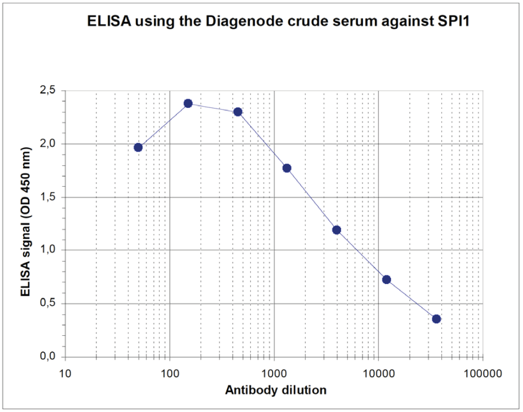
Figure 1. Determination of the titer
To determine the titer, an ELISA was performed using a serial dilution of the Diagenode antibody directed against mouse SPI1 (Cat. No. CS-120-100). The plates were coated with the peptides used for immunization of the rabbit. By plotting the absorbance against the antibody dilution (Figure 1), the titer of the antibody was estimated to be 1:2,600.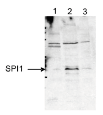
Figure 2. Western blot analysis using the Diagenode antibody directed against SPI1
Western blot was performed using and the Diagenode antibody against mouse SPI1 (Cat. No. CS-120-100) diluted 1:1,000 in TBS-Tween containing 5% skimmed milk. Figure 2 shows the results for mouse fibroblasts (3T3, lane 1), macrophages (RAW, lane 2) and pre-B cells (HAFTL, lane 3). The position of the protein of interest (expected size: 31 kDa) is indicated on the left. Fibroblasts do not express SPI1, whereas macrophages show a high and pre-B cells a low expression of SPI1.
Western blot performed by Maribel Parra, Center for Genomic Regulation, Barcelona, Spain. - Publications

How to properly cite our product/service in your work
We strongly recommend using this: SPI1 Antibody (Hologic Diagenode Cat# C15310120 Lot# A516-004). Click here to copy to clipboard.
Using our products or services in your publication? Let us know!