Notice (8): Undefined variable: solution_of_interest [APP/View/Products/view.ctp, line 755]Code Context<!-- BEGIN: REQUEST_FORM MODAL -->
<div id="request_formModal" class="reveal-modal medium" data-reveal aria-labelledby="modalTitle" aria-hidden="true" role="dialog">
<?= $this->element('Forms/simple_form', array('solution_of_interest' => $solution_of_interest, 'header' => $header, 'message' => $message, 'campaign_id' => $campaign_id)) ?>
$viewFile = '/home/website-server/www/app/View/Products/view.ctp'
$dataForView = array(
'language' => 'jp',
'meta_keywords' => 'HDAC1 polyclonal antibody,ChIP,Chromatin',
'meta_description' => 'Polyclonal antibody raised in rabbit against the C-terminal region of human HDAC1 (Histone deacetylase 1), using a KLH-conjugated syChIP nthetic peptide.',
'meta_title' => 'HDAC1 polyclonal antibody - Premium | Diagenode',
'product' => array(
'Product' => array(
'id' => '2813',
'antibody_id' => '478',
'name' => 'HDAC1 polyclonal antibody ',
'description' => '<p><span>Alternative names: <strong>HD1</strong>, <strong>RPD3</strong>, <strong>RPD3L1</strong>, <strong>GON-10</strong></span></p>
<p>Polyclonal antibody raised in rabbit against the C-terminal region of human HDAC1 (Histone deacetylase 1), using a KLH-conjugated synthetic peptide.</p>',
'label1' => 'Validation Data',
'info1' => '<div class="row">
<div class="small-4 columns">
<p><img src="https://www.diagenode.com/img/product/antibodies/c15410325-chip.jpg" alt="HDAC1 Antibody ChIP Grade" caption="false" width="278" height="211" /></p>
</div>
<div class="small-8 columns">
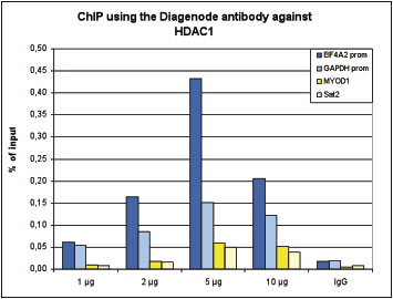
<p><small> <strong>Figure 1. ChIP results obtained with the Diagenode antibody directed against HDAC1</strong><br />ChIP was performed with the Diagenode antibody against HDAC1 (Cat. No. C15410325) on sheared chromatin from 4,000,000 HeLa cells. An antibody titration consisting of 1, 2, 5 and 10 μg per ChIP experiment was analysed. IgG (2 μg/IP) was used as negative IP control. QPCR was performed with primers specific for the EIF4A2 and GAPDH promoters, used as positive controls, and for the MYOD1 gene and Sat2 satellite repeat, used as negative controls. Figure 1 shows the recovery, expressed as a % of input (the relative amount of immunoprecipitated DNA compared to input DNA after qPCR analysis).</small></p>
</div>
</div>
<div class="row">
<div class="small-5 columns">
<p><img src="https://www.diagenode.com/img/product/antibodies/c15410325-chipseq-a.jpg" alt="HDAC1 Antibody ChIP-seq Grade" caption="false" width="354" height="43" /></p>
<p><img src="https://www.diagenode.com/img/product/antibodies/c15410325-chipseq-b.jpg" alt="HDAC1 Antibody for ChIP-seq " caption="false" width="354" height="58" /></p>
<p><img src="https://www.diagenode.com/img/product/antibodies/c15410325-chipseq-c.jpg" alt="HDAC1 Antibody for ChIP-seq assay" caption="false" width="354" height="53" /></p>
<p><img src="https://www.diagenode.com/img/product/antibodies/c15410325-chipseq-d.jpg" alt="HDAC1 Antibody validated in ChIP-seq " caption="false" width="354" height="68" /></p>
</div>
<div class="small-7 columns">
<p><small> <strong>Figure 2. ChIP-seq results obtained with the Diagenode antibody directed against HDAC1</strong><br />ChIP was performed on sheared chromatin from 4,000,000 HeLa cells using 2 μg of the Diagenode antibody against HDAC1 (Cat. No. C15410325) as described above. The IP’d DNA was subsequently analysed on an Illumina HiSeq 2000. Library preparation, cluster generation and sequencing were performed according to the manufacturer’s instructions. The 50 bp tags were aligned to the human genome using the BWA algorithm. Figure 2 shows the peak distribution along the complete sequence and a 1 Mb region of the X-chromosome (figure 2A and B) and in two regions surrounding the GAPDH and EIF4A2 positive control genes, respectively (figure 2C and D)..</small></p>
</div>
</div>
<div class="row">
<div class="small-4 columns">
<p><img src="https://www.diagenode.com/img/product/antibodies/c15410325-elisa.jpg" alt="HDAC1 Antibody ELISA validation" height="192" width="240" caption="false" /></p>
</div>
<div class="small-8 columns">
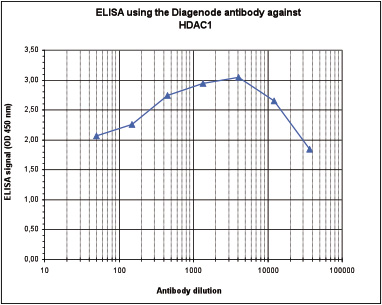
<p><small> <strong>Figure 3. Determination of the antibody titer</strong><br />To determine the titer of the antibody, an ELISA was performed using a serial dilution of Diagenode antibody directed against HDAC1 (Cat. No. C15410325), crude serum and flow through. The plates were coated with the peptide used for immunization of the rabbit. By plotting the absorbance against the antibody dilution (Figure 2), the titer of the antibody was estimated to be 1:75,000.</small></p>
</div>
</div>
<div class="row">
<div class="small-4 columns">
<p><img src="https://www.diagenode.com/img/product/antibodies/c15410325-wb.jpg" alt="HDAC1 Antibody validated in Western Blot" height="168" width="144" caption="false" /></p>
</div>
<div class="small-8 columns">
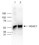
<p><small> <strong>Figure 4. Western blot analysis using the Diagenode antibody directed against HDAC1</strong> <br />Whole cell extracts (25 μg, lane 1) and nuclear extracts (25 μg, lane 2) from HeLa cells were analysed by Western blot using the Diagenode antibody against HDAC1 (Cat. No. C15410325) diluted 1:1,000 in TBS-Tween containing 5% skimmed milk. The position of the protein of interest is indicated on the right (expected size: 55 kDa); the marker (in kDa) is shown on the left.</small></p>
</div>
</div>
<div class="row">
<div class="small-4 columns">
<p><img src="https://www.diagenode.com/img/product/antibodies/c15410325-wb2.jpg" alt="HDAC1 Antibody validated in Western Blot" height="171" width="135" caption="false" /></p>
</div>
<div class="small-8 columns">
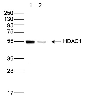
<p><small> <strong>Figure 5. Western blot analysis using the Diagenode antibody directed against HDAC1</strong><br /> Whole cell extracts (50 µg) from HeLa cells transfected with HDAC1 siRNA (lane 2) and from an untransfected control (lane 1) were analysed by Western blot using the Diagenode antibody against HDAC1 (Cat. No. C15410325) diluted 1:1,000 in TBS-Tween containing 5% skimmed milk. The position of the protein of interest is indicated on the right (expected size: 55 kDa); the marker (in kDa) is shown on the left.</small></p>
</div>
</div>
<div class="row">
<div class="small-4 columns">
<p><img src="https://www.diagenode.com/img/product/antibodies/c15410325-protein-array.jpg" alt="HDAC1 Antibody validated in Protein array" caption="false" width="278" height="110" /></p>
</div>
<div class="small-8 columns">
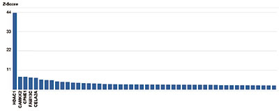
<p><small> <strong>Figure 6. Protein array analysis with the Diagenode antibody directed against HDAC1</strong> <br />The specificity of the Diagenode antibody against HDAC1 (Cat. No. C15410325) was demonstrated using the HuProt human protein microarray (CDI Laboratories), a protein array containing more than 19,000 human proteins. The antibody was used at a dilution of 1:100,000. Figure 6 shows the Z-score of the signal intensity (mean value of the duplicate spots on the array). The names of the proteins with 5 highest Z-scores are indicated at the bottom. This figure clearly shows the high specificity of the antibody for HDAC1.</small></p>
</div>
</div>
<div class="row">
<div class="small-4 columns">
<p><img src="https://www.diagenode.com/img/product/antibodies/c15410325-if.jpg" alt="HDAC1 Antibody validated in Immunofluorescence " caption="false" width="278" height="68" /></p>
</div>
<div class="small-8 columns">
<p><small> <strong>Figure 7. Immunofluorescence using the Diagenode antibody directed against HDAC1</strong> <br />HeLa cells were stained with the Diagenode antibody against HDAC1 (Cat. No. C15410325) and with DAPI. Cells were fixed with 4% formaldehyde for 10’ and blocked with PBS/TX-100 containing 5% normal goat serum and 1% BSA. The cells were immunofluorescently labelled with the HDAC1 antibody (left) diluted 1:500 in blocking solution followed by an anti-rabbit antibody conjugated to Alexa488. The middle panel shows staining of the nuclei with DAPI. A merge of the two stainings is shown on the right.</small></p>
</div>
</div>',
'label2' => 'Target description',
'info2' => '<p>HDAC1 (UniProt/Swiss-Prot entry Q13547) catalyses the deacetylation of lysine residues on the N-terminal part of the core histones (H2A, H2B, H3 and H4). Acetylation and deacetylation of these highly conserved lysine residues is important for the control of gene expression and HDAC activity is often associated with gene repression. Histone deacetylation is established by the formation of large multiprotein complexes. HDAC1 also interacts with the retinoblastoma tumor suppressor protein and is able to deacetylate p53. Therefore, it also plays an essential role in cell proliferation and differentiation and in apoptosi.</p>',
'label3' => '',
'info3' => '',
'format' => '50 μg',
'catalog_number' => 'C15410325',
'old_catalog_number' => '',
'sf_code' => 'C15410325-D001-000581',
'type' => 'FRE',
'search_order' => '',
'price_EUR' => '480',
'price_USD' => '470',
'price_GBP' => '430',
'price_JPY' => '75190',
'price_CNY' => '',
'price_AUD' => '1175',
'country' => 'ALL',
'except_countries' => 'None',
'quote' => false,
'in_stock' => false,
'featured' => false,
'no_promo' => false,
'online' => true,
'master' => true,
'last_datasheet_update' => '0000-00-00',
'slug' => 'hdac1-polyclonal-antibody-premium-50-ug',
'meta_title' => 'HDAC1 polyclonal antibody - Premium | Diagenode',
'meta_keywords' => 'HDAC1 polyclonal antibody,ChIP,Chromatin',
'meta_description' => 'Polyclonal antibody raised in rabbit against the C-terminal region of human HDAC1 (Histone deacetylase 1), using a KLH-conjugated syChIP nthetic peptide.',
'modified' => '2024-11-19 16:55:31',
'created' => '2016-06-17 11:36:17',
'locale' => 'jpn'
),
'Antibody' => array(
'host' => '*****',
'id' => '478',
'name' => 'HDAC1 polyclonal antibody',
'description' => 'HDAC1 (UniProt/Swiss-Prot entry Q13547) catalyses the deacetylation of lysine residues on the N-terminal part of the core histones (H2A, H2B, H3 and H4). Acetylation and deacetylation of these highly conserved lysine residues is important for the control of gene expression and HDAC activity is often associated with gene repression. Histone deacetylation is established by the formation of large multiprotein complexes. HDAC1 also interacts with the retinoblastoma tumor suppressor protein and is able to deacetylate p53. Therefore, it also plays an essential role in cell proliferation and differentiation and in apoptosi.',
'clonality' => '',
'isotype' => '',
'lot' => 'A21-001P',
'concentration' => '1.73 μg/μl',
'reactivity' => 'Human, mouse',
'type' => 'Polyclonal',
'purity' => 'Affinity purified',
'classification' => 'Premium',
'application_table' => '<table>
<thead>
<tr>
<th>Applications</th>
<th>Suggested dilution</th>
<th>References</th>
</tr>
</thead>
<tbody>
<tr>
<td>ChIP/ChIP-seq <sup>*</sup></td>
<td>2 μg/ChIP</td>
<td>Fig 1, 2</td>
</tr>
<tr>
<td>ELISA</td>
<td>1:4,000</td>
<td>Fig 3</td>
</tr>
<tr>
<td>Western Blotting</td>
<td>1:1,000</td>
<td>Fig 4, 5</td>
</tr>
<tr>
<td>Protein array</td>
<td>1:100,000</td>
<td>Fig 6</td>
</tr>
<tr>
<td>Immunofluorescence</td>
<td>1:500</td>
<td>Fig 7</td>
</tr>
</tbody>
</table>
<p><small><sup>*</sup> Please note that the optimal antibody amount per IP should be determined by the end-user. We recommend testing 1-5 μg per IP.</small></p>',
'storage_conditions' => '',
'storage_buffer' => '',
'precautions' => 'This product is for research use only. Not for use in diagnostic or therapeutic procedures.',
'uniprot_acc' => '',
'slug' => '',
'meta_keywords' => '',
'meta_description' => '',
'modified' => '2019-09-10 13:06:12',
'created' => '2016-06-17 10:31:53',
'select_label' => '478 - HDAC1 polyclonal antibody (A21-001P - 1.73 μg/μl - Human, mouse - Affinity purified - Rabbit)'
),
'Slave' => array(
(int) 0 => array(
[maximum depth reached]
)
),
'Group' => array(
'Group' => array(
[maximum depth reached]
),
'Master' => array(
[maximum depth reached]
),
'Product' => array(
[maximum depth reached]
)
),
'Related' => array(),
'Application' => array(
(int) 0 => array(
[maximum depth reached]
),
(int) 1 => array(
[maximum depth reached]
),
(int) 2 => array(
[maximum depth reached]
),
(int) 3 => array(
[maximum depth reached]
),
(int) 4 => array(
[maximum depth reached]
),
(int) 5 => array(
[maximum depth reached]
),
(int) 6 => array(
[maximum depth reached]
)
),
'Category' => array(
(int) 0 => array(
[maximum depth reached]
),
(int) 1 => array(
[maximum depth reached]
),
(int) 2 => array(
[maximum depth reached]
),
(int) 3 => array(
[maximum depth reached]
)
),
'Document' => array(
(int) 0 => array(
[maximum depth reached]
),
(int) 1 => array(
[maximum depth reached]
),
(int) 2 => array(
[maximum depth reached]
)
),
'Feature' => array(),
'Image' => array(
(int) 0 => array(
[maximum depth reached]
)
),
'Promotion' => array(),
'Protocol' => array(),
'Publication' => array(
(int) 0 => array(
[maximum depth reached]
),
(int) 1 => array(
[maximum depth reached]
),
(int) 2 => array(
[maximum depth reached]
),
(int) 3 => array(
[maximum depth reached]
),
(int) 4 => array(
[maximum depth reached]
),
(int) 5 => array(
[maximum depth reached]
),
(int) 6 => array(
[maximum depth reached]
),
(int) 7 => array(
[maximum depth reached]
),
(int) 8 => array(
[maximum depth reached]
),
(int) 9 => array(
[maximum depth reached]
),
(int) 10 => array(
[maximum depth reached]
),
(int) 11 => array(
[maximum depth reached]
),
(int) 12 => array(
[maximum depth reached]
),
(int) 13 => array(
[maximum depth reached]
),
(int) 14 => array(
[maximum depth reached]
),
(int) 15 => array(
[maximum depth reached]
),
(int) 16 => array(
[maximum depth reached]
)
),
'Testimonial' => array(),
'Area' => array(),
'SafetySheet' => array(
(int) 0 => array(
[maximum depth reached]
),
(int) 1 => array(
[maximum depth reached]
),
(int) 2 => array(
[maximum depth reached]
),
(int) 3 => array(
[maximum depth reached]
),
(int) 4 => array(
[maximum depth reached]
),
(int) 5 => array(
[maximum depth reached]
),
(int) 6 => array(
[maximum depth reached]
),
(int) 7 => array(
[maximum depth reached]
)
)
),
'meta_canonical' => 'https://www.diagenode.com/jp/p/hdac1-polyclonal-antibody-premium-50-ug'
)
$language = 'jp'
$meta_keywords = 'HDAC1 polyclonal antibody,ChIP,Chromatin'
$meta_description = 'Polyclonal antibody raised in rabbit against the C-terminal region of human HDAC1 (Histone deacetylase 1), using a KLH-conjugated syChIP nthetic peptide.'
$meta_title = 'HDAC1 polyclonal antibody - Premium | Diagenode'
$product = array(
'Product' => array(
'id' => '2813',
'antibody_id' => '478',
'name' => 'HDAC1 polyclonal antibody ',
'description' => '<p><span>Alternative names: <strong>HD1</strong>, <strong>RPD3</strong>, <strong>RPD3L1</strong>, <strong>GON-10</strong></span></p>
<p>Polyclonal antibody raised in rabbit against the C-terminal region of human HDAC1 (Histone deacetylase 1), using a KLH-conjugated synthetic peptide.</p>',
'label1' => 'Validation Data',
'info1' => '<div class="row">
<div class="small-4 columns">
<p><img src="https://www.diagenode.com/img/product/antibodies/c15410325-chip.jpg" alt="HDAC1 Antibody ChIP Grade" caption="false" width="278" height="211" /></p>
</div>
<div class="small-8 columns">
<p><small> <strong>Figure 1. ChIP results obtained with the Diagenode antibody directed against HDAC1</strong><br />ChIP was performed with the Diagenode antibody against HDAC1 (Cat. No. C15410325) on sheared chromatin from 4,000,000 HeLa cells. An antibody titration consisting of 1, 2, 5 and 10 μg per ChIP experiment was analysed. IgG (2 μg/IP) was used as negative IP control. QPCR was performed with primers specific for the EIF4A2 and GAPDH promoters, used as positive controls, and for the MYOD1 gene and Sat2 satellite repeat, used as negative controls. Figure 1 shows the recovery, expressed as a % of input (the relative amount of immunoprecipitated DNA compared to input DNA after qPCR analysis).</small></p>
</div>
</div>
<div class="row">
<div class="small-5 columns">
<p><img src="https://www.diagenode.com/img/product/antibodies/c15410325-chipseq-a.jpg" alt="HDAC1 Antibody ChIP-seq Grade" caption="false" width="354" height="43" /></p>
<p><img src="https://www.diagenode.com/img/product/antibodies/c15410325-chipseq-b.jpg" alt="HDAC1 Antibody for ChIP-seq " caption="false" width="354" height="58" /></p>
<p><img src="https://www.diagenode.com/img/product/antibodies/c15410325-chipseq-c.jpg" alt="HDAC1 Antibody for ChIP-seq assay" caption="false" width="354" height="53" /></p>
<p><img src="https://www.diagenode.com/img/product/antibodies/c15410325-chipseq-d.jpg" alt="HDAC1 Antibody validated in ChIP-seq " caption="false" width="354" height="68" /></p>
</div>
<div class="small-7 columns">
<p><small> <strong>Figure 2. ChIP-seq results obtained with the Diagenode antibody directed against HDAC1</strong><br />ChIP was performed on sheared chromatin from 4,000,000 HeLa cells using 2 μg of the Diagenode antibody against HDAC1 (Cat. No. C15410325) as described above. The IP’d DNA was subsequently analysed on an Illumina HiSeq 2000. Library preparation, cluster generation and sequencing were performed according to the manufacturer’s instructions. The 50 bp tags were aligned to the human genome using the BWA algorithm. Figure 2 shows the peak distribution along the complete sequence and a 1 Mb region of the X-chromosome (figure 2A and B) and in two regions surrounding the GAPDH and EIF4A2 positive control genes, respectively (figure 2C and D)..</small></p>
</div>
</div>
<div class="row">
<div class="small-4 columns">
<p><img src="https://www.diagenode.com/img/product/antibodies/c15410325-elisa.jpg" alt="HDAC1 Antibody ELISA validation" height="192" width="240" caption="false" /></p>
</div>
<div class="small-8 columns">
<p><small> <strong>Figure 3. Determination of the antibody titer</strong><br />To determine the titer of the antibody, an ELISA was performed using a serial dilution of Diagenode antibody directed against HDAC1 (Cat. No. C15410325), crude serum and flow through. The plates were coated with the peptide used for immunization of the rabbit. By plotting the absorbance against the antibody dilution (Figure 2), the titer of the antibody was estimated to be 1:75,000.</small></p>
</div>
</div>
<div class="row">
<div class="small-4 columns">
<p><img src="https://www.diagenode.com/img/product/antibodies/c15410325-wb.jpg" alt="HDAC1 Antibody validated in Western Blot" height="168" width="144" caption="false" /></p>
</div>
<div class="small-8 columns">
<p><small> <strong>Figure 4. Western blot analysis using the Diagenode antibody directed against HDAC1</strong> <br />Whole cell extracts (25 μg, lane 1) and nuclear extracts (25 μg, lane 2) from HeLa cells were analysed by Western blot using the Diagenode antibody against HDAC1 (Cat. No. C15410325) diluted 1:1,000 in TBS-Tween containing 5% skimmed milk. The position of the protein of interest is indicated on the right (expected size: 55 kDa); the marker (in kDa) is shown on the left.</small></p>
</div>
</div>
<div class="row">
<div class="small-4 columns">
<p><img src="https://www.diagenode.com/img/product/antibodies/c15410325-wb2.jpg" alt="HDAC1 Antibody validated in Western Blot" height="171" width="135" caption="false" /></p>
</div>
<div class="small-8 columns">
<p><small> <strong>Figure 5. Western blot analysis using the Diagenode antibody directed against HDAC1</strong><br /> Whole cell extracts (50 µg) from HeLa cells transfected with HDAC1 siRNA (lane 2) and from an untransfected control (lane 1) were analysed by Western blot using the Diagenode antibody against HDAC1 (Cat. No. C15410325) diluted 1:1,000 in TBS-Tween containing 5% skimmed milk. The position of the protein of interest is indicated on the right (expected size: 55 kDa); the marker (in kDa) is shown on the left.</small></p>
</div>
</div>
<div class="row">
<div class="small-4 columns">
<p><img src="https://www.diagenode.com/img/product/antibodies/c15410325-protein-array.jpg" alt="HDAC1 Antibody validated in Protein array" caption="false" width="278" height="110" /></p>
</div>
<div class="small-8 columns">
<p><small> <strong>Figure 6. Protein array analysis with the Diagenode antibody directed against HDAC1</strong> <br />The specificity of the Diagenode antibody against HDAC1 (Cat. No. C15410325) was demonstrated using the HuProt human protein microarray (CDI Laboratories), a protein array containing more than 19,000 human proteins. The antibody was used at a dilution of 1:100,000. Figure 6 shows the Z-score of the signal intensity (mean value of the duplicate spots on the array). The names of the proteins with 5 highest Z-scores are indicated at the bottom. This figure clearly shows the high specificity of the antibody for HDAC1.</small></p>
</div>
</div>
<div class="row">
<div class="small-4 columns">
<p><img src="https://www.diagenode.com/img/product/antibodies/c15410325-if.jpg" alt="HDAC1 Antibody validated in Immunofluorescence " caption="false" width="278" height="68" /></p>
</div>
<div class="small-8 columns">
<p><small> <strong>Figure 7. Immunofluorescence using the Diagenode antibody directed against HDAC1</strong> <br />HeLa cells were stained with the Diagenode antibody against HDAC1 (Cat. No. C15410325) and with DAPI. Cells were fixed with 4% formaldehyde for 10’ and blocked with PBS/TX-100 containing 5% normal goat serum and 1% BSA. The cells were immunofluorescently labelled with the HDAC1 antibody (left) diluted 1:500 in blocking solution followed by an anti-rabbit antibody conjugated to Alexa488. The middle panel shows staining of the nuclei with DAPI. A merge of the two stainings is shown on the right.</small></p>
</div>
</div>',
'label2' => 'Target description',
'info2' => '<p>HDAC1 (UniProt/Swiss-Prot entry Q13547) catalyses the deacetylation of lysine residues on the N-terminal part of the core histones (H2A, H2B, H3 and H4). Acetylation and deacetylation of these highly conserved lysine residues is important for the control of gene expression and HDAC activity is often associated with gene repression. Histone deacetylation is established by the formation of large multiprotein complexes. HDAC1 also interacts with the retinoblastoma tumor suppressor protein and is able to deacetylate p53. Therefore, it also plays an essential role in cell proliferation and differentiation and in apoptosi.</p>',
'label3' => '',
'info3' => '',
'format' => '50 μg',
'catalog_number' => 'C15410325',
'old_catalog_number' => '',
'sf_code' => 'C15410325-D001-000581',
'type' => 'FRE',
'search_order' => '',
'price_EUR' => '480',
'price_USD' => '470',
'price_GBP' => '430',
'price_JPY' => '75190',
'price_CNY' => '',
'price_AUD' => '1175',
'country' => 'ALL',
'except_countries' => 'None',
'quote' => false,
'in_stock' => false,
'featured' => false,
'no_promo' => false,
'online' => true,
'master' => true,
'last_datasheet_update' => '0000-00-00',
'slug' => 'hdac1-polyclonal-antibody-premium-50-ug',
'meta_title' => 'HDAC1 polyclonal antibody - Premium | Diagenode',
'meta_keywords' => 'HDAC1 polyclonal antibody,ChIP,Chromatin',
'meta_description' => 'Polyclonal antibody raised in rabbit against the C-terminal region of human HDAC1 (Histone deacetylase 1), using a KLH-conjugated syChIP nthetic peptide.',
'modified' => '2024-11-19 16:55:31',
'created' => '2016-06-17 11:36:17',
'locale' => 'jpn'
),
'Antibody' => array(
'host' => '*****',
'id' => '478',
'name' => 'HDAC1 polyclonal antibody',
'description' => 'HDAC1 (UniProt/Swiss-Prot entry Q13547) catalyses the deacetylation of lysine residues on the N-terminal part of the core histones (H2A, H2B, H3 and H4). Acetylation and deacetylation of these highly conserved lysine residues is important for the control of gene expression and HDAC activity is often associated with gene repression. Histone deacetylation is established by the formation of large multiprotein complexes. HDAC1 also interacts with the retinoblastoma tumor suppressor protein and is able to deacetylate p53. Therefore, it also plays an essential role in cell proliferation and differentiation and in apoptosi.',
'clonality' => '',
'isotype' => '',
'lot' => 'A21-001P',
'concentration' => '1.73 μg/μl',
'reactivity' => 'Human, mouse',
'type' => 'Polyclonal',
'purity' => 'Affinity purified',
'classification' => 'Premium',
'application_table' => '<table>
<thead>
<tr>
<th>Applications</th>
<th>Suggested dilution</th>
<th>References</th>
</tr>
</thead>
<tbody>
<tr>
<td>ChIP/ChIP-seq <sup>*</sup></td>
<td>2 μg/ChIP</td>
<td>Fig 1, 2</td>
</tr>
<tr>
<td>ELISA</td>
<td>1:4,000</td>
<td>Fig 3</td>
</tr>
<tr>
<td>Western Blotting</td>
<td>1:1,000</td>
<td>Fig 4, 5</td>
</tr>
<tr>
<td>Protein array</td>
<td>1:100,000</td>
<td>Fig 6</td>
</tr>
<tr>
<td>Immunofluorescence</td>
<td>1:500</td>
<td>Fig 7</td>
</tr>
</tbody>
</table>
<p><small><sup>*</sup> Please note that the optimal antibody amount per IP should be determined by the end-user. We recommend testing 1-5 μg per IP.</small></p>',
'storage_conditions' => '',
'storage_buffer' => '',
'precautions' => 'This product is for research use only. Not for use in diagnostic or therapeutic procedures.',
'uniprot_acc' => '',
'slug' => '',
'meta_keywords' => '',
'meta_description' => '',
'modified' => '2019-09-10 13:06:12',
'created' => '2016-06-17 10:31:53',
'select_label' => '478 - HDAC1 polyclonal antibody (A21-001P - 1.73 μg/μl - Human, mouse - Affinity purified - Rabbit)'
),
'Slave' => array(
(int) 0 => array(
'id' => '183',
'name' => 'C15410325',
'product_id' => '2813',
'modified' => '2016-06-17 12:34:02',
'created' => '2016-06-17 12:34:02'
)
),
'Group' => array(
'Group' => array(
'id' => '183',
'name' => 'C15410325',
'product_id' => '2813',
'modified' => '2016-06-17 12:34:02',
'created' => '2016-06-17 12:34:02'
),
'Master' => array(
'id' => '2813',
'antibody_id' => '478',
'name' => 'HDAC1 Antibody',
'description' => '<p><span>Alternative names: <strong>HD1</strong>, <strong>RPD3</strong>, <strong>RPD3L1</strong>, <strong>GON-10</strong></span></p>
<p>Polyclonal antibody raised in rabbit against the C-terminal region of human <strong>HDAC1</strong> <strong>(Histone deacetylase 1)</strong>, using a KLH-conjugated synthetic peptide.</p>',
'label1' => 'Validation Data',
'info1' => '<div class="row">
<div class="small-4 columns">
<p><img src="https://www.diagenode.com/img/product/antibodies/c15410325-chip.jpg" alt="HDAC1 Antibody ChIP Grade" caption="false" width="278" height="211" /></p>
</div>
<div class="small-8 columns">
<p><small> <strong>Figure 1. ChIP results obtained with the Diagenode antibody directed against HDAC1</strong><br />ChIP was performed with the Diagenode antibody against HDAC1 (Cat. No. C15410325) on sheared chromatin from 4,000,000 HeLa cells. An antibody titration consisting of 1, 2, 5 and 10 μg per ChIP experiment was analysed. IgG (2 μg/IP) was used as negative IP control. QPCR was performed with primers specific for the EIF4A2 and GAPDH promoters, used as positive controls, and for the MYOD1 gene and Sat2 satellite repeat, used as negative controls. Figure 1 shows the recovery, expressed as a % of input (the relative amount of immunoprecipitated DNA compared to input DNA after qPCR analysis).</small></p>
</div>
</div>
<div class="row">
<div class="small-5 columns">
<p><img src="https://www.diagenode.com/img/product/antibodies/c15410325-chipseq-a.jpg" alt="HDAC1 Antibody ChIP-seq Grade" caption="false" width="354" height="43" /></p>
<p><img src="https://www.diagenode.com/img/product/antibodies/c15410325-chipseq-b.jpg" alt="HDAC1 Antibody for ChIP-seq " caption="false" width="354" height="58" /></p>
<p><img src="https://www.diagenode.com/img/product/antibodies/c15410325-chipseq-c.jpg" alt="HDAC1 Antibody for ChIP-seq assay" caption="false" width="354" height="53" /></p>
<p><img src="https://www.diagenode.com/img/product/antibodies/c15410325-chipseq-d.jpg" alt="HDAC1 Antibody validated in ChIP-seq " caption="false" width="354" height="68" /></p>
</div>
<div class="small-7 columns">
<p><small> <strong>Figure 2. ChIP-seq results obtained with the Diagenode antibody directed against HDAC1</strong><br />ChIP was performed on sheared chromatin from 4,000,000 HeLa cells using 2 μg of the Diagenode antibody against HDAC1 (Cat. No. C15410325) as described above. The IP’d DNA was subsequently analysed on an Illumina HiSeq 2000. Library preparation, cluster generation and sequencing were performed according to the manufacturer’s instructions. The 50 bp tags were aligned to the human genome using the BWA algorithm. Figure 2 shows the peak distribution along the complete sequence and a 1 Mb region of the X-chromosome (figure 2A and B) and in two regions surrounding the GAPDH and EIF4A2 positive control genes, respectively (figure 2C and D)..</small></p>
</div>
</div>
<div class="row">
<div class="small-4 columns">
<p><img src="https://www.diagenode.com/img/product/antibodies/c15410325-elisa.jpg" alt="HDAC1 Antibody ELISA validation" height="192" width="240" caption="false" /></p>
</div>
<div class="small-8 columns">
<p><small> <strong>Figure 3. Determination of the antibody titer</strong><br />To determine the titer of the antibody, an ELISA was performed using a serial dilution of Diagenode antibody directed against HDAC1 (Cat. No. C15410325), crude serum and flow through. The plates were coated with the peptide used for immunization of the rabbit. By plotting the absorbance against the antibody dilution (Figure 2), the titer of the antibody was estimated to be 1:75,000.</small></p>
</div>
</div>
<div class="row">
<div class="small-4 columns">
<p><img src="https://www.diagenode.com/img/product/antibodies/c15410325-wb.jpg" alt="HDAC1 Antibody validated in Western Blot" height="168" width="144" caption="false" /></p>
</div>
<div class="small-8 columns">
<p><small> <strong>Figure 4. Western blot analysis using the Diagenode antibody directed against HDAC1</strong> <br />Whole cell extracts (25 μg, lane 1) and nuclear extracts (25 μg, lane 2) from HeLa cells were analysed by Western blot using the Diagenode antibody against HDAC1 (Cat. No. C15410325) diluted 1:1,000 in TBS-Tween containing 5% skimmed milk. The position of the protein of interest is indicated on the right (expected size: 55 kDa); the marker (in kDa) is shown on the left.</small></p>
</div>
</div>
<div class="row">
<div class="small-4 columns">
<p><img src="https://www.diagenode.com/img/product/antibodies/c15410325-wb2.jpg" alt="HDAC1 Antibody validated in Western Blot" height="171" width="135" caption="false" /></p>
</div>
<div class="small-8 columns">
<p><small> <strong>Figure 5. Western blot analysis using the Diagenode antibody directed against HDAC1</strong><br /> Whole cell extracts (50 µg) from HeLa cells transfected with HDAC1 siRNA (lane 2) and from an untransfected control (lane 1) were analysed by Western blot using the Diagenode antibody against HDAC1 (Cat. No. C15410325) diluted 1:1,000 in TBS-Tween containing 5% skimmed milk. The position of the protein of interest is indicated on the right (expected size: 55 kDa); the marker (in kDa) is shown on the left.</small></p>
</div>
</div>
<div class="row">
<div class="small-4 columns">
<p><img src="https://www.diagenode.com/img/product/antibodies/c15410325-protein-array.jpg" alt="HDAC1 Antibody validated in Protein array" caption="false" width="278" height="110" /></p>
</div>
<div class="small-8 columns">
<p><small> <strong>Figure 6. Protein array analysis with the Diagenode antibody directed against HDAC1</strong> <br />The specificity of the Diagenode antibody against HDAC1 (Cat. No. C15410325) was demonstrated using the HuProt human protein microarray (CDI Laboratories), a protein array containing more than 19,000 human proteins. The antibody was used at a dilution of 1:100,000. Figure 6 shows the Z-score of the signal intensity (mean value of the duplicate spots on the array). The names of the proteins with 5 highest Z-scores are indicated at the bottom. This figure clearly shows the high specificity of the antibody for HDAC1.</small></p>
</div>
</div>
<div class="row">
<div class="small-4 columns">
<p><img src="https://www.diagenode.com/img/product/antibodies/c15410325-if.jpg" alt="HDAC1 Antibody validated in Immunofluorescence " caption="false" width="278" height="68" /></p>
</div>
<div class="small-8 columns">
<p><small> <strong>Figure 7. Immunofluorescence using the Diagenode antibody directed against HDAC1</strong> <br />HeLa cells were stained with the Diagenode antibody against HDAC1 (Cat. No. C15410325) and with DAPI. Cells were fixed with 4% formaldehyde for 10’ and blocked with PBS/TX-100 containing 5% normal goat serum and 1% BSA. The cells were immunofluorescently labelled with the HDAC1 antibody (left) diluted 1:500 in blocking solution followed by an anti-rabbit antibody conjugated to Alexa488. The middle panel shows staining of the nuclei with DAPI. A merge of the two stainings is shown on the right.</small></p>
</div>
</div>',
'label2' => 'Target description',
'info2' => '<p>HDAC1 (UniProt/Swiss-Prot entry Q13547) catalyses the deacetylation of lysine residues on the N-terminal part of the core histones (H2A, H2B, H3 and H4). Acetylation and deacetylation of these highly conserved lysine residues is important for the control of gene expression and HDAC activity is often associated with gene repression. Histone deacetylation is established by the formation of large multiprotein complexes. HDAC1 also interacts with the retinoblastoma tumor suppressor protein and is able to deacetylate p53. Therefore, it also plays an essential role in cell proliferation and differentiation and in apoptosi.</p>',
'label3' => '',
'info3' => '',
'format' => '50 μg',
'catalog_number' => 'C15410325',
'old_catalog_number' => '',
'sf_code' => 'C15410325-D001-000581',
'type' => 'FRE',
'search_order' => '',
'price_EUR' => '480',
'price_USD' => '470',
'price_GBP' => '430',
'price_JPY' => '75190',
'price_CNY' => '',
'price_AUD' => '1175',
'country' => 'ALL',
'except_countries' => 'None',
'quote' => false,
'in_stock' => false,
'featured' => false,
'no_promo' => false,
'online' => true,
'master' => true,
'last_datasheet_update' => '0000-00-00',
'slug' => 'hdac1-polyclonal-antibody-premium-50-ug',
'meta_title' => 'HDAC1 Antibody - ChIP-seq Grade (C15410325) | Diagenode',
'meta_keywords' => 'HDAC1 polyclonal antibody,ChIP,Chromatin',
'meta_description' => 'HDAC1 (Histone deacetylase 1) Polyclonal Antibody validated in ChIP-seq, ChIP-qPCR, ELISA, WB and IF. Specificity confirmed by Peptide array and siRNA assay. Batch-specific data available on the website.Sample size available.',
'modified' => '2024-11-19 16:55:31',
'created' => '2016-06-17 11:36:17'
),
'Product' => array(
(int) 0 => array(
[maximum depth reached]
)
)
),
'Related' => array(),
'Application' => array(
(int) 0 => array(
'id' => '42',
'position' => '10',
'parent_id' => '40',
'name' => 'ChIP-seq (ab)',
'description' => '',
'in_footer' => false,
'in_menu' => false,
'online' => true,
'tabular' => true,
'slug' => 'chip-seq-antibodies',
'meta_keywords' => 'Chromatin Immunoprecipitation Sequencing,ChIP-Seq,ChIP-seq grade antibodies,DNA purification,qPCR,Shearing of chromatin',
'meta_description' => 'Diagenode offers a wide range of antibodies and technical support for ChIP Sequencing applications',
'meta_title' => 'ChIP Sequencing Antibodies (ChIP-Seq) | Diagenode',
'modified' => '2016-01-20 11:06:19',
'created' => '2015-10-20 11:44:45',
'ProductsApplication' => array(
[maximum depth reached]
)
),
(int) 1 => array(
'id' => '20',
'position' => '10',
'parent_id' => '40',
'name' => 'ELISA',
'description' => '<div class="row">
<div class="small-12 medium-12 large-12 columns">Enzyme-linked immunosorbent assay.</div>
</div>',
'in_footer' => false,
'in_menu' => false,
'online' => true,
'tabular' => true,
'slug' => 'elisa-antibodies',
'meta_keywords' => ' ELISA Antibodies,Monoclonal antibody, Polyclonal antibody',
'meta_description' => 'Diagenode offers Monoclonal & Polyclonal antibodies for ELISA applications',
'meta_title' => 'ELISA Antibodies - Monoclonal & Polyclonal antibody | Diagenode',
'modified' => '2016-01-13 12:21:41',
'created' => '2014-07-08 08:13:28',
'ProductsApplication' => array(
[maximum depth reached]
)
),
(int) 2 => array(
'id' => '19',
'position' => '10',
'parent_id' => '40',
'name' => 'WB',
'description' => '<p><strong>Western blot</strong> : The quality of antibodies used in this technique is crucial for correct and specific protein identification. Diagenode offers huge selection of highly sensitive and specific western blot-validated antibodies.</p>
<p>Learn more about: <a href="https://www.diagenode.com/applications/western-blot">Loading control, MW marker visualization</a><em>. <br /></em></p>
<p><em></em>Check our selection of antibodies validated in Western blot.</p>',
'in_footer' => false,
'in_menu' => false,
'online' => true,
'tabular' => true,
'slug' => 'western-blot-antibodies',
'meta_keywords' => ' Western Blot Antibodies ,western blot protocol,Western Blotting Products,Polyclonal antibodies ,monoclonal antibodies ',
'meta_description' => 'Diagenode offers a wide range of antibodies and technical support for western blot applications',
'meta_title' => ' Western Blot - Monoclonal antibody - Polyclonal antibody | Diagenode',
'modified' => '2016-04-26 12:44:51',
'created' => '2015-01-07 09:20:00',
'ProductsApplication' => array(
[maximum depth reached]
)
),
(int) 3 => array(
'id' => '4',
'position' => '10',
'parent_id' => null,
'name' => 'Protein Analysis',
'description' => '<div class="row">
<div class="small-12 medium-12 large-12 columns">
<p>Proteins play a critical role in virtually all cell processes including metabolism, structure, growth, and repair. Understanding protein function, behavior, structure, and their importance are key aspects of understanding disease and therapeutics. Experimental analysis of proteins typically involves expression and purification or the direct extraction of proteins from cells or tissues for further downstream analysis such as ligand binding assays, mass spectrometry, or protein sequencing. Other protein analysis methods include detection methods such as BCA assays and Western blots or methods to understand protein-to-protein interactions or protein-DNA interactions such as ChIP-sequencing.</p>
<p>At Diagenode, we simplify the protein research process with a portfolio of unique and robust tools to both isolate and analyze proteins. Our protein research products include the Bioruptor Plus sonication device for protein extraction, protein extraction beads, protein extraction kits, unique Western blot ladders that can be directly visualized on film, and highly validated antibodies for Western blot and ChIP-seq.</p>
</div>
</div>',
'in_footer' => true,
'in_menu' => true,
'online' => true,
'tabular' => false,
'slug' => 'protein-analysis',
'meta_keywords' => 'Protein Analysis,Protein extraction,Western blot,ChIP-sequencing,protein extraction kits',
'meta_description' => 'Diagenode offers protein research products for protein extraction beads,kits, unique Western blot ladders that can be directly visualized on film and highly validated antibodies for Western blot and ChIP-seq.',
'meta_title' => 'Unique and robust tools to both isolate and analyze proteins | Diagenode',
'modified' => '2015-08-14 14:26:48',
'created' => '2015-01-06 09:02:19',
'ProductsApplication' => array(
[maximum depth reached]
)
),
(int) 4 => array(
'id' => '29',
'position' => '10',
'parent_id' => '40',
'name' => 'IF',
'description' => '<p><strong>Immunofluorescence</strong>:</p>
<p>Diagenode offers huge selection of highly sensitive antibodies validated in IF.</p>
<p><img src="https://www.diagenode.com/img/product/antibodies/C15200229-IF.jpg" alt="" height="245" width="256" /></p>
<p><sup><strong>Immunofluorescence using the Diagenode monoclonal antibody directed against CRISPR/Cas9</strong></sup></p>
<p><sup>HeLa cells transfected with a Cas9 expression vector (left) or untransfected cells (right) were fixed in methanol at -20°C, permeabilized with acetone at -20°C and blocked with PBS containing 2% BSA. The cells were stained with the Cas9 C-terminal antibody (Cat. No. C15200229) diluted 1:400, followed by incubation with an anti-mouse secondary antibody coupled to AF488. The bottom images show counter-staining of the nuclei with Hoechst 33342.</sup></p>
<h5><sup>Check our selection of antibodies validated in IF.</sup></h5>',
'in_footer' => false,
'in_menu' => false,
'online' => true,
'tabular' => true,
'slug' => 'immunofluorescence',
'meta_keywords' => 'Immunofluorescence,Monoclonal antibody,Polyclonal antibody',
'meta_description' => 'Diagenode offers a wide range of antibodies and technical support for Immunofluorescence applications',
'meta_title' => 'Immunofluorescence - Monoclonal antibody - Polyclonal antibody | Diagenode',
'modified' => '2016-04-27 16:23:10',
'created' => '2015-07-08 13:46:02',
'ProductsApplication' => array(
[maximum depth reached]
)
),
(int) 5 => array(
'id' => '43',
'position' => '10',
'parent_id' => '40',
'name' => 'ChIP-qPCR (ab)',
'description' => '',
'in_footer' => false,
'in_menu' => false,
'online' => true,
'tabular' => true,
'slug' => 'chip-qpcr-antibodies',
'meta_keywords' => 'Chromatin Immunoprecipitation Sequencing,ChIP-Seq,ChIP-seq grade antibodies,DNA purification,qPCR,Shearing of chromatin',
'meta_description' => 'Diagenode offers a wide range of antibodies and technical support for ChIP-qPCR applications',
'meta_title' => 'ChIP Quantitative PCR Antibodies (ChIP-qPCR) | Diagenode',
'modified' => '2016-01-20 11:30:24',
'created' => '2015-10-20 11:45:36',
'ProductsApplication' => array(
[maximum depth reached]
)
),
(int) 6 => array(
'id' => '45',
'position' => '10',
'parent_id' => '40',
'name' => 'siRNA Knockdown',
'description' => '<div class="row">
<div class="small-10 columns">
<h3>Epigenetic antibodies you can trust!</h3>
<p>Antibody quality is essential for assay success. Diagenode offers antibodies that are actually validated and have been widely used and published by the scientific community. Now we are adding a new level of siRNA knockdown validation to assure the specificity of our non-histone antibodies.</p>
<p><strong>Short interfering RNA (siRNA)</strong> degrades target mRNA, followed by the knock-down of protein production. If the antibody that recognizes the protein of interest is specific, the Western blot of siRNA-treated cells will show a significant reduction of signal vs. untreated cells.</p>
<center><img src="https://www.diagenode.com/emailing/images/C15100144-wb.png" alt="" /></center>
<p class="text-center"><small>WB results obtained with the HDAC1 pAb (Cat. No. C15100144) <br />on siRNA transfected cells (lane 2) and on untransfected control cells (lane 1).</small></p>
</div>
<div class="small-2 columns">
<p><img src="https://www.diagenode.com/emailing/images/epi-success-guaranteed-icon.png" alt="Epigenetic success guaranteed" /></p>
</div>
</div>
<div class="spaced"></div>
<p style="text-align: left;"><span style="font-weight: 400;">The below list shows our first siRNA validated antibodies. More results - coming soon</span>.</p>',
'in_footer' => false,
'in_menu' => false,
'online' => true,
'tabular' => true,
'slug' => 'sirna',
'meta_keywords' => 'siRNA knockdown antibodies',
'meta_description' => 'Antibodies validated by siRNA Knockdown',
'meta_title' => '',
'modified' => '2017-01-26 15:58:23',
'created' => '2017-01-09 10:53:09',
'ProductsApplication' => array(
[maximum depth reached]
)
)
),
'Category' => array(
(int) 0 => array(
'id' => '103',
'position' => '0',
'parent_id' => '4',
'name' => 'All antibodies',
'description' => '<p><span style="font-weight: 400;">All Diagenode’s antibodies are listed below. Please, use our Quick search field to find the antibody of interest by target name, application, purity.</span></p>
<p><span style="font-weight: 400;">Diagenode’s highly validated antibodies:</span></p>
<ul>
<li>Highly sensitive and specific</li>
<li>Cost-effective (requires less antibody per reaction)</li>
<li>Batch-specific data is available on the website</li>
<li>Expert technical support</li>
<li>Sample sizes available</li>
<li>100% satisfaction guarantee</li>
</ul>',
'no_promo' => false,
'in_menu' => true,
'online' => true,
'tabular' => false,
'hide' => true,
'all_format' => false,
'is_antibody' => true,
'slug' => 'all-antibodies',
'cookies_tag_id' => null,
'meta_keywords' => 'Antibodies,Premium Antibodies,Classic,Pioneer',
'meta_description' => 'Diagenode Offers Strict quality standards with Rigorous QC and validated Antibodies. Classified based on level of validation for flexibility of Application. Comprehensive selection of histone and non-histone Antibodies',
'meta_title' => 'Diagenode's selection of Antibodies is exclusively dedicated for Epigenetic Research | Diagenode',
'modified' => '2019-07-03 10:55:44',
'created' => '2015-11-02 14:49:22',
'ProductsCategory' => array(
[maximum depth reached]
),
'CookiesTag' => array([maximum depth reached])
),
(int) 1 => array(
'id' => '17',
'position' => '10',
'parent_id' => '4',
'name' => 'ChIP-seq grade antibodies',
'description' => '<p><b>Unparalleled ChIP-Seq results with the most rigorously validated antibodies</b></p>
<p><span style="font-weight: 400;">Diagenode provides leading solutions for epigenetic research. Because ChIP-seq is a widely-used technique, we validate our antibodies in ChIP and ChIP-seq experiments (in addition to conventional methods like Western blot, Dot blot, ELISA, and immunofluorescence) to provide the highest quality antibody. We standardize our validation and production to guarantee high product quality without technical bias. Diagenode guarantees ChIP-seq grade antibody performance under our suggested conditions.</span></p>
<div class="row">
<div class="small-12 medium-9 large-9 columns">
<p><strong>ChIP-seq profile</strong> of active (H3K4me3 and H3K36me3) and inactive (H3K27me3) marks using Diagenode antibodies.</p>
<img src="https://www.diagenode.com/img/categories/antibodies/chip-seq-grade-antibodies.png" /></div>
<div class="small-12 medium-3 large-3 columns">
<p><small> ChIP was performed on sheared chromatin from 100,000 K562 cells using iDeal ChIP-seq kit for Histones (cat. No. C01010051) with 1 µg of the Diagenode antibodies against H3K27me3 (cat. No. C15410195) and H3K4me3 (cat. No. C15410003), and 0.5 µg of the antibody against H3K36me3 (cat. No. C15410192). The IP'd DNA was subsequently analysed on an Illumina Genome Analyzer. Library preparation, cluster generation and sequencing were performed according to the manufacturer's instructions. The 36 bp tags were aligned to the human genome using the ELAND algorithm. The figure shows the signal distribution along the complete sequence of human chromosome 3, a zoomin to a 10 Mb region and a further zoomin to a 1.5 Mb region. </small></p>
</div>
</div>
<p>Diagenode’s highly validated antibodies:</p>
<ul>
<li>Highly sensitive and specific</li>
<li>Cost-effective (requires less antibody per reaction)</li>
<li>Batch-specific data is available on the website</li>
<li>Expert technical support</li>
<li>Sample sizes available</li>
<li>100% satisfaction guarantee</li>
</ul>',
'no_promo' => false,
'in_menu' => true,
'online' => true,
'tabular' => false,
'hide' => true,
'all_format' => false,
'is_antibody' => true,
'slug' => 'chip-seq-grade-antibodies',
'cookies_tag_id' => null,
'meta_keywords' => 'ChIP-seq grade antibodies,polyclonal antibody,WB, ELISA, ChIP-seq (ab), ChIP-qPCR (ab)',
'meta_description' => 'Diagenode Offers Wide Range of Validated ChIP-Seq Grade Antibodies for Unparalleled ChIP-Seq Results',
'meta_title' => 'Chromatin Immunoprecipitation ChIP-Seq Grade Antibodies | Diagenode',
'modified' => '2019-07-03 10:57:22',
'created' => '2015-02-16 02:24:01',
'ProductsCategory' => array(
[maximum depth reached]
),
'CookiesTag' => array([maximum depth reached])
),
(int) 2 => array(
'id' => '21',
'position' => '20',
'parent_id' => '4',
'name' => 'Histone modifying enzymes',
'description' => '<p><span style="font-weight: 400;">Diagenode offers the large number of antibodies raised against histone modifying enzymes. The list below includes the antibodies against enzymes like: histone deacetylases, histone demethylases, histone transferases.</span></p>
<p><span style="font-weight: 400;">Diagenode’s highly validated antibodies:</span></p>
<ul>
<li>Highly sensitive and specific</li>
<li>Cost-effective (requires less antibody per reaction)</li>
<li>Batch-specific data is available on the website</li>
<li>Expert technical support</li>
<li>Sample sizes available</li>
<li>100% satisfaction guarantee</li>
</ul>',
'no_promo' => false,
'in_menu' => false,
'online' => true,
'tabular' => false,
'hide' => true,
'all_format' => false,
'is_antibody' => true,
'slug' => 'chromatin-modifying-proteins-histone-transferase',
'cookies_tag_id' => null,
'meta_keywords' => 'monoclonal antibodies,policlonal antibodies,Histone modifying enzymes',
'meta_description' => 'Diagenode Offers Polyclonal and Monoclonal Antibodies Against Histone Modifying Enzymes like: Histone Deacetylases, Histone Demethylases, Histone Transferases.',
'meta_title' => 'Histone modifying enzymes - Antibodies | Diagenode',
'modified' => '2019-07-04 16:19:15',
'created' => '2014-06-26 13:18:41',
'ProductsCategory' => array(
[maximum depth reached]
),
'CookiesTag' => array([maximum depth reached])
),
(int) 3 => array(
'id' => '127',
'position' => '10',
'parent_id' => '4',
'name' => 'ChIP-grade antibodies',
'description' => '<div class="row">
<div class="small-12 columns"><center></center>
<p><br />Chromatin immunoprecipitation (<b>ChIP</b>) is a technique to study the associations of proteins with the specific genomic regions in intact cells. One of the most important steps of this protocol is the immunoprecipitation of targeted protein using the antibody specifically recognizing it. The quality of antibodies used in ChIP is essential for the success of the experiment. Diagenode offers extensively validated ChIP-grade antibodies, confirmed for their specificity, and high level of performance in ChIP. Each batch is validated, and batch-specific data are available on the website.</p>
<p></p>
</div>
</div>
<p><strong>ChIP results</strong> obtained with the antibody directed against H3K4me3 (Cat. No. <a href="../p/h3k4me3-polyclonal-antibody-premium-50-ug-50-ul">C15410003</a>). </p>
<div class="row">
<div class="small-12 medium-6 large-6 columns"><img src="https://www.diagenode.com/img/product/antibodies/C15410003-fig1-ChIP.jpg" alt="" width="400" height="315" /> </div>
<div class="small-12 medium-6 large-6 columns">
<p></p>
<p></p>
<p></p>
</div>
</div>
<p></p>
<p>Our aim at Diagenode is to offer the largest collection of highly specific <strong>ChIP-grade antibodies</strong>. We add new antibodies monthly. Find your ChIP-grade antibody in the list below and check more information about tested applications, extensive validation data, and product information.</p>',
'no_promo' => false,
'in_menu' => true,
'online' => true,
'tabular' => false,
'hide' => true,
'all_format' => false,
'is_antibody' => true,
'slug' => 'chip-grade-antibodies',
'cookies_tag_id' => null,
'meta_keywords' => 'ChIP-grade antibodies, polyclonal antibody, monoclonal antibody, Diagenode',
'meta_description' => 'Diagenode Offers Extensively Validated ChIP-Grade Antibodies, Confirmed for their Specificity, and high level of Performance in Chromatin Immunoprecipitation ChIP',
'meta_title' => 'Chromatin immunoprecipitation ChIP-grade antibodies | Diagenode',
'modified' => '2024-11-19 17:27:07',
'created' => '2017-02-14 11:16:04',
'ProductsCategory' => array(
[maximum depth reached]
),
'CookiesTag' => array([maximum depth reached])
)
),
'Document' => array(
(int) 0 => array(
'id' => '903',
'name' => 'HDAC1 polyclonal antibody - Premium',
'description' => '<p>Polyclonal antibody raised in rabbit against the C-terminal region of human HDAC1 (Histone deacetylase 1), using a KLH-conjugated synthetic peptide.</p>',
'image_id' => null,
'type' => 'Datasheet',
'url' => 'files/products/antibodies/Datasheet_HDAC1_premium_C15410325.pdf',
'slug' => 'hdac-polyclonal-antibody-premium-c15410325-tds',
'meta_keywords' => '',
'meta_description' => '',
'modified' => '2016-06-17 11:40:29',
'created' => '2016-06-17 11:40:29',
'ProductsDocument' => array(
[maximum depth reached]
)
),
(int) 1 => array(
'id' => '38',
'name' => 'Epigenetic Antibodies Brochure',
'description' => '<p>More than in any other immuoprecipitation assays, quality antibodies are critical tools in many epigenetics experiments. Since 10 years, Diagenode has developed the most stringent quality production available on the market for antibodies exclusively focused on epigenetic uses. All our antibodies have been qualified to work in epigenetic applications.</p>',
'image_id' => null,
'type' => 'Brochure',
'url' => 'files/brochures/Epigenetic_Antibodies_Brochure.pdf',
'slug' => 'epigenetic-antibodies-brochure',
'meta_keywords' => '',
'meta_description' => '',
'modified' => '2016-06-15 11:24:06',
'created' => '2015-07-03 16:05:27',
'ProductsDocument' => array(
[maximum depth reached]
)
),
(int) 2 => array(
'id' => '11',
'name' => 'Antibodies you can trust',
'description' => '<p style="text-align: justify;"><span>Epigenetic research tools have evolved over time from endpoint PCR to qPCR to the analyses of large sets of genome-wide sequencing data. ChIP sequencing (ChIP-seq) has now become the gold standard method for chromatin studies, given the accuracy and coverage scale of the approach over other methods. Successful ChIP-seq, however, requires a higher level of experimental accuracy and consistency in all steps of ChIP than ever before. Particularly crucial is the quality of ChIP antibodies. </span></p>',
'image_id' => null,
'type' => 'Poster',
'url' => 'files/posters/Antibodies_you_can_trust_Poster.pdf',
'slug' => 'antibodies-you-can-trust-poster',
'meta_keywords' => '',
'meta_description' => '',
'modified' => '2015-10-01 20:18:31',
'created' => '2015-07-03 16:05:15',
'ProductsDocument' => array(
[maximum depth reached]
)
)
),
'Feature' => array(),
'Image' => array(
(int) 0 => array(
'id' => '1783',
'name' => 'product/antibodies/chipseq-grade-ab-icon.png',
'alt' => 'ChIP-seq Grade',
'modified' => '2020-11-27 07:04:40',
'created' => '2018-03-15 15:54:09',
'ProductsImage' => array(
[maximum depth reached]
)
)
),
'Promotion' => array(),
'Protocol' => array(),
'Publication' => array(
(int) 0 => array(
'id' => '4849',
'name' => 'NUP98 and RAE1 sustain progenitor function through HDAC-dependentchromatin targeting to escape from nucleolar localization.',
'authors' => 'Neely Amy E. et al.',
'description' => '<p>Self-renewing somatic tissues rely on progenitors to support the continuous tissue regeneration. The gene regulatory network maintaining progenitor function remains incompletely understood. Here we show that NUP98 and RAE1 are highly expressed in epidermal progenitors, forming a separate complex in the nucleoplasm. Reduction of NUP98 or RAE1 abolishes progenitors' regenerative capacity, inhibiting proliferation and inducing premature terminal differentiation. Mechanistically, NUP98 binds on chromatin near the transcription start sites of key epigenetic regulators (such as DNMT1, UHRF1 and EZH2) and sustains their expression in progenitors. NUP98's chromatin binding sites are co-occupied by HDAC1. HDAC inhibition diminishes NUP98's chromatin binding and dysregulates NUP98 and RAE1's target gene expression. Interestingly, HDAC inhibition further induces NUP98 and RAE1 to localize interdependently to the nucleolus. These findings identified a pathway in progenitor maintenance, where HDAC activity directs the high levels of NUP98 and RAE1 to directly control key epigenetic regulators, escaping from nucleolar aggregation.</p>',
'date' => '2023-06-01',
'pmid' => 'https://www.ncbi.nlm.nih.gov/pubmed/37353594',
'doi' => '10.1038/s42003-023-05043-2',
'modified' => '2023-08-01 14:22:16',
'created' => '2023-08-01 15:59:38',
'ProductsPublication' => array(
[maximum depth reached]
)
),
(int) 1 => array(
'id' => '4545',
'name' => 'Histone Deacetylases 1 and 2 target gene regulatory networks of nephronprogenitors to control nephrogenesis.',
'authors' => 'Liu Hongbing et al.',
'description' => '<p>Our studies demonstrated the critical role of Histone deacetylases (HDACs) in the regulation of nephrogenesis. To better understand the key pathways regulated by HDAC1/2 in early nephrogenesis, we performed chromatin immunoprecipitation sequencing (ChIP-Seq) of Hdac1/2 on isolated nephron progenitor cells (NPCs) from mouse E16.5 kidneys. Our analysis revealed that 11802 (40.4\%) of Hdac1 peaks overlap with Hdac2 peaks, further demonstrates the redundant role of Hdac1 and Hdac2 during nephrogenesis. Common Hdac1/2 peaks are densely concentrated close to the transcriptional start site (TSS). GREAT Gene Ontology analysis of overlapping Hdac1/2 peaks reveals that Hdac1/2 are associated with metanephric nephron morphogenesis, chromatin assembly or disassembly, as well as other DNA checkpoints. Pathway analysis shows that negative regulation of Wnt signaling pathway is one of Hdac1/2's most significant function in NPCs. Known motif analysis indicated that Hdac1 is enriched in motifs for Six2, Hox family, and Tcf family members, which are essential for self-renewal and differentiation of nephron progenitors. Interestingly, we found the enrichment of HDAC1/2 at the enhancer and promoter regions of actively transcribed genes, especially those concerned with NPC self-renewal. HDAC1/2 simultaneously activate or repress the expression of different genes to maintain the cellular state of nephron progenitors. We used the Integrative Genomics Viewer to visualize these target genes associated with each function and found that Hdac1/2 co-bound to the enhancers or/and promoters of genes associated with nephron morphogenesis, differentiation, and cell cycle control. Taken together, our ChIP-Seq analysis demonstrates that Hdac1/2 directly regulate the molecular cascades essential for nephrogenesis.</p>',
'date' => '2022-11-01',
'pmid' => 'https://www.ncbi.nlm.nih.gov/pubmed/36356658',
'doi' => '10.1016/j.bcp.2022.115341',
'modified' => '2022-11-24 10:24:07',
'created' => '2022-11-24 08:49:52',
'ProductsPublication' => array(
[maximum depth reached]
)
),
(int) 2 => array(
'id' => '3870',
'name' => 'Threonine phosphorylation of IκBζ mediates inhibition of selective proinflammatory target genes.',
'authors' => 'Grondona P, Bucher P, Schmitt A, Schönfeld C, Streibl B, Müller A, Essmann F, Liberatori S, Mohammed S, Hennig A, Kramer D, Schulze-Osthoff K, Hailfinger S',
'description' => '<p>Transcription factors of the NF-κB family play a crucial role for immune responses by activating the expression of chemokines, cytokines and antimicrobial peptides involved in pathogen clearance. IκBζ, an atypical nuclear IκB protein and selective coactivator of particular NF-κB target genes, has recently been identified as an essential regulator for skin immunity. In the present study, we discovered that IκBζ is strongly induced in keratinocytes sensing the fungal glucan zymosan A and that IκBζ is essential for the optimal expression of proinflammatory genes, such as IL6, CXCL5, IL1B or S100A9. Moreover, we found that IκBζ was not solely regulated on the transcriptional level but also by phosphorylation events. We identified several IκBζ phosphorylation sites, including a conserved cluster of threonine residues located in the N-terminus of the protein, which can be phosphorylated by MAPKs. Surprisingly, IκBζ phosphorylation at this threonine cluster promoted the recruitment of HDAC1 to specific target gene promoters and thus and thus negatively controlled transcription. Taken together, we propose a model of how an anti-fungal response translates to the expression of proinflammatory cytokines and highlight an additional layer of complexity in the regulation of the NF-κB responses in keratinocytes.</p>',
'date' => '2020-02-06',
'pmid' => 'http://www.pubmed.gov/32035922',
'doi' => '10.1016/j.jid.2019.12.036',
'modified' => '2020-03-20 17:44:59',
'created' => '2020-03-13 13:45:54',
'ProductsPublication' => array(
[maximum depth reached]
)
),
(int) 3 => array(
'id' => '3601',
'name' => 'Immunity drives regulation in cancer through NF-κB.',
'authors' => 'Collignon E, Canale A, Al Wardi C, Bizet M, Calonne E, Dedeurwaerder S, Garaud S, Naveaux C, Barham W, Wilson A, Bouchat S, Hubert P, Van Lint C, Yull F, Sotiriou C, Willard-Gallo K, Noel A, Fuks F',
'description' => '<p>Ten-eleven translocation enzymes (TET1, TET2, and TET3), which induce DNA demethylation and gene regulation by converting 5-methylcytosine (5mC) to 5-hydroxymethylcytosine (5hmC), are often down-regulated in cancer. We uncover, in basal-like breast cancer (BLBC), genome-wide 5hmC changes related to regulation. We further demonstrate that repression is associated with high expression of immune markers and high infiltration by immune cells. We identify in BLBC tissues an anticorrelation between expression and the major immunoregulator family nuclear factor κB (NF-κB). In vitro and in mice, is down-regulated in breast cancer cells upon NF-κB activation through binding of p65 to its consensus sequence in the promoter. We lastly show that these findings extend to other cancer types, including melanoma, lung, and thyroid cancers. Together, our data suggest a novel mode of regulation for in cancer and highlight a new paradigm in which the immune system can influence cancer cell epigenetics.</p>',
'date' => '2018-06-01',
'pmid' => 'http://www.pubmed.gov/29938218',
'doi' => '10.1126/sciadv.aap7309',
'modified' => '2019-04-17 15:00:20',
'created' => '2019-04-16 12:25:30',
'ProductsPublication' => array(
[maximum depth reached]
)
),
(int) 4 => array(
'id' => '3634',
'name' => 'Immunity drives TET1 regulation in cancer through NF-kB',
'authors' => 'Collignon E, Canale A, Al Wardi C, Bizet M, Calonne E, Dedeurwaerder S, Garaud S, Naveaux C, Barham W, Wilson A, Bouchat S, Hubert P, Van Lint C, Yull F, Sotiriou C, Willard-Gallo K, Noel A, Fuks F',
'description' => '<p>Ten-eleven translocation enzymes (TET1, TET2, and TET3), which induce DNA demethylation and gene regulation by converting 5-methylcytosine (5mC) to 5-hydroxymethylcytosine (5hmC), are often down-regulated in cancer. We uncover, in basal-like breast cancer (BLBC), genome-wide 5hmC changes related to regulation. We further demonstrate that repression is associated with high expression of immune markers and high infiltration by immune cells. We identify in BLBC tissues an anticorrelation between expression and the major immunoregulator family nuclear factor κB (NF-κB). In vitro and in mice, is down-regulated in breast cancer cells upon NF-κB activation through binding of p65 to its consensus sequence in the promoter. We lastly show that these findings extend to other cancer types, including melanoma, lung, and thyroid cancers. Together, our data suggest a novel mode of regulation for in cancer and highlight a new paradigm in which the immune system can influence cancer cell epigenetics.</p>',
'date' => '2018-06-01',
'pmid' => 'http://www.pubmed.gov/29938218',
'doi' => '10.1126/sciadv.aap7309',
'modified' => '2019-06-07 10:31:57',
'created' => '2019-06-06 12:11:18',
'ProductsPublication' => array(
[maximum depth reached]
)
),
(int) 5 => array(
'id' => '3161',
'name' => 'Krüppel-like transcription factor KLF10 suppresses TGFβ-induced epithelial-to-mesenchymal transition via a negative feedback mechanism',
'authors' => 'Mishra V.K. et al.',
'description' => '<p>TGFβ-SMAD signaling exerts a contextual effect that suppresses malignant growth early in epithelial tumorigenesis but promotes metastasis at later stages. Longstanding challenges in resolving this functional dichotomy may uncover new strategies to treat advanced carcinomas. The Krüppel-like transcription factor, KLF10, is a pivotal effector of TGFβ/SMAD signaling that mediates antiproliferative effects of TGFβ. In this study, we show how KLF10 opposes the prometastatic effects of TGFβ by limiting its ability to induce epithelial-to-mesenchymal transition (EMT). KLF10 depletion accentuated induction of EMT as assessed by multiple metrics. KLF10 occupied GC-rich sequences in the promoter region of the EMT-promoting transcription factor SLUG/SNAI2, repressing its transcription by recruiting HDAC1 and licensing the removal of activating histone acetylation marks. In clinical specimens of lung adenocarcinoma, low KLF10 expression associated with decreased patient survival, consistent with a pivotal role for KLF10 in distinguishing the antiproliferative versus prometastatic functions of TGFβ. Our results establish that KLF10 functions to suppress TGFβ-induced EMT, establishing a molecular basis for the dichotomy of TGFβ function during tumor progression.</p>',
'date' => '2017-03-01',
'pmid' => 'https://www.ncbi.nlm.nih.gov/pubmed/28249899',
'doi' => '',
'modified' => '2017-04-27 15:47:38',
'created' => '2017-04-27 15:47:38',
'ProductsPublication' => array(
[maximum depth reached]
)
),
(int) 6 => array(
'id' => '3010',
'name' => 'HDAC1 negatively regulates Bdnf and Pvalb required for parvalbumin interneuron maturation in an experience-dependent manner',
'authors' => 'Koh DX and Sng JC',
'description' => '<p>During early postnatal development, neuronal circuits are sculpted by sensory experience provided by the external environment. This experience-dependent regulation of circuitry development consolidates the balance of excitatory-inhibitory (E/I) neurons in the brain. The cortical barrel-column that innervates a single principal whisker is used to provide a clear reference frame for studying the consolidation of E/I circuitry. Sensory deprivation of S1 at birth disrupts the consolidation of excitatory-inhibitory balance by decreasing inhibitory transmission of parvalbumin interneurons. The molecular mechanisms underlying this decrease in inhibition are not completely understood. Our findings show that epigenetic mechanisms, in particular histone deacetylation by histone deacetylases, negatively regulate the expression of brain-derived neurotrophic factor (Bdnf) and parvalbumin (Pvalb) genes during development, which are required for the maturation of parvalbumin interneurons. After whisker deprivation, increased histone deacetylase 1 expression and activity led to increased histone deacetylase 1 binding and decreased histone acetylation at Bdnf promoters I-IV and Pvalb promoter, resulting in the repression of Bdnf and Pvalb gene transcription. The decrease in Bdnf expression further affected parvalbumin interneuron maturation at layer II/III in S1, demonstrated by decreased parvalbumin expression, a marker for parvalbumin interneuron maturation. Knockdown of HDAC1 recovered Bdnf and Pvalb gene transcription and also prevented the decrease of inhibitory synapses accompanying whisker deprivation.</p>',
'date' => '2016-08-17',
'pmid' => 'http://www.ncbi.nlm.nih.gov/pubmed/27534825',
'doi' => '',
'modified' => '2016-08-29 10:18:30',
'created' => '2016-08-29 10:18:30',
'ProductsPublication' => array(
[maximum depth reached]
)
),
(int) 7 => array(
'id' => '2866',
'name' => 'Genome-wide hydroxymethylcytosine pattern changes in response to oxidative stress',
'authors' => 'Delatte B, Jeschke J, Defrance M, Bachman M, Creppe C, Calonne E, Bizet M, Deplus R, Marroquí L, Libin M, Ravichandran M, Mascart F, Eizirik DL, Murrell A, Jurkowski TP, Fuks F',
'description' => '<div class="pl20 mq875-pl0 js-collapsible-section" id="abstract-content" itemprop="description">
<p><b>The TET enzymes convert methylcytosine to the newly discovered base hydroxymethylcytosine. While recent reports suggest that TETs may play a role in response to oxidative stress, this role remains uncertain, and results lack</b> <i><b>in vivo</b></i> <b>models. Here we show a global decrease of hydroxymethylcytosine in cells treated with buthionine sulfoximine, and in mice depleted for the major antioxidant enzymes</b> <i><b>GPx</b></i><b>1 and 2. Furthermore, genome-wide profiling revealed differentially hydroxymethylated regions in coding genes, and intriguingly in microRNA genes, both involved in response to oxidative stress. These results thus suggest a profound effect of</b> <i><b>in vivo</b></i> <b>oxidative stress on the global hydroxymethylome.</b></p>
</div>',
'date' => '2015-08-04',
'pmid' => 'http://www.nature.com/articles/srep12714',
'doi' => '10.1038/srep12714',
'modified' => '2016-03-22 10:37:38',
'created' => '2016-03-22 10:37:38',
'ProductsPublication' => array(
[maximum depth reached]
)
),
(int) 8 => array(
'id' => '2274',
'name' => 'SNAIL1 combines competitive displacement of ASCL2 and epigenetic mechanisms to rapidly silence the EPHB3 tumor suppressor in colorectal cancer.',
'authors' => 'Rönsch K, Jägle S, Rose K, Seidl M, Baumgartner F, Freihen V, Yousaf A, Metzger E, Lassmann S, Schüle R, Zeiser R, Michoel T, Hecht A',
'description' => 'EPHB3 is a critical cellular guidance factor in the intestinal epithelium and an important tumor suppressor in colorectal cancer (CRC) whose expression is frequently lost at the adenoma-carcinoma transition when tumor cells become invasive. The molecular mechanisms underlying EPHB3 silencing are incompletely understood. Here we show that EPHB3 expression is anti-correlated with inducers of epithelial-mesenchymal transition (EMT) in primary tumors and CRC cells. In vitro, SNAIL1 and SNAIL2, but not ZEB1, repress EPHB3 reporter constructs and compete with the stem cell factor ASCL2 for binding to an E-box motif. At the endogenous EPHB3 locus, SNAIL1 triggers the displacement of ASCL2, p300 and the Wnt pathway effector TCF7L2 and engages corepressor complexes containing HDACs and the histone demethylase LSD1 to collapse active chromatin structure, resulting in rapid downregulation of EPHB3. Beyond its impact on EPHB3, SNAIL1 deregulates markers of intestinal identity and stemness and in vitro forces CRC cells to undergo EMT with altered morphology, increased motility and invasiveness. In xenotransplants, SNAIL1 expression abrogated tumor cell palisading and led to focal loss of tumor encapsulation and the appearance of areas with tumor cells displaying a migratory phenotype. These changes were accompanied by loss of EPHB3 and CDH1 expression. Intriguingly, SNAIL1-induced phenotypic changes of CRC cells are significantly impaired by sustained EPHB3 expression both in vitro and in vivo. Altogether, our results identify EPHB3 as a novel target of SNAIL1 and suggest that disabling EPHB3 signaling is an important aspect to eliminate a roadblock at the onset of EMT processes.',
'date' => '2014-09-16',
'pmid' => 'https://www.ncbi.nlm.nih.gov/pubmed/25277775',
'doi' => '',
'modified' => '2015-07-24 15:39:03',
'created' => '2015-07-24 15:39:03',
'ProductsPublication' => array(
[maximum depth reached]
)
),
(int) 9 => array(
'id' => '2130',
'name' => 'Citrullination of DNMT3A by PADI4 regulates its stability and controls DNA methylation.',
'authors' => 'Deplus R, Denis H, Putmans P, Calonne E, Fourrez M, Yamamoto K, Suzuki A, Fuks F',
'description' => 'DNA methylation is a central epigenetic modification in mammals, with essential roles in development and disease. De novo DNA methyltransferases establish DNA methylation patterns in specific regions within the genome by mechanisms that remain poorly understood. Here we show that protein citrullination by peptidylarginine deiminase 4 (PADI4) affects the function of the DNA methyltransferase DNMT3A. We found that DNMT3A and PADI4 interact, from overexpressed as well as untransfected cells, and associate with each other's enzymatic activity. Both in vitro and in vivo, PADI4 was shown to citrullinate DNMT3A. We identified a sequence upstream of the PWWP domain of DNMT3A as its primary region citrullinated by PADI4. Increasing the PADI4 level caused the DNMT3A protein level to increase as well, provided that the PADI4 was catalytically active, and RNAi targeting PADI4 caused reduced DNMT3A levels. Accordingly, pulse-chase experiments revealed stabilization of the DNMT3A protein by catalytically active PADI4. Citrullination and increased expression of native DNMT3A by PADI4 were confirmed in PADI4-knockout MEFs. Finally, we showed that PADI4 overexpression increases DNA methyltransferase activity in a catalytic-dependent manner and use bisulfite pyrosequencing to demonstrate that PADI4 knockdown causes significant reduction of CpG methylation at the p21 promoter, a known target of DNMT3A and PADI4. Protein citrullination by PADI4 thus emerges as a novel mechanism for controlling a de novo DNA methyltransferase. Our results shed new light on how post-translational modifications might contribute to shaping the genomic CpG methylation landscape.',
'date' => '2014-06-23',
'pmid' => 'https://www.ncbi.nlm.nih.gov/pubmed/24957603',
'doi' => '',
'modified' => '2015-07-24 15:39:03',
'created' => '2015-07-24 15:39:03',
'ProductsPublication' => array(
[maximum depth reached]
)
),
(int) 10 => array(
'id' => '1553',
'name' => 'Dimethyl fumarate regulates histone deacetylase expression in astrocytes.',
'authors' => 'Kalinin S, Polak PE, Lin SX, Braun D, Guizzetti M, Zhang X, Rubinstein I, Feinstein DL',
'description' => 'We previously showed that dimethyl fumarate (DMF) reduces inflammatory activation in astrocytes, involving activation of transcription factor Nrf2. However, the pathways causing Nrf2 activation were not examined. We now show that DMF modifies expression of histone deacetylases (HDACs) in primary rat astrocytes. After 4h incubation, levels of HDAC1, 2, and 4 mRNAs were increased by DMF; however, after 24h, levels returned to or were below control values. At that time, HDAC protein levels and overall activity were also reduced by DMF. Stimulation of astrocytes with pro-inflammatory cytokines significantly increased HDAC mRNA levels after 24h, although protein levels were not increased at that time point. In the presence of cytokines, DMF reduced HDAC mRNAs, proteins, and activity. Proteomic analysis of DMF-treated astrocytes identified 8 proteins in which lysine acetylation was increased by DMF, including histones H2a.1 and H3.3. A role for HDACs in mediating DMF actions is suggested by findings that the selective HDAC inhibitor SAHA increased nuclear Nrf2:DNA binding activity, reduced inflammatory activation of astrocytes which was reversed by a selective inhibitor of the Nrf2 target gene heme-oxygenase 1. These data show that DMF regulates astrocyte HDAC expression, which could contribute to Nrf2 activation, suppression of inflammatory responses and cause long-lasting changes in gene expression.',
'date' => '2013-10-15',
'pmid' => 'https://www.ncbi.nlm.nih.gov/pubmed/23916696',
'doi' => '',
'modified' => '2015-07-24 15:39:00',
'created' => '2015-07-24 15:39:00',
'ProductsPublication' => array(
[maximum depth reached]
)
),
(int) 11 => array(
'id' => '800',
'name' => 'Phosphorylation of p65(RelA) on Ser547 by ATM Represses NF-κB-Dependent Transcription of Specific Genes after Genotoxic Stress',
'authors' => 'Sabatel H, Di Valentin E, Gloire G, Dequiedt F, Piette J, Habraken Y',
'description' => '',
'date' => '2012-06-08',
'pmid' => 'http://www.plosone.org/article/info%3Adoi%2F10.1371%2Fjournal.pone.0038246#abstract0',
'doi' => '',
'modified' => '2015-07-24 15:38:58',
'created' => '2015-07-24 15:38:58',
'ProductsPublication' => array(
[maximum depth reached]
)
),
(int) 12 => array(
'id' => '752',
'name' => 'The histone demethylase Kdm3a is essential to progression through differentiation.',
'authors' => 'Herzog M, Josseaux E, Dedeurwaerder S, Calonne E, Volkmar M, Fuks F',
'description' => 'Histone demethylation has important roles in regulating gene expression and forms part of the epigenetic memory system that regulates cell fate and identity by still poorly understood mechanisms. Here, we examined the role of histone demethylase Kdm3a during cell differentiation, showing that Kdm3a is essential for differentiation into parietal endoderm-like (PE) cells in the F9 mouse embryonal carcinoma model. We identified a number of target genes regulated by Kdm3a during endoderm differentiation; among the most dysregulated were the three developmental master regulators Dab2, Pdlim4 and FoxQ1. We show that dysregulation of the expression of these genes correlates with Kdm3a H3K9me2 demethylase activity. We further demonstrate that either Dab2 depletion or Kdm3a depletion prevents F9 cells from fully differentiating into PE cells, but that ectopic expression of Dab2 cannot compensate for Kdm3a knockdown; Dab2 is thus necessary, but insufficient on its own, to promote complete terminal differentiation. We conclude that Kdm3a plays a crucial role in progression through PE differentiation by regulating expression of a set of endoderm differentiation master genes. The emergence of Kdm3a as a key modulator of cell fate decision strengthens the view that histone demethylases are essential to cell differentiation.',
'date' => '2012-05-11',
'pmid' => 'https://www.ncbi.nlm.nih.gov/pubmed/22581778',
'doi' => '',
'modified' => '2015-07-24 15:38:58',
'created' => '2015-07-24 15:38:58',
'ProductsPublication' => array(
[maximum depth reached]
)
),
(int) 13 => array(
'id' => '714',
'name' => 'HDAC1 Regulates Fear Extinction in Mice.',
'authors' => 'Bahari-Javan S, Maddalena A, Kerimoglu C, Wittnam J, Held T, Bähr M, Burkhardt S, Delalle I, Kügler S, Fischer A, Sananbenesi F',
'description' => 'Histone acetylation has been implicated with the pathogenesis of neuropsychiatric disorders and targeting histone deacetylases (HDACs) using HDAC inhibitors was shown to be neuroprotective and to initiate neuroregenerative processes. However, little is known about the role of individual HDAC proteins during the pathogenesis of brain diseases. HDAC1 was found to be upregulated in patients suffering from neuropsychiatric diseases. Here, we show that virus-mediated overexpression of neuronal HDAC1 in the adult mouse hippocampus specifically affects the extinction of contextual fear memories, while other cognitive abilities were unaffected. In subsequent experiments we show that under physiological conditions, hippocampal HDAC1 is required for extinction learning via a mechanism that involves H3K9 deacetylation and subsequent trimethylation of target genes. In conclusion, our data show that hippocampal HDAC1 has a specific role in memory function.',
'date' => '2012-04-11',
'pmid' => 'https://www.ncbi.nlm.nih.gov/pubmed/22496552',
'doi' => '',
'modified' => '2015-07-24 15:38:58',
'created' => '2015-07-24 15:38:58',
'ProductsPublication' => array(
[maximum depth reached]
)
),
(int) 14 => array(
'id' => '816',
'name' => 'Enhancer of Zeste 2 (EZH2) is up-regulated in malignant gliomas and in glioma stem-like cells.',
'authors' => 'Orzan F, Pellegatta S, Poliani PL, Pisati F, Caldera V, Menghi F, Kapetis D, Marras C, Schiffer D, Finocchiaro G',
'description' => 'AIMS: Proteins of the Polycomb repressive complex 2 (PRC2) are epigenetic gene silencers and are involved in tumour development. Their oncogenic function might be associated with their role in stem cell maintenance. The histone methyltransferase Enhancer of Zeste 2 (EZH2) is a key member of PRC2 function: we have investigated its expression and function in gliomas. METHODS: EZH2 expression was studied in grade II-IV gliomas and in glioma stem-like cells (GSC) by quantitative PCR and immunohistochemistry. Effects of EZH2 down-regulation were analysed by treating GSC with the histone deacetylase (HDAC) inhibitor suberoylanide hydroxamic acid (SAHA) and by shRNA. RESULTS: DNA microarray analysis showed that EZH2 is highly expressed in murine and human GSC. Real-time PCR on gliomas of different grade (n = 66) indicated that EZH2 is more expressed in glioblastoma multiforme (GBM) than in low-grade gliomas (P = 0.0013). This was confirmed by immunohistochemistry on an independent set of 106 gliomas. Treatment with SAHA caused significant up-regulation of PRC2 predicted target genes, GSC disruption and decreased expression of EZH2 and of the stem cell marker CD133. Inhibition of EZH2 expression by shRNA was associated with a significant decrease of glioma proliferation. CONCLUSION: The data suggest that EZH2 plays a role in glioma progression and encourage the therapeutic targeting of these malignancies by HDAC inhibitors.',
'date' => '2011-06-01',
'pmid' => 'https://www.ncbi.nlm.nih.gov/pubmed/20946108',
'doi' => '',
'modified' => '2015-07-24 15:38:58',
'created' => '2015-07-24 15:38:58',
'ProductsPublication' => array(
[maximum depth reached]
)
),
(int) 15 => array(
'id' => '77',
'name' => 'The core binding factor CBF negatively regulates skeletal muscle terminal differentiation.',
'authors' => 'Philipot O, Joliot V, Ait-Mohamed O, Pellentz C, Robin P, Fritsch L, Ait-Si-Ali S',
'description' => 'BACKGROUND: Core Binding Factor or CBF is a transcription factor composed of two subunits, Runx1/AML-1 and CBF beta or CBFbeta. CBF was originally described as a regulator of hematopoiesis. METHODOLOGY/PRINCIPAL FINDINGS: Here we show that CBF is involved in the control of skeletal muscle terminal differentiation. Indeed, downregulation of either Runx1 or CBFbeta protein level accelerates cell cycle exit and muscle terminal differentiation. Conversely, overexpression of CBFbeta in myoblasts slows terminal differentiation. CBF interacts directly with the master myogenic transcription factor MyoD, preferentially in proliferating myoblasts, via Runx1 subunit. In addition, we show a preferential recruitment of Runx1 protein to MyoD target genes in proliferating myoblasts. The MyoD/CBF complex contains several chromatin modifying enzymes that inhibits MyoD activity, such as HDACs, Suv39h1 and HP1beta. When overexpressed, CBFbeta induced an inhibition of activating histone modification marks concomitant with an increase in repressive modifications at MyoD target promoters. CONCLUSIONS/SIGNIFICANCE: Taken together, our data show a new role for Runx1/CBFbeta in the control of the proliferation/differentiation in skeletal myoblasts.',
'date' => '2010-01-01',
'pmid' => 'https://www.ncbi.nlm.nih.gov/pubmed/20195544',
'doi' => '',
'modified' => '2015-07-24 15:38:56',
'created' => '2015-07-24 15:38:56',
'ProductsPublication' => array(
[maximum depth reached]
)
),
(int) 16 => array(
'id' => '78',
'name' => 'Functional connection between deimination and deacetylation of histones.',
'authors' => 'Denis H, Deplus R, Putmans P, Yamada M, Métivier R, Fuks F',
'description' => 'Histone methylation plays key roles in regulating chromatin structure and function. The recent identification of enzymes that antagonize or remove histone methylation offers new opportunities to appreciate histone methylation plasticity in the regulation of epigenetic pathways. Peptidylarginine deiminase 4 (PADI4; also known as PAD4) was the first enzyme shown to antagonize histone methylation. PADI4 functions as a histone deiminase converting a methylarginine residue to citrulline at specific sites on the tails of histones H3 and H4. This activity is linked to repression of the estrogen-regulated pS2 promoter. Very little is known as to how PADI4 silences gene expression. We show here that PADI4 associates with the histone deacetylase 1 (HDAC1). Kinetic chromatin immunoprecipitation assays revealed that PADI4 and HDAC1, and the corresponding activities, associate cyclically and coordinately with the pS2 promoter during repression phases. Knockdown of HDAC1 led to decreased H3 citrullination, concomitantly with increased histone arginine methylation. In cells with a reduced HDAC1 and a slightly decreased PADI4 level, these effects were more pronounced. Our data thus suggest that PADI4 and HDAC1 collaborate to generate a repressive chromatin environment on the pS2 promoter. These findings further substantiate the "transcriptional clock" concept, highlighting the dynamic connection between deimination and deacetylation of histones.',
'date' => '2009-09-01',
'pmid' => 'https://www.ncbi.nlm.nih.gov/pubmed/19581286',
'doi' => '',
'modified' => '2015-07-24 15:38:56',
'created' => '2015-07-24 15:38:56',
'ProductsPublication' => array(
[maximum depth reached]
)
)
),
'Testimonial' => array(),
'Area' => array(),
'SafetySheet' => array(
(int) 0 => array(
'id' => '1512',
'name' => 'HDAC1 Antibody SDS US en',
'language' => 'en',
'url' => 'files/SDS/HDAC1/SDS-C15410325-HDAC1_Antibody-US-en-GHS_2_0.pdf',
'countries' => 'US',
'modified' => '2021-08-30 15:33:52',
'created' => '2021-08-30 15:33:52',
'ProductsSafetySheet' => array(
[maximum depth reached]
)
),
(int) 1 => array(
'id' => '1510',
'name' => 'HDAC1 Antibody SDS GB en',
'language' => 'en',
'url' => 'files/SDS/HDAC1/SDS-C15410325-HDAC1_Antibody-GB-en-GHS_2_0.pdf',
'countries' => 'GB',
'modified' => '2021-08-30 15:33:52',
'created' => '2021-08-30 15:33:52',
'ProductsSafetySheet' => array(
[maximum depth reached]
)
),
(int) 2 => array(
'id' => '1505',
'name' => 'HDAC1 Antibody SDS BE fr',
'language' => 'fr',
'url' => 'files/SDS/HDAC1/SDS-C15410325-HDAC1_Antibody-BE-fr-GHS_2_0.pdf',
'countries' => 'BE',
'modified' => '2021-08-30 15:33:52',
'created' => '2021-08-30 15:33:52',
'ProductsSafetySheet' => array(
[maximum depth reached]
)
),
(int) 3 => array(
'id' => '1509',
'name' => 'HDAC1 Antibody SDS FR fr',
'language' => 'fr',
'url' => 'files/SDS/HDAC1/SDS-C15410325-HDAC1_Antibody-FR-fr-GHS_2_0.pdf',
'countries' => 'FR',
'modified' => '2021-08-30 15:33:52',
'created' => '2021-08-30 15:33:52',
'ProductsSafetySheet' => array(
[maximum depth reached]
)
),
(int) 4 => array(
'id' => '1508',
'name' => 'HDAC1 Antibody SDS ES es',
'language' => 'es',
'url' => 'files/SDS/HDAC1/SDS-C15410325-HDAC1_Antibody-ES-es-GHS_2_0.pdf',
'countries' => 'ES',
'modified' => '2021-08-30 15:33:52',
'created' => '2021-08-30 15:33:52',
'ProductsSafetySheet' => array(
[maximum depth reached]
)
),
(int) 5 => array(
'id' => '1507',
'name' => 'HDAC1 Antibody SDS DE de',
'language' => 'de',
'url' => 'files/SDS/HDAC1/SDS-C15410325-HDAC1_Antibody-DE-de-GHS_2_0.pdf',
'countries' => 'DE',
'modified' => '2021-08-30 15:33:52',
'created' => '2021-08-30 15:33:52',
'ProductsSafetySheet' => array(
[maximum depth reached]
)
),
(int) 6 => array(
'id' => '1511',
'name' => 'HDAC1 Antibody SDS JP ja',
'language' => 'ja',
'url' => 'files/SDS/HDAC1/SDS-C15410325-HDAC1_Antibody-JP-ja-GHS_3_0.pdf',
'countries' => 'JP',
'modified' => '2021-08-30 15:33:52',
'created' => '2021-08-30 15:33:52',
'ProductsSafetySheet' => array(
[maximum depth reached]
)
),
(int) 7 => array(
'id' => '1506',
'name' => 'HDAC1 Antibody SDS BE nl',
'language' => 'nl',
'url' => 'files/SDS/HDAC1/SDS-C15410325-HDAC1_Antibody-BE-nl-GHS_2_0.pdf',
'countries' => 'BE',
'modified' => '2021-08-30 15:33:52',
'created' => '2021-08-30 15:33:52',
'ProductsSafetySheet' => array(
[maximum depth reached]
)
)
)
)
$meta_canonical = 'https://www.diagenode.com/jp/p/hdac1-polyclonal-antibody-premium-50-ug'
$country = 'US'
$countries_allowed = array(
(int) 0 => 'CA',
(int) 1 => 'US',
(int) 2 => 'IE',
(int) 3 => 'GB',
(int) 4 => 'DK',
(int) 5 => 'NO',
(int) 6 => 'SE',
(int) 7 => 'FI',
(int) 8 => 'NL',
(int) 9 => 'BE',
(int) 10 => 'LU',
(int) 11 => 'FR',
(int) 12 => 'DE',
(int) 13 => 'CH',
(int) 14 => 'AT',
(int) 15 => 'ES',
(int) 16 => 'IT',
(int) 17 => 'PT'
)
$outsource = false
$other_formats = array(
(int) 0 => array(
'id' => '2814',
'antibody_id' => '478',
'name' => 'HDAC1 Antibody (sample size)',
'description' => '',
'label1' => '',
'info1' => '',
'label2' => '',
'info2' => '',
'label3' => '',
'info3' => '',
'format' => '10 µg',
'catalog_number' => 'C15410325-10',
'old_catalog_number' => '',
'sf_code' => 'C15410325-D001-000582',
'type' => 'FRE',
'search_order' => '',
'price_EUR' => '125',
'price_USD' => '115',
'price_GBP' => '115',
'price_JPY' => '19580',
'price_CNY' => '',
'price_AUD' => '288',
'country' => 'ALL',
'except_countries' => 'None',
'quote' => false,
'in_stock' => false,
'featured' => false,
'no_promo' => false,
'online' => true,
'master' => false,
'last_datasheet_update' => '0000-00-00',
'slug' => 'hdac1-polyclonal-antibody-premium-10-ug-c15410325',
'meta_title' => 'HDAC1 Antibody - ChIP-seq Grade (C15410325) | Diagenode',
'meta_keywords' => '',
'meta_description' => 'HDAC1 (Histone deacetylase 1) Polyclonal Antibody validated in ChIP-seq, ChIP-qPCR, ELISA, WB and IF. Specificity confirmed by Peptide array and siRNA Knockdown. Batch-specific data available on the website. Alternative names: HD1, RPD3, RPD3L1, GON-10. Sample size available.',
'modified' => '2022-01-05 14:51:31',
'created' => '2016-06-17 12:22:16',
'ProductsGroup' => array(
'id' => '199',
'product_id' => '2814',
'group_id' => '183'
)
)
)
$pro = array(
'id' => '2814',
'antibody_id' => '478',
'name' => 'HDAC1 Antibody (sample size)',
'description' => '',
'label1' => '',
'info1' => '',
'label2' => '',
'info2' => '',
'label3' => '',
'info3' => '',
'format' => '10 µg',
'catalog_number' => 'C15410325-10',
'old_catalog_number' => '',
'sf_code' => 'C15410325-D001-000582',
'type' => 'FRE',
'search_order' => '',
'price_EUR' => '125',
'price_USD' => '115',
'price_GBP' => '115',
'price_JPY' => '19580',
'price_CNY' => '',
'price_AUD' => '288',
'country' => 'ALL',
'except_countries' => 'None',
'quote' => false,
'in_stock' => false,
'featured' => false,
'no_promo' => false,
'online' => true,
'master' => false,
'last_datasheet_update' => '0000-00-00',
'slug' => 'hdac1-polyclonal-antibody-premium-10-ug-c15410325',
'meta_title' => 'HDAC1 Antibody - ChIP-seq Grade (C15410325) | Diagenode',
'meta_keywords' => '',
'meta_description' => 'HDAC1 (Histone deacetylase 1) Polyclonal Antibody validated in ChIP-seq, ChIP-qPCR, ELISA, WB and IF. Specificity confirmed by Peptide array and siRNA Knockdown. Batch-specific data available on the website. Alternative names: HD1, RPD3, RPD3L1, GON-10. Sample size available.',
'modified' => '2022-01-05 14:51:31',
'created' => '2016-06-17 12:22:16',
'ProductsGroup' => array(
'id' => '199',
'product_id' => '2814',
'group_id' => '183'
)
)
$edit = ''
$testimonials = ''
$featured_testimonials = ''
$related_products = ''
$rrbs_service = array(
(int) 0 => (int) 1894,
(int) 1 => (int) 1895
)
$chipseq_service = array(
(int) 0 => (int) 2683,
(int) 1 => (int) 1835,
(int) 2 => (int) 1836,
(int) 3 => (int) 2684,
(int) 4 => (int) 1838,
(int) 5 => (int) 1839,
(int) 6 => (int) 1856
)
$labelize = object(Closure) {
}
$old_catalog_number = ''
$country_code = 'US'
$other_format = array(
'id' => '2814',
'antibody_id' => '478',
'name' => 'HDAC1 Antibody (sample size)',
'description' => '',
'label1' => '',
'info1' => '',
'label2' => '',
'info2' => '',
'label3' => '',
'info3' => '',
'format' => '10 µg',
'catalog_number' => 'C15410325-10',
'old_catalog_number' => '',
'sf_code' => 'C15410325-D001-000582',
'type' => 'FRE',
'search_order' => '',
'price_EUR' => '125',
'price_USD' => '115',
'price_GBP' => '115',
'price_JPY' => '19580',
'price_CNY' => '',
'price_AUD' => '288',
'country' => 'ALL',
'except_countries' => 'None',
'quote' => false,
'in_stock' => false,
'featured' => false,
'no_promo' => false,
'online' => true,
'master' => false,
'last_datasheet_update' => '0000-00-00',
'slug' => 'hdac1-polyclonal-antibody-premium-10-ug-c15410325',
'meta_title' => 'HDAC1 Antibody - ChIP-seq Grade (C15410325) | Diagenode',
'meta_keywords' => '',
'meta_description' => 'HDAC1 (Histone deacetylase 1) Polyclonal Antibody validated in ChIP-seq, ChIP-qPCR, ELISA, WB and IF. Specificity confirmed by Peptide array and siRNA Knockdown. Batch-specific data available on the website. Alternative names: HD1, RPD3, RPD3L1, GON-10. Sample size available.',
'modified' => '2022-01-05 14:51:31',
'created' => '2016-06-17 12:22:16',
'ProductsGroup' => array(
'id' => '199',
'product_id' => '2814',
'group_id' => '183'
)
)
$img = 'banners/banner-cut_tag-chipmentation-500.jpg'
$label = '<img src="/img/banners/banner-customizer-back.png" alt=""/>'
$application = array(
'id' => '45',
'position' => '10',
'parent_id' => '40',
'name' => 'siRNA Knockdown',
'description' => '<div class="row">
<div class="small-10 columns">
<h3>Epigenetic antibodies you can trust!</h3>
<p>Antibody quality is essential for assay success. Diagenode offers antibodies that are actually validated and have been widely used and published by the scientific community. Now we are adding a new level of siRNA knockdown validation to assure the specificity of our non-histone antibodies.</p>
<p><strong>Short interfering RNA (siRNA)</strong> degrades target mRNA, followed by the knock-down of protein production. If the antibody that recognizes the protein of interest is specific, the Western blot of siRNA-treated cells will show a significant reduction of signal vs. untreated cells.</p>
<center><img src="https://www.diagenode.com/emailing/images/C15100144-wb.png" alt="" /></center>
<p class="text-center"><small>WB results obtained with the HDAC1 pAb (Cat. No. C15100144) <br />on siRNA transfected cells (lane 2) and on untransfected control cells (lane 1).</small></p>
</div>
<div class="small-2 columns">
<p><img src="https://www.diagenode.com/emailing/images/epi-success-guaranteed-icon.png" alt="Epigenetic success guaranteed" /></p>
</div>
</div>
<div class="spaced"></div>
<p style="text-align: left;"><span style="font-weight: 400;">The below list shows our first siRNA validated antibodies. More results - coming soon</span>.</p>',
'in_footer' => false,
'in_menu' => false,
'online' => true,
'tabular' => true,
'slug' => 'sirna',
'meta_keywords' => 'siRNA knockdown antibodies',
'meta_description' => 'Antibodies validated by siRNA Knockdown',
'meta_title' => '',
'modified' => '2017-01-26 15:58:23',
'created' => '2017-01-09 10:53:09',
'ProductsApplication' => array(
'id' => '4559',
'product_id' => '2813',
'application_id' => '45'
)
)
$slugs = array(
(int) 0 => 'sirna'
)
$applications = array(
'id' => '45',
'position' => '10',
'parent_id' => '40',
'name' => 'siRNA Knockdown',
'description' => '<div class="row">
<div class="small-10 columns">
<h3>Epigenetic antibodies you can trust!</h3>
<p>Antibody quality is essential for assay success. Diagenode offers antibodies that are actually validated and have been widely used and published by the scientific community. Now we are adding a new level of siRNA knockdown validation to assure the specificity of our non-histone antibodies.</p>
<p><strong>Short interfering RNA (siRNA)</strong> degrades target mRNA, followed by the knock-down of protein production. If the antibody that recognizes the protein of interest is specific, the Western blot of siRNA-treated cells will show a significant reduction of signal vs. untreated cells.</p>
<center><img src="https://www.diagenode.com/emailing/images/C15100144-wb.png" alt="" /></center>
<p class="text-center"><small>WB results obtained with the HDAC1 pAb (Cat. No. C15100144) <br />on siRNA transfected cells (lane 2) and on untransfected control cells (lane 1).</small></p>
</div>
<div class="small-2 columns">
<p><img src="https://www.diagenode.com/emailing/images/epi-success-guaranteed-icon.png" alt="Epigenetic success guaranteed" /></p>
</div>
</div>
<div class="spaced"></div>
<p style="text-align: left;"><span style="font-weight: 400;">The below list shows our first siRNA validated antibodies. More results - coming soon</span>.</p>',
'in_footer' => false,
'in_menu' => false,
'online' => true,
'tabular' => true,
'slug' => 'sirna',
'meta_keywords' => 'siRNA knockdown antibodies',
'meta_description' => 'Antibodies validated by siRNA Knockdown',
'meta_title' => '',
'modified' => '2017-01-26 15:58:23',
'created' => '2017-01-09 10:53:09',
'locale' => 'jpn'
)
$description = '<div class="row">
<div class="small-10 columns">
<h3>Epigenetic antibodies you can trust!</h3>
<p>Antibody quality is essential for assay success. Diagenode offers antibodies that are actually validated and have been widely used and published by the scientific community. Now we are adding a new level of siRNA knockdown validation to assure the specificity of our non-histone antibodies.</p>
<p><strong>Short interfering RNA (siRNA)</strong> degrades target mRNA, followed by the knock-down of protein production. If the antibody that recognizes the protein of interest is specific, the Western blot of siRNA-treated cells will show a significant reduction of signal vs. untreated cells.</p>
<center><img src="https://www.diagenode.com/emailing/images/C15100144-wb.png" alt="" /></center>
<p class="text-center"><small>WB results obtained with the HDAC1 pAb (Cat. No. C15100144) <br />on siRNA transfected cells (lane 2) and on untransfected control cells (lane 1).</small></p>
</div>
<div class="small-2 columns">
<p><img src="https://www.diagenode.com/emailing/images/epi-success-guaranteed-icon.png" alt="Epigenetic success guaranteed" /></p>
</div>
</div>
<div class="spaced"></div>
<p style="text-align: left;"><span style="font-weight: 400;">The below list shows our first siRNA validated antibodies. More results - coming soon</span>.</p>'
$name = 'siRNA Knockdown'
$document = array(
'id' => '11',
'name' => 'Antibodies you can trust',
'description' => '<p style="text-align: justify;"><span>Epigenetic research tools have evolved over time from endpoint PCR to qPCR to the analyses of large sets of genome-wide sequencing data. ChIP sequencing (ChIP-seq) has now become the gold standard method for chromatin studies, given the accuracy and coverage scale of the approach over other methods. Successful ChIP-seq, however, requires a higher level of experimental accuracy and consistency in all steps of ChIP than ever before. Particularly crucial is the quality of ChIP antibodies. </span></p>',
'image_id' => null,
'type' => 'Poster',
'url' => 'files/posters/Antibodies_you_can_trust_Poster.pdf',
'slug' => 'antibodies-you-can-trust-poster',
'meta_keywords' => '',
'meta_description' => '',
'modified' => '2015-10-01 20:18:31',
'created' => '2015-07-03 16:05:15',
'ProductsDocument' => array(
'id' => '2108',
'product_id' => '2813',
'document_id' => '11'
)
)
$sds = array(
'id' => '1506',
'name' => 'HDAC1 Antibody SDS BE nl',
'language' => 'nl',
'url' => 'files/SDS/HDAC1/SDS-C15410325-HDAC1_Antibody-BE-nl-GHS_2_0.pdf',
'countries' => 'BE',
'modified' => '2021-08-30 15:33:52',
'created' => '2021-08-30 15:33:52',
'ProductsSafetySheet' => array(
'id' => '2635',
'product_id' => '2813',
'safety_sheet_id' => '1506'
)
)
$publication = array(
'id' => '78',
'name' => 'Functional connection between deimination and deacetylation of histones.',
'authors' => 'Denis H, Deplus R, Putmans P, Yamada M, Métivier R, Fuks F',
'description' => 'Histone methylation plays key roles in regulating chromatin structure and function. The recent identification of enzymes that antagonize or remove histone methylation offers new opportunities to appreciate histone methylation plasticity in the regulation of epigenetic pathways. Peptidylarginine deiminase 4 (PADI4; also known as PAD4) was the first enzyme shown to antagonize histone methylation. PADI4 functions as a histone deiminase converting a methylarginine residue to citrulline at specific sites on the tails of histones H3 and H4. This activity is linked to repression of the estrogen-regulated pS2 promoter. Very little is known as to how PADI4 silences gene expression. We show here that PADI4 associates with the histone deacetylase 1 (HDAC1). Kinetic chromatin immunoprecipitation assays revealed that PADI4 and HDAC1, and the corresponding activities, associate cyclically and coordinately with the pS2 promoter during repression phases. Knockdown of HDAC1 led to decreased H3 citrullination, concomitantly with increased histone arginine methylation. In cells with a reduced HDAC1 and a slightly decreased PADI4 level, these effects were more pronounced. Our data thus suggest that PADI4 and HDAC1 collaborate to generate a repressive chromatin environment on the pS2 promoter. These findings further substantiate the "transcriptional clock" concept, highlighting the dynamic connection between deimination and deacetylation of histones.',
'date' => '2009-09-01',
'pmid' => 'https://www.ncbi.nlm.nih.gov/pubmed/19581286',
'doi' => '',
'modified' => '2015-07-24 15:38:56',
'created' => '2015-07-24 15:38:56',
'ProductsPublication' => array(
'id' => '1353',
'product_id' => '2813',
'publication_id' => '78'
)
)
$externalLink = ' <a href="https://www.ncbi.nlm.nih.gov/pubmed/19581286" target="_blank"><i class="fa fa-external-link"></i></a>'
include - APP/View/Products/view.ctp, line 755
View::_evaluate() - CORE/Cake/View/View.php, line 971
View::_render() - CORE/Cake/View/View.php, line 933
View::render() - CORE/Cake/View/View.php, line 473
Controller::render() - CORE/Cake/Controller/Controller.php, line 963
ProductsController::slug() - APP/Controller/ProductsController.php, line 1052
ReflectionMethod::invokeArgs() - [internal], line ??
Controller::invokeAction() - CORE/Cake/Controller/Controller.php, line 491
Dispatcher::_invoke() - CORE/Cake/Routing/Dispatcher.php, line 193
Dispatcher::dispatch() - CORE/Cake/Routing/Dispatcher.php, line 167
[main] - APP/webroot/index.php, line 118
Notice (8): Undefined variable: header [APP/View/Products/view.ctp, line 755]Code Context<!-- BEGIN: REQUEST_FORM MODAL -->
<div id="request_formModal" class="reveal-modal medium" data-reveal aria-labelledby="modalTitle" aria-hidden="true" role="dialog">
<?= $this->element('Forms/simple_form', array('solution_of_interest' => $solution_of_interest, 'header' => $header, 'message' => $message, 'campaign_id' => $campaign_id)) ?>
$viewFile = '/home/website-server/www/app/View/Products/view.ctp'
$dataForView = array(
'language' => 'jp',
'meta_keywords' => 'HDAC1 polyclonal antibody,ChIP,Chromatin',
'meta_description' => 'Polyclonal antibody raised in rabbit against the C-terminal region of human HDAC1 (Histone deacetylase 1), using a KLH-conjugated syChIP nthetic peptide.',
'meta_title' => 'HDAC1 polyclonal antibody - Premium | Diagenode',
'product' => array(
'Product' => array(
'id' => '2813',
'antibody_id' => '478',
'name' => 'HDAC1 polyclonal antibody ',
'description' => '<p><span>Alternative names: <strong>HD1</strong>, <strong>RPD3</strong>, <strong>RPD3L1</strong>, <strong>GON-10</strong></span></p>
<p>Polyclonal antibody raised in rabbit against the C-terminal region of human HDAC1 (Histone deacetylase 1), using a KLH-conjugated synthetic peptide.</p>',
'label1' => 'Validation Data',
'info1' => '<div class="row">
<div class="small-4 columns">
<p><img src="https://www.diagenode.com/img/product/antibodies/c15410325-chip.jpg" alt="HDAC1 Antibody ChIP Grade" caption="false" width="278" height="211" /></p>
</div>
<div class="small-8 columns">
<p><small> <strong>Figure 1. ChIP results obtained with the Diagenode antibody directed against HDAC1</strong><br />ChIP was performed with the Diagenode antibody against HDAC1 (Cat. No. C15410325) on sheared chromatin from 4,000,000 HeLa cells. An antibody titration consisting of 1, 2, 5 and 10 μg per ChIP experiment was analysed. IgG (2 μg/IP) was used as negative IP control. QPCR was performed with primers specific for the EIF4A2 and GAPDH promoters, used as positive controls, and for the MYOD1 gene and Sat2 satellite repeat, used as negative controls. Figure 1 shows the recovery, expressed as a % of input (the relative amount of immunoprecipitated DNA compared to input DNA after qPCR analysis).</small></p>
</div>
</div>
<div class="row">
<div class="small-5 columns">
<p><img src="https://www.diagenode.com/img/product/antibodies/c15410325-chipseq-a.jpg" alt="HDAC1 Antibody ChIP-seq Grade" caption="false" width="354" height="43" /></p>
<p><img src="https://www.diagenode.com/img/product/antibodies/c15410325-chipseq-b.jpg" alt="HDAC1 Antibody for ChIP-seq " caption="false" width="354" height="58" /></p>
<p><img src="https://www.diagenode.com/img/product/antibodies/c15410325-chipseq-c.jpg" alt="HDAC1 Antibody for ChIP-seq assay" caption="false" width="354" height="53" /></p>
<p><img src="https://www.diagenode.com/img/product/antibodies/c15410325-chipseq-d.jpg" alt="HDAC1 Antibody validated in ChIP-seq " caption="false" width="354" height="68" /></p>
</div>
<div class="small-7 columns">
<p><small> <strong>Figure 2. ChIP-seq results obtained with the Diagenode antibody directed against HDAC1</strong><br />ChIP was performed on sheared chromatin from 4,000,000 HeLa cells using 2 μg of the Diagenode antibody against HDAC1 (Cat. No. C15410325) as described above. The IP’d DNA was subsequently analysed on an Illumina HiSeq 2000. Library preparation, cluster generation and sequencing were performed according to the manufacturer’s instructions. The 50 bp tags were aligned to the human genome using the BWA algorithm. Figure 2 shows the peak distribution along the complete sequence and a 1 Mb region of the X-chromosome (figure 2A and B) and in two regions surrounding the GAPDH and EIF4A2 positive control genes, respectively (figure 2C and D)..</small></p>
</div>
</div>
<div class="row">
<div class="small-4 columns">
<p><img src="https://www.diagenode.com/img/product/antibodies/c15410325-elisa.jpg" alt="HDAC1 Antibody ELISA validation" height="192" width="240" caption="false" /></p>
</div>
<div class="small-8 columns">
<p><small> <strong>Figure 3. Determination of the antibody titer</strong><br />To determine the titer of the antibody, an ELISA was performed using a serial dilution of Diagenode antibody directed against HDAC1 (Cat. No. C15410325), crude serum and flow through. The plates were coated with the peptide used for immunization of the rabbit. By plotting the absorbance against the antibody dilution (Figure 2), the titer of the antibody was estimated to be 1:75,000.</small></p>
</div>
</div>
<div class="row">
<div class="small-4 columns">
<p><img src="https://www.diagenode.com/img/product/antibodies/c15410325-wb.jpg" alt="HDAC1 Antibody validated in Western Blot" height="168" width="144" caption="false" /></p>
</div>
<div class="small-8 columns">
<p><small> <strong>Figure 4. Western blot analysis using the Diagenode antibody directed against HDAC1</strong> <br />Whole cell extracts (25 μg, lane 1) and nuclear extracts (25 μg, lane 2) from HeLa cells were analysed by Western blot using the Diagenode antibody against HDAC1 (Cat. No. C15410325) diluted 1:1,000 in TBS-Tween containing 5% skimmed milk. The position of the protein of interest is indicated on the right (expected size: 55 kDa); the marker (in kDa) is shown on the left.</small></p>
</div>
</div>
<div class="row">
<div class="small-4 columns">
<p><img src="https://www.diagenode.com/img/product/antibodies/c15410325-wb2.jpg" alt="HDAC1 Antibody validated in Western Blot" height="171" width="135" caption="false" /></p>
</div>
<div class="small-8 columns">
<p><small> <strong>Figure 5. Western blot analysis using the Diagenode antibody directed against HDAC1</strong><br /> Whole cell extracts (50 µg) from HeLa cells transfected with HDAC1 siRNA (lane 2) and from an untransfected control (lane 1) were analysed by Western blot using the Diagenode antibody against HDAC1 (Cat. No. C15410325) diluted 1:1,000 in TBS-Tween containing 5% skimmed milk. The position of the protein of interest is indicated on the right (expected size: 55 kDa); the marker (in kDa) is shown on the left.</small></p>
</div>
</div>
<div class="row">
<div class="small-4 columns">
<p><img src="https://www.diagenode.com/img/product/antibodies/c15410325-protein-array.jpg" alt="HDAC1 Antibody validated in Protein array" caption="false" width="278" height="110" /></p>
</div>
<div class="small-8 columns">
<p><small> <strong>Figure 6. Protein array analysis with the Diagenode antibody directed against HDAC1</strong> <br />The specificity of the Diagenode antibody against HDAC1 (Cat. No. C15410325) was demonstrated using the HuProt human protein microarray (CDI Laboratories), a protein array containing more than 19,000 human proteins. The antibody was used at a dilution of 1:100,000. Figure 6 shows the Z-score of the signal intensity (mean value of the duplicate spots on the array). The names of the proteins with 5 highest Z-scores are indicated at the bottom. This figure clearly shows the high specificity of the antibody for HDAC1.</small></p>
</div>
</div>
<div class="row">
<div class="small-4 columns">
<p><img src="https://www.diagenode.com/img/product/antibodies/c15410325-if.jpg" alt="HDAC1 Antibody validated in Immunofluorescence " caption="false" width="278" height="68" /></p>
</div>
<div class="small-8 columns">
<p><small> <strong>Figure 7. Immunofluorescence using the Diagenode antibody directed against HDAC1</strong> <br />HeLa cells were stained with the Diagenode antibody against HDAC1 (Cat. No. C15410325) and with DAPI. Cells were fixed with 4% formaldehyde for 10’ and blocked with PBS/TX-100 containing 5% normal goat serum and 1% BSA. The cells were immunofluorescently labelled with the HDAC1 antibody (left) diluted 1:500 in blocking solution followed by an anti-rabbit antibody conjugated to Alexa488. The middle panel shows staining of the nuclei with DAPI. A merge of the two stainings is shown on the right.</small></p>
</div>
</div>',
'label2' => 'Target description',
'info2' => '<p>HDAC1 (UniProt/Swiss-Prot entry Q13547) catalyses the deacetylation of lysine residues on the N-terminal part of the core histones (H2A, H2B, H3 and H4). Acetylation and deacetylation of these highly conserved lysine residues is important for the control of gene expression and HDAC activity is often associated with gene repression. Histone deacetylation is established by the formation of large multiprotein complexes. HDAC1 also interacts with the retinoblastoma tumor suppressor protein and is able to deacetylate p53. Therefore, it also plays an essential role in cell proliferation and differentiation and in apoptosi.</p>',
'label3' => '',
'info3' => '',
'format' => '50 μg',
'catalog_number' => 'C15410325',
'old_catalog_number' => '',
'sf_code' => 'C15410325-D001-000581',
'type' => 'FRE',
'search_order' => '',
'price_EUR' => '480',
'price_USD' => '470',
'price_GBP' => '430',
'price_JPY' => '75190',
'price_CNY' => '',
'price_AUD' => '1175',
'country' => 'ALL',
'except_countries' => 'None',
'quote' => false,
'in_stock' => false,
'featured' => false,
'no_promo' => false,
'online' => true,
'master' => true,
'last_datasheet_update' => '0000-00-00',
'slug' => 'hdac1-polyclonal-antibody-premium-50-ug',
'meta_title' => 'HDAC1 polyclonal antibody - Premium | Diagenode',
'meta_keywords' => 'HDAC1 polyclonal antibody,ChIP,Chromatin',
'meta_description' => 'Polyclonal antibody raised in rabbit against the C-terminal region of human HDAC1 (Histone deacetylase 1), using a KLH-conjugated syChIP nthetic peptide.',
'modified' => '2024-11-19 16:55:31',
'created' => '2016-06-17 11:36:17',
'locale' => 'jpn'
),
'Antibody' => array(
'host' => '*****',
'id' => '478',
'name' => 'HDAC1 polyclonal antibody',
'description' => 'HDAC1 (UniProt/Swiss-Prot entry Q13547) catalyses the deacetylation of lysine residues on the N-terminal part of the core histones (H2A, H2B, H3 and H4). Acetylation and deacetylation of these highly conserved lysine residues is important for the control of gene expression and HDAC activity is often associated with gene repression. Histone deacetylation is established by the formation of large multiprotein complexes. HDAC1 also interacts with the retinoblastoma tumor suppressor protein and is able to deacetylate p53. Therefore, it also plays an essential role in cell proliferation and differentiation and in apoptosi.',
'clonality' => '',
'isotype' => '',
'lot' => 'A21-001P',
'concentration' => '1.73 μg/μl',
'reactivity' => 'Human, mouse',
'type' => 'Polyclonal',
'purity' => 'Affinity purified',
'classification' => 'Premium',
'application_table' => '<table>
<thead>
<tr>
<th>Applications</th>
<th>Suggested dilution</th>
<th>References</th>
</tr>
</thead>
<tbody>
<tr>
<td>ChIP/ChIP-seq <sup>*</sup></td>
<td>2 μg/ChIP</td>
<td>Fig 1, 2</td>
</tr>
<tr>
<td>ELISA</td>
<td>1:4,000</td>
<td>Fig 3</td>
</tr>
<tr>
<td>Western Blotting</td>
<td>1:1,000</td>
<td>Fig 4, 5</td>
</tr>
<tr>
<td>Protein array</td>
<td>1:100,000</td>
<td>Fig 6</td>
</tr>
<tr>
<td>Immunofluorescence</td>
<td>1:500</td>
<td>Fig 7</td>
</tr>
</tbody>
</table>
<p><small><sup>*</sup> Please note that the optimal antibody amount per IP should be determined by the end-user. We recommend testing 1-5 μg per IP.</small></p>',
'storage_conditions' => '',
'storage_buffer' => '',
'precautions' => 'This product is for research use only. Not for use in diagnostic or therapeutic procedures.',
'uniprot_acc' => '',
'slug' => '',
'meta_keywords' => '',
'meta_description' => '',
'modified' => '2019-09-10 13:06:12',
'created' => '2016-06-17 10:31:53',
'select_label' => '478 - HDAC1 polyclonal antibody (A21-001P - 1.73 μg/μl - Human, mouse - Affinity purified - Rabbit)'
),
'Slave' => array(
(int) 0 => array(
[maximum depth reached]
)
),
'Group' => array(
'Group' => array(
[maximum depth reached]
),
'Master' => array(
[maximum depth reached]
),
'Product' => array(
[maximum depth reached]
)
),
'Related' => array(),
'Application' => array(
(int) 0 => array(
[maximum depth reached]
),
(int) 1 => array(
[maximum depth reached]
),
(int) 2 => array(
[maximum depth reached]
),
(int) 3 => array(
[maximum depth reached]
),
(int) 4 => array(
[maximum depth reached]
),
(int) 5 => array(
[maximum depth reached]
),
(int) 6 => array(
[maximum depth reached]
)
),
'Category' => array(
(int) 0 => array(
[maximum depth reached]
),
(int) 1 => array(
[maximum depth reached]
),
(int) 2 => array(
[maximum depth reached]
),
(int) 3 => array(
[maximum depth reached]
)
),
'Document' => array(
(int) 0 => array(
[maximum depth reached]
),
(int) 1 => array(
[maximum depth reached]
),
(int) 2 => array(
[maximum depth reached]
)
),
'Feature' => array(),
'Image' => array(
(int) 0 => array(
[maximum depth reached]
)
),
'Promotion' => array(),
'Protocol' => array(),
'Publication' => array(
(int) 0 => array(
[maximum depth reached]
),
(int) 1 => array(
[maximum depth reached]
),
(int) 2 => array(
[maximum depth reached]
),
(int) 3 => array(
[maximum depth reached]
),
(int) 4 => array(
[maximum depth reached]
),
(int) 5 => array(
[maximum depth reached]
),
(int) 6 => array(
[maximum depth reached]
),
(int) 7 => array(
[maximum depth reached]
),
(int) 8 => array(
[maximum depth reached]
),
(int) 9 => array(
[maximum depth reached]
),
(int) 10 => array(
[maximum depth reached]
),
(int) 11 => array(
[maximum depth reached]
),
(int) 12 => array(
[maximum depth reached]
),
(int) 13 => array(
[maximum depth reached]
),
(int) 14 => array(
[maximum depth reached]
),
(int) 15 => array(
[maximum depth reached]
),
(int) 16 => array(
[maximum depth reached]
)
),
'Testimonial' => array(),
'Area' => array(),
'SafetySheet' => array(
(int) 0 => array(
[maximum depth reached]
),
(int) 1 => array(
[maximum depth reached]
),
(int) 2 => array(
[maximum depth reached]
),
(int) 3 => array(
[maximum depth reached]
),
(int) 4 => array(
[maximum depth reached]
),
(int) 5 => array(
[maximum depth reached]
),
(int) 6 => array(
[maximum depth reached]
),
(int) 7 => array(
[maximum depth reached]
)
)
),
'meta_canonical' => 'https://www.diagenode.com/jp/p/hdac1-polyclonal-antibody-premium-50-ug'
)
$language = 'jp'
$meta_keywords = 'HDAC1 polyclonal antibody,ChIP,Chromatin'
$meta_description = 'Polyclonal antibody raised in rabbit against the C-terminal region of human HDAC1 (Histone deacetylase 1), using a KLH-conjugated syChIP nthetic peptide.'
$meta_title = 'HDAC1 polyclonal antibody - Premium | Diagenode'
$product = array(
'Product' => array(
'id' => '2813',
'antibody_id' => '478',
'name' => 'HDAC1 polyclonal antibody ',
'description' => '<p><span>Alternative names: <strong>HD1</strong>, <strong>RPD3</strong>, <strong>RPD3L1</strong>, <strong>GON-10</strong></span></p>
<p>Polyclonal antibody raised in rabbit against the C-terminal region of human HDAC1 (Histone deacetylase 1), using a KLH-conjugated synthetic peptide.</p>',
'label1' => 'Validation Data',
'info1' => '<div class="row">
<div class="small-4 columns">
<p><img src="https://www.diagenode.com/img/product/antibodies/c15410325-chip.jpg" alt="HDAC1 Antibody ChIP Grade" caption="false" width="278" height="211" /></p>
</div>
<div class="small-8 columns">
<p><small> <strong>Figure 1. ChIP results obtained with the Diagenode antibody directed against HDAC1</strong><br />ChIP was performed with the Diagenode antibody against HDAC1 (Cat. No. C15410325) on sheared chromatin from 4,000,000 HeLa cells. An antibody titration consisting of 1, 2, 5 and 10 μg per ChIP experiment was analysed. IgG (2 μg/IP) was used as negative IP control. QPCR was performed with primers specific for the EIF4A2 and GAPDH promoters, used as positive controls, and for the MYOD1 gene and Sat2 satellite repeat, used as negative controls. Figure 1 shows the recovery, expressed as a % of input (the relative amount of immunoprecipitated DNA compared to input DNA after qPCR analysis).</small></p>
</div>
</div>
<div class="row">
<div class="small-5 columns">
<p><img src="https://www.diagenode.com/img/product/antibodies/c15410325-chipseq-a.jpg" alt="HDAC1 Antibody ChIP-seq Grade" caption="false" width="354" height="43" /></p>
<p><img src="https://www.diagenode.com/img/product/antibodies/c15410325-chipseq-b.jpg" alt="HDAC1 Antibody for ChIP-seq " caption="false" width="354" height="58" /></p>
<p><img src="https://www.diagenode.com/img/product/antibodies/c15410325-chipseq-c.jpg" alt="HDAC1 Antibody for ChIP-seq assay" caption="false" width="354" height="53" /></p>
<p><img src="https://www.diagenode.com/img/product/antibodies/c15410325-chipseq-d.jpg" alt="HDAC1 Antibody validated in ChIP-seq " caption="false" width="354" height="68" /></p>
</div>
<div class="small-7 columns">
<p><small> <strong>Figure 2. ChIP-seq results obtained with the Diagenode antibody directed against HDAC1</strong><br />ChIP was performed on sheared chromatin from 4,000,000 HeLa cells using 2 μg of the Diagenode antibody against HDAC1 (Cat. No. C15410325) as described above. The IP’d DNA was subsequently analysed on an Illumina HiSeq 2000. Library preparation, cluster generation and sequencing were performed according to the manufacturer’s instructions. The 50 bp tags were aligned to the human genome using the BWA algorithm. Figure 2 shows the peak distribution along the complete sequence and a 1 Mb region of the X-chromosome (figure 2A and B) and in two regions surrounding the GAPDH and EIF4A2 positive control genes, respectively (figure 2C and D)..</small></p>
</div>
</div>
<div class="row">
<div class="small-4 columns">
<p><img src="https://www.diagenode.com/img/product/antibodies/c15410325-elisa.jpg" alt="HDAC1 Antibody ELISA validation" height="192" width="240" caption="false" /></p>
</div>
<div class="small-8 columns">
<p><small> <strong>Figure 3. Determination of the antibody titer</strong><br />To determine the titer of the antibody, an ELISA was performed using a serial dilution of Diagenode antibody directed against HDAC1 (Cat. No. C15410325), crude serum and flow through. The plates were coated with the peptide used for immunization of the rabbit. By plotting the absorbance against the antibody dilution (Figure 2), the titer of the antibody was estimated to be 1:75,000.</small></p>
</div>
</div>
<div class="row">
<div class="small-4 columns">
<p><img src="https://www.diagenode.com/img/product/antibodies/c15410325-wb.jpg" alt="HDAC1 Antibody validated in Western Blot" height="168" width="144" caption="false" /></p>
</div>
<div class="small-8 columns">
<p><small> <strong>Figure 4. Western blot analysis using the Diagenode antibody directed against HDAC1</strong> <br />Whole cell extracts (25 μg, lane 1) and nuclear extracts (25 μg, lane 2) from HeLa cells were analysed by Western blot using the Diagenode antibody against HDAC1 (Cat. No. C15410325) diluted 1:1,000 in TBS-Tween containing 5% skimmed milk. The position of the protein of interest is indicated on the right (expected size: 55 kDa); the marker (in kDa) is shown on the left.</small></p>
</div>
</div>
<div class="row">
<div class="small-4 columns">
<p><img src="https://www.diagenode.com/img/product/antibodies/c15410325-wb2.jpg" alt="HDAC1 Antibody validated in Western Blot" height="171" width="135" caption="false" /></p>
</div>
<div class="small-8 columns">
<p><small> <strong>Figure 5. Western blot analysis using the Diagenode antibody directed against HDAC1</strong><br /> Whole cell extracts (50 µg) from HeLa cells transfected with HDAC1 siRNA (lane 2) and from an untransfected control (lane 1) were analysed by Western blot using the Diagenode antibody against HDAC1 (Cat. No. C15410325) diluted 1:1,000 in TBS-Tween containing 5% skimmed milk. The position of the protein of interest is indicated on the right (expected size: 55 kDa); the marker (in kDa) is shown on the left.</small></p>
</div>
</div>
<div class="row">
<div class="small-4 columns">
<p><img src="https://www.diagenode.com/img/product/antibodies/c15410325-protein-array.jpg" alt="HDAC1 Antibody validated in Protein array" caption="false" width="278" height="110" /></p>
</div>
<div class="small-8 columns">
<p><small> <strong>Figure 6. Protein array analysis with the Diagenode antibody directed against HDAC1</strong> <br />The specificity of the Diagenode antibody against HDAC1 (Cat. No. C15410325) was demonstrated using the HuProt human protein microarray (CDI Laboratories), a protein array containing more than 19,000 human proteins. The antibody was used at a dilution of 1:100,000. Figure 6 shows the Z-score of the signal intensity (mean value of the duplicate spots on the array). The names of the proteins with 5 highest Z-scores are indicated at the bottom. This figure clearly shows the high specificity of the antibody for HDAC1.</small></p>
</div>
</div>
<div class="row">
<div class="small-4 columns">
<p><img src="https://www.diagenode.com/img/product/antibodies/c15410325-if.jpg" alt="HDAC1 Antibody validated in Immunofluorescence " caption="false" width="278" height="68" /></p>
</div>
<div class="small-8 columns">
<p><small> <strong>Figure 7. Immunofluorescence using the Diagenode antibody directed against HDAC1</strong> <br />HeLa cells were stained with the Diagenode antibody against HDAC1 (Cat. No. C15410325) and with DAPI. Cells were fixed with 4% formaldehyde for 10’ and blocked with PBS/TX-100 containing 5% normal goat serum and 1% BSA. The cells were immunofluorescently labelled with the HDAC1 antibody (left) diluted 1:500 in blocking solution followed by an anti-rabbit antibody conjugated to Alexa488. The middle panel shows staining of the nuclei with DAPI. A merge of the two stainings is shown on the right.</small></p>
</div>
</div>',
'label2' => 'Target description',
'info2' => '<p>HDAC1 (UniProt/Swiss-Prot entry Q13547) catalyses the deacetylation of lysine residues on the N-terminal part of the core histones (H2A, H2B, H3 and H4). Acetylation and deacetylation of these highly conserved lysine residues is important for the control of gene expression and HDAC activity is often associated with gene repression. Histone deacetylation is established by the formation of large multiprotein complexes. HDAC1 also interacts with the retinoblastoma tumor suppressor protein and is able to deacetylate p53. Therefore, it also plays an essential role in cell proliferation and differentiation and in apoptosi.</p>',
'label3' => '',
'info3' => '',
'format' => '50 μg',
'catalog_number' => 'C15410325',
'old_catalog_number' => '',
'sf_code' => 'C15410325-D001-000581',
'type' => 'FRE',
'search_order' => '',
'price_EUR' => '480',
'price_USD' => '470',
'price_GBP' => '430',
'price_JPY' => '75190',
'price_CNY' => '',
'price_AUD' => '1175',
'country' => 'ALL',
'except_countries' => 'None',
'quote' => false,
'in_stock' => false,
'featured' => false,
'no_promo' => false,
'online' => true,
'master' => true,
'last_datasheet_update' => '0000-00-00',
'slug' => 'hdac1-polyclonal-antibody-premium-50-ug',
'meta_title' => 'HDAC1 polyclonal antibody - Premium | Diagenode',
'meta_keywords' => 'HDAC1 polyclonal antibody,ChIP,Chromatin',
'meta_description' => 'Polyclonal antibody raised in rabbit against the C-terminal region of human HDAC1 (Histone deacetylase 1), using a KLH-conjugated syChIP nthetic peptide.',
'modified' => '2024-11-19 16:55:31',
'created' => '2016-06-17 11:36:17',
'locale' => 'jpn'
),
'Antibody' => array(
'host' => '*****',
'id' => '478',
'name' => 'HDAC1 polyclonal antibody',
'description' => 'HDAC1 (UniProt/Swiss-Prot entry Q13547) catalyses the deacetylation of lysine residues on the N-terminal part of the core histones (H2A, H2B, H3 and H4). Acetylation and deacetylation of these highly conserved lysine residues is important for the control of gene expression and HDAC activity is often associated with gene repression. Histone deacetylation is established by the formation of large multiprotein complexes. HDAC1 also interacts with the retinoblastoma tumor suppressor protein and is able to deacetylate p53. Therefore, it also plays an essential role in cell proliferation and differentiation and in apoptosi.',
'clonality' => '',
'isotype' => '',
'lot' => 'A21-001P',
'concentration' => '1.73 μg/μl',
'reactivity' => 'Human, mouse',
'type' => 'Polyclonal',
'purity' => 'Affinity purified',
'classification' => 'Premium',
'application_table' => '<table>
<thead>
<tr>
<th>Applications</th>
<th>Suggested dilution</th>
<th>References</th>
</tr>
</thead>
<tbody>
<tr>
<td>ChIP/ChIP-seq <sup>*</sup></td>
<td>2 μg/ChIP</td>
<td>Fig 1, 2</td>
</tr>
<tr>
<td>ELISA</td>
<td>1:4,000</td>
<td>Fig 3</td>
</tr>
<tr>
<td>Western Blotting</td>
<td>1:1,000</td>
<td>Fig 4, 5</td>
</tr>
<tr>
<td>Protein array</td>
<td>1:100,000</td>
<td>Fig 6</td>
</tr>
<tr>
<td>Immunofluorescence</td>
<td>1:500</td>
<td>Fig 7</td>
</tr>
</tbody>
</table>
<p><small><sup>*</sup> Please note that the optimal antibody amount per IP should be determined by the end-user. We recommend testing 1-5 μg per IP.</small></p>',
'storage_conditions' => '',
'storage_buffer' => '',
'precautions' => 'This product is for research use only. Not for use in diagnostic or therapeutic procedures.',
'uniprot_acc' => '',
'slug' => '',
'meta_keywords' => '',
'meta_description' => '',
'modified' => '2019-09-10 13:06:12',
'created' => '2016-06-17 10:31:53',
'select_label' => '478 - HDAC1 polyclonal antibody (A21-001P - 1.73 μg/μl - Human, mouse - Affinity purified - Rabbit)'
),
'Slave' => array(
(int) 0 => array(
'id' => '183',
'name' => 'C15410325',
'product_id' => '2813',
'modified' => '2016-06-17 12:34:02',
'created' => '2016-06-17 12:34:02'
)
),
'Group' => array(
'Group' => array(
'id' => '183',
'name' => 'C15410325',
'product_id' => '2813',
'modified' => '2016-06-17 12:34:02',
'created' => '2016-06-17 12:34:02'
),
'Master' => array(
'id' => '2813',
'antibody_id' => '478',
'name' => 'HDAC1 Antibody',
'description' => '<p><span>Alternative names: <strong>HD1</strong>, <strong>RPD3</strong>, <strong>RPD3L1</strong>, <strong>GON-10</strong></span></p>
<p>Polyclonal antibody raised in rabbit against the C-terminal region of human <strong>HDAC1</strong> <strong>(Histone deacetylase 1)</strong>, using a KLH-conjugated synthetic peptide.</p>',
'label1' => 'Validation Data',
'info1' => '<div class="row">
<div class="small-4 columns">
<p><img src="https://www.diagenode.com/img/product/antibodies/c15410325-chip.jpg" alt="HDAC1 Antibody ChIP Grade" caption="false" width="278" height="211" /></p>
</div>
<div class="small-8 columns">
<p><small> <strong>Figure 1. ChIP results obtained with the Diagenode antibody directed against HDAC1</strong><br />ChIP was performed with the Diagenode antibody against HDAC1 (Cat. No. C15410325) on sheared chromatin from 4,000,000 HeLa cells. An antibody titration consisting of 1, 2, 5 and 10 μg per ChIP experiment was analysed. IgG (2 μg/IP) was used as negative IP control. QPCR was performed with primers specific for the EIF4A2 and GAPDH promoters, used as positive controls, and for the MYOD1 gene and Sat2 satellite repeat, used as negative controls. Figure 1 shows the recovery, expressed as a % of input (the relative amount of immunoprecipitated DNA compared to input DNA after qPCR analysis).</small></p>
</div>
</div>
<div class="row">
<div class="small-5 columns">
<p><img src="https://www.diagenode.com/img/product/antibodies/c15410325-chipseq-a.jpg" alt="HDAC1 Antibody ChIP-seq Grade" caption="false" width="354" height="43" /></p>
<p><img src="https://www.diagenode.com/img/product/antibodies/c15410325-chipseq-b.jpg" alt="HDAC1 Antibody for ChIP-seq " caption="false" width="354" height="58" /></p>
<p><img src="https://www.diagenode.com/img/product/antibodies/c15410325-chipseq-c.jpg" alt="HDAC1 Antibody for ChIP-seq assay" caption="false" width="354" height="53" /></p>
<p><img src="https://www.diagenode.com/img/product/antibodies/c15410325-chipseq-d.jpg" alt="HDAC1 Antibody validated in ChIP-seq " caption="false" width="354" height="68" /></p>
</div>
<div class="small-7 columns">
<p><small> <strong>Figure 2. ChIP-seq results obtained with the Diagenode antibody directed against HDAC1</strong><br />ChIP was performed on sheared chromatin from 4,000,000 HeLa cells using 2 μg of the Diagenode antibody against HDAC1 (Cat. No. C15410325) as described above. The IP’d DNA was subsequently analysed on an Illumina HiSeq 2000. Library preparation, cluster generation and sequencing were performed according to the manufacturer’s instructions. The 50 bp tags were aligned to the human genome using the BWA algorithm. Figure 2 shows the peak distribution along the complete sequence and a 1 Mb region of the X-chromosome (figure 2A and B) and in two regions surrounding the GAPDH and EIF4A2 positive control genes, respectively (figure 2C and D)..</small></p>
</div>
</div>
<div class="row">
<div class="small-4 columns">
<p><img src="https://www.diagenode.com/img/product/antibodies/c15410325-elisa.jpg" alt="HDAC1 Antibody ELISA validation" height="192" width="240" caption="false" /></p>
</div>
<div class="small-8 columns">
<p><small> <strong>Figure 3. Determination of the antibody titer</strong><br />To determine the titer of the antibody, an ELISA was performed using a serial dilution of Diagenode antibody directed against HDAC1 (Cat. No. C15410325), crude serum and flow through. The plates were coated with the peptide used for immunization of the rabbit. By plotting the absorbance against the antibody dilution (Figure 2), the titer of the antibody was estimated to be 1:75,000.</small></p>
</div>
</div>
<div class="row">
<div class="small-4 columns">
<p><img src="https://www.diagenode.com/img/product/antibodies/c15410325-wb.jpg" alt="HDAC1 Antibody validated in Western Blot" height="168" width="144" caption="false" /></p>
</div>
<div class="small-8 columns">
<p><small> <strong>Figure 4. Western blot analysis using the Diagenode antibody directed against HDAC1</strong> <br />Whole cell extracts (25 μg, lane 1) and nuclear extracts (25 μg, lane 2) from HeLa cells were analysed by Western blot using the Diagenode antibody against HDAC1 (Cat. No. C15410325) diluted 1:1,000 in TBS-Tween containing 5% skimmed milk. The position of the protein of interest is indicated on the right (expected size: 55 kDa); the marker (in kDa) is shown on the left.</small></p>
</div>
</div>
<div class="row">
<div class="small-4 columns">
<p><img src="https://www.diagenode.com/img/product/antibodies/c15410325-wb2.jpg" alt="HDAC1 Antibody validated in Western Blot" height="171" width="135" caption="false" /></p>
</div>
<div class="small-8 columns">
<p><small> <strong>Figure 5. Western blot analysis using the Diagenode antibody directed against HDAC1</strong><br /> Whole cell extracts (50 µg) from HeLa cells transfected with HDAC1 siRNA (lane 2) and from an untransfected control (lane 1) were analysed by Western blot using the Diagenode antibody against HDAC1 (Cat. No. C15410325) diluted 1:1,000 in TBS-Tween containing 5% skimmed milk. The position of the protein of interest is indicated on the right (expected size: 55 kDa); the marker (in kDa) is shown on the left.</small></p>
</div>
</div>
<div class="row">
<div class="small-4 columns">
<p><img src="https://www.diagenode.com/img/product/antibodies/c15410325-protein-array.jpg" alt="HDAC1 Antibody validated in Protein array" caption="false" width="278" height="110" /></p>
</div>
<div class="small-8 columns">
<p><small> <strong>Figure 6. Protein array analysis with the Diagenode antibody directed against HDAC1</strong> <br />The specificity of the Diagenode antibody against HDAC1 (Cat. No. C15410325) was demonstrated using the HuProt human protein microarray (CDI Laboratories), a protein array containing more than 19,000 human proteins. The antibody was used at a dilution of 1:100,000. Figure 6 shows the Z-score of the signal intensity (mean value of the duplicate spots on the array). The names of the proteins with 5 highest Z-scores are indicated at the bottom. This figure clearly shows the high specificity of the antibody for HDAC1.</small></p>
</div>
</div>
<div class="row">
<div class="small-4 columns">
<p><img src="https://www.diagenode.com/img/product/antibodies/c15410325-if.jpg" alt="HDAC1 Antibody validated in Immunofluorescence " caption="false" width="278" height="68" /></p>
</div>
<div class="small-8 columns">
<p><small> <strong>Figure 7. Immunofluorescence using the Diagenode antibody directed against HDAC1</strong> <br />HeLa cells were stained with the Diagenode antibody against HDAC1 (Cat. No. C15410325) and with DAPI. Cells were fixed with 4% formaldehyde for 10’ and blocked with PBS/TX-100 containing 5% normal goat serum and 1% BSA. The cells were immunofluorescently labelled with the HDAC1 antibody (left) diluted 1:500 in blocking solution followed by an anti-rabbit antibody conjugated to Alexa488. The middle panel shows staining of the nuclei with DAPI. A merge of the two stainings is shown on the right.</small></p>
</div>
</div>',
'label2' => 'Target description',
'info2' => '<p>HDAC1 (UniProt/Swiss-Prot entry Q13547) catalyses the deacetylation of lysine residues on the N-terminal part of the core histones (H2A, H2B, H3 and H4). Acetylation and deacetylation of these highly conserved lysine residues is important for the control of gene expression and HDAC activity is often associated with gene repression. Histone deacetylation is established by the formation of large multiprotein complexes. HDAC1 also interacts with the retinoblastoma tumor suppressor protein and is able to deacetylate p53. Therefore, it also plays an essential role in cell proliferation and differentiation and in apoptosi.</p>',
'label3' => '',
'info3' => '',
'format' => '50 μg',
'catalog_number' => 'C15410325',
'old_catalog_number' => '',
'sf_code' => 'C15410325-D001-000581',
'type' => 'FRE',
'search_order' => '',
'price_EUR' => '480',
'price_USD' => '470',
'price_GBP' => '430',
'price_JPY' => '75190',
'price_CNY' => '',
'price_AUD' => '1175',
'country' => 'ALL',
'except_countries' => 'None',
'quote' => false,
'in_stock' => false,
'featured' => false,
'no_promo' => false,
'online' => true,
'master' => true,
'last_datasheet_update' => '0000-00-00',
'slug' => 'hdac1-polyclonal-antibody-premium-50-ug',
'meta_title' => 'HDAC1 Antibody - ChIP-seq Grade (C15410325) | Diagenode',
'meta_keywords' => 'HDAC1 polyclonal antibody,ChIP,Chromatin',
'meta_description' => 'HDAC1 (Histone deacetylase 1) Polyclonal Antibody validated in ChIP-seq, ChIP-qPCR, ELISA, WB and IF. Specificity confirmed by Peptide array and siRNA assay. Batch-specific data available on the website.Sample size available.',
'modified' => '2024-11-19 16:55:31',
'created' => '2016-06-17 11:36:17'
),
'Product' => array(
(int) 0 => array(
[maximum depth reached]
)
)
),
'Related' => array(),
'Application' => array(
(int) 0 => array(
'id' => '42',
'position' => '10',
'parent_id' => '40',
'name' => 'ChIP-seq (ab)',
'description' => '',
'in_footer' => false,
'in_menu' => false,
'online' => true,
'tabular' => true,
'slug' => 'chip-seq-antibodies',
'meta_keywords' => 'Chromatin Immunoprecipitation Sequencing,ChIP-Seq,ChIP-seq grade antibodies,DNA purification,qPCR,Shearing of chromatin',
'meta_description' => 'Diagenode offers a wide range of antibodies and technical support for ChIP Sequencing applications',
'meta_title' => 'ChIP Sequencing Antibodies (ChIP-Seq) | Diagenode',
'modified' => '2016-01-20 11:06:19',
'created' => '2015-10-20 11:44:45',
'ProductsApplication' => array(
[maximum depth reached]
)
),
(int) 1 => array(
'id' => '20',
'position' => '10',
'parent_id' => '40',
'name' => 'ELISA',
'description' => '<div class="row">
<div class="small-12 medium-12 large-12 columns">Enzyme-linked immunosorbent assay.</div>
</div>',
'in_footer' => false,
'in_menu' => false,
'online' => true,
'tabular' => true,
'slug' => 'elisa-antibodies',
'meta_keywords' => ' ELISA Antibodies,Monoclonal antibody, Polyclonal antibody',
'meta_description' => 'Diagenode offers Monoclonal & Polyclonal antibodies for ELISA applications',
'meta_title' => 'ELISA Antibodies - Monoclonal & Polyclonal antibody | Diagenode',
'modified' => '2016-01-13 12:21:41',
'created' => '2014-07-08 08:13:28',
'ProductsApplication' => array(
[maximum depth reached]
)
),
(int) 2 => array(
'id' => '19',
'position' => '10',
'parent_id' => '40',
'name' => 'WB',
'description' => '<p><strong>Western blot</strong> : The quality of antibodies used in this technique is crucial for correct and specific protein identification. Diagenode offers huge selection of highly sensitive and specific western blot-validated antibodies.</p>
<p>Learn more about: <a href="https://www.diagenode.com/applications/western-blot">Loading control, MW marker visualization</a><em>. <br /></em></p>
<p><em></em>Check our selection of antibodies validated in Western blot.</p>',
'in_footer' => false,
'in_menu' => false,
'online' => true,
'tabular' => true,
'slug' => 'western-blot-antibodies',
'meta_keywords' => ' Western Blot Antibodies ,western blot protocol,Western Blotting Products,Polyclonal antibodies ,monoclonal antibodies ',
'meta_description' => 'Diagenode offers a wide range of antibodies and technical support for western blot applications',
'meta_title' => ' Western Blot - Monoclonal antibody - Polyclonal antibody | Diagenode',
'modified' => '2016-04-26 12:44:51',
'created' => '2015-01-07 09:20:00',
'ProductsApplication' => array(
[maximum depth reached]
)
),
(int) 3 => array(
'id' => '4',
'position' => '10',
'parent_id' => null,
'name' => 'Protein Analysis',
'description' => '<div class="row">
<div class="small-12 medium-12 large-12 columns">
<p>Proteins play a critical role in virtually all cell processes including metabolism, structure, growth, and repair. Understanding protein function, behavior, structure, and their importance are key aspects of understanding disease and therapeutics. Experimental analysis of proteins typically involves expression and purification or the direct extraction of proteins from cells or tissues for further downstream analysis such as ligand binding assays, mass spectrometry, or protein sequencing. Other protein analysis methods include detection methods such as BCA assays and Western blots or methods to understand protein-to-protein interactions or protein-DNA interactions such as ChIP-sequencing.</p>
<p>At Diagenode, we simplify the protein research process with a portfolio of unique and robust tools to both isolate and analyze proteins. Our protein research products include the Bioruptor Plus sonication device for protein extraction, protein extraction beads, protein extraction kits, unique Western blot ladders that can be directly visualized on film, and highly validated antibodies for Western blot and ChIP-seq.</p>
</div>
</div>',
'in_footer' => true,
'in_menu' => true,
'online' => true,
'tabular' => false,
'slug' => 'protein-analysis',
'meta_keywords' => 'Protein Analysis,Protein extraction,Western blot,ChIP-sequencing,protein extraction kits',
'meta_description' => 'Diagenode offers protein research products for protein extraction beads,kits, unique Western blot ladders that can be directly visualized on film and highly validated antibodies for Western blot and ChIP-seq.',
'meta_title' => 'Unique and robust tools to both isolate and analyze proteins | Diagenode',
'modified' => '2015-08-14 14:26:48',
'created' => '2015-01-06 09:02:19',
'ProductsApplication' => array(
[maximum depth reached]
)
),
(int) 4 => array(
'id' => '29',
'position' => '10',
'parent_id' => '40',
'name' => 'IF',
'description' => '<p><strong>Immunofluorescence</strong>:</p>
<p>Diagenode offers huge selection of highly sensitive antibodies validated in IF.</p>
<p><img src="https://www.diagenode.com/img/product/antibodies/C15200229-IF.jpg" alt="" height="245" width="256" /></p>
<p><sup><strong>Immunofluorescence using the Diagenode monoclonal antibody directed against CRISPR/Cas9</strong></sup></p>
<p><sup>HeLa cells transfected with a Cas9 expression vector (left) or untransfected cells (right) were fixed in methanol at -20°C, permeabilized with acetone at -20°C and blocked with PBS containing 2% BSA. The cells were stained with the Cas9 C-terminal antibody (Cat. No. C15200229) diluted 1:400, followed by incubation with an anti-mouse secondary antibody coupled to AF488. The bottom images show counter-staining of the nuclei with Hoechst 33342.</sup></p>
<h5><sup>Check our selection of antibodies validated in IF.</sup></h5>',
'in_footer' => false,
'in_menu' => false,
'online' => true,
'tabular' => true,
'slug' => 'immunofluorescence',
'meta_keywords' => 'Immunofluorescence,Monoclonal antibody,Polyclonal antibody',
'meta_description' => 'Diagenode offers a wide range of antibodies and technical support for Immunofluorescence applications',
'meta_title' => 'Immunofluorescence - Monoclonal antibody - Polyclonal antibody | Diagenode',
'modified' => '2016-04-27 16:23:10',
'created' => '2015-07-08 13:46:02',
'ProductsApplication' => array(
[maximum depth reached]
)
),
(int) 5 => array(
'id' => '43',
'position' => '10',
'parent_id' => '40',
'name' => 'ChIP-qPCR (ab)',
'description' => '',
'in_footer' => false,
'in_menu' => false,
'online' => true,
'tabular' => true,
'slug' => 'chip-qpcr-antibodies',
'meta_keywords' => 'Chromatin Immunoprecipitation Sequencing,ChIP-Seq,ChIP-seq grade antibodies,DNA purification,qPCR,Shearing of chromatin',
'meta_description' => 'Diagenode offers a wide range of antibodies and technical support for ChIP-qPCR applications',
'meta_title' => 'ChIP Quantitative PCR Antibodies (ChIP-qPCR) | Diagenode',
'modified' => '2016-01-20 11:30:24',
'created' => '2015-10-20 11:45:36',
'ProductsApplication' => array(
[maximum depth reached]
)
),
(int) 6 => array(
'id' => '45',
'position' => '10',
'parent_id' => '40',
'name' => 'siRNA Knockdown',
'description' => '<div class="row">
<div class="small-10 columns">
<h3>Epigenetic antibodies you can trust!</h3>
<p>Antibody quality is essential for assay success. Diagenode offers antibodies that are actually validated and have been widely used and published by the scientific community. Now we are adding a new level of siRNA knockdown validation to assure the specificity of our non-histone antibodies.</p>
<p><strong>Short interfering RNA (siRNA)</strong> degrades target mRNA, followed by the knock-down of protein production. If the antibody that recognizes the protein of interest is specific, the Western blot of siRNA-treated cells will show a significant reduction of signal vs. untreated cells.</p>
<center><img src="https://www.diagenode.com/emailing/images/C15100144-wb.png" alt="" /></center>
<p class="text-center"><small>WB results obtained with the HDAC1 pAb (Cat. No. C15100144) <br />on siRNA transfected cells (lane 2) and on untransfected control cells (lane 1).</small></p>
</div>
<div class="small-2 columns">
<p><img src="https://www.diagenode.com/emailing/images/epi-success-guaranteed-icon.png" alt="Epigenetic success guaranteed" /></p>
</div>
</div>
<div class="spaced"></div>
<p style="text-align: left;"><span style="font-weight: 400;">The below list shows our first siRNA validated antibodies. More results - coming soon</span>.</p>',
'in_footer' => false,
'in_menu' => false,
'online' => true,
'tabular' => true,
'slug' => 'sirna',
'meta_keywords' => 'siRNA knockdown antibodies',
'meta_description' => 'Antibodies validated by siRNA Knockdown',
'meta_title' => '',
'modified' => '2017-01-26 15:58:23',
'created' => '2017-01-09 10:53:09',
'ProductsApplication' => array(
[maximum depth reached]
)
)
),
'Category' => array(
(int) 0 => array(
'id' => '103',
'position' => '0',
'parent_id' => '4',
'name' => 'All antibodies',
'description' => '<p><span style="font-weight: 400;">All Diagenode’s antibodies are listed below. Please, use our Quick search field to find the antibody of interest by target name, application, purity.</span></p>
<p><span style="font-weight: 400;">Diagenode’s highly validated antibodies:</span></p>
<ul>
<li>Highly sensitive and specific</li>
<li>Cost-effective (requires less antibody per reaction)</li>
<li>Batch-specific data is available on the website</li>
<li>Expert technical support</li>
<li>Sample sizes available</li>
<li>100% satisfaction guarantee</li>
</ul>',
'no_promo' => false,
'in_menu' => true,
'online' => true,
'tabular' => false,
'hide' => true,
'all_format' => false,
'is_antibody' => true,
'slug' => 'all-antibodies',
'cookies_tag_id' => null,
'meta_keywords' => 'Antibodies,Premium Antibodies,Classic,Pioneer',
'meta_description' => 'Diagenode Offers Strict quality standards with Rigorous QC and validated Antibodies. Classified based on level of validation for flexibility of Application. Comprehensive selection of histone and non-histone Antibodies',
'meta_title' => 'Diagenode's selection of Antibodies is exclusively dedicated for Epigenetic Research | Diagenode',
'modified' => '2019-07-03 10:55:44',
'created' => '2015-11-02 14:49:22',
'ProductsCategory' => array(
[maximum depth reached]
),
'CookiesTag' => array([maximum depth reached])
),
(int) 1 => array(
'id' => '17',
'position' => '10',
'parent_id' => '4',
'name' => 'ChIP-seq grade antibodies',
'description' => '<p><b>Unparalleled ChIP-Seq results with the most rigorously validated antibodies</b></p>
<p><span style="font-weight: 400;">Diagenode provides leading solutions for epigenetic research. Because ChIP-seq is a widely-used technique, we validate our antibodies in ChIP and ChIP-seq experiments (in addition to conventional methods like Western blot, Dot blot, ELISA, and immunofluorescence) to provide the highest quality antibody. We standardize our validation and production to guarantee high product quality without technical bias. Diagenode guarantees ChIP-seq grade antibody performance under our suggested conditions.</span></p>
<div class="row">
<div class="small-12 medium-9 large-9 columns">
<p><strong>ChIP-seq profile</strong> of active (H3K4me3 and H3K36me3) and inactive (H3K27me3) marks using Diagenode antibodies.</p>
<img src="https://www.diagenode.com/img/categories/antibodies/chip-seq-grade-antibodies.png" /></div>
<div class="small-12 medium-3 large-3 columns">
<p><small> ChIP was performed on sheared chromatin from 100,000 K562 cells using iDeal ChIP-seq kit for Histones (cat. No. C01010051) with 1 µg of the Diagenode antibodies against H3K27me3 (cat. No. C15410195) and H3K4me3 (cat. No. C15410003), and 0.5 µg of the antibody against H3K36me3 (cat. No. C15410192). The IP'd DNA was subsequently analysed on an Illumina Genome Analyzer. Library preparation, cluster generation and sequencing were performed according to the manufacturer's instructions. The 36 bp tags were aligned to the human genome using the ELAND algorithm. The figure shows the signal distribution along the complete sequence of human chromosome 3, a zoomin to a 10 Mb region and a further zoomin to a 1.5 Mb region. </small></p>
</div>
</div>
<p>Diagenode’s highly validated antibodies:</p>
<ul>
<li>Highly sensitive and specific</li>
<li>Cost-effective (requires less antibody per reaction)</li>
<li>Batch-specific data is available on the website</li>
<li>Expert technical support</li>
<li>Sample sizes available</li>
<li>100% satisfaction guarantee</li>
</ul>',
'no_promo' => false,
'in_menu' => true,
'online' => true,
'tabular' => false,
'hide' => true,
'all_format' => false,
'is_antibody' => true,
'slug' => 'chip-seq-grade-antibodies',
'cookies_tag_id' => null,
'meta_keywords' => 'ChIP-seq grade antibodies,polyclonal antibody,WB, ELISA, ChIP-seq (ab), ChIP-qPCR (ab)',
'meta_description' => 'Diagenode Offers Wide Range of Validated ChIP-Seq Grade Antibodies for Unparalleled ChIP-Seq Results',
'meta_title' => 'Chromatin Immunoprecipitation ChIP-Seq Grade Antibodies | Diagenode',
'modified' => '2019-07-03 10:57:22',
'created' => '2015-02-16 02:24:01',
'ProductsCategory' => array(
[maximum depth reached]
),
'CookiesTag' => array([maximum depth reached])
),
(int) 2 => array(
'id' => '21',
'position' => '20',
'parent_id' => '4',
'name' => 'Histone modifying enzymes',
'description' => '<p><span style="font-weight: 400;">Diagenode offers the large number of antibodies raised against histone modifying enzymes. The list below includes the antibodies against enzymes like: histone deacetylases, histone demethylases, histone transferases.</span></p>
<p><span style="font-weight: 400;">Diagenode’s highly validated antibodies:</span></p>
<ul>
<li>Highly sensitive and specific</li>
<li>Cost-effective (requires less antibody per reaction)</li>
<li>Batch-specific data is available on the website</li>
<li>Expert technical support</li>
<li>Sample sizes available</li>
<li>100% satisfaction guarantee</li>
</ul>',
'no_promo' => false,
'in_menu' => false,
'online' => true,
'tabular' => false,
'hide' => true,
'all_format' => false,
'is_antibody' => true,
'slug' => 'chromatin-modifying-proteins-histone-transferase',
'cookies_tag_id' => null,
'meta_keywords' => 'monoclonal antibodies,policlonal antibodies,Histone modifying enzymes',
'meta_description' => 'Diagenode Offers Polyclonal and Monoclonal Antibodies Against Histone Modifying Enzymes like: Histone Deacetylases, Histone Demethylases, Histone Transferases.',
'meta_title' => 'Histone modifying enzymes - Antibodies | Diagenode',
'modified' => '2019-07-04 16:19:15',
'created' => '2014-06-26 13:18:41',
'ProductsCategory' => array(
[maximum depth reached]
),
'CookiesTag' => array([maximum depth reached])
),
(int) 3 => array(
'id' => '127',
'position' => '10',
'parent_id' => '4',
'name' => 'ChIP-grade antibodies',
'description' => '<div class="row">
<div class="small-12 columns"><center></center>
<p><br />Chromatin immunoprecipitation (<b>ChIP</b>) is a technique to study the associations of proteins with the specific genomic regions in intact cells. One of the most important steps of this protocol is the immunoprecipitation of targeted protein using the antibody specifically recognizing it. The quality of antibodies used in ChIP is essential for the success of the experiment. Diagenode offers extensively validated ChIP-grade antibodies, confirmed for their specificity, and high level of performance in ChIP. Each batch is validated, and batch-specific data are available on the website.</p>
<p></p>
</div>
</div>
<p><strong>ChIP results</strong> obtained with the antibody directed against H3K4me3 (Cat. No. <a href="../p/h3k4me3-polyclonal-antibody-premium-50-ug-50-ul">C15410003</a>). </p>
<div class="row">
<div class="small-12 medium-6 large-6 columns"><img src="https://www.diagenode.com/img/product/antibodies/C15410003-fig1-ChIP.jpg" alt="" width="400" height="315" /> </div>
<div class="small-12 medium-6 large-6 columns">
<p></p>
<p></p>
<p></p>
</div>
</div>
<p></p>
<p>Our aim at Diagenode is to offer the largest collection of highly specific <strong>ChIP-grade antibodies</strong>. We add new antibodies monthly. Find your ChIP-grade antibody in the list below and check more information about tested applications, extensive validation data, and product information.</p>',
'no_promo' => false,
'in_menu' => true,
'online' => true,
'tabular' => false,
'hide' => true,
'all_format' => false,
'is_antibody' => true,
'slug' => 'chip-grade-antibodies',
'cookies_tag_id' => null,
'meta_keywords' => 'ChIP-grade antibodies, polyclonal antibody, monoclonal antibody, Diagenode',
'meta_description' => 'Diagenode Offers Extensively Validated ChIP-Grade Antibodies, Confirmed for their Specificity, and high level of Performance in Chromatin Immunoprecipitation ChIP',
'meta_title' => 'Chromatin immunoprecipitation ChIP-grade antibodies | Diagenode',
'modified' => '2024-11-19 17:27:07',
'created' => '2017-02-14 11:16:04',
'ProductsCategory' => array(
[maximum depth reached]
),
'CookiesTag' => array([maximum depth reached])
)
),
'Document' => array(
(int) 0 => array(
'id' => '903',
'name' => 'HDAC1 polyclonal antibody - Premium',
'description' => '<p>Polyclonal antibody raised in rabbit against the C-terminal region of human HDAC1 (Histone deacetylase 1), using a KLH-conjugated synthetic peptide.</p>',
'image_id' => null,
'type' => 'Datasheet',
'url' => 'files/products/antibodies/Datasheet_HDAC1_premium_C15410325.pdf',
'slug' => 'hdac-polyclonal-antibody-premium-c15410325-tds',
'meta_keywords' => '',
'meta_description' => '',
'modified' => '2016-06-17 11:40:29',
'created' => '2016-06-17 11:40:29',
'ProductsDocument' => array(
[maximum depth reached]
)
),
(int) 1 => array(
'id' => '38',
'name' => 'Epigenetic Antibodies Brochure',
'description' => '<p>More than in any other immuoprecipitation assays, quality antibodies are critical tools in many epigenetics experiments. Since 10 years, Diagenode has developed the most stringent quality production available on the market for antibodies exclusively focused on epigenetic uses. All our antibodies have been qualified to work in epigenetic applications.</p>',
'image_id' => null,
'type' => 'Brochure',
'url' => 'files/brochures/Epigenetic_Antibodies_Brochure.pdf',
'slug' => 'epigenetic-antibodies-brochure',
'meta_keywords' => '',
'meta_description' => '',
'modified' => '2016-06-15 11:24:06',
'created' => '2015-07-03 16:05:27',
'ProductsDocument' => array(
[maximum depth reached]
)
),
(int) 2 => array(
'id' => '11',
'name' => 'Antibodies you can trust',
'description' => '<p style="text-align: justify;"><span>Epigenetic research tools have evolved over time from endpoint PCR to qPCR to the analyses of large sets of genome-wide sequencing data. ChIP sequencing (ChIP-seq) has now become the gold standard method for chromatin studies, given the accuracy and coverage scale of the approach over other methods. Successful ChIP-seq, however, requires a higher level of experimental accuracy and consistency in all steps of ChIP than ever before. Particularly crucial is the quality of ChIP antibodies. </span></p>',
'image_id' => null,
'type' => 'Poster',
'url' => 'files/posters/Antibodies_you_can_trust_Poster.pdf',
'slug' => 'antibodies-you-can-trust-poster',
'meta_keywords' => '',
'meta_description' => '',
'modified' => '2015-10-01 20:18:31',
'created' => '2015-07-03 16:05:15',
'ProductsDocument' => array(
[maximum depth reached]
)
)
),
'Feature' => array(),
'Image' => array(
(int) 0 => array(
'id' => '1783',
'name' => 'product/antibodies/chipseq-grade-ab-icon.png',
'alt' => 'ChIP-seq Grade',
'modified' => '2020-11-27 07:04:40',
'created' => '2018-03-15 15:54:09',
'ProductsImage' => array(
[maximum depth reached]
)
)
),
'Promotion' => array(),
'Protocol' => array(),
'Publication' => array(
(int) 0 => array(
'id' => '4849',
'name' => 'NUP98 and RAE1 sustain progenitor function through HDAC-dependentchromatin targeting to escape from nucleolar localization.',
'authors' => 'Neely Amy E. et al.',
'description' => '<p>Self-renewing somatic tissues rely on progenitors to support the continuous tissue regeneration. The gene regulatory network maintaining progenitor function remains incompletely understood. Here we show that NUP98 and RAE1 are highly expressed in epidermal progenitors, forming a separate complex in the nucleoplasm. Reduction of NUP98 or RAE1 abolishes progenitors' regenerative capacity, inhibiting proliferation and inducing premature terminal differentiation. Mechanistically, NUP98 binds on chromatin near the transcription start sites of key epigenetic regulators (such as DNMT1, UHRF1 and EZH2) and sustains their expression in progenitors. NUP98's chromatin binding sites are co-occupied by HDAC1. HDAC inhibition diminishes NUP98's chromatin binding and dysregulates NUP98 and RAE1's target gene expression. Interestingly, HDAC inhibition further induces NUP98 and RAE1 to localize interdependently to the nucleolus. These findings identified a pathway in progenitor maintenance, where HDAC activity directs the high levels of NUP98 and RAE1 to directly control key epigenetic regulators, escaping from nucleolar aggregation.</p>',
'date' => '2023-06-01',
'pmid' => 'https://www.ncbi.nlm.nih.gov/pubmed/37353594',
'doi' => '10.1038/s42003-023-05043-2',
'modified' => '2023-08-01 14:22:16',
'created' => '2023-08-01 15:59:38',
'ProductsPublication' => array(
[maximum depth reached]
)
),
(int) 1 => array(
'id' => '4545',
'name' => 'Histone Deacetylases 1 and 2 target gene regulatory networks of nephronprogenitors to control nephrogenesis.',
'authors' => 'Liu Hongbing et al.',
'description' => '<p>Our studies demonstrated the critical role of Histone deacetylases (HDACs) in the regulation of nephrogenesis. To better understand the key pathways regulated by HDAC1/2 in early nephrogenesis, we performed chromatin immunoprecipitation sequencing (ChIP-Seq) of Hdac1/2 on isolated nephron progenitor cells (NPCs) from mouse E16.5 kidneys. Our analysis revealed that 11802 (40.4\%) of Hdac1 peaks overlap with Hdac2 peaks, further demonstrates the redundant role of Hdac1 and Hdac2 during nephrogenesis. Common Hdac1/2 peaks are densely concentrated close to the transcriptional start site (TSS). GREAT Gene Ontology analysis of overlapping Hdac1/2 peaks reveals that Hdac1/2 are associated with metanephric nephron morphogenesis, chromatin assembly or disassembly, as well as other DNA checkpoints. Pathway analysis shows that negative regulation of Wnt signaling pathway is one of Hdac1/2's most significant function in NPCs. Known motif analysis indicated that Hdac1 is enriched in motifs for Six2, Hox family, and Tcf family members, which are essential for self-renewal and differentiation of nephron progenitors. Interestingly, we found the enrichment of HDAC1/2 at the enhancer and promoter regions of actively transcribed genes, especially those concerned with NPC self-renewal. HDAC1/2 simultaneously activate or repress the expression of different genes to maintain the cellular state of nephron progenitors. We used the Integrative Genomics Viewer to visualize these target genes associated with each function and found that Hdac1/2 co-bound to the enhancers or/and promoters of genes associated with nephron morphogenesis, differentiation, and cell cycle control. Taken together, our ChIP-Seq analysis demonstrates that Hdac1/2 directly regulate the molecular cascades essential for nephrogenesis.</p>',
'date' => '2022-11-01',
'pmid' => 'https://www.ncbi.nlm.nih.gov/pubmed/36356658',
'doi' => '10.1016/j.bcp.2022.115341',
'modified' => '2022-11-24 10:24:07',
'created' => '2022-11-24 08:49:52',
'ProductsPublication' => array(
[maximum depth reached]
)
),
(int) 2 => array(
'id' => '3870',
'name' => 'Threonine phosphorylation of IκBζ mediates inhibition of selective proinflammatory target genes.',
'authors' => 'Grondona P, Bucher P, Schmitt A, Schönfeld C, Streibl B, Müller A, Essmann F, Liberatori S, Mohammed S, Hennig A, Kramer D, Schulze-Osthoff K, Hailfinger S',
'description' => '<p>Transcription factors of the NF-κB family play a crucial role for immune responses by activating the expression of chemokines, cytokines and antimicrobial peptides involved in pathogen clearance. IκBζ, an atypical nuclear IκB protein and selective coactivator of particular NF-κB target genes, has recently been identified as an essential regulator for skin immunity. In the present study, we discovered that IκBζ is strongly induced in keratinocytes sensing the fungal glucan zymosan A and that IκBζ is essential for the optimal expression of proinflammatory genes, such as IL6, CXCL5, IL1B or S100A9. Moreover, we found that IκBζ was not solely regulated on the transcriptional level but also by phosphorylation events. We identified several IκBζ phosphorylation sites, including a conserved cluster of threonine residues located in the N-terminus of the protein, which can be phosphorylated by MAPKs. Surprisingly, IκBζ phosphorylation at this threonine cluster promoted the recruitment of HDAC1 to specific target gene promoters and thus and thus negatively controlled transcription. Taken together, we propose a model of how an anti-fungal response translates to the expression of proinflammatory cytokines and highlight an additional layer of complexity in the regulation of the NF-κB responses in keratinocytes.</p>',
'date' => '2020-02-06',
'pmid' => 'http://www.pubmed.gov/32035922',
'doi' => '10.1016/j.jid.2019.12.036',
'modified' => '2020-03-20 17:44:59',
'created' => '2020-03-13 13:45:54',
'ProductsPublication' => array(
[maximum depth reached]
)
),
(int) 3 => array(
'id' => '3601',
'name' => 'Immunity drives regulation in cancer through NF-κB.',
'authors' => 'Collignon E, Canale A, Al Wardi C, Bizet M, Calonne E, Dedeurwaerder S, Garaud S, Naveaux C, Barham W, Wilson A, Bouchat S, Hubert P, Van Lint C, Yull F, Sotiriou C, Willard-Gallo K, Noel A, Fuks F',
'description' => '<p>Ten-eleven translocation enzymes (TET1, TET2, and TET3), which induce DNA demethylation and gene regulation by converting 5-methylcytosine (5mC) to 5-hydroxymethylcytosine (5hmC), are often down-regulated in cancer. We uncover, in basal-like breast cancer (BLBC), genome-wide 5hmC changes related to regulation. We further demonstrate that repression is associated with high expression of immune markers and high infiltration by immune cells. We identify in BLBC tissues an anticorrelation between expression and the major immunoregulator family nuclear factor κB (NF-κB). In vitro and in mice, is down-regulated in breast cancer cells upon NF-κB activation through binding of p65 to its consensus sequence in the promoter. We lastly show that these findings extend to other cancer types, including melanoma, lung, and thyroid cancers. Together, our data suggest a novel mode of regulation for in cancer and highlight a new paradigm in which the immune system can influence cancer cell epigenetics.</p>',
'date' => '2018-06-01',
'pmid' => 'http://www.pubmed.gov/29938218',
'doi' => '10.1126/sciadv.aap7309',
'modified' => '2019-04-17 15:00:20',
'created' => '2019-04-16 12:25:30',
'ProductsPublication' => array(
[maximum depth reached]
)
),
(int) 4 => array(
'id' => '3634',
'name' => 'Immunity drives TET1 regulation in cancer through NF-kB',
'authors' => 'Collignon E, Canale A, Al Wardi C, Bizet M, Calonne E, Dedeurwaerder S, Garaud S, Naveaux C, Barham W, Wilson A, Bouchat S, Hubert P, Van Lint C, Yull F, Sotiriou C, Willard-Gallo K, Noel A, Fuks F',
'description' => '<p>Ten-eleven translocation enzymes (TET1, TET2, and TET3), which induce DNA demethylation and gene regulation by converting 5-methylcytosine (5mC) to 5-hydroxymethylcytosine (5hmC), are often down-regulated in cancer. We uncover, in basal-like breast cancer (BLBC), genome-wide 5hmC changes related to regulation. We further demonstrate that repression is associated with high expression of immune markers and high infiltration by immune cells. We identify in BLBC tissues an anticorrelation between expression and the major immunoregulator family nuclear factor κB (NF-κB). In vitro and in mice, is down-regulated in breast cancer cells upon NF-κB activation through binding of p65 to its consensus sequence in the promoter. We lastly show that these findings extend to other cancer types, including melanoma, lung, and thyroid cancers. Together, our data suggest a novel mode of regulation for in cancer and highlight a new paradigm in which the immune system can influence cancer cell epigenetics.</p>',
'date' => '2018-06-01',
'pmid' => 'http://www.pubmed.gov/29938218',
'doi' => '10.1126/sciadv.aap7309',
'modified' => '2019-06-07 10:31:57',
'created' => '2019-06-06 12:11:18',
'ProductsPublication' => array(
[maximum depth reached]
)
),
(int) 5 => array(
'id' => '3161',
'name' => 'Krüppel-like transcription factor KLF10 suppresses TGFβ-induced epithelial-to-mesenchymal transition via a negative feedback mechanism',
'authors' => 'Mishra V.K. et al.',
'description' => '<p>TGFβ-SMAD signaling exerts a contextual effect that suppresses malignant growth early in epithelial tumorigenesis but promotes metastasis at later stages. Longstanding challenges in resolving this functional dichotomy may uncover new strategies to treat advanced carcinomas. The Krüppel-like transcription factor, KLF10, is a pivotal effector of TGFβ/SMAD signaling that mediates antiproliferative effects of TGFβ. In this study, we show how KLF10 opposes the prometastatic effects of TGFβ by limiting its ability to induce epithelial-to-mesenchymal transition (EMT). KLF10 depletion accentuated induction of EMT as assessed by multiple metrics. KLF10 occupied GC-rich sequences in the promoter region of the EMT-promoting transcription factor SLUG/SNAI2, repressing its transcription by recruiting HDAC1 and licensing the removal of activating histone acetylation marks. In clinical specimens of lung adenocarcinoma, low KLF10 expression associated with decreased patient survival, consistent with a pivotal role for KLF10 in distinguishing the antiproliferative versus prometastatic functions of TGFβ. Our results establish that KLF10 functions to suppress TGFβ-induced EMT, establishing a molecular basis for the dichotomy of TGFβ function during tumor progression.</p>',
'date' => '2017-03-01',
'pmid' => 'https://www.ncbi.nlm.nih.gov/pubmed/28249899',
'doi' => '',
'modified' => '2017-04-27 15:47:38',
'created' => '2017-04-27 15:47:38',
'ProductsPublication' => array(
[maximum depth reached]
)
),
(int) 6 => array(
'id' => '3010',
'name' => 'HDAC1 negatively regulates Bdnf and Pvalb required for parvalbumin interneuron maturation in an experience-dependent manner',
'authors' => 'Koh DX and Sng JC',
'description' => '<p>During early postnatal development, neuronal circuits are sculpted by sensory experience provided by the external environment. This experience-dependent regulation of circuitry development consolidates the balance of excitatory-inhibitory (E/I) neurons in the brain. The cortical barrel-column that innervates a single principal whisker is used to provide a clear reference frame for studying the consolidation of E/I circuitry. Sensory deprivation of S1 at birth disrupts the consolidation of excitatory-inhibitory balance by decreasing inhibitory transmission of parvalbumin interneurons. The molecular mechanisms underlying this decrease in inhibition are not completely understood. Our findings show that epigenetic mechanisms, in particular histone deacetylation by histone deacetylases, negatively regulate the expression of brain-derived neurotrophic factor (Bdnf) and parvalbumin (Pvalb) genes during development, which are required for the maturation of parvalbumin interneurons. After whisker deprivation, increased histone deacetylase 1 expression and activity led to increased histone deacetylase 1 binding and decreased histone acetylation at Bdnf promoters I-IV and Pvalb promoter, resulting in the repression of Bdnf and Pvalb gene transcription. The decrease in Bdnf expression further affected parvalbumin interneuron maturation at layer II/III in S1, demonstrated by decreased parvalbumin expression, a marker for parvalbumin interneuron maturation. Knockdown of HDAC1 recovered Bdnf and Pvalb gene transcription and also prevented the decrease of inhibitory synapses accompanying whisker deprivation.</p>',
'date' => '2016-08-17',
'pmid' => 'http://www.ncbi.nlm.nih.gov/pubmed/27534825',
'doi' => '',
'modified' => '2016-08-29 10:18:30',
'created' => '2016-08-29 10:18:30',
'ProductsPublication' => array(
[maximum depth reached]
)
),
(int) 7 => array(
'id' => '2866',
'name' => 'Genome-wide hydroxymethylcytosine pattern changes in response to oxidative stress',
'authors' => 'Delatte B, Jeschke J, Defrance M, Bachman M, Creppe C, Calonne E, Bizet M, Deplus R, Marroquí L, Libin M, Ravichandran M, Mascart F, Eizirik DL, Murrell A, Jurkowski TP, Fuks F',
'description' => '<div class="pl20 mq875-pl0 js-collapsible-section" id="abstract-content" itemprop="description">
<p><b>The TET enzymes convert methylcytosine to the newly discovered base hydroxymethylcytosine. While recent reports suggest that TETs may play a role in response to oxidative stress, this role remains uncertain, and results lack</b> <i><b>in vivo</b></i> <b>models. Here we show a global decrease of hydroxymethylcytosine in cells treated with buthionine sulfoximine, and in mice depleted for the major antioxidant enzymes</b> <i><b>GPx</b></i><b>1 and 2. Furthermore, genome-wide profiling revealed differentially hydroxymethylated regions in coding genes, and intriguingly in microRNA genes, both involved in response to oxidative stress. These results thus suggest a profound effect of</b> <i><b>in vivo</b></i> <b>oxidative stress on the global hydroxymethylome.</b></p>
</div>',
'date' => '2015-08-04',
'pmid' => 'http://www.nature.com/articles/srep12714',
'doi' => '10.1038/srep12714',
'modified' => '2016-03-22 10:37:38',
'created' => '2016-03-22 10:37:38',
'ProductsPublication' => array(
[maximum depth reached]
)
),
(int) 8 => array(
'id' => '2274',
'name' => 'SNAIL1 combines competitive displacement of ASCL2 and epigenetic mechanisms to rapidly silence the EPHB3 tumor suppressor in colorectal cancer.',
'authors' => 'Rönsch K, Jägle S, Rose K, Seidl M, Baumgartner F, Freihen V, Yousaf A, Metzger E, Lassmann S, Schüle R, Zeiser R, Michoel T, Hecht A',
'description' => 'EPHB3 is a critical cellular guidance factor in the intestinal epithelium and an important tumor suppressor in colorectal cancer (CRC) whose expression is frequently lost at the adenoma-carcinoma transition when tumor cells become invasive. The molecular mechanisms underlying EPHB3 silencing are incompletely understood. Here we show that EPHB3 expression is anti-correlated with inducers of epithelial-mesenchymal transition (EMT) in primary tumors and CRC cells. In vitro, SNAIL1 and SNAIL2, but not ZEB1, repress EPHB3 reporter constructs and compete with the stem cell factor ASCL2 for binding to an E-box motif. At the endogenous EPHB3 locus, SNAIL1 triggers the displacement of ASCL2, p300 and the Wnt pathway effector TCF7L2 and engages corepressor complexes containing HDACs and the histone demethylase LSD1 to collapse active chromatin structure, resulting in rapid downregulation of EPHB3. Beyond its impact on EPHB3, SNAIL1 deregulates markers of intestinal identity and stemness and in vitro forces CRC cells to undergo EMT with altered morphology, increased motility and invasiveness. In xenotransplants, SNAIL1 expression abrogated tumor cell palisading and led to focal loss of tumor encapsulation and the appearance of areas with tumor cells displaying a migratory phenotype. These changes were accompanied by loss of EPHB3 and CDH1 expression. Intriguingly, SNAIL1-induced phenotypic changes of CRC cells are significantly impaired by sustained EPHB3 expression both in vitro and in vivo. Altogether, our results identify EPHB3 as a novel target of SNAIL1 and suggest that disabling EPHB3 signaling is an important aspect to eliminate a roadblock at the onset of EMT processes.',
'date' => '2014-09-16',
'pmid' => 'https://www.ncbi.nlm.nih.gov/pubmed/25277775',
'doi' => '',
'modified' => '2015-07-24 15:39:03',
'created' => '2015-07-24 15:39:03',
'ProductsPublication' => array(
[maximum depth reached]
)
),
(int) 9 => array(
'id' => '2130',
'name' => 'Citrullination of DNMT3A by PADI4 regulates its stability and controls DNA methylation.',
'authors' => 'Deplus R, Denis H, Putmans P, Calonne E, Fourrez M, Yamamoto K, Suzuki A, Fuks F',
'description' => 'DNA methylation is a central epigenetic modification in mammals, with essential roles in development and disease. De novo DNA methyltransferases establish DNA methylation patterns in specific regions within the genome by mechanisms that remain poorly understood. Here we show that protein citrullination by peptidylarginine deiminase 4 (PADI4) affects the function of the DNA methyltransferase DNMT3A. We found that DNMT3A and PADI4 interact, from overexpressed as well as untransfected cells, and associate with each other's enzymatic activity. Both in vitro and in vivo, PADI4 was shown to citrullinate DNMT3A. We identified a sequence upstream of the PWWP domain of DNMT3A as its primary region citrullinated by PADI4. Increasing the PADI4 level caused the DNMT3A protein level to increase as well, provided that the PADI4 was catalytically active, and RNAi targeting PADI4 caused reduced DNMT3A levels. Accordingly, pulse-chase experiments revealed stabilization of the DNMT3A protein by catalytically active PADI4. Citrullination and increased expression of native DNMT3A by PADI4 were confirmed in PADI4-knockout MEFs. Finally, we showed that PADI4 overexpression increases DNA methyltransferase activity in a catalytic-dependent manner and use bisulfite pyrosequencing to demonstrate that PADI4 knockdown causes significant reduction of CpG methylation at the p21 promoter, a known target of DNMT3A and PADI4. Protein citrullination by PADI4 thus emerges as a novel mechanism for controlling a de novo DNA methyltransferase. Our results shed new light on how post-translational modifications might contribute to shaping the genomic CpG methylation landscape.',
'date' => '2014-06-23',
'pmid' => 'https://www.ncbi.nlm.nih.gov/pubmed/24957603',
'doi' => '',
'modified' => '2015-07-24 15:39:03',
'created' => '2015-07-24 15:39:03',
'ProductsPublication' => array(
[maximum depth reached]
)
),
(int) 10 => array(
'id' => '1553',
'name' => 'Dimethyl fumarate regulates histone deacetylase expression in astrocytes.',
'authors' => 'Kalinin S, Polak PE, Lin SX, Braun D, Guizzetti M, Zhang X, Rubinstein I, Feinstein DL',
'description' => 'We previously showed that dimethyl fumarate (DMF) reduces inflammatory activation in astrocytes, involving activation of transcription factor Nrf2. However, the pathways causing Nrf2 activation were not examined. We now show that DMF modifies expression of histone deacetylases (HDACs) in primary rat astrocytes. After 4h incubation, levels of HDAC1, 2, and 4 mRNAs were increased by DMF; however, after 24h, levels returned to or were below control values. At that time, HDAC protein levels and overall activity were also reduced by DMF. Stimulation of astrocytes with pro-inflammatory cytokines significantly increased HDAC mRNA levels after 24h, although protein levels were not increased at that time point. In the presence of cytokines, DMF reduced HDAC mRNAs, proteins, and activity. Proteomic analysis of DMF-treated astrocytes identified 8 proteins in which lysine acetylation was increased by DMF, including histones H2a.1 and H3.3. A role for HDACs in mediating DMF actions is suggested by findings that the selective HDAC inhibitor SAHA increased nuclear Nrf2:DNA binding activity, reduced inflammatory activation of astrocytes which was reversed by a selective inhibitor of the Nrf2 target gene heme-oxygenase 1. These data show that DMF regulates astrocyte HDAC expression, which could contribute to Nrf2 activation, suppression of inflammatory responses and cause long-lasting changes in gene expression.',
'date' => '2013-10-15',
'pmid' => 'https://www.ncbi.nlm.nih.gov/pubmed/23916696',
'doi' => '',
'modified' => '2015-07-24 15:39:00',
'created' => '2015-07-24 15:39:00',
'ProductsPublication' => array(
[maximum depth reached]
)
),
(int) 11 => array(
'id' => '800',
'name' => 'Phosphorylation of p65(RelA) on Ser547 by ATM Represses NF-κB-Dependent Transcription of Specific Genes after Genotoxic Stress',
'authors' => 'Sabatel H, Di Valentin E, Gloire G, Dequiedt F, Piette J, Habraken Y',
'description' => '',
'date' => '2012-06-08',
'pmid' => 'http://www.plosone.org/article/info%3Adoi%2F10.1371%2Fjournal.pone.0038246#abstract0',
'doi' => '',
'modified' => '2015-07-24 15:38:58',
'created' => '2015-07-24 15:38:58',
'ProductsPublication' => array(
[maximum depth reached]
)
),
(int) 12 => array(
'id' => '752',
'name' => 'The histone demethylase Kdm3a is essential to progression through differentiation.',
'authors' => 'Herzog M, Josseaux E, Dedeurwaerder S, Calonne E, Volkmar M, Fuks F',
'description' => 'Histone demethylation has important roles in regulating gene expression and forms part of the epigenetic memory system that regulates cell fate and identity by still poorly understood mechanisms. Here, we examined the role of histone demethylase Kdm3a during cell differentiation, showing that Kdm3a is essential for differentiation into parietal endoderm-like (PE) cells in the F9 mouse embryonal carcinoma model. We identified a number of target genes regulated by Kdm3a during endoderm differentiation; among the most dysregulated were the three developmental master regulators Dab2, Pdlim4 and FoxQ1. We show that dysregulation of the expression of these genes correlates with Kdm3a H3K9me2 demethylase activity. We further demonstrate that either Dab2 depletion or Kdm3a depletion prevents F9 cells from fully differentiating into PE cells, but that ectopic expression of Dab2 cannot compensate for Kdm3a knockdown; Dab2 is thus necessary, but insufficient on its own, to promote complete terminal differentiation. We conclude that Kdm3a plays a crucial role in progression through PE differentiation by regulating expression of a set of endoderm differentiation master genes. The emergence of Kdm3a as a key modulator of cell fate decision strengthens the view that histone demethylases are essential to cell differentiation.',
'date' => '2012-05-11',
'pmid' => 'https://www.ncbi.nlm.nih.gov/pubmed/22581778',
'doi' => '',
'modified' => '2015-07-24 15:38:58',
'created' => '2015-07-24 15:38:58',
'ProductsPublication' => array(
[maximum depth reached]
)
),
(int) 13 => array(
'id' => '714',
'name' => 'HDAC1 Regulates Fear Extinction in Mice.',
'authors' => 'Bahari-Javan S, Maddalena A, Kerimoglu C, Wittnam J, Held T, Bähr M, Burkhardt S, Delalle I, Kügler S, Fischer A, Sananbenesi F',
'description' => 'Histone acetylation has been implicated with the pathogenesis of neuropsychiatric disorders and targeting histone deacetylases (HDACs) using HDAC inhibitors was shown to be neuroprotective and to initiate neuroregenerative processes. However, little is known about the role of individual HDAC proteins during the pathogenesis of brain diseases. HDAC1 was found to be upregulated in patients suffering from neuropsychiatric diseases. Here, we show that virus-mediated overexpression of neuronal HDAC1 in the adult mouse hippocampus specifically affects the extinction of contextual fear memories, while other cognitive abilities were unaffected. In subsequent experiments we show that under physiological conditions, hippocampal HDAC1 is required for extinction learning via a mechanism that involves H3K9 deacetylation and subsequent trimethylation of target genes. In conclusion, our data show that hippocampal HDAC1 has a specific role in memory function.',
'date' => '2012-04-11',
'pmid' => 'https://www.ncbi.nlm.nih.gov/pubmed/22496552',
'doi' => '',
'modified' => '2015-07-24 15:38:58',
'created' => '2015-07-24 15:38:58',
'ProductsPublication' => array(
[maximum depth reached]
)
),
(int) 14 => array(
'id' => '816',
'name' => 'Enhancer of Zeste 2 (EZH2) is up-regulated in malignant gliomas and in glioma stem-like cells.',
'authors' => 'Orzan F, Pellegatta S, Poliani PL, Pisati F, Caldera V, Menghi F, Kapetis D, Marras C, Schiffer D, Finocchiaro G',
'description' => 'AIMS: Proteins of the Polycomb repressive complex 2 (PRC2) are epigenetic gene silencers and are involved in tumour development. Their oncogenic function might be associated with their role in stem cell maintenance. The histone methyltransferase Enhancer of Zeste 2 (EZH2) is a key member of PRC2 function: we have investigated its expression and function in gliomas. METHODS: EZH2 expression was studied in grade II-IV gliomas and in glioma stem-like cells (GSC) by quantitative PCR and immunohistochemistry. Effects of EZH2 down-regulation were analysed by treating GSC with the histone deacetylase (HDAC) inhibitor suberoylanide hydroxamic acid (SAHA) and by shRNA. RESULTS: DNA microarray analysis showed that EZH2 is highly expressed in murine and human GSC. Real-time PCR on gliomas of different grade (n = 66) indicated that EZH2 is more expressed in glioblastoma multiforme (GBM) than in low-grade gliomas (P = 0.0013). This was confirmed by immunohistochemistry on an independent set of 106 gliomas. Treatment with SAHA caused significant up-regulation of PRC2 predicted target genes, GSC disruption and decreased expression of EZH2 and of the stem cell marker CD133. Inhibition of EZH2 expression by shRNA was associated with a significant decrease of glioma proliferation. CONCLUSION: The data suggest that EZH2 plays a role in glioma progression and encourage the therapeutic targeting of these malignancies by HDAC inhibitors.',
'date' => '2011-06-01',
'pmid' => 'https://www.ncbi.nlm.nih.gov/pubmed/20946108',
'doi' => '',
'modified' => '2015-07-24 15:38:58',
'created' => '2015-07-24 15:38:58',
'ProductsPublication' => array(
[maximum depth reached]
)
),
(int) 15 => array(
'id' => '77',
'name' => 'The core binding factor CBF negatively regulates skeletal muscle terminal differentiation.',
'authors' => 'Philipot O, Joliot V, Ait-Mohamed O, Pellentz C, Robin P, Fritsch L, Ait-Si-Ali S',
'description' => 'BACKGROUND: Core Binding Factor or CBF is a transcription factor composed of two subunits, Runx1/AML-1 and CBF beta or CBFbeta. CBF was originally described as a regulator of hematopoiesis. METHODOLOGY/PRINCIPAL FINDINGS: Here we show that CBF is involved in the control of skeletal muscle terminal differentiation. Indeed, downregulation of either Runx1 or CBFbeta protein level accelerates cell cycle exit and muscle terminal differentiation. Conversely, overexpression of CBFbeta in myoblasts slows terminal differentiation. CBF interacts directly with the master myogenic transcription factor MyoD, preferentially in proliferating myoblasts, via Runx1 subunit. In addition, we show a preferential recruitment of Runx1 protein to MyoD target genes in proliferating myoblasts. The MyoD/CBF complex contains several chromatin modifying enzymes that inhibits MyoD activity, such as HDACs, Suv39h1 and HP1beta. When overexpressed, CBFbeta induced an inhibition of activating histone modification marks concomitant with an increase in repressive modifications at MyoD target promoters. CONCLUSIONS/SIGNIFICANCE: Taken together, our data show a new role for Runx1/CBFbeta in the control of the proliferation/differentiation in skeletal myoblasts.',
'date' => '2010-01-01',
'pmid' => 'https://www.ncbi.nlm.nih.gov/pubmed/20195544',
'doi' => '',
'modified' => '2015-07-24 15:38:56',
'created' => '2015-07-24 15:38:56',
'ProductsPublication' => array(
[maximum depth reached]
)
),
(int) 16 => array(
'id' => '78',
'name' => 'Functional connection between deimination and deacetylation of histones.',
'authors' => 'Denis H, Deplus R, Putmans P, Yamada M, Métivier R, Fuks F',
'description' => 'Histone methylation plays key roles in regulating chromatin structure and function. The recent identification of enzymes that antagonize or remove histone methylation offers new opportunities to appreciate histone methylation plasticity in the regulation of epigenetic pathways. Peptidylarginine deiminase 4 (PADI4; also known as PAD4) was the first enzyme shown to antagonize histone methylation. PADI4 functions as a histone deiminase converting a methylarginine residue to citrulline at specific sites on the tails of histones H3 and H4. This activity is linked to repression of the estrogen-regulated pS2 promoter. Very little is known as to how PADI4 silences gene expression. We show here that PADI4 associates with the histone deacetylase 1 (HDAC1). Kinetic chromatin immunoprecipitation assays revealed that PADI4 and HDAC1, and the corresponding activities, associate cyclically and coordinately with the pS2 promoter during repression phases. Knockdown of HDAC1 led to decreased H3 citrullination, concomitantly with increased histone arginine methylation. In cells with a reduced HDAC1 and a slightly decreased PADI4 level, these effects were more pronounced. Our data thus suggest that PADI4 and HDAC1 collaborate to generate a repressive chromatin environment on the pS2 promoter. These findings further substantiate the "transcriptional clock" concept, highlighting the dynamic connection between deimination and deacetylation of histones.',
'date' => '2009-09-01',
'pmid' => 'https://www.ncbi.nlm.nih.gov/pubmed/19581286',
'doi' => '',
'modified' => '2015-07-24 15:38:56',
'created' => '2015-07-24 15:38:56',
'ProductsPublication' => array(
[maximum depth reached]
)
)
),
'Testimonial' => array(),
'Area' => array(),
'SafetySheet' => array(
(int) 0 => array(
'id' => '1512',
'name' => 'HDAC1 Antibody SDS US en',
'language' => 'en',
'url' => 'files/SDS/HDAC1/SDS-C15410325-HDAC1_Antibody-US-en-GHS_2_0.pdf',
'countries' => 'US',
'modified' => '2021-08-30 15:33:52',
'created' => '2021-08-30 15:33:52',
'ProductsSafetySheet' => array(
[maximum depth reached]
)
),
(int) 1 => array(
'id' => '1510',
'name' => 'HDAC1 Antibody SDS GB en',
'language' => 'en',
'url' => 'files/SDS/HDAC1/SDS-C15410325-HDAC1_Antibody-GB-en-GHS_2_0.pdf',
'countries' => 'GB',
'modified' => '2021-08-30 15:33:52',
'created' => '2021-08-30 15:33:52',
'ProductsSafetySheet' => array(
[maximum depth reached]
)
),
(int) 2 => array(
'id' => '1505',
'name' => 'HDAC1 Antibody SDS BE fr',
'language' => 'fr',
'url' => 'files/SDS/HDAC1/SDS-C15410325-HDAC1_Antibody-BE-fr-GHS_2_0.pdf',
'countries' => 'BE',
'modified' => '2021-08-30 15:33:52',
'created' => '2021-08-30 15:33:52',
'ProductsSafetySheet' => array(
[maximum depth reached]
)
),
(int) 3 => array(
'id' => '1509',
'name' => 'HDAC1 Antibody SDS FR fr',
'language' => 'fr',
'url' => 'files/SDS/HDAC1/SDS-C15410325-HDAC1_Antibody-FR-fr-GHS_2_0.pdf',
'countries' => 'FR',
'modified' => '2021-08-30 15:33:52',
'created' => '2021-08-30 15:33:52',
'ProductsSafetySheet' => array(
[maximum depth reached]
)
),
(int) 4 => array(
'id' => '1508',
'name' => 'HDAC1 Antibody SDS ES es',
'language' => 'es',
'url' => 'files/SDS/HDAC1/SDS-C15410325-HDAC1_Antibody-ES-es-GHS_2_0.pdf',
'countries' => 'ES',
'modified' => '2021-08-30 15:33:52',
'created' => '2021-08-30 15:33:52',
'ProductsSafetySheet' => array(
[maximum depth reached]
)
),
(int) 5 => array(
'id' => '1507',
'name' => 'HDAC1 Antibody SDS DE de',
'language' => 'de',
'url' => 'files/SDS/HDAC1/SDS-C15410325-HDAC1_Antibody-DE-de-GHS_2_0.pdf',
'countries' => 'DE',
'modified' => '2021-08-30 15:33:52',
'created' => '2021-08-30 15:33:52',
'ProductsSafetySheet' => array(
[maximum depth reached]
)
),
(int) 6 => array(
'id' => '1511',
'name' => 'HDAC1 Antibody SDS JP ja',
'language' => 'ja',
'url' => 'files/SDS/HDAC1/SDS-C15410325-HDAC1_Antibody-JP-ja-GHS_3_0.pdf',
'countries' => 'JP',
'modified' => '2021-08-30 15:33:52',
'created' => '2021-08-30 15:33:52',
'ProductsSafetySheet' => array(
[maximum depth reached]
)
),
(int) 7 => array(
'id' => '1506',
'name' => 'HDAC1 Antibody SDS BE nl',
'language' => 'nl',
'url' => 'files/SDS/HDAC1/SDS-C15410325-HDAC1_Antibody-BE-nl-GHS_2_0.pdf',
'countries' => 'BE',
'modified' => '2021-08-30 15:33:52',
'created' => '2021-08-30 15:33:52',
'ProductsSafetySheet' => array(
[maximum depth reached]
)
)
)
)
$meta_canonical = 'https://www.diagenode.com/jp/p/hdac1-polyclonal-antibody-premium-50-ug'
$country = 'US'
$countries_allowed = array(
(int) 0 => 'CA',
(int) 1 => 'US',
(int) 2 => 'IE',
(int) 3 => 'GB',
(int) 4 => 'DK',
(int) 5 => 'NO',
(int) 6 => 'SE',
(int) 7 => 'FI',
(int) 8 => 'NL',
(int) 9 => 'BE',
(int) 10 => 'LU',
(int) 11 => 'FR',
(int) 12 => 'DE',
(int) 13 => 'CH',
(int) 14 => 'AT',
(int) 15 => 'ES',
(int) 16 => 'IT',
(int) 17 => 'PT'
)
$outsource = false
$other_formats = array(
(int) 0 => array(
'id' => '2814',
'antibody_id' => '478',
'name' => 'HDAC1 Antibody (sample size)',
'description' => '',
'label1' => '',
'info1' => '',
'label2' => '',
'info2' => '',
'label3' => '',
'info3' => '',
'format' => '10 µg',
'catalog_number' => 'C15410325-10',
'old_catalog_number' => '',
'sf_code' => 'C15410325-D001-000582',
'type' => 'FRE',
'search_order' => '',
'price_EUR' => '125',
'price_USD' => '115',
'price_GBP' => '115',
'price_JPY' => '19580',
'price_CNY' => '',
'price_AUD' => '288',
'country' => 'ALL',
'except_countries' => 'None',
'quote' => false,
'in_stock' => false,
'featured' => false,
'no_promo' => false,
'online' => true,
'master' => false,
'last_datasheet_update' => '0000-00-00',
'slug' => 'hdac1-polyclonal-antibody-premium-10-ug-c15410325',
'meta_title' => 'HDAC1 Antibody - ChIP-seq Grade (C15410325) | Diagenode',
'meta_keywords' => '',
'meta_description' => 'HDAC1 (Histone deacetylase 1) Polyclonal Antibody validated in ChIP-seq, ChIP-qPCR, ELISA, WB and IF. Specificity confirmed by Peptide array and siRNA Knockdown. Batch-specific data available on the website. Alternative names: HD1, RPD3, RPD3L1, GON-10. Sample size available.',
'modified' => '2022-01-05 14:51:31',
'created' => '2016-06-17 12:22:16',
'ProductsGroup' => array(
'id' => '199',
'product_id' => '2814',
'group_id' => '183'
)
)
)
$pro = array(
'id' => '2814',
'antibody_id' => '478',
'name' => 'HDAC1 Antibody (sample size)',
'description' => '',
'label1' => '',
'info1' => '',
'label2' => '',
'info2' => '',
'label3' => '',
'info3' => '',
'format' => '10 µg',
'catalog_number' => 'C15410325-10',
'old_catalog_number' => '',
'sf_code' => 'C15410325-D001-000582',
'type' => 'FRE',
'search_order' => '',
'price_EUR' => '125',
'price_USD' => '115',
'price_GBP' => '115',
'price_JPY' => '19580',
'price_CNY' => '',
'price_AUD' => '288',
'country' => 'ALL',
'except_countries' => 'None',
'quote' => false,
'in_stock' => false,
'featured' => false,
'no_promo' => false,
'online' => true,
'master' => false,
'last_datasheet_update' => '0000-00-00',
'slug' => 'hdac1-polyclonal-antibody-premium-10-ug-c15410325',
'meta_title' => 'HDAC1 Antibody - ChIP-seq Grade (C15410325) | Diagenode',
'meta_keywords' => '',
'meta_description' => 'HDAC1 (Histone deacetylase 1) Polyclonal Antibody validated in ChIP-seq, ChIP-qPCR, ELISA, WB and IF. Specificity confirmed by Peptide array and siRNA Knockdown. Batch-specific data available on the website. Alternative names: HD1, RPD3, RPD3L1, GON-10. Sample size available.',
'modified' => '2022-01-05 14:51:31',
'created' => '2016-06-17 12:22:16',
'ProductsGroup' => array(
'id' => '199',
'product_id' => '2814',
'group_id' => '183'
)
)
$edit = ''
$testimonials = ''
$featured_testimonials = ''
$related_products = ''
$rrbs_service = array(
(int) 0 => (int) 1894,
(int) 1 => (int) 1895
)
$chipseq_service = array(
(int) 0 => (int) 2683,
(int) 1 => (int) 1835,
(int) 2 => (int) 1836,
(int) 3 => (int) 2684,
(int) 4 => (int) 1838,
(int) 5 => (int) 1839,
(int) 6 => (int) 1856
)
$labelize = object(Closure) {
}
$old_catalog_number = ''
$country_code = 'US'
$other_format = array(
'id' => '2814',
'antibody_id' => '478',
'name' => 'HDAC1 Antibody (sample size)',
'description' => '',
'label1' => '',
'info1' => '',
'label2' => '',
'info2' => '',
'label3' => '',
'info3' => '',
'format' => '10 µg',
'catalog_number' => 'C15410325-10',
'old_catalog_number' => '',
'sf_code' => 'C15410325-D001-000582',
'type' => 'FRE',
'search_order' => '',
'price_EUR' => '125',
'price_USD' => '115',
'price_GBP' => '115',
'price_JPY' => '19580',
'price_CNY' => '',
'price_AUD' => '288',
'country' => 'ALL',
'except_countries' => 'None',
'quote' => false,
'in_stock' => false,
'featured' => false,
'no_promo' => false,
'online' => true,
'master' => false,
'last_datasheet_update' => '0000-00-00',
'slug' => 'hdac1-polyclonal-antibody-premium-10-ug-c15410325',
'meta_title' => 'HDAC1 Antibody - ChIP-seq Grade (C15410325) | Diagenode',
'meta_keywords' => '',
'meta_description' => 'HDAC1 (Histone deacetylase 1) Polyclonal Antibody validated in ChIP-seq, ChIP-qPCR, ELISA, WB and IF. Specificity confirmed by Peptide array and siRNA Knockdown. Batch-specific data available on the website. Alternative names: HD1, RPD3, RPD3L1, GON-10. Sample size available.',
'modified' => '2022-01-05 14:51:31',
'created' => '2016-06-17 12:22:16',
'ProductsGroup' => array(
'id' => '199',
'product_id' => '2814',
'group_id' => '183'
)
)
$img = 'banners/banner-cut_tag-chipmentation-500.jpg'
$label = '<img src="/img/banners/banner-customizer-back.png" alt=""/>'
$application = array(
'id' => '45',
'position' => '10',
'parent_id' => '40',
'name' => 'siRNA Knockdown',
'description' => '<div class="row">
<div class="small-10 columns">
<h3>Epigenetic antibodies you can trust!</h3>
<p>Antibody quality is essential for assay success. Diagenode offers antibodies that are actually validated and have been widely used and published by the scientific community. Now we are adding a new level of siRNA knockdown validation to assure the specificity of our non-histone antibodies.</p>
<p><strong>Short interfering RNA (siRNA)</strong> degrades target mRNA, followed by the knock-down of protein production. If the antibody that recognizes the protein of interest is specific, the Western blot of siRNA-treated cells will show a significant reduction of signal vs. untreated cells.</p>
<center><img src="https://www.diagenode.com/emailing/images/C15100144-wb.png" alt="" /></center>
<p class="text-center"><small>WB results obtained with the HDAC1 pAb (Cat. No. C15100144) <br />on siRNA transfected cells (lane 2) and on untransfected control cells (lane 1).</small></p>
</div>
<div class="small-2 columns">
<p><img src="https://www.diagenode.com/emailing/images/epi-success-guaranteed-icon.png" alt="Epigenetic success guaranteed" /></p>
</div>
</div>
<div class="spaced"></div>
<p style="text-align: left;"><span style="font-weight: 400;">The below list shows our first siRNA validated antibodies. More results - coming soon</span>.</p>',
'in_footer' => false,
'in_menu' => false,
'online' => true,
'tabular' => true,
'slug' => 'sirna',
'meta_keywords' => 'siRNA knockdown antibodies',
'meta_description' => 'Antibodies validated by siRNA Knockdown',
'meta_title' => '',
'modified' => '2017-01-26 15:58:23',
'created' => '2017-01-09 10:53:09',
'ProductsApplication' => array(
'id' => '4559',
'product_id' => '2813',
'application_id' => '45'
)
)
$slugs = array(
(int) 0 => 'sirna'
)
$applications = array(
'id' => '45',
'position' => '10',
'parent_id' => '40',
'name' => 'siRNA Knockdown',
'description' => '<div class="row">
<div class="small-10 columns">
<h3>Epigenetic antibodies you can trust!</h3>
<p>Antibody quality is essential for assay success. Diagenode offers antibodies that are actually validated and have been widely used and published by the scientific community. Now we are adding a new level of siRNA knockdown validation to assure the specificity of our non-histone antibodies.</p>
<p><strong>Short interfering RNA (siRNA)</strong> degrades target mRNA, followed by the knock-down of protein production. If the antibody that recognizes the protein of interest is specific, the Western blot of siRNA-treated cells will show a significant reduction of signal vs. untreated cells.</p>
<center><img src="https://www.diagenode.com/emailing/images/C15100144-wb.png" alt="" /></center>
<p class="text-center"><small>WB results obtained with the HDAC1 pAb (Cat. No. C15100144) <br />on siRNA transfected cells (lane 2) and on untransfected control cells (lane 1).</small></p>
</div>
<div class="small-2 columns">
<p><img src="https://www.diagenode.com/emailing/images/epi-success-guaranteed-icon.png" alt="Epigenetic success guaranteed" /></p>
</div>
</div>
<div class="spaced"></div>
<p style="text-align: left;"><span style="font-weight: 400;">The below list shows our first siRNA validated antibodies. More results - coming soon</span>.</p>',
'in_footer' => false,
'in_menu' => false,
'online' => true,
'tabular' => true,
'slug' => 'sirna',
'meta_keywords' => 'siRNA knockdown antibodies',
'meta_description' => 'Antibodies validated by siRNA Knockdown',
'meta_title' => '',
'modified' => '2017-01-26 15:58:23',
'created' => '2017-01-09 10:53:09',
'locale' => 'jpn'
)
$description = '<div class="row">
<div class="small-10 columns">
<h3>Epigenetic antibodies you can trust!</h3>
<p>Antibody quality is essential for assay success. Diagenode offers antibodies that are actually validated and have been widely used and published by the scientific community. Now we are adding a new level of siRNA knockdown validation to assure the specificity of our non-histone antibodies.</p>
<p><strong>Short interfering RNA (siRNA)</strong> degrades target mRNA, followed by the knock-down of protein production. If the antibody that recognizes the protein of interest is specific, the Western blot of siRNA-treated cells will show a significant reduction of signal vs. untreated cells.</p>
<center><img src="https://www.diagenode.com/emailing/images/C15100144-wb.png" alt="" /></center>
<p class="text-center"><small>WB results obtained with the HDAC1 pAb (Cat. No. C15100144) <br />on siRNA transfected cells (lane 2) and on untransfected control cells (lane 1).</small></p>
</div>
<div class="small-2 columns">
<p><img src="https://www.diagenode.com/emailing/images/epi-success-guaranteed-icon.png" alt="Epigenetic success guaranteed" /></p>
</div>
</div>
<div class="spaced"></div>
<p style="text-align: left;"><span style="font-weight: 400;">The below list shows our first siRNA validated antibodies. More results - coming soon</span>.</p>'
$name = 'siRNA Knockdown'
$document = array(
'id' => '11',
'name' => 'Antibodies you can trust',
'description' => '<p style="text-align: justify;"><span>Epigenetic research tools have evolved over time from endpoint PCR to qPCR to the analyses of large sets of genome-wide sequencing data. ChIP sequencing (ChIP-seq) has now become the gold standard method for chromatin studies, given the accuracy and coverage scale of the approach over other methods. Successful ChIP-seq, however, requires a higher level of experimental accuracy and consistency in all steps of ChIP than ever before. Particularly crucial is the quality of ChIP antibodies. </span></p>',
'image_id' => null,
'type' => 'Poster',
'url' => 'files/posters/Antibodies_you_can_trust_Poster.pdf',
'slug' => 'antibodies-you-can-trust-poster',
'meta_keywords' => '',
'meta_description' => '',
'modified' => '2015-10-01 20:18:31',
'created' => '2015-07-03 16:05:15',
'ProductsDocument' => array(
'id' => '2108',
'product_id' => '2813',
'document_id' => '11'
)
)
$sds = array(
'id' => '1506',
'name' => 'HDAC1 Antibody SDS BE nl',
'language' => 'nl',
'url' => 'files/SDS/HDAC1/SDS-C15410325-HDAC1_Antibody-BE-nl-GHS_2_0.pdf',
'countries' => 'BE',
'modified' => '2021-08-30 15:33:52',
'created' => '2021-08-30 15:33:52',
'ProductsSafetySheet' => array(
'id' => '2635',
'product_id' => '2813',
'safety_sheet_id' => '1506'
)
)
$publication = array(
'id' => '78',
'name' => 'Functional connection between deimination and deacetylation of histones.',
'authors' => 'Denis H, Deplus R, Putmans P, Yamada M, Métivier R, Fuks F',
'description' => 'Histone methylation plays key roles in regulating chromatin structure and function. The recent identification of enzymes that antagonize or remove histone methylation offers new opportunities to appreciate histone methylation plasticity in the regulation of epigenetic pathways. Peptidylarginine deiminase 4 (PADI4; also known as PAD4) was the first enzyme shown to antagonize histone methylation. PADI4 functions as a histone deiminase converting a methylarginine residue to citrulline at specific sites on the tails of histones H3 and H4. This activity is linked to repression of the estrogen-regulated pS2 promoter. Very little is known as to how PADI4 silences gene expression. We show here that PADI4 associates with the histone deacetylase 1 (HDAC1). Kinetic chromatin immunoprecipitation assays revealed that PADI4 and HDAC1, and the corresponding activities, associate cyclically and coordinately with the pS2 promoter during repression phases. Knockdown of HDAC1 led to decreased H3 citrullination, concomitantly with increased histone arginine methylation. In cells with a reduced HDAC1 and a slightly decreased PADI4 level, these effects were more pronounced. Our data thus suggest that PADI4 and HDAC1 collaborate to generate a repressive chromatin environment on the pS2 promoter. These findings further substantiate the "transcriptional clock" concept, highlighting the dynamic connection between deimination and deacetylation of histones.',
'date' => '2009-09-01',
'pmid' => 'https://www.ncbi.nlm.nih.gov/pubmed/19581286',
'doi' => '',
'modified' => '2015-07-24 15:38:56',
'created' => '2015-07-24 15:38:56',
'ProductsPublication' => array(
'id' => '1353',
'product_id' => '2813',
'publication_id' => '78'
)
)
$externalLink = ' <a href="https://www.ncbi.nlm.nih.gov/pubmed/19581286" target="_blank"><i class="fa fa-external-link"></i></a>'
include - APP/View/Products/view.ctp, line 755
View::_evaluate() - CORE/Cake/View/View.php, line 971
View::_render() - CORE/Cake/View/View.php, line 933
View::render() - CORE/Cake/View/View.php, line 473
Controller::render() - CORE/Cake/Controller/Controller.php, line 963
ProductsController::slug() - APP/Controller/ProductsController.php, line 1052
ReflectionMethod::invokeArgs() - [internal], line ??
Controller::invokeAction() - CORE/Cake/Controller/Controller.php, line 491
Dispatcher::_invoke() - CORE/Cake/Routing/Dispatcher.php, line 193
Dispatcher::dispatch() - CORE/Cake/Routing/Dispatcher.php, line 167
[main] - APP/webroot/index.php, line 118
Notice (8): Undefined variable: message [APP/View/Products/view.ctp, line 755]Code Context<!-- BEGIN: REQUEST_FORM MODAL -->
<div id="request_formModal" class="reveal-modal medium" data-reveal aria-labelledby="modalTitle" aria-hidden="true" role="dialog">
<?= $this->element('Forms/simple_form', array('solution_of_interest' => $solution_of_interest, 'header' => $header, 'message' => $message, 'campaign_id' => $campaign_id)) ?>
$viewFile = '/home/website-server/www/app/View/Products/view.ctp'
$dataForView = array(
'language' => 'jp',
'meta_keywords' => 'HDAC1 polyclonal antibody,ChIP,Chromatin',
'meta_description' => 'Polyclonal antibody raised in rabbit against the C-terminal region of human HDAC1 (Histone deacetylase 1), using a KLH-conjugated syChIP nthetic peptide.',
'meta_title' => 'HDAC1 polyclonal antibody - Premium | Diagenode',
'product' => array(
'Product' => array(
'id' => '2813',
'antibody_id' => '478',
'name' => 'HDAC1 polyclonal antibody ',
'description' => '<p><span>Alternative names: <strong>HD1</strong>, <strong>RPD3</strong>, <strong>RPD3L1</strong>, <strong>GON-10</strong></span></p>
<p>Polyclonal antibody raised in rabbit against the C-terminal region of human HDAC1 (Histone deacetylase 1), using a KLH-conjugated synthetic peptide.</p>',
'label1' => 'Validation Data',
'info1' => '<div class="row">
<div class="small-4 columns">
<p><img src="https://www.diagenode.com/img/product/antibodies/c15410325-chip.jpg" alt="HDAC1 Antibody ChIP Grade" caption="false" width="278" height="211" /></p>
</div>
<div class="small-8 columns">
<p><small> <strong>Figure 1. ChIP results obtained with the Diagenode antibody directed against HDAC1</strong><br />ChIP was performed with the Diagenode antibody against HDAC1 (Cat. No. C15410325) on sheared chromatin from 4,000,000 HeLa cells. An antibody titration consisting of 1, 2, 5 and 10 μg per ChIP experiment was analysed. IgG (2 μg/IP) was used as negative IP control. QPCR was performed with primers specific for the EIF4A2 and GAPDH promoters, used as positive controls, and for the MYOD1 gene and Sat2 satellite repeat, used as negative controls. Figure 1 shows the recovery, expressed as a % of input (the relative amount of immunoprecipitated DNA compared to input DNA after qPCR analysis).</small></p>
</div>
</div>
<div class="row">
<div class="small-5 columns">
<p><img src="https://www.diagenode.com/img/product/antibodies/c15410325-chipseq-a.jpg" alt="HDAC1 Antibody ChIP-seq Grade" caption="false" width="354" height="43" /></p>
<p><img src="https://www.diagenode.com/img/product/antibodies/c15410325-chipseq-b.jpg" alt="HDAC1 Antibody for ChIP-seq " caption="false" width="354" height="58" /></p>
<p><img src="https://www.diagenode.com/img/product/antibodies/c15410325-chipseq-c.jpg" alt="HDAC1 Antibody for ChIP-seq assay" caption="false" width="354" height="53" /></p>
<p><img src="https://www.diagenode.com/img/product/antibodies/c15410325-chipseq-d.jpg" alt="HDAC1 Antibody validated in ChIP-seq " caption="false" width="354" height="68" /></p>
</div>
<div class="small-7 columns">
<p><small> <strong>Figure 2. ChIP-seq results obtained with the Diagenode antibody directed against HDAC1</strong><br />ChIP was performed on sheared chromatin from 4,000,000 HeLa cells using 2 μg of the Diagenode antibody against HDAC1 (Cat. No. C15410325) as described above. The IP’d DNA was subsequently analysed on an Illumina HiSeq 2000. Library preparation, cluster generation and sequencing were performed according to the manufacturer’s instructions. The 50 bp tags were aligned to the human genome using the BWA algorithm. Figure 2 shows the peak distribution along the complete sequence and a 1 Mb region of the X-chromosome (figure 2A and B) and in two regions surrounding the GAPDH and EIF4A2 positive control genes, respectively (figure 2C and D)..</small></p>
</div>
</div>
<div class="row">
<div class="small-4 columns">
<p><img src="https://www.diagenode.com/img/product/antibodies/c15410325-elisa.jpg" alt="HDAC1 Antibody ELISA validation" height="192" width="240" caption="false" /></p>
</div>
<div class="small-8 columns">
<p><small> <strong>Figure 3. Determination of the antibody titer</strong><br />To determine the titer of the antibody, an ELISA was performed using a serial dilution of Diagenode antibody directed against HDAC1 (Cat. No. C15410325), crude serum and flow through. The plates were coated with the peptide used for immunization of the rabbit. By plotting the absorbance against the antibody dilution (Figure 2), the titer of the antibody was estimated to be 1:75,000.</small></p>
</div>
</div>
<div class="row">
<div class="small-4 columns">
<p><img src="https://www.diagenode.com/img/product/antibodies/c15410325-wb.jpg" alt="HDAC1 Antibody validated in Western Blot" height="168" width="144" caption="false" /></p>
</div>
<div class="small-8 columns">
<p><small> <strong>Figure 4. Western blot analysis using the Diagenode antibody directed against HDAC1</strong> <br />Whole cell extracts (25 μg, lane 1) and nuclear extracts (25 μg, lane 2) from HeLa cells were analysed by Western blot using the Diagenode antibody against HDAC1 (Cat. No. C15410325) diluted 1:1,000 in TBS-Tween containing 5% skimmed milk. The position of the protein of interest is indicated on the right (expected size: 55 kDa); the marker (in kDa) is shown on the left.</small></p>
</div>
</div>
<div class="row">
<div class="small-4 columns">
<p><img src="https://www.diagenode.com/img/product/antibodies/c15410325-wb2.jpg" alt="HDAC1 Antibody validated in Western Blot" height="171" width="135" caption="false" /></p>
</div>
<div class="small-8 columns">
<p><small> <strong>Figure 5. Western blot analysis using the Diagenode antibody directed against HDAC1</strong><br /> Whole cell extracts (50 µg) from HeLa cells transfected with HDAC1 siRNA (lane 2) and from an untransfected control (lane 1) were analysed by Western blot using the Diagenode antibody against HDAC1 (Cat. No. C15410325) diluted 1:1,000 in TBS-Tween containing 5% skimmed milk. The position of the protein of interest is indicated on the right (expected size: 55 kDa); the marker (in kDa) is shown on the left.</small></p>
</div>
</div>
<div class="row">
<div class="small-4 columns">
<p><img src="https://www.diagenode.com/img/product/antibodies/c15410325-protein-array.jpg" alt="HDAC1 Antibody validated in Protein array" caption="false" width="278" height="110" /></p>
</div>
<div class="small-8 columns">
<p><small> <strong>Figure 6. Protein array analysis with the Diagenode antibody directed against HDAC1</strong> <br />The specificity of the Diagenode antibody against HDAC1 (Cat. No. C15410325) was demonstrated using the HuProt human protein microarray (CDI Laboratories), a protein array containing more than 19,000 human proteins. The antibody was used at a dilution of 1:100,000. Figure 6 shows the Z-score of the signal intensity (mean value of the duplicate spots on the array). The names of the proteins with 5 highest Z-scores are indicated at the bottom. This figure clearly shows the high specificity of the antibody for HDAC1.</small></p>
</div>
</div>
<div class="row">
<div class="small-4 columns">
<p><img src="https://www.diagenode.com/img/product/antibodies/c15410325-if.jpg" alt="HDAC1 Antibody validated in Immunofluorescence " caption="false" width="278" height="68" /></p>
</div>
<div class="small-8 columns">
<p><small> <strong>Figure 7. Immunofluorescence using the Diagenode antibody directed against HDAC1</strong> <br />HeLa cells were stained with the Diagenode antibody against HDAC1 (Cat. No. C15410325) and with DAPI. Cells were fixed with 4% formaldehyde for 10’ and blocked with PBS/TX-100 containing 5% normal goat serum and 1% BSA. The cells were immunofluorescently labelled with the HDAC1 antibody (left) diluted 1:500 in blocking solution followed by an anti-rabbit antibody conjugated to Alexa488. The middle panel shows staining of the nuclei with DAPI. A merge of the two stainings is shown on the right.</small></p>
</div>
</div>',
'label2' => 'Target description',
'info2' => '<p>HDAC1 (UniProt/Swiss-Prot entry Q13547) catalyses the deacetylation of lysine residues on the N-terminal part of the core histones (H2A, H2B, H3 and H4). Acetylation and deacetylation of these highly conserved lysine residues is important for the control of gene expression and HDAC activity is often associated with gene repression. Histone deacetylation is established by the formation of large multiprotein complexes. HDAC1 also interacts with the retinoblastoma tumor suppressor protein and is able to deacetylate p53. Therefore, it also plays an essential role in cell proliferation and differentiation and in apoptosi.</p>',
'label3' => '',
'info3' => '',
'format' => '50 μg',
'catalog_number' => 'C15410325',
'old_catalog_number' => '',
'sf_code' => 'C15410325-D001-000581',
'type' => 'FRE',
'search_order' => '',
'price_EUR' => '480',
'price_USD' => '470',
'price_GBP' => '430',
'price_JPY' => '75190',
'price_CNY' => '',
'price_AUD' => '1175',
'country' => 'ALL',
'except_countries' => 'None',
'quote' => false,
'in_stock' => false,
'featured' => false,
'no_promo' => false,
'online' => true,
'master' => true,
'last_datasheet_update' => '0000-00-00',
'slug' => 'hdac1-polyclonal-antibody-premium-50-ug',
'meta_title' => 'HDAC1 polyclonal antibody - Premium | Diagenode',
'meta_keywords' => 'HDAC1 polyclonal antibody,ChIP,Chromatin',
'meta_description' => 'Polyclonal antibody raised in rabbit against the C-terminal region of human HDAC1 (Histone deacetylase 1), using a KLH-conjugated syChIP nthetic peptide.',
'modified' => '2024-11-19 16:55:31',
'created' => '2016-06-17 11:36:17',
'locale' => 'jpn'
),
'Antibody' => array(
'host' => '*****',
'id' => '478',
'name' => 'HDAC1 polyclonal antibody',
'description' => 'HDAC1 (UniProt/Swiss-Prot entry Q13547) catalyses the deacetylation of lysine residues on the N-terminal part of the core histones (H2A, H2B, H3 and H4). Acetylation and deacetylation of these highly conserved lysine residues is important for the control of gene expression and HDAC activity is often associated with gene repression. Histone deacetylation is established by the formation of large multiprotein complexes. HDAC1 also interacts with the retinoblastoma tumor suppressor protein and is able to deacetylate p53. Therefore, it also plays an essential role in cell proliferation and differentiation and in apoptosi.',
'clonality' => '',
'isotype' => '',
'lot' => 'A21-001P',
'concentration' => '1.73 μg/μl',
'reactivity' => 'Human, mouse',
'type' => 'Polyclonal',
'purity' => 'Affinity purified',
'classification' => 'Premium',
'application_table' => '<table>
<thead>
<tr>
<th>Applications</th>
<th>Suggested dilution</th>
<th>References</th>
</tr>
</thead>
<tbody>
<tr>
<td>ChIP/ChIP-seq <sup>*</sup></td>
<td>2 μg/ChIP</td>
<td>Fig 1, 2</td>
</tr>
<tr>
<td>ELISA</td>
<td>1:4,000</td>
<td>Fig 3</td>
</tr>
<tr>
<td>Western Blotting</td>
<td>1:1,000</td>
<td>Fig 4, 5</td>
</tr>
<tr>
<td>Protein array</td>
<td>1:100,000</td>
<td>Fig 6</td>
</tr>
<tr>
<td>Immunofluorescence</td>
<td>1:500</td>
<td>Fig 7</td>
</tr>
</tbody>
</table>
<p><small><sup>*</sup> Please note that the optimal antibody amount per IP should be determined by the end-user. We recommend testing 1-5 μg per IP.</small></p>',
'storage_conditions' => '',
'storage_buffer' => '',
'precautions' => 'This product is for research use only. Not for use in diagnostic or therapeutic procedures.',
'uniprot_acc' => '',
'slug' => '',
'meta_keywords' => '',
'meta_description' => '',
'modified' => '2019-09-10 13:06:12',
'created' => '2016-06-17 10:31:53',
'select_label' => '478 - HDAC1 polyclonal antibody (A21-001P - 1.73 μg/μl - Human, mouse - Affinity purified - Rabbit)'
),
'Slave' => array(
(int) 0 => array(
[maximum depth reached]
)
),
'Group' => array(
'Group' => array(
[maximum depth reached]
),
'Master' => array(
[maximum depth reached]
),
'Product' => array(
[maximum depth reached]
)
),
'Related' => array(),
'Application' => array(
(int) 0 => array(
[maximum depth reached]
),
(int) 1 => array(
[maximum depth reached]
),
(int) 2 => array(
[maximum depth reached]
),
(int) 3 => array(
[maximum depth reached]
),
(int) 4 => array(
[maximum depth reached]
),
(int) 5 => array(
[maximum depth reached]
),
(int) 6 => array(
[maximum depth reached]
)
),
'Category' => array(
(int) 0 => array(
[maximum depth reached]
),
(int) 1 => array(
[maximum depth reached]
),
(int) 2 => array(
[maximum depth reached]
),
(int) 3 => array(
[maximum depth reached]
)
),
'Document' => array(
(int) 0 => array(
[maximum depth reached]
),
(int) 1 => array(
[maximum depth reached]
),
(int) 2 => array(
[maximum depth reached]
)
),
'Feature' => array(),
'Image' => array(
(int) 0 => array(
[maximum depth reached]
)
),
'Promotion' => array(),
'Protocol' => array(),
'Publication' => array(
(int) 0 => array(
[maximum depth reached]
),
(int) 1 => array(
[maximum depth reached]
),
(int) 2 => array(
[maximum depth reached]
),
(int) 3 => array(
[maximum depth reached]
),
(int) 4 => array(
[maximum depth reached]
),
(int) 5 => array(
[maximum depth reached]
),
(int) 6 => array(
[maximum depth reached]
),
(int) 7 => array(
[maximum depth reached]
),
(int) 8 => array(
[maximum depth reached]
),
(int) 9 => array(
[maximum depth reached]
),
(int) 10 => array(
[maximum depth reached]
),
(int) 11 => array(
[maximum depth reached]
),
(int) 12 => array(
[maximum depth reached]
),
(int) 13 => array(
[maximum depth reached]
),
(int) 14 => array(
[maximum depth reached]
),
(int) 15 => array(
[maximum depth reached]
),
(int) 16 => array(
[maximum depth reached]
)
),
'Testimonial' => array(),
'Area' => array(),
'SafetySheet' => array(
(int) 0 => array(
[maximum depth reached]
),
(int) 1 => array(
[maximum depth reached]
),
(int) 2 => array(
[maximum depth reached]
),
(int) 3 => array(
[maximum depth reached]
),
(int) 4 => array(
[maximum depth reached]
),
(int) 5 => array(
[maximum depth reached]
),
(int) 6 => array(
[maximum depth reached]
),
(int) 7 => array(
[maximum depth reached]
)
)
),
'meta_canonical' => 'https://www.diagenode.com/jp/p/hdac1-polyclonal-antibody-premium-50-ug'
)
$language = 'jp'
$meta_keywords = 'HDAC1 polyclonal antibody,ChIP,Chromatin'
$meta_description = 'Polyclonal antibody raised in rabbit against the C-terminal region of human HDAC1 (Histone deacetylase 1), using a KLH-conjugated syChIP nthetic peptide.'
$meta_title = 'HDAC1 polyclonal antibody - Premium | Diagenode'
$product = array(
'Product' => array(
'id' => '2813',
'antibody_id' => '478',
'name' => 'HDAC1 polyclonal antibody ',
'description' => '<p><span>Alternative names: <strong>HD1</strong>, <strong>RPD3</strong>, <strong>RPD3L1</strong>, <strong>GON-10</strong></span></p>
<p>Polyclonal antibody raised in rabbit against the C-terminal region of human HDAC1 (Histone deacetylase 1), using a KLH-conjugated synthetic peptide.</p>',
'label1' => 'Validation Data',
'info1' => '<div class="row">
<div class="small-4 columns">
<p><img src="https://www.diagenode.com/img/product/antibodies/c15410325-chip.jpg" alt="HDAC1 Antibody ChIP Grade" caption="false" width="278" height="211" /></p>
</div>
<div class="small-8 columns">
<p><small> <strong>Figure 1. ChIP results obtained with the Diagenode antibody directed against HDAC1</strong><br />ChIP was performed with the Diagenode antibody against HDAC1 (Cat. No. C15410325) on sheared chromatin from 4,000,000 HeLa cells. An antibody titration consisting of 1, 2, 5 and 10 μg per ChIP experiment was analysed. IgG (2 μg/IP) was used as negative IP control. QPCR was performed with primers specific for the EIF4A2 and GAPDH promoters, used as positive controls, and for the MYOD1 gene and Sat2 satellite repeat, used as negative controls. Figure 1 shows the recovery, expressed as a % of input (the relative amount of immunoprecipitated DNA compared to input DNA after qPCR analysis).</small></p>
</div>
</div>
<div class="row">
<div class="small-5 columns">
<p><img src="https://www.diagenode.com/img/product/antibodies/c15410325-chipseq-a.jpg" alt="HDAC1 Antibody ChIP-seq Grade" caption="false" width="354" height="43" /></p>
<p><img src="https://www.diagenode.com/img/product/antibodies/c15410325-chipseq-b.jpg" alt="HDAC1 Antibody for ChIP-seq " caption="false" width="354" height="58" /></p>
<p><img src="https://www.diagenode.com/img/product/antibodies/c15410325-chipseq-c.jpg" alt="HDAC1 Antibody for ChIP-seq assay" caption="false" width="354" height="53" /></p>
<p><img src="https://www.diagenode.com/img/product/antibodies/c15410325-chipseq-d.jpg" alt="HDAC1 Antibody validated in ChIP-seq " caption="false" width="354" height="68" /></p>
</div>
<div class="small-7 columns">
<p><small> <strong>Figure 2. ChIP-seq results obtained with the Diagenode antibody directed against HDAC1</strong><br />ChIP was performed on sheared chromatin from 4,000,000 HeLa cells using 2 μg of the Diagenode antibody against HDAC1 (Cat. No. C15410325) as described above. The IP’d DNA was subsequently analysed on an Illumina HiSeq 2000. Library preparation, cluster generation and sequencing were performed according to the manufacturer’s instructions. The 50 bp tags were aligned to the human genome using the BWA algorithm. Figure 2 shows the peak distribution along the complete sequence and a 1 Mb region of the X-chromosome (figure 2A and B) and in two regions surrounding the GAPDH and EIF4A2 positive control genes, respectively (figure 2C and D)..</small></p>
</div>
</div>
<div class="row">
<div class="small-4 columns">
<p><img src="https://www.diagenode.com/img/product/antibodies/c15410325-elisa.jpg" alt="HDAC1 Antibody ELISA validation" height="192" width="240" caption="false" /></p>
</div>
<div class="small-8 columns">
<p><small> <strong>Figure 3. Determination of the antibody titer</strong><br />To determine the titer of the antibody, an ELISA was performed using a serial dilution of Diagenode antibody directed against HDAC1 (Cat. No. C15410325), crude serum and flow through. The plates were coated with the peptide used for immunization of the rabbit. By plotting the absorbance against the antibody dilution (Figure 2), the titer of the antibody was estimated to be 1:75,000.</small></p>
</div>
</div>
<div class="row">
<div class="small-4 columns">
<p><img src="https://www.diagenode.com/img/product/antibodies/c15410325-wb.jpg" alt="HDAC1 Antibody validated in Western Blot" height="168" width="144" caption="false" /></p>
</div>
<div class="small-8 columns">
<p><small> <strong>Figure 4. Western blot analysis using the Diagenode antibody directed against HDAC1</strong> <br />Whole cell extracts (25 μg, lane 1) and nuclear extracts (25 μg, lane 2) from HeLa cells were analysed by Western blot using the Diagenode antibody against HDAC1 (Cat. No. C15410325) diluted 1:1,000 in TBS-Tween containing 5% skimmed milk. The position of the protein of interest is indicated on the right (expected size: 55 kDa); the marker (in kDa) is shown on the left.</small></p>
</div>
</div>
<div class="row">
<div class="small-4 columns">
<p><img src="https://www.diagenode.com/img/product/antibodies/c15410325-wb2.jpg" alt="HDAC1 Antibody validated in Western Blot" height="171" width="135" caption="false" /></p>
</div>
<div class="small-8 columns">
<p><small> <strong>Figure 5. Western blot analysis using the Diagenode antibody directed against HDAC1</strong><br /> Whole cell extracts (50 µg) from HeLa cells transfected with HDAC1 siRNA (lane 2) and from an untransfected control (lane 1) were analysed by Western blot using the Diagenode antibody against HDAC1 (Cat. No. C15410325) diluted 1:1,000 in TBS-Tween containing 5% skimmed milk. The position of the protein of interest is indicated on the right (expected size: 55 kDa); the marker (in kDa) is shown on the left.</small></p>
</div>
</div>
<div class="row">
<div class="small-4 columns">
<p><img src="https://www.diagenode.com/img/product/antibodies/c15410325-protein-array.jpg" alt="HDAC1 Antibody validated in Protein array" caption="false" width="278" height="110" /></p>
</div>
<div class="small-8 columns">
<p><small> <strong>Figure 6. Protein array analysis with the Diagenode antibody directed against HDAC1</strong> <br />The specificity of the Diagenode antibody against HDAC1 (Cat. No. C15410325) was demonstrated using the HuProt human protein microarray (CDI Laboratories), a protein array containing more than 19,000 human proteins. The antibody was used at a dilution of 1:100,000. Figure 6 shows the Z-score of the signal intensity (mean value of the duplicate spots on the array). The names of the proteins with 5 highest Z-scores are indicated at the bottom. This figure clearly shows the high specificity of the antibody for HDAC1.</small></p>
</div>
</div>
<div class="row">
<div class="small-4 columns">
<p><img src="https://www.diagenode.com/img/product/antibodies/c15410325-if.jpg" alt="HDAC1 Antibody validated in Immunofluorescence " caption="false" width="278" height="68" /></p>
</div>
<div class="small-8 columns">
<p><small> <strong>Figure 7. Immunofluorescence using the Diagenode antibody directed against HDAC1</strong> <br />HeLa cells were stained with the Diagenode antibody against HDAC1 (Cat. No. C15410325) and with DAPI. Cells were fixed with 4% formaldehyde for 10’ and blocked with PBS/TX-100 containing 5% normal goat serum and 1% BSA. The cells were immunofluorescently labelled with the HDAC1 antibody (left) diluted 1:500 in blocking solution followed by an anti-rabbit antibody conjugated to Alexa488. The middle panel shows staining of the nuclei with DAPI. A merge of the two stainings is shown on the right.</small></p>
</div>
</div>',
'label2' => 'Target description',
'info2' => '<p>HDAC1 (UniProt/Swiss-Prot entry Q13547) catalyses the deacetylation of lysine residues on the N-terminal part of the core histones (H2A, H2B, H3 and H4). Acetylation and deacetylation of these highly conserved lysine residues is important for the control of gene expression and HDAC activity is often associated with gene repression. Histone deacetylation is established by the formation of large multiprotein complexes. HDAC1 also interacts with the retinoblastoma tumor suppressor protein and is able to deacetylate p53. Therefore, it also plays an essential role in cell proliferation and differentiation and in apoptosi.</p>',
'label3' => '',
'info3' => '',
'format' => '50 μg',
'catalog_number' => 'C15410325',
'old_catalog_number' => '',
'sf_code' => 'C15410325-D001-000581',
'type' => 'FRE',
'search_order' => '',
'price_EUR' => '480',
'price_USD' => '470',
'price_GBP' => '430',
'price_JPY' => '75190',
'price_CNY' => '',
'price_AUD' => '1175',
'country' => 'ALL',
'except_countries' => 'None',
'quote' => false,
'in_stock' => false,
'featured' => false,
'no_promo' => false,
'online' => true,
'master' => true,
'last_datasheet_update' => '0000-00-00',
'slug' => 'hdac1-polyclonal-antibody-premium-50-ug',
'meta_title' => 'HDAC1 polyclonal antibody - Premium | Diagenode',
'meta_keywords' => 'HDAC1 polyclonal antibody,ChIP,Chromatin',
'meta_description' => 'Polyclonal antibody raised in rabbit against the C-terminal region of human HDAC1 (Histone deacetylase 1), using a KLH-conjugated syChIP nthetic peptide.',
'modified' => '2024-11-19 16:55:31',
'created' => '2016-06-17 11:36:17',
'locale' => 'jpn'
),
'Antibody' => array(
'host' => '*****',
'id' => '478',
'name' => 'HDAC1 polyclonal antibody',
'description' => 'HDAC1 (UniProt/Swiss-Prot entry Q13547) catalyses the deacetylation of lysine residues on the N-terminal part of the core histones (H2A, H2B, H3 and H4). Acetylation and deacetylation of these highly conserved lysine residues is important for the control of gene expression and HDAC activity is often associated with gene repression. Histone deacetylation is established by the formation of large multiprotein complexes. HDAC1 also interacts with the retinoblastoma tumor suppressor protein and is able to deacetylate p53. Therefore, it also plays an essential role in cell proliferation and differentiation and in apoptosi.',
'clonality' => '',
'isotype' => '',
'lot' => 'A21-001P',
'concentration' => '1.73 μg/μl',
'reactivity' => 'Human, mouse',
'type' => 'Polyclonal',
'purity' => 'Affinity purified',
'classification' => 'Premium',
'application_table' => '<table>
<thead>
<tr>
<th>Applications</th>
<th>Suggested dilution</th>
<th>References</th>
</tr>
</thead>
<tbody>
<tr>
<td>ChIP/ChIP-seq <sup>*</sup></td>
<td>2 μg/ChIP</td>
<td>Fig 1, 2</td>
</tr>
<tr>
<td>ELISA</td>
<td>1:4,000</td>
<td>Fig 3</td>
</tr>
<tr>
<td>Western Blotting</td>
<td>1:1,000</td>
<td>Fig 4, 5</td>
</tr>
<tr>
<td>Protein array</td>
<td>1:100,000</td>
<td>Fig 6</td>
</tr>
<tr>
<td>Immunofluorescence</td>
<td>1:500</td>
<td>Fig 7</td>
</tr>
</tbody>
</table>
<p><small><sup>*</sup> Please note that the optimal antibody amount per IP should be determined by the end-user. We recommend testing 1-5 μg per IP.</small></p>',
'storage_conditions' => '',
'storage_buffer' => '',
'precautions' => 'This product is for research use only. Not for use in diagnostic or therapeutic procedures.',
'uniprot_acc' => '',
'slug' => '',
'meta_keywords' => '',
'meta_description' => '',
'modified' => '2019-09-10 13:06:12',
'created' => '2016-06-17 10:31:53',
'select_label' => '478 - HDAC1 polyclonal antibody (A21-001P - 1.73 μg/μl - Human, mouse - Affinity purified - Rabbit)'
),
'Slave' => array(
(int) 0 => array(
'id' => '183',
'name' => 'C15410325',
'product_id' => '2813',
'modified' => '2016-06-17 12:34:02',
'created' => '2016-06-17 12:34:02'
)
),
'Group' => array(
'Group' => array(
'id' => '183',
'name' => 'C15410325',
'product_id' => '2813',
'modified' => '2016-06-17 12:34:02',
'created' => '2016-06-17 12:34:02'
),
'Master' => array(
'id' => '2813',
'antibody_id' => '478',
'name' => 'HDAC1 Antibody',
'description' => '<p><span>Alternative names: <strong>HD1</strong>, <strong>RPD3</strong>, <strong>RPD3L1</strong>, <strong>GON-10</strong></span></p>
<p>Polyclonal antibody raised in rabbit against the C-terminal region of human <strong>HDAC1</strong> <strong>(Histone deacetylase 1)</strong>, using a KLH-conjugated synthetic peptide.</p>',
'label1' => 'Validation Data',
'info1' => '<div class="row">
<div class="small-4 columns">
<p><img src="https://www.diagenode.com/img/product/antibodies/c15410325-chip.jpg" alt="HDAC1 Antibody ChIP Grade" caption="false" width="278" height="211" /></p>
</div>
<div class="small-8 columns">
<p><small> <strong>Figure 1. ChIP results obtained with the Diagenode antibody directed against HDAC1</strong><br />ChIP was performed with the Diagenode antibody against HDAC1 (Cat. No. C15410325) on sheared chromatin from 4,000,000 HeLa cells. An antibody titration consisting of 1, 2, 5 and 10 μg per ChIP experiment was analysed. IgG (2 μg/IP) was used as negative IP control. QPCR was performed with primers specific for the EIF4A2 and GAPDH promoters, used as positive controls, and for the MYOD1 gene and Sat2 satellite repeat, used as negative controls. Figure 1 shows the recovery, expressed as a % of input (the relative amount of immunoprecipitated DNA compared to input DNA after qPCR analysis).</small></p>
</div>
</div>
<div class="row">
<div class="small-5 columns">
<p><img src="https://www.diagenode.com/img/product/antibodies/c15410325-chipseq-a.jpg" alt="HDAC1 Antibody ChIP-seq Grade" caption="false" width="354" height="43" /></p>
<p><img src="https://www.diagenode.com/img/product/antibodies/c15410325-chipseq-b.jpg" alt="HDAC1 Antibody for ChIP-seq " caption="false" width="354" height="58" /></p>
<p><img src="https://www.diagenode.com/img/product/antibodies/c15410325-chipseq-c.jpg" alt="HDAC1 Antibody for ChIP-seq assay" caption="false" width="354" height="53" /></p>
<p><img src="https://www.diagenode.com/img/product/antibodies/c15410325-chipseq-d.jpg" alt="HDAC1 Antibody validated in ChIP-seq " caption="false" width="354" height="68" /></p>
</div>
<div class="small-7 columns">
<p><small> <strong>Figure 2. ChIP-seq results obtained with the Diagenode antibody directed against HDAC1</strong><br />ChIP was performed on sheared chromatin from 4,000,000 HeLa cells using 2 μg of the Diagenode antibody against HDAC1 (Cat. No. C15410325) as described above. The IP’d DNA was subsequently analysed on an Illumina HiSeq 2000. Library preparation, cluster generation and sequencing were performed according to the manufacturer’s instructions. The 50 bp tags were aligned to the human genome using the BWA algorithm. Figure 2 shows the peak distribution along the complete sequence and a 1 Mb region of the X-chromosome (figure 2A and B) and in two regions surrounding the GAPDH and EIF4A2 positive control genes, respectively (figure 2C and D)..</small></p>
</div>
</div>
<div class="row">
<div class="small-4 columns">
<p><img src="https://www.diagenode.com/img/product/antibodies/c15410325-elisa.jpg" alt="HDAC1 Antibody ELISA validation" height="192" width="240" caption="false" /></p>
</div>
<div class="small-8 columns">
<p><small> <strong>Figure 3. Determination of the antibody titer</strong><br />To determine the titer of the antibody, an ELISA was performed using a serial dilution of Diagenode antibody directed against HDAC1 (Cat. No. C15410325), crude serum and flow through. The plates were coated with the peptide used for immunization of the rabbit. By plotting the absorbance against the antibody dilution (Figure 2), the titer of the antibody was estimated to be 1:75,000.</small></p>
</div>
</div>
<div class="row">
<div class="small-4 columns">
<p><img src="https://www.diagenode.com/img/product/antibodies/c15410325-wb.jpg" alt="HDAC1 Antibody validated in Western Blot" height="168" width="144" caption="false" /></p>
</div>
<div class="small-8 columns">
<p><small> <strong>Figure 4. Western blot analysis using the Diagenode antibody directed against HDAC1</strong> <br />Whole cell extracts (25 μg, lane 1) and nuclear extracts (25 μg, lane 2) from HeLa cells were analysed by Western blot using the Diagenode antibody against HDAC1 (Cat. No. C15410325) diluted 1:1,000 in TBS-Tween containing 5% skimmed milk. The position of the protein of interest is indicated on the right (expected size: 55 kDa); the marker (in kDa) is shown on the left.</small></p>
</div>
</div>
<div class="row">
<div class="small-4 columns">
<p><img src="https://www.diagenode.com/img/product/antibodies/c15410325-wb2.jpg" alt="HDAC1 Antibody validated in Western Blot" height="171" width="135" caption="false" /></p>
</div>
<div class="small-8 columns">
<p><small> <strong>Figure 5. Western blot analysis using the Diagenode antibody directed against HDAC1</strong><br /> Whole cell extracts (50 µg) from HeLa cells transfected with HDAC1 siRNA (lane 2) and from an untransfected control (lane 1) were analysed by Western blot using the Diagenode antibody against HDAC1 (Cat. No. C15410325) diluted 1:1,000 in TBS-Tween containing 5% skimmed milk. The position of the protein of interest is indicated on the right (expected size: 55 kDa); the marker (in kDa) is shown on the left.</small></p>
</div>
</div>
<div class="row">
<div class="small-4 columns">
<p><img src="https://www.diagenode.com/img/product/antibodies/c15410325-protein-array.jpg" alt="HDAC1 Antibody validated in Protein array" caption="false" width="278" height="110" /></p>
</div>
<div class="small-8 columns">
<p><small> <strong>Figure 6. Protein array analysis with the Diagenode antibody directed against HDAC1</strong> <br />The specificity of the Diagenode antibody against HDAC1 (Cat. No. C15410325) was demonstrated using the HuProt human protein microarray (CDI Laboratories), a protein array containing more than 19,000 human proteins. The antibody was used at a dilution of 1:100,000. Figure 6 shows the Z-score of the signal intensity (mean value of the duplicate spots on the array). The names of the proteins with 5 highest Z-scores are indicated at the bottom. This figure clearly shows the high specificity of the antibody for HDAC1.</small></p>
</div>
</div>
<div class="row">
<div class="small-4 columns">
<p><img src="https://www.diagenode.com/img/product/antibodies/c15410325-if.jpg" alt="HDAC1 Antibody validated in Immunofluorescence " caption="false" width="278" height="68" /></p>
</div>
<div class="small-8 columns">
<p><small> <strong>Figure 7. Immunofluorescence using the Diagenode antibody directed against HDAC1</strong> <br />HeLa cells were stained with the Diagenode antibody against HDAC1 (Cat. No. C15410325) and with DAPI. Cells were fixed with 4% formaldehyde for 10’ and blocked with PBS/TX-100 containing 5% normal goat serum and 1% BSA. The cells were immunofluorescently labelled with the HDAC1 antibody (left) diluted 1:500 in blocking solution followed by an anti-rabbit antibody conjugated to Alexa488. The middle panel shows staining of the nuclei with DAPI. A merge of the two stainings is shown on the right.</small></p>
</div>
</div>',
'label2' => 'Target description',
'info2' => '<p>HDAC1 (UniProt/Swiss-Prot entry Q13547) catalyses the deacetylation of lysine residues on the N-terminal part of the core histones (H2A, H2B, H3 and H4). Acetylation and deacetylation of these highly conserved lysine residues is important for the control of gene expression and HDAC activity is often associated with gene repression. Histone deacetylation is established by the formation of large multiprotein complexes. HDAC1 also interacts with the retinoblastoma tumor suppressor protein and is able to deacetylate p53. Therefore, it also plays an essential role in cell proliferation and differentiation and in apoptosi.</p>',
'label3' => '',
'info3' => '',
'format' => '50 μg',
'catalog_number' => 'C15410325',
'old_catalog_number' => '',
'sf_code' => 'C15410325-D001-000581',
'type' => 'FRE',
'search_order' => '',
'price_EUR' => '480',
'price_USD' => '470',
'price_GBP' => '430',
'price_JPY' => '75190',
'price_CNY' => '',
'price_AUD' => '1175',
'country' => 'ALL',
'except_countries' => 'None',
'quote' => false,
'in_stock' => false,
'featured' => false,
'no_promo' => false,
'online' => true,
'master' => true,
'last_datasheet_update' => '0000-00-00',
'slug' => 'hdac1-polyclonal-antibody-premium-50-ug',
'meta_title' => 'HDAC1 Antibody - ChIP-seq Grade (C15410325) | Diagenode',
'meta_keywords' => 'HDAC1 polyclonal antibody,ChIP,Chromatin',
'meta_description' => 'HDAC1 (Histone deacetylase 1) Polyclonal Antibody validated in ChIP-seq, ChIP-qPCR, ELISA, WB and IF. Specificity confirmed by Peptide array and siRNA assay. Batch-specific data available on the website.Sample size available.',
'modified' => '2024-11-19 16:55:31',
'created' => '2016-06-17 11:36:17'
),
'Product' => array(
(int) 0 => array(
[maximum depth reached]
)
)
),
'Related' => array(),
'Application' => array(
(int) 0 => array(
'id' => '42',
'position' => '10',
'parent_id' => '40',
'name' => 'ChIP-seq (ab)',
'description' => '',
'in_footer' => false,
'in_menu' => false,
'online' => true,
'tabular' => true,
'slug' => 'chip-seq-antibodies',
'meta_keywords' => 'Chromatin Immunoprecipitation Sequencing,ChIP-Seq,ChIP-seq grade antibodies,DNA purification,qPCR,Shearing of chromatin',
'meta_description' => 'Diagenode offers a wide range of antibodies and technical support for ChIP Sequencing applications',
'meta_title' => 'ChIP Sequencing Antibodies (ChIP-Seq) | Diagenode',
'modified' => '2016-01-20 11:06:19',
'created' => '2015-10-20 11:44:45',
'ProductsApplication' => array(
[maximum depth reached]
)
),
(int) 1 => array(
'id' => '20',
'position' => '10',
'parent_id' => '40',
'name' => 'ELISA',
'description' => '<div class="row">
<div class="small-12 medium-12 large-12 columns">Enzyme-linked immunosorbent assay.</div>
</div>',
'in_footer' => false,
'in_menu' => false,
'online' => true,
'tabular' => true,
'slug' => 'elisa-antibodies',
'meta_keywords' => ' ELISA Antibodies,Monoclonal antibody, Polyclonal antibody',
'meta_description' => 'Diagenode offers Monoclonal & Polyclonal antibodies for ELISA applications',
'meta_title' => 'ELISA Antibodies - Monoclonal & Polyclonal antibody | Diagenode',
'modified' => '2016-01-13 12:21:41',
'created' => '2014-07-08 08:13:28',
'ProductsApplication' => array(
[maximum depth reached]
)
),
(int) 2 => array(
'id' => '19',
'position' => '10',
'parent_id' => '40',
'name' => 'WB',
'description' => '<p><strong>Western blot</strong> : The quality of antibodies used in this technique is crucial for correct and specific protein identification. Diagenode offers huge selection of highly sensitive and specific western blot-validated antibodies.</p>
<p>Learn more about: <a href="https://www.diagenode.com/applications/western-blot">Loading control, MW marker visualization</a><em>. <br /></em></p>
<p><em></em>Check our selection of antibodies validated in Western blot.</p>',
'in_footer' => false,
'in_menu' => false,
'online' => true,
'tabular' => true,
'slug' => 'western-blot-antibodies',
'meta_keywords' => ' Western Blot Antibodies ,western blot protocol,Western Blotting Products,Polyclonal antibodies ,monoclonal antibodies ',
'meta_description' => 'Diagenode offers a wide range of antibodies and technical support for western blot applications',
'meta_title' => ' Western Blot - Monoclonal antibody - Polyclonal antibody | Diagenode',
'modified' => '2016-04-26 12:44:51',
'created' => '2015-01-07 09:20:00',
'ProductsApplication' => array(
[maximum depth reached]
)
),
(int) 3 => array(
'id' => '4',
'position' => '10',
'parent_id' => null,
'name' => 'Protein Analysis',
'description' => '<div class="row">
<div class="small-12 medium-12 large-12 columns">
<p>Proteins play a critical role in virtually all cell processes including metabolism, structure, growth, and repair. Understanding protein function, behavior, structure, and their importance are key aspects of understanding disease and therapeutics. Experimental analysis of proteins typically involves expression and purification or the direct extraction of proteins from cells or tissues for further downstream analysis such as ligand binding assays, mass spectrometry, or protein sequencing. Other protein analysis methods include detection methods such as BCA assays and Western blots or methods to understand protein-to-protein interactions or protein-DNA interactions such as ChIP-sequencing.</p>
<p>At Diagenode, we simplify the protein research process with a portfolio of unique and robust tools to both isolate and analyze proteins. Our protein research products include the Bioruptor Plus sonication device for protein extraction, protein extraction beads, protein extraction kits, unique Western blot ladders that can be directly visualized on film, and highly validated antibodies for Western blot and ChIP-seq.</p>
</div>
</div>',
'in_footer' => true,
'in_menu' => true,
'online' => true,
'tabular' => false,
'slug' => 'protein-analysis',
'meta_keywords' => 'Protein Analysis,Protein extraction,Western blot,ChIP-sequencing,protein extraction kits',
'meta_description' => 'Diagenode offers protein research products for protein extraction beads,kits, unique Western blot ladders that can be directly visualized on film and highly validated antibodies for Western blot and ChIP-seq.',
'meta_title' => 'Unique and robust tools to both isolate and analyze proteins | Diagenode',
'modified' => '2015-08-14 14:26:48',
'created' => '2015-01-06 09:02:19',
'ProductsApplication' => array(
[maximum depth reached]
)
),
(int) 4 => array(
'id' => '29',
'position' => '10',
'parent_id' => '40',
'name' => 'IF',
'description' => '<p><strong>Immunofluorescence</strong>:</p>
<p>Diagenode offers huge selection of highly sensitive antibodies validated in IF.</p>
<p><img src="https://www.diagenode.com/img/product/antibodies/C15200229-IF.jpg" alt="" height="245" width="256" /></p>
<p><sup><strong>Immunofluorescence using the Diagenode monoclonal antibody directed against CRISPR/Cas9</strong></sup></p>
<p><sup>HeLa cells transfected with a Cas9 expression vector (left) or untransfected cells (right) were fixed in methanol at -20°C, permeabilized with acetone at -20°C and blocked with PBS containing 2% BSA. The cells were stained with the Cas9 C-terminal antibody (Cat. No. C15200229) diluted 1:400, followed by incubation with an anti-mouse secondary antibody coupled to AF488. The bottom images show counter-staining of the nuclei with Hoechst 33342.</sup></p>
<h5><sup>Check our selection of antibodies validated in IF.</sup></h5>',
'in_footer' => false,
'in_menu' => false,
'online' => true,
'tabular' => true,
'slug' => 'immunofluorescence',
'meta_keywords' => 'Immunofluorescence,Monoclonal antibody,Polyclonal antibody',
'meta_description' => 'Diagenode offers a wide range of antibodies and technical support for Immunofluorescence applications',
'meta_title' => 'Immunofluorescence - Monoclonal antibody - Polyclonal antibody | Diagenode',
'modified' => '2016-04-27 16:23:10',
'created' => '2015-07-08 13:46:02',
'ProductsApplication' => array(
[maximum depth reached]
)
),
(int) 5 => array(
'id' => '43',
'position' => '10',
'parent_id' => '40',
'name' => 'ChIP-qPCR (ab)',
'description' => '',
'in_footer' => false,
'in_menu' => false,
'online' => true,
'tabular' => true,
'slug' => 'chip-qpcr-antibodies',
'meta_keywords' => 'Chromatin Immunoprecipitation Sequencing,ChIP-Seq,ChIP-seq grade antibodies,DNA purification,qPCR,Shearing of chromatin',
'meta_description' => 'Diagenode offers a wide range of antibodies and technical support for ChIP-qPCR applications',
'meta_title' => 'ChIP Quantitative PCR Antibodies (ChIP-qPCR) | Diagenode',
'modified' => '2016-01-20 11:30:24',
'created' => '2015-10-20 11:45:36',
'ProductsApplication' => array(
[maximum depth reached]
)
),
(int) 6 => array(
'id' => '45',
'position' => '10',
'parent_id' => '40',
'name' => 'siRNA Knockdown',
'description' => '<div class="row">
<div class="small-10 columns">
<h3>Epigenetic antibodies you can trust!</h3>
<p>Antibody quality is essential for assay success. Diagenode offers antibodies that are actually validated and have been widely used and published by the scientific community. Now we are adding a new level of siRNA knockdown validation to assure the specificity of our non-histone antibodies.</p>
<p><strong>Short interfering RNA (siRNA)</strong> degrades target mRNA, followed by the knock-down of protein production. If the antibody that recognizes the protein of interest is specific, the Western blot of siRNA-treated cells will show a significant reduction of signal vs. untreated cells.</p>
<center><img src="https://www.diagenode.com/emailing/images/C15100144-wb.png" alt="" /></center>
<p class="text-center"><small>WB results obtained with the HDAC1 pAb (Cat. No. C15100144) <br />on siRNA transfected cells (lane 2) and on untransfected control cells (lane 1).</small></p>
</div>
<div class="small-2 columns">
<p><img src="https://www.diagenode.com/emailing/images/epi-success-guaranteed-icon.png" alt="Epigenetic success guaranteed" /></p>
</div>
</div>
<div class="spaced"></div>
<p style="text-align: left;"><span style="font-weight: 400;">The below list shows our first siRNA validated antibodies. More results - coming soon</span>.</p>',
'in_footer' => false,
'in_menu' => false,
'online' => true,
'tabular' => true,
'slug' => 'sirna',
'meta_keywords' => 'siRNA knockdown antibodies',
'meta_description' => 'Antibodies validated by siRNA Knockdown',
'meta_title' => '',
'modified' => '2017-01-26 15:58:23',
'created' => '2017-01-09 10:53:09',
'ProductsApplication' => array(
[maximum depth reached]
)
)
),
'Category' => array(
(int) 0 => array(
'id' => '103',
'position' => '0',
'parent_id' => '4',
'name' => 'All antibodies',
'description' => '<p><span style="font-weight: 400;">All Diagenode’s antibodies are listed below. Please, use our Quick search field to find the antibody of interest by target name, application, purity.</span></p>
<p><span style="font-weight: 400;">Diagenode’s highly validated antibodies:</span></p>
<ul>
<li>Highly sensitive and specific</li>
<li>Cost-effective (requires less antibody per reaction)</li>
<li>Batch-specific data is available on the website</li>
<li>Expert technical support</li>
<li>Sample sizes available</li>
<li>100% satisfaction guarantee</li>
</ul>',
'no_promo' => false,
'in_menu' => true,
'online' => true,
'tabular' => false,
'hide' => true,
'all_format' => false,
'is_antibody' => true,
'slug' => 'all-antibodies',
'cookies_tag_id' => null,
'meta_keywords' => 'Antibodies,Premium Antibodies,Classic,Pioneer',
'meta_description' => 'Diagenode Offers Strict quality standards with Rigorous QC and validated Antibodies. Classified based on level of validation for flexibility of Application. Comprehensive selection of histone and non-histone Antibodies',
'meta_title' => 'Diagenode's selection of Antibodies is exclusively dedicated for Epigenetic Research | Diagenode',
'modified' => '2019-07-03 10:55:44',
'created' => '2015-11-02 14:49:22',
'ProductsCategory' => array(
[maximum depth reached]
),
'CookiesTag' => array([maximum depth reached])
),
(int) 1 => array(
'id' => '17',
'position' => '10',
'parent_id' => '4',
'name' => 'ChIP-seq grade antibodies',
'description' => '<p><b>Unparalleled ChIP-Seq results with the most rigorously validated antibodies</b></p>
<p><span style="font-weight: 400;">Diagenode provides leading solutions for epigenetic research. Because ChIP-seq is a widely-used technique, we validate our antibodies in ChIP and ChIP-seq experiments (in addition to conventional methods like Western blot, Dot blot, ELISA, and immunofluorescence) to provide the highest quality antibody. We standardize our validation and production to guarantee high product quality without technical bias. Diagenode guarantees ChIP-seq grade antibody performance under our suggested conditions.</span></p>
<div class="row">
<div class="small-12 medium-9 large-9 columns">
<p><strong>ChIP-seq profile</strong> of active (H3K4me3 and H3K36me3) and inactive (H3K27me3) marks using Diagenode antibodies.</p>
<img src="https://www.diagenode.com/img/categories/antibodies/chip-seq-grade-antibodies.png" /></div>
<div class="small-12 medium-3 large-3 columns">
<p><small> ChIP was performed on sheared chromatin from 100,000 K562 cells using iDeal ChIP-seq kit for Histones (cat. No. C01010051) with 1 µg of the Diagenode antibodies against H3K27me3 (cat. No. C15410195) and H3K4me3 (cat. No. C15410003), and 0.5 µg of the antibody against H3K36me3 (cat. No. C15410192). The IP'd DNA was subsequently analysed on an Illumina Genome Analyzer. Library preparation, cluster generation and sequencing were performed according to the manufacturer's instructions. The 36 bp tags were aligned to the human genome using the ELAND algorithm. The figure shows the signal distribution along the complete sequence of human chromosome 3, a zoomin to a 10 Mb region and a further zoomin to a 1.5 Mb region. </small></p>
</div>
</div>
<p>Diagenode’s highly validated antibodies:</p>
<ul>
<li>Highly sensitive and specific</li>
<li>Cost-effective (requires less antibody per reaction)</li>
<li>Batch-specific data is available on the website</li>
<li>Expert technical support</li>
<li>Sample sizes available</li>
<li>100% satisfaction guarantee</li>
</ul>',
'no_promo' => false,
'in_menu' => true,
'online' => true,
'tabular' => false,
'hide' => true,
'all_format' => false,
'is_antibody' => true,
'slug' => 'chip-seq-grade-antibodies',
'cookies_tag_id' => null,
'meta_keywords' => 'ChIP-seq grade antibodies,polyclonal antibody,WB, ELISA, ChIP-seq (ab), ChIP-qPCR (ab)',
'meta_description' => 'Diagenode Offers Wide Range of Validated ChIP-Seq Grade Antibodies for Unparalleled ChIP-Seq Results',
'meta_title' => 'Chromatin Immunoprecipitation ChIP-Seq Grade Antibodies | Diagenode',
'modified' => '2019-07-03 10:57:22',
'created' => '2015-02-16 02:24:01',
'ProductsCategory' => array(
[maximum depth reached]
),
'CookiesTag' => array([maximum depth reached])
),
(int) 2 => array(
'id' => '21',
'position' => '20',
'parent_id' => '4',
'name' => 'Histone modifying enzymes',
'description' => '<p><span style="font-weight: 400;">Diagenode offers the large number of antibodies raised against histone modifying enzymes. The list below includes the antibodies against enzymes like: histone deacetylases, histone demethylases, histone transferases.</span></p>
<p><span style="font-weight: 400;">Diagenode’s highly validated antibodies:</span></p>
<ul>
<li>Highly sensitive and specific</li>
<li>Cost-effective (requires less antibody per reaction)</li>
<li>Batch-specific data is available on the website</li>
<li>Expert technical support</li>
<li>Sample sizes available</li>
<li>100% satisfaction guarantee</li>
</ul>',
'no_promo' => false,
'in_menu' => false,
'online' => true,
'tabular' => false,
'hide' => true,
'all_format' => false,
'is_antibody' => true,
'slug' => 'chromatin-modifying-proteins-histone-transferase',
'cookies_tag_id' => null,
'meta_keywords' => 'monoclonal antibodies,policlonal antibodies,Histone modifying enzymes',
'meta_description' => 'Diagenode Offers Polyclonal and Monoclonal Antibodies Against Histone Modifying Enzymes like: Histone Deacetylases, Histone Demethylases, Histone Transferases.',
'meta_title' => 'Histone modifying enzymes - Antibodies | Diagenode',
'modified' => '2019-07-04 16:19:15',
'created' => '2014-06-26 13:18:41',
'ProductsCategory' => array(
[maximum depth reached]
),
'CookiesTag' => array([maximum depth reached])
),
(int) 3 => array(
'id' => '127',
'position' => '10',
'parent_id' => '4',
'name' => 'ChIP-grade antibodies',
'description' => '<div class="row">
<div class="small-12 columns"><center></center>
<p><br />Chromatin immunoprecipitation (<b>ChIP</b>) is a technique to study the associations of proteins with the specific genomic regions in intact cells. One of the most important steps of this protocol is the immunoprecipitation of targeted protein using the antibody specifically recognizing it. The quality of antibodies used in ChIP is essential for the success of the experiment. Diagenode offers extensively validated ChIP-grade antibodies, confirmed for their specificity, and high level of performance in ChIP. Each batch is validated, and batch-specific data are available on the website.</p>
<p></p>
</div>
</div>
<p><strong>ChIP results</strong> obtained with the antibody directed against H3K4me3 (Cat. No. <a href="../p/h3k4me3-polyclonal-antibody-premium-50-ug-50-ul">C15410003</a>). </p>
<div class="row">
<div class="small-12 medium-6 large-6 columns"><img src="https://www.diagenode.com/img/product/antibodies/C15410003-fig1-ChIP.jpg" alt="" width="400" height="315" /> </div>
<div class="small-12 medium-6 large-6 columns">
<p></p>
<p></p>
<p></p>
</div>
</div>
<p></p>
<p>Our aim at Diagenode is to offer the largest collection of highly specific <strong>ChIP-grade antibodies</strong>. We add new antibodies monthly. Find your ChIP-grade antibody in the list below and check more information about tested applications, extensive validation data, and product information.</p>',
'no_promo' => false,
'in_menu' => true,
'online' => true,
'tabular' => false,
'hide' => true,
'all_format' => false,
'is_antibody' => true,
'slug' => 'chip-grade-antibodies',
'cookies_tag_id' => null,
'meta_keywords' => 'ChIP-grade antibodies, polyclonal antibody, monoclonal antibody, Diagenode',
'meta_description' => 'Diagenode Offers Extensively Validated ChIP-Grade Antibodies, Confirmed for their Specificity, and high level of Performance in Chromatin Immunoprecipitation ChIP',
'meta_title' => 'Chromatin immunoprecipitation ChIP-grade antibodies | Diagenode',
'modified' => '2024-11-19 17:27:07',
'created' => '2017-02-14 11:16:04',
'ProductsCategory' => array(
[maximum depth reached]
),
'CookiesTag' => array([maximum depth reached])
)
),
'Document' => array(
(int) 0 => array(
'id' => '903',
'name' => 'HDAC1 polyclonal antibody - Premium',
'description' => '<p>Polyclonal antibody raised in rabbit against the C-terminal region of human HDAC1 (Histone deacetylase 1), using a KLH-conjugated synthetic peptide.</p>',
'image_id' => null,
'type' => 'Datasheet',
'url' => 'files/products/antibodies/Datasheet_HDAC1_premium_C15410325.pdf',
'slug' => 'hdac-polyclonal-antibody-premium-c15410325-tds',
'meta_keywords' => '',
'meta_description' => '',
'modified' => '2016-06-17 11:40:29',
'created' => '2016-06-17 11:40:29',
'ProductsDocument' => array(
[maximum depth reached]
)
),
(int) 1 => array(
'id' => '38',
'name' => 'Epigenetic Antibodies Brochure',
'description' => '<p>More than in any other immuoprecipitation assays, quality antibodies are critical tools in many epigenetics experiments. Since 10 years, Diagenode has developed the most stringent quality production available on the market for antibodies exclusively focused on epigenetic uses. All our antibodies have been qualified to work in epigenetic applications.</p>',
'image_id' => null,
'type' => 'Brochure',
'url' => 'files/brochures/Epigenetic_Antibodies_Brochure.pdf',
'slug' => 'epigenetic-antibodies-brochure',
'meta_keywords' => '',
'meta_description' => '',
'modified' => '2016-06-15 11:24:06',
'created' => '2015-07-03 16:05:27',
'ProductsDocument' => array(
[maximum depth reached]
)
),
(int) 2 => array(
'id' => '11',
'name' => 'Antibodies you can trust',
'description' => '<p style="text-align: justify;"><span>Epigenetic research tools have evolved over time from endpoint PCR to qPCR to the analyses of large sets of genome-wide sequencing data. ChIP sequencing (ChIP-seq) has now become the gold standard method for chromatin studies, given the accuracy and coverage scale of the approach over other methods. Successful ChIP-seq, however, requires a higher level of experimental accuracy and consistency in all steps of ChIP than ever before. Particularly crucial is the quality of ChIP antibodies. </span></p>',
'image_id' => null,
'type' => 'Poster',
'url' => 'files/posters/Antibodies_you_can_trust_Poster.pdf',
'slug' => 'antibodies-you-can-trust-poster',
'meta_keywords' => '',
'meta_description' => '',
'modified' => '2015-10-01 20:18:31',
'created' => '2015-07-03 16:05:15',
'ProductsDocument' => array(
[maximum depth reached]
)
)
),
'Feature' => array(),
'Image' => array(
(int) 0 => array(
'id' => '1783',
'name' => 'product/antibodies/chipseq-grade-ab-icon.png',
'alt' => 'ChIP-seq Grade',
'modified' => '2020-11-27 07:04:40',
'created' => '2018-03-15 15:54:09',
'ProductsImage' => array(
[maximum depth reached]
)
)
),
'Promotion' => array(),
'Protocol' => array(),
'Publication' => array(
(int) 0 => array(
'id' => '4849',
'name' => 'NUP98 and RAE1 sustain progenitor function through HDAC-dependentchromatin targeting to escape from nucleolar localization.',
'authors' => 'Neely Amy E. et al.',
'description' => '<p>Self-renewing somatic tissues rely on progenitors to support the continuous tissue regeneration. The gene regulatory network maintaining progenitor function remains incompletely understood. Here we show that NUP98 and RAE1 are highly expressed in epidermal progenitors, forming a separate complex in the nucleoplasm. Reduction of NUP98 or RAE1 abolishes progenitors' regenerative capacity, inhibiting proliferation and inducing premature terminal differentiation. Mechanistically, NUP98 binds on chromatin near the transcription start sites of key epigenetic regulators (such as DNMT1, UHRF1 and EZH2) and sustains their expression in progenitors. NUP98's chromatin binding sites are co-occupied by HDAC1. HDAC inhibition diminishes NUP98's chromatin binding and dysregulates NUP98 and RAE1's target gene expression. Interestingly, HDAC inhibition further induces NUP98 and RAE1 to localize interdependently to the nucleolus. These findings identified a pathway in progenitor maintenance, where HDAC activity directs the high levels of NUP98 and RAE1 to directly control key epigenetic regulators, escaping from nucleolar aggregation.</p>',
'date' => '2023-06-01',
'pmid' => 'https://www.ncbi.nlm.nih.gov/pubmed/37353594',
'doi' => '10.1038/s42003-023-05043-2',
'modified' => '2023-08-01 14:22:16',
'created' => '2023-08-01 15:59:38',
'ProductsPublication' => array(
[maximum depth reached]
)
),
(int) 1 => array(
'id' => '4545',
'name' => 'Histone Deacetylases 1 and 2 target gene regulatory networks of nephronprogenitors to control nephrogenesis.',
'authors' => 'Liu Hongbing et al.',
'description' => '<p>Our studies demonstrated the critical role of Histone deacetylases (HDACs) in the regulation of nephrogenesis. To better understand the key pathways regulated by HDAC1/2 in early nephrogenesis, we performed chromatin immunoprecipitation sequencing (ChIP-Seq) of Hdac1/2 on isolated nephron progenitor cells (NPCs) from mouse E16.5 kidneys. Our analysis revealed that 11802 (40.4\%) of Hdac1 peaks overlap with Hdac2 peaks, further demonstrates the redundant role of Hdac1 and Hdac2 during nephrogenesis. Common Hdac1/2 peaks are densely concentrated close to the transcriptional start site (TSS). GREAT Gene Ontology analysis of overlapping Hdac1/2 peaks reveals that Hdac1/2 are associated with metanephric nephron morphogenesis, chromatin assembly or disassembly, as well as other DNA checkpoints. Pathway analysis shows that negative regulation of Wnt signaling pathway is one of Hdac1/2's most significant function in NPCs. Known motif analysis indicated that Hdac1 is enriched in motifs for Six2, Hox family, and Tcf family members, which are essential for self-renewal and differentiation of nephron progenitors. Interestingly, we found the enrichment of HDAC1/2 at the enhancer and promoter regions of actively transcribed genes, especially those concerned with NPC self-renewal. HDAC1/2 simultaneously activate or repress the expression of different genes to maintain the cellular state of nephron progenitors. We used the Integrative Genomics Viewer to visualize these target genes associated with each function and found that Hdac1/2 co-bound to the enhancers or/and promoters of genes associated with nephron morphogenesis, differentiation, and cell cycle control. Taken together, our ChIP-Seq analysis demonstrates that Hdac1/2 directly regulate the molecular cascades essential for nephrogenesis.</p>',
'date' => '2022-11-01',
'pmid' => 'https://www.ncbi.nlm.nih.gov/pubmed/36356658',
'doi' => '10.1016/j.bcp.2022.115341',
'modified' => '2022-11-24 10:24:07',
'created' => '2022-11-24 08:49:52',
'ProductsPublication' => array(
[maximum depth reached]
)
),
(int) 2 => array(
'id' => '3870',
'name' => 'Threonine phosphorylation of IκBζ mediates inhibition of selective proinflammatory target genes.',
'authors' => 'Grondona P, Bucher P, Schmitt A, Schönfeld C, Streibl B, Müller A, Essmann F, Liberatori S, Mohammed S, Hennig A, Kramer D, Schulze-Osthoff K, Hailfinger S',
'description' => '<p>Transcription factors of the NF-κB family play a crucial role for immune responses by activating the expression of chemokines, cytokines and antimicrobial peptides involved in pathogen clearance. IκBζ, an atypical nuclear IκB protein and selective coactivator of particular NF-κB target genes, has recently been identified as an essential regulator for skin immunity. In the present study, we discovered that IκBζ is strongly induced in keratinocytes sensing the fungal glucan zymosan A and that IκBζ is essential for the optimal expression of proinflammatory genes, such as IL6, CXCL5, IL1B or S100A9. Moreover, we found that IκBζ was not solely regulated on the transcriptional level but also by phosphorylation events. We identified several IκBζ phosphorylation sites, including a conserved cluster of threonine residues located in the N-terminus of the protein, which can be phosphorylated by MAPKs. Surprisingly, IκBζ phosphorylation at this threonine cluster promoted the recruitment of HDAC1 to specific target gene promoters and thus and thus negatively controlled transcription. Taken together, we propose a model of how an anti-fungal response translates to the expression of proinflammatory cytokines and highlight an additional layer of complexity in the regulation of the NF-κB responses in keratinocytes.</p>',
'date' => '2020-02-06',
'pmid' => 'http://www.pubmed.gov/32035922',
'doi' => '10.1016/j.jid.2019.12.036',
'modified' => '2020-03-20 17:44:59',
'created' => '2020-03-13 13:45:54',
'ProductsPublication' => array(
[maximum depth reached]
)
),
(int) 3 => array(
'id' => '3601',
'name' => 'Immunity drives regulation in cancer through NF-κB.',
'authors' => 'Collignon E, Canale A, Al Wardi C, Bizet M, Calonne E, Dedeurwaerder S, Garaud S, Naveaux C, Barham W, Wilson A, Bouchat S, Hubert P, Van Lint C, Yull F, Sotiriou C, Willard-Gallo K, Noel A, Fuks F',
'description' => '<p>Ten-eleven translocation enzymes (TET1, TET2, and TET3), which induce DNA demethylation and gene regulation by converting 5-methylcytosine (5mC) to 5-hydroxymethylcytosine (5hmC), are often down-regulated in cancer. We uncover, in basal-like breast cancer (BLBC), genome-wide 5hmC changes related to regulation. We further demonstrate that repression is associated with high expression of immune markers and high infiltration by immune cells. We identify in BLBC tissues an anticorrelation between expression and the major immunoregulator family nuclear factor κB (NF-κB). In vitro and in mice, is down-regulated in breast cancer cells upon NF-κB activation through binding of p65 to its consensus sequence in the promoter. We lastly show that these findings extend to other cancer types, including melanoma, lung, and thyroid cancers. Together, our data suggest a novel mode of regulation for in cancer and highlight a new paradigm in which the immune system can influence cancer cell epigenetics.</p>',
'date' => '2018-06-01',
'pmid' => 'http://www.pubmed.gov/29938218',
'doi' => '10.1126/sciadv.aap7309',
'modified' => '2019-04-17 15:00:20',
'created' => '2019-04-16 12:25:30',
'ProductsPublication' => array(
[maximum depth reached]
)
),
(int) 4 => array(
'id' => '3634',
'name' => 'Immunity drives TET1 regulation in cancer through NF-kB',
'authors' => 'Collignon E, Canale A, Al Wardi C, Bizet M, Calonne E, Dedeurwaerder S, Garaud S, Naveaux C, Barham W, Wilson A, Bouchat S, Hubert P, Van Lint C, Yull F, Sotiriou C, Willard-Gallo K, Noel A, Fuks F',
'description' => '<p>Ten-eleven translocation enzymes (TET1, TET2, and TET3), which induce DNA demethylation and gene regulation by converting 5-methylcytosine (5mC) to 5-hydroxymethylcytosine (5hmC), are often down-regulated in cancer. We uncover, in basal-like breast cancer (BLBC), genome-wide 5hmC changes related to regulation. We further demonstrate that repression is associated with high expression of immune markers and high infiltration by immune cells. We identify in BLBC tissues an anticorrelation between expression and the major immunoregulator family nuclear factor κB (NF-κB). In vitro and in mice, is down-regulated in breast cancer cells upon NF-κB activation through binding of p65 to its consensus sequence in the promoter. We lastly show that these findings extend to other cancer types, including melanoma, lung, and thyroid cancers. Together, our data suggest a novel mode of regulation for in cancer and highlight a new paradigm in which the immune system can influence cancer cell epigenetics.</p>',
'date' => '2018-06-01',
'pmid' => 'http://www.pubmed.gov/29938218',
'doi' => '10.1126/sciadv.aap7309',
'modified' => '2019-06-07 10:31:57',
'created' => '2019-06-06 12:11:18',
'ProductsPublication' => array(
[maximum depth reached]
)
),
(int) 5 => array(
'id' => '3161',
'name' => 'Krüppel-like transcription factor KLF10 suppresses TGFβ-induced epithelial-to-mesenchymal transition via a negative feedback mechanism',
'authors' => 'Mishra V.K. et al.',
'description' => '<p>TGFβ-SMAD signaling exerts a contextual effect that suppresses malignant growth early in epithelial tumorigenesis but promotes metastasis at later stages. Longstanding challenges in resolving this functional dichotomy may uncover new strategies to treat advanced carcinomas. The Krüppel-like transcription factor, KLF10, is a pivotal effector of TGFβ/SMAD signaling that mediates antiproliferative effects of TGFβ. In this study, we show how KLF10 opposes the prometastatic effects of TGFβ by limiting its ability to induce epithelial-to-mesenchymal transition (EMT). KLF10 depletion accentuated induction of EMT as assessed by multiple metrics. KLF10 occupied GC-rich sequences in the promoter region of the EMT-promoting transcription factor SLUG/SNAI2, repressing its transcription by recruiting HDAC1 and licensing the removal of activating histone acetylation marks. In clinical specimens of lung adenocarcinoma, low KLF10 expression associated with decreased patient survival, consistent with a pivotal role for KLF10 in distinguishing the antiproliferative versus prometastatic functions of TGFβ. Our results establish that KLF10 functions to suppress TGFβ-induced EMT, establishing a molecular basis for the dichotomy of TGFβ function during tumor progression.</p>',
'date' => '2017-03-01',
'pmid' => 'https://www.ncbi.nlm.nih.gov/pubmed/28249899',
'doi' => '',
'modified' => '2017-04-27 15:47:38',
'created' => '2017-04-27 15:47:38',
'ProductsPublication' => array(
[maximum depth reached]
)
),
(int) 6 => array(
'id' => '3010',
'name' => 'HDAC1 negatively regulates Bdnf and Pvalb required for parvalbumin interneuron maturation in an experience-dependent manner',
'authors' => 'Koh DX and Sng JC',
'description' => '<p>During early postnatal development, neuronal circuits are sculpted by sensory experience provided by the external environment. This experience-dependent regulation of circuitry development consolidates the balance of excitatory-inhibitory (E/I) neurons in the brain. The cortical barrel-column that innervates a single principal whisker is used to provide a clear reference frame for studying the consolidation of E/I circuitry. Sensory deprivation of S1 at birth disrupts the consolidation of excitatory-inhibitory balance by decreasing inhibitory transmission of parvalbumin interneurons. The molecular mechanisms underlying this decrease in inhibition are not completely understood. Our findings show that epigenetic mechanisms, in particular histone deacetylation by histone deacetylases, negatively regulate the expression of brain-derived neurotrophic factor (Bdnf) and parvalbumin (Pvalb) genes during development, which are required for the maturation of parvalbumin interneurons. After whisker deprivation, increased histone deacetylase 1 expression and activity led to increased histone deacetylase 1 binding and decreased histone acetylation at Bdnf promoters I-IV and Pvalb promoter, resulting in the repression of Bdnf and Pvalb gene transcription. The decrease in Bdnf expression further affected parvalbumin interneuron maturation at layer II/III in S1, demonstrated by decreased parvalbumin expression, a marker for parvalbumin interneuron maturation. Knockdown of HDAC1 recovered Bdnf and Pvalb gene transcription and also prevented the decrease of inhibitory synapses accompanying whisker deprivation.</p>',
'date' => '2016-08-17',
'pmid' => 'http://www.ncbi.nlm.nih.gov/pubmed/27534825',
'doi' => '',
'modified' => '2016-08-29 10:18:30',
'created' => '2016-08-29 10:18:30',
'ProductsPublication' => array(
[maximum depth reached]
)
),
(int) 7 => array(
'id' => '2866',
'name' => 'Genome-wide hydroxymethylcytosine pattern changes in response to oxidative stress',
'authors' => 'Delatte B, Jeschke J, Defrance M, Bachman M, Creppe C, Calonne E, Bizet M, Deplus R, Marroquí L, Libin M, Ravichandran M, Mascart F, Eizirik DL, Murrell A, Jurkowski TP, Fuks F',
'description' => '<div class="pl20 mq875-pl0 js-collapsible-section" id="abstract-content" itemprop="description">
<p><b>The TET enzymes convert methylcytosine to the newly discovered base hydroxymethylcytosine. While recent reports suggest that TETs may play a role in response to oxidative stress, this role remains uncertain, and results lack</b> <i><b>in vivo</b></i> <b>models. Here we show a global decrease of hydroxymethylcytosine in cells treated with buthionine sulfoximine, and in mice depleted for the major antioxidant enzymes</b> <i><b>GPx</b></i><b>1 and 2. Furthermore, genome-wide profiling revealed differentially hydroxymethylated regions in coding genes, and intriguingly in microRNA genes, both involved in response to oxidative stress. These results thus suggest a profound effect of</b> <i><b>in vivo</b></i> <b>oxidative stress on the global hydroxymethylome.</b></p>
</div>',
'date' => '2015-08-04',
'pmid' => 'http://www.nature.com/articles/srep12714',
'doi' => '10.1038/srep12714',
'modified' => '2016-03-22 10:37:38',
'created' => '2016-03-22 10:37:38',
'ProductsPublication' => array(
[maximum depth reached]
)
),
(int) 8 => array(
'id' => '2274',
'name' => 'SNAIL1 combines competitive displacement of ASCL2 and epigenetic mechanisms to rapidly silence the EPHB3 tumor suppressor in colorectal cancer.',
'authors' => 'Rönsch K, Jägle S, Rose K, Seidl M, Baumgartner F, Freihen V, Yousaf A, Metzger E, Lassmann S, Schüle R, Zeiser R, Michoel T, Hecht A',
'description' => 'EPHB3 is a critical cellular guidance factor in the intestinal epithelium and an important tumor suppressor in colorectal cancer (CRC) whose expression is frequently lost at the adenoma-carcinoma transition when tumor cells become invasive. The molecular mechanisms underlying EPHB3 silencing are incompletely understood. Here we show that EPHB3 expression is anti-correlated with inducers of epithelial-mesenchymal transition (EMT) in primary tumors and CRC cells. In vitro, SNAIL1 and SNAIL2, but not ZEB1, repress EPHB3 reporter constructs and compete with the stem cell factor ASCL2 for binding to an E-box motif. At the endogenous EPHB3 locus, SNAIL1 triggers the displacement of ASCL2, p300 and the Wnt pathway effector TCF7L2 and engages corepressor complexes containing HDACs and the histone demethylase LSD1 to collapse active chromatin structure, resulting in rapid downregulation of EPHB3. Beyond its impact on EPHB3, SNAIL1 deregulates markers of intestinal identity and stemness and in vitro forces CRC cells to undergo EMT with altered morphology, increased motility and invasiveness. In xenotransplants, SNAIL1 expression abrogated tumor cell palisading and led to focal loss of tumor encapsulation and the appearance of areas with tumor cells displaying a migratory phenotype. These changes were accompanied by loss of EPHB3 and CDH1 expression. Intriguingly, SNAIL1-induced phenotypic changes of CRC cells are significantly impaired by sustained EPHB3 expression both in vitro and in vivo. Altogether, our results identify EPHB3 as a novel target of SNAIL1 and suggest that disabling EPHB3 signaling is an important aspect to eliminate a roadblock at the onset of EMT processes.',
'date' => '2014-09-16',
'pmid' => 'https://www.ncbi.nlm.nih.gov/pubmed/25277775',
'doi' => '',
'modified' => '2015-07-24 15:39:03',
'created' => '2015-07-24 15:39:03',
'ProductsPublication' => array(
[maximum depth reached]
)
),
(int) 9 => array(
'id' => '2130',
'name' => 'Citrullination of DNMT3A by PADI4 regulates its stability and controls DNA methylation.',
'authors' => 'Deplus R, Denis H, Putmans P, Calonne E, Fourrez M, Yamamoto K, Suzuki A, Fuks F',
'description' => 'DNA methylation is a central epigenetic modification in mammals, with essential roles in development and disease. De novo DNA methyltransferases establish DNA methylation patterns in specific regions within the genome by mechanisms that remain poorly understood. Here we show that protein citrullination by peptidylarginine deiminase 4 (PADI4) affects the function of the DNA methyltransferase DNMT3A. We found that DNMT3A and PADI4 interact, from overexpressed as well as untransfected cells, and associate with each other's enzymatic activity. Both in vitro and in vivo, PADI4 was shown to citrullinate DNMT3A. We identified a sequence upstream of the PWWP domain of DNMT3A as its primary region citrullinated by PADI4. Increasing the PADI4 level caused the DNMT3A protein level to increase as well, provided that the PADI4 was catalytically active, and RNAi targeting PADI4 caused reduced DNMT3A levels. Accordingly, pulse-chase experiments revealed stabilization of the DNMT3A protein by catalytically active PADI4. Citrullination and increased expression of native DNMT3A by PADI4 were confirmed in PADI4-knockout MEFs. Finally, we showed that PADI4 overexpression increases DNA methyltransferase activity in a catalytic-dependent manner and use bisulfite pyrosequencing to demonstrate that PADI4 knockdown causes significant reduction of CpG methylation at the p21 promoter, a known target of DNMT3A and PADI4. Protein citrullination by PADI4 thus emerges as a novel mechanism for controlling a de novo DNA methyltransferase. Our results shed new light on how post-translational modifications might contribute to shaping the genomic CpG methylation landscape.',
'date' => '2014-06-23',
'pmid' => 'https://www.ncbi.nlm.nih.gov/pubmed/24957603',
'doi' => '',
'modified' => '2015-07-24 15:39:03',
'created' => '2015-07-24 15:39:03',
'ProductsPublication' => array(
[maximum depth reached]
)
),
(int) 10 => array(
'id' => '1553',
'name' => 'Dimethyl fumarate regulates histone deacetylase expression in astrocytes.',
'authors' => 'Kalinin S, Polak PE, Lin SX, Braun D, Guizzetti M, Zhang X, Rubinstein I, Feinstein DL',
'description' => 'We previously showed that dimethyl fumarate (DMF) reduces inflammatory activation in astrocytes, involving activation of transcription factor Nrf2. However, the pathways causing Nrf2 activation were not examined. We now show that DMF modifies expression of histone deacetylases (HDACs) in primary rat astrocytes. After 4h incubation, levels of HDAC1, 2, and 4 mRNAs were increased by DMF; however, after 24h, levels returned to or were below control values. At that time, HDAC protein levels and overall activity were also reduced by DMF. Stimulation of astrocytes with pro-inflammatory cytokines significantly increased HDAC mRNA levels after 24h, although protein levels were not increased at that time point. In the presence of cytokines, DMF reduced HDAC mRNAs, proteins, and activity. Proteomic analysis of DMF-treated astrocytes identified 8 proteins in which lysine acetylation was increased by DMF, including histones H2a.1 and H3.3. A role for HDACs in mediating DMF actions is suggested by findings that the selective HDAC inhibitor SAHA increased nuclear Nrf2:DNA binding activity, reduced inflammatory activation of astrocytes which was reversed by a selective inhibitor of the Nrf2 target gene heme-oxygenase 1. These data show that DMF regulates astrocyte HDAC expression, which could contribute to Nrf2 activation, suppression of inflammatory responses and cause long-lasting changes in gene expression.',
'date' => '2013-10-15',
'pmid' => 'https://www.ncbi.nlm.nih.gov/pubmed/23916696',
'doi' => '',
'modified' => '2015-07-24 15:39:00',
'created' => '2015-07-24 15:39:00',
'ProductsPublication' => array(
[maximum depth reached]
)
),
(int) 11 => array(
'id' => '800',
'name' => 'Phosphorylation of p65(RelA) on Ser547 by ATM Represses NF-κB-Dependent Transcription of Specific Genes after Genotoxic Stress',
'authors' => 'Sabatel H, Di Valentin E, Gloire G, Dequiedt F, Piette J, Habraken Y',
'description' => '',
'date' => '2012-06-08',
'pmid' => 'http://www.plosone.org/article/info%3Adoi%2F10.1371%2Fjournal.pone.0038246#abstract0',
'doi' => '',
'modified' => '2015-07-24 15:38:58',
'created' => '2015-07-24 15:38:58',
'ProductsPublication' => array(
[maximum depth reached]
)
),
(int) 12 => array(
'id' => '752',
'name' => 'The histone demethylase Kdm3a is essential to progression through differentiation.',
'authors' => 'Herzog M, Josseaux E, Dedeurwaerder S, Calonne E, Volkmar M, Fuks F',
'description' => 'Histone demethylation has important roles in regulating gene expression and forms part of the epigenetic memory system that regulates cell fate and identity by still poorly understood mechanisms. Here, we examined the role of histone demethylase Kdm3a during cell differentiation, showing that Kdm3a is essential for differentiation into parietal endoderm-like (PE) cells in the F9 mouse embryonal carcinoma model. We identified a number of target genes regulated by Kdm3a during endoderm differentiation; among the most dysregulated were the three developmental master regulators Dab2, Pdlim4 and FoxQ1. We show that dysregulation of the expression of these genes correlates with Kdm3a H3K9me2 demethylase activity. We further demonstrate that either Dab2 depletion or Kdm3a depletion prevents F9 cells from fully differentiating into PE cells, but that ectopic expression of Dab2 cannot compensate for Kdm3a knockdown; Dab2 is thus necessary, but insufficient on its own, to promote complete terminal differentiation. We conclude that Kdm3a plays a crucial role in progression through PE differentiation by regulating expression of a set of endoderm differentiation master genes. The emergence of Kdm3a as a key modulator of cell fate decision strengthens the view that histone demethylases are essential to cell differentiation.',
'date' => '2012-05-11',
'pmid' => 'https://www.ncbi.nlm.nih.gov/pubmed/22581778',
'doi' => '',
'modified' => '2015-07-24 15:38:58',
'created' => '2015-07-24 15:38:58',
'ProductsPublication' => array(
[maximum depth reached]
)
),
(int) 13 => array(
'id' => '714',
'name' => 'HDAC1 Regulates Fear Extinction in Mice.',
'authors' => 'Bahari-Javan S, Maddalena A, Kerimoglu C, Wittnam J, Held T, Bähr M, Burkhardt S, Delalle I, Kügler S, Fischer A, Sananbenesi F',
'description' => 'Histone acetylation has been implicated with the pathogenesis of neuropsychiatric disorders and targeting histone deacetylases (HDACs) using HDAC inhibitors was shown to be neuroprotective and to initiate neuroregenerative processes. However, little is known about the role of individual HDAC proteins during the pathogenesis of brain diseases. HDAC1 was found to be upregulated in patients suffering from neuropsychiatric diseases. Here, we show that virus-mediated overexpression of neuronal HDAC1 in the adult mouse hippocampus specifically affects the extinction of contextual fear memories, while other cognitive abilities were unaffected. In subsequent experiments we show that under physiological conditions, hippocampal HDAC1 is required for extinction learning via a mechanism that involves H3K9 deacetylation and subsequent trimethylation of target genes. In conclusion, our data show that hippocampal HDAC1 has a specific role in memory function.',
'date' => '2012-04-11',
'pmid' => 'https://www.ncbi.nlm.nih.gov/pubmed/22496552',
'doi' => '',
'modified' => '2015-07-24 15:38:58',
'created' => '2015-07-24 15:38:58',
'ProductsPublication' => array(
[maximum depth reached]
)
),
(int) 14 => array(
'id' => '816',
'name' => 'Enhancer of Zeste 2 (EZH2) is up-regulated in malignant gliomas and in glioma stem-like cells.',
'authors' => 'Orzan F, Pellegatta S, Poliani PL, Pisati F, Caldera V, Menghi F, Kapetis D, Marras C, Schiffer D, Finocchiaro G',
'description' => 'AIMS: Proteins of the Polycomb repressive complex 2 (PRC2) are epigenetic gene silencers and are involved in tumour development. Their oncogenic function might be associated with their role in stem cell maintenance. The histone methyltransferase Enhancer of Zeste 2 (EZH2) is a key member of PRC2 function: we have investigated its expression and function in gliomas. METHODS: EZH2 expression was studied in grade II-IV gliomas and in glioma stem-like cells (GSC) by quantitative PCR and immunohistochemistry. Effects of EZH2 down-regulation were analysed by treating GSC with the histone deacetylase (HDAC) inhibitor suberoylanide hydroxamic acid (SAHA) and by shRNA. RESULTS: DNA microarray analysis showed that EZH2 is highly expressed in murine and human GSC. Real-time PCR on gliomas of different grade (n = 66) indicated that EZH2 is more expressed in glioblastoma multiforme (GBM) than in low-grade gliomas (P = 0.0013). This was confirmed by immunohistochemistry on an independent set of 106 gliomas. Treatment with SAHA caused significant up-regulation of PRC2 predicted target genes, GSC disruption and decreased expression of EZH2 and of the stem cell marker CD133. Inhibition of EZH2 expression by shRNA was associated with a significant decrease of glioma proliferation. CONCLUSION: The data suggest that EZH2 plays a role in glioma progression and encourage the therapeutic targeting of these malignancies by HDAC inhibitors.',
'date' => '2011-06-01',
'pmid' => 'https://www.ncbi.nlm.nih.gov/pubmed/20946108',
'doi' => '',
'modified' => '2015-07-24 15:38:58',
'created' => '2015-07-24 15:38:58',
'ProductsPublication' => array(
[maximum depth reached]
)
),
(int) 15 => array(
'id' => '77',
'name' => 'The core binding factor CBF negatively regulates skeletal muscle terminal differentiation.',
'authors' => 'Philipot O, Joliot V, Ait-Mohamed O, Pellentz C, Robin P, Fritsch L, Ait-Si-Ali S',
'description' => 'BACKGROUND: Core Binding Factor or CBF is a transcription factor composed of two subunits, Runx1/AML-1 and CBF beta or CBFbeta. CBF was originally described as a regulator of hematopoiesis. METHODOLOGY/PRINCIPAL FINDINGS: Here we show that CBF is involved in the control of skeletal muscle terminal differentiation. Indeed, downregulation of either Runx1 or CBFbeta protein level accelerates cell cycle exit and muscle terminal differentiation. Conversely, overexpression of CBFbeta in myoblasts slows terminal differentiation. CBF interacts directly with the master myogenic transcription factor MyoD, preferentially in proliferating myoblasts, via Runx1 subunit. In addition, we show a preferential recruitment of Runx1 protein to MyoD target genes in proliferating myoblasts. The MyoD/CBF complex contains several chromatin modifying enzymes that inhibits MyoD activity, such as HDACs, Suv39h1 and HP1beta. When overexpressed, CBFbeta induced an inhibition of activating histone modification marks concomitant with an increase in repressive modifications at MyoD target promoters. CONCLUSIONS/SIGNIFICANCE: Taken together, our data show a new role for Runx1/CBFbeta in the control of the proliferation/differentiation in skeletal myoblasts.',
'date' => '2010-01-01',
'pmid' => 'https://www.ncbi.nlm.nih.gov/pubmed/20195544',
'doi' => '',
'modified' => '2015-07-24 15:38:56',
'created' => '2015-07-24 15:38:56',
'ProductsPublication' => array(
[maximum depth reached]
)
),
(int) 16 => array(
'id' => '78',
'name' => 'Functional connection between deimination and deacetylation of histones.',
'authors' => 'Denis H, Deplus R, Putmans P, Yamada M, Métivier R, Fuks F',
'description' => 'Histone methylation plays key roles in regulating chromatin structure and function. The recent identification of enzymes that antagonize or remove histone methylation offers new opportunities to appreciate histone methylation plasticity in the regulation of epigenetic pathways. Peptidylarginine deiminase 4 (PADI4; also known as PAD4) was the first enzyme shown to antagonize histone methylation. PADI4 functions as a histone deiminase converting a methylarginine residue to citrulline at specific sites on the tails of histones H3 and H4. This activity is linked to repression of the estrogen-regulated pS2 promoter. Very little is known as to how PADI4 silences gene expression. We show here that PADI4 associates with the histone deacetylase 1 (HDAC1). Kinetic chromatin immunoprecipitation assays revealed that PADI4 and HDAC1, and the corresponding activities, associate cyclically and coordinately with the pS2 promoter during repression phases. Knockdown of HDAC1 led to decreased H3 citrullination, concomitantly with increased histone arginine methylation. In cells with a reduced HDAC1 and a slightly decreased PADI4 level, these effects were more pronounced. Our data thus suggest that PADI4 and HDAC1 collaborate to generate a repressive chromatin environment on the pS2 promoter. These findings further substantiate the "transcriptional clock" concept, highlighting the dynamic connection between deimination and deacetylation of histones.',
'date' => '2009-09-01',
'pmid' => 'https://www.ncbi.nlm.nih.gov/pubmed/19581286',
'doi' => '',
'modified' => '2015-07-24 15:38:56',
'created' => '2015-07-24 15:38:56',
'ProductsPublication' => array(
[maximum depth reached]
)
)
),
'Testimonial' => array(),
'Area' => array(),
'SafetySheet' => array(
(int) 0 => array(
'id' => '1512',
'name' => 'HDAC1 Antibody SDS US en',
'language' => 'en',
'url' => 'files/SDS/HDAC1/SDS-C15410325-HDAC1_Antibody-US-en-GHS_2_0.pdf',
'countries' => 'US',
'modified' => '2021-08-30 15:33:52',
'created' => '2021-08-30 15:33:52',
'ProductsSafetySheet' => array(
[maximum depth reached]
)
),
(int) 1 => array(
'id' => '1510',
'name' => 'HDAC1 Antibody SDS GB en',
'language' => 'en',
'url' => 'files/SDS/HDAC1/SDS-C15410325-HDAC1_Antibody-GB-en-GHS_2_0.pdf',
'countries' => 'GB',
'modified' => '2021-08-30 15:33:52',
'created' => '2021-08-30 15:33:52',
'ProductsSafetySheet' => array(
[maximum depth reached]
)
),
(int) 2 => array(
'id' => '1505',
'name' => 'HDAC1 Antibody SDS BE fr',
'language' => 'fr',
'url' => 'files/SDS/HDAC1/SDS-C15410325-HDAC1_Antibody-BE-fr-GHS_2_0.pdf',
'countries' => 'BE',
'modified' => '2021-08-30 15:33:52',
'created' => '2021-08-30 15:33:52',
'ProductsSafetySheet' => array(
[maximum depth reached]
)
),
(int) 3 => array(
'id' => '1509',
'name' => 'HDAC1 Antibody SDS FR fr',
'language' => 'fr',
'url' => 'files/SDS/HDAC1/SDS-C15410325-HDAC1_Antibody-FR-fr-GHS_2_0.pdf',
'countries' => 'FR',
'modified' => '2021-08-30 15:33:52',
'created' => '2021-08-30 15:33:52',
'ProductsSafetySheet' => array(
[maximum depth reached]
)
),
(int) 4 => array(
'id' => '1508',
'name' => 'HDAC1 Antibody SDS ES es',
'language' => 'es',
'url' => 'files/SDS/HDAC1/SDS-C15410325-HDAC1_Antibody-ES-es-GHS_2_0.pdf',
'countries' => 'ES',
'modified' => '2021-08-30 15:33:52',
'created' => '2021-08-30 15:33:52',
'ProductsSafetySheet' => array(
[maximum depth reached]
)
),
(int) 5 => array(
'id' => '1507',
'name' => 'HDAC1 Antibody SDS DE de',
'language' => 'de',
'url' => 'files/SDS/HDAC1/SDS-C15410325-HDAC1_Antibody-DE-de-GHS_2_0.pdf',
'countries' => 'DE',
'modified' => '2021-08-30 15:33:52',
'created' => '2021-08-30 15:33:52',
'ProductsSafetySheet' => array(
[maximum depth reached]
)
),
(int) 6 => array(
'id' => '1511',
'name' => 'HDAC1 Antibody SDS JP ja',
'language' => 'ja',
'url' => 'files/SDS/HDAC1/SDS-C15410325-HDAC1_Antibody-JP-ja-GHS_3_0.pdf',
'countries' => 'JP',
'modified' => '2021-08-30 15:33:52',
'created' => '2021-08-30 15:33:52',
'ProductsSafetySheet' => array(
[maximum depth reached]
)
),
(int) 7 => array(
'id' => '1506',
'name' => 'HDAC1 Antibody SDS BE nl',
'language' => 'nl',
'url' => 'files/SDS/HDAC1/SDS-C15410325-HDAC1_Antibody-BE-nl-GHS_2_0.pdf',
'countries' => 'BE',
'modified' => '2021-08-30 15:33:52',
'created' => '2021-08-30 15:33:52',
'ProductsSafetySheet' => array(
[maximum depth reached]
)
)
)
)
$meta_canonical = 'https://www.diagenode.com/jp/p/hdac1-polyclonal-antibody-premium-50-ug'
$country = 'US'
$countries_allowed = array(
(int) 0 => 'CA',
(int) 1 => 'US',
(int) 2 => 'IE',
(int) 3 => 'GB',
(int) 4 => 'DK',
(int) 5 => 'NO',
(int) 6 => 'SE',
(int) 7 => 'FI',
(int) 8 => 'NL',
(int) 9 => 'BE',
(int) 10 => 'LU',
(int) 11 => 'FR',
(int) 12 => 'DE',
(int) 13 => 'CH',
(int) 14 => 'AT',
(int) 15 => 'ES',
(int) 16 => 'IT',
(int) 17 => 'PT'
)
$outsource = false
$other_formats = array(
(int) 0 => array(
'id' => '2814',
'antibody_id' => '478',
'name' => 'HDAC1 Antibody (sample size)',
'description' => '',
'label1' => '',
'info1' => '',
'label2' => '',
'info2' => '',
'label3' => '',
'info3' => '',
'format' => '10 µg',
'catalog_number' => 'C15410325-10',
'old_catalog_number' => '',
'sf_code' => 'C15410325-D001-000582',
'type' => 'FRE',
'search_order' => '',
'price_EUR' => '125',
'price_USD' => '115',
'price_GBP' => '115',
'price_JPY' => '19580',
'price_CNY' => '',
'price_AUD' => '288',
'country' => 'ALL',
'except_countries' => 'None',
'quote' => false,
'in_stock' => false,
'featured' => false,
'no_promo' => false,
'online' => true,
'master' => false,
'last_datasheet_update' => '0000-00-00',
'slug' => 'hdac1-polyclonal-antibody-premium-10-ug-c15410325',
'meta_title' => 'HDAC1 Antibody - ChIP-seq Grade (C15410325) | Diagenode',
'meta_keywords' => '',
'meta_description' => 'HDAC1 (Histone deacetylase 1) Polyclonal Antibody validated in ChIP-seq, ChIP-qPCR, ELISA, WB and IF. Specificity confirmed by Peptide array and siRNA Knockdown. Batch-specific data available on the website. Alternative names: HD1, RPD3, RPD3L1, GON-10. Sample size available.',
'modified' => '2022-01-05 14:51:31',
'created' => '2016-06-17 12:22:16',
'ProductsGroup' => array(
'id' => '199',
'product_id' => '2814',
'group_id' => '183'
)
)
)
$pro = array(
'id' => '2814',
'antibody_id' => '478',
'name' => 'HDAC1 Antibody (sample size)',
'description' => '',
'label1' => '',
'info1' => '',
'label2' => '',
'info2' => '',
'label3' => '',
'info3' => '',
'format' => '10 µg',
'catalog_number' => 'C15410325-10',
'old_catalog_number' => '',
'sf_code' => 'C15410325-D001-000582',
'type' => 'FRE',
'search_order' => '',
'price_EUR' => '125',
'price_USD' => '115',
'price_GBP' => '115',
'price_JPY' => '19580',
'price_CNY' => '',
'price_AUD' => '288',
'country' => 'ALL',
'except_countries' => 'None',
'quote' => false,
'in_stock' => false,
'featured' => false,
'no_promo' => false,
'online' => true,
'master' => false,
'last_datasheet_update' => '0000-00-00',
'slug' => 'hdac1-polyclonal-antibody-premium-10-ug-c15410325',
'meta_title' => 'HDAC1 Antibody - ChIP-seq Grade (C15410325) | Diagenode',
'meta_keywords' => '',
'meta_description' => 'HDAC1 (Histone deacetylase 1) Polyclonal Antibody validated in ChIP-seq, ChIP-qPCR, ELISA, WB and IF. Specificity confirmed by Peptide array and siRNA Knockdown. Batch-specific data available on the website. Alternative names: HD1, RPD3, RPD3L1, GON-10. Sample size available.',
'modified' => '2022-01-05 14:51:31',
'created' => '2016-06-17 12:22:16',
'ProductsGroup' => array(
'id' => '199',
'product_id' => '2814',
'group_id' => '183'
)
)
$edit = ''
$testimonials = ''
$featured_testimonials = ''
$related_products = ''
$rrbs_service = array(
(int) 0 => (int) 1894,
(int) 1 => (int) 1895
)
$chipseq_service = array(
(int) 0 => (int) 2683,
(int) 1 => (int) 1835,
(int) 2 => (int) 1836,
(int) 3 => (int) 2684,
(int) 4 => (int) 1838,
(int) 5 => (int) 1839,
(int) 6 => (int) 1856
)
$labelize = object(Closure) {
}
$old_catalog_number = ''
$country_code = 'US'
$other_format = array(
'id' => '2814',
'antibody_id' => '478',
'name' => 'HDAC1 Antibody (sample size)',
'description' => '',
'label1' => '',
'info1' => '',
'label2' => '',
'info2' => '',
'label3' => '',
'info3' => '',
'format' => '10 µg',
'catalog_number' => 'C15410325-10',
'old_catalog_number' => '',
'sf_code' => 'C15410325-D001-000582',
'type' => 'FRE',
'search_order' => '',
'price_EUR' => '125',
'price_USD' => '115',
'price_GBP' => '115',
'price_JPY' => '19580',
'price_CNY' => '',
'price_AUD' => '288',
'country' => 'ALL',
'except_countries' => 'None',
'quote' => false,
'in_stock' => false,
'featured' => false,
'no_promo' => false,
'online' => true,
'master' => false,
'last_datasheet_update' => '0000-00-00',
'slug' => 'hdac1-polyclonal-antibody-premium-10-ug-c15410325',
'meta_title' => 'HDAC1 Antibody - ChIP-seq Grade (C15410325) | Diagenode',
'meta_keywords' => '',
'meta_description' => 'HDAC1 (Histone deacetylase 1) Polyclonal Antibody validated in ChIP-seq, ChIP-qPCR, ELISA, WB and IF. Specificity confirmed by Peptide array and siRNA Knockdown. Batch-specific data available on the website. Alternative names: HD1, RPD3, RPD3L1, GON-10. Sample size available.',
'modified' => '2022-01-05 14:51:31',
'created' => '2016-06-17 12:22:16',
'ProductsGroup' => array(
'id' => '199',
'product_id' => '2814',
'group_id' => '183'
)
)
$img = 'banners/banner-cut_tag-chipmentation-500.jpg'
$label = '<img src="/img/banners/banner-customizer-back.png" alt=""/>'
$application = array(
'id' => '45',
'position' => '10',
'parent_id' => '40',
'name' => 'siRNA Knockdown',
'description' => '<div class="row">
<div class="small-10 columns">
<h3>Epigenetic antibodies you can trust!</h3>
<p>Antibody quality is essential for assay success. Diagenode offers antibodies that are actually validated and have been widely used and published by the scientific community. Now we are adding a new level of siRNA knockdown validation to assure the specificity of our non-histone antibodies.</p>
<p><strong>Short interfering RNA (siRNA)</strong> degrades target mRNA, followed by the knock-down of protein production. If the antibody that recognizes the protein of interest is specific, the Western blot of siRNA-treated cells will show a significant reduction of signal vs. untreated cells.</p>
<center><img src="https://www.diagenode.com/emailing/images/C15100144-wb.png" alt="" /></center>
<p class="text-center"><small>WB results obtained with the HDAC1 pAb (Cat. No. C15100144) <br />on siRNA transfected cells (lane 2) and on untransfected control cells (lane 1).</small></p>
</div>
<div class="small-2 columns">
<p><img src="https://www.diagenode.com/emailing/images/epi-success-guaranteed-icon.png" alt="Epigenetic success guaranteed" /></p>
</div>
</div>
<div class="spaced"></div>
<p style="text-align: left;"><span style="font-weight: 400;">The below list shows our first siRNA validated antibodies. More results - coming soon</span>.</p>',
'in_footer' => false,
'in_menu' => false,
'online' => true,
'tabular' => true,
'slug' => 'sirna',
'meta_keywords' => 'siRNA knockdown antibodies',
'meta_description' => 'Antibodies validated by siRNA Knockdown',
'meta_title' => '',
'modified' => '2017-01-26 15:58:23',
'created' => '2017-01-09 10:53:09',
'ProductsApplication' => array(
'id' => '4559',
'product_id' => '2813',
'application_id' => '45'
)
)
$slugs = array(
(int) 0 => 'sirna'
)
$applications = array(
'id' => '45',
'position' => '10',
'parent_id' => '40',
'name' => 'siRNA Knockdown',
'description' => '<div class="row">
<div class="small-10 columns">
<h3>Epigenetic antibodies you can trust!</h3>
<p>Antibody quality is essential for assay success. Diagenode offers antibodies that are actually validated and have been widely used and published by the scientific community. Now we are adding a new level of siRNA knockdown validation to assure the specificity of our non-histone antibodies.</p>
<p><strong>Short interfering RNA (siRNA)</strong> degrades target mRNA, followed by the knock-down of protein production. If the antibody that recognizes the protein of interest is specific, the Western blot of siRNA-treated cells will show a significant reduction of signal vs. untreated cells.</p>
<center><img src="https://www.diagenode.com/emailing/images/C15100144-wb.png" alt="" /></center>
<p class="text-center"><small>WB results obtained with the HDAC1 pAb (Cat. No. C15100144) <br />on siRNA transfected cells (lane 2) and on untransfected control cells (lane 1).</small></p>
</div>
<div class="small-2 columns">
<p><img src="https://www.diagenode.com/emailing/images/epi-success-guaranteed-icon.png" alt="Epigenetic success guaranteed" /></p>
</div>
</div>
<div class="spaced"></div>
<p style="text-align: left;"><span style="font-weight: 400;">The below list shows our first siRNA validated antibodies. More results - coming soon</span>.</p>',
'in_footer' => false,
'in_menu' => false,
'online' => true,
'tabular' => true,
'slug' => 'sirna',
'meta_keywords' => 'siRNA knockdown antibodies',
'meta_description' => 'Antibodies validated by siRNA Knockdown',
'meta_title' => '',
'modified' => '2017-01-26 15:58:23',
'created' => '2017-01-09 10:53:09',
'locale' => 'jpn'
)
$description = '<div class="row">
<div class="small-10 columns">
<h3>Epigenetic antibodies you can trust!</h3>
<p>Antibody quality is essential for assay success. Diagenode offers antibodies that are actually validated and have been widely used and published by the scientific community. Now we are adding a new level of siRNA knockdown validation to assure the specificity of our non-histone antibodies.</p>
<p><strong>Short interfering RNA (siRNA)</strong> degrades target mRNA, followed by the knock-down of protein production. If the antibody that recognizes the protein of interest is specific, the Western blot of siRNA-treated cells will show a significant reduction of signal vs. untreated cells.</p>
<center><img src="https://www.diagenode.com/emailing/images/C15100144-wb.png" alt="" /></center>
<p class="text-center"><small>WB results obtained with the HDAC1 pAb (Cat. No. C15100144) <br />on siRNA transfected cells (lane 2) and on untransfected control cells (lane 1).</small></p>
</div>
<div class="small-2 columns">
<p><img src="https://www.diagenode.com/emailing/images/epi-success-guaranteed-icon.png" alt="Epigenetic success guaranteed" /></p>
</div>
</div>
<div class="spaced"></div>
<p style="text-align: left;"><span style="font-weight: 400;">The below list shows our first siRNA validated antibodies. More results - coming soon</span>.</p>'
$name = 'siRNA Knockdown'
$document = array(
'id' => '11',
'name' => 'Antibodies you can trust',
'description' => '<p style="text-align: justify;"><span>Epigenetic research tools have evolved over time from endpoint PCR to qPCR to the analyses of large sets of genome-wide sequencing data. ChIP sequencing (ChIP-seq) has now become the gold standard method for chromatin studies, given the accuracy and coverage scale of the approach over other methods. Successful ChIP-seq, however, requires a higher level of experimental accuracy and consistency in all steps of ChIP than ever before. Particularly crucial is the quality of ChIP antibodies. </span></p>',
'image_id' => null,
'type' => 'Poster',
'url' => 'files/posters/Antibodies_you_can_trust_Poster.pdf',
'slug' => 'antibodies-you-can-trust-poster',
'meta_keywords' => '',
'meta_description' => '',
'modified' => '2015-10-01 20:18:31',
'created' => '2015-07-03 16:05:15',
'ProductsDocument' => array(
'id' => '2108',
'product_id' => '2813',
'document_id' => '11'
)
)
$sds = array(
'id' => '1506',
'name' => 'HDAC1 Antibody SDS BE nl',
'language' => 'nl',
'url' => 'files/SDS/HDAC1/SDS-C15410325-HDAC1_Antibody-BE-nl-GHS_2_0.pdf',
'countries' => 'BE',
'modified' => '2021-08-30 15:33:52',
'created' => '2021-08-30 15:33:52',
'ProductsSafetySheet' => array(
'id' => '2635',
'product_id' => '2813',
'safety_sheet_id' => '1506'
)
)
$publication = array(
'id' => '78',
'name' => 'Functional connection between deimination and deacetylation of histones.',
'authors' => 'Denis H, Deplus R, Putmans P, Yamada M, Métivier R, Fuks F',
'description' => 'Histone methylation plays key roles in regulating chromatin structure and function. The recent identification of enzymes that antagonize or remove histone methylation offers new opportunities to appreciate histone methylation plasticity in the regulation of epigenetic pathways. Peptidylarginine deiminase 4 (PADI4; also known as PAD4) was the first enzyme shown to antagonize histone methylation. PADI4 functions as a histone deiminase converting a methylarginine residue to citrulline at specific sites on the tails of histones H3 and H4. This activity is linked to repression of the estrogen-regulated pS2 promoter. Very little is known as to how PADI4 silences gene expression. We show here that PADI4 associates with the histone deacetylase 1 (HDAC1). Kinetic chromatin immunoprecipitation assays revealed that PADI4 and HDAC1, and the corresponding activities, associate cyclically and coordinately with the pS2 promoter during repression phases. Knockdown of HDAC1 led to decreased H3 citrullination, concomitantly with increased histone arginine methylation. In cells with a reduced HDAC1 and a slightly decreased PADI4 level, these effects were more pronounced. Our data thus suggest that PADI4 and HDAC1 collaborate to generate a repressive chromatin environment on the pS2 promoter. These findings further substantiate the "transcriptional clock" concept, highlighting the dynamic connection between deimination and deacetylation of histones.',
'date' => '2009-09-01',
'pmid' => 'https://www.ncbi.nlm.nih.gov/pubmed/19581286',
'doi' => '',
'modified' => '2015-07-24 15:38:56',
'created' => '2015-07-24 15:38:56',
'ProductsPublication' => array(
'id' => '1353',
'product_id' => '2813',
'publication_id' => '78'
)
)
$externalLink = ' <a href="https://www.ncbi.nlm.nih.gov/pubmed/19581286" target="_blank"><i class="fa fa-external-link"></i></a>'
include - APP/View/Products/view.ctp, line 755
View::_evaluate() - CORE/Cake/View/View.php, line 971
View::_render() - CORE/Cake/View/View.php, line 933
View::render() - CORE/Cake/View/View.php, line 473
Controller::render() - CORE/Cake/Controller/Controller.php, line 963
ProductsController::slug() - APP/Controller/ProductsController.php, line 1052
ReflectionMethod::invokeArgs() - [internal], line ??
Controller::invokeAction() - CORE/Cake/Controller/Controller.php, line 491
Dispatcher::_invoke() - CORE/Cake/Routing/Dispatcher.php, line 193
Dispatcher::dispatch() - CORE/Cake/Routing/Dispatcher.php, line 167
[main] - APP/webroot/index.php, line 118
Notice (8): Undefined variable: campaign_id [APP/View/Products/view.ctp, line 755]Code Context<!-- BEGIN: REQUEST_FORM MODAL -->
<div id="request_formModal" class="reveal-modal medium" data-reveal aria-labelledby="modalTitle" aria-hidden="true" role="dialog">
<?= $this->element('Forms/simple_form', array('solution_of_interest' => $solution_of_interest, 'header' => $header, 'message' => $message, 'campaign_id' => $campaign_id)) ?>
$viewFile = '/home/website-server/www/app/View/Products/view.ctp'
$dataForView = array(
'language' => 'jp',
'meta_keywords' => 'HDAC1 polyclonal antibody,ChIP,Chromatin',
'meta_description' => 'Polyclonal antibody raised in rabbit against the C-terminal region of human HDAC1 (Histone deacetylase 1), using a KLH-conjugated syChIP nthetic peptide.',
'meta_title' => 'HDAC1 polyclonal antibody - Premium | Diagenode',
'product' => array(
'Product' => array(
'id' => '2813',
'antibody_id' => '478',
'name' => 'HDAC1 polyclonal antibody ',
'description' => '<p><span>Alternative names: <strong>HD1</strong>, <strong>RPD3</strong>, <strong>RPD3L1</strong>, <strong>GON-10</strong></span></p>
<p>Polyclonal antibody raised in rabbit against the C-terminal region of human HDAC1 (Histone deacetylase 1), using a KLH-conjugated synthetic peptide.</p>',
'label1' => 'Validation Data',
'info1' => '<div class="row">
<div class="small-4 columns">
<p><img src="https://www.diagenode.com/img/product/antibodies/c15410325-chip.jpg" alt="HDAC1 Antibody ChIP Grade" caption="false" width="278" height="211" /></p>
</div>
<div class="small-8 columns">
<p><small> <strong>Figure 1. ChIP results obtained with the Diagenode antibody directed against HDAC1</strong><br />ChIP was performed with the Diagenode antibody against HDAC1 (Cat. No. C15410325) on sheared chromatin from 4,000,000 HeLa cells. An antibody titration consisting of 1, 2, 5 and 10 μg per ChIP experiment was analysed. IgG (2 μg/IP) was used as negative IP control. QPCR was performed with primers specific for the EIF4A2 and GAPDH promoters, used as positive controls, and for the MYOD1 gene and Sat2 satellite repeat, used as negative controls. Figure 1 shows the recovery, expressed as a % of input (the relative amount of immunoprecipitated DNA compared to input DNA after qPCR analysis).</small></p>
</div>
</div>
<div class="row">
<div class="small-5 columns">
<p><img src="https://www.diagenode.com/img/product/antibodies/c15410325-chipseq-a.jpg" alt="HDAC1 Antibody ChIP-seq Grade" caption="false" width="354" height="43" /></p>
<p><img src="https://www.diagenode.com/img/product/antibodies/c15410325-chipseq-b.jpg" alt="HDAC1 Antibody for ChIP-seq " caption="false" width="354" height="58" /></p>
<p><img src="https://www.diagenode.com/img/product/antibodies/c15410325-chipseq-c.jpg" alt="HDAC1 Antibody for ChIP-seq assay" caption="false" width="354" height="53" /></p>
<p><img src="https://www.diagenode.com/img/product/antibodies/c15410325-chipseq-d.jpg" alt="HDAC1 Antibody validated in ChIP-seq " caption="false" width="354" height="68" /></p>
</div>
<div class="small-7 columns">
<p><small> <strong>Figure 2. ChIP-seq results obtained with the Diagenode antibody directed against HDAC1</strong><br />ChIP was performed on sheared chromatin from 4,000,000 HeLa cells using 2 μg of the Diagenode antibody against HDAC1 (Cat. No. C15410325) as described above. The IP’d DNA was subsequently analysed on an Illumina HiSeq 2000. Library preparation, cluster generation and sequencing were performed according to the manufacturer’s instructions. The 50 bp tags were aligned to the human genome using the BWA algorithm. Figure 2 shows the peak distribution along the complete sequence and a 1 Mb region of the X-chromosome (figure 2A and B) and in two regions surrounding the GAPDH and EIF4A2 positive control genes, respectively (figure 2C and D)..</small></p>
</div>
</div>
<div class="row">
<div class="small-4 columns">
<p><img src="https://www.diagenode.com/img/product/antibodies/c15410325-elisa.jpg" alt="HDAC1 Antibody ELISA validation" height="192" width="240" caption="false" /></p>
</div>
<div class="small-8 columns">
<p><small> <strong>Figure 3. Determination of the antibody titer</strong><br />To determine the titer of the antibody, an ELISA was performed using a serial dilution of Diagenode antibody directed against HDAC1 (Cat. No. C15410325), crude serum and flow through. The plates were coated with the peptide used for immunization of the rabbit. By plotting the absorbance against the antibody dilution (Figure 2), the titer of the antibody was estimated to be 1:75,000.</small></p>
</div>
</div>
<div class="row">
<div class="small-4 columns">
<p><img src="https://www.diagenode.com/img/product/antibodies/c15410325-wb.jpg" alt="HDAC1 Antibody validated in Western Blot" height="168" width="144" caption="false" /></p>
</div>
<div class="small-8 columns">
<p><small> <strong>Figure 4. Western blot analysis using the Diagenode antibody directed against HDAC1</strong> <br />Whole cell extracts (25 μg, lane 1) and nuclear extracts (25 μg, lane 2) from HeLa cells were analysed by Western blot using the Diagenode antibody against HDAC1 (Cat. No. C15410325) diluted 1:1,000 in TBS-Tween containing 5% skimmed milk. The position of the protein of interest is indicated on the right (expected size: 55 kDa); the marker (in kDa) is shown on the left.</small></p>
</div>
</div>
<div class="row">
<div class="small-4 columns">
<p><img src="https://www.diagenode.com/img/product/antibodies/c15410325-wb2.jpg" alt="HDAC1 Antibody validated in Western Blot" height="171" width="135" caption="false" /></p>
</div>
<div class="small-8 columns">
<p><small> <strong>Figure 5. Western blot analysis using the Diagenode antibody directed against HDAC1</strong><br /> Whole cell extracts (50 µg) from HeLa cells transfected with HDAC1 siRNA (lane 2) and from an untransfected control (lane 1) were analysed by Western blot using the Diagenode antibody against HDAC1 (Cat. No. C15410325) diluted 1:1,000 in TBS-Tween containing 5% skimmed milk. The position of the protein of interest is indicated on the right (expected size: 55 kDa); the marker (in kDa) is shown on the left.</small></p>
</div>
</div>
<div class="row">
<div class="small-4 columns">
<p><img src="https://www.diagenode.com/img/product/antibodies/c15410325-protein-array.jpg" alt="HDAC1 Antibody validated in Protein array" caption="false" width="278" height="110" /></p>
</div>
<div class="small-8 columns">
<p><small> <strong>Figure 6. Protein array analysis with the Diagenode antibody directed against HDAC1</strong> <br />The specificity of the Diagenode antibody against HDAC1 (Cat. No. C15410325) was demonstrated using the HuProt human protein microarray (CDI Laboratories), a protein array containing more than 19,000 human proteins. The antibody was used at a dilution of 1:100,000. Figure 6 shows the Z-score of the signal intensity (mean value of the duplicate spots on the array). The names of the proteins with 5 highest Z-scores are indicated at the bottom. This figure clearly shows the high specificity of the antibody for HDAC1.</small></p>
</div>
</div>
<div class="row">
<div class="small-4 columns">
<p><img src="https://www.diagenode.com/img/product/antibodies/c15410325-if.jpg" alt="HDAC1 Antibody validated in Immunofluorescence " caption="false" width="278" height="68" /></p>
</div>
<div class="small-8 columns">
<p><small> <strong>Figure 7. Immunofluorescence using the Diagenode antibody directed against HDAC1</strong> <br />HeLa cells were stained with the Diagenode antibody against HDAC1 (Cat. No. C15410325) and with DAPI. Cells were fixed with 4% formaldehyde for 10’ and blocked with PBS/TX-100 containing 5% normal goat serum and 1% BSA. The cells were immunofluorescently labelled with the HDAC1 antibody (left) diluted 1:500 in blocking solution followed by an anti-rabbit antibody conjugated to Alexa488. The middle panel shows staining of the nuclei with DAPI. A merge of the two stainings is shown on the right.</small></p>
</div>
</div>',
'label2' => 'Target description',
'info2' => '<p>HDAC1 (UniProt/Swiss-Prot entry Q13547) catalyses the deacetylation of lysine residues on the N-terminal part of the core histones (H2A, H2B, H3 and H4). Acetylation and deacetylation of these highly conserved lysine residues is important for the control of gene expression and HDAC activity is often associated with gene repression. Histone deacetylation is established by the formation of large multiprotein complexes. HDAC1 also interacts with the retinoblastoma tumor suppressor protein and is able to deacetylate p53. Therefore, it also plays an essential role in cell proliferation and differentiation and in apoptosi.</p>',
'label3' => '',
'info3' => '',
'format' => '50 μg',
'catalog_number' => 'C15410325',
'old_catalog_number' => '',
'sf_code' => 'C15410325-D001-000581',
'type' => 'FRE',
'search_order' => '',
'price_EUR' => '480',
'price_USD' => '470',
'price_GBP' => '430',
'price_JPY' => '75190',
'price_CNY' => '',
'price_AUD' => '1175',
'country' => 'ALL',
'except_countries' => 'None',
'quote' => false,
'in_stock' => false,
'featured' => false,
'no_promo' => false,
'online' => true,
'master' => true,
'last_datasheet_update' => '0000-00-00',
'slug' => 'hdac1-polyclonal-antibody-premium-50-ug',
'meta_title' => 'HDAC1 polyclonal antibody - Premium | Diagenode',
'meta_keywords' => 'HDAC1 polyclonal antibody,ChIP,Chromatin',
'meta_description' => 'Polyclonal antibody raised in rabbit against the C-terminal region of human HDAC1 (Histone deacetylase 1), using a KLH-conjugated syChIP nthetic peptide.',
'modified' => '2024-11-19 16:55:31',
'created' => '2016-06-17 11:36:17',
'locale' => 'jpn'
),
'Antibody' => array(
'host' => '*****',
'id' => '478',
'name' => 'HDAC1 polyclonal antibody',
'description' => 'HDAC1 (UniProt/Swiss-Prot entry Q13547) catalyses the deacetylation of lysine residues on the N-terminal part of the core histones (H2A, H2B, H3 and H4). Acetylation and deacetylation of these highly conserved lysine residues is important for the control of gene expression and HDAC activity is often associated with gene repression. Histone deacetylation is established by the formation of large multiprotein complexes. HDAC1 also interacts with the retinoblastoma tumor suppressor protein and is able to deacetylate p53. Therefore, it also plays an essential role in cell proliferation and differentiation and in apoptosi.',
'clonality' => '',
'isotype' => '',
'lot' => 'A21-001P',
'concentration' => '1.73 μg/μl',
'reactivity' => 'Human, mouse',
'type' => 'Polyclonal',
'purity' => 'Affinity purified',
'classification' => 'Premium',
'application_table' => '<table>
<thead>
<tr>
<th>Applications</th>
<th>Suggested dilution</th>
<th>References</th>
</tr>
</thead>
<tbody>
<tr>
<td>ChIP/ChIP-seq <sup>*</sup></td>
<td>2 μg/ChIP</td>
<td>Fig 1, 2</td>
</tr>
<tr>
<td>ELISA</td>
<td>1:4,000</td>
<td>Fig 3</td>
</tr>
<tr>
<td>Western Blotting</td>
<td>1:1,000</td>
<td>Fig 4, 5</td>
</tr>
<tr>
<td>Protein array</td>
<td>1:100,000</td>
<td>Fig 6</td>
</tr>
<tr>
<td>Immunofluorescence</td>
<td>1:500</td>
<td>Fig 7</td>
</tr>
</tbody>
</table>
<p><small><sup>*</sup> Please note that the optimal antibody amount per IP should be determined by the end-user. We recommend testing 1-5 μg per IP.</small></p>',
'storage_conditions' => '',
'storage_buffer' => '',
'precautions' => 'This product is for research use only. Not for use in diagnostic or therapeutic procedures.',
'uniprot_acc' => '',
'slug' => '',
'meta_keywords' => '',
'meta_description' => '',
'modified' => '2019-09-10 13:06:12',
'created' => '2016-06-17 10:31:53',
'select_label' => '478 - HDAC1 polyclonal antibody (A21-001P - 1.73 μg/μl - Human, mouse - Affinity purified - Rabbit)'
),
'Slave' => array(
(int) 0 => array(
[maximum depth reached]
)
),
'Group' => array(
'Group' => array(
[maximum depth reached]
),
'Master' => array(
[maximum depth reached]
),
'Product' => array(
[maximum depth reached]
)
),
'Related' => array(),
'Application' => array(
(int) 0 => array(
[maximum depth reached]
),
(int) 1 => array(
[maximum depth reached]
),
(int) 2 => array(
[maximum depth reached]
),
(int) 3 => array(
[maximum depth reached]
),
(int) 4 => array(
[maximum depth reached]
),
(int) 5 => array(
[maximum depth reached]
),
(int) 6 => array(
[maximum depth reached]
)
),
'Category' => array(
(int) 0 => array(
[maximum depth reached]
),
(int) 1 => array(
[maximum depth reached]
),
(int) 2 => array(
[maximum depth reached]
),
(int) 3 => array(
[maximum depth reached]
)
),
'Document' => array(
(int) 0 => array(
[maximum depth reached]
),
(int) 1 => array(
[maximum depth reached]
),
(int) 2 => array(
[maximum depth reached]
)
),
'Feature' => array(),
'Image' => array(
(int) 0 => array(
[maximum depth reached]
)
),
'Promotion' => array(),
'Protocol' => array(),
'Publication' => array(
(int) 0 => array(
[maximum depth reached]
),
(int) 1 => array(
[maximum depth reached]
),
(int) 2 => array(
[maximum depth reached]
),
(int) 3 => array(
[maximum depth reached]
),
(int) 4 => array(
[maximum depth reached]
),
(int) 5 => array(
[maximum depth reached]
),
(int) 6 => array(
[maximum depth reached]
),
(int) 7 => array(
[maximum depth reached]
),
(int) 8 => array(
[maximum depth reached]
),
(int) 9 => array(
[maximum depth reached]
),
(int) 10 => array(
[maximum depth reached]
),
(int) 11 => array(
[maximum depth reached]
),
(int) 12 => array(
[maximum depth reached]
),
(int) 13 => array(
[maximum depth reached]
),
(int) 14 => array(
[maximum depth reached]
),
(int) 15 => array(
[maximum depth reached]
),
(int) 16 => array(
[maximum depth reached]
)
),
'Testimonial' => array(),
'Area' => array(),
'SafetySheet' => array(
(int) 0 => array(
[maximum depth reached]
),
(int) 1 => array(
[maximum depth reached]
),
(int) 2 => array(
[maximum depth reached]
),
(int) 3 => array(
[maximum depth reached]
),
(int) 4 => array(
[maximum depth reached]
),
(int) 5 => array(
[maximum depth reached]
),
(int) 6 => array(
[maximum depth reached]
),
(int) 7 => array(
[maximum depth reached]
)
)
),
'meta_canonical' => 'https://www.diagenode.com/jp/p/hdac1-polyclonal-antibody-premium-50-ug'
)
$language = 'jp'
$meta_keywords = 'HDAC1 polyclonal antibody,ChIP,Chromatin'
$meta_description = 'Polyclonal antibody raised in rabbit against the C-terminal region of human HDAC1 (Histone deacetylase 1), using a KLH-conjugated syChIP nthetic peptide.'
$meta_title = 'HDAC1 polyclonal antibody - Premium | Diagenode'
$product = array(
'Product' => array(
'id' => '2813',
'antibody_id' => '478',
'name' => 'HDAC1 polyclonal antibody ',
'description' => '<p><span>Alternative names: <strong>HD1</strong>, <strong>RPD3</strong>, <strong>RPD3L1</strong>, <strong>GON-10</strong></span></p>
<p>Polyclonal antibody raised in rabbit against the C-terminal region of human HDAC1 (Histone deacetylase 1), using a KLH-conjugated synthetic peptide.</p>',
'label1' => 'Validation Data',
'info1' => '<div class="row">
<div class="small-4 columns">
<p><img src="https://www.diagenode.com/img/product/antibodies/c15410325-chip.jpg" alt="HDAC1 Antibody ChIP Grade" caption="false" width="278" height="211" /></p>
</div>
<div class="small-8 columns">
<p><small> <strong>Figure 1. ChIP results obtained with the Diagenode antibody directed against HDAC1</strong><br />ChIP was performed with the Diagenode antibody against HDAC1 (Cat. No. C15410325) on sheared chromatin from 4,000,000 HeLa cells. An antibody titration consisting of 1, 2, 5 and 10 μg per ChIP experiment was analysed. IgG (2 μg/IP) was used as negative IP control. QPCR was performed with primers specific for the EIF4A2 and GAPDH promoters, used as positive controls, and for the MYOD1 gene and Sat2 satellite repeat, used as negative controls. Figure 1 shows the recovery, expressed as a % of input (the relative amount of immunoprecipitated DNA compared to input DNA after qPCR analysis).</small></p>
</div>
</div>
<div class="row">
<div class="small-5 columns">
<p><img src="https://www.diagenode.com/img/product/antibodies/c15410325-chipseq-a.jpg" alt="HDAC1 Antibody ChIP-seq Grade" caption="false" width="354" height="43" /></p>
<p><img src="https://www.diagenode.com/img/product/antibodies/c15410325-chipseq-b.jpg" alt="HDAC1 Antibody for ChIP-seq " caption="false" width="354" height="58" /></p>
<p><img src="https://www.diagenode.com/img/product/antibodies/c15410325-chipseq-c.jpg" alt="HDAC1 Antibody for ChIP-seq assay" caption="false" width="354" height="53" /></p>
<p><img src="https://www.diagenode.com/img/product/antibodies/c15410325-chipseq-d.jpg" alt="HDAC1 Antibody validated in ChIP-seq " caption="false" width="354" height="68" /></p>
</div>
<div class="small-7 columns">
<p><small> <strong>Figure 2. ChIP-seq results obtained with the Diagenode antibody directed against HDAC1</strong><br />ChIP was performed on sheared chromatin from 4,000,000 HeLa cells using 2 μg of the Diagenode antibody against HDAC1 (Cat. No. C15410325) as described above. The IP’d DNA was subsequently analysed on an Illumina HiSeq 2000. Library preparation, cluster generation and sequencing were performed according to the manufacturer’s instructions. The 50 bp tags were aligned to the human genome using the BWA algorithm. Figure 2 shows the peak distribution along the complete sequence and a 1 Mb region of the X-chromosome (figure 2A and B) and in two regions surrounding the GAPDH and EIF4A2 positive control genes, respectively (figure 2C and D)..</small></p>
</div>
</div>
<div class="row">
<div class="small-4 columns">
<p><img src="https://www.diagenode.com/img/product/antibodies/c15410325-elisa.jpg" alt="HDAC1 Antibody ELISA validation" height="192" width="240" caption="false" /></p>
</div>
<div class="small-8 columns">
<p><small> <strong>Figure 3. Determination of the antibody titer</strong><br />To determine the titer of the antibody, an ELISA was performed using a serial dilution of Diagenode antibody directed against HDAC1 (Cat. No. C15410325), crude serum and flow through. The plates were coated with the peptide used for immunization of the rabbit. By plotting the absorbance against the antibody dilution (Figure 2), the titer of the antibody was estimated to be 1:75,000.</small></p>
</div>
</div>
<div class="row">
<div class="small-4 columns">
<p><img src="https://www.diagenode.com/img/product/antibodies/c15410325-wb.jpg" alt="HDAC1 Antibody validated in Western Blot" height="168" width="144" caption="false" /></p>
</div>
<div class="small-8 columns">
<p><small> <strong>Figure 4. Western blot analysis using the Diagenode antibody directed against HDAC1</strong> <br />Whole cell extracts (25 μg, lane 1) and nuclear extracts (25 μg, lane 2) from HeLa cells were analysed by Western blot using the Diagenode antibody against HDAC1 (Cat. No. C15410325) diluted 1:1,000 in TBS-Tween containing 5% skimmed milk. The position of the protein of interest is indicated on the right (expected size: 55 kDa); the marker (in kDa) is shown on the left.</small></p>
</div>
</div>
<div class="row">
<div class="small-4 columns">
<p><img src="https://www.diagenode.com/img/product/antibodies/c15410325-wb2.jpg" alt="HDAC1 Antibody validated in Western Blot" height="171" width="135" caption="false" /></p>
</div>
<div class="small-8 columns">
<p><small> <strong>Figure 5. Western blot analysis using the Diagenode antibody directed against HDAC1</strong><br /> Whole cell extracts (50 µg) from HeLa cells transfected with HDAC1 siRNA (lane 2) and from an untransfected control (lane 1) were analysed by Western blot using the Diagenode antibody against HDAC1 (Cat. No. C15410325) diluted 1:1,000 in TBS-Tween containing 5% skimmed milk. The position of the protein of interest is indicated on the right (expected size: 55 kDa); the marker (in kDa) is shown on the left.</small></p>
</div>
</div>
<div class="row">
<div class="small-4 columns">
<p><img src="https://www.diagenode.com/img/product/antibodies/c15410325-protein-array.jpg" alt="HDAC1 Antibody validated in Protein array" caption="false" width="278" height="110" /></p>
</div>
<div class="small-8 columns">
<p><small> <strong>Figure 6. Protein array analysis with the Diagenode antibody directed against HDAC1</strong> <br />The specificity of the Diagenode antibody against HDAC1 (Cat. No. C15410325) was demonstrated using the HuProt human protein microarray (CDI Laboratories), a protein array containing more than 19,000 human proteins. The antibody was used at a dilution of 1:100,000. Figure 6 shows the Z-score of the signal intensity (mean value of the duplicate spots on the array). The names of the proteins with 5 highest Z-scores are indicated at the bottom. This figure clearly shows the high specificity of the antibody for HDAC1.</small></p>
</div>
</div>
<div class="row">
<div class="small-4 columns">
<p><img src="https://www.diagenode.com/img/product/antibodies/c15410325-if.jpg" alt="HDAC1 Antibody validated in Immunofluorescence " caption="false" width="278" height="68" /></p>
</div>
<div class="small-8 columns">
<p><small> <strong>Figure 7. Immunofluorescence using the Diagenode antibody directed against HDAC1</strong> <br />HeLa cells were stained with the Diagenode antibody against HDAC1 (Cat. No. C15410325) and with DAPI. Cells were fixed with 4% formaldehyde for 10’ and blocked with PBS/TX-100 containing 5% normal goat serum and 1% BSA. The cells were immunofluorescently labelled with the HDAC1 antibody (left) diluted 1:500 in blocking solution followed by an anti-rabbit antibody conjugated to Alexa488. The middle panel shows staining of the nuclei with DAPI. A merge of the two stainings is shown on the right.</small></p>
</div>
</div>',
'label2' => 'Target description',
'info2' => '<p>HDAC1 (UniProt/Swiss-Prot entry Q13547) catalyses the deacetylation of lysine residues on the N-terminal part of the core histones (H2A, H2B, H3 and H4). Acetylation and deacetylation of these highly conserved lysine residues is important for the control of gene expression and HDAC activity is often associated with gene repression. Histone deacetylation is established by the formation of large multiprotein complexes. HDAC1 also interacts with the retinoblastoma tumor suppressor protein and is able to deacetylate p53. Therefore, it also plays an essential role in cell proliferation and differentiation and in apoptosi.</p>',
'label3' => '',
'info3' => '',
'format' => '50 μg',
'catalog_number' => 'C15410325',
'old_catalog_number' => '',
'sf_code' => 'C15410325-D001-000581',
'type' => 'FRE',
'search_order' => '',
'price_EUR' => '480',
'price_USD' => '470',
'price_GBP' => '430',
'price_JPY' => '75190',
'price_CNY' => '',
'price_AUD' => '1175',
'country' => 'ALL',
'except_countries' => 'None',
'quote' => false,
'in_stock' => false,
'featured' => false,
'no_promo' => false,
'online' => true,
'master' => true,
'last_datasheet_update' => '0000-00-00',
'slug' => 'hdac1-polyclonal-antibody-premium-50-ug',
'meta_title' => 'HDAC1 polyclonal antibody - Premium | Diagenode',
'meta_keywords' => 'HDAC1 polyclonal antibody,ChIP,Chromatin',
'meta_description' => 'Polyclonal antibody raised in rabbit against the C-terminal region of human HDAC1 (Histone deacetylase 1), using a KLH-conjugated syChIP nthetic peptide.',
'modified' => '2024-11-19 16:55:31',
'created' => '2016-06-17 11:36:17',
'locale' => 'jpn'
),
'Antibody' => array(
'host' => '*****',
'id' => '478',
'name' => 'HDAC1 polyclonal antibody',
'description' => 'HDAC1 (UniProt/Swiss-Prot entry Q13547) catalyses the deacetylation of lysine residues on the N-terminal part of the core histones (H2A, H2B, H3 and H4). Acetylation and deacetylation of these highly conserved lysine residues is important for the control of gene expression and HDAC activity is often associated with gene repression. Histone deacetylation is established by the formation of large multiprotein complexes. HDAC1 also interacts with the retinoblastoma tumor suppressor protein and is able to deacetylate p53. Therefore, it also plays an essential role in cell proliferation and differentiation and in apoptosi.',
'clonality' => '',
'isotype' => '',
'lot' => 'A21-001P',
'concentration' => '1.73 μg/μl',
'reactivity' => 'Human, mouse',
'type' => 'Polyclonal',
'purity' => 'Affinity purified',
'classification' => 'Premium',
'application_table' => '<table>
<thead>
<tr>
<th>Applications</th>
<th>Suggested dilution</th>
<th>References</th>
</tr>
</thead>
<tbody>
<tr>
<td>ChIP/ChIP-seq <sup>*</sup></td>
<td>2 μg/ChIP</td>
<td>Fig 1, 2</td>
</tr>
<tr>
<td>ELISA</td>
<td>1:4,000</td>
<td>Fig 3</td>
</tr>
<tr>
<td>Western Blotting</td>
<td>1:1,000</td>
<td>Fig 4, 5</td>
</tr>
<tr>
<td>Protein array</td>
<td>1:100,000</td>
<td>Fig 6</td>
</tr>
<tr>
<td>Immunofluorescence</td>
<td>1:500</td>
<td>Fig 7</td>
</tr>
</tbody>
</table>
<p><small><sup>*</sup> Please note that the optimal antibody amount per IP should be determined by the end-user. We recommend testing 1-5 μg per IP.</small></p>',
'storage_conditions' => '',
'storage_buffer' => '',
'precautions' => 'This product is for research use only. Not for use in diagnostic or therapeutic procedures.',
'uniprot_acc' => '',
'slug' => '',
'meta_keywords' => '',
'meta_description' => '',
'modified' => '2019-09-10 13:06:12',
'created' => '2016-06-17 10:31:53',
'select_label' => '478 - HDAC1 polyclonal antibody (A21-001P - 1.73 μg/μl - Human, mouse - Affinity purified - Rabbit)'
),
'Slave' => array(
(int) 0 => array(
'id' => '183',
'name' => 'C15410325',
'product_id' => '2813',
'modified' => '2016-06-17 12:34:02',
'created' => '2016-06-17 12:34:02'
)
),
'Group' => array(
'Group' => array(
'id' => '183',
'name' => 'C15410325',
'product_id' => '2813',
'modified' => '2016-06-17 12:34:02',
'created' => '2016-06-17 12:34:02'
),
'Master' => array(
'id' => '2813',
'antibody_id' => '478',
'name' => 'HDAC1 Antibody',
'description' => '<p><span>Alternative names: <strong>HD1</strong>, <strong>RPD3</strong>, <strong>RPD3L1</strong>, <strong>GON-10</strong></span></p>
<p>Polyclonal antibody raised in rabbit against the C-terminal region of human <strong>HDAC1</strong> <strong>(Histone deacetylase 1)</strong>, using a KLH-conjugated synthetic peptide.</p>',
'label1' => 'Validation Data',
'info1' => '<div class="row">
<div class="small-4 columns">
<p><img src="https://www.diagenode.com/img/product/antibodies/c15410325-chip.jpg" alt="HDAC1 Antibody ChIP Grade" caption="false" width="278" height="211" /></p>
</div>
<div class="small-8 columns">
<p><small> <strong>Figure 1. ChIP results obtained with the Diagenode antibody directed against HDAC1</strong><br />ChIP was performed with the Diagenode antibody against HDAC1 (Cat. No. C15410325) on sheared chromatin from 4,000,000 HeLa cells. An antibody titration consisting of 1, 2, 5 and 10 μg per ChIP experiment was analysed. IgG (2 μg/IP) was used as negative IP control. QPCR was performed with primers specific for the EIF4A2 and GAPDH promoters, used as positive controls, and for the MYOD1 gene and Sat2 satellite repeat, used as negative controls. Figure 1 shows the recovery, expressed as a % of input (the relative amount of immunoprecipitated DNA compared to input DNA after qPCR analysis).</small></p>
</div>
</div>
<div class="row">
<div class="small-5 columns">
<p><img src="https://www.diagenode.com/img/product/antibodies/c15410325-chipseq-a.jpg" alt="HDAC1 Antibody ChIP-seq Grade" caption="false" width="354" height="43" /></p>
<p><img src="https://www.diagenode.com/img/product/antibodies/c15410325-chipseq-b.jpg" alt="HDAC1 Antibody for ChIP-seq " caption="false" width="354" height="58" /></p>
<p><img src="https://www.diagenode.com/img/product/antibodies/c15410325-chipseq-c.jpg" alt="HDAC1 Antibody for ChIP-seq assay" caption="false" width="354" height="53" /></p>
<p><img src="https://www.diagenode.com/img/product/antibodies/c15410325-chipseq-d.jpg" alt="HDAC1 Antibody validated in ChIP-seq " caption="false" width="354" height="68" /></p>
</div>
<div class="small-7 columns">
<p><small> <strong>Figure 2. ChIP-seq results obtained with the Diagenode antibody directed against HDAC1</strong><br />ChIP was performed on sheared chromatin from 4,000,000 HeLa cells using 2 μg of the Diagenode antibody against HDAC1 (Cat. No. C15410325) as described above. The IP’d DNA was subsequently analysed on an Illumina HiSeq 2000. Library preparation, cluster generation and sequencing were performed according to the manufacturer’s instructions. The 50 bp tags were aligned to the human genome using the BWA algorithm. Figure 2 shows the peak distribution along the complete sequence and a 1 Mb region of the X-chromosome (figure 2A and B) and in two regions surrounding the GAPDH and EIF4A2 positive control genes, respectively (figure 2C and D)..</small></p>
</div>
</div>
<div class="row">
<div class="small-4 columns">
<p><img src="https://www.diagenode.com/img/product/antibodies/c15410325-elisa.jpg" alt="HDAC1 Antibody ELISA validation" height="192" width="240" caption="false" /></p>
</div>
<div class="small-8 columns">
<p><small> <strong>Figure 3. Determination of the antibody titer</strong><br />To determine the titer of the antibody, an ELISA was performed using a serial dilution of Diagenode antibody directed against HDAC1 (Cat. No. C15410325), crude serum and flow through. The plates were coated with the peptide used for immunization of the rabbit. By plotting the absorbance against the antibody dilution (Figure 2), the titer of the antibody was estimated to be 1:75,000.</small></p>
</div>
</div>
<div class="row">
<div class="small-4 columns">
<p><img src="https://www.diagenode.com/img/product/antibodies/c15410325-wb.jpg" alt="HDAC1 Antibody validated in Western Blot" height="168" width="144" caption="false" /></p>
</div>
<div class="small-8 columns">
<p><small> <strong>Figure 4. Western blot analysis using the Diagenode antibody directed against HDAC1</strong> <br />Whole cell extracts (25 μg, lane 1) and nuclear extracts (25 μg, lane 2) from HeLa cells were analysed by Western blot using the Diagenode antibody against HDAC1 (Cat. No. C15410325) diluted 1:1,000 in TBS-Tween containing 5% skimmed milk. The position of the protein of interest is indicated on the right (expected size: 55 kDa); the marker (in kDa) is shown on the left.</small></p>
</div>
</div>
<div class="row">
<div class="small-4 columns">
<p><img src="https://www.diagenode.com/img/product/antibodies/c15410325-wb2.jpg" alt="HDAC1 Antibody validated in Western Blot" height="171" width="135" caption="false" /></p>
</div>
<div class="small-8 columns">
<p><small> <strong>Figure 5. Western blot analysis using the Diagenode antibody directed against HDAC1</strong><br /> Whole cell extracts (50 µg) from HeLa cells transfected with HDAC1 siRNA (lane 2) and from an untransfected control (lane 1) were analysed by Western blot using the Diagenode antibody against HDAC1 (Cat. No. C15410325) diluted 1:1,000 in TBS-Tween containing 5% skimmed milk. The position of the protein of interest is indicated on the right (expected size: 55 kDa); the marker (in kDa) is shown on the left.</small></p>
</div>
</div>
<div class="row">
<div class="small-4 columns">
<p><img src="https://www.diagenode.com/img/product/antibodies/c15410325-protein-array.jpg" alt="HDAC1 Antibody validated in Protein array" caption="false" width="278" height="110" /></p>
</div>
<div class="small-8 columns">
<p><small> <strong>Figure 6. Protein array analysis with the Diagenode antibody directed against HDAC1</strong> <br />The specificity of the Diagenode antibody against HDAC1 (Cat. No. C15410325) was demonstrated using the HuProt human protein microarray (CDI Laboratories), a protein array containing more than 19,000 human proteins. The antibody was used at a dilution of 1:100,000. Figure 6 shows the Z-score of the signal intensity (mean value of the duplicate spots on the array). The names of the proteins with 5 highest Z-scores are indicated at the bottom. This figure clearly shows the high specificity of the antibody for HDAC1.</small></p>
</div>
</div>
<div class="row">
<div class="small-4 columns">
<p><img src="https://www.diagenode.com/img/product/antibodies/c15410325-if.jpg" alt="HDAC1 Antibody validated in Immunofluorescence " caption="false" width="278" height="68" /></p>
</div>
<div class="small-8 columns">
<p><small> <strong>Figure 7. Immunofluorescence using the Diagenode antibody directed against HDAC1</strong> <br />HeLa cells were stained with the Diagenode antibody against HDAC1 (Cat. No. C15410325) and with DAPI. Cells were fixed with 4% formaldehyde for 10’ and blocked with PBS/TX-100 containing 5% normal goat serum and 1% BSA. The cells were immunofluorescently labelled with the HDAC1 antibody (left) diluted 1:500 in blocking solution followed by an anti-rabbit antibody conjugated to Alexa488. The middle panel shows staining of the nuclei with DAPI. A merge of the two stainings is shown on the right.</small></p>
</div>
</div>',
'label2' => 'Target description',
'info2' => '<p>HDAC1 (UniProt/Swiss-Prot entry Q13547) catalyses the deacetylation of lysine residues on the N-terminal part of the core histones (H2A, H2B, H3 and H4). Acetylation and deacetylation of these highly conserved lysine residues is important for the control of gene expression and HDAC activity is often associated with gene repression. Histone deacetylation is established by the formation of large multiprotein complexes. HDAC1 also interacts with the retinoblastoma tumor suppressor protein and is able to deacetylate p53. Therefore, it also plays an essential role in cell proliferation and differentiation and in apoptosi.</p>',
'label3' => '',
'info3' => '',
'format' => '50 μg',
'catalog_number' => 'C15410325',
'old_catalog_number' => '',
'sf_code' => 'C15410325-D001-000581',
'type' => 'FRE',
'search_order' => '',
'price_EUR' => '480',
'price_USD' => '470',
'price_GBP' => '430',
'price_JPY' => '75190',
'price_CNY' => '',
'price_AUD' => '1175',
'country' => 'ALL',
'except_countries' => 'None',
'quote' => false,
'in_stock' => false,
'featured' => false,
'no_promo' => false,
'online' => true,
'master' => true,
'last_datasheet_update' => '0000-00-00',
'slug' => 'hdac1-polyclonal-antibody-premium-50-ug',
'meta_title' => 'HDAC1 Antibody - ChIP-seq Grade (C15410325) | Diagenode',
'meta_keywords' => 'HDAC1 polyclonal antibody,ChIP,Chromatin',
'meta_description' => 'HDAC1 (Histone deacetylase 1) Polyclonal Antibody validated in ChIP-seq, ChIP-qPCR, ELISA, WB and IF. Specificity confirmed by Peptide array and siRNA assay. Batch-specific data available on the website.Sample size available.',
'modified' => '2024-11-19 16:55:31',
'created' => '2016-06-17 11:36:17'
),
'Product' => array(
(int) 0 => array(
[maximum depth reached]
)
)
),
'Related' => array(),
'Application' => array(
(int) 0 => array(
'id' => '42',
'position' => '10',
'parent_id' => '40',
'name' => 'ChIP-seq (ab)',
'description' => '',
'in_footer' => false,
'in_menu' => false,
'online' => true,
'tabular' => true,
'slug' => 'chip-seq-antibodies',
'meta_keywords' => 'Chromatin Immunoprecipitation Sequencing,ChIP-Seq,ChIP-seq grade antibodies,DNA purification,qPCR,Shearing of chromatin',
'meta_description' => 'Diagenode offers a wide range of antibodies and technical support for ChIP Sequencing applications',
'meta_title' => 'ChIP Sequencing Antibodies (ChIP-Seq) | Diagenode',
'modified' => '2016-01-20 11:06:19',
'created' => '2015-10-20 11:44:45',
'ProductsApplication' => array(
[maximum depth reached]
)
),
(int) 1 => array(
'id' => '20',
'position' => '10',
'parent_id' => '40',
'name' => 'ELISA',
'description' => '<div class="row">
<div class="small-12 medium-12 large-12 columns">Enzyme-linked immunosorbent assay.</div>
</div>',
'in_footer' => false,
'in_menu' => false,
'online' => true,
'tabular' => true,
'slug' => 'elisa-antibodies',
'meta_keywords' => ' ELISA Antibodies,Monoclonal antibody, Polyclonal antibody',
'meta_description' => 'Diagenode offers Monoclonal & Polyclonal antibodies for ELISA applications',
'meta_title' => 'ELISA Antibodies - Monoclonal & Polyclonal antibody | Diagenode',
'modified' => '2016-01-13 12:21:41',
'created' => '2014-07-08 08:13:28',
'ProductsApplication' => array(
[maximum depth reached]
)
),
(int) 2 => array(
'id' => '19',
'position' => '10',
'parent_id' => '40',
'name' => 'WB',
'description' => '<p><strong>Western blot</strong> : The quality of antibodies used in this technique is crucial for correct and specific protein identification. Diagenode offers huge selection of highly sensitive and specific western blot-validated antibodies.</p>
<p>Learn more about: <a href="https://www.diagenode.com/applications/western-blot">Loading control, MW marker visualization</a><em>. <br /></em></p>
<p><em></em>Check our selection of antibodies validated in Western blot.</p>',
'in_footer' => false,
'in_menu' => false,
'online' => true,
'tabular' => true,
'slug' => 'western-blot-antibodies',
'meta_keywords' => ' Western Blot Antibodies ,western blot protocol,Western Blotting Products,Polyclonal antibodies ,monoclonal antibodies ',
'meta_description' => 'Diagenode offers a wide range of antibodies and technical support for western blot applications',
'meta_title' => ' Western Blot - Monoclonal antibody - Polyclonal antibody | Diagenode',
'modified' => '2016-04-26 12:44:51',
'created' => '2015-01-07 09:20:00',
'ProductsApplication' => array(
[maximum depth reached]
)
),
(int) 3 => array(
'id' => '4',
'position' => '10',
'parent_id' => null,
'name' => 'Protein Analysis',
'description' => '<div class="row">
<div class="small-12 medium-12 large-12 columns">
<p>Proteins play a critical role in virtually all cell processes including metabolism, structure, growth, and repair. Understanding protein function, behavior, structure, and their importance are key aspects of understanding disease and therapeutics. Experimental analysis of proteins typically involves expression and purification or the direct extraction of proteins from cells or tissues for further downstream analysis such as ligand binding assays, mass spectrometry, or protein sequencing. Other protein analysis methods include detection methods such as BCA assays and Western blots or methods to understand protein-to-protein interactions or protein-DNA interactions such as ChIP-sequencing.</p>
<p>At Diagenode, we simplify the protein research process with a portfolio of unique and robust tools to both isolate and analyze proteins. Our protein research products include the Bioruptor Plus sonication device for protein extraction, protein extraction beads, protein extraction kits, unique Western blot ladders that can be directly visualized on film, and highly validated antibodies for Western blot and ChIP-seq.</p>
</div>
</div>',
'in_footer' => true,
'in_menu' => true,
'online' => true,
'tabular' => false,
'slug' => 'protein-analysis',
'meta_keywords' => 'Protein Analysis,Protein extraction,Western blot,ChIP-sequencing,protein extraction kits',
'meta_description' => 'Diagenode offers protein research products for protein extraction beads,kits, unique Western blot ladders that can be directly visualized on film and highly validated antibodies for Western blot and ChIP-seq.',
'meta_title' => 'Unique and robust tools to both isolate and analyze proteins | Diagenode',
'modified' => '2015-08-14 14:26:48',
'created' => '2015-01-06 09:02:19',
'ProductsApplication' => array(
[maximum depth reached]
)
),
(int) 4 => array(
'id' => '29',
'position' => '10',
'parent_id' => '40',
'name' => 'IF',
'description' => '<p><strong>Immunofluorescence</strong>:</p>
<p>Diagenode offers huge selection of highly sensitive antibodies validated in IF.</p>
<p><img src="https://www.diagenode.com/img/product/antibodies/C15200229-IF.jpg" alt="" height="245" width="256" /></p>
<p><sup><strong>Immunofluorescence using the Diagenode monoclonal antibody directed against CRISPR/Cas9</strong></sup></p>
<p><sup>HeLa cells transfected with a Cas9 expression vector (left) or untransfected cells (right) were fixed in methanol at -20°C, permeabilized with acetone at -20°C and blocked with PBS containing 2% BSA. The cells were stained with the Cas9 C-terminal antibody (Cat. No. C15200229) diluted 1:400, followed by incubation with an anti-mouse secondary antibody coupled to AF488. The bottom images show counter-staining of the nuclei with Hoechst 33342.</sup></p>
<h5><sup>Check our selection of antibodies validated in IF.</sup></h5>',
'in_footer' => false,
'in_menu' => false,
'online' => true,
'tabular' => true,
'slug' => 'immunofluorescence',
'meta_keywords' => 'Immunofluorescence,Monoclonal antibody,Polyclonal antibody',
'meta_description' => 'Diagenode offers a wide range of antibodies and technical support for Immunofluorescence applications',
'meta_title' => 'Immunofluorescence - Monoclonal antibody - Polyclonal antibody | Diagenode',
'modified' => '2016-04-27 16:23:10',
'created' => '2015-07-08 13:46:02',
'ProductsApplication' => array(
[maximum depth reached]
)
),
(int) 5 => array(
'id' => '43',
'position' => '10',
'parent_id' => '40',
'name' => 'ChIP-qPCR (ab)',
'description' => '',
'in_footer' => false,
'in_menu' => false,
'online' => true,
'tabular' => true,
'slug' => 'chip-qpcr-antibodies',
'meta_keywords' => 'Chromatin Immunoprecipitation Sequencing,ChIP-Seq,ChIP-seq grade antibodies,DNA purification,qPCR,Shearing of chromatin',
'meta_description' => 'Diagenode offers a wide range of antibodies and technical support for ChIP-qPCR applications',
'meta_title' => 'ChIP Quantitative PCR Antibodies (ChIP-qPCR) | Diagenode',
'modified' => '2016-01-20 11:30:24',
'created' => '2015-10-20 11:45:36',
'ProductsApplication' => array(
[maximum depth reached]
)
),
(int) 6 => array(
'id' => '45',
'position' => '10',
'parent_id' => '40',
'name' => 'siRNA Knockdown',
'description' => '<div class="row">
<div class="small-10 columns">
<h3>Epigenetic antibodies you can trust!</h3>
<p>Antibody quality is essential for assay success. Diagenode offers antibodies that are actually validated and have been widely used and published by the scientific community. Now we are adding a new level of siRNA knockdown validation to assure the specificity of our non-histone antibodies.</p>
<p><strong>Short interfering RNA (siRNA)</strong> degrades target mRNA, followed by the knock-down of protein production. If the antibody that recognizes the protein of interest is specific, the Western blot of siRNA-treated cells will show a significant reduction of signal vs. untreated cells.</p>
<center><img src="https://www.diagenode.com/emailing/images/C15100144-wb.png" alt="" /></center>
<p class="text-center"><small>WB results obtained with the HDAC1 pAb (Cat. No. C15100144) <br />on siRNA transfected cells (lane 2) and on untransfected control cells (lane 1).</small></p>
</div>
<div class="small-2 columns">
<p><img src="https://www.diagenode.com/emailing/images/epi-success-guaranteed-icon.png" alt="Epigenetic success guaranteed" /></p>
</div>
</div>
<div class="spaced"></div>
<p style="text-align: left;"><span style="font-weight: 400;">The below list shows our first siRNA validated antibodies. More results - coming soon</span>.</p>',
'in_footer' => false,
'in_menu' => false,
'online' => true,
'tabular' => true,
'slug' => 'sirna',
'meta_keywords' => 'siRNA knockdown antibodies',
'meta_description' => 'Antibodies validated by siRNA Knockdown',
'meta_title' => '',
'modified' => '2017-01-26 15:58:23',
'created' => '2017-01-09 10:53:09',
'ProductsApplication' => array(
[maximum depth reached]
)
)
),
'Category' => array(
(int) 0 => array(
'id' => '103',
'position' => '0',
'parent_id' => '4',
'name' => 'All antibodies',
'description' => '<p><span style="font-weight: 400;">All Diagenode’s antibodies are listed below. Please, use our Quick search field to find the antibody of interest by target name, application, purity.</span></p>
<p><span style="font-weight: 400;">Diagenode’s highly validated antibodies:</span></p>
<ul>
<li>Highly sensitive and specific</li>
<li>Cost-effective (requires less antibody per reaction)</li>
<li>Batch-specific data is available on the website</li>
<li>Expert technical support</li>
<li>Sample sizes available</li>
<li>100% satisfaction guarantee</li>
</ul>',
'no_promo' => false,
'in_menu' => true,
'online' => true,
'tabular' => false,
'hide' => true,
'all_format' => false,
'is_antibody' => true,
'slug' => 'all-antibodies',
'cookies_tag_id' => null,
'meta_keywords' => 'Antibodies,Premium Antibodies,Classic,Pioneer',
'meta_description' => 'Diagenode Offers Strict quality standards with Rigorous QC and validated Antibodies. Classified based on level of validation for flexibility of Application. Comprehensive selection of histone and non-histone Antibodies',
'meta_title' => 'Diagenode's selection of Antibodies is exclusively dedicated for Epigenetic Research | Diagenode',
'modified' => '2019-07-03 10:55:44',
'created' => '2015-11-02 14:49:22',
'ProductsCategory' => array(
[maximum depth reached]
),
'CookiesTag' => array([maximum depth reached])
),
(int) 1 => array(
'id' => '17',
'position' => '10',
'parent_id' => '4',
'name' => 'ChIP-seq grade antibodies',
'description' => '<p><b>Unparalleled ChIP-Seq results with the most rigorously validated antibodies</b></p>
<p><span style="font-weight: 400;">Diagenode provides leading solutions for epigenetic research. Because ChIP-seq is a widely-used technique, we validate our antibodies in ChIP and ChIP-seq experiments (in addition to conventional methods like Western blot, Dot blot, ELISA, and immunofluorescence) to provide the highest quality antibody. We standardize our validation and production to guarantee high product quality without technical bias. Diagenode guarantees ChIP-seq grade antibody performance under our suggested conditions.</span></p>
<div class="row">
<div class="small-12 medium-9 large-9 columns">
<p><strong>ChIP-seq profile</strong> of active (H3K4me3 and H3K36me3) and inactive (H3K27me3) marks using Diagenode antibodies.</p>
<img src="https://www.diagenode.com/img/categories/antibodies/chip-seq-grade-antibodies.png" /></div>
<div class="small-12 medium-3 large-3 columns">
<p><small> ChIP was performed on sheared chromatin from 100,000 K562 cells using iDeal ChIP-seq kit for Histones (cat. No. C01010051) with 1 µg of the Diagenode antibodies against H3K27me3 (cat. No. C15410195) and H3K4me3 (cat. No. C15410003), and 0.5 µg of the antibody against H3K36me3 (cat. No. C15410192). The IP'd DNA was subsequently analysed on an Illumina Genome Analyzer. Library preparation, cluster generation and sequencing were performed according to the manufacturer's instructions. The 36 bp tags were aligned to the human genome using the ELAND algorithm. The figure shows the signal distribution along the complete sequence of human chromosome 3, a zoomin to a 10 Mb region and a further zoomin to a 1.5 Mb region. </small></p>
</div>
</div>
<p>Diagenode’s highly validated antibodies:</p>
<ul>
<li>Highly sensitive and specific</li>
<li>Cost-effective (requires less antibody per reaction)</li>
<li>Batch-specific data is available on the website</li>
<li>Expert technical support</li>
<li>Sample sizes available</li>
<li>100% satisfaction guarantee</li>
</ul>',
'no_promo' => false,
'in_menu' => true,
'online' => true,
'tabular' => false,
'hide' => true,
'all_format' => false,
'is_antibody' => true,
'slug' => 'chip-seq-grade-antibodies',
'cookies_tag_id' => null,
'meta_keywords' => 'ChIP-seq grade antibodies,polyclonal antibody,WB, ELISA, ChIP-seq (ab), ChIP-qPCR (ab)',
'meta_description' => 'Diagenode Offers Wide Range of Validated ChIP-Seq Grade Antibodies for Unparalleled ChIP-Seq Results',
'meta_title' => 'Chromatin Immunoprecipitation ChIP-Seq Grade Antibodies | Diagenode',
'modified' => '2019-07-03 10:57:22',
'created' => '2015-02-16 02:24:01',
'ProductsCategory' => array(
[maximum depth reached]
),
'CookiesTag' => array([maximum depth reached])
),
(int) 2 => array(
'id' => '21',
'position' => '20',
'parent_id' => '4',
'name' => 'Histone modifying enzymes',
'description' => '<p><span style="font-weight: 400;">Diagenode offers the large number of antibodies raised against histone modifying enzymes. The list below includes the antibodies against enzymes like: histone deacetylases, histone demethylases, histone transferases.</span></p>
<p><span style="font-weight: 400;">Diagenode’s highly validated antibodies:</span></p>
<ul>
<li>Highly sensitive and specific</li>
<li>Cost-effective (requires less antibody per reaction)</li>
<li>Batch-specific data is available on the website</li>
<li>Expert technical support</li>
<li>Sample sizes available</li>
<li>100% satisfaction guarantee</li>
</ul>',
'no_promo' => false,
'in_menu' => false,
'online' => true,
'tabular' => false,
'hide' => true,
'all_format' => false,
'is_antibody' => true,
'slug' => 'chromatin-modifying-proteins-histone-transferase',
'cookies_tag_id' => null,
'meta_keywords' => 'monoclonal antibodies,policlonal antibodies,Histone modifying enzymes',
'meta_description' => 'Diagenode Offers Polyclonal and Monoclonal Antibodies Against Histone Modifying Enzymes like: Histone Deacetylases, Histone Demethylases, Histone Transferases.',
'meta_title' => 'Histone modifying enzymes - Antibodies | Diagenode',
'modified' => '2019-07-04 16:19:15',
'created' => '2014-06-26 13:18:41',
'ProductsCategory' => array(
[maximum depth reached]
),
'CookiesTag' => array([maximum depth reached])
),
(int) 3 => array(
'id' => '127',
'position' => '10',
'parent_id' => '4',
'name' => 'ChIP-grade antibodies',
'description' => '<div class="row">
<div class="small-12 columns"><center></center>
<p><br />Chromatin immunoprecipitation (<b>ChIP</b>) is a technique to study the associations of proteins with the specific genomic regions in intact cells. One of the most important steps of this protocol is the immunoprecipitation of targeted protein using the antibody specifically recognizing it. The quality of antibodies used in ChIP is essential for the success of the experiment. Diagenode offers extensively validated ChIP-grade antibodies, confirmed for their specificity, and high level of performance in ChIP. Each batch is validated, and batch-specific data are available on the website.</p>
<p></p>
</div>
</div>
<p><strong>ChIP results</strong> obtained with the antibody directed against H3K4me3 (Cat. No. <a href="../p/h3k4me3-polyclonal-antibody-premium-50-ug-50-ul">C15410003</a>). </p>
<div class="row">
<div class="small-12 medium-6 large-6 columns"><img src="https://www.diagenode.com/img/product/antibodies/C15410003-fig1-ChIP.jpg" alt="" width="400" height="315" /> </div>
<div class="small-12 medium-6 large-6 columns">
<p></p>
<p></p>
<p></p>
</div>
</div>
<p></p>
<p>Our aim at Diagenode is to offer the largest collection of highly specific <strong>ChIP-grade antibodies</strong>. We add new antibodies monthly. Find your ChIP-grade antibody in the list below and check more information about tested applications, extensive validation data, and product information.</p>',
'no_promo' => false,
'in_menu' => true,
'online' => true,
'tabular' => false,
'hide' => true,
'all_format' => false,
'is_antibody' => true,
'slug' => 'chip-grade-antibodies',
'cookies_tag_id' => null,
'meta_keywords' => 'ChIP-grade antibodies, polyclonal antibody, monoclonal antibody, Diagenode',
'meta_description' => 'Diagenode Offers Extensively Validated ChIP-Grade Antibodies, Confirmed for their Specificity, and high level of Performance in Chromatin Immunoprecipitation ChIP',
'meta_title' => 'Chromatin immunoprecipitation ChIP-grade antibodies | Diagenode',
'modified' => '2024-11-19 17:27:07',
'created' => '2017-02-14 11:16:04',
'ProductsCategory' => array(
[maximum depth reached]
),
'CookiesTag' => array([maximum depth reached])
)
),
'Document' => array(
(int) 0 => array(
'id' => '903',
'name' => 'HDAC1 polyclonal antibody - Premium',
'description' => '<p>Polyclonal antibody raised in rabbit against the C-terminal region of human HDAC1 (Histone deacetylase 1), using a KLH-conjugated synthetic peptide.</p>',
'image_id' => null,
'type' => 'Datasheet',
'url' => 'files/products/antibodies/Datasheet_HDAC1_premium_C15410325.pdf',
'slug' => 'hdac-polyclonal-antibody-premium-c15410325-tds',
'meta_keywords' => '',
'meta_description' => '',
'modified' => '2016-06-17 11:40:29',
'created' => '2016-06-17 11:40:29',
'ProductsDocument' => array(
[maximum depth reached]
)
),
(int) 1 => array(
'id' => '38',
'name' => 'Epigenetic Antibodies Brochure',
'description' => '<p>More than in any other immuoprecipitation assays, quality antibodies are critical tools in many epigenetics experiments. Since 10 years, Diagenode has developed the most stringent quality production available on the market for antibodies exclusively focused on epigenetic uses. All our antibodies have been qualified to work in epigenetic applications.</p>',
'image_id' => null,
'type' => 'Brochure',
'url' => 'files/brochures/Epigenetic_Antibodies_Brochure.pdf',
'slug' => 'epigenetic-antibodies-brochure',
'meta_keywords' => '',
'meta_description' => '',
'modified' => '2016-06-15 11:24:06',
'created' => '2015-07-03 16:05:27',
'ProductsDocument' => array(
[maximum depth reached]
)
),
(int) 2 => array(
'id' => '11',
'name' => 'Antibodies you can trust',
'description' => '<p style="text-align: justify;"><span>Epigenetic research tools have evolved over time from endpoint PCR to qPCR to the analyses of large sets of genome-wide sequencing data. ChIP sequencing (ChIP-seq) has now become the gold standard method for chromatin studies, given the accuracy and coverage scale of the approach over other methods. Successful ChIP-seq, however, requires a higher level of experimental accuracy and consistency in all steps of ChIP than ever before. Particularly crucial is the quality of ChIP antibodies. </span></p>',
'image_id' => null,
'type' => 'Poster',
'url' => 'files/posters/Antibodies_you_can_trust_Poster.pdf',
'slug' => 'antibodies-you-can-trust-poster',
'meta_keywords' => '',
'meta_description' => '',
'modified' => '2015-10-01 20:18:31',
'created' => '2015-07-03 16:05:15',
'ProductsDocument' => array(
[maximum depth reached]
)
)
),
'Feature' => array(),
'Image' => array(
(int) 0 => array(
'id' => '1783',
'name' => 'product/antibodies/chipseq-grade-ab-icon.png',
'alt' => 'ChIP-seq Grade',
'modified' => '2020-11-27 07:04:40',
'created' => '2018-03-15 15:54:09',
'ProductsImage' => array(
[maximum depth reached]
)
)
),
'Promotion' => array(),
'Protocol' => array(),
'Publication' => array(
(int) 0 => array(
'id' => '4849',
'name' => 'NUP98 and RAE1 sustain progenitor function through HDAC-dependentchromatin targeting to escape from nucleolar localization.',
'authors' => 'Neely Amy E. et al.',
'description' => '<p>Self-renewing somatic tissues rely on progenitors to support the continuous tissue regeneration. The gene regulatory network maintaining progenitor function remains incompletely understood. Here we show that NUP98 and RAE1 are highly expressed in epidermal progenitors, forming a separate complex in the nucleoplasm. Reduction of NUP98 or RAE1 abolishes progenitors' regenerative capacity, inhibiting proliferation and inducing premature terminal differentiation. Mechanistically, NUP98 binds on chromatin near the transcription start sites of key epigenetic regulators (such as DNMT1, UHRF1 and EZH2) and sustains their expression in progenitors. NUP98's chromatin binding sites are co-occupied by HDAC1. HDAC inhibition diminishes NUP98's chromatin binding and dysregulates NUP98 and RAE1's target gene expression. Interestingly, HDAC inhibition further induces NUP98 and RAE1 to localize interdependently to the nucleolus. These findings identified a pathway in progenitor maintenance, where HDAC activity directs the high levels of NUP98 and RAE1 to directly control key epigenetic regulators, escaping from nucleolar aggregation.</p>',
'date' => '2023-06-01',
'pmid' => 'https://www.ncbi.nlm.nih.gov/pubmed/37353594',
'doi' => '10.1038/s42003-023-05043-2',
'modified' => '2023-08-01 14:22:16',
'created' => '2023-08-01 15:59:38',
'ProductsPublication' => array(
[maximum depth reached]
)
),
(int) 1 => array(
'id' => '4545',
'name' => 'Histone Deacetylases 1 and 2 target gene regulatory networks of nephronprogenitors to control nephrogenesis.',
'authors' => 'Liu Hongbing et al.',
'description' => '<p>Our studies demonstrated the critical role of Histone deacetylases (HDACs) in the regulation of nephrogenesis. To better understand the key pathways regulated by HDAC1/2 in early nephrogenesis, we performed chromatin immunoprecipitation sequencing (ChIP-Seq) of Hdac1/2 on isolated nephron progenitor cells (NPCs) from mouse E16.5 kidneys. Our analysis revealed that 11802 (40.4\%) of Hdac1 peaks overlap with Hdac2 peaks, further demonstrates the redundant role of Hdac1 and Hdac2 during nephrogenesis. Common Hdac1/2 peaks are densely concentrated close to the transcriptional start site (TSS). GREAT Gene Ontology analysis of overlapping Hdac1/2 peaks reveals that Hdac1/2 are associated with metanephric nephron morphogenesis, chromatin assembly or disassembly, as well as other DNA checkpoints. Pathway analysis shows that negative regulation of Wnt signaling pathway is one of Hdac1/2's most significant function in NPCs. Known motif analysis indicated that Hdac1 is enriched in motifs for Six2, Hox family, and Tcf family members, which are essential for self-renewal and differentiation of nephron progenitors. Interestingly, we found the enrichment of HDAC1/2 at the enhancer and promoter regions of actively transcribed genes, especially those concerned with NPC self-renewal. HDAC1/2 simultaneously activate or repress the expression of different genes to maintain the cellular state of nephron progenitors. We used the Integrative Genomics Viewer to visualize these target genes associated with each function and found that Hdac1/2 co-bound to the enhancers or/and promoters of genes associated with nephron morphogenesis, differentiation, and cell cycle control. Taken together, our ChIP-Seq analysis demonstrates that Hdac1/2 directly regulate the molecular cascades essential for nephrogenesis.</p>',
'date' => '2022-11-01',
'pmid' => 'https://www.ncbi.nlm.nih.gov/pubmed/36356658',
'doi' => '10.1016/j.bcp.2022.115341',
'modified' => '2022-11-24 10:24:07',
'created' => '2022-11-24 08:49:52',
'ProductsPublication' => array(
[maximum depth reached]
)
),
(int) 2 => array(
'id' => '3870',
'name' => 'Threonine phosphorylation of IκBζ mediates inhibition of selective proinflammatory target genes.',
'authors' => 'Grondona P, Bucher P, Schmitt A, Schönfeld C, Streibl B, Müller A, Essmann F, Liberatori S, Mohammed S, Hennig A, Kramer D, Schulze-Osthoff K, Hailfinger S',
'description' => '<p>Transcription factors of the NF-κB family play a crucial role for immune responses by activating the expression of chemokines, cytokines and antimicrobial peptides involved in pathogen clearance. IκBζ, an atypical nuclear IκB protein and selective coactivator of particular NF-κB target genes, has recently been identified as an essential regulator for skin immunity. In the present study, we discovered that IκBζ is strongly induced in keratinocytes sensing the fungal glucan zymosan A and that IκBζ is essential for the optimal expression of proinflammatory genes, such as IL6, CXCL5, IL1B or S100A9. Moreover, we found that IκBζ was not solely regulated on the transcriptional level but also by phosphorylation events. We identified several IκBζ phosphorylation sites, including a conserved cluster of threonine residues located in the N-terminus of the protein, which can be phosphorylated by MAPKs. Surprisingly, IκBζ phosphorylation at this threonine cluster promoted the recruitment of HDAC1 to specific target gene promoters and thus and thus negatively controlled transcription. Taken together, we propose a model of how an anti-fungal response translates to the expression of proinflammatory cytokines and highlight an additional layer of complexity in the regulation of the NF-κB responses in keratinocytes.</p>',
'date' => '2020-02-06',
'pmid' => 'http://www.pubmed.gov/32035922',
'doi' => '10.1016/j.jid.2019.12.036',
'modified' => '2020-03-20 17:44:59',
'created' => '2020-03-13 13:45:54',
'ProductsPublication' => array(
[maximum depth reached]
)
),
(int) 3 => array(
'id' => '3601',
'name' => 'Immunity drives regulation in cancer through NF-κB.',
'authors' => 'Collignon E, Canale A, Al Wardi C, Bizet M, Calonne E, Dedeurwaerder S, Garaud S, Naveaux C, Barham W, Wilson A, Bouchat S, Hubert P, Van Lint C, Yull F, Sotiriou C, Willard-Gallo K, Noel A, Fuks F',
'description' => '<p>Ten-eleven translocation enzymes (TET1, TET2, and TET3), which induce DNA demethylation and gene regulation by converting 5-methylcytosine (5mC) to 5-hydroxymethylcytosine (5hmC), are often down-regulated in cancer. We uncover, in basal-like breast cancer (BLBC), genome-wide 5hmC changes related to regulation. We further demonstrate that repression is associated with high expression of immune markers and high infiltration by immune cells. We identify in BLBC tissues an anticorrelation between expression and the major immunoregulator family nuclear factor κB (NF-κB). In vitro and in mice, is down-regulated in breast cancer cells upon NF-κB activation through binding of p65 to its consensus sequence in the promoter. We lastly show that these findings extend to other cancer types, including melanoma, lung, and thyroid cancers. Together, our data suggest a novel mode of regulation for in cancer and highlight a new paradigm in which the immune system can influence cancer cell epigenetics.</p>',
'date' => '2018-06-01',
'pmid' => 'http://www.pubmed.gov/29938218',
'doi' => '10.1126/sciadv.aap7309',
'modified' => '2019-04-17 15:00:20',
'created' => '2019-04-16 12:25:30',
'ProductsPublication' => array(
[maximum depth reached]
)
),
(int) 4 => array(
'id' => '3634',
'name' => 'Immunity drives TET1 regulation in cancer through NF-kB',
'authors' => 'Collignon E, Canale A, Al Wardi C, Bizet M, Calonne E, Dedeurwaerder S, Garaud S, Naveaux C, Barham W, Wilson A, Bouchat S, Hubert P, Van Lint C, Yull F, Sotiriou C, Willard-Gallo K, Noel A, Fuks F',
'description' => '<p>Ten-eleven translocation enzymes (TET1, TET2, and TET3), which induce DNA demethylation and gene regulation by converting 5-methylcytosine (5mC) to 5-hydroxymethylcytosine (5hmC), are often down-regulated in cancer. We uncover, in basal-like breast cancer (BLBC), genome-wide 5hmC changes related to regulation. We further demonstrate that repression is associated with high expression of immune markers and high infiltration by immune cells. We identify in BLBC tissues an anticorrelation between expression and the major immunoregulator family nuclear factor κB (NF-κB). In vitro and in mice, is down-regulated in breast cancer cells upon NF-κB activation through binding of p65 to its consensus sequence in the promoter. We lastly show that these findings extend to other cancer types, including melanoma, lung, and thyroid cancers. Together, our data suggest a novel mode of regulation for in cancer and highlight a new paradigm in which the immune system can influence cancer cell epigenetics.</p>',
'date' => '2018-06-01',
'pmid' => 'http://www.pubmed.gov/29938218',
'doi' => '10.1126/sciadv.aap7309',
'modified' => '2019-06-07 10:31:57',
'created' => '2019-06-06 12:11:18',
'ProductsPublication' => array(
[maximum depth reached]
)
),
(int) 5 => array(
'id' => '3161',
'name' => 'Krüppel-like transcription factor KLF10 suppresses TGFβ-induced epithelial-to-mesenchymal transition via a negative feedback mechanism',
'authors' => 'Mishra V.K. et al.',
'description' => '<p>TGFβ-SMAD signaling exerts a contextual effect that suppresses malignant growth early in epithelial tumorigenesis but promotes metastasis at later stages. Longstanding challenges in resolving this functional dichotomy may uncover new strategies to treat advanced carcinomas. The Krüppel-like transcription factor, KLF10, is a pivotal effector of TGFβ/SMAD signaling that mediates antiproliferative effects of TGFβ. In this study, we show how KLF10 opposes the prometastatic effects of TGFβ by limiting its ability to induce epithelial-to-mesenchymal transition (EMT). KLF10 depletion accentuated induction of EMT as assessed by multiple metrics. KLF10 occupied GC-rich sequences in the promoter region of the EMT-promoting transcription factor SLUG/SNAI2, repressing its transcription by recruiting HDAC1 and licensing the removal of activating histone acetylation marks. In clinical specimens of lung adenocarcinoma, low KLF10 expression associated with decreased patient survival, consistent with a pivotal role for KLF10 in distinguishing the antiproliferative versus prometastatic functions of TGFβ. Our results establish that KLF10 functions to suppress TGFβ-induced EMT, establishing a molecular basis for the dichotomy of TGFβ function during tumor progression.</p>',
'date' => '2017-03-01',
'pmid' => 'https://www.ncbi.nlm.nih.gov/pubmed/28249899',
'doi' => '',
'modified' => '2017-04-27 15:47:38',
'created' => '2017-04-27 15:47:38',
'ProductsPublication' => array(
[maximum depth reached]
)
),
(int) 6 => array(
'id' => '3010',
'name' => 'HDAC1 negatively regulates Bdnf and Pvalb required for parvalbumin interneuron maturation in an experience-dependent manner',
'authors' => 'Koh DX and Sng JC',
'description' => '<p>During early postnatal development, neuronal circuits are sculpted by sensory experience provided by the external environment. This experience-dependent regulation of circuitry development consolidates the balance of excitatory-inhibitory (E/I) neurons in the brain. The cortical barrel-column that innervates a single principal whisker is used to provide a clear reference frame for studying the consolidation of E/I circuitry. Sensory deprivation of S1 at birth disrupts the consolidation of excitatory-inhibitory balance by decreasing inhibitory transmission of parvalbumin interneurons. The molecular mechanisms underlying this decrease in inhibition are not completely understood. Our findings show that epigenetic mechanisms, in particular histone deacetylation by histone deacetylases, negatively regulate the expression of brain-derived neurotrophic factor (Bdnf) and parvalbumin (Pvalb) genes during development, which are required for the maturation of parvalbumin interneurons. After whisker deprivation, increased histone deacetylase 1 expression and activity led to increased histone deacetylase 1 binding and decreased histone acetylation at Bdnf promoters I-IV and Pvalb promoter, resulting in the repression of Bdnf and Pvalb gene transcription. The decrease in Bdnf expression further affected parvalbumin interneuron maturation at layer II/III in S1, demonstrated by decreased parvalbumin expression, a marker for parvalbumin interneuron maturation. Knockdown of HDAC1 recovered Bdnf and Pvalb gene transcription and also prevented the decrease of inhibitory synapses accompanying whisker deprivation.</p>',
'date' => '2016-08-17',
'pmid' => 'http://www.ncbi.nlm.nih.gov/pubmed/27534825',
'doi' => '',
'modified' => '2016-08-29 10:18:30',
'created' => '2016-08-29 10:18:30',
'ProductsPublication' => array(
[maximum depth reached]
)
),
(int) 7 => array(
'id' => '2866',
'name' => 'Genome-wide hydroxymethylcytosine pattern changes in response to oxidative stress',
'authors' => 'Delatte B, Jeschke J, Defrance M, Bachman M, Creppe C, Calonne E, Bizet M, Deplus R, Marroquí L, Libin M, Ravichandran M, Mascart F, Eizirik DL, Murrell A, Jurkowski TP, Fuks F',
'description' => '<div class="pl20 mq875-pl0 js-collapsible-section" id="abstract-content" itemprop="description">
<p><b>The TET enzymes convert methylcytosine to the newly discovered base hydroxymethylcytosine. While recent reports suggest that TETs may play a role in response to oxidative stress, this role remains uncertain, and results lack</b> <i><b>in vivo</b></i> <b>models. Here we show a global decrease of hydroxymethylcytosine in cells treated with buthionine sulfoximine, and in mice depleted for the major antioxidant enzymes</b> <i><b>GPx</b></i><b>1 and 2. Furthermore, genome-wide profiling revealed differentially hydroxymethylated regions in coding genes, and intriguingly in microRNA genes, both involved in response to oxidative stress. These results thus suggest a profound effect of</b> <i><b>in vivo</b></i> <b>oxidative stress on the global hydroxymethylome.</b></p>
</div>',
'date' => '2015-08-04',
'pmid' => 'http://www.nature.com/articles/srep12714',
'doi' => '10.1038/srep12714',
'modified' => '2016-03-22 10:37:38',
'created' => '2016-03-22 10:37:38',
'ProductsPublication' => array(
[maximum depth reached]
)
),
(int) 8 => array(
'id' => '2274',
'name' => 'SNAIL1 combines competitive displacement of ASCL2 and epigenetic mechanisms to rapidly silence the EPHB3 tumor suppressor in colorectal cancer.',
'authors' => 'Rönsch K, Jägle S, Rose K, Seidl M, Baumgartner F, Freihen V, Yousaf A, Metzger E, Lassmann S, Schüle R, Zeiser R, Michoel T, Hecht A',
'description' => 'EPHB3 is a critical cellular guidance factor in the intestinal epithelium and an important tumor suppressor in colorectal cancer (CRC) whose expression is frequently lost at the adenoma-carcinoma transition when tumor cells become invasive. The molecular mechanisms underlying EPHB3 silencing are incompletely understood. Here we show that EPHB3 expression is anti-correlated with inducers of epithelial-mesenchymal transition (EMT) in primary tumors and CRC cells. In vitro, SNAIL1 and SNAIL2, but not ZEB1, repress EPHB3 reporter constructs and compete with the stem cell factor ASCL2 for binding to an E-box motif. At the endogenous EPHB3 locus, SNAIL1 triggers the displacement of ASCL2, p300 and the Wnt pathway effector TCF7L2 and engages corepressor complexes containing HDACs and the histone demethylase LSD1 to collapse active chromatin structure, resulting in rapid downregulation of EPHB3. Beyond its impact on EPHB3, SNAIL1 deregulates markers of intestinal identity and stemness and in vitro forces CRC cells to undergo EMT with altered morphology, increased motility and invasiveness. In xenotransplants, SNAIL1 expression abrogated tumor cell palisading and led to focal loss of tumor encapsulation and the appearance of areas with tumor cells displaying a migratory phenotype. These changes were accompanied by loss of EPHB3 and CDH1 expression. Intriguingly, SNAIL1-induced phenotypic changes of CRC cells are significantly impaired by sustained EPHB3 expression both in vitro and in vivo. Altogether, our results identify EPHB3 as a novel target of SNAIL1 and suggest that disabling EPHB3 signaling is an important aspect to eliminate a roadblock at the onset of EMT processes.',
'date' => '2014-09-16',
'pmid' => 'https://www.ncbi.nlm.nih.gov/pubmed/25277775',
'doi' => '',
'modified' => '2015-07-24 15:39:03',
'created' => '2015-07-24 15:39:03',
'ProductsPublication' => array(
[maximum depth reached]
)
),
(int) 9 => array(
'id' => '2130',
'name' => 'Citrullination of DNMT3A by PADI4 regulates its stability and controls DNA methylation.',
'authors' => 'Deplus R, Denis H, Putmans P, Calonne E, Fourrez M, Yamamoto K, Suzuki A, Fuks F',
'description' => 'DNA methylation is a central epigenetic modification in mammals, with essential roles in development and disease. De novo DNA methyltransferases establish DNA methylation patterns in specific regions within the genome by mechanisms that remain poorly understood. Here we show that protein citrullination by peptidylarginine deiminase 4 (PADI4) affects the function of the DNA methyltransferase DNMT3A. We found that DNMT3A and PADI4 interact, from overexpressed as well as untransfected cells, and associate with each other's enzymatic activity. Both in vitro and in vivo, PADI4 was shown to citrullinate DNMT3A. We identified a sequence upstream of the PWWP domain of DNMT3A as its primary region citrullinated by PADI4. Increasing the PADI4 level caused the DNMT3A protein level to increase as well, provided that the PADI4 was catalytically active, and RNAi targeting PADI4 caused reduced DNMT3A levels. Accordingly, pulse-chase experiments revealed stabilization of the DNMT3A protein by catalytically active PADI4. Citrullination and increased expression of native DNMT3A by PADI4 were confirmed in PADI4-knockout MEFs. Finally, we showed that PADI4 overexpression increases DNA methyltransferase activity in a catalytic-dependent manner and use bisulfite pyrosequencing to demonstrate that PADI4 knockdown causes significant reduction of CpG methylation at the p21 promoter, a known target of DNMT3A and PADI4. Protein citrullination by PADI4 thus emerges as a novel mechanism for controlling a de novo DNA methyltransferase. Our results shed new light on how post-translational modifications might contribute to shaping the genomic CpG methylation landscape.',
'date' => '2014-06-23',
'pmid' => 'https://www.ncbi.nlm.nih.gov/pubmed/24957603',
'doi' => '',
'modified' => '2015-07-24 15:39:03',
'created' => '2015-07-24 15:39:03',
'ProductsPublication' => array(
[maximum depth reached]
)
),
(int) 10 => array(
'id' => '1553',
'name' => 'Dimethyl fumarate regulates histone deacetylase expression in astrocytes.',
'authors' => 'Kalinin S, Polak PE, Lin SX, Braun D, Guizzetti M, Zhang X, Rubinstein I, Feinstein DL',
'description' => 'We previously showed that dimethyl fumarate (DMF) reduces inflammatory activation in astrocytes, involving activation of transcription factor Nrf2. However, the pathways causing Nrf2 activation were not examined. We now show that DMF modifies expression of histone deacetylases (HDACs) in primary rat astrocytes. After 4h incubation, levels of HDAC1, 2, and 4 mRNAs were increased by DMF; however, after 24h, levels returned to or were below control values. At that time, HDAC protein levels and overall activity were also reduced by DMF. Stimulation of astrocytes with pro-inflammatory cytokines significantly increased HDAC mRNA levels after 24h, although protein levels were not increased at that time point. In the presence of cytokines, DMF reduced HDAC mRNAs, proteins, and activity. Proteomic analysis of DMF-treated astrocytes identified 8 proteins in which lysine acetylation was increased by DMF, including histones H2a.1 and H3.3. A role for HDACs in mediating DMF actions is suggested by findings that the selective HDAC inhibitor SAHA increased nuclear Nrf2:DNA binding activity, reduced inflammatory activation of astrocytes which was reversed by a selective inhibitor of the Nrf2 target gene heme-oxygenase 1. These data show that DMF regulates astrocyte HDAC expression, which could contribute to Nrf2 activation, suppression of inflammatory responses and cause long-lasting changes in gene expression.',
'date' => '2013-10-15',
'pmid' => 'https://www.ncbi.nlm.nih.gov/pubmed/23916696',
'doi' => '',
'modified' => '2015-07-24 15:39:00',
'created' => '2015-07-24 15:39:00',
'ProductsPublication' => array(
[maximum depth reached]
)
),
(int) 11 => array(
'id' => '800',
'name' => 'Phosphorylation of p65(RelA) on Ser547 by ATM Represses NF-κB-Dependent Transcription of Specific Genes after Genotoxic Stress',
'authors' => 'Sabatel H, Di Valentin E, Gloire G, Dequiedt F, Piette J, Habraken Y',
'description' => '',
'date' => '2012-06-08',
'pmid' => 'http://www.plosone.org/article/info%3Adoi%2F10.1371%2Fjournal.pone.0038246#abstract0',
'doi' => '',
'modified' => '2015-07-24 15:38:58',
'created' => '2015-07-24 15:38:58',
'ProductsPublication' => array(
[maximum depth reached]
)
),
(int) 12 => array(
'id' => '752',
'name' => 'The histone demethylase Kdm3a is essential to progression through differentiation.',
'authors' => 'Herzog M, Josseaux E, Dedeurwaerder S, Calonne E, Volkmar M, Fuks F',
'description' => 'Histone demethylation has important roles in regulating gene expression and forms part of the epigenetic memory system that regulates cell fate and identity by still poorly understood mechanisms. Here, we examined the role of histone demethylase Kdm3a during cell differentiation, showing that Kdm3a is essential for differentiation into parietal endoderm-like (PE) cells in the F9 mouse embryonal carcinoma model. We identified a number of target genes regulated by Kdm3a during endoderm differentiation; among the most dysregulated were the three developmental master regulators Dab2, Pdlim4 and FoxQ1. We show that dysregulation of the expression of these genes correlates with Kdm3a H3K9me2 demethylase activity. We further demonstrate that either Dab2 depletion or Kdm3a depletion prevents F9 cells from fully differentiating into PE cells, but that ectopic expression of Dab2 cannot compensate for Kdm3a knockdown; Dab2 is thus necessary, but insufficient on its own, to promote complete terminal differentiation. We conclude that Kdm3a plays a crucial role in progression through PE differentiation by regulating expression of a set of endoderm differentiation master genes. The emergence of Kdm3a as a key modulator of cell fate decision strengthens the view that histone demethylases are essential to cell differentiation.',
'date' => '2012-05-11',
'pmid' => 'https://www.ncbi.nlm.nih.gov/pubmed/22581778',
'doi' => '',
'modified' => '2015-07-24 15:38:58',
'created' => '2015-07-24 15:38:58',
'ProductsPublication' => array(
[maximum depth reached]
)
),
(int) 13 => array(
'id' => '714',
'name' => 'HDAC1 Regulates Fear Extinction in Mice.',
'authors' => 'Bahari-Javan S, Maddalena A, Kerimoglu C, Wittnam J, Held T, Bähr M, Burkhardt S, Delalle I, Kügler S, Fischer A, Sananbenesi F',
'description' => 'Histone acetylation has been implicated with the pathogenesis of neuropsychiatric disorders and targeting histone deacetylases (HDACs) using HDAC inhibitors was shown to be neuroprotective and to initiate neuroregenerative processes. However, little is known about the role of individual HDAC proteins during the pathogenesis of brain diseases. HDAC1 was found to be upregulated in patients suffering from neuropsychiatric diseases. Here, we show that virus-mediated overexpression of neuronal HDAC1 in the adult mouse hippocampus specifically affects the extinction of contextual fear memories, while other cognitive abilities were unaffected. In subsequent experiments we show that under physiological conditions, hippocampal HDAC1 is required for extinction learning via a mechanism that involves H3K9 deacetylation and subsequent trimethylation of target genes. In conclusion, our data show that hippocampal HDAC1 has a specific role in memory function.',
'date' => '2012-04-11',
'pmid' => 'https://www.ncbi.nlm.nih.gov/pubmed/22496552',
'doi' => '',
'modified' => '2015-07-24 15:38:58',
'created' => '2015-07-24 15:38:58',
'ProductsPublication' => array(
[maximum depth reached]
)
),
(int) 14 => array(
'id' => '816',
'name' => 'Enhancer of Zeste 2 (EZH2) is up-regulated in malignant gliomas and in glioma stem-like cells.',
'authors' => 'Orzan F, Pellegatta S, Poliani PL, Pisati F, Caldera V, Menghi F, Kapetis D, Marras C, Schiffer D, Finocchiaro G',
'description' => 'AIMS: Proteins of the Polycomb repressive complex 2 (PRC2) are epigenetic gene silencers and are involved in tumour development. Their oncogenic function might be associated with their role in stem cell maintenance. The histone methyltransferase Enhancer of Zeste 2 (EZH2) is a key member of PRC2 function: we have investigated its expression and function in gliomas. METHODS: EZH2 expression was studied in grade II-IV gliomas and in glioma stem-like cells (GSC) by quantitative PCR and immunohistochemistry. Effects of EZH2 down-regulation were analysed by treating GSC with the histone deacetylase (HDAC) inhibitor suberoylanide hydroxamic acid (SAHA) and by shRNA. RESULTS: DNA microarray analysis showed that EZH2 is highly expressed in murine and human GSC. Real-time PCR on gliomas of different grade (n = 66) indicated that EZH2 is more expressed in glioblastoma multiforme (GBM) than in low-grade gliomas (P = 0.0013). This was confirmed by immunohistochemistry on an independent set of 106 gliomas. Treatment with SAHA caused significant up-regulation of PRC2 predicted target genes, GSC disruption and decreased expression of EZH2 and of the stem cell marker CD133. Inhibition of EZH2 expression by shRNA was associated with a significant decrease of glioma proliferation. CONCLUSION: The data suggest that EZH2 plays a role in glioma progression and encourage the therapeutic targeting of these malignancies by HDAC inhibitors.',
'date' => '2011-06-01',
'pmid' => 'https://www.ncbi.nlm.nih.gov/pubmed/20946108',
'doi' => '',
'modified' => '2015-07-24 15:38:58',
'created' => '2015-07-24 15:38:58',
'ProductsPublication' => array(
[maximum depth reached]
)
),
(int) 15 => array(
'id' => '77',
'name' => 'The core binding factor CBF negatively regulates skeletal muscle terminal differentiation.',
'authors' => 'Philipot O, Joliot V, Ait-Mohamed O, Pellentz C, Robin P, Fritsch L, Ait-Si-Ali S',
'description' => 'BACKGROUND: Core Binding Factor or CBF is a transcription factor composed of two subunits, Runx1/AML-1 and CBF beta or CBFbeta. CBF was originally described as a regulator of hematopoiesis. METHODOLOGY/PRINCIPAL FINDINGS: Here we show that CBF is involved in the control of skeletal muscle terminal differentiation. Indeed, downregulation of either Runx1 or CBFbeta protein level accelerates cell cycle exit and muscle terminal differentiation. Conversely, overexpression of CBFbeta in myoblasts slows terminal differentiation. CBF interacts directly with the master myogenic transcription factor MyoD, preferentially in proliferating myoblasts, via Runx1 subunit. In addition, we show a preferential recruitment of Runx1 protein to MyoD target genes in proliferating myoblasts. The MyoD/CBF complex contains several chromatin modifying enzymes that inhibits MyoD activity, such as HDACs, Suv39h1 and HP1beta. When overexpressed, CBFbeta induced an inhibition of activating histone modification marks concomitant with an increase in repressive modifications at MyoD target promoters. CONCLUSIONS/SIGNIFICANCE: Taken together, our data show a new role for Runx1/CBFbeta in the control of the proliferation/differentiation in skeletal myoblasts.',
'date' => '2010-01-01',
'pmid' => 'https://www.ncbi.nlm.nih.gov/pubmed/20195544',
'doi' => '',
'modified' => '2015-07-24 15:38:56',
'created' => '2015-07-24 15:38:56',
'ProductsPublication' => array(
[maximum depth reached]
)
),
(int) 16 => array(
'id' => '78',
'name' => 'Functional connection between deimination and deacetylation of histones.',
'authors' => 'Denis H, Deplus R, Putmans P, Yamada M, Métivier R, Fuks F',
'description' => 'Histone methylation plays key roles in regulating chromatin structure and function. The recent identification of enzymes that antagonize or remove histone methylation offers new opportunities to appreciate histone methylation plasticity in the regulation of epigenetic pathways. Peptidylarginine deiminase 4 (PADI4; also known as PAD4) was the first enzyme shown to antagonize histone methylation. PADI4 functions as a histone deiminase converting a methylarginine residue to citrulline at specific sites on the tails of histones H3 and H4. This activity is linked to repression of the estrogen-regulated pS2 promoter. Very little is known as to how PADI4 silences gene expression. We show here that PADI4 associates with the histone deacetylase 1 (HDAC1). Kinetic chromatin immunoprecipitation assays revealed that PADI4 and HDAC1, and the corresponding activities, associate cyclically and coordinately with the pS2 promoter during repression phases. Knockdown of HDAC1 led to decreased H3 citrullination, concomitantly with increased histone arginine methylation. In cells with a reduced HDAC1 and a slightly decreased PADI4 level, these effects were more pronounced. Our data thus suggest that PADI4 and HDAC1 collaborate to generate a repressive chromatin environment on the pS2 promoter. These findings further substantiate the "transcriptional clock" concept, highlighting the dynamic connection between deimination and deacetylation of histones.',
'date' => '2009-09-01',
'pmid' => 'https://www.ncbi.nlm.nih.gov/pubmed/19581286',
'doi' => '',
'modified' => '2015-07-24 15:38:56',
'created' => '2015-07-24 15:38:56',
'ProductsPublication' => array(
[maximum depth reached]
)
)
),
'Testimonial' => array(),
'Area' => array(),
'SafetySheet' => array(
(int) 0 => array(
'id' => '1512',
'name' => 'HDAC1 Antibody SDS US en',
'language' => 'en',
'url' => 'files/SDS/HDAC1/SDS-C15410325-HDAC1_Antibody-US-en-GHS_2_0.pdf',
'countries' => 'US',
'modified' => '2021-08-30 15:33:52',
'created' => '2021-08-30 15:33:52',
'ProductsSafetySheet' => array(
[maximum depth reached]
)
),
(int) 1 => array(
'id' => '1510',
'name' => 'HDAC1 Antibody SDS GB en',
'language' => 'en',
'url' => 'files/SDS/HDAC1/SDS-C15410325-HDAC1_Antibody-GB-en-GHS_2_0.pdf',
'countries' => 'GB',
'modified' => '2021-08-30 15:33:52',
'created' => '2021-08-30 15:33:52',
'ProductsSafetySheet' => array(
[maximum depth reached]
)
),
(int) 2 => array(
'id' => '1505',
'name' => 'HDAC1 Antibody SDS BE fr',
'language' => 'fr',
'url' => 'files/SDS/HDAC1/SDS-C15410325-HDAC1_Antibody-BE-fr-GHS_2_0.pdf',
'countries' => 'BE',
'modified' => '2021-08-30 15:33:52',
'created' => '2021-08-30 15:33:52',
'ProductsSafetySheet' => array(
[maximum depth reached]
)
),
(int) 3 => array(
'id' => '1509',
'name' => 'HDAC1 Antibody SDS FR fr',
'language' => 'fr',
'url' => 'files/SDS/HDAC1/SDS-C15410325-HDAC1_Antibody-FR-fr-GHS_2_0.pdf',
'countries' => 'FR',
'modified' => '2021-08-30 15:33:52',
'created' => '2021-08-30 15:33:52',
'ProductsSafetySheet' => array(
[maximum depth reached]
)
),
(int) 4 => array(
'id' => '1508',
'name' => 'HDAC1 Antibody SDS ES es',
'language' => 'es',
'url' => 'files/SDS/HDAC1/SDS-C15410325-HDAC1_Antibody-ES-es-GHS_2_0.pdf',
'countries' => 'ES',
'modified' => '2021-08-30 15:33:52',
'created' => '2021-08-30 15:33:52',
'ProductsSafetySheet' => array(
[maximum depth reached]
)
),
(int) 5 => array(
'id' => '1507',
'name' => 'HDAC1 Antibody SDS DE de',
'language' => 'de',
'url' => 'files/SDS/HDAC1/SDS-C15410325-HDAC1_Antibody-DE-de-GHS_2_0.pdf',
'countries' => 'DE',
'modified' => '2021-08-30 15:33:52',
'created' => '2021-08-30 15:33:52',
'ProductsSafetySheet' => array(
[maximum depth reached]
)
),
(int) 6 => array(
'id' => '1511',
'name' => 'HDAC1 Antibody SDS JP ja',
'language' => 'ja',
'url' => 'files/SDS/HDAC1/SDS-C15410325-HDAC1_Antibody-JP-ja-GHS_3_0.pdf',
'countries' => 'JP',
'modified' => '2021-08-30 15:33:52',
'created' => '2021-08-30 15:33:52',
'ProductsSafetySheet' => array(
[maximum depth reached]
)
),
(int) 7 => array(
'id' => '1506',
'name' => 'HDAC1 Antibody SDS BE nl',
'language' => 'nl',
'url' => 'files/SDS/HDAC1/SDS-C15410325-HDAC1_Antibody-BE-nl-GHS_2_0.pdf',
'countries' => 'BE',
'modified' => '2021-08-30 15:33:52',
'created' => '2021-08-30 15:33:52',
'ProductsSafetySheet' => array(
[maximum depth reached]
)
)
)
)
$meta_canonical = 'https://www.diagenode.com/jp/p/hdac1-polyclonal-antibody-premium-50-ug'
$country = 'US'
$countries_allowed = array(
(int) 0 => 'CA',
(int) 1 => 'US',
(int) 2 => 'IE',
(int) 3 => 'GB',
(int) 4 => 'DK',
(int) 5 => 'NO',
(int) 6 => 'SE',
(int) 7 => 'FI',
(int) 8 => 'NL',
(int) 9 => 'BE',
(int) 10 => 'LU',
(int) 11 => 'FR',
(int) 12 => 'DE',
(int) 13 => 'CH',
(int) 14 => 'AT',
(int) 15 => 'ES',
(int) 16 => 'IT',
(int) 17 => 'PT'
)
$outsource = false
$other_formats = array(
(int) 0 => array(
'id' => '2814',
'antibody_id' => '478',
'name' => 'HDAC1 Antibody (sample size)',
'description' => '',
'label1' => '',
'info1' => '',
'label2' => '',
'info2' => '',
'label3' => '',
'info3' => '',
'format' => '10 µg',
'catalog_number' => 'C15410325-10',
'old_catalog_number' => '',
'sf_code' => 'C15410325-D001-000582',
'type' => 'FRE',
'search_order' => '',
'price_EUR' => '125',
'price_USD' => '115',
'price_GBP' => '115',
'price_JPY' => '19580',
'price_CNY' => '',
'price_AUD' => '288',
'country' => 'ALL',
'except_countries' => 'None',
'quote' => false,
'in_stock' => false,
'featured' => false,
'no_promo' => false,
'online' => true,
'master' => false,
'last_datasheet_update' => '0000-00-00',
'slug' => 'hdac1-polyclonal-antibody-premium-10-ug-c15410325',
'meta_title' => 'HDAC1 Antibody - ChIP-seq Grade (C15410325) | Diagenode',
'meta_keywords' => '',
'meta_description' => 'HDAC1 (Histone deacetylase 1) Polyclonal Antibody validated in ChIP-seq, ChIP-qPCR, ELISA, WB and IF. Specificity confirmed by Peptide array and siRNA Knockdown. Batch-specific data available on the website. Alternative names: HD1, RPD3, RPD3L1, GON-10. Sample size available.',
'modified' => '2022-01-05 14:51:31',
'created' => '2016-06-17 12:22:16',
'ProductsGroup' => array(
'id' => '199',
'product_id' => '2814',
'group_id' => '183'
)
)
)
$pro = array(
'id' => '2814',
'antibody_id' => '478',
'name' => 'HDAC1 Antibody (sample size)',
'description' => '',
'label1' => '',
'info1' => '',
'label2' => '',
'info2' => '',
'label3' => '',
'info3' => '',
'format' => '10 µg',
'catalog_number' => 'C15410325-10',
'old_catalog_number' => '',
'sf_code' => 'C15410325-D001-000582',
'type' => 'FRE',
'search_order' => '',
'price_EUR' => '125',
'price_USD' => '115',
'price_GBP' => '115',
'price_JPY' => '19580',
'price_CNY' => '',
'price_AUD' => '288',
'country' => 'ALL',
'except_countries' => 'None',
'quote' => false,
'in_stock' => false,
'featured' => false,
'no_promo' => false,
'online' => true,
'master' => false,
'last_datasheet_update' => '0000-00-00',
'slug' => 'hdac1-polyclonal-antibody-premium-10-ug-c15410325',
'meta_title' => 'HDAC1 Antibody - ChIP-seq Grade (C15410325) | Diagenode',
'meta_keywords' => '',
'meta_description' => 'HDAC1 (Histone deacetylase 1) Polyclonal Antibody validated in ChIP-seq, ChIP-qPCR, ELISA, WB and IF. Specificity confirmed by Peptide array and siRNA Knockdown. Batch-specific data available on the website. Alternative names: HD1, RPD3, RPD3L1, GON-10. Sample size available.',
'modified' => '2022-01-05 14:51:31',
'created' => '2016-06-17 12:22:16',
'ProductsGroup' => array(
'id' => '199',
'product_id' => '2814',
'group_id' => '183'
)
)
$edit = ''
$testimonials = ''
$featured_testimonials = ''
$related_products = ''
$rrbs_service = array(
(int) 0 => (int) 1894,
(int) 1 => (int) 1895
)
$chipseq_service = array(
(int) 0 => (int) 2683,
(int) 1 => (int) 1835,
(int) 2 => (int) 1836,
(int) 3 => (int) 2684,
(int) 4 => (int) 1838,
(int) 5 => (int) 1839,
(int) 6 => (int) 1856
)
$labelize = object(Closure) {
}
$old_catalog_number = ''
$country_code = 'US'
$other_format = array(
'id' => '2814',
'antibody_id' => '478',
'name' => 'HDAC1 Antibody (sample size)',
'description' => '',
'label1' => '',
'info1' => '',
'label2' => '',
'info2' => '',
'label3' => '',
'info3' => '',
'format' => '10 µg',
'catalog_number' => 'C15410325-10',
'old_catalog_number' => '',
'sf_code' => 'C15410325-D001-000582',
'type' => 'FRE',
'search_order' => '',
'price_EUR' => '125',
'price_USD' => '115',
'price_GBP' => '115',
'price_JPY' => '19580',
'price_CNY' => '',
'price_AUD' => '288',
'country' => 'ALL',
'except_countries' => 'None',
'quote' => false,
'in_stock' => false,
'featured' => false,
'no_promo' => false,
'online' => true,
'master' => false,
'last_datasheet_update' => '0000-00-00',
'slug' => 'hdac1-polyclonal-antibody-premium-10-ug-c15410325',
'meta_title' => 'HDAC1 Antibody - ChIP-seq Grade (C15410325) | Diagenode',
'meta_keywords' => '',
'meta_description' => 'HDAC1 (Histone deacetylase 1) Polyclonal Antibody validated in ChIP-seq, ChIP-qPCR, ELISA, WB and IF. Specificity confirmed by Peptide array and siRNA Knockdown. Batch-specific data available on the website. Alternative names: HD1, RPD3, RPD3L1, GON-10. Sample size available.',
'modified' => '2022-01-05 14:51:31',
'created' => '2016-06-17 12:22:16',
'ProductsGroup' => array(
'id' => '199',
'product_id' => '2814',
'group_id' => '183'
)
)
$img = 'banners/banner-cut_tag-chipmentation-500.jpg'
$label = '<img src="/img/banners/banner-customizer-back.png" alt=""/>'
$application = array(
'id' => '45',
'position' => '10',
'parent_id' => '40',
'name' => 'siRNA Knockdown',
'description' => '<div class="row">
<div class="small-10 columns">
<h3>Epigenetic antibodies you can trust!</h3>
<p>Antibody quality is essential for assay success. Diagenode offers antibodies that are actually validated and have been widely used and published by the scientific community. Now we are adding a new level of siRNA knockdown validation to assure the specificity of our non-histone antibodies.</p>
<p><strong>Short interfering RNA (siRNA)</strong> degrades target mRNA, followed by the knock-down of protein production. If the antibody that recognizes the protein of interest is specific, the Western blot of siRNA-treated cells will show a significant reduction of signal vs. untreated cells.</p>
<center><img src="https://www.diagenode.com/emailing/images/C15100144-wb.png" alt="" /></center>
<p class="text-center"><small>WB results obtained with the HDAC1 pAb (Cat. No. C15100144) <br />on siRNA transfected cells (lane 2) and on untransfected control cells (lane 1).</small></p>
</div>
<div class="small-2 columns">
<p><img src="https://www.diagenode.com/emailing/images/epi-success-guaranteed-icon.png" alt="Epigenetic success guaranteed" /></p>
</div>
</div>
<div class="spaced"></div>
<p style="text-align: left;"><span style="font-weight: 400;">The below list shows our first siRNA validated antibodies. More results - coming soon</span>.</p>',
'in_footer' => false,
'in_menu' => false,
'online' => true,
'tabular' => true,
'slug' => 'sirna',
'meta_keywords' => 'siRNA knockdown antibodies',
'meta_description' => 'Antibodies validated by siRNA Knockdown',
'meta_title' => '',
'modified' => '2017-01-26 15:58:23',
'created' => '2017-01-09 10:53:09',
'ProductsApplication' => array(
'id' => '4559',
'product_id' => '2813',
'application_id' => '45'
)
)
$slugs = array(
(int) 0 => 'sirna'
)
$applications = array(
'id' => '45',
'position' => '10',
'parent_id' => '40',
'name' => 'siRNA Knockdown',
'description' => '<div class="row">
<div class="small-10 columns">
<h3>Epigenetic antibodies you can trust!</h3>
<p>Antibody quality is essential for assay success. Diagenode offers antibodies that are actually validated and have been widely used and published by the scientific community. Now we are adding a new level of siRNA knockdown validation to assure the specificity of our non-histone antibodies.</p>
<p><strong>Short interfering RNA (siRNA)</strong> degrades target mRNA, followed by the knock-down of protein production. If the antibody that recognizes the protein of interest is specific, the Western blot of siRNA-treated cells will show a significant reduction of signal vs. untreated cells.</p>
<center><img src="https://www.diagenode.com/emailing/images/C15100144-wb.png" alt="" /></center>
<p class="text-center"><small>WB results obtained with the HDAC1 pAb (Cat. No. C15100144) <br />on siRNA transfected cells (lane 2) and on untransfected control cells (lane 1).</small></p>
</div>
<div class="small-2 columns">
<p><img src="https://www.diagenode.com/emailing/images/epi-success-guaranteed-icon.png" alt="Epigenetic success guaranteed" /></p>
</div>
</div>
<div class="spaced"></div>
<p style="text-align: left;"><span style="font-weight: 400;">The below list shows our first siRNA validated antibodies. More results - coming soon</span>.</p>',
'in_footer' => false,
'in_menu' => false,
'online' => true,
'tabular' => true,
'slug' => 'sirna',
'meta_keywords' => 'siRNA knockdown antibodies',
'meta_description' => 'Antibodies validated by siRNA Knockdown',
'meta_title' => '',
'modified' => '2017-01-26 15:58:23',
'created' => '2017-01-09 10:53:09',
'locale' => 'jpn'
)
$description = '<div class="row">
<div class="small-10 columns">
<h3>Epigenetic antibodies you can trust!</h3>
<p>Antibody quality is essential for assay success. Diagenode offers antibodies that are actually validated and have been widely used and published by the scientific community. Now we are adding a new level of siRNA knockdown validation to assure the specificity of our non-histone antibodies.</p>
<p><strong>Short interfering RNA (siRNA)</strong> degrades target mRNA, followed by the knock-down of protein production. If the antibody that recognizes the protein of interest is specific, the Western blot of siRNA-treated cells will show a significant reduction of signal vs. untreated cells.</p>
<center><img src="https://www.diagenode.com/emailing/images/C15100144-wb.png" alt="" /></center>
<p class="text-center"><small>WB results obtained with the HDAC1 pAb (Cat. No. C15100144) <br />on siRNA transfected cells (lane 2) and on untransfected control cells (lane 1).</small></p>
</div>
<div class="small-2 columns">
<p><img src="https://www.diagenode.com/emailing/images/epi-success-guaranteed-icon.png" alt="Epigenetic success guaranteed" /></p>
</div>
</div>
<div class="spaced"></div>
<p style="text-align: left;"><span style="font-weight: 400;">The below list shows our first siRNA validated antibodies. More results - coming soon</span>.</p>'
$name = 'siRNA Knockdown'
$document = array(
'id' => '11',
'name' => 'Antibodies you can trust',
'description' => '<p style="text-align: justify;"><span>Epigenetic research tools have evolved over time from endpoint PCR to qPCR to the analyses of large sets of genome-wide sequencing data. ChIP sequencing (ChIP-seq) has now become the gold standard method for chromatin studies, given the accuracy and coverage scale of the approach over other methods. Successful ChIP-seq, however, requires a higher level of experimental accuracy and consistency in all steps of ChIP than ever before. Particularly crucial is the quality of ChIP antibodies. </span></p>',
'image_id' => null,
'type' => 'Poster',
'url' => 'files/posters/Antibodies_you_can_trust_Poster.pdf',
'slug' => 'antibodies-you-can-trust-poster',
'meta_keywords' => '',
'meta_description' => '',
'modified' => '2015-10-01 20:18:31',
'created' => '2015-07-03 16:05:15',
'ProductsDocument' => array(
'id' => '2108',
'product_id' => '2813',
'document_id' => '11'
)
)
$sds = array(
'id' => '1506',
'name' => 'HDAC1 Antibody SDS BE nl',
'language' => 'nl',
'url' => 'files/SDS/HDAC1/SDS-C15410325-HDAC1_Antibody-BE-nl-GHS_2_0.pdf',
'countries' => 'BE',
'modified' => '2021-08-30 15:33:52',
'created' => '2021-08-30 15:33:52',
'ProductsSafetySheet' => array(
'id' => '2635',
'product_id' => '2813',
'safety_sheet_id' => '1506'
)
)
$publication = array(
'id' => '78',
'name' => 'Functional connection between deimination and deacetylation of histones.',
'authors' => 'Denis H, Deplus R, Putmans P, Yamada M, Métivier R, Fuks F',
'description' => 'Histone methylation plays key roles in regulating chromatin structure and function. The recent identification of enzymes that antagonize or remove histone methylation offers new opportunities to appreciate histone methylation plasticity in the regulation of epigenetic pathways. Peptidylarginine deiminase 4 (PADI4; also known as PAD4) was the first enzyme shown to antagonize histone methylation. PADI4 functions as a histone deiminase converting a methylarginine residue to citrulline at specific sites on the tails of histones H3 and H4. This activity is linked to repression of the estrogen-regulated pS2 promoter. Very little is known as to how PADI4 silences gene expression. We show here that PADI4 associates with the histone deacetylase 1 (HDAC1). Kinetic chromatin immunoprecipitation assays revealed that PADI4 and HDAC1, and the corresponding activities, associate cyclically and coordinately with the pS2 promoter during repression phases. Knockdown of HDAC1 led to decreased H3 citrullination, concomitantly with increased histone arginine methylation. In cells with a reduced HDAC1 and a slightly decreased PADI4 level, these effects were more pronounced. Our data thus suggest that PADI4 and HDAC1 collaborate to generate a repressive chromatin environment on the pS2 promoter. These findings further substantiate the "transcriptional clock" concept, highlighting the dynamic connection between deimination and deacetylation of histones.',
'date' => '2009-09-01',
'pmid' => 'https://www.ncbi.nlm.nih.gov/pubmed/19581286',
'doi' => '',
'modified' => '2015-07-24 15:38:56',
'created' => '2015-07-24 15:38:56',
'ProductsPublication' => array(
'id' => '1353',
'product_id' => '2813',
'publication_id' => '78'
)
)
$externalLink = ' <a href="https://www.ncbi.nlm.nih.gov/pubmed/19581286" target="_blank"><i class="fa fa-external-link"></i></a>'
include - APP/View/Products/view.ctp, line 755
View::_evaluate() - CORE/Cake/View/View.php, line 971
View::_render() - CORE/Cake/View/View.php, line 933
View::render() - CORE/Cake/View/View.php, line 473
Controller::render() - CORE/Cake/Controller/Controller.php, line 963
ProductsController::slug() - APP/Controller/ProductsController.php, line 1052
ReflectionMethod::invokeArgs() - [internal], line ??
Controller::invokeAction() - CORE/Cake/Controller/Controller.php, line 491
Dispatcher::_invoke() - CORE/Cake/Routing/Dispatcher.php, line 193
Dispatcher::dispatch() - CORE/Cake/Routing/Dispatcher.php, line 167
[main] - APP/webroot/index.php, line 118
×