Notice (8): Undefined variable: solution_of_interest [APP/View/Products/view.ctp, line 755]Code Context<!-- BEGIN: REQUEST_FORM MODAL -->
<div id="request_formModal" class="reveal-modal medium" data-reveal aria-labelledby="modalTitle" aria-hidden="true" role="dialog">
<?= $this->element('Forms/simple_form', array('solution_of_interest' => $solution_of_interest, 'header' => $header, 'message' => $message, 'campaign_id' => $campaign_id)) ?>
$viewFile = '/home/website-server/www/app/View/Products/view.ctp'
$dataForView = array(
'language' => 'jp',
'meta_keywords' => '',
'meta_description' => 'Pol II monoclonal antibody - Classic',
'meta_title' => 'Pol II monoclonal antibody - Classic',
'product' => array(
'Product' => array(
'id' => '1962',
'antibody_id' => '195',
'name' => 'Pol II monoclonal antibody ',
'description' => '<p>Alternative names: <strong>POLR2A</strong>, <strong>RPB1</strong>, <strong>POLR2</strong>, <strong>RPOL2</strong></p>
<p><span>Monoclonal antibody raised in mouse against the YSPTSPS repeat in the B1 subunit of RNA polymerase II. </span></p>',
'label1' => 'Validation Data',
'info1' => '<div class="row">
<div class="small-6 columns"><img src="https://www.diagenode.com/img/product/antibodies/C15200004-CHIP.png" alt="Pol II Antibody ChIP Grade" style="display: block; margin-left: auto; margin-right: auto;" /></div>
<div class="small-6 columns">
<p><small><strong>Figure 1. ChIP results obtained with the Diagenode monoclonal antibody directed against Pol II</strong><br /> ChIP assays were performed using human HeLa cells, the Diagenode monoclonal antibody against Pol II (Cat. No. C15200004) and optimized PCR primer pairs for qPCR. ChIP was performed with the "iDeal ChIP-seq" kit (Cat. No. C01010051), using sheared chromatin from 1 million cells. A titration consisting of 1, 2, 5 and 10 µg of antibody per ChIP experiment was analyzed. IgG (2 µg/IP) was used as a negative IP control. Quantitative PCR was performed with primers specific for the promoter and the coding region of the constitutively expressed GAPDH and ACTB genes, used as positive controls, and for exon 2 of the inactive myoglobin (MB) gene and the Sat2 satellite repeat, used as negative controls. Figure 1 shows the recovery, expressed as a % of input (the relative amount of immunoprecipitated DNA compared to input DNA after qPCR analysis).</small></p>
</div>
</div>
<div class="row">
<div class="small-12 columns"><img src="https://www.diagenode.com/img/product/antibodies/C15200004_ChIPseq-A.png" alt="Pol II Antibody ChIP-seq Grade" style="display: block; margin-left: auto; margin-right: auto;" /><br /> <img src="https://www.diagenode.com/img/product/antibodies/C15200004_ChIPseq-B.png" alt="Pol II Antibody for ChIP-seq" style="display: block; margin-left: auto; margin-right: auto;" /><br /> <img src="https://www.diagenode.com/img/product/antibodies/C15200004_ChIPseq-C.png" alt="Pol II Antibody for ChIP-seq assay " style="display: block; margin-left: auto; margin-right: auto;" /></div>
</div>
<div class="row">
<div class="small-12 columns"><img src="https://www.diagenode.com/img/product/antibodies/C15200004_ChIPseq-D.png" alt="Pol II Antibody validated in ChIP-seq " style="display: block; margin-left: auto; margin-right: auto;" /></div>
</div>
<div class="row">
<div class="small-12 columns">
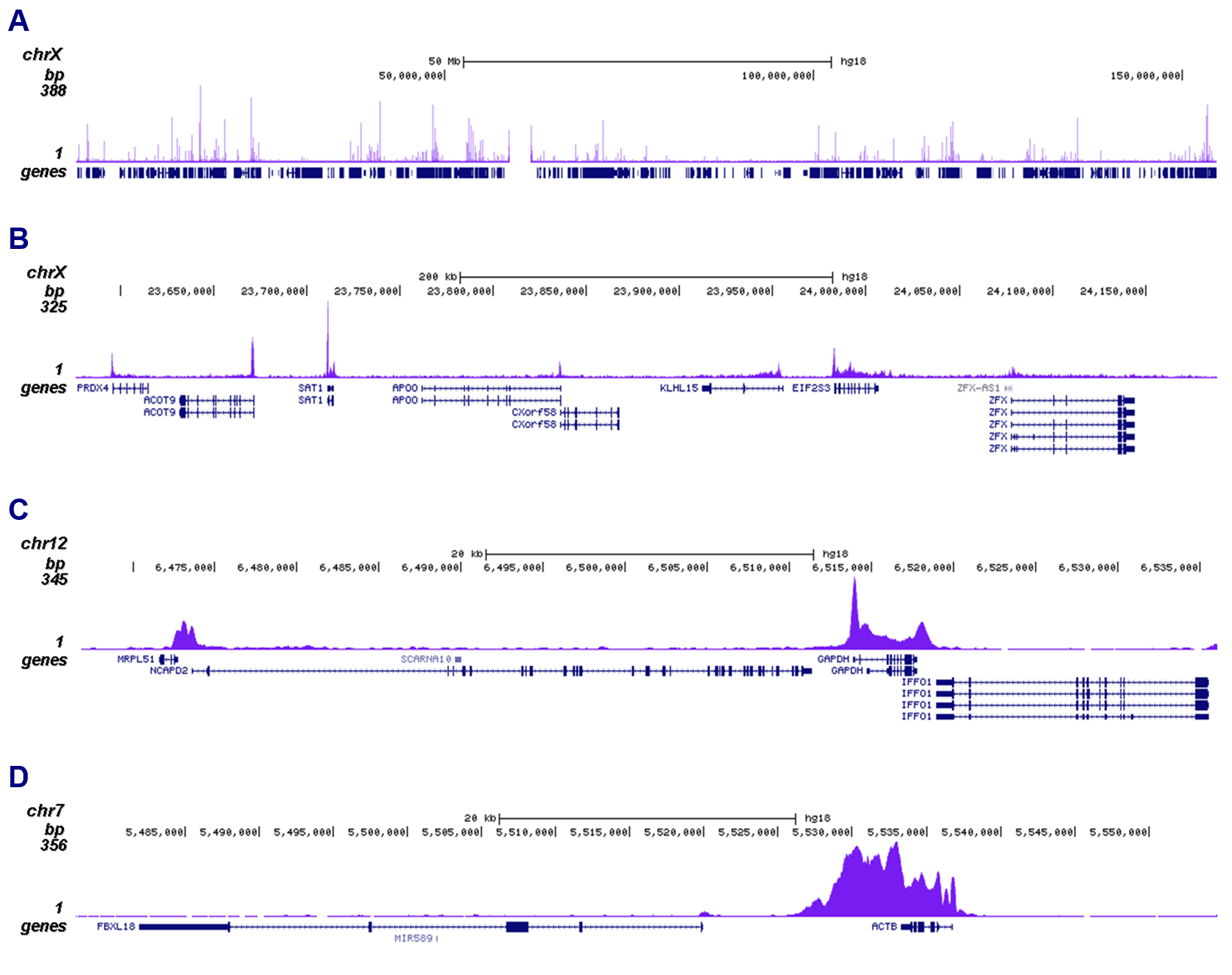
<p><small><strong>Figure 2. ChIP-seq results obtained with the Diagenode monoclonal antibody directed against Pol II</strong><br /> ChIP was performed on sheared chromatin from 1 million HeLaS3 cells using 1 µg of the Diagenode antibody against Pol II (Cat. No. C15200004) as described above. The IP'd DNA was subsequently analysed on an Illumina Genome Analyzer. Library preparation, cluster generation and sequencing were performed according to the manufacturer's instructions. The 36 bp tags were aligned to the human genome using the ELAND algorithm. Figure 2 shows the peak distribution along the complete sequence and a 400 kb region of the X-chromosome (figure 2A and B, respectively), and in a two genomic regions surrounding the GAPDH and ACTB positive control genes (figure 2C and D).</small></p>
</div>
</div>
<div class="row">
<div class="small-6 columns"><img src="https://www.diagenode.com/img/product/antibodies/C15200004_ELISA.png" alt="Pol II Antibody ELISA validation" style="display: block; margin-left: auto; margin-right: auto;" /></div>
<div class="small-6 columns">
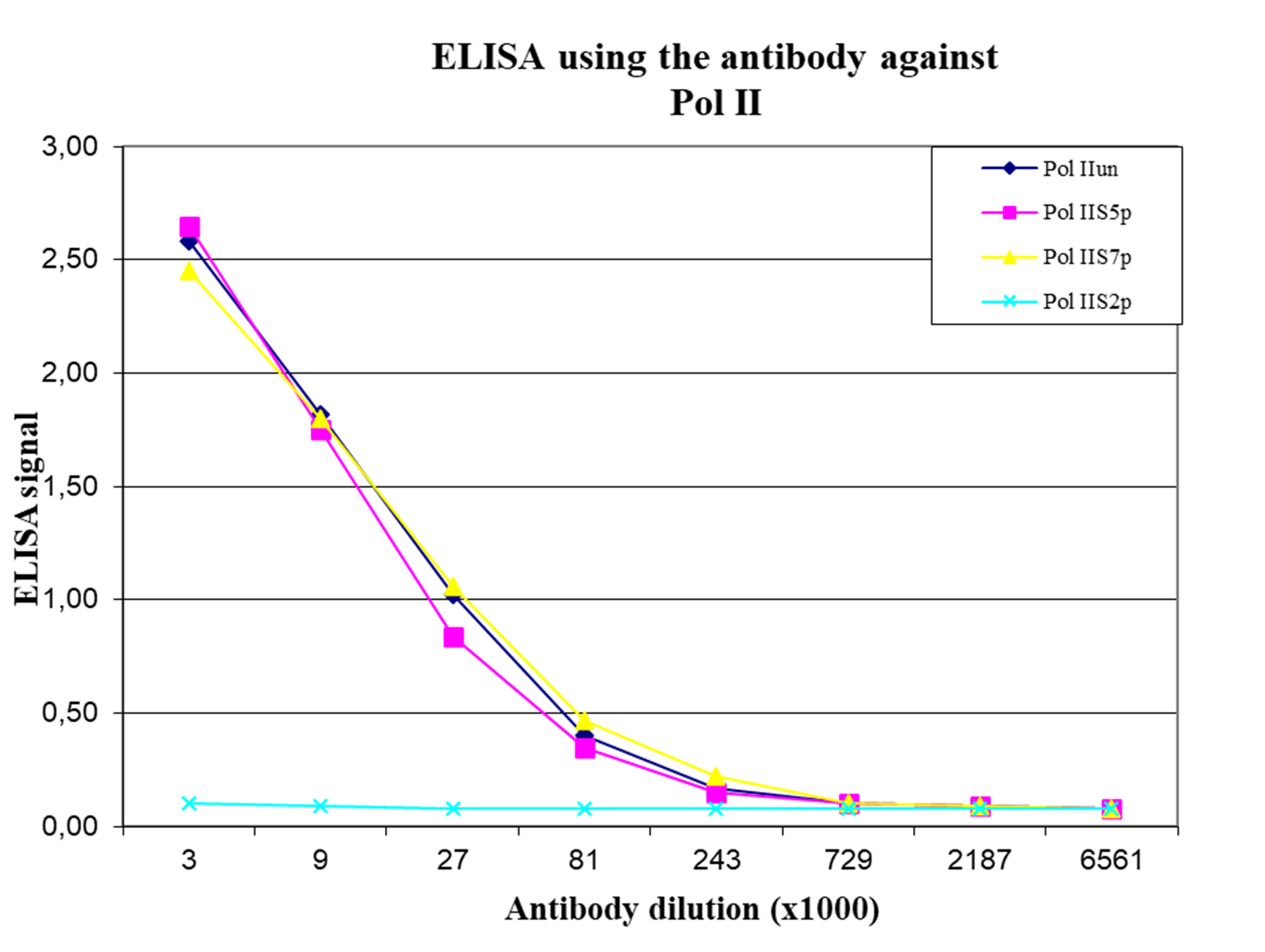
<p><small><strong>Figure 3. Cross reactivity of the Diagenode monoclonal antibody directed against Pol II</strong><br /> To test the specificity an ELISA was performed using a serial dilution of the Diagenode monoclonal antibody against Pol II (Cat. No. C15200004). The wells were coated with peptides containing the unmodified C-terminal repeat sequence as well as different phosphorylated peptides. Figure 3 shows that the antibody recognizes the unphosphorylated Pol II as well as most phosphorylated forms.</small></p>
</div>
</div>
<div class="spacer"></div>
<div class="row">
<div class="small-3 columns"><img src="https://www.diagenode.com/img/product/antibodies/C15200004_Wb.png" alt="Pol II Antibody for Western Blot" style="display: block; margin-left: auto; margin-right: auto;" /></div>
<div class="small-9 columns">
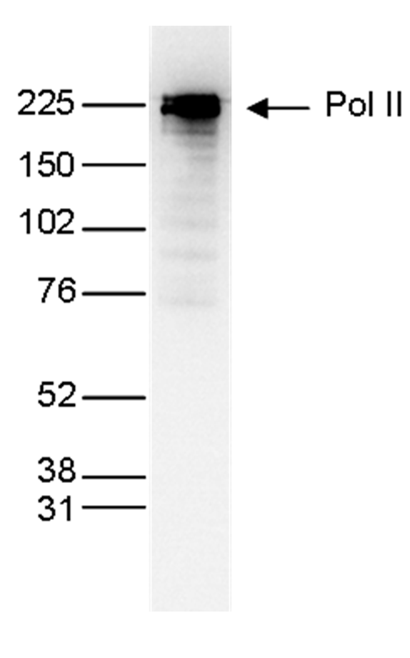
<p><small><strong>Figure 4. Western blot analysis using the Diagenode monoclonal antibody directed against Pol II</strong>Nuclear extracts (25 µg) from HeLa cells were analysed by Western blot using the Diagenode monoclonal antibody against Pol II (Cat. No. C15200004) diluted 1:1,000 in TBS-Tween containing 5% skimmed milk. The position of the protein of interest is indicated on the right; the marker (in kDa) is shown on the left.</small></p>
</div>
</div>
<div class="row">
<div class="small-3 columns"><img src="https://www.diagenode.com/img/product/antibodies/C15200004_001-11_Wb_2.png" alt="Pol II Antibody validated in Western Blot" style="display: block; margin-left: auto; margin-right: auto;" /></div>
<div class="small-9 columns">
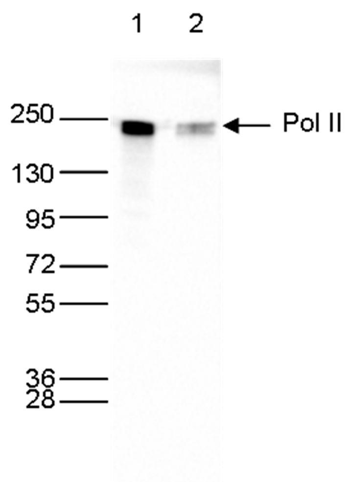
<p><small><strong>Figure 5. Western blot analysis using the Diagenode monoclonal antibody directed against Pol II</strong><br />Whole cell extracts (40 µg) from HeLa cells transfected with Pol II siRNA (lane 2) and from an untransfected control (lane 1) were analysed by Western blot using the Diagenode antibody against Pol II (Cat. No. C15200004) diluted 1:1,000 in TBS-Tween containing 5% skimmed milk. The position of the protein of interest is indicated on the right; the marker (in kDa) is shown on the left.</small></p>
</div>
</div>
<div class="row">
<div class="small-12 columns"><img src="https://www.diagenode.com/img/product/antibodies/C15200004_IF.png" alt="Pol II Antibody for Immunofluorescence" style="display: block; margin-left: auto; margin-right: auto;" /></div>
</div>
<div class="row">
<div class="small-12 columns">
<p><small><strong>Figure 6. Immunofluorescence using the Diagenode monoclonal antibody directed against Pol II</strong><br /> HeLa cells were stained with the Diagenode antibody against Pol II (Cat. No. C15200004) and with DAPI. Cells were fixed with methanol and blocked with PBS/TX-100 containing 5% normal goat serum and 1% BSA. The cells were immunofluorescently labelled with the Pol II antibody (left) diluted 1:500 in blocking solution followed by an anti-mouse antibody conjugated to Alexa594. The middle panel shows staining of the nuclei with DAPI. A merge of the two stainings is shown on the right.</small></p>
</div>
</div>',
'label2' => '',
'info2' => '<p>RNA polymerase II (pol II) is a key enzyme in the regulation and control of gene transcription. It is able to unwind the DNA double helix, synthesize RNA, and proofread the result. Pol II is a complex enzyme, consisting of 12 subunits, of which the B1 subunit (UniProt/Swiss-Prot entry P24928) is the largest. Together with the second largest subunit, B1 forms the catalytic core of the RNA polymerase II transcription machinery.</p>',
'label3' => '',
'info3' => '',
'format' => '50 μg',
'catalog_number' => 'C15200004',
'old_catalog_number' => '',
'sf_code' => 'C15200004-D001-000581',
'type' => 'FRE',
'search_order' => '03-Antibody',
'price_EUR' => '380',
'price_USD' => '380',
'price_GBP' => '340',
'price_JPY' => '59525',
'price_CNY' => '',
'price_AUD' => '950',
'country' => 'ALL',
'except_countries' => 'None',
'quote' => false,
'in_stock' => false,
'featured' => false,
'no_promo' => false,
'online' => true,
'master' => true,
'last_datasheet_update' => 'January 23, 2020',
'slug' => 'pol-ii-monoclonal-antibody-classic-50-mg',
'meta_title' => 'Pol II monoclonal antibody - Classic',
'meta_keywords' => '',
'meta_description' => 'Pol II monoclonal antibody - Classic',
'modified' => '2021-10-20 09:23:11',
'created' => '2015-06-29 14:08:20',
'locale' => 'jpn'
),
'Antibody' => array(
'host' => '*****',
'id' => '195',
'name' => 'Pol II monoclonal antibody',
'description' => 'RNA polymerase II (pol II) is a key enzyme in the regulation and control of gene transcription. It is able to unwind the DNA double helix, synthesize RNA, and proofread the result. Pol II is a complex enzyme, consisting of 12 subunits, of which the B1 subunit (UniProt/Swiss-Prot entry P24928) is the largest. Together with the second largest subunit, B1 forms the catalytic core of the RNA polymerase II transcription machinery.',
'clonality' => '',
'isotype' => 'IgG1',
'lot' => '001-14',
'concentration' => '1.0 µg/µl',
'reactivity' => 'Human, Xenopus, Yeast: positive. Other species: not tested.',
'type' => 'Monoclonal <strong>ChIP grade, ChIP-seq grade</strong>',
'purity' => 'Protein A purified monoclonal antibody.',
'classification' => 'Classic',
'application_table' => '<table>
<thead>
<tr>
<th>Applications</th>
<th>Suggested dilution</th>
<th>References</th>
</tr>
</thead>
<tbody>
<tr>
<td>ChIP/ChIP-seq <sup>*</sup></td>
<td>1 µg/ChIP</td>
<td>Fig 1, 2</td>
</tr>
<tr>
<td>ELISA</td>
<td>1:3,000</td>
<td>Fig 3</td>
</tr>
<tr>
<td>Western Blotting</td>
<td>1:1,000</td>
<td>Fig 4, 5</td>
</tr>
<tr>
<td>Immunofluorescence</td>
<td>1:500</td>
<td>Fig 6</td>
</tr>
</tbody>
</table>
<p></p>
<p><small><sup>*</sup> Please note that the optimal antibody amount per ChIP should be determined by the end-user. We recommend testing 1-5 µg per IP.</small></p>',
'storage_conditions' => 'Store at -20°C; for long storage, store at -80°C. Avoid multiple freeze-thaw cycles.',
'storage_buffer' => 'PBS containing 0.05% azide.',
'precautions' => 'This product is for research use only. Not for use in diagnostic or therapeutic procedures.',
'uniprot_acc' => '',
'slug' => '',
'meta_keywords' => '',
'meta_description' => '',
'modified' => '2022-08-04 14:16:39',
'created' => '0000-00-00 00:00:00',
'select_label' => '195 - Pol II monoclonal antibody (001-14 - 1.0 µg/µl - Human, Xenopus, Yeast: positive. Other species: not tested. - Protein A purified monoclonal antibody. - Mouse)'
),
'Slave' => array(
(int) 0 => array(
[maximum depth reached]
)
),
'Group' => array(
'Group' => array(
[maximum depth reached]
),
'Master' => array(
[maximum depth reached]
),
'Product' => array(
[maximum depth reached]
)
),
'Related' => array(
(int) 0 => array(
[maximum depth reached]
),
(int) 1 => array(
[maximum depth reached]
),
(int) 2 => array(
[maximum depth reached]
),
(int) 3 => array(
[maximum depth reached]
)
),
'Application' => array(
(int) 0 => array(
[maximum depth reached]
),
(int) 1 => array(
[maximum depth reached]
),
(int) 2 => array(
[maximum depth reached]
),
(int) 3 => array(
[maximum depth reached]
),
(int) 4 => array(
[maximum depth reached]
),
(int) 5 => array(
[maximum depth reached]
)
),
'Category' => array(
(int) 0 => array(
[maximum depth reached]
),
(int) 1 => array(
[maximum depth reached]
),
(int) 2 => array(
[maximum depth reached]
),
(int) 3 => array(
[maximum depth reached]
)
),
'Document' => array(
(int) 0 => array(
[maximum depth reached]
),
(int) 1 => array(
[maximum depth reached]
),
(int) 2 => array(
[maximum depth reached]
)
),
'Feature' => array(),
'Image' => array(
(int) 0 => array(
[maximum depth reached]
)
),
'Promotion' => array(),
'Protocol' => array(),
'Publication' => array(
(int) 0 => array(
[maximum depth reached]
),
(int) 1 => array(
[maximum depth reached]
),
(int) 2 => array(
[maximum depth reached]
),
(int) 3 => array(
[maximum depth reached]
),
(int) 4 => array(
[maximum depth reached]
),
(int) 5 => array(
[maximum depth reached]
),
(int) 6 => array(
[maximum depth reached]
),
(int) 7 => array(
[maximum depth reached]
),
(int) 8 => array(
[maximum depth reached]
),
(int) 9 => array(
[maximum depth reached]
),
(int) 10 => array(
[maximum depth reached]
),
(int) 11 => array(
[maximum depth reached]
),
(int) 12 => array(
[maximum depth reached]
),
(int) 13 => array(
[maximum depth reached]
)
),
'Testimonial' => array(
(int) 0 => array(
[maximum depth reached]
)
),
'Area' => array(),
'SafetySheet' => array(
(int) 0 => array(
[maximum depth reached]
),
(int) 1 => array(
[maximum depth reached]
),
(int) 2 => array(
[maximum depth reached]
),
(int) 3 => array(
[maximum depth reached]
),
(int) 4 => array(
[maximum depth reached]
),
(int) 5 => array(
[maximum depth reached]
),
(int) 6 => array(
[maximum depth reached]
),
(int) 7 => array(
[maximum depth reached]
)
)
),
'meta_canonical' => 'https://www.diagenode.com/jp/p/pol-ii-monoclonal-antibody-classic-50-mg'
)
$language = 'jp'
$meta_keywords = ''
$meta_description = 'Pol II monoclonal antibody - Classic'
$meta_title = 'Pol II monoclonal antibody - Classic'
$product = array(
'Product' => array(
'id' => '1962',
'antibody_id' => '195',
'name' => 'Pol II monoclonal antibody ',
'description' => '<p>Alternative names: <strong>POLR2A</strong>, <strong>RPB1</strong>, <strong>POLR2</strong>, <strong>RPOL2</strong></p>
<p><span>Monoclonal antibody raised in mouse against the YSPTSPS repeat in the B1 subunit of RNA polymerase II. </span></p>',
'label1' => 'Validation Data',
'info1' => '<div class="row">
<div class="small-6 columns"><img src="https://www.diagenode.com/img/product/antibodies/C15200004-CHIP.png" alt="Pol II Antibody ChIP Grade" style="display: block; margin-left: auto; margin-right: auto;" /></div>
<div class="small-6 columns">
<p><small><strong>Figure 1. ChIP results obtained with the Diagenode monoclonal antibody directed against Pol II</strong><br /> ChIP assays were performed using human HeLa cells, the Diagenode monoclonal antibody against Pol II (Cat. No. C15200004) and optimized PCR primer pairs for qPCR. ChIP was performed with the "iDeal ChIP-seq" kit (Cat. No. C01010051), using sheared chromatin from 1 million cells. A titration consisting of 1, 2, 5 and 10 µg of antibody per ChIP experiment was analyzed. IgG (2 µg/IP) was used as a negative IP control. Quantitative PCR was performed with primers specific for the promoter and the coding region of the constitutively expressed GAPDH and ACTB genes, used as positive controls, and for exon 2 of the inactive myoglobin (MB) gene and the Sat2 satellite repeat, used as negative controls. Figure 1 shows the recovery, expressed as a % of input (the relative amount of immunoprecipitated DNA compared to input DNA after qPCR analysis).</small></p>
</div>
</div>
<div class="row">
<div class="small-12 columns"><img src="https://www.diagenode.com/img/product/antibodies/C15200004_ChIPseq-A.png" alt="Pol II Antibody ChIP-seq Grade" style="display: block; margin-left: auto; margin-right: auto;" /><br /> <img src="https://www.diagenode.com/img/product/antibodies/C15200004_ChIPseq-B.png" alt="Pol II Antibody for ChIP-seq" style="display: block; margin-left: auto; margin-right: auto;" /><br /> <img src="https://www.diagenode.com/img/product/antibodies/C15200004_ChIPseq-C.png" alt="Pol II Antibody for ChIP-seq assay " style="display: block; margin-left: auto; margin-right: auto;" /></div>
</div>
<div class="row">
<div class="small-12 columns"><img src="https://www.diagenode.com/img/product/antibodies/C15200004_ChIPseq-D.png" alt="Pol II Antibody validated in ChIP-seq " style="display: block; margin-left: auto; margin-right: auto;" /></div>
</div>
<div class="row">
<div class="small-12 columns">
<p><small><strong>Figure 2. ChIP-seq results obtained with the Diagenode monoclonal antibody directed against Pol II</strong><br /> ChIP was performed on sheared chromatin from 1 million HeLaS3 cells using 1 µg of the Diagenode antibody against Pol II (Cat. No. C15200004) as described above. The IP'd DNA was subsequently analysed on an Illumina Genome Analyzer. Library preparation, cluster generation and sequencing were performed according to the manufacturer's instructions. The 36 bp tags were aligned to the human genome using the ELAND algorithm. Figure 2 shows the peak distribution along the complete sequence and a 400 kb region of the X-chromosome (figure 2A and B, respectively), and in a two genomic regions surrounding the GAPDH and ACTB positive control genes (figure 2C and D).</small></p>
</div>
</div>
<div class="row">
<div class="small-6 columns"><img src="https://www.diagenode.com/img/product/antibodies/C15200004_ELISA.png" alt="Pol II Antibody ELISA validation" style="display: block; margin-left: auto; margin-right: auto;" /></div>
<div class="small-6 columns">
<p><small><strong>Figure 3. Cross reactivity of the Diagenode monoclonal antibody directed against Pol II</strong><br /> To test the specificity an ELISA was performed using a serial dilution of the Diagenode monoclonal antibody against Pol II (Cat. No. C15200004). The wells were coated with peptides containing the unmodified C-terminal repeat sequence as well as different phosphorylated peptides. Figure 3 shows that the antibody recognizes the unphosphorylated Pol II as well as most phosphorylated forms.</small></p>
</div>
</div>
<div class="spacer"></div>
<div class="row">
<div class="small-3 columns"><img src="https://www.diagenode.com/img/product/antibodies/C15200004_Wb.png" alt="Pol II Antibody for Western Blot" style="display: block; margin-left: auto; margin-right: auto;" /></div>
<div class="small-9 columns">
<p><small><strong>Figure 4. Western blot analysis using the Diagenode monoclonal antibody directed against Pol II</strong>Nuclear extracts (25 µg) from HeLa cells were analysed by Western blot using the Diagenode monoclonal antibody against Pol II (Cat. No. C15200004) diluted 1:1,000 in TBS-Tween containing 5% skimmed milk. The position of the protein of interest is indicated on the right; the marker (in kDa) is shown on the left.</small></p>
</div>
</div>
<div class="row">
<div class="small-3 columns"><img src="https://www.diagenode.com/img/product/antibodies/C15200004_001-11_Wb_2.png" alt="Pol II Antibody validated in Western Blot" style="display: block; margin-left: auto; margin-right: auto;" /></div>
<div class="small-9 columns">
<p><small><strong>Figure 5. Western blot analysis using the Diagenode monoclonal antibody directed against Pol II</strong><br />Whole cell extracts (40 µg) from HeLa cells transfected with Pol II siRNA (lane 2) and from an untransfected control (lane 1) were analysed by Western blot using the Diagenode antibody against Pol II (Cat. No. C15200004) diluted 1:1,000 in TBS-Tween containing 5% skimmed milk. The position of the protein of interest is indicated on the right; the marker (in kDa) is shown on the left.</small></p>
</div>
</div>
<div class="row">
<div class="small-12 columns"><img src="https://www.diagenode.com/img/product/antibodies/C15200004_IF.png" alt="Pol II Antibody for Immunofluorescence" style="display: block; margin-left: auto; margin-right: auto;" /></div>
</div>
<div class="row">
<div class="small-12 columns">
<p><small><strong>Figure 6. Immunofluorescence using the Diagenode monoclonal antibody directed against Pol II</strong><br /> HeLa cells were stained with the Diagenode antibody against Pol II (Cat. No. C15200004) and with DAPI. Cells were fixed with methanol and blocked with PBS/TX-100 containing 5% normal goat serum and 1% BSA. The cells were immunofluorescently labelled with the Pol II antibody (left) diluted 1:500 in blocking solution followed by an anti-mouse antibody conjugated to Alexa594. The middle panel shows staining of the nuclei with DAPI. A merge of the two stainings is shown on the right.</small></p>
</div>
</div>',
'label2' => 'Target Description',
'info2' => '<p>RNA polymerase II (pol II) is a key enzyme in the regulation and control of gene transcription. It is able to unwind the DNA double helix, synthesize RNA, and proofread the result. Pol II is a complex enzyme, consisting of 12 subunits, of which the B1 subunit (UniProt/Swiss-Prot entry P24928) is the largest. Together with the second largest subunit, B1 forms the catalytic core of the RNA polymerase II transcription machinery.</p>',
'label3' => '',
'info3' => '',
'format' => '50 μg',
'catalog_number' => 'C15200004',
'old_catalog_number' => '',
'sf_code' => 'C15200004-D001-000581',
'type' => 'FRE',
'search_order' => '03-Antibody',
'price_EUR' => '380',
'price_USD' => '380',
'price_GBP' => '340',
'price_JPY' => '59525',
'price_CNY' => '',
'price_AUD' => '950',
'country' => 'ALL',
'except_countries' => 'None',
'quote' => false,
'in_stock' => false,
'featured' => false,
'no_promo' => false,
'online' => true,
'master' => true,
'last_datasheet_update' => 'January 23, 2020',
'slug' => 'pol-ii-monoclonal-antibody-classic-50-mg',
'meta_title' => 'Pol II monoclonal antibody - Classic',
'meta_keywords' => '',
'meta_description' => 'Pol II monoclonal antibody - Classic',
'modified' => '2021-10-20 09:23:11',
'created' => '2015-06-29 14:08:20',
'locale' => 'jpn'
),
'Antibody' => array(
'host' => '*****',
'id' => '195',
'name' => 'Pol II monoclonal antibody',
'description' => 'RNA polymerase II (pol II) is a key enzyme in the regulation and control of gene transcription. It is able to unwind the DNA double helix, synthesize RNA, and proofread the result. Pol II is a complex enzyme, consisting of 12 subunits, of which the B1 subunit (UniProt/Swiss-Prot entry P24928) is the largest. Together with the second largest subunit, B1 forms the catalytic core of the RNA polymerase II transcription machinery.',
'clonality' => '',
'isotype' => 'IgG1',
'lot' => '001-14',
'concentration' => '1.0 µg/µl',
'reactivity' => 'Human, Xenopus, Yeast: positive. Other species: not tested.',
'type' => 'Monoclonal <strong>ChIP grade, ChIP-seq grade</strong>',
'purity' => 'Protein A purified monoclonal antibody.',
'classification' => 'Classic',
'application_table' => '<table>
<thead>
<tr>
<th>Applications</th>
<th>Suggested dilution</th>
<th>References</th>
</tr>
</thead>
<tbody>
<tr>
<td>ChIP/ChIP-seq <sup>*</sup></td>
<td>1 µg/ChIP</td>
<td>Fig 1, 2</td>
</tr>
<tr>
<td>ELISA</td>
<td>1:3,000</td>
<td>Fig 3</td>
</tr>
<tr>
<td>Western Blotting</td>
<td>1:1,000</td>
<td>Fig 4, 5</td>
</tr>
<tr>
<td>Immunofluorescence</td>
<td>1:500</td>
<td>Fig 6</td>
</tr>
</tbody>
</table>
<p></p>
<p><small><sup>*</sup> Please note that the optimal antibody amount per ChIP should be determined by the end-user. We recommend testing 1-5 µg per IP.</small></p>',
'storage_conditions' => 'Store at -20°C; for long storage, store at -80°C. Avoid multiple freeze-thaw cycles.',
'storage_buffer' => 'PBS containing 0.05% azide.',
'precautions' => 'This product is for research use only. Not for use in diagnostic or therapeutic procedures.',
'uniprot_acc' => '',
'slug' => '',
'meta_keywords' => '',
'meta_description' => '',
'modified' => '2022-08-04 14:16:39',
'created' => '0000-00-00 00:00:00',
'select_label' => '195 - Pol II monoclonal antibody (001-14 - 1.0 µg/µl - Human, Xenopus, Yeast: positive. Other species: not tested. - Protein A purified monoclonal antibody. - Mouse)'
),
'Slave' => array(
(int) 0 => array(
'id' => '34',
'name' => 'C15200004',
'product_id' => '1962',
'modified' => '2016-02-18 20:43:46',
'created' => '2016-02-18 20:43:46'
)
),
'Group' => array(
'Group' => array(
'id' => '34',
'name' => 'C15200004',
'product_id' => '1962',
'modified' => '2016-02-18 20:43:46',
'created' => '2016-02-18 20:43:46'
),
'Master' => array(
'id' => '1962',
'antibody_id' => '195',
'name' => 'Pol II Antibody',
'description' => '<p>Alternative names: <strong>POLR2A</strong>, <strong>RPB1</strong>, <strong>POLR2</strong>, <strong>RPOL2</strong></p>
<p><span>Monoclonal antibody raised in mouse against the YSPTSPS repeat in the B1 subunit of <strong>RNA polymerase II</strong>. </span></p>',
'label1' => 'Validation data',
'info1' => '<div class="row">
<div class="small-6 columns"><img src="https://www.diagenode.com/img/product/antibodies/C15200004-CHIP.png" alt="Pol II Antibody ChIP Grade" style="display: block; margin-left: auto; margin-right: auto;" /></div>
<div class="small-6 columns">
<p><small><strong>Figure 1. ChIP results obtained with the Diagenode monoclonal antibody directed against Pol II</strong><br /> ChIP assays were performed using human HeLa cells, the Diagenode monoclonal antibody against Pol II (Cat. No. C15200004) and optimized PCR primer pairs for qPCR. ChIP was performed with the "iDeal ChIP-seq" kit (Cat. No. C01010051), using sheared chromatin from 1 million cells. A titration consisting of 1, 2, 5 and 10 µg of antibody per ChIP experiment was analyzed. IgG (2 µg/IP) was used as a negative IP control. Quantitative PCR was performed with primers specific for the promoter and the coding region of the constitutively expressed GAPDH and ACTB genes, used as positive controls, and for exon 2 of the inactive myoglobin (MB) gene and the Sat2 satellite repeat, used as negative controls. Figure 1 shows the recovery, expressed as a % of input (the relative amount of immunoprecipitated DNA compared to input DNA after qPCR analysis).</small></p>
</div>
</div>
<div class="row">
<div class="small-12 columns"><img src="https://www.diagenode.com/img/product/antibodies/C15200004_ChIPseq-A.png" alt="Pol II Antibody ChIP-seq Grade" style="display: block; margin-left: auto; margin-right: auto;" /><br /> <img src="https://www.diagenode.com/img/product/antibodies/C15200004_ChIPseq-B.png" alt="Pol II Antibody for ChIP-seq" style="display: block; margin-left: auto; margin-right: auto;" /><br /> <img src="https://www.diagenode.com/img/product/antibodies/C15200004_ChIPseq-C.png" alt="Pol II Antibody for ChIP-seq assay " style="display: block; margin-left: auto; margin-right: auto;" /></div>
</div>
<div class="row">
<div class="small-12 columns"><img src="https://www.diagenode.com/img/product/antibodies/C15200004_ChIPseq-D.png" alt="Pol II Antibody validated in ChIP-seq " style="display: block; margin-left: auto; margin-right: auto;" /></div>
</div>
<div class="row">
<div class="small-12 columns">
<p><small><strong>Figure 2. ChIP-seq results obtained with the Diagenode monoclonal antibody directed against Pol II</strong><br /> ChIP was performed on sheared chromatin from 1 million HeLaS3 cells using 1 µg of the Diagenode antibody against Pol II (Cat. No. C15200004) as described above. The IP'd DNA was subsequently analysed on an Illumina Genome Analyzer. Library preparation, cluster generation and sequencing were performed according to the manufacturer's instructions. The 36 bp tags were aligned to the human genome using the ELAND algorithm. Figure 2 shows the peak distribution along the complete sequence and a 400 kb region of the X-chromosome (figure 2A and B, respectively), and in a two genomic regions surrounding the GAPDH and ACTB positive control genes (figure 2C and D).</small></p>
</div>
</div>
<div class="row">
<div class="small-6 columns"><img src="https://www.diagenode.com/img/product/antibodies/C15200004_ELISA.png" alt="Pol II Antibody ELISA validation" style="display: block; margin-left: auto; margin-right: auto;" /></div>
<div class="small-6 columns">
<p><small><strong>Figure 3. Cross reactivity of the Diagenode monoclonal antibody directed against Pol II</strong><br /> To test the specificity an ELISA was performed using a serial dilution of the Diagenode monoclonal antibody against Pol II (Cat. No. C15200004). The wells were coated with peptides containing the unmodified C-terminal repeat sequence as well as different phosphorylated peptides. Figure 3 shows that the antibody recognizes the unphosphorylated Pol II as well as most phosphorylated forms.</small></p>
</div>
</div>
<div class="spacer"></div>
<div class="row">
<div class="small-3 columns"><img src="https://www.diagenode.com/img/product/antibodies/C15200004_Wb.png" alt="Pol II Antibody for Western Blot" style="display: block; margin-left: auto; margin-right: auto;" /></div>
<div class="small-9 columns">
<p><small><strong>Figure 4. Western blot analysis using the Diagenode monoclonal antibody directed against Pol II</strong>Nuclear extracts (25 µg) from HeLa cells were analysed by Western blot using the Diagenode monoclonal antibody against Pol II (Cat. No. C15200004) diluted 1:1,000 in TBS-Tween containing 5% skimmed milk. The position of the protein of interest is indicated on the right; the marker (in kDa) is shown on the left.</small></p>
</div>
</div>
<div class="row">
<div class="small-3 columns"><img src="https://www.diagenode.com/img/product/antibodies/C15200004_001-11_Wb_2.png" alt="Pol II Antibody validated in Western Blot" style="display: block; margin-left: auto; margin-right: auto;" /></div>
<div class="small-9 columns">
<p><small><strong>Figure 5. Western blot analysis using the Diagenode monoclonal antibody directed against Pol II</strong><br />Whole cell extracts (40 µg) from HeLa cells transfected with Pol II siRNA (lane 2) and from an untransfected control (lane 1) were analysed by Western blot using the Diagenode antibody against Pol II (Cat. No. C15200004) diluted 1:1,000 in TBS-Tween containing 5% skimmed milk. The position of the protein of interest is indicated on the right; the marker (in kDa) is shown on the left.</small></p>
</div>
</div>
<div class="row">
<div class="small-12 columns"><img src="https://www.diagenode.com/img/product/antibodies/C15200004_IF.png" alt="Pol II Antibody for Immunofluorescence" style="display: block; margin-left: auto; margin-right: auto;" /></div>
</div>
<div class="row">
<div class="small-12 columns">
<p><small><strong>Figure 6. Immunofluorescence using the Diagenode monoclonal antibody directed against Pol II</strong><br /> HeLa cells were stained with the Diagenode antibody against Pol II (Cat. No. C15200004) and with DAPI. Cells were fixed with methanol and blocked with PBS/TX-100 containing 5% normal goat serum and 1% BSA. The cells were immunofluorescently labelled with the Pol II antibody (left) diluted 1:500 in blocking solution followed by an anti-mouse antibody conjugated to Alexa594. The middle panel shows staining of the nuclei with DAPI. A merge of the two stainings is shown on the right.</small></p>
</div>
</div>',
'label2' => 'Target Description',
'info2' => '<p>RNA polymerase II (pol II) is a key enzyme in the regulation and control of gene transcription. It is able to unwind the DNA double helix, synthesize RNA, and proofread the result. Pol II is a complex enzyme, consisting of 12 subunits, of which the B1 subunit (UniProt/Swiss-Prot entry P24928) is the largest. Together with the second largest subunit, B1 forms the catalytic core of the RNA polymerase II transcription machinery.</p>',
'label3' => '',
'info3' => '',
'format' => '50 μg',
'catalog_number' => 'C15200004',
'old_catalog_number' => '',
'sf_code' => 'C15200004-D001-000581',
'type' => 'FRE',
'search_order' => '03-Antibody',
'price_EUR' => '380',
'price_USD' => '380',
'price_GBP' => '340',
'price_JPY' => '59525',
'price_CNY' => '',
'price_AUD' => '950',
'country' => 'ALL',
'except_countries' => 'None',
'quote' => false,
'in_stock' => false,
'featured' => false,
'no_promo' => false,
'online' => true,
'master' => true,
'last_datasheet_update' => 'January 23, 2020',
'slug' => 'pol-ii-monoclonal-antibody-classic-50-mg',
'meta_title' => 'Pol II Antibody - ChIP-seq Grade (C15200004) | Diagenode',
'meta_keywords' => '',
'meta_description' => 'Pol II (YSPTSPS repeat in the B1 subunit of RNA polymerase II) Monoclonal Antibody validated in ChIP-seq, ChIP-qPCR, WB and ELISA. Specificity confirmed by siRNA assay. Batch-specific data available on the website. Alternative names: POLR2A, RPB1, POLR2, RPOL2. Sample size available.',
'modified' => '2021-10-20 09:23:11',
'created' => '2015-06-29 14:08:20'
),
'Product' => array(
(int) 0 => array(
[maximum depth reached]
)
)
),
'Related' => array(
(int) 0 => array(
'id' => '1839',
'antibody_id' => null,
'name' => 'iDeal ChIP-seq kit for Transcription Factors',
'description' => '<p><a href="https://www.diagenode.com/files/products/kits/ideal-chipseq-transcription-factors-x10-manual.pdf"><img src="https://www.diagenode.com/img/buttons/bt-manual.png" /></a></p>
<div class="row">
<div class="small-12 medium-8 large-8 columns"><br />
<p><span style="font-weight: 400;">Diagenode’s <strong>iDeal ChIP-seq Kit for Transcription Factors</strong> is a highly validated solution for robust transcription factor and other non-histone proteins ChIP-seq results and contains everything you need for start-to-finish </span><b>ChIP </b><span style="font-weight: 400;">prior to </span><b>Next-Generation Sequencing</b><span style="font-weight: 400;">. This complete solution contains all buffers and reagents for cell lysis, chromatin shearing, immunoprecipitation, and DNA purification. In addition, unlike competing solutions, the kit contains positive and negative control antibodies (CTCF and IgG, respectively) as well as positive and negative control PCR primers pairs (H19 and Myoglobin exon 2, respectively) for your convenience and a guarantee of optimal results. <br /></span></p>
</div>
<div class="small-12 medium-4 large-4 columns"><center><br /><br />
<script>// <![CDATA[
var date = new Date(); var heure = date.getHours(); var jour = date.getDay(); var semaine = Math.floor(date.getDate() / 7) + 1; if (jour === 2 && ( (heure >= 9 && heure < 9.5) || (heure >= 18 && heure < 18.5) )) { document.write('<a href="https://us02web.zoom.us/j/85467619762"><img src="https://www.diagenode.com/img/epicafe-ON.gif"></a>'); } else { document.write('<a href="https://go.diagenode.com/l/928883/2023-04-26/3kq1v"><img src="https://www.diagenode.com/img/epicafe-OFF.png"></a>'); }
// ]]></script>
</center></div>
</div>
<p><span style="font-weight: 400;">The </span><b> iDeal ChIP-seq kit for Transcription Factors </b><span style="font-weight: 400;">is compatible for cells or tissues:</span></p>
<table style="width: 419px; margin-left: auto; margin-right: auto;">
<tbody>
<tr>
<td style="width: 144px;"></td>
<td style="width: 267px; text-align: center;"><span style="font-weight: 400;">Amount per IP</span></td>
</tr>
<tr>
<td style="width: 144px;">Cells</td>
<td style="width: 267px; text-align: center;"><strong>4,000,000</strong></td>
</tr>
<tr>
<td style="width: 144px;">Tissues</td>
<td style="width: 267px; text-align: center;"><strong>30 mg</strong></td>
</tr>
</tbody>
</table>
<p><span style="font-weight: 400;">The iDeal ChIP-seq kit is the only kit on the market validated for major sequencing systems. Our expertise in ChIP-seq tools allows reproducible and efficient results every time. </span></p>
<p></p>
<p></p>',
'label1' => 'Characteristics',
'info1' => '<ul>
<li><span style="font-weight: 400;"><strong>Highly optimized protocol</strong> for ChIP-seq from cells and tissues</span></li>
<li><span style="font-weight: 400;"><strong>Validated</strong> for <strong>ChIP-seq</strong> with multiple transcription factors and non-histone targets<br /></span></li>
<li><span style="font-weight: 400;"><strong>Most complete kit</strong> available (covers all steps, including the control antibodies and primers)<br /></span></li>
<li><span style="font-weight: 400;"><strong>Magnetic beads</strong> make ChIP <strong>easy</strong>, <strong>fast</strong> and more <strong>reproducible</strong></span></li>
<li><span style="font-weight: 400;">Combination with Diagenode ChIP-seq antibodies provides <strong>high yields</strong> with excellent <strong>specificity</strong> and <strong>sensitivity</strong><br /></span></li>
<li><span style="font-weight: 400;">Purified DNA suitable for any downstream application</span></li>
<li><span style="font-weight: 400;">Easy-to-follow protocol</span></li>
</ul>
<p><span style="font-weight: 400;"></span></p>
<p> </p>
<h3>ChIP-seq on cells</h3>
<p><img src="https://www.diagenode.com/img/product/kits/ideal-ctcf-diagenode.jpg" alt="CTCF Diagenode" style="display: block; margin-left: auto; margin-right: auto;" /></p>
<p><strong>Figure 1.</strong> (A) Chromatin Immunoprecipitation has been performed using chromatin from HeLa cells, the iDeal ChIP-seq kit for Transcription Factors and the Diagenode ChIP-seq-grade CTCF antibody. The IP'd DNA was subsequently analysed on an Illumina<sup>®</sup> HiSeq. Library preparation, cluster generation and sequencing were performed according to the manufacturer's instructions. This figure shows the peak distribution in a region surrounding the GAPDH positive control gene.</p>
<p><img src="https://www.diagenode.com/img/product/kits/ideal-figure-b-total-diagendoe-peaks.png" alt="CTCF Diagenode" style="display: block; margin-left: auto; margin-right: auto;" /></p>
<p><strong>Figure 1B.</strong> The ChIP-seq dataset from this experiment has been compared with a reference dataset from the Broad Institute. We observed a perfect match between the top 40% of Diagenode peaks and the reference dataset. Based on the NIH Encode project criterion, ChIP-seq results are considered reproducible between an original and reproduced dataset if the top 40% of peaks have at least an 80% overlap ratio with the compared dataset.</p>
<p> </p>
<p><img src="https://www.diagenode.com/img/product/kits/ideal-TF-chip-seq-A.png" alt="ChIP-seq figure A" style="display: block; margin-left: auto; margin-right: auto;" /></p>
<p><img src="https://www.diagenode.com/img/product/kits/ideal-TF-chip-seq-B.png" alt="ChIP-seq figure B" style="display: block; margin-left: auto; margin-right: auto;" /></p>
<p><img src="https://www.diagenode.com/img/product/kits/ideal-TF-chip-seq-C.png" alt="ChIP-seq figure C" style="display: block; margin-left: auto; margin-right: auto;" /></p>
<p><strong>Figure 2.</strong> Chromatin Immunoprecipitation has been performed using chromatin from HeLa cells, the iDeal ChIP-seq kit for Transcription Factors and the Diagenode ChIP-seq-grade HDAC1 (A), LSD1 (B) and p53 antibody (C). The IP'd DNA was subsequently analysed on an Illumina<sup>®</sup> Genome Analyzer. Library preparation, cluster generation and sequencing were performed according to the manufacturer's instructions. This figure shows the peak distribution in regions of chromosome 3 (A), chromosome 12 (B) and chromosome 6 (C) respectively.</p>
<p> </p>
<h3>ChIP-seq on tissue</h3>
<p><img src="https://www.diagenode.com/img/product/kits/ideal-figure-3a.jpg" alt="ChIP-seq figure A" style="display: block; margin-left: auto; margin-right: auto;" /></p>
<p><strong>Figure 3A.</strong> Chromatin Immunoprecipitation has been performed using chromatin from mouse liver tissue, the iDeal ChIP-seq kit for Transcription Factors and the Diagenode ChIP-seq-grade CTCF antibody. The IP'd DNA was subsequently analysed on an Illumina® HiSeq. Library preparation, cluster generation and sequencing were performed according to the manufacturer's instructions. This figure shows the peak distribution in a region surrounding the Vwf positive control gene.</p>
<p><img src="https://www.diagenode.com/img/product/kits/match-of-the-top40-peaks.png" alt="Match of the Top40 peaks" style="display: block; margin-left: auto; margin-right: auto;" /></p>
<p><strong>Figure 3B.</strong> The ChIP-seq dataset from this experiment has been compared with a reference dataset from the Broad Institute. We observed a perfect match between the top 40% of Diagenode peaks and the reference dataset. Based on the NIH Encode project criterion, ChIP-seq results are considered reproducible between an original and reproduced dataset if the top 40% of peaks have at least an 80% overlap ratio with the compared dataset.</p>',
'label2' => 'Species, cell lines, tissues tested',
'info2' => '<p>The iDeal ChIP-seq Kit for Transcription Factors is compatible with a broad variety of cell lines, tissues and species - some examples are shown below. Other species / cell lines / tissues can be used with this kit.</p>
<p><span style="text-decoration: underline;">Cell lines:</span></p>
<p>Human: A549, A673, BT-549, CD4 T, HCC1806, HeLa, HepG2, HFF, HK-GFP-MR, ILC, K562, KYSE-180, LapC4, M14, MCF7, MDA-MB-231, MDA-MB-436, RDES, SKNO1, VCaP, U2-OS, ZR-75-1 </p>
<p>Mouse: ESC, NPCs, BZ, GT1-7, acinar cells, HSPCs, Th2 cells, keratinocytes</p>
<p>Cattle: pbMEC, <span>MAC-T</span></p>
<p><span>Other cell lines / species: compatible, not tested</span></p>
<p><span style="text-decoration: underline;">Tissues:</span></p>
<p>Mouse: kidney, heart, brain, iris, liver, limbs from E10.5 embryos</p>
<p><span>Horse: l</span>iver, brain, heart, lung, skeletal muscle, lamina, ovary</p>
<p>Other tissues: compatible, not tested</p>
<p><span style="text-decoration: underline;">ChIP on yeast</span></p>
<p>The iDeal ChIP-seq kit for TF is compatible with yeast samples. Check out our <strong><a href="https://www.diagenode.com/files/products/kits/Application_Note-ChIP_on_Yeast.pdf">Application Note</a></strong> presenting an optimized detailed protocol for ChIP on yeast.</p>
<p></p>
<p>Did you use the iDeal ChIP-seq for Transcription Factors Kit on other cell line / tissue / species? <a href="mailto:agnieszka.zelisko@diagenode.com?subject=Species, cell lines, tissues tested with the iDeal ChIP-seq Kit for TF&body=Dear Customer,%0D%0A%0D%0APlease, leave below your feedback about the iDeal ChIP-seq for Transcription Factors (cell / tissue type, species, other information...).%0D%0A%0D%0AThank you for sharing with us your experience !%0D%0A%0D%0ABest regards,%0D%0A%0D%0AAgnieszka Zelisko-Schmidt, PhD">Let us know!</a></p>',
'label3' => 'Additional solutions compatible with iDeal ChIP-seq kit for Transcription Factors',
'info3' => '<p><span style="font-weight: 400;">The</span> <a href="https://www.diagenode.com/en/p/chromatin-shearing-optimization-kit-low-sds-for-tfs-25-rxns"><span style="font-weight: 400;">Chromatin EasyShear Kit – Low SDS </span></a><span style="font-weight: 400;">is the kit compatible with the iDeal ChIP-seq kit for TF, recommended for the optimization of chromatin shearing, a critical step for ChIP.</span></p>
<p><a href="https://www.diagenode.com/en/p/chip-cross-link-gold-600-ul"><span style="font-weight: 400;">ChIP Cross-link Gold</span></a> <span style="font-weight: 400;">should be used in combination with formaldehyde when working with higher order and/or dynamic interactions, for efficient protein-protein fixation.</span></p>
<p><span style="font-weight: 400;">For library preparation of immunoprecipitated samples we recommend to use the </span><b> </b><a href="https://www.diagenode.com/en/categories/library-preparation-for-ChIP-seq"><span style="font-weight: 400;">MicroPlex Library Preparation Kit</span></a><span style="font-weight: 400;"> - validated for library preparation from picogram inputs.</span></p>
<p><a href="https://www.diagenode.com/en/categories/chip-seq-grade-antibodies"><span style="font-weight: 400;">ChIP-seq grade antibodies</span></a><span style="font-weight: 400;"> provide high yields with excellent specificity and sensitivity.</span></p>
<p><span style="font-weight: 400;">Check the list of available </span><a href="https://www.diagenode.com/en/categories/primer-pairs"><span style="font-weight: 400;">Primer pairs</span></a><span style="font-weight: 400;"> designed for high specificity to specific genomic regions.</span></p>
<p><span style="font-weight: 400;">Plus, for our <a href="https://www.diagenode.com/en/categories/ip-star">IP-Star Automation</a> users for automated ChIP, check out our <a href="https://www.diagenode.com/en/p/auto-ideal-chip-seq-kit-for-transcription-factors-x24-24-rxns">automated version</a> of this kit.</span></p>',
'format' => '4 chrom. prep./24 IPs',
'catalog_number' => 'C01010055',
'old_catalog_number' => '',
'sf_code' => 'C01010055-',
'type' => 'RFR',
'search_order' => '04-undefined',
'price_EUR' => '915',
'price_USD' => '1130',
'price_GBP' => '840',
'price_JPY' => '143335',
'price_CNY' => '',
'price_AUD' => '2825',
'country' => 'ALL',
'except_countries' => 'None',
'quote' => false,
'in_stock' => false,
'featured' => true,
'no_promo' => false,
'online' => true,
'master' => true,
'last_datasheet_update' => '0000-00-00',
'slug' => 'ideal-chip-seq-kit-for-transcription-factors-x24-24-rxns',
'meta_title' => 'iDeal ChIP-seq kit for Transcription Factors x24',
'meta_keywords' => '',
'meta_description' => 'iDeal ChIP-seq kit for Transcription Factors x24',
'modified' => '2023-06-20 18:27:37',
'created' => '2015-06-29 14:08:20',
'ProductsRelated' => array(
[maximum depth reached]
),
'Image' => array(
[maximum depth reached]
)
),
(int) 1 => array(
'id' => '1842',
'antibody_id' => null,
'name' => 'Auto iDeal ChIP-seq Kit for Transcription Factors',
'description' => '<p><span><strong>This product must be used with the <a href="https://www.diagenode.com/en/p/sx-8g-ip-star-compact-automated-system-1-unit">IP-Star Compact Automated System</a>.</strong></span></p>
<p><span>Diagenode’s </span><strong>Auto iDeal ChIP-seq Kit for Transcription Factors</strong><span> is a highly specialized solution for robust Transcription Factor ChIP-seq results. Unlike competing solutions, our kit utilizes a highly optimized protocol and is backed by validation with a broad number and range of transcription factors. The kit provides high yields with excellent specificity and sensitivity.</span></p>',
'label1' => 'Characteristics',
'info1' => '<ul>
<li><strong>Confidence in results:</strong> Validated for ChIP-seq with multiple transcription factors</li>
<li><strong>Proven:</strong> Validated by the epigenetics community, including the BLUEPRINT consortium</li>
<li><strong>Most complete kit available</strong> for highest quality data - includes control antibodies and primers</li>
<li>Validated with Diagenode's <a href="https://www.diagenode.com/en/p/microplex-library-preparation-kit-v2-x12-12-indices-12-rxns"><span>MicroPlex Library Preparation™ kit</span></a> and <a href="https://www.diagenode.com/categories/ip-star" title="IP-Star Automated System">IP-Star<sup>®</sup></a> Automation System</li>
</ul>
<p> </p>
<h3>ChIP-seq on cells</h3>
<p><img src="https://www.diagenode.com/img/product/kits/ideal-ctcf-diagenode.jpg" alt="CTCF Diagenode" style="display: block; margin-left: auto; margin-right: auto;" /></p>
<p><strong>Figure 1.</strong> (A) Chromatin Immunoprecipitation has been performed using chromatin from HeLa cells, the iDeal ChIP-seq kit for Transcription Factors and the Diagenode ChIP-seq-grade CTCF antibody. The IP'd DNA was subsequently analysed on an Illumina<sup>®</sup> HiSeq. Library preparation, cluster generation and sequencing were performed according to the manufacturer's instructions. This figure shows the peak distribution in a region surrounding the GAPDH positive control gene.</p>
<p><img src="https://www.diagenode.com/img/product/kits/ideal-figure-b-total-diagendoe-peaks.png" alt="CTCF Diagenode" style="display: block; margin-left: auto; margin-right: auto;" /></p>
<p><strong>Figure 1B.</strong> The ChIP-seq dataset from this experiment has been compared with a reference dataset from the Broad Institute. We observed a perfect match between the top 40% of Diagenode peaks and the reference dataset. Based on the NIH Encode project criterion, ChIP-seq results are considered reproducible between an original and reproduced dataset if the top 40% of peaks have at least an 80% overlap ratio with the compared dataset.</p>
<p> </p>
<p><img src="https://www.diagenode.com/img/product/kits/ideal-TF-chip-seq-A.png" alt="ChIP-seq figure A" style="display: block; margin-left: auto; margin-right: auto;" /></p>
<p><img src="https://www.diagenode.com/img/product/kits/ideal-TF-chip-seq-B.png" alt="ChIP-seq figure B" style="display: block; margin-left: auto; margin-right: auto;" /></p>
<p><img src="https://www.diagenode.com/img/product/kits/ideal-TF-chip-seq-C.png" alt="ChIP-seq figure C" style="display: block; margin-left: auto; margin-right: auto;" /></p>
<p><strong>Figure 2.</strong> Chromatin Immunoprecipitation has been performed using chromatin from HeLa cells, the iDeal ChIP-seq kit for Transcription Factors and the Diagenode ChIP-seq-grade HDAC1 (A), LSD1 (B) and p53 antibody (C). The IP'd DNA was subsequently analysed on an Illumina<sup>®</sup> Genome Analyzer. Library preparation, cluster generation and sequencing were performed according to the manufacturer's instructions. This figure shows the peak distribution in regions of chromosome 3 (A), chromosome 12 (B) and chromosome 6 (C) respectively.</p>
<p> </p>
<h3>ChIP-seq on tissue</h3>
<p><img src="https://www.diagenode.com/img/product/kits/ideal-figure-3a.jpg" alt="ChIP-seq figure A" style="display: block; margin-left: auto; margin-right: auto;" /></p>
<p><strong>Figure 3A.</strong> Chromatin Immunoprecipitation has been performed using chromatin from mouse liver tissue, the iDeal ChIP-seq kit for Transcription Factors and the Diagenode ChIP-seq-grade CTCF antibody. The IP'd DNA was subsequently analysed on an Illumina® HiSeq. Library preparation, cluster generation and sequencing were performed according to the manufacturer's instructions. This figure shows the peak distribution in a region surrounding the Vwf positive control gene.</p>
<p><img src="https://www.diagenode.com/img/product/kits/match-of-the-top40-peaks.png" alt="Match of the Top40 peaks" style="display: block; margin-left: auto; margin-right: auto;" /></p>
<p><strong>Figure 3B.</strong> The ChIP-seq dataset from this experiment has been compared with a reference dataset from the Broad Institute. We observed a perfect match between the top 40% of Diagenode peaks and the reference dataset. Based on the NIH Encode project criterion, ChIP-seq results are considered reproducible between an original and reproduced dataset if the top 40% of peaks have at least an 80% overlap ratio with the compared dataset.</p>',
'label2' => 'Species, cell lines, tissues tested',
'info2' => '<p>The iDeal ChIP-seq Kit for Transcription Factors is compatible with a broad variety of cell lines, tissues and species, as shown below. Other species / cell lines / tissues can be used with this kit.</p>
<p><span style="text-decoration: underline;">Cell lines:</span></p>
<p>Human: A549, A673, BT-549, CD4 T, HCC1806, HeLa, HepG2, HFF, HK-GFP-MR, ILC, K562, KYSE-180, LapC4, M14, MCF7, MDA-MB-231, MDA-MB-436, RDES, SKNO1, VCaP, U2-OS, ZR-75-1 </p>
<p>Mouse: ESC, NPCs, BZ, GT1-7, acinar cells, HSPCs, Th2 cells, keratinocytes</p>
<p>Cattle: pbMEC, <span>MAC-T</span></p>
<p><span style="text-decoration: underline;">Tissues:</span></p>
<p>Mouse: kidney, heart, brain, iris, liver, limbs from E10.5 embryos</p>
<p><span>Horse: l</span>iver, brain, heart, lung, skeletal muscle, lamina, ovary</p>
<p><span style="text-decoration: underline;">ChIP on yeast</span></p>
<p>The iDeal ChIP-seq kit for TF is compatible with yeast samples. Check out our <strong><a href="https://www.diagenode.com/files/products/kits/Application_Note-ChIP_on_Yeast.pdf">Application Note</a></strong> presenting an optimized detailed protocol for ChIP on yeast.</p>
<p></p>
<p>Did you use the iDeal ChIP-seq for Transcription Factors Kit on other cell line / tissue / species? <a href="mailto:agnieszka.zelisko@diagenode.com?subject=Species, cell lines, tissues tested with the iDeal ChIP-seq Kit for TF&body=Dear Customer,%0D%0A%0D%0APlease, leave below your feedback about the iDeal ChIP-seq for Transcription Factors (cell / tissue type, species, other information...).%0D%0A%0D%0AThank you for sharing with us your experience !%0D%0A%0D%0ABest regards,%0D%0A%0D%0AAgnieszka Zelisko-Schmidt, PhD">Let us know!</a></p>',
'label3' => 'Additional solutions compatible with Auto iDeal ChIP-seq kit for Transcription Factors',
'info3' => '<p><span style="font-weight: 400;">The</span> <a href="https://www.diagenode.com/en/p/chromatin-shearing-optimization-kit-low-sds-for-tfs-25-rxns"><span style="font-weight: 400;">Chromatin shearing optimization kit – Low SDS (iDeal Kit for TFs)</span></a><span style="font-weight: 400;"> is the kit compatible with the iDeal ChIP-seq kit for TF, recommended for the optimization of chromatin shearing, a critical step for ChIP.</span></p>
<p><a href="https://www.diagenode.com/en/p/chip-cross-link-gold-600-ul"><span style="font-weight: 400;">ChIP Cross-link Gold</span></a> <span style="font-weight: 400;">should be used in combination with formaldehyde when working with higher order and/or dynamic interactions, for efficient protein-protein fixation.</span></p>
<p><span style="font-weight: 400;">For library preparation of immunoprecipitated samples we recommend to use the </span><b> </b><a href="https://www.diagenode.com/en/categories/library-preparation-for-ChIP-seq"><span style="font-weight: 400;">MicroPlex Library Preparation Kit</span></a><span style="font-weight: 400;"> - validated for library preparation from picogram inputs.</span></p>
<p><a href="https://www.diagenode.com/en/categories/chip-seq-grade-antibodies"><span style="font-weight: 400;">ChIP-seq grade antibodies</span></a><span style="font-weight: 400;"> provide high yields with excellent specificity and sensitivity.</span></p>
<p><span style="font-weight: 400;">Check the list of available </span><a href="https://www.diagenode.com/en/categories/primer-pairs"><span style="font-weight: 400;">Primer pairs</span></a><span style="font-weight: 400;"> designed for high specificity to specific genomic regions.</span></p>',
'format' => '24 rxns',
'catalog_number' => 'C01010058',
'old_catalog_number' => '',
'sf_code' => 'C01010058-',
'type' => 'RFR',
'search_order' => '01-Accessory',
'price_EUR' => '915',
'price_USD' => '1130',
'price_GBP' => '840',
'price_JPY' => '143335',
'price_CNY' => '',
'price_AUD' => '2825',
'country' => 'ALL',
'except_countries' => 'Japan',
'quote' => false,
'in_stock' => false,
'featured' => false,
'no_promo' => false,
'online' => true,
'master' => true,
'last_datasheet_update' => '0000-00-00',
'slug' => 'auto-ideal-chip-seq-kit-for-transcription-factors-x24-24-rxns',
'meta_title' => 'Auto iDeal ChIP-seq Kit for Transcription Factors x24',
'meta_keywords' => '',
'meta_description' => 'Auto iDeal ChIP-seq Kit for Transcription Factors x24',
'modified' => '2021-11-23 10:51:46',
'created' => '2015-06-29 14:08:20',
'ProductsRelated' => array(
[maximum depth reached]
),
'Image' => array(
[maximum depth reached]
)
),
(int) 2 => array(
'id' => '1927',
'antibody_id' => null,
'name' => 'MicroPlex Library Preparation Kit v2 (12 indexes)',
'description' => '<p><a href="https://www.diagenode.com/files/products/kits/MicroPlex-Libary-Prep-Kit-v2-manual.pdf"><img src="https://www.diagenode.com/img/buttons/bt-manual.png" /></a></p>
<p><span><strong>Specifically optimized for ChIP-seq</strong></span><br /><br /><span>The MicroPlex Library Preparation™ kit is the only kit on the market which is validated for ChIP-seq and which allows the preparation of indexed libraries from just picogram inputs. In combination with the </span><a href="./true-microchip-kit-x16-16-rxns">True MicroChIP kit</a><span>, it allows for performing ChIP-seq on as few as 10,000 cells. Less input, fewer steps, fewer supplies, faster time to results! </span></p>
<p>The MicroPlex v2 kit (Cat. No. C05010012) contains all necessary reagents including single indexes for multiplexing up to 12 samples using single barcoding. For higher multiplexing (using dual indexes) check <a href="https://www.diagenode.com/en/p/microplex-lib-prep-kit-v3-48-rxns">MicroPlex Library Preparation Kits v3</a>.</p>',
'label1' => 'Characteristics',
'info1' => '<ul>
<li><strong>1 tube, 2 hours, 3 steps</strong> protocol</li>
<li><strong>Input: </strong>50 pg – 50 ng</li>
<li><strong>Reduce potential bias</strong> - few PCR amplification cycles needed</li>
<li><strong>High sensitivity ChIP-seq</strong> - low PCR duplication rate</li>
<li><strong>Great multiplexing flexibility</strong> with 12 barcodes (8 nt) included</li>
<li><strong>Validated with the <a href="https://www.diagenode.com/p/sx-8g-ip-star-compact-automated-system-1-unit" title="IP-Star Automated System">IP-Star<sup>®</sup> Automated Platform</a></strong></li>
</ul>
<h3>How it works</h3>
<center><img src="https://www.diagenode.com/img/product/kits/microplex-method-overview-v2.png" /></center>
<p style="margin-bottom: 0;"><small><strong>Microplex workflow - protocol with single indexes</strong><br />An input of 50 pg to 50 ng of fragmented dsDNA is converted into sequencing-ready libraries for Illumina® NGS platforms using a fast and simple 3-step protocol</small></p>
<ul class="accordion" data-accordion="" id="readmore" style="margin-left: 0;">
<li class="accordion-navigation"><a href="#first" style="background: #ffffff; padding: 0rem; margin: 0rem; color: #13b2a2;"><small>Read more about MicroPlex workflow</small></a>
<div id="first" class="content">
<p><small><strong>Step 1. Template Preparation</strong> provides efficient repair of the fragmented double-stranded DNA input.</small></p>
<p><small>In this step, the DNA is repaired and yields molecules with blunt ends.</small></p>
<p><small><strong>Step 2. Library Synthesis.</strong> enables ligation of MicroPlex patented stem- loop adapters.</small></p>
<p><small>In the next step, stem-loop adaptors with blocked 5’ ends are ligated with high efficiency to the 5’ end of the genomic DNA, leaving a nick at the 3’ end. The adaptors cannot ligate to each other and do not have single- strand tails, both of which contribute to non-specific background found with many other NGS preparations.</small></p>
<p><small><strong>Step 3. Library Amplification</strong> enables extension of the template, cleavage of the stem-loop adaptors, and amplification of the library. Illumina- compatible indexes are also introduced using a high-fidelity, highly- processive, low-bias DNA polymerase.</small></p>
<p><small>In the final step, the 3’ ends of the genomic DNA are extended to complete library synthesis and Illumina-compatible indexes are added through a high-fidelity amplification. Any remaining free adaptors are destroyed. Hands-on time and the risk of contamination are minimized by using a single tube and eliminating intermediate purifications.</small></p>
<p><small>Obtained libraries are purified, quantified and sized. The libraries pooling can be performed as well before sequencing.</small></p>
</div>
</li>
</ul>
<p></p>
<h3>Reliable detection of enrichments in ChIP-seq</h3>
<p><img src="https://www.diagenode.com/img/product/kits/microplex-library-prep-kit-figure-a.png" alt="Reliable detection of enrichments in ChIP-seq figure 1" style="display: block; margin-left: auto; margin-right: auto;" /></p>
<p><strong>Figure A.</strong> ChIP has been peformed with H3K4me3 antibody, amplification of 17 pg of DNA ChIP'd from 10.000 cells and amplification of 35 pg of DNA ChIP'd from 100.000 cells (control experiment). The IP'd DNA was amplified and transformed into a sequencing-ready preparation for the Illumina plateform with the MicroPlex Library Preparation kit. The library was then analysed on an Illumina<sup>®</sup> Genome Analyzer. Cluster generation and sequencing were performed according to the manufacturer's instructions.</p>
<p><img src="https://www.diagenode.com/img/product/kits/microplex-library-prep-kit-figure-b.png" alt="Reliable detection of enrichments in ChIP-seq figure 2" style="display: block; margin-left: auto; margin-right: auto;" /></p>
<p><strong>Figure B.</strong> We observed a perfect match between the top 40% of True MicroChIP peaks and the reference dataset. Based on the NIH Encode project criterion, ChIP-seq results are considered reproducible between an original and reproduced dataset if the top 40% of peaks have at least an 80% overlap ratio with the compared dataset.</p>',
'label2' => '',
'info2' => '',
'label3' => '',
'info3' => '',
'format' => '12 rxns',
'catalog_number' => 'C05010012',
'old_catalog_number' => 'C05010010',
'sf_code' => 'C05010012-',
'type' => 'FRE',
'search_order' => '04-undefined',
'price_EUR' => '935',
'price_USD' => '1215',
'price_GBP' => '835',
'price_JPY' => '146470',
'price_CNY' => '',
'price_AUD' => '3038',
'country' => 'ALL',
'except_countries' => 'None',
'quote' => false,
'in_stock' => false,
'featured' => false,
'no_promo' => false,
'online' => true,
'master' => true,
'last_datasheet_update' => '0000-00-00',
'slug' => 'microplex-library-preparation-kit-v2-x12-12-indices-12-rxns',
'meta_title' => 'MicroPlex Library Preparation Kit v2 x12 (12 indices)',
'meta_keywords' => '',
'meta_description' => 'MicroPlex Library Preparation Kit v2 x12 (12 indices)',
'modified' => '2023-04-20 15:01:16',
'created' => '2015-06-29 14:08:20',
'ProductsRelated' => array(
[maximum depth reached]
),
'Image' => array(
[maximum depth reached]
)
),
(int) 3 => array(
'id' => '2288',
'antibody_id' => '250',
'name' => 'CTCF Antibody ',
'description' => '<p>Alternative name: <strong>MRD21</strong></p>
<p>Polyclonal antibody raised in rabbit against human <strong>CTCF</strong> (<strong>CCCTC-Binding Factor</strong>), using 4 KLH coupled peptides.</p>
<p></p>',
'label1' => 'Validation Data',
'info1' => '<div class="row">
<div class="small-6 columns">
<p><img src="https://www.diagenode.com/img/product/antibodies/C15410210-chip.png" alt="CTCF Antibody ChIP Grade" /></p>
</div>
<div class="small-6 columns">
<p><small><strong> Figure 1. ChIP results obtained with the Diagenode antibody directed against CTCF</strong><br />ChIP was performed with the Diagenode antibody against CTCF (cat. No. C15410210) on sheared chromatin from 4,000,000 HeLa cells. A titration consisting of 1, 2, 5 and 10 µg of antibody per ChIP experiment was analyzed. IgG (2 µg/IP) was used as a negative IP control. Quantitative PCR was performed with optimized primers for the H19 imprinting control region, and a specific region in the GAPDH gene, used as positive controls, and for the Sat2 satellite repeat region, used as a negative control. Figure 1 shows the recovery, expressed as a % of input (the relative amount of immunoprecipitated DNA compared to input DNA after qPCR analysis). </small></p>
</div>
</div>
<div class="row">
<div class="small-12 columns">
<p>A.<img src="https://www.diagenode.com/img/product/antibodies/c15410210-chipseq-a.jpg" alt="CTCF Antibody ChIP-seq Grade" /></p>
<p>B.<img src="https://www.diagenode.com/img/product/antibodies/c15410210-chipseq-b.jpg" alt="CTCF Antibody for ChIP-seq " /></p>
<p>C.<img src="https://www.diagenode.com/img/product/antibodies/c15410210-chipseq-c.jpg" alt="CTCF Antibody for ChIP-seq assay" /></p>
<p>D.<img src="https://www.diagenode.com/img/product/antibodies/c15410210-chipseq-d.jpg" alt="CTCF Antibody validated in ChIP-seq" /></p>
</div>
</div>
<div class="row">
<div class="small-12 columns">
<p><small><strong> Figure 2. ChIP-seq results obtained with the Diagenode antibody directed against CTCF</strong><br /> ChIP was performed on sheared chromatin from 4,000,000 HeLa cells using 1 µg of the Diagenode antibody against CTCF (cat. No. C15410210) as described above. The IP'd DNA was subsequently analysed on an Illumina NovaSeq. Library preparation, cluster generation and sequencing were performed according to the manufacturer's instructions. The 50 bp tags were aligned to the human genome using the BWA algorithm. Figure 2 shows the peak distribution along the complete sequence and a 60 kb region of the human X-chromosome (figure 2A and B) and in two regions surrounding the GAPDH and H19 positive control genes, respectively (figure 2C and D).</small></p>
</div>
</div>
<div class="row">
<div class="small-12 columns">
<p>A.<img src="https://www.diagenode.com/img/product/antibodies/C15410210-cuttag-a.png" alt="CTCF Antibody CUT&Tag" /></p>
<p>B.<img src="https://www.diagenode.com/img/product/antibodies/C15410210-cuttag-b.png" alt="CTCF Antibody CUT&Tag " /></p>
</div>
</div>
<div class="row">
<div class="small-12 columns">
<p><small><strong> Figure 3. Cut&Tag results obtained with the Diagenode antibody directed against CTCF</strong><br /> CUT&TAG (Kaya-Okur, H.S., Nat Commun 10, 1930, 2019) was performed on 50,000 K562 cells using 1 µg of the Diagenode antibody against CTCF (cat. No. C15410210) and the Diagenode pA-Tn5 transposase (C01070001). The libraries were subsequently analysed on an Illumina NextSeq 500 sequencer (2x75 paired-end reads) according to the manufacturer's instructions. The tags were aligned to the human genome (hg19) using the BWA algorithm. Figure 3 shows the peak distribution in 2 genomic regions surrounding the h19 imprinting control gene on chromosome 11 and the AMER3 gene on chromosome 2 (figure 3A and B, respectively).</small></p>
</div>
</div>
<div class="row">
<div class="small-6 columns">
<p><img src="https://www.diagenode.com/img/product/antibodies/C15410210-elisa.png" alt="CTCF Antibody ELISA validation" /></p>
</div>
<div class="small-6 columns">
<p><small><strong> Figure 4. Determination of the antibody titer</strong><br />To determine the titer of the antibody, an ELISA was performed using a serial dilution of the Diagenode antibody against CTCF (cat. No. C15410210). The plates were coated with the peptides used for immunization of the rabbit. By plotting the absorbance against the antibody dilution (Figure 4), the titer of the antibody was estimated to be 1:90,000.</small></p>
</div>
</div>
<div class="row">
<div class="small-3 columns">
<p><img src="https://www.diagenode.com/img/product/antibodies/C15410210-wb.png" alt="CTCF Antibody for Western Blot" /></p>
</div>
<div class="small-9 columns">
<p><small><strong>Figure 5. Western blot analysis using the Diagenode antibody directed against CTCF</strong><br /> Whole cell extracts (40 µg) from HeLa cells transfected with CTCF siRNA (lane 2) and from an untransfected control (lane 1) were analysed by Western blot using the Diagenode antibody against CTCF (cat. No. C15410210) diluted 1:1,000 in TBS-Tween containing 5% skimmed milk. The position of the protein of interest is indicated on the right; the marker (in kDa) is shown on the left.</small></p>
</div>
</div>',
'label2' => 'Target Description',
'info2' => '<p>CTCF (UniProt/Swiss-Prot entry P49711) is a transcriptional regulator protein with 11 highly conserved zinc finger domains. By using different combinations of the zinc finger domains, CTCF can bind to different DNA sequences and proteins. As such it can act as both a transcriptional repressor and a transcriptional activator. By binding to transcriptional insulator elements, CTCF can also block communication between enhancers and upstream promoters, thereby regulating imprinted gene expression. CTCF also binds to the H19 imprinting control region and mediates maternally inherited higher-order chromatin conformation to restrict enhancer access to IGF2. Mutations in the CTCF gene have been associated with invasive breast cancers, prostate cancers, and Wilms’ tumor.</p>',
'label3' => '',
'info3' => '',
'format' => '50 μg',
'catalog_number' => 'C15410210',
'old_catalog_number' => '',
'sf_code' => 'C15410210-D001-000581',
'type' => 'FRE',
'search_order' => '03-Antibody',
'price_EUR' => '380',
'price_USD' => '380',
'price_GBP' => '340',
'price_JPY' => '59525',
'price_CNY' => '',
'price_AUD' => '950',
'country' => 'ALL',
'except_countries' => 'None',
'quote' => false,
'in_stock' => false,
'featured' => false,
'no_promo' => false,
'online' => true,
'master' => true,
'last_datasheet_update' => 'August 21, 2020',
'slug' => 'ctcf-polyclonal-antibody-classic-50-mg',
'meta_title' => 'CTCF Antibody - ChIP-seq grade (C15410210) | Diagenode',
'meta_keywords' => '',
'meta_description' => 'CTCF (CCCTC-Binding Factor) Polyclonal Antibody validated in ChIP-seq, ChIP-qPCR, WB, IF and ELISA. Specificity confirmed by siRNA assay. Batch-specific data available on the website. Other names: MRD21. Sample size available.',
'modified' => '2024-11-19 16:36:54',
'created' => '2015-06-29 14:08:20',
'ProductsRelated' => array(
[maximum depth reached]
),
'Image' => array(
[maximum depth reached]
)
)
),
'Application' => array(
(int) 0 => array(
'id' => '20',
'position' => '10',
'parent_id' => '40',
'name' => 'ELISA',
'description' => '<div class="row">
<div class="small-12 medium-12 large-12 columns">Enzyme-linked immunosorbent assay.</div>
</div>',
'in_footer' => false,
'in_menu' => false,
'online' => true,
'tabular' => true,
'slug' => 'elisa-antibodies',
'meta_keywords' => ' ELISA Antibodies,Monoclonal antibody, Polyclonal antibody',
'meta_description' => 'Diagenode offers Monoclonal & Polyclonal antibodies for ELISA applications',
'meta_title' => 'ELISA Antibodies - Monoclonal & Polyclonal antibody | Diagenode',
'modified' => '2016-01-13 12:21:41',
'created' => '2014-07-08 08:13:28',
'ProductsApplication' => array(
[maximum depth reached]
)
),
(int) 1 => array(
'id' => '19',
'position' => '10',
'parent_id' => '40',
'name' => 'WB',
'description' => '<p><strong>Western blot</strong> : The quality of antibodies used in this technique is crucial for correct and specific protein identification. Diagenode offers huge selection of highly sensitive and specific western blot-validated antibodies.</p>
<p>Learn more about: <a href="https://www.diagenode.com/applications/western-blot">Loading control, MW marker visualization</a><em>. <br /></em></p>
<p><em></em>Check our selection of antibodies validated in Western blot.</p>',
'in_footer' => false,
'in_menu' => false,
'online' => true,
'tabular' => true,
'slug' => 'western-blot-antibodies',
'meta_keywords' => ' Western Blot Antibodies ,western blot protocol,Western Blotting Products,Polyclonal antibodies ,monoclonal antibodies ',
'meta_description' => 'Diagenode offers a wide range of antibodies and technical support for western blot applications',
'meta_title' => ' Western Blot - Monoclonal antibody - Polyclonal antibody | Diagenode',
'modified' => '2016-04-26 12:44:51',
'created' => '2015-01-07 09:20:00',
'ProductsApplication' => array(
[maximum depth reached]
)
),
(int) 2 => array(
'id' => '29',
'position' => '10',
'parent_id' => '40',
'name' => 'IF',
'description' => '<p><strong>Immunofluorescence</strong>:</p>
<p>Diagenode offers huge selection of highly sensitive antibodies validated in IF.</p>
<p><img src="https://www.diagenode.com/img/product/antibodies/C15200229-IF.jpg" alt="" height="245" width="256" /></p>
<p><sup><strong>Immunofluorescence using the Diagenode monoclonal antibody directed against CRISPR/Cas9</strong></sup></p>
<p><sup>HeLa cells transfected with a Cas9 expression vector (left) or untransfected cells (right) were fixed in methanol at -20°C, permeabilized with acetone at -20°C and blocked with PBS containing 2% BSA. The cells were stained with the Cas9 C-terminal antibody (Cat. No. C15200229) diluted 1:400, followed by incubation with an anti-mouse secondary antibody coupled to AF488. The bottom images show counter-staining of the nuclei with Hoechst 33342.</sup></p>
<h5><sup>Check our selection of antibodies validated in IF.</sup></h5>',
'in_footer' => false,
'in_menu' => false,
'online' => true,
'tabular' => true,
'slug' => 'immunofluorescence',
'meta_keywords' => 'Immunofluorescence,Monoclonal antibody,Polyclonal antibody',
'meta_description' => 'Diagenode offers a wide range of antibodies and technical support for Immunofluorescence applications',
'meta_title' => 'Immunofluorescence - Monoclonal antibody - Polyclonal antibody | Diagenode',
'modified' => '2016-04-27 16:23:10',
'created' => '2015-07-08 13:46:02',
'ProductsApplication' => array(
[maximum depth reached]
)
),
(int) 3 => array(
'id' => '42',
'position' => '10',
'parent_id' => '40',
'name' => 'ChIP-seq (ab)',
'description' => '',
'in_footer' => false,
'in_menu' => false,
'online' => true,
'tabular' => true,
'slug' => 'chip-seq-antibodies',
'meta_keywords' => 'Chromatin Immunoprecipitation Sequencing,ChIP-Seq,ChIP-seq grade antibodies,DNA purification,qPCR,Shearing of chromatin',
'meta_description' => 'Diagenode offers a wide range of antibodies and technical support for ChIP Sequencing applications',
'meta_title' => 'ChIP Sequencing Antibodies (ChIP-Seq) | Diagenode',
'modified' => '2016-01-20 11:06:19',
'created' => '2015-10-20 11:44:45',
'ProductsApplication' => array(
[maximum depth reached]
)
),
(int) 4 => array(
'id' => '43',
'position' => '10',
'parent_id' => '40',
'name' => 'ChIP-qPCR (ab)',
'description' => '',
'in_footer' => false,
'in_menu' => false,
'online' => true,
'tabular' => true,
'slug' => 'chip-qpcr-antibodies',
'meta_keywords' => 'Chromatin Immunoprecipitation Sequencing,ChIP-Seq,ChIP-seq grade antibodies,DNA purification,qPCR,Shearing of chromatin',
'meta_description' => 'Diagenode offers a wide range of antibodies and technical support for ChIP-qPCR applications',
'meta_title' => 'ChIP Quantitative PCR Antibodies (ChIP-qPCR) | Diagenode',
'modified' => '2016-01-20 11:30:24',
'created' => '2015-10-20 11:45:36',
'ProductsApplication' => array(
[maximum depth reached]
)
),
(int) 5 => array(
'id' => '45',
'position' => '10',
'parent_id' => '40',
'name' => 'siRNA Knockdown',
'description' => '<div class="row">
<div class="small-10 columns">
<h3>Epigenetic antibodies you can trust!</h3>
<p>Antibody quality is essential for assay success. Diagenode offers antibodies that are actually validated and have been widely used and published by the scientific community. Now we are adding a new level of siRNA knockdown validation to assure the specificity of our non-histone antibodies.</p>
<p><strong>Short interfering RNA (siRNA)</strong> degrades target mRNA, followed by the knock-down of protein production. If the antibody that recognizes the protein of interest is specific, the Western blot of siRNA-treated cells will show a significant reduction of signal vs. untreated cells.</p>
<center><img src="https://www.diagenode.com/emailing/images/C15100144-wb.png" alt="" /></center>
<p class="text-center"><small>WB results obtained with the HDAC1 pAb (Cat. No. C15100144) <br />on siRNA transfected cells (lane 2) and on untransfected control cells (lane 1).</small></p>
</div>
<div class="small-2 columns">
<p><img src="https://www.diagenode.com/emailing/images/epi-success-guaranteed-icon.png" alt="Epigenetic success guaranteed" /></p>
</div>
</div>
<div class="spaced"></div>
<p style="text-align: left;"><span style="font-weight: 400;">The below list shows our first siRNA validated antibodies. More results - coming soon</span>.</p>',
'in_footer' => false,
'in_menu' => false,
'online' => true,
'tabular' => true,
'slug' => 'sirna',
'meta_keywords' => 'siRNA knockdown antibodies',
'meta_description' => 'Antibodies validated by siRNA Knockdown',
'meta_title' => '',
'modified' => '2017-01-26 15:58:23',
'created' => '2017-01-09 10:53:09',
'ProductsApplication' => array(
[maximum depth reached]
)
)
),
'Category' => array(
(int) 0 => array(
'id' => '30',
'position' => '50',
'parent_id' => '4',
'name' => 'Transcription',
'description' => '<p><span style="font-weight: 400;">The list of Diagenode’s highly specific antibodies for transcription studies includes the antibodies against many transcription factors and nuclear receptors. Check the list below to see our targets.</span></p>
<p><span style="font-weight: 400;">Diagenode’s highly validated antibodies:</span></p>
<ul>
<li><span style="font-weight: 400;"> Highly sensitive and specific</span></li>
<li><span style="font-weight: 400;"> Cost-effective (requires less antibody per reaction)</span></li>
<li><span style="font-weight: 400;"> Batch-specific data is available on the website</span></li>
<li><span style="font-weight: 400;"> Expert technical support</span></li>
<li><span style="font-weight: 400;"> Sample sizes available</span></li>
<li><span style="font-weight: 400;"> 100% satisfaction guarantee</span></li>
</ul>',
'no_promo' => false,
'in_menu' => false,
'online' => true,
'tabular' => false,
'hide' => true,
'all_format' => false,
'is_antibody' => true,
'slug' => 'transcription-factor',
'cookies_tag_id' => null,
'meta_keywords' => ' Transcription factor antibodies,monoclonal antibodies,polyclonal antibodies',
'meta_description' => 'Diagenode offers polyclonal and monoclonal antibodies for Transcription studie',
'meta_title' => 'Transcription factor Antibodies | Diagenode',
'modified' => '2020-07-06 12:59:19',
'created' => '2015-03-12 10:20:08',
'ProductsCategory' => array(
[maximum depth reached]
),
'CookiesTag' => array([maximum depth reached])
),
(int) 1 => array(
'id' => '17',
'position' => '10',
'parent_id' => '4',
'name' => 'ChIP-seq grade antibodies',
'description' => '<p><b>Unparalleled ChIP-Seq results with the most rigorously validated antibodies</b></p>
<p><span style="font-weight: 400;">Diagenode provides leading solutions for epigenetic research. Because ChIP-seq is a widely-used technique, we validate our antibodies in ChIP and ChIP-seq experiments (in addition to conventional methods like Western blot, Dot blot, ELISA, and immunofluorescence) to provide the highest quality antibody. We standardize our validation and production to guarantee high product quality without technical bias. Diagenode guarantees ChIP-seq grade antibody performance under our suggested conditions.</span></p>
<div class="row">
<div class="small-12 medium-9 large-9 columns">
<p><strong>ChIP-seq profile</strong> of active (H3K4me3 and H3K36me3) and inactive (H3K27me3) marks using Diagenode antibodies.</p>
<img src="https://www.diagenode.com/img/categories/antibodies/chip-seq-grade-antibodies.png" /></div>
<div class="small-12 medium-3 large-3 columns">
<p><small> ChIP was performed on sheared chromatin from 100,000 K562 cells using iDeal ChIP-seq kit for Histones (cat. No. C01010051) with 1 µg of the Diagenode antibodies against H3K27me3 (cat. No. C15410195) and H3K4me3 (cat. No. C15410003), and 0.5 µg of the antibody against H3K36me3 (cat. No. C15410192). The IP'd DNA was subsequently analysed on an Illumina Genome Analyzer. Library preparation, cluster generation and sequencing were performed according to the manufacturer's instructions. The 36 bp tags were aligned to the human genome using the ELAND algorithm. The figure shows the signal distribution along the complete sequence of human chromosome 3, a zoomin to a 10 Mb region and a further zoomin to a 1.5 Mb region. </small></p>
</div>
</div>
<p>Diagenode’s highly validated antibodies:</p>
<ul>
<li>Highly sensitive and specific</li>
<li>Cost-effective (requires less antibody per reaction)</li>
<li>Batch-specific data is available on the website</li>
<li>Expert technical support</li>
<li>Sample sizes available</li>
<li>100% satisfaction guarantee</li>
</ul>',
'no_promo' => false,
'in_menu' => true,
'online' => true,
'tabular' => false,
'hide' => true,
'all_format' => false,
'is_antibody' => true,
'slug' => 'chip-seq-grade-antibodies',
'cookies_tag_id' => null,
'meta_keywords' => 'ChIP-seq grade antibodies,polyclonal antibody,WB, ELISA, ChIP-seq (ab), ChIP-qPCR (ab)',
'meta_description' => 'Diagenode Offers Wide Range of Validated ChIP-Seq Grade Antibodies for Unparalleled ChIP-Seq Results',
'meta_title' => 'Chromatin Immunoprecipitation ChIP-Seq Grade Antibodies | Diagenode',
'modified' => '2019-07-03 10:57:22',
'created' => '2015-02-16 02:24:01',
'ProductsCategory' => array(
[maximum depth reached]
),
'CookiesTag' => array([maximum depth reached])
),
(int) 2 => array(
'id' => '103',
'position' => '0',
'parent_id' => '4',
'name' => 'All antibodies',
'description' => '<p><span style="font-weight: 400;">All Diagenode’s antibodies are listed below. Please, use our Quick search field to find the antibody of interest by target name, application, purity.</span></p>
<p><span style="font-weight: 400;">Diagenode’s highly validated antibodies:</span></p>
<ul>
<li>Highly sensitive and specific</li>
<li>Cost-effective (requires less antibody per reaction)</li>
<li>Batch-specific data is available on the website</li>
<li>Expert technical support</li>
<li>Sample sizes available</li>
<li>100% satisfaction guarantee</li>
</ul>',
'no_promo' => false,
'in_menu' => true,
'online' => true,
'tabular' => false,
'hide' => true,
'all_format' => false,
'is_antibody' => true,
'slug' => 'all-antibodies',
'cookies_tag_id' => null,
'meta_keywords' => 'Antibodies,Premium Antibodies,Classic,Pioneer',
'meta_description' => 'Diagenode Offers Strict quality standards with Rigorous QC and validated Antibodies. Classified based on level of validation for flexibility of Application. Comprehensive selection of histone and non-histone Antibodies',
'meta_title' => 'Diagenode's selection of Antibodies is exclusively dedicated for Epigenetic Research | Diagenode',
'modified' => '2019-07-03 10:55:44',
'created' => '2015-11-02 14:49:22',
'ProductsCategory' => array(
[maximum depth reached]
),
'CookiesTag' => array([maximum depth reached])
),
(int) 3 => array(
'id' => '127',
'position' => '10',
'parent_id' => '4',
'name' => 'ChIP-grade antibodies',
'description' => '<div class="row">
<div class="small-12 columns"><center></center>
<p><br />Chromatin immunoprecipitation (<b>ChIP</b>) is a technique to study the associations of proteins with the specific genomic regions in intact cells. One of the most important steps of this protocol is the immunoprecipitation of targeted protein using the antibody specifically recognizing it. The quality of antibodies used in ChIP is essential for the success of the experiment. Diagenode offers extensively validated ChIP-grade antibodies, confirmed for their specificity, and high level of performance in ChIP. Each batch is validated, and batch-specific data are available on the website.</p>
<p></p>
</div>
</div>
<p><strong>ChIP results</strong> obtained with the antibody directed against H3K4me3 (Cat. No. <a href="../p/h3k4me3-polyclonal-antibody-premium-50-ug-50-ul">C15410003</a>). </p>
<div class="row">
<div class="small-12 medium-6 large-6 columns"><img src="https://www.diagenode.com/img/product/antibodies/C15410003-fig1-ChIP.jpg" alt="" width="400" height="315" /> </div>
<div class="small-12 medium-6 large-6 columns">
<p></p>
<p></p>
<p></p>
</div>
</div>
<p></p>
<p>Our aim at Diagenode is to offer the largest collection of highly specific <strong>ChIP-grade antibodies</strong>. We add new antibodies monthly. Find your ChIP-grade antibody in the list below and check more information about tested applications, extensive validation data, and product information.</p>',
'no_promo' => false,
'in_menu' => true,
'online' => true,
'tabular' => false,
'hide' => true,
'all_format' => false,
'is_antibody' => true,
'slug' => 'chip-grade-antibodies',
'cookies_tag_id' => null,
'meta_keywords' => 'ChIP-grade antibodies, polyclonal antibody, monoclonal antibody, Diagenode',
'meta_description' => 'Diagenode Offers Extensively Validated ChIP-Grade Antibodies, Confirmed for their Specificity, and high level of Performance in Chromatin Immunoprecipitation ChIP',
'meta_title' => 'Chromatin immunoprecipitation ChIP-grade antibodies | Diagenode',
'modified' => '2024-11-19 17:27:07',
'created' => '2017-02-14 11:16:04',
'ProductsCategory' => array(
[maximum depth reached]
),
'CookiesTag' => array([maximum depth reached])
)
),
'Document' => array(
(int) 0 => array(
'id' => '549',
'name' => 'Datasheet Polll C15200004',
'description' => '<p><span>Monoclonal antibody raised in mouse against the YSPTSPS repeat in the B1 subunit of RNA polymerase II. </span></p>',
'image_id' => null,
'type' => 'Datasheet',
'url' => 'files/products/antibodies/Datasheet_Polll_C15200004.pdf',
'slug' => 'datasheet-polll-c15200004',
'meta_keywords' => '',
'meta_description' => '',
'modified' => '2015-08-28 23:40:21',
'created' => '2015-07-07 11:47:44',
'ProductsDocument' => array(
[maximum depth reached]
)
),
(int) 1 => array(
'id' => '11',
'name' => 'Antibodies you can trust',
'description' => '<p style="text-align: justify;"><span>Epigenetic research tools have evolved over time from endpoint PCR to qPCR to the analyses of large sets of genome-wide sequencing data. ChIP sequencing (ChIP-seq) has now become the gold standard method for chromatin studies, given the accuracy and coverage scale of the approach over other methods. Successful ChIP-seq, however, requires a higher level of experimental accuracy and consistency in all steps of ChIP than ever before. Particularly crucial is the quality of ChIP antibodies. </span></p>',
'image_id' => null,
'type' => 'Poster',
'url' => 'files/posters/Antibodies_you_can_trust_Poster.pdf',
'slug' => 'antibodies-you-can-trust-poster',
'meta_keywords' => '',
'meta_description' => '',
'modified' => '2015-10-01 20:18:31',
'created' => '2015-07-03 16:05:15',
'ProductsDocument' => array(
[maximum depth reached]
)
),
(int) 2 => array(
'id' => '38',
'name' => 'Epigenetic Antibodies Brochure',
'description' => '<p>More than in any other immuoprecipitation assays, quality antibodies are critical tools in many epigenetics experiments. Since 10 years, Diagenode has developed the most stringent quality production available on the market for antibodies exclusively focused on epigenetic uses. All our antibodies have been qualified to work in epigenetic applications.</p>',
'image_id' => null,
'type' => 'Brochure',
'url' => 'files/brochures/Epigenetic_Antibodies_Brochure.pdf',
'slug' => 'epigenetic-antibodies-brochure',
'meta_keywords' => '',
'meta_description' => '',
'modified' => '2016-06-15 11:24:06',
'created' => '2015-07-03 16:05:27',
'ProductsDocument' => array(
[maximum depth reached]
)
)
),
'Feature' => array(),
'Image' => array(
(int) 0 => array(
'id' => '1783',
'name' => 'product/antibodies/chipseq-grade-ab-icon.png',
'alt' => 'ChIP-seq Grade',
'modified' => '2020-11-27 07:04:40',
'created' => '2018-03-15 15:54:09',
'ProductsImage' => array(
[maximum depth reached]
)
)
),
'Promotion' => array(),
'Protocol' => array(),
'Publication' => array(
(int) 0 => array(
'id' => '4400',
'name' => 'HIRA supports hepatitis B virus minichromosome establishment andtranscriptional activity in infected hepatocytes.',
'authors' => 'Locatelli M. et al.',
'description' => '<p>BACKGROUND \& AIMS: Upon Hepatitis B virus (HBV) infection, partially double stranded viral DNA converts into a covalently-closed-circular chromatinized episomal structure (cccDNA). This form represents the long-lived genomic reservoir responsible for viral persistence in the infected liver. While the involvement of host cell DNA damage response in cccDNA formation has been established, this work aims at investigating the yet to be identified histone dynamics on cccDNA during early phases of infection in human hepatocytes. METHODS: Detailed studies of host chromatin-associated factors were performed in cell culture models of natural infection, i.e. HepG2 cells and primary human hepatocytes infected with HBV, by cccDNA-specific chromatin immunoprecipitation and loss of function experiments during early kinetics of viral minichromosome establishment and onset of viral transcription. RESULTS: Our results show that cccDNA formation requires the deposition of the histone variant H3.3 via the histone regulator A (HIRA)-dependent pathway. This occurs simultaneously with repair of the cccDNA precursor and independently from de novo viral protein expression. Moreover, H3.3 in its S31 phosphorylated form appears to be the preferential H3 variant found on transcriptionally active cccDNA in infected cultured cells and human livers. HIRA depletion after cccDNA pool establishment demonstrated that HIRA recruitment is required for viral transcription and RNA production. CONCLUSIONS: Altogether, we show a crucial role for HIRA in the interplay between HBV genome and host cellular machinery to ensure the formation and active transcription of the viral minichromosome in infected hepatocytes.</p>',
'date' => '2022-05-01',
'pmid' => 'https://doi.org/10.1016%2Fj.jcmgh.2022.05.007',
'doi' => '10.1016/j.jcmgh.2022.05.007',
'modified' => '2022-08-11 14:38:05',
'created' => '2022-08-11 12:14:50',
'ProductsPublication' => array(
[maximum depth reached]
)
),
(int) 1 => array(
'id' => '4280',
'name' => 'NR4A1 regulates expression of immediate early genes, suppressingreplication stress in cancer.',
'authors' => 'Guo Hongshan et al.',
'description' => '<p>Deregulation of oncogenic signals in cancer triggers replication stress. Immediate early genes (IEGs) are rapidly and transiently expressed following stressful signals, contributing to an integrated response. Here, we find that the orphan nuclear receptor NR4A1 localizes across the gene body and 3' UTR of IEGs, where it inhibits transcriptional elongation by RNA Pol II, generating R-loops and accessible chromatin domains. Acute replication stress causes immediate dissociation of NR4A1 and a burst of transcriptionally poised IEG expression. Ectopic expression of NR4A1 enhances tumorigenesis by breast cancer cells, while its deletion leads to massive chromosomal instability and proliferative failure, driven by deregulated expression of its IEG target, FOS. Approximately half of breast and other primary cancers exhibit accessible chromatin domains at IEG gene bodies, consistent with this stress-regulatory pathway. Cancers that have retained this mechanism in adapting to oncogenic replication stress may be dependent on NR4A1 for their proliferation.</p>',
'date' => '2021-10-01',
'pmid' => 'https://doi.org/10.1016%2Fj.molcel.2021.09.016',
'doi' => '10.1016/j.molcel.2021.09.016',
'modified' => '2022-05-23 10:02:54',
'created' => '2022-05-19 10:41:50',
'ProductsPublication' => array(
[maximum depth reached]
)
),
(int) 2 => array(
'id' => '3981',
'name' => 'Vulnerability of drug-resistant EML4-ALK rearranged lung cancer to transcriptional inhibition.',
'authors' => 'Paliouras AR, Buzzetti M, Shi L, Donaldson IJ, Magee P, Sahoo S, Leong HS, Fassan M, Carter M, Di Leva G, Krebs MG, Blackhall F, Lovly CM, Garofalo M',
'description' => '<p>A subset of lung adenocarcinomas is driven by the EML4-ALK translocation. Even though ALK inhibitors in the clinic lead to excellent initial responses, acquired resistance to these inhibitors due to on-target mutations or parallel pathway alterations is a major clinical challenge. Exploring these mechanisms of resistance, we found that EML4-ALK cells parental or resistant to crizotinib, ceritinib or alectinib are remarkably sensitive to inhibition of CDK7/12 with THZ1 and CDK9 with alvocidib or dinaciclib. These compounds robustly induce apoptosis through transcriptional inhibition and downregulation of anti-apoptotic genes. Importantly, alvocidib reduced tumour progression in xenograft mouse models. In summary, our study takes advantage of the transcriptional addiction hypothesis to propose a new treatment strategy for a subset of patients with acquired resistance to first-, second- and third-generation ALK inhibitors.</p>',
'date' => '2020-06-17',
'pmid' => 'http://www.pubmed.gov/32558295',
'doi' => 'https://doi.org/10.15252/emmm.201911099',
'modified' => '2020-09-01 15:20:40',
'created' => '2020-08-21 16:41:39',
'ProductsPublication' => array(
[maximum depth reached]
)
),
(int) 3 => array(
'id' => '3921',
'name' => 'High density of unrepaired genomic ribonucleotides leads to Topoisomerase 1-mediated severe growth defects in absence of ribonucleotide reductase.',
'authors' => 'Cerritelli SM, Iranzo J, Sharma S, Chabes A, Crouch RJ, Tollervey D, Hage AE',
'description' => '<p>Cellular levels of ribonucleoside triphosphates (rNTPs) are much higher than those of deoxyribonucleoside triphosphates (dNTPs), thereby influencing the frequency of incorporation of ribonucleoside monophosphates (rNMPs) by DNA polymerases (Pol) into DNA. RNase H2-initiated ribonucleotide excision repair (RER) efficiently removes single rNMPs in genomic DNA. However, processing of rNMPs by Topoisomerase 1 (Top1) in absence of RER induces mutations and genome instability. Here, we greatly increased the abundance of genomic rNMPs in Saccharomyces cerevisiae by depleting Rnr1, the major subunit of ribonucleotide reductase, which converts ribonucleotides to deoxyribonucleotides. We found that in strains that are depleted of Rnr1, RER-deficient, and harbor an rNTP-permissive replicative Pol mutant, excessive accumulation of single genomic rNMPs severely compromised growth, but this was reversed in absence of Top1. Thus, under Rnr1 depletion, limited dNTP pools slow DNA synthesis by replicative Pols and provoke the incorporation of high levels of rNMPs in genomic DNA. If a threshold of single genomic rNMPs is exceeded in absence of RER and presence of limited dNTP pools, Top1-mediated genome instability leads to severe growth defects. Finally, we provide evidence showing that accumulation of RNA/DNA hybrids in absence of RNase H1 and RNase H2 leads to cell lethality under Rnr1 depletion.</p>',
'date' => '2020-03-18',
'pmid' => 'http://www.pubmed.gov/32187369',
'doi' => '10.1093/nar/gkaa103',
'modified' => '2020-08-17 10:57:13',
'created' => '2020-08-10 12:12:25',
'ProductsPublication' => array(
[maximum depth reached]
)
),
(int) 4 => array(
'id' => '3874',
'name' => 'Recombination may occur in the absence of transcription in the immunoglobulin heavy chain recombination centre.',
'authors' => 'Oudinet C, Braikia FZ, Dauba A, Khamlichi AA',
'description' => '<p>Developing B cells undergo V(D)J recombination to generate a vast repertoire of Ig molecules. V(D)J recombination is initiated by the RAG1/RAG2 complex in recombination centres (RCs), where gene segments become accessible to the complex. Whether transcription is the causal factor of accessibility or whether it is a side product of other processes that generate accessibility remains a controversial issue. At the IgH locus, V(D)J recombination is controlled by Eμ enhancer, which directs the transcriptional, epigenetic and recombinational events in the IgH RC. Deletion of Eμ enhancer affects both transcription and recombination, making it difficult to conclude if Eμ controls the two processes through the same or different mechanisms. By using a mouse line carrying a CpG-rich sequence upstream of Eμ enhancer and analyzing transcription and recombination at the single-cell level, we found that recombination could occur in the RC in the absence of detectable transcription, suggesting that Eμ controls transcription and recombination through distinct mechanisms. Moreover, while the normally Eμ-dependent transcription and demethylating activities were impaired, recruitment of chromatin remodeling complexes was unaffected. RAG1 was efficiently recruited, thus compensating for the defective transcription-associated recruitment of RAG2, and providing a mechanistic basis for RAG1/RAG2 assembly to initiate V(D)J recombination.</p>',
'date' => '2020-02-22',
'pmid' => 'http://www.pubmed.gov/32086526',
'doi' => '10.1093/nar/gkaa108',
'modified' => '2020-03-20 17:40:41',
'created' => '2020-03-13 13:45:54',
'ProductsPublication' => array(
[maximum depth reached]
)
),
(int) 5 => array(
'id' => '3812',
'name' => 'Recurrent SMARCB1 Mutations Reveal a Nucleosome Acidic Patch Interaction Site That Potentiates mSWI/SNF Complex Chromatin Remodeling.',
'authors' => 'Valencia AM, Collings CK, Dao HT, St Pierre R, Cheng YC, Huang J, Sun ZY, Seo HS, Mashtalir N, Comstock DE, Bolonduro O, Vangos NE, Yeoh ZC, Dornon MK, Hermawan C, Barrett L, Dhe-Paganon S, Woolf CJ, Muir TW, Kadoch C',
'description' => '<p>Mammalian switch/sucrose non-fermentable (mSWI/SNF) complexes are multi-component machines that remodel chromatin architecture. Dissection of the subunit- and domain-specific contributions to complex activities is needed to advance mechanistic understanding. Here, we examine the molecular, structural, and genome-wide regulatory consequences of recurrent, single-residue mutations in the putative coiled-coil C-terminal domain (CTD) of the SMARCB1 (BAF47) subunit, which cause the intellectual disability disorder Coffin-Siris syndrome (CSS), and are recurrently found in cancers. We find that the SMARCB1 CTD contains a basic α helix that binds directly to the nucleosome acidic patch and that all CSS-associated mutations disrupt this binding. Furthermore, these mutations abrogate mSWI/SNF-mediated nucleosome remodeling activity and enhancer DNA accessibility without changes in genome-wide complex localization. Finally, heterozygous CSS-associated SMARCB1 mutations result in dominant gene regulatory and morphologic changes during iPSC-neuronal differentiation. These studies unmask an evolutionarily conserved structural role for the SMARCB1 CTD that is perturbed in human disease.</p>',
'date' => '2019-11-19',
'pmid' => 'http://www.pubmed.gov/31759698',
'doi' => '10.1016/j.cell.2019.10.044',
'modified' => '2019-12-05 11:00:24',
'created' => '2019-12-02 15:25:44',
'ProductsPublication' => array(
[maximum depth reached]
)
),
(int) 6 => array(
'id' => '3587',
'name' => 'The SS18-SSX Fusion Oncoprotein Hijacks BAF Complex Targeting and Function to Drive Synovial Sarcoma.',
'authors' => 'McBride MJ, Pulice JL, Beird HC, Ingram DR, D'Avino AR, Shern JF, Charville GW, Hornick JL, Nakayama RT, Garcia-Rivera EM, Araujo DM, Wang WL, Tsai JW, Yeagley M, Wagner AJ, Futreal PA, Khan J, Lazar AJ, Kadoch C',
'description' => '<p>Synovial sarcoma (SS) is defined by the hallmark SS18-SSX fusion oncoprotein, which renders BAF complexes aberrant in two manners: gain of SSX to the SS18 subunit and concomitant loss of BAF47 subunit assembly. Here we demonstrate that SS18-SSX globally hijacks BAF complexes on chromatin to activate an SS transcriptional signature that we define using primary tumors and cell lines. Specifically, SS18-SSX retargets BAF complexes from enhancers to broad polycomb domains to oppose PRC2-mediated repression and activate bivalent genes. Upon suppression of SS18-SSX, reassembly of BAF47 restores enhancer activation, but is not required for proliferative arrest. These results establish a global hijacking mechanism for SS18-SSX on chromatin, and define the distinct contributions of two concurrent BAF complex perturbations.</p>',
'date' => '2018-06-11',
'pmid' => 'http://www.pubmed.gov/29861296',
'doi' => '10.1016/j.ccell.2018.05.002',
'modified' => '2019-04-17 15:25:35',
'created' => '2019-04-16 12:25:30',
'ProductsPublication' => array(
[maximum depth reached]
)
),
(int) 7 => array(
'id' => '3423',
'name' => 'The Polycomb-Dependent Epigenome Controls β Cell Dysfunction, Dedifferentiation, and Diabetes.',
'authors' => 'Lu TT, Heyne S, Dror E, Casas E, Leonhardt L, Boenke T, Yang CH, Sagar , Arrigoni L, Dalgaard K, Teperino R, Enders L, Selvaraj M, Ruf M, Raja SJ, Xie H, Boenisch U, Orkin SH, Lynn FC, Hoffman BG, Grün D, Vavouri T, Lempradl AM, Pospisilik JA',
'description' => '<p>To date, it remains largely unclear to what extent chromatin machinery contributes to the susceptibility and progression of complex diseases. Here, we combine deep epigenome mapping with single-cell transcriptomics to mine for evidence of chromatin dysregulation in type 2 diabetes. We find two chromatin-state signatures that track β cell dysfunction in mice and humans: ectopic activation of bivalent Polycomb-silenced domains and loss of expression at an epigenomically unique class of lineage-defining genes. β cell-specific Polycomb (Eed/PRC2) loss of function in mice triggers diabetes-mimicking transcriptional signatures and highly penetrant, hyperglycemia-independent dedifferentiation, indicating that PRC2 dysregulation contributes to disease. The work provides novel resources for exploring β cell transcriptional regulation and identifies PRC2 as necessary for long-term maintenance of β cell identity. Importantly, the data suggest a two-hit (chromatin and hyperglycemia) model for loss of β cell identity in diabetes.</p>',
'date' => '2018-06-05',
'pmid' => 'http://www.pubmed.gov/29754954',
'doi' => '10.1016/j.cmet.2018.04.013',
'modified' => '2018-12-31 11:43:24',
'created' => '2018-12-04 09:51:07',
'ProductsPublication' => array(
[maximum depth reached]
)
),
(int) 8 => array(
'id' => '3428',
'name' => 'Epigenetic regulation of vascular NADPH oxidase expression and reactive oxygen species production by histone deacetylase-dependent mechanisms in experimental diabetes.',
'authors' => 'Manea SA, Antonescu ML, Fenyo IM, Raicu M, Simionescu M, Manea A',
'description' => '<p>Reactive oxygen species (ROS) generated by up-regulated NADPH oxidase (Nox) contribute to structural-functional alterations of the vascular wall in diabetes. Epigenetic mechanisms, such as histone acetylation, emerged as important regulators of gene expression in cardiovascular disorders. Since their role in diabetes is still elusive we hypothesized that histone deacetylase (HDAC)-dependent mechanisms could mediate vascular Nox overexpression in diabetic conditions. Non-diabetic and streptozotocin-induced diabetic C57BL/6J mice were randomized to receive vehicle or suberoylanilide hydroxamic acid (SAHA), a pan-HDAC inhibitor. In vitro studies were performed on a human aortic smooth muscle cell (SMC) line. Aortic SMCs typically express Nox1, Nox4, and Nox5 subtypes. HDAC1 and HDAC2 proteins along with Nox1, Nox2, and Nox4 levels were found significantly elevated in the aortas of diabetic mice compared to non-diabetic animals. Treatment of diabetic mice with SAHA mitigated the aortic expression of Nox1, Nox2, and Nox4 subtypes and NADPH-stimulated ROS production. High concentrations of glucose increased HDAC1 and HDAC2 protein levels in cultured SMCs. SAHA significantly reduced the high glucose-induced Nox1/4/5 expression, ROS production, and the formation malondialdehyde-protein adducts in SMCs. Overexpression of HDAC2 up-regulated the Nox1/4/5 gene promoter activities in SMCs. Physical interactions of HDAC1/2 and p300 proteins with Nox1/4/5 promoters were detected at the sites of active transcription. High glucose induced histone H3K27 acetylation enrichment at the promoters of Nox1/4/5 genes in SMCs. The novel data of this study indicate that HDACs mediate vascular Nox up-regulation in diabetes. HDAC inhibition reduces vascular ROS production in experimental diabetes, possibly by a mechanism involving negative regulation of Nox expression.</p>',
'date' => '2018-06-01',
'pmid' => 'http://www.pubmed.gov/29587244',
'doi' => '10.1016/j.redox.2018.03.011',
'modified' => '2018-12-31 11:46:31',
'created' => '2018-12-04 09:51:07',
'ProductsPublication' => array(
[maximum depth reached]
)
),
(int) 9 => array(
'id' => '3285',
'name' => 'CDK8/19 Mediator kinases potentiate induction of transcription by NFκB',
'authors' => 'Chen M. et al.',
'description' => '<p>The nuclear factor-κB (NFκB) family of <span class="highlight">transcription</span> factors has been implicated in inflammatory disorders, viral infections, and cancer. Most of the drugs that inhibit NFκB show significant side effects, possibly due to sustained NFκB suppression. Drugs affecting induced, but not basal, NFκB activity may have the potential to provide therapeutic benefit without associated toxicity. NFκB activation by stress-inducible cell cycle inhibitor p21 was shown to be mediated by a p21-stimulated <span class="highlight">transcription</span>-regulating kinase <span class="highlight">CDK8</span>. <span class="highlight">CDK8</span> and its paralog CDK19, associated with the transcriptional <span class="highlight">Mediator</span> complex, act as coregulators of several <span class="highlight">transcription</span> factors implicated in cancer; <span class="highlight">CDK8</span>/<span class="highlight">19</span> inhibitors are entering clinical development. Here we show that <span class="highlight">CDK8</span>/<span class="highlight">19</span> inhibition by different small-molecule kinase inhibitors or shRNAs suppresses the elongation of NFκB-induced <span class="highlight">transcription</span> when such <span class="highlight">transcription</span> is activated by p21-independent canonical inducers, such as TNFα. On NFκB activation, <span class="highlight">CDK8</span>/<span class="highlight">19</span> are corecruited with NFκB to the promoters of the responsive genes. Inhibition of <span class="highlight">CDK8</span>/<span class="highlight">19</span> kinase activity suppresses the RNA polymerase II C-terminal domain phosphorylation required for transcriptional elongation, in a gene-specific manner. Genes coregulated by <span class="highlight">CDK8</span>/<span class="highlight">19</span> and NFκB include <i>IL8</i>, <i>CXCL1</i>, and <i>CXCL2</i>, which encode tumor-promoting proinflammatory cytokines. Although it suppressed newly induced NFκB-driven <span class="highlight">transcription</span>, <span class="highlight">CDK8</span>/<span class="highlight">19</span> inhibition in most cases had no effect on the basal expression of NFκB-regulated genes or promoters; the same selective regulation of newly induced <span class="highlight">transcription</span> was observed with other <span class="highlight">transcription</span> signals potentiated by <span class="highlight">CDK8</span>/<span class="highlight">19</span>. This selective role of <span class="highlight">CDK8</span>/<span class="highlight">19</span> identifies these <span class="highlight">kinases</span> as mediators of transcriptional reprogramming, a key aspect of development and differentiation as well as pathological processes.</p>',
'date' => '2017-09-19',
'pmid' => 'https://www.ncbi.nlm.nih.gov/pmc/articles/PMC5617299/',
'doi' => '',
'modified' => '2017-10-24 09:52:06',
'created' => '2017-10-24 09:52:06',
'ProductsPublication' => array(
[maximum depth reached]
)
),
(int) 10 => array(
'id' => '2942',
'name' => 'Functional incompatibility between the generic NF-κB motif and a subtype-specific Sp1III element drives the formation of HIV-1 subtype C viral promoter',
'authors' => 'Verma A et al.',
'description' => '<p>Of the various genetic subtypes of HIV-1, HIV-2 and SIV, only in subtype C of HIV-1, a genetically variant NF-κB binding site is found at the core of the viral promoter in association with a subtype-specific Sp1III motif. How the subtype-associated variations in the core transcription factor binding sites (TFBS) influence gene expression from the viral promoter has not been examined previously. Using panels of infectious viral molecular clones, we demonstrate that subtype-specific NF-κB and Sp1III motifs have evolved for optimal gene expression, and neither of the motifs can be substituted by a corresponding TFBS variant.The variant NF-κB motif binds NF-κB with an affinity two-fold higher than that of the generic NF-κB site. Importantly, in the context of an infectious virus, the subtype-specific Sp1III motif demonstrates a profound loss of function in association with the generic NF-κB motif. An additional substitution of the Sp1III motif fully restores viral replication suggesting that the subtype C specific Sp1III has evolved to function with the variant, but not generic, NF-κB motif. A change of only two base pairs in the central NF-κB motif completely suppresses viral transcription from the provirus and converts the promoter into heterochromatin refractory to TNF-α induction. The present work represents the first demonstration of functional incompatibility between an otherwise functional NF-κB motif and a unique Sp1 site in the context of HIV-1 promoter. Our work provides important leads as per the evolution of HIV-1 subtype C viral promoter with relevance for gene expression regulation and viral latency.</p>',
'date' => '2016-05-18',
'pmid' => 'http://www.ncbi.nlm.nih.gov/pubmed/27194770',
'doi' => '10.1128/JVI.00308-16',
'modified' => '2016-06-30 15:57:48',
'created' => '2016-06-06 10:12:54',
'ProductsPublication' => array(
[maximum depth reached]
)
),
(int) 11 => array(
'id' => '2958',
'name' => 'Embryonic transcription is controlled by maternally defined chromatin state',
'authors' => 'Hontelez S et al.',
'description' => '<p>Histone-modifying enzymes are required for cell identity and lineage commitment, however little is known about the regulatory origins of the epigenome during embryonic development. Here we generate a comprehensive set of epigenome reference maps, which we use to determine the extent to which maternal factors shape chromatin state in <i>Xenopus</i> embryos. Using <span class="mb">α</span>-amanitin to inhibit zygotic transcription, we find that the majority of H3K4me3- and H3K27me3-enriched regions form a maternally defined epigenetic regulatory space with an underlying logic of hypomethylated islands. This maternal regulatory space extends to a substantial proportion of neurula stage-activated promoters. In contrast, p300 recruitment to distal regulatory regions requires embryonic transcription at most loci. The results show that H3K4me3 and H3K27me3 are part of a regulatory space that exerts an extended maternal control well into post-gastrulation development, and highlight the combinatorial action of maternal and zygotic factors through proximal and distal regulatory sequences.</p>',
'date' => '2015-12-18',
'pmid' => 'http://www.nature.com/ncomms/2015/151218/ncomms10148/full/ncomms10148.html',
'doi' => '10.1038/ncomms10148',
'modified' => '2016-06-23 10:16:30',
'created' => '2016-06-23 10:16:30',
'ProductsPublication' => array(
[maximum depth reached]
)
),
(int) 12 => array(
'id' => '2529',
'name' => 'iRNA-seq: computational method for genome-wide assessment of acute transcriptional regulation from total RNA-seq data.',
'authors' => 'Madsen JG, Schmidt SF, Larsen BD, Loft A, Nielsen R, Mandrup S',
'description' => 'RNA-seq is a sensitive and accurate technique to compare steady-state levels of RNA between different cellular states. However, as it does not provide an account of transcriptional activity per se, other technologies are needed to more precisely determine acute transcriptional responses. Here, we have developed an easy, sensitive and accurate novel computational method, IRNA-SEQ: , for genome-wide assessment of transcriptional activity based on analysis of intron coverage from total RNA-seq data. Comparison of the results derived from iRNA-seq analyses with parallel results derived using current methods for genome-wide determination of transcriptional activity, i.e. global run-on (GRO)-seq and RNA polymerase II (RNAPII) ChIP-seq, demonstrate that iRNA-seq provides similar results in terms of number of regulated genes and their fold change. However, unlike the current methods that are all very labor-intensive and demanding in terms of sample material and technologies, iRNA-seq is cheap and easy and requires very little sample material. In conclusion, iRNA-seq offers an attractive novel alternative to current methods for determination of changes in transcriptional activity at a genome-wide level.',
'date' => '2015-01-06',
'pmid' => 'https://www.ncbi.nlm.nih.gov/pubmed/25564527',
'doi' => '',
'modified' => '2015-07-24 15:39:04',
'created' => '2015-07-24 15:39:04',
'ProductsPublication' => array(
[maximum depth reached]
)
),
(int) 13 => array(
'id' => '4775',
'name' => 'PRAME induces genomic instability in uveal melanoma',
'authors' => 'Harbour J. W. et al.',
'description' => '<p>PRAME is a CUL2 ubiquitin ligase subunit that is normally expressed in the testis but becomes aberrantly overexpressed in many cancer types in association with aneuploidy and metastasis. Here, we show that PRAME is expressed predominantly in spermatogonia around the time of meiotic crossing-over in coordination with genes mediating DNA double strand break repair. Expression of PRAME in somatic cells upregulates pathways involved in meiosis, chromosome segregation and DNA repair, and it leads to increased DNA double strand breaks, telomere dysfunction and aneuploidy in neoplastic and non-neoplastic cells. This effect is mediated at least in part by ubiquitination of SMC1A and altered cohesin function. PRAME expression renders cells susceptible to inhibition of PARP1/2, suggesting increased dependence on alternative base excision repair pathways. These findings reveal a distinct oncogenic function of PRAME than can be targeted therapeutically in cancer.</p>',
'date' => '0000-00-00',
'pmid' => 'https://doi.org/10.21203%2Frs.3.rs-2861359%2Fv1',
'doi' => '10.21203/rs.3.rs-2861359/v1',
'modified' => '2023-06-13 09:23:08',
'created' => '2023-05-05 12:34:24',
'ProductsPublication' => array(
[maximum depth reached]
)
)
),
'Testimonial' => array(
(int) 0 => array(
'id' => '65',
'name' => 'Junaid Akhtar',
'description' => '<p>I have used Diagenode's antibodies for my ChIP Seq experiments. The antibodies performed very well in our experiments with specific signal, and good signal to noise ratio.</p>
<p>Antibodies used in our lab: Pol II Monoclonal Classic (C15200004). </p>',
'author' => 'Junaid Akhtar, Institute of Development Biology and Neurobiology, Mainz, Germany',
'featured' => false,
'slug' => '',
'meta_keywords' => '',
'meta_description' => '',
'modified' => '2017-04-26 12:58:52',
'created' => '2017-04-26 12:58:52',
'ProductsTestimonial' => array(
[maximum depth reached]
)
)
),
'Area' => array(),
'SafetySheet' => array(
(int) 0 => array(
'id' => '134',
'name' => 'Pol II antibody SDS GB en',
'language' => 'en',
'url' => 'files/SDS/Pol-II/SDS-C15200004-Pol_II_Antibody-GB-en-GHS_2_0.pdf',
'countries' => 'GB',
'modified' => '2020-03-13 16:44:44',
'created' => '2020-03-13 16:44:44',
'ProductsSafetySheet' => array(
[maximum depth reached]
)
),
(int) 1 => array(
'id' => '136',
'name' => 'Pol II antibody SDS US en',
'language' => 'en',
'url' => 'files/SDS/Pol-II/SDS-C15200004-Pol_II_Antibody-US-en-GHS_2_0.pdf',
'countries' => 'US',
'modified' => '2020-03-13 16:45:55',
'created' => '2020-03-13 16:45:55',
'ProductsSafetySheet' => array(
[maximum depth reached]
)
),
(int) 2 => array(
'id' => '131',
'name' => 'Pol II antibody SDS DE de',
'language' => 'de',
'url' => 'files/SDS/Pol-II/SDS-C15200004-Pol_II_Antibody-DE-de-GHS_2_0.pdf',
'countries' => 'DE',
'modified' => '2020-03-13 16:43:10',
'created' => '2020-03-13 16:43:10',
'ProductsSafetySheet' => array(
[maximum depth reached]
)
),
(int) 3 => array(
'id' => '135',
'name' => 'Pol II antibody SDS JP ja',
'language' => 'ja',
'url' => 'files/SDS/Pol-II/SDS-C15200004-Pol_II_Antibody-JP-ja-GHS_2_0.pdf',
'countries' => 'JP',
'modified' => '2020-03-13 16:45:20',
'created' => '2020-03-13 16:45:20',
'ProductsSafetySheet' => array(
[maximum depth reached]
)
),
(int) 4 => array(
'id' => '130',
'name' => 'Pol II antibody SDS BE nl',
'language' => 'nl',
'url' => 'files/SDS/Pol-II/SDS-C15200004-Pol_II_Antibody-BE-nl-GHS_2_0.pdf',
'countries' => 'BE',
'modified' => '2020-03-13 16:42:29',
'created' => '2020-03-13 16:42:29',
'ProductsSafetySheet' => array(
[maximum depth reached]
)
),
(int) 5 => array(
'id' => '129',
'name' => 'Pol II antibody SDS BE fr',
'language' => 'fr',
'url' => 'files/SDS/Pol-II/SDS-C15200004-Pol_II_Antibody-BE-fr-GHS_2_0.pdf',
'countries' => 'BE',
'modified' => '2020-03-13 16:41:58',
'created' => '2020-03-13 16:41:58',
'ProductsSafetySheet' => array(
[maximum depth reached]
)
),
(int) 6 => array(
'id' => '133',
'name' => 'Pol II antibody SDS FR fr',
'language' => 'fr',
'url' => 'files/SDS/Pol-II/SDS-C15200004-Pol_II_Antibody-FR-fr-GHS_2_0.pdf',
'countries' => 'FR',
'modified' => '2020-03-13 16:44:12',
'created' => '2020-03-13 16:44:12',
'ProductsSafetySheet' => array(
[maximum depth reached]
)
),
(int) 7 => array(
'id' => '132',
'name' => 'Pol II antibody SDS ES es',
'language' => 'es',
'url' => 'files/SDS/Pol-II/SDS-C15200004-Pol_II_Antibody-ES-es-GHS_2_0.pdf',
'countries' => 'ES',
'modified' => '2020-03-13 16:43:41',
'created' => '2020-03-13 16:43:41',
'ProductsSafetySheet' => array(
[maximum depth reached]
)
)
)
)
$meta_canonical = 'https://www.diagenode.com/jp/p/pol-ii-monoclonal-antibody-classic-50-mg'
$country = 'US'
$countries_allowed = array(
(int) 0 => 'CA',
(int) 1 => 'US',
(int) 2 => 'IE',
(int) 3 => 'GB',
(int) 4 => 'DK',
(int) 5 => 'NO',
(int) 6 => 'SE',
(int) 7 => 'FI',
(int) 8 => 'NL',
(int) 9 => 'BE',
(int) 10 => 'LU',
(int) 11 => 'FR',
(int) 12 => 'DE',
(int) 13 => 'CH',
(int) 14 => 'AT',
(int) 15 => 'ES',
(int) 16 => 'IT',
(int) 17 => 'PT'
)
$outsource = true
$other_formats = array(
(int) 0 => array(
'id' => '1963',
'antibody_id' => '195',
'name' => 'Pol II Antibody (sample size)',
'description' => '<p><span>Alternative names: <strong>POLR2A</strong>, <strong>RPB1</strong>, <strong>POLR2</strong>, <strong>RPOL2</strong></span></p>
<p><span>Monoclonal antibody raised in mouse against the YSPTSPS repeat in the B1 subunit of RNA polymerase II. </span></p>',
'label1' => 'Validation Data',
'info1' => '',
'label2' => '',
'info2' => '',
'label3' => '',
'info3' => '',
'format' => '10 µl',
'catalog_number' => 'C15200004-10',
'old_catalog_number' => '',
'sf_code' => 'C15200004-D001-000582',
'type' => 'FRE',
'search_order' => '03-Antibody',
'price_EUR' => '105',
'price_USD' => '115',
'price_GBP' => '100',
'price_JPY' => '16450',
'price_CNY' => '',
'price_AUD' => '288',
'country' => 'ALL',
'except_countries' => 'None',
'quote' => false,
'in_stock' => false,
'featured' => false,
'no_promo' => false,
'online' => true,
'master' => false,
'last_datasheet_update' => '0000-00-00',
'slug' => 'pol-ii-monoclonal-antibody-classic-sample-size-10-ul',
'meta_title' => 'Pol II Antibody - ChIP-seq Grade (C15200004) | Diagenode ',
'meta_keywords' => '',
'meta_description' => 'Pol II Monoclonal Antibody validated in ChIP-seq, ChIP-qPCR, WB, IF and ELISA. Batch-specific data available on the website. Sample size available.',
'modified' => '2021-10-20 09:23:30',
'created' => '2015-06-29 14:08:20',
'ProductsGroup' => array(
'id' => '41',
'product_id' => '1963',
'group_id' => '34'
)
)
)
$pro = array(
'id' => '1963',
'antibody_id' => '195',
'name' => 'Pol II Antibody (sample size)',
'description' => '<p><span>Alternative names: <strong>POLR2A</strong>, <strong>RPB1</strong>, <strong>POLR2</strong>, <strong>RPOL2</strong></span></p>
<p><span>Monoclonal antibody raised in mouse against the YSPTSPS repeat in the B1 subunit of RNA polymerase II. </span></p>',
'label1' => 'Validation Data',
'info1' => '',
'label2' => '',
'info2' => '',
'label3' => '',
'info3' => '',
'format' => '10 µl',
'catalog_number' => 'C15200004-10',
'old_catalog_number' => '',
'sf_code' => 'C15200004-D001-000582',
'type' => 'FRE',
'search_order' => '03-Antibody',
'price_EUR' => '105',
'price_USD' => '115',
'price_GBP' => '100',
'price_JPY' => '16450',
'price_CNY' => '',
'price_AUD' => '288',
'country' => 'ALL',
'except_countries' => 'None',
'quote' => false,
'in_stock' => false,
'featured' => false,
'no_promo' => false,
'online' => true,
'master' => false,
'last_datasheet_update' => '0000-00-00',
'slug' => 'pol-ii-monoclonal-antibody-classic-sample-size-10-ul',
'meta_title' => 'Pol II Antibody - ChIP-seq Grade (C15200004) | Diagenode ',
'meta_keywords' => '',
'meta_description' => 'Pol II Monoclonal Antibody validated in ChIP-seq, ChIP-qPCR, WB, IF and ELISA. Batch-specific data available on the website. Sample size available.',
'modified' => '2021-10-20 09:23:30',
'created' => '2015-06-29 14:08:20',
'ProductsGroup' => array(
'id' => '41',
'product_id' => '1963',
'group_id' => '34'
)
)
$edit = ''
$testimonials = '<blockquote><p>I have used Diagenode's antibodies for my ChIP Seq experiments. The antibodies performed very well in our experiments with specific signal, and good signal to noise ratio.</p>
<p>Antibodies used in our lab: Pol II Monoclonal Classic (C15200004). </p><cite>Junaid Akhtar, Institute of Development Biology and Neurobiology, Mainz, Germany</cite></blockquote>
'
$featured_testimonials = ''
$testimonial = array(
'id' => '65',
'name' => 'Junaid Akhtar',
'description' => '<p>I have used Diagenode's antibodies for my ChIP Seq experiments. The antibodies performed very well in our experiments with specific signal, and good signal to noise ratio.</p>
<p>Antibodies used in our lab: Pol II Monoclonal Classic (C15200004). </p>',
'author' => 'Junaid Akhtar, Institute of Development Biology and Neurobiology, Mainz, Germany',
'featured' => false,
'slug' => '',
'meta_keywords' => '',
'meta_description' => '',
'modified' => '2017-04-26 12:58:52',
'created' => '2017-04-26 12:58:52',
'ProductsTestimonial' => array(
'id' => '154',
'product_id' => '1962',
'testimonial_id' => '65'
)
)
$related_products = '<li>
<div class="row">
<div class="small-12 columns">
<a href="/jp/p/ideal-chip-seq-kit-for-transcription-factors-x24-24-rxns"><img src="/img/product/kits/chip-kit-icon.png" alt="ChIP kit icon" class="th"/></a> </div>
<div class="small-12 columns">
<div class="small-6 columns" style="padding-left:0px;padding-right:0px;margin-top:-6px;margin-left:-1px">
<span class="success label" style="">C01010055</span>
</div>
<div class="small-6 columns text-right" style="padding-left:0px;padding-right:0px;margin-top:-6px">
<!--a href="#" style="color:#B21329"><i class="fa fa-info-circle"></i></a-->
<!-- BEGIN: ADD TO CART MODAL --><div id="cartModal-1839" class="reveal-modal small" data-reveal aria-labelledby="modalTitle" aria-hidden="true" role="dialog">
<form action="/jp/carts/add/1839" id="CartAdd/1839Form" method="post" accept-charset="utf-8"><div style="display:none;"><input type="hidden" name="_method" value="POST"/></div><input type="hidden" name="data[Cart][product_id]" value="1839" id="CartProductId"/>
<div class="row">
<div class="small-12 medium-12 large-12 columns">
<p><strong><input name="data[Cart][quantity]" placeholder="1" value="1" min="1" style="width:60px;display:inline" type="number" id="CartQuantity" required="required"/></strong>iDeal ChIP-seq kit for Transcription Factors個カートに追加。</p>
<div class="row">
<div class="small-6 medium-6 large-6 columns">
<button class="alert small button expand" onclick="$(this).addToCart('iDeal ChIP-seq kit for Transcription Factors',
'C01010055',
'1130',
$('#CartQuantity').val());" name="checkout" id="checkout" value="checkout" type="submit">お会計</button> </div>
<div class="small-6 medium-6 large-6 columns">
<button class="alert small button expand" onclick="$(this).addToCart('iDeal ChIP-seq kit for Transcription Factors',
'C01010055',
'1130',
$('#CartQuantity').val());" name="keepshop" id="keepshop" type="submit">お買い物を続ける</button> </div>
</div>
</div>
</div>
</form><a class="close-reveal-modal" aria-label="Close">×</a></div><!-- END: ADD TO CART MODAL --><a href="#" id="ideal-chip-seq-kit-for-transcription-factors-x24-24-rxns" data-reveal-id="cartModal-1839" class="" style="color:#B21329"><i class="fa fa-cart-plus"></i></a>
</div>
</div>
<div class="small-12 columns" >
<h6 style="height:60px">iDeal ChIP-seq kit for Transcription Factors</h6>
</div>
</div>
</li>
<li>
<div class="row">
<div class="small-12 columns">
<a href="/jp/p/auto-ideal-chip-seq-kit-for-transcription-factors-x24-24-rxns"><img src="/img/product/kits/chip-kit-icon.png" alt="ChIP kit icon" class="th"/></a> </div>
<div class="small-12 columns">
<div class="small-6 columns" style="padding-left:0px;padding-right:0px;margin-top:-6px;margin-left:-1px">
<span class="success label" style="">C01010058</span>
</div>
<div class="small-6 columns text-right" style="padding-left:0px;padding-right:0px;margin-top:-6px">
<!--a href="#" style="color:#B21329"><i class="fa fa-info-circle"></i></a-->
<!-- BEGIN: ADD TO CART MODAL --><div id="cartModal-1842" class="reveal-modal small" data-reveal aria-labelledby="modalTitle" aria-hidden="true" role="dialog">
<form action="/jp/carts/add/1842" id="CartAdd/1842Form" method="post" accept-charset="utf-8"><div style="display:none;"><input type="hidden" name="_method" value="POST"/></div><input type="hidden" name="data[Cart][product_id]" value="1842" id="CartProductId"/>
<div class="row">
<div class="small-12 medium-12 large-12 columns">
<p><strong><input name="data[Cart][quantity]" placeholder="1" value="1" min="1" style="width:60px;display:inline" type="number" id="CartQuantity" required="required"/></strong>Auto iDeal ChIP-seq Kit for Transcription Factors個カートに追加。</p>
<div class="row">
<div class="small-6 medium-6 large-6 columns">
<button class="alert small button expand" onclick="$(this).addToCart('Auto iDeal ChIP-seq Kit for Transcription Factors',
'C01010058',
'1130',
$('#CartQuantity').val());" name="checkout" id="checkout" value="checkout" type="submit">お会計</button> </div>
<div class="small-6 medium-6 large-6 columns">
<button class="alert small button expand" onclick="$(this).addToCart('Auto iDeal ChIP-seq Kit for Transcription Factors',
'C01010058',
'1130',
$('#CartQuantity').val());" name="keepshop" id="keepshop" type="submit">お買い物を続ける</button> </div>
</div>
</div>
</div>
</form><a class="close-reveal-modal" aria-label="Close">×</a></div><!-- END: ADD TO CART MODAL --><a href="#" id="auto-ideal-chip-seq-kit-for-transcription-factors-x24-24-rxns" data-reveal-id="cartModal-1842" class="" style="color:#B21329"><i class="fa fa-cart-plus"></i></a>
</div>
</div>
<div class="small-12 columns" >
<h6 style="height:60px">Auto iDeal ChIP-seq Kit for Transcription Factors</h6>
</div>
</div>
</li>
<li>
<div class="row">
<div class="small-12 columns">
<a href="/jp/p/microplex-library-preparation-kit-v2-x12-12-indices-12-rxns"><img src="/img/product/kits/chip-kit-icon.png" alt="ChIP kit icon" class="th"/></a> </div>
<div class="small-12 columns">
<div class="small-6 columns" style="padding-left:0px;padding-right:0px;margin-top:-6px;margin-left:-1px">
<span class="success label" style="">C05010012</span>
</div>
<div class="small-6 columns text-right" style="padding-left:0px;padding-right:0px;margin-top:-6px">
<!--a href="#" style="color:#B21329"><i class="fa fa-info-circle"></i></a-->
<!-- BEGIN: ADD TO CART MODAL --><div id="cartModal-1927" class="reveal-modal small" data-reveal aria-labelledby="modalTitle" aria-hidden="true" role="dialog">
<form action="/jp/carts/add/1927" id="CartAdd/1927Form" method="post" accept-charset="utf-8"><div style="display:none;"><input type="hidden" name="_method" value="POST"/></div><input type="hidden" name="data[Cart][product_id]" value="1927" id="CartProductId"/>
<div class="row">
<div class="small-12 medium-12 large-12 columns">
<p><strong><input name="data[Cart][quantity]" placeholder="1" value="1" min="1" style="width:60px;display:inline" type="number" id="CartQuantity" required="required"/></strong>MicroPlex Library Preparation Kit v2 (12 indexes)個カートに追加。</p>
<div class="row">
<div class="small-6 medium-6 large-6 columns">
<button class="alert small button expand" onclick="$(this).addToCart('MicroPlex Library Preparation Kit v2 (12 indexes)',
'C05010012',
'1215',
$('#CartQuantity').val());" name="checkout" id="checkout" value="checkout" type="submit">お会計</button> </div>
<div class="small-6 medium-6 large-6 columns">
<button class="alert small button expand" onclick="$(this).addToCart('MicroPlex Library Preparation Kit v2 (12 indexes)',
'C05010012',
'1215',
$('#CartQuantity').val());" name="keepshop" id="keepshop" type="submit">お買い物を続ける</button> </div>
</div>
</div>
</div>
</form><a class="close-reveal-modal" aria-label="Close">×</a></div><!-- END: ADD TO CART MODAL --><a href="#" id="microplex-library-preparation-kit-v2-x12-12-indices-12-rxns" data-reveal-id="cartModal-1927" class="" style="color:#B21329"><i class="fa fa-cart-plus"></i></a>
</div>
</div>
<div class="small-12 columns" >
<h6 style="height:60px">MicroPlex Library Preparation Kit v2 (12 indices)</h6>
</div>
</div>
</li>
<li>
<div class="row">
<div class="small-12 columns">
<a href="/jp/p/ctcf-polyclonal-antibody-classic-50-mg"><img src="/img/product/antibodies/ab-cuttag-icon.png" alt="cut and tag antibody icon" class="th"/></a> </div>
<div class="small-12 columns">
<div class="small-6 columns" style="padding-left:0px;padding-right:0px;margin-top:-6px;margin-left:-1px">
<span class="success label" style="">C15410210</span>
</div>
<div class="small-6 columns text-right" style="padding-left:0px;padding-right:0px;margin-top:-6px">
<!--a href="#" style="color:#B21329"><i class="fa fa-info-circle"></i></a-->
<!-- BEGIN: ADD TO CART MODAL --><div id="cartModal-2288" class="reveal-modal small" data-reveal aria-labelledby="modalTitle" aria-hidden="true" role="dialog">
<form action="/jp/carts/add/2288" id="CartAdd/2288Form" method="post" accept-charset="utf-8"><div style="display:none;"><input type="hidden" name="_method" value="POST"/></div><input type="hidden" name="data[Cart][product_id]" value="2288" id="CartProductId"/>
<div class="row">
<div class="small-12 medium-12 large-12 columns">
<p><strong><input name="data[Cart][quantity]" placeholder="1" value="1" min="1" style="width:60px;display:inline" type="number" id="CartQuantity" required="required"/></strong>CTCF Antibody 個カートに追加。</p>
<div class="row">
<div class="small-6 medium-6 large-6 columns">
<button class="alert small button expand" onclick="$(this).addToCart('CTCF Antibody ',
'C15410210',
'380',
$('#CartQuantity').val());" name="checkout" id="checkout" value="checkout" type="submit">お会計</button> </div>
<div class="small-6 medium-6 large-6 columns">
<button class="alert small button expand" onclick="$(this).addToCart('CTCF Antibody ',
'C15410210',
'380',
$('#CartQuantity').val());" name="keepshop" id="keepshop" type="submit">お買い物を続ける</button> </div>
</div>
</div>
</div>
</form><a class="close-reveal-modal" aria-label="Close">×</a></div><!-- END: ADD TO CART MODAL --><a href="#" id="ctcf-polyclonal-antibody-classic-50-mg" data-reveal-id="cartModal-2288" class="" style="color:#B21329"><i class="fa fa-cart-plus"></i></a>
</div>
</div>
<div class="small-12 columns" >
<h6 style="height:60px">CTCF polyclonal antibody </h6>
</div>
</div>
</li>
'
$related = array(
'id' => '2288',
'antibody_id' => '250',
'name' => 'CTCF Antibody ',
'description' => '<p>Alternative name: <strong>MRD21</strong></p>
<p>Polyclonal antibody raised in rabbit against human <strong>CTCF</strong> (<strong>CCCTC-Binding Factor</strong>), using 4 KLH coupled peptides.</p>
<p></p>',
'label1' => 'Validation Data',
'info1' => '<div class="row">
<div class="small-6 columns">
<p><img src="https://www.diagenode.com/img/product/antibodies/C15410210-chip.png" alt="CTCF Antibody ChIP Grade" /></p>
</div>
<div class="small-6 columns">
<p><small><strong> Figure 1. ChIP results obtained with the Diagenode antibody directed against CTCF</strong><br />ChIP was performed with the Diagenode antibody against CTCF (cat. No. C15410210) on sheared chromatin from 4,000,000 HeLa cells. A titration consisting of 1, 2, 5 and 10 µg of antibody per ChIP experiment was analyzed. IgG (2 µg/IP) was used as a negative IP control. Quantitative PCR was performed with optimized primers for the H19 imprinting control region, and a specific region in the GAPDH gene, used as positive controls, and for the Sat2 satellite repeat region, used as a negative control. Figure 1 shows the recovery, expressed as a % of input (the relative amount of immunoprecipitated DNA compared to input DNA after qPCR analysis). </small></p>
</div>
</div>
<div class="row">
<div class="small-12 columns">
<p>A.<img src="https://www.diagenode.com/img/product/antibodies/c15410210-chipseq-a.jpg" alt="CTCF Antibody ChIP-seq Grade" /></p>
<p>B.<img src="https://www.diagenode.com/img/product/antibodies/c15410210-chipseq-b.jpg" alt="CTCF Antibody for ChIP-seq " /></p>
<p>C.<img src="https://www.diagenode.com/img/product/antibodies/c15410210-chipseq-c.jpg" alt="CTCF Antibody for ChIP-seq assay" /></p>
<p>D.<img src="https://www.diagenode.com/img/product/antibodies/c15410210-chipseq-d.jpg" alt="CTCF Antibody validated in ChIP-seq" /></p>
</div>
</div>
<div class="row">
<div class="small-12 columns">
<p><small><strong> Figure 2. ChIP-seq results obtained with the Diagenode antibody directed against CTCF</strong><br /> ChIP was performed on sheared chromatin from 4,000,000 HeLa cells using 1 µg of the Diagenode antibody against CTCF (cat. No. C15410210) as described above. The IP'd DNA was subsequently analysed on an Illumina NovaSeq. Library preparation, cluster generation and sequencing were performed according to the manufacturer's instructions. The 50 bp tags were aligned to the human genome using the BWA algorithm. Figure 2 shows the peak distribution along the complete sequence and a 60 kb region of the human X-chromosome (figure 2A and B) and in two regions surrounding the GAPDH and H19 positive control genes, respectively (figure 2C and D).</small></p>
</div>
</div>
<div class="row">
<div class="small-12 columns">
<p>A.<img src="https://www.diagenode.com/img/product/antibodies/C15410210-cuttag-a.png" alt="CTCF Antibody CUT&Tag" /></p>
<p>B.<img src="https://www.diagenode.com/img/product/antibodies/C15410210-cuttag-b.png" alt="CTCF Antibody CUT&Tag " /></p>
</div>
</div>
<div class="row">
<div class="small-12 columns">
<p><small><strong> Figure 3. Cut&Tag results obtained with the Diagenode antibody directed against CTCF</strong><br /> CUT&TAG (Kaya-Okur, H.S., Nat Commun 10, 1930, 2019) was performed on 50,000 K562 cells using 1 µg of the Diagenode antibody against CTCF (cat. No. C15410210) and the Diagenode pA-Tn5 transposase (C01070001). The libraries were subsequently analysed on an Illumina NextSeq 500 sequencer (2x75 paired-end reads) according to the manufacturer's instructions. The tags were aligned to the human genome (hg19) using the BWA algorithm. Figure 3 shows the peak distribution in 2 genomic regions surrounding the h19 imprinting control gene on chromosome 11 and the AMER3 gene on chromosome 2 (figure 3A and B, respectively).</small></p>
</div>
</div>
<div class="row">
<div class="small-6 columns">
<p><img src="https://www.diagenode.com/img/product/antibodies/C15410210-elisa.png" alt="CTCF Antibody ELISA validation" /></p>
</div>
<div class="small-6 columns">
<p><small><strong> Figure 4. Determination of the antibody titer</strong><br />To determine the titer of the antibody, an ELISA was performed using a serial dilution of the Diagenode antibody against CTCF (cat. No. C15410210). The plates were coated with the peptides used for immunization of the rabbit. By plotting the absorbance against the antibody dilution (Figure 4), the titer of the antibody was estimated to be 1:90,000.</small></p>
</div>
</div>
<div class="row">
<div class="small-3 columns">
<p><img src="https://www.diagenode.com/img/product/antibodies/C15410210-wb.png" alt="CTCF Antibody for Western Blot" /></p>
</div>
<div class="small-9 columns">
<p><small><strong>Figure 5. Western blot analysis using the Diagenode antibody directed against CTCF</strong><br /> Whole cell extracts (40 µg) from HeLa cells transfected with CTCF siRNA (lane 2) and from an untransfected control (lane 1) were analysed by Western blot using the Diagenode antibody against CTCF (cat. No. C15410210) diluted 1:1,000 in TBS-Tween containing 5% skimmed milk. The position of the protein of interest is indicated on the right; the marker (in kDa) is shown on the left.</small></p>
</div>
</div>',
'label2' => 'Target Description',
'info2' => '<p>CTCF (UniProt/Swiss-Prot entry P49711) is a transcriptional regulator protein with 11 highly conserved zinc finger domains. By using different combinations of the zinc finger domains, CTCF can bind to different DNA sequences and proteins. As such it can act as both a transcriptional repressor and a transcriptional activator. By binding to transcriptional insulator elements, CTCF can also block communication between enhancers and upstream promoters, thereby regulating imprinted gene expression. CTCF also binds to the H19 imprinting control region and mediates maternally inherited higher-order chromatin conformation to restrict enhancer access to IGF2. Mutations in the CTCF gene have been associated with invasive breast cancers, prostate cancers, and Wilms’ tumor.</p>',
'label3' => '',
'info3' => '',
'format' => '50 μg',
'catalog_number' => 'C15410210',
'old_catalog_number' => '',
'sf_code' => 'C15410210-D001-000581',
'type' => 'FRE',
'search_order' => '03-Antibody',
'price_EUR' => '380',
'price_USD' => '380',
'price_GBP' => '340',
'price_JPY' => '59525',
'price_CNY' => '',
'price_AUD' => '950',
'country' => 'ALL',
'except_countries' => 'None',
'quote' => false,
'in_stock' => false,
'featured' => false,
'no_promo' => false,
'online' => true,
'master' => true,
'last_datasheet_update' => 'August 21, 2020',
'slug' => 'ctcf-polyclonal-antibody-classic-50-mg',
'meta_title' => 'CTCF Antibody - ChIP-seq grade (C15410210) | Diagenode',
'meta_keywords' => '',
'meta_description' => 'CTCF (CCCTC-Binding Factor) Polyclonal Antibody validated in ChIP-seq, ChIP-qPCR, WB, IF and ELISA. Specificity confirmed by siRNA assay. Batch-specific data available on the website. Other names: MRD21. Sample size available.',
'modified' => '2024-11-19 16:36:54',
'created' => '2015-06-29 14:08:20',
'ProductsRelated' => array(
'id' => '2640',
'product_id' => '1962',
'related_id' => '2288'
),
'Image' => array(
(int) 0 => array(
'id' => '1815',
'name' => 'product/antibodies/ab-cuttag-icon.png',
'alt' => 'cut and tag antibody icon',
'modified' => '2021-02-11 12:45:34',
'created' => '2021-02-11 12:45:34',
'ProductsImage' => array(
[maximum depth reached]
)
)
)
)
$rrbs_service = array(
(int) 0 => (int) 1894,
(int) 1 => (int) 1895
)
$chipseq_service = array(
(int) 0 => (int) 2683,
(int) 1 => (int) 1835,
(int) 2 => (int) 1836,
(int) 3 => (int) 2684,
(int) 4 => (int) 1838,
(int) 5 => (int) 1839,
(int) 6 => (int) 1856
)
$labelize = object(Closure) {
}
$old_catalog_number = ''
$country_code = 'US'
$other_format = array(
'id' => '1963',
'antibody_id' => '195',
'name' => 'Pol II Antibody (sample size)',
'description' => '<p><span>Alternative names: <strong>POLR2A</strong>, <strong>RPB1</strong>, <strong>POLR2</strong>, <strong>RPOL2</strong></span></p>
<p><span>Monoclonal antibody raised in mouse against the YSPTSPS repeat in the B1 subunit of RNA polymerase II. </span></p>',
'label1' => 'Validation Data',
'info1' => '',
'label2' => '',
'info2' => '',
'label3' => '',
'info3' => '',
'format' => '10 µl',
'catalog_number' => 'C15200004-10',
'old_catalog_number' => '',
'sf_code' => 'C15200004-D001-000582',
'type' => 'FRE',
'search_order' => '03-Antibody',
'price_EUR' => '105',
'price_USD' => '115',
'price_GBP' => '100',
'price_JPY' => '16450',
'price_CNY' => '',
'price_AUD' => '288',
'country' => 'ALL',
'except_countries' => 'None',
'quote' => false,
'in_stock' => false,
'featured' => false,
'no_promo' => false,
'online' => true,
'master' => false,
'last_datasheet_update' => '0000-00-00',
'slug' => 'pol-ii-monoclonal-antibody-classic-sample-size-10-ul',
'meta_title' => 'Pol II Antibody - ChIP-seq Grade (C15200004) | Diagenode ',
'meta_keywords' => '',
'meta_description' => 'Pol II Monoclonal Antibody validated in ChIP-seq, ChIP-qPCR, WB, IF and ELISA. Batch-specific data available on the website. Sample size available.',
'modified' => '2021-10-20 09:23:30',
'created' => '2015-06-29 14:08:20',
'ProductsGroup' => array(
'id' => '41',
'product_id' => '1963',
'group_id' => '34'
)
)
$img = 'banners/banner-cut_tag-chipmentation-500.jpg'
$label = '<img src="/img/banners/banner-customizer-back.png" alt=""/>'
$application = array(
'id' => '45',
'position' => '10',
'parent_id' => '40',
'name' => 'siRNA Knockdown',
'description' => '<div class="row">
<div class="small-10 columns">
<h3>Epigenetic antibodies you can trust!</h3>
<p>Antibody quality is essential for assay success. Diagenode offers antibodies that are actually validated and have been widely used and published by the scientific community. Now we are adding a new level of siRNA knockdown validation to assure the specificity of our non-histone antibodies.</p>
<p><strong>Short interfering RNA (siRNA)</strong> degrades target mRNA, followed by the knock-down of protein production. If the antibody that recognizes the protein of interest is specific, the Western blot of siRNA-treated cells will show a significant reduction of signal vs. untreated cells.</p>
<center><img src="https://www.diagenode.com/emailing/images/C15100144-wb.png" alt="" /></center>
<p class="text-center"><small>WB results obtained with the HDAC1 pAb (Cat. No. C15100144) <br />on siRNA transfected cells (lane 2) and on untransfected control cells (lane 1).</small></p>
</div>
<div class="small-2 columns">
<p><img src="https://www.diagenode.com/emailing/images/epi-success-guaranteed-icon.png" alt="Epigenetic success guaranteed" /></p>
</div>
</div>
<div class="spaced"></div>
<p style="text-align: left;"><span style="font-weight: 400;">The below list shows our first siRNA validated antibodies. More results - coming soon</span>.</p>',
'in_footer' => false,
'in_menu' => false,
'online' => true,
'tabular' => true,
'slug' => 'sirna',
'meta_keywords' => 'siRNA knockdown antibodies',
'meta_description' => 'Antibodies validated by siRNA Knockdown',
'meta_title' => '',
'modified' => '2017-01-26 15:58:23',
'created' => '2017-01-09 10:53:09',
'ProductsApplication' => array(
'id' => '4584',
'product_id' => '1962',
'application_id' => '45'
)
)
$slugs = array(
(int) 0 => 'sirna'
)
$applications = array(
'id' => '45',
'position' => '10',
'parent_id' => '40',
'name' => 'siRNA Knockdown',
'description' => '<div class="row">
<div class="small-10 columns">
<h3>Epigenetic antibodies you can trust!</h3>
<p>Antibody quality is essential for assay success. Diagenode offers antibodies that are actually validated and have been widely used and published by the scientific community. Now we are adding a new level of siRNA knockdown validation to assure the specificity of our non-histone antibodies.</p>
<p><strong>Short interfering RNA (siRNA)</strong> degrades target mRNA, followed by the knock-down of protein production. If the antibody that recognizes the protein of interest is specific, the Western blot of siRNA-treated cells will show a significant reduction of signal vs. untreated cells.</p>
<center><img src="https://www.diagenode.com/emailing/images/C15100144-wb.png" alt="" /></center>
<p class="text-center"><small>WB results obtained with the HDAC1 pAb (Cat. No. C15100144) <br />on siRNA transfected cells (lane 2) and on untransfected control cells (lane 1).</small></p>
</div>
<div class="small-2 columns">
<p><img src="https://www.diagenode.com/emailing/images/epi-success-guaranteed-icon.png" alt="Epigenetic success guaranteed" /></p>
</div>
</div>
<div class="spaced"></div>
<p style="text-align: left;"><span style="font-weight: 400;">The below list shows our first siRNA validated antibodies. More results - coming soon</span>.</p>',
'in_footer' => false,
'in_menu' => false,
'online' => true,
'tabular' => true,
'slug' => 'sirna',
'meta_keywords' => 'siRNA knockdown antibodies',
'meta_description' => 'Antibodies validated by siRNA Knockdown',
'meta_title' => '',
'modified' => '2017-01-26 15:58:23',
'created' => '2017-01-09 10:53:09',
'locale' => 'jpn'
)
$description = '<div class="row">
<div class="small-10 columns">
<h3>Epigenetic antibodies you can trust!</h3>
<p>Antibody quality is essential for assay success. Diagenode offers antibodies that are actually validated and have been widely used and published by the scientific community. Now we are adding a new level of siRNA knockdown validation to assure the specificity of our non-histone antibodies.</p>
<p><strong>Short interfering RNA (siRNA)</strong> degrades target mRNA, followed by the knock-down of protein production. If the antibody that recognizes the protein of interest is specific, the Western blot of siRNA-treated cells will show a significant reduction of signal vs. untreated cells.</p>
<center><img src="https://www.diagenode.com/emailing/images/C15100144-wb.png" alt="" /></center>
<p class="text-center"><small>WB results obtained with the HDAC1 pAb (Cat. No. C15100144) <br />on siRNA transfected cells (lane 2) and on untransfected control cells (lane 1).</small></p>
</div>
<div class="small-2 columns">
<p><img src="https://www.diagenode.com/emailing/images/epi-success-guaranteed-icon.png" alt="Epigenetic success guaranteed" /></p>
</div>
</div>
<div class="spaced"></div>
<p style="text-align: left;"><span style="font-weight: 400;">The below list shows our first siRNA validated antibodies. More results - coming soon</span>.</p>'
$name = 'siRNA Knockdown'
$document = array(
'id' => '38',
'name' => 'Epigenetic Antibodies Brochure',
'description' => '<p>More than in any other immuoprecipitation assays, quality antibodies are critical tools in many epigenetics experiments. Since 10 years, Diagenode has developed the most stringent quality production available on the market for antibodies exclusively focused on epigenetic uses. All our antibodies have been qualified to work in epigenetic applications.</p>',
'image_id' => null,
'type' => 'Brochure',
'url' => 'files/brochures/Epigenetic_Antibodies_Brochure.pdf',
'slug' => 'epigenetic-antibodies-brochure',
'meta_keywords' => '',
'meta_description' => '',
'modified' => '2016-06-15 11:24:06',
'created' => '2015-07-03 16:05:27',
'ProductsDocument' => array(
'id' => '1449',
'product_id' => '1962',
'document_id' => '38'
)
)
$sds = array(
'id' => '132',
'name' => 'Pol II antibody SDS ES es',
'language' => 'es',
'url' => 'files/SDS/Pol-II/SDS-C15200004-Pol_II_Antibody-ES-es-GHS_2_0.pdf',
'countries' => 'ES',
'modified' => '2020-03-13 16:43:41',
'created' => '2020-03-13 16:43:41',
'ProductsSafetySheet' => array(
'id' => '263',
'product_id' => '1962',
'safety_sheet_id' => '132'
)
)
$publication = array(
'id' => '4775',
'name' => 'PRAME induces genomic instability in uveal melanoma',
'authors' => 'Harbour J. W. et al.',
'description' => '<p>PRAME is a CUL2 ubiquitin ligase subunit that is normally expressed in the testis but becomes aberrantly overexpressed in many cancer types in association with aneuploidy and metastasis. Here, we show that PRAME is expressed predominantly in spermatogonia around the time of meiotic crossing-over in coordination with genes mediating DNA double strand break repair. Expression of PRAME in somatic cells upregulates pathways involved in meiosis, chromosome segregation and DNA repair, and it leads to increased DNA double strand breaks, telomere dysfunction and aneuploidy in neoplastic and non-neoplastic cells. This effect is mediated at least in part by ubiquitination of SMC1A and altered cohesin function. PRAME expression renders cells susceptible to inhibition of PARP1/2, suggesting increased dependence on alternative base excision repair pathways. These findings reveal a distinct oncogenic function of PRAME than can be targeted therapeutically in cancer.</p>',
'date' => '0000-00-00',
'pmid' => 'https://doi.org/10.21203%2Frs.3.rs-2861359%2Fv1',
'doi' => '10.21203/rs.3.rs-2861359/v1',
'modified' => '2023-06-13 09:23:08',
'created' => '2023-05-05 12:34:24',
'ProductsPublication' => array(
'id' => '6912',
'product_id' => '1962',
'publication_id' => '4775'
)
)
$externalLink = ' <a href="https://doi.org/10.21203%2Frs.3.rs-2861359%2Fv1" target="_blank"><i class="fa fa-external-link"></i></a>'
include - APP/View/Products/view.ctp, line 755
View::_evaluate() - CORE/Cake/View/View.php, line 971
View::_render() - CORE/Cake/View/View.php, line 933
View::render() - CORE/Cake/View/View.php, line 473
Controller::render() - CORE/Cake/Controller/Controller.php, line 963
ProductsController::slug() - APP/Controller/ProductsController.php, line 1052
ReflectionMethod::invokeArgs() - [internal], line ??
Controller::invokeAction() - CORE/Cake/Controller/Controller.php, line 491
Dispatcher::_invoke() - CORE/Cake/Routing/Dispatcher.php, line 193
Dispatcher::dispatch() - CORE/Cake/Routing/Dispatcher.php, line 167
[main] - APP/webroot/index.php, line 118
Notice (8): Undefined variable: header [APP/View/Products/view.ctp, line 755]Code Context<!-- BEGIN: REQUEST_FORM MODAL -->
<div id="request_formModal" class="reveal-modal medium" data-reveal aria-labelledby="modalTitle" aria-hidden="true" role="dialog">
<?= $this->element('Forms/simple_form', array('solution_of_interest' => $solution_of_interest, 'header' => $header, 'message' => $message, 'campaign_id' => $campaign_id)) ?>
$viewFile = '/home/website-server/www/app/View/Products/view.ctp'
$dataForView = array(
'language' => 'jp',
'meta_keywords' => '',
'meta_description' => 'Pol II monoclonal antibody - Classic',
'meta_title' => 'Pol II monoclonal antibody - Classic',
'product' => array(
'Product' => array(
'id' => '1962',
'antibody_id' => '195',
'name' => 'Pol II monoclonal antibody ',
'description' => '<p>Alternative names: <strong>POLR2A</strong>, <strong>RPB1</strong>, <strong>POLR2</strong>, <strong>RPOL2</strong></p>
<p><span>Monoclonal antibody raised in mouse against the YSPTSPS repeat in the B1 subunit of RNA polymerase II. </span></p>',
'label1' => 'Validation Data',
'info1' => '<div class="row">
<div class="small-6 columns"><img src="https://www.diagenode.com/img/product/antibodies/C15200004-CHIP.png" alt="Pol II Antibody ChIP Grade" style="display: block; margin-left: auto; margin-right: auto;" /></div>
<div class="small-6 columns">
<p><small><strong>Figure 1. ChIP results obtained with the Diagenode monoclonal antibody directed against Pol II</strong><br /> ChIP assays were performed using human HeLa cells, the Diagenode monoclonal antibody against Pol II (Cat. No. C15200004) and optimized PCR primer pairs for qPCR. ChIP was performed with the "iDeal ChIP-seq" kit (Cat. No. C01010051), using sheared chromatin from 1 million cells. A titration consisting of 1, 2, 5 and 10 µg of antibody per ChIP experiment was analyzed. IgG (2 µg/IP) was used as a negative IP control. Quantitative PCR was performed with primers specific for the promoter and the coding region of the constitutively expressed GAPDH and ACTB genes, used as positive controls, and for exon 2 of the inactive myoglobin (MB) gene and the Sat2 satellite repeat, used as negative controls. Figure 1 shows the recovery, expressed as a % of input (the relative amount of immunoprecipitated DNA compared to input DNA after qPCR analysis).</small></p>
</div>
</div>
<div class="row">
<div class="small-12 columns"><img src="https://www.diagenode.com/img/product/antibodies/C15200004_ChIPseq-A.png" alt="Pol II Antibody ChIP-seq Grade" style="display: block; margin-left: auto; margin-right: auto;" /><br /> <img src="https://www.diagenode.com/img/product/antibodies/C15200004_ChIPseq-B.png" alt="Pol II Antibody for ChIP-seq" style="display: block; margin-left: auto; margin-right: auto;" /><br /> <img src="https://www.diagenode.com/img/product/antibodies/C15200004_ChIPseq-C.png" alt="Pol II Antibody for ChIP-seq assay " style="display: block; margin-left: auto; margin-right: auto;" /></div>
</div>
<div class="row">
<div class="small-12 columns"><img src="https://www.diagenode.com/img/product/antibodies/C15200004_ChIPseq-D.png" alt="Pol II Antibody validated in ChIP-seq " style="display: block; margin-left: auto; margin-right: auto;" /></div>
</div>
<div class="row">
<div class="small-12 columns">
<p><small><strong>Figure 2. ChIP-seq results obtained with the Diagenode monoclonal antibody directed against Pol II</strong><br /> ChIP was performed on sheared chromatin from 1 million HeLaS3 cells using 1 µg of the Diagenode antibody against Pol II (Cat. No. C15200004) as described above. The IP'd DNA was subsequently analysed on an Illumina Genome Analyzer. Library preparation, cluster generation and sequencing were performed according to the manufacturer's instructions. The 36 bp tags were aligned to the human genome using the ELAND algorithm. Figure 2 shows the peak distribution along the complete sequence and a 400 kb region of the X-chromosome (figure 2A and B, respectively), and in a two genomic regions surrounding the GAPDH and ACTB positive control genes (figure 2C and D).</small></p>
</div>
</div>
<div class="row">
<div class="small-6 columns"><img src="https://www.diagenode.com/img/product/antibodies/C15200004_ELISA.png" alt="Pol II Antibody ELISA validation" style="display: block; margin-left: auto; margin-right: auto;" /></div>
<div class="small-6 columns">
<p><small><strong>Figure 3. Cross reactivity of the Diagenode monoclonal antibody directed against Pol II</strong><br /> To test the specificity an ELISA was performed using a serial dilution of the Diagenode monoclonal antibody against Pol II (Cat. No. C15200004). The wells were coated with peptides containing the unmodified C-terminal repeat sequence as well as different phosphorylated peptides. Figure 3 shows that the antibody recognizes the unphosphorylated Pol II as well as most phosphorylated forms.</small></p>
</div>
</div>
<div class="spacer"></div>
<div class="row">
<div class="small-3 columns"><img src="https://www.diagenode.com/img/product/antibodies/C15200004_Wb.png" alt="Pol II Antibody for Western Blot" style="display: block; margin-left: auto; margin-right: auto;" /></div>
<div class="small-9 columns">
<p><small><strong>Figure 4. Western blot analysis using the Diagenode monoclonal antibody directed against Pol II</strong>Nuclear extracts (25 µg) from HeLa cells were analysed by Western blot using the Diagenode monoclonal antibody against Pol II (Cat. No. C15200004) diluted 1:1,000 in TBS-Tween containing 5% skimmed milk. The position of the protein of interest is indicated on the right; the marker (in kDa) is shown on the left.</small></p>
</div>
</div>
<div class="row">
<div class="small-3 columns"><img src="https://www.diagenode.com/img/product/antibodies/C15200004_001-11_Wb_2.png" alt="Pol II Antibody validated in Western Blot" style="display: block; margin-left: auto; margin-right: auto;" /></div>
<div class="small-9 columns">
<p><small><strong>Figure 5. Western blot analysis using the Diagenode monoclonal antibody directed against Pol II</strong><br />Whole cell extracts (40 µg) from HeLa cells transfected with Pol II siRNA (lane 2) and from an untransfected control (lane 1) were analysed by Western blot using the Diagenode antibody against Pol II (Cat. No. C15200004) diluted 1:1,000 in TBS-Tween containing 5% skimmed milk. The position of the protein of interest is indicated on the right; the marker (in kDa) is shown on the left.</small></p>
</div>
</div>
<div class="row">
<div class="small-12 columns"><img src="https://www.diagenode.com/img/product/antibodies/C15200004_IF.png" alt="Pol II Antibody for Immunofluorescence" style="display: block; margin-left: auto; margin-right: auto;" /></div>
</div>
<div class="row">
<div class="small-12 columns">
<p><small><strong>Figure 6. Immunofluorescence using the Diagenode monoclonal antibody directed against Pol II</strong><br /> HeLa cells were stained with the Diagenode antibody against Pol II (Cat. No. C15200004) and with DAPI. Cells were fixed with methanol and blocked with PBS/TX-100 containing 5% normal goat serum and 1% BSA. The cells were immunofluorescently labelled with the Pol II antibody (left) diluted 1:500 in blocking solution followed by an anti-mouse antibody conjugated to Alexa594. The middle panel shows staining of the nuclei with DAPI. A merge of the two stainings is shown on the right.</small></p>
</div>
</div>',
'label2' => '',
'info2' => '<p>RNA polymerase II (pol II) is a key enzyme in the regulation and control of gene transcription. It is able to unwind the DNA double helix, synthesize RNA, and proofread the result. Pol II is a complex enzyme, consisting of 12 subunits, of which the B1 subunit (UniProt/Swiss-Prot entry P24928) is the largest. Together with the second largest subunit, B1 forms the catalytic core of the RNA polymerase II transcription machinery.</p>',
'label3' => '',
'info3' => '',
'format' => '50 μg',
'catalog_number' => 'C15200004',
'old_catalog_number' => '',
'sf_code' => 'C15200004-D001-000581',
'type' => 'FRE',
'search_order' => '03-Antibody',
'price_EUR' => '380',
'price_USD' => '380',
'price_GBP' => '340',
'price_JPY' => '59525',
'price_CNY' => '',
'price_AUD' => '950',
'country' => 'ALL',
'except_countries' => 'None',
'quote' => false,
'in_stock' => false,
'featured' => false,
'no_promo' => false,
'online' => true,
'master' => true,
'last_datasheet_update' => 'January 23, 2020',
'slug' => 'pol-ii-monoclonal-antibody-classic-50-mg',
'meta_title' => 'Pol II monoclonal antibody - Classic',
'meta_keywords' => '',
'meta_description' => 'Pol II monoclonal antibody - Classic',
'modified' => '2021-10-20 09:23:11',
'created' => '2015-06-29 14:08:20',
'locale' => 'jpn'
),
'Antibody' => array(
'host' => '*****',
'id' => '195',
'name' => 'Pol II monoclonal antibody',
'description' => 'RNA polymerase II (pol II) is a key enzyme in the regulation and control of gene transcription. It is able to unwind the DNA double helix, synthesize RNA, and proofread the result. Pol II is a complex enzyme, consisting of 12 subunits, of which the B1 subunit (UniProt/Swiss-Prot entry P24928) is the largest. Together with the second largest subunit, B1 forms the catalytic core of the RNA polymerase II transcription machinery.',
'clonality' => '',
'isotype' => 'IgG1',
'lot' => '001-14',
'concentration' => '1.0 µg/µl',
'reactivity' => 'Human, Xenopus, Yeast: positive. Other species: not tested.',
'type' => 'Monoclonal <strong>ChIP grade, ChIP-seq grade</strong>',
'purity' => 'Protein A purified monoclonal antibody.',
'classification' => 'Classic',
'application_table' => '<table>
<thead>
<tr>
<th>Applications</th>
<th>Suggested dilution</th>
<th>References</th>
</tr>
</thead>
<tbody>
<tr>
<td>ChIP/ChIP-seq <sup>*</sup></td>
<td>1 µg/ChIP</td>
<td>Fig 1, 2</td>
</tr>
<tr>
<td>ELISA</td>
<td>1:3,000</td>
<td>Fig 3</td>
</tr>
<tr>
<td>Western Blotting</td>
<td>1:1,000</td>
<td>Fig 4, 5</td>
</tr>
<tr>
<td>Immunofluorescence</td>
<td>1:500</td>
<td>Fig 6</td>
</tr>
</tbody>
</table>
<p></p>
<p><small><sup>*</sup> Please note that the optimal antibody amount per ChIP should be determined by the end-user. We recommend testing 1-5 µg per IP.</small></p>',
'storage_conditions' => 'Store at -20°C; for long storage, store at -80°C. Avoid multiple freeze-thaw cycles.',
'storage_buffer' => 'PBS containing 0.05% azide.',
'precautions' => 'This product is for research use only. Not for use in diagnostic or therapeutic procedures.',
'uniprot_acc' => '',
'slug' => '',
'meta_keywords' => '',
'meta_description' => '',
'modified' => '2022-08-04 14:16:39',
'created' => '0000-00-00 00:00:00',
'select_label' => '195 - Pol II monoclonal antibody (001-14 - 1.0 µg/µl - Human, Xenopus, Yeast: positive. Other species: not tested. - Protein A purified monoclonal antibody. - Mouse)'
),
'Slave' => array(
(int) 0 => array(
[maximum depth reached]
)
),
'Group' => array(
'Group' => array(
[maximum depth reached]
),
'Master' => array(
[maximum depth reached]
),
'Product' => array(
[maximum depth reached]
)
),
'Related' => array(
(int) 0 => array(
[maximum depth reached]
),
(int) 1 => array(
[maximum depth reached]
),
(int) 2 => array(
[maximum depth reached]
),
(int) 3 => array(
[maximum depth reached]
)
),
'Application' => array(
(int) 0 => array(
[maximum depth reached]
),
(int) 1 => array(
[maximum depth reached]
),
(int) 2 => array(
[maximum depth reached]
),
(int) 3 => array(
[maximum depth reached]
),
(int) 4 => array(
[maximum depth reached]
),
(int) 5 => array(
[maximum depth reached]
)
),
'Category' => array(
(int) 0 => array(
[maximum depth reached]
),
(int) 1 => array(
[maximum depth reached]
),
(int) 2 => array(
[maximum depth reached]
),
(int) 3 => array(
[maximum depth reached]
)
),
'Document' => array(
(int) 0 => array(
[maximum depth reached]
),
(int) 1 => array(
[maximum depth reached]
),
(int) 2 => array(
[maximum depth reached]
)
),
'Feature' => array(),
'Image' => array(
(int) 0 => array(
[maximum depth reached]
)
),
'Promotion' => array(),
'Protocol' => array(),
'Publication' => array(
(int) 0 => array(
[maximum depth reached]
),
(int) 1 => array(
[maximum depth reached]
),
(int) 2 => array(
[maximum depth reached]
),
(int) 3 => array(
[maximum depth reached]
),
(int) 4 => array(
[maximum depth reached]
),
(int) 5 => array(
[maximum depth reached]
),
(int) 6 => array(
[maximum depth reached]
),
(int) 7 => array(
[maximum depth reached]
),
(int) 8 => array(
[maximum depth reached]
),
(int) 9 => array(
[maximum depth reached]
),
(int) 10 => array(
[maximum depth reached]
),
(int) 11 => array(
[maximum depth reached]
),
(int) 12 => array(
[maximum depth reached]
),
(int) 13 => array(
[maximum depth reached]
)
),
'Testimonial' => array(
(int) 0 => array(
[maximum depth reached]
)
),
'Area' => array(),
'SafetySheet' => array(
(int) 0 => array(
[maximum depth reached]
),
(int) 1 => array(
[maximum depth reached]
),
(int) 2 => array(
[maximum depth reached]
),
(int) 3 => array(
[maximum depth reached]
),
(int) 4 => array(
[maximum depth reached]
),
(int) 5 => array(
[maximum depth reached]
),
(int) 6 => array(
[maximum depth reached]
),
(int) 7 => array(
[maximum depth reached]
)
)
),
'meta_canonical' => 'https://www.diagenode.com/jp/p/pol-ii-monoclonal-antibody-classic-50-mg'
)
$language = 'jp'
$meta_keywords = ''
$meta_description = 'Pol II monoclonal antibody - Classic'
$meta_title = 'Pol II monoclonal antibody - Classic'
$product = array(
'Product' => array(
'id' => '1962',
'antibody_id' => '195',
'name' => 'Pol II monoclonal antibody ',
'description' => '<p>Alternative names: <strong>POLR2A</strong>, <strong>RPB1</strong>, <strong>POLR2</strong>, <strong>RPOL2</strong></p>
<p><span>Monoclonal antibody raised in mouse against the YSPTSPS repeat in the B1 subunit of RNA polymerase II. </span></p>',
'label1' => 'Validation Data',
'info1' => '<div class="row">
<div class="small-6 columns"><img src="https://www.diagenode.com/img/product/antibodies/C15200004-CHIP.png" alt="Pol II Antibody ChIP Grade" style="display: block; margin-left: auto; margin-right: auto;" /></div>
<div class="small-6 columns">
<p><small><strong>Figure 1. ChIP results obtained with the Diagenode monoclonal antibody directed against Pol II</strong><br /> ChIP assays were performed using human HeLa cells, the Diagenode monoclonal antibody against Pol II (Cat. No. C15200004) and optimized PCR primer pairs for qPCR. ChIP was performed with the "iDeal ChIP-seq" kit (Cat. No. C01010051), using sheared chromatin from 1 million cells. A titration consisting of 1, 2, 5 and 10 µg of antibody per ChIP experiment was analyzed. IgG (2 µg/IP) was used as a negative IP control. Quantitative PCR was performed with primers specific for the promoter and the coding region of the constitutively expressed GAPDH and ACTB genes, used as positive controls, and for exon 2 of the inactive myoglobin (MB) gene and the Sat2 satellite repeat, used as negative controls. Figure 1 shows the recovery, expressed as a % of input (the relative amount of immunoprecipitated DNA compared to input DNA after qPCR analysis).</small></p>
</div>
</div>
<div class="row">
<div class="small-12 columns"><img src="https://www.diagenode.com/img/product/antibodies/C15200004_ChIPseq-A.png" alt="Pol II Antibody ChIP-seq Grade" style="display: block; margin-left: auto; margin-right: auto;" /><br /> <img src="https://www.diagenode.com/img/product/antibodies/C15200004_ChIPseq-B.png" alt="Pol II Antibody for ChIP-seq" style="display: block; margin-left: auto; margin-right: auto;" /><br /> <img src="https://www.diagenode.com/img/product/antibodies/C15200004_ChIPseq-C.png" alt="Pol II Antibody for ChIP-seq assay " style="display: block; margin-left: auto; margin-right: auto;" /></div>
</div>
<div class="row">
<div class="small-12 columns"><img src="https://www.diagenode.com/img/product/antibodies/C15200004_ChIPseq-D.png" alt="Pol II Antibody validated in ChIP-seq " style="display: block; margin-left: auto; margin-right: auto;" /></div>
</div>
<div class="row">
<div class="small-12 columns">
<p><small><strong>Figure 2. ChIP-seq results obtained with the Diagenode monoclonal antibody directed against Pol II</strong><br /> ChIP was performed on sheared chromatin from 1 million HeLaS3 cells using 1 µg of the Diagenode antibody against Pol II (Cat. No. C15200004) as described above. The IP'd DNA was subsequently analysed on an Illumina Genome Analyzer. Library preparation, cluster generation and sequencing were performed according to the manufacturer's instructions. The 36 bp tags were aligned to the human genome using the ELAND algorithm. Figure 2 shows the peak distribution along the complete sequence and a 400 kb region of the X-chromosome (figure 2A and B, respectively), and in a two genomic regions surrounding the GAPDH and ACTB positive control genes (figure 2C and D).</small></p>
</div>
</div>
<div class="row">
<div class="small-6 columns"><img src="https://www.diagenode.com/img/product/antibodies/C15200004_ELISA.png" alt="Pol II Antibody ELISA validation" style="display: block; margin-left: auto; margin-right: auto;" /></div>
<div class="small-6 columns">
<p><small><strong>Figure 3. Cross reactivity of the Diagenode monoclonal antibody directed against Pol II</strong><br /> To test the specificity an ELISA was performed using a serial dilution of the Diagenode monoclonal antibody against Pol II (Cat. No. C15200004). The wells were coated with peptides containing the unmodified C-terminal repeat sequence as well as different phosphorylated peptides. Figure 3 shows that the antibody recognizes the unphosphorylated Pol II as well as most phosphorylated forms.</small></p>
</div>
</div>
<div class="spacer"></div>
<div class="row">
<div class="small-3 columns"><img src="https://www.diagenode.com/img/product/antibodies/C15200004_Wb.png" alt="Pol II Antibody for Western Blot" style="display: block; margin-left: auto; margin-right: auto;" /></div>
<div class="small-9 columns">
<p><small><strong>Figure 4. Western blot analysis using the Diagenode monoclonal antibody directed against Pol II</strong>Nuclear extracts (25 µg) from HeLa cells were analysed by Western blot using the Diagenode monoclonal antibody against Pol II (Cat. No. C15200004) diluted 1:1,000 in TBS-Tween containing 5% skimmed milk. The position of the protein of interest is indicated on the right; the marker (in kDa) is shown on the left.</small></p>
</div>
</div>
<div class="row">
<div class="small-3 columns"><img src="https://www.diagenode.com/img/product/antibodies/C15200004_001-11_Wb_2.png" alt="Pol II Antibody validated in Western Blot" style="display: block; margin-left: auto; margin-right: auto;" /></div>
<div class="small-9 columns">
<p><small><strong>Figure 5. Western blot analysis using the Diagenode monoclonal antibody directed against Pol II</strong><br />Whole cell extracts (40 µg) from HeLa cells transfected with Pol II siRNA (lane 2) and from an untransfected control (lane 1) were analysed by Western blot using the Diagenode antibody against Pol II (Cat. No. C15200004) diluted 1:1,000 in TBS-Tween containing 5% skimmed milk. The position of the protein of interest is indicated on the right; the marker (in kDa) is shown on the left.</small></p>
</div>
</div>
<div class="row">
<div class="small-12 columns"><img src="https://www.diagenode.com/img/product/antibodies/C15200004_IF.png" alt="Pol II Antibody for Immunofluorescence" style="display: block; margin-left: auto; margin-right: auto;" /></div>
</div>
<div class="row">
<div class="small-12 columns">
<p><small><strong>Figure 6. Immunofluorescence using the Diagenode monoclonal antibody directed against Pol II</strong><br /> HeLa cells were stained with the Diagenode antibody against Pol II (Cat. No. C15200004) and with DAPI. Cells were fixed with methanol and blocked with PBS/TX-100 containing 5% normal goat serum and 1% BSA. The cells were immunofluorescently labelled with the Pol II antibody (left) diluted 1:500 in blocking solution followed by an anti-mouse antibody conjugated to Alexa594. The middle panel shows staining of the nuclei with DAPI. A merge of the two stainings is shown on the right.</small></p>
</div>
</div>',
'label2' => 'Target Description',
'info2' => '<p>RNA polymerase II (pol II) is a key enzyme in the regulation and control of gene transcription. It is able to unwind the DNA double helix, synthesize RNA, and proofread the result. Pol II is a complex enzyme, consisting of 12 subunits, of which the B1 subunit (UniProt/Swiss-Prot entry P24928) is the largest. Together with the second largest subunit, B1 forms the catalytic core of the RNA polymerase II transcription machinery.</p>',
'label3' => '',
'info3' => '',
'format' => '50 μg',
'catalog_number' => 'C15200004',
'old_catalog_number' => '',
'sf_code' => 'C15200004-D001-000581',
'type' => 'FRE',
'search_order' => '03-Antibody',
'price_EUR' => '380',
'price_USD' => '380',
'price_GBP' => '340',
'price_JPY' => '59525',
'price_CNY' => '',
'price_AUD' => '950',
'country' => 'ALL',
'except_countries' => 'None',
'quote' => false,
'in_stock' => false,
'featured' => false,
'no_promo' => false,
'online' => true,
'master' => true,
'last_datasheet_update' => 'January 23, 2020',
'slug' => 'pol-ii-monoclonal-antibody-classic-50-mg',
'meta_title' => 'Pol II monoclonal antibody - Classic',
'meta_keywords' => '',
'meta_description' => 'Pol II monoclonal antibody - Classic',
'modified' => '2021-10-20 09:23:11',
'created' => '2015-06-29 14:08:20',
'locale' => 'jpn'
),
'Antibody' => array(
'host' => '*****',
'id' => '195',
'name' => 'Pol II monoclonal antibody',
'description' => 'RNA polymerase II (pol II) is a key enzyme in the regulation and control of gene transcription. It is able to unwind the DNA double helix, synthesize RNA, and proofread the result. Pol II is a complex enzyme, consisting of 12 subunits, of which the B1 subunit (UniProt/Swiss-Prot entry P24928) is the largest. Together with the second largest subunit, B1 forms the catalytic core of the RNA polymerase II transcription machinery.',
'clonality' => '',
'isotype' => 'IgG1',
'lot' => '001-14',
'concentration' => '1.0 µg/µl',
'reactivity' => 'Human, Xenopus, Yeast: positive. Other species: not tested.',
'type' => 'Monoclonal <strong>ChIP grade, ChIP-seq grade</strong>',
'purity' => 'Protein A purified monoclonal antibody.',
'classification' => 'Classic',
'application_table' => '<table>
<thead>
<tr>
<th>Applications</th>
<th>Suggested dilution</th>
<th>References</th>
</tr>
</thead>
<tbody>
<tr>
<td>ChIP/ChIP-seq <sup>*</sup></td>
<td>1 µg/ChIP</td>
<td>Fig 1, 2</td>
</tr>
<tr>
<td>ELISA</td>
<td>1:3,000</td>
<td>Fig 3</td>
</tr>
<tr>
<td>Western Blotting</td>
<td>1:1,000</td>
<td>Fig 4, 5</td>
</tr>
<tr>
<td>Immunofluorescence</td>
<td>1:500</td>
<td>Fig 6</td>
</tr>
</tbody>
</table>
<p></p>
<p><small><sup>*</sup> Please note that the optimal antibody amount per ChIP should be determined by the end-user. We recommend testing 1-5 µg per IP.</small></p>',
'storage_conditions' => 'Store at -20°C; for long storage, store at -80°C. Avoid multiple freeze-thaw cycles.',
'storage_buffer' => 'PBS containing 0.05% azide.',
'precautions' => 'This product is for research use only. Not for use in diagnostic or therapeutic procedures.',
'uniprot_acc' => '',
'slug' => '',
'meta_keywords' => '',
'meta_description' => '',
'modified' => '2022-08-04 14:16:39',
'created' => '0000-00-00 00:00:00',
'select_label' => '195 - Pol II monoclonal antibody (001-14 - 1.0 µg/µl - Human, Xenopus, Yeast: positive. Other species: not tested. - Protein A purified monoclonal antibody. - Mouse)'
),
'Slave' => array(
(int) 0 => array(
'id' => '34',
'name' => 'C15200004',
'product_id' => '1962',
'modified' => '2016-02-18 20:43:46',
'created' => '2016-02-18 20:43:46'
)
),
'Group' => array(
'Group' => array(
'id' => '34',
'name' => 'C15200004',
'product_id' => '1962',
'modified' => '2016-02-18 20:43:46',
'created' => '2016-02-18 20:43:46'
),
'Master' => array(
'id' => '1962',
'antibody_id' => '195',
'name' => 'Pol II Antibody',
'description' => '<p>Alternative names: <strong>POLR2A</strong>, <strong>RPB1</strong>, <strong>POLR2</strong>, <strong>RPOL2</strong></p>
<p><span>Monoclonal antibody raised in mouse against the YSPTSPS repeat in the B1 subunit of <strong>RNA polymerase II</strong>. </span></p>',
'label1' => 'Validation data',
'info1' => '<div class="row">
<div class="small-6 columns"><img src="https://www.diagenode.com/img/product/antibodies/C15200004-CHIP.png" alt="Pol II Antibody ChIP Grade" style="display: block; margin-left: auto; margin-right: auto;" /></div>
<div class="small-6 columns">
<p><small><strong>Figure 1. ChIP results obtained with the Diagenode monoclonal antibody directed against Pol II</strong><br /> ChIP assays were performed using human HeLa cells, the Diagenode monoclonal antibody against Pol II (Cat. No. C15200004) and optimized PCR primer pairs for qPCR. ChIP was performed with the "iDeal ChIP-seq" kit (Cat. No. C01010051), using sheared chromatin from 1 million cells. A titration consisting of 1, 2, 5 and 10 µg of antibody per ChIP experiment was analyzed. IgG (2 µg/IP) was used as a negative IP control. Quantitative PCR was performed with primers specific for the promoter and the coding region of the constitutively expressed GAPDH and ACTB genes, used as positive controls, and for exon 2 of the inactive myoglobin (MB) gene and the Sat2 satellite repeat, used as negative controls. Figure 1 shows the recovery, expressed as a % of input (the relative amount of immunoprecipitated DNA compared to input DNA after qPCR analysis).</small></p>
</div>
</div>
<div class="row">
<div class="small-12 columns"><img src="https://www.diagenode.com/img/product/antibodies/C15200004_ChIPseq-A.png" alt="Pol II Antibody ChIP-seq Grade" style="display: block; margin-left: auto; margin-right: auto;" /><br /> <img src="https://www.diagenode.com/img/product/antibodies/C15200004_ChIPseq-B.png" alt="Pol II Antibody for ChIP-seq" style="display: block; margin-left: auto; margin-right: auto;" /><br /> <img src="https://www.diagenode.com/img/product/antibodies/C15200004_ChIPseq-C.png" alt="Pol II Antibody for ChIP-seq assay " style="display: block; margin-left: auto; margin-right: auto;" /></div>
</div>
<div class="row">
<div class="small-12 columns"><img src="https://www.diagenode.com/img/product/antibodies/C15200004_ChIPseq-D.png" alt="Pol II Antibody validated in ChIP-seq " style="display: block; margin-left: auto; margin-right: auto;" /></div>
</div>
<div class="row">
<div class="small-12 columns">
<p><small><strong>Figure 2. ChIP-seq results obtained with the Diagenode monoclonal antibody directed against Pol II</strong><br /> ChIP was performed on sheared chromatin from 1 million HeLaS3 cells using 1 µg of the Diagenode antibody against Pol II (Cat. No. C15200004) as described above. The IP'd DNA was subsequently analysed on an Illumina Genome Analyzer. Library preparation, cluster generation and sequencing were performed according to the manufacturer's instructions. The 36 bp tags were aligned to the human genome using the ELAND algorithm. Figure 2 shows the peak distribution along the complete sequence and a 400 kb region of the X-chromosome (figure 2A and B, respectively), and in a two genomic regions surrounding the GAPDH and ACTB positive control genes (figure 2C and D).</small></p>
</div>
</div>
<div class="row">
<div class="small-6 columns"><img src="https://www.diagenode.com/img/product/antibodies/C15200004_ELISA.png" alt="Pol II Antibody ELISA validation" style="display: block; margin-left: auto; margin-right: auto;" /></div>
<div class="small-6 columns">
<p><small><strong>Figure 3. Cross reactivity of the Diagenode monoclonal antibody directed against Pol II</strong><br /> To test the specificity an ELISA was performed using a serial dilution of the Diagenode monoclonal antibody against Pol II (Cat. No. C15200004). The wells were coated with peptides containing the unmodified C-terminal repeat sequence as well as different phosphorylated peptides. Figure 3 shows that the antibody recognizes the unphosphorylated Pol II as well as most phosphorylated forms.</small></p>
</div>
</div>
<div class="spacer"></div>
<div class="row">
<div class="small-3 columns"><img src="https://www.diagenode.com/img/product/antibodies/C15200004_Wb.png" alt="Pol II Antibody for Western Blot" style="display: block; margin-left: auto; margin-right: auto;" /></div>
<div class="small-9 columns">
<p><small><strong>Figure 4. Western blot analysis using the Diagenode monoclonal antibody directed against Pol II</strong>Nuclear extracts (25 µg) from HeLa cells were analysed by Western blot using the Diagenode monoclonal antibody against Pol II (Cat. No. C15200004) diluted 1:1,000 in TBS-Tween containing 5% skimmed milk. The position of the protein of interest is indicated on the right; the marker (in kDa) is shown on the left.</small></p>
</div>
</div>
<div class="row">
<div class="small-3 columns"><img src="https://www.diagenode.com/img/product/antibodies/C15200004_001-11_Wb_2.png" alt="Pol II Antibody validated in Western Blot" style="display: block; margin-left: auto; margin-right: auto;" /></div>
<div class="small-9 columns">
<p><small><strong>Figure 5. Western blot analysis using the Diagenode monoclonal antibody directed against Pol II</strong><br />Whole cell extracts (40 µg) from HeLa cells transfected with Pol II siRNA (lane 2) and from an untransfected control (lane 1) were analysed by Western blot using the Diagenode antibody against Pol II (Cat. No. C15200004) diluted 1:1,000 in TBS-Tween containing 5% skimmed milk. The position of the protein of interest is indicated on the right; the marker (in kDa) is shown on the left.</small></p>
</div>
</div>
<div class="row">
<div class="small-12 columns"><img src="https://www.diagenode.com/img/product/antibodies/C15200004_IF.png" alt="Pol II Antibody for Immunofluorescence" style="display: block; margin-left: auto; margin-right: auto;" /></div>
</div>
<div class="row">
<div class="small-12 columns">
<p><small><strong>Figure 6. Immunofluorescence using the Diagenode monoclonal antibody directed against Pol II</strong><br /> HeLa cells were stained with the Diagenode antibody against Pol II (Cat. No. C15200004) and with DAPI. Cells were fixed with methanol and blocked with PBS/TX-100 containing 5% normal goat serum and 1% BSA. The cells were immunofluorescently labelled with the Pol II antibody (left) diluted 1:500 in blocking solution followed by an anti-mouse antibody conjugated to Alexa594. The middle panel shows staining of the nuclei with DAPI. A merge of the two stainings is shown on the right.</small></p>
</div>
</div>',
'label2' => 'Target Description',
'info2' => '<p>RNA polymerase II (pol II) is a key enzyme in the regulation and control of gene transcription. It is able to unwind the DNA double helix, synthesize RNA, and proofread the result. Pol II is a complex enzyme, consisting of 12 subunits, of which the B1 subunit (UniProt/Swiss-Prot entry P24928) is the largest. Together with the second largest subunit, B1 forms the catalytic core of the RNA polymerase II transcription machinery.</p>',
'label3' => '',
'info3' => '',
'format' => '50 μg',
'catalog_number' => 'C15200004',
'old_catalog_number' => '',
'sf_code' => 'C15200004-D001-000581',
'type' => 'FRE',
'search_order' => '03-Antibody',
'price_EUR' => '380',
'price_USD' => '380',
'price_GBP' => '340',
'price_JPY' => '59525',
'price_CNY' => '',
'price_AUD' => '950',
'country' => 'ALL',
'except_countries' => 'None',
'quote' => false,
'in_stock' => false,
'featured' => false,
'no_promo' => false,
'online' => true,
'master' => true,
'last_datasheet_update' => 'January 23, 2020',
'slug' => 'pol-ii-monoclonal-antibody-classic-50-mg',
'meta_title' => 'Pol II Antibody - ChIP-seq Grade (C15200004) | Diagenode',
'meta_keywords' => '',
'meta_description' => 'Pol II (YSPTSPS repeat in the B1 subunit of RNA polymerase II) Monoclonal Antibody validated in ChIP-seq, ChIP-qPCR, WB and ELISA. Specificity confirmed by siRNA assay. Batch-specific data available on the website. Alternative names: POLR2A, RPB1, POLR2, RPOL2. Sample size available.',
'modified' => '2021-10-20 09:23:11',
'created' => '2015-06-29 14:08:20'
),
'Product' => array(
(int) 0 => array(
[maximum depth reached]
)
)
),
'Related' => array(
(int) 0 => array(
'id' => '1839',
'antibody_id' => null,
'name' => 'iDeal ChIP-seq kit for Transcription Factors',
'description' => '<p><a href="https://www.diagenode.com/files/products/kits/ideal-chipseq-transcription-factors-x10-manual.pdf"><img src="https://www.diagenode.com/img/buttons/bt-manual.png" /></a></p>
<div class="row">
<div class="small-12 medium-8 large-8 columns"><br />
<p><span style="font-weight: 400;">Diagenode’s <strong>iDeal ChIP-seq Kit for Transcription Factors</strong> is a highly validated solution for robust transcription factor and other non-histone proteins ChIP-seq results and contains everything you need for start-to-finish </span><b>ChIP </b><span style="font-weight: 400;">prior to </span><b>Next-Generation Sequencing</b><span style="font-weight: 400;">. This complete solution contains all buffers and reagents for cell lysis, chromatin shearing, immunoprecipitation, and DNA purification. In addition, unlike competing solutions, the kit contains positive and negative control antibodies (CTCF and IgG, respectively) as well as positive and negative control PCR primers pairs (H19 and Myoglobin exon 2, respectively) for your convenience and a guarantee of optimal results. <br /></span></p>
</div>
<div class="small-12 medium-4 large-4 columns"><center><br /><br />
<script>// <![CDATA[
var date = new Date(); var heure = date.getHours(); var jour = date.getDay(); var semaine = Math.floor(date.getDate() / 7) + 1; if (jour === 2 && ( (heure >= 9 && heure < 9.5) || (heure >= 18 && heure < 18.5) )) { document.write('<a href="https://us02web.zoom.us/j/85467619762"><img src="https://www.diagenode.com/img/epicafe-ON.gif"></a>'); } else { document.write('<a href="https://go.diagenode.com/l/928883/2023-04-26/3kq1v"><img src="https://www.diagenode.com/img/epicafe-OFF.png"></a>'); }
// ]]></script>
</center></div>
</div>
<p><span style="font-weight: 400;">The </span><b> iDeal ChIP-seq kit for Transcription Factors </b><span style="font-weight: 400;">is compatible for cells or tissues:</span></p>
<table style="width: 419px; margin-left: auto; margin-right: auto;">
<tbody>
<tr>
<td style="width: 144px;"></td>
<td style="width: 267px; text-align: center;"><span style="font-weight: 400;">Amount per IP</span></td>
</tr>
<tr>
<td style="width: 144px;">Cells</td>
<td style="width: 267px; text-align: center;"><strong>4,000,000</strong></td>
</tr>
<tr>
<td style="width: 144px;">Tissues</td>
<td style="width: 267px; text-align: center;"><strong>30 mg</strong></td>
</tr>
</tbody>
</table>
<p><span style="font-weight: 400;">The iDeal ChIP-seq kit is the only kit on the market validated for major sequencing systems. Our expertise in ChIP-seq tools allows reproducible and efficient results every time. </span></p>
<p></p>
<p></p>',
'label1' => 'Characteristics',
'info1' => '<ul>
<li><span style="font-weight: 400;"><strong>Highly optimized protocol</strong> for ChIP-seq from cells and tissues</span></li>
<li><span style="font-weight: 400;"><strong>Validated</strong> for <strong>ChIP-seq</strong> with multiple transcription factors and non-histone targets<br /></span></li>
<li><span style="font-weight: 400;"><strong>Most complete kit</strong> available (covers all steps, including the control antibodies and primers)<br /></span></li>
<li><span style="font-weight: 400;"><strong>Magnetic beads</strong> make ChIP <strong>easy</strong>, <strong>fast</strong> and more <strong>reproducible</strong></span></li>
<li><span style="font-weight: 400;">Combination with Diagenode ChIP-seq antibodies provides <strong>high yields</strong> with excellent <strong>specificity</strong> and <strong>sensitivity</strong><br /></span></li>
<li><span style="font-weight: 400;">Purified DNA suitable for any downstream application</span></li>
<li><span style="font-weight: 400;">Easy-to-follow protocol</span></li>
</ul>
<p><span style="font-weight: 400;"></span></p>
<p> </p>
<h3>ChIP-seq on cells</h3>
<p><img src="https://www.diagenode.com/img/product/kits/ideal-ctcf-diagenode.jpg" alt="CTCF Diagenode" style="display: block; margin-left: auto; margin-right: auto;" /></p>
<p><strong>Figure 1.</strong> (A) Chromatin Immunoprecipitation has been performed using chromatin from HeLa cells, the iDeal ChIP-seq kit for Transcription Factors and the Diagenode ChIP-seq-grade CTCF antibody. The IP'd DNA was subsequently analysed on an Illumina<sup>®</sup> HiSeq. Library preparation, cluster generation and sequencing were performed according to the manufacturer's instructions. This figure shows the peak distribution in a region surrounding the GAPDH positive control gene.</p>
<p><img src="https://www.diagenode.com/img/product/kits/ideal-figure-b-total-diagendoe-peaks.png" alt="CTCF Diagenode" style="display: block; margin-left: auto; margin-right: auto;" /></p>
<p><strong>Figure 1B.</strong> The ChIP-seq dataset from this experiment has been compared with a reference dataset from the Broad Institute. We observed a perfect match between the top 40% of Diagenode peaks and the reference dataset. Based on the NIH Encode project criterion, ChIP-seq results are considered reproducible between an original and reproduced dataset if the top 40% of peaks have at least an 80% overlap ratio with the compared dataset.</p>
<p> </p>
<p><img src="https://www.diagenode.com/img/product/kits/ideal-TF-chip-seq-A.png" alt="ChIP-seq figure A" style="display: block; margin-left: auto; margin-right: auto;" /></p>
<p><img src="https://www.diagenode.com/img/product/kits/ideal-TF-chip-seq-B.png" alt="ChIP-seq figure B" style="display: block; margin-left: auto; margin-right: auto;" /></p>
<p><img src="https://www.diagenode.com/img/product/kits/ideal-TF-chip-seq-C.png" alt="ChIP-seq figure C" style="display: block; margin-left: auto; margin-right: auto;" /></p>
<p><strong>Figure 2.</strong> Chromatin Immunoprecipitation has been performed using chromatin from HeLa cells, the iDeal ChIP-seq kit for Transcription Factors and the Diagenode ChIP-seq-grade HDAC1 (A), LSD1 (B) and p53 antibody (C). The IP'd DNA was subsequently analysed on an Illumina<sup>®</sup> Genome Analyzer. Library preparation, cluster generation and sequencing were performed according to the manufacturer's instructions. This figure shows the peak distribution in regions of chromosome 3 (A), chromosome 12 (B) and chromosome 6 (C) respectively.</p>
<p> </p>
<h3>ChIP-seq on tissue</h3>
<p><img src="https://www.diagenode.com/img/product/kits/ideal-figure-3a.jpg" alt="ChIP-seq figure A" style="display: block; margin-left: auto; margin-right: auto;" /></p>
<p><strong>Figure 3A.</strong> Chromatin Immunoprecipitation has been performed using chromatin from mouse liver tissue, the iDeal ChIP-seq kit for Transcription Factors and the Diagenode ChIP-seq-grade CTCF antibody. The IP'd DNA was subsequently analysed on an Illumina® HiSeq. Library preparation, cluster generation and sequencing were performed according to the manufacturer's instructions. This figure shows the peak distribution in a region surrounding the Vwf positive control gene.</p>
<p><img src="https://www.diagenode.com/img/product/kits/match-of-the-top40-peaks.png" alt="Match of the Top40 peaks" style="display: block; margin-left: auto; margin-right: auto;" /></p>
<p><strong>Figure 3B.</strong> The ChIP-seq dataset from this experiment has been compared with a reference dataset from the Broad Institute. We observed a perfect match between the top 40% of Diagenode peaks and the reference dataset. Based on the NIH Encode project criterion, ChIP-seq results are considered reproducible between an original and reproduced dataset if the top 40% of peaks have at least an 80% overlap ratio with the compared dataset.</p>',
'label2' => 'Species, cell lines, tissues tested',
'info2' => '<p>The iDeal ChIP-seq Kit for Transcription Factors is compatible with a broad variety of cell lines, tissues and species - some examples are shown below. Other species / cell lines / tissues can be used with this kit.</p>
<p><span style="text-decoration: underline;">Cell lines:</span></p>
<p>Human: A549, A673, BT-549, CD4 T, HCC1806, HeLa, HepG2, HFF, HK-GFP-MR, ILC, K562, KYSE-180, LapC4, M14, MCF7, MDA-MB-231, MDA-MB-436, RDES, SKNO1, VCaP, U2-OS, ZR-75-1 </p>
<p>Mouse: ESC, NPCs, BZ, GT1-7, acinar cells, HSPCs, Th2 cells, keratinocytes</p>
<p>Cattle: pbMEC, <span>MAC-T</span></p>
<p><span>Other cell lines / species: compatible, not tested</span></p>
<p><span style="text-decoration: underline;">Tissues:</span></p>
<p>Mouse: kidney, heart, brain, iris, liver, limbs from E10.5 embryos</p>
<p><span>Horse: l</span>iver, brain, heart, lung, skeletal muscle, lamina, ovary</p>
<p>Other tissues: compatible, not tested</p>
<p><span style="text-decoration: underline;">ChIP on yeast</span></p>
<p>The iDeal ChIP-seq kit for TF is compatible with yeast samples. Check out our <strong><a href="https://www.diagenode.com/files/products/kits/Application_Note-ChIP_on_Yeast.pdf">Application Note</a></strong> presenting an optimized detailed protocol for ChIP on yeast.</p>
<p></p>
<p>Did you use the iDeal ChIP-seq for Transcription Factors Kit on other cell line / tissue / species? <a href="mailto:agnieszka.zelisko@diagenode.com?subject=Species, cell lines, tissues tested with the iDeal ChIP-seq Kit for TF&body=Dear Customer,%0D%0A%0D%0APlease, leave below your feedback about the iDeal ChIP-seq for Transcription Factors (cell / tissue type, species, other information...).%0D%0A%0D%0AThank you for sharing with us your experience !%0D%0A%0D%0ABest regards,%0D%0A%0D%0AAgnieszka Zelisko-Schmidt, PhD">Let us know!</a></p>',
'label3' => 'Additional solutions compatible with iDeal ChIP-seq kit for Transcription Factors',
'info3' => '<p><span style="font-weight: 400;">The</span> <a href="https://www.diagenode.com/en/p/chromatin-shearing-optimization-kit-low-sds-for-tfs-25-rxns"><span style="font-weight: 400;">Chromatin EasyShear Kit – Low SDS </span></a><span style="font-weight: 400;">is the kit compatible with the iDeal ChIP-seq kit for TF, recommended for the optimization of chromatin shearing, a critical step for ChIP.</span></p>
<p><a href="https://www.diagenode.com/en/p/chip-cross-link-gold-600-ul"><span style="font-weight: 400;">ChIP Cross-link Gold</span></a> <span style="font-weight: 400;">should be used in combination with formaldehyde when working with higher order and/or dynamic interactions, for efficient protein-protein fixation.</span></p>
<p><span style="font-weight: 400;">For library preparation of immunoprecipitated samples we recommend to use the </span><b> </b><a href="https://www.diagenode.com/en/categories/library-preparation-for-ChIP-seq"><span style="font-weight: 400;">MicroPlex Library Preparation Kit</span></a><span style="font-weight: 400;"> - validated for library preparation from picogram inputs.</span></p>
<p><a href="https://www.diagenode.com/en/categories/chip-seq-grade-antibodies"><span style="font-weight: 400;">ChIP-seq grade antibodies</span></a><span style="font-weight: 400;"> provide high yields with excellent specificity and sensitivity.</span></p>
<p><span style="font-weight: 400;">Check the list of available </span><a href="https://www.diagenode.com/en/categories/primer-pairs"><span style="font-weight: 400;">Primer pairs</span></a><span style="font-weight: 400;"> designed for high specificity to specific genomic regions.</span></p>
<p><span style="font-weight: 400;">Plus, for our <a href="https://www.diagenode.com/en/categories/ip-star">IP-Star Automation</a> users for automated ChIP, check out our <a href="https://www.diagenode.com/en/p/auto-ideal-chip-seq-kit-for-transcription-factors-x24-24-rxns">automated version</a> of this kit.</span></p>',
'format' => '4 chrom. prep./24 IPs',
'catalog_number' => 'C01010055',
'old_catalog_number' => '',
'sf_code' => 'C01010055-',
'type' => 'RFR',
'search_order' => '04-undefined',
'price_EUR' => '915',
'price_USD' => '1130',
'price_GBP' => '840',
'price_JPY' => '143335',
'price_CNY' => '',
'price_AUD' => '2825',
'country' => 'ALL',
'except_countries' => 'None',
'quote' => false,
'in_stock' => false,
'featured' => true,
'no_promo' => false,
'online' => true,
'master' => true,
'last_datasheet_update' => '0000-00-00',
'slug' => 'ideal-chip-seq-kit-for-transcription-factors-x24-24-rxns',
'meta_title' => 'iDeal ChIP-seq kit for Transcription Factors x24',
'meta_keywords' => '',
'meta_description' => 'iDeal ChIP-seq kit for Transcription Factors x24',
'modified' => '2023-06-20 18:27:37',
'created' => '2015-06-29 14:08:20',
'ProductsRelated' => array(
[maximum depth reached]
),
'Image' => array(
[maximum depth reached]
)
),
(int) 1 => array(
'id' => '1842',
'antibody_id' => null,
'name' => 'Auto iDeal ChIP-seq Kit for Transcription Factors',
'description' => '<p><span><strong>This product must be used with the <a href="https://www.diagenode.com/en/p/sx-8g-ip-star-compact-automated-system-1-unit">IP-Star Compact Automated System</a>.</strong></span></p>
<p><span>Diagenode’s </span><strong>Auto iDeal ChIP-seq Kit for Transcription Factors</strong><span> is a highly specialized solution for robust Transcription Factor ChIP-seq results. Unlike competing solutions, our kit utilizes a highly optimized protocol and is backed by validation with a broad number and range of transcription factors. The kit provides high yields with excellent specificity and sensitivity.</span></p>',
'label1' => 'Characteristics',
'info1' => '<ul>
<li><strong>Confidence in results:</strong> Validated for ChIP-seq with multiple transcription factors</li>
<li><strong>Proven:</strong> Validated by the epigenetics community, including the BLUEPRINT consortium</li>
<li><strong>Most complete kit available</strong> for highest quality data - includes control antibodies and primers</li>
<li>Validated with Diagenode's <a href="https://www.diagenode.com/en/p/microplex-library-preparation-kit-v2-x12-12-indices-12-rxns"><span>MicroPlex Library Preparation™ kit</span></a> and <a href="https://www.diagenode.com/categories/ip-star" title="IP-Star Automated System">IP-Star<sup>®</sup></a> Automation System</li>
</ul>
<p> </p>
<h3>ChIP-seq on cells</h3>
<p><img src="https://www.diagenode.com/img/product/kits/ideal-ctcf-diagenode.jpg" alt="CTCF Diagenode" style="display: block; margin-left: auto; margin-right: auto;" /></p>
<p><strong>Figure 1.</strong> (A) Chromatin Immunoprecipitation has been performed using chromatin from HeLa cells, the iDeal ChIP-seq kit for Transcription Factors and the Diagenode ChIP-seq-grade CTCF antibody. The IP'd DNA was subsequently analysed on an Illumina<sup>®</sup> HiSeq. Library preparation, cluster generation and sequencing were performed according to the manufacturer's instructions. This figure shows the peak distribution in a region surrounding the GAPDH positive control gene.</p>
<p><img src="https://www.diagenode.com/img/product/kits/ideal-figure-b-total-diagendoe-peaks.png" alt="CTCF Diagenode" style="display: block; margin-left: auto; margin-right: auto;" /></p>
<p><strong>Figure 1B.</strong> The ChIP-seq dataset from this experiment has been compared with a reference dataset from the Broad Institute. We observed a perfect match between the top 40% of Diagenode peaks and the reference dataset. Based on the NIH Encode project criterion, ChIP-seq results are considered reproducible between an original and reproduced dataset if the top 40% of peaks have at least an 80% overlap ratio with the compared dataset.</p>
<p> </p>
<p><img src="https://www.diagenode.com/img/product/kits/ideal-TF-chip-seq-A.png" alt="ChIP-seq figure A" style="display: block; margin-left: auto; margin-right: auto;" /></p>
<p><img src="https://www.diagenode.com/img/product/kits/ideal-TF-chip-seq-B.png" alt="ChIP-seq figure B" style="display: block; margin-left: auto; margin-right: auto;" /></p>
<p><img src="https://www.diagenode.com/img/product/kits/ideal-TF-chip-seq-C.png" alt="ChIP-seq figure C" style="display: block; margin-left: auto; margin-right: auto;" /></p>
<p><strong>Figure 2.</strong> Chromatin Immunoprecipitation has been performed using chromatin from HeLa cells, the iDeal ChIP-seq kit for Transcription Factors and the Diagenode ChIP-seq-grade HDAC1 (A), LSD1 (B) and p53 antibody (C). The IP'd DNA was subsequently analysed on an Illumina<sup>®</sup> Genome Analyzer. Library preparation, cluster generation and sequencing were performed according to the manufacturer's instructions. This figure shows the peak distribution in regions of chromosome 3 (A), chromosome 12 (B) and chromosome 6 (C) respectively.</p>
<p> </p>
<h3>ChIP-seq on tissue</h3>
<p><img src="https://www.diagenode.com/img/product/kits/ideal-figure-3a.jpg" alt="ChIP-seq figure A" style="display: block; margin-left: auto; margin-right: auto;" /></p>
<p><strong>Figure 3A.</strong> Chromatin Immunoprecipitation has been performed using chromatin from mouse liver tissue, the iDeal ChIP-seq kit for Transcription Factors and the Diagenode ChIP-seq-grade CTCF antibody. The IP'd DNA was subsequently analysed on an Illumina® HiSeq. Library preparation, cluster generation and sequencing were performed according to the manufacturer's instructions. This figure shows the peak distribution in a region surrounding the Vwf positive control gene.</p>
<p><img src="https://www.diagenode.com/img/product/kits/match-of-the-top40-peaks.png" alt="Match of the Top40 peaks" style="display: block; margin-left: auto; margin-right: auto;" /></p>
<p><strong>Figure 3B.</strong> The ChIP-seq dataset from this experiment has been compared with a reference dataset from the Broad Institute. We observed a perfect match between the top 40% of Diagenode peaks and the reference dataset. Based on the NIH Encode project criterion, ChIP-seq results are considered reproducible between an original and reproduced dataset if the top 40% of peaks have at least an 80% overlap ratio with the compared dataset.</p>',
'label2' => 'Species, cell lines, tissues tested',
'info2' => '<p>The iDeal ChIP-seq Kit for Transcription Factors is compatible with a broad variety of cell lines, tissues and species, as shown below. Other species / cell lines / tissues can be used with this kit.</p>
<p><span style="text-decoration: underline;">Cell lines:</span></p>
<p>Human: A549, A673, BT-549, CD4 T, HCC1806, HeLa, HepG2, HFF, HK-GFP-MR, ILC, K562, KYSE-180, LapC4, M14, MCF7, MDA-MB-231, MDA-MB-436, RDES, SKNO1, VCaP, U2-OS, ZR-75-1 </p>
<p>Mouse: ESC, NPCs, BZ, GT1-7, acinar cells, HSPCs, Th2 cells, keratinocytes</p>
<p>Cattle: pbMEC, <span>MAC-T</span></p>
<p><span style="text-decoration: underline;">Tissues:</span></p>
<p>Mouse: kidney, heart, brain, iris, liver, limbs from E10.5 embryos</p>
<p><span>Horse: l</span>iver, brain, heart, lung, skeletal muscle, lamina, ovary</p>
<p><span style="text-decoration: underline;">ChIP on yeast</span></p>
<p>The iDeal ChIP-seq kit for TF is compatible with yeast samples. Check out our <strong><a href="https://www.diagenode.com/files/products/kits/Application_Note-ChIP_on_Yeast.pdf">Application Note</a></strong> presenting an optimized detailed protocol for ChIP on yeast.</p>
<p></p>
<p>Did you use the iDeal ChIP-seq for Transcription Factors Kit on other cell line / tissue / species? <a href="mailto:agnieszka.zelisko@diagenode.com?subject=Species, cell lines, tissues tested with the iDeal ChIP-seq Kit for TF&body=Dear Customer,%0D%0A%0D%0APlease, leave below your feedback about the iDeal ChIP-seq for Transcription Factors (cell / tissue type, species, other information...).%0D%0A%0D%0AThank you for sharing with us your experience !%0D%0A%0D%0ABest regards,%0D%0A%0D%0AAgnieszka Zelisko-Schmidt, PhD">Let us know!</a></p>',
'label3' => 'Additional solutions compatible with Auto iDeal ChIP-seq kit for Transcription Factors',
'info3' => '<p><span style="font-weight: 400;">The</span> <a href="https://www.diagenode.com/en/p/chromatin-shearing-optimization-kit-low-sds-for-tfs-25-rxns"><span style="font-weight: 400;">Chromatin shearing optimization kit – Low SDS (iDeal Kit for TFs)</span></a><span style="font-weight: 400;"> is the kit compatible with the iDeal ChIP-seq kit for TF, recommended for the optimization of chromatin shearing, a critical step for ChIP.</span></p>
<p><a href="https://www.diagenode.com/en/p/chip-cross-link-gold-600-ul"><span style="font-weight: 400;">ChIP Cross-link Gold</span></a> <span style="font-weight: 400;">should be used in combination with formaldehyde when working with higher order and/or dynamic interactions, for efficient protein-protein fixation.</span></p>
<p><span style="font-weight: 400;">For library preparation of immunoprecipitated samples we recommend to use the </span><b> </b><a href="https://www.diagenode.com/en/categories/library-preparation-for-ChIP-seq"><span style="font-weight: 400;">MicroPlex Library Preparation Kit</span></a><span style="font-weight: 400;"> - validated for library preparation from picogram inputs.</span></p>
<p><a href="https://www.diagenode.com/en/categories/chip-seq-grade-antibodies"><span style="font-weight: 400;">ChIP-seq grade antibodies</span></a><span style="font-weight: 400;"> provide high yields with excellent specificity and sensitivity.</span></p>
<p><span style="font-weight: 400;">Check the list of available </span><a href="https://www.diagenode.com/en/categories/primer-pairs"><span style="font-weight: 400;">Primer pairs</span></a><span style="font-weight: 400;"> designed for high specificity to specific genomic regions.</span></p>',
'format' => '24 rxns',
'catalog_number' => 'C01010058',
'old_catalog_number' => '',
'sf_code' => 'C01010058-',
'type' => 'RFR',
'search_order' => '01-Accessory',
'price_EUR' => '915',
'price_USD' => '1130',
'price_GBP' => '840',
'price_JPY' => '143335',
'price_CNY' => '',
'price_AUD' => '2825',
'country' => 'ALL',
'except_countries' => 'Japan',
'quote' => false,
'in_stock' => false,
'featured' => false,
'no_promo' => false,
'online' => true,
'master' => true,
'last_datasheet_update' => '0000-00-00',
'slug' => 'auto-ideal-chip-seq-kit-for-transcription-factors-x24-24-rxns',
'meta_title' => 'Auto iDeal ChIP-seq Kit for Transcription Factors x24',
'meta_keywords' => '',
'meta_description' => 'Auto iDeal ChIP-seq Kit for Transcription Factors x24',
'modified' => '2021-11-23 10:51:46',
'created' => '2015-06-29 14:08:20',
'ProductsRelated' => array(
[maximum depth reached]
),
'Image' => array(
[maximum depth reached]
)
),
(int) 2 => array(
'id' => '1927',
'antibody_id' => null,
'name' => 'MicroPlex Library Preparation Kit v2 (12 indexes)',
'description' => '<p><a href="https://www.diagenode.com/files/products/kits/MicroPlex-Libary-Prep-Kit-v2-manual.pdf"><img src="https://www.diagenode.com/img/buttons/bt-manual.png" /></a></p>
<p><span><strong>Specifically optimized for ChIP-seq</strong></span><br /><br /><span>The MicroPlex Library Preparation™ kit is the only kit on the market which is validated for ChIP-seq and which allows the preparation of indexed libraries from just picogram inputs. In combination with the </span><a href="./true-microchip-kit-x16-16-rxns">True MicroChIP kit</a><span>, it allows for performing ChIP-seq on as few as 10,000 cells. Less input, fewer steps, fewer supplies, faster time to results! </span></p>
<p>The MicroPlex v2 kit (Cat. No. C05010012) contains all necessary reagents including single indexes for multiplexing up to 12 samples using single barcoding. For higher multiplexing (using dual indexes) check <a href="https://www.diagenode.com/en/p/microplex-lib-prep-kit-v3-48-rxns">MicroPlex Library Preparation Kits v3</a>.</p>',
'label1' => 'Characteristics',
'info1' => '<ul>
<li><strong>1 tube, 2 hours, 3 steps</strong> protocol</li>
<li><strong>Input: </strong>50 pg – 50 ng</li>
<li><strong>Reduce potential bias</strong> - few PCR amplification cycles needed</li>
<li><strong>High sensitivity ChIP-seq</strong> - low PCR duplication rate</li>
<li><strong>Great multiplexing flexibility</strong> with 12 barcodes (8 nt) included</li>
<li><strong>Validated with the <a href="https://www.diagenode.com/p/sx-8g-ip-star-compact-automated-system-1-unit" title="IP-Star Automated System">IP-Star<sup>®</sup> Automated Platform</a></strong></li>
</ul>
<h3>How it works</h3>
<center><img src="https://www.diagenode.com/img/product/kits/microplex-method-overview-v2.png" /></center>
<p style="margin-bottom: 0;"><small><strong>Microplex workflow - protocol with single indexes</strong><br />An input of 50 pg to 50 ng of fragmented dsDNA is converted into sequencing-ready libraries for Illumina® NGS platforms using a fast and simple 3-step protocol</small></p>
<ul class="accordion" data-accordion="" id="readmore" style="margin-left: 0;">
<li class="accordion-navigation"><a href="#first" style="background: #ffffff; padding: 0rem; margin: 0rem; color: #13b2a2;"><small>Read more about MicroPlex workflow</small></a>
<div id="first" class="content">
<p><small><strong>Step 1. Template Preparation</strong> provides efficient repair of the fragmented double-stranded DNA input.</small></p>
<p><small>In this step, the DNA is repaired and yields molecules with blunt ends.</small></p>
<p><small><strong>Step 2. Library Synthesis.</strong> enables ligation of MicroPlex patented stem- loop adapters.</small></p>
<p><small>In the next step, stem-loop adaptors with blocked 5’ ends are ligated with high efficiency to the 5’ end of the genomic DNA, leaving a nick at the 3’ end. The adaptors cannot ligate to each other and do not have single- strand tails, both of which contribute to non-specific background found with many other NGS preparations.</small></p>
<p><small><strong>Step 3. Library Amplification</strong> enables extension of the template, cleavage of the stem-loop adaptors, and amplification of the library. Illumina- compatible indexes are also introduced using a high-fidelity, highly- processive, low-bias DNA polymerase.</small></p>
<p><small>In the final step, the 3’ ends of the genomic DNA are extended to complete library synthesis and Illumina-compatible indexes are added through a high-fidelity amplification. Any remaining free adaptors are destroyed. Hands-on time and the risk of contamination are minimized by using a single tube and eliminating intermediate purifications.</small></p>
<p><small>Obtained libraries are purified, quantified and sized. The libraries pooling can be performed as well before sequencing.</small></p>
</div>
</li>
</ul>
<p></p>
<h3>Reliable detection of enrichments in ChIP-seq</h3>
<p><img src="https://www.diagenode.com/img/product/kits/microplex-library-prep-kit-figure-a.png" alt="Reliable detection of enrichments in ChIP-seq figure 1" style="display: block; margin-left: auto; margin-right: auto;" /></p>
<p><strong>Figure A.</strong> ChIP has been peformed with H3K4me3 antibody, amplification of 17 pg of DNA ChIP'd from 10.000 cells and amplification of 35 pg of DNA ChIP'd from 100.000 cells (control experiment). The IP'd DNA was amplified and transformed into a sequencing-ready preparation for the Illumina plateform with the MicroPlex Library Preparation kit. The library was then analysed on an Illumina<sup>®</sup> Genome Analyzer. Cluster generation and sequencing were performed according to the manufacturer's instructions.</p>
<p><img src="https://www.diagenode.com/img/product/kits/microplex-library-prep-kit-figure-b.png" alt="Reliable detection of enrichments in ChIP-seq figure 2" style="display: block; margin-left: auto; margin-right: auto;" /></p>
<p><strong>Figure B.</strong> We observed a perfect match between the top 40% of True MicroChIP peaks and the reference dataset. Based on the NIH Encode project criterion, ChIP-seq results are considered reproducible between an original and reproduced dataset if the top 40% of peaks have at least an 80% overlap ratio with the compared dataset.</p>',
'label2' => '',
'info2' => '',
'label3' => '',
'info3' => '',
'format' => '12 rxns',
'catalog_number' => 'C05010012',
'old_catalog_number' => 'C05010010',
'sf_code' => 'C05010012-',
'type' => 'FRE',
'search_order' => '04-undefined',
'price_EUR' => '935',
'price_USD' => '1215',
'price_GBP' => '835',
'price_JPY' => '146470',
'price_CNY' => '',
'price_AUD' => '3038',
'country' => 'ALL',
'except_countries' => 'None',
'quote' => false,
'in_stock' => false,
'featured' => false,
'no_promo' => false,
'online' => true,
'master' => true,
'last_datasheet_update' => '0000-00-00',
'slug' => 'microplex-library-preparation-kit-v2-x12-12-indices-12-rxns',
'meta_title' => 'MicroPlex Library Preparation Kit v2 x12 (12 indices)',
'meta_keywords' => '',
'meta_description' => 'MicroPlex Library Preparation Kit v2 x12 (12 indices)',
'modified' => '2023-04-20 15:01:16',
'created' => '2015-06-29 14:08:20',
'ProductsRelated' => array(
[maximum depth reached]
),
'Image' => array(
[maximum depth reached]
)
),
(int) 3 => array(
'id' => '2288',
'antibody_id' => '250',
'name' => 'CTCF Antibody ',
'description' => '<p>Alternative name: <strong>MRD21</strong></p>
<p>Polyclonal antibody raised in rabbit against human <strong>CTCF</strong> (<strong>CCCTC-Binding Factor</strong>), using 4 KLH coupled peptides.</p>
<p></p>',
'label1' => 'Validation Data',
'info1' => '<div class="row">
<div class="small-6 columns">
<p><img src="https://www.diagenode.com/img/product/antibodies/C15410210-chip.png" alt="CTCF Antibody ChIP Grade" /></p>
</div>
<div class="small-6 columns">
<p><small><strong> Figure 1. ChIP results obtained with the Diagenode antibody directed against CTCF</strong><br />ChIP was performed with the Diagenode antibody against CTCF (cat. No. C15410210) on sheared chromatin from 4,000,000 HeLa cells. A titration consisting of 1, 2, 5 and 10 µg of antibody per ChIP experiment was analyzed. IgG (2 µg/IP) was used as a negative IP control. Quantitative PCR was performed with optimized primers for the H19 imprinting control region, and a specific region in the GAPDH gene, used as positive controls, and for the Sat2 satellite repeat region, used as a negative control. Figure 1 shows the recovery, expressed as a % of input (the relative amount of immunoprecipitated DNA compared to input DNA after qPCR analysis). </small></p>
</div>
</div>
<div class="row">
<div class="small-12 columns">
<p>A.<img src="https://www.diagenode.com/img/product/antibodies/c15410210-chipseq-a.jpg" alt="CTCF Antibody ChIP-seq Grade" /></p>
<p>B.<img src="https://www.diagenode.com/img/product/antibodies/c15410210-chipseq-b.jpg" alt="CTCF Antibody for ChIP-seq " /></p>
<p>C.<img src="https://www.diagenode.com/img/product/antibodies/c15410210-chipseq-c.jpg" alt="CTCF Antibody for ChIP-seq assay" /></p>
<p>D.<img src="https://www.diagenode.com/img/product/antibodies/c15410210-chipseq-d.jpg" alt="CTCF Antibody validated in ChIP-seq" /></p>
</div>
</div>
<div class="row">
<div class="small-12 columns">
<p><small><strong> Figure 2. ChIP-seq results obtained with the Diagenode antibody directed against CTCF</strong><br /> ChIP was performed on sheared chromatin from 4,000,000 HeLa cells using 1 µg of the Diagenode antibody against CTCF (cat. No. C15410210) as described above. The IP'd DNA was subsequently analysed on an Illumina NovaSeq. Library preparation, cluster generation and sequencing were performed according to the manufacturer's instructions. The 50 bp tags were aligned to the human genome using the BWA algorithm. Figure 2 shows the peak distribution along the complete sequence and a 60 kb region of the human X-chromosome (figure 2A and B) and in two regions surrounding the GAPDH and H19 positive control genes, respectively (figure 2C and D).</small></p>
</div>
</div>
<div class="row">
<div class="small-12 columns">
<p>A.<img src="https://www.diagenode.com/img/product/antibodies/C15410210-cuttag-a.png" alt="CTCF Antibody CUT&Tag" /></p>
<p>B.<img src="https://www.diagenode.com/img/product/antibodies/C15410210-cuttag-b.png" alt="CTCF Antibody CUT&Tag " /></p>
</div>
</div>
<div class="row">
<div class="small-12 columns">
<p><small><strong> Figure 3. Cut&Tag results obtained with the Diagenode antibody directed against CTCF</strong><br /> CUT&TAG (Kaya-Okur, H.S., Nat Commun 10, 1930, 2019) was performed on 50,000 K562 cells using 1 µg of the Diagenode antibody against CTCF (cat. No. C15410210) and the Diagenode pA-Tn5 transposase (C01070001). The libraries were subsequently analysed on an Illumina NextSeq 500 sequencer (2x75 paired-end reads) according to the manufacturer's instructions. The tags were aligned to the human genome (hg19) using the BWA algorithm. Figure 3 shows the peak distribution in 2 genomic regions surrounding the h19 imprinting control gene on chromosome 11 and the AMER3 gene on chromosome 2 (figure 3A and B, respectively).</small></p>
</div>
</div>
<div class="row">
<div class="small-6 columns">
<p><img src="https://www.diagenode.com/img/product/antibodies/C15410210-elisa.png" alt="CTCF Antibody ELISA validation" /></p>
</div>
<div class="small-6 columns">
<p><small><strong> Figure 4. Determination of the antibody titer</strong><br />To determine the titer of the antibody, an ELISA was performed using a serial dilution of the Diagenode antibody against CTCF (cat. No. C15410210). The plates were coated with the peptides used for immunization of the rabbit. By plotting the absorbance against the antibody dilution (Figure 4), the titer of the antibody was estimated to be 1:90,000.</small></p>
</div>
</div>
<div class="row">
<div class="small-3 columns">
<p><img src="https://www.diagenode.com/img/product/antibodies/C15410210-wb.png" alt="CTCF Antibody for Western Blot" /></p>
</div>
<div class="small-9 columns">
<p><small><strong>Figure 5. Western blot analysis using the Diagenode antibody directed against CTCF</strong><br /> Whole cell extracts (40 µg) from HeLa cells transfected with CTCF siRNA (lane 2) and from an untransfected control (lane 1) were analysed by Western blot using the Diagenode antibody against CTCF (cat. No. C15410210) diluted 1:1,000 in TBS-Tween containing 5% skimmed milk. The position of the protein of interest is indicated on the right; the marker (in kDa) is shown on the left.</small></p>
</div>
</div>',
'label2' => 'Target Description',
'info2' => '<p>CTCF (UniProt/Swiss-Prot entry P49711) is a transcriptional regulator protein with 11 highly conserved zinc finger domains. By using different combinations of the zinc finger domains, CTCF can bind to different DNA sequences and proteins. As such it can act as both a transcriptional repressor and a transcriptional activator. By binding to transcriptional insulator elements, CTCF can also block communication between enhancers and upstream promoters, thereby regulating imprinted gene expression. CTCF also binds to the H19 imprinting control region and mediates maternally inherited higher-order chromatin conformation to restrict enhancer access to IGF2. Mutations in the CTCF gene have been associated with invasive breast cancers, prostate cancers, and Wilms’ tumor.</p>',
'label3' => '',
'info3' => '',
'format' => '50 μg',
'catalog_number' => 'C15410210',
'old_catalog_number' => '',
'sf_code' => 'C15410210-D001-000581',
'type' => 'FRE',
'search_order' => '03-Antibody',
'price_EUR' => '380',
'price_USD' => '380',
'price_GBP' => '340',
'price_JPY' => '59525',
'price_CNY' => '',
'price_AUD' => '950',
'country' => 'ALL',
'except_countries' => 'None',
'quote' => false,
'in_stock' => false,
'featured' => false,
'no_promo' => false,
'online' => true,
'master' => true,
'last_datasheet_update' => 'August 21, 2020',
'slug' => 'ctcf-polyclonal-antibody-classic-50-mg',
'meta_title' => 'CTCF Antibody - ChIP-seq grade (C15410210) | Diagenode',
'meta_keywords' => '',
'meta_description' => 'CTCF (CCCTC-Binding Factor) Polyclonal Antibody validated in ChIP-seq, ChIP-qPCR, WB, IF and ELISA. Specificity confirmed by siRNA assay. Batch-specific data available on the website. Other names: MRD21. Sample size available.',
'modified' => '2024-11-19 16:36:54',
'created' => '2015-06-29 14:08:20',
'ProductsRelated' => array(
[maximum depth reached]
),
'Image' => array(
[maximum depth reached]
)
)
),
'Application' => array(
(int) 0 => array(
'id' => '20',
'position' => '10',
'parent_id' => '40',
'name' => 'ELISA',
'description' => '<div class="row">
<div class="small-12 medium-12 large-12 columns">Enzyme-linked immunosorbent assay.</div>
</div>',
'in_footer' => false,
'in_menu' => false,
'online' => true,
'tabular' => true,
'slug' => 'elisa-antibodies',
'meta_keywords' => ' ELISA Antibodies,Monoclonal antibody, Polyclonal antibody',
'meta_description' => 'Diagenode offers Monoclonal & Polyclonal antibodies for ELISA applications',
'meta_title' => 'ELISA Antibodies - Monoclonal & Polyclonal antibody | Diagenode',
'modified' => '2016-01-13 12:21:41',
'created' => '2014-07-08 08:13:28',
'ProductsApplication' => array(
[maximum depth reached]
)
),
(int) 1 => array(
'id' => '19',
'position' => '10',
'parent_id' => '40',
'name' => 'WB',
'description' => '<p><strong>Western blot</strong> : The quality of antibodies used in this technique is crucial for correct and specific protein identification. Diagenode offers huge selection of highly sensitive and specific western blot-validated antibodies.</p>
<p>Learn more about: <a href="https://www.diagenode.com/applications/western-blot">Loading control, MW marker visualization</a><em>. <br /></em></p>
<p><em></em>Check our selection of antibodies validated in Western blot.</p>',
'in_footer' => false,
'in_menu' => false,
'online' => true,
'tabular' => true,
'slug' => 'western-blot-antibodies',
'meta_keywords' => ' Western Blot Antibodies ,western blot protocol,Western Blotting Products,Polyclonal antibodies ,monoclonal antibodies ',
'meta_description' => 'Diagenode offers a wide range of antibodies and technical support for western blot applications',
'meta_title' => ' Western Blot - Monoclonal antibody - Polyclonal antibody | Diagenode',
'modified' => '2016-04-26 12:44:51',
'created' => '2015-01-07 09:20:00',
'ProductsApplication' => array(
[maximum depth reached]
)
),
(int) 2 => array(
'id' => '29',
'position' => '10',
'parent_id' => '40',
'name' => 'IF',
'description' => '<p><strong>Immunofluorescence</strong>:</p>
<p>Diagenode offers huge selection of highly sensitive antibodies validated in IF.</p>
<p><img src="https://www.diagenode.com/img/product/antibodies/C15200229-IF.jpg" alt="" height="245" width="256" /></p>
<p><sup><strong>Immunofluorescence using the Diagenode monoclonal antibody directed against CRISPR/Cas9</strong></sup></p>
<p><sup>HeLa cells transfected with a Cas9 expression vector (left) or untransfected cells (right) were fixed in methanol at -20°C, permeabilized with acetone at -20°C and blocked with PBS containing 2% BSA. The cells were stained with the Cas9 C-terminal antibody (Cat. No. C15200229) diluted 1:400, followed by incubation with an anti-mouse secondary antibody coupled to AF488. The bottom images show counter-staining of the nuclei with Hoechst 33342.</sup></p>
<h5><sup>Check our selection of antibodies validated in IF.</sup></h5>',
'in_footer' => false,
'in_menu' => false,
'online' => true,
'tabular' => true,
'slug' => 'immunofluorescence',
'meta_keywords' => 'Immunofluorescence,Monoclonal antibody,Polyclonal antibody',
'meta_description' => 'Diagenode offers a wide range of antibodies and technical support for Immunofluorescence applications',
'meta_title' => 'Immunofluorescence - Monoclonal antibody - Polyclonal antibody | Diagenode',
'modified' => '2016-04-27 16:23:10',
'created' => '2015-07-08 13:46:02',
'ProductsApplication' => array(
[maximum depth reached]
)
),
(int) 3 => array(
'id' => '42',
'position' => '10',
'parent_id' => '40',
'name' => 'ChIP-seq (ab)',
'description' => '',
'in_footer' => false,
'in_menu' => false,
'online' => true,
'tabular' => true,
'slug' => 'chip-seq-antibodies',
'meta_keywords' => 'Chromatin Immunoprecipitation Sequencing,ChIP-Seq,ChIP-seq grade antibodies,DNA purification,qPCR,Shearing of chromatin',
'meta_description' => 'Diagenode offers a wide range of antibodies and technical support for ChIP Sequencing applications',
'meta_title' => 'ChIP Sequencing Antibodies (ChIP-Seq) | Diagenode',
'modified' => '2016-01-20 11:06:19',
'created' => '2015-10-20 11:44:45',
'ProductsApplication' => array(
[maximum depth reached]
)
),
(int) 4 => array(
'id' => '43',
'position' => '10',
'parent_id' => '40',
'name' => 'ChIP-qPCR (ab)',
'description' => '',
'in_footer' => false,
'in_menu' => false,
'online' => true,
'tabular' => true,
'slug' => 'chip-qpcr-antibodies',
'meta_keywords' => 'Chromatin Immunoprecipitation Sequencing,ChIP-Seq,ChIP-seq grade antibodies,DNA purification,qPCR,Shearing of chromatin',
'meta_description' => 'Diagenode offers a wide range of antibodies and technical support for ChIP-qPCR applications',
'meta_title' => 'ChIP Quantitative PCR Antibodies (ChIP-qPCR) | Diagenode',
'modified' => '2016-01-20 11:30:24',
'created' => '2015-10-20 11:45:36',
'ProductsApplication' => array(
[maximum depth reached]
)
),
(int) 5 => array(
'id' => '45',
'position' => '10',
'parent_id' => '40',
'name' => 'siRNA Knockdown',
'description' => '<div class="row">
<div class="small-10 columns">
<h3>Epigenetic antibodies you can trust!</h3>
<p>Antibody quality is essential for assay success. Diagenode offers antibodies that are actually validated and have been widely used and published by the scientific community. Now we are adding a new level of siRNA knockdown validation to assure the specificity of our non-histone antibodies.</p>
<p><strong>Short interfering RNA (siRNA)</strong> degrades target mRNA, followed by the knock-down of protein production. If the antibody that recognizes the protein of interest is specific, the Western blot of siRNA-treated cells will show a significant reduction of signal vs. untreated cells.</p>
<center><img src="https://www.diagenode.com/emailing/images/C15100144-wb.png" alt="" /></center>
<p class="text-center"><small>WB results obtained with the HDAC1 pAb (Cat. No. C15100144) <br />on siRNA transfected cells (lane 2) and on untransfected control cells (lane 1).</small></p>
</div>
<div class="small-2 columns">
<p><img src="https://www.diagenode.com/emailing/images/epi-success-guaranteed-icon.png" alt="Epigenetic success guaranteed" /></p>
</div>
</div>
<div class="spaced"></div>
<p style="text-align: left;"><span style="font-weight: 400;">The below list shows our first siRNA validated antibodies. More results - coming soon</span>.</p>',
'in_footer' => false,
'in_menu' => false,
'online' => true,
'tabular' => true,
'slug' => 'sirna',
'meta_keywords' => 'siRNA knockdown antibodies',
'meta_description' => 'Antibodies validated by siRNA Knockdown',
'meta_title' => '',
'modified' => '2017-01-26 15:58:23',
'created' => '2017-01-09 10:53:09',
'ProductsApplication' => array(
[maximum depth reached]
)
)
),
'Category' => array(
(int) 0 => array(
'id' => '30',
'position' => '50',
'parent_id' => '4',
'name' => 'Transcription',
'description' => '<p><span style="font-weight: 400;">The list of Diagenode’s highly specific antibodies for transcription studies includes the antibodies against many transcription factors and nuclear receptors. Check the list below to see our targets.</span></p>
<p><span style="font-weight: 400;">Diagenode’s highly validated antibodies:</span></p>
<ul>
<li><span style="font-weight: 400;"> Highly sensitive and specific</span></li>
<li><span style="font-weight: 400;"> Cost-effective (requires less antibody per reaction)</span></li>
<li><span style="font-weight: 400;"> Batch-specific data is available on the website</span></li>
<li><span style="font-weight: 400;"> Expert technical support</span></li>
<li><span style="font-weight: 400;"> Sample sizes available</span></li>
<li><span style="font-weight: 400;"> 100% satisfaction guarantee</span></li>
</ul>',
'no_promo' => false,
'in_menu' => false,
'online' => true,
'tabular' => false,
'hide' => true,
'all_format' => false,
'is_antibody' => true,
'slug' => 'transcription-factor',
'cookies_tag_id' => null,
'meta_keywords' => ' Transcription factor antibodies,monoclonal antibodies,polyclonal antibodies',
'meta_description' => 'Diagenode offers polyclonal and monoclonal antibodies for Transcription studie',
'meta_title' => 'Transcription factor Antibodies | Diagenode',
'modified' => '2020-07-06 12:59:19',
'created' => '2015-03-12 10:20:08',
'ProductsCategory' => array(
[maximum depth reached]
),
'CookiesTag' => array([maximum depth reached])
),
(int) 1 => array(
'id' => '17',
'position' => '10',
'parent_id' => '4',
'name' => 'ChIP-seq grade antibodies',
'description' => '<p><b>Unparalleled ChIP-Seq results with the most rigorously validated antibodies</b></p>
<p><span style="font-weight: 400;">Diagenode provides leading solutions for epigenetic research. Because ChIP-seq is a widely-used technique, we validate our antibodies in ChIP and ChIP-seq experiments (in addition to conventional methods like Western blot, Dot blot, ELISA, and immunofluorescence) to provide the highest quality antibody. We standardize our validation and production to guarantee high product quality without technical bias. Diagenode guarantees ChIP-seq grade antibody performance under our suggested conditions.</span></p>
<div class="row">
<div class="small-12 medium-9 large-9 columns">
<p><strong>ChIP-seq profile</strong> of active (H3K4me3 and H3K36me3) and inactive (H3K27me3) marks using Diagenode antibodies.</p>
<img src="https://www.diagenode.com/img/categories/antibodies/chip-seq-grade-antibodies.png" /></div>
<div class="small-12 medium-3 large-3 columns">
<p><small> ChIP was performed on sheared chromatin from 100,000 K562 cells using iDeal ChIP-seq kit for Histones (cat. No. C01010051) with 1 µg of the Diagenode antibodies against H3K27me3 (cat. No. C15410195) and H3K4me3 (cat. No. C15410003), and 0.5 µg of the antibody against H3K36me3 (cat. No. C15410192). The IP'd DNA was subsequently analysed on an Illumina Genome Analyzer. Library preparation, cluster generation and sequencing were performed according to the manufacturer's instructions. The 36 bp tags were aligned to the human genome using the ELAND algorithm. The figure shows the signal distribution along the complete sequence of human chromosome 3, a zoomin to a 10 Mb region and a further zoomin to a 1.5 Mb region. </small></p>
</div>
</div>
<p>Diagenode’s highly validated antibodies:</p>
<ul>
<li>Highly sensitive and specific</li>
<li>Cost-effective (requires less antibody per reaction)</li>
<li>Batch-specific data is available on the website</li>
<li>Expert technical support</li>
<li>Sample sizes available</li>
<li>100% satisfaction guarantee</li>
</ul>',
'no_promo' => false,
'in_menu' => true,
'online' => true,
'tabular' => false,
'hide' => true,
'all_format' => false,
'is_antibody' => true,
'slug' => 'chip-seq-grade-antibodies',
'cookies_tag_id' => null,
'meta_keywords' => 'ChIP-seq grade antibodies,polyclonal antibody,WB, ELISA, ChIP-seq (ab), ChIP-qPCR (ab)',
'meta_description' => 'Diagenode Offers Wide Range of Validated ChIP-Seq Grade Antibodies for Unparalleled ChIP-Seq Results',
'meta_title' => 'Chromatin Immunoprecipitation ChIP-Seq Grade Antibodies | Diagenode',
'modified' => '2019-07-03 10:57:22',
'created' => '2015-02-16 02:24:01',
'ProductsCategory' => array(
[maximum depth reached]
),
'CookiesTag' => array([maximum depth reached])
),
(int) 2 => array(
'id' => '103',
'position' => '0',
'parent_id' => '4',
'name' => 'All antibodies',
'description' => '<p><span style="font-weight: 400;">All Diagenode’s antibodies are listed below. Please, use our Quick search field to find the antibody of interest by target name, application, purity.</span></p>
<p><span style="font-weight: 400;">Diagenode’s highly validated antibodies:</span></p>
<ul>
<li>Highly sensitive and specific</li>
<li>Cost-effective (requires less antibody per reaction)</li>
<li>Batch-specific data is available on the website</li>
<li>Expert technical support</li>
<li>Sample sizes available</li>
<li>100% satisfaction guarantee</li>
</ul>',
'no_promo' => false,
'in_menu' => true,
'online' => true,
'tabular' => false,
'hide' => true,
'all_format' => false,
'is_antibody' => true,
'slug' => 'all-antibodies',
'cookies_tag_id' => null,
'meta_keywords' => 'Antibodies,Premium Antibodies,Classic,Pioneer',
'meta_description' => 'Diagenode Offers Strict quality standards with Rigorous QC and validated Antibodies. Classified based on level of validation for flexibility of Application. Comprehensive selection of histone and non-histone Antibodies',
'meta_title' => 'Diagenode's selection of Antibodies is exclusively dedicated for Epigenetic Research | Diagenode',
'modified' => '2019-07-03 10:55:44',
'created' => '2015-11-02 14:49:22',
'ProductsCategory' => array(
[maximum depth reached]
),
'CookiesTag' => array([maximum depth reached])
),
(int) 3 => array(
'id' => '127',
'position' => '10',
'parent_id' => '4',
'name' => 'ChIP-grade antibodies',
'description' => '<div class="row">
<div class="small-12 columns"><center></center>
<p><br />Chromatin immunoprecipitation (<b>ChIP</b>) is a technique to study the associations of proteins with the specific genomic regions in intact cells. One of the most important steps of this protocol is the immunoprecipitation of targeted protein using the antibody specifically recognizing it. The quality of antibodies used in ChIP is essential for the success of the experiment. Diagenode offers extensively validated ChIP-grade antibodies, confirmed for their specificity, and high level of performance in ChIP. Each batch is validated, and batch-specific data are available on the website.</p>
<p></p>
</div>
</div>
<p><strong>ChIP results</strong> obtained with the antibody directed against H3K4me3 (Cat. No. <a href="../p/h3k4me3-polyclonal-antibody-premium-50-ug-50-ul">C15410003</a>). </p>
<div class="row">
<div class="small-12 medium-6 large-6 columns"><img src="https://www.diagenode.com/img/product/antibodies/C15410003-fig1-ChIP.jpg" alt="" width="400" height="315" /> </div>
<div class="small-12 medium-6 large-6 columns">
<p></p>
<p></p>
<p></p>
</div>
</div>
<p></p>
<p>Our aim at Diagenode is to offer the largest collection of highly specific <strong>ChIP-grade antibodies</strong>. We add new antibodies monthly. Find your ChIP-grade antibody in the list below and check more information about tested applications, extensive validation data, and product information.</p>',
'no_promo' => false,
'in_menu' => true,
'online' => true,
'tabular' => false,
'hide' => true,
'all_format' => false,
'is_antibody' => true,
'slug' => 'chip-grade-antibodies',
'cookies_tag_id' => null,
'meta_keywords' => 'ChIP-grade antibodies, polyclonal antibody, monoclonal antibody, Diagenode',
'meta_description' => 'Diagenode Offers Extensively Validated ChIP-Grade Antibodies, Confirmed for their Specificity, and high level of Performance in Chromatin Immunoprecipitation ChIP',
'meta_title' => 'Chromatin immunoprecipitation ChIP-grade antibodies | Diagenode',
'modified' => '2024-11-19 17:27:07',
'created' => '2017-02-14 11:16:04',
'ProductsCategory' => array(
[maximum depth reached]
),
'CookiesTag' => array([maximum depth reached])
)
),
'Document' => array(
(int) 0 => array(
'id' => '549',
'name' => 'Datasheet Polll C15200004',
'description' => '<p><span>Monoclonal antibody raised in mouse against the YSPTSPS repeat in the B1 subunit of RNA polymerase II. </span></p>',
'image_id' => null,
'type' => 'Datasheet',
'url' => 'files/products/antibodies/Datasheet_Polll_C15200004.pdf',
'slug' => 'datasheet-polll-c15200004',
'meta_keywords' => '',
'meta_description' => '',
'modified' => '2015-08-28 23:40:21',
'created' => '2015-07-07 11:47:44',
'ProductsDocument' => array(
[maximum depth reached]
)
),
(int) 1 => array(
'id' => '11',
'name' => 'Antibodies you can trust',
'description' => '<p style="text-align: justify;"><span>Epigenetic research tools have evolved over time from endpoint PCR to qPCR to the analyses of large sets of genome-wide sequencing data. ChIP sequencing (ChIP-seq) has now become the gold standard method for chromatin studies, given the accuracy and coverage scale of the approach over other methods. Successful ChIP-seq, however, requires a higher level of experimental accuracy and consistency in all steps of ChIP than ever before. Particularly crucial is the quality of ChIP antibodies. </span></p>',
'image_id' => null,
'type' => 'Poster',
'url' => 'files/posters/Antibodies_you_can_trust_Poster.pdf',
'slug' => 'antibodies-you-can-trust-poster',
'meta_keywords' => '',
'meta_description' => '',
'modified' => '2015-10-01 20:18:31',
'created' => '2015-07-03 16:05:15',
'ProductsDocument' => array(
[maximum depth reached]
)
),
(int) 2 => array(
'id' => '38',
'name' => 'Epigenetic Antibodies Brochure',
'description' => '<p>More than in any other immuoprecipitation assays, quality antibodies are critical tools in many epigenetics experiments. Since 10 years, Diagenode has developed the most stringent quality production available on the market for antibodies exclusively focused on epigenetic uses. All our antibodies have been qualified to work in epigenetic applications.</p>',
'image_id' => null,
'type' => 'Brochure',
'url' => 'files/brochures/Epigenetic_Antibodies_Brochure.pdf',
'slug' => 'epigenetic-antibodies-brochure',
'meta_keywords' => '',
'meta_description' => '',
'modified' => '2016-06-15 11:24:06',
'created' => '2015-07-03 16:05:27',
'ProductsDocument' => array(
[maximum depth reached]
)
)
),
'Feature' => array(),
'Image' => array(
(int) 0 => array(
'id' => '1783',
'name' => 'product/antibodies/chipseq-grade-ab-icon.png',
'alt' => 'ChIP-seq Grade',
'modified' => '2020-11-27 07:04:40',
'created' => '2018-03-15 15:54:09',
'ProductsImage' => array(
[maximum depth reached]
)
)
),
'Promotion' => array(),
'Protocol' => array(),
'Publication' => array(
(int) 0 => array(
'id' => '4400',
'name' => 'HIRA supports hepatitis B virus minichromosome establishment andtranscriptional activity in infected hepatocytes.',
'authors' => 'Locatelli M. et al.',
'description' => '<p>BACKGROUND \& AIMS: Upon Hepatitis B virus (HBV) infection, partially double stranded viral DNA converts into a covalently-closed-circular chromatinized episomal structure (cccDNA). This form represents the long-lived genomic reservoir responsible for viral persistence in the infected liver. While the involvement of host cell DNA damage response in cccDNA formation has been established, this work aims at investigating the yet to be identified histone dynamics on cccDNA during early phases of infection in human hepatocytes. METHODS: Detailed studies of host chromatin-associated factors were performed in cell culture models of natural infection, i.e. HepG2 cells and primary human hepatocytes infected with HBV, by cccDNA-specific chromatin immunoprecipitation and loss of function experiments during early kinetics of viral minichromosome establishment and onset of viral transcription. RESULTS: Our results show that cccDNA formation requires the deposition of the histone variant H3.3 via the histone regulator A (HIRA)-dependent pathway. This occurs simultaneously with repair of the cccDNA precursor and independently from de novo viral protein expression. Moreover, H3.3 in its S31 phosphorylated form appears to be the preferential H3 variant found on transcriptionally active cccDNA in infected cultured cells and human livers. HIRA depletion after cccDNA pool establishment demonstrated that HIRA recruitment is required for viral transcription and RNA production. CONCLUSIONS: Altogether, we show a crucial role for HIRA in the interplay between HBV genome and host cellular machinery to ensure the formation and active transcription of the viral minichromosome in infected hepatocytes.</p>',
'date' => '2022-05-01',
'pmid' => 'https://doi.org/10.1016%2Fj.jcmgh.2022.05.007',
'doi' => '10.1016/j.jcmgh.2022.05.007',
'modified' => '2022-08-11 14:38:05',
'created' => '2022-08-11 12:14:50',
'ProductsPublication' => array(
[maximum depth reached]
)
),
(int) 1 => array(
'id' => '4280',
'name' => 'NR4A1 regulates expression of immediate early genes, suppressingreplication stress in cancer.',
'authors' => 'Guo Hongshan et al.',
'description' => '<p>Deregulation of oncogenic signals in cancer triggers replication stress. Immediate early genes (IEGs) are rapidly and transiently expressed following stressful signals, contributing to an integrated response. Here, we find that the orphan nuclear receptor NR4A1 localizes across the gene body and 3' UTR of IEGs, where it inhibits transcriptional elongation by RNA Pol II, generating R-loops and accessible chromatin domains. Acute replication stress causes immediate dissociation of NR4A1 and a burst of transcriptionally poised IEG expression. Ectopic expression of NR4A1 enhances tumorigenesis by breast cancer cells, while its deletion leads to massive chromosomal instability and proliferative failure, driven by deregulated expression of its IEG target, FOS. Approximately half of breast and other primary cancers exhibit accessible chromatin domains at IEG gene bodies, consistent with this stress-regulatory pathway. Cancers that have retained this mechanism in adapting to oncogenic replication stress may be dependent on NR4A1 for their proliferation.</p>',
'date' => '2021-10-01',
'pmid' => 'https://doi.org/10.1016%2Fj.molcel.2021.09.016',
'doi' => '10.1016/j.molcel.2021.09.016',
'modified' => '2022-05-23 10:02:54',
'created' => '2022-05-19 10:41:50',
'ProductsPublication' => array(
[maximum depth reached]
)
),
(int) 2 => array(
'id' => '3981',
'name' => 'Vulnerability of drug-resistant EML4-ALK rearranged lung cancer to transcriptional inhibition.',
'authors' => 'Paliouras AR, Buzzetti M, Shi L, Donaldson IJ, Magee P, Sahoo S, Leong HS, Fassan M, Carter M, Di Leva G, Krebs MG, Blackhall F, Lovly CM, Garofalo M',
'description' => '<p>A subset of lung adenocarcinomas is driven by the EML4-ALK translocation. Even though ALK inhibitors in the clinic lead to excellent initial responses, acquired resistance to these inhibitors due to on-target mutations or parallel pathway alterations is a major clinical challenge. Exploring these mechanisms of resistance, we found that EML4-ALK cells parental or resistant to crizotinib, ceritinib or alectinib are remarkably sensitive to inhibition of CDK7/12 with THZ1 and CDK9 with alvocidib or dinaciclib. These compounds robustly induce apoptosis through transcriptional inhibition and downregulation of anti-apoptotic genes. Importantly, alvocidib reduced tumour progression in xenograft mouse models. In summary, our study takes advantage of the transcriptional addiction hypothesis to propose a new treatment strategy for a subset of patients with acquired resistance to first-, second- and third-generation ALK inhibitors.</p>',
'date' => '2020-06-17',
'pmid' => 'http://www.pubmed.gov/32558295',
'doi' => 'https://doi.org/10.15252/emmm.201911099',
'modified' => '2020-09-01 15:20:40',
'created' => '2020-08-21 16:41:39',
'ProductsPublication' => array(
[maximum depth reached]
)
),
(int) 3 => array(
'id' => '3921',
'name' => 'High density of unrepaired genomic ribonucleotides leads to Topoisomerase 1-mediated severe growth defects in absence of ribonucleotide reductase.',
'authors' => 'Cerritelli SM, Iranzo J, Sharma S, Chabes A, Crouch RJ, Tollervey D, Hage AE',
'description' => '<p>Cellular levels of ribonucleoside triphosphates (rNTPs) are much higher than those of deoxyribonucleoside triphosphates (dNTPs), thereby influencing the frequency of incorporation of ribonucleoside monophosphates (rNMPs) by DNA polymerases (Pol) into DNA. RNase H2-initiated ribonucleotide excision repair (RER) efficiently removes single rNMPs in genomic DNA. However, processing of rNMPs by Topoisomerase 1 (Top1) in absence of RER induces mutations and genome instability. Here, we greatly increased the abundance of genomic rNMPs in Saccharomyces cerevisiae by depleting Rnr1, the major subunit of ribonucleotide reductase, which converts ribonucleotides to deoxyribonucleotides. We found that in strains that are depleted of Rnr1, RER-deficient, and harbor an rNTP-permissive replicative Pol mutant, excessive accumulation of single genomic rNMPs severely compromised growth, but this was reversed in absence of Top1. Thus, under Rnr1 depletion, limited dNTP pools slow DNA synthesis by replicative Pols and provoke the incorporation of high levels of rNMPs in genomic DNA. If a threshold of single genomic rNMPs is exceeded in absence of RER and presence of limited dNTP pools, Top1-mediated genome instability leads to severe growth defects. Finally, we provide evidence showing that accumulation of RNA/DNA hybrids in absence of RNase H1 and RNase H2 leads to cell lethality under Rnr1 depletion.</p>',
'date' => '2020-03-18',
'pmid' => 'http://www.pubmed.gov/32187369',
'doi' => '10.1093/nar/gkaa103',
'modified' => '2020-08-17 10:57:13',
'created' => '2020-08-10 12:12:25',
'ProductsPublication' => array(
[maximum depth reached]
)
),
(int) 4 => array(
'id' => '3874',
'name' => 'Recombination may occur in the absence of transcription in the immunoglobulin heavy chain recombination centre.',
'authors' => 'Oudinet C, Braikia FZ, Dauba A, Khamlichi AA',
'description' => '<p>Developing B cells undergo V(D)J recombination to generate a vast repertoire of Ig molecules. V(D)J recombination is initiated by the RAG1/RAG2 complex in recombination centres (RCs), where gene segments become accessible to the complex. Whether transcription is the causal factor of accessibility or whether it is a side product of other processes that generate accessibility remains a controversial issue. At the IgH locus, V(D)J recombination is controlled by Eμ enhancer, which directs the transcriptional, epigenetic and recombinational events in the IgH RC. Deletion of Eμ enhancer affects both transcription and recombination, making it difficult to conclude if Eμ controls the two processes through the same or different mechanisms. By using a mouse line carrying a CpG-rich sequence upstream of Eμ enhancer and analyzing transcription and recombination at the single-cell level, we found that recombination could occur in the RC in the absence of detectable transcription, suggesting that Eμ controls transcription and recombination through distinct mechanisms. Moreover, while the normally Eμ-dependent transcription and demethylating activities were impaired, recruitment of chromatin remodeling complexes was unaffected. RAG1 was efficiently recruited, thus compensating for the defective transcription-associated recruitment of RAG2, and providing a mechanistic basis for RAG1/RAG2 assembly to initiate V(D)J recombination.</p>',
'date' => '2020-02-22',
'pmid' => 'http://www.pubmed.gov/32086526',
'doi' => '10.1093/nar/gkaa108',
'modified' => '2020-03-20 17:40:41',
'created' => '2020-03-13 13:45:54',
'ProductsPublication' => array(
[maximum depth reached]
)
),
(int) 5 => array(
'id' => '3812',
'name' => 'Recurrent SMARCB1 Mutations Reveal a Nucleosome Acidic Patch Interaction Site That Potentiates mSWI/SNF Complex Chromatin Remodeling.',
'authors' => 'Valencia AM, Collings CK, Dao HT, St Pierre R, Cheng YC, Huang J, Sun ZY, Seo HS, Mashtalir N, Comstock DE, Bolonduro O, Vangos NE, Yeoh ZC, Dornon MK, Hermawan C, Barrett L, Dhe-Paganon S, Woolf CJ, Muir TW, Kadoch C',
'description' => '<p>Mammalian switch/sucrose non-fermentable (mSWI/SNF) complexes are multi-component machines that remodel chromatin architecture. Dissection of the subunit- and domain-specific contributions to complex activities is needed to advance mechanistic understanding. Here, we examine the molecular, structural, and genome-wide regulatory consequences of recurrent, single-residue mutations in the putative coiled-coil C-terminal domain (CTD) of the SMARCB1 (BAF47) subunit, which cause the intellectual disability disorder Coffin-Siris syndrome (CSS), and are recurrently found in cancers. We find that the SMARCB1 CTD contains a basic α helix that binds directly to the nucleosome acidic patch and that all CSS-associated mutations disrupt this binding. Furthermore, these mutations abrogate mSWI/SNF-mediated nucleosome remodeling activity and enhancer DNA accessibility without changes in genome-wide complex localization. Finally, heterozygous CSS-associated SMARCB1 mutations result in dominant gene regulatory and morphologic changes during iPSC-neuronal differentiation. These studies unmask an evolutionarily conserved structural role for the SMARCB1 CTD that is perturbed in human disease.</p>',
'date' => '2019-11-19',
'pmid' => 'http://www.pubmed.gov/31759698',
'doi' => '10.1016/j.cell.2019.10.044',
'modified' => '2019-12-05 11:00:24',
'created' => '2019-12-02 15:25:44',
'ProductsPublication' => array(
[maximum depth reached]
)
),
(int) 6 => array(
'id' => '3587',
'name' => 'The SS18-SSX Fusion Oncoprotein Hijacks BAF Complex Targeting and Function to Drive Synovial Sarcoma.',
'authors' => 'McBride MJ, Pulice JL, Beird HC, Ingram DR, D'Avino AR, Shern JF, Charville GW, Hornick JL, Nakayama RT, Garcia-Rivera EM, Araujo DM, Wang WL, Tsai JW, Yeagley M, Wagner AJ, Futreal PA, Khan J, Lazar AJ, Kadoch C',
'description' => '<p>Synovial sarcoma (SS) is defined by the hallmark SS18-SSX fusion oncoprotein, which renders BAF complexes aberrant in two manners: gain of SSX to the SS18 subunit and concomitant loss of BAF47 subunit assembly. Here we demonstrate that SS18-SSX globally hijacks BAF complexes on chromatin to activate an SS transcriptional signature that we define using primary tumors and cell lines. Specifically, SS18-SSX retargets BAF complexes from enhancers to broad polycomb domains to oppose PRC2-mediated repression and activate bivalent genes. Upon suppression of SS18-SSX, reassembly of BAF47 restores enhancer activation, but is not required for proliferative arrest. These results establish a global hijacking mechanism for SS18-SSX on chromatin, and define the distinct contributions of two concurrent BAF complex perturbations.</p>',
'date' => '2018-06-11',
'pmid' => 'http://www.pubmed.gov/29861296',
'doi' => '10.1016/j.ccell.2018.05.002',
'modified' => '2019-04-17 15:25:35',
'created' => '2019-04-16 12:25:30',
'ProductsPublication' => array(
[maximum depth reached]
)
),
(int) 7 => array(
'id' => '3423',
'name' => 'The Polycomb-Dependent Epigenome Controls β Cell Dysfunction, Dedifferentiation, and Diabetes.',
'authors' => 'Lu TT, Heyne S, Dror E, Casas E, Leonhardt L, Boenke T, Yang CH, Sagar , Arrigoni L, Dalgaard K, Teperino R, Enders L, Selvaraj M, Ruf M, Raja SJ, Xie H, Boenisch U, Orkin SH, Lynn FC, Hoffman BG, Grün D, Vavouri T, Lempradl AM, Pospisilik JA',
'description' => '<p>To date, it remains largely unclear to what extent chromatin machinery contributes to the susceptibility and progression of complex diseases. Here, we combine deep epigenome mapping with single-cell transcriptomics to mine for evidence of chromatin dysregulation in type 2 diabetes. We find two chromatin-state signatures that track β cell dysfunction in mice and humans: ectopic activation of bivalent Polycomb-silenced domains and loss of expression at an epigenomically unique class of lineage-defining genes. β cell-specific Polycomb (Eed/PRC2) loss of function in mice triggers diabetes-mimicking transcriptional signatures and highly penetrant, hyperglycemia-independent dedifferentiation, indicating that PRC2 dysregulation contributes to disease. The work provides novel resources for exploring β cell transcriptional regulation and identifies PRC2 as necessary for long-term maintenance of β cell identity. Importantly, the data suggest a two-hit (chromatin and hyperglycemia) model for loss of β cell identity in diabetes.</p>',
'date' => '2018-06-05',
'pmid' => 'http://www.pubmed.gov/29754954',
'doi' => '10.1016/j.cmet.2018.04.013',
'modified' => '2018-12-31 11:43:24',
'created' => '2018-12-04 09:51:07',
'ProductsPublication' => array(
[maximum depth reached]
)
),
(int) 8 => array(
'id' => '3428',
'name' => 'Epigenetic regulation of vascular NADPH oxidase expression and reactive oxygen species production by histone deacetylase-dependent mechanisms in experimental diabetes.',
'authors' => 'Manea SA, Antonescu ML, Fenyo IM, Raicu M, Simionescu M, Manea A',
'description' => '<p>Reactive oxygen species (ROS) generated by up-regulated NADPH oxidase (Nox) contribute to structural-functional alterations of the vascular wall in diabetes. Epigenetic mechanisms, such as histone acetylation, emerged as important regulators of gene expression in cardiovascular disorders. Since their role in diabetes is still elusive we hypothesized that histone deacetylase (HDAC)-dependent mechanisms could mediate vascular Nox overexpression in diabetic conditions. Non-diabetic and streptozotocin-induced diabetic C57BL/6J mice were randomized to receive vehicle or suberoylanilide hydroxamic acid (SAHA), a pan-HDAC inhibitor. In vitro studies were performed on a human aortic smooth muscle cell (SMC) line. Aortic SMCs typically express Nox1, Nox4, and Nox5 subtypes. HDAC1 and HDAC2 proteins along with Nox1, Nox2, and Nox4 levels were found significantly elevated in the aortas of diabetic mice compared to non-diabetic animals. Treatment of diabetic mice with SAHA mitigated the aortic expression of Nox1, Nox2, and Nox4 subtypes and NADPH-stimulated ROS production. High concentrations of glucose increased HDAC1 and HDAC2 protein levels in cultured SMCs. SAHA significantly reduced the high glucose-induced Nox1/4/5 expression, ROS production, and the formation malondialdehyde-protein adducts in SMCs. Overexpression of HDAC2 up-regulated the Nox1/4/5 gene promoter activities in SMCs. Physical interactions of HDAC1/2 and p300 proteins with Nox1/4/5 promoters were detected at the sites of active transcription. High glucose induced histone H3K27 acetylation enrichment at the promoters of Nox1/4/5 genes in SMCs. The novel data of this study indicate that HDACs mediate vascular Nox up-regulation in diabetes. HDAC inhibition reduces vascular ROS production in experimental diabetes, possibly by a mechanism involving negative regulation of Nox expression.</p>',
'date' => '2018-06-01',
'pmid' => 'http://www.pubmed.gov/29587244',
'doi' => '10.1016/j.redox.2018.03.011',
'modified' => '2018-12-31 11:46:31',
'created' => '2018-12-04 09:51:07',
'ProductsPublication' => array(
[maximum depth reached]
)
),
(int) 9 => array(
'id' => '3285',
'name' => 'CDK8/19 Mediator kinases potentiate induction of transcription by NFκB',
'authors' => 'Chen M. et al.',
'description' => '<p>The nuclear factor-κB (NFκB) family of <span class="highlight">transcription</span> factors has been implicated in inflammatory disorders, viral infections, and cancer. Most of the drugs that inhibit NFκB show significant side effects, possibly due to sustained NFκB suppression. Drugs affecting induced, but not basal, NFκB activity may have the potential to provide therapeutic benefit without associated toxicity. NFκB activation by stress-inducible cell cycle inhibitor p21 was shown to be mediated by a p21-stimulated <span class="highlight">transcription</span>-regulating kinase <span class="highlight">CDK8</span>. <span class="highlight">CDK8</span> and its paralog CDK19, associated with the transcriptional <span class="highlight">Mediator</span> complex, act as coregulators of several <span class="highlight">transcription</span> factors implicated in cancer; <span class="highlight">CDK8</span>/<span class="highlight">19</span> inhibitors are entering clinical development. Here we show that <span class="highlight">CDK8</span>/<span class="highlight">19</span> inhibition by different small-molecule kinase inhibitors or shRNAs suppresses the elongation of NFκB-induced <span class="highlight">transcription</span> when such <span class="highlight">transcription</span> is activated by p21-independent canonical inducers, such as TNFα. On NFκB activation, <span class="highlight">CDK8</span>/<span class="highlight">19</span> are corecruited with NFκB to the promoters of the responsive genes. Inhibition of <span class="highlight">CDK8</span>/<span class="highlight">19</span> kinase activity suppresses the RNA polymerase II C-terminal domain phosphorylation required for transcriptional elongation, in a gene-specific manner. Genes coregulated by <span class="highlight">CDK8</span>/<span class="highlight">19</span> and NFκB include <i>IL8</i>, <i>CXCL1</i>, and <i>CXCL2</i>, which encode tumor-promoting proinflammatory cytokines. Although it suppressed newly induced NFκB-driven <span class="highlight">transcription</span>, <span class="highlight">CDK8</span>/<span class="highlight">19</span> inhibition in most cases had no effect on the basal expression of NFκB-regulated genes or promoters; the same selective regulation of newly induced <span class="highlight">transcription</span> was observed with other <span class="highlight">transcription</span> signals potentiated by <span class="highlight">CDK8</span>/<span class="highlight">19</span>. This selective role of <span class="highlight">CDK8</span>/<span class="highlight">19</span> identifies these <span class="highlight">kinases</span> as mediators of transcriptional reprogramming, a key aspect of development and differentiation as well as pathological processes.</p>',
'date' => '2017-09-19',
'pmid' => 'https://www.ncbi.nlm.nih.gov/pmc/articles/PMC5617299/',
'doi' => '',
'modified' => '2017-10-24 09:52:06',
'created' => '2017-10-24 09:52:06',
'ProductsPublication' => array(
[maximum depth reached]
)
),
(int) 10 => array(
'id' => '2942',
'name' => 'Functional incompatibility between the generic NF-κB motif and a subtype-specific Sp1III element drives the formation of HIV-1 subtype C viral promoter',
'authors' => 'Verma A et al.',
'description' => '<p>Of the various genetic subtypes of HIV-1, HIV-2 and SIV, only in subtype C of HIV-1, a genetically variant NF-κB binding site is found at the core of the viral promoter in association with a subtype-specific Sp1III motif. How the subtype-associated variations in the core transcription factor binding sites (TFBS) influence gene expression from the viral promoter has not been examined previously. Using panels of infectious viral molecular clones, we demonstrate that subtype-specific NF-κB and Sp1III motifs have evolved for optimal gene expression, and neither of the motifs can be substituted by a corresponding TFBS variant.The variant NF-κB motif binds NF-κB with an affinity two-fold higher than that of the generic NF-κB site. Importantly, in the context of an infectious virus, the subtype-specific Sp1III motif demonstrates a profound loss of function in association with the generic NF-κB motif. An additional substitution of the Sp1III motif fully restores viral replication suggesting that the subtype C specific Sp1III has evolved to function with the variant, but not generic, NF-κB motif. A change of only two base pairs in the central NF-κB motif completely suppresses viral transcription from the provirus and converts the promoter into heterochromatin refractory to TNF-α induction. The present work represents the first demonstration of functional incompatibility between an otherwise functional NF-κB motif and a unique Sp1 site in the context of HIV-1 promoter. Our work provides important leads as per the evolution of HIV-1 subtype C viral promoter with relevance for gene expression regulation and viral latency.</p>',
'date' => '2016-05-18',
'pmid' => 'http://www.ncbi.nlm.nih.gov/pubmed/27194770',
'doi' => '10.1128/JVI.00308-16',
'modified' => '2016-06-30 15:57:48',
'created' => '2016-06-06 10:12:54',
'ProductsPublication' => array(
[maximum depth reached]
)
),
(int) 11 => array(
'id' => '2958',
'name' => 'Embryonic transcription is controlled by maternally defined chromatin state',
'authors' => 'Hontelez S et al.',
'description' => '<p>Histone-modifying enzymes are required for cell identity and lineage commitment, however little is known about the regulatory origins of the epigenome during embryonic development. Here we generate a comprehensive set of epigenome reference maps, which we use to determine the extent to which maternal factors shape chromatin state in <i>Xenopus</i> embryos. Using <span class="mb">α</span>-amanitin to inhibit zygotic transcription, we find that the majority of H3K4me3- and H3K27me3-enriched regions form a maternally defined epigenetic regulatory space with an underlying logic of hypomethylated islands. This maternal regulatory space extends to a substantial proportion of neurula stage-activated promoters. In contrast, p300 recruitment to distal regulatory regions requires embryonic transcription at most loci. The results show that H3K4me3 and H3K27me3 are part of a regulatory space that exerts an extended maternal control well into post-gastrulation development, and highlight the combinatorial action of maternal and zygotic factors through proximal and distal regulatory sequences.</p>',
'date' => '2015-12-18',
'pmid' => 'http://www.nature.com/ncomms/2015/151218/ncomms10148/full/ncomms10148.html',
'doi' => '10.1038/ncomms10148',
'modified' => '2016-06-23 10:16:30',
'created' => '2016-06-23 10:16:30',
'ProductsPublication' => array(
[maximum depth reached]
)
),
(int) 12 => array(
'id' => '2529',
'name' => 'iRNA-seq: computational method for genome-wide assessment of acute transcriptional regulation from total RNA-seq data.',
'authors' => 'Madsen JG, Schmidt SF, Larsen BD, Loft A, Nielsen R, Mandrup S',
'description' => 'RNA-seq is a sensitive and accurate technique to compare steady-state levels of RNA between different cellular states. However, as it does not provide an account of transcriptional activity per se, other technologies are needed to more precisely determine acute transcriptional responses. Here, we have developed an easy, sensitive and accurate novel computational method, IRNA-SEQ: , for genome-wide assessment of transcriptional activity based on analysis of intron coverage from total RNA-seq data. Comparison of the results derived from iRNA-seq analyses with parallel results derived using current methods for genome-wide determination of transcriptional activity, i.e. global run-on (GRO)-seq and RNA polymerase II (RNAPII) ChIP-seq, demonstrate that iRNA-seq provides similar results in terms of number of regulated genes and their fold change. However, unlike the current methods that are all very labor-intensive and demanding in terms of sample material and technologies, iRNA-seq is cheap and easy and requires very little sample material. In conclusion, iRNA-seq offers an attractive novel alternative to current methods for determination of changes in transcriptional activity at a genome-wide level.',
'date' => '2015-01-06',
'pmid' => 'https://www.ncbi.nlm.nih.gov/pubmed/25564527',
'doi' => '',
'modified' => '2015-07-24 15:39:04',
'created' => '2015-07-24 15:39:04',
'ProductsPublication' => array(
[maximum depth reached]
)
),
(int) 13 => array(
'id' => '4775',
'name' => 'PRAME induces genomic instability in uveal melanoma',
'authors' => 'Harbour J. W. et al.',
'description' => '<p>PRAME is a CUL2 ubiquitin ligase subunit that is normally expressed in the testis but becomes aberrantly overexpressed in many cancer types in association with aneuploidy and metastasis. Here, we show that PRAME is expressed predominantly in spermatogonia around the time of meiotic crossing-over in coordination with genes mediating DNA double strand break repair. Expression of PRAME in somatic cells upregulates pathways involved in meiosis, chromosome segregation and DNA repair, and it leads to increased DNA double strand breaks, telomere dysfunction and aneuploidy in neoplastic and non-neoplastic cells. This effect is mediated at least in part by ubiquitination of SMC1A and altered cohesin function. PRAME expression renders cells susceptible to inhibition of PARP1/2, suggesting increased dependence on alternative base excision repair pathways. These findings reveal a distinct oncogenic function of PRAME than can be targeted therapeutically in cancer.</p>',
'date' => '0000-00-00',
'pmid' => 'https://doi.org/10.21203%2Frs.3.rs-2861359%2Fv1',
'doi' => '10.21203/rs.3.rs-2861359/v1',
'modified' => '2023-06-13 09:23:08',
'created' => '2023-05-05 12:34:24',
'ProductsPublication' => array(
[maximum depth reached]
)
)
),
'Testimonial' => array(
(int) 0 => array(
'id' => '65',
'name' => 'Junaid Akhtar',
'description' => '<p>I have used Diagenode's antibodies for my ChIP Seq experiments. The antibodies performed very well in our experiments with specific signal, and good signal to noise ratio.</p>
<p>Antibodies used in our lab: Pol II Monoclonal Classic (C15200004). </p>',
'author' => 'Junaid Akhtar, Institute of Development Biology and Neurobiology, Mainz, Germany',
'featured' => false,
'slug' => '',
'meta_keywords' => '',
'meta_description' => '',
'modified' => '2017-04-26 12:58:52',
'created' => '2017-04-26 12:58:52',
'ProductsTestimonial' => array(
[maximum depth reached]
)
)
),
'Area' => array(),
'SafetySheet' => array(
(int) 0 => array(
'id' => '134',
'name' => 'Pol II antibody SDS GB en',
'language' => 'en',
'url' => 'files/SDS/Pol-II/SDS-C15200004-Pol_II_Antibody-GB-en-GHS_2_0.pdf',
'countries' => 'GB',
'modified' => '2020-03-13 16:44:44',
'created' => '2020-03-13 16:44:44',
'ProductsSafetySheet' => array(
[maximum depth reached]
)
),
(int) 1 => array(
'id' => '136',
'name' => 'Pol II antibody SDS US en',
'language' => 'en',
'url' => 'files/SDS/Pol-II/SDS-C15200004-Pol_II_Antibody-US-en-GHS_2_0.pdf',
'countries' => 'US',
'modified' => '2020-03-13 16:45:55',
'created' => '2020-03-13 16:45:55',
'ProductsSafetySheet' => array(
[maximum depth reached]
)
),
(int) 2 => array(
'id' => '131',
'name' => 'Pol II antibody SDS DE de',
'language' => 'de',
'url' => 'files/SDS/Pol-II/SDS-C15200004-Pol_II_Antibody-DE-de-GHS_2_0.pdf',
'countries' => 'DE',
'modified' => '2020-03-13 16:43:10',
'created' => '2020-03-13 16:43:10',
'ProductsSafetySheet' => array(
[maximum depth reached]
)
),
(int) 3 => array(
'id' => '135',
'name' => 'Pol II antibody SDS JP ja',
'language' => 'ja',
'url' => 'files/SDS/Pol-II/SDS-C15200004-Pol_II_Antibody-JP-ja-GHS_2_0.pdf',
'countries' => 'JP',
'modified' => '2020-03-13 16:45:20',
'created' => '2020-03-13 16:45:20',
'ProductsSafetySheet' => array(
[maximum depth reached]
)
),
(int) 4 => array(
'id' => '130',
'name' => 'Pol II antibody SDS BE nl',
'language' => 'nl',
'url' => 'files/SDS/Pol-II/SDS-C15200004-Pol_II_Antibody-BE-nl-GHS_2_0.pdf',
'countries' => 'BE',
'modified' => '2020-03-13 16:42:29',
'created' => '2020-03-13 16:42:29',
'ProductsSafetySheet' => array(
[maximum depth reached]
)
),
(int) 5 => array(
'id' => '129',
'name' => 'Pol II antibody SDS BE fr',
'language' => 'fr',
'url' => 'files/SDS/Pol-II/SDS-C15200004-Pol_II_Antibody-BE-fr-GHS_2_0.pdf',
'countries' => 'BE',
'modified' => '2020-03-13 16:41:58',
'created' => '2020-03-13 16:41:58',
'ProductsSafetySheet' => array(
[maximum depth reached]
)
),
(int) 6 => array(
'id' => '133',
'name' => 'Pol II antibody SDS FR fr',
'language' => 'fr',
'url' => 'files/SDS/Pol-II/SDS-C15200004-Pol_II_Antibody-FR-fr-GHS_2_0.pdf',
'countries' => 'FR',
'modified' => '2020-03-13 16:44:12',
'created' => '2020-03-13 16:44:12',
'ProductsSafetySheet' => array(
[maximum depth reached]
)
),
(int) 7 => array(
'id' => '132',
'name' => 'Pol II antibody SDS ES es',
'language' => 'es',
'url' => 'files/SDS/Pol-II/SDS-C15200004-Pol_II_Antibody-ES-es-GHS_2_0.pdf',
'countries' => 'ES',
'modified' => '2020-03-13 16:43:41',
'created' => '2020-03-13 16:43:41',
'ProductsSafetySheet' => array(
[maximum depth reached]
)
)
)
)
$meta_canonical = 'https://www.diagenode.com/jp/p/pol-ii-monoclonal-antibody-classic-50-mg'
$country = 'US'
$countries_allowed = array(
(int) 0 => 'CA',
(int) 1 => 'US',
(int) 2 => 'IE',
(int) 3 => 'GB',
(int) 4 => 'DK',
(int) 5 => 'NO',
(int) 6 => 'SE',
(int) 7 => 'FI',
(int) 8 => 'NL',
(int) 9 => 'BE',
(int) 10 => 'LU',
(int) 11 => 'FR',
(int) 12 => 'DE',
(int) 13 => 'CH',
(int) 14 => 'AT',
(int) 15 => 'ES',
(int) 16 => 'IT',
(int) 17 => 'PT'
)
$outsource = true
$other_formats = array(
(int) 0 => array(
'id' => '1963',
'antibody_id' => '195',
'name' => 'Pol II Antibody (sample size)',
'description' => '<p><span>Alternative names: <strong>POLR2A</strong>, <strong>RPB1</strong>, <strong>POLR2</strong>, <strong>RPOL2</strong></span></p>
<p><span>Monoclonal antibody raised in mouse against the YSPTSPS repeat in the B1 subunit of RNA polymerase II. </span></p>',
'label1' => 'Validation Data',
'info1' => '',
'label2' => '',
'info2' => '',
'label3' => '',
'info3' => '',
'format' => '10 µl',
'catalog_number' => 'C15200004-10',
'old_catalog_number' => '',
'sf_code' => 'C15200004-D001-000582',
'type' => 'FRE',
'search_order' => '03-Antibody',
'price_EUR' => '105',
'price_USD' => '115',
'price_GBP' => '100',
'price_JPY' => '16450',
'price_CNY' => '',
'price_AUD' => '288',
'country' => 'ALL',
'except_countries' => 'None',
'quote' => false,
'in_stock' => false,
'featured' => false,
'no_promo' => false,
'online' => true,
'master' => false,
'last_datasheet_update' => '0000-00-00',
'slug' => 'pol-ii-monoclonal-antibody-classic-sample-size-10-ul',
'meta_title' => 'Pol II Antibody - ChIP-seq Grade (C15200004) | Diagenode ',
'meta_keywords' => '',
'meta_description' => 'Pol II Monoclonal Antibody validated in ChIP-seq, ChIP-qPCR, WB, IF and ELISA. Batch-specific data available on the website. Sample size available.',
'modified' => '2021-10-20 09:23:30',
'created' => '2015-06-29 14:08:20',
'ProductsGroup' => array(
'id' => '41',
'product_id' => '1963',
'group_id' => '34'
)
)
)
$pro = array(
'id' => '1963',
'antibody_id' => '195',
'name' => 'Pol II Antibody (sample size)',
'description' => '<p><span>Alternative names: <strong>POLR2A</strong>, <strong>RPB1</strong>, <strong>POLR2</strong>, <strong>RPOL2</strong></span></p>
<p><span>Monoclonal antibody raised in mouse against the YSPTSPS repeat in the B1 subunit of RNA polymerase II. </span></p>',
'label1' => 'Validation Data',
'info1' => '',
'label2' => '',
'info2' => '',
'label3' => '',
'info3' => '',
'format' => '10 µl',
'catalog_number' => 'C15200004-10',
'old_catalog_number' => '',
'sf_code' => 'C15200004-D001-000582',
'type' => 'FRE',
'search_order' => '03-Antibody',
'price_EUR' => '105',
'price_USD' => '115',
'price_GBP' => '100',
'price_JPY' => '16450',
'price_CNY' => '',
'price_AUD' => '288',
'country' => 'ALL',
'except_countries' => 'None',
'quote' => false,
'in_stock' => false,
'featured' => false,
'no_promo' => false,
'online' => true,
'master' => false,
'last_datasheet_update' => '0000-00-00',
'slug' => 'pol-ii-monoclonal-antibody-classic-sample-size-10-ul',
'meta_title' => 'Pol II Antibody - ChIP-seq Grade (C15200004) | Diagenode ',
'meta_keywords' => '',
'meta_description' => 'Pol II Monoclonal Antibody validated in ChIP-seq, ChIP-qPCR, WB, IF and ELISA. Batch-specific data available on the website. Sample size available.',
'modified' => '2021-10-20 09:23:30',
'created' => '2015-06-29 14:08:20',
'ProductsGroup' => array(
'id' => '41',
'product_id' => '1963',
'group_id' => '34'
)
)
$edit = ''
$testimonials = '<blockquote><p>I have used Diagenode's antibodies for my ChIP Seq experiments. The antibodies performed very well in our experiments with specific signal, and good signal to noise ratio.</p>
<p>Antibodies used in our lab: Pol II Monoclonal Classic (C15200004). </p><cite>Junaid Akhtar, Institute of Development Biology and Neurobiology, Mainz, Germany</cite></blockquote>
'
$featured_testimonials = ''
$testimonial = array(
'id' => '65',
'name' => 'Junaid Akhtar',
'description' => '<p>I have used Diagenode's antibodies for my ChIP Seq experiments. The antibodies performed very well in our experiments with specific signal, and good signal to noise ratio.</p>
<p>Antibodies used in our lab: Pol II Monoclonal Classic (C15200004). </p>',
'author' => 'Junaid Akhtar, Institute of Development Biology and Neurobiology, Mainz, Germany',
'featured' => false,
'slug' => '',
'meta_keywords' => '',
'meta_description' => '',
'modified' => '2017-04-26 12:58:52',
'created' => '2017-04-26 12:58:52',
'ProductsTestimonial' => array(
'id' => '154',
'product_id' => '1962',
'testimonial_id' => '65'
)
)
$related_products = '<li>
<div class="row">
<div class="small-12 columns">
<a href="/jp/p/ideal-chip-seq-kit-for-transcription-factors-x24-24-rxns"><img src="/img/product/kits/chip-kit-icon.png" alt="ChIP kit icon" class="th"/></a> </div>
<div class="small-12 columns">
<div class="small-6 columns" style="padding-left:0px;padding-right:0px;margin-top:-6px;margin-left:-1px">
<span class="success label" style="">C01010055</span>
</div>
<div class="small-6 columns text-right" style="padding-left:0px;padding-right:0px;margin-top:-6px">
<!--a href="#" style="color:#B21329"><i class="fa fa-info-circle"></i></a-->
<!-- BEGIN: ADD TO CART MODAL --><div id="cartModal-1839" class="reveal-modal small" data-reveal aria-labelledby="modalTitle" aria-hidden="true" role="dialog">
<form action="/jp/carts/add/1839" id="CartAdd/1839Form" method="post" accept-charset="utf-8"><div style="display:none;"><input type="hidden" name="_method" value="POST"/></div><input type="hidden" name="data[Cart][product_id]" value="1839" id="CartProductId"/>
<div class="row">
<div class="small-12 medium-12 large-12 columns">
<p><strong><input name="data[Cart][quantity]" placeholder="1" value="1" min="1" style="width:60px;display:inline" type="number" id="CartQuantity" required="required"/></strong>iDeal ChIP-seq kit for Transcription Factors個カートに追加。</p>
<div class="row">
<div class="small-6 medium-6 large-6 columns">
<button class="alert small button expand" onclick="$(this).addToCart('iDeal ChIP-seq kit for Transcription Factors',
'C01010055',
'1130',
$('#CartQuantity').val());" name="checkout" id="checkout" value="checkout" type="submit">お会計</button> </div>
<div class="small-6 medium-6 large-6 columns">
<button class="alert small button expand" onclick="$(this).addToCart('iDeal ChIP-seq kit for Transcription Factors',
'C01010055',
'1130',
$('#CartQuantity').val());" name="keepshop" id="keepshop" type="submit">お買い物を続ける</button> </div>
</div>
</div>
</div>
</form><a class="close-reveal-modal" aria-label="Close">×</a></div><!-- END: ADD TO CART MODAL --><a href="#" id="ideal-chip-seq-kit-for-transcription-factors-x24-24-rxns" data-reveal-id="cartModal-1839" class="" style="color:#B21329"><i class="fa fa-cart-plus"></i></a>
</div>
</div>
<div class="small-12 columns" >
<h6 style="height:60px">iDeal ChIP-seq kit for Transcription Factors</h6>
</div>
</div>
</li>
<li>
<div class="row">
<div class="small-12 columns">
<a href="/jp/p/auto-ideal-chip-seq-kit-for-transcription-factors-x24-24-rxns"><img src="/img/product/kits/chip-kit-icon.png" alt="ChIP kit icon" class="th"/></a> </div>
<div class="small-12 columns">
<div class="small-6 columns" style="padding-left:0px;padding-right:0px;margin-top:-6px;margin-left:-1px">
<span class="success label" style="">C01010058</span>
</div>
<div class="small-6 columns text-right" style="padding-left:0px;padding-right:0px;margin-top:-6px">
<!--a href="#" style="color:#B21329"><i class="fa fa-info-circle"></i></a-->
<!-- BEGIN: ADD TO CART MODAL --><div id="cartModal-1842" class="reveal-modal small" data-reveal aria-labelledby="modalTitle" aria-hidden="true" role="dialog">
<form action="/jp/carts/add/1842" id="CartAdd/1842Form" method="post" accept-charset="utf-8"><div style="display:none;"><input type="hidden" name="_method" value="POST"/></div><input type="hidden" name="data[Cart][product_id]" value="1842" id="CartProductId"/>
<div class="row">
<div class="small-12 medium-12 large-12 columns">
<p><strong><input name="data[Cart][quantity]" placeholder="1" value="1" min="1" style="width:60px;display:inline" type="number" id="CartQuantity" required="required"/></strong>Auto iDeal ChIP-seq Kit for Transcription Factors個カートに追加。</p>
<div class="row">
<div class="small-6 medium-6 large-6 columns">
<button class="alert small button expand" onclick="$(this).addToCart('Auto iDeal ChIP-seq Kit for Transcription Factors',
'C01010058',
'1130',
$('#CartQuantity').val());" name="checkout" id="checkout" value="checkout" type="submit">お会計</button> </div>
<div class="small-6 medium-6 large-6 columns">
<button class="alert small button expand" onclick="$(this).addToCart('Auto iDeal ChIP-seq Kit for Transcription Factors',
'C01010058',
'1130',
$('#CartQuantity').val());" name="keepshop" id="keepshop" type="submit">お買い物を続ける</button> </div>
</div>
</div>
</div>
</form><a class="close-reveal-modal" aria-label="Close">×</a></div><!-- END: ADD TO CART MODAL --><a href="#" id="auto-ideal-chip-seq-kit-for-transcription-factors-x24-24-rxns" data-reveal-id="cartModal-1842" class="" style="color:#B21329"><i class="fa fa-cart-plus"></i></a>
</div>
</div>
<div class="small-12 columns" >
<h6 style="height:60px">Auto iDeal ChIP-seq Kit for Transcription Factors</h6>
</div>
</div>
</li>
<li>
<div class="row">
<div class="small-12 columns">
<a href="/jp/p/microplex-library-preparation-kit-v2-x12-12-indices-12-rxns"><img src="/img/product/kits/chip-kit-icon.png" alt="ChIP kit icon" class="th"/></a> </div>
<div class="small-12 columns">
<div class="small-6 columns" style="padding-left:0px;padding-right:0px;margin-top:-6px;margin-left:-1px">
<span class="success label" style="">C05010012</span>
</div>
<div class="small-6 columns text-right" style="padding-left:0px;padding-right:0px;margin-top:-6px">
<!--a href="#" style="color:#B21329"><i class="fa fa-info-circle"></i></a-->
<!-- BEGIN: ADD TO CART MODAL --><div id="cartModal-1927" class="reveal-modal small" data-reveal aria-labelledby="modalTitle" aria-hidden="true" role="dialog">
<form action="/jp/carts/add/1927" id="CartAdd/1927Form" method="post" accept-charset="utf-8"><div style="display:none;"><input type="hidden" name="_method" value="POST"/></div><input type="hidden" name="data[Cart][product_id]" value="1927" id="CartProductId"/>
<div class="row">
<div class="small-12 medium-12 large-12 columns">
<p><strong><input name="data[Cart][quantity]" placeholder="1" value="1" min="1" style="width:60px;display:inline" type="number" id="CartQuantity" required="required"/></strong>MicroPlex Library Preparation Kit v2 (12 indexes)個カートに追加。</p>
<div class="row">
<div class="small-6 medium-6 large-6 columns">
<button class="alert small button expand" onclick="$(this).addToCart('MicroPlex Library Preparation Kit v2 (12 indexes)',
'C05010012',
'1215',
$('#CartQuantity').val());" name="checkout" id="checkout" value="checkout" type="submit">お会計</button> </div>
<div class="small-6 medium-6 large-6 columns">
<button class="alert small button expand" onclick="$(this).addToCart('MicroPlex Library Preparation Kit v2 (12 indexes)',
'C05010012',
'1215',
$('#CartQuantity').val());" name="keepshop" id="keepshop" type="submit">お買い物を続ける</button> </div>
</div>
</div>
</div>
</form><a class="close-reveal-modal" aria-label="Close">×</a></div><!-- END: ADD TO CART MODAL --><a href="#" id="microplex-library-preparation-kit-v2-x12-12-indices-12-rxns" data-reveal-id="cartModal-1927" class="" style="color:#B21329"><i class="fa fa-cart-plus"></i></a>
</div>
</div>
<div class="small-12 columns" >
<h6 style="height:60px">MicroPlex Library Preparation Kit v2 (12 indices)</h6>
</div>
</div>
</li>
<li>
<div class="row">
<div class="small-12 columns">
<a href="/jp/p/ctcf-polyclonal-antibody-classic-50-mg"><img src="/img/product/antibodies/ab-cuttag-icon.png" alt="cut and tag antibody icon" class="th"/></a> </div>
<div class="small-12 columns">
<div class="small-6 columns" style="padding-left:0px;padding-right:0px;margin-top:-6px;margin-left:-1px">
<span class="success label" style="">C15410210</span>
</div>
<div class="small-6 columns text-right" style="padding-left:0px;padding-right:0px;margin-top:-6px">
<!--a href="#" style="color:#B21329"><i class="fa fa-info-circle"></i></a-->
<!-- BEGIN: ADD TO CART MODAL --><div id="cartModal-2288" class="reveal-modal small" data-reveal aria-labelledby="modalTitle" aria-hidden="true" role="dialog">
<form action="/jp/carts/add/2288" id="CartAdd/2288Form" method="post" accept-charset="utf-8"><div style="display:none;"><input type="hidden" name="_method" value="POST"/></div><input type="hidden" name="data[Cart][product_id]" value="2288" id="CartProductId"/>
<div class="row">
<div class="small-12 medium-12 large-12 columns">
<p><strong><input name="data[Cart][quantity]" placeholder="1" value="1" min="1" style="width:60px;display:inline" type="number" id="CartQuantity" required="required"/></strong>CTCF Antibody 個カートに追加。</p>
<div class="row">
<div class="small-6 medium-6 large-6 columns">
<button class="alert small button expand" onclick="$(this).addToCart('CTCF Antibody ',
'C15410210',
'380',
$('#CartQuantity').val());" name="checkout" id="checkout" value="checkout" type="submit">お会計</button> </div>
<div class="small-6 medium-6 large-6 columns">
<button class="alert small button expand" onclick="$(this).addToCart('CTCF Antibody ',
'C15410210',
'380',
$('#CartQuantity').val());" name="keepshop" id="keepshop" type="submit">お買い物を続ける</button> </div>
</div>
</div>
</div>
</form><a class="close-reveal-modal" aria-label="Close">×</a></div><!-- END: ADD TO CART MODAL --><a href="#" id="ctcf-polyclonal-antibody-classic-50-mg" data-reveal-id="cartModal-2288" class="" style="color:#B21329"><i class="fa fa-cart-plus"></i></a>
</div>
</div>
<div class="small-12 columns" >
<h6 style="height:60px">CTCF polyclonal antibody </h6>
</div>
</div>
</li>
'
$related = array(
'id' => '2288',
'antibody_id' => '250',
'name' => 'CTCF Antibody ',
'description' => '<p>Alternative name: <strong>MRD21</strong></p>
<p>Polyclonal antibody raised in rabbit against human <strong>CTCF</strong> (<strong>CCCTC-Binding Factor</strong>), using 4 KLH coupled peptides.</p>
<p></p>',
'label1' => 'Validation Data',
'info1' => '<div class="row">
<div class="small-6 columns">
<p><img src="https://www.diagenode.com/img/product/antibodies/C15410210-chip.png" alt="CTCF Antibody ChIP Grade" /></p>
</div>
<div class="small-6 columns">
<p><small><strong> Figure 1. ChIP results obtained with the Diagenode antibody directed against CTCF</strong><br />ChIP was performed with the Diagenode antibody against CTCF (cat. No. C15410210) on sheared chromatin from 4,000,000 HeLa cells. A titration consisting of 1, 2, 5 and 10 µg of antibody per ChIP experiment was analyzed. IgG (2 µg/IP) was used as a negative IP control. Quantitative PCR was performed with optimized primers for the H19 imprinting control region, and a specific region in the GAPDH gene, used as positive controls, and for the Sat2 satellite repeat region, used as a negative control. Figure 1 shows the recovery, expressed as a % of input (the relative amount of immunoprecipitated DNA compared to input DNA after qPCR analysis). </small></p>
</div>
</div>
<div class="row">
<div class="small-12 columns">
<p>A.<img src="https://www.diagenode.com/img/product/antibodies/c15410210-chipseq-a.jpg" alt="CTCF Antibody ChIP-seq Grade" /></p>
<p>B.<img src="https://www.diagenode.com/img/product/antibodies/c15410210-chipseq-b.jpg" alt="CTCF Antibody for ChIP-seq " /></p>
<p>C.<img src="https://www.diagenode.com/img/product/antibodies/c15410210-chipseq-c.jpg" alt="CTCF Antibody for ChIP-seq assay" /></p>
<p>D.<img src="https://www.diagenode.com/img/product/antibodies/c15410210-chipseq-d.jpg" alt="CTCF Antibody validated in ChIP-seq" /></p>
</div>
</div>
<div class="row">
<div class="small-12 columns">
<p><small><strong> Figure 2. ChIP-seq results obtained with the Diagenode antibody directed against CTCF</strong><br /> ChIP was performed on sheared chromatin from 4,000,000 HeLa cells using 1 µg of the Diagenode antibody against CTCF (cat. No. C15410210) as described above. The IP'd DNA was subsequently analysed on an Illumina NovaSeq. Library preparation, cluster generation and sequencing were performed according to the manufacturer's instructions. The 50 bp tags were aligned to the human genome using the BWA algorithm. Figure 2 shows the peak distribution along the complete sequence and a 60 kb region of the human X-chromosome (figure 2A and B) and in two regions surrounding the GAPDH and H19 positive control genes, respectively (figure 2C and D).</small></p>
</div>
</div>
<div class="row">
<div class="small-12 columns">
<p>A.<img src="https://www.diagenode.com/img/product/antibodies/C15410210-cuttag-a.png" alt="CTCF Antibody CUT&Tag" /></p>
<p>B.<img src="https://www.diagenode.com/img/product/antibodies/C15410210-cuttag-b.png" alt="CTCF Antibody CUT&Tag " /></p>
</div>
</div>
<div class="row">
<div class="small-12 columns">
<p><small><strong> Figure 3. Cut&Tag results obtained with the Diagenode antibody directed against CTCF</strong><br /> CUT&TAG (Kaya-Okur, H.S., Nat Commun 10, 1930, 2019) was performed on 50,000 K562 cells using 1 µg of the Diagenode antibody against CTCF (cat. No. C15410210) and the Diagenode pA-Tn5 transposase (C01070001). The libraries were subsequently analysed on an Illumina NextSeq 500 sequencer (2x75 paired-end reads) according to the manufacturer's instructions. The tags were aligned to the human genome (hg19) using the BWA algorithm. Figure 3 shows the peak distribution in 2 genomic regions surrounding the h19 imprinting control gene on chromosome 11 and the AMER3 gene on chromosome 2 (figure 3A and B, respectively).</small></p>
</div>
</div>
<div class="row">
<div class="small-6 columns">
<p><img src="https://www.diagenode.com/img/product/antibodies/C15410210-elisa.png" alt="CTCF Antibody ELISA validation" /></p>
</div>
<div class="small-6 columns">
<p><small><strong> Figure 4. Determination of the antibody titer</strong><br />To determine the titer of the antibody, an ELISA was performed using a serial dilution of the Diagenode antibody against CTCF (cat. No. C15410210). The plates were coated with the peptides used for immunization of the rabbit. By plotting the absorbance against the antibody dilution (Figure 4), the titer of the antibody was estimated to be 1:90,000.</small></p>
</div>
</div>
<div class="row">
<div class="small-3 columns">
<p><img src="https://www.diagenode.com/img/product/antibodies/C15410210-wb.png" alt="CTCF Antibody for Western Blot" /></p>
</div>
<div class="small-9 columns">
<p><small><strong>Figure 5. Western blot analysis using the Diagenode antibody directed against CTCF</strong><br /> Whole cell extracts (40 µg) from HeLa cells transfected with CTCF siRNA (lane 2) and from an untransfected control (lane 1) were analysed by Western blot using the Diagenode antibody against CTCF (cat. No. C15410210) diluted 1:1,000 in TBS-Tween containing 5% skimmed milk. The position of the protein of interest is indicated on the right; the marker (in kDa) is shown on the left.</small></p>
</div>
</div>',
'label2' => 'Target Description',
'info2' => '<p>CTCF (UniProt/Swiss-Prot entry P49711) is a transcriptional regulator protein with 11 highly conserved zinc finger domains. By using different combinations of the zinc finger domains, CTCF can bind to different DNA sequences and proteins. As such it can act as both a transcriptional repressor and a transcriptional activator. By binding to transcriptional insulator elements, CTCF can also block communication between enhancers and upstream promoters, thereby regulating imprinted gene expression. CTCF also binds to the H19 imprinting control region and mediates maternally inherited higher-order chromatin conformation to restrict enhancer access to IGF2. Mutations in the CTCF gene have been associated with invasive breast cancers, prostate cancers, and Wilms’ tumor.</p>',
'label3' => '',
'info3' => '',
'format' => '50 μg',
'catalog_number' => 'C15410210',
'old_catalog_number' => '',
'sf_code' => 'C15410210-D001-000581',
'type' => 'FRE',
'search_order' => '03-Antibody',
'price_EUR' => '380',
'price_USD' => '380',
'price_GBP' => '340',
'price_JPY' => '59525',
'price_CNY' => '',
'price_AUD' => '950',
'country' => 'ALL',
'except_countries' => 'None',
'quote' => false,
'in_stock' => false,
'featured' => false,
'no_promo' => false,
'online' => true,
'master' => true,
'last_datasheet_update' => 'August 21, 2020',
'slug' => 'ctcf-polyclonal-antibody-classic-50-mg',
'meta_title' => 'CTCF Antibody - ChIP-seq grade (C15410210) | Diagenode',
'meta_keywords' => '',
'meta_description' => 'CTCF (CCCTC-Binding Factor) Polyclonal Antibody validated in ChIP-seq, ChIP-qPCR, WB, IF and ELISA. Specificity confirmed by siRNA assay. Batch-specific data available on the website. Other names: MRD21. Sample size available.',
'modified' => '2024-11-19 16:36:54',
'created' => '2015-06-29 14:08:20',
'ProductsRelated' => array(
'id' => '2640',
'product_id' => '1962',
'related_id' => '2288'
),
'Image' => array(
(int) 0 => array(
'id' => '1815',
'name' => 'product/antibodies/ab-cuttag-icon.png',
'alt' => 'cut and tag antibody icon',
'modified' => '2021-02-11 12:45:34',
'created' => '2021-02-11 12:45:34',
'ProductsImage' => array(
[maximum depth reached]
)
)
)
)
$rrbs_service = array(
(int) 0 => (int) 1894,
(int) 1 => (int) 1895
)
$chipseq_service = array(
(int) 0 => (int) 2683,
(int) 1 => (int) 1835,
(int) 2 => (int) 1836,
(int) 3 => (int) 2684,
(int) 4 => (int) 1838,
(int) 5 => (int) 1839,
(int) 6 => (int) 1856
)
$labelize = object(Closure) {
}
$old_catalog_number = ''
$country_code = 'US'
$other_format = array(
'id' => '1963',
'antibody_id' => '195',
'name' => 'Pol II Antibody (sample size)',
'description' => '<p><span>Alternative names: <strong>POLR2A</strong>, <strong>RPB1</strong>, <strong>POLR2</strong>, <strong>RPOL2</strong></span></p>
<p><span>Monoclonal antibody raised in mouse against the YSPTSPS repeat in the B1 subunit of RNA polymerase II. </span></p>',
'label1' => 'Validation Data',
'info1' => '',
'label2' => '',
'info2' => '',
'label3' => '',
'info3' => '',
'format' => '10 µl',
'catalog_number' => 'C15200004-10',
'old_catalog_number' => '',
'sf_code' => 'C15200004-D001-000582',
'type' => 'FRE',
'search_order' => '03-Antibody',
'price_EUR' => '105',
'price_USD' => '115',
'price_GBP' => '100',
'price_JPY' => '16450',
'price_CNY' => '',
'price_AUD' => '288',
'country' => 'ALL',
'except_countries' => 'None',
'quote' => false,
'in_stock' => false,
'featured' => false,
'no_promo' => false,
'online' => true,
'master' => false,
'last_datasheet_update' => '0000-00-00',
'slug' => 'pol-ii-monoclonal-antibody-classic-sample-size-10-ul',
'meta_title' => 'Pol II Antibody - ChIP-seq Grade (C15200004) | Diagenode ',
'meta_keywords' => '',
'meta_description' => 'Pol II Monoclonal Antibody validated in ChIP-seq, ChIP-qPCR, WB, IF and ELISA. Batch-specific data available on the website. Sample size available.',
'modified' => '2021-10-20 09:23:30',
'created' => '2015-06-29 14:08:20',
'ProductsGroup' => array(
'id' => '41',
'product_id' => '1963',
'group_id' => '34'
)
)
$img = 'banners/banner-cut_tag-chipmentation-500.jpg'
$label = '<img src="/img/banners/banner-customizer-back.png" alt=""/>'
$application = array(
'id' => '45',
'position' => '10',
'parent_id' => '40',
'name' => 'siRNA Knockdown',
'description' => '<div class="row">
<div class="small-10 columns">
<h3>Epigenetic antibodies you can trust!</h3>
<p>Antibody quality is essential for assay success. Diagenode offers antibodies that are actually validated and have been widely used and published by the scientific community. Now we are adding a new level of siRNA knockdown validation to assure the specificity of our non-histone antibodies.</p>
<p><strong>Short interfering RNA (siRNA)</strong> degrades target mRNA, followed by the knock-down of protein production. If the antibody that recognizes the protein of interest is specific, the Western blot of siRNA-treated cells will show a significant reduction of signal vs. untreated cells.</p>
<center><img src="https://www.diagenode.com/emailing/images/C15100144-wb.png" alt="" /></center>
<p class="text-center"><small>WB results obtained with the HDAC1 pAb (Cat. No. C15100144) <br />on siRNA transfected cells (lane 2) and on untransfected control cells (lane 1).</small></p>
</div>
<div class="small-2 columns">
<p><img src="https://www.diagenode.com/emailing/images/epi-success-guaranteed-icon.png" alt="Epigenetic success guaranteed" /></p>
</div>
</div>
<div class="spaced"></div>
<p style="text-align: left;"><span style="font-weight: 400;">The below list shows our first siRNA validated antibodies. More results - coming soon</span>.</p>',
'in_footer' => false,
'in_menu' => false,
'online' => true,
'tabular' => true,
'slug' => 'sirna',
'meta_keywords' => 'siRNA knockdown antibodies',
'meta_description' => 'Antibodies validated by siRNA Knockdown',
'meta_title' => '',
'modified' => '2017-01-26 15:58:23',
'created' => '2017-01-09 10:53:09',
'ProductsApplication' => array(
'id' => '4584',
'product_id' => '1962',
'application_id' => '45'
)
)
$slugs = array(
(int) 0 => 'sirna'
)
$applications = array(
'id' => '45',
'position' => '10',
'parent_id' => '40',
'name' => 'siRNA Knockdown',
'description' => '<div class="row">
<div class="small-10 columns">
<h3>Epigenetic antibodies you can trust!</h3>
<p>Antibody quality is essential for assay success. Diagenode offers antibodies that are actually validated and have been widely used and published by the scientific community. Now we are adding a new level of siRNA knockdown validation to assure the specificity of our non-histone antibodies.</p>
<p><strong>Short interfering RNA (siRNA)</strong> degrades target mRNA, followed by the knock-down of protein production. If the antibody that recognizes the protein of interest is specific, the Western blot of siRNA-treated cells will show a significant reduction of signal vs. untreated cells.</p>
<center><img src="https://www.diagenode.com/emailing/images/C15100144-wb.png" alt="" /></center>
<p class="text-center"><small>WB results obtained with the HDAC1 pAb (Cat. No. C15100144) <br />on siRNA transfected cells (lane 2) and on untransfected control cells (lane 1).</small></p>
</div>
<div class="small-2 columns">
<p><img src="https://www.diagenode.com/emailing/images/epi-success-guaranteed-icon.png" alt="Epigenetic success guaranteed" /></p>
</div>
</div>
<div class="spaced"></div>
<p style="text-align: left;"><span style="font-weight: 400;">The below list shows our first siRNA validated antibodies. More results - coming soon</span>.</p>',
'in_footer' => false,
'in_menu' => false,
'online' => true,
'tabular' => true,
'slug' => 'sirna',
'meta_keywords' => 'siRNA knockdown antibodies',
'meta_description' => 'Antibodies validated by siRNA Knockdown',
'meta_title' => '',
'modified' => '2017-01-26 15:58:23',
'created' => '2017-01-09 10:53:09',
'locale' => 'jpn'
)
$description = '<div class="row">
<div class="small-10 columns">
<h3>Epigenetic antibodies you can trust!</h3>
<p>Antibody quality is essential for assay success. Diagenode offers antibodies that are actually validated and have been widely used and published by the scientific community. Now we are adding a new level of siRNA knockdown validation to assure the specificity of our non-histone antibodies.</p>
<p><strong>Short interfering RNA (siRNA)</strong> degrades target mRNA, followed by the knock-down of protein production. If the antibody that recognizes the protein of interest is specific, the Western blot of siRNA-treated cells will show a significant reduction of signal vs. untreated cells.</p>
<center><img src="https://www.diagenode.com/emailing/images/C15100144-wb.png" alt="" /></center>
<p class="text-center"><small>WB results obtained with the HDAC1 pAb (Cat. No. C15100144) <br />on siRNA transfected cells (lane 2) and on untransfected control cells (lane 1).</small></p>
</div>
<div class="small-2 columns">
<p><img src="https://www.diagenode.com/emailing/images/epi-success-guaranteed-icon.png" alt="Epigenetic success guaranteed" /></p>
</div>
</div>
<div class="spaced"></div>
<p style="text-align: left;"><span style="font-weight: 400;">The below list shows our first siRNA validated antibodies. More results - coming soon</span>.</p>'
$name = 'siRNA Knockdown'
$document = array(
'id' => '38',
'name' => 'Epigenetic Antibodies Brochure',
'description' => '<p>More than in any other immuoprecipitation assays, quality antibodies are critical tools in many epigenetics experiments. Since 10 years, Diagenode has developed the most stringent quality production available on the market for antibodies exclusively focused on epigenetic uses. All our antibodies have been qualified to work in epigenetic applications.</p>',
'image_id' => null,
'type' => 'Brochure',
'url' => 'files/brochures/Epigenetic_Antibodies_Brochure.pdf',
'slug' => 'epigenetic-antibodies-brochure',
'meta_keywords' => '',
'meta_description' => '',
'modified' => '2016-06-15 11:24:06',
'created' => '2015-07-03 16:05:27',
'ProductsDocument' => array(
'id' => '1449',
'product_id' => '1962',
'document_id' => '38'
)
)
$sds = array(
'id' => '132',
'name' => 'Pol II antibody SDS ES es',
'language' => 'es',
'url' => 'files/SDS/Pol-II/SDS-C15200004-Pol_II_Antibody-ES-es-GHS_2_0.pdf',
'countries' => 'ES',
'modified' => '2020-03-13 16:43:41',
'created' => '2020-03-13 16:43:41',
'ProductsSafetySheet' => array(
'id' => '263',
'product_id' => '1962',
'safety_sheet_id' => '132'
)
)
$publication = array(
'id' => '4775',
'name' => 'PRAME induces genomic instability in uveal melanoma',
'authors' => 'Harbour J. W. et al.',
'description' => '<p>PRAME is a CUL2 ubiquitin ligase subunit that is normally expressed in the testis but becomes aberrantly overexpressed in many cancer types in association with aneuploidy and metastasis. Here, we show that PRAME is expressed predominantly in spermatogonia around the time of meiotic crossing-over in coordination with genes mediating DNA double strand break repair. Expression of PRAME in somatic cells upregulates pathways involved in meiosis, chromosome segregation and DNA repair, and it leads to increased DNA double strand breaks, telomere dysfunction and aneuploidy in neoplastic and non-neoplastic cells. This effect is mediated at least in part by ubiquitination of SMC1A and altered cohesin function. PRAME expression renders cells susceptible to inhibition of PARP1/2, suggesting increased dependence on alternative base excision repair pathways. These findings reveal a distinct oncogenic function of PRAME than can be targeted therapeutically in cancer.</p>',
'date' => '0000-00-00',
'pmid' => 'https://doi.org/10.21203%2Frs.3.rs-2861359%2Fv1',
'doi' => '10.21203/rs.3.rs-2861359/v1',
'modified' => '2023-06-13 09:23:08',
'created' => '2023-05-05 12:34:24',
'ProductsPublication' => array(
'id' => '6912',
'product_id' => '1962',
'publication_id' => '4775'
)
)
$externalLink = ' <a href="https://doi.org/10.21203%2Frs.3.rs-2861359%2Fv1" target="_blank"><i class="fa fa-external-link"></i></a>'
include - APP/View/Products/view.ctp, line 755
View::_evaluate() - CORE/Cake/View/View.php, line 971
View::_render() - CORE/Cake/View/View.php, line 933
View::render() - CORE/Cake/View/View.php, line 473
Controller::render() - CORE/Cake/Controller/Controller.php, line 963
ProductsController::slug() - APP/Controller/ProductsController.php, line 1052
ReflectionMethod::invokeArgs() - [internal], line ??
Controller::invokeAction() - CORE/Cake/Controller/Controller.php, line 491
Dispatcher::_invoke() - CORE/Cake/Routing/Dispatcher.php, line 193
Dispatcher::dispatch() - CORE/Cake/Routing/Dispatcher.php, line 167
[main] - APP/webroot/index.php, line 118
Notice (8): Undefined variable: message [APP/View/Products/view.ctp, line 755]Code Context<!-- BEGIN: REQUEST_FORM MODAL -->
<div id="request_formModal" class="reveal-modal medium" data-reveal aria-labelledby="modalTitle" aria-hidden="true" role="dialog">
<?= $this->element('Forms/simple_form', array('solution_of_interest' => $solution_of_interest, 'header' => $header, 'message' => $message, 'campaign_id' => $campaign_id)) ?>
$viewFile = '/home/website-server/www/app/View/Products/view.ctp'
$dataForView = array(
'language' => 'jp',
'meta_keywords' => '',
'meta_description' => 'Pol II monoclonal antibody - Classic',
'meta_title' => 'Pol II monoclonal antibody - Classic',
'product' => array(
'Product' => array(
'id' => '1962',
'antibody_id' => '195',
'name' => 'Pol II monoclonal antibody ',
'description' => '<p>Alternative names: <strong>POLR2A</strong>, <strong>RPB1</strong>, <strong>POLR2</strong>, <strong>RPOL2</strong></p>
<p><span>Monoclonal antibody raised in mouse against the YSPTSPS repeat in the B1 subunit of RNA polymerase II. </span></p>',
'label1' => 'Validation Data',
'info1' => '<div class="row">
<div class="small-6 columns"><img src="https://www.diagenode.com/img/product/antibodies/C15200004-CHIP.png" alt="Pol II Antibody ChIP Grade" style="display: block; margin-left: auto; margin-right: auto;" /></div>
<div class="small-6 columns">
<p><small><strong>Figure 1. ChIP results obtained with the Diagenode monoclonal antibody directed against Pol II</strong><br /> ChIP assays were performed using human HeLa cells, the Diagenode monoclonal antibody against Pol II (Cat. No. C15200004) and optimized PCR primer pairs for qPCR. ChIP was performed with the "iDeal ChIP-seq" kit (Cat. No. C01010051), using sheared chromatin from 1 million cells. A titration consisting of 1, 2, 5 and 10 µg of antibody per ChIP experiment was analyzed. IgG (2 µg/IP) was used as a negative IP control. Quantitative PCR was performed with primers specific for the promoter and the coding region of the constitutively expressed GAPDH and ACTB genes, used as positive controls, and for exon 2 of the inactive myoglobin (MB) gene and the Sat2 satellite repeat, used as negative controls. Figure 1 shows the recovery, expressed as a % of input (the relative amount of immunoprecipitated DNA compared to input DNA after qPCR analysis).</small></p>
</div>
</div>
<div class="row">
<div class="small-12 columns"><img src="https://www.diagenode.com/img/product/antibodies/C15200004_ChIPseq-A.png" alt="Pol II Antibody ChIP-seq Grade" style="display: block; margin-left: auto; margin-right: auto;" /><br /> <img src="https://www.diagenode.com/img/product/antibodies/C15200004_ChIPseq-B.png" alt="Pol II Antibody for ChIP-seq" style="display: block; margin-left: auto; margin-right: auto;" /><br /> <img src="https://www.diagenode.com/img/product/antibodies/C15200004_ChIPseq-C.png" alt="Pol II Antibody for ChIP-seq assay " style="display: block; margin-left: auto; margin-right: auto;" /></div>
</div>
<div class="row">
<div class="small-12 columns"><img src="https://www.diagenode.com/img/product/antibodies/C15200004_ChIPseq-D.png" alt="Pol II Antibody validated in ChIP-seq " style="display: block; margin-left: auto; margin-right: auto;" /></div>
</div>
<div class="row">
<div class="small-12 columns">
<p><small><strong>Figure 2. ChIP-seq results obtained with the Diagenode monoclonal antibody directed against Pol II</strong><br /> ChIP was performed on sheared chromatin from 1 million HeLaS3 cells using 1 µg of the Diagenode antibody against Pol II (Cat. No. C15200004) as described above. The IP'd DNA was subsequently analysed on an Illumina Genome Analyzer. Library preparation, cluster generation and sequencing were performed according to the manufacturer's instructions. The 36 bp tags were aligned to the human genome using the ELAND algorithm. Figure 2 shows the peak distribution along the complete sequence and a 400 kb region of the X-chromosome (figure 2A and B, respectively), and in a two genomic regions surrounding the GAPDH and ACTB positive control genes (figure 2C and D).</small></p>
</div>
</div>
<div class="row">
<div class="small-6 columns"><img src="https://www.diagenode.com/img/product/antibodies/C15200004_ELISA.png" alt="Pol II Antibody ELISA validation" style="display: block; margin-left: auto; margin-right: auto;" /></div>
<div class="small-6 columns">
<p><small><strong>Figure 3. Cross reactivity of the Diagenode monoclonal antibody directed against Pol II</strong><br /> To test the specificity an ELISA was performed using a serial dilution of the Diagenode monoclonal antibody against Pol II (Cat. No. C15200004). The wells were coated with peptides containing the unmodified C-terminal repeat sequence as well as different phosphorylated peptides. Figure 3 shows that the antibody recognizes the unphosphorylated Pol II as well as most phosphorylated forms.</small></p>
</div>
</div>
<div class="spacer"></div>
<div class="row">
<div class="small-3 columns"><img src="https://www.diagenode.com/img/product/antibodies/C15200004_Wb.png" alt="Pol II Antibody for Western Blot" style="display: block; margin-left: auto; margin-right: auto;" /></div>
<div class="small-9 columns">
<p><small><strong>Figure 4. Western blot analysis using the Diagenode monoclonal antibody directed against Pol II</strong>Nuclear extracts (25 µg) from HeLa cells were analysed by Western blot using the Diagenode monoclonal antibody against Pol II (Cat. No. C15200004) diluted 1:1,000 in TBS-Tween containing 5% skimmed milk. The position of the protein of interest is indicated on the right; the marker (in kDa) is shown on the left.</small></p>
</div>
</div>
<div class="row">
<div class="small-3 columns"><img src="https://www.diagenode.com/img/product/antibodies/C15200004_001-11_Wb_2.png" alt="Pol II Antibody validated in Western Blot" style="display: block; margin-left: auto; margin-right: auto;" /></div>
<div class="small-9 columns">
<p><small><strong>Figure 5. Western blot analysis using the Diagenode monoclonal antibody directed against Pol II</strong><br />Whole cell extracts (40 µg) from HeLa cells transfected with Pol II siRNA (lane 2) and from an untransfected control (lane 1) were analysed by Western blot using the Diagenode antibody against Pol II (Cat. No. C15200004) diluted 1:1,000 in TBS-Tween containing 5% skimmed milk. The position of the protein of interest is indicated on the right; the marker (in kDa) is shown on the left.</small></p>
</div>
</div>
<div class="row">
<div class="small-12 columns"><img src="https://www.diagenode.com/img/product/antibodies/C15200004_IF.png" alt="Pol II Antibody for Immunofluorescence" style="display: block; margin-left: auto; margin-right: auto;" /></div>
</div>
<div class="row">
<div class="small-12 columns">
<p><small><strong>Figure 6. Immunofluorescence using the Diagenode monoclonal antibody directed against Pol II</strong><br /> HeLa cells were stained with the Diagenode antibody against Pol II (Cat. No. C15200004) and with DAPI. Cells were fixed with methanol and blocked with PBS/TX-100 containing 5% normal goat serum and 1% BSA. The cells were immunofluorescently labelled with the Pol II antibody (left) diluted 1:500 in blocking solution followed by an anti-mouse antibody conjugated to Alexa594. The middle panel shows staining of the nuclei with DAPI. A merge of the two stainings is shown on the right.</small></p>
</div>
</div>',
'label2' => '',
'info2' => '<p>RNA polymerase II (pol II) is a key enzyme in the regulation and control of gene transcription. It is able to unwind the DNA double helix, synthesize RNA, and proofread the result. Pol II is a complex enzyme, consisting of 12 subunits, of which the B1 subunit (UniProt/Swiss-Prot entry P24928) is the largest. Together with the second largest subunit, B1 forms the catalytic core of the RNA polymerase II transcription machinery.</p>',
'label3' => '',
'info3' => '',
'format' => '50 μg',
'catalog_number' => 'C15200004',
'old_catalog_number' => '',
'sf_code' => 'C15200004-D001-000581',
'type' => 'FRE',
'search_order' => '03-Antibody',
'price_EUR' => '380',
'price_USD' => '380',
'price_GBP' => '340',
'price_JPY' => '59525',
'price_CNY' => '',
'price_AUD' => '950',
'country' => 'ALL',
'except_countries' => 'None',
'quote' => false,
'in_stock' => false,
'featured' => false,
'no_promo' => false,
'online' => true,
'master' => true,
'last_datasheet_update' => 'January 23, 2020',
'slug' => 'pol-ii-monoclonal-antibody-classic-50-mg',
'meta_title' => 'Pol II monoclonal antibody - Classic',
'meta_keywords' => '',
'meta_description' => 'Pol II monoclonal antibody - Classic',
'modified' => '2021-10-20 09:23:11',
'created' => '2015-06-29 14:08:20',
'locale' => 'jpn'
),
'Antibody' => array(
'host' => '*****',
'id' => '195',
'name' => 'Pol II monoclonal antibody',
'description' => 'RNA polymerase II (pol II) is a key enzyme in the regulation and control of gene transcription. It is able to unwind the DNA double helix, synthesize RNA, and proofread the result. Pol II is a complex enzyme, consisting of 12 subunits, of which the B1 subunit (UniProt/Swiss-Prot entry P24928) is the largest. Together with the second largest subunit, B1 forms the catalytic core of the RNA polymerase II transcription machinery.',
'clonality' => '',
'isotype' => 'IgG1',
'lot' => '001-14',
'concentration' => '1.0 µg/µl',
'reactivity' => 'Human, Xenopus, Yeast: positive. Other species: not tested.',
'type' => 'Monoclonal <strong>ChIP grade, ChIP-seq grade</strong>',
'purity' => 'Protein A purified monoclonal antibody.',
'classification' => 'Classic',
'application_table' => '<table>
<thead>
<tr>
<th>Applications</th>
<th>Suggested dilution</th>
<th>References</th>
</tr>
</thead>
<tbody>
<tr>
<td>ChIP/ChIP-seq <sup>*</sup></td>
<td>1 µg/ChIP</td>
<td>Fig 1, 2</td>
</tr>
<tr>
<td>ELISA</td>
<td>1:3,000</td>
<td>Fig 3</td>
</tr>
<tr>
<td>Western Blotting</td>
<td>1:1,000</td>
<td>Fig 4, 5</td>
</tr>
<tr>
<td>Immunofluorescence</td>
<td>1:500</td>
<td>Fig 6</td>
</tr>
</tbody>
</table>
<p></p>
<p><small><sup>*</sup> Please note that the optimal antibody amount per ChIP should be determined by the end-user. We recommend testing 1-5 µg per IP.</small></p>',
'storage_conditions' => 'Store at -20°C; for long storage, store at -80°C. Avoid multiple freeze-thaw cycles.',
'storage_buffer' => 'PBS containing 0.05% azide.',
'precautions' => 'This product is for research use only. Not for use in diagnostic or therapeutic procedures.',
'uniprot_acc' => '',
'slug' => '',
'meta_keywords' => '',
'meta_description' => '',
'modified' => '2022-08-04 14:16:39',
'created' => '0000-00-00 00:00:00',
'select_label' => '195 - Pol II monoclonal antibody (001-14 - 1.0 µg/µl - Human, Xenopus, Yeast: positive. Other species: not tested. - Protein A purified monoclonal antibody. - Mouse)'
),
'Slave' => array(
(int) 0 => array(
[maximum depth reached]
)
),
'Group' => array(
'Group' => array(
[maximum depth reached]
),
'Master' => array(
[maximum depth reached]
),
'Product' => array(
[maximum depth reached]
)
),
'Related' => array(
(int) 0 => array(
[maximum depth reached]
),
(int) 1 => array(
[maximum depth reached]
),
(int) 2 => array(
[maximum depth reached]
),
(int) 3 => array(
[maximum depth reached]
)
),
'Application' => array(
(int) 0 => array(
[maximum depth reached]
),
(int) 1 => array(
[maximum depth reached]
),
(int) 2 => array(
[maximum depth reached]
),
(int) 3 => array(
[maximum depth reached]
),
(int) 4 => array(
[maximum depth reached]
),
(int) 5 => array(
[maximum depth reached]
)
),
'Category' => array(
(int) 0 => array(
[maximum depth reached]
),
(int) 1 => array(
[maximum depth reached]
),
(int) 2 => array(
[maximum depth reached]
),
(int) 3 => array(
[maximum depth reached]
)
),
'Document' => array(
(int) 0 => array(
[maximum depth reached]
),
(int) 1 => array(
[maximum depth reached]
),
(int) 2 => array(
[maximum depth reached]
)
),
'Feature' => array(),
'Image' => array(
(int) 0 => array(
[maximum depth reached]
)
),
'Promotion' => array(),
'Protocol' => array(),
'Publication' => array(
(int) 0 => array(
[maximum depth reached]
),
(int) 1 => array(
[maximum depth reached]
),
(int) 2 => array(
[maximum depth reached]
),
(int) 3 => array(
[maximum depth reached]
),
(int) 4 => array(
[maximum depth reached]
),
(int) 5 => array(
[maximum depth reached]
),
(int) 6 => array(
[maximum depth reached]
),
(int) 7 => array(
[maximum depth reached]
),
(int) 8 => array(
[maximum depth reached]
),
(int) 9 => array(
[maximum depth reached]
),
(int) 10 => array(
[maximum depth reached]
),
(int) 11 => array(
[maximum depth reached]
),
(int) 12 => array(
[maximum depth reached]
),
(int) 13 => array(
[maximum depth reached]
)
),
'Testimonial' => array(
(int) 0 => array(
[maximum depth reached]
)
),
'Area' => array(),
'SafetySheet' => array(
(int) 0 => array(
[maximum depth reached]
),
(int) 1 => array(
[maximum depth reached]
),
(int) 2 => array(
[maximum depth reached]
),
(int) 3 => array(
[maximum depth reached]
),
(int) 4 => array(
[maximum depth reached]
),
(int) 5 => array(
[maximum depth reached]
),
(int) 6 => array(
[maximum depth reached]
),
(int) 7 => array(
[maximum depth reached]
)
)
),
'meta_canonical' => 'https://www.diagenode.com/jp/p/pol-ii-monoclonal-antibody-classic-50-mg'
)
$language = 'jp'
$meta_keywords = ''
$meta_description = 'Pol II monoclonal antibody - Classic'
$meta_title = 'Pol II monoclonal antibody - Classic'
$product = array(
'Product' => array(
'id' => '1962',
'antibody_id' => '195',
'name' => 'Pol II monoclonal antibody ',
'description' => '<p>Alternative names: <strong>POLR2A</strong>, <strong>RPB1</strong>, <strong>POLR2</strong>, <strong>RPOL2</strong></p>
<p><span>Monoclonal antibody raised in mouse against the YSPTSPS repeat in the B1 subunit of RNA polymerase II. </span></p>',
'label1' => 'Validation Data',
'info1' => '<div class="row">
<div class="small-6 columns"><img src="https://www.diagenode.com/img/product/antibodies/C15200004-CHIP.png" alt="Pol II Antibody ChIP Grade" style="display: block; margin-left: auto; margin-right: auto;" /></div>
<div class="small-6 columns">
<p><small><strong>Figure 1. ChIP results obtained with the Diagenode monoclonal antibody directed against Pol II</strong><br /> ChIP assays were performed using human HeLa cells, the Diagenode monoclonal antibody against Pol II (Cat. No. C15200004) and optimized PCR primer pairs for qPCR. ChIP was performed with the "iDeal ChIP-seq" kit (Cat. No. C01010051), using sheared chromatin from 1 million cells. A titration consisting of 1, 2, 5 and 10 µg of antibody per ChIP experiment was analyzed. IgG (2 µg/IP) was used as a negative IP control. Quantitative PCR was performed with primers specific for the promoter and the coding region of the constitutively expressed GAPDH and ACTB genes, used as positive controls, and for exon 2 of the inactive myoglobin (MB) gene and the Sat2 satellite repeat, used as negative controls. Figure 1 shows the recovery, expressed as a % of input (the relative amount of immunoprecipitated DNA compared to input DNA after qPCR analysis).</small></p>
</div>
</div>
<div class="row">
<div class="small-12 columns"><img src="https://www.diagenode.com/img/product/antibodies/C15200004_ChIPseq-A.png" alt="Pol II Antibody ChIP-seq Grade" style="display: block; margin-left: auto; margin-right: auto;" /><br /> <img src="https://www.diagenode.com/img/product/antibodies/C15200004_ChIPseq-B.png" alt="Pol II Antibody for ChIP-seq" style="display: block; margin-left: auto; margin-right: auto;" /><br /> <img src="https://www.diagenode.com/img/product/antibodies/C15200004_ChIPseq-C.png" alt="Pol II Antibody for ChIP-seq assay " style="display: block; margin-left: auto; margin-right: auto;" /></div>
</div>
<div class="row">
<div class="small-12 columns"><img src="https://www.diagenode.com/img/product/antibodies/C15200004_ChIPseq-D.png" alt="Pol II Antibody validated in ChIP-seq " style="display: block; margin-left: auto; margin-right: auto;" /></div>
</div>
<div class="row">
<div class="small-12 columns">
<p><small><strong>Figure 2. ChIP-seq results obtained with the Diagenode monoclonal antibody directed against Pol II</strong><br /> ChIP was performed on sheared chromatin from 1 million HeLaS3 cells using 1 µg of the Diagenode antibody against Pol II (Cat. No. C15200004) as described above. The IP'd DNA was subsequently analysed on an Illumina Genome Analyzer. Library preparation, cluster generation and sequencing were performed according to the manufacturer's instructions. The 36 bp tags were aligned to the human genome using the ELAND algorithm. Figure 2 shows the peak distribution along the complete sequence and a 400 kb region of the X-chromosome (figure 2A and B, respectively), and in a two genomic regions surrounding the GAPDH and ACTB positive control genes (figure 2C and D).</small></p>
</div>
</div>
<div class="row">
<div class="small-6 columns"><img src="https://www.diagenode.com/img/product/antibodies/C15200004_ELISA.png" alt="Pol II Antibody ELISA validation" style="display: block; margin-left: auto; margin-right: auto;" /></div>
<div class="small-6 columns">
<p><small><strong>Figure 3. Cross reactivity of the Diagenode monoclonal antibody directed against Pol II</strong><br /> To test the specificity an ELISA was performed using a serial dilution of the Diagenode monoclonal antibody against Pol II (Cat. No. C15200004). The wells were coated with peptides containing the unmodified C-terminal repeat sequence as well as different phosphorylated peptides. Figure 3 shows that the antibody recognizes the unphosphorylated Pol II as well as most phosphorylated forms.</small></p>
</div>
</div>
<div class="spacer"></div>
<div class="row">
<div class="small-3 columns"><img src="https://www.diagenode.com/img/product/antibodies/C15200004_Wb.png" alt="Pol II Antibody for Western Blot" style="display: block; margin-left: auto; margin-right: auto;" /></div>
<div class="small-9 columns">
<p><small><strong>Figure 4. Western blot analysis using the Diagenode monoclonal antibody directed against Pol II</strong>Nuclear extracts (25 µg) from HeLa cells were analysed by Western blot using the Diagenode monoclonal antibody against Pol II (Cat. No. C15200004) diluted 1:1,000 in TBS-Tween containing 5% skimmed milk. The position of the protein of interest is indicated on the right; the marker (in kDa) is shown on the left.</small></p>
</div>
</div>
<div class="row">
<div class="small-3 columns"><img src="https://www.diagenode.com/img/product/antibodies/C15200004_001-11_Wb_2.png" alt="Pol II Antibody validated in Western Blot" style="display: block; margin-left: auto; margin-right: auto;" /></div>
<div class="small-9 columns">
<p><small><strong>Figure 5. Western blot analysis using the Diagenode monoclonal antibody directed against Pol II</strong><br />Whole cell extracts (40 µg) from HeLa cells transfected with Pol II siRNA (lane 2) and from an untransfected control (lane 1) were analysed by Western blot using the Diagenode antibody against Pol II (Cat. No. C15200004) diluted 1:1,000 in TBS-Tween containing 5% skimmed milk. The position of the protein of interest is indicated on the right; the marker (in kDa) is shown on the left.</small></p>
</div>
</div>
<div class="row">
<div class="small-12 columns"><img src="https://www.diagenode.com/img/product/antibodies/C15200004_IF.png" alt="Pol II Antibody for Immunofluorescence" style="display: block; margin-left: auto; margin-right: auto;" /></div>
</div>
<div class="row">
<div class="small-12 columns">
<p><small><strong>Figure 6. Immunofluorescence using the Diagenode monoclonal antibody directed against Pol II</strong><br /> HeLa cells were stained with the Diagenode antibody against Pol II (Cat. No. C15200004) and with DAPI. Cells were fixed with methanol and blocked with PBS/TX-100 containing 5% normal goat serum and 1% BSA. The cells were immunofluorescently labelled with the Pol II antibody (left) diluted 1:500 in blocking solution followed by an anti-mouse antibody conjugated to Alexa594. The middle panel shows staining of the nuclei with DAPI. A merge of the two stainings is shown on the right.</small></p>
</div>
</div>',
'label2' => 'Target Description',
'info2' => '<p>RNA polymerase II (pol II) is a key enzyme in the regulation and control of gene transcription. It is able to unwind the DNA double helix, synthesize RNA, and proofread the result. Pol II is a complex enzyme, consisting of 12 subunits, of which the B1 subunit (UniProt/Swiss-Prot entry P24928) is the largest. Together with the second largest subunit, B1 forms the catalytic core of the RNA polymerase II transcription machinery.</p>',
'label3' => '',
'info3' => '',
'format' => '50 μg',
'catalog_number' => 'C15200004',
'old_catalog_number' => '',
'sf_code' => 'C15200004-D001-000581',
'type' => 'FRE',
'search_order' => '03-Antibody',
'price_EUR' => '380',
'price_USD' => '380',
'price_GBP' => '340',
'price_JPY' => '59525',
'price_CNY' => '',
'price_AUD' => '950',
'country' => 'ALL',
'except_countries' => 'None',
'quote' => false,
'in_stock' => false,
'featured' => false,
'no_promo' => false,
'online' => true,
'master' => true,
'last_datasheet_update' => 'January 23, 2020',
'slug' => 'pol-ii-monoclonal-antibody-classic-50-mg',
'meta_title' => 'Pol II monoclonal antibody - Classic',
'meta_keywords' => '',
'meta_description' => 'Pol II monoclonal antibody - Classic',
'modified' => '2021-10-20 09:23:11',
'created' => '2015-06-29 14:08:20',
'locale' => 'jpn'
),
'Antibody' => array(
'host' => '*****',
'id' => '195',
'name' => 'Pol II monoclonal antibody',
'description' => 'RNA polymerase II (pol II) is a key enzyme in the regulation and control of gene transcription. It is able to unwind the DNA double helix, synthesize RNA, and proofread the result. Pol II is a complex enzyme, consisting of 12 subunits, of which the B1 subunit (UniProt/Swiss-Prot entry P24928) is the largest. Together with the second largest subunit, B1 forms the catalytic core of the RNA polymerase II transcription machinery.',
'clonality' => '',
'isotype' => 'IgG1',
'lot' => '001-14',
'concentration' => '1.0 µg/µl',
'reactivity' => 'Human, Xenopus, Yeast: positive. Other species: not tested.',
'type' => 'Monoclonal <strong>ChIP grade, ChIP-seq grade</strong>',
'purity' => 'Protein A purified monoclonal antibody.',
'classification' => 'Classic',
'application_table' => '<table>
<thead>
<tr>
<th>Applications</th>
<th>Suggested dilution</th>
<th>References</th>
</tr>
</thead>
<tbody>
<tr>
<td>ChIP/ChIP-seq <sup>*</sup></td>
<td>1 µg/ChIP</td>
<td>Fig 1, 2</td>
</tr>
<tr>
<td>ELISA</td>
<td>1:3,000</td>
<td>Fig 3</td>
</tr>
<tr>
<td>Western Blotting</td>
<td>1:1,000</td>
<td>Fig 4, 5</td>
</tr>
<tr>
<td>Immunofluorescence</td>
<td>1:500</td>
<td>Fig 6</td>
</tr>
</tbody>
</table>
<p></p>
<p><small><sup>*</sup> Please note that the optimal antibody amount per ChIP should be determined by the end-user. We recommend testing 1-5 µg per IP.</small></p>',
'storage_conditions' => 'Store at -20°C; for long storage, store at -80°C. Avoid multiple freeze-thaw cycles.',
'storage_buffer' => 'PBS containing 0.05% azide.',
'precautions' => 'This product is for research use only. Not for use in diagnostic or therapeutic procedures.',
'uniprot_acc' => '',
'slug' => '',
'meta_keywords' => '',
'meta_description' => '',
'modified' => '2022-08-04 14:16:39',
'created' => '0000-00-00 00:00:00',
'select_label' => '195 - Pol II monoclonal antibody (001-14 - 1.0 µg/µl - Human, Xenopus, Yeast: positive. Other species: not tested. - Protein A purified monoclonal antibody. - Mouse)'
),
'Slave' => array(
(int) 0 => array(
'id' => '34',
'name' => 'C15200004',
'product_id' => '1962',
'modified' => '2016-02-18 20:43:46',
'created' => '2016-02-18 20:43:46'
)
),
'Group' => array(
'Group' => array(
'id' => '34',
'name' => 'C15200004',
'product_id' => '1962',
'modified' => '2016-02-18 20:43:46',
'created' => '2016-02-18 20:43:46'
),
'Master' => array(
'id' => '1962',
'antibody_id' => '195',
'name' => 'Pol II Antibody',
'description' => '<p>Alternative names: <strong>POLR2A</strong>, <strong>RPB1</strong>, <strong>POLR2</strong>, <strong>RPOL2</strong></p>
<p><span>Monoclonal antibody raised in mouse against the YSPTSPS repeat in the B1 subunit of <strong>RNA polymerase II</strong>. </span></p>',
'label1' => 'Validation data',
'info1' => '<div class="row">
<div class="small-6 columns"><img src="https://www.diagenode.com/img/product/antibodies/C15200004-CHIP.png" alt="Pol II Antibody ChIP Grade" style="display: block; margin-left: auto; margin-right: auto;" /></div>
<div class="small-6 columns">
<p><small><strong>Figure 1. ChIP results obtained with the Diagenode monoclonal antibody directed against Pol II</strong><br /> ChIP assays were performed using human HeLa cells, the Diagenode monoclonal antibody against Pol II (Cat. No. C15200004) and optimized PCR primer pairs for qPCR. ChIP was performed with the "iDeal ChIP-seq" kit (Cat. No. C01010051), using sheared chromatin from 1 million cells. A titration consisting of 1, 2, 5 and 10 µg of antibody per ChIP experiment was analyzed. IgG (2 µg/IP) was used as a negative IP control. Quantitative PCR was performed with primers specific for the promoter and the coding region of the constitutively expressed GAPDH and ACTB genes, used as positive controls, and for exon 2 of the inactive myoglobin (MB) gene and the Sat2 satellite repeat, used as negative controls. Figure 1 shows the recovery, expressed as a % of input (the relative amount of immunoprecipitated DNA compared to input DNA after qPCR analysis).</small></p>
</div>
</div>
<div class="row">
<div class="small-12 columns"><img src="https://www.diagenode.com/img/product/antibodies/C15200004_ChIPseq-A.png" alt="Pol II Antibody ChIP-seq Grade" style="display: block; margin-left: auto; margin-right: auto;" /><br /> <img src="https://www.diagenode.com/img/product/antibodies/C15200004_ChIPseq-B.png" alt="Pol II Antibody for ChIP-seq" style="display: block; margin-left: auto; margin-right: auto;" /><br /> <img src="https://www.diagenode.com/img/product/antibodies/C15200004_ChIPseq-C.png" alt="Pol II Antibody for ChIP-seq assay " style="display: block; margin-left: auto; margin-right: auto;" /></div>
</div>
<div class="row">
<div class="small-12 columns"><img src="https://www.diagenode.com/img/product/antibodies/C15200004_ChIPseq-D.png" alt="Pol II Antibody validated in ChIP-seq " style="display: block; margin-left: auto; margin-right: auto;" /></div>
</div>
<div class="row">
<div class="small-12 columns">
<p><small><strong>Figure 2. ChIP-seq results obtained with the Diagenode monoclonal antibody directed against Pol II</strong><br /> ChIP was performed on sheared chromatin from 1 million HeLaS3 cells using 1 µg of the Diagenode antibody against Pol II (Cat. No. C15200004) as described above. The IP'd DNA was subsequently analysed on an Illumina Genome Analyzer. Library preparation, cluster generation and sequencing were performed according to the manufacturer's instructions. The 36 bp tags were aligned to the human genome using the ELAND algorithm. Figure 2 shows the peak distribution along the complete sequence and a 400 kb region of the X-chromosome (figure 2A and B, respectively), and in a two genomic regions surrounding the GAPDH and ACTB positive control genes (figure 2C and D).</small></p>
</div>
</div>
<div class="row">
<div class="small-6 columns"><img src="https://www.diagenode.com/img/product/antibodies/C15200004_ELISA.png" alt="Pol II Antibody ELISA validation" style="display: block; margin-left: auto; margin-right: auto;" /></div>
<div class="small-6 columns">
<p><small><strong>Figure 3. Cross reactivity of the Diagenode monoclonal antibody directed against Pol II</strong><br /> To test the specificity an ELISA was performed using a serial dilution of the Diagenode monoclonal antibody against Pol II (Cat. No. C15200004). The wells were coated with peptides containing the unmodified C-terminal repeat sequence as well as different phosphorylated peptides. Figure 3 shows that the antibody recognizes the unphosphorylated Pol II as well as most phosphorylated forms.</small></p>
</div>
</div>
<div class="spacer"></div>
<div class="row">
<div class="small-3 columns"><img src="https://www.diagenode.com/img/product/antibodies/C15200004_Wb.png" alt="Pol II Antibody for Western Blot" style="display: block; margin-left: auto; margin-right: auto;" /></div>
<div class="small-9 columns">
<p><small><strong>Figure 4. Western blot analysis using the Diagenode monoclonal antibody directed against Pol II</strong>Nuclear extracts (25 µg) from HeLa cells were analysed by Western blot using the Diagenode monoclonal antibody against Pol II (Cat. No. C15200004) diluted 1:1,000 in TBS-Tween containing 5% skimmed milk. The position of the protein of interest is indicated on the right; the marker (in kDa) is shown on the left.</small></p>
</div>
</div>
<div class="row">
<div class="small-3 columns"><img src="https://www.diagenode.com/img/product/antibodies/C15200004_001-11_Wb_2.png" alt="Pol II Antibody validated in Western Blot" style="display: block; margin-left: auto; margin-right: auto;" /></div>
<div class="small-9 columns">
<p><small><strong>Figure 5. Western blot analysis using the Diagenode monoclonal antibody directed against Pol II</strong><br />Whole cell extracts (40 µg) from HeLa cells transfected with Pol II siRNA (lane 2) and from an untransfected control (lane 1) were analysed by Western blot using the Diagenode antibody against Pol II (Cat. No. C15200004) diluted 1:1,000 in TBS-Tween containing 5% skimmed milk. The position of the protein of interest is indicated on the right; the marker (in kDa) is shown on the left.</small></p>
</div>
</div>
<div class="row">
<div class="small-12 columns"><img src="https://www.diagenode.com/img/product/antibodies/C15200004_IF.png" alt="Pol II Antibody for Immunofluorescence" style="display: block; margin-left: auto; margin-right: auto;" /></div>
</div>
<div class="row">
<div class="small-12 columns">
<p><small><strong>Figure 6. Immunofluorescence using the Diagenode monoclonal antibody directed against Pol II</strong><br /> HeLa cells were stained with the Diagenode antibody against Pol II (Cat. No. C15200004) and with DAPI. Cells were fixed with methanol and blocked with PBS/TX-100 containing 5% normal goat serum and 1% BSA. The cells were immunofluorescently labelled with the Pol II antibody (left) diluted 1:500 in blocking solution followed by an anti-mouse antibody conjugated to Alexa594. The middle panel shows staining of the nuclei with DAPI. A merge of the two stainings is shown on the right.</small></p>
</div>
</div>',
'label2' => 'Target Description',
'info2' => '<p>RNA polymerase II (pol II) is a key enzyme in the regulation and control of gene transcription. It is able to unwind the DNA double helix, synthesize RNA, and proofread the result. Pol II is a complex enzyme, consisting of 12 subunits, of which the B1 subunit (UniProt/Swiss-Prot entry P24928) is the largest. Together with the second largest subunit, B1 forms the catalytic core of the RNA polymerase II transcription machinery.</p>',
'label3' => '',
'info3' => '',
'format' => '50 μg',
'catalog_number' => 'C15200004',
'old_catalog_number' => '',
'sf_code' => 'C15200004-D001-000581',
'type' => 'FRE',
'search_order' => '03-Antibody',
'price_EUR' => '380',
'price_USD' => '380',
'price_GBP' => '340',
'price_JPY' => '59525',
'price_CNY' => '',
'price_AUD' => '950',
'country' => 'ALL',
'except_countries' => 'None',
'quote' => false,
'in_stock' => false,
'featured' => false,
'no_promo' => false,
'online' => true,
'master' => true,
'last_datasheet_update' => 'January 23, 2020',
'slug' => 'pol-ii-monoclonal-antibody-classic-50-mg',
'meta_title' => 'Pol II Antibody - ChIP-seq Grade (C15200004) | Diagenode',
'meta_keywords' => '',
'meta_description' => 'Pol II (YSPTSPS repeat in the B1 subunit of RNA polymerase II) Monoclonal Antibody validated in ChIP-seq, ChIP-qPCR, WB and ELISA. Specificity confirmed by siRNA assay. Batch-specific data available on the website. Alternative names: POLR2A, RPB1, POLR2, RPOL2. Sample size available.',
'modified' => '2021-10-20 09:23:11',
'created' => '2015-06-29 14:08:20'
),
'Product' => array(
(int) 0 => array(
[maximum depth reached]
)
)
),
'Related' => array(
(int) 0 => array(
'id' => '1839',
'antibody_id' => null,
'name' => 'iDeal ChIP-seq kit for Transcription Factors',
'description' => '<p><a href="https://www.diagenode.com/files/products/kits/ideal-chipseq-transcription-factors-x10-manual.pdf"><img src="https://www.diagenode.com/img/buttons/bt-manual.png" /></a></p>
<div class="row">
<div class="small-12 medium-8 large-8 columns"><br />
<p><span style="font-weight: 400;">Diagenode’s <strong>iDeal ChIP-seq Kit for Transcription Factors</strong> is a highly validated solution for robust transcription factor and other non-histone proteins ChIP-seq results and contains everything you need for start-to-finish </span><b>ChIP </b><span style="font-weight: 400;">prior to </span><b>Next-Generation Sequencing</b><span style="font-weight: 400;">. This complete solution contains all buffers and reagents for cell lysis, chromatin shearing, immunoprecipitation, and DNA purification. In addition, unlike competing solutions, the kit contains positive and negative control antibodies (CTCF and IgG, respectively) as well as positive and negative control PCR primers pairs (H19 and Myoglobin exon 2, respectively) for your convenience and a guarantee of optimal results. <br /></span></p>
</div>
<div class="small-12 medium-4 large-4 columns"><center><br /><br />
<script>// <![CDATA[
var date = new Date(); var heure = date.getHours(); var jour = date.getDay(); var semaine = Math.floor(date.getDate() / 7) + 1; if (jour === 2 && ( (heure >= 9 && heure < 9.5) || (heure >= 18 && heure < 18.5) )) { document.write('<a href="https://us02web.zoom.us/j/85467619762"><img src="https://www.diagenode.com/img/epicafe-ON.gif"></a>'); } else { document.write('<a href="https://go.diagenode.com/l/928883/2023-04-26/3kq1v"><img src="https://www.diagenode.com/img/epicafe-OFF.png"></a>'); }
// ]]></script>
</center></div>
</div>
<p><span style="font-weight: 400;">The </span><b> iDeal ChIP-seq kit for Transcription Factors </b><span style="font-weight: 400;">is compatible for cells or tissues:</span></p>
<table style="width: 419px; margin-left: auto; margin-right: auto;">
<tbody>
<tr>
<td style="width: 144px;"></td>
<td style="width: 267px; text-align: center;"><span style="font-weight: 400;">Amount per IP</span></td>
</tr>
<tr>
<td style="width: 144px;">Cells</td>
<td style="width: 267px; text-align: center;"><strong>4,000,000</strong></td>
</tr>
<tr>
<td style="width: 144px;">Tissues</td>
<td style="width: 267px; text-align: center;"><strong>30 mg</strong></td>
</tr>
</tbody>
</table>
<p><span style="font-weight: 400;">The iDeal ChIP-seq kit is the only kit on the market validated for major sequencing systems. Our expertise in ChIP-seq tools allows reproducible and efficient results every time. </span></p>
<p></p>
<p></p>',
'label1' => 'Characteristics',
'info1' => '<ul>
<li><span style="font-weight: 400;"><strong>Highly optimized protocol</strong> for ChIP-seq from cells and tissues</span></li>
<li><span style="font-weight: 400;"><strong>Validated</strong> for <strong>ChIP-seq</strong> with multiple transcription factors and non-histone targets<br /></span></li>
<li><span style="font-weight: 400;"><strong>Most complete kit</strong> available (covers all steps, including the control antibodies and primers)<br /></span></li>
<li><span style="font-weight: 400;"><strong>Magnetic beads</strong> make ChIP <strong>easy</strong>, <strong>fast</strong> and more <strong>reproducible</strong></span></li>
<li><span style="font-weight: 400;">Combination with Diagenode ChIP-seq antibodies provides <strong>high yields</strong> with excellent <strong>specificity</strong> and <strong>sensitivity</strong><br /></span></li>
<li><span style="font-weight: 400;">Purified DNA suitable for any downstream application</span></li>
<li><span style="font-weight: 400;">Easy-to-follow protocol</span></li>
</ul>
<p><span style="font-weight: 400;"></span></p>
<p> </p>
<h3>ChIP-seq on cells</h3>
<p><img src="https://www.diagenode.com/img/product/kits/ideal-ctcf-diagenode.jpg" alt="CTCF Diagenode" style="display: block; margin-left: auto; margin-right: auto;" /></p>
<p><strong>Figure 1.</strong> (A) Chromatin Immunoprecipitation has been performed using chromatin from HeLa cells, the iDeal ChIP-seq kit for Transcription Factors and the Diagenode ChIP-seq-grade CTCF antibody. The IP'd DNA was subsequently analysed on an Illumina<sup>®</sup> HiSeq. Library preparation, cluster generation and sequencing were performed according to the manufacturer's instructions. This figure shows the peak distribution in a region surrounding the GAPDH positive control gene.</p>
<p><img src="https://www.diagenode.com/img/product/kits/ideal-figure-b-total-diagendoe-peaks.png" alt="CTCF Diagenode" style="display: block; margin-left: auto; margin-right: auto;" /></p>
<p><strong>Figure 1B.</strong> The ChIP-seq dataset from this experiment has been compared with a reference dataset from the Broad Institute. We observed a perfect match between the top 40% of Diagenode peaks and the reference dataset. Based on the NIH Encode project criterion, ChIP-seq results are considered reproducible between an original and reproduced dataset if the top 40% of peaks have at least an 80% overlap ratio with the compared dataset.</p>
<p> </p>
<p><img src="https://www.diagenode.com/img/product/kits/ideal-TF-chip-seq-A.png" alt="ChIP-seq figure A" style="display: block; margin-left: auto; margin-right: auto;" /></p>
<p><img src="https://www.diagenode.com/img/product/kits/ideal-TF-chip-seq-B.png" alt="ChIP-seq figure B" style="display: block; margin-left: auto; margin-right: auto;" /></p>
<p><img src="https://www.diagenode.com/img/product/kits/ideal-TF-chip-seq-C.png" alt="ChIP-seq figure C" style="display: block; margin-left: auto; margin-right: auto;" /></p>
<p><strong>Figure 2.</strong> Chromatin Immunoprecipitation has been performed using chromatin from HeLa cells, the iDeal ChIP-seq kit for Transcription Factors and the Diagenode ChIP-seq-grade HDAC1 (A), LSD1 (B) and p53 antibody (C). The IP'd DNA was subsequently analysed on an Illumina<sup>®</sup> Genome Analyzer. Library preparation, cluster generation and sequencing were performed according to the manufacturer's instructions. This figure shows the peak distribution in regions of chromosome 3 (A), chromosome 12 (B) and chromosome 6 (C) respectively.</p>
<p> </p>
<h3>ChIP-seq on tissue</h3>
<p><img src="https://www.diagenode.com/img/product/kits/ideal-figure-3a.jpg" alt="ChIP-seq figure A" style="display: block; margin-left: auto; margin-right: auto;" /></p>
<p><strong>Figure 3A.</strong> Chromatin Immunoprecipitation has been performed using chromatin from mouse liver tissue, the iDeal ChIP-seq kit for Transcription Factors and the Diagenode ChIP-seq-grade CTCF antibody. The IP'd DNA was subsequently analysed on an Illumina® HiSeq. Library preparation, cluster generation and sequencing were performed according to the manufacturer's instructions. This figure shows the peak distribution in a region surrounding the Vwf positive control gene.</p>
<p><img src="https://www.diagenode.com/img/product/kits/match-of-the-top40-peaks.png" alt="Match of the Top40 peaks" style="display: block; margin-left: auto; margin-right: auto;" /></p>
<p><strong>Figure 3B.</strong> The ChIP-seq dataset from this experiment has been compared with a reference dataset from the Broad Institute. We observed a perfect match between the top 40% of Diagenode peaks and the reference dataset. Based on the NIH Encode project criterion, ChIP-seq results are considered reproducible between an original and reproduced dataset if the top 40% of peaks have at least an 80% overlap ratio with the compared dataset.</p>',
'label2' => 'Species, cell lines, tissues tested',
'info2' => '<p>The iDeal ChIP-seq Kit for Transcription Factors is compatible with a broad variety of cell lines, tissues and species - some examples are shown below. Other species / cell lines / tissues can be used with this kit.</p>
<p><span style="text-decoration: underline;">Cell lines:</span></p>
<p>Human: A549, A673, BT-549, CD4 T, HCC1806, HeLa, HepG2, HFF, HK-GFP-MR, ILC, K562, KYSE-180, LapC4, M14, MCF7, MDA-MB-231, MDA-MB-436, RDES, SKNO1, VCaP, U2-OS, ZR-75-1 </p>
<p>Mouse: ESC, NPCs, BZ, GT1-7, acinar cells, HSPCs, Th2 cells, keratinocytes</p>
<p>Cattle: pbMEC, <span>MAC-T</span></p>
<p><span>Other cell lines / species: compatible, not tested</span></p>
<p><span style="text-decoration: underline;">Tissues:</span></p>
<p>Mouse: kidney, heart, brain, iris, liver, limbs from E10.5 embryos</p>
<p><span>Horse: l</span>iver, brain, heart, lung, skeletal muscle, lamina, ovary</p>
<p>Other tissues: compatible, not tested</p>
<p><span style="text-decoration: underline;">ChIP on yeast</span></p>
<p>The iDeal ChIP-seq kit for TF is compatible with yeast samples. Check out our <strong><a href="https://www.diagenode.com/files/products/kits/Application_Note-ChIP_on_Yeast.pdf">Application Note</a></strong> presenting an optimized detailed protocol for ChIP on yeast.</p>
<p></p>
<p>Did you use the iDeal ChIP-seq for Transcription Factors Kit on other cell line / tissue / species? <a href="mailto:agnieszka.zelisko@diagenode.com?subject=Species, cell lines, tissues tested with the iDeal ChIP-seq Kit for TF&body=Dear Customer,%0D%0A%0D%0APlease, leave below your feedback about the iDeal ChIP-seq for Transcription Factors (cell / tissue type, species, other information...).%0D%0A%0D%0AThank you for sharing with us your experience !%0D%0A%0D%0ABest regards,%0D%0A%0D%0AAgnieszka Zelisko-Schmidt, PhD">Let us know!</a></p>',
'label3' => 'Additional solutions compatible with iDeal ChIP-seq kit for Transcription Factors',
'info3' => '<p><span style="font-weight: 400;">The</span> <a href="https://www.diagenode.com/en/p/chromatin-shearing-optimization-kit-low-sds-for-tfs-25-rxns"><span style="font-weight: 400;">Chromatin EasyShear Kit – Low SDS </span></a><span style="font-weight: 400;">is the kit compatible with the iDeal ChIP-seq kit for TF, recommended for the optimization of chromatin shearing, a critical step for ChIP.</span></p>
<p><a href="https://www.diagenode.com/en/p/chip-cross-link-gold-600-ul"><span style="font-weight: 400;">ChIP Cross-link Gold</span></a> <span style="font-weight: 400;">should be used in combination with formaldehyde when working with higher order and/or dynamic interactions, for efficient protein-protein fixation.</span></p>
<p><span style="font-weight: 400;">For library preparation of immunoprecipitated samples we recommend to use the </span><b> </b><a href="https://www.diagenode.com/en/categories/library-preparation-for-ChIP-seq"><span style="font-weight: 400;">MicroPlex Library Preparation Kit</span></a><span style="font-weight: 400;"> - validated for library preparation from picogram inputs.</span></p>
<p><a href="https://www.diagenode.com/en/categories/chip-seq-grade-antibodies"><span style="font-weight: 400;">ChIP-seq grade antibodies</span></a><span style="font-weight: 400;"> provide high yields with excellent specificity and sensitivity.</span></p>
<p><span style="font-weight: 400;">Check the list of available </span><a href="https://www.diagenode.com/en/categories/primer-pairs"><span style="font-weight: 400;">Primer pairs</span></a><span style="font-weight: 400;"> designed for high specificity to specific genomic regions.</span></p>
<p><span style="font-weight: 400;">Plus, for our <a href="https://www.diagenode.com/en/categories/ip-star">IP-Star Automation</a> users for automated ChIP, check out our <a href="https://www.diagenode.com/en/p/auto-ideal-chip-seq-kit-for-transcription-factors-x24-24-rxns">automated version</a> of this kit.</span></p>',
'format' => '4 chrom. prep./24 IPs',
'catalog_number' => 'C01010055',
'old_catalog_number' => '',
'sf_code' => 'C01010055-',
'type' => 'RFR',
'search_order' => '04-undefined',
'price_EUR' => '915',
'price_USD' => '1130',
'price_GBP' => '840',
'price_JPY' => '143335',
'price_CNY' => '',
'price_AUD' => '2825',
'country' => 'ALL',
'except_countries' => 'None',
'quote' => false,
'in_stock' => false,
'featured' => true,
'no_promo' => false,
'online' => true,
'master' => true,
'last_datasheet_update' => '0000-00-00',
'slug' => 'ideal-chip-seq-kit-for-transcription-factors-x24-24-rxns',
'meta_title' => 'iDeal ChIP-seq kit for Transcription Factors x24',
'meta_keywords' => '',
'meta_description' => 'iDeal ChIP-seq kit for Transcription Factors x24',
'modified' => '2023-06-20 18:27:37',
'created' => '2015-06-29 14:08:20',
'ProductsRelated' => array(
[maximum depth reached]
),
'Image' => array(
[maximum depth reached]
)
),
(int) 1 => array(
'id' => '1842',
'antibody_id' => null,
'name' => 'Auto iDeal ChIP-seq Kit for Transcription Factors',
'description' => '<p><span><strong>This product must be used with the <a href="https://www.diagenode.com/en/p/sx-8g-ip-star-compact-automated-system-1-unit">IP-Star Compact Automated System</a>.</strong></span></p>
<p><span>Diagenode’s </span><strong>Auto iDeal ChIP-seq Kit for Transcription Factors</strong><span> is a highly specialized solution for robust Transcription Factor ChIP-seq results. Unlike competing solutions, our kit utilizes a highly optimized protocol and is backed by validation with a broad number and range of transcription factors. The kit provides high yields with excellent specificity and sensitivity.</span></p>',
'label1' => 'Characteristics',
'info1' => '<ul>
<li><strong>Confidence in results:</strong> Validated for ChIP-seq with multiple transcription factors</li>
<li><strong>Proven:</strong> Validated by the epigenetics community, including the BLUEPRINT consortium</li>
<li><strong>Most complete kit available</strong> for highest quality data - includes control antibodies and primers</li>
<li>Validated with Diagenode's <a href="https://www.diagenode.com/en/p/microplex-library-preparation-kit-v2-x12-12-indices-12-rxns"><span>MicroPlex Library Preparation™ kit</span></a> and <a href="https://www.diagenode.com/categories/ip-star" title="IP-Star Automated System">IP-Star<sup>®</sup></a> Automation System</li>
</ul>
<p> </p>
<h3>ChIP-seq on cells</h3>
<p><img src="https://www.diagenode.com/img/product/kits/ideal-ctcf-diagenode.jpg" alt="CTCF Diagenode" style="display: block; margin-left: auto; margin-right: auto;" /></p>
<p><strong>Figure 1.</strong> (A) Chromatin Immunoprecipitation has been performed using chromatin from HeLa cells, the iDeal ChIP-seq kit for Transcription Factors and the Diagenode ChIP-seq-grade CTCF antibody. The IP'd DNA was subsequently analysed on an Illumina<sup>®</sup> HiSeq. Library preparation, cluster generation and sequencing were performed according to the manufacturer's instructions. This figure shows the peak distribution in a region surrounding the GAPDH positive control gene.</p>
<p><img src="https://www.diagenode.com/img/product/kits/ideal-figure-b-total-diagendoe-peaks.png" alt="CTCF Diagenode" style="display: block; margin-left: auto; margin-right: auto;" /></p>
<p><strong>Figure 1B.</strong> The ChIP-seq dataset from this experiment has been compared with a reference dataset from the Broad Institute. We observed a perfect match between the top 40% of Diagenode peaks and the reference dataset. Based on the NIH Encode project criterion, ChIP-seq results are considered reproducible between an original and reproduced dataset if the top 40% of peaks have at least an 80% overlap ratio with the compared dataset.</p>
<p> </p>
<p><img src="https://www.diagenode.com/img/product/kits/ideal-TF-chip-seq-A.png" alt="ChIP-seq figure A" style="display: block; margin-left: auto; margin-right: auto;" /></p>
<p><img src="https://www.diagenode.com/img/product/kits/ideal-TF-chip-seq-B.png" alt="ChIP-seq figure B" style="display: block; margin-left: auto; margin-right: auto;" /></p>
<p><img src="https://www.diagenode.com/img/product/kits/ideal-TF-chip-seq-C.png" alt="ChIP-seq figure C" style="display: block; margin-left: auto; margin-right: auto;" /></p>
<p><strong>Figure 2.</strong> Chromatin Immunoprecipitation has been performed using chromatin from HeLa cells, the iDeal ChIP-seq kit for Transcription Factors and the Diagenode ChIP-seq-grade HDAC1 (A), LSD1 (B) and p53 antibody (C). The IP'd DNA was subsequently analysed on an Illumina<sup>®</sup> Genome Analyzer. Library preparation, cluster generation and sequencing were performed according to the manufacturer's instructions. This figure shows the peak distribution in regions of chromosome 3 (A), chromosome 12 (B) and chromosome 6 (C) respectively.</p>
<p> </p>
<h3>ChIP-seq on tissue</h3>
<p><img src="https://www.diagenode.com/img/product/kits/ideal-figure-3a.jpg" alt="ChIP-seq figure A" style="display: block; margin-left: auto; margin-right: auto;" /></p>
<p><strong>Figure 3A.</strong> Chromatin Immunoprecipitation has been performed using chromatin from mouse liver tissue, the iDeal ChIP-seq kit for Transcription Factors and the Diagenode ChIP-seq-grade CTCF antibody. The IP'd DNA was subsequently analysed on an Illumina® HiSeq. Library preparation, cluster generation and sequencing were performed according to the manufacturer's instructions. This figure shows the peak distribution in a region surrounding the Vwf positive control gene.</p>
<p><img src="https://www.diagenode.com/img/product/kits/match-of-the-top40-peaks.png" alt="Match of the Top40 peaks" style="display: block; margin-left: auto; margin-right: auto;" /></p>
<p><strong>Figure 3B.</strong> The ChIP-seq dataset from this experiment has been compared with a reference dataset from the Broad Institute. We observed a perfect match between the top 40% of Diagenode peaks and the reference dataset. Based on the NIH Encode project criterion, ChIP-seq results are considered reproducible between an original and reproduced dataset if the top 40% of peaks have at least an 80% overlap ratio with the compared dataset.</p>',
'label2' => 'Species, cell lines, tissues tested',
'info2' => '<p>The iDeal ChIP-seq Kit for Transcription Factors is compatible with a broad variety of cell lines, tissues and species, as shown below. Other species / cell lines / tissues can be used with this kit.</p>
<p><span style="text-decoration: underline;">Cell lines:</span></p>
<p>Human: A549, A673, BT-549, CD4 T, HCC1806, HeLa, HepG2, HFF, HK-GFP-MR, ILC, K562, KYSE-180, LapC4, M14, MCF7, MDA-MB-231, MDA-MB-436, RDES, SKNO1, VCaP, U2-OS, ZR-75-1 </p>
<p>Mouse: ESC, NPCs, BZ, GT1-7, acinar cells, HSPCs, Th2 cells, keratinocytes</p>
<p>Cattle: pbMEC, <span>MAC-T</span></p>
<p><span style="text-decoration: underline;">Tissues:</span></p>
<p>Mouse: kidney, heart, brain, iris, liver, limbs from E10.5 embryos</p>
<p><span>Horse: l</span>iver, brain, heart, lung, skeletal muscle, lamina, ovary</p>
<p><span style="text-decoration: underline;">ChIP on yeast</span></p>
<p>The iDeal ChIP-seq kit for TF is compatible with yeast samples. Check out our <strong><a href="https://www.diagenode.com/files/products/kits/Application_Note-ChIP_on_Yeast.pdf">Application Note</a></strong> presenting an optimized detailed protocol for ChIP on yeast.</p>
<p></p>
<p>Did you use the iDeal ChIP-seq for Transcription Factors Kit on other cell line / tissue / species? <a href="mailto:agnieszka.zelisko@diagenode.com?subject=Species, cell lines, tissues tested with the iDeal ChIP-seq Kit for TF&body=Dear Customer,%0D%0A%0D%0APlease, leave below your feedback about the iDeal ChIP-seq for Transcription Factors (cell / tissue type, species, other information...).%0D%0A%0D%0AThank you for sharing with us your experience !%0D%0A%0D%0ABest regards,%0D%0A%0D%0AAgnieszka Zelisko-Schmidt, PhD">Let us know!</a></p>',
'label3' => 'Additional solutions compatible with Auto iDeal ChIP-seq kit for Transcription Factors',
'info3' => '<p><span style="font-weight: 400;">The</span> <a href="https://www.diagenode.com/en/p/chromatin-shearing-optimization-kit-low-sds-for-tfs-25-rxns"><span style="font-weight: 400;">Chromatin shearing optimization kit – Low SDS (iDeal Kit for TFs)</span></a><span style="font-weight: 400;"> is the kit compatible with the iDeal ChIP-seq kit for TF, recommended for the optimization of chromatin shearing, a critical step for ChIP.</span></p>
<p><a href="https://www.diagenode.com/en/p/chip-cross-link-gold-600-ul"><span style="font-weight: 400;">ChIP Cross-link Gold</span></a> <span style="font-weight: 400;">should be used in combination with formaldehyde when working with higher order and/or dynamic interactions, for efficient protein-protein fixation.</span></p>
<p><span style="font-weight: 400;">For library preparation of immunoprecipitated samples we recommend to use the </span><b> </b><a href="https://www.diagenode.com/en/categories/library-preparation-for-ChIP-seq"><span style="font-weight: 400;">MicroPlex Library Preparation Kit</span></a><span style="font-weight: 400;"> - validated for library preparation from picogram inputs.</span></p>
<p><a href="https://www.diagenode.com/en/categories/chip-seq-grade-antibodies"><span style="font-weight: 400;">ChIP-seq grade antibodies</span></a><span style="font-weight: 400;"> provide high yields with excellent specificity and sensitivity.</span></p>
<p><span style="font-weight: 400;">Check the list of available </span><a href="https://www.diagenode.com/en/categories/primer-pairs"><span style="font-weight: 400;">Primer pairs</span></a><span style="font-weight: 400;"> designed for high specificity to specific genomic regions.</span></p>',
'format' => '24 rxns',
'catalog_number' => 'C01010058',
'old_catalog_number' => '',
'sf_code' => 'C01010058-',
'type' => 'RFR',
'search_order' => '01-Accessory',
'price_EUR' => '915',
'price_USD' => '1130',
'price_GBP' => '840',
'price_JPY' => '143335',
'price_CNY' => '',
'price_AUD' => '2825',
'country' => 'ALL',
'except_countries' => 'Japan',
'quote' => false,
'in_stock' => false,
'featured' => false,
'no_promo' => false,
'online' => true,
'master' => true,
'last_datasheet_update' => '0000-00-00',
'slug' => 'auto-ideal-chip-seq-kit-for-transcription-factors-x24-24-rxns',
'meta_title' => 'Auto iDeal ChIP-seq Kit for Transcription Factors x24',
'meta_keywords' => '',
'meta_description' => 'Auto iDeal ChIP-seq Kit for Transcription Factors x24',
'modified' => '2021-11-23 10:51:46',
'created' => '2015-06-29 14:08:20',
'ProductsRelated' => array(
[maximum depth reached]
),
'Image' => array(
[maximum depth reached]
)
),
(int) 2 => array(
'id' => '1927',
'antibody_id' => null,
'name' => 'MicroPlex Library Preparation Kit v2 (12 indexes)',
'description' => '<p><a href="https://www.diagenode.com/files/products/kits/MicroPlex-Libary-Prep-Kit-v2-manual.pdf"><img src="https://www.diagenode.com/img/buttons/bt-manual.png" /></a></p>
<p><span><strong>Specifically optimized for ChIP-seq</strong></span><br /><br /><span>The MicroPlex Library Preparation™ kit is the only kit on the market which is validated for ChIP-seq and which allows the preparation of indexed libraries from just picogram inputs. In combination with the </span><a href="./true-microchip-kit-x16-16-rxns">True MicroChIP kit</a><span>, it allows for performing ChIP-seq on as few as 10,000 cells. Less input, fewer steps, fewer supplies, faster time to results! </span></p>
<p>The MicroPlex v2 kit (Cat. No. C05010012) contains all necessary reagents including single indexes for multiplexing up to 12 samples using single barcoding. For higher multiplexing (using dual indexes) check <a href="https://www.diagenode.com/en/p/microplex-lib-prep-kit-v3-48-rxns">MicroPlex Library Preparation Kits v3</a>.</p>',
'label1' => 'Characteristics',
'info1' => '<ul>
<li><strong>1 tube, 2 hours, 3 steps</strong> protocol</li>
<li><strong>Input: </strong>50 pg – 50 ng</li>
<li><strong>Reduce potential bias</strong> - few PCR amplification cycles needed</li>
<li><strong>High sensitivity ChIP-seq</strong> - low PCR duplication rate</li>
<li><strong>Great multiplexing flexibility</strong> with 12 barcodes (8 nt) included</li>
<li><strong>Validated with the <a href="https://www.diagenode.com/p/sx-8g-ip-star-compact-automated-system-1-unit" title="IP-Star Automated System">IP-Star<sup>®</sup> Automated Platform</a></strong></li>
</ul>
<h3>How it works</h3>
<center><img src="https://www.diagenode.com/img/product/kits/microplex-method-overview-v2.png" /></center>
<p style="margin-bottom: 0;"><small><strong>Microplex workflow - protocol with single indexes</strong><br />An input of 50 pg to 50 ng of fragmented dsDNA is converted into sequencing-ready libraries for Illumina® NGS platforms using a fast and simple 3-step protocol</small></p>
<ul class="accordion" data-accordion="" id="readmore" style="margin-left: 0;">
<li class="accordion-navigation"><a href="#first" style="background: #ffffff; padding: 0rem; margin: 0rem; color: #13b2a2;"><small>Read more about MicroPlex workflow</small></a>
<div id="first" class="content">
<p><small><strong>Step 1. Template Preparation</strong> provides efficient repair of the fragmented double-stranded DNA input.</small></p>
<p><small>In this step, the DNA is repaired and yields molecules with blunt ends.</small></p>
<p><small><strong>Step 2. Library Synthesis.</strong> enables ligation of MicroPlex patented stem- loop adapters.</small></p>
<p><small>In the next step, stem-loop adaptors with blocked 5’ ends are ligated with high efficiency to the 5’ end of the genomic DNA, leaving a nick at the 3’ end. The adaptors cannot ligate to each other and do not have single- strand tails, both of which contribute to non-specific background found with many other NGS preparations.</small></p>
<p><small><strong>Step 3. Library Amplification</strong> enables extension of the template, cleavage of the stem-loop adaptors, and amplification of the library. Illumina- compatible indexes are also introduced using a high-fidelity, highly- processive, low-bias DNA polymerase.</small></p>
<p><small>In the final step, the 3’ ends of the genomic DNA are extended to complete library synthesis and Illumina-compatible indexes are added through a high-fidelity amplification. Any remaining free adaptors are destroyed. Hands-on time and the risk of contamination are minimized by using a single tube and eliminating intermediate purifications.</small></p>
<p><small>Obtained libraries are purified, quantified and sized. The libraries pooling can be performed as well before sequencing.</small></p>
</div>
</li>
</ul>
<p></p>
<h3>Reliable detection of enrichments in ChIP-seq</h3>
<p><img src="https://www.diagenode.com/img/product/kits/microplex-library-prep-kit-figure-a.png" alt="Reliable detection of enrichments in ChIP-seq figure 1" style="display: block; margin-left: auto; margin-right: auto;" /></p>
<p><strong>Figure A.</strong> ChIP has been peformed with H3K4me3 antibody, amplification of 17 pg of DNA ChIP'd from 10.000 cells and amplification of 35 pg of DNA ChIP'd from 100.000 cells (control experiment). The IP'd DNA was amplified and transformed into a sequencing-ready preparation for the Illumina plateform with the MicroPlex Library Preparation kit. The library was then analysed on an Illumina<sup>®</sup> Genome Analyzer. Cluster generation and sequencing were performed according to the manufacturer's instructions.</p>
<p><img src="https://www.diagenode.com/img/product/kits/microplex-library-prep-kit-figure-b.png" alt="Reliable detection of enrichments in ChIP-seq figure 2" style="display: block; margin-left: auto; margin-right: auto;" /></p>
<p><strong>Figure B.</strong> We observed a perfect match between the top 40% of True MicroChIP peaks and the reference dataset. Based on the NIH Encode project criterion, ChIP-seq results are considered reproducible between an original and reproduced dataset if the top 40% of peaks have at least an 80% overlap ratio with the compared dataset.</p>',
'label2' => '',
'info2' => '',
'label3' => '',
'info3' => '',
'format' => '12 rxns',
'catalog_number' => 'C05010012',
'old_catalog_number' => 'C05010010',
'sf_code' => 'C05010012-',
'type' => 'FRE',
'search_order' => '04-undefined',
'price_EUR' => '935',
'price_USD' => '1215',
'price_GBP' => '835',
'price_JPY' => '146470',
'price_CNY' => '',
'price_AUD' => '3038',
'country' => 'ALL',
'except_countries' => 'None',
'quote' => false,
'in_stock' => false,
'featured' => false,
'no_promo' => false,
'online' => true,
'master' => true,
'last_datasheet_update' => '0000-00-00',
'slug' => 'microplex-library-preparation-kit-v2-x12-12-indices-12-rxns',
'meta_title' => 'MicroPlex Library Preparation Kit v2 x12 (12 indices)',
'meta_keywords' => '',
'meta_description' => 'MicroPlex Library Preparation Kit v2 x12 (12 indices)',
'modified' => '2023-04-20 15:01:16',
'created' => '2015-06-29 14:08:20',
'ProductsRelated' => array(
[maximum depth reached]
),
'Image' => array(
[maximum depth reached]
)
),
(int) 3 => array(
'id' => '2288',
'antibody_id' => '250',
'name' => 'CTCF Antibody ',
'description' => '<p>Alternative name: <strong>MRD21</strong></p>
<p>Polyclonal antibody raised in rabbit against human <strong>CTCF</strong> (<strong>CCCTC-Binding Factor</strong>), using 4 KLH coupled peptides.</p>
<p></p>',
'label1' => 'Validation Data',
'info1' => '<div class="row">
<div class="small-6 columns">
<p><img src="https://www.diagenode.com/img/product/antibodies/C15410210-chip.png" alt="CTCF Antibody ChIP Grade" /></p>
</div>
<div class="small-6 columns">
<p><small><strong> Figure 1. ChIP results obtained with the Diagenode antibody directed against CTCF</strong><br />ChIP was performed with the Diagenode antibody against CTCF (cat. No. C15410210) on sheared chromatin from 4,000,000 HeLa cells. A titration consisting of 1, 2, 5 and 10 µg of antibody per ChIP experiment was analyzed. IgG (2 µg/IP) was used as a negative IP control. Quantitative PCR was performed with optimized primers for the H19 imprinting control region, and a specific region in the GAPDH gene, used as positive controls, and for the Sat2 satellite repeat region, used as a negative control. Figure 1 shows the recovery, expressed as a % of input (the relative amount of immunoprecipitated DNA compared to input DNA after qPCR analysis). </small></p>
</div>
</div>
<div class="row">
<div class="small-12 columns">
<p>A.<img src="https://www.diagenode.com/img/product/antibodies/c15410210-chipseq-a.jpg" alt="CTCF Antibody ChIP-seq Grade" /></p>
<p>B.<img src="https://www.diagenode.com/img/product/antibodies/c15410210-chipseq-b.jpg" alt="CTCF Antibody for ChIP-seq " /></p>
<p>C.<img src="https://www.diagenode.com/img/product/antibodies/c15410210-chipseq-c.jpg" alt="CTCF Antibody for ChIP-seq assay" /></p>
<p>D.<img src="https://www.diagenode.com/img/product/antibodies/c15410210-chipseq-d.jpg" alt="CTCF Antibody validated in ChIP-seq" /></p>
</div>
</div>
<div class="row">
<div class="small-12 columns">
<p><small><strong> Figure 2. ChIP-seq results obtained with the Diagenode antibody directed against CTCF</strong><br /> ChIP was performed on sheared chromatin from 4,000,000 HeLa cells using 1 µg of the Diagenode antibody against CTCF (cat. No. C15410210) as described above. The IP'd DNA was subsequently analysed on an Illumina NovaSeq. Library preparation, cluster generation and sequencing were performed according to the manufacturer's instructions. The 50 bp tags were aligned to the human genome using the BWA algorithm. Figure 2 shows the peak distribution along the complete sequence and a 60 kb region of the human X-chromosome (figure 2A and B) and in two regions surrounding the GAPDH and H19 positive control genes, respectively (figure 2C and D).</small></p>
</div>
</div>
<div class="row">
<div class="small-12 columns">
<p>A.<img src="https://www.diagenode.com/img/product/antibodies/C15410210-cuttag-a.png" alt="CTCF Antibody CUT&Tag" /></p>
<p>B.<img src="https://www.diagenode.com/img/product/antibodies/C15410210-cuttag-b.png" alt="CTCF Antibody CUT&Tag " /></p>
</div>
</div>
<div class="row">
<div class="small-12 columns">
<p><small><strong> Figure 3. Cut&Tag results obtained with the Diagenode antibody directed against CTCF</strong><br /> CUT&TAG (Kaya-Okur, H.S., Nat Commun 10, 1930, 2019) was performed on 50,000 K562 cells using 1 µg of the Diagenode antibody against CTCF (cat. No. C15410210) and the Diagenode pA-Tn5 transposase (C01070001). The libraries were subsequently analysed on an Illumina NextSeq 500 sequencer (2x75 paired-end reads) according to the manufacturer's instructions. The tags were aligned to the human genome (hg19) using the BWA algorithm. Figure 3 shows the peak distribution in 2 genomic regions surrounding the h19 imprinting control gene on chromosome 11 and the AMER3 gene on chromosome 2 (figure 3A and B, respectively).</small></p>
</div>
</div>
<div class="row">
<div class="small-6 columns">
<p><img src="https://www.diagenode.com/img/product/antibodies/C15410210-elisa.png" alt="CTCF Antibody ELISA validation" /></p>
</div>
<div class="small-6 columns">
<p><small><strong> Figure 4. Determination of the antibody titer</strong><br />To determine the titer of the antibody, an ELISA was performed using a serial dilution of the Diagenode antibody against CTCF (cat. No. C15410210). The plates were coated with the peptides used for immunization of the rabbit. By plotting the absorbance against the antibody dilution (Figure 4), the titer of the antibody was estimated to be 1:90,000.</small></p>
</div>
</div>
<div class="row">
<div class="small-3 columns">
<p><img src="https://www.diagenode.com/img/product/antibodies/C15410210-wb.png" alt="CTCF Antibody for Western Blot" /></p>
</div>
<div class="small-9 columns">
<p><small><strong>Figure 5. Western blot analysis using the Diagenode antibody directed against CTCF</strong><br /> Whole cell extracts (40 µg) from HeLa cells transfected with CTCF siRNA (lane 2) and from an untransfected control (lane 1) were analysed by Western blot using the Diagenode antibody against CTCF (cat. No. C15410210) diluted 1:1,000 in TBS-Tween containing 5% skimmed milk. The position of the protein of interest is indicated on the right; the marker (in kDa) is shown on the left.</small></p>
</div>
</div>',
'label2' => 'Target Description',
'info2' => '<p>CTCF (UniProt/Swiss-Prot entry P49711) is a transcriptional regulator protein with 11 highly conserved zinc finger domains. By using different combinations of the zinc finger domains, CTCF can bind to different DNA sequences and proteins. As such it can act as both a transcriptional repressor and a transcriptional activator. By binding to transcriptional insulator elements, CTCF can also block communication between enhancers and upstream promoters, thereby regulating imprinted gene expression. CTCF also binds to the H19 imprinting control region and mediates maternally inherited higher-order chromatin conformation to restrict enhancer access to IGF2. Mutations in the CTCF gene have been associated with invasive breast cancers, prostate cancers, and Wilms’ tumor.</p>',
'label3' => '',
'info3' => '',
'format' => '50 μg',
'catalog_number' => 'C15410210',
'old_catalog_number' => '',
'sf_code' => 'C15410210-D001-000581',
'type' => 'FRE',
'search_order' => '03-Antibody',
'price_EUR' => '380',
'price_USD' => '380',
'price_GBP' => '340',
'price_JPY' => '59525',
'price_CNY' => '',
'price_AUD' => '950',
'country' => 'ALL',
'except_countries' => 'None',
'quote' => false,
'in_stock' => false,
'featured' => false,
'no_promo' => false,
'online' => true,
'master' => true,
'last_datasheet_update' => 'August 21, 2020',
'slug' => 'ctcf-polyclonal-antibody-classic-50-mg',
'meta_title' => 'CTCF Antibody - ChIP-seq grade (C15410210) | Diagenode',
'meta_keywords' => '',
'meta_description' => 'CTCF (CCCTC-Binding Factor) Polyclonal Antibody validated in ChIP-seq, ChIP-qPCR, WB, IF and ELISA. Specificity confirmed by siRNA assay. Batch-specific data available on the website. Other names: MRD21. Sample size available.',
'modified' => '2024-11-19 16:36:54',
'created' => '2015-06-29 14:08:20',
'ProductsRelated' => array(
[maximum depth reached]
),
'Image' => array(
[maximum depth reached]
)
)
),
'Application' => array(
(int) 0 => array(
'id' => '20',
'position' => '10',
'parent_id' => '40',
'name' => 'ELISA',
'description' => '<div class="row">
<div class="small-12 medium-12 large-12 columns">Enzyme-linked immunosorbent assay.</div>
</div>',
'in_footer' => false,
'in_menu' => false,
'online' => true,
'tabular' => true,
'slug' => 'elisa-antibodies',
'meta_keywords' => ' ELISA Antibodies,Monoclonal antibody, Polyclonal antibody',
'meta_description' => 'Diagenode offers Monoclonal & Polyclonal antibodies for ELISA applications',
'meta_title' => 'ELISA Antibodies - Monoclonal & Polyclonal antibody | Diagenode',
'modified' => '2016-01-13 12:21:41',
'created' => '2014-07-08 08:13:28',
'ProductsApplication' => array(
[maximum depth reached]
)
),
(int) 1 => array(
'id' => '19',
'position' => '10',
'parent_id' => '40',
'name' => 'WB',
'description' => '<p><strong>Western blot</strong> : The quality of antibodies used in this technique is crucial for correct and specific protein identification. Diagenode offers huge selection of highly sensitive and specific western blot-validated antibodies.</p>
<p>Learn more about: <a href="https://www.diagenode.com/applications/western-blot">Loading control, MW marker visualization</a><em>. <br /></em></p>
<p><em></em>Check our selection of antibodies validated in Western blot.</p>',
'in_footer' => false,
'in_menu' => false,
'online' => true,
'tabular' => true,
'slug' => 'western-blot-antibodies',
'meta_keywords' => ' Western Blot Antibodies ,western blot protocol,Western Blotting Products,Polyclonal antibodies ,monoclonal antibodies ',
'meta_description' => 'Diagenode offers a wide range of antibodies and technical support for western blot applications',
'meta_title' => ' Western Blot - Monoclonal antibody - Polyclonal antibody | Diagenode',
'modified' => '2016-04-26 12:44:51',
'created' => '2015-01-07 09:20:00',
'ProductsApplication' => array(
[maximum depth reached]
)
),
(int) 2 => array(
'id' => '29',
'position' => '10',
'parent_id' => '40',
'name' => 'IF',
'description' => '<p><strong>Immunofluorescence</strong>:</p>
<p>Diagenode offers huge selection of highly sensitive antibodies validated in IF.</p>
<p><img src="https://www.diagenode.com/img/product/antibodies/C15200229-IF.jpg" alt="" height="245" width="256" /></p>
<p><sup><strong>Immunofluorescence using the Diagenode monoclonal antibody directed against CRISPR/Cas9</strong></sup></p>
<p><sup>HeLa cells transfected with a Cas9 expression vector (left) or untransfected cells (right) were fixed in methanol at -20°C, permeabilized with acetone at -20°C and blocked with PBS containing 2% BSA. The cells were stained with the Cas9 C-terminal antibody (Cat. No. C15200229) diluted 1:400, followed by incubation with an anti-mouse secondary antibody coupled to AF488. The bottom images show counter-staining of the nuclei with Hoechst 33342.</sup></p>
<h5><sup>Check our selection of antibodies validated in IF.</sup></h5>',
'in_footer' => false,
'in_menu' => false,
'online' => true,
'tabular' => true,
'slug' => 'immunofluorescence',
'meta_keywords' => 'Immunofluorescence,Monoclonal antibody,Polyclonal antibody',
'meta_description' => 'Diagenode offers a wide range of antibodies and technical support for Immunofluorescence applications',
'meta_title' => 'Immunofluorescence - Monoclonal antibody - Polyclonal antibody | Diagenode',
'modified' => '2016-04-27 16:23:10',
'created' => '2015-07-08 13:46:02',
'ProductsApplication' => array(
[maximum depth reached]
)
),
(int) 3 => array(
'id' => '42',
'position' => '10',
'parent_id' => '40',
'name' => 'ChIP-seq (ab)',
'description' => '',
'in_footer' => false,
'in_menu' => false,
'online' => true,
'tabular' => true,
'slug' => 'chip-seq-antibodies',
'meta_keywords' => 'Chromatin Immunoprecipitation Sequencing,ChIP-Seq,ChIP-seq grade antibodies,DNA purification,qPCR,Shearing of chromatin',
'meta_description' => 'Diagenode offers a wide range of antibodies and technical support for ChIP Sequencing applications',
'meta_title' => 'ChIP Sequencing Antibodies (ChIP-Seq) | Diagenode',
'modified' => '2016-01-20 11:06:19',
'created' => '2015-10-20 11:44:45',
'ProductsApplication' => array(
[maximum depth reached]
)
),
(int) 4 => array(
'id' => '43',
'position' => '10',
'parent_id' => '40',
'name' => 'ChIP-qPCR (ab)',
'description' => '',
'in_footer' => false,
'in_menu' => false,
'online' => true,
'tabular' => true,
'slug' => 'chip-qpcr-antibodies',
'meta_keywords' => 'Chromatin Immunoprecipitation Sequencing,ChIP-Seq,ChIP-seq grade antibodies,DNA purification,qPCR,Shearing of chromatin',
'meta_description' => 'Diagenode offers a wide range of antibodies and technical support for ChIP-qPCR applications',
'meta_title' => 'ChIP Quantitative PCR Antibodies (ChIP-qPCR) | Diagenode',
'modified' => '2016-01-20 11:30:24',
'created' => '2015-10-20 11:45:36',
'ProductsApplication' => array(
[maximum depth reached]
)
),
(int) 5 => array(
'id' => '45',
'position' => '10',
'parent_id' => '40',
'name' => 'siRNA Knockdown',
'description' => '<div class="row">
<div class="small-10 columns">
<h3>Epigenetic antibodies you can trust!</h3>
<p>Antibody quality is essential for assay success. Diagenode offers antibodies that are actually validated and have been widely used and published by the scientific community. Now we are adding a new level of siRNA knockdown validation to assure the specificity of our non-histone antibodies.</p>
<p><strong>Short interfering RNA (siRNA)</strong> degrades target mRNA, followed by the knock-down of protein production. If the antibody that recognizes the protein of interest is specific, the Western blot of siRNA-treated cells will show a significant reduction of signal vs. untreated cells.</p>
<center><img src="https://www.diagenode.com/emailing/images/C15100144-wb.png" alt="" /></center>
<p class="text-center"><small>WB results obtained with the HDAC1 pAb (Cat. No. C15100144) <br />on siRNA transfected cells (lane 2) and on untransfected control cells (lane 1).</small></p>
</div>
<div class="small-2 columns">
<p><img src="https://www.diagenode.com/emailing/images/epi-success-guaranteed-icon.png" alt="Epigenetic success guaranteed" /></p>
</div>
</div>
<div class="spaced"></div>
<p style="text-align: left;"><span style="font-weight: 400;">The below list shows our first siRNA validated antibodies. More results - coming soon</span>.</p>',
'in_footer' => false,
'in_menu' => false,
'online' => true,
'tabular' => true,
'slug' => 'sirna',
'meta_keywords' => 'siRNA knockdown antibodies',
'meta_description' => 'Antibodies validated by siRNA Knockdown',
'meta_title' => '',
'modified' => '2017-01-26 15:58:23',
'created' => '2017-01-09 10:53:09',
'ProductsApplication' => array(
[maximum depth reached]
)
)
),
'Category' => array(
(int) 0 => array(
'id' => '30',
'position' => '50',
'parent_id' => '4',
'name' => 'Transcription',
'description' => '<p><span style="font-weight: 400;">The list of Diagenode’s highly specific antibodies for transcription studies includes the antibodies against many transcription factors and nuclear receptors. Check the list below to see our targets.</span></p>
<p><span style="font-weight: 400;">Diagenode’s highly validated antibodies:</span></p>
<ul>
<li><span style="font-weight: 400;"> Highly sensitive and specific</span></li>
<li><span style="font-weight: 400;"> Cost-effective (requires less antibody per reaction)</span></li>
<li><span style="font-weight: 400;"> Batch-specific data is available on the website</span></li>
<li><span style="font-weight: 400;"> Expert technical support</span></li>
<li><span style="font-weight: 400;"> Sample sizes available</span></li>
<li><span style="font-weight: 400;"> 100% satisfaction guarantee</span></li>
</ul>',
'no_promo' => false,
'in_menu' => false,
'online' => true,
'tabular' => false,
'hide' => true,
'all_format' => false,
'is_antibody' => true,
'slug' => 'transcription-factor',
'cookies_tag_id' => null,
'meta_keywords' => ' Transcription factor antibodies,monoclonal antibodies,polyclonal antibodies',
'meta_description' => 'Diagenode offers polyclonal and monoclonal antibodies for Transcription studie',
'meta_title' => 'Transcription factor Antibodies | Diagenode',
'modified' => '2020-07-06 12:59:19',
'created' => '2015-03-12 10:20:08',
'ProductsCategory' => array(
[maximum depth reached]
),
'CookiesTag' => array([maximum depth reached])
),
(int) 1 => array(
'id' => '17',
'position' => '10',
'parent_id' => '4',
'name' => 'ChIP-seq grade antibodies',
'description' => '<p><b>Unparalleled ChIP-Seq results with the most rigorously validated antibodies</b></p>
<p><span style="font-weight: 400;">Diagenode provides leading solutions for epigenetic research. Because ChIP-seq is a widely-used technique, we validate our antibodies in ChIP and ChIP-seq experiments (in addition to conventional methods like Western blot, Dot blot, ELISA, and immunofluorescence) to provide the highest quality antibody. We standardize our validation and production to guarantee high product quality without technical bias. Diagenode guarantees ChIP-seq grade antibody performance under our suggested conditions.</span></p>
<div class="row">
<div class="small-12 medium-9 large-9 columns">
<p><strong>ChIP-seq profile</strong> of active (H3K4me3 and H3K36me3) and inactive (H3K27me3) marks using Diagenode antibodies.</p>
<img src="https://www.diagenode.com/img/categories/antibodies/chip-seq-grade-antibodies.png" /></div>
<div class="small-12 medium-3 large-3 columns">
<p><small> ChIP was performed on sheared chromatin from 100,000 K562 cells using iDeal ChIP-seq kit for Histones (cat. No. C01010051) with 1 µg of the Diagenode antibodies against H3K27me3 (cat. No. C15410195) and H3K4me3 (cat. No. C15410003), and 0.5 µg of the antibody against H3K36me3 (cat. No. C15410192). The IP'd DNA was subsequently analysed on an Illumina Genome Analyzer. Library preparation, cluster generation and sequencing were performed according to the manufacturer's instructions. The 36 bp tags were aligned to the human genome using the ELAND algorithm. The figure shows the signal distribution along the complete sequence of human chromosome 3, a zoomin to a 10 Mb region and a further zoomin to a 1.5 Mb region. </small></p>
</div>
</div>
<p>Diagenode’s highly validated antibodies:</p>
<ul>
<li>Highly sensitive and specific</li>
<li>Cost-effective (requires less antibody per reaction)</li>
<li>Batch-specific data is available on the website</li>
<li>Expert technical support</li>
<li>Sample sizes available</li>
<li>100% satisfaction guarantee</li>
</ul>',
'no_promo' => false,
'in_menu' => true,
'online' => true,
'tabular' => false,
'hide' => true,
'all_format' => false,
'is_antibody' => true,
'slug' => 'chip-seq-grade-antibodies',
'cookies_tag_id' => null,
'meta_keywords' => 'ChIP-seq grade antibodies,polyclonal antibody,WB, ELISA, ChIP-seq (ab), ChIP-qPCR (ab)',
'meta_description' => 'Diagenode Offers Wide Range of Validated ChIP-Seq Grade Antibodies for Unparalleled ChIP-Seq Results',
'meta_title' => 'Chromatin Immunoprecipitation ChIP-Seq Grade Antibodies | Diagenode',
'modified' => '2019-07-03 10:57:22',
'created' => '2015-02-16 02:24:01',
'ProductsCategory' => array(
[maximum depth reached]
),
'CookiesTag' => array([maximum depth reached])
),
(int) 2 => array(
'id' => '103',
'position' => '0',
'parent_id' => '4',
'name' => 'All antibodies',
'description' => '<p><span style="font-weight: 400;">All Diagenode’s antibodies are listed below. Please, use our Quick search field to find the antibody of interest by target name, application, purity.</span></p>
<p><span style="font-weight: 400;">Diagenode’s highly validated antibodies:</span></p>
<ul>
<li>Highly sensitive and specific</li>
<li>Cost-effective (requires less antibody per reaction)</li>
<li>Batch-specific data is available on the website</li>
<li>Expert technical support</li>
<li>Sample sizes available</li>
<li>100% satisfaction guarantee</li>
</ul>',
'no_promo' => false,
'in_menu' => true,
'online' => true,
'tabular' => false,
'hide' => true,
'all_format' => false,
'is_antibody' => true,
'slug' => 'all-antibodies',
'cookies_tag_id' => null,
'meta_keywords' => 'Antibodies,Premium Antibodies,Classic,Pioneer',
'meta_description' => 'Diagenode Offers Strict quality standards with Rigorous QC and validated Antibodies. Classified based on level of validation for flexibility of Application. Comprehensive selection of histone and non-histone Antibodies',
'meta_title' => 'Diagenode's selection of Antibodies is exclusively dedicated for Epigenetic Research | Diagenode',
'modified' => '2019-07-03 10:55:44',
'created' => '2015-11-02 14:49:22',
'ProductsCategory' => array(
[maximum depth reached]
),
'CookiesTag' => array([maximum depth reached])
),
(int) 3 => array(
'id' => '127',
'position' => '10',
'parent_id' => '4',
'name' => 'ChIP-grade antibodies',
'description' => '<div class="row">
<div class="small-12 columns"><center></center>
<p><br />Chromatin immunoprecipitation (<b>ChIP</b>) is a technique to study the associations of proteins with the specific genomic regions in intact cells. One of the most important steps of this protocol is the immunoprecipitation of targeted protein using the antibody specifically recognizing it. The quality of antibodies used in ChIP is essential for the success of the experiment. Diagenode offers extensively validated ChIP-grade antibodies, confirmed for their specificity, and high level of performance in ChIP. Each batch is validated, and batch-specific data are available on the website.</p>
<p></p>
</div>
</div>
<p><strong>ChIP results</strong> obtained with the antibody directed against H3K4me3 (Cat. No. <a href="../p/h3k4me3-polyclonal-antibody-premium-50-ug-50-ul">C15410003</a>). </p>
<div class="row">
<div class="small-12 medium-6 large-6 columns"><img src="https://www.diagenode.com/img/product/antibodies/C15410003-fig1-ChIP.jpg" alt="" width="400" height="315" /> </div>
<div class="small-12 medium-6 large-6 columns">
<p></p>
<p></p>
<p></p>
</div>
</div>
<p></p>
<p>Our aim at Diagenode is to offer the largest collection of highly specific <strong>ChIP-grade antibodies</strong>. We add new antibodies monthly. Find your ChIP-grade antibody in the list below and check more information about tested applications, extensive validation data, and product information.</p>',
'no_promo' => false,
'in_menu' => true,
'online' => true,
'tabular' => false,
'hide' => true,
'all_format' => false,
'is_antibody' => true,
'slug' => 'chip-grade-antibodies',
'cookies_tag_id' => null,
'meta_keywords' => 'ChIP-grade antibodies, polyclonal antibody, monoclonal antibody, Diagenode',
'meta_description' => 'Diagenode Offers Extensively Validated ChIP-Grade Antibodies, Confirmed for their Specificity, and high level of Performance in Chromatin Immunoprecipitation ChIP',
'meta_title' => 'Chromatin immunoprecipitation ChIP-grade antibodies | Diagenode',
'modified' => '2024-11-19 17:27:07',
'created' => '2017-02-14 11:16:04',
'ProductsCategory' => array(
[maximum depth reached]
),
'CookiesTag' => array([maximum depth reached])
)
),
'Document' => array(
(int) 0 => array(
'id' => '549',
'name' => 'Datasheet Polll C15200004',
'description' => '<p><span>Monoclonal antibody raised in mouse against the YSPTSPS repeat in the B1 subunit of RNA polymerase II. </span></p>',
'image_id' => null,
'type' => 'Datasheet',
'url' => 'files/products/antibodies/Datasheet_Polll_C15200004.pdf',
'slug' => 'datasheet-polll-c15200004',
'meta_keywords' => '',
'meta_description' => '',
'modified' => '2015-08-28 23:40:21',
'created' => '2015-07-07 11:47:44',
'ProductsDocument' => array(
[maximum depth reached]
)
),
(int) 1 => array(
'id' => '11',
'name' => 'Antibodies you can trust',
'description' => '<p style="text-align: justify;"><span>Epigenetic research tools have evolved over time from endpoint PCR to qPCR to the analyses of large sets of genome-wide sequencing data. ChIP sequencing (ChIP-seq) has now become the gold standard method for chromatin studies, given the accuracy and coverage scale of the approach over other methods. Successful ChIP-seq, however, requires a higher level of experimental accuracy and consistency in all steps of ChIP than ever before. Particularly crucial is the quality of ChIP antibodies. </span></p>',
'image_id' => null,
'type' => 'Poster',
'url' => 'files/posters/Antibodies_you_can_trust_Poster.pdf',
'slug' => 'antibodies-you-can-trust-poster',
'meta_keywords' => '',
'meta_description' => '',
'modified' => '2015-10-01 20:18:31',
'created' => '2015-07-03 16:05:15',
'ProductsDocument' => array(
[maximum depth reached]
)
),
(int) 2 => array(
'id' => '38',
'name' => 'Epigenetic Antibodies Brochure',
'description' => '<p>More than in any other immuoprecipitation assays, quality antibodies are critical tools in many epigenetics experiments. Since 10 years, Diagenode has developed the most stringent quality production available on the market for antibodies exclusively focused on epigenetic uses. All our antibodies have been qualified to work in epigenetic applications.</p>',
'image_id' => null,
'type' => 'Brochure',
'url' => 'files/brochures/Epigenetic_Antibodies_Brochure.pdf',
'slug' => 'epigenetic-antibodies-brochure',
'meta_keywords' => '',
'meta_description' => '',
'modified' => '2016-06-15 11:24:06',
'created' => '2015-07-03 16:05:27',
'ProductsDocument' => array(
[maximum depth reached]
)
)
),
'Feature' => array(),
'Image' => array(
(int) 0 => array(
'id' => '1783',
'name' => 'product/antibodies/chipseq-grade-ab-icon.png',
'alt' => 'ChIP-seq Grade',
'modified' => '2020-11-27 07:04:40',
'created' => '2018-03-15 15:54:09',
'ProductsImage' => array(
[maximum depth reached]
)
)
),
'Promotion' => array(),
'Protocol' => array(),
'Publication' => array(
(int) 0 => array(
'id' => '4400',
'name' => 'HIRA supports hepatitis B virus minichromosome establishment andtranscriptional activity in infected hepatocytes.',
'authors' => 'Locatelli M. et al.',
'description' => '<p>BACKGROUND \& AIMS: Upon Hepatitis B virus (HBV) infection, partially double stranded viral DNA converts into a covalently-closed-circular chromatinized episomal structure (cccDNA). This form represents the long-lived genomic reservoir responsible for viral persistence in the infected liver. While the involvement of host cell DNA damage response in cccDNA formation has been established, this work aims at investigating the yet to be identified histone dynamics on cccDNA during early phases of infection in human hepatocytes. METHODS: Detailed studies of host chromatin-associated factors were performed in cell culture models of natural infection, i.e. HepG2 cells and primary human hepatocytes infected with HBV, by cccDNA-specific chromatin immunoprecipitation and loss of function experiments during early kinetics of viral minichromosome establishment and onset of viral transcription. RESULTS: Our results show that cccDNA formation requires the deposition of the histone variant H3.3 via the histone regulator A (HIRA)-dependent pathway. This occurs simultaneously with repair of the cccDNA precursor and independently from de novo viral protein expression. Moreover, H3.3 in its S31 phosphorylated form appears to be the preferential H3 variant found on transcriptionally active cccDNA in infected cultured cells and human livers. HIRA depletion after cccDNA pool establishment demonstrated that HIRA recruitment is required for viral transcription and RNA production. CONCLUSIONS: Altogether, we show a crucial role for HIRA in the interplay between HBV genome and host cellular machinery to ensure the formation and active transcription of the viral minichromosome in infected hepatocytes.</p>',
'date' => '2022-05-01',
'pmid' => 'https://doi.org/10.1016%2Fj.jcmgh.2022.05.007',
'doi' => '10.1016/j.jcmgh.2022.05.007',
'modified' => '2022-08-11 14:38:05',
'created' => '2022-08-11 12:14:50',
'ProductsPublication' => array(
[maximum depth reached]
)
),
(int) 1 => array(
'id' => '4280',
'name' => 'NR4A1 regulates expression of immediate early genes, suppressingreplication stress in cancer.',
'authors' => 'Guo Hongshan et al.',
'description' => '<p>Deregulation of oncogenic signals in cancer triggers replication stress. Immediate early genes (IEGs) are rapidly and transiently expressed following stressful signals, contributing to an integrated response. Here, we find that the orphan nuclear receptor NR4A1 localizes across the gene body and 3' UTR of IEGs, where it inhibits transcriptional elongation by RNA Pol II, generating R-loops and accessible chromatin domains. Acute replication stress causes immediate dissociation of NR4A1 and a burst of transcriptionally poised IEG expression. Ectopic expression of NR4A1 enhances tumorigenesis by breast cancer cells, while its deletion leads to massive chromosomal instability and proliferative failure, driven by deregulated expression of its IEG target, FOS. Approximately half of breast and other primary cancers exhibit accessible chromatin domains at IEG gene bodies, consistent with this stress-regulatory pathway. Cancers that have retained this mechanism in adapting to oncogenic replication stress may be dependent on NR4A1 for their proliferation.</p>',
'date' => '2021-10-01',
'pmid' => 'https://doi.org/10.1016%2Fj.molcel.2021.09.016',
'doi' => '10.1016/j.molcel.2021.09.016',
'modified' => '2022-05-23 10:02:54',
'created' => '2022-05-19 10:41:50',
'ProductsPublication' => array(
[maximum depth reached]
)
),
(int) 2 => array(
'id' => '3981',
'name' => 'Vulnerability of drug-resistant EML4-ALK rearranged lung cancer to transcriptional inhibition.',
'authors' => 'Paliouras AR, Buzzetti M, Shi L, Donaldson IJ, Magee P, Sahoo S, Leong HS, Fassan M, Carter M, Di Leva G, Krebs MG, Blackhall F, Lovly CM, Garofalo M',
'description' => '<p>A subset of lung adenocarcinomas is driven by the EML4-ALK translocation. Even though ALK inhibitors in the clinic lead to excellent initial responses, acquired resistance to these inhibitors due to on-target mutations or parallel pathway alterations is a major clinical challenge. Exploring these mechanisms of resistance, we found that EML4-ALK cells parental or resistant to crizotinib, ceritinib or alectinib are remarkably sensitive to inhibition of CDK7/12 with THZ1 and CDK9 with alvocidib or dinaciclib. These compounds robustly induce apoptosis through transcriptional inhibition and downregulation of anti-apoptotic genes. Importantly, alvocidib reduced tumour progression in xenograft mouse models. In summary, our study takes advantage of the transcriptional addiction hypothesis to propose a new treatment strategy for a subset of patients with acquired resistance to first-, second- and third-generation ALK inhibitors.</p>',
'date' => '2020-06-17',
'pmid' => 'http://www.pubmed.gov/32558295',
'doi' => 'https://doi.org/10.15252/emmm.201911099',
'modified' => '2020-09-01 15:20:40',
'created' => '2020-08-21 16:41:39',
'ProductsPublication' => array(
[maximum depth reached]
)
),
(int) 3 => array(
'id' => '3921',
'name' => 'High density of unrepaired genomic ribonucleotides leads to Topoisomerase 1-mediated severe growth defects in absence of ribonucleotide reductase.',
'authors' => 'Cerritelli SM, Iranzo J, Sharma S, Chabes A, Crouch RJ, Tollervey D, Hage AE',
'description' => '<p>Cellular levels of ribonucleoside triphosphates (rNTPs) are much higher than those of deoxyribonucleoside triphosphates (dNTPs), thereby influencing the frequency of incorporation of ribonucleoside monophosphates (rNMPs) by DNA polymerases (Pol) into DNA. RNase H2-initiated ribonucleotide excision repair (RER) efficiently removes single rNMPs in genomic DNA. However, processing of rNMPs by Topoisomerase 1 (Top1) in absence of RER induces mutations and genome instability. Here, we greatly increased the abundance of genomic rNMPs in Saccharomyces cerevisiae by depleting Rnr1, the major subunit of ribonucleotide reductase, which converts ribonucleotides to deoxyribonucleotides. We found that in strains that are depleted of Rnr1, RER-deficient, and harbor an rNTP-permissive replicative Pol mutant, excessive accumulation of single genomic rNMPs severely compromised growth, but this was reversed in absence of Top1. Thus, under Rnr1 depletion, limited dNTP pools slow DNA synthesis by replicative Pols and provoke the incorporation of high levels of rNMPs in genomic DNA. If a threshold of single genomic rNMPs is exceeded in absence of RER and presence of limited dNTP pools, Top1-mediated genome instability leads to severe growth defects. Finally, we provide evidence showing that accumulation of RNA/DNA hybrids in absence of RNase H1 and RNase H2 leads to cell lethality under Rnr1 depletion.</p>',
'date' => '2020-03-18',
'pmid' => 'http://www.pubmed.gov/32187369',
'doi' => '10.1093/nar/gkaa103',
'modified' => '2020-08-17 10:57:13',
'created' => '2020-08-10 12:12:25',
'ProductsPublication' => array(
[maximum depth reached]
)
),
(int) 4 => array(
'id' => '3874',
'name' => 'Recombination may occur in the absence of transcription in the immunoglobulin heavy chain recombination centre.',
'authors' => 'Oudinet C, Braikia FZ, Dauba A, Khamlichi AA',
'description' => '<p>Developing B cells undergo V(D)J recombination to generate a vast repertoire of Ig molecules. V(D)J recombination is initiated by the RAG1/RAG2 complex in recombination centres (RCs), where gene segments become accessible to the complex. Whether transcription is the causal factor of accessibility or whether it is a side product of other processes that generate accessibility remains a controversial issue. At the IgH locus, V(D)J recombination is controlled by Eμ enhancer, which directs the transcriptional, epigenetic and recombinational events in the IgH RC. Deletion of Eμ enhancer affects both transcription and recombination, making it difficult to conclude if Eμ controls the two processes through the same or different mechanisms. By using a mouse line carrying a CpG-rich sequence upstream of Eμ enhancer and analyzing transcription and recombination at the single-cell level, we found that recombination could occur in the RC in the absence of detectable transcription, suggesting that Eμ controls transcription and recombination through distinct mechanisms. Moreover, while the normally Eμ-dependent transcription and demethylating activities were impaired, recruitment of chromatin remodeling complexes was unaffected. RAG1 was efficiently recruited, thus compensating for the defective transcription-associated recruitment of RAG2, and providing a mechanistic basis for RAG1/RAG2 assembly to initiate V(D)J recombination.</p>',
'date' => '2020-02-22',
'pmid' => 'http://www.pubmed.gov/32086526',
'doi' => '10.1093/nar/gkaa108',
'modified' => '2020-03-20 17:40:41',
'created' => '2020-03-13 13:45:54',
'ProductsPublication' => array(
[maximum depth reached]
)
),
(int) 5 => array(
'id' => '3812',
'name' => 'Recurrent SMARCB1 Mutations Reveal a Nucleosome Acidic Patch Interaction Site That Potentiates mSWI/SNF Complex Chromatin Remodeling.',
'authors' => 'Valencia AM, Collings CK, Dao HT, St Pierre R, Cheng YC, Huang J, Sun ZY, Seo HS, Mashtalir N, Comstock DE, Bolonduro O, Vangos NE, Yeoh ZC, Dornon MK, Hermawan C, Barrett L, Dhe-Paganon S, Woolf CJ, Muir TW, Kadoch C',
'description' => '<p>Mammalian switch/sucrose non-fermentable (mSWI/SNF) complexes are multi-component machines that remodel chromatin architecture. Dissection of the subunit- and domain-specific contributions to complex activities is needed to advance mechanistic understanding. Here, we examine the molecular, structural, and genome-wide regulatory consequences of recurrent, single-residue mutations in the putative coiled-coil C-terminal domain (CTD) of the SMARCB1 (BAF47) subunit, which cause the intellectual disability disorder Coffin-Siris syndrome (CSS), and are recurrently found in cancers. We find that the SMARCB1 CTD contains a basic α helix that binds directly to the nucleosome acidic patch and that all CSS-associated mutations disrupt this binding. Furthermore, these mutations abrogate mSWI/SNF-mediated nucleosome remodeling activity and enhancer DNA accessibility without changes in genome-wide complex localization. Finally, heterozygous CSS-associated SMARCB1 mutations result in dominant gene regulatory and morphologic changes during iPSC-neuronal differentiation. These studies unmask an evolutionarily conserved structural role for the SMARCB1 CTD that is perturbed in human disease.</p>',
'date' => '2019-11-19',
'pmid' => 'http://www.pubmed.gov/31759698',
'doi' => '10.1016/j.cell.2019.10.044',
'modified' => '2019-12-05 11:00:24',
'created' => '2019-12-02 15:25:44',
'ProductsPublication' => array(
[maximum depth reached]
)
),
(int) 6 => array(
'id' => '3587',
'name' => 'The SS18-SSX Fusion Oncoprotein Hijacks BAF Complex Targeting and Function to Drive Synovial Sarcoma.',
'authors' => 'McBride MJ, Pulice JL, Beird HC, Ingram DR, D'Avino AR, Shern JF, Charville GW, Hornick JL, Nakayama RT, Garcia-Rivera EM, Araujo DM, Wang WL, Tsai JW, Yeagley M, Wagner AJ, Futreal PA, Khan J, Lazar AJ, Kadoch C',
'description' => '<p>Synovial sarcoma (SS) is defined by the hallmark SS18-SSX fusion oncoprotein, which renders BAF complexes aberrant in two manners: gain of SSX to the SS18 subunit and concomitant loss of BAF47 subunit assembly. Here we demonstrate that SS18-SSX globally hijacks BAF complexes on chromatin to activate an SS transcriptional signature that we define using primary tumors and cell lines. Specifically, SS18-SSX retargets BAF complexes from enhancers to broad polycomb domains to oppose PRC2-mediated repression and activate bivalent genes. Upon suppression of SS18-SSX, reassembly of BAF47 restores enhancer activation, but is not required for proliferative arrest. These results establish a global hijacking mechanism for SS18-SSX on chromatin, and define the distinct contributions of two concurrent BAF complex perturbations.</p>',
'date' => '2018-06-11',
'pmid' => 'http://www.pubmed.gov/29861296',
'doi' => '10.1016/j.ccell.2018.05.002',
'modified' => '2019-04-17 15:25:35',
'created' => '2019-04-16 12:25:30',
'ProductsPublication' => array(
[maximum depth reached]
)
),
(int) 7 => array(
'id' => '3423',
'name' => 'The Polycomb-Dependent Epigenome Controls β Cell Dysfunction, Dedifferentiation, and Diabetes.',
'authors' => 'Lu TT, Heyne S, Dror E, Casas E, Leonhardt L, Boenke T, Yang CH, Sagar , Arrigoni L, Dalgaard K, Teperino R, Enders L, Selvaraj M, Ruf M, Raja SJ, Xie H, Boenisch U, Orkin SH, Lynn FC, Hoffman BG, Grün D, Vavouri T, Lempradl AM, Pospisilik JA',
'description' => '<p>To date, it remains largely unclear to what extent chromatin machinery contributes to the susceptibility and progression of complex diseases. Here, we combine deep epigenome mapping with single-cell transcriptomics to mine for evidence of chromatin dysregulation in type 2 diabetes. We find two chromatin-state signatures that track β cell dysfunction in mice and humans: ectopic activation of bivalent Polycomb-silenced domains and loss of expression at an epigenomically unique class of lineage-defining genes. β cell-specific Polycomb (Eed/PRC2) loss of function in mice triggers diabetes-mimicking transcriptional signatures and highly penetrant, hyperglycemia-independent dedifferentiation, indicating that PRC2 dysregulation contributes to disease. The work provides novel resources for exploring β cell transcriptional regulation and identifies PRC2 as necessary for long-term maintenance of β cell identity. Importantly, the data suggest a two-hit (chromatin and hyperglycemia) model for loss of β cell identity in diabetes.</p>',
'date' => '2018-06-05',
'pmid' => 'http://www.pubmed.gov/29754954',
'doi' => '10.1016/j.cmet.2018.04.013',
'modified' => '2018-12-31 11:43:24',
'created' => '2018-12-04 09:51:07',
'ProductsPublication' => array(
[maximum depth reached]
)
),
(int) 8 => array(
'id' => '3428',
'name' => 'Epigenetic regulation of vascular NADPH oxidase expression and reactive oxygen species production by histone deacetylase-dependent mechanisms in experimental diabetes.',
'authors' => 'Manea SA, Antonescu ML, Fenyo IM, Raicu M, Simionescu M, Manea A',
'description' => '<p>Reactive oxygen species (ROS) generated by up-regulated NADPH oxidase (Nox) contribute to structural-functional alterations of the vascular wall in diabetes. Epigenetic mechanisms, such as histone acetylation, emerged as important regulators of gene expression in cardiovascular disorders. Since their role in diabetes is still elusive we hypothesized that histone deacetylase (HDAC)-dependent mechanisms could mediate vascular Nox overexpression in diabetic conditions. Non-diabetic and streptozotocin-induced diabetic C57BL/6J mice were randomized to receive vehicle or suberoylanilide hydroxamic acid (SAHA), a pan-HDAC inhibitor. In vitro studies were performed on a human aortic smooth muscle cell (SMC) line. Aortic SMCs typically express Nox1, Nox4, and Nox5 subtypes. HDAC1 and HDAC2 proteins along with Nox1, Nox2, and Nox4 levels were found significantly elevated in the aortas of diabetic mice compared to non-diabetic animals. Treatment of diabetic mice with SAHA mitigated the aortic expression of Nox1, Nox2, and Nox4 subtypes and NADPH-stimulated ROS production. High concentrations of glucose increased HDAC1 and HDAC2 protein levels in cultured SMCs. SAHA significantly reduced the high glucose-induced Nox1/4/5 expression, ROS production, and the formation malondialdehyde-protein adducts in SMCs. Overexpression of HDAC2 up-regulated the Nox1/4/5 gene promoter activities in SMCs. Physical interactions of HDAC1/2 and p300 proteins with Nox1/4/5 promoters were detected at the sites of active transcription. High glucose induced histone H3K27 acetylation enrichment at the promoters of Nox1/4/5 genes in SMCs. The novel data of this study indicate that HDACs mediate vascular Nox up-regulation in diabetes. HDAC inhibition reduces vascular ROS production in experimental diabetes, possibly by a mechanism involving negative regulation of Nox expression.</p>',
'date' => '2018-06-01',
'pmid' => 'http://www.pubmed.gov/29587244',
'doi' => '10.1016/j.redox.2018.03.011',
'modified' => '2018-12-31 11:46:31',
'created' => '2018-12-04 09:51:07',
'ProductsPublication' => array(
[maximum depth reached]
)
),
(int) 9 => array(
'id' => '3285',
'name' => 'CDK8/19 Mediator kinases potentiate induction of transcription by NFκB',
'authors' => 'Chen M. et al.',
'description' => '<p>The nuclear factor-κB (NFκB) family of <span class="highlight">transcription</span> factors has been implicated in inflammatory disorders, viral infections, and cancer. Most of the drugs that inhibit NFκB show significant side effects, possibly due to sustained NFκB suppression. Drugs affecting induced, but not basal, NFκB activity may have the potential to provide therapeutic benefit without associated toxicity. NFκB activation by stress-inducible cell cycle inhibitor p21 was shown to be mediated by a p21-stimulated <span class="highlight">transcription</span>-regulating kinase <span class="highlight">CDK8</span>. <span class="highlight">CDK8</span> and its paralog CDK19, associated with the transcriptional <span class="highlight">Mediator</span> complex, act as coregulators of several <span class="highlight">transcription</span> factors implicated in cancer; <span class="highlight">CDK8</span>/<span class="highlight">19</span> inhibitors are entering clinical development. Here we show that <span class="highlight">CDK8</span>/<span class="highlight">19</span> inhibition by different small-molecule kinase inhibitors or shRNAs suppresses the elongation of NFκB-induced <span class="highlight">transcription</span> when such <span class="highlight">transcription</span> is activated by p21-independent canonical inducers, such as TNFα. On NFκB activation, <span class="highlight">CDK8</span>/<span class="highlight">19</span> are corecruited with NFκB to the promoters of the responsive genes. Inhibition of <span class="highlight">CDK8</span>/<span class="highlight">19</span> kinase activity suppresses the RNA polymerase II C-terminal domain phosphorylation required for transcriptional elongation, in a gene-specific manner. Genes coregulated by <span class="highlight">CDK8</span>/<span class="highlight">19</span> and NFκB include <i>IL8</i>, <i>CXCL1</i>, and <i>CXCL2</i>, which encode tumor-promoting proinflammatory cytokines. Although it suppressed newly induced NFκB-driven <span class="highlight">transcription</span>, <span class="highlight">CDK8</span>/<span class="highlight">19</span> inhibition in most cases had no effect on the basal expression of NFκB-regulated genes or promoters; the same selective regulation of newly induced <span class="highlight">transcription</span> was observed with other <span class="highlight">transcription</span> signals potentiated by <span class="highlight">CDK8</span>/<span class="highlight">19</span>. This selective role of <span class="highlight">CDK8</span>/<span class="highlight">19</span> identifies these <span class="highlight">kinases</span> as mediators of transcriptional reprogramming, a key aspect of development and differentiation as well as pathological processes.</p>',
'date' => '2017-09-19',
'pmid' => 'https://www.ncbi.nlm.nih.gov/pmc/articles/PMC5617299/',
'doi' => '',
'modified' => '2017-10-24 09:52:06',
'created' => '2017-10-24 09:52:06',
'ProductsPublication' => array(
[maximum depth reached]
)
),
(int) 10 => array(
'id' => '2942',
'name' => 'Functional incompatibility between the generic NF-κB motif and a subtype-specific Sp1III element drives the formation of HIV-1 subtype C viral promoter',
'authors' => 'Verma A et al.',
'description' => '<p>Of the various genetic subtypes of HIV-1, HIV-2 and SIV, only in subtype C of HIV-1, a genetically variant NF-κB binding site is found at the core of the viral promoter in association with a subtype-specific Sp1III motif. How the subtype-associated variations in the core transcription factor binding sites (TFBS) influence gene expression from the viral promoter has not been examined previously. Using panels of infectious viral molecular clones, we demonstrate that subtype-specific NF-κB and Sp1III motifs have evolved for optimal gene expression, and neither of the motifs can be substituted by a corresponding TFBS variant.The variant NF-κB motif binds NF-κB with an affinity two-fold higher than that of the generic NF-κB site. Importantly, in the context of an infectious virus, the subtype-specific Sp1III motif demonstrates a profound loss of function in association with the generic NF-κB motif. An additional substitution of the Sp1III motif fully restores viral replication suggesting that the subtype C specific Sp1III has evolved to function with the variant, but not generic, NF-κB motif. A change of only two base pairs in the central NF-κB motif completely suppresses viral transcription from the provirus and converts the promoter into heterochromatin refractory to TNF-α induction. The present work represents the first demonstration of functional incompatibility between an otherwise functional NF-κB motif and a unique Sp1 site in the context of HIV-1 promoter. Our work provides important leads as per the evolution of HIV-1 subtype C viral promoter with relevance for gene expression regulation and viral latency.</p>',
'date' => '2016-05-18',
'pmid' => 'http://www.ncbi.nlm.nih.gov/pubmed/27194770',
'doi' => '10.1128/JVI.00308-16',
'modified' => '2016-06-30 15:57:48',
'created' => '2016-06-06 10:12:54',
'ProductsPublication' => array(
[maximum depth reached]
)
),
(int) 11 => array(
'id' => '2958',
'name' => 'Embryonic transcription is controlled by maternally defined chromatin state',
'authors' => 'Hontelez S et al.',
'description' => '<p>Histone-modifying enzymes are required for cell identity and lineage commitment, however little is known about the regulatory origins of the epigenome during embryonic development. Here we generate a comprehensive set of epigenome reference maps, which we use to determine the extent to which maternal factors shape chromatin state in <i>Xenopus</i> embryos. Using <span class="mb">α</span>-amanitin to inhibit zygotic transcription, we find that the majority of H3K4me3- and H3K27me3-enriched regions form a maternally defined epigenetic regulatory space with an underlying logic of hypomethylated islands. This maternal regulatory space extends to a substantial proportion of neurula stage-activated promoters. In contrast, p300 recruitment to distal regulatory regions requires embryonic transcription at most loci. The results show that H3K4me3 and H3K27me3 are part of a regulatory space that exerts an extended maternal control well into post-gastrulation development, and highlight the combinatorial action of maternal and zygotic factors through proximal and distal regulatory sequences.</p>',
'date' => '2015-12-18',
'pmid' => 'http://www.nature.com/ncomms/2015/151218/ncomms10148/full/ncomms10148.html',
'doi' => '10.1038/ncomms10148',
'modified' => '2016-06-23 10:16:30',
'created' => '2016-06-23 10:16:30',
'ProductsPublication' => array(
[maximum depth reached]
)
),
(int) 12 => array(
'id' => '2529',
'name' => 'iRNA-seq: computational method for genome-wide assessment of acute transcriptional regulation from total RNA-seq data.',
'authors' => 'Madsen JG, Schmidt SF, Larsen BD, Loft A, Nielsen R, Mandrup S',
'description' => 'RNA-seq is a sensitive and accurate technique to compare steady-state levels of RNA between different cellular states. However, as it does not provide an account of transcriptional activity per se, other technologies are needed to more precisely determine acute transcriptional responses. Here, we have developed an easy, sensitive and accurate novel computational method, IRNA-SEQ: , for genome-wide assessment of transcriptional activity based on analysis of intron coverage from total RNA-seq data. Comparison of the results derived from iRNA-seq analyses with parallel results derived using current methods for genome-wide determination of transcriptional activity, i.e. global run-on (GRO)-seq and RNA polymerase II (RNAPII) ChIP-seq, demonstrate that iRNA-seq provides similar results in terms of number of regulated genes and their fold change. However, unlike the current methods that are all very labor-intensive and demanding in terms of sample material and technologies, iRNA-seq is cheap and easy and requires very little sample material. In conclusion, iRNA-seq offers an attractive novel alternative to current methods for determination of changes in transcriptional activity at a genome-wide level.',
'date' => '2015-01-06',
'pmid' => 'https://www.ncbi.nlm.nih.gov/pubmed/25564527',
'doi' => '',
'modified' => '2015-07-24 15:39:04',
'created' => '2015-07-24 15:39:04',
'ProductsPublication' => array(
[maximum depth reached]
)
),
(int) 13 => array(
'id' => '4775',
'name' => 'PRAME induces genomic instability in uveal melanoma',
'authors' => 'Harbour J. W. et al.',
'description' => '<p>PRAME is a CUL2 ubiquitin ligase subunit that is normally expressed in the testis but becomes aberrantly overexpressed in many cancer types in association with aneuploidy and metastasis. Here, we show that PRAME is expressed predominantly in spermatogonia around the time of meiotic crossing-over in coordination with genes mediating DNA double strand break repair. Expression of PRAME in somatic cells upregulates pathways involved in meiosis, chromosome segregation and DNA repair, and it leads to increased DNA double strand breaks, telomere dysfunction and aneuploidy in neoplastic and non-neoplastic cells. This effect is mediated at least in part by ubiquitination of SMC1A and altered cohesin function. PRAME expression renders cells susceptible to inhibition of PARP1/2, suggesting increased dependence on alternative base excision repair pathways. These findings reveal a distinct oncogenic function of PRAME than can be targeted therapeutically in cancer.</p>',
'date' => '0000-00-00',
'pmid' => 'https://doi.org/10.21203%2Frs.3.rs-2861359%2Fv1',
'doi' => '10.21203/rs.3.rs-2861359/v1',
'modified' => '2023-06-13 09:23:08',
'created' => '2023-05-05 12:34:24',
'ProductsPublication' => array(
[maximum depth reached]
)
)
),
'Testimonial' => array(
(int) 0 => array(
'id' => '65',
'name' => 'Junaid Akhtar',
'description' => '<p>I have used Diagenode's antibodies for my ChIP Seq experiments. The antibodies performed very well in our experiments with specific signal, and good signal to noise ratio.</p>
<p>Antibodies used in our lab: Pol II Monoclonal Classic (C15200004). </p>',
'author' => 'Junaid Akhtar, Institute of Development Biology and Neurobiology, Mainz, Germany',
'featured' => false,
'slug' => '',
'meta_keywords' => '',
'meta_description' => '',
'modified' => '2017-04-26 12:58:52',
'created' => '2017-04-26 12:58:52',
'ProductsTestimonial' => array(
[maximum depth reached]
)
)
),
'Area' => array(),
'SafetySheet' => array(
(int) 0 => array(
'id' => '134',
'name' => 'Pol II antibody SDS GB en',
'language' => 'en',
'url' => 'files/SDS/Pol-II/SDS-C15200004-Pol_II_Antibody-GB-en-GHS_2_0.pdf',
'countries' => 'GB',
'modified' => '2020-03-13 16:44:44',
'created' => '2020-03-13 16:44:44',
'ProductsSafetySheet' => array(
[maximum depth reached]
)
),
(int) 1 => array(
'id' => '136',
'name' => 'Pol II antibody SDS US en',
'language' => 'en',
'url' => 'files/SDS/Pol-II/SDS-C15200004-Pol_II_Antibody-US-en-GHS_2_0.pdf',
'countries' => 'US',
'modified' => '2020-03-13 16:45:55',
'created' => '2020-03-13 16:45:55',
'ProductsSafetySheet' => array(
[maximum depth reached]
)
),
(int) 2 => array(
'id' => '131',
'name' => 'Pol II antibody SDS DE de',
'language' => 'de',
'url' => 'files/SDS/Pol-II/SDS-C15200004-Pol_II_Antibody-DE-de-GHS_2_0.pdf',
'countries' => 'DE',
'modified' => '2020-03-13 16:43:10',
'created' => '2020-03-13 16:43:10',
'ProductsSafetySheet' => array(
[maximum depth reached]
)
),
(int) 3 => array(
'id' => '135',
'name' => 'Pol II antibody SDS JP ja',
'language' => 'ja',
'url' => 'files/SDS/Pol-II/SDS-C15200004-Pol_II_Antibody-JP-ja-GHS_2_0.pdf',
'countries' => 'JP',
'modified' => '2020-03-13 16:45:20',
'created' => '2020-03-13 16:45:20',
'ProductsSafetySheet' => array(
[maximum depth reached]
)
),
(int) 4 => array(
'id' => '130',
'name' => 'Pol II antibody SDS BE nl',
'language' => 'nl',
'url' => 'files/SDS/Pol-II/SDS-C15200004-Pol_II_Antibody-BE-nl-GHS_2_0.pdf',
'countries' => 'BE',
'modified' => '2020-03-13 16:42:29',
'created' => '2020-03-13 16:42:29',
'ProductsSafetySheet' => array(
[maximum depth reached]
)
),
(int) 5 => array(
'id' => '129',
'name' => 'Pol II antibody SDS BE fr',
'language' => 'fr',
'url' => 'files/SDS/Pol-II/SDS-C15200004-Pol_II_Antibody-BE-fr-GHS_2_0.pdf',
'countries' => 'BE',
'modified' => '2020-03-13 16:41:58',
'created' => '2020-03-13 16:41:58',
'ProductsSafetySheet' => array(
[maximum depth reached]
)
),
(int) 6 => array(
'id' => '133',
'name' => 'Pol II antibody SDS FR fr',
'language' => 'fr',
'url' => 'files/SDS/Pol-II/SDS-C15200004-Pol_II_Antibody-FR-fr-GHS_2_0.pdf',
'countries' => 'FR',
'modified' => '2020-03-13 16:44:12',
'created' => '2020-03-13 16:44:12',
'ProductsSafetySheet' => array(
[maximum depth reached]
)
),
(int) 7 => array(
'id' => '132',
'name' => 'Pol II antibody SDS ES es',
'language' => 'es',
'url' => 'files/SDS/Pol-II/SDS-C15200004-Pol_II_Antibody-ES-es-GHS_2_0.pdf',
'countries' => 'ES',
'modified' => '2020-03-13 16:43:41',
'created' => '2020-03-13 16:43:41',
'ProductsSafetySheet' => array(
[maximum depth reached]
)
)
)
)
$meta_canonical = 'https://www.diagenode.com/jp/p/pol-ii-monoclonal-antibody-classic-50-mg'
$country = 'US'
$countries_allowed = array(
(int) 0 => 'CA',
(int) 1 => 'US',
(int) 2 => 'IE',
(int) 3 => 'GB',
(int) 4 => 'DK',
(int) 5 => 'NO',
(int) 6 => 'SE',
(int) 7 => 'FI',
(int) 8 => 'NL',
(int) 9 => 'BE',
(int) 10 => 'LU',
(int) 11 => 'FR',
(int) 12 => 'DE',
(int) 13 => 'CH',
(int) 14 => 'AT',
(int) 15 => 'ES',
(int) 16 => 'IT',
(int) 17 => 'PT'
)
$outsource = true
$other_formats = array(
(int) 0 => array(
'id' => '1963',
'antibody_id' => '195',
'name' => 'Pol II Antibody (sample size)',
'description' => '<p><span>Alternative names: <strong>POLR2A</strong>, <strong>RPB1</strong>, <strong>POLR2</strong>, <strong>RPOL2</strong></span></p>
<p><span>Monoclonal antibody raised in mouse against the YSPTSPS repeat in the B1 subunit of RNA polymerase II. </span></p>',
'label1' => 'Validation Data',
'info1' => '',
'label2' => '',
'info2' => '',
'label3' => '',
'info3' => '',
'format' => '10 µl',
'catalog_number' => 'C15200004-10',
'old_catalog_number' => '',
'sf_code' => 'C15200004-D001-000582',
'type' => 'FRE',
'search_order' => '03-Antibody',
'price_EUR' => '105',
'price_USD' => '115',
'price_GBP' => '100',
'price_JPY' => '16450',
'price_CNY' => '',
'price_AUD' => '288',
'country' => 'ALL',
'except_countries' => 'None',
'quote' => false,
'in_stock' => false,
'featured' => false,
'no_promo' => false,
'online' => true,
'master' => false,
'last_datasheet_update' => '0000-00-00',
'slug' => 'pol-ii-monoclonal-antibody-classic-sample-size-10-ul',
'meta_title' => 'Pol II Antibody - ChIP-seq Grade (C15200004) | Diagenode ',
'meta_keywords' => '',
'meta_description' => 'Pol II Monoclonal Antibody validated in ChIP-seq, ChIP-qPCR, WB, IF and ELISA. Batch-specific data available on the website. Sample size available.',
'modified' => '2021-10-20 09:23:30',
'created' => '2015-06-29 14:08:20',
'ProductsGroup' => array(
'id' => '41',
'product_id' => '1963',
'group_id' => '34'
)
)
)
$pro = array(
'id' => '1963',
'antibody_id' => '195',
'name' => 'Pol II Antibody (sample size)',
'description' => '<p><span>Alternative names: <strong>POLR2A</strong>, <strong>RPB1</strong>, <strong>POLR2</strong>, <strong>RPOL2</strong></span></p>
<p><span>Monoclonal antibody raised in mouse against the YSPTSPS repeat in the B1 subunit of RNA polymerase II. </span></p>',
'label1' => 'Validation Data',
'info1' => '',
'label2' => '',
'info2' => '',
'label3' => '',
'info3' => '',
'format' => '10 µl',
'catalog_number' => 'C15200004-10',
'old_catalog_number' => '',
'sf_code' => 'C15200004-D001-000582',
'type' => 'FRE',
'search_order' => '03-Antibody',
'price_EUR' => '105',
'price_USD' => '115',
'price_GBP' => '100',
'price_JPY' => '16450',
'price_CNY' => '',
'price_AUD' => '288',
'country' => 'ALL',
'except_countries' => 'None',
'quote' => false,
'in_stock' => false,
'featured' => false,
'no_promo' => false,
'online' => true,
'master' => false,
'last_datasheet_update' => '0000-00-00',
'slug' => 'pol-ii-monoclonal-antibody-classic-sample-size-10-ul',
'meta_title' => 'Pol II Antibody - ChIP-seq Grade (C15200004) | Diagenode ',
'meta_keywords' => '',
'meta_description' => 'Pol II Monoclonal Antibody validated in ChIP-seq, ChIP-qPCR, WB, IF and ELISA. Batch-specific data available on the website. Sample size available.',
'modified' => '2021-10-20 09:23:30',
'created' => '2015-06-29 14:08:20',
'ProductsGroup' => array(
'id' => '41',
'product_id' => '1963',
'group_id' => '34'
)
)
$edit = ''
$testimonials = '<blockquote><p>I have used Diagenode's antibodies for my ChIP Seq experiments. The antibodies performed very well in our experiments with specific signal, and good signal to noise ratio.</p>
<p>Antibodies used in our lab: Pol II Monoclonal Classic (C15200004). </p><cite>Junaid Akhtar, Institute of Development Biology and Neurobiology, Mainz, Germany</cite></blockquote>
'
$featured_testimonials = ''
$testimonial = array(
'id' => '65',
'name' => 'Junaid Akhtar',
'description' => '<p>I have used Diagenode's antibodies for my ChIP Seq experiments. The antibodies performed very well in our experiments with specific signal, and good signal to noise ratio.</p>
<p>Antibodies used in our lab: Pol II Monoclonal Classic (C15200004). </p>',
'author' => 'Junaid Akhtar, Institute of Development Biology and Neurobiology, Mainz, Germany',
'featured' => false,
'slug' => '',
'meta_keywords' => '',
'meta_description' => '',
'modified' => '2017-04-26 12:58:52',
'created' => '2017-04-26 12:58:52',
'ProductsTestimonial' => array(
'id' => '154',
'product_id' => '1962',
'testimonial_id' => '65'
)
)
$related_products = '<li>
<div class="row">
<div class="small-12 columns">
<a href="/jp/p/ideal-chip-seq-kit-for-transcription-factors-x24-24-rxns"><img src="/img/product/kits/chip-kit-icon.png" alt="ChIP kit icon" class="th"/></a> </div>
<div class="small-12 columns">
<div class="small-6 columns" style="padding-left:0px;padding-right:0px;margin-top:-6px;margin-left:-1px">
<span class="success label" style="">C01010055</span>
</div>
<div class="small-6 columns text-right" style="padding-left:0px;padding-right:0px;margin-top:-6px">
<!--a href="#" style="color:#B21329"><i class="fa fa-info-circle"></i></a-->
<!-- BEGIN: ADD TO CART MODAL --><div id="cartModal-1839" class="reveal-modal small" data-reveal aria-labelledby="modalTitle" aria-hidden="true" role="dialog">
<form action="/jp/carts/add/1839" id="CartAdd/1839Form" method="post" accept-charset="utf-8"><div style="display:none;"><input type="hidden" name="_method" value="POST"/></div><input type="hidden" name="data[Cart][product_id]" value="1839" id="CartProductId"/>
<div class="row">
<div class="small-12 medium-12 large-12 columns">
<p><strong><input name="data[Cart][quantity]" placeholder="1" value="1" min="1" style="width:60px;display:inline" type="number" id="CartQuantity" required="required"/></strong>iDeal ChIP-seq kit for Transcription Factors個カートに追加。</p>
<div class="row">
<div class="small-6 medium-6 large-6 columns">
<button class="alert small button expand" onclick="$(this).addToCart('iDeal ChIP-seq kit for Transcription Factors',
'C01010055',
'1130',
$('#CartQuantity').val());" name="checkout" id="checkout" value="checkout" type="submit">お会計</button> </div>
<div class="small-6 medium-6 large-6 columns">
<button class="alert small button expand" onclick="$(this).addToCart('iDeal ChIP-seq kit for Transcription Factors',
'C01010055',
'1130',
$('#CartQuantity').val());" name="keepshop" id="keepshop" type="submit">お買い物を続ける</button> </div>
</div>
</div>
</div>
</form><a class="close-reveal-modal" aria-label="Close">×</a></div><!-- END: ADD TO CART MODAL --><a href="#" id="ideal-chip-seq-kit-for-transcription-factors-x24-24-rxns" data-reveal-id="cartModal-1839" class="" style="color:#B21329"><i class="fa fa-cart-plus"></i></a>
</div>
</div>
<div class="small-12 columns" >
<h6 style="height:60px">iDeal ChIP-seq kit for Transcription Factors</h6>
</div>
</div>
</li>
<li>
<div class="row">
<div class="small-12 columns">
<a href="/jp/p/auto-ideal-chip-seq-kit-for-transcription-factors-x24-24-rxns"><img src="/img/product/kits/chip-kit-icon.png" alt="ChIP kit icon" class="th"/></a> </div>
<div class="small-12 columns">
<div class="small-6 columns" style="padding-left:0px;padding-right:0px;margin-top:-6px;margin-left:-1px">
<span class="success label" style="">C01010058</span>
</div>
<div class="small-6 columns text-right" style="padding-left:0px;padding-right:0px;margin-top:-6px">
<!--a href="#" style="color:#B21329"><i class="fa fa-info-circle"></i></a-->
<!-- BEGIN: ADD TO CART MODAL --><div id="cartModal-1842" class="reveal-modal small" data-reveal aria-labelledby="modalTitle" aria-hidden="true" role="dialog">
<form action="/jp/carts/add/1842" id="CartAdd/1842Form" method="post" accept-charset="utf-8"><div style="display:none;"><input type="hidden" name="_method" value="POST"/></div><input type="hidden" name="data[Cart][product_id]" value="1842" id="CartProductId"/>
<div class="row">
<div class="small-12 medium-12 large-12 columns">
<p><strong><input name="data[Cart][quantity]" placeholder="1" value="1" min="1" style="width:60px;display:inline" type="number" id="CartQuantity" required="required"/></strong>Auto iDeal ChIP-seq Kit for Transcription Factors個カートに追加。</p>
<div class="row">
<div class="small-6 medium-6 large-6 columns">
<button class="alert small button expand" onclick="$(this).addToCart('Auto iDeal ChIP-seq Kit for Transcription Factors',
'C01010058',
'1130',
$('#CartQuantity').val());" name="checkout" id="checkout" value="checkout" type="submit">お会計</button> </div>
<div class="small-6 medium-6 large-6 columns">
<button class="alert small button expand" onclick="$(this).addToCart('Auto iDeal ChIP-seq Kit for Transcription Factors',
'C01010058',
'1130',
$('#CartQuantity').val());" name="keepshop" id="keepshop" type="submit">お買い物を続ける</button> </div>
</div>
</div>
</div>
</form><a class="close-reveal-modal" aria-label="Close">×</a></div><!-- END: ADD TO CART MODAL --><a href="#" id="auto-ideal-chip-seq-kit-for-transcription-factors-x24-24-rxns" data-reveal-id="cartModal-1842" class="" style="color:#B21329"><i class="fa fa-cart-plus"></i></a>
</div>
</div>
<div class="small-12 columns" >
<h6 style="height:60px">Auto iDeal ChIP-seq Kit for Transcription Factors</h6>
</div>
</div>
</li>
<li>
<div class="row">
<div class="small-12 columns">
<a href="/jp/p/microplex-library-preparation-kit-v2-x12-12-indices-12-rxns"><img src="/img/product/kits/chip-kit-icon.png" alt="ChIP kit icon" class="th"/></a> </div>
<div class="small-12 columns">
<div class="small-6 columns" style="padding-left:0px;padding-right:0px;margin-top:-6px;margin-left:-1px">
<span class="success label" style="">C05010012</span>
</div>
<div class="small-6 columns text-right" style="padding-left:0px;padding-right:0px;margin-top:-6px">
<!--a href="#" style="color:#B21329"><i class="fa fa-info-circle"></i></a-->
<!-- BEGIN: ADD TO CART MODAL --><div id="cartModal-1927" class="reveal-modal small" data-reveal aria-labelledby="modalTitle" aria-hidden="true" role="dialog">
<form action="/jp/carts/add/1927" id="CartAdd/1927Form" method="post" accept-charset="utf-8"><div style="display:none;"><input type="hidden" name="_method" value="POST"/></div><input type="hidden" name="data[Cart][product_id]" value="1927" id="CartProductId"/>
<div class="row">
<div class="small-12 medium-12 large-12 columns">
<p><strong><input name="data[Cart][quantity]" placeholder="1" value="1" min="1" style="width:60px;display:inline" type="number" id="CartQuantity" required="required"/></strong>MicroPlex Library Preparation Kit v2 (12 indexes)個カートに追加。</p>
<div class="row">
<div class="small-6 medium-6 large-6 columns">
<button class="alert small button expand" onclick="$(this).addToCart('MicroPlex Library Preparation Kit v2 (12 indexes)',
'C05010012',
'1215',
$('#CartQuantity').val());" name="checkout" id="checkout" value="checkout" type="submit">お会計</button> </div>
<div class="small-6 medium-6 large-6 columns">
<button class="alert small button expand" onclick="$(this).addToCart('MicroPlex Library Preparation Kit v2 (12 indexes)',
'C05010012',
'1215',
$('#CartQuantity').val());" name="keepshop" id="keepshop" type="submit">お買い物を続ける</button> </div>
</div>
</div>
</div>
</form><a class="close-reveal-modal" aria-label="Close">×</a></div><!-- END: ADD TO CART MODAL --><a href="#" id="microplex-library-preparation-kit-v2-x12-12-indices-12-rxns" data-reveal-id="cartModal-1927" class="" style="color:#B21329"><i class="fa fa-cart-plus"></i></a>
</div>
</div>
<div class="small-12 columns" >
<h6 style="height:60px">MicroPlex Library Preparation Kit v2 (12 indices)</h6>
</div>
</div>
</li>
<li>
<div class="row">
<div class="small-12 columns">
<a href="/jp/p/ctcf-polyclonal-antibody-classic-50-mg"><img src="/img/product/antibodies/ab-cuttag-icon.png" alt="cut and tag antibody icon" class="th"/></a> </div>
<div class="small-12 columns">
<div class="small-6 columns" style="padding-left:0px;padding-right:0px;margin-top:-6px;margin-left:-1px">
<span class="success label" style="">C15410210</span>
</div>
<div class="small-6 columns text-right" style="padding-left:0px;padding-right:0px;margin-top:-6px">
<!--a href="#" style="color:#B21329"><i class="fa fa-info-circle"></i></a-->
<!-- BEGIN: ADD TO CART MODAL --><div id="cartModal-2288" class="reveal-modal small" data-reveal aria-labelledby="modalTitle" aria-hidden="true" role="dialog">
<form action="/jp/carts/add/2288" id="CartAdd/2288Form" method="post" accept-charset="utf-8"><div style="display:none;"><input type="hidden" name="_method" value="POST"/></div><input type="hidden" name="data[Cart][product_id]" value="2288" id="CartProductId"/>
<div class="row">
<div class="small-12 medium-12 large-12 columns">
<p><strong><input name="data[Cart][quantity]" placeholder="1" value="1" min="1" style="width:60px;display:inline" type="number" id="CartQuantity" required="required"/></strong>CTCF Antibody 個カートに追加。</p>
<div class="row">
<div class="small-6 medium-6 large-6 columns">
<button class="alert small button expand" onclick="$(this).addToCart('CTCF Antibody ',
'C15410210',
'380',
$('#CartQuantity').val());" name="checkout" id="checkout" value="checkout" type="submit">お会計</button> </div>
<div class="small-6 medium-6 large-6 columns">
<button class="alert small button expand" onclick="$(this).addToCart('CTCF Antibody ',
'C15410210',
'380',
$('#CartQuantity').val());" name="keepshop" id="keepshop" type="submit">お買い物を続ける</button> </div>
</div>
</div>
</div>
</form><a class="close-reveal-modal" aria-label="Close">×</a></div><!-- END: ADD TO CART MODAL --><a href="#" id="ctcf-polyclonal-antibody-classic-50-mg" data-reveal-id="cartModal-2288" class="" style="color:#B21329"><i class="fa fa-cart-plus"></i></a>
</div>
</div>
<div class="small-12 columns" >
<h6 style="height:60px">CTCF polyclonal antibody </h6>
</div>
</div>
</li>
'
$related = array(
'id' => '2288',
'antibody_id' => '250',
'name' => 'CTCF Antibody ',
'description' => '<p>Alternative name: <strong>MRD21</strong></p>
<p>Polyclonal antibody raised in rabbit against human <strong>CTCF</strong> (<strong>CCCTC-Binding Factor</strong>), using 4 KLH coupled peptides.</p>
<p></p>',
'label1' => 'Validation Data',
'info1' => '<div class="row">
<div class="small-6 columns">
<p><img src="https://www.diagenode.com/img/product/antibodies/C15410210-chip.png" alt="CTCF Antibody ChIP Grade" /></p>
</div>
<div class="small-6 columns">
<p><small><strong> Figure 1. ChIP results obtained with the Diagenode antibody directed against CTCF</strong><br />ChIP was performed with the Diagenode antibody against CTCF (cat. No. C15410210) on sheared chromatin from 4,000,000 HeLa cells. A titration consisting of 1, 2, 5 and 10 µg of antibody per ChIP experiment was analyzed. IgG (2 µg/IP) was used as a negative IP control. Quantitative PCR was performed with optimized primers for the H19 imprinting control region, and a specific region in the GAPDH gene, used as positive controls, and for the Sat2 satellite repeat region, used as a negative control. Figure 1 shows the recovery, expressed as a % of input (the relative amount of immunoprecipitated DNA compared to input DNA after qPCR analysis). </small></p>
</div>
</div>
<div class="row">
<div class="small-12 columns">
<p>A.<img src="https://www.diagenode.com/img/product/antibodies/c15410210-chipseq-a.jpg" alt="CTCF Antibody ChIP-seq Grade" /></p>
<p>B.<img src="https://www.diagenode.com/img/product/antibodies/c15410210-chipseq-b.jpg" alt="CTCF Antibody for ChIP-seq " /></p>
<p>C.<img src="https://www.diagenode.com/img/product/antibodies/c15410210-chipseq-c.jpg" alt="CTCF Antibody for ChIP-seq assay" /></p>
<p>D.<img src="https://www.diagenode.com/img/product/antibodies/c15410210-chipseq-d.jpg" alt="CTCF Antibody validated in ChIP-seq" /></p>
</div>
</div>
<div class="row">
<div class="small-12 columns">
<p><small><strong> Figure 2. ChIP-seq results obtained with the Diagenode antibody directed against CTCF</strong><br /> ChIP was performed on sheared chromatin from 4,000,000 HeLa cells using 1 µg of the Diagenode antibody against CTCF (cat. No. C15410210) as described above. The IP'd DNA was subsequently analysed on an Illumina NovaSeq. Library preparation, cluster generation and sequencing were performed according to the manufacturer's instructions. The 50 bp tags were aligned to the human genome using the BWA algorithm. Figure 2 shows the peak distribution along the complete sequence and a 60 kb region of the human X-chromosome (figure 2A and B) and in two regions surrounding the GAPDH and H19 positive control genes, respectively (figure 2C and D).</small></p>
</div>
</div>
<div class="row">
<div class="small-12 columns">
<p>A.<img src="https://www.diagenode.com/img/product/antibodies/C15410210-cuttag-a.png" alt="CTCF Antibody CUT&Tag" /></p>
<p>B.<img src="https://www.diagenode.com/img/product/antibodies/C15410210-cuttag-b.png" alt="CTCF Antibody CUT&Tag " /></p>
</div>
</div>
<div class="row">
<div class="small-12 columns">
<p><small><strong> Figure 3. Cut&Tag results obtained with the Diagenode antibody directed against CTCF</strong><br /> CUT&TAG (Kaya-Okur, H.S., Nat Commun 10, 1930, 2019) was performed on 50,000 K562 cells using 1 µg of the Diagenode antibody against CTCF (cat. No. C15410210) and the Diagenode pA-Tn5 transposase (C01070001). The libraries were subsequently analysed on an Illumina NextSeq 500 sequencer (2x75 paired-end reads) according to the manufacturer's instructions. The tags were aligned to the human genome (hg19) using the BWA algorithm. Figure 3 shows the peak distribution in 2 genomic regions surrounding the h19 imprinting control gene on chromosome 11 and the AMER3 gene on chromosome 2 (figure 3A and B, respectively).</small></p>
</div>
</div>
<div class="row">
<div class="small-6 columns">
<p><img src="https://www.diagenode.com/img/product/antibodies/C15410210-elisa.png" alt="CTCF Antibody ELISA validation" /></p>
</div>
<div class="small-6 columns">
<p><small><strong> Figure 4. Determination of the antibody titer</strong><br />To determine the titer of the antibody, an ELISA was performed using a serial dilution of the Diagenode antibody against CTCF (cat. No. C15410210). The plates were coated with the peptides used for immunization of the rabbit. By plotting the absorbance against the antibody dilution (Figure 4), the titer of the antibody was estimated to be 1:90,000.</small></p>
</div>
</div>
<div class="row">
<div class="small-3 columns">
<p><img src="https://www.diagenode.com/img/product/antibodies/C15410210-wb.png" alt="CTCF Antibody for Western Blot" /></p>
</div>
<div class="small-9 columns">
<p><small><strong>Figure 5. Western blot analysis using the Diagenode antibody directed against CTCF</strong><br /> Whole cell extracts (40 µg) from HeLa cells transfected with CTCF siRNA (lane 2) and from an untransfected control (lane 1) were analysed by Western blot using the Diagenode antibody against CTCF (cat. No. C15410210) diluted 1:1,000 in TBS-Tween containing 5% skimmed milk. The position of the protein of interest is indicated on the right; the marker (in kDa) is shown on the left.</small></p>
</div>
</div>',
'label2' => 'Target Description',
'info2' => '<p>CTCF (UniProt/Swiss-Prot entry P49711) is a transcriptional regulator protein with 11 highly conserved zinc finger domains. By using different combinations of the zinc finger domains, CTCF can bind to different DNA sequences and proteins. As such it can act as both a transcriptional repressor and a transcriptional activator. By binding to transcriptional insulator elements, CTCF can also block communication between enhancers and upstream promoters, thereby regulating imprinted gene expression. CTCF also binds to the H19 imprinting control region and mediates maternally inherited higher-order chromatin conformation to restrict enhancer access to IGF2. Mutations in the CTCF gene have been associated with invasive breast cancers, prostate cancers, and Wilms’ tumor.</p>',
'label3' => '',
'info3' => '',
'format' => '50 μg',
'catalog_number' => 'C15410210',
'old_catalog_number' => '',
'sf_code' => 'C15410210-D001-000581',
'type' => 'FRE',
'search_order' => '03-Antibody',
'price_EUR' => '380',
'price_USD' => '380',
'price_GBP' => '340',
'price_JPY' => '59525',
'price_CNY' => '',
'price_AUD' => '950',
'country' => 'ALL',
'except_countries' => 'None',
'quote' => false,
'in_stock' => false,
'featured' => false,
'no_promo' => false,
'online' => true,
'master' => true,
'last_datasheet_update' => 'August 21, 2020',
'slug' => 'ctcf-polyclonal-antibody-classic-50-mg',
'meta_title' => 'CTCF Antibody - ChIP-seq grade (C15410210) | Diagenode',
'meta_keywords' => '',
'meta_description' => 'CTCF (CCCTC-Binding Factor) Polyclonal Antibody validated in ChIP-seq, ChIP-qPCR, WB, IF and ELISA. Specificity confirmed by siRNA assay. Batch-specific data available on the website. Other names: MRD21. Sample size available.',
'modified' => '2024-11-19 16:36:54',
'created' => '2015-06-29 14:08:20',
'ProductsRelated' => array(
'id' => '2640',
'product_id' => '1962',
'related_id' => '2288'
),
'Image' => array(
(int) 0 => array(
'id' => '1815',
'name' => 'product/antibodies/ab-cuttag-icon.png',
'alt' => 'cut and tag antibody icon',
'modified' => '2021-02-11 12:45:34',
'created' => '2021-02-11 12:45:34',
'ProductsImage' => array(
[maximum depth reached]
)
)
)
)
$rrbs_service = array(
(int) 0 => (int) 1894,
(int) 1 => (int) 1895
)
$chipseq_service = array(
(int) 0 => (int) 2683,
(int) 1 => (int) 1835,
(int) 2 => (int) 1836,
(int) 3 => (int) 2684,
(int) 4 => (int) 1838,
(int) 5 => (int) 1839,
(int) 6 => (int) 1856
)
$labelize = object(Closure) {
}
$old_catalog_number = ''
$country_code = 'US'
$other_format = array(
'id' => '1963',
'antibody_id' => '195',
'name' => 'Pol II Antibody (sample size)',
'description' => '<p><span>Alternative names: <strong>POLR2A</strong>, <strong>RPB1</strong>, <strong>POLR2</strong>, <strong>RPOL2</strong></span></p>
<p><span>Monoclonal antibody raised in mouse against the YSPTSPS repeat in the B1 subunit of RNA polymerase II. </span></p>',
'label1' => 'Validation Data',
'info1' => '',
'label2' => '',
'info2' => '',
'label3' => '',
'info3' => '',
'format' => '10 µl',
'catalog_number' => 'C15200004-10',
'old_catalog_number' => '',
'sf_code' => 'C15200004-D001-000582',
'type' => 'FRE',
'search_order' => '03-Antibody',
'price_EUR' => '105',
'price_USD' => '115',
'price_GBP' => '100',
'price_JPY' => '16450',
'price_CNY' => '',
'price_AUD' => '288',
'country' => 'ALL',
'except_countries' => 'None',
'quote' => false,
'in_stock' => false,
'featured' => false,
'no_promo' => false,
'online' => true,
'master' => false,
'last_datasheet_update' => '0000-00-00',
'slug' => 'pol-ii-monoclonal-antibody-classic-sample-size-10-ul',
'meta_title' => 'Pol II Antibody - ChIP-seq Grade (C15200004) | Diagenode ',
'meta_keywords' => '',
'meta_description' => 'Pol II Monoclonal Antibody validated in ChIP-seq, ChIP-qPCR, WB, IF and ELISA. Batch-specific data available on the website. Sample size available.',
'modified' => '2021-10-20 09:23:30',
'created' => '2015-06-29 14:08:20',
'ProductsGroup' => array(
'id' => '41',
'product_id' => '1963',
'group_id' => '34'
)
)
$img = 'banners/banner-cut_tag-chipmentation-500.jpg'
$label = '<img src="/img/banners/banner-customizer-back.png" alt=""/>'
$application = array(
'id' => '45',
'position' => '10',
'parent_id' => '40',
'name' => 'siRNA Knockdown',
'description' => '<div class="row">
<div class="small-10 columns">
<h3>Epigenetic antibodies you can trust!</h3>
<p>Antibody quality is essential for assay success. Diagenode offers antibodies that are actually validated and have been widely used and published by the scientific community. Now we are adding a new level of siRNA knockdown validation to assure the specificity of our non-histone antibodies.</p>
<p><strong>Short interfering RNA (siRNA)</strong> degrades target mRNA, followed by the knock-down of protein production. If the antibody that recognizes the protein of interest is specific, the Western blot of siRNA-treated cells will show a significant reduction of signal vs. untreated cells.</p>
<center><img src="https://www.diagenode.com/emailing/images/C15100144-wb.png" alt="" /></center>
<p class="text-center"><small>WB results obtained with the HDAC1 pAb (Cat. No. C15100144) <br />on siRNA transfected cells (lane 2) and on untransfected control cells (lane 1).</small></p>
</div>
<div class="small-2 columns">
<p><img src="https://www.diagenode.com/emailing/images/epi-success-guaranteed-icon.png" alt="Epigenetic success guaranteed" /></p>
</div>
</div>
<div class="spaced"></div>
<p style="text-align: left;"><span style="font-weight: 400;">The below list shows our first siRNA validated antibodies. More results - coming soon</span>.</p>',
'in_footer' => false,
'in_menu' => false,
'online' => true,
'tabular' => true,
'slug' => 'sirna',
'meta_keywords' => 'siRNA knockdown antibodies',
'meta_description' => 'Antibodies validated by siRNA Knockdown',
'meta_title' => '',
'modified' => '2017-01-26 15:58:23',
'created' => '2017-01-09 10:53:09',
'ProductsApplication' => array(
'id' => '4584',
'product_id' => '1962',
'application_id' => '45'
)
)
$slugs = array(
(int) 0 => 'sirna'
)
$applications = array(
'id' => '45',
'position' => '10',
'parent_id' => '40',
'name' => 'siRNA Knockdown',
'description' => '<div class="row">
<div class="small-10 columns">
<h3>Epigenetic antibodies you can trust!</h3>
<p>Antibody quality is essential for assay success. Diagenode offers antibodies that are actually validated and have been widely used and published by the scientific community. Now we are adding a new level of siRNA knockdown validation to assure the specificity of our non-histone antibodies.</p>
<p><strong>Short interfering RNA (siRNA)</strong> degrades target mRNA, followed by the knock-down of protein production. If the antibody that recognizes the protein of interest is specific, the Western blot of siRNA-treated cells will show a significant reduction of signal vs. untreated cells.</p>
<center><img src="https://www.diagenode.com/emailing/images/C15100144-wb.png" alt="" /></center>
<p class="text-center"><small>WB results obtained with the HDAC1 pAb (Cat. No. C15100144) <br />on siRNA transfected cells (lane 2) and on untransfected control cells (lane 1).</small></p>
</div>
<div class="small-2 columns">
<p><img src="https://www.diagenode.com/emailing/images/epi-success-guaranteed-icon.png" alt="Epigenetic success guaranteed" /></p>
</div>
</div>
<div class="spaced"></div>
<p style="text-align: left;"><span style="font-weight: 400;">The below list shows our first siRNA validated antibodies. More results - coming soon</span>.</p>',
'in_footer' => false,
'in_menu' => false,
'online' => true,
'tabular' => true,
'slug' => 'sirna',
'meta_keywords' => 'siRNA knockdown antibodies',
'meta_description' => 'Antibodies validated by siRNA Knockdown',
'meta_title' => '',
'modified' => '2017-01-26 15:58:23',
'created' => '2017-01-09 10:53:09',
'locale' => 'jpn'
)
$description = '<div class="row">
<div class="small-10 columns">
<h3>Epigenetic antibodies you can trust!</h3>
<p>Antibody quality is essential for assay success. Diagenode offers antibodies that are actually validated and have been widely used and published by the scientific community. Now we are adding a new level of siRNA knockdown validation to assure the specificity of our non-histone antibodies.</p>
<p><strong>Short interfering RNA (siRNA)</strong> degrades target mRNA, followed by the knock-down of protein production. If the antibody that recognizes the protein of interest is specific, the Western blot of siRNA-treated cells will show a significant reduction of signal vs. untreated cells.</p>
<center><img src="https://www.diagenode.com/emailing/images/C15100144-wb.png" alt="" /></center>
<p class="text-center"><small>WB results obtained with the HDAC1 pAb (Cat. No. C15100144) <br />on siRNA transfected cells (lane 2) and on untransfected control cells (lane 1).</small></p>
</div>
<div class="small-2 columns">
<p><img src="https://www.diagenode.com/emailing/images/epi-success-guaranteed-icon.png" alt="Epigenetic success guaranteed" /></p>
</div>
</div>
<div class="spaced"></div>
<p style="text-align: left;"><span style="font-weight: 400;">The below list shows our first siRNA validated antibodies. More results - coming soon</span>.</p>'
$name = 'siRNA Knockdown'
$document = array(
'id' => '38',
'name' => 'Epigenetic Antibodies Brochure',
'description' => '<p>More than in any other immuoprecipitation assays, quality antibodies are critical tools in many epigenetics experiments. Since 10 years, Diagenode has developed the most stringent quality production available on the market for antibodies exclusively focused on epigenetic uses. All our antibodies have been qualified to work in epigenetic applications.</p>',
'image_id' => null,
'type' => 'Brochure',
'url' => 'files/brochures/Epigenetic_Antibodies_Brochure.pdf',
'slug' => 'epigenetic-antibodies-brochure',
'meta_keywords' => '',
'meta_description' => '',
'modified' => '2016-06-15 11:24:06',
'created' => '2015-07-03 16:05:27',
'ProductsDocument' => array(
'id' => '1449',
'product_id' => '1962',
'document_id' => '38'
)
)
$sds = array(
'id' => '132',
'name' => 'Pol II antibody SDS ES es',
'language' => 'es',
'url' => 'files/SDS/Pol-II/SDS-C15200004-Pol_II_Antibody-ES-es-GHS_2_0.pdf',
'countries' => 'ES',
'modified' => '2020-03-13 16:43:41',
'created' => '2020-03-13 16:43:41',
'ProductsSafetySheet' => array(
'id' => '263',
'product_id' => '1962',
'safety_sheet_id' => '132'
)
)
$publication = array(
'id' => '4775',
'name' => 'PRAME induces genomic instability in uveal melanoma',
'authors' => 'Harbour J. W. et al.',
'description' => '<p>PRAME is a CUL2 ubiquitin ligase subunit that is normally expressed in the testis but becomes aberrantly overexpressed in many cancer types in association with aneuploidy and metastasis. Here, we show that PRAME is expressed predominantly in spermatogonia around the time of meiotic crossing-over in coordination with genes mediating DNA double strand break repair. Expression of PRAME in somatic cells upregulates pathways involved in meiosis, chromosome segregation and DNA repair, and it leads to increased DNA double strand breaks, telomere dysfunction and aneuploidy in neoplastic and non-neoplastic cells. This effect is mediated at least in part by ubiquitination of SMC1A and altered cohesin function. PRAME expression renders cells susceptible to inhibition of PARP1/2, suggesting increased dependence on alternative base excision repair pathways. These findings reveal a distinct oncogenic function of PRAME than can be targeted therapeutically in cancer.</p>',
'date' => '0000-00-00',
'pmid' => 'https://doi.org/10.21203%2Frs.3.rs-2861359%2Fv1',
'doi' => '10.21203/rs.3.rs-2861359/v1',
'modified' => '2023-06-13 09:23:08',
'created' => '2023-05-05 12:34:24',
'ProductsPublication' => array(
'id' => '6912',
'product_id' => '1962',
'publication_id' => '4775'
)
)
$externalLink = ' <a href="https://doi.org/10.21203%2Frs.3.rs-2861359%2Fv1" target="_blank"><i class="fa fa-external-link"></i></a>'
include - APP/View/Products/view.ctp, line 755
View::_evaluate() - CORE/Cake/View/View.php, line 971
View::_render() - CORE/Cake/View/View.php, line 933
View::render() - CORE/Cake/View/View.php, line 473
Controller::render() - CORE/Cake/Controller/Controller.php, line 963
ProductsController::slug() - APP/Controller/ProductsController.php, line 1052
ReflectionMethod::invokeArgs() - [internal], line ??
Controller::invokeAction() - CORE/Cake/Controller/Controller.php, line 491
Dispatcher::_invoke() - CORE/Cake/Routing/Dispatcher.php, line 193
Dispatcher::dispatch() - CORE/Cake/Routing/Dispatcher.php, line 167
[main] - APP/webroot/index.php, line 118
Notice (8): Undefined variable: campaign_id [APP/View/Products/view.ctp, line 755]Code Context<!-- BEGIN: REQUEST_FORM MODAL -->
<div id="request_formModal" class="reveal-modal medium" data-reveal aria-labelledby="modalTitle" aria-hidden="true" role="dialog">
<?= $this->element('Forms/simple_form', array('solution_of_interest' => $solution_of_interest, 'header' => $header, 'message' => $message, 'campaign_id' => $campaign_id)) ?>
$viewFile = '/home/website-server/www/app/View/Products/view.ctp'
$dataForView = array(
'language' => 'jp',
'meta_keywords' => '',
'meta_description' => 'Pol II monoclonal antibody - Classic',
'meta_title' => 'Pol II monoclonal antibody - Classic',
'product' => array(
'Product' => array(
'id' => '1962',
'antibody_id' => '195',
'name' => 'Pol II monoclonal antibody ',
'description' => '<p>Alternative names: <strong>POLR2A</strong>, <strong>RPB1</strong>, <strong>POLR2</strong>, <strong>RPOL2</strong></p>
<p><span>Monoclonal antibody raised in mouse against the YSPTSPS repeat in the B1 subunit of RNA polymerase II. </span></p>',
'label1' => 'Validation Data',
'info1' => '<div class="row">
<div class="small-6 columns"><img src="https://www.diagenode.com/img/product/antibodies/C15200004-CHIP.png" alt="Pol II Antibody ChIP Grade" style="display: block; margin-left: auto; margin-right: auto;" /></div>
<div class="small-6 columns">
<p><small><strong>Figure 1. ChIP results obtained with the Diagenode monoclonal antibody directed against Pol II</strong><br /> ChIP assays were performed using human HeLa cells, the Diagenode monoclonal antibody against Pol II (Cat. No. C15200004) and optimized PCR primer pairs for qPCR. ChIP was performed with the "iDeal ChIP-seq" kit (Cat. No. C01010051), using sheared chromatin from 1 million cells. A titration consisting of 1, 2, 5 and 10 µg of antibody per ChIP experiment was analyzed. IgG (2 µg/IP) was used as a negative IP control. Quantitative PCR was performed with primers specific for the promoter and the coding region of the constitutively expressed GAPDH and ACTB genes, used as positive controls, and for exon 2 of the inactive myoglobin (MB) gene and the Sat2 satellite repeat, used as negative controls. Figure 1 shows the recovery, expressed as a % of input (the relative amount of immunoprecipitated DNA compared to input DNA after qPCR analysis).</small></p>
</div>
</div>
<div class="row">
<div class="small-12 columns"><img src="https://www.diagenode.com/img/product/antibodies/C15200004_ChIPseq-A.png" alt="Pol II Antibody ChIP-seq Grade" style="display: block; margin-left: auto; margin-right: auto;" /><br /> <img src="https://www.diagenode.com/img/product/antibodies/C15200004_ChIPseq-B.png" alt="Pol II Antibody for ChIP-seq" style="display: block; margin-left: auto; margin-right: auto;" /><br /> <img src="https://www.diagenode.com/img/product/antibodies/C15200004_ChIPseq-C.png" alt="Pol II Antibody for ChIP-seq assay " style="display: block; margin-left: auto; margin-right: auto;" /></div>
</div>
<div class="row">
<div class="small-12 columns"><img src="https://www.diagenode.com/img/product/antibodies/C15200004_ChIPseq-D.png" alt="Pol II Antibody validated in ChIP-seq " style="display: block; margin-left: auto; margin-right: auto;" /></div>
</div>
<div class="row">
<div class="small-12 columns">
<p><small><strong>Figure 2. ChIP-seq results obtained with the Diagenode monoclonal antibody directed against Pol II</strong><br /> ChIP was performed on sheared chromatin from 1 million HeLaS3 cells using 1 µg of the Diagenode antibody against Pol II (Cat. No. C15200004) as described above. The IP'd DNA was subsequently analysed on an Illumina Genome Analyzer. Library preparation, cluster generation and sequencing were performed according to the manufacturer's instructions. The 36 bp tags were aligned to the human genome using the ELAND algorithm. Figure 2 shows the peak distribution along the complete sequence and a 400 kb region of the X-chromosome (figure 2A and B, respectively), and in a two genomic regions surrounding the GAPDH and ACTB positive control genes (figure 2C and D).</small></p>
</div>
</div>
<div class="row">
<div class="small-6 columns"><img src="https://www.diagenode.com/img/product/antibodies/C15200004_ELISA.png" alt="Pol II Antibody ELISA validation" style="display: block; margin-left: auto; margin-right: auto;" /></div>
<div class="small-6 columns">
<p><small><strong>Figure 3. Cross reactivity of the Diagenode monoclonal antibody directed against Pol II</strong><br /> To test the specificity an ELISA was performed using a serial dilution of the Diagenode monoclonal antibody against Pol II (Cat. No. C15200004). The wells were coated with peptides containing the unmodified C-terminal repeat sequence as well as different phosphorylated peptides. Figure 3 shows that the antibody recognizes the unphosphorylated Pol II as well as most phosphorylated forms.</small></p>
</div>
</div>
<div class="spacer"></div>
<div class="row">
<div class="small-3 columns"><img src="https://www.diagenode.com/img/product/antibodies/C15200004_Wb.png" alt="Pol II Antibody for Western Blot" style="display: block; margin-left: auto; margin-right: auto;" /></div>
<div class="small-9 columns">
<p><small><strong>Figure 4. Western blot analysis using the Diagenode monoclonal antibody directed against Pol II</strong>Nuclear extracts (25 µg) from HeLa cells were analysed by Western blot using the Diagenode monoclonal antibody against Pol II (Cat. No. C15200004) diluted 1:1,000 in TBS-Tween containing 5% skimmed milk. The position of the protein of interest is indicated on the right; the marker (in kDa) is shown on the left.</small></p>
</div>
</div>
<div class="row">
<div class="small-3 columns"><img src="https://www.diagenode.com/img/product/antibodies/C15200004_001-11_Wb_2.png" alt="Pol II Antibody validated in Western Blot" style="display: block; margin-left: auto; margin-right: auto;" /></div>
<div class="small-9 columns">
<p><small><strong>Figure 5. Western blot analysis using the Diagenode monoclonal antibody directed against Pol II</strong><br />Whole cell extracts (40 µg) from HeLa cells transfected with Pol II siRNA (lane 2) and from an untransfected control (lane 1) were analysed by Western blot using the Diagenode antibody against Pol II (Cat. No. C15200004) diluted 1:1,000 in TBS-Tween containing 5% skimmed milk. The position of the protein of interest is indicated on the right; the marker (in kDa) is shown on the left.</small></p>
</div>
</div>
<div class="row">
<div class="small-12 columns"><img src="https://www.diagenode.com/img/product/antibodies/C15200004_IF.png" alt="Pol II Antibody for Immunofluorescence" style="display: block; margin-left: auto; margin-right: auto;" /></div>
</div>
<div class="row">
<div class="small-12 columns">
<p><small><strong>Figure 6. Immunofluorescence using the Diagenode monoclonal antibody directed against Pol II</strong><br /> HeLa cells were stained with the Diagenode antibody against Pol II (Cat. No. C15200004) and with DAPI. Cells were fixed with methanol and blocked with PBS/TX-100 containing 5% normal goat serum and 1% BSA. The cells were immunofluorescently labelled with the Pol II antibody (left) diluted 1:500 in blocking solution followed by an anti-mouse antibody conjugated to Alexa594. The middle panel shows staining of the nuclei with DAPI. A merge of the two stainings is shown on the right.</small></p>
</div>
</div>',
'label2' => '',
'info2' => '<p>RNA polymerase II (pol II) is a key enzyme in the regulation and control of gene transcription. It is able to unwind the DNA double helix, synthesize RNA, and proofread the result. Pol II is a complex enzyme, consisting of 12 subunits, of which the B1 subunit (UniProt/Swiss-Prot entry P24928) is the largest. Together with the second largest subunit, B1 forms the catalytic core of the RNA polymerase II transcription machinery.</p>',
'label3' => '',
'info3' => '',
'format' => '50 μg',
'catalog_number' => 'C15200004',
'old_catalog_number' => '',
'sf_code' => 'C15200004-D001-000581',
'type' => 'FRE',
'search_order' => '03-Antibody',
'price_EUR' => '380',
'price_USD' => '380',
'price_GBP' => '340',
'price_JPY' => '59525',
'price_CNY' => '',
'price_AUD' => '950',
'country' => 'ALL',
'except_countries' => 'None',
'quote' => false,
'in_stock' => false,
'featured' => false,
'no_promo' => false,
'online' => true,
'master' => true,
'last_datasheet_update' => 'January 23, 2020',
'slug' => 'pol-ii-monoclonal-antibody-classic-50-mg',
'meta_title' => 'Pol II monoclonal antibody - Classic',
'meta_keywords' => '',
'meta_description' => 'Pol II monoclonal antibody - Classic',
'modified' => '2021-10-20 09:23:11',
'created' => '2015-06-29 14:08:20',
'locale' => 'jpn'
),
'Antibody' => array(
'host' => '*****',
'id' => '195',
'name' => 'Pol II monoclonal antibody',
'description' => 'RNA polymerase II (pol II) is a key enzyme in the regulation and control of gene transcription. It is able to unwind the DNA double helix, synthesize RNA, and proofread the result. Pol II is a complex enzyme, consisting of 12 subunits, of which the B1 subunit (UniProt/Swiss-Prot entry P24928) is the largest. Together with the second largest subunit, B1 forms the catalytic core of the RNA polymerase II transcription machinery.',
'clonality' => '',
'isotype' => 'IgG1',
'lot' => '001-14',
'concentration' => '1.0 µg/µl',
'reactivity' => 'Human, Xenopus, Yeast: positive. Other species: not tested.',
'type' => 'Monoclonal <strong>ChIP grade, ChIP-seq grade</strong>',
'purity' => 'Protein A purified monoclonal antibody.',
'classification' => 'Classic',
'application_table' => '<table>
<thead>
<tr>
<th>Applications</th>
<th>Suggested dilution</th>
<th>References</th>
</tr>
</thead>
<tbody>
<tr>
<td>ChIP/ChIP-seq <sup>*</sup></td>
<td>1 µg/ChIP</td>
<td>Fig 1, 2</td>
</tr>
<tr>
<td>ELISA</td>
<td>1:3,000</td>
<td>Fig 3</td>
</tr>
<tr>
<td>Western Blotting</td>
<td>1:1,000</td>
<td>Fig 4, 5</td>
</tr>
<tr>
<td>Immunofluorescence</td>
<td>1:500</td>
<td>Fig 6</td>
</tr>
</tbody>
</table>
<p></p>
<p><small><sup>*</sup> Please note that the optimal antibody amount per ChIP should be determined by the end-user. We recommend testing 1-5 µg per IP.</small></p>',
'storage_conditions' => 'Store at -20°C; for long storage, store at -80°C. Avoid multiple freeze-thaw cycles.',
'storage_buffer' => 'PBS containing 0.05% azide.',
'precautions' => 'This product is for research use only. Not for use in diagnostic or therapeutic procedures.',
'uniprot_acc' => '',
'slug' => '',
'meta_keywords' => '',
'meta_description' => '',
'modified' => '2022-08-04 14:16:39',
'created' => '0000-00-00 00:00:00',
'select_label' => '195 - Pol II monoclonal antibody (001-14 - 1.0 µg/µl - Human, Xenopus, Yeast: positive. Other species: not tested. - Protein A purified monoclonal antibody. - Mouse)'
),
'Slave' => array(
(int) 0 => array(
[maximum depth reached]
)
),
'Group' => array(
'Group' => array(
[maximum depth reached]
),
'Master' => array(
[maximum depth reached]
),
'Product' => array(
[maximum depth reached]
)
),
'Related' => array(
(int) 0 => array(
[maximum depth reached]
),
(int) 1 => array(
[maximum depth reached]
),
(int) 2 => array(
[maximum depth reached]
),
(int) 3 => array(
[maximum depth reached]
)
),
'Application' => array(
(int) 0 => array(
[maximum depth reached]
),
(int) 1 => array(
[maximum depth reached]
),
(int) 2 => array(
[maximum depth reached]
),
(int) 3 => array(
[maximum depth reached]
),
(int) 4 => array(
[maximum depth reached]
),
(int) 5 => array(
[maximum depth reached]
)
),
'Category' => array(
(int) 0 => array(
[maximum depth reached]
),
(int) 1 => array(
[maximum depth reached]
),
(int) 2 => array(
[maximum depth reached]
),
(int) 3 => array(
[maximum depth reached]
)
),
'Document' => array(
(int) 0 => array(
[maximum depth reached]
),
(int) 1 => array(
[maximum depth reached]
),
(int) 2 => array(
[maximum depth reached]
)
),
'Feature' => array(),
'Image' => array(
(int) 0 => array(
[maximum depth reached]
)
),
'Promotion' => array(),
'Protocol' => array(),
'Publication' => array(
(int) 0 => array(
[maximum depth reached]
),
(int) 1 => array(
[maximum depth reached]
),
(int) 2 => array(
[maximum depth reached]
),
(int) 3 => array(
[maximum depth reached]
),
(int) 4 => array(
[maximum depth reached]
),
(int) 5 => array(
[maximum depth reached]
),
(int) 6 => array(
[maximum depth reached]
),
(int) 7 => array(
[maximum depth reached]
),
(int) 8 => array(
[maximum depth reached]
),
(int) 9 => array(
[maximum depth reached]
),
(int) 10 => array(
[maximum depth reached]
),
(int) 11 => array(
[maximum depth reached]
),
(int) 12 => array(
[maximum depth reached]
),
(int) 13 => array(
[maximum depth reached]
)
),
'Testimonial' => array(
(int) 0 => array(
[maximum depth reached]
)
),
'Area' => array(),
'SafetySheet' => array(
(int) 0 => array(
[maximum depth reached]
),
(int) 1 => array(
[maximum depth reached]
),
(int) 2 => array(
[maximum depth reached]
),
(int) 3 => array(
[maximum depth reached]
),
(int) 4 => array(
[maximum depth reached]
),
(int) 5 => array(
[maximum depth reached]
),
(int) 6 => array(
[maximum depth reached]
),
(int) 7 => array(
[maximum depth reached]
)
)
),
'meta_canonical' => 'https://www.diagenode.com/jp/p/pol-ii-monoclonal-antibody-classic-50-mg'
)
$language = 'jp'
$meta_keywords = ''
$meta_description = 'Pol II monoclonal antibody - Classic'
$meta_title = 'Pol II monoclonal antibody - Classic'
$product = array(
'Product' => array(
'id' => '1962',
'antibody_id' => '195',
'name' => 'Pol II monoclonal antibody ',
'description' => '<p>Alternative names: <strong>POLR2A</strong>, <strong>RPB1</strong>, <strong>POLR2</strong>, <strong>RPOL2</strong></p>
<p><span>Monoclonal antibody raised in mouse against the YSPTSPS repeat in the B1 subunit of RNA polymerase II. </span></p>',
'label1' => 'Validation Data',
'info1' => '<div class="row">
<div class="small-6 columns"><img src="https://www.diagenode.com/img/product/antibodies/C15200004-CHIP.png" alt="Pol II Antibody ChIP Grade" style="display: block; margin-left: auto; margin-right: auto;" /></div>
<div class="small-6 columns">
<p><small><strong>Figure 1. ChIP results obtained with the Diagenode monoclonal antibody directed against Pol II</strong><br /> ChIP assays were performed using human HeLa cells, the Diagenode monoclonal antibody against Pol II (Cat. No. C15200004) and optimized PCR primer pairs for qPCR. ChIP was performed with the "iDeal ChIP-seq" kit (Cat. No. C01010051), using sheared chromatin from 1 million cells. A titration consisting of 1, 2, 5 and 10 µg of antibody per ChIP experiment was analyzed. IgG (2 µg/IP) was used as a negative IP control. Quantitative PCR was performed with primers specific for the promoter and the coding region of the constitutively expressed GAPDH and ACTB genes, used as positive controls, and for exon 2 of the inactive myoglobin (MB) gene and the Sat2 satellite repeat, used as negative controls. Figure 1 shows the recovery, expressed as a % of input (the relative amount of immunoprecipitated DNA compared to input DNA after qPCR analysis).</small></p>
</div>
</div>
<div class="row">
<div class="small-12 columns"><img src="https://www.diagenode.com/img/product/antibodies/C15200004_ChIPseq-A.png" alt="Pol II Antibody ChIP-seq Grade" style="display: block; margin-left: auto; margin-right: auto;" /><br /> <img src="https://www.diagenode.com/img/product/antibodies/C15200004_ChIPseq-B.png" alt="Pol II Antibody for ChIP-seq" style="display: block; margin-left: auto; margin-right: auto;" /><br /> <img src="https://www.diagenode.com/img/product/antibodies/C15200004_ChIPseq-C.png" alt="Pol II Antibody for ChIP-seq assay " style="display: block; margin-left: auto; margin-right: auto;" /></div>
</div>
<div class="row">
<div class="small-12 columns"><img src="https://www.diagenode.com/img/product/antibodies/C15200004_ChIPseq-D.png" alt="Pol II Antibody validated in ChIP-seq " style="display: block; margin-left: auto; margin-right: auto;" /></div>
</div>
<div class="row">
<div class="small-12 columns">
<p><small><strong>Figure 2. ChIP-seq results obtained with the Diagenode monoclonal antibody directed against Pol II</strong><br /> ChIP was performed on sheared chromatin from 1 million HeLaS3 cells using 1 µg of the Diagenode antibody against Pol II (Cat. No. C15200004) as described above. The IP'd DNA was subsequently analysed on an Illumina Genome Analyzer. Library preparation, cluster generation and sequencing were performed according to the manufacturer's instructions. The 36 bp tags were aligned to the human genome using the ELAND algorithm. Figure 2 shows the peak distribution along the complete sequence and a 400 kb region of the X-chromosome (figure 2A and B, respectively), and in a two genomic regions surrounding the GAPDH and ACTB positive control genes (figure 2C and D).</small></p>
</div>
</div>
<div class="row">
<div class="small-6 columns"><img src="https://www.diagenode.com/img/product/antibodies/C15200004_ELISA.png" alt="Pol II Antibody ELISA validation" style="display: block; margin-left: auto; margin-right: auto;" /></div>
<div class="small-6 columns">
<p><small><strong>Figure 3. Cross reactivity of the Diagenode monoclonal antibody directed against Pol II</strong><br /> To test the specificity an ELISA was performed using a serial dilution of the Diagenode monoclonal antibody against Pol II (Cat. No. C15200004). The wells were coated with peptides containing the unmodified C-terminal repeat sequence as well as different phosphorylated peptides. Figure 3 shows that the antibody recognizes the unphosphorylated Pol II as well as most phosphorylated forms.</small></p>
</div>
</div>
<div class="spacer"></div>
<div class="row">
<div class="small-3 columns"><img src="https://www.diagenode.com/img/product/antibodies/C15200004_Wb.png" alt="Pol II Antibody for Western Blot" style="display: block; margin-left: auto; margin-right: auto;" /></div>
<div class="small-9 columns">
<p><small><strong>Figure 4. Western blot analysis using the Diagenode monoclonal antibody directed against Pol II</strong>Nuclear extracts (25 µg) from HeLa cells were analysed by Western blot using the Diagenode monoclonal antibody against Pol II (Cat. No. C15200004) diluted 1:1,000 in TBS-Tween containing 5% skimmed milk. The position of the protein of interest is indicated on the right; the marker (in kDa) is shown on the left.</small></p>
</div>
</div>
<div class="row">
<div class="small-3 columns"><img src="https://www.diagenode.com/img/product/antibodies/C15200004_001-11_Wb_2.png" alt="Pol II Antibody validated in Western Blot" style="display: block; margin-left: auto; margin-right: auto;" /></div>
<div class="small-9 columns">
<p><small><strong>Figure 5. Western blot analysis using the Diagenode monoclonal antibody directed against Pol II</strong><br />Whole cell extracts (40 µg) from HeLa cells transfected with Pol II siRNA (lane 2) and from an untransfected control (lane 1) were analysed by Western blot using the Diagenode antibody against Pol II (Cat. No. C15200004) diluted 1:1,000 in TBS-Tween containing 5% skimmed milk. The position of the protein of interest is indicated on the right; the marker (in kDa) is shown on the left.</small></p>
</div>
</div>
<div class="row">
<div class="small-12 columns"><img src="https://www.diagenode.com/img/product/antibodies/C15200004_IF.png" alt="Pol II Antibody for Immunofluorescence" style="display: block; margin-left: auto; margin-right: auto;" /></div>
</div>
<div class="row">
<div class="small-12 columns">
<p><small><strong>Figure 6. Immunofluorescence using the Diagenode monoclonal antibody directed against Pol II</strong><br /> HeLa cells were stained with the Diagenode antibody against Pol II (Cat. No. C15200004) and with DAPI. Cells were fixed with methanol and blocked with PBS/TX-100 containing 5% normal goat serum and 1% BSA. The cells were immunofluorescently labelled with the Pol II antibody (left) diluted 1:500 in blocking solution followed by an anti-mouse antibody conjugated to Alexa594. The middle panel shows staining of the nuclei with DAPI. A merge of the two stainings is shown on the right.</small></p>
</div>
</div>',
'label2' => 'Target Description',
'info2' => '<p>RNA polymerase II (pol II) is a key enzyme in the regulation and control of gene transcription. It is able to unwind the DNA double helix, synthesize RNA, and proofread the result. Pol II is a complex enzyme, consisting of 12 subunits, of which the B1 subunit (UniProt/Swiss-Prot entry P24928) is the largest. Together with the second largest subunit, B1 forms the catalytic core of the RNA polymerase II transcription machinery.</p>',
'label3' => '',
'info3' => '',
'format' => '50 μg',
'catalog_number' => 'C15200004',
'old_catalog_number' => '',
'sf_code' => 'C15200004-D001-000581',
'type' => 'FRE',
'search_order' => '03-Antibody',
'price_EUR' => '380',
'price_USD' => '380',
'price_GBP' => '340',
'price_JPY' => '59525',
'price_CNY' => '',
'price_AUD' => '950',
'country' => 'ALL',
'except_countries' => 'None',
'quote' => false,
'in_stock' => false,
'featured' => false,
'no_promo' => false,
'online' => true,
'master' => true,
'last_datasheet_update' => 'January 23, 2020',
'slug' => 'pol-ii-monoclonal-antibody-classic-50-mg',
'meta_title' => 'Pol II monoclonal antibody - Classic',
'meta_keywords' => '',
'meta_description' => 'Pol II monoclonal antibody - Classic',
'modified' => '2021-10-20 09:23:11',
'created' => '2015-06-29 14:08:20',
'locale' => 'jpn'
),
'Antibody' => array(
'host' => '*****',
'id' => '195',
'name' => 'Pol II monoclonal antibody',
'description' => 'RNA polymerase II (pol II) is a key enzyme in the regulation and control of gene transcription. It is able to unwind the DNA double helix, synthesize RNA, and proofread the result. Pol II is a complex enzyme, consisting of 12 subunits, of which the B1 subunit (UniProt/Swiss-Prot entry P24928) is the largest. Together with the second largest subunit, B1 forms the catalytic core of the RNA polymerase II transcription machinery.',
'clonality' => '',
'isotype' => 'IgG1',
'lot' => '001-14',
'concentration' => '1.0 µg/µl',
'reactivity' => 'Human, Xenopus, Yeast: positive. Other species: not tested.',
'type' => 'Monoclonal <strong>ChIP grade, ChIP-seq grade</strong>',
'purity' => 'Protein A purified monoclonal antibody.',
'classification' => 'Classic',
'application_table' => '<table>
<thead>
<tr>
<th>Applications</th>
<th>Suggested dilution</th>
<th>References</th>
</tr>
</thead>
<tbody>
<tr>
<td>ChIP/ChIP-seq <sup>*</sup></td>
<td>1 µg/ChIP</td>
<td>Fig 1, 2</td>
</tr>
<tr>
<td>ELISA</td>
<td>1:3,000</td>
<td>Fig 3</td>
</tr>
<tr>
<td>Western Blotting</td>
<td>1:1,000</td>
<td>Fig 4, 5</td>
</tr>
<tr>
<td>Immunofluorescence</td>
<td>1:500</td>
<td>Fig 6</td>
</tr>
</tbody>
</table>
<p></p>
<p><small><sup>*</sup> Please note that the optimal antibody amount per ChIP should be determined by the end-user. We recommend testing 1-5 µg per IP.</small></p>',
'storage_conditions' => 'Store at -20°C; for long storage, store at -80°C. Avoid multiple freeze-thaw cycles.',
'storage_buffer' => 'PBS containing 0.05% azide.',
'precautions' => 'This product is for research use only. Not for use in diagnostic or therapeutic procedures.',
'uniprot_acc' => '',
'slug' => '',
'meta_keywords' => '',
'meta_description' => '',
'modified' => '2022-08-04 14:16:39',
'created' => '0000-00-00 00:00:00',
'select_label' => '195 - Pol II monoclonal antibody (001-14 - 1.0 µg/µl - Human, Xenopus, Yeast: positive. Other species: not tested. - Protein A purified monoclonal antibody. - Mouse)'
),
'Slave' => array(
(int) 0 => array(
'id' => '34',
'name' => 'C15200004',
'product_id' => '1962',
'modified' => '2016-02-18 20:43:46',
'created' => '2016-02-18 20:43:46'
)
),
'Group' => array(
'Group' => array(
'id' => '34',
'name' => 'C15200004',
'product_id' => '1962',
'modified' => '2016-02-18 20:43:46',
'created' => '2016-02-18 20:43:46'
),
'Master' => array(
'id' => '1962',
'antibody_id' => '195',
'name' => 'Pol II Antibody',
'description' => '<p>Alternative names: <strong>POLR2A</strong>, <strong>RPB1</strong>, <strong>POLR2</strong>, <strong>RPOL2</strong></p>
<p><span>Monoclonal antibody raised in mouse against the YSPTSPS repeat in the B1 subunit of <strong>RNA polymerase II</strong>. </span></p>',
'label1' => 'Validation data',
'info1' => '<div class="row">
<div class="small-6 columns"><img src="https://www.diagenode.com/img/product/antibodies/C15200004-CHIP.png" alt="Pol II Antibody ChIP Grade" style="display: block; margin-left: auto; margin-right: auto;" /></div>
<div class="small-6 columns">
<p><small><strong>Figure 1. ChIP results obtained with the Diagenode monoclonal antibody directed against Pol II</strong><br /> ChIP assays were performed using human HeLa cells, the Diagenode monoclonal antibody against Pol II (Cat. No. C15200004) and optimized PCR primer pairs for qPCR. ChIP was performed with the "iDeal ChIP-seq" kit (Cat. No. C01010051), using sheared chromatin from 1 million cells. A titration consisting of 1, 2, 5 and 10 µg of antibody per ChIP experiment was analyzed. IgG (2 µg/IP) was used as a negative IP control. Quantitative PCR was performed with primers specific for the promoter and the coding region of the constitutively expressed GAPDH and ACTB genes, used as positive controls, and for exon 2 of the inactive myoglobin (MB) gene and the Sat2 satellite repeat, used as negative controls. Figure 1 shows the recovery, expressed as a % of input (the relative amount of immunoprecipitated DNA compared to input DNA after qPCR analysis).</small></p>
</div>
</div>
<div class="row">
<div class="small-12 columns"><img src="https://www.diagenode.com/img/product/antibodies/C15200004_ChIPseq-A.png" alt="Pol II Antibody ChIP-seq Grade" style="display: block; margin-left: auto; margin-right: auto;" /><br /> <img src="https://www.diagenode.com/img/product/antibodies/C15200004_ChIPseq-B.png" alt="Pol II Antibody for ChIP-seq" style="display: block; margin-left: auto; margin-right: auto;" /><br /> <img src="https://www.diagenode.com/img/product/antibodies/C15200004_ChIPseq-C.png" alt="Pol II Antibody for ChIP-seq assay " style="display: block; margin-left: auto; margin-right: auto;" /></div>
</div>
<div class="row">
<div class="small-12 columns"><img src="https://www.diagenode.com/img/product/antibodies/C15200004_ChIPseq-D.png" alt="Pol II Antibody validated in ChIP-seq " style="display: block; margin-left: auto; margin-right: auto;" /></div>
</div>
<div class="row">
<div class="small-12 columns">
<p><small><strong>Figure 2. ChIP-seq results obtained with the Diagenode monoclonal antibody directed against Pol II</strong><br /> ChIP was performed on sheared chromatin from 1 million HeLaS3 cells using 1 µg of the Diagenode antibody against Pol II (Cat. No. C15200004) as described above. The IP'd DNA was subsequently analysed on an Illumina Genome Analyzer. Library preparation, cluster generation and sequencing were performed according to the manufacturer's instructions. The 36 bp tags were aligned to the human genome using the ELAND algorithm. Figure 2 shows the peak distribution along the complete sequence and a 400 kb region of the X-chromosome (figure 2A and B, respectively), and in a two genomic regions surrounding the GAPDH and ACTB positive control genes (figure 2C and D).</small></p>
</div>
</div>
<div class="row">
<div class="small-6 columns"><img src="https://www.diagenode.com/img/product/antibodies/C15200004_ELISA.png" alt="Pol II Antibody ELISA validation" style="display: block; margin-left: auto; margin-right: auto;" /></div>
<div class="small-6 columns">
<p><small><strong>Figure 3. Cross reactivity of the Diagenode monoclonal antibody directed against Pol II</strong><br /> To test the specificity an ELISA was performed using a serial dilution of the Diagenode monoclonal antibody against Pol II (Cat. No. C15200004). The wells were coated with peptides containing the unmodified C-terminal repeat sequence as well as different phosphorylated peptides. Figure 3 shows that the antibody recognizes the unphosphorylated Pol II as well as most phosphorylated forms.</small></p>
</div>
</div>
<div class="spacer"></div>
<div class="row">
<div class="small-3 columns"><img src="https://www.diagenode.com/img/product/antibodies/C15200004_Wb.png" alt="Pol II Antibody for Western Blot" style="display: block; margin-left: auto; margin-right: auto;" /></div>
<div class="small-9 columns">
<p><small><strong>Figure 4. Western blot analysis using the Diagenode monoclonal antibody directed against Pol II</strong>Nuclear extracts (25 µg) from HeLa cells were analysed by Western blot using the Diagenode monoclonal antibody against Pol II (Cat. No. C15200004) diluted 1:1,000 in TBS-Tween containing 5% skimmed milk. The position of the protein of interest is indicated on the right; the marker (in kDa) is shown on the left.</small></p>
</div>
</div>
<div class="row">
<div class="small-3 columns"><img src="https://www.diagenode.com/img/product/antibodies/C15200004_001-11_Wb_2.png" alt="Pol II Antibody validated in Western Blot" style="display: block; margin-left: auto; margin-right: auto;" /></div>
<div class="small-9 columns">
<p><small><strong>Figure 5. Western blot analysis using the Diagenode monoclonal antibody directed against Pol II</strong><br />Whole cell extracts (40 µg) from HeLa cells transfected with Pol II siRNA (lane 2) and from an untransfected control (lane 1) were analysed by Western blot using the Diagenode antibody against Pol II (Cat. No. C15200004) diluted 1:1,000 in TBS-Tween containing 5% skimmed milk. The position of the protein of interest is indicated on the right; the marker (in kDa) is shown on the left.</small></p>
</div>
</div>
<div class="row">
<div class="small-12 columns"><img src="https://www.diagenode.com/img/product/antibodies/C15200004_IF.png" alt="Pol II Antibody for Immunofluorescence" style="display: block; margin-left: auto; margin-right: auto;" /></div>
</div>
<div class="row">
<div class="small-12 columns">
<p><small><strong>Figure 6. Immunofluorescence using the Diagenode monoclonal antibody directed against Pol II</strong><br /> HeLa cells were stained with the Diagenode antibody against Pol II (Cat. No. C15200004) and with DAPI. Cells were fixed with methanol and blocked with PBS/TX-100 containing 5% normal goat serum and 1% BSA. The cells were immunofluorescently labelled with the Pol II antibody (left) diluted 1:500 in blocking solution followed by an anti-mouse antibody conjugated to Alexa594. The middle panel shows staining of the nuclei with DAPI. A merge of the two stainings is shown on the right.</small></p>
</div>
</div>',
'label2' => 'Target Description',
'info2' => '<p>RNA polymerase II (pol II) is a key enzyme in the regulation and control of gene transcription. It is able to unwind the DNA double helix, synthesize RNA, and proofread the result. Pol II is a complex enzyme, consisting of 12 subunits, of which the B1 subunit (UniProt/Swiss-Prot entry P24928) is the largest. Together with the second largest subunit, B1 forms the catalytic core of the RNA polymerase II transcription machinery.</p>',
'label3' => '',
'info3' => '',
'format' => '50 μg',
'catalog_number' => 'C15200004',
'old_catalog_number' => '',
'sf_code' => 'C15200004-D001-000581',
'type' => 'FRE',
'search_order' => '03-Antibody',
'price_EUR' => '380',
'price_USD' => '380',
'price_GBP' => '340',
'price_JPY' => '59525',
'price_CNY' => '',
'price_AUD' => '950',
'country' => 'ALL',
'except_countries' => 'None',
'quote' => false,
'in_stock' => false,
'featured' => false,
'no_promo' => false,
'online' => true,
'master' => true,
'last_datasheet_update' => 'January 23, 2020',
'slug' => 'pol-ii-monoclonal-antibody-classic-50-mg',
'meta_title' => 'Pol II Antibody - ChIP-seq Grade (C15200004) | Diagenode',
'meta_keywords' => '',
'meta_description' => 'Pol II (YSPTSPS repeat in the B1 subunit of RNA polymerase II) Monoclonal Antibody validated in ChIP-seq, ChIP-qPCR, WB and ELISA. Specificity confirmed by siRNA assay. Batch-specific data available on the website. Alternative names: POLR2A, RPB1, POLR2, RPOL2. Sample size available.',
'modified' => '2021-10-20 09:23:11',
'created' => '2015-06-29 14:08:20'
),
'Product' => array(
(int) 0 => array(
[maximum depth reached]
)
)
),
'Related' => array(
(int) 0 => array(
'id' => '1839',
'antibody_id' => null,
'name' => 'iDeal ChIP-seq kit for Transcription Factors',
'description' => '<p><a href="https://www.diagenode.com/files/products/kits/ideal-chipseq-transcription-factors-x10-manual.pdf"><img src="https://www.diagenode.com/img/buttons/bt-manual.png" /></a></p>
<div class="row">
<div class="small-12 medium-8 large-8 columns"><br />
<p><span style="font-weight: 400;">Diagenode’s <strong>iDeal ChIP-seq Kit for Transcription Factors</strong> is a highly validated solution for robust transcription factor and other non-histone proteins ChIP-seq results and contains everything you need for start-to-finish </span><b>ChIP </b><span style="font-weight: 400;">prior to </span><b>Next-Generation Sequencing</b><span style="font-weight: 400;">. This complete solution contains all buffers and reagents for cell lysis, chromatin shearing, immunoprecipitation, and DNA purification. In addition, unlike competing solutions, the kit contains positive and negative control antibodies (CTCF and IgG, respectively) as well as positive and negative control PCR primers pairs (H19 and Myoglobin exon 2, respectively) for your convenience and a guarantee of optimal results. <br /></span></p>
</div>
<div class="small-12 medium-4 large-4 columns"><center><br /><br />
<script>// <![CDATA[
var date = new Date(); var heure = date.getHours(); var jour = date.getDay(); var semaine = Math.floor(date.getDate() / 7) + 1; if (jour === 2 && ( (heure >= 9 && heure < 9.5) || (heure >= 18 && heure < 18.5) )) { document.write('<a href="https://us02web.zoom.us/j/85467619762"><img src="https://www.diagenode.com/img/epicafe-ON.gif"></a>'); } else { document.write('<a href="https://go.diagenode.com/l/928883/2023-04-26/3kq1v"><img src="https://www.diagenode.com/img/epicafe-OFF.png"></a>'); }
// ]]></script>
</center></div>
</div>
<p><span style="font-weight: 400;">The </span><b> iDeal ChIP-seq kit for Transcription Factors </b><span style="font-weight: 400;">is compatible for cells or tissues:</span></p>
<table style="width: 419px; margin-left: auto; margin-right: auto;">
<tbody>
<tr>
<td style="width: 144px;"></td>
<td style="width: 267px; text-align: center;"><span style="font-weight: 400;">Amount per IP</span></td>
</tr>
<tr>
<td style="width: 144px;">Cells</td>
<td style="width: 267px; text-align: center;"><strong>4,000,000</strong></td>
</tr>
<tr>
<td style="width: 144px;">Tissues</td>
<td style="width: 267px; text-align: center;"><strong>30 mg</strong></td>
</tr>
</tbody>
</table>
<p><span style="font-weight: 400;">The iDeal ChIP-seq kit is the only kit on the market validated for major sequencing systems. Our expertise in ChIP-seq tools allows reproducible and efficient results every time. </span></p>
<p></p>
<p></p>',
'label1' => 'Characteristics',
'info1' => '<ul>
<li><span style="font-weight: 400;"><strong>Highly optimized protocol</strong> for ChIP-seq from cells and tissues</span></li>
<li><span style="font-weight: 400;"><strong>Validated</strong> for <strong>ChIP-seq</strong> with multiple transcription factors and non-histone targets<br /></span></li>
<li><span style="font-weight: 400;"><strong>Most complete kit</strong> available (covers all steps, including the control antibodies and primers)<br /></span></li>
<li><span style="font-weight: 400;"><strong>Magnetic beads</strong> make ChIP <strong>easy</strong>, <strong>fast</strong> and more <strong>reproducible</strong></span></li>
<li><span style="font-weight: 400;">Combination with Diagenode ChIP-seq antibodies provides <strong>high yields</strong> with excellent <strong>specificity</strong> and <strong>sensitivity</strong><br /></span></li>
<li><span style="font-weight: 400;">Purified DNA suitable for any downstream application</span></li>
<li><span style="font-weight: 400;">Easy-to-follow protocol</span></li>
</ul>
<p><span style="font-weight: 400;"></span></p>
<p> </p>
<h3>ChIP-seq on cells</h3>
<p><img src="https://www.diagenode.com/img/product/kits/ideal-ctcf-diagenode.jpg" alt="CTCF Diagenode" style="display: block; margin-left: auto; margin-right: auto;" /></p>
<p><strong>Figure 1.</strong> (A) Chromatin Immunoprecipitation has been performed using chromatin from HeLa cells, the iDeal ChIP-seq kit for Transcription Factors and the Diagenode ChIP-seq-grade CTCF antibody. The IP'd DNA was subsequently analysed on an Illumina<sup>®</sup> HiSeq. Library preparation, cluster generation and sequencing were performed according to the manufacturer's instructions. This figure shows the peak distribution in a region surrounding the GAPDH positive control gene.</p>
<p><img src="https://www.diagenode.com/img/product/kits/ideal-figure-b-total-diagendoe-peaks.png" alt="CTCF Diagenode" style="display: block; margin-left: auto; margin-right: auto;" /></p>
<p><strong>Figure 1B.</strong> The ChIP-seq dataset from this experiment has been compared with a reference dataset from the Broad Institute. We observed a perfect match between the top 40% of Diagenode peaks and the reference dataset. Based on the NIH Encode project criterion, ChIP-seq results are considered reproducible between an original and reproduced dataset if the top 40% of peaks have at least an 80% overlap ratio with the compared dataset.</p>
<p> </p>
<p><img src="https://www.diagenode.com/img/product/kits/ideal-TF-chip-seq-A.png" alt="ChIP-seq figure A" style="display: block; margin-left: auto; margin-right: auto;" /></p>
<p><img src="https://www.diagenode.com/img/product/kits/ideal-TF-chip-seq-B.png" alt="ChIP-seq figure B" style="display: block; margin-left: auto; margin-right: auto;" /></p>
<p><img src="https://www.diagenode.com/img/product/kits/ideal-TF-chip-seq-C.png" alt="ChIP-seq figure C" style="display: block; margin-left: auto; margin-right: auto;" /></p>
<p><strong>Figure 2.</strong> Chromatin Immunoprecipitation has been performed using chromatin from HeLa cells, the iDeal ChIP-seq kit for Transcription Factors and the Diagenode ChIP-seq-grade HDAC1 (A), LSD1 (B) and p53 antibody (C). The IP'd DNA was subsequently analysed on an Illumina<sup>®</sup> Genome Analyzer. Library preparation, cluster generation and sequencing were performed according to the manufacturer's instructions. This figure shows the peak distribution in regions of chromosome 3 (A), chromosome 12 (B) and chromosome 6 (C) respectively.</p>
<p> </p>
<h3>ChIP-seq on tissue</h3>
<p><img src="https://www.diagenode.com/img/product/kits/ideal-figure-3a.jpg" alt="ChIP-seq figure A" style="display: block; margin-left: auto; margin-right: auto;" /></p>
<p><strong>Figure 3A.</strong> Chromatin Immunoprecipitation has been performed using chromatin from mouse liver tissue, the iDeal ChIP-seq kit for Transcription Factors and the Diagenode ChIP-seq-grade CTCF antibody. The IP'd DNA was subsequently analysed on an Illumina® HiSeq. Library preparation, cluster generation and sequencing were performed according to the manufacturer's instructions. This figure shows the peak distribution in a region surrounding the Vwf positive control gene.</p>
<p><img src="https://www.diagenode.com/img/product/kits/match-of-the-top40-peaks.png" alt="Match of the Top40 peaks" style="display: block; margin-left: auto; margin-right: auto;" /></p>
<p><strong>Figure 3B.</strong> The ChIP-seq dataset from this experiment has been compared with a reference dataset from the Broad Institute. We observed a perfect match between the top 40% of Diagenode peaks and the reference dataset. Based on the NIH Encode project criterion, ChIP-seq results are considered reproducible between an original and reproduced dataset if the top 40% of peaks have at least an 80% overlap ratio with the compared dataset.</p>',
'label2' => 'Species, cell lines, tissues tested',
'info2' => '<p>The iDeal ChIP-seq Kit for Transcription Factors is compatible with a broad variety of cell lines, tissues and species - some examples are shown below. Other species / cell lines / tissues can be used with this kit.</p>
<p><span style="text-decoration: underline;">Cell lines:</span></p>
<p>Human: A549, A673, BT-549, CD4 T, HCC1806, HeLa, HepG2, HFF, HK-GFP-MR, ILC, K562, KYSE-180, LapC4, M14, MCF7, MDA-MB-231, MDA-MB-436, RDES, SKNO1, VCaP, U2-OS, ZR-75-1 </p>
<p>Mouse: ESC, NPCs, BZ, GT1-7, acinar cells, HSPCs, Th2 cells, keratinocytes</p>
<p>Cattle: pbMEC, <span>MAC-T</span></p>
<p><span>Other cell lines / species: compatible, not tested</span></p>
<p><span style="text-decoration: underline;">Tissues:</span></p>
<p>Mouse: kidney, heart, brain, iris, liver, limbs from E10.5 embryos</p>
<p><span>Horse: l</span>iver, brain, heart, lung, skeletal muscle, lamina, ovary</p>
<p>Other tissues: compatible, not tested</p>
<p><span style="text-decoration: underline;">ChIP on yeast</span></p>
<p>The iDeal ChIP-seq kit for TF is compatible with yeast samples. Check out our <strong><a href="https://www.diagenode.com/files/products/kits/Application_Note-ChIP_on_Yeast.pdf">Application Note</a></strong> presenting an optimized detailed protocol for ChIP on yeast.</p>
<p></p>
<p>Did you use the iDeal ChIP-seq for Transcription Factors Kit on other cell line / tissue / species? <a href="mailto:agnieszka.zelisko@diagenode.com?subject=Species, cell lines, tissues tested with the iDeal ChIP-seq Kit for TF&body=Dear Customer,%0D%0A%0D%0APlease, leave below your feedback about the iDeal ChIP-seq for Transcription Factors (cell / tissue type, species, other information...).%0D%0A%0D%0AThank you for sharing with us your experience !%0D%0A%0D%0ABest regards,%0D%0A%0D%0AAgnieszka Zelisko-Schmidt, PhD">Let us know!</a></p>',
'label3' => 'Additional solutions compatible with iDeal ChIP-seq kit for Transcription Factors',
'info3' => '<p><span style="font-weight: 400;">The</span> <a href="https://www.diagenode.com/en/p/chromatin-shearing-optimization-kit-low-sds-for-tfs-25-rxns"><span style="font-weight: 400;">Chromatin EasyShear Kit – Low SDS </span></a><span style="font-weight: 400;">is the kit compatible with the iDeal ChIP-seq kit for TF, recommended for the optimization of chromatin shearing, a critical step for ChIP.</span></p>
<p><a href="https://www.diagenode.com/en/p/chip-cross-link-gold-600-ul"><span style="font-weight: 400;">ChIP Cross-link Gold</span></a> <span style="font-weight: 400;">should be used in combination with formaldehyde when working with higher order and/or dynamic interactions, for efficient protein-protein fixation.</span></p>
<p><span style="font-weight: 400;">For library preparation of immunoprecipitated samples we recommend to use the </span><b> </b><a href="https://www.diagenode.com/en/categories/library-preparation-for-ChIP-seq"><span style="font-weight: 400;">MicroPlex Library Preparation Kit</span></a><span style="font-weight: 400;"> - validated for library preparation from picogram inputs.</span></p>
<p><a href="https://www.diagenode.com/en/categories/chip-seq-grade-antibodies"><span style="font-weight: 400;">ChIP-seq grade antibodies</span></a><span style="font-weight: 400;"> provide high yields with excellent specificity and sensitivity.</span></p>
<p><span style="font-weight: 400;">Check the list of available </span><a href="https://www.diagenode.com/en/categories/primer-pairs"><span style="font-weight: 400;">Primer pairs</span></a><span style="font-weight: 400;"> designed for high specificity to specific genomic regions.</span></p>
<p><span style="font-weight: 400;">Plus, for our <a href="https://www.diagenode.com/en/categories/ip-star">IP-Star Automation</a> users for automated ChIP, check out our <a href="https://www.diagenode.com/en/p/auto-ideal-chip-seq-kit-for-transcription-factors-x24-24-rxns">automated version</a> of this kit.</span></p>',
'format' => '4 chrom. prep./24 IPs',
'catalog_number' => 'C01010055',
'old_catalog_number' => '',
'sf_code' => 'C01010055-',
'type' => 'RFR',
'search_order' => '04-undefined',
'price_EUR' => '915',
'price_USD' => '1130',
'price_GBP' => '840',
'price_JPY' => '143335',
'price_CNY' => '',
'price_AUD' => '2825',
'country' => 'ALL',
'except_countries' => 'None',
'quote' => false,
'in_stock' => false,
'featured' => true,
'no_promo' => false,
'online' => true,
'master' => true,
'last_datasheet_update' => '0000-00-00',
'slug' => 'ideal-chip-seq-kit-for-transcription-factors-x24-24-rxns',
'meta_title' => 'iDeal ChIP-seq kit for Transcription Factors x24',
'meta_keywords' => '',
'meta_description' => 'iDeal ChIP-seq kit for Transcription Factors x24',
'modified' => '2023-06-20 18:27:37',
'created' => '2015-06-29 14:08:20',
'ProductsRelated' => array(
[maximum depth reached]
),
'Image' => array(
[maximum depth reached]
)
),
(int) 1 => array(
'id' => '1842',
'antibody_id' => null,
'name' => 'Auto iDeal ChIP-seq Kit for Transcription Factors',
'description' => '<p><span><strong>This product must be used with the <a href="https://www.diagenode.com/en/p/sx-8g-ip-star-compact-automated-system-1-unit">IP-Star Compact Automated System</a>.</strong></span></p>
<p><span>Diagenode’s </span><strong>Auto iDeal ChIP-seq Kit for Transcription Factors</strong><span> is a highly specialized solution for robust Transcription Factor ChIP-seq results. Unlike competing solutions, our kit utilizes a highly optimized protocol and is backed by validation with a broad number and range of transcription factors. The kit provides high yields with excellent specificity and sensitivity.</span></p>',
'label1' => 'Characteristics',
'info1' => '<ul>
<li><strong>Confidence in results:</strong> Validated for ChIP-seq with multiple transcription factors</li>
<li><strong>Proven:</strong> Validated by the epigenetics community, including the BLUEPRINT consortium</li>
<li><strong>Most complete kit available</strong> for highest quality data - includes control antibodies and primers</li>
<li>Validated with Diagenode's <a href="https://www.diagenode.com/en/p/microplex-library-preparation-kit-v2-x12-12-indices-12-rxns"><span>MicroPlex Library Preparation™ kit</span></a> and <a href="https://www.diagenode.com/categories/ip-star" title="IP-Star Automated System">IP-Star<sup>®</sup></a> Automation System</li>
</ul>
<p> </p>
<h3>ChIP-seq on cells</h3>
<p><img src="https://www.diagenode.com/img/product/kits/ideal-ctcf-diagenode.jpg" alt="CTCF Diagenode" style="display: block; margin-left: auto; margin-right: auto;" /></p>
<p><strong>Figure 1.</strong> (A) Chromatin Immunoprecipitation has been performed using chromatin from HeLa cells, the iDeal ChIP-seq kit for Transcription Factors and the Diagenode ChIP-seq-grade CTCF antibody. The IP'd DNA was subsequently analysed on an Illumina<sup>®</sup> HiSeq. Library preparation, cluster generation and sequencing were performed according to the manufacturer's instructions. This figure shows the peak distribution in a region surrounding the GAPDH positive control gene.</p>
<p><img src="https://www.diagenode.com/img/product/kits/ideal-figure-b-total-diagendoe-peaks.png" alt="CTCF Diagenode" style="display: block; margin-left: auto; margin-right: auto;" /></p>
<p><strong>Figure 1B.</strong> The ChIP-seq dataset from this experiment has been compared with a reference dataset from the Broad Institute. We observed a perfect match between the top 40% of Diagenode peaks and the reference dataset. Based on the NIH Encode project criterion, ChIP-seq results are considered reproducible between an original and reproduced dataset if the top 40% of peaks have at least an 80% overlap ratio with the compared dataset.</p>
<p> </p>
<p><img src="https://www.diagenode.com/img/product/kits/ideal-TF-chip-seq-A.png" alt="ChIP-seq figure A" style="display: block; margin-left: auto; margin-right: auto;" /></p>
<p><img src="https://www.diagenode.com/img/product/kits/ideal-TF-chip-seq-B.png" alt="ChIP-seq figure B" style="display: block; margin-left: auto; margin-right: auto;" /></p>
<p><img src="https://www.diagenode.com/img/product/kits/ideal-TF-chip-seq-C.png" alt="ChIP-seq figure C" style="display: block; margin-left: auto; margin-right: auto;" /></p>
<p><strong>Figure 2.</strong> Chromatin Immunoprecipitation has been performed using chromatin from HeLa cells, the iDeal ChIP-seq kit for Transcription Factors and the Diagenode ChIP-seq-grade HDAC1 (A), LSD1 (B) and p53 antibody (C). The IP'd DNA was subsequently analysed on an Illumina<sup>®</sup> Genome Analyzer. Library preparation, cluster generation and sequencing were performed according to the manufacturer's instructions. This figure shows the peak distribution in regions of chromosome 3 (A), chromosome 12 (B) and chromosome 6 (C) respectively.</p>
<p> </p>
<h3>ChIP-seq on tissue</h3>
<p><img src="https://www.diagenode.com/img/product/kits/ideal-figure-3a.jpg" alt="ChIP-seq figure A" style="display: block; margin-left: auto; margin-right: auto;" /></p>
<p><strong>Figure 3A.</strong> Chromatin Immunoprecipitation has been performed using chromatin from mouse liver tissue, the iDeal ChIP-seq kit for Transcription Factors and the Diagenode ChIP-seq-grade CTCF antibody. The IP'd DNA was subsequently analysed on an Illumina® HiSeq. Library preparation, cluster generation and sequencing were performed according to the manufacturer's instructions. This figure shows the peak distribution in a region surrounding the Vwf positive control gene.</p>
<p><img src="https://www.diagenode.com/img/product/kits/match-of-the-top40-peaks.png" alt="Match of the Top40 peaks" style="display: block; margin-left: auto; margin-right: auto;" /></p>
<p><strong>Figure 3B.</strong> The ChIP-seq dataset from this experiment has been compared with a reference dataset from the Broad Institute. We observed a perfect match between the top 40% of Diagenode peaks and the reference dataset. Based on the NIH Encode project criterion, ChIP-seq results are considered reproducible between an original and reproduced dataset if the top 40% of peaks have at least an 80% overlap ratio with the compared dataset.</p>',
'label2' => 'Species, cell lines, tissues tested',
'info2' => '<p>The iDeal ChIP-seq Kit for Transcription Factors is compatible with a broad variety of cell lines, tissues and species, as shown below. Other species / cell lines / tissues can be used with this kit.</p>
<p><span style="text-decoration: underline;">Cell lines:</span></p>
<p>Human: A549, A673, BT-549, CD4 T, HCC1806, HeLa, HepG2, HFF, HK-GFP-MR, ILC, K562, KYSE-180, LapC4, M14, MCF7, MDA-MB-231, MDA-MB-436, RDES, SKNO1, VCaP, U2-OS, ZR-75-1 </p>
<p>Mouse: ESC, NPCs, BZ, GT1-7, acinar cells, HSPCs, Th2 cells, keratinocytes</p>
<p>Cattle: pbMEC, <span>MAC-T</span></p>
<p><span style="text-decoration: underline;">Tissues:</span></p>
<p>Mouse: kidney, heart, brain, iris, liver, limbs from E10.5 embryos</p>
<p><span>Horse: l</span>iver, brain, heart, lung, skeletal muscle, lamina, ovary</p>
<p><span style="text-decoration: underline;">ChIP on yeast</span></p>
<p>The iDeal ChIP-seq kit for TF is compatible with yeast samples. Check out our <strong><a href="https://www.diagenode.com/files/products/kits/Application_Note-ChIP_on_Yeast.pdf">Application Note</a></strong> presenting an optimized detailed protocol for ChIP on yeast.</p>
<p></p>
<p>Did you use the iDeal ChIP-seq for Transcription Factors Kit on other cell line / tissue / species? <a href="mailto:agnieszka.zelisko@diagenode.com?subject=Species, cell lines, tissues tested with the iDeal ChIP-seq Kit for TF&body=Dear Customer,%0D%0A%0D%0APlease, leave below your feedback about the iDeal ChIP-seq for Transcription Factors (cell / tissue type, species, other information...).%0D%0A%0D%0AThank you for sharing with us your experience !%0D%0A%0D%0ABest regards,%0D%0A%0D%0AAgnieszka Zelisko-Schmidt, PhD">Let us know!</a></p>',
'label3' => 'Additional solutions compatible with Auto iDeal ChIP-seq kit for Transcription Factors',
'info3' => '<p><span style="font-weight: 400;">The</span> <a href="https://www.diagenode.com/en/p/chromatin-shearing-optimization-kit-low-sds-for-tfs-25-rxns"><span style="font-weight: 400;">Chromatin shearing optimization kit – Low SDS (iDeal Kit for TFs)</span></a><span style="font-weight: 400;"> is the kit compatible with the iDeal ChIP-seq kit for TF, recommended for the optimization of chromatin shearing, a critical step for ChIP.</span></p>
<p><a href="https://www.diagenode.com/en/p/chip-cross-link-gold-600-ul"><span style="font-weight: 400;">ChIP Cross-link Gold</span></a> <span style="font-weight: 400;">should be used in combination with formaldehyde when working with higher order and/or dynamic interactions, for efficient protein-protein fixation.</span></p>
<p><span style="font-weight: 400;">For library preparation of immunoprecipitated samples we recommend to use the </span><b> </b><a href="https://www.diagenode.com/en/categories/library-preparation-for-ChIP-seq"><span style="font-weight: 400;">MicroPlex Library Preparation Kit</span></a><span style="font-weight: 400;"> - validated for library preparation from picogram inputs.</span></p>
<p><a href="https://www.diagenode.com/en/categories/chip-seq-grade-antibodies"><span style="font-weight: 400;">ChIP-seq grade antibodies</span></a><span style="font-weight: 400;"> provide high yields with excellent specificity and sensitivity.</span></p>
<p><span style="font-weight: 400;">Check the list of available </span><a href="https://www.diagenode.com/en/categories/primer-pairs"><span style="font-weight: 400;">Primer pairs</span></a><span style="font-weight: 400;"> designed for high specificity to specific genomic regions.</span></p>',
'format' => '24 rxns',
'catalog_number' => 'C01010058',
'old_catalog_number' => '',
'sf_code' => 'C01010058-',
'type' => 'RFR',
'search_order' => '01-Accessory',
'price_EUR' => '915',
'price_USD' => '1130',
'price_GBP' => '840',
'price_JPY' => '143335',
'price_CNY' => '',
'price_AUD' => '2825',
'country' => 'ALL',
'except_countries' => 'Japan',
'quote' => false,
'in_stock' => false,
'featured' => false,
'no_promo' => false,
'online' => true,
'master' => true,
'last_datasheet_update' => '0000-00-00',
'slug' => 'auto-ideal-chip-seq-kit-for-transcription-factors-x24-24-rxns',
'meta_title' => 'Auto iDeal ChIP-seq Kit for Transcription Factors x24',
'meta_keywords' => '',
'meta_description' => 'Auto iDeal ChIP-seq Kit for Transcription Factors x24',
'modified' => '2021-11-23 10:51:46',
'created' => '2015-06-29 14:08:20',
'ProductsRelated' => array(
[maximum depth reached]
),
'Image' => array(
[maximum depth reached]
)
),
(int) 2 => array(
'id' => '1927',
'antibody_id' => null,
'name' => 'MicroPlex Library Preparation Kit v2 (12 indexes)',
'description' => '<p><a href="https://www.diagenode.com/files/products/kits/MicroPlex-Libary-Prep-Kit-v2-manual.pdf"><img src="https://www.diagenode.com/img/buttons/bt-manual.png" /></a></p>
<p><span><strong>Specifically optimized for ChIP-seq</strong></span><br /><br /><span>The MicroPlex Library Preparation™ kit is the only kit on the market which is validated for ChIP-seq and which allows the preparation of indexed libraries from just picogram inputs. In combination with the </span><a href="./true-microchip-kit-x16-16-rxns">True MicroChIP kit</a><span>, it allows for performing ChIP-seq on as few as 10,000 cells. Less input, fewer steps, fewer supplies, faster time to results! </span></p>
<p>The MicroPlex v2 kit (Cat. No. C05010012) contains all necessary reagents including single indexes for multiplexing up to 12 samples using single barcoding. For higher multiplexing (using dual indexes) check <a href="https://www.diagenode.com/en/p/microplex-lib-prep-kit-v3-48-rxns">MicroPlex Library Preparation Kits v3</a>.</p>',
'label1' => 'Characteristics',
'info1' => '<ul>
<li><strong>1 tube, 2 hours, 3 steps</strong> protocol</li>
<li><strong>Input: </strong>50 pg – 50 ng</li>
<li><strong>Reduce potential bias</strong> - few PCR amplification cycles needed</li>
<li><strong>High sensitivity ChIP-seq</strong> - low PCR duplication rate</li>
<li><strong>Great multiplexing flexibility</strong> with 12 barcodes (8 nt) included</li>
<li><strong>Validated with the <a href="https://www.diagenode.com/p/sx-8g-ip-star-compact-automated-system-1-unit" title="IP-Star Automated System">IP-Star<sup>®</sup> Automated Platform</a></strong></li>
</ul>
<h3>How it works</h3>
<center><img src="https://www.diagenode.com/img/product/kits/microplex-method-overview-v2.png" /></center>
<p style="margin-bottom: 0;"><small><strong>Microplex workflow - protocol with single indexes</strong><br />An input of 50 pg to 50 ng of fragmented dsDNA is converted into sequencing-ready libraries for Illumina® NGS platforms using a fast and simple 3-step protocol</small></p>
<ul class="accordion" data-accordion="" id="readmore" style="margin-left: 0;">
<li class="accordion-navigation"><a href="#first" style="background: #ffffff; padding: 0rem; margin: 0rem; color: #13b2a2;"><small>Read more about MicroPlex workflow</small></a>
<div id="first" class="content">
<p><small><strong>Step 1. Template Preparation</strong> provides efficient repair of the fragmented double-stranded DNA input.</small></p>
<p><small>In this step, the DNA is repaired and yields molecules with blunt ends.</small></p>
<p><small><strong>Step 2. Library Synthesis.</strong> enables ligation of MicroPlex patented stem- loop adapters.</small></p>
<p><small>In the next step, stem-loop adaptors with blocked 5’ ends are ligated with high efficiency to the 5’ end of the genomic DNA, leaving a nick at the 3’ end. The adaptors cannot ligate to each other and do not have single- strand tails, both of which contribute to non-specific background found with many other NGS preparations.</small></p>
<p><small><strong>Step 3. Library Amplification</strong> enables extension of the template, cleavage of the stem-loop adaptors, and amplification of the library. Illumina- compatible indexes are also introduced using a high-fidelity, highly- processive, low-bias DNA polymerase.</small></p>
<p><small>In the final step, the 3’ ends of the genomic DNA are extended to complete library synthesis and Illumina-compatible indexes are added through a high-fidelity amplification. Any remaining free adaptors are destroyed. Hands-on time and the risk of contamination are minimized by using a single tube and eliminating intermediate purifications.</small></p>
<p><small>Obtained libraries are purified, quantified and sized. The libraries pooling can be performed as well before sequencing.</small></p>
</div>
</li>
</ul>
<p></p>
<h3>Reliable detection of enrichments in ChIP-seq</h3>
<p><img src="https://www.diagenode.com/img/product/kits/microplex-library-prep-kit-figure-a.png" alt="Reliable detection of enrichments in ChIP-seq figure 1" style="display: block; margin-left: auto; margin-right: auto;" /></p>
<p><strong>Figure A.</strong> ChIP has been peformed with H3K4me3 antibody, amplification of 17 pg of DNA ChIP'd from 10.000 cells and amplification of 35 pg of DNA ChIP'd from 100.000 cells (control experiment). The IP'd DNA was amplified and transformed into a sequencing-ready preparation for the Illumina plateform with the MicroPlex Library Preparation kit. The library was then analysed on an Illumina<sup>®</sup> Genome Analyzer. Cluster generation and sequencing were performed according to the manufacturer's instructions.</p>
<p><img src="https://www.diagenode.com/img/product/kits/microplex-library-prep-kit-figure-b.png" alt="Reliable detection of enrichments in ChIP-seq figure 2" style="display: block; margin-left: auto; margin-right: auto;" /></p>
<p><strong>Figure B.</strong> We observed a perfect match between the top 40% of True MicroChIP peaks and the reference dataset. Based on the NIH Encode project criterion, ChIP-seq results are considered reproducible between an original and reproduced dataset if the top 40% of peaks have at least an 80% overlap ratio with the compared dataset.</p>',
'label2' => '',
'info2' => '',
'label3' => '',
'info3' => '',
'format' => '12 rxns',
'catalog_number' => 'C05010012',
'old_catalog_number' => 'C05010010',
'sf_code' => 'C05010012-',
'type' => 'FRE',
'search_order' => '04-undefined',
'price_EUR' => '935',
'price_USD' => '1215',
'price_GBP' => '835',
'price_JPY' => '146470',
'price_CNY' => '',
'price_AUD' => '3038',
'country' => 'ALL',
'except_countries' => 'None',
'quote' => false,
'in_stock' => false,
'featured' => false,
'no_promo' => false,
'online' => true,
'master' => true,
'last_datasheet_update' => '0000-00-00',
'slug' => 'microplex-library-preparation-kit-v2-x12-12-indices-12-rxns',
'meta_title' => 'MicroPlex Library Preparation Kit v2 x12 (12 indices)',
'meta_keywords' => '',
'meta_description' => 'MicroPlex Library Preparation Kit v2 x12 (12 indices)',
'modified' => '2023-04-20 15:01:16',
'created' => '2015-06-29 14:08:20',
'ProductsRelated' => array(
[maximum depth reached]
),
'Image' => array(
[maximum depth reached]
)
),
(int) 3 => array(
'id' => '2288',
'antibody_id' => '250',
'name' => 'CTCF Antibody ',
'description' => '<p>Alternative name: <strong>MRD21</strong></p>
<p>Polyclonal antibody raised in rabbit against human <strong>CTCF</strong> (<strong>CCCTC-Binding Factor</strong>), using 4 KLH coupled peptides.</p>
<p></p>',
'label1' => 'Validation Data',
'info1' => '<div class="row">
<div class="small-6 columns">
<p><img src="https://www.diagenode.com/img/product/antibodies/C15410210-chip.png" alt="CTCF Antibody ChIP Grade" /></p>
</div>
<div class="small-6 columns">
<p><small><strong> Figure 1. ChIP results obtained with the Diagenode antibody directed against CTCF</strong><br />ChIP was performed with the Diagenode antibody against CTCF (cat. No. C15410210) on sheared chromatin from 4,000,000 HeLa cells. A titration consisting of 1, 2, 5 and 10 µg of antibody per ChIP experiment was analyzed. IgG (2 µg/IP) was used as a negative IP control. Quantitative PCR was performed with optimized primers for the H19 imprinting control region, and a specific region in the GAPDH gene, used as positive controls, and for the Sat2 satellite repeat region, used as a negative control. Figure 1 shows the recovery, expressed as a % of input (the relative amount of immunoprecipitated DNA compared to input DNA after qPCR analysis). </small></p>
</div>
</div>
<div class="row">
<div class="small-12 columns">
<p>A.<img src="https://www.diagenode.com/img/product/antibodies/c15410210-chipseq-a.jpg" alt="CTCF Antibody ChIP-seq Grade" /></p>
<p>B.<img src="https://www.diagenode.com/img/product/antibodies/c15410210-chipseq-b.jpg" alt="CTCF Antibody for ChIP-seq " /></p>
<p>C.<img src="https://www.diagenode.com/img/product/antibodies/c15410210-chipseq-c.jpg" alt="CTCF Antibody for ChIP-seq assay" /></p>
<p>D.<img src="https://www.diagenode.com/img/product/antibodies/c15410210-chipseq-d.jpg" alt="CTCF Antibody validated in ChIP-seq" /></p>
</div>
</div>
<div class="row">
<div class="small-12 columns">
<p><small><strong> Figure 2. ChIP-seq results obtained with the Diagenode antibody directed against CTCF</strong><br /> ChIP was performed on sheared chromatin from 4,000,000 HeLa cells using 1 µg of the Diagenode antibody against CTCF (cat. No. C15410210) as described above. The IP'd DNA was subsequently analysed on an Illumina NovaSeq. Library preparation, cluster generation and sequencing were performed according to the manufacturer's instructions. The 50 bp tags were aligned to the human genome using the BWA algorithm. Figure 2 shows the peak distribution along the complete sequence and a 60 kb region of the human X-chromosome (figure 2A and B) and in two regions surrounding the GAPDH and H19 positive control genes, respectively (figure 2C and D).</small></p>
</div>
</div>
<div class="row">
<div class="small-12 columns">
<p>A.<img src="https://www.diagenode.com/img/product/antibodies/C15410210-cuttag-a.png" alt="CTCF Antibody CUT&Tag" /></p>
<p>B.<img src="https://www.diagenode.com/img/product/antibodies/C15410210-cuttag-b.png" alt="CTCF Antibody CUT&Tag " /></p>
</div>
</div>
<div class="row">
<div class="small-12 columns">
<p><small><strong> Figure 3. Cut&Tag results obtained with the Diagenode antibody directed against CTCF</strong><br /> CUT&TAG (Kaya-Okur, H.S., Nat Commun 10, 1930, 2019) was performed on 50,000 K562 cells using 1 µg of the Diagenode antibody against CTCF (cat. No. C15410210) and the Diagenode pA-Tn5 transposase (C01070001). The libraries were subsequently analysed on an Illumina NextSeq 500 sequencer (2x75 paired-end reads) according to the manufacturer's instructions. The tags were aligned to the human genome (hg19) using the BWA algorithm. Figure 3 shows the peak distribution in 2 genomic regions surrounding the h19 imprinting control gene on chromosome 11 and the AMER3 gene on chromosome 2 (figure 3A and B, respectively).</small></p>
</div>
</div>
<div class="row">
<div class="small-6 columns">
<p><img src="https://www.diagenode.com/img/product/antibodies/C15410210-elisa.png" alt="CTCF Antibody ELISA validation" /></p>
</div>
<div class="small-6 columns">
<p><small><strong> Figure 4. Determination of the antibody titer</strong><br />To determine the titer of the antibody, an ELISA was performed using a serial dilution of the Diagenode antibody against CTCF (cat. No. C15410210). The plates were coated with the peptides used for immunization of the rabbit. By plotting the absorbance against the antibody dilution (Figure 4), the titer of the antibody was estimated to be 1:90,000.</small></p>
</div>
</div>
<div class="row">
<div class="small-3 columns">
<p><img src="https://www.diagenode.com/img/product/antibodies/C15410210-wb.png" alt="CTCF Antibody for Western Blot" /></p>
</div>
<div class="small-9 columns">
<p><small><strong>Figure 5. Western blot analysis using the Diagenode antibody directed against CTCF</strong><br /> Whole cell extracts (40 µg) from HeLa cells transfected with CTCF siRNA (lane 2) and from an untransfected control (lane 1) were analysed by Western blot using the Diagenode antibody against CTCF (cat. No. C15410210) diluted 1:1,000 in TBS-Tween containing 5% skimmed milk. The position of the protein of interest is indicated on the right; the marker (in kDa) is shown on the left.</small></p>
</div>
</div>',
'label2' => 'Target Description',
'info2' => '<p>CTCF (UniProt/Swiss-Prot entry P49711) is a transcriptional regulator protein with 11 highly conserved zinc finger domains. By using different combinations of the zinc finger domains, CTCF can bind to different DNA sequences and proteins. As such it can act as both a transcriptional repressor and a transcriptional activator. By binding to transcriptional insulator elements, CTCF can also block communication between enhancers and upstream promoters, thereby regulating imprinted gene expression. CTCF also binds to the H19 imprinting control region and mediates maternally inherited higher-order chromatin conformation to restrict enhancer access to IGF2. Mutations in the CTCF gene have been associated with invasive breast cancers, prostate cancers, and Wilms’ tumor.</p>',
'label3' => '',
'info3' => '',
'format' => '50 μg',
'catalog_number' => 'C15410210',
'old_catalog_number' => '',
'sf_code' => 'C15410210-D001-000581',
'type' => 'FRE',
'search_order' => '03-Antibody',
'price_EUR' => '380',
'price_USD' => '380',
'price_GBP' => '340',
'price_JPY' => '59525',
'price_CNY' => '',
'price_AUD' => '950',
'country' => 'ALL',
'except_countries' => 'None',
'quote' => false,
'in_stock' => false,
'featured' => false,
'no_promo' => false,
'online' => true,
'master' => true,
'last_datasheet_update' => 'August 21, 2020',
'slug' => 'ctcf-polyclonal-antibody-classic-50-mg',
'meta_title' => 'CTCF Antibody - ChIP-seq grade (C15410210) | Diagenode',
'meta_keywords' => '',
'meta_description' => 'CTCF (CCCTC-Binding Factor) Polyclonal Antibody validated in ChIP-seq, ChIP-qPCR, WB, IF and ELISA. Specificity confirmed by siRNA assay. Batch-specific data available on the website. Other names: MRD21. Sample size available.',
'modified' => '2024-11-19 16:36:54',
'created' => '2015-06-29 14:08:20',
'ProductsRelated' => array(
[maximum depth reached]
),
'Image' => array(
[maximum depth reached]
)
)
),
'Application' => array(
(int) 0 => array(
'id' => '20',
'position' => '10',
'parent_id' => '40',
'name' => 'ELISA',
'description' => '<div class="row">
<div class="small-12 medium-12 large-12 columns">Enzyme-linked immunosorbent assay.</div>
</div>',
'in_footer' => false,
'in_menu' => false,
'online' => true,
'tabular' => true,
'slug' => 'elisa-antibodies',
'meta_keywords' => ' ELISA Antibodies,Monoclonal antibody, Polyclonal antibody',
'meta_description' => 'Diagenode offers Monoclonal & Polyclonal antibodies for ELISA applications',
'meta_title' => 'ELISA Antibodies - Monoclonal & Polyclonal antibody | Diagenode',
'modified' => '2016-01-13 12:21:41',
'created' => '2014-07-08 08:13:28',
'ProductsApplication' => array(
[maximum depth reached]
)
),
(int) 1 => array(
'id' => '19',
'position' => '10',
'parent_id' => '40',
'name' => 'WB',
'description' => '<p><strong>Western blot</strong> : The quality of antibodies used in this technique is crucial for correct and specific protein identification. Diagenode offers huge selection of highly sensitive and specific western blot-validated antibodies.</p>
<p>Learn more about: <a href="https://www.diagenode.com/applications/western-blot">Loading control, MW marker visualization</a><em>. <br /></em></p>
<p><em></em>Check our selection of antibodies validated in Western blot.</p>',
'in_footer' => false,
'in_menu' => false,
'online' => true,
'tabular' => true,
'slug' => 'western-blot-antibodies',
'meta_keywords' => ' Western Blot Antibodies ,western blot protocol,Western Blotting Products,Polyclonal antibodies ,monoclonal antibodies ',
'meta_description' => 'Diagenode offers a wide range of antibodies and technical support for western blot applications',
'meta_title' => ' Western Blot - Monoclonal antibody - Polyclonal antibody | Diagenode',
'modified' => '2016-04-26 12:44:51',
'created' => '2015-01-07 09:20:00',
'ProductsApplication' => array(
[maximum depth reached]
)
),
(int) 2 => array(
'id' => '29',
'position' => '10',
'parent_id' => '40',
'name' => 'IF',
'description' => '<p><strong>Immunofluorescence</strong>:</p>
<p>Diagenode offers huge selection of highly sensitive antibodies validated in IF.</p>
<p><img src="https://www.diagenode.com/img/product/antibodies/C15200229-IF.jpg" alt="" height="245" width="256" /></p>
<p><sup><strong>Immunofluorescence using the Diagenode monoclonal antibody directed against CRISPR/Cas9</strong></sup></p>
<p><sup>HeLa cells transfected with a Cas9 expression vector (left) or untransfected cells (right) were fixed in methanol at -20°C, permeabilized with acetone at -20°C and blocked with PBS containing 2% BSA. The cells were stained with the Cas9 C-terminal antibody (Cat. No. C15200229) diluted 1:400, followed by incubation with an anti-mouse secondary antibody coupled to AF488. The bottom images show counter-staining of the nuclei with Hoechst 33342.</sup></p>
<h5><sup>Check our selection of antibodies validated in IF.</sup></h5>',
'in_footer' => false,
'in_menu' => false,
'online' => true,
'tabular' => true,
'slug' => 'immunofluorescence',
'meta_keywords' => 'Immunofluorescence,Monoclonal antibody,Polyclonal antibody',
'meta_description' => 'Diagenode offers a wide range of antibodies and technical support for Immunofluorescence applications',
'meta_title' => 'Immunofluorescence - Monoclonal antibody - Polyclonal antibody | Diagenode',
'modified' => '2016-04-27 16:23:10',
'created' => '2015-07-08 13:46:02',
'ProductsApplication' => array(
[maximum depth reached]
)
),
(int) 3 => array(
'id' => '42',
'position' => '10',
'parent_id' => '40',
'name' => 'ChIP-seq (ab)',
'description' => '',
'in_footer' => false,
'in_menu' => false,
'online' => true,
'tabular' => true,
'slug' => 'chip-seq-antibodies',
'meta_keywords' => 'Chromatin Immunoprecipitation Sequencing,ChIP-Seq,ChIP-seq grade antibodies,DNA purification,qPCR,Shearing of chromatin',
'meta_description' => 'Diagenode offers a wide range of antibodies and technical support for ChIP Sequencing applications',
'meta_title' => 'ChIP Sequencing Antibodies (ChIP-Seq) | Diagenode',
'modified' => '2016-01-20 11:06:19',
'created' => '2015-10-20 11:44:45',
'ProductsApplication' => array(
[maximum depth reached]
)
),
(int) 4 => array(
'id' => '43',
'position' => '10',
'parent_id' => '40',
'name' => 'ChIP-qPCR (ab)',
'description' => '',
'in_footer' => false,
'in_menu' => false,
'online' => true,
'tabular' => true,
'slug' => 'chip-qpcr-antibodies',
'meta_keywords' => 'Chromatin Immunoprecipitation Sequencing,ChIP-Seq,ChIP-seq grade antibodies,DNA purification,qPCR,Shearing of chromatin',
'meta_description' => 'Diagenode offers a wide range of antibodies and technical support for ChIP-qPCR applications',
'meta_title' => 'ChIP Quantitative PCR Antibodies (ChIP-qPCR) | Diagenode',
'modified' => '2016-01-20 11:30:24',
'created' => '2015-10-20 11:45:36',
'ProductsApplication' => array(
[maximum depth reached]
)
),
(int) 5 => array(
'id' => '45',
'position' => '10',
'parent_id' => '40',
'name' => 'siRNA Knockdown',
'description' => '<div class="row">
<div class="small-10 columns">
<h3>Epigenetic antibodies you can trust!</h3>
<p>Antibody quality is essential for assay success. Diagenode offers antibodies that are actually validated and have been widely used and published by the scientific community. Now we are adding a new level of siRNA knockdown validation to assure the specificity of our non-histone antibodies.</p>
<p><strong>Short interfering RNA (siRNA)</strong> degrades target mRNA, followed by the knock-down of protein production. If the antibody that recognizes the protein of interest is specific, the Western blot of siRNA-treated cells will show a significant reduction of signal vs. untreated cells.</p>
<center><img src="https://www.diagenode.com/emailing/images/C15100144-wb.png" alt="" /></center>
<p class="text-center"><small>WB results obtained with the HDAC1 pAb (Cat. No. C15100144) <br />on siRNA transfected cells (lane 2) and on untransfected control cells (lane 1).</small></p>
</div>
<div class="small-2 columns">
<p><img src="https://www.diagenode.com/emailing/images/epi-success-guaranteed-icon.png" alt="Epigenetic success guaranteed" /></p>
</div>
</div>
<div class="spaced"></div>
<p style="text-align: left;"><span style="font-weight: 400;">The below list shows our first siRNA validated antibodies. More results - coming soon</span>.</p>',
'in_footer' => false,
'in_menu' => false,
'online' => true,
'tabular' => true,
'slug' => 'sirna',
'meta_keywords' => 'siRNA knockdown antibodies',
'meta_description' => 'Antibodies validated by siRNA Knockdown',
'meta_title' => '',
'modified' => '2017-01-26 15:58:23',
'created' => '2017-01-09 10:53:09',
'ProductsApplication' => array(
[maximum depth reached]
)
)
),
'Category' => array(
(int) 0 => array(
'id' => '30',
'position' => '50',
'parent_id' => '4',
'name' => 'Transcription',
'description' => '<p><span style="font-weight: 400;">The list of Diagenode’s highly specific antibodies for transcription studies includes the antibodies against many transcription factors and nuclear receptors. Check the list below to see our targets.</span></p>
<p><span style="font-weight: 400;">Diagenode’s highly validated antibodies:</span></p>
<ul>
<li><span style="font-weight: 400;"> Highly sensitive and specific</span></li>
<li><span style="font-weight: 400;"> Cost-effective (requires less antibody per reaction)</span></li>
<li><span style="font-weight: 400;"> Batch-specific data is available on the website</span></li>
<li><span style="font-weight: 400;"> Expert technical support</span></li>
<li><span style="font-weight: 400;"> Sample sizes available</span></li>
<li><span style="font-weight: 400;"> 100% satisfaction guarantee</span></li>
</ul>',
'no_promo' => false,
'in_menu' => false,
'online' => true,
'tabular' => false,
'hide' => true,
'all_format' => false,
'is_antibody' => true,
'slug' => 'transcription-factor',
'cookies_tag_id' => null,
'meta_keywords' => ' Transcription factor antibodies,monoclonal antibodies,polyclonal antibodies',
'meta_description' => 'Diagenode offers polyclonal and monoclonal antibodies for Transcription studie',
'meta_title' => 'Transcription factor Antibodies | Diagenode',
'modified' => '2020-07-06 12:59:19',
'created' => '2015-03-12 10:20:08',
'ProductsCategory' => array(
[maximum depth reached]
),
'CookiesTag' => array([maximum depth reached])
),
(int) 1 => array(
'id' => '17',
'position' => '10',
'parent_id' => '4',
'name' => 'ChIP-seq grade antibodies',
'description' => '<p><b>Unparalleled ChIP-Seq results with the most rigorously validated antibodies</b></p>
<p><span style="font-weight: 400;">Diagenode provides leading solutions for epigenetic research. Because ChIP-seq is a widely-used technique, we validate our antibodies in ChIP and ChIP-seq experiments (in addition to conventional methods like Western blot, Dot blot, ELISA, and immunofluorescence) to provide the highest quality antibody. We standardize our validation and production to guarantee high product quality without technical bias. Diagenode guarantees ChIP-seq grade antibody performance under our suggested conditions.</span></p>
<div class="row">
<div class="small-12 medium-9 large-9 columns">
<p><strong>ChIP-seq profile</strong> of active (H3K4me3 and H3K36me3) and inactive (H3K27me3) marks using Diagenode antibodies.</p>
<img src="https://www.diagenode.com/img/categories/antibodies/chip-seq-grade-antibodies.png" /></div>
<div class="small-12 medium-3 large-3 columns">
<p><small> ChIP was performed on sheared chromatin from 100,000 K562 cells using iDeal ChIP-seq kit for Histones (cat. No. C01010051) with 1 µg of the Diagenode antibodies against H3K27me3 (cat. No. C15410195) and H3K4me3 (cat. No. C15410003), and 0.5 µg of the antibody against H3K36me3 (cat. No. C15410192). The IP'd DNA was subsequently analysed on an Illumina Genome Analyzer. Library preparation, cluster generation and sequencing were performed according to the manufacturer's instructions. The 36 bp tags were aligned to the human genome using the ELAND algorithm. The figure shows the signal distribution along the complete sequence of human chromosome 3, a zoomin to a 10 Mb region and a further zoomin to a 1.5 Mb region. </small></p>
</div>
</div>
<p>Diagenode’s highly validated antibodies:</p>
<ul>
<li>Highly sensitive and specific</li>
<li>Cost-effective (requires less antibody per reaction)</li>
<li>Batch-specific data is available on the website</li>
<li>Expert technical support</li>
<li>Sample sizes available</li>
<li>100% satisfaction guarantee</li>
</ul>',
'no_promo' => false,
'in_menu' => true,
'online' => true,
'tabular' => false,
'hide' => true,
'all_format' => false,
'is_antibody' => true,
'slug' => 'chip-seq-grade-antibodies',
'cookies_tag_id' => null,
'meta_keywords' => 'ChIP-seq grade antibodies,polyclonal antibody,WB, ELISA, ChIP-seq (ab), ChIP-qPCR (ab)',
'meta_description' => 'Diagenode Offers Wide Range of Validated ChIP-Seq Grade Antibodies for Unparalleled ChIP-Seq Results',
'meta_title' => 'Chromatin Immunoprecipitation ChIP-Seq Grade Antibodies | Diagenode',
'modified' => '2019-07-03 10:57:22',
'created' => '2015-02-16 02:24:01',
'ProductsCategory' => array(
[maximum depth reached]
),
'CookiesTag' => array([maximum depth reached])
),
(int) 2 => array(
'id' => '103',
'position' => '0',
'parent_id' => '4',
'name' => 'All antibodies',
'description' => '<p><span style="font-weight: 400;">All Diagenode’s antibodies are listed below. Please, use our Quick search field to find the antibody of interest by target name, application, purity.</span></p>
<p><span style="font-weight: 400;">Diagenode’s highly validated antibodies:</span></p>
<ul>
<li>Highly sensitive and specific</li>
<li>Cost-effective (requires less antibody per reaction)</li>
<li>Batch-specific data is available on the website</li>
<li>Expert technical support</li>
<li>Sample sizes available</li>
<li>100% satisfaction guarantee</li>
</ul>',
'no_promo' => false,
'in_menu' => true,
'online' => true,
'tabular' => false,
'hide' => true,
'all_format' => false,
'is_antibody' => true,
'slug' => 'all-antibodies',
'cookies_tag_id' => null,
'meta_keywords' => 'Antibodies,Premium Antibodies,Classic,Pioneer',
'meta_description' => 'Diagenode Offers Strict quality standards with Rigorous QC and validated Antibodies. Classified based on level of validation for flexibility of Application. Comprehensive selection of histone and non-histone Antibodies',
'meta_title' => 'Diagenode's selection of Antibodies is exclusively dedicated for Epigenetic Research | Diagenode',
'modified' => '2019-07-03 10:55:44',
'created' => '2015-11-02 14:49:22',
'ProductsCategory' => array(
[maximum depth reached]
),
'CookiesTag' => array([maximum depth reached])
),
(int) 3 => array(
'id' => '127',
'position' => '10',
'parent_id' => '4',
'name' => 'ChIP-grade antibodies',
'description' => '<div class="row">
<div class="small-12 columns"><center></center>
<p><br />Chromatin immunoprecipitation (<b>ChIP</b>) is a technique to study the associations of proteins with the specific genomic regions in intact cells. One of the most important steps of this protocol is the immunoprecipitation of targeted protein using the antibody specifically recognizing it. The quality of antibodies used in ChIP is essential for the success of the experiment. Diagenode offers extensively validated ChIP-grade antibodies, confirmed for their specificity, and high level of performance in ChIP. Each batch is validated, and batch-specific data are available on the website.</p>
<p></p>
</div>
</div>
<p><strong>ChIP results</strong> obtained with the antibody directed against H3K4me3 (Cat. No. <a href="../p/h3k4me3-polyclonal-antibody-premium-50-ug-50-ul">C15410003</a>). </p>
<div class="row">
<div class="small-12 medium-6 large-6 columns"><img src="https://www.diagenode.com/img/product/antibodies/C15410003-fig1-ChIP.jpg" alt="" width="400" height="315" /> </div>
<div class="small-12 medium-6 large-6 columns">
<p></p>
<p></p>
<p></p>
</div>
</div>
<p></p>
<p>Our aim at Diagenode is to offer the largest collection of highly specific <strong>ChIP-grade antibodies</strong>. We add new antibodies monthly. Find your ChIP-grade antibody in the list below and check more information about tested applications, extensive validation data, and product information.</p>',
'no_promo' => false,
'in_menu' => true,
'online' => true,
'tabular' => false,
'hide' => true,
'all_format' => false,
'is_antibody' => true,
'slug' => 'chip-grade-antibodies',
'cookies_tag_id' => null,
'meta_keywords' => 'ChIP-grade antibodies, polyclonal antibody, monoclonal antibody, Diagenode',
'meta_description' => 'Diagenode Offers Extensively Validated ChIP-Grade Antibodies, Confirmed for their Specificity, and high level of Performance in Chromatin Immunoprecipitation ChIP',
'meta_title' => 'Chromatin immunoprecipitation ChIP-grade antibodies | Diagenode',
'modified' => '2024-11-19 17:27:07',
'created' => '2017-02-14 11:16:04',
'ProductsCategory' => array(
[maximum depth reached]
),
'CookiesTag' => array([maximum depth reached])
)
),
'Document' => array(
(int) 0 => array(
'id' => '549',
'name' => 'Datasheet Polll C15200004',
'description' => '<p><span>Monoclonal antibody raised in mouse against the YSPTSPS repeat in the B1 subunit of RNA polymerase II. </span></p>',
'image_id' => null,
'type' => 'Datasheet',
'url' => 'files/products/antibodies/Datasheet_Polll_C15200004.pdf',
'slug' => 'datasheet-polll-c15200004',
'meta_keywords' => '',
'meta_description' => '',
'modified' => '2015-08-28 23:40:21',
'created' => '2015-07-07 11:47:44',
'ProductsDocument' => array(
[maximum depth reached]
)
),
(int) 1 => array(
'id' => '11',
'name' => 'Antibodies you can trust',
'description' => '<p style="text-align: justify;"><span>Epigenetic research tools have evolved over time from endpoint PCR to qPCR to the analyses of large sets of genome-wide sequencing data. ChIP sequencing (ChIP-seq) has now become the gold standard method for chromatin studies, given the accuracy and coverage scale of the approach over other methods. Successful ChIP-seq, however, requires a higher level of experimental accuracy and consistency in all steps of ChIP than ever before. Particularly crucial is the quality of ChIP antibodies. </span></p>',
'image_id' => null,
'type' => 'Poster',
'url' => 'files/posters/Antibodies_you_can_trust_Poster.pdf',
'slug' => 'antibodies-you-can-trust-poster',
'meta_keywords' => '',
'meta_description' => '',
'modified' => '2015-10-01 20:18:31',
'created' => '2015-07-03 16:05:15',
'ProductsDocument' => array(
[maximum depth reached]
)
),
(int) 2 => array(
'id' => '38',
'name' => 'Epigenetic Antibodies Brochure',
'description' => '<p>More than in any other immuoprecipitation assays, quality antibodies are critical tools in many epigenetics experiments. Since 10 years, Diagenode has developed the most stringent quality production available on the market for antibodies exclusively focused on epigenetic uses. All our antibodies have been qualified to work in epigenetic applications.</p>',
'image_id' => null,
'type' => 'Brochure',
'url' => 'files/brochures/Epigenetic_Antibodies_Brochure.pdf',
'slug' => 'epigenetic-antibodies-brochure',
'meta_keywords' => '',
'meta_description' => '',
'modified' => '2016-06-15 11:24:06',
'created' => '2015-07-03 16:05:27',
'ProductsDocument' => array(
[maximum depth reached]
)
)
),
'Feature' => array(),
'Image' => array(
(int) 0 => array(
'id' => '1783',
'name' => 'product/antibodies/chipseq-grade-ab-icon.png',
'alt' => 'ChIP-seq Grade',
'modified' => '2020-11-27 07:04:40',
'created' => '2018-03-15 15:54:09',
'ProductsImage' => array(
[maximum depth reached]
)
)
),
'Promotion' => array(),
'Protocol' => array(),
'Publication' => array(
(int) 0 => array(
'id' => '4400',
'name' => 'HIRA supports hepatitis B virus minichromosome establishment andtranscriptional activity in infected hepatocytes.',
'authors' => 'Locatelli M. et al.',
'description' => '<p>BACKGROUND \& AIMS: Upon Hepatitis B virus (HBV) infection, partially double stranded viral DNA converts into a covalently-closed-circular chromatinized episomal structure (cccDNA). This form represents the long-lived genomic reservoir responsible for viral persistence in the infected liver. While the involvement of host cell DNA damage response in cccDNA formation has been established, this work aims at investigating the yet to be identified histone dynamics on cccDNA during early phases of infection in human hepatocytes. METHODS: Detailed studies of host chromatin-associated factors were performed in cell culture models of natural infection, i.e. HepG2 cells and primary human hepatocytes infected with HBV, by cccDNA-specific chromatin immunoprecipitation and loss of function experiments during early kinetics of viral minichromosome establishment and onset of viral transcription. RESULTS: Our results show that cccDNA formation requires the deposition of the histone variant H3.3 via the histone regulator A (HIRA)-dependent pathway. This occurs simultaneously with repair of the cccDNA precursor and independently from de novo viral protein expression. Moreover, H3.3 in its S31 phosphorylated form appears to be the preferential H3 variant found on transcriptionally active cccDNA in infected cultured cells and human livers. HIRA depletion after cccDNA pool establishment demonstrated that HIRA recruitment is required for viral transcription and RNA production. CONCLUSIONS: Altogether, we show a crucial role for HIRA in the interplay between HBV genome and host cellular machinery to ensure the formation and active transcription of the viral minichromosome in infected hepatocytes.</p>',
'date' => '2022-05-01',
'pmid' => 'https://doi.org/10.1016%2Fj.jcmgh.2022.05.007',
'doi' => '10.1016/j.jcmgh.2022.05.007',
'modified' => '2022-08-11 14:38:05',
'created' => '2022-08-11 12:14:50',
'ProductsPublication' => array(
[maximum depth reached]
)
),
(int) 1 => array(
'id' => '4280',
'name' => 'NR4A1 regulates expression of immediate early genes, suppressingreplication stress in cancer.',
'authors' => 'Guo Hongshan et al.',
'description' => '<p>Deregulation of oncogenic signals in cancer triggers replication stress. Immediate early genes (IEGs) are rapidly and transiently expressed following stressful signals, contributing to an integrated response. Here, we find that the orphan nuclear receptor NR4A1 localizes across the gene body and 3' UTR of IEGs, where it inhibits transcriptional elongation by RNA Pol II, generating R-loops and accessible chromatin domains. Acute replication stress causes immediate dissociation of NR4A1 and a burst of transcriptionally poised IEG expression. Ectopic expression of NR4A1 enhances tumorigenesis by breast cancer cells, while its deletion leads to massive chromosomal instability and proliferative failure, driven by deregulated expression of its IEG target, FOS. Approximately half of breast and other primary cancers exhibit accessible chromatin domains at IEG gene bodies, consistent with this stress-regulatory pathway. Cancers that have retained this mechanism in adapting to oncogenic replication stress may be dependent on NR4A1 for their proliferation.</p>',
'date' => '2021-10-01',
'pmid' => 'https://doi.org/10.1016%2Fj.molcel.2021.09.016',
'doi' => '10.1016/j.molcel.2021.09.016',
'modified' => '2022-05-23 10:02:54',
'created' => '2022-05-19 10:41:50',
'ProductsPublication' => array(
[maximum depth reached]
)
),
(int) 2 => array(
'id' => '3981',
'name' => 'Vulnerability of drug-resistant EML4-ALK rearranged lung cancer to transcriptional inhibition.',
'authors' => 'Paliouras AR, Buzzetti M, Shi L, Donaldson IJ, Magee P, Sahoo S, Leong HS, Fassan M, Carter M, Di Leva G, Krebs MG, Blackhall F, Lovly CM, Garofalo M',
'description' => '<p>A subset of lung adenocarcinomas is driven by the EML4-ALK translocation. Even though ALK inhibitors in the clinic lead to excellent initial responses, acquired resistance to these inhibitors due to on-target mutations or parallel pathway alterations is a major clinical challenge. Exploring these mechanisms of resistance, we found that EML4-ALK cells parental or resistant to crizotinib, ceritinib or alectinib are remarkably sensitive to inhibition of CDK7/12 with THZ1 and CDK9 with alvocidib or dinaciclib. These compounds robustly induce apoptosis through transcriptional inhibition and downregulation of anti-apoptotic genes. Importantly, alvocidib reduced tumour progression in xenograft mouse models. In summary, our study takes advantage of the transcriptional addiction hypothesis to propose a new treatment strategy for a subset of patients with acquired resistance to first-, second- and third-generation ALK inhibitors.</p>',
'date' => '2020-06-17',
'pmid' => 'http://www.pubmed.gov/32558295',
'doi' => 'https://doi.org/10.15252/emmm.201911099',
'modified' => '2020-09-01 15:20:40',
'created' => '2020-08-21 16:41:39',
'ProductsPublication' => array(
[maximum depth reached]
)
),
(int) 3 => array(
'id' => '3921',
'name' => 'High density of unrepaired genomic ribonucleotides leads to Topoisomerase 1-mediated severe growth defects in absence of ribonucleotide reductase.',
'authors' => 'Cerritelli SM, Iranzo J, Sharma S, Chabes A, Crouch RJ, Tollervey D, Hage AE',
'description' => '<p>Cellular levels of ribonucleoside triphosphates (rNTPs) are much higher than those of deoxyribonucleoside triphosphates (dNTPs), thereby influencing the frequency of incorporation of ribonucleoside monophosphates (rNMPs) by DNA polymerases (Pol) into DNA. RNase H2-initiated ribonucleotide excision repair (RER) efficiently removes single rNMPs in genomic DNA. However, processing of rNMPs by Topoisomerase 1 (Top1) in absence of RER induces mutations and genome instability. Here, we greatly increased the abundance of genomic rNMPs in Saccharomyces cerevisiae by depleting Rnr1, the major subunit of ribonucleotide reductase, which converts ribonucleotides to deoxyribonucleotides. We found that in strains that are depleted of Rnr1, RER-deficient, and harbor an rNTP-permissive replicative Pol mutant, excessive accumulation of single genomic rNMPs severely compromised growth, but this was reversed in absence of Top1. Thus, under Rnr1 depletion, limited dNTP pools slow DNA synthesis by replicative Pols and provoke the incorporation of high levels of rNMPs in genomic DNA. If a threshold of single genomic rNMPs is exceeded in absence of RER and presence of limited dNTP pools, Top1-mediated genome instability leads to severe growth defects. Finally, we provide evidence showing that accumulation of RNA/DNA hybrids in absence of RNase H1 and RNase H2 leads to cell lethality under Rnr1 depletion.</p>',
'date' => '2020-03-18',
'pmid' => 'http://www.pubmed.gov/32187369',
'doi' => '10.1093/nar/gkaa103',
'modified' => '2020-08-17 10:57:13',
'created' => '2020-08-10 12:12:25',
'ProductsPublication' => array(
[maximum depth reached]
)
),
(int) 4 => array(
'id' => '3874',
'name' => 'Recombination may occur in the absence of transcription in the immunoglobulin heavy chain recombination centre.',
'authors' => 'Oudinet C, Braikia FZ, Dauba A, Khamlichi AA',
'description' => '<p>Developing B cells undergo V(D)J recombination to generate a vast repertoire of Ig molecules. V(D)J recombination is initiated by the RAG1/RAG2 complex in recombination centres (RCs), where gene segments become accessible to the complex. Whether transcription is the causal factor of accessibility or whether it is a side product of other processes that generate accessibility remains a controversial issue. At the IgH locus, V(D)J recombination is controlled by Eμ enhancer, which directs the transcriptional, epigenetic and recombinational events in the IgH RC. Deletion of Eμ enhancer affects both transcription and recombination, making it difficult to conclude if Eμ controls the two processes through the same or different mechanisms. By using a mouse line carrying a CpG-rich sequence upstream of Eμ enhancer and analyzing transcription and recombination at the single-cell level, we found that recombination could occur in the RC in the absence of detectable transcription, suggesting that Eμ controls transcription and recombination through distinct mechanisms. Moreover, while the normally Eμ-dependent transcription and demethylating activities were impaired, recruitment of chromatin remodeling complexes was unaffected. RAG1 was efficiently recruited, thus compensating for the defective transcription-associated recruitment of RAG2, and providing a mechanistic basis for RAG1/RAG2 assembly to initiate V(D)J recombination.</p>',
'date' => '2020-02-22',
'pmid' => 'http://www.pubmed.gov/32086526',
'doi' => '10.1093/nar/gkaa108',
'modified' => '2020-03-20 17:40:41',
'created' => '2020-03-13 13:45:54',
'ProductsPublication' => array(
[maximum depth reached]
)
),
(int) 5 => array(
'id' => '3812',
'name' => 'Recurrent SMARCB1 Mutations Reveal a Nucleosome Acidic Patch Interaction Site That Potentiates mSWI/SNF Complex Chromatin Remodeling.',
'authors' => 'Valencia AM, Collings CK, Dao HT, St Pierre R, Cheng YC, Huang J, Sun ZY, Seo HS, Mashtalir N, Comstock DE, Bolonduro O, Vangos NE, Yeoh ZC, Dornon MK, Hermawan C, Barrett L, Dhe-Paganon S, Woolf CJ, Muir TW, Kadoch C',
'description' => '<p>Mammalian switch/sucrose non-fermentable (mSWI/SNF) complexes are multi-component machines that remodel chromatin architecture. Dissection of the subunit- and domain-specific contributions to complex activities is needed to advance mechanistic understanding. Here, we examine the molecular, structural, and genome-wide regulatory consequences of recurrent, single-residue mutations in the putative coiled-coil C-terminal domain (CTD) of the SMARCB1 (BAF47) subunit, which cause the intellectual disability disorder Coffin-Siris syndrome (CSS), and are recurrently found in cancers. We find that the SMARCB1 CTD contains a basic α helix that binds directly to the nucleosome acidic patch and that all CSS-associated mutations disrupt this binding. Furthermore, these mutations abrogate mSWI/SNF-mediated nucleosome remodeling activity and enhancer DNA accessibility without changes in genome-wide complex localization. Finally, heterozygous CSS-associated SMARCB1 mutations result in dominant gene regulatory and morphologic changes during iPSC-neuronal differentiation. These studies unmask an evolutionarily conserved structural role for the SMARCB1 CTD that is perturbed in human disease.</p>',
'date' => '2019-11-19',
'pmid' => 'http://www.pubmed.gov/31759698',
'doi' => '10.1016/j.cell.2019.10.044',
'modified' => '2019-12-05 11:00:24',
'created' => '2019-12-02 15:25:44',
'ProductsPublication' => array(
[maximum depth reached]
)
),
(int) 6 => array(
'id' => '3587',
'name' => 'The SS18-SSX Fusion Oncoprotein Hijacks BAF Complex Targeting and Function to Drive Synovial Sarcoma.',
'authors' => 'McBride MJ, Pulice JL, Beird HC, Ingram DR, D'Avino AR, Shern JF, Charville GW, Hornick JL, Nakayama RT, Garcia-Rivera EM, Araujo DM, Wang WL, Tsai JW, Yeagley M, Wagner AJ, Futreal PA, Khan J, Lazar AJ, Kadoch C',
'description' => '<p>Synovial sarcoma (SS) is defined by the hallmark SS18-SSX fusion oncoprotein, which renders BAF complexes aberrant in two manners: gain of SSX to the SS18 subunit and concomitant loss of BAF47 subunit assembly. Here we demonstrate that SS18-SSX globally hijacks BAF complexes on chromatin to activate an SS transcriptional signature that we define using primary tumors and cell lines. Specifically, SS18-SSX retargets BAF complexes from enhancers to broad polycomb domains to oppose PRC2-mediated repression and activate bivalent genes. Upon suppression of SS18-SSX, reassembly of BAF47 restores enhancer activation, but is not required for proliferative arrest. These results establish a global hijacking mechanism for SS18-SSX on chromatin, and define the distinct contributions of two concurrent BAF complex perturbations.</p>',
'date' => '2018-06-11',
'pmid' => 'http://www.pubmed.gov/29861296',
'doi' => '10.1016/j.ccell.2018.05.002',
'modified' => '2019-04-17 15:25:35',
'created' => '2019-04-16 12:25:30',
'ProductsPublication' => array(
[maximum depth reached]
)
),
(int) 7 => array(
'id' => '3423',
'name' => 'The Polycomb-Dependent Epigenome Controls β Cell Dysfunction, Dedifferentiation, and Diabetes.',
'authors' => 'Lu TT, Heyne S, Dror E, Casas E, Leonhardt L, Boenke T, Yang CH, Sagar , Arrigoni L, Dalgaard K, Teperino R, Enders L, Selvaraj M, Ruf M, Raja SJ, Xie H, Boenisch U, Orkin SH, Lynn FC, Hoffman BG, Grün D, Vavouri T, Lempradl AM, Pospisilik JA',
'description' => '<p>To date, it remains largely unclear to what extent chromatin machinery contributes to the susceptibility and progression of complex diseases. Here, we combine deep epigenome mapping with single-cell transcriptomics to mine for evidence of chromatin dysregulation in type 2 diabetes. We find two chromatin-state signatures that track β cell dysfunction in mice and humans: ectopic activation of bivalent Polycomb-silenced domains and loss of expression at an epigenomically unique class of lineage-defining genes. β cell-specific Polycomb (Eed/PRC2) loss of function in mice triggers diabetes-mimicking transcriptional signatures and highly penetrant, hyperglycemia-independent dedifferentiation, indicating that PRC2 dysregulation contributes to disease. The work provides novel resources for exploring β cell transcriptional regulation and identifies PRC2 as necessary for long-term maintenance of β cell identity. Importantly, the data suggest a two-hit (chromatin and hyperglycemia) model for loss of β cell identity in diabetes.</p>',
'date' => '2018-06-05',
'pmid' => 'http://www.pubmed.gov/29754954',
'doi' => '10.1016/j.cmet.2018.04.013',
'modified' => '2018-12-31 11:43:24',
'created' => '2018-12-04 09:51:07',
'ProductsPublication' => array(
[maximum depth reached]
)
),
(int) 8 => array(
'id' => '3428',
'name' => 'Epigenetic regulation of vascular NADPH oxidase expression and reactive oxygen species production by histone deacetylase-dependent mechanisms in experimental diabetes.',
'authors' => 'Manea SA, Antonescu ML, Fenyo IM, Raicu M, Simionescu M, Manea A',
'description' => '<p>Reactive oxygen species (ROS) generated by up-regulated NADPH oxidase (Nox) contribute to structural-functional alterations of the vascular wall in diabetes. Epigenetic mechanisms, such as histone acetylation, emerged as important regulators of gene expression in cardiovascular disorders. Since their role in diabetes is still elusive we hypothesized that histone deacetylase (HDAC)-dependent mechanisms could mediate vascular Nox overexpression in diabetic conditions. Non-diabetic and streptozotocin-induced diabetic C57BL/6J mice were randomized to receive vehicle or suberoylanilide hydroxamic acid (SAHA), a pan-HDAC inhibitor. In vitro studies were performed on a human aortic smooth muscle cell (SMC) line. Aortic SMCs typically express Nox1, Nox4, and Nox5 subtypes. HDAC1 and HDAC2 proteins along with Nox1, Nox2, and Nox4 levels were found significantly elevated in the aortas of diabetic mice compared to non-diabetic animals. Treatment of diabetic mice with SAHA mitigated the aortic expression of Nox1, Nox2, and Nox4 subtypes and NADPH-stimulated ROS production. High concentrations of glucose increased HDAC1 and HDAC2 protein levels in cultured SMCs. SAHA significantly reduced the high glucose-induced Nox1/4/5 expression, ROS production, and the formation malondialdehyde-protein adducts in SMCs. Overexpression of HDAC2 up-regulated the Nox1/4/5 gene promoter activities in SMCs. Physical interactions of HDAC1/2 and p300 proteins with Nox1/4/5 promoters were detected at the sites of active transcription. High glucose induced histone H3K27 acetylation enrichment at the promoters of Nox1/4/5 genes in SMCs. The novel data of this study indicate that HDACs mediate vascular Nox up-regulation in diabetes. HDAC inhibition reduces vascular ROS production in experimental diabetes, possibly by a mechanism involving negative regulation of Nox expression.</p>',
'date' => '2018-06-01',
'pmid' => 'http://www.pubmed.gov/29587244',
'doi' => '10.1016/j.redox.2018.03.011',
'modified' => '2018-12-31 11:46:31',
'created' => '2018-12-04 09:51:07',
'ProductsPublication' => array(
[maximum depth reached]
)
),
(int) 9 => array(
'id' => '3285',
'name' => 'CDK8/19 Mediator kinases potentiate induction of transcription by NFκB',
'authors' => 'Chen M. et al.',
'description' => '<p>The nuclear factor-κB (NFκB) family of <span class="highlight">transcription</span> factors has been implicated in inflammatory disorders, viral infections, and cancer. Most of the drugs that inhibit NFκB show significant side effects, possibly due to sustained NFκB suppression. Drugs affecting induced, but not basal, NFκB activity may have the potential to provide therapeutic benefit without associated toxicity. NFκB activation by stress-inducible cell cycle inhibitor p21 was shown to be mediated by a p21-stimulated <span class="highlight">transcription</span>-regulating kinase <span class="highlight">CDK8</span>. <span class="highlight">CDK8</span> and its paralog CDK19, associated with the transcriptional <span class="highlight">Mediator</span> complex, act as coregulators of several <span class="highlight">transcription</span> factors implicated in cancer; <span class="highlight">CDK8</span>/<span class="highlight">19</span> inhibitors are entering clinical development. Here we show that <span class="highlight">CDK8</span>/<span class="highlight">19</span> inhibition by different small-molecule kinase inhibitors or shRNAs suppresses the elongation of NFκB-induced <span class="highlight">transcription</span> when such <span class="highlight">transcription</span> is activated by p21-independent canonical inducers, such as TNFα. On NFκB activation, <span class="highlight">CDK8</span>/<span class="highlight">19</span> are corecruited with NFκB to the promoters of the responsive genes. Inhibition of <span class="highlight">CDK8</span>/<span class="highlight">19</span> kinase activity suppresses the RNA polymerase II C-terminal domain phosphorylation required for transcriptional elongation, in a gene-specific manner. Genes coregulated by <span class="highlight">CDK8</span>/<span class="highlight">19</span> and NFκB include <i>IL8</i>, <i>CXCL1</i>, and <i>CXCL2</i>, which encode tumor-promoting proinflammatory cytokines. Although it suppressed newly induced NFκB-driven <span class="highlight">transcription</span>, <span class="highlight">CDK8</span>/<span class="highlight">19</span> inhibition in most cases had no effect on the basal expression of NFκB-regulated genes or promoters; the same selective regulation of newly induced <span class="highlight">transcription</span> was observed with other <span class="highlight">transcription</span> signals potentiated by <span class="highlight">CDK8</span>/<span class="highlight">19</span>. This selective role of <span class="highlight">CDK8</span>/<span class="highlight">19</span> identifies these <span class="highlight">kinases</span> as mediators of transcriptional reprogramming, a key aspect of development and differentiation as well as pathological processes.</p>',
'date' => '2017-09-19',
'pmid' => 'https://www.ncbi.nlm.nih.gov/pmc/articles/PMC5617299/',
'doi' => '',
'modified' => '2017-10-24 09:52:06',
'created' => '2017-10-24 09:52:06',
'ProductsPublication' => array(
[maximum depth reached]
)
),
(int) 10 => array(
'id' => '2942',
'name' => 'Functional incompatibility between the generic NF-κB motif and a subtype-specific Sp1III element drives the formation of HIV-1 subtype C viral promoter',
'authors' => 'Verma A et al.',
'description' => '<p>Of the various genetic subtypes of HIV-1, HIV-2 and SIV, only in subtype C of HIV-1, a genetically variant NF-κB binding site is found at the core of the viral promoter in association with a subtype-specific Sp1III motif. How the subtype-associated variations in the core transcription factor binding sites (TFBS) influence gene expression from the viral promoter has not been examined previously. Using panels of infectious viral molecular clones, we demonstrate that subtype-specific NF-κB and Sp1III motifs have evolved for optimal gene expression, and neither of the motifs can be substituted by a corresponding TFBS variant.The variant NF-κB motif binds NF-κB with an affinity two-fold higher than that of the generic NF-κB site. Importantly, in the context of an infectious virus, the subtype-specific Sp1III motif demonstrates a profound loss of function in association with the generic NF-κB motif. An additional substitution of the Sp1III motif fully restores viral replication suggesting that the subtype C specific Sp1III has evolved to function with the variant, but not generic, NF-κB motif. A change of only two base pairs in the central NF-κB motif completely suppresses viral transcription from the provirus and converts the promoter into heterochromatin refractory to TNF-α induction. The present work represents the first demonstration of functional incompatibility between an otherwise functional NF-κB motif and a unique Sp1 site in the context of HIV-1 promoter. Our work provides important leads as per the evolution of HIV-1 subtype C viral promoter with relevance for gene expression regulation and viral latency.</p>',
'date' => '2016-05-18',
'pmid' => 'http://www.ncbi.nlm.nih.gov/pubmed/27194770',
'doi' => '10.1128/JVI.00308-16',
'modified' => '2016-06-30 15:57:48',
'created' => '2016-06-06 10:12:54',
'ProductsPublication' => array(
[maximum depth reached]
)
),
(int) 11 => array(
'id' => '2958',
'name' => 'Embryonic transcription is controlled by maternally defined chromatin state',
'authors' => 'Hontelez S et al.',
'description' => '<p>Histone-modifying enzymes are required for cell identity and lineage commitment, however little is known about the regulatory origins of the epigenome during embryonic development. Here we generate a comprehensive set of epigenome reference maps, which we use to determine the extent to which maternal factors shape chromatin state in <i>Xenopus</i> embryos. Using <span class="mb">α</span>-amanitin to inhibit zygotic transcription, we find that the majority of H3K4me3- and H3K27me3-enriched regions form a maternally defined epigenetic regulatory space with an underlying logic of hypomethylated islands. This maternal regulatory space extends to a substantial proportion of neurula stage-activated promoters. In contrast, p300 recruitment to distal regulatory regions requires embryonic transcription at most loci. The results show that H3K4me3 and H3K27me3 are part of a regulatory space that exerts an extended maternal control well into post-gastrulation development, and highlight the combinatorial action of maternal and zygotic factors through proximal and distal regulatory sequences.</p>',
'date' => '2015-12-18',
'pmid' => 'http://www.nature.com/ncomms/2015/151218/ncomms10148/full/ncomms10148.html',
'doi' => '10.1038/ncomms10148',
'modified' => '2016-06-23 10:16:30',
'created' => '2016-06-23 10:16:30',
'ProductsPublication' => array(
[maximum depth reached]
)
),
(int) 12 => array(
'id' => '2529',
'name' => 'iRNA-seq: computational method for genome-wide assessment of acute transcriptional regulation from total RNA-seq data.',
'authors' => 'Madsen JG, Schmidt SF, Larsen BD, Loft A, Nielsen R, Mandrup S',
'description' => 'RNA-seq is a sensitive and accurate technique to compare steady-state levels of RNA between different cellular states. However, as it does not provide an account of transcriptional activity per se, other technologies are needed to more precisely determine acute transcriptional responses. Here, we have developed an easy, sensitive and accurate novel computational method, IRNA-SEQ: , for genome-wide assessment of transcriptional activity based on analysis of intron coverage from total RNA-seq data. Comparison of the results derived from iRNA-seq analyses with parallel results derived using current methods for genome-wide determination of transcriptional activity, i.e. global run-on (GRO)-seq and RNA polymerase II (RNAPII) ChIP-seq, demonstrate that iRNA-seq provides similar results in terms of number of regulated genes and their fold change. However, unlike the current methods that are all very labor-intensive and demanding in terms of sample material and technologies, iRNA-seq is cheap and easy and requires very little sample material. In conclusion, iRNA-seq offers an attractive novel alternative to current methods for determination of changes in transcriptional activity at a genome-wide level.',
'date' => '2015-01-06',
'pmid' => 'https://www.ncbi.nlm.nih.gov/pubmed/25564527',
'doi' => '',
'modified' => '2015-07-24 15:39:04',
'created' => '2015-07-24 15:39:04',
'ProductsPublication' => array(
[maximum depth reached]
)
),
(int) 13 => array(
'id' => '4775',
'name' => 'PRAME induces genomic instability in uveal melanoma',
'authors' => 'Harbour J. W. et al.',
'description' => '<p>PRAME is a CUL2 ubiquitin ligase subunit that is normally expressed in the testis but becomes aberrantly overexpressed in many cancer types in association with aneuploidy and metastasis. Here, we show that PRAME is expressed predominantly in spermatogonia around the time of meiotic crossing-over in coordination with genes mediating DNA double strand break repair. Expression of PRAME in somatic cells upregulates pathways involved in meiosis, chromosome segregation and DNA repair, and it leads to increased DNA double strand breaks, telomere dysfunction and aneuploidy in neoplastic and non-neoplastic cells. This effect is mediated at least in part by ubiquitination of SMC1A and altered cohesin function. PRAME expression renders cells susceptible to inhibition of PARP1/2, suggesting increased dependence on alternative base excision repair pathways. These findings reveal a distinct oncogenic function of PRAME than can be targeted therapeutically in cancer.</p>',
'date' => '0000-00-00',
'pmid' => 'https://doi.org/10.21203%2Frs.3.rs-2861359%2Fv1',
'doi' => '10.21203/rs.3.rs-2861359/v1',
'modified' => '2023-06-13 09:23:08',
'created' => '2023-05-05 12:34:24',
'ProductsPublication' => array(
[maximum depth reached]
)
)
),
'Testimonial' => array(
(int) 0 => array(
'id' => '65',
'name' => 'Junaid Akhtar',
'description' => '<p>I have used Diagenode's antibodies for my ChIP Seq experiments. The antibodies performed very well in our experiments with specific signal, and good signal to noise ratio.</p>
<p>Antibodies used in our lab: Pol II Monoclonal Classic (C15200004). </p>',
'author' => 'Junaid Akhtar, Institute of Development Biology and Neurobiology, Mainz, Germany',
'featured' => false,
'slug' => '',
'meta_keywords' => '',
'meta_description' => '',
'modified' => '2017-04-26 12:58:52',
'created' => '2017-04-26 12:58:52',
'ProductsTestimonial' => array(
[maximum depth reached]
)
)
),
'Area' => array(),
'SafetySheet' => array(
(int) 0 => array(
'id' => '134',
'name' => 'Pol II antibody SDS GB en',
'language' => 'en',
'url' => 'files/SDS/Pol-II/SDS-C15200004-Pol_II_Antibody-GB-en-GHS_2_0.pdf',
'countries' => 'GB',
'modified' => '2020-03-13 16:44:44',
'created' => '2020-03-13 16:44:44',
'ProductsSafetySheet' => array(
[maximum depth reached]
)
),
(int) 1 => array(
'id' => '136',
'name' => 'Pol II antibody SDS US en',
'language' => 'en',
'url' => 'files/SDS/Pol-II/SDS-C15200004-Pol_II_Antibody-US-en-GHS_2_0.pdf',
'countries' => 'US',
'modified' => '2020-03-13 16:45:55',
'created' => '2020-03-13 16:45:55',
'ProductsSafetySheet' => array(
[maximum depth reached]
)
),
(int) 2 => array(
'id' => '131',
'name' => 'Pol II antibody SDS DE de',
'language' => 'de',
'url' => 'files/SDS/Pol-II/SDS-C15200004-Pol_II_Antibody-DE-de-GHS_2_0.pdf',
'countries' => 'DE',
'modified' => '2020-03-13 16:43:10',
'created' => '2020-03-13 16:43:10',
'ProductsSafetySheet' => array(
[maximum depth reached]
)
),
(int) 3 => array(
'id' => '135',
'name' => 'Pol II antibody SDS JP ja',
'language' => 'ja',
'url' => 'files/SDS/Pol-II/SDS-C15200004-Pol_II_Antibody-JP-ja-GHS_2_0.pdf',
'countries' => 'JP',
'modified' => '2020-03-13 16:45:20',
'created' => '2020-03-13 16:45:20',
'ProductsSafetySheet' => array(
[maximum depth reached]
)
),
(int) 4 => array(
'id' => '130',
'name' => 'Pol II antibody SDS BE nl',
'language' => 'nl',
'url' => 'files/SDS/Pol-II/SDS-C15200004-Pol_II_Antibody-BE-nl-GHS_2_0.pdf',
'countries' => 'BE',
'modified' => '2020-03-13 16:42:29',
'created' => '2020-03-13 16:42:29',
'ProductsSafetySheet' => array(
[maximum depth reached]
)
),
(int) 5 => array(
'id' => '129',
'name' => 'Pol II antibody SDS BE fr',
'language' => 'fr',
'url' => 'files/SDS/Pol-II/SDS-C15200004-Pol_II_Antibody-BE-fr-GHS_2_0.pdf',
'countries' => 'BE',
'modified' => '2020-03-13 16:41:58',
'created' => '2020-03-13 16:41:58',
'ProductsSafetySheet' => array(
[maximum depth reached]
)
),
(int) 6 => array(
'id' => '133',
'name' => 'Pol II antibody SDS FR fr',
'language' => 'fr',
'url' => 'files/SDS/Pol-II/SDS-C15200004-Pol_II_Antibody-FR-fr-GHS_2_0.pdf',
'countries' => 'FR',
'modified' => '2020-03-13 16:44:12',
'created' => '2020-03-13 16:44:12',
'ProductsSafetySheet' => array(
[maximum depth reached]
)
),
(int) 7 => array(
'id' => '132',
'name' => 'Pol II antibody SDS ES es',
'language' => 'es',
'url' => 'files/SDS/Pol-II/SDS-C15200004-Pol_II_Antibody-ES-es-GHS_2_0.pdf',
'countries' => 'ES',
'modified' => '2020-03-13 16:43:41',
'created' => '2020-03-13 16:43:41',
'ProductsSafetySheet' => array(
[maximum depth reached]
)
)
)
)
$meta_canonical = 'https://www.diagenode.com/jp/p/pol-ii-monoclonal-antibody-classic-50-mg'
$country = 'US'
$countries_allowed = array(
(int) 0 => 'CA',
(int) 1 => 'US',
(int) 2 => 'IE',
(int) 3 => 'GB',
(int) 4 => 'DK',
(int) 5 => 'NO',
(int) 6 => 'SE',
(int) 7 => 'FI',
(int) 8 => 'NL',
(int) 9 => 'BE',
(int) 10 => 'LU',
(int) 11 => 'FR',
(int) 12 => 'DE',
(int) 13 => 'CH',
(int) 14 => 'AT',
(int) 15 => 'ES',
(int) 16 => 'IT',
(int) 17 => 'PT'
)
$outsource = true
$other_formats = array(
(int) 0 => array(
'id' => '1963',
'antibody_id' => '195',
'name' => 'Pol II Antibody (sample size)',
'description' => '<p><span>Alternative names: <strong>POLR2A</strong>, <strong>RPB1</strong>, <strong>POLR2</strong>, <strong>RPOL2</strong></span></p>
<p><span>Monoclonal antibody raised in mouse against the YSPTSPS repeat in the B1 subunit of RNA polymerase II. </span></p>',
'label1' => 'Validation Data',
'info1' => '',
'label2' => '',
'info2' => '',
'label3' => '',
'info3' => '',
'format' => '10 µl',
'catalog_number' => 'C15200004-10',
'old_catalog_number' => '',
'sf_code' => 'C15200004-D001-000582',
'type' => 'FRE',
'search_order' => '03-Antibody',
'price_EUR' => '105',
'price_USD' => '115',
'price_GBP' => '100',
'price_JPY' => '16450',
'price_CNY' => '',
'price_AUD' => '288',
'country' => 'ALL',
'except_countries' => 'None',
'quote' => false,
'in_stock' => false,
'featured' => false,
'no_promo' => false,
'online' => true,
'master' => false,
'last_datasheet_update' => '0000-00-00',
'slug' => 'pol-ii-monoclonal-antibody-classic-sample-size-10-ul',
'meta_title' => 'Pol II Antibody - ChIP-seq Grade (C15200004) | Diagenode ',
'meta_keywords' => '',
'meta_description' => 'Pol II Monoclonal Antibody validated in ChIP-seq, ChIP-qPCR, WB, IF and ELISA. Batch-specific data available on the website. Sample size available.',
'modified' => '2021-10-20 09:23:30',
'created' => '2015-06-29 14:08:20',
'ProductsGroup' => array(
'id' => '41',
'product_id' => '1963',
'group_id' => '34'
)
)
)
$pro = array(
'id' => '1963',
'antibody_id' => '195',
'name' => 'Pol II Antibody (sample size)',
'description' => '<p><span>Alternative names: <strong>POLR2A</strong>, <strong>RPB1</strong>, <strong>POLR2</strong>, <strong>RPOL2</strong></span></p>
<p><span>Monoclonal antibody raised in mouse against the YSPTSPS repeat in the B1 subunit of RNA polymerase II. </span></p>',
'label1' => 'Validation Data',
'info1' => '',
'label2' => '',
'info2' => '',
'label3' => '',
'info3' => '',
'format' => '10 µl',
'catalog_number' => 'C15200004-10',
'old_catalog_number' => '',
'sf_code' => 'C15200004-D001-000582',
'type' => 'FRE',
'search_order' => '03-Antibody',
'price_EUR' => '105',
'price_USD' => '115',
'price_GBP' => '100',
'price_JPY' => '16450',
'price_CNY' => '',
'price_AUD' => '288',
'country' => 'ALL',
'except_countries' => 'None',
'quote' => false,
'in_stock' => false,
'featured' => false,
'no_promo' => false,
'online' => true,
'master' => false,
'last_datasheet_update' => '0000-00-00',
'slug' => 'pol-ii-monoclonal-antibody-classic-sample-size-10-ul',
'meta_title' => 'Pol II Antibody - ChIP-seq Grade (C15200004) | Diagenode ',
'meta_keywords' => '',
'meta_description' => 'Pol II Monoclonal Antibody validated in ChIP-seq, ChIP-qPCR, WB, IF and ELISA. Batch-specific data available on the website. Sample size available.',
'modified' => '2021-10-20 09:23:30',
'created' => '2015-06-29 14:08:20',
'ProductsGroup' => array(
'id' => '41',
'product_id' => '1963',
'group_id' => '34'
)
)
$edit = ''
$testimonials = '<blockquote><p>I have used Diagenode's antibodies for my ChIP Seq experiments. The antibodies performed very well in our experiments with specific signal, and good signal to noise ratio.</p>
<p>Antibodies used in our lab: Pol II Monoclonal Classic (C15200004). </p><cite>Junaid Akhtar, Institute of Development Biology and Neurobiology, Mainz, Germany</cite></blockquote>
'
$featured_testimonials = ''
$testimonial = array(
'id' => '65',
'name' => 'Junaid Akhtar',
'description' => '<p>I have used Diagenode's antibodies for my ChIP Seq experiments. The antibodies performed very well in our experiments with specific signal, and good signal to noise ratio.</p>
<p>Antibodies used in our lab: Pol II Monoclonal Classic (C15200004). </p>',
'author' => 'Junaid Akhtar, Institute of Development Biology and Neurobiology, Mainz, Germany',
'featured' => false,
'slug' => '',
'meta_keywords' => '',
'meta_description' => '',
'modified' => '2017-04-26 12:58:52',
'created' => '2017-04-26 12:58:52',
'ProductsTestimonial' => array(
'id' => '154',
'product_id' => '1962',
'testimonial_id' => '65'
)
)
$related_products = '<li>
<div class="row">
<div class="small-12 columns">
<a href="/jp/p/ideal-chip-seq-kit-for-transcription-factors-x24-24-rxns"><img src="/img/product/kits/chip-kit-icon.png" alt="ChIP kit icon" class="th"/></a> </div>
<div class="small-12 columns">
<div class="small-6 columns" style="padding-left:0px;padding-right:0px;margin-top:-6px;margin-left:-1px">
<span class="success label" style="">C01010055</span>
</div>
<div class="small-6 columns text-right" style="padding-left:0px;padding-right:0px;margin-top:-6px">
<!--a href="#" style="color:#B21329"><i class="fa fa-info-circle"></i></a-->
<!-- BEGIN: ADD TO CART MODAL --><div id="cartModal-1839" class="reveal-modal small" data-reveal aria-labelledby="modalTitle" aria-hidden="true" role="dialog">
<form action="/jp/carts/add/1839" id="CartAdd/1839Form" method="post" accept-charset="utf-8"><div style="display:none;"><input type="hidden" name="_method" value="POST"/></div><input type="hidden" name="data[Cart][product_id]" value="1839" id="CartProductId"/>
<div class="row">
<div class="small-12 medium-12 large-12 columns">
<p><strong><input name="data[Cart][quantity]" placeholder="1" value="1" min="1" style="width:60px;display:inline" type="number" id="CartQuantity" required="required"/></strong>iDeal ChIP-seq kit for Transcription Factors個カートに追加。</p>
<div class="row">
<div class="small-6 medium-6 large-6 columns">
<button class="alert small button expand" onclick="$(this).addToCart('iDeal ChIP-seq kit for Transcription Factors',
'C01010055',
'1130',
$('#CartQuantity').val());" name="checkout" id="checkout" value="checkout" type="submit">お会計</button> </div>
<div class="small-6 medium-6 large-6 columns">
<button class="alert small button expand" onclick="$(this).addToCart('iDeal ChIP-seq kit for Transcription Factors',
'C01010055',
'1130',
$('#CartQuantity').val());" name="keepshop" id="keepshop" type="submit">お買い物を続ける</button> </div>
</div>
</div>
</div>
</form><a class="close-reveal-modal" aria-label="Close">×</a></div><!-- END: ADD TO CART MODAL --><a href="#" id="ideal-chip-seq-kit-for-transcription-factors-x24-24-rxns" data-reveal-id="cartModal-1839" class="" style="color:#B21329"><i class="fa fa-cart-plus"></i></a>
</div>
</div>
<div class="small-12 columns" >
<h6 style="height:60px">iDeal ChIP-seq kit for Transcription Factors</h6>
</div>
</div>
</li>
<li>
<div class="row">
<div class="small-12 columns">
<a href="/jp/p/auto-ideal-chip-seq-kit-for-transcription-factors-x24-24-rxns"><img src="/img/product/kits/chip-kit-icon.png" alt="ChIP kit icon" class="th"/></a> </div>
<div class="small-12 columns">
<div class="small-6 columns" style="padding-left:0px;padding-right:0px;margin-top:-6px;margin-left:-1px">
<span class="success label" style="">C01010058</span>
</div>
<div class="small-6 columns text-right" style="padding-left:0px;padding-right:0px;margin-top:-6px">
<!--a href="#" style="color:#B21329"><i class="fa fa-info-circle"></i></a-->
<!-- BEGIN: ADD TO CART MODAL --><div id="cartModal-1842" class="reveal-modal small" data-reveal aria-labelledby="modalTitle" aria-hidden="true" role="dialog">
<form action="/jp/carts/add/1842" id="CartAdd/1842Form" method="post" accept-charset="utf-8"><div style="display:none;"><input type="hidden" name="_method" value="POST"/></div><input type="hidden" name="data[Cart][product_id]" value="1842" id="CartProductId"/>
<div class="row">
<div class="small-12 medium-12 large-12 columns">
<p><strong><input name="data[Cart][quantity]" placeholder="1" value="1" min="1" style="width:60px;display:inline" type="number" id="CartQuantity" required="required"/></strong>Auto iDeal ChIP-seq Kit for Transcription Factors個カートに追加。</p>
<div class="row">
<div class="small-6 medium-6 large-6 columns">
<button class="alert small button expand" onclick="$(this).addToCart('Auto iDeal ChIP-seq Kit for Transcription Factors',
'C01010058',
'1130',
$('#CartQuantity').val());" name="checkout" id="checkout" value="checkout" type="submit">お会計</button> </div>
<div class="small-6 medium-6 large-6 columns">
<button class="alert small button expand" onclick="$(this).addToCart('Auto iDeal ChIP-seq Kit for Transcription Factors',
'C01010058',
'1130',
$('#CartQuantity').val());" name="keepshop" id="keepshop" type="submit">お買い物を続ける</button> </div>
</div>
</div>
</div>
</form><a class="close-reveal-modal" aria-label="Close">×</a></div><!-- END: ADD TO CART MODAL --><a href="#" id="auto-ideal-chip-seq-kit-for-transcription-factors-x24-24-rxns" data-reveal-id="cartModal-1842" class="" style="color:#B21329"><i class="fa fa-cart-plus"></i></a>
</div>
</div>
<div class="small-12 columns" >
<h6 style="height:60px">Auto iDeal ChIP-seq Kit for Transcription Factors</h6>
</div>
</div>
</li>
<li>
<div class="row">
<div class="small-12 columns">
<a href="/jp/p/microplex-library-preparation-kit-v2-x12-12-indices-12-rxns"><img src="/img/product/kits/chip-kit-icon.png" alt="ChIP kit icon" class="th"/></a> </div>
<div class="small-12 columns">
<div class="small-6 columns" style="padding-left:0px;padding-right:0px;margin-top:-6px;margin-left:-1px">
<span class="success label" style="">C05010012</span>
</div>
<div class="small-6 columns text-right" style="padding-left:0px;padding-right:0px;margin-top:-6px">
<!--a href="#" style="color:#B21329"><i class="fa fa-info-circle"></i></a-->
<!-- BEGIN: ADD TO CART MODAL --><div id="cartModal-1927" class="reveal-modal small" data-reveal aria-labelledby="modalTitle" aria-hidden="true" role="dialog">
<form action="/jp/carts/add/1927" id="CartAdd/1927Form" method="post" accept-charset="utf-8"><div style="display:none;"><input type="hidden" name="_method" value="POST"/></div><input type="hidden" name="data[Cart][product_id]" value="1927" id="CartProductId"/>
<div class="row">
<div class="small-12 medium-12 large-12 columns">
<p><strong><input name="data[Cart][quantity]" placeholder="1" value="1" min="1" style="width:60px;display:inline" type="number" id="CartQuantity" required="required"/></strong>MicroPlex Library Preparation Kit v2 (12 indexes)個カートに追加。</p>
<div class="row">
<div class="small-6 medium-6 large-6 columns">
<button class="alert small button expand" onclick="$(this).addToCart('MicroPlex Library Preparation Kit v2 (12 indexes)',
'C05010012',
'1215',
$('#CartQuantity').val());" name="checkout" id="checkout" value="checkout" type="submit">お会計</button> </div>
<div class="small-6 medium-6 large-6 columns">
<button class="alert small button expand" onclick="$(this).addToCart('MicroPlex Library Preparation Kit v2 (12 indexes)',
'C05010012',
'1215',
$('#CartQuantity').val());" name="keepshop" id="keepshop" type="submit">お買い物を続ける</button> </div>
</div>
</div>
</div>
</form><a class="close-reveal-modal" aria-label="Close">×</a></div><!-- END: ADD TO CART MODAL --><a href="#" id="microplex-library-preparation-kit-v2-x12-12-indices-12-rxns" data-reveal-id="cartModal-1927" class="" style="color:#B21329"><i class="fa fa-cart-plus"></i></a>
</div>
</div>
<div class="small-12 columns" >
<h6 style="height:60px">MicroPlex Library Preparation Kit v2 (12 indices)</h6>
</div>
</div>
</li>
<li>
<div class="row">
<div class="small-12 columns">
<a href="/jp/p/ctcf-polyclonal-antibody-classic-50-mg"><img src="/img/product/antibodies/ab-cuttag-icon.png" alt="cut and tag antibody icon" class="th"/></a> </div>
<div class="small-12 columns">
<div class="small-6 columns" style="padding-left:0px;padding-right:0px;margin-top:-6px;margin-left:-1px">
<span class="success label" style="">C15410210</span>
</div>
<div class="small-6 columns text-right" style="padding-left:0px;padding-right:0px;margin-top:-6px">
<!--a href="#" style="color:#B21329"><i class="fa fa-info-circle"></i></a-->
<!-- BEGIN: ADD TO CART MODAL --><div id="cartModal-2288" class="reveal-modal small" data-reveal aria-labelledby="modalTitle" aria-hidden="true" role="dialog">
<form action="/jp/carts/add/2288" id="CartAdd/2288Form" method="post" accept-charset="utf-8"><div style="display:none;"><input type="hidden" name="_method" value="POST"/></div><input type="hidden" name="data[Cart][product_id]" value="2288" id="CartProductId"/>
<div class="row">
<div class="small-12 medium-12 large-12 columns">
<p><strong><input name="data[Cart][quantity]" placeholder="1" value="1" min="1" style="width:60px;display:inline" type="number" id="CartQuantity" required="required"/></strong>CTCF Antibody 個カートに追加。</p>
<div class="row">
<div class="small-6 medium-6 large-6 columns">
<button class="alert small button expand" onclick="$(this).addToCart('CTCF Antibody ',
'C15410210',
'380',
$('#CartQuantity').val());" name="checkout" id="checkout" value="checkout" type="submit">お会計</button> </div>
<div class="small-6 medium-6 large-6 columns">
<button class="alert small button expand" onclick="$(this).addToCart('CTCF Antibody ',
'C15410210',
'380',
$('#CartQuantity').val());" name="keepshop" id="keepshop" type="submit">お買い物を続ける</button> </div>
</div>
</div>
</div>
</form><a class="close-reveal-modal" aria-label="Close">×</a></div><!-- END: ADD TO CART MODAL --><a href="#" id="ctcf-polyclonal-antibody-classic-50-mg" data-reveal-id="cartModal-2288" class="" style="color:#B21329"><i class="fa fa-cart-plus"></i></a>
</div>
</div>
<div class="small-12 columns" >
<h6 style="height:60px">CTCF polyclonal antibody </h6>
</div>
</div>
</li>
'
$related = array(
'id' => '2288',
'antibody_id' => '250',
'name' => 'CTCF Antibody ',
'description' => '<p>Alternative name: <strong>MRD21</strong></p>
<p>Polyclonal antibody raised in rabbit against human <strong>CTCF</strong> (<strong>CCCTC-Binding Factor</strong>), using 4 KLH coupled peptides.</p>
<p></p>',
'label1' => 'Validation Data',
'info1' => '<div class="row">
<div class="small-6 columns">
<p><img src="https://www.diagenode.com/img/product/antibodies/C15410210-chip.png" alt="CTCF Antibody ChIP Grade" /></p>
</div>
<div class="small-6 columns">
<p><small><strong> Figure 1. ChIP results obtained with the Diagenode antibody directed against CTCF</strong><br />ChIP was performed with the Diagenode antibody against CTCF (cat. No. C15410210) on sheared chromatin from 4,000,000 HeLa cells. A titration consisting of 1, 2, 5 and 10 µg of antibody per ChIP experiment was analyzed. IgG (2 µg/IP) was used as a negative IP control. Quantitative PCR was performed with optimized primers for the H19 imprinting control region, and a specific region in the GAPDH gene, used as positive controls, and for the Sat2 satellite repeat region, used as a negative control. Figure 1 shows the recovery, expressed as a % of input (the relative amount of immunoprecipitated DNA compared to input DNA after qPCR analysis). </small></p>
</div>
</div>
<div class="row">
<div class="small-12 columns">
<p>A.<img src="https://www.diagenode.com/img/product/antibodies/c15410210-chipseq-a.jpg" alt="CTCF Antibody ChIP-seq Grade" /></p>
<p>B.<img src="https://www.diagenode.com/img/product/antibodies/c15410210-chipseq-b.jpg" alt="CTCF Antibody for ChIP-seq " /></p>
<p>C.<img src="https://www.diagenode.com/img/product/antibodies/c15410210-chipseq-c.jpg" alt="CTCF Antibody for ChIP-seq assay" /></p>
<p>D.<img src="https://www.diagenode.com/img/product/antibodies/c15410210-chipseq-d.jpg" alt="CTCF Antibody validated in ChIP-seq" /></p>
</div>
</div>
<div class="row">
<div class="small-12 columns">
<p><small><strong> Figure 2. ChIP-seq results obtained with the Diagenode antibody directed against CTCF</strong><br /> ChIP was performed on sheared chromatin from 4,000,000 HeLa cells using 1 µg of the Diagenode antibody against CTCF (cat. No. C15410210) as described above. The IP'd DNA was subsequently analysed on an Illumina NovaSeq. Library preparation, cluster generation and sequencing were performed according to the manufacturer's instructions. The 50 bp tags were aligned to the human genome using the BWA algorithm. Figure 2 shows the peak distribution along the complete sequence and a 60 kb region of the human X-chromosome (figure 2A and B) and in two regions surrounding the GAPDH and H19 positive control genes, respectively (figure 2C and D).</small></p>
</div>
</div>
<div class="row">
<div class="small-12 columns">
<p>A.<img src="https://www.diagenode.com/img/product/antibodies/C15410210-cuttag-a.png" alt="CTCF Antibody CUT&Tag" /></p>
<p>B.<img src="https://www.diagenode.com/img/product/antibodies/C15410210-cuttag-b.png" alt="CTCF Antibody CUT&Tag " /></p>
</div>
</div>
<div class="row">
<div class="small-12 columns">
<p><small><strong> Figure 3. Cut&Tag results obtained with the Diagenode antibody directed against CTCF</strong><br /> CUT&TAG (Kaya-Okur, H.S., Nat Commun 10, 1930, 2019) was performed on 50,000 K562 cells using 1 µg of the Diagenode antibody against CTCF (cat. No. C15410210) and the Diagenode pA-Tn5 transposase (C01070001). The libraries were subsequently analysed on an Illumina NextSeq 500 sequencer (2x75 paired-end reads) according to the manufacturer's instructions. The tags were aligned to the human genome (hg19) using the BWA algorithm. Figure 3 shows the peak distribution in 2 genomic regions surrounding the h19 imprinting control gene on chromosome 11 and the AMER3 gene on chromosome 2 (figure 3A and B, respectively).</small></p>
</div>
</div>
<div class="row">
<div class="small-6 columns">
<p><img src="https://www.diagenode.com/img/product/antibodies/C15410210-elisa.png" alt="CTCF Antibody ELISA validation" /></p>
</div>
<div class="small-6 columns">
<p><small><strong> Figure 4. Determination of the antibody titer</strong><br />To determine the titer of the antibody, an ELISA was performed using a serial dilution of the Diagenode antibody against CTCF (cat. No. C15410210). The plates were coated with the peptides used for immunization of the rabbit. By plotting the absorbance against the antibody dilution (Figure 4), the titer of the antibody was estimated to be 1:90,000.</small></p>
</div>
</div>
<div class="row">
<div class="small-3 columns">
<p><img src="https://www.diagenode.com/img/product/antibodies/C15410210-wb.png" alt="CTCF Antibody for Western Blot" /></p>
</div>
<div class="small-9 columns">
<p><small><strong>Figure 5. Western blot analysis using the Diagenode antibody directed against CTCF</strong><br /> Whole cell extracts (40 µg) from HeLa cells transfected with CTCF siRNA (lane 2) and from an untransfected control (lane 1) were analysed by Western blot using the Diagenode antibody against CTCF (cat. No. C15410210) diluted 1:1,000 in TBS-Tween containing 5% skimmed milk. The position of the protein of interest is indicated on the right; the marker (in kDa) is shown on the left.</small></p>
</div>
</div>',
'label2' => 'Target Description',
'info2' => '<p>CTCF (UniProt/Swiss-Prot entry P49711) is a transcriptional regulator protein with 11 highly conserved zinc finger domains. By using different combinations of the zinc finger domains, CTCF can bind to different DNA sequences and proteins. As such it can act as both a transcriptional repressor and a transcriptional activator. By binding to transcriptional insulator elements, CTCF can also block communication between enhancers and upstream promoters, thereby regulating imprinted gene expression. CTCF also binds to the H19 imprinting control region and mediates maternally inherited higher-order chromatin conformation to restrict enhancer access to IGF2. Mutations in the CTCF gene have been associated with invasive breast cancers, prostate cancers, and Wilms’ tumor.</p>',
'label3' => '',
'info3' => '',
'format' => '50 μg',
'catalog_number' => 'C15410210',
'old_catalog_number' => '',
'sf_code' => 'C15410210-D001-000581',
'type' => 'FRE',
'search_order' => '03-Antibody',
'price_EUR' => '380',
'price_USD' => '380',
'price_GBP' => '340',
'price_JPY' => '59525',
'price_CNY' => '',
'price_AUD' => '950',
'country' => 'ALL',
'except_countries' => 'None',
'quote' => false,
'in_stock' => false,
'featured' => false,
'no_promo' => false,
'online' => true,
'master' => true,
'last_datasheet_update' => 'August 21, 2020',
'slug' => 'ctcf-polyclonal-antibody-classic-50-mg',
'meta_title' => 'CTCF Antibody - ChIP-seq grade (C15410210) | Diagenode',
'meta_keywords' => '',
'meta_description' => 'CTCF (CCCTC-Binding Factor) Polyclonal Antibody validated in ChIP-seq, ChIP-qPCR, WB, IF and ELISA. Specificity confirmed by siRNA assay. Batch-specific data available on the website. Other names: MRD21. Sample size available.',
'modified' => '2024-11-19 16:36:54',
'created' => '2015-06-29 14:08:20',
'ProductsRelated' => array(
'id' => '2640',
'product_id' => '1962',
'related_id' => '2288'
),
'Image' => array(
(int) 0 => array(
'id' => '1815',
'name' => 'product/antibodies/ab-cuttag-icon.png',
'alt' => 'cut and tag antibody icon',
'modified' => '2021-02-11 12:45:34',
'created' => '2021-02-11 12:45:34',
'ProductsImage' => array(
[maximum depth reached]
)
)
)
)
$rrbs_service = array(
(int) 0 => (int) 1894,
(int) 1 => (int) 1895
)
$chipseq_service = array(
(int) 0 => (int) 2683,
(int) 1 => (int) 1835,
(int) 2 => (int) 1836,
(int) 3 => (int) 2684,
(int) 4 => (int) 1838,
(int) 5 => (int) 1839,
(int) 6 => (int) 1856
)
$labelize = object(Closure) {
}
$old_catalog_number = ''
$country_code = 'US'
$other_format = array(
'id' => '1963',
'antibody_id' => '195',
'name' => 'Pol II Antibody (sample size)',
'description' => '<p><span>Alternative names: <strong>POLR2A</strong>, <strong>RPB1</strong>, <strong>POLR2</strong>, <strong>RPOL2</strong></span></p>
<p><span>Monoclonal antibody raised in mouse against the YSPTSPS repeat in the B1 subunit of RNA polymerase II. </span></p>',
'label1' => 'Validation Data',
'info1' => '',
'label2' => '',
'info2' => '',
'label3' => '',
'info3' => '',
'format' => '10 µl',
'catalog_number' => 'C15200004-10',
'old_catalog_number' => '',
'sf_code' => 'C15200004-D001-000582',
'type' => 'FRE',
'search_order' => '03-Antibody',
'price_EUR' => '105',
'price_USD' => '115',
'price_GBP' => '100',
'price_JPY' => '16450',
'price_CNY' => '',
'price_AUD' => '288',
'country' => 'ALL',
'except_countries' => 'None',
'quote' => false,
'in_stock' => false,
'featured' => false,
'no_promo' => false,
'online' => true,
'master' => false,
'last_datasheet_update' => '0000-00-00',
'slug' => 'pol-ii-monoclonal-antibody-classic-sample-size-10-ul',
'meta_title' => 'Pol II Antibody - ChIP-seq Grade (C15200004) | Diagenode ',
'meta_keywords' => '',
'meta_description' => 'Pol II Monoclonal Antibody validated in ChIP-seq, ChIP-qPCR, WB, IF and ELISA. Batch-specific data available on the website. Sample size available.',
'modified' => '2021-10-20 09:23:30',
'created' => '2015-06-29 14:08:20',
'ProductsGroup' => array(
'id' => '41',
'product_id' => '1963',
'group_id' => '34'
)
)
$img = 'banners/banner-cut_tag-chipmentation-500.jpg'
$label = '<img src="/img/banners/banner-customizer-back.png" alt=""/>'
$application = array(
'id' => '45',
'position' => '10',
'parent_id' => '40',
'name' => 'siRNA Knockdown',
'description' => '<div class="row">
<div class="small-10 columns">
<h3>Epigenetic antibodies you can trust!</h3>
<p>Antibody quality is essential for assay success. Diagenode offers antibodies that are actually validated and have been widely used and published by the scientific community. Now we are adding a new level of siRNA knockdown validation to assure the specificity of our non-histone antibodies.</p>
<p><strong>Short interfering RNA (siRNA)</strong> degrades target mRNA, followed by the knock-down of protein production. If the antibody that recognizes the protein of interest is specific, the Western blot of siRNA-treated cells will show a significant reduction of signal vs. untreated cells.</p>
<center><img src="https://www.diagenode.com/emailing/images/C15100144-wb.png" alt="" /></center>
<p class="text-center"><small>WB results obtained with the HDAC1 pAb (Cat. No. C15100144) <br />on siRNA transfected cells (lane 2) and on untransfected control cells (lane 1).</small></p>
</div>
<div class="small-2 columns">
<p><img src="https://www.diagenode.com/emailing/images/epi-success-guaranteed-icon.png" alt="Epigenetic success guaranteed" /></p>
</div>
</div>
<div class="spaced"></div>
<p style="text-align: left;"><span style="font-weight: 400;">The below list shows our first siRNA validated antibodies. More results - coming soon</span>.</p>',
'in_footer' => false,
'in_menu' => false,
'online' => true,
'tabular' => true,
'slug' => 'sirna',
'meta_keywords' => 'siRNA knockdown antibodies',
'meta_description' => 'Antibodies validated by siRNA Knockdown',
'meta_title' => '',
'modified' => '2017-01-26 15:58:23',
'created' => '2017-01-09 10:53:09',
'ProductsApplication' => array(
'id' => '4584',
'product_id' => '1962',
'application_id' => '45'
)
)
$slugs = array(
(int) 0 => 'sirna'
)
$applications = array(
'id' => '45',
'position' => '10',
'parent_id' => '40',
'name' => 'siRNA Knockdown',
'description' => '<div class="row">
<div class="small-10 columns">
<h3>Epigenetic antibodies you can trust!</h3>
<p>Antibody quality is essential for assay success. Diagenode offers antibodies that are actually validated and have been widely used and published by the scientific community. Now we are adding a new level of siRNA knockdown validation to assure the specificity of our non-histone antibodies.</p>
<p><strong>Short interfering RNA (siRNA)</strong> degrades target mRNA, followed by the knock-down of protein production. If the antibody that recognizes the protein of interest is specific, the Western blot of siRNA-treated cells will show a significant reduction of signal vs. untreated cells.</p>
<center><img src="https://www.diagenode.com/emailing/images/C15100144-wb.png" alt="" /></center>
<p class="text-center"><small>WB results obtained with the HDAC1 pAb (Cat. No. C15100144) <br />on siRNA transfected cells (lane 2) and on untransfected control cells (lane 1).</small></p>
</div>
<div class="small-2 columns">
<p><img src="https://www.diagenode.com/emailing/images/epi-success-guaranteed-icon.png" alt="Epigenetic success guaranteed" /></p>
</div>
</div>
<div class="spaced"></div>
<p style="text-align: left;"><span style="font-weight: 400;">The below list shows our first siRNA validated antibodies. More results - coming soon</span>.</p>',
'in_footer' => false,
'in_menu' => false,
'online' => true,
'tabular' => true,
'slug' => 'sirna',
'meta_keywords' => 'siRNA knockdown antibodies',
'meta_description' => 'Antibodies validated by siRNA Knockdown',
'meta_title' => '',
'modified' => '2017-01-26 15:58:23',
'created' => '2017-01-09 10:53:09',
'locale' => 'jpn'
)
$description = '<div class="row">
<div class="small-10 columns">
<h3>Epigenetic antibodies you can trust!</h3>
<p>Antibody quality is essential for assay success. Diagenode offers antibodies that are actually validated and have been widely used and published by the scientific community. Now we are adding a new level of siRNA knockdown validation to assure the specificity of our non-histone antibodies.</p>
<p><strong>Short interfering RNA (siRNA)</strong> degrades target mRNA, followed by the knock-down of protein production. If the antibody that recognizes the protein of interest is specific, the Western blot of siRNA-treated cells will show a significant reduction of signal vs. untreated cells.</p>
<center><img src="https://www.diagenode.com/emailing/images/C15100144-wb.png" alt="" /></center>
<p class="text-center"><small>WB results obtained with the HDAC1 pAb (Cat. No. C15100144) <br />on siRNA transfected cells (lane 2) and on untransfected control cells (lane 1).</small></p>
</div>
<div class="small-2 columns">
<p><img src="https://www.diagenode.com/emailing/images/epi-success-guaranteed-icon.png" alt="Epigenetic success guaranteed" /></p>
</div>
</div>
<div class="spaced"></div>
<p style="text-align: left;"><span style="font-weight: 400;">The below list shows our first siRNA validated antibodies. More results - coming soon</span>.</p>'
$name = 'siRNA Knockdown'
$document = array(
'id' => '38',
'name' => 'Epigenetic Antibodies Brochure',
'description' => '<p>More than in any other immuoprecipitation assays, quality antibodies are critical tools in many epigenetics experiments. Since 10 years, Diagenode has developed the most stringent quality production available on the market for antibodies exclusively focused on epigenetic uses. All our antibodies have been qualified to work in epigenetic applications.</p>',
'image_id' => null,
'type' => 'Brochure',
'url' => 'files/brochures/Epigenetic_Antibodies_Brochure.pdf',
'slug' => 'epigenetic-antibodies-brochure',
'meta_keywords' => '',
'meta_description' => '',
'modified' => '2016-06-15 11:24:06',
'created' => '2015-07-03 16:05:27',
'ProductsDocument' => array(
'id' => '1449',
'product_id' => '1962',
'document_id' => '38'
)
)
$sds = array(
'id' => '132',
'name' => 'Pol II antibody SDS ES es',
'language' => 'es',
'url' => 'files/SDS/Pol-II/SDS-C15200004-Pol_II_Antibody-ES-es-GHS_2_0.pdf',
'countries' => 'ES',
'modified' => '2020-03-13 16:43:41',
'created' => '2020-03-13 16:43:41',
'ProductsSafetySheet' => array(
'id' => '263',
'product_id' => '1962',
'safety_sheet_id' => '132'
)
)
$publication = array(
'id' => '4775',
'name' => 'PRAME induces genomic instability in uveal melanoma',
'authors' => 'Harbour J. W. et al.',
'description' => '<p>PRAME is a CUL2 ubiquitin ligase subunit that is normally expressed in the testis but becomes aberrantly overexpressed in many cancer types in association with aneuploidy and metastasis. Here, we show that PRAME is expressed predominantly in spermatogonia around the time of meiotic crossing-over in coordination with genes mediating DNA double strand break repair. Expression of PRAME in somatic cells upregulates pathways involved in meiosis, chromosome segregation and DNA repair, and it leads to increased DNA double strand breaks, telomere dysfunction and aneuploidy in neoplastic and non-neoplastic cells. This effect is mediated at least in part by ubiquitination of SMC1A and altered cohesin function. PRAME expression renders cells susceptible to inhibition of PARP1/2, suggesting increased dependence on alternative base excision repair pathways. These findings reveal a distinct oncogenic function of PRAME than can be targeted therapeutically in cancer.</p>',
'date' => '0000-00-00',
'pmid' => 'https://doi.org/10.21203%2Frs.3.rs-2861359%2Fv1',
'doi' => '10.21203/rs.3.rs-2861359/v1',
'modified' => '2023-06-13 09:23:08',
'created' => '2023-05-05 12:34:24',
'ProductsPublication' => array(
'id' => '6912',
'product_id' => '1962',
'publication_id' => '4775'
)
)
$externalLink = ' <a href="https://doi.org/10.21203%2Frs.3.rs-2861359%2Fv1" target="_blank"><i class="fa fa-external-link"></i></a>'
include - APP/View/Products/view.ctp, line 755
View::_evaluate() - CORE/Cake/View/View.php, line 971
View::_render() - CORE/Cake/View/View.php, line 933
View::render() - CORE/Cake/View/View.php, line 473
Controller::render() - CORE/Cake/Controller/Controller.php, line 963
ProductsController::slug() - APP/Controller/ProductsController.php, line 1052
ReflectionMethod::invokeArgs() - [internal], line ??
Controller::invokeAction() - CORE/Cake/Controller/Controller.php, line 491
Dispatcher::_invoke() - CORE/Cake/Routing/Dispatcher.php, line 193
Dispatcher::dispatch() - CORE/Cake/Routing/Dispatcher.php, line 167
[main] - APP/webroot/index.php, line 118
×