We have what you need for a successful Western blot!
Western blots require multiple steps - sample preparation, sample loading, electrophoresis, protein transfer, antibody incubation, and signal detection - so it is important to be sure that each step is done correctly.
For a successful Western blot we offer:
Loading control
Use Diagenode’s H3pan monoclonal antibody as a loading control for nuclear samples to compare the protein expression level between different samples.
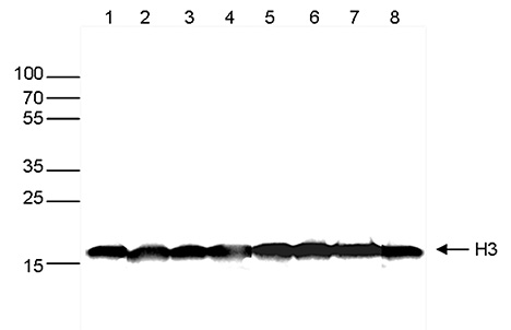
Western blot analysis using H3pan monoclonal antibody
Western blot was performed on whole cell extracts (30 μg) from different cell types (lane 1: HeLa, lane 2: K562, lane 3: MCF7, lane 4: U2OS, lane 5: HepG2, lane 6: Jurkat, lane 7: NIH3T3, lane 8: E14Tg2a mouse ES cells) using the monoclonal antibody against H3. The antibody was diluted 1:1,000 in TBS-Tween containing 5% skimmed milk. The marker (in kDa) is shown on the left,and the position of the protein of interest is indicated on the right.
Why do you need a loading control?
- To verify that equal sample amount was loaded on the gel
- To confirm that the electrotransfer was done equally across the lines
- To check that the signal detection after antibody incubation is homogenous across the lines
Learn more about this antibody
Blue ladder-HRP monoclonal antibody
Visualize your marker directly on film
Most prestained protein MW markers used in western blot contain fragments that are labelled with a blue dye. Diagenode has developed a revolutionary new antibody that specifically reacts with this blue dye, enabling direct visualization of the different marker fragments on the blot. This makes the positioning of the marker and thus the accurate detection of proteins significantly easier.
- Faster and easier protein detection - manual marking is no longer necessary
- Develop your marker directly on X-ray film
- Reduced ladder consumption
- Compatible with most blue-stained ladders
- Outstanding specificity with no cross-reactivity or background
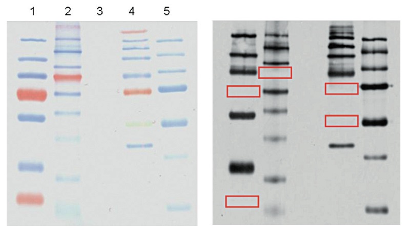
Western blot analysis using the Diagenode Blue ladder - HRP monoclonal antibody
PAGE analysis of 5 different protein MW markers: 1. Page Ruler Plus (Thermo, cat. No. 26619); 2. Color Plus broad range (NEB, cat. No. P7711); 3. Page Ruler (Unstained, Fermentas, cat. No. SM0661); 4. Spectra Multicolor (Fermentas, cat. No. SM1859); 5. Precision Plus (Biorad, cat. No. 161-0373). 4 μl of each marker was loaded on the gel. The right panel shows the Western blot analysis with the Diagenode Blue ladder - HRP antibody (cat. No. C1520202) diluted 1:1,000, using a standard Western blot protocol.
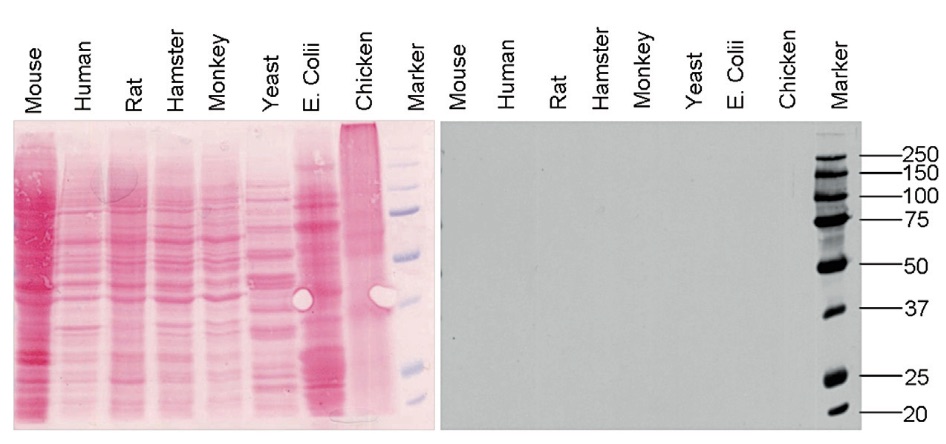
Western blot analysis using the Diagenode Blue ladder - HRP monoclonal antibody
Protein extracts from different species were subjected to SDS-PAGE and analyzed by Western blot with the Diagenode Blue ladder - HRP antibody (Cat. No. C15200202), diluted 1:1,000. The results are shown on the right. The left figure shows a Ponceau staining of the gel. This figure clearly demonstrates that the antibody does not react with any proteins of the species that were tested.
Learn more
New! Blue ladder monoclonal antibody is available
Antibodies validated in WB
Diagenode offers the antibodies selected for highest-quality results in Western blot. The high sensitivity and specificity of our antibodies enable the most accurate results.
TESTIMONIAL
I have been a Diagenode customer for over two years now. I have used the antibodies against the histone modifications H3K4me3, H3K4me2, H3K4me1, H3k27me3, H3K9ac, H3K27ac etc. provided by Diagenode to perform western blots.The high level of specificity of these antibodies in Pristionchus pacificus samples, confirmed by using several negative and positive controls run in parallel with synchronized culture samples, ensured successful and reproducible results.
Also, I am satisfied with the service and the support of personnel of Diagenode. They are prompt in replying to my emails and very helpful with their advice. Thanks a lot!
Vahan Serobyan, Prof. Dr Ralf J. Sommer's Lab, Max Planck Institute for Developmental Biology, Tübingen, Germany
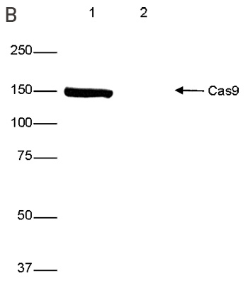
Western blot analysis using the Diagenode antibody directed against CRISPR/Cas9 (Cat. No. C15200229)
Western blot was performed on protein extracts from HeLa cells transfected with Cas9 (lane 1) or from untransfected cells (lane 2) using the Diagenode antibody against CRISPR/Cas9, diluted 1:4,000 in PBS-T containing 3% NFDM. The marker is shown on the left, position of the Cas9 protein is indicated on the right.
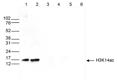
Western blot analysis using the Diagenode antibody directed against H3K14ac (cat. No. C15410310)
Western blot was performed on whole cell (25 μg, lane 1) and histone extracts (15 μg, lane 2) from HeLa cells, and on 1 μg of recombinant histone H2A, H2B, H3 and H4 (lane 3, 4, 5 and 6, respectively) using the Diagenode antibody against H3K14ac. The antibody was diluted 1:1,000 in TBS-Tween containing 5% skimmed milk. The position of the protein of interest is indicated on the right; the marker (in kDa) is shown on the left.
Check out our selection!

