Notice (8): Undefined variable: solution_of_interest [APP/View/Products/view.ctp, line 755]Code Context<!-- BEGIN: REQUEST_FORM MODAL -->
<div id="request_formModal" class="reveal-modal medium" data-reveal aria-labelledby="modalTitle" aria-hidden="true" role="dialog">
<?= $this->element('Forms/simple_form', array('solution_of_interest' => $solution_of_interest, 'header' => $header, 'message' => $message, 'campaign_id' => $campaign_id)) ?>
$viewFile = '/home/website-server/www/app/View/Products/view.ctp'
$dataForView = array(
'language' => 'cn',
'meta_keywords' => '',
'meta_description' => '',
'meta_title' => '',
'product' => array(
'Product' => array(
'id' => '3138',
'antibody_id' => '612',
'name' => '5-methylcytosine (5-mC) Antibody for ICC/IF (sample size)',
'description' => '',
'label1' => '',
'info1' => '',
'label2' => '',
'info2' => '',
'label3' => '',
'info3' => '',
'format' => '10 μg',
'catalog_number' => 'C15200003-10',
'old_catalog_number' => '',
'sf_code' => 'C15200003-D001-000582',
'type' => 'FRE',
'search_order' => '03-Antibody',
'price_EUR' => '105',
'price_USD' => '115',
'price_GBP' => '100',
'price_JPY' => '16450',
'price_CNY' => '',
'price_AUD' => '288',
'country' => 'ALL',
'except_countries' => 'None',
'quote' => false,
'in_stock' => false,
'featured' => false,
'no_promo' => false,
'online' => true,
'master' => false,
'last_datasheet_update' => 'July 30, 2020',
'slug' => '5-mc-monoclonal-antibody-for-icc-if-classic-10',
'meta_title' => '',
'meta_keywords' => '',
'meta_description' => '',
'modified' => '2020-11-19 10:47:27',
'created' => '2020-11-18 16:06:47',
'locale' => 'zho'
),
'Antibody' => array(
'host' => '*****',
'id' => '612',
'name' => '5-methylcytosine (5-mC) Antibody for ICC/IF (sample size)',
'description' => '',
'clonality' => '',
'isotype' => 'IgG1',
'lot' => '003',
'concentration' => '1.3 µg/µl',
'reactivity' => 'Human, mouse, drosophila, other (wide range): positive.',
'type' => 'Monoclonal',
'purity' => 'Protein A purified monoclonal antibody.',
'classification' => '',
'application_table' => '<table>
<thead>
<tr>
<th>Applications</th>
<th>Suggested dilution</th>
<th>References</th>
</tr>
</thead>
<tbody>
<tr>
<td>MeDIP <sup>*</sup></td>
<td>1-2 µg per IP</td>
<td></td>
</tr>
<tr>
<td>Dot Blotting **</td>
<td>1:600</td>
<td>Fig 1</td>
</tr>
<tr>
<td>Immunofluorescence</td>
<td>1:1,000</td>
<td>Fig 2, 3</td>
</tr>
<tr>
<td>FISH</td>
<td>1:200 - 1:1,000</td>
<td>Fig 4, 5</td>
</tr>
</tbody>
</table>
<p><small><sup>*</sup> Please note that the optimal antibody amount per IP should be determined by the end-user. We recommend testing 1-5 µg per IP.</small></p>
<p><small><sup>**</sup> Dot blot was only performed to demonstrate the specificity. This antibody is not recommended for dot blot on biological samples</small></p>',
'storage_conditions' => 'Store at -20°C; for long storage, store at -80°C. Avoid multiple freeze-thaw cycles.',
'storage_buffer' => 'PBS containing 0.05% azide.',
'precautions' => 'This product is for research use only. Not for use in diagnostic or therapeutic procedures.',
'uniprot_acc' => '',
'slug' => '5-mc-monoclonal-antibody-for-icc-if-classic-10',
'meta_keywords' => '',
'meta_description' => '',
'modified' => '2020-11-18 16:01:41',
'created' => '2020-11-18 16:01:41',
'select_label' => '612 - 5-methylcytosine (5-mC) Antibody for ICC/IF (sample size) (003 - 1.3 µg/µl - Human, mouse, drosophila, other (wide range): positive. - Protein A purified monoclonal antibody. - Mouse)'
),
'Slave' => array(),
'Group' => array(
'Group' => array(
[maximum depth reached]
),
'Master' => array(
[maximum depth reached]
),
'Product' => array(
[maximum depth reached]
)
),
'Related' => array(),
'Application' => array(
(int) 0 => array(
[maximum depth reached]
),
(int) 1 => array(
[maximum depth reached]
),
(int) 2 => array(
[maximum depth reached]
)
),
'Category' => array(
(int) 0 => array(
[maximum depth reached]
),
(int) 1 => array(
[maximum depth reached]
),
(int) 2 => array(
[maximum depth reached]
)
),
'Document' => array(
(int) 0 => array(
[maximum depth reached]
),
(int) 1 => array(
[maximum depth reached]
)
),
'Feature' => array(),
'Image' => array(
(int) 0 => array(
[maximum depth reached]
)
),
'Promotion' => array(),
'Protocol' => array(),
'Publication' => array(
(int) 0 => array(
[maximum depth reached]
),
(int) 1 => array(
[maximum depth reached]
),
(int) 2 => array(
[maximum depth reached]
),
(int) 3 => array(
[maximum depth reached]
)
),
'Testimonial' => array(),
'Area' => array(),
'SafetySheet' => array(
(int) 0 => array(
[maximum depth reached]
),
(int) 1 => array(
[maximum depth reached]
),
(int) 2 => array(
[maximum depth reached]
),
(int) 3 => array(
[maximum depth reached]
),
(int) 4 => array(
[maximum depth reached]
),
(int) 5 => array(
[maximum depth reached]
),
(int) 6 => array(
[maximum depth reached]
),
(int) 7 => array(
[maximum depth reached]
)
)
),
'meta_canonical' => 'https://www.diagenode.com/cn/p/5-mc-monoclonal-antibody-for-icc-if-classic-50-mg-21-ml'
)
$language = 'cn'
$meta_keywords = ''
$meta_description = ''
$meta_title = ''
$product = array(
'Product' => array(
'id' => '3138',
'antibody_id' => '612',
'name' => '5-methylcytosine (5-mC) Antibody for ICC/IF (sample size)',
'description' => '<p><span>Monoclonal antibody raised in mouse against 5-mC (5-methylcytosine) conjugated to BSA. </span></p>',
'label1' => 'Validation Data',
'info1' => '<div class="row">
<div class="small-4 columns">
<p><img src="https://www.diagenode.com/img/product/antibodies/C15200003_DotBlot.png" alt="Dot Blot" style="display: block; margin-left: auto; margin-right: auto;" /></p>
</div>
<div class="small-8 columns">
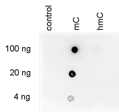
<p><small><strong>Figure 1. Dot blot analysis using the Diagenode monoclonal antibody directed against 5-mC </strong><br />To demonstrate the specificity of the Diagenode antibody against 5-mC (Cat. No. C15200003), a Dot blot analysis was performed using the hmC, mC and C controls from the Diagenode “5-hmC, 5-mC & cytosine DNA Standard Pack” (Cat. No. AF-101-0002). One hundred to 4 ng (equivalent of 5 to 0.2 pmol of C-bases) of the controls were spotted on a membrane (Amersham Hybond-N+). The antibody was used at a dilution of 1:600. Figure 1 shows a high specificity of the antibody for the methylated control. </small></p>
</div>
</div>
<div class="row">
<div class="small-5 columns">
<p><img src="https://www.diagenode.com/img/product/antibodies/C15200003-IF-1.png" alt="Immunofluorescence" style="display: block; margin-left: auto; margin-right: auto;" /></p>
</div>
<div class="small-7 columns">
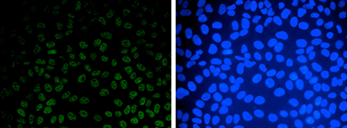
<p><small><strong>Figure 2. Immunofluorescence results obtained with the Diagenode monoclonal antibody directed against 5-mC </strong><br />HeLa cells were stained with the Diagenode antibody against 5-mC (Cat. No. C15200003) and with DAPI. Cells were fixed with 4% formaldehyde for 10’ and blocked with PBS/TX-100 containing 1% BSA. The cells were immunofluorescently labelled with the 5-mC antibody (middle) diluted 1:1,000 in blocking solution followed by an anti-mouse antibody conjugated to Alexa594. The left panel shows staining of the nuclei with DAPI. A merge of the two stainings is shown on the right. </small></p>
</div>
</div>
<div class="row">
<div class="small-4 columns">
<p><small>A.</small><img src="https://www.diagenode.com/img/product/antibodies/C15200003-IF-2A.png" alt="Immunofluorescence" style="display: block; margin-left: auto; margin-right: auto;" /></p>
<p><small>B.</small> <img src="https://www.diagenode.com/img/product/antibodies/C15200003-IF-2B.png" alt="Immunofluorescence" style="display: block; margin-left: auto; margin-right: auto;" /></p>
</div>
<div class="small-8 columns">
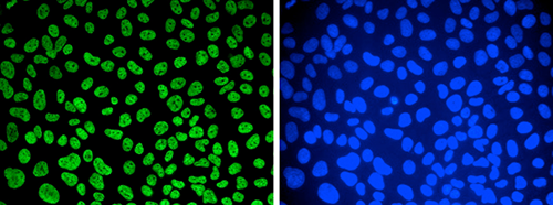
<p><small><strong>Figure 3. Immunofluorescence results obtained with the Diagenode monoclonal antibody directed against 5-mC </strong><br />Human osteosarcoma (U2OS) cells were stained with the Diagenode monoclonal antibody against 5-mC (Cat. No. C15200003). Cells were fixed with 2.5% PFA in PBS for 30’, permeabilised with 0.5% Triton X-100 for 1 hour and treated with 2N HCl for 1 hour followed by 2 x 5 minutes with 0.1 M borate buffer to depurinate the DNA. After blocking with PBS containing 0.1% Triton X-100 and 1% BSA, the cells were immunofluorescently labelled with the 5-mC antibody diluted 1:500 in blocking solution, followed by a goat anti-mouse antibody conjugated to Alexa488. Figure 3A: cells were immunofluorescently labelled with the 5-mC antibody after incubation of the antibody with 50 µM mCTP (left) or with DAPI (right). Figure 3C: staining of the cells with the 5-mC antibody after incubation of the antibody with 50 µM hmCTP and with DAPI.</small></p>
</div>
</div>
<div class="extra-spaced"></div>
<div class="row">
<div class="small-5 columns">
<p><img src="https://www.diagenode.com/img/product/antibodies/C15200003_FISH_2.png" alt="Fish" style="display: block; margin-left: auto; margin-right: auto;" /></p>
</div>
<div class="small-7 columns">
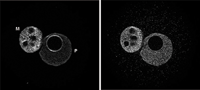
<p><small><strong>Figure 4. FISH using the Diagenode monoclonal antibody directed against 5-mC</strong><br />To detect methylated chromosomal regions, FISH was performed on metaphase chromosomes from HeLa cells using the Diagenode monoclonal antibody against 5-mC (Cat. No. C15200003). The cells were blocked in metaphase by treatment with colcemid (0.1 µg/ml) for 1 - 2 hours, fixed overnight at -20°C with ethanol/glacial acetic acid and treated with 2N HCl for 30’ at room temperature. Subsequently, the cells were blocked with PBS containing 1% BSA and 0.1% Triton X-100 and stained with the 5-mC antibody (left) diluted 1:1,000 in blocking solution, followed by an anti-mouse antibody conjugated to Alexa594. The middle panel shows staining of the chromosomes with DAPI. A merge of the two stainings is shown on the right.</small></p>
</div>
</div>
<div class="row">
<div class="small-5 columns">
<p><img src="https://www.diagenode.com/img/product/antibodies/C15200003_FISH.png" alt="Fish" style="display: block; margin-left: auto; margin-right: auto;" /></p>
</div>
<div class="small-7 columns">
<p><small><strong>Figure 5. FISH using the Diagenode monoclonal antibody directed against 5-mC </strong><br />To detect methylated chromosomal regions, FISH was performed on metaphase chromosomes from HeLa cells using the Diagenode monoclonal antibody against 5-mC (Cat. No. C15200003). The cells were blocked in metaphase by treatment with colcemid (0.1 µg/ml) for 1 - 2 hours, fixed overnight at -20°C with ethanol/glacial acetic acid and treated with 2N HCl for 30’ at room temperature. Subsequently, the cells were blocked with PBS containing 1% BSA and 0.1% Triton X-100 and stained with the 5-mC antibody (left) diluted 1:1,000 in blocking solution, followed by an anti-mouse antibody conjugated to Alexa594. The middle panel shows staining of the chromosomes with DAPI. A merge of the two stainings is shown on the right. </small></p>
</div>
</div>',
'label2' => '',
'info2' => '',
'label3' => '',
'info3' => '',
'format' => '10 μg',
'catalog_number' => 'C15200003-10',
'old_catalog_number' => '',
'sf_code' => 'C15200003-D001-000582',
'type' => 'FRE',
'search_order' => '03-Antibody',
'price_EUR' => '105',
'price_USD' => '115',
'price_GBP' => '100',
'price_JPY' => '16450',
'price_CNY' => '',
'price_AUD' => '288',
'country' => 'ALL',
'except_countries' => 'None',
'quote' => false,
'in_stock' => false,
'featured' => false,
'no_promo' => false,
'online' => true,
'master' => false,
'last_datasheet_update' => 'July 30, 2020',
'slug' => '5-mc-monoclonal-antibody-for-icc-if-classic-10',
'meta_title' => '',
'meta_keywords' => '',
'meta_description' => '',
'modified' => '2020-11-19 10:47:27',
'created' => '2020-11-18 16:06:47',
'locale' => 'zho'
),
'Antibody' => array(
'host' => '*****',
'id' => '612',
'name' => '5-methylcytosine (5-mC) Antibody for ICC/IF (sample size)',
'description' => '',
'clonality' => '',
'isotype' => 'IgG1',
'lot' => '003',
'concentration' => '1.3 µg/µl',
'reactivity' => 'Human, mouse, drosophila, other (wide range): positive.',
'type' => 'Monoclonal',
'purity' => 'Protein A purified monoclonal antibody.',
'classification' => '',
'application_table' => '<table>
<thead>
<tr>
<th>Applications</th>
<th>Suggested dilution</th>
<th>References</th>
</tr>
</thead>
<tbody>
<tr>
<td>MeDIP <sup>*</sup></td>
<td>1-2 µg per IP</td>
<td></td>
</tr>
<tr>
<td>Dot Blotting **</td>
<td>1:600</td>
<td>Fig 1</td>
</tr>
<tr>
<td>Immunofluorescence</td>
<td>1:1,000</td>
<td>Fig 2, 3</td>
</tr>
<tr>
<td>FISH</td>
<td>1:200 - 1:1,000</td>
<td>Fig 4, 5</td>
</tr>
</tbody>
</table>
<p><small><sup>*</sup> Please note that the optimal antibody amount per IP should be determined by the end-user. We recommend testing 1-5 µg per IP.</small></p>
<p><small><sup>**</sup> Dot blot was only performed to demonstrate the specificity. This antibody is not recommended for dot blot on biological samples</small></p>',
'storage_conditions' => 'Store at -20°C; for long storage, store at -80°C. Avoid multiple freeze-thaw cycles.',
'storage_buffer' => 'PBS containing 0.05% azide.',
'precautions' => 'This product is for research use only. Not for use in diagnostic or therapeutic procedures.',
'uniprot_acc' => '',
'slug' => '5-mc-monoclonal-antibody-for-icc-if-classic-10',
'meta_keywords' => '',
'meta_description' => '',
'modified' => '2020-11-18 16:01:41',
'created' => '2020-11-18 16:01:41',
'select_label' => '612 - 5-methylcytosine (5-mC) Antibody for ICC/IF (sample size) (003 - 1.3 µg/µl - Human, mouse, drosophila, other (wide range): positive. - Protein A purified monoclonal antibody. - Mouse)'
),
'Slave' => array(),
'Group' => array(
'Group' => array(
'id' => '317',
'name' => 'C15200003',
'product_id' => '1961',
'modified' => '2020-11-18 16:07:10',
'created' => '2020-11-18 16:07:10'
),
'Master' => array(
'id' => '1961',
'antibody_id' => '30',
'name' => ' 5-methylcytosine (5-mC) Antibody for ICC/IF ',
'description' => '<p><span>Monoclonal antibody raised in mouse against 5-mC (5-methylcytosine) conjugated to BSA. </span></p>',
'label1' => 'Validation Data',
'info1' => '<div class="row">
<div class="small-4 columns">
<p><img src="https://www.diagenode.com/img/product/antibodies/C15200003_DotBlot.png" alt="Dot Blot" style="display: block; margin-left: auto; margin-right: auto;" /></p>
</div>
<div class="small-8 columns">
<p><small><strong>Figure 1. Dot blot analysis using the Diagenode monoclonal antibody directed against 5-mC </strong><br />To demonstrate the specificity of the Diagenode antibody against 5-mC (Cat. No. C15200003), a Dot blot analysis was performed using the hmC, mC and C controls from the Diagenode “5-hmC, 5-mC & cytosine DNA Standard Pack” (Cat. No. AF-101-0002). One hundred to 4 ng (equivalent of 5 to 0.2 pmol of C-bases) of the controls were spotted on a membrane (Amersham Hybond-N+). The antibody was used at a dilution of 1:600. Figure 1 shows a high specificity of the antibody for the methylated control. </small></p>
</div>
</div>
<div class="row">
<div class="small-5 columns">
<p><img src="https://www.diagenode.com/img/product/antibodies/C15200003-IF-1.png" alt="Immunofluorescence" style="display: block; margin-left: auto; margin-right: auto;" /></p>
</div>
<div class="small-7 columns">
<p><small><strong>Figure 2. Immunofluorescence results obtained with the Diagenode monoclonal antibody directed against 5-mC </strong><br />HeLa cells were stained with the Diagenode antibody against 5-mC (Cat. No. C15200003) and with DAPI. Cells were fixed with 4% formaldehyde for 10’ and blocked with PBS/TX-100 containing 1% BSA. The cells were immunofluorescently labelled with the 5-mC antibody (middle) diluted 1:1,000 in blocking solution followed by an anti-mouse antibody conjugated to Alexa594. The left panel shows staining of the nuclei with DAPI. A merge of the two stainings is shown on the right. </small></p>
</div>
</div>
<div class="row">
<div class="small-4 columns">
<p><small>A.</small><img src="https://www.diagenode.com/img/product/antibodies/C15200003-IF-2A.png" alt="Immunofluorescence" style="display: block; margin-left: auto; margin-right: auto;" /></p>
<p><small>B.</small> <img src="https://www.diagenode.com/img/product/antibodies/C15200003-IF-2B.png" alt="Immunofluorescence" style="display: block; margin-left: auto; margin-right: auto;" /></p>
</div>
<div class="small-8 columns">
<p><small><strong>Figure 3. Immunofluorescence results obtained with the Diagenode monoclonal antibody directed against 5-mC </strong><br />Human osteosarcoma (U2OS) cells were stained with the Diagenode monoclonal antibody against 5-mC (Cat. No. C15200003). Cells were fixed with 2.5% PFA in PBS for 30’, permeabilised with 0.5% Triton X-100 for 1 hour and treated with 2N HCl for 1 hour followed by 2 x 5 minutes with 0.1 M borate buffer to depurinate the DNA. After blocking with PBS containing 0.1% Triton X-100 and 1% BSA, the cells were immunofluorescently labelled with the 5-mC antibody diluted 1:500 in blocking solution, followed by a goat anti-mouse antibody conjugated to Alexa488. Figure 3A: cells were immunofluorescently labelled with the 5-mC antibody after incubation of the antibody with 50 µM mCTP (left) or with DAPI (right). Figure 3C: staining of the cells with the 5-mC antibody after incubation of the antibody with 50 µM hmCTP and with DAPI.</small></p>
</div>
</div>
<div class="extra-spaced"></div>
<div class="row">
<div class="small-5 columns">
<p><img src="https://www.diagenode.com/img/product/antibodies/C15200003_FISH_2.png" alt="Fish" style="display: block; margin-left: auto; margin-right: auto;" /></p>
</div>
<div class="small-7 columns">
<p><small><strong>Figure 4. FISH using the Diagenode monoclonal antibody directed against 5-mC</strong><br />To detect methylated chromosomal regions, FISH was performed on metaphase chromosomes from HeLa cells using the Diagenode monoclonal antibody against 5-mC (Cat. No. C15200003). The cells were blocked in metaphase by treatment with colcemid (0.1 µg/ml) for 1 - 2 hours, fixed overnight at -20°C with ethanol/glacial acetic acid and treated with 2N HCl for 30’ at room temperature. Subsequently, the cells were blocked with PBS containing 1% BSA and 0.1% Triton X-100 and stained with the 5-mC antibody (left) diluted 1:1,000 in blocking solution, followed by an anti-mouse antibody conjugated to Alexa594. The middle panel shows staining of the chromosomes with DAPI. A merge of the two stainings is shown on the right.</small></p>
</div>
</div>
<div class="row">
<div class="small-5 columns">
<p><img src="https://www.diagenode.com/img/product/antibodies/C15200003_FISH.png" alt="Fish" style="display: block; margin-left: auto; margin-right: auto;" /></p>
</div>
<div class="small-7 columns">
<p><small><strong>Figure 5. FISH using the Diagenode monoclonal antibody directed against 5-mC </strong><br />To detect methylated chromosomal regions, FISH was performed on metaphase chromosomes from HeLa cells using the Diagenode monoclonal antibody against 5-mC (Cat. No. C15200003). The cells were blocked in metaphase by treatment with colcemid (0.1 µg/ml) for 1 - 2 hours, fixed overnight at -20°C with ethanol/glacial acetic acid and treated with 2N HCl for 30’ at room temperature. Subsequently, the cells were blocked with PBS containing 1% BSA and 0.1% Triton X-100 and stained with the 5-mC antibody (left) diluted 1:1,000 in blocking solution, followed by an anti-mouse antibody conjugated to Alexa594. The middle panel shows staining of the chromosomes with DAPI. A merge of the two stainings is shown on the right. </small></p>
</div>
</div>',
'label2' => '',
'info2' => '',
'label3' => '',
'info3' => '',
'format' => '50 μg/39 μl',
'catalog_number' => 'C15200003',
'old_catalog_number' => '',
'sf_code' => 'C15200003-D001-000581',
'type' => 'FRE',
'search_order' => '03-Antibody',
'price_EUR' => '380',
'price_USD' => '380',
'price_GBP' => '340',
'price_JPY' => '59525',
'price_CNY' => '',
'price_AUD' => '950',
'country' => 'ALL',
'except_countries' => 'None',
'quote' => false,
'in_stock' => false,
'featured' => false,
'no_promo' => false,
'online' => true,
'master' => true,
'last_datasheet_update' => 'July 30, 2020',
'slug' => '5-mc-monoclonal-antibody-for-icc-if-classic-50-mg-21-ml',
'meta_title' => '5-methylcytosine (5-mC) Monoclonal Antibody for ICC/IF | Diagenode',
'meta_keywords' => '5-methylcytosine (5-mC),monoclonal antibody ',
'meta_description' => ' 5-methylcytosine (5-mC) Monoclonal Antibody for ICC/IF validated in MeDIP, DB, IF, and FISH. Batch-specific data available on the website. ',
'modified' => '2022-01-05 15:26:15',
'created' => '2015-06-29 14:08:20'
),
'Product' => array(
(int) 0 => array(
[maximum depth reached]
)
)
),
'Related' => array(),
'Application' => array(
(int) 0 => array(
'id' => '28',
'position' => '10',
'parent_id' => '40',
'name' => 'DB',
'description' => '<p>Dot blotting</p>',
'in_footer' => false,
'in_menu' => false,
'online' => true,
'tabular' => true,
'slug' => 'dot-blotting',
'meta_keywords' => 'Dot blotting,Monoclonal & Polyclonal antibody,',
'meta_description' => 'Diagenode offers Monoclonal & Polyclonal antibodies for Dot blotting applications',
'meta_title' => 'Dot blotting Antibodies - Monoclonal & Polyclonal antibody | Diagenode',
'modified' => '2016-01-13 14:40:49',
'created' => '2015-07-08 13:45:05',
'ProductsApplication' => array(
[maximum depth reached]
)
),
(int) 1 => array(
'id' => '29',
'position' => '10',
'parent_id' => '40',
'name' => 'IF',
'description' => '<p><strong>Immunofluorescence</strong>:</p>
<p>Diagenode offers huge selection of highly sensitive antibodies validated in IF.</p>
<p><img src="https://www.diagenode.com/img/product/antibodies/C15200229-IF.jpg" alt="" height="245" width="256" /></p>
<p><sup><strong>Immunofluorescence using the Diagenode monoclonal antibody directed against CRISPR/Cas9</strong></sup></p>
<p><sup>HeLa cells transfected with a Cas9 expression vector (left) or untransfected cells (right) were fixed in methanol at -20°C, permeabilized with acetone at -20°C and blocked with PBS containing 2% BSA. The cells were stained with the Cas9 C-terminal antibody (Cat. No. C15200229) diluted 1:400, followed by incubation with an anti-mouse secondary antibody coupled to AF488. The bottom images show counter-staining of the nuclei with Hoechst 33342.</sup></p>
<h5><sup>Check our selection of antibodies validated in IF.</sup></h5>',
'in_footer' => false,
'in_menu' => false,
'online' => true,
'tabular' => true,
'slug' => 'immunofluorescence',
'meta_keywords' => 'Immunofluorescence,Monoclonal antibody,Polyclonal antibody',
'meta_description' => 'Diagenode offers a wide range of antibodies and technical support for Immunofluorescence applications',
'meta_title' => 'Immunofluorescence - Monoclonal antibody - Polyclonal antibody | Diagenode',
'modified' => '2016-04-27 16:23:10',
'created' => '2015-07-08 13:46:02',
'ProductsApplication' => array(
[maximum depth reached]
)
),
(int) 2 => array(
'id' => '35',
'position' => '10',
'parent_id' => '40',
'name' => 'FISH',
'description' => '<p>FISH</p>',
'in_footer' => false,
'in_menu' => false,
'online' => true,
'tabular' => true,
'slug' => 'fish-5-mC',
'meta_keywords' => '5-Methylcytosine,5-mC,monoclonal antibody',
'meta_description' => '5-Methylcytosine(5-mC) monoclonal antibody for fish ',
'meta_title' => '5-Methylcytosine(5-mC) monoclonal antibody for fish | Diagenode',
'modified' => '2016-01-13 12:24:55',
'created' => '2015-07-08 13:53:27',
'ProductsApplication' => array(
[maximum depth reached]
)
)
),
'Category' => array(
(int) 0 => array(
'id' => '23',
'position' => '40',
'parent_id' => '4',
'name' => 'DNA modifications',
'description' => '<p><span style="font-weight: 400;">T</span><span style="font-weight: 400;">he pattern of <strong>DNA modifications</strong> is critical for genome stability and the control of gene expression in the cell. Methylation of 5-cytosine (5-mC), one of the best-studied epigenetic marks, is carried out by the <strong>DNA methyltransferases</strong> DNMT3A and B and DNMT1. DNMT3A and DNMT3B are responsible for </span><i><span style="font-weight: 400;">de novo</span></i><span style="font-weight: 400;"> DNA methylation, whereas DNMT1 maintains existing methylation. 5-mC undergoes active demethylation which is performed by the <strong>Ten-Eleven Translocation</strong> (TET) familly of DNA hydroxylases. The latter consists of 3 members TET1, 2 and 3. All 3 members catalyze the conversion of <strong>5-methylcytosine</strong> (5-mC) into <strong>5-hydroxymethylcytosine</strong> (5-hmC), and further into <strong>5-formylcytosine</strong> (5-fC) and <strong>5-carboxycytosine</strong> (5-caC). 5-fC and 5-caC can be converted to unmodified cytosine by <strong>Thymine DNA Glycosylase</strong> (TDG). It is not yet clear if 5-hmC, 5-fC and 5-caC have specific functions or are simply intermediates in the demethylation of 5-mC.</span></p>
<p><span style="font-weight: 400;">DNA methylation is generally considered as a repressive mark and is usually associated with gene silencing. It is essential that the balance between DNA methylation and demethylation is precisely maintained. Dysregulation of DNA methylation may lead to many different human diseases and is often observed in cancer cells.</span></p>
<p><span style="font-weight: 400;">Diagenode offers highly validated antibodies against different proteins involved in DNA modifications as well as against the modified bases allowing the study of all steps and intermediates in the DNA methylation/demethylation pathway:</span></p>
<p><img src="https://www.diagenode.com/img/categories/antibodies/dna-methylation.jpg" height="599" width="816" /></p>
<p><strong>Diagenode exclusively sources the original 5-methylcytosine monoclonal antibody (clone 33D3).</strong></p>
<p>Check out the list below to see all proposed antibodies for DNA modifications.</p>
<p>Diagenode’s highly validated antibodies:</p>
<ul>
<li>Highly sensitive and specific</li>
<li>Cost-effective (requires less antibody per reaction)</li>
<li>Batch-specific data is available on the website</li>
<li>Expert technical support</li>
<li>Sample sizes available</li>
<li>100% satisfaction guarantee</li>
</ul>',
'no_promo' => false,
'in_menu' => true,
'online' => true,
'tabular' => false,
'hide' => true,
'all_format' => false,
'is_antibody' => true,
'slug' => 'dna-methylation-antibody',
'cookies_tag_id' => null,
'meta_keywords' => 'Monoclonal Antibodies,Polyclonal antibodies,DNA methylation,Diagenode',
'meta_description' => 'Diagenode offers Monoclonal and Polyclonal antibodies for DNA Methylation. The pattern of DNA modifications is critical for genome stability and the control of gene expression in the cell. ',
'meta_title' => 'DNA modifications - Monoclonal and Polyclonal Antibodies for DNA Methylation | Diagenode',
'modified' => '2020-08-03 15:25:37',
'created' => '2014-07-31 03:51:53',
'ProductsCategory' => array(
[maximum depth reached]
),
'CookiesTag' => array([maximum depth reached])
),
(int) 1 => array(
'id' => '103',
'position' => '0',
'parent_id' => '4',
'name' => 'All antibodies',
'description' => '<p><span style="font-weight: 400;">All Diagenode’s antibodies are listed below. Please, use our Quick search field to find the antibody of interest by target name, application, purity.</span></p>
<p><span style="font-weight: 400;">Diagenode’s highly validated antibodies:</span></p>
<ul>
<li>Highly sensitive and specific</li>
<li>Cost-effective (requires less antibody per reaction)</li>
<li>Batch-specific data is available on the website</li>
<li>Expert technical support</li>
<li>Sample sizes available</li>
<li>100% satisfaction guarantee</li>
</ul>',
'no_promo' => false,
'in_menu' => true,
'online' => true,
'tabular' => false,
'hide' => true,
'all_format' => false,
'is_antibody' => true,
'slug' => 'all-antibodies',
'cookies_tag_id' => null,
'meta_keywords' => 'Antibodies,Premium Antibodies,Classic,Pioneer',
'meta_description' => 'Diagenode Offers Strict quality standards with Rigorous QC and validated Antibodies. Classified based on level of validation for flexibility of Application. Comprehensive selection of histone and non-histone Antibodies',
'meta_title' => 'Diagenode's selection of Antibodies is exclusively dedicated for Epigenetic Research | Diagenode',
'modified' => '2019-07-03 10:55:44',
'created' => '2015-11-02 14:49:22',
'ProductsCategory' => array(
[maximum depth reached]
),
'CookiesTag' => array([maximum depth reached])
),
(int) 2 => array(
'id' => '102',
'position' => '1',
'parent_id' => '4',
'name' => 'Sample size antibodies',
'description' => '<h1><strong>Validated epigenetics antibodies</strong> – care for a sample?<br /> </h1>
<p>Diagenode has partnered with leading epigenetics experts and numerous epigenetics consortiums to bring to you a validated and comprehensive collection of epigenetic antibodies. As an expert in epigenetics, we are committed to offering highly-specific antibodies validated for ChIP/ChIP-seq and many other applications. All batch-specific validation data is available on our website.<br /><a href="../categories/antibodies">Read about our expertise in antibody production</a>.</p>
<ul>
<li><strong>Focused</strong> - Diagenode's selection of antibodies is exclusively dedicated for epigenetic research. <a title="See the full collection." href="../categories/all-antibodies">See the full collection.</a></li>
<li><strong>Strict quality standards</strong> with rigorous QC and validation</li>
<li><strong>Classified</strong> based on level of validation for flexibility of application</li>
</ul>
<p>Existing sample sizes are listed below. We will soon expand our collection. Are you looking for a sample size of another antibody? Just <a href="mailto:agnieszka.zelisko@diagenode.com?Subject=Sample%20Size%20Request" target="_top">Contact us</a>.</p>',
'no_promo' => false,
'in_menu' => true,
'online' => true,
'tabular' => false,
'hide' => true,
'all_format' => true,
'is_antibody' => true,
'slug' => 'sample-size-antibodies',
'cookies_tag_id' => null,
'meta_keywords' => '5-hmC monoclonal antibody,CRISPR/Cas9 polyclonal antibody ,H3K36me3 polyclonal antibody,diagenode',
'meta_description' => 'Diagenode offers sample volume on selected antibodies for researchers to test, validate and provide confidence and flexibility in choosing from our wide range of antibodies ',
'meta_title' => 'Sample-size Antibodies | Diagenode',
'modified' => '2019-07-03 10:57:05',
'created' => '2015-10-27 12:13:34',
'ProductsCategory' => array(
[maximum depth reached]
),
'CookiesTag' => array([maximum depth reached])
)
),
'Document' => array(
(int) 0 => array(
'id' => '760',
'name' => 'Datasheet 5-mC C15200003',
'description' => '<p><span>Monoclonal antibody raised in mouse against 5-mC (5-methylcytosine) conjugated to BSA.</span></p>',
'image_id' => null,
'type' => 'Datasheet',
'url' => 'files/products/antibodies/Datasheet_5-mC_C15200003.pdf',
'slug' => 'datasheet-5-mc-c15200003',
'meta_keywords' => '',
'meta_description' => '',
'modified' => '2016-07-08 15:40:45',
'created' => '2015-07-07 11:47:45',
'ProductsDocument' => array(
[maximum depth reached]
)
),
(int) 1 => array(
'id' => '38',
'name' => 'Epigenetic Antibodies Brochure',
'description' => '<p>More than in any other immuoprecipitation assays, quality antibodies are critical tools in many epigenetics experiments. Since 10 years, Diagenode has developed the most stringent quality production available on the market for antibodies exclusively focused on epigenetic uses. All our antibodies have been qualified to work in epigenetic applications.</p>',
'image_id' => null,
'type' => 'Brochure',
'url' => 'files/brochures/Epigenetic_Antibodies_Brochure.pdf',
'slug' => 'epigenetic-antibodies-brochure',
'meta_keywords' => '',
'meta_description' => '',
'modified' => '2016-06-15 11:24:06',
'created' => '2015-07-03 16:05:27',
'ProductsDocument' => array(
[maximum depth reached]
)
)
),
'Feature' => array(),
'Image' => array(
(int) 0 => array(
'id' => '250',
'name' => 'product/antibodies/antibody.png',
'alt' => 'Mouse IgG',
'modified' => '2020-11-27 07:00:09',
'created' => '2015-07-17 10:12:18',
'ProductsImage' => array(
[maximum depth reached]
)
)
),
'Promotion' => array(),
'Protocol' => array(),
'Publication' => array(
(int) 0 => array(
'id' => '4382',
'name' => 'Developmental mRNA mC landscape and regulatory innovations of massivemC modification of maternal mRNAs in animals.',
'authors' => 'Liu J. et al.',
'description' => '<p>mC is one of the longest-known RNA modifications, however, its developmental dynamics, functions, and evolution in mRNAs remain largely unknown. Here, we generate quantitative mRNA mC maps at different stages of development in 6 vertebrate and invertebrate species and find convergent and unexpected massive methylation of maternal mRNAs mediated by NSUN2 and NSUN6. Using Drosophila as a model, we reveal that embryos lacking maternal mRNA mC undergo cell cycle delays and fail to timely initiate maternal-to-zygotic transition, implying the functional importance of maternal mRNA mC. From invertebrates to the lineage leading to humans, two waves of mC regulatory innovations are observed: higher animals gain cis-directed NSUN2-mediated mC sites at the 5' end of the mRNAs, accompanied by the emergence of more structured 5'UTR regions; humans gain thousands of trans-directed NSUN6-mediated mC sites enriched in genes regulating the mitotic cell cycle. Collectively, our studies highlight the existence and regulatory innovations of a mechanism of early embryonic development and provide key resources for elucidating the role of mRNA mC in biology and disease.</p>',
'date' => '2022-05-01',
'pmid' => 'https://www.ncbi.nlm.nih.gov/pubmed/35513466',
'doi' => '10.1038/s41467-022-30210-0',
'modified' => '2022-08-04 16:09:02',
'created' => '2022-08-04 14:55:36',
'ProductsPublication' => array(
[maximum depth reached]
)
),
(int) 1 => array(
'id' => '4177',
'name' => 'Detection of genetic variation and base modifications at base-pairresolution on both DNA and RNA.',
'authors' => 'Wang, Zhen et al.',
'description' => '<p>Accurate decoding of nucleic acid variation is critical to understand the complexity and regulation of genome function. Here we use a single-molecule magnetic tweezer (MT) platform to identify sequence variation and map a range of important epigenetic base modifications with high sensitivity, specificity, and precision in the same single molecules of DNA or RNA. We have also developed a highly specific amplification-free CRISPR-Cas enrichment strategy to isolate genomic regions from native DNA. We demonstrate enrichment of DNA from both E. coli and the FMR1 5'UTR coming from cells derived from a Fragile X carrier. From these kilobase-length enriched molecules we could characterize the differential levels of adenine and cytosine base modifications on E. coli, and the repeat expansion length and methylation status of FMR1. Together these results demonstrate that our platform can detect a variety of genetic, epigenetic, and base modification changes concomitantly within the same single molecules.</p>',
'date' => '2021-01-01',
'pmid' => 'https://www.ncbi.nlm.nih.gov/pubmed/33514840',
'doi' => '10.1038/s42003-021-01648-7',
'modified' => '2021-12-21 16:19:46',
'created' => '2021-12-06 15:53:19',
'ProductsPublication' => array(
[maximum depth reached]
)
),
(int) 2 => array(
'id' => '2811',
'name' => 'Transcriptome-wide distribution and function of RNA hydroxymethylcytosine',
'authors' => 'Delatte B, Wang F, Ngoc LV, Collignon E, Bonvin E, Deplus R, Calonne E, Hassabi B, Putmans P, Awe S, Wetzel C, Kreher J, Soin R, Creppe C, Limbach PA, Gueydan C, Kruys V, Brehm A, Minakhina S, Defrance M, Steward R, Fuks F.',
'description' => '<p>Hydroxymethylcytosine, well described in DNA, occurs also in RNA. Here, we show that hydroxymethylcytosine preferentially marks polyadenylated RNAs and is deposited by Tet in Drosophila. We map the transcriptome-wide hydroxymethylation landscape, revealing hydroxymethylcytosine in the transcripts of many genes, notably in coding sequences, and identify consensus sites for hydroxymethylation. We found that RNA hydroxymethylation can favor mRNA translation. Tet and hydroxymethylated RNA are found to be most abundant in the Drosophila brain, and Tet-deficient fruitflies suffer impaired brain development, accompanied by decreased RNA hydroxymethylation. This study highlights the distribution, localization, and function of cytosine hydroxymethylation and identifies central roles for this modification in Drosophila.</p>',
'date' => '2016-01-15',
'pmid' => 'http://pubmed.gov/26816380',
'doi' => '10.1126/science.aac5253',
'modified' => '2016-01-28 21:57:55',
'created' => '2016-01-28 21:57:55',
'ProductsPublication' => array(
[maximum depth reached]
)
),
(int) 3 => array(
'id' => '3750',
'name' => 'RNA biochemistry. Transcriptome-wide distribution and function of RNA hydroxymethylcytosine.',
'authors' => 'Delatte B, Wang F, Ngoc LV, Collignon E, Bonvin E, Deplus R, Calonne E, Hassabi B, Putmans P, Awe S, Wetzel C, Kreher J, Soin R, Creppe C, Limbach PA, Gueydan C, Kruys V, Brehm A, Minakhina S, Defrance M, Steward R, Fuks F',
'description' => '<p>Hydroxymethylcytosine, well described in DNA, occurs also in RNA. Here, we show that hydroxymethylcytosine preferentially marks polyadenylated RNAs and is deposited by Tet in Drosophila. We map the transcriptome-wide hydroxymethylation landscape, revealing hydroxymethylcytosine in the transcripts of many genes, notably in coding sequences, and identify consensus sites for hydroxymethylation. We found that RNA hydroxymethylation can favor mRNA translation. Tet and hydroxymethylated RNA are found to be most abundant in the Drosophila brain, and Tet-deficient fruitflies suffer impaired brain development, accompanied by decreased RNA hydroxymethylation. This study highlights the distribution, localization, and function of cytosine hydroxymethylation and identifies central roles for this modification in Drosophila.</p>',
'date' => '2016-01-15',
'pmid' => 'http://www.pubmed.org/26816380',
'doi' => '10.1126/science.aac5253',
'modified' => '2019-10-03 12:29:05',
'created' => '2019-10-02 16:16:55',
'ProductsPublication' => array(
[maximum depth reached]
)
)
),
'Testimonial' => array(),
'Area' => array(),
'SafetySheet' => array(
(int) 0 => array(
'id' => '1192',
'name' => '5-mC antibody for ICC/IF SDS US en',
'language' => 'en',
'url' => 'files/SDS/5-mC/SDS-C15200003-5-methylcytosine_5-mC_Antibody_for_ICC_IF-US-en-GHS_2_0.pdf',
'countries' => 'US',
'modified' => '2021-08-24 16:32:39',
'created' => '2021-08-24 16:32:39',
'ProductsSafetySheet' => array(
[maximum depth reached]
)
),
(int) 1 => array(
'id' => '1190',
'name' => '5-mC antibody for ICC/IF SDS GB en',
'language' => 'en',
'url' => 'files/SDS/5-mC/SDS-C15200003-5-methylcytosine_5-mC_Antibody_for_ICC_IF-GB-en-GHS_2_0.pdf',
'countries' => 'GB',
'modified' => '2021-08-24 16:32:39',
'created' => '2021-08-24 16:32:39',
'ProductsSafetySheet' => array(
[maximum depth reached]
)
),
(int) 2 => array(
'id' => '1185',
'name' => '5-mC antibody for ICC/IF SDS BE fr',
'language' => 'fr',
'url' => 'files/SDS/5-mC/SDS-C15200003-5-methylcytosine_5-mC_Antibody_for_ICC_IF-BE-fr-GHS_2_0.pdf',
'countries' => 'BE',
'modified' => '2021-08-24 16:32:39',
'created' => '2021-08-24 16:32:39',
'ProductsSafetySheet' => array(
[maximum depth reached]
)
),
(int) 3 => array(
'id' => '1189',
'name' => '5-mC antibody for ICC/IF SDS FR fr',
'language' => 'fr',
'url' => 'files/SDS/5-mC/SDS-C15200003-5-methylcytosine_5-mC_Antibody_for_ICC_IF-FR-fr-GHS_2_0.pdf',
'countries' => 'FR',
'modified' => '2021-08-24 16:32:39',
'created' => '2021-08-24 16:32:39',
'ProductsSafetySheet' => array(
[maximum depth reached]
)
),
(int) 4 => array(
'id' => '1188',
'name' => '5-mC antibody for ICC/IF SDS ES es',
'language' => 'es',
'url' => 'files/SDS/5-mC/SDS-C15200003-5-methylcytosine_5-mC_Antibody_for_ICC_IF-ES-es-GHS_2_0.pdf',
'countries' => 'ES',
'modified' => '2021-08-24 16:32:39',
'created' => '2021-08-24 16:32:39',
'ProductsSafetySheet' => array(
[maximum depth reached]
)
),
(int) 5 => array(
'id' => '1187',
'name' => '5-mC antibody for ICC/IF SDS DE de',
'language' => 'de',
'url' => 'files/SDS/5-mC/SDS-C15200003-5-methylcytosine_5-mC_Antibody_for_ICC_IF-DE-de-GHS_2_0.pdf',
'countries' => 'DE',
'modified' => '2021-08-24 16:32:39',
'created' => '2021-08-24 16:32:39',
'ProductsSafetySheet' => array(
[maximum depth reached]
)
),
(int) 6 => array(
'id' => '1191',
'name' => '5-mC antibody for ICC/IF SDS JP ja',
'language' => 'ja',
'url' => 'files/SDS/5-mC/SDS-C15200003-5-methylcytosine_5-mC_Antibody_for_ICC_IF-JP-ja-GHS_2_0.pdf',
'countries' => 'JP',
'modified' => '2021-08-24 16:32:39',
'created' => '2021-08-24 16:32:39',
'ProductsSafetySheet' => array(
[maximum depth reached]
)
),
(int) 7 => array(
'id' => '1186',
'name' => '5-mC antibody for ICC/IF SDS BE nl',
'language' => 'nl',
'url' => 'files/SDS/5-mC/SDS-C15200003-5-methylcytosine_5-mC_Antibody_for_ICC_IF-BE-nl-GHS_2_0.pdf',
'countries' => 'BE',
'modified' => '2021-08-24 16:32:39',
'created' => '2021-08-24 16:32:39',
'ProductsSafetySheet' => array(
[maximum depth reached]
)
)
)
)
$meta_canonical = 'https://www.diagenode.com/cn/p/5-mc-monoclonal-antibody-for-icc-if-classic-50-mg-21-ml'
$country = 'US'
$countries_allowed = array(
(int) 0 => 'CA',
(int) 1 => 'US',
(int) 2 => 'IE',
(int) 3 => 'GB',
(int) 4 => 'DK',
(int) 5 => 'NO',
(int) 6 => 'SE',
(int) 7 => 'FI',
(int) 8 => 'NL',
(int) 9 => 'BE',
(int) 10 => 'LU',
(int) 11 => 'FR',
(int) 12 => 'DE',
(int) 13 => 'CH',
(int) 14 => 'AT',
(int) 15 => 'ES',
(int) 16 => 'IT',
(int) 17 => 'PT'
)
$outsource = true
$other_formats = array(
(int) 0 => array(
'id' => '1961',
'antibody_id' => '30',
'name' => ' 5-methylcytosine (5-mC) Antibody for ICC/IF ',
'description' => '<p><span>Monoclonal antibody raised in mouse against 5-mC (5-methylcytosine) conjugated to BSA. </span></p>',
'label1' => 'Validation Data',
'info1' => '<div class="row">
<div class="small-4 columns">
<p><img src="https://www.diagenode.com/img/product/antibodies/C15200003_DotBlot.png" alt="Dot Blot" style="display: block; margin-left: auto; margin-right: auto;" /></p>
</div>
<div class="small-8 columns">
<p><small><strong>Figure 1. Dot blot analysis using the Diagenode monoclonal antibody directed against 5-mC </strong><br />To demonstrate the specificity of the Diagenode antibody against 5-mC (Cat. No. C15200003), a Dot blot analysis was performed using the hmC, mC and C controls from the Diagenode “5-hmC, 5-mC & cytosine DNA Standard Pack” (Cat. No. AF-101-0002). One hundred to 4 ng (equivalent of 5 to 0.2 pmol of C-bases) of the controls were spotted on a membrane (Amersham Hybond-N+). The antibody was used at a dilution of 1:600. Figure 1 shows a high specificity of the antibody for the methylated control. </small></p>
</div>
</div>
<div class="row">
<div class="small-5 columns">
<p><img src="https://www.diagenode.com/img/product/antibodies/C15200003-IF-1.png" alt="Immunofluorescence" style="display: block; margin-left: auto; margin-right: auto;" /></p>
</div>
<div class="small-7 columns">
<p><small><strong>Figure 2. Immunofluorescence results obtained with the Diagenode monoclonal antibody directed against 5-mC </strong><br />HeLa cells were stained with the Diagenode antibody against 5-mC (Cat. No. C15200003) and with DAPI. Cells were fixed with 4% formaldehyde for 10’ and blocked with PBS/TX-100 containing 1% BSA. The cells were immunofluorescently labelled with the 5-mC antibody (middle) diluted 1:1,000 in blocking solution followed by an anti-mouse antibody conjugated to Alexa594. The left panel shows staining of the nuclei with DAPI. A merge of the two stainings is shown on the right. </small></p>
</div>
</div>
<div class="row">
<div class="small-4 columns">
<p><small>A.</small><img src="https://www.diagenode.com/img/product/antibodies/C15200003-IF-2A.png" alt="Immunofluorescence" style="display: block; margin-left: auto; margin-right: auto;" /></p>
<p><small>B.</small> <img src="https://www.diagenode.com/img/product/antibodies/C15200003-IF-2B.png" alt="Immunofluorescence" style="display: block; margin-left: auto; margin-right: auto;" /></p>
</div>
<div class="small-8 columns">
<p><small><strong>Figure 3. Immunofluorescence results obtained with the Diagenode monoclonal antibody directed against 5-mC </strong><br />Human osteosarcoma (U2OS) cells were stained with the Diagenode monoclonal antibody against 5-mC (Cat. No. C15200003). Cells were fixed with 2.5% PFA in PBS for 30’, permeabilised with 0.5% Triton X-100 for 1 hour and treated with 2N HCl for 1 hour followed by 2 x 5 minutes with 0.1 M borate buffer to depurinate the DNA. After blocking with PBS containing 0.1% Triton X-100 and 1% BSA, the cells were immunofluorescently labelled with the 5-mC antibody diluted 1:500 in blocking solution, followed by a goat anti-mouse antibody conjugated to Alexa488. Figure 3A: cells were immunofluorescently labelled with the 5-mC antibody after incubation of the antibody with 50 µM mCTP (left) or with DAPI (right). Figure 3C: staining of the cells with the 5-mC antibody after incubation of the antibody with 50 µM hmCTP and with DAPI.</small></p>
</div>
</div>
<div class="extra-spaced"></div>
<div class="row">
<div class="small-5 columns">
<p><img src="https://www.diagenode.com/img/product/antibodies/C15200003_FISH_2.png" alt="Fish" style="display: block; margin-left: auto; margin-right: auto;" /></p>
</div>
<div class="small-7 columns">
<p><small><strong>Figure 4. FISH using the Diagenode monoclonal antibody directed against 5-mC</strong><br />To detect methylated chromosomal regions, FISH was performed on metaphase chromosomes from HeLa cells using the Diagenode monoclonal antibody against 5-mC (Cat. No. C15200003). The cells were blocked in metaphase by treatment with colcemid (0.1 µg/ml) for 1 - 2 hours, fixed overnight at -20°C with ethanol/glacial acetic acid and treated with 2N HCl for 30’ at room temperature. Subsequently, the cells were blocked with PBS containing 1% BSA and 0.1% Triton X-100 and stained with the 5-mC antibody (left) diluted 1:1,000 in blocking solution, followed by an anti-mouse antibody conjugated to Alexa594. The middle panel shows staining of the chromosomes with DAPI. A merge of the two stainings is shown on the right.</small></p>
</div>
</div>
<div class="row">
<div class="small-5 columns">
<p><img src="https://www.diagenode.com/img/product/antibodies/C15200003_FISH.png" alt="Fish" style="display: block; margin-left: auto; margin-right: auto;" /></p>
</div>
<div class="small-7 columns">
<p><small><strong>Figure 5. FISH using the Diagenode monoclonal antibody directed against 5-mC </strong><br />To detect methylated chromosomal regions, FISH was performed on metaphase chromosomes from HeLa cells using the Diagenode monoclonal antibody against 5-mC (Cat. No. C15200003). The cells were blocked in metaphase by treatment with colcemid (0.1 µg/ml) for 1 - 2 hours, fixed overnight at -20°C with ethanol/glacial acetic acid and treated with 2N HCl for 30’ at room temperature. Subsequently, the cells were blocked with PBS containing 1% BSA and 0.1% Triton X-100 and stained with the 5-mC antibody (left) diluted 1:1,000 in blocking solution, followed by an anti-mouse antibody conjugated to Alexa594. The middle panel shows staining of the chromosomes with DAPI. A merge of the two stainings is shown on the right. </small></p>
</div>
</div>',
'label2' => '',
'info2' => '',
'label3' => '',
'info3' => '',
'format' => '50 μg/39 μl',
'catalog_number' => 'C15200003',
'old_catalog_number' => '',
'sf_code' => 'C15200003-D001-000581',
'type' => 'FRE',
'search_order' => '03-Antibody',
'price_EUR' => '380',
'price_USD' => '380',
'price_GBP' => '340',
'price_JPY' => '59525',
'price_CNY' => '',
'price_AUD' => '950',
'country' => 'ALL',
'except_countries' => 'None',
'quote' => false,
'in_stock' => false,
'featured' => false,
'no_promo' => false,
'online' => true,
'master' => true,
'last_datasheet_update' => 'July 30, 2020',
'slug' => '5-mc-monoclonal-antibody-for-icc-if-classic-50-mg-21-ml',
'meta_title' => '5-methylcytosine (5-mC) Monoclonal Antibody for ICC/IF | Diagenode',
'meta_keywords' => '5-methylcytosine (5-mC),monoclonal antibody ',
'meta_description' => ' 5-methylcytosine (5-mC) Monoclonal Antibody for ICC/IF validated in MeDIP, DB, IF, and FISH. Batch-specific data available on the website. ',
'modified' => '2022-01-05 15:26:15',
'created' => '2015-06-29 14:08:20'
)
)
$pro = array(
'id' => '3138',
'antibody_id' => '612',
'name' => '5-methylcytosine (5-mC) Antibody for ICC/IF (sample size)',
'description' => '',
'label1' => '',
'info1' => '',
'label2' => '',
'info2' => '',
'label3' => '',
'info3' => '',
'format' => '10 μg',
'catalog_number' => 'C15200003-10',
'old_catalog_number' => '',
'sf_code' => 'C15200003-D001-000582',
'type' => 'FRE',
'search_order' => '03-Antibody',
'price_EUR' => '105',
'price_USD' => '115',
'price_GBP' => '100',
'price_JPY' => '16450',
'price_CNY' => '',
'price_AUD' => '288',
'country' => 'ALL',
'except_countries' => 'None',
'quote' => false,
'in_stock' => false,
'featured' => false,
'no_promo' => false,
'online' => true,
'master' => false,
'last_datasheet_update' => 'July 30, 2020',
'slug' => '5-mc-monoclonal-antibody-for-icc-if-classic-10',
'meta_title' => '',
'meta_keywords' => '',
'meta_description' => '',
'modified' => '2020-11-19 10:47:27',
'created' => '2020-11-18 16:06:47',
'ProductsGroup' => array(
'id' => '357',
'product_id' => '3138',
'group_id' => '317'
)
)
$edit = ''
$testimonials = ''
$featured_testimonials = ''
$related_products = ''
$rrbs_service = array(
(int) 0 => (int) 1894,
(int) 1 => (int) 1895
)
$chipseq_service = array(
(int) 0 => (int) 2683,
(int) 1 => (int) 1835,
(int) 2 => (int) 1836,
(int) 3 => (int) 2684,
(int) 4 => (int) 1838,
(int) 5 => (int) 1839,
(int) 6 => (int) 1856
)
$labelize = object(Closure) {
}
$old_catalog_number = ''
$country_code = 'US'
$other_format = array(
'id' => '1961',
'antibody_id' => '30',
'name' => ' 5-methylcytosine (5-mC) Antibody for ICC/IF ',
'description' => '<p><span>Monoclonal antibody raised in mouse against 5-mC (5-methylcytosine) conjugated to BSA. </span></p>',
'label1' => 'Validation Data',
'info1' => '<div class="row">
<div class="small-4 columns">
<p><img src="https://www.diagenode.com/img/product/antibodies/C15200003_DotBlot.png" alt="Dot Blot" style="display: block; margin-left: auto; margin-right: auto;" /></p>
</div>
<div class="small-8 columns">
<p><small><strong>Figure 1. Dot blot analysis using the Diagenode monoclonal antibody directed against 5-mC </strong><br />To demonstrate the specificity of the Diagenode antibody against 5-mC (Cat. No. C15200003), a Dot blot analysis was performed using the hmC, mC and C controls from the Diagenode “5-hmC, 5-mC & cytosine DNA Standard Pack” (Cat. No. AF-101-0002). One hundred to 4 ng (equivalent of 5 to 0.2 pmol of C-bases) of the controls were spotted on a membrane (Amersham Hybond-N+). The antibody was used at a dilution of 1:600. Figure 1 shows a high specificity of the antibody for the methylated control. </small></p>
</div>
</div>
<div class="row">
<div class="small-5 columns">
<p><img src="https://www.diagenode.com/img/product/antibodies/C15200003-IF-1.png" alt="Immunofluorescence" style="display: block; margin-left: auto; margin-right: auto;" /></p>
</div>
<div class="small-7 columns">
<p><small><strong>Figure 2. Immunofluorescence results obtained with the Diagenode monoclonal antibody directed against 5-mC </strong><br />HeLa cells were stained with the Diagenode antibody against 5-mC (Cat. No. C15200003) and with DAPI. Cells were fixed with 4% formaldehyde for 10’ and blocked with PBS/TX-100 containing 1% BSA. The cells were immunofluorescently labelled with the 5-mC antibody (middle) diluted 1:1,000 in blocking solution followed by an anti-mouse antibody conjugated to Alexa594. The left panel shows staining of the nuclei with DAPI. A merge of the two stainings is shown on the right. </small></p>
</div>
</div>
<div class="row">
<div class="small-4 columns">
<p><small>A.</small><img src="https://www.diagenode.com/img/product/antibodies/C15200003-IF-2A.png" alt="Immunofluorescence" style="display: block; margin-left: auto; margin-right: auto;" /></p>
<p><small>B.</small> <img src="https://www.diagenode.com/img/product/antibodies/C15200003-IF-2B.png" alt="Immunofluorescence" style="display: block; margin-left: auto; margin-right: auto;" /></p>
</div>
<div class="small-8 columns">
<p><small><strong>Figure 3. Immunofluorescence results obtained with the Diagenode monoclonal antibody directed against 5-mC </strong><br />Human osteosarcoma (U2OS) cells were stained with the Diagenode monoclonal antibody against 5-mC (Cat. No. C15200003). Cells were fixed with 2.5% PFA in PBS for 30’, permeabilised with 0.5% Triton X-100 for 1 hour and treated with 2N HCl for 1 hour followed by 2 x 5 minutes with 0.1 M borate buffer to depurinate the DNA. After blocking with PBS containing 0.1% Triton X-100 and 1% BSA, the cells were immunofluorescently labelled with the 5-mC antibody diluted 1:500 in blocking solution, followed by a goat anti-mouse antibody conjugated to Alexa488. Figure 3A: cells were immunofluorescently labelled with the 5-mC antibody after incubation of the antibody with 50 µM mCTP (left) or with DAPI (right). Figure 3C: staining of the cells with the 5-mC antibody after incubation of the antibody with 50 µM hmCTP and with DAPI.</small></p>
</div>
</div>
<div class="extra-spaced"></div>
<div class="row">
<div class="small-5 columns">
<p><img src="https://www.diagenode.com/img/product/antibodies/C15200003_FISH_2.png" alt="Fish" style="display: block; margin-left: auto; margin-right: auto;" /></p>
</div>
<div class="small-7 columns">
<p><small><strong>Figure 4. FISH using the Diagenode monoclonal antibody directed against 5-mC</strong><br />To detect methylated chromosomal regions, FISH was performed on metaphase chromosomes from HeLa cells using the Diagenode monoclonal antibody against 5-mC (Cat. No. C15200003). The cells were blocked in metaphase by treatment with colcemid (0.1 µg/ml) for 1 - 2 hours, fixed overnight at -20°C with ethanol/glacial acetic acid and treated with 2N HCl for 30’ at room temperature. Subsequently, the cells were blocked with PBS containing 1% BSA and 0.1% Triton X-100 and stained with the 5-mC antibody (left) diluted 1:1,000 in blocking solution, followed by an anti-mouse antibody conjugated to Alexa594. The middle panel shows staining of the chromosomes with DAPI. A merge of the two stainings is shown on the right.</small></p>
</div>
</div>
<div class="row">
<div class="small-5 columns">
<p><img src="https://www.diagenode.com/img/product/antibodies/C15200003_FISH.png" alt="Fish" style="display: block; margin-left: auto; margin-right: auto;" /></p>
</div>
<div class="small-7 columns">
<p><small><strong>Figure 5. FISH using the Diagenode monoclonal antibody directed against 5-mC </strong><br />To detect methylated chromosomal regions, FISH was performed on metaphase chromosomes from HeLa cells using the Diagenode monoclonal antibody against 5-mC (Cat. No. C15200003). The cells were blocked in metaphase by treatment with colcemid (0.1 µg/ml) for 1 - 2 hours, fixed overnight at -20°C with ethanol/glacial acetic acid and treated with 2N HCl for 30’ at room temperature. Subsequently, the cells were blocked with PBS containing 1% BSA and 0.1% Triton X-100 and stained with the 5-mC antibody (left) diluted 1:1,000 in blocking solution, followed by an anti-mouse antibody conjugated to Alexa594. The middle panel shows staining of the chromosomes with DAPI. A merge of the two stainings is shown on the right. </small></p>
</div>
</div>',
'label2' => '',
'info2' => '',
'label3' => '',
'info3' => '',
'format' => '50 μg/39 μl',
'catalog_number' => 'C15200003',
'old_catalog_number' => '',
'sf_code' => 'C15200003-D001-000581',
'type' => 'FRE',
'search_order' => '03-Antibody',
'price_EUR' => '380',
'price_USD' => '380',
'price_GBP' => '340',
'price_JPY' => '59525',
'price_CNY' => '',
'price_AUD' => '950',
'country' => 'ALL',
'except_countries' => 'None',
'quote' => false,
'in_stock' => false,
'featured' => false,
'no_promo' => false,
'online' => true,
'master' => true,
'last_datasheet_update' => 'July 30, 2020',
'slug' => '5-mc-monoclonal-antibody-for-icc-if-classic-50-mg-21-ml',
'meta_title' => '5-methylcytosine (5-mC) Monoclonal Antibody for ICC/IF | Diagenode',
'meta_keywords' => '5-methylcytosine (5-mC),monoclonal antibody ',
'meta_description' => ' 5-methylcytosine (5-mC) Monoclonal Antibody for ICC/IF validated in MeDIP, DB, IF, and FISH. Batch-specific data available on the website. ',
'modified' => '2022-01-05 15:26:15',
'created' => '2015-06-29 14:08:20'
)
$img = 'banners/banner-cut_tag-chipmentation-500.jpg'
$label = '<img src="/img/banners/banner-customizer-back.png" alt=""/>'
$application = array(
'id' => '35',
'position' => '10',
'parent_id' => '40',
'name' => 'FISH',
'description' => '<p>FISH</p>',
'in_footer' => false,
'in_menu' => false,
'online' => true,
'tabular' => true,
'slug' => 'fish-5-mC',
'meta_keywords' => '5-Methylcytosine,5-mC,monoclonal antibody',
'meta_description' => '5-Methylcytosine(5-mC) monoclonal antibody for fish ',
'meta_title' => '5-Methylcytosine(5-mC) monoclonal antibody for fish | Diagenode',
'modified' => '2016-01-13 12:24:55',
'created' => '2015-07-08 13:53:27',
'ProductsApplication' => array(
'id' => '5313',
'product_id' => '3138',
'application_id' => '35'
)
)
$slugs = array(
(int) 0 => 'fish-5-mC'
)
$applications = array(
'id' => '35',
'position' => '10',
'parent_id' => '40',
'name' => 'FISH',
'description' => '<p>FISH</p>',
'in_footer' => false,
'in_menu' => false,
'online' => true,
'tabular' => true,
'slug' => 'fish-5-mC',
'meta_keywords' => '5-Methylcytosine,5-mC,monoclonal antibody',
'meta_description' => '5-Methylcytosine(5-mC) monoclonal antibody for fish ',
'meta_title' => '5-Methylcytosine(5-mC) monoclonal antibody for fish | Diagenode',
'modified' => '2016-01-13 12:24:55',
'created' => '2015-07-08 13:53:27',
'locale' => 'zho'
)
$description = '<p>FISH</p>'
$name = 'FISH'
$document = array(
'id' => '38',
'name' => 'Epigenetic Antibodies Brochure',
'description' => '<p>More than in any other immuoprecipitation assays, quality antibodies are critical tools in many epigenetics experiments. Since 10 years, Diagenode has developed the most stringent quality production available on the market for antibodies exclusively focused on epigenetic uses. All our antibodies have been qualified to work in epigenetic applications.</p>',
'image_id' => null,
'type' => 'Brochure',
'url' => 'files/brochures/Epigenetic_Antibodies_Brochure.pdf',
'slug' => 'epigenetic-antibodies-brochure',
'meta_keywords' => '',
'meta_description' => '',
'modified' => '2016-06-15 11:24:06',
'created' => '2015-07-03 16:05:27',
'ProductsDocument' => array(
'id' => '3002',
'product_id' => '3138',
'document_id' => '38'
)
)
$sds = array(
'id' => '1186',
'name' => '5-mC antibody for ICC/IF SDS BE nl',
'language' => 'nl',
'url' => 'files/SDS/5-mC/SDS-C15200003-5-methylcytosine_5-mC_Antibody_for_ICC_IF-BE-nl-GHS_2_0.pdf',
'countries' => 'BE',
'modified' => '2021-08-24 16:32:39',
'created' => '2021-08-24 16:32:39',
'ProductsSafetySheet' => array(
'id' => '2073',
'product_id' => '3138',
'safety_sheet_id' => '1186'
)
)
$publication = array(
'id' => '3750',
'name' => 'RNA biochemistry. Transcriptome-wide distribution and function of RNA hydroxymethylcytosine.',
'authors' => 'Delatte B, Wang F, Ngoc LV, Collignon E, Bonvin E, Deplus R, Calonne E, Hassabi B, Putmans P, Awe S, Wetzel C, Kreher J, Soin R, Creppe C, Limbach PA, Gueydan C, Kruys V, Brehm A, Minakhina S, Defrance M, Steward R, Fuks F',
'description' => '<p>Hydroxymethylcytosine, well described in DNA, occurs also in RNA. Here, we show that hydroxymethylcytosine preferentially marks polyadenylated RNAs and is deposited by Tet in Drosophila. We map the transcriptome-wide hydroxymethylation landscape, revealing hydroxymethylcytosine in the transcripts of many genes, notably in coding sequences, and identify consensus sites for hydroxymethylation. We found that RNA hydroxymethylation can favor mRNA translation. Tet and hydroxymethylated RNA are found to be most abundant in the Drosophila brain, and Tet-deficient fruitflies suffer impaired brain development, accompanied by decreased RNA hydroxymethylation. This study highlights the distribution, localization, and function of cytosine hydroxymethylation and identifies central roles for this modification in Drosophila.</p>',
'date' => '2016-01-15',
'pmid' => 'http://www.pubmed.org/26816380',
'doi' => '10.1126/science.aac5253',
'modified' => '2019-10-03 12:29:05',
'created' => '2019-10-02 16:16:55',
'ProductsPublication' => array(
'id' => '4829',
'product_id' => '3138',
'publication_id' => '3750'
)
)
$externalLink = ' <a href="http://www.pubmed.org/26816380" target="_blank"><i class="fa fa-external-link"></i></a>'
include - APP/View/Products/view.ctp, line 755
View::_evaluate() - CORE/Cake/View/View.php, line 971
View::_render() - CORE/Cake/View/View.php, line 933
View::render() - CORE/Cake/View/View.php, line 473
Controller::render() - CORE/Cake/Controller/Controller.php, line 963
ProductsController::slug() - APP/Controller/ProductsController.php, line 1052
ReflectionMethod::invokeArgs() - [internal], line ??
Controller::invokeAction() - CORE/Cake/Controller/Controller.php, line 491
Dispatcher::_invoke() - CORE/Cake/Routing/Dispatcher.php, line 193
Dispatcher::dispatch() - CORE/Cake/Routing/Dispatcher.php, line 167
[main] - APP/webroot/index.php, line 118
Notice (8): Undefined variable: header [APP/View/Products/view.ctp, line 755]Code Context<!-- BEGIN: REQUEST_FORM MODAL -->
<div id="request_formModal" class="reveal-modal medium" data-reveal aria-labelledby="modalTitle" aria-hidden="true" role="dialog">
<?= $this->element('Forms/simple_form', array('solution_of_interest' => $solution_of_interest, 'header' => $header, 'message' => $message, 'campaign_id' => $campaign_id)) ?>
$viewFile = '/home/website-server/www/app/View/Products/view.ctp'
$dataForView = array(
'language' => 'cn',
'meta_keywords' => '',
'meta_description' => '',
'meta_title' => '',
'product' => array(
'Product' => array(
'id' => '3138',
'antibody_id' => '612',
'name' => '5-methylcytosine (5-mC) Antibody for ICC/IF (sample size)',
'description' => '',
'label1' => '',
'info1' => '',
'label2' => '',
'info2' => '',
'label3' => '',
'info3' => '',
'format' => '10 μg',
'catalog_number' => 'C15200003-10',
'old_catalog_number' => '',
'sf_code' => 'C15200003-D001-000582',
'type' => 'FRE',
'search_order' => '03-Antibody',
'price_EUR' => '105',
'price_USD' => '115',
'price_GBP' => '100',
'price_JPY' => '16450',
'price_CNY' => '',
'price_AUD' => '288',
'country' => 'ALL',
'except_countries' => 'None',
'quote' => false,
'in_stock' => false,
'featured' => false,
'no_promo' => false,
'online' => true,
'master' => false,
'last_datasheet_update' => 'July 30, 2020',
'slug' => '5-mc-monoclonal-antibody-for-icc-if-classic-10',
'meta_title' => '',
'meta_keywords' => '',
'meta_description' => '',
'modified' => '2020-11-19 10:47:27',
'created' => '2020-11-18 16:06:47',
'locale' => 'zho'
),
'Antibody' => array(
'host' => '*****',
'id' => '612',
'name' => '5-methylcytosine (5-mC) Antibody for ICC/IF (sample size)',
'description' => '',
'clonality' => '',
'isotype' => 'IgG1',
'lot' => '003',
'concentration' => '1.3 µg/µl',
'reactivity' => 'Human, mouse, drosophila, other (wide range): positive.',
'type' => 'Monoclonal',
'purity' => 'Protein A purified monoclonal antibody.',
'classification' => '',
'application_table' => '<table>
<thead>
<tr>
<th>Applications</th>
<th>Suggested dilution</th>
<th>References</th>
</tr>
</thead>
<tbody>
<tr>
<td>MeDIP <sup>*</sup></td>
<td>1-2 µg per IP</td>
<td></td>
</tr>
<tr>
<td>Dot Blotting **</td>
<td>1:600</td>
<td>Fig 1</td>
</tr>
<tr>
<td>Immunofluorescence</td>
<td>1:1,000</td>
<td>Fig 2, 3</td>
</tr>
<tr>
<td>FISH</td>
<td>1:200 - 1:1,000</td>
<td>Fig 4, 5</td>
</tr>
</tbody>
</table>
<p><small><sup>*</sup> Please note that the optimal antibody amount per IP should be determined by the end-user. We recommend testing 1-5 µg per IP.</small></p>
<p><small><sup>**</sup> Dot blot was only performed to demonstrate the specificity. This antibody is not recommended for dot blot on biological samples</small></p>',
'storage_conditions' => 'Store at -20°C; for long storage, store at -80°C. Avoid multiple freeze-thaw cycles.',
'storage_buffer' => 'PBS containing 0.05% azide.',
'precautions' => 'This product is for research use only. Not for use in diagnostic or therapeutic procedures.',
'uniprot_acc' => '',
'slug' => '5-mc-monoclonal-antibody-for-icc-if-classic-10',
'meta_keywords' => '',
'meta_description' => '',
'modified' => '2020-11-18 16:01:41',
'created' => '2020-11-18 16:01:41',
'select_label' => '612 - 5-methylcytosine (5-mC) Antibody for ICC/IF (sample size) (003 - 1.3 µg/µl - Human, mouse, drosophila, other (wide range): positive. - Protein A purified monoclonal antibody. - Mouse)'
),
'Slave' => array(),
'Group' => array(
'Group' => array(
[maximum depth reached]
),
'Master' => array(
[maximum depth reached]
),
'Product' => array(
[maximum depth reached]
)
),
'Related' => array(),
'Application' => array(
(int) 0 => array(
[maximum depth reached]
),
(int) 1 => array(
[maximum depth reached]
),
(int) 2 => array(
[maximum depth reached]
)
),
'Category' => array(
(int) 0 => array(
[maximum depth reached]
),
(int) 1 => array(
[maximum depth reached]
),
(int) 2 => array(
[maximum depth reached]
)
),
'Document' => array(
(int) 0 => array(
[maximum depth reached]
),
(int) 1 => array(
[maximum depth reached]
)
),
'Feature' => array(),
'Image' => array(
(int) 0 => array(
[maximum depth reached]
)
),
'Promotion' => array(),
'Protocol' => array(),
'Publication' => array(
(int) 0 => array(
[maximum depth reached]
),
(int) 1 => array(
[maximum depth reached]
),
(int) 2 => array(
[maximum depth reached]
),
(int) 3 => array(
[maximum depth reached]
)
),
'Testimonial' => array(),
'Area' => array(),
'SafetySheet' => array(
(int) 0 => array(
[maximum depth reached]
),
(int) 1 => array(
[maximum depth reached]
),
(int) 2 => array(
[maximum depth reached]
),
(int) 3 => array(
[maximum depth reached]
),
(int) 4 => array(
[maximum depth reached]
),
(int) 5 => array(
[maximum depth reached]
),
(int) 6 => array(
[maximum depth reached]
),
(int) 7 => array(
[maximum depth reached]
)
)
),
'meta_canonical' => 'https://www.diagenode.com/cn/p/5-mc-monoclonal-antibody-for-icc-if-classic-50-mg-21-ml'
)
$language = 'cn'
$meta_keywords = ''
$meta_description = ''
$meta_title = ''
$product = array(
'Product' => array(
'id' => '3138',
'antibody_id' => '612',
'name' => '5-methylcytosine (5-mC) Antibody for ICC/IF (sample size)',
'description' => '<p><span>Monoclonal antibody raised in mouse against 5-mC (5-methylcytosine) conjugated to BSA. </span></p>',
'label1' => 'Validation Data',
'info1' => '<div class="row">
<div class="small-4 columns">
<p><img src="https://www.diagenode.com/img/product/antibodies/C15200003_DotBlot.png" alt="Dot Blot" style="display: block; margin-left: auto; margin-right: auto;" /></p>
</div>
<div class="small-8 columns">
<p><small><strong>Figure 1. Dot blot analysis using the Diagenode monoclonal antibody directed against 5-mC </strong><br />To demonstrate the specificity of the Diagenode antibody against 5-mC (Cat. No. C15200003), a Dot blot analysis was performed using the hmC, mC and C controls from the Diagenode “5-hmC, 5-mC & cytosine DNA Standard Pack” (Cat. No. AF-101-0002). One hundred to 4 ng (equivalent of 5 to 0.2 pmol of C-bases) of the controls were spotted on a membrane (Amersham Hybond-N+). The antibody was used at a dilution of 1:600. Figure 1 shows a high specificity of the antibody for the methylated control. </small></p>
</div>
</div>
<div class="row">
<div class="small-5 columns">
<p><img src="https://www.diagenode.com/img/product/antibodies/C15200003-IF-1.png" alt="Immunofluorescence" style="display: block; margin-left: auto; margin-right: auto;" /></p>
</div>
<div class="small-7 columns">
<p><small><strong>Figure 2. Immunofluorescence results obtained with the Diagenode monoclonal antibody directed against 5-mC </strong><br />HeLa cells were stained with the Diagenode antibody against 5-mC (Cat. No. C15200003) and with DAPI. Cells were fixed with 4% formaldehyde for 10’ and blocked with PBS/TX-100 containing 1% BSA. The cells were immunofluorescently labelled with the 5-mC antibody (middle) diluted 1:1,000 in blocking solution followed by an anti-mouse antibody conjugated to Alexa594. The left panel shows staining of the nuclei with DAPI. A merge of the two stainings is shown on the right. </small></p>
</div>
</div>
<div class="row">
<div class="small-4 columns">
<p><small>A.</small><img src="https://www.diagenode.com/img/product/antibodies/C15200003-IF-2A.png" alt="Immunofluorescence" style="display: block; margin-left: auto; margin-right: auto;" /></p>
<p><small>B.</small> <img src="https://www.diagenode.com/img/product/antibodies/C15200003-IF-2B.png" alt="Immunofluorescence" style="display: block; margin-left: auto; margin-right: auto;" /></p>
</div>
<div class="small-8 columns">
<p><small><strong>Figure 3. Immunofluorescence results obtained with the Diagenode monoclonal antibody directed against 5-mC </strong><br />Human osteosarcoma (U2OS) cells were stained with the Diagenode monoclonal antibody against 5-mC (Cat. No. C15200003). Cells were fixed with 2.5% PFA in PBS for 30’, permeabilised with 0.5% Triton X-100 for 1 hour and treated with 2N HCl for 1 hour followed by 2 x 5 minutes with 0.1 M borate buffer to depurinate the DNA. After blocking with PBS containing 0.1% Triton X-100 and 1% BSA, the cells were immunofluorescently labelled with the 5-mC antibody diluted 1:500 in blocking solution, followed by a goat anti-mouse antibody conjugated to Alexa488. Figure 3A: cells were immunofluorescently labelled with the 5-mC antibody after incubation of the antibody with 50 µM mCTP (left) or with DAPI (right). Figure 3C: staining of the cells with the 5-mC antibody after incubation of the antibody with 50 µM hmCTP and with DAPI.</small></p>
</div>
</div>
<div class="extra-spaced"></div>
<div class="row">
<div class="small-5 columns">
<p><img src="https://www.diagenode.com/img/product/antibodies/C15200003_FISH_2.png" alt="Fish" style="display: block; margin-left: auto; margin-right: auto;" /></p>
</div>
<div class="small-7 columns">
<p><small><strong>Figure 4. FISH using the Diagenode monoclonal antibody directed against 5-mC</strong><br />To detect methylated chromosomal regions, FISH was performed on metaphase chromosomes from HeLa cells using the Diagenode monoclonal antibody against 5-mC (Cat. No. C15200003). The cells were blocked in metaphase by treatment with colcemid (0.1 µg/ml) for 1 - 2 hours, fixed overnight at -20°C with ethanol/glacial acetic acid and treated with 2N HCl for 30’ at room temperature. Subsequently, the cells were blocked with PBS containing 1% BSA and 0.1% Triton X-100 and stained with the 5-mC antibody (left) diluted 1:1,000 in blocking solution, followed by an anti-mouse antibody conjugated to Alexa594. The middle panel shows staining of the chromosomes with DAPI. A merge of the two stainings is shown on the right.</small></p>
</div>
</div>
<div class="row">
<div class="small-5 columns">
<p><img src="https://www.diagenode.com/img/product/antibodies/C15200003_FISH.png" alt="Fish" style="display: block; margin-left: auto; margin-right: auto;" /></p>
</div>
<div class="small-7 columns">
<p><small><strong>Figure 5. FISH using the Diagenode monoclonal antibody directed against 5-mC </strong><br />To detect methylated chromosomal regions, FISH was performed on metaphase chromosomes from HeLa cells using the Diagenode monoclonal antibody against 5-mC (Cat. No. C15200003). The cells were blocked in metaphase by treatment with colcemid (0.1 µg/ml) for 1 - 2 hours, fixed overnight at -20°C with ethanol/glacial acetic acid and treated with 2N HCl for 30’ at room temperature. Subsequently, the cells were blocked with PBS containing 1% BSA and 0.1% Triton X-100 and stained with the 5-mC antibody (left) diluted 1:1,000 in blocking solution, followed by an anti-mouse antibody conjugated to Alexa594. The middle panel shows staining of the chromosomes with DAPI. A merge of the two stainings is shown on the right. </small></p>
</div>
</div>',
'label2' => '',
'info2' => '',
'label3' => '',
'info3' => '',
'format' => '10 μg',
'catalog_number' => 'C15200003-10',
'old_catalog_number' => '',
'sf_code' => 'C15200003-D001-000582',
'type' => 'FRE',
'search_order' => '03-Antibody',
'price_EUR' => '105',
'price_USD' => '115',
'price_GBP' => '100',
'price_JPY' => '16450',
'price_CNY' => '',
'price_AUD' => '288',
'country' => 'ALL',
'except_countries' => 'None',
'quote' => false,
'in_stock' => false,
'featured' => false,
'no_promo' => false,
'online' => true,
'master' => false,
'last_datasheet_update' => 'July 30, 2020',
'slug' => '5-mc-monoclonal-antibody-for-icc-if-classic-10',
'meta_title' => '',
'meta_keywords' => '',
'meta_description' => '',
'modified' => '2020-11-19 10:47:27',
'created' => '2020-11-18 16:06:47',
'locale' => 'zho'
),
'Antibody' => array(
'host' => '*****',
'id' => '612',
'name' => '5-methylcytosine (5-mC) Antibody for ICC/IF (sample size)',
'description' => '',
'clonality' => '',
'isotype' => 'IgG1',
'lot' => '003',
'concentration' => '1.3 µg/µl',
'reactivity' => 'Human, mouse, drosophila, other (wide range): positive.',
'type' => 'Monoclonal',
'purity' => 'Protein A purified monoclonal antibody.',
'classification' => '',
'application_table' => '<table>
<thead>
<tr>
<th>Applications</th>
<th>Suggested dilution</th>
<th>References</th>
</tr>
</thead>
<tbody>
<tr>
<td>MeDIP <sup>*</sup></td>
<td>1-2 µg per IP</td>
<td></td>
</tr>
<tr>
<td>Dot Blotting **</td>
<td>1:600</td>
<td>Fig 1</td>
</tr>
<tr>
<td>Immunofluorescence</td>
<td>1:1,000</td>
<td>Fig 2, 3</td>
</tr>
<tr>
<td>FISH</td>
<td>1:200 - 1:1,000</td>
<td>Fig 4, 5</td>
</tr>
</tbody>
</table>
<p><small><sup>*</sup> Please note that the optimal antibody amount per IP should be determined by the end-user. We recommend testing 1-5 µg per IP.</small></p>
<p><small><sup>**</sup> Dot blot was only performed to demonstrate the specificity. This antibody is not recommended for dot blot on biological samples</small></p>',
'storage_conditions' => 'Store at -20°C; for long storage, store at -80°C. Avoid multiple freeze-thaw cycles.',
'storage_buffer' => 'PBS containing 0.05% azide.',
'precautions' => 'This product is for research use only. Not for use in diagnostic or therapeutic procedures.',
'uniprot_acc' => '',
'slug' => '5-mc-monoclonal-antibody-for-icc-if-classic-10',
'meta_keywords' => '',
'meta_description' => '',
'modified' => '2020-11-18 16:01:41',
'created' => '2020-11-18 16:01:41',
'select_label' => '612 - 5-methylcytosine (5-mC) Antibody for ICC/IF (sample size) (003 - 1.3 µg/µl - Human, mouse, drosophila, other (wide range): positive. - Protein A purified monoclonal antibody. - Mouse)'
),
'Slave' => array(),
'Group' => array(
'Group' => array(
'id' => '317',
'name' => 'C15200003',
'product_id' => '1961',
'modified' => '2020-11-18 16:07:10',
'created' => '2020-11-18 16:07:10'
),
'Master' => array(
'id' => '1961',
'antibody_id' => '30',
'name' => ' 5-methylcytosine (5-mC) Antibody for ICC/IF ',
'description' => '<p><span>Monoclonal antibody raised in mouse against 5-mC (5-methylcytosine) conjugated to BSA. </span></p>',
'label1' => 'Validation Data',
'info1' => '<div class="row">
<div class="small-4 columns">
<p><img src="https://www.diagenode.com/img/product/antibodies/C15200003_DotBlot.png" alt="Dot Blot" style="display: block; margin-left: auto; margin-right: auto;" /></p>
</div>
<div class="small-8 columns">
<p><small><strong>Figure 1. Dot blot analysis using the Diagenode monoclonal antibody directed against 5-mC </strong><br />To demonstrate the specificity of the Diagenode antibody against 5-mC (Cat. No. C15200003), a Dot blot analysis was performed using the hmC, mC and C controls from the Diagenode “5-hmC, 5-mC & cytosine DNA Standard Pack” (Cat. No. AF-101-0002). One hundred to 4 ng (equivalent of 5 to 0.2 pmol of C-bases) of the controls were spotted on a membrane (Amersham Hybond-N+). The antibody was used at a dilution of 1:600. Figure 1 shows a high specificity of the antibody for the methylated control. </small></p>
</div>
</div>
<div class="row">
<div class="small-5 columns">
<p><img src="https://www.diagenode.com/img/product/antibodies/C15200003-IF-1.png" alt="Immunofluorescence" style="display: block; margin-left: auto; margin-right: auto;" /></p>
</div>
<div class="small-7 columns">
<p><small><strong>Figure 2. Immunofluorescence results obtained with the Diagenode monoclonal antibody directed against 5-mC </strong><br />HeLa cells were stained with the Diagenode antibody against 5-mC (Cat. No. C15200003) and with DAPI. Cells were fixed with 4% formaldehyde for 10’ and blocked with PBS/TX-100 containing 1% BSA. The cells were immunofluorescently labelled with the 5-mC antibody (middle) diluted 1:1,000 in blocking solution followed by an anti-mouse antibody conjugated to Alexa594. The left panel shows staining of the nuclei with DAPI. A merge of the two stainings is shown on the right. </small></p>
</div>
</div>
<div class="row">
<div class="small-4 columns">
<p><small>A.</small><img src="https://www.diagenode.com/img/product/antibodies/C15200003-IF-2A.png" alt="Immunofluorescence" style="display: block; margin-left: auto; margin-right: auto;" /></p>
<p><small>B.</small> <img src="https://www.diagenode.com/img/product/antibodies/C15200003-IF-2B.png" alt="Immunofluorescence" style="display: block; margin-left: auto; margin-right: auto;" /></p>
</div>
<div class="small-8 columns">
<p><small><strong>Figure 3. Immunofluorescence results obtained with the Diagenode monoclonal antibody directed against 5-mC </strong><br />Human osteosarcoma (U2OS) cells were stained with the Diagenode monoclonal antibody against 5-mC (Cat. No. C15200003). Cells were fixed with 2.5% PFA in PBS for 30’, permeabilised with 0.5% Triton X-100 for 1 hour and treated with 2N HCl for 1 hour followed by 2 x 5 minutes with 0.1 M borate buffer to depurinate the DNA. After blocking with PBS containing 0.1% Triton X-100 and 1% BSA, the cells were immunofluorescently labelled with the 5-mC antibody diluted 1:500 in blocking solution, followed by a goat anti-mouse antibody conjugated to Alexa488. Figure 3A: cells were immunofluorescently labelled with the 5-mC antibody after incubation of the antibody with 50 µM mCTP (left) or with DAPI (right). Figure 3C: staining of the cells with the 5-mC antibody after incubation of the antibody with 50 µM hmCTP and with DAPI.</small></p>
</div>
</div>
<div class="extra-spaced"></div>
<div class="row">
<div class="small-5 columns">
<p><img src="https://www.diagenode.com/img/product/antibodies/C15200003_FISH_2.png" alt="Fish" style="display: block; margin-left: auto; margin-right: auto;" /></p>
</div>
<div class="small-7 columns">
<p><small><strong>Figure 4. FISH using the Diagenode monoclonal antibody directed against 5-mC</strong><br />To detect methylated chromosomal regions, FISH was performed on metaphase chromosomes from HeLa cells using the Diagenode monoclonal antibody against 5-mC (Cat. No. C15200003). The cells were blocked in metaphase by treatment with colcemid (0.1 µg/ml) for 1 - 2 hours, fixed overnight at -20°C with ethanol/glacial acetic acid and treated with 2N HCl for 30’ at room temperature. Subsequently, the cells were blocked with PBS containing 1% BSA and 0.1% Triton X-100 and stained with the 5-mC antibody (left) diluted 1:1,000 in blocking solution, followed by an anti-mouse antibody conjugated to Alexa594. The middle panel shows staining of the chromosomes with DAPI. A merge of the two stainings is shown on the right.</small></p>
</div>
</div>
<div class="row">
<div class="small-5 columns">
<p><img src="https://www.diagenode.com/img/product/antibodies/C15200003_FISH.png" alt="Fish" style="display: block; margin-left: auto; margin-right: auto;" /></p>
</div>
<div class="small-7 columns">
<p><small><strong>Figure 5. FISH using the Diagenode monoclonal antibody directed against 5-mC </strong><br />To detect methylated chromosomal regions, FISH was performed on metaphase chromosomes from HeLa cells using the Diagenode monoclonal antibody against 5-mC (Cat. No. C15200003). The cells were blocked in metaphase by treatment with colcemid (0.1 µg/ml) for 1 - 2 hours, fixed overnight at -20°C with ethanol/glacial acetic acid and treated with 2N HCl for 30’ at room temperature. Subsequently, the cells were blocked with PBS containing 1% BSA and 0.1% Triton X-100 and stained with the 5-mC antibody (left) diluted 1:1,000 in blocking solution, followed by an anti-mouse antibody conjugated to Alexa594. The middle panel shows staining of the chromosomes with DAPI. A merge of the two stainings is shown on the right. </small></p>
</div>
</div>',
'label2' => '',
'info2' => '',
'label3' => '',
'info3' => '',
'format' => '50 μg/39 μl',
'catalog_number' => 'C15200003',
'old_catalog_number' => '',
'sf_code' => 'C15200003-D001-000581',
'type' => 'FRE',
'search_order' => '03-Antibody',
'price_EUR' => '380',
'price_USD' => '380',
'price_GBP' => '340',
'price_JPY' => '59525',
'price_CNY' => '',
'price_AUD' => '950',
'country' => 'ALL',
'except_countries' => 'None',
'quote' => false,
'in_stock' => false,
'featured' => false,
'no_promo' => false,
'online' => true,
'master' => true,
'last_datasheet_update' => 'July 30, 2020',
'slug' => '5-mc-monoclonal-antibody-for-icc-if-classic-50-mg-21-ml',
'meta_title' => '5-methylcytosine (5-mC) Monoclonal Antibody for ICC/IF | Diagenode',
'meta_keywords' => '5-methylcytosine (5-mC),monoclonal antibody ',
'meta_description' => ' 5-methylcytosine (5-mC) Monoclonal Antibody for ICC/IF validated in MeDIP, DB, IF, and FISH. Batch-specific data available on the website. ',
'modified' => '2022-01-05 15:26:15',
'created' => '2015-06-29 14:08:20'
),
'Product' => array(
(int) 0 => array(
[maximum depth reached]
)
)
),
'Related' => array(),
'Application' => array(
(int) 0 => array(
'id' => '28',
'position' => '10',
'parent_id' => '40',
'name' => 'DB',
'description' => '<p>Dot blotting</p>',
'in_footer' => false,
'in_menu' => false,
'online' => true,
'tabular' => true,
'slug' => 'dot-blotting',
'meta_keywords' => 'Dot blotting,Monoclonal & Polyclonal antibody,',
'meta_description' => 'Diagenode offers Monoclonal & Polyclonal antibodies for Dot blotting applications',
'meta_title' => 'Dot blotting Antibodies - Monoclonal & Polyclonal antibody | Diagenode',
'modified' => '2016-01-13 14:40:49',
'created' => '2015-07-08 13:45:05',
'ProductsApplication' => array(
[maximum depth reached]
)
),
(int) 1 => array(
'id' => '29',
'position' => '10',
'parent_id' => '40',
'name' => 'IF',
'description' => '<p><strong>Immunofluorescence</strong>:</p>
<p>Diagenode offers huge selection of highly sensitive antibodies validated in IF.</p>
<p><img src="https://www.diagenode.com/img/product/antibodies/C15200229-IF.jpg" alt="" height="245" width="256" /></p>
<p><sup><strong>Immunofluorescence using the Diagenode monoclonal antibody directed against CRISPR/Cas9</strong></sup></p>
<p><sup>HeLa cells transfected with a Cas9 expression vector (left) or untransfected cells (right) were fixed in methanol at -20°C, permeabilized with acetone at -20°C and blocked with PBS containing 2% BSA. The cells were stained with the Cas9 C-terminal antibody (Cat. No. C15200229) diluted 1:400, followed by incubation with an anti-mouse secondary antibody coupled to AF488. The bottom images show counter-staining of the nuclei with Hoechst 33342.</sup></p>
<h5><sup>Check our selection of antibodies validated in IF.</sup></h5>',
'in_footer' => false,
'in_menu' => false,
'online' => true,
'tabular' => true,
'slug' => 'immunofluorescence',
'meta_keywords' => 'Immunofluorescence,Monoclonal antibody,Polyclonal antibody',
'meta_description' => 'Diagenode offers a wide range of antibodies and technical support for Immunofluorescence applications',
'meta_title' => 'Immunofluorescence - Monoclonal antibody - Polyclonal antibody | Diagenode',
'modified' => '2016-04-27 16:23:10',
'created' => '2015-07-08 13:46:02',
'ProductsApplication' => array(
[maximum depth reached]
)
),
(int) 2 => array(
'id' => '35',
'position' => '10',
'parent_id' => '40',
'name' => 'FISH',
'description' => '<p>FISH</p>',
'in_footer' => false,
'in_menu' => false,
'online' => true,
'tabular' => true,
'slug' => 'fish-5-mC',
'meta_keywords' => '5-Methylcytosine,5-mC,monoclonal antibody',
'meta_description' => '5-Methylcytosine(5-mC) monoclonal antibody for fish ',
'meta_title' => '5-Methylcytosine(5-mC) monoclonal antibody for fish | Diagenode',
'modified' => '2016-01-13 12:24:55',
'created' => '2015-07-08 13:53:27',
'ProductsApplication' => array(
[maximum depth reached]
)
)
),
'Category' => array(
(int) 0 => array(
'id' => '23',
'position' => '40',
'parent_id' => '4',
'name' => 'DNA modifications',
'description' => '<p><span style="font-weight: 400;">T</span><span style="font-weight: 400;">he pattern of <strong>DNA modifications</strong> is critical for genome stability and the control of gene expression in the cell. Methylation of 5-cytosine (5-mC), one of the best-studied epigenetic marks, is carried out by the <strong>DNA methyltransferases</strong> DNMT3A and B and DNMT1. DNMT3A and DNMT3B are responsible for </span><i><span style="font-weight: 400;">de novo</span></i><span style="font-weight: 400;"> DNA methylation, whereas DNMT1 maintains existing methylation. 5-mC undergoes active demethylation which is performed by the <strong>Ten-Eleven Translocation</strong> (TET) familly of DNA hydroxylases. The latter consists of 3 members TET1, 2 and 3. All 3 members catalyze the conversion of <strong>5-methylcytosine</strong> (5-mC) into <strong>5-hydroxymethylcytosine</strong> (5-hmC), and further into <strong>5-formylcytosine</strong> (5-fC) and <strong>5-carboxycytosine</strong> (5-caC). 5-fC and 5-caC can be converted to unmodified cytosine by <strong>Thymine DNA Glycosylase</strong> (TDG). It is not yet clear if 5-hmC, 5-fC and 5-caC have specific functions or are simply intermediates in the demethylation of 5-mC.</span></p>
<p><span style="font-weight: 400;">DNA methylation is generally considered as a repressive mark and is usually associated with gene silencing. It is essential that the balance between DNA methylation and demethylation is precisely maintained. Dysregulation of DNA methylation may lead to many different human diseases and is often observed in cancer cells.</span></p>
<p><span style="font-weight: 400;">Diagenode offers highly validated antibodies against different proteins involved in DNA modifications as well as against the modified bases allowing the study of all steps and intermediates in the DNA methylation/demethylation pathway:</span></p>
<p><img src="https://www.diagenode.com/img/categories/antibodies/dna-methylation.jpg" height="599" width="816" /></p>
<p><strong>Diagenode exclusively sources the original 5-methylcytosine monoclonal antibody (clone 33D3).</strong></p>
<p>Check out the list below to see all proposed antibodies for DNA modifications.</p>
<p>Diagenode’s highly validated antibodies:</p>
<ul>
<li>Highly sensitive and specific</li>
<li>Cost-effective (requires less antibody per reaction)</li>
<li>Batch-specific data is available on the website</li>
<li>Expert technical support</li>
<li>Sample sizes available</li>
<li>100% satisfaction guarantee</li>
</ul>',
'no_promo' => false,
'in_menu' => true,
'online' => true,
'tabular' => false,
'hide' => true,
'all_format' => false,
'is_antibody' => true,
'slug' => 'dna-methylation-antibody',
'cookies_tag_id' => null,
'meta_keywords' => 'Monoclonal Antibodies,Polyclonal antibodies,DNA methylation,Diagenode',
'meta_description' => 'Diagenode offers Monoclonal and Polyclonal antibodies for DNA Methylation. The pattern of DNA modifications is critical for genome stability and the control of gene expression in the cell. ',
'meta_title' => 'DNA modifications - Monoclonal and Polyclonal Antibodies for DNA Methylation | Diagenode',
'modified' => '2020-08-03 15:25:37',
'created' => '2014-07-31 03:51:53',
'ProductsCategory' => array(
[maximum depth reached]
),
'CookiesTag' => array([maximum depth reached])
),
(int) 1 => array(
'id' => '103',
'position' => '0',
'parent_id' => '4',
'name' => 'All antibodies',
'description' => '<p><span style="font-weight: 400;">All Diagenode’s antibodies are listed below. Please, use our Quick search field to find the antibody of interest by target name, application, purity.</span></p>
<p><span style="font-weight: 400;">Diagenode’s highly validated antibodies:</span></p>
<ul>
<li>Highly sensitive and specific</li>
<li>Cost-effective (requires less antibody per reaction)</li>
<li>Batch-specific data is available on the website</li>
<li>Expert technical support</li>
<li>Sample sizes available</li>
<li>100% satisfaction guarantee</li>
</ul>',
'no_promo' => false,
'in_menu' => true,
'online' => true,
'tabular' => false,
'hide' => true,
'all_format' => false,
'is_antibody' => true,
'slug' => 'all-antibodies',
'cookies_tag_id' => null,
'meta_keywords' => 'Antibodies,Premium Antibodies,Classic,Pioneer',
'meta_description' => 'Diagenode Offers Strict quality standards with Rigorous QC and validated Antibodies. Classified based on level of validation for flexibility of Application. Comprehensive selection of histone and non-histone Antibodies',
'meta_title' => 'Diagenode's selection of Antibodies is exclusively dedicated for Epigenetic Research | Diagenode',
'modified' => '2019-07-03 10:55:44',
'created' => '2015-11-02 14:49:22',
'ProductsCategory' => array(
[maximum depth reached]
),
'CookiesTag' => array([maximum depth reached])
),
(int) 2 => array(
'id' => '102',
'position' => '1',
'parent_id' => '4',
'name' => 'Sample size antibodies',
'description' => '<h1><strong>Validated epigenetics antibodies</strong> – care for a sample?<br /> </h1>
<p>Diagenode has partnered with leading epigenetics experts and numerous epigenetics consortiums to bring to you a validated and comprehensive collection of epigenetic antibodies. As an expert in epigenetics, we are committed to offering highly-specific antibodies validated for ChIP/ChIP-seq and many other applications. All batch-specific validation data is available on our website.<br /><a href="../categories/antibodies">Read about our expertise in antibody production</a>.</p>
<ul>
<li><strong>Focused</strong> - Diagenode's selection of antibodies is exclusively dedicated for epigenetic research. <a title="See the full collection." href="../categories/all-antibodies">See the full collection.</a></li>
<li><strong>Strict quality standards</strong> with rigorous QC and validation</li>
<li><strong>Classified</strong> based on level of validation for flexibility of application</li>
</ul>
<p>Existing sample sizes are listed below. We will soon expand our collection. Are you looking for a sample size of another antibody? Just <a href="mailto:agnieszka.zelisko@diagenode.com?Subject=Sample%20Size%20Request" target="_top">Contact us</a>.</p>',
'no_promo' => false,
'in_menu' => true,
'online' => true,
'tabular' => false,
'hide' => true,
'all_format' => true,
'is_antibody' => true,
'slug' => 'sample-size-antibodies',
'cookies_tag_id' => null,
'meta_keywords' => '5-hmC monoclonal antibody,CRISPR/Cas9 polyclonal antibody ,H3K36me3 polyclonal antibody,diagenode',
'meta_description' => 'Diagenode offers sample volume on selected antibodies for researchers to test, validate and provide confidence and flexibility in choosing from our wide range of antibodies ',
'meta_title' => 'Sample-size Antibodies | Diagenode',
'modified' => '2019-07-03 10:57:05',
'created' => '2015-10-27 12:13:34',
'ProductsCategory' => array(
[maximum depth reached]
),
'CookiesTag' => array([maximum depth reached])
)
),
'Document' => array(
(int) 0 => array(
'id' => '760',
'name' => 'Datasheet 5-mC C15200003',
'description' => '<p><span>Monoclonal antibody raised in mouse against 5-mC (5-methylcytosine) conjugated to BSA.</span></p>',
'image_id' => null,
'type' => 'Datasheet',
'url' => 'files/products/antibodies/Datasheet_5-mC_C15200003.pdf',
'slug' => 'datasheet-5-mc-c15200003',
'meta_keywords' => '',
'meta_description' => '',
'modified' => '2016-07-08 15:40:45',
'created' => '2015-07-07 11:47:45',
'ProductsDocument' => array(
[maximum depth reached]
)
),
(int) 1 => array(
'id' => '38',
'name' => 'Epigenetic Antibodies Brochure',
'description' => '<p>More than in any other immuoprecipitation assays, quality antibodies are critical tools in many epigenetics experiments. Since 10 years, Diagenode has developed the most stringent quality production available on the market for antibodies exclusively focused on epigenetic uses. All our antibodies have been qualified to work in epigenetic applications.</p>',
'image_id' => null,
'type' => 'Brochure',
'url' => 'files/brochures/Epigenetic_Antibodies_Brochure.pdf',
'slug' => 'epigenetic-antibodies-brochure',
'meta_keywords' => '',
'meta_description' => '',
'modified' => '2016-06-15 11:24:06',
'created' => '2015-07-03 16:05:27',
'ProductsDocument' => array(
[maximum depth reached]
)
)
),
'Feature' => array(),
'Image' => array(
(int) 0 => array(
'id' => '250',
'name' => 'product/antibodies/antibody.png',
'alt' => 'Mouse IgG',
'modified' => '2020-11-27 07:00:09',
'created' => '2015-07-17 10:12:18',
'ProductsImage' => array(
[maximum depth reached]
)
)
),
'Promotion' => array(),
'Protocol' => array(),
'Publication' => array(
(int) 0 => array(
'id' => '4382',
'name' => 'Developmental mRNA mC landscape and regulatory innovations of massivemC modification of maternal mRNAs in animals.',
'authors' => 'Liu J. et al.',
'description' => '<p>mC is one of the longest-known RNA modifications, however, its developmental dynamics, functions, and evolution in mRNAs remain largely unknown. Here, we generate quantitative mRNA mC maps at different stages of development in 6 vertebrate and invertebrate species and find convergent and unexpected massive methylation of maternal mRNAs mediated by NSUN2 and NSUN6. Using Drosophila as a model, we reveal that embryos lacking maternal mRNA mC undergo cell cycle delays and fail to timely initiate maternal-to-zygotic transition, implying the functional importance of maternal mRNA mC. From invertebrates to the lineage leading to humans, two waves of mC regulatory innovations are observed: higher animals gain cis-directed NSUN2-mediated mC sites at the 5' end of the mRNAs, accompanied by the emergence of more structured 5'UTR regions; humans gain thousands of trans-directed NSUN6-mediated mC sites enriched in genes regulating the mitotic cell cycle. Collectively, our studies highlight the existence and regulatory innovations of a mechanism of early embryonic development and provide key resources for elucidating the role of mRNA mC in biology and disease.</p>',
'date' => '2022-05-01',
'pmid' => 'https://www.ncbi.nlm.nih.gov/pubmed/35513466',
'doi' => '10.1038/s41467-022-30210-0',
'modified' => '2022-08-04 16:09:02',
'created' => '2022-08-04 14:55:36',
'ProductsPublication' => array(
[maximum depth reached]
)
),
(int) 1 => array(
'id' => '4177',
'name' => 'Detection of genetic variation and base modifications at base-pairresolution on both DNA and RNA.',
'authors' => 'Wang, Zhen et al.',
'description' => '<p>Accurate decoding of nucleic acid variation is critical to understand the complexity and regulation of genome function. Here we use a single-molecule magnetic tweezer (MT) platform to identify sequence variation and map a range of important epigenetic base modifications with high sensitivity, specificity, and precision in the same single molecules of DNA or RNA. We have also developed a highly specific amplification-free CRISPR-Cas enrichment strategy to isolate genomic regions from native DNA. We demonstrate enrichment of DNA from both E. coli and the FMR1 5'UTR coming from cells derived from a Fragile X carrier. From these kilobase-length enriched molecules we could characterize the differential levels of adenine and cytosine base modifications on E. coli, and the repeat expansion length and methylation status of FMR1. Together these results demonstrate that our platform can detect a variety of genetic, epigenetic, and base modification changes concomitantly within the same single molecules.</p>',
'date' => '2021-01-01',
'pmid' => 'https://www.ncbi.nlm.nih.gov/pubmed/33514840',
'doi' => '10.1038/s42003-021-01648-7',
'modified' => '2021-12-21 16:19:46',
'created' => '2021-12-06 15:53:19',
'ProductsPublication' => array(
[maximum depth reached]
)
),
(int) 2 => array(
'id' => '2811',
'name' => 'Transcriptome-wide distribution and function of RNA hydroxymethylcytosine',
'authors' => 'Delatte B, Wang F, Ngoc LV, Collignon E, Bonvin E, Deplus R, Calonne E, Hassabi B, Putmans P, Awe S, Wetzel C, Kreher J, Soin R, Creppe C, Limbach PA, Gueydan C, Kruys V, Brehm A, Minakhina S, Defrance M, Steward R, Fuks F.',
'description' => '<p>Hydroxymethylcytosine, well described in DNA, occurs also in RNA. Here, we show that hydroxymethylcytosine preferentially marks polyadenylated RNAs and is deposited by Tet in Drosophila. We map the transcriptome-wide hydroxymethylation landscape, revealing hydroxymethylcytosine in the transcripts of many genes, notably in coding sequences, and identify consensus sites for hydroxymethylation. We found that RNA hydroxymethylation can favor mRNA translation. Tet and hydroxymethylated RNA are found to be most abundant in the Drosophila brain, and Tet-deficient fruitflies suffer impaired brain development, accompanied by decreased RNA hydroxymethylation. This study highlights the distribution, localization, and function of cytosine hydroxymethylation and identifies central roles for this modification in Drosophila.</p>',
'date' => '2016-01-15',
'pmid' => 'http://pubmed.gov/26816380',
'doi' => '10.1126/science.aac5253',
'modified' => '2016-01-28 21:57:55',
'created' => '2016-01-28 21:57:55',
'ProductsPublication' => array(
[maximum depth reached]
)
),
(int) 3 => array(
'id' => '3750',
'name' => 'RNA biochemistry. Transcriptome-wide distribution and function of RNA hydroxymethylcytosine.',
'authors' => 'Delatte B, Wang F, Ngoc LV, Collignon E, Bonvin E, Deplus R, Calonne E, Hassabi B, Putmans P, Awe S, Wetzel C, Kreher J, Soin R, Creppe C, Limbach PA, Gueydan C, Kruys V, Brehm A, Minakhina S, Defrance M, Steward R, Fuks F',
'description' => '<p>Hydroxymethylcytosine, well described in DNA, occurs also in RNA. Here, we show that hydroxymethylcytosine preferentially marks polyadenylated RNAs and is deposited by Tet in Drosophila. We map the transcriptome-wide hydroxymethylation landscape, revealing hydroxymethylcytosine in the transcripts of many genes, notably in coding sequences, and identify consensus sites for hydroxymethylation. We found that RNA hydroxymethylation can favor mRNA translation. Tet and hydroxymethylated RNA are found to be most abundant in the Drosophila brain, and Tet-deficient fruitflies suffer impaired brain development, accompanied by decreased RNA hydroxymethylation. This study highlights the distribution, localization, and function of cytosine hydroxymethylation and identifies central roles for this modification in Drosophila.</p>',
'date' => '2016-01-15',
'pmid' => 'http://www.pubmed.org/26816380',
'doi' => '10.1126/science.aac5253',
'modified' => '2019-10-03 12:29:05',
'created' => '2019-10-02 16:16:55',
'ProductsPublication' => array(
[maximum depth reached]
)
)
),
'Testimonial' => array(),
'Area' => array(),
'SafetySheet' => array(
(int) 0 => array(
'id' => '1192',
'name' => '5-mC antibody for ICC/IF SDS US en',
'language' => 'en',
'url' => 'files/SDS/5-mC/SDS-C15200003-5-methylcytosine_5-mC_Antibody_for_ICC_IF-US-en-GHS_2_0.pdf',
'countries' => 'US',
'modified' => '2021-08-24 16:32:39',
'created' => '2021-08-24 16:32:39',
'ProductsSafetySheet' => array(
[maximum depth reached]
)
),
(int) 1 => array(
'id' => '1190',
'name' => '5-mC antibody for ICC/IF SDS GB en',
'language' => 'en',
'url' => 'files/SDS/5-mC/SDS-C15200003-5-methylcytosine_5-mC_Antibody_for_ICC_IF-GB-en-GHS_2_0.pdf',
'countries' => 'GB',
'modified' => '2021-08-24 16:32:39',
'created' => '2021-08-24 16:32:39',
'ProductsSafetySheet' => array(
[maximum depth reached]
)
),
(int) 2 => array(
'id' => '1185',
'name' => '5-mC antibody for ICC/IF SDS BE fr',
'language' => 'fr',
'url' => 'files/SDS/5-mC/SDS-C15200003-5-methylcytosine_5-mC_Antibody_for_ICC_IF-BE-fr-GHS_2_0.pdf',
'countries' => 'BE',
'modified' => '2021-08-24 16:32:39',
'created' => '2021-08-24 16:32:39',
'ProductsSafetySheet' => array(
[maximum depth reached]
)
),
(int) 3 => array(
'id' => '1189',
'name' => '5-mC antibody for ICC/IF SDS FR fr',
'language' => 'fr',
'url' => 'files/SDS/5-mC/SDS-C15200003-5-methylcytosine_5-mC_Antibody_for_ICC_IF-FR-fr-GHS_2_0.pdf',
'countries' => 'FR',
'modified' => '2021-08-24 16:32:39',
'created' => '2021-08-24 16:32:39',
'ProductsSafetySheet' => array(
[maximum depth reached]
)
),
(int) 4 => array(
'id' => '1188',
'name' => '5-mC antibody for ICC/IF SDS ES es',
'language' => 'es',
'url' => 'files/SDS/5-mC/SDS-C15200003-5-methylcytosine_5-mC_Antibody_for_ICC_IF-ES-es-GHS_2_0.pdf',
'countries' => 'ES',
'modified' => '2021-08-24 16:32:39',
'created' => '2021-08-24 16:32:39',
'ProductsSafetySheet' => array(
[maximum depth reached]
)
),
(int) 5 => array(
'id' => '1187',
'name' => '5-mC antibody for ICC/IF SDS DE de',
'language' => 'de',
'url' => 'files/SDS/5-mC/SDS-C15200003-5-methylcytosine_5-mC_Antibody_for_ICC_IF-DE-de-GHS_2_0.pdf',
'countries' => 'DE',
'modified' => '2021-08-24 16:32:39',
'created' => '2021-08-24 16:32:39',
'ProductsSafetySheet' => array(
[maximum depth reached]
)
),
(int) 6 => array(
'id' => '1191',
'name' => '5-mC antibody for ICC/IF SDS JP ja',
'language' => 'ja',
'url' => 'files/SDS/5-mC/SDS-C15200003-5-methylcytosine_5-mC_Antibody_for_ICC_IF-JP-ja-GHS_2_0.pdf',
'countries' => 'JP',
'modified' => '2021-08-24 16:32:39',
'created' => '2021-08-24 16:32:39',
'ProductsSafetySheet' => array(
[maximum depth reached]
)
),
(int) 7 => array(
'id' => '1186',
'name' => '5-mC antibody for ICC/IF SDS BE nl',
'language' => 'nl',
'url' => 'files/SDS/5-mC/SDS-C15200003-5-methylcytosine_5-mC_Antibody_for_ICC_IF-BE-nl-GHS_2_0.pdf',
'countries' => 'BE',
'modified' => '2021-08-24 16:32:39',
'created' => '2021-08-24 16:32:39',
'ProductsSafetySheet' => array(
[maximum depth reached]
)
)
)
)
$meta_canonical = 'https://www.diagenode.com/cn/p/5-mc-monoclonal-antibody-for-icc-if-classic-50-mg-21-ml'
$country = 'US'
$countries_allowed = array(
(int) 0 => 'CA',
(int) 1 => 'US',
(int) 2 => 'IE',
(int) 3 => 'GB',
(int) 4 => 'DK',
(int) 5 => 'NO',
(int) 6 => 'SE',
(int) 7 => 'FI',
(int) 8 => 'NL',
(int) 9 => 'BE',
(int) 10 => 'LU',
(int) 11 => 'FR',
(int) 12 => 'DE',
(int) 13 => 'CH',
(int) 14 => 'AT',
(int) 15 => 'ES',
(int) 16 => 'IT',
(int) 17 => 'PT'
)
$outsource = true
$other_formats = array(
(int) 0 => array(
'id' => '1961',
'antibody_id' => '30',
'name' => ' 5-methylcytosine (5-mC) Antibody for ICC/IF ',
'description' => '<p><span>Monoclonal antibody raised in mouse against 5-mC (5-methylcytosine) conjugated to BSA. </span></p>',
'label1' => 'Validation Data',
'info1' => '<div class="row">
<div class="small-4 columns">
<p><img src="https://www.diagenode.com/img/product/antibodies/C15200003_DotBlot.png" alt="Dot Blot" style="display: block; margin-left: auto; margin-right: auto;" /></p>
</div>
<div class="small-8 columns">
<p><small><strong>Figure 1. Dot blot analysis using the Diagenode monoclonal antibody directed against 5-mC </strong><br />To demonstrate the specificity of the Diagenode antibody against 5-mC (Cat. No. C15200003), a Dot blot analysis was performed using the hmC, mC and C controls from the Diagenode “5-hmC, 5-mC & cytosine DNA Standard Pack” (Cat. No. AF-101-0002). One hundred to 4 ng (equivalent of 5 to 0.2 pmol of C-bases) of the controls were spotted on a membrane (Amersham Hybond-N+). The antibody was used at a dilution of 1:600. Figure 1 shows a high specificity of the antibody for the methylated control. </small></p>
</div>
</div>
<div class="row">
<div class="small-5 columns">
<p><img src="https://www.diagenode.com/img/product/antibodies/C15200003-IF-1.png" alt="Immunofluorescence" style="display: block; margin-left: auto; margin-right: auto;" /></p>
</div>
<div class="small-7 columns">
<p><small><strong>Figure 2. Immunofluorescence results obtained with the Diagenode monoclonal antibody directed against 5-mC </strong><br />HeLa cells were stained with the Diagenode antibody against 5-mC (Cat. No. C15200003) and with DAPI. Cells were fixed with 4% formaldehyde for 10’ and blocked with PBS/TX-100 containing 1% BSA. The cells were immunofluorescently labelled with the 5-mC antibody (middle) diluted 1:1,000 in blocking solution followed by an anti-mouse antibody conjugated to Alexa594. The left panel shows staining of the nuclei with DAPI. A merge of the two stainings is shown on the right. </small></p>
</div>
</div>
<div class="row">
<div class="small-4 columns">
<p><small>A.</small><img src="https://www.diagenode.com/img/product/antibodies/C15200003-IF-2A.png" alt="Immunofluorescence" style="display: block; margin-left: auto; margin-right: auto;" /></p>
<p><small>B.</small> <img src="https://www.diagenode.com/img/product/antibodies/C15200003-IF-2B.png" alt="Immunofluorescence" style="display: block; margin-left: auto; margin-right: auto;" /></p>
</div>
<div class="small-8 columns">
<p><small><strong>Figure 3. Immunofluorescence results obtained with the Diagenode monoclonal antibody directed against 5-mC </strong><br />Human osteosarcoma (U2OS) cells were stained with the Diagenode monoclonal antibody against 5-mC (Cat. No. C15200003). Cells were fixed with 2.5% PFA in PBS for 30’, permeabilised with 0.5% Triton X-100 for 1 hour and treated with 2N HCl for 1 hour followed by 2 x 5 minutes with 0.1 M borate buffer to depurinate the DNA. After blocking with PBS containing 0.1% Triton X-100 and 1% BSA, the cells were immunofluorescently labelled with the 5-mC antibody diluted 1:500 in blocking solution, followed by a goat anti-mouse antibody conjugated to Alexa488. Figure 3A: cells were immunofluorescently labelled with the 5-mC antibody after incubation of the antibody with 50 µM mCTP (left) or with DAPI (right). Figure 3C: staining of the cells with the 5-mC antibody after incubation of the antibody with 50 µM hmCTP and with DAPI.</small></p>
</div>
</div>
<div class="extra-spaced"></div>
<div class="row">
<div class="small-5 columns">
<p><img src="https://www.diagenode.com/img/product/antibodies/C15200003_FISH_2.png" alt="Fish" style="display: block; margin-left: auto; margin-right: auto;" /></p>
</div>
<div class="small-7 columns">
<p><small><strong>Figure 4. FISH using the Diagenode monoclonal antibody directed against 5-mC</strong><br />To detect methylated chromosomal regions, FISH was performed on metaphase chromosomes from HeLa cells using the Diagenode monoclonal antibody against 5-mC (Cat. No. C15200003). The cells were blocked in metaphase by treatment with colcemid (0.1 µg/ml) for 1 - 2 hours, fixed overnight at -20°C with ethanol/glacial acetic acid and treated with 2N HCl for 30’ at room temperature. Subsequently, the cells were blocked with PBS containing 1% BSA and 0.1% Triton X-100 and stained with the 5-mC antibody (left) diluted 1:1,000 in blocking solution, followed by an anti-mouse antibody conjugated to Alexa594. The middle panel shows staining of the chromosomes with DAPI. A merge of the two stainings is shown on the right.</small></p>
</div>
</div>
<div class="row">
<div class="small-5 columns">
<p><img src="https://www.diagenode.com/img/product/antibodies/C15200003_FISH.png" alt="Fish" style="display: block; margin-left: auto; margin-right: auto;" /></p>
</div>
<div class="small-7 columns">
<p><small><strong>Figure 5. FISH using the Diagenode monoclonal antibody directed against 5-mC </strong><br />To detect methylated chromosomal regions, FISH was performed on metaphase chromosomes from HeLa cells using the Diagenode monoclonal antibody against 5-mC (Cat. No. C15200003). The cells were blocked in metaphase by treatment with colcemid (0.1 µg/ml) for 1 - 2 hours, fixed overnight at -20°C with ethanol/glacial acetic acid and treated with 2N HCl for 30’ at room temperature. Subsequently, the cells were blocked with PBS containing 1% BSA and 0.1% Triton X-100 and stained with the 5-mC antibody (left) diluted 1:1,000 in blocking solution, followed by an anti-mouse antibody conjugated to Alexa594. The middle panel shows staining of the chromosomes with DAPI. A merge of the two stainings is shown on the right. </small></p>
</div>
</div>',
'label2' => '',
'info2' => '',
'label3' => '',
'info3' => '',
'format' => '50 μg/39 μl',
'catalog_number' => 'C15200003',
'old_catalog_number' => '',
'sf_code' => 'C15200003-D001-000581',
'type' => 'FRE',
'search_order' => '03-Antibody',
'price_EUR' => '380',
'price_USD' => '380',
'price_GBP' => '340',
'price_JPY' => '59525',
'price_CNY' => '',
'price_AUD' => '950',
'country' => 'ALL',
'except_countries' => 'None',
'quote' => false,
'in_stock' => false,
'featured' => false,
'no_promo' => false,
'online' => true,
'master' => true,
'last_datasheet_update' => 'July 30, 2020',
'slug' => '5-mc-monoclonal-antibody-for-icc-if-classic-50-mg-21-ml',
'meta_title' => '5-methylcytosine (5-mC) Monoclonal Antibody for ICC/IF | Diagenode',
'meta_keywords' => '5-methylcytosine (5-mC),monoclonal antibody ',
'meta_description' => ' 5-methylcytosine (5-mC) Monoclonal Antibody for ICC/IF validated in MeDIP, DB, IF, and FISH. Batch-specific data available on the website. ',
'modified' => '2022-01-05 15:26:15',
'created' => '2015-06-29 14:08:20'
)
)
$pro = array(
'id' => '3138',
'antibody_id' => '612',
'name' => '5-methylcytosine (5-mC) Antibody for ICC/IF (sample size)',
'description' => '',
'label1' => '',
'info1' => '',
'label2' => '',
'info2' => '',
'label3' => '',
'info3' => '',
'format' => '10 μg',
'catalog_number' => 'C15200003-10',
'old_catalog_number' => '',
'sf_code' => 'C15200003-D001-000582',
'type' => 'FRE',
'search_order' => '03-Antibody',
'price_EUR' => '105',
'price_USD' => '115',
'price_GBP' => '100',
'price_JPY' => '16450',
'price_CNY' => '',
'price_AUD' => '288',
'country' => 'ALL',
'except_countries' => 'None',
'quote' => false,
'in_stock' => false,
'featured' => false,
'no_promo' => false,
'online' => true,
'master' => false,
'last_datasheet_update' => 'July 30, 2020',
'slug' => '5-mc-monoclonal-antibody-for-icc-if-classic-10',
'meta_title' => '',
'meta_keywords' => '',
'meta_description' => '',
'modified' => '2020-11-19 10:47:27',
'created' => '2020-11-18 16:06:47',
'ProductsGroup' => array(
'id' => '357',
'product_id' => '3138',
'group_id' => '317'
)
)
$edit = ''
$testimonials = ''
$featured_testimonials = ''
$related_products = ''
$rrbs_service = array(
(int) 0 => (int) 1894,
(int) 1 => (int) 1895
)
$chipseq_service = array(
(int) 0 => (int) 2683,
(int) 1 => (int) 1835,
(int) 2 => (int) 1836,
(int) 3 => (int) 2684,
(int) 4 => (int) 1838,
(int) 5 => (int) 1839,
(int) 6 => (int) 1856
)
$labelize = object(Closure) {
}
$old_catalog_number = ''
$country_code = 'US'
$other_format = array(
'id' => '1961',
'antibody_id' => '30',
'name' => ' 5-methylcytosine (5-mC) Antibody for ICC/IF ',
'description' => '<p><span>Monoclonal antibody raised in mouse against 5-mC (5-methylcytosine) conjugated to BSA. </span></p>',
'label1' => 'Validation Data',
'info1' => '<div class="row">
<div class="small-4 columns">
<p><img src="https://www.diagenode.com/img/product/antibodies/C15200003_DotBlot.png" alt="Dot Blot" style="display: block; margin-left: auto; margin-right: auto;" /></p>
</div>
<div class="small-8 columns">
<p><small><strong>Figure 1. Dot blot analysis using the Diagenode monoclonal antibody directed against 5-mC </strong><br />To demonstrate the specificity of the Diagenode antibody against 5-mC (Cat. No. C15200003), a Dot blot analysis was performed using the hmC, mC and C controls from the Diagenode “5-hmC, 5-mC & cytosine DNA Standard Pack” (Cat. No. AF-101-0002). One hundred to 4 ng (equivalent of 5 to 0.2 pmol of C-bases) of the controls were spotted on a membrane (Amersham Hybond-N+). The antibody was used at a dilution of 1:600. Figure 1 shows a high specificity of the antibody for the methylated control. </small></p>
</div>
</div>
<div class="row">
<div class="small-5 columns">
<p><img src="https://www.diagenode.com/img/product/antibodies/C15200003-IF-1.png" alt="Immunofluorescence" style="display: block; margin-left: auto; margin-right: auto;" /></p>
</div>
<div class="small-7 columns">
<p><small><strong>Figure 2. Immunofluorescence results obtained with the Diagenode monoclonal antibody directed against 5-mC </strong><br />HeLa cells were stained with the Diagenode antibody against 5-mC (Cat. No. C15200003) and with DAPI. Cells were fixed with 4% formaldehyde for 10’ and blocked with PBS/TX-100 containing 1% BSA. The cells were immunofluorescently labelled with the 5-mC antibody (middle) diluted 1:1,000 in blocking solution followed by an anti-mouse antibody conjugated to Alexa594. The left panel shows staining of the nuclei with DAPI. A merge of the two stainings is shown on the right. </small></p>
</div>
</div>
<div class="row">
<div class="small-4 columns">
<p><small>A.</small><img src="https://www.diagenode.com/img/product/antibodies/C15200003-IF-2A.png" alt="Immunofluorescence" style="display: block; margin-left: auto; margin-right: auto;" /></p>
<p><small>B.</small> <img src="https://www.diagenode.com/img/product/antibodies/C15200003-IF-2B.png" alt="Immunofluorescence" style="display: block; margin-left: auto; margin-right: auto;" /></p>
</div>
<div class="small-8 columns">
<p><small><strong>Figure 3. Immunofluorescence results obtained with the Diagenode monoclonal antibody directed against 5-mC </strong><br />Human osteosarcoma (U2OS) cells were stained with the Diagenode monoclonal antibody against 5-mC (Cat. No. C15200003). Cells were fixed with 2.5% PFA in PBS for 30’, permeabilised with 0.5% Triton X-100 for 1 hour and treated with 2N HCl for 1 hour followed by 2 x 5 minutes with 0.1 M borate buffer to depurinate the DNA. After blocking with PBS containing 0.1% Triton X-100 and 1% BSA, the cells were immunofluorescently labelled with the 5-mC antibody diluted 1:500 in blocking solution, followed by a goat anti-mouse antibody conjugated to Alexa488. Figure 3A: cells were immunofluorescently labelled with the 5-mC antibody after incubation of the antibody with 50 µM mCTP (left) or with DAPI (right). Figure 3C: staining of the cells with the 5-mC antibody after incubation of the antibody with 50 µM hmCTP and with DAPI.</small></p>
</div>
</div>
<div class="extra-spaced"></div>
<div class="row">
<div class="small-5 columns">
<p><img src="https://www.diagenode.com/img/product/antibodies/C15200003_FISH_2.png" alt="Fish" style="display: block; margin-left: auto; margin-right: auto;" /></p>
</div>
<div class="small-7 columns">
<p><small><strong>Figure 4. FISH using the Diagenode monoclonal antibody directed against 5-mC</strong><br />To detect methylated chromosomal regions, FISH was performed on metaphase chromosomes from HeLa cells using the Diagenode monoclonal antibody against 5-mC (Cat. No. C15200003). The cells were blocked in metaphase by treatment with colcemid (0.1 µg/ml) for 1 - 2 hours, fixed overnight at -20°C with ethanol/glacial acetic acid and treated with 2N HCl for 30’ at room temperature. Subsequently, the cells were blocked with PBS containing 1% BSA and 0.1% Triton X-100 and stained with the 5-mC antibody (left) diluted 1:1,000 in blocking solution, followed by an anti-mouse antibody conjugated to Alexa594. The middle panel shows staining of the chromosomes with DAPI. A merge of the two stainings is shown on the right.</small></p>
</div>
</div>
<div class="row">
<div class="small-5 columns">
<p><img src="https://www.diagenode.com/img/product/antibodies/C15200003_FISH.png" alt="Fish" style="display: block; margin-left: auto; margin-right: auto;" /></p>
</div>
<div class="small-7 columns">
<p><small><strong>Figure 5. FISH using the Diagenode monoclonal antibody directed against 5-mC </strong><br />To detect methylated chromosomal regions, FISH was performed on metaphase chromosomes from HeLa cells using the Diagenode monoclonal antibody against 5-mC (Cat. No. C15200003). The cells were blocked in metaphase by treatment with colcemid (0.1 µg/ml) for 1 - 2 hours, fixed overnight at -20°C with ethanol/glacial acetic acid and treated with 2N HCl for 30’ at room temperature. Subsequently, the cells were blocked with PBS containing 1% BSA and 0.1% Triton X-100 and stained with the 5-mC antibody (left) diluted 1:1,000 in blocking solution, followed by an anti-mouse antibody conjugated to Alexa594. The middle panel shows staining of the chromosomes with DAPI. A merge of the two stainings is shown on the right. </small></p>
</div>
</div>',
'label2' => '',
'info2' => '',
'label3' => '',
'info3' => '',
'format' => '50 μg/39 μl',
'catalog_number' => 'C15200003',
'old_catalog_number' => '',
'sf_code' => 'C15200003-D001-000581',
'type' => 'FRE',
'search_order' => '03-Antibody',
'price_EUR' => '380',
'price_USD' => '380',
'price_GBP' => '340',
'price_JPY' => '59525',
'price_CNY' => '',
'price_AUD' => '950',
'country' => 'ALL',
'except_countries' => 'None',
'quote' => false,
'in_stock' => false,
'featured' => false,
'no_promo' => false,
'online' => true,
'master' => true,
'last_datasheet_update' => 'July 30, 2020',
'slug' => '5-mc-monoclonal-antibody-for-icc-if-classic-50-mg-21-ml',
'meta_title' => '5-methylcytosine (5-mC) Monoclonal Antibody for ICC/IF | Diagenode',
'meta_keywords' => '5-methylcytosine (5-mC),monoclonal antibody ',
'meta_description' => ' 5-methylcytosine (5-mC) Monoclonal Antibody for ICC/IF validated in MeDIP, DB, IF, and FISH. Batch-specific data available on the website. ',
'modified' => '2022-01-05 15:26:15',
'created' => '2015-06-29 14:08:20'
)
$img = 'banners/banner-cut_tag-chipmentation-500.jpg'
$label = '<img src="/img/banners/banner-customizer-back.png" alt=""/>'
$application = array(
'id' => '35',
'position' => '10',
'parent_id' => '40',
'name' => 'FISH',
'description' => '<p>FISH</p>',
'in_footer' => false,
'in_menu' => false,
'online' => true,
'tabular' => true,
'slug' => 'fish-5-mC',
'meta_keywords' => '5-Methylcytosine,5-mC,monoclonal antibody',
'meta_description' => '5-Methylcytosine(5-mC) monoclonal antibody for fish ',
'meta_title' => '5-Methylcytosine(5-mC) monoclonal antibody for fish | Diagenode',
'modified' => '2016-01-13 12:24:55',
'created' => '2015-07-08 13:53:27',
'ProductsApplication' => array(
'id' => '5313',
'product_id' => '3138',
'application_id' => '35'
)
)
$slugs = array(
(int) 0 => 'fish-5-mC'
)
$applications = array(
'id' => '35',
'position' => '10',
'parent_id' => '40',
'name' => 'FISH',
'description' => '<p>FISH</p>',
'in_footer' => false,
'in_menu' => false,
'online' => true,
'tabular' => true,
'slug' => 'fish-5-mC',
'meta_keywords' => '5-Methylcytosine,5-mC,monoclonal antibody',
'meta_description' => '5-Methylcytosine(5-mC) monoclonal antibody for fish ',
'meta_title' => '5-Methylcytosine(5-mC) monoclonal antibody for fish | Diagenode',
'modified' => '2016-01-13 12:24:55',
'created' => '2015-07-08 13:53:27',
'locale' => 'zho'
)
$description = '<p>FISH</p>'
$name = 'FISH'
$document = array(
'id' => '38',
'name' => 'Epigenetic Antibodies Brochure',
'description' => '<p>More than in any other immuoprecipitation assays, quality antibodies are critical tools in many epigenetics experiments. Since 10 years, Diagenode has developed the most stringent quality production available on the market for antibodies exclusively focused on epigenetic uses. All our antibodies have been qualified to work in epigenetic applications.</p>',
'image_id' => null,
'type' => 'Brochure',
'url' => 'files/brochures/Epigenetic_Antibodies_Brochure.pdf',
'slug' => 'epigenetic-antibodies-brochure',
'meta_keywords' => '',
'meta_description' => '',
'modified' => '2016-06-15 11:24:06',
'created' => '2015-07-03 16:05:27',
'ProductsDocument' => array(
'id' => '3002',
'product_id' => '3138',
'document_id' => '38'
)
)
$sds = array(
'id' => '1186',
'name' => '5-mC antibody for ICC/IF SDS BE nl',
'language' => 'nl',
'url' => 'files/SDS/5-mC/SDS-C15200003-5-methylcytosine_5-mC_Antibody_for_ICC_IF-BE-nl-GHS_2_0.pdf',
'countries' => 'BE',
'modified' => '2021-08-24 16:32:39',
'created' => '2021-08-24 16:32:39',
'ProductsSafetySheet' => array(
'id' => '2073',
'product_id' => '3138',
'safety_sheet_id' => '1186'
)
)
$publication = array(
'id' => '3750',
'name' => 'RNA biochemistry. Transcriptome-wide distribution and function of RNA hydroxymethylcytosine.',
'authors' => 'Delatte B, Wang F, Ngoc LV, Collignon E, Bonvin E, Deplus R, Calonne E, Hassabi B, Putmans P, Awe S, Wetzel C, Kreher J, Soin R, Creppe C, Limbach PA, Gueydan C, Kruys V, Brehm A, Minakhina S, Defrance M, Steward R, Fuks F',
'description' => '<p>Hydroxymethylcytosine, well described in DNA, occurs also in RNA. Here, we show that hydroxymethylcytosine preferentially marks polyadenylated RNAs and is deposited by Tet in Drosophila. We map the transcriptome-wide hydroxymethylation landscape, revealing hydroxymethylcytosine in the transcripts of many genes, notably in coding sequences, and identify consensus sites for hydroxymethylation. We found that RNA hydroxymethylation can favor mRNA translation. Tet and hydroxymethylated RNA are found to be most abundant in the Drosophila brain, and Tet-deficient fruitflies suffer impaired brain development, accompanied by decreased RNA hydroxymethylation. This study highlights the distribution, localization, and function of cytosine hydroxymethylation and identifies central roles for this modification in Drosophila.</p>',
'date' => '2016-01-15',
'pmid' => 'http://www.pubmed.org/26816380',
'doi' => '10.1126/science.aac5253',
'modified' => '2019-10-03 12:29:05',
'created' => '2019-10-02 16:16:55',
'ProductsPublication' => array(
'id' => '4829',
'product_id' => '3138',
'publication_id' => '3750'
)
)
$externalLink = ' <a href="http://www.pubmed.org/26816380" target="_blank"><i class="fa fa-external-link"></i></a>'
include - APP/View/Products/view.ctp, line 755
View::_evaluate() - CORE/Cake/View/View.php, line 971
View::_render() - CORE/Cake/View/View.php, line 933
View::render() - CORE/Cake/View/View.php, line 473
Controller::render() - CORE/Cake/Controller/Controller.php, line 963
ProductsController::slug() - APP/Controller/ProductsController.php, line 1052
ReflectionMethod::invokeArgs() - [internal], line ??
Controller::invokeAction() - CORE/Cake/Controller/Controller.php, line 491
Dispatcher::_invoke() - CORE/Cake/Routing/Dispatcher.php, line 193
Dispatcher::dispatch() - CORE/Cake/Routing/Dispatcher.php, line 167
[main] - APP/webroot/index.php, line 118
Notice (8): Undefined variable: message [APP/View/Products/view.ctp, line 755]Code Context<!-- BEGIN: REQUEST_FORM MODAL -->
<div id="request_formModal" class="reveal-modal medium" data-reveal aria-labelledby="modalTitle" aria-hidden="true" role="dialog">
<?= $this->element('Forms/simple_form', array('solution_of_interest' => $solution_of_interest, 'header' => $header, 'message' => $message, 'campaign_id' => $campaign_id)) ?>
$viewFile = '/home/website-server/www/app/View/Products/view.ctp'
$dataForView = array(
'language' => 'cn',
'meta_keywords' => '',
'meta_description' => '',
'meta_title' => '',
'product' => array(
'Product' => array(
'id' => '3138',
'antibody_id' => '612',
'name' => '5-methylcytosine (5-mC) Antibody for ICC/IF (sample size)',
'description' => '',
'label1' => '',
'info1' => '',
'label2' => '',
'info2' => '',
'label3' => '',
'info3' => '',
'format' => '10 μg',
'catalog_number' => 'C15200003-10',
'old_catalog_number' => '',
'sf_code' => 'C15200003-D001-000582',
'type' => 'FRE',
'search_order' => '03-Antibody',
'price_EUR' => '105',
'price_USD' => '115',
'price_GBP' => '100',
'price_JPY' => '16450',
'price_CNY' => '',
'price_AUD' => '288',
'country' => 'ALL',
'except_countries' => 'None',
'quote' => false,
'in_stock' => false,
'featured' => false,
'no_promo' => false,
'online' => true,
'master' => false,
'last_datasheet_update' => 'July 30, 2020',
'slug' => '5-mc-monoclonal-antibody-for-icc-if-classic-10',
'meta_title' => '',
'meta_keywords' => '',
'meta_description' => '',
'modified' => '2020-11-19 10:47:27',
'created' => '2020-11-18 16:06:47',
'locale' => 'zho'
),
'Antibody' => array(
'host' => '*****',
'id' => '612',
'name' => '5-methylcytosine (5-mC) Antibody for ICC/IF (sample size)',
'description' => '',
'clonality' => '',
'isotype' => 'IgG1',
'lot' => '003',
'concentration' => '1.3 µg/µl',
'reactivity' => 'Human, mouse, drosophila, other (wide range): positive.',
'type' => 'Monoclonal',
'purity' => 'Protein A purified monoclonal antibody.',
'classification' => '',
'application_table' => '<table>
<thead>
<tr>
<th>Applications</th>
<th>Suggested dilution</th>
<th>References</th>
</tr>
</thead>
<tbody>
<tr>
<td>MeDIP <sup>*</sup></td>
<td>1-2 µg per IP</td>
<td></td>
</tr>
<tr>
<td>Dot Blotting **</td>
<td>1:600</td>
<td>Fig 1</td>
</tr>
<tr>
<td>Immunofluorescence</td>
<td>1:1,000</td>
<td>Fig 2, 3</td>
</tr>
<tr>
<td>FISH</td>
<td>1:200 - 1:1,000</td>
<td>Fig 4, 5</td>
</tr>
</tbody>
</table>
<p><small><sup>*</sup> Please note that the optimal antibody amount per IP should be determined by the end-user. We recommend testing 1-5 µg per IP.</small></p>
<p><small><sup>**</sup> Dot blot was only performed to demonstrate the specificity. This antibody is not recommended for dot blot on biological samples</small></p>',
'storage_conditions' => 'Store at -20°C; for long storage, store at -80°C. Avoid multiple freeze-thaw cycles.',
'storage_buffer' => 'PBS containing 0.05% azide.',
'precautions' => 'This product is for research use only. Not for use in diagnostic or therapeutic procedures.',
'uniprot_acc' => '',
'slug' => '5-mc-monoclonal-antibody-for-icc-if-classic-10',
'meta_keywords' => '',
'meta_description' => '',
'modified' => '2020-11-18 16:01:41',
'created' => '2020-11-18 16:01:41',
'select_label' => '612 - 5-methylcytosine (5-mC) Antibody for ICC/IF (sample size) (003 - 1.3 µg/µl - Human, mouse, drosophila, other (wide range): positive. - Protein A purified monoclonal antibody. - Mouse)'
),
'Slave' => array(),
'Group' => array(
'Group' => array(
[maximum depth reached]
),
'Master' => array(
[maximum depth reached]
),
'Product' => array(
[maximum depth reached]
)
),
'Related' => array(),
'Application' => array(
(int) 0 => array(
[maximum depth reached]
),
(int) 1 => array(
[maximum depth reached]
),
(int) 2 => array(
[maximum depth reached]
)
),
'Category' => array(
(int) 0 => array(
[maximum depth reached]
),
(int) 1 => array(
[maximum depth reached]
),
(int) 2 => array(
[maximum depth reached]
)
),
'Document' => array(
(int) 0 => array(
[maximum depth reached]
),
(int) 1 => array(
[maximum depth reached]
)
),
'Feature' => array(),
'Image' => array(
(int) 0 => array(
[maximum depth reached]
)
),
'Promotion' => array(),
'Protocol' => array(),
'Publication' => array(
(int) 0 => array(
[maximum depth reached]
),
(int) 1 => array(
[maximum depth reached]
),
(int) 2 => array(
[maximum depth reached]
),
(int) 3 => array(
[maximum depth reached]
)
),
'Testimonial' => array(),
'Area' => array(),
'SafetySheet' => array(
(int) 0 => array(
[maximum depth reached]
),
(int) 1 => array(
[maximum depth reached]
),
(int) 2 => array(
[maximum depth reached]
),
(int) 3 => array(
[maximum depth reached]
),
(int) 4 => array(
[maximum depth reached]
),
(int) 5 => array(
[maximum depth reached]
),
(int) 6 => array(
[maximum depth reached]
),
(int) 7 => array(
[maximum depth reached]
)
)
),
'meta_canonical' => 'https://www.diagenode.com/cn/p/5-mc-monoclonal-antibody-for-icc-if-classic-50-mg-21-ml'
)
$language = 'cn'
$meta_keywords = ''
$meta_description = ''
$meta_title = ''
$product = array(
'Product' => array(
'id' => '3138',
'antibody_id' => '612',
'name' => '5-methylcytosine (5-mC) Antibody for ICC/IF (sample size)',
'description' => '<p><span>Monoclonal antibody raised in mouse against 5-mC (5-methylcytosine) conjugated to BSA. </span></p>',
'label1' => 'Validation Data',
'info1' => '<div class="row">
<div class="small-4 columns">
<p><img src="https://www.diagenode.com/img/product/antibodies/C15200003_DotBlot.png" alt="Dot Blot" style="display: block; margin-left: auto; margin-right: auto;" /></p>
</div>
<div class="small-8 columns">
<p><small><strong>Figure 1. Dot blot analysis using the Diagenode monoclonal antibody directed against 5-mC </strong><br />To demonstrate the specificity of the Diagenode antibody against 5-mC (Cat. No. C15200003), a Dot blot analysis was performed using the hmC, mC and C controls from the Diagenode “5-hmC, 5-mC & cytosine DNA Standard Pack” (Cat. No. AF-101-0002). One hundred to 4 ng (equivalent of 5 to 0.2 pmol of C-bases) of the controls were spotted on a membrane (Amersham Hybond-N+). The antibody was used at a dilution of 1:600. Figure 1 shows a high specificity of the antibody for the methylated control. </small></p>
</div>
</div>
<div class="row">
<div class="small-5 columns">
<p><img src="https://www.diagenode.com/img/product/antibodies/C15200003-IF-1.png" alt="Immunofluorescence" style="display: block; margin-left: auto; margin-right: auto;" /></p>
</div>
<div class="small-7 columns">
<p><small><strong>Figure 2. Immunofluorescence results obtained with the Diagenode monoclonal antibody directed against 5-mC </strong><br />HeLa cells were stained with the Diagenode antibody against 5-mC (Cat. No. C15200003) and with DAPI. Cells were fixed with 4% formaldehyde for 10’ and blocked with PBS/TX-100 containing 1% BSA. The cells were immunofluorescently labelled with the 5-mC antibody (middle) diluted 1:1,000 in blocking solution followed by an anti-mouse antibody conjugated to Alexa594. The left panel shows staining of the nuclei with DAPI. A merge of the two stainings is shown on the right. </small></p>
</div>
</div>
<div class="row">
<div class="small-4 columns">
<p><small>A.</small><img src="https://www.diagenode.com/img/product/antibodies/C15200003-IF-2A.png" alt="Immunofluorescence" style="display: block; margin-left: auto; margin-right: auto;" /></p>
<p><small>B.</small> <img src="https://www.diagenode.com/img/product/antibodies/C15200003-IF-2B.png" alt="Immunofluorescence" style="display: block; margin-left: auto; margin-right: auto;" /></p>
</div>
<div class="small-8 columns">
<p><small><strong>Figure 3. Immunofluorescence results obtained with the Diagenode monoclonal antibody directed against 5-mC </strong><br />Human osteosarcoma (U2OS) cells were stained with the Diagenode monoclonal antibody against 5-mC (Cat. No. C15200003). Cells were fixed with 2.5% PFA in PBS for 30’, permeabilised with 0.5% Triton X-100 for 1 hour and treated with 2N HCl for 1 hour followed by 2 x 5 minutes with 0.1 M borate buffer to depurinate the DNA. After blocking with PBS containing 0.1% Triton X-100 and 1% BSA, the cells were immunofluorescently labelled with the 5-mC antibody diluted 1:500 in blocking solution, followed by a goat anti-mouse antibody conjugated to Alexa488. Figure 3A: cells were immunofluorescently labelled with the 5-mC antibody after incubation of the antibody with 50 µM mCTP (left) or with DAPI (right). Figure 3C: staining of the cells with the 5-mC antibody after incubation of the antibody with 50 µM hmCTP and with DAPI.</small></p>
</div>
</div>
<div class="extra-spaced"></div>
<div class="row">
<div class="small-5 columns">
<p><img src="https://www.diagenode.com/img/product/antibodies/C15200003_FISH_2.png" alt="Fish" style="display: block; margin-left: auto; margin-right: auto;" /></p>
</div>
<div class="small-7 columns">
<p><small><strong>Figure 4. FISH using the Diagenode monoclonal antibody directed against 5-mC</strong><br />To detect methylated chromosomal regions, FISH was performed on metaphase chromosomes from HeLa cells using the Diagenode monoclonal antibody against 5-mC (Cat. No. C15200003). The cells were blocked in metaphase by treatment with colcemid (0.1 µg/ml) for 1 - 2 hours, fixed overnight at -20°C with ethanol/glacial acetic acid and treated with 2N HCl for 30’ at room temperature. Subsequently, the cells were blocked with PBS containing 1% BSA and 0.1% Triton X-100 and stained with the 5-mC antibody (left) diluted 1:1,000 in blocking solution, followed by an anti-mouse antibody conjugated to Alexa594. The middle panel shows staining of the chromosomes with DAPI. A merge of the two stainings is shown on the right.</small></p>
</div>
</div>
<div class="row">
<div class="small-5 columns">
<p><img src="https://www.diagenode.com/img/product/antibodies/C15200003_FISH.png" alt="Fish" style="display: block; margin-left: auto; margin-right: auto;" /></p>
</div>
<div class="small-7 columns">
<p><small><strong>Figure 5. FISH using the Diagenode monoclonal antibody directed against 5-mC </strong><br />To detect methylated chromosomal regions, FISH was performed on metaphase chromosomes from HeLa cells using the Diagenode monoclonal antibody against 5-mC (Cat. No. C15200003). The cells were blocked in metaphase by treatment with colcemid (0.1 µg/ml) for 1 - 2 hours, fixed overnight at -20°C with ethanol/glacial acetic acid and treated with 2N HCl for 30’ at room temperature. Subsequently, the cells were blocked with PBS containing 1% BSA and 0.1% Triton X-100 and stained with the 5-mC antibody (left) diluted 1:1,000 in blocking solution, followed by an anti-mouse antibody conjugated to Alexa594. The middle panel shows staining of the chromosomes with DAPI. A merge of the two stainings is shown on the right. </small></p>
</div>
</div>',
'label2' => '',
'info2' => '',
'label3' => '',
'info3' => '',
'format' => '10 μg',
'catalog_number' => 'C15200003-10',
'old_catalog_number' => '',
'sf_code' => 'C15200003-D001-000582',
'type' => 'FRE',
'search_order' => '03-Antibody',
'price_EUR' => '105',
'price_USD' => '115',
'price_GBP' => '100',
'price_JPY' => '16450',
'price_CNY' => '',
'price_AUD' => '288',
'country' => 'ALL',
'except_countries' => 'None',
'quote' => false,
'in_stock' => false,
'featured' => false,
'no_promo' => false,
'online' => true,
'master' => false,
'last_datasheet_update' => 'July 30, 2020',
'slug' => '5-mc-monoclonal-antibody-for-icc-if-classic-10',
'meta_title' => '',
'meta_keywords' => '',
'meta_description' => '',
'modified' => '2020-11-19 10:47:27',
'created' => '2020-11-18 16:06:47',
'locale' => 'zho'
),
'Antibody' => array(
'host' => '*****',
'id' => '612',
'name' => '5-methylcytosine (5-mC) Antibody for ICC/IF (sample size)',
'description' => '',
'clonality' => '',
'isotype' => 'IgG1',
'lot' => '003',
'concentration' => '1.3 µg/µl',
'reactivity' => 'Human, mouse, drosophila, other (wide range): positive.',
'type' => 'Monoclonal',
'purity' => 'Protein A purified monoclonal antibody.',
'classification' => '',
'application_table' => '<table>
<thead>
<tr>
<th>Applications</th>
<th>Suggested dilution</th>
<th>References</th>
</tr>
</thead>
<tbody>
<tr>
<td>MeDIP <sup>*</sup></td>
<td>1-2 µg per IP</td>
<td></td>
</tr>
<tr>
<td>Dot Blotting **</td>
<td>1:600</td>
<td>Fig 1</td>
</tr>
<tr>
<td>Immunofluorescence</td>
<td>1:1,000</td>
<td>Fig 2, 3</td>
</tr>
<tr>
<td>FISH</td>
<td>1:200 - 1:1,000</td>
<td>Fig 4, 5</td>
</tr>
</tbody>
</table>
<p><small><sup>*</sup> Please note that the optimal antibody amount per IP should be determined by the end-user. We recommend testing 1-5 µg per IP.</small></p>
<p><small><sup>**</sup> Dot blot was only performed to demonstrate the specificity. This antibody is not recommended for dot blot on biological samples</small></p>',
'storage_conditions' => 'Store at -20°C; for long storage, store at -80°C. Avoid multiple freeze-thaw cycles.',
'storage_buffer' => 'PBS containing 0.05% azide.',
'precautions' => 'This product is for research use only. Not for use in diagnostic or therapeutic procedures.',
'uniprot_acc' => '',
'slug' => '5-mc-monoclonal-antibody-for-icc-if-classic-10',
'meta_keywords' => '',
'meta_description' => '',
'modified' => '2020-11-18 16:01:41',
'created' => '2020-11-18 16:01:41',
'select_label' => '612 - 5-methylcytosine (5-mC) Antibody for ICC/IF (sample size) (003 - 1.3 µg/µl - Human, mouse, drosophila, other (wide range): positive. - Protein A purified monoclonal antibody. - Mouse)'
),
'Slave' => array(),
'Group' => array(
'Group' => array(
'id' => '317',
'name' => 'C15200003',
'product_id' => '1961',
'modified' => '2020-11-18 16:07:10',
'created' => '2020-11-18 16:07:10'
),
'Master' => array(
'id' => '1961',
'antibody_id' => '30',
'name' => ' 5-methylcytosine (5-mC) Antibody for ICC/IF ',
'description' => '<p><span>Monoclonal antibody raised in mouse against 5-mC (5-methylcytosine) conjugated to BSA. </span></p>',
'label1' => 'Validation Data',
'info1' => '<div class="row">
<div class="small-4 columns">
<p><img src="https://www.diagenode.com/img/product/antibodies/C15200003_DotBlot.png" alt="Dot Blot" style="display: block; margin-left: auto; margin-right: auto;" /></p>
</div>
<div class="small-8 columns">
<p><small><strong>Figure 1. Dot blot analysis using the Diagenode monoclonal antibody directed against 5-mC </strong><br />To demonstrate the specificity of the Diagenode antibody against 5-mC (Cat. No. C15200003), a Dot blot analysis was performed using the hmC, mC and C controls from the Diagenode “5-hmC, 5-mC & cytosine DNA Standard Pack” (Cat. No. AF-101-0002). One hundred to 4 ng (equivalent of 5 to 0.2 pmol of C-bases) of the controls were spotted on a membrane (Amersham Hybond-N+). The antibody was used at a dilution of 1:600. Figure 1 shows a high specificity of the antibody for the methylated control. </small></p>
</div>
</div>
<div class="row">
<div class="small-5 columns">
<p><img src="https://www.diagenode.com/img/product/antibodies/C15200003-IF-1.png" alt="Immunofluorescence" style="display: block; margin-left: auto; margin-right: auto;" /></p>
</div>
<div class="small-7 columns">
<p><small><strong>Figure 2. Immunofluorescence results obtained with the Diagenode monoclonal antibody directed against 5-mC </strong><br />HeLa cells were stained with the Diagenode antibody against 5-mC (Cat. No. C15200003) and with DAPI. Cells were fixed with 4% formaldehyde for 10’ and blocked with PBS/TX-100 containing 1% BSA. The cells were immunofluorescently labelled with the 5-mC antibody (middle) diluted 1:1,000 in blocking solution followed by an anti-mouse antibody conjugated to Alexa594. The left panel shows staining of the nuclei with DAPI. A merge of the two stainings is shown on the right. </small></p>
</div>
</div>
<div class="row">
<div class="small-4 columns">
<p><small>A.</small><img src="https://www.diagenode.com/img/product/antibodies/C15200003-IF-2A.png" alt="Immunofluorescence" style="display: block; margin-left: auto; margin-right: auto;" /></p>
<p><small>B.</small> <img src="https://www.diagenode.com/img/product/antibodies/C15200003-IF-2B.png" alt="Immunofluorescence" style="display: block; margin-left: auto; margin-right: auto;" /></p>
</div>
<div class="small-8 columns">
<p><small><strong>Figure 3. Immunofluorescence results obtained with the Diagenode monoclonal antibody directed against 5-mC </strong><br />Human osteosarcoma (U2OS) cells were stained with the Diagenode monoclonal antibody against 5-mC (Cat. No. C15200003). Cells were fixed with 2.5% PFA in PBS for 30’, permeabilised with 0.5% Triton X-100 for 1 hour and treated with 2N HCl for 1 hour followed by 2 x 5 minutes with 0.1 M borate buffer to depurinate the DNA. After blocking with PBS containing 0.1% Triton X-100 and 1% BSA, the cells were immunofluorescently labelled with the 5-mC antibody diluted 1:500 in blocking solution, followed by a goat anti-mouse antibody conjugated to Alexa488. Figure 3A: cells were immunofluorescently labelled with the 5-mC antibody after incubation of the antibody with 50 µM mCTP (left) or with DAPI (right). Figure 3C: staining of the cells with the 5-mC antibody after incubation of the antibody with 50 µM hmCTP and with DAPI.</small></p>
</div>
</div>
<div class="extra-spaced"></div>
<div class="row">
<div class="small-5 columns">
<p><img src="https://www.diagenode.com/img/product/antibodies/C15200003_FISH_2.png" alt="Fish" style="display: block; margin-left: auto; margin-right: auto;" /></p>
</div>
<div class="small-7 columns">
<p><small><strong>Figure 4. FISH using the Diagenode monoclonal antibody directed against 5-mC</strong><br />To detect methylated chromosomal regions, FISH was performed on metaphase chromosomes from HeLa cells using the Diagenode monoclonal antibody against 5-mC (Cat. No. C15200003). The cells were blocked in metaphase by treatment with colcemid (0.1 µg/ml) for 1 - 2 hours, fixed overnight at -20°C with ethanol/glacial acetic acid and treated with 2N HCl for 30’ at room temperature. Subsequently, the cells were blocked with PBS containing 1% BSA and 0.1% Triton X-100 and stained with the 5-mC antibody (left) diluted 1:1,000 in blocking solution, followed by an anti-mouse antibody conjugated to Alexa594. The middle panel shows staining of the chromosomes with DAPI. A merge of the two stainings is shown on the right.</small></p>
</div>
</div>
<div class="row">
<div class="small-5 columns">
<p><img src="https://www.diagenode.com/img/product/antibodies/C15200003_FISH.png" alt="Fish" style="display: block; margin-left: auto; margin-right: auto;" /></p>
</div>
<div class="small-7 columns">
<p><small><strong>Figure 5. FISH using the Diagenode monoclonal antibody directed against 5-mC </strong><br />To detect methylated chromosomal regions, FISH was performed on metaphase chromosomes from HeLa cells using the Diagenode monoclonal antibody against 5-mC (Cat. No. C15200003). The cells were blocked in metaphase by treatment with colcemid (0.1 µg/ml) for 1 - 2 hours, fixed overnight at -20°C with ethanol/glacial acetic acid and treated with 2N HCl for 30’ at room temperature. Subsequently, the cells were blocked with PBS containing 1% BSA and 0.1% Triton X-100 and stained with the 5-mC antibody (left) diluted 1:1,000 in blocking solution, followed by an anti-mouse antibody conjugated to Alexa594. The middle panel shows staining of the chromosomes with DAPI. A merge of the two stainings is shown on the right. </small></p>
</div>
</div>',
'label2' => '',
'info2' => '',
'label3' => '',
'info3' => '',
'format' => '50 μg/39 μl',
'catalog_number' => 'C15200003',
'old_catalog_number' => '',
'sf_code' => 'C15200003-D001-000581',
'type' => 'FRE',
'search_order' => '03-Antibody',
'price_EUR' => '380',
'price_USD' => '380',
'price_GBP' => '340',
'price_JPY' => '59525',
'price_CNY' => '',
'price_AUD' => '950',
'country' => 'ALL',
'except_countries' => 'None',
'quote' => false,
'in_stock' => false,
'featured' => false,
'no_promo' => false,
'online' => true,
'master' => true,
'last_datasheet_update' => 'July 30, 2020',
'slug' => '5-mc-monoclonal-antibody-for-icc-if-classic-50-mg-21-ml',
'meta_title' => '5-methylcytosine (5-mC) Monoclonal Antibody for ICC/IF | Diagenode',
'meta_keywords' => '5-methylcytosine (5-mC),monoclonal antibody ',
'meta_description' => ' 5-methylcytosine (5-mC) Monoclonal Antibody for ICC/IF validated in MeDIP, DB, IF, and FISH. Batch-specific data available on the website. ',
'modified' => '2022-01-05 15:26:15',
'created' => '2015-06-29 14:08:20'
),
'Product' => array(
(int) 0 => array(
[maximum depth reached]
)
)
),
'Related' => array(),
'Application' => array(
(int) 0 => array(
'id' => '28',
'position' => '10',
'parent_id' => '40',
'name' => 'DB',
'description' => '<p>Dot blotting</p>',
'in_footer' => false,
'in_menu' => false,
'online' => true,
'tabular' => true,
'slug' => 'dot-blotting',
'meta_keywords' => 'Dot blotting,Monoclonal & Polyclonal antibody,',
'meta_description' => 'Diagenode offers Monoclonal & Polyclonal antibodies for Dot blotting applications',
'meta_title' => 'Dot blotting Antibodies - Monoclonal & Polyclonal antibody | Diagenode',
'modified' => '2016-01-13 14:40:49',
'created' => '2015-07-08 13:45:05',
'ProductsApplication' => array(
[maximum depth reached]
)
),
(int) 1 => array(
'id' => '29',
'position' => '10',
'parent_id' => '40',
'name' => 'IF',
'description' => '<p><strong>Immunofluorescence</strong>:</p>
<p>Diagenode offers huge selection of highly sensitive antibodies validated in IF.</p>
<p><img src="https://www.diagenode.com/img/product/antibodies/C15200229-IF.jpg" alt="" height="245" width="256" /></p>
<p><sup><strong>Immunofluorescence using the Diagenode monoclonal antibody directed against CRISPR/Cas9</strong></sup></p>
<p><sup>HeLa cells transfected with a Cas9 expression vector (left) or untransfected cells (right) were fixed in methanol at -20°C, permeabilized with acetone at -20°C and blocked with PBS containing 2% BSA. The cells were stained with the Cas9 C-terminal antibody (Cat. No. C15200229) diluted 1:400, followed by incubation with an anti-mouse secondary antibody coupled to AF488. The bottom images show counter-staining of the nuclei with Hoechst 33342.</sup></p>
<h5><sup>Check our selection of antibodies validated in IF.</sup></h5>',
'in_footer' => false,
'in_menu' => false,
'online' => true,
'tabular' => true,
'slug' => 'immunofluorescence',
'meta_keywords' => 'Immunofluorescence,Monoclonal antibody,Polyclonal antibody',
'meta_description' => 'Diagenode offers a wide range of antibodies and technical support for Immunofluorescence applications',
'meta_title' => 'Immunofluorescence - Monoclonal antibody - Polyclonal antibody | Diagenode',
'modified' => '2016-04-27 16:23:10',
'created' => '2015-07-08 13:46:02',
'ProductsApplication' => array(
[maximum depth reached]
)
),
(int) 2 => array(
'id' => '35',
'position' => '10',
'parent_id' => '40',
'name' => 'FISH',
'description' => '<p>FISH</p>',
'in_footer' => false,
'in_menu' => false,
'online' => true,
'tabular' => true,
'slug' => 'fish-5-mC',
'meta_keywords' => '5-Methylcytosine,5-mC,monoclonal antibody',
'meta_description' => '5-Methylcytosine(5-mC) monoclonal antibody for fish ',
'meta_title' => '5-Methylcytosine(5-mC) monoclonal antibody for fish | Diagenode',
'modified' => '2016-01-13 12:24:55',
'created' => '2015-07-08 13:53:27',
'ProductsApplication' => array(
[maximum depth reached]
)
)
),
'Category' => array(
(int) 0 => array(
'id' => '23',
'position' => '40',
'parent_id' => '4',
'name' => 'DNA modifications',
'description' => '<p><span style="font-weight: 400;">T</span><span style="font-weight: 400;">he pattern of <strong>DNA modifications</strong> is critical for genome stability and the control of gene expression in the cell. Methylation of 5-cytosine (5-mC), one of the best-studied epigenetic marks, is carried out by the <strong>DNA methyltransferases</strong> DNMT3A and B and DNMT1. DNMT3A and DNMT3B are responsible for </span><i><span style="font-weight: 400;">de novo</span></i><span style="font-weight: 400;"> DNA methylation, whereas DNMT1 maintains existing methylation. 5-mC undergoes active demethylation which is performed by the <strong>Ten-Eleven Translocation</strong> (TET) familly of DNA hydroxylases. The latter consists of 3 members TET1, 2 and 3. All 3 members catalyze the conversion of <strong>5-methylcytosine</strong> (5-mC) into <strong>5-hydroxymethylcytosine</strong> (5-hmC), and further into <strong>5-formylcytosine</strong> (5-fC) and <strong>5-carboxycytosine</strong> (5-caC). 5-fC and 5-caC can be converted to unmodified cytosine by <strong>Thymine DNA Glycosylase</strong> (TDG). It is not yet clear if 5-hmC, 5-fC and 5-caC have specific functions or are simply intermediates in the demethylation of 5-mC.</span></p>
<p><span style="font-weight: 400;">DNA methylation is generally considered as a repressive mark and is usually associated with gene silencing. It is essential that the balance between DNA methylation and demethylation is precisely maintained. Dysregulation of DNA methylation may lead to many different human diseases and is often observed in cancer cells.</span></p>
<p><span style="font-weight: 400;">Diagenode offers highly validated antibodies against different proteins involved in DNA modifications as well as against the modified bases allowing the study of all steps and intermediates in the DNA methylation/demethylation pathway:</span></p>
<p><img src="https://www.diagenode.com/img/categories/antibodies/dna-methylation.jpg" height="599" width="816" /></p>
<p><strong>Diagenode exclusively sources the original 5-methylcytosine monoclonal antibody (clone 33D3).</strong></p>
<p>Check out the list below to see all proposed antibodies for DNA modifications.</p>
<p>Diagenode’s highly validated antibodies:</p>
<ul>
<li>Highly sensitive and specific</li>
<li>Cost-effective (requires less antibody per reaction)</li>
<li>Batch-specific data is available on the website</li>
<li>Expert technical support</li>
<li>Sample sizes available</li>
<li>100% satisfaction guarantee</li>
</ul>',
'no_promo' => false,
'in_menu' => true,
'online' => true,
'tabular' => false,
'hide' => true,
'all_format' => false,
'is_antibody' => true,
'slug' => 'dna-methylation-antibody',
'cookies_tag_id' => null,
'meta_keywords' => 'Monoclonal Antibodies,Polyclonal antibodies,DNA methylation,Diagenode',
'meta_description' => 'Diagenode offers Monoclonal and Polyclonal antibodies for DNA Methylation. The pattern of DNA modifications is critical for genome stability and the control of gene expression in the cell. ',
'meta_title' => 'DNA modifications - Monoclonal and Polyclonal Antibodies for DNA Methylation | Diagenode',
'modified' => '2020-08-03 15:25:37',
'created' => '2014-07-31 03:51:53',
'ProductsCategory' => array(
[maximum depth reached]
),
'CookiesTag' => array([maximum depth reached])
),
(int) 1 => array(
'id' => '103',
'position' => '0',
'parent_id' => '4',
'name' => 'All antibodies',
'description' => '<p><span style="font-weight: 400;">All Diagenode’s antibodies are listed below. Please, use our Quick search field to find the antibody of interest by target name, application, purity.</span></p>
<p><span style="font-weight: 400;">Diagenode’s highly validated antibodies:</span></p>
<ul>
<li>Highly sensitive and specific</li>
<li>Cost-effective (requires less antibody per reaction)</li>
<li>Batch-specific data is available on the website</li>
<li>Expert technical support</li>
<li>Sample sizes available</li>
<li>100% satisfaction guarantee</li>
</ul>',
'no_promo' => false,
'in_menu' => true,
'online' => true,
'tabular' => false,
'hide' => true,
'all_format' => false,
'is_antibody' => true,
'slug' => 'all-antibodies',
'cookies_tag_id' => null,
'meta_keywords' => 'Antibodies,Premium Antibodies,Classic,Pioneer',
'meta_description' => 'Diagenode Offers Strict quality standards with Rigorous QC and validated Antibodies. Classified based on level of validation for flexibility of Application. Comprehensive selection of histone and non-histone Antibodies',
'meta_title' => 'Diagenode's selection of Antibodies is exclusively dedicated for Epigenetic Research | Diagenode',
'modified' => '2019-07-03 10:55:44',
'created' => '2015-11-02 14:49:22',
'ProductsCategory' => array(
[maximum depth reached]
),
'CookiesTag' => array([maximum depth reached])
),
(int) 2 => array(
'id' => '102',
'position' => '1',
'parent_id' => '4',
'name' => 'Sample size antibodies',
'description' => '<h1><strong>Validated epigenetics antibodies</strong> – care for a sample?<br /> </h1>
<p>Diagenode has partnered with leading epigenetics experts and numerous epigenetics consortiums to bring to you a validated and comprehensive collection of epigenetic antibodies. As an expert in epigenetics, we are committed to offering highly-specific antibodies validated for ChIP/ChIP-seq and many other applications. All batch-specific validation data is available on our website.<br /><a href="../categories/antibodies">Read about our expertise in antibody production</a>.</p>
<ul>
<li><strong>Focused</strong> - Diagenode's selection of antibodies is exclusively dedicated for epigenetic research. <a title="See the full collection." href="../categories/all-antibodies">See the full collection.</a></li>
<li><strong>Strict quality standards</strong> with rigorous QC and validation</li>
<li><strong>Classified</strong> based on level of validation for flexibility of application</li>
</ul>
<p>Existing sample sizes are listed below. We will soon expand our collection. Are you looking for a sample size of another antibody? Just <a href="mailto:agnieszka.zelisko@diagenode.com?Subject=Sample%20Size%20Request" target="_top">Contact us</a>.</p>',
'no_promo' => false,
'in_menu' => true,
'online' => true,
'tabular' => false,
'hide' => true,
'all_format' => true,
'is_antibody' => true,
'slug' => 'sample-size-antibodies',
'cookies_tag_id' => null,
'meta_keywords' => '5-hmC monoclonal antibody,CRISPR/Cas9 polyclonal antibody ,H3K36me3 polyclonal antibody,diagenode',
'meta_description' => 'Diagenode offers sample volume on selected antibodies for researchers to test, validate and provide confidence and flexibility in choosing from our wide range of antibodies ',
'meta_title' => 'Sample-size Antibodies | Diagenode',
'modified' => '2019-07-03 10:57:05',
'created' => '2015-10-27 12:13:34',
'ProductsCategory' => array(
[maximum depth reached]
),
'CookiesTag' => array([maximum depth reached])
)
),
'Document' => array(
(int) 0 => array(
'id' => '760',
'name' => 'Datasheet 5-mC C15200003',
'description' => '<p><span>Monoclonal antibody raised in mouse against 5-mC (5-methylcytosine) conjugated to BSA.</span></p>',
'image_id' => null,
'type' => 'Datasheet',
'url' => 'files/products/antibodies/Datasheet_5-mC_C15200003.pdf',
'slug' => 'datasheet-5-mc-c15200003',
'meta_keywords' => '',
'meta_description' => '',
'modified' => '2016-07-08 15:40:45',
'created' => '2015-07-07 11:47:45',
'ProductsDocument' => array(
[maximum depth reached]
)
),
(int) 1 => array(
'id' => '38',
'name' => 'Epigenetic Antibodies Brochure',
'description' => '<p>More than in any other immuoprecipitation assays, quality antibodies are critical tools in many epigenetics experiments. Since 10 years, Diagenode has developed the most stringent quality production available on the market for antibodies exclusively focused on epigenetic uses. All our antibodies have been qualified to work in epigenetic applications.</p>',
'image_id' => null,
'type' => 'Brochure',
'url' => 'files/brochures/Epigenetic_Antibodies_Brochure.pdf',
'slug' => 'epigenetic-antibodies-brochure',
'meta_keywords' => '',
'meta_description' => '',
'modified' => '2016-06-15 11:24:06',
'created' => '2015-07-03 16:05:27',
'ProductsDocument' => array(
[maximum depth reached]
)
)
),
'Feature' => array(),
'Image' => array(
(int) 0 => array(
'id' => '250',
'name' => 'product/antibodies/antibody.png',
'alt' => 'Mouse IgG',
'modified' => '2020-11-27 07:00:09',
'created' => '2015-07-17 10:12:18',
'ProductsImage' => array(
[maximum depth reached]
)
)
),
'Promotion' => array(),
'Protocol' => array(),
'Publication' => array(
(int) 0 => array(
'id' => '4382',
'name' => 'Developmental mRNA mC landscape and regulatory innovations of massivemC modification of maternal mRNAs in animals.',
'authors' => 'Liu J. et al.',
'description' => '<p>mC is one of the longest-known RNA modifications, however, its developmental dynamics, functions, and evolution in mRNAs remain largely unknown. Here, we generate quantitative mRNA mC maps at different stages of development in 6 vertebrate and invertebrate species and find convergent and unexpected massive methylation of maternal mRNAs mediated by NSUN2 and NSUN6. Using Drosophila as a model, we reveal that embryos lacking maternal mRNA mC undergo cell cycle delays and fail to timely initiate maternal-to-zygotic transition, implying the functional importance of maternal mRNA mC. From invertebrates to the lineage leading to humans, two waves of mC regulatory innovations are observed: higher animals gain cis-directed NSUN2-mediated mC sites at the 5' end of the mRNAs, accompanied by the emergence of more structured 5'UTR regions; humans gain thousands of trans-directed NSUN6-mediated mC sites enriched in genes regulating the mitotic cell cycle. Collectively, our studies highlight the existence and regulatory innovations of a mechanism of early embryonic development and provide key resources for elucidating the role of mRNA mC in biology and disease.</p>',
'date' => '2022-05-01',
'pmid' => 'https://www.ncbi.nlm.nih.gov/pubmed/35513466',
'doi' => '10.1038/s41467-022-30210-0',
'modified' => '2022-08-04 16:09:02',
'created' => '2022-08-04 14:55:36',
'ProductsPublication' => array(
[maximum depth reached]
)
),
(int) 1 => array(
'id' => '4177',
'name' => 'Detection of genetic variation and base modifications at base-pairresolution on both DNA and RNA.',
'authors' => 'Wang, Zhen et al.',
'description' => '<p>Accurate decoding of nucleic acid variation is critical to understand the complexity and regulation of genome function. Here we use a single-molecule magnetic tweezer (MT) platform to identify sequence variation and map a range of important epigenetic base modifications with high sensitivity, specificity, and precision in the same single molecules of DNA or RNA. We have also developed a highly specific amplification-free CRISPR-Cas enrichment strategy to isolate genomic regions from native DNA. We demonstrate enrichment of DNA from both E. coli and the FMR1 5'UTR coming from cells derived from a Fragile X carrier. From these kilobase-length enriched molecules we could characterize the differential levels of adenine and cytosine base modifications on E. coli, and the repeat expansion length and methylation status of FMR1. Together these results demonstrate that our platform can detect a variety of genetic, epigenetic, and base modification changes concomitantly within the same single molecules.</p>',
'date' => '2021-01-01',
'pmid' => 'https://www.ncbi.nlm.nih.gov/pubmed/33514840',
'doi' => '10.1038/s42003-021-01648-7',
'modified' => '2021-12-21 16:19:46',
'created' => '2021-12-06 15:53:19',
'ProductsPublication' => array(
[maximum depth reached]
)
),
(int) 2 => array(
'id' => '2811',
'name' => 'Transcriptome-wide distribution and function of RNA hydroxymethylcytosine',
'authors' => 'Delatte B, Wang F, Ngoc LV, Collignon E, Bonvin E, Deplus R, Calonne E, Hassabi B, Putmans P, Awe S, Wetzel C, Kreher J, Soin R, Creppe C, Limbach PA, Gueydan C, Kruys V, Brehm A, Minakhina S, Defrance M, Steward R, Fuks F.',
'description' => '<p>Hydroxymethylcytosine, well described in DNA, occurs also in RNA. Here, we show that hydroxymethylcytosine preferentially marks polyadenylated RNAs and is deposited by Tet in Drosophila. We map the transcriptome-wide hydroxymethylation landscape, revealing hydroxymethylcytosine in the transcripts of many genes, notably in coding sequences, and identify consensus sites for hydroxymethylation. We found that RNA hydroxymethylation can favor mRNA translation. Tet and hydroxymethylated RNA are found to be most abundant in the Drosophila brain, and Tet-deficient fruitflies suffer impaired brain development, accompanied by decreased RNA hydroxymethylation. This study highlights the distribution, localization, and function of cytosine hydroxymethylation and identifies central roles for this modification in Drosophila.</p>',
'date' => '2016-01-15',
'pmid' => 'http://pubmed.gov/26816380',
'doi' => '10.1126/science.aac5253',
'modified' => '2016-01-28 21:57:55',
'created' => '2016-01-28 21:57:55',
'ProductsPublication' => array(
[maximum depth reached]
)
),
(int) 3 => array(
'id' => '3750',
'name' => 'RNA biochemistry. Transcriptome-wide distribution and function of RNA hydroxymethylcytosine.',
'authors' => 'Delatte B, Wang F, Ngoc LV, Collignon E, Bonvin E, Deplus R, Calonne E, Hassabi B, Putmans P, Awe S, Wetzel C, Kreher J, Soin R, Creppe C, Limbach PA, Gueydan C, Kruys V, Brehm A, Minakhina S, Defrance M, Steward R, Fuks F',
'description' => '<p>Hydroxymethylcytosine, well described in DNA, occurs also in RNA. Here, we show that hydroxymethylcytosine preferentially marks polyadenylated RNAs and is deposited by Tet in Drosophila. We map the transcriptome-wide hydroxymethylation landscape, revealing hydroxymethylcytosine in the transcripts of many genes, notably in coding sequences, and identify consensus sites for hydroxymethylation. We found that RNA hydroxymethylation can favor mRNA translation. Tet and hydroxymethylated RNA are found to be most abundant in the Drosophila brain, and Tet-deficient fruitflies suffer impaired brain development, accompanied by decreased RNA hydroxymethylation. This study highlights the distribution, localization, and function of cytosine hydroxymethylation and identifies central roles for this modification in Drosophila.</p>',
'date' => '2016-01-15',
'pmid' => 'http://www.pubmed.org/26816380',
'doi' => '10.1126/science.aac5253',
'modified' => '2019-10-03 12:29:05',
'created' => '2019-10-02 16:16:55',
'ProductsPublication' => array(
[maximum depth reached]
)
)
),
'Testimonial' => array(),
'Area' => array(),
'SafetySheet' => array(
(int) 0 => array(
'id' => '1192',
'name' => '5-mC antibody for ICC/IF SDS US en',
'language' => 'en',
'url' => 'files/SDS/5-mC/SDS-C15200003-5-methylcytosine_5-mC_Antibody_for_ICC_IF-US-en-GHS_2_0.pdf',
'countries' => 'US',
'modified' => '2021-08-24 16:32:39',
'created' => '2021-08-24 16:32:39',
'ProductsSafetySheet' => array(
[maximum depth reached]
)
),
(int) 1 => array(
'id' => '1190',
'name' => '5-mC antibody for ICC/IF SDS GB en',
'language' => 'en',
'url' => 'files/SDS/5-mC/SDS-C15200003-5-methylcytosine_5-mC_Antibody_for_ICC_IF-GB-en-GHS_2_0.pdf',
'countries' => 'GB',
'modified' => '2021-08-24 16:32:39',
'created' => '2021-08-24 16:32:39',
'ProductsSafetySheet' => array(
[maximum depth reached]
)
),
(int) 2 => array(
'id' => '1185',
'name' => '5-mC antibody for ICC/IF SDS BE fr',
'language' => 'fr',
'url' => 'files/SDS/5-mC/SDS-C15200003-5-methylcytosine_5-mC_Antibody_for_ICC_IF-BE-fr-GHS_2_0.pdf',
'countries' => 'BE',
'modified' => '2021-08-24 16:32:39',
'created' => '2021-08-24 16:32:39',
'ProductsSafetySheet' => array(
[maximum depth reached]
)
),
(int) 3 => array(
'id' => '1189',
'name' => '5-mC antibody for ICC/IF SDS FR fr',
'language' => 'fr',
'url' => 'files/SDS/5-mC/SDS-C15200003-5-methylcytosine_5-mC_Antibody_for_ICC_IF-FR-fr-GHS_2_0.pdf',
'countries' => 'FR',
'modified' => '2021-08-24 16:32:39',
'created' => '2021-08-24 16:32:39',
'ProductsSafetySheet' => array(
[maximum depth reached]
)
),
(int) 4 => array(
'id' => '1188',
'name' => '5-mC antibody for ICC/IF SDS ES es',
'language' => 'es',
'url' => 'files/SDS/5-mC/SDS-C15200003-5-methylcytosine_5-mC_Antibody_for_ICC_IF-ES-es-GHS_2_0.pdf',
'countries' => 'ES',
'modified' => '2021-08-24 16:32:39',
'created' => '2021-08-24 16:32:39',
'ProductsSafetySheet' => array(
[maximum depth reached]
)
),
(int) 5 => array(
'id' => '1187',
'name' => '5-mC antibody for ICC/IF SDS DE de',
'language' => 'de',
'url' => 'files/SDS/5-mC/SDS-C15200003-5-methylcytosine_5-mC_Antibody_for_ICC_IF-DE-de-GHS_2_0.pdf',
'countries' => 'DE',
'modified' => '2021-08-24 16:32:39',
'created' => '2021-08-24 16:32:39',
'ProductsSafetySheet' => array(
[maximum depth reached]
)
),
(int) 6 => array(
'id' => '1191',
'name' => '5-mC antibody for ICC/IF SDS JP ja',
'language' => 'ja',
'url' => 'files/SDS/5-mC/SDS-C15200003-5-methylcytosine_5-mC_Antibody_for_ICC_IF-JP-ja-GHS_2_0.pdf',
'countries' => 'JP',
'modified' => '2021-08-24 16:32:39',
'created' => '2021-08-24 16:32:39',
'ProductsSafetySheet' => array(
[maximum depth reached]
)
),
(int) 7 => array(
'id' => '1186',
'name' => '5-mC antibody for ICC/IF SDS BE nl',
'language' => 'nl',
'url' => 'files/SDS/5-mC/SDS-C15200003-5-methylcytosine_5-mC_Antibody_for_ICC_IF-BE-nl-GHS_2_0.pdf',
'countries' => 'BE',
'modified' => '2021-08-24 16:32:39',
'created' => '2021-08-24 16:32:39',
'ProductsSafetySheet' => array(
[maximum depth reached]
)
)
)
)
$meta_canonical = 'https://www.diagenode.com/cn/p/5-mc-monoclonal-antibody-for-icc-if-classic-50-mg-21-ml'
$country = 'US'
$countries_allowed = array(
(int) 0 => 'CA',
(int) 1 => 'US',
(int) 2 => 'IE',
(int) 3 => 'GB',
(int) 4 => 'DK',
(int) 5 => 'NO',
(int) 6 => 'SE',
(int) 7 => 'FI',
(int) 8 => 'NL',
(int) 9 => 'BE',
(int) 10 => 'LU',
(int) 11 => 'FR',
(int) 12 => 'DE',
(int) 13 => 'CH',
(int) 14 => 'AT',
(int) 15 => 'ES',
(int) 16 => 'IT',
(int) 17 => 'PT'
)
$outsource = true
$other_formats = array(
(int) 0 => array(
'id' => '1961',
'antibody_id' => '30',
'name' => ' 5-methylcytosine (5-mC) Antibody for ICC/IF ',
'description' => '<p><span>Monoclonal antibody raised in mouse against 5-mC (5-methylcytosine) conjugated to BSA. </span></p>',
'label1' => 'Validation Data',
'info1' => '<div class="row">
<div class="small-4 columns">
<p><img src="https://www.diagenode.com/img/product/antibodies/C15200003_DotBlot.png" alt="Dot Blot" style="display: block; margin-left: auto; margin-right: auto;" /></p>
</div>
<div class="small-8 columns">
<p><small><strong>Figure 1. Dot blot analysis using the Diagenode monoclonal antibody directed against 5-mC </strong><br />To demonstrate the specificity of the Diagenode antibody against 5-mC (Cat. No. C15200003), a Dot blot analysis was performed using the hmC, mC and C controls from the Diagenode “5-hmC, 5-mC & cytosine DNA Standard Pack” (Cat. No. AF-101-0002). One hundred to 4 ng (equivalent of 5 to 0.2 pmol of C-bases) of the controls were spotted on a membrane (Amersham Hybond-N+). The antibody was used at a dilution of 1:600. Figure 1 shows a high specificity of the antibody for the methylated control. </small></p>
</div>
</div>
<div class="row">
<div class="small-5 columns">
<p><img src="https://www.diagenode.com/img/product/antibodies/C15200003-IF-1.png" alt="Immunofluorescence" style="display: block; margin-left: auto; margin-right: auto;" /></p>
</div>
<div class="small-7 columns">
<p><small><strong>Figure 2. Immunofluorescence results obtained with the Diagenode monoclonal antibody directed against 5-mC </strong><br />HeLa cells were stained with the Diagenode antibody against 5-mC (Cat. No. C15200003) and with DAPI. Cells were fixed with 4% formaldehyde for 10’ and blocked with PBS/TX-100 containing 1% BSA. The cells were immunofluorescently labelled with the 5-mC antibody (middle) diluted 1:1,000 in blocking solution followed by an anti-mouse antibody conjugated to Alexa594. The left panel shows staining of the nuclei with DAPI. A merge of the two stainings is shown on the right. </small></p>
</div>
</div>
<div class="row">
<div class="small-4 columns">
<p><small>A.</small><img src="https://www.diagenode.com/img/product/antibodies/C15200003-IF-2A.png" alt="Immunofluorescence" style="display: block; margin-left: auto; margin-right: auto;" /></p>
<p><small>B.</small> <img src="https://www.diagenode.com/img/product/antibodies/C15200003-IF-2B.png" alt="Immunofluorescence" style="display: block; margin-left: auto; margin-right: auto;" /></p>
</div>
<div class="small-8 columns">
<p><small><strong>Figure 3. Immunofluorescence results obtained with the Diagenode monoclonal antibody directed against 5-mC </strong><br />Human osteosarcoma (U2OS) cells were stained with the Diagenode monoclonal antibody against 5-mC (Cat. No. C15200003). Cells were fixed with 2.5% PFA in PBS for 30’, permeabilised with 0.5% Triton X-100 for 1 hour and treated with 2N HCl for 1 hour followed by 2 x 5 minutes with 0.1 M borate buffer to depurinate the DNA. After blocking with PBS containing 0.1% Triton X-100 and 1% BSA, the cells were immunofluorescently labelled with the 5-mC antibody diluted 1:500 in blocking solution, followed by a goat anti-mouse antibody conjugated to Alexa488. Figure 3A: cells were immunofluorescently labelled with the 5-mC antibody after incubation of the antibody with 50 µM mCTP (left) or with DAPI (right). Figure 3C: staining of the cells with the 5-mC antibody after incubation of the antibody with 50 µM hmCTP and with DAPI.</small></p>
</div>
</div>
<div class="extra-spaced"></div>
<div class="row">
<div class="small-5 columns">
<p><img src="https://www.diagenode.com/img/product/antibodies/C15200003_FISH_2.png" alt="Fish" style="display: block; margin-left: auto; margin-right: auto;" /></p>
</div>
<div class="small-7 columns">
<p><small><strong>Figure 4. FISH using the Diagenode monoclonal antibody directed against 5-mC</strong><br />To detect methylated chromosomal regions, FISH was performed on metaphase chromosomes from HeLa cells using the Diagenode monoclonal antibody against 5-mC (Cat. No. C15200003). The cells were blocked in metaphase by treatment with colcemid (0.1 µg/ml) for 1 - 2 hours, fixed overnight at -20°C with ethanol/glacial acetic acid and treated with 2N HCl for 30’ at room temperature. Subsequently, the cells were blocked with PBS containing 1% BSA and 0.1% Triton X-100 and stained with the 5-mC antibody (left) diluted 1:1,000 in blocking solution, followed by an anti-mouse antibody conjugated to Alexa594. The middle panel shows staining of the chromosomes with DAPI. A merge of the two stainings is shown on the right.</small></p>
</div>
</div>
<div class="row">
<div class="small-5 columns">
<p><img src="https://www.diagenode.com/img/product/antibodies/C15200003_FISH.png" alt="Fish" style="display: block; margin-left: auto; margin-right: auto;" /></p>
</div>
<div class="small-7 columns">
<p><small><strong>Figure 5. FISH using the Diagenode monoclonal antibody directed against 5-mC </strong><br />To detect methylated chromosomal regions, FISH was performed on metaphase chromosomes from HeLa cells using the Diagenode monoclonal antibody against 5-mC (Cat. No. C15200003). The cells were blocked in metaphase by treatment with colcemid (0.1 µg/ml) for 1 - 2 hours, fixed overnight at -20°C with ethanol/glacial acetic acid and treated with 2N HCl for 30’ at room temperature. Subsequently, the cells were blocked with PBS containing 1% BSA and 0.1% Triton X-100 and stained with the 5-mC antibody (left) diluted 1:1,000 in blocking solution, followed by an anti-mouse antibody conjugated to Alexa594. The middle panel shows staining of the chromosomes with DAPI. A merge of the two stainings is shown on the right. </small></p>
</div>
</div>',
'label2' => '',
'info2' => '',
'label3' => '',
'info3' => '',
'format' => '50 μg/39 μl',
'catalog_number' => 'C15200003',
'old_catalog_number' => '',
'sf_code' => 'C15200003-D001-000581',
'type' => 'FRE',
'search_order' => '03-Antibody',
'price_EUR' => '380',
'price_USD' => '380',
'price_GBP' => '340',
'price_JPY' => '59525',
'price_CNY' => '',
'price_AUD' => '950',
'country' => 'ALL',
'except_countries' => 'None',
'quote' => false,
'in_stock' => false,
'featured' => false,
'no_promo' => false,
'online' => true,
'master' => true,
'last_datasheet_update' => 'July 30, 2020',
'slug' => '5-mc-monoclonal-antibody-for-icc-if-classic-50-mg-21-ml',
'meta_title' => '5-methylcytosine (5-mC) Monoclonal Antibody for ICC/IF | Diagenode',
'meta_keywords' => '5-methylcytosine (5-mC),monoclonal antibody ',
'meta_description' => ' 5-methylcytosine (5-mC) Monoclonal Antibody for ICC/IF validated in MeDIP, DB, IF, and FISH. Batch-specific data available on the website. ',
'modified' => '2022-01-05 15:26:15',
'created' => '2015-06-29 14:08:20'
)
)
$pro = array(
'id' => '3138',
'antibody_id' => '612',
'name' => '5-methylcytosine (5-mC) Antibody for ICC/IF (sample size)',
'description' => '',
'label1' => '',
'info1' => '',
'label2' => '',
'info2' => '',
'label3' => '',
'info3' => '',
'format' => '10 μg',
'catalog_number' => 'C15200003-10',
'old_catalog_number' => '',
'sf_code' => 'C15200003-D001-000582',
'type' => 'FRE',
'search_order' => '03-Antibody',
'price_EUR' => '105',
'price_USD' => '115',
'price_GBP' => '100',
'price_JPY' => '16450',
'price_CNY' => '',
'price_AUD' => '288',
'country' => 'ALL',
'except_countries' => 'None',
'quote' => false,
'in_stock' => false,
'featured' => false,
'no_promo' => false,
'online' => true,
'master' => false,
'last_datasheet_update' => 'July 30, 2020',
'slug' => '5-mc-monoclonal-antibody-for-icc-if-classic-10',
'meta_title' => '',
'meta_keywords' => '',
'meta_description' => '',
'modified' => '2020-11-19 10:47:27',
'created' => '2020-11-18 16:06:47',
'ProductsGroup' => array(
'id' => '357',
'product_id' => '3138',
'group_id' => '317'
)
)
$edit = ''
$testimonials = ''
$featured_testimonials = ''
$related_products = ''
$rrbs_service = array(
(int) 0 => (int) 1894,
(int) 1 => (int) 1895
)
$chipseq_service = array(
(int) 0 => (int) 2683,
(int) 1 => (int) 1835,
(int) 2 => (int) 1836,
(int) 3 => (int) 2684,
(int) 4 => (int) 1838,
(int) 5 => (int) 1839,
(int) 6 => (int) 1856
)
$labelize = object(Closure) {
}
$old_catalog_number = ''
$country_code = 'US'
$other_format = array(
'id' => '1961',
'antibody_id' => '30',
'name' => ' 5-methylcytosine (5-mC) Antibody for ICC/IF ',
'description' => '<p><span>Monoclonal antibody raised in mouse against 5-mC (5-methylcytosine) conjugated to BSA. </span></p>',
'label1' => 'Validation Data',
'info1' => '<div class="row">
<div class="small-4 columns">
<p><img src="https://www.diagenode.com/img/product/antibodies/C15200003_DotBlot.png" alt="Dot Blot" style="display: block; margin-left: auto; margin-right: auto;" /></p>
</div>
<div class="small-8 columns">
<p><small><strong>Figure 1. Dot blot analysis using the Diagenode monoclonal antibody directed against 5-mC </strong><br />To demonstrate the specificity of the Diagenode antibody against 5-mC (Cat. No. C15200003), a Dot blot analysis was performed using the hmC, mC and C controls from the Diagenode “5-hmC, 5-mC & cytosine DNA Standard Pack” (Cat. No. AF-101-0002). One hundred to 4 ng (equivalent of 5 to 0.2 pmol of C-bases) of the controls were spotted on a membrane (Amersham Hybond-N+). The antibody was used at a dilution of 1:600. Figure 1 shows a high specificity of the antibody for the methylated control. </small></p>
</div>
</div>
<div class="row">
<div class="small-5 columns">
<p><img src="https://www.diagenode.com/img/product/antibodies/C15200003-IF-1.png" alt="Immunofluorescence" style="display: block; margin-left: auto; margin-right: auto;" /></p>
</div>
<div class="small-7 columns">
<p><small><strong>Figure 2. Immunofluorescence results obtained with the Diagenode monoclonal antibody directed against 5-mC </strong><br />HeLa cells were stained with the Diagenode antibody against 5-mC (Cat. No. C15200003) and with DAPI. Cells were fixed with 4% formaldehyde for 10’ and blocked with PBS/TX-100 containing 1% BSA. The cells were immunofluorescently labelled with the 5-mC antibody (middle) diluted 1:1,000 in blocking solution followed by an anti-mouse antibody conjugated to Alexa594. The left panel shows staining of the nuclei with DAPI. A merge of the two stainings is shown on the right. </small></p>
</div>
</div>
<div class="row">
<div class="small-4 columns">
<p><small>A.</small><img src="https://www.diagenode.com/img/product/antibodies/C15200003-IF-2A.png" alt="Immunofluorescence" style="display: block; margin-left: auto; margin-right: auto;" /></p>
<p><small>B.</small> <img src="https://www.diagenode.com/img/product/antibodies/C15200003-IF-2B.png" alt="Immunofluorescence" style="display: block; margin-left: auto; margin-right: auto;" /></p>
</div>
<div class="small-8 columns">
<p><small><strong>Figure 3. Immunofluorescence results obtained with the Diagenode monoclonal antibody directed against 5-mC </strong><br />Human osteosarcoma (U2OS) cells were stained with the Diagenode monoclonal antibody against 5-mC (Cat. No. C15200003). Cells were fixed with 2.5% PFA in PBS for 30’, permeabilised with 0.5% Triton X-100 for 1 hour and treated with 2N HCl for 1 hour followed by 2 x 5 minutes with 0.1 M borate buffer to depurinate the DNA. After blocking with PBS containing 0.1% Triton X-100 and 1% BSA, the cells were immunofluorescently labelled with the 5-mC antibody diluted 1:500 in blocking solution, followed by a goat anti-mouse antibody conjugated to Alexa488. Figure 3A: cells were immunofluorescently labelled with the 5-mC antibody after incubation of the antibody with 50 µM mCTP (left) or with DAPI (right). Figure 3C: staining of the cells with the 5-mC antibody after incubation of the antibody with 50 µM hmCTP and with DAPI.</small></p>
</div>
</div>
<div class="extra-spaced"></div>
<div class="row">
<div class="small-5 columns">
<p><img src="https://www.diagenode.com/img/product/antibodies/C15200003_FISH_2.png" alt="Fish" style="display: block; margin-left: auto; margin-right: auto;" /></p>
</div>
<div class="small-7 columns">
<p><small><strong>Figure 4. FISH using the Diagenode monoclonal antibody directed against 5-mC</strong><br />To detect methylated chromosomal regions, FISH was performed on metaphase chromosomes from HeLa cells using the Diagenode monoclonal antibody against 5-mC (Cat. No. C15200003). The cells were blocked in metaphase by treatment with colcemid (0.1 µg/ml) for 1 - 2 hours, fixed overnight at -20°C with ethanol/glacial acetic acid and treated with 2N HCl for 30’ at room temperature. Subsequently, the cells were blocked with PBS containing 1% BSA and 0.1% Triton X-100 and stained with the 5-mC antibody (left) diluted 1:1,000 in blocking solution, followed by an anti-mouse antibody conjugated to Alexa594. The middle panel shows staining of the chromosomes with DAPI. A merge of the two stainings is shown on the right.</small></p>
</div>
</div>
<div class="row">
<div class="small-5 columns">
<p><img src="https://www.diagenode.com/img/product/antibodies/C15200003_FISH.png" alt="Fish" style="display: block; margin-left: auto; margin-right: auto;" /></p>
</div>
<div class="small-7 columns">
<p><small><strong>Figure 5. FISH using the Diagenode monoclonal antibody directed against 5-mC </strong><br />To detect methylated chromosomal regions, FISH was performed on metaphase chromosomes from HeLa cells using the Diagenode monoclonal antibody against 5-mC (Cat. No. C15200003). The cells were blocked in metaphase by treatment with colcemid (0.1 µg/ml) for 1 - 2 hours, fixed overnight at -20°C with ethanol/glacial acetic acid and treated with 2N HCl for 30’ at room temperature. Subsequently, the cells were blocked with PBS containing 1% BSA and 0.1% Triton X-100 and stained with the 5-mC antibody (left) diluted 1:1,000 in blocking solution, followed by an anti-mouse antibody conjugated to Alexa594. The middle panel shows staining of the chromosomes with DAPI. A merge of the two stainings is shown on the right. </small></p>
</div>
</div>',
'label2' => '',
'info2' => '',
'label3' => '',
'info3' => '',
'format' => '50 μg/39 μl',
'catalog_number' => 'C15200003',
'old_catalog_number' => '',
'sf_code' => 'C15200003-D001-000581',
'type' => 'FRE',
'search_order' => '03-Antibody',
'price_EUR' => '380',
'price_USD' => '380',
'price_GBP' => '340',
'price_JPY' => '59525',
'price_CNY' => '',
'price_AUD' => '950',
'country' => 'ALL',
'except_countries' => 'None',
'quote' => false,
'in_stock' => false,
'featured' => false,
'no_promo' => false,
'online' => true,
'master' => true,
'last_datasheet_update' => 'July 30, 2020',
'slug' => '5-mc-monoclonal-antibody-for-icc-if-classic-50-mg-21-ml',
'meta_title' => '5-methylcytosine (5-mC) Monoclonal Antibody for ICC/IF | Diagenode',
'meta_keywords' => '5-methylcytosine (5-mC),monoclonal antibody ',
'meta_description' => ' 5-methylcytosine (5-mC) Monoclonal Antibody for ICC/IF validated in MeDIP, DB, IF, and FISH. Batch-specific data available on the website. ',
'modified' => '2022-01-05 15:26:15',
'created' => '2015-06-29 14:08:20'
)
$img = 'banners/banner-cut_tag-chipmentation-500.jpg'
$label = '<img src="/img/banners/banner-customizer-back.png" alt=""/>'
$application = array(
'id' => '35',
'position' => '10',
'parent_id' => '40',
'name' => 'FISH',
'description' => '<p>FISH</p>',
'in_footer' => false,
'in_menu' => false,
'online' => true,
'tabular' => true,
'slug' => 'fish-5-mC',
'meta_keywords' => '5-Methylcytosine,5-mC,monoclonal antibody',
'meta_description' => '5-Methylcytosine(5-mC) monoclonal antibody for fish ',
'meta_title' => '5-Methylcytosine(5-mC) monoclonal antibody for fish | Diagenode',
'modified' => '2016-01-13 12:24:55',
'created' => '2015-07-08 13:53:27',
'ProductsApplication' => array(
'id' => '5313',
'product_id' => '3138',
'application_id' => '35'
)
)
$slugs = array(
(int) 0 => 'fish-5-mC'
)
$applications = array(
'id' => '35',
'position' => '10',
'parent_id' => '40',
'name' => 'FISH',
'description' => '<p>FISH</p>',
'in_footer' => false,
'in_menu' => false,
'online' => true,
'tabular' => true,
'slug' => 'fish-5-mC',
'meta_keywords' => '5-Methylcytosine,5-mC,monoclonal antibody',
'meta_description' => '5-Methylcytosine(5-mC) monoclonal antibody for fish ',
'meta_title' => '5-Methylcytosine(5-mC) monoclonal antibody for fish | Diagenode',
'modified' => '2016-01-13 12:24:55',
'created' => '2015-07-08 13:53:27',
'locale' => 'zho'
)
$description = '<p>FISH</p>'
$name = 'FISH'
$document = array(
'id' => '38',
'name' => 'Epigenetic Antibodies Brochure',
'description' => '<p>More than in any other immuoprecipitation assays, quality antibodies are critical tools in many epigenetics experiments. Since 10 years, Diagenode has developed the most stringent quality production available on the market for antibodies exclusively focused on epigenetic uses. All our antibodies have been qualified to work in epigenetic applications.</p>',
'image_id' => null,
'type' => 'Brochure',
'url' => 'files/brochures/Epigenetic_Antibodies_Brochure.pdf',
'slug' => 'epigenetic-antibodies-brochure',
'meta_keywords' => '',
'meta_description' => '',
'modified' => '2016-06-15 11:24:06',
'created' => '2015-07-03 16:05:27',
'ProductsDocument' => array(
'id' => '3002',
'product_id' => '3138',
'document_id' => '38'
)
)
$sds = array(
'id' => '1186',
'name' => '5-mC antibody for ICC/IF SDS BE nl',
'language' => 'nl',
'url' => 'files/SDS/5-mC/SDS-C15200003-5-methylcytosine_5-mC_Antibody_for_ICC_IF-BE-nl-GHS_2_0.pdf',
'countries' => 'BE',
'modified' => '2021-08-24 16:32:39',
'created' => '2021-08-24 16:32:39',
'ProductsSafetySheet' => array(
'id' => '2073',
'product_id' => '3138',
'safety_sheet_id' => '1186'
)
)
$publication = array(
'id' => '3750',
'name' => 'RNA biochemistry. Transcriptome-wide distribution and function of RNA hydroxymethylcytosine.',
'authors' => 'Delatte B, Wang F, Ngoc LV, Collignon E, Bonvin E, Deplus R, Calonne E, Hassabi B, Putmans P, Awe S, Wetzel C, Kreher J, Soin R, Creppe C, Limbach PA, Gueydan C, Kruys V, Brehm A, Minakhina S, Defrance M, Steward R, Fuks F',
'description' => '<p>Hydroxymethylcytosine, well described in DNA, occurs also in RNA. Here, we show that hydroxymethylcytosine preferentially marks polyadenylated RNAs and is deposited by Tet in Drosophila. We map the transcriptome-wide hydroxymethylation landscape, revealing hydroxymethylcytosine in the transcripts of many genes, notably in coding sequences, and identify consensus sites for hydroxymethylation. We found that RNA hydroxymethylation can favor mRNA translation. Tet and hydroxymethylated RNA are found to be most abundant in the Drosophila brain, and Tet-deficient fruitflies suffer impaired brain development, accompanied by decreased RNA hydroxymethylation. This study highlights the distribution, localization, and function of cytosine hydroxymethylation and identifies central roles for this modification in Drosophila.</p>',
'date' => '2016-01-15',
'pmid' => 'http://www.pubmed.org/26816380',
'doi' => '10.1126/science.aac5253',
'modified' => '2019-10-03 12:29:05',
'created' => '2019-10-02 16:16:55',
'ProductsPublication' => array(
'id' => '4829',
'product_id' => '3138',
'publication_id' => '3750'
)
)
$externalLink = ' <a href="http://www.pubmed.org/26816380" target="_blank"><i class="fa fa-external-link"></i></a>'
include - APP/View/Products/view.ctp, line 755
View::_evaluate() - CORE/Cake/View/View.php, line 971
View::_render() - CORE/Cake/View/View.php, line 933
View::render() - CORE/Cake/View/View.php, line 473
Controller::render() - CORE/Cake/Controller/Controller.php, line 963
ProductsController::slug() - APP/Controller/ProductsController.php, line 1052
ReflectionMethod::invokeArgs() - [internal], line ??
Controller::invokeAction() - CORE/Cake/Controller/Controller.php, line 491
Dispatcher::_invoke() - CORE/Cake/Routing/Dispatcher.php, line 193
Dispatcher::dispatch() - CORE/Cake/Routing/Dispatcher.php, line 167
[main] - APP/webroot/index.php, line 118
Notice (8): Undefined variable: campaign_id [APP/View/Products/view.ctp, line 755]Code Context<!-- BEGIN: REQUEST_FORM MODAL -->
<div id="request_formModal" class="reveal-modal medium" data-reveal aria-labelledby="modalTitle" aria-hidden="true" role="dialog">
<?= $this->element('Forms/simple_form', array('solution_of_interest' => $solution_of_interest, 'header' => $header, 'message' => $message, 'campaign_id' => $campaign_id)) ?>
$viewFile = '/home/website-server/www/app/View/Products/view.ctp'
$dataForView = array(
'language' => 'cn',
'meta_keywords' => '',
'meta_description' => '',
'meta_title' => '',
'product' => array(
'Product' => array(
'id' => '3138',
'antibody_id' => '612',
'name' => '5-methylcytosine (5-mC) Antibody for ICC/IF (sample size)',
'description' => '',
'label1' => '',
'info1' => '',
'label2' => '',
'info2' => '',
'label3' => '',
'info3' => '',
'format' => '10 μg',
'catalog_number' => 'C15200003-10',
'old_catalog_number' => '',
'sf_code' => 'C15200003-D001-000582',
'type' => 'FRE',
'search_order' => '03-Antibody',
'price_EUR' => '105',
'price_USD' => '115',
'price_GBP' => '100',
'price_JPY' => '16450',
'price_CNY' => '',
'price_AUD' => '288',
'country' => 'ALL',
'except_countries' => 'None',
'quote' => false,
'in_stock' => false,
'featured' => false,
'no_promo' => false,
'online' => true,
'master' => false,
'last_datasheet_update' => 'July 30, 2020',
'slug' => '5-mc-monoclonal-antibody-for-icc-if-classic-10',
'meta_title' => '',
'meta_keywords' => '',
'meta_description' => '',
'modified' => '2020-11-19 10:47:27',
'created' => '2020-11-18 16:06:47',
'locale' => 'zho'
),
'Antibody' => array(
'host' => '*****',
'id' => '612',
'name' => '5-methylcytosine (5-mC) Antibody for ICC/IF (sample size)',
'description' => '',
'clonality' => '',
'isotype' => 'IgG1',
'lot' => '003',
'concentration' => '1.3 µg/µl',
'reactivity' => 'Human, mouse, drosophila, other (wide range): positive.',
'type' => 'Monoclonal',
'purity' => 'Protein A purified monoclonal antibody.',
'classification' => '',
'application_table' => '<table>
<thead>
<tr>
<th>Applications</th>
<th>Suggested dilution</th>
<th>References</th>
</tr>
</thead>
<tbody>
<tr>
<td>MeDIP <sup>*</sup></td>
<td>1-2 µg per IP</td>
<td></td>
</tr>
<tr>
<td>Dot Blotting **</td>
<td>1:600</td>
<td>Fig 1</td>
</tr>
<tr>
<td>Immunofluorescence</td>
<td>1:1,000</td>
<td>Fig 2, 3</td>
</tr>
<tr>
<td>FISH</td>
<td>1:200 - 1:1,000</td>
<td>Fig 4, 5</td>
</tr>
</tbody>
</table>
<p><small><sup>*</sup> Please note that the optimal antibody amount per IP should be determined by the end-user. We recommend testing 1-5 µg per IP.</small></p>
<p><small><sup>**</sup> Dot blot was only performed to demonstrate the specificity. This antibody is not recommended for dot blot on biological samples</small></p>',
'storage_conditions' => 'Store at -20°C; for long storage, store at -80°C. Avoid multiple freeze-thaw cycles.',
'storage_buffer' => 'PBS containing 0.05% azide.',
'precautions' => 'This product is for research use only. Not for use in diagnostic or therapeutic procedures.',
'uniprot_acc' => '',
'slug' => '5-mc-monoclonal-antibody-for-icc-if-classic-10',
'meta_keywords' => '',
'meta_description' => '',
'modified' => '2020-11-18 16:01:41',
'created' => '2020-11-18 16:01:41',
'select_label' => '612 - 5-methylcytosine (5-mC) Antibody for ICC/IF (sample size) (003 - 1.3 µg/µl - Human, mouse, drosophila, other (wide range): positive. - Protein A purified monoclonal antibody. - Mouse)'
),
'Slave' => array(),
'Group' => array(
'Group' => array(
[maximum depth reached]
),
'Master' => array(
[maximum depth reached]
),
'Product' => array(
[maximum depth reached]
)
),
'Related' => array(),
'Application' => array(
(int) 0 => array(
[maximum depth reached]
),
(int) 1 => array(
[maximum depth reached]
),
(int) 2 => array(
[maximum depth reached]
)
),
'Category' => array(
(int) 0 => array(
[maximum depth reached]
),
(int) 1 => array(
[maximum depth reached]
),
(int) 2 => array(
[maximum depth reached]
)
),
'Document' => array(
(int) 0 => array(
[maximum depth reached]
),
(int) 1 => array(
[maximum depth reached]
)
),
'Feature' => array(),
'Image' => array(
(int) 0 => array(
[maximum depth reached]
)
),
'Promotion' => array(),
'Protocol' => array(),
'Publication' => array(
(int) 0 => array(
[maximum depth reached]
),
(int) 1 => array(
[maximum depth reached]
),
(int) 2 => array(
[maximum depth reached]
),
(int) 3 => array(
[maximum depth reached]
)
),
'Testimonial' => array(),
'Area' => array(),
'SafetySheet' => array(
(int) 0 => array(
[maximum depth reached]
),
(int) 1 => array(
[maximum depth reached]
),
(int) 2 => array(
[maximum depth reached]
),
(int) 3 => array(
[maximum depth reached]
),
(int) 4 => array(
[maximum depth reached]
),
(int) 5 => array(
[maximum depth reached]
),
(int) 6 => array(
[maximum depth reached]
),
(int) 7 => array(
[maximum depth reached]
)
)
),
'meta_canonical' => 'https://www.diagenode.com/cn/p/5-mc-monoclonal-antibody-for-icc-if-classic-50-mg-21-ml'
)
$language = 'cn'
$meta_keywords = ''
$meta_description = ''
$meta_title = ''
$product = array(
'Product' => array(
'id' => '3138',
'antibody_id' => '612',
'name' => '5-methylcytosine (5-mC) Antibody for ICC/IF (sample size)',
'description' => '<p><span>Monoclonal antibody raised in mouse against 5-mC (5-methylcytosine) conjugated to BSA. </span></p>',
'label1' => 'Validation Data',
'info1' => '<div class="row">
<div class="small-4 columns">
<p><img src="https://www.diagenode.com/img/product/antibodies/C15200003_DotBlot.png" alt="Dot Blot" style="display: block; margin-left: auto; margin-right: auto;" /></p>
</div>
<div class="small-8 columns">
<p><small><strong>Figure 1. Dot blot analysis using the Diagenode monoclonal antibody directed against 5-mC </strong><br />To demonstrate the specificity of the Diagenode antibody against 5-mC (Cat. No. C15200003), a Dot blot analysis was performed using the hmC, mC and C controls from the Diagenode “5-hmC, 5-mC & cytosine DNA Standard Pack” (Cat. No. AF-101-0002). One hundred to 4 ng (equivalent of 5 to 0.2 pmol of C-bases) of the controls were spotted on a membrane (Amersham Hybond-N+). The antibody was used at a dilution of 1:600. Figure 1 shows a high specificity of the antibody for the methylated control. </small></p>
</div>
</div>
<div class="row">
<div class="small-5 columns">
<p><img src="https://www.diagenode.com/img/product/antibodies/C15200003-IF-1.png" alt="Immunofluorescence" style="display: block; margin-left: auto; margin-right: auto;" /></p>
</div>
<div class="small-7 columns">
<p><small><strong>Figure 2. Immunofluorescence results obtained with the Diagenode monoclonal antibody directed against 5-mC </strong><br />HeLa cells were stained with the Diagenode antibody against 5-mC (Cat. No. C15200003) and with DAPI. Cells were fixed with 4% formaldehyde for 10’ and blocked with PBS/TX-100 containing 1% BSA. The cells were immunofluorescently labelled with the 5-mC antibody (middle) diluted 1:1,000 in blocking solution followed by an anti-mouse antibody conjugated to Alexa594. The left panel shows staining of the nuclei with DAPI. A merge of the two stainings is shown on the right. </small></p>
</div>
</div>
<div class="row">
<div class="small-4 columns">
<p><small>A.</small><img src="https://www.diagenode.com/img/product/antibodies/C15200003-IF-2A.png" alt="Immunofluorescence" style="display: block; margin-left: auto; margin-right: auto;" /></p>
<p><small>B.</small> <img src="https://www.diagenode.com/img/product/antibodies/C15200003-IF-2B.png" alt="Immunofluorescence" style="display: block; margin-left: auto; margin-right: auto;" /></p>
</div>
<div class="small-8 columns">
<p><small><strong>Figure 3. Immunofluorescence results obtained with the Diagenode monoclonal antibody directed against 5-mC </strong><br />Human osteosarcoma (U2OS) cells were stained with the Diagenode monoclonal antibody against 5-mC (Cat. No. C15200003). Cells were fixed with 2.5% PFA in PBS for 30’, permeabilised with 0.5% Triton X-100 for 1 hour and treated with 2N HCl for 1 hour followed by 2 x 5 minutes with 0.1 M borate buffer to depurinate the DNA. After blocking with PBS containing 0.1% Triton X-100 and 1% BSA, the cells were immunofluorescently labelled with the 5-mC antibody diluted 1:500 in blocking solution, followed by a goat anti-mouse antibody conjugated to Alexa488. Figure 3A: cells were immunofluorescently labelled with the 5-mC antibody after incubation of the antibody with 50 µM mCTP (left) or with DAPI (right). Figure 3C: staining of the cells with the 5-mC antibody after incubation of the antibody with 50 µM hmCTP and with DAPI.</small></p>
</div>
</div>
<div class="extra-spaced"></div>
<div class="row">
<div class="small-5 columns">
<p><img src="https://www.diagenode.com/img/product/antibodies/C15200003_FISH_2.png" alt="Fish" style="display: block; margin-left: auto; margin-right: auto;" /></p>
</div>
<div class="small-7 columns">
<p><small><strong>Figure 4. FISH using the Diagenode monoclonal antibody directed against 5-mC</strong><br />To detect methylated chromosomal regions, FISH was performed on metaphase chromosomes from HeLa cells using the Diagenode monoclonal antibody against 5-mC (Cat. No. C15200003). The cells were blocked in metaphase by treatment with colcemid (0.1 µg/ml) for 1 - 2 hours, fixed overnight at -20°C with ethanol/glacial acetic acid and treated with 2N HCl for 30’ at room temperature. Subsequently, the cells were blocked with PBS containing 1% BSA and 0.1% Triton X-100 and stained with the 5-mC antibody (left) diluted 1:1,000 in blocking solution, followed by an anti-mouse antibody conjugated to Alexa594. The middle panel shows staining of the chromosomes with DAPI. A merge of the two stainings is shown on the right.</small></p>
</div>
</div>
<div class="row">
<div class="small-5 columns">
<p><img src="https://www.diagenode.com/img/product/antibodies/C15200003_FISH.png" alt="Fish" style="display: block; margin-left: auto; margin-right: auto;" /></p>
</div>
<div class="small-7 columns">
<p><small><strong>Figure 5. FISH using the Diagenode monoclonal antibody directed against 5-mC </strong><br />To detect methylated chromosomal regions, FISH was performed on metaphase chromosomes from HeLa cells using the Diagenode monoclonal antibody against 5-mC (Cat. No. C15200003). The cells were blocked in metaphase by treatment with colcemid (0.1 µg/ml) for 1 - 2 hours, fixed overnight at -20°C with ethanol/glacial acetic acid and treated with 2N HCl for 30’ at room temperature. Subsequently, the cells were blocked with PBS containing 1% BSA and 0.1% Triton X-100 and stained with the 5-mC antibody (left) diluted 1:1,000 in blocking solution, followed by an anti-mouse antibody conjugated to Alexa594. The middle panel shows staining of the chromosomes with DAPI. A merge of the two stainings is shown on the right. </small></p>
</div>
</div>',
'label2' => '',
'info2' => '',
'label3' => '',
'info3' => '',
'format' => '10 μg',
'catalog_number' => 'C15200003-10',
'old_catalog_number' => '',
'sf_code' => 'C15200003-D001-000582',
'type' => 'FRE',
'search_order' => '03-Antibody',
'price_EUR' => '105',
'price_USD' => '115',
'price_GBP' => '100',
'price_JPY' => '16450',
'price_CNY' => '',
'price_AUD' => '288',
'country' => 'ALL',
'except_countries' => 'None',
'quote' => false,
'in_stock' => false,
'featured' => false,
'no_promo' => false,
'online' => true,
'master' => false,
'last_datasheet_update' => 'July 30, 2020',
'slug' => '5-mc-monoclonal-antibody-for-icc-if-classic-10',
'meta_title' => '',
'meta_keywords' => '',
'meta_description' => '',
'modified' => '2020-11-19 10:47:27',
'created' => '2020-11-18 16:06:47',
'locale' => 'zho'
),
'Antibody' => array(
'host' => '*****',
'id' => '612',
'name' => '5-methylcytosine (5-mC) Antibody for ICC/IF (sample size)',
'description' => '',
'clonality' => '',
'isotype' => 'IgG1',
'lot' => '003',
'concentration' => '1.3 µg/µl',
'reactivity' => 'Human, mouse, drosophila, other (wide range): positive.',
'type' => 'Monoclonal',
'purity' => 'Protein A purified monoclonal antibody.',
'classification' => '',
'application_table' => '<table>
<thead>
<tr>
<th>Applications</th>
<th>Suggested dilution</th>
<th>References</th>
</tr>
</thead>
<tbody>
<tr>
<td>MeDIP <sup>*</sup></td>
<td>1-2 µg per IP</td>
<td></td>
</tr>
<tr>
<td>Dot Blotting **</td>
<td>1:600</td>
<td>Fig 1</td>
</tr>
<tr>
<td>Immunofluorescence</td>
<td>1:1,000</td>
<td>Fig 2, 3</td>
</tr>
<tr>
<td>FISH</td>
<td>1:200 - 1:1,000</td>
<td>Fig 4, 5</td>
</tr>
</tbody>
</table>
<p><small><sup>*</sup> Please note that the optimal antibody amount per IP should be determined by the end-user. We recommend testing 1-5 µg per IP.</small></p>
<p><small><sup>**</sup> Dot blot was only performed to demonstrate the specificity. This antibody is not recommended for dot blot on biological samples</small></p>',
'storage_conditions' => 'Store at -20°C; for long storage, store at -80°C. Avoid multiple freeze-thaw cycles.',
'storage_buffer' => 'PBS containing 0.05% azide.',
'precautions' => 'This product is for research use only. Not for use in diagnostic or therapeutic procedures.',
'uniprot_acc' => '',
'slug' => '5-mc-monoclonal-antibody-for-icc-if-classic-10',
'meta_keywords' => '',
'meta_description' => '',
'modified' => '2020-11-18 16:01:41',
'created' => '2020-11-18 16:01:41',
'select_label' => '612 - 5-methylcytosine (5-mC) Antibody for ICC/IF (sample size) (003 - 1.3 µg/µl - Human, mouse, drosophila, other (wide range): positive. - Protein A purified monoclonal antibody. - Mouse)'
),
'Slave' => array(),
'Group' => array(
'Group' => array(
'id' => '317',
'name' => 'C15200003',
'product_id' => '1961',
'modified' => '2020-11-18 16:07:10',
'created' => '2020-11-18 16:07:10'
),
'Master' => array(
'id' => '1961',
'antibody_id' => '30',
'name' => ' 5-methylcytosine (5-mC) Antibody for ICC/IF ',
'description' => '<p><span>Monoclonal antibody raised in mouse against 5-mC (5-methylcytosine) conjugated to BSA. </span></p>',
'label1' => 'Validation Data',
'info1' => '<div class="row">
<div class="small-4 columns">
<p><img src="https://www.diagenode.com/img/product/antibodies/C15200003_DotBlot.png" alt="Dot Blot" style="display: block; margin-left: auto; margin-right: auto;" /></p>
</div>
<div class="small-8 columns">
<p><small><strong>Figure 1. Dot blot analysis using the Diagenode monoclonal antibody directed against 5-mC </strong><br />To demonstrate the specificity of the Diagenode antibody against 5-mC (Cat. No. C15200003), a Dot blot analysis was performed using the hmC, mC and C controls from the Diagenode “5-hmC, 5-mC & cytosine DNA Standard Pack” (Cat. No. AF-101-0002). One hundred to 4 ng (equivalent of 5 to 0.2 pmol of C-bases) of the controls were spotted on a membrane (Amersham Hybond-N+). The antibody was used at a dilution of 1:600. Figure 1 shows a high specificity of the antibody for the methylated control. </small></p>
</div>
</div>
<div class="row">
<div class="small-5 columns">
<p><img src="https://www.diagenode.com/img/product/antibodies/C15200003-IF-1.png" alt="Immunofluorescence" style="display: block; margin-left: auto; margin-right: auto;" /></p>
</div>
<div class="small-7 columns">
<p><small><strong>Figure 2. Immunofluorescence results obtained with the Diagenode monoclonal antibody directed against 5-mC </strong><br />HeLa cells were stained with the Diagenode antibody against 5-mC (Cat. No. C15200003) and with DAPI. Cells were fixed with 4% formaldehyde for 10’ and blocked with PBS/TX-100 containing 1% BSA. The cells were immunofluorescently labelled with the 5-mC antibody (middle) diluted 1:1,000 in blocking solution followed by an anti-mouse antibody conjugated to Alexa594. The left panel shows staining of the nuclei with DAPI. A merge of the two stainings is shown on the right. </small></p>
</div>
</div>
<div class="row">
<div class="small-4 columns">
<p><small>A.</small><img src="https://www.diagenode.com/img/product/antibodies/C15200003-IF-2A.png" alt="Immunofluorescence" style="display: block; margin-left: auto; margin-right: auto;" /></p>
<p><small>B.</small> <img src="https://www.diagenode.com/img/product/antibodies/C15200003-IF-2B.png" alt="Immunofluorescence" style="display: block; margin-left: auto; margin-right: auto;" /></p>
</div>
<div class="small-8 columns">
<p><small><strong>Figure 3. Immunofluorescence results obtained with the Diagenode monoclonal antibody directed against 5-mC </strong><br />Human osteosarcoma (U2OS) cells were stained with the Diagenode monoclonal antibody against 5-mC (Cat. No. C15200003). Cells were fixed with 2.5% PFA in PBS for 30’, permeabilised with 0.5% Triton X-100 for 1 hour and treated with 2N HCl for 1 hour followed by 2 x 5 minutes with 0.1 M borate buffer to depurinate the DNA. After blocking with PBS containing 0.1% Triton X-100 and 1% BSA, the cells were immunofluorescently labelled with the 5-mC antibody diluted 1:500 in blocking solution, followed by a goat anti-mouse antibody conjugated to Alexa488. Figure 3A: cells were immunofluorescently labelled with the 5-mC antibody after incubation of the antibody with 50 µM mCTP (left) or with DAPI (right). Figure 3C: staining of the cells with the 5-mC antibody after incubation of the antibody with 50 µM hmCTP and with DAPI.</small></p>
</div>
</div>
<div class="extra-spaced"></div>
<div class="row">
<div class="small-5 columns">
<p><img src="https://www.diagenode.com/img/product/antibodies/C15200003_FISH_2.png" alt="Fish" style="display: block; margin-left: auto; margin-right: auto;" /></p>
</div>
<div class="small-7 columns">
<p><small><strong>Figure 4. FISH using the Diagenode monoclonal antibody directed against 5-mC</strong><br />To detect methylated chromosomal regions, FISH was performed on metaphase chromosomes from HeLa cells using the Diagenode monoclonal antibody against 5-mC (Cat. No. C15200003). The cells were blocked in metaphase by treatment with colcemid (0.1 µg/ml) for 1 - 2 hours, fixed overnight at -20°C with ethanol/glacial acetic acid and treated with 2N HCl for 30’ at room temperature. Subsequently, the cells were blocked with PBS containing 1% BSA and 0.1% Triton X-100 and stained with the 5-mC antibody (left) diluted 1:1,000 in blocking solution, followed by an anti-mouse antibody conjugated to Alexa594. The middle panel shows staining of the chromosomes with DAPI. A merge of the two stainings is shown on the right.</small></p>
</div>
</div>
<div class="row">
<div class="small-5 columns">
<p><img src="https://www.diagenode.com/img/product/antibodies/C15200003_FISH.png" alt="Fish" style="display: block; margin-left: auto; margin-right: auto;" /></p>
</div>
<div class="small-7 columns">
<p><small><strong>Figure 5. FISH using the Diagenode monoclonal antibody directed against 5-mC </strong><br />To detect methylated chromosomal regions, FISH was performed on metaphase chromosomes from HeLa cells using the Diagenode monoclonal antibody against 5-mC (Cat. No. C15200003). The cells were blocked in metaphase by treatment with colcemid (0.1 µg/ml) for 1 - 2 hours, fixed overnight at -20°C with ethanol/glacial acetic acid and treated with 2N HCl for 30’ at room temperature. Subsequently, the cells were blocked with PBS containing 1% BSA and 0.1% Triton X-100 and stained with the 5-mC antibody (left) diluted 1:1,000 in blocking solution, followed by an anti-mouse antibody conjugated to Alexa594. The middle panel shows staining of the chromosomes with DAPI. A merge of the two stainings is shown on the right. </small></p>
</div>
</div>',
'label2' => '',
'info2' => '',
'label3' => '',
'info3' => '',
'format' => '50 μg/39 μl',
'catalog_number' => 'C15200003',
'old_catalog_number' => '',
'sf_code' => 'C15200003-D001-000581',
'type' => 'FRE',
'search_order' => '03-Antibody',
'price_EUR' => '380',
'price_USD' => '380',
'price_GBP' => '340',
'price_JPY' => '59525',
'price_CNY' => '',
'price_AUD' => '950',
'country' => 'ALL',
'except_countries' => 'None',
'quote' => false,
'in_stock' => false,
'featured' => false,
'no_promo' => false,
'online' => true,
'master' => true,
'last_datasheet_update' => 'July 30, 2020',
'slug' => '5-mc-monoclonal-antibody-for-icc-if-classic-50-mg-21-ml',
'meta_title' => '5-methylcytosine (5-mC) Monoclonal Antibody for ICC/IF | Diagenode',
'meta_keywords' => '5-methylcytosine (5-mC),monoclonal antibody ',
'meta_description' => ' 5-methylcytosine (5-mC) Monoclonal Antibody for ICC/IF validated in MeDIP, DB, IF, and FISH. Batch-specific data available on the website. ',
'modified' => '2022-01-05 15:26:15',
'created' => '2015-06-29 14:08:20'
),
'Product' => array(
(int) 0 => array(
[maximum depth reached]
)
)
),
'Related' => array(),
'Application' => array(
(int) 0 => array(
'id' => '28',
'position' => '10',
'parent_id' => '40',
'name' => 'DB',
'description' => '<p>Dot blotting</p>',
'in_footer' => false,
'in_menu' => false,
'online' => true,
'tabular' => true,
'slug' => 'dot-blotting',
'meta_keywords' => 'Dot blotting,Monoclonal & Polyclonal antibody,',
'meta_description' => 'Diagenode offers Monoclonal & Polyclonal antibodies for Dot blotting applications',
'meta_title' => 'Dot blotting Antibodies - Monoclonal & Polyclonal antibody | Diagenode',
'modified' => '2016-01-13 14:40:49',
'created' => '2015-07-08 13:45:05',
'ProductsApplication' => array(
[maximum depth reached]
)
),
(int) 1 => array(
'id' => '29',
'position' => '10',
'parent_id' => '40',
'name' => 'IF',
'description' => '<p><strong>Immunofluorescence</strong>:</p>
<p>Diagenode offers huge selection of highly sensitive antibodies validated in IF.</p>
<p><img src="https://www.diagenode.com/img/product/antibodies/C15200229-IF.jpg" alt="" height="245" width="256" /></p>
<p><sup><strong>Immunofluorescence using the Diagenode monoclonal antibody directed against CRISPR/Cas9</strong></sup></p>
<p><sup>HeLa cells transfected with a Cas9 expression vector (left) or untransfected cells (right) were fixed in methanol at -20°C, permeabilized with acetone at -20°C and blocked with PBS containing 2% BSA. The cells were stained with the Cas9 C-terminal antibody (Cat. No. C15200229) diluted 1:400, followed by incubation with an anti-mouse secondary antibody coupled to AF488. The bottom images show counter-staining of the nuclei with Hoechst 33342.</sup></p>
<h5><sup>Check our selection of antibodies validated in IF.</sup></h5>',
'in_footer' => false,
'in_menu' => false,
'online' => true,
'tabular' => true,
'slug' => 'immunofluorescence',
'meta_keywords' => 'Immunofluorescence,Monoclonal antibody,Polyclonal antibody',
'meta_description' => 'Diagenode offers a wide range of antibodies and technical support for Immunofluorescence applications',
'meta_title' => 'Immunofluorescence - Monoclonal antibody - Polyclonal antibody | Diagenode',
'modified' => '2016-04-27 16:23:10',
'created' => '2015-07-08 13:46:02',
'ProductsApplication' => array(
[maximum depth reached]
)
),
(int) 2 => array(
'id' => '35',
'position' => '10',
'parent_id' => '40',
'name' => 'FISH',
'description' => '<p>FISH</p>',
'in_footer' => false,
'in_menu' => false,
'online' => true,
'tabular' => true,
'slug' => 'fish-5-mC',
'meta_keywords' => '5-Methylcytosine,5-mC,monoclonal antibody',
'meta_description' => '5-Methylcytosine(5-mC) monoclonal antibody for fish ',
'meta_title' => '5-Methylcytosine(5-mC) monoclonal antibody for fish | Diagenode',
'modified' => '2016-01-13 12:24:55',
'created' => '2015-07-08 13:53:27',
'ProductsApplication' => array(
[maximum depth reached]
)
)
),
'Category' => array(
(int) 0 => array(
'id' => '23',
'position' => '40',
'parent_id' => '4',
'name' => 'DNA modifications',
'description' => '<p><span style="font-weight: 400;">T</span><span style="font-weight: 400;">he pattern of <strong>DNA modifications</strong> is critical for genome stability and the control of gene expression in the cell. Methylation of 5-cytosine (5-mC), one of the best-studied epigenetic marks, is carried out by the <strong>DNA methyltransferases</strong> DNMT3A and B and DNMT1. DNMT3A and DNMT3B are responsible for </span><i><span style="font-weight: 400;">de novo</span></i><span style="font-weight: 400;"> DNA methylation, whereas DNMT1 maintains existing methylation. 5-mC undergoes active demethylation which is performed by the <strong>Ten-Eleven Translocation</strong> (TET) familly of DNA hydroxylases. The latter consists of 3 members TET1, 2 and 3. All 3 members catalyze the conversion of <strong>5-methylcytosine</strong> (5-mC) into <strong>5-hydroxymethylcytosine</strong> (5-hmC), and further into <strong>5-formylcytosine</strong> (5-fC) and <strong>5-carboxycytosine</strong> (5-caC). 5-fC and 5-caC can be converted to unmodified cytosine by <strong>Thymine DNA Glycosylase</strong> (TDG). It is not yet clear if 5-hmC, 5-fC and 5-caC have specific functions or are simply intermediates in the demethylation of 5-mC.</span></p>
<p><span style="font-weight: 400;">DNA methylation is generally considered as a repressive mark and is usually associated with gene silencing. It is essential that the balance between DNA methylation and demethylation is precisely maintained. Dysregulation of DNA methylation may lead to many different human diseases and is often observed in cancer cells.</span></p>
<p><span style="font-weight: 400;">Diagenode offers highly validated antibodies against different proteins involved in DNA modifications as well as against the modified bases allowing the study of all steps and intermediates in the DNA methylation/demethylation pathway:</span></p>
<p><img src="https://www.diagenode.com/img/categories/antibodies/dna-methylation.jpg" height="599" width="816" /></p>
<p><strong>Diagenode exclusively sources the original 5-methylcytosine monoclonal antibody (clone 33D3).</strong></p>
<p>Check out the list below to see all proposed antibodies for DNA modifications.</p>
<p>Diagenode’s highly validated antibodies:</p>
<ul>
<li>Highly sensitive and specific</li>
<li>Cost-effective (requires less antibody per reaction)</li>
<li>Batch-specific data is available on the website</li>
<li>Expert technical support</li>
<li>Sample sizes available</li>
<li>100% satisfaction guarantee</li>
</ul>',
'no_promo' => false,
'in_menu' => true,
'online' => true,
'tabular' => false,
'hide' => true,
'all_format' => false,
'is_antibody' => true,
'slug' => 'dna-methylation-antibody',
'cookies_tag_id' => null,
'meta_keywords' => 'Monoclonal Antibodies,Polyclonal antibodies,DNA methylation,Diagenode',
'meta_description' => 'Diagenode offers Monoclonal and Polyclonal antibodies for DNA Methylation. The pattern of DNA modifications is critical for genome stability and the control of gene expression in the cell. ',
'meta_title' => 'DNA modifications - Monoclonal and Polyclonal Antibodies for DNA Methylation | Diagenode',
'modified' => '2020-08-03 15:25:37',
'created' => '2014-07-31 03:51:53',
'ProductsCategory' => array(
[maximum depth reached]
),
'CookiesTag' => array([maximum depth reached])
),
(int) 1 => array(
'id' => '103',
'position' => '0',
'parent_id' => '4',
'name' => 'All antibodies',
'description' => '<p><span style="font-weight: 400;">All Diagenode’s antibodies are listed below. Please, use our Quick search field to find the antibody of interest by target name, application, purity.</span></p>
<p><span style="font-weight: 400;">Diagenode’s highly validated antibodies:</span></p>
<ul>
<li>Highly sensitive and specific</li>
<li>Cost-effective (requires less antibody per reaction)</li>
<li>Batch-specific data is available on the website</li>
<li>Expert technical support</li>
<li>Sample sizes available</li>
<li>100% satisfaction guarantee</li>
</ul>',
'no_promo' => false,
'in_menu' => true,
'online' => true,
'tabular' => false,
'hide' => true,
'all_format' => false,
'is_antibody' => true,
'slug' => 'all-antibodies',
'cookies_tag_id' => null,
'meta_keywords' => 'Antibodies,Premium Antibodies,Classic,Pioneer',
'meta_description' => 'Diagenode Offers Strict quality standards with Rigorous QC and validated Antibodies. Classified based on level of validation for flexibility of Application. Comprehensive selection of histone and non-histone Antibodies',
'meta_title' => 'Diagenode's selection of Antibodies is exclusively dedicated for Epigenetic Research | Diagenode',
'modified' => '2019-07-03 10:55:44',
'created' => '2015-11-02 14:49:22',
'ProductsCategory' => array(
[maximum depth reached]
),
'CookiesTag' => array([maximum depth reached])
),
(int) 2 => array(
'id' => '102',
'position' => '1',
'parent_id' => '4',
'name' => 'Sample size antibodies',
'description' => '<h1><strong>Validated epigenetics antibodies</strong> – care for a sample?<br /> </h1>
<p>Diagenode has partnered with leading epigenetics experts and numerous epigenetics consortiums to bring to you a validated and comprehensive collection of epigenetic antibodies. As an expert in epigenetics, we are committed to offering highly-specific antibodies validated for ChIP/ChIP-seq and many other applications. All batch-specific validation data is available on our website.<br /><a href="../categories/antibodies">Read about our expertise in antibody production</a>.</p>
<ul>
<li><strong>Focused</strong> - Diagenode's selection of antibodies is exclusively dedicated for epigenetic research. <a title="See the full collection." href="../categories/all-antibodies">See the full collection.</a></li>
<li><strong>Strict quality standards</strong> with rigorous QC and validation</li>
<li><strong>Classified</strong> based on level of validation for flexibility of application</li>
</ul>
<p>Existing sample sizes are listed below. We will soon expand our collection. Are you looking for a sample size of another antibody? Just <a href="mailto:agnieszka.zelisko@diagenode.com?Subject=Sample%20Size%20Request" target="_top">Contact us</a>.</p>',
'no_promo' => false,
'in_menu' => true,
'online' => true,
'tabular' => false,
'hide' => true,
'all_format' => true,
'is_antibody' => true,
'slug' => 'sample-size-antibodies',
'cookies_tag_id' => null,
'meta_keywords' => '5-hmC monoclonal antibody,CRISPR/Cas9 polyclonal antibody ,H3K36me3 polyclonal antibody,diagenode',
'meta_description' => 'Diagenode offers sample volume on selected antibodies for researchers to test, validate and provide confidence and flexibility in choosing from our wide range of antibodies ',
'meta_title' => 'Sample-size Antibodies | Diagenode',
'modified' => '2019-07-03 10:57:05',
'created' => '2015-10-27 12:13:34',
'ProductsCategory' => array(
[maximum depth reached]
),
'CookiesTag' => array([maximum depth reached])
)
),
'Document' => array(
(int) 0 => array(
'id' => '760',
'name' => 'Datasheet 5-mC C15200003',
'description' => '<p><span>Monoclonal antibody raised in mouse against 5-mC (5-methylcytosine) conjugated to BSA.</span></p>',
'image_id' => null,
'type' => 'Datasheet',
'url' => 'files/products/antibodies/Datasheet_5-mC_C15200003.pdf',
'slug' => 'datasheet-5-mc-c15200003',
'meta_keywords' => '',
'meta_description' => '',
'modified' => '2016-07-08 15:40:45',
'created' => '2015-07-07 11:47:45',
'ProductsDocument' => array(
[maximum depth reached]
)
),
(int) 1 => array(
'id' => '38',
'name' => 'Epigenetic Antibodies Brochure',
'description' => '<p>More than in any other immuoprecipitation assays, quality antibodies are critical tools in many epigenetics experiments. Since 10 years, Diagenode has developed the most stringent quality production available on the market for antibodies exclusively focused on epigenetic uses. All our antibodies have been qualified to work in epigenetic applications.</p>',
'image_id' => null,
'type' => 'Brochure',
'url' => 'files/brochures/Epigenetic_Antibodies_Brochure.pdf',
'slug' => 'epigenetic-antibodies-brochure',
'meta_keywords' => '',
'meta_description' => '',
'modified' => '2016-06-15 11:24:06',
'created' => '2015-07-03 16:05:27',
'ProductsDocument' => array(
[maximum depth reached]
)
)
),
'Feature' => array(),
'Image' => array(
(int) 0 => array(
'id' => '250',
'name' => 'product/antibodies/antibody.png',
'alt' => 'Mouse IgG',
'modified' => '2020-11-27 07:00:09',
'created' => '2015-07-17 10:12:18',
'ProductsImage' => array(
[maximum depth reached]
)
)
),
'Promotion' => array(),
'Protocol' => array(),
'Publication' => array(
(int) 0 => array(
'id' => '4382',
'name' => 'Developmental mRNA mC landscape and regulatory innovations of massivemC modification of maternal mRNAs in animals.',
'authors' => 'Liu J. et al.',
'description' => '<p>mC is one of the longest-known RNA modifications, however, its developmental dynamics, functions, and evolution in mRNAs remain largely unknown. Here, we generate quantitative mRNA mC maps at different stages of development in 6 vertebrate and invertebrate species and find convergent and unexpected massive methylation of maternal mRNAs mediated by NSUN2 and NSUN6. Using Drosophila as a model, we reveal that embryos lacking maternal mRNA mC undergo cell cycle delays and fail to timely initiate maternal-to-zygotic transition, implying the functional importance of maternal mRNA mC. From invertebrates to the lineage leading to humans, two waves of mC regulatory innovations are observed: higher animals gain cis-directed NSUN2-mediated mC sites at the 5' end of the mRNAs, accompanied by the emergence of more structured 5'UTR regions; humans gain thousands of trans-directed NSUN6-mediated mC sites enriched in genes regulating the mitotic cell cycle. Collectively, our studies highlight the existence and regulatory innovations of a mechanism of early embryonic development and provide key resources for elucidating the role of mRNA mC in biology and disease.</p>',
'date' => '2022-05-01',
'pmid' => 'https://www.ncbi.nlm.nih.gov/pubmed/35513466',
'doi' => '10.1038/s41467-022-30210-0',
'modified' => '2022-08-04 16:09:02',
'created' => '2022-08-04 14:55:36',
'ProductsPublication' => array(
[maximum depth reached]
)
),
(int) 1 => array(
'id' => '4177',
'name' => 'Detection of genetic variation and base modifications at base-pairresolution on both DNA and RNA.',
'authors' => 'Wang, Zhen et al.',
'description' => '<p>Accurate decoding of nucleic acid variation is critical to understand the complexity and regulation of genome function. Here we use a single-molecule magnetic tweezer (MT) platform to identify sequence variation and map a range of important epigenetic base modifications with high sensitivity, specificity, and precision in the same single molecules of DNA or RNA. We have also developed a highly specific amplification-free CRISPR-Cas enrichment strategy to isolate genomic regions from native DNA. We demonstrate enrichment of DNA from both E. coli and the FMR1 5'UTR coming from cells derived from a Fragile X carrier. From these kilobase-length enriched molecules we could characterize the differential levels of adenine and cytosine base modifications on E. coli, and the repeat expansion length and methylation status of FMR1. Together these results demonstrate that our platform can detect a variety of genetic, epigenetic, and base modification changes concomitantly within the same single molecules.</p>',
'date' => '2021-01-01',
'pmid' => 'https://www.ncbi.nlm.nih.gov/pubmed/33514840',
'doi' => '10.1038/s42003-021-01648-7',
'modified' => '2021-12-21 16:19:46',
'created' => '2021-12-06 15:53:19',
'ProductsPublication' => array(
[maximum depth reached]
)
),
(int) 2 => array(
'id' => '2811',
'name' => 'Transcriptome-wide distribution and function of RNA hydroxymethylcytosine',
'authors' => 'Delatte B, Wang F, Ngoc LV, Collignon E, Bonvin E, Deplus R, Calonne E, Hassabi B, Putmans P, Awe S, Wetzel C, Kreher J, Soin R, Creppe C, Limbach PA, Gueydan C, Kruys V, Brehm A, Minakhina S, Defrance M, Steward R, Fuks F.',
'description' => '<p>Hydroxymethylcytosine, well described in DNA, occurs also in RNA. Here, we show that hydroxymethylcytosine preferentially marks polyadenylated RNAs and is deposited by Tet in Drosophila. We map the transcriptome-wide hydroxymethylation landscape, revealing hydroxymethylcytosine in the transcripts of many genes, notably in coding sequences, and identify consensus sites for hydroxymethylation. We found that RNA hydroxymethylation can favor mRNA translation. Tet and hydroxymethylated RNA are found to be most abundant in the Drosophila brain, and Tet-deficient fruitflies suffer impaired brain development, accompanied by decreased RNA hydroxymethylation. This study highlights the distribution, localization, and function of cytosine hydroxymethylation and identifies central roles for this modification in Drosophila.</p>',
'date' => '2016-01-15',
'pmid' => 'http://pubmed.gov/26816380',
'doi' => '10.1126/science.aac5253',
'modified' => '2016-01-28 21:57:55',
'created' => '2016-01-28 21:57:55',
'ProductsPublication' => array(
[maximum depth reached]
)
),
(int) 3 => array(
'id' => '3750',
'name' => 'RNA biochemistry. Transcriptome-wide distribution and function of RNA hydroxymethylcytosine.',
'authors' => 'Delatte B, Wang F, Ngoc LV, Collignon E, Bonvin E, Deplus R, Calonne E, Hassabi B, Putmans P, Awe S, Wetzel C, Kreher J, Soin R, Creppe C, Limbach PA, Gueydan C, Kruys V, Brehm A, Minakhina S, Defrance M, Steward R, Fuks F',
'description' => '<p>Hydroxymethylcytosine, well described in DNA, occurs also in RNA. Here, we show that hydroxymethylcytosine preferentially marks polyadenylated RNAs and is deposited by Tet in Drosophila. We map the transcriptome-wide hydroxymethylation landscape, revealing hydroxymethylcytosine in the transcripts of many genes, notably in coding sequences, and identify consensus sites for hydroxymethylation. We found that RNA hydroxymethylation can favor mRNA translation. Tet and hydroxymethylated RNA are found to be most abundant in the Drosophila brain, and Tet-deficient fruitflies suffer impaired brain development, accompanied by decreased RNA hydroxymethylation. This study highlights the distribution, localization, and function of cytosine hydroxymethylation and identifies central roles for this modification in Drosophila.</p>',
'date' => '2016-01-15',
'pmid' => 'http://www.pubmed.org/26816380',
'doi' => '10.1126/science.aac5253',
'modified' => '2019-10-03 12:29:05',
'created' => '2019-10-02 16:16:55',
'ProductsPublication' => array(
[maximum depth reached]
)
)
),
'Testimonial' => array(),
'Area' => array(),
'SafetySheet' => array(
(int) 0 => array(
'id' => '1192',
'name' => '5-mC antibody for ICC/IF SDS US en',
'language' => 'en',
'url' => 'files/SDS/5-mC/SDS-C15200003-5-methylcytosine_5-mC_Antibody_for_ICC_IF-US-en-GHS_2_0.pdf',
'countries' => 'US',
'modified' => '2021-08-24 16:32:39',
'created' => '2021-08-24 16:32:39',
'ProductsSafetySheet' => array(
[maximum depth reached]
)
),
(int) 1 => array(
'id' => '1190',
'name' => '5-mC antibody for ICC/IF SDS GB en',
'language' => 'en',
'url' => 'files/SDS/5-mC/SDS-C15200003-5-methylcytosine_5-mC_Antibody_for_ICC_IF-GB-en-GHS_2_0.pdf',
'countries' => 'GB',
'modified' => '2021-08-24 16:32:39',
'created' => '2021-08-24 16:32:39',
'ProductsSafetySheet' => array(
[maximum depth reached]
)
),
(int) 2 => array(
'id' => '1185',
'name' => '5-mC antibody for ICC/IF SDS BE fr',
'language' => 'fr',
'url' => 'files/SDS/5-mC/SDS-C15200003-5-methylcytosine_5-mC_Antibody_for_ICC_IF-BE-fr-GHS_2_0.pdf',
'countries' => 'BE',
'modified' => '2021-08-24 16:32:39',
'created' => '2021-08-24 16:32:39',
'ProductsSafetySheet' => array(
[maximum depth reached]
)
),
(int) 3 => array(
'id' => '1189',
'name' => '5-mC antibody for ICC/IF SDS FR fr',
'language' => 'fr',
'url' => 'files/SDS/5-mC/SDS-C15200003-5-methylcytosine_5-mC_Antibody_for_ICC_IF-FR-fr-GHS_2_0.pdf',
'countries' => 'FR',
'modified' => '2021-08-24 16:32:39',
'created' => '2021-08-24 16:32:39',
'ProductsSafetySheet' => array(
[maximum depth reached]
)
),
(int) 4 => array(
'id' => '1188',
'name' => '5-mC antibody for ICC/IF SDS ES es',
'language' => 'es',
'url' => 'files/SDS/5-mC/SDS-C15200003-5-methylcytosine_5-mC_Antibody_for_ICC_IF-ES-es-GHS_2_0.pdf',
'countries' => 'ES',
'modified' => '2021-08-24 16:32:39',
'created' => '2021-08-24 16:32:39',
'ProductsSafetySheet' => array(
[maximum depth reached]
)
),
(int) 5 => array(
'id' => '1187',
'name' => '5-mC antibody for ICC/IF SDS DE de',
'language' => 'de',
'url' => 'files/SDS/5-mC/SDS-C15200003-5-methylcytosine_5-mC_Antibody_for_ICC_IF-DE-de-GHS_2_0.pdf',
'countries' => 'DE',
'modified' => '2021-08-24 16:32:39',
'created' => '2021-08-24 16:32:39',
'ProductsSafetySheet' => array(
[maximum depth reached]
)
),
(int) 6 => array(
'id' => '1191',
'name' => '5-mC antibody for ICC/IF SDS JP ja',
'language' => 'ja',
'url' => 'files/SDS/5-mC/SDS-C15200003-5-methylcytosine_5-mC_Antibody_for_ICC_IF-JP-ja-GHS_2_0.pdf',
'countries' => 'JP',
'modified' => '2021-08-24 16:32:39',
'created' => '2021-08-24 16:32:39',
'ProductsSafetySheet' => array(
[maximum depth reached]
)
),
(int) 7 => array(
'id' => '1186',
'name' => '5-mC antibody for ICC/IF SDS BE nl',
'language' => 'nl',
'url' => 'files/SDS/5-mC/SDS-C15200003-5-methylcytosine_5-mC_Antibody_for_ICC_IF-BE-nl-GHS_2_0.pdf',
'countries' => 'BE',
'modified' => '2021-08-24 16:32:39',
'created' => '2021-08-24 16:32:39',
'ProductsSafetySheet' => array(
[maximum depth reached]
)
)
)
)
$meta_canonical = 'https://www.diagenode.com/cn/p/5-mc-monoclonal-antibody-for-icc-if-classic-50-mg-21-ml'
$country = 'US'
$countries_allowed = array(
(int) 0 => 'CA',
(int) 1 => 'US',
(int) 2 => 'IE',
(int) 3 => 'GB',
(int) 4 => 'DK',
(int) 5 => 'NO',
(int) 6 => 'SE',
(int) 7 => 'FI',
(int) 8 => 'NL',
(int) 9 => 'BE',
(int) 10 => 'LU',
(int) 11 => 'FR',
(int) 12 => 'DE',
(int) 13 => 'CH',
(int) 14 => 'AT',
(int) 15 => 'ES',
(int) 16 => 'IT',
(int) 17 => 'PT'
)
$outsource = true
$other_formats = array(
(int) 0 => array(
'id' => '1961',
'antibody_id' => '30',
'name' => ' 5-methylcytosine (5-mC) Antibody for ICC/IF ',
'description' => '<p><span>Monoclonal antibody raised in mouse against 5-mC (5-methylcytosine) conjugated to BSA. </span></p>',
'label1' => 'Validation Data',
'info1' => '<div class="row">
<div class="small-4 columns">
<p><img src="https://www.diagenode.com/img/product/antibodies/C15200003_DotBlot.png" alt="Dot Blot" style="display: block; margin-left: auto; margin-right: auto;" /></p>
</div>
<div class="small-8 columns">
<p><small><strong>Figure 1. Dot blot analysis using the Diagenode monoclonal antibody directed against 5-mC </strong><br />To demonstrate the specificity of the Diagenode antibody against 5-mC (Cat. No. C15200003), a Dot blot analysis was performed using the hmC, mC and C controls from the Diagenode “5-hmC, 5-mC & cytosine DNA Standard Pack” (Cat. No. AF-101-0002). One hundred to 4 ng (equivalent of 5 to 0.2 pmol of C-bases) of the controls were spotted on a membrane (Amersham Hybond-N+). The antibody was used at a dilution of 1:600. Figure 1 shows a high specificity of the antibody for the methylated control. </small></p>
</div>
</div>
<div class="row">
<div class="small-5 columns">
<p><img src="https://www.diagenode.com/img/product/antibodies/C15200003-IF-1.png" alt="Immunofluorescence" style="display: block; margin-left: auto; margin-right: auto;" /></p>
</div>
<div class="small-7 columns">
<p><small><strong>Figure 2. Immunofluorescence results obtained with the Diagenode monoclonal antibody directed against 5-mC </strong><br />HeLa cells were stained with the Diagenode antibody against 5-mC (Cat. No. C15200003) and with DAPI. Cells were fixed with 4% formaldehyde for 10’ and blocked with PBS/TX-100 containing 1% BSA. The cells were immunofluorescently labelled with the 5-mC antibody (middle) diluted 1:1,000 in blocking solution followed by an anti-mouse antibody conjugated to Alexa594. The left panel shows staining of the nuclei with DAPI. A merge of the two stainings is shown on the right. </small></p>
</div>
</div>
<div class="row">
<div class="small-4 columns">
<p><small>A.</small><img src="https://www.diagenode.com/img/product/antibodies/C15200003-IF-2A.png" alt="Immunofluorescence" style="display: block; margin-left: auto; margin-right: auto;" /></p>
<p><small>B.</small> <img src="https://www.diagenode.com/img/product/antibodies/C15200003-IF-2B.png" alt="Immunofluorescence" style="display: block; margin-left: auto; margin-right: auto;" /></p>
</div>
<div class="small-8 columns">
<p><small><strong>Figure 3. Immunofluorescence results obtained with the Diagenode monoclonal antibody directed against 5-mC </strong><br />Human osteosarcoma (U2OS) cells were stained with the Diagenode monoclonal antibody against 5-mC (Cat. No. C15200003). Cells were fixed with 2.5% PFA in PBS for 30’, permeabilised with 0.5% Triton X-100 for 1 hour and treated with 2N HCl for 1 hour followed by 2 x 5 minutes with 0.1 M borate buffer to depurinate the DNA. After blocking with PBS containing 0.1% Triton X-100 and 1% BSA, the cells were immunofluorescently labelled with the 5-mC antibody diluted 1:500 in blocking solution, followed by a goat anti-mouse antibody conjugated to Alexa488. Figure 3A: cells were immunofluorescently labelled with the 5-mC antibody after incubation of the antibody with 50 µM mCTP (left) or with DAPI (right). Figure 3C: staining of the cells with the 5-mC antibody after incubation of the antibody with 50 µM hmCTP and with DAPI.</small></p>
</div>
</div>
<div class="extra-spaced"></div>
<div class="row">
<div class="small-5 columns">
<p><img src="https://www.diagenode.com/img/product/antibodies/C15200003_FISH_2.png" alt="Fish" style="display: block; margin-left: auto; margin-right: auto;" /></p>
</div>
<div class="small-7 columns">
<p><small><strong>Figure 4. FISH using the Diagenode monoclonal antibody directed against 5-mC</strong><br />To detect methylated chromosomal regions, FISH was performed on metaphase chromosomes from HeLa cells using the Diagenode monoclonal antibody against 5-mC (Cat. No. C15200003). The cells were blocked in metaphase by treatment with colcemid (0.1 µg/ml) for 1 - 2 hours, fixed overnight at -20°C with ethanol/glacial acetic acid and treated with 2N HCl for 30’ at room temperature. Subsequently, the cells were blocked with PBS containing 1% BSA and 0.1% Triton X-100 and stained with the 5-mC antibody (left) diluted 1:1,000 in blocking solution, followed by an anti-mouse antibody conjugated to Alexa594. The middle panel shows staining of the chromosomes with DAPI. A merge of the two stainings is shown on the right.</small></p>
</div>
</div>
<div class="row">
<div class="small-5 columns">
<p><img src="https://www.diagenode.com/img/product/antibodies/C15200003_FISH.png" alt="Fish" style="display: block; margin-left: auto; margin-right: auto;" /></p>
</div>
<div class="small-7 columns">
<p><small><strong>Figure 5. FISH using the Diagenode monoclonal antibody directed against 5-mC </strong><br />To detect methylated chromosomal regions, FISH was performed on metaphase chromosomes from HeLa cells using the Diagenode monoclonal antibody against 5-mC (Cat. No. C15200003). The cells were blocked in metaphase by treatment with colcemid (0.1 µg/ml) for 1 - 2 hours, fixed overnight at -20°C with ethanol/glacial acetic acid and treated with 2N HCl for 30’ at room temperature. Subsequently, the cells were blocked with PBS containing 1% BSA and 0.1% Triton X-100 and stained with the 5-mC antibody (left) diluted 1:1,000 in blocking solution, followed by an anti-mouse antibody conjugated to Alexa594. The middle panel shows staining of the chromosomes with DAPI. A merge of the two stainings is shown on the right. </small></p>
</div>
</div>',
'label2' => '',
'info2' => '',
'label3' => '',
'info3' => '',
'format' => '50 μg/39 μl',
'catalog_number' => 'C15200003',
'old_catalog_number' => '',
'sf_code' => 'C15200003-D001-000581',
'type' => 'FRE',
'search_order' => '03-Antibody',
'price_EUR' => '380',
'price_USD' => '380',
'price_GBP' => '340',
'price_JPY' => '59525',
'price_CNY' => '',
'price_AUD' => '950',
'country' => 'ALL',
'except_countries' => 'None',
'quote' => false,
'in_stock' => false,
'featured' => false,
'no_promo' => false,
'online' => true,
'master' => true,
'last_datasheet_update' => 'July 30, 2020',
'slug' => '5-mc-monoclonal-antibody-for-icc-if-classic-50-mg-21-ml',
'meta_title' => '5-methylcytosine (5-mC) Monoclonal Antibody for ICC/IF | Diagenode',
'meta_keywords' => '5-methylcytosine (5-mC),monoclonal antibody ',
'meta_description' => ' 5-methylcytosine (5-mC) Monoclonal Antibody for ICC/IF validated in MeDIP, DB, IF, and FISH. Batch-specific data available on the website. ',
'modified' => '2022-01-05 15:26:15',
'created' => '2015-06-29 14:08:20'
)
)
$pro = array(
'id' => '3138',
'antibody_id' => '612',
'name' => '5-methylcytosine (5-mC) Antibody for ICC/IF (sample size)',
'description' => '',
'label1' => '',
'info1' => '',
'label2' => '',
'info2' => '',
'label3' => '',
'info3' => '',
'format' => '10 μg',
'catalog_number' => 'C15200003-10',
'old_catalog_number' => '',
'sf_code' => 'C15200003-D001-000582',
'type' => 'FRE',
'search_order' => '03-Antibody',
'price_EUR' => '105',
'price_USD' => '115',
'price_GBP' => '100',
'price_JPY' => '16450',
'price_CNY' => '',
'price_AUD' => '288',
'country' => 'ALL',
'except_countries' => 'None',
'quote' => false,
'in_stock' => false,
'featured' => false,
'no_promo' => false,
'online' => true,
'master' => false,
'last_datasheet_update' => 'July 30, 2020',
'slug' => '5-mc-monoclonal-antibody-for-icc-if-classic-10',
'meta_title' => '',
'meta_keywords' => '',
'meta_description' => '',
'modified' => '2020-11-19 10:47:27',
'created' => '2020-11-18 16:06:47',
'ProductsGroup' => array(
'id' => '357',
'product_id' => '3138',
'group_id' => '317'
)
)
$edit = ''
$testimonials = ''
$featured_testimonials = ''
$related_products = ''
$rrbs_service = array(
(int) 0 => (int) 1894,
(int) 1 => (int) 1895
)
$chipseq_service = array(
(int) 0 => (int) 2683,
(int) 1 => (int) 1835,
(int) 2 => (int) 1836,
(int) 3 => (int) 2684,
(int) 4 => (int) 1838,
(int) 5 => (int) 1839,
(int) 6 => (int) 1856
)
$labelize = object(Closure) {
}
$old_catalog_number = ''
$country_code = 'US'
$other_format = array(
'id' => '1961',
'antibody_id' => '30',
'name' => ' 5-methylcytosine (5-mC) Antibody for ICC/IF ',
'description' => '<p><span>Monoclonal antibody raised in mouse against 5-mC (5-methylcytosine) conjugated to BSA. </span></p>',
'label1' => 'Validation Data',
'info1' => '<div class="row">
<div class="small-4 columns">
<p><img src="https://www.diagenode.com/img/product/antibodies/C15200003_DotBlot.png" alt="Dot Blot" style="display: block; margin-left: auto; margin-right: auto;" /></p>
</div>
<div class="small-8 columns">
<p><small><strong>Figure 1. Dot blot analysis using the Diagenode monoclonal antibody directed against 5-mC </strong><br />To demonstrate the specificity of the Diagenode antibody against 5-mC (Cat. No. C15200003), a Dot blot analysis was performed using the hmC, mC and C controls from the Diagenode “5-hmC, 5-mC & cytosine DNA Standard Pack” (Cat. No. AF-101-0002). One hundred to 4 ng (equivalent of 5 to 0.2 pmol of C-bases) of the controls were spotted on a membrane (Amersham Hybond-N+). The antibody was used at a dilution of 1:600. Figure 1 shows a high specificity of the antibody for the methylated control. </small></p>
</div>
</div>
<div class="row">
<div class="small-5 columns">
<p><img src="https://www.diagenode.com/img/product/antibodies/C15200003-IF-1.png" alt="Immunofluorescence" style="display: block; margin-left: auto; margin-right: auto;" /></p>
</div>
<div class="small-7 columns">
<p><small><strong>Figure 2. Immunofluorescence results obtained with the Diagenode monoclonal antibody directed against 5-mC </strong><br />HeLa cells were stained with the Diagenode antibody against 5-mC (Cat. No. C15200003) and with DAPI. Cells were fixed with 4% formaldehyde for 10’ and blocked with PBS/TX-100 containing 1% BSA. The cells were immunofluorescently labelled with the 5-mC antibody (middle) diluted 1:1,000 in blocking solution followed by an anti-mouse antibody conjugated to Alexa594. The left panel shows staining of the nuclei with DAPI. A merge of the two stainings is shown on the right. </small></p>
</div>
</div>
<div class="row">
<div class="small-4 columns">
<p><small>A.</small><img src="https://www.diagenode.com/img/product/antibodies/C15200003-IF-2A.png" alt="Immunofluorescence" style="display: block; margin-left: auto; margin-right: auto;" /></p>
<p><small>B.</small> <img src="https://www.diagenode.com/img/product/antibodies/C15200003-IF-2B.png" alt="Immunofluorescence" style="display: block; margin-left: auto; margin-right: auto;" /></p>
</div>
<div class="small-8 columns">
<p><small><strong>Figure 3. Immunofluorescence results obtained with the Diagenode monoclonal antibody directed against 5-mC </strong><br />Human osteosarcoma (U2OS) cells were stained with the Diagenode monoclonal antibody against 5-mC (Cat. No. C15200003). Cells were fixed with 2.5% PFA in PBS for 30’, permeabilised with 0.5% Triton X-100 for 1 hour and treated with 2N HCl for 1 hour followed by 2 x 5 minutes with 0.1 M borate buffer to depurinate the DNA. After blocking with PBS containing 0.1% Triton X-100 and 1% BSA, the cells were immunofluorescently labelled with the 5-mC antibody diluted 1:500 in blocking solution, followed by a goat anti-mouse antibody conjugated to Alexa488. Figure 3A: cells were immunofluorescently labelled with the 5-mC antibody after incubation of the antibody with 50 µM mCTP (left) or with DAPI (right). Figure 3C: staining of the cells with the 5-mC antibody after incubation of the antibody with 50 µM hmCTP and with DAPI.</small></p>
</div>
</div>
<div class="extra-spaced"></div>
<div class="row">
<div class="small-5 columns">
<p><img src="https://www.diagenode.com/img/product/antibodies/C15200003_FISH_2.png" alt="Fish" style="display: block; margin-left: auto; margin-right: auto;" /></p>
</div>
<div class="small-7 columns">
<p><small><strong>Figure 4. FISH using the Diagenode monoclonal antibody directed against 5-mC</strong><br />To detect methylated chromosomal regions, FISH was performed on metaphase chromosomes from HeLa cells using the Diagenode monoclonal antibody against 5-mC (Cat. No. C15200003). The cells were blocked in metaphase by treatment with colcemid (0.1 µg/ml) for 1 - 2 hours, fixed overnight at -20°C with ethanol/glacial acetic acid and treated with 2N HCl for 30’ at room temperature. Subsequently, the cells were blocked with PBS containing 1% BSA and 0.1% Triton X-100 and stained with the 5-mC antibody (left) diluted 1:1,000 in blocking solution, followed by an anti-mouse antibody conjugated to Alexa594. The middle panel shows staining of the chromosomes with DAPI. A merge of the two stainings is shown on the right.</small></p>
</div>
</div>
<div class="row">
<div class="small-5 columns">
<p><img src="https://www.diagenode.com/img/product/antibodies/C15200003_FISH.png" alt="Fish" style="display: block; margin-left: auto; margin-right: auto;" /></p>
</div>
<div class="small-7 columns">
<p><small><strong>Figure 5. FISH using the Diagenode monoclonal antibody directed against 5-mC </strong><br />To detect methylated chromosomal regions, FISH was performed on metaphase chromosomes from HeLa cells using the Diagenode monoclonal antibody against 5-mC (Cat. No. C15200003). The cells were blocked in metaphase by treatment with colcemid (0.1 µg/ml) for 1 - 2 hours, fixed overnight at -20°C with ethanol/glacial acetic acid and treated with 2N HCl for 30’ at room temperature. Subsequently, the cells were blocked with PBS containing 1% BSA and 0.1% Triton X-100 and stained with the 5-mC antibody (left) diluted 1:1,000 in blocking solution, followed by an anti-mouse antibody conjugated to Alexa594. The middle panel shows staining of the chromosomes with DAPI. A merge of the two stainings is shown on the right. </small></p>
</div>
</div>',
'label2' => '',
'info2' => '',
'label3' => '',
'info3' => '',
'format' => '50 μg/39 μl',
'catalog_number' => 'C15200003',
'old_catalog_number' => '',
'sf_code' => 'C15200003-D001-000581',
'type' => 'FRE',
'search_order' => '03-Antibody',
'price_EUR' => '380',
'price_USD' => '380',
'price_GBP' => '340',
'price_JPY' => '59525',
'price_CNY' => '',
'price_AUD' => '950',
'country' => 'ALL',
'except_countries' => 'None',
'quote' => false,
'in_stock' => false,
'featured' => false,
'no_promo' => false,
'online' => true,
'master' => true,
'last_datasheet_update' => 'July 30, 2020',
'slug' => '5-mc-monoclonal-antibody-for-icc-if-classic-50-mg-21-ml',
'meta_title' => '5-methylcytosine (5-mC) Monoclonal Antibody for ICC/IF | Diagenode',
'meta_keywords' => '5-methylcytosine (5-mC),monoclonal antibody ',
'meta_description' => ' 5-methylcytosine (5-mC) Monoclonal Antibody for ICC/IF validated in MeDIP, DB, IF, and FISH. Batch-specific data available on the website. ',
'modified' => '2022-01-05 15:26:15',
'created' => '2015-06-29 14:08:20'
)
$img = 'banners/banner-cut_tag-chipmentation-500.jpg'
$label = '<img src="/img/banners/banner-customizer-back.png" alt=""/>'
$application = array(
'id' => '35',
'position' => '10',
'parent_id' => '40',
'name' => 'FISH',
'description' => '<p>FISH</p>',
'in_footer' => false,
'in_menu' => false,
'online' => true,
'tabular' => true,
'slug' => 'fish-5-mC',
'meta_keywords' => '5-Methylcytosine,5-mC,monoclonal antibody',
'meta_description' => '5-Methylcytosine(5-mC) monoclonal antibody for fish ',
'meta_title' => '5-Methylcytosine(5-mC) monoclonal antibody for fish | Diagenode',
'modified' => '2016-01-13 12:24:55',
'created' => '2015-07-08 13:53:27',
'ProductsApplication' => array(
'id' => '5313',
'product_id' => '3138',
'application_id' => '35'
)
)
$slugs = array(
(int) 0 => 'fish-5-mC'
)
$applications = array(
'id' => '35',
'position' => '10',
'parent_id' => '40',
'name' => 'FISH',
'description' => '<p>FISH</p>',
'in_footer' => false,
'in_menu' => false,
'online' => true,
'tabular' => true,
'slug' => 'fish-5-mC',
'meta_keywords' => '5-Methylcytosine,5-mC,monoclonal antibody',
'meta_description' => '5-Methylcytosine(5-mC) monoclonal antibody for fish ',
'meta_title' => '5-Methylcytosine(5-mC) monoclonal antibody for fish | Diagenode',
'modified' => '2016-01-13 12:24:55',
'created' => '2015-07-08 13:53:27',
'locale' => 'zho'
)
$description = '<p>FISH</p>'
$name = 'FISH'
$document = array(
'id' => '38',
'name' => 'Epigenetic Antibodies Brochure',
'description' => '<p>More than in any other immuoprecipitation assays, quality antibodies are critical tools in many epigenetics experiments. Since 10 years, Diagenode has developed the most stringent quality production available on the market for antibodies exclusively focused on epigenetic uses. All our antibodies have been qualified to work in epigenetic applications.</p>',
'image_id' => null,
'type' => 'Brochure',
'url' => 'files/brochures/Epigenetic_Antibodies_Brochure.pdf',
'slug' => 'epigenetic-antibodies-brochure',
'meta_keywords' => '',
'meta_description' => '',
'modified' => '2016-06-15 11:24:06',
'created' => '2015-07-03 16:05:27',
'ProductsDocument' => array(
'id' => '3002',
'product_id' => '3138',
'document_id' => '38'
)
)
$sds = array(
'id' => '1186',
'name' => '5-mC antibody for ICC/IF SDS BE nl',
'language' => 'nl',
'url' => 'files/SDS/5-mC/SDS-C15200003-5-methylcytosine_5-mC_Antibody_for_ICC_IF-BE-nl-GHS_2_0.pdf',
'countries' => 'BE',
'modified' => '2021-08-24 16:32:39',
'created' => '2021-08-24 16:32:39',
'ProductsSafetySheet' => array(
'id' => '2073',
'product_id' => '3138',
'safety_sheet_id' => '1186'
)
)
$publication = array(
'id' => '3750',
'name' => 'RNA biochemistry. Transcriptome-wide distribution and function of RNA hydroxymethylcytosine.',
'authors' => 'Delatte B, Wang F, Ngoc LV, Collignon E, Bonvin E, Deplus R, Calonne E, Hassabi B, Putmans P, Awe S, Wetzel C, Kreher J, Soin R, Creppe C, Limbach PA, Gueydan C, Kruys V, Brehm A, Minakhina S, Defrance M, Steward R, Fuks F',
'description' => '<p>Hydroxymethylcytosine, well described in DNA, occurs also in RNA. Here, we show that hydroxymethylcytosine preferentially marks polyadenylated RNAs and is deposited by Tet in Drosophila. We map the transcriptome-wide hydroxymethylation landscape, revealing hydroxymethylcytosine in the transcripts of many genes, notably in coding sequences, and identify consensus sites for hydroxymethylation. We found that RNA hydroxymethylation can favor mRNA translation. Tet and hydroxymethylated RNA are found to be most abundant in the Drosophila brain, and Tet-deficient fruitflies suffer impaired brain development, accompanied by decreased RNA hydroxymethylation. This study highlights the distribution, localization, and function of cytosine hydroxymethylation and identifies central roles for this modification in Drosophila.</p>',
'date' => '2016-01-15',
'pmid' => 'http://www.pubmed.org/26816380',
'doi' => '10.1126/science.aac5253',
'modified' => '2019-10-03 12:29:05',
'created' => '2019-10-02 16:16:55',
'ProductsPublication' => array(
'id' => '4829',
'product_id' => '3138',
'publication_id' => '3750'
)
)
$externalLink = ' <a href="http://www.pubmed.org/26816380" target="_blank"><i class="fa fa-external-link"></i></a>'
include - APP/View/Products/view.ctp, line 755
View::_evaluate() - CORE/Cake/View/View.php, line 971
View::_render() - CORE/Cake/View/View.php, line 933
View::render() - CORE/Cake/View/View.php, line 473
Controller::render() - CORE/Cake/Controller/Controller.php, line 963
ProductsController::slug() - APP/Controller/ProductsController.php, line 1052
ReflectionMethod::invokeArgs() - [internal], line ??
Controller::invokeAction() - CORE/Cake/Controller/Controller.php, line 491
Dispatcher::_invoke() - CORE/Cake/Routing/Dispatcher.php, line 193
Dispatcher::dispatch() - CORE/Cake/Routing/Dispatcher.php, line 167
[main] - APP/webroot/index.php, line 118
×