Notice (8): Undefined variable: solution_of_interest [APP/View/Products/view.ctp, line 755]Code Context<!-- BEGIN: REQUEST_FORM MODAL -->
<div id="request_formModal" class="reveal-modal medium" data-reveal aria-labelledby="modalTitle" aria-hidden="true" role="dialog">
<?= $this->element('Forms/simple_form', array('solution_of_interest' => $solution_of_interest, 'header' => $header, 'message' => $message, 'campaign_id' => $campaign_id)) ?>
$viewFile = '/home/website-server/www/app/View/Products/view.ctp'
$dataForView = array(
'language' => 'cn',
'meta_keywords' => '',
'meta_description' => 'H3K27me3 (Histone H3 trimethylated at lysine 27) Polyclonal Antibody validated in ChIP-seq, ChIP-qPCR, ELISA, DB, WB and IF. Specificity confirmed by Peptide array. Batch-specific data available on the website. Sample size available',
'meta_title' => 'H3K27me3 Antibody - ChIP-seq Grade (C15410195-10) | Diagenode',
'product' => array(
'Product' => array(
'id' => '2269',
'antibody_id' => '70',
'name' => 'H3K27me3 antibody - ChIP-seq Grade (sample size)',
'description' => '<p><span>Polyclonal antibody raised in rabbit against the region of histone H3 containing the trimethylated lysine 27 (<strong>H3K27me3</strong>), using a KLH-conjugated synthetic peptide.</span></p>',
'label1' => 'Validation Data',
'info1' => '',
'label2' => '',
'info2' => '',
'label3' => '',
'info3' => '',
'format' => '10 µg',
'catalog_number' => 'C15410195-10',
'old_catalog_number' => 'pAb-195-050',
'sf_code' => 'C15410195-D001-000582',
'type' => 'FRE',
'search_order' => '03-Antibody',
'price_EUR' => '125',
'price_USD' => '115',
'price_GBP' => '115',
'price_JPY' => '19580',
'price_CNY' => '',
'price_AUD' => '288',
'country' => 'ALL',
'except_countries' => 'None',
'quote' => false,
'in_stock' => false,
'featured' => false,
'no_promo' => false,
'online' => true,
'master' => false,
'last_datasheet_update' => '0000-00-00',
'slug' => 'h3k27me3-polyclonal-antibody-premium-sample-size-10-ug',
'meta_title' => 'H3K27me3 Antibody - ChIP-seq Grade (C15410195-10) | Diagenode',
'meta_keywords' => '',
'meta_description' => 'H3K27me3 (Histone H3 trimethylated at lysine 27) Polyclonal Antibody validated in ChIP-seq, ChIP-qPCR, ELISA, DB, WB and IF. Specificity confirmed by Peptide array. Batch-specific data available on the website. Sample size available',
'modified' => '2024-01-17 13:56:29',
'created' => '2015-06-29 14:08:20',
'locale' => 'zho'
),
'Antibody' => array(
'host' => '*****',
'id' => '70',
'name' => 'H3K27me3 polyclonal antibody',
'description' => 'Histones are the main constituents of the protein part of chromosomes of eukaryotic cells. They are rich in the amino acids arginine and lysine and have been greatly conserved during evolution. Histones pack the DNA into tight masses of chromatin. Two core histones of each class H2A, H2B, H3 and H4 assemble and are wrapped by 146 base pairs of DNA to form one octameric nucleosome. Histone tails undergo numerous post-translational modifications, which alter chromatin structure to facilitate transcriptional activation, repression or other nuclear processes. In addition to the genetic code, combinations of the different histone modifications reveal the so-called “histone code”. Histone methylation and demethylation is regulated by histone methyl transferases and histone demethylases. Methylation of histone H3K27 is associated with inactive genomic regions.',
'clonality' => '',
'isotype' => '',
'lot' => 'A0824D',
'concentration' => '1.1 µg/µl',
'reactivity' => 'Human, mouse, Drosophila, C. elegans, Daphnia, Arabidopsis, maize, tomato, poplar, silena latifolia, C. merolae, wide range expected.',
'type' => 'Polyclonal',
'purity' => 'Affinity purified polyclonal antibody.',
'classification' => 'Premium',
'application_table' => '<table>
<thead>
<tr>
<th>Applications</th>
<th>Suggested dilution</th>
<th>References</th>
</tr>
</thead>
<tbody>
<tr>
<td>ChIP/ChIP-seq <sup>*</sup></td>
<td>1 - 2 µg/IP per ChIP</td>
<td>Fig 1, 2</td>
</tr>
<tr>
<td>CUT&TAG</td>
<td>1 µg</td>
<td>Fig 3</td>
</tr>
<tr>
<td>ELISA</td>
<td>1:100 - 1:500</td>
<td>Fig 4</td>
</tr>
<tr>
<td>Dot Blotting/Peptide array</td>
<td>1:5,000</td>
<td>Fig 5</td>
</tr>
<tr>
<td>Western Blotting</td>
<td>1:1,000</td>
<td>Fig 6</td>
</tr>
<tr>
<td>Immunofluorescence</td>
<td>1:200</td>
<td>Fig 7</td>
</tr>
</tbody>
</table>
<p></p>
<p><small><sup>*</sup> Please note that the optimal antibody amount per IP should be determined by the end-user. We recommend testing 0.5-5 µg per IP.</small></p>',
'storage_conditions' => 'Store at -20°C; for long storage, store at -80°C. Avoid multiple freeze-thaw cycles.',
'storage_buffer' => 'PBS containing 0.05% azide and 0.05% ProClin 300.',
'precautions' => 'This product is for research use only. Not for use in diagnostic or therapeutic procedures.',
'uniprot_acc' => '',
'slug' => '',
'meta_keywords' => '',
'meta_description' => '',
'modified' => '2021-02-26 11:29:05',
'created' => '0000-00-00 00:00:00',
'select_label' => '70 - H3K27me3 polyclonal antibody (A0824D - 1.1 µg/µl - Human, mouse, Drosophila, C. elegans, Daphnia, Arabidopsis, maize, tomato, poplar, silena latifolia, C. merolae, wide range expected. - Affinity purified polyclonal antibody. - Rabbit)'
),
'Slave' => array(),
'Group' => array(
'Group' => array(
[maximum depth reached]
),
'Master' => array(
[maximum depth reached]
),
'Product' => array(
[maximum depth reached]
)
),
'Related' => array(
(int) 0 => array(
[maximum depth reached]
)
),
'Application' => array(
(int) 0 => array(
[maximum depth reached]
),
(int) 1 => array(
[maximum depth reached]
),
(int) 2 => array(
[maximum depth reached]
),
(int) 3 => array(
[maximum depth reached]
),
(int) 4 => array(
[maximum depth reached]
),
(int) 5 => array(
[maximum depth reached]
),
(int) 6 => array(
[maximum depth reached]
),
(int) 7 => array(
[maximum depth reached]
)
),
'Category' => array(
(int) 0 => array(
[maximum depth reached]
),
(int) 1 => array(
[maximum depth reached]
),
(int) 2 => array(
[maximum depth reached]
),
(int) 3 => array(
[maximum depth reached]
),
(int) 4 => array(
[maximum depth reached]
)
),
'Document' => array(
(int) 0 => array(
[maximum depth reached]
),
(int) 1 => array(
[maximum depth reached]
),
(int) 2 => array(
[maximum depth reached]
)
),
'Feature' => array(),
'Image' => array(
(int) 0 => array(
[maximum depth reached]
)
),
'Promotion' => array(),
'Protocol' => array(),
'Publication' => array(
(int) 0 => array(
[maximum depth reached]
),
(int) 1 => array(
[maximum depth reached]
),
(int) 2 => array(
[maximum depth reached]
),
(int) 3 => array(
[maximum depth reached]
),
(int) 4 => array(
[maximum depth reached]
),
(int) 5 => array(
[maximum depth reached]
),
(int) 6 => array(
[maximum depth reached]
),
(int) 7 => array(
[maximum depth reached]
),
(int) 8 => array(
[maximum depth reached]
),
(int) 9 => array(
[maximum depth reached]
),
(int) 10 => array(
[maximum depth reached]
),
(int) 11 => array(
[maximum depth reached]
),
(int) 12 => array(
[maximum depth reached]
),
(int) 13 => array(
[maximum depth reached]
),
(int) 14 => array(
[maximum depth reached]
),
(int) 15 => array(
[maximum depth reached]
),
(int) 16 => array(
[maximum depth reached]
),
(int) 17 => array(
[maximum depth reached]
),
(int) 18 => array(
[maximum depth reached]
),
(int) 19 => array(
[maximum depth reached]
),
(int) 20 => array(
[maximum depth reached]
),
(int) 21 => array(
[maximum depth reached]
),
(int) 22 => array(
[maximum depth reached]
),
(int) 23 => array(
[maximum depth reached]
),
(int) 24 => array(
[maximum depth reached]
),
(int) 25 => array(
[maximum depth reached]
),
(int) 26 => array(
[maximum depth reached]
),
(int) 27 => array(
[maximum depth reached]
),
(int) 28 => array(
[maximum depth reached]
),
(int) 29 => array(
[maximum depth reached]
),
(int) 30 => array(
[maximum depth reached]
),
(int) 31 => array(
[maximum depth reached]
),
(int) 32 => array(
[maximum depth reached]
),
(int) 33 => array(
[maximum depth reached]
),
(int) 34 => array(
[maximum depth reached]
),
(int) 35 => array(
[maximum depth reached]
),
(int) 36 => array(
[maximum depth reached]
),
(int) 37 => array(
[maximum depth reached]
),
(int) 38 => array(
[maximum depth reached]
),
(int) 39 => array(
[maximum depth reached]
),
(int) 40 => array(
[maximum depth reached]
),
(int) 41 => array(
[maximum depth reached]
),
(int) 42 => array(
[maximum depth reached]
),
(int) 43 => array(
[maximum depth reached]
),
(int) 44 => array(
[maximum depth reached]
),
(int) 45 => array(
[maximum depth reached]
),
(int) 46 => array(
[maximum depth reached]
),
(int) 47 => array(
[maximum depth reached]
),
(int) 48 => array(
[maximum depth reached]
),
(int) 49 => array(
[maximum depth reached]
),
(int) 50 => array(
[maximum depth reached]
),
(int) 51 => array(
[maximum depth reached]
),
(int) 52 => array(
[maximum depth reached]
),
(int) 53 => array(
[maximum depth reached]
),
(int) 54 => array(
[maximum depth reached]
),
(int) 55 => array(
[maximum depth reached]
),
(int) 56 => array(
[maximum depth reached]
),
(int) 57 => array(
[maximum depth reached]
),
(int) 58 => array(
[maximum depth reached]
),
(int) 59 => array(
[maximum depth reached]
),
(int) 60 => array(
[maximum depth reached]
),
(int) 61 => array(
[maximum depth reached]
),
(int) 62 => array(
[maximum depth reached]
),
(int) 63 => array(
[maximum depth reached]
),
(int) 64 => array(
[maximum depth reached]
),
(int) 65 => array(
[maximum depth reached]
),
(int) 66 => array(
[maximum depth reached]
),
(int) 67 => array(
[maximum depth reached]
),
(int) 68 => array(
[maximum depth reached]
),
(int) 69 => array(
[maximum depth reached]
),
(int) 70 => array(
[maximum depth reached]
),
(int) 71 => array(
[maximum depth reached]
),
(int) 72 => array(
[maximum depth reached]
),
(int) 73 => array(
[maximum depth reached]
),
(int) 74 => array(
[maximum depth reached]
),
(int) 75 => array(
[maximum depth reached]
),
(int) 76 => array(
[maximum depth reached]
),
(int) 77 => array(
[maximum depth reached]
),
(int) 78 => array(
[maximum depth reached]
),
(int) 79 => array(
[maximum depth reached]
),
(int) 80 => array(
[maximum depth reached]
),
(int) 81 => array(
[maximum depth reached]
),
(int) 82 => array(
[maximum depth reached]
),
(int) 83 => array(
[maximum depth reached]
),
(int) 84 => array(
[maximum depth reached]
),
(int) 85 => array(
[maximum depth reached]
),
(int) 86 => array(
[maximum depth reached]
),
(int) 87 => array(
[maximum depth reached]
),
(int) 88 => array(
[maximum depth reached]
),
(int) 89 => array(
[maximum depth reached]
),
(int) 90 => array(
[maximum depth reached]
),
(int) 91 => array(
[maximum depth reached]
),
(int) 92 => array(
[maximum depth reached]
),
(int) 93 => array(
[maximum depth reached]
),
(int) 94 => array(
[maximum depth reached]
),
(int) 95 => array(
[maximum depth reached]
),
(int) 96 => array(
[maximum depth reached]
),
(int) 97 => array(
[maximum depth reached]
),
(int) 98 => array(
[maximum depth reached]
),
(int) 99 => array(
[maximum depth reached]
),
(int) 100 => array(
[maximum depth reached]
),
(int) 101 => array(
[maximum depth reached]
),
(int) 102 => array(
[maximum depth reached]
),
(int) 103 => array(
[maximum depth reached]
),
(int) 104 => array(
[maximum depth reached]
),
(int) 105 => array(
[maximum depth reached]
),
(int) 106 => array(
[maximum depth reached]
),
(int) 107 => array(
[maximum depth reached]
),
(int) 108 => array(
[maximum depth reached]
),
(int) 109 => array(
[maximum depth reached]
),
(int) 110 => array(
[maximum depth reached]
),
(int) 111 => array(
[maximum depth reached]
),
(int) 112 => array(
[maximum depth reached]
),
(int) 113 => array(
[maximum depth reached]
),
(int) 114 => array(
[maximum depth reached]
),
(int) 115 => array(
[maximum depth reached]
),
(int) 116 => array(
[maximum depth reached]
),
(int) 117 => array(
[maximum depth reached]
),
(int) 118 => array(
[maximum depth reached]
),
(int) 119 => array(
[maximum depth reached]
),
(int) 120 => array(
[maximum depth reached]
)
),
'Testimonial' => array(
(int) 0 => array(
[maximum depth reached]
),
(int) 1 => array(
[maximum depth reached]
)
),
'Area' => array(),
'SafetySheet' => array(
(int) 0 => array(
[maximum depth reached]
),
(int) 1 => array(
[maximum depth reached]
),
(int) 2 => array(
[maximum depth reached]
),
(int) 3 => array(
[maximum depth reached]
),
(int) 4 => array(
[maximum depth reached]
),
(int) 5 => array(
[maximum depth reached]
),
(int) 6 => array(
[maximum depth reached]
),
(int) 7 => array(
[maximum depth reached]
)
)
),
'meta_canonical' => 'https://www.diagenode.com/cn/p/h3k27me3-polyclonal-antibody-premium-50-mg-27-ml'
)
$language = 'cn'
$meta_keywords = ''
$meta_description = 'H3K27me3 (Histone H3 trimethylated at lysine 27) Polyclonal Antibody validated in ChIP-seq, ChIP-qPCR, ELISA, DB, WB and IF. Specificity confirmed by Peptide array. Batch-specific data available on the website. Sample size available'
$meta_title = 'H3K27me3 Antibody - ChIP-seq Grade (C15410195-10) | Diagenode'
$product = array(
'Product' => array(
'id' => '2269',
'antibody_id' => '70',
'name' => 'H3K27me3 antibody - ChIP-seq Grade (sample size)',
'description' => '<p><span>Polyclonal antibody raised in rabbit against the region of histone H3 containing the trimethylated lysine 27 (<strong>H3K27me3</strong>), using a KLH-conjugated synthetic peptide.</span></p>',
'label1' => 'Validation Data',
'info1' => '<div class="row">
<div class="small-6 columns">
<p>A. <img src="https://www.diagenode.com/img/product/antibodies/C15410195-ChIP-Fig1.png" alt="H3K27me3 Antibody ChIP Grade" /></p>
<p>B. <img src="https://www.diagenode.com/img/product/antibodies/C15410195-ChIP-Fig2.png" alt="H3K27me3 Antibody for ChIP" /></p>
</div>
<div class="small-6 columns">
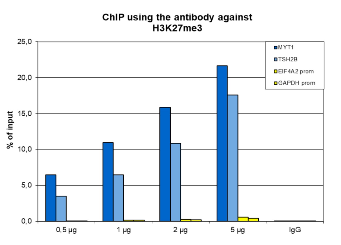
<p><small><strong>Figure 1. ChIP results obtained with the Diagenode antibody directed against H3K27me3</strong><br />ChIP assays were performed using human HeLa cells, the Diagenode antibody against H3K27me3 (Cat. No. C15410195) and optimized PCR primer pairs for qPCR. ChIP was performed with the “iDeal ChIP-seq” kit (Cat. No. C01010051), using sheared chromatin from 1 million cells. The chromatin was spiked with a panel of in vitro assembled nucleosomes, each containing a specific lysine methylation. A titration consisting of 0.5, 1, 2 and 5 µg of antibody per ChIP experiment was analyzed. IgG (1 µg/IP) was used as a negative IP control.</small></p>
<p><small><strong>Figure 1A.</strong> Quantitative PCR was performed with primers specific for the promoter of the active GAPDH and EIF4A2 genes, used as negative controls, and for the inactive TSH2B and MYT1 genes, used as positive controls. The graph shows the recovery, expressed as a % of input (the relative amount of immunoprecipitated DNA compared to input DNA after qPCR analysis).</small></p>
<p><small><strong>Figure 1B.</strong> Recovery of the nucleosomes carrying the H3K27me1, H3K27me2, H3K27me3, H3K4me3, H3K9me3 and H3K36me3 modifications and the unmodified H3K27 as determined by qPCR. The figure clearly shows the antibody is very specific in ChIP for the H3K27me3 modification.</small></p>
</div>
</div>
<div class="row">
<div class="small-12 columns">
<p>A. <img src="https://www.diagenode.com/img/product/antibodies/C15410195-ChIP-Fig2a.png" alt="H3K27me3 Antibody ChIP-seq Grade" /></p>
</div>
</div>
<div class="extra-spaced"></div>
<div class="row">
<div class="small-12 columns">
<p>B. <img src="https://www.diagenode.com/img/product/antibodies/C15410195-ChIP-Fig2b.png" alt="H3K27me3 Antibody for ChIP-seq" /></p>
<p>C. <img src="https://www.diagenode.com/img/product/antibodies/C15410195-ChIP-Fig2c.png" alt="H3K27me3 Antibody for ChIP-seq assay" /></p>
<p>D. <img src="https://www.diagenode.com/img/product/antibodies/C15410195-ChIP-Fig2d.png" alt="H3K27me3 Antibody validated in ChIP-seq" /></p>
</div>
</div>
<div class="row">
<div class="small-12 columns">
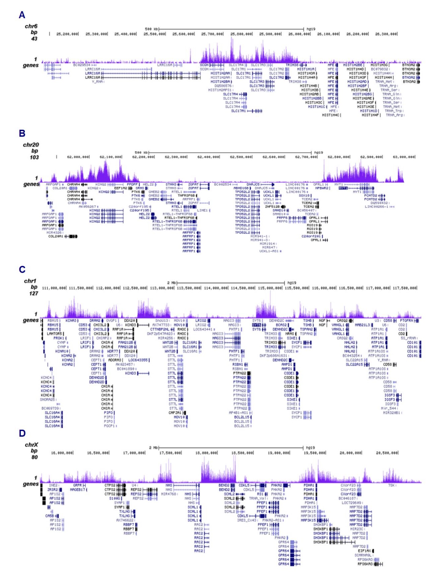
<p><small><strong>Figure 2. ChIP-seq results obtained with the Diagenode antibody directed against H3K27me3</strong><br />ChIP was performed on sheared chromatin from 1 million HeLa cells using 1 µg of the Diagenode antibody against H3K27me3 (Cat. No. C15410195) as described above. The IP'd DNA was subsequently analysed on an Illumina HiSeq. Library preparation, cluster generation and sequencing were performed according to the manufacturer's instructions. The 50 bp tags were aligned to the human genome using the BWA algorithm. Figure 2 shows the enrichment in genomic regions of chromosome 6 and 20, surrounding the TSH2B and MYT1 positive control genes (fig 2A and 2B, respectively), and in two genomic regions of chromosome 1 and X (figure 2C and D).</small></p>
</div>
</div>
<div class="extra-spaced"></div>
<div class="row">
<div class="small-12 columns">
<p>A. <img src="https://www.diagenode.com/img/product/antibodies/C15410195-CUTTAG-Fig3A.png" /></p>
<p>B. <img src="https://www.diagenode.com/img/product/antibodies/C15410195-CUTTAG-Fig3B.png" /></p>
</div>
</div>
<div class="row">
<div class="small-12 columns">
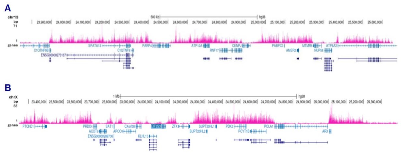
<p><small><strong>Figure 3. Cut&Tag results obtained with the Diagenode antibody directed against H3K27me3</strong><br />CUT&TAG (Kaya-Okur, H.S., Nat Commun 10, 1930, 2019) was performed on 50,000 K562 cells using 1 µg of the Diagenode antibody against H3K27me3 (cat. No. C15410195) and the Diagenode pA-Tn5 transposase (C01070001). The libraries were subsequently analysed on an Illumina NextSeq 500 sequencer (2x75 paired-end reads) according to the manufacturer's instructions. The tags were aligned to the human genome (hg19) using the BWA algorithm. Figure 3 shows the peak distribution in 2 genomic regions on chromosome and 13 and 20 (figure 3A and B, respectively).</small></p>
</div>
</div>
<div class="extra-spaced"></div>
<div class="row">
<div class="small-6 columns">
<p><img src="https://www.diagenode.com/img/product/antibodies/C15410195-ELISA-Fig4.png" alt="H3K27me3 Antibody ELISA Validation " /></p>
</div>
<div class="small-6 columns">
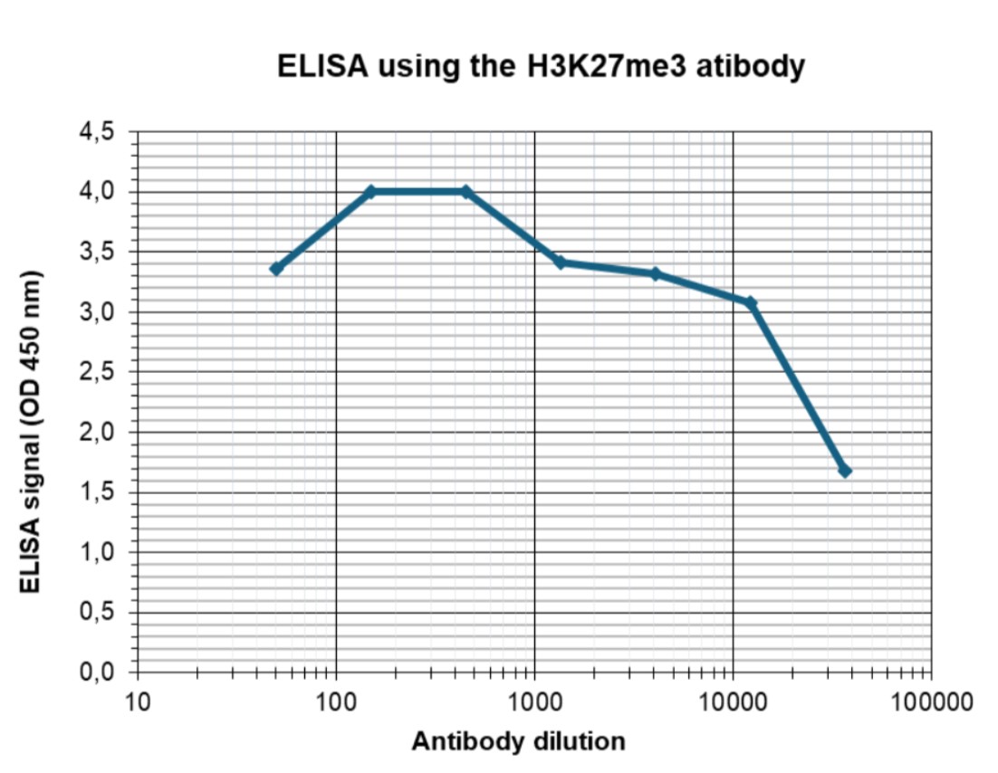
<p><small><strong>Figure 4. Determination of the antibody titer</strong><br />To determine the titer of the antibody, an ELISA was performed using a serial dilution of the Diagenode antibody directed against H3K27me3 (Cat. No. C15410195). The antigen used was a peptide containing the histone modification of interest. By plotting the absorbance against the antibody dilution (Figure 4), the titer of the antibody was estimated to be 1:3,000.</small></p>
</div>
</div>
<div class="extra-spaced"></div>
<div class="row">
<div class="small-6 columns">
<p><img src="https://www.diagenode.com/img/product/antibodies/C15410195-DB-Fig5a.png" alt="H3K27me3 Antibody Dot Blot Validation " /></p>
</div>
<div class="small-6 columns">
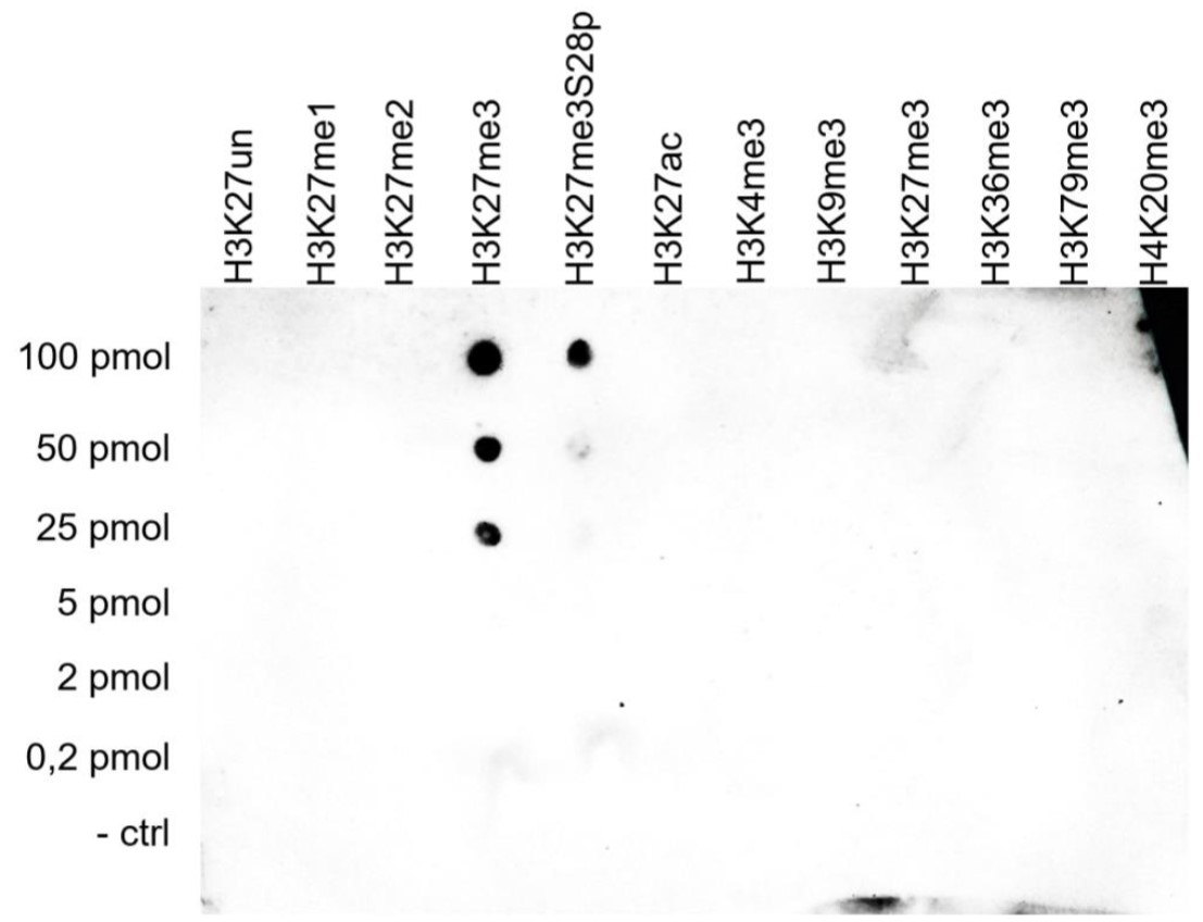
<p><small><strong>Figure 5. Cross reactivity tests using the Diagenode antibody directed against H3K27me3</strong><br />A Dot Blot analysis was performed to test the cross reactivity of the Diagenode antibody against H3K27me3 (Cat. No. C15410195) with peptides containing other modifications of histone H3 and H4 and the unmodified H3K27 sequence. One hundred to 0.2 pmol of the peptide containing the respective histone modification were spotted on a membrane. The antibody was used at a dilution of 1:5,000. Figure 5 shows a high specificity of the antibody for the modification of interest. Please note that the antibody also recognizes the modification if S28 is phosphorylated.</small></p>
</div>
</div>
<div class="row">
<div class="small-6 columns">
<p><img src="https://www.diagenode.com/img/product/antibodies/C15410195-WB-Fig6.png" alt="H3K27me3 Antibody validated in Western Blot" /></p>
</div>
<div class="small-6 columns">
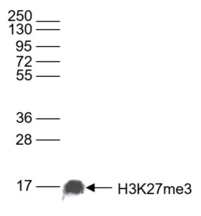
<p><small><strong>Figure 6. Western blot analysis using the Diagenode antibody directed against H3K27me3</strong><br />Western blot was performed on whole cell (25 µg, lane 1) and histone extracts (15 µg, lane 2) from HeLa cells, and on 1 µg of recombinant histone H2A, H2B, H3 and H4 (lane 3, 4, 5 and 6, respectively) using the Diagenode antibody against H3K27me3 (cat. No. C15410195) diluted 1:500 in TBS-Tween containing 5% skimmed milk. The position of the protein of interest is indicated on the right; the marker (in kDa) is shown on the left.</small></p>
</div>
</div>
<div class="row">
<div class="small-12 columns">
<p><img src="https://www.diagenode.com/img/product/antibodies/C15410195-IF-Fig7.png" alt="H3K27me3 Antibody validated for Immunofluorescence" /></p>
</div>
</div>
<div class="row">
<div class="small-12 columns">
<p><small><strong>Figure 7. Immunofluorescence using the Diagenode antibody directed against H3K27me3</strong><br />Human HeLa cells were stained with the Diagenode antibody against H3K27me3 (Cat. No. C15410195) and with DAPI. Cells were fixed with 4% formaldehyde for 10’ and blocked with PBS/TX-100 containing 5% normal goat serum and 1% BSA. The cells were immunofluorescently labelled with the H3K27me3 antibody (left) diluted 1:200 in blocking solution followed by an anti-rabbit antibody conjugated to Alexa488. The middle panel shows staining of the nuclei with DAPI. A merge of the two stainings is shown on the right.</small></p>
</div>
</div>',
'label2' => 'Target Description',
'info2' => '<p><small>Histones are the main constituents of the protein part of chromosomes of eukaryotic cells. They are rich in the amino acids arginine and lysine and have been greatly conserved during evolution. Histones pack the DNA into tight masses of chromatin. Two core histones of each class H2A, H2B, H3 and H4 assemble and are wrapped by 146 base pairs of DNA to form one octameric nucleosome. Histone tails undergo numerous post-translational modifications, which alter chromatin structure to facilitate transcriptional activation, repression or other nuclear processes. In addition to the genetic code, combinations of the different histone modifications reveal the so-called “histone code”. Histone methylation and demethylation is regulated by histone methyl transferases and histone demethylases. Methylation of histone H3K27 is associated with inactive genomic regions.</small></p>',
'label3' => '',
'info3' => '',
'format' => '10 µg',
'catalog_number' => 'C15410195-10',
'old_catalog_number' => 'pAb-195-050',
'sf_code' => 'C15410195-D001-000582',
'type' => 'FRE',
'search_order' => '03-Antibody',
'price_EUR' => '125',
'price_USD' => '115',
'price_GBP' => '115',
'price_JPY' => '19580',
'price_CNY' => '',
'price_AUD' => '288',
'country' => 'ALL',
'except_countries' => 'None',
'quote' => false,
'in_stock' => false,
'featured' => false,
'no_promo' => false,
'online' => true,
'master' => false,
'last_datasheet_update' => '0000-00-00',
'slug' => 'h3k27me3-polyclonal-antibody-premium-sample-size-10-ug',
'meta_title' => 'H3K27me3 Antibody - ChIP-seq Grade (C15410195-10) | Diagenode',
'meta_keywords' => '',
'meta_description' => 'H3K27me3 (Histone H3 trimethylated at lysine 27) Polyclonal Antibody validated in ChIP-seq, ChIP-qPCR, ELISA, DB, WB and IF. Specificity confirmed by Peptide array. Batch-specific data available on the website. Sample size available',
'modified' => '2024-01-17 13:56:29',
'created' => '2015-06-29 14:08:20',
'locale' => 'zho'
),
'Antibody' => array(
'host' => '*****',
'id' => '70',
'name' => 'H3K27me3 polyclonal antibody',
'description' => 'Histones are the main constituents of the protein part of chromosomes of eukaryotic cells. They are rich in the amino acids arginine and lysine and have been greatly conserved during evolution. Histones pack the DNA into tight masses of chromatin. Two core histones of each class H2A, H2B, H3 and H4 assemble and are wrapped by 146 base pairs of DNA to form one octameric nucleosome. Histone tails undergo numerous post-translational modifications, which alter chromatin structure to facilitate transcriptional activation, repression or other nuclear processes. In addition to the genetic code, combinations of the different histone modifications reveal the so-called “histone code”. Histone methylation and demethylation is regulated by histone methyl transferases and histone demethylases. Methylation of histone H3K27 is associated with inactive genomic regions.',
'clonality' => '',
'isotype' => '',
'lot' => 'A0824D',
'concentration' => '1.1 µg/µl',
'reactivity' => 'Human, mouse, Drosophila, C. elegans, Daphnia, Arabidopsis, maize, tomato, poplar, silena latifolia, C. merolae, wide range expected.',
'type' => 'Polyclonal',
'purity' => 'Affinity purified polyclonal antibody.',
'classification' => 'Premium',
'application_table' => '<table>
<thead>
<tr>
<th>Applications</th>
<th>Suggested dilution</th>
<th>References</th>
</tr>
</thead>
<tbody>
<tr>
<td>ChIP/ChIP-seq <sup>*</sup></td>
<td>1 - 2 µg/IP per ChIP</td>
<td>Fig 1, 2</td>
</tr>
<tr>
<td>CUT&TAG</td>
<td>1 µg</td>
<td>Fig 3</td>
</tr>
<tr>
<td>ELISA</td>
<td>1:100 - 1:500</td>
<td>Fig 4</td>
</tr>
<tr>
<td>Dot Blotting/Peptide array</td>
<td>1:5,000</td>
<td>Fig 5</td>
</tr>
<tr>
<td>Western Blotting</td>
<td>1:1,000</td>
<td>Fig 6</td>
</tr>
<tr>
<td>Immunofluorescence</td>
<td>1:200</td>
<td>Fig 7</td>
</tr>
</tbody>
</table>
<p></p>
<p><small><sup>*</sup> Please note that the optimal antibody amount per IP should be determined by the end-user. We recommend testing 0.5-5 µg per IP.</small></p>',
'storage_conditions' => 'Store at -20°C; for long storage, store at -80°C. Avoid multiple freeze-thaw cycles.',
'storage_buffer' => 'PBS containing 0.05% azide and 0.05% ProClin 300.',
'precautions' => 'This product is for research use only. Not for use in diagnostic or therapeutic procedures.',
'uniprot_acc' => '',
'slug' => '',
'meta_keywords' => '',
'meta_description' => '',
'modified' => '2021-02-26 11:29:05',
'created' => '0000-00-00 00:00:00',
'select_label' => '70 - H3K27me3 polyclonal antibody (A0824D - 1.1 µg/µl - Human, mouse, Drosophila, C. elegans, Daphnia, Arabidopsis, maize, tomato, poplar, silena latifolia, C. merolae, wide range expected. - Affinity purified polyclonal antibody. - Rabbit)'
),
'Slave' => array(),
'Group' => array(
'Group' => array(
'id' => '22',
'name' => 'C15410195',
'product_id' => '2268',
'modified' => '2016-02-18 18:04:04',
'created' => '2016-02-18 18:04:04'
),
'Master' => array(
'id' => '2268',
'antibody_id' => '70',
'name' => 'H3K27me3 Antibody',
'description' => '<p>Polyclonal antibody raised in rabbit against the region of histone <strong>H3 containing the trimethylated lysine 27</strong> (<strong>H3K27me3</strong>), using a KLH-conjugated synthetic peptide.</p>',
'label1' => 'Validation Data',
'info1' => '<div class="row">
<div class="small-6 columns">
<p>A. <img src="https://www.diagenode.com/img/product/antibodies/C15410195-ChIP-Fig1.png" alt="H3K27me3 Antibody ChIP Grade" /></p>
<p>B. <img src="https://www.diagenode.com/img/product/antibodies/C15410195-ChIP-Fig2.png" alt="H3K27me3 Antibody for ChIP" /></p>
</div>
<div class="small-6 columns">
<p><small><strong>Figure 1. ChIP results obtained with the Diagenode antibody directed against H3K27me3</strong><br />ChIP assays were performed using human HeLa cells, the Diagenode antibody against H3K27me3 (Cat. No. C15410195) and optimized PCR primer pairs for qPCR. ChIP was performed with the “iDeal ChIP-seq” kit (Cat. No. C01010051), using sheared chromatin from 1 million cells. The chromatin was spiked with a panel of in vitro assembled nucleosomes, each containing a specific lysine methylation. A titration consisting of 0.5, 1, 2 and 5 µg of antibody per ChIP experiment was analyzed. IgG (1 µg/IP) was used as a negative IP control.</small></p>
<p><small><strong>Figure 1A.</strong> Quantitative PCR was performed with primers specific for the promoter of the active GAPDH and EIF4A2 genes, used as negative controls, and for the inactive TSH2B and MYT1 genes, used as positive controls. The graph shows the recovery, expressed as a % of input (the relative amount of immunoprecipitated DNA compared to input DNA after qPCR analysis).</small></p>
<p><small><strong>Figure 1B.</strong> Recovery of the nucleosomes carrying the H3K27me1, H3K27me2, H3K27me3, H3K4me3, H3K9me3 and H3K36me3 modifications and the unmodified H3K27 as determined by qPCR. The figure clearly shows the antibody is very specific in ChIP for the H3K27me3 modification.</small></p>
</div>
</div>
<div class="row">
<div class="small-12 columns">
<p>A. <img src="https://www.diagenode.com/img/product/antibodies/C15410195-ChIP-Fig2a.png" alt="H3K27me3 Antibody ChIP-seq Grade" /></p>
</div>
</div>
<div class="extra-spaced"></div>
<div class="row">
<div class="small-12 columns">
<p>B. <img src="https://www.diagenode.com/img/product/antibodies/C15410195-ChIP-Fig2b.png" alt="H3K27me3 Antibody for ChIP-seq" /></p>
<p>C. <img src="https://www.diagenode.com/img/product/antibodies/C15410195-ChIP-Fig2c.png" alt="H3K27me3 Antibody for ChIP-seq assay" /></p>
<p>D. <img src="https://www.diagenode.com/img/product/antibodies/C15410195-ChIP-Fig2d.png" alt="H3K27me3 Antibody validated in ChIP-seq" /></p>
</div>
</div>
<div class="row">
<div class="small-12 columns">
<p><small><strong>Figure 2. ChIP-seq results obtained with the Diagenode antibody directed against H3K27me3</strong><br />ChIP was performed on sheared chromatin from 1 million HeLa cells using 1 µg of the Diagenode antibody against H3K27me3 (Cat. No. C15410195) as described above. The IP'd DNA was subsequently analysed on an Illumina HiSeq. Library preparation, cluster generation and sequencing were performed according to the manufacturer's instructions. The 50 bp tags were aligned to the human genome using the BWA algorithm. Figure 2 shows the enrichment in genomic regions of chromosome 6 and 20, surrounding the TSH2B and MYT1 positive control genes (fig 2A and 2B, respectively), and in two genomic regions of chromosome 1 and X (figure 2C and D).</small></p>
</div>
</div>
<div class="extra-spaced"></div>
<div class="row">
<div class="small-12 columns">
<p>A. <img src="https://www.diagenode.com/img/product/antibodies/C15410195-CUTTAG-Fig3A.png" /></p>
<p>B. <img src="https://www.diagenode.com/img/product/antibodies/C15410195-CUTTAG-Fig3B.png" /></p>
</div>
</div>
<div class="row">
<div class="small-12 columns">
<p><small><strong>Figure 3. Cut&Tag results obtained with the Diagenode antibody directed against H3K27me3</strong><br />CUT&TAG (Kaya-Okur, H.S., Nat Commun 10, 1930, 2019) was performed on 50,000 K562 cells using 1 µg of the Diagenode antibody against H3K27me3 (cat. No. C15410195) and the Diagenode pA-Tn5 transposase (C01070001). The libraries were subsequently analysed on an Illumina NextSeq 500 sequencer (2x75 paired-end reads) according to the manufacturer's instructions. The tags were aligned to the human genome (hg19) using the BWA algorithm. Figure 3 shows the peak distribution in 2 genomic regions on chromosome and 13 and 20 (figure 3A and B, respectively).</small></p>
</div>
</div>
<div class="extra-spaced"></div>
<div class="row">
<div class="small-6 columns">
<p><img src="https://www.diagenode.com/img/product/antibodies/C15410195-ELISA-Fig4.png" alt="H3K27me3 Antibody ELISA Validation " /></p>
</div>
<div class="small-6 columns">
<p><small><strong>Figure 4. Determination of the antibody titer</strong><br />To determine the titer of the antibody, an ELISA was performed using a serial dilution of the Diagenode antibody directed against H3K27me3 (Cat. No. C15410195). The antigen used was a peptide containing the histone modification of interest. By plotting the absorbance against the antibody dilution (Figure 4), the titer of the antibody was estimated to be 1:3,000.</small></p>
</div>
</div>
<div class="extra-spaced"></div>
<div class="row">
<div class="small-6 columns">
<p><img src="https://www.diagenode.com/img/product/antibodies/C15410195-DB-Fig5a.png" alt="H3K27me3 Antibody Dot Blot Validation " /></p>
</div>
<div class="small-6 columns">
<p><small><strong>Figure 5. Cross reactivity tests using the Diagenode antibody directed against H3K27me3</strong><br />A Dot Blot analysis was performed to test the cross reactivity of the Diagenode antibody against H3K27me3 (Cat. No. C15410195) with peptides containing other modifications of histone H3 and H4 and the unmodified H3K27 sequence. One hundred to 0.2 pmol of the peptide containing the respective histone modification were spotted on a membrane. The antibody was used at a dilution of 1:5,000. Figure 5 shows a high specificity of the antibody for the modification of interest. Please note that the antibody also recognizes the modification if S28 is phosphorylated.</small></p>
</div>
</div>
<div class="row">
<div class="small-6 columns">
<p><img src="https://www.diagenode.com/img/product/antibodies/C15410195-WB-Fig6.png" alt="H3K27me3 Antibody validated in Western Blot" /></p>
</div>
<div class="small-6 columns">
<p><small><strong>Figure 6. Western blot analysis using the Diagenode antibody directed against H3K27me3</strong><br />Western blot was performed on whole cell (25 µg, lane 1) and histone extracts (15 µg, lane 2) from HeLa cells, and on 1 µg of recombinant histone H2A, H2B, H3 and H4 (lane 3, 4, 5 and 6, respectively) using the Diagenode antibody against H3K27me3 (cat. No. C15410195) diluted 1:500 in TBS-Tween containing 5% skimmed milk. The position of the protein of interest is indicated on the right; the marker (in kDa) is shown on the left.</small></p>
</div>
</div>
<div class="row">
<div class="small-12 columns">
<p><img src="https://www.diagenode.com/img/product/antibodies/C15410195-IF-Fig7.png" alt="H3K27me3 Antibody validated for Immunofluorescence" /></p>
</div>
</div>
<div class="row">
<div class="small-12 columns">
<p><small><strong>Figure 7. Immunofluorescence using the Diagenode antibody directed against H3K27me3</strong><br />Human HeLa cells were stained with the Diagenode antibody against H3K27me3 (Cat. No. C15410195) and with DAPI. Cells were fixed with 4% formaldehyde for 10’ and blocked with PBS/TX-100 containing 5% normal goat serum and 1% BSA. The cells were immunofluorescently labelled with the H3K27me3 antibody (left) diluted 1:200 in blocking solution followed by an anti-rabbit antibody conjugated to Alexa488. The middle panel shows staining of the nuclei with DAPI. A merge of the two stainings is shown on the right.</small></p>
</div>
</div>',
'label2' => 'Target Description',
'info2' => '<p><small>Histones are the main constituents of the protein part of chromosomes of eukaryotic cells. They are rich in the amino acids arginine and lysine and have been greatly conserved during evolution. Histones pack the DNA into tight masses of chromatin. Two core histones of each class H2A, H2B, H3 and H4 assemble and are wrapped by 146 base pairs of DNA to form one octameric nucleosome. Histone tails undergo numerous post-translational modifications, which alter chromatin structure to facilitate transcriptional activation, repression or other nuclear processes. In addition to the genetic code, combinations of the different histone modifications reveal the so-called “histone code”. Histone methylation and demethylation is regulated by histone methyl transferases and histone demethylases. Methylation of histone H3K27 is associated with inactive genomic regions.</small></p>',
'label3' => '',
'info3' => '',
'format' => '50 μg',
'catalog_number' => 'C15410195',
'old_catalog_number' => 'pAb-195-050',
'sf_code' => 'C15410195-D001-000581',
'type' => 'FRE',
'search_order' => '03-Antibody',
'price_EUR' => '480',
'price_USD' => '470',
'price_GBP' => '430',
'price_JPY' => '75190',
'price_CNY' => '',
'price_AUD' => '1175',
'country' => 'ALL',
'except_countries' => 'None',
'quote' => false,
'in_stock' => false,
'featured' => false,
'no_promo' => false,
'online' => true,
'master' => true,
'last_datasheet_update' => 'January 14, 2021',
'slug' => 'h3k27me3-polyclonal-antibody-premium-50-mg-27-ml',
'meta_title' => 'H3K27me3 Antibody - ChIP-seq Grade (C15410195) | Diagenode',
'meta_keywords' => '',
'meta_description' => 'H3K27me3 (Histone H3 trimethylated at lysine 27) Polyclonal Antibody validated in ChIP-seq, ChIP-qPCR, CUT&Tag, ELISA, DB, WB and IF. Specificity confirmed by Peptide array assay. Batch-specific data available on the website. Sample size available.',
'modified' => '2024-01-17 13:55:58',
'created' => '2015-06-29 14:08:20'
),
'Product' => array(
(int) 0 => array(
[maximum depth reached]
)
)
),
'Related' => array(
(int) 0 => array(
'id' => '2268',
'antibody_id' => '70',
'name' => 'H3K27me3 Antibody',
'description' => '<p>Polyclonal antibody raised in rabbit against the region of histone <strong>H3 containing the trimethylated lysine 27</strong> (<strong>H3K27me3</strong>), using a KLH-conjugated synthetic peptide.</p>',
'label1' => 'Validation Data',
'info1' => '<div class="row">
<div class="small-6 columns">
<p>A. <img src="https://www.diagenode.com/img/product/antibodies/C15410195-ChIP-Fig1.png" alt="H3K27me3 Antibody ChIP Grade" /></p>
<p>B. <img src="https://www.diagenode.com/img/product/antibodies/C15410195-ChIP-Fig2.png" alt="H3K27me3 Antibody for ChIP" /></p>
</div>
<div class="small-6 columns">
<p><small><strong>Figure 1. ChIP results obtained with the Diagenode antibody directed against H3K27me3</strong><br />ChIP assays were performed using human HeLa cells, the Diagenode antibody against H3K27me3 (Cat. No. C15410195) and optimized PCR primer pairs for qPCR. ChIP was performed with the “iDeal ChIP-seq” kit (Cat. No. C01010051), using sheared chromatin from 1 million cells. The chromatin was spiked with a panel of in vitro assembled nucleosomes, each containing a specific lysine methylation. A titration consisting of 0.5, 1, 2 and 5 µg of antibody per ChIP experiment was analyzed. IgG (1 µg/IP) was used as a negative IP control.</small></p>
<p><small><strong>Figure 1A.</strong> Quantitative PCR was performed with primers specific for the promoter of the active GAPDH and EIF4A2 genes, used as negative controls, and for the inactive TSH2B and MYT1 genes, used as positive controls. The graph shows the recovery, expressed as a % of input (the relative amount of immunoprecipitated DNA compared to input DNA after qPCR analysis).</small></p>
<p><small><strong>Figure 1B.</strong> Recovery of the nucleosomes carrying the H3K27me1, H3K27me2, H3K27me3, H3K4me3, H3K9me3 and H3K36me3 modifications and the unmodified H3K27 as determined by qPCR. The figure clearly shows the antibody is very specific in ChIP for the H3K27me3 modification.</small></p>
</div>
</div>
<div class="row">
<div class="small-12 columns">
<p>A. <img src="https://www.diagenode.com/img/product/antibodies/C15410195-ChIP-Fig2a.png" alt="H3K27me3 Antibody ChIP-seq Grade" /></p>
</div>
</div>
<div class="extra-spaced"></div>
<div class="row">
<div class="small-12 columns">
<p>B. <img src="https://www.diagenode.com/img/product/antibodies/C15410195-ChIP-Fig2b.png" alt="H3K27me3 Antibody for ChIP-seq" /></p>
<p>C. <img src="https://www.diagenode.com/img/product/antibodies/C15410195-ChIP-Fig2c.png" alt="H3K27me3 Antibody for ChIP-seq assay" /></p>
<p>D. <img src="https://www.diagenode.com/img/product/antibodies/C15410195-ChIP-Fig2d.png" alt="H3K27me3 Antibody validated in ChIP-seq" /></p>
</div>
</div>
<div class="row">
<div class="small-12 columns">
<p><small><strong>Figure 2. ChIP-seq results obtained with the Diagenode antibody directed against H3K27me3</strong><br />ChIP was performed on sheared chromatin from 1 million HeLa cells using 1 µg of the Diagenode antibody against H3K27me3 (Cat. No. C15410195) as described above. The IP'd DNA was subsequently analysed on an Illumina HiSeq. Library preparation, cluster generation and sequencing were performed according to the manufacturer's instructions. The 50 bp tags were aligned to the human genome using the BWA algorithm. Figure 2 shows the enrichment in genomic regions of chromosome 6 and 20, surrounding the TSH2B and MYT1 positive control genes (fig 2A and 2B, respectively), and in two genomic regions of chromosome 1 and X (figure 2C and D).</small></p>
</div>
</div>
<div class="extra-spaced"></div>
<div class="row">
<div class="small-12 columns">
<p>A. <img src="https://www.diagenode.com/img/product/antibodies/C15410195-CUTTAG-Fig3A.png" /></p>
<p>B. <img src="https://www.diagenode.com/img/product/antibodies/C15410195-CUTTAG-Fig3B.png" /></p>
</div>
</div>
<div class="row">
<div class="small-12 columns">
<p><small><strong>Figure 3. Cut&Tag results obtained with the Diagenode antibody directed against H3K27me3</strong><br />CUT&TAG (Kaya-Okur, H.S., Nat Commun 10, 1930, 2019) was performed on 50,000 K562 cells using 1 µg of the Diagenode antibody against H3K27me3 (cat. No. C15410195) and the Diagenode pA-Tn5 transposase (C01070001). The libraries were subsequently analysed on an Illumina NextSeq 500 sequencer (2x75 paired-end reads) according to the manufacturer's instructions. The tags were aligned to the human genome (hg19) using the BWA algorithm. Figure 3 shows the peak distribution in 2 genomic regions on chromosome and 13 and 20 (figure 3A and B, respectively).</small></p>
</div>
</div>
<div class="extra-spaced"></div>
<div class="row">
<div class="small-6 columns">
<p><img src="https://www.diagenode.com/img/product/antibodies/C15410195-ELISA-Fig4.png" alt="H3K27me3 Antibody ELISA Validation " /></p>
</div>
<div class="small-6 columns">
<p><small><strong>Figure 4. Determination of the antibody titer</strong><br />To determine the titer of the antibody, an ELISA was performed using a serial dilution of the Diagenode antibody directed against H3K27me3 (Cat. No. C15410195). The antigen used was a peptide containing the histone modification of interest. By plotting the absorbance against the antibody dilution (Figure 4), the titer of the antibody was estimated to be 1:3,000.</small></p>
</div>
</div>
<div class="extra-spaced"></div>
<div class="row">
<div class="small-6 columns">
<p><img src="https://www.diagenode.com/img/product/antibodies/C15410195-DB-Fig5a.png" alt="H3K27me3 Antibody Dot Blot Validation " /></p>
</div>
<div class="small-6 columns">
<p><small><strong>Figure 5. Cross reactivity tests using the Diagenode antibody directed against H3K27me3</strong><br />A Dot Blot analysis was performed to test the cross reactivity of the Diagenode antibody against H3K27me3 (Cat. No. C15410195) with peptides containing other modifications of histone H3 and H4 and the unmodified H3K27 sequence. One hundred to 0.2 pmol of the peptide containing the respective histone modification were spotted on a membrane. The antibody was used at a dilution of 1:5,000. Figure 5 shows a high specificity of the antibody for the modification of interest. Please note that the antibody also recognizes the modification if S28 is phosphorylated.</small></p>
</div>
</div>
<div class="row">
<div class="small-6 columns">
<p><img src="https://www.diagenode.com/img/product/antibodies/C15410195-WB-Fig6.png" alt="H3K27me3 Antibody validated in Western Blot" /></p>
</div>
<div class="small-6 columns">
<p><small><strong>Figure 6. Western blot analysis using the Diagenode antibody directed against H3K27me3</strong><br />Western blot was performed on whole cell (25 µg, lane 1) and histone extracts (15 µg, lane 2) from HeLa cells, and on 1 µg of recombinant histone H2A, H2B, H3 and H4 (lane 3, 4, 5 and 6, respectively) using the Diagenode antibody against H3K27me3 (cat. No. C15410195) diluted 1:500 in TBS-Tween containing 5% skimmed milk. The position of the protein of interest is indicated on the right; the marker (in kDa) is shown on the left.</small></p>
</div>
</div>
<div class="row">
<div class="small-12 columns">
<p><img src="https://www.diagenode.com/img/product/antibodies/C15410195-IF-Fig7.png" alt="H3K27me3 Antibody validated for Immunofluorescence" /></p>
</div>
</div>
<div class="row">
<div class="small-12 columns">
<p><small><strong>Figure 7. Immunofluorescence using the Diagenode antibody directed against H3K27me3</strong><br />Human HeLa cells were stained with the Diagenode antibody against H3K27me3 (Cat. No. C15410195) and with DAPI. Cells were fixed with 4% formaldehyde for 10’ and blocked with PBS/TX-100 containing 5% normal goat serum and 1% BSA. The cells were immunofluorescently labelled with the H3K27me3 antibody (left) diluted 1:200 in blocking solution followed by an anti-rabbit antibody conjugated to Alexa488. The middle panel shows staining of the nuclei with DAPI. A merge of the two stainings is shown on the right.</small></p>
</div>
</div>',
'label2' => 'Target Description',
'info2' => '<p><small>Histones are the main constituents of the protein part of chromosomes of eukaryotic cells. They are rich in the amino acids arginine and lysine and have been greatly conserved during evolution. Histones pack the DNA into tight masses of chromatin. Two core histones of each class H2A, H2B, H3 and H4 assemble and are wrapped by 146 base pairs of DNA to form one octameric nucleosome. Histone tails undergo numerous post-translational modifications, which alter chromatin structure to facilitate transcriptional activation, repression or other nuclear processes. In addition to the genetic code, combinations of the different histone modifications reveal the so-called “histone code”. Histone methylation and demethylation is regulated by histone methyl transferases and histone demethylases. Methylation of histone H3K27 is associated with inactive genomic regions.</small></p>',
'label3' => '',
'info3' => '',
'format' => '50 μg',
'catalog_number' => 'C15410195',
'old_catalog_number' => 'pAb-195-050',
'sf_code' => 'C15410195-D001-000581',
'type' => 'FRE',
'search_order' => '03-Antibody',
'price_EUR' => '480',
'price_USD' => '470',
'price_GBP' => '430',
'price_JPY' => '75190',
'price_CNY' => '',
'price_AUD' => '1175',
'country' => 'ALL',
'except_countries' => 'None',
'quote' => false,
'in_stock' => false,
'featured' => false,
'no_promo' => false,
'online' => true,
'master' => true,
'last_datasheet_update' => 'January 14, 2021',
'slug' => 'h3k27me3-polyclonal-antibody-premium-50-mg-27-ml',
'meta_title' => 'H3K27me3 Antibody - ChIP-seq Grade (C15410195) | Diagenode',
'meta_keywords' => '',
'meta_description' => 'H3K27me3 (Histone H3 trimethylated at lysine 27) Polyclonal Antibody validated in ChIP-seq, ChIP-qPCR, CUT&Tag, ELISA, DB, WB and IF. Specificity confirmed by Peptide array assay. Batch-specific data available on the website. Sample size available.',
'modified' => '2024-01-17 13:55:58',
'created' => '2015-06-29 14:08:20',
'ProductsRelated' => array(
[maximum depth reached]
),
'Image' => array(
[maximum depth reached]
)
)
),
'Application' => array(
(int) 0 => array(
'id' => '20',
'position' => '10',
'parent_id' => '40',
'name' => 'ELISA',
'description' => '<div class="row">
<div class="small-12 medium-12 large-12 columns">Enzyme-linked immunosorbent assay.</div>
</div>',
'in_footer' => false,
'in_menu' => false,
'online' => true,
'tabular' => true,
'slug' => 'elisa-antibodies',
'meta_keywords' => ' ELISA Antibodies,Monoclonal antibody, Polyclonal antibody',
'meta_description' => 'Diagenode offers Monoclonal & Polyclonal antibodies for ELISA applications',
'meta_title' => 'ELISA Antibodies - Monoclonal & Polyclonal antibody | Diagenode',
'modified' => '2016-01-13 12:21:41',
'created' => '2014-07-08 08:13:28',
'ProductsApplication' => array(
[maximum depth reached]
)
),
(int) 1 => array(
'id' => '28',
'position' => '10',
'parent_id' => '40',
'name' => 'DB',
'description' => '<p>Dot blotting</p>',
'in_footer' => false,
'in_menu' => false,
'online' => true,
'tabular' => true,
'slug' => 'dot-blotting',
'meta_keywords' => 'Dot blotting,Monoclonal & Polyclonal antibody,',
'meta_description' => 'Diagenode offers Monoclonal & Polyclonal antibodies for Dot blotting applications',
'meta_title' => 'Dot blotting Antibodies - Monoclonal & Polyclonal antibody | Diagenode',
'modified' => '2016-01-13 14:40:49',
'created' => '2015-07-08 13:45:05',
'ProductsApplication' => array(
[maximum depth reached]
)
),
(int) 2 => array(
'id' => '19',
'position' => '10',
'parent_id' => '40',
'name' => 'WB',
'description' => '<p><strong>Western blot</strong> : The quality of antibodies used in this technique is crucial for correct and specific protein identification. Diagenode offers huge selection of highly sensitive and specific western blot-validated antibodies.</p>
<p>Learn more about: <a href="https://www.diagenode.com/applications/western-blot">Loading control, MW marker visualization</a><em>. <br /></em></p>
<p><em></em>Check our selection of antibodies validated in Western blot.</p>',
'in_footer' => false,
'in_menu' => false,
'online' => true,
'tabular' => true,
'slug' => 'western-blot-antibodies',
'meta_keywords' => ' Western Blot Antibodies ,western blot protocol,Western Blotting Products,Polyclonal antibodies ,monoclonal antibodies ',
'meta_description' => 'Diagenode offers a wide range of antibodies and technical support for western blot applications',
'meta_title' => ' Western Blot - Monoclonal antibody - Polyclonal antibody | Diagenode',
'modified' => '2016-04-26 12:44:51',
'created' => '2015-01-07 09:20:00',
'ProductsApplication' => array(
[maximum depth reached]
)
),
(int) 3 => array(
'id' => '37',
'position' => '10',
'parent_id' => '40',
'name' => 'Peptide array',
'description' => '<p>Peptide array</p>',
'in_footer' => false,
'in_menu' => false,
'online' => true,
'tabular' => true,
'slug' => 'peptide-arry',
'meta_keywords' => 'Peptide array antibodies,Histone antibodies,policlonal antibodies',
'meta_description' => 'Diagenode offers a wide range of antibodies and technical support for peptide array applications',
'meta_title' => 'Peptide array antibodies | Diagenode',
'modified' => '2016-01-20 12:24:40',
'created' => '2015-07-08 13:55:25',
'ProductsApplication' => array(
[maximum depth reached]
)
),
(int) 4 => array(
'id' => '29',
'position' => '10',
'parent_id' => '40',
'name' => 'IF',
'description' => '<p><strong>Immunofluorescence</strong>:</p>
<p>Diagenode offers huge selection of highly sensitive antibodies validated in IF.</p>
<p><img src="https://www.diagenode.com/img/product/antibodies/C15200229-IF.jpg" alt="" height="245" width="256" /></p>
<p><sup><strong>Immunofluorescence using the Diagenode monoclonal antibody directed against CRISPR/Cas9</strong></sup></p>
<p><sup>HeLa cells transfected with a Cas9 expression vector (left) or untransfected cells (right) were fixed in methanol at -20°C, permeabilized with acetone at -20°C and blocked with PBS containing 2% BSA. The cells were stained with the Cas9 C-terminal antibody (Cat. No. C15200229) diluted 1:400, followed by incubation with an anti-mouse secondary antibody coupled to AF488. The bottom images show counter-staining of the nuclei with Hoechst 33342.</sup></p>
<h5><sup>Check our selection of antibodies validated in IF.</sup></h5>',
'in_footer' => false,
'in_menu' => false,
'online' => true,
'tabular' => true,
'slug' => 'immunofluorescence',
'meta_keywords' => 'Immunofluorescence,Monoclonal antibody,Polyclonal antibody',
'meta_description' => 'Diagenode offers a wide range of antibodies and technical support for Immunofluorescence applications',
'meta_title' => 'Immunofluorescence - Monoclonal antibody - Polyclonal antibody | Diagenode',
'modified' => '2016-04-27 16:23:10',
'created' => '2015-07-08 13:46:02',
'ProductsApplication' => array(
[maximum depth reached]
)
),
(int) 5 => array(
'id' => '42',
'position' => '10',
'parent_id' => '40',
'name' => 'ChIP-seq (ab)',
'description' => '',
'in_footer' => false,
'in_menu' => false,
'online' => true,
'tabular' => true,
'slug' => 'chip-seq-antibodies',
'meta_keywords' => 'Chromatin Immunoprecipitation Sequencing,ChIP-Seq,ChIP-seq grade antibodies,DNA purification,qPCR,Shearing of chromatin',
'meta_description' => 'Diagenode offers a wide range of antibodies and technical support for ChIP Sequencing applications',
'meta_title' => 'ChIP Sequencing Antibodies (ChIP-Seq) | Diagenode',
'modified' => '2016-01-20 11:06:19',
'created' => '2015-10-20 11:44:45',
'ProductsApplication' => array(
[maximum depth reached]
)
),
(int) 6 => array(
'id' => '43',
'position' => '10',
'parent_id' => '40',
'name' => 'ChIP-qPCR (ab)',
'description' => '',
'in_footer' => false,
'in_menu' => false,
'online' => true,
'tabular' => true,
'slug' => 'chip-qpcr-antibodies',
'meta_keywords' => 'Chromatin Immunoprecipitation Sequencing,ChIP-Seq,ChIP-seq grade antibodies,DNA purification,qPCR,Shearing of chromatin',
'meta_description' => 'Diagenode offers a wide range of antibodies and technical support for ChIP-qPCR applications',
'meta_title' => 'ChIP Quantitative PCR Antibodies (ChIP-qPCR) | Diagenode',
'modified' => '2016-01-20 11:30:24',
'created' => '2015-10-20 11:45:36',
'ProductsApplication' => array(
[maximum depth reached]
)
),
(int) 7 => array(
'id' => '55',
'position' => '10',
'parent_id' => '40',
'name' => 'CUT&Tag',
'description' => '<p>CUT&Tagアッセイを成功させるための重要な要素の1つは使用される抗体の品質です。 特異性高い抗体は、目的のタンパク質のみをターゲットとした確実な結果を可能にします。 CUT&Tagで検証済みの抗体のセレクションはこちらからご覧ください。</p>
<p>Read more:</p>
<p><a href="https://www.diagenode.com/en/categories/cutandtag">Products for CUT&Tag assay</a></p>
<p><a href="https://www.diagenode.com/en/pages/cut-and-tag">Performance of Diagenode's antibodies in CUT&Tag</a></p>
<script src="chrome-extension://hhojmcideegachlhfgfdhailpfhgknjm/web_accessible_resources/index.js"></script>',
'in_footer' => false,
'in_menu' => false,
'online' => true,
'tabular' => true,
'slug' => 'cut-and-tag',
'meta_keywords' => 'CUT&Tag',
'meta_description' => 'CUT&Tag',
'meta_title' => 'CUT&Tag',
'modified' => '2021-04-27 05:17:46',
'created' => '2020-08-20 10:13:47',
'ProductsApplication' => array(
[maximum depth reached]
)
)
),
'Category' => array(
(int) 0 => array(
'id' => '111',
'position' => '40',
'parent_id' => '4',
'name' => 'Histone antibodies',
'description' => '<p>Histones are the main protein components of chromatin involved in the compaction of DNA into nucleosomes, the basic units of chromatin. A <strong>nucleosome</strong> consists of one pair of each of the core histones (<strong>H2A</strong>, <strong>H2B</strong>, <strong>H3</strong> and <strong>H4</strong>) forming an octameric structure wrapped by 146 base pairs of DNA. The different nucleosomes are linked by the linker histone<strong> H1, </strong>allowing for further condensation of chromatin.</p>
<p>The core histones have a globular structure with large unstructured N-terminal tails protruding from the nucleosome. They can undergo to multiple post-translational modifications (PTM), mainly at the N-terminal tails. These <strong>post-translational modifications </strong>include methylation, acetylation, phosphorylation, ubiquitinylation, citrullination, sumoylation, deamination and crotonylation. The most well characterized PTMs are <strong>methylation,</strong> <strong>acetylation and phosphorylation</strong>. Histone methylation occurs mainly on lysine (K) residues, which can be mono-, di- or tri-methylated, and on arginines (R), which can be mono-methylated and symmetrically or asymmetrically di-methylated. Histone acetylation occurs on lysines and histone phosphorylation mainly on serines (S), threonines (T) and tyrosines (Y).</p>
<p>The PTMs of the different residues are involved in numerous processes such as DNA repair, DNA replication and chromosome condensation. They influence the chromatin organization and can be positively or negatively associated with gene expression. Trimethylation of H3K4, H3K36 and H3K79, and lysine acetylation generally result in an open chromatin configuration (figure below) and are therefore associated with <strong>euchromatin</strong> and gene activation. Trimethylation of H3K9, K3K27 and H4K20, on the other hand, is enriched in <strong>heterochromatin </strong>and associated with gene silencing. The combination of different histone modifications is called the "<strong>histone code</strong>”, analogous to the genetic code.</p>
<p><img src="https://www.diagenode.com/img/categories/antibodies/histone-marks-illustration.png" /></p>
<p>Diagenode is proud to offer a large range of antibodies against histones and histone modifications. Our antibodies are highly specific and have been validated in many applications, including <strong>ChIP</strong> and <strong>ChIP-seq</strong>.</p>
<p>Diagenode’s collection includes antibodies recognizing:</p>
<ul>
<li><strong>Histone H1 variants</strong></li>
<li><strong>Histone H2A, H2A variants and histone H2A</strong> <strong>modifications</strong> (serine phosphorylation, lysine acetylation, lysine ubiquitinylation)</li>
<li><strong>Histone H2B and H2B</strong> <strong>modifications </strong>(serine phosphorylation, lysine acetylation)</li>
<li><strong>Histone H3 and H3 modifications </strong>(lysine methylation (mono-, di- and tri-methylated), lysine acetylation, serine phosphorylation, threonine phosphorylation, arginine methylation (mono-methylated, symmetrically and asymmetrically di-methylated))</li>
<li><strong>Histone H4 and H4 modifications (</strong>lysine methylation (mono-, di- and tri-methylated), lysine acetylation, arginine methylation (mono-methylated and symmetrically di-methylated), serine phosphorylation )</li>
</ul>
<p><span style="font-weight: 400;"><strong>HDAC's HAT's, HMT's and other</strong> <strong>enzymes</strong> which modify histones can be found in the category <a href="../categories/chromatin-modifying-proteins-histone-transferase">Histone modifying enzymes</a><br /></span></p>
<p><span style="font-weight: 400;"> Diagenode’s highly validated antibodies:</span></p>
<ul>
<li><span style="font-weight: 400;"> Highly sensitive and specific</span></li>
<li><span style="font-weight: 400;"> Cost-effective (requires less antibody per reaction)</span></li>
<li><span style="font-weight: 400;"> Batch-specific data is available on the website</span></li>
<li><span style="font-weight: 400;"> Expert technical support</span></li>
<li><span style="font-weight: 400;"> Sample sizes available</span></li>
<li><span style="font-weight: 400;"> 100% satisfaction guarantee</span></li>
</ul>',
'no_promo' => false,
'in_menu' => false,
'online' => true,
'tabular' => false,
'hide' => true,
'all_format' => false,
'is_antibody' => true,
'slug' => 'histone-antibodies',
'cookies_tag_id' => null,
'meta_keywords' => 'Histone, antibody, histone h1, histone h2, histone h3, histone h4',
'meta_description' => 'Polyclonal and Monoclonal Antibodies against Histones and their modifications validated for many applications, including Chromatin Immunoprecipitation (ChIP) and ChIP-Sequencing (ChIP-seq)',
'meta_title' => 'Histone and Modified Histone Antibodies | Diagenode',
'modified' => '2020-09-17 13:34:56',
'created' => '2016-04-01 16:01:32',
'ProductsCategory' => array(
[maximum depth reached]
),
'CookiesTag' => array([maximum depth reached])
),
(int) 1 => array(
'id' => '17',
'position' => '10',
'parent_id' => '4',
'name' => 'ChIP-seq grade antibodies',
'description' => '<p><b>Unparalleled ChIP-Seq results with the most rigorously validated antibodies</b></p>
<p><span style="font-weight: 400;">Diagenode provides leading solutions for epigenetic research. Because ChIP-seq is a widely-used technique, we validate our antibodies in ChIP and ChIP-seq experiments (in addition to conventional methods like Western blot, Dot blot, ELISA, and immunofluorescence) to provide the highest quality antibody. We standardize our validation and production to guarantee high product quality without technical bias. Diagenode guarantees ChIP-seq grade antibody performance under our suggested conditions.</span></p>
<div class="row">
<div class="small-12 medium-9 large-9 columns">
<p><strong>ChIP-seq profile</strong> of active (H3K4me3 and H3K36me3) and inactive (H3K27me3) marks using Diagenode antibodies.</p>
<img src="https://www.diagenode.com/img/categories/antibodies/chip-seq-grade-antibodies.png" /></div>
<div class="small-12 medium-3 large-3 columns">
<p><small> ChIP was performed on sheared chromatin from 100,000 K562 cells using iDeal ChIP-seq kit for Histones (cat. No. C01010051) with 1 µg of the Diagenode antibodies against H3K27me3 (cat. No. C15410195) and H3K4me3 (cat. No. C15410003), and 0.5 µg of the antibody against H3K36me3 (cat. No. C15410192). The IP'd DNA was subsequently analysed on an Illumina Genome Analyzer. Library preparation, cluster generation and sequencing were performed according to the manufacturer's instructions. The 36 bp tags were aligned to the human genome using the ELAND algorithm. The figure shows the signal distribution along the complete sequence of human chromosome 3, a zoomin to a 10 Mb region and a further zoomin to a 1.5 Mb region. </small></p>
</div>
</div>
<p>Diagenode’s highly validated antibodies:</p>
<ul>
<li>Highly sensitive and specific</li>
<li>Cost-effective (requires less antibody per reaction)</li>
<li>Batch-specific data is available on the website</li>
<li>Expert technical support</li>
<li>Sample sizes available</li>
<li>100% satisfaction guarantee</li>
</ul>',
'no_promo' => false,
'in_menu' => true,
'online' => true,
'tabular' => false,
'hide' => true,
'all_format' => false,
'is_antibody' => true,
'slug' => 'chip-seq-grade-antibodies',
'cookies_tag_id' => null,
'meta_keywords' => 'ChIP-seq grade antibodies,polyclonal antibody,WB, ELISA, ChIP-seq (ab), ChIP-qPCR (ab)',
'meta_description' => 'Diagenode Offers Wide Range of Validated ChIP-Seq Grade Antibodies for Unparalleled ChIP-Seq Results',
'meta_title' => 'Chromatin Immunoprecipitation ChIP-Seq Grade Antibodies | Diagenode',
'modified' => '2019-07-03 10:57:22',
'created' => '2015-02-16 02:24:01',
'ProductsCategory' => array(
[maximum depth reached]
),
'CookiesTag' => array([maximum depth reached])
),
(int) 2 => array(
'id' => '102',
'position' => '1',
'parent_id' => '4',
'name' => 'Sample size antibodies',
'description' => '<h1><strong>Validated epigenetics antibodies</strong> – care for a sample?<br /> </h1>
<p>Diagenode has partnered with leading epigenetics experts and numerous epigenetics consortiums to bring to you a validated and comprehensive collection of epigenetic antibodies. As an expert in epigenetics, we are committed to offering highly-specific antibodies validated for ChIP/ChIP-seq and many other applications. All batch-specific validation data is available on our website.<br /><a href="../categories/antibodies">Read about our expertise in antibody production</a>.</p>
<ul>
<li><strong>Focused</strong> - Diagenode's selection of antibodies is exclusively dedicated for epigenetic research. <a title="See the full collection." href="../categories/all-antibodies">See the full collection.</a></li>
<li><strong>Strict quality standards</strong> with rigorous QC and validation</li>
<li><strong>Classified</strong> based on level of validation for flexibility of application</li>
</ul>
<p>Existing sample sizes are listed below. We will soon expand our collection. Are you looking for a sample size of another antibody? Just <a href="mailto:agnieszka.zelisko@diagenode.com?Subject=Sample%20Size%20Request" target="_top">Contact us</a>.</p>',
'no_promo' => false,
'in_menu' => true,
'online' => true,
'tabular' => false,
'hide' => true,
'all_format' => true,
'is_antibody' => true,
'slug' => 'sample-size-antibodies',
'cookies_tag_id' => null,
'meta_keywords' => '5-hmC monoclonal antibody,CRISPR/Cas9 polyclonal antibody ,H3K36me3 polyclonal antibody,diagenode',
'meta_description' => 'Diagenode offers sample volume on selected antibodies for researchers to test, validate and provide confidence and flexibility in choosing from our wide range of antibodies ',
'meta_title' => 'Sample-size Antibodies | Diagenode',
'modified' => '2019-07-03 10:57:05',
'created' => '2015-10-27 12:13:34',
'ProductsCategory' => array(
[maximum depth reached]
),
'CookiesTag' => array([maximum depth reached])
),
(int) 3 => array(
'id' => '103',
'position' => '0',
'parent_id' => '4',
'name' => 'All antibodies',
'description' => '<p><span style="font-weight: 400;">All Diagenode’s antibodies are listed below. Please, use our Quick search field to find the antibody of interest by target name, application, purity.</span></p>
<p><span style="font-weight: 400;">Diagenode’s highly validated antibodies:</span></p>
<ul>
<li>Highly sensitive and specific</li>
<li>Cost-effective (requires less antibody per reaction)</li>
<li>Batch-specific data is available on the website</li>
<li>Expert technical support</li>
<li>Sample sizes available</li>
<li>100% satisfaction guarantee</li>
</ul>',
'no_promo' => false,
'in_menu' => true,
'online' => true,
'tabular' => false,
'hide' => true,
'all_format' => false,
'is_antibody' => true,
'slug' => 'all-antibodies',
'cookies_tag_id' => null,
'meta_keywords' => 'Antibodies,Premium Antibodies,Classic,Pioneer',
'meta_description' => 'Diagenode Offers Strict quality standards with Rigorous QC and validated Antibodies. Classified based on level of validation for flexibility of Application. Comprehensive selection of histone and non-histone Antibodies',
'meta_title' => 'Diagenode's selection of Antibodies is exclusively dedicated for Epigenetic Research | Diagenode',
'modified' => '2019-07-03 10:55:44',
'created' => '2015-11-02 14:49:22',
'ProductsCategory' => array(
[maximum depth reached]
),
'CookiesTag' => array([maximum depth reached])
),
(int) 4 => array(
'id' => '127',
'position' => '10',
'parent_id' => '4',
'name' => 'ChIP-grade antibodies',
'description' => '<div class="row">
<div class="small-12 columns"><center></center>
<p><br />Chromatin immunoprecipitation (<b>ChIP</b>) is a technique to study the associations of proteins with the specific genomic regions in intact cells. One of the most important steps of this protocol is the immunoprecipitation of targeted protein using the antibody specifically recognizing it. The quality of antibodies used in ChIP is essential for the success of the experiment. Diagenode offers extensively validated ChIP-grade antibodies, confirmed for their specificity, and high level of performance in ChIP. Each batch is validated, and batch-specific data are available on the website.</p>
<p></p>
</div>
</div>
<p><strong>ChIP results</strong> obtained with the antibody directed against H3K4me3 (Cat. No. <a href="../p/h3k4me3-polyclonal-antibody-premium-50-ug-50-ul">C15410003</a>). </p>
<div class="row">
<div class="small-12 medium-6 large-6 columns"><img src="https://www.diagenode.com/img/product/antibodies/C15410003-fig1-ChIP.jpg" alt="" width="400" height="315" /> </div>
<div class="small-12 medium-6 large-6 columns">
<p></p>
<p></p>
<p></p>
</div>
</div>
<p></p>
<p>Our aim at Diagenode is to offer the largest collection of highly specific <strong>ChIP-grade antibodies</strong>. We add new antibodies monthly. Find your ChIP-grade antibody in the list below and check more information about tested applications, extensive validation data, and product information.</p>',
'no_promo' => false,
'in_menu' => true,
'online' => true,
'tabular' => false,
'hide' => true,
'all_format' => false,
'is_antibody' => true,
'slug' => 'chip-grade-antibodies',
'cookies_tag_id' => null,
'meta_keywords' => 'ChIP-grade antibodies, polyclonal antibody, monoclonal antibody, Diagenode',
'meta_description' => 'Diagenode Offers Extensively Validated ChIP-Grade Antibodies, Confirmed for their Specificity, and high level of Performance in Chromatin Immunoprecipitation ChIP',
'meta_title' => 'Chromatin immunoprecipitation ChIP-grade antibodies | Diagenode',
'modified' => '2024-11-19 17:27:07',
'created' => '2017-02-14 11:16:04',
'ProductsCategory' => array(
[maximum depth reached]
),
'CookiesTag' => array([maximum depth reached])
)
),
'Document' => array(
(int) 0 => array(
'id' => '632',
'name' => 'Datasheet H3K27me3 C15410195',
'description' => '<p><span>Polyclonal antibody raised in rabbit against the region of histone H3 containing the trimethylated lysine 27 (H3K27me3), using a KLH-conjugated synthetic peptide.</span></p>',
'image_id' => null,
'type' => 'Datasheet',
'url' => 'files/products/antibodies/Datasheet_H3K27me3_C15410195.pdf',
'slug' => 'datasheet-h3k27me3-C15410195',
'meta_keywords' => '',
'meta_description' => '',
'modified' => '2015-11-23 17:18:28',
'created' => '2015-07-07 11:47:44',
'ProductsDocument' => array(
[maximum depth reached]
)
),
(int) 1 => array(
'id' => '11',
'name' => 'Antibodies you can trust',
'description' => '<p style="text-align: justify;"><span>Epigenetic research tools have evolved over time from endpoint PCR to qPCR to the analyses of large sets of genome-wide sequencing data. ChIP sequencing (ChIP-seq) has now become the gold standard method for chromatin studies, given the accuracy and coverage scale of the approach over other methods. Successful ChIP-seq, however, requires a higher level of experimental accuracy and consistency in all steps of ChIP than ever before. Particularly crucial is the quality of ChIP antibodies. </span></p>',
'image_id' => null,
'type' => 'Poster',
'url' => 'files/posters/Antibodies_you_can_trust_Poster.pdf',
'slug' => 'antibodies-you-can-trust-poster',
'meta_keywords' => '',
'meta_description' => '',
'modified' => '2015-10-01 20:18:31',
'created' => '2015-07-03 16:05:15',
'ProductsDocument' => array(
[maximum depth reached]
)
),
(int) 2 => array(
'id' => '38',
'name' => 'Epigenetic Antibodies Brochure',
'description' => '<p>More than in any other immuoprecipitation assays, quality antibodies are critical tools in many epigenetics experiments. Since 10 years, Diagenode has developed the most stringent quality production available on the market for antibodies exclusively focused on epigenetic uses. All our antibodies have been qualified to work in epigenetic applications.</p>',
'image_id' => null,
'type' => 'Brochure',
'url' => 'files/brochures/Epigenetic_Antibodies_Brochure.pdf',
'slug' => 'epigenetic-antibodies-brochure',
'meta_keywords' => '',
'meta_description' => '',
'modified' => '2016-06-15 11:24:06',
'created' => '2015-07-03 16:05:27',
'ProductsDocument' => array(
[maximum depth reached]
)
)
),
'Feature' => array(),
'Image' => array(
(int) 0 => array(
'id' => '1815',
'name' => 'product/antibodies/ab-cuttag-icon.png',
'alt' => 'cut and tag antibody icon',
'modified' => '2021-02-11 12:45:34',
'created' => '2021-02-11 12:45:34',
'ProductsImage' => array(
[maximum depth reached]
)
)
),
'Promotion' => array(),
'Protocol' => array(),
'Publication' => array(
(int) 0 => array(
'id' => '4992',
'name' => 'GTSF1 is required for transposon silencing in the unicellular eukaryote Paramecium tetraurelia ',
'authors' => 'Chundi Wang et al.',
'description' => '<p><span>The PIWI-interacting RNA (piRNA) pathway is crucial for transposon repression and the maintenance of genomic integrity. Gametocyte-specific factor 1 (GTSF1), a PIWI-associated protein indispensable for transposon repression, has been recently shown to potentiate the catalytic activity of PIWI in many metazoans. Whether the requirement of GTSF1 extends to PIWI proteins beyond metazoans is unknown. In this study, we identified a homolog of GTSF1 in the unicellular eukaryote </span><em>Paramecium tetraurelia</em><span><span> </span>(PtGtsf1) and found that its role as a PIWI-cofactor is conserved. PtGtsf1 interacts with PIWI (Ptiwi09) and Polycomb Repressive Complex 2 and is essential for PIWI-dependent DNA elimination of transposons during sexual development. PtGtsf1 is crucial for the degradation of PIWI-bound small RNAs that recognize the organism's own genomic sequences. Without PtGtsf1, self-matching small RNAs are not degraded and results in an accumulation of H3K9me3 and H3K27me3, which may disturb transposon recognition. Our results demonstrate that the PIWI–GTSF1 interaction also exists in unicellular eukaryotes with a role in transposon silencing.</span></p>',
'date' => '2024-10-23',
'pmid' => 'https://academic.oup.com/nar/advance-article/doi/10.1093/nar/gkae925/7832353',
'doi' => 'https://doi.org/10.1093/nar/gkae925',
'modified' => '2024-10-25 10:00:24',
'created' => '2024-10-25 10:00:24',
'ProductsPublication' => array(
[maximum depth reached]
)
),
(int) 1 => array(
'id' => '4974',
'name' => 'Systematic prioritization of functional variants and effector genes underlying colorectal cancer risk',
'authors' => 'Law P.J. et al.',
'description' => '<p><span>Genome-wide association studies of colorectal cancer (CRC) have identified 170 autosomal risk loci. However, for most of these, the functional variants and their target genes are unknown. Here, we perform statistical fine-mapping incorporating tissue-specific epigenetic annotations and massively parallel reporter assays to systematically prioritize functional variants for each CRC risk locus. We identify plausible causal variants for the 170 risk loci, with a single variant for 40. We link these variants to 208 target genes by analyzing colon-specific quantitative trait loci and implementing the activity-by-contact model, which integrates epigenomic features and Micro-C data, to predict enhancer–gene connections. By deciphering CRC risk loci, we identify direct links between risk variants and target genes, providing further insight into the molecular basis of CRC susceptibility and highlighting potential pharmaceutical targets for prevention and treatment.</span></p>',
'date' => '2024-09-16',
'pmid' => 'https://www.nature.com/articles/s41588-024-01900-w',
'doi' => 'https://doi.org/10.1038/s41588-024-01900-w',
'modified' => '2024-09-23 10:14:18',
'created' => '2024-09-23 10:14:18',
'ProductsPublication' => array(
[maximum depth reached]
)
),
(int) 2 => array(
'id' => '4971',
'name' => 'Bivalent chromatin accommodates survivin and BRG1/SWI complex to activate DNA damage response in CD4+ cells',
'authors' => 'Chandrasekaran V. et al.',
'description' => '<section aria-labelledby="Abs1" data-title="Abstract" lang="en">
<div class="c-article-section" id="Abs1-section">
<div class="c-article-section__content" id="Abs1-content">
<h3 class="c-article__sub-heading" data-test="abstract-sub-heading">Background</h3>
<p>Bivalent regions of chromatin (BvCR) are characterized by trimethylated lysine 4 (H3K4me3) and lysine 27 on histone H3 (H3K27me3) deposition which aid gene expression control during cell differentiation. The role of BvCR in post-transcriptional DNA damage response remains unidentified. Oncoprotein survivin binds chromatin and mediates IFNγ effects in CD4<sup>+</sup><span> </span>cells. In this study, we explored the role of BvCR in DNA damage response of autoimmune CD4<sup>+</sup><span> </span>cells in rheumatoid arthritis (RA).</p>
<h3 class="c-article__sub-heading" data-test="abstract-sub-heading">Methods</h3>
<p>We performed deep sequencing of the chromatin bound to survivin, H3K4me3, H3K27me3, and H3K27ac, in human CD4<sup>+</sup><span> </span>cells and identified BvCR, which possessed all three histone H3 modifications. Protein partners of survivin on chromatin were predicted by integration of motif enrichment analysis, computational machine-learning, and structural modeling, and validated experimentally by mass spectrometry and peptide binding array. Survivin-dependent change in BvCR and transcription of genes controlled by the BvCR was studied in CD4<sup>+</sup><span> </span>cells treated with survivin inhibitor, which revealed survivin-dependent biological processes. Finally, the survivin-dependent processes were mapped to the transcriptome of CD4<sup>+</sup><span> </span>cells in blood and in synovial tissue of RA patients and the effect of modern immunomodulating drugs on these processes was explored.</p>
<h3 class="c-article__sub-heading" data-test="abstract-sub-heading">Results</h3>
<p>We identified that BvCR dominated by H3K4me3 (H3K4me3-BvCR) accommodated survivin within<span> </span><i>cis</i>-regulatory elements of the genes controlling DNA damage. Inhibition of survivin or JAK-STAT signaling enhanced H3K4me3-BvCR dominance, which improved DNA damage recognition and arrested cell cycle progression in cultured CD4<sup>+</sup><span> </span>cells. Specifically, BvCR accommodating survivin aided sequence-specific anchoring of the BRG1/SWI chromatin-remodeling complex coordinating DNA damage response. Mapping survivin interactome to BRG1/SWI complex demonstrated interaction of survivin with the subunits anchoring the complex to chromatin. Co-expression of BRG1, survivin and IFNγ in CD4<sup>+</sup><span> </span>cells rendered complete deregulation of DNA damage response in RA. Such cells possessed strong ability of homing to RA joints. Immunomodulating drugs inhibited the anchoring subunits of BRG1/SWI complex, which affected arthritogenic profile of CD4<sup>+</sup><span> </span>cells.</p>
<h3 class="c-article__sub-heading" data-test="abstract-sub-heading">Conclusions</h3>
<p>BvCR execute DNA damage control to maintain genome fidelity in IFN-activated CD4<sup>+</sup><span> </span>cells. Survivin anchors the BRG1/SWI complex to BvCR to repress DNA damage response. These results offer a platform for therapeutic interventions targeting survivin and BRG1/SWI complex in autoimmunity.</p>
</div>
</div>
</section>
<section data-title="Background">
<div class="c-article-section" id="Sec1-section">
<h2 class="c-article-section__title js-section-title js-c-reading-companion-sections-item" id="Sec1"></h2>
</div>
</section>',
'date' => '2024-09-11',
'pmid' => 'https://biosignaling.biomedcentral.com/articles/10.1186/s12964-024-01814-4',
'doi' => 'https://doi.org/10.1186/s12964-024-01814-4',
'modified' => '2024-09-16 10:02:18',
'created' => '2024-09-16 10:02:18',
'ProductsPublication' => array(
[maximum depth reached]
)
),
(int) 3 => array(
'id' => '4951',
'name' => 'LL37/self-DNA complexes mediate monocyte reprogramming',
'authors' => 'Aman Damara et al.',
'description' => '<p><span>LL37 alone and in complex with self-DNA triggers inflammatory responses in myeloid cells and plays a crucial role in the development of systemic autoimmune diseases, like psoriasis and systemic lupus erythematosus. We demonstrated that LL37/self-DNA complexes induce long-term metabolic and epigenetic changes in monocytes, enhancing their responsiveness to subsequent stimuli. Monocytes trained with LL37/self-DNA complexes and those derived from psoriatic patients exhibited heightened glycolytic and oxidative phosphorylation rates, elevated release of proinflammatory cytokines, and affected naïve CD4</span><sup>+</sup><span><span> </span>T cells. Additionally, KDM6A/B, a demethylase of lysine 27 on histone 3, was upregulated in psoriatic monocytes and monocytes treated with LL37/self-DNA complexes. Inhibition of KDM6A/B reversed the trained immune phenotype by reducing proinflammatory cytokine production, metabolic activity, and the induction of IL-17-producing T cells by LL37/self-DNA-treated monocytes. Our findings highlight the role of LL37/self-DNA-induced innate immune memory in psoriasis pathogenesis, uncovering its impact on monocyte and T cell dynamics.</span></p>',
'date' => '2024-08-01',
'pmid' => 'https://www.sciencedirect.com/science/article/pii/S1521661624003966',
'doi' => 'https://doi.org/10.1016/j.clim.2024.110287',
'modified' => '2024-07-04 15:53:17',
'created' => '2024-07-04 15:53:17',
'ProductsPublication' => array(
[maximum depth reached]
)
),
(int) 4 => array(
'id' => '4954',
'name' => 'A multiomic atlas of the aging hippocampus reveals molecular changes in response to environmental enrichment',
'authors' => 'Perez R. F. at al. ',
'description' => '<p><span>Aging involves the deterioration of organismal function, leading to the emergence of multiple pathologies. Environmental stimuli, including lifestyle, can influence the trajectory of this process and may be used as tools in the pursuit of healthy aging. To evaluate the role of epigenetic mechanisms in this context, we have generated bulk tissue and single cell multi-omic maps of the male mouse dorsal hippocampus in young and old animals exposed to environmental stimulation in the form of enriched environments. We present a molecular atlas of the aging process, highlighting two distinct axes, related to inflammation and to the dysregulation of mRNA metabolism, at the functional RNA and protein level. Additionally, we report the alteration of heterochromatin domains, including the loss of bivalent chromatin and the uncovering of a heterochromatin-switch phenomenon whereby constitutive heterochromatin loss is partially mitigated through gains in facultative heterochromatin. Notably, we observed the multi-omic reversal of a great number of aging-associated alterations in the context of environmental enrichment, which was particularly linked to glial and oligodendrocyte pathways. In conclusion, our work describes the epigenomic landscape of environmental stimulation in the context of aging and reveals how lifestyle intervention can lead to the multi-layered reversal of aging-associated decline.</span></p>',
'date' => '2024-07-16',
'pmid' => 'https://www.nature.com/articles/s41467-024-49608-z',
'doi' => 'https://doi.org/10.1038/s41467-024-49608-z',
'modified' => '2024-07-29 11:33:49',
'created' => '2024-07-29 11:33:49',
'ProductsPublication' => array(
[maximum depth reached]
)
),
(int) 5 => array(
'id' => '4948',
'name' => 'Single-cell epigenomic reconstruction of developmental trajectories from pluripotency in human neural organoid systems',
'authors' => 'Fides Zenk et al.',
'description' => '<p><span>Cell fate progression of pluripotent progenitors is strictly regulated, resulting in high human cell diversity. Epigenetic modifications also orchestrate cell fate restriction. Unveiling the epigenetic mechanisms underlying human cell diversity has been difficult. In this study, we use human brain and retina organoid models and present single-cell profiling of H3K27ac, H3K27me3 and H3K4me3 histone modifications from progenitor to differentiated neural fates to reconstruct the epigenomic trajectories regulating cell identity acquisition. We capture transitions from pluripotency through neuroepithelium to retinal and brain region and cell type specification. Switching of repressive and activating epigenetic modifications can precede and predict cell fate decisions at each stage, providing a temporal census of gene regulatory elements and transcription factors. Removing H3K27me3 at the neuroectoderm stage disrupts fate restriction, resulting in aberrant cell identity acquisition. Our single-cell epigenome-wide map of human neural organoid development serves as a blueprint to explore human cell fate determination.</span></p>',
'date' => '2024-06-24',
'pmid' => 'https://www.nature.com/articles/s41593-024-01652-0',
'doi' => 'https://doi.org/10.1038/s41593-024-01652-0',
'modified' => '2024-07-04 14:54:14',
'created' => '2024-07-04 14:54:14',
'ProductsPublication' => array(
[maximum depth reached]
)
),
(int) 6 => array(
'id' => '4941',
'name' => 'Brassica rapa CURLY LEAF is a major H3K27 methyltransferase regulating flowering time',
'authors' => 'Poza-Viejo L. et al.',
'description' => '<h3 class="c-article__sub-heading" data-test="abstract-sub-heading">Main conclusion</h3>
<p>In<span> </span><i>Brassica rapa</i>, the epigenetic modifier BraA.CLF orchestrates flowering by modulating H3K27me3 levels at the floral integrator genes<span> </span><i>FT</i>,<span> </span><i>SOC1</i>, and<span> </span><i>SEP3</i>, thereby influencing their expression.</p>
<h3 class="c-article__sub-heading" data-test="abstract-sub-heading">Abstract</h3>
<p>CURLY LEAF (CLF) is the catalytic subunit of the plant Polycomb Repressive Complex 2 that mediates the trimethylation of histone H3 lysine 27 (H3K27me3), an epigenetic modification that leads to gene silencing. While the function of CURLY LEAF (CLF) has been extensively studied in<span> </span><i>Arabidopsis thaliana</i>, its role in Brassica crops is barely known. In this study, we focused on the<span> </span><i>Brassica rapa</i><span> </span>homolog of<span> </span><i>CLF</i><span> </span>and found that the loss-of-function mutant<span> </span><i>braA.clf-1</i><span> </span>exhibits an accelerated flowering together with pleiotropic phenotypic alterations compared to wild-type plants. In addition, we carried out transcriptomic and H3K27me3 genome-wide analyses to identify the genes regulated by BraA.CLF. Interestingly, we observed that several floral regulatory genes, including the<span> </span><i>B. rapa</i><span> </span>homologs of<span> </span><i>FT</i>,<span> </span><i>SOC1</i><span> </span>and<span> </span><i>SEP3</i>, show reduced H3K27me3 levels and increased transcript levels compared to wild-type plants, suggesting that they are direct targets of BraA.CLF and key players in regulating flowering time in this crop. In addition, the results obtained will enhance our understanding of the epigenetic mechanisms regulating key developmental traits and will aid to increase crop yield by engineering new Brassica varieties with different flowering time requirements.</p>',
'date' => '2024-06-12',
'pmid' => 'https://link.springer.com/article/10.1007/s00425-024-04454-7',
'doi' => 'https://doi.org/10.1007/s00425-024-04454-7',
'modified' => '2024-06-24 10:29:01',
'created' => '2024-06-24 10:29:01',
'ProductsPublication' => array(
[maximum depth reached]
)
),
(int) 7 => array(
'id' => '4924',
'name' => 'SURVIVIN IN SYNERGY WITH BAF/SWI COMPLEX BINDS BIVALENT CHROMATIN REGIONS AND ACTIVATES DNA DAMAGE RESPONSE IN CD4+ T CELLS',
'authors' => 'Chandrasekaran V. et al.',
'description' => '<p id="p-2">This study explores a regulatory role of oncoprotein survivin on the bivalent regions of chromatin (BvCR) characterized by concomitant deposition of trimethylated lysine of histone H3 at position 4 (H3K4me3) and 27 (H3K27me3).</p>
<p id="p-3">Intersect between BvCR and chromatin sequences bound to survivin demonstrated their co-localization on<span> </span><em>cis</em>-regulatory elements of genes which execute DNA damage control in primary human CD4<sup>+</sup><span> </span>cells. Survivin anchored BRG1-complex to BvCR to repress DNA damage repair genes in IFNγ-stimulated CD4<sup>+</sup><span> </span>cells. In contrast, survivin inhibition shifted the functional balance of BvCR in favor of H3K4me3, which activated DNA damage recognition and repair. Co-expression of BRG1, survivin and IFNγ in CD4<sup>+</sup><span> </span>cells of patients with rheumatoid arthritis identified arthritogenic BRG1<sup>hi</sup><span> </span>cells abundant in autoimmune synovia. Immunomodulating drugs inhibited the subunits anchoring BRG1-complex to BvCR, which changed the arthritogenic profile.</p>
<p id="p-4">Together, this study demonstrates the function of BvCR in DNA damage control of CD4<sup>+</sup><span> </span>cells offering an epigenetic platform for survivin and BRG1-complex targeting interventions to combat autoimmunity.</p>
<div id="sec-1" class="subsection">
<p id="p-5"><strong>Summary</strong><span> </span>This study shows that bivalent chromatin regions accommodate survivin which represses DNA repair enzymes in IFNγ-stimulated CD4<sup>+</sup><span> </span>T cells. Survivin anchors BAF/SWI complex to these regions and supports autoimmune profile of T cells, providing novel targets for therapeutic intervention.</p>
</div>',
'date' => '2024-03-10',
'pmid' => 'https://www.biorxiv.org/content/10.1101/2024.03.05.583464v1',
'doi' => 'https://doi.org/10.1101/2024.03.05.583464',
'modified' => '2024-03-13 17:07:31',
'created' => '2024-03-13 17:07:31',
'ProductsPublication' => array(
[maximum depth reached]
)
),
(int) 8 => array(
'id' => '4911',
'name' => 'Multiomics uncovers the epigenomic and transcriptomic response to viral and bacterial stimulation in turbot',
'authors' => 'Aramburu O. et al.',
'description' => '<p><span>Uncovering the epigenomic regulation of immune responses is essential for a comprehensive understanding of host defence mechanisms but remains poorly described in farmed fish. Here, we report the first annotation of the innate immune regulatory response in the genome of turbot (</span><em>Scophthalmus maximus</em><span>), a farmed flatfish. We integrated RNA-Seq with ATAC-Seq and ChIP-Seq (histone marks H3K4me3, H3K27ac and H3K27me3) using samples from head kidney. Sampling was performed 24 hours post-stimulation with viral (poly I:C) and bacterial (inactivate<span> </span></span><em>Vibrio anguillarum</em><span>) mimics<span> </span></span><em>in vivo</em><span><span> </span>and<span> </span></span><em>in vitro</em><span><span> </span>(primary leukocyte cultures). Among the 8,797 differentially expressed genes (DEGs), we observed enrichment of transcriptional activation pathways in response to<span> </span></span><em>Vibrio</em><span><span> </span>and immune response pathways - including interferon stimulated genes - for poly I:C. Meanwhile, metabolic and cell cycle were downregulated by both mimics. We identified notable differences in chromatin accessibility (20,617<span> </span></span><em>in vitro</em><span>, 59,892<span> </span></span><em>in vivo</em><span>) and H3K4me3 bound regions (11,454<span> </span></span><em>in vitro</em><span>, 10,275<span> </span></span><em>in viv</em><span>o) - i.e. marking active promoters - between stimulations and controls. Overlaps of DEGs with promoters showing differential accessibility or histone mark binding revealed a significant coupling of the transcriptome and chromatin state. DEGs with activation marks in their promoters were enriched for similar functions to the global DEG set, but not in all cases, suggesting key regulatory genes were in poised or bivalent states. Active promoters and putative enhancers were differentially enriched in transcription factor binding motifs, many of them common to viral and bacterial responses. Finally, an in-depth analysis of immune response changes in chromatin state surrounding key DEGs encoding transcription factors was performed. This comprehensive multi-omics investigation provides an improved understanding of the epigenomic basis for the turbot immune responses and provides novel functional genomic information that can be leveraged in selective breeding towards enhanced disease resistance.</span></p>',
'date' => '2024-02-15',
'pmid' => 'https://www.biorxiv.org/content/10.1101/2024.02.15.580452v1',
'doi' => 'https://doi.org/10.1101/2024.02.15.580452',
'modified' => '2024-02-22 11:41:27',
'created' => '2024-02-22 11:41:27',
'ProductsPublication' => array(
[maximum depth reached]
)
),
(int) 9 => array(
'id' => '4598',
'name' => 'SUMO protease FUG1, histone reader AL3 and the PRC1 Complex areintegral to repeat-expansion induced epigenetic silencing in Arabidopsisthaliana',
'authors' => 'Sureshkumar S. et al.',
'description' => '<p>Epigenetic gene silencing induced by expanded repeats can cause diverse phenotypes ranging from severe growth defects in plants to genetic diseases such as Friedreich’s ataxia in humans1. The molecular mechanisms underlying repeat expansion-induced epigenetic silencing remain largely unknown2,3. Using a plant model, we have previously shown that expanded repeats can induce smallRNAs which in turn can lead to epigenetic silencing through the RNA-dependent DNA methylation pathway4,5. Here, using a genetic suppressor screen, we confirm a key role for the RdDM pathway and identify novel components required for epigenetic silencing caused by expanded repeats. We show that FOURTH ULP LIKE GENE CLASS 1 (FUG1) – a SUMO protease, ALFIN-LIKE 3 – a histone reader and LIKE HETEROCHROMATIN 1 (LHP1) - a component of the PRC1 complex are required for repeat expansion-induced epigenetic silencing. Loss of any of these components suppress repeat expansion-associated phenotypes. SUMO protease FUG1 physically interacts with AL3 and perturbing its potential SUMOylation site disrupts its nuclear localisation. AL3 physically interacts with LHP1 of the PRC1 complex and the FUG1-AL3-LHP1 module is essential to confer repeat expansion-associated epigenetic silencing. Our findings highlight the importance post-translational modifiers and histone readers in epigenetic silencing caused by repeat expansions.</p>',
'date' => '2023-12-01',
'pmid' => 'https://doi.org/10.1101%2F2023.01.13.523841',
'doi' => '10.1101/2023.01.13.523841',
'modified' => '2023-04-06 09:10:33',
'created' => '2023-02-21 09:59:46',
'ProductsPublication' => array(
[maximum depth reached]
)
),
(int) 10 => array(
'id' => '4842',
'name' => 'Alterations in the hepatocyte epigenetic landscape in steatosis.',
'authors' => 'Maji Ranjan K. et al.',
'description' => '<p>Fatty liver disease or the accumulation of fat in the liver, has been reported to affect the global population. This comes with an increased risk for the development of fibrosis, cirrhosis, and hepatocellular carcinoma. Yet, little is known about the effects of a diet containing high fat and alcohol towards epigenetic aging, with respect to changes in transcriptional and epigenomic profiles. In this study, we took up a multi-omics approach and integrated gene expression, methylation signals, and chromatin signals to study the epigenomic effects of a high-fat and alcohol-containing diet on mouse hepatocytes. We identified four relevant gene network clusters that were associated with relevant pathways that promote steatosis. Using a machine learning approach, we predict specific transcription factors that might be responsible to modulate the functionally relevant clusters. Finally, we discover four additional CpG loci and validate aging-related differential CpG methylation. Differential CpG methylation linked to aging showed minimal overlap with altered methylation in steatosis.</p>',
'date' => '2023-07-01',
'pmid' => 'https://www.ncbi.nlm.nih.gov/pubmed/37415213',
'doi' => '10.1186/s13072-023-00504-8',
'modified' => '2023-08-01 14:08:16',
'created' => '2023-08-01 15:59:38',
'ProductsPublication' => array(
[maximum depth reached]
)
),
(int) 11 => array(
'id' => '4794',
'name' => 'Polycomb protein SCML2 mediates paternal epigenetic inheritance throughsperm chromatin.',
'authors' => 'Sakashita A. et al.',
'description' => '<p><span>Sperm chromatin retains small amounts of histones, and chromatin states of sperm mirror gene expression programs of the next generation. However, it remains largely unknown how paternal epigenetic information is transmitted through sperm chromatin. Here, we present a novel mouse model of paternal epigenetic inheritance, in which deposition of Polycomb repressive complex 2 (PRC2) mediated-repressive H3K27me3 is attenuated in the paternal germline. By applying modified methods of assisted reproductive technology using testicular sperm, we rescued infertility of mice missing Polycomb protein SCML2, which regulates germline gene expression by establishing H3K27me3 on bivalent promoters with other active marks H3K4me2/3. We profiled epigenomic states (H3K27me3 and H3K4me3) of testicular sperm and epididymal sperm, demonstrating that the epididymal pattern of the sperm epigenome is already established in testicular sperm and that SCML2 is required for this process. In F1 males of X-linked Scml2-knockout mice, which have a wild-type genotype, gene expression is dysregulated in the male germline during spermiogenesis. These dysregulated genes are targets of SCML2-mediated H3K27me3 in F0 sperm. Further, dysregulation of gene expression was observed in the mutant-derived wild-type F1 preimplantation embryos. Together, we present functional evidence that the classic epigenetic regulator Polycomb mediates paternal epigenetic inheritance through sperm chromatin.</span></p>',
'date' => '2023-06-01',
'pmid' => 'https://www.ncbi.nlm.nih.gov/pubmed/37283086',
'doi' => '10.1093/nar/gkad479',
'modified' => '2023-06-14 15:58:24',
'created' => '2023-06-13 21:11:31',
'ProductsPublication' => array(
[maximum depth reached]
)
),
(int) 12 => array(
'id' => '4862',
'name' => 'Mutant FUS induces chromatin reorganization in the hippocampus andalters memory processes.',
'authors' => 'Tzeplaeff L. et al.',
'description' => '<p>Cytoplasmic mislocalization of the nuclear Fused in Sarcoma (FUS) protein is associated to amyotrophic lateral sclerosis (ALS) and frontotemporal dementia (FTD). Cytoplasmic FUS accumulation is recapitulated in the frontal cortex and spinal cord of heterozygous Fus mice. Yet, the mechanisms linking FUS mislocalization to hippocampal function and memory formation are still not characterized. Herein, we show that in these mice, the hippocampus paradoxically displays nuclear FUS accumulation. Multi-omic analyses showed that FUS binds to a set of genes characterized by the presence of an ETS/ELK-binding motifs, and involved in RNA metabolism, transcription, ribosome/mitochondria and chromatin organization. Importantly, hippocampal nuclei showed a decompaction of the neuronal chromatin at highly expressed genes and an inappropriate transcriptomic response was observed after spatial training of Fus mice. Furthermore, these mice lacked precision in a hippocampal-dependent spatial memory task and displayed decreased dendritic spine density. These studies shows that mutated FUS affects epigenetic regulation of the chromatin landscape in hippocampal neurons, which could participate in FTD/ALS pathogenic events. These data call for further investigation in the neurological phenotype of FUS-related diseases and open therapeutic strategies towards epigenetic drugs.</p>',
'date' => '2023-06-01',
'pmid' => 'https://www.ncbi.nlm.nih.gov/pubmed/37327984',
'doi' => '10.1016/j.pneurobio.2023.102483',
'modified' => '2023-08-01 14:55:49',
'created' => '2023-08-01 15:59:38',
'ProductsPublication' => array(
[maximum depth reached]
)
),
(int) 13 => array(
'id' => '4822',
'name' => 'RUNX1 colludes with NOTCH1 to reprogram chromatin in T-cell acutelymphoblastic leukemia',
'authors' => 'Islam R. et al.',
'description' => '<p><span>Runt-related transcription factor 1 (RUNX1) is oncogenic in diverse types of leukemia and epithelial cancers where its expression is associated with poor prognosis. Current models suggest that RUNX1 cooperates with other oncogenic factors (e.g., NOTCH1, TAL1) to drive the expression of proto-oncogenes in T cell acute lymphoblastic leukemia (T-ALL) but the molecular mechanisms controlled by RUNX1 and its cooperation with other factors remain unclear. Integrative chromatin and transcriptional analysis following inhibition of RUNX1 and NOTCH1 revealed a surprisingly widespread role of RUNX1 in the establishment of global H3K27ac levels and that RUNX1 is required by NOTCH1 for cooperative transcription activation of key NOTCH1 target genes including </span><em>MYC, DTX1, HES4, IL7R,</em><span><span> </span>and<span> </span></span><em>NOTCH3</em><span>. Super-enhancers were preferentially sensitive to RUNX1 knockdown and RUNX1-dependent super-enhancers were disrupted following the treatment of a pan-BET inhibitor, I-BET151.</span></p>',
'date' => '2023-05-01',
'pmid' => 'https://doi.org/10.1016%2Fj.isci.2023.106795',
'doi' => '10.1016/j.isci.2023.106795',
'modified' => '2023-06-19 10:14:27',
'created' => '2023-06-13 21:11:31',
'ProductsPublication' => array(
[maximum depth reached]
)
),
(int) 14 => array(
'id' => '4785',
'name' => 'Farrerol directly activates the deubiqutinase UCHL3 to promote DNArepair and reprogramming when mediated by somatic cell nuclear transfer.',
'authors' => 'Zhang W. et al.',
'description' => '<p>Farrerol, a natural flavanone, promotes homologous recombination (HR) repair to improve genome-editing efficiency, but the specific protein that farrerol directly targets to regulate HR repair and the underlying molecular mechanisms have not been determined. Here, we find that the deubiquitinase UCHL3 is the direct target of farrerol. Mechanistically, farrerol enhanced the deubiquitinase activity of UCHL3 to promote RAD51 deubiquitination, thereby improving HR repair. Importantly, we find that embryos of somatic cell nuclear transfer (SCNT) exhibited defective HR repair, increased genomic instability and aneuploidy, and that the farrerol treatment post nuclear transfer enhances HR repair, restores transcriptional and epigenetic network, and promotes SCNT embryo development. Ablating UCHL3 significantly attenuates farrerol-mediated stimulation in HR and SCNT embryo development. In summary, we identify farrerol as an activator of the deubiquitinase UCHL3, highlighted the importance of HR and epigenetic changes in SCNT reprogramming and provide a feasible method to promote SCNT efficiency.</p>',
'date' => '2023-04-01',
'pmid' => 'https://www.ncbi.nlm.nih.gov/pubmed/37012254',
'doi' => '10.1038/s41467-023-37576-9',
'modified' => '2023-06-12 08:53:42',
'created' => '2023-05-05 12:34:24',
'ProductsPublication' => array(
[maximum depth reached]
)
),
(int) 15 => array(
'id' => '4765',
'name' => 'Epigenetic dosage identifies two major and functionally distinct beta cells ubtypes.',
'authors' => 'Dror E.et al.',
'description' => '<p>The mechanisms that specify and stabilize cell subtypes remain poorly understood. Here, we identify two major subtypes of pancreatic β cells based on histone mark heterogeneity (beta HI and beta LO). Beta HI cells exhibit 4-fold higher levels of H3K27me3, distinct chromatin organization and compaction, and a specific transcriptional pattern. B<span>eta HI and beta LO</span> cells also differ in size, morphology, cytosolic and nuclear ultrastructure, epigenomes, cell surface marker expression, and function, and can be FACS separated into CD24 and CD24 fractions. Functionally, β cells have increased mitochondrial mass, activity, and insulin secretion in vivo and ex vivo. Partial loss of function indicates that H3K27me3 dosage regulates <span>beta HI/beta LO </span>ratio in vivo, suggesting that control of <span>beta HI </span>cell subtype identity and ratio is at least partially uncoupled. Both subtypes are conserved in humans, with <span>beta HI</span> cells enriched in humans with type 2 diabetes. Thus, epigenetic dosage is a novel regulator of cell subtype specification and identifies two functionally distinct beta cell subtypes.</p>',
'date' => '2023-03-01',
'pmid' => 'https://www.ncbi.nlm.nih.gov/pubmed/36948185',
'doi' => '10.1016/j.cmet.2023.03.008',
'modified' => '2023-04-17 09:26:02',
'created' => '2023-04-14 13:41:22',
'ProductsPublication' => array(
[maximum depth reached]
)
),
(int) 16 => array(
'id' => '4667',
'name' => 'Detailed molecular and epigenetic characterization of the Pig IPECJ2and Chicken SL-29 cell lines',
'authors' => 'de Vos J. et al.',
'description' => '<p>The pig IPECJ2 and chicken SL-29 cell lines are of interest because of their untransformed nature and wide use in functional studies. Molecular characterization of these cell lines is important to gain insight into possible molecular aberrations. The aims of this paper are providing a molecular and epigenetic characterization of the IPEC-J2 and SL-29 cell lines and providing a cell-line reference for the FAANG community, and future biomedical research. Whole genome sequencing , gene expression, DNA methylation , chromatin accessibility and ChIP-seq of four histone marks (H3K4me1, H3K4me3, H3K27ac, H3K27me3) and an insulator (CTCF) are used to achieve these aims. Heteroploidy (aneuploidy) of various chromosomes was observed from whole genome sequencing analysis in both cell lines. Furthermore, higher gene expression for genes located on chromosomes with aneuploidy in comparison to diploid chromosomes was observed. Regulatory complexity of gene expression, DNA methylation and chromatin accessibility was investigated through an integrative approach.</p>',
'date' => '2023-02-01',
'pmid' => 'https://doi.org/10.1016%2Fj.isci.2023.106252',
'doi' => '10.1016/j.isci.2023.106252',
'modified' => '2023-04-07 16:52:26',
'created' => '2023-02-28 12:19:11',
'ProductsPublication' => array(
[maximum depth reached]
)
),
(int) 17 => array(
'id' => '4692',
'name' => 'Temporal modification of H3K9/14ac and H3K4me3 histone marksmediates mechano-responsive gene expression during the accommodationprocess in poplar',
'authors' => 'Ghosh R. et al.',
'description' => '<p>Plants can attenuate their molecular response to repetitive mechanical stimulation as a function of their mechanical history. For instance, a single bending of stem is sufficient to attenuate the gene expression in poplar plants to the subsequent mechanical stimulation, and the state of desensitization can last for several days. The role of histone modifications in memory gene expression and modulating plant response to abiotic or biotic signals is well known. However, such information is still lacking to explain the attenuated expression pattern of mechano-responsive genes in plants under repetitive stimulation. Using poplar as a model plant in this study, we first measured the global level of H3K9/14ac and H3K4me3 marks in the bent stem. The result shows that a single mild bending of the stem for 6 seconds is sufficient to alter the global level of the H3K9/14ac mark in poplar, highlighting the fact that plants are extremely sensitive to mechanical signals. Next, we analyzed the temporal dynamics of these two active histone marks at attenuated (PtaZFP2, PtaXET6, and PtaACA13) and non-attenuated (PtaHRD) mechano-responsive loci during the desensitization and resensitization phases. Enrichment of H3K9/14ac and H3K4me3 in the regulatory region of attenuated genes correlates well with their transient expression pattern after the first bending. Moreover, the levels of H3K4me3 correlate well with their expression pattern after the second bending at desensitization (3 days after the first bending) as well as resensitization (5 days after the first bending) phases. On the other hand, H3K9/14ac status correlates only with their attenuated expression pattern at the desensitization phase. The expression efficiency of the attenuated genes was restored after the second bending in the histone deacetylase inhibitor-treated plants. While both histone modifications contribute to the expression of attenuated genes, mechanostimulated expression of the non-attenuated PtaHRD gene seems to be H3K4me3 dependent.</p>',
'date' => '2023-02-01',
'pmid' => 'https://doi.org/10.1101%2F2023.02.12.526104',
'doi' => '10.1101/2023.02.12.526104',
'modified' => '2023-04-14 09:20:38',
'created' => '2023-02-28 12:19:11',
'ProductsPublication' => array(
[maximum depth reached]
)
),
(int) 18 => array(
'id' => '4669',
'name' => 'Histone remodeling reflects conserved mechanisms of bovine and humanpreimplantation development.',
'authors' => 'Zhou C. et al.',
'description' => '<p>How histone modifications regulate changes in gene expression during preimplantation development in any species remains poorly understood. Using CUT\&Tag to overcome limiting amounts of biological material, we profiled two activating (H3K4me3 and H3K27ac) and two repressive (H3K9me3 and H3K27me3) marks in bovine oocytes, 2-, 4-, and 8-cell embryos, morula, blastocysts, inner cell mass, and trophectoderm. In oocytes, broad bivalent domains mark developmental genes, and prior to embryonic genome activation (EGA), H3K9me3 and H3K27me3 co-occupy gene bodies, suggesting a global mechanism for transcription repression. During EGA, chromatin accessibility is established before canonical H3K4me3 and H3K27ac signatures. Embryonic transcription is required for this remodeling, indicating that maternally provided products alone are insufficient for reprogramming. Last, H3K27me3 plays a major role in restriction of cellular potency, as blastocyst lineages are defined by differential polycomb repression and transcription factor activity. Notably, inferred regulators of EGA and blastocyst formation strongly resemble those described in humans, as opposed to mice. These similarities suggest that cattle are a better model than rodents to investigate the molecular basis of human preimplantation development.</p>',
'date' => '2023-02-01',
'pmid' => 'https://www.ncbi.nlm.nih.gov/pubmed/36779365',
'doi' => '10.15252/embr.202255726',
'modified' => '2023-04-14 09:34:12',
'created' => '2023-02-28 12:19:11',
'ProductsPublication' => array(
[maximum depth reached]
)
),
(int) 19 => array(
'id' => '4802',
'name' => 'Analyzing the Genome-Wide Distribution of Histone Marks byCUT\&Tag in Drosophila Embryos.',
'authors' => 'Zenk F. et al.',
'description' => '<p><span>CUT&Tag is a method to map the genome-wide distribution of histone modifications and some chromatin-associated proteins. CUT&Tag relies on antibody-targeted chromatin tagmentation and can easily be scaled up or automatized. This protocol provides clear experimental guidelines and helpful considerations when planning and executing CUT&Tag experiments.</span></p>',
'date' => '2023-01-01',
'pmid' => 'https://www.ncbi.nlm.nih.gov/pubmed/37212984',
'doi' => '10.1007/978-1-0716-3143-0_1',
'modified' => '2023-06-15 08:43:40',
'created' => '2023-06-13 21:11:31',
'ProductsPublication' => array(
[maximum depth reached]
)
),
(int) 20 => array(
'id' => '4574',
'name' => 'Trichoderma root colonization triggers epigenetic changes in jasmonic andsalicylic acid pathway-related genes.',
'authors' => 'Agostini R. B. et al.',
'description' => '<p>Beneficial interactions between plant-roots and Trichoderma spp. lead to a local and systemic enhancement of the plant immune system through a mechanism known as priming of defenses. In recent reports, we outlined a repertoire of genes and proteins differentially regulated in distant tissues of maize plants previously inoculated with Trichoderma atroviride. To further investigate the mechanisms involved in the systemic activation of plant responses, we continued evaluating the regulatory aspects of a selected group of genes when priming is triggered in maize plants. We conducted a time-course expression experiment from the beginning of the interaction between T. atroviride and maize roots, along plant vegetative growth and during Colletotrichum graminicola leaf infection. In addition to gene expression studies, the levels of jasmonic and salicylic acid were determined in the same samples for a comprehensive understanding of the gene expression results. Lastly, chromatin structure and modification assays were designed to evaluate the role of epigenetic marks during the long-lasting activation of the primed state of maize plants. The overall analysis of the results allowed us to shed some light on the interplay between the phytohormones and epigenetic regulatory events in the systemic and long-lasting regulation of maize plant defenses after Trichoderma inoculation.</p>',
'date' => '2022-12-01',
'pmid' => 'https://www.ncbi.nlm.nih.gov/pubmed/36575905',
'doi' => '10.1093/jxb/erac518',
'modified' => '2023-04-14 09:08:14',
'created' => '2023-02-21 09:59:46',
'ProductsPublication' => array(
[maximum depth reached]
)
),
(int) 21 => array(
'id' => '4535',
'name' => 'Identification of genomic binding sites and direct target genes for thetranscription factor DDIT3/CHOP.',
'authors' => 'Osman A. et al.',
'description' => '<p>DDIT3 is a tightly regulated basic leucine zipper (bZIP) transcription factor and key regulator in cellular stress responses. It is involved in a variety of pathological conditions and may cause cell cycle block and apoptosis. It is also implicated in differentiation of some specialized cell types and as an oncogene in several types of cancer. DDIT3 is believed to act as a dominant-negative inhibitor by forming heterodimers with other bZIP transcription factors, preventing their DNA binding and transactivating functions. DDIT3 has, however, been reported to bind DNA and regulate target genes. Here, we employed ChIP sequencing combined with microarray-based expression analysis to identify direct binding motifs and target genes of DDIT3. The results reveal DDIT3 binding to motifs similar to other bZIP transcription factors, known to form heterodimers with DDIT3. Binding to a class III satellite DNA repeat sequence was also detected. DDIT3 acted as a DNA-binding transcription factor and bound mainly to the promotor region of regulated genes. ChIP sequencing analysis of histone H3K27 methylation and acetylation showed a strong overlap between H3K27-acetylated marks and DDIT3 binding. These results support a role for DDIT3 as a transcriptional regulator of H3K27ac-marked genes in transcriptionally active chromatin.</p>',
'date' => '2022-11-01',
'pmid' => 'https://www.ncbi.nlm.nih.gov/pubmed/36402425',
'doi' => '10.1016/j.yexcr.2022.113418',
'modified' => '2022-11-25 08:47:49',
'created' => '2022-11-24 08:49:52',
'ProductsPublication' => array(
[maximum depth reached]
)
),
(int) 22 => array(
'id' => '4788',
'name' => 'Dietary methionine starvation impairs acute myeloid leukemia progression.',
'authors' => 'Cunningham A. et al.',
'description' => '<p>Targeting altered tumor cell metabolism might provide an attractive opportunity for patients with acute myeloid leukemia (AML). An amino acid dropout screen on primary leukemic stem cells and progenitor populations revealed a number of amino acid dependencies, of which methionine was one of the strongest. By using various metabolite rescue experiments, nuclear magnetic resonance-based metabolite quantifications and 13C-tracing, polysomal profiling, and chromatin immunoprecipitation sequencing, we identified that methionine is used predominantly for protein translation and to provide methyl groups to histones via S-adenosylmethionine for epigenetic marking. H3K36me3 was consistently the most heavily impacted mark following loss of methionine. Methionine depletion also reduced total RNA levels, enhanced apoptosis, and induced a cell cycle block. Reactive oxygen species levels were not increased following methionine depletion, and replacement of methionine with glutathione or N-acetylcysteine could not rescue phenotypes, excluding a role for methionine in controlling redox balance control in AML. Although considered to be an essential amino acid, methionine can be recycled from homocysteine. We uncovered that this is primarily performed by the enzyme methionine synthase and only when methionine availability becomes limiting. In vivo, dietary methionine starvation was not only tolerated by mice, but also significantly delayed both cell line and patient-derived AML progression. Finally, we show that inhibition of the H3K36-specific methyltransferase SETD2 phenocopies much of the cytotoxic effects of methionine depletion, providing a more targeted therapeutic approach. In conclusion, we show that methionine depletion is a vulnerability in AML that can be exploited therapeutically, and we provide mechanistic insight into how cells metabolize and recycle methionine.</p>',
'date' => '2022-11-01',
'pmid' => 'https://doi.org/10.33612%2Fdiss.205032978',
'doi' => '10.1182/blood.2022017575',
'modified' => '2023-06-12 09:01:21',
'created' => '2023-05-05 12:34:24',
'ProductsPublication' => array(
[maximum depth reached]
)
),
(int) 23 => array(
'id' => '4451',
'name' => 'bESCs from cloned embryos do not retain transcriptomic or epigenetic memory from somatic donor cells.',
'authors' => 'Navarro M. et al.',
'description' => '<p>Embryonic stem cells (ESC) indefinitely maintain the pluripotent state of the blastocyst epiblast. Stem cells are invaluable for studying development and lineage commitment, and in livestock they constitute a useful tool for genomic improvement and in vitro breeding programs. Although these cells have been recently derived from bovine blastocysts, a detailed characterization of their molecular state is still lacking. Here, we apply cutting-edge technologies to analyze the transcriptomic and epigenomic landscape of bovine ESC (bESC) obtained from in vitro fertilized (IVF) and somatic cell nuclear transfer (SCNT) embryos. Bovine ESC were efficiently derived from SCNT and IVF embryos and expressed pluripotency markers while retaining genome stability. Transcriptome analysis revealed that only 46 genes were differentially expressed between IVF- and SCNT-derived bESC, which did not reflect significant deviation in cellular function. Interrogating the histone marks H3K4me3, H3K9me3 and H3K27me3 with CUT\&Tag, we found that the epigenomes of both bESC groups were virtually indistinguishable. Minor epigenetic differences were randomly distributed throughout the genome and were not associated with differentially expressed or developmentally important genes. Finally, categorization of genomic regions according to their combined histone mark signal demonstrated that all bESC shared the same epigenomic signatures, especially at promoters. Overall, we conclude that bESC derived from SCNT and IVF are transcriptomically and epigenetically analogous, allowing for the production of an unlimited source of pluripotent cells from high genetic merit organisms without resorting to genome editing techniques.</p>',
'date' => '2022-08-01',
'pmid' => 'https://pubmed.ncbi.nlm.nih.gov/35951478/',
'doi' => '10.1530/REP-22-0063',
'modified' => '2022-10-21 09:31:32',
'created' => '2022-09-28 09:53:13',
'ProductsPublication' => array(
[maximum depth reached]
)
),
(int) 24 => array(
'id' => '4511',
'name' => 'The Arabidopsis APOLO and human UPAT sequence-unrelated longnoncoding RNAs can modulate DNA and histone methylation machineries inplants.',
'authors' => 'Fonouni-Farde C. et al.',
'description' => '<p>BACKGROUND: RNA-DNA hybrid (R-loop)-associated long noncoding RNAs (lncRNAs), including the Arabidopsis lncRNA AUXIN-REGULATED PROMOTER LOOP (APOLO), are emerging as important regulators of three-dimensional chromatin conformation and gene transcriptional activity. RESULTS: Here, we show that in addition to the PRC1-component LIKE HETEROCHROMATIN PROTEIN 1 (LHP1), APOLO interacts with the methylcytosine-binding protein VARIANT IN METHYLATION 1 (VIM1), a conserved homolog of the mammalian DNA methylation regulator UBIQUITIN-LIKE CONTAINING PHD AND RING FINGER DOMAINS 1 (UHRF1). The APOLO-VIM1-LHP1 complex directly regulates the transcription of the auxin biosynthesis gene YUCCA2 by dynamically determining DNA methylation and H3K27me3 deposition over its promoter during the plant thermomorphogenic response. Strikingly, we demonstrate that the lncRNA UHRF1 Protein Associated Transcript (UPAT), a direct interactor of UHRF1 in humans, can be recognized by VIM1 and LHP1 in plant cells, despite the lack of sequence homology between UPAT and APOLO. In addition, we show that increased levels of APOLO or UPAT hamper VIM1 and LHP1 binding to YUCCA2 promoter and globally alter the Arabidopsis transcriptome in a similar manner. CONCLUSIONS: Collectively, our results uncover a new mechanism in which a plant lncRNA coordinates Polycomb action and DNA methylation through the interaction with VIM1, and indicates that evolutionary unrelated lncRNAs with potentially conserved structures may exert similar functions by interacting with homolog partners.</p>',
'date' => '2022-08-01',
'pmid' => 'https://www.ncbi.nlm.nih.gov/pubmed/36038910',
'doi' => '10.1186/s13059-022-02750-7',
'modified' => '2022-11-21 10:43:16',
'created' => '2022-11-15 09:26:20',
'ProductsPublication' => array(
[maximum depth reached]
)
),
(int) 25 => array(
'id' => '4552',
'name' => 'Prolonged FOS activity disrupts a global myogenic transcriptionalprogram by altering 3D chromatin architecture in primary muscleprogenitor cells.',
'authors' => 'Barutcu A Rasim et al.',
'description' => '<p>BACKGROUND: The AP-1 transcription factor, FBJ osteosarcoma oncogene (FOS), is induced in adult muscle satellite cells (SCs) within hours following muscle damage and is required for effective stem cell activation and muscle repair. However, why FOS is rapidly downregulated before SCs enter cell cycle as progenitor cells (i.e., transiently expressed) remains unclear. Further, whether boosting FOS levels in the proliferating progeny of SCs can enhance their myogenic properties needs further evaluation. METHODS: We established an inducible, FOS expression system to evaluate the impact of persistent FOS activity in muscle progenitor cells ex vivo. We performed various assays to measure cellular proliferation and differentiation, as well as uncover changes in RNA levels and three-dimensional (3D) chromatin interactions. RESULTS: Persistent FOS activity in primary muscle progenitor cells severely antagonizes their ability to differentiate and form myotubes within the first 2 weeks in culture. RNA-seq analysis revealed that ectopic FOS activity in muscle progenitor cells suppressed a global pro-myogenic transcriptional program, while activating a stress-induced, mitogen-activated protein kinase (MAPK) transcriptional signature. Additionally, we observed various FOS-dependent, chromosomal re-organization events in A/B compartments, topologically associated domains (TADs), and genomic loops near FOS-regulated genes. CONCLUSIONS: Our results suggest that elevated FOS activity in recently activated muscle progenitor cells perturbs cellular differentiation by altering the 3D chromosome organization near critical pro-myogenic genes. This work highlights the crucial importance of tightly controlling FOS expression in the muscle lineage and suggests that in states of chronic stress or disease, persistent FOS activity in muscle precursor cells may disrupt the muscle-forming process.</p>',
'date' => '2022-08-01',
'pmid' => 'https://www.ncbi.nlm.nih.gov/pubmed/35971133',
'doi' => '10.1186/s13395-022-00303-x',
'modified' => '2022-11-24 10:11:55',
'created' => '2022-11-24 08:49:52',
'ProductsPublication' => array(
[maximum depth reached]
)
),
(int) 26 => array(
'id' => '4836',
'name' => 'Caffeine intake exerts dual genome-wide effects on hippocampal metabolismand learning-dependent transcription.',
'authors' => 'Paiva I. et al.',
'description' => '<p>Caffeine is the most widely consumed psychoactive substance in the world. Strikingly, the molecular pathways engaged by its regular consumption remain unclear. We herein addressed the mechanisms associated with habitual (chronic) caffeine consumption in the mouse hippocampus using untargeted orthogonal omics techniques. Our results revealed that chronic caffeine exerts concerted pleiotropic effects in the hippocampus at the epigenomic, proteomic, and metabolomic levels. Caffeine lowered metabolism-related processes (e.g., at the level of metabolomics and gene expression) in bulk tissue, while it induced neuron-specific epigenetic changes at synaptic transmission/plasticity-related genes and increased experience-driven transcriptional activity. Altogether, these findings suggest that regular caffeine intake improves the signal-to-noise ratio during information encoding, in part through fine-tuning of metabolic genes, while boosting the salience of information processing during learning in neuronal circuits.</p>',
'date' => '2022-06-01',
'pmid' => 'https://www.ncbi.nlm.nih.gov/pubmed/35536645',
'doi' => '10.1172/JCI149371',
'modified' => '2023-08-01 13:52:29',
'created' => '2023-08-01 15:59:38',
'ProductsPublication' => array(
[maximum depth reached]
)
),
(int) 27 => array(
'id' => '4440',
'name' => 'PHF13 epigenetically activates TGFβ driven epithelial to mesenchymaltransition',
'authors' => 'Sun Yating et al.',
'description' => '<p>Epigenetic alteration is a pivotal factor in tumor metastasis. PHD finger protein 13 (PHF13) is a recently identified epigenetic reader of H3K4me2/3 that functions as a transcriptional co-regulator. In this study, we demonstrate that PHF13 is required for pancreatic-cancer-cell growth and metastasis. Integrative analysis of transcriptome and epigenetic profiles provide further mechanistic insights into the epigenetic regulation of genes associated with cell metastasis during the epithelial-to-mesenchymal transition (EMT) induced by transforming growth factor β (TGFβ). Our data suggest PHF13 depletion impairs activation of TGFβ stimulated genes and correlates with a loss of active epigenetic marks (H3K4me3 and H3K27ac) at these genomic regions. These observations argue for a dependency of TGFβ target activation on PHF13. Furthermore, PHF13-dependent chromatin regions are enriched in broad H3K4me3 domains and super-enhancers, which control genes critical to cancer-cell migration and invasion, such as SNAI1 and SOX9. Overall, our data indicate a functional and mechanistic correlation between PHF13 and EMT.</p>',
'date' => '2022-05-01',
'pmid' => 'https://www.ncbi.nlm.nih.gov/pubmed/35597793',
'doi' => '10.1038/s41419-022-04940-4',
'modified' => '2022-09-28 09:29:50',
'created' => '2022-09-08 16:32:20',
'ProductsPublication' => array(
[maximum depth reached]
)
),
(int) 28 => array(
'id' => '4446',
'name' => 'Variation in PU.1 binding and chromatin looping at neutrophil enhancersinfluences autoimmune disease susceptibility',
'authors' => 'Watt S. et al. ',
'description' => '<p>Neutrophils play fundamental roles in innate inflammatory response, shape adaptive immunity1, and have been identified as a potentially causal cell type underpinning genetic associations with immune system traits and diseases2,3 The majority of these variants are non-coding and the underlying mechanisms are not fully understood. Here, we profiled the binding of one of the principal myeloid transcriptional regulators, PU.1, in primary neutrophils across nearly a hundred volunteers, and elucidate the coordinated genetic effects of PU.1 binding variation, local chromatin state, promoter-enhancer interactions and gene expression. We show that PU.1 binding and the associated chain of molecular changes underlie genetically-driven differences in cell count and autoimmune disease susceptibility. Our results advance interpretation for genetic loci associated with neutrophil biology and immune disease.</p>',
'date' => '2022-05-01',
'pmid' => 'https://www.biorxiv.org/content/10.1101/620260v1.abstract',
'doi' => '10.1101/620260',
'modified' => '2022-10-14 16:39:03',
'created' => '2022-09-28 09:53:13',
'ProductsPublication' => array(
[maximum depth reached]
)
),
(int) 29 => array(
'id' => '4373',
'name' => 'ACTL6a coordinates axonal caliber recognition and myelination in theperipheral nerve.',
'authors' => 'Park H-J et al.',
'description' => '<p>Cells elaborate transcriptional programs in response to external signals. In the peripheral nerves, Schwann cells (SC) sort axons of given caliber and start the process of wrapping their membrane around them. We identify Actin-like protein 6a (ACTL6a), part of SWI/SNF chromatin remodeling complex, as critical for the integration of axonal caliber recognition with the transcriptional program of myelination. Nuclear levels of ACTL6A in SC are increased by contact with large caliber axons or nanofibers, and result in the eviction of repressive histone marks to facilitate myelination. Without the SC are unable to coordinate caliber recognition and myelin production. Peripheral nerves in knockout mice display defective radial sorting, hypo-myelination of large caliber axons, and redundant myelin around small caliber axons, resulting in a clinical motor phenotype. Overall, this suggests that ACTL6A is a key component of the machinery integrating external signals for proper myelination of the peripheral nerve.</p>',
'date' => '2022-04-01',
'pmid' => 'https://pubmed.ncbi.nlm.nih.gov/35434551/',
'doi' => '10.1016/j.isci.2022.104132',
'modified' => '2022-08-04 16:10:40',
'created' => '2022-08-04 14:55:36',
'ProductsPublication' => array(
[maximum depth reached]
)
),
(int) 30 => array(
'id' => '4566',
'name' => 'Determinants of heritable gene silencing for KRAB-dCas9 + DNMT3and Ezh2-dCas9 + DNMT3 hit-and-run epigenome editing.',
'authors' => 'O'Geen H.et al.',
'description' => '<p>Precision epigenome editing has gained significant attention as a method to modulate gene expression without altering genetic information. However, a major limiting factor has been that the gene expression changes are often transient, unlike the life-long epigenetic changes that occur frequently in nature. Here, we systematically interrogate the ability of CRISPR/dCas9-based epigenome editors (Epi-dCas9) to engineer persistent epigenetic silencing. We elucidated cis regulatory features that contribute to the differential stability of epigenetic reprogramming, such as the active transcription histone marks H3K36me3 and H3K27ac strongly correlating with resistance to short-term repression and resistance to long-term silencing, respectively. H3K27ac inversely correlates with increased DNA methylation. Interestingly, the dependance on H3K27ac was only observed when a combination of KRAB-dCas9 and targetable DNA methyltransferases (DNMT3A-dCas9 + DNMT3L) was used, but not when KRAB was replaced with the targetable H3K27 histone methyltransferase Ezh2. In addition, programmable Ezh2/DNMT3A + L treatment demonstrated enhanced engineering of localized DNA methylation and was not sensitive to a divergent chromatin state. Our results highlight the importance of local chromatin features for heritability of programmable silencing and the differential response to KRAB- and Ezh2-based epigenetic editing platforms. The information gained in this study provides fundamental insights into understanding contextual cues to more predictably engineer persistent silencing.</p>',
'date' => '2022-04-01',
'pmid' => 'https://www.ncbi.nlm.nih.gov/pubmed/35234927',
'doi' => '10.1093/nar/gkac123',
'modified' => '2022-11-24 09:26:11',
'created' => '2022-11-24 08:49:52',
'ProductsPublication' => array(
[maximum depth reached]
)
),
(int) 31 => array(
'id' => '4521',
'name' => 'Assessment of TET1 gene expression, DNA methylation and H3K27me3level of its promoter region in eutopic endometrium of women withendometriosis and infertility.',
'authors' => 'Adamczyk Magdalena et al.',
'description' => '<p>Endometriosis is the cause of infertility. The eutopic endometrium of women with endometriosis showed an aberrant expression pattern of multitude genes. The role of TET1 protein in the pathogenesis of endometriosis and related infertility is not sufficiently known. Further, knowledge on TET1 transcriptional control still remains incomplete. The aim of the study was assessment of TET1 gene expression, DNA methylation and H3K27me3 level of its promoter region in eutopic endometrium of women with endometriosis and infertility. The study included 44 infertile patients with endometriosis (IWE) and 77 infertile (IW) and fertile (FW) patients without endometriosis. The research material was eutopic endometrium. The TET1 mRNA level was analyzed by qPCR. Western blot was used to evaluate the level of TET1 protein. The level of DNA methylation and H3K27me3 level of TET1 gene's promoter region were assessed using HRM and ChIP qPCR, respectively. The level of TET1 expression (TET1 mRNA; TET1 protein level) was lower in IWE during the implantation window (p < 0.001; p = 0.0329). The level of TET1 DNA methylation was higher in the secretory endometrium in mild and advanced IWE (p < 0.004; p < 0.008). H3K27me3 level did not differ between the study groups. The diminished expression of TET1 gene during the secretory phase, may account for the aberrant process of embryonic implantation in infertile endometriosis patients. DNA hypermethylation of TET1 gene is a potential relevant regulator of its expression. H3K27me3 occupancy does not affect the expression of TET1 gene in our study group.</p>',
'date' => '2022-04-01',
'pmid' => 'https://www.ncbi.nlm.nih.gov/pubmed/35489280',
'doi' => '10.1016/j.biopha.2022.112989',
'modified' => '2022-11-24 10:17:09',
'created' => '2022-11-15 09:26:20',
'ProductsPublication' => array(
[maximum depth reached]
)
),
(int) 32 => array(
'id' => '4857',
'name' => 'Broad domains of histone marks in the highly compact macronucleargenome.',
'authors' => 'Drews F. et al.',
'description' => '<p>The unicellular ciliate contains a large vegetative macronucleus with several unusual characteristics, including an extremely high coding density and high polyploidy. As macronculear chromatin is devoid of heterochromatin, our study characterizes the functional epigenomic organization necessary for gene regulation and proper Pol II activity. Histone marks (H3K4me3, H3K9ac, H3K27me3) reveal no narrow peaks but broad domains along gene bodies, whereas intergenic regions are devoid of nucleosomes. Our data implicate H3K4me3 levels inside ORFs to be the main factor associated with gene expression, and H3K27me3 appears in association with H3K4me3 in plastic genes. Silent and lowly expressed genes show low nucleosome occupancy, suggesting that gene inactivation does not involve increased nucleosome occupancy and chromatin condensation. Because of a high occupancy of Pol II along highly expressed ORFs, transcriptional elongation appears to be quite different from that of other species. This is supported by missing heptameric repeats in the C-terminal domain of Pol II and a divergent elongation system. Our data imply that unoccupied DNA is the default state, whereas gene activation requires nucleosome recruitment together with broad domains of H3K4me3. In summary, gene activation and silencing in run counter to the current understanding of chromatin biology.</p>',
'date' => '2022-04-01',
'pmid' => 'https://www.ncbi.nlm.nih.gov/pubmed/35264449',
'doi' => '10.1101/gr.276126.121',
'modified' => '2023-08-01 14:45:37',
'created' => '2023-08-01 15:59:38',
'ProductsPublication' => array(
[maximum depth reached]
)
),
(int) 33 => array(
'id' => '4214',
'name' => 'Comprehensive characterization of the epigenetic landscape in Multiple Myeloma',
'authors' => 'Elina Alaterre et al.',
'description' => '<p>Background: Human multiple myeloma (MM) cell lines (HMCLs) have been widely used to understand the<br />molecular processes that drive MM biology. Epigenetic modifications are involved in MM development,<br />progression, and drug resistance. A comprehensive characterization of the epigenetic landscape of MM would<br />advance our understanding of MM pathophysiology and may attempt to identify new therapeutic targets.<br />Methods: We performed chromatin immunoprecipitation sequencing to analyze histone mark changes<br />(H3K4me1, H3K4me3, H3K9me3, H3K27ac, H3K27me3 and H3K36me3) on 16 HMCLs.<br />Results: Differential analysis of histone modification profiles highlighted links between histone modifications<br />and cytogenetic abnormalities or recurrent mutations. Using histone modifications associated to enhancer<br />regions, we identified super-enhancers (SE) associated with genes involved in MM biology. We also identified<br />promoters of genes enriched in H3K9me3 and H3K27me3 repressive marks associated to potential tumor<br />suppressor functions. The prognostic value of genes associated with repressive domains and SE was used to<br />build two distinct scores identifying high-risk MM patients in two independent cohorts (CoMMpass cohort; n =<br />674 and Montpellier cohort; n = 69). Finally, we explored H3K4me3 marks comparing drug-resistant and<br />-sensitive HMCLs to identify regions involved in drug resistance. From these data, we developed epigenetic<br />biomarkers based on the H3K4me3 modification predicting MM cell response to lenalidomide and histone<br />deacetylase inhibitors (HDACi).<br />Conclusions: The epigenetic landscape of MM cells represents a unique resource for future biological studies.<br />Furthermore, risk-scores based on SE and repressive regions together with epigenetic biomarkers of drug<br />response could represent new tools for precision medicine in MM.</p>',
'date' => '2022-01-16',
'pmid' => 'https://www.thno.org/v12p1715',
'doi' => '10.7150/thno.54453',
'modified' => '2022-01-27 13:17:28',
'created' => '2022-01-27 13:14:17',
'ProductsPublication' => array(
[maximum depth reached]
)
),
(int) 34 => array(
'id' => '4225',
'name' => 'Comprehensive characterization of the epigenetic landscape in Multiple
Myeloma',
'authors' => 'Alaterre, Elina and Ovejero, Sara and Herviou, Laurie and de
Boussac, Hugues and Papadopoulos, Giorgio and Kulis, Marta and
Boireau, Stéphanie and Robert, Nicolas and Requirand, Guilhem
and Bruyer, Angélique and Cartron, Guillaume and Vincent,
Laure and M',
'description' => 'Background: Human multiple myeloma (MM) cell lines (HMCLs) have
been widely used to understand the molecular processes that drive MM
biology. Epigenetic modifications are involved in MM development,
progression, and drug resistance. A comprehensive characterization of the
epigenetic landscape of MM would advance our understanding of MM
pathophysiology and may attempt to identify new therapeutic
targets.
Methods: We performed chromatin immunoprecipitation
sequencing to analyze histone mark changes (H3K4me1, H3K4me3,
H3K9me3, H3K27ac, H3K27me3 and H3K36me3) on 16
HMCLs.
Results: Differential analysis of histone modification
profiles highlighted links between histone modifications and cytogenetic
abnormalities or recurrent mutations. Using histone modifications
associated to enhancer regions, we identified super-enhancers (SE)
associated with genes involved in MM biology. We also identified
promoters of genes enriched in H3K9me3 and H3K27me3 repressive
marks associated to potential tumor suppressor functions. The prognostic
value of genes associated with repressive domains and SE was used to
build two distinct scores identifying high-risk MM patients in two
independent cohorts (CoMMpass cohort; n = 674 and Montpellier cohort;
n = 69). Finally, we explored H3K4me3 marks comparing drug-resistant
and -sensitive HMCLs to identify regions involved in drug resistance.
From these data, we developed epigenetic biomarkers based on the
H3K4me3 modification predicting MM cell response to lenalidomide and
histone deacetylase inhibitors (HDACi).
Conclusions: The epigenetic
landscape of MM cells represents a unique resource for future biological
studies. Furthermore, risk-scores based on SE and repressive regions
together with epigenetic biomarkers of drug response could represent new
tools for precision medicine in MM.',
'date' => '2022-01-01',
'pmid' => 'https://www.thno.org/v12p1715.htm',
'doi' => '10.7150/thno.54453',
'modified' => '2022-05-19 10:41:50',
'created' => '2022-05-19 10:41:50',
'ProductsPublication' => array(
[maximum depth reached]
)
),
(int) 35 => array(
'id' => '4280',
'name' => 'NR4A1 regulates expression of immediate early genes, suppressingreplication stress in cancer.',
'authors' => 'Guo Hongshan et al.',
'description' => '<p>Deregulation of oncogenic signals in cancer triggers replication stress. Immediate early genes (IEGs) are rapidly and transiently expressed following stressful signals, contributing to an integrated response. Here, we find that the orphan nuclear receptor NR4A1 localizes across the gene body and 3' UTR of IEGs, where it inhibits transcriptional elongation by RNA Pol II, generating R-loops and accessible chromatin domains. Acute replication stress causes immediate dissociation of NR4A1 and a burst of transcriptionally poised IEG expression. Ectopic expression of NR4A1 enhances tumorigenesis by breast cancer cells, while its deletion leads to massive chromosomal instability and proliferative failure, driven by deregulated expression of its IEG target, FOS. Approximately half of breast and other primary cancers exhibit accessible chromatin domains at IEG gene bodies, consistent with this stress-regulatory pathway. Cancers that have retained this mechanism in adapting to oncogenic replication stress may be dependent on NR4A1 for their proliferation.</p>',
'date' => '2021-10-01',
'pmid' => 'https://doi.org/10.1016%2Fj.molcel.2021.09.016',
'doi' => '10.1016/j.molcel.2021.09.016',
'modified' => '2022-05-23 10:02:54',
'created' => '2022-05-19 10:41:50',
'ProductsPublication' => array(
[maximum depth reached]
)
),
(int) 36 => array(
'id' => '4292',
'name' => 'The prolyl-isomerase PIN1 is essential for nuclear Lamin-Bstructure and function and protects heterochromatin under mechanicalstress.',
'authors' => 'Napoletano Francesco et al.',
'description' => '<p>Chromatin organization plays a crucial role in tissue homeostasis. Heterochromatin relaxation and consequent unscheduled mobilization of transposable elements (TEs) are emerging as key contributors of aging and aging-related pathologies, including Alzheimer's disease (AD) and cancer. However, the mechanisms governing heterochromatin maintenance or its relaxation in pathological conditions remain poorly understood. Here we show that PIN1, the only phosphorylation-specific cis/trans prolyl isomerase, whose loss is associated with premature aging and AD, is essential to preserve heterochromatin. We demonstrate that this PIN1 function is conserved from Drosophila to humans and prevents TE mobilization-dependent neurodegeneration and cognitive defects. Mechanistically, PIN1 maintains nuclear type-B Lamin structure and anchoring function for heterochromatin protein 1α (HP1α). This mechanism prevents nuclear envelope alterations and heterochromatin relaxation under mechanical stress, which is a key contributor to aging-related pathologies.</p>',
'date' => '2021-09-01',
'pmid' => 'https://www.ncbi.nlm.nih.gov/pubmed/34525372',
'doi' => '10.1016/j.celrep.2021.109694',
'modified' => '2022-05-24 09:18:40',
'created' => '2022-05-19 10:41:50',
'ProductsPublication' => array(
[maximum depth reached]
)
),
(int) 37 => array(
'id' => '4296',
'name' => 'WT1 regulates HOXB9 gene expression in a bidirectional way.',
'authors' => 'Schmidt Valentin et al.',
'description' => '<p>The homeoboxB9 (HOXB9) gene is necessary for specification of the anterior-posterior body axis during embryonic development and expressed in various types of cancer. Here we show that the Wilms tumor transcription factor WT1 regulates the HOXB9 gene in a bidirectional manner. Silencing of WT1 activates HOXB9 in Wt1 expressing renal cell adenocarcinoma-derived 786-0 cells, mesonephric M15 cells and ex vivo cultured murine embryonic kidneys. In contrast, HOXB9 expression in U2OS osteosarcoma and human embryonic kidney (HEK) 293 cells, which lack endogenous WT1, is enhanced by overexpression of WT1. Consistently, Hoxb9 promoter activity is stimulated by WT1 in transiently transfected U2OS and HEK293 cells, but inhibited in M15 cells with CRISPR/Cas9-mediated Wt1 deletion. Electrophoretic mobility shift assay and chromatin immunoprecipitation demonstrate binding of WT1 to the HOXB9 promoter in WT1-overexpressing U2OS cells and M15 cells. BASP1, a transcriptional co-repressor of WT1, is associated with the HOXB9 promoter in the chromatin of these cell lines. Co-transfection of U2OS and HEK293 cells with BASP1 plus WT1 prevents the stimulatory effect of WT1 on the HOXB9 promoter. Our findings identify HOXB9 as a novel downstream target gene of WT1. Depending on the endogenous expression of WT1, forced changes in WT1 can either stimulate or repress HOXB9, and the inhibitory effect of WT1 on transcription of HOXB9 involves BASP1. Consistent with inhibition of Hoxb9 expression by WT1, both transcripts are distributed in an almost non-overlapping pattern in embryonic mouse kidneys. Regulation of HOXB9 expression by WT1 might become relevant during kidney development and cancer progression.</p>',
'date' => '2021-09-01',
'pmid' => 'https://www.ncbi.nlm.nih.gov/pubmed/34508900',
'doi' => '10.1016/j.bbagrm.2021.194764',
'modified' => '2022-05-24 09:38:00',
'created' => '2022-05-19 10:41:50',
'ProductsPublication' => array(
[maximum depth reached]
)
),
(int) 38 => array(
'id' => '4298',
'name' => 'Histone deacetylase 4 deletion broadly affects cardiac epigeneticrepression and regulates transcriptional susceptibility via H3K9methylation.',
'authors' => 'Finke Daniel et al.',
'description' => '<p>Histone deacetylase 4 (HDAC4) is a member of class IIa histone deacetylases (class IIa HDACs) and is believed to possess a low intrinsic deacetylase activity. However, HDAC4 sufficiently represses distinct transcription factors (TFs) such as the myocyte enhancer factor 2 (MEF2). Transcriptional repression by HDAC4 has been suggested to be mediated by the recruitment of other chromatin-modifying enzymes, such as methyltransferases or class I histone deacetylases. However, this concept has not been investigated by an unbiased approach. Therefore, we studied the histone modifications H3K4me3, H3K9ac, H3K27ac, H3K9me2 and H3K27me3 in a genome-wide approach using HDAC4-deficient cardiomyocytes. We identified a general epigenetic shift from a 'repressive' to an 'active' status, characterized by an increase of H3K4me3, H3K9ac and H3K27ac and a decrease of H3K9me2 and H3K27me3. In HDAC4-deficient cardiomyocytes, MEF2 binding sites were considerably overrepresented in upregulated promoter regions of H3K9ac and H3K4me3. For example, we identified the promoter of Adprhl1 as a new genomic target of HDAC4 and MEF2. Overexpression of HDAC4 in cardiomyocytes was able to repress the transcription of the Adprhl1 promoter in the presence of the methyltransferase SUV39H1. On a genome-wide level, the decrease of H3K9 methylation did not change baseline expression but was associated with exercise-induced gene expression. We conclude that HDAC4, on the one hand, associates with activating histone modifications, such as H3K4me3 and H3K9ac. A functional consequence, on the other hand, requires an indirect regulation of H3K9me2. H3K9 hypomethylation in HDAC4 target genes ('first hit') plus a 'second hit' (e.g., exercise) determines the transcriptional response.</p>',
'date' => '2021-09-01',
'pmid' => 'https://www.ncbi.nlm.nih.gov/pubmed/34492228',
'doi' => '10.1016/j.yjmcc.2021.09.001',
'modified' => '2022-05-30 09:38:28',
'created' => '2022-05-19 10:41:50',
'ProductsPublication' => array(
[maximum depth reached]
)
),
(int) 39 => array(
'id' => '4282',
'name' => 'Enhanced targeted DNA methylation of the CMV and endogenous promoterswith dCas9-DNMT3A3L entails distinct subsequent histonemodification changes in CHO cells.',
'authors' => 'Marx Nicolas et al. ',
'description' => '<p>With the emergence of new CRISPR/dCas9 tools that enable site specific modulation of DNA methylation and histone modifications, more detailed investigations of the contribution of epigenetic regulation to the precise phenotype of cells in culture, including recombinant production subclones, is now possible. These also allow a wide range of applications in metabolic engineering once the impact of such epigenetic modifications on the chromatin state is available. In this study, enhanced DNA methylation tools were targeted to a recombinant viral promoter (CMV), an endogenous promoter that is silenced in its native state in CHO cells, but had been reactivated previously (β-galactoside α-2,6-sialyltransferase 1) and an active endogenous promoter (α-1,6-fucosyltransferase), respectively. Comparative ChIP-analysis of histone modifications revealed a general loss of active promoter histone marks and the acquisition of distinct repressive heterochromatin marks after targeted methylation. On the other hand, targeted demethylation resulted in autologous acquisition of active promoter histone marks and loss of repressive heterochromatin marks. These data suggest that DNA methylation directs the removal or deposition of specific histone marks associated with either active, poised or silenced chromatin. Moreover, we show that de novo methylation of the CMV promoter results in reduced transgene expression in CHO cells. Although targeted DNA methylation is not efficient, the transgene is repressed, thus offering an explanation for seemingly conflicting reports about the source of CMV promoter instability in CHO cells. Importantly, modulation of epigenetic marks enables to nudge the cell into a specific gene expression pattern or phenotype, which is stabilized in the cell by autologous addition of further epigenetic marks. Such engineering strategies have the added advantage of being reversible and potentially tunable to not only turn on or off a targeted gene, but also to achieve the setting of a desirable expression level.</p>',
'date' => '2021-07-01',
'pmid' => 'https://doi.org/10.1016%2Fj.ymben.2021.04.014',
'doi' => '10.1016/j.ymben.2021.04.014',
'modified' => '2022-05-23 10:09:24',
'created' => '2022-05-19 10:41:50',
'ProductsPublication' => array(
[maximum depth reached]
)
),
(int) 40 => array(
'id' => '4349',
'name' => 'Lasp1 regulates adherens junction dynamics and fibroblast transformationin destructive arthritis',
'authors' => 'Beckmann D. et al.',
'description' => '<p>The LIM and SH3 domain protein 1 (Lasp1) was originally cloned from metastatic breast cancer and characterised as an adaptor molecule associated with tumourigenesis and cancer cell invasion. However, the regulation of Lasp1 and its function in the aggressive transformation of cells is unclear. Here we use integrative epigenomic profiling of invasive fibroblast-like synoviocytes (FLS) from patients with rheumatoid arthritis (RA) and from mouse models of the disease, to identify Lasp1 as an epigenomically co-modified region in chronic inflammatory arthritis and a functionally important binding partner of the Cadherin-11/β-Catenin complex in zipper-like cell-to-cell contacts. In vitro, loss or blocking of Lasp1 alters pathological tissue formation, migratory behaviour and platelet-derived growth factor response of arthritic FLS. In arthritic human TNF transgenic mice, deletion of Lasp1 reduces arthritic joint destruction. Therefore, we show a function of Lasp1 in cellular junction formation and inflammatory tissue remodelling and identify Lasp1 as a potential target for treating inflammatory joint disorders associated with aggressive cellular transformation.</p>',
'date' => '2021-06-01',
'pmid' => 'https://www.ncbi.nlm.nih.gov/pubmed/34131132',
'doi' => '10.1038/s41467-021-23706-8',
'modified' => '2022-08-03 17:02:30',
'created' => '2022-05-19 10:41:50',
'ProductsPublication' => array(
[maximum depth reached]
)
),
(int) 41 => array(
'id' => '4136',
'name' => 'The lncRNA and the transcription factor WRKY42 target common cell wallEXTENSIN encoding genes to trigger root hair cell elongation.',
'authors' => 'Pacheco, J. M. et al.',
'description' => '<p>Plant long noncoding RNAs (lncRNAs) are key chromatin dynamics regulators, directing the transcriptional programs driving a wide variety of developmental outputs. Recently, we uncovered how the lncRNA () directly recognizes the locus encoding the root hair (RH) master regulator () modulating its transcriptional activation and leading to low temperature-induced RH elongation. We further demonstrated that interacts with the transcription factor WRKY42 in a novel ribonucleoprotein complex shaping epigenetic environment and integrating signals governing RH growth and development. In this work, we expand this model showing that is able to bind and positively control the expression of several cell wall EXTENSIN (EXT) encoding genes, including , a key regulator for RH growth. Interestingly, emerged as a novel common target of and WRKY42. Furthermore, we showed that the ROS homeostasis-related gene is deregulated upon overexpression, likely through the RHD6-RSL4 pathway, and that is required for low temperature-dependent enhancement of RH growth. Collectively, our results uncover an intricate regulatory network involving the /WRKY42 hub in the control of master and effector genes during RH development.</p>',
'date' => '2021-05-01',
'pmid' => 'https://www.ncbi.nlm.nih.gov/pubmed/33944666',
'doi' => '10.1080/15592324.2021.1920191',
'modified' => '2021-12-13 09:06:26',
'created' => '2021-12-06 15:53:19',
'ProductsPublication' => array(
[maximum depth reached]
)
),
(int) 42 => array(
'id' => '4160',
'name' => 'Sarcomere function activates a p53-dependent DNA damage response that promotes polyploidization and limits in vivo cell engraftment.',
'authors' => 'Pettinato, Anthony M. et al. ',
'description' => '<p>Human cardiac regeneration is limited by low cardiomyocyte replicative rates and progressive polyploidization by unclear mechanisms. To study this process, we engineer a human cardiomyocyte model to track replication and polyploidization using fluorescently tagged cyclin B1 and cardiac troponin T. Using time-lapse imaging, in vitro cardiomyocyte replication patterns recapitulate the progressive mononuclear polyploidization and replicative arrest observed in vivo. Single-cell transcriptomics and chromatin state analyses reveal that polyploidization is preceded by sarcomere assembly, enhanced oxidative metabolism, a DNA damage response, and p53 activation. CRISPR knockout screening reveals p53 as a driver of cell-cycle arrest and polyploidization. Inhibiting sarcomere function, or scavenging ROS, inhibits cell-cycle arrest and polyploidization. Finally, we show that cardiomyocyte engraftment in infarcted rat hearts is enhanced 4-fold by the increased proliferation of troponin-knockout cardiomyocytes. Thus, the sarcomere inhibits cell division through a DNA damage response that can be targeted to improve cardiomyocyte replacement strategies.</p>',
'date' => '2021-05-01',
'pmid' => 'https://www.ncbi.nlm.nih.gov/pubmed/33951429',
'doi' => '10.1016/j.celrep.2021.109088',
'modified' => '2021-12-16 10:58:59',
'created' => '2021-12-06 15:53:19',
'ProductsPublication' => array(
[maximum depth reached]
)
),
(int) 43 => array(
'id' => '4343',
'name' => 'The SAM domain-containing protein 1 (SAMD1) acts as a repressivechromatin regulator at unmethylated CpG islands',
'authors' => 'Stielow B. et al. ',
'description' => '<p>CpG islands (CGIs) are key regulatory DNA elements at most promoters, but how they influence the chromatin status and transcription remains elusive. Here, we identify and characterize SAMD1 (SAM domain-containing protein 1) as an unmethylated CGI-binding protein. SAMD1 has an atypical winged-helix domain that directly recognizes unmethylated CpG-containing DNA via simultaneous interactions with both the major and the minor groove. The SAM domain interacts with L3MBTL3, but it can also homopolymerize into a closed pentameric ring. At a genome-wide level, SAMD1 localizes to H3K4me3-decorated CGIs, where it acts as a repressor. SAMD1 tethers L3MBTL3 to chromatin and interacts with the KDM1A histone demethylase complex to modulate H3K4me2 and H3K4me3 levels at CGIs, thereby providing a mechanism for SAMD1-mediated transcriptional repression. The absence of SAMD1 impairs ES cell differentiation processes, leading to misregulation of key biological pathways. Together, our work establishes SAMD1 as a newly identified chromatin regulator acting at unmethylated CGIs.</p>',
'date' => '2021-05-01',
'pmid' => 'https://www.ncbi.nlm.nih.gov/pubmed/33980486',
'doi' => '10.1126/sciadv.abf2229',
'modified' => '2022-08-03 16:34:24',
'created' => '2022-05-19 10:41:50',
'ProductsPublication' => array(
[maximum depth reached]
)
),
(int) 44 => array(
'id' => '4350',
'name' => 'Simplification of culture conditions and feeder-free expansion of bovineembryonic stem cells',
'authors' => 'Soto D. A. et al. ',
'description' => '<p>Bovine embryonic stem cells (bESCs) extend the lifespan of the transient pluripotent bovine inner cell mass in vitro. After years of research, derivation of stable bESCs was only recently reported. Although successful, bESC culture relies on complex culture conditions that require a custom-made base medium and mouse embryonic fibroblasts (MEF) feeders, limiting the widespread use of bESCs. We report here simplified bESC culture conditions based on replacing custom base medium with a commercially available alternative and eliminating the need for MEF feeders by using a chemically-defined substrate. bESC lines were cultured and derived using a base medium consisting of N2B27 supplements and 1\% BSA (NBFR-bESCs). Newly derived bESC lines were easy to establish, simple to propagate and stable after long-term culture. These cells expressed pluripotency markers and actively proliferated for more than 35 passages while maintaining normal karyotype and the ability to differentiate into derivatives of all three germ lineages in embryoid bodies and teratomas. In addition, NBFR-bESCs grew for multiple passages in a feeder-free culture system based on vitronectin and Activin A medium supplementation while maintaining pluripotency. Simplified conditions will facilitate the use of bESCs for gene editing applications and pluripotency and lineage commitment studies.</p>',
'date' => '2021-05-01',
'pmid' => 'https://www.ncbi.nlm.nih.gov/pubmed/34040070',
'doi' => '10.1038/s41598-021-90422-0',
'modified' => '2022-08-03 16:38:27',
'created' => '2022-05-19 10:41:50',
'ProductsPublication' => array(
[maximum depth reached]
)
),
(int) 45 => array(
'id' => '4125',
'name' => 'Androgen and glucocorticoid receptor direct distinct transcriptionalprograms by receptor-specific and shared DNA binding sites.',
'authors' => 'Kulik, Marina et al.',
'description' => '<p>The glucocorticoid (GR) and androgen (AR) receptors execute unique functions in vivo, yet have nearly identical DNA binding specificities. To identify mechanisms that facilitate functional diversification among these transcription factor paralogs, we studied them in an equivalent cellular context. Analysis of chromatin and sequence suggest that divergent binding, and corresponding gene regulation, are driven by different abilities of AR and GR to interact with relatively inaccessible chromatin. Divergent genomic binding patterns can also be the result of subtle differences in DNA binding preference between AR and GR. Furthermore, the sequence composition of large regions (>10 kb) surrounding selectively occupied binding sites differs significantly, indicating a role for the sequence environment in guiding AR and GR to distinct binding sites. The comparison of binding sites that are shared shows that the specificity paradox can also be resolved by differences in the events that occur downstream of receptor binding. Specifically, shared binding sites display receptor-specific enhancer activity, cofactor recruitment and changes in histone modifications. Genomic deletion of shared binding sites demonstrates their contribution to directing receptor-specific gene regulation. Together, these data suggest that differences in genomic occupancy as well as divergence in the events that occur downstream of receptor binding direct functional diversification among transcription factor paralogs.</p>',
'date' => '2021-04-01',
'pmid' => 'https://www.ncbi.nlm.nih.gov/pubmed/33751115',
'doi' => '10.1093/nar/gkab185',
'modified' => '2021-12-07 10:05:59',
'created' => '2021-12-06 15:53:19',
'ProductsPublication' => array(
[maximum depth reached]
)
),
(int) 46 => array(
'id' => '4181',
'name' => 'Genetic perturbation of PU.1 binding and chromatin looping at neutrophilenhancers associates with autoimmune disease.',
'authors' => 'Watt, Stephen et al.',
'description' => '<p>Neutrophils play fundamental roles in innate immune response, shape adaptive immunity, and are a potentially causal cell type underpinning genetic associations with immune system traits and diseases. Here, we profile the binding of myeloid master regulator PU.1 in primary neutrophils across nearly a hundred volunteers. We show that variants associated with differential PU.1 binding underlie genetically-driven differences in cell count and susceptibility to autoimmune and inflammatory diseases. We integrate these results with other multi-individual genomic readouts, revealing coordinated effects of PU.1 binding variants on the local chromatin state, enhancer-promoter contacts and downstream gene expression, and providing a functional interpretation for 27 genes underlying immune traits. Collectively, these results demonstrate the functional role of PU.1 and its target enhancers in neutrophil transcriptional control and immune disease susceptibility.</p>',
'date' => '2021-04-01',
'pmid' => 'https://www.ncbi.nlm.nih.gov/pubmed/33863903',
'doi' => '10.1038/s41467-021-22548-8',
'modified' => '2021-12-21 16:50:30',
'created' => '2021-12-06 15:53:19',
'ProductsPublication' => array(
[maximum depth reached]
)
),
(int) 47 => array(
'id' => '4158',
'name' => 'Unique Patterns of H3K4me3 and H3K27me3 in 2-Cell-like Embryonic StemCells.',
'authors' => 'Zhang, Yanping and Huang, Yixin and Dong, Yu and Liu, Xiaoyu and Kou,Xiaochen and Zhao, Yanhong and Zhao, Anqi and Sun, Jiatong and Su, Zhongquand Li, Zongyu and Zhang, Huan and Li, Yunwei and Cao, Shuyuan and Wei,Junhao and Yin, Jiqing and Kang, Lan a',
'description' => '<p>A small subgroup of embryonic stem cells (ESCs) exhibit molecular features similar to those of two-cell embryos (2C). However, it remains elusive whether 2C-like cells and 2C embryos share similar epigenetic features. Here, we map the genome-wide profiles of histone H3K4me3 and H3K27me3 in 2C-like cells. We found that the majority of genes in 2C-like cells inherit their histone status from ESCs. Among the genes showing a switch in their histone methylation status during 2C-like transitions, only a small number acquire 2C-embryo epigenetic signatures. In contrast, broad H3K4me3 domains display extensive loss in 2C-like cells. Most of the differentially expressed genes display decreased H3K4me3 and H3K27me3 levels in 2C-like cells, whereas de novo H3K4me3 deposition is closely linked with the expression levels of upregulated 2C-specific genes. Taken together, our study reveals the unique epigenetic profiles of 2C-like cells, facilitating the further exploration of totipotency in the future.</p>',
'date' => '2021-03-01',
'pmid' => 'https://www.ncbi.nlm.nih.gov/pubmed/33636112',
'doi' => '10.1016/j.stemcr.2021.01.020',
'modified' => '2021-12-16 10:51:41',
'created' => '2021-12-06 15:53:19',
'ProductsPublication' => array(
[maximum depth reached]
)
),
(int) 48 => array(
'id' => '4144',
'name' => 'REPROGRAMMING CBX8-PRC1 FUNCTION WITH A POSITIVE ALLOSTERICMODULATOR',
'authors' => 'Suh, J. L. et al.',
'description' => '<p>Canonical targeting of Polycomb Repressive Complex 1 (PRC1) to repress developmental genes is mediated by cell type-specific, paralogous chromobox (CBX) proteins (CBX2, 4, 6, 7 and 8). Based on their central role in silencing and their misregulation associated with human disease including cancer, CBX proteins are attractive targets for small molecule chemical probe development. Here, we have used a quantitative and target-specific cellular assay to discover a potent positive allosteric modulator (PAM) of CBX8. The PAM activity of UNC7040 antagonizes H3K27me3 binding by CBX8 while increasing interactions with nucleic acids and participation in variant PRC1. We show that treatment with UNC7040 leads to efficient PRC1 chromatin eviction, loss of silencing and reduced proliferation across different cancer cell lines. Our discovery and characterization of UNC7040 not only revealed the most cellularly potent CBX8-specific chemical probe to date, but also corroborates a mechanism of polycomb regulation by non-histone lysine methylated interaction partners.</p>',
'date' => '2021-02-01',
'pmid' => 'https://doi.org/10.1101%2F2021.02.23.432388',
'doi' => '10.1101/2021.02.23.432388',
'modified' => '2021-12-13 09:35:04',
'created' => '2021-12-06 15:53:19',
'ProductsPublication' => array(
[maximum depth reached]
)
),
(int) 49 => array(
'id' => '4146',
'name' => 'The G2-phase enriched lncRNA SNHG26 is necessary for proper cell cycleprogression and proliferation',
'authors' => 'Samdal, H. et al.',
'description' => '<p>Long noncoding RNAs (lncRNAs) are involved in the regulation of cell cycle, although only a few have been functionally characterized. By combining RNA sequencing and ChIP sequencing of cell cycle synchronized HaCaT cells we have previously identified lncRNAs highly enriched for cell cycle functions. Based on a cyclic expression profile and an overall high correlation to histone 3 lysine 4 trimethylation (H3K4me3) and RNA polymerase II (Pol II) signals, the lncRNA SNHG26 was identified as a top candidate. In the present study we report that downregulation of SNHG26 affects mitochondrial stress, proliferation, cell cycle phase distribution, and gene expression in cis- and in trans, and that this effect is reversed by upregulation of SNHG26. We also find that the effect on cell cycle phase distribution is cell type specific and stable over time. Results indicate an oncogenic role of SNHG26, possibly by affecting cell cycle progression through the regulation of downstream MYC-responsive genes.</p>',
'date' => '2021-02-01',
'pmid' => 'https://doi.org/10.1101%2F2021.02.22.432245',
'doi' => '10.1101/2021.02.22.432245',
'modified' => '2021-12-14 09:21:27',
'created' => '2021-12-06 15:53:19',
'ProductsPublication' => array(
[maximum depth reached]
)
),
(int) 50 => array(
'id' => '4152',
'name' => 'Environmental enrichment induces epigenomic and genome organization changesrelevant for cognitive function',
'authors' => 'Espeso-Gil, S. et al.',
'description' => '<p>In early development, the environment triggers mnemonic epigenomic programs resulting in memory and learning experiences to confer cognitive phenotypes into adulthood. To uncover how environmental stimulation impacts the epigenome and genome organization, we used the paradigm of environmental enrichment (EE) in young mice constantly receiving novel stimulation. We profiled epigenome and chromatin architecture in whole cortex and sorted neurons by deep-sequencing techniques. Specifically, we studied chromatin accessibility, gene and protein regulation, and 3D genome conformation, combined with predicted enhancer and chromatin interactions. We identified increased chromatin accessibility, transcription factor binding including CTCF-mediated insulation, differential occupancy of H3K36me3 and H3K79me2, and changes in transcriptional programs required for neuronal development. EE stimuli led to local genome re-organization by inducing increased contacts between chromosomes 7 and 17 (inter-chromosomal). Our findings support the notion that EE-induced learning and memory processes are directly associated with the epigenome and genome organization.</p>',
'date' => '2021-02-01',
'pmid' => 'https://doi.org/10.1101%2F2021.01.31.428988',
'doi' => '10.1101/2021.01.31.428988',
'modified' => '2021-12-16 09:56:05',
'created' => '2021-12-06 15:53:19',
'ProductsPublication' => array(
[maximum depth reached]
)
),
(int) 51 => array(
'id' => '4197',
'name' => 'Derivation of Intermediate Pluripotent Stem Cells Amenable to PrimordialGerm Cell Specification.',
'authors' => 'Yu L. et al.',
'description' => '<p>Dynamic pluripotent stem cell (PSC) states are in vitro adaptations of pluripotency continuum in vivo. Previous studies have generated a number of PSCs with distinct properties. To date, however, no known PSCs have demonstrated dual competency for chimera formation and direct responsiveness to primordial germ cell (PGC) specification, a unique functional feature of formative pluripotency. Here, by modulating fibroblast growth factor (FGF), transforming growth factor β (TGF-β), and WNT pathways, we derived PSCs from mice, horses, and humans (designated as XPSCs) that are permissive for direct PGC-like cell induction in vitro and are capable of contributing to intra- or inter-species chimeras in vivo. XPSCs represent a pluripotency state between naive and primed pluripotency and harbor molecular, cellular, and phenotypic features characteristic of formative pluripotency. XPSCs open new avenues for studying mammalian pluripotency and dissecting the molecular mechanisms governing PGC specification. Our method may be broadly applicable for the derivation of analogous stem cells from other mammalian species.</p>',
'date' => '2020-11-01',
'pmid' => 'https://www.ncbi.nlm.nih.gov/pubmed/33271070',
'doi' => '10.1016/j.stem.2020.11.003',
'modified' => '2022-01-06 14:35:44',
'created' => '2021-12-06 15:53:19',
'ProductsPublication' => array(
[maximum depth reached]
)
),
(int) 52 => array(
'id' => '4091',
'name' => 'Epigenetic regulation of the lineage specificity of primary human dermallymphatic and blood vascular endothelial cells.',
'authors' => 'Tacconi, Carlotta and He, Yuliang and Ducoli, Luca and Detmar, Michael',
'description' => '<p>Lymphatic and blood vascular endothelial cells (ECs) share several molecular and developmental features. However, these two cell types possess distinct phenotypic signatures, reflecting their different biological functions. Despite significant advances in elucidating how the specification of lymphatic and blood vascular ECs is regulated at the transcriptional level during development, the key molecular mechanisms governing their lineage identity under physiological or pathological conditions remain poorly understood. To explore the epigenomic signatures in the maintenance of EC lineage specificity, we compared the transcriptomic landscapes, histone composition (H3K4me3 and H3K27me3) and DNA methylomes of cultured matched human primary dermal lymphatic and blood vascular ECs. Our findings reveal that blood vascular lineage genes manifest a more 'repressed' histone composition in lymphatic ECs, whereas DNA methylation at promoters is less linked to the differential transcriptomes of lymphatic versus blood vascular ECs. Meta-analyses identified two transcriptional regulators, BCL6 and MEF2C, which potentially govern endothelial lineage specificity. Notably, the blood vascular endothelial lineage markers CD34, ESAM and FLT1 and the lymphatic endothelial lineage markers PROX1, PDPN and FLT4 exhibited highly differential epigenetic profiles and responded in distinct manners to epigenetic drug treatments. The perturbation of histone and DNA methylation selectively promoted the expression of blood vascular endothelial markers in lymphatic endothelial cells, but not vice versa. Overall, our study reveals that the fine regulation of lymphatic and blood vascular endothelial transcriptomes is maintained via several epigenetic mechanisms, which are crucial to the maintenance of endothelial cell identity.</p>',
'date' => '2020-09-01',
'pmid' => 'https://www.ncbi.nlm.nih.gov/pubmed/32918672',
'doi' => '10.1007/s10456-020-09743-9',
'modified' => '2021-03-17 17:09:36',
'created' => '2021-02-18 10:21:53',
'ProductsPublication' => array(
[maximum depth reached]
)
),
(int) 53 => array(
'id' => '4010',
'name' => 'Combined treatment with CBP and BET inhibitors reverses inadvertentactivation of detrimental super enhancer programs in DIPG cells.',
'authors' => 'Wiese, M and Hamdan, FH and Kubiak, K and Diederichs, C and Gielen, GHand Nussbaumer, G and Carcaboso, AM and Hulleman, E and Johnsen, SA andKramm, CM',
'description' => '<p>Diffuse intrinsic pontine gliomas (DIPG) are the most aggressive brain tumors in children with 5-year survival rates of only 2%. About 85% of all DIPG are characterized by a lysine-to-methionine substitution in histone 3, which leads to global H3K27 hypomethylation accompanied by H3K27 hyperacetylation. Hyperacetylation in DIPG favors the action of the Bromodomain and Extra-Terminal (BET) protein BRD4, and leads to the reprogramming of the enhancer landscape contributing to the activation of DIPG super enhancer-driven oncogenes. The activity of the acetyltransferase CREB-binding protein (CBP) is enhanced by BRD4 and associated with acetylation of nucleosomes at super enhancers (SE). In addition, CBP contributes to transcriptional activation through its function as a scaffold and protein bridge. Monotherapy with either a CBP (ICG-001) or BET inhibitor (JQ1) led to the reduction of tumor-related characteristics. Interestingly, combined treatment induced strong cytotoxic effects in H3.3K27M-mutated DIPG cell lines. RNA sequencing and chromatin immunoprecipitation revealed that these effects were caused by the inactivation of DIPG SE-controlled tumor-related genes. However, single treatment with ICG-001 or JQ1, respectively, led to activation of a subgroup of detrimental super enhancers. Combinatorial treatment reversed the inadvertent activation of these super enhancers and rescued the effect of ICG-001 and JQ1 single treatment on enhancer-driven oncogenes in H3K27M-mutated DIPG, but not in H3 wild-type pedHGG cells. In conclusion, combinatorial treatment with CBP and BET inhibitors is highly efficient in H3K27M-mutant DIPG due to reversal of inadvertent activation of detrimental SE programs in comparison with monotherapy.</p>',
'date' => '2020-08-21',
'pmid' => 'http://www.pubmed.gov/32826850',
'doi' => '10.1038/s41419-020-02800-7',
'modified' => '2020-12-18 13:25:09',
'created' => '2020-10-12 14:54:59',
'ProductsPublication' => array(
[maximum depth reached]
)
),
(int) 54 => array(
'id' => '4783',
'name' => 'Role of JMJD3 Demethylase and Its Inhibitor GSK-J4 in Regulation of MGMT, TRA2A, RPS6KA2 and U2AF1 Genes in Prostate Cancer Cell Lines.',
'authors' => 'Sanchez A. et al.',
'description' => '<p>Abstract not availabale</p>',
'date' => '2020-08-01',
'pmid' => 'https://www.ncbi.nlm.nih.gov/pubmed/32525734',
'doi' => '10.1089/omi.2020.0054',
'modified' => '2023-06-13 09:27:40',
'created' => '2023-05-05 12:34:24',
'ProductsPublication' => array(
[maximum depth reached]
)
),
(int) 55 => array(
'id' => '3992',
'name' => 'Egr2-guided histone H2B monoubiquitination is required for peripheral nervous system myelination.',
'authors' => 'Wüst HM, Wegener A, Fröb F, Hartwig AC, Wegwitz F, Kari V, Schimmel M, Tamm ER, Johnsen SA, Wegner M, Sock E',
'description' => '<p>Schwann cells are the nerve ensheathing cells of the peripheral nervous system. Absence, loss and malfunction of Schwann cells or their myelin sheaths lead to peripheral neuropathies such as Charcot-Marie-Tooth disease in humans. During Schwann cell development and myelination chromatin is dramatically modified. However, impact and functional relevance of these modifications are poorly understood. Here, we analyzed histone H2B monoubiquitination as one such chromatin modification by conditionally deleting the Rnf40 subunit of the responsible E3 ligase in mice. Rnf40-deficient Schwann cells were arrested immediately before myelination or generated abnormally thin, unstable myelin, resulting in a peripheral neuropathy characterized by hypomyelination and progressive axonal degeneration. By combining sequencing techniques with functional studies we show that H2B monoubiquitination does not influence global gene expression patterns, but instead ensures selective high expression of myelin and lipid biosynthesis genes and proper repression of immaturity genes. This requires the specific recruitment of the Rnf40-containing E3 ligase by Egr2, the central transcriptional regulator of peripheral myelination, to its target genes. Our study identifies histone ubiquitination as essential for Schwann cell myelination and unravels new disease-relevant links between chromatin modifications and transcription factors in the underlying regulatory network.</p>',
'date' => '2020-07-16',
'pmid' => 'http://www.pubmed.gov/32672815',
'doi' => '10.1093/nar/gkaa606',
'modified' => '2020-09-01 15:02:28',
'created' => '2020-08-21 16:41:39',
'ProductsPublication' => array(
[maximum depth reached]
)
),
(int) 56 => array(
'id' => '3982',
'name' => 'Genomic deregulation of PRMT5 supports growth and stress tolerance in chronic lymphocytic leukemia.',
'authors' => 'Schnormeier AK, Pommerenke C, Kaufmann M, Drexler HG, Koeppel M',
'description' => '<p>Patients suffering from chronic lymphocytic leukemia (CLL) display highly diverse clinical courses ranging from indolent cases to aggressive disease, with genetic and epigenetic features resembling this diversity. Here, we developed a comprehensive approach combining a variety of molecular and clinical data to pinpoint translocation events disrupting long-range chromatin interactions and causing cancer-relevant transcriptional deregulation. Thereby, we discovered a B cell specific cis-regulatory element restricting the expression of genes in the associated locus, including PRMT5 and DAD1, two factors with oncogenic potential. Experimental PRMT5 inhibition identified transcriptional programs similar to those in patients with differences in PRMT5 abundance, especially MYC-driven and stress response pathways. In turn, such inhibition impairs factors involved in DNA repair, sensitizing cells for apoptosis. Moreover, we show that artificial deletion of the regulatory element from its endogenous context resulted in upregulation of corresponding genes, including PRMT5. Furthermore, such disruption renders PRMT5 transcription vulnerable to additional stimuli and subsequently alters the expression of downstream PRMT5 targets. These studies provide a mechanism of PRMT5 deregulation in CLL and the molecular dependencies identified might have therapeutic implementations.</p>',
'date' => '2020-06-17',
'pmid' => 'http://www.pubmed.gov/32555249',
'doi' => '10.1038/s41598-020-66224-1',
'modified' => '2020-09-01 15:17:40',
'created' => '2020-08-21 16:41:39',
'ProductsPublication' => array(
[maximum depth reached]
)
),
(int) 57 => array(
'id' => '3965',
'name' => 'Histone post-translational modifications in Silene latifolia X and Y chromosomes suggest a mammal-like dosage compensation system',
'authors' => 'Luis Rodríguez Lorenzo José, Hubinský Marcel, Vyskot Boris, Hobza Roman',
'description' => '<p>Silene latifolia is a model organism to study evolutionary young heteromorphic sex chromosome evolution in plants. Previous research indicates a Y-allele gene degeneration and a dosage compensation system already operating. Here, we propose an epigenetic approach based on analysis of several histone post-translational modifications (PTMs) to find the first epigenetic hints of the X:Y sex chromosome system regulation in S. latifolia. Through chromatin immunoprecipitation we interrogated six genes from X and Y alleles. Several histone PTMS linked to DNA methylation and transcriptional repression (H3K27me3, H3K23me, H3K9me2 and H3K9me3) and to transcriptional activation (H3K4me3 and H4K5, 8, 12, 16ac) were used. DNA enrichment (Immunoprecipitated DNA/input DNA) was analyzed and showed three main results: i) promoters of the Y allele are associated with heterochromatin marks, ii) promoters of the X allele in males are associated with activation of transcription marks and finally, iii) promoters of X alleles in females are associated with active and repressive marks. Our finding indicates a transcription activation of X allele and transcription repression of Y allele in males. In females we found a possible differential regulation (up X1, down X2) of each female X allele. These results agree with the mammal-like epigenetic dosage compensation regulation.</p>',
'date' => '2020-05-24',
'pmid' => 'https://www.sciencedirect.com/science/article/abs/pii/S0168945220301333',
'doi' => '10.1016/j.plantsci.2020.110528',
'modified' => '2020-08-12 09:42:21',
'created' => '2020-08-10 12:12:25',
'ProductsPublication' => array(
[maximum depth reached]
)
),
(int) 58 => array(
'id' => '3951',
'name' => 'In vitro capture and characterization of embryonic rosette-stage pluripotency between naive and primed states.',
'authors' => 'Neagu A, van Genderen E, Escudero I, Verwegen L, Kurek D, Lehmann J, Stel J, Dirks RAM, van Mierlo G, Maas A, Eleveld C, Ge Y, den Dekker AT, Brouwer RWW, van IJcken WFJ, Modic M, Drukker M, Jansen JH, Rivron NC, Baart EB, Marks H, Ten Berge D',
'description' => '<p>Following implantation, the naive pluripotent epiblast of the mouse blastocyst generates a rosette, undergoes lumenogenesis and forms the primed pluripotent egg cylinder, which is able to generate the embryonic tissues. How pluripotency progression and morphogenesis are linked and whether intermediate pluripotent states exist remain controversial. We identify here a rosette pluripotent state defined by the co-expression of naive factors with the transcription factor OTX2. Downregulation of blastocyst WNT signals drives the transition into rosette pluripotency by inducing OTX2. The rosette then activates MEK signals that induce lumenogenesis and drive progression to primed pluripotency. Consequently, combined WNT and MEK inhibition supports rosette-like stem cells, a self-renewing naive-primed intermediate. Rosette-like stem cells erase constitutive heterochromatin marks and display a primed chromatin landscape, with bivalently marked primed pluripotency genes. Nonetheless, WNT induces reversion to naive pluripotency. The rosette is therefore a reversible pluripotent intermediate whereby control over both pluripotency progression and morphogenesis pivots from WNT to MEK signals.</p>',
'date' => '2020-05-01',
'pmid' => 'http://www.pubmed.gov/32367046',
'doi' => '10.1038/s41556-020-0508-x',
'modified' => '2020-08-17 09:55:37',
'created' => '2020-08-10 12:12:25',
'ProductsPublication' => array(
[maximum depth reached]
)
),
(int) 59 => array(
'id' => '3929',
'name' => 'The TGF-β profibrotic cascade targets ecto-5'-nucleotidase gene in proximal tubule epithelial cells and is a traceable marker of progressive diabetic kidney disease.',
'authors' => 'Cappelli C, Tellez A, Jara C, Alarcón S, Torres A, Mendoza P, Podestá L, Flores C, Quezada C, Oyarzún C, Martín RS',
'description' => '<p>Progressive diabetic nephropathy (DN) and loss of renal function correlate with kidney fibrosis. Crosstalk between TGF-β and adenosinergic signaling contributes to the phenotypic transition of cells and to renal fibrosis in DN models. We evaluated the role of TGF-β on NT5E gene expression coding for the ecto-5`-nucleotidase CD73, the limiting enzyme in extracellular adenosine production. We showed that high d-glucose may predispose HK-2 cells towards active transcription of the proximal promoter region of the NT5E gene while additional TGF-β results in full activation. The epigenetic landscape of the NT5E gene promoter was modified by concurrent TGF-β with occupancy by the p300 co-activator and the phosphorylated forms of the Smad2/3 complex and RNA Pol II. Transcriptional induction at NT5E in response to TGF-β was earlier compared to the classic responsiveness genes PAI-1 and Fn1. CD73 levels and AMPase activity were concomitantly increased by TGF-β in HK-2 cells. Interestingly, we found increased CD73 content in urinary extracellular vesicles only in diabetic patients with renal repercussions. Further, CD73-mediated AMPase activity was increased in the urinary sediment of DN patients. We conclude that the NT5E gene is a target of the profibrotic TGF-β cascade and is a traceable marker of progressive DN.</p>',
'date' => '2020-04-11',
'pmid' => 'http://www.pubmed.gov/32289379',
'doi' => '10.1016/j.bbadis.2020.165796',
'modified' => '2020-08-17 10:46:30',
'created' => '2020-08-10 12:12:25',
'ProductsPublication' => array(
[maximum depth reached]
)
),
(int) 60 => array(
'id' => '3887',
'name' => 'ChromID identifies the protein interactome at chromatin marks.',
'authors' => 'Villaseñor R, Pfaendler R, Ambrosi C, Butz S, Giuliani S, Bryan E, Sheahan TW, Gable AL, Schmolka N, Manzo M, Wirz J, Feller C, von Mering C, Aebersold R, Voigt P, Baubec T',
'description' => '<p>Chromatin modifications regulate genome function by recruiting proteins to the genome. However, the protein composition at distinct chromatin modifications has yet to be fully characterized. In this study, we used natural protein domains as modular building blocks to develop engineered chromatin readers (eCRs) selective for DNA methylation and histone tri-methylation at H3K4, H3K9 and H3K27 residues. We first demonstrated their utility as selective chromatin binders in living cells by stably expressing eCRs in mouse embryonic stem cells and measuring their subnuclear localization, genomic distribution and histone-modification-binding preference. By fusing eCRs to the biotin ligase BASU, we established ChromID, a method for identifying the chromatin-dependent protein interactome on the basis of proximity biotinylation, and applied it to distinct chromatin modifications in mouse stem cells. Using a synthetic dual-modification reader, we also uncovered the protein composition at bivalently modified promoters marked by H3K4me3 and H3K27me3. These results highlight the ability of ChromID to obtain a detailed view of protein interaction networks on chromatin.</p>',
'date' => '2020-03-02',
'pmid' => 'http://www.pubmed.gov/32123383',
'doi' => '10.1038/s41587-020-0434-2',
'modified' => '2020-03-20 17:22:27',
'created' => '2020-03-13 13:45:54',
'ProductsPublication' => array(
[maximum depth reached]
)
),
(int) 61 => array(
'id' => '3848',
'name' => 'A comprehensive epigenomic analysis of phenotypically distinguishable, genetically identical female and male Daphnia pulex.',
'authors' => 'Kvist J, Athanàsio CG, Pfrender ME, Brown JB, Colbourne JK, Mirbahai L',
'description' => '<p>BACKGROUND: Daphnia species reproduce by cyclic parthenogenesis involving both sexual and asexual reproduction. The sex of the offspring is environmentally determined and mediated via endocrine signalling by the mother. Interestingly, male and female Daphnia can be genetically identical, yet display large differences in behaviour, morphology, lifespan and metabolic activity. Our goal was to integrate multiple omics datasets, including gene expression, splicing, histone modification and DNA methylation data generated from genetically identical female and male Daphnia pulex under controlled laboratory settings with the aim of achieving a better understanding of the underlying epigenetic factors that may contribute to the phenotypic differences observed between the two genders. RESULTS: In this study we demonstrate that gene expression level is positively correlated with increased DNA methylation, and histone H3 trimethylation at lysine 4 (H3K4me3) at predicted promoter regions. Conversely, elevated histone H3 trimethylation at lysine 27 (H3K27me3), distributed across the entire transcript length, is negatively correlated with gene expression level. Interestingly, male Daphnia are dominated with epigenetic modifications that globally promote elevated gene expression, while female Daphnia are dominated with epigenetic modifications that reduce gene expression globally. For examples, CpG methylation (positively correlated with gene expression level) is significantly higher in almost all differentially methylated sites in male compared to female Daphnia. Furthermore, H3K4me3 modifications are higher in male compared to female Daphnia in more than 3/4 of the differentially regulated promoters. On the other hand, H3K27me3 is higher in female compared to male Daphnia in more than 5/6 of differentially modified sites. However, both sexes demonstrate roughly equal number of genes that are up-regulated in one gender compared to the other sex. Since, gene expression analyses typically assume that most genes are expressed at equal level among samples and different conditions, and thus cannot detect global changes affecting most genes. CONCLUSIONS: The epigenetic differences between male and female in Daphnia pulex are vast and dominated by changes that promote elevated gene expression in male Daphnia. Furthermore, the differences observed in both gene expression changes and epigenetic modifications between the genders relate to pathways that are physiologically relevant to the observed phenotypic differences.</p>',
'date' => '2020-01-06',
'pmid' => 'http://www.pubmed.gov/31906859',
'doi' => '10.1186/s12864-019-6415-5',
'modified' => '2020-02-20 11:34:47',
'created' => '2020-02-13 10:02:44',
'ProductsPublication' => array(
[maximum depth reached]
)
),
(int) 62 => array(
'id' => '3802',
'name' => 'Analysis of Histone Modifications in Rodent Pancreatic Islets by Native Chromatin Immunoprecipitation.',
'authors' => 'Sandovici I, Nicholas LM, O'Neill LP',
'description' => '<p>The islets of Langerhans are clusters of cells dispersed throughout the pancreas that produce several hormones essential for controlling a variety of metabolic processes, including glucose homeostasis and lipid metabolism. Studying the transcriptional control of pancreatic islet cells has important implications for understanding the mechanisms that control their normal development, as well as the pathogenesis of metabolic diseases such as diabetes. Histones represent the main protein components of the chromatin and undergo diverse covalent modifications that are very important for gene regulation. Here we describe the isolation of pancreatic islets from rodents and subsequently outline the methods used to immunoprecipitate and analyze the native chromatin obtained from these cells.</p>',
'date' => '2020-01-01',
'pmid' => 'http://www.pubmed.gov/31586329',
'doi' => '10.1007/978-1-4939-9882-1',
'modified' => '2019-12-05 11:28:01',
'created' => '2019-12-02 15:25:44',
'ProductsPublication' => array(
[maximum depth reached]
)
),
(int) 63 => array(
'id' => '4096',
'name' => 'Changes in H3K27ac at Gene Regulatory Regions in Porcine AlveolarMacrophages Following LPS or PolyIC Exposure.',
'authors' => 'Herrera-Uribe, Juber and Liu, Haibo and Byrne, Kristen A and Bond, Zahra Fand Loving, Crystal L and Tuggle, Christopher K',
'description' => '<p>Changes in chromatin structure, especially in histone modifications (HMs), linked with chromatin accessibility for transcription machinery, are considered to play significant roles in transcriptional regulation. Alveolar macrophages (AM) are important immune cells for protection against pulmonary pathogens, and must readily respond to bacteria and viruses that enter the airways. Mechanism(s) controlling AM innate response to different pathogen-associated molecular patterns (PAMPs) are not well defined in pigs. By combining RNA sequencing (RNA-seq) with chromatin immunoprecipitation and sequencing (ChIP-seq) for four histone marks (H3K4me3, H3K4me1, H3K27ac and H3K27me3), we established a chromatin state map for AM stimulated with two different PAMPs, lipopolysaccharide (LPS) and Poly(I:C), and investigated the potential effect of identified histone modifications on transcription factor binding motif (TFBM) prediction and RNA abundance changes in these AM. The integrative analysis suggests that the differential gene expression between non-stimulated and stimulated AM is significantly associated with changes in the H3K27ac level at active regulatory regions. Although global changes in chromatin states were minor after stimulation, we detected chromatin state changes for differentially expressed genes involved in the TLR4, TLR3 and RIG-I signaling pathways. We found that regions marked by H3K27ac genome-wide were enriched for TFBMs of TF that are involved in the inflammatory response. We further documented that TF whose expression was induced by these stimuli had TFBMs enriched within H3K27ac-marked regions whose chromatin state changed by these same stimuli. Given that the dramatic transcriptomic changes and minor chromatin state changes occurred in response to both stimuli, we conclude that regulatory elements (i.e. active promoters) that contain transcription factor binding motifs were already active/poised in AM for immediate inflammatory response to PAMPs. In summary, our data provides the first chromatin state map of porcine AM in response to bacterial and viral PAMPs, contributing to the Functional Annotation of Animal Genomes (FAANG) project, and demonstrates the role of HMs, especially H3K27ac, in regulating transcription in AM in response to LPS and Poly(I:C).</p>',
'date' => '2020-01-01',
'pmid' => 'https://www.frontiersin.org/articles/10.3389/fgene.2020.00817/full',
'doi' => '10.3389/fgene.2020.00817',
'modified' => '2021-03-17 17:22:56',
'created' => '2021-02-18 10:21:53',
'ProductsPublication' => array(
[maximum depth reached]
)
),
(int) 64 => array(
'id' => '3847',
'name' => 'The Inhibition of the Histone Methyltransferase EZH2 by DZNEP or SiRNA Demonstrates Its Involvement in MGMT, TRA2A, RPS6KA2, and U2AF1 Gene Regulation in Prostate Cancer.',
'authors' => 'El Ouardi D, Idrissou M, Sanchez A, Penault-Llorca F, Bignon YJ, Guy L, Bernard-Gallon D',
'description' => '<p>In France, prostate cancer is the most common cancer in men (Bray et al., 2018). Previously, our team has reported the involvement of epigenetic factors in prostate cancer (Ngollo et al., 2014, 2017). The histone 3 lysine 27 trimethylation (H3K27me3) is a repressive mark that induces chromatin compaction and thus gene inactivation. This mark is regulated positively by the methyltransferase EZH2 that found to be overexpressed in prostate cancer.</p>',
'date' => '2019-12-31',
'pmid' => 'http://www.pubmed.gov/31895624',
'doi' => '10.1089/omi.2019.0162',
'modified' => '2020-02-20 11:10:06',
'created' => '2020-02-13 10:02:44',
'ProductsPublication' => array(
[maximum depth reached]
)
),
(int) 65 => array(
'id' => '3839',
'name' => 'Functionally Annotating Regulatory Elements in the Equine Genome Using Histone Mark ChIP-Seq.',
'authors' => 'Kingsley NB, Kern C, Creppe C, Hales EN, Zhou H, Kalbfleisch TS, MacLeod JN, Petersen JL, Finno CJ, Bellone RR',
'description' => '<p>One of the primary aims of the Functional Annotation of ANimal Genomes (FAANG) initiative is to characterize tissue-specific regulation within animal genomes. To this end, we used chromatin immunoprecipitation followed by sequencing (ChIP-Seq) to map four histone modifications (H3K4me1, H3K4me3, H3K27ac, and H3K27me3) in eight prioritized tissues collected as part of the FAANG equine biobank from two thoroughbred mares. Data were generated according to optimized experimental parameters developed during quality control testing. To ensure that we obtained sufficient ChIP and successful peak-calling, data and peak-calls were assessed using six quality metrics, replicate comparisons, and site-specific evaluations. Tissue specificity was explored by identifying binding motifs within unique active regions, and motifs were further characterized by gene ontology (GO) and protein-protein interaction analyses. The histone marks identified in this study represent some of the first resources for tissue-specific regulation within the equine genome. As such, these publicly available annotation data can be used to advance equine studies investigating health, performance, reproduction, and other traits of economic interest in the horse.</p>',
'date' => '2019-12-18',
'pmid' => 'http://www.pubmed.gov/31861495',
'doi' => '10.3390/genes11010003',
'modified' => '2020-02-20 11:20:25',
'created' => '2020-02-13 10:02:44',
'ProductsPublication' => array(
[maximum depth reached]
)
),
(int) 66 => array(
'id' => '3800',
'name' => 'CURLY LEAF regulates micro RNA activity by controlling ARGONAUTE 1 degradation in plants.',
'authors' => 'Ré DA, Cambiagno DA, Arce AL, Tomassi AH, Giustozzi M, Casati P, Ariel FD, Manavella PA',
'description' => '<p>CURLY LEAF (CLF) encodes the methyl-transferase sub-unit of the Polycomb Repressor Complex 2 (PRC2), which regulates the expression of target genes through H3K27 tri-methylation. We isolated a new CLF mutant allele (clf-78) using a genetic screening designed to identify micro RNAs (miRNA) deficient mutants. CLF mutant plants showed impaired miRNA activity caused by increased AGO1 ubiquitination and enhanced degradation in specific tissues. Such CLF-mediated AGO1 regulation was evidenced when plants were exposed to UV radiation, causing increased susceptibility of clf mutants to some UV-induced responses. Furthermore, we showed that CLF directly regulates FBW2, which in turn triggers AGO1 degradation in the mutants. Interestingly, AGO1 bound to a target appeared particularly prone to degradation in the mutant plants, a process that is exacerbated when the complex bound a non-cleavable target. Thus, a prolonged AGO1-target interaction seems to favor AGO1 degradation, suggesting that non-cleavable miRNA targets may overcome translation inhibition by modulating AGO1 stability in plants.</p>',
'date' => '2019-10-10',
'pmid' => 'http://www.pubmed.gov/31606467',
'doi' => '10.1016/j.molp.2019.10.003',
'modified' => '2019-12-05 11:29:58',
'created' => '2019-12-02 15:25:44',
'ProductsPublication' => array(
[maximum depth reached]
)
),
(int) 67 => array(
'id' => '3793',
'name' => 'Epigenomic signatures underpin the axonal regenerative ability of dorsal root ganglia sensory neurons.',
'authors' => 'Palmisano I, Danzi MC, Hutson TH, Zhou L, McLachlan E, Serger E, Shkura K, Srivastava PK, Hervera A, Neill NO, Liu T, Dhrif H, Wang Z, Kubat M, Wuchty S, Merkenschlager M, Levi L, Elliott E, Bixby JL, Lemmon VP, Di Giovanni S',
'description' => '<p>Axonal injury results in regenerative success or failure, depending on whether the axon lies in the peripheral or the CNS, respectively. The present study addresses whether epigenetic signatures in dorsal root ganglia discriminate between regenerative and non-regenerative axonal injury. Chromatin immunoprecipitation for the histone 3 (H3) post-translational modifications H3K9ac, H3K27ac and H3K27me3; an assay for transposase-accessible chromatin; and RNA sequencing were performed in dorsal root ganglia after sciatic nerve or dorsal column axotomy. Distinct histone acetylation and chromatin accessibility signatures correlated with gene expression after peripheral, but not central, axonal injury. DNA-footprinting analyses revealed new transcriptional regulators associated with regenerative ability. Machine-learning algorithms inferred the direction of most of the gene expression changes. Neuronal conditional deletion of the chromatin remodeler CCCTC-binding factor impaired nerve regeneration, implicating chromatin organization in the regenerative competence. Altogether, the present study offers the first epigenomic map providing insight into the transcriptional response to injury and the differential regenerative ability of sensory neurons.</p>',
'date' => '2019-10-07',
'pmid' => 'http://www.pubmed.gov/31591560',
'doi' => '10.1038/s41593-019-0490-4',
'modified' => '2019-12-05 11:43:26',
'created' => '2019-12-02 15:25:44',
'ProductsPublication' => array(
[maximum depth reached]
)
),
(int) 68 => array(
'id' => '3774',
'name' => 'Reactivation of super-enhancers by KLF4 in human Head and Neck Squamous Cell Carcinoma.',
'authors' => 'Tsompana M, Gluck C, Sethi I, Joshi I, Bard J, Nowak NJ, Sinha S, Buck MJ',
'description' => '<p>Head and neck squamous cell carcinoma (HNSCC) is a disease of significant morbidity and mortality and rarely diagnosed in early stages. Despite extensive genetic and genomic characterization, targeted therapeutics and diagnostic markers of HNSCC are lacking due to the inherent heterogeneity and complexity of the disease. Herein, we have generated the global histone mark based epigenomic and transcriptomic cartogram of SCC25, a representative cell type of mesenchymal HNSCC and its normal oral keratinocyte counterpart. Examination of genomic regions marked by differential chromatin states and associated with misregulated gene expression led us to identify SCC25 enriched regulatory sequences and transcription factors (TF) motifs. These findings were further strengthened by ATAC-seq based open chromatin and TF footprint analysis which unearthed Krüppel-like Factor 4 (KLF4) as a potential key regulator of the SCC25 cistrome. We reaffirm the results obtained from in silico and chromatin studies in SCC25 by ChIP-seq of KLF4 and identify ΔNp63 as a co-oncogenic driver of the cancer-specific gene expression milieu. Taken together, our results lead us to propose a model where elevated KLF4 levels sustains the oncogenic state of HNSCC by reactivating repressed chromatin domains at key downstream genes, often by targeting super-enhancers.</p>',
'date' => '2019-09-02',
'pmid' => 'http://www.pubmed.gov/31477832',
'doi' => '10.1038/s41388-019-0990-4',
'modified' => '2019-10-02 17:05:36',
'created' => '2019-10-02 16:16:55',
'ProductsPublication' => array(
[maximum depth reached]
)
),
(int) 69 => array(
'id' => '3742',
'name' => 'Development and epigenetic plasticity of murine Müller glia.',
'authors' => 'Dvoriantchikova G, Seemungal RJ, Ivanov D',
'description' => '<p>The ability to regenerate the entire retina and restore lost sight after injury is found in some species and relies mostly on the epigenetic plasticity of Müller glia. To understand the role of mammalian Müller glia as a source of progenitors for retinal regeneration, we investigated changes in gene expression during differentiation of retinal progenitor cells (RPCs) into Müller glia. We also analyzed the global epigenetic profile of adult Müller glia. We observed significant changes in gene expression during differentiation of RPCs into Müller glia in only a small group of genes. We found a high similarity between RPCs and Müller glia on the transcriptomic and epigenomic levels. Our findings also indicate that Müller glia are epigenetically very close to late-born retinal neurons, but not early-born retinal neurons. Importantly, we found that key genes required for phototransduction were highly methylated. Thus, our data suggest that Müller glia are epigenetically very similar to late RPCs. Meanwhile, obstacles for regeneration of the entire mammalian retina from Müller glia may consist of repressive chromatin and highly methylated DNA in the promoter regions of many genes required for the development of early-born retinal neurons. In addition, DNA demethylation may be required for proper reprogramming and differentiation of Müller glia into rod photoreceptors.</p>
<div id="ConnectiveDocSignExtentionInstalled" data-extension-version="1.0.4"></div>
<div id="ConnectiveDocSignExtentionInstalled" data-extension-version="1.0.4"></div>',
'date' => '2019-07-02',
'pmid' => 'http://www.pubmed.gov/31276697',
'doi' => '10.1016/j.bbamcr.2019.06.019',
'modified' => '2019-08-13 10:50:24',
'created' => '2019-07-31 13:35:50',
'ProductsPublication' => array(
[maximum depth reached]
)
),
(int) 70 => array(
'id' => '3754',
'name' => 'The alarmin S100A9 hampers osteoclast differentiation from human circulating precursors by reducing the expression of RANK.',
'authors' => 'Di Ceglie I, Blom AB, Davar R, Logie C, Martens JHA, Habibi E, Böttcher LM, Roth J, Vogl T, Goodyear CS, van der Kraan PM, van Lent PL, van den Bosch MH',
'description' => '<p>The alarmin S100A8/A9 is implicated in sterile inflammation-induced bone resorption and has been shown to increase the bone-resorptive capacity of mature osteoclasts. Here, we investigated the effects of S100A9 on osteoclast differentiation from human CD14 circulating precursors. Hereto, human CD14 monocytes were isolated and differentiated toward osteoclasts with M-CSF and receptor activator of NF-κB (RANK) ligand (RANKL) in the presence or absence of S100A9. Tartrate-resistant acid phosphatase staining showed that exposure to S100A9 during monocyte-to-osteoclast differentiation strongly decreased the numbers of multinucleated osteoclasts. This was underlined by a decreased resorption of a hydroxyapatite-like coating. The thus differentiated cells showed a high mRNA and protein production of proinflammatory factors after 16 h of exposure. In contrast, at d 4, the cells showed a decreased production of the osteoclast-promoting protein TNF-α. Interestingly, S100A9 exposure during the first 16 h of culture only was sufficient to reduce osteoclastogenesis. Using fluorescently labeled RANKL, we showed that, within this time frame, S100A9 inhibited the M-CSF-mediated induction of RANK. Chromatin immunoprecipitation showed that this was associated with changes in various histone marks at the epigenetic level. This S100A9-induced reduction in RANK was in part recovered by blocking TNF-α but not IL-1. Together, our data show that S100A9 impedes monocyte-to-osteoclast differentiation, probably a reduction in RANK expression.-Di Ceglie, I., Blom, A. B., Davar, R., Logie, C., Martens, J. H. A., Habibi, E., Böttcher, L.-M., Roth, J., Vogl, T., Goodyear, C. S., van der Kraan, P. M., van Lent, P. L., van den Bosch, M. H. The alarmin S100A9 hampers osteoclast differentiation from human circulating precursors by reducing the expression of RANK.</p>',
'date' => '2019-06-14',
'pmid' => 'http://www.pubmed.gov/31199668',
'doi' => '10.1096/fj.201802691RR',
'modified' => '2019-10-03 12:20:02',
'created' => '2019-10-02 16:16:55',
'ProductsPublication' => array(
[maximum depth reached]
)
),
(int) 71 => array(
'id' => '3734',
'name' => 'Twist2 amplification in rhabdomyosarcoma represses myogenesis and promotes oncogenesis by redirecting MyoD DNA binding.',
'authors' => 'Li S, Chen K, Zhang Y, Barnes SD, Jaichander P, Zheng Y, Hassan M, Malladi VS, Skapek SX, Xu L, Bassel-Duby R, Olson EN, Liu N',
'description' => '<p>Rhabdomyosarcoma (RMS) is an aggressive pediatric cancer composed of myoblast-like cells. Recently, we discovered a unique muscle progenitor marked by the expression of the Twist2 transcription factor. Genomic analyses of 258 RMS patient tumors uncovered prevalent copy number amplification events and increased expression of in fusion-negative RMS. Knockdown of in RMS cells results in up-regulation of and a decrease in proliferation, implicating TWIST2 as an oncogene in RMS. Through an inducible Twist2 expression system, we identified Twist2 as a reversible inhibitor of myogenic differentiation with the remarkable ability to promote myotube dedifferentiation in vitro. Integrated analysis of genome-wide ChIP-seq and RNA-seq data revealed the first dynamic chromatin and transcriptional landscape of Twist2 binding during myogenic differentiation. During differentiation, Twist2 competes with MyoD at shared DNA motifs to direct global gene transcription and repression of the myogenic program. Additionally, Twist2 shapes the epigenetic landscape to drive chromatin opening at oncogenic loci and chromatin closing at myogenic loci. These epigenetic changes redirect MyoD binding from myogenic genes toward oncogenic, metabolic, and growth genes. Our study reveals the dynamic interplay between two opposing transcriptional regulators that control the fate of RMS and provides insight into the molecular etiology of this aggressive form of cancer.</p>',
'date' => '2019-06-01',
'pmid' => 'http://www.pubmed.gov/30975722',
'doi' => '10.1101/gad.324467.119.',
'modified' => '2019-08-06 17:03:15',
'created' => '2019-07-31 13:35:50',
'ProductsPublication' => array(
[maximum depth reached]
)
),
(int) 72 => array(
'id' => '3714',
'name' => 'Canonical PRC1 controls sequence-independent propagation of Polycomb-mediated gene silencing.',
'authors' => 'Moussa HF, Bsteh D, Yelagandula R, Pribitzer C, Stecher K, Bartalska K, Michetti L, Wang J, Zepeda-Martinez JA, Elling U, Stuckey JI, James LI, Frye SV, Bell O',
'description' => '<p>Polycomb group (PcG) proteins play critical roles in the epigenetic inheritance of cell fate. The Polycomb Repressive Complexes PRC1 and PRC2 catalyse distinct chromatin modifications to enforce gene silencing, but how transcriptional repression is propagated through mitotic cell divisions remains a key unresolved question. Using reversible tethering of PcG proteins to ectopic sites in mouse embryonic stem cells, here we show that PRC1 can trigger transcriptional repression and Polycomb-dependent chromatin modifications. We find that canonical PRC1 (cPRC1), but not variant PRC1, maintains gene silencing through cell division upon reversal of tethering. Propagation of gene repression is sustained by cis-acting histone modifications, PRC2-mediated H3K27me3 and cPRC1-mediated H2AK119ub1, promoting a sequence-independent feedback mechanism for PcG protein recruitment. Thus, the distinct PRC1 complexes present in vertebrates can differentially regulate epigenetic maintenance of gene silencing, potentially enabling dynamic heritable responses to complex stimuli. Our findings reveal how PcG repression is potentially inherited in vertebrates.</p>',
'date' => '2019-04-29',
'pmid' => 'http://www.pubmed.gov/31036804',
'doi' => '10.1038/s41467-019-09628-6',
'modified' => '2019-07-05 13:29:12',
'created' => '2019-07-04 10:42:34',
'ProductsPublication' => array(
[maximum depth reached]
)
),
(int) 73 => array(
'id' => '3569',
'name' => 'The epigenetic basis for the impaired ability of adult murine retinal pigment epithelium cells to regenerate retinal tissue.',
'authors' => 'Dvoriantchikova G, Seemungal RJ, Ivanov D',
'description' => '<p>The epigenetic plasticity of amphibian retinal pigment epithelium (RPE) allows them to regenerate the entire retina, a trait known to be absent in mammals. In this study, we investigated the epigenetic plasticity of adult murine RPE to identify possible mechanisms that prevent mammalian RPE from regenerating retinal tissue. RPE were analyzed using microarray, ChIP-seq, and whole-genome bisulfite sequencing approaches. We found that the majority of key genes required for progenitor phenotypes were in a permissive chromatin state and unmethylated in RPE. We observed that the majority of non-photoreceptor genes had promoters in a repressive chromatin state, but these promoters were in unmethylated or low-methylated regions. Meanwhile, the majority of promoters for photoreceptor genes were found in a permissive chromatin state, but were highly-methylated. Methylome states of photoreceptor-related genes in adult RPE and embryonic retina (which mostly contain progenitors) were very similar. However, promoters of these genes were demethylated and activated during retinal development. Our data suggest that, epigenetically, adult murine RPE cells are a progenitor-like cell type. Most likely two mechanisms prevent adult RPE from reprogramming and differentiating into retinal neurons: 1) repressive chromatin in the promoter regions of non-photoreceptor retinal neuron genes; 2) highly-methylated promoters of photoreceptor-related genes.</p>',
'date' => '2019-03-07',
'pmid' => 'http://www.pubmed.gov/30846751',
'doi' => '10.1038/s41598-019-40262-w',
'modified' => '2019-05-09 17:33:09',
'created' => '2019-03-21 14:12:08',
'ProductsPublication' => array(
[maximum depth reached]
)
),
(int) 74 => array(
'id' => '3563',
'name' => 'EZH2 is overexpressed in transitional preplasmablasts and is involved in human plasma cell differentiation.',
'authors' => 'Herviou L, Jourdan M, Martinez AM, Cavalli G, Moreaux J',
'description' => '<p>Plasma cells (PCs) play a major role in the defense of the host organism against pathogens. We have shown that PC generation can be modeled using multi-step culture systems that reproduce the sequential cell differentiation occurring in vivo. Using this unique model, we investigated the role of EZH2 during PC differentiation (PCD) using H3K27me3 and EZH2 ChIP-binding profiles. We then studied the effect of the inhibition of EZH2 enzymatic activity to understand how EZH2 regulates the key functions involved in PCD. EZH2 expression significantly increases in preplasmablasts with H3K27me3 mediated repression of genes involved in B cell and plasma cell identity. EZH2 was also found to be recruited to H3K27me3-free promoters of transcriptionally active genes known to regulate cell proliferation. Inhibition the catalytic activity of EZH2 resulted in B to PC transcriptional changes associated with PC maturation induction, as well as higher immunoglobulin secretion. Altogether, our data suggest that EZH2 is involved in the maintenance of preplasmablast transitory immature proliferative state that supports their amplification.</p>',
'date' => '2019-02-12',
'pmid' => 'http://www.pubmed.gov/30755708',
'doi' => '10.1038/s41375-019-0392-1',
'modified' => '2019-03-21 17:17:48',
'created' => '2019-03-21 14:12:08',
'ProductsPublication' => array(
[maximum depth reached]
)
),
(int) 75 => array(
'id' => '3671',
'name' => 'Chromatin-Based Classification of Genetically Heterogeneous AMLs into Two Distinct Subtypes with Diverse Stemness Phenotypes.',
'authors' => 'Yi G, Wierenga ATJ, Petraglia F, Narang P, Janssen-Megens EM, Mandoli A, Merkel A, Berentsen K, Kim B, Matarese F, Singh AA, Habibi E, Prange KHM, Mulder AB, Jansen JH, Clarke L, Heath S, van der Reijden BA, Flicek P, Yaspo ML, Gut I, Bock C, Schuringa JJ',
'description' => '<p>Global investigation of histone marks in acute myeloid leukemia (AML) remains limited. Analyses of 38 AML samples through integrated transcriptional and chromatin mark analysis exposes 2 major subtypes. One subtype is dominated by patients with NPM1 mutations or MLL-fusion genes, shows activation of the regulatory pathways involving HOX-family genes as targets, and displays high self-renewal capacity and stemness. The second subtype is enriched for RUNX1 or spliceosome mutations, suggesting potential interplay between the 2 aberrations, and mainly depends on IRF family regulators. Cellular consequences in prognosis predict a relatively worse outcome for the first subtype. Our integrated profiling establishes a rich resource to probe AML subtypes on the basis of expression and chromatin data.</p>',
'date' => '2019-01-22',
'pmid' => 'http://www.pubmed.gov/30673601',
'doi' => '10.1016/j.celrep.2018.12.098',
'modified' => '2019-07-01 11:30:31',
'created' => '2019-06-21 14:55:31',
'ProductsPublication' => array(
[maximum depth reached]
)
),
(int) 76 => array(
'id' => '3629',
'name' => 'Comprehensive Analysis of Chromatin States in Atypical Teratoid/Rhabdoid Tumor Identifies Diverging Roles for SWI/SNF and Polycomb in Gene Regulation.',
'authors' => 'Erkek S, Johann PD, Finetti MA, Drosos Y, Chou HC, Zapatka M, Sturm D, Jones DTW, Korshunov A, Rhyzova M, Wolf S, Mallm JP, Beck K, Witt O, Kulozik AE, Frühwald MC, Northcott PA, Korbel JO, Lichter P, Eils R, Gajjar A, Roberts CWM, Williamson D, Hasselbla',
'description' => '<p>Biallelic inactivation of SMARCB1, encoding a member of the SWI/SNF chromatin remodeling complex, is the hallmark genetic aberration of atypical teratoid rhabdoid tumors (ATRT). Here, we report how loss of SMARCB1 affects the epigenome in these tumors. Using chromatin immunoprecipitation sequencing (ChIP-seq) on primary tumors for a series of active and repressive histone marks, we identified the chromatin states differentially represented in ATRTs compared with other brain tumors and non-neoplastic brain. Re-expression of SMARCB1 in ATRT cell lines enabled confirmation of our genome-wide findings for the chromatin states. Additional generation of ChIP-seq data for SWI/SNF and Polycomb group proteins and the transcriptional repressor protein REST determined differential dependencies of SWI/SNF and Polycomb complexes in regulation of diverse gene sets in ATRTs.</p>',
'date' => '2019-01-14',
'pmid' => 'http://www.pubmed.gov/30595504',
'doi' => '10.1016/j.ccell.2018.11.014',
'modified' => '2019-05-08 12:27:57',
'created' => '2019-04-25 11:11:44',
'ProductsPublication' => array(
[maximum depth reached]
)
),
(int) 77 => array(
'id' => '3652',
'name' => 'Epigenetic suppression of E-cadherin expression by Snail2 during the metastasis of colorectal cancer.',
'authors' => 'Hu Y, Dai M, Zheng Y, Wu J, Yu B, Zhang H, Kong W, Wu H, Yu X',
'description' => '<p>BACKGROUND: The transcription factor Snail2 is a repressor of E-cadherin expression during carcinogenesis; however, the specific mechanisms involved in this process in human colorectal cancer (CRC) remain largely unknown. METHOD: We checked the expression of Snail2 in several clinical CRC specimens. Then, we established Snail2-overexpressing and knockdown cell lines to determine the function of Snail2 during EMT and metastasis processes in CRC. In addition, we used luciferase reporter assay to explore how Snail2 inhibits the expression of E-cadherin and induces EMT. RESULTS: We found that the expression of Snail2 was higher in clinical specimens of colorectal cancer (CRC) compared to non-cancerous tissues. Overexpression of Snail2 induced migration and metastatic properties in CRC cells in vitro and in vivo. Furthermore, overexpression of Snail2 promoted the occurrence of the epithelial-mesenchymal transition (EMT), downregulating the expression of E-cadherin and upregulating that of vimentin. Specifically, Snail2 could interact with HDAC6 and then recruited HDAC6 and PRC2 to the promoter of E-cadherin and thus inhibited the expression of E-cadherin, promoting EMT and inducing invasion and metastasis of CRC. CONCLUSION: Our study reveals that Snail2 might epigenetically suppress the expression of E-cadherin during CRC metastasis.</p>',
'date' => '2018-12-12',
'pmid' => 'http://www.pubmed.gov/30541610',
'doi' => '10.1186/s13148-018-0592-y',
'modified' => '2019-06-07 09:07:16',
'created' => '2019-06-06 12:11:18',
'ProductsPublication' => array(
[maximum depth reached]
)
),
(int) 78 => array(
'id' => '3552',
'name' => 'PRC2 targeting is a therapeutic strategy for EZ score defined high-risk multiple myeloma patients and overcome resistance to IMiDs.',
'authors' => 'Herviou L, Kassambara A, Boireau S, Robert N, Requirand G, Müller-Tidow C, Vincent L, Seckinger A, Goldschmidt H, Cartron G, Hose D, Cavalli G, Moreaux J',
'description' => '<p>BACKGROUND: Multiple myeloma (MM) is a malignant plasma cell disease with a poor survival, characterized by the accumulation of myeloma cells (MMCs) within the bone marrow. Epigenetic modifications in MM are associated not only with cancer development and progression, but also with drug resistance. METHODS: We identified a significant upregulation of the polycomb repressive complex 2 (PRC2) core genes in MM cells in association with proliferation. We used EPZ-6438, a specific small molecule inhibitor of EZH2 methyltransferase activity, to evaluate its effects on MM cells phenotype and gene expression prolile. RESULTS: PRC2 targeting results in growth inhibition due to cell cycle arrest and apoptosis together with polycomb, DNA methylation, TP53, and RB1 target genes induction. Resistance to EZH2 inhibitor is mediated by DNA methylation of PRC2 target genes. We also demonstrate a synergistic effect of EPZ-6438 and lenalidomide, a conventional drug used for MM treatment, activating B cell transcription factors and tumor suppressor gene expression in concert with MYC repression. We establish a gene expression-based EZ score allowing to identify poor prognosis patients that could benefit from EZH2 inhibitor treatment. CONCLUSIONS: These data suggest that PRC2 targeting in association with IMiDs could have a therapeutic interest in MM patients characterized by high EZ score values, reactivating B cell transcription factors, and tumor suppressor genes.</p>',
'date' => '2018-10-03',
'pmid' => 'http://www.pubmed.org/30285865',
'doi' => '10.1186/s13148-018-0554-4',
'modified' => '2019-03-21 16:45:55',
'created' => '2019-03-21 14:12:08',
'ProductsPublication' => array(
[maximum depth reached]
)
),
(int) 79 => array(
'id' => '3396',
'name' => 'The Itaconate Pathway Is a Central Regulatory Node Linking Innate Immune Tolerance and Trained Immunity',
'authors' => 'Domínguez-Andrés Jorge, Novakovic Boris, Li Yang, Scicluna Brendon P., Gresnigt Mark S., Arts Rob J.W., Oosting Marije, Moorlag Simone J.C.F.M., Groh Laszlo A., Zwaag Jelle, Koch Rebecca M., ter Horst Rob, Joosten Leo A.B., Wijmenga Cisca, Michelucci Ales',
'description' => '<p>Sepsis involves simultaneous hyperactivation of the immune system and immune paralysis, leading to both organ dysfunction and increased susceptibility to secondary infections. Acute activation of myeloid cells induced itaconate synthesis, which subsequently mediated innate immune tolerance in human monocytes. In contrast, induction of trained immunity by b-glucan counteracted tolerance induced in a model of human endotoxemia by inhibiting the expression of immune-responsive gene 1 (IRG1), the enzyme that controls itaconate synthesis. b-Glucan also increased the expression of succinate dehydrogenase (SDH), contributing to the integrity of the TCA cycle and leading to an enhanced innate immune response after secondary stimulation. The role of itaconate was further validated by IRG1 and SDH polymorphisms that modulate induction of tolerance and trained immunity in human monocytes. These data demonstrate the importance of the IRG1-itaconateSDH axis in the development of immune tolerance and training and highlight the potential of b-glucaninduced trained immunity to revert immunoparalysis.</p>',
'date' => '2018-10-01',
'pmid' => 'http://www.pubmed.gov/30293776',
'doi' => '10.1016/j.cmet.2018.09.003',
'modified' => '2018-11-22 15:18:30',
'created' => '2018-11-08 12:59:45',
'ProductsPublication' => array(
[maximum depth reached]
)
),
(int) 80 => array(
'id' => '3566',
'name' => 'Mapping molecular landmarks of human skeletal ontogeny and pluripotent stem cell-derived articular chondrocytes.',
'authors' => 'Ferguson GB, Van Handel B, Bay M, Fiziev P, Org T, Lee S, Shkhyan R, Banks NW, Scheinberg M, Wu L, Saitta B, Elphingstone J, Larson AN, Riester SM, Pyle AD, Bernthal NM, Mikkola HK, Ernst J, van Wijnen AJ, Bonaguidi M, Evseenko D',
'description' => '<p>Tissue-specific gene expression defines cellular identity and function, but knowledge of early human development is limited, hampering application of cell-based therapies. Here we profiled 5 distinct cell types at a single fetal stage, as well as chondrocytes at 4 stages in vivo and 2 stages during in vitro differentiation. Network analysis delineated five tissue-specific gene modules; these modules and chromatin state analysis defined broad similarities in gene expression during cartilage specification and maturation in vitro and in vivo, including early expression and progressive silencing of muscle- and bone-specific genes. Finally, ontogenetic analysis of freshly isolated and pluripotent stem cell-derived articular chondrocytes identified that integrin alpha 4 defines 2 subsets of functionally and molecularly distinct chondrocytes characterized by their gene expression, osteochondral potential in vitro and proliferative signature in vivo. These analyses provide new insight into human musculoskeletal development and provide an essential comparative resource for disease modeling and regenerative medicine.</p>',
'date' => '2018-09-07',
'pmid' => 'http://www.pubmed.gov/30194383',
'doi' => '10.1038/s41467-018-05573-y',
'modified' => '2019-03-25 11:14:45',
'created' => '2019-03-21 14:12:08',
'ProductsPublication' => array(
[maximum depth reached]
)
),
(int) 81 => array(
'id' => '3515',
'name' => 'Integrative multi-omics analysis of intestinal organoid differentiation',
'authors' => 'Rik GH Lindeboom, Lisa van Voorthuijsen1, Koen C Oost, Maria J Rodríguez-Colman, Maria V Luna-Velez, Cristina Furlan, Floriane Baraille, Pascal WTC Jansen, Agnès Ribeiro, Boudewijn MT Burgering, Hugo J Snippert, & Michiel Vermeulen',
'description' => '<p>Intestinal organoids accurately recapitulate epithelial homeostasis in vivo, thereby representing a powerful in vitro system to investigate lineage specification and cellular differentiation. Here, we applied a multi-omics framework on stem cell-enriched and stem cell-depleted mouse intestinal organoids to obtain a holistic view of the molecular mechanisms that drive differential gene expression during adult intestinal stem cell differentiation. Our data revealed a global rewiring of the transcriptome and proteome between intestinal stem cells and enterocytes, with the majority of dynamic protein expression being transcription-driven. Integrating absolute mRNA and protein copy numbers revealed post-transcriptional regulation of gene expression. Probing the epigenetic landscape identified a large number of cell-type-specific regulatory elements, which revealed Hnf4g as a major driver of enterocyte differentiation. In summary, by applying an integrative systems biology approach, we uncovered multiple layers of gene expression regulation, which contribute to lineage specification and plasticity of the mouse small intestinal epithelium.</p>',
'date' => '2018-06-26',
'pmid' => 'https://pubmed.ncbi.nlm.nih.gov/29945941/',
'doi' => '10.15252/msb.20188227',
'modified' => '2022-05-18 18:45:53',
'created' => '2019-02-27 12:54:44',
'ProductsPublication' => array(
[maximum depth reached]
)
),
(int) 82 => array(
'id' => '3423',
'name' => 'The Polycomb-Dependent Epigenome Controls β Cell Dysfunction, Dedifferentiation, and Diabetes.',
'authors' => 'Lu TT, Heyne S, Dror E, Casas E, Leonhardt L, Boenke T, Yang CH, Sagar , Arrigoni L, Dalgaard K, Teperino R, Enders L, Selvaraj M, Ruf M, Raja SJ, Xie H, Boenisch U, Orkin SH, Lynn FC, Hoffman BG, Grün D, Vavouri T, Lempradl AM, Pospisilik JA',
'description' => '<p>To date, it remains largely unclear to what extent chromatin machinery contributes to the susceptibility and progression of complex diseases. Here, we combine deep epigenome mapping with single-cell transcriptomics to mine for evidence of chromatin dysregulation in type 2 diabetes. We find two chromatin-state signatures that track β cell dysfunction in mice and humans: ectopic activation of bivalent Polycomb-silenced domains and loss of expression at an epigenomically unique class of lineage-defining genes. β cell-specific Polycomb (Eed/PRC2) loss of function in mice triggers diabetes-mimicking transcriptional signatures and highly penetrant, hyperglycemia-independent dedifferentiation, indicating that PRC2 dysregulation contributes to disease. The work provides novel resources for exploring β cell transcriptional regulation and identifies PRC2 as necessary for long-term maintenance of β cell identity. Importantly, the data suggest a two-hit (chromatin and hyperglycemia) model for loss of β cell identity in diabetes.</p>',
'date' => '2018-06-05',
'pmid' => 'http://www.pubmed.gov/29754954',
'doi' => '10.1016/j.cmet.2018.04.013',
'modified' => '2018-12-31 11:43:24',
'created' => '2018-12-04 09:51:07',
'ProductsPublication' => array(
[maximum depth reached]
)
),
(int) 83 => array(
'id' => '3380',
'name' => 'The reference epigenome and regulatory chromatin landscape of chronic lymphocytic leukemia',
'authors' => 'Beekman R. et al.',
'description' => '<p>Chronic lymphocytic leukemia (CLL) is a frequent hematological neoplasm in which underlying epigenetic alterations are only partially understood. Here, we analyze the reference epigenome of seven primary CLLs and the regulatory chromatin landscape of 107 primary cases in the context of normal B cell differentiation. We identify that the CLL chromatin landscape is largely influenced by distinct dynamics during normal B cell maturation. Beyond this, we define extensive catalogues of regulatory elements de novo reprogrammed in CLL as a whole and in its major clinico-biological subtypes classified by IGHV somatic hypermutation levels. We uncover that IGHV-unmutated CLLs harbor more active and open chromatin than IGHV-mutated cases. Furthermore, we show that de novo active regions in CLL are enriched for NFAT, FOX and TCF/LEF transcription factor family binding sites. Although most genetic alterations are not associated with consistent epigenetic profiles, CLLs with MYD88 mutations and trisomy 12 show distinct chromatin configurations. Furthermore, we observe that non-coding mutations in IGHV-mutated CLLs are enriched in H3K27ac-associated regulatory elements outside accessible chromatin. Overall, this study provides an integrative portrait of the CLL epigenome, identifies extensive networks of altered regulatory elements and sheds light on the relationship between the genetic and epigenetic architecture of the disease.</p>',
'date' => '2018-06-01',
'pmid' => 'https://www.ncbi.nlm.nih.gov/pubmed/29785028',
'doi' => '',
'modified' => '2018-07-27 17:10:43',
'created' => '2018-07-27 17:10:43',
'ProductsPublication' => array(
[maximum depth reached]
)
),
(int) 84 => array(
'id' => '3469',
'name' => 'Increased H3K9 methylation and impaired expression of Protocadherins are associated with the cognitive dysfunctions of the Kleefstra syndrome.',
'authors' => 'Iacono G, Dubos A, Méziane H, Benevento M, Habibi E, Mandoli A, Riet F, Selloum M, Feil R, Zhou H, Kleefstra T, Kasri NN, van Bokhoven H, Herault Y, Stunnenberg HG',
'description' => '<p>Kleefstra syndrome, a disease with intellectual disability, autism spectrum disorders and other developmental defects is caused in humans by haploinsufficiency of EHMT1. Although EHMT1 and its paralog EHMT2 were shown to be histone methyltransferases responsible for deposition of the di-methylated H3K9 (H3K9me2), the exact nature of epigenetic dysfunctions in Kleefstra syndrome remains unknown. Here, we found that the epigenome of Ehmt1+/- adult mouse brain displays a marked increase of H3K9me2/3 which correlates with impaired expression of protocadherins, master regulators of neuronal diversity. Increased H3K9me3 was present already at birth, indicating that aberrant methylation patterns are established during embryogenesis. Interestingly, we found that Ehmt2+/- mice do not present neither the marked increase of H3K9me2/3 nor the cognitive deficits found in Ehmt1+/- mice, indicating an evolutionary diversification of functions. Our finding of increased H3K9me3 in Ehmt1+/- mice is the first one supporting the notion that EHMT1 can quench the deposition of tri-methylation by other Histone methyltransferases, ultimately leading to impaired neurocognitive functioning. Our insights into the epigenetic pathophysiology of Kleefstra syndrome may offer guidance for future developments of therapeutic strategies for this disease.</p>',
'date' => '2018-06-01',
'pmid' => 'http://www.pubmed.gov/29554304',
'doi' => '10.1093/nar/gky196',
'modified' => '2019-02-15 21:04:02',
'created' => '2019-02-14 15:01:22',
'ProductsPublication' => array(
[maximum depth reached]
)
),
(int) 85 => array(
'id' => '3478',
'name' => 'Pro-inflammatory cytokines activate hypoxia-inducible factor 3α via epigenetic changes in mesenchymal stromal/stem cells.',
'authors' => 'Cuomo F, Coppola A, Botti C, Maione C, Forte A, Scisciola L, Liguori G, Caiafa I, Ursini MV, Galderisi U, Cipollaro M, Altucci L, Cobellis G',
'description' => '<p>Human mesenchymal stromal/stem cells (hMSCs) emerged as a promising therapeutic tool for ischemic disorders, due to their ability to regenerate damaged tissues, promote angiogenesis and reduce inflammation, leading to encouraging, but still limited results. The outcomes in clinical trials exploring hMSC therapy are influenced by low cell retention and survival in affected tissues, partially influenced by lesion's microenvironment, where low oxygen conditions (i.e. hypoxia) and inflammation coexist. Hypoxia and inflammation are pathophysiological stresses, sharing common activators, such as hypoxia-inducible factors (HIFs) and NF-κB. HIF1α and HIF2α respond essentially to hypoxia, activating pathways involved in tissue repair. Little is known about the regulation of HIF3α. Here we investigated the role of HIF3α in vitro and in vivo. Human MSCs expressed HIF3α, differentially regulated by pro-inflammatory cytokines in an oxygen-independent manner, a novel and still uncharacterized mechanism, where NF-κB is critical for its expression. We investigated if epigenetic modifications are involved in HIF3α expression by methylation-specific PCR and histone modifications. Robust hypermethylation of histone H3 was observed across HIF3A locus driven by pro-inflammatory cytokines. Experiments in a murine model of arteriotomy highlighted the activation of Hif3α expression in infiltrated inflammatory cells, suggesting a new role for Hif3α in inflammation in vivo.</p>',
'date' => '2018-04-11',
'pmid' => 'http://www.pubmed.gov/29643458',
'doi' => '10.1038/s41598-018-24221-5',
'modified' => '2019-02-15 20:21:28',
'created' => '2019-02-14 15:01:22',
'ProductsPublication' => array(
[maximum depth reached]
)
),
(int) 86 => array(
'id' => '3326',
'name' => 'BRACHYURY directs histone acetylation to target loci during mesoderm development.',
'authors' => 'Beisaw A. et al.',
'description' => '<p>T-box transcription factors play essential roles in multiple aspects of vertebrate development. Here, we show that cooperative function of BRACHYURY (T) with histone-modifying enzymes is essential for mouse embryogenesis. A single point mutation (T<sup>Y88A</sup>) results in decreased histone 3 lysine 27 acetylation (H3K27ac) at T target sites, including the <i>T</i> locus, suggesting that T autoregulates the maintenance of its expression and functions by recruiting permissive chromatin modifications to putative enhancers during mesoderm specification. Our data indicate that T mediates H3K27ac recruitment through a physical interaction with p300. In addition, we determine that T plays a prominent role in the specification of hematopoietic and endothelial cell types. Hematopoietic and endothelial gene expression programs are disrupted in <i>T</i><sup><i>Y88A</i></sup> mutant embryos, leading to a defect in the differentiation of hematopoietic progenitors. We show that this role of T is mediated, at least in part, through activation of a distal <i>Lmo2</i> enhancer.</p>',
'date' => '2018-01-01',
'pmid' => 'https://www.ncbi.nlm.nih.gov/pubmed/29141987',
'doi' => '',
'modified' => '2018-02-06 09:48:53',
'created' => '2018-02-06 09:48:06',
'ProductsPublication' => array(
[maximum depth reached]
)
),
(int) 87 => array(
'id' => '3362',
'name' => 'Single-cell absolute contact probability detection reveals chromosomes are organized by multiple low-frequency yet specific interactions',
'authors' => 'Cattoni DI et al.',
'description' => '<p>At the kilo- to megabase pair scales, eukaryotic genomes are partitioned into self-interacting modules or topologically associated domains (TADs) that associate to form nuclear compartments. Here, we combine high-content super-resolution microscopies with state-of-the-art DNA-labeling methods to reveal the variability in the multiscale organization of the Drosophila genome. We find that association frequencies within TADs and between TAD borders are below ~10%, independently of TAD size, epigenetic state, or cell type. Critically, despite this large heterogeneity, we are able to visualize nanometer-sized epigenetic domains at the single-cell level. In addition, absolute contact frequencies within and between TADs are to a large extent defined by genomic distance, higher-order chromosome architecture, and epigenetic identity. We propose that TADs and compartments are organized by multiple, small-frequency, yet specific interactions that are regulated by epigenetics and transcriptional state.</p>',
'date' => '2017-11-24',
'pmid' => 'https://www.ncbi.nlm.nih.gov/pubmed/29170434',
'doi' => '',
'modified' => '2018-04-06 17:01:02',
'created' => '2018-04-06 17:01:02',
'ProductsPublication' => array(
[maximum depth reached]
)
),
(int) 88 => array(
'id' => '3322',
'name' => 'In Situ Fixation Redefines Quiescence and Early Activation of Skeletal Muscle Stem Cells',
'authors' => 'Machado L. et al.',
'description' => '<div class="abstract">
<h2 class="sectionTitle" tabindex="0">Summary</h2>
<div class="content">
<p>State of the art techniques have been developed to isolate and analyze cells from various tissues, aiming to capture their <em>in vivo</em> state. However, the majority of cell isolation protocols involve lengthy mechanical and enzymatic dissociation steps followed by flow cytometry, exposing cells to stress and disrupting their physiological niche. Focusing on adult skeletal muscle stem cells, we have developed a protocol that circumvents the impact of isolation procedures and captures cells in their native quiescent state. We show that current isolation protocols induce major transcriptional changes accompanied by specific histone modifications while having negligible effects on DNA methylation. In addition to proposing a protocol to avoid isolation-induced artifacts, our study reveals previously undetected quiescence and early activation genes of potential biological interest.</p>
</div>
</div>',
'date' => '2017-11-14',
'pmid' => 'http://www.cell.com/cell-reports/abstract/S2211-1247(17)31543-7',
'doi' => '',
'modified' => '2022-05-19 16:11:43',
'created' => '2018-02-02 16:36:37',
'ProductsPublication' => array(
[maximum depth reached]
)
),
(int) 89 => array(
'id' => '3298',
'name' => 'Chromosome contacts in activated T cells identify autoimmune disease candidate genes',
'authors' => 'Burren OS et al.',
'description' => '<div class="abstr">
<div class="">
<h4>BACKGROUND:</h4>
<p><abstracttext label="BACKGROUND" nlmcategory="BACKGROUND">Autoimmune disease-associated variants are preferentially found in regulatory regions in immune cells, particularly CD4<sup>+</sup> T cells. Linking such regulatory regions to gene promoters in disease-relevant cell contexts facilitates identification of candidate disease genes.</abstracttext></p>
<h4>RESULTS:</h4>
<p><abstracttext label="RESULTS" nlmcategory="RESULTS">Within 4 h, activation of CD4<sup>+</sup> T cells invokes changes in histone modifications and enhancer RNA transcription that correspond to altered expression of the interacting genes identified by promoter capture Hi-C. By integrating promoter capture Hi-C data with genetic associations for five autoimmune diseases, we prioritised 245 candidate genes with a median distance from peak signal to prioritised gene of 153 kb. Just under half (108/245) prioritised genes related to activation-sensitive interactions. This included IL2RA, where allele-specific expression analyses were consistent with its interaction-mediated regulation, illustrating the utility of the approach.</abstracttext></p>
<h4>CONCLUSIONS:</h4>
<p><abstracttext label="CONCLUSIONS" nlmcategory="CONCLUSIONS">Our systematic experimental framework offers an alternative approach to candidate causal gene identification for variants with cell state-specific functional effects, with achievable sample sizes.</abstracttext></p>
</div>
</div>',
'date' => '2017-09-04',
'pmid' => 'https://www.ncbi.nlm.nih.gov/pubmed/28870212',
'doi' => '',
'modified' => '2017-12-04 11:25:15',
'created' => '2017-12-04 11:25:15',
'ProductsPublication' => array(
[maximum depth reached]
)
),
(int) 90 => array(
'id' => '3339',
'name' => 'Platelet function is modified by common sequence variation in megakaryocyte super enhancers',
'authors' => 'Petersen R. et al.',
'description' => '<p>Linking non-coding genetic variants associated with the risk of diseases or disease-relevant traits to target genes is a crucial step to realize GWAS potential in the introduction of precision medicine. Here we set out to determine the mechanisms underpinning variant association with platelet quantitative traits using cell type-matched epigenomic data and promoter long-range interactions. We identify potential regulatory functions for 423 of 565 (75%) non-coding variants associated with platelet traits and we demonstrate, through <em>ex vivo</em> and proof of principle genome editing validation, that variants in super enhancers play an important role in controlling archetypical platelet functions.</p>',
'date' => '2017-07-13',
'pmid' => 'https://www.ncbi.nlm.nih.gov/pmc/articles/PMC5511350/#S1',
'doi' => '',
'modified' => '2018-02-15 10:25:39',
'created' => '2018-02-15 10:25:39',
'ProductsPublication' => array(
[maximum depth reached]
)
),
(int) 91 => array(
'id' => '3358',
'name' => 'Characterization of the Polycomb-Group Mark H3K27me3 in Unicellular Algae',
'authors' => 'Mikulski P. et al.',
'description' => '<p>Polycomb Group (PcG) proteins mediate chromatin repression in plants and animals by catalyzing H3K27 methylation and H2AK118/119 mono-ubiquitination through the activity of the Polycomb repressive complex 2 (PRC2) and PRC1, respectively. PcG proteins were extensively studied in higher plants, but their function and target genes in unicellular branches of the green lineage remain largely unknown. To shed light on PcG function and <i>modus operandi</i> in a broad evolutionary context, we demonstrate phylogenetic relationship of core PRC1 and PRC2 proteins and H3K27me3 biochemical presence in several unicellular algae of different phylogenetic subclades. We focus then on one of the species, the model red alga <i>Cyanidioschizon merolae</i>, and show that H3K27me3 occupies both, genes and repetitive elements, and mediates the strength of repression depending on the differential occupancy over gene bodies. Furthermore, we report that H3K27me3 in <i>C. merolae</i> is enriched in telomeric and subtelomeric regions of the chromosomes and has unique preferential binding toward intein-containing genes involved in protein splicing. Thus, our study gives important insight for Polycomb-mediated repression in lower eukaryotes, uncovering a previously unknown link between H3K27me3 targets and protein splicing.</p>',
'date' => '2017-04-26',
'pmid' => 'https://www.ncbi.nlm.nih.gov/pubmed/28491069',
'doi' => '',
'modified' => '2018-04-05 13:09:46',
'created' => '2018-04-05 13:09:46',
'ProductsPublication' => array(
[maximum depth reached]
)
),
(int) 92 => array(
'id' => '3193',
'name' => 'Global analysis of H3K27me3 as an epigenetic marker in prostate cancer progression',
'authors' => 'Ngollo M. et al.',
'description' => '<div class="">
<h4>BACKGROUND:</h4>
<p><abstracttext label="BACKGROUND" nlmcategory="BACKGROUND">H3K27me3 histone marks shape the inhibition of gene transcription. In prostate cancer, the deregulation of H3K27me3 marks might play a role in prostate tumor progression.</abstracttext></p>
<h4>METHODS:</h4>
<p><abstracttext label="METHODS" nlmcategory="METHODS">We investigated genome-wide H3K27me3 histone methylation profile using chromatin immunoprecipitation (ChIP) and 2X400K promoter microarrays to identify differentially-enriched regions in biopsy samples from prostate cancer patients. H3K27me3 marks were assessed in 34 prostate tumors: 11 with Gleason score > 7 (GS > 7), 10 with Gleason score ≤ 7 (GS ≤ 7), and 13 morphologically normal prostate samples.</abstracttext></p>
<h4>RESULTS:</h4>
<p><abstracttext label="RESULTS" nlmcategory="RESULTS">Here, H3K27me3 profiling identified an average of 386 enriched-genes on promoter regions in healthy control group versus 545 genes in GS ≤ 7 and 748 genes in GS > 7 group. We then ran a factorial discriminant analysis (FDA) and compared the enriched genes in prostate-tumor biopsies and normal biopsies using ANOVA to identify significantly differentially-enriched genes. The analysis identified ALG5, EXOSC8, CBX1, GRID2, GRIN3B, ING3, MYO1D, NPHP3-AS1, MSH6, FBXO11, SND1, SPATS2, TENM4 and TRA2A genes. These genes are possibly associated with prostate cancer. Notably, the H3K27me3 histone mark emerged as a novel regulatory mechanism in poor-prognosis prostate cancer.</abstracttext></p>
<h4>CONCLUSIONS:</h4>
<p><abstracttext label="CONCLUSIONS" nlmcategory="CONCLUSIONS">Our findings point to epigenetic mark H3K27me3 as an important event in prostate carcinogenesis and progression. The results reported here provide new molecular insights into the pathogenesis of prostate cancer.</abstracttext></p>
</div>',
'date' => '2017-04-12',
'pmid' => 'https://www.ncbi.nlm.nih.gov/pubmed/28403887',
'doi' => '',
'modified' => '2017-06-19 14:07:35',
'created' => '2017-06-19 14:05:03',
'ProductsPublication' => array(
[maximum depth reached]
)
),
(int) 93 => array(
'id' => '3187',
'name' => 'Epigenetically-driven anatomical diversity of synovial fibroblasts guides joint-specific fibroblast functions',
'authors' => 'Frank-Bertoncelj M, Trenkmann M, Klein K, Karouzakis E, Rehrauer H, Bratus A, Kolling C, Armaka M, Filer A, Michel BA, Gay RE, Buckley CD, Kollias G, Gay S, Ospelt C',
'description' => '<p>A number of human diseases, such as arthritis and atherosclerosis, include characteristic pathology in specific anatomical locations. Here we show transcriptomic differences in synovial fibroblasts from different joint locations and that HOX gene signatures reflect the joint-specific origins of mouse and human synovial fibroblasts and synovial tissues. Alongside DNA methylation and histone modifications, bromodomain and extra-terminal reader proteins regulate joint-specific HOX gene expression. Anatomical transcriptional diversity translates into joint-specific synovial fibroblast phenotypes with distinct adhesive, proliferative, chemotactic and matrix-degrading characteristics and differential responsiveness to TNF, creating a unique microenvironment in each joint. These findings indicate that local stroma might control positional disease patterns not only in arthritis but in any disease with a prominent stromal component.</p>',
'date' => '2017-03-27',
'pmid' => 'https://www.ncbi.nlm.nih.gov/pubmed/28332497',
'doi' => '',
'modified' => '2017-05-24 17:07:07',
'created' => '2017-05-24 17:07:07',
'ProductsPublication' => array(
[maximum depth reached]
)
),
(int) 94 => array(
'id' => '3149',
'name' => 'RNF40 regulates gene expression in an epigenetic context-dependent manner',
'authors' => 'Xie W. et al.',
'description' => '<div class="">
<h4>BACKGROUND:</h4>
<p><abstracttext label="BACKGROUND" nlmcategory="BACKGROUND">Monoubiquitination of H2B (H2Bub1) is a largely enigmatic histone modification that has been linked to transcriptional elongation. Because of this association, it has been commonly assumed that H2Bub1 is an exclusively positively acting histone modification and that increased H2Bub1 occupancy correlates with increased gene expression. In contrast, depletion of the H2B ubiquitin ligases RNF20 or RNF40 alters the expression of only a subset of genes.</abstracttext></p>
<h4>RESULTS:</h4>
<p><abstracttext label="RESULTS" nlmcategory="RESULTS">Using conditional Rnf40 knockout mouse embryo fibroblasts, we show that genes occupied by low to moderate amounts of H2Bub1 are selectively regulated in response to Rnf40 deletion, whereas genes marked by high levels of H2Bub1 are mostly unaffected by Rnf40 loss. Furthermore, we find that decreased expression of RNF40-dependent genes is highly associated with widespread narrowing of H3K4me3 peaks. H2Bub1 promotes the broadening of H3K4me3 to increase transcriptional elongation, which together lead to increased tissue-specific gene transcription. Notably, genes upregulated following Rnf40 deletion, including Foxl2, are enriched for H3K27me3, which is decreased following Rnf40 deletion due to decreased expression of the Ezh2 gene. As a consequence, increased expression of some RNF40-"suppressed" genes is associated with enhancer activation via FOXL2.</abstracttext></p>
<h4>CONCLUSION:</h4>
<p><abstracttext label="CONCLUSION" nlmcategory="CONCLUSIONS">Together these findings reveal the complexity and context-dependency whereby one histone modification can have divergent effects on gene transcription. Furthermore, we show that these effects are dependent upon the activity of other epigenetic regulatory proteins and histone modifications.</abstracttext></p>
</div>',
'date' => '2017-02-16',
'pmid' => 'https://www.ncbi.nlm.nih.gov/pubmed/28209164',
'doi' => '',
'modified' => '2017-03-24 17:22:20',
'created' => '2017-03-24 17:22:20',
'ProductsPublication' => array(
[maximum depth reached]
)
),
(int) 95 => array(
'id' => '3140',
'name' => 'Menin regulates Inhbb expression through an Akt/Ezh2-mediated H3K27 histone modification',
'authors' => 'Gherardi S. et al.',
'description' => '<p>Although Men1 is a well-known tumour suppressor gene, little is known about the functions of Menin, the protein it encodes for. Since few years, numerous publications support a major role of Menin in the control of epigenetics gene regulation. While Menin interaction with MLL complex favours transcriptional activation of target genes through H3K4me3 marks, Menin also represses gene expression via mechanisms involving the Polycomb repressing complex (PRC). Interestingly, Ezh2, the PRC-methyltransferase that catalyses H3K27me3 repressive marks and Menin have been shown to co-occupy a large number of promoters. However, lack of binding between Menin and Ezh2 suggests that another member of the PRC complex is mediating this indirect interaction. Having found that ActivinB - a TGFβ superfamily member encoded by the Inhbb gene - is upregulated in insulinoma tumours caused by Men1 invalidation, we hypothesize that Menin could directly participate in the epigenetic-repression of Inhbb gene expression. Using Animal model and cell lines, we report that loss of Menin is directly associated with ActivinB-induced expression both in vivo and in vitro. Our work further reveals that ActivinB expression is mediated through a direct modulation of H3K27me3 marks on the Inhbb locus in Menin-KO cell lines. More importantly, we show that Menin binds on the promoter of Inhbb gene where it favours the recruitment of Ezh2 via an indirect mechanism involving Akt-phosphorylation. Our data suggests therefore that Menin could take an important part to the Ezh2-epigenetic repressive landscape in many cells and tissues through its capacity to modulate Akt phosphorylation.</p>',
'date' => '2017-02-12',
'pmid' => 'https://www.ncbi.nlm.nih.gov/pubmed/28215965',
'doi' => '',
'modified' => '2017-03-22 12:07:48',
'created' => '2017-03-22 12:07:48',
'ProductsPublication' => array(
[maximum depth reached]
)
),
(int) 96 => array(
'id' => '3131',
'name' => 'DNA methylation heterogeneity defines a disease spectrum in Ewing sarcoma',
'authors' => 'Sheffield N.C. et al.',
'description' => '<p>Developmental tumors in children and young adults carry few genetic alterations, yet they have diverse clinical presentation. Focusing on Ewing sarcoma, we sought to establish the prevalence and characteristics of epigenetic heterogeneity in genetically homogeneous cancers. We performed genome-scale DNA methylation sequencing for a large cohort of Ewing sarcoma tumors and analyzed epigenetic heterogeneity on three levels: between cancers, between tumors, and within tumors. We observed consistent DNA hypomethylation at enhancers regulated by the disease-defining EWS-FLI1 fusion protein, thus establishing epigenomic enhancer reprogramming as a ubiquitous and characteristic feature of Ewing sarcoma. DNA methylation differences between tumors identified a continuous disease spectrum underlying Ewing sarcoma, which reflected the strength of an EWS-FLI1 regulatory signature and a continuum between mesenchymal and stem cell signatures. There was substantial epigenetic heterogeneity within tumors, particularly in patients with metastatic disease. In summary, our study provides a comprehensive assessment of epigenetic heterogeneity in Ewing sarcoma and thereby highlights the importance of considering nongenetic aspects of tumor heterogeneity in the context of cancer biology and personalized medicine.</p>',
'date' => '2017-01-30',
'pmid' => 'http://www.nature.com/nm/journal/vaop/ncurrent/full/nm.4273.html',
'doi' => '',
'modified' => '2017-03-07 15:33:50',
'created' => '2017-03-07 15:33:50',
'ProductsPublication' => array(
[maximum depth reached]
)
),
(int) 97 => array(
'id' => '3090',
'name' => 'Arabidopsis SWI/SNF chromatin remodeling complex binds both promoters and terminators to regulate gene expression',
'authors' => 'Archacki R. et al.',
'description' => '<p>ATP-dependent chromatin remodeling complexes are important regulators of gene expression in Eukaryotes. In plants, SWI/SNF-type complexes have been shown critical for transcriptional control of key developmental processes, growth and stress responses. To gain insight into mechanisms underlying these roles, we performed whole genome mapping of the SWI/SNF catalytic subunit BRM in Arabidopsis thaliana, combined with transcript profiling experiments. Our data show that BRM occupies thousands of sites in Arabidopsis genome, most of which located within or close to genes. Among identified direct BRM transcriptional targets almost equal numbers were up- and downregulated upon BRM depletion, suggesting that BRM can act as both activator and repressor of gene expression. Interestingly, in addition to genes showing canonical pattern of BRM enrichment near transcription start site, many other genes showed a transcription termination site-centred BRM occupancy profile. We found that BRM-bound 3' gene regions have promoter-like features, including presence of TATA boxes and high H3K4me3 levels, and possess high antisense transcriptional activity which is subjected to both activation and repression by SWI/SNF complex. Our data suggest that binding to gene terminators and controlling transcription of non-coding RNAs is another way through which SWI/SNF complex regulates expression of its targets.</p>',
'date' => '2016-12-19',
'pmid' => 'https://www.ncbi.nlm.nih.gov/pubmed/27994035',
'doi' => '',
'modified' => '2017-01-03 10:02:56',
'created' => '2017-01-03 10:02:56',
'ProductsPublication' => array(
[maximum depth reached]
)
),
(int) 98 => array(
'id' => '3101',
'name' => 'Co-occurrence of Histone H3 K27M and BRAF V600E mutations in paediatric midline grade I ganglioglioma',
'authors' => 'Pagès M. et al.',
'description' => '<p>Ganglioglioma (GG) is a grade I tumour characterized by alterations in the MAPK pathway, including BRAF V600E mutation. Recently, diffuse midline glioma with an H3 K27M mutation was added to the WHO 2016 classification as a new grade IV entity. As co-occurrence of H3 K27M and BRAF V600E mutations has been reported in midline tumours and anaplastic GG, we searched for BRAF V600E and H3 K27M mutations in a series of 54 paediatric midline grade I GG (midline GG) to determine the frequency of double mutations and its relevance for prognosis. Twenty-seven patients (50%) possessed the BRAF V600E mutation. The frequency of the co-occurrence of H3F3A/BRAF mutations at diagnosis was 9.3%. No H3 K27M mutation was detected in the absence of the BRAF V600E mutation. Double-immunostaining revealed that BRAF V600E and H3 K27M mutant proteins were present in both the glial and neuronal components. Immunopositivity for the BRAF V600E mutant protein correlated with BRAF mutation status as detected by massARRAY or digital droplet PCR. The median follow-up of patients with double mutation was four years. One patient died of progressive disease 8 years after diagnosis, whereas the four other patients were all alive with stable disease at the last clinical follow-up (at 9 months, 1 year, and 7 years) without adjuvant therapy. We demonstrate in this first series of midline GGs that the H3 K27M mutation can occur in association with the BRAF V600E mutation in grade I glioneuronal tumours. Despite the presence of H3 K27M mutations, these cases should not be graded and treated as grade IV tumours because they have a better spontaneous outcome than classic diffuse midline H3 K27M-mutant glioma. These data suggest that H3 K27M cannot be considered a specific hallmark of grade IV diffuse gliomas and highlight the importance of integrated histomolecular diagnosis in paediatric brain tumours. This article is protected by copyright. All rights reserved.</p>',
'date' => '2016-12-16',
'pmid' => 'https://www.ncbi.nlm.nih.gov/pubmed/27984673',
'doi' => '',
'modified' => '2017-01-03 15:07:25',
'created' => '2017-01-03 15:07:25',
'ProductsPublication' => array(
[maximum depth reached]
)
),
(int) 99 => array(
'id' => '3088',
'name' => 'FOXA1 Directs H3K4 Monomethylation at Enhancers via Recruitment of the Methyltransferase MLL3',
'authors' => 'Jozwik K.M. et al.',
'description' => '<p>FOXA1 is a pioneer factor that binds to enhancer regions that are enriched in H3K4 mono- and dimethylation (H3K4me1 and H3K4me2). We performed a FOXA1 rapid immunoprecipitation mass spectrometry of endogenous proteins (RIME) screen in ERα-positive MCF-7 breast cancer cells and found histone-lysine N-methyltransferase (MLL3) as the top FOXA1-interacting protein. MLL3 is typically thought to induce H3K4me3 at promoter regions, but recent findings suggest it may contribute to H3K4me1 deposition. We performed MLL3 chromatin immunoprecipitation sequencing (ChIP-seq) in breast cancer cells, and MLL3 was shown to occupy regions marked by FOXA1 occupancy and H3K4me1 and H3K4me2. MLL3 binding was dependent on FOXA1, indicating that FOXA1 recruits MLL3 to chromatin. MLL3 silencing decreased H3K4me1 at enhancer elements but had no appreciable impact on H3K4me3 at enhancer elements. We propose a mechanism whereby the pioneer factor FOXA1 recruits the chromatin modifier MLL3 to facilitate the deposition of H3K4me1 histone marks, subsequently demarcating active enhancer elements.</p>',
'date' => '2016-12-06',
'pmid' => 'https://www.ncbi.nlm.nih.gov/pubmed/27926873',
'doi' => '',
'modified' => '2017-01-02 11:24:48',
'created' => '2017-01-02 11:24:48',
'ProductsPublication' => array(
[maximum depth reached]
)
),
(int) 100 => array(
'id' => '3103',
'name' => 'β-Glucan Reverses the Epigenetic State of LPS-Induced Immunological Tolerance',
'authors' => 'Novakovic B. et al.',
'description' => '<p>Innate immune memory is the phenomenon whereby innate immune cells such as monocytes or macrophages undergo functional reprogramming after exposure to microbial components such as lipopolysaccharide (LPS). We apply an integrated epigenomic approach to characterize the molecular events involved in LPS-induced tolerance in a time-dependent manner. Mechanistically, LPS-treated monocytes fail to accumulate active histone marks at promoter and enhancers of genes in the lipid metabolism and phagocytic pathways. Transcriptional inactivity in response to a second LPS exposure in tolerized macrophages is accompanied by failure to deposit active histone marks at promoters of tolerized genes. In contrast, β-glucan partially reverses the LPS-induced tolerance in vitro. Importantly, ex vivo β-glucan treatment of monocytes from volunteers with experimental endotoxemia re-instates their capacity for cytokine production. Tolerance is reversed at the level of distal element histone modification and transcriptional reactivation of otherwise unresponsive genes.</p>',
'date' => '2016-11-17',
'pmid' => 'https://www.ncbi.nlm.nih.gov/pubmed/27863248',
'doi' => '',
'modified' => '2017-01-03 15:31:46',
'created' => '2017-01-03 15:31:46',
'ProductsPublication' => array(
[maximum depth reached]
)
),
(int) 101 => array(
'id' => '3087',
'name' => 'The Hematopoietic Transcription Factors RUNX1 and ERG Prevent AML1-ETO Oncogene Overexpression and Onset of the Apoptosis Program in t(8;21) AMLs',
'authors' => 'Mandoli A. et al.',
'description' => '<p>The t(8;21) acute myeloid leukemia (AML)-associated oncoprotein AML1-ETO disrupts normal hematopoietic differentiation. Here, we have investigated its effects on the transcriptome and epigenome in t(8,21) patient cells. AML1-ETO binding was found at promoter regions of active genes with high levels of histone acetylation but also at distal elements characterized by low acetylation levels and binding of the hematopoietic transcription factors LYL1 and LMO2. In contrast, ERG, FLI1, TAL1, and RUNX1 bind at all AML1-ETO-occupied regulatory regions, including those of the AML1-ETO gene itself, suggesting their involvement in regulating AML1-ETO expression levels. While expression of AML1-ETO in myeloid differentiated induced pluripotent stem cells (iPSCs) induces leukemic characteristics, overexpression increases cell death. We find that expression of wild-type transcription factors RUNX1 and ERG in AML is required to prevent this oncogene overexpression. Together our results show that the interplay of the epigenome and transcription factors prevents apoptosis in t(8;21) AML cells.</p>',
'date' => '2016-11-15',
'pmid' => 'https://www.ncbi.nlm.nih.gov/pubmed/27851970',
'doi' => '',
'modified' => '2017-01-02 11:07:24',
'created' => '2017-01-02 11:07:24',
'ProductsPublication' => array(
[maximum depth reached]
)
),
(int) 102 => array(
'id' => '3082',
'name' => 'Chromatin Preparation and Chromatin Immuno-precipitation from Drosophila Embryos',
'authors' => 'Löser E. et al.',
'description' => '<p>This protocol provides specific details on how to perform Chromatin immunoprecipitation (ChIP) from Drosophila embryos. ChIP allows the matching of proteins or histone modifications to specific genomic regions. Formaldehyde-cross-linked chromatin is isolated and antibodies against the target of interest are used to determine whether the target is associated with a specific DNA sequence. This can be performed in spatial and temporal manner and it can provide information about the genome-wide localization of a given protein or histone modification if coupled with deep sequencing technology (ChIP-Seq).</p>',
'date' => '2016-11-01',
'pmid' => 'https://www.ncbi.nlm.nih.gov/pubmed/27659972',
'doi' => '',
'modified' => '2016-12-19 10:10:51',
'created' => '2016-12-19 10:10:51',
'ProductsPublication' => array(
[maximum depth reached]
)
),
(int) 103 => array(
'id' => '3032',
'name' => 'Neonatal monocytes exhibit a unique histone modification landscape',
'authors' => 'Bermick JR et al.',
'description' => '<div xmlns="http://www.w3.org/1999/xhtml" class="AbstractSection" id="ASec1">
<h3 xmlns="" class="Heading">Background</h3>
<p id="Par1" class="Para">Neonates have dampened expression of pro-inflammatory cytokines and difficulty clearing pathogens. This makes them uniquely susceptible to infections, but the factors regulating neonatal-specific immune responses are poorly understood. Epigenetics, including histone modifications, can activate or silence gene transcription by modulating chromatin structure and stability without affecting the DNA sequence itself and are potentially modifiable. Histone modifications are known to regulate immune cell differentiation and function in adults but have not been well studied in neonates.</p>
</div>
<div xmlns="http://www.w3.org/1999/xhtml" class="AbstractSection" id="ASec2">
<h3 xmlns="" class="Heading">Results</h3>
<p id="Par2" class="Para">To elucidate the role of histone modifications in neonatal immune function, we performed chromatin immunoprecipitation on mononuclear cells from 45 healthy neonates (gestational ages 23–40 weeks). As gestation approached term, there was increased activating H3K4me3 on the pro-inflammatory <em xmlns="" class="EmphasisTypeItalic">IL1B</em>, <em xmlns="" class="EmphasisTypeItalic">IL6</em>, <em xmlns="" class="EmphasisTypeItalic">IL12B</em>, and <em xmlns="" class="EmphasisTypeItalic">TNF</em> cytokine promoters (<em xmlns="" class="EmphasisTypeItalic">p</em>  < 0.01) with no change in repressive H3K27me3, suggesting that these promoters in preterm neonates are less open and accessible to transcription factors than in term neonates. Chromatin immunoprecipitation with massively parallel DNA sequencing (ChIP-seq) was then performed to establish the H3K4me3, H3K9me3, H3K27me3, H3K4me1, H3K27ac, and H3K36me3 landscapes in neonatal and adult CD14+ monocytes. As development progressed from neonate to adult, monocytes lost the poised enhancer mark H3K4me1 and gained the activating mark H3K4me3, without a change in additional histone modifications. This decreased H3K4me3 abundance at immunologically important neonatal monocyte gene promoters, including <em xmlns="" class="EmphasisTypeItalic">CCR2</em>, <em xmlns="" class="EmphasisTypeItalic">CD300C</em>, <em xmlns="" class="EmphasisTypeItalic">ILF2</em>, <em xmlns="" class="EmphasisTypeItalic">IL1B</em>, and <em xmlns="" class="EmphasisTypeItalic">TNF</em> was associated with reduced gene expression.</p>
</div>
<div xmlns="http://www.w3.org/1999/xhtml" class="AbstractSection" id="ASec3">
<h3 xmlns="" class="Heading">Conclusions</h3>
<p id="Par3" class="Para">These results provide evidence that neonatal immune cells exist in an epigenetic state that is distinctly different from adults and that this state contributes to neonatal-specific immune responses that leaves them particularly vulnerable to infections.</p>
</div>',
'date' => '2016-09-20',
'pmid' => 'http://clinicalepigeneticsjournal.biomedcentral.com/articles/10.1186/s13148-016-0265-7',
'doi' => '',
'modified' => '2016-09-20 15:19:10',
'created' => '2016-09-20 15:19:10',
'ProductsPublication' => array(
[maximum depth reached]
)
),
(int) 104 => array(
'id' => '3042',
'name' => 'BRD4 localization to lineage-specific enhancers is associated with a distinct transcription factor repertoire',
'authors' => 'Najafova Z. et al.',
'description' => '<p>Proper temporal epigenetic regulation of gene expression is essential for cell fate determination and tissue development. The Bromodomain-containing Protein-4 (BRD4) was previously shown to control the transcription of defined subsets of genes in various cell systems. In this study we examined the role of BRD4 in promoting lineage-specific gene expression and show that BRD4 is essential for osteoblast differentiation. Genome-wide analyses demonstrate that BRD4 is recruited to the transcriptional start site of differentiation-induced genes. Unexpectedly, while promoter-proximal BRD4 occupancy correlated with gene expression, genes which displayed moderate expression and promoter-proximal BRD4 occupancy were most highly regulated and sensitive to BRD4 inhibition. Therefore, we examined distal BRD4 occupancy and uncovered a specific co-localization of BRD4 with the transcription factors C/EBPb, TEAD1, FOSL2 and JUND at putative osteoblast-specific enhancers. These findings reveal the intricacies of lineage specification and provide new insight into the context-dependent functions of BRD4.</p>',
'date' => '2016-09-19',
'pmid' => 'http://nar.oxfordjournals.org/content/early/2016/09/19/nar.gkw826.abstract',
'doi' => '',
'modified' => '2016-10-10 09:58:41',
'created' => '2016-10-10 09:49:57',
'ProductsPublication' => array(
[maximum depth reached]
)
),
(int) 105 => array(
'id' => '3044',
'name' => 'Coordinate redeployment of PRC1 proteins suppresses tumor formation during Drosophila development',
'authors' => 'Loubiere V. et al.',
'description' => '<p>Polycomb group proteins form two main complexes, PRC2 and PRC1, which generally coregulate their target genes. Here we show that PRC1 components act as neoplastic tumor suppressors independently of PRC2 function. By mapping the distribution of PRC1 components and trimethylation of histone H3 at Lys27 (H3K27me3) across the genome, we identify a large set of genes that acquire PRC1 in the absence of H3K27me3 in <i>Drosophila</i> larval tissues. These genes massively outnumber canonical targets and are mainly involved in the regulation of cell proliferation, signaling and polarity. Alterations in PRC1 components specifically deregulate this set of genes, whereas canonical targets are derepressed in both PRC1 and PRC2 mutants. In human embryonic stem cells, PRC1 components colocalize with H3K27me3 as in <i>Drosophila</i> embryos, whereas in differentiated cell types they are selectively recruited to a large set of proliferation and signaling-associated genes that lack H3K27me3, suggesting that the redeployment of PRC1 components during development is evolutionarily conserved.</p>',
'date' => '2016-09-19',
'pmid' => 'http://www.nature.com/ng/journal/vaop/ncurrent/full/ng.3671.html',
'doi' => '',
'modified' => '2016-10-10 10:51:47',
'created' => '2016-10-10 10:51:47',
'ProductsPublication' => array(
[maximum depth reached]
)
),
(int) 106 => array(
'id' => '3012',
'name' => 'Clinical, Imaging, Histopathological and Molecular Characterization of Anaplastic Ganglioglioma',
'authors' => 'Zanello M et al.',
'description' => '<p>Anaplastic ganglioglioma (AGG) is a rare and malignant variant of ganglioglioma. According to the World Health Organization classification version 2016, their histopathological grading criteria are still ill-defined. The aim of the present study was to assess the clinical, imaging, histopathological, and molecular characteristics and outcomes of AGGs in a large consecutive and retrospective adult and pediatric case series. Eighteen patients with AGGs (13 adults and 5 children) were identified (14 <em>de novo</em> and 4 secondary) from a cohort of 222 gangliogliomas (GG) (8%) treated at our institution between 2000 and 2015. AGGs represented a very aggressive disease with poor outcome (median progression-free survival, 10 months; median overall survival, 27 months). They were located in the temporal lobe only in 22% and presented with seizures (44%) or increased intracranial pressure (44%) at diagnosis. Concerning histopathological and molecular data, they shared morphological characteristics and <em>BRAF</em> V600E mutation (39%) with their benign counterparts but also showed <em>hTERT</em> promoter mutation (61%), p53 accumulation (39%), ATRX loss (17%), or p.K27M <em>H3F3A</em> mutation (17%). AGGs are malignant neoplasms requiring aggressive oncological treatment. In the perspective of targeted therapies, AGGs should be screened for <em>BRAF</em> V600E, <em>hTERT</em>, ATRX, and mutations of histone genes.</p>',
'date' => '2016-08-18',
'pmid' => 'http://jnen.oxfordjournals.org/content/early/2016/08/16/jnen.nlw074',
'doi' => '',
'modified' => '2016-08-29 10:34:18',
'created' => '2016-08-29 10:34:18',
'ProductsPublication' => array(
[maximum depth reached]
)
),
(int) 107 => array(
'id' => '3006',
'name' => 'reChIP-seq reveals widespread bivalency of H3K4me3 and H3K27me3 in CD4(+) memory T cells',
'authors' => 'Kinkley S et al.',
'description' => '<p>The combinatorial action of co-localizing chromatin modifications and regulators determines chromatin structure and function. However, identifying co-localizing chromatin features in a high-throughput manner remains a technical challenge. Here we describe a novel reChIP-seq approach and tailored bioinformatic analysis tool, normR that allows for the sequential enrichment and detection of co-localizing DNA-associated proteins in an unbiased and genome-wide manner. We illustrate the utility of the reChIP-seq method and normR by identifying H3K4me3 or H3K27me3 bivalently modified nucleosomes in primary human CD4(+) memory T cells. We unravel widespread bivalency at hypomethylated CpG-islands coinciding with inactive promoters of developmental regulators. reChIP-seq additionally uncovered heterogeneous bivalency in the population, which was undetectable by intersecting H3K4me3 and H3K27me3 ChIP-seq tracks. Finally, we provide evidence that bivalency is established and stabilized by an interplay between the genome and epigenome. Our reChIP-seq approach augments conventional ChIP-seq and is broadly applicable to unravel combinatorial modes of action.</p>',
'date' => '2016-08-17',
'pmid' => 'http://www.ncbi.nlm.nih.gov/pubmed/27530917',
'doi' => '',
'modified' => '2016-08-26 11:56:46',
'created' => '2016-08-26 11:38:15',
'ProductsPublication' => array(
[maximum depth reached]
)
),
(int) 108 => array(
'id' => '3003',
'name' => 'Epigenetic dynamics of monocyte-to-macrophage differentiation',
'authors' => 'Wallner S et al.',
'description' => '<div class="">
<h4>BACKGROUND:</h4>
<p><abstracttext label="BACKGROUND" nlmcategory="BACKGROUND">Monocyte-to-macrophage differentiation involves major biochemical and structural changes. In order to elucidate the role of gene regulatory changes during this process, we used high-throughput sequencing to analyze the complete transcriptome and epigenome of human monocytes that were differentiated in vitro by addition of colony-stimulating factor 1 in serum-free medium.</abstracttext></p>
<h4>RESULTS:</h4>
<p><abstracttext label="RESULTS" nlmcategory="RESULTS">Numerous mRNAs and miRNAs were significantly up- or down-regulated. More than 100 discrete DNA regions, most often far away from transcription start sites, were rapidly demethylated by the ten eleven translocation enzymes, became nucleosome-free and gained histone marks indicative of active enhancers. These regions were unique for macrophages and associated with genes involved in the regulation of the actin cytoskeleton, phagocytosis and innate immune response.</abstracttext></p>
<h4>CONCLUSIONS:</h4>
<p><abstracttext label="CONCLUSIONS" nlmcategory="CONCLUSIONS">In summary, we have discovered a phagocytic gene network that is repressed by DNA methylation in monocytes and rapidly de-repressed after the onset of macrophage differentiation.</abstracttext></p>
</div>',
'date' => '2016-07-29',
'pmid' => 'http://www.ncbi.nlm.nih.gov/pubmed/27478504',
'doi' => '10.1186/s13072-016-0079-z',
'modified' => '2016-08-26 11:59:54',
'created' => '2016-08-26 10:20:34',
'ProductsPublication' => array(
[maximum depth reached]
)
),
(int) 109 => array(
'id' => '3078',
'name' => 'The dynamic interactome and genomic targets of Polycomb complexes during stem-cell differentiation',
'authors' => 'Kloet S.L. et al.',
'description' => '<p>Although the core subunits of Polycomb group (PcG) complexes are well characterized, little is known about the dynamics of these protein complexes during cellular differentiation. We used quantitative interaction proteomics and genome-wide profiling to study PcG proteins in mouse embryonic stem cells (ESCs) and neural progenitor cells (NPCs). We found that the stoichiometry and genome-wide binding of PRC1 and PRC2 were highly dynamic during neural differentiation. Intriguingly, we observed a downregulation and loss of PRC2 from chromatin marked with trimethylated histone H3 K27 (H3K27me3) during differentiation, whereas PRC1 was retained at these sites. Additionally, we found PRC1 at enhancer and promoter regions independently of PRC2 binding and H3K27me3. Finally, overexpression of NPC-specific PRC1 interactors in ESCs led to increased Ring1b binding to, and decreased expression of, NPC-enriched Ring1b-target genes. In summary, our integrative analyses uncovered dynamic PcG subcomplexes and their widespread colocalization with active chromatin marks during differentiation.</p>',
'date' => '2016-07-01',
'pmid' => 'https://www.ncbi.nlm.nih.gov/pubmed/27294783',
'doi' => '',
'modified' => '2016-12-09 17:02:06',
'created' => '2016-12-09 17:02:06',
'ProductsPublication' => array(
[maximum depth reached]
)
),
(int) 110 => array(
'id' => '2940',
'name' => 'PHF13 is a molecular reader and transcriptional co-regulator of H3K4me2/3',
'authors' => 'Chung HR et al. ',
'description' => '<p>PHF13 is a chromatin affiliated protein with a functional role in differentiation, cell division, DNA damage response and higher chromatin order. To gain insight into PHF13's ability to modulate these processes, we elucidate the mechanisms targeting PHF13 to chromatin, its genome wide localization and its molecular chromatin context. Size exclusion chromatography, mass spectrometry, X-ray crystallography and ChIP sequencing demonstrate that PHF13 binds chromatin in a multivalent fashion via direct interactions with H3K4me2/3 and DNA, and indirectly via interactions with PRC2 and RNA PolII. Furthermore, PHF13 depletion disrupted the interactions between PRC2, RNA PolII S5P, H3K4me3 and H3K27me3 and resulted in the up and down regulation of genes functionally enriched in transcriptional regulation, DNA binding, cell cycle, differentiation and chromatin organization. Together our findings argue that PHF13 is an H3K4me2/3 molecular reader and transcriptional co-regulator, affording it the ability to impact different chromatin processes.</p>',
'date' => '2016-05-25',
'pmid' => 'http://www.ncbi.nlm.nih.gov/pubmed/27223324',
'doi' => ' 10.7554/eLife.10607',
'modified' => '2016-06-03 10:20:00',
'created' => '2016-06-03 10:20:00',
'ProductsPublication' => array(
[maximum depth reached]
)
),
(int) 111 => array(
'id' => '2894',
'name' => 'Comprehensive genome and epigenome characterization of CHO cells in response to evolutionary pressures and over time',
'authors' => 'Feichtinger J, Hernández I, Fischer C, Hanscho M, Auer N, Hackl M, Jadhav V, Baumann M, Krempl PM, Schmidl C, Farlik M, Schuster M, Merkel A, Sommer A, Heath S, Rico D, Bock C, Thallinger GG, Borth N',
'description' => '<p>The most striking characteristic of CHO cells is their adaptability, which enables efficient production of proteins as well as growth under a variety of culture conditions, but also results in genomic and phenotypic instability. To investigate the relative contribution of genomic and epigenetic modifications towards phenotype evolution, comprehensive genome and epigenome data are presented for 6 related CHO cell lines, both in response to perturbations (different culture conditions and media as well as selection of a specific phenotype with increased transient productivity) and in steady state (prolonged time in culture under constant conditions). Clear transitions were observed in DNA-methylation patterns upon each perturbation, while few changes occurred over time under constant conditions. Only minor DNA-methylation changes were observed between exponential and stationary growth phase, however, throughout a batch culture the histone modification pattern underwent continuous adaptation. Variation in genome sequence between the 6 cell lines on the level of SNPs, InDels and structural variants is high, both upon perturbation and under constant conditions over time. The here presented comprehensive resource may open the door to improved control and manipulation of gene expression during industrial bioprocesses based on epigenetic mechanisms</p>',
'date' => '2016-04-12',
'pmid' => 'http://www.ncbi.nlm.nih.gov/pubmed/27072894',
'doi' => '10.1002/bit.25990',
'modified' => '2016-04-22 12:53:44',
'created' => '2016-04-22 12:37:44',
'ProductsPublication' => array(
[maximum depth reached]
)
),
(int) 112 => array(
'id' => '2856',
'name' => 'Epigenetic regulation of diacylglycerol kinase alpha promotes radiation-induced fibrosis',
'authors' => 'Weigel C. et al.',
'description' => '<p>Radiotherapy is a fundamental part of cancer treatment but its use is limited by the onset of late adverse effects in the normal tissue, especially radiation-induced fibrosis. Since the molecular causes for fibrosis are largely unknown, we analyse if epigenetic regulation might explain inter-individual differences in fibrosis risk. DNA methylation profiling of dermal fibroblasts obtained from breast cancer patients prior to irradiation identifies differences associated with fibrosis. One region is characterized as a differentially methylated enhancer of diacylglycerol kinase alpha (<i>DGKA</i>). Decreased DNA methylation at this enhancer enables recruitment of the profibrotic transcription factor early growth response 1 (EGR1) and facilitates radiation-induced <i>DGKA</i> transcription in cells from patients later developing fibrosis. Conversely, inhibition of DGKA has pronounced effects on diacylglycerol-mediated lipid homeostasis and reduces profibrotic fibroblast activation. Collectively, DGKA is an epigenetically deregulated kinase involved in radiation response and may serve as a marker and therapeutic target for personalized radiotherapy.</p>',
'date' => '2016-03-11',
'pmid' => 'http://www.nature.com/ncomms/2016/160311/ncomms10893/full/ncomms10893.html',
'doi' => '10.1038/ncomms10893',
'modified' => '2016-03-15 11:08:21',
'created' => '2016-03-15 11:08:21',
'ProductsPublication' => array(
[maximum depth reached]
)
),
(int) 113 => array(
'id' => '2810',
'name' => 'Standardizing chromatin research: a simple and universal method for ChIP-seq',
'authors' => 'Laura Arrigoni, Andreas S. Richter, Emily Betancourt, Kerstin Bruder, Sarah Diehl, Thomas Manke and Ulrike Bönisch',
'description' => '<p><span>Here we demonstrate that harmonization of ChIP-seq workflows across cell types and conditions is possible when obtaining chromatin from properly isolated nuclei. We established an ultrasound-based nuclei extraction method (Nuclei Extraction by Sonication) that is highly effective across various organisms, cell types and cell numbers. The described method has the potential to replace complex cell-type-specific, but largely ineffective, nuclei isolation protocols. This article demonstrates protocol standardization using the Bioruptor shearing systems and the IP-Star Automation System for ChIP automation.</span></p>',
'date' => '2015-12-23',
'pmid' => 'http://pubmed.gov/26704968',
'doi' => '10.1093/nar/gkv1495',
'modified' => '2016-06-09 09:47:00',
'created' => '2016-01-10 08:32:58',
'ProductsPublication' => array(
[maximum depth reached]
)
),
(int) 114 => array(
'id' => '2957',
'name' => 'The homeoprotein DLX3 and tumor suppressor p53 co-regulate cell cycle progression and squamous tumor growth',
'authors' => 'Palazzo E et al.',
'description' => '<p>Epidermal homeostasis depends on the coordinated control of keratinocyte cell cycle. Differentiation and the alteration of this balance can result in neoplastic development. Here we report on a novel DLX3-dependent network that constrains epidermal hyperplasia and squamous tumorigenesis. By integrating genetic and transcriptomic approaches, we demonstrate that DLX3 operates through a p53-regulated network. DLX3 and p53 physically interact on the p21 promoter to enhance p21 expression. Elevating DLX3 in keratinocytes produces a G1-S blockade associated with p53 signature transcriptional profiles. In contrast, DLX3 loss promotes a mitogenic phenotype associated with constitutive activation of ERK. DLX3 expression is lost in human skin cancers and is extinguished during progression of experimentally induced mouse squamous cell carcinoma (SCC). Reinstatement of DLX3 function is sufficient to attenuate the migration of SCC cells, leading to decreased wound closure. Our data establish the DLX3-p53 interplay as a major regulatory axis in epidermal differentiation and suggest that DLX3 is a modulator of skin carcinogenesis.</p>',
'date' => '2015-11-02',
'pmid' => 'http://www.ncbi.nlm.nih.gov/pubmed/26522723',
'doi' => '10.1038/onc.2015.380',
'modified' => '2016-06-15 16:18:44',
'created' => '2016-06-15 16:18:44',
'ProductsPublication' => array(
[maximum depth reached]
)
),
(int) 115 => array(
'id' => '2790',
'name' => 'Reinforcement of STAT3 activity reprogrammes human embryonic stem cells to naive-like pluripotency.',
'authors' => 'Chen H, Aksoy I, Gonnot F, Osteil P, Aubry M, Hamela C, Rognard C, Hochard A, Voisin S, Fontaine E, Mure M, Afanassieff M, Cleroux E, Guibert S, Chen J, Vallot C, Acloque H, Genthon C, Donnadieu C, De Vos J, Sanlaville D, Guérin JF, Weber M, Stanton LW, R',
'description' => 'Leukemia inhibitory factor (LIF)/STAT3 signalling is a hallmark of naive pluripotency in rodent pluripotent stem cells (PSCs), whereas fibroblast growth factor (FGF)-2 and activin/nodal signalling is required to sustain self-renewal of human PSCs in a condition referred to as the primed state. It is unknown why LIF/STAT3 signalling alone fails to sustain pluripotency in human PSCs. Here we show that the forced expression of the hormone-dependent STAT3-ER (ER, ligand-binding domain of the human oestrogen receptor) in combination with 2i/LIF and tamoxifen allows human PSCs to escape from the primed state and enter a state characterized by the activation of STAT3 target genes and long-term self-renewal in FGF2- and feeder-free conditions. These cells acquire growth properties, a gene expression profile and an epigenetic landscape closer to those described in mouse naive PSCs. Together, these results show that temporarily increasing STAT3 activity is sufficient to reprogramme human PSCs to naive-like pluripotent cells.',
'date' => '2015-05-13',
'pmid' => 'https://www.ncbi.nlm.nih.gov/pubmed/25968054',
'doi' => '',
'modified' => '2015-07-24 15:39:05',
'created' => '2015-07-24 15:39:05',
'ProductsPublication' => array(
[maximum depth reached]
)
),
(int) 116 => array(
'id' => '2658',
'name' => 'A lncRNA regulates alternative splicing via establishment of a splicing-specific chromatin signature.',
'authors' => 'Gonzalez I, Munita R, Agirre E, Dittmer TA, Gysling K, Misteli T, Luco RF',
'description' => '<p>Alternative pre-mRNA splicing is a highly cell type-specific process essential to generating protein diversity. However, the mechanisms responsible for the establishment and maintenance of heritable cell-specific alternative-splicing programs are poorly understood. Recent observations point to a role of histone modifications in the regulation of alternative splicing. Here we report a new mechanism of chromatin-mediated splicing control involving a long noncoding RNA (lncRNA). We have identified an evolutionarily conserved nuclear antisense lncRNA, generated from within the human FGFR2 locus, that promotes epithelial-specific alternative splicing of FGFR2. The lncRNA acts through recruitment of Polycomb-group proteins and the histone demethylase KDM2a to create a chromatin environment that impairs binding of a repressive chromatin-splicing adaptor complex important for mesenchymal-specific splicing. Our results uncover a new function for lncRNAs in the establishment and maintenance of cell-specific alternative splicing via modulation of chromatin signatures.</p>',
'date' => '2015-05-01',
'pmid' => 'https://www.ncbi.nlm.nih.gov/pubmed/25849144',
'doi' => '',
'modified' => '2016-04-13 09:51:36',
'created' => '2015-07-24 15:39:05',
'ProductsPublication' => array(
[maximum depth reached]
)
),
(int) 117 => array(
'id' => '2684',
'name' => 'A cohesin-OCT4 complex mediates Sox enhancers to prime an early embryonic lineage.',
'authors' => 'Abboud N, Moore-Morris T, Hiriart E, Yang H, Bezerra H, Gualazzi MG, Stefanovic S, Guénantin AC, Evans SM, Pucéat M',
'description' => 'Short- and long-scales intra- and inter-chromosomal interactions are linked to gene transcription, but the molecular events underlying these structures and how they affect cell fate decision during embryonic development are poorly understood. One of the first embryonic cell fate decisions (that is, mesendoderm determination) is driven by the POU factor OCT4, acting in concert with the high-mobility group genes Sox-2 and Sox-17. Here we report a chromatin-remodelling mechanism and enhancer function that mediate cell fate switching. OCT4 alters the higher-order chromatin structure at both Sox-2 and Sox-17 loci. OCT4 titrates out cohesin and switches the Sox-17 enhancer from a locked (within an inter-chromosomal Sox-2 enhancer/CCCTC-binding factor CTCF/cohesin loop) to an active (within an intra-chromosomal Sox-17 promoter/enhancer/cohesin loop) state. SALL4 concomitantly mobilizes the polycomb complexes at the Soxs loci. Thus, OCT4/SALL4-driven cohesin- and polycombs-mediated changes in higher-order chromatin structure mediate instruction of early cell fate in embryonic cells.',
'date' => '2015-04-08',
'pmid' => 'https://www.ncbi.nlm.nih.gov/pubmed/25851587',
'doi' => '',
'modified' => '2015-07-24 15:39:05',
'created' => '2015-07-24 15:39:05',
'ProductsPublication' => array(
[maximum depth reached]
)
),
(int) 118 => array(
'id' => '2625',
'name' => 'Epigenome mapping reveals distinct modes of gene regulation and widespread enhancer reprogramming by the oncogenic fusion protein EWS-FLI1.',
'authors' => 'Tomazou EM, Sheffield NC, Schmidl C, Schuster M, Schönegger A, Datlinger P, Kubicek S, Bock C, Kovar H',
'description' => '<p>Transcription factor fusion proteins can transform cells by inducing global changes of the transcriptome, often creating a state of oncogene addiction. Here, we investigate the role of epigenetic mechanisms in this process, focusing on Ewing sarcoma cells that are dependent on the EWS-FLI1 fusion protein. We established reference epigenome maps comprising DNA methylation, seven histone marks, open chromatin states, and RNA levels, and we analyzed the epigenome dynamics upon downregulation of the driving oncogene. Reduced EWS-FLI1 expression led to widespread epigenetic changes in promoters, enhancers, and super-enhancers, and we identified histone H3K27 acetylation as the most strongly affected mark. Clustering of epigenetic promoter signatures defined classes of EWS-FLI1-regulated genes that responded differently to low-dose treatment with histone deacetylase inhibitors. Furthermore, we observed strong and opposing enrichment patterns for E2F and AP-1 among EWS-FLI1-correlated and anticorrelated genes. Our data describe extensive genome-wide rewiring of epigenetic cell states driven by an oncogenic fusion protein.</p>',
'date' => '2015-02-24',
'pmid' => 'https://www.ncbi.nlm.nih.gov/pubmed/25704812',
'doi' => '',
'modified' => '2017-02-14 12:53:04',
'created' => '2015-07-24 15:39:05',
'ProductsPublication' => array(
[maximum depth reached]
)
),
(int) 119 => array(
'id' => '1974',
'name' => 'Global effects of the CSR-1 RNA interference pathway on the transcriptional landscape.',
'authors' => 'Cecere G, Hoersch S, O'Keeffe S, Sachidanandam R, Grishok A',
'description' => 'Argonaute proteins and their small RNA cofactors short interfering RNAs are known to inhibit gene expression at the transcriptional and post-transcriptional levels. In Caenorhabditis elegans, the Argonaute CSR-1 binds thousands of endogenous siRNAs (endo-siRNAs) that are antisense to germline transcripts. However, its role in gene expression regulation remains controversial. Here we used genome-wide profiling of nascent RNA transcripts and found that the CSR-1 RNA interference pathway promoted sense-oriented RNA polymerase II transcription. Moreover, a loss of CSR-1 function resulted in global increase in antisense transcription and ectopic transcription of silent chromatin domains, which led to reduced chromatin incorporation of centromere-specific histone H3. On the basis of these findings, we propose that the CSR-1 pathway helps maintain the directionality of active transcription, thereby propagating the distinction between transcriptionally active and silent genomic regions.',
'date' => '2014-04-01',
'pmid' => 'https://www.ncbi.nlm.nih.gov/pubmed/24681887',
'doi' => '',
'modified' => '2015-07-24 15:39:02',
'created' => '2015-07-24 15:39:02',
'ProductsPublication' => array(
[maximum depth reached]
)
),
(int) 120 => array(
'id' => '4789',
'name' => 'Nucleophosmin 1 cooperates with the methyltransferase DOT1L toregulate H3K79me2 levels and DNA satellites expression atperi-nucleolar heterochromatin',
'authors' => 'Izzo A. et al.',
'description' => '<p>The histone methyltransferase DOT1L catalyzes methylation of H3K79 and it is highly conserved in mammals. DOT1L plays a functional role in several biological processes including cell cycle regulation, DNA repair, RNA splicing and gene expression, suggesting a complex role in chromatin organization and regulation. Such a remarkable range of functions performed by DOT1L can be the result, at least partially, of its interaction with a plethora of proteins and presence in different complexes.</p>',
'date' => '0000-00-00',
'pmid' => 'https://doi.org/10.21203%2Frs.3.rs-2745386%2Fv1',
'doi' => '10.21203/rs.3.rs-2745386/v1',
'modified' => '2023-06-12 09:03:26',
'created' => '2023-05-05 12:34:24',
'ProductsPublication' => array(
[maximum depth reached]
)
)
),
'Testimonial' => array(
(int) 0 => array(
'id' => '53',
'name' => 'antibodies-florian-heidelberg',
'description' => '<p>In life sciences, epigenetics is nowadays the most rapid developing field with new astonishing discoveries made every day. To keep pace with this field, we are in need of reliable tools to foster our research - tools Diagenode provides us with. From <strong>antibodies</strong> to <strong>automated solutions</strong> - all from one source and with robust support. Antibodies used in our lab: H3K27me3 polyclonal antibody – Premium, H3K4me3 polyclonal antibody – Premium, H3K9me3 polyclonal antibody – Premium, H3K4me1 polyclonal antibody – Premium, CTCF polyclonal antibody – Classic, Rabbit IgG.</p>',
'author' => 'Dr. Florian Uhle, Dept. of Anesthesiology, Heidelberg University Hospital, Germany',
'featured' => false,
'slug' => 'antibodies-florian-heidelberg',
'meta_keywords' => '',
'meta_description' => '',
'modified' => '2016-03-11 10:43:28',
'created' => '2016-03-10 16:56:56',
'ProductsTestimonial' => array(
[maximum depth reached]
)
),
(int) 1 => array(
'id' => '43',
'name' => 'Microchip Andrea',
'description' => '<p>I am working with the <a href="../p/true-microchip-kit-x16-16-rxns">True MicroChIP</a> & <a href="../p/microplex-library-preparation-kit-v2-x12-12-indices-12-rxns">Microplex Library Preparation</a> Kits and several histone modification antibodies like H3K27ac, H3K4me3, H3K36me3, and H3K27me3. I got always very good and reproducible results for my ChIP-seq experiments.</p>',
'author' => 'Andrea Thiesen, ZMB, Developmental Biology, Prof. Dr. Andrea Vortkamp´s lab, University Duisburg-Essen, Germany',
'featured' => false,
'slug' => 'microchip-andrea',
'meta_keywords' => '',
'meta_description' => '',
'modified' => '2016-03-09 16:00:08',
'created' => '2015-12-07 13:04:58',
'ProductsTestimonial' => array(
[maximum depth reached]
)
)
),
'Area' => array(),
'SafetySheet' => array(
(int) 0 => array(
'id' => '3549',
'name' => 'SDS C15410195 H3K27me3 Antibody GB en',
'language' => 'en',
'url' => 'files/SDS/H3K27me3/SDS-C15410195-H3K27me3_Antibody-GB-en-GHS_2_0.pdf',
'countries' => 'GB',
'modified' => '2024-01-17 13:53:16',
'created' => '2024-01-17 13:53:16',
'ProductsSafetySheet' => array(
[maximum depth reached]
)
),
(int) 1 => array(
'id' => '3551',
'name' => 'SDS C15410195 H3K27me3 Antibody US en',
'language' => 'en',
'url' => 'files/SDS/H3K27me3/SDS-C15410195-H3K27me3_Antibody-US-en-GHS_2_0.pdf',
'countries' => 'US',
'modified' => '2024-01-17 13:53:55',
'created' => '2024-01-17 13:53:55',
'ProductsSafetySheet' => array(
[maximum depth reached]
)
),
(int) 2 => array(
'id' => '3552',
'name' => 'SDS C15410195 H3K27me3 Antibody BE fr',
'language' => 'fr',
'url' => 'files/SDS/H3K27me3/SDS-C15410195-H3K27me3_Antibody-BE-fr-GHS_2_0.pdf',
'countries' => 'BE',
'modified' => '2024-01-17 13:54:13',
'created' => '2024-01-17 13:54:13',
'ProductsSafetySheet' => array(
[maximum depth reached]
)
),
(int) 3 => array(
'id' => '3548',
'name' => 'SDS C15410195 H3K27me3 Antibody FR fr',
'language' => 'fr',
'url' => 'files/SDS/H3K27me3/SDS-C15410195-H3K27me3_Antibody-FR-fr-GHS_2_0.pdf',
'countries' => 'FR',
'modified' => '2024-01-17 13:52:59',
'created' => '2024-01-17 13:52:59',
'ProductsSafetySheet' => array(
[maximum depth reached]
)
),
(int) 4 => array(
'id' => '3547',
'name' => 'SDS C15410195 H3K27me3 Antibody ES es',
'language' => 'es',
'url' => 'files/SDS/H3K27me3/SDS-C15410195-H3K27me3_Antibody-ES-es-GHS_2_0.pdf',
'countries' => 'ES',
'modified' => '2024-01-17 13:52:39',
'created' => '2024-01-17 13:52:39',
'ProductsSafetySheet' => array(
[maximum depth reached]
)
),
(int) 5 => array(
'id' => '3546',
'name' => 'SDS C15410195 H3K27me3 Antibody DE de',
'language' => 'de',
'url' => 'files/SDS/H3K27me3/SDS-C15410195-H3K27me3_Antibody-DE-de-GHS_2_0.pdf',
'countries' => 'DE',
'modified' => '2024-01-17 13:52:18',
'created' => '2024-01-17 13:52:18',
'ProductsSafetySheet' => array(
[maximum depth reached]
)
),
(int) 6 => array(
'id' => '3550',
'name' => 'SDS C15410195 H3K27me3 Antibody JP ja',
'language' => 'ja',
'url' => 'files/SDS/H3K27me3/SDS-C15410195-H3K27me3_Antibody-JP-ja-GHS_4_0.pdf',
'countries' => 'JP',
'modified' => '2024-01-17 13:53:36',
'created' => '2024-01-17 13:53:36',
'ProductsSafetySheet' => array(
[maximum depth reached]
)
),
(int) 7 => array(
'id' => '3553',
'name' => 'SDS C15410195 H3K27me3 Antibody BE nl',
'language' => 'nl',
'url' => 'files/SDS/H3K27me3/SDS-C15410195-H3K27me3_Antibody-BE-nl-GHS_2_0.pdf',
'countries' => 'BE',
'modified' => '2024-01-17 13:54:32',
'created' => '2024-01-17 13:54:32',
'ProductsSafetySheet' => array(
[maximum depth reached]
)
)
)
)
$meta_canonical = 'https://www.diagenode.com/cn/p/h3k27me3-polyclonal-antibody-premium-50-mg-27-ml'
$country = 'US'
$countries_allowed = array(
(int) 0 => 'CA',
(int) 1 => 'US',
(int) 2 => 'IE',
(int) 3 => 'GB',
(int) 4 => 'DK',
(int) 5 => 'NO',
(int) 6 => 'SE',
(int) 7 => 'FI',
(int) 8 => 'NL',
(int) 9 => 'BE',
(int) 10 => 'LU',
(int) 11 => 'FR',
(int) 12 => 'DE',
(int) 13 => 'CH',
(int) 14 => 'AT',
(int) 15 => 'ES',
(int) 16 => 'IT',
(int) 17 => 'PT'
)
$outsource = true
$other_formats = array(
(int) 0 => array(
'id' => '2268',
'antibody_id' => '70',
'name' => 'H3K27me3 Antibody',
'description' => '<p>Polyclonal antibody raised in rabbit against the region of histone <strong>H3 containing the trimethylated lysine 27</strong> (<strong>H3K27me3</strong>), using a KLH-conjugated synthetic peptide.</p>',
'label1' => 'Validation Data',
'info1' => '<div class="row">
<div class="small-6 columns">
<p>A. <img src="https://www.diagenode.com/img/product/antibodies/C15410195-ChIP-Fig1.png" alt="H3K27me3 Antibody ChIP Grade" /></p>
<p>B. <img src="https://www.diagenode.com/img/product/antibodies/C15410195-ChIP-Fig2.png" alt="H3K27me3 Antibody for ChIP" /></p>
</div>
<div class="small-6 columns">
<p><small><strong>Figure 1. ChIP results obtained with the Diagenode antibody directed against H3K27me3</strong><br />ChIP assays were performed using human HeLa cells, the Diagenode antibody against H3K27me3 (Cat. No. C15410195) and optimized PCR primer pairs for qPCR. ChIP was performed with the “iDeal ChIP-seq” kit (Cat. No. C01010051), using sheared chromatin from 1 million cells. The chromatin was spiked with a panel of in vitro assembled nucleosomes, each containing a specific lysine methylation. A titration consisting of 0.5, 1, 2 and 5 µg of antibody per ChIP experiment was analyzed. IgG (1 µg/IP) was used as a negative IP control.</small></p>
<p><small><strong>Figure 1A.</strong> Quantitative PCR was performed with primers specific for the promoter of the active GAPDH and EIF4A2 genes, used as negative controls, and for the inactive TSH2B and MYT1 genes, used as positive controls. The graph shows the recovery, expressed as a % of input (the relative amount of immunoprecipitated DNA compared to input DNA after qPCR analysis).</small></p>
<p><small><strong>Figure 1B.</strong> Recovery of the nucleosomes carrying the H3K27me1, H3K27me2, H3K27me3, H3K4me3, H3K9me3 and H3K36me3 modifications and the unmodified H3K27 as determined by qPCR. The figure clearly shows the antibody is very specific in ChIP for the H3K27me3 modification.</small></p>
</div>
</div>
<div class="row">
<div class="small-12 columns">
<p>A. <img src="https://www.diagenode.com/img/product/antibodies/C15410195-ChIP-Fig2a.png" alt="H3K27me3 Antibody ChIP-seq Grade" /></p>
</div>
</div>
<div class="extra-spaced"></div>
<div class="row">
<div class="small-12 columns">
<p>B. <img src="https://www.diagenode.com/img/product/antibodies/C15410195-ChIP-Fig2b.png" alt="H3K27me3 Antibody for ChIP-seq" /></p>
<p>C. <img src="https://www.diagenode.com/img/product/antibodies/C15410195-ChIP-Fig2c.png" alt="H3K27me3 Antibody for ChIP-seq assay" /></p>
<p>D. <img src="https://www.diagenode.com/img/product/antibodies/C15410195-ChIP-Fig2d.png" alt="H3K27me3 Antibody validated in ChIP-seq" /></p>
</div>
</div>
<div class="row">
<div class="small-12 columns">
<p><small><strong>Figure 2. ChIP-seq results obtained with the Diagenode antibody directed against H3K27me3</strong><br />ChIP was performed on sheared chromatin from 1 million HeLa cells using 1 µg of the Diagenode antibody against H3K27me3 (Cat. No. C15410195) as described above. The IP'd DNA was subsequently analysed on an Illumina HiSeq. Library preparation, cluster generation and sequencing were performed according to the manufacturer's instructions. The 50 bp tags were aligned to the human genome using the BWA algorithm. Figure 2 shows the enrichment in genomic regions of chromosome 6 and 20, surrounding the TSH2B and MYT1 positive control genes (fig 2A and 2B, respectively), and in two genomic regions of chromosome 1 and X (figure 2C and D).</small></p>
</div>
</div>
<div class="extra-spaced"></div>
<div class="row">
<div class="small-12 columns">
<p>A. <img src="https://www.diagenode.com/img/product/antibodies/C15410195-CUTTAG-Fig3A.png" /></p>
<p>B. <img src="https://www.diagenode.com/img/product/antibodies/C15410195-CUTTAG-Fig3B.png" /></p>
</div>
</div>
<div class="row">
<div class="small-12 columns">
<p><small><strong>Figure 3. Cut&Tag results obtained with the Diagenode antibody directed against H3K27me3</strong><br />CUT&TAG (Kaya-Okur, H.S., Nat Commun 10, 1930, 2019) was performed on 50,000 K562 cells using 1 µg of the Diagenode antibody against H3K27me3 (cat. No. C15410195) and the Diagenode pA-Tn5 transposase (C01070001). The libraries were subsequently analysed on an Illumina NextSeq 500 sequencer (2x75 paired-end reads) according to the manufacturer's instructions. The tags were aligned to the human genome (hg19) using the BWA algorithm. Figure 3 shows the peak distribution in 2 genomic regions on chromosome and 13 and 20 (figure 3A and B, respectively).</small></p>
</div>
</div>
<div class="extra-spaced"></div>
<div class="row">
<div class="small-6 columns">
<p><img src="https://www.diagenode.com/img/product/antibodies/C15410195-ELISA-Fig4.png" alt="H3K27me3 Antibody ELISA Validation " /></p>
</div>
<div class="small-6 columns">
<p><small><strong>Figure 4. Determination of the antibody titer</strong><br />To determine the titer of the antibody, an ELISA was performed using a serial dilution of the Diagenode antibody directed against H3K27me3 (Cat. No. C15410195). The antigen used was a peptide containing the histone modification of interest. By plotting the absorbance against the antibody dilution (Figure 4), the titer of the antibody was estimated to be 1:3,000.</small></p>
</div>
</div>
<div class="extra-spaced"></div>
<div class="row">
<div class="small-6 columns">
<p><img src="https://www.diagenode.com/img/product/antibodies/C15410195-DB-Fig5a.png" alt="H3K27me3 Antibody Dot Blot Validation " /></p>
</div>
<div class="small-6 columns">
<p><small><strong>Figure 5. Cross reactivity tests using the Diagenode antibody directed against H3K27me3</strong><br />A Dot Blot analysis was performed to test the cross reactivity of the Diagenode antibody against H3K27me3 (Cat. No. C15410195) with peptides containing other modifications of histone H3 and H4 and the unmodified H3K27 sequence. One hundred to 0.2 pmol of the peptide containing the respective histone modification were spotted on a membrane. The antibody was used at a dilution of 1:5,000. Figure 5 shows a high specificity of the antibody for the modification of interest. Please note that the antibody also recognizes the modification if S28 is phosphorylated.</small></p>
</div>
</div>
<div class="row">
<div class="small-6 columns">
<p><img src="https://www.diagenode.com/img/product/antibodies/C15410195-WB-Fig6.png" alt="H3K27me3 Antibody validated in Western Blot" /></p>
</div>
<div class="small-6 columns">
<p><small><strong>Figure 6. Western blot analysis using the Diagenode antibody directed against H3K27me3</strong><br />Western blot was performed on whole cell (25 µg, lane 1) and histone extracts (15 µg, lane 2) from HeLa cells, and on 1 µg of recombinant histone H2A, H2B, H3 and H4 (lane 3, 4, 5 and 6, respectively) using the Diagenode antibody against H3K27me3 (cat. No. C15410195) diluted 1:500 in TBS-Tween containing 5% skimmed milk. The position of the protein of interest is indicated on the right; the marker (in kDa) is shown on the left.</small></p>
</div>
</div>
<div class="row">
<div class="small-12 columns">
<p><img src="https://www.diagenode.com/img/product/antibodies/C15410195-IF-Fig7.png" alt="H3K27me3 Antibody validated for Immunofluorescence" /></p>
</div>
</div>
<div class="row">
<div class="small-12 columns">
<p><small><strong>Figure 7. Immunofluorescence using the Diagenode antibody directed against H3K27me3</strong><br />Human HeLa cells were stained with the Diagenode antibody against H3K27me3 (Cat. No. C15410195) and with DAPI. Cells were fixed with 4% formaldehyde for 10’ and blocked with PBS/TX-100 containing 5% normal goat serum and 1% BSA. The cells were immunofluorescently labelled with the H3K27me3 antibody (left) diluted 1:200 in blocking solution followed by an anti-rabbit antibody conjugated to Alexa488. The middle panel shows staining of the nuclei with DAPI. A merge of the two stainings is shown on the right.</small></p>
</div>
</div>',
'label2' => 'Target Description',
'info2' => '<p><small>Histones are the main constituents of the protein part of chromosomes of eukaryotic cells. They are rich in the amino acids arginine and lysine and have been greatly conserved during evolution. Histones pack the DNA into tight masses of chromatin. Two core histones of each class H2A, H2B, H3 and H4 assemble and are wrapped by 146 base pairs of DNA to form one octameric nucleosome. Histone tails undergo numerous post-translational modifications, which alter chromatin structure to facilitate transcriptional activation, repression or other nuclear processes. In addition to the genetic code, combinations of the different histone modifications reveal the so-called “histone code”. Histone methylation and demethylation is regulated by histone methyl transferases and histone demethylases. Methylation of histone H3K27 is associated with inactive genomic regions.</small></p>',
'label3' => '',
'info3' => '',
'format' => '50 μg',
'catalog_number' => 'C15410195',
'old_catalog_number' => 'pAb-195-050',
'sf_code' => 'C15410195-D001-000581',
'type' => 'FRE',
'search_order' => '03-Antibody',
'price_EUR' => '480',
'price_USD' => '470',
'price_GBP' => '430',
'price_JPY' => '75190',
'price_CNY' => '',
'price_AUD' => '1175',
'country' => 'ALL',
'except_countries' => 'None',
'quote' => false,
'in_stock' => false,
'featured' => false,
'no_promo' => false,
'online' => true,
'master' => true,
'last_datasheet_update' => 'January 14, 2021',
'slug' => 'h3k27me3-polyclonal-antibody-premium-50-mg-27-ml',
'meta_title' => 'H3K27me3 Antibody - ChIP-seq Grade (C15410195) | Diagenode',
'meta_keywords' => '',
'meta_description' => 'H3K27me3 (Histone H3 trimethylated at lysine 27) Polyclonal Antibody validated in ChIP-seq, ChIP-qPCR, CUT&Tag, ELISA, DB, WB and IF. Specificity confirmed by Peptide array assay. Batch-specific data available on the website. Sample size available.',
'modified' => '2024-01-17 13:55:58',
'created' => '2015-06-29 14:08:20'
)
)
$pro = array(
'id' => '2269',
'antibody_id' => '70',
'name' => 'H3K27me3 antibody (sample size)',
'description' => '<p><span>Polyclonal antibody raised in rabbit against the region of histone H3 containing the trimethylated lysine 27 (<strong>H3K27me3</strong>), using a KLH-conjugated synthetic peptide.</span></p>',
'label1' => 'Validation Data',
'info1' => '',
'label2' => '',
'info2' => '',
'label3' => '',
'info3' => '',
'format' => '10 µg',
'catalog_number' => 'C15410195-10',
'old_catalog_number' => 'pAb-195-050',
'sf_code' => 'C15410195-D001-000582',
'type' => 'FRE',
'search_order' => '03-Antibody',
'price_EUR' => '125',
'price_USD' => '115',
'price_GBP' => '115',
'price_JPY' => '19580',
'price_CNY' => '',
'price_AUD' => '288',
'country' => 'ALL',
'except_countries' => 'None',
'quote' => false,
'in_stock' => false,
'featured' => false,
'no_promo' => false,
'online' => true,
'master' => false,
'last_datasheet_update' => '0000-00-00',
'slug' => 'h3k27me3-polyclonal-antibody-premium-sample-size-10-ug',
'meta_title' => 'H3K27me3 Antibody - ChIP-seq Grade (C15410195-10) | Diagenode',
'meta_keywords' => '',
'meta_description' => 'H3K27me3 (Histone H3 trimethylated at lysine 27) Polyclonal Antibody validated in ChIP-seq, ChIP-qPCR, ELISA, DB, WB and IF. Specificity confirmed by Peptide array. Batch-specific data available on the website. Sample size available',
'modified' => '2024-01-17 13:56:29',
'created' => '2015-06-29 14:08:20',
'ProductsGroup' => array(
'id' => '29',
'product_id' => '2269',
'group_id' => '22'
)
)
$edit = ''
$testimonials = '<blockquote><p>In life sciences, epigenetics is nowadays the most rapid developing field with new astonishing discoveries made every day. To keep pace with this field, we are in need of reliable tools to foster our research - tools Diagenode provides us with. From <strong>antibodies</strong> to <strong>automated solutions</strong> - all from one source and with robust support. Antibodies used in our lab: H3K27me3 polyclonal antibody – Premium, H3K4me3 polyclonal antibody – Premium, H3K9me3 polyclonal antibody – Premium, H3K4me1 polyclonal antibody – Premium, CTCF polyclonal antibody – Classic, Rabbit IgG.</p><cite>Dr. Florian Uhle, Dept. of Anesthesiology, Heidelberg University Hospital, Germany</cite></blockquote>
<blockquote><p>I am working with the <a href="../p/true-microchip-kit-x16-16-rxns">True MicroChIP</a> & <a href="../p/microplex-library-preparation-kit-v2-x12-12-indices-12-rxns">Microplex Library Preparation</a> Kits and several histone modification antibodies like H3K27ac, H3K4me3, H3K36me3, and H3K27me3. I got always very good and reproducible results for my ChIP-seq experiments.</p><cite>Andrea Thiesen, ZMB, Developmental Biology, Prof. Dr. Andrea Vortkamp´s lab, University Duisburg-Essen, Germany</cite></blockquote>
'
$featured_testimonials = ''
$testimonial = array(
'id' => '43',
'name' => 'Microchip Andrea',
'description' => '<p>I am working with the <a href="../p/true-microchip-kit-x16-16-rxns">True MicroChIP</a> & <a href="../p/microplex-library-preparation-kit-v2-x12-12-indices-12-rxns">Microplex Library Preparation</a> Kits and several histone modification antibodies like H3K27ac, H3K4me3, H3K36me3, and H3K27me3. I got always very good and reproducible results for my ChIP-seq experiments.</p>',
'author' => 'Andrea Thiesen, ZMB, Developmental Biology, Prof. Dr. Andrea Vortkamp´s lab, University Duisburg-Essen, Germany',
'featured' => false,
'slug' => 'microchip-andrea',
'meta_keywords' => '',
'meta_description' => '',
'modified' => '2016-03-09 16:00:08',
'created' => '2015-12-07 13:04:58',
'ProductsTestimonial' => array(
'id' => '69',
'product_id' => '2269',
'testimonial_id' => '43'
)
)
$related_products = '<li>
<div class="row">
<div class="small-12 columns">
<a href="/cn/p/h3k27me3-polyclonal-antibody-premium-50-mg-27-ml"><img src="/img/product/antibodies/ab-cuttag-icon.png" alt="cut and tag antibody icon" class="th"/></a> </div>
<div class="small-12 columns">
<div class="small-6 columns" style="padding-left:0px;padding-right:0px;margin-top:-6px;margin-left:-1px">
<span class="success label" style="">C15410195</span>
</div>
<div class="small-6 columns text-right" style="padding-left:0px;padding-right:0px;margin-top:-6px">
<!--a href="#" style="color:#B21329"><i class="fa fa-info-circle"></i></a-->
<!-- BEGIN: ADD TO CART MODAL --><div id="cartModal-2268" class="reveal-modal small" data-reveal aria-labelledby="modalTitle" aria-hidden="true" role="dialog">
<form action="/cn/carts/add/2268" id="CartAdd/2268Form" method="post" accept-charset="utf-8"><div style="display:none;"><input type="hidden" name="_method" value="POST"/></div><input type="hidden" name="data[Cart][product_id]" value="2268" id="CartProductId"/>
<div class="row">
<div class="small-12 medium-12 large-12 columns">
<p>将 <input name="data[Cart][quantity]" placeholder="1" value="1" min="1" style="width:60px;display:inline" type="number" id="CartQuantity" required="required"/> <strong> H3K27me3 Antibody</strong> 添加至我的购物车。</p>
<div class="row">
<div class="small-6 medium-6 large-6 columns">
<button class="alert small button expand" onclick="$(this).addToCart('H3K27me3 Antibody',
'C15410195',
'470',
$('#CartQuantity').val());" name="checkout" id="checkout" value="checkout" type="submit">结账</button> </div>
<div class="small-6 medium-6 large-6 columns">
<button class="alert small button expand" onclick="$(this).addToCart('H3K27me3 Antibody',
'C15410195',
'470',
$('#CartQuantity').val());" name="keepshop" id="keepshop" type="submit">继续购物</button> </div>
</div>
</div>
</div>
</form><a class="close-reveal-modal" aria-label="Close">×</a></div><!-- END: ADD TO CART MODAL --><a href="#" id="h3k27me3-polyclonal-antibody-premium-50-mg-27-ml" data-reveal-id="cartModal-2268" class="" style="color:#B21329"><i class="fa fa-cart-plus"></i></a>
</div>
</div>
<div class="small-12 columns" >
<h6 style="height:60px">H3K27me3 Antibody - ChIP-seq Grade</h6>
</div>
</div>
</li>
'
$related = array(
'id' => '2268',
'antibody_id' => '70',
'name' => 'H3K27me3 Antibody',
'description' => '<p>Polyclonal antibody raised in rabbit against the region of histone <strong>H3 containing the trimethylated lysine 27</strong> (<strong>H3K27me3</strong>), using a KLH-conjugated synthetic peptide.</p>',
'label1' => 'Validation Data',
'info1' => '<div class="row">
<div class="small-6 columns">
<p>A. <img src="https://www.diagenode.com/img/product/antibodies/C15410195-ChIP-Fig1.png" alt="H3K27me3 Antibody ChIP Grade" /></p>
<p>B. <img src="https://www.diagenode.com/img/product/antibodies/C15410195-ChIP-Fig2.png" alt="H3K27me3 Antibody for ChIP" /></p>
</div>
<div class="small-6 columns">
<p><small><strong>Figure 1. ChIP results obtained with the Diagenode antibody directed against H3K27me3</strong><br />ChIP assays were performed using human HeLa cells, the Diagenode antibody against H3K27me3 (Cat. No. C15410195) and optimized PCR primer pairs for qPCR. ChIP was performed with the “iDeal ChIP-seq” kit (Cat. No. C01010051), using sheared chromatin from 1 million cells. The chromatin was spiked with a panel of in vitro assembled nucleosomes, each containing a specific lysine methylation. A titration consisting of 0.5, 1, 2 and 5 µg of antibody per ChIP experiment was analyzed. IgG (1 µg/IP) was used as a negative IP control.</small></p>
<p><small><strong>Figure 1A.</strong> Quantitative PCR was performed with primers specific for the promoter of the active GAPDH and EIF4A2 genes, used as negative controls, and for the inactive TSH2B and MYT1 genes, used as positive controls. The graph shows the recovery, expressed as a % of input (the relative amount of immunoprecipitated DNA compared to input DNA after qPCR analysis).</small></p>
<p><small><strong>Figure 1B.</strong> Recovery of the nucleosomes carrying the H3K27me1, H3K27me2, H3K27me3, H3K4me3, H3K9me3 and H3K36me3 modifications and the unmodified H3K27 as determined by qPCR. The figure clearly shows the antibody is very specific in ChIP for the H3K27me3 modification.</small></p>
</div>
</div>
<div class="row">
<div class="small-12 columns">
<p>A. <img src="https://www.diagenode.com/img/product/antibodies/C15410195-ChIP-Fig2a.png" alt="H3K27me3 Antibody ChIP-seq Grade" /></p>
</div>
</div>
<div class="extra-spaced"></div>
<div class="row">
<div class="small-12 columns">
<p>B. <img src="https://www.diagenode.com/img/product/antibodies/C15410195-ChIP-Fig2b.png" alt="H3K27me3 Antibody for ChIP-seq" /></p>
<p>C. <img src="https://www.diagenode.com/img/product/antibodies/C15410195-ChIP-Fig2c.png" alt="H3K27me3 Antibody for ChIP-seq assay" /></p>
<p>D. <img src="https://www.diagenode.com/img/product/antibodies/C15410195-ChIP-Fig2d.png" alt="H3K27me3 Antibody validated in ChIP-seq" /></p>
</div>
</div>
<div class="row">
<div class="small-12 columns">
<p><small><strong>Figure 2. ChIP-seq results obtained with the Diagenode antibody directed against H3K27me3</strong><br />ChIP was performed on sheared chromatin from 1 million HeLa cells using 1 µg of the Diagenode antibody against H3K27me3 (Cat. No. C15410195) as described above. The IP'd DNA was subsequently analysed on an Illumina HiSeq. Library preparation, cluster generation and sequencing were performed according to the manufacturer's instructions. The 50 bp tags were aligned to the human genome using the BWA algorithm. Figure 2 shows the enrichment in genomic regions of chromosome 6 and 20, surrounding the TSH2B and MYT1 positive control genes (fig 2A and 2B, respectively), and in two genomic regions of chromosome 1 and X (figure 2C and D).</small></p>
</div>
</div>
<div class="extra-spaced"></div>
<div class="row">
<div class="small-12 columns">
<p>A. <img src="https://www.diagenode.com/img/product/antibodies/C15410195-CUTTAG-Fig3A.png" /></p>
<p>B. <img src="https://www.diagenode.com/img/product/antibodies/C15410195-CUTTAG-Fig3B.png" /></p>
</div>
</div>
<div class="row">
<div class="small-12 columns">
<p><small><strong>Figure 3. Cut&Tag results obtained with the Diagenode antibody directed against H3K27me3</strong><br />CUT&TAG (Kaya-Okur, H.S., Nat Commun 10, 1930, 2019) was performed on 50,000 K562 cells using 1 µg of the Diagenode antibody against H3K27me3 (cat. No. C15410195) and the Diagenode pA-Tn5 transposase (C01070001). The libraries were subsequently analysed on an Illumina NextSeq 500 sequencer (2x75 paired-end reads) according to the manufacturer's instructions. The tags were aligned to the human genome (hg19) using the BWA algorithm. Figure 3 shows the peak distribution in 2 genomic regions on chromosome and 13 and 20 (figure 3A and B, respectively).</small></p>
</div>
</div>
<div class="extra-spaced"></div>
<div class="row">
<div class="small-6 columns">
<p><img src="https://www.diagenode.com/img/product/antibodies/C15410195-ELISA-Fig4.png" alt="H3K27me3 Antibody ELISA Validation " /></p>
</div>
<div class="small-6 columns">
<p><small><strong>Figure 4. Determination of the antibody titer</strong><br />To determine the titer of the antibody, an ELISA was performed using a serial dilution of the Diagenode antibody directed against H3K27me3 (Cat. No. C15410195). The antigen used was a peptide containing the histone modification of interest. By plotting the absorbance against the antibody dilution (Figure 4), the titer of the antibody was estimated to be 1:3,000.</small></p>
</div>
</div>
<div class="extra-spaced"></div>
<div class="row">
<div class="small-6 columns">
<p><img src="https://www.diagenode.com/img/product/antibodies/C15410195-DB-Fig5a.png" alt="H3K27me3 Antibody Dot Blot Validation " /></p>
</div>
<div class="small-6 columns">
<p><small><strong>Figure 5. Cross reactivity tests using the Diagenode antibody directed against H3K27me3</strong><br />A Dot Blot analysis was performed to test the cross reactivity of the Diagenode antibody against H3K27me3 (Cat. No. C15410195) with peptides containing other modifications of histone H3 and H4 and the unmodified H3K27 sequence. One hundred to 0.2 pmol of the peptide containing the respective histone modification were spotted on a membrane. The antibody was used at a dilution of 1:5,000. Figure 5 shows a high specificity of the antibody for the modification of interest. Please note that the antibody also recognizes the modification if S28 is phosphorylated.</small></p>
</div>
</div>
<div class="row">
<div class="small-6 columns">
<p><img src="https://www.diagenode.com/img/product/antibodies/C15410195-WB-Fig6.png" alt="H3K27me3 Antibody validated in Western Blot" /></p>
</div>
<div class="small-6 columns">
<p><small><strong>Figure 6. Western blot analysis using the Diagenode antibody directed against H3K27me3</strong><br />Western blot was performed on whole cell (25 µg, lane 1) and histone extracts (15 µg, lane 2) from HeLa cells, and on 1 µg of recombinant histone H2A, H2B, H3 and H4 (lane 3, 4, 5 and 6, respectively) using the Diagenode antibody against H3K27me3 (cat. No. C15410195) diluted 1:500 in TBS-Tween containing 5% skimmed milk. The position of the protein of interest is indicated on the right; the marker (in kDa) is shown on the left.</small></p>
</div>
</div>
<div class="row">
<div class="small-12 columns">
<p><img src="https://www.diagenode.com/img/product/antibodies/C15410195-IF-Fig7.png" alt="H3K27me3 Antibody validated for Immunofluorescence" /></p>
</div>
</div>
<div class="row">
<div class="small-12 columns">
<p><small><strong>Figure 7. Immunofluorescence using the Diagenode antibody directed against H3K27me3</strong><br />Human HeLa cells were stained with the Diagenode antibody against H3K27me3 (Cat. No. C15410195) and with DAPI. Cells were fixed with 4% formaldehyde for 10’ and blocked with PBS/TX-100 containing 5% normal goat serum and 1% BSA. The cells were immunofluorescently labelled with the H3K27me3 antibody (left) diluted 1:200 in blocking solution followed by an anti-rabbit antibody conjugated to Alexa488. The middle panel shows staining of the nuclei with DAPI. A merge of the two stainings is shown on the right.</small></p>
</div>
</div>',
'label2' => 'Target Description',
'info2' => '<p><small>Histones are the main constituents of the protein part of chromosomes of eukaryotic cells. They are rich in the amino acids arginine and lysine and have been greatly conserved during evolution. Histones pack the DNA into tight masses of chromatin. Two core histones of each class H2A, H2B, H3 and H4 assemble and are wrapped by 146 base pairs of DNA to form one octameric nucleosome. Histone tails undergo numerous post-translational modifications, which alter chromatin structure to facilitate transcriptional activation, repression or other nuclear processes. In addition to the genetic code, combinations of the different histone modifications reveal the so-called “histone code”. Histone methylation and demethylation is regulated by histone methyl transferases and histone demethylases. Methylation of histone H3K27 is associated with inactive genomic regions.</small></p>',
'label3' => '',
'info3' => '',
'format' => '50 μg',
'catalog_number' => 'C15410195',
'old_catalog_number' => 'pAb-195-050',
'sf_code' => 'C15410195-D001-000581',
'type' => 'FRE',
'search_order' => '03-Antibody',
'price_EUR' => '480',
'price_USD' => '470',
'price_GBP' => '430',
'price_JPY' => '75190',
'price_CNY' => '',
'price_AUD' => '1175',
'country' => 'ALL',
'except_countries' => 'None',
'quote' => false,
'in_stock' => false,
'featured' => false,
'no_promo' => false,
'online' => true,
'master' => true,
'last_datasheet_update' => 'January 14, 2021',
'slug' => 'h3k27me3-polyclonal-antibody-premium-50-mg-27-ml',
'meta_title' => 'H3K27me3 Antibody - ChIP-seq Grade (C15410195) | Diagenode',
'meta_keywords' => '',
'meta_description' => 'H3K27me3 (Histone H3 trimethylated at lysine 27) Polyclonal Antibody validated in ChIP-seq, ChIP-qPCR, CUT&Tag, ELISA, DB, WB and IF. Specificity confirmed by Peptide array assay. Batch-specific data available on the website. Sample size available.',
'modified' => '2024-01-17 13:55:58',
'created' => '2015-06-29 14:08:20',
'ProductsRelated' => array(
'id' => '3181',
'product_id' => '2269',
'related_id' => '2268'
),
'Image' => array(
(int) 0 => array(
'id' => '1815',
'name' => 'product/antibodies/ab-cuttag-icon.png',
'alt' => 'cut and tag antibody icon',
'modified' => '2021-02-11 12:45:34',
'created' => '2021-02-11 12:45:34',
'ProductsImage' => array(
[maximum depth reached]
)
)
)
)
$rrbs_service = array(
(int) 0 => (int) 1894,
(int) 1 => (int) 1895
)
$chipseq_service = array(
(int) 0 => (int) 2683,
(int) 1 => (int) 1835,
(int) 2 => (int) 1836,
(int) 3 => (int) 2684,
(int) 4 => (int) 1838,
(int) 5 => (int) 1839,
(int) 6 => (int) 1856
)
$labelize = object(Closure) {
}
$old_catalog_number = ' <span style="color:#CCC">(pAb-195-050)</span>'
$country_code = 'US'
$other_format = array(
'id' => '2268',
'antibody_id' => '70',
'name' => 'H3K27me3 Antibody',
'description' => '<p>Polyclonal antibody raised in rabbit against the region of histone <strong>H3 containing the trimethylated lysine 27</strong> (<strong>H3K27me3</strong>), using a KLH-conjugated synthetic peptide.</p>',
'label1' => 'Validation Data',
'info1' => '<div class="row">
<div class="small-6 columns">
<p>A. <img src="https://www.diagenode.com/img/product/antibodies/C15410195-ChIP-Fig1.png" alt="H3K27me3 Antibody ChIP Grade" /></p>
<p>B. <img src="https://www.diagenode.com/img/product/antibodies/C15410195-ChIP-Fig2.png" alt="H3K27me3 Antibody for ChIP" /></p>
</div>
<div class="small-6 columns">
<p><small><strong>Figure 1. ChIP results obtained with the Diagenode antibody directed against H3K27me3</strong><br />ChIP assays were performed using human HeLa cells, the Diagenode antibody against H3K27me3 (Cat. No. C15410195) and optimized PCR primer pairs for qPCR. ChIP was performed with the “iDeal ChIP-seq” kit (Cat. No. C01010051), using sheared chromatin from 1 million cells. The chromatin was spiked with a panel of in vitro assembled nucleosomes, each containing a specific lysine methylation. A titration consisting of 0.5, 1, 2 and 5 µg of antibody per ChIP experiment was analyzed. IgG (1 µg/IP) was used as a negative IP control.</small></p>
<p><small><strong>Figure 1A.</strong> Quantitative PCR was performed with primers specific for the promoter of the active GAPDH and EIF4A2 genes, used as negative controls, and for the inactive TSH2B and MYT1 genes, used as positive controls. The graph shows the recovery, expressed as a % of input (the relative amount of immunoprecipitated DNA compared to input DNA after qPCR analysis).</small></p>
<p><small><strong>Figure 1B.</strong> Recovery of the nucleosomes carrying the H3K27me1, H3K27me2, H3K27me3, H3K4me3, H3K9me3 and H3K36me3 modifications and the unmodified H3K27 as determined by qPCR. The figure clearly shows the antibody is very specific in ChIP for the H3K27me3 modification.</small></p>
</div>
</div>
<div class="row">
<div class="small-12 columns">
<p>A. <img src="https://www.diagenode.com/img/product/antibodies/C15410195-ChIP-Fig2a.png" alt="H3K27me3 Antibody ChIP-seq Grade" /></p>
</div>
</div>
<div class="extra-spaced"></div>
<div class="row">
<div class="small-12 columns">
<p>B. <img src="https://www.diagenode.com/img/product/antibodies/C15410195-ChIP-Fig2b.png" alt="H3K27me3 Antibody for ChIP-seq" /></p>
<p>C. <img src="https://www.diagenode.com/img/product/antibodies/C15410195-ChIP-Fig2c.png" alt="H3K27me3 Antibody for ChIP-seq assay" /></p>
<p>D. <img src="https://www.diagenode.com/img/product/antibodies/C15410195-ChIP-Fig2d.png" alt="H3K27me3 Antibody validated in ChIP-seq" /></p>
</div>
</div>
<div class="row">
<div class="small-12 columns">
<p><small><strong>Figure 2. ChIP-seq results obtained with the Diagenode antibody directed against H3K27me3</strong><br />ChIP was performed on sheared chromatin from 1 million HeLa cells using 1 µg of the Diagenode antibody against H3K27me3 (Cat. No. C15410195) as described above. The IP'd DNA was subsequently analysed on an Illumina HiSeq. Library preparation, cluster generation and sequencing were performed according to the manufacturer's instructions. The 50 bp tags were aligned to the human genome using the BWA algorithm. Figure 2 shows the enrichment in genomic regions of chromosome 6 and 20, surrounding the TSH2B and MYT1 positive control genes (fig 2A and 2B, respectively), and in two genomic regions of chromosome 1 and X (figure 2C and D).</small></p>
</div>
</div>
<div class="extra-spaced"></div>
<div class="row">
<div class="small-12 columns">
<p>A. <img src="https://www.diagenode.com/img/product/antibodies/C15410195-CUTTAG-Fig3A.png" /></p>
<p>B. <img src="https://www.diagenode.com/img/product/antibodies/C15410195-CUTTAG-Fig3B.png" /></p>
</div>
</div>
<div class="row">
<div class="small-12 columns">
<p><small><strong>Figure 3. Cut&Tag results obtained with the Diagenode antibody directed against H3K27me3</strong><br />CUT&TAG (Kaya-Okur, H.S., Nat Commun 10, 1930, 2019) was performed on 50,000 K562 cells using 1 µg of the Diagenode antibody against H3K27me3 (cat. No. C15410195) and the Diagenode pA-Tn5 transposase (C01070001). The libraries were subsequently analysed on an Illumina NextSeq 500 sequencer (2x75 paired-end reads) according to the manufacturer's instructions. The tags were aligned to the human genome (hg19) using the BWA algorithm. Figure 3 shows the peak distribution in 2 genomic regions on chromosome and 13 and 20 (figure 3A and B, respectively).</small></p>
</div>
</div>
<div class="extra-spaced"></div>
<div class="row">
<div class="small-6 columns">
<p><img src="https://www.diagenode.com/img/product/antibodies/C15410195-ELISA-Fig4.png" alt="H3K27me3 Antibody ELISA Validation " /></p>
</div>
<div class="small-6 columns">
<p><small><strong>Figure 4. Determination of the antibody titer</strong><br />To determine the titer of the antibody, an ELISA was performed using a serial dilution of the Diagenode antibody directed against H3K27me3 (Cat. No. C15410195). The antigen used was a peptide containing the histone modification of interest. By plotting the absorbance against the antibody dilution (Figure 4), the titer of the antibody was estimated to be 1:3,000.</small></p>
</div>
</div>
<div class="extra-spaced"></div>
<div class="row">
<div class="small-6 columns">
<p><img src="https://www.diagenode.com/img/product/antibodies/C15410195-DB-Fig5a.png" alt="H3K27me3 Antibody Dot Blot Validation " /></p>
</div>
<div class="small-6 columns">
<p><small><strong>Figure 5. Cross reactivity tests using the Diagenode antibody directed against H3K27me3</strong><br />A Dot Blot analysis was performed to test the cross reactivity of the Diagenode antibody against H3K27me3 (Cat. No. C15410195) with peptides containing other modifications of histone H3 and H4 and the unmodified H3K27 sequence. One hundred to 0.2 pmol of the peptide containing the respective histone modification were spotted on a membrane. The antibody was used at a dilution of 1:5,000. Figure 5 shows a high specificity of the antibody for the modification of interest. Please note that the antibody also recognizes the modification if S28 is phosphorylated.</small></p>
</div>
</div>
<div class="row">
<div class="small-6 columns">
<p><img src="https://www.diagenode.com/img/product/antibodies/C15410195-WB-Fig6.png" alt="H3K27me3 Antibody validated in Western Blot" /></p>
</div>
<div class="small-6 columns">
<p><small><strong>Figure 6. Western blot analysis using the Diagenode antibody directed against H3K27me3</strong><br />Western blot was performed on whole cell (25 µg, lane 1) and histone extracts (15 µg, lane 2) from HeLa cells, and on 1 µg of recombinant histone H2A, H2B, H3 and H4 (lane 3, 4, 5 and 6, respectively) using the Diagenode antibody against H3K27me3 (cat. No. C15410195) diluted 1:500 in TBS-Tween containing 5% skimmed milk. The position of the protein of interest is indicated on the right; the marker (in kDa) is shown on the left.</small></p>
</div>
</div>
<div class="row">
<div class="small-12 columns">
<p><img src="https://www.diagenode.com/img/product/antibodies/C15410195-IF-Fig7.png" alt="H3K27me3 Antibody validated for Immunofluorescence" /></p>
</div>
</div>
<div class="row">
<div class="small-12 columns">
<p><small><strong>Figure 7. Immunofluorescence using the Diagenode antibody directed against H3K27me3</strong><br />Human HeLa cells were stained with the Diagenode antibody against H3K27me3 (Cat. No. C15410195) and with DAPI. Cells were fixed with 4% formaldehyde for 10’ and blocked with PBS/TX-100 containing 5% normal goat serum and 1% BSA. The cells were immunofluorescently labelled with the H3K27me3 antibody (left) diluted 1:200 in blocking solution followed by an anti-rabbit antibody conjugated to Alexa488. The middle panel shows staining of the nuclei with DAPI. A merge of the two stainings is shown on the right.</small></p>
</div>
</div>',
'label2' => 'Target Description',
'info2' => '<p><small>Histones are the main constituents of the protein part of chromosomes of eukaryotic cells. They are rich in the amino acids arginine and lysine and have been greatly conserved during evolution. Histones pack the DNA into tight masses of chromatin. Two core histones of each class H2A, H2B, H3 and H4 assemble and are wrapped by 146 base pairs of DNA to form one octameric nucleosome. Histone tails undergo numerous post-translational modifications, which alter chromatin structure to facilitate transcriptional activation, repression or other nuclear processes. In addition to the genetic code, combinations of the different histone modifications reveal the so-called “histone code”. Histone methylation and demethylation is regulated by histone methyl transferases and histone demethylases. Methylation of histone H3K27 is associated with inactive genomic regions.</small></p>',
'label3' => '',
'info3' => '',
'format' => '50 μg',
'catalog_number' => 'C15410195',
'old_catalog_number' => 'pAb-195-050',
'sf_code' => 'C15410195-D001-000581',
'type' => 'FRE',
'search_order' => '03-Antibody',
'price_EUR' => '480',
'price_USD' => '470',
'price_GBP' => '430',
'price_JPY' => '75190',
'price_CNY' => '',
'price_AUD' => '1175',
'country' => 'ALL',
'except_countries' => 'None',
'quote' => false,
'in_stock' => false,
'featured' => false,
'no_promo' => false,
'online' => true,
'master' => true,
'last_datasheet_update' => 'January 14, 2021',
'slug' => 'h3k27me3-polyclonal-antibody-premium-50-mg-27-ml',
'meta_title' => 'H3K27me3 Antibody - ChIP-seq Grade (C15410195) | Diagenode',
'meta_keywords' => '',
'meta_description' => 'H3K27me3 (Histone H3 trimethylated at lysine 27) Polyclonal Antibody validated in ChIP-seq, ChIP-qPCR, CUT&Tag, ELISA, DB, WB and IF. Specificity confirmed by Peptide array assay. Batch-specific data available on the website. Sample size available.',
'modified' => '2024-01-17 13:55:58',
'created' => '2015-06-29 14:08:20'
)
$img = 'banners/banner-cut_tag-chipmentation-500.jpg'
$label = '<img src="/img/banners/banner-customizer-back.png" alt=""/>'
$application = array(
'id' => '55',
'position' => '10',
'parent_id' => '40',
'name' => 'CUT&Tag',
'description' => '<p>CUT&Tagアッセイを成功させるための重要な要素の1つは使用される抗体の品質です。 特異性高い抗体は、目的のタンパク質のみをターゲットとした確実な結果を可能にします。 CUT&Tagで検証済みの抗体のセレクションはこちらからご覧ください。</p>
<p>Read more:</p>
<p><a href="https://www.diagenode.com/en/categories/cutandtag">Products for CUT&Tag assay</a></p>
<p><a href="https://www.diagenode.com/en/pages/cut-and-tag">Performance of Diagenode's antibodies in CUT&Tag</a></p>
<script src="chrome-extension://hhojmcideegachlhfgfdhailpfhgknjm/web_accessible_resources/index.js"></script>',
'in_footer' => false,
'in_menu' => false,
'online' => true,
'tabular' => true,
'slug' => 'cut-and-tag',
'meta_keywords' => 'CUT&Tag',
'meta_description' => 'CUT&Tag',
'meta_title' => 'CUT&Tag',
'modified' => '2021-04-27 05:17:46',
'created' => '2020-08-20 10:13:47',
'ProductsApplication' => array(
'id' => '5503',
'product_id' => '2269',
'application_id' => '55'
)
)
$slugs = array(
(int) 0 => 'cut-and-tag'
)
$applications = array(
'id' => '55',
'position' => '10',
'parent_id' => '40',
'name' => 'CUT&Tag',
'description' => '',
'in_footer' => false,
'in_menu' => false,
'online' => true,
'tabular' => true,
'slug' => 'cut-and-tag',
'meta_keywords' => 'CUT&Tag',
'meta_description' => 'CUT&Tag',
'meta_title' => 'CUT&Tag',
'modified' => '2021-04-27 05:17:46',
'created' => '2020-08-20 10:13:47',
'locale' => 'zho'
)
$description = ''
$name = 'CUT&Tag'
$document = array(
'id' => '38',
'name' => 'Epigenetic Antibodies Brochure',
'description' => '<p>More than in any other immuoprecipitation assays, quality antibodies are critical tools in many epigenetics experiments. Since 10 years, Diagenode has developed the most stringent quality production available on the market for antibodies exclusively focused on epigenetic uses. All our antibodies have been qualified to work in epigenetic applications.</p>',
'image_id' => null,
'type' => 'Brochure',
'url' => 'files/brochures/Epigenetic_Antibodies_Brochure.pdf',
'slug' => 'epigenetic-antibodies-brochure',
'meta_keywords' => '',
'meta_description' => '',
'modified' => '2016-06-15 11:24:06',
'created' => '2015-07-03 16:05:27',
'ProductsDocument' => array(
'id' => '1333',
'product_id' => '2269',
'document_id' => '38'
)
)
$sds = array(
'id' => '3553',
'name' => 'SDS C15410195 H3K27me3 Antibody BE nl',
'language' => 'nl',
'url' => 'files/SDS/H3K27me3/SDS-C15410195-H3K27me3_Antibody-BE-nl-GHS_2_0.pdf',
'countries' => 'BE',
'modified' => '2024-01-17 13:54:32',
'created' => '2024-01-17 13:54:32',
'ProductsSafetySheet' => array(
'id' => '5804',
'product_id' => '2269',
'safety_sheet_id' => '3553'
)
)
$publication = array(
'id' => '4789',
'name' => 'Nucleophosmin 1 cooperates with the methyltransferase DOT1L toregulate H3K79me2 levels and DNA satellites expression atperi-nucleolar heterochromatin',
'authors' => 'Izzo A. et al.',
'description' => '<p>The histone methyltransferase DOT1L catalyzes methylation of H3K79 and it is highly conserved in mammals. DOT1L plays a functional role in several biological processes including cell cycle regulation, DNA repair, RNA splicing and gene expression, suggesting a complex role in chromatin organization and regulation. Such a remarkable range of functions performed by DOT1L can be the result, at least partially, of its interaction with a plethora of proteins and presence in different complexes.</p>',
'date' => '0000-00-00',
'pmid' => 'https://doi.org/10.21203%2Frs.3.rs-2745386%2Fv1',
'doi' => '10.21203/rs.3.rs-2745386/v1',
'modified' => '2023-06-12 09:03:26',
'created' => '2023-05-05 12:34:24',
'ProductsPublication' => array(
'id' => '6881',
'product_id' => '2269',
'publication_id' => '4789'
)
)
$externalLink = ' <a href="https://doi.org/10.21203%2Frs.3.rs-2745386%2Fv1" target="_blank"><i class="fa fa-external-link"></i></a>'
include - APP/View/Products/view.ctp, line 755
View::_evaluate() - CORE/Cake/View/View.php, line 971
View::_render() - CORE/Cake/View/View.php, line 933
View::render() - CORE/Cake/View/View.php, line 473
Controller::render() - CORE/Cake/Controller/Controller.php, line 963
ProductsController::slug() - APP/Controller/ProductsController.php, line 1052
ReflectionMethod::invokeArgs() - [internal], line ??
Controller::invokeAction() - CORE/Cake/Controller/Controller.php, line 491
Dispatcher::_invoke() - CORE/Cake/Routing/Dispatcher.php, line 193
Dispatcher::dispatch() - CORE/Cake/Routing/Dispatcher.php, line 167
[main] - APP/webroot/index.php, line 118
Notice (8): Undefined variable: header [APP/View/Products/view.ctp, line 755]Code Context<!-- BEGIN: REQUEST_FORM MODAL -->
<div id="request_formModal" class="reveal-modal medium" data-reveal aria-labelledby="modalTitle" aria-hidden="true" role="dialog">
<?= $this->element('Forms/simple_form', array('solution_of_interest' => $solution_of_interest, 'header' => $header, 'message' => $message, 'campaign_id' => $campaign_id)) ?>
$viewFile = '/home/website-server/www/app/View/Products/view.ctp'
$dataForView = array(
'language' => 'cn',
'meta_keywords' => '',
'meta_description' => 'H3K27me3 (Histone H3 trimethylated at lysine 27) Polyclonal Antibody validated in ChIP-seq, ChIP-qPCR, ELISA, DB, WB and IF. Specificity confirmed by Peptide array. Batch-specific data available on the website. Sample size available',
'meta_title' => 'H3K27me3 Antibody - ChIP-seq Grade (C15410195-10) | Diagenode',
'product' => array(
'Product' => array(
'id' => '2269',
'antibody_id' => '70',
'name' => 'H3K27me3 antibody - ChIP-seq Grade (sample size)',
'description' => '<p><span>Polyclonal antibody raised in rabbit against the region of histone H3 containing the trimethylated lysine 27 (<strong>H3K27me3</strong>), using a KLH-conjugated synthetic peptide.</span></p>',
'label1' => 'Validation Data',
'info1' => '',
'label2' => '',
'info2' => '',
'label3' => '',
'info3' => '',
'format' => '10 µg',
'catalog_number' => 'C15410195-10',
'old_catalog_number' => 'pAb-195-050',
'sf_code' => 'C15410195-D001-000582',
'type' => 'FRE',
'search_order' => '03-Antibody',
'price_EUR' => '125',
'price_USD' => '115',
'price_GBP' => '115',
'price_JPY' => '19580',
'price_CNY' => '',
'price_AUD' => '288',
'country' => 'ALL',
'except_countries' => 'None',
'quote' => false,
'in_stock' => false,
'featured' => false,
'no_promo' => false,
'online' => true,
'master' => false,
'last_datasheet_update' => '0000-00-00',
'slug' => 'h3k27me3-polyclonal-antibody-premium-sample-size-10-ug',
'meta_title' => 'H3K27me3 Antibody - ChIP-seq Grade (C15410195-10) | Diagenode',
'meta_keywords' => '',
'meta_description' => 'H3K27me3 (Histone H3 trimethylated at lysine 27) Polyclonal Antibody validated in ChIP-seq, ChIP-qPCR, ELISA, DB, WB and IF. Specificity confirmed by Peptide array. Batch-specific data available on the website. Sample size available',
'modified' => '2024-01-17 13:56:29',
'created' => '2015-06-29 14:08:20',
'locale' => 'zho'
),
'Antibody' => array(
'host' => '*****',
'id' => '70',
'name' => 'H3K27me3 polyclonal antibody',
'description' => 'Histones are the main constituents of the protein part of chromosomes of eukaryotic cells. They are rich in the amino acids arginine and lysine and have been greatly conserved during evolution. Histones pack the DNA into tight masses of chromatin. Two core histones of each class H2A, H2B, H3 and H4 assemble and are wrapped by 146 base pairs of DNA to form one octameric nucleosome. Histone tails undergo numerous post-translational modifications, which alter chromatin structure to facilitate transcriptional activation, repression or other nuclear processes. In addition to the genetic code, combinations of the different histone modifications reveal the so-called “histone code”. Histone methylation and demethylation is regulated by histone methyl transferases and histone demethylases. Methylation of histone H3K27 is associated with inactive genomic regions.',
'clonality' => '',
'isotype' => '',
'lot' => 'A0824D',
'concentration' => '1.1 µg/µl',
'reactivity' => 'Human, mouse, Drosophila, C. elegans, Daphnia, Arabidopsis, maize, tomato, poplar, silena latifolia, C. merolae, wide range expected.',
'type' => 'Polyclonal',
'purity' => 'Affinity purified polyclonal antibody.',
'classification' => 'Premium',
'application_table' => '<table>
<thead>
<tr>
<th>Applications</th>
<th>Suggested dilution</th>
<th>References</th>
</tr>
</thead>
<tbody>
<tr>
<td>ChIP/ChIP-seq <sup>*</sup></td>
<td>1 - 2 µg/IP per ChIP</td>
<td>Fig 1, 2</td>
</tr>
<tr>
<td>CUT&TAG</td>
<td>1 µg</td>
<td>Fig 3</td>
</tr>
<tr>
<td>ELISA</td>
<td>1:100 - 1:500</td>
<td>Fig 4</td>
</tr>
<tr>
<td>Dot Blotting/Peptide array</td>
<td>1:5,000</td>
<td>Fig 5</td>
</tr>
<tr>
<td>Western Blotting</td>
<td>1:1,000</td>
<td>Fig 6</td>
</tr>
<tr>
<td>Immunofluorescence</td>
<td>1:200</td>
<td>Fig 7</td>
</tr>
</tbody>
</table>
<p></p>
<p><small><sup>*</sup> Please note that the optimal antibody amount per IP should be determined by the end-user. We recommend testing 0.5-5 µg per IP.</small></p>',
'storage_conditions' => 'Store at -20°C; for long storage, store at -80°C. Avoid multiple freeze-thaw cycles.',
'storage_buffer' => 'PBS containing 0.05% azide and 0.05% ProClin 300.',
'precautions' => 'This product is for research use only. Not for use in diagnostic or therapeutic procedures.',
'uniprot_acc' => '',
'slug' => '',
'meta_keywords' => '',
'meta_description' => '',
'modified' => '2021-02-26 11:29:05',
'created' => '0000-00-00 00:00:00',
'select_label' => '70 - H3K27me3 polyclonal antibody (A0824D - 1.1 µg/µl - Human, mouse, Drosophila, C. elegans, Daphnia, Arabidopsis, maize, tomato, poplar, silena latifolia, C. merolae, wide range expected. - Affinity purified polyclonal antibody. - Rabbit)'
),
'Slave' => array(),
'Group' => array(
'Group' => array(
[maximum depth reached]
),
'Master' => array(
[maximum depth reached]
),
'Product' => array(
[maximum depth reached]
)
),
'Related' => array(
(int) 0 => array(
[maximum depth reached]
)
),
'Application' => array(
(int) 0 => array(
[maximum depth reached]
),
(int) 1 => array(
[maximum depth reached]
),
(int) 2 => array(
[maximum depth reached]
),
(int) 3 => array(
[maximum depth reached]
),
(int) 4 => array(
[maximum depth reached]
),
(int) 5 => array(
[maximum depth reached]
),
(int) 6 => array(
[maximum depth reached]
),
(int) 7 => array(
[maximum depth reached]
)
),
'Category' => array(
(int) 0 => array(
[maximum depth reached]
),
(int) 1 => array(
[maximum depth reached]
),
(int) 2 => array(
[maximum depth reached]
),
(int) 3 => array(
[maximum depth reached]
),
(int) 4 => array(
[maximum depth reached]
)
),
'Document' => array(
(int) 0 => array(
[maximum depth reached]
),
(int) 1 => array(
[maximum depth reached]
),
(int) 2 => array(
[maximum depth reached]
)
),
'Feature' => array(),
'Image' => array(
(int) 0 => array(
[maximum depth reached]
)
),
'Promotion' => array(),
'Protocol' => array(),
'Publication' => array(
(int) 0 => array(
[maximum depth reached]
),
(int) 1 => array(
[maximum depth reached]
),
(int) 2 => array(
[maximum depth reached]
),
(int) 3 => array(
[maximum depth reached]
),
(int) 4 => array(
[maximum depth reached]
),
(int) 5 => array(
[maximum depth reached]
),
(int) 6 => array(
[maximum depth reached]
),
(int) 7 => array(
[maximum depth reached]
),
(int) 8 => array(
[maximum depth reached]
),
(int) 9 => array(
[maximum depth reached]
),
(int) 10 => array(
[maximum depth reached]
),
(int) 11 => array(
[maximum depth reached]
),
(int) 12 => array(
[maximum depth reached]
),
(int) 13 => array(
[maximum depth reached]
),
(int) 14 => array(
[maximum depth reached]
),
(int) 15 => array(
[maximum depth reached]
),
(int) 16 => array(
[maximum depth reached]
),
(int) 17 => array(
[maximum depth reached]
),
(int) 18 => array(
[maximum depth reached]
),
(int) 19 => array(
[maximum depth reached]
),
(int) 20 => array(
[maximum depth reached]
),
(int) 21 => array(
[maximum depth reached]
),
(int) 22 => array(
[maximum depth reached]
),
(int) 23 => array(
[maximum depth reached]
),
(int) 24 => array(
[maximum depth reached]
),
(int) 25 => array(
[maximum depth reached]
),
(int) 26 => array(
[maximum depth reached]
),
(int) 27 => array(
[maximum depth reached]
),
(int) 28 => array(
[maximum depth reached]
),
(int) 29 => array(
[maximum depth reached]
),
(int) 30 => array(
[maximum depth reached]
),
(int) 31 => array(
[maximum depth reached]
),
(int) 32 => array(
[maximum depth reached]
),
(int) 33 => array(
[maximum depth reached]
),
(int) 34 => array(
[maximum depth reached]
),
(int) 35 => array(
[maximum depth reached]
),
(int) 36 => array(
[maximum depth reached]
),
(int) 37 => array(
[maximum depth reached]
),
(int) 38 => array(
[maximum depth reached]
),
(int) 39 => array(
[maximum depth reached]
),
(int) 40 => array(
[maximum depth reached]
),
(int) 41 => array(
[maximum depth reached]
),
(int) 42 => array(
[maximum depth reached]
),
(int) 43 => array(
[maximum depth reached]
),
(int) 44 => array(
[maximum depth reached]
),
(int) 45 => array(
[maximum depth reached]
),
(int) 46 => array(
[maximum depth reached]
),
(int) 47 => array(
[maximum depth reached]
),
(int) 48 => array(
[maximum depth reached]
),
(int) 49 => array(
[maximum depth reached]
),
(int) 50 => array(
[maximum depth reached]
),
(int) 51 => array(
[maximum depth reached]
),
(int) 52 => array(
[maximum depth reached]
),
(int) 53 => array(
[maximum depth reached]
),
(int) 54 => array(
[maximum depth reached]
),
(int) 55 => array(
[maximum depth reached]
),
(int) 56 => array(
[maximum depth reached]
),
(int) 57 => array(
[maximum depth reached]
),
(int) 58 => array(
[maximum depth reached]
),
(int) 59 => array(
[maximum depth reached]
),
(int) 60 => array(
[maximum depth reached]
),
(int) 61 => array(
[maximum depth reached]
),
(int) 62 => array(
[maximum depth reached]
),
(int) 63 => array(
[maximum depth reached]
),
(int) 64 => array(
[maximum depth reached]
),
(int) 65 => array(
[maximum depth reached]
),
(int) 66 => array(
[maximum depth reached]
),
(int) 67 => array(
[maximum depth reached]
),
(int) 68 => array(
[maximum depth reached]
),
(int) 69 => array(
[maximum depth reached]
),
(int) 70 => array(
[maximum depth reached]
),
(int) 71 => array(
[maximum depth reached]
),
(int) 72 => array(
[maximum depth reached]
),
(int) 73 => array(
[maximum depth reached]
),
(int) 74 => array(
[maximum depth reached]
),
(int) 75 => array(
[maximum depth reached]
),
(int) 76 => array(
[maximum depth reached]
),
(int) 77 => array(
[maximum depth reached]
),
(int) 78 => array(
[maximum depth reached]
),
(int) 79 => array(
[maximum depth reached]
),
(int) 80 => array(
[maximum depth reached]
),
(int) 81 => array(
[maximum depth reached]
),
(int) 82 => array(
[maximum depth reached]
),
(int) 83 => array(
[maximum depth reached]
),
(int) 84 => array(
[maximum depth reached]
),
(int) 85 => array(
[maximum depth reached]
),
(int) 86 => array(
[maximum depth reached]
),
(int) 87 => array(
[maximum depth reached]
),
(int) 88 => array(
[maximum depth reached]
),
(int) 89 => array(
[maximum depth reached]
),
(int) 90 => array(
[maximum depth reached]
),
(int) 91 => array(
[maximum depth reached]
),
(int) 92 => array(
[maximum depth reached]
),
(int) 93 => array(
[maximum depth reached]
),
(int) 94 => array(
[maximum depth reached]
),
(int) 95 => array(
[maximum depth reached]
),
(int) 96 => array(
[maximum depth reached]
),
(int) 97 => array(
[maximum depth reached]
),
(int) 98 => array(
[maximum depth reached]
),
(int) 99 => array(
[maximum depth reached]
),
(int) 100 => array(
[maximum depth reached]
),
(int) 101 => array(
[maximum depth reached]
),
(int) 102 => array(
[maximum depth reached]
),
(int) 103 => array(
[maximum depth reached]
),
(int) 104 => array(
[maximum depth reached]
),
(int) 105 => array(
[maximum depth reached]
),
(int) 106 => array(
[maximum depth reached]
),
(int) 107 => array(
[maximum depth reached]
),
(int) 108 => array(
[maximum depth reached]
),
(int) 109 => array(
[maximum depth reached]
),
(int) 110 => array(
[maximum depth reached]
),
(int) 111 => array(
[maximum depth reached]
),
(int) 112 => array(
[maximum depth reached]
),
(int) 113 => array(
[maximum depth reached]
),
(int) 114 => array(
[maximum depth reached]
),
(int) 115 => array(
[maximum depth reached]
),
(int) 116 => array(
[maximum depth reached]
),
(int) 117 => array(
[maximum depth reached]
),
(int) 118 => array(
[maximum depth reached]
),
(int) 119 => array(
[maximum depth reached]
),
(int) 120 => array(
[maximum depth reached]
)
),
'Testimonial' => array(
(int) 0 => array(
[maximum depth reached]
),
(int) 1 => array(
[maximum depth reached]
)
),
'Area' => array(),
'SafetySheet' => array(
(int) 0 => array(
[maximum depth reached]
),
(int) 1 => array(
[maximum depth reached]
),
(int) 2 => array(
[maximum depth reached]
),
(int) 3 => array(
[maximum depth reached]
),
(int) 4 => array(
[maximum depth reached]
),
(int) 5 => array(
[maximum depth reached]
),
(int) 6 => array(
[maximum depth reached]
),
(int) 7 => array(
[maximum depth reached]
)
)
),
'meta_canonical' => 'https://www.diagenode.com/cn/p/h3k27me3-polyclonal-antibody-premium-50-mg-27-ml'
)
$language = 'cn'
$meta_keywords = ''
$meta_description = 'H3K27me3 (Histone H3 trimethylated at lysine 27) Polyclonal Antibody validated in ChIP-seq, ChIP-qPCR, ELISA, DB, WB and IF. Specificity confirmed by Peptide array. Batch-specific data available on the website. Sample size available'
$meta_title = 'H3K27me3 Antibody - ChIP-seq Grade (C15410195-10) | Diagenode'
$product = array(
'Product' => array(
'id' => '2269',
'antibody_id' => '70',
'name' => 'H3K27me3 antibody - ChIP-seq Grade (sample size)',
'description' => '<p><span>Polyclonal antibody raised in rabbit against the region of histone H3 containing the trimethylated lysine 27 (<strong>H3K27me3</strong>), using a KLH-conjugated synthetic peptide.</span></p>',
'label1' => 'Validation Data',
'info1' => '<div class="row">
<div class="small-6 columns">
<p>A. <img src="https://www.diagenode.com/img/product/antibodies/C15410195-ChIP-Fig1.png" alt="H3K27me3 Antibody ChIP Grade" /></p>
<p>B. <img src="https://www.diagenode.com/img/product/antibodies/C15410195-ChIP-Fig2.png" alt="H3K27me3 Antibody for ChIP" /></p>
</div>
<div class="small-6 columns">
<p><small><strong>Figure 1. ChIP results obtained with the Diagenode antibody directed against H3K27me3</strong><br />ChIP assays were performed using human HeLa cells, the Diagenode antibody against H3K27me3 (Cat. No. C15410195) and optimized PCR primer pairs for qPCR. ChIP was performed with the “iDeal ChIP-seq” kit (Cat. No. C01010051), using sheared chromatin from 1 million cells. The chromatin was spiked with a panel of in vitro assembled nucleosomes, each containing a specific lysine methylation. A titration consisting of 0.5, 1, 2 and 5 µg of antibody per ChIP experiment was analyzed. IgG (1 µg/IP) was used as a negative IP control.</small></p>
<p><small><strong>Figure 1A.</strong> Quantitative PCR was performed with primers specific for the promoter of the active GAPDH and EIF4A2 genes, used as negative controls, and for the inactive TSH2B and MYT1 genes, used as positive controls. The graph shows the recovery, expressed as a % of input (the relative amount of immunoprecipitated DNA compared to input DNA after qPCR analysis).</small></p>
<p><small><strong>Figure 1B.</strong> Recovery of the nucleosomes carrying the H3K27me1, H3K27me2, H3K27me3, H3K4me3, H3K9me3 and H3K36me3 modifications and the unmodified H3K27 as determined by qPCR. The figure clearly shows the antibody is very specific in ChIP for the H3K27me3 modification.</small></p>
</div>
</div>
<div class="row">
<div class="small-12 columns">
<p>A. <img src="https://www.diagenode.com/img/product/antibodies/C15410195-ChIP-Fig2a.png" alt="H3K27me3 Antibody ChIP-seq Grade" /></p>
</div>
</div>
<div class="extra-spaced"></div>
<div class="row">
<div class="small-12 columns">
<p>B. <img src="https://www.diagenode.com/img/product/antibodies/C15410195-ChIP-Fig2b.png" alt="H3K27me3 Antibody for ChIP-seq" /></p>
<p>C. <img src="https://www.diagenode.com/img/product/antibodies/C15410195-ChIP-Fig2c.png" alt="H3K27me3 Antibody for ChIP-seq assay" /></p>
<p>D. <img src="https://www.diagenode.com/img/product/antibodies/C15410195-ChIP-Fig2d.png" alt="H3K27me3 Antibody validated in ChIP-seq" /></p>
</div>
</div>
<div class="row">
<div class="small-12 columns">
<p><small><strong>Figure 2. ChIP-seq results obtained with the Diagenode antibody directed against H3K27me3</strong><br />ChIP was performed on sheared chromatin from 1 million HeLa cells using 1 µg of the Diagenode antibody against H3K27me3 (Cat. No. C15410195) as described above. The IP'd DNA was subsequently analysed on an Illumina HiSeq. Library preparation, cluster generation and sequencing were performed according to the manufacturer's instructions. The 50 bp tags were aligned to the human genome using the BWA algorithm. Figure 2 shows the enrichment in genomic regions of chromosome 6 and 20, surrounding the TSH2B and MYT1 positive control genes (fig 2A and 2B, respectively), and in two genomic regions of chromosome 1 and X (figure 2C and D).</small></p>
</div>
</div>
<div class="extra-spaced"></div>
<div class="row">
<div class="small-12 columns">
<p>A. <img src="https://www.diagenode.com/img/product/antibodies/C15410195-CUTTAG-Fig3A.png" /></p>
<p>B. <img src="https://www.diagenode.com/img/product/antibodies/C15410195-CUTTAG-Fig3B.png" /></p>
</div>
</div>
<div class="row">
<div class="small-12 columns">
<p><small><strong>Figure 3. Cut&Tag results obtained with the Diagenode antibody directed against H3K27me3</strong><br />CUT&TAG (Kaya-Okur, H.S., Nat Commun 10, 1930, 2019) was performed on 50,000 K562 cells using 1 µg of the Diagenode antibody against H3K27me3 (cat. No. C15410195) and the Diagenode pA-Tn5 transposase (C01070001). The libraries were subsequently analysed on an Illumina NextSeq 500 sequencer (2x75 paired-end reads) according to the manufacturer's instructions. The tags were aligned to the human genome (hg19) using the BWA algorithm. Figure 3 shows the peak distribution in 2 genomic regions on chromosome and 13 and 20 (figure 3A and B, respectively).</small></p>
</div>
</div>
<div class="extra-spaced"></div>
<div class="row">
<div class="small-6 columns">
<p><img src="https://www.diagenode.com/img/product/antibodies/C15410195-ELISA-Fig4.png" alt="H3K27me3 Antibody ELISA Validation " /></p>
</div>
<div class="small-6 columns">
<p><small><strong>Figure 4. Determination of the antibody titer</strong><br />To determine the titer of the antibody, an ELISA was performed using a serial dilution of the Diagenode antibody directed against H3K27me3 (Cat. No. C15410195). The antigen used was a peptide containing the histone modification of interest. By plotting the absorbance against the antibody dilution (Figure 4), the titer of the antibody was estimated to be 1:3,000.</small></p>
</div>
</div>
<div class="extra-spaced"></div>
<div class="row">
<div class="small-6 columns">
<p><img src="https://www.diagenode.com/img/product/antibodies/C15410195-DB-Fig5a.png" alt="H3K27me3 Antibody Dot Blot Validation " /></p>
</div>
<div class="small-6 columns">
<p><small><strong>Figure 5. Cross reactivity tests using the Diagenode antibody directed against H3K27me3</strong><br />A Dot Blot analysis was performed to test the cross reactivity of the Diagenode antibody against H3K27me3 (Cat. No. C15410195) with peptides containing other modifications of histone H3 and H4 and the unmodified H3K27 sequence. One hundred to 0.2 pmol of the peptide containing the respective histone modification were spotted on a membrane. The antibody was used at a dilution of 1:5,000. Figure 5 shows a high specificity of the antibody for the modification of interest. Please note that the antibody also recognizes the modification if S28 is phosphorylated.</small></p>
</div>
</div>
<div class="row">
<div class="small-6 columns">
<p><img src="https://www.diagenode.com/img/product/antibodies/C15410195-WB-Fig6.png" alt="H3K27me3 Antibody validated in Western Blot" /></p>
</div>
<div class="small-6 columns">
<p><small><strong>Figure 6. Western blot analysis using the Diagenode antibody directed against H3K27me3</strong><br />Western blot was performed on whole cell (25 µg, lane 1) and histone extracts (15 µg, lane 2) from HeLa cells, and on 1 µg of recombinant histone H2A, H2B, H3 and H4 (lane 3, 4, 5 and 6, respectively) using the Diagenode antibody against H3K27me3 (cat. No. C15410195) diluted 1:500 in TBS-Tween containing 5% skimmed milk. The position of the protein of interest is indicated on the right; the marker (in kDa) is shown on the left.</small></p>
</div>
</div>
<div class="row">
<div class="small-12 columns">
<p><img src="https://www.diagenode.com/img/product/antibodies/C15410195-IF-Fig7.png" alt="H3K27me3 Antibody validated for Immunofluorescence" /></p>
</div>
</div>
<div class="row">
<div class="small-12 columns">
<p><small><strong>Figure 7. Immunofluorescence using the Diagenode antibody directed against H3K27me3</strong><br />Human HeLa cells were stained with the Diagenode antibody against H3K27me3 (Cat. No. C15410195) and with DAPI. Cells were fixed with 4% formaldehyde for 10’ and blocked with PBS/TX-100 containing 5% normal goat serum and 1% BSA. The cells were immunofluorescently labelled with the H3K27me3 antibody (left) diluted 1:200 in blocking solution followed by an anti-rabbit antibody conjugated to Alexa488. The middle panel shows staining of the nuclei with DAPI. A merge of the two stainings is shown on the right.</small></p>
</div>
</div>',
'label2' => 'Target Description',
'info2' => '<p><small>Histones are the main constituents of the protein part of chromosomes of eukaryotic cells. They are rich in the amino acids arginine and lysine and have been greatly conserved during evolution. Histones pack the DNA into tight masses of chromatin. Two core histones of each class H2A, H2B, H3 and H4 assemble and are wrapped by 146 base pairs of DNA to form one octameric nucleosome. Histone tails undergo numerous post-translational modifications, which alter chromatin structure to facilitate transcriptional activation, repression or other nuclear processes. In addition to the genetic code, combinations of the different histone modifications reveal the so-called “histone code”. Histone methylation and demethylation is regulated by histone methyl transferases and histone demethylases. Methylation of histone H3K27 is associated with inactive genomic regions.</small></p>',
'label3' => '',
'info3' => '',
'format' => '10 µg',
'catalog_number' => 'C15410195-10',
'old_catalog_number' => 'pAb-195-050',
'sf_code' => 'C15410195-D001-000582',
'type' => 'FRE',
'search_order' => '03-Antibody',
'price_EUR' => '125',
'price_USD' => '115',
'price_GBP' => '115',
'price_JPY' => '19580',
'price_CNY' => '',
'price_AUD' => '288',
'country' => 'ALL',
'except_countries' => 'None',
'quote' => false,
'in_stock' => false,
'featured' => false,
'no_promo' => false,
'online' => true,
'master' => false,
'last_datasheet_update' => '0000-00-00',
'slug' => 'h3k27me3-polyclonal-antibody-premium-sample-size-10-ug',
'meta_title' => 'H3K27me3 Antibody - ChIP-seq Grade (C15410195-10) | Diagenode',
'meta_keywords' => '',
'meta_description' => 'H3K27me3 (Histone H3 trimethylated at lysine 27) Polyclonal Antibody validated in ChIP-seq, ChIP-qPCR, ELISA, DB, WB and IF. Specificity confirmed by Peptide array. Batch-specific data available on the website. Sample size available',
'modified' => '2024-01-17 13:56:29',
'created' => '2015-06-29 14:08:20',
'locale' => 'zho'
),
'Antibody' => array(
'host' => '*****',
'id' => '70',
'name' => 'H3K27me3 polyclonal antibody',
'description' => 'Histones are the main constituents of the protein part of chromosomes of eukaryotic cells. They are rich in the amino acids arginine and lysine and have been greatly conserved during evolution. Histones pack the DNA into tight masses of chromatin. Two core histones of each class H2A, H2B, H3 and H4 assemble and are wrapped by 146 base pairs of DNA to form one octameric nucleosome. Histone tails undergo numerous post-translational modifications, which alter chromatin structure to facilitate transcriptional activation, repression or other nuclear processes. In addition to the genetic code, combinations of the different histone modifications reveal the so-called “histone code”. Histone methylation and demethylation is regulated by histone methyl transferases and histone demethylases. Methylation of histone H3K27 is associated with inactive genomic regions.',
'clonality' => '',
'isotype' => '',
'lot' => 'A0824D',
'concentration' => '1.1 µg/µl',
'reactivity' => 'Human, mouse, Drosophila, C. elegans, Daphnia, Arabidopsis, maize, tomato, poplar, silena latifolia, C. merolae, wide range expected.',
'type' => 'Polyclonal',
'purity' => 'Affinity purified polyclonal antibody.',
'classification' => 'Premium',
'application_table' => '<table>
<thead>
<tr>
<th>Applications</th>
<th>Suggested dilution</th>
<th>References</th>
</tr>
</thead>
<tbody>
<tr>
<td>ChIP/ChIP-seq <sup>*</sup></td>
<td>1 - 2 µg/IP per ChIP</td>
<td>Fig 1, 2</td>
</tr>
<tr>
<td>CUT&TAG</td>
<td>1 µg</td>
<td>Fig 3</td>
</tr>
<tr>
<td>ELISA</td>
<td>1:100 - 1:500</td>
<td>Fig 4</td>
</tr>
<tr>
<td>Dot Blotting/Peptide array</td>
<td>1:5,000</td>
<td>Fig 5</td>
</tr>
<tr>
<td>Western Blotting</td>
<td>1:1,000</td>
<td>Fig 6</td>
</tr>
<tr>
<td>Immunofluorescence</td>
<td>1:200</td>
<td>Fig 7</td>
</tr>
</tbody>
</table>
<p></p>
<p><small><sup>*</sup> Please note that the optimal antibody amount per IP should be determined by the end-user. We recommend testing 0.5-5 µg per IP.</small></p>',
'storage_conditions' => 'Store at -20°C; for long storage, store at -80°C. Avoid multiple freeze-thaw cycles.',
'storage_buffer' => 'PBS containing 0.05% azide and 0.05% ProClin 300.',
'precautions' => 'This product is for research use only. Not for use in diagnostic or therapeutic procedures.',
'uniprot_acc' => '',
'slug' => '',
'meta_keywords' => '',
'meta_description' => '',
'modified' => '2021-02-26 11:29:05',
'created' => '0000-00-00 00:00:00',
'select_label' => '70 - H3K27me3 polyclonal antibody (A0824D - 1.1 µg/µl - Human, mouse, Drosophila, C. elegans, Daphnia, Arabidopsis, maize, tomato, poplar, silena latifolia, C. merolae, wide range expected. - Affinity purified polyclonal antibody. - Rabbit)'
),
'Slave' => array(),
'Group' => array(
'Group' => array(
'id' => '22',
'name' => 'C15410195',
'product_id' => '2268',
'modified' => '2016-02-18 18:04:04',
'created' => '2016-02-18 18:04:04'
),
'Master' => array(
'id' => '2268',
'antibody_id' => '70',
'name' => 'H3K27me3 Antibody',
'description' => '<p>Polyclonal antibody raised in rabbit against the region of histone <strong>H3 containing the trimethylated lysine 27</strong> (<strong>H3K27me3</strong>), using a KLH-conjugated synthetic peptide.</p>',
'label1' => 'Validation Data',
'info1' => '<div class="row">
<div class="small-6 columns">
<p>A. <img src="https://www.diagenode.com/img/product/antibodies/C15410195-ChIP-Fig1.png" alt="H3K27me3 Antibody ChIP Grade" /></p>
<p>B. <img src="https://www.diagenode.com/img/product/antibodies/C15410195-ChIP-Fig2.png" alt="H3K27me3 Antibody for ChIP" /></p>
</div>
<div class="small-6 columns">
<p><small><strong>Figure 1. ChIP results obtained with the Diagenode antibody directed against H3K27me3</strong><br />ChIP assays were performed using human HeLa cells, the Diagenode antibody against H3K27me3 (Cat. No. C15410195) and optimized PCR primer pairs for qPCR. ChIP was performed with the “iDeal ChIP-seq” kit (Cat. No. C01010051), using sheared chromatin from 1 million cells. The chromatin was spiked with a panel of in vitro assembled nucleosomes, each containing a specific lysine methylation. A titration consisting of 0.5, 1, 2 and 5 µg of antibody per ChIP experiment was analyzed. IgG (1 µg/IP) was used as a negative IP control.</small></p>
<p><small><strong>Figure 1A.</strong> Quantitative PCR was performed with primers specific for the promoter of the active GAPDH and EIF4A2 genes, used as negative controls, and for the inactive TSH2B and MYT1 genes, used as positive controls. The graph shows the recovery, expressed as a % of input (the relative amount of immunoprecipitated DNA compared to input DNA after qPCR analysis).</small></p>
<p><small><strong>Figure 1B.</strong> Recovery of the nucleosomes carrying the H3K27me1, H3K27me2, H3K27me3, H3K4me3, H3K9me3 and H3K36me3 modifications and the unmodified H3K27 as determined by qPCR. The figure clearly shows the antibody is very specific in ChIP for the H3K27me3 modification.</small></p>
</div>
</div>
<div class="row">
<div class="small-12 columns">
<p>A. <img src="https://www.diagenode.com/img/product/antibodies/C15410195-ChIP-Fig2a.png" alt="H3K27me3 Antibody ChIP-seq Grade" /></p>
</div>
</div>
<div class="extra-spaced"></div>
<div class="row">
<div class="small-12 columns">
<p>B. <img src="https://www.diagenode.com/img/product/antibodies/C15410195-ChIP-Fig2b.png" alt="H3K27me3 Antibody for ChIP-seq" /></p>
<p>C. <img src="https://www.diagenode.com/img/product/antibodies/C15410195-ChIP-Fig2c.png" alt="H3K27me3 Antibody for ChIP-seq assay" /></p>
<p>D. <img src="https://www.diagenode.com/img/product/antibodies/C15410195-ChIP-Fig2d.png" alt="H3K27me3 Antibody validated in ChIP-seq" /></p>
</div>
</div>
<div class="row">
<div class="small-12 columns">
<p><small><strong>Figure 2. ChIP-seq results obtained with the Diagenode antibody directed against H3K27me3</strong><br />ChIP was performed on sheared chromatin from 1 million HeLa cells using 1 µg of the Diagenode antibody against H3K27me3 (Cat. No. C15410195) as described above. The IP'd DNA was subsequently analysed on an Illumina HiSeq. Library preparation, cluster generation and sequencing were performed according to the manufacturer's instructions. The 50 bp tags were aligned to the human genome using the BWA algorithm. Figure 2 shows the enrichment in genomic regions of chromosome 6 and 20, surrounding the TSH2B and MYT1 positive control genes (fig 2A and 2B, respectively), and in two genomic regions of chromosome 1 and X (figure 2C and D).</small></p>
</div>
</div>
<div class="extra-spaced"></div>
<div class="row">
<div class="small-12 columns">
<p>A. <img src="https://www.diagenode.com/img/product/antibodies/C15410195-CUTTAG-Fig3A.png" /></p>
<p>B. <img src="https://www.diagenode.com/img/product/antibodies/C15410195-CUTTAG-Fig3B.png" /></p>
</div>
</div>
<div class="row">
<div class="small-12 columns">
<p><small><strong>Figure 3. Cut&Tag results obtained with the Diagenode antibody directed against H3K27me3</strong><br />CUT&TAG (Kaya-Okur, H.S., Nat Commun 10, 1930, 2019) was performed on 50,000 K562 cells using 1 µg of the Diagenode antibody against H3K27me3 (cat. No. C15410195) and the Diagenode pA-Tn5 transposase (C01070001). The libraries were subsequently analysed on an Illumina NextSeq 500 sequencer (2x75 paired-end reads) according to the manufacturer's instructions. The tags were aligned to the human genome (hg19) using the BWA algorithm. Figure 3 shows the peak distribution in 2 genomic regions on chromosome and 13 and 20 (figure 3A and B, respectively).</small></p>
</div>
</div>
<div class="extra-spaced"></div>
<div class="row">
<div class="small-6 columns">
<p><img src="https://www.diagenode.com/img/product/antibodies/C15410195-ELISA-Fig4.png" alt="H3K27me3 Antibody ELISA Validation " /></p>
</div>
<div class="small-6 columns">
<p><small><strong>Figure 4. Determination of the antibody titer</strong><br />To determine the titer of the antibody, an ELISA was performed using a serial dilution of the Diagenode antibody directed against H3K27me3 (Cat. No. C15410195). The antigen used was a peptide containing the histone modification of interest. By plotting the absorbance against the antibody dilution (Figure 4), the titer of the antibody was estimated to be 1:3,000.</small></p>
</div>
</div>
<div class="extra-spaced"></div>
<div class="row">
<div class="small-6 columns">
<p><img src="https://www.diagenode.com/img/product/antibodies/C15410195-DB-Fig5a.png" alt="H3K27me3 Antibody Dot Blot Validation " /></p>
</div>
<div class="small-6 columns">
<p><small><strong>Figure 5. Cross reactivity tests using the Diagenode antibody directed against H3K27me3</strong><br />A Dot Blot analysis was performed to test the cross reactivity of the Diagenode antibody against H3K27me3 (Cat. No. C15410195) with peptides containing other modifications of histone H3 and H4 and the unmodified H3K27 sequence. One hundred to 0.2 pmol of the peptide containing the respective histone modification were spotted on a membrane. The antibody was used at a dilution of 1:5,000. Figure 5 shows a high specificity of the antibody for the modification of interest. Please note that the antibody also recognizes the modification if S28 is phosphorylated.</small></p>
</div>
</div>
<div class="row">
<div class="small-6 columns">
<p><img src="https://www.diagenode.com/img/product/antibodies/C15410195-WB-Fig6.png" alt="H3K27me3 Antibody validated in Western Blot" /></p>
</div>
<div class="small-6 columns">
<p><small><strong>Figure 6. Western blot analysis using the Diagenode antibody directed against H3K27me3</strong><br />Western blot was performed on whole cell (25 µg, lane 1) and histone extracts (15 µg, lane 2) from HeLa cells, and on 1 µg of recombinant histone H2A, H2B, H3 and H4 (lane 3, 4, 5 and 6, respectively) using the Diagenode antibody against H3K27me3 (cat. No. C15410195) diluted 1:500 in TBS-Tween containing 5% skimmed milk. The position of the protein of interest is indicated on the right; the marker (in kDa) is shown on the left.</small></p>
</div>
</div>
<div class="row">
<div class="small-12 columns">
<p><img src="https://www.diagenode.com/img/product/antibodies/C15410195-IF-Fig7.png" alt="H3K27me3 Antibody validated for Immunofluorescence" /></p>
</div>
</div>
<div class="row">
<div class="small-12 columns">
<p><small><strong>Figure 7. Immunofluorescence using the Diagenode antibody directed against H3K27me3</strong><br />Human HeLa cells were stained with the Diagenode antibody against H3K27me3 (Cat. No. C15410195) and with DAPI. Cells were fixed with 4% formaldehyde for 10’ and blocked with PBS/TX-100 containing 5% normal goat serum and 1% BSA. The cells were immunofluorescently labelled with the H3K27me3 antibody (left) diluted 1:200 in blocking solution followed by an anti-rabbit antibody conjugated to Alexa488. The middle panel shows staining of the nuclei with DAPI. A merge of the two stainings is shown on the right.</small></p>
</div>
</div>',
'label2' => 'Target Description',
'info2' => '<p><small>Histones are the main constituents of the protein part of chromosomes of eukaryotic cells. They are rich in the amino acids arginine and lysine and have been greatly conserved during evolution. Histones pack the DNA into tight masses of chromatin. Two core histones of each class H2A, H2B, H3 and H4 assemble and are wrapped by 146 base pairs of DNA to form one octameric nucleosome. Histone tails undergo numerous post-translational modifications, which alter chromatin structure to facilitate transcriptional activation, repression or other nuclear processes. In addition to the genetic code, combinations of the different histone modifications reveal the so-called “histone code”. Histone methylation and demethylation is regulated by histone methyl transferases and histone demethylases. Methylation of histone H3K27 is associated with inactive genomic regions.</small></p>',
'label3' => '',
'info3' => '',
'format' => '50 μg',
'catalog_number' => 'C15410195',
'old_catalog_number' => 'pAb-195-050',
'sf_code' => 'C15410195-D001-000581',
'type' => 'FRE',
'search_order' => '03-Antibody',
'price_EUR' => '480',
'price_USD' => '470',
'price_GBP' => '430',
'price_JPY' => '75190',
'price_CNY' => '',
'price_AUD' => '1175',
'country' => 'ALL',
'except_countries' => 'None',
'quote' => false,
'in_stock' => false,
'featured' => false,
'no_promo' => false,
'online' => true,
'master' => true,
'last_datasheet_update' => 'January 14, 2021',
'slug' => 'h3k27me3-polyclonal-antibody-premium-50-mg-27-ml',
'meta_title' => 'H3K27me3 Antibody - ChIP-seq Grade (C15410195) | Diagenode',
'meta_keywords' => '',
'meta_description' => 'H3K27me3 (Histone H3 trimethylated at lysine 27) Polyclonal Antibody validated in ChIP-seq, ChIP-qPCR, CUT&Tag, ELISA, DB, WB and IF. Specificity confirmed by Peptide array assay. Batch-specific data available on the website. Sample size available.',
'modified' => '2024-01-17 13:55:58',
'created' => '2015-06-29 14:08:20'
),
'Product' => array(
(int) 0 => array(
[maximum depth reached]
)
)
),
'Related' => array(
(int) 0 => array(
'id' => '2268',
'antibody_id' => '70',
'name' => 'H3K27me3 Antibody',
'description' => '<p>Polyclonal antibody raised in rabbit against the region of histone <strong>H3 containing the trimethylated lysine 27</strong> (<strong>H3K27me3</strong>), using a KLH-conjugated synthetic peptide.</p>',
'label1' => 'Validation Data',
'info1' => '<div class="row">
<div class="small-6 columns">
<p>A. <img src="https://www.diagenode.com/img/product/antibodies/C15410195-ChIP-Fig1.png" alt="H3K27me3 Antibody ChIP Grade" /></p>
<p>B. <img src="https://www.diagenode.com/img/product/antibodies/C15410195-ChIP-Fig2.png" alt="H3K27me3 Antibody for ChIP" /></p>
</div>
<div class="small-6 columns">
<p><small><strong>Figure 1. ChIP results obtained with the Diagenode antibody directed against H3K27me3</strong><br />ChIP assays were performed using human HeLa cells, the Diagenode antibody against H3K27me3 (Cat. No. C15410195) and optimized PCR primer pairs for qPCR. ChIP was performed with the “iDeal ChIP-seq” kit (Cat. No. C01010051), using sheared chromatin from 1 million cells. The chromatin was spiked with a panel of in vitro assembled nucleosomes, each containing a specific lysine methylation. A titration consisting of 0.5, 1, 2 and 5 µg of antibody per ChIP experiment was analyzed. IgG (1 µg/IP) was used as a negative IP control.</small></p>
<p><small><strong>Figure 1A.</strong> Quantitative PCR was performed with primers specific for the promoter of the active GAPDH and EIF4A2 genes, used as negative controls, and for the inactive TSH2B and MYT1 genes, used as positive controls. The graph shows the recovery, expressed as a % of input (the relative amount of immunoprecipitated DNA compared to input DNA after qPCR analysis).</small></p>
<p><small><strong>Figure 1B.</strong> Recovery of the nucleosomes carrying the H3K27me1, H3K27me2, H3K27me3, H3K4me3, H3K9me3 and H3K36me3 modifications and the unmodified H3K27 as determined by qPCR. The figure clearly shows the antibody is very specific in ChIP for the H3K27me3 modification.</small></p>
</div>
</div>
<div class="row">
<div class="small-12 columns">
<p>A. <img src="https://www.diagenode.com/img/product/antibodies/C15410195-ChIP-Fig2a.png" alt="H3K27me3 Antibody ChIP-seq Grade" /></p>
</div>
</div>
<div class="extra-spaced"></div>
<div class="row">
<div class="small-12 columns">
<p>B. <img src="https://www.diagenode.com/img/product/antibodies/C15410195-ChIP-Fig2b.png" alt="H3K27me3 Antibody for ChIP-seq" /></p>
<p>C. <img src="https://www.diagenode.com/img/product/antibodies/C15410195-ChIP-Fig2c.png" alt="H3K27me3 Antibody for ChIP-seq assay" /></p>
<p>D. <img src="https://www.diagenode.com/img/product/antibodies/C15410195-ChIP-Fig2d.png" alt="H3K27me3 Antibody validated in ChIP-seq" /></p>
</div>
</div>
<div class="row">
<div class="small-12 columns">
<p><small><strong>Figure 2. ChIP-seq results obtained with the Diagenode antibody directed against H3K27me3</strong><br />ChIP was performed on sheared chromatin from 1 million HeLa cells using 1 µg of the Diagenode antibody against H3K27me3 (Cat. No. C15410195) as described above. The IP'd DNA was subsequently analysed on an Illumina HiSeq. Library preparation, cluster generation and sequencing were performed according to the manufacturer's instructions. The 50 bp tags were aligned to the human genome using the BWA algorithm. Figure 2 shows the enrichment in genomic regions of chromosome 6 and 20, surrounding the TSH2B and MYT1 positive control genes (fig 2A and 2B, respectively), and in two genomic regions of chromosome 1 and X (figure 2C and D).</small></p>
</div>
</div>
<div class="extra-spaced"></div>
<div class="row">
<div class="small-12 columns">
<p>A. <img src="https://www.diagenode.com/img/product/antibodies/C15410195-CUTTAG-Fig3A.png" /></p>
<p>B. <img src="https://www.diagenode.com/img/product/antibodies/C15410195-CUTTAG-Fig3B.png" /></p>
</div>
</div>
<div class="row">
<div class="small-12 columns">
<p><small><strong>Figure 3. Cut&Tag results obtained with the Diagenode antibody directed against H3K27me3</strong><br />CUT&TAG (Kaya-Okur, H.S., Nat Commun 10, 1930, 2019) was performed on 50,000 K562 cells using 1 µg of the Diagenode antibody against H3K27me3 (cat. No. C15410195) and the Diagenode pA-Tn5 transposase (C01070001). The libraries were subsequently analysed on an Illumina NextSeq 500 sequencer (2x75 paired-end reads) according to the manufacturer's instructions. The tags were aligned to the human genome (hg19) using the BWA algorithm. Figure 3 shows the peak distribution in 2 genomic regions on chromosome and 13 and 20 (figure 3A and B, respectively).</small></p>
</div>
</div>
<div class="extra-spaced"></div>
<div class="row">
<div class="small-6 columns">
<p><img src="https://www.diagenode.com/img/product/antibodies/C15410195-ELISA-Fig4.png" alt="H3K27me3 Antibody ELISA Validation " /></p>
</div>
<div class="small-6 columns">
<p><small><strong>Figure 4. Determination of the antibody titer</strong><br />To determine the titer of the antibody, an ELISA was performed using a serial dilution of the Diagenode antibody directed against H3K27me3 (Cat. No. C15410195). The antigen used was a peptide containing the histone modification of interest. By plotting the absorbance against the antibody dilution (Figure 4), the titer of the antibody was estimated to be 1:3,000.</small></p>
</div>
</div>
<div class="extra-spaced"></div>
<div class="row">
<div class="small-6 columns">
<p><img src="https://www.diagenode.com/img/product/antibodies/C15410195-DB-Fig5a.png" alt="H3K27me3 Antibody Dot Blot Validation " /></p>
</div>
<div class="small-6 columns">
<p><small><strong>Figure 5. Cross reactivity tests using the Diagenode antibody directed against H3K27me3</strong><br />A Dot Blot analysis was performed to test the cross reactivity of the Diagenode antibody against H3K27me3 (Cat. No. C15410195) with peptides containing other modifications of histone H3 and H4 and the unmodified H3K27 sequence. One hundred to 0.2 pmol of the peptide containing the respective histone modification were spotted on a membrane. The antibody was used at a dilution of 1:5,000. Figure 5 shows a high specificity of the antibody for the modification of interest. Please note that the antibody also recognizes the modification if S28 is phosphorylated.</small></p>
</div>
</div>
<div class="row">
<div class="small-6 columns">
<p><img src="https://www.diagenode.com/img/product/antibodies/C15410195-WB-Fig6.png" alt="H3K27me3 Antibody validated in Western Blot" /></p>
</div>
<div class="small-6 columns">
<p><small><strong>Figure 6. Western blot analysis using the Diagenode antibody directed against H3K27me3</strong><br />Western blot was performed on whole cell (25 µg, lane 1) and histone extracts (15 µg, lane 2) from HeLa cells, and on 1 µg of recombinant histone H2A, H2B, H3 and H4 (lane 3, 4, 5 and 6, respectively) using the Diagenode antibody against H3K27me3 (cat. No. C15410195) diluted 1:500 in TBS-Tween containing 5% skimmed milk. The position of the protein of interest is indicated on the right; the marker (in kDa) is shown on the left.</small></p>
</div>
</div>
<div class="row">
<div class="small-12 columns">
<p><img src="https://www.diagenode.com/img/product/antibodies/C15410195-IF-Fig7.png" alt="H3K27me3 Antibody validated for Immunofluorescence" /></p>
</div>
</div>
<div class="row">
<div class="small-12 columns">
<p><small><strong>Figure 7. Immunofluorescence using the Diagenode antibody directed against H3K27me3</strong><br />Human HeLa cells were stained with the Diagenode antibody against H3K27me3 (Cat. No. C15410195) and with DAPI. Cells were fixed with 4% formaldehyde for 10’ and blocked with PBS/TX-100 containing 5% normal goat serum and 1% BSA. The cells were immunofluorescently labelled with the H3K27me3 antibody (left) diluted 1:200 in blocking solution followed by an anti-rabbit antibody conjugated to Alexa488. The middle panel shows staining of the nuclei with DAPI. A merge of the two stainings is shown on the right.</small></p>
</div>
</div>',
'label2' => 'Target Description',
'info2' => '<p><small>Histones are the main constituents of the protein part of chromosomes of eukaryotic cells. They are rich in the amino acids arginine and lysine and have been greatly conserved during evolution. Histones pack the DNA into tight masses of chromatin. Two core histones of each class H2A, H2B, H3 and H4 assemble and are wrapped by 146 base pairs of DNA to form one octameric nucleosome. Histone tails undergo numerous post-translational modifications, which alter chromatin structure to facilitate transcriptional activation, repression or other nuclear processes. In addition to the genetic code, combinations of the different histone modifications reveal the so-called “histone code”. Histone methylation and demethylation is regulated by histone methyl transferases and histone demethylases. Methylation of histone H3K27 is associated with inactive genomic regions.</small></p>',
'label3' => '',
'info3' => '',
'format' => '50 μg',
'catalog_number' => 'C15410195',
'old_catalog_number' => 'pAb-195-050',
'sf_code' => 'C15410195-D001-000581',
'type' => 'FRE',
'search_order' => '03-Antibody',
'price_EUR' => '480',
'price_USD' => '470',
'price_GBP' => '430',
'price_JPY' => '75190',
'price_CNY' => '',
'price_AUD' => '1175',
'country' => 'ALL',
'except_countries' => 'None',
'quote' => false,
'in_stock' => false,
'featured' => false,
'no_promo' => false,
'online' => true,
'master' => true,
'last_datasheet_update' => 'January 14, 2021',
'slug' => 'h3k27me3-polyclonal-antibody-premium-50-mg-27-ml',
'meta_title' => 'H3K27me3 Antibody - ChIP-seq Grade (C15410195) | Diagenode',
'meta_keywords' => '',
'meta_description' => 'H3K27me3 (Histone H3 trimethylated at lysine 27) Polyclonal Antibody validated in ChIP-seq, ChIP-qPCR, CUT&Tag, ELISA, DB, WB and IF. Specificity confirmed by Peptide array assay. Batch-specific data available on the website. Sample size available.',
'modified' => '2024-01-17 13:55:58',
'created' => '2015-06-29 14:08:20',
'ProductsRelated' => array(
[maximum depth reached]
),
'Image' => array(
[maximum depth reached]
)
)
),
'Application' => array(
(int) 0 => array(
'id' => '20',
'position' => '10',
'parent_id' => '40',
'name' => 'ELISA',
'description' => '<div class="row">
<div class="small-12 medium-12 large-12 columns">Enzyme-linked immunosorbent assay.</div>
</div>',
'in_footer' => false,
'in_menu' => false,
'online' => true,
'tabular' => true,
'slug' => 'elisa-antibodies',
'meta_keywords' => ' ELISA Antibodies,Monoclonal antibody, Polyclonal antibody',
'meta_description' => 'Diagenode offers Monoclonal & Polyclonal antibodies for ELISA applications',
'meta_title' => 'ELISA Antibodies - Monoclonal & Polyclonal antibody | Diagenode',
'modified' => '2016-01-13 12:21:41',
'created' => '2014-07-08 08:13:28',
'ProductsApplication' => array(
[maximum depth reached]
)
),
(int) 1 => array(
'id' => '28',
'position' => '10',
'parent_id' => '40',
'name' => 'DB',
'description' => '<p>Dot blotting</p>',
'in_footer' => false,
'in_menu' => false,
'online' => true,
'tabular' => true,
'slug' => 'dot-blotting',
'meta_keywords' => 'Dot blotting,Monoclonal & Polyclonal antibody,',
'meta_description' => 'Diagenode offers Monoclonal & Polyclonal antibodies for Dot blotting applications',
'meta_title' => 'Dot blotting Antibodies - Monoclonal & Polyclonal antibody | Diagenode',
'modified' => '2016-01-13 14:40:49',
'created' => '2015-07-08 13:45:05',
'ProductsApplication' => array(
[maximum depth reached]
)
),
(int) 2 => array(
'id' => '19',
'position' => '10',
'parent_id' => '40',
'name' => 'WB',
'description' => '<p><strong>Western blot</strong> : The quality of antibodies used in this technique is crucial for correct and specific protein identification. Diagenode offers huge selection of highly sensitive and specific western blot-validated antibodies.</p>
<p>Learn more about: <a href="https://www.diagenode.com/applications/western-blot">Loading control, MW marker visualization</a><em>. <br /></em></p>
<p><em></em>Check our selection of antibodies validated in Western blot.</p>',
'in_footer' => false,
'in_menu' => false,
'online' => true,
'tabular' => true,
'slug' => 'western-blot-antibodies',
'meta_keywords' => ' Western Blot Antibodies ,western blot protocol,Western Blotting Products,Polyclonal antibodies ,monoclonal antibodies ',
'meta_description' => 'Diagenode offers a wide range of antibodies and technical support for western blot applications',
'meta_title' => ' Western Blot - Monoclonal antibody - Polyclonal antibody | Diagenode',
'modified' => '2016-04-26 12:44:51',
'created' => '2015-01-07 09:20:00',
'ProductsApplication' => array(
[maximum depth reached]
)
),
(int) 3 => array(
'id' => '37',
'position' => '10',
'parent_id' => '40',
'name' => 'Peptide array',
'description' => '<p>Peptide array</p>',
'in_footer' => false,
'in_menu' => false,
'online' => true,
'tabular' => true,
'slug' => 'peptide-arry',
'meta_keywords' => 'Peptide array antibodies,Histone antibodies,policlonal antibodies',
'meta_description' => 'Diagenode offers a wide range of antibodies and technical support for peptide array applications',
'meta_title' => 'Peptide array antibodies | Diagenode',
'modified' => '2016-01-20 12:24:40',
'created' => '2015-07-08 13:55:25',
'ProductsApplication' => array(
[maximum depth reached]
)
),
(int) 4 => array(
'id' => '29',
'position' => '10',
'parent_id' => '40',
'name' => 'IF',
'description' => '<p><strong>Immunofluorescence</strong>:</p>
<p>Diagenode offers huge selection of highly sensitive antibodies validated in IF.</p>
<p><img src="https://www.diagenode.com/img/product/antibodies/C15200229-IF.jpg" alt="" height="245" width="256" /></p>
<p><sup><strong>Immunofluorescence using the Diagenode monoclonal antibody directed against CRISPR/Cas9</strong></sup></p>
<p><sup>HeLa cells transfected with a Cas9 expression vector (left) or untransfected cells (right) were fixed in methanol at -20°C, permeabilized with acetone at -20°C and blocked with PBS containing 2% BSA. The cells were stained with the Cas9 C-terminal antibody (Cat. No. C15200229) diluted 1:400, followed by incubation with an anti-mouse secondary antibody coupled to AF488. The bottom images show counter-staining of the nuclei with Hoechst 33342.</sup></p>
<h5><sup>Check our selection of antibodies validated in IF.</sup></h5>',
'in_footer' => false,
'in_menu' => false,
'online' => true,
'tabular' => true,
'slug' => 'immunofluorescence',
'meta_keywords' => 'Immunofluorescence,Monoclonal antibody,Polyclonal antibody',
'meta_description' => 'Diagenode offers a wide range of antibodies and technical support for Immunofluorescence applications',
'meta_title' => 'Immunofluorescence - Monoclonal antibody - Polyclonal antibody | Diagenode',
'modified' => '2016-04-27 16:23:10',
'created' => '2015-07-08 13:46:02',
'ProductsApplication' => array(
[maximum depth reached]
)
),
(int) 5 => array(
'id' => '42',
'position' => '10',
'parent_id' => '40',
'name' => 'ChIP-seq (ab)',
'description' => '',
'in_footer' => false,
'in_menu' => false,
'online' => true,
'tabular' => true,
'slug' => 'chip-seq-antibodies',
'meta_keywords' => 'Chromatin Immunoprecipitation Sequencing,ChIP-Seq,ChIP-seq grade antibodies,DNA purification,qPCR,Shearing of chromatin',
'meta_description' => 'Diagenode offers a wide range of antibodies and technical support for ChIP Sequencing applications',
'meta_title' => 'ChIP Sequencing Antibodies (ChIP-Seq) | Diagenode',
'modified' => '2016-01-20 11:06:19',
'created' => '2015-10-20 11:44:45',
'ProductsApplication' => array(
[maximum depth reached]
)
),
(int) 6 => array(
'id' => '43',
'position' => '10',
'parent_id' => '40',
'name' => 'ChIP-qPCR (ab)',
'description' => '',
'in_footer' => false,
'in_menu' => false,
'online' => true,
'tabular' => true,
'slug' => 'chip-qpcr-antibodies',
'meta_keywords' => 'Chromatin Immunoprecipitation Sequencing,ChIP-Seq,ChIP-seq grade antibodies,DNA purification,qPCR,Shearing of chromatin',
'meta_description' => 'Diagenode offers a wide range of antibodies and technical support for ChIP-qPCR applications',
'meta_title' => 'ChIP Quantitative PCR Antibodies (ChIP-qPCR) | Diagenode',
'modified' => '2016-01-20 11:30:24',
'created' => '2015-10-20 11:45:36',
'ProductsApplication' => array(
[maximum depth reached]
)
),
(int) 7 => array(
'id' => '55',
'position' => '10',
'parent_id' => '40',
'name' => 'CUT&Tag',
'description' => '<p>CUT&Tagアッセイを成功させるための重要な要素の1つは使用される抗体の品質です。 特異性高い抗体は、目的のタンパク質のみをターゲットとした確実な結果を可能にします。 CUT&Tagで検証済みの抗体のセレクションはこちらからご覧ください。</p>
<p>Read more:</p>
<p><a href="https://www.diagenode.com/en/categories/cutandtag">Products for CUT&Tag assay</a></p>
<p><a href="https://www.diagenode.com/en/pages/cut-and-tag">Performance of Diagenode's antibodies in CUT&Tag</a></p>
<script src="chrome-extension://hhojmcideegachlhfgfdhailpfhgknjm/web_accessible_resources/index.js"></script>',
'in_footer' => false,
'in_menu' => false,
'online' => true,
'tabular' => true,
'slug' => 'cut-and-tag',
'meta_keywords' => 'CUT&Tag',
'meta_description' => 'CUT&Tag',
'meta_title' => 'CUT&Tag',
'modified' => '2021-04-27 05:17:46',
'created' => '2020-08-20 10:13:47',
'ProductsApplication' => array(
[maximum depth reached]
)
)
),
'Category' => array(
(int) 0 => array(
'id' => '111',
'position' => '40',
'parent_id' => '4',
'name' => 'Histone antibodies',
'description' => '<p>Histones are the main protein components of chromatin involved in the compaction of DNA into nucleosomes, the basic units of chromatin. A <strong>nucleosome</strong> consists of one pair of each of the core histones (<strong>H2A</strong>, <strong>H2B</strong>, <strong>H3</strong> and <strong>H4</strong>) forming an octameric structure wrapped by 146 base pairs of DNA. The different nucleosomes are linked by the linker histone<strong> H1, </strong>allowing for further condensation of chromatin.</p>
<p>The core histones have a globular structure with large unstructured N-terminal tails protruding from the nucleosome. They can undergo to multiple post-translational modifications (PTM), mainly at the N-terminal tails. These <strong>post-translational modifications </strong>include methylation, acetylation, phosphorylation, ubiquitinylation, citrullination, sumoylation, deamination and crotonylation. The most well characterized PTMs are <strong>methylation,</strong> <strong>acetylation and phosphorylation</strong>. Histone methylation occurs mainly on lysine (K) residues, which can be mono-, di- or tri-methylated, and on arginines (R), which can be mono-methylated and symmetrically or asymmetrically di-methylated. Histone acetylation occurs on lysines and histone phosphorylation mainly on serines (S), threonines (T) and tyrosines (Y).</p>
<p>The PTMs of the different residues are involved in numerous processes such as DNA repair, DNA replication and chromosome condensation. They influence the chromatin organization and can be positively or negatively associated with gene expression. Trimethylation of H3K4, H3K36 and H3K79, and lysine acetylation generally result in an open chromatin configuration (figure below) and are therefore associated with <strong>euchromatin</strong> and gene activation. Trimethylation of H3K9, K3K27 and H4K20, on the other hand, is enriched in <strong>heterochromatin </strong>and associated with gene silencing. The combination of different histone modifications is called the "<strong>histone code</strong>”, analogous to the genetic code.</p>
<p><img src="https://www.diagenode.com/img/categories/antibodies/histone-marks-illustration.png" /></p>
<p>Diagenode is proud to offer a large range of antibodies against histones and histone modifications. Our antibodies are highly specific and have been validated in many applications, including <strong>ChIP</strong> and <strong>ChIP-seq</strong>.</p>
<p>Diagenode’s collection includes antibodies recognizing:</p>
<ul>
<li><strong>Histone H1 variants</strong></li>
<li><strong>Histone H2A, H2A variants and histone H2A</strong> <strong>modifications</strong> (serine phosphorylation, lysine acetylation, lysine ubiquitinylation)</li>
<li><strong>Histone H2B and H2B</strong> <strong>modifications </strong>(serine phosphorylation, lysine acetylation)</li>
<li><strong>Histone H3 and H3 modifications </strong>(lysine methylation (mono-, di- and tri-methylated), lysine acetylation, serine phosphorylation, threonine phosphorylation, arginine methylation (mono-methylated, symmetrically and asymmetrically di-methylated))</li>
<li><strong>Histone H4 and H4 modifications (</strong>lysine methylation (mono-, di- and tri-methylated), lysine acetylation, arginine methylation (mono-methylated and symmetrically di-methylated), serine phosphorylation )</li>
</ul>
<p><span style="font-weight: 400;"><strong>HDAC's HAT's, HMT's and other</strong> <strong>enzymes</strong> which modify histones can be found in the category <a href="../categories/chromatin-modifying-proteins-histone-transferase">Histone modifying enzymes</a><br /></span></p>
<p><span style="font-weight: 400;"> Diagenode’s highly validated antibodies:</span></p>
<ul>
<li><span style="font-weight: 400;"> Highly sensitive and specific</span></li>
<li><span style="font-weight: 400;"> Cost-effective (requires less antibody per reaction)</span></li>
<li><span style="font-weight: 400;"> Batch-specific data is available on the website</span></li>
<li><span style="font-weight: 400;"> Expert technical support</span></li>
<li><span style="font-weight: 400;"> Sample sizes available</span></li>
<li><span style="font-weight: 400;"> 100% satisfaction guarantee</span></li>
</ul>',
'no_promo' => false,
'in_menu' => false,
'online' => true,
'tabular' => false,
'hide' => true,
'all_format' => false,
'is_antibody' => true,
'slug' => 'histone-antibodies',
'cookies_tag_id' => null,
'meta_keywords' => 'Histone, antibody, histone h1, histone h2, histone h3, histone h4',
'meta_description' => 'Polyclonal and Monoclonal Antibodies against Histones and their modifications validated for many applications, including Chromatin Immunoprecipitation (ChIP) and ChIP-Sequencing (ChIP-seq)',
'meta_title' => 'Histone and Modified Histone Antibodies | Diagenode',
'modified' => '2020-09-17 13:34:56',
'created' => '2016-04-01 16:01:32',
'ProductsCategory' => array(
[maximum depth reached]
),
'CookiesTag' => array([maximum depth reached])
),
(int) 1 => array(
'id' => '17',
'position' => '10',
'parent_id' => '4',
'name' => 'ChIP-seq grade antibodies',
'description' => '<p><b>Unparalleled ChIP-Seq results with the most rigorously validated antibodies</b></p>
<p><span style="font-weight: 400;">Diagenode provides leading solutions for epigenetic research. Because ChIP-seq is a widely-used technique, we validate our antibodies in ChIP and ChIP-seq experiments (in addition to conventional methods like Western blot, Dot blot, ELISA, and immunofluorescence) to provide the highest quality antibody. We standardize our validation and production to guarantee high product quality without technical bias. Diagenode guarantees ChIP-seq grade antibody performance under our suggested conditions.</span></p>
<div class="row">
<div class="small-12 medium-9 large-9 columns">
<p><strong>ChIP-seq profile</strong> of active (H3K4me3 and H3K36me3) and inactive (H3K27me3) marks using Diagenode antibodies.</p>
<img src="https://www.diagenode.com/img/categories/antibodies/chip-seq-grade-antibodies.png" /></div>
<div class="small-12 medium-3 large-3 columns">
<p><small> ChIP was performed on sheared chromatin from 100,000 K562 cells using iDeal ChIP-seq kit for Histones (cat. No. C01010051) with 1 µg of the Diagenode antibodies against H3K27me3 (cat. No. C15410195) and H3K4me3 (cat. No. C15410003), and 0.5 µg of the antibody against H3K36me3 (cat. No. C15410192). The IP'd DNA was subsequently analysed on an Illumina Genome Analyzer. Library preparation, cluster generation and sequencing were performed according to the manufacturer's instructions. The 36 bp tags were aligned to the human genome using the ELAND algorithm. The figure shows the signal distribution along the complete sequence of human chromosome 3, a zoomin to a 10 Mb region and a further zoomin to a 1.5 Mb region. </small></p>
</div>
</div>
<p>Diagenode’s highly validated antibodies:</p>
<ul>
<li>Highly sensitive and specific</li>
<li>Cost-effective (requires less antibody per reaction)</li>
<li>Batch-specific data is available on the website</li>
<li>Expert technical support</li>
<li>Sample sizes available</li>
<li>100% satisfaction guarantee</li>
</ul>',
'no_promo' => false,
'in_menu' => true,
'online' => true,
'tabular' => false,
'hide' => true,
'all_format' => false,
'is_antibody' => true,
'slug' => 'chip-seq-grade-antibodies',
'cookies_tag_id' => null,
'meta_keywords' => 'ChIP-seq grade antibodies,polyclonal antibody,WB, ELISA, ChIP-seq (ab), ChIP-qPCR (ab)',
'meta_description' => 'Diagenode Offers Wide Range of Validated ChIP-Seq Grade Antibodies for Unparalleled ChIP-Seq Results',
'meta_title' => 'Chromatin Immunoprecipitation ChIP-Seq Grade Antibodies | Diagenode',
'modified' => '2019-07-03 10:57:22',
'created' => '2015-02-16 02:24:01',
'ProductsCategory' => array(
[maximum depth reached]
),
'CookiesTag' => array([maximum depth reached])
),
(int) 2 => array(
'id' => '102',
'position' => '1',
'parent_id' => '4',
'name' => 'Sample size antibodies',
'description' => '<h1><strong>Validated epigenetics antibodies</strong> – care for a sample?<br /> </h1>
<p>Diagenode has partnered with leading epigenetics experts and numerous epigenetics consortiums to bring to you a validated and comprehensive collection of epigenetic antibodies. As an expert in epigenetics, we are committed to offering highly-specific antibodies validated for ChIP/ChIP-seq and many other applications. All batch-specific validation data is available on our website.<br /><a href="../categories/antibodies">Read about our expertise in antibody production</a>.</p>
<ul>
<li><strong>Focused</strong> - Diagenode's selection of antibodies is exclusively dedicated for epigenetic research. <a title="See the full collection." href="../categories/all-antibodies">See the full collection.</a></li>
<li><strong>Strict quality standards</strong> with rigorous QC and validation</li>
<li><strong>Classified</strong> based on level of validation for flexibility of application</li>
</ul>
<p>Existing sample sizes are listed below. We will soon expand our collection. Are you looking for a sample size of another antibody? Just <a href="mailto:agnieszka.zelisko@diagenode.com?Subject=Sample%20Size%20Request" target="_top">Contact us</a>.</p>',
'no_promo' => false,
'in_menu' => true,
'online' => true,
'tabular' => false,
'hide' => true,
'all_format' => true,
'is_antibody' => true,
'slug' => 'sample-size-antibodies',
'cookies_tag_id' => null,
'meta_keywords' => '5-hmC monoclonal antibody,CRISPR/Cas9 polyclonal antibody ,H3K36me3 polyclonal antibody,diagenode',
'meta_description' => 'Diagenode offers sample volume on selected antibodies for researchers to test, validate and provide confidence and flexibility in choosing from our wide range of antibodies ',
'meta_title' => 'Sample-size Antibodies | Diagenode',
'modified' => '2019-07-03 10:57:05',
'created' => '2015-10-27 12:13:34',
'ProductsCategory' => array(
[maximum depth reached]
),
'CookiesTag' => array([maximum depth reached])
),
(int) 3 => array(
'id' => '103',
'position' => '0',
'parent_id' => '4',
'name' => 'All antibodies',
'description' => '<p><span style="font-weight: 400;">All Diagenode’s antibodies are listed below. Please, use our Quick search field to find the antibody of interest by target name, application, purity.</span></p>
<p><span style="font-weight: 400;">Diagenode’s highly validated antibodies:</span></p>
<ul>
<li>Highly sensitive and specific</li>
<li>Cost-effective (requires less antibody per reaction)</li>
<li>Batch-specific data is available on the website</li>
<li>Expert technical support</li>
<li>Sample sizes available</li>
<li>100% satisfaction guarantee</li>
</ul>',
'no_promo' => false,
'in_menu' => true,
'online' => true,
'tabular' => false,
'hide' => true,
'all_format' => false,
'is_antibody' => true,
'slug' => 'all-antibodies',
'cookies_tag_id' => null,
'meta_keywords' => 'Antibodies,Premium Antibodies,Classic,Pioneer',
'meta_description' => 'Diagenode Offers Strict quality standards with Rigorous QC and validated Antibodies. Classified based on level of validation for flexibility of Application. Comprehensive selection of histone and non-histone Antibodies',
'meta_title' => 'Diagenode's selection of Antibodies is exclusively dedicated for Epigenetic Research | Diagenode',
'modified' => '2019-07-03 10:55:44',
'created' => '2015-11-02 14:49:22',
'ProductsCategory' => array(
[maximum depth reached]
),
'CookiesTag' => array([maximum depth reached])
),
(int) 4 => array(
'id' => '127',
'position' => '10',
'parent_id' => '4',
'name' => 'ChIP-grade antibodies',
'description' => '<div class="row">
<div class="small-12 columns"><center></center>
<p><br />Chromatin immunoprecipitation (<b>ChIP</b>) is a technique to study the associations of proteins with the specific genomic regions in intact cells. One of the most important steps of this protocol is the immunoprecipitation of targeted protein using the antibody specifically recognizing it. The quality of antibodies used in ChIP is essential for the success of the experiment. Diagenode offers extensively validated ChIP-grade antibodies, confirmed for their specificity, and high level of performance in ChIP. Each batch is validated, and batch-specific data are available on the website.</p>
<p></p>
</div>
</div>
<p><strong>ChIP results</strong> obtained with the antibody directed against H3K4me3 (Cat. No. <a href="../p/h3k4me3-polyclonal-antibody-premium-50-ug-50-ul">C15410003</a>). </p>
<div class="row">
<div class="small-12 medium-6 large-6 columns"><img src="https://www.diagenode.com/img/product/antibodies/C15410003-fig1-ChIP.jpg" alt="" width="400" height="315" /> </div>
<div class="small-12 medium-6 large-6 columns">
<p></p>
<p></p>
<p></p>
</div>
</div>
<p></p>
<p>Our aim at Diagenode is to offer the largest collection of highly specific <strong>ChIP-grade antibodies</strong>. We add new antibodies monthly. Find your ChIP-grade antibody in the list below and check more information about tested applications, extensive validation data, and product information.</p>',
'no_promo' => false,
'in_menu' => true,
'online' => true,
'tabular' => false,
'hide' => true,
'all_format' => false,
'is_antibody' => true,
'slug' => 'chip-grade-antibodies',
'cookies_tag_id' => null,
'meta_keywords' => 'ChIP-grade antibodies, polyclonal antibody, monoclonal antibody, Diagenode',
'meta_description' => 'Diagenode Offers Extensively Validated ChIP-Grade Antibodies, Confirmed for their Specificity, and high level of Performance in Chromatin Immunoprecipitation ChIP',
'meta_title' => 'Chromatin immunoprecipitation ChIP-grade antibodies | Diagenode',
'modified' => '2024-11-19 17:27:07',
'created' => '2017-02-14 11:16:04',
'ProductsCategory' => array(
[maximum depth reached]
),
'CookiesTag' => array([maximum depth reached])
)
),
'Document' => array(
(int) 0 => array(
'id' => '632',
'name' => 'Datasheet H3K27me3 C15410195',
'description' => '<p><span>Polyclonal antibody raised in rabbit against the region of histone H3 containing the trimethylated lysine 27 (H3K27me3), using a KLH-conjugated synthetic peptide.</span></p>',
'image_id' => null,
'type' => 'Datasheet',
'url' => 'files/products/antibodies/Datasheet_H3K27me3_C15410195.pdf',
'slug' => 'datasheet-h3k27me3-C15410195',
'meta_keywords' => '',
'meta_description' => '',
'modified' => '2015-11-23 17:18:28',
'created' => '2015-07-07 11:47:44',
'ProductsDocument' => array(
[maximum depth reached]
)
),
(int) 1 => array(
'id' => '11',
'name' => 'Antibodies you can trust',
'description' => '<p style="text-align: justify;"><span>Epigenetic research tools have evolved over time from endpoint PCR to qPCR to the analyses of large sets of genome-wide sequencing data. ChIP sequencing (ChIP-seq) has now become the gold standard method for chromatin studies, given the accuracy and coverage scale of the approach over other methods. Successful ChIP-seq, however, requires a higher level of experimental accuracy and consistency in all steps of ChIP than ever before. Particularly crucial is the quality of ChIP antibodies. </span></p>',
'image_id' => null,
'type' => 'Poster',
'url' => 'files/posters/Antibodies_you_can_trust_Poster.pdf',
'slug' => 'antibodies-you-can-trust-poster',
'meta_keywords' => '',
'meta_description' => '',
'modified' => '2015-10-01 20:18:31',
'created' => '2015-07-03 16:05:15',
'ProductsDocument' => array(
[maximum depth reached]
)
),
(int) 2 => array(
'id' => '38',
'name' => 'Epigenetic Antibodies Brochure',
'description' => '<p>More than in any other immuoprecipitation assays, quality antibodies are critical tools in many epigenetics experiments. Since 10 years, Diagenode has developed the most stringent quality production available on the market for antibodies exclusively focused on epigenetic uses. All our antibodies have been qualified to work in epigenetic applications.</p>',
'image_id' => null,
'type' => 'Brochure',
'url' => 'files/brochures/Epigenetic_Antibodies_Brochure.pdf',
'slug' => 'epigenetic-antibodies-brochure',
'meta_keywords' => '',
'meta_description' => '',
'modified' => '2016-06-15 11:24:06',
'created' => '2015-07-03 16:05:27',
'ProductsDocument' => array(
[maximum depth reached]
)
)
),
'Feature' => array(),
'Image' => array(
(int) 0 => array(
'id' => '1815',
'name' => 'product/antibodies/ab-cuttag-icon.png',
'alt' => 'cut and tag antibody icon',
'modified' => '2021-02-11 12:45:34',
'created' => '2021-02-11 12:45:34',
'ProductsImage' => array(
[maximum depth reached]
)
)
),
'Promotion' => array(),
'Protocol' => array(),
'Publication' => array(
(int) 0 => array(
'id' => '4992',
'name' => 'GTSF1 is required for transposon silencing in the unicellular eukaryote Paramecium tetraurelia ',
'authors' => 'Chundi Wang et al.',
'description' => '<p><span>The PIWI-interacting RNA (piRNA) pathway is crucial for transposon repression and the maintenance of genomic integrity. Gametocyte-specific factor 1 (GTSF1), a PIWI-associated protein indispensable for transposon repression, has been recently shown to potentiate the catalytic activity of PIWI in many metazoans. Whether the requirement of GTSF1 extends to PIWI proteins beyond metazoans is unknown. In this study, we identified a homolog of GTSF1 in the unicellular eukaryote </span><em>Paramecium tetraurelia</em><span><span> </span>(PtGtsf1) and found that its role as a PIWI-cofactor is conserved. PtGtsf1 interacts with PIWI (Ptiwi09) and Polycomb Repressive Complex 2 and is essential for PIWI-dependent DNA elimination of transposons during sexual development. PtGtsf1 is crucial for the degradation of PIWI-bound small RNAs that recognize the organism's own genomic sequences. Without PtGtsf1, self-matching small RNAs are not degraded and results in an accumulation of H3K9me3 and H3K27me3, which may disturb transposon recognition. Our results demonstrate that the PIWI–GTSF1 interaction also exists in unicellular eukaryotes with a role in transposon silencing.</span></p>',
'date' => '2024-10-23',
'pmid' => 'https://academic.oup.com/nar/advance-article/doi/10.1093/nar/gkae925/7832353',
'doi' => 'https://doi.org/10.1093/nar/gkae925',
'modified' => '2024-10-25 10:00:24',
'created' => '2024-10-25 10:00:24',
'ProductsPublication' => array(
[maximum depth reached]
)
),
(int) 1 => array(
'id' => '4974',
'name' => 'Systematic prioritization of functional variants and effector genes underlying colorectal cancer risk',
'authors' => 'Law P.J. et al.',
'description' => '<p><span>Genome-wide association studies of colorectal cancer (CRC) have identified 170 autosomal risk loci. However, for most of these, the functional variants and their target genes are unknown. Here, we perform statistical fine-mapping incorporating tissue-specific epigenetic annotations and massively parallel reporter assays to systematically prioritize functional variants for each CRC risk locus. We identify plausible causal variants for the 170 risk loci, with a single variant for 40. We link these variants to 208 target genes by analyzing colon-specific quantitative trait loci and implementing the activity-by-contact model, which integrates epigenomic features and Micro-C data, to predict enhancer–gene connections. By deciphering CRC risk loci, we identify direct links between risk variants and target genes, providing further insight into the molecular basis of CRC susceptibility and highlighting potential pharmaceutical targets for prevention and treatment.</span></p>',
'date' => '2024-09-16',
'pmid' => 'https://www.nature.com/articles/s41588-024-01900-w',
'doi' => 'https://doi.org/10.1038/s41588-024-01900-w',
'modified' => '2024-09-23 10:14:18',
'created' => '2024-09-23 10:14:18',
'ProductsPublication' => array(
[maximum depth reached]
)
),
(int) 2 => array(
'id' => '4971',
'name' => 'Bivalent chromatin accommodates survivin and BRG1/SWI complex to activate DNA damage response in CD4+ cells',
'authors' => 'Chandrasekaran V. et al.',
'description' => '<section aria-labelledby="Abs1" data-title="Abstract" lang="en">
<div class="c-article-section" id="Abs1-section">
<div class="c-article-section__content" id="Abs1-content">
<h3 class="c-article__sub-heading" data-test="abstract-sub-heading">Background</h3>
<p>Bivalent regions of chromatin (BvCR) are characterized by trimethylated lysine 4 (H3K4me3) and lysine 27 on histone H3 (H3K27me3) deposition which aid gene expression control during cell differentiation. The role of BvCR in post-transcriptional DNA damage response remains unidentified. Oncoprotein survivin binds chromatin and mediates IFNγ effects in CD4<sup>+</sup><span> </span>cells. In this study, we explored the role of BvCR in DNA damage response of autoimmune CD4<sup>+</sup><span> </span>cells in rheumatoid arthritis (RA).</p>
<h3 class="c-article__sub-heading" data-test="abstract-sub-heading">Methods</h3>
<p>We performed deep sequencing of the chromatin bound to survivin, H3K4me3, H3K27me3, and H3K27ac, in human CD4<sup>+</sup><span> </span>cells and identified BvCR, which possessed all three histone H3 modifications. Protein partners of survivin on chromatin were predicted by integration of motif enrichment analysis, computational machine-learning, and structural modeling, and validated experimentally by mass spectrometry and peptide binding array. Survivin-dependent change in BvCR and transcription of genes controlled by the BvCR was studied in CD4<sup>+</sup><span> </span>cells treated with survivin inhibitor, which revealed survivin-dependent biological processes. Finally, the survivin-dependent processes were mapped to the transcriptome of CD4<sup>+</sup><span> </span>cells in blood and in synovial tissue of RA patients and the effect of modern immunomodulating drugs on these processes was explored.</p>
<h3 class="c-article__sub-heading" data-test="abstract-sub-heading">Results</h3>
<p>We identified that BvCR dominated by H3K4me3 (H3K4me3-BvCR) accommodated survivin within<span> </span><i>cis</i>-regulatory elements of the genes controlling DNA damage. Inhibition of survivin or JAK-STAT signaling enhanced H3K4me3-BvCR dominance, which improved DNA damage recognition and arrested cell cycle progression in cultured CD4<sup>+</sup><span> </span>cells. Specifically, BvCR accommodating survivin aided sequence-specific anchoring of the BRG1/SWI chromatin-remodeling complex coordinating DNA damage response. Mapping survivin interactome to BRG1/SWI complex demonstrated interaction of survivin with the subunits anchoring the complex to chromatin. Co-expression of BRG1, survivin and IFNγ in CD4<sup>+</sup><span> </span>cells rendered complete deregulation of DNA damage response in RA. Such cells possessed strong ability of homing to RA joints. Immunomodulating drugs inhibited the anchoring subunits of BRG1/SWI complex, which affected arthritogenic profile of CD4<sup>+</sup><span> </span>cells.</p>
<h3 class="c-article__sub-heading" data-test="abstract-sub-heading">Conclusions</h3>
<p>BvCR execute DNA damage control to maintain genome fidelity in IFN-activated CD4<sup>+</sup><span> </span>cells. Survivin anchors the BRG1/SWI complex to BvCR to repress DNA damage response. These results offer a platform for therapeutic interventions targeting survivin and BRG1/SWI complex in autoimmunity.</p>
</div>
</div>
</section>
<section data-title="Background">
<div class="c-article-section" id="Sec1-section">
<h2 class="c-article-section__title js-section-title js-c-reading-companion-sections-item" id="Sec1"></h2>
</div>
</section>',
'date' => '2024-09-11',
'pmid' => 'https://biosignaling.biomedcentral.com/articles/10.1186/s12964-024-01814-4',
'doi' => 'https://doi.org/10.1186/s12964-024-01814-4',
'modified' => '2024-09-16 10:02:18',
'created' => '2024-09-16 10:02:18',
'ProductsPublication' => array(
[maximum depth reached]
)
),
(int) 3 => array(
'id' => '4951',
'name' => 'LL37/self-DNA complexes mediate monocyte reprogramming',
'authors' => 'Aman Damara et al.',
'description' => '<p><span>LL37 alone and in complex with self-DNA triggers inflammatory responses in myeloid cells and plays a crucial role in the development of systemic autoimmune diseases, like psoriasis and systemic lupus erythematosus. We demonstrated that LL37/self-DNA complexes induce long-term metabolic and epigenetic changes in monocytes, enhancing their responsiveness to subsequent stimuli. Monocytes trained with LL37/self-DNA complexes and those derived from psoriatic patients exhibited heightened glycolytic and oxidative phosphorylation rates, elevated release of proinflammatory cytokines, and affected naïve CD4</span><sup>+</sup><span><span> </span>T cells. Additionally, KDM6A/B, a demethylase of lysine 27 on histone 3, was upregulated in psoriatic monocytes and monocytes treated with LL37/self-DNA complexes. Inhibition of KDM6A/B reversed the trained immune phenotype by reducing proinflammatory cytokine production, metabolic activity, and the induction of IL-17-producing T cells by LL37/self-DNA-treated monocytes. Our findings highlight the role of LL37/self-DNA-induced innate immune memory in psoriasis pathogenesis, uncovering its impact on monocyte and T cell dynamics.</span></p>',
'date' => '2024-08-01',
'pmid' => 'https://www.sciencedirect.com/science/article/pii/S1521661624003966',
'doi' => 'https://doi.org/10.1016/j.clim.2024.110287',
'modified' => '2024-07-04 15:53:17',
'created' => '2024-07-04 15:53:17',
'ProductsPublication' => array(
[maximum depth reached]
)
),
(int) 4 => array(
'id' => '4954',
'name' => 'A multiomic atlas of the aging hippocampus reveals molecular changes in response to environmental enrichment',
'authors' => 'Perez R. F. at al. ',
'description' => '<p><span>Aging involves the deterioration of organismal function, leading to the emergence of multiple pathologies. Environmental stimuli, including lifestyle, can influence the trajectory of this process and may be used as tools in the pursuit of healthy aging. To evaluate the role of epigenetic mechanisms in this context, we have generated bulk tissue and single cell multi-omic maps of the male mouse dorsal hippocampus in young and old animals exposed to environmental stimulation in the form of enriched environments. We present a molecular atlas of the aging process, highlighting two distinct axes, related to inflammation and to the dysregulation of mRNA metabolism, at the functional RNA and protein level. Additionally, we report the alteration of heterochromatin domains, including the loss of bivalent chromatin and the uncovering of a heterochromatin-switch phenomenon whereby constitutive heterochromatin loss is partially mitigated through gains in facultative heterochromatin. Notably, we observed the multi-omic reversal of a great number of aging-associated alterations in the context of environmental enrichment, which was particularly linked to glial and oligodendrocyte pathways. In conclusion, our work describes the epigenomic landscape of environmental stimulation in the context of aging and reveals how lifestyle intervention can lead to the multi-layered reversal of aging-associated decline.</span></p>',
'date' => '2024-07-16',
'pmid' => 'https://www.nature.com/articles/s41467-024-49608-z',
'doi' => 'https://doi.org/10.1038/s41467-024-49608-z',
'modified' => '2024-07-29 11:33:49',
'created' => '2024-07-29 11:33:49',
'ProductsPublication' => array(
[maximum depth reached]
)
),
(int) 5 => array(
'id' => '4948',
'name' => 'Single-cell epigenomic reconstruction of developmental trajectories from pluripotency in human neural organoid systems',
'authors' => 'Fides Zenk et al.',
'description' => '<p><span>Cell fate progression of pluripotent progenitors is strictly regulated, resulting in high human cell diversity. Epigenetic modifications also orchestrate cell fate restriction. Unveiling the epigenetic mechanisms underlying human cell diversity has been difficult. In this study, we use human brain and retina organoid models and present single-cell profiling of H3K27ac, H3K27me3 and H3K4me3 histone modifications from progenitor to differentiated neural fates to reconstruct the epigenomic trajectories regulating cell identity acquisition. We capture transitions from pluripotency through neuroepithelium to retinal and brain region and cell type specification. Switching of repressive and activating epigenetic modifications can precede and predict cell fate decisions at each stage, providing a temporal census of gene regulatory elements and transcription factors. Removing H3K27me3 at the neuroectoderm stage disrupts fate restriction, resulting in aberrant cell identity acquisition. Our single-cell epigenome-wide map of human neural organoid development serves as a blueprint to explore human cell fate determination.</span></p>',
'date' => '2024-06-24',
'pmid' => 'https://www.nature.com/articles/s41593-024-01652-0',
'doi' => 'https://doi.org/10.1038/s41593-024-01652-0',
'modified' => '2024-07-04 14:54:14',
'created' => '2024-07-04 14:54:14',
'ProductsPublication' => array(
[maximum depth reached]
)
),
(int) 6 => array(
'id' => '4941',
'name' => 'Brassica rapa CURLY LEAF is a major H3K27 methyltransferase regulating flowering time',
'authors' => 'Poza-Viejo L. et al.',
'description' => '<h3 class="c-article__sub-heading" data-test="abstract-sub-heading">Main conclusion</h3>
<p>In<span> </span><i>Brassica rapa</i>, the epigenetic modifier BraA.CLF orchestrates flowering by modulating H3K27me3 levels at the floral integrator genes<span> </span><i>FT</i>,<span> </span><i>SOC1</i>, and<span> </span><i>SEP3</i>, thereby influencing their expression.</p>
<h3 class="c-article__sub-heading" data-test="abstract-sub-heading">Abstract</h3>
<p>CURLY LEAF (CLF) is the catalytic subunit of the plant Polycomb Repressive Complex 2 that mediates the trimethylation of histone H3 lysine 27 (H3K27me3), an epigenetic modification that leads to gene silencing. While the function of CURLY LEAF (CLF) has been extensively studied in<span> </span><i>Arabidopsis thaliana</i>, its role in Brassica crops is barely known. In this study, we focused on the<span> </span><i>Brassica rapa</i><span> </span>homolog of<span> </span><i>CLF</i><span> </span>and found that the loss-of-function mutant<span> </span><i>braA.clf-1</i><span> </span>exhibits an accelerated flowering together with pleiotropic phenotypic alterations compared to wild-type plants. In addition, we carried out transcriptomic and H3K27me3 genome-wide analyses to identify the genes regulated by BraA.CLF. Interestingly, we observed that several floral regulatory genes, including the<span> </span><i>B. rapa</i><span> </span>homologs of<span> </span><i>FT</i>,<span> </span><i>SOC1</i><span> </span>and<span> </span><i>SEP3</i>, show reduced H3K27me3 levels and increased transcript levels compared to wild-type plants, suggesting that they are direct targets of BraA.CLF and key players in regulating flowering time in this crop. In addition, the results obtained will enhance our understanding of the epigenetic mechanisms regulating key developmental traits and will aid to increase crop yield by engineering new Brassica varieties with different flowering time requirements.</p>',
'date' => '2024-06-12',
'pmid' => 'https://link.springer.com/article/10.1007/s00425-024-04454-7',
'doi' => 'https://doi.org/10.1007/s00425-024-04454-7',
'modified' => '2024-06-24 10:29:01',
'created' => '2024-06-24 10:29:01',
'ProductsPublication' => array(
[maximum depth reached]
)
),
(int) 7 => array(
'id' => '4924',
'name' => 'SURVIVIN IN SYNERGY WITH BAF/SWI COMPLEX BINDS BIVALENT CHROMATIN REGIONS AND ACTIVATES DNA DAMAGE RESPONSE IN CD4+ T CELLS',
'authors' => 'Chandrasekaran V. et al.',
'description' => '<p id="p-2">This study explores a regulatory role of oncoprotein survivin on the bivalent regions of chromatin (BvCR) characterized by concomitant deposition of trimethylated lysine of histone H3 at position 4 (H3K4me3) and 27 (H3K27me3).</p>
<p id="p-3">Intersect between BvCR and chromatin sequences bound to survivin demonstrated their co-localization on<span> </span><em>cis</em>-regulatory elements of genes which execute DNA damage control in primary human CD4<sup>+</sup><span> </span>cells. Survivin anchored BRG1-complex to BvCR to repress DNA damage repair genes in IFNγ-stimulated CD4<sup>+</sup><span> </span>cells. In contrast, survivin inhibition shifted the functional balance of BvCR in favor of H3K4me3, which activated DNA damage recognition and repair. Co-expression of BRG1, survivin and IFNγ in CD4<sup>+</sup><span> </span>cells of patients with rheumatoid arthritis identified arthritogenic BRG1<sup>hi</sup><span> </span>cells abundant in autoimmune synovia. Immunomodulating drugs inhibited the subunits anchoring BRG1-complex to BvCR, which changed the arthritogenic profile.</p>
<p id="p-4">Together, this study demonstrates the function of BvCR in DNA damage control of CD4<sup>+</sup><span> </span>cells offering an epigenetic platform for survivin and BRG1-complex targeting interventions to combat autoimmunity.</p>
<div id="sec-1" class="subsection">
<p id="p-5"><strong>Summary</strong><span> </span>This study shows that bivalent chromatin regions accommodate survivin which represses DNA repair enzymes in IFNγ-stimulated CD4<sup>+</sup><span> </span>T cells. Survivin anchors BAF/SWI complex to these regions and supports autoimmune profile of T cells, providing novel targets for therapeutic intervention.</p>
</div>',
'date' => '2024-03-10',
'pmid' => 'https://www.biorxiv.org/content/10.1101/2024.03.05.583464v1',
'doi' => 'https://doi.org/10.1101/2024.03.05.583464',
'modified' => '2024-03-13 17:07:31',
'created' => '2024-03-13 17:07:31',
'ProductsPublication' => array(
[maximum depth reached]
)
),
(int) 8 => array(
'id' => '4911',
'name' => 'Multiomics uncovers the epigenomic and transcriptomic response to viral and bacterial stimulation in turbot',
'authors' => 'Aramburu O. et al.',
'description' => '<p><span>Uncovering the epigenomic regulation of immune responses is essential for a comprehensive understanding of host defence mechanisms but remains poorly described in farmed fish. Here, we report the first annotation of the innate immune regulatory response in the genome of turbot (</span><em>Scophthalmus maximus</em><span>), a farmed flatfish. We integrated RNA-Seq with ATAC-Seq and ChIP-Seq (histone marks H3K4me3, H3K27ac and H3K27me3) using samples from head kidney. Sampling was performed 24 hours post-stimulation with viral (poly I:C) and bacterial (inactivate<span> </span></span><em>Vibrio anguillarum</em><span>) mimics<span> </span></span><em>in vivo</em><span><span> </span>and<span> </span></span><em>in vitro</em><span><span> </span>(primary leukocyte cultures). Among the 8,797 differentially expressed genes (DEGs), we observed enrichment of transcriptional activation pathways in response to<span> </span></span><em>Vibrio</em><span><span> </span>and immune response pathways - including interferon stimulated genes - for poly I:C. Meanwhile, metabolic and cell cycle were downregulated by both mimics. We identified notable differences in chromatin accessibility (20,617<span> </span></span><em>in vitro</em><span>, 59,892<span> </span></span><em>in vivo</em><span>) and H3K4me3 bound regions (11,454<span> </span></span><em>in vitro</em><span>, 10,275<span> </span></span><em>in viv</em><span>o) - i.e. marking active promoters - between stimulations and controls. Overlaps of DEGs with promoters showing differential accessibility or histone mark binding revealed a significant coupling of the transcriptome and chromatin state. DEGs with activation marks in their promoters were enriched for similar functions to the global DEG set, but not in all cases, suggesting key regulatory genes were in poised or bivalent states. Active promoters and putative enhancers were differentially enriched in transcription factor binding motifs, many of them common to viral and bacterial responses. Finally, an in-depth analysis of immune response changes in chromatin state surrounding key DEGs encoding transcription factors was performed. This comprehensive multi-omics investigation provides an improved understanding of the epigenomic basis for the turbot immune responses and provides novel functional genomic information that can be leveraged in selective breeding towards enhanced disease resistance.</span></p>',
'date' => '2024-02-15',
'pmid' => 'https://www.biorxiv.org/content/10.1101/2024.02.15.580452v1',
'doi' => 'https://doi.org/10.1101/2024.02.15.580452',
'modified' => '2024-02-22 11:41:27',
'created' => '2024-02-22 11:41:27',
'ProductsPublication' => array(
[maximum depth reached]
)
),
(int) 9 => array(
'id' => '4598',
'name' => 'SUMO protease FUG1, histone reader AL3 and the PRC1 Complex areintegral to repeat-expansion induced epigenetic silencing in Arabidopsisthaliana',
'authors' => 'Sureshkumar S. et al.',
'description' => '<p>Epigenetic gene silencing induced by expanded repeats can cause diverse phenotypes ranging from severe growth defects in plants to genetic diseases such as Friedreich’s ataxia in humans1. The molecular mechanisms underlying repeat expansion-induced epigenetic silencing remain largely unknown2,3. Using a plant model, we have previously shown that expanded repeats can induce smallRNAs which in turn can lead to epigenetic silencing through the RNA-dependent DNA methylation pathway4,5. Here, using a genetic suppressor screen, we confirm a key role for the RdDM pathway and identify novel components required for epigenetic silencing caused by expanded repeats. We show that FOURTH ULP LIKE GENE CLASS 1 (FUG1) – a SUMO protease, ALFIN-LIKE 3 – a histone reader and LIKE HETEROCHROMATIN 1 (LHP1) - a component of the PRC1 complex are required for repeat expansion-induced epigenetic silencing. Loss of any of these components suppress repeat expansion-associated phenotypes. SUMO protease FUG1 physically interacts with AL3 and perturbing its potential SUMOylation site disrupts its nuclear localisation. AL3 physically interacts with LHP1 of the PRC1 complex and the FUG1-AL3-LHP1 module is essential to confer repeat expansion-associated epigenetic silencing. Our findings highlight the importance post-translational modifiers and histone readers in epigenetic silencing caused by repeat expansions.</p>',
'date' => '2023-12-01',
'pmid' => 'https://doi.org/10.1101%2F2023.01.13.523841',
'doi' => '10.1101/2023.01.13.523841',
'modified' => '2023-04-06 09:10:33',
'created' => '2023-02-21 09:59:46',
'ProductsPublication' => array(
[maximum depth reached]
)
),
(int) 10 => array(
'id' => '4842',
'name' => 'Alterations in the hepatocyte epigenetic landscape in steatosis.',
'authors' => 'Maji Ranjan K. et al.',
'description' => '<p>Fatty liver disease or the accumulation of fat in the liver, has been reported to affect the global population. This comes with an increased risk for the development of fibrosis, cirrhosis, and hepatocellular carcinoma. Yet, little is known about the effects of a diet containing high fat and alcohol towards epigenetic aging, with respect to changes in transcriptional and epigenomic profiles. In this study, we took up a multi-omics approach and integrated gene expression, methylation signals, and chromatin signals to study the epigenomic effects of a high-fat and alcohol-containing diet on mouse hepatocytes. We identified four relevant gene network clusters that were associated with relevant pathways that promote steatosis. Using a machine learning approach, we predict specific transcription factors that might be responsible to modulate the functionally relevant clusters. Finally, we discover four additional CpG loci and validate aging-related differential CpG methylation. Differential CpG methylation linked to aging showed minimal overlap with altered methylation in steatosis.</p>',
'date' => '2023-07-01',
'pmid' => 'https://www.ncbi.nlm.nih.gov/pubmed/37415213',
'doi' => '10.1186/s13072-023-00504-8',
'modified' => '2023-08-01 14:08:16',
'created' => '2023-08-01 15:59:38',
'ProductsPublication' => array(
[maximum depth reached]
)
),
(int) 11 => array(
'id' => '4794',
'name' => 'Polycomb protein SCML2 mediates paternal epigenetic inheritance throughsperm chromatin.',
'authors' => 'Sakashita A. et al.',
'description' => '<p><span>Sperm chromatin retains small amounts of histones, and chromatin states of sperm mirror gene expression programs of the next generation. However, it remains largely unknown how paternal epigenetic information is transmitted through sperm chromatin. Here, we present a novel mouse model of paternal epigenetic inheritance, in which deposition of Polycomb repressive complex 2 (PRC2) mediated-repressive H3K27me3 is attenuated in the paternal germline. By applying modified methods of assisted reproductive technology using testicular sperm, we rescued infertility of mice missing Polycomb protein SCML2, which regulates germline gene expression by establishing H3K27me3 on bivalent promoters with other active marks H3K4me2/3. We profiled epigenomic states (H3K27me3 and H3K4me3) of testicular sperm and epididymal sperm, demonstrating that the epididymal pattern of the sperm epigenome is already established in testicular sperm and that SCML2 is required for this process. In F1 males of X-linked Scml2-knockout mice, which have a wild-type genotype, gene expression is dysregulated in the male germline during spermiogenesis. These dysregulated genes are targets of SCML2-mediated H3K27me3 in F0 sperm. Further, dysregulation of gene expression was observed in the mutant-derived wild-type F1 preimplantation embryos. Together, we present functional evidence that the classic epigenetic regulator Polycomb mediates paternal epigenetic inheritance through sperm chromatin.</span></p>',
'date' => '2023-06-01',
'pmid' => 'https://www.ncbi.nlm.nih.gov/pubmed/37283086',
'doi' => '10.1093/nar/gkad479',
'modified' => '2023-06-14 15:58:24',
'created' => '2023-06-13 21:11:31',
'ProductsPublication' => array(
[maximum depth reached]
)
),
(int) 12 => array(
'id' => '4862',
'name' => 'Mutant FUS induces chromatin reorganization in the hippocampus andalters memory processes.',
'authors' => 'Tzeplaeff L. et al.',
'description' => '<p>Cytoplasmic mislocalization of the nuclear Fused in Sarcoma (FUS) protein is associated to amyotrophic lateral sclerosis (ALS) and frontotemporal dementia (FTD). Cytoplasmic FUS accumulation is recapitulated in the frontal cortex and spinal cord of heterozygous Fus mice. Yet, the mechanisms linking FUS mislocalization to hippocampal function and memory formation are still not characterized. Herein, we show that in these mice, the hippocampus paradoxically displays nuclear FUS accumulation. Multi-omic analyses showed that FUS binds to a set of genes characterized by the presence of an ETS/ELK-binding motifs, and involved in RNA metabolism, transcription, ribosome/mitochondria and chromatin organization. Importantly, hippocampal nuclei showed a decompaction of the neuronal chromatin at highly expressed genes and an inappropriate transcriptomic response was observed after spatial training of Fus mice. Furthermore, these mice lacked precision in a hippocampal-dependent spatial memory task and displayed decreased dendritic spine density. These studies shows that mutated FUS affects epigenetic regulation of the chromatin landscape in hippocampal neurons, which could participate in FTD/ALS pathogenic events. These data call for further investigation in the neurological phenotype of FUS-related diseases and open therapeutic strategies towards epigenetic drugs.</p>',
'date' => '2023-06-01',
'pmid' => 'https://www.ncbi.nlm.nih.gov/pubmed/37327984',
'doi' => '10.1016/j.pneurobio.2023.102483',
'modified' => '2023-08-01 14:55:49',
'created' => '2023-08-01 15:59:38',
'ProductsPublication' => array(
[maximum depth reached]
)
),
(int) 13 => array(
'id' => '4822',
'name' => 'RUNX1 colludes with NOTCH1 to reprogram chromatin in T-cell acutelymphoblastic leukemia',
'authors' => 'Islam R. et al.',
'description' => '<p><span>Runt-related transcription factor 1 (RUNX1) is oncogenic in diverse types of leukemia and epithelial cancers where its expression is associated with poor prognosis. Current models suggest that RUNX1 cooperates with other oncogenic factors (e.g., NOTCH1, TAL1) to drive the expression of proto-oncogenes in T cell acute lymphoblastic leukemia (T-ALL) but the molecular mechanisms controlled by RUNX1 and its cooperation with other factors remain unclear. Integrative chromatin and transcriptional analysis following inhibition of RUNX1 and NOTCH1 revealed a surprisingly widespread role of RUNX1 in the establishment of global H3K27ac levels and that RUNX1 is required by NOTCH1 for cooperative transcription activation of key NOTCH1 target genes including </span><em>MYC, DTX1, HES4, IL7R,</em><span><span> </span>and<span> </span></span><em>NOTCH3</em><span>. Super-enhancers were preferentially sensitive to RUNX1 knockdown and RUNX1-dependent super-enhancers were disrupted following the treatment of a pan-BET inhibitor, I-BET151.</span></p>',
'date' => '2023-05-01',
'pmid' => 'https://doi.org/10.1016%2Fj.isci.2023.106795',
'doi' => '10.1016/j.isci.2023.106795',
'modified' => '2023-06-19 10:14:27',
'created' => '2023-06-13 21:11:31',
'ProductsPublication' => array(
[maximum depth reached]
)
),
(int) 14 => array(
'id' => '4785',
'name' => 'Farrerol directly activates the deubiqutinase UCHL3 to promote DNArepair and reprogramming when mediated by somatic cell nuclear transfer.',
'authors' => 'Zhang W. et al.',
'description' => '<p>Farrerol, a natural flavanone, promotes homologous recombination (HR) repair to improve genome-editing efficiency, but the specific protein that farrerol directly targets to regulate HR repair and the underlying molecular mechanisms have not been determined. Here, we find that the deubiquitinase UCHL3 is the direct target of farrerol. Mechanistically, farrerol enhanced the deubiquitinase activity of UCHL3 to promote RAD51 deubiquitination, thereby improving HR repair. Importantly, we find that embryos of somatic cell nuclear transfer (SCNT) exhibited defective HR repair, increased genomic instability and aneuploidy, and that the farrerol treatment post nuclear transfer enhances HR repair, restores transcriptional and epigenetic network, and promotes SCNT embryo development. Ablating UCHL3 significantly attenuates farrerol-mediated stimulation in HR and SCNT embryo development. In summary, we identify farrerol as an activator of the deubiquitinase UCHL3, highlighted the importance of HR and epigenetic changes in SCNT reprogramming and provide a feasible method to promote SCNT efficiency.</p>',
'date' => '2023-04-01',
'pmid' => 'https://www.ncbi.nlm.nih.gov/pubmed/37012254',
'doi' => '10.1038/s41467-023-37576-9',
'modified' => '2023-06-12 08:53:42',
'created' => '2023-05-05 12:34:24',
'ProductsPublication' => array(
[maximum depth reached]
)
),
(int) 15 => array(
'id' => '4765',
'name' => 'Epigenetic dosage identifies two major and functionally distinct beta cells ubtypes.',
'authors' => 'Dror E.et al.',
'description' => '<p>The mechanisms that specify and stabilize cell subtypes remain poorly understood. Here, we identify two major subtypes of pancreatic β cells based on histone mark heterogeneity (beta HI and beta LO). Beta HI cells exhibit 4-fold higher levels of H3K27me3, distinct chromatin organization and compaction, and a specific transcriptional pattern. B<span>eta HI and beta LO</span> cells also differ in size, morphology, cytosolic and nuclear ultrastructure, epigenomes, cell surface marker expression, and function, and can be FACS separated into CD24 and CD24 fractions. Functionally, β cells have increased mitochondrial mass, activity, and insulin secretion in vivo and ex vivo. Partial loss of function indicates that H3K27me3 dosage regulates <span>beta HI/beta LO </span>ratio in vivo, suggesting that control of <span>beta HI </span>cell subtype identity and ratio is at least partially uncoupled. Both subtypes are conserved in humans, with <span>beta HI</span> cells enriched in humans with type 2 diabetes. Thus, epigenetic dosage is a novel regulator of cell subtype specification and identifies two functionally distinct beta cell subtypes.</p>',
'date' => '2023-03-01',
'pmid' => 'https://www.ncbi.nlm.nih.gov/pubmed/36948185',
'doi' => '10.1016/j.cmet.2023.03.008',
'modified' => '2023-04-17 09:26:02',
'created' => '2023-04-14 13:41:22',
'ProductsPublication' => array(
[maximum depth reached]
)
),
(int) 16 => array(
'id' => '4667',
'name' => 'Detailed molecular and epigenetic characterization of the Pig IPECJ2and Chicken SL-29 cell lines',
'authors' => 'de Vos J. et al.',
'description' => '<p>The pig IPECJ2 and chicken SL-29 cell lines are of interest because of their untransformed nature and wide use in functional studies. Molecular characterization of these cell lines is important to gain insight into possible molecular aberrations. The aims of this paper are providing a molecular and epigenetic characterization of the IPEC-J2 and SL-29 cell lines and providing a cell-line reference for the FAANG community, and future biomedical research. Whole genome sequencing , gene expression, DNA methylation , chromatin accessibility and ChIP-seq of four histone marks (H3K4me1, H3K4me3, H3K27ac, H3K27me3) and an insulator (CTCF) are used to achieve these aims. Heteroploidy (aneuploidy) of various chromosomes was observed from whole genome sequencing analysis in both cell lines. Furthermore, higher gene expression for genes located on chromosomes with aneuploidy in comparison to diploid chromosomes was observed. Regulatory complexity of gene expression, DNA methylation and chromatin accessibility was investigated through an integrative approach.</p>',
'date' => '2023-02-01',
'pmid' => 'https://doi.org/10.1016%2Fj.isci.2023.106252',
'doi' => '10.1016/j.isci.2023.106252',
'modified' => '2023-04-07 16:52:26',
'created' => '2023-02-28 12:19:11',
'ProductsPublication' => array(
[maximum depth reached]
)
),
(int) 17 => array(
'id' => '4692',
'name' => 'Temporal modification of H3K9/14ac and H3K4me3 histone marksmediates mechano-responsive gene expression during the accommodationprocess in poplar',
'authors' => 'Ghosh R. et al.',
'description' => '<p>Plants can attenuate their molecular response to repetitive mechanical stimulation as a function of their mechanical history. For instance, a single bending of stem is sufficient to attenuate the gene expression in poplar plants to the subsequent mechanical stimulation, and the state of desensitization can last for several days. The role of histone modifications in memory gene expression and modulating plant response to abiotic or biotic signals is well known. However, such information is still lacking to explain the attenuated expression pattern of mechano-responsive genes in plants under repetitive stimulation. Using poplar as a model plant in this study, we first measured the global level of H3K9/14ac and H3K4me3 marks in the bent stem. The result shows that a single mild bending of the stem for 6 seconds is sufficient to alter the global level of the H3K9/14ac mark in poplar, highlighting the fact that plants are extremely sensitive to mechanical signals. Next, we analyzed the temporal dynamics of these two active histone marks at attenuated (PtaZFP2, PtaXET6, and PtaACA13) and non-attenuated (PtaHRD) mechano-responsive loci during the desensitization and resensitization phases. Enrichment of H3K9/14ac and H3K4me3 in the regulatory region of attenuated genes correlates well with their transient expression pattern after the first bending. Moreover, the levels of H3K4me3 correlate well with their expression pattern after the second bending at desensitization (3 days after the first bending) as well as resensitization (5 days after the first bending) phases. On the other hand, H3K9/14ac status correlates only with their attenuated expression pattern at the desensitization phase. The expression efficiency of the attenuated genes was restored after the second bending in the histone deacetylase inhibitor-treated plants. While both histone modifications contribute to the expression of attenuated genes, mechanostimulated expression of the non-attenuated PtaHRD gene seems to be H3K4me3 dependent.</p>',
'date' => '2023-02-01',
'pmid' => 'https://doi.org/10.1101%2F2023.02.12.526104',
'doi' => '10.1101/2023.02.12.526104',
'modified' => '2023-04-14 09:20:38',
'created' => '2023-02-28 12:19:11',
'ProductsPublication' => array(
[maximum depth reached]
)
),
(int) 18 => array(
'id' => '4669',
'name' => 'Histone remodeling reflects conserved mechanisms of bovine and humanpreimplantation development.',
'authors' => 'Zhou C. et al.',
'description' => '<p>How histone modifications regulate changes in gene expression during preimplantation development in any species remains poorly understood. Using CUT\&Tag to overcome limiting amounts of biological material, we profiled two activating (H3K4me3 and H3K27ac) and two repressive (H3K9me3 and H3K27me3) marks in bovine oocytes, 2-, 4-, and 8-cell embryos, morula, blastocysts, inner cell mass, and trophectoderm. In oocytes, broad bivalent domains mark developmental genes, and prior to embryonic genome activation (EGA), H3K9me3 and H3K27me3 co-occupy gene bodies, suggesting a global mechanism for transcription repression. During EGA, chromatin accessibility is established before canonical H3K4me3 and H3K27ac signatures. Embryonic transcription is required for this remodeling, indicating that maternally provided products alone are insufficient for reprogramming. Last, H3K27me3 plays a major role in restriction of cellular potency, as blastocyst lineages are defined by differential polycomb repression and transcription factor activity. Notably, inferred regulators of EGA and blastocyst formation strongly resemble those described in humans, as opposed to mice. These similarities suggest that cattle are a better model than rodents to investigate the molecular basis of human preimplantation development.</p>',
'date' => '2023-02-01',
'pmid' => 'https://www.ncbi.nlm.nih.gov/pubmed/36779365',
'doi' => '10.15252/embr.202255726',
'modified' => '2023-04-14 09:34:12',
'created' => '2023-02-28 12:19:11',
'ProductsPublication' => array(
[maximum depth reached]
)
),
(int) 19 => array(
'id' => '4802',
'name' => 'Analyzing the Genome-Wide Distribution of Histone Marks byCUT\&Tag in Drosophila Embryos.',
'authors' => 'Zenk F. et al.',
'description' => '<p><span>CUT&Tag is a method to map the genome-wide distribution of histone modifications and some chromatin-associated proteins. CUT&Tag relies on antibody-targeted chromatin tagmentation and can easily be scaled up or automatized. This protocol provides clear experimental guidelines and helpful considerations when planning and executing CUT&Tag experiments.</span></p>',
'date' => '2023-01-01',
'pmid' => 'https://www.ncbi.nlm.nih.gov/pubmed/37212984',
'doi' => '10.1007/978-1-0716-3143-0_1',
'modified' => '2023-06-15 08:43:40',
'created' => '2023-06-13 21:11:31',
'ProductsPublication' => array(
[maximum depth reached]
)
),
(int) 20 => array(
'id' => '4574',
'name' => 'Trichoderma root colonization triggers epigenetic changes in jasmonic andsalicylic acid pathway-related genes.',
'authors' => 'Agostini R. B. et al.',
'description' => '<p>Beneficial interactions between plant-roots and Trichoderma spp. lead to a local and systemic enhancement of the plant immune system through a mechanism known as priming of defenses. In recent reports, we outlined a repertoire of genes and proteins differentially regulated in distant tissues of maize plants previously inoculated with Trichoderma atroviride. To further investigate the mechanisms involved in the systemic activation of plant responses, we continued evaluating the regulatory aspects of a selected group of genes when priming is triggered in maize plants. We conducted a time-course expression experiment from the beginning of the interaction between T. atroviride and maize roots, along plant vegetative growth and during Colletotrichum graminicola leaf infection. In addition to gene expression studies, the levels of jasmonic and salicylic acid were determined in the same samples for a comprehensive understanding of the gene expression results. Lastly, chromatin structure and modification assays were designed to evaluate the role of epigenetic marks during the long-lasting activation of the primed state of maize plants. The overall analysis of the results allowed us to shed some light on the interplay between the phytohormones and epigenetic regulatory events in the systemic and long-lasting regulation of maize plant defenses after Trichoderma inoculation.</p>',
'date' => '2022-12-01',
'pmid' => 'https://www.ncbi.nlm.nih.gov/pubmed/36575905',
'doi' => '10.1093/jxb/erac518',
'modified' => '2023-04-14 09:08:14',
'created' => '2023-02-21 09:59:46',
'ProductsPublication' => array(
[maximum depth reached]
)
),
(int) 21 => array(
'id' => '4535',
'name' => 'Identification of genomic binding sites and direct target genes for thetranscription factor DDIT3/CHOP.',
'authors' => 'Osman A. et al.',
'description' => '<p>DDIT3 is a tightly regulated basic leucine zipper (bZIP) transcription factor and key regulator in cellular stress responses. It is involved in a variety of pathological conditions and may cause cell cycle block and apoptosis. It is also implicated in differentiation of some specialized cell types and as an oncogene in several types of cancer. DDIT3 is believed to act as a dominant-negative inhibitor by forming heterodimers with other bZIP transcription factors, preventing their DNA binding and transactivating functions. DDIT3 has, however, been reported to bind DNA and regulate target genes. Here, we employed ChIP sequencing combined with microarray-based expression analysis to identify direct binding motifs and target genes of DDIT3. The results reveal DDIT3 binding to motifs similar to other bZIP transcription factors, known to form heterodimers with DDIT3. Binding to a class III satellite DNA repeat sequence was also detected. DDIT3 acted as a DNA-binding transcription factor and bound mainly to the promotor region of regulated genes. ChIP sequencing analysis of histone H3K27 methylation and acetylation showed a strong overlap between H3K27-acetylated marks and DDIT3 binding. These results support a role for DDIT3 as a transcriptional regulator of H3K27ac-marked genes in transcriptionally active chromatin.</p>',
'date' => '2022-11-01',
'pmid' => 'https://www.ncbi.nlm.nih.gov/pubmed/36402425',
'doi' => '10.1016/j.yexcr.2022.113418',
'modified' => '2022-11-25 08:47:49',
'created' => '2022-11-24 08:49:52',
'ProductsPublication' => array(
[maximum depth reached]
)
),
(int) 22 => array(
'id' => '4788',
'name' => 'Dietary methionine starvation impairs acute myeloid leukemia progression.',
'authors' => 'Cunningham A. et al.',
'description' => '<p>Targeting altered tumor cell metabolism might provide an attractive opportunity for patients with acute myeloid leukemia (AML). An amino acid dropout screen on primary leukemic stem cells and progenitor populations revealed a number of amino acid dependencies, of which methionine was one of the strongest. By using various metabolite rescue experiments, nuclear magnetic resonance-based metabolite quantifications and 13C-tracing, polysomal profiling, and chromatin immunoprecipitation sequencing, we identified that methionine is used predominantly for protein translation and to provide methyl groups to histones via S-adenosylmethionine for epigenetic marking. H3K36me3 was consistently the most heavily impacted mark following loss of methionine. Methionine depletion also reduced total RNA levels, enhanced apoptosis, and induced a cell cycle block. Reactive oxygen species levels were not increased following methionine depletion, and replacement of methionine with glutathione or N-acetylcysteine could not rescue phenotypes, excluding a role for methionine in controlling redox balance control in AML. Although considered to be an essential amino acid, methionine can be recycled from homocysteine. We uncovered that this is primarily performed by the enzyme methionine synthase and only when methionine availability becomes limiting. In vivo, dietary methionine starvation was not only tolerated by mice, but also significantly delayed both cell line and patient-derived AML progression. Finally, we show that inhibition of the H3K36-specific methyltransferase SETD2 phenocopies much of the cytotoxic effects of methionine depletion, providing a more targeted therapeutic approach. In conclusion, we show that methionine depletion is a vulnerability in AML that can be exploited therapeutically, and we provide mechanistic insight into how cells metabolize and recycle methionine.</p>',
'date' => '2022-11-01',
'pmid' => 'https://doi.org/10.33612%2Fdiss.205032978',
'doi' => '10.1182/blood.2022017575',
'modified' => '2023-06-12 09:01:21',
'created' => '2023-05-05 12:34:24',
'ProductsPublication' => array(
[maximum depth reached]
)
),
(int) 23 => array(
'id' => '4451',
'name' => 'bESCs from cloned embryos do not retain transcriptomic or epigenetic memory from somatic donor cells.',
'authors' => 'Navarro M. et al.',
'description' => '<p>Embryonic stem cells (ESC) indefinitely maintain the pluripotent state of the blastocyst epiblast. Stem cells are invaluable for studying development and lineage commitment, and in livestock they constitute a useful tool for genomic improvement and in vitro breeding programs. Although these cells have been recently derived from bovine blastocysts, a detailed characterization of their molecular state is still lacking. Here, we apply cutting-edge technologies to analyze the transcriptomic and epigenomic landscape of bovine ESC (bESC) obtained from in vitro fertilized (IVF) and somatic cell nuclear transfer (SCNT) embryos. Bovine ESC were efficiently derived from SCNT and IVF embryos and expressed pluripotency markers while retaining genome stability. Transcriptome analysis revealed that only 46 genes were differentially expressed between IVF- and SCNT-derived bESC, which did not reflect significant deviation in cellular function. Interrogating the histone marks H3K4me3, H3K9me3 and H3K27me3 with CUT\&Tag, we found that the epigenomes of both bESC groups were virtually indistinguishable. Minor epigenetic differences were randomly distributed throughout the genome and were not associated with differentially expressed or developmentally important genes. Finally, categorization of genomic regions according to their combined histone mark signal demonstrated that all bESC shared the same epigenomic signatures, especially at promoters. Overall, we conclude that bESC derived from SCNT and IVF are transcriptomically and epigenetically analogous, allowing for the production of an unlimited source of pluripotent cells from high genetic merit organisms without resorting to genome editing techniques.</p>',
'date' => '2022-08-01',
'pmid' => 'https://pubmed.ncbi.nlm.nih.gov/35951478/',
'doi' => '10.1530/REP-22-0063',
'modified' => '2022-10-21 09:31:32',
'created' => '2022-09-28 09:53:13',
'ProductsPublication' => array(
[maximum depth reached]
)
),
(int) 24 => array(
'id' => '4511',
'name' => 'The Arabidopsis APOLO and human UPAT sequence-unrelated longnoncoding RNAs can modulate DNA and histone methylation machineries inplants.',
'authors' => 'Fonouni-Farde C. et al.',
'description' => '<p>BACKGROUND: RNA-DNA hybrid (R-loop)-associated long noncoding RNAs (lncRNAs), including the Arabidopsis lncRNA AUXIN-REGULATED PROMOTER LOOP (APOLO), are emerging as important regulators of three-dimensional chromatin conformation and gene transcriptional activity. RESULTS: Here, we show that in addition to the PRC1-component LIKE HETEROCHROMATIN PROTEIN 1 (LHP1), APOLO interacts with the methylcytosine-binding protein VARIANT IN METHYLATION 1 (VIM1), a conserved homolog of the mammalian DNA methylation regulator UBIQUITIN-LIKE CONTAINING PHD AND RING FINGER DOMAINS 1 (UHRF1). The APOLO-VIM1-LHP1 complex directly regulates the transcription of the auxin biosynthesis gene YUCCA2 by dynamically determining DNA methylation and H3K27me3 deposition over its promoter during the plant thermomorphogenic response. Strikingly, we demonstrate that the lncRNA UHRF1 Protein Associated Transcript (UPAT), a direct interactor of UHRF1 in humans, can be recognized by VIM1 and LHP1 in plant cells, despite the lack of sequence homology between UPAT and APOLO. In addition, we show that increased levels of APOLO or UPAT hamper VIM1 and LHP1 binding to YUCCA2 promoter and globally alter the Arabidopsis transcriptome in a similar manner. CONCLUSIONS: Collectively, our results uncover a new mechanism in which a plant lncRNA coordinates Polycomb action and DNA methylation through the interaction with VIM1, and indicates that evolutionary unrelated lncRNAs with potentially conserved structures may exert similar functions by interacting with homolog partners.</p>',
'date' => '2022-08-01',
'pmid' => 'https://www.ncbi.nlm.nih.gov/pubmed/36038910',
'doi' => '10.1186/s13059-022-02750-7',
'modified' => '2022-11-21 10:43:16',
'created' => '2022-11-15 09:26:20',
'ProductsPublication' => array(
[maximum depth reached]
)
),
(int) 25 => array(
'id' => '4552',
'name' => 'Prolonged FOS activity disrupts a global myogenic transcriptionalprogram by altering 3D chromatin architecture in primary muscleprogenitor cells.',
'authors' => 'Barutcu A Rasim et al.',
'description' => '<p>BACKGROUND: The AP-1 transcription factor, FBJ osteosarcoma oncogene (FOS), is induced in adult muscle satellite cells (SCs) within hours following muscle damage and is required for effective stem cell activation and muscle repair. However, why FOS is rapidly downregulated before SCs enter cell cycle as progenitor cells (i.e., transiently expressed) remains unclear. Further, whether boosting FOS levels in the proliferating progeny of SCs can enhance their myogenic properties needs further evaluation. METHODS: We established an inducible, FOS expression system to evaluate the impact of persistent FOS activity in muscle progenitor cells ex vivo. We performed various assays to measure cellular proliferation and differentiation, as well as uncover changes in RNA levels and three-dimensional (3D) chromatin interactions. RESULTS: Persistent FOS activity in primary muscle progenitor cells severely antagonizes their ability to differentiate and form myotubes within the first 2 weeks in culture. RNA-seq analysis revealed that ectopic FOS activity in muscle progenitor cells suppressed a global pro-myogenic transcriptional program, while activating a stress-induced, mitogen-activated protein kinase (MAPK) transcriptional signature. Additionally, we observed various FOS-dependent, chromosomal re-organization events in A/B compartments, topologically associated domains (TADs), and genomic loops near FOS-regulated genes. CONCLUSIONS: Our results suggest that elevated FOS activity in recently activated muscle progenitor cells perturbs cellular differentiation by altering the 3D chromosome organization near critical pro-myogenic genes. This work highlights the crucial importance of tightly controlling FOS expression in the muscle lineage and suggests that in states of chronic stress or disease, persistent FOS activity in muscle precursor cells may disrupt the muscle-forming process.</p>',
'date' => '2022-08-01',
'pmid' => 'https://www.ncbi.nlm.nih.gov/pubmed/35971133',
'doi' => '10.1186/s13395-022-00303-x',
'modified' => '2022-11-24 10:11:55',
'created' => '2022-11-24 08:49:52',
'ProductsPublication' => array(
[maximum depth reached]
)
),
(int) 26 => array(
'id' => '4836',
'name' => 'Caffeine intake exerts dual genome-wide effects on hippocampal metabolismand learning-dependent transcription.',
'authors' => 'Paiva I. et al.',
'description' => '<p>Caffeine is the most widely consumed psychoactive substance in the world. Strikingly, the molecular pathways engaged by its regular consumption remain unclear. We herein addressed the mechanisms associated with habitual (chronic) caffeine consumption in the mouse hippocampus using untargeted orthogonal omics techniques. Our results revealed that chronic caffeine exerts concerted pleiotropic effects in the hippocampus at the epigenomic, proteomic, and metabolomic levels. Caffeine lowered metabolism-related processes (e.g., at the level of metabolomics and gene expression) in bulk tissue, while it induced neuron-specific epigenetic changes at synaptic transmission/plasticity-related genes and increased experience-driven transcriptional activity. Altogether, these findings suggest that regular caffeine intake improves the signal-to-noise ratio during information encoding, in part through fine-tuning of metabolic genes, while boosting the salience of information processing during learning in neuronal circuits.</p>',
'date' => '2022-06-01',
'pmid' => 'https://www.ncbi.nlm.nih.gov/pubmed/35536645',
'doi' => '10.1172/JCI149371',
'modified' => '2023-08-01 13:52:29',
'created' => '2023-08-01 15:59:38',
'ProductsPublication' => array(
[maximum depth reached]
)
),
(int) 27 => array(
'id' => '4440',
'name' => 'PHF13 epigenetically activates TGFβ driven epithelial to mesenchymaltransition',
'authors' => 'Sun Yating et al.',
'description' => '<p>Epigenetic alteration is a pivotal factor in tumor metastasis. PHD finger protein 13 (PHF13) is a recently identified epigenetic reader of H3K4me2/3 that functions as a transcriptional co-regulator. In this study, we demonstrate that PHF13 is required for pancreatic-cancer-cell growth and metastasis. Integrative analysis of transcriptome and epigenetic profiles provide further mechanistic insights into the epigenetic regulation of genes associated with cell metastasis during the epithelial-to-mesenchymal transition (EMT) induced by transforming growth factor β (TGFβ). Our data suggest PHF13 depletion impairs activation of TGFβ stimulated genes and correlates with a loss of active epigenetic marks (H3K4me3 and H3K27ac) at these genomic regions. These observations argue for a dependency of TGFβ target activation on PHF13. Furthermore, PHF13-dependent chromatin regions are enriched in broad H3K4me3 domains and super-enhancers, which control genes critical to cancer-cell migration and invasion, such as SNAI1 and SOX9. Overall, our data indicate a functional and mechanistic correlation between PHF13 and EMT.</p>',
'date' => '2022-05-01',
'pmid' => 'https://www.ncbi.nlm.nih.gov/pubmed/35597793',
'doi' => '10.1038/s41419-022-04940-4',
'modified' => '2022-09-28 09:29:50',
'created' => '2022-09-08 16:32:20',
'ProductsPublication' => array(
[maximum depth reached]
)
),
(int) 28 => array(
'id' => '4446',
'name' => 'Variation in PU.1 binding and chromatin looping at neutrophil enhancersinfluences autoimmune disease susceptibility',
'authors' => 'Watt S. et al. ',
'description' => '<p>Neutrophils play fundamental roles in innate inflammatory response, shape adaptive immunity1, and have been identified as a potentially causal cell type underpinning genetic associations with immune system traits and diseases2,3 The majority of these variants are non-coding and the underlying mechanisms are not fully understood. Here, we profiled the binding of one of the principal myeloid transcriptional regulators, PU.1, in primary neutrophils across nearly a hundred volunteers, and elucidate the coordinated genetic effects of PU.1 binding variation, local chromatin state, promoter-enhancer interactions and gene expression. We show that PU.1 binding and the associated chain of molecular changes underlie genetically-driven differences in cell count and autoimmune disease susceptibility. Our results advance interpretation for genetic loci associated with neutrophil biology and immune disease.</p>',
'date' => '2022-05-01',
'pmid' => 'https://www.biorxiv.org/content/10.1101/620260v1.abstract',
'doi' => '10.1101/620260',
'modified' => '2022-10-14 16:39:03',
'created' => '2022-09-28 09:53:13',
'ProductsPublication' => array(
[maximum depth reached]
)
),
(int) 29 => array(
'id' => '4373',
'name' => 'ACTL6a coordinates axonal caliber recognition and myelination in theperipheral nerve.',
'authors' => 'Park H-J et al.',
'description' => '<p>Cells elaborate transcriptional programs in response to external signals. In the peripheral nerves, Schwann cells (SC) sort axons of given caliber and start the process of wrapping their membrane around them. We identify Actin-like protein 6a (ACTL6a), part of SWI/SNF chromatin remodeling complex, as critical for the integration of axonal caliber recognition with the transcriptional program of myelination. Nuclear levels of ACTL6A in SC are increased by contact with large caliber axons or nanofibers, and result in the eviction of repressive histone marks to facilitate myelination. Without the SC are unable to coordinate caliber recognition and myelin production. Peripheral nerves in knockout mice display defective radial sorting, hypo-myelination of large caliber axons, and redundant myelin around small caliber axons, resulting in a clinical motor phenotype. Overall, this suggests that ACTL6A is a key component of the machinery integrating external signals for proper myelination of the peripheral nerve.</p>',
'date' => '2022-04-01',
'pmid' => 'https://pubmed.ncbi.nlm.nih.gov/35434551/',
'doi' => '10.1016/j.isci.2022.104132',
'modified' => '2022-08-04 16:10:40',
'created' => '2022-08-04 14:55:36',
'ProductsPublication' => array(
[maximum depth reached]
)
),
(int) 30 => array(
'id' => '4566',
'name' => 'Determinants of heritable gene silencing for KRAB-dCas9 + DNMT3and Ezh2-dCas9 + DNMT3 hit-and-run epigenome editing.',
'authors' => 'O'Geen H.et al.',
'description' => '<p>Precision epigenome editing has gained significant attention as a method to modulate gene expression without altering genetic information. However, a major limiting factor has been that the gene expression changes are often transient, unlike the life-long epigenetic changes that occur frequently in nature. Here, we systematically interrogate the ability of CRISPR/dCas9-based epigenome editors (Epi-dCas9) to engineer persistent epigenetic silencing. We elucidated cis regulatory features that contribute to the differential stability of epigenetic reprogramming, such as the active transcription histone marks H3K36me3 and H3K27ac strongly correlating with resistance to short-term repression and resistance to long-term silencing, respectively. H3K27ac inversely correlates with increased DNA methylation. Interestingly, the dependance on H3K27ac was only observed when a combination of KRAB-dCas9 and targetable DNA methyltransferases (DNMT3A-dCas9 + DNMT3L) was used, but not when KRAB was replaced with the targetable H3K27 histone methyltransferase Ezh2. In addition, programmable Ezh2/DNMT3A + L treatment demonstrated enhanced engineering of localized DNA methylation and was not sensitive to a divergent chromatin state. Our results highlight the importance of local chromatin features for heritability of programmable silencing and the differential response to KRAB- and Ezh2-based epigenetic editing platforms. The information gained in this study provides fundamental insights into understanding contextual cues to more predictably engineer persistent silencing.</p>',
'date' => '2022-04-01',
'pmid' => 'https://www.ncbi.nlm.nih.gov/pubmed/35234927',
'doi' => '10.1093/nar/gkac123',
'modified' => '2022-11-24 09:26:11',
'created' => '2022-11-24 08:49:52',
'ProductsPublication' => array(
[maximum depth reached]
)
),
(int) 31 => array(
'id' => '4521',
'name' => 'Assessment of TET1 gene expression, DNA methylation and H3K27me3level of its promoter region in eutopic endometrium of women withendometriosis and infertility.',
'authors' => 'Adamczyk Magdalena et al.',
'description' => '<p>Endometriosis is the cause of infertility. The eutopic endometrium of women with endometriosis showed an aberrant expression pattern of multitude genes. The role of TET1 protein in the pathogenesis of endometriosis and related infertility is not sufficiently known. Further, knowledge on TET1 transcriptional control still remains incomplete. The aim of the study was assessment of TET1 gene expression, DNA methylation and H3K27me3 level of its promoter region in eutopic endometrium of women with endometriosis and infertility. The study included 44 infertile patients with endometriosis (IWE) and 77 infertile (IW) and fertile (FW) patients without endometriosis. The research material was eutopic endometrium. The TET1 mRNA level was analyzed by qPCR. Western blot was used to evaluate the level of TET1 protein. The level of DNA methylation and H3K27me3 level of TET1 gene's promoter region were assessed using HRM and ChIP qPCR, respectively. The level of TET1 expression (TET1 mRNA; TET1 protein level) was lower in IWE during the implantation window (p < 0.001; p = 0.0329). The level of TET1 DNA methylation was higher in the secretory endometrium in mild and advanced IWE (p < 0.004; p < 0.008). H3K27me3 level did not differ between the study groups. The diminished expression of TET1 gene during the secretory phase, may account for the aberrant process of embryonic implantation in infertile endometriosis patients. DNA hypermethylation of TET1 gene is a potential relevant regulator of its expression. H3K27me3 occupancy does not affect the expression of TET1 gene in our study group.</p>',
'date' => '2022-04-01',
'pmid' => 'https://www.ncbi.nlm.nih.gov/pubmed/35489280',
'doi' => '10.1016/j.biopha.2022.112989',
'modified' => '2022-11-24 10:17:09',
'created' => '2022-11-15 09:26:20',
'ProductsPublication' => array(
[maximum depth reached]
)
),
(int) 32 => array(
'id' => '4857',
'name' => 'Broad domains of histone marks in the highly compact macronucleargenome.',
'authors' => 'Drews F. et al.',
'description' => '<p>The unicellular ciliate contains a large vegetative macronucleus with several unusual characteristics, including an extremely high coding density and high polyploidy. As macronculear chromatin is devoid of heterochromatin, our study characterizes the functional epigenomic organization necessary for gene regulation and proper Pol II activity. Histone marks (H3K4me3, H3K9ac, H3K27me3) reveal no narrow peaks but broad domains along gene bodies, whereas intergenic regions are devoid of nucleosomes. Our data implicate H3K4me3 levels inside ORFs to be the main factor associated with gene expression, and H3K27me3 appears in association with H3K4me3 in plastic genes. Silent and lowly expressed genes show low nucleosome occupancy, suggesting that gene inactivation does not involve increased nucleosome occupancy and chromatin condensation. Because of a high occupancy of Pol II along highly expressed ORFs, transcriptional elongation appears to be quite different from that of other species. This is supported by missing heptameric repeats in the C-terminal domain of Pol II and a divergent elongation system. Our data imply that unoccupied DNA is the default state, whereas gene activation requires nucleosome recruitment together with broad domains of H3K4me3. In summary, gene activation and silencing in run counter to the current understanding of chromatin biology.</p>',
'date' => '2022-04-01',
'pmid' => 'https://www.ncbi.nlm.nih.gov/pubmed/35264449',
'doi' => '10.1101/gr.276126.121',
'modified' => '2023-08-01 14:45:37',
'created' => '2023-08-01 15:59:38',
'ProductsPublication' => array(
[maximum depth reached]
)
),
(int) 33 => array(
'id' => '4214',
'name' => 'Comprehensive characterization of the epigenetic landscape in Multiple Myeloma',
'authors' => 'Elina Alaterre et al.',
'description' => '<p>Background: Human multiple myeloma (MM) cell lines (HMCLs) have been widely used to understand the<br />molecular processes that drive MM biology. Epigenetic modifications are involved in MM development,<br />progression, and drug resistance. A comprehensive characterization of the epigenetic landscape of MM would<br />advance our understanding of MM pathophysiology and may attempt to identify new therapeutic targets.<br />Methods: We performed chromatin immunoprecipitation sequencing to analyze histone mark changes<br />(H3K4me1, H3K4me3, H3K9me3, H3K27ac, H3K27me3 and H3K36me3) on 16 HMCLs.<br />Results: Differential analysis of histone modification profiles highlighted links between histone modifications<br />and cytogenetic abnormalities or recurrent mutations. Using histone modifications associated to enhancer<br />regions, we identified super-enhancers (SE) associated with genes involved in MM biology. We also identified<br />promoters of genes enriched in H3K9me3 and H3K27me3 repressive marks associated to potential tumor<br />suppressor functions. The prognostic value of genes associated with repressive domains and SE was used to<br />build two distinct scores identifying high-risk MM patients in two independent cohorts (CoMMpass cohort; n =<br />674 and Montpellier cohort; n = 69). Finally, we explored H3K4me3 marks comparing drug-resistant and<br />-sensitive HMCLs to identify regions involved in drug resistance. From these data, we developed epigenetic<br />biomarkers based on the H3K4me3 modification predicting MM cell response to lenalidomide and histone<br />deacetylase inhibitors (HDACi).<br />Conclusions: The epigenetic landscape of MM cells represents a unique resource for future biological studies.<br />Furthermore, risk-scores based on SE and repressive regions together with epigenetic biomarkers of drug<br />response could represent new tools for precision medicine in MM.</p>',
'date' => '2022-01-16',
'pmid' => 'https://www.thno.org/v12p1715',
'doi' => '10.7150/thno.54453',
'modified' => '2022-01-27 13:17:28',
'created' => '2022-01-27 13:14:17',
'ProductsPublication' => array(
[maximum depth reached]
)
),
(int) 34 => array(
'id' => '4225',
'name' => 'Comprehensive characterization of the epigenetic landscape in Multiple
Myeloma',
'authors' => 'Alaterre, Elina and Ovejero, Sara and Herviou, Laurie and de
Boussac, Hugues and Papadopoulos, Giorgio and Kulis, Marta and
Boireau, Stéphanie and Robert, Nicolas and Requirand, Guilhem
and Bruyer, Angélique and Cartron, Guillaume and Vincent,
Laure and M',
'description' => 'Background: Human multiple myeloma (MM) cell lines (HMCLs) have
been widely used to understand the molecular processes that drive MM
biology. Epigenetic modifications are involved in MM development,
progression, and drug resistance. A comprehensive characterization of the
epigenetic landscape of MM would advance our understanding of MM
pathophysiology and may attempt to identify new therapeutic
targets.
Methods: We performed chromatin immunoprecipitation
sequencing to analyze histone mark changes (H3K4me1, H3K4me3,
H3K9me3, H3K27ac, H3K27me3 and H3K36me3) on 16
HMCLs.
Results: Differential analysis of histone modification
profiles highlighted links between histone modifications and cytogenetic
abnormalities or recurrent mutations. Using histone modifications
associated to enhancer regions, we identified super-enhancers (SE)
associated with genes involved in MM biology. We also identified
promoters of genes enriched in H3K9me3 and H3K27me3 repressive
marks associated to potential tumor suppressor functions. The prognostic
value of genes associated with repressive domains and SE was used to
build two distinct scores identifying high-risk MM patients in two
independent cohorts (CoMMpass cohort; n = 674 and Montpellier cohort;
n = 69). Finally, we explored H3K4me3 marks comparing drug-resistant
and -sensitive HMCLs to identify regions involved in drug resistance.
From these data, we developed epigenetic biomarkers based on the
H3K4me3 modification predicting MM cell response to lenalidomide and
histone deacetylase inhibitors (HDACi).
Conclusions: The epigenetic
landscape of MM cells represents a unique resource for future biological
studies. Furthermore, risk-scores based on SE and repressive regions
together with epigenetic biomarkers of drug response could represent new
tools for precision medicine in MM.',
'date' => '2022-01-01',
'pmid' => 'https://www.thno.org/v12p1715.htm',
'doi' => '10.7150/thno.54453',
'modified' => '2022-05-19 10:41:50',
'created' => '2022-05-19 10:41:50',
'ProductsPublication' => array(
[maximum depth reached]
)
),
(int) 35 => array(
'id' => '4280',
'name' => 'NR4A1 regulates expression of immediate early genes, suppressingreplication stress in cancer.',
'authors' => 'Guo Hongshan et al.',
'description' => '<p>Deregulation of oncogenic signals in cancer triggers replication stress. Immediate early genes (IEGs) are rapidly and transiently expressed following stressful signals, contributing to an integrated response. Here, we find that the orphan nuclear receptor NR4A1 localizes across the gene body and 3' UTR of IEGs, where it inhibits transcriptional elongation by RNA Pol II, generating R-loops and accessible chromatin domains. Acute replication stress causes immediate dissociation of NR4A1 and a burst of transcriptionally poised IEG expression. Ectopic expression of NR4A1 enhances tumorigenesis by breast cancer cells, while its deletion leads to massive chromosomal instability and proliferative failure, driven by deregulated expression of its IEG target, FOS. Approximately half of breast and other primary cancers exhibit accessible chromatin domains at IEG gene bodies, consistent with this stress-regulatory pathway. Cancers that have retained this mechanism in adapting to oncogenic replication stress may be dependent on NR4A1 for their proliferation.</p>',
'date' => '2021-10-01',
'pmid' => 'https://doi.org/10.1016%2Fj.molcel.2021.09.016',
'doi' => '10.1016/j.molcel.2021.09.016',
'modified' => '2022-05-23 10:02:54',
'created' => '2022-05-19 10:41:50',
'ProductsPublication' => array(
[maximum depth reached]
)
),
(int) 36 => array(
'id' => '4292',
'name' => 'The prolyl-isomerase PIN1 is essential for nuclear Lamin-Bstructure and function and protects heterochromatin under mechanicalstress.',
'authors' => 'Napoletano Francesco et al.',
'description' => '<p>Chromatin organization plays a crucial role in tissue homeostasis. Heterochromatin relaxation and consequent unscheduled mobilization of transposable elements (TEs) are emerging as key contributors of aging and aging-related pathologies, including Alzheimer's disease (AD) and cancer. However, the mechanisms governing heterochromatin maintenance or its relaxation in pathological conditions remain poorly understood. Here we show that PIN1, the only phosphorylation-specific cis/trans prolyl isomerase, whose loss is associated with premature aging and AD, is essential to preserve heterochromatin. We demonstrate that this PIN1 function is conserved from Drosophila to humans and prevents TE mobilization-dependent neurodegeneration and cognitive defects. Mechanistically, PIN1 maintains nuclear type-B Lamin structure and anchoring function for heterochromatin protein 1α (HP1α). This mechanism prevents nuclear envelope alterations and heterochromatin relaxation under mechanical stress, which is a key contributor to aging-related pathologies.</p>',
'date' => '2021-09-01',
'pmid' => 'https://www.ncbi.nlm.nih.gov/pubmed/34525372',
'doi' => '10.1016/j.celrep.2021.109694',
'modified' => '2022-05-24 09:18:40',
'created' => '2022-05-19 10:41:50',
'ProductsPublication' => array(
[maximum depth reached]
)
),
(int) 37 => array(
'id' => '4296',
'name' => 'WT1 regulates HOXB9 gene expression in a bidirectional way.',
'authors' => 'Schmidt Valentin et al.',
'description' => '<p>The homeoboxB9 (HOXB9) gene is necessary for specification of the anterior-posterior body axis during embryonic development and expressed in various types of cancer. Here we show that the Wilms tumor transcription factor WT1 regulates the HOXB9 gene in a bidirectional manner. Silencing of WT1 activates HOXB9 in Wt1 expressing renal cell adenocarcinoma-derived 786-0 cells, mesonephric M15 cells and ex vivo cultured murine embryonic kidneys. In contrast, HOXB9 expression in U2OS osteosarcoma and human embryonic kidney (HEK) 293 cells, which lack endogenous WT1, is enhanced by overexpression of WT1. Consistently, Hoxb9 promoter activity is stimulated by WT1 in transiently transfected U2OS and HEK293 cells, but inhibited in M15 cells with CRISPR/Cas9-mediated Wt1 deletion. Electrophoretic mobility shift assay and chromatin immunoprecipitation demonstrate binding of WT1 to the HOXB9 promoter in WT1-overexpressing U2OS cells and M15 cells. BASP1, a transcriptional co-repressor of WT1, is associated with the HOXB9 promoter in the chromatin of these cell lines. Co-transfection of U2OS and HEK293 cells with BASP1 plus WT1 prevents the stimulatory effect of WT1 on the HOXB9 promoter. Our findings identify HOXB9 as a novel downstream target gene of WT1. Depending on the endogenous expression of WT1, forced changes in WT1 can either stimulate or repress HOXB9, and the inhibitory effect of WT1 on transcription of HOXB9 involves BASP1. Consistent with inhibition of Hoxb9 expression by WT1, both transcripts are distributed in an almost non-overlapping pattern in embryonic mouse kidneys. Regulation of HOXB9 expression by WT1 might become relevant during kidney development and cancer progression.</p>',
'date' => '2021-09-01',
'pmid' => 'https://www.ncbi.nlm.nih.gov/pubmed/34508900',
'doi' => '10.1016/j.bbagrm.2021.194764',
'modified' => '2022-05-24 09:38:00',
'created' => '2022-05-19 10:41:50',
'ProductsPublication' => array(
[maximum depth reached]
)
),
(int) 38 => array(
'id' => '4298',
'name' => 'Histone deacetylase 4 deletion broadly affects cardiac epigeneticrepression and regulates transcriptional susceptibility via H3K9methylation.',
'authors' => 'Finke Daniel et al.',
'description' => '<p>Histone deacetylase 4 (HDAC4) is a member of class IIa histone deacetylases (class IIa HDACs) and is believed to possess a low intrinsic deacetylase activity. However, HDAC4 sufficiently represses distinct transcription factors (TFs) such as the myocyte enhancer factor 2 (MEF2). Transcriptional repression by HDAC4 has been suggested to be mediated by the recruitment of other chromatin-modifying enzymes, such as methyltransferases or class I histone deacetylases. However, this concept has not been investigated by an unbiased approach. Therefore, we studied the histone modifications H3K4me3, H3K9ac, H3K27ac, H3K9me2 and H3K27me3 in a genome-wide approach using HDAC4-deficient cardiomyocytes. We identified a general epigenetic shift from a 'repressive' to an 'active' status, characterized by an increase of H3K4me3, H3K9ac and H3K27ac and a decrease of H3K9me2 and H3K27me3. In HDAC4-deficient cardiomyocytes, MEF2 binding sites were considerably overrepresented in upregulated promoter regions of H3K9ac and H3K4me3. For example, we identified the promoter of Adprhl1 as a new genomic target of HDAC4 and MEF2. Overexpression of HDAC4 in cardiomyocytes was able to repress the transcription of the Adprhl1 promoter in the presence of the methyltransferase SUV39H1. On a genome-wide level, the decrease of H3K9 methylation did not change baseline expression but was associated with exercise-induced gene expression. We conclude that HDAC4, on the one hand, associates with activating histone modifications, such as H3K4me3 and H3K9ac. A functional consequence, on the other hand, requires an indirect regulation of H3K9me2. H3K9 hypomethylation in HDAC4 target genes ('first hit') plus a 'second hit' (e.g., exercise) determines the transcriptional response.</p>',
'date' => '2021-09-01',
'pmid' => 'https://www.ncbi.nlm.nih.gov/pubmed/34492228',
'doi' => '10.1016/j.yjmcc.2021.09.001',
'modified' => '2022-05-30 09:38:28',
'created' => '2022-05-19 10:41:50',
'ProductsPublication' => array(
[maximum depth reached]
)
),
(int) 39 => array(
'id' => '4282',
'name' => 'Enhanced targeted DNA methylation of the CMV and endogenous promoterswith dCas9-DNMT3A3L entails distinct subsequent histonemodification changes in CHO cells.',
'authors' => 'Marx Nicolas et al. ',
'description' => '<p>With the emergence of new CRISPR/dCas9 tools that enable site specific modulation of DNA methylation and histone modifications, more detailed investigations of the contribution of epigenetic regulation to the precise phenotype of cells in culture, including recombinant production subclones, is now possible. These also allow a wide range of applications in metabolic engineering once the impact of such epigenetic modifications on the chromatin state is available. In this study, enhanced DNA methylation tools were targeted to a recombinant viral promoter (CMV), an endogenous promoter that is silenced in its native state in CHO cells, but had been reactivated previously (β-galactoside α-2,6-sialyltransferase 1) and an active endogenous promoter (α-1,6-fucosyltransferase), respectively. Comparative ChIP-analysis of histone modifications revealed a general loss of active promoter histone marks and the acquisition of distinct repressive heterochromatin marks after targeted methylation. On the other hand, targeted demethylation resulted in autologous acquisition of active promoter histone marks and loss of repressive heterochromatin marks. These data suggest that DNA methylation directs the removal or deposition of specific histone marks associated with either active, poised or silenced chromatin. Moreover, we show that de novo methylation of the CMV promoter results in reduced transgene expression in CHO cells. Although targeted DNA methylation is not efficient, the transgene is repressed, thus offering an explanation for seemingly conflicting reports about the source of CMV promoter instability in CHO cells. Importantly, modulation of epigenetic marks enables to nudge the cell into a specific gene expression pattern or phenotype, which is stabilized in the cell by autologous addition of further epigenetic marks. Such engineering strategies have the added advantage of being reversible and potentially tunable to not only turn on or off a targeted gene, but also to achieve the setting of a desirable expression level.</p>',
'date' => '2021-07-01',
'pmid' => 'https://doi.org/10.1016%2Fj.ymben.2021.04.014',
'doi' => '10.1016/j.ymben.2021.04.014',
'modified' => '2022-05-23 10:09:24',
'created' => '2022-05-19 10:41:50',
'ProductsPublication' => array(
[maximum depth reached]
)
),
(int) 40 => array(
'id' => '4349',
'name' => 'Lasp1 regulates adherens junction dynamics and fibroblast transformationin destructive arthritis',
'authors' => 'Beckmann D. et al.',
'description' => '<p>The LIM and SH3 domain protein 1 (Lasp1) was originally cloned from metastatic breast cancer and characterised as an adaptor molecule associated with tumourigenesis and cancer cell invasion. However, the regulation of Lasp1 and its function in the aggressive transformation of cells is unclear. Here we use integrative epigenomic profiling of invasive fibroblast-like synoviocytes (FLS) from patients with rheumatoid arthritis (RA) and from mouse models of the disease, to identify Lasp1 as an epigenomically co-modified region in chronic inflammatory arthritis and a functionally important binding partner of the Cadherin-11/β-Catenin complex in zipper-like cell-to-cell contacts. In vitro, loss or blocking of Lasp1 alters pathological tissue formation, migratory behaviour and platelet-derived growth factor response of arthritic FLS. In arthritic human TNF transgenic mice, deletion of Lasp1 reduces arthritic joint destruction. Therefore, we show a function of Lasp1 in cellular junction formation and inflammatory tissue remodelling and identify Lasp1 as a potential target for treating inflammatory joint disorders associated with aggressive cellular transformation.</p>',
'date' => '2021-06-01',
'pmid' => 'https://www.ncbi.nlm.nih.gov/pubmed/34131132',
'doi' => '10.1038/s41467-021-23706-8',
'modified' => '2022-08-03 17:02:30',
'created' => '2022-05-19 10:41:50',
'ProductsPublication' => array(
[maximum depth reached]
)
),
(int) 41 => array(
'id' => '4136',
'name' => 'The lncRNA and the transcription factor WRKY42 target common cell wallEXTENSIN encoding genes to trigger root hair cell elongation.',
'authors' => 'Pacheco, J. M. et al.',
'description' => '<p>Plant long noncoding RNAs (lncRNAs) are key chromatin dynamics regulators, directing the transcriptional programs driving a wide variety of developmental outputs. Recently, we uncovered how the lncRNA () directly recognizes the locus encoding the root hair (RH) master regulator () modulating its transcriptional activation and leading to low temperature-induced RH elongation. We further demonstrated that interacts with the transcription factor WRKY42 in a novel ribonucleoprotein complex shaping epigenetic environment and integrating signals governing RH growth and development. In this work, we expand this model showing that is able to bind and positively control the expression of several cell wall EXTENSIN (EXT) encoding genes, including , a key regulator for RH growth. Interestingly, emerged as a novel common target of and WRKY42. Furthermore, we showed that the ROS homeostasis-related gene is deregulated upon overexpression, likely through the RHD6-RSL4 pathway, and that is required for low temperature-dependent enhancement of RH growth. Collectively, our results uncover an intricate regulatory network involving the /WRKY42 hub in the control of master and effector genes during RH development.</p>',
'date' => '2021-05-01',
'pmid' => 'https://www.ncbi.nlm.nih.gov/pubmed/33944666',
'doi' => '10.1080/15592324.2021.1920191',
'modified' => '2021-12-13 09:06:26',
'created' => '2021-12-06 15:53:19',
'ProductsPublication' => array(
[maximum depth reached]
)
),
(int) 42 => array(
'id' => '4160',
'name' => 'Sarcomere function activates a p53-dependent DNA damage response that promotes polyploidization and limits in vivo cell engraftment.',
'authors' => 'Pettinato, Anthony M. et al. ',
'description' => '<p>Human cardiac regeneration is limited by low cardiomyocyte replicative rates and progressive polyploidization by unclear mechanisms. To study this process, we engineer a human cardiomyocyte model to track replication and polyploidization using fluorescently tagged cyclin B1 and cardiac troponin T. Using time-lapse imaging, in vitro cardiomyocyte replication patterns recapitulate the progressive mononuclear polyploidization and replicative arrest observed in vivo. Single-cell transcriptomics and chromatin state analyses reveal that polyploidization is preceded by sarcomere assembly, enhanced oxidative metabolism, a DNA damage response, and p53 activation. CRISPR knockout screening reveals p53 as a driver of cell-cycle arrest and polyploidization. Inhibiting sarcomere function, or scavenging ROS, inhibits cell-cycle arrest and polyploidization. Finally, we show that cardiomyocyte engraftment in infarcted rat hearts is enhanced 4-fold by the increased proliferation of troponin-knockout cardiomyocytes. Thus, the sarcomere inhibits cell division through a DNA damage response that can be targeted to improve cardiomyocyte replacement strategies.</p>',
'date' => '2021-05-01',
'pmid' => 'https://www.ncbi.nlm.nih.gov/pubmed/33951429',
'doi' => '10.1016/j.celrep.2021.109088',
'modified' => '2021-12-16 10:58:59',
'created' => '2021-12-06 15:53:19',
'ProductsPublication' => array(
[maximum depth reached]
)
),
(int) 43 => array(
'id' => '4343',
'name' => 'The SAM domain-containing protein 1 (SAMD1) acts as a repressivechromatin regulator at unmethylated CpG islands',
'authors' => 'Stielow B. et al. ',
'description' => '<p>CpG islands (CGIs) are key regulatory DNA elements at most promoters, but how they influence the chromatin status and transcription remains elusive. Here, we identify and characterize SAMD1 (SAM domain-containing protein 1) as an unmethylated CGI-binding protein. SAMD1 has an atypical winged-helix domain that directly recognizes unmethylated CpG-containing DNA via simultaneous interactions with both the major and the minor groove. The SAM domain interacts with L3MBTL3, but it can also homopolymerize into a closed pentameric ring. At a genome-wide level, SAMD1 localizes to H3K4me3-decorated CGIs, where it acts as a repressor. SAMD1 tethers L3MBTL3 to chromatin and interacts with the KDM1A histone demethylase complex to modulate H3K4me2 and H3K4me3 levels at CGIs, thereby providing a mechanism for SAMD1-mediated transcriptional repression. The absence of SAMD1 impairs ES cell differentiation processes, leading to misregulation of key biological pathways. Together, our work establishes SAMD1 as a newly identified chromatin regulator acting at unmethylated CGIs.</p>',
'date' => '2021-05-01',
'pmid' => 'https://www.ncbi.nlm.nih.gov/pubmed/33980486',
'doi' => '10.1126/sciadv.abf2229',
'modified' => '2022-08-03 16:34:24',
'created' => '2022-05-19 10:41:50',
'ProductsPublication' => array(
[maximum depth reached]
)
),
(int) 44 => array(
'id' => '4350',
'name' => 'Simplification of culture conditions and feeder-free expansion of bovineembryonic stem cells',
'authors' => 'Soto D. A. et al. ',
'description' => '<p>Bovine embryonic stem cells (bESCs) extend the lifespan of the transient pluripotent bovine inner cell mass in vitro. After years of research, derivation of stable bESCs was only recently reported. Although successful, bESC culture relies on complex culture conditions that require a custom-made base medium and mouse embryonic fibroblasts (MEF) feeders, limiting the widespread use of bESCs. We report here simplified bESC culture conditions based on replacing custom base medium with a commercially available alternative and eliminating the need for MEF feeders by using a chemically-defined substrate. bESC lines were cultured and derived using a base medium consisting of N2B27 supplements and 1\% BSA (NBFR-bESCs). Newly derived bESC lines were easy to establish, simple to propagate and stable after long-term culture. These cells expressed pluripotency markers and actively proliferated for more than 35 passages while maintaining normal karyotype and the ability to differentiate into derivatives of all three germ lineages in embryoid bodies and teratomas. In addition, NBFR-bESCs grew for multiple passages in a feeder-free culture system based on vitronectin and Activin A medium supplementation while maintaining pluripotency. Simplified conditions will facilitate the use of bESCs for gene editing applications and pluripotency and lineage commitment studies.</p>',
'date' => '2021-05-01',
'pmid' => 'https://www.ncbi.nlm.nih.gov/pubmed/34040070',
'doi' => '10.1038/s41598-021-90422-0',
'modified' => '2022-08-03 16:38:27',
'created' => '2022-05-19 10:41:50',
'ProductsPublication' => array(
[maximum depth reached]
)
),
(int) 45 => array(
'id' => '4125',
'name' => 'Androgen and glucocorticoid receptor direct distinct transcriptionalprograms by receptor-specific and shared DNA binding sites.',
'authors' => 'Kulik, Marina et al.',
'description' => '<p>The glucocorticoid (GR) and androgen (AR) receptors execute unique functions in vivo, yet have nearly identical DNA binding specificities. To identify mechanisms that facilitate functional diversification among these transcription factor paralogs, we studied them in an equivalent cellular context. Analysis of chromatin and sequence suggest that divergent binding, and corresponding gene regulation, are driven by different abilities of AR and GR to interact with relatively inaccessible chromatin. Divergent genomic binding patterns can also be the result of subtle differences in DNA binding preference between AR and GR. Furthermore, the sequence composition of large regions (>10 kb) surrounding selectively occupied binding sites differs significantly, indicating a role for the sequence environment in guiding AR and GR to distinct binding sites. The comparison of binding sites that are shared shows that the specificity paradox can also be resolved by differences in the events that occur downstream of receptor binding. Specifically, shared binding sites display receptor-specific enhancer activity, cofactor recruitment and changes in histone modifications. Genomic deletion of shared binding sites demonstrates their contribution to directing receptor-specific gene regulation. Together, these data suggest that differences in genomic occupancy as well as divergence in the events that occur downstream of receptor binding direct functional diversification among transcription factor paralogs.</p>',
'date' => '2021-04-01',
'pmid' => 'https://www.ncbi.nlm.nih.gov/pubmed/33751115',
'doi' => '10.1093/nar/gkab185',
'modified' => '2021-12-07 10:05:59',
'created' => '2021-12-06 15:53:19',
'ProductsPublication' => array(
[maximum depth reached]
)
),
(int) 46 => array(
'id' => '4181',
'name' => 'Genetic perturbation of PU.1 binding and chromatin looping at neutrophilenhancers associates with autoimmune disease.',
'authors' => 'Watt, Stephen et al.',
'description' => '<p>Neutrophils play fundamental roles in innate immune response, shape adaptive immunity, and are a potentially causal cell type underpinning genetic associations with immune system traits and diseases. Here, we profile the binding of myeloid master regulator PU.1 in primary neutrophils across nearly a hundred volunteers. We show that variants associated with differential PU.1 binding underlie genetically-driven differences in cell count and susceptibility to autoimmune and inflammatory diseases. We integrate these results with other multi-individual genomic readouts, revealing coordinated effects of PU.1 binding variants on the local chromatin state, enhancer-promoter contacts and downstream gene expression, and providing a functional interpretation for 27 genes underlying immune traits. Collectively, these results demonstrate the functional role of PU.1 and its target enhancers in neutrophil transcriptional control and immune disease susceptibility.</p>',
'date' => '2021-04-01',
'pmid' => 'https://www.ncbi.nlm.nih.gov/pubmed/33863903',
'doi' => '10.1038/s41467-021-22548-8',
'modified' => '2021-12-21 16:50:30',
'created' => '2021-12-06 15:53:19',
'ProductsPublication' => array(
[maximum depth reached]
)
),
(int) 47 => array(
'id' => '4158',
'name' => 'Unique Patterns of H3K4me3 and H3K27me3 in 2-Cell-like Embryonic StemCells.',
'authors' => 'Zhang, Yanping and Huang, Yixin and Dong, Yu and Liu, Xiaoyu and Kou,Xiaochen and Zhao, Yanhong and Zhao, Anqi and Sun, Jiatong and Su, Zhongquand Li, Zongyu and Zhang, Huan and Li, Yunwei and Cao, Shuyuan and Wei,Junhao and Yin, Jiqing and Kang, Lan a',
'description' => '<p>A small subgroup of embryonic stem cells (ESCs) exhibit molecular features similar to those of two-cell embryos (2C). However, it remains elusive whether 2C-like cells and 2C embryos share similar epigenetic features. Here, we map the genome-wide profiles of histone H3K4me3 and H3K27me3 in 2C-like cells. We found that the majority of genes in 2C-like cells inherit their histone status from ESCs. Among the genes showing a switch in their histone methylation status during 2C-like transitions, only a small number acquire 2C-embryo epigenetic signatures. In contrast, broad H3K4me3 domains display extensive loss in 2C-like cells. Most of the differentially expressed genes display decreased H3K4me3 and H3K27me3 levels in 2C-like cells, whereas de novo H3K4me3 deposition is closely linked with the expression levels of upregulated 2C-specific genes. Taken together, our study reveals the unique epigenetic profiles of 2C-like cells, facilitating the further exploration of totipotency in the future.</p>',
'date' => '2021-03-01',
'pmid' => 'https://www.ncbi.nlm.nih.gov/pubmed/33636112',
'doi' => '10.1016/j.stemcr.2021.01.020',
'modified' => '2021-12-16 10:51:41',
'created' => '2021-12-06 15:53:19',
'ProductsPublication' => array(
[maximum depth reached]
)
),
(int) 48 => array(
'id' => '4144',
'name' => 'REPROGRAMMING CBX8-PRC1 FUNCTION WITH A POSITIVE ALLOSTERICMODULATOR',
'authors' => 'Suh, J. L. et al.',
'description' => '<p>Canonical targeting of Polycomb Repressive Complex 1 (PRC1) to repress developmental genes is mediated by cell type-specific, paralogous chromobox (CBX) proteins (CBX2, 4, 6, 7 and 8). Based on their central role in silencing and their misregulation associated with human disease including cancer, CBX proteins are attractive targets for small molecule chemical probe development. Here, we have used a quantitative and target-specific cellular assay to discover a potent positive allosteric modulator (PAM) of CBX8. The PAM activity of UNC7040 antagonizes H3K27me3 binding by CBX8 while increasing interactions with nucleic acids and participation in variant PRC1. We show that treatment with UNC7040 leads to efficient PRC1 chromatin eviction, loss of silencing and reduced proliferation across different cancer cell lines. Our discovery and characterization of UNC7040 not only revealed the most cellularly potent CBX8-specific chemical probe to date, but also corroborates a mechanism of polycomb regulation by non-histone lysine methylated interaction partners.</p>',
'date' => '2021-02-01',
'pmid' => 'https://doi.org/10.1101%2F2021.02.23.432388',
'doi' => '10.1101/2021.02.23.432388',
'modified' => '2021-12-13 09:35:04',
'created' => '2021-12-06 15:53:19',
'ProductsPublication' => array(
[maximum depth reached]
)
),
(int) 49 => array(
'id' => '4146',
'name' => 'The G2-phase enriched lncRNA SNHG26 is necessary for proper cell cycleprogression and proliferation',
'authors' => 'Samdal, H. et al.',
'description' => '<p>Long noncoding RNAs (lncRNAs) are involved in the regulation of cell cycle, although only a few have been functionally characterized. By combining RNA sequencing and ChIP sequencing of cell cycle synchronized HaCaT cells we have previously identified lncRNAs highly enriched for cell cycle functions. Based on a cyclic expression profile and an overall high correlation to histone 3 lysine 4 trimethylation (H3K4me3) and RNA polymerase II (Pol II) signals, the lncRNA SNHG26 was identified as a top candidate. In the present study we report that downregulation of SNHG26 affects mitochondrial stress, proliferation, cell cycle phase distribution, and gene expression in cis- and in trans, and that this effect is reversed by upregulation of SNHG26. We also find that the effect on cell cycle phase distribution is cell type specific and stable over time. Results indicate an oncogenic role of SNHG26, possibly by affecting cell cycle progression through the regulation of downstream MYC-responsive genes.</p>',
'date' => '2021-02-01',
'pmid' => 'https://doi.org/10.1101%2F2021.02.22.432245',
'doi' => '10.1101/2021.02.22.432245',
'modified' => '2021-12-14 09:21:27',
'created' => '2021-12-06 15:53:19',
'ProductsPublication' => array(
[maximum depth reached]
)
),
(int) 50 => array(
'id' => '4152',
'name' => 'Environmental enrichment induces epigenomic and genome organization changesrelevant for cognitive function',
'authors' => 'Espeso-Gil, S. et al.',
'description' => '<p>In early development, the environment triggers mnemonic epigenomic programs resulting in memory and learning experiences to confer cognitive phenotypes into adulthood. To uncover how environmental stimulation impacts the epigenome and genome organization, we used the paradigm of environmental enrichment (EE) in young mice constantly receiving novel stimulation. We profiled epigenome and chromatin architecture in whole cortex and sorted neurons by deep-sequencing techniques. Specifically, we studied chromatin accessibility, gene and protein regulation, and 3D genome conformation, combined with predicted enhancer and chromatin interactions. We identified increased chromatin accessibility, transcription factor binding including CTCF-mediated insulation, differential occupancy of H3K36me3 and H3K79me2, and changes in transcriptional programs required for neuronal development. EE stimuli led to local genome re-organization by inducing increased contacts between chromosomes 7 and 17 (inter-chromosomal). Our findings support the notion that EE-induced learning and memory processes are directly associated with the epigenome and genome organization.</p>',
'date' => '2021-02-01',
'pmid' => 'https://doi.org/10.1101%2F2021.01.31.428988',
'doi' => '10.1101/2021.01.31.428988',
'modified' => '2021-12-16 09:56:05',
'created' => '2021-12-06 15:53:19',
'ProductsPublication' => array(
[maximum depth reached]
)
),
(int) 51 => array(
'id' => '4197',
'name' => 'Derivation of Intermediate Pluripotent Stem Cells Amenable to PrimordialGerm Cell Specification.',
'authors' => 'Yu L. et al.',
'description' => '<p>Dynamic pluripotent stem cell (PSC) states are in vitro adaptations of pluripotency continuum in vivo. Previous studies have generated a number of PSCs with distinct properties. To date, however, no known PSCs have demonstrated dual competency for chimera formation and direct responsiveness to primordial germ cell (PGC) specification, a unique functional feature of formative pluripotency. Here, by modulating fibroblast growth factor (FGF), transforming growth factor β (TGF-β), and WNT pathways, we derived PSCs from mice, horses, and humans (designated as XPSCs) that are permissive for direct PGC-like cell induction in vitro and are capable of contributing to intra- or inter-species chimeras in vivo. XPSCs represent a pluripotency state between naive and primed pluripotency and harbor molecular, cellular, and phenotypic features characteristic of formative pluripotency. XPSCs open new avenues for studying mammalian pluripotency and dissecting the molecular mechanisms governing PGC specification. Our method may be broadly applicable for the derivation of analogous stem cells from other mammalian species.</p>',
'date' => '2020-11-01',
'pmid' => 'https://www.ncbi.nlm.nih.gov/pubmed/33271070',
'doi' => '10.1016/j.stem.2020.11.003',
'modified' => '2022-01-06 14:35:44',
'created' => '2021-12-06 15:53:19',
'ProductsPublication' => array(
[maximum depth reached]
)
),
(int) 52 => array(
'id' => '4091',
'name' => 'Epigenetic regulation of the lineage specificity of primary human dermallymphatic and blood vascular endothelial cells.',
'authors' => 'Tacconi, Carlotta and He, Yuliang and Ducoli, Luca and Detmar, Michael',
'description' => '<p>Lymphatic and blood vascular endothelial cells (ECs) share several molecular and developmental features. However, these two cell types possess distinct phenotypic signatures, reflecting their different biological functions. Despite significant advances in elucidating how the specification of lymphatic and blood vascular ECs is regulated at the transcriptional level during development, the key molecular mechanisms governing their lineage identity under physiological or pathological conditions remain poorly understood. To explore the epigenomic signatures in the maintenance of EC lineage specificity, we compared the transcriptomic landscapes, histone composition (H3K4me3 and H3K27me3) and DNA methylomes of cultured matched human primary dermal lymphatic and blood vascular ECs. Our findings reveal that blood vascular lineage genes manifest a more 'repressed' histone composition in lymphatic ECs, whereas DNA methylation at promoters is less linked to the differential transcriptomes of lymphatic versus blood vascular ECs. Meta-analyses identified two transcriptional regulators, BCL6 and MEF2C, which potentially govern endothelial lineage specificity. Notably, the blood vascular endothelial lineage markers CD34, ESAM and FLT1 and the lymphatic endothelial lineage markers PROX1, PDPN and FLT4 exhibited highly differential epigenetic profiles and responded in distinct manners to epigenetic drug treatments. The perturbation of histone and DNA methylation selectively promoted the expression of blood vascular endothelial markers in lymphatic endothelial cells, but not vice versa. Overall, our study reveals that the fine regulation of lymphatic and blood vascular endothelial transcriptomes is maintained via several epigenetic mechanisms, which are crucial to the maintenance of endothelial cell identity.</p>',
'date' => '2020-09-01',
'pmid' => 'https://www.ncbi.nlm.nih.gov/pubmed/32918672',
'doi' => '10.1007/s10456-020-09743-9',
'modified' => '2021-03-17 17:09:36',
'created' => '2021-02-18 10:21:53',
'ProductsPublication' => array(
[maximum depth reached]
)
),
(int) 53 => array(
'id' => '4010',
'name' => 'Combined treatment with CBP and BET inhibitors reverses inadvertentactivation of detrimental super enhancer programs in DIPG cells.',
'authors' => 'Wiese, M and Hamdan, FH and Kubiak, K and Diederichs, C and Gielen, GHand Nussbaumer, G and Carcaboso, AM and Hulleman, E and Johnsen, SA andKramm, CM',
'description' => '<p>Diffuse intrinsic pontine gliomas (DIPG) are the most aggressive brain tumors in children with 5-year survival rates of only 2%. About 85% of all DIPG are characterized by a lysine-to-methionine substitution in histone 3, which leads to global H3K27 hypomethylation accompanied by H3K27 hyperacetylation. Hyperacetylation in DIPG favors the action of the Bromodomain and Extra-Terminal (BET) protein BRD4, and leads to the reprogramming of the enhancer landscape contributing to the activation of DIPG super enhancer-driven oncogenes. The activity of the acetyltransferase CREB-binding protein (CBP) is enhanced by BRD4 and associated with acetylation of nucleosomes at super enhancers (SE). In addition, CBP contributes to transcriptional activation through its function as a scaffold and protein bridge. Monotherapy with either a CBP (ICG-001) or BET inhibitor (JQ1) led to the reduction of tumor-related characteristics. Interestingly, combined treatment induced strong cytotoxic effects in H3.3K27M-mutated DIPG cell lines. RNA sequencing and chromatin immunoprecipitation revealed that these effects were caused by the inactivation of DIPG SE-controlled tumor-related genes. However, single treatment with ICG-001 or JQ1, respectively, led to activation of a subgroup of detrimental super enhancers. Combinatorial treatment reversed the inadvertent activation of these super enhancers and rescued the effect of ICG-001 and JQ1 single treatment on enhancer-driven oncogenes in H3K27M-mutated DIPG, but not in H3 wild-type pedHGG cells. In conclusion, combinatorial treatment with CBP and BET inhibitors is highly efficient in H3K27M-mutant DIPG due to reversal of inadvertent activation of detrimental SE programs in comparison with monotherapy.</p>',
'date' => '2020-08-21',
'pmid' => 'http://www.pubmed.gov/32826850',
'doi' => '10.1038/s41419-020-02800-7',
'modified' => '2020-12-18 13:25:09',
'created' => '2020-10-12 14:54:59',
'ProductsPublication' => array(
[maximum depth reached]
)
),
(int) 54 => array(
'id' => '4783',
'name' => 'Role of JMJD3 Demethylase and Its Inhibitor GSK-J4 in Regulation of MGMT, TRA2A, RPS6KA2 and U2AF1 Genes in Prostate Cancer Cell Lines.',
'authors' => 'Sanchez A. et al.',
'description' => '<p>Abstract not availabale</p>',
'date' => '2020-08-01',
'pmid' => 'https://www.ncbi.nlm.nih.gov/pubmed/32525734',
'doi' => '10.1089/omi.2020.0054',
'modified' => '2023-06-13 09:27:40',
'created' => '2023-05-05 12:34:24',
'ProductsPublication' => array(
[maximum depth reached]
)
),
(int) 55 => array(
'id' => '3992',
'name' => 'Egr2-guided histone H2B monoubiquitination is required for peripheral nervous system myelination.',
'authors' => 'Wüst HM, Wegener A, Fröb F, Hartwig AC, Wegwitz F, Kari V, Schimmel M, Tamm ER, Johnsen SA, Wegner M, Sock E',
'description' => '<p>Schwann cells are the nerve ensheathing cells of the peripheral nervous system. Absence, loss and malfunction of Schwann cells or their myelin sheaths lead to peripheral neuropathies such as Charcot-Marie-Tooth disease in humans. During Schwann cell development and myelination chromatin is dramatically modified. However, impact and functional relevance of these modifications are poorly understood. Here, we analyzed histone H2B monoubiquitination as one such chromatin modification by conditionally deleting the Rnf40 subunit of the responsible E3 ligase in mice. Rnf40-deficient Schwann cells were arrested immediately before myelination or generated abnormally thin, unstable myelin, resulting in a peripheral neuropathy characterized by hypomyelination and progressive axonal degeneration. By combining sequencing techniques with functional studies we show that H2B monoubiquitination does not influence global gene expression patterns, but instead ensures selective high expression of myelin and lipid biosynthesis genes and proper repression of immaturity genes. This requires the specific recruitment of the Rnf40-containing E3 ligase by Egr2, the central transcriptional regulator of peripheral myelination, to its target genes. Our study identifies histone ubiquitination as essential for Schwann cell myelination and unravels new disease-relevant links between chromatin modifications and transcription factors in the underlying regulatory network.</p>',
'date' => '2020-07-16',
'pmid' => 'http://www.pubmed.gov/32672815',
'doi' => '10.1093/nar/gkaa606',
'modified' => '2020-09-01 15:02:28',
'created' => '2020-08-21 16:41:39',
'ProductsPublication' => array(
[maximum depth reached]
)
),
(int) 56 => array(
'id' => '3982',
'name' => 'Genomic deregulation of PRMT5 supports growth and stress tolerance in chronic lymphocytic leukemia.',
'authors' => 'Schnormeier AK, Pommerenke C, Kaufmann M, Drexler HG, Koeppel M',
'description' => '<p>Patients suffering from chronic lymphocytic leukemia (CLL) display highly diverse clinical courses ranging from indolent cases to aggressive disease, with genetic and epigenetic features resembling this diversity. Here, we developed a comprehensive approach combining a variety of molecular and clinical data to pinpoint translocation events disrupting long-range chromatin interactions and causing cancer-relevant transcriptional deregulation. Thereby, we discovered a B cell specific cis-regulatory element restricting the expression of genes in the associated locus, including PRMT5 and DAD1, two factors with oncogenic potential. Experimental PRMT5 inhibition identified transcriptional programs similar to those in patients with differences in PRMT5 abundance, especially MYC-driven and stress response pathways. In turn, such inhibition impairs factors involved in DNA repair, sensitizing cells for apoptosis. Moreover, we show that artificial deletion of the regulatory element from its endogenous context resulted in upregulation of corresponding genes, including PRMT5. Furthermore, such disruption renders PRMT5 transcription vulnerable to additional stimuli and subsequently alters the expression of downstream PRMT5 targets. These studies provide a mechanism of PRMT5 deregulation in CLL and the molecular dependencies identified might have therapeutic implementations.</p>',
'date' => '2020-06-17',
'pmid' => 'http://www.pubmed.gov/32555249',
'doi' => '10.1038/s41598-020-66224-1',
'modified' => '2020-09-01 15:17:40',
'created' => '2020-08-21 16:41:39',
'ProductsPublication' => array(
[maximum depth reached]
)
),
(int) 57 => array(
'id' => '3965',
'name' => 'Histone post-translational modifications in Silene latifolia X and Y chromosomes suggest a mammal-like dosage compensation system',
'authors' => 'Luis Rodríguez Lorenzo José, Hubinský Marcel, Vyskot Boris, Hobza Roman',
'description' => '<p>Silene latifolia is a model organism to study evolutionary young heteromorphic sex chromosome evolution in plants. Previous research indicates a Y-allele gene degeneration and a dosage compensation system already operating. Here, we propose an epigenetic approach based on analysis of several histone post-translational modifications (PTMs) to find the first epigenetic hints of the X:Y sex chromosome system regulation in S. latifolia. Through chromatin immunoprecipitation we interrogated six genes from X and Y alleles. Several histone PTMS linked to DNA methylation and transcriptional repression (H3K27me3, H3K23me, H3K9me2 and H3K9me3) and to transcriptional activation (H3K4me3 and H4K5, 8, 12, 16ac) were used. DNA enrichment (Immunoprecipitated DNA/input DNA) was analyzed and showed three main results: i) promoters of the Y allele are associated with heterochromatin marks, ii) promoters of the X allele in males are associated with activation of transcription marks and finally, iii) promoters of X alleles in females are associated with active and repressive marks. Our finding indicates a transcription activation of X allele and transcription repression of Y allele in males. In females we found a possible differential regulation (up X1, down X2) of each female X allele. These results agree with the mammal-like epigenetic dosage compensation regulation.</p>',
'date' => '2020-05-24',
'pmid' => 'https://www.sciencedirect.com/science/article/abs/pii/S0168945220301333',
'doi' => '10.1016/j.plantsci.2020.110528',
'modified' => '2020-08-12 09:42:21',
'created' => '2020-08-10 12:12:25',
'ProductsPublication' => array(
[maximum depth reached]
)
),
(int) 58 => array(
'id' => '3951',
'name' => 'In vitro capture and characterization of embryonic rosette-stage pluripotency between naive and primed states.',
'authors' => 'Neagu A, van Genderen E, Escudero I, Verwegen L, Kurek D, Lehmann J, Stel J, Dirks RAM, van Mierlo G, Maas A, Eleveld C, Ge Y, den Dekker AT, Brouwer RWW, van IJcken WFJ, Modic M, Drukker M, Jansen JH, Rivron NC, Baart EB, Marks H, Ten Berge D',
'description' => '<p>Following implantation, the naive pluripotent epiblast of the mouse blastocyst generates a rosette, undergoes lumenogenesis and forms the primed pluripotent egg cylinder, which is able to generate the embryonic tissues. How pluripotency progression and morphogenesis are linked and whether intermediate pluripotent states exist remain controversial. We identify here a rosette pluripotent state defined by the co-expression of naive factors with the transcription factor OTX2. Downregulation of blastocyst WNT signals drives the transition into rosette pluripotency by inducing OTX2. The rosette then activates MEK signals that induce lumenogenesis and drive progression to primed pluripotency. Consequently, combined WNT and MEK inhibition supports rosette-like stem cells, a self-renewing naive-primed intermediate. Rosette-like stem cells erase constitutive heterochromatin marks and display a primed chromatin landscape, with bivalently marked primed pluripotency genes. Nonetheless, WNT induces reversion to naive pluripotency. The rosette is therefore a reversible pluripotent intermediate whereby control over both pluripotency progression and morphogenesis pivots from WNT to MEK signals.</p>',
'date' => '2020-05-01',
'pmid' => 'http://www.pubmed.gov/32367046',
'doi' => '10.1038/s41556-020-0508-x',
'modified' => '2020-08-17 09:55:37',
'created' => '2020-08-10 12:12:25',
'ProductsPublication' => array(
[maximum depth reached]
)
),
(int) 59 => array(
'id' => '3929',
'name' => 'The TGF-β profibrotic cascade targets ecto-5'-nucleotidase gene in proximal tubule epithelial cells and is a traceable marker of progressive diabetic kidney disease.',
'authors' => 'Cappelli C, Tellez A, Jara C, Alarcón S, Torres A, Mendoza P, Podestá L, Flores C, Quezada C, Oyarzún C, Martín RS',
'description' => '<p>Progressive diabetic nephropathy (DN) and loss of renal function correlate with kidney fibrosis. Crosstalk between TGF-β and adenosinergic signaling contributes to the phenotypic transition of cells and to renal fibrosis in DN models. We evaluated the role of TGF-β on NT5E gene expression coding for the ecto-5`-nucleotidase CD73, the limiting enzyme in extracellular adenosine production. We showed that high d-glucose may predispose HK-2 cells towards active transcription of the proximal promoter region of the NT5E gene while additional TGF-β results in full activation. The epigenetic landscape of the NT5E gene promoter was modified by concurrent TGF-β with occupancy by the p300 co-activator and the phosphorylated forms of the Smad2/3 complex and RNA Pol II. Transcriptional induction at NT5E in response to TGF-β was earlier compared to the classic responsiveness genes PAI-1 and Fn1. CD73 levels and AMPase activity were concomitantly increased by TGF-β in HK-2 cells. Interestingly, we found increased CD73 content in urinary extracellular vesicles only in diabetic patients with renal repercussions. Further, CD73-mediated AMPase activity was increased in the urinary sediment of DN patients. We conclude that the NT5E gene is a target of the profibrotic TGF-β cascade and is a traceable marker of progressive DN.</p>',
'date' => '2020-04-11',
'pmid' => 'http://www.pubmed.gov/32289379',
'doi' => '10.1016/j.bbadis.2020.165796',
'modified' => '2020-08-17 10:46:30',
'created' => '2020-08-10 12:12:25',
'ProductsPublication' => array(
[maximum depth reached]
)
),
(int) 60 => array(
'id' => '3887',
'name' => 'ChromID identifies the protein interactome at chromatin marks.',
'authors' => 'Villaseñor R, Pfaendler R, Ambrosi C, Butz S, Giuliani S, Bryan E, Sheahan TW, Gable AL, Schmolka N, Manzo M, Wirz J, Feller C, von Mering C, Aebersold R, Voigt P, Baubec T',
'description' => '<p>Chromatin modifications regulate genome function by recruiting proteins to the genome. However, the protein composition at distinct chromatin modifications has yet to be fully characterized. In this study, we used natural protein domains as modular building blocks to develop engineered chromatin readers (eCRs) selective for DNA methylation and histone tri-methylation at H3K4, H3K9 and H3K27 residues. We first demonstrated their utility as selective chromatin binders in living cells by stably expressing eCRs in mouse embryonic stem cells and measuring their subnuclear localization, genomic distribution and histone-modification-binding preference. By fusing eCRs to the biotin ligase BASU, we established ChromID, a method for identifying the chromatin-dependent protein interactome on the basis of proximity biotinylation, and applied it to distinct chromatin modifications in mouse stem cells. Using a synthetic dual-modification reader, we also uncovered the protein composition at bivalently modified promoters marked by H3K4me3 and H3K27me3. These results highlight the ability of ChromID to obtain a detailed view of protein interaction networks on chromatin.</p>',
'date' => '2020-03-02',
'pmid' => 'http://www.pubmed.gov/32123383',
'doi' => '10.1038/s41587-020-0434-2',
'modified' => '2020-03-20 17:22:27',
'created' => '2020-03-13 13:45:54',
'ProductsPublication' => array(
[maximum depth reached]
)
),
(int) 61 => array(
'id' => '3848',
'name' => 'A comprehensive epigenomic analysis of phenotypically distinguishable, genetically identical female and male Daphnia pulex.',
'authors' => 'Kvist J, Athanàsio CG, Pfrender ME, Brown JB, Colbourne JK, Mirbahai L',
'description' => '<p>BACKGROUND: Daphnia species reproduce by cyclic parthenogenesis involving both sexual and asexual reproduction. The sex of the offspring is environmentally determined and mediated via endocrine signalling by the mother. Interestingly, male and female Daphnia can be genetically identical, yet display large differences in behaviour, morphology, lifespan and metabolic activity. Our goal was to integrate multiple omics datasets, including gene expression, splicing, histone modification and DNA methylation data generated from genetically identical female and male Daphnia pulex under controlled laboratory settings with the aim of achieving a better understanding of the underlying epigenetic factors that may contribute to the phenotypic differences observed between the two genders. RESULTS: In this study we demonstrate that gene expression level is positively correlated with increased DNA methylation, and histone H3 trimethylation at lysine 4 (H3K4me3) at predicted promoter regions. Conversely, elevated histone H3 trimethylation at lysine 27 (H3K27me3), distributed across the entire transcript length, is negatively correlated with gene expression level. Interestingly, male Daphnia are dominated with epigenetic modifications that globally promote elevated gene expression, while female Daphnia are dominated with epigenetic modifications that reduce gene expression globally. For examples, CpG methylation (positively correlated with gene expression level) is significantly higher in almost all differentially methylated sites in male compared to female Daphnia. Furthermore, H3K4me3 modifications are higher in male compared to female Daphnia in more than 3/4 of the differentially regulated promoters. On the other hand, H3K27me3 is higher in female compared to male Daphnia in more than 5/6 of differentially modified sites. However, both sexes demonstrate roughly equal number of genes that are up-regulated in one gender compared to the other sex. Since, gene expression analyses typically assume that most genes are expressed at equal level among samples and different conditions, and thus cannot detect global changes affecting most genes. CONCLUSIONS: The epigenetic differences between male and female in Daphnia pulex are vast and dominated by changes that promote elevated gene expression in male Daphnia. Furthermore, the differences observed in both gene expression changes and epigenetic modifications between the genders relate to pathways that are physiologically relevant to the observed phenotypic differences.</p>',
'date' => '2020-01-06',
'pmid' => 'http://www.pubmed.gov/31906859',
'doi' => '10.1186/s12864-019-6415-5',
'modified' => '2020-02-20 11:34:47',
'created' => '2020-02-13 10:02:44',
'ProductsPublication' => array(
[maximum depth reached]
)
),
(int) 62 => array(
'id' => '3802',
'name' => 'Analysis of Histone Modifications in Rodent Pancreatic Islets by Native Chromatin Immunoprecipitation.',
'authors' => 'Sandovici I, Nicholas LM, O'Neill LP',
'description' => '<p>The islets of Langerhans are clusters of cells dispersed throughout the pancreas that produce several hormones essential for controlling a variety of metabolic processes, including glucose homeostasis and lipid metabolism. Studying the transcriptional control of pancreatic islet cells has important implications for understanding the mechanisms that control their normal development, as well as the pathogenesis of metabolic diseases such as diabetes. Histones represent the main protein components of the chromatin and undergo diverse covalent modifications that are very important for gene regulation. Here we describe the isolation of pancreatic islets from rodents and subsequently outline the methods used to immunoprecipitate and analyze the native chromatin obtained from these cells.</p>',
'date' => '2020-01-01',
'pmid' => 'http://www.pubmed.gov/31586329',
'doi' => '10.1007/978-1-4939-9882-1',
'modified' => '2019-12-05 11:28:01',
'created' => '2019-12-02 15:25:44',
'ProductsPublication' => array(
[maximum depth reached]
)
),
(int) 63 => array(
'id' => '4096',
'name' => 'Changes in H3K27ac at Gene Regulatory Regions in Porcine AlveolarMacrophages Following LPS or PolyIC Exposure.',
'authors' => 'Herrera-Uribe, Juber and Liu, Haibo and Byrne, Kristen A and Bond, Zahra Fand Loving, Crystal L and Tuggle, Christopher K',
'description' => '<p>Changes in chromatin structure, especially in histone modifications (HMs), linked with chromatin accessibility for transcription machinery, are considered to play significant roles in transcriptional regulation. Alveolar macrophages (AM) are important immune cells for protection against pulmonary pathogens, and must readily respond to bacteria and viruses that enter the airways. Mechanism(s) controlling AM innate response to different pathogen-associated molecular patterns (PAMPs) are not well defined in pigs. By combining RNA sequencing (RNA-seq) with chromatin immunoprecipitation and sequencing (ChIP-seq) for four histone marks (H3K4me3, H3K4me1, H3K27ac and H3K27me3), we established a chromatin state map for AM stimulated with two different PAMPs, lipopolysaccharide (LPS) and Poly(I:C), and investigated the potential effect of identified histone modifications on transcription factor binding motif (TFBM) prediction and RNA abundance changes in these AM. The integrative analysis suggests that the differential gene expression between non-stimulated and stimulated AM is significantly associated with changes in the H3K27ac level at active regulatory regions. Although global changes in chromatin states were minor after stimulation, we detected chromatin state changes for differentially expressed genes involved in the TLR4, TLR3 and RIG-I signaling pathways. We found that regions marked by H3K27ac genome-wide were enriched for TFBMs of TF that are involved in the inflammatory response. We further documented that TF whose expression was induced by these stimuli had TFBMs enriched within H3K27ac-marked regions whose chromatin state changed by these same stimuli. Given that the dramatic transcriptomic changes and minor chromatin state changes occurred in response to both stimuli, we conclude that regulatory elements (i.e. active promoters) that contain transcription factor binding motifs were already active/poised in AM for immediate inflammatory response to PAMPs. In summary, our data provides the first chromatin state map of porcine AM in response to bacterial and viral PAMPs, contributing to the Functional Annotation of Animal Genomes (FAANG) project, and demonstrates the role of HMs, especially H3K27ac, in regulating transcription in AM in response to LPS and Poly(I:C).</p>',
'date' => '2020-01-01',
'pmid' => 'https://www.frontiersin.org/articles/10.3389/fgene.2020.00817/full',
'doi' => '10.3389/fgene.2020.00817',
'modified' => '2021-03-17 17:22:56',
'created' => '2021-02-18 10:21:53',
'ProductsPublication' => array(
[maximum depth reached]
)
),
(int) 64 => array(
'id' => '3847',
'name' => 'The Inhibition of the Histone Methyltransferase EZH2 by DZNEP or SiRNA Demonstrates Its Involvement in MGMT, TRA2A, RPS6KA2, and U2AF1 Gene Regulation in Prostate Cancer.',
'authors' => 'El Ouardi D, Idrissou M, Sanchez A, Penault-Llorca F, Bignon YJ, Guy L, Bernard-Gallon D',
'description' => '<p>In France, prostate cancer is the most common cancer in men (Bray et al., 2018). Previously, our team has reported the involvement of epigenetic factors in prostate cancer (Ngollo et al., 2014, 2017). The histone 3 lysine 27 trimethylation (H3K27me3) is a repressive mark that induces chromatin compaction and thus gene inactivation. This mark is regulated positively by the methyltransferase EZH2 that found to be overexpressed in prostate cancer.</p>',
'date' => '2019-12-31',
'pmid' => 'http://www.pubmed.gov/31895624',
'doi' => '10.1089/omi.2019.0162',
'modified' => '2020-02-20 11:10:06',
'created' => '2020-02-13 10:02:44',
'ProductsPublication' => array(
[maximum depth reached]
)
),
(int) 65 => array(
'id' => '3839',
'name' => 'Functionally Annotating Regulatory Elements in the Equine Genome Using Histone Mark ChIP-Seq.',
'authors' => 'Kingsley NB, Kern C, Creppe C, Hales EN, Zhou H, Kalbfleisch TS, MacLeod JN, Petersen JL, Finno CJ, Bellone RR',
'description' => '<p>One of the primary aims of the Functional Annotation of ANimal Genomes (FAANG) initiative is to characterize tissue-specific regulation within animal genomes. To this end, we used chromatin immunoprecipitation followed by sequencing (ChIP-Seq) to map four histone modifications (H3K4me1, H3K4me3, H3K27ac, and H3K27me3) in eight prioritized tissues collected as part of the FAANG equine biobank from two thoroughbred mares. Data were generated according to optimized experimental parameters developed during quality control testing. To ensure that we obtained sufficient ChIP and successful peak-calling, data and peak-calls were assessed using six quality metrics, replicate comparisons, and site-specific evaluations. Tissue specificity was explored by identifying binding motifs within unique active regions, and motifs were further characterized by gene ontology (GO) and protein-protein interaction analyses. The histone marks identified in this study represent some of the first resources for tissue-specific regulation within the equine genome. As such, these publicly available annotation data can be used to advance equine studies investigating health, performance, reproduction, and other traits of economic interest in the horse.</p>',
'date' => '2019-12-18',
'pmid' => 'http://www.pubmed.gov/31861495',
'doi' => '10.3390/genes11010003',
'modified' => '2020-02-20 11:20:25',
'created' => '2020-02-13 10:02:44',
'ProductsPublication' => array(
[maximum depth reached]
)
),
(int) 66 => array(
'id' => '3800',
'name' => 'CURLY LEAF regulates micro RNA activity by controlling ARGONAUTE 1 degradation in plants.',
'authors' => 'Ré DA, Cambiagno DA, Arce AL, Tomassi AH, Giustozzi M, Casati P, Ariel FD, Manavella PA',
'description' => '<p>CURLY LEAF (CLF) encodes the methyl-transferase sub-unit of the Polycomb Repressor Complex 2 (PRC2), which regulates the expression of target genes through H3K27 tri-methylation. We isolated a new CLF mutant allele (clf-78) using a genetic screening designed to identify micro RNAs (miRNA) deficient mutants. CLF mutant plants showed impaired miRNA activity caused by increased AGO1 ubiquitination and enhanced degradation in specific tissues. Such CLF-mediated AGO1 regulation was evidenced when plants were exposed to UV radiation, causing increased susceptibility of clf mutants to some UV-induced responses. Furthermore, we showed that CLF directly regulates FBW2, which in turn triggers AGO1 degradation in the mutants. Interestingly, AGO1 bound to a target appeared particularly prone to degradation in the mutant plants, a process that is exacerbated when the complex bound a non-cleavable target. Thus, a prolonged AGO1-target interaction seems to favor AGO1 degradation, suggesting that non-cleavable miRNA targets may overcome translation inhibition by modulating AGO1 stability in plants.</p>',
'date' => '2019-10-10',
'pmid' => 'http://www.pubmed.gov/31606467',
'doi' => '10.1016/j.molp.2019.10.003',
'modified' => '2019-12-05 11:29:58',
'created' => '2019-12-02 15:25:44',
'ProductsPublication' => array(
[maximum depth reached]
)
),
(int) 67 => array(
'id' => '3793',
'name' => 'Epigenomic signatures underpin the axonal regenerative ability of dorsal root ganglia sensory neurons.',
'authors' => 'Palmisano I, Danzi MC, Hutson TH, Zhou L, McLachlan E, Serger E, Shkura K, Srivastava PK, Hervera A, Neill NO, Liu T, Dhrif H, Wang Z, Kubat M, Wuchty S, Merkenschlager M, Levi L, Elliott E, Bixby JL, Lemmon VP, Di Giovanni S',
'description' => '<p>Axonal injury results in regenerative success or failure, depending on whether the axon lies in the peripheral or the CNS, respectively. The present study addresses whether epigenetic signatures in dorsal root ganglia discriminate between regenerative and non-regenerative axonal injury. Chromatin immunoprecipitation for the histone 3 (H3) post-translational modifications H3K9ac, H3K27ac and H3K27me3; an assay for transposase-accessible chromatin; and RNA sequencing were performed in dorsal root ganglia after sciatic nerve or dorsal column axotomy. Distinct histone acetylation and chromatin accessibility signatures correlated with gene expression after peripheral, but not central, axonal injury. DNA-footprinting analyses revealed new transcriptional regulators associated with regenerative ability. Machine-learning algorithms inferred the direction of most of the gene expression changes. Neuronal conditional deletion of the chromatin remodeler CCCTC-binding factor impaired nerve regeneration, implicating chromatin organization in the regenerative competence. Altogether, the present study offers the first epigenomic map providing insight into the transcriptional response to injury and the differential regenerative ability of sensory neurons.</p>',
'date' => '2019-10-07',
'pmid' => 'http://www.pubmed.gov/31591560',
'doi' => '10.1038/s41593-019-0490-4',
'modified' => '2019-12-05 11:43:26',
'created' => '2019-12-02 15:25:44',
'ProductsPublication' => array(
[maximum depth reached]
)
),
(int) 68 => array(
'id' => '3774',
'name' => 'Reactivation of super-enhancers by KLF4 in human Head and Neck Squamous Cell Carcinoma.',
'authors' => 'Tsompana M, Gluck C, Sethi I, Joshi I, Bard J, Nowak NJ, Sinha S, Buck MJ',
'description' => '<p>Head and neck squamous cell carcinoma (HNSCC) is a disease of significant morbidity and mortality and rarely diagnosed in early stages. Despite extensive genetic and genomic characterization, targeted therapeutics and diagnostic markers of HNSCC are lacking due to the inherent heterogeneity and complexity of the disease. Herein, we have generated the global histone mark based epigenomic and transcriptomic cartogram of SCC25, a representative cell type of mesenchymal HNSCC and its normal oral keratinocyte counterpart. Examination of genomic regions marked by differential chromatin states and associated with misregulated gene expression led us to identify SCC25 enriched regulatory sequences and transcription factors (TF) motifs. These findings were further strengthened by ATAC-seq based open chromatin and TF footprint analysis which unearthed Krüppel-like Factor 4 (KLF4) as a potential key regulator of the SCC25 cistrome. We reaffirm the results obtained from in silico and chromatin studies in SCC25 by ChIP-seq of KLF4 and identify ΔNp63 as a co-oncogenic driver of the cancer-specific gene expression milieu. Taken together, our results lead us to propose a model where elevated KLF4 levels sustains the oncogenic state of HNSCC by reactivating repressed chromatin domains at key downstream genes, often by targeting super-enhancers.</p>',
'date' => '2019-09-02',
'pmid' => 'http://www.pubmed.gov/31477832',
'doi' => '10.1038/s41388-019-0990-4',
'modified' => '2019-10-02 17:05:36',
'created' => '2019-10-02 16:16:55',
'ProductsPublication' => array(
[maximum depth reached]
)
),
(int) 69 => array(
'id' => '3742',
'name' => 'Development and epigenetic plasticity of murine Müller glia.',
'authors' => 'Dvoriantchikova G, Seemungal RJ, Ivanov D',
'description' => '<p>The ability to regenerate the entire retina and restore lost sight after injury is found in some species and relies mostly on the epigenetic plasticity of Müller glia. To understand the role of mammalian Müller glia as a source of progenitors for retinal regeneration, we investigated changes in gene expression during differentiation of retinal progenitor cells (RPCs) into Müller glia. We also analyzed the global epigenetic profile of adult Müller glia. We observed significant changes in gene expression during differentiation of RPCs into Müller glia in only a small group of genes. We found a high similarity between RPCs and Müller glia on the transcriptomic and epigenomic levels. Our findings also indicate that Müller glia are epigenetically very close to late-born retinal neurons, but not early-born retinal neurons. Importantly, we found that key genes required for phototransduction were highly methylated. Thus, our data suggest that Müller glia are epigenetically very similar to late RPCs. Meanwhile, obstacles for regeneration of the entire mammalian retina from Müller glia may consist of repressive chromatin and highly methylated DNA in the promoter regions of many genes required for the development of early-born retinal neurons. In addition, DNA demethylation may be required for proper reprogramming and differentiation of Müller glia into rod photoreceptors.</p>
<div id="ConnectiveDocSignExtentionInstalled" data-extension-version="1.0.4"></div>
<div id="ConnectiveDocSignExtentionInstalled" data-extension-version="1.0.4"></div>',
'date' => '2019-07-02',
'pmid' => 'http://www.pubmed.gov/31276697',
'doi' => '10.1016/j.bbamcr.2019.06.019',
'modified' => '2019-08-13 10:50:24',
'created' => '2019-07-31 13:35:50',
'ProductsPublication' => array(
[maximum depth reached]
)
),
(int) 70 => array(
'id' => '3754',
'name' => 'The alarmin S100A9 hampers osteoclast differentiation from human circulating precursors by reducing the expression of RANK.',
'authors' => 'Di Ceglie I, Blom AB, Davar R, Logie C, Martens JHA, Habibi E, Böttcher LM, Roth J, Vogl T, Goodyear CS, van der Kraan PM, van Lent PL, van den Bosch MH',
'description' => '<p>The alarmin S100A8/A9 is implicated in sterile inflammation-induced bone resorption and has been shown to increase the bone-resorptive capacity of mature osteoclasts. Here, we investigated the effects of S100A9 on osteoclast differentiation from human CD14 circulating precursors. Hereto, human CD14 monocytes were isolated and differentiated toward osteoclasts with M-CSF and receptor activator of NF-κB (RANK) ligand (RANKL) in the presence or absence of S100A9. Tartrate-resistant acid phosphatase staining showed that exposure to S100A9 during monocyte-to-osteoclast differentiation strongly decreased the numbers of multinucleated osteoclasts. This was underlined by a decreased resorption of a hydroxyapatite-like coating. The thus differentiated cells showed a high mRNA and protein production of proinflammatory factors after 16 h of exposure. In contrast, at d 4, the cells showed a decreased production of the osteoclast-promoting protein TNF-α. Interestingly, S100A9 exposure during the first 16 h of culture only was sufficient to reduce osteoclastogenesis. Using fluorescently labeled RANKL, we showed that, within this time frame, S100A9 inhibited the M-CSF-mediated induction of RANK. Chromatin immunoprecipitation showed that this was associated with changes in various histone marks at the epigenetic level. This S100A9-induced reduction in RANK was in part recovered by blocking TNF-α but not IL-1. Together, our data show that S100A9 impedes monocyte-to-osteoclast differentiation, probably a reduction in RANK expression.-Di Ceglie, I., Blom, A. B., Davar, R., Logie, C., Martens, J. H. A., Habibi, E., Böttcher, L.-M., Roth, J., Vogl, T., Goodyear, C. S., van der Kraan, P. M., van Lent, P. L., van den Bosch, M. H. The alarmin S100A9 hampers osteoclast differentiation from human circulating precursors by reducing the expression of RANK.</p>',
'date' => '2019-06-14',
'pmid' => 'http://www.pubmed.gov/31199668',
'doi' => '10.1096/fj.201802691RR',
'modified' => '2019-10-03 12:20:02',
'created' => '2019-10-02 16:16:55',
'ProductsPublication' => array(
[maximum depth reached]
)
),
(int) 71 => array(
'id' => '3734',
'name' => 'Twist2 amplification in rhabdomyosarcoma represses myogenesis and promotes oncogenesis by redirecting MyoD DNA binding.',
'authors' => 'Li S, Chen K, Zhang Y, Barnes SD, Jaichander P, Zheng Y, Hassan M, Malladi VS, Skapek SX, Xu L, Bassel-Duby R, Olson EN, Liu N',
'description' => '<p>Rhabdomyosarcoma (RMS) is an aggressive pediatric cancer composed of myoblast-like cells. Recently, we discovered a unique muscle progenitor marked by the expression of the Twist2 transcription factor. Genomic analyses of 258 RMS patient tumors uncovered prevalent copy number amplification events and increased expression of in fusion-negative RMS. Knockdown of in RMS cells results in up-regulation of and a decrease in proliferation, implicating TWIST2 as an oncogene in RMS. Through an inducible Twist2 expression system, we identified Twist2 as a reversible inhibitor of myogenic differentiation with the remarkable ability to promote myotube dedifferentiation in vitro. Integrated analysis of genome-wide ChIP-seq and RNA-seq data revealed the first dynamic chromatin and transcriptional landscape of Twist2 binding during myogenic differentiation. During differentiation, Twist2 competes with MyoD at shared DNA motifs to direct global gene transcription and repression of the myogenic program. Additionally, Twist2 shapes the epigenetic landscape to drive chromatin opening at oncogenic loci and chromatin closing at myogenic loci. These epigenetic changes redirect MyoD binding from myogenic genes toward oncogenic, metabolic, and growth genes. Our study reveals the dynamic interplay between two opposing transcriptional regulators that control the fate of RMS and provides insight into the molecular etiology of this aggressive form of cancer.</p>',
'date' => '2019-06-01',
'pmid' => 'http://www.pubmed.gov/30975722',
'doi' => '10.1101/gad.324467.119.',
'modified' => '2019-08-06 17:03:15',
'created' => '2019-07-31 13:35:50',
'ProductsPublication' => array(
[maximum depth reached]
)
),
(int) 72 => array(
'id' => '3714',
'name' => 'Canonical PRC1 controls sequence-independent propagation of Polycomb-mediated gene silencing.',
'authors' => 'Moussa HF, Bsteh D, Yelagandula R, Pribitzer C, Stecher K, Bartalska K, Michetti L, Wang J, Zepeda-Martinez JA, Elling U, Stuckey JI, James LI, Frye SV, Bell O',
'description' => '<p>Polycomb group (PcG) proteins play critical roles in the epigenetic inheritance of cell fate. The Polycomb Repressive Complexes PRC1 and PRC2 catalyse distinct chromatin modifications to enforce gene silencing, but how transcriptional repression is propagated through mitotic cell divisions remains a key unresolved question. Using reversible tethering of PcG proteins to ectopic sites in mouse embryonic stem cells, here we show that PRC1 can trigger transcriptional repression and Polycomb-dependent chromatin modifications. We find that canonical PRC1 (cPRC1), but not variant PRC1, maintains gene silencing through cell division upon reversal of tethering. Propagation of gene repression is sustained by cis-acting histone modifications, PRC2-mediated H3K27me3 and cPRC1-mediated H2AK119ub1, promoting a sequence-independent feedback mechanism for PcG protein recruitment. Thus, the distinct PRC1 complexes present in vertebrates can differentially regulate epigenetic maintenance of gene silencing, potentially enabling dynamic heritable responses to complex stimuli. Our findings reveal how PcG repression is potentially inherited in vertebrates.</p>',
'date' => '2019-04-29',
'pmid' => 'http://www.pubmed.gov/31036804',
'doi' => '10.1038/s41467-019-09628-6',
'modified' => '2019-07-05 13:29:12',
'created' => '2019-07-04 10:42:34',
'ProductsPublication' => array(
[maximum depth reached]
)
),
(int) 73 => array(
'id' => '3569',
'name' => 'The epigenetic basis for the impaired ability of adult murine retinal pigment epithelium cells to regenerate retinal tissue.',
'authors' => 'Dvoriantchikova G, Seemungal RJ, Ivanov D',
'description' => '<p>The epigenetic plasticity of amphibian retinal pigment epithelium (RPE) allows them to regenerate the entire retina, a trait known to be absent in mammals. In this study, we investigated the epigenetic plasticity of adult murine RPE to identify possible mechanisms that prevent mammalian RPE from regenerating retinal tissue. RPE were analyzed using microarray, ChIP-seq, and whole-genome bisulfite sequencing approaches. We found that the majority of key genes required for progenitor phenotypes were in a permissive chromatin state and unmethylated in RPE. We observed that the majority of non-photoreceptor genes had promoters in a repressive chromatin state, but these promoters were in unmethylated or low-methylated regions. Meanwhile, the majority of promoters for photoreceptor genes were found in a permissive chromatin state, but were highly-methylated. Methylome states of photoreceptor-related genes in adult RPE and embryonic retina (which mostly contain progenitors) were very similar. However, promoters of these genes were demethylated and activated during retinal development. Our data suggest that, epigenetically, adult murine RPE cells are a progenitor-like cell type. Most likely two mechanisms prevent adult RPE from reprogramming and differentiating into retinal neurons: 1) repressive chromatin in the promoter regions of non-photoreceptor retinal neuron genes; 2) highly-methylated promoters of photoreceptor-related genes.</p>',
'date' => '2019-03-07',
'pmid' => 'http://www.pubmed.gov/30846751',
'doi' => '10.1038/s41598-019-40262-w',
'modified' => '2019-05-09 17:33:09',
'created' => '2019-03-21 14:12:08',
'ProductsPublication' => array(
[maximum depth reached]
)
),
(int) 74 => array(
'id' => '3563',
'name' => 'EZH2 is overexpressed in transitional preplasmablasts and is involved in human plasma cell differentiation.',
'authors' => 'Herviou L, Jourdan M, Martinez AM, Cavalli G, Moreaux J',
'description' => '<p>Plasma cells (PCs) play a major role in the defense of the host organism against pathogens. We have shown that PC generation can be modeled using multi-step culture systems that reproduce the sequential cell differentiation occurring in vivo. Using this unique model, we investigated the role of EZH2 during PC differentiation (PCD) using H3K27me3 and EZH2 ChIP-binding profiles. We then studied the effect of the inhibition of EZH2 enzymatic activity to understand how EZH2 regulates the key functions involved in PCD. EZH2 expression significantly increases in preplasmablasts with H3K27me3 mediated repression of genes involved in B cell and plasma cell identity. EZH2 was also found to be recruited to H3K27me3-free promoters of transcriptionally active genes known to regulate cell proliferation. Inhibition the catalytic activity of EZH2 resulted in B to PC transcriptional changes associated with PC maturation induction, as well as higher immunoglobulin secretion. Altogether, our data suggest that EZH2 is involved in the maintenance of preplasmablast transitory immature proliferative state that supports their amplification.</p>',
'date' => '2019-02-12',
'pmid' => 'http://www.pubmed.gov/30755708',
'doi' => '10.1038/s41375-019-0392-1',
'modified' => '2019-03-21 17:17:48',
'created' => '2019-03-21 14:12:08',
'ProductsPublication' => array(
[maximum depth reached]
)
),
(int) 75 => array(
'id' => '3671',
'name' => 'Chromatin-Based Classification of Genetically Heterogeneous AMLs into Two Distinct Subtypes with Diverse Stemness Phenotypes.',
'authors' => 'Yi G, Wierenga ATJ, Petraglia F, Narang P, Janssen-Megens EM, Mandoli A, Merkel A, Berentsen K, Kim B, Matarese F, Singh AA, Habibi E, Prange KHM, Mulder AB, Jansen JH, Clarke L, Heath S, van der Reijden BA, Flicek P, Yaspo ML, Gut I, Bock C, Schuringa JJ',
'description' => '<p>Global investigation of histone marks in acute myeloid leukemia (AML) remains limited. Analyses of 38 AML samples through integrated transcriptional and chromatin mark analysis exposes 2 major subtypes. One subtype is dominated by patients with NPM1 mutations or MLL-fusion genes, shows activation of the regulatory pathways involving HOX-family genes as targets, and displays high self-renewal capacity and stemness. The second subtype is enriched for RUNX1 or spliceosome mutations, suggesting potential interplay between the 2 aberrations, and mainly depends on IRF family regulators. Cellular consequences in prognosis predict a relatively worse outcome for the first subtype. Our integrated profiling establishes a rich resource to probe AML subtypes on the basis of expression and chromatin data.</p>',
'date' => '2019-01-22',
'pmid' => 'http://www.pubmed.gov/30673601',
'doi' => '10.1016/j.celrep.2018.12.098',
'modified' => '2019-07-01 11:30:31',
'created' => '2019-06-21 14:55:31',
'ProductsPublication' => array(
[maximum depth reached]
)
),
(int) 76 => array(
'id' => '3629',
'name' => 'Comprehensive Analysis of Chromatin States in Atypical Teratoid/Rhabdoid Tumor Identifies Diverging Roles for SWI/SNF and Polycomb in Gene Regulation.',
'authors' => 'Erkek S, Johann PD, Finetti MA, Drosos Y, Chou HC, Zapatka M, Sturm D, Jones DTW, Korshunov A, Rhyzova M, Wolf S, Mallm JP, Beck K, Witt O, Kulozik AE, Frühwald MC, Northcott PA, Korbel JO, Lichter P, Eils R, Gajjar A, Roberts CWM, Williamson D, Hasselbla',
'description' => '<p>Biallelic inactivation of SMARCB1, encoding a member of the SWI/SNF chromatin remodeling complex, is the hallmark genetic aberration of atypical teratoid rhabdoid tumors (ATRT). Here, we report how loss of SMARCB1 affects the epigenome in these tumors. Using chromatin immunoprecipitation sequencing (ChIP-seq) on primary tumors for a series of active and repressive histone marks, we identified the chromatin states differentially represented in ATRTs compared with other brain tumors and non-neoplastic brain. Re-expression of SMARCB1 in ATRT cell lines enabled confirmation of our genome-wide findings for the chromatin states. Additional generation of ChIP-seq data for SWI/SNF and Polycomb group proteins and the transcriptional repressor protein REST determined differential dependencies of SWI/SNF and Polycomb complexes in regulation of diverse gene sets in ATRTs.</p>',
'date' => '2019-01-14',
'pmid' => 'http://www.pubmed.gov/30595504',
'doi' => '10.1016/j.ccell.2018.11.014',
'modified' => '2019-05-08 12:27:57',
'created' => '2019-04-25 11:11:44',
'ProductsPublication' => array(
[maximum depth reached]
)
),
(int) 77 => array(
'id' => '3652',
'name' => 'Epigenetic suppression of E-cadherin expression by Snail2 during the metastasis of colorectal cancer.',
'authors' => 'Hu Y, Dai M, Zheng Y, Wu J, Yu B, Zhang H, Kong W, Wu H, Yu X',
'description' => '<p>BACKGROUND: The transcription factor Snail2 is a repressor of E-cadherin expression during carcinogenesis; however, the specific mechanisms involved in this process in human colorectal cancer (CRC) remain largely unknown. METHOD: We checked the expression of Snail2 in several clinical CRC specimens. Then, we established Snail2-overexpressing and knockdown cell lines to determine the function of Snail2 during EMT and metastasis processes in CRC. In addition, we used luciferase reporter assay to explore how Snail2 inhibits the expression of E-cadherin and induces EMT. RESULTS: We found that the expression of Snail2 was higher in clinical specimens of colorectal cancer (CRC) compared to non-cancerous tissues. Overexpression of Snail2 induced migration and metastatic properties in CRC cells in vitro and in vivo. Furthermore, overexpression of Snail2 promoted the occurrence of the epithelial-mesenchymal transition (EMT), downregulating the expression of E-cadherin and upregulating that of vimentin. Specifically, Snail2 could interact with HDAC6 and then recruited HDAC6 and PRC2 to the promoter of E-cadherin and thus inhibited the expression of E-cadherin, promoting EMT and inducing invasion and metastasis of CRC. CONCLUSION: Our study reveals that Snail2 might epigenetically suppress the expression of E-cadherin during CRC metastasis.</p>',
'date' => '2018-12-12',
'pmid' => 'http://www.pubmed.gov/30541610',
'doi' => '10.1186/s13148-018-0592-y',
'modified' => '2019-06-07 09:07:16',
'created' => '2019-06-06 12:11:18',
'ProductsPublication' => array(
[maximum depth reached]
)
),
(int) 78 => array(
'id' => '3552',
'name' => 'PRC2 targeting is a therapeutic strategy for EZ score defined high-risk multiple myeloma patients and overcome resistance to IMiDs.',
'authors' => 'Herviou L, Kassambara A, Boireau S, Robert N, Requirand G, Müller-Tidow C, Vincent L, Seckinger A, Goldschmidt H, Cartron G, Hose D, Cavalli G, Moreaux J',
'description' => '<p>BACKGROUND: Multiple myeloma (MM) is a malignant plasma cell disease with a poor survival, characterized by the accumulation of myeloma cells (MMCs) within the bone marrow. Epigenetic modifications in MM are associated not only with cancer development and progression, but also with drug resistance. METHODS: We identified a significant upregulation of the polycomb repressive complex 2 (PRC2) core genes in MM cells in association with proliferation. We used EPZ-6438, a specific small molecule inhibitor of EZH2 methyltransferase activity, to evaluate its effects on MM cells phenotype and gene expression prolile. RESULTS: PRC2 targeting results in growth inhibition due to cell cycle arrest and apoptosis together with polycomb, DNA methylation, TP53, and RB1 target genes induction. Resistance to EZH2 inhibitor is mediated by DNA methylation of PRC2 target genes. We also demonstrate a synergistic effect of EPZ-6438 and lenalidomide, a conventional drug used for MM treatment, activating B cell transcription factors and tumor suppressor gene expression in concert with MYC repression. We establish a gene expression-based EZ score allowing to identify poor prognosis patients that could benefit from EZH2 inhibitor treatment. CONCLUSIONS: These data suggest that PRC2 targeting in association with IMiDs could have a therapeutic interest in MM patients characterized by high EZ score values, reactivating B cell transcription factors, and tumor suppressor genes.</p>',
'date' => '2018-10-03',
'pmid' => 'http://www.pubmed.org/30285865',
'doi' => '10.1186/s13148-018-0554-4',
'modified' => '2019-03-21 16:45:55',
'created' => '2019-03-21 14:12:08',
'ProductsPublication' => array(
[maximum depth reached]
)
),
(int) 79 => array(
'id' => '3396',
'name' => 'The Itaconate Pathway Is a Central Regulatory Node Linking Innate Immune Tolerance and Trained Immunity',
'authors' => 'Domínguez-Andrés Jorge, Novakovic Boris, Li Yang, Scicluna Brendon P., Gresnigt Mark S., Arts Rob J.W., Oosting Marije, Moorlag Simone J.C.F.M., Groh Laszlo A., Zwaag Jelle, Koch Rebecca M., ter Horst Rob, Joosten Leo A.B., Wijmenga Cisca, Michelucci Ales',
'description' => '<p>Sepsis involves simultaneous hyperactivation of the immune system and immune paralysis, leading to both organ dysfunction and increased susceptibility to secondary infections. Acute activation of myeloid cells induced itaconate synthesis, which subsequently mediated innate immune tolerance in human monocytes. In contrast, induction of trained immunity by b-glucan counteracted tolerance induced in a model of human endotoxemia by inhibiting the expression of immune-responsive gene 1 (IRG1), the enzyme that controls itaconate synthesis. b-Glucan also increased the expression of succinate dehydrogenase (SDH), contributing to the integrity of the TCA cycle and leading to an enhanced innate immune response after secondary stimulation. The role of itaconate was further validated by IRG1 and SDH polymorphisms that modulate induction of tolerance and trained immunity in human monocytes. These data demonstrate the importance of the IRG1-itaconateSDH axis in the development of immune tolerance and training and highlight the potential of b-glucaninduced trained immunity to revert immunoparalysis.</p>',
'date' => '2018-10-01',
'pmid' => 'http://www.pubmed.gov/30293776',
'doi' => '10.1016/j.cmet.2018.09.003',
'modified' => '2018-11-22 15:18:30',
'created' => '2018-11-08 12:59:45',
'ProductsPublication' => array(
[maximum depth reached]
)
),
(int) 80 => array(
'id' => '3566',
'name' => 'Mapping molecular landmarks of human skeletal ontogeny and pluripotent stem cell-derived articular chondrocytes.',
'authors' => 'Ferguson GB, Van Handel B, Bay M, Fiziev P, Org T, Lee S, Shkhyan R, Banks NW, Scheinberg M, Wu L, Saitta B, Elphingstone J, Larson AN, Riester SM, Pyle AD, Bernthal NM, Mikkola HK, Ernst J, van Wijnen AJ, Bonaguidi M, Evseenko D',
'description' => '<p>Tissue-specific gene expression defines cellular identity and function, but knowledge of early human development is limited, hampering application of cell-based therapies. Here we profiled 5 distinct cell types at a single fetal stage, as well as chondrocytes at 4 stages in vivo and 2 stages during in vitro differentiation. Network analysis delineated five tissue-specific gene modules; these modules and chromatin state analysis defined broad similarities in gene expression during cartilage specification and maturation in vitro and in vivo, including early expression and progressive silencing of muscle- and bone-specific genes. Finally, ontogenetic analysis of freshly isolated and pluripotent stem cell-derived articular chondrocytes identified that integrin alpha 4 defines 2 subsets of functionally and molecularly distinct chondrocytes characterized by their gene expression, osteochondral potential in vitro and proliferative signature in vivo. These analyses provide new insight into human musculoskeletal development and provide an essential comparative resource for disease modeling and regenerative medicine.</p>',
'date' => '2018-09-07',
'pmid' => 'http://www.pubmed.gov/30194383',
'doi' => '10.1038/s41467-018-05573-y',
'modified' => '2019-03-25 11:14:45',
'created' => '2019-03-21 14:12:08',
'ProductsPublication' => array(
[maximum depth reached]
)
),
(int) 81 => array(
'id' => '3515',
'name' => 'Integrative multi-omics analysis of intestinal organoid differentiation',
'authors' => 'Rik GH Lindeboom, Lisa van Voorthuijsen1, Koen C Oost, Maria J Rodríguez-Colman, Maria V Luna-Velez, Cristina Furlan, Floriane Baraille, Pascal WTC Jansen, Agnès Ribeiro, Boudewijn MT Burgering, Hugo J Snippert, & Michiel Vermeulen',
'description' => '<p>Intestinal organoids accurately recapitulate epithelial homeostasis in vivo, thereby representing a powerful in vitro system to investigate lineage specification and cellular differentiation. Here, we applied a multi-omics framework on stem cell-enriched and stem cell-depleted mouse intestinal organoids to obtain a holistic view of the molecular mechanisms that drive differential gene expression during adult intestinal stem cell differentiation. Our data revealed a global rewiring of the transcriptome and proteome between intestinal stem cells and enterocytes, with the majority of dynamic protein expression being transcription-driven. Integrating absolute mRNA and protein copy numbers revealed post-transcriptional regulation of gene expression. Probing the epigenetic landscape identified a large number of cell-type-specific regulatory elements, which revealed Hnf4g as a major driver of enterocyte differentiation. In summary, by applying an integrative systems biology approach, we uncovered multiple layers of gene expression regulation, which contribute to lineage specification and plasticity of the mouse small intestinal epithelium.</p>',
'date' => '2018-06-26',
'pmid' => 'https://pubmed.ncbi.nlm.nih.gov/29945941/',
'doi' => '10.15252/msb.20188227',
'modified' => '2022-05-18 18:45:53',
'created' => '2019-02-27 12:54:44',
'ProductsPublication' => array(
[maximum depth reached]
)
),
(int) 82 => array(
'id' => '3423',
'name' => 'The Polycomb-Dependent Epigenome Controls β Cell Dysfunction, Dedifferentiation, and Diabetes.',
'authors' => 'Lu TT, Heyne S, Dror E, Casas E, Leonhardt L, Boenke T, Yang CH, Sagar , Arrigoni L, Dalgaard K, Teperino R, Enders L, Selvaraj M, Ruf M, Raja SJ, Xie H, Boenisch U, Orkin SH, Lynn FC, Hoffman BG, Grün D, Vavouri T, Lempradl AM, Pospisilik JA',
'description' => '<p>To date, it remains largely unclear to what extent chromatin machinery contributes to the susceptibility and progression of complex diseases. Here, we combine deep epigenome mapping with single-cell transcriptomics to mine for evidence of chromatin dysregulation in type 2 diabetes. We find two chromatin-state signatures that track β cell dysfunction in mice and humans: ectopic activation of bivalent Polycomb-silenced domains and loss of expression at an epigenomically unique class of lineage-defining genes. β cell-specific Polycomb (Eed/PRC2) loss of function in mice triggers diabetes-mimicking transcriptional signatures and highly penetrant, hyperglycemia-independent dedifferentiation, indicating that PRC2 dysregulation contributes to disease. The work provides novel resources for exploring β cell transcriptional regulation and identifies PRC2 as necessary for long-term maintenance of β cell identity. Importantly, the data suggest a two-hit (chromatin and hyperglycemia) model for loss of β cell identity in diabetes.</p>',
'date' => '2018-06-05',
'pmid' => 'http://www.pubmed.gov/29754954',
'doi' => '10.1016/j.cmet.2018.04.013',
'modified' => '2018-12-31 11:43:24',
'created' => '2018-12-04 09:51:07',
'ProductsPublication' => array(
[maximum depth reached]
)
),
(int) 83 => array(
'id' => '3380',
'name' => 'The reference epigenome and regulatory chromatin landscape of chronic lymphocytic leukemia',
'authors' => 'Beekman R. et al.',
'description' => '<p>Chronic lymphocytic leukemia (CLL) is a frequent hematological neoplasm in which underlying epigenetic alterations are only partially understood. Here, we analyze the reference epigenome of seven primary CLLs and the regulatory chromatin landscape of 107 primary cases in the context of normal B cell differentiation. We identify that the CLL chromatin landscape is largely influenced by distinct dynamics during normal B cell maturation. Beyond this, we define extensive catalogues of regulatory elements de novo reprogrammed in CLL as a whole and in its major clinico-biological subtypes classified by IGHV somatic hypermutation levels. We uncover that IGHV-unmutated CLLs harbor more active and open chromatin than IGHV-mutated cases. Furthermore, we show that de novo active regions in CLL are enriched for NFAT, FOX and TCF/LEF transcription factor family binding sites. Although most genetic alterations are not associated with consistent epigenetic profiles, CLLs with MYD88 mutations and trisomy 12 show distinct chromatin configurations. Furthermore, we observe that non-coding mutations in IGHV-mutated CLLs are enriched in H3K27ac-associated regulatory elements outside accessible chromatin. Overall, this study provides an integrative portrait of the CLL epigenome, identifies extensive networks of altered regulatory elements and sheds light on the relationship between the genetic and epigenetic architecture of the disease.</p>',
'date' => '2018-06-01',
'pmid' => 'https://www.ncbi.nlm.nih.gov/pubmed/29785028',
'doi' => '',
'modified' => '2018-07-27 17:10:43',
'created' => '2018-07-27 17:10:43',
'ProductsPublication' => array(
[maximum depth reached]
)
),
(int) 84 => array(
'id' => '3469',
'name' => 'Increased H3K9 methylation and impaired expression of Protocadherins are associated with the cognitive dysfunctions of the Kleefstra syndrome.',
'authors' => 'Iacono G, Dubos A, Méziane H, Benevento M, Habibi E, Mandoli A, Riet F, Selloum M, Feil R, Zhou H, Kleefstra T, Kasri NN, van Bokhoven H, Herault Y, Stunnenberg HG',
'description' => '<p>Kleefstra syndrome, a disease with intellectual disability, autism spectrum disorders and other developmental defects is caused in humans by haploinsufficiency of EHMT1. Although EHMT1 and its paralog EHMT2 were shown to be histone methyltransferases responsible for deposition of the di-methylated H3K9 (H3K9me2), the exact nature of epigenetic dysfunctions in Kleefstra syndrome remains unknown. Here, we found that the epigenome of Ehmt1+/- adult mouse brain displays a marked increase of H3K9me2/3 which correlates with impaired expression of protocadherins, master regulators of neuronal diversity. Increased H3K9me3 was present already at birth, indicating that aberrant methylation patterns are established during embryogenesis. Interestingly, we found that Ehmt2+/- mice do not present neither the marked increase of H3K9me2/3 nor the cognitive deficits found in Ehmt1+/- mice, indicating an evolutionary diversification of functions. Our finding of increased H3K9me3 in Ehmt1+/- mice is the first one supporting the notion that EHMT1 can quench the deposition of tri-methylation by other Histone methyltransferases, ultimately leading to impaired neurocognitive functioning. Our insights into the epigenetic pathophysiology of Kleefstra syndrome may offer guidance for future developments of therapeutic strategies for this disease.</p>',
'date' => '2018-06-01',
'pmid' => 'http://www.pubmed.gov/29554304',
'doi' => '10.1093/nar/gky196',
'modified' => '2019-02-15 21:04:02',
'created' => '2019-02-14 15:01:22',
'ProductsPublication' => array(
[maximum depth reached]
)
),
(int) 85 => array(
'id' => '3478',
'name' => 'Pro-inflammatory cytokines activate hypoxia-inducible factor 3α via epigenetic changes in mesenchymal stromal/stem cells.',
'authors' => 'Cuomo F, Coppola A, Botti C, Maione C, Forte A, Scisciola L, Liguori G, Caiafa I, Ursini MV, Galderisi U, Cipollaro M, Altucci L, Cobellis G',
'description' => '<p>Human mesenchymal stromal/stem cells (hMSCs) emerged as a promising therapeutic tool for ischemic disorders, due to their ability to regenerate damaged tissues, promote angiogenesis and reduce inflammation, leading to encouraging, but still limited results. The outcomes in clinical trials exploring hMSC therapy are influenced by low cell retention and survival in affected tissues, partially influenced by lesion's microenvironment, where low oxygen conditions (i.e. hypoxia) and inflammation coexist. Hypoxia and inflammation are pathophysiological stresses, sharing common activators, such as hypoxia-inducible factors (HIFs) and NF-κB. HIF1α and HIF2α respond essentially to hypoxia, activating pathways involved in tissue repair. Little is known about the regulation of HIF3α. Here we investigated the role of HIF3α in vitro and in vivo. Human MSCs expressed HIF3α, differentially regulated by pro-inflammatory cytokines in an oxygen-independent manner, a novel and still uncharacterized mechanism, where NF-κB is critical for its expression. We investigated if epigenetic modifications are involved in HIF3α expression by methylation-specific PCR and histone modifications. Robust hypermethylation of histone H3 was observed across HIF3A locus driven by pro-inflammatory cytokines. Experiments in a murine model of arteriotomy highlighted the activation of Hif3α expression in infiltrated inflammatory cells, suggesting a new role for Hif3α in inflammation in vivo.</p>',
'date' => '2018-04-11',
'pmid' => 'http://www.pubmed.gov/29643458',
'doi' => '10.1038/s41598-018-24221-5',
'modified' => '2019-02-15 20:21:28',
'created' => '2019-02-14 15:01:22',
'ProductsPublication' => array(
[maximum depth reached]
)
),
(int) 86 => array(
'id' => '3326',
'name' => 'BRACHYURY directs histone acetylation to target loci during mesoderm development.',
'authors' => 'Beisaw A. et al.',
'description' => '<p>T-box transcription factors play essential roles in multiple aspects of vertebrate development. Here, we show that cooperative function of BRACHYURY (T) with histone-modifying enzymes is essential for mouse embryogenesis. A single point mutation (T<sup>Y88A</sup>) results in decreased histone 3 lysine 27 acetylation (H3K27ac) at T target sites, including the <i>T</i> locus, suggesting that T autoregulates the maintenance of its expression and functions by recruiting permissive chromatin modifications to putative enhancers during mesoderm specification. Our data indicate that T mediates H3K27ac recruitment through a physical interaction with p300. In addition, we determine that T plays a prominent role in the specification of hematopoietic and endothelial cell types. Hematopoietic and endothelial gene expression programs are disrupted in <i>T</i><sup><i>Y88A</i></sup> mutant embryos, leading to a defect in the differentiation of hematopoietic progenitors. We show that this role of T is mediated, at least in part, through activation of a distal <i>Lmo2</i> enhancer.</p>',
'date' => '2018-01-01',
'pmid' => 'https://www.ncbi.nlm.nih.gov/pubmed/29141987',
'doi' => '',
'modified' => '2018-02-06 09:48:53',
'created' => '2018-02-06 09:48:06',
'ProductsPublication' => array(
[maximum depth reached]
)
),
(int) 87 => array(
'id' => '3362',
'name' => 'Single-cell absolute contact probability detection reveals chromosomes are organized by multiple low-frequency yet specific interactions',
'authors' => 'Cattoni DI et al.',
'description' => '<p>At the kilo- to megabase pair scales, eukaryotic genomes are partitioned into self-interacting modules or topologically associated domains (TADs) that associate to form nuclear compartments. Here, we combine high-content super-resolution microscopies with state-of-the-art DNA-labeling methods to reveal the variability in the multiscale organization of the Drosophila genome. We find that association frequencies within TADs and between TAD borders are below ~10%, independently of TAD size, epigenetic state, or cell type. Critically, despite this large heterogeneity, we are able to visualize nanometer-sized epigenetic domains at the single-cell level. In addition, absolute contact frequencies within and between TADs are to a large extent defined by genomic distance, higher-order chromosome architecture, and epigenetic identity. We propose that TADs and compartments are organized by multiple, small-frequency, yet specific interactions that are regulated by epigenetics and transcriptional state.</p>',
'date' => '2017-11-24',
'pmid' => 'https://www.ncbi.nlm.nih.gov/pubmed/29170434',
'doi' => '',
'modified' => '2018-04-06 17:01:02',
'created' => '2018-04-06 17:01:02',
'ProductsPublication' => array(
[maximum depth reached]
)
),
(int) 88 => array(
'id' => '3322',
'name' => 'In Situ Fixation Redefines Quiescence and Early Activation of Skeletal Muscle Stem Cells',
'authors' => 'Machado L. et al.',
'description' => '<div class="abstract">
<h2 class="sectionTitle" tabindex="0">Summary</h2>
<div class="content">
<p>State of the art techniques have been developed to isolate and analyze cells from various tissues, aiming to capture their <em>in vivo</em> state. However, the majority of cell isolation protocols involve lengthy mechanical and enzymatic dissociation steps followed by flow cytometry, exposing cells to stress and disrupting their physiological niche. Focusing on adult skeletal muscle stem cells, we have developed a protocol that circumvents the impact of isolation procedures and captures cells in their native quiescent state. We show that current isolation protocols induce major transcriptional changes accompanied by specific histone modifications while having negligible effects on DNA methylation. In addition to proposing a protocol to avoid isolation-induced artifacts, our study reveals previously undetected quiescence and early activation genes of potential biological interest.</p>
</div>
</div>',
'date' => '2017-11-14',
'pmid' => 'http://www.cell.com/cell-reports/abstract/S2211-1247(17)31543-7',
'doi' => '',
'modified' => '2022-05-19 16:11:43',
'created' => '2018-02-02 16:36:37',
'ProductsPublication' => array(
[maximum depth reached]
)
),
(int) 89 => array(
'id' => '3298',
'name' => 'Chromosome contacts in activated T cells identify autoimmune disease candidate genes',
'authors' => 'Burren OS et al.',
'description' => '<div class="abstr">
<div class="">
<h4>BACKGROUND:</h4>
<p><abstracttext label="BACKGROUND" nlmcategory="BACKGROUND">Autoimmune disease-associated variants are preferentially found in regulatory regions in immune cells, particularly CD4<sup>+</sup> T cells. Linking such regulatory regions to gene promoters in disease-relevant cell contexts facilitates identification of candidate disease genes.</abstracttext></p>
<h4>RESULTS:</h4>
<p><abstracttext label="RESULTS" nlmcategory="RESULTS">Within 4 h, activation of CD4<sup>+</sup> T cells invokes changes in histone modifications and enhancer RNA transcription that correspond to altered expression of the interacting genes identified by promoter capture Hi-C. By integrating promoter capture Hi-C data with genetic associations for five autoimmune diseases, we prioritised 245 candidate genes with a median distance from peak signal to prioritised gene of 153 kb. Just under half (108/245) prioritised genes related to activation-sensitive interactions. This included IL2RA, where allele-specific expression analyses were consistent with its interaction-mediated regulation, illustrating the utility of the approach.</abstracttext></p>
<h4>CONCLUSIONS:</h4>
<p><abstracttext label="CONCLUSIONS" nlmcategory="CONCLUSIONS">Our systematic experimental framework offers an alternative approach to candidate causal gene identification for variants with cell state-specific functional effects, with achievable sample sizes.</abstracttext></p>
</div>
</div>',
'date' => '2017-09-04',
'pmid' => 'https://www.ncbi.nlm.nih.gov/pubmed/28870212',
'doi' => '',
'modified' => '2017-12-04 11:25:15',
'created' => '2017-12-04 11:25:15',
'ProductsPublication' => array(
[maximum depth reached]
)
),
(int) 90 => array(
'id' => '3339',
'name' => 'Platelet function is modified by common sequence variation in megakaryocyte super enhancers',
'authors' => 'Petersen R. et al.',
'description' => '<p>Linking non-coding genetic variants associated with the risk of diseases or disease-relevant traits to target genes is a crucial step to realize GWAS potential in the introduction of precision medicine. Here we set out to determine the mechanisms underpinning variant association with platelet quantitative traits using cell type-matched epigenomic data and promoter long-range interactions. We identify potential regulatory functions for 423 of 565 (75%) non-coding variants associated with platelet traits and we demonstrate, through <em>ex vivo</em> and proof of principle genome editing validation, that variants in super enhancers play an important role in controlling archetypical platelet functions.</p>',
'date' => '2017-07-13',
'pmid' => 'https://www.ncbi.nlm.nih.gov/pmc/articles/PMC5511350/#S1',
'doi' => '',
'modified' => '2018-02-15 10:25:39',
'created' => '2018-02-15 10:25:39',
'ProductsPublication' => array(
[maximum depth reached]
)
),
(int) 91 => array(
'id' => '3358',
'name' => 'Characterization of the Polycomb-Group Mark H3K27me3 in Unicellular Algae',
'authors' => 'Mikulski P. et al.',
'description' => '<p>Polycomb Group (PcG) proteins mediate chromatin repression in plants and animals by catalyzing H3K27 methylation and H2AK118/119 mono-ubiquitination through the activity of the Polycomb repressive complex 2 (PRC2) and PRC1, respectively. PcG proteins were extensively studied in higher plants, but their function and target genes in unicellular branches of the green lineage remain largely unknown. To shed light on PcG function and <i>modus operandi</i> in a broad evolutionary context, we demonstrate phylogenetic relationship of core PRC1 and PRC2 proteins and H3K27me3 biochemical presence in several unicellular algae of different phylogenetic subclades. We focus then on one of the species, the model red alga <i>Cyanidioschizon merolae</i>, and show that H3K27me3 occupies both, genes and repetitive elements, and mediates the strength of repression depending on the differential occupancy over gene bodies. Furthermore, we report that H3K27me3 in <i>C. merolae</i> is enriched in telomeric and subtelomeric regions of the chromosomes and has unique preferential binding toward intein-containing genes involved in protein splicing. Thus, our study gives important insight for Polycomb-mediated repression in lower eukaryotes, uncovering a previously unknown link between H3K27me3 targets and protein splicing.</p>',
'date' => '2017-04-26',
'pmid' => 'https://www.ncbi.nlm.nih.gov/pubmed/28491069',
'doi' => '',
'modified' => '2018-04-05 13:09:46',
'created' => '2018-04-05 13:09:46',
'ProductsPublication' => array(
[maximum depth reached]
)
),
(int) 92 => array(
'id' => '3193',
'name' => 'Global analysis of H3K27me3 as an epigenetic marker in prostate cancer progression',
'authors' => 'Ngollo M. et al.',
'description' => '<div class="">
<h4>BACKGROUND:</h4>
<p><abstracttext label="BACKGROUND" nlmcategory="BACKGROUND">H3K27me3 histone marks shape the inhibition of gene transcription. In prostate cancer, the deregulation of H3K27me3 marks might play a role in prostate tumor progression.</abstracttext></p>
<h4>METHODS:</h4>
<p><abstracttext label="METHODS" nlmcategory="METHODS">We investigated genome-wide H3K27me3 histone methylation profile using chromatin immunoprecipitation (ChIP) and 2X400K promoter microarrays to identify differentially-enriched regions in biopsy samples from prostate cancer patients. H3K27me3 marks were assessed in 34 prostate tumors: 11 with Gleason score > 7 (GS > 7), 10 with Gleason score ≤ 7 (GS ≤ 7), and 13 morphologically normal prostate samples.</abstracttext></p>
<h4>RESULTS:</h4>
<p><abstracttext label="RESULTS" nlmcategory="RESULTS">Here, H3K27me3 profiling identified an average of 386 enriched-genes on promoter regions in healthy control group versus 545 genes in GS ≤ 7 and 748 genes in GS > 7 group. We then ran a factorial discriminant analysis (FDA) and compared the enriched genes in prostate-tumor biopsies and normal biopsies using ANOVA to identify significantly differentially-enriched genes. The analysis identified ALG5, EXOSC8, CBX1, GRID2, GRIN3B, ING3, MYO1D, NPHP3-AS1, MSH6, FBXO11, SND1, SPATS2, TENM4 and TRA2A genes. These genes are possibly associated with prostate cancer. Notably, the H3K27me3 histone mark emerged as a novel regulatory mechanism in poor-prognosis prostate cancer.</abstracttext></p>
<h4>CONCLUSIONS:</h4>
<p><abstracttext label="CONCLUSIONS" nlmcategory="CONCLUSIONS">Our findings point to epigenetic mark H3K27me3 as an important event in prostate carcinogenesis and progression. The results reported here provide new molecular insights into the pathogenesis of prostate cancer.</abstracttext></p>
</div>',
'date' => '2017-04-12',
'pmid' => 'https://www.ncbi.nlm.nih.gov/pubmed/28403887',
'doi' => '',
'modified' => '2017-06-19 14:07:35',
'created' => '2017-06-19 14:05:03',
'ProductsPublication' => array(
[maximum depth reached]
)
),
(int) 93 => array(
'id' => '3187',
'name' => 'Epigenetically-driven anatomical diversity of synovial fibroblasts guides joint-specific fibroblast functions',
'authors' => 'Frank-Bertoncelj M, Trenkmann M, Klein K, Karouzakis E, Rehrauer H, Bratus A, Kolling C, Armaka M, Filer A, Michel BA, Gay RE, Buckley CD, Kollias G, Gay S, Ospelt C',
'description' => '<p>A number of human diseases, such as arthritis and atherosclerosis, include characteristic pathology in specific anatomical locations. Here we show transcriptomic differences in synovial fibroblasts from different joint locations and that HOX gene signatures reflect the joint-specific origins of mouse and human synovial fibroblasts and synovial tissues. Alongside DNA methylation and histone modifications, bromodomain and extra-terminal reader proteins regulate joint-specific HOX gene expression. Anatomical transcriptional diversity translates into joint-specific synovial fibroblast phenotypes with distinct adhesive, proliferative, chemotactic and matrix-degrading characteristics and differential responsiveness to TNF, creating a unique microenvironment in each joint. These findings indicate that local stroma might control positional disease patterns not only in arthritis but in any disease with a prominent stromal component.</p>',
'date' => '2017-03-27',
'pmid' => 'https://www.ncbi.nlm.nih.gov/pubmed/28332497',
'doi' => '',
'modified' => '2017-05-24 17:07:07',
'created' => '2017-05-24 17:07:07',
'ProductsPublication' => array(
[maximum depth reached]
)
),
(int) 94 => array(
'id' => '3149',
'name' => 'RNF40 regulates gene expression in an epigenetic context-dependent manner',
'authors' => 'Xie W. et al.',
'description' => '<div class="">
<h4>BACKGROUND:</h4>
<p><abstracttext label="BACKGROUND" nlmcategory="BACKGROUND">Monoubiquitination of H2B (H2Bub1) is a largely enigmatic histone modification that has been linked to transcriptional elongation. Because of this association, it has been commonly assumed that H2Bub1 is an exclusively positively acting histone modification and that increased H2Bub1 occupancy correlates with increased gene expression. In contrast, depletion of the H2B ubiquitin ligases RNF20 or RNF40 alters the expression of only a subset of genes.</abstracttext></p>
<h4>RESULTS:</h4>
<p><abstracttext label="RESULTS" nlmcategory="RESULTS">Using conditional Rnf40 knockout mouse embryo fibroblasts, we show that genes occupied by low to moderate amounts of H2Bub1 are selectively regulated in response to Rnf40 deletion, whereas genes marked by high levels of H2Bub1 are mostly unaffected by Rnf40 loss. Furthermore, we find that decreased expression of RNF40-dependent genes is highly associated with widespread narrowing of H3K4me3 peaks. H2Bub1 promotes the broadening of H3K4me3 to increase transcriptional elongation, which together lead to increased tissue-specific gene transcription. Notably, genes upregulated following Rnf40 deletion, including Foxl2, are enriched for H3K27me3, which is decreased following Rnf40 deletion due to decreased expression of the Ezh2 gene. As a consequence, increased expression of some RNF40-"suppressed" genes is associated with enhancer activation via FOXL2.</abstracttext></p>
<h4>CONCLUSION:</h4>
<p><abstracttext label="CONCLUSION" nlmcategory="CONCLUSIONS">Together these findings reveal the complexity and context-dependency whereby one histone modification can have divergent effects on gene transcription. Furthermore, we show that these effects are dependent upon the activity of other epigenetic regulatory proteins and histone modifications.</abstracttext></p>
</div>',
'date' => '2017-02-16',
'pmid' => 'https://www.ncbi.nlm.nih.gov/pubmed/28209164',
'doi' => '',
'modified' => '2017-03-24 17:22:20',
'created' => '2017-03-24 17:22:20',
'ProductsPublication' => array(
[maximum depth reached]
)
),
(int) 95 => array(
'id' => '3140',
'name' => 'Menin regulates Inhbb expression through an Akt/Ezh2-mediated H3K27 histone modification',
'authors' => 'Gherardi S. et al.',
'description' => '<p>Although Men1 is a well-known tumour suppressor gene, little is known about the functions of Menin, the protein it encodes for. Since few years, numerous publications support a major role of Menin in the control of epigenetics gene regulation. While Menin interaction with MLL complex favours transcriptional activation of target genes through H3K4me3 marks, Menin also represses gene expression via mechanisms involving the Polycomb repressing complex (PRC). Interestingly, Ezh2, the PRC-methyltransferase that catalyses H3K27me3 repressive marks and Menin have been shown to co-occupy a large number of promoters. However, lack of binding between Menin and Ezh2 suggests that another member of the PRC complex is mediating this indirect interaction. Having found that ActivinB - a TGFβ superfamily member encoded by the Inhbb gene - is upregulated in insulinoma tumours caused by Men1 invalidation, we hypothesize that Menin could directly participate in the epigenetic-repression of Inhbb gene expression. Using Animal model and cell lines, we report that loss of Menin is directly associated with ActivinB-induced expression both in vivo and in vitro. Our work further reveals that ActivinB expression is mediated through a direct modulation of H3K27me3 marks on the Inhbb locus in Menin-KO cell lines. More importantly, we show that Menin binds on the promoter of Inhbb gene where it favours the recruitment of Ezh2 via an indirect mechanism involving Akt-phosphorylation. Our data suggests therefore that Menin could take an important part to the Ezh2-epigenetic repressive landscape in many cells and tissues through its capacity to modulate Akt phosphorylation.</p>',
'date' => '2017-02-12',
'pmid' => 'https://www.ncbi.nlm.nih.gov/pubmed/28215965',
'doi' => '',
'modified' => '2017-03-22 12:07:48',
'created' => '2017-03-22 12:07:48',
'ProductsPublication' => array(
[maximum depth reached]
)
),
(int) 96 => array(
'id' => '3131',
'name' => 'DNA methylation heterogeneity defines a disease spectrum in Ewing sarcoma',
'authors' => 'Sheffield N.C. et al.',
'description' => '<p>Developmental tumors in children and young adults carry few genetic alterations, yet they have diverse clinical presentation. Focusing on Ewing sarcoma, we sought to establish the prevalence and characteristics of epigenetic heterogeneity in genetically homogeneous cancers. We performed genome-scale DNA methylation sequencing for a large cohort of Ewing sarcoma tumors and analyzed epigenetic heterogeneity on three levels: between cancers, between tumors, and within tumors. We observed consistent DNA hypomethylation at enhancers regulated by the disease-defining EWS-FLI1 fusion protein, thus establishing epigenomic enhancer reprogramming as a ubiquitous and characteristic feature of Ewing sarcoma. DNA methylation differences between tumors identified a continuous disease spectrum underlying Ewing sarcoma, which reflected the strength of an EWS-FLI1 regulatory signature and a continuum between mesenchymal and stem cell signatures. There was substantial epigenetic heterogeneity within tumors, particularly in patients with metastatic disease. In summary, our study provides a comprehensive assessment of epigenetic heterogeneity in Ewing sarcoma and thereby highlights the importance of considering nongenetic aspects of tumor heterogeneity in the context of cancer biology and personalized medicine.</p>',
'date' => '2017-01-30',
'pmid' => 'http://www.nature.com/nm/journal/vaop/ncurrent/full/nm.4273.html',
'doi' => '',
'modified' => '2017-03-07 15:33:50',
'created' => '2017-03-07 15:33:50',
'ProductsPublication' => array(
[maximum depth reached]
)
),
(int) 97 => array(
'id' => '3090',
'name' => 'Arabidopsis SWI/SNF chromatin remodeling complex binds both promoters and terminators to regulate gene expression',
'authors' => 'Archacki R. et al.',
'description' => '<p>ATP-dependent chromatin remodeling complexes are important regulators of gene expression in Eukaryotes. In plants, SWI/SNF-type complexes have been shown critical for transcriptional control of key developmental processes, growth and stress responses. To gain insight into mechanisms underlying these roles, we performed whole genome mapping of the SWI/SNF catalytic subunit BRM in Arabidopsis thaliana, combined with transcript profiling experiments. Our data show that BRM occupies thousands of sites in Arabidopsis genome, most of which located within or close to genes. Among identified direct BRM transcriptional targets almost equal numbers were up- and downregulated upon BRM depletion, suggesting that BRM can act as both activator and repressor of gene expression. Interestingly, in addition to genes showing canonical pattern of BRM enrichment near transcription start site, many other genes showed a transcription termination site-centred BRM occupancy profile. We found that BRM-bound 3' gene regions have promoter-like features, including presence of TATA boxes and high H3K4me3 levels, and possess high antisense transcriptional activity which is subjected to both activation and repression by SWI/SNF complex. Our data suggest that binding to gene terminators and controlling transcription of non-coding RNAs is another way through which SWI/SNF complex regulates expression of its targets.</p>',
'date' => '2016-12-19',
'pmid' => 'https://www.ncbi.nlm.nih.gov/pubmed/27994035',
'doi' => '',
'modified' => '2017-01-03 10:02:56',
'created' => '2017-01-03 10:02:56',
'ProductsPublication' => array(
[maximum depth reached]
)
),
(int) 98 => array(
'id' => '3101',
'name' => 'Co-occurrence of Histone H3 K27M and BRAF V600E mutations in paediatric midline grade I ganglioglioma',
'authors' => 'Pagès M. et al.',
'description' => '<p>Ganglioglioma (GG) is a grade I tumour characterized by alterations in the MAPK pathway, including BRAF V600E mutation. Recently, diffuse midline glioma with an H3 K27M mutation was added to the WHO 2016 classification as a new grade IV entity. As co-occurrence of H3 K27M and BRAF V600E mutations has been reported in midline tumours and anaplastic GG, we searched for BRAF V600E and H3 K27M mutations in a series of 54 paediatric midline grade I GG (midline GG) to determine the frequency of double mutations and its relevance for prognosis. Twenty-seven patients (50%) possessed the BRAF V600E mutation. The frequency of the co-occurrence of H3F3A/BRAF mutations at diagnosis was 9.3%. No H3 K27M mutation was detected in the absence of the BRAF V600E mutation. Double-immunostaining revealed that BRAF V600E and H3 K27M mutant proteins were present in both the glial and neuronal components. Immunopositivity for the BRAF V600E mutant protein correlated with BRAF mutation status as detected by massARRAY or digital droplet PCR. The median follow-up of patients with double mutation was four years. One patient died of progressive disease 8 years after diagnosis, whereas the four other patients were all alive with stable disease at the last clinical follow-up (at 9 months, 1 year, and 7 years) without adjuvant therapy. We demonstrate in this first series of midline GGs that the H3 K27M mutation can occur in association with the BRAF V600E mutation in grade I glioneuronal tumours. Despite the presence of H3 K27M mutations, these cases should not be graded and treated as grade IV tumours because they have a better spontaneous outcome than classic diffuse midline H3 K27M-mutant glioma. These data suggest that H3 K27M cannot be considered a specific hallmark of grade IV diffuse gliomas and highlight the importance of integrated histomolecular diagnosis in paediatric brain tumours. This article is protected by copyright. All rights reserved.</p>',
'date' => '2016-12-16',
'pmid' => 'https://www.ncbi.nlm.nih.gov/pubmed/27984673',
'doi' => '',
'modified' => '2017-01-03 15:07:25',
'created' => '2017-01-03 15:07:25',
'ProductsPublication' => array(
[maximum depth reached]
)
),
(int) 99 => array(
'id' => '3088',
'name' => 'FOXA1 Directs H3K4 Monomethylation at Enhancers via Recruitment of the Methyltransferase MLL3',
'authors' => 'Jozwik K.M. et al.',
'description' => '<p>FOXA1 is a pioneer factor that binds to enhancer regions that are enriched in H3K4 mono- and dimethylation (H3K4me1 and H3K4me2). We performed a FOXA1 rapid immunoprecipitation mass spectrometry of endogenous proteins (RIME) screen in ERα-positive MCF-7 breast cancer cells and found histone-lysine N-methyltransferase (MLL3) as the top FOXA1-interacting protein. MLL3 is typically thought to induce H3K4me3 at promoter regions, but recent findings suggest it may contribute to H3K4me1 deposition. We performed MLL3 chromatin immunoprecipitation sequencing (ChIP-seq) in breast cancer cells, and MLL3 was shown to occupy regions marked by FOXA1 occupancy and H3K4me1 and H3K4me2. MLL3 binding was dependent on FOXA1, indicating that FOXA1 recruits MLL3 to chromatin. MLL3 silencing decreased H3K4me1 at enhancer elements but had no appreciable impact on H3K4me3 at enhancer elements. We propose a mechanism whereby the pioneer factor FOXA1 recruits the chromatin modifier MLL3 to facilitate the deposition of H3K4me1 histone marks, subsequently demarcating active enhancer elements.</p>',
'date' => '2016-12-06',
'pmid' => 'https://www.ncbi.nlm.nih.gov/pubmed/27926873',
'doi' => '',
'modified' => '2017-01-02 11:24:48',
'created' => '2017-01-02 11:24:48',
'ProductsPublication' => array(
[maximum depth reached]
)
),
(int) 100 => array(
'id' => '3103',
'name' => 'β-Glucan Reverses the Epigenetic State of LPS-Induced Immunological Tolerance',
'authors' => 'Novakovic B. et al.',
'description' => '<p>Innate immune memory is the phenomenon whereby innate immune cells such as monocytes or macrophages undergo functional reprogramming after exposure to microbial components such as lipopolysaccharide (LPS). We apply an integrated epigenomic approach to characterize the molecular events involved in LPS-induced tolerance in a time-dependent manner. Mechanistically, LPS-treated monocytes fail to accumulate active histone marks at promoter and enhancers of genes in the lipid metabolism and phagocytic pathways. Transcriptional inactivity in response to a second LPS exposure in tolerized macrophages is accompanied by failure to deposit active histone marks at promoters of tolerized genes. In contrast, β-glucan partially reverses the LPS-induced tolerance in vitro. Importantly, ex vivo β-glucan treatment of monocytes from volunteers with experimental endotoxemia re-instates their capacity for cytokine production. Tolerance is reversed at the level of distal element histone modification and transcriptional reactivation of otherwise unresponsive genes.</p>',
'date' => '2016-11-17',
'pmid' => 'https://www.ncbi.nlm.nih.gov/pubmed/27863248',
'doi' => '',
'modified' => '2017-01-03 15:31:46',
'created' => '2017-01-03 15:31:46',
'ProductsPublication' => array(
[maximum depth reached]
)
),
(int) 101 => array(
'id' => '3087',
'name' => 'The Hematopoietic Transcription Factors RUNX1 and ERG Prevent AML1-ETO Oncogene Overexpression and Onset of the Apoptosis Program in t(8;21) AMLs',
'authors' => 'Mandoli A. et al.',
'description' => '<p>The t(8;21) acute myeloid leukemia (AML)-associated oncoprotein AML1-ETO disrupts normal hematopoietic differentiation. Here, we have investigated its effects on the transcriptome and epigenome in t(8,21) patient cells. AML1-ETO binding was found at promoter regions of active genes with high levels of histone acetylation but also at distal elements characterized by low acetylation levels and binding of the hematopoietic transcription factors LYL1 and LMO2. In contrast, ERG, FLI1, TAL1, and RUNX1 bind at all AML1-ETO-occupied regulatory regions, including those of the AML1-ETO gene itself, suggesting their involvement in regulating AML1-ETO expression levels. While expression of AML1-ETO in myeloid differentiated induced pluripotent stem cells (iPSCs) induces leukemic characteristics, overexpression increases cell death. We find that expression of wild-type transcription factors RUNX1 and ERG in AML is required to prevent this oncogene overexpression. Together our results show that the interplay of the epigenome and transcription factors prevents apoptosis in t(8;21) AML cells.</p>',
'date' => '2016-11-15',
'pmid' => 'https://www.ncbi.nlm.nih.gov/pubmed/27851970',
'doi' => '',
'modified' => '2017-01-02 11:07:24',
'created' => '2017-01-02 11:07:24',
'ProductsPublication' => array(
[maximum depth reached]
)
),
(int) 102 => array(
'id' => '3082',
'name' => 'Chromatin Preparation and Chromatin Immuno-precipitation from Drosophila Embryos',
'authors' => 'Löser E. et al.',
'description' => '<p>This protocol provides specific details on how to perform Chromatin immunoprecipitation (ChIP) from Drosophila embryos. ChIP allows the matching of proteins or histone modifications to specific genomic regions. Formaldehyde-cross-linked chromatin is isolated and antibodies against the target of interest are used to determine whether the target is associated with a specific DNA sequence. This can be performed in spatial and temporal manner and it can provide information about the genome-wide localization of a given protein or histone modification if coupled with deep sequencing technology (ChIP-Seq).</p>',
'date' => '2016-11-01',
'pmid' => 'https://www.ncbi.nlm.nih.gov/pubmed/27659972',
'doi' => '',
'modified' => '2016-12-19 10:10:51',
'created' => '2016-12-19 10:10:51',
'ProductsPublication' => array(
[maximum depth reached]
)
),
(int) 103 => array(
'id' => '3032',
'name' => 'Neonatal monocytes exhibit a unique histone modification landscape',
'authors' => 'Bermick JR et al.',
'description' => '<div xmlns="http://www.w3.org/1999/xhtml" class="AbstractSection" id="ASec1">
<h3 xmlns="" class="Heading">Background</h3>
<p id="Par1" class="Para">Neonates have dampened expression of pro-inflammatory cytokines and difficulty clearing pathogens. This makes them uniquely susceptible to infections, but the factors regulating neonatal-specific immune responses are poorly understood. Epigenetics, including histone modifications, can activate or silence gene transcription by modulating chromatin structure and stability without affecting the DNA sequence itself and are potentially modifiable. Histone modifications are known to regulate immune cell differentiation and function in adults but have not been well studied in neonates.</p>
</div>
<div xmlns="http://www.w3.org/1999/xhtml" class="AbstractSection" id="ASec2">
<h3 xmlns="" class="Heading">Results</h3>
<p id="Par2" class="Para">To elucidate the role of histone modifications in neonatal immune function, we performed chromatin immunoprecipitation on mononuclear cells from 45 healthy neonates (gestational ages 23–40 weeks). As gestation approached term, there was increased activating H3K4me3 on the pro-inflammatory <em xmlns="" class="EmphasisTypeItalic">IL1B</em>, <em xmlns="" class="EmphasisTypeItalic">IL6</em>, <em xmlns="" class="EmphasisTypeItalic">IL12B</em>, and <em xmlns="" class="EmphasisTypeItalic">TNF</em> cytokine promoters (<em xmlns="" class="EmphasisTypeItalic">p</em>  < 0.01) with no change in repressive H3K27me3, suggesting that these promoters in preterm neonates are less open and accessible to transcription factors than in term neonates. Chromatin immunoprecipitation with massively parallel DNA sequencing (ChIP-seq) was then performed to establish the H3K4me3, H3K9me3, H3K27me3, H3K4me1, H3K27ac, and H3K36me3 landscapes in neonatal and adult CD14+ monocytes. As development progressed from neonate to adult, monocytes lost the poised enhancer mark H3K4me1 and gained the activating mark H3K4me3, without a change in additional histone modifications. This decreased H3K4me3 abundance at immunologically important neonatal monocyte gene promoters, including <em xmlns="" class="EmphasisTypeItalic">CCR2</em>, <em xmlns="" class="EmphasisTypeItalic">CD300C</em>, <em xmlns="" class="EmphasisTypeItalic">ILF2</em>, <em xmlns="" class="EmphasisTypeItalic">IL1B</em>, and <em xmlns="" class="EmphasisTypeItalic">TNF</em> was associated with reduced gene expression.</p>
</div>
<div xmlns="http://www.w3.org/1999/xhtml" class="AbstractSection" id="ASec3">
<h3 xmlns="" class="Heading">Conclusions</h3>
<p id="Par3" class="Para">These results provide evidence that neonatal immune cells exist in an epigenetic state that is distinctly different from adults and that this state contributes to neonatal-specific immune responses that leaves them particularly vulnerable to infections.</p>
</div>',
'date' => '2016-09-20',
'pmid' => 'http://clinicalepigeneticsjournal.biomedcentral.com/articles/10.1186/s13148-016-0265-7',
'doi' => '',
'modified' => '2016-09-20 15:19:10',
'created' => '2016-09-20 15:19:10',
'ProductsPublication' => array(
[maximum depth reached]
)
),
(int) 104 => array(
'id' => '3042',
'name' => 'BRD4 localization to lineage-specific enhancers is associated with a distinct transcription factor repertoire',
'authors' => 'Najafova Z. et al.',
'description' => '<p>Proper temporal epigenetic regulation of gene expression is essential for cell fate determination and tissue development. The Bromodomain-containing Protein-4 (BRD4) was previously shown to control the transcription of defined subsets of genes in various cell systems. In this study we examined the role of BRD4 in promoting lineage-specific gene expression and show that BRD4 is essential for osteoblast differentiation. Genome-wide analyses demonstrate that BRD4 is recruited to the transcriptional start site of differentiation-induced genes. Unexpectedly, while promoter-proximal BRD4 occupancy correlated with gene expression, genes which displayed moderate expression and promoter-proximal BRD4 occupancy were most highly regulated and sensitive to BRD4 inhibition. Therefore, we examined distal BRD4 occupancy and uncovered a specific co-localization of BRD4 with the transcription factors C/EBPb, TEAD1, FOSL2 and JUND at putative osteoblast-specific enhancers. These findings reveal the intricacies of lineage specification and provide new insight into the context-dependent functions of BRD4.</p>',
'date' => '2016-09-19',
'pmid' => 'http://nar.oxfordjournals.org/content/early/2016/09/19/nar.gkw826.abstract',
'doi' => '',
'modified' => '2016-10-10 09:58:41',
'created' => '2016-10-10 09:49:57',
'ProductsPublication' => array(
[maximum depth reached]
)
),
(int) 105 => array(
'id' => '3044',
'name' => 'Coordinate redeployment of PRC1 proteins suppresses tumor formation during Drosophila development',
'authors' => 'Loubiere V. et al.',
'description' => '<p>Polycomb group proteins form two main complexes, PRC2 and PRC1, which generally coregulate their target genes. Here we show that PRC1 components act as neoplastic tumor suppressors independently of PRC2 function. By mapping the distribution of PRC1 components and trimethylation of histone H3 at Lys27 (H3K27me3) across the genome, we identify a large set of genes that acquire PRC1 in the absence of H3K27me3 in <i>Drosophila</i> larval tissues. These genes massively outnumber canonical targets and are mainly involved in the regulation of cell proliferation, signaling and polarity. Alterations in PRC1 components specifically deregulate this set of genes, whereas canonical targets are derepressed in both PRC1 and PRC2 mutants. In human embryonic stem cells, PRC1 components colocalize with H3K27me3 as in <i>Drosophila</i> embryos, whereas in differentiated cell types they are selectively recruited to a large set of proliferation and signaling-associated genes that lack H3K27me3, suggesting that the redeployment of PRC1 components during development is evolutionarily conserved.</p>',
'date' => '2016-09-19',
'pmid' => 'http://www.nature.com/ng/journal/vaop/ncurrent/full/ng.3671.html',
'doi' => '',
'modified' => '2016-10-10 10:51:47',
'created' => '2016-10-10 10:51:47',
'ProductsPublication' => array(
[maximum depth reached]
)
),
(int) 106 => array(
'id' => '3012',
'name' => 'Clinical, Imaging, Histopathological and Molecular Characterization of Anaplastic Ganglioglioma',
'authors' => 'Zanello M et al.',
'description' => '<p>Anaplastic ganglioglioma (AGG) is a rare and malignant variant of ganglioglioma. According to the World Health Organization classification version 2016, their histopathological grading criteria are still ill-defined. The aim of the present study was to assess the clinical, imaging, histopathological, and molecular characteristics and outcomes of AGGs in a large consecutive and retrospective adult and pediatric case series. Eighteen patients with AGGs (13 adults and 5 children) were identified (14 <em>de novo</em> and 4 secondary) from a cohort of 222 gangliogliomas (GG) (8%) treated at our institution between 2000 and 2015. AGGs represented a very aggressive disease with poor outcome (median progression-free survival, 10 months; median overall survival, 27 months). They were located in the temporal lobe only in 22% and presented with seizures (44%) or increased intracranial pressure (44%) at diagnosis. Concerning histopathological and molecular data, they shared morphological characteristics and <em>BRAF</em> V600E mutation (39%) with their benign counterparts but also showed <em>hTERT</em> promoter mutation (61%), p53 accumulation (39%), ATRX loss (17%), or p.K27M <em>H3F3A</em> mutation (17%). AGGs are malignant neoplasms requiring aggressive oncological treatment. In the perspective of targeted therapies, AGGs should be screened for <em>BRAF</em> V600E, <em>hTERT</em>, ATRX, and mutations of histone genes.</p>',
'date' => '2016-08-18',
'pmid' => 'http://jnen.oxfordjournals.org/content/early/2016/08/16/jnen.nlw074',
'doi' => '',
'modified' => '2016-08-29 10:34:18',
'created' => '2016-08-29 10:34:18',
'ProductsPublication' => array(
[maximum depth reached]
)
),
(int) 107 => array(
'id' => '3006',
'name' => 'reChIP-seq reveals widespread bivalency of H3K4me3 and H3K27me3 in CD4(+) memory T cells',
'authors' => 'Kinkley S et al.',
'description' => '<p>The combinatorial action of co-localizing chromatin modifications and regulators determines chromatin structure and function. However, identifying co-localizing chromatin features in a high-throughput manner remains a technical challenge. Here we describe a novel reChIP-seq approach and tailored bioinformatic analysis tool, normR that allows for the sequential enrichment and detection of co-localizing DNA-associated proteins in an unbiased and genome-wide manner. We illustrate the utility of the reChIP-seq method and normR by identifying H3K4me3 or H3K27me3 bivalently modified nucleosomes in primary human CD4(+) memory T cells. We unravel widespread bivalency at hypomethylated CpG-islands coinciding with inactive promoters of developmental regulators. reChIP-seq additionally uncovered heterogeneous bivalency in the population, which was undetectable by intersecting H3K4me3 and H3K27me3 ChIP-seq tracks. Finally, we provide evidence that bivalency is established and stabilized by an interplay between the genome and epigenome. Our reChIP-seq approach augments conventional ChIP-seq and is broadly applicable to unravel combinatorial modes of action.</p>',
'date' => '2016-08-17',
'pmid' => 'http://www.ncbi.nlm.nih.gov/pubmed/27530917',
'doi' => '',
'modified' => '2016-08-26 11:56:46',
'created' => '2016-08-26 11:38:15',
'ProductsPublication' => array(
[maximum depth reached]
)
),
(int) 108 => array(
'id' => '3003',
'name' => 'Epigenetic dynamics of monocyte-to-macrophage differentiation',
'authors' => 'Wallner S et al.',
'description' => '<div class="">
<h4>BACKGROUND:</h4>
<p><abstracttext label="BACKGROUND" nlmcategory="BACKGROUND">Monocyte-to-macrophage differentiation involves major biochemical and structural changes. In order to elucidate the role of gene regulatory changes during this process, we used high-throughput sequencing to analyze the complete transcriptome and epigenome of human monocytes that were differentiated in vitro by addition of colony-stimulating factor 1 in serum-free medium.</abstracttext></p>
<h4>RESULTS:</h4>
<p><abstracttext label="RESULTS" nlmcategory="RESULTS">Numerous mRNAs and miRNAs were significantly up- or down-regulated. More than 100 discrete DNA regions, most often far away from transcription start sites, were rapidly demethylated by the ten eleven translocation enzymes, became nucleosome-free and gained histone marks indicative of active enhancers. These regions were unique for macrophages and associated with genes involved in the regulation of the actin cytoskeleton, phagocytosis and innate immune response.</abstracttext></p>
<h4>CONCLUSIONS:</h4>
<p><abstracttext label="CONCLUSIONS" nlmcategory="CONCLUSIONS">In summary, we have discovered a phagocytic gene network that is repressed by DNA methylation in monocytes and rapidly de-repressed after the onset of macrophage differentiation.</abstracttext></p>
</div>',
'date' => '2016-07-29',
'pmid' => 'http://www.ncbi.nlm.nih.gov/pubmed/27478504',
'doi' => '10.1186/s13072-016-0079-z',
'modified' => '2016-08-26 11:59:54',
'created' => '2016-08-26 10:20:34',
'ProductsPublication' => array(
[maximum depth reached]
)
),
(int) 109 => array(
'id' => '3078',
'name' => 'The dynamic interactome and genomic targets of Polycomb complexes during stem-cell differentiation',
'authors' => 'Kloet S.L. et al.',
'description' => '<p>Although the core subunits of Polycomb group (PcG) complexes are well characterized, little is known about the dynamics of these protein complexes during cellular differentiation. We used quantitative interaction proteomics and genome-wide profiling to study PcG proteins in mouse embryonic stem cells (ESCs) and neural progenitor cells (NPCs). We found that the stoichiometry and genome-wide binding of PRC1 and PRC2 were highly dynamic during neural differentiation. Intriguingly, we observed a downregulation and loss of PRC2 from chromatin marked with trimethylated histone H3 K27 (H3K27me3) during differentiation, whereas PRC1 was retained at these sites. Additionally, we found PRC1 at enhancer and promoter regions independently of PRC2 binding and H3K27me3. Finally, overexpression of NPC-specific PRC1 interactors in ESCs led to increased Ring1b binding to, and decreased expression of, NPC-enriched Ring1b-target genes. In summary, our integrative analyses uncovered dynamic PcG subcomplexes and their widespread colocalization with active chromatin marks during differentiation.</p>',
'date' => '2016-07-01',
'pmid' => 'https://www.ncbi.nlm.nih.gov/pubmed/27294783',
'doi' => '',
'modified' => '2016-12-09 17:02:06',
'created' => '2016-12-09 17:02:06',
'ProductsPublication' => array(
[maximum depth reached]
)
),
(int) 110 => array(
'id' => '2940',
'name' => 'PHF13 is a molecular reader and transcriptional co-regulator of H3K4me2/3',
'authors' => 'Chung HR et al. ',
'description' => '<p>PHF13 is a chromatin affiliated protein with a functional role in differentiation, cell division, DNA damage response and higher chromatin order. To gain insight into PHF13's ability to modulate these processes, we elucidate the mechanisms targeting PHF13 to chromatin, its genome wide localization and its molecular chromatin context. Size exclusion chromatography, mass spectrometry, X-ray crystallography and ChIP sequencing demonstrate that PHF13 binds chromatin in a multivalent fashion via direct interactions with H3K4me2/3 and DNA, and indirectly via interactions with PRC2 and RNA PolII. Furthermore, PHF13 depletion disrupted the interactions between PRC2, RNA PolII S5P, H3K4me3 and H3K27me3 and resulted in the up and down regulation of genes functionally enriched in transcriptional regulation, DNA binding, cell cycle, differentiation and chromatin organization. Together our findings argue that PHF13 is an H3K4me2/3 molecular reader and transcriptional co-regulator, affording it the ability to impact different chromatin processes.</p>',
'date' => '2016-05-25',
'pmid' => 'http://www.ncbi.nlm.nih.gov/pubmed/27223324',
'doi' => ' 10.7554/eLife.10607',
'modified' => '2016-06-03 10:20:00',
'created' => '2016-06-03 10:20:00',
'ProductsPublication' => array(
[maximum depth reached]
)
),
(int) 111 => array(
'id' => '2894',
'name' => 'Comprehensive genome and epigenome characterization of CHO cells in response to evolutionary pressures and over time',
'authors' => 'Feichtinger J, Hernández I, Fischer C, Hanscho M, Auer N, Hackl M, Jadhav V, Baumann M, Krempl PM, Schmidl C, Farlik M, Schuster M, Merkel A, Sommer A, Heath S, Rico D, Bock C, Thallinger GG, Borth N',
'description' => '<p>The most striking characteristic of CHO cells is their adaptability, which enables efficient production of proteins as well as growth under a variety of culture conditions, but also results in genomic and phenotypic instability. To investigate the relative contribution of genomic and epigenetic modifications towards phenotype evolution, comprehensive genome and epigenome data are presented for 6 related CHO cell lines, both in response to perturbations (different culture conditions and media as well as selection of a specific phenotype with increased transient productivity) and in steady state (prolonged time in culture under constant conditions). Clear transitions were observed in DNA-methylation patterns upon each perturbation, while few changes occurred over time under constant conditions. Only minor DNA-methylation changes were observed between exponential and stationary growth phase, however, throughout a batch culture the histone modification pattern underwent continuous adaptation. Variation in genome sequence between the 6 cell lines on the level of SNPs, InDels and structural variants is high, both upon perturbation and under constant conditions over time. The here presented comprehensive resource may open the door to improved control and manipulation of gene expression during industrial bioprocesses based on epigenetic mechanisms</p>',
'date' => '2016-04-12',
'pmid' => 'http://www.ncbi.nlm.nih.gov/pubmed/27072894',
'doi' => '10.1002/bit.25990',
'modified' => '2016-04-22 12:53:44',
'created' => '2016-04-22 12:37:44',
'ProductsPublication' => array(
[maximum depth reached]
)
),
(int) 112 => array(
'id' => '2856',
'name' => 'Epigenetic regulation of diacylglycerol kinase alpha promotes radiation-induced fibrosis',
'authors' => 'Weigel C. et al.',
'description' => '<p>Radiotherapy is a fundamental part of cancer treatment but its use is limited by the onset of late adverse effects in the normal tissue, especially radiation-induced fibrosis. Since the molecular causes for fibrosis are largely unknown, we analyse if epigenetic regulation might explain inter-individual differences in fibrosis risk. DNA methylation profiling of dermal fibroblasts obtained from breast cancer patients prior to irradiation identifies differences associated with fibrosis. One region is characterized as a differentially methylated enhancer of diacylglycerol kinase alpha (<i>DGKA</i>). Decreased DNA methylation at this enhancer enables recruitment of the profibrotic transcription factor early growth response 1 (EGR1) and facilitates radiation-induced <i>DGKA</i> transcription in cells from patients later developing fibrosis. Conversely, inhibition of DGKA has pronounced effects on diacylglycerol-mediated lipid homeostasis and reduces profibrotic fibroblast activation. Collectively, DGKA is an epigenetically deregulated kinase involved in radiation response and may serve as a marker and therapeutic target for personalized radiotherapy.</p>',
'date' => '2016-03-11',
'pmid' => 'http://www.nature.com/ncomms/2016/160311/ncomms10893/full/ncomms10893.html',
'doi' => '10.1038/ncomms10893',
'modified' => '2016-03-15 11:08:21',
'created' => '2016-03-15 11:08:21',
'ProductsPublication' => array(
[maximum depth reached]
)
),
(int) 113 => array(
'id' => '2810',
'name' => 'Standardizing chromatin research: a simple and universal method for ChIP-seq',
'authors' => 'Laura Arrigoni, Andreas S. Richter, Emily Betancourt, Kerstin Bruder, Sarah Diehl, Thomas Manke and Ulrike Bönisch',
'description' => '<p><span>Here we demonstrate that harmonization of ChIP-seq workflows across cell types and conditions is possible when obtaining chromatin from properly isolated nuclei. We established an ultrasound-based nuclei extraction method (Nuclei Extraction by Sonication) that is highly effective across various organisms, cell types and cell numbers. The described method has the potential to replace complex cell-type-specific, but largely ineffective, nuclei isolation protocols. This article demonstrates protocol standardization using the Bioruptor shearing systems and the IP-Star Automation System for ChIP automation.</span></p>',
'date' => '2015-12-23',
'pmid' => 'http://pubmed.gov/26704968',
'doi' => '10.1093/nar/gkv1495',
'modified' => '2016-06-09 09:47:00',
'created' => '2016-01-10 08:32:58',
'ProductsPublication' => array(
[maximum depth reached]
)
),
(int) 114 => array(
'id' => '2957',
'name' => 'The homeoprotein DLX3 and tumor suppressor p53 co-regulate cell cycle progression and squamous tumor growth',
'authors' => 'Palazzo E et al.',
'description' => '<p>Epidermal homeostasis depends on the coordinated control of keratinocyte cell cycle. Differentiation and the alteration of this balance can result in neoplastic development. Here we report on a novel DLX3-dependent network that constrains epidermal hyperplasia and squamous tumorigenesis. By integrating genetic and transcriptomic approaches, we demonstrate that DLX3 operates through a p53-regulated network. DLX3 and p53 physically interact on the p21 promoter to enhance p21 expression. Elevating DLX3 in keratinocytes produces a G1-S blockade associated with p53 signature transcriptional profiles. In contrast, DLX3 loss promotes a mitogenic phenotype associated with constitutive activation of ERK. DLX3 expression is lost in human skin cancers and is extinguished during progression of experimentally induced mouse squamous cell carcinoma (SCC). Reinstatement of DLX3 function is sufficient to attenuate the migration of SCC cells, leading to decreased wound closure. Our data establish the DLX3-p53 interplay as a major regulatory axis in epidermal differentiation and suggest that DLX3 is a modulator of skin carcinogenesis.</p>',
'date' => '2015-11-02',
'pmid' => 'http://www.ncbi.nlm.nih.gov/pubmed/26522723',
'doi' => '10.1038/onc.2015.380',
'modified' => '2016-06-15 16:18:44',
'created' => '2016-06-15 16:18:44',
'ProductsPublication' => array(
[maximum depth reached]
)
),
(int) 115 => array(
'id' => '2790',
'name' => 'Reinforcement of STAT3 activity reprogrammes human embryonic stem cells to naive-like pluripotency.',
'authors' => 'Chen H, Aksoy I, Gonnot F, Osteil P, Aubry M, Hamela C, Rognard C, Hochard A, Voisin S, Fontaine E, Mure M, Afanassieff M, Cleroux E, Guibert S, Chen J, Vallot C, Acloque H, Genthon C, Donnadieu C, De Vos J, Sanlaville D, Guérin JF, Weber M, Stanton LW, R',
'description' => 'Leukemia inhibitory factor (LIF)/STAT3 signalling is a hallmark of naive pluripotency in rodent pluripotent stem cells (PSCs), whereas fibroblast growth factor (FGF)-2 and activin/nodal signalling is required to sustain self-renewal of human PSCs in a condition referred to as the primed state. It is unknown why LIF/STAT3 signalling alone fails to sustain pluripotency in human PSCs. Here we show that the forced expression of the hormone-dependent STAT3-ER (ER, ligand-binding domain of the human oestrogen receptor) in combination with 2i/LIF and tamoxifen allows human PSCs to escape from the primed state and enter a state characterized by the activation of STAT3 target genes and long-term self-renewal in FGF2- and feeder-free conditions. These cells acquire growth properties, a gene expression profile and an epigenetic landscape closer to those described in mouse naive PSCs. Together, these results show that temporarily increasing STAT3 activity is sufficient to reprogramme human PSCs to naive-like pluripotent cells.',
'date' => '2015-05-13',
'pmid' => 'https://www.ncbi.nlm.nih.gov/pubmed/25968054',
'doi' => '',
'modified' => '2015-07-24 15:39:05',
'created' => '2015-07-24 15:39:05',
'ProductsPublication' => array(
[maximum depth reached]
)
),
(int) 116 => array(
'id' => '2658',
'name' => 'A lncRNA regulates alternative splicing via establishment of a splicing-specific chromatin signature.',
'authors' => 'Gonzalez I, Munita R, Agirre E, Dittmer TA, Gysling K, Misteli T, Luco RF',
'description' => '<p>Alternative pre-mRNA splicing is a highly cell type-specific process essential to generating protein diversity. However, the mechanisms responsible for the establishment and maintenance of heritable cell-specific alternative-splicing programs are poorly understood. Recent observations point to a role of histone modifications in the regulation of alternative splicing. Here we report a new mechanism of chromatin-mediated splicing control involving a long noncoding RNA (lncRNA). We have identified an evolutionarily conserved nuclear antisense lncRNA, generated from within the human FGFR2 locus, that promotes epithelial-specific alternative splicing of FGFR2. The lncRNA acts through recruitment of Polycomb-group proteins and the histone demethylase KDM2a to create a chromatin environment that impairs binding of a repressive chromatin-splicing adaptor complex important for mesenchymal-specific splicing. Our results uncover a new function for lncRNAs in the establishment and maintenance of cell-specific alternative splicing via modulation of chromatin signatures.</p>',
'date' => '2015-05-01',
'pmid' => 'https://www.ncbi.nlm.nih.gov/pubmed/25849144',
'doi' => '',
'modified' => '2016-04-13 09:51:36',
'created' => '2015-07-24 15:39:05',
'ProductsPublication' => array(
[maximum depth reached]
)
),
(int) 117 => array(
'id' => '2684',
'name' => 'A cohesin-OCT4 complex mediates Sox enhancers to prime an early embryonic lineage.',
'authors' => 'Abboud N, Moore-Morris T, Hiriart E, Yang H, Bezerra H, Gualazzi MG, Stefanovic S, Guénantin AC, Evans SM, Pucéat M',
'description' => 'Short- and long-scales intra- and inter-chromosomal interactions are linked to gene transcription, but the molecular events underlying these structures and how they affect cell fate decision during embryonic development are poorly understood. One of the first embryonic cell fate decisions (that is, mesendoderm determination) is driven by the POU factor OCT4, acting in concert with the high-mobility group genes Sox-2 and Sox-17. Here we report a chromatin-remodelling mechanism and enhancer function that mediate cell fate switching. OCT4 alters the higher-order chromatin structure at both Sox-2 and Sox-17 loci. OCT4 titrates out cohesin and switches the Sox-17 enhancer from a locked (within an inter-chromosomal Sox-2 enhancer/CCCTC-binding factor CTCF/cohesin loop) to an active (within an intra-chromosomal Sox-17 promoter/enhancer/cohesin loop) state. SALL4 concomitantly mobilizes the polycomb complexes at the Soxs loci. Thus, OCT4/SALL4-driven cohesin- and polycombs-mediated changes in higher-order chromatin structure mediate instruction of early cell fate in embryonic cells.',
'date' => '2015-04-08',
'pmid' => 'https://www.ncbi.nlm.nih.gov/pubmed/25851587',
'doi' => '',
'modified' => '2015-07-24 15:39:05',
'created' => '2015-07-24 15:39:05',
'ProductsPublication' => array(
[maximum depth reached]
)
),
(int) 118 => array(
'id' => '2625',
'name' => 'Epigenome mapping reveals distinct modes of gene regulation and widespread enhancer reprogramming by the oncogenic fusion protein EWS-FLI1.',
'authors' => 'Tomazou EM, Sheffield NC, Schmidl C, Schuster M, Schönegger A, Datlinger P, Kubicek S, Bock C, Kovar H',
'description' => '<p>Transcription factor fusion proteins can transform cells by inducing global changes of the transcriptome, often creating a state of oncogene addiction. Here, we investigate the role of epigenetic mechanisms in this process, focusing on Ewing sarcoma cells that are dependent on the EWS-FLI1 fusion protein. We established reference epigenome maps comprising DNA methylation, seven histone marks, open chromatin states, and RNA levels, and we analyzed the epigenome dynamics upon downregulation of the driving oncogene. Reduced EWS-FLI1 expression led to widespread epigenetic changes in promoters, enhancers, and super-enhancers, and we identified histone H3K27 acetylation as the most strongly affected mark. Clustering of epigenetic promoter signatures defined classes of EWS-FLI1-regulated genes that responded differently to low-dose treatment with histone deacetylase inhibitors. Furthermore, we observed strong and opposing enrichment patterns for E2F and AP-1 among EWS-FLI1-correlated and anticorrelated genes. Our data describe extensive genome-wide rewiring of epigenetic cell states driven by an oncogenic fusion protein.</p>',
'date' => '2015-02-24',
'pmid' => 'https://www.ncbi.nlm.nih.gov/pubmed/25704812',
'doi' => '',
'modified' => '2017-02-14 12:53:04',
'created' => '2015-07-24 15:39:05',
'ProductsPublication' => array(
[maximum depth reached]
)
),
(int) 119 => array(
'id' => '1974',
'name' => 'Global effects of the CSR-1 RNA interference pathway on the transcriptional landscape.',
'authors' => 'Cecere G, Hoersch S, O'Keeffe S, Sachidanandam R, Grishok A',
'description' => 'Argonaute proteins and their small RNA cofactors short interfering RNAs are known to inhibit gene expression at the transcriptional and post-transcriptional levels. In Caenorhabditis elegans, the Argonaute CSR-1 binds thousands of endogenous siRNAs (endo-siRNAs) that are antisense to germline transcripts. However, its role in gene expression regulation remains controversial. Here we used genome-wide profiling of nascent RNA transcripts and found that the CSR-1 RNA interference pathway promoted sense-oriented RNA polymerase II transcription. Moreover, a loss of CSR-1 function resulted in global increase in antisense transcription and ectopic transcription of silent chromatin domains, which led to reduced chromatin incorporation of centromere-specific histone H3. On the basis of these findings, we propose that the CSR-1 pathway helps maintain the directionality of active transcription, thereby propagating the distinction between transcriptionally active and silent genomic regions.',
'date' => '2014-04-01',
'pmid' => 'https://www.ncbi.nlm.nih.gov/pubmed/24681887',
'doi' => '',
'modified' => '2015-07-24 15:39:02',
'created' => '2015-07-24 15:39:02',
'ProductsPublication' => array(
[maximum depth reached]
)
),
(int) 120 => array(
'id' => '4789',
'name' => 'Nucleophosmin 1 cooperates with the methyltransferase DOT1L toregulate H3K79me2 levels and DNA satellites expression atperi-nucleolar heterochromatin',
'authors' => 'Izzo A. et al.',
'description' => '<p>The histone methyltransferase DOT1L catalyzes methylation of H3K79 and it is highly conserved in mammals. DOT1L plays a functional role in several biological processes including cell cycle regulation, DNA repair, RNA splicing and gene expression, suggesting a complex role in chromatin organization and regulation. Such a remarkable range of functions performed by DOT1L can be the result, at least partially, of its interaction with a plethora of proteins and presence in different complexes.</p>',
'date' => '0000-00-00',
'pmid' => 'https://doi.org/10.21203%2Frs.3.rs-2745386%2Fv1',
'doi' => '10.21203/rs.3.rs-2745386/v1',
'modified' => '2023-06-12 09:03:26',
'created' => '2023-05-05 12:34:24',
'ProductsPublication' => array(
[maximum depth reached]
)
)
),
'Testimonial' => array(
(int) 0 => array(
'id' => '53',
'name' => 'antibodies-florian-heidelberg',
'description' => '<p>In life sciences, epigenetics is nowadays the most rapid developing field with new astonishing discoveries made every day. To keep pace with this field, we are in need of reliable tools to foster our research - tools Diagenode provides us with. From <strong>antibodies</strong> to <strong>automated solutions</strong> - all from one source and with robust support. Antibodies used in our lab: H3K27me3 polyclonal antibody – Premium, H3K4me3 polyclonal antibody – Premium, H3K9me3 polyclonal antibody – Premium, H3K4me1 polyclonal antibody – Premium, CTCF polyclonal antibody – Classic, Rabbit IgG.</p>',
'author' => 'Dr. Florian Uhle, Dept. of Anesthesiology, Heidelberg University Hospital, Germany',
'featured' => false,
'slug' => 'antibodies-florian-heidelberg',
'meta_keywords' => '',
'meta_description' => '',
'modified' => '2016-03-11 10:43:28',
'created' => '2016-03-10 16:56:56',
'ProductsTestimonial' => array(
[maximum depth reached]
)
),
(int) 1 => array(
'id' => '43',
'name' => 'Microchip Andrea',
'description' => '<p>I am working with the <a href="../p/true-microchip-kit-x16-16-rxns">True MicroChIP</a> & <a href="../p/microplex-library-preparation-kit-v2-x12-12-indices-12-rxns">Microplex Library Preparation</a> Kits and several histone modification antibodies like H3K27ac, H3K4me3, H3K36me3, and H3K27me3. I got always very good and reproducible results for my ChIP-seq experiments.</p>',
'author' => 'Andrea Thiesen, ZMB, Developmental Biology, Prof. Dr. Andrea Vortkamp´s lab, University Duisburg-Essen, Germany',
'featured' => false,
'slug' => 'microchip-andrea',
'meta_keywords' => '',
'meta_description' => '',
'modified' => '2016-03-09 16:00:08',
'created' => '2015-12-07 13:04:58',
'ProductsTestimonial' => array(
[maximum depth reached]
)
)
),
'Area' => array(),
'SafetySheet' => array(
(int) 0 => array(
'id' => '3549',
'name' => 'SDS C15410195 H3K27me3 Antibody GB en',
'language' => 'en',
'url' => 'files/SDS/H3K27me3/SDS-C15410195-H3K27me3_Antibody-GB-en-GHS_2_0.pdf',
'countries' => 'GB',
'modified' => '2024-01-17 13:53:16',
'created' => '2024-01-17 13:53:16',
'ProductsSafetySheet' => array(
[maximum depth reached]
)
),
(int) 1 => array(
'id' => '3551',
'name' => 'SDS C15410195 H3K27me3 Antibody US en',
'language' => 'en',
'url' => 'files/SDS/H3K27me3/SDS-C15410195-H3K27me3_Antibody-US-en-GHS_2_0.pdf',
'countries' => 'US',
'modified' => '2024-01-17 13:53:55',
'created' => '2024-01-17 13:53:55',
'ProductsSafetySheet' => array(
[maximum depth reached]
)
),
(int) 2 => array(
'id' => '3552',
'name' => 'SDS C15410195 H3K27me3 Antibody BE fr',
'language' => 'fr',
'url' => 'files/SDS/H3K27me3/SDS-C15410195-H3K27me3_Antibody-BE-fr-GHS_2_0.pdf',
'countries' => 'BE',
'modified' => '2024-01-17 13:54:13',
'created' => '2024-01-17 13:54:13',
'ProductsSafetySheet' => array(
[maximum depth reached]
)
),
(int) 3 => array(
'id' => '3548',
'name' => 'SDS C15410195 H3K27me3 Antibody FR fr',
'language' => 'fr',
'url' => 'files/SDS/H3K27me3/SDS-C15410195-H3K27me3_Antibody-FR-fr-GHS_2_0.pdf',
'countries' => 'FR',
'modified' => '2024-01-17 13:52:59',
'created' => '2024-01-17 13:52:59',
'ProductsSafetySheet' => array(
[maximum depth reached]
)
),
(int) 4 => array(
'id' => '3547',
'name' => 'SDS C15410195 H3K27me3 Antibody ES es',
'language' => 'es',
'url' => 'files/SDS/H3K27me3/SDS-C15410195-H3K27me3_Antibody-ES-es-GHS_2_0.pdf',
'countries' => 'ES',
'modified' => '2024-01-17 13:52:39',
'created' => '2024-01-17 13:52:39',
'ProductsSafetySheet' => array(
[maximum depth reached]
)
),
(int) 5 => array(
'id' => '3546',
'name' => 'SDS C15410195 H3K27me3 Antibody DE de',
'language' => 'de',
'url' => 'files/SDS/H3K27me3/SDS-C15410195-H3K27me3_Antibody-DE-de-GHS_2_0.pdf',
'countries' => 'DE',
'modified' => '2024-01-17 13:52:18',
'created' => '2024-01-17 13:52:18',
'ProductsSafetySheet' => array(
[maximum depth reached]
)
),
(int) 6 => array(
'id' => '3550',
'name' => 'SDS C15410195 H3K27me3 Antibody JP ja',
'language' => 'ja',
'url' => 'files/SDS/H3K27me3/SDS-C15410195-H3K27me3_Antibody-JP-ja-GHS_4_0.pdf',
'countries' => 'JP',
'modified' => '2024-01-17 13:53:36',
'created' => '2024-01-17 13:53:36',
'ProductsSafetySheet' => array(
[maximum depth reached]
)
),
(int) 7 => array(
'id' => '3553',
'name' => 'SDS C15410195 H3K27me3 Antibody BE nl',
'language' => 'nl',
'url' => 'files/SDS/H3K27me3/SDS-C15410195-H3K27me3_Antibody-BE-nl-GHS_2_0.pdf',
'countries' => 'BE',
'modified' => '2024-01-17 13:54:32',
'created' => '2024-01-17 13:54:32',
'ProductsSafetySheet' => array(
[maximum depth reached]
)
)
)
)
$meta_canonical = 'https://www.diagenode.com/cn/p/h3k27me3-polyclonal-antibody-premium-50-mg-27-ml'
$country = 'US'
$countries_allowed = array(
(int) 0 => 'CA',
(int) 1 => 'US',
(int) 2 => 'IE',
(int) 3 => 'GB',
(int) 4 => 'DK',
(int) 5 => 'NO',
(int) 6 => 'SE',
(int) 7 => 'FI',
(int) 8 => 'NL',
(int) 9 => 'BE',
(int) 10 => 'LU',
(int) 11 => 'FR',
(int) 12 => 'DE',
(int) 13 => 'CH',
(int) 14 => 'AT',
(int) 15 => 'ES',
(int) 16 => 'IT',
(int) 17 => 'PT'
)
$outsource = true
$other_formats = array(
(int) 0 => array(
'id' => '2268',
'antibody_id' => '70',
'name' => 'H3K27me3 Antibody',
'description' => '<p>Polyclonal antibody raised in rabbit against the region of histone <strong>H3 containing the trimethylated lysine 27</strong> (<strong>H3K27me3</strong>), using a KLH-conjugated synthetic peptide.</p>',
'label1' => 'Validation Data',
'info1' => '<div class="row">
<div class="small-6 columns">
<p>A. <img src="https://www.diagenode.com/img/product/antibodies/C15410195-ChIP-Fig1.png" alt="H3K27me3 Antibody ChIP Grade" /></p>
<p>B. <img src="https://www.diagenode.com/img/product/antibodies/C15410195-ChIP-Fig2.png" alt="H3K27me3 Antibody for ChIP" /></p>
</div>
<div class="small-6 columns">
<p><small><strong>Figure 1. ChIP results obtained with the Diagenode antibody directed against H3K27me3</strong><br />ChIP assays were performed using human HeLa cells, the Diagenode antibody against H3K27me3 (Cat. No. C15410195) and optimized PCR primer pairs for qPCR. ChIP was performed with the “iDeal ChIP-seq” kit (Cat. No. C01010051), using sheared chromatin from 1 million cells. The chromatin was spiked with a panel of in vitro assembled nucleosomes, each containing a specific lysine methylation. A titration consisting of 0.5, 1, 2 and 5 µg of antibody per ChIP experiment was analyzed. IgG (1 µg/IP) was used as a negative IP control.</small></p>
<p><small><strong>Figure 1A.</strong> Quantitative PCR was performed with primers specific for the promoter of the active GAPDH and EIF4A2 genes, used as negative controls, and for the inactive TSH2B and MYT1 genes, used as positive controls. The graph shows the recovery, expressed as a % of input (the relative amount of immunoprecipitated DNA compared to input DNA after qPCR analysis).</small></p>
<p><small><strong>Figure 1B.</strong> Recovery of the nucleosomes carrying the H3K27me1, H3K27me2, H3K27me3, H3K4me3, H3K9me3 and H3K36me3 modifications and the unmodified H3K27 as determined by qPCR. The figure clearly shows the antibody is very specific in ChIP for the H3K27me3 modification.</small></p>
</div>
</div>
<div class="row">
<div class="small-12 columns">
<p>A. <img src="https://www.diagenode.com/img/product/antibodies/C15410195-ChIP-Fig2a.png" alt="H3K27me3 Antibody ChIP-seq Grade" /></p>
</div>
</div>
<div class="extra-spaced"></div>
<div class="row">
<div class="small-12 columns">
<p>B. <img src="https://www.diagenode.com/img/product/antibodies/C15410195-ChIP-Fig2b.png" alt="H3K27me3 Antibody for ChIP-seq" /></p>
<p>C. <img src="https://www.diagenode.com/img/product/antibodies/C15410195-ChIP-Fig2c.png" alt="H3K27me3 Antibody for ChIP-seq assay" /></p>
<p>D. <img src="https://www.diagenode.com/img/product/antibodies/C15410195-ChIP-Fig2d.png" alt="H3K27me3 Antibody validated in ChIP-seq" /></p>
</div>
</div>
<div class="row">
<div class="small-12 columns">
<p><small><strong>Figure 2. ChIP-seq results obtained with the Diagenode antibody directed against H3K27me3</strong><br />ChIP was performed on sheared chromatin from 1 million HeLa cells using 1 µg of the Diagenode antibody against H3K27me3 (Cat. No. C15410195) as described above. The IP'd DNA was subsequently analysed on an Illumina HiSeq. Library preparation, cluster generation and sequencing were performed according to the manufacturer's instructions. The 50 bp tags were aligned to the human genome using the BWA algorithm. Figure 2 shows the enrichment in genomic regions of chromosome 6 and 20, surrounding the TSH2B and MYT1 positive control genes (fig 2A and 2B, respectively), and in two genomic regions of chromosome 1 and X (figure 2C and D).</small></p>
</div>
</div>
<div class="extra-spaced"></div>
<div class="row">
<div class="small-12 columns">
<p>A. <img src="https://www.diagenode.com/img/product/antibodies/C15410195-CUTTAG-Fig3A.png" /></p>
<p>B. <img src="https://www.diagenode.com/img/product/antibodies/C15410195-CUTTAG-Fig3B.png" /></p>
</div>
</div>
<div class="row">
<div class="small-12 columns">
<p><small><strong>Figure 3. Cut&Tag results obtained with the Diagenode antibody directed against H3K27me3</strong><br />CUT&TAG (Kaya-Okur, H.S., Nat Commun 10, 1930, 2019) was performed on 50,000 K562 cells using 1 µg of the Diagenode antibody against H3K27me3 (cat. No. C15410195) and the Diagenode pA-Tn5 transposase (C01070001). The libraries were subsequently analysed on an Illumina NextSeq 500 sequencer (2x75 paired-end reads) according to the manufacturer's instructions. The tags were aligned to the human genome (hg19) using the BWA algorithm. Figure 3 shows the peak distribution in 2 genomic regions on chromosome and 13 and 20 (figure 3A and B, respectively).</small></p>
</div>
</div>
<div class="extra-spaced"></div>
<div class="row">
<div class="small-6 columns">
<p><img src="https://www.diagenode.com/img/product/antibodies/C15410195-ELISA-Fig4.png" alt="H3K27me3 Antibody ELISA Validation " /></p>
</div>
<div class="small-6 columns">
<p><small><strong>Figure 4. Determination of the antibody titer</strong><br />To determine the titer of the antibody, an ELISA was performed using a serial dilution of the Diagenode antibody directed against H3K27me3 (Cat. No. C15410195). The antigen used was a peptide containing the histone modification of interest. By plotting the absorbance against the antibody dilution (Figure 4), the titer of the antibody was estimated to be 1:3,000.</small></p>
</div>
</div>
<div class="extra-spaced"></div>
<div class="row">
<div class="small-6 columns">
<p><img src="https://www.diagenode.com/img/product/antibodies/C15410195-DB-Fig5a.png" alt="H3K27me3 Antibody Dot Blot Validation " /></p>
</div>
<div class="small-6 columns">
<p><small><strong>Figure 5. Cross reactivity tests using the Diagenode antibody directed against H3K27me3</strong><br />A Dot Blot analysis was performed to test the cross reactivity of the Diagenode antibody against H3K27me3 (Cat. No. C15410195) with peptides containing other modifications of histone H3 and H4 and the unmodified H3K27 sequence. One hundred to 0.2 pmol of the peptide containing the respective histone modification were spotted on a membrane. The antibody was used at a dilution of 1:5,000. Figure 5 shows a high specificity of the antibody for the modification of interest. Please note that the antibody also recognizes the modification if S28 is phosphorylated.</small></p>
</div>
</div>
<div class="row">
<div class="small-6 columns">
<p><img src="https://www.diagenode.com/img/product/antibodies/C15410195-WB-Fig6.png" alt="H3K27me3 Antibody validated in Western Blot" /></p>
</div>
<div class="small-6 columns">
<p><small><strong>Figure 6. Western blot analysis using the Diagenode antibody directed against H3K27me3</strong><br />Western blot was performed on whole cell (25 µg, lane 1) and histone extracts (15 µg, lane 2) from HeLa cells, and on 1 µg of recombinant histone H2A, H2B, H3 and H4 (lane 3, 4, 5 and 6, respectively) using the Diagenode antibody against H3K27me3 (cat. No. C15410195) diluted 1:500 in TBS-Tween containing 5% skimmed milk. The position of the protein of interest is indicated on the right; the marker (in kDa) is shown on the left.</small></p>
</div>
</div>
<div class="row">
<div class="small-12 columns">
<p><img src="https://www.diagenode.com/img/product/antibodies/C15410195-IF-Fig7.png" alt="H3K27me3 Antibody validated for Immunofluorescence" /></p>
</div>
</div>
<div class="row">
<div class="small-12 columns">
<p><small><strong>Figure 7. Immunofluorescence using the Diagenode antibody directed against H3K27me3</strong><br />Human HeLa cells were stained with the Diagenode antibody against H3K27me3 (Cat. No. C15410195) and with DAPI. Cells were fixed with 4% formaldehyde for 10’ and blocked with PBS/TX-100 containing 5% normal goat serum and 1% BSA. The cells were immunofluorescently labelled with the H3K27me3 antibody (left) diluted 1:200 in blocking solution followed by an anti-rabbit antibody conjugated to Alexa488. The middle panel shows staining of the nuclei with DAPI. A merge of the two stainings is shown on the right.</small></p>
</div>
</div>',
'label2' => 'Target Description',
'info2' => '<p><small>Histones are the main constituents of the protein part of chromosomes of eukaryotic cells. They are rich in the amino acids arginine and lysine and have been greatly conserved during evolution. Histones pack the DNA into tight masses of chromatin. Two core histones of each class H2A, H2B, H3 and H4 assemble and are wrapped by 146 base pairs of DNA to form one octameric nucleosome. Histone tails undergo numerous post-translational modifications, which alter chromatin structure to facilitate transcriptional activation, repression or other nuclear processes. In addition to the genetic code, combinations of the different histone modifications reveal the so-called “histone code”. Histone methylation and demethylation is regulated by histone methyl transferases and histone demethylases. Methylation of histone H3K27 is associated with inactive genomic regions.</small></p>',
'label3' => '',
'info3' => '',
'format' => '50 μg',
'catalog_number' => 'C15410195',
'old_catalog_number' => 'pAb-195-050',
'sf_code' => 'C15410195-D001-000581',
'type' => 'FRE',
'search_order' => '03-Antibody',
'price_EUR' => '480',
'price_USD' => '470',
'price_GBP' => '430',
'price_JPY' => '75190',
'price_CNY' => '',
'price_AUD' => '1175',
'country' => 'ALL',
'except_countries' => 'None',
'quote' => false,
'in_stock' => false,
'featured' => false,
'no_promo' => false,
'online' => true,
'master' => true,
'last_datasheet_update' => 'January 14, 2021',
'slug' => 'h3k27me3-polyclonal-antibody-premium-50-mg-27-ml',
'meta_title' => 'H3K27me3 Antibody - ChIP-seq Grade (C15410195) | Diagenode',
'meta_keywords' => '',
'meta_description' => 'H3K27me3 (Histone H3 trimethylated at lysine 27) Polyclonal Antibody validated in ChIP-seq, ChIP-qPCR, CUT&Tag, ELISA, DB, WB and IF. Specificity confirmed by Peptide array assay. Batch-specific data available on the website. Sample size available.',
'modified' => '2024-01-17 13:55:58',
'created' => '2015-06-29 14:08:20'
)
)
$pro = array(
'id' => '2269',
'antibody_id' => '70',
'name' => 'H3K27me3 antibody (sample size)',
'description' => '<p><span>Polyclonal antibody raised in rabbit against the region of histone H3 containing the trimethylated lysine 27 (<strong>H3K27me3</strong>), using a KLH-conjugated synthetic peptide.</span></p>',
'label1' => 'Validation Data',
'info1' => '',
'label2' => '',
'info2' => '',
'label3' => '',
'info3' => '',
'format' => '10 µg',
'catalog_number' => 'C15410195-10',
'old_catalog_number' => 'pAb-195-050',
'sf_code' => 'C15410195-D001-000582',
'type' => 'FRE',
'search_order' => '03-Antibody',
'price_EUR' => '125',
'price_USD' => '115',
'price_GBP' => '115',
'price_JPY' => '19580',
'price_CNY' => '',
'price_AUD' => '288',
'country' => 'ALL',
'except_countries' => 'None',
'quote' => false,
'in_stock' => false,
'featured' => false,
'no_promo' => false,
'online' => true,
'master' => false,
'last_datasheet_update' => '0000-00-00',
'slug' => 'h3k27me3-polyclonal-antibody-premium-sample-size-10-ug',
'meta_title' => 'H3K27me3 Antibody - ChIP-seq Grade (C15410195-10) | Diagenode',
'meta_keywords' => '',
'meta_description' => 'H3K27me3 (Histone H3 trimethylated at lysine 27) Polyclonal Antibody validated in ChIP-seq, ChIP-qPCR, ELISA, DB, WB and IF. Specificity confirmed by Peptide array. Batch-specific data available on the website. Sample size available',
'modified' => '2024-01-17 13:56:29',
'created' => '2015-06-29 14:08:20',
'ProductsGroup' => array(
'id' => '29',
'product_id' => '2269',
'group_id' => '22'
)
)
$edit = ''
$testimonials = '<blockquote><p>In life sciences, epigenetics is nowadays the most rapid developing field with new astonishing discoveries made every day. To keep pace with this field, we are in need of reliable tools to foster our research - tools Diagenode provides us with. From <strong>antibodies</strong> to <strong>automated solutions</strong> - all from one source and with robust support. Antibodies used in our lab: H3K27me3 polyclonal antibody – Premium, H3K4me3 polyclonal antibody – Premium, H3K9me3 polyclonal antibody – Premium, H3K4me1 polyclonal antibody – Premium, CTCF polyclonal antibody – Classic, Rabbit IgG.</p><cite>Dr. Florian Uhle, Dept. of Anesthesiology, Heidelberg University Hospital, Germany</cite></blockquote>
<blockquote><p>I am working with the <a href="../p/true-microchip-kit-x16-16-rxns">True MicroChIP</a> & <a href="../p/microplex-library-preparation-kit-v2-x12-12-indices-12-rxns">Microplex Library Preparation</a> Kits and several histone modification antibodies like H3K27ac, H3K4me3, H3K36me3, and H3K27me3. I got always very good and reproducible results for my ChIP-seq experiments.</p><cite>Andrea Thiesen, ZMB, Developmental Biology, Prof. Dr. Andrea Vortkamp´s lab, University Duisburg-Essen, Germany</cite></blockquote>
'
$featured_testimonials = ''
$testimonial = array(
'id' => '43',
'name' => 'Microchip Andrea',
'description' => '<p>I am working with the <a href="../p/true-microchip-kit-x16-16-rxns">True MicroChIP</a> & <a href="../p/microplex-library-preparation-kit-v2-x12-12-indices-12-rxns">Microplex Library Preparation</a> Kits and several histone modification antibodies like H3K27ac, H3K4me3, H3K36me3, and H3K27me3. I got always very good and reproducible results for my ChIP-seq experiments.</p>',
'author' => 'Andrea Thiesen, ZMB, Developmental Biology, Prof. Dr. Andrea Vortkamp´s lab, University Duisburg-Essen, Germany',
'featured' => false,
'slug' => 'microchip-andrea',
'meta_keywords' => '',
'meta_description' => '',
'modified' => '2016-03-09 16:00:08',
'created' => '2015-12-07 13:04:58',
'ProductsTestimonial' => array(
'id' => '69',
'product_id' => '2269',
'testimonial_id' => '43'
)
)
$related_products = '<li>
<div class="row">
<div class="small-12 columns">
<a href="/cn/p/h3k27me3-polyclonal-antibody-premium-50-mg-27-ml"><img src="/img/product/antibodies/ab-cuttag-icon.png" alt="cut and tag antibody icon" class="th"/></a> </div>
<div class="small-12 columns">
<div class="small-6 columns" style="padding-left:0px;padding-right:0px;margin-top:-6px;margin-left:-1px">
<span class="success label" style="">C15410195</span>
</div>
<div class="small-6 columns text-right" style="padding-left:0px;padding-right:0px;margin-top:-6px">
<!--a href="#" style="color:#B21329"><i class="fa fa-info-circle"></i></a-->
<!-- BEGIN: ADD TO CART MODAL --><div id="cartModal-2268" class="reveal-modal small" data-reveal aria-labelledby="modalTitle" aria-hidden="true" role="dialog">
<form action="/cn/carts/add/2268" id="CartAdd/2268Form" method="post" accept-charset="utf-8"><div style="display:none;"><input type="hidden" name="_method" value="POST"/></div><input type="hidden" name="data[Cart][product_id]" value="2268" id="CartProductId"/>
<div class="row">
<div class="small-12 medium-12 large-12 columns">
<p>将 <input name="data[Cart][quantity]" placeholder="1" value="1" min="1" style="width:60px;display:inline" type="number" id="CartQuantity" required="required"/> <strong> H3K27me3 Antibody</strong> 添加至我的购物车。</p>
<div class="row">
<div class="small-6 medium-6 large-6 columns">
<button class="alert small button expand" onclick="$(this).addToCart('H3K27me3 Antibody',
'C15410195',
'470',
$('#CartQuantity').val());" name="checkout" id="checkout" value="checkout" type="submit">结账</button> </div>
<div class="small-6 medium-6 large-6 columns">
<button class="alert small button expand" onclick="$(this).addToCart('H3K27me3 Antibody',
'C15410195',
'470',
$('#CartQuantity').val());" name="keepshop" id="keepshop" type="submit">继续购物</button> </div>
</div>
</div>
</div>
</form><a class="close-reveal-modal" aria-label="Close">×</a></div><!-- END: ADD TO CART MODAL --><a href="#" id="h3k27me3-polyclonal-antibody-premium-50-mg-27-ml" data-reveal-id="cartModal-2268" class="" style="color:#B21329"><i class="fa fa-cart-plus"></i></a>
</div>
</div>
<div class="small-12 columns" >
<h6 style="height:60px">H3K27me3 Antibody - ChIP-seq Grade</h6>
</div>
</div>
</li>
'
$related = array(
'id' => '2268',
'antibody_id' => '70',
'name' => 'H3K27me3 Antibody',
'description' => '<p>Polyclonal antibody raised in rabbit against the region of histone <strong>H3 containing the trimethylated lysine 27</strong> (<strong>H3K27me3</strong>), using a KLH-conjugated synthetic peptide.</p>',
'label1' => 'Validation Data',
'info1' => '<div class="row">
<div class="small-6 columns">
<p>A. <img src="https://www.diagenode.com/img/product/antibodies/C15410195-ChIP-Fig1.png" alt="H3K27me3 Antibody ChIP Grade" /></p>
<p>B. <img src="https://www.diagenode.com/img/product/antibodies/C15410195-ChIP-Fig2.png" alt="H3K27me3 Antibody for ChIP" /></p>
</div>
<div class="small-6 columns">
<p><small><strong>Figure 1. ChIP results obtained with the Diagenode antibody directed against H3K27me3</strong><br />ChIP assays were performed using human HeLa cells, the Diagenode antibody against H3K27me3 (Cat. No. C15410195) and optimized PCR primer pairs for qPCR. ChIP was performed with the “iDeal ChIP-seq” kit (Cat. No. C01010051), using sheared chromatin from 1 million cells. The chromatin was spiked with a panel of in vitro assembled nucleosomes, each containing a specific lysine methylation. A titration consisting of 0.5, 1, 2 and 5 µg of antibody per ChIP experiment was analyzed. IgG (1 µg/IP) was used as a negative IP control.</small></p>
<p><small><strong>Figure 1A.</strong> Quantitative PCR was performed with primers specific for the promoter of the active GAPDH and EIF4A2 genes, used as negative controls, and for the inactive TSH2B and MYT1 genes, used as positive controls. The graph shows the recovery, expressed as a % of input (the relative amount of immunoprecipitated DNA compared to input DNA after qPCR analysis).</small></p>
<p><small><strong>Figure 1B.</strong> Recovery of the nucleosomes carrying the H3K27me1, H3K27me2, H3K27me3, H3K4me3, H3K9me3 and H3K36me3 modifications and the unmodified H3K27 as determined by qPCR. The figure clearly shows the antibody is very specific in ChIP for the H3K27me3 modification.</small></p>
</div>
</div>
<div class="row">
<div class="small-12 columns">
<p>A. <img src="https://www.diagenode.com/img/product/antibodies/C15410195-ChIP-Fig2a.png" alt="H3K27me3 Antibody ChIP-seq Grade" /></p>
</div>
</div>
<div class="extra-spaced"></div>
<div class="row">
<div class="small-12 columns">
<p>B. <img src="https://www.diagenode.com/img/product/antibodies/C15410195-ChIP-Fig2b.png" alt="H3K27me3 Antibody for ChIP-seq" /></p>
<p>C. <img src="https://www.diagenode.com/img/product/antibodies/C15410195-ChIP-Fig2c.png" alt="H3K27me3 Antibody for ChIP-seq assay" /></p>
<p>D. <img src="https://www.diagenode.com/img/product/antibodies/C15410195-ChIP-Fig2d.png" alt="H3K27me3 Antibody validated in ChIP-seq" /></p>
</div>
</div>
<div class="row">
<div class="small-12 columns">
<p><small><strong>Figure 2. ChIP-seq results obtained with the Diagenode antibody directed against H3K27me3</strong><br />ChIP was performed on sheared chromatin from 1 million HeLa cells using 1 µg of the Diagenode antibody against H3K27me3 (Cat. No. C15410195) as described above. The IP'd DNA was subsequently analysed on an Illumina HiSeq. Library preparation, cluster generation and sequencing were performed according to the manufacturer's instructions. The 50 bp tags were aligned to the human genome using the BWA algorithm. Figure 2 shows the enrichment in genomic regions of chromosome 6 and 20, surrounding the TSH2B and MYT1 positive control genes (fig 2A and 2B, respectively), and in two genomic regions of chromosome 1 and X (figure 2C and D).</small></p>
</div>
</div>
<div class="extra-spaced"></div>
<div class="row">
<div class="small-12 columns">
<p>A. <img src="https://www.diagenode.com/img/product/antibodies/C15410195-CUTTAG-Fig3A.png" /></p>
<p>B. <img src="https://www.diagenode.com/img/product/antibodies/C15410195-CUTTAG-Fig3B.png" /></p>
</div>
</div>
<div class="row">
<div class="small-12 columns">
<p><small><strong>Figure 3. Cut&Tag results obtained with the Diagenode antibody directed against H3K27me3</strong><br />CUT&TAG (Kaya-Okur, H.S., Nat Commun 10, 1930, 2019) was performed on 50,000 K562 cells using 1 µg of the Diagenode antibody against H3K27me3 (cat. No. C15410195) and the Diagenode pA-Tn5 transposase (C01070001). The libraries were subsequently analysed on an Illumina NextSeq 500 sequencer (2x75 paired-end reads) according to the manufacturer's instructions. The tags were aligned to the human genome (hg19) using the BWA algorithm. Figure 3 shows the peak distribution in 2 genomic regions on chromosome and 13 and 20 (figure 3A and B, respectively).</small></p>
</div>
</div>
<div class="extra-spaced"></div>
<div class="row">
<div class="small-6 columns">
<p><img src="https://www.diagenode.com/img/product/antibodies/C15410195-ELISA-Fig4.png" alt="H3K27me3 Antibody ELISA Validation " /></p>
</div>
<div class="small-6 columns">
<p><small><strong>Figure 4. Determination of the antibody titer</strong><br />To determine the titer of the antibody, an ELISA was performed using a serial dilution of the Diagenode antibody directed against H3K27me3 (Cat. No. C15410195). The antigen used was a peptide containing the histone modification of interest. By plotting the absorbance against the antibody dilution (Figure 4), the titer of the antibody was estimated to be 1:3,000.</small></p>
</div>
</div>
<div class="extra-spaced"></div>
<div class="row">
<div class="small-6 columns">
<p><img src="https://www.diagenode.com/img/product/antibodies/C15410195-DB-Fig5a.png" alt="H3K27me3 Antibody Dot Blot Validation " /></p>
</div>
<div class="small-6 columns">
<p><small><strong>Figure 5. Cross reactivity tests using the Diagenode antibody directed against H3K27me3</strong><br />A Dot Blot analysis was performed to test the cross reactivity of the Diagenode antibody against H3K27me3 (Cat. No. C15410195) with peptides containing other modifications of histone H3 and H4 and the unmodified H3K27 sequence. One hundred to 0.2 pmol of the peptide containing the respective histone modification were spotted on a membrane. The antibody was used at a dilution of 1:5,000. Figure 5 shows a high specificity of the antibody for the modification of interest. Please note that the antibody also recognizes the modification if S28 is phosphorylated.</small></p>
</div>
</div>
<div class="row">
<div class="small-6 columns">
<p><img src="https://www.diagenode.com/img/product/antibodies/C15410195-WB-Fig6.png" alt="H3K27me3 Antibody validated in Western Blot" /></p>
</div>
<div class="small-6 columns">
<p><small><strong>Figure 6. Western blot analysis using the Diagenode antibody directed against H3K27me3</strong><br />Western blot was performed on whole cell (25 µg, lane 1) and histone extracts (15 µg, lane 2) from HeLa cells, and on 1 µg of recombinant histone H2A, H2B, H3 and H4 (lane 3, 4, 5 and 6, respectively) using the Diagenode antibody against H3K27me3 (cat. No. C15410195) diluted 1:500 in TBS-Tween containing 5% skimmed milk. The position of the protein of interest is indicated on the right; the marker (in kDa) is shown on the left.</small></p>
</div>
</div>
<div class="row">
<div class="small-12 columns">
<p><img src="https://www.diagenode.com/img/product/antibodies/C15410195-IF-Fig7.png" alt="H3K27me3 Antibody validated for Immunofluorescence" /></p>
</div>
</div>
<div class="row">
<div class="small-12 columns">
<p><small><strong>Figure 7. Immunofluorescence using the Diagenode antibody directed against H3K27me3</strong><br />Human HeLa cells were stained with the Diagenode antibody against H3K27me3 (Cat. No. C15410195) and with DAPI. Cells were fixed with 4% formaldehyde for 10’ and blocked with PBS/TX-100 containing 5% normal goat serum and 1% BSA. The cells were immunofluorescently labelled with the H3K27me3 antibody (left) diluted 1:200 in blocking solution followed by an anti-rabbit antibody conjugated to Alexa488. The middle panel shows staining of the nuclei with DAPI. A merge of the two stainings is shown on the right.</small></p>
</div>
</div>',
'label2' => 'Target Description',
'info2' => '<p><small>Histones are the main constituents of the protein part of chromosomes of eukaryotic cells. They are rich in the amino acids arginine and lysine and have been greatly conserved during evolution. Histones pack the DNA into tight masses of chromatin. Two core histones of each class H2A, H2B, H3 and H4 assemble and are wrapped by 146 base pairs of DNA to form one octameric nucleosome. Histone tails undergo numerous post-translational modifications, which alter chromatin structure to facilitate transcriptional activation, repression or other nuclear processes. In addition to the genetic code, combinations of the different histone modifications reveal the so-called “histone code”. Histone methylation and demethylation is regulated by histone methyl transferases and histone demethylases. Methylation of histone H3K27 is associated with inactive genomic regions.</small></p>',
'label3' => '',
'info3' => '',
'format' => '50 μg',
'catalog_number' => 'C15410195',
'old_catalog_number' => 'pAb-195-050',
'sf_code' => 'C15410195-D001-000581',
'type' => 'FRE',
'search_order' => '03-Antibody',
'price_EUR' => '480',
'price_USD' => '470',
'price_GBP' => '430',
'price_JPY' => '75190',
'price_CNY' => '',
'price_AUD' => '1175',
'country' => 'ALL',
'except_countries' => 'None',
'quote' => false,
'in_stock' => false,
'featured' => false,
'no_promo' => false,
'online' => true,
'master' => true,
'last_datasheet_update' => 'January 14, 2021',
'slug' => 'h3k27me3-polyclonal-antibody-premium-50-mg-27-ml',
'meta_title' => 'H3K27me3 Antibody - ChIP-seq Grade (C15410195) | Diagenode',
'meta_keywords' => '',
'meta_description' => 'H3K27me3 (Histone H3 trimethylated at lysine 27) Polyclonal Antibody validated in ChIP-seq, ChIP-qPCR, CUT&Tag, ELISA, DB, WB and IF. Specificity confirmed by Peptide array assay. Batch-specific data available on the website. Sample size available.',
'modified' => '2024-01-17 13:55:58',
'created' => '2015-06-29 14:08:20',
'ProductsRelated' => array(
'id' => '3181',
'product_id' => '2269',
'related_id' => '2268'
),
'Image' => array(
(int) 0 => array(
'id' => '1815',
'name' => 'product/antibodies/ab-cuttag-icon.png',
'alt' => 'cut and tag antibody icon',
'modified' => '2021-02-11 12:45:34',
'created' => '2021-02-11 12:45:34',
'ProductsImage' => array(
[maximum depth reached]
)
)
)
)
$rrbs_service = array(
(int) 0 => (int) 1894,
(int) 1 => (int) 1895
)
$chipseq_service = array(
(int) 0 => (int) 2683,
(int) 1 => (int) 1835,
(int) 2 => (int) 1836,
(int) 3 => (int) 2684,
(int) 4 => (int) 1838,
(int) 5 => (int) 1839,
(int) 6 => (int) 1856
)
$labelize = object(Closure) {
}
$old_catalog_number = ' <span style="color:#CCC">(pAb-195-050)</span>'
$country_code = 'US'
$other_format = array(
'id' => '2268',
'antibody_id' => '70',
'name' => 'H3K27me3 Antibody',
'description' => '<p>Polyclonal antibody raised in rabbit against the region of histone <strong>H3 containing the trimethylated lysine 27</strong> (<strong>H3K27me3</strong>), using a KLH-conjugated synthetic peptide.</p>',
'label1' => 'Validation Data',
'info1' => '<div class="row">
<div class="small-6 columns">
<p>A. <img src="https://www.diagenode.com/img/product/antibodies/C15410195-ChIP-Fig1.png" alt="H3K27me3 Antibody ChIP Grade" /></p>
<p>B. <img src="https://www.diagenode.com/img/product/antibodies/C15410195-ChIP-Fig2.png" alt="H3K27me3 Antibody for ChIP" /></p>
</div>
<div class="small-6 columns">
<p><small><strong>Figure 1. ChIP results obtained with the Diagenode antibody directed against H3K27me3</strong><br />ChIP assays were performed using human HeLa cells, the Diagenode antibody against H3K27me3 (Cat. No. C15410195) and optimized PCR primer pairs for qPCR. ChIP was performed with the “iDeal ChIP-seq” kit (Cat. No. C01010051), using sheared chromatin from 1 million cells. The chromatin was spiked with a panel of in vitro assembled nucleosomes, each containing a specific lysine methylation. A titration consisting of 0.5, 1, 2 and 5 µg of antibody per ChIP experiment was analyzed. IgG (1 µg/IP) was used as a negative IP control.</small></p>
<p><small><strong>Figure 1A.</strong> Quantitative PCR was performed with primers specific for the promoter of the active GAPDH and EIF4A2 genes, used as negative controls, and for the inactive TSH2B and MYT1 genes, used as positive controls. The graph shows the recovery, expressed as a % of input (the relative amount of immunoprecipitated DNA compared to input DNA after qPCR analysis).</small></p>
<p><small><strong>Figure 1B.</strong> Recovery of the nucleosomes carrying the H3K27me1, H3K27me2, H3K27me3, H3K4me3, H3K9me3 and H3K36me3 modifications and the unmodified H3K27 as determined by qPCR. The figure clearly shows the antibody is very specific in ChIP for the H3K27me3 modification.</small></p>
</div>
</div>
<div class="row">
<div class="small-12 columns">
<p>A. <img src="https://www.diagenode.com/img/product/antibodies/C15410195-ChIP-Fig2a.png" alt="H3K27me3 Antibody ChIP-seq Grade" /></p>
</div>
</div>
<div class="extra-spaced"></div>
<div class="row">
<div class="small-12 columns">
<p>B. <img src="https://www.diagenode.com/img/product/antibodies/C15410195-ChIP-Fig2b.png" alt="H3K27me3 Antibody for ChIP-seq" /></p>
<p>C. <img src="https://www.diagenode.com/img/product/antibodies/C15410195-ChIP-Fig2c.png" alt="H3K27me3 Antibody for ChIP-seq assay" /></p>
<p>D. <img src="https://www.diagenode.com/img/product/antibodies/C15410195-ChIP-Fig2d.png" alt="H3K27me3 Antibody validated in ChIP-seq" /></p>
</div>
</div>
<div class="row">
<div class="small-12 columns">
<p><small><strong>Figure 2. ChIP-seq results obtained with the Diagenode antibody directed against H3K27me3</strong><br />ChIP was performed on sheared chromatin from 1 million HeLa cells using 1 µg of the Diagenode antibody against H3K27me3 (Cat. No. C15410195) as described above. The IP'd DNA was subsequently analysed on an Illumina HiSeq. Library preparation, cluster generation and sequencing were performed according to the manufacturer's instructions. The 50 bp tags were aligned to the human genome using the BWA algorithm. Figure 2 shows the enrichment in genomic regions of chromosome 6 and 20, surrounding the TSH2B and MYT1 positive control genes (fig 2A and 2B, respectively), and in two genomic regions of chromosome 1 and X (figure 2C and D).</small></p>
</div>
</div>
<div class="extra-spaced"></div>
<div class="row">
<div class="small-12 columns">
<p>A. <img src="https://www.diagenode.com/img/product/antibodies/C15410195-CUTTAG-Fig3A.png" /></p>
<p>B. <img src="https://www.diagenode.com/img/product/antibodies/C15410195-CUTTAG-Fig3B.png" /></p>
</div>
</div>
<div class="row">
<div class="small-12 columns">
<p><small><strong>Figure 3. Cut&Tag results obtained with the Diagenode antibody directed against H3K27me3</strong><br />CUT&TAG (Kaya-Okur, H.S., Nat Commun 10, 1930, 2019) was performed on 50,000 K562 cells using 1 µg of the Diagenode antibody against H3K27me3 (cat. No. C15410195) and the Diagenode pA-Tn5 transposase (C01070001). The libraries were subsequently analysed on an Illumina NextSeq 500 sequencer (2x75 paired-end reads) according to the manufacturer's instructions. The tags were aligned to the human genome (hg19) using the BWA algorithm. Figure 3 shows the peak distribution in 2 genomic regions on chromosome and 13 and 20 (figure 3A and B, respectively).</small></p>
</div>
</div>
<div class="extra-spaced"></div>
<div class="row">
<div class="small-6 columns">
<p><img src="https://www.diagenode.com/img/product/antibodies/C15410195-ELISA-Fig4.png" alt="H3K27me3 Antibody ELISA Validation " /></p>
</div>
<div class="small-6 columns">
<p><small><strong>Figure 4. Determination of the antibody titer</strong><br />To determine the titer of the antibody, an ELISA was performed using a serial dilution of the Diagenode antibody directed against H3K27me3 (Cat. No. C15410195). The antigen used was a peptide containing the histone modification of interest. By plotting the absorbance against the antibody dilution (Figure 4), the titer of the antibody was estimated to be 1:3,000.</small></p>
</div>
</div>
<div class="extra-spaced"></div>
<div class="row">
<div class="small-6 columns">
<p><img src="https://www.diagenode.com/img/product/antibodies/C15410195-DB-Fig5a.png" alt="H3K27me3 Antibody Dot Blot Validation " /></p>
</div>
<div class="small-6 columns">
<p><small><strong>Figure 5. Cross reactivity tests using the Diagenode antibody directed against H3K27me3</strong><br />A Dot Blot analysis was performed to test the cross reactivity of the Diagenode antibody against H3K27me3 (Cat. No. C15410195) with peptides containing other modifications of histone H3 and H4 and the unmodified H3K27 sequence. One hundred to 0.2 pmol of the peptide containing the respective histone modification were spotted on a membrane. The antibody was used at a dilution of 1:5,000. Figure 5 shows a high specificity of the antibody for the modification of interest. Please note that the antibody also recognizes the modification if S28 is phosphorylated.</small></p>
</div>
</div>
<div class="row">
<div class="small-6 columns">
<p><img src="https://www.diagenode.com/img/product/antibodies/C15410195-WB-Fig6.png" alt="H3K27me3 Antibody validated in Western Blot" /></p>
</div>
<div class="small-6 columns">
<p><small><strong>Figure 6. Western blot analysis using the Diagenode antibody directed against H3K27me3</strong><br />Western blot was performed on whole cell (25 µg, lane 1) and histone extracts (15 µg, lane 2) from HeLa cells, and on 1 µg of recombinant histone H2A, H2B, H3 and H4 (lane 3, 4, 5 and 6, respectively) using the Diagenode antibody against H3K27me3 (cat. No. C15410195) diluted 1:500 in TBS-Tween containing 5% skimmed milk. The position of the protein of interest is indicated on the right; the marker (in kDa) is shown on the left.</small></p>
</div>
</div>
<div class="row">
<div class="small-12 columns">
<p><img src="https://www.diagenode.com/img/product/antibodies/C15410195-IF-Fig7.png" alt="H3K27me3 Antibody validated for Immunofluorescence" /></p>
</div>
</div>
<div class="row">
<div class="small-12 columns">
<p><small><strong>Figure 7. Immunofluorescence using the Diagenode antibody directed against H3K27me3</strong><br />Human HeLa cells were stained with the Diagenode antibody against H3K27me3 (Cat. No. C15410195) and with DAPI. Cells were fixed with 4% formaldehyde for 10’ and blocked with PBS/TX-100 containing 5% normal goat serum and 1% BSA. The cells were immunofluorescently labelled with the H3K27me3 antibody (left) diluted 1:200 in blocking solution followed by an anti-rabbit antibody conjugated to Alexa488. The middle panel shows staining of the nuclei with DAPI. A merge of the two stainings is shown on the right.</small></p>
</div>
</div>',
'label2' => 'Target Description',
'info2' => '<p><small>Histones are the main constituents of the protein part of chromosomes of eukaryotic cells. They are rich in the amino acids arginine and lysine and have been greatly conserved during evolution. Histones pack the DNA into tight masses of chromatin. Two core histones of each class H2A, H2B, H3 and H4 assemble and are wrapped by 146 base pairs of DNA to form one octameric nucleosome. Histone tails undergo numerous post-translational modifications, which alter chromatin structure to facilitate transcriptional activation, repression or other nuclear processes. In addition to the genetic code, combinations of the different histone modifications reveal the so-called “histone code”. Histone methylation and demethylation is regulated by histone methyl transferases and histone demethylases. Methylation of histone H3K27 is associated with inactive genomic regions.</small></p>',
'label3' => '',
'info3' => '',
'format' => '50 μg',
'catalog_number' => 'C15410195',
'old_catalog_number' => 'pAb-195-050',
'sf_code' => 'C15410195-D001-000581',
'type' => 'FRE',
'search_order' => '03-Antibody',
'price_EUR' => '480',
'price_USD' => '470',
'price_GBP' => '430',
'price_JPY' => '75190',
'price_CNY' => '',
'price_AUD' => '1175',
'country' => 'ALL',
'except_countries' => 'None',
'quote' => false,
'in_stock' => false,
'featured' => false,
'no_promo' => false,
'online' => true,
'master' => true,
'last_datasheet_update' => 'January 14, 2021',
'slug' => 'h3k27me3-polyclonal-antibody-premium-50-mg-27-ml',
'meta_title' => 'H3K27me3 Antibody - ChIP-seq Grade (C15410195) | Diagenode',
'meta_keywords' => '',
'meta_description' => 'H3K27me3 (Histone H3 trimethylated at lysine 27) Polyclonal Antibody validated in ChIP-seq, ChIP-qPCR, CUT&Tag, ELISA, DB, WB and IF. Specificity confirmed by Peptide array assay. Batch-specific data available on the website. Sample size available.',
'modified' => '2024-01-17 13:55:58',
'created' => '2015-06-29 14:08:20'
)
$img = 'banners/banner-cut_tag-chipmentation-500.jpg'
$label = '<img src="/img/banners/banner-customizer-back.png" alt=""/>'
$application = array(
'id' => '55',
'position' => '10',
'parent_id' => '40',
'name' => 'CUT&Tag',
'description' => '<p>CUT&Tagアッセイを成功させるための重要な要素の1つは使用される抗体の品質です。 特異性高い抗体は、目的のタンパク質のみをターゲットとした確実な結果を可能にします。 CUT&Tagで検証済みの抗体のセレクションはこちらからご覧ください。</p>
<p>Read more:</p>
<p><a href="https://www.diagenode.com/en/categories/cutandtag">Products for CUT&Tag assay</a></p>
<p><a href="https://www.diagenode.com/en/pages/cut-and-tag">Performance of Diagenode's antibodies in CUT&Tag</a></p>
<script src="chrome-extension://hhojmcideegachlhfgfdhailpfhgknjm/web_accessible_resources/index.js"></script>',
'in_footer' => false,
'in_menu' => false,
'online' => true,
'tabular' => true,
'slug' => 'cut-and-tag',
'meta_keywords' => 'CUT&Tag',
'meta_description' => 'CUT&Tag',
'meta_title' => 'CUT&Tag',
'modified' => '2021-04-27 05:17:46',
'created' => '2020-08-20 10:13:47',
'ProductsApplication' => array(
'id' => '5503',
'product_id' => '2269',
'application_id' => '55'
)
)
$slugs = array(
(int) 0 => 'cut-and-tag'
)
$applications = array(
'id' => '55',
'position' => '10',
'parent_id' => '40',
'name' => 'CUT&Tag',
'description' => '',
'in_footer' => false,
'in_menu' => false,
'online' => true,
'tabular' => true,
'slug' => 'cut-and-tag',
'meta_keywords' => 'CUT&Tag',
'meta_description' => 'CUT&Tag',
'meta_title' => 'CUT&Tag',
'modified' => '2021-04-27 05:17:46',
'created' => '2020-08-20 10:13:47',
'locale' => 'zho'
)
$description = ''
$name = 'CUT&Tag'
$document = array(
'id' => '38',
'name' => 'Epigenetic Antibodies Brochure',
'description' => '<p>More than in any other immuoprecipitation assays, quality antibodies are critical tools in many epigenetics experiments. Since 10 years, Diagenode has developed the most stringent quality production available on the market for antibodies exclusively focused on epigenetic uses. All our antibodies have been qualified to work in epigenetic applications.</p>',
'image_id' => null,
'type' => 'Brochure',
'url' => 'files/brochures/Epigenetic_Antibodies_Brochure.pdf',
'slug' => 'epigenetic-antibodies-brochure',
'meta_keywords' => '',
'meta_description' => '',
'modified' => '2016-06-15 11:24:06',
'created' => '2015-07-03 16:05:27',
'ProductsDocument' => array(
'id' => '1333',
'product_id' => '2269',
'document_id' => '38'
)
)
$sds = array(
'id' => '3553',
'name' => 'SDS C15410195 H3K27me3 Antibody BE nl',
'language' => 'nl',
'url' => 'files/SDS/H3K27me3/SDS-C15410195-H3K27me3_Antibody-BE-nl-GHS_2_0.pdf',
'countries' => 'BE',
'modified' => '2024-01-17 13:54:32',
'created' => '2024-01-17 13:54:32',
'ProductsSafetySheet' => array(
'id' => '5804',
'product_id' => '2269',
'safety_sheet_id' => '3553'
)
)
$publication = array(
'id' => '4789',
'name' => 'Nucleophosmin 1 cooperates with the methyltransferase DOT1L toregulate H3K79me2 levels and DNA satellites expression atperi-nucleolar heterochromatin',
'authors' => 'Izzo A. et al.',
'description' => '<p>The histone methyltransferase DOT1L catalyzes methylation of H3K79 and it is highly conserved in mammals. DOT1L plays a functional role in several biological processes including cell cycle regulation, DNA repair, RNA splicing and gene expression, suggesting a complex role in chromatin organization and regulation. Such a remarkable range of functions performed by DOT1L can be the result, at least partially, of its interaction with a plethora of proteins and presence in different complexes.</p>',
'date' => '0000-00-00',
'pmid' => 'https://doi.org/10.21203%2Frs.3.rs-2745386%2Fv1',
'doi' => '10.21203/rs.3.rs-2745386/v1',
'modified' => '2023-06-12 09:03:26',
'created' => '2023-05-05 12:34:24',
'ProductsPublication' => array(
'id' => '6881',
'product_id' => '2269',
'publication_id' => '4789'
)
)
$externalLink = ' <a href="https://doi.org/10.21203%2Frs.3.rs-2745386%2Fv1" target="_blank"><i class="fa fa-external-link"></i></a>'
include - APP/View/Products/view.ctp, line 755
View::_evaluate() - CORE/Cake/View/View.php, line 971
View::_render() - CORE/Cake/View/View.php, line 933
View::render() - CORE/Cake/View/View.php, line 473
Controller::render() - CORE/Cake/Controller/Controller.php, line 963
ProductsController::slug() - APP/Controller/ProductsController.php, line 1052
ReflectionMethod::invokeArgs() - [internal], line ??
Controller::invokeAction() - CORE/Cake/Controller/Controller.php, line 491
Dispatcher::_invoke() - CORE/Cake/Routing/Dispatcher.php, line 193
Dispatcher::dispatch() - CORE/Cake/Routing/Dispatcher.php, line 167
[main] - APP/webroot/index.php, line 118
Notice (8): Undefined variable: message [APP/View/Products/view.ctp, line 755]Code Context<!-- BEGIN: REQUEST_FORM MODAL -->
<div id="request_formModal" class="reveal-modal medium" data-reveal aria-labelledby="modalTitle" aria-hidden="true" role="dialog">
<?= $this->element('Forms/simple_form', array('solution_of_interest' => $solution_of_interest, 'header' => $header, 'message' => $message, 'campaign_id' => $campaign_id)) ?>
$viewFile = '/home/website-server/www/app/View/Products/view.ctp'
$dataForView = array(
'language' => 'cn',
'meta_keywords' => '',
'meta_description' => 'H3K27me3 (Histone H3 trimethylated at lysine 27) Polyclonal Antibody validated in ChIP-seq, ChIP-qPCR, ELISA, DB, WB and IF. Specificity confirmed by Peptide array. Batch-specific data available on the website. Sample size available',
'meta_title' => 'H3K27me3 Antibody - ChIP-seq Grade (C15410195-10) | Diagenode',
'product' => array(
'Product' => array(
'id' => '2269',
'antibody_id' => '70',
'name' => 'H3K27me3 antibody - ChIP-seq Grade (sample size)',
'description' => '<p><span>Polyclonal antibody raised in rabbit against the region of histone H3 containing the trimethylated lysine 27 (<strong>H3K27me3</strong>), using a KLH-conjugated synthetic peptide.</span></p>',
'label1' => 'Validation Data',
'info1' => '',
'label2' => '',
'info2' => '',
'label3' => '',
'info3' => '',
'format' => '10 µg',
'catalog_number' => 'C15410195-10',
'old_catalog_number' => 'pAb-195-050',
'sf_code' => 'C15410195-D001-000582',
'type' => 'FRE',
'search_order' => '03-Antibody',
'price_EUR' => '125',
'price_USD' => '115',
'price_GBP' => '115',
'price_JPY' => '19580',
'price_CNY' => '',
'price_AUD' => '288',
'country' => 'ALL',
'except_countries' => 'None',
'quote' => false,
'in_stock' => false,
'featured' => false,
'no_promo' => false,
'online' => true,
'master' => false,
'last_datasheet_update' => '0000-00-00',
'slug' => 'h3k27me3-polyclonal-antibody-premium-sample-size-10-ug',
'meta_title' => 'H3K27me3 Antibody - ChIP-seq Grade (C15410195-10) | Diagenode',
'meta_keywords' => '',
'meta_description' => 'H3K27me3 (Histone H3 trimethylated at lysine 27) Polyclonal Antibody validated in ChIP-seq, ChIP-qPCR, ELISA, DB, WB and IF. Specificity confirmed by Peptide array. Batch-specific data available on the website. Sample size available',
'modified' => '2024-01-17 13:56:29',
'created' => '2015-06-29 14:08:20',
'locale' => 'zho'
),
'Antibody' => array(
'host' => '*****',
'id' => '70',
'name' => 'H3K27me3 polyclonal antibody',
'description' => 'Histones are the main constituents of the protein part of chromosomes of eukaryotic cells. They are rich in the amino acids arginine and lysine and have been greatly conserved during evolution. Histones pack the DNA into tight masses of chromatin. Two core histones of each class H2A, H2B, H3 and H4 assemble and are wrapped by 146 base pairs of DNA to form one octameric nucleosome. Histone tails undergo numerous post-translational modifications, which alter chromatin structure to facilitate transcriptional activation, repression or other nuclear processes. In addition to the genetic code, combinations of the different histone modifications reveal the so-called “histone code”. Histone methylation and demethylation is regulated by histone methyl transferases and histone demethylases. Methylation of histone H3K27 is associated with inactive genomic regions.',
'clonality' => '',
'isotype' => '',
'lot' => 'A0824D',
'concentration' => '1.1 µg/µl',
'reactivity' => 'Human, mouse, Drosophila, C. elegans, Daphnia, Arabidopsis, maize, tomato, poplar, silena latifolia, C. merolae, wide range expected.',
'type' => 'Polyclonal',
'purity' => 'Affinity purified polyclonal antibody.',
'classification' => 'Premium',
'application_table' => '<table>
<thead>
<tr>
<th>Applications</th>
<th>Suggested dilution</th>
<th>References</th>
</tr>
</thead>
<tbody>
<tr>
<td>ChIP/ChIP-seq <sup>*</sup></td>
<td>1 - 2 µg/IP per ChIP</td>
<td>Fig 1, 2</td>
</tr>
<tr>
<td>CUT&TAG</td>
<td>1 µg</td>
<td>Fig 3</td>
</tr>
<tr>
<td>ELISA</td>
<td>1:100 - 1:500</td>
<td>Fig 4</td>
</tr>
<tr>
<td>Dot Blotting/Peptide array</td>
<td>1:5,000</td>
<td>Fig 5</td>
</tr>
<tr>
<td>Western Blotting</td>
<td>1:1,000</td>
<td>Fig 6</td>
</tr>
<tr>
<td>Immunofluorescence</td>
<td>1:200</td>
<td>Fig 7</td>
</tr>
</tbody>
</table>
<p></p>
<p><small><sup>*</sup> Please note that the optimal antibody amount per IP should be determined by the end-user. We recommend testing 0.5-5 µg per IP.</small></p>',
'storage_conditions' => 'Store at -20°C; for long storage, store at -80°C. Avoid multiple freeze-thaw cycles.',
'storage_buffer' => 'PBS containing 0.05% azide and 0.05% ProClin 300.',
'precautions' => 'This product is for research use only. Not for use in diagnostic or therapeutic procedures.',
'uniprot_acc' => '',
'slug' => '',
'meta_keywords' => '',
'meta_description' => '',
'modified' => '2021-02-26 11:29:05',
'created' => '0000-00-00 00:00:00',
'select_label' => '70 - H3K27me3 polyclonal antibody (A0824D - 1.1 µg/µl - Human, mouse, Drosophila, C. elegans, Daphnia, Arabidopsis, maize, tomato, poplar, silena latifolia, C. merolae, wide range expected. - Affinity purified polyclonal antibody. - Rabbit)'
),
'Slave' => array(),
'Group' => array(
'Group' => array(
[maximum depth reached]
),
'Master' => array(
[maximum depth reached]
),
'Product' => array(
[maximum depth reached]
)
),
'Related' => array(
(int) 0 => array(
[maximum depth reached]
)
),
'Application' => array(
(int) 0 => array(
[maximum depth reached]
),
(int) 1 => array(
[maximum depth reached]
),
(int) 2 => array(
[maximum depth reached]
),
(int) 3 => array(
[maximum depth reached]
),
(int) 4 => array(
[maximum depth reached]
),
(int) 5 => array(
[maximum depth reached]
),
(int) 6 => array(
[maximum depth reached]
),
(int) 7 => array(
[maximum depth reached]
)
),
'Category' => array(
(int) 0 => array(
[maximum depth reached]
),
(int) 1 => array(
[maximum depth reached]
),
(int) 2 => array(
[maximum depth reached]
),
(int) 3 => array(
[maximum depth reached]
),
(int) 4 => array(
[maximum depth reached]
)
),
'Document' => array(
(int) 0 => array(
[maximum depth reached]
),
(int) 1 => array(
[maximum depth reached]
),
(int) 2 => array(
[maximum depth reached]
)
),
'Feature' => array(),
'Image' => array(
(int) 0 => array(
[maximum depth reached]
)
),
'Promotion' => array(),
'Protocol' => array(),
'Publication' => array(
(int) 0 => array(
[maximum depth reached]
),
(int) 1 => array(
[maximum depth reached]
),
(int) 2 => array(
[maximum depth reached]
),
(int) 3 => array(
[maximum depth reached]
),
(int) 4 => array(
[maximum depth reached]
),
(int) 5 => array(
[maximum depth reached]
),
(int) 6 => array(
[maximum depth reached]
),
(int) 7 => array(
[maximum depth reached]
),
(int) 8 => array(
[maximum depth reached]
),
(int) 9 => array(
[maximum depth reached]
),
(int) 10 => array(
[maximum depth reached]
),
(int) 11 => array(
[maximum depth reached]
),
(int) 12 => array(
[maximum depth reached]
),
(int) 13 => array(
[maximum depth reached]
),
(int) 14 => array(
[maximum depth reached]
),
(int) 15 => array(
[maximum depth reached]
),
(int) 16 => array(
[maximum depth reached]
),
(int) 17 => array(
[maximum depth reached]
),
(int) 18 => array(
[maximum depth reached]
),
(int) 19 => array(
[maximum depth reached]
),
(int) 20 => array(
[maximum depth reached]
),
(int) 21 => array(
[maximum depth reached]
),
(int) 22 => array(
[maximum depth reached]
),
(int) 23 => array(
[maximum depth reached]
),
(int) 24 => array(
[maximum depth reached]
),
(int) 25 => array(
[maximum depth reached]
),
(int) 26 => array(
[maximum depth reached]
),
(int) 27 => array(
[maximum depth reached]
),
(int) 28 => array(
[maximum depth reached]
),
(int) 29 => array(
[maximum depth reached]
),
(int) 30 => array(
[maximum depth reached]
),
(int) 31 => array(
[maximum depth reached]
),
(int) 32 => array(
[maximum depth reached]
),
(int) 33 => array(
[maximum depth reached]
),
(int) 34 => array(
[maximum depth reached]
),
(int) 35 => array(
[maximum depth reached]
),
(int) 36 => array(
[maximum depth reached]
),
(int) 37 => array(
[maximum depth reached]
),
(int) 38 => array(
[maximum depth reached]
),
(int) 39 => array(
[maximum depth reached]
),
(int) 40 => array(
[maximum depth reached]
),
(int) 41 => array(
[maximum depth reached]
),
(int) 42 => array(
[maximum depth reached]
),
(int) 43 => array(
[maximum depth reached]
),
(int) 44 => array(
[maximum depth reached]
),
(int) 45 => array(
[maximum depth reached]
),
(int) 46 => array(
[maximum depth reached]
),
(int) 47 => array(
[maximum depth reached]
),
(int) 48 => array(
[maximum depth reached]
),
(int) 49 => array(
[maximum depth reached]
),
(int) 50 => array(
[maximum depth reached]
),
(int) 51 => array(
[maximum depth reached]
),
(int) 52 => array(
[maximum depth reached]
),
(int) 53 => array(
[maximum depth reached]
),
(int) 54 => array(
[maximum depth reached]
),
(int) 55 => array(
[maximum depth reached]
),
(int) 56 => array(
[maximum depth reached]
),
(int) 57 => array(
[maximum depth reached]
),
(int) 58 => array(
[maximum depth reached]
),
(int) 59 => array(
[maximum depth reached]
),
(int) 60 => array(
[maximum depth reached]
),
(int) 61 => array(
[maximum depth reached]
),
(int) 62 => array(
[maximum depth reached]
),
(int) 63 => array(
[maximum depth reached]
),
(int) 64 => array(
[maximum depth reached]
),
(int) 65 => array(
[maximum depth reached]
),
(int) 66 => array(
[maximum depth reached]
),
(int) 67 => array(
[maximum depth reached]
),
(int) 68 => array(
[maximum depth reached]
),
(int) 69 => array(
[maximum depth reached]
),
(int) 70 => array(
[maximum depth reached]
),
(int) 71 => array(
[maximum depth reached]
),
(int) 72 => array(
[maximum depth reached]
),
(int) 73 => array(
[maximum depth reached]
),
(int) 74 => array(
[maximum depth reached]
),
(int) 75 => array(
[maximum depth reached]
),
(int) 76 => array(
[maximum depth reached]
),
(int) 77 => array(
[maximum depth reached]
),
(int) 78 => array(
[maximum depth reached]
),
(int) 79 => array(
[maximum depth reached]
),
(int) 80 => array(
[maximum depth reached]
),
(int) 81 => array(
[maximum depth reached]
),
(int) 82 => array(
[maximum depth reached]
),
(int) 83 => array(
[maximum depth reached]
),
(int) 84 => array(
[maximum depth reached]
),
(int) 85 => array(
[maximum depth reached]
),
(int) 86 => array(
[maximum depth reached]
),
(int) 87 => array(
[maximum depth reached]
),
(int) 88 => array(
[maximum depth reached]
),
(int) 89 => array(
[maximum depth reached]
),
(int) 90 => array(
[maximum depth reached]
),
(int) 91 => array(
[maximum depth reached]
),
(int) 92 => array(
[maximum depth reached]
),
(int) 93 => array(
[maximum depth reached]
),
(int) 94 => array(
[maximum depth reached]
),
(int) 95 => array(
[maximum depth reached]
),
(int) 96 => array(
[maximum depth reached]
),
(int) 97 => array(
[maximum depth reached]
),
(int) 98 => array(
[maximum depth reached]
),
(int) 99 => array(
[maximum depth reached]
),
(int) 100 => array(
[maximum depth reached]
),
(int) 101 => array(
[maximum depth reached]
),
(int) 102 => array(
[maximum depth reached]
),
(int) 103 => array(
[maximum depth reached]
),
(int) 104 => array(
[maximum depth reached]
),
(int) 105 => array(
[maximum depth reached]
),
(int) 106 => array(
[maximum depth reached]
),
(int) 107 => array(
[maximum depth reached]
),
(int) 108 => array(
[maximum depth reached]
),
(int) 109 => array(
[maximum depth reached]
),
(int) 110 => array(
[maximum depth reached]
),
(int) 111 => array(
[maximum depth reached]
),
(int) 112 => array(
[maximum depth reached]
),
(int) 113 => array(
[maximum depth reached]
),
(int) 114 => array(
[maximum depth reached]
),
(int) 115 => array(
[maximum depth reached]
),
(int) 116 => array(
[maximum depth reached]
),
(int) 117 => array(
[maximum depth reached]
),
(int) 118 => array(
[maximum depth reached]
),
(int) 119 => array(
[maximum depth reached]
),
(int) 120 => array(
[maximum depth reached]
)
),
'Testimonial' => array(
(int) 0 => array(
[maximum depth reached]
),
(int) 1 => array(
[maximum depth reached]
)
),
'Area' => array(),
'SafetySheet' => array(
(int) 0 => array(
[maximum depth reached]
),
(int) 1 => array(
[maximum depth reached]
),
(int) 2 => array(
[maximum depth reached]
),
(int) 3 => array(
[maximum depth reached]
),
(int) 4 => array(
[maximum depth reached]
),
(int) 5 => array(
[maximum depth reached]
),
(int) 6 => array(
[maximum depth reached]
),
(int) 7 => array(
[maximum depth reached]
)
)
),
'meta_canonical' => 'https://www.diagenode.com/cn/p/h3k27me3-polyclonal-antibody-premium-50-mg-27-ml'
)
$language = 'cn'
$meta_keywords = ''
$meta_description = 'H3K27me3 (Histone H3 trimethylated at lysine 27) Polyclonal Antibody validated in ChIP-seq, ChIP-qPCR, ELISA, DB, WB and IF. Specificity confirmed by Peptide array. Batch-specific data available on the website. Sample size available'
$meta_title = 'H3K27me3 Antibody - ChIP-seq Grade (C15410195-10) | Diagenode'
$product = array(
'Product' => array(
'id' => '2269',
'antibody_id' => '70',
'name' => 'H3K27me3 antibody - ChIP-seq Grade (sample size)',
'description' => '<p><span>Polyclonal antibody raised in rabbit against the region of histone H3 containing the trimethylated lysine 27 (<strong>H3K27me3</strong>), using a KLH-conjugated synthetic peptide.</span></p>',
'label1' => 'Validation Data',
'info1' => '<div class="row">
<div class="small-6 columns">
<p>A. <img src="https://www.diagenode.com/img/product/antibodies/C15410195-ChIP-Fig1.png" alt="H3K27me3 Antibody ChIP Grade" /></p>
<p>B. <img src="https://www.diagenode.com/img/product/antibodies/C15410195-ChIP-Fig2.png" alt="H3K27me3 Antibody for ChIP" /></p>
</div>
<div class="small-6 columns">
<p><small><strong>Figure 1. ChIP results obtained with the Diagenode antibody directed against H3K27me3</strong><br />ChIP assays were performed using human HeLa cells, the Diagenode antibody against H3K27me3 (Cat. No. C15410195) and optimized PCR primer pairs for qPCR. ChIP was performed with the “iDeal ChIP-seq” kit (Cat. No. C01010051), using sheared chromatin from 1 million cells. The chromatin was spiked with a panel of in vitro assembled nucleosomes, each containing a specific lysine methylation. A titration consisting of 0.5, 1, 2 and 5 µg of antibody per ChIP experiment was analyzed. IgG (1 µg/IP) was used as a negative IP control.</small></p>
<p><small><strong>Figure 1A.</strong> Quantitative PCR was performed with primers specific for the promoter of the active GAPDH and EIF4A2 genes, used as negative controls, and for the inactive TSH2B and MYT1 genes, used as positive controls. The graph shows the recovery, expressed as a % of input (the relative amount of immunoprecipitated DNA compared to input DNA after qPCR analysis).</small></p>
<p><small><strong>Figure 1B.</strong> Recovery of the nucleosomes carrying the H3K27me1, H3K27me2, H3K27me3, H3K4me3, H3K9me3 and H3K36me3 modifications and the unmodified H3K27 as determined by qPCR. The figure clearly shows the antibody is very specific in ChIP for the H3K27me3 modification.</small></p>
</div>
</div>
<div class="row">
<div class="small-12 columns">
<p>A. <img src="https://www.diagenode.com/img/product/antibodies/C15410195-ChIP-Fig2a.png" alt="H3K27me3 Antibody ChIP-seq Grade" /></p>
</div>
</div>
<div class="extra-spaced"></div>
<div class="row">
<div class="small-12 columns">
<p>B. <img src="https://www.diagenode.com/img/product/antibodies/C15410195-ChIP-Fig2b.png" alt="H3K27me3 Antibody for ChIP-seq" /></p>
<p>C. <img src="https://www.diagenode.com/img/product/antibodies/C15410195-ChIP-Fig2c.png" alt="H3K27me3 Antibody for ChIP-seq assay" /></p>
<p>D. <img src="https://www.diagenode.com/img/product/antibodies/C15410195-ChIP-Fig2d.png" alt="H3K27me3 Antibody validated in ChIP-seq" /></p>
</div>
</div>
<div class="row">
<div class="small-12 columns">
<p><small><strong>Figure 2. ChIP-seq results obtained with the Diagenode antibody directed against H3K27me3</strong><br />ChIP was performed on sheared chromatin from 1 million HeLa cells using 1 µg of the Diagenode antibody against H3K27me3 (Cat. No. C15410195) as described above. The IP'd DNA was subsequently analysed on an Illumina HiSeq. Library preparation, cluster generation and sequencing were performed according to the manufacturer's instructions. The 50 bp tags were aligned to the human genome using the BWA algorithm. Figure 2 shows the enrichment in genomic regions of chromosome 6 and 20, surrounding the TSH2B and MYT1 positive control genes (fig 2A and 2B, respectively), and in two genomic regions of chromosome 1 and X (figure 2C and D).</small></p>
</div>
</div>
<div class="extra-spaced"></div>
<div class="row">
<div class="small-12 columns">
<p>A. <img src="https://www.diagenode.com/img/product/antibodies/C15410195-CUTTAG-Fig3A.png" /></p>
<p>B. <img src="https://www.diagenode.com/img/product/antibodies/C15410195-CUTTAG-Fig3B.png" /></p>
</div>
</div>
<div class="row">
<div class="small-12 columns">
<p><small><strong>Figure 3. Cut&Tag results obtained with the Diagenode antibody directed against H3K27me3</strong><br />CUT&TAG (Kaya-Okur, H.S., Nat Commun 10, 1930, 2019) was performed on 50,000 K562 cells using 1 µg of the Diagenode antibody against H3K27me3 (cat. No. C15410195) and the Diagenode pA-Tn5 transposase (C01070001). The libraries were subsequently analysed on an Illumina NextSeq 500 sequencer (2x75 paired-end reads) according to the manufacturer's instructions. The tags were aligned to the human genome (hg19) using the BWA algorithm. Figure 3 shows the peak distribution in 2 genomic regions on chromosome and 13 and 20 (figure 3A and B, respectively).</small></p>
</div>
</div>
<div class="extra-spaced"></div>
<div class="row">
<div class="small-6 columns">
<p><img src="https://www.diagenode.com/img/product/antibodies/C15410195-ELISA-Fig4.png" alt="H3K27me3 Antibody ELISA Validation " /></p>
</div>
<div class="small-6 columns">
<p><small><strong>Figure 4. Determination of the antibody titer</strong><br />To determine the titer of the antibody, an ELISA was performed using a serial dilution of the Diagenode antibody directed against H3K27me3 (Cat. No. C15410195). The antigen used was a peptide containing the histone modification of interest. By plotting the absorbance against the antibody dilution (Figure 4), the titer of the antibody was estimated to be 1:3,000.</small></p>
</div>
</div>
<div class="extra-spaced"></div>
<div class="row">
<div class="small-6 columns">
<p><img src="https://www.diagenode.com/img/product/antibodies/C15410195-DB-Fig5a.png" alt="H3K27me3 Antibody Dot Blot Validation " /></p>
</div>
<div class="small-6 columns">
<p><small><strong>Figure 5. Cross reactivity tests using the Diagenode antibody directed against H3K27me3</strong><br />A Dot Blot analysis was performed to test the cross reactivity of the Diagenode antibody against H3K27me3 (Cat. No. C15410195) with peptides containing other modifications of histone H3 and H4 and the unmodified H3K27 sequence. One hundred to 0.2 pmol of the peptide containing the respective histone modification were spotted on a membrane. The antibody was used at a dilution of 1:5,000. Figure 5 shows a high specificity of the antibody for the modification of interest. Please note that the antibody also recognizes the modification if S28 is phosphorylated.</small></p>
</div>
</div>
<div class="row">
<div class="small-6 columns">
<p><img src="https://www.diagenode.com/img/product/antibodies/C15410195-WB-Fig6.png" alt="H3K27me3 Antibody validated in Western Blot" /></p>
</div>
<div class="small-6 columns">
<p><small><strong>Figure 6. Western blot analysis using the Diagenode antibody directed against H3K27me3</strong><br />Western blot was performed on whole cell (25 µg, lane 1) and histone extracts (15 µg, lane 2) from HeLa cells, and on 1 µg of recombinant histone H2A, H2B, H3 and H4 (lane 3, 4, 5 and 6, respectively) using the Diagenode antibody against H3K27me3 (cat. No. C15410195) diluted 1:500 in TBS-Tween containing 5% skimmed milk. The position of the protein of interest is indicated on the right; the marker (in kDa) is shown on the left.</small></p>
</div>
</div>
<div class="row">
<div class="small-12 columns">
<p><img src="https://www.diagenode.com/img/product/antibodies/C15410195-IF-Fig7.png" alt="H3K27me3 Antibody validated for Immunofluorescence" /></p>
</div>
</div>
<div class="row">
<div class="small-12 columns">
<p><small><strong>Figure 7. Immunofluorescence using the Diagenode antibody directed against H3K27me3</strong><br />Human HeLa cells were stained with the Diagenode antibody against H3K27me3 (Cat. No. C15410195) and with DAPI. Cells were fixed with 4% formaldehyde for 10’ and blocked with PBS/TX-100 containing 5% normal goat serum and 1% BSA. The cells were immunofluorescently labelled with the H3K27me3 antibody (left) diluted 1:200 in blocking solution followed by an anti-rabbit antibody conjugated to Alexa488. The middle panel shows staining of the nuclei with DAPI. A merge of the two stainings is shown on the right.</small></p>
</div>
</div>',
'label2' => 'Target Description',
'info2' => '<p><small>Histones are the main constituents of the protein part of chromosomes of eukaryotic cells. They are rich in the amino acids arginine and lysine and have been greatly conserved during evolution. Histones pack the DNA into tight masses of chromatin. Two core histones of each class H2A, H2B, H3 and H4 assemble and are wrapped by 146 base pairs of DNA to form one octameric nucleosome. Histone tails undergo numerous post-translational modifications, which alter chromatin structure to facilitate transcriptional activation, repression or other nuclear processes. In addition to the genetic code, combinations of the different histone modifications reveal the so-called “histone code”. Histone methylation and demethylation is regulated by histone methyl transferases and histone demethylases. Methylation of histone H3K27 is associated with inactive genomic regions.</small></p>',
'label3' => '',
'info3' => '',
'format' => '10 µg',
'catalog_number' => 'C15410195-10',
'old_catalog_number' => 'pAb-195-050',
'sf_code' => 'C15410195-D001-000582',
'type' => 'FRE',
'search_order' => '03-Antibody',
'price_EUR' => '125',
'price_USD' => '115',
'price_GBP' => '115',
'price_JPY' => '19580',
'price_CNY' => '',
'price_AUD' => '288',
'country' => 'ALL',
'except_countries' => 'None',
'quote' => false,
'in_stock' => false,
'featured' => false,
'no_promo' => false,
'online' => true,
'master' => false,
'last_datasheet_update' => '0000-00-00',
'slug' => 'h3k27me3-polyclonal-antibody-premium-sample-size-10-ug',
'meta_title' => 'H3K27me3 Antibody - ChIP-seq Grade (C15410195-10) | Diagenode',
'meta_keywords' => '',
'meta_description' => 'H3K27me3 (Histone H3 trimethylated at lysine 27) Polyclonal Antibody validated in ChIP-seq, ChIP-qPCR, ELISA, DB, WB and IF. Specificity confirmed by Peptide array. Batch-specific data available on the website. Sample size available',
'modified' => '2024-01-17 13:56:29',
'created' => '2015-06-29 14:08:20',
'locale' => 'zho'
),
'Antibody' => array(
'host' => '*****',
'id' => '70',
'name' => 'H3K27me3 polyclonal antibody',
'description' => 'Histones are the main constituents of the protein part of chromosomes of eukaryotic cells. They are rich in the amino acids arginine and lysine and have been greatly conserved during evolution. Histones pack the DNA into tight masses of chromatin. Two core histones of each class H2A, H2B, H3 and H4 assemble and are wrapped by 146 base pairs of DNA to form one octameric nucleosome. Histone tails undergo numerous post-translational modifications, which alter chromatin structure to facilitate transcriptional activation, repression or other nuclear processes. In addition to the genetic code, combinations of the different histone modifications reveal the so-called “histone code”. Histone methylation and demethylation is regulated by histone methyl transferases and histone demethylases. Methylation of histone H3K27 is associated with inactive genomic regions.',
'clonality' => '',
'isotype' => '',
'lot' => 'A0824D',
'concentration' => '1.1 µg/µl',
'reactivity' => 'Human, mouse, Drosophila, C. elegans, Daphnia, Arabidopsis, maize, tomato, poplar, silena latifolia, C. merolae, wide range expected.',
'type' => 'Polyclonal',
'purity' => 'Affinity purified polyclonal antibody.',
'classification' => 'Premium',
'application_table' => '<table>
<thead>
<tr>
<th>Applications</th>
<th>Suggested dilution</th>
<th>References</th>
</tr>
</thead>
<tbody>
<tr>
<td>ChIP/ChIP-seq <sup>*</sup></td>
<td>1 - 2 µg/IP per ChIP</td>
<td>Fig 1, 2</td>
</tr>
<tr>
<td>CUT&TAG</td>
<td>1 µg</td>
<td>Fig 3</td>
</tr>
<tr>
<td>ELISA</td>
<td>1:100 - 1:500</td>
<td>Fig 4</td>
</tr>
<tr>
<td>Dot Blotting/Peptide array</td>
<td>1:5,000</td>
<td>Fig 5</td>
</tr>
<tr>
<td>Western Blotting</td>
<td>1:1,000</td>
<td>Fig 6</td>
</tr>
<tr>
<td>Immunofluorescence</td>
<td>1:200</td>
<td>Fig 7</td>
</tr>
</tbody>
</table>
<p></p>
<p><small><sup>*</sup> Please note that the optimal antibody amount per IP should be determined by the end-user. We recommend testing 0.5-5 µg per IP.</small></p>',
'storage_conditions' => 'Store at -20°C; for long storage, store at -80°C. Avoid multiple freeze-thaw cycles.',
'storage_buffer' => 'PBS containing 0.05% azide and 0.05% ProClin 300.',
'precautions' => 'This product is for research use only. Not for use in diagnostic or therapeutic procedures.',
'uniprot_acc' => '',
'slug' => '',
'meta_keywords' => '',
'meta_description' => '',
'modified' => '2021-02-26 11:29:05',
'created' => '0000-00-00 00:00:00',
'select_label' => '70 - H3K27me3 polyclonal antibody (A0824D - 1.1 µg/µl - Human, mouse, Drosophila, C. elegans, Daphnia, Arabidopsis, maize, tomato, poplar, silena latifolia, C. merolae, wide range expected. - Affinity purified polyclonal antibody. - Rabbit)'
),
'Slave' => array(),
'Group' => array(
'Group' => array(
'id' => '22',
'name' => 'C15410195',
'product_id' => '2268',
'modified' => '2016-02-18 18:04:04',
'created' => '2016-02-18 18:04:04'
),
'Master' => array(
'id' => '2268',
'antibody_id' => '70',
'name' => 'H3K27me3 Antibody',
'description' => '<p>Polyclonal antibody raised in rabbit against the region of histone <strong>H3 containing the trimethylated lysine 27</strong> (<strong>H3K27me3</strong>), using a KLH-conjugated synthetic peptide.</p>',
'label1' => 'Validation Data',
'info1' => '<div class="row">
<div class="small-6 columns">
<p>A. <img src="https://www.diagenode.com/img/product/antibodies/C15410195-ChIP-Fig1.png" alt="H3K27me3 Antibody ChIP Grade" /></p>
<p>B. <img src="https://www.diagenode.com/img/product/antibodies/C15410195-ChIP-Fig2.png" alt="H3K27me3 Antibody for ChIP" /></p>
</div>
<div class="small-6 columns">
<p><small><strong>Figure 1. ChIP results obtained with the Diagenode antibody directed against H3K27me3</strong><br />ChIP assays were performed using human HeLa cells, the Diagenode antibody against H3K27me3 (Cat. No. C15410195) and optimized PCR primer pairs for qPCR. ChIP was performed with the “iDeal ChIP-seq” kit (Cat. No. C01010051), using sheared chromatin from 1 million cells. The chromatin was spiked with a panel of in vitro assembled nucleosomes, each containing a specific lysine methylation. A titration consisting of 0.5, 1, 2 and 5 µg of antibody per ChIP experiment was analyzed. IgG (1 µg/IP) was used as a negative IP control.</small></p>
<p><small><strong>Figure 1A.</strong> Quantitative PCR was performed with primers specific for the promoter of the active GAPDH and EIF4A2 genes, used as negative controls, and for the inactive TSH2B and MYT1 genes, used as positive controls. The graph shows the recovery, expressed as a % of input (the relative amount of immunoprecipitated DNA compared to input DNA after qPCR analysis).</small></p>
<p><small><strong>Figure 1B.</strong> Recovery of the nucleosomes carrying the H3K27me1, H3K27me2, H3K27me3, H3K4me3, H3K9me3 and H3K36me3 modifications and the unmodified H3K27 as determined by qPCR. The figure clearly shows the antibody is very specific in ChIP for the H3K27me3 modification.</small></p>
</div>
</div>
<div class="row">
<div class="small-12 columns">
<p>A. <img src="https://www.diagenode.com/img/product/antibodies/C15410195-ChIP-Fig2a.png" alt="H3K27me3 Antibody ChIP-seq Grade" /></p>
</div>
</div>
<div class="extra-spaced"></div>
<div class="row">
<div class="small-12 columns">
<p>B. <img src="https://www.diagenode.com/img/product/antibodies/C15410195-ChIP-Fig2b.png" alt="H3K27me3 Antibody for ChIP-seq" /></p>
<p>C. <img src="https://www.diagenode.com/img/product/antibodies/C15410195-ChIP-Fig2c.png" alt="H3K27me3 Antibody for ChIP-seq assay" /></p>
<p>D. <img src="https://www.diagenode.com/img/product/antibodies/C15410195-ChIP-Fig2d.png" alt="H3K27me3 Antibody validated in ChIP-seq" /></p>
</div>
</div>
<div class="row">
<div class="small-12 columns">
<p><small><strong>Figure 2. ChIP-seq results obtained with the Diagenode antibody directed against H3K27me3</strong><br />ChIP was performed on sheared chromatin from 1 million HeLa cells using 1 µg of the Diagenode antibody against H3K27me3 (Cat. No. C15410195) as described above. The IP'd DNA was subsequently analysed on an Illumina HiSeq. Library preparation, cluster generation and sequencing were performed according to the manufacturer's instructions. The 50 bp tags were aligned to the human genome using the BWA algorithm. Figure 2 shows the enrichment in genomic regions of chromosome 6 and 20, surrounding the TSH2B and MYT1 positive control genes (fig 2A and 2B, respectively), and in two genomic regions of chromosome 1 and X (figure 2C and D).</small></p>
</div>
</div>
<div class="extra-spaced"></div>
<div class="row">
<div class="small-12 columns">
<p>A. <img src="https://www.diagenode.com/img/product/antibodies/C15410195-CUTTAG-Fig3A.png" /></p>
<p>B. <img src="https://www.diagenode.com/img/product/antibodies/C15410195-CUTTAG-Fig3B.png" /></p>
</div>
</div>
<div class="row">
<div class="small-12 columns">
<p><small><strong>Figure 3. Cut&Tag results obtained with the Diagenode antibody directed against H3K27me3</strong><br />CUT&TAG (Kaya-Okur, H.S., Nat Commun 10, 1930, 2019) was performed on 50,000 K562 cells using 1 µg of the Diagenode antibody against H3K27me3 (cat. No. C15410195) and the Diagenode pA-Tn5 transposase (C01070001). The libraries were subsequently analysed on an Illumina NextSeq 500 sequencer (2x75 paired-end reads) according to the manufacturer's instructions. The tags were aligned to the human genome (hg19) using the BWA algorithm. Figure 3 shows the peak distribution in 2 genomic regions on chromosome and 13 and 20 (figure 3A and B, respectively).</small></p>
</div>
</div>
<div class="extra-spaced"></div>
<div class="row">
<div class="small-6 columns">
<p><img src="https://www.diagenode.com/img/product/antibodies/C15410195-ELISA-Fig4.png" alt="H3K27me3 Antibody ELISA Validation " /></p>
</div>
<div class="small-6 columns">
<p><small><strong>Figure 4. Determination of the antibody titer</strong><br />To determine the titer of the antibody, an ELISA was performed using a serial dilution of the Diagenode antibody directed against H3K27me3 (Cat. No. C15410195). The antigen used was a peptide containing the histone modification of interest. By plotting the absorbance against the antibody dilution (Figure 4), the titer of the antibody was estimated to be 1:3,000.</small></p>
</div>
</div>
<div class="extra-spaced"></div>
<div class="row">
<div class="small-6 columns">
<p><img src="https://www.diagenode.com/img/product/antibodies/C15410195-DB-Fig5a.png" alt="H3K27me3 Antibody Dot Blot Validation " /></p>
</div>
<div class="small-6 columns">
<p><small><strong>Figure 5. Cross reactivity tests using the Diagenode antibody directed against H3K27me3</strong><br />A Dot Blot analysis was performed to test the cross reactivity of the Diagenode antibody against H3K27me3 (Cat. No. C15410195) with peptides containing other modifications of histone H3 and H4 and the unmodified H3K27 sequence. One hundred to 0.2 pmol of the peptide containing the respective histone modification were spotted on a membrane. The antibody was used at a dilution of 1:5,000. Figure 5 shows a high specificity of the antibody for the modification of interest. Please note that the antibody also recognizes the modification if S28 is phosphorylated.</small></p>
</div>
</div>
<div class="row">
<div class="small-6 columns">
<p><img src="https://www.diagenode.com/img/product/antibodies/C15410195-WB-Fig6.png" alt="H3K27me3 Antibody validated in Western Blot" /></p>
</div>
<div class="small-6 columns">
<p><small><strong>Figure 6. Western blot analysis using the Diagenode antibody directed against H3K27me3</strong><br />Western blot was performed on whole cell (25 µg, lane 1) and histone extracts (15 µg, lane 2) from HeLa cells, and on 1 µg of recombinant histone H2A, H2B, H3 and H4 (lane 3, 4, 5 and 6, respectively) using the Diagenode antibody against H3K27me3 (cat. No. C15410195) diluted 1:500 in TBS-Tween containing 5% skimmed milk. The position of the protein of interest is indicated on the right; the marker (in kDa) is shown on the left.</small></p>
</div>
</div>
<div class="row">
<div class="small-12 columns">
<p><img src="https://www.diagenode.com/img/product/antibodies/C15410195-IF-Fig7.png" alt="H3K27me3 Antibody validated for Immunofluorescence" /></p>
</div>
</div>
<div class="row">
<div class="small-12 columns">
<p><small><strong>Figure 7. Immunofluorescence using the Diagenode antibody directed against H3K27me3</strong><br />Human HeLa cells were stained with the Diagenode antibody against H3K27me3 (Cat. No. C15410195) and with DAPI. Cells were fixed with 4% formaldehyde for 10’ and blocked with PBS/TX-100 containing 5% normal goat serum and 1% BSA. The cells were immunofluorescently labelled with the H3K27me3 antibody (left) diluted 1:200 in blocking solution followed by an anti-rabbit antibody conjugated to Alexa488. The middle panel shows staining of the nuclei with DAPI. A merge of the two stainings is shown on the right.</small></p>
</div>
</div>',
'label2' => 'Target Description',
'info2' => '<p><small>Histones are the main constituents of the protein part of chromosomes of eukaryotic cells. They are rich in the amino acids arginine and lysine and have been greatly conserved during evolution. Histones pack the DNA into tight masses of chromatin. Two core histones of each class H2A, H2B, H3 and H4 assemble and are wrapped by 146 base pairs of DNA to form one octameric nucleosome. Histone tails undergo numerous post-translational modifications, which alter chromatin structure to facilitate transcriptional activation, repression or other nuclear processes. In addition to the genetic code, combinations of the different histone modifications reveal the so-called “histone code”. Histone methylation and demethylation is regulated by histone methyl transferases and histone demethylases. Methylation of histone H3K27 is associated with inactive genomic regions.</small></p>',
'label3' => '',
'info3' => '',
'format' => '50 μg',
'catalog_number' => 'C15410195',
'old_catalog_number' => 'pAb-195-050',
'sf_code' => 'C15410195-D001-000581',
'type' => 'FRE',
'search_order' => '03-Antibody',
'price_EUR' => '480',
'price_USD' => '470',
'price_GBP' => '430',
'price_JPY' => '75190',
'price_CNY' => '',
'price_AUD' => '1175',
'country' => 'ALL',
'except_countries' => 'None',
'quote' => false,
'in_stock' => false,
'featured' => false,
'no_promo' => false,
'online' => true,
'master' => true,
'last_datasheet_update' => 'January 14, 2021',
'slug' => 'h3k27me3-polyclonal-antibody-premium-50-mg-27-ml',
'meta_title' => 'H3K27me3 Antibody - ChIP-seq Grade (C15410195) | Diagenode',
'meta_keywords' => '',
'meta_description' => 'H3K27me3 (Histone H3 trimethylated at lysine 27) Polyclonal Antibody validated in ChIP-seq, ChIP-qPCR, CUT&Tag, ELISA, DB, WB and IF. Specificity confirmed by Peptide array assay. Batch-specific data available on the website. Sample size available.',
'modified' => '2024-01-17 13:55:58',
'created' => '2015-06-29 14:08:20'
),
'Product' => array(
(int) 0 => array(
[maximum depth reached]
)
)
),
'Related' => array(
(int) 0 => array(
'id' => '2268',
'antibody_id' => '70',
'name' => 'H3K27me3 Antibody',
'description' => '<p>Polyclonal antibody raised in rabbit against the region of histone <strong>H3 containing the trimethylated lysine 27</strong> (<strong>H3K27me3</strong>), using a KLH-conjugated synthetic peptide.</p>',
'label1' => 'Validation Data',
'info1' => '<div class="row">
<div class="small-6 columns">
<p>A. <img src="https://www.diagenode.com/img/product/antibodies/C15410195-ChIP-Fig1.png" alt="H3K27me3 Antibody ChIP Grade" /></p>
<p>B. <img src="https://www.diagenode.com/img/product/antibodies/C15410195-ChIP-Fig2.png" alt="H3K27me3 Antibody for ChIP" /></p>
</div>
<div class="small-6 columns">
<p><small><strong>Figure 1. ChIP results obtained with the Diagenode antibody directed against H3K27me3</strong><br />ChIP assays were performed using human HeLa cells, the Diagenode antibody against H3K27me3 (Cat. No. C15410195) and optimized PCR primer pairs for qPCR. ChIP was performed with the “iDeal ChIP-seq” kit (Cat. No. C01010051), using sheared chromatin from 1 million cells. The chromatin was spiked with a panel of in vitro assembled nucleosomes, each containing a specific lysine methylation. A titration consisting of 0.5, 1, 2 and 5 µg of antibody per ChIP experiment was analyzed. IgG (1 µg/IP) was used as a negative IP control.</small></p>
<p><small><strong>Figure 1A.</strong> Quantitative PCR was performed with primers specific for the promoter of the active GAPDH and EIF4A2 genes, used as negative controls, and for the inactive TSH2B and MYT1 genes, used as positive controls. The graph shows the recovery, expressed as a % of input (the relative amount of immunoprecipitated DNA compared to input DNA after qPCR analysis).</small></p>
<p><small><strong>Figure 1B.</strong> Recovery of the nucleosomes carrying the H3K27me1, H3K27me2, H3K27me3, H3K4me3, H3K9me3 and H3K36me3 modifications and the unmodified H3K27 as determined by qPCR. The figure clearly shows the antibody is very specific in ChIP for the H3K27me3 modification.</small></p>
</div>
</div>
<div class="row">
<div class="small-12 columns">
<p>A. <img src="https://www.diagenode.com/img/product/antibodies/C15410195-ChIP-Fig2a.png" alt="H3K27me3 Antibody ChIP-seq Grade" /></p>
</div>
</div>
<div class="extra-spaced"></div>
<div class="row">
<div class="small-12 columns">
<p>B. <img src="https://www.diagenode.com/img/product/antibodies/C15410195-ChIP-Fig2b.png" alt="H3K27me3 Antibody for ChIP-seq" /></p>
<p>C. <img src="https://www.diagenode.com/img/product/antibodies/C15410195-ChIP-Fig2c.png" alt="H3K27me3 Antibody for ChIP-seq assay" /></p>
<p>D. <img src="https://www.diagenode.com/img/product/antibodies/C15410195-ChIP-Fig2d.png" alt="H3K27me3 Antibody validated in ChIP-seq" /></p>
</div>
</div>
<div class="row">
<div class="small-12 columns">
<p><small><strong>Figure 2. ChIP-seq results obtained with the Diagenode antibody directed against H3K27me3</strong><br />ChIP was performed on sheared chromatin from 1 million HeLa cells using 1 µg of the Diagenode antibody against H3K27me3 (Cat. No. C15410195) as described above. The IP'd DNA was subsequently analysed on an Illumina HiSeq. Library preparation, cluster generation and sequencing were performed according to the manufacturer's instructions. The 50 bp tags were aligned to the human genome using the BWA algorithm. Figure 2 shows the enrichment in genomic regions of chromosome 6 and 20, surrounding the TSH2B and MYT1 positive control genes (fig 2A and 2B, respectively), and in two genomic regions of chromosome 1 and X (figure 2C and D).</small></p>
</div>
</div>
<div class="extra-spaced"></div>
<div class="row">
<div class="small-12 columns">
<p>A. <img src="https://www.diagenode.com/img/product/antibodies/C15410195-CUTTAG-Fig3A.png" /></p>
<p>B. <img src="https://www.diagenode.com/img/product/antibodies/C15410195-CUTTAG-Fig3B.png" /></p>
</div>
</div>
<div class="row">
<div class="small-12 columns">
<p><small><strong>Figure 3. Cut&Tag results obtained with the Diagenode antibody directed against H3K27me3</strong><br />CUT&TAG (Kaya-Okur, H.S., Nat Commun 10, 1930, 2019) was performed on 50,000 K562 cells using 1 µg of the Diagenode antibody against H3K27me3 (cat. No. C15410195) and the Diagenode pA-Tn5 transposase (C01070001). The libraries were subsequently analysed on an Illumina NextSeq 500 sequencer (2x75 paired-end reads) according to the manufacturer's instructions. The tags were aligned to the human genome (hg19) using the BWA algorithm. Figure 3 shows the peak distribution in 2 genomic regions on chromosome and 13 and 20 (figure 3A and B, respectively).</small></p>
</div>
</div>
<div class="extra-spaced"></div>
<div class="row">
<div class="small-6 columns">
<p><img src="https://www.diagenode.com/img/product/antibodies/C15410195-ELISA-Fig4.png" alt="H3K27me3 Antibody ELISA Validation " /></p>
</div>
<div class="small-6 columns">
<p><small><strong>Figure 4. Determination of the antibody titer</strong><br />To determine the titer of the antibody, an ELISA was performed using a serial dilution of the Diagenode antibody directed against H3K27me3 (Cat. No. C15410195). The antigen used was a peptide containing the histone modification of interest. By plotting the absorbance against the antibody dilution (Figure 4), the titer of the antibody was estimated to be 1:3,000.</small></p>
</div>
</div>
<div class="extra-spaced"></div>
<div class="row">
<div class="small-6 columns">
<p><img src="https://www.diagenode.com/img/product/antibodies/C15410195-DB-Fig5a.png" alt="H3K27me3 Antibody Dot Blot Validation " /></p>
</div>
<div class="small-6 columns">
<p><small><strong>Figure 5. Cross reactivity tests using the Diagenode antibody directed against H3K27me3</strong><br />A Dot Blot analysis was performed to test the cross reactivity of the Diagenode antibody against H3K27me3 (Cat. No. C15410195) with peptides containing other modifications of histone H3 and H4 and the unmodified H3K27 sequence. One hundred to 0.2 pmol of the peptide containing the respective histone modification were spotted on a membrane. The antibody was used at a dilution of 1:5,000. Figure 5 shows a high specificity of the antibody for the modification of interest. Please note that the antibody also recognizes the modification if S28 is phosphorylated.</small></p>
</div>
</div>
<div class="row">
<div class="small-6 columns">
<p><img src="https://www.diagenode.com/img/product/antibodies/C15410195-WB-Fig6.png" alt="H3K27me3 Antibody validated in Western Blot" /></p>
</div>
<div class="small-6 columns">
<p><small><strong>Figure 6. Western blot analysis using the Diagenode antibody directed against H3K27me3</strong><br />Western blot was performed on whole cell (25 µg, lane 1) and histone extracts (15 µg, lane 2) from HeLa cells, and on 1 µg of recombinant histone H2A, H2B, H3 and H4 (lane 3, 4, 5 and 6, respectively) using the Diagenode antibody against H3K27me3 (cat. No. C15410195) diluted 1:500 in TBS-Tween containing 5% skimmed milk. The position of the protein of interest is indicated on the right; the marker (in kDa) is shown on the left.</small></p>
</div>
</div>
<div class="row">
<div class="small-12 columns">
<p><img src="https://www.diagenode.com/img/product/antibodies/C15410195-IF-Fig7.png" alt="H3K27me3 Antibody validated for Immunofluorescence" /></p>
</div>
</div>
<div class="row">
<div class="small-12 columns">
<p><small><strong>Figure 7. Immunofluorescence using the Diagenode antibody directed against H3K27me3</strong><br />Human HeLa cells were stained with the Diagenode antibody against H3K27me3 (Cat. No. C15410195) and with DAPI. Cells were fixed with 4% formaldehyde for 10’ and blocked with PBS/TX-100 containing 5% normal goat serum and 1% BSA. The cells were immunofluorescently labelled with the H3K27me3 antibody (left) diluted 1:200 in blocking solution followed by an anti-rabbit antibody conjugated to Alexa488. The middle panel shows staining of the nuclei with DAPI. A merge of the two stainings is shown on the right.</small></p>
</div>
</div>',
'label2' => 'Target Description',
'info2' => '<p><small>Histones are the main constituents of the protein part of chromosomes of eukaryotic cells. They are rich in the amino acids arginine and lysine and have been greatly conserved during evolution. Histones pack the DNA into tight masses of chromatin. Two core histones of each class H2A, H2B, H3 and H4 assemble and are wrapped by 146 base pairs of DNA to form one octameric nucleosome. Histone tails undergo numerous post-translational modifications, which alter chromatin structure to facilitate transcriptional activation, repression or other nuclear processes. In addition to the genetic code, combinations of the different histone modifications reveal the so-called “histone code”. Histone methylation and demethylation is regulated by histone methyl transferases and histone demethylases. Methylation of histone H3K27 is associated with inactive genomic regions.</small></p>',
'label3' => '',
'info3' => '',
'format' => '50 μg',
'catalog_number' => 'C15410195',
'old_catalog_number' => 'pAb-195-050',
'sf_code' => 'C15410195-D001-000581',
'type' => 'FRE',
'search_order' => '03-Antibody',
'price_EUR' => '480',
'price_USD' => '470',
'price_GBP' => '430',
'price_JPY' => '75190',
'price_CNY' => '',
'price_AUD' => '1175',
'country' => 'ALL',
'except_countries' => 'None',
'quote' => false,
'in_stock' => false,
'featured' => false,
'no_promo' => false,
'online' => true,
'master' => true,
'last_datasheet_update' => 'January 14, 2021',
'slug' => 'h3k27me3-polyclonal-antibody-premium-50-mg-27-ml',
'meta_title' => 'H3K27me3 Antibody - ChIP-seq Grade (C15410195) | Diagenode',
'meta_keywords' => '',
'meta_description' => 'H3K27me3 (Histone H3 trimethylated at lysine 27) Polyclonal Antibody validated in ChIP-seq, ChIP-qPCR, CUT&Tag, ELISA, DB, WB and IF. Specificity confirmed by Peptide array assay. Batch-specific data available on the website. Sample size available.',
'modified' => '2024-01-17 13:55:58',
'created' => '2015-06-29 14:08:20',
'ProductsRelated' => array(
[maximum depth reached]
),
'Image' => array(
[maximum depth reached]
)
)
),
'Application' => array(
(int) 0 => array(
'id' => '20',
'position' => '10',
'parent_id' => '40',
'name' => 'ELISA',
'description' => '<div class="row">
<div class="small-12 medium-12 large-12 columns">Enzyme-linked immunosorbent assay.</div>
</div>',
'in_footer' => false,
'in_menu' => false,
'online' => true,
'tabular' => true,
'slug' => 'elisa-antibodies',
'meta_keywords' => ' ELISA Antibodies,Monoclonal antibody, Polyclonal antibody',
'meta_description' => 'Diagenode offers Monoclonal & Polyclonal antibodies for ELISA applications',
'meta_title' => 'ELISA Antibodies - Monoclonal & Polyclonal antibody | Diagenode',
'modified' => '2016-01-13 12:21:41',
'created' => '2014-07-08 08:13:28',
'ProductsApplication' => array(
[maximum depth reached]
)
),
(int) 1 => array(
'id' => '28',
'position' => '10',
'parent_id' => '40',
'name' => 'DB',
'description' => '<p>Dot blotting</p>',
'in_footer' => false,
'in_menu' => false,
'online' => true,
'tabular' => true,
'slug' => 'dot-blotting',
'meta_keywords' => 'Dot blotting,Monoclonal & Polyclonal antibody,',
'meta_description' => 'Diagenode offers Monoclonal & Polyclonal antibodies for Dot blotting applications',
'meta_title' => 'Dot blotting Antibodies - Monoclonal & Polyclonal antibody | Diagenode',
'modified' => '2016-01-13 14:40:49',
'created' => '2015-07-08 13:45:05',
'ProductsApplication' => array(
[maximum depth reached]
)
),
(int) 2 => array(
'id' => '19',
'position' => '10',
'parent_id' => '40',
'name' => 'WB',
'description' => '<p><strong>Western blot</strong> : The quality of antibodies used in this technique is crucial for correct and specific protein identification. Diagenode offers huge selection of highly sensitive and specific western blot-validated antibodies.</p>
<p>Learn more about: <a href="https://www.diagenode.com/applications/western-blot">Loading control, MW marker visualization</a><em>. <br /></em></p>
<p><em></em>Check our selection of antibodies validated in Western blot.</p>',
'in_footer' => false,
'in_menu' => false,
'online' => true,
'tabular' => true,
'slug' => 'western-blot-antibodies',
'meta_keywords' => ' Western Blot Antibodies ,western blot protocol,Western Blotting Products,Polyclonal antibodies ,monoclonal antibodies ',
'meta_description' => 'Diagenode offers a wide range of antibodies and technical support for western blot applications',
'meta_title' => ' Western Blot - Monoclonal antibody - Polyclonal antibody | Diagenode',
'modified' => '2016-04-26 12:44:51',
'created' => '2015-01-07 09:20:00',
'ProductsApplication' => array(
[maximum depth reached]
)
),
(int) 3 => array(
'id' => '37',
'position' => '10',
'parent_id' => '40',
'name' => 'Peptide array',
'description' => '<p>Peptide array</p>',
'in_footer' => false,
'in_menu' => false,
'online' => true,
'tabular' => true,
'slug' => 'peptide-arry',
'meta_keywords' => 'Peptide array antibodies,Histone antibodies,policlonal antibodies',
'meta_description' => 'Diagenode offers a wide range of antibodies and technical support for peptide array applications',
'meta_title' => 'Peptide array antibodies | Diagenode',
'modified' => '2016-01-20 12:24:40',
'created' => '2015-07-08 13:55:25',
'ProductsApplication' => array(
[maximum depth reached]
)
),
(int) 4 => array(
'id' => '29',
'position' => '10',
'parent_id' => '40',
'name' => 'IF',
'description' => '<p><strong>Immunofluorescence</strong>:</p>
<p>Diagenode offers huge selection of highly sensitive antibodies validated in IF.</p>
<p><img src="https://www.diagenode.com/img/product/antibodies/C15200229-IF.jpg" alt="" height="245" width="256" /></p>
<p><sup><strong>Immunofluorescence using the Diagenode monoclonal antibody directed against CRISPR/Cas9</strong></sup></p>
<p><sup>HeLa cells transfected with a Cas9 expression vector (left) or untransfected cells (right) were fixed in methanol at -20°C, permeabilized with acetone at -20°C and blocked with PBS containing 2% BSA. The cells were stained with the Cas9 C-terminal antibody (Cat. No. C15200229) diluted 1:400, followed by incubation with an anti-mouse secondary antibody coupled to AF488. The bottom images show counter-staining of the nuclei with Hoechst 33342.</sup></p>
<h5><sup>Check our selection of antibodies validated in IF.</sup></h5>',
'in_footer' => false,
'in_menu' => false,
'online' => true,
'tabular' => true,
'slug' => 'immunofluorescence',
'meta_keywords' => 'Immunofluorescence,Monoclonal antibody,Polyclonal antibody',
'meta_description' => 'Diagenode offers a wide range of antibodies and technical support for Immunofluorescence applications',
'meta_title' => 'Immunofluorescence - Monoclonal antibody - Polyclonal antibody | Diagenode',
'modified' => '2016-04-27 16:23:10',
'created' => '2015-07-08 13:46:02',
'ProductsApplication' => array(
[maximum depth reached]
)
),
(int) 5 => array(
'id' => '42',
'position' => '10',
'parent_id' => '40',
'name' => 'ChIP-seq (ab)',
'description' => '',
'in_footer' => false,
'in_menu' => false,
'online' => true,
'tabular' => true,
'slug' => 'chip-seq-antibodies',
'meta_keywords' => 'Chromatin Immunoprecipitation Sequencing,ChIP-Seq,ChIP-seq grade antibodies,DNA purification,qPCR,Shearing of chromatin',
'meta_description' => 'Diagenode offers a wide range of antibodies and technical support for ChIP Sequencing applications',
'meta_title' => 'ChIP Sequencing Antibodies (ChIP-Seq) | Diagenode',
'modified' => '2016-01-20 11:06:19',
'created' => '2015-10-20 11:44:45',
'ProductsApplication' => array(
[maximum depth reached]
)
),
(int) 6 => array(
'id' => '43',
'position' => '10',
'parent_id' => '40',
'name' => 'ChIP-qPCR (ab)',
'description' => '',
'in_footer' => false,
'in_menu' => false,
'online' => true,
'tabular' => true,
'slug' => 'chip-qpcr-antibodies',
'meta_keywords' => 'Chromatin Immunoprecipitation Sequencing,ChIP-Seq,ChIP-seq grade antibodies,DNA purification,qPCR,Shearing of chromatin',
'meta_description' => 'Diagenode offers a wide range of antibodies and technical support for ChIP-qPCR applications',
'meta_title' => 'ChIP Quantitative PCR Antibodies (ChIP-qPCR) | Diagenode',
'modified' => '2016-01-20 11:30:24',
'created' => '2015-10-20 11:45:36',
'ProductsApplication' => array(
[maximum depth reached]
)
),
(int) 7 => array(
'id' => '55',
'position' => '10',
'parent_id' => '40',
'name' => 'CUT&Tag',
'description' => '<p>CUT&Tagアッセイを成功させるための重要な要素の1つは使用される抗体の品質です。 特異性高い抗体は、目的のタンパク質のみをターゲットとした確実な結果を可能にします。 CUT&Tagで検証済みの抗体のセレクションはこちらからご覧ください。</p>
<p>Read more:</p>
<p><a href="https://www.diagenode.com/en/categories/cutandtag">Products for CUT&Tag assay</a></p>
<p><a href="https://www.diagenode.com/en/pages/cut-and-tag">Performance of Diagenode's antibodies in CUT&Tag</a></p>
<script src="chrome-extension://hhojmcideegachlhfgfdhailpfhgknjm/web_accessible_resources/index.js"></script>',
'in_footer' => false,
'in_menu' => false,
'online' => true,
'tabular' => true,
'slug' => 'cut-and-tag',
'meta_keywords' => 'CUT&Tag',
'meta_description' => 'CUT&Tag',
'meta_title' => 'CUT&Tag',
'modified' => '2021-04-27 05:17:46',
'created' => '2020-08-20 10:13:47',
'ProductsApplication' => array(
[maximum depth reached]
)
)
),
'Category' => array(
(int) 0 => array(
'id' => '111',
'position' => '40',
'parent_id' => '4',
'name' => 'Histone antibodies',
'description' => '<p>Histones are the main protein components of chromatin involved in the compaction of DNA into nucleosomes, the basic units of chromatin. A <strong>nucleosome</strong> consists of one pair of each of the core histones (<strong>H2A</strong>, <strong>H2B</strong>, <strong>H3</strong> and <strong>H4</strong>) forming an octameric structure wrapped by 146 base pairs of DNA. The different nucleosomes are linked by the linker histone<strong> H1, </strong>allowing for further condensation of chromatin.</p>
<p>The core histones have a globular structure with large unstructured N-terminal tails protruding from the nucleosome. They can undergo to multiple post-translational modifications (PTM), mainly at the N-terminal tails. These <strong>post-translational modifications </strong>include methylation, acetylation, phosphorylation, ubiquitinylation, citrullination, sumoylation, deamination and crotonylation. The most well characterized PTMs are <strong>methylation,</strong> <strong>acetylation and phosphorylation</strong>. Histone methylation occurs mainly on lysine (K) residues, which can be mono-, di- or tri-methylated, and on arginines (R), which can be mono-methylated and symmetrically or asymmetrically di-methylated. Histone acetylation occurs on lysines and histone phosphorylation mainly on serines (S), threonines (T) and tyrosines (Y).</p>
<p>The PTMs of the different residues are involved in numerous processes such as DNA repair, DNA replication and chromosome condensation. They influence the chromatin organization and can be positively or negatively associated with gene expression. Trimethylation of H3K4, H3K36 and H3K79, and lysine acetylation generally result in an open chromatin configuration (figure below) and are therefore associated with <strong>euchromatin</strong> and gene activation. Trimethylation of H3K9, K3K27 and H4K20, on the other hand, is enriched in <strong>heterochromatin </strong>and associated with gene silencing. The combination of different histone modifications is called the "<strong>histone code</strong>”, analogous to the genetic code.</p>
<p><img src="https://www.diagenode.com/img/categories/antibodies/histone-marks-illustration.png" /></p>
<p>Diagenode is proud to offer a large range of antibodies against histones and histone modifications. Our antibodies are highly specific and have been validated in many applications, including <strong>ChIP</strong> and <strong>ChIP-seq</strong>.</p>
<p>Diagenode’s collection includes antibodies recognizing:</p>
<ul>
<li><strong>Histone H1 variants</strong></li>
<li><strong>Histone H2A, H2A variants and histone H2A</strong> <strong>modifications</strong> (serine phosphorylation, lysine acetylation, lysine ubiquitinylation)</li>
<li><strong>Histone H2B and H2B</strong> <strong>modifications </strong>(serine phosphorylation, lysine acetylation)</li>
<li><strong>Histone H3 and H3 modifications </strong>(lysine methylation (mono-, di- and tri-methylated), lysine acetylation, serine phosphorylation, threonine phosphorylation, arginine methylation (mono-methylated, symmetrically and asymmetrically di-methylated))</li>
<li><strong>Histone H4 and H4 modifications (</strong>lysine methylation (mono-, di- and tri-methylated), lysine acetylation, arginine methylation (mono-methylated and symmetrically di-methylated), serine phosphorylation )</li>
</ul>
<p><span style="font-weight: 400;"><strong>HDAC's HAT's, HMT's and other</strong> <strong>enzymes</strong> which modify histones can be found in the category <a href="../categories/chromatin-modifying-proteins-histone-transferase">Histone modifying enzymes</a><br /></span></p>
<p><span style="font-weight: 400;"> Diagenode’s highly validated antibodies:</span></p>
<ul>
<li><span style="font-weight: 400;"> Highly sensitive and specific</span></li>
<li><span style="font-weight: 400;"> Cost-effective (requires less antibody per reaction)</span></li>
<li><span style="font-weight: 400;"> Batch-specific data is available on the website</span></li>
<li><span style="font-weight: 400;"> Expert technical support</span></li>
<li><span style="font-weight: 400;"> Sample sizes available</span></li>
<li><span style="font-weight: 400;"> 100% satisfaction guarantee</span></li>
</ul>',
'no_promo' => false,
'in_menu' => false,
'online' => true,
'tabular' => false,
'hide' => true,
'all_format' => false,
'is_antibody' => true,
'slug' => 'histone-antibodies',
'cookies_tag_id' => null,
'meta_keywords' => 'Histone, antibody, histone h1, histone h2, histone h3, histone h4',
'meta_description' => 'Polyclonal and Monoclonal Antibodies against Histones and their modifications validated for many applications, including Chromatin Immunoprecipitation (ChIP) and ChIP-Sequencing (ChIP-seq)',
'meta_title' => 'Histone and Modified Histone Antibodies | Diagenode',
'modified' => '2020-09-17 13:34:56',
'created' => '2016-04-01 16:01:32',
'ProductsCategory' => array(
[maximum depth reached]
),
'CookiesTag' => array([maximum depth reached])
),
(int) 1 => array(
'id' => '17',
'position' => '10',
'parent_id' => '4',
'name' => 'ChIP-seq grade antibodies',
'description' => '<p><b>Unparalleled ChIP-Seq results with the most rigorously validated antibodies</b></p>
<p><span style="font-weight: 400;">Diagenode provides leading solutions for epigenetic research. Because ChIP-seq is a widely-used technique, we validate our antibodies in ChIP and ChIP-seq experiments (in addition to conventional methods like Western blot, Dot blot, ELISA, and immunofluorescence) to provide the highest quality antibody. We standardize our validation and production to guarantee high product quality without technical bias. Diagenode guarantees ChIP-seq grade antibody performance under our suggested conditions.</span></p>
<div class="row">
<div class="small-12 medium-9 large-9 columns">
<p><strong>ChIP-seq profile</strong> of active (H3K4me3 and H3K36me3) and inactive (H3K27me3) marks using Diagenode antibodies.</p>
<img src="https://www.diagenode.com/img/categories/antibodies/chip-seq-grade-antibodies.png" /></div>
<div class="small-12 medium-3 large-3 columns">
<p><small> ChIP was performed on sheared chromatin from 100,000 K562 cells using iDeal ChIP-seq kit for Histones (cat. No. C01010051) with 1 µg of the Diagenode antibodies against H3K27me3 (cat. No. C15410195) and H3K4me3 (cat. No. C15410003), and 0.5 µg of the antibody against H3K36me3 (cat. No. C15410192). The IP'd DNA was subsequently analysed on an Illumina Genome Analyzer. Library preparation, cluster generation and sequencing were performed according to the manufacturer's instructions. The 36 bp tags were aligned to the human genome using the ELAND algorithm. The figure shows the signal distribution along the complete sequence of human chromosome 3, a zoomin to a 10 Mb region and a further zoomin to a 1.5 Mb region. </small></p>
</div>
</div>
<p>Diagenode’s highly validated antibodies:</p>
<ul>
<li>Highly sensitive and specific</li>
<li>Cost-effective (requires less antibody per reaction)</li>
<li>Batch-specific data is available on the website</li>
<li>Expert technical support</li>
<li>Sample sizes available</li>
<li>100% satisfaction guarantee</li>
</ul>',
'no_promo' => false,
'in_menu' => true,
'online' => true,
'tabular' => false,
'hide' => true,
'all_format' => false,
'is_antibody' => true,
'slug' => 'chip-seq-grade-antibodies',
'cookies_tag_id' => null,
'meta_keywords' => 'ChIP-seq grade antibodies,polyclonal antibody,WB, ELISA, ChIP-seq (ab), ChIP-qPCR (ab)',
'meta_description' => 'Diagenode Offers Wide Range of Validated ChIP-Seq Grade Antibodies for Unparalleled ChIP-Seq Results',
'meta_title' => 'Chromatin Immunoprecipitation ChIP-Seq Grade Antibodies | Diagenode',
'modified' => '2019-07-03 10:57:22',
'created' => '2015-02-16 02:24:01',
'ProductsCategory' => array(
[maximum depth reached]
),
'CookiesTag' => array([maximum depth reached])
),
(int) 2 => array(
'id' => '102',
'position' => '1',
'parent_id' => '4',
'name' => 'Sample size antibodies',
'description' => '<h1><strong>Validated epigenetics antibodies</strong> – care for a sample?<br /> </h1>
<p>Diagenode has partnered with leading epigenetics experts and numerous epigenetics consortiums to bring to you a validated and comprehensive collection of epigenetic antibodies. As an expert in epigenetics, we are committed to offering highly-specific antibodies validated for ChIP/ChIP-seq and many other applications. All batch-specific validation data is available on our website.<br /><a href="../categories/antibodies">Read about our expertise in antibody production</a>.</p>
<ul>
<li><strong>Focused</strong> - Diagenode's selection of antibodies is exclusively dedicated for epigenetic research. <a title="See the full collection." href="../categories/all-antibodies">See the full collection.</a></li>
<li><strong>Strict quality standards</strong> with rigorous QC and validation</li>
<li><strong>Classified</strong> based on level of validation for flexibility of application</li>
</ul>
<p>Existing sample sizes are listed below. We will soon expand our collection. Are you looking for a sample size of another antibody? Just <a href="mailto:agnieszka.zelisko@diagenode.com?Subject=Sample%20Size%20Request" target="_top">Contact us</a>.</p>',
'no_promo' => false,
'in_menu' => true,
'online' => true,
'tabular' => false,
'hide' => true,
'all_format' => true,
'is_antibody' => true,
'slug' => 'sample-size-antibodies',
'cookies_tag_id' => null,
'meta_keywords' => '5-hmC monoclonal antibody,CRISPR/Cas9 polyclonal antibody ,H3K36me3 polyclonal antibody,diagenode',
'meta_description' => 'Diagenode offers sample volume on selected antibodies for researchers to test, validate and provide confidence and flexibility in choosing from our wide range of antibodies ',
'meta_title' => 'Sample-size Antibodies | Diagenode',
'modified' => '2019-07-03 10:57:05',
'created' => '2015-10-27 12:13:34',
'ProductsCategory' => array(
[maximum depth reached]
),
'CookiesTag' => array([maximum depth reached])
),
(int) 3 => array(
'id' => '103',
'position' => '0',
'parent_id' => '4',
'name' => 'All antibodies',
'description' => '<p><span style="font-weight: 400;">All Diagenode’s antibodies are listed below. Please, use our Quick search field to find the antibody of interest by target name, application, purity.</span></p>
<p><span style="font-weight: 400;">Diagenode’s highly validated antibodies:</span></p>
<ul>
<li>Highly sensitive and specific</li>
<li>Cost-effective (requires less antibody per reaction)</li>
<li>Batch-specific data is available on the website</li>
<li>Expert technical support</li>
<li>Sample sizes available</li>
<li>100% satisfaction guarantee</li>
</ul>',
'no_promo' => false,
'in_menu' => true,
'online' => true,
'tabular' => false,
'hide' => true,
'all_format' => false,
'is_antibody' => true,
'slug' => 'all-antibodies',
'cookies_tag_id' => null,
'meta_keywords' => 'Antibodies,Premium Antibodies,Classic,Pioneer',
'meta_description' => 'Diagenode Offers Strict quality standards with Rigorous QC and validated Antibodies. Classified based on level of validation for flexibility of Application. Comprehensive selection of histone and non-histone Antibodies',
'meta_title' => 'Diagenode's selection of Antibodies is exclusively dedicated for Epigenetic Research | Diagenode',
'modified' => '2019-07-03 10:55:44',
'created' => '2015-11-02 14:49:22',
'ProductsCategory' => array(
[maximum depth reached]
),
'CookiesTag' => array([maximum depth reached])
),
(int) 4 => array(
'id' => '127',
'position' => '10',
'parent_id' => '4',
'name' => 'ChIP-grade antibodies',
'description' => '<div class="row">
<div class="small-12 columns"><center></center>
<p><br />Chromatin immunoprecipitation (<b>ChIP</b>) is a technique to study the associations of proteins with the specific genomic regions in intact cells. One of the most important steps of this protocol is the immunoprecipitation of targeted protein using the antibody specifically recognizing it. The quality of antibodies used in ChIP is essential for the success of the experiment. Diagenode offers extensively validated ChIP-grade antibodies, confirmed for their specificity, and high level of performance in ChIP. Each batch is validated, and batch-specific data are available on the website.</p>
<p></p>
</div>
</div>
<p><strong>ChIP results</strong> obtained with the antibody directed against H3K4me3 (Cat. No. <a href="../p/h3k4me3-polyclonal-antibody-premium-50-ug-50-ul">C15410003</a>). </p>
<div class="row">
<div class="small-12 medium-6 large-6 columns"><img src="https://www.diagenode.com/img/product/antibodies/C15410003-fig1-ChIP.jpg" alt="" width="400" height="315" /> </div>
<div class="small-12 medium-6 large-6 columns">
<p></p>
<p></p>
<p></p>
</div>
</div>
<p></p>
<p>Our aim at Diagenode is to offer the largest collection of highly specific <strong>ChIP-grade antibodies</strong>. We add new antibodies monthly. Find your ChIP-grade antibody in the list below and check more information about tested applications, extensive validation data, and product information.</p>',
'no_promo' => false,
'in_menu' => true,
'online' => true,
'tabular' => false,
'hide' => true,
'all_format' => false,
'is_antibody' => true,
'slug' => 'chip-grade-antibodies',
'cookies_tag_id' => null,
'meta_keywords' => 'ChIP-grade antibodies, polyclonal antibody, monoclonal antibody, Diagenode',
'meta_description' => 'Diagenode Offers Extensively Validated ChIP-Grade Antibodies, Confirmed for their Specificity, and high level of Performance in Chromatin Immunoprecipitation ChIP',
'meta_title' => 'Chromatin immunoprecipitation ChIP-grade antibodies | Diagenode',
'modified' => '2024-11-19 17:27:07',
'created' => '2017-02-14 11:16:04',
'ProductsCategory' => array(
[maximum depth reached]
),
'CookiesTag' => array([maximum depth reached])
)
),
'Document' => array(
(int) 0 => array(
'id' => '632',
'name' => 'Datasheet H3K27me3 C15410195',
'description' => '<p><span>Polyclonal antibody raised in rabbit against the region of histone H3 containing the trimethylated lysine 27 (H3K27me3), using a KLH-conjugated synthetic peptide.</span></p>',
'image_id' => null,
'type' => 'Datasheet',
'url' => 'files/products/antibodies/Datasheet_H3K27me3_C15410195.pdf',
'slug' => 'datasheet-h3k27me3-C15410195',
'meta_keywords' => '',
'meta_description' => '',
'modified' => '2015-11-23 17:18:28',
'created' => '2015-07-07 11:47:44',
'ProductsDocument' => array(
[maximum depth reached]
)
),
(int) 1 => array(
'id' => '11',
'name' => 'Antibodies you can trust',
'description' => '<p style="text-align: justify;"><span>Epigenetic research tools have evolved over time from endpoint PCR to qPCR to the analyses of large sets of genome-wide sequencing data. ChIP sequencing (ChIP-seq) has now become the gold standard method for chromatin studies, given the accuracy and coverage scale of the approach over other methods. Successful ChIP-seq, however, requires a higher level of experimental accuracy and consistency in all steps of ChIP than ever before. Particularly crucial is the quality of ChIP antibodies. </span></p>',
'image_id' => null,
'type' => 'Poster',
'url' => 'files/posters/Antibodies_you_can_trust_Poster.pdf',
'slug' => 'antibodies-you-can-trust-poster',
'meta_keywords' => '',
'meta_description' => '',
'modified' => '2015-10-01 20:18:31',
'created' => '2015-07-03 16:05:15',
'ProductsDocument' => array(
[maximum depth reached]
)
),
(int) 2 => array(
'id' => '38',
'name' => 'Epigenetic Antibodies Brochure',
'description' => '<p>More than in any other immuoprecipitation assays, quality antibodies are critical tools in many epigenetics experiments. Since 10 years, Diagenode has developed the most stringent quality production available on the market for antibodies exclusively focused on epigenetic uses. All our antibodies have been qualified to work in epigenetic applications.</p>',
'image_id' => null,
'type' => 'Brochure',
'url' => 'files/brochures/Epigenetic_Antibodies_Brochure.pdf',
'slug' => 'epigenetic-antibodies-brochure',
'meta_keywords' => '',
'meta_description' => '',
'modified' => '2016-06-15 11:24:06',
'created' => '2015-07-03 16:05:27',
'ProductsDocument' => array(
[maximum depth reached]
)
)
),
'Feature' => array(),
'Image' => array(
(int) 0 => array(
'id' => '1815',
'name' => 'product/antibodies/ab-cuttag-icon.png',
'alt' => 'cut and tag antibody icon',
'modified' => '2021-02-11 12:45:34',
'created' => '2021-02-11 12:45:34',
'ProductsImage' => array(
[maximum depth reached]
)
)
),
'Promotion' => array(),
'Protocol' => array(),
'Publication' => array(
(int) 0 => array(
'id' => '4992',
'name' => 'GTSF1 is required for transposon silencing in the unicellular eukaryote Paramecium tetraurelia ',
'authors' => 'Chundi Wang et al.',
'description' => '<p><span>The PIWI-interacting RNA (piRNA) pathway is crucial for transposon repression and the maintenance of genomic integrity. Gametocyte-specific factor 1 (GTSF1), a PIWI-associated protein indispensable for transposon repression, has been recently shown to potentiate the catalytic activity of PIWI in many metazoans. Whether the requirement of GTSF1 extends to PIWI proteins beyond metazoans is unknown. In this study, we identified a homolog of GTSF1 in the unicellular eukaryote </span><em>Paramecium tetraurelia</em><span><span> </span>(PtGtsf1) and found that its role as a PIWI-cofactor is conserved. PtGtsf1 interacts with PIWI (Ptiwi09) and Polycomb Repressive Complex 2 and is essential for PIWI-dependent DNA elimination of transposons during sexual development. PtGtsf1 is crucial for the degradation of PIWI-bound small RNAs that recognize the organism's own genomic sequences. Without PtGtsf1, self-matching small RNAs are not degraded and results in an accumulation of H3K9me3 and H3K27me3, which may disturb transposon recognition. Our results demonstrate that the PIWI–GTSF1 interaction also exists in unicellular eukaryotes with a role in transposon silencing.</span></p>',
'date' => '2024-10-23',
'pmid' => 'https://academic.oup.com/nar/advance-article/doi/10.1093/nar/gkae925/7832353',
'doi' => 'https://doi.org/10.1093/nar/gkae925',
'modified' => '2024-10-25 10:00:24',
'created' => '2024-10-25 10:00:24',
'ProductsPublication' => array(
[maximum depth reached]
)
),
(int) 1 => array(
'id' => '4974',
'name' => 'Systematic prioritization of functional variants and effector genes underlying colorectal cancer risk',
'authors' => 'Law P.J. et al.',
'description' => '<p><span>Genome-wide association studies of colorectal cancer (CRC) have identified 170 autosomal risk loci. However, for most of these, the functional variants and their target genes are unknown. Here, we perform statistical fine-mapping incorporating tissue-specific epigenetic annotations and massively parallel reporter assays to systematically prioritize functional variants for each CRC risk locus. We identify plausible causal variants for the 170 risk loci, with a single variant for 40. We link these variants to 208 target genes by analyzing colon-specific quantitative trait loci and implementing the activity-by-contact model, which integrates epigenomic features and Micro-C data, to predict enhancer–gene connections. By deciphering CRC risk loci, we identify direct links between risk variants and target genes, providing further insight into the molecular basis of CRC susceptibility and highlighting potential pharmaceutical targets for prevention and treatment.</span></p>',
'date' => '2024-09-16',
'pmid' => 'https://www.nature.com/articles/s41588-024-01900-w',
'doi' => 'https://doi.org/10.1038/s41588-024-01900-w',
'modified' => '2024-09-23 10:14:18',
'created' => '2024-09-23 10:14:18',
'ProductsPublication' => array(
[maximum depth reached]
)
),
(int) 2 => array(
'id' => '4971',
'name' => 'Bivalent chromatin accommodates survivin and BRG1/SWI complex to activate DNA damage response in CD4+ cells',
'authors' => 'Chandrasekaran V. et al.',
'description' => '<section aria-labelledby="Abs1" data-title="Abstract" lang="en">
<div class="c-article-section" id="Abs1-section">
<div class="c-article-section__content" id="Abs1-content">
<h3 class="c-article__sub-heading" data-test="abstract-sub-heading">Background</h3>
<p>Bivalent regions of chromatin (BvCR) are characterized by trimethylated lysine 4 (H3K4me3) and lysine 27 on histone H3 (H3K27me3) deposition which aid gene expression control during cell differentiation. The role of BvCR in post-transcriptional DNA damage response remains unidentified. Oncoprotein survivin binds chromatin and mediates IFNγ effects in CD4<sup>+</sup><span> </span>cells. In this study, we explored the role of BvCR in DNA damage response of autoimmune CD4<sup>+</sup><span> </span>cells in rheumatoid arthritis (RA).</p>
<h3 class="c-article__sub-heading" data-test="abstract-sub-heading">Methods</h3>
<p>We performed deep sequencing of the chromatin bound to survivin, H3K4me3, H3K27me3, and H3K27ac, in human CD4<sup>+</sup><span> </span>cells and identified BvCR, which possessed all three histone H3 modifications. Protein partners of survivin on chromatin were predicted by integration of motif enrichment analysis, computational machine-learning, and structural modeling, and validated experimentally by mass spectrometry and peptide binding array. Survivin-dependent change in BvCR and transcription of genes controlled by the BvCR was studied in CD4<sup>+</sup><span> </span>cells treated with survivin inhibitor, which revealed survivin-dependent biological processes. Finally, the survivin-dependent processes were mapped to the transcriptome of CD4<sup>+</sup><span> </span>cells in blood and in synovial tissue of RA patients and the effect of modern immunomodulating drugs on these processes was explored.</p>
<h3 class="c-article__sub-heading" data-test="abstract-sub-heading">Results</h3>
<p>We identified that BvCR dominated by H3K4me3 (H3K4me3-BvCR) accommodated survivin within<span> </span><i>cis</i>-regulatory elements of the genes controlling DNA damage. Inhibition of survivin or JAK-STAT signaling enhanced H3K4me3-BvCR dominance, which improved DNA damage recognition and arrested cell cycle progression in cultured CD4<sup>+</sup><span> </span>cells. Specifically, BvCR accommodating survivin aided sequence-specific anchoring of the BRG1/SWI chromatin-remodeling complex coordinating DNA damage response. Mapping survivin interactome to BRG1/SWI complex demonstrated interaction of survivin with the subunits anchoring the complex to chromatin. Co-expression of BRG1, survivin and IFNγ in CD4<sup>+</sup><span> </span>cells rendered complete deregulation of DNA damage response in RA. Such cells possessed strong ability of homing to RA joints. Immunomodulating drugs inhibited the anchoring subunits of BRG1/SWI complex, which affected arthritogenic profile of CD4<sup>+</sup><span> </span>cells.</p>
<h3 class="c-article__sub-heading" data-test="abstract-sub-heading">Conclusions</h3>
<p>BvCR execute DNA damage control to maintain genome fidelity in IFN-activated CD4<sup>+</sup><span> </span>cells. Survivin anchors the BRG1/SWI complex to BvCR to repress DNA damage response. These results offer a platform for therapeutic interventions targeting survivin and BRG1/SWI complex in autoimmunity.</p>
</div>
</div>
</section>
<section data-title="Background">
<div class="c-article-section" id="Sec1-section">
<h2 class="c-article-section__title js-section-title js-c-reading-companion-sections-item" id="Sec1"></h2>
</div>
</section>',
'date' => '2024-09-11',
'pmid' => 'https://biosignaling.biomedcentral.com/articles/10.1186/s12964-024-01814-4',
'doi' => 'https://doi.org/10.1186/s12964-024-01814-4',
'modified' => '2024-09-16 10:02:18',
'created' => '2024-09-16 10:02:18',
'ProductsPublication' => array(
[maximum depth reached]
)
),
(int) 3 => array(
'id' => '4951',
'name' => 'LL37/self-DNA complexes mediate monocyte reprogramming',
'authors' => 'Aman Damara et al.',
'description' => '<p><span>LL37 alone and in complex with self-DNA triggers inflammatory responses in myeloid cells and plays a crucial role in the development of systemic autoimmune diseases, like psoriasis and systemic lupus erythematosus. We demonstrated that LL37/self-DNA complexes induce long-term metabolic and epigenetic changes in monocytes, enhancing their responsiveness to subsequent stimuli. Monocytes trained with LL37/self-DNA complexes and those derived from psoriatic patients exhibited heightened glycolytic and oxidative phosphorylation rates, elevated release of proinflammatory cytokines, and affected naïve CD4</span><sup>+</sup><span><span> </span>T cells. Additionally, KDM6A/B, a demethylase of lysine 27 on histone 3, was upregulated in psoriatic monocytes and monocytes treated with LL37/self-DNA complexes. Inhibition of KDM6A/B reversed the trained immune phenotype by reducing proinflammatory cytokine production, metabolic activity, and the induction of IL-17-producing T cells by LL37/self-DNA-treated monocytes. Our findings highlight the role of LL37/self-DNA-induced innate immune memory in psoriasis pathogenesis, uncovering its impact on monocyte and T cell dynamics.</span></p>',
'date' => '2024-08-01',
'pmid' => 'https://www.sciencedirect.com/science/article/pii/S1521661624003966',
'doi' => 'https://doi.org/10.1016/j.clim.2024.110287',
'modified' => '2024-07-04 15:53:17',
'created' => '2024-07-04 15:53:17',
'ProductsPublication' => array(
[maximum depth reached]
)
),
(int) 4 => array(
'id' => '4954',
'name' => 'A multiomic atlas of the aging hippocampus reveals molecular changes in response to environmental enrichment',
'authors' => 'Perez R. F. at al. ',
'description' => '<p><span>Aging involves the deterioration of organismal function, leading to the emergence of multiple pathologies. Environmental stimuli, including lifestyle, can influence the trajectory of this process and may be used as tools in the pursuit of healthy aging. To evaluate the role of epigenetic mechanisms in this context, we have generated bulk tissue and single cell multi-omic maps of the male mouse dorsal hippocampus in young and old animals exposed to environmental stimulation in the form of enriched environments. We present a molecular atlas of the aging process, highlighting two distinct axes, related to inflammation and to the dysregulation of mRNA metabolism, at the functional RNA and protein level. Additionally, we report the alteration of heterochromatin domains, including the loss of bivalent chromatin and the uncovering of a heterochromatin-switch phenomenon whereby constitutive heterochromatin loss is partially mitigated through gains in facultative heterochromatin. Notably, we observed the multi-omic reversal of a great number of aging-associated alterations in the context of environmental enrichment, which was particularly linked to glial and oligodendrocyte pathways. In conclusion, our work describes the epigenomic landscape of environmental stimulation in the context of aging and reveals how lifestyle intervention can lead to the multi-layered reversal of aging-associated decline.</span></p>',
'date' => '2024-07-16',
'pmid' => 'https://www.nature.com/articles/s41467-024-49608-z',
'doi' => 'https://doi.org/10.1038/s41467-024-49608-z',
'modified' => '2024-07-29 11:33:49',
'created' => '2024-07-29 11:33:49',
'ProductsPublication' => array(
[maximum depth reached]
)
),
(int) 5 => array(
'id' => '4948',
'name' => 'Single-cell epigenomic reconstruction of developmental trajectories from pluripotency in human neural organoid systems',
'authors' => 'Fides Zenk et al.',
'description' => '<p><span>Cell fate progression of pluripotent progenitors is strictly regulated, resulting in high human cell diversity. Epigenetic modifications also orchestrate cell fate restriction. Unveiling the epigenetic mechanisms underlying human cell diversity has been difficult. In this study, we use human brain and retina organoid models and present single-cell profiling of H3K27ac, H3K27me3 and H3K4me3 histone modifications from progenitor to differentiated neural fates to reconstruct the epigenomic trajectories regulating cell identity acquisition. We capture transitions from pluripotency through neuroepithelium to retinal and brain region and cell type specification. Switching of repressive and activating epigenetic modifications can precede and predict cell fate decisions at each stage, providing a temporal census of gene regulatory elements and transcription factors. Removing H3K27me3 at the neuroectoderm stage disrupts fate restriction, resulting in aberrant cell identity acquisition. Our single-cell epigenome-wide map of human neural organoid development serves as a blueprint to explore human cell fate determination.</span></p>',
'date' => '2024-06-24',
'pmid' => 'https://www.nature.com/articles/s41593-024-01652-0',
'doi' => 'https://doi.org/10.1038/s41593-024-01652-0',
'modified' => '2024-07-04 14:54:14',
'created' => '2024-07-04 14:54:14',
'ProductsPublication' => array(
[maximum depth reached]
)
),
(int) 6 => array(
'id' => '4941',
'name' => 'Brassica rapa CURLY LEAF is a major H3K27 methyltransferase regulating flowering time',
'authors' => 'Poza-Viejo L. et al.',
'description' => '<h3 class="c-article__sub-heading" data-test="abstract-sub-heading">Main conclusion</h3>
<p>In<span> </span><i>Brassica rapa</i>, the epigenetic modifier BraA.CLF orchestrates flowering by modulating H3K27me3 levels at the floral integrator genes<span> </span><i>FT</i>,<span> </span><i>SOC1</i>, and<span> </span><i>SEP3</i>, thereby influencing their expression.</p>
<h3 class="c-article__sub-heading" data-test="abstract-sub-heading">Abstract</h3>
<p>CURLY LEAF (CLF) is the catalytic subunit of the plant Polycomb Repressive Complex 2 that mediates the trimethylation of histone H3 lysine 27 (H3K27me3), an epigenetic modification that leads to gene silencing. While the function of CURLY LEAF (CLF) has been extensively studied in<span> </span><i>Arabidopsis thaliana</i>, its role in Brassica crops is barely known. In this study, we focused on the<span> </span><i>Brassica rapa</i><span> </span>homolog of<span> </span><i>CLF</i><span> </span>and found that the loss-of-function mutant<span> </span><i>braA.clf-1</i><span> </span>exhibits an accelerated flowering together with pleiotropic phenotypic alterations compared to wild-type plants. In addition, we carried out transcriptomic and H3K27me3 genome-wide analyses to identify the genes regulated by BraA.CLF. Interestingly, we observed that several floral regulatory genes, including the<span> </span><i>B. rapa</i><span> </span>homologs of<span> </span><i>FT</i>,<span> </span><i>SOC1</i><span> </span>and<span> </span><i>SEP3</i>, show reduced H3K27me3 levels and increased transcript levels compared to wild-type plants, suggesting that they are direct targets of BraA.CLF and key players in regulating flowering time in this crop. In addition, the results obtained will enhance our understanding of the epigenetic mechanisms regulating key developmental traits and will aid to increase crop yield by engineering new Brassica varieties with different flowering time requirements.</p>',
'date' => '2024-06-12',
'pmid' => 'https://link.springer.com/article/10.1007/s00425-024-04454-7',
'doi' => 'https://doi.org/10.1007/s00425-024-04454-7',
'modified' => '2024-06-24 10:29:01',
'created' => '2024-06-24 10:29:01',
'ProductsPublication' => array(
[maximum depth reached]
)
),
(int) 7 => array(
'id' => '4924',
'name' => 'SURVIVIN IN SYNERGY WITH BAF/SWI COMPLEX BINDS BIVALENT CHROMATIN REGIONS AND ACTIVATES DNA DAMAGE RESPONSE IN CD4+ T CELLS',
'authors' => 'Chandrasekaran V. et al.',
'description' => '<p id="p-2">This study explores a regulatory role of oncoprotein survivin on the bivalent regions of chromatin (BvCR) characterized by concomitant deposition of trimethylated lysine of histone H3 at position 4 (H3K4me3) and 27 (H3K27me3).</p>
<p id="p-3">Intersect between BvCR and chromatin sequences bound to survivin demonstrated their co-localization on<span> </span><em>cis</em>-regulatory elements of genes which execute DNA damage control in primary human CD4<sup>+</sup><span> </span>cells. Survivin anchored BRG1-complex to BvCR to repress DNA damage repair genes in IFNγ-stimulated CD4<sup>+</sup><span> </span>cells. In contrast, survivin inhibition shifted the functional balance of BvCR in favor of H3K4me3, which activated DNA damage recognition and repair. Co-expression of BRG1, survivin and IFNγ in CD4<sup>+</sup><span> </span>cells of patients with rheumatoid arthritis identified arthritogenic BRG1<sup>hi</sup><span> </span>cells abundant in autoimmune synovia. Immunomodulating drugs inhibited the subunits anchoring BRG1-complex to BvCR, which changed the arthritogenic profile.</p>
<p id="p-4">Together, this study demonstrates the function of BvCR in DNA damage control of CD4<sup>+</sup><span> </span>cells offering an epigenetic platform for survivin and BRG1-complex targeting interventions to combat autoimmunity.</p>
<div id="sec-1" class="subsection">
<p id="p-5"><strong>Summary</strong><span> </span>This study shows that bivalent chromatin regions accommodate survivin which represses DNA repair enzymes in IFNγ-stimulated CD4<sup>+</sup><span> </span>T cells. Survivin anchors BAF/SWI complex to these regions and supports autoimmune profile of T cells, providing novel targets for therapeutic intervention.</p>
</div>',
'date' => '2024-03-10',
'pmid' => 'https://www.biorxiv.org/content/10.1101/2024.03.05.583464v1',
'doi' => 'https://doi.org/10.1101/2024.03.05.583464',
'modified' => '2024-03-13 17:07:31',
'created' => '2024-03-13 17:07:31',
'ProductsPublication' => array(
[maximum depth reached]
)
),
(int) 8 => array(
'id' => '4911',
'name' => 'Multiomics uncovers the epigenomic and transcriptomic response to viral and bacterial stimulation in turbot',
'authors' => 'Aramburu O. et al.',
'description' => '<p><span>Uncovering the epigenomic regulation of immune responses is essential for a comprehensive understanding of host defence mechanisms but remains poorly described in farmed fish. Here, we report the first annotation of the innate immune regulatory response in the genome of turbot (</span><em>Scophthalmus maximus</em><span>), a farmed flatfish. We integrated RNA-Seq with ATAC-Seq and ChIP-Seq (histone marks H3K4me3, H3K27ac and H3K27me3) using samples from head kidney. Sampling was performed 24 hours post-stimulation with viral (poly I:C) and bacterial (inactivate<span> </span></span><em>Vibrio anguillarum</em><span>) mimics<span> </span></span><em>in vivo</em><span><span> </span>and<span> </span></span><em>in vitro</em><span><span> </span>(primary leukocyte cultures). Among the 8,797 differentially expressed genes (DEGs), we observed enrichment of transcriptional activation pathways in response to<span> </span></span><em>Vibrio</em><span><span> </span>and immune response pathways - including interferon stimulated genes - for poly I:C. Meanwhile, metabolic and cell cycle were downregulated by both mimics. We identified notable differences in chromatin accessibility (20,617<span> </span></span><em>in vitro</em><span>, 59,892<span> </span></span><em>in vivo</em><span>) and H3K4me3 bound regions (11,454<span> </span></span><em>in vitro</em><span>, 10,275<span> </span></span><em>in viv</em><span>o) - i.e. marking active promoters - between stimulations and controls. Overlaps of DEGs with promoters showing differential accessibility or histone mark binding revealed a significant coupling of the transcriptome and chromatin state. DEGs with activation marks in their promoters were enriched for similar functions to the global DEG set, but not in all cases, suggesting key regulatory genes were in poised or bivalent states. Active promoters and putative enhancers were differentially enriched in transcription factor binding motifs, many of them common to viral and bacterial responses. Finally, an in-depth analysis of immune response changes in chromatin state surrounding key DEGs encoding transcription factors was performed. This comprehensive multi-omics investigation provides an improved understanding of the epigenomic basis for the turbot immune responses and provides novel functional genomic information that can be leveraged in selective breeding towards enhanced disease resistance.</span></p>',
'date' => '2024-02-15',
'pmid' => 'https://www.biorxiv.org/content/10.1101/2024.02.15.580452v1',
'doi' => 'https://doi.org/10.1101/2024.02.15.580452',
'modified' => '2024-02-22 11:41:27',
'created' => '2024-02-22 11:41:27',
'ProductsPublication' => array(
[maximum depth reached]
)
),
(int) 9 => array(
'id' => '4598',
'name' => 'SUMO protease FUG1, histone reader AL3 and the PRC1 Complex areintegral to repeat-expansion induced epigenetic silencing in Arabidopsisthaliana',
'authors' => 'Sureshkumar S. et al.',
'description' => '<p>Epigenetic gene silencing induced by expanded repeats can cause diverse phenotypes ranging from severe growth defects in plants to genetic diseases such as Friedreich’s ataxia in humans1. The molecular mechanisms underlying repeat expansion-induced epigenetic silencing remain largely unknown2,3. Using a plant model, we have previously shown that expanded repeats can induce smallRNAs which in turn can lead to epigenetic silencing through the RNA-dependent DNA methylation pathway4,5. Here, using a genetic suppressor screen, we confirm a key role for the RdDM pathway and identify novel components required for epigenetic silencing caused by expanded repeats. We show that FOURTH ULP LIKE GENE CLASS 1 (FUG1) – a SUMO protease, ALFIN-LIKE 3 – a histone reader and LIKE HETEROCHROMATIN 1 (LHP1) - a component of the PRC1 complex are required for repeat expansion-induced epigenetic silencing. Loss of any of these components suppress repeat expansion-associated phenotypes. SUMO protease FUG1 physically interacts with AL3 and perturbing its potential SUMOylation site disrupts its nuclear localisation. AL3 physically interacts with LHP1 of the PRC1 complex and the FUG1-AL3-LHP1 module is essential to confer repeat expansion-associated epigenetic silencing. Our findings highlight the importance post-translational modifiers and histone readers in epigenetic silencing caused by repeat expansions.</p>',
'date' => '2023-12-01',
'pmid' => 'https://doi.org/10.1101%2F2023.01.13.523841',
'doi' => '10.1101/2023.01.13.523841',
'modified' => '2023-04-06 09:10:33',
'created' => '2023-02-21 09:59:46',
'ProductsPublication' => array(
[maximum depth reached]
)
),
(int) 10 => array(
'id' => '4842',
'name' => 'Alterations in the hepatocyte epigenetic landscape in steatosis.',
'authors' => 'Maji Ranjan K. et al.',
'description' => '<p>Fatty liver disease or the accumulation of fat in the liver, has been reported to affect the global population. This comes with an increased risk for the development of fibrosis, cirrhosis, and hepatocellular carcinoma. Yet, little is known about the effects of a diet containing high fat and alcohol towards epigenetic aging, with respect to changes in transcriptional and epigenomic profiles. In this study, we took up a multi-omics approach and integrated gene expression, methylation signals, and chromatin signals to study the epigenomic effects of a high-fat and alcohol-containing diet on mouse hepatocytes. We identified four relevant gene network clusters that were associated with relevant pathways that promote steatosis. Using a machine learning approach, we predict specific transcription factors that might be responsible to modulate the functionally relevant clusters. Finally, we discover four additional CpG loci and validate aging-related differential CpG methylation. Differential CpG methylation linked to aging showed minimal overlap with altered methylation in steatosis.</p>',
'date' => '2023-07-01',
'pmid' => 'https://www.ncbi.nlm.nih.gov/pubmed/37415213',
'doi' => '10.1186/s13072-023-00504-8',
'modified' => '2023-08-01 14:08:16',
'created' => '2023-08-01 15:59:38',
'ProductsPublication' => array(
[maximum depth reached]
)
),
(int) 11 => array(
'id' => '4794',
'name' => 'Polycomb protein SCML2 mediates paternal epigenetic inheritance throughsperm chromatin.',
'authors' => 'Sakashita A. et al.',
'description' => '<p><span>Sperm chromatin retains small amounts of histones, and chromatin states of sperm mirror gene expression programs of the next generation. However, it remains largely unknown how paternal epigenetic information is transmitted through sperm chromatin. Here, we present a novel mouse model of paternal epigenetic inheritance, in which deposition of Polycomb repressive complex 2 (PRC2) mediated-repressive H3K27me3 is attenuated in the paternal germline. By applying modified methods of assisted reproductive technology using testicular sperm, we rescued infertility of mice missing Polycomb protein SCML2, which regulates germline gene expression by establishing H3K27me3 on bivalent promoters with other active marks H3K4me2/3. We profiled epigenomic states (H3K27me3 and H3K4me3) of testicular sperm and epididymal sperm, demonstrating that the epididymal pattern of the sperm epigenome is already established in testicular sperm and that SCML2 is required for this process. In F1 males of X-linked Scml2-knockout mice, which have a wild-type genotype, gene expression is dysregulated in the male germline during spermiogenesis. These dysregulated genes are targets of SCML2-mediated H3K27me3 in F0 sperm. Further, dysregulation of gene expression was observed in the mutant-derived wild-type F1 preimplantation embryos. Together, we present functional evidence that the classic epigenetic regulator Polycomb mediates paternal epigenetic inheritance through sperm chromatin.</span></p>',
'date' => '2023-06-01',
'pmid' => 'https://www.ncbi.nlm.nih.gov/pubmed/37283086',
'doi' => '10.1093/nar/gkad479',
'modified' => '2023-06-14 15:58:24',
'created' => '2023-06-13 21:11:31',
'ProductsPublication' => array(
[maximum depth reached]
)
),
(int) 12 => array(
'id' => '4862',
'name' => 'Mutant FUS induces chromatin reorganization in the hippocampus andalters memory processes.',
'authors' => 'Tzeplaeff L. et al.',
'description' => '<p>Cytoplasmic mislocalization of the nuclear Fused in Sarcoma (FUS) protein is associated to amyotrophic lateral sclerosis (ALS) and frontotemporal dementia (FTD). Cytoplasmic FUS accumulation is recapitulated in the frontal cortex and spinal cord of heterozygous Fus mice. Yet, the mechanisms linking FUS mislocalization to hippocampal function and memory formation are still not characterized. Herein, we show that in these mice, the hippocampus paradoxically displays nuclear FUS accumulation. Multi-omic analyses showed that FUS binds to a set of genes characterized by the presence of an ETS/ELK-binding motifs, and involved in RNA metabolism, transcription, ribosome/mitochondria and chromatin organization. Importantly, hippocampal nuclei showed a decompaction of the neuronal chromatin at highly expressed genes and an inappropriate transcriptomic response was observed after spatial training of Fus mice. Furthermore, these mice lacked precision in a hippocampal-dependent spatial memory task and displayed decreased dendritic spine density. These studies shows that mutated FUS affects epigenetic regulation of the chromatin landscape in hippocampal neurons, which could participate in FTD/ALS pathogenic events. These data call for further investigation in the neurological phenotype of FUS-related diseases and open therapeutic strategies towards epigenetic drugs.</p>',
'date' => '2023-06-01',
'pmid' => 'https://www.ncbi.nlm.nih.gov/pubmed/37327984',
'doi' => '10.1016/j.pneurobio.2023.102483',
'modified' => '2023-08-01 14:55:49',
'created' => '2023-08-01 15:59:38',
'ProductsPublication' => array(
[maximum depth reached]
)
),
(int) 13 => array(
'id' => '4822',
'name' => 'RUNX1 colludes with NOTCH1 to reprogram chromatin in T-cell acutelymphoblastic leukemia',
'authors' => 'Islam R. et al.',
'description' => '<p><span>Runt-related transcription factor 1 (RUNX1) is oncogenic in diverse types of leukemia and epithelial cancers where its expression is associated with poor prognosis. Current models suggest that RUNX1 cooperates with other oncogenic factors (e.g., NOTCH1, TAL1) to drive the expression of proto-oncogenes in T cell acute lymphoblastic leukemia (T-ALL) but the molecular mechanisms controlled by RUNX1 and its cooperation with other factors remain unclear. Integrative chromatin and transcriptional analysis following inhibition of RUNX1 and NOTCH1 revealed a surprisingly widespread role of RUNX1 in the establishment of global H3K27ac levels and that RUNX1 is required by NOTCH1 for cooperative transcription activation of key NOTCH1 target genes including </span><em>MYC, DTX1, HES4, IL7R,</em><span><span> </span>and<span> </span></span><em>NOTCH3</em><span>. Super-enhancers were preferentially sensitive to RUNX1 knockdown and RUNX1-dependent super-enhancers were disrupted following the treatment of a pan-BET inhibitor, I-BET151.</span></p>',
'date' => '2023-05-01',
'pmid' => 'https://doi.org/10.1016%2Fj.isci.2023.106795',
'doi' => '10.1016/j.isci.2023.106795',
'modified' => '2023-06-19 10:14:27',
'created' => '2023-06-13 21:11:31',
'ProductsPublication' => array(
[maximum depth reached]
)
),
(int) 14 => array(
'id' => '4785',
'name' => 'Farrerol directly activates the deubiqutinase UCHL3 to promote DNArepair and reprogramming when mediated by somatic cell nuclear transfer.',
'authors' => 'Zhang W. et al.',
'description' => '<p>Farrerol, a natural flavanone, promotes homologous recombination (HR) repair to improve genome-editing efficiency, but the specific protein that farrerol directly targets to regulate HR repair and the underlying molecular mechanisms have not been determined. Here, we find that the deubiquitinase UCHL3 is the direct target of farrerol. Mechanistically, farrerol enhanced the deubiquitinase activity of UCHL3 to promote RAD51 deubiquitination, thereby improving HR repair. Importantly, we find that embryos of somatic cell nuclear transfer (SCNT) exhibited defective HR repair, increased genomic instability and aneuploidy, and that the farrerol treatment post nuclear transfer enhances HR repair, restores transcriptional and epigenetic network, and promotes SCNT embryo development. Ablating UCHL3 significantly attenuates farrerol-mediated stimulation in HR and SCNT embryo development. In summary, we identify farrerol as an activator of the deubiquitinase UCHL3, highlighted the importance of HR and epigenetic changes in SCNT reprogramming and provide a feasible method to promote SCNT efficiency.</p>',
'date' => '2023-04-01',
'pmid' => 'https://www.ncbi.nlm.nih.gov/pubmed/37012254',
'doi' => '10.1038/s41467-023-37576-9',
'modified' => '2023-06-12 08:53:42',
'created' => '2023-05-05 12:34:24',
'ProductsPublication' => array(
[maximum depth reached]
)
),
(int) 15 => array(
'id' => '4765',
'name' => 'Epigenetic dosage identifies two major and functionally distinct beta cells ubtypes.',
'authors' => 'Dror E.et al.',
'description' => '<p>The mechanisms that specify and stabilize cell subtypes remain poorly understood. Here, we identify two major subtypes of pancreatic β cells based on histone mark heterogeneity (beta HI and beta LO). Beta HI cells exhibit 4-fold higher levels of H3K27me3, distinct chromatin organization and compaction, and a specific transcriptional pattern. B<span>eta HI and beta LO</span> cells also differ in size, morphology, cytosolic and nuclear ultrastructure, epigenomes, cell surface marker expression, and function, and can be FACS separated into CD24 and CD24 fractions. Functionally, β cells have increased mitochondrial mass, activity, and insulin secretion in vivo and ex vivo. Partial loss of function indicates that H3K27me3 dosage regulates <span>beta HI/beta LO </span>ratio in vivo, suggesting that control of <span>beta HI </span>cell subtype identity and ratio is at least partially uncoupled. Both subtypes are conserved in humans, with <span>beta HI</span> cells enriched in humans with type 2 diabetes. Thus, epigenetic dosage is a novel regulator of cell subtype specification and identifies two functionally distinct beta cell subtypes.</p>',
'date' => '2023-03-01',
'pmid' => 'https://www.ncbi.nlm.nih.gov/pubmed/36948185',
'doi' => '10.1016/j.cmet.2023.03.008',
'modified' => '2023-04-17 09:26:02',
'created' => '2023-04-14 13:41:22',
'ProductsPublication' => array(
[maximum depth reached]
)
),
(int) 16 => array(
'id' => '4667',
'name' => 'Detailed molecular and epigenetic characterization of the Pig IPECJ2and Chicken SL-29 cell lines',
'authors' => 'de Vos J. et al.',
'description' => '<p>The pig IPECJ2 and chicken SL-29 cell lines are of interest because of their untransformed nature and wide use in functional studies. Molecular characterization of these cell lines is important to gain insight into possible molecular aberrations. The aims of this paper are providing a molecular and epigenetic characterization of the IPEC-J2 and SL-29 cell lines and providing a cell-line reference for the FAANG community, and future biomedical research. Whole genome sequencing , gene expression, DNA methylation , chromatin accessibility and ChIP-seq of four histone marks (H3K4me1, H3K4me3, H3K27ac, H3K27me3) and an insulator (CTCF) are used to achieve these aims. Heteroploidy (aneuploidy) of various chromosomes was observed from whole genome sequencing analysis in both cell lines. Furthermore, higher gene expression for genes located on chromosomes with aneuploidy in comparison to diploid chromosomes was observed. Regulatory complexity of gene expression, DNA methylation and chromatin accessibility was investigated through an integrative approach.</p>',
'date' => '2023-02-01',
'pmid' => 'https://doi.org/10.1016%2Fj.isci.2023.106252',
'doi' => '10.1016/j.isci.2023.106252',
'modified' => '2023-04-07 16:52:26',
'created' => '2023-02-28 12:19:11',
'ProductsPublication' => array(
[maximum depth reached]
)
),
(int) 17 => array(
'id' => '4692',
'name' => 'Temporal modification of H3K9/14ac and H3K4me3 histone marksmediates mechano-responsive gene expression during the accommodationprocess in poplar',
'authors' => 'Ghosh R. et al.',
'description' => '<p>Plants can attenuate their molecular response to repetitive mechanical stimulation as a function of their mechanical history. For instance, a single bending of stem is sufficient to attenuate the gene expression in poplar plants to the subsequent mechanical stimulation, and the state of desensitization can last for several days. The role of histone modifications in memory gene expression and modulating plant response to abiotic or biotic signals is well known. However, such information is still lacking to explain the attenuated expression pattern of mechano-responsive genes in plants under repetitive stimulation. Using poplar as a model plant in this study, we first measured the global level of H3K9/14ac and H3K4me3 marks in the bent stem. The result shows that a single mild bending of the stem for 6 seconds is sufficient to alter the global level of the H3K9/14ac mark in poplar, highlighting the fact that plants are extremely sensitive to mechanical signals. Next, we analyzed the temporal dynamics of these two active histone marks at attenuated (PtaZFP2, PtaXET6, and PtaACA13) and non-attenuated (PtaHRD) mechano-responsive loci during the desensitization and resensitization phases. Enrichment of H3K9/14ac and H3K4me3 in the regulatory region of attenuated genes correlates well with their transient expression pattern after the first bending. Moreover, the levels of H3K4me3 correlate well with their expression pattern after the second bending at desensitization (3 days after the first bending) as well as resensitization (5 days after the first bending) phases. On the other hand, H3K9/14ac status correlates only with their attenuated expression pattern at the desensitization phase. The expression efficiency of the attenuated genes was restored after the second bending in the histone deacetylase inhibitor-treated plants. While both histone modifications contribute to the expression of attenuated genes, mechanostimulated expression of the non-attenuated PtaHRD gene seems to be H3K4me3 dependent.</p>',
'date' => '2023-02-01',
'pmid' => 'https://doi.org/10.1101%2F2023.02.12.526104',
'doi' => '10.1101/2023.02.12.526104',
'modified' => '2023-04-14 09:20:38',
'created' => '2023-02-28 12:19:11',
'ProductsPublication' => array(
[maximum depth reached]
)
),
(int) 18 => array(
'id' => '4669',
'name' => 'Histone remodeling reflects conserved mechanisms of bovine and humanpreimplantation development.',
'authors' => 'Zhou C. et al.',
'description' => '<p>How histone modifications regulate changes in gene expression during preimplantation development in any species remains poorly understood. Using CUT\&Tag to overcome limiting amounts of biological material, we profiled two activating (H3K4me3 and H3K27ac) and two repressive (H3K9me3 and H3K27me3) marks in bovine oocytes, 2-, 4-, and 8-cell embryos, morula, blastocysts, inner cell mass, and trophectoderm. In oocytes, broad bivalent domains mark developmental genes, and prior to embryonic genome activation (EGA), H3K9me3 and H3K27me3 co-occupy gene bodies, suggesting a global mechanism for transcription repression. During EGA, chromatin accessibility is established before canonical H3K4me3 and H3K27ac signatures. Embryonic transcription is required for this remodeling, indicating that maternally provided products alone are insufficient for reprogramming. Last, H3K27me3 plays a major role in restriction of cellular potency, as blastocyst lineages are defined by differential polycomb repression and transcription factor activity. Notably, inferred regulators of EGA and blastocyst formation strongly resemble those described in humans, as opposed to mice. These similarities suggest that cattle are a better model than rodents to investigate the molecular basis of human preimplantation development.</p>',
'date' => '2023-02-01',
'pmid' => 'https://www.ncbi.nlm.nih.gov/pubmed/36779365',
'doi' => '10.15252/embr.202255726',
'modified' => '2023-04-14 09:34:12',
'created' => '2023-02-28 12:19:11',
'ProductsPublication' => array(
[maximum depth reached]
)
),
(int) 19 => array(
'id' => '4802',
'name' => 'Analyzing the Genome-Wide Distribution of Histone Marks byCUT\&Tag in Drosophila Embryos.',
'authors' => 'Zenk F. et al.',
'description' => '<p><span>CUT&Tag is a method to map the genome-wide distribution of histone modifications and some chromatin-associated proteins. CUT&Tag relies on antibody-targeted chromatin tagmentation and can easily be scaled up or automatized. This protocol provides clear experimental guidelines and helpful considerations when planning and executing CUT&Tag experiments.</span></p>',
'date' => '2023-01-01',
'pmid' => 'https://www.ncbi.nlm.nih.gov/pubmed/37212984',
'doi' => '10.1007/978-1-0716-3143-0_1',
'modified' => '2023-06-15 08:43:40',
'created' => '2023-06-13 21:11:31',
'ProductsPublication' => array(
[maximum depth reached]
)
),
(int) 20 => array(
'id' => '4574',
'name' => 'Trichoderma root colonization triggers epigenetic changes in jasmonic andsalicylic acid pathway-related genes.',
'authors' => 'Agostini R. B. et al.',
'description' => '<p>Beneficial interactions between plant-roots and Trichoderma spp. lead to a local and systemic enhancement of the plant immune system through a mechanism known as priming of defenses. In recent reports, we outlined a repertoire of genes and proteins differentially regulated in distant tissues of maize plants previously inoculated with Trichoderma atroviride. To further investigate the mechanisms involved in the systemic activation of plant responses, we continued evaluating the regulatory aspects of a selected group of genes when priming is triggered in maize plants. We conducted a time-course expression experiment from the beginning of the interaction between T. atroviride and maize roots, along plant vegetative growth and during Colletotrichum graminicola leaf infection. In addition to gene expression studies, the levels of jasmonic and salicylic acid were determined in the same samples for a comprehensive understanding of the gene expression results. Lastly, chromatin structure and modification assays were designed to evaluate the role of epigenetic marks during the long-lasting activation of the primed state of maize plants. The overall analysis of the results allowed us to shed some light on the interplay between the phytohormones and epigenetic regulatory events in the systemic and long-lasting regulation of maize plant defenses after Trichoderma inoculation.</p>',
'date' => '2022-12-01',
'pmid' => 'https://www.ncbi.nlm.nih.gov/pubmed/36575905',
'doi' => '10.1093/jxb/erac518',
'modified' => '2023-04-14 09:08:14',
'created' => '2023-02-21 09:59:46',
'ProductsPublication' => array(
[maximum depth reached]
)
),
(int) 21 => array(
'id' => '4535',
'name' => 'Identification of genomic binding sites and direct target genes for thetranscription factor DDIT3/CHOP.',
'authors' => 'Osman A. et al.',
'description' => '<p>DDIT3 is a tightly regulated basic leucine zipper (bZIP) transcription factor and key regulator in cellular stress responses. It is involved in a variety of pathological conditions and may cause cell cycle block and apoptosis. It is also implicated in differentiation of some specialized cell types and as an oncogene in several types of cancer. DDIT3 is believed to act as a dominant-negative inhibitor by forming heterodimers with other bZIP transcription factors, preventing their DNA binding and transactivating functions. DDIT3 has, however, been reported to bind DNA and regulate target genes. Here, we employed ChIP sequencing combined with microarray-based expression analysis to identify direct binding motifs and target genes of DDIT3. The results reveal DDIT3 binding to motifs similar to other bZIP transcription factors, known to form heterodimers with DDIT3. Binding to a class III satellite DNA repeat sequence was also detected. DDIT3 acted as a DNA-binding transcription factor and bound mainly to the promotor region of regulated genes. ChIP sequencing analysis of histone H3K27 methylation and acetylation showed a strong overlap between H3K27-acetylated marks and DDIT3 binding. These results support a role for DDIT3 as a transcriptional regulator of H3K27ac-marked genes in transcriptionally active chromatin.</p>',
'date' => '2022-11-01',
'pmid' => 'https://www.ncbi.nlm.nih.gov/pubmed/36402425',
'doi' => '10.1016/j.yexcr.2022.113418',
'modified' => '2022-11-25 08:47:49',
'created' => '2022-11-24 08:49:52',
'ProductsPublication' => array(
[maximum depth reached]
)
),
(int) 22 => array(
'id' => '4788',
'name' => 'Dietary methionine starvation impairs acute myeloid leukemia progression.',
'authors' => 'Cunningham A. et al.',
'description' => '<p>Targeting altered tumor cell metabolism might provide an attractive opportunity for patients with acute myeloid leukemia (AML). An amino acid dropout screen on primary leukemic stem cells and progenitor populations revealed a number of amino acid dependencies, of which methionine was one of the strongest. By using various metabolite rescue experiments, nuclear magnetic resonance-based metabolite quantifications and 13C-tracing, polysomal profiling, and chromatin immunoprecipitation sequencing, we identified that methionine is used predominantly for protein translation and to provide methyl groups to histones via S-adenosylmethionine for epigenetic marking. H3K36me3 was consistently the most heavily impacted mark following loss of methionine. Methionine depletion also reduced total RNA levels, enhanced apoptosis, and induced a cell cycle block. Reactive oxygen species levels were not increased following methionine depletion, and replacement of methionine with glutathione or N-acetylcysteine could not rescue phenotypes, excluding a role for methionine in controlling redox balance control in AML. Although considered to be an essential amino acid, methionine can be recycled from homocysteine. We uncovered that this is primarily performed by the enzyme methionine synthase and only when methionine availability becomes limiting. In vivo, dietary methionine starvation was not only tolerated by mice, but also significantly delayed both cell line and patient-derived AML progression. Finally, we show that inhibition of the H3K36-specific methyltransferase SETD2 phenocopies much of the cytotoxic effects of methionine depletion, providing a more targeted therapeutic approach. In conclusion, we show that methionine depletion is a vulnerability in AML that can be exploited therapeutically, and we provide mechanistic insight into how cells metabolize and recycle methionine.</p>',
'date' => '2022-11-01',
'pmid' => 'https://doi.org/10.33612%2Fdiss.205032978',
'doi' => '10.1182/blood.2022017575',
'modified' => '2023-06-12 09:01:21',
'created' => '2023-05-05 12:34:24',
'ProductsPublication' => array(
[maximum depth reached]
)
),
(int) 23 => array(
'id' => '4451',
'name' => 'bESCs from cloned embryos do not retain transcriptomic or epigenetic memory from somatic donor cells.',
'authors' => 'Navarro M. et al.',
'description' => '<p>Embryonic stem cells (ESC) indefinitely maintain the pluripotent state of the blastocyst epiblast. Stem cells are invaluable for studying development and lineage commitment, and in livestock they constitute a useful tool for genomic improvement and in vitro breeding programs. Although these cells have been recently derived from bovine blastocysts, a detailed characterization of their molecular state is still lacking. Here, we apply cutting-edge technologies to analyze the transcriptomic and epigenomic landscape of bovine ESC (bESC) obtained from in vitro fertilized (IVF) and somatic cell nuclear transfer (SCNT) embryos. Bovine ESC were efficiently derived from SCNT and IVF embryos and expressed pluripotency markers while retaining genome stability. Transcriptome analysis revealed that only 46 genes were differentially expressed between IVF- and SCNT-derived bESC, which did not reflect significant deviation in cellular function. Interrogating the histone marks H3K4me3, H3K9me3 and H3K27me3 with CUT\&Tag, we found that the epigenomes of both bESC groups were virtually indistinguishable. Minor epigenetic differences were randomly distributed throughout the genome and were not associated with differentially expressed or developmentally important genes. Finally, categorization of genomic regions according to their combined histone mark signal demonstrated that all bESC shared the same epigenomic signatures, especially at promoters. Overall, we conclude that bESC derived from SCNT and IVF are transcriptomically and epigenetically analogous, allowing for the production of an unlimited source of pluripotent cells from high genetic merit organisms without resorting to genome editing techniques.</p>',
'date' => '2022-08-01',
'pmid' => 'https://pubmed.ncbi.nlm.nih.gov/35951478/',
'doi' => '10.1530/REP-22-0063',
'modified' => '2022-10-21 09:31:32',
'created' => '2022-09-28 09:53:13',
'ProductsPublication' => array(
[maximum depth reached]
)
),
(int) 24 => array(
'id' => '4511',
'name' => 'The Arabidopsis APOLO and human UPAT sequence-unrelated longnoncoding RNAs can modulate DNA and histone methylation machineries inplants.',
'authors' => 'Fonouni-Farde C. et al.',
'description' => '<p>BACKGROUND: RNA-DNA hybrid (R-loop)-associated long noncoding RNAs (lncRNAs), including the Arabidopsis lncRNA AUXIN-REGULATED PROMOTER LOOP (APOLO), are emerging as important regulators of three-dimensional chromatin conformation and gene transcriptional activity. RESULTS: Here, we show that in addition to the PRC1-component LIKE HETEROCHROMATIN PROTEIN 1 (LHP1), APOLO interacts with the methylcytosine-binding protein VARIANT IN METHYLATION 1 (VIM1), a conserved homolog of the mammalian DNA methylation regulator UBIQUITIN-LIKE CONTAINING PHD AND RING FINGER DOMAINS 1 (UHRF1). The APOLO-VIM1-LHP1 complex directly regulates the transcription of the auxin biosynthesis gene YUCCA2 by dynamically determining DNA methylation and H3K27me3 deposition over its promoter during the plant thermomorphogenic response. Strikingly, we demonstrate that the lncRNA UHRF1 Protein Associated Transcript (UPAT), a direct interactor of UHRF1 in humans, can be recognized by VIM1 and LHP1 in plant cells, despite the lack of sequence homology between UPAT and APOLO. In addition, we show that increased levels of APOLO or UPAT hamper VIM1 and LHP1 binding to YUCCA2 promoter and globally alter the Arabidopsis transcriptome in a similar manner. CONCLUSIONS: Collectively, our results uncover a new mechanism in which a plant lncRNA coordinates Polycomb action and DNA methylation through the interaction with VIM1, and indicates that evolutionary unrelated lncRNAs with potentially conserved structures may exert similar functions by interacting with homolog partners.</p>',
'date' => '2022-08-01',
'pmid' => 'https://www.ncbi.nlm.nih.gov/pubmed/36038910',
'doi' => '10.1186/s13059-022-02750-7',
'modified' => '2022-11-21 10:43:16',
'created' => '2022-11-15 09:26:20',
'ProductsPublication' => array(
[maximum depth reached]
)
),
(int) 25 => array(
'id' => '4552',
'name' => 'Prolonged FOS activity disrupts a global myogenic transcriptionalprogram by altering 3D chromatin architecture in primary muscleprogenitor cells.',
'authors' => 'Barutcu A Rasim et al.',
'description' => '<p>BACKGROUND: The AP-1 transcription factor, FBJ osteosarcoma oncogene (FOS), is induced in adult muscle satellite cells (SCs) within hours following muscle damage and is required for effective stem cell activation and muscle repair. However, why FOS is rapidly downregulated before SCs enter cell cycle as progenitor cells (i.e., transiently expressed) remains unclear. Further, whether boosting FOS levels in the proliferating progeny of SCs can enhance their myogenic properties needs further evaluation. METHODS: We established an inducible, FOS expression system to evaluate the impact of persistent FOS activity in muscle progenitor cells ex vivo. We performed various assays to measure cellular proliferation and differentiation, as well as uncover changes in RNA levels and three-dimensional (3D) chromatin interactions. RESULTS: Persistent FOS activity in primary muscle progenitor cells severely antagonizes their ability to differentiate and form myotubes within the first 2 weeks in culture. RNA-seq analysis revealed that ectopic FOS activity in muscle progenitor cells suppressed a global pro-myogenic transcriptional program, while activating a stress-induced, mitogen-activated protein kinase (MAPK) transcriptional signature. Additionally, we observed various FOS-dependent, chromosomal re-organization events in A/B compartments, topologically associated domains (TADs), and genomic loops near FOS-regulated genes. CONCLUSIONS: Our results suggest that elevated FOS activity in recently activated muscle progenitor cells perturbs cellular differentiation by altering the 3D chromosome organization near critical pro-myogenic genes. This work highlights the crucial importance of tightly controlling FOS expression in the muscle lineage and suggests that in states of chronic stress or disease, persistent FOS activity in muscle precursor cells may disrupt the muscle-forming process.</p>',
'date' => '2022-08-01',
'pmid' => 'https://www.ncbi.nlm.nih.gov/pubmed/35971133',
'doi' => '10.1186/s13395-022-00303-x',
'modified' => '2022-11-24 10:11:55',
'created' => '2022-11-24 08:49:52',
'ProductsPublication' => array(
[maximum depth reached]
)
),
(int) 26 => array(
'id' => '4836',
'name' => 'Caffeine intake exerts dual genome-wide effects on hippocampal metabolismand learning-dependent transcription.',
'authors' => 'Paiva I. et al.',
'description' => '<p>Caffeine is the most widely consumed psychoactive substance in the world. Strikingly, the molecular pathways engaged by its regular consumption remain unclear. We herein addressed the mechanisms associated with habitual (chronic) caffeine consumption in the mouse hippocampus using untargeted orthogonal omics techniques. Our results revealed that chronic caffeine exerts concerted pleiotropic effects in the hippocampus at the epigenomic, proteomic, and metabolomic levels. Caffeine lowered metabolism-related processes (e.g., at the level of metabolomics and gene expression) in bulk tissue, while it induced neuron-specific epigenetic changes at synaptic transmission/plasticity-related genes and increased experience-driven transcriptional activity. Altogether, these findings suggest that regular caffeine intake improves the signal-to-noise ratio during information encoding, in part through fine-tuning of metabolic genes, while boosting the salience of information processing during learning in neuronal circuits.</p>',
'date' => '2022-06-01',
'pmid' => 'https://www.ncbi.nlm.nih.gov/pubmed/35536645',
'doi' => '10.1172/JCI149371',
'modified' => '2023-08-01 13:52:29',
'created' => '2023-08-01 15:59:38',
'ProductsPublication' => array(
[maximum depth reached]
)
),
(int) 27 => array(
'id' => '4440',
'name' => 'PHF13 epigenetically activates TGFβ driven epithelial to mesenchymaltransition',
'authors' => 'Sun Yating et al.',
'description' => '<p>Epigenetic alteration is a pivotal factor in tumor metastasis. PHD finger protein 13 (PHF13) is a recently identified epigenetic reader of H3K4me2/3 that functions as a transcriptional co-regulator. In this study, we demonstrate that PHF13 is required for pancreatic-cancer-cell growth and metastasis. Integrative analysis of transcriptome and epigenetic profiles provide further mechanistic insights into the epigenetic regulation of genes associated with cell metastasis during the epithelial-to-mesenchymal transition (EMT) induced by transforming growth factor β (TGFβ). Our data suggest PHF13 depletion impairs activation of TGFβ stimulated genes and correlates with a loss of active epigenetic marks (H3K4me3 and H3K27ac) at these genomic regions. These observations argue for a dependency of TGFβ target activation on PHF13. Furthermore, PHF13-dependent chromatin regions are enriched in broad H3K4me3 domains and super-enhancers, which control genes critical to cancer-cell migration and invasion, such as SNAI1 and SOX9. Overall, our data indicate a functional and mechanistic correlation between PHF13 and EMT.</p>',
'date' => '2022-05-01',
'pmid' => 'https://www.ncbi.nlm.nih.gov/pubmed/35597793',
'doi' => '10.1038/s41419-022-04940-4',
'modified' => '2022-09-28 09:29:50',
'created' => '2022-09-08 16:32:20',
'ProductsPublication' => array(
[maximum depth reached]
)
),
(int) 28 => array(
'id' => '4446',
'name' => 'Variation in PU.1 binding and chromatin looping at neutrophil enhancersinfluences autoimmune disease susceptibility',
'authors' => 'Watt S. et al. ',
'description' => '<p>Neutrophils play fundamental roles in innate inflammatory response, shape adaptive immunity1, and have been identified as a potentially causal cell type underpinning genetic associations with immune system traits and diseases2,3 The majority of these variants are non-coding and the underlying mechanisms are not fully understood. Here, we profiled the binding of one of the principal myeloid transcriptional regulators, PU.1, in primary neutrophils across nearly a hundred volunteers, and elucidate the coordinated genetic effects of PU.1 binding variation, local chromatin state, promoter-enhancer interactions and gene expression. We show that PU.1 binding and the associated chain of molecular changes underlie genetically-driven differences in cell count and autoimmune disease susceptibility. Our results advance interpretation for genetic loci associated with neutrophil biology and immune disease.</p>',
'date' => '2022-05-01',
'pmid' => 'https://www.biorxiv.org/content/10.1101/620260v1.abstract',
'doi' => '10.1101/620260',
'modified' => '2022-10-14 16:39:03',
'created' => '2022-09-28 09:53:13',
'ProductsPublication' => array(
[maximum depth reached]
)
),
(int) 29 => array(
'id' => '4373',
'name' => 'ACTL6a coordinates axonal caliber recognition and myelination in theperipheral nerve.',
'authors' => 'Park H-J et al.',
'description' => '<p>Cells elaborate transcriptional programs in response to external signals. In the peripheral nerves, Schwann cells (SC) sort axons of given caliber and start the process of wrapping their membrane around them. We identify Actin-like protein 6a (ACTL6a), part of SWI/SNF chromatin remodeling complex, as critical for the integration of axonal caliber recognition with the transcriptional program of myelination. Nuclear levels of ACTL6A in SC are increased by contact with large caliber axons or nanofibers, and result in the eviction of repressive histone marks to facilitate myelination. Without the SC are unable to coordinate caliber recognition and myelin production. Peripheral nerves in knockout mice display defective radial sorting, hypo-myelination of large caliber axons, and redundant myelin around small caliber axons, resulting in a clinical motor phenotype. Overall, this suggests that ACTL6A is a key component of the machinery integrating external signals for proper myelination of the peripheral nerve.</p>',
'date' => '2022-04-01',
'pmid' => 'https://pubmed.ncbi.nlm.nih.gov/35434551/',
'doi' => '10.1016/j.isci.2022.104132',
'modified' => '2022-08-04 16:10:40',
'created' => '2022-08-04 14:55:36',
'ProductsPublication' => array(
[maximum depth reached]
)
),
(int) 30 => array(
'id' => '4566',
'name' => 'Determinants of heritable gene silencing for KRAB-dCas9 + DNMT3and Ezh2-dCas9 + DNMT3 hit-and-run epigenome editing.',
'authors' => 'O'Geen H.et al.',
'description' => '<p>Precision epigenome editing has gained significant attention as a method to modulate gene expression without altering genetic information. However, a major limiting factor has been that the gene expression changes are often transient, unlike the life-long epigenetic changes that occur frequently in nature. Here, we systematically interrogate the ability of CRISPR/dCas9-based epigenome editors (Epi-dCas9) to engineer persistent epigenetic silencing. We elucidated cis regulatory features that contribute to the differential stability of epigenetic reprogramming, such as the active transcription histone marks H3K36me3 and H3K27ac strongly correlating with resistance to short-term repression and resistance to long-term silencing, respectively. H3K27ac inversely correlates with increased DNA methylation. Interestingly, the dependance on H3K27ac was only observed when a combination of KRAB-dCas9 and targetable DNA methyltransferases (DNMT3A-dCas9 + DNMT3L) was used, but not when KRAB was replaced with the targetable H3K27 histone methyltransferase Ezh2. In addition, programmable Ezh2/DNMT3A + L treatment demonstrated enhanced engineering of localized DNA methylation and was not sensitive to a divergent chromatin state. Our results highlight the importance of local chromatin features for heritability of programmable silencing and the differential response to KRAB- and Ezh2-based epigenetic editing platforms. The information gained in this study provides fundamental insights into understanding contextual cues to more predictably engineer persistent silencing.</p>',
'date' => '2022-04-01',
'pmid' => 'https://www.ncbi.nlm.nih.gov/pubmed/35234927',
'doi' => '10.1093/nar/gkac123',
'modified' => '2022-11-24 09:26:11',
'created' => '2022-11-24 08:49:52',
'ProductsPublication' => array(
[maximum depth reached]
)
),
(int) 31 => array(
'id' => '4521',
'name' => 'Assessment of TET1 gene expression, DNA methylation and H3K27me3level of its promoter region in eutopic endometrium of women withendometriosis and infertility.',
'authors' => 'Adamczyk Magdalena et al.',
'description' => '<p>Endometriosis is the cause of infertility. The eutopic endometrium of women with endometriosis showed an aberrant expression pattern of multitude genes. The role of TET1 protein in the pathogenesis of endometriosis and related infertility is not sufficiently known. Further, knowledge on TET1 transcriptional control still remains incomplete. The aim of the study was assessment of TET1 gene expression, DNA methylation and H3K27me3 level of its promoter region in eutopic endometrium of women with endometriosis and infertility. The study included 44 infertile patients with endometriosis (IWE) and 77 infertile (IW) and fertile (FW) patients without endometriosis. The research material was eutopic endometrium. The TET1 mRNA level was analyzed by qPCR. Western blot was used to evaluate the level of TET1 protein. The level of DNA methylation and H3K27me3 level of TET1 gene's promoter region were assessed using HRM and ChIP qPCR, respectively. The level of TET1 expression (TET1 mRNA; TET1 protein level) was lower in IWE during the implantation window (p < 0.001; p = 0.0329). The level of TET1 DNA methylation was higher in the secretory endometrium in mild and advanced IWE (p < 0.004; p < 0.008). H3K27me3 level did not differ between the study groups. The diminished expression of TET1 gene during the secretory phase, may account for the aberrant process of embryonic implantation in infertile endometriosis patients. DNA hypermethylation of TET1 gene is a potential relevant regulator of its expression. H3K27me3 occupancy does not affect the expression of TET1 gene in our study group.</p>',
'date' => '2022-04-01',
'pmid' => 'https://www.ncbi.nlm.nih.gov/pubmed/35489280',
'doi' => '10.1016/j.biopha.2022.112989',
'modified' => '2022-11-24 10:17:09',
'created' => '2022-11-15 09:26:20',
'ProductsPublication' => array(
[maximum depth reached]
)
),
(int) 32 => array(
'id' => '4857',
'name' => 'Broad domains of histone marks in the highly compact macronucleargenome.',
'authors' => 'Drews F. et al.',
'description' => '<p>The unicellular ciliate contains a large vegetative macronucleus with several unusual characteristics, including an extremely high coding density and high polyploidy. As macronculear chromatin is devoid of heterochromatin, our study characterizes the functional epigenomic organization necessary for gene regulation and proper Pol II activity. Histone marks (H3K4me3, H3K9ac, H3K27me3) reveal no narrow peaks but broad domains along gene bodies, whereas intergenic regions are devoid of nucleosomes. Our data implicate H3K4me3 levels inside ORFs to be the main factor associated with gene expression, and H3K27me3 appears in association with H3K4me3 in plastic genes. Silent and lowly expressed genes show low nucleosome occupancy, suggesting that gene inactivation does not involve increased nucleosome occupancy and chromatin condensation. Because of a high occupancy of Pol II along highly expressed ORFs, transcriptional elongation appears to be quite different from that of other species. This is supported by missing heptameric repeats in the C-terminal domain of Pol II and a divergent elongation system. Our data imply that unoccupied DNA is the default state, whereas gene activation requires nucleosome recruitment together with broad domains of H3K4me3. In summary, gene activation and silencing in run counter to the current understanding of chromatin biology.</p>',
'date' => '2022-04-01',
'pmid' => 'https://www.ncbi.nlm.nih.gov/pubmed/35264449',
'doi' => '10.1101/gr.276126.121',
'modified' => '2023-08-01 14:45:37',
'created' => '2023-08-01 15:59:38',
'ProductsPublication' => array(
[maximum depth reached]
)
),
(int) 33 => array(
'id' => '4214',
'name' => 'Comprehensive characterization of the epigenetic landscape in Multiple Myeloma',
'authors' => 'Elina Alaterre et al.',
'description' => '<p>Background: Human multiple myeloma (MM) cell lines (HMCLs) have been widely used to understand the<br />molecular processes that drive MM biology. Epigenetic modifications are involved in MM development,<br />progression, and drug resistance. A comprehensive characterization of the epigenetic landscape of MM would<br />advance our understanding of MM pathophysiology and may attempt to identify new therapeutic targets.<br />Methods: We performed chromatin immunoprecipitation sequencing to analyze histone mark changes<br />(H3K4me1, H3K4me3, H3K9me3, H3K27ac, H3K27me3 and H3K36me3) on 16 HMCLs.<br />Results: Differential analysis of histone modification profiles highlighted links between histone modifications<br />and cytogenetic abnormalities or recurrent mutations. Using histone modifications associated to enhancer<br />regions, we identified super-enhancers (SE) associated with genes involved in MM biology. We also identified<br />promoters of genes enriched in H3K9me3 and H3K27me3 repressive marks associated to potential tumor<br />suppressor functions. The prognostic value of genes associated with repressive domains and SE was used to<br />build two distinct scores identifying high-risk MM patients in two independent cohorts (CoMMpass cohort; n =<br />674 and Montpellier cohort; n = 69). Finally, we explored H3K4me3 marks comparing drug-resistant and<br />-sensitive HMCLs to identify regions involved in drug resistance. From these data, we developed epigenetic<br />biomarkers based on the H3K4me3 modification predicting MM cell response to lenalidomide and histone<br />deacetylase inhibitors (HDACi).<br />Conclusions: The epigenetic landscape of MM cells represents a unique resource for future biological studies.<br />Furthermore, risk-scores based on SE and repressive regions together with epigenetic biomarkers of drug<br />response could represent new tools for precision medicine in MM.</p>',
'date' => '2022-01-16',
'pmid' => 'https://www.thno.org/v12p1715',
'doi' => '10.7150/thno.54453',
'modified' => '2022-01-27 13:17:28',
'created' => '2022-01-27 13:14:17',
'ProductsPublication' => array(
[maximum depth reached]
)
),
(int) 34 => array(
'id' => '4225',
'name' => 'Comprehensive characterization of the epigenetic landscape in Multiple
Myeloma',
'authors' => 'Alaterre, Elina and Ovejero, Sara and Herviou, Laurie and de
Boussac, Hugues and Papadopoulos, Giorgio and Kulis, Marta and
Boireau, Stéphanie and Robert, Nicolas and Requirand, Guilhem
and Bruyer, Angélique and Cartron, Guillaume and Vincent,
Laure and M',
'description' => 'Background: Human multiple myeloma (MM) cell lines (HMCLs) have
been widely used to understand the molecular processes that drive MM
biology. Epigenetic modifications are involved in MM development,
progression, and drug resistance. A comprehensive characterization of the
epigenetic landscape of MM would advance our understanding of MM
pathophysiology and may attempt to identify new therapeutic
targets.
Methods: We performed chromatin immunoprecipitation
sequencing to analyze histone mark changes (H3K4me1, H3K4me3,
H3K9me3, H3K27ac, H3K27me3 and H3K36me3) on 16
HMCLs.
Results: Differential analysis of histone modification
profiles highlighted links between histone modifications and cytogenetic
abnormalities or recurrent mutations. Using histone modifications
associated to enhancer regions, we identified super-enhancers (SE)
associated with genes involved in MM biology. We also identified
promoters of genes enriched in H3K9me3 and H3K27me3 repressive
marks associated to potential tumor suppressor functions. The prognostic
value of genes associated with repressive domains and SE was used to
build two distinct scores identifying high-risk MM patients in two
independent cohorts (CoMMpass cohort; n = 674 and Montpellier cohort;
n = 69). Finally, we explored H3K4me3 marks comparing drug-resistant
and -sensitive HMCLs to identify regions involved in drug resistance.
From these data, we developed epigenetic biomarkers based on the
H3K4me3 modification predicting MM cell response to lenalidomide and
histone deacetylase inhibitors (HDACi).
Conclusions: The epigenetic
landscape of MM cells represents a unique resource for future biological
studies. Furthermore, risk-scores based on SE and repressive regions
together with epigenetic biomarkers of drug response could represent new
tools for precision medicine in MM.',
'date' => '2022-01-01',
'pmid' => 'https://www.thno.org/v12p1715.htm',
'doi' => '10.7150/thno.54453',
'modified' => '2022-05-19 10:41:50',
'created' => '2022-05-19 10:41:50',
'ProductsPublication' => array(
[maximum depth reached]
)
),
(int) 35 => array(
'id' => '4280',
'name' => 'NR4A1 regulates expression of immediate early genes, suppressingreplication stress in cancer.',
'authors' => 'Guo Hongshan et al.',
'description' => '<p>Deregulation of oncogenic signals in cancer triggers replication stress. Immediate early genes (IEGs) are rapidly and transiently expressed following stressful signals, contributing to an integrated response. Here, we find that the orphan nuclear receptor NR4A1 localizes across the gene body and 3' UTR of IEGs, where it inhibits transcriptional elongation by RNA Pol II, generating R-loops and accessible chromatin domains. Acute replication stress causes immediate dissociation of NR4A1 and a burst of transcriptionally poised IEG expression. Ectopic expression of NR4A1 enhances tumorigenesis by breast cancer cells, while its deletion leads to massive chromosomal instability and proliferative failure, driven by deregulated expression of its IEG target, FOS. Approximately half of breast and other primary cancers exhibit accessible chromatin domains at IEG gene bodies, consistent with this stress-regulatory pathway. Cancers that have retained this mechanism in adapting to oncogenic replication stress may be dependent on NR4A1 for their proliferation.</p>',
'date' => '2021-10-01',
'pmid' => 'https://doi.org/10.1016%2Fj.molcel.2021.09.016',
'doi' => '10.1016/j.molcel.2021.09.016',
'modified' => '2022-05-23 10:02:54',
'created' => '2022-05-19 10:41:50',
'ProductsPublication' => array(
[maximum depth reached]
)
),
(int) 36 => array(
'id' => '4292',
'name' => 'The prolyl-isomerase PIN1 is essential for nuclear Lamin-Bstructure and function and protects heterochromatin under mechanicalstress.',
'authors' => 'Napoletano Francesco et al.',
'description' => '<p>Chromatin organization plays a crucial role in tissue homeostasis. Heterochromatin relaxation and consequent unscheduled mobilization of transposable elements (TEs) are emerging as key contributors of aging and aging-related pathologies, including Alzheimer's disease (AD) and cancer. However, the mechanisms governing heterochromatin maintenance or its relaxation in pathological conditions remain poorly understood. Here we show that PIN1, the only phosphorylation-specific cis/trans prolyl isomerase, whose loss is associated with premature aging and AD, is essential to preserve heterochromatin. We demonstrate that this PIN1 function is conserved from Drosophila to humans and prevents TE mobilization-dependent neurodegeneration and cognitive defects. Mechanistically, PIN1 maintains nuclear type-B Lamin structure and anchoring function for heterochromatin protein 1α (HP1α). This mechanism prevents nuclear envelope alterations and heterochromatin relaxation under mechanical stress, which is a key contributor to aging-related pathologies.</p>',
'date' => '2021-09-01',
'pmid' => 'https://www.ncbi.nlm.nih.gov/pubmed/34525372',
'doi' => '10.1016/j.celrep.2021.109694',
'modified' => '2022-05-24 09:18:40',
'created' => '2022-05-19 10:41:50',
'ProductsPublication' => array(
[maximum depth reached]
)
),
(int) 37 => array(
'id' => '4296',
'name' => 'WT1 regulates HOXB9 gene expression in a bidirectional way.',
'authors' => 'Schmidt Valentin et al.',
'description' => '<p>The homeoboxB9 (HOXB9) gene is necessary for specification of the anterior-posterior body axis during embryonic development and expressed in various types of cancer. Here we show that the Wilms tumor transcription factor WT1 regulates the HOXB9 gene in a bidirectional manner. Silencing of WT1 activates HOXB9 in Wt1 expressing renal cell adenocarcinoma-derived 786-0 cells, mesonephric M15 cells and ex vivo cultured murine embryonic kidneys. In contrast, HOXB9 expression in U2OS osteosarcoma and human embryonic kidney (HEK) 293 cells, which lack endogenous WT1, is enhanced by overexpression of WT1. Consistently, Hoxb9 promoter activity is stimulated by WT1 in transiently transfected U2OS and HEK293 cells, but inhibited in M15 cells with CRISPR/Cas9-mediated Wt1 deletion. Electrophoretic mobility shift assay and chromatin immunoprecipitation demonstrate binding of WT1 to the HOXB9 promoter in WT1-overexpressing U2OS cells and M15 cells. BASP1, a transcriptional co-repressor of WT1, is associated with the HOXB9 promoter in the chromatin of these cell lines. Co-transfection of U2OS and HEK293 cells with BASP1 plus WT1 prevents the stimulatory effect of WT1 on the HOXB9 promoter. Our findings identify HOXB9 as a novel downstream target gene of WT1. Depending on the endogenous expression of WT1, forced changes in WT1 can either stimulate or repress HOXB9, and the inhibitory effect of WT1 on transcription of HOXB9 involves BASP1. Consistent with inhibition of Hoxb9 expression by WT1, both transcripts are distributed in an almost non-overlapping pattern in embryonic mouse kidneys. Regulation of HOXB9 expression by WT1 might become relevant during kidney development and cancer progression.</p>',
'date' => '2021-09-01',
'pmid' => 'https://www.ncbi.nlm.nih.gov/pubmed/34508900',
'doi' => '10.1016/j.bbagrm.2021.194764',
'modified' => '2022-05-24 09:38:00',
'created' => '2022-05-19 10:41:50',
'ProductsPublication' => array(
[maximum depth reached]
)
),
(int) 38 => array(
'id' => '4298',
'name' => 'Histone deacetylase 4 deletion broadly affects cardiac epigeneticrepression and regulates transcriptional susceptibility via H3K9methylation.',
'authors' => 'Finke Daniel et al.',
'description' => '<p>Histone deacetylase 4 (HDAC4) is a member of class IIa histone deacetylases (class IIa HDACs) and is believed to possess a low intrinsic deacetylase activity. However, HDAC4 sufficiently represses distinct transcription factors (TFs) such as the myocyte enhancer factor 2 (MEF2). Transcriptional repression by HDAC4 has been suggested to be mediated by the recruitment of other chromatin-modifying enzymes, such as methyltransferases or class I histone deacetylases. However, this concept has not been investigated by an unbiased approach. Therefore, we studied the histone modifications H3K4me3, H3K9ac, H3K27ac, H3K9me2 and H3K27me3 in a genome-wide approach using HDAC4-deficient cardiomyocytes. We identified a general epigenetic shift from a 'repressive' to an 'active' status, characterized by an increase of H3K4me3, H3K9ac and H3K27ac and a decrease of H3K9me2 and H3K27me3. In HDAC4-deficient cardiomyocytes, MEF2 binding sites were considerably overrepresented in upregulated promoter regions of H3K9ac and H3K4me3. For example, we identified the promoter of Adprhl1 as a new genomic target of HDAC4 and MEF2. Overexpression of HDAC4 in cardiomyocytes was able to repress the transcription of the Adprhl1 promoter in the presence of the methyltransferase SUV39H1. On a genome-wide level, the decrease of H3K9 methylation did not change baseline expression but was associated with exercise-induced gene expression. We conclude that HDAC4, on the one hand, associates with activating histone modifications, such as H3K4me3 and H3K9ac. A functional consequence, on the other hand, requires an indirect regulation of H3K9me2. H3K9 hypomethylation in HDAC4 target genes ('first hit') plus a 'second hit' (e.g., exercise) determines the transcriptional response.</p>',
'date' => '2021-09-01',
'pmid' => 'https://www.ncbi.nlm.nih.gov/pubmed/34492228',
'doi' => '10.1016/j.yjmcc.2021.09.001',
'modified' => '2022-05-30 09:38:28',
'created' => '2022-05-19 10:41:50',
'ProductsPublication' => array(
[maximum depth reached]
)
),
(int) 39 => array(
'id' => '4282',
'name' => 'Enhanced targeted DNA methylation of the CMV and endogenous promoterswith dCas9-DNMT3A3L entails distinct subsequent histonemodification changes in CHO cells.',
'authors' => 'Marx Nicolas et al. ',
'description' => '<p>With the emergence of new CRISPR/dCas9 tools that enable site specific modulation of DNA methylation and histone modifications, more detailed investigations of the contribution of epigenetic regulation to the precise phenotype of cells in culture, including recombinant production subclones, is now possible. These also allow a wide range of applications in metabolic engineering once the impact of such epigenetic modifications on the chromatin state is available. In this study, enhanced DNA methylation tools were targeted to a recombinant viral promoter (CMV), an endogenous promoter that is silenced in its native state in CHO cells, but had been reactivated previously (β-galactoside α-2,6-sialyltransferase 1) and an active endogenous promoter (α-1,6-fucosyltransferase), respectively. Comparative ChIP-analysis of histone modifications revealed a general loss of active promoter histone marks and the acquisition of distinct repressive heterochromatin marks after targeted methylation. On the other hand, targeted demethylation resulted in autologous acquisition of active promoter histone marks and loss of repressive heterochromatin marks. These data suggest that DNA methylation directs the removal or deposition of specific histone marks associated with either active, poised or silenced chromatin. Moreover, we show that de novo methylation of the CMV promoter results in reduced transgene expression in CHO cells. Although targeted DNA methylation is not efficient, the transgene is repressed, thus offering an explanation for seemingly conflicting reports about the source of CMV promoter instability in CHO cells. Importantly, modulation of epigenetic marks enables to nudge the cell into a specific gene expression pattern or phenotype, which is stabilized in the cell by autologous addition of further epigenetic marks. Such engineering strategies have the added advantage of being reversible and potentially tunable to not only turn on or off a targeted gene, but also to achieve the setting of a desirable expression level.</p>',
'date' => '2021-07-01',
'pmid' => 'https://doi.org/10.1016%2Fj.ymben.2021.04.014',
'doi' => '10.1016/j.ymben.2021.04.014',
'modified' => '2022-05-23 10:09:24',
'created' => '2022-05-19 10:41:50',
'ProductsPublication' => array(
[maximum depth reached]
)
),
(int) 40 => array(
'id' => '4349',
'name' => 'Lasp1 regulates adherens junction dynamics and fibroblast transformationin destructive arthritis',
'authors' => 'Beckmann D. et al.',
'description' => '<p>The LIM and SH3 domain protein 1 (Lasp1) was originally cloned from metastatic breast cancer and characterised as an adaptor molecule associated with tumourigenesis and cancer cell invasion. However, the regulation of Lasp1 and its function in the aggressive transformation of cells is unclear. Here we use integrative epigenomic profiling of invasive fibroblast-like synoviocytes (FLS) from patients with rheumatoid arthritis (RA) and from mouse models of the disease, to identify Lasp1 as an epigenomically co-modified region in chronic inflammatory arthritis and a functionally important binding partner of the Cadherin-11/β-Catenin complex in zipper-like cell-to-cell contacts. In vitro, loss or blocking of Lasp1 alters pathological tissue formation, migratory behaviour and platelet-derived growth factor response of arthritic FLS. In arthritic human TNF transgenic mice, deletion of Lasp1 reduces arthritic joint destruction. Therefore, we show a function of Lasp1 in cellular junction formation and inflammatory tissue remodelling and identify Lasp1 as a potential target for treating inflammatory joint disorders associated with aggressive cellular transformation.</p>',
'date' => '2021-06-01',
'pmid' => 'https://www.ncbi.nlm.nih.gov/pubmed/34131132',
'doi' => '10.1038/s41467-021-23706-8',
'modified' => '2022-08-03 17:02:30',
'created' => '2022-05-19 10:41:50',
'ProductsPublication' => array(
[maximum depth reached]
)
),
(int) 41 => array(
'id' => '4136',
'name' => 'The lncRNA and the transcription factor WRKY42 target common cell wallEXTENSIN encoding genes to trigger root hair cell elongation.',
'authors' => 'Pacheco, J. M. et al.',
'description' => '<p>Plant long noncoding RNAs (lncRNAs) are key chromatin dynamics regulators, directing the transcriptional programs driving a wide variety of developmental outputs. Recently, we uncovered how the lncRNA () directly recognizes the locus encoding the root hair (RH) master regulator () modulating its transcriptional activation and leading to low temperature-induced RH elongation. We further demonstrated that interacts with the transcription factor WRKY42 in a novel ribonucleoprotein complex shaping epigenetic environment and integrating signals governing RH growth and development. In this work, we expand this model showing that is able to bind and positively control the expression of several cell wall EXTENSIN (EXT) encoding genes, including , a key regulator for RH growth. Interestingly, emerged as a novel common target of and WRKY42. Furthermore, we showed that the ROS homeostasis-related gene is deregulated upon overexpression, likely through the RHD6-RSL4 pathway, and that is required for low temperature-dependent enhancement of RH growth. Collectively, our results uncover an intricate regulatory network involving the /WRKY42 hub in the control of master and effector genes during RH development.</p>',
'date' => '2021-05-01',
'pmid' => 'https://www.ncbi.nlm.nih.gov/pubmed/33944666',
'doi' => '10.1080/15592324.2021.1920191',
'modified' => '2021-12-13 09:06:26',
'created' => '2021-12-06 15:53:19',
'ProductsPublication' => array(
[maximum depth reached]
)
),
(int) 42 => array(
'id' => '4160',
'name' => 'Sarcomere function activates a p53-dependent DNA damage response that promotes polyploidization and limits in vivo cell engraftment.',
'authors' => 'Pettinato, Anthony M. et al. ',
'description' => '<p>Human cardiac regeneration is limited by low cardiomyocyte replicative rates and progressive polyploidization by unclear mechanisms. To study this process, we engineer a human cardiomyocyte model to track replication and polyploidization using fluorescently tagged cyclin B1 and cardiac troponin T. Using time-lapse imaging, in vitro cardiomyocyte replication patterns recapitulate the progressive mononuclear polyploidization and replicative arrest observed in vivo. Single-cell transcriptomics and chromatin state analyses reveal that polyploidization is preceded by sarcomere assembly, enhanced oxidative metabolism, a DNA damage response, and p53 activation. CRISPR knockout screening reveals p53 as a driver of cell-cycle arrest and polyploidization. Inhibiting sarcomere function, or scavenging ROS, inhibits cell-cycle arrest and polyploidization. Finally, we show that cardiomyocyte engraftment in infarcted rat hearts is enhanced 4-fold by the increased proliferation of troponin-knockout cardiomyocytes. Thus, the sarcomere inhibits cell division through a DNA damage response that can be targeted to improve cardiomyocyte replacement strategies.</p>',
'date' => '2021-05-01',
'pmid' => 'https://www.ncbi.nlm.nih.gov/pubmed/33951429',
'doi' => '10.1016/j.celrep.2021.109088',
'modified' => '2021-12-16 10:58:59',
'created' => '2021-12-06 15:53:19',
'ProductsPublication' => array(
[maximum depth reached]
)
),
(int) 43 => array(
'id' => '4343',
'name' => 'The SAM domain-containing protein 1 (SAMD1) acts as a repressivechromatin regulator at unmethylated CpG islands',
'authors' => 'Stielow B. et al. ',
'description' => '<p>CpG islands (CGIs) are key regulatory DNA elements at most promoters, but how they influence the chromatin status and transcription remains elusive. Here, we identify and characterize SAMD1 (SAM domain-containing protein 1) as an unmethylated CGI-binding protein. SAMD1 has an atypical winged-helix domain that directly recognizes unmethylated CpG-containing DNA via simultaneous interactions with both the major and the minor groove. The SAM domain interacts with L3MBTL3, but it can also homopolymerize into a closed pentameric ring. At a genome-wide level, SAMD1 localizes to H3K4me3-decorated CGIs, where it acts as a repressor. SAMD1 tethers L3MBTL3 to chromatin and interacts with the KDM1A histone demethylase complex to modulate H3K4me2 and H3K4me3 levels at CGIs, thereby providing a mechanism for SAMD1-mediated transcriptional repression. The absence of SAMD1 impairs ES cell differentiation processes, leading to misregulation of key biological pathways. Together, our work establishes SAMD1 as a newly identified chromatin regulator acting at unmethylated CGIs.</p>',
'date' => '2021-05-01',
'pmid' => 'https://www.ncbi.nlm.nih.gov/pubmed/33980486',
'doi' => '10.1126/sciadv.abf2229',
'modified' => '2022-08-03 16:34:24',
'created' => '2022-05-19 10:41:50',
'ProductsPublication' => array(
[maximum depth reached]
)
),
(int) 44 => array(
'id' => '4350',
'name' => 'Simplification of culture conditions and feeder-free expansion of bovineembryonic stem cells',
'authors' => 'Soto D. A. et al. ',
'description' => '<p>Bovine embryonic stem cells (bESCs) extend the lifespan of the transient pluripotent bovine inner cell mass in vitro. After years of research, derivation of stable bESCs was only recently reported. Although successful, bESC culture relies on complex culture conditions that require a custom-made base medium and mouse embryonic fibroblasts (MEF) feeders, limiting the widespread use of bESCs. We report here simplified bESC culture conditions based on replacing custom base medium with a commercially available alternative and eliminating the need for MEF feeders by using a chemically-defined substrate. bESC lines were cultured and derived using a base medium consisting of N2B27 supplements and 1\% BSA (NBFR-bESCs). Newly derived bESC lines were easy to establish, simple to propagate and stable after long-term culture. These cells expressed pluripotency markers and actively proliferated for more than 35 passages while maintaining normal karyotype and the ability to differentiate into derivatives of all three germ lineages in embryoid bodies and teratomas. In addition, NBFR-bESCs grew for multiple passages in a feeder-free culture system based on vitronectin and Activin A medium supplementation while maintaining pluripotency. Simplified conditions will facilitate the use of bESCs for gene editing applications and pluripotency and lineage commitment studies.</p>',
'date' => '2021-05-01',
'pmid' => 'https://www.ncbi.nlm.nih.gov/pubmed/34040070',
'doi' => '10.1038/s41598-021-90422-0',
'modified' => '2022-08-03 16:38:27',
'created' => '2022-05-19 10:41:50',
'ProductsPublication' => array(
[maximum depth reached]
)
),
(int) 45 => array(
'id' => '4125',
'name' => 'Androgen and glucocorticoid receptor direct distinct transcriptionalprograms by receptor-specific and shared DNA binding sites.',
'authors' => 'Kulik, Marina et al.',
'description' => '<p>The glucocorticoid (GR) and androgen (AR) receptors execute unique functions in vivo, yet have nearly identical DNA binding specificities. To identify mechanisms that facilitate functional diversification among these transcription factor paralogs, we studied them in an equivalent cellular context. Analysis of chromatin and sequence suggest that divergent binding, and corresponding gene regulation, are driven by different abilities of AR and GR to interact with relatively inaccessible chromatin. Divergent genomic binding patterns can also be the result of subtle differences in DNA binding preference between AR and GR. Furthermore, the sequence composition of large regions (>10 kb) surrounding selectively occupied binding sites differs significantly, indicating a role for the sequence environment in guiding AR and GR to distinct binding sites. The comparison of binding sites that are shared shows that the specificity paradox can also be resolved by differences in the events that occur downstream of receptor binding. Specifically, shared binding sites display receptor-specific enhancer activity, cofactor recruitment and changes in histone modifications. Genomic deletion of shared binding sites demonstrates their contribution to directing receptor-specific gene regulation. Together, these data suggest that differences in genomic occupancy as well as divergence in the events that occur downstream of receptor binding direct functional diversification among transcription factor paralogs.</p>',
'date' => '2021-04-01',
'pmid' => 'https://www.ncbi.nlm.nih.gov/pubmed/33751115',
'doi' => '10.1093/nar/gkab185',
'modified' => '2021-12-07 10:05:59',
'created' => '2021-12-06 15:53:19',
'ProductsPublication' => array(
[maximum depth reached]
)
),
(int) 46 => array(
'id' => '4181',
'name' => 'Genetic perturbation of PU.1 binding and chromatin looping at neutrophilenhancers associates with autoimmune disease.',
'authors' => 'Watt, Stephen et al.',
'description' => '<p>Neutrophils play fundamental roles in innate immune response, shape adaptive immunity, and are a potentially causal cell type underpinning genetic associations with immune system traits and diseases. Here, we profile the binding of myeloid master regulator PU.1 in primary neutrophils across nearly a hundred volunteers. We show that variants associated with differential PU.1 binding underlie genetically-driven differences in cell count and susceptibility to autoimmune and inflammatory diseases. We integrate these results with other multi-individual genomic readouts, revealing coordinated effects of PU.1 binding variants on the local chromatin state, enhancer-promoter contacts and downstream gene expression, and providing a functional interpretation for 27 genes underlying immune traits. Collectively, these results demonstrate the functional role of PU.1 and its target enhancers in neutrophil transcriptional control and immune disease susceptibility.</p>',
'date' => '2021-04-01',
'pmid' => 'https://www.ncbi.nlm.nih.gov/pubmed/33863903',
'doi' => '10.1038/s41467-021-22548-8',
'modified' => '2021-12-21 16:50:30',
'created' => '2021-12-06 15:53:19',
'ProductsPublication' => array(
[maximum depth reached]
)
),
(int) 47 => array(
'id' => '4158',
'name' => 'Unique Patterns of H3K4me3 and H3K27me3 in 2-Cell-like Embryonic StemCells.',
'authors' => 'Zhang, Yanping and Huang, Yixin and Dong, Yu and Liu, Xiaoyu and Kou,Xiaochen and Zhao, Yanhong and Zhao, Anqi and Sun, Jiatong and Su, Zhongquand Li, Zongyu and Zhang, Huan and Li, Yunwei and Cao, Shuyuan and Wei,Junhao and Yin, Jiqing and Kang, Lan a',
'description' => '<p>A small subgroup of embryonic stem cells (ESCs) exhibit molecular features similar to those of two-cell embryos (2C). However, it remains elusive whether 2C-like cells and 2C embryos share similar epigenetic features. Here, we map the genome-wide profiles of histone H3K4me3 and H3K27me3 in 2C-like cells. We found that the majority of genes in 2C-like cells inherit their histone status from ESCs. Among the genes showing a switch in their histone methylation status during 2C-like transitions, only a small number acquire 2C-embryo epigenetic signatures. In contrast, broad H3K4me3 domains display extensive loss in 2C-like cells. Most of the differentially expressed genes display decreased H3K4me3 and H3K27me3 levels in 2C-like cells, whereas de novo H3K4me3 deposition is closely linked with the expression levels of upregulated 2C-specific genes. Taken together, our study reveals the unique epigenetic profiles of 2C-like cells, facilitating the further exploration of totipotency in the future.</p>',
'date' => '2021-03-01',
'pmid' => 'https://www.ncbi.nlm.nih.gov/pubmed/33636112',
'doi' => '10.1016/j.stemcr.2021.01.020',
'modified' => '2021-12-16 10:51:41',
'created' => '2021-12-06 15:53:19',
'ProductsPublication' => array(
[maximum depth reached]
)
),
(int) 48 => array(
'id' => '4144',
'name' => 'REPROGRAMMING CBX8-PRC1 FUNCTION WITH A POSITIVE ALLOSTERICMODULATOR',
'authors' => 'Suh, J. L. et al.',
'description' => '<p>Canonical targeting of Polycomb Repressive Complex 1 (PRC1) to repress developmental genes is mediated by cell type-specific, paralogous chromobox (CBX) proteins (CBX2, 4, 6, 7 and 8). Based on their central role in silencing and their misregulation associated with human disease including cancer, CBX proteins are attractive targets for small molecule chemical probe development. Here, we have used a quantitative and target-specific cellular assay to discover a potent positive allosteric modulator (PAM) of CBX8. The PAM activity of UNC7040 antagonizes H3K27me3 binding by CBX8 while increasing interactions with nucleic acids and participation in variant PRC1. We show that treatment with UNC7040 leads to efficient PRC1 chromatin eviction, loss of silencing and reduced proliferation across different cancer cell lines. Our discovery and characterization of UNC7040 not only revealed the most cellularly potent CBX8-specific chemical probe to date, but also corroborates a mechanism of polycomb regulation by non-histone lysine methylated interaction partners.</p>',
'date' => '2021-02-01',
'pmid' => 'https://doi.org/10.1101%2F2021.02.23.432388',
'doi' => '10.1101/2021.02.23.432388',
'modified' => '2021-12-13 09:35:04',
'created' => '2021-12-06 15:53:19',
'ProductsPublication' => array(
[maximum depth reached]
)
),
(int) 49 => array(
'id' => '4146',
'name' => 'The G2-phase enriched lncRNA SNHG26 is necessary for proper cell cycleprogression and proliferation',
'authors' => 'Samdal, H. et al.',
'description' => '<p>Long noncoding RNAs (lncRNAs) are involved in the regulation of cell cycle, although only a few have been functionally characterized. By combining RNA sequencing and ChIP sequencing of cell cycle synchronized HaCaT cells we have previously identified lncRNAs highly enriched for cell cycle functions. Based on a cyclic expression profile and an overall high correlation to histone 3 lysine 4 trimethylation (H3K4me3) and RNA polymerase II (Pol II) signals, the lncRNA SNHG26 was identified as a top candidate. In the present study we report that downregulation of SNHG26 affects mitochondrial stress, proliferation, cell cycle phase distribution, and gene expression in cis- and in trans, and that this effect is reversed by upregulation of SNHG26. We also find that the effect on cell cycle phase distribution is cell type specific and stable over time. Results indicate an oncogenic role of SNHG26, possibly by affecting cell cycle progression through the regulation of downstream MYC-responsive genes.</p>',
'date' => '2021-02-01',
'pmid' => 'https://doi.org/10.1101%2F2021.02.22.432245',
'doi' => '10.1101/2021.02.22.432245',
'modified' => '2021-12-14 09:21:27',
'created' => '2021-12-06 15:53:19',
'ProductsPublication' => array(
[maximum depth reached]
)
),
(int) 50 => array(
'id' => '4152',
'name' => 'Environmental enrichment induces epigenomic and genome organization changesrelevant for cognitive function',
'authors' => 'Espeso-Gil, S. et al.',
'description' => '<p>In early development, the environment triggers mnemonic epigenomic programs resulting in memory and learning experiences to confer cognitive phenotypes into adulthood. To uncover how environmental stimulation impacts the epigenome and genome organization, we used the paradigm of environmental enrichment (EE) in young mice constantly receiving novel stimulation. We profiled epigenome and chromatin architecture in whole cortex and sorted neurons by deep-sequencing techniques. Specifically, we studied chromatin accessibility, gene and protein regulation, and 3D genome conformation, combined with predicted enhancer and chromatin interactions. We identified increased chromatin accessibility, transcription factor binding including CTCF-mediated insulation, differential occupancy of H3K36me3 and H3K79me2, and changes in transcriptional programs required for neuronal development. EE stimuli led to local genome re-organization by inducing increased contacts between chromosomes 7 and 17 (inter-chromosomal). Our findings support the notion that EE-induced learning and memory processes are directly associated with the epigenome and genome organization.</p>',
'date' => '2021-02-01',
'pmid' => 'https://doi.org/10.1101%2F2021.01.31.428988',
'doi' => '10.1101/2021.01.31.428988',
'modified' => '2021-12-16 09:56:05',
'created' => '2021-12-06 15:53:19',
'ProductsPublication' => array(
[maximum depth reached]
)
),
(int) 51 => array(
'id' => '4197',
'name' => 'Derivation of Intermediate Pluripotent Stem Cells Amenable to PrimordialGerm Cell Specification.',
'authors' => 'Yu L. et al.',
'description' => '<p>Dynamic pluripotent stem cell (PSC) states are in vitro adaptations of pluripotency continuum in vivo. Previous studies have generated a number of PSCs with distinct properties. To date, however, no known PSCs have demonstrated dual competency for chimera formation and direct responsiveness to primordial germ cell (PGC) specification, a unique functional feature of formative pluripotency. Here, by modulating fibroblast growth factor (FGF), transforming growth factor β (TGF-β), and WNT pathways, we derived PSCs from mice, horses, and humans (designated as XPSCs) that are permissive for direct PGC-like cell induction in vitro and are capable of contributing to intra- or inter-species chimeras in vivo. XPSCs represent a pluripotency state between naive and primed pluripotency and harbor molecular, cellular, and phenotypic features characteristic of formative pluripotency. XPSCs open new avenues for studying mammalian pluripotency and dissecting the molecular mechanisms governing PGC specification. Our method may be broadly applicable for the derivation of analogous stem cells from other mammalian species.</p>',
'date' => '2020-11-01',
'pmid' => 'https://www.ncbi.nlm.nih.gov/pubmed/33271070',
'doi' => '10.1016/j.stem.2020.11.003',
'modified' => '2022-01-06 14:35:44',
'created' => '2021-12-06 15:53:19',
'ProductsPublication' => array(
[maximum depth reached]
)
),
(int) 52 => array(
'id' => '4091',
'name' => 'Epigenetic regulation of the lineage specificity of primary human dermallymphatic and blood vascular endothelial cells.',
'authors' => 'Tacconi, Carlotta and He, Yuliang and Ducoli, Luca and Detmar, Michael',
'description' => '<p>Lymphatic and blood vascular endothelial cells (ECs) share several molecular and developmental features. However, these two cell types possess distinct phenotypic signatures, reflecting their different biological functions. Despite significant advances in elucidating how the specification of lymphatic and blood vascular ECs is regulated at the transcriptional level during development, the key molecular mechanisms governing their lineage identity under physiological or pathological conditions remain poorly understood. To explore the epigenomic signatures in the maintenance of EC lineage specificity, we compared the transcriptomic landscapes, histone composition (H3K4me3 and H3K27me3) and DNA methylomes of cultured matched human primary dermal lymphatic and blood vascular ECs. Our findings reveal that blood vascular lineage genes manifest a more 'repressed' histone composition in lymphatic ECs, whereas DNA methylation at promoters is less linked to the differential transcriptomes of lymphatic versus blood vascular ECs. Meta-analyses identified two transcriptional regulators, BCL6 and MEF2C, which potentially govern endothelial lineage specificity. Notably, the blood vascular endothelial lineage markers CD34, ESAM and FLT1 and the lymphatic endothelial lineage markers PROX1, PDPN and FLT4 exhibited highly differential epigenetic profiles and responded in distinct manners to epigenetic drug treatments. The perturbation of histone and DNA methylation selectively promoted the expression of blood vascular endothelial markers in lymphatic endothelial cells, but not vice versa. Overall, our study reveals that the fine regulation of lymphatic and blood vascular endothelial transcriptomes is maintained via several epigenetic mechanisms, which are crucial to the maintenance of endothelial cell identity.</p>',
'date' => '2020-09-01',
'pmid' => 'https://www.ncbi.nlm.nih.gov/pubmed/32918672',
'doi' => '10.1007/s10456-020-09743-9',
'modified' => '2021-03-17 17:09:36',
'created' => '2021-02-18 10:21:53',
'ProductsPublication' => array(
[maximum depth reached]
)
),
(int) 53 => array(
'id' => '4010',
'name' => 'Combined treatment with CBP and BET inhibitors reverses inadvertentactivation of detrimental super enhancer programs in DIPG cells.',
'authors' => 'Wiese, M and Hamdan, FH and Kubiak, K and Diederichs, C and Gielen, GHand Nussbaumer, G and Carcaboso, AM and Hulleman, E and Johnsen, SA andKramm, CM',
'description' => '<p>Diffuse intrinsic pontine gliomas (DIPG) are the most aggressive brain tumors in children with 5-year survival rates of only 2%. About 85% of all DIPG are characterized by a lysine-to-methionine substitution in histone 3, which leads to global H3K27 hypomethylation accompanied by H3K27 hyperacetylation. Hyperacetylation in DIPG favors the action of the Bromodomain and Extra-Terminal (BET) protein BRD4, and leads to the reprogramming of the enhancer landscape contributing to the activation of DIPG super enhancer-driven oncogenes. The activity of the acetyltransferase CREB-binding protein (CBP) is enhanced by BRD4 and associated with acetylation of nucleosomes at super enhancers (SE). In addition, CBP contributes to transcriptional activation through its function as a scaffold and protein bridge. Monotherapy with either a CBP (ICG-001) or BET inhibitor (JQ1) led to the reduction of tumor-related characteristics. Interestingly, combined treatment induced strong cytotoxic effects in H3.3K27M-mutated DIPG cell lines. RNA sequencing and chromatin immunoprecipitation revealed that these effects were caused by the inactivation of DIPG SE-controlled tumor-related genes. However, single treatment with ICG-001 or JQ1, respectively, led to activation of a subgroup of detrimental super enhancers. Combinatorial treatment reversed the inadvertent activation of these super enhancers and rescued the effect of ICG-001 and JQ1 single treatment on enhancer-driven oncogenes in H3K27M-mutated DIPG, but not in H3 wild-type pedHGG cells. In conclusion, combinatorial treatment with CBP and BET inhibitors is highly efficient in H3K27M-mutant DIPG due to reversal of inadvertent activation of detrimental SE programs in comparison with monotherapy.</p>',
'date' => '2020-08-21',
'pmid' => 'http://www.pubmed.gov/32826850',
'doi' => '10.1038/s41419-020-02800-7',
'modified' => '2020-12-18 13:25:09',
'created' => '2020-10-12 14:54:59',
'ProductsPublication' => array(
[maximum depth reached]
)
),
(int) 54 => array(
'id' => '4783',
'name' => 'Role of JMJD3 Demethylase and Its Inhibitor GSK-J4 in Regulation of MGMT, TRA2A, RPS6KA2 and U2AF1 Genes in Prostate Cancer Cell Lines.',
'authors' => 'Sanchez A. et al.',
'description' => '<p>Abstract not availabale</p>',
'date' => '2020-08-01',
'pmid' => 'https://www.ncbi.nlm.nih.gov/pubmed/32525734',
'doi' => '10.1089/omi.2020.0054',
'modified' => '2023-06-13 09:27:40',
'created' => '2023-05-05 12:34:24',
'ProductsPublication' => array(
[maximum depth reached]
)
),
(int) 55 => array(
'id' => '3992',
'name' => 'Egr2-guided histone H2B monoubiquitination is required for peripheral nervous system myelination.',
'authors' => 'Wüst HM, Wegener A, Fröb F, Hartwig AC, Wegwitz F, Kari V, Schimmel M, Tamm ER, Johnsen SA, Wegner M, Sock E',
'description' => '<p>Schwann cells are the nerve ensheathing cells of the peripheral nervous system. Absence, loss and malfunction of Schwann cells or their myelin sheaths lead to peripheral neuropathies such as Charcot-Marie-Tooth disease in humans. During Schwann cell development and myelination chromatin is dramatically modified. However, impact and functional relevance of these modifications are poorly understood. Here, we analyzed histone H2B monoubiquitination as one such chromatin modification by conditionally deleting the Rnf40 subunit of the responsible E3 ligase in mice. Rnf40-deficient Schwann cells were arrested immediately before myelination or generated abnormally thin, unstable myelin, resulting in a peripheral neuropathy characterized by hypomyelination and progressive axonal degeneration. By combining sequencing techniques with functional studies we show that H2B monoubiquitination does not influence global gene expression patterns, but instead ensures selective high expression of myelin and lipid biosynthesis genes and proper repression of immaturity genes. This requires the specific recruitment of the Rnf40-containing E3 ligase by Egr2, the central transcriptional regulator of peripheral myelination, to its target genes. Our study identifies histone ubiquitination as essential for Schwann cell myelination and unravels new disease-relevant links between chromatin modifications and transcription factors in the underlying regulatory network.</p>',
'date' => '2020-07-16',
'pmid' => 'http://www.pubmed.gov/32672815',
'doi' => '10.1093/nar/gkaa606',
'modified' => '2020-09-01 15:02:28',
'created' => '2020-08-21 16:41:39',
'ProductsPublication' => array(
[maximum depth reached]
)
),
(int) 56 => array(
'id' => '3982',
'name' => 'Genomic deregulation of PRMT5 supports growth and stress tolerance in chronic lymphocytic leukemia.',
'authors' => 'Schnormeier AK, Pommerenke C, Kaufmann M, Drexler HG, Koeppel M',
'description' => '<p>Patients suffering from chronic lymphocytic leukemia (CLL) display highly diverse clinical courses ranging from indolent cases to aggressive disease, with genetic and epigenetic features resembling this diversity. Here, we developed a comprehensive approach combining a variety of molecular and clinical data to pinpoint translocation events disrupting long-range chromatin interactions and causing cancer-relevant transcriptional deregulation. Thereby, we discovered a B cell specific cis-regulatory element restricting the expression of genes in the associated locus, including PRMT5 and DAD1, two factors with oncogenic potential. Experimental PRMT5 inhibition identified transcriptional programs similar to those in patients with differences in PRMT5 abundance, especially MYC-driven and stress response pathways. In turn, such inhibition impairs factors involved in DNA repair, sensitizing cells for apoptosis. Moreover, we show that artificial deletion of the regulatory element from its endogenous context resulted in upregulation of corresponding genes, including PRMT5. Furthermore, such disruption renders PRMT5 transcription vulnerable to additional stimuli and subsequently alters the expression of downstream PRMT5 targets. These studies provide a mechanism of PRMT5 deregulation in CLL and the molecular dependencies identified might have therapeutic implementations.</p>',
'date' => '2020-06-17',
'pmid' => 'http://www.pubmed.gov/32555249',
'doi' => '10.1038/s41598-020-66224-1',
'modified' => '2020-09-01 15:17:40',
'created' => '2020-08-21 16:41:39',
'ProductsPublication' => array(
[maximum depth reached]
)
),
(int) 57 => array(
'id' => '3965',
'name' => 'Histone post-translational modifications in Silene latifolia X and Y chromosomes suggest a mammal-like dosage compensation system',
'authors' => 'Luis Rodríguez Lorenzo José, Hubinský Marcel, Vyskot Boris, Hobza Roman',
'description' => '<p>Silene latifolia is a model organism to study evolutionary young heteromorphic sex chromosome evolution in plants. Previous research indicates a Y-allele gene degeneration and a dosage compensation system already operating. Here, we propose an epigenetic approach based on analysis of several histone post-translational modifications (PTMs) to find the first epigenetic hints of the X:Y sex chromosome system regulation in S. latifolia. Through chromatin immunoprecipitation we interrogated six genes from X and Y alleles. Several histone PTMS linked to DNA methylation and transcriptional repression (H3K27me3, H3K23me, H3K9me2 and H3K9me3) and to transcriptional activation (H3K4me3 and H4K5, 8, 12, 16ac) were used. DNA enrichment (Immunoprecipitated DNA/input DNA) was analyzed and showed three main results: i) promoters of the Y allele are associated with heterochromatin marks, ii) promoters of the X allele in males are associated with activation of transcription marks and finally, iii) promoters of X alleles in females are associated with active and repressive marks. Our finding indicates a transcription activation of X allele and transcription repression of Y allele in males. In females we found a possible differential regulation (up X1, down X2) of each female X allele. These results agree with the mammal-like epigenetic dosage compensation regulation.</p>',
'date' => '2020-05-24',
'pmid' => 'https://www.sciencedirect.com/science/article/abs/pii/S0168945220301333',
'doi' => '10.1016/j.plantsci.2020.110528',
'modified' => '2020-08-12 09:42:21',
'created' => '2020-08-10 12:12:25',
'ProductsPublication' => array(
[maximum depth reached]
)
),
(int) 58 => array(
'id' => '3951',
'name' => 'In vitro capture and characterization of embryonic rosette-stage pluripotency between naive and primed states.',
'authors' => 'Neagu A, van Genderen E, Escudero I, Verwegen L, Kurek D, Lehmann J, Stel J, Dirks RAM, van Mierlo G, Maas A, Eleveld C, Ge Y, den Dekker AT, Brouwer RWW, van IJcken WFJ, Modic M, Drukker M, Jansen JH, Rivron NC, Baart EB, Marks H, Ten Berge D',
'description' => '<p>Following implantation, the naive pluripotent epiblast of the mouse blastocyst generates a rosette, undergoes lumenogenesis and forms the primed pluripotent egg cylinder, which is able to generate the embryonic tissues. How pluripotency progression and morphogenesis are linked and whether intermediate pluripotent states exist remain controversial. We identify here a rosette pluripotent state defined by the co-expression of naive factors with the transcription factor OTX2. Downregulation of blastocyst WNT signals drives the transition into rosette pluripotency by inducing OTX2. The rosette then activates MEK signals that induce lumenogenesis and drive progression to primed pluripotency. Consequently, combined WNT and MEK inhibition supports rosette-like stem cells, a self-renewing naive-primed intermediate. Rosette-like stem cells erase constitutive heterochromatin marks and display a primed chromatin landscape, with bivalently marked primed pluripotency genes. Nonetheless, WNT induces reversion to naive pluripotency. The rosette is therefore a reversible pluripotent intermediate whereby control over both pluripotency progression and morphogenesis pivots from WNT to MEK signals.</p>',
'date' => '2020-05-01',
'pmid' => 'http://www.pubmed.gov/32367046',
'doi' => '10.1038/s41556-020-0508-x',
'modified' => '2020-08-17 09:55:37',
'created' => '2020-08-10 12:12:25',
'ProductsPublication' => array(
[maximum depth reached]
)
),
(int) 59 => array(
'id' => '3929',
'name' => 'The TGF-β profibrotic cascade targets ecto-5'-nucleotidase gene in proximal tubule epithelial cells and is a traceable marker of progressive diabetic kidney disease.',
'authors' => 'Cappelli C, Tellez A, Jara C, Alarcón S, Torres A, Mendoza P, Podestá L, Flores C, Quezada C, Oyarzún C, Martín RS',
'description' => '<p>Progressive diabetic nephropathy (DN) and loss of renal function correlate with kidney fibrosis. Crosstalk between TGF-β and adenosinergic signaling contributes to the phenotypic transition of cells and to renal fibrosis in DN models. We evaluated the role of TGF-β on NT5E gene expression coding for the ecto-5`-nucleotidase CD73, the limiting enzyme in extracellular adenosine production. We showed that high d-glucose may predispose HK-2 cells towards active transcription of the proximal promoter region of the NT5E gene while additional TGF-β results in full activation. The epigenetic landscape of the NT5E gene promoter was modified by concurrent TGF-β with occupancy by the p300 co-activator and the phosphorylated forms of the Smad2/3 complex and RNA Pol II. Transcriptional induction at NT5E in response to TGF-β was earlier compared to the classic responsiveness genes PAI-1 and Fn1. CD73 levels and AMPase activity were concomitantly increased by TGF-β in HK-2 cells. Interestingly, we found increased CD73 content in urinary extracellular vesicles only in diabetic patients with renal repercussions. Further, CD73-mediated AMPase activity was increased in the urinary sediment of DN patients. We conclude that the NT5E gene is a target of the profibrotic TGF-β cascade and is a traceable marker of progressive DN.</p>',
'date' => '2020-04-11',
'pmid' => 'http://www.pubmed.gov/32289379',
'doi' => '10.1016/j.bbadis.2020.165796',
'modified' => '2020-08-17 10:46:30',
'created' => '2020-08-10 12:12:25',
'ProductsPublication' => array(
[maximum depth reached]
)
),
(int) 60 => array(
'id' => '3887',
'name' => 'ChromID identifies the protein interactome at chromatin marks.',
'authors' => 'Villaseñor R, Pfaendler R, Ambrosi C, Butz S, Giuliani S, Bryan E, Sheahan TW, Gable AL, Schmolka N, Manzo M, Wirz J, Feller C, von Mering C, Aebersold R, Voigt P, Baubec T',
'description' => '<p>Chromatin modifications regulate genome function by recruiting proteins to the genome. However, the protein composition at distinct chromatin modifications has yet to be fully characterized. In this study, we used natural protein domains as modular building blocks to develop engineered chromatin readers (eCRs) selective for DNA methylation and histone tri-methylation at H3K4, H3K9 and H3K27 residues. We first demonstrated their utility as selective chromatin binders in living cells by stably expressing eCRs in mouse embryonic stem cells and measuring their subnuclear localization, genomic distribution and histone-modification-binding preference. By fusing eCRs to the biotin ligase BASU, we established ChromID, a method for identifying the chromatin-dependent protein interactome on the basis of proximity biotinylation, and applied it to distinct chromatin modifications in mouse stem cells. Using a synthetic dual-modification reader, we also uncovered the protein composition at bivalently modified promoters marked by H3K4me3 and H3K27me3. These results highlight the ability of ChromID to obtain a detailed view of protein interaction networks on chromatin.</p>',
'date' => '2020-03-02',
'pmid' => 'http://www.pubmed.gov/32123383',
'doi' => '10.1038/s41587-020-0434-2',
'modified' => '2020-03-20 17:22:27',
'created' => '2020-03-13 13:45:54',
'ProductsPublication' => array(
[maximum depth reached]
)
),
(int) 61 => array(
'id' => '3848',
'name' => 'A comprehensive epigenomic analysis of phenotypically distinguishable, genetically identical female and male Daphnia pulex.',
'authors' => 'Kvist J, Athanàsio CG, Pfrender ME, Brown JB, Colbourne JK, Mirbahai L',
'description' => '<p>BACKGROUND: Daphnia species reproduce by cyclic parthenogenesis involving both sexual and asexual reproduction. The sex of the offspring is environmentally determined and mediated via endocrine signalling by the mother. Interestingly, male and female Daphnia can be genetically identical, yet display large differences in behaviour, morphology, lifespan and metabolic activity. Our goal was to integrate multiple omics datasets, including gene expression, splicing, histone modification and DNA methylation data generated from genetically identical female and male Daphnia pulex under controlled laboratory settings with the aim of achieving a better understanding of the underlying epigenetic factors that may contribute to the phenotypic differences observed between the two genders. RESULTS: In this study we demonstrate that gene expression level is positively correlated with increased DNA methylation, and histone H3 trimethylation at lysine 4 (H3K4me3) at predicted promoter regions. Conversely, elevated histone H3 trimethylation at lysine 27 (H3K27me3), distributed across the entire transcript length, is negatively correlated with gene expression level. Interestingly, male Daphnia are dominated with epigenetic modifications that globally promote elevated gene expression, while female Daphnia are dominated with epigenetic modifications that reduce gene expression globally. For examples, CpG methylation (positively correlated with gene expression level) is significantly higher in almost all differentially methylated sites in male compared to female Daphnia. Furthermore, H3K4me3 modifications are higher in male compared to female Daphnia in more than 3/4 of the differentially regulated promoters. On the other hand, H3K27me3 is higher in female compared to male Daphnia in more than 5/6 of differentially modified sites. However, both sexes demonstrate roughly equal number of genes that are up-regulated in one gender compared to the other sex. Since, gene expression analyses typically assume that most genes are expressed at equal level among samples and different conditions, and thus cannot detect global changes affecting most genes. CONCLUSIONS: The epigenetic differences between male and female in Daphnia pulex are vast and dominated by changes that promote elevated gene expression in male Daphnia. Furthermore, the differences observed in both gene expression changes and epigenetic modifications between the genders relate to pathways that are physiologically relevant to the observed phenotypic differences.</p>',
'date' => '2020-01-06',
'pmid' => 'http://www.pubmed.gov/31906859',
'doi' => '10.1186/s12864-019-6415-5',
'modified' => '2020-02-20 11:34:47',
'created' => '2020-02-13 10:02:44',
'ProductsPublication' => array(
[maximum depth reached]
)
),
(int) 62 => array(
'id' => '3802',
'name' => 'Analysis of Histone Modifications in Rodent Pancreatic Islets by Native Chromatin Immunoprecipitation.',
'authors' => 'Sandovici I, Nicholas LM, O'Neill LP',
'description' => '<p>The islets of Langerhans are clusters of cells dispersed throughout the pancreas that produce several hormones essential for controlling a variety of metabolic processes, including glucose homeostasis and lipid metabolism. Studying the transcriptional control of pancreatic islet cells has important implications for understanding the mechanisms that control their normal development, as well as the pathogenesis of metabolic diseases such as diabetes. Histones represent the main protein components of the chromatin and undergo diverse covalent modifications that are very important for gene regulation. Here we describe the isolation of pancreatic islets from rodents and subsequently outline the methods used to immunoprecipitate and analyze the native chromatin obtained from these cells.</p>',
'date' => '2020-01-01',
'pmid' => 'http://www.pubmed.gov/31586329',
'doi' => '10.1007/978-1-4939-9882-1',
'modified' => '2019-12-05 11:28:01',
'created' => '2019-12-02 15:25:44',
'ProductsPublication' => array(
[maximum depth reached]
)
),
(int) 63 => array(
'id' => '4096',
'name' => 'Changes in H3K27ac at Gene Regulatory Regions in Porcine AlveolarMacrophages Following LPS or PolyIC Exposure.',
'authors' => 'Herrera-Uribe, Juber and Liu, Haibo and Byrne, Kristen A and Bond, Zahra Fand Loving, Crystal L and Tuggle, Christopher K',
'description' => '<p>Changes in chromatin structure, especially in histone modifications (HMs), linked with chromatin accessibility for transcription machinery, are considered to play significant roles in transcriptional regulation. Alveolar macrophages (AM) are important immune cells for protection against pulmonary pathogens, and must readily respond to bacteria and viruses that enter the airways. Mechanism(s) controlling AM innate response to different pathogen-associated molecular patterns (PAMPs) are not well defined in pigs. By combining RNA sequencing (RNA-seq) with chromatin immunoprecipitation and sequencing (ChIP-seq) for four histone marks (H3K4me3, H3K4me1, H3K27ac and H3K27me3), we established a chromatin state map for AM stimulated with two different PAMPs, lipopolysaccharide (LPS) and Poly(I:C), and investigated the potential effect of identified histone modifications on transcription factor binding motif (TFBM) prediction and RNA abundance changes in these AM. The integrative analysis suggests that the differential gene expression between non-stimulated and stimulated AM is significantly associated with changes in the H3K27ac level at active regulatory regions. Although global changes in chromatin states were minor after stimulation, we detected chromatin state changes for differentially expressed genes involved in the TLR4, TLR3 and RIG-I signaling pathways. We found that regions marked by H3K27ac genome-wide were enriched for TFBMs of TF that are involved in the inflammatory response. We further documented that TF whose expression was induced by these stimuli had TFBMs enriched within H3K27ac-marked regions whose chromatin state changed by these same stimuli. Given that the dramatic transcriptomic changes and minor chromatin state changes occurred in response to both stimuli, we conclude that regulatory elements (i.e. active promoters) that contain transcription factor binding motifs were already active/poised in AM for immediate inflammatory response to PAMPs. In summary, our data provides the first chromatin state map of porcine AM in response to bacterial and viral PAMPs, contributing to the Functional Annotation of Animal Genomes (FAANG) project, and demonstrates the role of HMs, especially H3K27ac, in regulating transcription in AM in response to LPS and Poly(I:C).</p>',
'date' => '2020-01-01',
'pmid' => 'https://www.frontiersin.org/articles/10.3389/fgene.2020.00817/full',
'doi' => '10.3389/fgene.2020.00817',
'modified' => '2021-03-17 17:22:56',
'created' => '2021-02-18 10:21:53',
'ProductsPublication' => array(
[maximum depth reached]
)
),
(int) 64 => array(
'id' => '3847',
'name' => 'The Inhibition of the Histone Methyltransferase EZH2 by DZNEP or SiRNA Demonstrates Its Involvement in MGMT, TRA2A, RPS6KA2, and U2AF1 Gene Regulation in Prostate Cancer.',
'authors' => 'El Ouardi D, Idrissou M, Sanchez A, Penault-Llorca F, Bignon YJ, Guy L, Bernard-Gallon D',
'description' => '<p>In France, prostate cancer is the most common cancer in men (Bray et al., 2018). Previously, our team has reported the involvement of epigenetic factors in prostate cancer (Ngollo et al., 2014, 2017). The histone 3 lysine 27 trimethylation (H3K27me3) is a repressive mark that induces chromatin compaction and thus gene inactivation. This mark is regulated positively by the methyltransferase EZH2 that found to be overexpressed in prostate cancer.</p>',
'date' => '2019-12-31',
'pmid' => 'http://www.pubmed.gov/31895624',
'doi' => '10.1089/omi.2019.0162',
'modified' => '2020-02-20 11:10:06',
'created' => '2020-02-13 10:02:44',
'ProductsPublication' => array(
[maximum depth reached]
)
),
(int) 65 => array(
'id' => '3839',
'name' => 'Functionally Annotating Regulatory Elements in the Equine Genome Using Histone Mark ChIP-Seq.',
'authors' => 'Kingsley NB, Kern C, Creppe C, Hales EN, Zhou H, Kalbfleisch TS, MacLeod JN, Petersen JL, Finno CJ, Bellone RR',
'description' => '<p>One of the primary aims of the Functional Annotation of ANimal Genomes (FAANG) initiative is to characterize tissue-specific regulation within animal genomes. To this end, we used chromatin immunoprecipitation followed by sequencing (ChIP-Seq) to map four histone modifications (H3K4me1, H3K4me3, H3K27ac, and H3K27me3) in eight prioritized tissues collected as part of the FAANG equine biobank from two thoroughbred mares. Data were generated according to optimized experimental parameters developed during quality control testing. To ensure that we obtained sufficient ChIP and successful peak-calling, data and peak-calls were assessed using six quality metrics, replicate comparisons, and site-specific evaluations. Tissue specificity was explored by identifying binding motifs within unique active regions, and motifs were further characterized by gene ontology (GO) and protein-protein interaction analyses. The histone marks identified in this study represent some of the first resources for tissue-specific regulation within the equine genome. As such, these publicly available annotation data can be used to advance equine studies investigating health, performance, reproduction, and other traits of economic interest in the horse.</p>',
'date' => '2019-12-18',
'pmid' => 'http://www.pubmed.gov/31861495',
'doi' => '10.3390/genes11010003',
'modified' => '2020-02-20 11:20:25',
'created' => '2020-02-13 10:02:44',
'ProductsPublication' => array(
[maximum depth reached]
)
),
(int) 66 => array(
'id' => '3800',
'name' => 'CURLY LEAF regulates micro RNA activity by controlling ARGONAUTE 1 degradation in plants.',
'authors' => 'Ré DA, Cambiagno DA, Arce AL, Tomassi AH, Giustozzi M, Casati P, Ariel FD, Manavella PA',
'description' => '<p>CURLY LEAF (CLF) encodes the methyl-transferase sub-unit of the Polycomb Repressor Complex 2 (PRC2), which regulates the expression of target genes through H3K27 tri-methylation. We isolated a new CLF mutant allele (clf-78) using a genetic screening designed to identify micro RNAs (miRNA) deficient mutants. CLF mutant plants showed impaired miRNA activity caused by increased AGO1 ubiquitination and enhanced degradation in specific tissues. Such CLF-mediated AGO1 regulation was evidenced when plants were exposed to UV radiation, causing increased susceptibility of clf mutants to some UV-induced responses. Furthermore, we showed that CLF directly regulates FBW2, which in turn triggers AGO1 degradation in the mutants. Interestingly, AGO1 bound to a target appeared particularly prone to degradation in the mutant plants, a process that is exacerbated when the complex bound a non-cleavable target. Thus, a prolonged AGO1-target interaction seems to favor AGO1 degradation, suggesting that non-cleavable miRNA targets may overcome translation inhibition by modulating AGO1 stability in plants.</p>',
'date' => '2019-10-10',
'pmid' => 'http://www.pubmed.gov/31606467',
'doi' => '10.1016/j.molp.2019.10.003',
'modified' => '2019-12-05 11:29:58',
'created' => '2019-12-02 15:25:44',
'ProductsPublication' => array(
[maximum depth reached]
)
),
(int) 67 => array(
'id' => '3793',
'name' => 'Epigenomic signatures underpin the axonal regenerative ability of dorsal root ganglia sensory neurons.',
'authors' => 'Palmisano I, Danzi MC, Hutson TH, Zhou L, McLachlan E, Serger E, Shkura K, Srivastava PK, Hervera A, Neill NO, Liu T, Dhrif H, Wang Z, Kubat M, Wuchty S, Merkenschlager M, Levi L, Elliott E, Bixby JL, Lemmon VP, Di Giovanni S',
'description' => '<p>Axonal injury results in regenerative success or failure, depending on whether the axon lies in the peripheral or the CNS, respectively. The present study addresses whether epigenetic signatures in dorsal root ganglia discriminate between regenerative and non-regenerative axonal injury. Chromatin immunoprecipitation for the histone 3 (H3) post-translational modifications H3K9ac, H3K27ac and H3K27me3; an assay for transposase-accessible chromatin; and RNA sequencing were performed in dorsal root ganglia after sciatic nerve or dorsal column axotomy. Distinct histone acetylation and chromatin accessibility signatures correlated with gene expression after peripheral, but not central, axonal injury. DNA-footprinting analyses revealed new transcriptional regulators associated with regenerative ability. Machine-learning algorithms inferred the direction of most of the gene expression changes. Neuronal conditional deletion of the chromatin remodeler CCCTC-binding factor impaired nerve regeneration, implicating chromatin organization in the regenerative competence. Altogether, the present study offers the first epigenomic map providing insight into the transcriptional response to injury and the differential regenerative ability of sensory neurons.</p>',
'date' => '2019-10-07',
'pmid' => 'http://www.pubmed.gov/31591560',
'doi' => '10.1038/s41593-019-0490-4',
'modified' => '2019-12-05 11:43:26',
'created' => '2019-12-02 15:25:44',
'ProductsPublication' => array(
[maximum depth reached]
)
),
(int) 68 => array(
'id' => '3774',
'name' => 'Reactivation of super-enhancers by KLF4 in human Head and Neck Squamous Cell Carcinoma.',
'authors' => 'Tsompana M, Gluck C, Sethi I, Joshi I, Bard J, Nowak NJ, Sinha S, Buck MJ',
'description' => '<p>Head and neck squamous cell carcinoma (HNSCC) is a disease of significant morbidity and mortality and rarely diagnosed in early stages. Despite extensive genetic and genomic characterization, targeted therapeutics and diagnostic markers of HNSCC are lacking due to the inherent heterogeneity and complexity of the disease. Herein, we have generated the global histone mark based epigenomic and transcriptomic cartogram of SCC25, a representative cell type of mesenchymal HNSCC and its normal oral keratinocyte counterpart. Examination of genomic regions marked by differential chromatin states and associated with misregulated gene expression led us to identify SCC25 enriched regulatory sequences and transcription factors (TF) motifs. These findings were further strengthened by ATAC-seq based open chromatin and TF footprint analysis which unearthed Krüppel-like Factor 4 (KLF4) as a potential key regulator of the SCC25 cistrome. We reaffirm the results obtained from in silico and chromatin studies in SCC25 by ChIP-seq of KLF4 and identify ΔNp63 as a co-oncogenic driver of the cancer-specific gene expression milieu. Taken together, our results lead us to propose a model where elevated KLF4 levels sustains the oncogenic state of HNSCC by reactivating repressed chromatin domains at key downstream genes, often by targeting super-enhancers.</p>',
'date' => '2019-09-02',
'pmid' => 'http://www.pubmed.gov/31477832',
'doi' => '10.1038/s41388-019-0990-4',
'modified' => '2019-10-02 17:05:36',
'created' => '2019-10-02 16:16:55',
'ProductsPublication' => array(
[maximum depth reached]
)
),
(int) 69 => array(
'id' => '3742',
'name' => 'Development and epigenetic plasticity of murine Müller glia.',
'authors' => 'Dvoriantchikova G, Seemungal RJ, Ivanov D',
'description' => '<p>The ability to regenerate the entire retina and restore lost sight after injury is found in some species and relies mostly on the epigenetic plasticity of Müller glia. To understand the role of mammalian Müller glia as a source of progenitors for retinal regeneration, we investigated changes in gene expression during differentiation of retinal progenitor cells (RPCs) into Müller glia. We also analyzed the global epigenetic profile of adult Müller glia. We observed significant changes in gene expression during differentiation of RPCs into Müller glia in only a small group of genes. We found a high similarity between RPCs and Müller glia on the transcriptomic and epigenomic levels. Our findings also indicate that Müller glia are epigenetically very close to late-born retinal neurons, but not early-born retinal neurons. Importantly, we found that key genes required for phototransduction were highly methylated. Thus, our data suggest that Müller glia are epigenetically very similar to late RPCs. Meanwhile, obstacles for regeneration of the entire mammalian retina from Müller glia may consist of repressive chromatin and highly methylated DNA in the promoter regions of many genes required for the development of early-born retinal neurons. In addition, DNA demethylation may be required for proper reprogramming and differentiation of Müller glia into rod photoreceptors.</p>
<div id="ConnectiveDocSignExtentionInstalled" data-extension-version="1.0.4"></div>
<div id="ConnectiveDocSignExtentionInstalled" data-extension-version="1.0.4"></div>',
'date' => '2019-07-02',
'pmid' => 'http://www.pubmed.gov/31276697',
'doi' => '10.1016/j.bbamcr.2019.06.019',
'modified' => '2019-08-13 10:50:24',
'created' => '2019-07-31 13:35:50',
'ProductsPublication' => array(
[maximum depth reached]
)
),
(int) 70 => array(
'id' => '3754',
'name' => 'The alarmin S100A9 hampers osteoclast differentiation from human circulating precursors by reducing the expression of RANK.',
'authors' => 'Di Ceglie I, Blom AB, Davar R, Logie C, Martens JHA, Habibi E, Böttcher LM, Roth J, Vogl T, Goodyear CS, van der Kraan PM, van Lent PL, van den Bosch MH',
'description' => '<p>The alarmin S100A8/A9 is implicated in sterile inflammation-induced bone resorption and has been shown to increase the bone-resorptive capacity of mature osteoclasts. Here, we investigated the effects of S100A9 on osteoclast differentiation from human CD14 circulating precursors. Hereto, human CD14 monocytes were isolated and differentiated toward osteoclasts with M-CSF and receptor activator of NF-κB (RANK) ligand (RANKL) in the presence or absence of S100A9. Tartrate-resistant acid phosphatase staining showed that exposure to S100A9 during monocyte-to-osteoclast differentiation strongly decreased the numbers of multinucleated osteoclasts. This was underlined by a decreased resorption of a hydroxyapatite-like coating. The thus differentiated cells showed a high mRNA and protein production of proinflammatory factors after 16 h of exposure. In contrast, at d 4, the cells showed a decreased production of the osteoclast-promoting protein TNF-α. Interestingly, S100A9 exposure during the first 16 h of culture only was sufficient to reduce osteoclastogenesis. Using fluorescently labeled RANKL, we showed that, within this time frame, S100A9 inhibited the M-CSF-mediated induction of RANK. Chromatin immunoprecipitation showed that this was associated with changes in various histone marks at the epigenetic level. This S100A9-induced reduction in RANK was in part recovered by blocking TNF-α but not IL-1. Together, our data show that S100A9 impedes monocyte-to-osteoclast differentiation, probably a reduction in RANK expression.-Di Ceglie, I., Blom, A. B., Davar, R., Logie, C., Martens, J. H. A., Habibi, E., Böttcher, L.-M., Roth, J., Vogl, T., Goodyear, C. S., van der Kraan, P. M., van Lent, P. L., van den Bosch, M. H. The alarmin S100A9 hampers osteoclast differentiation from human circulating precursors by reducing the expression of RANK.</p>',
'date' => '2019-06-14',
'pmid' => 'http://www.pubmed.gov/31199668',
'doi' => '10.1096/fj.201802691RR',
'modified' => '2019-10-03 12:20:02',
'created' => '2019-10-02 16:16:55',
'ProductsPublication' => array(
[maximum depth reached]
)
),
(int) 71 => array(
'id' => '3734',
'name' => 'Twist2 amplification in rhabdomyosarcoma represses myogenesis and promotes oncogenesis by redirecting MyoD DNA binding.',
'authors' => 'Li S, Chen K, Zhang Y, Barnes SD, Jaichander P, Zheng Y, Hassan M, Malladi VS, Skapek SX, Xu L, Bassel-Duby R, Olson EN, Liu N',
'description' => '<p>Rhabdomyosarcoma (RMS) is an aggressive pediatric cancer composed of myoblast-like cells. Recently, we discovered a unique muscle progenitor marked by the expression of the Twist2 transcription factor. Genomic analyses of 258 RMS patient tumors uncovered prevalent copy number amplification events and increased expression of in fusion-negative RMS. Knockdown of in RMS cells results in up-regulation of and a decrease in proliferation, implicating TWIST2 as an oncogene in RMS. Through an inducible Twist2 expression system, we identified Twist2 as a reversible inhibitor of myogenic differentiation with the remarkable ability to promote myotube dedifferentiation in vitro. Integrated analysis of genome-wide ChIP-seq and RNA-seq data revealed the first dynamic chromatin and transcriptional landscape of Twist2 binding during myogenic differentiation. During differentiation, Twist2 competes with MyoD at shared DNA motifs to direct global gene transcription and repression of the myogenic program. Additionally, Twist2 shapes the epigenetic landscape to drive chromatin opening at oncogenic loci and chromatin closing at myogenic loci. These epigenetic changes redirect MyoD binding from myogenic genes toward oncogenic, metabolic, and growth genes. Our study reveals the dynamic interplay between two opposing transcriptional regulators that control the fate of RMS and provides insight into the molecular etiology of this aggressive form of cancer.</p>',
'date' => '2019-06-01',
'pmid' => 'http://www.pubmed.gov/30975722',
'doi' => '10.1101/gad.324467.119.',
'modified' => '2019-08-06 17:03:15',
'created' => '2019-07-31 13:35:50',
'ProductsPublication' => array(
[maximum depth reached]
)
),
(int) 72 => array(
'id' => '3714',
'name' => 'Canonical PRC1 controls sequence-independent propagation of Polycomb-mediated gene silencing.',
'authors' => 'Moussa HF, Bsteh D, Yelagandula R, Pribitzer C, Stecher K, Bartalska K, Michetti L, Wang J, Zepeda-Martinez JA, Elling U, Stuckey JI, James LI, Frye SV, Bell O',
'description' => '<p>Polycomb group (PcG) proteins play critical roles in the epigenetic inheritance of cell fate. The Polycomb Repressive Complexes PRC1 and PRC2 catalyse distinct chromatin modifications to enforce gene silencing, but how transcriptional repression is propagated through mitotic cell divisions remains a key unresolved question. Using reversible tethering of PcG proteins to ectopic sites in mouse embryonic stem cells, here we show that PRC1 can trigger transcriptional repression and Polycomb-dependent chromatin modifications. We find that canonical PRC1 (cPRC1), but not variant PRC1, maintains gene silencing through cell division upon reversal of tethering. Propagation of gene repression is sustained by cis-acting histone modifications, PRC2-mediated H3K27me3 and cPRC1-mediated H2AK119ub1, promoting a sequence-independent feedback mechanism for PcG protein recruitment. Thus, the distinct PRC1 complexes present in vertebrates can differentially regulate epigenetic maintenance of gene silencing, potentially enabling dynamic heritable responses to complex stimuli. Our findings reveal how PcG repression is potentially inherited in vertebrates.</p>',
'date' => '2019-04-29',
'pmid' => 'http://www.pubmed.gov/31036804',
'doi' => '10.1038/s41467-019-09628-6',
'modified' => '2019-07-05 13:29:12',
'created' => '2019-07-04 10:42:34',
'ProductsPublication' => array(
[maximum depth reached]
)
),
(int) 73 => array(
'id' => '3569',
'name' => 'The epigenetic basis for the impaired ability of adult murine retinal pigment epithelium cells to regenerate retinal tissue.',
'authors' => 'Dvoriantchikova G, Seemungal RJ, Ivanov D',
'description' => '<p>The epigenetic plasticity of amphibian retinal pigment epithelium (RPE) allows them to regenerate the entire retina, a trait known to be absent in mammals. In this study, we investigated the epigenetic plasticity of adult murine RPE to identify possible mechanisms that prevent mammalian RPE from regenerating retinal tissue. RPE were analyzed using microarray, ChIP-seq, and whole-genome bisulfite sequencing approaches. We found that the majority of key genes required for progenitor phenotypes were in a permissive chromatin state and unmethylated in RPE. We observed that the majority of non-photoreceptor genes had promoters in a repressive chromatin state, but these promoters were in unmethylated or low-methylated regions. Meanwhile, the majority of promoters for photoreceptor genes were found in a permissive chromatin state, but were highly-methylated. Methylome states of photoreceptor-related genes in adult RPE and embryonic retina (which mostly contain progenitors) were very similar. However, promoters of these genes were demethylated and activated during retinal development. Our data suggest that, epigenetically, adult murine RPE cells are a progenitor-like cell type. Most likely two mechanisms prevent adult RPE from reprogramming and differentiating into retinal neurons: 1) repressive chromatin in the promoter regions of non-photoreceptor retinal neuron genes; 2) highly-methylated promoters of photoreceptor-related genes.</p>',
'date' => '2019-03-07',
'pmid' => 'http://www.pubmed.gov/30846751',
'doi' => '10.1038/s41598-019-40262-w',
'modified' => '2019-05-09 17:33:09',
'created' => '2019-03-21 14:12:08',
'ProductsPublication' => array(
[maximum depth reached]
)
),
(int) 74 => array(
'id' => '3563',
'name' => 'EZH2 is overexpressed in transitional preplasmablasts and is involved in human plasma cell differentiation.',
'authors' => 'Herviou L, Jourdan M, Martinez AM, Cavalli G, Moreaux J',
'description' => '<p>Plasma cells (PCs) play a major role in the defense of the host organism against pathogens. We have shown that PC generation can be modeled using multi-step culture systems that reproduce the sequential cell differentiation occurring in vivo. Using this unique model, we investigated the role of EZH2 during PC differentiation (PCD) using H3K27me3 and EZH2 ChIP-binding profiles. We then studied the effect of the inhibition of EZH2 enzymatic activity to understand how EZH2 regulates the key functions involved in PCD. EZH2 expression significantly increases in preplasmablasts with H3K27me3 mediated repression of genes involved in B cell and plasma cell identity. EZH2 was also found to be recruited to H3K27me3-free promoters of transcriptionally active genes known to regulate cell proliferation. Inhibition the catalytic activity of EZH2 resulted in B to PC transcriptional changes associated with PC maturation induction, as well as higher immunoglobulin secretion. Altogether, our data suggest that EZH2 is involved in the maintenance of preplasmablast transitory immature proliferative state that supports their amplification.</p>',
'date' => '2019-02-12',
'pmid' => 'http://www.pubmed.gov/30755708',
'doi' => '10.1038/s41375-019-0392-1',
'modified' => '2019-03-21 17:17:48',
'created' => '2019-03-21 14:12:08',
'ProductsPublication' => array(
[maximum depth reached]
)
),
(int) 75 => array(
'id' => '3671',
'name' => 'Chromatin-Based Classification of Genetically Heterogeneous AMLs into Two Distinct Subtypes with Diverse Stemness Phenotypes.',
'authors' => 'Yi G, Wierenga ATJ, Petraglia F, Narang P, Janssen-Megens EM, Mandoli A, Merkel A, Berentsen K, Kim B, Matarese F, Singh AA, Habibi E, Prange KHM, Mulder AB, Jansen JH, Clarke L, Heath S, van der Reijden BA, Flicek P, Yaspo ML, Gut I, Bock C, Schuringa JJ',
'description' => '<p>Global investigation of histone marks in acute myeloid leukemia (AML) remains limited. Analyses of 38 AML samples through integrated transcriptional and chromatin mark analysis exposes 2 major subtypes. One subtype is dominated by patients with NPM1 mutations or MLL-fusion genes, shows activation of the regulatory pathways involving HOX-family genes as targets, and displays high self-renewal capacity and stemness. The second subtype is enriched for RUNX1 or spliceosome mutations, suggesting potential interplay between the 2 aberrations, and mainly depends on IRF family regulators. Cellular consequences in prognosis predict a relatively worse outcome for the first subtype. Our integrated profiling establishes a rich resource to probe AML subtypes on the basis of expression and chromatin data.</p>',
'date' => '2019-01-22',
'pmid' => 'http://www.pubmed.gov/30673601',
'doi' => '10.1016/j.celrep.2018.12.098',
'modified' => '2019-07-01 11:30:31',
'created' => '2019-06-21 14:55:31',
'ProductsPublication' => array(
[maximum depth reached]
)
),
(int) 76 => array(
'id' => '3629',
'name' => 'Comprehensive Analysis of Chromatin States in Atypical Teratoid/Rhabdoid Tumor Identifies Diverging Roles for SWI/SNF and Polycomb in Gene Regulation.',
'authors' => 'Erkek S, Johann PD, Finetti MA, Drosos Y, Chou HC, Zapatka M, Sturm D, Jones DTW, Korshunov A, Rhyzova M, Wolf S, Mallm JP, Beck K, Witt O, Kulozik AE, Frühwald MC, Northcott PA, Korbel JO, Lichter P, Eils R, Gajjar A, Roberts CWM, Williamson D, Hasselbla',
'description' => '<p>Biallelic inactivation of SMARCB1, encoding a member of the SWI/SNF chromatin remodeling complex, is the hallmark genetic aberration of atypical teratoid rhabdoid tumors (ATRT). Here, we report how loss of SMARCB1 affects the epigenome in these tumors. Using chromatin immunoprecipitation sequencing (ChIP-seq) on primary tumors for a series of active and repressive histone marks, we identified the chromatin states differentially represented in ATRTs compared with other brain tumors and non-neoplastic brain. Re-expression of SMARCB1 in ATRT cell lines enabled confirmation of our genome-wide findings for the chromatin states. Additional generation of ChIP-seq data for SWI/SNF and Polycomb group proteins and the transcriptional repressor protein REST determined differential dependencies of SWI/SNF and Polycomb complexes in regulation of diverse gene sets in ATRTs.</p>',
'date' => '2019-01-14',
'pmid' => 'http://www.pubmed.gov/30595504',
'doi' => '10.1016/j.ccell.2018.11.014',
'modified' => '2019-05-08 12:27:57',
'created' => '2019-04-25 11:11:44',
'ProductsPublication' => array(
[maximum depth reached]
)
),
(int) 77 => array(
'id' => '3652',
'name' => 'Epigenetic suppression of E-cadherin expression by Snail2 during the metastasis of colorectal cancer.',
'authors' => 'Hu Y, Dai M, Zheng Y, Wu J, Yu B, Zhang H, Kong W, Wu H, Yu X',
'description' => '<p>BACKGROUND: The transcription factor Snail2 is a repressor of E-cadherin expression during carcinogenesis; however, the specific mechanisms involved in this process in human colorectal cancer (CRC) remain largely unknown. METHOD: We checked the expression of Snail2 in several clinical CRC specimens. Then, we established Snail2-overexpressing and knockdown cell lines to determine the function of Snail2 during EMT and metastasis processes in CRC. In addition, we used luciferase reporter assay to explore how Snail2 inhibits the expression of E-cadherin and induces EMT. RESULTS: We found that the expression of Snail2 was higher in clinical specimens of colorectal cancer (CRC) compared to non-cancerous tissues. Overexpression of Snail2 induced migration and metastatic properties in CRC cells in vitro and in vivo. Furthermore, overexpression of Snail2 promoted the occurrence of the epithelial-mesenchymal transition (EMT), downregulating the expression of E-cadherin and upregulating that of vimentin. Specifically, Snail2 could interact with HDAC6 and then recruited HDAC6 and PRC2 to the promoter of E-cadherin and thus inhibited the expression of E-cadherin, promoting EMT and inducing invasion and metastasis of CRC. CONCLUSION: Our study reveals that Snail2 might epigenetically suppress the expression of E-cadherin during CRC metastasis.</p>',
'date' => '2018-12-12',
'pmid' => 'http://www.pubmed.gov/30541610',
'doi' => '10.1186/s13148-018-0592-y',
'modified' => '2019-06-07 09:07:16',
'created' => '2019-06-06 12:11:18',
'ProductsPublication' => array(
[maximum depth reached]
)
),
(int) 78 => array(
'id' => '3552',
'name' => 'PRC2 targeting is a therapeutic strategy for EZ score defined high-risk multiple myeloma patients and overcome resistance to IMiDs.',
'authors' => 'Herviou L, Kassambara A, Boireau S, Robert N, Requirand G, Müller-Tidow C, Vincent L, Seckinger A, Goldschmidt H, Cartron G, Hose D, Cavalli G, Moreaux J',
'description' => '<p>BACKGROUND: Multiple myeloma (MM) is a malignant plasma cell disease with a poor survival, characterized by the accumulation of myeloma cells (MMCs) within the bone marrow. Epigenetic modifications in MM are associated not only with cancer development and progression, but also with drug resistance. METHODS: We identified a significant upregulation of the polycomb repressive complex 2 (PRC2) core genes in MM cells in association with proliferation. We used EPZ-6438, a specific small molecule inhibitor of EZH2 methyltransferase activity, to evaluate its effects on MM cells phenotype and gene expression prolile. RESULTS: PRC2 targeting results in growth inhibition due to cell cycle arrest and apoptosis together with polycomb, DNA methylation, TP53, and RB1 target genes induction. Resistance to EZH2 inhibitor is mediated by DNA methylation of PRC2 target genes. We also demonstrate a synergistic effect of EPZ-6438 and lenalidomide, a conventional drug used for MM treatment, activating B cell transcription factors and tumor suppressor gene expression in concert with MYC repression. We establish a gene expression-based EZ score allowing to identify poor prognosis patients that could benefit from EZH2 inhibitor treatment. CONCLUSIONS: These data suggest that PRC2 targeting in association with IMiDs could have a therapeutic interest in MM patients characterized by high EZ score values, reactivating B cell transcription factors, and tumor suppressor genes.</p>',
'date' => '2018-10-03',
'pmid' => 'http://www.pubmed.org/30285865',
'doi' => '10.1186/s13148-018-0554-4',
'modified' => '2019-03-21 16:45:55',
'created' => '2019-03-21 14:12:08',
'ProductsPublication' => array(
[maximum depth reached]
)
),
(int) 79 => array(
'id' => '3396',
'name' => 'The Itaconate Pathway Is a Central Regulatory Node Linking Innate Immune Tolerance and Trained Immunity',
'authors' => 'Domínguez-Andrés Jorge, Novakovic Boris, Li Yang, Scicluna Brendon P., Gresnigt Mark S., Arts Rob J.W., Oosting Marije, Moorlag Simone J.C.F.M., Groh Laszlo A., Zwaag Jelle, Koch Rebecca M., ter Horst Rob, Joosten Leo A.B., Wijmenga Cisca, Michelucci Ales',
'description' => '<p>Sepsis involves simultaneous hyperactivation of the immune system and immune paralysis, leading to both organ dysfunction and increased susceptibility to secondary infections. Acute activation of myeloid cells induced itaconate synthesis, which subsequently mediated innate immune tolerance in human monocytes. In contrast, induction of trained immunity by b-glucan counteracted tolerance induced in a model of human endotoxemia by inhibiting the expression of immune-responsive gene 1 (IRG1), the enzyme that controls itaconate synthesis. b-Glucan also increased the expression of succinate dehydrogenase (SDH), contributing to the integrity of the TCA cycle and leading to an enhanced innate immune response after secondary stimulation. The role of itaconate was further validated by IRG1 and SDH polymorphisms that modulate induction of tolerance and trained immunity in human monocytes. These data demonstrate the importance of the IRG1-itaconateSDH axis in the development of immune tolerance and training and highlight the potential of b-glucaninduced trained immunity to revert immunoparalysis.</p>',
'date' => '2018-10-01',
'pmid' => 'http://www.pubmed.gov/30293776',
'doi' => '10.1016/j.cmet.2018.09.003',
'modified' => '2018-11-22 15:18:30',
'created' => '2018-11-08 12:59:45',
'ProductsPublication' => array(
[maximum depth reached]
)
),
(int) 80 => array(
'id' => '3566',
'name' => 'Mapping molecular landmarks of human skeletal ontogeny and pluripotent stem cell-derived articular chondrocytes.',
'authors' => 'Ferguson GB, Van Handel B, Bay M, Fiziev P, Org T, Lee S, Shkhyan R, Banks NW, Scheinberg M, Wu L, Saitta B, Elphingstone J, Larson AN, Riester SM, Pyle AD, Bernthal NM, Mikkola HK, Ernst J, van Wijnen AJ, Bonaguidi M, Evseenko D',
'description' => '<p>Tissue-specific gene expression defines cellular identity and function, but knowledge of early human development is limited, hampering application of cell-based therapies. Here we profiled 5 distinct cell types at a single fetal stage, as well as chondrocytes at 4 stages in vivo and 2 stages during in vitro differentiation. Network analysis delineated five tissue-specific gene modules; these modules and chromatin state analysis defined broad similarities in gene expression during cartilage specification and maturation in vitro and in vivo, including early expression and progressive silencing of muscle- and bone-specific genes. Finally, ontogenetic analysis of freshly isolated and pluripotent stem cell-derived articular chondrocytes identified that integrin alpha 4 defines 2 subsets of functionally and molecularly distinct chondrocytes characterized by their gene expression, osteochondral potential in vitro and proliferative signature in vivo. These analyses provide new insight into human musculoskeletal development and provide an essential comparative resource for disease modeling and regenerative medicine.</p>',
'date' => '2018-09-07',
'pmid' => 'http://www.pubmed.gov/30194383',
'doi' => '10.1038/s41467-018-05573-y',
'modified' => '2019-03-25 11:14:45',
'created' => '2019-03-21 14:12:08',
'ProductsPublication' => array(
[maximum depth reached]
)
),
(int) 81 => array(
'id' => '3515',
'name' => 'Integrative multi-omics analysis of intestinal organoid differentiation',
'authors' => 'Rik GH Lindeboom, Lisa van Voorthuijsen1, Koen C Oost, Maria J Rodríguez-Colman, Maria V Luna-Velez, Cristina Furlan, Floriane Baraille, Pascal WTC Jansen, Agnès Ribeiro, Boudewijn MT Burgering, Hugo J Snippert, & Michiel Vermeulen',
'description' => '<p>Intestinal organoids accurately recapitulate epithelial homeostasis in vivo, thereby representing a powerful in vitro system to investigate lineage specification and cellular differentiation. Here, we applied a multi-omics framework on stem cell-enriched and stem cell-depleted mouse intestinal organoids to obtain a holistic view of the molecular mechanisms that drive differential gene expression during adult intestinal stem cell differentiation. Our data revealed a global rewiring of the transcriptome and proteome between intestinal stem cells and enterocytes, with the majority of dynamic protein expression being transcription-driven. Integrating absolute mRNA and protein copy numbers revealed post-transcriptional regulation of gene expression. Probing the epigenetic landscape identified a large number of cell-type-specific regulatory elements, which revealed Hnf4g as a major driver of enterocyte differentiation. In summary, by applying an integrative systems biology approach, we uncovered multiple layers of gene expression regulation, which contribute to lineage specification and plasticity of the mouse small intestinal epithelium.</p>',
'date' => '2018-06-26',
'pmid' => 'https://pubmed.ncbi.nlm.nih.gov/29945941/',
'doi' => '10.15252/msb.20188227',
'modified' => '2022-05-18 18:45:53',
'created' => '2019-02-27 12:54:44',
'ProductsPublication' => array(
[maximum depth reached]
)
),
(int) 82 => array(
'id' => '3423',
'name' => 'The Polycomb-Dependent Epigenome Controls β Cell Dysfunction, Dedifferentiation, and Diabetes.',
'authors' => 'Lu TT, Heyne S, Dror E, Casas E, Leonhardt L, Boenke T, Yang CH, Sagar , Arrigoni L, Dalgaard K, Teperino R, Enders L, Selvaraj M, Ruf M, Raja SJ, Xie H, Boenisch U, Orkin SH, Lynn FC, Hoffman BG, Grün D, Vavouri T, Lempradl AM, Pospisilik JA',
'description' => '<p>To date, it remains largely unclear to what extent chromatin machinery contributes to the susceptibility and progression of complex diseases. Here, we combine deep epigenome mapping with single-cell transcriptomics to mine for evidence of chromatin dysregulation in type 2 diabetes. We find two chromatin-state signatures that track β cell dysfunction in mice and humans: ectopic activation of bivalent Polycomb-silenced domains and loss of expression at an epigenomically unique class of lineage-defining genes. β cell-specific Polycomb (Eed/PRC2) loss of function in mice triggers diabetes-mimicking transcriptional signatures and highly penetrant, hyperglycemia-independent dedifferentiation, indicating that PRC2 dysregulation contributes to disease. The work provides novel resources for exploring β cell transcriptional regulation and identifies PRC2 as necessary for long-term maintenance of β cell identity. Importantly, the data suggest a two-hit (chromatin and hyperglycemia) model for loss of β cell identity in diabetes.</p>',
'date' => '2018-06-05',
'pmid' => 'http://www.pubmed.gov/29754954',
'doi' => '10.1016/j.cmet.2018.04.013',
'modified' => '2018-12-31 11:43:24',
'created' => '2018-12-04 09:51:07',
'ProductsPublication' => array(
[maximum depth reached]
)
),
(int) 83 => array(
'id' => '3380',
'name' => 'The reference epigenome and regulatory chromatin landscape of chronic lymphocytic leukemia',
'authors' => 'Beekman R. et al.',
'description' => '<p>Chronic lymphocytic leukemia (CLL) is a frequent hematological neoplasm in which underlying epigenetic alterations are only partially understood. Here, we analyze the reference epigenome of seven primary CLLs and the regulatory chromatin landscape of 107 primary cases in the context of normal B cell differentiation. We identify that the CLL chromatin landscape is largely influenced by distinct dynamics during normal B cell maturation. Beyond this, we define extensive catalogues of regulatory elements de novo reprogrammed in CLL as a whole and in its major clinico-biological subtypes classified by IGHV somatic hypermutation levels. We uncover that IGHV-unmutated CLLs harbor more active and open chromatin than IGHV-mutated cases. Furthermore, we show that de novo active regions in CLL are enriched for NFAT, FOX and TCF/LEF transcription factor family binding sites. Although most genetic alterations are not associated with consistent epigenetic profiles, CLLs with MYD88 mutations and trisomy 12 show distinct chromatin configurations. Furthermore, we observe that non-coding mutations in IGHV-mutated CLLs are enriched in H3K27ac-associated regulatory elements outside accessible chromatin. Overall, this study provides an integrative portrait of the CLL epigenome, identifies extensive networks of altered regulatory elements and sheds light on the relationship between the genetic and epigenetic architecture of the disease.</p>',
'date' => '2018-06-01',
'pmid' => 'https://www.ncbi.nlm.nih.gov/pubmed/29785028',
'doi' => '',
'modified' => '2018-07-27 17:10:43',
'created' => '2018-07-27 17:10:43',
'ProductsPublication' => array(
[maximum depth reached]
)
),
(int) 84 => array(
'id' => '3469',
'name' => 'Increased H3K9 methylation and impaired expression of Protocadherins are associated with the cognitive dysfunctions of the Kleefstra syndrome.',
'authors' => 'Iacono G, Dubos A, Méziane H, Benevento M, Habibi E, Mandoli A, Riet F, Selloum M, Feil R, Zhou H, Kleefstra T, Kasri NN, van Bokhoven H, Herault Y, Stunnenberg HG',
'description' => '<p>Kleefstra syndrome, a disease with intellectual disability, autism spectrum disorders and other developmental defects is caused in humans by haploinsufficiency of EHMT1. Although EHMT1 and its paralog EHMT2 were shown to be histone methyltransferases responsible for deposition of the di-methylated H3K9 (H3K9me2), the exact nature of epigenetic dysfunctions in Kleefstra syndrome remains unknown. Here, we found that the epigenome of Ehmt1+/- adult mouse brain displays a marked increase of H3K9me2/3 which correlates with impaired expression of protocadherins, master regulators of neuronal diversity. Increased H3K9me3 was present already at birth, indicating that aberrant methylation patterns are established during embryogenesis. Interestingly, we found that Ehmt2+/- mice do not present neither the marked increase of H3K9me2/3 nor the cognitive deficits found in Ehmt1+/- mice, indicating an evolutionary diversification of functions. Our finding of increased H3K9me3 in Ehmt1+/- mice is the first one supporting the notion that EHMT1 can quench the deposition of tri-methylation by other Histone methyltransferases, ultimately leading to impaired neurocognitive functioning. Our insights into the epigenetic pathophysiology of Kleefstra syndrome may offer guidance for future developments of therapeutic strategies for this disease.</p>',
'date' => '2018-06-01',
'pmid' => 'http://www.pubmed.gov/29554304',
'doi' => '10.1093/nar/gky196',
'modified' => '2019-02-15 21:04:02',
'created' => '2019-02-14 15:01:22',
'ProductsPublication' => array(
[maximum depth reached]
)
),
(int) 85 => array(
'id' => '3478',
'name' => 'Pro-inflammatory cytokines activate hypoxia-inducible factor 3α via epigenetic changes in mesenchymal stromal/stem cells.',
'authors' => 'Cuomo F, Coppola A, Botti C, Maione C, Forte A, Scisciola L, Liguori G, Caiafa I, Ursini MV, Galderisi U, Cipollaro M, Altucci L, Cobellis G',
'description' => '<p>Human mesenchymal stromal/stem cells (hMSCs) emerged as a promising therapeutic tool for ischemic disorders, due to their ability to regenerate damaged tissues, promote angiogenesis and reduce inflammation, leading to encouraging, but still limited results. The outcomes in clinical trials exploring hMSC therapy are influenced by low cell retention and survival in affected tissues, partially influenced by lesion's microenvironment, where low oxygen conditions (i.e. hypoxia) and inflammation coexist. Hypoxia and inflammation are pathophysiological stresses, sharing common activators, such as hypoxia-inducible factors (HIFs) and NF-κB. HIF1α and HIF2α respond essentially to hypoxia, activating pathways involved in tissue repair. Little is known about the regulation of HIF3α. Here we investigated the role of HIF3α in vitro and in vivo. Human MSCs expressed HIF3α, differentially regulated by pro-inflammatory cytokines in an oxygen-independent manner, a novel and still uncharacterized mechanism, where NF-κB is critical for its expression. We investigated if epigenetic modifications are involved in HIF3α expression by methylation-specific PCR and histone modifications. Robust hypermethylation of histone H3 was observed across HIF3A locus driven by pro-inflammatory cytokines. Experiments in a murine model of arteriotomy highlighted the activation of Hif3α expression in infiltrated inflammatory cells, suggesting a new role for Hif3α in inflammation in vivo.</p>',
'date' => '2018-04-11',
'pmid' => 'http://www.pubmed.gov/29643458',
'doi' => '10.1038/s41598-018-24221-5',
'modified' => '2019-02-15 20:21:28',
'created' => '2019-02-14 15:01:22',
'ProductsPublication' => array(
[maximum depth reached]
)
),
(int) 86 => array(
'id' => '3326',
'name' => 'BRACHYURY directs histone acetylation to target loci during mesoderm development.',
'authors' => 'Beisaw A. et al.',
'description' => '<p>T-box transcription factors play essential roles in multiple aspects of vertebrate development. Here, we show that cooperative function of BRACHYURY (T) with histone-modifying enzymes is essential for mouse embryogenesis. A single point mutation (T<sup>Y88A</sup>) results in decreased histone 3 lysine 27 acetylation (H3K27ac) at T target sites, including the <i>T</i> locus, suggesting that T autoregulates the maintenance of its expression and functions by recruiting permissive chromatin modifications to putative enhancers during mesoderm specification. Our data indicate that T mediates H3K27ac recruitment through a physical interaction with p300. In addition, we determine that T plays a prominent role in the specification of hematopoietic and endothelial cell types. Hematopoietic and endothelial gene expression programs are disrupted in <i>T</i><sup><i>Y88A</i></sup> mutant embryos, leading to a defect in the differentiation of hematopoietic progenitors. We show that this role of T is mediated, at least in part, through activation of a distal <i>Lmo2</i> enhancer.</p>',
'date' => '2018-01-01',
'pmid' => 'https://www.ncbi.nlm.nih.gov/pubmed/29141987',
'doi' => '',
'modified' => '2018-02-06 09:48:53',
'created' => '2018-02-06 09:48:06',
'ProductsPublication' => array(
[maximum depth reached]
)
),
(int) 87 => array(
'id' => '3362',
'name' => 'Single-cell absolute contact probability detection reveals chromosomes are organized by multiple low-frequency yet specific interactions',
'authors' => 'Cattoni DI et al.',
'description' => '<p>At the kilo- to megabase pair scales, eukaryotic genomes are partitioned into self-interacting modules or topologically associated domains (TADs) that associate to form nuclear compartments. Here, we combine high-content super-resolution microscopies with state-of-the-art DNA-labeling methods to reveal the variability in the multiscale organization of the Drosophila genome. We find that association frequencies within TADs and between TAD borders are below ~10%, independently of TAD size, epigenetic state, or cell type. Critically, despite this large heterogeneity, we are able to visualize nanometer-sized epigenetic domains at the single-cell level. In addition, absolute contact frequencies within and between TADs are to a large extent defined by genomic distance, higher-order chromosome architecture, and epigenetic identity. We propose that TADs and compartments are organized by multiple, small-frequency, yet specific interactions that are regulated by epigenetics and transcriptional state.</p>',
'date' => '2017-11-24',
'pmid' => 'https://www.ncbi.nlm.nih.gov/pubmed/29170434',
'doi' => '',
'modified' => '2018-04-06 17:01:02',
'created' => '2018-04-06 17:01:02',
'ProductsPublication' => array(
[maximum depth reached]
)
),
(int) 88 => array(
'id' => '3322',
'name' => 'In Situ Fixation Redefines Quiescence and Early Activation of Skeletal Muscle Stem Cells',
'authors' => 'Machado L. et al.',
'description' => '<div class="abstract">
<h2 class="sectionTitle" tabindex="0">Summary</h2>
<div class="content">
<p>State of the art techniques have been developed to isolate and analyze cells from various tissues, aiming to capture their <em>in vivo</em> state. However, the majority of cell isolation protocols involve lengthy mechanical and enzymatic dissociation steps followed by flow cytometry, exposing cells to stress and disrupting their physiological niche. Focusing on adult skeletal muscle stem cells, we have developed a protocol that circumvents the impact of isolation procedures and captures cells in their native quiescent state. We show that current isolation protocols induce major transcriptional changes accompanied by specific histone modifications while having negligible effects on DNA methylation. In addition to proposing a protocol to avoid isolation-induced artifacts, our study reveals previously undetected quiescence and early activation genes of potential biological interest.</p>
</div>
</div>',
'date' => '2017-11-14',
'pmid' => 'http://www.cell.com/cell-reports/abstract/S2211-1247(17)31543-7',
'doi' => '',
'modified' => '2022-05-19 16:11:43',
'created' => '2018-02-02 16:36:37',
'ProductsPublication' => array(
[maximum depth reached]
)
),
(int) 89 => array(
'id' => '3298',
'name' => 'Chromosome contacts in activated T cells identify autoimmune disease candidate genes',
'authors' => 'Burren OS et al.',
'description' => '<div class="abstr">
<div class="">
<h4>BACKGROUND:</h4>
<p><abstracttext label="BACKGROUND" nlmcategory="BACKGROUND">Autoimmune disease-associated variants are preferentially found in regulatory regions in immune cells, particularly CD4<sup>+</sup> T cells. Linking such regulatory regions to gene promoters in disease-relevant cell contexts facilitates identification of candidate disease genes.</abstracttext></p>
<h4>RESULTS:</h4>
<p><abstracttext label="RESULTS" nlmcategory="RESULTS">Within 4 h, activation of CD4<sup>+</sup> T cells invokes changes in histone modifications and enhancer RNA transcription that correspond to altered expression of the interacting genes identified by promoter capture Hi-C. By integrating promoter capture Hi-C data with genetic associations for five autoimmune diseases, we prioritised 245 candidate genes with a median distance from peak signal to prioritised gene of 153 kb. Just under half (108/245) prioritised genes related to activation-sensitive interactions. This included IL2RA, where allele-specific expression analyses were consistent with its interaction-mediated regulation, illustrating the utility of the approach.</abstracttext></p>
<h4>CONCLUSIONS:</h4>
<p><abstracttext label="CONCLUSIONS" nlmcategory="CONCLUSIONS">Our systematic experimental framework offers an alternative approach to candidate causal gene identification for variants with cell state-specific functional effects, with achievable sample sizes.</abstracttext></p>
</div>
</div>',
'date' => '2017-09-04',
'pmid' => 'https://www.ncbi.nlm.nih.gov/pubmed/28870212',
'doi' => '',
'modified' => '2017-12-04 11:25:15',
'created' => '2017-12-04 11:25:15',
'ProductsPublication' => array(
[maximum depth reached]
)
),
(int) 90 => array(
'id' => '3339',
'name' => 'Platelet function is modified by common sequence variation in megakaryocyte super enhancers',
'authors' => 'Petersen R. et al.',
'description' => '<p>Linking non-coding genetic variants associated with the risk of diseases or disease-relevant traits to target genes is a crucial step to realize GWAS potential in the introduction of precision medicine. Here we set out to determine the mechanisms underpinning variant association with platelet quantitative traits using cell type-matched epigenomic data and promoter long-range interactions. We identify potential regulatory functions for 423 of 565 (75%) non-coding variants associated with platelet traits and we demonstrate, through <em>ex vivo</em> and proof of principle genome editing validation, that variants in super enhancers play an important role in controlling archetypical platelet functions.</p>',
'date' => '2017-07-13',
'pmid' => 'https://www.ncbi.nlm.nih.gov/pmc/articles/PMC5511350/#S1',
'doi' => '',
'modified' => '2018-02-15 10:25:39',
'created' => '2018-02-15 10:25:39',
'ProductsPublication' => array(
[maximum depth reached]
)
),
(int) 91 => array(
'id' => '3358',
'name' => 'Characterization of the Polycomb-Group Mark H3K27me3 in Unicellular Algae',
'authors' => 'Mikulski P. et al.',
'description' => '<p>Polycomb Group (PcG) proteins mediate chromatin repression in plants and animals by catalyzing H3K27 methylation and H2AK118/119 mono-ubiquitination through the activity of the Polycomb repressive complex 2 (PRC2) and PRC1, respectively. PcG proteins were extensively studied in higher plants, but their function and target genes in unicellular branches of the green lineage remain largely unknown. To shed light on PcG function and <i>modus operandi</i> in a broad evolutionary context, we demonstrate phylogenetic relationship of core PRC1 and PRC2 proteins and H3K27me3 biochemical presence in several unicellular algae of different phylogenetic subclades. We focus then on one of the species, the model red alga <i>Cyanidioschizon merolae</i>, and show that H3K27me3 occupies both, genes and repetitive elements, and mediates the strength of repression depending on the differential occupancy over gene bodies. Furthermore, we report that H3K27me3 in <i>C. merolae</i> is enriched in telomeric and subtelomeric regions of the chromosomes and has unique preferential binding toward intein-containing genes involved in protein splicing. Thus, our study gives important insight for Polycomb-mediated repression in lower eukaryotes, uncovering a previously unknown link between H3K27me3 targets and protein splicing.</p>',
'date' => '2017-04-26',
'pmid' => 'https://www.ncbi.nlm.nih.gov/pubmed/28491069',
'doi' => '',
'modified' => '2018-04-05 13:09:46',
'created' => '2018-04-05 13:09:46',
'ProductsPublication' => array(
[maximum depth reached]
)
),
(int) 92 => array(
'id' => '3193',
'name' => 'Global analysis of H3K27me3 as an epigenetic marker in prostate cancer progression',
'authors' => 'Ngollo M. et al.',
'description' => '<div class="">
<h4>BACKGROUND:</h4>
<p><abstracttext label="BACKGROUND" nlmcategory="BACKGROUND">H3K27me3 histone marks shape the inhibition of gene transcription. In prostate cancer, the deregulation of H3K27me3 marks might play a role in prostate tumor progression.</abstracttext></p>
<h4>METHODS:</h4>
<p><abstracttext label="METHODS" nlmcategory="METHODS">We investigated genome-wide H3K27me3 histone methylation profile using chromatin immunoprecipitation (ChIP) and 2X400K promoter microarrays to identify differentially-enriched regions in biopsy samples from prostate cancer patients. H3K27me3 marks were assessed in 34 prostate tumors: 11 with Gleason score > 7 (GS > 7), 10 with Gleason score ≤ 7 (GS ≤ 7), and 13 morphologically normal prostate samples.</abstracttext></p>
<h4>RESULTS:</h4>
<p><abstracttext label="RESULTS" nlmcategory="RESULTS">Here, H3K27me3 profiling identified an average of 386 enriched-genes on promoter regions in healthy control group versus 545 genes in GS ≤ 7 and 748 genes in GS > 7 group. We then ran a factorial discriminant analysis (FDA) and compared the enriched genes in prostate-tumor biopsies and normal biopsies using ANOVA to identify significantly differentially-enriched genes. The analysis identified ALG5, EXOSC8, CBX1, GRID2, GRIN3B, ING3, MYO1D, NPHP3-AS1, MSH6, FBXO11, SND1, SPATS2, TENM4 and TRA2A genes. These genes are possibly associated with prostate cancer. Notably, the H3K27me3 histone mark emerged as a novel regulatory mechanism in poor-prognosis prostate cancer.</abstracttext></p>
<h4>CONCLUSIONS:</h4>
<p><abstracttext label="CONCLUSIONS" nlmcategory="CONCLUSIONS">Our findings point to epigenetic mark H3K27me3 as an important event in prostate carcinogenesis and progression. The results reported here provide new molecular insights into the pathogenesis of prostate cancer.</abstracttext></p>
</div>',
'date' => '2017-04-12',
'pmid' => 'https://www.ncbi.nlm.nih.gov/pubmed/28403887',
'doi' => '',
'modified' => '2017-06-19 14:07:35',
'created' => '2017-06-19 14:05:03',
'ProductsPublication' => array(
[maximum depth reached]
)
),
(int) 93 => array(
'id' => '3187',
'name' => 'Epigenetically-driven anatomical diversity of synovial fibroblasts guides joint-specific fibroblast functions',
'authors' => 'Frank-Bertoncelj M, Trenkmann M, Klein K, Karouzakis E, Rehrauer H, Bratus A, Kolling C, Armaka M, Filer A, Michel BA, Gay RE, Buckley CD, Kollias G, Gay S, Ospelt C',
'description' => '<p>A number of human diseases, such as arthritis and atherosclerosis, include characteristic pathology in specific anatomical locations. Here we show transcriptomic differences in synovial fibroblasts from different joint locations and that HOX gene signatures reflect the joint-specific origins of mouse and human synovial fibroblasts and synovial tissues. Alongside DNA methylation and histone modifications, bromodomain and extra-terminal reader proteins regulate joint-specific HOX gene expression. Anatomical transcriptional diversity translates into joint-specific synovial fibroblast phenotypes with distinct adhesive, proliferative, chemotactic and matrix-degrading characteristics and differential responsiveness to TNF, creating a unique microenvironment in each joint. These findings indicate that local stroma might control positional disease patterns not only in arthritis but in any disease with a prominent stromal component.</p>',
'date' => '2017-03-27',
'pmid' => 'https://www.ncbi.nlm.nih.gov/pubmed/28332497',
'doi' => '',
'modified' => '2017-05-24 17:07:07',
'created' => '2017-05-24 17:07:07',
'ProductsPublication' => array(
[maximum depth reached]
)
),
(int) 94 => array(
'id' => '3149',
'name' => 'RNF40 regulates gene expression in an epigenetic context-dependent manner',
'authors' => 'Xie W. et al.',
'description' => '<div class="">
<h4>BACKGROUND:</h4>
<p><abstracttext label="BACKGROUND" nlmcategory="BACKGROUND">Monoubiquitination of H2B (H2Bub1) is a largely enigmatic histone modification that has been linked to transcriptional elongation. Because of this association, it has been commonly assumed that H2Bub1 is an exclusively positively acting histone modification and that increased H2Bub1 occupancy correlates with increased gene expression. In contrast, depletion of the H2B ubiquitin ligases RNF20 or RNF40 alters the expression of only a subset of genes.</abstracttext></p>
<h4>RESULTS:</h4>
<p><abstracttext label="RESULTS" nlmcategory="RESULTS">Using conditional Rnf40 knockout mouse embryo fibroblasts, we show that genes occupied by low to moderate amounts of H2Bub1 are selectively regulated in response to Rnf40 deletion, whereas genes marked by high levels of H2Bub1 are mostly unaffected by Rnf40 loss. Furthermore, we find that decreased expression of RNF40-dependent genes is highly associated with widespread narrowing of H3K4me3 peaks. H2Bub1 promotes the broadening of H3K4me3 to increase transcriptional elongation, which together lead to increased tissue-specific gene transcription. Notably, genes upregulated following Rnf40 deletion, including Foxl2, are enriched for H3K27me3, which is decreased following Rnf40 deletion due to decreased expression of the Ezh2 gene. As a consequence, increased expression of some RNF40-"suppressed" genes is associated with enhancer activation via FOXL2.</abstracttext></p>
<h4>CONCLUSION:</h4>
<p><abstracttext label="CONCLUSION" nlmcategory="CONCLUSIONS">Together these findings reveal the complexity and context-dependency whereby one histone modification can have divergent effects on gene transcription. Furthermore, we show that these effects are dependent upon the activity of other epigenetic regulatory proteins and histone modifications.</abstracttext></p>
</div>',
'date' => '2017-02-16',
'pmid' => 'https://www.ncbi.nlm.nih.gov/pubmed/28209164',
'doi' => '',
'modified' => '2017-03-24 17:22:20',
'created' => '2017-03-24 17:22:20',
'ProductsPublication' => array(
[maximum depth reached]
)
),
(int) 95 => array(
'id' => '3140',
'name' => 'Menin regulates Inhbb expression through an Akt/Ezh2-mediated H3K27 histone modification',
'authors' => 'Gherardi S. et al.',
'description' => '<p>Although Men1 is a well-known tumour suppressor gene, little is known about the functions of Menin, the protein it encodes for. Since few years, numerous publications support a major role of Menin in the control of epigenetics gene regulation. While Menin interaction with MLL complex favours transcriptional activation of target genes through H3K4me3 marks, Menin also represses gene expression via mechanisms involving the Polycomb repressing complex (PRC). Interestingly, Ezh2, the PRC-methyltransferase that catalyses H3K27me3 repressive marks and Menin have been shown to co-occupy a large number of promoters. However, lack of binding between Menin and Ezh2 suggests that another member of the PRC complex is mediating this indirect interaction. Having found that ActivinB - a TGFβ superfamily member encoded by the Inhbb gene - is upregulated in insulinoma tumours caused by Men1 invalidation, we hypothesize that Menin could directly participate in the epigenetic-repression of Inhbb gene expression. Using Animal model and cell lines, we report that loss of Menin is directly associated with ActivinB-induced expression both in vivo and in vitro. Our work further reveals that ActivinB expression is mediated through a direct modulation of H3K27me3 marks on the Inhbb locus in Menin-KO cell lines. More importantly, we show that Menin binds on the promoter of Inhbb gene where it favours the recruitment of Ezh2 via an indirect mechanism involving Akt-phosphorylation. Our data suggests therefore that Menin could take an important part to the Ezh2-epigenetic repressive landscape in many cells and tissues through its capacity to modulate Akt phosphorylation.</p>',
'date' => '2017-02-12',
'pmid' => 'https://www.ncbi.nlm.nih.gov/pubmed/28215965',
'doi' => '',
'modified' => '2017-03-22 12:07:48',
'created' => '2017-03-22 12:07:48',
'ProductsPublication' => array(
[maximum depth reached]
)
),
(int) 96 => array(
'id' => '3131',
'name' => 'DNA methylation heterogeneity defines a disease spectrum in Ewing sarcoma',
'authors' => 'Sheffield N.C. et al.',
'description' => '<p>Developmental tumors in children and young adults carry few genetic alterations, yet they have diverse clinical presentation. Focusing on Ewing sarcoma, we sought to establish the prevalence and characteristics of epigenetic heterogeneity in genetically homogeneous cancers. We performed genome-scale DNA methylation sequencing for a large cohort of Ewing sarcoma tumors and analyzed epigenetic heterogeneity on three levels: between cancers, between tumors, and within tumors. We observed consistent DNA hypomethylation at enhancers regulated by the disease-defining EWS-FLI1 fusion protein, thus establishing epigenomic enhancer reprogramming as a ubiquitous and characteristic feature of Ewing sarcoma. DNA methylation differences between tumors identified a continuous disease spectrum underlying Ewing sarcoma, which reflected the strength of an EWS-FLI1 regulatory signature and a continuum between mesenchymal and stem cell signatures. There was substantial epigenetic heterogeneity within tumors, particularly in patients with metastatic disease. In summary, our study provides a comprehensive assessment of epigenetic heterogeneity in Ewing sarcoma and thereby highlights the importance of considering nongenetic aspects of tumor heterogeneity in the context of cancer biology and personalized medicine.</p>',
'date' => '2017-01-30',
'pmid' => 'http://www.nature.com/nm/journal/vaop/ncurrent/full/nm.4273.html',
'doi' => '',
'modified' => '2017-03-07 15:33:50',
'created' => '2017-03-07 15:33:50',
'ProductsPublication' => array(
[maximum depth reached]
)
),
(int) 97 => array(
'id' => '3090',
'name' => 'Arabidopsis SWI/SNF chromatin remodeling complex binds both promoters and terminators to regulate gene expression',
'authors' => 'Archacki R. et al.',
'description' => '<p>ATP-dependent chromatin remodeling complexes are important regulators of gene expression in Eukaryotes. In plants, SWI/SNF-type complexes have been shown critical for transcriptional control of key developmental processes, growth and stress responses. To gain insight into mechanisms underlying these roles, we performed whole genome mapping of the SWI/SNF catalytic subunit BRM in Arabidopsis thaliana, combined with transcript profiling experiments. Our data show that BRM occupies thousands of sites in Arabidopsis genome, most of which located within or close to genes. Among identified direct BRM transcriptional targets almost equal numbers were up- and downregulated upon BRM depletion, suggesting that BRM can act as both activator and repressor of gene expression. Interestingly, in addition to genes showing canonical pattern of BRM enrichment near transcription start site, many other genes showed a transcription termination site-centred BRM occupancy profile. We found that BRM-bound 3' gene regions have promoter-like features, including presence of TATA boxes and high H3K4me3 levels, and possess high antisense transcriptional activity which is subjected to both activation and repression by SWI/SNF complex. Our data suggest that binding to gene terminators and controlling transcription of non-coding RNAs is another way through which SWI/SNF complex regulates expression of its targets.</p>',
'date' => '2016-12-19',
'pmid' => 'https://www.ncbi.nlm.nih.gov/pubmed/27994035',
'doi' => '',
'modified' => '2017-01-03 10:02:56',
'created' => '2017-01-03 10:02:56',
'ProductsPublication' => array(
[maximum depth reached]
)
),
(int) 98 => array(
'id' => '3101',
'name' => 'Co-occurrence of Histone H3 K27M and BRAF V600E mutations in paediatric midline grade I ganglioglioma',
'authors' => 'Pagès M. et al.',
'description' => '<p>Ganglioglioma (GG) is a grade I tumour characterized by alterations in the MAPK pathway, including BRAF V600E mutation. Recently, diffuse midline glioma with an H3 K27M mutation was added to the WHO 2016 classification as a new grade IV entity. As co-occurrence of H3 K27M and BRAF V600E mutations has been reported in midline tumours and anaplastic GG, we searched for BRAF V600E and H3 K27M mutations in a series of 54 paediatric midline grade I GG (midline GG) to determine the frequency of double mutations and its relevance for prognosis. Twenty-seven patients (50%) possessed the BRAF V600E mutation. The frequency of the co-occurrence of H3F3A/BRAF mutations at diagnosis was 9.3%. No H3 K27M mutation was detected in the absence of the BRAF V600E mutation. Double-immunostaining revealed that BRAF V600E and H3 K27M mutant proteins were present in both the glial and neuronal components. Immunopositivity for the BRAF V600E mutant protein correlated with BRAF mutation status as detected by massARRAY or digital droplet PCR. The median follow-up of patients with double mutation was four years. One patient died of progressive disease 8 years after diagnosis, whereas the four other patients were all alive with stable disease at the last clinical follow-up (at 9 months, 1 year, and 7 years) without adjuvant therapy. We demonstrate in this first series of midline GGs that the H3 K27M mutation can occur in association with the BRAF V600E mutation in grade I glioneuronal tumours. Despite the presence of H3 K27M mutations, these cases should not be graded and treated as grade IV tumours because they have a better spontaneous outcome than classic diffuse midline H3 K27M-mutant glioma. These data suggest that H3 K27M cannot be considered a specific hallmark of grade IV diffuse gliomas and highlight the importance of integrated histomolecular diagnosis in paediatric brain tumours. This article is protected by copyright. All rights reserved.</p>',
'date' => '2016-12-16',
'pmid' => 'https://www.ncbi.nlm.nih.gov/pubmed/27984673',
'doi' => '',
'modified' => '2017-01-03 15:07:25',
'created' => '2017-01-03 15:07:25',
'ProductsPublication' => array(
[maximum depth reached]
)
),
(int) 99 => array(
'id' => '3088',
'name' => 'FOXA1 Directs H3K4 Monomethylation at Enhancers via Recruitment of the Methyltransferase MLL3',
'authors' => 'Jozwik K.M. et al.',
'description' => '<p>FOXA1 is a pioneer factor that binds to enhancer regions that are enriched in H3K4 mono- and dimethylation (H3K4me1 and H3K4me2). We performed a FOXA1 rapid immunoprecipitation mass spectrometry of endogenous proteins (RIME) screen in ERα-positive MCF-7 breast cancer cells and found histone-lysine N-methyltransferase (MLL3) as the top FOXA1-interacting protein. MLL3 is typically thought to induce H3K4me3 at promoter regions, but recent findings suggest it may contribute to H3K4me1 deposition. We performed MLL3 chromatin immunoprecipitation sequencing (ChIP-seq) in breast cancer cells, and MLL3 was shown to occupy regions marked by FOXA1 occupancy and H3K4me1 and H3K4me2. MLL3 binding was dependent on FOXA1, indicating that FOXA1 recruits MLL3 to chromatin. MLL3 silencing decreased H3K4me1 at enhancer elements but had no appreciable impact on H3K4me3 at enhancer elements. We propose a mechanism whereby the pioneer factor FOXA1 recruits the chromatin modifier MLL3 to facilitate the deposition of H3K4me1 histone marks, subsequently demarcating active enhancer elements.</p>',
'date' => '2016-12-06',
'pmid' => 'https://www.ncbi.nlm.nih.gov/pubmed/27926873',
'doi' => '',
'modified' => '2017-01-02 11:24:48',
'created' => '2017-01-02 11:24:48',
'ProductsPublication' => array(
[maximum depth reached]
)
),
(int) 100 => array(
'id' => '3103',
'name' => 'β-Glucan Reverses the Epigenetic State of LPS-Induced Immunological Tolerance',
'authors' => 'Novakovic B. et al.',
'description' => '<p>Innate immune memory is the phenomenon whereby innate immune cells such as monocytes or macrophages undergo functional reprogramming after exposure to microbial components such as lipopolysaccharide (LPS). We apply an integrated epigenomic approach to characterize the molecular events involved in LPS-induced tolerance in a time-dependent manner. Mechanistically, LPS-treated monocytes fail to accumulate active histone marks at promoter and enhancers of genes in the lipid metabolism and phagocytic pathways. Transcriptional inactivity in response to a second LPS exposure in tolerized macrophages is accompanied by failure to deposit active histone marks at promoters of tolerized genes. In contrast, β-glucan partially reverses the LPS-induced tolerance in vitro. Importantly, ex vivo β-glucan treatment of monocytes from volunteers with experimental endotoxemia re-instates their capacity for cytokine production. Tolerance is reversed at the level of distal element histone modification and transcriptional reactivation of otherwise unresponsive genes.</p>',
'date' => '2016-11-17',
'pmid' => 'https://www.ncbi.nlm.nih.gov/pubmed/27863248',
'doi' => '',
'modified' => '2017-01-03 15:31:46',
'created' => '2017-01-03 15:31:46',
'ProductsPublication' => array(
[maximum depth reached]
)
),
(int) 101 => array(
'id' => '3087',
'name' => 'The Hematopoietic Transcription Factors RUNX1 and ERG Prevent AML1-ETO Oncogene Overexpression and Onset of the Apoptosis Program in t(8;21) AMLs',
'authors' => 'Mandoli A. et al.',
'description' => '<p>The t(8;21) acute myeloid leukemia (AML)-associated oncoprotein AML1-ETO disrupts normal hematopoietic differentiation. Here, we have investigated its effects on the transcriptome and epigenome in t(8,21) patient cells. AML1-ETO binding was found at promoter regions of active genes with high levels of histone acetylation but also at distal elements characterized by low acetylation levels and binding of the hematopoietic transcription factors LYL1 and LMO2. In contrast, ERG, FLI1, TAL1, and RUNX1 bind at all AML1-ETO-occupied regulatory regions, including those of the AML1-ETO gene itself, suggesting their involvement in regulating AML1-ETO expression levels. While expression of AML1-ETO in myeloid differentiated induced pluripotent stem cells (iPSCs) induces leukemic characteristics, overexpression increases cell death. We find that expression of wild-type transcription factors RUNX1 and ERG in AML is required to prevent this oncogene overexpression. Together our results show that the interplay of the epigenome and transcription factors prevents apoptosis in t(8;21) AML cells.</p>',
'date' => '2016-11-15',
'pmid' => 'https://www.ncbi.nlm.nih.gov/pubmed/27851970',
'doi' => '',
'modified' => '2017-01-02 11:07:24',
'created' => '2017-01-02 11:07:24',
'ProductsPublication' => array(
[maximum depth reached]
)
),
(int) 102 => array(
'id' => '3082',
'name' => 'Chromatin Preparation and Chromatin Immuno-precipitation from Drosophila Embryos',
'authors' => 'Löser E. et al.',
'description' => '<p>This protocol provides specific details on how to perform Chromatin immunoprecipitation (ChIP) from Drosophila embryos. ChIP allows the matching of proteins or histone modifications to specific genomic regions. Formaldehyde-cross-linked chromatin is isolated and antibodies against the target of interest are used to determine whether the target is associated with a specific DNA sequence. This can be performed in spatial and temporal manner and it can provide information about the genome-wide localization of a given protein or histone modification if coupled with deep sequencing technology (ChIP-Seq).</p>',
'date' => '2016-11-01',
'pmid' => 'https://www.ncbi.nlm.nih.gov/pubmed/27659972',
'doi' => '',
'modified' => '2016-12-19 10:10:51',
'created' => '2016-12-19 10:10:51',
'ProductsPublication' => array(
[maximum depth reached]
)
),
(int) 103 => array(
'id' => '3032',
'name' => 'Neonatal monocytes exhibit a unique histone modification landscape',
'authors' => 'Bermick JR et al.',
'description' => '<div xmlns="http://www.w3.org/1999/xhtml" class="AbstractSection" id="ASec1">
<h3 xmlns="" class="Heading">Background</h3>
<p id="Par1" class="Para">Neonates have dampened expression of pro-inflammatory cytokines and difficulty clearing pathogens. This makes them uniquely susceptible to infections, but the factors regulating neonatal-specific immune responses are poorly understood. Epigenetics, including histone modifications, can activate or silence gene transcription by modulating chromatin structure and stability without affecting the DNA sequence itself and are potentially modifiable. Histone modifications are known to regulate immune cell differentiation and function in adults but have not been well studied in neonates.</p>
</div>
<div xmlns="http://www.w3.org/1999/xhtml" class="AbstractSection" id="ASec2">
<h3 xmlns="" class="Heading">Results</h3>
<p id="Par2" class="Para">To elucidate the role of histone modifications in neonatal immune function, we performed chromatin immunoprecipitation on mononuclear cells from 45 healthy neonates (gestational ages 23–40 weeks). As gestation approached term, there was increased activating H3K4me3 on the pro-inflammatory <em xmlns="" class="EmphasisTypeItalic">IL1B</em>, <em xmlns="" class="EmphasisTypeItalic">IL6</em>, <em xmlns="" class="EmphasisTypeItalic">IL12B</em>, and <em xmlns="" class="EmphasisTypeItalic">TNF</em> cytokine promoters (<em xmlns="" class="EmphasisTypeItalic">p</em>  < 0.01) with no change in repressive H3K27me3, suggesting that these promoters in preterm neonates are less open and accessible to transcription factors than in term neonates. Chromatin immunoprecipitation with massively parallel DNA sequencing (ChIP-seq) was then performed to establish the H3K4me3, H3K9me3, H3K27me3, H3K4me1, H3K27ac, and H3K36me3 landscapes in neonatal and adult CD14+ monocytes. As development progressed from neonate to adult, monocytes lost the poised enhancer mark H3K4me1 and gained the activating mark H3K4me3, without a change in additional histone modifications. This decreased H3K4me3 abundance at immunologically important neonatal monocyte gene promoters, including <em xmlns="" class="EmphasisTypeItalic">CCR2</em>, <em xmlns="" class="EmphasisTypeItalic">CD300C</em>, <em xmlns="" class="EmphasisTypeItalic">ILF2</em>, <em xmlns="" class="EmphasisTypeItalic">IL1B</em>, and <em xmlns="" class="EmphasisTypeItalic">TNF</em> was associated with reduced gene expression.</p>
</div>
<div xmlns="http://www.w3.org/1999/xhtml" class="AbstractSection" id="ASec3">
<h3 xmlns="" class="Heading">Conclusions</h3>
<p id="Par3" class="Para">These results provide evidence that neonatal immune cells exist in an epigenetic state that is distinctly different from adults and that this state contributes to neonatal-specific immune responses that leaves them particularly vulnerable to infections.</p>
</div>',
'date' => '2016-09-20',
'pmid' => 'http://clinicalepigeneticsjournal.biomedcentral.com/articles/10.1186/s13148-016-0265-7',
'doi' => '',
'modified' => '2016-09-20 15:19:10',
'created' => '2016-09-20 15:19:10',
'ProductsPublication' => array(
[maximum depth reached]
)
),
(int) 104 => array(
'id' => '3042',
'name' => 'BRD4 localization to lineage-specific enhancers is associated with a distinct transcription factor repertoire',
'authors' => 'Najafova Z. et al.',
'description' => '<p>Proper temporal epigenetic regulation of gene expression is essential for cell fate determination and tissue development. The Bromodomain-containing Protein-4 (BRD4) was previously shown to control the transcription of defined subsets of genes in various cell systems. In this study we examined the role of BRD4 in promoting lineage-specific gene expression and show that BRD4 is essential for osteoblast differentiation. Genome-wide analyses demonstrate that BRD4 is recruited to the transcriptional start site of differentiation-induced genes. Unexpectedly, while promoter-proximal BRD4 occupancy correlated with gene expression, genes which displayed moderate expression and promoter-proximal BRD4 occupancy were most highly regulated and sensitive to BRD4 inhibition. Therefore, we examined distal BRD4 occupancy and uncovered a specific co-localization of BRD4 with the transcription factors C/EBPb, TEAD1, FOSL2 and JUND at putative osteoblast-specific enhancers. These findings reveal the intricacies of lineage specification and provide new insight into the context-dependent functions of BRD4.</p>',
'date' => '2016-09-19',
'pmid' => 'http://nar.oxfordjournals.org/content/early/2016/09/19/nar.gkw826.abstract',
'doi' => '',
'modified' => '2016-10-10 09:58:41',
'created' => '2016-10-10 09:49:57',
'ProductsPublication' => array(
[maximum depth reached]
)
),
(int) 105 => array(
'id' => '3044',
'name' => 'Coordinate redeployment of PRC1 proteins suppresses tumor formation during Drosophila development',
'authors' => 'Loubiere V. et al.',
'description' => '<p>Polycomb group proteins form two main complexes, PRC2 and PRC1, which generally coregulate their target genes. Here we show that PRC1 components act as neoplastic tumor suppressors independently of PRC2 function. By mapping the distribution of PRC1 components and trimethylation of histone H3 at Lys27 (H3K27me3) across the genome, we identify a large set of genes that acquire PRC1 in the absence of H3K27me3 in <i>Drosophila</i> larval tissues. These genes massively outnumber canonical targets and are mainly involved in the regulation of cell proliferation, signaling and polarity. Alterations in PRC1 components specifically deregulate this set of genes, whereas canonical targets are derepressed in both PRC1 and PRC2 mutants. In human embryonic stem cells, PRC1 components colocalize with H3K27me3 as in <i>Drosophila</i> embryos, whereas in differentiated cell types they are selectively recruited to a large set of proliferation and signaling-associated genes that lack H3K27me3, suggesting that the redeployment of PRC1 components during development is evolutionarily conserved.</p>',
'date' => '2016-09-19',
'pmid' => 'http://www.nature.com/ng/journal/vaop/ncurrent/full/ng.3671.html',
'doi' => '',
'modified' => '2016-10-10 10:51:47',
'created' => '2016-10-10 10:51:47',
'ProductsPublication' => array(
[maximum depth reached]
)
),
(int) 106 => array(
'id' => '3012',
'name' => 'Clinical, Imaging, Histopathological and Molecular Characterization of Anaplastic Ganglioglioma',
'authors' => 'Zanello M et al.',
'description' => '<p>Anaplastic ganglioglioma (AGG) is a rare and malignant variant of ganglioglioma. According to the World Health Organization classification version 2016, their histopathological grading criteria are still ill-defined. The aim of the present study was to assess the clinical, imaging, histopathological, and molecular characteristics and outcomes of AGGs in a large consecutive and retrospective adult and pediatric case series. Eighteen patients with AGGs (13 adults and 5 children) were identified (14 <em>de novo</em> and 4 secondary) from a cohort of 222 gangliogliomas (GG) (8%) treated at our institution between 2000 and 2015. AGGs represented a very aggressive disease with poor outcome (median progression-free survival, 10 months; median overall survival, 27 months). They were located in the temporal lobe only in 22% and presented with seizures (44%) or increased intracranial pressure (44%) at diagnosis. Concerning histopathological and molecular data, they shared morphological characteristics and <em>BRAF</em> V600E mutation (39%) with their benign counterparts but also showed <em>hTERT</em> promoter mutation (61%), p53 accumulation (39%), ATRX loss (17%), or p.K27M <em>H3F3A</em> mutation (17%). AGGs are malignant neoplasms requiring aggressive oncological treatment. In the perspective of targeted therapies, AGGs should be screened for <em>BRAF</em> V600E, <em>hTERT</em>, ATRX, and mutations of histone genes.</p>',
'date' => '2016-08-18',
'pmid' => 'http://jnen.oxfordjournals.org/content/early/2016/08/16/jnen.nlw074',
'doi' => '',
'modified' => '2016-08-29 10:34:18',
'created' => '2016-08-29 10:34:18',
'ProductsPublication' => array(
[maximum depth reached]
)
),
(int) 107 => array(
'id' => '3006',
'name' => 'reChIP-seq reveals widespread bivalency of H3K4me3 and H3K27me3 in CD4(+) memory T cells',
'authors' => 'Kinkley S et al.',
'description' => '<p>The combinatorial action of co-localizing chromatin modifications and regulators determines chromatin structure and function. However, identifying co-localizing chromatin features in a high-throughput manner remains a technical challenge. Here we describe a novel reChIP-seq approach and tailored bioinformatic analysis tool, normR that allows for the sequential enrichment and detection of co-localizing DNA-associated proteins in an unbiased and genome-wide manner. We illustrate the utility of the reChIP-seq method and normR by identifying H3K4me3 or H3K27me3 bivalently modified nucleosomes in primary human CD4(+) memory T cells. We unravel widespread bivalency at hypomethylated CpG-islands coinciding with inactive promoters of developmental regulators. reChIP-seq additionally uncovered heterogeneous bivalency in the population, which was undetectable by intersecting H3K4me3 and H3K27me3 ChIP-seq tracks. Finally, we provide evidence that bivalency is established and stabilized by an interplay between the genome and epigenome. Our reChIP-seq approach augments conventional ChIP-seq and is broadly applicable to unravel combinatorial modes of action.</p>',
'date' => '2016-08-17',
'pmid' => 'http://www.ncbi.nlm.nih.gov/pubmed/27530917',
'doi' => '',
'modified' => '2016-08-26 11:56:46',
'created' => '2016-08-26 11:38:15',
'ProductsPublication' => array(
[maximum depth reached]
)
),
(int) 108 => array(
'id' => '3003',
'name' => 'Epigenetic dynamics of monocyte-to-macrophage differentiation',
'authors' => 'Wallner S et al.',
'description' => '<div class="">
<h4>BACKGROUND:</h4>
<p><abstracttext label="BACKGROUND" nlmcategory="BACKGROUND">Monocyte-to-macrophage differentiation involves major biochemical and structural changes. In order to elucidate the role of gene regulatory changes during this process, we used high-throughput sequencing to analyze the complete transcriptome and epigenome of human monocytes that were differentiated in vitro by addition of colony-stimulating factor 1 in serum-free medium.</abstracttext></p>
<h4>RESULTS:</h4>
<p><abstracttext label="RESULTS" nlmcategory="RESULTS">Numerous mRNAs and miRNAs were significantly up- or down-regulated. More than 100 discrete DNA regions, most often far away from transcription start sites, were rapidly demethylated by the ten eleven translocation enzymes, became nucleosome-free and gained histone marks indicative of active enhancers. These regions were unique for macrophages and associated with genes involved in the regulation of the actin cytoskeleton, phagocytosis and innate immune response.</abstracttext></p>
<h4>CONCLUSIONS:</h4>
<p><abstracttext label="CONCLUSIONS" nlmcategory="CONCLUSIONS">In summary, we have discovered a phagocytic gene network that is repressed by DNA methylation in monocytes and rapidly de-repressed after the onset of macrophage differentiation.</abstracttext></p>
</div>',
'date' => '2016-07-29',
'pmid' => 'http://www.ncbi.nlm.nih.gov/pubmed/27478504',
'doi' => '10.1186/s13072-016-0079-z',
'modified' => '2016-08-26 11:59:54',
'created' => '2016-08-26 10:20:34',
'ProductsPublication' => array(
[maximum depth reached]
)
),
(int) 109 => array(
'id' => '3078',
'name' => 'The dynamic interactome and genomic targets of Polycomb complexes during stem-cell differentiation',
'authors' => 'Kloet S.L. et al.',
'description' => '<p>Although the core subunits of Polycomb group (PcG) complexes are well characterized, little is known about the dynamics of these protein complexes during cellular differentiation. We used quantitative interaction proteomics and genome-wide profiling to study PcG proteins in mouse embryonic stem cells (ESCs) and neural progenitor cells (NPCs). We found that the stoichiometry and genome-wide binding of PRC1 and PRC2 were highly dynamic during neural differentiation. Intriguingly, we observed a downregulation and loss of PRC2 from chromatin marked with trimethylated histone H3 K27 (H3K27me3) during differentiation, whereas PRC1 was retained at these sites. Additionally, we found PRC1 at enhancer and promoter regions independently of PRC2 binding and H3K27me3. Finally, overexpression of NPC-specific PRC1 interactors in ESCs led to increased Ring1b binding to, and decreased expression of, NPC-enriched Ring1b-target genes. In summary, our integrative analyses uncovered dynamic PcG subcomplexes and their widespread colocalization with active chromatin marks during differentiation.</p>',
'date' => '2016-07-01',
'pmid' => 'https://www.ncbi.nlm.nih.gov/pubmed/27294783',
'doi' => '',
'modified' => '2016-12-09 17:02:06',
'created' => '2016-12-09 17:02:06',
'ProductsPublication' => array(
[maximum depth reached]
)
),
(int) 110 => array(
'id' => '2940',
'name' => 'PHF13 is a molecular reader and transcriptional co-regulator of H3K4me2/3',
'authors' => 'Chung HR et al. ',
'description' => '<p>PHF13 is a chromatin affiliated protein with a functional role in differentiation, cell division, DNA damage response and higher chromatin order. To gain insight into PHF13's ability to modulate these processes, we elucidate the mechanisms targeting PHF13 to chromatin, its genome wide localization and its molecular chromatin context. Size exclusion chromatography, mass spectrometry, X-ray crystallography and ChIP sequencing demonstrate that PHF13 binds chromatin in a multivalent fashion via direct interactions with H3K4me2/3 and DNA, and indirectly via interactions with PRC2 and RNA PolII. Furthermore, PHF13 depletion disrupted the interactions between PRC2, RNA PolII S5P, H3K4me3 and H3K27me3 and resulted in the up and down regulation of genes functionally enriched in transcriptional regulation, DNA binding, cell cycle, differentiation and chromatin organization. Together our findings argue that PHF13 is an H3K4me2/3 molecular reader and transcriptional co-regulator, affording it the ability to impact different chromatin processes.</p>',
'date' => '2016-05-25',
'pmid' => 'http://www.ncbi.nlm.nih.gov/pubmed/27223324',
'doi' => ' 10.7554/eLife.10607',
'modified' => '2016-06-03 10:20:00',
'created' => '2016-06-03 10:20:00',
'ProductsPublication' => array(
[maximum depth reached]
)
),
(int) 111 => array(
'id' => '2894',
'name' => 'Comprehensive genome and epigenome characterization of CHO cells in response to evolutionary pressures and over time',
'authors' => 'Feichtinger J, Hernández I, Fischer C, Hanscho M, Auer N, Hackl M, Jadhav V, Baumann M, Krempl PM, Schmidl C, Farlik M, Schuster M, Merkel A, Sommer A, Heath S, Rico D, Bock C, Thallinger GG, Borth N',
'description' => '<p>The most striking characteristic of CHO cells is their adaptability, which enables efficient production of proteins as well as growth under a variety of culture conditions, but also results in genomic and phenotypic instability. To investigate the relative contribution of genomic and epigenetic modifications towards phenotype evolution, comprehensive genome and epigenome data are presented for 6 related CHO cell lines, both in response to perturbations (different culture conditions and media as well as selection of a specific phenotype with increased transient productivity) and in steady state (prolonged time in culture under constant conditions). Clear transitions were observed in DNA-methylation patterns upon each perturbation, while few changes occurred over time under constant conditions. Only minor DNA-methylation changes were observed between exponential and stationary growth phase, however, throughout a batch culture the histone modification pattern underwent continuous adaptation. Variation in genome sequence between the 6 cell lines on the level of SNPs, InDels and structural variants is high, both upon perturbation and under constant conditions over time. The here presented comprehensive resource may open the door to improved control and manipulation of gene expression during industrial bioprocesses based on epigenetic mechanisms</p>',
'date' => '2016-04-12',
'pmid' => 'http://www.ncbi.nlm.nih.gov/pubmed/27072894',
'doi' => '10.1002/bit.25990',
'modified' => '2016-04-22 12:53:44',
'created' => '2016-04-22 12:37:44',
'ProductsPublication' => array(
[maximum depth reached]
)
),
(int) 112 => array(
'id' => '2856',
'name' => 'Epigenetic regulation of diacylglycerol kinase alpha promotes radiation-induced fibrosis',
'authors' => 'Weigel C. et al.',
'description' => '<p>Radiotherapy is a fundamental part of cancer treatment but its use is limited by the onset of late adverse effects in the normal tissue, especially radiation-induced fibrosis. Since the molecular causes for fibrosis are largely unknown, we analyse if epigenetic regulation might explain inter-individual differences in fibrosis risk. DNA methylation profiling of dermal fibroblasts obtained from breast cancer patients prior to irradiation identifies differences associated with fibrosis. One region is characterized as a differentially methylated enhancer of diacylglycerol kinase alpha (<i>DGKA</i>). Decreased DNA methylation at this enhancer enables recruitment of the profibrotic transcription factor early growth response 1 (EGR1) and facilitates radiation-induced <i>DGKA</i> transcription in cells from patients later developing fibrosis. Conversely, inhibition of DGKA has pronounced effects on diacylglycerol-mediated lipid homeostasis and reduces profibrotic fibroblast activation. Collectively, DGKA is an epigenetically deregulated kinase involved in radiation response and may serve as a marker and therapeutic target for personalized radiotherapy.</p>',
'date' => '2016-03-11',
'pmid' => 'http://www.nature.com/ncomms/2016/160311/ncomms10893/full/ncomms10893.html',
'doi' => '10.1038/ncomms10893',
'modified' => '2016-03-15 11:08:21',
'created' => '2016-03-15 11:08:21',
'ProductsPublication' => array(
[maximum depth reached]
)
),
(int) 113 => array(
'id' => '2810',
'name' => 'Standardizing chromatin research: a simple and universal method for ChIP-seq',
'authors' => 'Laura Arrigoni, Andreas S. Richter, Emily Betancourt, Kerstin Bruder, Sarah Diehl, Thomas Manke and Ulrike Bönisch',
'description' => '<p><span>Here we demonstrate that harmonization of ChIP-seq workflows across cell types and conditions is possible when obtaining chromatin from properly isolated nuclei. We established an ultrasound-based nuclei extraction method (Nuclei Extraction by Sonication) that is highly effective across various organisms, cell types and cell numbers. The described method has the potential to replace complex cell-type-specific, but largely ineffective, nuclei isolation protocols. This article demonstrates protocol standardization using the Bioruptor shearing systems and the IP-Star Automation System for ChIP automation.</span></p>',
'date' => '2015-12-23',
'pmid' => 'http://pubmed.gov/26704968',
'doi' => '10.1093/nar/gkv1495',
'modified' => '2016-06-09 09:47:00',
'created' => '2016-01-10 08:32:58',
'ProductsPublication' => array(
[maximum depth reached]
)
),
(int) 114 => array(
'id' => '2957',
'name' => 'The homeoprotein DLX3 and tumor suppressor p53 co-regulate cell cycle progression and squamous tumor growth',
'authors' => 'Palazzo E et al.',
'description' => '<p>Epidermal homeostasis depends on the coordinated control of keratinocyte cell cycle. Differentiation and the alteration of this balance can result in neoplastic development. Here we report on a novel DLX3-dependent network that constrains epidermal hyperplasia and squamous tumorigenesis. By integrating genetic and transcriptomic approaches, we demonstrate that DLX3 operates through a p53-regulated network. DLX3 and p53 physically interact on the p21 promoter to enhance p21 expression. Elevating DLX3 in keratinocytes produces a G1-S blockade associated with p53 signature transcriptional profiles. In contrast, DLX3 loss promotes a mitogenic phenotype associated with constitutive activation of ERK. DLX3 expression is lost in human skin cancers and is extinguished during progression of experimentally induced mouse squamous cell carcinoma (SCC). Reinstatement of DLX3 function is sufficient to attenuate the migration of SCC cells, leading to decreased wound closure. Our data establish the DLX3-p53 interplay as a major regulatory axis in epidermal differentiation and suggest that DLX3 is a modulator of skin carcinogenesis.</p>',
'date' => '2015-11-02',
'pmid' => 'http://www.ncbi.nlm.nih.gov/pubmed/26522723',
'doi' => '10.1038/onc.2015.380',
'modified' => '2016-06-15 16:18:44',
'created' => '2016-06-15 16:18:44',
'ProductsPublication' => array(
[maximum depth reached]
)
),
(int) 115 => array(
'id' => '2790',
'name' => 'Reinforcement of STAT3 activity reprogrammes human embryonic stem cells to naive-like pluripotency.',
'authors' => 'Chen H, Aksoy I, Gonnot F, Osteil P, Aubry M, Hamela C, Rognard C, Hochard A, Voisin S, Fontaine E, Mure M, Afanassieff M, Cleroux E, Guibert S, Chen J, Vallot C, Acloque H, Genthon C, Donnadieu C, De Vos J, Sanlaville D, Guérin JF, Weber M, Stanton LW, R',
'description' => 'Leukemia inhibitory factor (LIF)/STAT3 signalling is a hallmark of naive pluripotency in rodent pluripotent stem cells (PSCs), whereas fibroblast growth factor (FGF)-2 and activin/nodal signalling is required to sustain self-renewal of human PSCs in a condition referred to as the primed state. It is unknown why LIF/STAT3 signalling alone fails to sustain pluripotency in human PSCs. Here we show that the forced expression of the hormone-dependent STAT3-ER (ER, ligand-binding domain of the human oestrogen receptor) in combination with 2i/LIF and tamoxifen allows human PSCs to escape from the primed state and enter a state characterized by the activation of STAT3 target genes and long-term self-renewal in FGF2- and feeder-free conditions. These cells acquire growth properties, a gene expression profile and an epigenetic landscape closer to those described in mouse naive PSCs. Together, these results show that temporarily increasing STAT3 activity is sufficient to reprogramme human PSCs to naive-like pluripotent cells.',
'date' => '2015-05-13',
'pmid' => 'https://www.ncbi.nlm.nih.gov/pubmed/25968054',
'doi' => '',
'modified' => '2015-07-24 15:39:05',
'created' => '2015-07-24 15:39:05',
'ProductsPublication' => array(
[maximum depth reached]
)
),
(int) 116 => array(
'id' => '2658',
'name' => 'A lncRNA regulates alternative splicing via establishment of a splicing-specific chromatin signature.',
'authors' => 'Gonzalez I, Munita R, Agirre E, Dittmer TA, Gysling K, Misteli T, Luco RF',
'description' => '<p>Alternative pre-mRNA splicing is a highly cell type-specific process essential to generating protein diversity. However, the mechanisms responsible for the establishment and maintenance of heritable cell-specific alternative-splicing programs are poorly understood. Recent observations point to a role of histone modifications in the regulation of alternative splicing. Here we report a new mechanism of chromatin-mediated splicing control involving a long noncoding RNA (lncRNA). We have identified an evolutionarily conserved nuclear antisense lncRNA, generated from within the human FGFR2 locus, that promotes epithelial-specific alternative splicing of FGFR2. The lncRNA acts through recruitment of Polycomb-group proteins and the histone demethylase KDM2a to create a chromatin environment that impairs binding of a repressive chromatin-splicing adaptor complex important for mesenchymal-specific splicing. Our results uncover a new function for lncRNAs in the establishment and maintenance of cell-specific alternative splicing via modulation of chromatin signatures.</p>',
'date' => '2015-05-01',
'pmid' => 'https://www.ncbi.nlm.nih.gov/pubmed/25849144',
'doi' => '',
'modified' => '2016-04-13 09:51:36',
'created' => '2015-07-24 15:39:05',
'ProductsPublication' => array(
[maximum depth reached]
)
),
(int) 117 => array(
'id' => '2684',
'name' => 'A cohesin-OCT4 complex mediates Sox enhancers to prime an early embryonic lineage.',
'authors' => 'Abboud N, Moore-Morris T, Hiriart E, Yang H, Bezerra H, Gualazzi MG, Stefanovic S, Guénantin AC, Evans SM, Pucéat M',
'description' => 'Short- and long-scales intra- and inter-chromosomal interactions are linked to gene transcription, but the molecular events underlying these structures and how they affect cell fate decision during embryonic development are poorly understood. One of the first embryonic cell fate decisions (that is, mesendoderm determination) is driven by the POU factor OCT4, acting in concert with the high-mobility group genes Sox-2 and Sox-17. Here we report a chromatin-remodelling mechanism and enhancer function that mediate cell fate switching. OCT4 alters the higher-order chromatin structure at both Sox-2 and Sox-17 loci. OCT4 titrates out cohesin and switches the Sox-17 enhancer from a locked (within an inter-chromosomal Sox-2 enhancer/CCCTC-binding factor CTCF/cohesin loop) to an active (within an intra-chromosomal Sox-17 promoter/enhancer/cohesin loop) state. SALL4 concomitantly mobilizes the polycomb complexes at the Soxs loci. Thus, OCT4/SALL4-driven cohesin- and polycombs-mediated changes in higher-order chromatin structure mediate instruction of early cell fate in embryonic cells.',
'date' => '2015-04-08',
'pmid' => 'https://www.ncbi.nlm.nih.gov/pubmed/25851587',
'doi' => '',
'modified' => '2015-07-24 15:39:05',
'created' => '2015-07-24 15:39:05',
'ProductsPublication' => array(
[maximum depth reached]
)
),
(int) 118 => array(
'id' => '2625',
'name' => 'Epigenome mapping reveals distinct modes of gene regulation and widespread enhancer reprogramming by the oncogenic fusion protein EWS-FLI1.',
'authors' => 'Tomazou EM, Sheffield NC, Schmidl C, Schuster M, Schönegger A, Datlinger P, Kubicek S, Bock C, Kovar H',
'description' => '<p>Transcription factor fusion proteins can transform cells by inducing global changes of the transcriptome, often creating a state of oncogene addiction. Here, we investigate the role of epigenetic mechanisms in this process, focusing on Ewing sarcoma cells that are dependent on the EWS-FLI1 fusion protein. We established reference epigenome maps comprising DNA methylation, seven histone marks, open chromatin states, and RNA levels, and we analyzed the epigenome dynamics upon downregulation of the driving oncogene. Reduced EWS-FLI1 expression led to widespread epigenetic changes in promoters, enhancers, and super-enhancers, and we identified histone H3K27 acetylation as the most strongly affected mark. Clustering of epigenetic promoter signatures defined classes of EWS-FLI1-regulated genes that responded differently to low-dose treatment with histone deacetylase inhibitors. Furthermore, we observed strong and opposing enrichment patterns for E2F and AP-1 among EWS-FLI1-correlated and anticorrelated genes. Our data describe extensive genome-wide rewiring of epigenetic cell states driven by an oncogenic fusion protein.</p>',
'date' => '2015-02-24',
'pmid' => 'https://www.ncbi.nlm.nih.gov/pubmed/25704812',
'doi' => '',
'modified' => '2017-02-14 12:53:04',
'created' => '2015-07-24 15:39:05',
'ProductsPublication' => array(
[maximum depth reached]
)
),
(int) 119 => array(
'id' => '1974',
'name' => 'Global effects of the CSR-1 RNA interference pathway on the transcriptional landscape.',
'authors' => 'Cecere G, Hoersch S, O'Keeffe S, Sachidanandam R, Grishok A',
'description' => 'Argonaute proteins and their small RNA cofactors short interfering RNAs are known to inhibit gene expression at the transcriptional and post-transcriptional levels. In Caenorhabditis elegans, the Argonaute CSR-1 binds thousands of endogenous siRNAs (endo-siRNAs) that are antisense to germline transcripts. However, its role in gene expression regulation remains controversial. Here we used genome-wide profiling of nascent RNA transcripts and found that the CSR-1 RNA interference pathway promoted sense-oriented RNA polymerase II transcription. Moreover, a loss of CSR-1 function resulted in global increase in antisense transcription and ectopic transcription of silent chromatin domains, which led to reduced chromatin incorporation of centromere-specific histone H3. On the basis of these findings, we propose that the CSR-1 pathway helps maintain the directionality of active transcription, thereby propagating the distinction between transcriptionally active and silent genomic regions.',
'date' => '2014-04-01',
'pmid' => 'https://www.ncbi.nlm.nih.gov/pubmed/24681887',
'doi' => '',
'modified' => '2015-07-24 15:39:02',
'created' => '2015-07-24 15:39:02',
'ProductsPublication' => array(
[maximum depth reached]
)
),
(int) 120 => array(
'id' => '4789',
'name' => 'Nucleophosmin 1 cooperates with the methyltransferase DOT1L toregulate H3K79me2 levels and DNA satellites expression atperi-nucleolar heterochromatin',
'authors' => 'Izzo A. et al.',
'description' => '<p>The histone methyltransferase DOT1L catalyzes methylation of H3K79 and it is highly conserved in mammals. DOT1L plays a functional role in several biological processes including cell cycle regulation, DNA repair, RNA splicing and gene expression, suggesting a complex role in chromatin organization and regulation. Such a remarkable range of functions performed by DOT1L can be the result, at least partially, of its interaction with a plethora of proteins and presence in different complexes.</p>',
'date' => '0000-00-00',
'pmid' => 'https://doi.org/10.21203%2Frs.3.rs-2745386%2Fv1',
'doi' => '10.21203/rs.3.rs-2745386/v1',
'modified' => '2023-06-12 09:03:26',
'created' => '2023-05-05 12:34:24',
'ProductsPublication' => array(
[maximum depth reached]
)
)
),
'Testimonial' => array(
(int) 0 => array(
'id' => '53',
'name' => 'antibodies-florian-heidelberg',
'description' => '<p>In life sciences, epigenetics is nowadays the most rapid developing field with new astonishing discoveries made every day. To keep pace with this field, we are in need of reliable tools to foster our research - tools Diagenode provides us with. From <strong>antibodies</strong> to <strong>automated solutions</strong> - all from one source and with robust support. Antibodies used in our lab: H3K27me3 polyclonal antibody – Premium, H3K4me3 polyclonal antibody – Premium, H3K9me3 polyclonal antibody – Premium, H3K4me1 polyclonal antibody – Premium, CTCF polyclonal antibody – Classic, Rabbit IgG.</p>',
'author' => 'Dr. Florian Uhle, Dept. of Anesthesiology, Heidelberg University Hospital, Germany',
'featured' => false,
'slug' => 'antibodies-florian-heidelberg',
'meta_keywords' => '',
'meta_description' => '',
'modified' => '2016-03-11 10:43:28',
'created' => '2016-03-10 16:56:56',
'ProductsTestimonial' => array(
[maximum depth reached]
)
),
(int) 1 => array(
'id' => '43',
'name' => 'Microchip Andrea',
'description' => '<p>I am working with the <a href="../p/true-microchip-kit-x16-16-rxns">True MicroChIP</a> & <a href="../p/microplex-library-preparation-kit-v2-x12-12-indices-12-rxns">Microplex Library Preparation</a> Kits and several histone modification antibodies like H3K27ac, H3K4me3, H3K36me3, and H3K27me3. I got always very good and reproducible results for my ChIP-seq experiments.</p>',
'author' => 'Andrea Thiesen, ZMB, Developmental Biology, Prof. Dr. Andrea Vortkamp´s lab, University Duisburg-Essen, Germany',
'featured' => false,
'slug' => 'microchip-andrea',
'meta_keywords' => '',
'meta_description' => '',
'modified' => '2016-03-09 16:00:08',
'created' => '2015-12-07 13:04:58',
'ProductsTestimonial' => array(
[maximum depth reached]
)
)
),
'Area' => array(),
'SafetySheet' => array(
(int) 0 => array(
'id' => '3549',
'name' => 'SDS C15410195 H3K27me3 Antibody GB en',
'language' => 'en',
'url' => 'files/SDS/H3K27me3/SDS-C15410195-H3K27me3_Antibody-GB-en-GHS_2_0.pdf',
'countries' => 'GB',
'modified' => '2024-01-17 13:53:16',
'created' => '2024-01-17 13:53:16',
'ProductsSafetySheet' => array(
[maximum depth reached]
)
),
(int) 1 => array(
'id' => '3551',
'name' => 'SDS C15410195 H3K27me3 Antibody US en',
'language' => 'en',
'url' => 'files/SDS/H3K27me3/SDS-C15410195-H3K27me3_Antibody-US-en-GHS_2_0.pdf',
'countries' => 'US',
'modified' => '2024-01-17 13:53:55',
'created' => '2024-01-17 13:53:55',
'ProductsSafetySheet' => array(
[maximum depth reached]
)
),
(int) 2 => array(
'id' => '3552',
'name' => 'SDS C15410195 H3K27me3 Antibody BE fr',
'language' => 'fr',
'url' => 'files/SDS/H3K27me3/SDS-C15410195-H3K27me3_Antibody-BE-fr-GHS_2_0.pdf',
'countries' => 'BE',
'modified' => '2024-01-17 13:54:13',
'created' => '2024-01-17 13:54:13',
'ProductsSafetySheet' => array(
[maximum depth reached]
)
),
(int) 3 => array(
'id' => '3548',
'name' => 'SDS C15410195 H3K27me3 Antibody FR fr',
'language' => 'fr',
'url' => 'files/SDS/H3K27me3/SDS-C15410195-H3K27me3_Antibody-FR-fr-GHS_2_0.pdf',
'countries' => 'FR',
'modified' => '2024-01-17 13:52:59',
'created' => '2024-01-17 13:52:59',
'ProductsSafetySheet' => array(
[maximum depth reached]
)
),
(int) 4 => array(
'id' => '3547',
'name' => 'SDS C15410195 H3K27me3 Antibody ES es',
'language' => 'es',
'url' => 'files/SDS/H3K27me3/SDS-C15410195-H3K27me3_Antibody-ES-es-GHS_2_0.pdf',
'countries' => 'ES',
'modified' => '2024-01-17 13:52:39',
'created' => '2024-01-17 13:52:39',
'ProductsSafetySheet' => array(
[maximum depth reached]
)
),
(int) 5 => array(
'id' => '3546',
'name' => 'SDS C15410195 H3K27me3 Antibody DE de',
'language' => 'de',
'url' => 'files/SDS/H3K27me3/SDS-C15410195-H3K27me3_Antibody-DE-de-GHS_2_0.pdf',
'countries' => 'DE',
'modified' => '2024-01-17 13:52:18',
'created' => '2024-01-17 13:52:18',
'ProductsSafetySheet' => array(
[maximum depth reached]
)
),
(int) 6 => array(
'id' => '3550',
'name' => 'SDS C15410195 H3K27me3 Antibody JP ja',
'language' => 'ja',
'url' => 'files/SDS/H3K27me3/SDS-C15410195-H3K27me3_Antibody-JP-ja-GHS_4_0.pdf',
'countries' => 'JP',
'modified' => '2024-01-17 13:53:36',
'created' => '2024-01-17 13:53:36',
'ProductsSafetySheet' => array(
[maximum depth reached]
)
),
(int) 7 => array(
'id' => '3553',
'name' => 'SDS C15410195 H3K27me3 Antibody BE nl',
'language' => 'nl',
'url' => 'files/SDS/H3K27me3/SDS-C15410195-H3K27me3_Antibody-BE-nl-GHS_2_0.pdf',
'countries' => 'BE',
'modified' => '2024-01-17 13:54:32',
'created' => '2024-01-17 13:54:32',
'ProductsSafetySheet' => array(
[maximum depth reached]
)
)
)
)
$meta_canonical = 'https://www.diagenode.com/cn/p/h3k27me3-polyclonal-antibody-premium-50-mg-27-ml'
$country = 'US'
$countries_allowed = array(
(int) 0 => 'CA',
(int) 1 => 'US',
(int) 2 => 'IE',
(int) 3 => 'GB',
(int) 4 => 'DK',
(int) 5 => 'NO',
(int) 6 => 'SE',
(int) 7 => 'FI',
(int) 8 => 'NL',
(int) 9 => 'BE',
(int) 10 => 'LU',
(int) 11 => 'FR',
(int) 12 => 'DE',
(int) 13 => 'CH',
(int) 14 => 'AT',
(int) 15 => 'ES',
(int) 16 => 'IT',
(int) 17 => 'PT'
)
$outsource = true
$other_formats = array(
(int) 0 => array(
'id' => '2268',
'antibody_id' => '70',
'name' => 'H3K27me3 Antibody',
'description' => '<p>Polyclonal antibody raised in rabbit against the region of histone <strong>H3 containing the trimethylated lysine 27</strong> (<strong>H3K27me3</strong>), using a KLH-conjugated synthetic peptide.</p>',
'label1' => 'Validation Data',
'info1' => '<div class="row">
<div class="small-6 columns">
<p>A. <img src="https://www.diagenode.com/img/product/antibodies/C15410195-ChIP-Fig1.png" alt="H3K27me3 Antibody ChIP Grade" /></p>
<p>B. <img src="https://www.diagenode.com/img/product/antibodies/C15410195-ChIP-Fig2.png" alt="H3K27me3 Antibody for ChIP" /></p>
</div>
<div class="small-6 columns">
<p><small><strong>Figure 1. ChIP results obtained with the Diagenode antibody directed against H3K27me3</strong><br />ChIP assays were performed using human HeLa cells, the Diagenode antibody against H3K27me3 (Cat. No. C15410195) and optimized PCR primer pairs for qPCR. ChIP was performed with the “iDeal ChIP-seq” kit (Cat. No. C01010051), using sheared chromatin from 1 million cells. The chromatin was spiked with a panel of in vitro assembled nucleosomes, each containing a specific lysine methylation. A titration consisting of 0.5, 1, 2 and 5 µg of antibody per ChIP experiment was analyzed. IgG (1 µg/IP) was used as a negative IP control.</small></p>
<p><small><strong>Figure 1A.</strong> Quantitative PCR was performed with primers specific for the promoter of the active GAPDH and EIF4A2 genes, used as negative controls, and for the inactive TSH2B and MYT1 genes, used as positive controls. The graph shows the recovery, expressed as a % of input (the relative amount of immunoprecipitated DNA compared to input DNA after qPCR analysis).</small></p>
<p><small><strong>Figure 1B.</strong> Recovery of the nucleosomes carrying the H3K27me1, H3K27me2, H3K27me3, H3K4me3, H3K9me3 and H3K36me3 modifications and the unmodified H3K27 as determined by qPCR. The figure clearly shows the antibody is very specific in ChIP for the H3K27me3 modification.</small></p>
</div>
</div>
<div class="row">
<div class="small-12 columns">
<p>A. <img src="https://www.diagenode.com/img/product/antibodies/C15410195-ChIP-Fig2a.png" alt="H3K27me3 Antibody ChIP-seq Grade" /></p>
</div>
</div>
<div class="extra-spaced"></div>
<div class="row">
<div class="small-12 columns">
<p>B. <img src="https://www.diagenode.com/img/product/antibodies/C15410195-ChIP-Fig2b.png" alt="H3K27me3 Antibody for ChIP-seq" /></p>
<p>C. <img src="https://www.diagenode.com/img/product/antibodies/C15410195-ChIP-Fig2c.png" alt="H3K27me3 Antibody for ChIP-seq assay" /></p>
<p>D. <img src="https://www.diagenode.com/img/product/antibodies/C15410195-ChIP-Fig2d.png" alt="H3K27me3 Antibody validated in ChIP-seq" /></p>
</div>
</div>
<div class="row">
<div class="small-12 columns">
<p><small><strong>Figure 2. ChIP-seq results obtained with the Diagenode antibody directed against H3K27me3</strong><br />ChIP was performed on sheared chromatin from 1 million HeLa cells using 1 µg of the Diagenode antibody against H3K27me3 (Cat. No. C15410195) as described above. The IP'd DNA was subsequently analysed on an Illumina HiSeq. Library preparation, cluster generation and sequencing were performed according to the manufacturer's instructions. The 50 bp tags were aligned to the human genome using the BWA algorithm. Figure 2 shows the enrichment in genomic regions of chromosome 6 and 20, surrounding the TSH2B and MYT1 positive control genes (fig 2A and 2B, respectively), and in two genomic regions of chromosome 1 and X (figure 2C and D).</small></p>
</div>
</div>
<div class="extra-spaced"></div>
<div class="row">
<div class="small-12 columns">
<p>A. <img src="https://www.diagenode.com/img/product/antibodies/C15410195-CUTTAG-Fig3A.png" /></p>
<p>B. <img src="https://www.diagenode.com/img/product/antibodies/C15410195-CUTTAG-Fig3B.png" /></p>
</div>
</div>
<div class="row">
<div class="small-12 columns">
<p><small><strong>Figure 3. Cut&Tag results obtained with the Diagenode antibody directed against H3K27me3</strong><br />CUT&TAG (Kaya-Okur, H.S., Nat Commun 10, 1930, 2019) was performed on 50,000 K562 cells using 1 µg of the Diagenode antibody against H3K27me3 (cat. No. C15410195) and the Diagenode pA-Tn5 transposase (C01070001). The libraries were subsequently analysed on an Illumina NextSeq 500 sequencer (2x75 paired-end reads) according to the manufacturer's instructions. The tags were aligned to the human genome (hg19) using the BWA algorithm. Figure 3 shows the peak distribution in 2 genomic regions on chromosome and 13 and 20 (figure 3A and B, respectively).</small></p>
</div>
</div>
<div class="extra-spaced"></div>
<div class="row">
<div class="small-6 columns">
<p><img src="https://www.diagenode.com/img/product/antibodies/C15410195-ELISA-Fig4.png" alt="H3K27me3 Antibody ELISA Validation " /></p>
</div>
<div class="small-6 columns">
<p><small><strong>Figure 4. Determination of the antibody titer</strong><br />To determine the titer of the antibody, an ELISA was performed using a serial dilution of the Diagenode antibody directed against H3K27me3 (Cat. No. C15410195). The antigen used was a peptide containing the histone modification of interest. By plotting the absorbance against the antibody dilution (Figure 4), the titer of the antibody was estimated to be 1:3,000.</small></p>
</div>
</div>
<div class="extra-spaced"></div>
<div class="row">
<div class="small-6 columns">
<p><img src="https://www.diagenode.com/img/product/antibodies/C15410195-DB-Fig5a.png" alt="H3K27me3 Antibody Dot Blot Validation " /></p>
</div>
<div class="small-6 columns">
<p><small><strong>Figure 5. Cross reactivity tests using the Diagenode antibody directed against H3K27me3</strong><br />A Dot Blot analysis was performed to test the cross reactivity of the Diagenode antibody against H3K27me3 (Cat. No. C15410195) with peptides containing other modifications of histone H3 and H4 and the unmodified H3K27 sequence. One hundred to 0.2 pmol of the peptide containing the respective histone modification were spotted on a membrane. The antibody was used at a dilution of 1:5,000. Figure 5 shows a high specificity of the antibody for the modification of interest. Please note that the antibody also recognizes the modification if S28 is phosphorylated.</small></p>
</div>
</div>
<div class="row">
<div class="small-6 columns">
<p><img src="https://www.diagenode.com/img/product/antibodies/C15410195-WB-Fig6.png" alt="H3K27me3 Antibody validated in Western Blot" /></p>
</div>
<div class="small-6 columns">
<p><small><strong>Figure 6. Western blot analysis using the Diagenode antibody directed against H3K27me3</strong><br />Western blot was performed on whole cell (25 µg, lane 1) and histone extracts (15 µg, lane 2) from HeLa cells, and on 1 µg of recombinant histone H2A, H2B, H3 and H4 (lane 3, 4, 5 and 6, respectively) using the Diagenode antibody against H3K27me3 (cat. No. C15410195) diluted 1:500 in TBS-Tween containing 5% skimmed milk. The position of the protein of interest is indicated on the right; the marker (in kDa) is shown on the left.</small></p>
</div>
</div>
<div class="row">
<div class="small-12 columns">
<p><img src="https://www.diagenode.com/img/product/antibodies/C15410195-IF-Fig7.png" alt="H3K27me3 Antibody validated for Immunofluorescence" /></p>
</div>
</div>
<div class="row">
<div class="small-12 columns">
<p><small><strong>Figure 7. Immunofluorescence using the Diagenode antibody directed against H3K27me3</strong><br />Human HeLa cells were stained with the Diagenode antibody against H3K27me3 (Cat. No. C15410195) and with DAPI. Cells were fixed with 4% formaldehyde for 10’ and blocked with PBS/TX-100 containing 5% normal goat serum and 1% BSA. The cells were immunofluorescently labelled with the H3K27me3 antibody (left) diluted 1:200 in blocking solution followed by an anti-rabbit antibody conjugated to Alexa488. The middle panel shows staining of the nuclei with DAPI. A merge of the two stainings is shown on the right.</small></p>
</div>
</div>',
'label2' => 'Target Description',
'info2' => '<p><small>Histones are the main constituents of the protein part of chromosomes of eukaryotic cells. They are rich in the amino acids arginine and lysine and have been greatly conserved during evolution. Histones pack the DNA into tight masses of chromatin. Two core histones of each class H2A, H2B, H3 and H4 assemble and are wrapped by 146 base pairs of DNA to form one octameric nucleosome. Histone tails undergo numerous post-translational modifications, which alter chromatin structure to facilitate transcriptional activation, repression or other nuclear processes. In addition to the genetic code, combinations of the different histone modifications reveal the so-called “histone code”. Histone methylation and demethylation is regulated by histone methyl transferases and histone demethylases. Methylation of histone H3K27 is associated with inactive genomic regions.</small></p>',
'label3' => '',
'info3' => '',
'format' => '50 μg',
'catalog_number' => 'C15410195',
'old_catalog_number' => 'pAb-195-050',
'sf_code' => 'C15410195-D001-000581',
'type' => 'FRE',
'search_order' => '03-Antibody',
'price_EUR' => '480',
'price_USD' => '470',
'price_GBP' => '430',
'price_JPY' => '75190',
'price_CNY' => '',
'price_AUD' => '1175',
'country' => 'ALL',
'except_countries' => 'None',
'quote' => false,
'in_stock' => false,
'featured' => false,
'no_promo' => false,
'online' => true,
'master' => true,
'last_datasheet_update' => 'January 14, 2021',
'slug' => 'h3k27me3-polyclonal-antibody-premium-50-mg-27-ml',
'meta_title' => 'H3K27me3 Antibody - ChIP-seq Grade (C15410195) | Diagenode',
'meta_keywords' => '',
'meta_description' => 'H3K27me3 (Histone H3 trimethylated at lysine 27) Polyclonal Antibody validated in ChIP-seq, ChIP-qPCR, CUT&Tag, ELISA, DB, WB and IF. Specificity confirmed by Peptide array assay. Batch-specific data available on the website. Sample size available.',
'modified' => '2024-01-17 13:55:58',
'created' => '2015-06-29 14:08:20'
)
)
$pro = array(
'id' => '2269',
'antibody_id' => '70',
'name' => 'H3K27me3 antibody (sample size)',
'description' => '<p><span>Polyclonal antibody raised in rabbit against the region of histone H3 containing the trimethylated lysine 27 (<strong>H3K27me3</strong>), using a KLH-conjugated synthetic peptide.</span></p>',
'label1' => 'Validation Data',
'info1' => '',
'label2' => '',
'info2' => '',
'label3' => '',
'info3' => '',
'format' => '10 µg',
'catalog_number' => 'C15410195-10',
'old_catalog_number' => 'pAb-195-050',
'sf_code' => 'C15410195-D001-000582',
'type' => 'FRE',
'search_order' => '03-Antibody',
'price_EUR' => '125',
'price_USD' => '115',
'price_GBP' => '115',
'price_JPY' => '19580',
'price_CNY' => '',
'price_AUD' => '288',
'country' => 'ALL',
'except_countries' => 'None',
'quote' => false,
'in_stock' => false,
'featured' => false,
'no_promo' => false,
'online' => true,
'master' => false,
'last_datasheet_update' => '0000-00-00',
'slug' => 'h3k27me3-polyclonal-antibody-premium-sample-size-10-ug',
'meta_title' => 'H3K27me3 Antibody - ChIP-seq Grade (C15410195-10) | Diagenode',
'meta_keywords' => '',
'meta_description' => 'H3K27me3 (Histone H3 trimethylated at lysine 27) Polyclonal Antibody validated in ChIP-seq, ChIP-qPCR, ELISA, DB, WB and IF. Specificity confirmed by Peptide array. Batch-specific data available on the website. Sample size available',
'modified' => '2024-01-17 13:56:29',
'created' => '2015-06-29 14:08:20',
'ProductsGroup' => array(
'id' => '29',
'product_id' => '2269',
'group_id' => '22'
)
)
$edit = ''
$testimonials = '<blockquote><p>In life sciences, epigenetics is nowadays the most rapid developing field with new astonishing discoveries made every day. To keep pace with this field, we are in need of reliable tools to foster our research - tools Diagenode provides us with. From <strong>antibodies</strong> to <strong>automated solutions</strong> - all from one source and with robust support. Antibodies used in our lab: H3K27me3 polyclonal antibody – Premium, H3K4me3 polyclonal antibody – Premium, H3K9me3 polyclonal antibody – Premium, H3K4me1 polyclonal antibody – Premium, CTCF polyclonal antibody – Classic, Rabbit IgG.</p><cite>Dr. Florian Uhle, Dept. of Anesthesiology, Heidelberg University Hospital, Germany</cite></blockquote>
<blockquote><p>I am working with the <a href="../p/true-microchip-kit-x16-16-rxns">True MicroChIP</a> & <a href="../p/microplex-library-preparation-kit-v2-x12-12-indices-12-rxns">Microplex Library Preparation</a> Kits and several histone modification antibodies like H3K27ac, H3K4me3, H3K36me3, and H3K27me3. I got always very good and reproducible results for my ChIP-seq experiments.</p><cite>Andrea Thiesen, ZMB, Developmental Biology, Prof. Dr. Andrea Vortkamp´s lab, University Duisburg-Essen, Germany</cite></blockquote>
'
$featured_testimonials = ''
$testimonial = array(
'id' => '43',
'name' => 'Microchip Andrea',
'description' => '<p>I am working with the <a href="../p/true-microchip-kit-x16-16-rxns">True MicroChIP</a> & <a href="../p/microplex-library-preparation-kit-v2-x12-12-indices-12-rxns">Microplex Library Preparation</a> Kits and several histone modification antibodies like H3K27ac, H3K4me3, H3K36me3, and H3K27me3. I got always very good and reproducible results for my ChIP-seq experiments.</p>',
'author' => 'Andrea Thiesen, ZMB, Developmental Biology, Prof. Dr. Andrea Vortkamp´s lab, University Duisburg-Essen, Germany',
'featured' => false,
'slug' => 'microchip-andrea',
'meta_keywords' => '',
'meta_description' => '',
'modified' => '2016-03-09 16:00:08',
'created' => '2015-12-07 13:04:58',
'ProductsTestimonial' => array(
'id' => '69',
'product_id' => '2269',
'testimonial_id' => '43'
)
)
$related_products = '<li>
<div class="row">
<div class="small-12 columns">
<a href="/cn/p/h3k27me3-polyclonal-antibody-premium-50-mg-27-ml"><img src="/img/product/antibodies/ab-cuttag-icon.png" alt="cut and tag antibody icon" class="th"/></a> </div>
<div class="small-12 columns">
<div class="small-6 columns" style="padding-left:0px;padding-right:0px;margin-top:-6px;margin-left:-1px">
<span class="success label" style="">C15410195</span>
</div>
<div class="small-6 columns text-right" style="padding-left:0px;padding-right:0px;margin-top:-6px">
<!--a href="#" style="color:#B21329"><i class="fa fa-info-circle"></i></a-->
<!-- BEGIN: ADD TO CART MODAL --><div id="cartModal-2268" class="reveal-modal small" data-reveal aria-labelledby="modalTitle" aria-hidden="true" role="dialog">
<form action="/cn/carts/add/2268" id="CartAdd/2268Form" method="post" accept-charset="utf-8"><div style="display:none;"><input type="hidden" name="_method" value="POST"/></div><input type="hidden" name="data[Cart][product_id]" value="2268" id="CartProductId"/>
<div class="row">
<div class="small-12 medium-12 large-12 columns">
<p>将 <input name="data[Cart][quantity]" placeholder="1" value="1" min="1" style="width:60px;display:inline" type="number" id="CartQuantity" required="required"/> <strong> H3K27me3 Antibody</strong> 添加至我的购物车。</p>
<div class="row">
<div class="small-6 medium-6 large-6 columns">
<button class="alert small button expand" onclick="$(this).addToCart('H3K27me3 Antibody',
'C15410195',
'470',
$('#CartQuantity').val());" name="checkout" id="checkout" value="checkout" type="submit">结账</button> </div>
<div class="small-6 medium-6 large-6 columns">
<button class="alert small button expand" onclick="$(this).addToCart('H3K27me3 Antibody',
'C15410195',
'470',
$('#CartQuantity').val());" name="keepshop" id="keepshop" type="submit">继续购物</button> </div>
</div>
</div>
</div>
</form><a class="close-reveal-modal" aria-label="Close">×</a></div><!-- END: ADD TO CART MODAL --><a href="#" id="h3k27me3-polyclonal-antibody-premium-50-mg-27-ml" data-reveal-id="cartModal-2268" class="" style="color:#B21329"><i class="fa fa-cart-plus"></i></a>
</div>
</div>
<div class="small-12 columns" >
<h6 style="height:60px">H3K27me3 Antibody - ChIP-seq Grade</h6>
</div>
</div>
</li>
'
$related = array(
'id' => '2268',
'antibody_id' => '70',
'name' => 'H3K27me3 Antibody',
'description' => '<p>Polyclonal antibody raised in rabbit against the region of histone <strong>H3 containing the trimethylated lysine 27</strong> (<strong>H3K27me3</strong>), using a KLH-conjugated synthetic peptide.</p>',
'label1' => 'Validation Data',
'info1' => '<div class="row">
<div class="small-6 columns">
<p>A. <img src="https://www.diagenode.com/img/product/antibodies/C15410195-ChIP-Fig1.png" alt="H3K27me3 Antibody ChIP Grade" /></p>
<p>B. <img src="https://www.diagenode.com/img/product/antibodies/C15410195-ChIP-Fig2.png" alt="H3K27me3 Antibody for ChIP" /></p>
</div>
<div class="small-6 columns">
<p><small><strong>Figure 1. ChIP results obtained with the Diagenode antibody directed against H3K27me3</strong><br />ChIP assays were performed using human HeLa cells, the Diagenode antibody against H3K27me3 (Cat. No. C15410195) and optimized PCR primer pairs for qPCR. ChIP was performed with the “iDeal ChIP-seq” kit (Cat. No. C01010051), using sheared chromatin from 1 million cells. The chromatin was spiked with a panel of in vitro assembled nucleosomes, each containing a specific lysine methylation. A titration consisting of 0.5, 1, 2 and 5 µg of antibody per ChIP experiment was analyzed. IgG (1 µg/IP) was used as a negative IP control.</small></p>
<p><small><strong>Figure 1A.</strong> Quantitative PCR was performed with primers specific for the promoter of the active GAPDH and EIF4A2 genes, used as negative controls, and for the inactive TSH2B and MYT1 genes, used as positive controls. The graph shows the recovery, expressed as a % of input (the relative amount of immunoprecipitated DNA compared to input DNA after qPCR analysis).</small></p>
<p><small><strong>Figure 1B.</strong> Recovery of the nucleosomes carrying the H3K27me1, H3K27me2, H3K27me3, H3K4me3, H3K9me3 and H3K36me3 modifications and the unmodified H3K27 as determined by qPCR. The figure clearly shows the antibody is very specific in ChIP for the H3K27me3 modification.</small></p>
</div>
</div>
<div class="row">
<div class="small-12 columns">
<p>A. <img src="https://www.diagenode.com/img/product/antibodies/C15410195-ChIP-Fig2a.png" alt="H3K27me3 Antibody ChIP-seq Grade" /></p>
</div>
</div>
<div class="extra-spaced"></div>
<div class="row">
<div class="small-12 columns">
<p>B. <img src="https://www.diagenode.com/img/product/antibodies/C15410195-ChIP-Fig2b.png" alt="H3K27me3 Antibody for ChIP-seq" /></p>
<p>C. <img src="https://www.diagenode.com/img/product/antibodies/C15410195-ChIP-Fig2c.png" alt="H3K27me3 Antibody for ChIP-seq assay" /></p>
<p>D. <img src="https://www.diagenode.com/img/product/antibodies/C15410195-ChIP-Fig2d.png" alt="H3K27me3 Antibody validated in ChIP-seq" /></p>
</div>
</div>
<div class="row">
<div class="small-12 columns">
<p><small><strong>Figure 2. ChIP-seq results obtained with the Diagenode antibody directed against H3K27me3</strong><br />ChIP was performed on sheared chromatin from 1 million HeLa cells using 1 µg of the Diagenode antibody against H3K27me3 (Cat. No. C15410195) as described above. The IP'd DNA was subsequently analysed on an Illumina HiSeq. Library preparation, cluster generation and sequencing were performed according to the manufacturer's instructions. The 50 bp tags were aligned to the human genome using the BWA algorithm. Figure 2 shows the enrichment in genomic regions of chromosome 6 and 20, surrounding the TSH2B and MYT1 positive control genes (fig 2A and 2B, respectively), and in two genomic regions of chromosome 1 and X (figure 2C and D).</small></p>
</div>
</div>
<div class="extra-spaced"></div>
<div class="row">
<div class="small-12 columns">
<p>A. <img src="https://www.diagenode.com/img/product/antibodies/C15410195-CUTTAG-Fig3A.png" /></p>
<p>B. <img src="https://www.diagenode.com/img/product/antibodies/C15410195-CUTTAG-Fig3B.png" /></p>
</div>
</div>
<div class="row">
<div class="small-12 columns">
<p><small><strong>Figure 3. Cut&Tag results obtained with the Diagenode antibody directed against H3K27me3</strong><br />CUT&TAG (Kaya-Okur, H.S., Nat Commun 10, 1930, 2019) was performed on 50,000 K562 cells using 1 µg of the Diagenode antibody against H3K27me3 (cat. No. C15410195) and the Diagenode pA-Tn5 transposase (C01070001). The libraries were subsequently analysed on an Illumina NextSeq 500 sequencer (2x75 paired-end reads) according to the manufacturer's instructions. The tags were aligned to the human genome (hg19) using the BWA algorithm. Figure 3 shows the peak distribution in 2 genomic regions on chromosome and 13 and 20 (figure 3A and B, respectively).</small></p>
</div>
</div>
<div class="extra-spaced"></div>
<div class="row">
<div class="small-6 columns">
<p><img src="https://www.diagenode.com/img/product/antibodies/C15410195-ELISA-Fig4.png" alt="H3K27me3 Antibody ELISA Validation " /></p>
</div>
<div class="small-6 columns">
<p><small><strong>Figure 4. Determination of the antibody titer</strong><br />To determine the titer of the antibody, an ELISA was performed using a serial dilution of the Diagenode antibody directed against H3K27me3 (Cat. No. C15410195). The antigen used was a peptide containing the histone modification of interest. By plotting the absorbance against the antibody dilution (Figure 4), the titer of the antibody was estimated to be 1:3,000.</small></p>
</div>
</div>
<div class="extra-spaced"></div>
<div class="row">
<div class="small-6 columns">
<p><img src="https://www.diagenode.com/img/product/antibodies/C15410195-DB-Fig5a.png" alt="H3K27me3 Antibody Dot Blot Validation " /></p>
</div>
<div class="small-6 columns">
<p><small><strong>Figure 5. Cross reactivity tests using the Diagenode antibody directed against H3K27me3</strong><br />A Dot Blot analysis was performed to test the cross reactivity of the Diagenode antibody against H3K27me3 (Cat. No. C15410195) with peptides containing other modifications of histone H3 and H4 and the unmodified H3K27 sequence. One hundred to 0.2 pmol of the peptide containing the respective histone modification were spotted on a membrane. The antibody was used at a dilution of 1:5,000. Figure 5 shows a high specificity of the antibody for the modification of interest. Please note that the antibody also recognizes the modification if S28 is phosphorylated.</small></p>
</div>
</div>
<div class="row">
<div class="small-6 columns">
<p><img src="https://www.diagenode.com/img/product/antibodies/C15410195-WB-Fig6.png" alt="H3K27me3 Antibody validated in Western Blot" /></p>
</div>
<div class="small-6 columns">
<p><small><strong>Figure 6. Western blot analysis using the Diagenode antibody directed against H3K27me3</strong><br />Western blot was performed on whole cell (25 µg, lane 1) and histone extracts (15 µg, lane 2) from HeLa cells, and on 1 µg of recombinant histone H2A, H2B, H3 and H4 (lane 3, 4, 5 and 6, respectively) using the Diagenode antibody against H3K27me3 (cat. No. C15410195) diluted 1:500 in TBS-Tween containing 5% skimmed milk. The position of the protein of interest is indicated on the right; the marker (in kDa) is shown on the left.</small></p>
</div>
</div>
<div class="row">
<div class="small-12 columns">
<p><img src="https://www.diagenode.com/img/product/antibodies/C15410195-IF-Fig7.png" alt="H3K27me3 Antibody validated for Immunofluorescence" /></p>
</div>
</div>
<div class="row">
<div class="small-12 columns">
<p><small><strong>Figure 7. Immunofluorescence using the Diagenode antibody directed against H3K27me3</strong><br />Human HeLa cells were stained with the Diagenode antibody against H3K27me3 (Cat. No. C15410195) and with DAPI. Cells were fixed with 4% formaldehyde for 10’ and blocked with PBS/TX-100 containing 5% normal goat serum and 1% BSA. The cells were immunofluorescently labelled with the H3K27me3 antibody (left) diluted 1:200 in blocking solution followed by an anti-rabbit antibody conjugated to Alexa488. The middle panel shows staining of the nuclei with DAPI. A merge of the two stainings is shown on the right.</small></p>
</div>
</div>',
'label2' => 'Target Description',
'info2' => '<p><small>Histones are the main constituents of the protein part of chromosomes of eukaryotic cells. They are rich in the amino acids arginine and lysine and have been greatly conserved during evolution. Histones pack the DNA into tight masses of chromatin. Two core histones of each class H2A, H2B, H3 and H4 assemble and are wrapped by 146 base pairs of DNA to form one octameric nucleosome. Histone tails undergo numerous post-translational modifications, which alter chromatin structure to facilitate transcriptional activation, repression or other nuclear processes. In addition to the genetic code, combinations of the different histone modifications reveal the so-called “histone code”. Histone methylation and demethylation is regulated by histone methyl transferases and histone demethylases. Methylation of histone H3K27 is associated with inactive genomic regions.</small></p>',
'label3' => '',
'info3' => '',
'format' => '50 μg',
'catalog_number' => 'C15410195',
'old_catalog_number' => 'pAb-195-050',
'sf_code' => 'C15410195-D001-000581',
'type' => 'FRE',
'search_order' => '03-Antibody',
'price_EUR' => '480',
'price_USD' => '470',
'price_GBP' => '430',
'price_JPY' => '75190',
'price_CNY' => '',
'price_AUD' => '1175',
'country' => 'ALL',
'except_countries' => 'None',
'quote' => false,
'in_stock' => false,
'featured' => false,
'no_promo' => false,
'online' => true,
'master' => true,
'last_datasheet_update' => 'January 14, 2021',
'slug' => 'h3k27me3-polyclonal-antibody-premium-50-mg-27-ml',
'meta_title' => 'H3K27me3 Antibody - ChIP-seq Grade (C15410195) | Diagenode',
'meta_keywords' => '',
'meta_description' => 'H3K27me3 (Histone H3 trimethylated at lysine 27) Polyclonal Antibody validated in ChIP-seq, ChIP-qPCR, CUT&Tag, ELISA, DB, WB and IF. Specificity confirmed by Peptide array assay. Batch-specific data available on the website. Sample size available.',
'modified' => '2024-01-17 13:55:58',
'created' => '2015-06-29 14:08:20',
'ProductsRelated' => array(
'id' => '3181',
'product_id' => '2269',
'related_id' => '2268'
),
'Image' => array(
(int) 0 => array(
'id' => '1815',
'name' => 'product/antibodies/ab-cuttag-icon.png',
'alt' => 'cut and tag antibody icon',
'modified' => '2021-02-11 12:45:34',
'created' => '2021-02-11 12:45:34',
'ProductsImage' => array(
[maximum depth reached]
)
)
)
)
$rrbs_service = array(
(int) 0 => (int) 1894,
(int) 1 => (int) 1895
)
$chipseq_service = array(
(int) 0 => (int) 2683,
(int) 1 => (int) 1835,
(int) 2 => (int) 1836,
(int) 3 => (int) 2684,
(int) 4 => (int) 1838,
(int) 5 => (int) 1839,
(int) 6 => (int) 1856
)
$labelize = object(Closure) {
}
$old_catalog_number = ' <span style="color:#CCC">(pAb-195-050)</span>'
$country_code = 'US'
$other_format = array(
'id' => '2268',
'antibody_id' => '70',
'name' => 'H3K27me3 Antibody',
'description' => '<p>Polyclonal antibody raised in rabbit against the region of histone <strong>H3 containing the trimethylated lysine 27</strong> (<strong>H3K27me3</strong>), using a KLH-conjugated synthetic peptide.</p>',
'label1' => 'Validation Data',
'info1' => '<div class="row">
<div class="small-6 columns">
<p>A. <img src="https://www.diagenode.com/img/product/antibodies/C15410195-ChIP-Fig1.png" alt="H3K27me3 Antibody ChIP Grade" /></p>
<p>B. <img src="https://www.diagenode.com/img/product/antibodies/C15410195-ChIP-Fig2.png" alt="H3K27me3 Antibody for ChIP" /></p>
</div>
<div class="small-6 columns">
<p><small><strong>Figure 1. ChIP results obtained with the Diagenode antibody directed against H3K27me3</strong><br />ChIP assays were performed using human HeLa cells, the Diagenode antibody against H3K27me3 (Cat. No. C15410195) and optimized PCR primer pairs for qPCR. ChIP was performed with the “iDeal ChIP-seq” kit (Cat. No. C01010051), using sheared chromatin from 1 million cells. The chromatin was spiked with a panel of in vitro assembled nucleosomes, each containing a specific lysine methylation. A titration consisting of 0.5, 1, 2 and 5 µg of antibody per ChIP experiment was analyzed. IgG (1 µg/IP) was used as a negative IP control.</small></p>
<p><small><strong>Figure 1A.</strong> Quantitative PCR was performed with primers specific for the promoter of the active GAPDH and EIF4A2 genes, used as negative controls, and for the inactive TSH2B and MYT1 genes, used as positive controls. The graph shows the recovery, expressed as a % of input (the relative amount of immunoprecipitated DNA compared to input DNA after qPCR analysis).</small></p>
<p><small><strong>Figure 1B.</strong> Recovery of the nucleosomes carrying the H3K27me1, H3K27me2, H3K27me3, H3K4me3, H3K9me3 and H3K36me3 modifications and the unmodified H3K27 as determined by qPCR. The figure clearly shows the antibody is very specific in ChIP for the H3K27me3 modification.</small></p>
</div>
</div>
<div class="row">
<div class="small-12 columns">
<p>A. <img src="https://www.diagenode.com/img/product/antibodies/C15410195-ChIP-Fig2a.png" alt="H3K27me3 Antibody ChIP-seq Grade" /></p>
</div>
</div>
<div class="extra-spaced"></div>
<div class="row">
<div class="small-12 columns">
<p>B. <img src="https://www.diagenode.com/img/product/antibodies/C15410195-ChIP-Fig2b.png" alt="H3K27me3 Antibody for ChIP-seq" /></p>
<p>C. <img src="https://www.diagenode.com/img/product/antibodies/C15410195-ChIP-Fig2c.png" alt="H3K27me3 Antibody for ChIP-seq assay" /></p>
<p>D. <img src="https://www.diagenode.com/img/product/antibodies/C15410195-ChIP-Fig2d.png" alt="H3K27me3 Antibody validated in ChIP-seq" /></p>
</div>
</div>
<div class="row">
<div class="small-12 columns">
<p><small><strong>Figure 2. ChIP-seq results obtained with the Diagenode antibody directed against H3K27me3</strong><br />ChIP was performed on sheared chromatin from 1 million HeLa cells using 1 µg of the Diagenode antibody against H3K27me3 (Cat. No. C15410195) as described above. The IP'd DNA was subsequently analysed on an Illumina HiSeq. Library preparation, cluster generation and sequencing were performed according to the manufacturer's instructions. The 50 bp tags were aligned to the human genome using the BWA algorithm. Figure 2 shows the enrichment in genomic regions of chromosome 6 and 20, surrounding the TSH2B and MYT1 positive control genes (fig 2A and 2B, respectively), and in two genomic regions of chromosome 1 and X (figure 2C and D).</small></p>
</div>
</div>
<div class="extra-spaced"></div>
<div class="row">
<div class="small-12 columns">
<p>A. <img src="https://www.diagenode.com/img/product/antibodies/C15410195-CUTTAG-Fig3A.png" /></p>
<p>B. <img src="https://www.diagenode.com/img/product/antibodies/C15410195-CUTTAG-Fig3B.png" /></p>
</div>
</div>
<div class="row">
<div class="small-12 columns">
<p><small><strong>Figure 3. Cut&Tag results obtained with the Diagenode antibody directed against H3K27me3</strong><br />CUT&TAG (Kaya-Okur, H.S., Nat Commun 10, 1930, 2019) was performed on 50,000 K562 cells using 1 µg of the Diagenode antibody against H3K27me3 (cat. No. C15410195) and the Diagenode pA-Tn5 transposase (C01070001). The libraries were subsequently analysed on an Illumina NextSeq 500 sequencer (2x75 paired-end reads) according to the manufacturer's instructions. The tags were aligned to the human genome (hg19) using the BWA algorithm. Figure 3 shows the peak distribution in 2 genomic regions on chromosome and 13 and 20 (figure 3A and B, respectively).</small></p>
</div>
</div>
<div class="extra-spaced"></div>
<div class="row">
<div class="small-6 columns">
<p><img src="https://www.diagenode.com/img/product/antibodies/C15410195-ELISA-Fig4.png" alt="H3K27me3 Antibody ELISA Validation " /></p>
</div>
<div class="small-6 columns">
<p><small><strong>Figure 4. Determination of the antibody titer</strong><br />To determine the titer of the antibody, an ELISA was performed using a serial dilution of the Diagenode antibody directed against H3K27me3 (Cat. No. C15410195). The antigen used was a peptide containing the histone modification of interest. By plotting the absorbance against the antibody dilution (Figure 4), the titer of the antibody was estimated to be 1:3,000.</small></p>
</div>
</div>
<div class="extra-spaced"></div>
<div class="row">
<div class="small-6 columns">
<p><img src="https://www.diagenode.com/img/product/antibodies/C15410195-DB-Fig5a.png" alt="H3K27me3 Antibody Dot Blot Validation " /></p>
</div>
<div class="small-6 columns">
<p><small><strong>Figure 5. Cross reactivity tests using the Diagenode antibody directed against H3K27me3</strong><br />A Dot Blot analysis was performed to test the cross reactivity of the Diagenode antibody against H3K27me3 (Cat. No. C15410195) with peptides containing other modifications of histone H3 and H4 and the unmodified H3K27 sequence. One hundred to 0.2 pmol of the peptide containing the respective histone modification were spotted on a membrane. The antibody was used at a dilution of 1:5,000. Figure 5 shows a high specificity of the antibody for the modification of interest. Please note that the antibody also recognizes the modification if S28 is phosphorylated.</small></p>
</div>
</div>
<div class="row">
<div class="small-6 columns">
<p><img src="https://www.diagenode.com/img/product/antibodies/C15410195-WB-Fig6.png" alt="H3K27me3 Antibody validated in Western Blot" /></p>
</div>
<div class="small-6 columns">
<p><small><strong>Figure 6. Western blot analysis using the Diagenode antibody directed against H3K27me3</strong><br />Western blot was performed on whole cell (25 µg, lane 1) and histone extracts (15 µg, lane 2) from HeLa cells, and on 1 µg of recombinant histone H2A, H2B, H3 and H4 (lane 3, 4, 5 and 6, respectively) using the Diagenode antibody against H3K27me3 (cat. No. C15410195) diluted 1:500 in TBS-Tween containing 5% skimmed milk. The position of the protein of interest is indicated on the right; the marker (in kDa) is shown on the left.</small></p>
</div>
</div>
<div class="row">
<div class="small-12 columns">
<p><img src="https://www.diagenode.com/img/product/antibodies/C15410195-IF-Fig7.png" alt="H3K27me3 Antibody validated for Immunofluorescence" /></p>
</div>
</div>
<div class="row">
<div class="small-12 columns">
<p><small><strong>Figure 7. Immunofluorescence using the Diagenode antibody directed against H3K27me3</strong><br />Human HeLa cells were stained with the Diagenode antibody against H3K27me3 (Cat. No. C15410195) and with DAPI. Cells were fixed with 4% formaldehyde for 10’ and blocked with PBS/TX-100 containing 5% normal goat serum and 1% BSA. The cells were immunofluorescently labelled with the H3K27me3 antibody (left) diluted 1:200 in blocking solution followed by an anti-rabbit antibody conjugated to Alexa488. The middle panel shows staining of the nuclei with DAPI. A merge of the two stainings is shown on the right.</small></p>
</div>
</div>',
'label2' => 'Target Description',
'info2' => '<p><small>Histones are the main constituents of the protein part of chromosomes of eukaryotic cells. They are rich in the amino acids arginine and lysine and have been greatly conserved during evolution. Histones pack the DNA into tight masses of chromatin. Two core histones of each class H2A, H2B, H3 and H4 assemble and are wrapped by 146 base pairs of DNA to form one octameric nucleosome. Histone tails undergo numerous post-translational modifications, which alter chromatin structure to facilitate transcriptional activation, repression or other nuclear processes. In addition to the genetic code, combinations of the different histone modifications reveal the so-called “histone code”. Histone methylation and demethylation is regulated by histone methyl transferases and histone demethylases. Methylation of histone H3K27 is associated with inactive genomic regions.</small></p>',
'label3' => '',
'info3' => '',
'format' => '50 μg',
'catalog_number' => 'C15410195',
'old_catalog_number' => 'pAb-195-050',
'sf_code' => 'C15410195-D001-000581',
'type' => 'FRE',
'search_order' => '03-Antibody',
'price_EUR' => '480',
'price_USD' => '470',
'price_GBP' => '430',
'price_JPY' => '75190',
'price_CNY' => '',
'price_AUD' => '1175',
'country' => 'ALL',
'except_countries' => 'None',
'quote' => false,
'in_stock' => false,
'featured' => false,
'no_promo' => false,
'online' => true,
'master' => true,
'last_datasheet_update' => 'January 14, 2021',
'slug' => 'h3k27me3-polyclonal-antibody-premium-50-mg-27-ml',
'meta_title' => 'H3K27me3 Antibody - ChIP-seq Grade (C15410195) | Diagenode',
'meta_keywords' => '',
'meta_description' => 'H3K27me3 (Histone H3 trimethylated at lysine 27) Polyclonal Antibody validated in ChIP-seq, ChIP-qPCR, CUT&Tag, ELISA, DB, WB and IF. Specificity confirmed by Peptide array assay. Batch-specific data available on the website. Sample size available.',
'modified' => '2024-01-17 13:55:58',
'created' => '2015-06-29 14:08:20'
)
$img = 'banners/banner-cut_tag-chipmentation-500.jpg'
$label = '<img src="/img/banners/banner-customizer-back.png" alt=""/>'
$application = array(
'id' => '55',
'position' => '10',
'parent_id' => '40',
'name' => 'CUT&Tag',
'description' => '<p>CUT&Tagアッセイを成功させるための重要な要素の1つは使用される抗体の品質です。 特異性高い抗体は、目的のタンパク質のみをターゲットとした確実な結果を可能にします。 CUT&Tagで検証済みの抗体のセレクションはこちらからご覧ください。</p>
<p>Read more:</p>
<p><a href="https://www.diagenode.com/en/categories/cutandtag">Products for CUT&Tag assay</a></p>
<p><a href="https://www.diagenode.com/en/pages/cut-and-tag">Performance of Diagenode's antibodies in CUT&Tag</a></p>
<script src="chrome-extension://hhojmcideegachlhfgfdhailpfhgknjm/web_accessible_resources/index.js"></script>',
'in_footer' => false,
'in_menu' => false,
'online' => true,
'tabular' => true,
'slug' => 'cut-and-tag',
'meta_keywords' => 'CUT&Tag',
'meta_description' => 'CUT&Tag',
'meta_title' => 'CUT&Tag',
'modified' => '2021-04-27 05:17:46',
'created' => '2020-08-20 10:13:47',
'ProductsApplication' => array(
'id' => '5503',
'product_id' => '2269',
'application_id' => '55'
)
)
$slugs = array(
(int) 0 => 'cut-and-tag'
)
$applications = array(
'id' => '55',
'position' => '10',
'parent_id' => '40',
'name' => 'CUT&Tag',
'description' => '',
'in_footer' => false,
'in_menu' => false,
'online' => true,
'tabular' => true,
'slug' => 'cut-and-tag',
'meta_keywords' => 'CUT&Tag',
'meta_description' => 'CUT&Tag',
'meta_title' => 'CUT&Tag',
'modified' => '2021-04-27 05:17:46',
'created' => '2020-08-20 10:13:47',
'locale' => 'zho'
)
$description = ''
$name = 'CUT&Tag'
$document = array(
'id' => '38',
'name' => 'Epigenetic Antibodies Brochure',
'description' => '<p>More than in any other immuoprecipitation assays, quality antibodies are critical tools in many epigenetics experiments. Since 10 years, Diagenode has developed the most stringent quality production available on the market for antibodies exclusively focused on epigenetic uses. All our antibodies have been qualified to work in epigenetic applications.</p>',
'image_id' => null,
'type' => 'Brochure',
'url' => 'files/brochures/Epigenetic_Antibodies_Brochure.pdf',
'slug' => 'epigenetic-antibodies-brochure',
'meta_keywords' => '',
'meta_description' => '',
'modified' => '2016-06-15 11:24:06',
'created' => '2015-07-03 16:05:27',
'ProductsDocument' => array(
'id' => '1333',
'product_id' => '2269',
'document_id' => '38'
)
)
$sds = array(
'id' => '3553',
'name' => 'SDS C15410195 H3K27me3 Antibody BE nl',
'language' => 'nl',
'url' => 'files/SDS/H3K27me3/SDS-C15410195-H3K27me3_Antibody-BE-nl-GHS_2_0.pdf',
'countries' => 'BE',
'modified' => '2024-01-17 13:54:32',
'created' => '2024-01-17 13:54:32',
'ProductsSafetySheet' => array(
'id' => '5804',
'product_id' => '2269',
'safety_sheet_id' => '3553'
)
)
$publication = array(
'id' => '4789',
'name' => 'Nucleophosmin 1 cooperates with the methyltransferase DOT1L toregulate H3K79me2 levels and DNA satellites expression atperi-nucleolar heterochromatin',
'authors' => 'Izzo A. et al.',
'description' => '<p>The histone methyltransferase DOT1L catalyzes methylation of H3K79 and it is highly conserved in mammals. DOT1L plays a functional role in several biological processes including cell cycle regulation, DNA repair, RNA splicing and gene expression, suggesting a complex role in chromatin organization and regulation. Such a remarkable range of functions performed by DOT1L can be the result, at least partially, of its interaction with a plethora of proteins and presence in different complexes.</p>',
'date' => '0000-00-00',
'pmid' => 'https://doi.org/10.21203%2Frs.3.rs-2745386%2Fv1',
'doi' => '10.21203/rs.3.rs-2745386/v1',
'modified' => '2023-06-12 09:03:26',
'created' => '2023-05-05 12:34:24',
'ProductsPublication' => array(
'id' => '6881',
'product_id' => '2269',
'publication_id' => '4789'
)
)
$externalLink = ' <a href="https://doi.org/10.21203%2Frs.3.rs-2745386%2Fv1" target="_blank"><i class="fa fa-external-link"></i></a>'
include - APP/View/Products/view.ctp, line 755
View::_evaluate() - CORE/Cake/View/View.php, line 971
View::_render() - CORE/Cake/View/View.php, line 933
View::render() - CORE/Cake/View/View.php, line 473
Controller::render() - CORE/Cake/Controller/Controller.php, line 963
ProductsController::slug() - APP/Controller/ProductsController.php, line 1052
ReflectionMethod::invokeArgs() - [internal], line ??
Controller::invokeAction() - CORE/Cake/Controller/Controller.php, line 491
Dispatcher::_invoke() - CORE/Cake/Routing/Dispatcher.php, line 193
Dispatcher::dispatch() - CORE/Cake/Routing/Dispatcher.php, line 167
[main] - APP/webroot/index.php, line 118
Notice (8): Undefined variable: campaign_id [APP/View/Products/view.ctp, line 755]Code Context<!-- BEGIN: REQUEST_FORM MODAL -->
<div id="request_formModal" class="reveal-modal medium" data-reveal aria-labelledby="modalTitle" aria-hidden="true" role="dialog">
<?= $this->element('Forms/simple_form', array('solution_of_interest' => $solution_of_interest, 'header' => $header, 'message' => $message, 'campaign_id' => $campaign_id)) ?>
$viewFile = '/home/website-server/www/app/View/Products/view.ctp'
$dataForView = array(
'language' => 'cn',
'meta_keywords' => '',
'meta_description' => 'H3K27me3 (Histone H3 trimethylated at lysine 27) Polyclonal Antibody validated in ChIP-seq, ChIP-qPCR, ELISA, DB, WB and IF. Specificity confirmed by Peptide array. Batch-specific data available on the website. Sample size available',
'meta_title' => 'H3K27me3 Antibody - ChIP-seq Grade (C15410195-10) | Diagenode',
'product' => array(
'Product' => array(
'id' => '2269',
'antibody_id' => '70',
'name' => 'H3K27me3 antibody - ChIP-seq Grade (sample size)',
'description' => '<p><span>Polyclonal antibody raised in rabbit against the region of histone H3 containing the trimethylated lysine 27 (<strong>H3K27me3</strong>), using a KLH-conjugated synthetic peptide.</span></p>',
'label1' => 'Validation Data',
'info1' => '',
'label2' => '',
'info2' => '',
'label3' => '',
'info3' => '',
'format' => '10 µg',
'catalog_number' => 'C15410195-10',
'old_catalog_number' => 'pAb-195-050',
'sf_code' => 'C15410195-D001-000582',
'type' => 'FRE',
'search_order' => '03-Antibody',
'price_EUR' => '125',
'price_USD' => '115',
'price_GBP' => '115',
'price_JPY' => '19580',
'price_CNY' => '',
'price_AUD' => '288',
'country' => 'ALL',
'except_countries' => 'None',
'quote' => false,
'in_stock' => false,
'featured' => false,
'no_promo' => false,
'online' => true,
'master' => false,
'last_datasheet_update' => '0000-00-00',
'slug' => 'h3k27me3-polyclonal-antibody-premium-sample-size-10-ug',
'meta_title' => 'H3K27me3 Antibody - ChIP-seq Grade (C15410195-10) | Diagenode',
'meta_keywords' => '',
'meta_description' => 'H3K27me3 (Histone H3 trimethylated at lysine 27) Polyclonal Antibody validated in ChIP-seq, ChIP-qPCR, ELISA, DB, WB and IF. Specificity confirmed by Peptide array. Batch-specific data available on the website. Sample size available',
'modified' => '2024-01-17 13:56:29',
'created' => '2015-06-29 14:08:20',
'locale' => 'zho'
),
'Antibody' => array(
'host' => '*****',
'id' => '70',
'name' => 'H3K27me3 polyclonal antibody',
'description' => 'Histones are the main constituents of the protein part of chromosomes of eukaryotic cells. They are rich in the amino acids arginine and lysine and have been greatly conserved during evolution. Histones pack the DNA into tight masses of chromatin. Two core histones of each class H2A, H2B, H3 and H4 assemble and are wrapped by 146 base pairs of DNA to form one octameric nucleosome. Histone tails undergo numerous post-translational modifications, which alter chromatin structure to facilitate transcriptional activation, repression or other nuclear processes. In addition to the genetic code, combinations of the different histone modifications reveal the so-called “histone code”. Histone methylation and demethylation is regulated by histone methyl transferases and histone demethylases. Methylation of histone H3K27 is associated with inactive genomic regions.',
'clonality' => '',
'isotype' => '',
'lot' => 'A0824D',
'concentration' => '1.1 µg/µl',
'reactivity' => 'Human, mouse, Drosophila, C. elegans, Daphnia, Arabidopsis, maize, tomato, poplar, silena latifolia, C. merolae, wide range expected.',
'type' => 'Polyclonal',
'purity' => 'Affinity purified polyclonal antibody.',
'classification' => 'Premium',
'application_table' => '<table>
<thead>
<tr>
<th>Applications</th>
<th>Suggested dilution</th>
<th>References</th>
</tr>
</thead>
<tbody>
<tr>
<td>ChIP/ChIP-seq <sup>*</sup></td>
<td>1 - 2 µg/IP per ChIP</td>
<td>Fig 1, 2</td>
</tr>
<tr>
<td>CUT&TAG</td>
<td>1 µg</td>
<td>Fig 3</td>
</tr>
<tr>
<td>ELISA</td>
<td>1:100 - 1:500</td>
<td>Fig 4</td>
</tr>
<tr>
<td>Dot Blotting/Peptide array</td>
<td>1:5,000</td>
<td>Fig 5</td>
</tr>
<tr>
<td>Western Blotting</td>
<td>1:1,000</td>
<td>Fig 6</td>
</tr>
<tr>
<td>Immunofluorescence</td>
<td>1:200</td>
<td>Fig 7</td>
</tr>
</tbody>
</table>
<p></p>
<p><small><sup>*</sup> Please note that the optimal antibody amount per IP should be determined by the end-user. We recommend testing 0.5-5 µg per IP.</small></p>',
'storage_conditions' => 'Store at -20°C; for long storage, store at -80°C. Avoid multiple freeze-thaw cycles.',
'storage_buffer' => 'PBS containing 0.05% azide and 0.05% ProClin 300.',
'precautions' => 'This product is for research use only. Not for use in diagnostic or therapeutic procedures.',
'uniprot_acc' => '',
'slug' => '',
'meta_keywords' => '',
'meta_description' => '',
'modified' => '2021-02-26 11:29:05',
'created' => '0000-00-00 00:00:00',
'select_label' => '70 - H3K27me3 polyclonal antibody (A0824D - 1.1 µg/µl - Human, mouse, Drosophila, C. elegans, Daphnia, Arabidopsis, maize, tomato, poplar, silena latifolia, C. merolae, wide range expected. - Affinity purified polyclonal antibody. - Rabbit)'
),
'Slave' => array(),
'Group' => array(
'Group' => array(
[maximum depth reached]
),
'Master' => array(
[maximum depth reached]
),
'Product' => array(
[maximum depth reached]
)
),
'Related' => array(
(int) 0 => array(
[maximum depth reached]
)
),
'Application' => array(
(int) 0 => array(
[maximum depth reached]
),
(int) 1 => array(
[maximum depth reached]
),
(int) 2 => array(
[maximum depth reached]
),
(int) 3 => array(
[maximum depth reached]
),
(int) 4 => array(
[maximum depth reached]
),
(int) 5 => array(
[maximum depth reached]
),
(int) 6 => array(
[maximum depth reached]
),
(int) 7 => array(
[maximum depth reached]
)
),
'Category' => array(
(int) 0 => array(
[maximum depth reached]
),
(int) 1 => array(
[maximum depth reached]
),
(int) 2 => array(
[maximum depth reached]
),
(int) 3 => array(
[maximum depth reached]
),
(int) 4 => array(
[maximum depth reached]
)
),
'Document' => array(
(int) 0 => array(
[maximum depth reached]
),
(int) 1 => array(
[maximum depth reached]
),
(int) 2 => array(
[maximum depth reached]
)
),
'Feature' => array(),
'Image' => array(
(int) 0 => array(
[maximum depth reached]
)
),
'Promotion' => array(),
'Protocol' => array(),
'Publication' => array(
(int) 0 => array(
[maximum depth reached]
),
(int) 1 => array(
[maximum depth reached]
),
(int) 2 => array(
[maximum depth reached]
),
(int) 3 => array(
[maximum depth reached]
),
(int) 4 => array(
[maximum depth reached]
),
(int) 5 => array(
[maximum depth reached]
),
(int) 6 => array(
[maximum depth reached]
),
(int) 7 => array(
[maximum depth reached]
),
(int) 8 => array(
[maximum depth reached]
),
(int) 9 => array(
[maximum depth reached]
),
(int) 10 => array(
[maximum depth reached]
),
(int) 11 => array(
[maximum depth reached]
),
(int) 12 => array(
[maximum depth reached]
),
(int) 13 => array(
[maximum depth reached]
),
(int) 14 => array(
[maximum depth reached]
),
(int) 15 => array(
[maximum depth reached]
),
(int) 16 => array(
[maximum depth reached]
),
(int) 17 => array(
[maximum depth reached]
),
(int) 18 => array(
[maximum depth reached]
),
(int) 19 => array(
[maximum depth reached]
),
(int) 20 => array(
[maximum depth reached]
),
(int) 21 => array(
[maximum depth reached]
),
(int) 22 => array(
[maximum depth reached]
),
(int) 23 => array(
[maximum depth reached]
),
(int) 24 => array(
[maximum depth reached]
),
(int) 25 => array(
[maximum depth reached]
),
(int) 26 => array(
[maximum depth reached]
),
(int) 27 => array(
[maximum depth reached]
),
(int) 28 => array(
[maximum depth reached]
),
(int) 29 => array(
[maximum depth reached]
),
(int) 30 => array(
[maximum depth reached]
),
(int) 31 => array(
[maximum depth reached]
),
(int) 32 => array(
[maximum depth reached]
),
(int) 33 => array(
[maximum depth reached]
),
(int) 34 => array(
[maximum depth reached]
),
(int) 35 => array(
[maximum depth reached]
),
(int) 36 => array(
[maximum depth reached]
),
(int) 37 => array(
[maximum depth reached]
),
(int) 38 => array(
[maximum depth reached]
),
(int) 39 => array(
[maximum depth reached]
),
(int) 40 => array(
[maximum depth reached]
),
(int) 41 => array(
[maximum depth reached]
),
(int) 42 => array(
[maximum depth reached]
),
(int) 43 => array(
[maximum depth reached]
),
(int) 44 => array(
[maximum depth reached]
),
(int) 45 => array(
[maximum depth reached]
),
(int) 46 => array(
[maximum depth reached]
),
(int) 47 => array(
[maximum depth reached]
),
(int) 48 => array(
[maximum depth reached]
),
(int) 49 => array(
[maximum depth reached]
),
(int) 50 => array(
[maximum depth reached]
),
(int) 51 => array(
[maximum depth reached]
),
(int) 52 => array(
[maximum depth reached]
),
(int) 53 => array(
[maximum depth reached]
),
(int) 54 => array(
[maximum depth reached]
),
(int) 55 => array(
[maximum depth reached]
),
(int) 56 => array(
[maximum depth reached]
),
(int) 57 => array(
[maximum depth reached]
),
(int) 58 => array(
[maximum depth reached]
),
(int) 59 => array(
[maximum depth reached]
),
(int) 60 => array(
[maximum depth reached]
),
(int) 61 => array(
[maximum depth reached]
),
(int) 62 => array(
[maximum depth reached]
),
(int) 63 => array(
[maximum depth reached]
),
(int) 64 => array(
[maximum depth reached]
),
(int) 65 => array(
[maximum depth reached]
),
(int) 66 => array(
[maximum depth reached]
),
(int) 67 => array(
[maximum depth reached]
),
(int) 68 => array(
[maximum depth reached]
),
(int) 69 => array(
[maximum depth reached]
),
(int) 70 => array(
[maximum depth reached]
),
(int) 71 => array(
[maximum depth reached]
),
(int) 72 => array(
[maximum depth reached]
),
(int) 73 => array(
[maximum depth reached]
),
(int) 74 => array(
[maximum depth reached]
),
(int) 75 => array(
[maximum depth reached]
),
(int) 76 => array(
[maximum depth reached]
),
(int) 77 => array(
[maximum depth reached]
),
(int) 78 => array(
[maximum depth reached]
),
(int) 79 => array(
[maximum depth reached]
),
(int) 80 => array(
[maximum depth reached]
),
(int) 81 => array(
[maximum depth reached]
),
(int) 82 => array(
[maximum depth reached]
),
(int) 83 => array(
[maximum depth reached]
),
(int) 84 => array(
[maximum depth reached]
),
(int) 85 => array(
[maximum depth reached]
),
(int) 86 => array(
[maximum depth reached]
),
(int) 87 => array(
[maximum depth reached]
),
(int) 88 => array(
[maximum depth reached]
),
(int) 89 => array(
[maximum depth reached]
),
(int) 90 => array(
[maximum depth reached]
),
(int) 91 => array(
[maximum depth reached]
),
(int) 92 => array(
[maximum depth reached]
),
(int) 93 => array(
[maximum depth reached]
),
(int) 94 => array(
[maximum depth reached]
),
(int) 95 => array(
[maximum depth reached]
),
(int) 96 => array(
[maximum depth reached]
),
(int) 97 => array(
[maximum depth reached]
),
(int) 98 => array(
[maximum depth reached]
),
(int) 99 => array(
[maximum depth reached]
),
(int) 100 => array(
[maximum depth reached]
),
(int) 101 => array(
[maximum depth reached]
),
(int) 102 => array(
[maximum depth reached]
),
(int) 103 => array(
[maximum depth reached]
),
(int) 104 => array(
[maximum depth reached]
),
(int) 105 => array(
[maximum depth reached]
),
(int) 106 => array(
[maximum depth reached]
),
(int) 107 => array(
[maximum depth reached]
),
(int) 108 => array(
[maximum depth reached]
),
(int) 109 => array(
[maximum depth reached]
),
(int) 110 => array(
[maximum depth reached]
),
(int) 111 => array(
[maximum depth reached]
),
(int) 112 => array(
[maximum depth reached]
),
(int) 113 => array(
[maximum depth reached]
),
(int) 114 => array(
[maximum depth reached]
),
(int) 115 => array(
[maximum depth reached]
),
(int) 116 => array(
[maximum depth reached]
),
(int) 117 => array(
[maximum depth reached]
),
(int) 118 => array(
[maximum depth reached]
),
(int) 119 => array(
[maximum depth reached]
),
(int) 120 => array(
[maximum depth reached]
)
),
'Testimonial' => array(
(int) 0 => array(
[maximum depth reached]
),
(int) 1 => array(
[maximum depth reached]
)
),
'Area' => array(),
'SafetySheet' => array(
(int) 0 => array(
[maximum depth reached]
),
(int) 1 => array(
[maximum depth reached]
),
(int) 2 => array(
[maximum depth reached]
),
(int) 3 => array(
[maximum depth reached]
),
(int) 4 => array(
[maximum depth reached]
),
(int) 5 => array(
[maximum depth reached]
),
(int) 6 => array(
[maximum depth reached]
),
(int) 7 => array(
[maximum depth reached]
)
)
),
'meta_canonical' => 'https://www.diagenode.com/cn/p/h3k27me3-polyclonal-antibody-premium-50-mg-27-ml'
)
$language = 'cn'
$meta_keywords = ''
$meta_description = 'H3K27me3 (Histone H3 trimethylated at lysine 27) Polyclonal Antibody validated in ChIP-seq, ChIP-qPCR, ELISA, DB, WB and IF. Specificity confirmed by Peptide array. Batch-specific data available on the website. Sample size available'
$meta_title = 'H3K27me3 Antibody - ChIP-seq Grade (C15410195-10) | Diagenode'
$product = array(
'Product' => array(
'id' => '2269',
'antibody_id' => '70',
'name' => 'H3K27me3 antibody - ChIP-seq Grade (sample size)',
'description' => '<p><span>Polyclonal antibody raised in rabbit against the region of histone H3 containing the trimethylated lysine 27 (<strong>H3K27me3</strong>), using a KLH-conjugated synthetic peptide.</span></p>',
'label1' => 'Validation Data',
'info1' => '<div class="row">
<div class="small-6 columns">
<p>A. <img src="https://www.diagenode.com/img/product/antibodies/C15410195-ChIP-Fig1.png" alt="H3K27me3 Antibody ChIP Grade" /></p>
<p>B. <img src="https://www.diagenode.com/img/product/antibodies/C15410195-ChIP-Fig2.png" alt="H3K27me3 Antibody for ChIP" /></p>
</div>
<div class="small-6 columns">
<p><small><strong>Figure 1. ChIP results obtained with the Diagenode antibody directed against H3K27me3</strong><br />ChIP assays were performed using human HeLa cells, the Diagenode antibody against H3K27me3 (Cat. No. C15410195) and optimized PCR primer pairs for qPCR. ChIP was performed with the “iDeal ChIP-seq” kit (Cat. No. C01010051), using sheared chromatin from 1 million cells. The chromatin was spiked with a panel of in vitro assembled nucleosomes, each containing a specific lysine methylation. A titration consisting of 0.5, 1, 2 and 5 µg of antibody per ChIP experiment was analyzed. IgG (1 µg/IP) was used as a negative IP control.</small></p>
<p><small><strong>Figure 1A.</strong> Quantitative PCR was performed with primers specific for the promoter of the active GAPDH and EIF4A2 genes, used as negative controls, and for the inactive TSH2B and MYT1 genes, used as positive controls. The graph shows the recovery, expressed as a % of input (the relative amount of immunoprecipitated DNA compared to input DNA after qPCR analysis).</small></p>
<p><small><strong>Figure 1B.</strong> Recovery of the nucleosomes carrying the H3K27me1, H3K27me2, H3K27me3, H3K4me3, H3K9me3 and H3K36me3 modifications and the unmodified H3K27 as determined by qPCR. The figure clearly shows the antibody is very specific in ChIP for the H3K27me3 modification.</small></p>
</div>
</div>
<div class="row">
<div class="small-12 columns">
<p>A. <img src="https://www.diagenode.com/img/product/antibodies/C15410195-ChIP-Fig2a.png" alt="H3K27me3 Antibody ChIP-seq Grade" /></p>
</div>
</div>
<div class="extra-spaced"></div>
<div class="row">
<div class="small-12 columns">
<p>B. <img src="https://www.diagenode.com/img/product/antibodies/C15410195-ChIP-Fig2b.png" alt="H3K27me3 Antibody for ChIP-seq" /></p>
<p>C. <img src="https://www.diagenode.com/img/product/antibodies/C15410195-ChIP-Fig2c.png" alt="H3K27me3 Antibody for ChIP-seq assay" /></p>
<p>D. <img src="https://www.diagenode.com/img/product/antibodies/C15410195-ChIP-Fig2d.png" alt="H3K27me3 Antibody validated in ChIP-seq" /></p>
</div>
</div>
<div class="row">
<div class="small-12 columns">
<p><small><strong>Figure 2. ChIP-seq results obtained with the Diagenode antibody directed against H3K27me3</strong><br />ChIP was performed on sheared chromatin from 1 million HeLa cells using 1 µg of the Diagenode antibody against H3K27me3 (Cat. No. C15410195) as described above. The IP'd DNA was subsequently analysed on an Illumina HiSeq. Library preparation, cluster generation and sequencing were performed according to the manufacturer's instructions. The 50 bp tags were aligned to the human genome using the BWA algorithm. Figure 2 shows the enrichment in genomic regions of chromosome 6 and 20, surrounding the TSH2B and MYT1 positive control genes (fig 2A and 2B, respectively), and in two genomic regions of chromosome 1 and X (figure 2C and D).</small></p>
</div>
</div>
<div class="extra-spaced"></div>
<div class="row">
<div class="small-12 columns">
<p>A. <img src="https://www.diagenode.com/img/product/antibodies/C15410195-CUTTAG-Fig3A.png" /></p>
<p>B. <img src="https://www.diagenode.com/img/product/antibodies/C15410195-CUTTAG-Fig3B.png" /></p>
</div>
</div>
<div class="row">
<div class="small-12 columns">
<p><small><strong>Figure 3. Cut&Tag results obtained with the Diagenode antibody directed against H3K27me3</strong><br />CUT&TAG (Kaya-Okur, H.S., Nat Commun 10, 1930, 2019) was performed on 50,000 K562 cells using 1 µg of the Diagenode antibody against H3K27me3 (cat. No. C15410195) and the Diagenode pA-Tn5 transposase (C01070001). The libraries were subsequently analysed on an Illumina NextSeq 500 sequencer (2x75 paired-end reads) according to the manufacturer's instructions. The tags were aligned to the human genome (hg19) using the BWA algorithm. Figure 3 shows the peak distribution in 2 genomic regions on chromosome and 13 and 20 (figure 3A and B, respectively).</small></p>
</div>
</div>
<div class="extra-spaced"></div>
<div class="row">
<div class="small-6 columns">
<p><img src="https://www.diagenode.com/img/product/antibodies/C15410195-ELISA-Fig4.png" alt="H3K27me3 Antibody ELISA Validation " /></p>
</div>
<div class="small-6 columns">
<p><small><strong>Figure 4. Determination of the antibody titer</strong><br />To determine the titer of the antibody, an ELISA was performed using a serial dilution of the Diagenode antibody directed against H3K27me3 (Cat. No. C15410195). The antigen used was a peptide containing the histone modification of interest. By plotting the absorbance against the antibody dilution (Figure 4), the titer of the antibody was estimated to be 1:3,000.</small></p>
</div>
</div>
<div class="extra-spaced"></div>
<div class="row">
<div class="small-6 columns">
<p><img src="https://www.diagenode.com/img/product/antibodies/C15410195-DB-Fig5a.png" alt="H3K27me3 Antibody Dot Blot Validation " /></p>
</div>
<div class="small-6 columns">
<p><small><strong>Figure 5. Cross reactivity tests using the Diagenode antibody directed against H3K27me3</strong><br />A Dot Blot analysis was performed to test the cross reactivity of the Diagenode antibody against H3K27me3 (Cat. No. C15410195) with peptides containing other modifications of histone H3 and H4 and the unmodified H3K27 sequence. One hundred to 0.2 pmol of the peptide containing the respective histone modification were spotted on a membrane. The antibody was used at a dilution of 1:5,000. Figure 5 shows a high specificity of the antibody for the modification of interest. Please note that the antibody also recognizes the modification if S28 is phosphorylated.</small></p>
</div>
</div>
<div class="row">
<div class="small-6 columns">
<p><img src="https://www.diagenode.com/img/product/antibodies/C15410195-WB-Fig6.png" alt="H3K27me3 Antibody validated in Western Blot" /></p>
</div>
<div class="small-6 columns">
<p><small><strong>Figure 6. Western blot analysis using the Diagenode antibody directed against H3K27me3</strong><br />Western blot was performed on whole cell (25 µg, lane 1) and histone extracts (15 µg, lane 2) from HeLa cells, and on 1 µg of recombinant histone H2A, H2B, H3 and H4 (lane 3, 4, 5 and 6, respectively) using the Diagenode antibody against H3K27me3 (cat. No. C15410195) diluted 1:500 in TBS-Tween containing 5% skimmed milk. The position of the protein of interest is indicated on the right; the marker (in kDa) is shown on the left.</small></p>
</div>
</div>
<div class="row">
<div class="small-12 columns">
<p><img src="https://www.diagenode.com/img/product/antibodies/C15410195-IF-Fig7.png" alt="H3K27me3 Antibody validated for Immunofluorescence" /></p>
</div>
</div>
<div class="row">
<div class="small-12 columns">
<p><small><strong>Figure 7. Immunofluorescence using the Diagenode antibody directed against H3K27me3</strong><br />Human HeLa cells were stained with the Diagenode antibody against H3K27me3 (Cat. No. C15410195) and with DAPI. Cells were fixed with 4% formaldehyde for 10’ and blocked with PBS/TX-100 containing 5% normal goat serum and 1% BSA. The cells were immunofluorescently labelled with the H3K27me3 antibody (left) diluted 1:200 in blocking solution followed by an anti-rabbit antibody conjugated to Alexa488. The middle panel shows staining of the nuclei with DAPI. A merge of the two stainings is shown on the right.</small></p>
</div>
</div>',
'label2' => 'Target Description',
'info2' => '<p><small>Histones are the main constituents of the protein part of chromosomes of eukaryotic cells. They are rich in the amino acids arginine and lysine and have been greatly conserved during evolution. Histones pack the DNA into tight masses of chromatin. Two core histones of each class H2A, H2B, H3 and H4 assemble and are wrapped by 146 base pairs of DNA to form one octameric nucleosome. Histone tails undergo numerous post-translational modifications, which alter chromatin structure to facilitate transcriptional activation, repression or other nuclear processes. In addition to the genetic code, combinations of the different histone modifications reveal the so-called “histone code”. Histone methylation and demethylation is regulated by histone methyl transferases and histone demethylases. Methylation of histone H3K27 is associated with inactive genomic regions.</small></p>',
'label3' => '',
'info3' => '',
'format' => '10 µg',
'catalog_number' => 'C15410195-10',
'old_catalog_number' => 'pAb-195-050',
'sf_code' => 'C15410195-D001-000582',
'type' => 'FRE',
'search_order' => '03-Antibody',
'price_EUR' => '125',
'price_USD' => '115',
'price_GBP' => '115',
'price_JPY' => '19580',
'price_CNY' => '',
'price_AUD' => '288',
'country' => 'ALL',
'except_countries' => 'None',
'quote' => false,
'in_stock' => false,
'featured' => false,
'no_promo' => false,
'online' => true,
'master' => false,
'last_datasheet_update' => '0000-00-00',
'slug' => 'h3k27me3-polyclonal-antibody-premium-sample-size-10-ug',
'meta_title' => 'H3K27me3 Antibody - ChIP-seq Grade (C15410195-10) | Diagenode',
'meta_keywords' => '',
'meta_description' => 'H3K27me3 (Histone H3 trimethylated at lysine 27) Polyclonal Antibody validated in ChIP-seq, ChIP-qPCR, ELISA, DB, WB and IF. Specificity confirmed by Peptide array. Batch-specific data available on the website. Sample size available',
'modified' => '2024-01-17 13:56:29',
'created' => '2015-06-29 14:08:20',
'locale' => 'zho'
),
'Antibody' => array(
'host' => '*****',
'id' => '70',
'name' => 'H3K27me3 polyclonal antibody',
'description' => 'Histones are the main constituents of the protein part of chromosomes of eukaryotic cells. They are rich in the amino acids arginine and lysine and have been greatly conserved during evolution. Histones pack the DNA into tight masses of chromatin. Two core histones of each class H2A, H2B, H3 and H4 assemble and are wrapped by 146 base pairs of DNA to form one octameric nucleosome. Histone tails undergo numerous post-translational modifications, which alter chromatin structure to facilitate transcriptional activation, repression or other nuclear processes. In addition to the genetic code, combinations of the different histone modifications reveal the so-called “histone code”. Histone methylation and demethylation is regulated by histone methyl transferases and histone demethylases. Methylation of histone H3K27 is associated with inactive genomic regions.',
'clonality' => '',
'isotype' => '',
'lot' => 'A0824D',
'concentration' => '1.1 µg/µl',
'reactivity' => 'Human, mouse, Drosophila, C. elegans, Daphnia, Arabidopsis, maize, tomato, poplar, silena latifolia, C. merolae, wide range expected.',
'type' => 'Polyclonal',
'purity' => 'Affinity purified polyclonal antibody.',
'classification' => 'Premium',
'application_table' => '<table>
<thead>
<tr>
<th>Applications</th>
<th>Suggested dilution</th>
<th>References</th>
</tr>
</thead>
<tbody>
<tr>
<td>ChIP/ChIP-seq <sup>*</sup></td>
<td>1 - 2 µg/IP per ChIP</td>
<td>Fig 1, 2</td>
</tr>
<tr>
<td>CUT&TAG</td>
<td>1 µg</td>
<td>Fig 3</td>
</tr>
<tr>
<td>ELISA</td>
<td>1:100 - 1:500</td>
<td>Fig 4</td>
</tr>
<tr>
<td>Dot Blotting/Peptide array</td>
<td>1:5,000</td>
<td>Fig 5</td>
</tr>
<tr>
<td>Western Blotting</td>
<td>1:1,000</td>
<td>Fig 6</td>
</tr>
<tr>
<td>Immunofluorescence</td>
<td>1:200</td>
<td>Fig 7</td>
</tr>
</tbody>
</table>
<p></p>
<p><small><sup>*</sup> Please note that the optimal antibody amount per IP should be determined by the end-user. We recommend testing 0.5-5 µg per IP.</small></p>',
'storage_conditions' => 'Store at -20°C; for long storage, store at -80°C. Avoid multiple freeze-thaw cycles.',
'storage_buffer' => 'PBS containing 0.05% azide and 0.05% ProClin 300.',
'precautions' => 'This product is for research use only. Not for use in diagnostic or therapeutic procedures.',
'uniprot_acc' => '',
'slug' => '',
'meta_keywords' => '',
'meta_description' => '',
'modified' => '2021-02-26 11:29:05',
'created' => '0000-00-00 00:00:00',
'select_label' => '70 - H3K27me3 polyclonal antibody (A0824D - 1.1 µg/µl - Human, mouse, Drosophila, C. elegans, Daphnia, Arabidopsis, maize, tomato, poplar, silena latifolia, C. merolae, wide range expected. - Affinity purified polyclonal antibody. - Rabbit)'
),
'Slave' => array(),
'Group' => array(
'Group' => array(
'id' => '22',
'name' => 'C15410195',
'product_id' => '2268',
'modified' => '2016-02-18 18:04:04',
'created' => '2016-02-18 18:04:04'
),
'Master' => array(
'id' => '2268',
'antibody_id' => '70',
'name' => 'H3K27me3 Antibody',
'description' => '<p>Polyclonal antibody raised in rabbit against the region of histone <strong>H3 containing the trimethylated lysine 27</strong> (<strong>H3K27me3</strong>), using a KLH-conjugated synthetic peptide.</p>',
'label1' => 'Validation Data',
'info1' => '<div class="row">
<div class="small-6 columns">
<p>A. <img src="https://www.diagenode.com/img/product/antibodies/C15410195-ChIP-Fig1.png" alt="H3K27me3 Antibody ChIP Grade" /></p>
<p>B. <img src="https://www.diagenode.com/img/product/antibodies/C15410195-ChIP-Fig2.png" alt="H3K27me3 Antibody for ChIP" /></p>
</div>
<div class="small-6 columns">
<p><small><strong>Figure 1. ChIP results obtained with the Diagenode antibody directed against H3K27me3</strong><br />ChIP assays were performed using human HeLa cells, the Diagenode antibody against H3K27me3 (Cat. No. C15410195) and optimized PCR primer pairs for qPCR. ChIP was performed with the “iDeal ChIP-seq” kit (Cat. No. C01010051), using sheared chromatin from 1 million cells. The chromatin was spiked with a panel of in vitro assembled nucleosomes, each containing a specific lysine methylation. A titration consisting of 0.5, 1, 2 and 5 µg of antibody per ChIP experiment was analyzed. IgG (1 µg/IP) was used as a negative IP control.</small></p>
<p><small><strong>Figure 1A.</strong> Quantitative PCR was performed with primers specific for the promoter of the active GAPDH and EIF4A2 genes, used as negative controls, and for the inactive TSH2B and MYT1 genes, used as positive controls. The graph shows the recovery, expressed as a % of input (the relative amount of immunoprecipitated DNA compared to input DNA after qPCR analysis).</small></p>
<p><small><strong>Figure 1B.</strong> Recovery of the nucleosomes carrying the H3K27me1, H3K27me2, H3K27me3, H3K4me3, H3K9me3 and H3K36me3 modifications and the unmodified H3K27 as determined by qPCR. The figure clearly shows the antibody is very specific in ChIP for the H3K27me3 modification.</small></p>
</div>
</div>
<div class="row">
<div class="small-12 columns">
<p>A. <img src="https://www.diagenode.com/img/product/antibodies/C15410195-ChIP-Fig2a.png" alt="H3K27me3 Antibody ChIP-seq Grade" /></p>
</div>
</div>
<div class="extra-spaced"></div>
<div class="row">
<div class="small-12 columns">
<p>B. <img src="https://www.diagenode.com/img/product/antibodies/C15410195-ChIP-Fig2b.png" alt="H3K27me3 Antibody for ChIP-seq" /></p>
<p>C. <img src="https://www.diagenode.com/img/product/antibodies/C15410195-ChIP-Fig2c.png" alt="H3K27me3 Antibody for ChIP-seq assay" /></p>
<p>D. <img src="https://www.diagenode.com/img/product/antibodies/C15410195-ChIP-Fig2d.png" alt="H3K27me3 Antibody validated in ChIP-seq" /></p>
</div>
</div>
<div class="row">
<div class="small-12 columns">
<p><small><strong>Figure 2. ChIP-seq results obtained with the Diagenode antibody directed against H3K27me3</strong><br />ChIP was performed on sheared chromatin from 1 million HeLa cells using 1 µg of the Diagenode antibody against H3K27me3 (Cat. No. C15410195) as described above. The IP'd DNA was subsequently analysed on an Illumina HiSeq. Library preparation, cluster generation and sequencing were performed according to the manufacturer's instructions. The 50 bp tags were aligned to the human genome using the BWA algorithm. Figure 2 shows the enrichment in genomic regions of chromosome 6 and 20, surrounding the TSH2B and MYT1 positive control genes (fig 2A and 2B, respectively), and in two genomic regions of chromosome 1 and X (figure 2C and D).</small></p>
</div>
</div>
<div class="extra-spaced"></div>
<div class="row">
<div class="small-12 columns">
<p>A. <img src="https://www.diagenode.com/img/product/antibodies/C15410195-CUTTAG-Fig3A.png" /></p>
<p>B. <img src="https://www.diagenode.com/img/product/antibodies/C15410195-CUTTAG-Fig3B.png" /></p>
</div>
</div>
<div class="row">
<div class="small-12 columns">
<p><small><strong>Figure 3. Cut&Tag results obtained with the Diagenode antibody directed against H3K27me3</strong><br />CUT&TAG (Kaya-Okur, H.S., Nat Commun 10, 1930, 2019) was performed on 50,000 K562 cells using 1 µg of the Diagenode antibody against H3K27me3 (cat. No. C15410195) and the Diagenode pA-Tn5 transposase (C01070001). The libraries were subsequently analysed on an Illumina NextSeq 500 sequencer (2x75 paired-end reads) according to the manufacturer's instructions. The tags were aligned to the human genome (hg19) using the BWA algorithm. Figure 3 shows the peak distribution in 2 genomic regions on chromosome and 13 and 20 (figure 3A and B, respectively).</small></p>
</div>
</div>
<div class="extra-spaced"></div>
<div class="row">
<div class="small-6 columns">
<p><img src="https://www.diagenode.com/img/product/antibodies/C15410195-ELISA-Fig4.png" alt="H3K27me3 Antibody ELISA Validation " /></p>
</div>
<div class="small-6 columns">
<p><small><strong>Figure 4. Determination of the antibody titer</strong><br />To determine the titer of the antibody, an ELISA was performed using a serial dilution of the Diagenode antibody directed against H3K27me3 (Cat. No. C15410195). The antigen used was a peptide containing the histone modification of interest. By plotting the absorbance against the antibody dilution (Figure 4), the titer of the antibody was estimated to be 1:3,000.</small></p>
</div>
</div>
<div class="extra-spaced"></div>
<div class="row">
<div class="small-6 columns">
<p><img src="https://www.diagenode.com/img/product/antibodies/C15410195-DB-Fig5a.png" alt="H3K27me3 Antibody Dot Blot Validation " /></p>
</div>
<div class="small-6 columns">
<p><small><strong>Figure 5. Cross reactivity tests using the Diagenode antibody directed against H3K27me3</strong><br />A Dot Blot analysis was performed to test the cross reactivity of the Diagenode antibody against H3K27me3 (Cat. No. C15410195) with peptides containing other modifications of histone H3 and H4 and the unmodified H3K27 sequence. One hundred to 0.2 pmol of the peptide containing the respective histone modification were spotted on a membrane. The antibody was used at a dilution of 1:5,000. Figure 5 shows a high specificity of the antibody for the modification of interest. Please note that the antibody also recognizes the modification if S28 is phosphorylated.</small></p>
</div>
</div>
<div class="row">
<div class="small-6 columns">
<p><img src="https://www.diagenode.com/img/product/antibodies/C15410195-WB-Fig6.png" alt="H3K27me3 Antibody validated in Western Blot" /></p>
</div>
<div class="small-6 columns">
<p><small><strong>Figure 6. Western blot analysis using the Diagenode antibody directed against H3K27me3</strong><br />Western blot was performed on whole cell (25 µg, lane 1) and histone extracts (15 µg, lane 2) from HeLa cells, and on 1 µg of recombinant histone H2A, H2B, H3 and H4 (lane 3, 4, 5 and 6, respectively) using the Diagenode antibody against H3K27me3 (cat. No. C15410195) diluted 1:500 in TBS-Tween containing 5% skimmed milk. The position of the protein of interest is indicated on the right; the marker (in kDa) is shown on the left.</small></p>
</div>
</div>
<div class="row">
<div class="small-12 columns">
<p><img src="https://www.diagenode.com/img/product/antibodies/C15410195-IF-Fig7.png" alt="H3K27me3 Antibody validated for Immunofluorescence" /></p>
</div>
</div>
<div class="row">
<div class="small-12 columns">
<p><small><strong>Figure 7. Immunofluorescence using the Diagenode antibody directed against H3K27me3</strong><br />Human HeLa cells were stained with the Diagenode antibody against H3K27me3 (Cat. No. C15410195) and with DAPI. Cells were fixed with 4% formaldehyde for 10’ and blocked with PBS/TX-100 containing 5% normal goat serum and 1% BSA. The cells were immunofluorescently labelled with the H3K27me3 antibody (left) diluted 1:200 in blocking solution followed by an anti-rabbit antibody conjugated to Alexa488. The middle panel shows staining of the nuclei with DAPI. A merge of the two stainings is shown on the right.</small></p>
</div>
</div>',
'label2' => 'Target Description',
'info2' => '<p><small>Histones are the main constituents of the protein part of chromosomes of eukaryotic cells. They are rich in the amino acids arginine and lysine and have been greatly conserved during evolution. Histones pack the DNA into tight masses of chromatin. Two core histones of each class H2A, H2B, H3 and H4 assemble and are wrapped by 146 base pairs of DNA to form one octameric nucleosome. Histone tails undergo numerous post-translational modifications, which alter chromatin structure to facilitate transcriptional activation, repression or other nuclear processes. In addition to the genetic code, combinations of the different histone modifications reveal the so-called “histone code”. Histone methylation and demethylation is regulated by histone methyl transferases and histone demethylases. Methylation of histone H3K27 is associated with inactive genomic regions.</small></p>',
'label3' => '',
'info3' => '',
'format' => '50 μg',
'catalog_number' => 'C15410195',
'old_catalog_number' => 'pAb-195-050',
'sf_code' => 'C15410195-D001-000581',
'type' => 'FRE',
'search_order' => '03-Antibody',
'price_EUR' => '480',
'price_USD' => '470',
'price_GBP' => '430',
'price_JPY' => '75190',
'price_CNY' => '',
'price_AUD' => '1175',
'country' => 'ALL',
'except_countries' => 'None',
'quote' => false,
'in_stock' => false,
'featured' => false,
'no_promo' => false,
'online' => true,
'master' => true,
'last_datasheet_update' => 'January 14, 2021',
'slug' => 'h3k27me3-polyclonal-antibody-premium-50-mg-27-ml',
'meta_title' => 'H3K27me3 Antibody - ChIP-seq Grade (C15410195) | Diagenode',
'meta_keywords' => '',
'meta_description' => 'H3K27me3 (Histone H3 trimethylated at lysine 27) Polyclonal Antibody validated in ChIP-seq, ChIP-qPCR, CUT&Tag, ELISA, DB, WB and IF. Specificity confirmed by Peptide array assay. Batch-specific data available on the website. Sample size available.',
'modified' => '2024-01-17 13:55:58',
'created' => '2015-06-29 14:08:20'
),
'Product' => array(
(int) 0 => array(
[maximum depth reached]
)
)
),
'Related' => array(
(int) 0 => array(
'id' => '2268',
'antibody_id' => '70',
'name' => 'H3K27me3 Antibody',
'description' => '<p>Polyclonal antibody raised in rabbit against the region of histone <strong>H3 containing the trimethylated lysine 27</strong> (<strong>H3K27me3</strong>), using a KLH-conjugated synthetic peptide.</p>',
'label1' => 'Validation Data',
'info1' => '<div class="row">
<div class="small-6 columns">
<p>A. <img src="https://www.diagenode.com/img/product/antibodies/C15410195-ChIP-Fig1.png" alt="H3K27me3 Antibody ChIP Grade" /></p>
<p>B. <img src="https://www.diagenode.com/img/product/antibodies/C15410195-ChIP-Fig2.png" alt="H3K27me3 Antibody for ChIP" /></p>
</div>
<div class="small-6 columns">
<p><small><strong>Figure 1. ChIP results obtained with the Diagenode antibody directed against H3K27me3</strong><br />ChIP assays were performed using human HeLa cells, the Diagenode antibody against H3K27me3 (Cat. No. C15410195) and optimized PCR primer pairs for qPCR. ChIP was performed with the “iDeal ChIP-seq” kit (Cat. No. C01010051), using sheared chromatin from 1 million cells. The chromatin was spiked with a panel of in vitro assembled nucleosomes, each containing a specific lysine methylation. A titration consisting of 0.5, 1, 2 and 5 µg of antibody per ChIP experiment was analyzed. IgG (1 µg/IP) was used as a negative IP control.</small></p>
<p><small><strong>Figure 1A.</strong> Quantitative PCR was performed with primers specific for the promoter of the active GAPDH and EIF4A2 genes, used as negative controls, and for the inactive TSH2B and MYT1 genes, used as positive controls. The graph shows the recovery, expressed as a % of input (the relative amount of immunoprecipitated DNA compared to input DNA after qPCR analysis).</small></p>
<p><small><strong>Figure 1B.</strong> Recovery of the nucleosomes carrying the H3K27me1, H3K27me2, H3K27me3, H3K4me3, H3K9me3 and H3K36me3 modifications and the unmodified H3K27 as determined by qPCR. The figure clearly shows the antibody is very specific in ChIP for the H3K27me3 modification.</small></p>
</div>
</div>
<div class="row">
<div class="small-12 columns">
<p>A. <img src="https://www.diagenode.com/img/product/antibodies/C15410195-ChIP-Fig2a.png" alt="H3K27me3 Antibody ChIP-seq Grade" /></p>
</div>
</div>
<div class="extra-spaced"></div>
<div class="row">
<div class="small-12 columns">
<p>B. <img src="https://www.diagenode.com/img/product/antibodies/C15410195-ChIP-Fig2b.png" alt="H3K27me3 Antibody for ChIP-seq" /></p>
<p>C. <img src="https://www.diagenode.com/img/product/antibodies/C15410195-ChIP-Fig2c.png" alt="H3K27me3 Antibody for ChIP-seq assay" /></p>
<p>D. <img src="https://www.diagenode.com/img/product/antibodies/C15410195-ChIP-Fig2d.png" alt="H3K27me3 Antibody validated in ChIP-seq" /></p>
</div>
</div>
<div class="row">
<div class="small-12 columns">
<p><small><strong>Figure 2. ChIP-seq results obtained with the Diagenode antibody directed against H3K27me3</strong><br />ChIP was performed on sheared chromatin from 1 million HeLa cells using 1 µg of the Diagenode antibody against H3K27me3 (Cat. No. C15410195) as described above. The IP'd DNA was subsequently analysed on an Illumina HiSeq. Library preparation, cluster generation and sequencing were performed according to the manufacturer's instructions. The 50 bp tags were aligned to the human genome using the BWA algorithm. Figure 2 shows the enrichment in genomic regions of chromosome 6 and 20, surrounding the TSH2B and MYT1 positive control genes (fig 2A and 2B, respectively), and in two genomic regions of chromosome 1 and X (figure 2C and D).</small></p>
</div>
</div>
<div class="extra-spaced"></div>
<div class="row">
<div class="small-12 columns">
<p>A. <img src="https://www.diagenode.com/img/product/antibodies/C15410195-CUTTAG-Fig3A.png" /></p>
<p>B. <img src="https://www.diagenode.com/img/product/antibodies/C15410195-CUTTAG-Fig3B.png" /></p>
</div>
</div>
<div class="row">
<div class="small-12 columns">
<p><small><strong>Figure 3. Cut&Tag results obtained with the Diagenode antibody directed against H3K27me3</strong><br />CUT&TAG (Kaya-Okur, H.S., Nat Commun 10, 1930, 2019) was performed on 50,000 K562 cells using 1 µg of the Diagenode antibody against H3K27me3 (cat. No. C15410195) and the Diagenode pA-Tn5 transposase (C01070001). The libraries were subsequently analysed on an Illumina NextSeq 500 sequencer (2x75 paired-end reads) according to the manufacturer's instructions. The tags were aligned to the human genome (hg19) using the BWA algorithm. Figure 3 shows the peak distribution in 2 genomic regions on chromosome and 13 and 20 (figure 3A and B, respectively).</small></p>
</div>
</div>
<div class="extra-spaced"></div>
<div class="row">
<div class="small-6 columns">
<p><img src="https://www.diagenode.com/img/product/antibodies/C15410195-ELISA-Fig4.png" alt="H3K27me3 Antibody ELISA Validation " /></p>
</div>
<div class="small-6 columns">
<p><small><strong>Figure 4. Determination of the antibody titer</strong><br />To determine the titer of the antibody, an ELISA was performed using a serial dilution of the Diagenode antibody directed against H3K27me3 (Cat. No. C15410195). The antigen used was a peptide containing the histone modification of interest. By plotting the absorbance against the antibody dilution (Figure 4), the titer of the antibody was estimated to be 1:3,000.</small></p>
</div>
</div>
<div class="extra-spaced"></div>
<div class="row">
<div class="small-6 columns">
<p><img src="https://www.diagenode.com/img/product/antibodies/C15410195-DB-Fig5a.png" alt="H3K27me3 Antibody Dot Blot Validation " /></p>
</div>
<div class="small-6 columns">
<p><small><strong>Figure 5. Cross reactivity tests using the Diagenode antibody directed against H3K27me3</strong><br />A Dot Blot analysis was performed to test the cross reactivity of the Diagenode antibody against H3K27me3 (Cat. No. C15410195) with peptides containing other modifications of histone H3 and H4 and the unmodified H3K27 sequence. One hundred to 0.2 pmol of the peptide containing the respective histone modification were spotted on a membrane. The antibody was used at a dilution of 1:5,000. Figure 5 shows a high specificity of the antibody for the modification of interest. Please note that the antibody also recognizes the modification if S28 is phosphorylated.</small></p>
</div>
</div>
<div class="row">
<div class="small-6 columns">
<p><img src="https://www.diagenode.com/img/product/antibodies/C15410195-WB-Fig6.png" alt="H3K27me3 Antibody validated in Western Blot" /></p>
</div>
<div class="small-6 columns">
<p><small><strong>Figure 6. Western blot analysis using the Diagenode antibody directed against H3K27me3</strong><br />Western blot was performed on whole cell (25 µg, lane 1) and histone extracts (15 µg, lane 2) from HeLa cells, and on 1 µg of recombinant histone H2A, H2B, H3 and H4 (lane 3, 4, 5 and 6, respectively) using the Diagenode antibody against H3K27me3 (cat. No. C15410195) diluted 1:500 in TBS-Tween containing 5% skimmed milk. The position of the protein of interest is indicated on the right; the marker (in kDa) is shown on the left.</small></p>
</div>
</div>
<div class="row">
<div class="small-12 columns">
<p><img src="https://www.diagenode.com/img/product/antibodies/C15410195-IF-Fig7.png" alt="H3K27me3 Antibody validated for Immunofluorescence" /></p>
</div>
</div>
<div class="row">
<div class="small-12 columns">
<p><small><strong>Figure 7. Immunofluorescence using the Diagenode antibody directed against H3K27me3</strong><br />Human HeLa cells were stained with the Diagenode antibody against H3K27me3 (Cat. No. C15410195) and with DAPI. Cells were fixed with 4% formaldehyde for 10’ and blocked with PBS/TX-100 containing 5% normal goat serum and 1% BSA. The cells were immunofluorescently labelled with the H3K27me3 antibody (left) diluted 1:200 in blocking solution followed by an anti-rabbit antibody conjugated to Alexa488. The middle panel shows staining of the nuclei with DAPI. A merge of the two stainings is shown on the right.</small></p>
</div>
</div>',
'label2' => 'Target Description',
'info2' => '<p><small>Histones are the main constituents of the protein part of chromosomes of eukaryotic cells. They are rich in the amino acids arginine and lysine and have been greatly conserved during evolution. Histones pack the DNA into tight masses of chromatin. Two core histones of each class H2A, H2B, H3 and H4 assemble and are wrapped by 146 base pairs of DNA to form one octameric nucleosome. Histone tails undergo numerous post-translational modifications, which alter chromatin structure to facilitate transcriptional activation, repression or other nuclear processes. In addition to the genetic code, combinations of the different histone modifications reveal the so-called “histone code”. Histone methylation and demethylation is regulated by histone methyl transferases and histone demethylases. Methylation of histone H3K27 is associated with inactive genomic regions.</small></p>',
'label3' => '',
'info3' => '',
'format' => '50 μg',
'catalog_number' => 'C15410195',
'old_catalog_number' => 'pAb-195-050',
'sf_code' => 'C15410195-D001-000581',
'type' => 'FRE',
'search_order' => '03-Antibody',
'price_EUR' => '480',
'price_USD' => '470',
'price_GBP' => '430',
'price_JPY' => '75190',
'price_CNY' => '',
'price_AUD' => '1175',
'country' => 'ALL',
'except_countries' => 'None',
'quote' => false,
'in_stock' => false,
'featured' => false,
'no_promo' => false,
'online' => true,
'master' => true,
'last_datasheet_update' => 'January 14, 2021',
'slug' => 'h3k27me3-polyclonal-antibody-premium-50-mg-27-ml',
'meta_title' => 'H3K27me3 Antibody - ChIP-seq Grade (C15410195) | Diagenode',
'meta_keywords' => '',
'meta_description' => 'H3K27me3 (Histone H3 trimethylated at lysine 27) Polyclonal Antibody validated in ChIP-seq, ChIP-qPCR, CUT&Tag, ELISA, DB, WB and IF. Specificity confirmed by Peptide array assay. Batch-specific data available on the website. Sample size available.',
'modified' => '2024-01-17 13:55:58',
'created' => '2015-06-29 14:08:20',
'ProductsRelated' => array(
[maximum depth reached]
),
'Image' => array(
[maximum depth reached]
)
)
),
'Application' => array(
(int) 0 => array(
'id' => '20',
'position' => '10',
'parent_id' => '40',
'name' => 'ELISA',
'description' => '<div class="row">
<div class="small-12 medium-12 large-12 columns">Enzyme-linked immunosorbent assay.</div>
</div>',
'in_footer' => false,
'in_menu' => false,
'online' => true,
'tabular' => true,
'slug' => 'elisa-antibodies',
'meta_keywords' => ' ELISA Antibodies,Monoclonal antibody, Polyclonal antibody',
'meta_description' => 'Diagenode offers Monoclonal & Polyclonal antibodies for ELISA applications',
'meta_title' => 'ELISA Antibodies - Monoclonal & Polyclonal antibody | Diagenode',
'modified' => '2016-01-13 12:21:41',
'created' => '2014-07-08 08:13:28',
'ProductsApplication' => array(
[maximum depth reached]
)
),
(int) 1 => array(
'id' => '28',
'position' => '10',
'parent_id' => '40',
'name' => 'DB',
'description' => '<p>Dot blotting</p>',
'in_footer' => false,
'in_menu' => false,
'online' => true,
'tabular' => true,
'slug' => 'dot-blotting',
'meta_keywords' => 'Dot blotting,Monoclonal & Polyclonal antibody,',
'meta_description' => 'Diagenode offers Monoclonal & Polyclonal antibodies for Dot blotting applications',
'meta_title' => 'Dot blotting Antibodies - Monoclonal & Polyclonal antibody | Diagenode',
'modified' => '2016-01-13 14:40:49',
'created' => '2015-07-08 13:45:05',
'ProductsApplication' => array(
[maximum depth reached]
)
),
(int) 2 => array(
'id' => '19',
'position' => '10',
'parent_id' => '40',
'name' => 'WB',
'description' => '<p><strong>Western blot</strong> : The quality of antibodies used in this technique is crucial for correct and specific protein identification. Diagenode offers huge selection of highly sensitive and specific western blot-validated antibodies.</p>
<p>Learn more about: <a href="https://www.diagenode.com/applications/western-blot">Loading control, MW marker visualization</a><em>. <br /></em></p>
<p><em></em>Check our selection of antibodies validated in Western blot.</p>',
'in_footer' => false,
'in_menu' => false,
'online' => true,
'tabular' => true,
'slug' => 'western-blot-antibodies',
'meta_keywords' => ' Western Blot Antibodies ,western blot protocol,Western Blotting Products,Polyclonal antibodies ,monoclonal antibodies ',
'meta_description' => 'Diagenode offers a wide range of antibodies and technical support for western blot applications',
'meta_title' => ' Western Blot - Monoclonal antibody - Polyclonal antibody | Diagenode',
'modified' => '2016-04-26 12:44:51',
'created' => '2015-01-07 09:20:00',
'ProductsApplication' => array(
[maximum depth reached]
)
),
(int) 3 => array(
'id' => '37',
'position' => '10',
'parent_id' => '40',
'name' => 'Peptide array',
'description' => '<p>Peptide array</p>',
'in_footer' => false,
'in_menu' => false,
'online' => true,
'tabular' => true,
'slug' => 'peptide-arry',
'meta_keywords' => 'Peptide array antibodies,Histone antibodies,policlonal antibodies',
'meta_description' => 'Diagenode offers a wide range of antibodies and technical support for peptide array applications',
'meta_title' => 'Peptide array antibodies | Diagenode',
'modified' => '2016-01-20 12:24:40',
'created' => '2015-07-08 13:55:25',
'ProductsApplication' => array(
[maximum depth reached]
)
),
(int) 4 => array(
'id' => '29',
'position' => '10',
'parent_id' => '40',
'name' => 'IF',
'description' => '<p><strong>Immunofluorescence</strong>:</p>
<p>Diagenode offers huge selection of highly sensitive antibodies validated in IF.</p>
<p><img src="https://www.diagenode.com/img/product/antibodies/C15200229-IF.jpg" alt="" height="245" width="256" /></p>
<p><sup><strong>Immunofluorescence using the Diagenode monoclonal antibody directed against CRISPR/Cas9</strong></sup></p>
<p><sup>HeLa cells transfected with a Cas9 expression vector (left) or untransfected cells (right) were fixed in methanol at -20°C, permeabilized with acetone at -20°C and blocked with PBS containing 2% BSA. The cells were stained with the Cas9 C-terminal antibody (Cat. No. C15200229) diluted 1:400, followed by incubation with an anti-mouse secondary antibody coupled to AF488. The bottom images show counter-staining of the nuclei with Hoechst 33342.</sup></p>
<h5><sup>Check our selection of antibodies validated in IF.</sup></h5>',
'in_footer' => false,
'in_menu' => false,
'online' => true,
'tabular' => true,
'slug' => 'immunofluorescence',
'meta_keywords' => 'Immunofluorescence,Monoclonal antibody,Polyclonal antibody',
'meta_description' => 'Diagenode offers a wide range of antibodies and technical support for Immunofluorescence applications',
'meta_title' => 'Immunofluorescence - Monoclonal antibody - Polyclonal antibody | Diagenode',
'modified' => '2016-04-27 16:23:10',
'created' => '2015-07-08 13:46:02',
'ProductsApplication' => array(
[maximum depth reached]
)
),
(int) 5 => array(
'id' => '42',
'position' => '10',
'parent_id' => '40',
'name' => 'ChIP-seq (ab)',
'description' => '',
'in_footer' => false,
'in_menu' => false,
'online' => true,
'tabular' => true,
'slug' => 'chip-seq-antibodies',
'meta_keywords' => 'Chromatin Immunoprecipitation Sequencing,ChIP-Seq,ChIP-seq grade antibodies,DNA purification,qPCR,Shearing of chromatin',
'meta_description' => 'Diagenode offers a wide range of antibodies and technical support for ChIP Sequencing applications',
'meta_title' => 'ChIP Sequencing Antibodies (ChIP-Seq) | Diagenode',
'modified' => '2016-01-20 11:06:19',
'created' => '2015-10-20 11:44:45',
'ProductsApplication' => array(
[maximum depth reached]
)
),
(int) 6 => array(
'id' => '43',
'position' => '10',
'parent_id' => '40',
'name' => 'ChIP-qPCR (ab)',
'description' => '',
'in_footer' => false,
'in_menu' => false,
'online' => true,
'tabular' => true,
'slug' => 'chip-qpcr-antibodies',
'meta_keywords' => 'Chromatin Immunoprecipitation Sequencing,ChIP-Seq,ChIP-seq grade antibodies,DNA purification,qPCR,Shearing of chromatin',
'meta_description' => 'Diagenode offers a wide range of antibodies and technical support for ChIP-qPCR applications',
'meta_title' => 'ChIP Quantitative PCR Antibodies (ChIP-qPCR) | Diagenode',
'modified' => '2016-01-20 11:30:24',
'created' => '2015-10-20 11:45:36',
'ProductsApplication' => array(
[maximum depth reached]
)
),
(int) 7 => array(
'id' => '55',
'position' => '10',
'parent_id' => '40',
'name' => 'CUT&Tag',
'description' => '<p>CUT&Tagアッセイを成功させるための重要な要素の1つは使用される抗体の品質です。 特異性高い抗体は、目的のタンパク質のみをターゲットとした確実な結果を可能にします。 CUT&Tagで検証済みの抗体のセレクションはこちらからご覧ください。</p>
<p>Read more:</p>
<p><a href="https://www.diagenode.com/en/categories/cutandtag">Products for CUT&Tag assay</a></p>
<p><a href="https://www.diagenode.com/en/pages/cut-and-tag">Performance of Diagenode's antibodies in CUT&Tag</a></p>
<script src="chrome-extension://hhojmcideegachlhfgfdhailpfhgknjm/web_accessible_resources/index.js"></script>',
'in_footer' => false,
'in_menu' => false,
'online' => true,
'tabular' => true,
'slug' => 'cut-and-tag',
'meta_keywords' => 'CUT&Tag',
'meta_description' => 'CUT&Tag',
'meta_title' => 'CUT&Tag',
'modified' => '2021-04-27 05:17:46',
'created' => '2020-08-20 10:13:47',
'ProductsApplication' => array(
[maximum depth reached]
)
)
),
'Category' => array(
(int) 0 => array(
'id' => '111',
'position' => '40',
'parent_id' => '4',
'name' => 'Histone antibodies',
'description' => '<p>Histones are the main protein components of chromatin involved in the compaction of DNA into nucleosomes, the basic units of chromatin. A <strong>nucleosome</strong> consists of one pair of each of the core histones (<strong>H2A</strong>, <strong>H2B</strong>, <strong>H3</strong> and <strong>H4</strong>) forming an octameric structure wrapped by 146 base pairs of DNA. The different nucleosomes are linked by the linker histone<strong> H1, </strong>allowing for further condensation of chromatin.</p>
<p>The core histones have a globular structure with large unstructured N-terminal tails protruding from the nucleosome. They can undergo to multiple post-translational modifications (PTM), mainly at the N-terminal tails. These <strong>post-translational modifications </strong>include methylation, acetylation, phosphorylation, ubiquitinylation, citrullination, sumoylation, deamination and crotonylation. The most well characterized PTMs are <strong>methylation,</strong> <strong>acetylation and phosphorylation</strong>. Histone methylation occurs mainly on lysine (K) residues, which can be mono-, di- or tri-methylated, and on arginines (R), which can be mono-methylated and symmetrically or asymmetrically di-methylated. Histone acetylation occurs on lysines and histone phosphorylation mainly on serines (S), threonines (T) and tyrosines (Y).</p>
<p>The PTMs of the different residues are involved in numerous processes such as DNA repair, DNA replication and chromosome condensation. They influence the chromatin organization and can be positively or negatively associated with gene expression. Trimethylation of H3K4, H3K36 and H3K79, and lysine acetylation generally result in an open chromatin configuration (figure below) and are therefore associated with <strong>euchromatin</strong> and gene activation. Trimethylation of H3K9, K3K27 and H4K20, on the other hand, is enriched in <strong>heterochromatin </strong>and associated with gene silencing. The combination of different histone modifications is called the "<strong>histone code</strong>”, analogous to the genetic code.</p>
<p><img src="https://www.diagenode.com/img/categories/antibodies/histone-marks-illustration.png" /></p>
<p>Diagenode is proud to offer a large range of antibodies against histones and histone modifications. Our antibodies are highly specific and have been validated in many applications, including <strong>ChIP</strong> and <strong>ChIP-seq</strong>.</p>
<p>Diagenode’s collection includes antibodies recognizing:</p>
<ul>
<li><strong>Histone H1 variants</strong></li>
<li><strong>Histone H2A, H2A variants and histone H2A</strong> <strong>modifications</strong> (serine phosphorylation, lysine acetylation, lysine ubiquitinylation)</li>
<li><strong>Histone H2B and H2B</strong> <strong>modifications </strong>(serine phosphorylation, lysine acetylation)</li>
<li><strong>Histone H3 and H3 modifications </strong>(lysine methylation (mono-, di- and tri-methylated), lysine acetylation, serine phosphorylation, threonine phosphorylation, arginine methylation (mono-methylated, symmetrically and asymmetrically di-methylated))</li>
<li><strong>Histone H4 and H4 modifications (</strong>lysine methylation (mono-, di- and tri-methylated), lysine acetylation, arginine methylation (mono-methylated and symmetrically di-methylated), serine phosphorylation )</li>
</ul>
<p><span style="font-weight: 400;"><strong>HDAC's HAT's, HMT's and other</strong> <strong>enzymes</strong> which modify histones can be found in the category <a href="../categories/chromatin-modifying-proteins-histone-transferase">Histone modifying enzymes</a><br /></span></p>
<p><span style="font-weight: 400;"> Diagenode’s highly validated antibodies:</span></p>
<ul>
<li><span style="font-weight: 400;"> Highly sensitive and specific</span></li>
<li><span style="font-weight: 400;"> Cost-effective (requires less antibody per reaction)</span></li>
<li><span style="font-weight: 400;"> Batch-specific data is available on the website</span></li>
<li><span style="font-weight: 400;"> Expert technical support</span></li>
<li><span style="font-weight: 400;"> Sample sizes available</span></li>
<li><span style="font-weight: 400;"> 100% satisfaction guarantee</span></li>
</ul>',
'no_promo' => false,
'in_menu' => false,
'online' => true,
'tabular' => false,
'hide' => true,
'all_format' => false,
'is_antibody' => true,
'slug' => 'histone-antibodies',
'cookies_tag_id' => null,
'meta_keywords' => 'Histone, antibody, histone h1, histone h2, histone h3, histone h4',
'meta_description' => 'Polyclonal and Monoclonal Antibodies against Histones and their modifications validated for many applications, including Chromatin Immunoprecipitation (ChIP) and ChIP-Sequencing (ChIP-seq)',
'meta_title' => 'Histone and Modified Histone Antibodies | Diagenode',
'modified' => '2020-09-17 13:34:56',
'created' => '2016-04-01 16:01:32',
'ProductsCategory' => array(
[maximum depth reached]
),
'CookiesTag' => array([maximum depth reached])
),
(int) 1 => array(
'id' => '17',
'position' => '10',
'parent_id' => '4',
'name' => 'ChIP-seq grade antibodies',
'description' => '<p><b>Unparalleled ChIP-Seq results with the most rigorously validated antibodies</b></p>
<p><span style="font-weight: 400;">Diagenode provides leading solutions for epigenetic research. Because ChIP-seq is a widely-used technique, we validate our antibodies in ChIP and ChIP-seq experiments (in addition to conventional methods like Western blot, Dot blot, ELISA, and immunofluorescence) to provide the highest quality antibody. We standardize our validation and production to guarantee high product quality without technical bias. Diagenode guarantees ChIP-seq grade antibody performance under our suggested conditions.</span></p>
<div class="row">
<div class="small-12 medium-9 large-9 columns">
<p><strong>ChIP-seq profile</strong> of active (H3K4me3 and H3K36me3) and inactive (H3K27me3) marks using Diagenode antibodies.</p>
<img src="https://www.diagenode.com/img/categories/antibodies/chip-seq-grade-antibodies.png" /></div>
<div class="small-12 medium-3 large-3 columns">
<p><small> ChIP was performed on sheared chromatin from 100,000 K562 cells using iDeal ChIP-seq kit for Histones (cat. No. C01010051) with 1 µg of the Diagenode antibodies against H3K27me3 (cat. No. C15410195) and H3K4me3 (cat. No. C15410003), and 0.5 µg of the antibody against H3K36me3 (cat. No. C15410192). The IP'd DNA was subsequently analysed on an Illumina Genome Analyzer. Library preparation, cluster generation and sequencing were performed according to the manufacturer's instructions. The 36 bp tags were aligned to the human genome using the ELAND algorithm. The figure shows the signal distribution along the complete sequence of human chromosome 3, a zoomin to a 10 Mb region and a further zoomin to a 1.5 Mb region. </small></p>
</div>
</div>
<p>Diagenode’s highly validated antibodies:</p>
<ul>
<li>Highly sensitive and specific</li>
<li>Cost-effective (requires less antibody per reaction)</li>
<li>Batch-specific data is available on the website</li>
<li>Expert technical support</li>
<li>Sample sizes available</li>
<li>100% satisfaction guarantee</li>
</ul>',
'no_promo' => false,
'in_menu' => true,
'online' => true,
'tabular' => false,
'hide' => true,
'all_format' => false,
'is_antibody' => true,
'slug' => 'chip-seq-grade-antibodies',
'cookies_tag_id' => null,
'meta_keywords' => 'ChIP-seq grade antibodies,polyclonal antibody,WB, ELISA, ChIP-seq (ab), ChIP-qPCR (ab)',
'meta_description' => 'Diagenode Offers Wide Range of Validated ChIP-Seq Grade Antibodies for Unparalleled ChIP-Seq Results',
'meta_title' => 'Chromatin Immunoprecipitation ChIP-Seq Grade Antibodies | Diagenode',
'modified' => '2019-07-03 10:57:22',
'created' => '2015-02-16 02:24:01',
'ProductsCategory' => array(
[maximum depth reached]
),
'CookiesTag' => array([maximum depth reached])
),
(int) 2 => array(
'id' => '102',
'position' => '1',
'parent_id' => '4',
'name' => 'Sample size antibodies',
'description' => '<h1><strong>Validated epigenetics antibodies</strong> – care for a sample?<br /> </h1>
<p>Diagenode has partnered with leading epigenetics experts and numerous epigenetics consortiums to bring to you a validated and comprehensive collection of epigenetic antibodies. As an expert in epigenetics, we are committed to offering highly-specific antibodies validated for ChIP/ChIP-seq and many other applications. All batch-specific validation data is available on our website.<br /><a href="../categories/antibodies">Read about our expertise in antibody production</a>.</p>
<ul>
<li><strong>Focused</strong> - Diagenode's selection of antibodies is exclusively dedicated for epigenetic research. <a title="See the full collection." href="../categories/all-antibodies">See the full collection.</a></li>
<li><strong>Strict quality standards</strong> with rigorous QC and validation</li>
<li><strong>Classified</strong> based on level of validation for flexibility of application</li>
</ul>
<p>Existing sample sizes are listed below. We will soon expand our collection. Are you looking for a sample size of another antibody? Just <a href="mailto:agnieszka.zelisko@diagenode.com?Subject=Sample%20Size%20Request" target="_top">Contact us</a>.</p>',
'no_promo' => false,
'in_menu' => true,
'online' => true,
'tabular' => false,
'hide' => true,
'all_format' => true,
'is_antibody' => true,
'slug' => 'sample-size-antibodies',
'cookies_tag_id' => null,
'meta_keywords' => '5-hmC monoclonal antibody,CRISPR/Cas9 polyclonal antibody ,H3K36me3 polyclonal antibody,diagenode',
'meta_description' => 'Diagenode offers sample volume on selected antibodies for researchers to test, validate and provide confidence and flexibility in choosing from our wide range of antibodies ',
'meta_title' => 'Sample-size Antibodies | Diagenode',
'modified' => '2019-07-03 10:57:05',
'created' => '2015-10-27 12:13:34',
'ProductsCategory' => array(
[maximum depth reached]
),
'CookiesTag' => array([maximum depth reached])
),
(int) 3 => array(
'id' => '103',
'position' => '0',
'parent_id' => '4',
'name' => 'All antibodies',
'description' => '<p><span style="font-weight: 400;">All Diagenode’s antibodies are listed below. Please, use our Quick search field to find the antibody of interest by target name, application, purity.</span></p>
<p><span style="font-weight: 400;">Diagenode’s highly validated antibodies:</span></p>
<ul>
<li>Highly sensitive and specific</li>
<li>Cost-effective (requires less antibody per reaction)</li>
<li>Batch-specific data is available on the website</li>
<li>Expert technical support</li>
<li>Sample sizes available</li>
<li>100% satisfaction guarantee</li>
</ul>',
'no_promo' => false,
'in_menu' => true,
'online' => true,
'tabular' => false,
'hide' => true,
'all_format' => false,
'is_antibody' => true,
'slug' => 'all-antibodies',
'cookies_tag_id' => null,
'meta_keywords' => 'Antibodies,Premium Antibodies,Classic,Pioneer',
'meta_description' => 'Diagenode Offers Strict quality standards with Rigorous QC and validated Antibodies. Classified based on level of validation for flexibility of Application. Comprehensive selection of histone and non-histone Antibodies',
'meta_title' => 'Diagenode's selection of Antibodies is exclusively dedicated for Epigenetic Research | Diagenode',
'modified' => '2019-07-03 10:55:44',
'created' => '2015-11-02 14:49:22',
'ProductsCategory' => array(
[maximum depth reached]
),
'CookiesTag' => array([maximum depth reached])
),
(int) 4 => array(
'id' => '127',
'position' => '10',
'parent_id' => '4',
'name' => 'ChIP-grade antibodies',
'description' => '<div class="row">
<div class="small-12 columns"><center></center>
<p><br />Chromatin immunoprecipitation (<b>ChIP</b>) is a technique to study the associations of proteins with the specific genomic regions in intact cells. One of the most important steps of this protocol is the immunoprecipitation of targeted protein using the antibody specifically recognizing it. The quality of antibodies used in ChIP is essential for the success of the experiment. Diagenode offers extensively validated ChIP-grade antibodies, confirmed for their specificity, and high level of performance in ChIP. Each batch is validated, and batch-specific data are available on the website.</p>
<p></p>
</div>
</div>
<p><strong>ChIP results</strong> obtained with the antibody directed against H3K4me3 (Cat. No. <a href="../p/h3k4me3-polyclonal-antibody-premium-50-ug-50-ul">C15410003</a>). </p>
<div class="row">
<div class="small-12 medium-6 large-6 columns"><img src="https://www.diagenode.com/img/product/antibodies/C15410003-fig1-ChIP.jpg" alt="" width="400" height="315" /> </div>
<div class="small-12 medium-6 large-6 columns">
<p></p>
<p></p>
<p></p>
</div>
</div>
<p></p>
<p>Our aim at Diagenode is to offer the largest collection of highly specific <strong>ChIP-grade antibodies</strong>. We add new antibodies monthly. Find your ChIP-grade antibody in the list below and check more information about tested applications, extensive validation data, and product information.</p>',
'no_promo' => false,
'in_menu' => true,
'online' => true,
'tabular' => false,
'hide' => true,
'all_format' => false,
'is_antibody' => true,
'slug' => 'chip-grade-antibodies',
'cookies_tag_id' => null,
'meta_keywords' => 'ChIP-grade antibodies, polyclonal antibody, monoclonal antibody, Diagenode',
'meta_description' => 'Diagenode Offers Extensively Validated ChIP-Grade Antibodies, Confirmed for their Specificity, and high level of Performance in Chromatin Immunoprecipitation ChIP',
'meta_title' => 'Chromatin immunoprecipitation ChIP-grade antibodies | Diagenode',
'modified' => '2024-11-19 17:27:07',
'created' => '2017-02-14 11:16:04',
'ProductsCategory' => array(
[maximum depth reached]
),
'CookiesTag' => array([maximum depth reached])
)
),
'Document' => array(
(int) 0 => array(
'id' => '632',
'name' => 'Datasheet H3K27me3 C15410195',
'description' => '<p><span>Polyclonal antibody raised in rabbit against the region of histone H3 containing the trimethylated lysine 27 (H3K27me3), using a KLH-conjugated synthetic peptide.</span></p>',
'image_id' => null,
'type' => 'Datasheet',
'url' => 'files/products/antibodies/Datasheet_H3K27me3_C15410195.pdf',
'slug' => 'datasheet-h3k27me3-C15410195',
'meta_keywords' => '',
'meta_description' => '',
'modified' => '2015-11-23 17:18:28',
'created' => '2015-07-07 11:47:44',
'ProductsDocument' => array(
[maximum depth reached]
)
),
(int) 1 => array(
'id' => '11',
'name' => 'Antibodies you can trust',
'description' => '<p style="text-align: justify;"><span>Epigenetic research tools have evolved over time from endpoint PCR to qPCR to the analyses of large sets of genome-wide sequencing data. ChIP sequencing (ChIP-seq) has now become the gold standard method for chromatin studies, given the accuracy and coverage scale of the approach over other methods. Successful ChIP-seq, however, requires a higher level of experimental accuracy and consistency in all steps of ChIP than ever before. Particularly crucial is the quality of ChIP antibodies. </span></p>',
'image_id' => null,
'type' => 'Poster',
'url' => 'files/posters/Antibodies_you_can_trust_Poster.pdf',
'slug' => 'antibodies-you-can-trust-poster',
'meta_keywords' => '',
'meta_description' => '',
'modified' => '2015-10-01 20:18:31',
'created' => '2015-07-03 16:05:15',
'ProductsDocument' => array(
[maximum depth reached]
)
),
(int) 2 => array(
'id' => '38',
'name' => 'Epigenetic Antibodies Brochure',
'description' => '<p>More than in any other immuoprecipitation assays, quality antibodies are critical tools in many epigenetics experiments. Since 10 years, Diagenode has developed the most stringent quality production available on the market for antibodies exclusively focused on epigenetic uses. All our antibodies have been qualified to work in epigenetic applications.</p>',
'image_id' => null,
'type' => 'Brochure',
'url' => 'files/brochures/Epigenetic_Antibodies_Brochure.pdf',
'slug' => 'epigenetic-antibodies-brochure',
'meta_keywords' => '',
'meta_description' => '',
'modified' => '2016-06-15 11:24:06',
'created' => '2015-07-03 16:05:27',
'ProductsDocument' => array(
[maximum depth reached]
)
)
),
'Feature' => array(),
'Image' => array(
(int) 0 => array(
'id' => '1815',
'name' => 'product/antibodies/ab-cuttag-icon.png',
'alt' => 'cut and tag antibody icon',
'modified' => '2021-02-11 12:45:34',
'created' => '2021-02-11 12:45:34',
'ProductsImage' => array(
[maximum depth reached]
)
)
),
'Promotion' => array(),
'Protocol' => array(),
'Publication' => array(
(int) 0 => array(
'id' => '4992',
'name' => 'GTSF1 is required for transposon silencing in the unicellular eukaryote Paramecium tetraurelia ',
'authors' => 'Chundi Wang et al.',
'description' => '<p><span>The PIWI-interacting RNA (piRNA) pathway is crucial for transposon repression and the maintenance of genomic integrity. Gametocyte-specific factor 1 (GTSF1), a PIWI-associated protein indispensable for transposon repression, has been recently shown to potentiate the catalytic activity of PIWI in many metazoans. Whether the requirement of GTSF1 extends to PIWI proteins beyond metazoans is unknown. In this study, we identified a homolog of GTSF1 in the unicellular eukaryote </span><em>Paramecium tetraurelia</em><span><span> </span>(PtGtsf1) and found that its role as a PIWI-cofactor is conserved. PtGtsf1 interacts with PIWI (Ptiwi09) and Polycomb Repressive Complex 2 and is essential for PIWI-dependent DNA elimination of transposons during sexual development. PtGtsf1 is crucial for the degradation of PIWI-bound small RNAs that recognize the organism's own genomic sequences. Without PtGtsf1, self-matching small RNAs are not degraded and results in an accumulation of H3K9me3 and H3K27me3, which may disturb transposon recognition. Our results demonstrate that the PIWI–GTSF1 interaction also exists in unicellular eukaryotes with a role in transposon silencing.</span></p>',
'date' => '2024-10-23',
'pmid' => 'https://academic.oup.com/nar/advance-article/doi/10.1093/nar/gkae925/7832353',
'doi' => 'https://doi.org/10.1093/nar/gkae925',
'modified' => '2024-10-25 10:00:24',
'created' => '2024-10-25 10:00:24',
'ProductsPublication' => array(
[maximum depth reached]
)
),
(int) 1 => array(
'id' => '4974',
'name' => 'Systematic prioritization of functional variants and effector genes underlying colorectal cancer risk',
'authors' => 'Law P.J. et al.',
'description' => '<p><span>Genome-wide association studies of colorectal cancer (CRC) have identified 170 autosomal risk loci. However, for most of these, the functional variants and their target genes are unknown. Here, we perform statistical fine-mapping incorporating tissue-specific epigenetic annotations and massively parallel reporter assays to systematically prioritize functional variants for each CRC risk locus. We identify plausible causal variants for the 170 risk loci, with a single variant for 40. We link these variants to 208 target genes by analyzing colon-specific quantitative trait loci and implementing the activity-by-contact model, which integrates epigenomic features and Micro-C data, to predict enhancer–gene connections. By deciphering CRC risk loci, we identify direct links between risk variants and target genes, providing further insight into the molecular basis of CRC susceptibility and highlighting potential pharmaceutical targets for prevention and treatment.</span></p>',
'date' => '2024-09-16',
'pmid' => 'https://www.nature.com/articles/s41588-024-01900-w',
'doi' => 'https://doi.org/10.1038/s41588-024-01900-w',
'modified' => '2024-09-23 10:14:18',
'created' => '2024-09-23 10:14:18',
'ProductsPublication' => array(
[maximum depth reached]
)
),
(int) 2 => array(
'id' => '4971',
'name' => 'Bivalent chromatin accommodates survivin and BRG1/SWI complex to activate DNA damage response in CD4+ cells',
'authors' => 'Chandrasekaran V. et al.',
'description' => '<section aria-labelledby="Abs1" data-title="Abstract" lang="en">
<div class="c-article-section" id="Abs1-section">
<div class="c-article-section__content" id="Abs1-content">
<h3 class="c-article__sub-heading" data-test="abstract-sub-heading">Background</h3>
<p>Bivalent regions of chromatin (BvCR) are characterized by trimethylated lysine 4 (H3K4me3) and lysine 27 on histone H3 (H3K27me3) deposition which aid gene expression control during cell differentiation. The role of BvCR in post-transcriptional DNA damage response remains unidentified. Oncoprotein survivin binds chromatin and mediates IFNγ effects in CD4<sup>+</sup><span> </span>cells. In this study, we explored the role of BvCR in DNA damage response of autoimmune CD4<sup>+</sup><span> </span>cells in rheumatoid arthritis (RA).</p>
<h3 class="c-article__sub-heading" data-test="abstract-sub-heading">Methods</h3>
<p>We performed deep sequencing of the chromatin bound to survivin, H3K4me3, H3K27me3, and H3K27ac, in human CD4<sup>+</sup><span> </span>cells and identified BvCR, which possessed all three histone H3 modifications. Protein partners of survivin on chromatin were predicted by integration of motif enrichment analysis, computational machine-learning, and structural modeling, and validated experimentally by mass spectrometry and peptide binding array. Survivin-dependent change in BvCR and transcription of genes controlled by the BvCR was studied in CD4<sup>+</sup><span> </span>cells treated with survivin inhibitor, which revealed survivin-dependent biological processes. Finally, the survivin-dependent processes were mapped to the transcriptome of CD4<sup>+</sup><span> </span>cells in blood and in synovial tissue of RA patients and the effect of modern immunomodulating drugs on these processes was explored.</p>
<h3 class="c-article__sub-heading" data-test="abstract-sub-heading">Results</h3>
<p>We identified that BvCR dominated by H3K4me3 (H3K4me3-BvCR) accommodated survivin within<span> </span><i>cis</i>-regulatory elements of the genes controlling DNA damage. Inhibition of survivin or JAK-STAT signaling enhanced H3K4me3-BvCR dominance, which improved DNA damage recognition and arrested cell cycle progression in cultured CD4<sup>+</sup><span> </span>cells. Specifically, BvCR accommodating survivin aided sequence-specific anchoring of the BRG1/SWI chromatin-remodeling complex coordinating DNA damage response. Mapping survivin interactome to BRG1/SWI complex demonstrated interaction of survivin with the subunits anchoring the complex to chromatin. Co-expression of BRG1, survivin and IFNγ in CD4<sup>+</sup><span> </span>cells rendered complete deregulation of DNA damage response in RA. Such cells possessed strong ability of homing to RA joints. Immunomodulating drugs inhibited the anchoring subunits of BRG1/SWI complex, which affected arthritogenic profile of CD4<sup>+</sup><span> </span>cells.</p>
<h3 class="c-article__sub-heading" data-test="abstract-sub-heading">Conclusions</h3>
<p>BvCR execute DNA damage control to maintain genome fidelity in IFN-activated CD4<sup>+</sup><span> </span>cells. Survivin anchors the BRG1/SWI complex to BvCR to repress DNA damage response. These results offer a platform for therapeutic interventions targeting survivin and BRG1/SWI complex in autoimmunity.</p>
</div>
</div>
</section>
<section data-title="Background">
<div class="c-article-section" id="Sec1-section">
<h2 class="c-article-section__title js-section-title js-c-reading-companion-sections-item" id="Sec1"></h2>
</div>
</section>',
'date' => '2024-09-11',
'pmid' => 'https://biosignaling.biomedcentral.com/articles/10.1186/s12964-024-01814-4',
'doi' => 'https://doi.org/10.1186/s12964-024-01814-4',
'modified' => '2024-09-16 10:02:18',
'created' => '2024-09-16 10:02:18',
'ProductsPublication' => array(
[maximum depth reached]
)
),
(int) 3 => array(
'id' => '4951',
'name' => 'LL37/self-DNA complexes mediate monocyte reprogramming',
'authors' => 'Aman Damara et al.',
'description' => '<p><span>LL37 alone and in complex with self-DNA triggers inflammatory responses in myeloid cells and plays a crucial role in the development of systemic autoimmune diseases, like psoriasis and systemic lupus erythematosus. We demonstrated that LL37/self-DNA complexes induce long-term metabolic and epigenetic changes in monocytes, enhancing their responsiveness to subsequent stimuli. Monocytes trained with LL37/self-DNA complexes and those derived from psoriatic patients exhibited heightened glycolytic and oxidative phosphorylation rates, elevated release of proinflammatory cytokines, and affected naïve CD4</span><sup>+</sup><span><span> </span>T cells. Additionally, KDM6A/B, a demethylase of lysine 27 on histone 3, was upregulated in psoriatic monocytes and monocytes treated with LL37/self-DNA complexes. Inhibition of KDM6A/B reversed the trained immune phenotype by reducing proinflammatory cytokine production, metabolic activity, and the induction of IL-17-producing T cells by LL37/self-DNA-treated monocytes. Our findings highlight the role of LL37/self-DNA-induced innate immune memory in psoriasis pathogenesis, uncovering its impact on monocyte and T cell dynamics.</span></p>',
'date' => '2024-08-01',
'pmid' => 'https://www.sciencedirect.com/science/article/pii/S1521661624003966',
'doi' => 'https://doi.org/10.1016/j.clim.2024.110287',
'modified' => '2024-07-04 15:53:17',
'created' => '2024-07-04 15:53:17',
'ProductsPublication' => array(
[maximum depth reached]
)
),
(int) 4 => array(
'id' => '4954',
'name' => 'A multiomic atlas of the aging hippocampus reveals molecular changes in response to environmental enrichment',
'authors' => 'Perez R. F. at al. ',
'description' => '<p><span>Aging involves the deterioration of organismal function, leading to the emergence of multiple pathologies. Environmental stimuli, including lifestyle, can influence the trajectory of this process and may be used as tools in the pursuit of healthy aging. To evaluate the role of epigenetic mechanisms in this context, we have generated bulk tissue and single cell multi-omic maps of the male mouse dorsal hippocampus in young and old animals exposed to environmental stimulation in the form of enriched environments. We present a molecular atlas of the aging process, highlighting two distinct axes, related to inflammation and to the dysregulation of mRNA metabolism, at the functional RNA and protein level. Additionally, we report the alteration of heterochromatin domains, including the loss of bivalent chromatin and the uncovering of a heterochromatin-switch phenomenon whereby constitutive heterochromatin loss is partially mitigated through gains in facultative heterochromatin. Notably, we observed the multi-omic reversal of a great number of aging-associated alterations in the context of environmental enrichment, which was particularly linked to glial and oligodendrocyte pathways. In conclusion, our work describes the epigenomic landscape of environmental stimulation in the context of aging and reveals how lifestyle intervention can lead to the multi-layered reversal of aging-associated decline.</span></p>',
'date' => '2024-07-16',
'pmid' => 'https://www.nature.com/articles/s41467-024-49608-z',
'doi' => 'https://doi.org/10.1038/s41467-024-49608-z',
'modified' => '2024-07-29 11:33:49',
'created' => '2024-07-29 11:33:49',
'ProductsPublication' => array(
[maximum depth reached]
)
),
(int) 5 => array(
'id' => '4948',
'name' => 'Single-cell epigenomic reconstruction of developmental trajectories from pluripotency in human neural organoid systems',
'authors' => 'Fides Zenk et al.',
'description' => '<p><span>Cell fate progression of pluripotent progenitors is strictly regulated, resulting in high human cell diversity. Epigenetic modifications also orchestrate cell fate restriction. Unveiling the epigenetic mechanisms underlying human cell diversity has been difficult. In this study, we use human brain and retina organoid models and present single-cell profiling of H3K27ac, H3K27me3 and H3K4me3 histone modifications from progenitor to differentiated neural fates to reconstruct the epigenomic trajectories regulating cell identity acquisition. We capture transitions from pluripotency through neuroepithelium to retinal and brain region and cell type specification. Switching of repressive and activating epigenetic modifications can precede and predict cell fate decisions at each stage, providing a temporal census of gene regulatory elements and transcription factors. Removing H3K27me3 at the neuroectoderm stage disrupts fate restriction, resulting in aberrant cell identity acquisition. Our single-cell epigenome-wide map of human neural organoid development serves as a blueprint to explore human cell fate determination.</span></p>',
'date' => '2024-06-24',
'pmid' => 'https://www.nature.com/articles/s41593-024-01652-0',
'doi' => 'https://doi.org/10.1038/s41593-024-01652-0',
'modified' => '2024-07-04 14:54:14',
'created' => '2024-07-04 14:54:14',
'ProductsPublication' => array(
[maximum depth reached]
)
),
(int) 6 => array(
'id' => '4941',
'name' => 'Brassica rapa CURLY LEAF is a major H3K27 methyltransferase regulating flowering time',
'authors' => 'Poza-Viejo L. et al.',
'description' => '<h3 class="c-article__sub-heading" data-test="abstract-sub-heading">Main conclusion</h3>
<p>In<span> </span><i>Brassica rapa</i>, the epigenetic modifier BraA.CLF orchestrates flowering by modulating H3K27me3 levels at the floral integrator genes<span> </span><i>FT</i>,<span> </span><i>SOC1</i>, and<span> </span><i>SEP3</i>, thereby influencing their expression.</p>
<h3 class="c-article__sub-heading" data-test="abstract-sub-heading">Abstract</h3>
<p>CURLY LEAF (CLF) is the catalytic subunit of the plant Polycomb Repressive Complex 2 that mediates the trimethylation of histone H3 lysine 27 (H3K27me3), an epigenetic modification that leads to gene silencing. While the function of CURLY LEAF (CLF) has been extensively studied in<span> </span><i>Arabidopsis thaliana</i>, its role in Brassica crops is barely known. In this study, we focused on the<span> </span><i>Brassica rapa</i><span> </span>homolog of<span> </span><i>CLF</i><span> </span>and found that the loss-of-function mutant<span> </span><i>braA.clf-1</i><span> </span>exhibits an accelerated flowering together with pleiotropic phenotypic alterations compared to wild-type plants. In addition, we carried out transcriptomic and H3K27me3 genome-wide analyses to identify the genes regulated by BraA.CLF. Interestingly, we observed that several floral regulatory genes, including the<span> </span><i>B. rapa</i><span> </span>homologs of<span> </span><i>FT</i>,<span> </span><i>SOC1</i><span> </span>and<span> </span><i>SEP3</i>, show reduced H3K27me3 levels and increased transcript levels compared to wild-type plants, suggesting that they are direct targets of BraA.CLF and key players in regulating flowering time in this crop. In addition, the results obtained will enhance our understanding of the epigenetic mechanisms regulating key developmental traits and will aid to increase crop yield by engineering new Brassica varieties with different flowering time requirements.</p>',
'date' => '2024-06-12',
'pmid' => 'https://link.springer.com/article/10.1007/s00425-024-04454-7',
'doi' => 'https://doi.org/10.1007/s00425-024-04454-7',
'modified' => '2024-06-24 10:29:01',
'created' => '2024-06-24 10:29:01',
'ProductsPublication' => array(
[maximum depth reached]
)
),
(int) 7 => array(
'id' => '4924',
'name' => 'SURVIVIN IN SYNERGY WITH BAF/SWI COMPLEX BINDS BIVALENT CHROMATIN REGIONS AND ACTIVATES DNA DAMAGE RESPONSE IN CD4+ T CELLS',
'authors' => 'Chandrasekaran V. et al.',
'description' => '<p id="p-2">This study explores a regulatory role of oncoprotein survivin on the bivalent regions of chromatin (BvCR) characterized by concomitant deposition of trimethylated lysine of histone H3 at position 4 (H3K4me3) and 27 (H3K27me3).</p>
<p id="p-3">Intersect between BvCR and chromatin sequences bound to survivin demonstrated their co-localization on<span> </span><em>cis</em>-regulatory elements of genes which execute DNA damage control in primary human CD4<sup>+</sup><span> </span>cells. Survivin anchored BRG1-complex to BvCR to repress DNA damage repair genes in IFNγ-stimulated CD4<sup>+</sup><span> </span>cells. In contrast, survivin inhibition shifted the functional balance of BvCR in favor of H3K4me3, which activated DNA damage recognition and repair. Co-expression of BRG1, survivin and IFNγ in CD4<sup>+</sup><span> </span>cells of patients with rheumatoid arthritis identified arthritogenic BRG1<sup>hi</sup><span> </span>cells abundant in autoimmune synovia. Immunomodulating drugs inhibited the subunits anchoring BRG1-complex to BvCR, which changed the arthritogenic profile.</p>
<p id="p-4">Together, this study demonstrates the function of BvCR in DNA damage control of CD4<sup>+</sup><span> </span>cells offering an epigenetic platform for survivin and BRG1-complex targeting interventions to combat autoimmunity.</p>
<div id="sec-1" class="subsection">
<p id="p-5"><strong>Summary</strong><span> </span>This study shows that bivalent chromatin regions accommodate survivin which represses DNA repair enzymes in IFNγ-stimulated CD4<sup>+</sup><span> </span>T cells. Survivin anchors BAF/SWI complex to these regions and supports autoimmune profile of T cells, providing novel targets for therapeutic intervention.</p>
</div>',
'date' => '2024-03-10',
'pmid' => 'https://www.biorxiv.org/content/10.1101/2024.03.05.583464v1',
'doi' => 'https://doi.org/10.1101/2024.03.05.583464',
'modified' => '2024-03-13 17:07:31',
'created' => '2024-03-13 17:07:31',
'ProductsPublication' => array(
[maximum depth reached]
)
),
(int) 8 => array(
'id' => '4911',
'name' => 'Multiomics uncovers the epigenomic and transcriptomic response to viral and bacterial stimulation in turbot',
'authors' => 'Aramburu O. et al.',
'description' => '<p><span>Uncovering the epigenomic regulation of immune responses is essential for a comprehensive understanding of host defence mechanisms but remains poorly described in farmed fish. Here, we report the first annotation of the innate immune regulatory response in the genome of turbot (</span><em>Scophthalmus maximus</em><span>), a farmed flatfish. We integrated RNA-Seq with ATAC-Seq and ChIP-Seq (histone marks H3K4me3, H3K27ac and H3K27me3) using samples from head kidney. Sampling was performed 24 hours post-stimulation with viral (poly I:C) and bacterial (inactivate<span> </span></span><em>Vibrio anguillarum</em><span>) mimics<span> </span></span><em>in vivo</em><span><span> </span>and<span> </span></span><em>in vitro</em><span><span> </span>(primary leukocyte cultures). Among the 8,797 differentially expressed genes (DEGs), we observed enrichment of transcriptional activation pathways in response to<span> </span></span><em>Vibrio</em><span><span> </span>and immune response pathways - including interferon stimulated genes - for poly I:C. Meanwhile, metabolic and cell cycle were downregulated by both mimics. We identified notable differences in chromatin accessibility (20,617<span> </span></span><em>in vitro</em><span>, 59,892<span> </span></span><em>in vivo</em><span>) and H3K4me3 bound regions (11,454<span> </span></span><em>in vitro</em><span>, 10,275<span> </span></span><em>in viv</em><span>o) - i.e. marking active promoters - between stimulations and controls. Overlaps of DEGs with promoters showing differential accessibility or histone mark binding revealed a significant coupling of the transcriptome and chromatin state. DEGs with activation marks in their promoters were enriched for similar functions to the global DEG set, but not in all cases, suggesting key regulatory genes were in poised or bivalent states. Active promoters and putative enhancers were differentially enriched in transcription factor binding motifs, many of them common to viral and bacterial responses. Finally, an in-depth analysis of immune response changes in chromatin state surrounding key DEGs encoding transcription factors was performed. This comprehensive multi-omics investigation provides an improved understanding of the epigenomic basis for the turbot immune responses and provides novel functional genomic information that can be leveraged in selective breeding towards enhanced disease resistance.</span></p>',
'date' => '2024-02-15',
'pmid' => 'https://www.biorxiv.org/content/10.1101/2024.02.15.580452v1',
'doi' => 'https://doi.org/10.1101/2024.02.15.580452',
'modified' => '2024-02-22 11:41:27',
'created' => '2024-02-22 11:41:27',
'ProductsPublication' => array(
[maximum depth reached]
)
),
(int) 9 => array(
'id' => '4598',
'name' => 'SUMO protease FUG1, histone reader AL3 and the PRC1 Complex areintegral to repeat-expansion induced epigenetic silencing in Arabidopsisthaliana',
'authors' => 'Sureshkumar S. et al.',
'description' => '<p>Epigenetic gene silencing induced by expanded repeats can cause diverse phenotypes ranging from severe growth defects in plants to genetic diseases such as Friedreich’s ataxia in humans1. The molecular mechanisms underlying repeat expansion-induced epigenetic silencing remain largely unknown2,3. Using a plant model, we have previously shown that expanded repeats can induce smallRNAs which in turn can lead to epigenetic silencing through the RNA-dependent DNA methylation pathway4,5. Here, using a genetic suppressor screen, we confirm a key role for the RdDM pathway and identify novel components required for epigenetic silencing caused by expanded repeats. We show that FOURTH ULP LIKE GENE CLASS 1 (FUG1) – a SUMO protease, ALFIN-LIKE 3 – a histone reader and LIKE HETEROCHROMATIN 1 (LHP1) - a component of the PRC1 complex are required for repeat expansion-induced epigenetic silencing. Loss of any of these components suppress repeat expansion-associated phenotypes. SUMO protease FUG1 physically interacts with AL3 and perturbing its potential SUMOylation site disrupts its nuclear localisation. AL3 physically interacts with LHP1 of the PRC1 complex and the FUG1-AL3-LHP1 module is essential to confer repeat expansion-associated epigenetic silencing. Our findings highlight the importance post-translational modifiers and histone readers in epigenetic silencing caused by repeat expansions.</p>',
'date' => '2023-12-01',
'pmid' => 'https://doi.org/10.1101%2F2023.01.13.523841',
'doi' => '10.1101/2023.01.13.523841',
'modified' => '2023-04-06 09:10:33',
'created' => '2023-02-21 09:59:46',
'ProductsPublication' => array(
[maximum depth reached]
)
),
(int) 10 => array(
'id' => '4842',
'name' => 'Alterations in the hepatocyte epigenetic landscape in steatosis.',
'authors' => 'Maji Ranjan K. et al.',
'description' => '<p>Fatty liver disease or the accumulation of fat in the liver, has been reported to affect the global population. This comes with an increased risk for the development of fibrosis, cirrhosis, and hepatocellular carcinoma. Yet, little is known about the effects of a diet containing high fat and alcohol towards epigenetic aging, with respect to changes in transcriptional and epigenomic profiles. In this study, we took up a multi-omics approach and integrated gene expression, methylation signals, and chromatin signals to study the epigenomic effects of a high-fat and alcohol-containing diet on mouse hepatocytes. We identified four relevant gene network clusters that were associated with relevant pathways that promote steatosis. Using a machine learning approach, we predict specific transcription factors that might be responsible to modulate the functionally relevant clusters. Finally, we discover four additional CpG loci and validate aging-related differential CpG methylation. Differential CpG methylation linked to aging showed minimal overlap with altered methylation in steatosis.</p>',
'date' => '2023-07-01',
'pmid' => 'https://www.ncbi.nlm.nih.gov/pubmed/37415213',
'doi' => '10.1186/s13072-023-00504-8',
'modified' => '2023-08-01 14:08:16',
'created' => '2023-08-01 15:59:38',
'ProductsPublication' => array(
[maximum depth reached]
)
),
(int) 11 => array(
'id' => '4794',
'name' => 'Polycomb protein SCML2 mediates paternal epigenetic inheritance throughsperm chromatin.',
'authors' => 'Sakashita A. et al.',
'description' => '<p><span>Sperm chromatin retains small amounts of histones, and chromatin states of sperm mirror gene expression programs of the next generation. However, it remains largely unknown how paternal epigenetic information is transmitted through sperm chromatin. Here, we present a novel mouse model of paternal epigenetic inheritance, in which deposition of Polycomb repressive complex 2 (PRC2) mediated-repressive H3K27me3 is attenuated in the paternal germline. By applying modified methods of assisted reproductive technology using testicular sperm, we rescued infertility of mice missing Polycomb protein SCML2, which regulates germline gene expression by establishing H3K27me3 on bivalent promoters with other active marks H3K4me2/3. We profiled epigenomic states (H3K27me3 and H3K4me3) of testicular sperm and epididymal sperm, demonstrating that the epididymal pattern of the sperm epigenome is already established in testicular sperm and that SCML2 is required for this process. In F1 males of X-linked Scml2-knockout mice, which have a wild-type genotype, gene expression is dysregulated in the male germline during spermiogenesis. These dysregulated genes are targets of SCML2-mediated H3K27me3 in F0 sperm. Further, dysregulation of gene expression was observed in the mutant-derived wild-type F1 preimplantation embryos. Together, we present functional evidence that the classic epigenetic regulator Polycomb mediates paternal epigenetic inheritance through sperm chromatin.</span></p>',
'date' => '2023-06-01',
'pmid' => 'https://www.ncbi.nlm.nih.gov/pubmed/37283086',
'doi' => '10.1093/nar/gkad479',
'modified' => '2023-06-14 15:58:24',
'created' => '2023-06-13 21:11:31',
'ProductsPublication' => array(
[maximum depth reached]
)
),
(int) 12 => array(
'id' => '4862',
'name' => 'Mutant FUS induces chromatin reorganization in the hippocampus andalters memory processes.',
'authors' => 'Tzeplaeff L. et al.',
'description' => '<p>Cytoplasmic mislocalization of the nuclear Fused in Sarcoma (FUS) protein is associated to amyotrophic lateral sclerosis (ALS) and frontotemporal dementia (FTD). Cytoplasmic FUS accumulation is recapitulated in the frontal cortex and spinal cord of heterozygous Fus mice. Yet, the mechanisms linking FUS mislocalization to hippocampal function and memory formation are still not characterized. Herein, we show that in these mice, the hippocampus paradoxically displays nuclear FUS accumulation. Multi-omic analyses showed that FUS binds to a set of genes characterized by the presence of an ETS/ELK-binding motifs, and involved in RNA metabolism, transcription, ribosome/mitochondria and chromatin organization. Importantly, hippocampal nuclei showed a decompaction of the neuronal chromatin at highly expressed genes and an inappropriate transcriptomic response was observed after spatial training of Fus mice. Furthermore, these mice lacked precision in a hippocampal-dependent spatial memory task and displayed decreased dendritic spine density. These studies shows that mutated FUS affects epigenetic regulation of the chromatin landscape in hippocampal neurons, which could participate in FTD/ALS pathogenic events. These data call for further investigation in the neurological phenotype of FUS-related diseases and open therapeutic strategies towards epigenetic drugs.</p>',
'date' => '2023-06-01',
'pmid' => 'https://www.ncbi.nlm.nih.gov/pubmed/37327984',
'doi' => '10.1016/j.pneurobio.2023.102483',
'modified' => '2023-08-01 14:55:49',
'created' => '2023-08-01 15:59:38',
'ProductsPublication' => array(
[maximum depth reached]
)
),
(int) 13 => array(
'id' => '4822',
'name' => 'RUNX1 colludes with NOTCH1 to reprogram chromatin in T-cell acutelymphoblastic leukemia',
'authors' => 'Islam R. et al.',
'description' => '<p><span>Runt-related transcription factor 1 (RUNX1) is oncogenic in diverse types of leukemia and epithelial cancers where its expression is associated with poor prognosis. Current models suggest that RUNX1 cooperates with other oncogenic factors (e.g., NOTCH1, TAL1) to drive the expression of proto-oncogenes in T cell acute lymphoblastic leukemia (T-ALL) but the molecular mechanisms controlled by RUNX1 and its cooperation with other factors remain unclear. Integrative chromatin and transcriptional analysis following inhibition of RUNX1 and NOTCH1 revealed a surprisingly widespread role of RUNX1 in the establishment of global H3K27ac levels and that RUNX1 is required by NOTCH1 for cooperative transcription activation of key NOTCH1 target genes including </span><em>MYC, DTX1, HES4, IL7R,</em><span><span> </span>and<span> </span></span><em>NOTCH3</em><span>. Super-enhancers were preferentially sensitive to RUNX1 knockdown and RUNX1-dependent super-enhancers were disrupted following the treatment of a pan-BET inhibitor, I-BET151.</span></p>',
'date' => '2023-05-01',
'pmid' => 'https://doi.org/10.1016%2Fj.isci.2023.106795',
'doi' => '10.1016/j.isci.2023.106795',
'modified' => '2023-06-19 10:14:27',
'created' => '2023-06-13 21:11:31',
'ProductsPublication' => array(
[maximum depth reached]
)
),
(int) 14 => array(
'id' => '4785',
'name' => 'Farrerol directly activates the deubiqutinase UCHL3 to promote DNArepair and reprogramming when mediated by somatic cell nuclear transfer.',
'authors' => 'Zhang W. et al.',
'description' => '<p>Farrerol, a natural flavanone, promotes homologous recombination (HR) repair to improve genome-editing efficiency, but the specific protein that farrerol directly targets to regulate HR repair and the underlying molecular mechanisms have not been determined. Here, we find that the deubiquitinase UCHL3 is the direct target of farrerol. Mechanistically, farrerol enhanced the deubiquitinase activity of UCHL3 to promote RAD51 deubiquitination, thereby improving HR repair. Importantly, we find that embryos of somatic cell nuclear transfer (SCNT) exhibited defective HR repair, increased genomic instability and aneuploidy, and that the farrerol treatment post nuclear transfer enhances HR repair, restores transcriptional and epigenetic network, and promotes SCNT embryo development. Ablating UCHL3 significantly attenuates farrerol-mediated stimulation in HR and SCNT embryo development. In summary, we identify farrerol as an activator of the deubiquitinase UCHL3, highlighted the importance of HR and epigenetic changes in SCNT reprogramming and provide a feasible method to promote SCNT efficiency.</p>',
'date' => '2023-04-01',
'pmid' => 'https://www.ncbi.nlm.nih.gov/pubmed/37012254',
'doi' => '10.1038/s41467-023-37576-9',
'modified' => '2023-06-12 08:53:42',
'created' => '2023-05-05 12:34:24',
'ProductsPublication' => array(
[maximum depth reached]
)
),
(int) 15 => array(
'id' => '4765',
'name' => 'Epigenetic dosage identifies two major and functionally distinct beta cells ubtypes.',
'authors' => 'Dror E.et al.',
'description' => '<p>The mechanisms that specify and stabilize cell subtypes remain poorly understood. Here, we identify two major subtypes of pancreatic β cells based on histone mark heterogeneity (beta HI and beta LO). Beta HI cells exhibit 4-fold higher levels of H3K27me3, distinct chromatin organization and compaction, and a specific transcriptional pattern. B<span>eta HI and beta LO</span> cells also differ in size, morphology, cytosolic and nuclear ultrastructure, epigenomes, cell surface marker expression, and function, and can be FACS separated into CD24 and CD24 fractions. Functionally, β cells have increased mitochondrial mass, activity, and insulin secretion in vivo and ex vivo. Partial loss of function indicates that H3K27me3 dosage regulates <span>beta HI/beta LO </span>ratio in vivo, suggesting that control of <span>beta HI </span>cell subtype identity and ratio is at least partially uncoupled. Both subtypes are conserved in humans, with <span>beta HI</span> cells enriched in humans with type 2 diabetes. Thus, epigenetic dosage is a novel regulator of cell subtype specification and identifies two functionally distinct beta cell subtypes.</p>',
'date' => '2023-03-01',
'pmid' => 'https://www.ncbi.nlm.nih.gov/pubmed/36948185',
'doi' => '10.1016/j.cmet.2023.03.008',
'modified' => '2023-04-17 09:26:02',
'created' => '2023-04-14 13:41:22',
'ProductsPublication' => array(
[maximum depth reached]
)
),
(int) 16 => array(
'id' => '4667',
'name' => 'Detailed molecular and epigenetic characterization of the Pig IPECJ2and Chicken SL-29 cell lines',
'authors' => 'de Vos J. et al.',
'description' => '<p>The pig IPECJ2 and chicken SL-29 cell lines are of interest because of their untransformed nature and wide use in functional studies. Molecular characterization of these cell lines is important to gain insight into possible molecular aberrations. The aims of this paper are providing a molecular and epigenetic characterization of the IPEC-J2 and SL-29 cell lines and providing a cell-line reference for the FAANG community, and future biomedical research. Whole genome sequencing , gene expression, DNA methylation , chromatin accessibility and ChIP-seq of four histone marks (H3K4me1, H3K4me3, H3K27ac, H3K27me3) and an insulator (CTCF) are used to achieve these aims. Heteroploidy (aneuploidy) of various chromosomes was observed from whole genome sequencing analysis in both cell lines. Furthermore, higher gene expression for genes located on chromosomes with aneuploidy in comparison to diploid chromosomes was observed. Regulatory complexity of gene expression, DNA methylation and chromatin accessibility was investigated through an integrative approach.</p>',
'date' => '2023-02-01',
'pmid' => 'https://doi.org/10.1016%2Fj.isci.2023.106252',
'doi' => '10.1016/j.isci.2023.106252',
'modified' => '2023-04-07 16:52:26',
'created' => '2023-02-28 12:19:11',
'ProductsPublication' => array(
[maximum depth reached]
)
),
(int) 17 => array(
'id' => '4692',
'name' => 'Temporal modification of H3K9/14ac and H3K4me3 histone marksmediates mechano-responsive gene expression during the accommodationprocess in poplar',
'authors' => 'Ghosh R. et al.',
'description' => '<p>Plants can attenuate their molecular response to repetitive mechanical stimulation as a function of their mechanical history. For instance, a single bending of stem is sufficient to attenuate the gene expression in poplar plants to the subsequent mechanical stimulation, and the state of desensitization can last for several days. The role of histone modifications in memory gene expression and modulating plant response to abiotic or biotic signals is well known. However, such information is still lacking to explain the attenuated expression pattern of mechano-responsive genes in plants under repetitive stimulation. Using poplar as a model plant in this study, we first measured the global level of H3K9/14ac and H3K4me3 marks in the bent stem. The result shows that a single mild bending of the stem for 6 seconds is sufficient to alter the global level of the H3K9/14ac mark in poplar, highlighting the fact that plants are extremely sensitive to mechanical signals. Next, we analyzed the temporal dynamics of these two active histone marks at attenuated (PtaZFP2, PtaXET6, and PtaACA13) and non-attenuated (PtaHRD) mechano-responsive loci during the desensitization and resensitization phases. Enrichment of H3K9/14ac and H3K4me3 in the regulatory region of attenuated genes correlates well with their transient expression pattern after the first bending. Moreover, the levels of H3K4me3 correlate well with their expression pattern after the second bending at desensitization (3 days after the first bending) as well as resensitization (5 days after the first bending) phases. On the other hand, H3K9/14ac status correlates only with their attenuated expression pattern at the desensitization phase. The expression efficiency of the attenuated genes was restored after the second bending in the histone deacetylase inhibitor-treated plants. While both histone modifications contribute to the expression of attenuated genes, mechanostimulated expression of the non-attenuated PtaHRD gene seems to be H3K4me3 dependent.</p>',
'date' => '2023-02-01',
'pmid' => 'https://doi.org/10.1101%2F2023.02.12.526104',
'doi' => '10.1101/2023.02.12.526104',
'modified' => '2023-04-14 09:20:38',
'created' => '2023-02-28 12:19:11',
'ProductsPublication' => array(
[maximum depth reached]
)
),
(int) 18 => array(
'id' => '4669',
'name' => 'Histone remodeling reflects conserved mechanisms of bovine and humanpreimplantation development.',
'authors' => 'Zhou C. et al.',
'description' => '<p>How histone modifications regulate changes in gene expression during preimplantation development in any species remains poorly understood. Using CUT\&Tag to overcome limiting amounts of biological material, we profiled two activating (H3K4me3 and H3K27ac) and two repressive (H3K9me3 and H3K27me3) marks in bovine oocytes, 2-, 4-, and 8-cell embryos, morula, blastocysts, inner cell mass, and trophectoderm. In oocytes, broad bivalent domains mark developmental genes, and prior to embryonic genome activation (EGA), H3K9me3 and H3K27me3 co-occupy gene bodies, suggesting a global mechanism for transcription repression. During EGA, chromatin accessibility is established before canonical H3K4me3 and H3K27ac signatures. Embryonic transcription is required for this remodeling, indicating that maternally provided products alone are insufficient for reprogramming. Last, H3K27me3 plays a major role in restriction of cellular potency, as blastocyst lineages are defined by differential polycomb repression and transcription factor activity. Notably, inferred regulators of EGA and blastocyst formation strongly resemble those described in humans, as opposed to mice. These similarities suggest that cattle are a better model than rodents to investigate the molecular basis of human preimplantation development.</p>',
'date' => '2023-02-01',
'pmid' => 'https://www.ncbi.nlm.nih.gov/pubmed/36779365',
'doi' => '10.15252/embr.202255726',
'modified' => '2023-04-14 09:34:12',
'created' => '2023-02-28 12:19:11',
'ProductsPublication' => array(
[maximum depth reached]
)
),
(int) 19 => array(
'id' => '4802',
'name' => 'Analyzing the Genome-Wide Distribution of Histone Marks byCUT\&Tag in Drosophila Embryos.',
'authors' => 'Zenk F. et al.',
'description' => '<p><span>CUT&Tag is a method to map the genome-wide distribution of histone modifications and some chromatin-associated proteins. CUT&Tag relies on antibody-targeted chromatin tagmentation and can easily be scaled up or automatized. This protocol provides clear experimental guidelines and helpful considerations when planning and executing CUT&Tag experiments.</span></p>',
'date' => '2023-01-01',
'pmid' => 'https://www.ncbi.nlm.nih.gov/pubmed/37212984',
'doi' => '10.1007/978-1-0716-3143-0_1',
'modified' => '2023-06-15 08:43:40',
'created' => '2023-06-13 21:11:31',
'ProductsPublication' => array(
[maximum depth reached]
)
),
(int) 20 => array(
'id' => '4574',
'name' => 'Trichoderma root colonization triggers epigenetic changes in jasmonic andsalicylic acid pathway-related genes.',
'authors' => 'Agostini R. B. et al.',
'description' => '<p>Beneficial interactions between plant-roots and Trichoderma spp. lead to a local and systemic enhancement of the plant immune system through a mechanism known as priming of defenses. In recent reports, we outlined a repertoire of genes and proteins differentially regulated in distant tissues of maize plants previously inoculated with Trichoderma atroviride. To further investigate the mechanisms involved in the systemic activation of plant responses, we continued evaluating the regulatory aspects of a selected group of genes when priming is triggered in maize plants. We conducted a time-course expression experiment from the beginning of the interaction between T. atroviride and maize roots, along plant vegetative growth and during Colletotrichum graminicola leaf infection. In addition to gene expression studies, the levels of jasmonic and salicylic acid were determined in the same samples for a comprehensive understanding of the gene expression results. Lastly, chromatin structure and modification assays were designed to evaluate the role of epigenetic marks during the long-lasting activation of the primed state of maize plants. The overall analysis of the results allowed us to shed some light on the interplay between the phytohormones and epigenetic regulatory events in the systemic and long-lasting regulation of maize plant defenses after Trichoderma inoculation.</p>',
'date' => '2022-12-01',
'pmid' => 'https://www.ncbi.nlm.nih.gov/pubmed/36575905',
'doi' => '10.1093/jxb/erac518',
'modified' => '2023-04-14 09:08:14',
'created' => '2023-02-21 09:59:46',
'ProductsPublication' => array(
[maximum depth reached]
)
),
(int) 21 => array(
'id' => '4535',
'name' => 'Identification of genomic binding sites and direct target genes for thetranscription factor DDIT3/CHOP.',
'authors' => 'Osman A. et al.',
'description' => '<p>DDIT3 is a tightly regulated basic leucine zipper (bZIP) transcription factor and key regulator in cellular stress responses. It is involved in a variety of pathological conditions and may cause cell cycle block and apoptosis. It is also implicated in differentiation of some specialized cell types and as an oncogene in several types of cancer. DDIT3 is believed to act as a dominant-negative inhibitor by forming heterodimers with other bZIP transcription factors, preventing their DNA binding and transactivating functions. DDIT3 has, however, been reported to bind DNA and regulate target genes. Here, we employed ChIP sequencing combined with microarray-based expression analysis to identify direct binding motifs and target genes of DDIT3. The results reveal DDIT3 binding to motifs similar to other bZIP transcription factors, known to form heterodimers with DDIT3. Binding to a class III satellite DNA repeat sequence was also detected. DDIT3 acted as a DNA-binding transcription factor and bound mainly to the promotor region of regulated genes. ChIP sequencing analysis of histone H3K27 methylation and acetylation showed a strong overlap between H3K27-acetylated marks and DDIT3 binding. These results support a role for DDIT3 as a transcriptional regulator of H3K27ac-marked genes in transcriptionally active chromatin.</p>',
'date' => '2022-11-01',
'pmid' => 'https://www.ncbi.nlm.nih.gov/pubmed/36402425',
'doi' => '10.1016/j.yexcr.2022.113418',
'modified' => '2022-11-25 08:47:49',
'created' => '2022-11-24 08:49:52',
'ProductsPublication' => array(
[maximum depth reached]
)
),
(int) 22 => array(
'id' => '4788',
'name' => 'Dietary methionine starvation impairs acute myeloid leukemia progression.',
'authors' => 'Cunningham A. et al.',
'description' => '<p>Targeting altered tumor cell metabolism might provide an attractive opportunity for patients with acute myeloid leukemia (AML). An amino acid dropout screen on primary leukemic stem cells and progenitor populations revealed a number of amino acid dependencies, of which methionine was one of the strongest. By using various metabolite rescue experiments, nuclear magnetic resonance-based metabolite quantifications and 13C-tracing, polysomal profiling, and chromatin immunoprecipitation sequencing, we identified that methionine is used predominantly for protein translation and to provide methyl groups to histones via S-adenosylmethionine for epigenetic marking. H3K36me3 was consistently the most heavily impacted mark following loss of methionine. Methionine depletion also reduced total RNA levels, enhanced apoptosis, and induced a cell cycle block. Reactive oxygen species levels were not increased following methionine depletion, and replacement of methionine with glutathione or N-acetylcysteine could not rescue phenotypes, excluding a role for methionine in controlling redox balance control in AML. Although considered to be an essential amino acid, methionine can be recycled from homocysteine. We uncovered that this is primarily performed by the enzyme methionine synthase and only when methionine availability becomes limiting. In vivo, dietary methionine starvation was not only tolerated by mice, but also significantly delayed both cell line and patient-derived AML progression. Finally, we show that inhibition of the H3K36-specific methyltransferase SETD2 phenocopies much of the cytotoxic effects of methionine depletion, providing a more targeted therapeutic approach. In conclusion, we show that methionine depletion is a vulnerability in AML that can be exploited therapeutically, and we provide mechanistic insight into how cells metabolize and recycle methionine.</p>',
'date' => '2022-11-01',
'pmid' => 'https://doi.org/10.33612%2Fdiss.205032978',
'doi' => '10.1182/blood.2022017575',
'modified' => '2023-06-12 09:01:21',
'created' => '2023-05-05 12:34:24',
'ProductsPublication' => array(
[maximum depth reached]
)
),
(int) 23 => array(
'id' => '4451',
'name' => 'bESCs from cloned embryos do not retain transcriptomic or epigenetic memory from somatic donor cells.',
'authors' => 'Navarro M. et al.',
'description' => '<p>Embryonic stem cells (ESC) indefinitely maintain the pluripotent state of the blastocyst epiblast. Stem cells are invaluable for studying development and lineage commitment, and in livestock they constitute a useful tool for genomic improvement and in vitro breeding programs. Although these cells have been recently derived from bovine blastocysts, a detailed characterization of their molecular state is still lacking. Here, we apply cutting-edge technologies to analyze the transcriptomic and epigenomic landscape of bovine ESC (bESC) obtained from in vitro fertilized (IVF) and somatic cell nuclear transfer (SCNT) embryos. Bovine ESC were efficiently derived from SCNT and IVF embryos and expressed pluripotency markers while retaining genome stability. Transcriptome analysis revealed that only 46 genes were differentially expressed between IVF- and SCNT-derived bESC, which did not reflect significant deviation in cellular function. Interrogating the histone marks H3K4me3, H3K9me3 and H3K27me3 with CUT\&Tag, we found that the epigenomes of both bESC groups were virtually indistinguishable. Minor epigenetic differences were randomly distributed throughout the genome and were not associated with differentially expressed or developmentally important genes. Finally, categorization of genomic regions according to their combined histone mark signal demonstrated that all bESC shared the same epigenomic signatures, especially at promoters. Overall, we conclude that bESC derived from SCNT and IVF are transcriptomically and epigenetically analogous, allowing for the production of an unlimited source of pluripotent cells from high genetic merit organisms without resorting to genome editing techniques.</p>',
'date' => '2022-08-01',
'pmid' => 'https://pubmed.ncbi.nlm.nih.gov/35951478/',
'doi' => '10.1530/REP-22-0063',
'modified' => '2022-10-21 09:31:32',
'created' => '2022-09-28 09:53:13',
'ProductsPublication' => array(
[maximum depth reached]
)
),
(int) 24 => array(
'id' => '4511',
'name' => 'The Arabidopsis APOLO and human UPAT sequence-unrelated longnoncoding RNAs can modulate DNA and histone methylation machineries inplants.',
'authors' => 'Fonouni-Farde C. et al.',
'description' => '<p>BACKGROUND: RNA-DNA hybrid (R-loop)-associated long noncoding RNAs (lncRNAs), including the Arabidopsis lncRNA AUXIN-REGULATED PROMOTER LOOP (APOLO), are emerging as important regulators of three-dimensional chromatin conformation and gene transcriptional activity. RESULTS: Here, we show that in addition to the PRC1-component LIKE HETEROCHROMATIN PROTEIN 1 (LHP1), APOLO interacts with the methylcytosine-binding protein VARIANT IN METHYLATION 1 (VIM1), a conserved homolog of the mammalian DNA methylation regulator UBIQUITIN-LIKE CONTAINING PHD AND RING FINGER DOMAINS 1 (UHRF1). The APOLO-VIM1-LHP1 complex directly regulates the transcription of the auxin biosynthesis gene YUCCA2 by dynamically determining DNA methylation and H3K27me3 deposition over its promoter during the plant thermomorphogenic response. Strikingly, we demonstrate that the lncRNA UHRF1 Protein Associated Transcript (UPAT), a direct interactor of UHRF1 in humans, can be recognized by VIM1 and LHP1 in plant cells, despite the lack of sequence homology between UPAT and APOLO. In addition, we show that increased levels of APOLO or UPAT hamper VIM1 and LHP1 binding to YUCCA2 promoter and globally alter the Arabidopsis transcriptome in a similar manner. CONCLUSIONS: Collectively, our results uncover a new mechanism in which a plant lncRNA coordinates Polycomb action and DNA methylation through the interaction with VIM1, and indicates that evolutionary unrelated lncRNAs with potentially conserved structures may exert similar functions by interacting with homolog partners.</p>',
'date' => '2022-08-01',
'pmid' => 'https://www.ncbi.nlm.nih.gov/pubmed/36038910',
'doi' => '10.1186/s13059-022-02750-7',
'modified' => '2022-11-21 10:43:16',
'created' => '2022-11-15 09:26:20',
'ProductsPublication' => array(
[maximum depth reached]
)
),
(int) 25 => array(
'id' => '4552',
'name' => 'Prolonged FOS activity disrupts a global myogenic transcriptionalprogram by altering 3D chromatin architecture in primary muscleprogenitor cells.',
'authors' => 'Barutcu A Rasim et al.',
'description' => '<p>BACKGROUND: The AP-1 transcription factor, FBJ osteosarcoma oncogene (FOS), is induced in adult muscle satellite cells (SCs) within hours following muscle damage and is required for effective stem cell activation and muscle repair. However, why FOS is rapidly downregulated before SCs enter cell cycle as progenitor cells (i.e., transiently expressed) remains unclear. Further, whether boosting FOS levels in the proliferating progeny of SCs can enhance their myogenic properties needs further evaluation. METHODS: We established an inducible, FOS expression system to evaluate the impact of persistent FOS activity in muscle progenitor cells ex vivo. We performed various assays to measure cellular proliferation and differentiation, as well as uncover changes in RNA levels and three-dimensional (3D) chromatin interactions. RESULTS: Persistent FOS activity in primary muscle progenitor cells severely antagonizes their ability to differentiate and form myotubes within the first 2 weeks in culture. RNA-seq analysis revealed that ectopic FOS activity in muscle progenitor cells suppressed a global pro-myogenic transcriptional program, while activating a stress-induced, mitogen-activated protein kinase (MAPK) transcriptional signature. Additionally, we observed various FOS-dependent, chromosomal re-organization events in A/B compartments, topologically associated domains (TADs), and genomic loops near FOS-regulated genes. CONCLUSIONS: Our results suggest that elevated FOS activity in recently activated muscle progenitor cells perturbs cellular differentiation by altering the 3D chromosome organization near critical pro-myogenic genes. This work highlights the crucial importance of tightly controlling FOS expression in the muscle lineage and suggests that in states of chronic stress or disease, persistent FOS activity in muscle precursor cells may disrupt the muscle-forming process.</p>',
'date' => '2022-08-01',
'pmid' => 'https://www.ncbi.nlm.nih.gov/pubmed/35971133',
'doi' => '10.1186/s13395-022-00303-x',
'modified' => '2022-11-24 10:11:55',
'created' => '2022-11-24 08:49:52',
'ProductsPublication' => array(
[maximum depth reached]
)
),
(int) 26 => array(
'id' => '4836',
'name' => 'Caffeine intake exerts dual genome-wide effects on hippocampal metabolismand learning-dependent transcription.',
'authors' => 'Paiva I. et al.',
'description' => '<p>Caffeine is the most widely consumed psychoactive substance in the world. Strikingly, the molecular pathways engaged by its regular consumption remain unclear. We herein addressed the mechanisms associated with habitual (chronic) caffeine consumption in the mouse hippocampus using untargeted orthogonal omics techniques. Our results revealed that chronic caffeine exerts concerted pleiotropic effects in the hippocampus at the epigenomic, proteomic, and metabolomic levels. Caffeine lowered metabolism-related processes (e.g., at the level of metabolomics and gene expression) in bulk tissue, while it induced neuron-specific epigenetic changes at synaptic transmission/plasticity-related genes and increased experience-driven transcriptional activity. Altogether, these findings suggest that regular caffeine intake improves the signal-to-noise ratio during information encoding, in part through fine-tuning of metabolic genes, while boosting the salience of information processing during learning in neuronal circuits.</p>',
'date' => '2022-06-01',
'pmid' => 'https://www.ncbi.nlm.nih.gov/pubmed/35536645',
'doi' => '10.1172/JCI149371',
'modified' => '2023-08-01 13:52:29',
'created' => '2023-08-01 15:59:38',
'ProductsPublication' => array(
[maximum depth reached]
)
),
(int) 27 => array(
'id' => '4440',
'name' => 'PHF13 epigenetically activates TGFβ driven epithelial to mesenchymaltransition',
'authors' => 'Sun Yating et al.',
'description' => '<p>Epigenetic alteration is a pivotal factor in tumor metastasis. PHD finger protein 13 (PHF13) is a recently identified epigenetic reader of H3K4me2/3 that functions as a transcriptional co-regulator. In this study, we demonstrate that PHF13 is required for pancreatic-cancer-cell growth and metastasis. Integrative analysis of transcriptome and epigenetic profiles provide further mechanistic insights into the epigenetic regulation of genes associated with cell metastasis during the epithelial-to-mesenchymal transition (EMT) induced by transforming growth factor β (TGFβ). Our data suggest PHF13 depletion impairs activation of TGFβ stimulated genes and correlates with a loss of active epigenetic marks (H3K4me3 and H3K27ac) at these genomic regions. These observations argue for a dependency of TGFβ target activation on PHF13. Furthermore, PHF13-dependent chromatin regions are enriched in broad H3K4me3 domains and super-enhancers, which control genes critical to cancer-cell migration and invasion, such as SNAI1 and SOX9. Overall, our data indicate a functional and mechanistic correlation between PHF13 and EMT.</p>',
'date' => '2022-05-01',
'pmid' => 'https://www.ncbi.nlm.nih.gov/pubmed/35597793',
'doi' => '10.1038/s41419-022-04940-4',
'modified' => '2022-09-28 09:29:50',
'created' => '2022-09-08 16:32:20',
'ProductsPublication' => array(
[maximum depth reached]
)
),
(int) 28 => array(
'id' => '4446',
'name' => 'Variation in PU.1 binding and chromatin looping at neutrophil enhancersinfluences autoimmune disease susceptibility',
'authors' => 'Watt S. et al. ',
'description' => '<p>Neutrophils play fundamental roles in innate inflammatory response, shape adaptive immunity1, and have been identified as a potentially causal cell type underpinning genetic associations with immune system traits and diseases2,3 The majority of these variants are non-coding and the underlying mechanisms are not fully understood. Here, we profiled the binding of one of the principal myeloid transcriptional regulators, PU.1, in primary neutrophils across nearly a hundred volunteers, and elucidate the coordinated genetic effects of PU.1 binding variation, local chromatin state, promoter-enhancer interactions and gene expression. We show that PU.1 binding and the associated chain of molecular changes underlie genetically-driven differences in cell count and autoimmune disease susceptibility. Our results advance interpretation for genetic loci associated with neutrophil biology and immune disease.</p>',
'date' => '2022-05-01',
'pmid' => 'https://www.biorxiv.org/content/10.1101/620260v1.abstract',
'doi' => '10.1101/620260',
'modified' => '2022-10-14 16:39:03',
'created' => '2022-09-28 09:53:13',
'ProductsPublication' => array(
[maximum depth reached]
)
),
(int) 29 => array(
'id' => '4373',
'name' => 'ACTL6a coordinates axonal caliber recognition and myelination in theperipheral nerve.',
'authors' => 'Park H-J et al.',
'description' => '<p>Cells elaborate transcriptional programs in response to external signals. In the peripheral nerves, Schwann cells (SC) sort axons of given caliber and start the process of wrapping their membrane around them. We identify Actin-like protein 6a (ACTL6a), part of SWI/SNF chromatin remodeling complex, as critical for the integration of axonal caliber recognition with the transcriptional program of myelination. Nuclear levels of ACTL6A in SC are increased by contact with large caliber axons or nanofibers, and result in the eviction of repressive histone marks to facilitate myelination. Without the SC are unable to coordinate caliber recognition and myelin production. Peripheral nerves in knockout mice display defective radial sorting, hypo-myelination of large caliber axons, and redundant myelin around small caliber axons, resulting in a clinical motor phenotype. Overall, this suggests that ACTL6A is a key component of the machinery integrating external signals for proper myelination of the peripheral nerve.</p>',
'date' => '2022-04-01',
'pmid' => 'https://pubmed.ncbi.nlm.nih.gov/35434551/',
'doi' => '10.1016/j.isci.2022.104132',
'modified' => '2022-08-04 16:10:40',
'created' => '2022-08-04 14:55:36',
'ProductsPublication' => array(
[maximum depth reached]
)
),
(int) 30 => array(
'id' => '4566',
'name' => 'Determinants of heritable gene silencing for KRAB-dCas9 + DNMT3and Ezh2-dCas9 + DNMT3 hit-and-run epigenome editing.',
'authors' => 'O'Geen H.et al.',
'description' => '<p>Precision epigenome editing has gained significant attention as a method to modulate gene expression without altering genetic information. However, a major limiting factor has been that the gene expression changes are often transient, unlike the life-long epigenetic changes that occur frequently in nature. Here, we systematically interrogate the ability of CRISPR/dCas9-based epigenome editors (Epi-dCas9) to engineer persistent epigenetic silencing. We elucidated cis regulatory features that contribute to the differential stability of epigenetic reprogramming, such as the active transcription histone marks H3K36me3 and H3K27ac strongly correlating with resistance to short-term repression and resistance to long-term silencing, respectively. H3K27ac inversely correlates with increased DNA methylation. Interestingly, the dependance on H3K27ac was only observed when a combination of KRAB-dCas9 and targetable DNA methyltransferases (DNMT3A-dCas9 + DNMT3L) was used, but not when KRAB was replaced with the targetable H3K27 histone methyltransferase Ezh2. In addition, programmable Ezh2/DNMT3A + L treatment demonstrated enhanced engineering of localized DNA methylation and was not sensitive to a divergent chromatin state. Our results highlight the importance of local chromatin features for heritability of programmable silencing and the differential response to KRAB- and Ezh2-based epigenetic editing platforms. The information gained in this study provides fundamental insights into understanding contextual cues to more predictably engineer persistent silencing.</p>',
'date' => '2022-04-01',
'pmid' => 'https://www.ncbi.nlm.nih.gov/pubmed/35234927',
'doi' => '10.1093/nar/gkac123',
'modified' => '2022-11-24 09:26:11',
'created' => '2022-11-24 08:49:52',
'ProductsPublication' => array(
[maximum depth reached]
)
),
(int) 31 => array(
'id' => '4521',
'name' => 'Assessment of TET1 gene expression, DNA methylation and H3K27me3level of its promoter region in eutopic endometrium of women withendometriosis and infertility.',
'authors' => 'Adamczyk Magdalena et al.',
'description' => '<p>Endometriosis is the cause of infertility. The eutopic endometrium of women with endometriosis showed an aberrant expression pattern of multitude genes. The role of TET1 protein in the pathogenesis of endometriosis and related infertility is not sufficiently known. Further, knowledge on TET1 transcriptional control still remains incomplete. The aim of the study was assessment of TET1 gene expression, DNA methylation and H3K27me3 level of its promoter region in eutopic endometrium of women with endometriosis and infertility. The study included 44 infertile patients with endometriosis (IWE) and 77 infertile (IW) and fertile (FW) patients without endometriosis. The research material was eutopic endometrium. The TET1 mRNA level was analyzed by qPCR. Western blot was used to evaluate the level of TET1 protein. The level of DNA methylation and H3K27me3 level of TET1 gene's promoter region were assessed using HRM and ChIP qPCR, respectively. The level of TET1 expression (TET1 mRNA; TET1 protein level) was lower in IWE during the implantation window (p < 0.001; p = 0.0329). The level of TET1 DNA methylation was higher in the secretory endometrium in mild and advanced IWE (p < 0.004; p < 0.008). H3K27me3 level did not differ between the study groups. The diminished expression of TET1 gene during the secretory phase, may account for the aberrant process of embryonic implantation in infertile endometriosis patients. DNA hypermethylation of TET1 gene is a potential relevant regulator of its expression. H3K27me3 occupancy does not affect the expression of TET1 gene in our study group.</p>',
'date' => '2022-04-01',
'pmid' => 'https://www.ncbi.nlm.nih.gov/pubmed/35489280',
'doi' => '10.1016/j.biopha.2022.112989',
'modified' => '2022-11-24 10:17:09',
'created' => '2022-11-15 09:26:20',
'ProductsPublication' => array(
[maximum depth reached]
)
),
(int) 32 => array(
'id' => '4857',
'name' => 'Broad domains of histone marks in the highly compact macronucleargenome.',
'authors' => 'Drews F. et al.',
'description' => '<p>The unicellular ciliate contains a large vegetative macronucleus with several unusual characteristics, including an extremely high coding density and high polyploidy. As macronculear chromatin is devoid of heterochromatin, our study characterizes the functional epigenomic organization necessary for gene regulation and proper Pol II activity. Histone marks (H3K4me3, H3K9ac, H3K27me3) reveal no narrow peaks but broad domains along gene bodies, whereas intergenic regions are devoid of nucleosomes. Our data implicate H3K4me3 levels inside ORFs to be the main factor associated with gene expression, and H3K27me3 appears in association with H3K4me3 in plastic genes. Silent and lowly expressed genes show low nucleosome occupancy, suggesting that gene inactivation does not involve increased nucleosome occupancy and chromatin condensation. Because of a high occupancy of Pol II along highly expressed ORFs, transcriptional elongation appears to be quite different from that of other species. This is supported by missing heptameric repeats in the C-terminal domain of Pol II and a divergent elongation system. Our data imply that unoccupied DNA is the default state, whereas gene activation requires nucleosome recruitment together with broad domains of H3K4me3. In summary, gene activation and silencing in run counter to the current understanding of chromatin biology.</p>',
'date' => '2022-04-01',
'pmid' => 'https://www.ncbi.nlm.nih.gov/pubmed/35264449',
'doi' => '10.1101/gr.276126.121',
'modified' => '2023-08-01 14:45:37',
'created' => '2023-08-01 15:59:38',
'ProductsPublication' => array(
[maximum depth reached]
)
),
(int) 33 => array(
'id' => '4214',
'name' => 'Comprehensive characterization of the epigenetic landscape in Multiple Myeloma',
'authors' => 'Elina Alaterre et al.',
'description' => '<p>Background: Human multiple myeloma (MM) cell lines (HMCLs) have been widely used to understand the<br />molecular processes that drive MM biology. Epigenetic modifications are involved in MM development,<br />progression, and drug resistance. A comprehensive characterization of the epigenetic landscape of MM would<br />advance our understanding of MM pathophysiology and may attempt to identify new therapeutic targets.<br />Methods: We performed chromatin immunoprecipitation sequencing to analyze histone mark changes<br />(H3K4me1, H3K4me3, H3K9me3, H3K27ac, H3K27me3 and H3K36me3) on 16 HMCLs.<br />Results: Differential analysis of histone modification profiles highlighted links between histone modifications<br />and cytogenetic abnormalities or recurrent mutations. Using histone modifications associated to enhancer<br />regions, we identified super-enhancers (SE) associated with genes involved in MM biology. We also identified<br />promoters of genes enriched in H3K9me3 and H3K27me3 repressive marks associated to potential tumor<br />suppressor functions. The prognostic value of genes associated with repressive domains and SE was used to<br />build two distinct scores identifying high-risk MM patients in two independent cohorts (CoMMpass cohort; n =<br />674 and Montpellier cohort; n = 69). Finally, we explored H3K4me3 marks comparing drug-resistant and<br />-sensitive HMCLs to identify regions involved in drug resistance. From these data, we developed epigenetic<br />biomarkers based on the H3K4me3 modification predicting MM cell response to lenalidomide and histone<br />deacetylase inhibitors (HDACi).<br />Conclusions: The epigenetic landscape of MM cells represents a unique resource for future biological studies.<br />Furthermore, risk-scores based on SE and repressive regions together with epigenetic biomarkers of drug<br />response could represent new tools for precision medicine in MM.</p>',
'date' => '2022-01-16',
'pmid' => 'https://www.thno.org/v12p1715',
'doi' => '10.7150/thno.54453',
'modified' => '2022-01-27 13:17:28',
'created' => '2022-01-27 13:14:17',
'ProductsPublication' => array(
[maximum depth reached]
)
),
(int) 34 => array(
'id' => '4225',
'name' => 'Comprehensive characterization of the epigenetic landscape in Multiple
Myeloma',
'authors' => 'Alaterre, Elina and Ovejero, Sara and Herviou, Laurie and de
Boussac, Hugues and Papadopoulos, Giorgio and Kulis, Marta and
Boireau, Stéphanie and Robert, Nicolas and Requirand, Guilhem
and Bruyer, Angélique and Cartron, Guillaume and Vincent,
Laure and M',
'description' => 'Background: Human multiple myeloma (MM) cell lines (HMCLs) have
been widely used to understand the molecular processes that drive MM
biology. Epigenetic modifications are involved in MM development,
progression, and drug resistance. A comprehensive characterization of the
epigenetic landscape of MM would advance our understanding of MM
pathophysiology and may attempt to identify new therapeutic
targets.
Methods: We performed chromatin immunoprecipitation
sequencing to analyze histone mark changes (H3K4me1, H3K4me3,
H3K9me3, H3K27ac, H3K27me3 and H3K36me3) on 16
HMCLs.
Results: Differential analysis of histone modification
profiles highlighted links between histone modifications and cytogenetic
abnormalities or recurrent mutations. Using histone modifications
associated to enhancer regions, we identified super-enhancers (SE)
associated with genes involved in MM biology. We also identified
promoters of genes enriched in H3K9me3 and H3K27me3 repressive
marks associated to potential tumor suppressor functions. The prognostic
value of genes associated with repressive domains and SE was used to
build two distinct scores identifying high-risk MM patients in two
independent cohorts (CoMMpass cohort; n = 674 and Montpellier cohort;
n = 69). Finally, we explored H3K4me3 marks comparing drug-resistant
and -sensitive HMCLs to identify regions involved in drug resistance.
From these data, we developed epigenetic biomarkers based on the
H3K4me3 modification predicting MM cell response to lenalidomide and
histone deacetylase inhibitors (HDACi).
Conclusions: The epigenetic
landscape of MM cells represents a unique resource for future biological
studies. Furthermore, risk-scores based on SE and repressive regions
together with epigenetic biomarkers of drug response could represent new
tools for precision medicine in MM.',
'date' => '2022-01-01',
'pmid' => 'https://www.thno.org/v12p1715.htm',
'doi' => '10.7150/thno.54453',
'modified' => '2022-05-19 10:41:50',
'created' => '2022-05-19 10:41:50',
'ProductsPublication' => array(
[maximum depth reached]
)
),
(int) 35 => array(
'id' => '4280',
'name' => 'NR4A1 regulates expression of immediate early genes, suppressingreplication stress in cancer.',
'authors' => 'Guo Hongshan et al.',
'description' => '<p>Deregulation of oncogenic signals in cancer triggers replication stress. Immediate early genes (IEGs) are rapidly and transiently expressed following stressful signals, contributing to an integrated response. Here, we find that the orphan nuclear receptor NR4A1 localizes across the gene body and 3' UTR of IEGs, where it inhibits transcriptional elongation by RNA Pol II, generating R-loops and accessible chromatin domains. Acute replication stress causes immediate dissociation of NR4A1 and a burst of transcriptionally poised IEG expression. Ectopic expression of NR4A1 enhances tumorigenesis by breast cancer cells, while its deletion leads to massive chromosomal instability and proliferative failure, driven by deregulated expression of its IEG target, FOS. Approximately half of breast and other primary cancers exhibit accessible chromatin domains at IEG gene bodies, consistent with this stress-regulatory pathway. Cancers that have retained this mechanism in adapting to oncogenic replication stress may be dependent on NR4A1 for their proliferation.</p>',
'date' => '2021-10-01',
'pmid' => 'https://doi.org/10.1016%2Fj.molcel.2021.09.016',
'doi' => '10.1016/j.molcel.2021.09.016',
'modified' => '2022-05-23 10:02:54',
'created' => '2022-05-19 10:41:50',
'ProductsPublication' => array(
[maximum depth reached]
)
),
(int) 36 => array(
'id' => '4292',
'name' => 'The prolyl-isomerase PIN1 is essential for nuclear Lamin-Bstructure and function and protects heterochromatin under mechanicalstress.',
'authors' => 'Napoletano Francesco et al.',
'description' => '<p>Chromatin organization plays a crucial role in tissue homeostasis. Heterochromatin relaxation and consequent unscheduled mobilization of transposable elements (TEs) are emerging as key contributors of aging and aging-related pathologies, including Alzheimer's disease (AD) and cancer. However, the mechanisms governing heterochromatin maintenance or its relaxation in pathological conditions remain poorly understood. Here we show that PIN1, the only phosphorylation-specific cis/trans prolyl isomerase, whose loss is associated with premature aging and AD, is essential to preserve heterochromatin. We demonstrate that this PIN1 function is conserved from Drosophila to humans and prevents TE mobilization-dependent neurodegeneration and cognitive defects. Mechanistically, PIN1 maintains nuclear type-B Lamin structure and anchoring function for heterochromatin protein 1α (HP1α). This mechanism prevents nuclear envelope alterations and heterochromatin relaxation under mechanical stress, which is a key contributor to aging-related pathologies.</p>',
'date' => '2021-09-01',
'pmid' => 'https://www.ncbi.nlm.nih.gov/pubmed/34525372',
'doi' => '10.1016/j.celrep.2021.109694',
'modified' => '2022-05-24 09:18:40',
'created' => '2022-05-19 10:41:50',
'ProductsPublication' => array(
[maximum depth reached]
)
),
(int) 37 => array(
'id' => '4296',
'name' => 'WT1 regulates HOXB9 gene expression in a bidirectional way.',
'authors' => 'Schmidt Valentin et al.',
'description' => '<p>The homeoboxB9 (HOXB9) gene is necessary for specification of the anterior-posterior body axis during embryonic development and expressed in various types of cancer. Here we show that the Wilms tumor transcription factor WT1 regulates the HOXB9 gene in a bidirectional manner. Silencing of WT1 activates HOXB9 in Wt1 expressing renal cell adenocarcinoma-derived 786-0 cells, mesonephric M15 cells and ex vivo cultured murine embryonic kidneys. In contrast, HOXB9 expression in U2OS osteosarcoma and human embryonic kidney (HEK) 293 cells, which lack endogenous WT1, is enhanced by overexpression of WT1. Consistently, Hoxb9 promoter activity is stimulated by WT1 in transiently transfected U2OS and HEK293 cells, but inhibited in M15 cells with CRISPR/Cas9-mediated Wt1 deletion. Electrophoretic mobility shift assay and chromatin immunoprecipitation demonstrate binding of WT1 to the HOXB9 promoter in WT1-overexpressing U2OS cells and M15 cells. BASP1, a transcriptional co-repressor of WT1, is associated with the HOXB9 promoter in the chromatin of these cell lines. Co-transfection of U2OS and HEK293 cells with BASP1 plus WT1 prevents the stimulatory effect of WT1 on the HOXB9 promoter. Our findings identify HOXB9 as a novel downstream target gene of WT1. Depending on the endogenous expression of WT1, forced changes in WT1 can either stimulate or repress HOXB9, and the inhibitory effect of WT1 on transcription of HOXB9 involves BASP1. Consistent with inhibition of Hoxb9 expression by WT1, both transcripts are distributed in an almost non-overlapping pattern in embryonic mouse kidneys. Regulation of HOXB9 expression by WT1 might become relevant during kidney development and cancer progression.</p>',
'date' => '2021-09-01',
'pmid' => 'https://www.ncbi.nlm.nih.gov/pubmed/34508900',
'doi' => '10.1016/j.bbagrm.2021.194764',
'modified' => '2022-05-24 09:38:00',
'created' => '2022-05-19 10:41:50',
'ProductsPublication' => array(
[maximum depth reached]
)
),
(int) 38 => array(
'id' => '4298',
'name' => 'Histone deacetylase 4 deletion broadly affects cardiac epigeneticrepression and regulates transcriptional susceptibility via H3K9methylation.',
'authors' => 'Finke Daniel et al.',
'description' => '<p>Histone deacetylase 4 (HDAC4) is a member of class IIa histone deacetylases (class IIa HDACs) and is believed to possess a low intrinsic deacetylase activity. However, HDAC4 sufficiently represses distinct transcription factors (TFs) such as the myocyte enhancer factor 2 (MEF2). Transcriptional repression by HDAC4 has been suggested to be mediated by the recruitment of other chromatin-modifying enzymes, such as methyltransferases or class I histone deacetylases. However, this concept has not been investigated by an unbiased approach. Therefore, we studied the histone modifications H3K4me3, H3K9ac, H3K27ac, H3K9me2 and H3K27me3 in a genome-wide approach using HDAC4-deficient cardiomyocytes. We identified a general epigenetic shift from a 'repressive' to an 'active' status, characterized by an increase of H3K4me3, H3K9ac and H3K27ac and a decrease of H3K9me2 and H3K27me3. In HDAC4-deficient cardiomyocytes, MEF2 binding sites were considerably overrepresented in upregulated promoter regions of H3K9ac and H3K4me3. For example, we identified the promoter of Adprhl1 as a new genomic target of HDAC4 and MEF2. Overexpression of HDAC4 in cardiomyocytes was able to repress the transcription of the Adprhl1 promoter in the presence of the methyltransferase SUV39H1. On a genome-wide level, the decrease of H3K9 methylation did not change baseline expression but was associated with exercise-induced gene expression. We conclude that HDAC4, on the one hand, associates with activating histone modifications, such as H3K4me3 and H3K9ac. A functional consequence, on the other hand, requires an indirect regulation of H3K9me2. H3K9 hypomethylation in HDAC4 target genes ('first hit') plus a 'second hit' (e.g., exercise) determines the transcriptional response.</p>',
'date' => '2021-09-01',
'pmid' => 'https://www.ncbi.nlm.nih.gov/pubmed/34492228',
'doi' => '10.1016/j.yjmcc.2021.09.001',
'modified' => '2022-05-30 09:38:28',
'created' => '2022-05-19 10:41:50',
'ProductsPublication' => array(
[maximum depth reached]
)
),
(int) 39 => array(
'id' => '4282',
'name' => 'Enhanced targeted DNA methylation of the CMV and endogenous promoterswith dCas9-DNMT3A3L entails distinct subsequent histonemodification changes in CHO cells.',
'authors' => 'Marx Nicolas et al. ',
'description' => '<p>With the emergence of new CRISPR/dCas9 tools that enable site specific modulation of DNA methylation and histone modifications, more detailed investigations of the contribution of epigenetic regulation to the precise phenotype of cells in culture, including recombinant production subclones, is now possible. These also allow a wide range of applications in metabolic engineering once the impact of such epigenetic modifications on the chromatin state is available. In this study, enhanced DNA methylation tools were targeted to a recombinant viral promoter (CMV), an endogenous promoter that is silenced in its native state in CHO cells, but had been reactivated previously (β-galactoside α-2,6-sialyltransferase 1) and an active endogenous promoter (α-1,6-fucosyltransferase), respectively. Comparative ChIP-analysis of histone modifications revealed a general loss of active promoter histone marks and the acquisition of distinct repressive heterochromatin marks after targeted methylation. On the other hand, targeted demethylation resulted in autologous acquisition of active promoter histone marks and loss of repressive heterochromatin marks. These data suggest that DNA methylation directs the removal or deposition of specific histone marks associated with either active, poised or silenced chromatin. Moreover, we show that de novo methylation of the CMV promoter results in reduced transgene expression in CHO cells. Although targeted DNA methylation is not efficient, the transgene is repressed, thus offering an explanation for seemingly conflicting reports about the source of CMV promoter instability in CHO cells. Importantly, modulation of epigenetic marks enables to nudge the cell into a specific gene expression pattern or phenotype, which is stabilized in the cell by autologous addition of further epigenetic marks. Such engineering strategies have the added advantage of being reversible and potentially tunable to not only turn on or off a targeted gene, but also to achieve the setting of a desirable expression level.</p>',
'date' => '2021-07-01',
'pmid' => 'https://doi.org/10.1016%2Fj.ymben.2021.04.014',
'doi' => '10.1016/j.ymben.2021.04.014',
'modified' => '2022-05-23 10:09:24',
'created' => '2022-05-19 10:41:50',
'ProductsPublication' => array(
[maximum depth reached]
)
),
(int) 40 => array(
'id' => '4349',
'name' => 'Lasp1 regulates adherens junction dynamics and fibroblast transformationin destructive arthritis',
'authors' => 'Beckmann D. et al.',
'description' => '<p>The LIM and SH3 domain protein 1 (Lasp1) was originally cloned from metastatic breast cancer and characterised as an adaptor molecule associated with tumourigenesis and cancer cell invasion. However, the regulation of Lasp1 and its function in the aggressive transformation of cells is unclear. Here we use integrative epigenomic profiling of invasive fibroblast-like synoviocytes (FLS) from patients with rheumatoid arthritis (RA) and from mouse models of the disease, to identify Lasp1 as an epigenomically co-modified region in chronic inflammatory arthritis and a functionally important binding partner of the Cadherin-11/β-Catenin complex in zipper-like cell-to-cell contacts. In vitro, loss or blocking of Lasp1 alters pathological tissue formation, migratory behaviour and platelet-derived growth factor response of arthritic FLS. In arthritic human TNF transgenic mice, deletion of Lasp1 reduces arthritic joint destruction. Therefore, we show a function of Lasp1 in cellular junction formation and inflammatory tissue remodelling and identify Lasp1 as a potential target for treating inflammatory joint disorders associated with aggressive cellular transformation.</p>',
'date' => '2021-06-01',
'pmid' => 'https://www.ncbi.nlm.nih.gov/pubmed/34131132',
'doi' => '10.1038/s41467-021-23706-8',
'modified' => '2022-08-03 17:02:30',
'created' => '2022-05-19 10:41:50',
'ProductsPublication' => array(
[maximum depth reached]
)
),
(int) 41 => array(
'id' => '4136',
'name' => 'The lncRNA and the transcription factor WRKY42 target common cell wallEXTENSIN encoding genes to trigger root hair cell elongation.',
'authors' => 'Pacheco, J. M. et al.',
'description' => '<p>Plant long noncoding RNAs (lncRNAs) are key chromatin dynamics regulators, directing the transcriptional programs driving a wide variety of developmental outputs. Recently, we uncovered how the lncRNA () directly recognizes the locus encoding the root hair (RH) master regulator () modulating its transcriptional activation and leading to low temperature-induced RH elongation. We further demonstrated that interacts with the transcription factor WRKY42 in a novel ribonucleoprotein complex shaping epigenetic environment and integrating signals governing RH growth and development. In this work, we expand this model showing that is able to bind and positively control the expression of several cell wall EXTENSIN (EXT) encoding genes, including , a key regulator for RH growth. Interestingly, emerged as a novel common target of and WRKY42. Furthermore, we showed that the ROS homeostasis-related gene is deregulated upon overexpression, likely through the RHD6-RSL4 pathway, and that is required for low temperature-dependent enhancement of RH growth. Collectively, our results uncover an intricate regulatory network involving the /WRKY42 hub in the control of master and effector genes during RH development.</p>',
'date' => '2021-05-01',
'pmid' => 'https://www.ncbi.nlm.nih.gov/pubmed/33944666',
'doi' => '10.1080/15592324.2021.1920191',
'modified' => '2021-12-13 09:06:26',
'created' => '2021-12-06 15:53:19',
'ProductsPublication' => array(
[maximum depth reached]
)
),
(int) 42 => array(
'id' => '4160',
'name' => 'Sarcomere function activates a p53-dependent DNA damage response that promotes polyploidization and limits in vivo cell engraftment.',
'authors' => 'Pettinato, Anthony M. et al. ',
'description' => '<p>Human cardiac regeneration is limited by low cardiomyocyte replicative rates and progressive polyploidization by unclear mechanisms. To study this process, we engineer a human cardiomyocyte model to track replication and polyploidization using fluorescently tagged cyclin B1 and cardiac troponin T. Using time-lapse imaging, in vitro cardiomyocyte replication patterns recapitulate the progressive mononuclear polyploidization and replicative arrest observed in vivo. Single-cell transcriptomics and chromatin state analyses reveal that polyploidization is preceded by sarcomere assembly, enhanced oxidative metabolism, a DNA damage response, and p53 activation. CRISPR knockout screening reveals p53 as a driver of cell-cycle arrest and polyploidization. Inhibiting sarcomere function, or scavenging ROS, inhibits cell-cycle arrest and polyploidization. Finally, we show that cardiomyocyte engraftment in infarcted rat hearts is enhanced 4-fold by the increased proliferation of troponin-knockout cardiomyocytes. Thus, the sarcomere inhibits cell division through a DNA damage response that can be targeted to improve cardiomyocyte replacement strategies.</p>',
'date' => '2021-05-01',
'pmid' => 'https://www.ncbi.nlm.nih.gov/pubmed/33951429',
'doi' => '10.1016/j.celrep.2021.109088',
'modified' => '2021-12-16 10:58:59',
'created' => '2021-12-06 15:53:19',
'ProductsPublication' => array(
[maximum depth reached]
)
),
(int) 43 => array(
'id' => '4343',
'name' => 'The SAM domain-containing protein 1 (SAMD1) acts as a repressivechromatin regulator at unmethylated CpG islands',
'authors' => 'Stielow B. et al. ',
'description' => '<p>CpG islands (CGIs) are key regulatory DNA elements at most promoters, but how they influence the chromatin status and transcription remains elusive. Here, we identify and characterize SAMD1 (SAM domain-containing protein 1) as an unmethylated CGI-binding protein. SAMD1 has an atypical winged-helix domain that directly recognizes unmethylated CpG-containing DNA via simultaneous interactions with both the major and the minor groove. The SAM domain interacts with L3MBTL3, but it can also homopolymerize into a closed pentameric ring. At a genome-wide level, SAMD1 localizes to H3K4me3-decorated CGIs, where it acts as a repressor. SAMD1 tethers L3MBTL3 to chromatin and interacts with the KDM1A histone demethylase complex to modulate H3K4me2 and H3K4me3 levels at CGIs, thereby providing a mechanism for SAMD1-mediated transcriptional repression. The absence of SAMD1 impairs ES cell differentiation processes, leading to misregulation of key biological pathways. Together, our work establishes SAMD1 as a newly identified chromatin regulator acting at unmethylated CGIs.</p>',
'date' => '2021-05-01',
'pmid' => 'https://www.ncbi.nlm.nih.gov/pubmed/33980486',
'doi' => '10.1126/sciadv.abf2229',
'modified' => '2022-08-03 16:34:24',
'created' => '2022-05-19 10:41:50',
'ProductsPublication' => array(
[maximum depth reached]
)
),
(int) 44 => array(
'id' => '4350',
'name' => 'Simplification of culture conditions and feeder-free expansion of bovineembryonic stem cells',
'authors' => 'Soto D. A. et al. ',
'description' => '<p>Bovine embryonic stem cells (bESCs) extend the lifespan of the transient pluripotent bovine inner cell mass in vitro. After years of research, derivation of stable bESCs was only recently reported. Although successful, bESC culture relies on complex culture conditions that require a custom-made base medium and mouse embryonic fibroblasts (MEF) feeders, limiting the widespread use of bESCs. We report here simplified bESC culture conditions based on replacing custom base medium with a commercially available alternative and eliminating the need for MEF feeders by using a chemically-defined substrate. bESC lines were cultured and derived using a base medium consisting of N2B27 supplements and 1\% BSA (NBFR-bESCs). Newly derived bESC lines were easy to establish, simple to propagate and stable after long-term culture. These cells expressed pluripotency markers and actively proliferated for more than 35 passages while maintaining normal karyotype and the ability to differentiate into derivatives of all three germ lineages in embryoid bodies and teratomas. In addition, NBFR-bESCs grew for multiple passages in a feeder-free culture system based on vitronectin and Activin A medium supplementation while maintaining pluripotency. Simplified conditions will facilitate the use of bESCs for gene editing applications and pluripotency and lineage commitment studies.</p>',
'date' => '2021-05-01',
'pmid' => 'https://www.ncbi.nlm.nih.gov/pubmed/34040070',
'doi' => '10.1038/s41598-021-90422-0',
'modified' => '2022-08-03 16:38:27',
'created' => '2022-05-19 10:41:50',
'ProductsPublication' => array(
[maximum depth reached]
)
),
(int) 45 => array(
'id' => '4125',
'name' => 'Androgen and glucocorticoid receptor direct distinct transcriptionalprograms by receptor-specific and shared DNA binding sites.',
'authors' => 'Kulik, Marina et al.',
'description' => '<p>The glucocorticoid (GR) and androgen (AR) receptors execute unique functions in vivo, yet have nearly identical DNA binding specificities. To identify mechanisms that facilitate functional diversification among these transcription factor paralogs, we studied them in an equivalent cellular context. Analysis of chromatin and sequence suggest that divergent binding, and corresponding gene regulation, are driven by different abilities of AR and GR to interact with relatively inaccessible chromatin. Divergent genomic binding patterns can also be the result of subtle differences in DNA binding preference between AR and GR. Furthermore, the sequence composition of large regions (>10 kb) surrounding selectively occupied binding sites differs significantly, indicating a role for the sequence environment in guiding AR and GR to distinct binding sites. The comparison of binding sites that are shared shows that the specificity paradox can also be resolved by differences in the events that occur downstream of receptor binding. Specifically, shared binding sites display receptor-specific enhancer activity, cofactor recruitment and changes in histone modifications. Genomic deletion of shared binding sites demonstrates their contribution to directing receptor-specific gene regulation. Together, these data suggest that differences in genomic occupancy as well as divergence in the events that occur downstream of receptor binding direct functional diversification among transcription factor paralogs.</p>',
'date' => '2021-04-01',
'pmid' => 'https://www.ncbi.nlm.nih.gov/pubmed/33751115',
'doi' => '10.1093/nar/gkab185',
'modified' => '2021-12-07 10:05:59',
'created' => '2021-12-06 15:53:19',
'ProductsPublication' => array(
[maximum depth reached]
)
),
(int) 46 => array(
'id' => '4181',
'name' => 'Genetic perturbation of PU.1 binding and chromatin looping at neutrophilenhancers associates with autoimmune disease.',
'authors' => 'Watt, Stephen et al.',
'description' => '<p>Neutrophils play fundamental roles in innate immune response, shape adaptive immunity, and are a potentially causal cell type underpinning genetic associations with immune system traits and diseases. Here, we profile the binding of myeloid master regulator PU.1 in primary neutrophils across nearly a hundred volunteers. We show that variants associated with differential PU.1 binding underlie genetically-driven differences in cell count and susceptibility to autoimmune and inflammatory diseases. We integrate these results with other multi-individual genomic readouts, revealing coordinated effects of PU.1 binding variants on the local chromatin state, enhancer-promoter contacts and downstream gene expression, and providing a functional interpretation for 27 genes underlying immune traits. Collectively, these results demonstrate the functional role of PU.1 and its target enhancers in neutrophil transcriptional control and immune disease susceptibility.</p>',
'date' => '2021-04-01',
'pmid' => 'https://www.ncbi.nlm.nih.gov/pubmed/33863903',
'doi' => '10.1038/s41467-021-22548-8',
'modified' => '2021-12-21 16:50:30',
'created' => '2021-12-06 15:53:19',
'ProductsPublication' => array(
[maximum depth reached]
)
),
(int) 47 => array(
'id' => '4158',
'name' => 'Unique Patterns of H3K4me3 and H3K27me3 in 2-Cell-like Embryonic StemCells.',
'authors' => 'Zhang, Yanping and Huang, Yixin and Dong, Yu and Liu, Xiaoyu and Kou,Xiaochen and Zhao, Yanhong and Zhao, Anqi and Sun, Jiatong and Su, Zhongquand Li, Zongyu and Zhang, Huan and Li, Yunwei and Cao, Shuyuan and Wei,Junhao and Yin, Jiqing and Kang, Lan a',
'description' => '<p>A small subgroup of embryonic stem cells (ESCs) exhibit molecular features similar to those of two-cell embryos (2C). However, it remains elusive whether 2C-like cells and 2C embryos share similar epigenetic features. Here, we map the genome-wide profiles of histone H3K4me3 and H3K27me3 in 2C-like cells. We found that the majority of genes in 2C-like cells inherit their histone status from ESCs. Among the genes showing a switch in their histone methylation status during 2C-like transitions, only a small number acquire 2C-embryo epigenetic signatures. In contrast, broad H3K4me3 domains display extensive loss in 2C-like cells. Most of the differentially expressed genes display decreased H3K4me3 and H3K27me3 levels in 2C-like cells, whereas de novo H3K4me3 deposition is closely linked with the expression levels of upregulated 2C-specific genes. Taken together, our study reveals the unique epigenetic profiles of 2C-like cells, facilitating the further exploration of totipotency in the future.</p>',
'date' => '2021-03-01',
'pmid' => 'https://www.ncbi.nlm.nih.gov/pubmed/33636112',
'doi' => '10.1016/j.stemcr.2021.01.020',
'modified' => '2021-12-16 10:51:41',
'created' => '2021-12-06 15:53:19',
'ProductsPublication' => array(
[maximum depth reached]
)
),
(int) 48 => array(
'id' => '4144',
'name' => 'REPROGRAMMING CBX8-PRC1 FUNCTION WITH A POSITIVE ALLOSTERICMODULATOR',
'authors' => 'Suh, J. L. et al.',
'description' => '<p>Canonical targeting of Polycomb Repressive Complex 1 (PRC1) to repress developmental genes is mediated by cell type-specific, paralogous chromobox (CBX) proteins (CBX2, 4, 6, 7 and 8). Based on their central role in silencing and their misregulation associated with human disease including cancer, CBX proteins are attractive targets for small molecule chemical probe development. Here, we have used a quantitative and target-specific cellular assay to discover a potent positive allosteric modulator (PAM) of CBX8. The PAM activity of UNC7040 antagonizes H3K27me3 binding by CBX8 while increasing interactions with nucleic acids and participation in variant PRC1. We show that treatment with UNC7040 leads to efficient PRC1 chromatin eviction, loss of silencing and reduced proliferation across different cancer cell lines. Our discovery and characterization of UNC7040 not only revealed the most cellularly potent CBX8-specific chemical probe to date, but also corroborates a mechanism of polycomb regulation by non-histone lysine methylated interaction partners.</p>',
'date' => '2021-02-01',
'pmid' => 'https://doi.org/10.1101%2F2021.02.23.432388',
'doi' => '10.1101/2021.02.23.432388',
'modified' => '2021-12-13 09:35:04',
'created' => '2021-12-06 15:53:19',
'ProductsPublication' => array(
[maximum depth reached]
)
),
(int) 49 => array(
'id' => '4146',
'name' => 'The G2-phase enriched lncRNA SNHG26 is necessary for proper cell cycleprogression and proliferation',
'authors' => 'Samdal, H. et al.',
'description' => '<p>Long noncoding RNAs (lncRNAs) are involved in the regulation of cell cycle, although only a few have been functionally characterized. By combining RNA sequencing and ChIP sequencing of cell cycle synchronized HaCaT cells we have previously identified lncRNAs highly enriched for cell cycle functions. Based on a cyclic expression profile and an overall high correlation to histone 3 lysine 4 trimethylation (H3K4me3) and RNA polymerase II (Pol II) signals, the lncRNA SNHG26 was identified as a top candidate. In the present study we report that downregulation of SNHG26 affects mitochondrial stress, proliferation, cell cycle phase distribution, and gene expression in cis- and in trans, and that this effect is reversed by upregulation of SNHG26. We also find that the effect on cell cycle phase distribution is cell type specific and stable over time. Results indicate an oncogenic role of SNHG26, possibly by affecting cell cycle progression through the regulation of downstream MYC-responsive genes.</p>',
'date' => '2021-02-01',
'pmid' => 'https://doi.org/10.1101%2F2021.02.22.432245',
'doi' => '10.1101/2021.02.22.432245',
'modified' => '2021-12-14 09:21:27',
'created' => '2021-12-06 15:53:19',
'ProductsPublication' => array(
[maximum depth reached]
)
),
(int) 50 => array(
'id' => '4152',
'name' => 'Environmental enrichment induces epigenomic and genome organization changesrelevant for cognitive function',
'authors' => 'Espeso-Gil, S. et al.',
'description' => '<p>In early development, the environment triggers mnemonic epigenomic programs resulting in memory and learning experiences to confer cognitive phenotypes into adulthood. To uncover how environmental stimulation impacts the epigenome and genome organization, we used the paradigm of environmental enrichment (EE) in young mice constantly receiving novel stimulation. We profiled epigenome and chromatin architecture in whole cortex and sorted neurons by deep-sequencing techniques. Specifically, we studied chromatin accessibility, gene and protein regulation, and 3D genome conformation, combined with predicted enhancer and chromatin interactions. We identified increased chromatin accessibility, transcription factor binding including CTCF-mediated insulation, differential occupancy of H3K36me3 and H3K79me2, and changes in transcriptional programs required for neuronal development. EE stimuli led to local genome re-organization by inducing increased contacts between chromosomes 7 and 17 (inter-chromosomal). Our findings support the notion that EE-induced learning and memory processes are directly associated with the epigenome and genome organization.</p>',
'date' => '2021-02-01',
'pmid' => 'https://doi.org/10.1101%2F2021.01.31.428988',
'doi' => '10.1101/2021.01.31.428988',
'modified' => '2021-12-16 09:56:05',
'created' => '2021-12-06 15:53:19',
'ProductsPublication' => array(
[maximum depth reached]
)
),
(int) 51 => array(
'id' => '4197',
'name' => 'Derivation of Intermediate Pluripotent Stem Cells Amenable to PrimordialGerm Cell Specification.',
'authors' => 'Yu L. et al.',
'description' => '<p>Dynamic pluripotent stem cell (PSC) states are in vitro adaptations of pluripotency continuum in vivo. Previous studies have generated a number of PSCs with distinct properties. To date, however, no known PSCs have demonstrated dual competency for chimera formation and direct responsiveness to primordial germ cell (PGC) specification, a unique functional feature of formative pluripotency. Here, by modulating fibroblast growth factor (FGF), transforming growth factor β (TGF-β), and WNT pathways, we derived PSCs from mice, horses, and humans (designated as XPSCs) that are permissive for direct PGC-like cell induction in vitro and are capable of contributing to intra- or inter-species chimeras in vivo. XPSCs represent a pluripotency state between naive and primed pluripotency and harbor molecular, cellular, and phenotypic features characteristic of formative pluripotency. XPSCs open new avenues for studying mammalian pluripotency and dissecting the molecular mechanisms governing PGC specification. Our method may be broadly applicable for the derivation of analogous stem cells from other mammalian species.</p>',
'date' => '2020-11-01',
'pmid' => 'https://www.ncbi.nlm.nih.gov/pubmed/33271070',
'doi' => '10.1016/j.stem.2020.11.003',
'modified' => '2022-01-06 14:35:44',
'created' => '2021-12-06 15:53:19',
'ProductsPublication' => array(
[maximum depth reached]
)
),
(int) 52 => array(
'id' => '4091',
'name' => 'Epigenetic regulation of the lineage specificity of primary human dermallymphatic and blood vascular endothelial cells.',
'authors' => 'Tacconi, Carlotta and He, Yuliang and Ducoli, Luca and Detmar, Michael',
'description' => '<p>Lymphatic and blood vascular endothelial cells (ECs) share several molecular and developmental features. However, these two cell types possess distinct phenotypic signatures, reflecting their different biological functions. Despite significant advances in elucidating how the specification of lymphatic and blood vascular ECs is regulated at the transcriptional level during development, the key molecular mechanisms governing their lineage identity under physiological or pathological conditions remain poorly understood. To explore the epigenomic signatures in the maintenance of EC lineage specificity, we compared the transcriptomic landscapes, histone composition (H3K4me3 and H3K27me3) and DNA methylomes of cultured matched human primary dermal lymphatic and blood vascular ECs. Our findings reveal that blood vascular lineage genes manifest a more 'repressed' histone composition in lymphatic ECs, whereas DNA methylation at promoters is less linked to the differential transcriptomes of lymphatic versus blood vascular ECs. Meta-analyses identified two transcriptional regulators, BCL6 and MEF2C, which potentially govern endothelial lineage specificity. Notably, the blood vascular endothelial lineage markers CD34, ESAM and FLT1 and the lymphatic endothelial lineage markers PROX1, PDPN and FLT4 exhibited highly differential epigenetic profiles and responded in distinct manners to epigenetic drug treatments. The perturbation of histone and DNA methylation selectively promoted the expression of blood vascular endothelial markers in lymphatic endothelial cells, but not vice versa. Overall, our study reveals that the fine regulation of lymphatic and blood vascular endothelial transcriptomes is maintained via several epigenetic mechanisms, which are crucial to the maintenance of endothelial cell identity.</p>',
'date' => '2020-09-01',
'pmid' => 'https://www.ncbi.nlm.nih.gov/pubmed/32918672',
'doi' => '10.1007/s10456-020-09743-9',
'modified' => '2021-03-17 17:09:36',
'created' => '2021-02-18 10:21:53',
'ProductsPublication' => array(
[maximum depth reached]
)
),
(int) 53 => array(
'id' => '4010',
'name' => 'Combined treatment with CBP and BET inhibitors reverses inadvertentactivation of detrimental super enhancer programs in DIPG cells.',
'authors' => 'Wiese, M and Hamdan, FH and Kubiak, K and Diederichs, C and Gielen, GHand Nussbaumer, G and Carcaboso, AM and Hulleman, E and Johnsen, SA andKramm, CM',
'description' => '<p>Diffuse intrinsic pontine gliomas (DIPG) are the most aggressive brain tumors in children with 5-year survival rates of only 2%. About 85% of all DIPG are characterized by a lysine-to-methionine substitution in histone 3, which leads to global H3K27 hypomethylation accompanied by H3K27 hyperacetylation. Hyperacetylation in DIPG favors the action of the Bromodomain and Extra-Terminal (BET) protein BRD4, and leads to the reprogramming of the enhancer landscape contributing to the activation of DIPG super enhancer-driven oncogenes. The activity of the acetyltransferase CREB-binding protein (CBP) is enhanced by BRD4 and associated with acetylation of nucleosomes at super enhancers (SE). In addition, CBP contributes to transcriptional activation through its function as a scaffold and protein bridge. Monotherapy with either a CBP (ICG-001) or BET inhibitor (JQ1) led to the reduction of tumor-related characteristics. Interestingly, combined treatment induced strong cytotoxic effects in H3.3K27M-mutated DIPG cell lines. RNA sequencing and chromatin immunoprecipitation revealed that these effects were caused by the inactivation of DIPG SE-controlled tumor-related genes. However, single treatment with ICG-001 or JQ1, respectively, led to activation of a subgroup of detrimental super enhancers. Combinatorial treatment reversed the inadvertent activation of these super enhancers and rescued the effect of ICG-001 and JQ1 single treatment on enhancer-driven oncogenes in H3K27M-mutated DIPG, but not in H3 wild-type pedHGG cells. In conclusion, combinatorial treatment with CBP and BET inhibitors is highly efficient in H3K27M-mutant DIPG due to reversal of inadvertent activation of detrimental SE programs in comparison with monotherapy.</p>',
'date' => '2020-08-21',
'pmid' => 'http://www.pubmed.gov/32826850',
'doi' => '10.1038/s41419-020-02800-7',
'modified' => '2020-12-18 13:25:09',
'created' => '2020-10-12 14:54:59',
'ProductsPublication' => array(
[maximum depth reached]
)
),
(int) 54 => array(
'id' => '4783',
'name' => 'Role of JMJD3 Demethylase and Its Inhibitor GSK-J4 in Regulation of MGMT, TRA2A, RPS6KA2 and U2AF1 Genes in Prostate Cancer Cell Lines.',
'authors' => 'Sanchez A. et al.',
'description' => '<p>Abstract not availabale</p>',
'date' => '2020-08-01',
'pmid' => 'https://www.ncbi.nlm.nih.gov/pubmed/32525734',
'doi' => '10.1089/omi.2020.0054',
'modified' => '2023-06-13 09:27:40',
'created' => '2023-05-05 12:34:24',
'ProductsPublication' => array(
[maximum depth reached]
)
),
(int) 55 => array(
'id' => '3992',
'name' => 'Egr2-guided histone H2B monoubiquitination is required for peripheral nervous system myelination.',
'authors' => 'Wüst HM, Wegener A, Fröb F, Hartwig AC, Wegwitz F, Kari V, Schimmel M, Tamm ER, Johnsen SA, Wegner M, Sock E',
'description' => '<p>Schwann cells are the nerve ensheathing cells of the peripheral nervous system. Absence, loss and malfunction of Schwann cells or their myelin sheaths lead to peripheral neuropathies such as Charcot-Marie-Tooth disease in humans. During Schwann cell development and myelination chromatin is dramatically modified. However, impact and functional relevance of these modifications are poorly understood. Here, we analyzed histone H2B monoubiquitination as one such chromatin modification by conditionally deleting the Rnf40 subunit of the responsible E3 ligase in mice. Rnf40-deficient Schwann cells were arrested immediately before myelination or generated abnormally thin, unstable myelin, resulting in a peripheral neuropathy characterized by hypomyelination and progressive axonal degeneration. By combining sequencing techniques with functional studies we show that H2B monoubiquitination does not influence global gene expression patterns, but instead ensures selective high expression of myelin and lipid biosynthesis genes and proper repression of immaturity genes. This requires the specific recruitment of the Rnf40-containing E3 ligase by Egr2, the central transcriptional regulator of peripheral myelination, to its target genes. Our study identifies histone ubiquitination as essential for Schwann cell myelination and unravels new disease-relevant links between chromatin modifications and transcription factors in the underlying regulatory network.</p>',
'date' => '2020-07-16',
'pmid' => 'http://www.pubmed.gov/32672815',
'doi' => '10.1093/nar/gkaa606',
'modified' => '2020-09-01 15:02:28',
'created' => '2020-08-21 16:41:39',
'ProductsPublication' => array(
[maximum depth reached]
)
),
(int) 56 => array(
'id' => '3982',
'name' => 'Genomic deregulation of PRMT5 supports growth and stress tolerance in chronic lymphocytic leukemia.',
'authors' => 'Schnormeier AK, Pommerenke C, Kaufmann M, Drexler HG, Koeppel M',
'description' => '<p>Patients suffering from chronic lymphocytic leukemia (CLL) display highly diverse clinical courses ranging from indolent cases to aggressive disease, with genetic and epigenetic features resembling this diversity. Here, we developed a comprehensive approach combining a variety of molecular and clinical data to pinpoint translocation events disrupting long-range chromatin interactions and causing cancer-relevant transcriptional deregulation. Thereby, we discovered a B cell specific cis-regulatory element restricting the expression of genes in the associated locus, including PRMT5 and DAD1, two factors with oncogenic potential. Experimental PRMT5 inhibition identified transcriptional programs similar to those in patients with differences in PRMT5 abundance, especially MYC-driven and stress response pathways. In turn, such inhibition impairs factors involved in DNA repair, sensitizing cells for apoptosis. Moreover, we show that artificial deletion of the regulatory element from its endogenous context resulted in upregulation of corresponding genes, including PRMT5. Furthermore, such disruption renders PRMT5 transcription vulnerable to additional stimuli and subsequently alters the expression of downstream PRMT5 targets. These studies provide a mechanism of PRMT5 deregulation in CLL and the molecular dependencies identified might have therapeutic implementations.</p>',
'date' => '2020-06-17',
'pmid' => 'http://www.pubmed.gov/32555249',
'doi' => '10.1038/s41598-020-66224-1',
'modified' => '2020-09-01 15:17:40',
'created' => '2020-08-21 16:41:39',
'ProductsPublication' => array(
[maximum depth reached]
)
),
(int) 57 => array(
'id' => '3965',
'name' => 'Histone post-translational modifications in Silene latifolia X and Y chromosomes suggest a mammal-like dosage compensation system',
'authors' => 'Luis Rodríguez Lorenzo José, Hubinský Marcel, Vyskot Boris, Hobza Roman',
'description' => '<p>Silene latifolia is a model organism to study evolutionary young heteromorphic sex chromosome evolution in plants. Previous research indicates a Y-allele gene degeneration and a dosage compensation system already operating. Here, we propose an epigenetic approach based on analysis of several histone post-translational modifications (PTMs) to find the first epigenetic hints of the X:Y sex chromosome system regulation in S. latifolia. Through chromatin immunoprecipitation we interrogated six genes from X and Y alleles. Several histone PTMS linked to DNA methylation and transcriptional repression (H3K27me3, H3K23me, H3K9me2 and H3K9me3) and to transcriptional activation (H3K4me3 and H4K5, 8, 12, 16ac) were used. DNA enrichment (Immunoprecipitated DNA/input DNA) was analyzed and showed three main results: i) promoters of the Y allele are associated with heterochromatin marks, ii) promoters of the X allele in males are associated with activation of transcription marks and finally, iii) promoters of X alleles in females are associated with active and repressive marks. Our finding indicates a transcription activation of X allele and transcription repression of Y allele in males. In females we found a possible differential regulation (up X1, down X2) of each female X allele. These results agree with the mammal-like epigenetic dosage compensation regulation.</p>',
'date' => '2020-05-24',
'pmid' => 'https://www.sciencedirect.com/science/article/abs/pii/S0168945220301333',
'doi' => '10.1016/j.plantsci.2020.110528',
'modified' => '2020-08-12 09:42:21',
'created' => '2020-08-10 12:12:25',
'ProductsPublication' => array(
[maximum depth reached]
)
),
(int) 58 => array(
'id' => '3951',
'name' => 'In vitro capture and characterization of embryonic rosette-stage pluripotency between naive and primed states.',
'authors' => 'Neagu A, van Genderen E, Escudero I, Verwegen L, Kurek D, Lehmann J, Stel J, Dirks RAM, van Mierlo G, Maas A, Eleveld C, Ge Y, den Dekker AT, Brouwer RWW, van IJcken WFJ, Modic M, Drukker M, Jansen JH, Rivron NC, Baart EB, Marks H, Ten Berge D',
'description' => '<p>Following implantation, the naive pluripotent epiblast of the mouse blastocyst generates a rosette, undergoes lumenogenesis and forms the primed pluripotent egg cylinder, which is able to generate the embryonic tissues. How pluripotency progression and morphogenesis are linked and whether intermediate pluripotent states exist remain controversial. We identify here a rosette pluripotent state defined by the co-expression of naive factors with the transcription factor OTX2. Downregulation of blastocyst WNT signals drives the transition into rosette pluripotency by inducing OTX2. The rosette then activates MEK signals that induce lumenogenesis and drive progression to primed pluripotency. Consequently, combined WNT and MEK inhibition supports rosette-like stem cells, a self-renewing naive-primed intermediate. Rosette-like stem cells erase constitutive heterochromatin marks and display a primed chromatin landscape, with bivalently marked primed pluripotency genes. Nonetheless, WNT induces reversion to naive pluripotency. The rosette is therefore a reversible pluripotent intermediate whereby control over both pluripotency progression and morphogenesis pivots from WNT to MEK signals.</p>',
'date' => '2020-05-01',
'pmid' => 'http://www.pubmed.gov/32367046',
'doi' => '10.1038/s41556-020-0508-x',
'modified' => '2020-08-17 09:55:37',
'created' => '2020-08-10 12:12:25',
'ProductsPublication' => array(
[maximum depth reached]
)
),
(int) 59 => array(
'id' => '3929',
'name' => 'The TGF-β profibrotic cascade targets ecto-5'-nucleotidase gene in proximal tubule epithelial cells and is a traceable marker of progressive diabetic kidney disease.',
'authors' => 'Cappelli C, Tellez A, Jara C, Alarcón S, Torres A, Mendoza P, Podestá L, Flores C, Quezada C, Oyarzún C, Martín RS',
'description' => '<p>Progressive diabetic nephropathy (DN) and loss of renal function correlate with kidney fibrosis. Crosstalk between TGF-β and adenosinergic signaling contributes to the phenotypic transition of cells and to renal fibrosis in DN models. We evaluated the role of TGF-β on NT5E gene expression coding for the ecto-5`-nucleotidase CD73, the limiting enzyme in extracellular adenosine production. We showed that high d-glucose may predispose HK-2 cells towards active transcription of the proximal promoter region of the NT5E gene while additional TGF-β results in full activation. The epigenetic landscape of the NT5E gene promoter was modified by concurrent TGF-β with occupancy by the p300 co-activator and the phosphorylated forms of the Smad2/3 complex and RNA Pol II. Transcriptional induction at NT5E in response to TGF-β was earlier compared to the classic responsiveness genes PAI-1 and Fn1. CD73 levels and AMPase activity were concomitantly increased by TGF-β in HK-2 cells. Interestingly, we found increased CD73 content in urinary extracellular vesicles only in diabetic patients with renal repercussions. Further, CD73-mediated AMPase activity was increased in the urinary sediment of DN patients. We conclude that the NT5E gene is a target of the profibrotic TGF-β cascade and is a traceable marker of progressive DN.</p>',
'date' => '2020-04-11',
'pmid' => 'http://www.pubmed.gov/32289379',
'doi' => '10.1016/j.bbadis.2020.165796',
'modified' => '2020-08-17 10:46:30',
'created' => '2020-08-10 12:12:25',
'ProductsPublication' => array(
[maximum depth reached]
)
),
(int) 60 => array(
'id' => '3887',
'name' => 'ChromID identifies the protein interactome at chromatin marks.',
'authors' => 'Villaseñor R, Pfaendler R, Ambrosi C, Butz S, Giuliani S, Bryan E, Sheahan TW, Gable AL, Schmolka N, Manzo M, Wirz J, Feller C, von Mering C, Aebersold R, Voigt P, Baubec T',
'description' => '<p>Chromatin modifications regulate genome function by recruiting proteins to the genome. However, the protein composition at distinct chromatin modifications has yet to be fully characterized. In this study, we used natural protein domains as modular building blocks to develop engineered chromatin readers (eCRs) selective for DNA methylation and histone tri-methylation at H3K4, H3K9 and H3K27 residues. We first demonstrated their utility as selective chromatin binders in living cells by stably expressing eCRs in mouse embryonic stem cells and measuring their subnuclear localization, genomic distribution and histone-modification-binding preference. By fusing eCRs to the biotin ligase BASU, we established ChromID, a method for identifying the chromatin-dependent protein interactome on the basis of proximity biotinylation, and applied it to distinct chromatin modifications in mouse stem cells. Using a synthetic dual-modification reader, we also uncovered the protein composition at bivalently modified promoters marked by H3K4me3 and H3K27me3. These results highlight the ability of ChromID to obtain a detailed view of protein interaction networks on chromatin.</p>',
'date' => '2020-03-02',
'pmid' => 'http://www.pubmed.gov/32123383',
'doi' => '10.1038/s41587-020-0434-2',
'modified' => '2020-03-20 17:22:27',
'created' => '2020-03-13 13:45:54',
'ProductsPublication' => array(
[maximum depth reached]
)
),
(int) 61 => array(
'id' => '3848',
'name' => 'A comprehensive epigenomic analysis of phenotypically distinguishable, genetically identical female and male Daphnia pulex.',
'authors' => 'Kvist J, Athanàsio CG, Pfrender ME, Brown JB, Colbourne JK, Mirbahai L',
'description' => '<p>BACKGROUND: Daphnia species reproduce by cyclic parthenogenesis involving both sexual and asexual reproduction. The sex of the offspring is environmentally determined and mediated via endocrine signalling by the mother. Interestingly, male and female Daphnia can be genetically identical, yet display large differences in behaviour, morphology, lifespan and metabolic activity. Our goal was to integrate multiple omics datasets, including gene expression, splicing, histone modification and DNA methylation data generated from genetically identical female and male Daphnia pulex under controlled laboratory settings with the aim of achieving a better understanding of the underlying epigenetic factors that may contribute to the phenotypic differences observed between the two genders. RESULTS: In this study we demonstrate that gene expression level is positively correlated with increased DNA methylation, and histone H3 trimethylation at lysine 4 (H3K4me3) at predicted promoter regions. Conversely, elevated histone H3 trimethylation at lysine 27 (H3K27me3), distributed across the entire transcript length, is negatively correlated with gene expression level. Interestingly, male Daphnia are dominated with epigenetic modifications that globally promote elevated gene expression, while female Daphnia are dominated with epigenetic modifications that reduce gene expression globally. For examples, CpG methylation (positively correlated with gene expression level) is significantly higher in almost all differentially methylated sites in male compared to female Daphnia. Furthermore, H3K4me3 modifications are higher in male compared to female Daphnia in more than 3/4 of the differentially regulated promoters. On the other hand, H3K27me3 is higher in female compared to male Daphnia in more than 5/6 of differentially modified sites. However, both sexes demonstrate roughly equal number of genes that are up-regulated in one gender compared to the other sex. Since, gene expression analyses typically assume that most genes are expressed at equal level among samples and different conditions, and thus cannot detect global changes affecting most genes. CONCLUSIONS: The epigenetic differences between male and female in Daphnia pulex are vast and dominated by changes that promote elevated gene expression in male Daphnia. Furthermore, the differences observed in both gene expression changes and epigenetic modifications between the genders relate to pathways that are physiologically relevant to the observed phenotypic differences.</p>',
'date' => '2020-01-06',
'pmid' => 'http://www.pubmed.gov/31906859',
'doi' => '10.1186/s12864-019-6415-5',
'modified' => '2020-02-20 11:34:47',
'created' => '2020-02-13 10:02:44',
'ProductsPublication' => array(
[maximum depth reached]
)
),
(int) 62 => array(
'id' => '3802',
'name' => 'Analysis of Histone Modifications in Rodent Pancreatic Islets by Native Chromatin Immunoprecipitation.',
'authors' => 'Sandovici I, Nicholas LM, O'Neill LP',
'description' => '<p>The islets of Langerhans are clusters of cells dispersed throughout the pancreas that produce several hormones essential for controlling a variety of metabolic processes, including glucose homeostasis and lipid metabolism. Studying the transcriptional control of pancreatic islet cells has important implications for understanding the mechanisms that control their normal development, as well as the pathogenesis of metabolic diseases such as diabetes. Histones represent the main protein components of the chromatin and undergo diverse covalent modifications that are very important for gene regulation. Here we describe the isolation of pancreatic islets from rodents and subsequently outline the methods used to immunoprecipitate and analyze the native chromatin obtained from these cells.</p>',
'date' => '2020-01-01',
'pmid' => 'http://www.pubmed.gov/31586329',
'doi' => '10.1007/978-1-4939-9882-1',
'modified' => '2019-12-05 11:28:01',
'created' => '2019-12-02 15:25:44',
'ProductsPublication' => array(
[maximum depth reached]
)
),
(int) 63 => array(
'id' => '4096',
'name' => 'Changes in H3K27ac at Gene Regulatory Regions in Porcine AlveolarMacrophages Following LPS or PolyIC Exposure.',
'authors' => 'Herrera-Uribe, Juber and Liu, Haibo and Byrne, Kristen A and Bond, Zahra Fand Loving, Crystal L and Tuggle, Christopher K',
'description' => '<p>Changes in chromatin structure, especially in histone modifications (HMs), linked with chromatin accessibility for transcription machinery, are considered to play significant roles in transcriptional regulation. Alveolar macrophages (AM) are important immune cells for protection against pulmonary pathogens, and must readily respond to bacteria and viruses that enter the airways. Mechanism(s) controlling AM innate response to different pathogen-associated molecular patterns (PAMPs) are not well defined in pigs. By combining RNA sequencing (RNA-seq) with chromatin immunoprecipitation and sequencing (ChIP-seq) for four histone marks (H3K4me3, H3K4me1, H3K27ac and H3K27me3), we established a chromatin state map for AM stimulated with two different PAMPs, lipopolysaccharide (LPS) and Poly(I:C), and investigated the potential effect of identified histone modifications on transcription factor binding motif (TFBM) prediction and RNA abundance changes in these AM. The integrative analysis suggests that the differential gene expression between non-stimulated and stimulated AM is significantly associated with changes in the H3K27ac level at active regulatory regions. Although global changes in chromatin states were minor after stimulation, we detected chromatin state changes for differentially expressed genes involved in the TLR4, TLR3 and RIG-I signaling pathways. We found that regions marked by H3K27ac genome-wide were enriched for TFBMs of TF that are involved in the inflammatory response. We further documented that TF whose expression was induced by these stimuli had TFBMs enriched within H3K27ac-marked regions whose chromatin state changed by these same stimuli. Given that the dramatic transcriptomic changes and minor chromatin state changes occurred in response to both stimuli, we conclude that regulatory elements (i.e. active promoters) that contain transcription factor binding motifs were already active/poised in AM for immediate inflammatory response to PAMPs. In summary, our data provides the first chromatin state map of porcine AM in response to bacterial and viral PAMPs, contributing to the Functional Annotation of Animal Genomes (FAANG) project, and demonstrates the role of HMs, especially H3K27ac, in regulating transcription in AM in response to LPS and Poly(I:C).</p>',
'date' => '2020-01-01',
'pmid' => 'https://www.frontiersin.org/articles/10.3389/fgene.2020.00817/full',
'doi' => '10.3389/fgene.2020.00817',
'modified' => '2021-03-17 17:22:56',
'created' => '2021-02-18 10:21:53',
'ProductsPublication' => array(
[maximum depth reached]
)
),
(int) 64 => array(
'id' => '3847',
'name' => 'The Inhibition of the Histone Methyltransferase EZH2 by DZNEP or SiRNA Demonstrates Its Involvement in MGMT, TRA2A, RPS6KA2, and U2AF1 Gene Regulation in Prostate Cancer.',
'authors' => 'El Ouardi D, Idrissou M, Sanchez A, Penault-Llorca F, Bignon YJ, Guy L, Bernard-Gallon D',
'description' => '<p>In France, prostate cancer is the most common cancer in men (Bray et al., 2018). Previously, our team has reported the involvement of epigenetic factors in prostate cancer (Ngollo et al., 2014, 2017). The histone 3 lysine 27 trimethylation (H3K27me3) is a repressive mark that induces chromatin compaction and thus gene inactivation. This mark is regulated positively by the methyltransferase EZH2 that found to be overexpressed in prostate cancer.</p>',
'date' => '2019-12-31',
'pmid' => 'http://www.pubmed.gov/31895624',
'doi' => '10.1089/omi.2019.0162',
'modified' => '2020-02-20 11:10:06',
'created' => '2020-02-13 10:02:44',
'ProductsPublication' => array(
[maximum depth reached]
)
),
(int) 65 => array(
'id' => '3839',
'name' => 'Functionally Annotating Regulatory Elements in the Equine Genome Using Histone Mark ChIP-Seq.',
'authors' => 'Kingsley NB, Kern C, Creppe C, Hales EN, Zhou H, Kalbfleisch TS, MacLeod JN, Petersen JL, Finno CJ, Bellone RR',
'description' => '<p>One of the primary aims of the Functional Annotation of ANimal Genomes (FAANG) initiative is to characterize tissue-specific regulation within animal genomes. To this end, we used chromatin immunoprecipitation followed by sequencing (ChIP-Seq) to map four histone modifications (H3K4me1, H3K4me3, H3K27ac, and H3K27me3) in eight prioritized tissues collected as part of the FAANG equine biobank from two thoroughbred mares. Data were generated according to optimized experimental parameters developed during quality control testing. To ensure that we obtained sufficient ChIP and successful peak-calling, data and peak-calls were assessed using six quality metrics, replicate comparisons, and site-specific evaluations. Tissue specificity was explored by identifying binding motifs within unique active regions, and motifs were further characterized by gene ontology (GO) and protein-protein interaction analyses. The histone marks identified in this study represent some of the first resources for tissue-specific regulation within the equine genome. As such, these publicly available annotation data can be used to advance equine studies investigating health, performance, reproduction, and other traits of economic interest in the horse.</p>',
'date' => '2019-12-18',
'pmid' => 'http://www.pubmed.gov/31861495',
'doi' => '10.3390/genes11010003',
'modified' => '2020-02-20 11:20:25',
'created' => '2020-02-13 10:02:44',
'ProductsPublication' => array(
[maximum depth reached]
)
),
(int) 66 => array(
'id' => '3800',
'name' => 'CURLY LEAF regulates micro RNA activity by controlling ARGONAUTE 1 degradation in plants.',
'authors' => 'Ré DA, Cambiagno DA, Arce AL, Tomassi AH, Giustozzi M, Casati P, Ariel FD, Manavella PA',
'description' => '<p>CURLY LEAF (CLF) encodes the methyl-transferase sub-unit of the Polycomb Repressor Complex 2 (PRC2), which regulates the expression of target genes through H3K27 tri-methylation. We isolated a new CLF mutant allele (clf-78) using a genetic screening designed to identify micro RNAs (miRNA) deficient mutants. CLF mutant plants showed impaired miRNA activity caused by increased AGO1 ubiquitination and enhanced degradation in specific tissues. Such CLF-mediated AGO1 regulation was evidenced when plants were exposed to UV radiation, causing increased susceptibility of clf mutants to some UV-induced responses. Furthermore, we showed that CLF directly regulates FBW2, which in turn triggers AGO1 degradation in the mutants. Interestingly, AGO1 bound to a target appeared particularly prone to degradation in the mutant plants, a process that is exacerbated when the complex bound a non-cleavable target. Thus, a prolonged AGO1-target interaction seems to favor AGO1 degradation, suggesting that non-cleavable miRNA targets may overcome translation inhibition by modulating AGO1 stability in plants.</p>',
'date' => '2019-10-10',
'pmid' => 'http://www.pubmed.gov/31606467',
'doi' => '10.1016/j.molp.2019.10.003',
'modified' => '2019-12-05 11:29:58',
'created' => '2019-12-02 15:25:44',
'ProductsPublication' => array(
[maximum depth reached]
)
),
(int) 67 => array(
'id' => '3793',
'name' => 'Epigenomic signatures underpin the axonal regenerative ability of dorsal root ganglia sensory neurons.',
'authors' => 'Palmisano I, Danzi MC, Hutson TH, Zhou L, McLachlan E, Serger E, Shkura K, Srivastava PK, Hervera A, Neill NO, Liu T, Dhrif H, Wang Z, Kubat M, Wuchty S, Merkenschlager M, Levi L, Elliott E, Bixby JL, Lemmon VP, Di Giovanni S',
'description' => '<p>Axonal injury results in regenerative success or failure, depending on whether the axon lies in the peripheral or the CNS, respectively. The present study addresses whether epigenetic signatures in dorsal root ganglia discriminate between regenerative and non-regenerative axonal injury. Chromatin immunoprecipitation for the histone 3 (H3) post-translational modifications H3K9ac, H3K27ac and H3K27me3; an assay for transposase-accessible chromatin; and RNA sequencing were performed in dorsal root ganglia after sciatic nerve or dorsal column axotomy. Distinct histone acetylation and chromatin accessibility signatures correlated with gene expression after peripheral, but not central, axonal injury. DNA-footprinting analyses revealed new transcriptional regulators associated with regenerative ability. Machine-learning algorithms inferred the direction of most of the gene expression changes. Neuronal conditional deletion of the chromatin remodeler CCCTC-binding factor impaired nerve regeneration, implicating chromatin organization in the regenerative competence. Altogether, the present study offers the first epigenomic map providing insight into the transcriptional response to injury and the differential regenerative ability of sensory neurons.</p>',
'date' => '2019-10-07',
'pmid' => 'http://www.pubmed.gov/31591560',
'doi' => '10.1038/s41593-019-0490-4',
'modified' => '2019-12-05 11:43:26',
'created' => '2019-12-02 15:25:44',
'ProductsPublication' => array(
[maximum depth reached]
)
),
(int) 68 => array(
'id' => '3774',
'name' => 'Reactivation of super-enhancers by KLF4 in human Head and Neck Squamous Cell Carcinoma.',
'authors' => 'Tsompana M, Gluck C, Sethi I, Joshi I, Bard J, Nowak NJ, Sinha S, Buck MJ',
'description' => '<p>Head and neck squamous cell carcinoma (HNSCC) is a disease of significant morbidity and mortality and rarely diagnosed in early stages. Despite extensive genetic and genomic characterization, targeted therapeutics and diagnostic markers of HNSCC are lacking due to the inherent heterogeneity and complexity of the disease. Herein, we have generated the global histone mark based epigenomic and transcriptomic cartogram of SCC25, a representative cell type of mesenchymal HNSCC and its normal oral keratinocyte counterpart. Examination of genomic regions marked by differential chromatin states and associated with misregulated gene expression led us to identify SCC25 enriched regulatory sequences and transcription factors (TF) motifs. These findings were further strengthened by ATAC-seq based open chromatin and TF footprint analysis which unearthed Krüppel-like Factor 4 (KLF4) as a potential key regulator of the SCC25 cistrome. We reaffirm the results obtained from in silico and chromatin studies in SCC25 by ChIP-seq of KLF4 and identify ΔNp63 as a co-oncogenic driver of the cancer-specific gene expression milieu. Taken together, our results lead us to propose a model where elevated KLF4 levels sustains the oncogenic state of HNSCC by reactivating repressed chromatin domains at key downstream genes, often by targeting super-enhancers.</p>',
'date' => '2019-09-02',
'pmid' => 'http://www.pubmed.gov/31477832',
'doi' => '10.1038/s41388-019-0990-4',
'modified' => '2019-10-02 17:05:36',
'created' => '2019-10-02 16:16:55',
'ProductsPublication' => array(
[maximum depth reached]
)
),
(int) 69 => array(
'id' => '3742',
'name' => 'Development and epigenetic plasticity of murine Müller glia.',
'authors' => 'Dvoriantchikova G, Seemungal RJ, Ivanov D',
'description' => '<p>The ability to regenerate the entire retina and restore lost sight after injury is found in some species and relies mostly on the epigenetic plasticity of Müller glia. To understand the role of mammalian Müller glia as a source of progenitors for retinal regeneration, we investigated changes in gene expression during differentiation of retinal progenitor cells (RPCs) into Müller glia. We also analyzed the global epigenetic profile of adult Müller glia. We observed significant changes in gene expression during differentiation of RPCs into Müller glia in only a small group of genes. We found a high similarity between RPCs and Müller glia on the transcriptomic and epigenomic levels. Our findings also indicate that Müller glia are epigenetically very close to late-born retinal neurons, but not early-born retinal neurons. Importantly, we found that key genes required for phototransduction were highly methylated. Thus, our data suggest that Müller glia are epigenetically very similar to late RPCs. Meanwhile, obstacles for regeneration of the entire mammalian retina from Müller glia may consist of repressive chromatin and highly methylated DNA in the promoter regions of many genes required for the development of early-born retinal neurons. In addition, DNA demethylation may be required for proper reprogramming and differentiation of Müller glia into rod photoreceptors.</p>
<div id="ConnectiveDocSignExtentionInstalled" data-extension-version="1.0.4"></div>
<div id="ConnectiveDocSignExtentionInstalled" data-extension-version="1.0.4"></div>',
'date' => '2019-07-02',
'pmid' => 'http://www.pubmed.gov/31276697',
'doi' => '10.1016/j.bbamcr.2019.06.019',
'modified' => '2019-08-13 10:50:24',
'created' => '2019-07-31 13:35:50',
'ProductsPublication' => array(
[maximum depth reached]
)
),
(int) 70 => array(
'id' => '3754',
'name' => 'The alarmin S100A9 hampers osteoclast differentiation from human circulating precursors by reducing the expression of RANK.',
'authors' => 'Di Ceglie I, Blom AB, Davar R, Logie C, Martens JHA, Habibi E, Böttcher LM, Roth J, Vogl T, Goodyear CS, van der Kraan PM, van Lent PL, van den Bosch MH',
'description' => '<p>The alarmin S100A8/A9 is implicated in sterile inflammation-induced bone resorption and has been shown to increase the bone-resorptive capacity of mature osteoclasts. Here, we investigated the effects of S100A9 on osteoclast differentiation from human CD14 circulating precursors. Hereto, human CD14 monocytes were isolated and differentiated toward osteoclasts with M-CSF and receptor activator of NF-κB (RANK) ligand (RANKL) in the presence or absence of S100A9. Tartrate-resistant acid phosphatase staining showed that exposure to S100A9 during monocyte-to-osteoclast differentiation strongly decreased the numbers of multinucleated osteoclasts. This was underlined by a decreased resorption of a hydroxyapatite-like coating. The thus differentiated cells showed a high mRNA and protein production of proinflammatory factors after 16 h of exposure. In contrast, at d 4, the cells showed a decreased production of the osteoclast-promoting protein TNF-α. Interestingly, S100A9 exposure during the first 16 h of culture only was sufficient to reduce osteoclastogenesis. Using fluorescently labeled RANKL, we showed that, within this time frame, S100A9 inhibited the M-CSF-mediated induction of RANK. Chromatin immunoprecipitation showed that this was associated with changes in various histone marks at the epigenetic level. This S100A9-induced reduction in RANK was in part recovered by blocking TNF-α but not IL-1. Together, our data show that S100A9 impedes monocyte-to-osteoclast differentiation, probably a reduction in RANK expression.-Di Ceglie, I., Blom, A. B., Davar, R., Logie, C., Martens, J. H. A., Habibi, E., Böttcher, L.-M., Roth, J., Vogl, T., Goodyear, C. S., van der Kraan, P. M., van Lent, P. L., van den Bosch, M. H. The alarmin S100A9 hampers osteoclast differentiation from human circulating precursors by reducing the expression of RANK.</p>',
'date' => '2019-06-14',
'pmid' => 'http://www.pubmed.gov/31199668',
'doi' => '10.1096/fj.201802691RR',
'modified' => '2019-10-03 12:20:02',
'created' => '2019-10-02 16:16:55',
'ProductsPublication' => array(
[maximum depth reached]
)
),
(int) 71 => array(
'id' => '3734',
'name' => 'Twist2 amplification in rhabdomyosarcoma represses myogenesis and promotes oncogenesis by redirecting MyoD DNA binding.',
'authors' => 'Li S, Chen K, Zhang Y, Barnes SD, Jaichander P, Zheng Y, Hassan M, Malladi VS, Skapek SX, Xu L, Bassel-Duby R, Olson EN, Liu N',
'description' => '<p>Rhabdomyosarcoma (RMS) is an aggressive pediatric cancer composed of myoblast-like cells. Recently, we discovered a unique muscle progenitor marked by the expression of the Twist2 transcription factor. Genomic analyses of 258 RMS patient tumors uncovered prevalent copy number amplification events and increased expression of in fusion-negative RMS. Knockdown of in RMS cells results in up-regulation of and a decrease in proliferation, implicating TWIST2 as an oncogene in RMS. Through an inducible Twist2 expression system, we identified Twist2 as a reversible inhibitor of myogenic differentiation with the remarkable ability to promote myotube dedifferentiation in vitro. Integrated analysis of genome-wide ChIP-seq and RNA-seq data revealed the first dynamic chromatin and transcriptional landscape of Twist2 binding during myogenic differentiation. During differentiation, Twist2 competes with MyoD at shared DNA motifs to direct global gene transcription and repression of the myogenic program. Additionally, Twist2 shapes the epigenetic landscape to drive chromatin opening at oncogenic loci and chromatin closing at myogenic loci. These epigenetic changes redirect MyoD binding from myogenic genes toward oncogenic, metabolic, and growth genes. Our study reveals the dynamic interplay between two opposing transcriptional regulators that control the fate of RMS and provides insight into the molecular etiology of this aggressive form of cancer.</p>',
'date' => '2019-06-01',
'pmid' => 'http://www.pubmed.gov/30975722',
'doi' => '10.1101/gad.324467.119.',
'modified' => '2019-08-06 17:03:15',
'created' => '2019-07-31 13:35:50',
'ProductsPublication' => array(
[maximum depth reached]
)
),
(int) 72 => array(
'id' => '3714',
'name' => 'Canonical PRC1 controls sequence-independent propagation of Polycomb-mediated gene silencing.',
'authors' => 'Moussa HF, Bsteh D, Yelagandula R, Pribitzer C, Stecher K, Bartalska K, Michetti L, Wang J, Zepeda-Martinez JA, Elling U, Stuckey JI, James LI, Frye SV, Bell O',
'description' => '<p>Polycomb group (PcG) proteins play critical roles in the epigenetic inheritance of cell fate. The Polycomb Repressive Complexes PRC1 and PRC2 catalyse distinct chromatin modifications to enforce gene silencing, but how transcriptional repression is propagated through mitotic cell divisions remains a key unresolved question. Using reversible tethering of PcG proteins to ectopic sites in mouse embryonic stem cells, here we show that PRC1 can trigger transcriptional repression and Polycomb-dependent chromatin modifications. We find that canonical PRC1 (cPRC1), but not variant PRC1, maintains gene silencing through cell division upon reversal of tethering. Propagation of gene repression is sustained by cis-acting histone modifications, PRC2-mediated H3K27me3 and cPRC1-mediated H2AK119ub1, promoting a sequence-independent feedback mechanism for PcG protein recruitment. Thus, the distinct PRC1 complexes present in vertebrates can differentially regulate epigenetic maintenance of gene silencing, potentially enabling dynamic heritable responses to complex stimuli. Our findings reveal how PcG repression is potentially inherited in vertebrates.</p>',
'date' => '2019-04-29',
'pmid' => 'http://www.pubmed.gov/31036804',
'doi' => '10.1038/s41467-019-09628-6',
'modified' => '2019-07-05 13:29:12',
'created' => '2019-07-04 10:42:34',
'ProductsPublication' => array(
[maximum depth reached]
)
),
(int) 73 => array(
'id' => '3569',
'name' => 'The epigenetic basis for the impaired ability of adult murine retinal pigment epithelium cells to regenerate retinal tissue.',
'authors' => 'Dvoriantchikova G, Seemungal RJ, Ivanov D',
'description' => '<p>The epigenetic plasticity of amphibian retinal pigment epithelium (RPE) allows them to regenerate the entire retina, a trait known to be absent in mammals. In this study, we investigated the epigenetic plasticity of adult murine RPE to identify possible mechanisms that prevent mammalian RPE from regenerating retinal tissue. RPE were analyzed using microarray, ChIP-seq, and whole-genome bisulfite sequencing approaches. We found that the majority of key genes required for progenitor phenotypes were in a permissive chromatin state and unmethylated in RPE. We observed that the majority of non-photoreceptor genes had promoters in a repressive chromatin state, but these promoters were in unmethylated or low-methylated regions. Meanwhile, the majority of promoters for photoreceptor genes were found in a permissive chromatin state, but were highly-methylated. Methylome states of photoreceptor-related genes in adult RPE and embryonic retina (which mostly contain progenitors) were very similar. However, promoters of these genes were demethylated and activated during retinal development. Our data suggest that, epigenetically, adult murine RPE cells are a progenitor-like cell type. Most likely two mechanisms prevent adult RPE from reprogramming and differentiating into retinal neurons: 1) repressive chromatin in the promoter regions of non-photoreceptor retinal neuron genes; 2) highly-methylated promoters of photoreceptor-related genes.</p>',
'date' => '2019-03-07',
'pmid' => 'http://www.pubmed.gov/30846751',
'doi' => '10.1038/s41598-019-40262-w',
'modified' => '2019-05-09 17:33:09',
'created' => '2019-03-21 14:12:08',
'ProductsPublication' => array(
[maximum depth reached]
)
),
(int) 74 => array(
'id' => '3563',
'name' => 'EZH2 is overexpressed in transitional preplasmablasts and is involved in human plasma cell differentiation.',
'authors' => 'Herviou L, Jourdan M, Martinez AM, Cavalli G, Moreaux J',
'description' => '<p>Plasma cells (PCs) play a major role in the defense of the host organism against pathogens. We have shown that PC generation can be modeled using multi-step culture systems that reproduce the sequential cell differentiation occurring in vivo. Using this unique model, we investigated the role of EZH2 during PC differentiation (PCD) using H3K27me3 and EZH2 ChIP-binding profiles. We then studied the effect of the inhibition of EZH2 enzymatic activity to understand how EZH2 regulates the key functions involved in PCD. EZH2 expression significantly increases in preplasmablasts with H3K27me3 mediated repression of genes involved in B cell and plasma cell identity. EZH2 was also found to be recruited to H3K27me3-free promoters of transcriptionally active genes known to regulate cell proliferation. Inhibition the catalytic activity of EZH2 resulted in B to PC transcriptional changes associated with PC maturation induction, as well as higher immunoglobulin secretion. Altogether, our data suggest that EZH2 is involved in the maintenance of preplasmablast transitory immature proliferative state that supports their amplification.</p>',
'date' => '2019-02-12',
'pmid' => 'http://www.pubmed.gov/30755708',
'doi' => '10.1038/s41375-019-0392-1',
'modified' => '2019-03-21 17:17:48',
'created' => '2019-03-21 14:12:08',
'ProductsPublication' => array(
[maximum depth reached]
)
),
(int) 75 => array(
'id' => '3671',
'name' => 'Chromatin-Based Classification of Genetically Heterogeneous AMLs into Two Distinct Subtypes with Diverse Stemness Phenotypes.',
'authors' => 'Yi G, Wierenga ATJ, Petraglia F, Narang P, Janssen-Megens EM, Mandoli A, Merkel A, Berentsen K, Kim B, Matarese F, Singh AA, Habibi E, Prange KHM, Mulder AB, Jansen JH, Clarke L, Heath S, van der Reijden BA, Flicek P, Yaspo ML, Gut I, Bock C, Schuringa JJ',
'description' => '<p>Global investigation of histone marks in acute myeloid leukemia (AML) remains limited. Analyses of 38 AML samples through integrated transcriptional and chromatin mark analysis exposes 2 major subtypes. One subtype is dominated by patients with NPM1 mutations or MLL-fusion genes, shows activation of the regulatory pathways involving HOX-family genes as targets, and displays high self-renewal capacity and stemness. The second subtype is enriched for RUNX1 or spliceosome mutations, suggesting potential interplay between the 2 aberrations, and mainly depends on IRF family regulators. Cellular consequences in prognosis predict a relatively worse outcome for the first subtype. Our integrated profiling establishes a rich resource to probe AML subtypes on the basis of expression and chromatin data.</p>',
'date' => '2019-01-22',
'pmid' => 'http://www.pubmed.gov/30673601',
'doi' => '10.1016/j.celrep.2018.12.098',
'modified' => '2019-07-01 11:30:31',
'created' => '2019-06-21 14:55:31',
'ProductsPublication' => array(
[maximum depth reached]
)
),
(int) 76 => array(
'id' => '3629',
'name' => 'Comprehensive Analysis of Chromatin States in Atypical Teratoid/Rhabdoid Tumor Identifies Diverging Roles for SWI/SNF and Polycomb in Gene Regulation.',
'authors' => 'Erkek S, Johann PD, Finetti MA, Drosos Y, Chou HC, Zapatka M, Sturm D, Jones DTW, Korshunov A, Rhyzova M, Wolf S, Mallm JP, Beck K, Witt O, Kulozik AE, Frühwald MC, Northcott PA, Korbel JO, Lichter P, Eils R, Gajjar A, Roberts CWM, Williamson D, Hasselbla',
'description' => '<p>Biallelic inactivation of SMARCB1, encoding a member of the SWI/SNF chromatin remodeling complex, is the hallmark genetic aberration of atypical teratoid rhabdoid tumors (ATRT). Here, we report how loss of SMARCB1 affects the epigenome in these tumors. Using chromatin immunoprecipitation sequencing (ChIP-seq) on primary tumors for a series of active and repressive histone marks, we identified the chromatin states differentially represented in ATRTs compared with other brain tumors and non-neoplastic brain. Re-expression of SMARCB1 in ATRT cell lines enabled confirmation of our genome-wide findings for the chromatin states. Additional generation of ChIP-seq data for SWI/SNF and Polycomb group proteins and the transcriptional repressor protein REST determined differential dependencies of SWI/SNF and Polycomb complexes in regulation of diverse gene sets in ATRTs.</p>',
'date' => '2019-01-14',
'pmid' => 'http://www.pubmed.gov/30595504',
'doi' => '10.1016/j.ccell.2018.11.014',
'modified' => '2019-05-08 12:27:57',
'created' => '2019-04-25 11:11:44',
'ProductsPublication' => array(
[maximum depth reached]
)
),
(int) 77 => array(
'id' => '3652',
'name' => 'Epigenetic suppression of E-cadherin expression by Snail2 during the metastasis of colorectal cancer.',
'authors' => 'Hu Y, Dai M, Zheng Y, Wu J, Yu B, Zhang H, Kong W, Wu H, Yu X',
'description' => '<p>BACKGROUND: The transcription factor Snail2 is a repressor of E-cadherin expression during carcinogenesis; however, the specific mechanisms involved in this process in human colorectal cancer (CRC) remain largely unknown. METHOD: We checked the expression of Snail2 in several clinical CRC specimens. Then, we established Snail2-overexpressing and knockdown cell lines to determine the function of Snail2 during EMT and metastasis processes in CRC. In addition, we used luciferase reporter assay to explore how Snail2 inhibits the expression of E-cadherin and induces EMT. RESULTS: We found that the expression of Snail2 was higher in clinical specimens of colorectal cancer (CRC) compared to non-cancerous tissues. Overexpression of Snail2 induced migration and metastatic properties in CRC cells in vitro and in vivo. Furthermore, overexpression of Snail2 promoted the occurrence of the epithelial-mesenchymal transition (EMT), downregulating the expression of E-cadherin and upregulating that of vimentin. Specifically, Snail2 could interact with HDAC6 and then recruited HDAC6 and PRC2 to the promoter of E-cadherin and thus inhibited the expression of E-cadherin, promoting EMT and inducing invasion and metastasis of CRC. CONCLUSION: Our study reveals that Snail2 might epigenetically suppress the expression of E-cadherin during CRC metastasis.</p>',
'date' => '2018-12-12',
'pmid' => 'http://www.pubmed.gov/30541610',
'doi' => '10.1186/s13148-018-0592-y',
'modified' => '2019-06-07 09:07:16',
'created' => '2019-06-06 12:11:18',
'ProductsPublication' => array(
[maximum depth reached]
)
),
(int) 78 => array(
'id' => '3552',
'name' => 'PRC2 targeting is a therapeutic strategy for EZ score defined high-risk multiple myeloma patients and overcome resistance to IMiDs.',
'authors' => 'Herviou L, Kassambara A, Boireau S, Robert N, Requirand G, Müller-Tidow C, Vincent L, Seckinger A, Goldschmidt H, Cartron G, Hose D, Cavalli G, Moreaux J',
'description' => '<p>BACKGROUND: Multiple myeloma (MM) is a malignant plasma cell disease with a poor survival, characterized by the accumulation of myeloma cells (MMCs) within the bone marrow. Epigenetic modifications in MM are associated not only with cancer development and progression, but also with drug resistance. METHODS: We identified a significant upregulation of the polycomb repressive complex 2 (PRC2) core genes in MM cells in association with proliferation. We used EPZ-6438, a specific small molecule inhibitor of EZH2 methyltransferase activity, to evaluate its effects on MM cells phenotype and gene expression prolile. RESULTS: PRC2 targeting results in growth inhibition due to cell cycle arrest and apoptosis together with polycomb, DNA methylation, TP53, and RB1 target genes induction. Resistance to EZH2 inhibitor is mediated by DNA methylation of PRC2 target genes. We also demonstrate a synergistic effect of EPZ-6438 and lenalidomide, a conventional drug used for MM treatment, activating B cell transcription factors and tumor suppressor gene expression in concert with MYC repression. We establish a gene expression-based EZ score allowing to identify poor prognosis patients that could benefit from EZH2 inhibitor treatment. CONCLUSIONS: These data suggest that PRC2 targeting in association with IMiDs could have a therapeutic interest in MM patients characterized by high EZ score values, reactivating B cell transcription factors, and tumor suppressor genes.</p>',
'date' => '2018-10-03',
'pmid' => 'http://www.pubmed.org/30285865',
'doi' => '10.1186/s13148-018-0554-4',
'modified' => '2019-03-21 16:45:55',
'created' => '2019-03-21 14:12:08',
'ProductsPublication' => array(
[maximum depth reached]
)
),
(int) 79 => array(
'id' => '3396',
'name' => 'The Itaconate Pathway Is a Central Regulatory Node Linking Innate Immune Tolerance and Trained Immunity',
'authors' => 'Domínguez-Andrés Jorge, Novakovic Boris, Li Yang, Scicluna Brendon P., Gresnigt Mark S., Arts Rob J.W., Oosting Marije, Moorlag Simone J.C.F.M., Groh Laszlo A., Zwaag Jelle, Koch Rebecca M., ter Horst Rob, Joosten Leo A.B., Wijmenga Cisca, Michelucci Ales',
'description' => '<p>Sepsis involves simultaneous hyperactivation of the immune system and immune paralysis, leading to both organ dysfunction and increased susceptibility to secondary infections. Acute activation of myeloid cells induced itaconate synthesis, which subsequently mediated innate immune tolerance in human monocytes. In contrast, induction of trained immunity by b-glucan counteracted tolerance induced in a model of human endotoxemia by inhibiting the expression of immune-responsive gene 1 (IRG1), the enzyme that controls itaconate synthesis. b-Glucan also increased the expression of succinate dehydrogenase (SDH), contributing to the integrity of the TCA cycle and leading to an enhanced innate immune response after secondary stimulation. The role of itaconate was further validated by IRG1 and SDH polymorphisms that modulate induction of tolerance and trained immunity in human monocytes. These data demonstrate the importance of the IRG1-itaconateSDH axis in the development of immune tolerance and training and highlight the potential of b-glucaninduced trained immunity to revert immunoparalysis.</p>',
'date' => '2018-10-01',
'pmid' => 'http://www.pubmed.gov/30293776',
'doi' => '10.1016/j.cmet.2018.09.003',
'modified' => '2018-11-22 15:18:30',
'created' => '2018-11-08 12:59:45',
'ProductsPublication' => array(
[maximum depth reached]
)
),
(int) 80 => array(
'id' => '3566',
'name' => 'Mapping molecular landmarks of human skeletal ontogeny and pluripotent stem cell-derived articular chondrocytes.',
'authors' => 'Ferguson GB, Van Handel B, Bay M, Fiziev P, Org T, Lee S, Shkhyan R, Banks NW, Scheinberg M, Wu L, Saitta B, Elphingstone J, Larson AN, Riester SM, Pyle AD, Bernthal NM, Mikkola HK, Ernst J, van Wijnen AJ, Bonaguidi M, Evseenko D',
'description' => '<p>Tissue-specific gene expression defines cellular identity and function, but knowledge of early human development is limited, hampering application of cell-based therapies. Here we profiled 5 distinct cell types at a single fetal stage, as well as chondrocytes at 4 stages in vivo and 2 stages during in vitro differentiation. Network analysis delineated five tissue-specific gene modules; these modules and chromatin state analysis defined broad similarities in gene expression during cartilage specification and maturation in vitro and in vivo, including early expression and progressive silencing of muscle- and bone-specific genes. Finally, ontogenetic analysis of freshly isolated and pluripotent stem cell-derived articular chondrocytes identified that integrin alpha 4 defines 2 subsets of functionally and molecularly distinct chondrocytes characterized by their gene expression, osteochondral potential in vitro and proliferative signature in vivo. These analyses provide new insight into human musculoskeletal development and provide an essential comparative resource for disease modeling and regenerative medicine.</p>',
'date' => '2018-09-07',
'pmid' => 'http://www.pubmed.gov/30194383',
'doi' => '10.1038/s41467-018-05573-y',
'modified' => '2019-03-25 11:14:45',
'created' => '2019-03-21 14:12:08',
'ProductsPublication' => array(
[maximum depth reached]
)
),
(int) 81 => array(
'id' => '3515',
'name' => 'Integrative multi-omics analysis of intestinal organoid differentiation',
'authors' => 'Rik GH Lindeboom, Lisa van Voorthuijsen1, Koen C Oost, Maria J Rodríguez-Colman, Maria V Luna-Velez, Cristina Furlan, Floriane Baraille, Pascal WTC Jansen, Agnès Ribeiro, Boudewijn MT Burgering, Hugo J Snippert, & Michiel Vermeulen',
'description' => '<p>Intestinal organoids accurately recapitulate epithelial homeostasis in vivo, thereby representing a powerful in vitro system to investigate lineage specification and cellular differentiation. Here, we applied a multi-omics framework on stem cell-enriched and stem cell-depleted mouse intestinal organoids to obtain a holistic view of the molecular mechanisms that drive differential gene expression during adult intestinal stem cell differentiation. Our data revealed a global rewiring of the transcriptome and proteome between intestinal stem cells and enterocytes, with the majority of dynamic protein expression being transcription-driven. Integrating absolute mRNA and protein copy numbers revealed post-transcriptional regulation of gene expression. Probing the epigenetic landscape identified a large number of cell-type-specific regulatory elements, which revealed Hnf4g as a major driver of enterocyte differentiation. In summary, by applying an integrative systems biology approach, we uncovered multiple layers of gene expression regulation, which contribute to lineage specification and plasticity of the mouse small intestinal epithelium.</p>',
'date' => '2018-06-26',
'pmid' => 'https://pubmed.ncbi.nlm.nih.gov/29945941/',
'doi' => '10.15252/msb.20188227',
'modified' => '2022-05-18 18:45:53',
'created' => '2019-02-27 12:54:44',
'ProductsPublication' => array(
[maximum depth reached]
)
),
(int) 82 => array(
'id' => '3423',
'name' => 'The Polycomb-Dependent Epigenome Controls β Cell Dysfunction, Dedifferentiation, and Diabetes.',
'authors' => 'Lu TT, Heyne S, Dror E, Casas E, Leonhardt L, Boenke T, Yang CH, Sagar , Arrigoni L, Dalgaard K, Teperino R, Enders L, Selvaraj M, Ruf M, Raja SJ, Xie H, Boenisch U, Orkin SH, Lynn FC, Hoffman BG, Grün D, Vavouri T, Lempradl AM, Pospisilik JA',
'description' => '<p>To date, it remains largely unclear to what extent chromatin machinery contributes to the susceptibility and progression of complex diseases. Here, we combine deep epigenome mapping with single-cell transcriptomics to mine for evidence of chromatin dysregulation in type 2 diabetes. We find two chromatin-state signatures that track β cell dysfunction in mice and humans: ectopic activation of bivalent Polycomb-silenced domains and loss of expression at an epigenomically unique class of lineage-defining genes. β cell-specific Polycomb (Eed/PRC2) loss of function in mice triggers diabetes-mimicking transcriptional signatures and highly penetrant, hyperglycemia-independent dedifferentiation, indicating that PRC2 dysregulation contributes to disease. The work provides novel resources for exploring β cell transcriptional regulation and identifies PRC2 as necessary for long-term maintenance of β cell identity. Importantly, the data suggest a two-hit (chromatin and hyperglycemia) model for loss of β cell identity in diabetes.</p>',
'date' => '2018-06-05',
'pmid' => 'http://www.pubmed.gov/29754954',
'doi' => '10.1016/j.cmet.2018.04.013',
'modified' => '2018-12-31 11:43:24',
'created' => '2018-12-04 09:51:07',
'ProductsPublication' => array(
[maximum depth reached]
)
),
(int) 83 => array(
'id' => '3380',
'name' => 'The reference epigenome and regulatory chromatin landscape of chronic lymphocytic leukemia',
'authors' => 'Beekman R. et al.',
'description' => '<p>Chronic lymphocytic leukemia (CLL) is a frequent hematological neoplasm in which underlying epigenetic alterations are only partially understood. Here, we analyze the reference epigenome of seven primary CLLs and the regulatory chromatin landscape of 107 primary cases in the context of normal B cell differentiation. We identify that the CLL chromatin landscape is largely influenced by distinct dynamics during normal B cell maturation. Beyond this, we define extensive catalogues of regulatory elements de novo reprogrammed in CLL as a whole and in its major clinico-biological subtypes classified by IGHV somatic hypermutation levels. We uncover that IGHV-unmutated CLLs harbor more active and open chromatin than IGHV-mutated cases. Furthermore, we show that de novo active regions in CLL are enriched for NFAT, FOX and TCF/LEF transcription factor family binding sites. Although most genetic alterations are not associated with consistent epigenetic profiles, CLLs with MYD88 mutations and trisomy 12 show distinct chromatin configurations. Furthermore, we observe that non-coding mutations in IGHV-mutated CLLs are enriched in H3K27ac-associated regulatory elements outside accessible chromatin. Overall, this study provides an integrative portrait of the CLL epigenome, identifies extensive networks of altered regulatory elements and sheds light on the relationship between the genetic and epigenetic architecture of the disease.</p>',
'date' => '2018-06-01',
'pmid' => 'https://www.ncbi.nlm.nih.gov/pubmed/29785028',
'doi' => '',
'modified' => '2018-07-27 17:10:43',
'created' => '2018-07-27 17:10:43',
'ProductsPublication' => array(
[maximum depth reached]
)
),
(int) 84 => array(
'id' => '3469',
'name' => 'Increased H3K9 methylation and impaired expression of Protocadherins are associated with the cognitive dysfunctions of the Kleefstra syndrome.',
'authors' => 'Iacono G, Dubos A, Méziane H, Benevento M, Habibi E, Mandoli A, Riet F, Selloum M, Feil R, Zhou H, Kleefstra T, Kasri NN, van Bokhoven H, Herault Y, Stunnenberg HG',
'description' => '<p>Kleefstra syndrome, a disease with intellectual disability, autism spectrum disorders and other developmental defects is caused in humans by haploinsufficiency of EHMT1. Although EHMT1 and its paralog EHMT2 were shown to be histone methyltransferases responsible for deposition of the di-methylated H3K9 (H3K9me2), the exact nature of epigenetic dysfunctions in Kleefstra syndrome remains unknown. Here, we found that the epigenome of Ehmt1+/- adult mouse brain displays a marked increase of H3K9me2/3 which correlates with impaired expression of protocadherins, master regulators of neuronal diversity. Increased H3K9me3 was present already at birth, indicating that aberrant methylation patterns are established during embryogenesis. Interestingly, we found that Ehmt2+/- mice do not present neither the marked increase of H3K9me2/3 nor the cognitive deficits found in Ehmt1+/- mice, indicating an evolutionary diversification of functions. Our finding of increased H3K9me3 in Ehmt1+/- mice is the first one supporting the notion that EHMT1 can quench the deposition of tri-methylation by other Histone methyltransferases, ultimately leading to impaired neurocognitive functioning. Our insights into the epigenetic pathophysiology of Kleefstra syndrome may offer guidance for future developments of therapeutic strategies for this disease.</p>',
'date' => '2018-06-01',
'pmid' => 'http://www.pubmed.gov/29554304',
'doi' => '10.1093/nar/gky196',
'modified' => '2019-02-15 21:04:02',
'created' => '2019-02-14 15:01:22',
'ProductsPublication' => array(
[maximum depth reached]
)
),
(int) 85 => array(
'id' => '3478',
'name' => 'Pro-inflammatory cytokines activate hypoxia-inducible factor 3α via epigenetic changes in mesenchymal stromal/stem cells.',
'authors' => 'Cuomo F, Coppola A, Botti C, Maione C, Forte A, Scisciola L, Liguori G, Caiafa I, Ursini MV, Galderisi U, Cipollaro M, Altucci L, Cobellis G',
'description' => '<p>Human mesenchymal stromal/stem cells (hMSCs) emerged as a promising therapeutic tool for ischemic disorders, due to their ability to regenerate damaged tissues, promote angiogenesis and reduce inflammation, leading to encouraging, but still limited results. The outcomes in clinical trials exploring hMSC therapy are influenced by low cell retention and survival in affected tissues, partially influenced by lesion's microenvironment, where low oxygen conditions (i.e. hypoxia) and inflammation coexist. Hypoxia and inflammation are pathophysiological stresses, sharing common activators, such as hypoxia-inducible factors (HIFs) and NF-κB. HIF1α and HIF2α respond essentially to hypoxia, activating pathways involved in tissue repair. Little is known about the regulation of HIF3α. Here we investigated the role of HIF3α in vitro and in vivo. Human MSCs expressed HIF3α, differentially regulated by pro-inflammatory cytokines in an oxygen-independent manner, a novel and still uncharacterized mechanism, where NF-κB is critical for its expression. We investigated if epigenetic modifications are involved in HIF3α expression by methylation-specific PCR and histone modifications. Robust hypermethylation of histone H3 was observed across HIF3A locus driven by pro-inflammatory cytokines. Experiments in a murine model of arteriotomy highlighted the activation of Hif3α expression in infiltrated inflammatory cells, suggesting a new role for Hif3α in inflammation in vivo.</p>',
'date' => '2018-04-11',
'pmid' => 'http://www.pubmed.gov/29643458',
'doi' => '10.1038/s41598-018-24221-5',
'modified' => '2019-02-15 20:21:28',
'created' => '2019-02-14 15:01:22',
'ProductsPublication' => array(
[maximum depth reached]
)
),
(int) 86 => array(
'id' => '3326',
'name' => 'BRACHYURY directs histone acetylation to target loci during mesoderm development.',
'authors' => 'Beisaw A. et al.',
'description' => '<p>T-box transcription factors play essential roles in multiple aspects of vertebrate development. Here, we show that cooperative function of BRACHYURY (T) with histone-modifying enzymes is essential for mouse embryogenesis. A single point mutation (T<sup>Y88A</sup>) results in decreased histone 3 lysine 27 acetylation (H3K27ac) at T target sites, including the <i>T</i> locus, suggesting that T autoregulates the maintenance of its expression and functions by recruiting permissive chromatin modifications to putative enhancers during mesoderm specification. Our data indicate that T mediates H3K27ac recruitment through a physical interaction with p300. In addition, we determine that T plays a prominent role in the specification of hematopoietic and endothelial cell types. Hematopoietic and endothelial gene expression programs are disrupted in <i>T</i><sup><i>Y88A</i></sup> mutant embryos, leading to a defect in the differentiation of hematopoietic progenitors. We show that this role of T is mediated, at least in part, through activation of a distal <i>Lmo2</i> enhancer.</p>',
'date' => '2018-01-01',
'pmid' => 'https://www.ncbi.nlm.nih.gov/pubmed/29141987',
'doi' => '',
'modified' => '2018-02-06 09:48:53',
'created' => '2018-02-06 09:48:06',
'ProductsPublication' => array(
[maximum depth reached]
)
),
(int) 87 => array(
'id' => '3362',
'name' => 'Single-cell absolute contact probability detection reveals chromosomes are organized by multiple low-frequency yet specific interactions',
'authors' => 'Cattoni DI et al.',
'description' => '<p>At the kilo- to megabase pair scales, eukaryotic genomes are partitioned into self-interacting modules or topologically associated domains (TADs) that associate to form nuclear compartments. Here, we combine high-content super-resolution microscopies with state-of-the-art DNA-labeling methods to reveal the variability in the multiscale organization of the Drosophila genome. We find that association frequencies within TADs and between TAD borders are below ~10%, independently of TAD size, epigenetic state, or cell type. Critically, despite this large heterogeneity, we are able to visualize nanometer-sized epigenetic domains at the single-cell level. In addition, absolute contact frequencies within and between TADs are to a large extent defined by genomic distance, higher-order chromosome architecture, and epigenetic identity. We propose that TADs and compartments are organized by multiple, small-frequency, yet specific interactions that are regulated by epigenetics and transcriptional state.</p>',
'date' => '2017-11-24',
'pmid' => 'https://www.ncbi.nlm.nih.gov/pubmed/29170434',
'doi' => '',
'modified' => '2018-04-06 17:01:02',
'created' => '2018-04-06 17:01:02',
'ProductsPublication' => array(
[maximum depth reached]
)
),
(int) 88 => array(
'id' => '3322',
'name' => 'In Situ Fixation Redefines Quiescence and Early Activation of Skeletal Muscle Stem Cells',
'authors' => 'Machado L. et al.',
'description' => '<div class="abstract">
<h2 class="sectionTitle" tabindex="0">Summary</h2>
<div class="content">
<p>State of the art techniques have been developed to isolate and analyze cells from various tissues, aiming to capture their <em>in vivo</em> state. However, the majority of cell isolation protocols involve lengthy mechanical and enzymatic dissociation steps followed by flow cytometry, exposing cells to stress and disrupting their physiological niche. Focusing on adult skeletal muscle stem cells, we have developed a protocol that circumvents the impact of isolation procedures and captures cells in their native quiescent state. We show that current isolation protocols induce major transcriptional changes accompanied by specific histone modifications while having negligible effects on DNA methylation. In addition to proposing a protocol to avoid isolation-induced artifacts, our study reveals previously undetected quiescence and early activation genes of potential biological interest.</p>
</div>
</div>',
'date' => '2017-11-14',
'pmid' => 'http://www.cell.com/cell-reports/abstract/S2211-1247(17)31543-7',
'doi' => '',
'modified' => '2022-05-19 16:11:43',
'created' => '2018-02-02 16:36:37',
'ProductsPublication' => array(
[maximum depth reached]
)
),
(int) 89 => array(
'id' => '3298',
'name' => 'Chromosome contacts in activated T cells identify autoimmune disease candidate genes',
'authors' => 'Burren OS et al.',
'description' => '<div class="abstr">
<div class="">
<h4>BACKGROUND:</h4>
<p><abstracttext label="BACKGROUND" nlmcategory="BACKGROUND">Autoimmune disease-associated variants are preferentially found in regulatory regions in immune cells, particularly CD4<sup>+</sup> T cells. Linking such regulatory regions to gene promoters in disease-relevant cell contexts facilitates identification of candidate disease genes.</abstracttext></p>
<h4>RESULTS:</h4>
<p><abstracttext label="RESULTS" nlmcategory="RESULTS">Within 4 h, activation of CD4<sup>+</sup> T cells invokes changes in histone modifications and enhancer RNA transcription that correspond to altered expression of the interacting genes identified by promoter capture Hi-C. By integrating promoter capture Hi-C data with genetic associations for five autoimmune diseases, we prioritised 245 candidate genes with a median distance from peak signal to prioritised gene of 153 kb. Just under half (108/245) prioritised genes related to activation-sensitive interactions. This included IL2RA, where allele-specific expression analyses were consistent with its interaction-mediated regulation, illustrating the utility of the approach.</abstracttext></p>
<h4>CONCLUSIONS:</h4>
<p><abstracttext label="CONCLUSIONS" nlmcategory="CONCLUSIONS">Our systematic experimental framework offers an alternative approach to candidate causal gene identification for variants with cell state-specific functional effects, with achievable sample sizes.</abstracttext></p>
</div>
</div>',
'date' => '2017-09-04',
'pmid' => 'https://www.ncbi.nlm.nih.gov/pubmed/28870212',
'doi' => '',
'modified' => '2017-12-04 11:25:15',
'created' => '2017-12-04 11:25:15',
'ProductsPublication' => array(
[maximum depth reached]
)
),
(int) 90 => array(
'id' => '3339',
'name' => 'Platelet function is modified by common sequence variation in megakaryocyte super enhancers',
'authors' => 'Petersen R. et al.',
'description' => '<p>Linking non-coding genetic variants associated with the risk of diseases or disease-relevant traits to target genes is a crucial step to realize GWAS potential in the introduction of precision medicine. Here we set out to determine the mechanisms underpinning variant association with platelet quantitative traits using cell type-matched epigenomic data and promoter long-range interactions. We identify potential regulatory functions for 423 of 565 (75%) non-coding variants associated with platelet traits and we demonstrate, through <em>ex vivo</em> and proof of principle genome editing validation, that variants in super enhancers play an important role in controlling archetypical platelet functions.</p>',
'date' => '2017-07-13',
'pmid' => 'https://www.ncbi.nlm.nih.gov/pmc/articles/PMC5511350/#S1',
'doi' => '',
'modified' => '2018-02-15 10:25:39',
'created' => '2018-02-15 10:25:39',
'ProductsPublication' => array(
[maximum depth reached]
)
),
(int) 91 => array(
'id' => '3358',
'name' => 'Characterization of the Polycomb-Group Mark H3K27me3 in Unicellular Algae',
'authors' => 'Mikulski P. et al.',
'description' => '<p>Polycomb Group (PcG) proteins mediate chromatin repression in plants and animals by catalyzing H3K27 methylation and H2AK118/119 mono-ubiquitination through the activity of the Polycomb repressive complex 2 (PRC2) and PRC1, respectively. PcG proteins were extensively studied in higher plants, but their function and target genes in unicellular branches of the green lineage remain largely unknown. To shed light on PcG function and <i>modus operandi</i> in a broad evolutionary context, we demonstrate phylogenetic relationship of core PRC1 and PRC2 proteins and H3K27me3 biochemical presence in several unicellular algae of different phylogenetic subclades. We focus then on one of the species, the model red alga <i>Cyanidioschizon merolae</i>, and show that H3K27me3 occupies both, genes and repetitive elements, and mediates the strength of repression depending on the differential occupancy over gene bodies. Furthermore, we report that H3K27me3 in <i>C. merolae</i> is enriched in telomeric and subtelomeric regions of the chromosomes and has unique preferential binding toward intein-containing genes involved in protein splicing. Thus, our study gives important insight for Polycomb-mediated repression in lower eukaryotes, uncovering a previously unknown link between H3K27me3 targets and protein splicing.</p>',
'date' => '2017-04-26',
'pmid' => 'https://www.ncbi.nlm.nih.gov/pubmed/28491069',
'doi' => '',
'modified' => '2018-04-05 13:09:46',
'created' => '2018-04-05 13:09:46',
'ProductsPublication' => array(
[maximum depth reached]
)
),
(int) 92 => array(
'id' => '3193',
'name' => 'Global analysis of H3K27me3 as an epigenetic marker in prostate cancer progression',
'authors' => 'Ngollo M. et al.',
'description' => '<div class="">
<h4>BACKGROUND:</h4>
<p><abstracttext label="BACKGROUND" nlmcategory="BACKGROUND">H3K27me3 histone marks shape the inhibition of gene transcription. In prostate cancer, the deregulation of H3K27me3 marks might play a role in prostate tumor progression.</abstracttext></p>
<h4>METHODS:</h4>
<p><abstracttext label="METHODS" nlmcategory="METHODS">We investigated genome-wide H3K27me3 histone methylation profile using chromatin immunoprecipitation (ChIP) and 2X400K promoter microarrays to identify differentially-enriched regions in biopsy samples from prostate cancer patients. H3K27me3 marks were assessed in 34 prostate tumors: 11 with Gleason score > 7 (GS > 7), 10 with Gleason score ≤ 7 (GS ≤ 7), and 13 morphologically normal prostate samples.</abstracttext></p>
<h4>RESULTS:</h4>
<p><abstracttext label="RESULTS" nlmcategory="RESULTS">Here, H3K27me3 profiling identified an average of 386 enriched-genes on promoter regions in healthy control group versus 545 genes in GS ≤ 7 and 748 genes in GS > 7 group. We then ran a factorial discriminant analysis (FDA) and compared the enriched genes in prostate-tumor biopsies and normal biopsies using ANOVA to identify significantly differentially-enriched genes. The analysis identified ALG5, EXOSC8, CBX1, GRID2, GRIN3B, ING3, MYO1D, NPHP3-AS1, MSH6, FBXO11, SND1, SPATS2, TENM4 and TRA2A genes. These genes are possibly associated with prostate cancer. Notably, the H3K27me3 histone mark emerged as a novel regulatory mechanism in poor-prognosis prostate cancer.</abstracttext></p>
<h4>CONCLUSIONS:</h4>
<p><abstracttext label="CONCLUSIONS" nlmcategory="CONCLUSIONS">Our findings point to epigenetic mark H3K27me3 as an important event in prostate carcinogenesis and progression. The results reported here provide new molecular insights into the pathogenesis of prostate cancer.</abstracttext></p>
</div>',
'date' => '2017-04-12',
'pmid' => 'https://www.ncbi.nlm.nih.gov/pubmed/28403887',
'doi' => '',
'modified' => '2017-06-19 14:07:35',
'created' => '2017-06-19 14:05:03',
'ProductsPublication' => array(
[maximum depth reached]
)
),
(int) 93 => array(
'id' => '3187',
'name' => 'Epigenetically-driven anatomical diversity of synovial fibroblasts guides joint-specific fibroblast functions',
'authors' => 'Frank-Bertoncelj M, Trenkmann M, Klein K, Karouzakis E, Rehrauer H, Bratus A, Kolling C, Armaka M, Filer A, Michel BA, Gay RE, Buckley CD, Kollias G, Gay S, Ospelt C',
'description' => '<p>A number of human diseases, such as arthritis and atherosclerosis, include characteristic pathology in specific anatomical locations. Here we show transcriptomic differences in synovial fibroblasts from different joint locations and that HOX gene signatures reflect the joint-specific origins of mouse and human synovial fibroblasts and synovial tissues. Alongside DNA methylation and histone modifications, bromodomain and extra-terminal reader proteins regulate joint-specific HOX gene expression. Anatomical transcriptional diversity translates into joint-specific synovial fibroblast phenotypes with distinct adhesive, proliferative, chemotactic and matrix-degrading characteristics and differential responsiveness to TNF, creating a unique microenvironment in each joint. These findings indicate that local stroma might control positional disease patterns not only in arthritis but in any disease with a prominent stromal component.</p>',
'date' => '2017-03-27',
'pmid' => 'https://www.ncbi.nlm.nih.gov/pubmed/28332497',
'doi' => '',
'modified' => '2017-05-24 17:07:07',
'created' => '2017-05-24 17:07:07',
'ProductsPublication' => array(
[maximum depth reached]
)
),
(int) 94 => array(
'id' => '3149',
'name' => 'RNF40 regulates gene expression in an epigenetic context-dependent manner',
'authors' => 'Xie W. et al.',
'description' => '<div class="">
<h4>BACKGROUND:</h4>
<p><abstracttext label="BACKGROUND" nlmcategory="BACKGROUND">Monoubiquitination of H2B (H2Bub1) is a largely enigmatic histone modification that has been linked to transcriptional elongation. Because of this association, it has been commonly assumed that H2Bub1 is an exclusively positively acting histone modification and that increased H2Bub1 occupancy correlates with increased gene expression. In contrast, depletion of the H2B ubiquitin ligases RNF20 or RNF40 alters the expression of only a subset of genes.</abstracttext></p>
<h4>RESULTS:</h4>
<p><abstracttext label="RESULTS" nlmcategory="RESULTS">Using conditional Rnf40 knockout mouse embryo fibroblasts, we show that genes occupied by low to moderate amounts of H2Bub1 are selectively regulated in response to Rnf40 deletion, whereas genes marked by high levels of H2Bub1 are mostly unaffected by Rnf40 loss. Furthermore, we find that decreased expression of RNF40-dependent genes is highly associated with widespread narrowing of H3K4me3 peaks. H2Bub1 promotes the broadening of H3K4me3 to increase transcriptional elongation, which together lead to increased tissue-specific gene transcription. Notably, genes upregulated following Rnf40 deletion, including Foxl2, are enriched for H3K27me3, which is decreased following Rnf40 deletion due to decreased expression of the Ezh2 gene. As a consequence, increased expression of some RNF40-"suppressed" genes is associated with enhancer activation via FOXL2.</abstracttext></p>
<h4>CONCLUSION:</h4>
<p><abstracttext label="CONCLUSION" nlmcategory="CONCLUSIONS">Together these findings reveal the complexity and context-dependency whereby one histone modification can have divergent effects on gene transcription. Furthermore, we show that these effects are dependent upon the activity of other epigenetic regulatory proteins and histone modifications.</abstracttext></p>
</div>',
'date' => '2017-02-16',
'pmid' => 'https://www.ncbi.nlm.nih.gov/pubmed/28209164',
'doi' => '',
'modified' => '2017-03-24 17:22:20',
'created' => '2017-03-24 17:22:20',
'ProductsPublication' => array(
[maximum depth reached]
)
),
(int) 95 => array(
'id' => '3140',
'name' => 'Menin regulates Inhbb expression through an Akt/Ezh2-mediated H3K27 histone modification',
'authors' => 'Gherardi S. et al.',
'description' => '<p>Although Men1 is a well-known tumour suppressor gene, little is known about the functions of Menin, the protein it encodes for. Since few years, numerous publications support a major role of Menin in the control of epigenetics gene regulation. While Menin interaction with MLL complex favours transcriptional activation of target genes through H3K4me3 marks, Menin also represses gene expression via mechanisms involving the Polycomb repressing complex (PRC). Interestingly, Ezh2, the PRC-methyltransferase that catalyses H3K27me3 repressive marks and Menin have been shown to co-occupy a large number of promoters. However, lack of binding between Menin and Ezh2 suggests that another member of the PRC complex is mediating this indirect interaction. Having found that ActivinB - a TGFβ superfamily member encoded by the Inhbb gene - is upregulated in insulinoma tumours caused by Men1 invalidation, we hypothesize that Menin could directly participate in the epigenetic-repression of Inhbb gene expression. Using Animal model and cell lines, we report that loss of Menin is directly associated with ActivinB-induced expression both in vivo and in vitro. Our work further reveals that ActivinB expression is mediated through a direct modulation of H3K27me3 marks on the Inhbb locus in Menin-KO cell lines. More importantly, we show that Menin binds on the promoter of Inhbb gene where it favours the recruitment of Ezh2 via an indirect mechanism involving Akt-phosphorylation. Our data suggests therefore that Menin could take an important part to the Ezh2-epigenetic repressive landscape in many cells and tissues through its capacity to modulate Akt phosphorylation.</p>',
'date' => '2017-02-12',
'pmid' => 'https://www.ncbi.nlm.nih.gov/pubmed/28215965',
'doi' => '',
'modified' => '2017-03-22 12:07:48',
'created' => '2017-03-22 12:07:48',
'ProductsPublication' => array(
[maximum depth reached]
)
),
(int) 96 => array(
'id' => '3131',
'name' => 'DNA methylation heterogeneity defines a disease spectrum in Ewing sarcoma',
'authors' => 'Sheffield N.C. et al.',
'description' => '<p>Developmental tumors in children and young adults carry few genetic alterations, yet they have diverse clinical presentation. Focusing on Ewing sarcoma, we sought to establish the prevalence and characteristics of epigenetic heterogeneity in genetically homogeneous cancers. We performed genome-scale DNA methylation sequencing for a large cohort of Ewing sarcoma tumors and analyzed epigenetic heterogeneity on three levels: between cancers, between tumors, and within tumors. We observed consistent DNA hypomethylation at enhancers regulated by the disease-defining EWS-FLI1 fusion protein, thus establishing epigenomic enhancer reprogramming as a ubiquitous and characteristic feature of Ewing sarcoma. DNA methylation differences between tumors identified a continuous disease spectrum underlying Ewing sarcoma, which reflected the strength of an EWS-FLI1 regulatory signature and a continuum between mesenchymal and stem cell signatures. There was substantial epigenetic heterogeneity within tumors, particularly in patients with metastatic disease. In summary, our study provides a comprehensive assessment of epigenetic heterogeneity in Ewing sarcoma and thereby highlights the importance of considering nongenetic aspects of tumor heterogeneity in the context of cancer biology and personalized medicine.</p>',
'date' => '2017-01-30',
'pmid' => 'http://www.nature.com/nm/journal/vaop/ncurrent/full/nm.4273.html',
'doi' => '',
'modified' => '2017-03-07 15:33:50',
'created' => '2017-03-07 15:33:50',
'ProductsPublication' => array(
[maximum depth reached]
)
),
(int) 97 => array(
'id' => '3090',
'name' => 'Arabidopsis SWI/SNF chromatin remodeling complex binds both promoters and terminators to regulate gene expression',
'authors' => 'Archacki R. et al.',
'description' => '<p>ATP-dependent chromatin remodeling complexes are important regulators of gene expression in Eukaryotes. In plants, SWI/SNF-type complexes have been shown critical for transcriptional control of key developmental processes, growth and stress responses. To gain insight into mechanisms underlying these roles, we performed whole genome mapping of the SWI/SNF catalytic subunit BRM in Arabidopsis thaliana, combined with transcript profiling experiments. Our data show that BRM occupies thousands of sites in Arabidopsis genome, most of which located within or close to genes. Among identified direct BRM transcriptional targets almost equal numbers were up- and downregulated upon BRM depletion, suggesting that BRM can act as both activator and repressor of gene expression. Interestingly, in addition to genes showing canonical pattern of BRM enrichment near transcription start site, many other genes showed a transcription termination site-centred BRM occupancy profile. We found that BRM-bound 3' gene regions have promoter-like features, including presence of TATA boxes and high H3K4me3 levels, and possess high antisense transcriptional activity which is subjected to both activation and repression by SWI/SNF complex. Our data suggest that binding to gene terminators and controlling transcription of non-coding RNAs is another way through which SWI/SNF complex regulates expression of its targets.</p>',
'date' => '2016-12-19',
'pmid' => 'https://www.ncbi.nlm.nih.gov/pubmed/27994035',
'doi' => '',
'modified' => '2017-01-03 10:02:56',
'created' => '2017-01-03 10:02:56',
'ProductsPublication' => array(
[maximum depth reached]
)
),
(int) 98 => array(
'id' => '3101',
'name' => 'Co-occurrence of Histone H3 K27M and BRAF V600E mutations in paediatric midline grade I ganglioglioma',
'authors' => 'Pagès M. et al.',
'description' => '<p>Ganglioglioma (GG) is a grade I tumour characterized by alterations in the MAPK pathway, including BRAF V600E mutation. Recently, diffuse midline glioma with an H3 K27M mutation was added to the WHO 2016 classification as a new grade IV entity. As co-occurrence of H3 K27M and BRAF V600E mutations has been reported in midline tumours and anaplastic GG, we searched for BRAF V600E and H3 K27M mutations in a series of 54 paediatric midline grade I GG (midline GG) to determine the frequency of double mutations and its relevance for prognosis. Twenty-seven patients (50%) possessed the BRAF V600E mutation. The frequency of the co-occurrence of H3F3A/BRAF mutations at diagnosis was 9.3%. No H3 K27M mutation was detected in the absence of the BRAF V600E mutation. Double-immunostaining revealed that BRAF V600E and H3 K27M mutant proteins were present in both the glial and neuronal components. Immunopositivity for the BRAF V600E mutant protein correlated with BRAF mutation status as detected by massARRAY or digital droplet PCR. The median follow-up of patients with double mutation was four years. One patient died of progressive disease 8 years after diagnosis, whereas the four other patients were all alive with stable disease at the last clinical follow-up (at 9 months, 1 year, and 7 years) without adjuvant therapy. We demonstrate in this first series of midline GGs that the H3 K27M mutation can occur in association with the BRAF V600E mutation in grade I glioneuronal tumours. Despite the presence of H3 K27M mutations, these cases should not be graded and treated as grade IV tumours because they have a better spontaneous outcome than classic diffuse midline H3 K27M-mutant glioma. These data suggest that H3 K27M cannot be considered a specific hallmark of grade IV diffuse gliomas and highlight the importance of integrated histomolecular diagnosis in paediatric brain tumours. This article is protected by copyright. All rights reserved.</p>',
'date' => '2016-12-16',
'pmid' => 'https://www.ncbi.nlm.nih.gov/pubmed/27984673',
'doi' => '',
'modified' => '2017-01-03 15:07:25',
'created' => '2017-01-03 15:07:25',
'ProductsPublication' => array(
[maximum depth reached]
)
),
(int) 99 => array(
'id' => '3088',
'name' => 'FOXA1 Directs H3K4 Monomethylation at Enhancers via Recruitment of the Methyltransferase MLL3',
'authors' => 'Jozwik K.M. et al.',
'description' => '<p>FOXA1 is a pioneer factor that binds to enhancer regions that are enriched in H3K4 mono- and dimethylation (H3K4me1 and H3K4me2). We performed a FOXA1 rapid immunoprecipitation mass spectrometry of endogenous proteins (RIME) screen in ERα-positive MCF-7 breast cancer cells and found histone-lysine N-methyltransferase (MLL3) as the top FOXA1-interacting protein. MLL3 is typically thought to induce H3K4me3 at promoter regions, but recent findings suggest it may contribute to H3K4me1 deposition. We performed MLL3 chromatin immunoprecipitation sequencing (ChIP-seq) in breast cancer cells, and MLL3 was shown to occupy regions marked by FOXA1 occupancy and H3K4me1 and H3K4me2. MLL3 binding was dependent on FOXA1, indicating that FOXA1 recruits MLL3 to chromatin. MLL3 silencing decreased H3K4me1 at enhancer elements but had no appreciable impact on H3K4me3 at enhancer elements. We propose a mechanism whereby the pioneer factor FOXA1 recruits the chromatin modifier MLL3 to facilitate the deposition of H3K4me1 histone marks, subsequently demarcating active enhancer elements.</p>',
'date' => '2016-12-06',
'pmid' => 'https://www.ncbi.nlm.nih.gov/pubmed/27926873',
'doi' => '',
'modified' => '2017-01-02 11:24:48',
'created' => '2017-01-02 11:24:48',
'ProductsPublication' => array(
[maximum depth reached]
)
),
(int) 100 => array(
'id' => '3103',
'name' => 'β-Glucan Reverses the Epigenetic State of LPS-Induced Immunological Tolerance',
'authors' => 'Novakovic B. et al.',
'description' => '<p>Innate immune memory is the phenomenon whereby innate immune cells such as monocytes or macrophages undergo functional reprogramming after exposure to microbial components such as lipopolysaccharide (LPS). We apply an integrated epigenomic approach to characterize the molecular events involved in LPS-induced tolerance in a time-dependent manner. Mechanistically, LPS-treated monocytes fail to accumulate active histone marks at promoter and enhancers of genes in the lipid metabolism and phagocytic pathways. Transcriptional inactivity in response to a second LPS exposure in tolerized macrophages is accompanied by failure to deposit active histone marks at promoters of tolerized genes. In contrast, β-glucan partially reverses the LPS-induced tolerance in vitro. Importantly, ex vivo β-glucan treatment of monocytes from volunteers with experimental endotoxemia re-instates their capacity for cytokine production. Tolerance is reversed at the level of distal element histone modification and transcriptional reactivation of otherwise unresponsive genes.</p>',
'date' => '2016-11-17',
'pmid' => 'https://www.ncbi.nlm.nih.gov/pubmed/27863248',
'doi' => '',
'modified' => '2017-01-03 15:31:46',
'created' => '2017-01-03 15:31:46',
'ProductsPublication' => array(
[maximum depth reached]
)
),
(int) 101 => array(
'id' => '3087',
'name' => 'The Hematopoietic Transcription Factors RUNX1 and ERG Prevent AML1-ETO Oncogene Overexpression and Onset of the Apoptosis Program in t(8;21) AMLs',
'authors' => 'Mandoli A. et al.',
'description' => '<p>The t(8;21) acute myeloid leukemia (AML)-associated oncoprotein AML1-ETO disrupts normal hematopoietic differentiation. Here, we have investigated its effects on the transcriptome and epigenome in t(8,21) patient cells. AML1-ETO binding was found at promoter regions of active genes with high levels of histone acetylation but also at distal elements characterized by low acetylation levels and binding of the hematopoietic transcription factors LYL1 and LMO2. In contrast, ERG, FLI1, TAL1, and RUNX1 bind at all AML1-ETO-occupied regulatory regions, including those of the AML1-ETO gene itself, suggesting their involvement in regulating AML1-ETO expression levels. While expression of AML1-ETO in myeloid differentiated induced pluripotent stem cells (iPSCs) induces leukemic characteristics, overexpression increases cell death. We find that expression of wild-type transcription factors RUNX1 and ERG in AML is required to prevent this oncogene overexpression. Together our results show that the interplay of the epigenome and transcription factors prevents apoptosis in t(8;21) AML cells.</p>',
'date' => '2016-11-15',
'pmid' => 'https://www.ncbi.nlm.nih.gov/pubmed/27851970',
'doi' => '',
'modified' => '2017-01-02 11:07:24',
'created' => '2017-01-02 11:07:24',
'ProductsPublication' => array(
[maximum depth reached]
)
),
(int) 102 => array(
'id' => '3082',
'name' => 'Chromatin Preparation and Chromatin Immuno-precipitation from Drosophila Embryos',
'authors' => 'Löser E. et al.',
'description' => '<p>This protocol provides specific details on how to perform Chromatin immunoprecipitation (ChIP) from Drosophila embryos. ChIP allows the matching of proteins or histone modifications to specific genomic regions. Formaldehyde-cross-linked chromatin is isolated and antibodies against the target of interest are used to determine whether the target is associated with a specific DNA sequence. This can be performed in spatial and temporal manner and it can provide information about the genome-wide localization of a given protein or histone modification if coupled with deep sequencing technology (ChIP-Seq).</p>',
'date' => '2016-11-01',
'pmid' => 'https://www.ncbi.nlm.nih.gov/pubmed/27659972',
'doi' => '',
'modified' => '2016-12-19 10:10:51',
'created' => '2016-12-19 10:10:51',
'ProductsPublication' => array(
[maximum depth reached]
)
),
(int) 103 => array(
'id' => '3032',
'name' => 'Neonatal monocytes exhibit a unique histone modification landscape',
'authors' => 'Bermick JR et al.',
'description' => '<div xmlns="http://www.w3.org/1999/xhtml" class="AbstractSection" id="ASec1">
<h3 xmlns="" class="Heading">Background</h3>
<p id="Par1" class="Para">Neonates have dampened expression of pro-inflammatory cytokines and difficulty clearing pathogens. This makes them uniquely susceptible to infections, but the factors regulating neonatal-specific immune responses are poorly understood. Epigenetics, including histone modifications, can activate or silence gene transcription by modulating chromatin structure and stability without affecting the DNA sequence itself and are potentially modifiable. Histone modifications are known to regulate immune cell differentiation and function in adults but have not been well studied in neonates.</p>
</div>
<div xmlns="http://www.w3.org/1999/xhtml" class="AbstractSection" id="ASec2">
<h3 xmlns="" class="Heading">Results</h3>
<p id="Par2" class="Para">To elucidate the role of histone modifications in neonatal immune function, we performed chromatin immunoprecipitation on mononuclear cells from 45 healthy neonates (gestational ages 23–40 weeks). As gestation approached term, there was increased activating H3K4me3 on the pro-inflammatory <em xmlns="" class="EmphasisTypeItalic">IL1B</em>, <em xmlns="" class="EmphasisTypeItalic">IL6</em>, <em xmlns="" class="EmphasisTypeItalic">IL12B</em>, and <em xmlns="" class="EmphasisTypeItalic">TNF</em> cytokine promoters (<em xmlns="" class="EmphasisTypeItalic">p</em>  < 0.01) with no change in repressive H3K27me3, suggesting that these promoters in preterm neonates are less open and accessible to transcription factors than in term neonates. Chromatin immunoprecipitation with massively parallel DNA sequencing (ChIP-seq) was then performed to establish the H3K4me3, H3K9me3, H3K27me3, H3K4me1, H3K27ac, and H3K36me3 landscapes in neonatal and adult CD14+ monocytes. As development progressed from neonate to adult, monocytes lost the poised enhancer mark H3K4me1 and gained the activating mark H3K4me3, without a change in additional histone modifications. This decreased H3K4me3 abundance at immunologically important neonatal monocyte gene promoters, including <em xmlns="" class="EmphasisTypeItalic">CCR2</em>, <em xmlns="" class="EmphasisTypeItalic">CD300C</em>, <em xmlns="" class="EmphasisTypeItalic">ILF2</em>, <em xmlns="" class="EmphasisTypeItalic">IL1B</em>, and <em xmlns="" class="EmphasisTypeItalic">TNF</em> was associated with reduced gene expression.</p>
</div>
<div xmlns="http://www.w3.org/1999/xhtml" class="AbstractSection" id="ASec3">
<h3 xmlns="" class="Heading">Conclusions</h3>
<p id="Par3" class="Para">These results provide evidence that neonatal immune cells exist in an epigenetic state that is distinctly different from adults and that this state contributes to neonatal-specific immune responses that leaves them particularly vulnerable to infections.</p>
</div>',
'date' => '2016-09-20',
'pmid' => 'http://clinicalepigeneticsjournal.biomedcentral.com/articles/10.1186/s13148-016-0265-7',
'doi' => '',
'modified' => '2016-09-20 15:19:10',
'created' => '2016-09-20 15:19:10',
'ProductsPublication' => array(
[maximum depth reached]
)
),
(int) 104 => array(
'id' => '3042',
'name' => 'BRD4 localization to lineage-specific enhancers is associated with a distinct transcription factor repertoire',
'authors' => 'Najafova Z. et al.',
'description' => '<p>Proper temporal epigenetic regulation of gene expression is essential for cell fate determination and tissue development. The Bromodomain-containing Protein-4 (BRD4) was previously shown to control the transcription of defined subsets of genes in various cell systems. In this study we examined the role of BRD4 in promoting lineage-specific gene expression and show that BRD4 is essential for osteoblast differentiation. Genome-wide analyses demonstrate that BRD4 is recruited to the transcriptional start site of differentiation-induced genes. Unexpectedly, while promoter-proximal BRD4 occupancy correlated with gene expression, genes which displayed moderate expression and promoter-proximal BRD4 occupancy were most highly regulated and sensitive to BRD4 inhibition. Therefore, we examined distal BRD4 occupancy and uncovered a specific co-localization of BRD4 with the transcription factors C/EBPb, TEAD1, FOSL2 and JUND at putative osteoblast-specific enhancers. These findings reveal the intricacies of lineage specification and provide new insight into the context-dependent functions of BRD4.</p>',
'date' => '2016-09-19',
'pmid' => 'http://nar.oxfordjournals.org/content/early/2016/09/19/nar.gkw826.abstract',
'doi' => '',
'modified' => '2016-10-10 09:58:41',
'created' => '2016-10-10 09:49:57',
'ProductsPublication' => array(
[maximum depth reached]
)
),
(int) 105 => array(
'id' => '3044',
'name' => 'Coordinate redeployment of PRC1 proteins suppresses tumor formation during Drosophila development',
'authors' => 'Loubiere V. et al.',
'description' => '<p>Polycomb group proteins form two main complexes, PRC2 and PRC1, which generally coregulate their target genes. Here we show that PRC1 components act as neoplastic tumor suppressors independently of PRC2 function. By mapping the distribution of PRC1 components and trimethylation of histone H3 at Lys27 (H3K27me3) across the genome, we identify a large set of genes that acquire PRC1 in the absence of H3K27me3 in <i>Drosophila</i> larval tissues. These genes massively outnumber canonical targets and are mainly involved in the regulation of cell proliferation, signaling and polarity. Alterations in PRC1 components specifically deregulate this set of genes, whereas canonical targets are derepressed in both PRC1 and PRC2 mutants. In human embryonic stem cells, PRC1 components colocalize with H3K27me3 as in <i>Drosophila</i> embryos, whereas in differentiated cell types they are selectively recruited to a large set of proliferation and signaling-associated genes that lack H3K27me3, suggesting that the redeployment of PRC1 components during development is evolutionarily conserved.</p>',
'date' => '2016-09-19',
'pmid' => 'http://www.nature.com/ng/journal/vaop/ncurrent/full/ng.3671.html',
'doi' => '',
'modified' => '2016-10-10 10:51:47',
'created' => '2016-10-10 10:51:47',
'ProductsPublication' => array(
[maximum depth reached]
)
),
(int) 106 => array(
'id' => '3012',
'name' => 'Clinical, Imaging, Histopathological and Molecular Characterization of Anaplastic Ganglioglioma',
'authors' => 'Zanello M et al.',
'description' => '<p>Anaplastic ganglioglioma (AGG) is a rare and malignant variant of ganglioglioma. According to the World Health Organization classification version 2016, their histopathological grading criteria are still ill-defined. The aim of the present study was to assess the clinical, imaging, histopathological, and molecular characteristics and outcomes of AGGs in a large consecutive and retrospective adult and pediatric case series. Eighteen patients with AGGs (13 adults and 5 children) were identified (14 <em>de novo</em> and 4 secondary) from a cohort of 222 gangliogliomas (GG) (8%) treated at our institution between 2000 and 2015. AGGs represented a very aggressive disease with poor outcome (median progression-free survival, 10 months; median overall survival, 27 months). They were located in the temporal lobe only in 22% and presented with seizures (44%) or increased intracranial pressure (44%) at diagnosis. Concerning histopathological and molecular data, they shared morphological characteristics and <em>BRAF</em> V600E mutation (39%) with their benign counterparts but also showed <em>hTERT</em> promoter mutation (61%), p53 accumulation (39%), ATRX loss (17%), or p.K27M <em>H3F3A</em> mutation (17%). AGGs are malignant neoplasms requiring aggressive oncological treatment. In the perspective of targeted therapies, AGGs should be screened for <em>BRAF</em> V600E, <em>hTERT</em>, ATRX, and mutations of histone genes.</p>',
'date' => '2016-08-18',
'pmid' => 'http://jnen.oxfordjournals.org/content/early/2016/08/16/jnen.nlw074',
'doi' => '',
'modified' => '2016-08-29 10:34:18',
'created' => '2016-08-29 10:34:18',
'ProductsPublication' => array(
[maximum depth reached]
)
),
(int) 107 => array(
'id' => '3006',
'name' => 'reChIP-seq reveals widespread bivalency of H3K4me3 and H3K27me3 in CD4(+) memory T cells',
'authors' => 'Kinkley S et al.',
'description' => '<p>The combinatorial action of co-localizing chromatin modifications and regulators determines chromatin structure and function. However, identifying co-localizing chromatin features in a high-throughput manner remains a technical challenge. Here we describe a novel reChIP-seq approach and tailored bioinformatic analysis tool, normR that allows for the sequential enrichment and detection of co-localizing DNA-associated proteins in an unbiased and genome-wide manner. We illustrate the utility of the reChIP-seq method and normR by identifying H3K4me3 or H3K27me3 bivalently modified nucleosomes in primary human CD4(+) memory T cells. We unravel widespread bivalency at hypomethylated CpG-islands coinciding with inactive promoters of developmental regulators. reChIP-seq additionally uncovered heterogeneous bivalency in the population, which was undetectable by intersecting H3K4me3 and H3K27me3 ChIP-seq tracks. Finally, we provide evidence that bivalency is established and stabilized by an interplay between the genome and epigenome. Our reChIP-seq approach augments conventional ChIP-seq and is broadly applicable to unravel combinatorial modes of action.</p>',
'date' => '2016-08-17',
'pmid' => 'http://www.ncbi.nlm.nih.gov/pubmed/27530917',
'doi' => '',
'modified' => '2016-08-26 11:56:46',
'created' => '2016-08-26 11:38:15',
'ProductsPublication' => array(
[maximum depth reached]
)
),
(int) 108 => array(
'id' => '3003',
'name' => 'Epigenetic dynamics of monocyte-to-macrophage differentiation',
'authors' => 'Wallner S et al.',
'description' => '<div class="">
<h4>BACKGROUND:</h4>
<p><abstracttext label="BACKGROUND" nlmcategory="BACKGROUND">Monocyte-to-macrophage differentiation involves major biochemical and structural changes. In order to elucidate the role of gene regulatory changes during this process, we used high-throughput sequencing to analyze the complete transcriptome and epigenome of human monocytes that were differentiated in vitro by addition of colony-stimulating factor 1 in serum-free medium.</abstracttext></p>
<h4>RESULTS:</h4>
<p><abstracttext label="RESULTS" nlmcategory="RESULTS">Numerous mRNAs and miRNAs were significantly up- or down-regulated. More than 100 discrete DNA regions, most often far away from transcription start sites, were rapidly demethylated by the ten eleven translocation enzymes, became nucleosome-free and gained histone marks indicative of active enhancers. These regions were unique for macrophages and associated with genes involved in the regulation of the actin cytoskeleton, phagocytosis and innate immune response.</abstracttext></p>
<h4>CONCLUSIONS:</h4>
<p><abstracttext label="CONCLUSIONS" nlmcategory="CONCLUSIONS">In summary, we have discovered a phagocytic gene network that is repressed by DNA methylation in monocytes and rapidly de-repressed after the onset of macrophage differentiation.</abstracttext></p>
</div>',
'date' => '2016-07-29',
'pmid' => 'http://www.ncbi.nlm.nih.gov/pubmed/27478504',
'doi' => '10.1186/s13072-016-0079-z',
'modified' => '2016-08-26 11:59:54',
'created' => '2016-08-26 10:20:34',
'ProductsPublication' => array(
[maximum depth reached]
)
),
(int) 109 => array(
'id' => '3078',
'name' => 'The dynamic interactome and genomic targets of Polycomb complexes during stem-cell differentiation',
'authors' => 'Kloet S.L. et al.',
'description' => '<p>Although the core subunits of Polycomb group (PcG) complexes are well characterized, little is known about the dynamics of these protein complexes during cellular differentiation. We used quantitative interaction proteomics and genome-wide profiling to study PcG proteins in mouse embryonic stem cells (ESCs) and neural progenitor cells (NPCs). We found that the stoichiometry and genome-wide binding of PRC1 and PRC2 were highly dynamic during neural differentiation. Intriguingly, we observed a downregulation and loss of PRC2 from chromatin marked with trimethylated histone H3 K27 (H3K27me3) during differentiation, whereas PRC1 was retained at these sites. Additionally, we found PRC1 at enhancer and promoter regions independently of PRC2 binding and H3K27me3. Finally, overexpression of NPC-specific PRC1 interactors in ESCs led to increased Ring1b binding to, and decreased expression of, NPC-enriched Ring1b-target genes. In summary, our integrative analyses uncovered dynamic PcG subcomplexes and their widespread colocalization with active chromatin marks during differentiation.</p>',
'date' => '2016-07-01',
'pmid' => 'https://www.ncbi.nlm.nih.gov/pubmed/27294783',
'doi' => '',
'modified' => '2016-12-09 17:02:06',
'created' => '2016-12-09 17:02:06',
'ProductsPublication' => array(
[maximum depth reached]
)
),
(int) 110 => array(
'id' => '2940',
'name' => 'PHF13 is a molecular reader and transcriptional co-regulator of H3K4me2/3',
'authors' => 'Chung HR et al. ',
'description' => '<p>PHF13 is a chromatin affiliated protein with a functional role in differentiation, cell division, DNA damage response and higher chromatin order. To gain insight into PHF13's ability to modulate these processes, we elucidate the mechanisms targeting PHF13 to chromatin, its genome wide localization and its molecular chromatin context. Size exclusion chromatography, mass spectrometry, X-ray crystallography and ChIP sequencing demonstrate that PHF13 binds chromatin in a multivalent fashion via direct interactions with H3K4me2/3 and DNA, and indirectly via interactions with PRC2 and RNA PolII. Furthermore, PHF13 depletion disrupted the interactions between PRC2, RNA PolII S5P, H3K4me3 and H3K27me3 and resulted in the up and down regulation of genes functionally enriched in transcriptional regulation, DNA binding, cell cycle, differentiation and chromatin organization. Together our findings argue that PHF13 is an H3K4me2/3 molecular reader and transcriptional co-regulator, affording it the ability to impact different chromatin processes.</p>',
'date' => '2016-05-25',
'pmid' => 'http://www.ncbi.nlm.nih.gov/pubmed/27223324',
'doi' => ' 10.7554/eLife.10607',
'modified' => '2016-06-03 10:20:00',
'created' => '2016-06-03 10:20:00',
'ProductsPublication' => array(
[maximum depth reached]
)
),
(int) 111 => array(
'id' => '2894',
'name' => 'Comprehensive genome and epigenome characterization of CHO cells in response to evolutionary pressures and over time',
'authors' => 'Feichtinger J, Hernández I, Fischer C, Hanscho M, Auer N, Hackl M, Jadhav V, Baumann M, Krempl PM, Schmidl C, Farlik M, Schuster M, Merkel A, Sommer A, Heath S, Rico D, Bock C, Thallinger GG, Borth N',
'description' => '<p>The most striking characteristic of CHO cells is their adaptability, which enables efficient production of proteins as well as growth under a variety of culture conditions, but also results in genomic and phenotypic instability. To investigate the relative contribution of genomic and epigenetic modifications towards phenotype evolution, comprehensive genome and epigenome data are presented for 6 related CHO cell lines, both in response to perturbations (different culture conditions and media as well as selection of a specific phenotype with increased transient productivity) and in steady state (prolonged time in culture under constant conditions). Clear transitions were observed in DNA-methylation patterns upon each perturbation, while few changes occurred over time under constant conditions. Only minor DNA-methylation changes were observed between exponential and stationary growth phase, however, throughout a batch culture the histone modification pattern underwent continuous adaptation. Variation in genome sequence between the 6 cell lines on the level of SNPs, InDels and structural variants is high, both upon perturbation and under constant conditions over time. The here presented comprehensive resource may open the door to improved control and manipulation of gene expression during industrial bioprocesses based on epigenetic mechanisms</p>',
'date' => '2016-04-12',
'pmid' => 'http://www.ncbi.nlm.nih.gov/pubmed/27072894',
'doi' => '10.1002/bit.25990',
'modified' => '2016-04-22 12:53:44',
'created' => '2016-04-22 12:37:44',
'ProductsPublication' => array(
[maximum depth reached]
)
),
(int) 112 => array(
'id' => '2856',
'name' => 'Epigenetic regulation of diacylglycerol kinase alpha promotes radiation-induced fibrosis',
'authors' => 'Weigel C. et al.',
'description' => '<p>Radiotherapy is a fundamental part of cancer treatment but its use is limited by the onset of late adverse effects in the normal tissue, especially radiation-induced fibrosis. Since the molecular causes for fibrosis are largely unknown, we analyse if epigenetic regulation might explain inter-individual differences in fibrosis risk. DNA methylation profiling of dermal fibroblasts obtained from breast cancer patients prior to irradiation identifies differences associated with fibrosis. One region is characterized as a differentially methylated enhancer of diacylglycerol kinase alpha (<i>DGKA</i>). Decreased DNA methylation at this enhancer enables recruitment of the profibrotic transcription factor early growth response 1 (EGR1) and facilitates radiation-induced <i>DGKA</i> transcription in cells from patients later developing fibrosis. Conversely, inhibition of DGKA has pronounced effects on diacylglycerol-mediated lipid homeostasis and reduces profibrotic fibroblast activation. Collectively, DGKA is an epigenetically deregulated kinase involved in radiation response and may serve as a marker and therapeutic target for personalized radiotherapy.</p>',
'date' => '2016-03-11',
'pmid' => 'http://www.nature.com/ncomms/2016/160311/ncomms10893/full/ncomms10893.html',
'doi' => '10.1038/ncomms10893',
'modified' => '2016-03-15 11:08:21',
'created' => '2016-03-15 11:08:21',
'ProductsPublication' => array(
[maximum depth reached]
)
),
(int) 113 => array(
'id' => '2810',
'name' => 'Standardizing chromatin research: a simple and universal method for ChIP-seq',
'authors' => 'Laura Arrigoni, Andreas S. Richter, Emily Betancourt, Kerstin Bruder, Sarah Diehl, Thomas Manke and Ulrike Bönisch',
'description' => '<p><span>Here we demonstrate that harmonization of ChIP-seq workflows across cell types and conditions is possible when obtaining chromatin from properly isolated nuclei. We established an ultrasound-based nuclei extraction method (Nuclei Extraction by Sonication) that is highly effective across various organisms, cell types and cell numbers. The described method has the potential to replace complex cell-type-specific, but largely ineffective, nuclei isolation protocols. This article demonstrates protocol standardization using the Bioruptor shearing systems and the IP-Star Automation System for ChIP automation.</span></p>',
'date' => '2015-12-23',
'pmid' => 'http://pubmed.gov/26704968',
'doi' => '10.1093/nar/gkv1495',
'modified' => '2016-06-09 09:47:00',
'created' => '2016-01-10 08:32:58',
'ProductsPublication' => array(
[maximum depth reached]
)
),
(int) 114 => array(
'id' => '2957',
'name' => 'The homeoprotein DLX3 and tumor suppressor p53 co-regulate cell cycle progression and squamous tumor growth',
'authors' => 'Palazzo E et al.',
'description' => '<p>Epidermal homeostasis depends on the coordinated control of keratinocyte cell cycle. Differentiation and the alteration of this balance can result in neoplastic development. Here we report on a novel DLX3-dependent network that constrains epidermal hyperplasia and squamous tumorigenesis. By integrating genetic and transcriptomic approaches, we demonstrate that DLX3 operates through a p53-regulated network. DLX3 and p53 physically interact on the p21 promoter to enhance p21 expression. Elevating DLX3 in keratinocytes produces a G1-S blockade associated with p53 signature transcriptional profiles. In contrast, DLX3 loss promotes a mitogenic phenotype associated with constitutive activation of ERK. DLX3 expression is lost in human skin cancers and is extinguished during progression of experimentally induced mouse squamous cell carcinoma (SCC). Reinstatement of DLX3 function is sufficient to attenuate the migration of SCC cells, leading to decreased wound closure. Our data establish the DLX3-p53 interplay as a major regulatory axis in epidermal differentiation and suggest that DLX3 is a modulator of skin carcinogenesis.</p>',
'date' => '2015-11-02',
'pmid' => 'http://www.ncbi.nlm.nih.gov/pubmed/26522723',
'doi' => '10.1038/onc.2015.380',
'modified' => '2016-06-15 16:18:44',
'created' => '2016-06-15 16:18:44',
'ProductsPublication' => array(
[maximum depth reached]
)
),
(int) 115 => array(
'id' => '2790',
'name' => 'Reinforcement of STAT3 activity reprogrammes human embryonic stem cells to naive-like pluripotency.',
'authors' => 'Chen H, Aksoy I, Gonnot F, Osteil P, Aubry M, Hamela C, Rognard C, Hochard A, Voisin S, Fontaine E, Mure M, Afanassieff M, Cleroux E, Guibert S, Chen J, Vallot C, Acloque H, Genthon C, Donnadieu C, De Vos J, Sanlaville D, Guérin JF, Weber M, Stanton LW, R',
'description' => 'Leukemia inhibitory factor (LIF)/STAT3 signalling is a hallmark of naive pluripotency in rodent pluripotent stem cells (PSCs), whereas fibroblast growth factor (FGF)-2 and activin/nodal signalling is required to sustain self-renewal of human PSCs in a condition referred to as the primed state. It is unknown why LIF/STAT3 signalling alone fails to sustain pluripotency in human PSCs. Here we show that the forced expression of the hormone-dependent STAT3-ER (ER, ligand-binding domain of the human oestrogen receptor) in combination with 2i/LIF and tamoxifen allows human PSCs to escape from the primed state and enter a state characterized by the activation of STAT3 target genes and long-term self-renewal in FGF2- and feeder-free conditions. These cells acquire growth properties, a gene expression profile and an epigenetic landscape closer to those described in mouse naive PSCs. Together, these results show that temporarily increasing STAT3 activity is sufficient to reprogramme human PSCs to naive-like pluripotent cells.',
'date' => '2015-05-13',
'pmid' => 'https://www.ncbi.nlm.nih.gov/pubmed/25968054',
'doi' => '',
'modified' => '2015-07-24 15:39:05',
'created' => '2015-07-24 15:39:05',
'ProductsPublication' => array(
[maximum depth reached]
)
),
(int) 116 => array(
'id' => '2658',
'name' => 'A lncRNA regulates alternative splicing via establishment of a splicing-specific chromatin signature.',
'authors' => 'Gonzalez I, Munita R, Agirre E, Dittmer TA, Gysling K, Misteli T, Luco RF',
'description' => '<p>Alternative pre-mRNA splicing is a highly cell type-specific process essential to generating protein diversity. However, the mechanisms responsible for the establishment and maintenance of heritable cell-specific alternative-splicing programs are poorly understood. Recent observations point to a role of histone modifications in the regulation of alternative splicing. Here we report a new mechanism of chromatin-mediated splicing control involving a long noncoding RNA (lncRNA). We have identified an evolutionarily conserved nuclear antisense lncRNA, generated from within the human FGFR2 locus, that promotes epithelial-specific alternative splicing of FGFR2. The lncRNA acts through recruitment of Polycomb-group proteins and the histone demethylase KDM2a to create a chromatin environment that impairs binding of a repressive chromatin-splicing adaptor complex important for mesenchymal-specific splicing. Our results uncover a new function for lncRNAs in the establishment and maintenance of cell-specific alternative splicing via modulation of chromatin signatures.</p>',
'date' => '2015-05-01',
'pmid' => 'https://www.ncbi.nlm.nih.gov/pubmed/25849144',
'doi' => '',
'modified' => '2016-04-13 09:51:36',
'created' => '2015-07-24 15:39:05',
'ProductsPublication' => array(
[maximum depth reached]
)
),
(int) 117 => array(
'id' => '2684',
'name' => 'A cohesin-OCT4 complex mediates Sox enhancers to prime an early embryonic lineage.',
'authors' => 'Abboud N, Moore-Morris T, Hiriart E, Yang H, Bezerra H, Gualazzi MG, Stefanovic S, Guénantin AC, Evans SM, Pucéat M',
'description' => 'Short- and long-scales intra- and inter-chromosomal interactions are linked to gene transcription, but the molecular events underlying these structures and how they affect cell fate decision during embryonic development are poorly understood. One of the first embryonic cell fate decisions (that is, mesendoderm determination) is driven by the POU factor OCT4, acting in concert with the high-mobility group genes Sox-2 and Sox-17. Here we report a chromatin-remodelling mechanism and enhancer function that mediate cell fate switching. OCT4 alters the higher-order chromatin structure at both Sox-2 and Sox-17 loci. OCT4 titrates out cohesin and switches the Sox-17 enhancer from a locked (within an inter-chromosomal Sox-2 enhancer/CCCTC-binding factor CTCF/cohesin loop) to an active (within an intra-chromosomal Sox-17 promoter/enhancer/cohesin loop) state. SALL4 concomitantly mobilizes the polycomb complexes at the Soxs loci. Thus, OCT4/SALL4-driven cohesin- and polycombs-mediated changes in higher-order chromatin structure mediate instruction of early cell fate in embryonic cells.',
'date' => '2015-04-08',
'pmid' => 'https://www.ncbi.nlm.nih.gov/pubmed/25851587',
'doi' => '',
'modified' => '2015-07-24 15:39:05',
'created' => '2015-07-24 15:39:05',
'ProductsPublication' => array(
[maximum depth reached]
)
),
(int) 118 => array(
'id' => '2625',
'name' => 'Epigenome mapping reveals distinct modes of gene regulation and widespread enhancer reprogramming by the oncogenic fusion protein EWS-FLI1.',
'authors' => 'Tomazou EM, Sheffield NC, Schmidl C, Schuster M, Schönegger A, Datlinger P, Kubicek S, Bock C, Kovar H',
'description' => '<p>Transcription factor fusion proteins can transform cells by inducing global changes of the transcriptome, often creating a state of oncogene addiction. Here, we investigate the role of epigenetic mechanisms in this process, focusing on Ewing sarcoma cells that are dependent on the EWS-FLI1 fusion protein. We established reference epigenome maps comprising DNA methylation, seven histone marks, open chromatin states, and RNA levels, and we analyzed the epigenome dynamics upon downregulation of the driving oncogene. Reduced EWS-FLI1 expression led to widespread epigenetic changes in promoters, enhancers, and super-enhancers, and we identified histone H3K27 acetylation as the most strongly affected mark. Clustering of epigenetic promoter signatures defined classes of EWS-FLI1-regulated genes that responded differently to low-dose treatment with histone deacetylase inhibitors. Furthermore, we observed strong and opposing enrichment patterns for E2F and AP-1 among EWS-FLI1-correlated and anticorrelated genes. Our data describe extensive genome-wide rewiring of epigenetic cell states driven by an oncogenic fusion protein.</p>',
'date' => '2015-02-24',
'pmid' => 'https://www.ncbi.nlm.nih.gov/pubmed/25704812',
'doi' => '',
'modified' => '2017-02-14 12:53:04',
'created' => '2015-07-24 15:39:05',
'ProductsPublication' => array(
[maximum depth reached]
)
),
(int) 119 => array(
'id' => '1974',
'name' => 'Global effects of the CSR-1 RNA interference pathway on the transcriptional landscape.',
'authors' => 'Cecere G, Hoersch S, O'Keeffe S, Sachidanandam R, Grishok A',
'description' => 'Argonaute proteins and their small RNA cofactors short interfering RNAs are known to inhibit gene expression at the transcriptional and post-transcriptional levels. In Caenorhabditis elegans, the Argonaute CSR-1 binds thousands of endogenous siRNAs (endo-siRNAs) that are antisense to germline transcripts. However, its role in gene expression regulation remains controversial. Here we used genome-wide profiling of nascent RNA transcripts and found that the CSR-1 RNA interference pathway promoted sense-oriented RNA polymerase II transcription. Moreover, a loss of CSR-1 function resulted in global increase in antisense transcription and ectopic transcription of silent chromatin domains, which led to reduced chromatin incorporation of centromere-specific histone H3. On the basis of these findings, we propose that the CSR-1 pathway helps maintain the directionality of active transcription, thereby propagating the distinction between transcriptionally active and silent genomic regions.',
'date' => '2014-04-01',
'pmid' => 'https://www.ncbi.nlm.nih.gov/pubmed/24681887',
'doi' => '',
'modified' => '2015-07-24 15:39:02',
'created' => '2015-07-24 15:39:02',
'ProductsPublication' => array(
[maximum depth reached]
)
),
(int) 120 => array(
'id' => '4789',
'name' => 'Nucleophosmin 1 cooperates with the methyltransferase DOT1L toregulate H3K79me2 levels and DNA satellites expression atperi-nucleolar heterochromatin',
'authors' => 'Izzo A. et al.',
'description' => '<p>The histone methyltransferase DOT1L catalyzes methylation of H3K79 and it is highly conserved in mammals. DOT1L plays a functional role in several biological processes including cell cycle regulation, DNA repair, RNA splicing and gene expression, suggesting a complex role in chromatin organization and regulation. Such a remarkable range of functions performed by DOT1L can be the result, at least partially, of its interaction with a plethora of proteins and presence in different complexes.</p>',
'date' => '0000-00-00',
'pmid' => 'https://doi.org/10.21203%2Frs.3.rs-2745386%2Fv1',
'doi' => '10.21203/rs.3.rs-2745386/v1',
'modified' => '2023-06-12 09:03:26',
'created' => '2023-05-05 12:34:24',
'ProductsPublication' => array(
[maximum depth reached]
)
)
),
'Testimonial' => array(
(int) 0 => array(
'id' => '53',
'name' => 'antibodies-florian-heidelberg',
'description' => '<p>In life sciences, epigenetics is nowadays the most rapid developing field with new astonishing discoveries made every day. To keep pace with this field, we are in need of reliable tools to foster our research - tools Diagenode provides us with. From <strong>antibodies</strong> to <strong>automated solutions</strong> - all from one source and with robust support. Antibodies used in our lab: H3K27me3 polyclonal antibody – Premium, H3K4me3 polyclonal antibody – Premium, H3K9me3 polyclonal antibody – Premium, H3K4me1 polyclonal antibody – Premium, CTCF polyclonal antibody – Classic, Rabbit IgG.</p>',
'author' => 'Dr. Florian Uhle, Dept. of Anesthesiology, Heidelberg University Hospital, Germany',
'featured' => false,
'slug' => 'antibodies-florian-heidelberg',
'meta_keywords' => '',
'meta_description' => '',
'modified' => '2016-03-11 10:43:28',
'created' => '2016-03-10 16:56:56',
'ProductsTestimonial' => array(
[maximum depth reached]
)
),
(int) 1 => array(
'id' => '43',
'name' => 'Microchip Andrea',
'description' => '<p>I am working with the <a href="../p/true-microchip-kit-x16-16-rxns">True MicroChIP</a> & <a href="../p/microplex-library-preparation-kit-v2-x12-12-indices-12-rxns">Microplex Library Preparation</a> Kits and several histone modification antibodies like H3K27ac, H3K4me3, H3K36me3, and H3K27me3. I got always very good and reproducible results for my ChIP-seq experiments.</p>',
'author' => 'Andrea Thiesen, ZMB, Developmental Biology, Prof. Dr. Andrea Vortkamp´s lab, University Duisburg-Essen, Germany',
'featured' => false,
'slug' => 'microchip-andrea',
'meta_keywords' => '',
'meta_description' => '',
'modified' => '2016-03-09 16:00:08',
'created' => '2015-12-07 13:04:58',
'ProductsTestimonial' => array(
[maximum depth reached]
)
)
),
'Area' => array(),
'SafetySheet' => array(
(int) 0 => array(
'id' => '3549',
'name' => 'SDS C15410195 H3K27me3 Antibody GB en',
'language' => 'en',
'url' => 'files/SDS/H3K27me3/SDS-C15410195-H3K27me3_Antibody-GB-en-GHS_2_0.pdf',
'countries' => 'GB',
'modified' => '2024-01-17 13:53:16',
'created' => '2024-01-17 13:53:16',
'ProductsSafetySheet' => array(
[maximum depth reached]
)
),
(int) 1 => array(
'id' => '3551',
'name' => 'SDS C15410195 H3K27me3 Antibody US en',
'language' => 'en',
'url' => 'files/SDS/H3K27me3/SDS-C15410195-H3K27me3_Antibody-US-en-GHS_2_0.pdf',
'countries' => 'US',
'modified' => '2024-01-17 13:53:55',
'created' => '2024-01-17 13:53:55',
'ProductsSafetySheet' => array(
[maximum depth reached]
)
),
(int) 2 => array(
'id' => '3552',
'name' => 'SDS C15410195 H3K27me3 Antibody BE fr',
'language' => 'fr',
'url' => 'files/SDS/H3K27me3/SDS-C15410195-H3K27me3_Antibody-BE-fr-GHS_2_0.pdf',
'countries' => 'BE',
'modified' => '2024-01-17 13:54:13',
'created' => '2024-01-17 13:54:13',
'ProductsSafetySheet' => array(
[maximum depth reached]
)
),
(int) 3 => array(
'id' => '3548',
'name' => 'SDS C15410195 H3K27me3 Antibody FR fr',
'language' => 'fr',
'url' => 'files/SDS/H3K27me3/SDS-C15410195-H3K27me3_Antibody-FR-fr-GHS_2_0.pdf',
'countries' => 'FR',
'modified' => '2024-01-17 13:52:59',
'created' => '2024-01-17 13:52:59',
'ProductsSafetySheet' => array(
[maximum depth reached]
)
),
(int) 4 => array(
'id' => '3547',
'name' => 'SDS C15410195 H3K27me3 Antibody ES es',
'language' => 'es',
'url' => 'files/SDS/H3K27me3/SDS-C15410195-H3K27me3_Antibody-ES-es-GHS_2_0.pdf',
'countries' => 'ES',
'modified' => '2024-01-17 13:52:39',
'created' => '2024-01-17 13:52:39',
'ProductsSafetySheet' => array(
[maximum depth reached]
)
),
(int) 5 => array(
'id' => '3546',
'name' => 'SDS C15410195 H3K27me3 Antibody DE de',
'language' => 'de',
'url' => 'files/SDS/H3K27me3/SDS-C15410195-H3K27me3_Antibody-DE-de-GHS_2_0.pdf',
'countries' => 'DE',
'modified' => '2024-01-17 13:52:18',
'created' => '2024-01-17 13:52:18',
'ProductsSafetySheet' => array(
[maximum depth reached]
)
),
(int) 6 => array(
'id' => '3550',
'name' => 'SDS C15410195 H3K27me3 Antibody JP ja',
'language' => 'ja',
'url' => 'files/SDS/H3K27me3/SDS-C15410195-H3K27me3_Antibody-JP-ja-GHS_4_0.pdf',
'countries' => 'JP',
'modified' => '2024-01-17 13:53:36',
'created' => '2024-01-17 13:53:36',
'ProductsSafetySheet' => array(
[maximum depth reached]
)
),
(int) 7 => array(
'id' => '3553',
'name' => 'SDS C15410195 H3K27me3 Antibody BE nl',
'language' => 'nl',
'url' => 'files/SDS/H3K27me3/SDS-C15410195-H3K27me3_Antibody-BE-nl-GHS_2_0.pdf',
'countries' => 'BE',
'modified' => '2024-01-17 13:54:32',
'created' => '2024-01-17 13:54:32',
'ProductsSafetySheet' => array(
[maximum depth reached]
)
)
)
)
$meta_canonical = 'https://www.diagenode.com/cn/p/h3k27me3-polyclonal-antibody-premium-50-mg-27-ml'
$country = 'US'
$countries_allowed = array(
(int) 0 => 'CA',
(int) 1 => 'US',
(int) 2 => 'IE',
(int) 3 => 'GB',
(int) 4 => 'DK',
(int) 5 => 'NO',
(int) 6 => 'SE',
(int) 7 => 'FI',
(int) 8 => 'NL',
(int) 9 => 'BE',
(int) 10 => 'LU',
(int) 11 => 'FR',
(int) 12 => 'DE',
(int) 13 => 'CH',
(int) 14 => 'AT',
(int) 15 => 'ES',
(int) 16 => 'IT',
(int) 17 => 'PT'
)
$outsource = true
$other_formats = array(
(int) 0 => array(
'id' => '2268',
'antibody_id' => '70',
'name' => 'H3K27me3 Antibody',
'description' => '<p>Polyclonal antibody raised in rabbit against the region of histone <strong>H3 containing the trimethylated lysine 27</strong> (<strong>H3K27me3</strong>), using a KLH-conjugated synthetic peptide.</p>',
'label1' => 'Validation Data',
'info1' => '<div class="row">
<div class="small-6 columns">
<p>A. <img src="https://www.diagenode.com/img/product/antibodies/C15410195-ChIP-Fig1.png" alt="H3K27me3 Antibody ChIP Grade" /></p>
<p>B. <img src="https://www.diagenode.com/img/product/antibodies/C15410195-ChIP-Fig2.png" alt="H3K27me3 Antibody for ChIP" /></p>
</div>
<div class="small-6 columns">
<p><small><strong>Figure 1. ChIP results obtained with the Diagenode antibody directed against H3K27me3</strong><br />ChIP assays were performed using human HeLa cells, the Diagenode antibody against H3K27me3 (Cat. No. C15410195) and optimized PCR primer pairs for qPCR. ChIP was performed with the “iDeal ChIP-seq” kit (Cat. No. C01010051), using sheared chromatin from 1 million cells. The chromatin was spiked with a panel of in vitro assembled nucleosomes, each containing a specific lysine methylation. A titration consisting of 0.5, 1, 2 and 5 µg of antibody per ChIP experiment was analyzed. IgG (1 µg/IP) was used as a negative IP control.</small></p>
<p><small><strong>Figure 1A.</strong> Quantitative PCR was performed with primers specific for the promoter of the active GAPDH and EIF4A2 genes, used as negative controls, and for the inactive TSH2B and MYT1 genes, used as positive controls. The graph shows the recovery, expressed as a % of input (the relative amount of immunoprecipitated DNA compared to input DNA after qPCR analysis).</small></p>
<p><small><strong>Figure 1B.</strong> Recovery of the nucleosomes carrying the H3K27me1, H3K27me2, H3K27me3, H3K4me3, H3K9me3 and H3K36me3 modifications and the unmodified H3K27 as determined by qPCR. The figure clearly shows the antibody is very specific in ChIP for the H3K27me3 modification.</small></p>
</div>
</div>
<div class="row">
<div class="small-12 columns">
<p>A. <img src="https://www.diagenode.com/img/product/antibodies/C15410195-ChIP-Fig2a.png" alt="H3K27me3 Antibody ChIP-seq Grade" /></p>
</div>
</div>
<div class="extra-spaced"></div>
<div class="row">
<div class="small-12 columns">
<p>B. <img src="https://www.diagenode.com/img/product/antibodies/C15410195-ChIP-Fig2b.png" alt="H3K27me3 Antibody for ChIP-seq" /></p>
<p>C. <img src="https://www.diagenode.com/img/product/antibodies/C15410195-ChIP-Fig2c.png" alt="H3K27me3 Antibody for ChIP-seq assay" /></p>
<p>D. <img src="https://www.diagenode.com/img/product/antibodies/C15410195-ChIP-Fig2d.png" alt="H3K27me3 Antibody validated in ChIP-seq" /></p>
</div>
</div>
<div class="row">
<div class="small-12 columns">
<p><small><strong>Figure 2. ChIP-seq results obtained with the Diagenode antibody directed against H3K27me3</strong><br />ChIP was performed on sheared chromatin from 1 million HeLa cells using 1 µg of the Diagenode antibody against H3K27me3 (Cat. No. C15410195) as described above. The IP'd DNA was subsequently analysed on an Illumina HiSeq. Library preparation, cluster generation and sequencing were performed according to the manufacturer's instructions. The 50 bp tags were aligned to the human genome using the BWA algorithm. Figure 2 shows the enrichment in genomic regions of chromosome 6 and 20, surrounding the TSH2B and MYT1 positive control genes (fig 2A and 2B, respectively), and in two genomic regions of chromosome 1 and X (figure 2C and D).</small></p>
</div>
</div>
<div class="extra-spaced"></div>
<div class="row">
<div class="small-12 columns">
<p>A. <img src="https://www.diagenode.com/img/product/antibodies/C15410195-CUTTAG-Fig3A.png" /></p>
<p>B. <img src="https://www.diagenode.com/img/product/antibodies/C15410195-CUTTAG-Fig3B.png" /></p>
</div>
</div>
<div class="row">
<div class="small-12 columns">
<p><small><strong>Figure 3. Cut&Tag results obtained with the Diagenode antibody directed against H3K27me3</strong><br />CUT&TAG (Kaya-Okur, H.S., Nat Commun 10, 1930, 2019) was performed on 50,000 K562 cells using 1 µg of the Diagenode antibody against H3K27me3 (cat. No. C15410195) and the Diagenode pA-Tn5 transposase (C01070001). The libraries were subsequently analysed on an Illumina NextSeq 500 sequencer (2x75 paired-end reads) according to the manufacturer's instructions. The tags were aligned to the human genome (hg19) using the BWA algorithm. Figure 3 shows the peak distribution in 2 genomic regions on chromosome and 13 and 20 (figure 3A and B, respectively).</small></p>
</div>
</div>
<div class="extra-spaced"></div>
<div class="row">
<div class="small-6 columns">
<p><img src="https://www.diagenode.com/img/product/antibodies/C15410195-ELISA-Fig4.png" alt="H3K27me3 Antibody ELISA Validation " /></p>
</div>
<div class="small-6 columns">
<p><small><strong>Figure 4. Determination of the antibody titer</strong><br />To determine the titer of the antibody, an ELISA was performed using a serial dilution of the Diagenode antibody directed against H3K27me3 (Cat. No. C15410195). The antigen used was a peptide containing the histone modification of interest. By plotting the absorbance against the antibody dilution (Figure 4), the titer of the antibody was estimated to be 1:3,000.</small></p>
</div>
</div>
<div class="extra-spaced"></div>
<div class="row">
<div class="small-6 columns">
<p><img src="https://www.diagenode.com/img/product/antibodies/C15410195-DB-Fig5a.png" alt="H3K27me3 Antibody Dot Blot Validation " /></p>
</div>
<div class="small-6 columns">
<p><small><strong>Figure 5. Cross reactivity tests using the Diagenode antibody directed against H3K27me3</strong><br />A Dot Blot analysis was performed to test the cross reactivity of the Diagenode antibody against H3K27me3 (Cat. No. C15410195) with peptides containing other modifications of histone H3 and H4 and the unmodified H3K27 sequence. One hundred to 0.2 pmol of the peptide containing the respective histone modification were spotted on a membrane. The antibody was used at a dilution of 1:5,000. Figure 5 shows a high specificity of the antibody for the modification of interest. Please note that the antibody also recognizes the modification if S28 is phosphorylated.</small></p>
</div>
</div>
<div class="row">
<div class="small-6 columns">
<p><img src="https://www.diagenode.com/img/product/antibodies/C15410195-WB-Fig6.png" alt="H3K27me3 Antibody validated in Western Blot" /></p>
</div>
<div class="small-6 columns">
<p><small><strong>Figure 6. Western blot analysis using the Diagenode antibody directed against H3K27me3</strong><br />Western blot was performed on whole cell (25 µg, lane 1) and histone extracts (15 µg, lane 2) from HeLa cells, and on 1 µg of recombinant histone H2A, H2B, H3 and H4 (lane 3, 4, 5 and 6, respectively) using the Diagenode antibody against H3K27me3 (cat. No. C15410195) diluted 1:500 in TBS-Tween containing 5% skimmed milk. The position of the protein of interest is indicated on the right; the marker (in kDa) is shown on the left.</small></p>
</div>
</div>
<div class="row">
<div class="small-12 columns">
<p><img src="https://www.diagenode.com/img/product/antibodies/C15410195-IF-Fig7.png" alt="H3K27me3 Antibody validated for Immunofluorescence" /></p>
</div>
</div>
<div class="row">
<div class="small-12 columns">
<p><small><strong>Figure 7. Immunofluorescence using the Diagenode antibody directed against H3K27me3</strong><br />Human HeLa cells were stained with the Diagenode antibody against H3K27me3 (Cat. No. C15410195) and with DAPI. Cells were fixed with 4% formaldehyde for 10’ and blocked with PBS/TX-100 containing 5% normal goat serum and 1% BSA. The cells were immunofluorescently labelled with the H3K27me3 antibody (left) diluted 1:200 in blocking solution followed by an anti-rabbit antibody conjugated to Alexa488. The middle panel shows staining of the nuclei with DAPI. A merge of the two stainings is shown on the right.</small></p>
</div>
</div>',
'label2' => 'Target Description',
'info2' => '<p><small>Histones are the main constituents of the protein part of chromosomes of eukaryotic cells. They are rich in the amino acids arginine and lysine and have been greatly conserved during evolution. Histones pack the DNA into tight masses of chromatin. Two core histones of each class H2A, H2B, H3 and H4 assemble and are wrapped by 146 base pairs of DNA to form one octameric nucleosome. Histone tails undergo numerous post-translational modifications, which alter chromatin structure to facilitate transcriptional activation, repression or other nuclear processes. In addition to the genetic code, combinations of the different histone modifications reveal the so-called “histone code”. Histone methylation and demethylation is regulated by histone methyl transferases and histone demethylases. Methylation of histone H3K27 is associated with inactive genomic regions.</small></p>',
'label3' => '',
'info3' => '',
'format' => '50 μg',
'catalog_number' => 'C15410195',
'old_catalog_number' => 'pAb-195-050',
'sf_code' => 'C15410195-D001-000581',
'type' => 'FRE',
'search_order' => '03-Antibody',
'price_EUR' => '480',
'price_USD' => '470',
'price_GBP' => '430',
'price_JPY' => '75190',
'price_CNY' => '',
'price_AUD' => '1175',
'country' => 'ALL',
'except_countries' => 'None',
'quote' => false,
'in_stock' => false,
'featured' => false,
'no_promo' => false,
'online' => true,
'master' => true,
'last_datasheet_update' => 'January 14, 2021',
'slug' => 'h3k27me3-polyclonal-antibody-premium-50-mg-27-ml',
'meta_title' => 'H3K27me3 Antibody - ChIP-seq Grade (C15410195) | Diagenode',
'meta_keywords' => '',
'meta_description' => 'H3K27me3 (Histone H3 trimethylated at lysine 27) Polyclonal Antibody validated in ChIP-seq, ChIP-qPCR, CUT&Tag, ELISA, DB, WB and IF. Specificity confirmed by Peptide array assay. Batch-specific data available on the website. Sample size available.',
'modified' => '2024-01-17 13:55:58',
'created' => '2015-06-29 14:08:20'
)
)
$pro = array(
'id' => '2269',
'antibody_id' => '70',
'name' => 'H3K27me3 antibody (sample size)',
'description' => '<p><span>Polyclonal antibody raised in rabbit against the region of histone H3 containing the trimethylated lysine 27 (<strong>H3K27me3</strong>), using a KLH-conjugated synthetic peptide.</span></p>',
'label1' => 'Validation Data',
'info1' => '',
'label2' => '',
'info2' => '',
'label3' => '',
'info3' => '',
'format' => '10 µg',
'catalog_number' => 'C15410195-10',
'old_catalog_number' => 'pAb-195-050',
'sf_code' => 'C15410195-D001-000582',
'type' => 'FRE',
'search_order' => '03-Antibody',
'price_EUR' => '125',
'price_USD' => '115',
'price_GBP' => '115',
'price_JPY' => '19580',
'price_CNY' => '',
'price_AUD' => '288',
'country' => 'ALL',
'except_countries' => 'None',
'quote' => false,
'in_stock' => false,
'featured' => false,
'no_promo' => false,
'online' => true,
'master' => false,
'last_datasheet_update' => '0000-00-00',
'slug' => 'h3k27me3-polyclonal-antibody-premium-sample-size-10-ug',
'meta_title' => 'H3K27me3 Antibody - ChIP-seq Grade (C15410195-10) | Diagenode',
'meta_keywords' => '',
'meta_description' => 'H3K27me3 (Histone H3 trimethylated at lysine 27) Polyclonal Antibody validated in ChIP-seq, ChIP-qPCR, ELISA, DB, WB and IF. Specificity confirmed by Peptide array. Batch-specific data available on the website. Sample size available',
'modified' => '2024-01-17 13:56:29',
'created' => '2015-06-29 14:08:20',
'ProductsGroup' => array(
'id' => '29',
'product_id' => '2269',
'group_id' => '22'
)
)
$edit = ''
$testimonials = '<blockquote><p>In life sciences, epigenetics is nowadays the most rapid developing field with new astonishing discoveries made every day. To keep pace with this field, we are in need of reliable tools to foster our research - tools Diagenode provides us with. From <strong>antibodies</strong> to <strong>automated solutions</strong> - all from one source and with robust support. Antibodies used in our lab: H3K27me3 polyclonal antibody – Premium, H3K4me3 polyclonal antibody – Premium, H3K9me3 polyclonal antibody – Premium, H3K4me1 polyclonal antibody – Premium, CTCF polyclonal antibody – Classic, Rabbit IgG.</p><cite>Dr. Florian Uhle, Dept. of Anesthesiology, Heidelberg University Hospital, Germany</cite></blockquote>
<blockquote><p>I am working with the <a href="../p/true-microchip-kit-x16-16-rxns">True MicroChIP</a> & <a href="../p/microplex-library-preparation-kit-v2-x12-12-indices-12-rxns">Microplex Library Preparation</a> Kits and several histone modification antibodies like H3K27ac, H3K4me3, H3K36me3, and H3K27me3. I got always very good and reproducible results for my ChIP-seq experiments.</p><cite>Andrea Thiesen, ZMB, Developmental Biology, Prof. Dr. Andrea Vortkamp´s lab, University Duisburg-Essen, Germany</cite></blockquote>
'
$featured_testimonials = ''
$testimonial = array(
'id' => '43',
'name' => 'Microchip Andrea',
'description' => '<p>I am working with the <a href="../p/true-microchip-kit-x16-16-rxns">True MicroChIP</a> & <a href="../p/microplex-library-preparation-kit-v2-x12-12-indices-12-rxns">Microplex Library Preparation</a> Kits and several histone modification antibodies like H3K27ac, H3K4me3, H3K36me3, and H3K27me3. I got always very good and reproducible results for my ChIP-seq experiments.</p>',
'author' => 'Andrea Thiesen, ZMB, Developmental Biology, Prof. Dr. Andrea Vortkamp´s lab, University Duisburg-Essen, Germany',
'featured' => false,
'slug' => 'microchip-andrea',
'meta_keywords' => '',
'meta_description' => '',
'modified' => '2016-03-09 16:00:08',
'created' => '2015-12-07 13:04:58',
'ProductsTestimonial' => array(
'id' => '69',
'product_id' => '2269',
'testimonial_id' => '43'
)
)
$related_products = '<li>
<div class="row">
<div class="small-12 columns">
<a href="/cn/p/h3k27me3-polyclonal-antibody-premium-50-mg-27-ml"><img src="/img/product/antibodies/ab-cuttag-icon.png" alt="cut and tag antibody icon" class="th"/></a> </div>
<div class="small-12 columns">
<div class="small-6 columns" style="padding-left:0px;padding-right:0px;margin-top:-6px;margin-left:-1px">
<span class="success label" style="">C15410195</span>
</div>
<div class="small-6 columns text-right" style="padding-left:0px;padding-right:0px;margin-top:-6px">
<!--a href="#" style="color:#B21329"><i class="fa fa-info-circle"></i></a-->
<!-- BEGIN: ADD TO CART MODAL --><div id="cartModal-2268" class="reveal-modal small" data-reveal aria-labelledby="modalTitle" aria-hidden="true" role="dialog">
<form action="/cn/carts/add/2268" id="CartAdd/2268Form" method="post" accept-charset="utf-8"><div style="display:none;"><input type="hidden" name="_method" value="POST"/></div><input type="hidden" name="data[Cart][product_id]" value="2268" id="CartProductId"/>
<div class="row">
<div class="small-12 medium-12 large-12 columns">
<p>将 <input name="data[Cart][quantity]" placeholder="1" value="1" min="1" style="width:60px;display:inline" type="number" id="CartQuantity" required="required"/> <strong> H3K27me3 Antibody</strong> 添加至我的购物车。</p>
<div class="row">
<div class="small-6 medium-6 large-6 columns">
<button class="alert small button expand" onclick="$(this).addToCart('H3K27me3 Antibody',
'C15410195',
'470',
$('#CartQuantity').val());" name="checkout" id="checkout" value="checkout" type="submit">结账</button> </div>
<div class="small-6 medium-6 large-6 columns">
<button class="alert small button expand" onclick="$(this).addToCart('H3K27me3 Antibody',
'C15410195',
'470',
$('#CartQuantity').val());" name="keepshop" id="keepshop" type="submit">继续购物</button> </div>
</div>
</div>
</div>
</form><a class="close-reveal-modal" aria-label="Close">×</a></div><!-- END: ADD TO CART MODAL --><a href="#" id="h3k27me3-polyclonal-antibody-premium-50-mg-27-ml" data-reveal-id="cartModal-2268" class="" style="color:#B21329"><i class="fa fa-cart-plus"></i></a>
</div>
</div>
<div class="small-12 columns" >
<h6 style="height:60px">H3K27me3 Antibody - ChIP-seq Grade</h6>
</div>
</div>
</li>
'
$related = array(
'id' => '2268',
'antibody_id' => '70',
'name' => 'H3K27me3 Antibody',
'description' => '<p>Polyclonal antibody raised in rabbit against the region of histone <strong>H3 containing the trimethylated lysine 27</strong> (<strong>H3K27me3</strong>), using a KLH-conjugated synthetic peptide.</p>',
'label1' => 'Validation Data',
'info1' => '<div class="row">
<div class="small-6 columns">
<p>A. <img src="https://www.diagenode.com/img/product/antibodies/C15410195-ChIP-Fig1.png" alt="H3K27me3 Antibody ChIP Grade" /></p>
<p>B. <img src="https://www.diagenode.com/img/product/antibodies/C15410195-ChIP-Fig2.png" alt="H3K27me3 Antibody for ChIP" /></p>
</div>
<div class="small-6 columns">
<p><small><strong>Figure 1. ChIP results obtained with the Diagenode antibody directed against H3K27me3</strong><br />ChIP assays were performed using human HeLa cells, the Diagenode antibody against H3K27me3 (Cat. No. C15410195) and optimized PCR primer pairs for qPCR. ChIP was performed with the “iDeal ChIP-seq” kit (Cat. No. C01010051), using sheared chromatin from 1 million cells. The chromatin was spiked with a panel of in vitro assembled nucleosomes, each containing a specific lysine methylation. A titration consisting of 0.5, 1, 2 and 5 µg of antibody per ChIP experiment was analyzed. IgG (1 µg/IP) was used as a negative IP control.</small></p>
<p><small><strong>Figure 1A.</strong> Quantitative PCR was performed with primers specific for the promoter of the active GAPDH and EIF4A2 genes, used as negative controls, and for the inactive TSH2B and MYT1 genes, used as positive controls. The graph shows the recovery, expressed as a % of input (the relative amount of immunoprecipitated DNA compared to input DNA after qPCR analysis).</small></p>
<p><small><strong>Figure 1B.</strong> Recovery of the nucleosomes carrying the H3K27me1, H3K27me2, H3K27me3, H3K4me3, H3K9me3 and H3K36me3 modifications and the unmodified H3K27 as determined by qPCR. The figure clearly shows the antibody is very specific in ChIP for the H3K27me3 modification.</small></p>
</div>
</div>
<div class="row">
<div class="small-12 columns">
<p>A. <img src="https://www.diagenode.com/img/product/antibodies/C15410195-ChIP-Fig2a.png" alt="H3K27me3 Antibody ChIP-seq Grade" /></p>
</div>
</div>
<div class="extra-spaced"></div>
<div class="row">
<div class="small-12 columns">
<p>B. <img src="https://www.diagenode.com/img/product/antibodies/C15410195-ChIP-Fig2b.png" alt="H3K27me3 Antibody for ChIP-seq" /></p>
<p>C. <img src="https://www.diagenode.com/img/product/antibodies/C15410195-ChIP-Fig2c.png" alt="H3K27me3 Antibody for ChIP-seq assay" /></p>
<p>D. <img src="https://www.diagenode.com/img/product/antibodies/C15410195-ChIP-Fig2d.png" alt="H3K27me3 Antibody validated in ChIP-seq" /></p>
</div>
</div>
<div class="row">
<div class="small-12 columns">
<p><small><strong>Figure 2. ChIP-seq results obtained with the Diagenode antibody directed against H3K27me3</strong><br />ChIP was performed on sheared chromatin from 1 million HeLa cells using 1 µg of the Diagenode antibody against H3K27me3 (Cat. No. C15410195) as described above. The IP'd DNA was subsequently analysed on an Illumina HiSeq. Library preparation, cluster generation and sequencing were performed according to the manufacturer's instructions. The 50 bp tags were aligned to the human genome using the BWA algorithm. Figure 2 shows the enrichment in genomic regions of chromosome 6 and 20, surrounding the TSH2B and MYT1 positive control genes (fig 2A and 2B, respectively), and in two genomic regions of chromosome 1 and X (figure 2C and D).</small></p>
</div>
</div>
<div class="extra-spaced"></div>
<div class="row">
<div class="small-12 columns">
<p>A. <img src="https://www.diagenode.com/img/product/antibodies/C15410195-CUTTAG-Fig3A.png" /></p>
<p>B. <img src="https://www.diagenode.com/img/product/antibodies/C15410195-CUTTAG-Fig3B.png" /></p>
</div>
</div>
<div class="row">
<div class="small-12 columns">
<p><small><strong>Figure 3. Cut&Tag results obtained with the Diagenode antibody directed against H3K27me3</strong><br />CUT&TAG (Kaya-Okur, H.S., Nat Commun 10, 1930, 2019) was performed on 50,000 K562 cells using 1 µg of the Diagenode antibody against H3K27me3 (cat. No. C15410195) and the Diagenode pA-Tn5 transposase (C01070001). The libraries were subsequently analysed on an Illumina NextSeq 500 sequencer (2x75 paired-end reads) according to the manufacturer's instructions. The tags were aligned to the human genome (hg19) using the BWA algorithm. Figure 3 shows the peak distribution in 2 genomic regions on chromosome and 13 and 20 (figure 3A and B, respectively).</small></p>
</div>
</div>
<div class="extra-spaced"></div>
<div class="row">
<div class="small-6 columns">
<p><img src="https://www.diagenode.com/img/product/antibodies/C15410195-ELISA-Fig4.png" alt="H3K27me3 Antibody ELISA Validation " /></p>
</div>
<div class="small-6 columns">
<p><small><strong>Figure 4. Determination of the antibody titer</strong><br />To determine the titer of the antibody, an ELISA was performed using a serial dilution of the Diagenode antibody directed against H3K27me3 (Cat. No. C15410195). The antigen used was a peptide containing the histone modification of interest. By plotting the absorbance against the antibody dilution (Figure 4), the titer of the antibody was estimated to be 1:3,000.</small></p>
</div>
</div>
<div class="extra-spaced"></div>
<div class="row">
<div class="small-6 columns">
<p><img src="https://www.diagenode.com/img/product/antibodies/C15410195-DB-Fig5a.png" alt="H3K27me3 Antibody Dot Blot Validation " /></p>
</div>
<div class="small-6 columns">
<p><small><strong>Figure 5. Cross reactivity tests using the Diagenode antibody directed against H3K27me3</strong><br />A Dot Blot analysis was performed to test the cross reactivity of the Diagenode antibody against H3K27me3 (Cat. No. C15410195) with peptides containing other modifications of histone H3 and H4 and the unmodified H3K27 sequence. One hundred to 0.2 pmol of the peptide containing the respective histone modification were spotted on a membrane. The antibody was used at a dilution of 1:5,000. Figure 5 shows a high specificity of the antibody for the modification of interest. Please note that the antibody also recognizes the modification if S28 is phosphorylated.</small></p>
</div>
</div>
<div class="row">
<div class="small-6 columns">
<p><img src="https://www.diagenode.com/img/product/antibodies/C15410195-WB-Fig6.png" alt="H3K27me3 Antibody validated in Western Blot" /></p>
</div>
<div class="small-6 columns">
<p><small><strong>Figure 6. Western blot analysis using the Diagenode antibody directed against H3K27me3</strong><br />Western blot was performed on whole cell (25 µg, lane 1) and histone extracts (15 µg, lane 2) from HeLa cells, and on 1 µg of recombinant histone H2A, H2B, H3 and H4 (lane 3, 4, 5 and 6, respectively) using the Diagenode antibody against H3K27me3 (cat. No. C15410195) diluted 1:500 in TBS-Tween containing 5% skimmed milk. The position of the protein of interest is indicated on the right; the marker (in kDa) is shown on the left.</small></p>
</div>
</div>
<div class="row">
<div class="small-12 columns">
<p><img src="https://www.diagenode.com/img/product/antibodies/C15410195-IF-Fig7.png" alt="H3K27me3 Antibody validated for Immunofluorescence" /></p>
</div>
</div>
<div class="row">
<div class="small-12 columns">
<p><small><strong>Figure 7. Immunofluorescence using the Diagenode antibody directed against H3K27me3</strong><br />Human HeLa cells were stained with the Diagenode antibody against H3K27me3 (Cat. No. C15410195) and with DAPI. Cells were fixed with 4% formaldehyde for 10’ and blocked with PBS/TX-100 containing 5% normal goat serum and 1% BSA. The cells were immunofluorescently labelled with the H3K27me3 antibody (left) diluted 1:200 in blocking solution followed by an anti-rabbit antibody conjugated to Alexa488. The middle panel shows staining of the nuclei with DAPI. A merge of the two stainings is shown on the right.</small></p>
</div>
</div>',
'label2' => 'Target Description',
'info2' => '<p><small>Histones are the main constituents of the protein part of chromosomes of eukaryotic cells. They are rich in the amino acids arginine and lysine and have been greatly conserved during evolution. Histones pack the DNA into tight masses of chromatin. Two core histones of each class H2A, H2B, H3 and H4 assemble and are wrapped by 146 base pairs of DNA to form one octameric nucleosome. Histone tails undergo numerous post-translational modifications, which alter chromatin structure to facilitate transcriptional activation, repression or other nuclear processes. In addition to the genetic code, combinations of the different histone modifications reveal the so-called “histone code”. Histone methylation and demethylation is regulated by histone methyl transferases and histone demethylases. Methylation of histone H3K27 is associated with inactive genomic regions.</small></p>',
'label3' => '',
'info3' => '',
'format' => '50 μg',
'catalog_number' => 'C15410195',
'old_catalog_number' => 'pAb-195-050',
'sf_code' => 'C15410195-D001-000581',
'type' => 'FRE',
'search_order' => '03-Antibody',
'price_EUR' => '480',
'price_USD' => '470',
'price_GBP' => '430',
'price_JPY' => '75190',
'price_CNY' => '',
'price_AUD' => '1175',
'country' => 'ALL',
'except_countries' => 'None',
'quote' => false,
'in_stock' => false,
'featured' => false,
'no_promo' => false,
'online' => true,
'master' => true,
'last_datasheet_update' => 'January 14, 2021',
'slug' => 'h3k27me3-polyclonal-antibody-premium-50-mg-27-ml',
'meta_title' => 'H3K27me3 Antibody - ChIP-seq Grade (C15410195) | Diagenode',
'meta_keywords' => '',
'meta_description' => 'H3K27me3 (Histone H3 trimethylated at lysine 27) Polyclonal Antibody validated in ChIP-seq, ChIP-qPCR, CUT&Tag, ELISA, DB, WB and IF. Specificity confirmed by Peptide array assay. Batch-specific data available on the website. Sample size available.',
'modified' => '2024-01-17 13:55:58',
'created' => '2015-06-29 14:08:20',
'ProductsRelated' => array(
'id' => '3181',
'product_id' => '2269',
'related_id' => '2268'
),
'Image' => array(
(int) 0 => array(
'id' => '1815',
'name' => 'product/antibodies/ab-cuttag-icon.png',
'alt' => 'cut and tag antibody icon',
'modified' => '2021-02-11 12:45:34',
'created' => '2021-02-11 12:45:34',
'ProductsImage' => array(
[maximum depth reached]
)
)
)
)
$rrbs_service = array(
(int) 0 => (int) 1894,
(int) 1 => (int) 1895
)
$chipseq_service = array(
(int) 0 => (int) 2683,
(int) 1 => (int) 1835,
(int) 2 => (int) 1836,
(int) 3 => (int) 2684,
(int) 4 => (int) 1838,
(int) 5 => (int) 1839,
(int) 6 => (int) 1856
)
$labelize = object(Closure) {
}
$old_catalog_number = ' <span style="color:#CCC">(pAb-195-050)</span>'
$country_code = 'US'
$other_format = array(
'id' => '2268',
'antibody_id' => '70',
'name' => 'H3K27me3 Antibody',
'description' => '<p>Polyclonal antibody raised in rabbit against the region of histone <strong>H3 containing the trimethylated lysine 27</strong> (<strong>H3K27me3</strong>), using a KLH-conjugated synthetic peptide.</p>',
'label1' => 'Validation Data',
'info1' => '<div class="row">
<div class="small-6 columns">
<p>A. <img src="https://www.diagenode.com/img/product/antibodies/C15410195-ChIP-Fig1.png" alt="H3K27me3 Antibody ChIP Grade" /></p>
<p>B. <img src="https://www.diagenode.com/img/product/antibodies/C15410195-ChIP-Fig2.png" alt="H3K27me3 Antibody for ChIP" /></p>
</div>
<div class="small-6 columns">
<p><small><strong>Figure 1. ChIP results obtained with the Diagenode antibody directed against H3K27me3</strong><br />ChIP assays were performed using human HeLa cells, the Diagenode antibody against H3K27me3 (Cat. No. C15410195) and optimized PCR primer pairs for qPCR. ChIP was performed with the “iDeal ChIP-seq” kit (Cat. No. C01010051), using sheared chromatin from 1 million cells. The chromatin was spiked with a panel of in vitro assembled nucleosomes, each containing a specific lysine methylation. A titration consisting of 0.5, 1, 2 and 5 µg of antibody per ChIP experiment was analyzed. IgG (1 µg/IP) was used as a negative IP control.</small></p>
<p><small><strong>Figure 1A.</strong> Quantitative PCR was performed with primers specific for the promoter of the active GAPDH and EIF4A2 genes, used as negative controls, and for the inactive TSH2B and MYT1 genes, used as positive controls. The graph shows the recovery, expressed as a % of input (the relative amount of immunoprecipitated DNA compared to input DNA after qPCR analysis).</small></p>
<p><small><strong>Figure 1B.</strong> Recovery of the nucleosomes carrying the H3K27me1, H3K27me2, H3K27me3, H3K4me3, H3K9me3 and H3K36me3 modifications and the unmodified H3K27 as determined by qPCR. The figure clearly shows the antibody is very specific in ChIP for the H3K27me3 modification.</small></p>
</div>
</div>
<div class="row">
<div class="small-12 columns">
<p>A. <img src="https://www.diagenode.com/img/product/antibodies/C15410195-ChIP-Fig2a.png" alt="H3K27me3 Antibody ChIP-seq Grade" /></p>
</div>
</div>
<div class="extra-spaced"></div>
<div class="row">
<div class="small-12 columns">
<p>B. <img src="https://www.diagenode.com/img/product/antibodies/C15410195-ChIP-Fig2b.png" alt="H3K27me3 Antibody for ChIP-seq" /></p>
<p>C. <img src="https://www.diagenode.com/img/product/antibodies/C15410195-ChIP-Fig2c.png" alt="H3K27me3 Antibody for ChIP-seq assay" /></p>
<p>D. <img src="https://www.diagenode.com/img/product/antibodies/C15410195-ChIP-Fig2d.png" alt="H3K27me3 Antibody validated in ChIP-seq" /></p>
</div>
</div>
<div class="row">
<div class="small-12 columns">
<p><small><strong>Figure 2. ChIP-seq results obtained with the Diagenode antibody directed against H3K27me3</strong><br />ChIP was performed on sheared chromatin from 1 million HeLa cells using 1 µg of the Diagenode antibody against H3K27me3 (Cat. No. C15410195) as described above. The IP'd DNA was subsequently analysed on an Illumina HiSeq. Library preparation, cluster generation and sequencing were performed according to the manufacturer's instructions. The 50 bp tags were aligned to the human genome using the BWA algorithm. Figure 2 shows the enrichment in genomic regions of chromosome 6 and 20, surrounding the TSH2B and MYT1 positive control genes (fig 2A and 2B, respectively), and in two genomic regions of chromosome 1 and X (figure 2C and D).</small></p>
</div>
</div>
<div class="extra-spaced"></div>
<div class="row">
<div class="small-12 columns">
<p>A. <img src="https://www.diagenode.com/img/product/antibodies/C15410195-CUTTAG-Fig3A.png" /></p>
<p>B. <img src="https://www.diagenode.com/img/product/antibodies/C15410195-CUTTAG-Fig3B.png" /></p>
</div>
</div>
<div class="row">
<div class="small-12 columns">
<p><small><strong>Figure 3. Cut&Tag results obtained with the Diagenode antibody directed against H3K27me3</strong><br />CUT&TAG (Kaya-Okur, H.S., Nat Commun 10, 1930, 2019) was performed on 50,000 K562 cells using 1 µg of the Diagenode antibody against H3K27me3 (cat. No. C15410195) and the Diagenode pA-Tn5 transposase (C01070001). The libraries were subsequently analysed on an Illumina NextSeq 500 sequencer (2x75 paired-end reads) according to the manufacturer's instructions. The tags were aligned to the human genome (hg19) using the BWA algorithm. Figure 3 shows the peak distribution in 2 genomic regions on chromosome and 13 and 20 (figure 3A and B, respectively).</small></p>
</div>
</div>
<div class="extra-spaced"></div>
<div class="row">
<div class="small-6 columns">
<p><img src="https://www.diagenode.com/img/product/antibodies/C15410195-ELISA-Fig4.png" alt="H3K27me3 Antibody ELISA Validation " /></p>
</div>
<div class="small-6 columns">
<p><small><strong>Figure 4. Determination of the antibody titer</strong><br />To determine the titer of the antibody, an ELISA was performed using a serial dilution of the Diagenode antibody directed against H3K27me3 (Cat. No. C15410195). The antigen used was a peptide containing the histone modification of interest. By plotting the absorbance against the antibody dilution (Figure 4), the titer of the antibody was estimated to be 1:3,000.</small></p>
</div>
</div>
<div class="extra-spaced"></div>
<div class="row">
<div class="small-6 columns">
<p><img src="https://www.diagenode.com/img/product/antibodies/C15410195-DB-Fig5a.png" alt="H3K27me3 Antibody Dot Blot Validation " /></p>
</div>
<div class="small-6 columns">
<p><small><strong>Figure 5. Cross reactivity tests using the Diagenode antibody directed against H3K27me3</strong><br />A Dot Blot analysis was performed to test the cross reactivity of the Diagenode antibody against H3K27me3 (Cat. No. C15410195) with peptides containing other modifications of histone H3 and H4 and the unmodified H3K27 sequence. One hundred to 0.2 pmol of the peptide containing the respective histone modification were spotted on a membrane. The antibody was used at a dilution of 1:5,000. Figure 5 shows a high specificity of the antibody for the modification of interest. Please note that the antibody also recognizes the modification if S28 is phosphorylated.</small></p>
</div>
</div>
<div class="row">
<div class="small-6 columns">
<p><img src="https://www.diagenode.com/img/product/antibodies/C15410195-WB-Fig6.png" alt="H3K27me3 Antibody validated in Western Blot" /></p>
</div>
<div class="small-6 columns">
<p><small><strong>Figure 6. Western blot analysis using the Diagenode antibody directed against H3K27me3</strong><br />Western blot was performed on whole cell (25 µg, lane 1) and histone extracts (15 µg, lane 2) from HeLa cells, and on 1 µg of recombinant histone H2A, H2B, H3 and H4 (lane 3, 4, 5 and 6, respectively) using the Diagenode antibody against H3K27me3 (cat. No. C15410195) diluted 1:500 in TBS-Tween containing 5% skimmed milk. The position of the protein of interest is indicated on the right; the marker (in kDa) is shown on the left.</small></p>
</div>
</div>
<div class="row">
<div class="small-12 columns">
<p><img src="https://www.diagenode.com/img/product/antibodies/C15410195-IF-Fig7.png" alt="H3K27me3 Antibody validated for Immunofluorescence" /></p>
</div>
</div>
<div class="row">
<div class="small-12 columns">
<p><small><strong>Figure 7. Immunofluorescence using the Diagenode antibody directed against H3K27me3</strong><br />Human HeLa cells were stained with the Diagenode antibody against H3K27me3 (Cat. No. C15410195) and with DAPI. Cells were fixed with 4% formaldehyde for 10’ and blocked with PBS/TX-100 containing 5% normal goat serum and 1% BSA. The cells were immunofluorescently labelled with the H3K27me3 antibody (left) diluted 1:200 in blocking solution followed by an anti-rabbit antibody conjugated to Alexa488. The middle panel shows staining of the nuclei with DAPI. A merge of the two stainings is shown on the right.</small></p>
</div>
</div>',
'label2' => 'Target Description',
'info2' => '<p><small>Histones are the main constituents of the protein part of chromosomes of eukaryotic cells. They are rich in the amino acids arginine and lysine and have been greatly conserved during evolution. Histones pack the DNA into tight masses of chromatin. Two core histones of each class H2A, H2B, H3 and H4 assemble and are wrapped by 146 base pairs of DNA to form one octameric nucleosome. Histone tails undergo numerous post-translational modifications, which alter chromatin structure to facilitate transcriptional activation, repression or other nuclear processes. In addition to the genetic code, combinations of the different histone modifications reveal the so-called “histone code”. Histone methylation and demethylation is regulated by histone methyl transferases and histone demethylases. Methylation of histone H3K27 is associated with inactive genomic regions.</small></p>',
'label3' => '',
'info3' => '',
'format' => '50 μg',
'catalog_number' => 'C15410195',
'old_catalog_number' => 'pAb-195-050',
'sf_code' => 'C15410195-D001-000581',
'type' => 'FRE',
'search_order' => '03-Antibody',
'price_EUR' => '480',
'price_USD' => '470',
'price_GBP' => '430',
'price_JPY' => '75190',
'price_CNY' => '',
'price_AUD' => '1175',
'country' => 'ALL',
'except_countries' => 'None',
'quote' => false,
'in_stock' => false,
'featured' => false,
'no_promo' => false,
'online' => true,
'master' => true,
'last_datasheet_update' => 'January 14, 2021',
'slug' => 'h3k27me3-polyclonal-antibody-premium-50-mg-27-ml',
'meta_title' => 'H3K27me3 Antibody - ChIP-seq Grade (C15410195) | Diagenode',
'meta_keywords' => '',
'meta_description' => 'H3K27me3 (Histone H3 trimethylated at lysine 27) Polyclonal Antibody validated in ChIP-seq, ChIP-qPCR, CUT&Tag, ELISA, DB, WB and IF. Specificity confirmed by Peptide array assay. Batch-specific data available on the website. Sample size available.',
'modified' => '2024-01-17 13:55:58',
'created' => '2015-06-29 14:08:20'
)
$img = 'banners/banner-cut_tag-chipmentation-500.jpg'
$label = '<img src="/img/banners/banner-customizer-back.png" alt=""/>'
$application = array(
'id' => '55',
'position' => '10',
'parent_id' => '40',
'name' => 'CUT&Tag',
'description' => '<p>CUT&Tagアッセイを成功させるための重要な要素の1つは使用される抗体の品質です。 特異性高い抗体は、目的のタンパク質のみをターゲットとした確実な結果を可能にします。 CUT&Tagで検証済みの抗体のセレクションはこちらからご覧ください。</p>
<p>Read more:</p>
<p><a href="https://www.diagenode.com/en/categories/cutandtag">Products for CUT&Tag assay</a></p>
<p><a href="https://www.diagenode.com/en/pages/cut-and-tag">Performance of Diagenode's antibodies in CUT&Tag</a></p>
<script src="chrome-extension://hhojmcideegachlhfgfdhailpfhgknjm/web_accessible_resources/index.js"></script>',
'in_footer' => false,
'in_menu' => false,
'online' => true,
'tabular' => true,
'slug' => 'cut-and-tag',
'meta_keywords' => 'CUT&Tag',
'meta_description' => 'CUT&Tag',
'meta_title' => 'CUT&Tag',
'modified' => '2021-04-27 05:17:46',
'created' => '2020-08-20 10:13:47',
'ProductsApplication' => array(
'id' => '5503',
'product_id' => '2269',
'application_id' => '55'
)
)
$slugs = array(
(int) 0 => 'cut-and-tag'
)
$applications = array(
'id' => '55',
'position' => '10',
'parent_id' => '40',
'name' => 'CUT&Tag',
'description' => '',
'in_footer' => false,
'in_menu' => false,
'online' => true,
'tabular' => true,
'slug' => 'cut-and-tag',
'meta_keywords' => 'CUT&Tag',
'meta_description' => 'CUT&Tag',
'meta_title' => 'CUT&Tag',
'modified' => '2021-04-27 05:17:46',
'created' => '2020-08-20 10:13:47',
'locale' => 'zho'
)
$description = ''
$name = 'CUT&Tag'
$document = array(
'id' => '38',
'name' => 'Epigenetic Antibodies Brochure',
'description' => '<p>More than in any other immuoprecipitation assays, quality antibodies are critical tools in many epigenetics experiments. Since 10 years, Diagenode has developed the most stringent quality production available on the market for antibodies exclusively focused on epigenetic uses. All our antibodies have been qualified to work in epigenetic applications.</p>',
'image_id' => null,
'type' => 'Brochure',
'url' => 'files/brochures/Epigenetic_Antibodies_Brochure.pdf',
'slug' => 'epigenetic-antibodies-brochure',
'meta_keywords' => '',
'meta_description' => '',
'modified' => '2016-06-15 11:24:06',
'created' => '2015-07-03 16:05:27',
'ProductsDocument' => array(
'id' => '1333',
'product_id' => '2269',
'document_id' => '38'
)
)
$sds = array(
'id' => '3553',
'name' => 'SDS C15410195 H3K27me3 Antibody BE nl',
'language' => 'nl',
'url' => 'files/SDS/H3K27me3/SDS-C15410195-H3K27me3_Antibody-BE-nl-GHS_2_0.pdf',
'countries' => 'BE',
'modified' => '2024-01-17 13:54:32',
'created' => '2024-01-17 13:54:32',
'ProductsSafetySheet' => array(
'id' => '5804',
'product_id' => '2269',
'safety_sheet_id' => '3553'
)
)
$publication = array(
'id' => '4789',
'name' => 'Nucleophosmin 1 cooperates with the methyltransferase DOT1L toregulate H3K79me2 levels and DNA satellites expression atperi-nucleolar heterochromatin',
'authors' => 'Izzo A. et al.',
'description' => '<p>The histone methyltransferase DOT1L catalyzes methylation of H3K79 and it is highly conserved in mammals. DOT1L plays a functional role in several biological processes including cell cycle regulation, DNA repair, RNA splicing and gene expression, suggesting a complex role in chromatin organization and regulation. Such a remarkable range of functions performed by DOT1L can be the result, at least partially, of its interaction with a plethora of proteins and presence in different complexes.</p>',
'date' => '0000-00-00',
'pmid' => 'https://doi.org/10.21203%2Frs.3.rs-2745386%2Fv1',
'doi' => '10.21203/rs.3.rs-2745386/v1',
'modified' => '2023-06-12 09:03:26',
'created' => '2023-05-05 12:34:24',
'ProductsPublication' => array(
'id' => '6881',
'product_id' => '2269',
'publication_id' => '4789'
)
)
$externalLink = ' <a href="https://doi.org/10.21203%2Frs.3.rs-2745386%2Fv1" target="_blank"><i class="fa fa-external-link"></i></a>'
include - APP/View/Products/view.ctp, line 755
View::_evaluate() - CORE/Cake/View/View.php, line 971
View::_render() - CORE/Cake/View/View.php, line 933
View::render() - CORE/Cake/View/View.php, line 473
Controller::render() - CORE/Cake/Controller/Controller.php, line 963
ProductsController::slug() - APP/Controller/ProductsController.php, line 1052
ReflectionMethod::invokeArgs() - [internal], line ??
Controller::invokeAction() - CORE/Cake/Controller/Controller.php, line 491
Dispatcher::_invoke() - CORE/Cake/Routing/Dispatcher.php, line 193
Dispatcher::dispatch() - CORE/Cake/Routing/Dispatcher.php, line 167
[main] - APP/webroot/index.php, line 118
×