Notice (8): Undefined variable: solution_of_interest [APP/View/Products/view.ctp, line 755]Code Context<!-- BEGIN: REQUEST_FORM MODAL -->
<div id="request_formModal" class="reveal-modal medium" data-reveal aria-labelledby="modalTitle" aria-hidden="true" role="dialog">
<?= $this->element('Forms/simple_form', array('solution_of_interest' => $solution_of_interest, 'header' => $header, 'message' => $message, 'campaign_id' => $campaign_id)) ?>
$viewFile = '/home/website-server/www/app/View/Products/view.ctp'
$dataForView = array(
'language' => 'cn',
'meta_keywords' => '',
'meta_description' => 'H3K36me3 (Histone H3 trimethylated at lysine 36) Polyclonal Antibody validated in ChIP-seq, ChIP-grade, ELISA, DB, WB and IF. Specificity confirmed by SNAP-ChIP and Peptide array. Batch-specific data available on the website. Sample size available. ',
'meta_title' => 'H3K36me3 Antibody - ChIP-seq Grade (C15410192) | Diagenode',
'product' => array(
'Product' => array(
'id' => '2262',
'antibody_id' => '74',
'name' => 'H3K36me3 Antibody - ChIP-seq Grade',
'description' => '<p>Polyclonal antibody raised in rabbit against the region of histone H3 containing the trimethylated lysine 36 (<strong>H3K36me3</strong>), using a KLH-conjugated synthetic peptide.</p>',
'label1' => 'Validation Data',
'info1' => '<div class="row">
<div class="small-6 columns">
<p><img src="https://www.diagenode.com/img/product/antibodies/C15410192-fig1.png" alt="H3K36me3 Antibody ChIP Grade" caption="false" width="432" height="674" /></p>
</div>
<div class="small-6 columns">
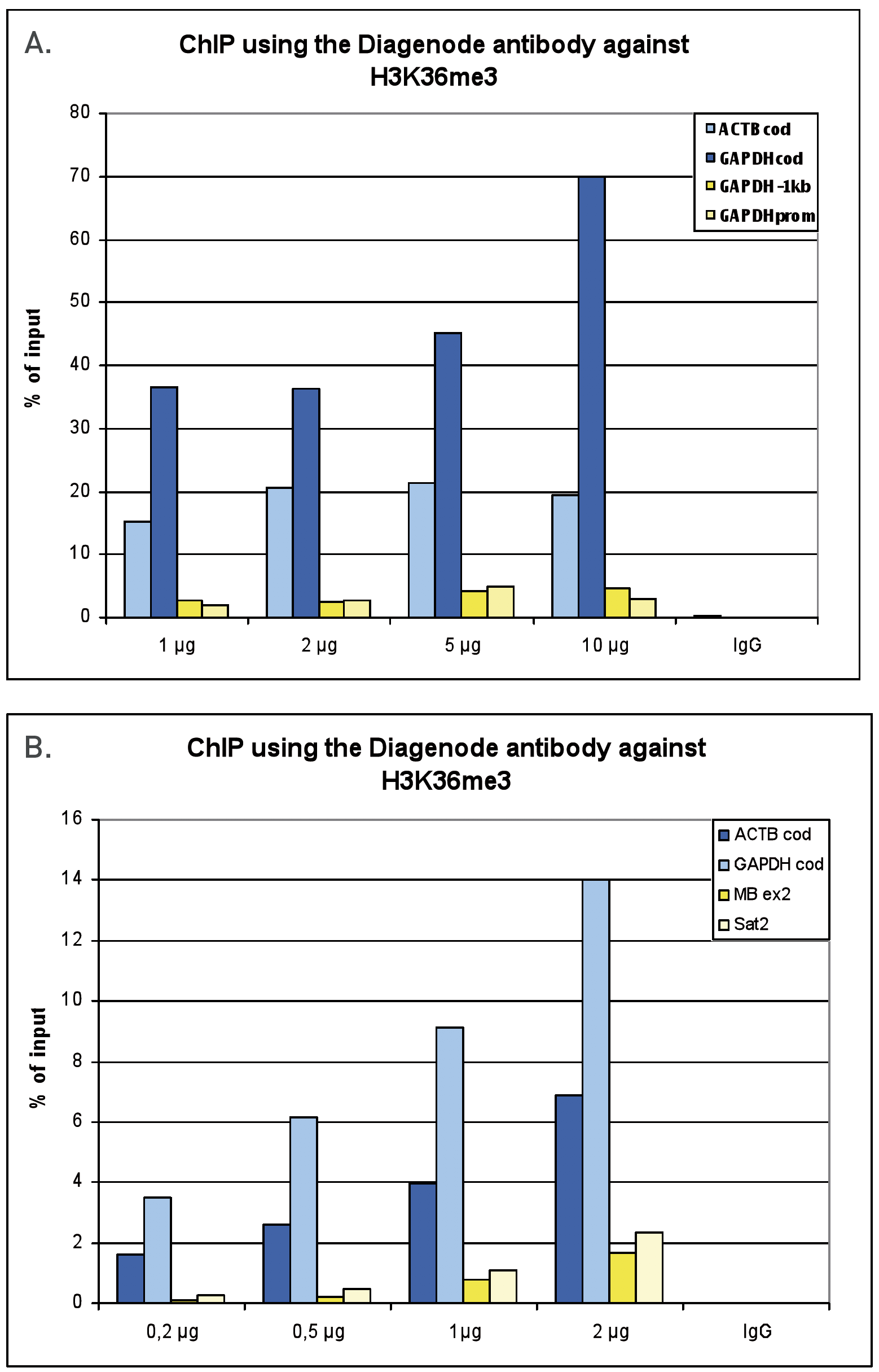
<p><small><strong>Figure 1. ChIP results obtained with the Diagenode antibody directed against H3K36me3</strong><br /> <strong>Figure 1A</strong> ChIP assays were performed using human HeLa cells, the Diagenode antibody against H3K36me3 (Cat. No. C15410192) and optimized PCR primer pairs for qPCR. ChIP was performed with the “Auto Histone ChIP-seq” kit (Cat. No. C01010022) on the IP-Star automated system, using sheared chromatin from 1,000,000 cells. A titration consisting of 1, 2, 5 and 10 µg of antibody per ChIP experiment was analyzed. IgG (2 µg/IP) was used as a negative IP control. Quantitative PCR was performed with primers for the coding region of the active GAPDH and ACTB genes, used as positive controls, and for the promoter and a region located 1 kb upstream of the promoter of the GAPDH gene, used as negative controls.<br /><br /> <strong>Figure 1B</strong> ChIP assays were performed using human K562 cells, the Diagenode antibody against H3K36me3 (Cat. No. C15410192) and optimized PCR primer pairs for qPCR. ChIP was performed with the “iDeal ChIP-seq” kit (Cat. No. C01010051), using sheared chromatin from 100,000 cells. A titration consisting of 0.2, 0.5, 1 and 2 µg of antibody per ChIP experiment was analyzed. IgG (1 µg/IP) was used as a negative IP control. Quantitative PCR was performed with primers for the coding region of the active GAPDH and ACTB genes, used as positive controls, and for the coding region of the inactive MB gene and the Sat satellite repeat, used as negative controls. Figure 1 shows the recovery, expressed as a % of input (the relative amount of immunoprecipitated DNA compared to input DNA after qPCR analysis).</small></p>
</div>
</div>
<div class="extra-spaced"></div>
<div class="row">
<div class="small-6 columns">
<p><img src="https://www.diagenode.com/img/product/antibodies/C15410192-fig2-2.jpg" alt="H3K36me3 Antibody SNAP-ChIP validation" caption="false" width="432" height="298" /></p>
</div>
<div class="small-6 columns">
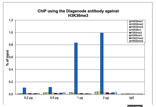
<p><small><strong>Figure 2. ChIP results obtained with the Diagenode antibody directed against H3K36me3</strong><br /> ChIP assays were performed on sheared chromatin from 1 million human HeLa cells as described above. The chromatin was spiked with a panel of in vitro assembled nucleosomes, each containing a specific lysine methylation (SNAP-ChIP K-MetStat Panel, Epicypher). A titration consisting of 0.2, 0.5, 1 and 2 µg of antibody per ChIP experiment was analyzed. IgG (1 µg/IP) was used as a negative IP control. Quantitative PCR was performed with primers specific for the nucleosomes carrying the H3K36me1, H3K36me2, H3K36me3, H3K4me3, H3K9me3, H3K27me3 and H4K20me3 modifications and the unmodified H3K4. The graph shows the recovery, expressed as a % of input. These results demonstrate a high specificity of the H3K36me3 antibody for the modification of interest.</small></p>
</div>
</div>
<div class="row">
<div class="small-12 columns">
<p><img src="https://www.diagenode.com/img/product/antibodies/C15410192-fig2.png" alt="H3K36me3 Antibody for ChIP-seq" caption="false" width="893" height="702" /></p>
</div>
</div>
<div class="row">
<div class="small-12 columns">
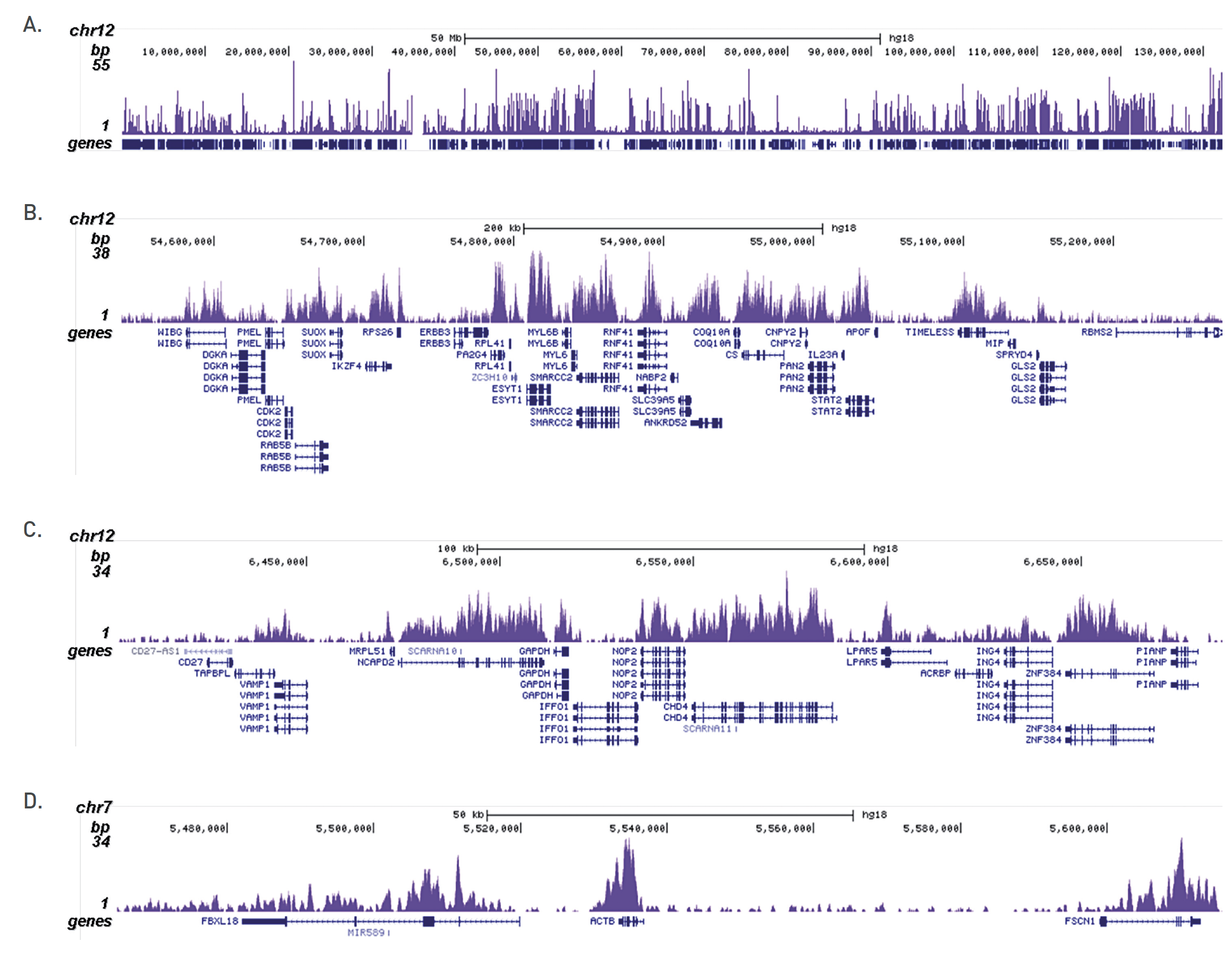
<p><small><strong>Figure 3. ChIP-seq results obtained with the Diagenode antibody directed against H3K36me3</strong><br /> ChIP was performed on sheared chromatin from 100,000 K562 cells with the “iDeal ChIP-seq” kit (Cat. No. C01010051) using 0.5 µg of the Diagenode antibody against H3K36me3 (Cat. No. C15410192) as described above. The IP’d DNA was subsequently analysed on an Illumina Genome Analyzer. Library preparation, cluster generation and sequencing were performed according to the manufacturer’s instructions. The 36 bp tags were aligned to the human genome using the ELAND algorithm. Figure 3 shows the H3K36me3 signal distribution along the complete sequence and a zoomin of human chromosome 12 (figure 2A and B) and in 2 genomic regions containing the GAPDH and ACTB positive control genes (figure 3C and D).</small></p>
</div>
</div>
<div class="row">
<div class="small-6 columns">
<p><img src="https://www.diagenode.com/img/product/antibodies/C15410192-fig3.png" alt="H3K36me3 Antibody ELISA validation" caption="false" width="432" height="328" /></p>
</div>
<div class="small-6 columns">
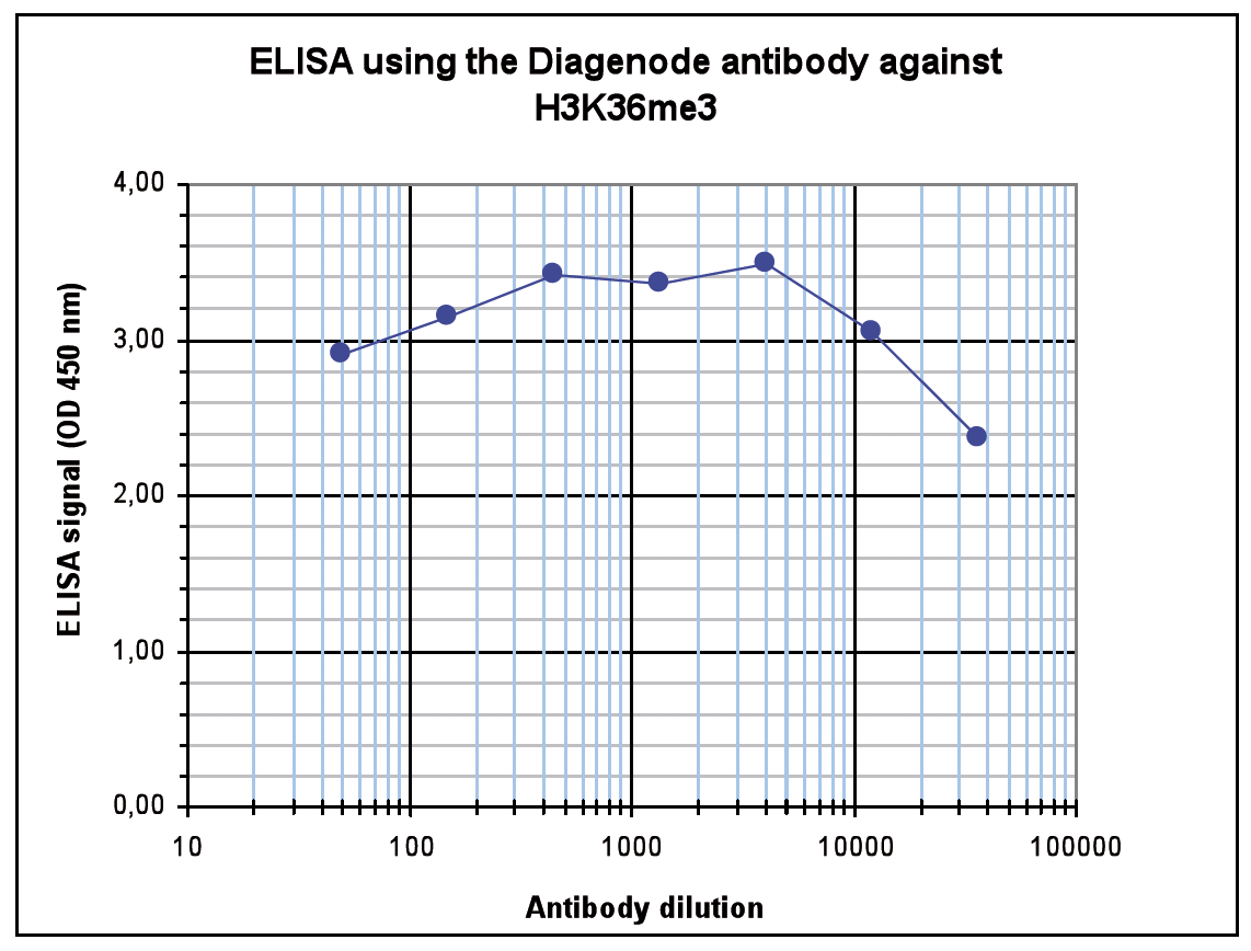
<p><small><strong>Figure 4. Determination of the antibody titer</strong><br />To determine the titer of the antibody, an ELISA was performed using a serial dilution of the Diagenode antibody against H3K36me3 (Cat. No. C15410192). The antigen used was a peptide containing the histone modification of interest. By plotting the absorbance against the antibody dilution (Figure 4), the titer of the antibody was estimated to be 1:132,000.</small></p>
</div>
</div>
<div class="row">
<div class="small-6 columns">
<p>A.<img src="https://www.diagenode.com/img/product/antibodies/C15410192-fig4-a.png" alt="H3K36me3 Antibody Dot Blot Validation" caption="false" width="432" height="162" /></p>
<p>B.<img src="https://www.diagenode.com/img/product/antibodies/C15410192-fig4-b.png" alt="H3K36me3 Antibody Peptide Array validation" caption="false" width="432" height="257" /></p>
</div>
<div class="small-6 columns">
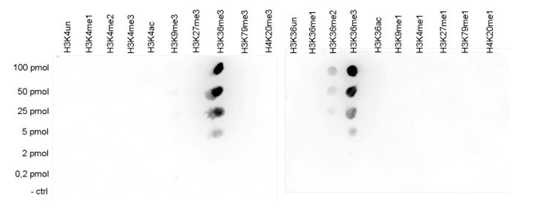
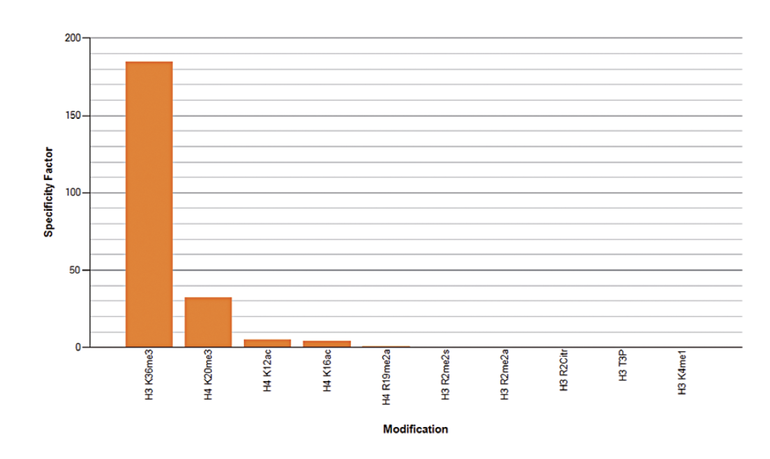
<p><small><strong>Figure 5. Cross reactivity tests using the Diagenode antibody directed against H3K36me3</strong><br /> <strong>Figure 5A.</strong> To test the cross reactivity of the Diagenode antibody against H3K36me3 (Cat. No. C15410192), a Dot Blot analysis was performed with peptides containing other modifications or unmodified sequences of histone H3 and H4. One hundred to 0.2 pmol of the respective peptides were spotted on a membrane. The antibody was used at a dilution of 1:20,000. Figure 5A shows a high specificity of the antibody for the modification of interest. <strong>Figure 5B.</strong> The specificity of the antibody was further demonstrated by peptide array analyses on an array containing 384 peptides with different combinations of modifications from histone H3, H4, H2A and H2B. The antibody was used at a dilution of 1:10,000. Figure 5B shows the specificity factor, calculated as the ratio of the average intensity of all spots containing the mark, divided by the average intensity of all spots not containing the mark. The peptide array analysis shows a slight cross reaction with H4K20me3 that was not observed in dot blot.</small></p>
</div>
</div>
<div class="extra-spaced"></div>
<div class="row">
<div class="small-6 columns">
<p><img src="https://www.diagenode.com/img/product/antibodies/C15410192-fig5.png" alt="H3K36me3 Antibody for Western Blot" caption="false" width="432" height="346" /></p>
</div>
<div class="small-6 columns">
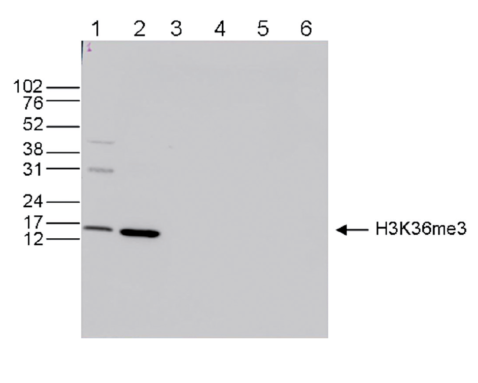
<p><small><strong>Figure 6. Western blot analysis using the Diagenode antibody directed against H3K36me3</strong><br /> Western blot was performed on whole cell (25 µg, lane 1) and histone extracts (15 µg, lane 2) from HeLa cells, and on 1 µg of recombinant histone H2A, H2B, H3 and H4 (lane 3, 4, 5 and 6, respectively) using the Diagenode antibody against H3K36me3 (Cat. No. C15410192). The antibody was diluted 1:1,000 in TBS-Tween containing 5% skimmed milk. The position of the protein of interest is shown on the right, the marker (in kDa) is shown on the left.</small></p>
</div>
</div>
<div class="row">
<div class="small-12 columns">
<p><img src="https://www.diagenode.com/img/product/antibodies/C15410192-fig6.png" alt="H3K36me3 Antibody for Immunofluorescence " caption="false" width="893" height="232" /></p>
</div>
</div>
<div class="row">
<div class="small-12 columns">
<p><small><strong>Figure 7. Immunofluorescence using the Diagenode antibody directed against H3K36me3</strong><br /> HeLa cells were stained with the Diagenode antibody against H3K36me3 (Cat. C15410192) and with DAPI. Cells were fixed with 4% formaldehyde for 10’ and blocked with PBS/TX-100 containing 5% normal goat serum and 1% BSA. The cells were immunofluorescently labeled with the H3K36me3 antibody (left) diluted 1:500 in blocking solution followed by an anti-rabbit antibody conjugated to Alexa488. The middle panel shows staining of the nuclei with DAPI. A merge of the two stainings is shown on the right.</small></p>
</div>
</div>',
'label2' => 'Target Description',
'info2' => '<p>Histones are the main constituents of the protein part of chromosomes of eukaryotic cells. They are rich in the amino acids arginine and lysine and have been greatly conserved during evolution. Histones pack the DNA into tight masses of chromatin. Two core histones of each class H2A, H2B, H3 and H4 assemble and are wrapped by 146 base pairs of DNA to form one octameric nucleosome. Histone tails undergo numerous post-translational modifications, which either directly or indirectly alter chromatin structure to facilitate transcriptional activation or repression or other nuclear processes. In addition to the genetic code, combinations of the different histone modifications reveal the so-called “histone code”. Histone methylation and demethylation is dynamically regulated by respectively histone methyl transferases and histone demethylases. Trimethylation of histone H3K36 is associated with active genes.</p>',
'label3' => '',
'info3' => '',
'format' => '50 μg',
'catalog_number' => 'C15410192',
'old_catalog_number' => 'pAb-192-050',
'sf_code' => 'C15410192-D001-000581',
'type' => 'FRE',
'search_order' => '03-Antibody',
'price_EUR' => '480',
'price_USD' => '470',
'price_GBP' => '430',
'price_JPY' => '75190',
'price_CNY' => '',
'price_AUD' => '1175',
'country' => 'ALL',
'except_countries' => 'None',
'quote' => false,
'in_stock' => false,
'featured' => false,
'no_promo' => false,
'online' => true,
'master' => true,
'last_datasheet_update' => 'December 19, 2019',
'slug' => 'h3k36me3-polyclonal-antibody-premium-50-mg',
'meta_title' => 'H3K36me3 Antibody - ChIP-seq Grade (C15410192) | Diagenode',
'meta_keywords' => '',
'meta_description' => 'H3K36me3 (Histone H3 trimethylated at lysine 36) Polyclonal Antibody validated in ChIP-seq, ChIP-grade, ELISA, DB, WB and IF. Specificity confirmed by SNAP-ChIP and Peptide array. Batch-specific data available on the website. Sample size available. ',
'modified' => '2021-10-20 09:55:18',
'created' => '2015-06-29 14:08:20',
'locale' => 'zho'
),
'Antibody' => array(
'host' => '*****',
'id' => '74',
'name' => 'H3K36me3 polyclonal antibody',
'description' => 'Histones are the main constituents of the protein part of chromosomes of eukaryotic cells. They are rich in the amino acids arginine and lysine and have been greatly conserved during evolution. Histones pack the DNA into tight masses of chromatin. Two core histones of each class H2A, H2B, H3 and H4 assemble and are wrapped by 146 base pairs of DNA to form one octameric nucleosome. Histone tails undergo numerous post-translational modifications, which either directly or indirectly alter chromatin structure to facilitate transcriptional activation or repression or other nuclear processes. In addition to the genetic code, combinations of the different histone modifications reveal the so-called “histone code”. Histone methylation and demethylation is dynamically regulated by respectively histone methyl transferases and histone demethylases. Trimethylation of histone H3K36 is associated with active genes.',
'clonality' => '',
'isotype' => '',
'lot' => 'A1845P',
'concentration' => '1.6 μg/μl',
'reactivity' => 'Human, mouse, Arabidopsis, rice, wide range expected.',
'type' => 'Polyclonal',
'purity' => 'Affinity purified polyclonal antibody.',
'classification' => 'Premium',
'application_table' => '<table>
<thead>
<tr>
<th>Applications</th>
<th>Suggested dilution</th>
<th>References</th>
</tr>
</thead>
<tbody>
<tr>
<td>ChIP/ChIP-seq <sup>*</sup></td>
<td>0.5 - 1 μg per IP</td>
<td>Fig 1, 2, 3</td>
</tr>
<tr>
<td>ELISA</td>
<td>1:4,000</td>
<td>Fig 4</td>
</tr>
<tr>
<td>Dot Blotting/Peptide array</td>
<td>1:20,000/1:10,000</td>
<td>Fig 5</td>
</tr>
<tr>
<td>Western Blotting</td>
<td>1:1,000</td>
<td>Fig 6</td>
</tr>
<tr>
<td>Immunofluorescence</td>
<td>1:500</td>
<td>Fig 7</td>
</tr>
</tbody>
</table>
<p></p>
<p><small><sup>*</sup> Please note that the optimal antibody amount per IP should be determined by the end-user. We recommend testing 0.5-5 µg per IP.</small></p>',
'storage_conditions' => 'Store at -20°C; for long storage, store at -80°C. Avoid multiple freeze-thaw cycles.',
'storage_buffer' => 'PBS containing 0.05% azide and 0.05% ProClin 300.',
'precautions' => 'This product is for research use only. Not for use in diagnostic or therapeutic procedures.',
'uniprot_acc' => '',
'slug' => '',
'meta_keywords' => '',
'meta_description' => '',
'modified' => '2020-02-26 18:52:53',
'created' => '0000-00-00 00:00:00',
'select_label' => '74 - H3K36me3 polyclonal antibody (A1845P - 1.6 μg/μl - Human, mouse, Arabidopsis, rice, wide range expected. - Affinity purified polyclonal antibody. - Rabbit)'
),
'Slave' => array(
(int) 0 => array(
[maximum depth reached]
)
),
'Group' => array(
'Group' => array(
[maximum depth reached]
),
'Master' => array(
[maximum depth reached]
),
'Product' => array(
[maximum depth reached]
)
),
'Related' => array(
(int) 0 => array(
[maximum depth reached]
),
(int) 1 => array(
[maximum depth reached]
),
(int) 2 => array(
[maximum depth reached]
),
(int) 3 => array(
[maximum depth reached]
),
(int) 4 => array(
[maximum depth reached]
),
(int) 5 => array(
[maximum depth reached]
),
(int) 6 => array(
[maximum depth reached]
)
),
'Application' => array(
(int) 0 => array(
[maximum depth reached]
),
(int) 1 => array(
[maximum depth reached]
),
(int) 2 => array(
[maximum depth reached]
),
(int) 3 => array(
[maximum depth reached]
),
(int) 4 => array(
[maximum depth reached]
),
(int) 5 => array(
[maximum depth reached]
)
),
'Category' => array(
(int) 0 => array(
[maximum depth reached]
),
(int) 1 => array(
[maximum depth reached]
),
(int) 2 => array(
[maximum depth reached]
),
(int) 3 => array(
[maximum depth reached]
)
),
'Document' => array(
(int) 0 => array(
[maximum depth reached]
),
(int) 1 => array(
[maximum depth reached]
),
(int) 2 => array(
[maximum depth reached]
)
),
'Feature' => array(),
'Image' => array(
(int) 0 => array(
[maximum depth reached]
)
),
'Promotion' => array(),
'Protocol' => array(),
'Publication' => array(
(int) 0 => array(
[maximum depth reached]
),
(int) 1 => array(
[maximum depth reached]
),
(int) 2 => array(
[maximum depth reached]
),
(int) 3 => array(
[maximum depth reached]
),
(int) 4 => array(
[maximum depth reached]
),
(int) 5 => array(
[maximum depth reached]
),
(int) 6 => array(
[maximum depth reached]
),
(int) 7 => array(
[maximum depth reached]
),
(int) 8 => array(
[maximum depth reached]
),
(int) 9 => array(
[maximum depth reached]
),
(int) 10 => array(
[maximum depth reached]
),
(int) 11 => array(
[maximum depth reached]
),
(int) 12 => array(
[maximum depth reached]
),
(int) 13 => array(
[maximum depth reached]
),
(int) 14 => array(
[maximum depth reached]
),
(int) 15 => array(
[maximum depth reached]
),
(int) 16 => array(
[maximum depth reached]
),
(int) 17 => array(
[maximum depth reached]
),
(int) 18 => array(
[maximum depth reached]
),
(int) 19 => array(
[maximum depth reached]
),
(int) 20 => array(
[maximum depth reached]
),
(int) 21 => array(
[maximum depth reached]
),
(int) 22 => array(
[maximum depth reached]
),
(int) 23 => array(
[maximum depth reached]
),
(int) 24 => array(
[maximum depth reached]
),
(int) 25 => array(
[maximum depth reached]
),
(int) 26 => array(
[maximum depth reached]
),
(int) 27 => array(
[maximum depth reached]
),
(int) 28 => array(
[maximum depth reached]
),
(int) 29 => array(
[maximum depth reached]
),
(int) 30 => array(
[maximum depth reached]
),
(int) 31 => array(
[maximum depth reached]
),
(int) 32 => array(
[maximum depth reached]
)
),
'Testimonial' => array(
(int) 0 => array(
[maximum depth reached]
)
),
'Area' => array(),
'SafetySheet' => array(
(int) 0 => array(
[maximum depth reached]
),
(int) 1 => array(
[maximum depth reached]
),
(int) 2 => array(
[maximum depth reached]
),
(int) 3 => array(
[maximum depth reached]
),
(int) 4 => array(
[maximum depth reached]
),
(int) 5 => array(
[maximum depth reached]
),
(int) 6 => array(
[maximum depth reached]
),
(int) 7 => array(
[maximum depth reached]
)
)
),
'meta_canonical' => 'https://www.diagenode.com/cn/p/h3k36me3-polyclonal-antibody-premium-50-mg'
)
$language = 'cn'
$meta_keywords = ''
$meta_description = 'H3K36me3 (Histone H3 trimethylated at lysine 36) Polyclonal Antibody validated in ChIP-seq, ChIP-grade, ELISA, DB, WB and IF. Specificity confirmed by SNAP-ChIP and Peptide array. Batch-specific data available on the website. Sample size available. '
$meta_title = 'H3K36me3 Antibody - ChIP-seq Grade (C15410192) | Diagenode'
$product = array(
'Product' => array(
'id' => '2262',
'antibody_id' => '74',
'name' => 'H3K36me3 Antibody - ChIP-seq Grade',
'description' => '<p>Polyclonal antibody raised in rabbit against the region of histone H3 containing the trimethylated lysine 36 (<strong>H3K36me3</strong>), using a KLH-conjugated synthetic peptide.</p>',
'label1' => 'Validation Data',
'info1' => '<div class="row">
<div class="small-6 columns">
<p><img src="https://www.diagenode.com/img/product/antibodies/C15410192-fig1.png" alt="H3K36me3 Antibody ChIP Grade" caption="false" width="432" height="674" /></p>
</div>
<div class="small-6 columns">
<p><small><strong>Figure 1. ChIP results obtained with the Diagenode antibody directed against H3K36me3</strong><br /> <strong>Figure 1A</strong> ChIP assays were performed using human HeLa cells, the Diagenode antibody against H3K36me3 (Cat. No. C15410192) and optimized PCR primer pairs for qPCR. ChIP was performed with the “Auto Histone ChIP-seq” kit (Cat. No. C01010022) on the IP-Star automated system, using sheared chromatin from 1,000,000 cells. A titration consisting of 1, 2, 5 and 10 µg of antibody per ChIP experiment was analyzed. IgG (2 µg/IP) was used as a negative IP control. Quantitative PCR was performed with primers for the coding region of the active GAPDH and ACTB genes, used as positive controls, and for the promoter and a region located 1 kb upstream of the promoter of the GAPDH gene, used as negative controls.<br /><br /> <strong>Figure 1B</strong> ChIP assays were performed using human K562 cells, the Diagenode antibody against H3K36me3 (Cat. No. C15410192) and optimized PCR primer pairs for qPCR. ChIP was performed with the “iDeal ChIP-seq” kit (Cat. No. C01010051), using sheared chromatin from 100,000 cells. A titration consisting of 0.2, 0.5, 1 and 2 µg of antibody per ChIP experiment was analyzed. IgG (1 µg/IP) was used as a negative IP control. Quantitative PCR was performed with primers for the coding region of the active GAPDH and ACTB genes, used as positive controls, and for the coding region of the inactive MB gene and the Sat satellite repeat, used as negative controls. Figure 1 shows the recovery, expressed as a % of input (the relative amount of immunoprecipitated DNA compared to input DNA after qPCR analysis).</small></p>
</div>
</div>
<div class="extra-spaced"></div>
<div class="row">
<div class="small-6 columns">
<p><img src="https://www.diagenode.com/img/product/antibodies/C15410192-fig2-2.jpg" alt="H3K36me3 Antibody SNAP-ChIP validation" caption="false" width="432" height="298" /></p>
</div>
<div class="small-6 columns">
<p><small><strong>Figure 2. ChIP results obtained with the Diagenode antibody directed against H3K36me3</strong><br /> ChIP assays were performed on sheared chromatin from 1 million human HeLa cells as described above. The chromatin was spiked with a panel of in vitro assembled nucleosomes, each containing a specific lysine methylation (SNAP-ChIP K-MetStat Panel, Epicypher). A titration consisting of 0.2, 0.5, 1 and 2 µg of antibody per ChIP experiment was analyzed. IgG (1 µg/IP) was used as a negative IP control. Quantitative PCR was performed with primers specific for the nucleosomes carrying the H3K36me1, H3K36me2, H3K36me3, H3K4me3, H3K9me3, H3K27me3 and H4K20me3 modifications and the unmodified H3K4. The graph shows the recovery, expressed as a % of input. These results demonstrate a high specificity of the H3K36me3 antibody for the modification of interest.</small></p>
</div>
</div>
<div class="row">
<div class="small-12 columns">
<p><img src="https://www.diagenode.com/img/product/antibodies/C15410192-fig2.png" alt="H3K36me3 Antibody for ChIP-seq" caption="false" width="893" height="702" /></p>
</div>
</div>
<div class="row">
<div class="small-12 columns">
<p><small><strong>Figure 3. ChIP-seq results obtained with the Diagenode antibody directed against H3K36me3</strong><br /> ChIP was performed on sheared chromatin from 100,000 K562 cells with the “iDeal ChIP-seq” kit (Cat. No. C01010051) using 0.5 µg of the Diagenode antibody against H3K36me3 (Cat. No. C15410192) as described above. The IP’d DNA was subsequently analysed on an Illumina Genome Analyzer. Library preparation, cluster generation and sequencing were performed according to the manufacturer’s instructions. The 36 bp tags were aligned to the human genome using the ELAND algorithm. Figure 3 shows the H3K36me3 signal distribution along the complete sequence and a zoomin of human chromosome 12 (figure 2A and B) and in 2 genomic regions containing the GAPDH and ACTB positive control genes (figure 3C and D).</small></p>
</div>
</div>
<div class="row">
<div class="small-6 columns">
<p><img src="https://www.diagenode.com/img/product/antibodies/C15410192-fig3.png" alt="H3K36me3 Antibody ELISA validation" caption="false" width="432" height="328" /></p>
</div>
<div class="small-6 columns">
<p><small><strong>Figure 4. Determination of the antibody titer</strong><br />To determine the titer of the antibody, an ELISA was performed using a serial dilution of the Diagenode antibody against H3K36me3 (Cat. No. C15410192). The antigen used was a peptide containing the histone modification of interest. By plotting the absorbance against the antibody dilution (Figure 4), the titer of the antibody was estimated to be 1:132,000.</small></p>
</div>
</div>
<div class="row">
<div class="small-6 columns">
<p>A.<img src="https://www.diagenode.com/img/product/antibodies/C15410192-fig4-a.png" alt="H3K36me3 Antibody Dot Blot Validation" caption="false" width="432" height="162" /></p>
<p>B.<img src="https://www.diagenode.com/img/product/antibodies/C15410192-fig4-b.png" alt="H3K36me3 Antibody Peptide Array validation" caption="false" width="432" height="257" /></p>
</div>
<div class="small-6 columns">
<p><small><strong>Figure 5. Cross reactivity tests using the Diagenode antibody directed against H3K36me3</strong><br /> <strong>Figure 5A.</strong> To test the cross reactivity of the Diagenode antibody against H3K36me3 (Cat. No. C15410192), a Dot Blot analysis was performed with peptides containing other modifications or unmodified sequences of histone H3 and H4. One hundred to 0.2 pmol of the respective peptides were spotted on a membrane. The antibody was used at a dilution of 1:20,000. Figure 5A shows a high specificity of the antibody for the modification of interest. <strong>Figure 5B.</strong> The specificity of the antibody was further demonstrated by peptide array analyses on an array containing 384 peptides with different combinations of modifications from histone H3, H4, H2A and H2B. The antibody was used at a dilution of 1:10,000. Figure 5B shows the specificity factor, calculated as the ratio of the average intensity of all spots containing the mark, divided by the average intensity of all spots not containing the mark. The peptide array analysis shows a slight cross reaction with H4K20me3 that was not observed in dot blot.</small></p>
</div>
</div>
<div class="extra-spaced"></div>
<div class="row">
<div class="small-6 columns">
<p><img src="https://www.diagenode.com/img/product/antibodies/C15410192-fig5.png" alt="H3K36me3 Antibody for Western Blot" caption="false" width="432" height="346" /></p>
</div>
<div class="small-6 columns">
<p><small><strong>Figure 6. Western blot analysis using the Diagenode antibody directed against H3K36me3</strong><br /> Western blot was performed on whole cell (25 µg, lane 1) and histone extracts (15 µg, lane 2) from HeLa cells, and on 1 µg of recombinant histone H2A, H2B, H3 and H4 (lane 3, 4, 5 and 6, respectively) using the Diagenode antibody against H3K36me3 (Cat. No. C15410192). The antibody was diluted 1:1,000 in TBS-Tween containing 5% skimmed milk. The position of the protein of interest is shown on the right, the marker (in kDa) is shown on the left.</small></p>
</div>
</div>
<div class="row">
<div class="small-12 columns">
<p><img src="https://www.diagenode.com/img/product/antibodies/C15410192-fig6.png" alt="H3K36me3 Antibody for Immunofluorescence " caption="false" width="893" height="232" /></p>
</div>
</div>
<div class="row">
<div class="small-12 columns">
<p><small><strong>Figure 7. Immunofluorescence using the Diagenode antibody directed against H3K36me3</strong><br /> HeLa cells were stained with the Diagenode antibody against H3K36me3 (Cat. C15410192) and with DAPI. Cells were fixed with 4% formaldehyde for 10’ and blocked with PBS/TX-100 containing 5% normal goat serum and 1% BSA. The cells were immunofluorescently labeled with the H3K36me3 antibody (left) diluted 1:500 in blocking solution followed by an anti-rabbit antibody conjugated to Alexa488. The middle panel shows staining of the nuclei with DAPI. A merge of the two stainings is shown on the right.</small></p>
</div>
</div>',
'label2' => 'Target Description',
'info2' => '<p>Histones are the main constituents of the protein part of chromosomes of eukaryotic cells. They are rich in the amino acids arginine and lysine and have been greatly conserved during evolution. Histones pack the DNA into tight masses of chromatin. Two core histones of each class H2A, H2B, H3 and H4 assemble and are wrapped by 146 base pairs of DNA to form one octameric nucleosome. Histone tails undergo numerous post-translational modifications, which either directly or indirectly alter chromatin structure to facilitate transcriptional activation or repression or other nuclear processes. In addition to the genetic code, combinations of the different histone modifications reveal the so-called “histone code”. Histone methylation and demethylation is dynamically regulated by respectively histone methyl transferases and histone demethylases. Trimethylation of histone H3K36 is associated with active genes.</p>',
'label3' => '',
'info3' => '',
'format' => '50 μg',
'catalog_number' => 'C15410192',
'old_catalog_number' => 'pAb-192-050',
'sf_code' => 'C15410192-D001-000581',
'type' => 'FRE',
'search_order' => '03-Antibody',
'price_EUR' => '480',
'price_USD' => '470',
'price_GBP' => '430',
'price_JPY' => '75190',
'price_CNY' => '',
'price_AUD' => '1175',
'country' => 'ALL',
'except_countries' => 'None',
'quote' => false,
'in_stock' => false,
'featured' => false,
'no_promo' => false,
'online' => true,
'master' => true,
'last_datasheet_update' => 'December 19, 2019',
'slug' => 'h3k36me3-polyclonal-antibody-premium-50-mg',
'meta_title' => 'H3K36me3 Antibody - ChIP-seq Grade (C15410192) | Diagenode',
'meta_keywords' => '',
'meta_description' => 'H3K36me3 (Histone H3 trimethylated at lysine 36) Polyclonal Antibody validated in ChIP-seq, ChIP-grade, ELISA, DB, WB and IF. Specificity confirmed by SNAP-ChIP and Peptide array. Batch-specific data available on the website. Sample size available. ',
'modified' => '2021-10-20 09:55:18',
'created' => '2015-06-29 14:08:20',
'locale' => 'zho'
),
'Antibody' => array(
'host' => '*****',
'id' => '74',
'name' => 'H3K36me3 polyclonal antibody',
'description' => 'Histones are the main constituents of the protein part of chromosomes of eukaryotic cells. They are rich in the amino acids arginine and lysine and have been greatly conserved during evolution. Histones pack the DNA into tight masses of chromatin. Two core histones of each class H2A, H2B, H3 and H4 assemble and are wrapped by 146 base pairs of DNA to form one octameric nucleosome. Histone tails undergo numerous post-translational modifications, which either directly or indirectly alter chromatin structure to facilitate transcriptional activation or repression or other nuclear processes. In addition to the genetic code, combinations of the different histone modifications reveal the so-called “histone code”. Histone methylation and demethylation is dynamically regulated by respectively histone methyl transferases and histone demethylases. Trimethylation of histone H3K36 is associated with active genes.',
'clonality' => '',
'isotype' => '',
'lot' => 'A1845P',
'concentration' => '1.6 μg/μl',
'reactivity' => 'Human, mouse, Arabidopsis, rice, wide range expected.',
'type' => 'Polyclonal',
'purity' => 'Affinity purified polyclonal antibody.',
'classification' => 'Premium',
'application_table' => '<table>
<thead>
<tr>
<th>Applications</th>
<th>Suggested dilution</th>
<th>References</th>
</tr>
</thead>
<tbody>
<tr>
<td>ChIP/ChIP-seq <sup>*</sup></td>
<td>0.5 - 1 μg per IP</td>
<td>Fig 1, 2, 3</td>
</tr>
<tr>
<td>ELISA</td>
<td>1:4,000</td>
<td>Fig 4</td>
</tr>
<tr>
<td>Dot Blotting/Peptide array</td>
<td>1:20,000/1:10,000</td>
<td>Fig 5</td>
</tr>
<tr>
<td>Western Blotting</td>
<td>1:1,000</td>
<td>Fig 6</td>
</tr>
<tr>
<td>Immunofluorescence</td>
<td>1:500</td>
<td>Fig 7</td>
</tr>
</tbody>
</table>
<p></p>
<p><small><sup>*</sup> Please note that the optimal antibody amount per IP should be determined by the end-user. We recommend testing 0.5-5 µg per IP.</small></p>',
'storage_conditions' => 'Store at -20°C; for long storage, store at -80°C. Avoid multiple freeze-thaw cycles.',
'storage_buffer' => 'PBS containing 0.05% azide and 0.05% ProClin 300.',
'precautions' => 'This product is for research use only. Not for use in diagnostic or therapeutic procedures.',
'uniprot_acc' => '',
'slug' => '',
'meta_keywords' => '',
'meta_description' => '',
'modified' => '2020-02-26 18:52:53',
'created' => '0000-00-00 00:00:00',
'select_label' => '74 - H3K36me3 polyclonal antibody (A1845P - 1.6 μg/μl - Human, mouse, Arabidopsis, rice, wide range expected. - Affinity purified polyclonal antibody. - Rabbit)'
),
'Slave' => array(
(int) 0 => array(
'id' => '44',
'name' => 'C15410192',
'product_id' => '2262',
'modified' => '2016-02-18 20:49:25',
'created' => '2016-02-18 20:49:25'
)
),
'Group' => array(
'Group' => array(
'id' => '44',
'name' => 'C15410192',
'product_id' => '2262',
'modified' => '2016-02-18 20:49:25',
'created' => '2016-02-18 20:49:25'
),
'Master' => array(
'id' => '2262',
'antibody_id' => '74',
'name' => 'H3K36me3 Antibody',
'description' => '<p>Polyclonal antibody raised in rabbit against the region of histone <strong>H3 containing the trimethylated lysine 36</strong> (<strong>H3K36me3</strong>), using a KLH-conjugated synthetic peptide.</p>',
'label1' => 'Validation Data',
'info1' => '<div class="row">
<div class="small-6 columns">
<p><img src="https://www.diagenode.com/img/product/antibodies/C15410192-fig1.png" alt="H3K36me3 Antibody ChIP Grade" caption="false" width="432" height="674" /></p>
</div>
<div class="small-6 columns">
<p><small><strong>Figure 1. ChIP results obtained with the Diagenode antibody directed against H3K36me3</strong><br /> <strong>Figure 1A</strong> ChIP assays were performed using human HeLa cells, the Diagenode antibody against H3K36me3 (Cat. No. C15410192) and optimized PCR primer pairs for qPCR. ChIP was performed with the “Auto Histone ChIP-seq” kit (Cat. No. C01010022) on the IP-Star automated system, using sheared chromatin from 1,000,000 cells. A titration consisting of 1, 2, 5 and 10 µg of antibody per ChIP experiment was analyzed. IgG (2 µg/IP) was used as a negative IP control. Quantitative PCR was performed with primers for the coding region of the active GAPDH and ACTB genes, used as positive controls, and for the promoter and a region located 1 kb upstream of the promoter of the GAPDH gene, used as negative controls.<br /><br /> <strong>Figure 1B</strong> ChIP assays were performed using human K562 cells, the Diagenode antibody against H3K36me3 (Cat. No. C15410192) and optimized PCR primer pairs for qPCR. ChIP was performed with the “iDeal ChIP-seq” kit (Cat. No. C01010051), using sheared chromatin from 100,000 cells. A titration consisting of 0.2, 0.5, 1 and 2 µg of antibody per ChIP experiment was analyzed. IgG (1 µg/IP) was used as a negative IP control. Quantitative PCR was performed with primers for the coding region of the active GAPDH and ACTB genes, used as positive controls, and for the coding region of the inactive MB gene and the Sat satellite repeat, used as negative controls. Figure 1 shows the recovery, expressed as a % of input (the relative amount of immunoprecipitated DNA compared to input DNA after qPCR analysis).</small></p>
</div>
</div>
<div class="extra-spaced"></div>
<div class="row">
<div class="small-6 columns">
<p><img src="https://www.diagenode.com/img/product/antibodies/C15410192-fig2-2.jpg" alt="H3K36me3 Antibody SNAP-ChIP validation" caption="false" width="432" height="298" /></p>
</div>
<div class="small-6 columns">
<p><small><strong>Figure 2. ChIP results obtained with the Diagenode antibody directed against H3K36me3</strong><br /> ChIP assays were performed on sheared chromatin from 1 million human HeLa cells as described above. The chromatin was spiked with a panel of in vitro assembled nucleosomes, each containing a specific lysine methylation (SNAP-ChIP K-MetStat Panel, Epicypher). A titration consisting of 0.2, 0.5, 1 and 2 µg of antibody per ChIP experiment was analyzed. IgG (1 µg/IP) was used as a negative IP control. Quantitative PCR was performed with primers specific for the nucleosomes carrying the H3K36me1, H3K36me2, H3K36me3, H3K4me3, H3K9me3, H3K27me3 and H4K20me3 modifications and the unmodified H3K4. The graph shows the recovery, expressed as a % of input. These results demonstrate a high specificity of the H3K36me3 antibody for the modification of interest.</small></p>
</div>
</div>
<div class="row">
<div class="small-12 columns">
<p><img src="https://www.diagenode.com/img/product/antibodies/C15410192-fig2.png" alt="H3K36me3 Antibody for ChIP-seq" caption="false" width="893" height="702" /></p>
</div>
</div>
<div class="row">
<div class="small-12 columns">
<p><small><strong>Figure 3. ChIP-seq results obtained with the Diagenode antibody directed against H3K36me3</strong><br /> ChIP was performed on sheared chromatin from 100,000 K562 cells with the “iDeal ChIP-seq” kit (Cat. No. C01010051) using 0.5 µg of the Diagenode antibody against H3K36me3 (Cat. No. C15410192) as described above. The IP’d DNA was subsequently analysed on an Illumina Genome Analyzer. Library preparation, cluster generation and sequencing were performed according to the manufacturer’s instructions. The 36 bp tags were aligned to the human genome using the ELAND algorithm. Figure 3 shows the H3K36me3 signal distribution along the complete sequence and a zoomin of human chromosome 12 (figure 2A and B) and in 2 genomic regions containing the GAPDH and ACTB positive control genes (figure 3C and D).</small></p>
</div>
</div>
<div class="row">
<div class="small-6 columns">
<p><img src="https://www.diagenode.com/img/product/antibodies/C15410192-fig3.png" alt="H3K36me3 Antibody ELISA validation" caption="false" width="432" height="328" /></p>
</div>
<div class="small-6 columns">
<p><small><strong>Figure 4. Determination of the antibody titer</strong><br />To determine the titer of the antibody, an ELISA was performed using a serial dilution of the Diagenode antibody against H3K36me3 (Cat. No. C15410192). The antigen used was a peptide containing the histone modification of interest. By plotting the absorbance against the antibody dilution (Figure 4), the titer of the antibody was estimated to be 1:132,000.</small></p>
</div>
</div>
<div class="row">
<div class="small-6 columns">
<p>A.<img src="https://www.diagenode.com/img/product/antibodies/C15410192-fig4-a.png" alt="H3K36me3 Antibody Dot Blot Validation" caption="false" width="432" height="162" /></p>
<p>B.<img src="https://www.diagenode.com/img/product/antibodies/C15410192-fig4-b.png" alt="H3K36me3 Antibody Peptide Array validation" caption="false" width="432" height="257" /></p>
</div>
<div class="small-6 columns">
<p><small><strong>Figure 5. Cross reactivity tests using the Diagenode antibody directed against H3K36me3</strong><br /> <strong>Figure 5A.</strong> To test the cross reactivity of the Diagenode antibody against H3K36me3 (Cat. No. C15410192), a Dot Blot analysis was performed with peptides containing other modifications or unmodified sequences of histone H3 and H4. One hundred to 0.2 pmol of the respective peptides were spotted on a membrane. The antibody was used at a dilution of 1:20,000. Figure 5A shows a high specificity of the antibody for the modification of interest. <strong>Figure 5B.</strong> The specificity of the antibody was further demonstrated by peptide array analyses on an array containing 384 peptides with different combinations of modifications from histone H3, H4, H2A and H2B. The antibody was used at a dilution of 1:10,000. Figure 5B shows the specificity factor, calculated as the ratio of the average intensity of all spots containing the mark, divided by the average intensity of all spots not containing the mark. The peptide array analysis shows a slight cross reaction with H4K20me3 that was not observed in dot blot.</small></p>
</div>
</div>
<div class="extra-spaced"></div>
<div class="row">
<div class="small-6 columns">
<p><img src="https://www.diagenode.com/img/product/antibodies/C15410192-fig5.png" alt="H3K36me3 Antibody for Western Blot" caption="false" width="432" height="346" /></p>
</div>
<div class="small-6 columns">
<p><small><strong>Figure 6. Western blot analysis using the Diagenode antibody directed against H3K36me3</strong><br /> Western blot was performed on whole cell (25 µg, lane 1) and histone extracts (15 µg, lane 2) from HeLa cells, and on 1 µg of recombinant histone H2A, H2B, H3 and H4 (lane 3, 4, 5 and 6, respectively) using the Diagenode antibody against H3K36me3 (Cat. No. C15410192). The antibody was diluted 1:1,000 in TBS-Tween containing 5% skimmed milk. The position of the protein of interest is shown on the right, the marker (in kDa) is shown on the left.</small></p>
</div>
</div>
<div class="row">
<div class="small-12 columns">
<p><img src="https://www.diagenode.com/img/product/antibodies/C15410192-fig6.png" alt="H3K36me3 Antibody for Immunofluorescence " caption="false" width="893" height="232" /></p>
</div>
</div>
<div class="row">
<div class="small-12 columns">
<p><small><strong>Figure 7. Immunofluorescence using the Diagenode antibody directed against H3K36me3</strong><br /> HeLa cells were stained with the Diagenode antibody against H3K36me3 (Cat. C15410192) and with DAPI. Cells were fixed with 4% formaldehyde for 10’ and blocked with PBS/TX-100 containing 5% normal goat serum and 1% BSA. The cells were immunofluorescently labeled with the H3K36me3 antibody (left) diluted 1:500 in blocking solution followed by an anti-rabbit antibody conjugated to Alexa488. The middle panel shows staining of the nuclei with DAPI. A merge of the two stainings is shown on the right.</small></p>
</div>
</div>',
'label2' => 'Target Description',
'info2' => '<p>Histones are the main constituents of the protein part of chromosomes of eukaryotic cells. They are rich in the amino acids arginine and lysine and have been greatly conserved during evolution. Histones pack the DNA into tight masses of chromatin. Two core histones of each class H2A, H2B, H3 and H4 assemble and are wrapped by 146 base pairs of DNA to form one octameric nucleosome. Histone tails undergo numerous post-translational modifications, which either directly or indirectly alter chromatin structure to facilitate transcriptional activation or repression or other nuclear processes. In addition to the genetic code, combinations of the different histone modifications reveal the so-called “histone code”. Histone methylation and demethylation is dynamically regulated by respectively histone methyl transferases and histone demethylases. Trimethylation of histone H3K36 is associated with active genes.</p>',
'label3' => '',
'info3' => '',
'format' => '50 μg',
'catalog_number' => 'C15410192',
'old_catalog_number' => 'pAb-192-050',
'sf_code' => 'C15410192-D001-000581',
'type' => 'FRE',
'search_order' => '03-Antibody',
'price_EUR' => '480',
'price_USD' => '470',
'price_GBP' => '430',
'price_JPY' => '75190',
'price_CNY' => '',
'price_AUD' => '1175',
'country' => 'ALL',
'except_countries' => 'None',
'quote' => false,
'in_stock' => false,
'featured' => false,
'no_promo' => false,
'online' => true,
'master' => true,
'last_datasheet_update' => 'December 19, 2019',
'slug' => 'h3k36me3-polyclonal-antibody-premium-50-mg',
'meta_title' => 'H3K36me3 Antibody - ChIP-seq Grade (C15410192) | Diagenode',
'meta_keywords' => '',
'meta_description' => 'H3K36me3 (Histone H3 trimethylated at lysine 36) Polyclonal Antibody validated in ChIP-seq, ChIP-grade, ELISA, DB, WB and IF. Specificity confirmed by Peptide array. Batch-specific data available on the website. Sample size available. ',
'modified' => '2021-10-20 09:55:18',
'created' => '2015-06-29 14:08:20'
),
'Product' => array(
(int) 0 => array(
[maximum depth reached]
)
)
),
'Related' => array(
(int) 0 => array(
'id' => '1836',
'antibody_id' => null,
'name' => 'iDeal ChIP-seq kit for Histones',
'description' => '<p><a href="https://www.diagenode.com/files/products/kits/ideal-chipseq-for-histones-complete-manual.pdf"><img src="https://www.diagenode.com/img/buttons/bt-manual.png" /></a></p>
<p>Don’t risk wasting your precious sequencing samples. Diagenode’s validated <strong>iDeal ChIP-seq kit for Histones</strong> has everything you need for a successful start-to-finish <strong>ChIP of histones prior to Next-Generation Sequencing</strong>. The complete kit contains all buffers and reagents for cell lysis, chromatin shearing, immunoprecipitation and DNA purification. In addition, unlike competing solutions, the kit contains positive and negative control antibodies (H3K4me3 and IgG, respectively) as well as positive and negative control PCR primers pairs (GAPDH TSS and Myoglobin exon 2, respectively) for your convenience and a guarantee of optimal results. The kit has been validated on multiple histone marks.</p>
<p> The iDeal ChIP-seq kit for Histones<strong> </strong>is perfect for <strong>cells</strong> (<strong>100,000 cells</strong> to <strong>1,000,000 cells</strong> per IP) and has been validated for <strong>tissues</strong> (<strong>1.5 mg</strong> to <strong>5 mg</strong> of tissue per IP).</p>
<p> The iDeal ChIP-seq kit is the only kit on the market validated for the major sequencing systems. Our expertise in ChIP-seq tools allows reproducible and efficient results every time.</p>
<p></p>
<p> <strong></strong></p>
<p></p>',
'label1' => 'Characteristics',
'info1' => '<ul style="list-style-type: disc;">
<li>Highly <strong>optimized</strong> protocol for ChIP-seq from cells and tissues</li>
<li><strong>Validated</strong> for ChIP-seq with multiple histones marks</li>
<li>Most <strong>complete</strong> kit available (covers all steps, including the control antibodies and primers)</li>
<li>Optimized chromatin preparation in combination with the Bioruptor ensuring the best <strong>epitope integrity</strong></li>
<li>Magnetic beads make ChIP easy, fast and more <strong>reproducible</strong></li>
<li>Combination with Diagenode ChIP-seq antibodies provides high yields with excellent <strong>specificity</strong> and <strong>sensitivity</strong></li>
<li>Purified DNA suitable for any downstream application</li>
<li>Easy-to-follow protocol</li>
</ul>
<p>Note: to obtain optimal results, this kit should be used in combination with the DiaMag1.5 - magnetic rack.</p>
<h3>ChIP-seq on cells</h3>
<p><img src="https://www.diagenode.com/img/product/kits/iDeal-kit-C01010053-figure-1.jpg" alt="Figure 1A" style="display: block; margin-left: auto; margin-right: auto;" /></p>
<p><strong>Figure 1A. The high consistency of the iDeal ChIP-seq kit on the Ion Torrent™ PGM™ (Life Technologies) and GAIIx (Illumina<sup>®</sup>)</strong><br /> ChIP was performed on sheared chromatin from 1 million HelaS3 cells using the iDeal ChIP-seq kit and 1 µg of H3K4me3 positive control antibody. Two different biological samples have been analyzed using two different sequencers - GAIIx (Illumina<sup>®</sup>) and PGM™ (Ion Torrent™). The expected ChIP-seq profile for H3K4me3 on the GAPDH promoter region has been obtained.<br /> Image A shows a several hundred bp along chr12 with high similarity of read distribution despite the radically different sequencers. Image B is a close capture focusing on the GAPDH that shows that even the peak structure is similar.</p>
<p class="text-center"><strong>Perfect match between ChIP-seq data obtained with the iDeal ChIP-seq workflow and reference dataset</strong></p>
<p><img src="https://www.diagenode.com/img/product/kits/perfect-match-between-chipseq-data.png" alt="Figure 1B" style="display: block; margin-left: auto; margin-right: auto;" /></p>
<p><strong>Figure 1B.</strong> The ChIP-seq dataset from this experiment has been compared with a reference dataset from the Broad Institute. We observed a perfect match between the top 40% of Diagenode peaks and the reference dataset. Based on the NIH Encode project criterion, ChIP-seq results are considered reproducible between an original and reproduced dataset if the top 40% of peaks have at least an 80% overlap ratio with the compared dataset.</p>
<p><img src="https://www.diagenode.com/img/product/kits/iDeal-kit-C01010053-figure-2.jpg" alt="Figure 2" style="display: block; margin-left: auto; margin-right: auto;" /></p>
<p><strong>Figure 2. Efficient and easy chromatin shearing using the Bioruptor<sup>®</sup> and Shearing buffer iS1 from the iDeal ChIP-seq kit</strong><br /> Chromatin from 1 million of Hela cells was sheared using the Bioruptor<sup>®</sup> combined with the Bioruptor<sup>®</sup> Water cooler (Cat No. BioAcc-cool) during 3 rounds of 10 cycles of 30 seconds “ON” / 30 seconds “OFF” at HIGH power setting (position H). Diagenode 1.5 ml TPX tubes (Cat No. M-50001) were used for chromatin shearing. Samples were gently vortexed before and after performing each sonication round (rounds of 10 cycles), followed by a short centrifugation at 4°C to recover the sample volume at the bottom of the tube. The sheared chromatin was then decross-linked as described in the kit manual and analyzed by agarose gel electrophoresis.</p>
<p><img src="https://www.diagenode.com/img/product/kits/iDeal-kit-C01010053-figure-3.jpg" alt="Figure 3" style="display: block; margin-left: auto; margin-right: auto;" width="264" height="320" /></p>
<p><strong>Figure 3. Validation of ChIP by qPCR: reliable results using Diagenode’s ChIP-seq grade H3K4me3 antibody, isotype control and sets of validated primers</strong><br /> Specific enrichment on positive loci (GAPDH, EIF4A2, c-fos promoter regions) comparing to no enrichment on negative loci (TSH2B promoter region and Myoglobin exon 2) was detected by qPCR. Samples were prepared using the Diagenode iDeal ChIP-seq kit. Diagenode ChIP-seq grade antibody against H3K4me3 and the corresponding isotype control IgG were used for immunoprecipitation. qPCR amplification was performed with sets of validated primers.</p>
<h3>ChIP-seq on tissue</h3>
<p><img src="https://www.diagenode.com/img/product/kits/ideal-figure-h3k4me3.jpg" alt="Figure 4A" style="display: block; margin-left: auto; margin-right: auto;" /></p>
<p><strong>Figure 4A.</strong> Chromatin Immunoprecipitation has been performed using chromatin from mouse liver tissue, the iDeal ChIP-seq kit for Histones and the Diagenode ChIP-seq-grade H3K4me3 (Cat. No. C15410003) antibody. The IP'd DNA was subsequently analysed on an Illumina® HiSeq. Library preparation, cluster generation and sequencing were performed according to the manufacturer's instructions. This figure shows the peak distribution in a region surrounding the GAPDH positive control gene.</p>
<p><img src="https://www.diagenode.com/img/product/kits/match-of-the-top40-peaks-2.png" alt="Figure 4B" caption="false" style="display: block; margin-left: auto; margin-right: auto;" width="700" height="280" /></p>
<p><strong>Figure 4B.</strong> The ChIP-seq dataset from this experiment has been compared with a reference dataset from the Broad Institute. We observed a perfect match between the top 40% of Diagenode peaks and the reference dataset. Based on the NIH Encode project criterion, ChIP-seq results are considered reproducible between an original and reproduced dataset if the top 40% of peaks have at least an 80% overlap ratio with the compared dataset.</p>',
'label2' => 'Species, cell lines, tissues tested',
'info2' => '<p>The iDeal ChIP-seq Kit for Histones is compatible with a broad variety of cell lines, tissues and species - some examples are shown below. Other species / cell lines / tissues can be used with this kit.</p>
<p><u>Cell lines:</u></p>
<p>Human: A549, A673, CD8+ T, Blood vascular endothelial cells, Lymphatic endothelial cells, fibroblasts, K562, MDA-MB231</p>
<p>Pig: Alveolar macrophages</p>
<p>Mouse: C2C12, primary HSPC, synovial fibroblasts, HeLa-S3, FACS sorted cells from embryonic kidneys, macrophages, mesodermal cells, myoblasts, NPC, salivary glands, spermatids, spermatocytes, skeletal muscle stem cells, stem cells, Th2</p>
<p>Hamster: CHO</p>
<p>Other cell lines / species: compatible, not tested</p>
<p><u>Tissues</u></p>
<p>Bee – brain</p>
<p>Daphnia – whole animal</p>
<p>Horse – brain, heart, lamina, liver, lung, skeletal muscles, ovary</p>
<p>Human – Erwing sarcoma tumor samples</p>
<p>Other tissues: compatible, not tested</p>
<p>Did you use the iDeal ChIP-seq for Histones Kit on other cell line / tissue / species? <a href="mailto:agnieszka.zelisko@diagenode.com?subject=Species, cell lines, tissues tested with the iDeal ChIP-seq Kit for TF&body=Dear Customer,%0D%0A%0D%0APlease, leave below your feedback about the iDeal ChIP-seq for Transcription Factors (cell / tissue type, species, other information...).%0D%0A%0D%0AThank you for sharing with us your experience !%0D%0A%0D%0ABest regards,%0D%0A%0D%0AAgnieszka Zelisko-Schmidt, PhD">Let us know!</a></p>',
'label3' => ' Additional solutions compatible with iDeal ChIP-seq Kit for Histones',
'info3' => '<p><a href="../p/chromatin-shearing-optimization-kit-low-sds-100-million-cells">Chromatin EasyShear Kit - Ultra Low SDS </a>optimizes chromatin shearing, a critical step for ChIP.</p>
<p> The <a href="https://www.diagenode.com/en/categories/library-preparation-for-ChIP-seq">MicroPlex Library Preparation Kit </a>provides easy and optimal library preparation of ChIPed samples.</p>
<p><a href="../categories/chip-seq-grade-antibodies">ChIP-seq grade anti-histone antibodies</a> provide high yields with excellent specificity and sensitivity.</p>
<p> Plus, for our IP-Star Automation users for automated ChIP, check out our <a href="../p/auto-ideal-chip-seq-kit-for-histones-x24-24-rxns">automated</a> version of this kit.</p>',
'format' => '4 chrom. prep./24 IPs',
'catalog_number' => 'C01010051',
'old_catalog_number' => 'AB-001-0024',
'sf_code' => 'C01010051-',
'type' => 'RFR',
'search_order' => '04-undefined',
'price_EUR' => '915',
'price_USD' => '1130',
'price_GBP' => '840',
'price_JPY' => '143335',
'price_CNY' => '',
'price_AUD' => '2825',
'country' => 'ALL',
'except_countries' => 'None',
'quote' => false,
'in_stock' => false,
'featured' => true,
'no_promo' => false,
'online' => true,
'master' => true,
'last_datasheet_update' => '0000-00-00',
'slug' => 'ideal-chip-seq-kit-x24-24-rxns',
'meta_title' => 'iDeal ChIP-seq kit x24',
'meta_keywords' => '',
'meta_description' => 'iDeal ChIP-seq kit x24',
'modified' => '2023-04-20 16:00:20',
'created' => '2015-06-29 14:08:20',
'ProductsRelated' => array(
[maximum depth reached]
),
'Image' => array(
[maximum depth reached]
)
),
(int) 1 => array(
'id' => '1856',
'antibody_id' => null,
'name' => 'True MicroChIP-seq Kit',
'description' => '<p><a href="https://www.diagenode.com/files/products/kits/truemicrochipseq-kit-manual.pdf"><img src="https://www.diagenode.com/img/buttons/bt-manual.png" /></a></p>
<p>The <b>True </b><b>MicroChIP-seq</b><b> kit </b>provides a robust ChIP protocol suitable for the investigation of histone modifications within chromatin from as few as <b>10 000 cells</b>, including <b>FACS sorted cells</b>. The kit can be used for chromatin preparation for downstream ChIP-qPCR or ChIP-seq analysis. The <b>complete kit</b> contains everything you need for start-to-finish ChIP including all validated buffers and reagents for chromatin shearing, immunoprecipitation and DNA purification for exceptional <strong>ChIP-qPCR</strong> or <strong>ChIP-seq</strong> results. In addition, positive control antibodies and negative control PCR primers are included for your convenience and assurance of result sensitivity and specificity.</p>
<p>The True MicroChIP-seq kit offers unique benefits:</p>
<ul>
<li>An <b>optimized chromatin preparation </b>protocol compatible with low number of cells (<b>10.000</b>) in combination with the Bioruptor™ shearing device</li>
<li>Most <b>complete kit </b>available (covers all steps and includes control antibodies and primers)</li>
<li><b>Magnetic beads </b>make ChIP easy, fast, and more reproducible</li>
<li>MicroChIP DiaPure columns (included in the kit) enable the <b>maximum recovery </b>of immunoprecipitation DNA suitable for any downstream application</li>
<li><b>Excellent </b><b>ChIP</b><b>-seq </b>result when combined with <a href="https://www.diagenode.com/en/categories/library-preparation-for-ChIP-seq">MicroPlex</a><a href="https://www.diagenode.com/en/categories/library-preparation-for-ChIP-seq"> Library Preparation kit </a>adapted for low input</li>
</ul>
<p>For fast ChIP-seq on low input – check out Diagenode’s <a href="https://www.diagenode.com/en/p/uchipmentation-for-histones-24-rxns">µ</a><a href="https://www.diagenode.com/en/p/uchipmentation-for-histones-24-rxns">ChIPmentation</a><a href="https://www.diagenode.com/en/p/uchipmentation-for-histones-24-rxns"> for histones</a>.</p>
<p><sub>The True MicroChIP-seq kit, Cat. No. C01010132 is an upgraded version of the kit True MicroChIP, Cat. No. C01010130, with the new validated protocols (e.g. FACS sorted cells) and MicroChIP DiaPure columns included in the kit.</sub></p>',
'label1' => 'Characteristics',
'info1' => '<ul>
<li><b>Revolutionary:</b> Only 10,000 cells needed for complete ChIP-seq procedure</li>
<li><b>Validated on</b> studies for histone marks</li>
<li><b>Automated protocol </b>for the IP-Star<sup>®</sup> Compact Automated Platform available</li>
</ul>
<p></p>
<p>The True MicroChIP-seq kit protocol has been optimized for the use of 10,000 - 100,000 cells per immunoprecipitation reaction. Regarding chromatin immunoprecipitation, three protocol variants have been optimized:<br />starting with a batch, starting with an individual sample and starting with the FACS-sorted cells.</p>
<div><button id="readmorebtn" style="background-color: #b02736; color: white; border-radius: 5px; border: none; padding: 5px;">Show Workflow</button></div>
<p><br /> <img src="https://www.diagenode.com/img/product/kits/workflow-microchip.png" id="workflowchip" class="hidden" width="600px" /></p>
<p>
<script type="text/javascript">// <![CDATA[
const bouton = document.querySelector('#readmorebtn');
const workflow = document.getElementById('workflowchip');
bouton.addEventListener('click', () => workflow.classList.toggle('hidden'))
// ]]></script>
</p>
<div class="extra-spaced" align="center"></div>
<div class="row">
<div class="carrousel" style="background-position: center;">
<div class="container">
<div class="row" style="background: rgba(255,255,255,0.1);">
<div class="large-12 columns truemicro-slider" id="truemicro-slider">
<div>
<h3>High efficiency ChIP on 10,000 cells</h3>
<div class="large-10 small-12 medium-10 large-centered medium-centered small-centered columns"><img src="https://www.diagenode.com/img/product/kits/true-micro-chip-histone-results.png" width="800px" /></div>
<div class="large-10 small-12 medium-10 large-centered medium-centered small-centered columns"><center>
<p><small><strong>Figure 1. </strong>ChIP efficiency on 10,000 cells. ChIP was performed on human Hela cells using the Diagenode antibodies <a href="https://www.diagenode.com/en/p/h3k4me3-polyclonal-antibody-premium-50-ug-50-ul">H3K4me3</a> (Cat. No. C15410003), <a href="https://www.diagenode.com/en/p/h3k27ac-polyclonal-antibody-classic-50-mg-42-ml">H3K27ac</a> (C15410174), <a href="https://www.diagenode.com/en/p/h3k9me3-polyclonal-antibody-classic-50-ug">H3K9me3</a> (C15410056) and <a href="https://www.diagenode.com/en/p/h3k27me3-polyclonal-antibody-classic-50-mg-34-ml">H3K27me3</a> (C15410069). Sheared chromatin from 10,000 cells and 0.1 µg (H3K27ac), 0.25 µg (H3K4me3 and H3K27me3) or 0.5 µg (H3K9me3) of the antibody were used per IP. Corresponding amount of IgG was used as control. Quantitative PCR was performed with primers for corresponding positive and negative loci. Figure shows the recovery, expressed as a % of input (the relative amount of immunoprecipitated DNA compared to input DNA after qPCR analysis).</small></p>
</center></div>
</div>
<div>
<h3>True MicroChIP-seq protocol in a combination with MicroPlex library preparation kit results in reliable and accurate sequencing data</h3>
<div class="large-10 small-12 medium-10 large-centered medium-centered small-centered columns"><img src="https://www.diagenode.com/img/product/kits/fig2-truemicro.jpg" alt="True MicroChip results" width="800px" /></div>
<div class="large-10 small-12 medium-10 large-centered medium-centered small-centered columns"><center>
<p><small><strong>Figure 2.</strong> Integrative genomics viewer (IGV) visualization of ChIP-seq experiments using 50.000 of K562 cells. ChIP has been performed accordingly to True MicroChIP protocol followed by the library preparation using MicroPlex Library Preparation Kit (C05010001). The above figure shows the peaks from ChIP-seq experiments using the following antibodies: H3K4me1 (C15410194), H3K9/14ac (C15410200), H3K27ac (C15410196) and H3K36me3 (C15410192).</small></p>
</center></div>
</div>
<div>
<h3>Successful chromatin profiling from 10.000 of FACS-sorted cells</h3>
<div class="large-10 small-12 medium-10 large-centered medium-centered small-centered columns"><img src="https://www.diagenode.com/img/product/kits/fig3ab-truemicro.jpg" alt="small non coding RNA" width="800px" /></div>
<div class="large-10 small-12 medium-10 large-centered medium-centered small-centered columns"><center>
<p><small><strong>Figure 3.</strong> (A) Integrative genomics viewer (IGV) visualization of ChIP-seq experiments and heatmap 3kb upstream and downstream of the TSS (B) for H3K4me3. ChIP has been performed using 10.000 of FACS-sorted cells (K562) and H3K4me3 antibody (C15410003) accordingly to True MicroChIP protocol followed by the library preparation using MicroPlex Library Preparation Kit (C05010001). Data were compared to ENCODE standards.</small></p>
</center></div>
</div>
</div>
</div>
</div>
</div>
</div>
<p>
<script type="text/javascript">// <![CDATA[
$('.truemicro-slider').slick({
arrows: true,
dots: true,
autoplay:true,
autoplaySpeed: 3000
});
// ]]></script>
</p>',
'label2' => 'Additional solutions compatible with the True MicroChIP-seq Kit',
'info2' => '<p><span style="font-weight: 400;">The <a href="https://www.diagenode.com/en/p/chromatin-shearing-optimization-kit-high-sds-100-million-cells">Chromatin EasyShear Kit – High SDS</a></span><span style="font-weight: 400;"> Recommended for the optimizing chromatin shearing.</span></p>
<p><a href="https://www.diagenode.com/en/categories/chip-seq-grade-antibodies"><span style="font-weight: 400;">ChIP-seq grade antibodies</span></a><span style="font-weight: 400;"> for high yields, specificity, and sensitivity.</span></p>
<p><span style="font-weight: 400;">Check the list of available </span><a href="https://www.diagenode.com/en/categories/primer-pairs"><span style="font-weight: 400;">primer pairs</span></a><span style="font-weight: 400;"> designed for high specificity to specific genomic regions.</span></p>
<p><span style="font-weight: 400;">For library preparation of immunoprecipitated samples we recommend to use the </span><b> </b><a href="https://www.diagenode.com/en/categories/library-preparation-for-ChIP-seq"><span style="font-weight: 400;">MicroPlex Library Preparation Kit</span></a><span style="font-weight: 400;"> - validated for library preparation from picogram inputs.</span></p>
<p><span style="font-weight: 400;">For IP-Star Automation users, check out the </span><a href="https://www.diagenode.com/en/p/auto-true-microchip-kit-16-rxns"><span style="font-weight: 400;">automated version</span></a><span style="font-weight: 400;"> of this kit.</span></p>
<p><span style="font-weight: 400;">Application note: </span><a href="https://www.diagenode.com/files/application_notes/Diagenode_AATI_Joint.pdf"><span style="font-weight: 400;">Best Workflow Practices for ChIP-seq Analysis with Small Samples</span></a></p>
<p></p>',
'label3' => 'Species, cell lines, tissues tested',
'info3' => '<p>The True MicroChIP-seq kit is compatible with a broad variety of cell lines, tissues and species - some examples are shown below. Other species / cell lines / tissues can be used with this kit.</p>
<p><strong>Cell lines:</strong></p>
<p>Bovine: blastocysts,<br />Drosophila: embryos, salivary glands<br />Human: EndoC-ẞH1 cells, HeLa cells, PBMC, urothelial cells<br />Mouse: adipocytes, B cells, blastocysts, pre-B cells, BMDM cells, chondrocytes, embryonic stem cells, KH2 cells, LSK cells, macrophages, MEP cells, microglia, NK cells, oocytes, pancreatic cells, P19Cl6 cells, RPE cells,</p>
<p>Other cell lines / species: compatible, not tested</p>
<p><strong>Tissues:</strong></p>
<p>Horse: adipose tissue</p>
<p>Mice: intestine tissue</p>
<p>Other tissues: not tested</p>',
'format' => '20 rxns',
'catalog_number' => 'C01010132',
'old_catalog_number' => 'C01010130',
'sf_code' => 'C01010132-',
'type' => 'RFR',
'search_order' => '04-undefined',
'price_EUR' => '625',
'price_USD' => '680',
'price_GBP' => '575',
'price_JPY' => '97905',
'price_CNY' => '',
'price_AUD' => '1700',
'country' => 'ALL',
'except_countries' => 'None',
'quote' => false,
'in_stock' => false,
'featured' => true,
'no_promo' => false,
'online' => true,
'master' => true,
'last_datasheet_update' => '0000-00-00',
'slug' => 'true-microchip-kit-x16-16-rxns',
'meta_title' => 'True MicroChIP-seq Kit | Diagenode C01010132',
'meta_keywords' => '',
'meta_description' => 'True MicroChIP-seq Kit provides a robust ChIP protocol suitable for the investigation of histone modifications within chromatin from as few as 10 000 cells, including FACS sorted cells. Compatible with ChIP-qPCR as well as ChIP-seq.',
'modified' => '2023-04-20 16:06:10',
'created' => '2015-06-29 14:08:20',
'ProductsRelated' => array(
[maximum depth reached]
),
'Image' => array(
[maximum depth reached]
)
),
(int) 2 => array(
'id' => '1927',
'antibody_id' => null,
'name' => 'MicroPlex Library Preparation Kit v2 (12 indexes)',
'description' => '<p><a href="https://www.diagenode.com/files/products/kits/MicroPlex-Libary-Prep-Kit-v2-manual.pdf"><img src="https://www.diagenode.com/img/buttons/bt-manual.png" /></a></p>
<p><span><strong>Specifically optimized for ChIP-seq</strong></span><br /><br /><span>The MicroPlex Library Preparation™ kit is the only kit on the market which is validated for ChIP-seq and which allows the preparation of indexed libraries from just picogram inputs. In combination with the </span><a href="./true-microchip-kit-x16-16-rxns">True MicroChIP kit</a><span>, it allows for performing ChIP-seq on as few as 10,000 cells. Less input, fewer steps, fewer supplies, faster time to results! </span></p>
<p>The MicroPlex v2 kit (Cat. No. C05010012) contains all necessary reagents including single indexes for multiplexing up to 12 samples using single barcoding. For higher multiplexing (using dual indexes) check <a href="https://www.diagenode.com/en/p/microplex-lib-prep-kit-v3-48-rxns">MicroPlex Library Preparation Kits v3</a>.</p>',
'label1' => 'Characteristics',
'info1' => '<ul>
<li><strong>1 tube, 2 hours, 3 steps</strong> protocol</li>
<li><strong>Input: </strong>50 pg – 50 ng</li>
<li><strong>Reduce potential bias</strong> - few PCR amplification cycles needed</li>
<li><strong>High sensitivity ChIP-seq</strong> - low PCR duplication rate</li>
<li><strong>Great multiplexing flexibility</strong> with 12 barcodes (8 nt) included</li>
<li><strong>Validated with the <a href="https://www.diagenode.com/p/sx-8g-ip-star-compact-automated-system-1-unit" title="IP-Star Automated System">IP-Star<sup>®</sup> Automated Platform</a></strong></li>
</ul>
<h3>How it works</h3>
<center><img src="https://www.diagenode.com/img/product/kits/microplex-method-overview-v2.png" /></center>
<p style="margin-bottom: 0;"><small><strong>Microplex workflow - protocol with single indexes</strong><br />An input of 50 pg to 50 ng of fragmented dsDNA is converted into sequencing-ready libraries for Illumina® NGS platforms using a fast and simple 3-step protocol</small></p>
<ul class="accordion" data-accordion="" id="readmore" style="margin-left: 0;">
<li class="accordion-navigation"><a href="#first" style="background: #ffffff; padding: 0rem; margin: 0rem; color: #13b2a2;"><small>Read more about MicroPlex workflow</small></a>
<div id="first" class="content">
<p><small><strong>Step 1. Template Preparation</strong> provides efficient repair of the fragmented double-stranded DNA input.</small></p>
<p><small>In this step, the DNA is repaired and yields molecules with blunt ends.</small></p>
<p><small><strong>Step 2. Library Synthesis.</strong> enables ligation of MicroPlex patented stem- loop adapters.</small></p>
<p><small>In the next step, stem-loop adaptors with blocked 5’ ends are ligated with high efficiency to the 5’ end of the genomic DNA, leaving a nick at the 3’ end. The adaptors cannot ligate to each other and do not have single- strand tails, both of which contribute to non-specific background found with many other NGS preparations.</small></p>
<p><small><strong>Step 3. Library Amplification</strong> enables extension of the template, cleavage of the stem-loop adaptors, and amplification of the library. Illumina- compatible indexes are also introduced using a high-fidelity, highly- processive, low-bias DNA polymerase.</small></p>
<p><small>In the final step, the 3’ ends of the genomic DNA are extended to complete library synthesis and Illumina-compatible indexes are added through a high-fidelity amplification. Any remaining free adaptors are destroyed. Hands-on time and the risk of contamination are minimized by using a single tube and eliminating intermediate purifications.</small></p>
<p><small>Obtained libraries are purified, quantified and sized. The libraries pooling can be performed as well before sequencing.</small></p>
</div>
</li>
</ul>
<p></p>
<h3>Reliable detection of enrichments in ChIP-seq</h3>
<p><img src="https://www.diagenode.com/img/product/kits/microplex-library-prep-kit-figure-a.png" alt="Reliable detection of enrichments in ChIP-seq figure 1" style="display: block; margin-left: auto; margin-right: auto;" /></p>
<p><strong>Figure A.</strong> ChIP has been peformed with H3K4me3 antibody, amplification of 17 pg of DNA ChIP'd from 10.000 cells and amplification of 35 pg of DNA ChIP'd from 100.000 cells (control experiment). The IP'd DNA was amplified and transformed into a sequencing-ready preparation for the Illumina plateform with the MicroPlex Library Preparation kit. The library was then analysed on an Illumina<sup>®</sup> Genome Analyzer. Cluster generation and sequencing were performed according to the manufacturer's instructions.</p>
<p><img src="https://www.diagenode.com/img/product/kits/microplex-library-prep-kit-figure-b.png" alt="Reliable detection of enrichments in ChIP-seq figure 2" style="display: block; margin-left: auto; margin-right: auto;" /></p>
<p><strong>Figure B.</strong> We observed a perfect match between the top 40% of True MicroChIP peaks and the reference dataset. Based on the NIH Encode project criterion, ChIP-seq results are considered reproducible between an original and reproduced dataset if the top 40% of peaks have at least an 80% overlap ratio with the compared dataset.</p>',
'label2' => '',
'info2' => '',
'label3' => '',
'info3' => '',
'format' => '12 rxns',
'catalog_number' => 'C05010012',
'old_catalog_number' => 'C05010010',
'sf_code' => 'C05010012-',
'type' => 'FRE',
'search_order' => '04-undefined',
'price_EUR' => '935',
'price_USD' => '1215',
'price_GBP' => '835',
'price_JPY' => '146470',
'price_CNY' => '',
'price_AUD' => '3038',
'country' => 'ALL',
'except_countries' => 'None',
'quote' => false,
'in_stock' => false,
'featured' => false,
'no_promo' => false,
'online' => true,
'master' => true,
'last_datasheet_update' => '0000-00-00',
'slug' => 'microplex-library-preparation-kit-v2-x12-12-indices-12-rxns',
'meta_title' => 'MicroPlex Library Preparation Kit v2 x12 (12 indices)',
'meta_keywords' => '',
'meta_description' => 'MicroPlex Library Preparation Kit v2 x12 (12 indices)',
'modified' => '2023-04-20 15:01:16',
'created' => '2015-06-29 14:08:20',
'ProductsRelated' => array(
[maximum depth reached]
),
'Image' => array(
[maximum depth reached]
)
),
(int) 3 => array(
'id' => '2173',
'antibody_id' => '115',
'name' => 'H3K4me3 Antibody',
'description' => '<p><span>Polyclonal antibody raised in rabbit against the region of histone <strong>H3 containing the trimethylated lysine 4</strong> (<strong>H3K4me3</strong>), using a KLH-conjugated synthetic peptide.</span></p>',
'label1' => 'Validation data',
'info1' => '<div class="row">
<div class="small-6 columns"><center><img src="https://www.diagenode.com/img/product/antibodies/C15410003-fig1-ChIP.jpg" /></center></div>
<div class="small-6 columns">
<p><small><strong>Figure 1. ChIP results obtained with the Diagenode antibody directed against H3K4me3</strong><br />ChIP assays were performed using human K562 cells, the Diagenode antibody against H3K4me3 (cat. No. C15410003) and optimized PCR primer pairs for qPCR. ChIP was performed with the iDeal ChIP-seq kit (cat. No. C01010051), using sheared chromatin from 500,000 cells. A titration consisting of 0.5, 1, 2 and 5 µg of antibody per ChIP experiment was analyzed. IgG (1 µg/IP) was used as a negative IP control. Quantitative PCR was performed with primers specific for the promoter of the active genes GAPDH and EIF4A2, used as positive controls, and for the inactive MYOD1 gene and the Sat2 satellite repeat, used as negative controls. Figure 1 shows the recovery, expressed as a % of input (the relative amount of immunoprecipitated DNA compared to input DNA after qPCR analysis). </small></p>
</div>
</div>
<p></p>
<div class="row">
<div class="small-12 columns"><center>A.<img src="https://www.diagenode.com/img/product/antibodies/C15410003-fig2a-ChIP-seq.jpg" width="800" /></center><center>B.<img src="https://www.diagenode.com/img/product/antibodies/C15410003-fig2b-ChIP-seq.jpg" width="800" /></center><center>C.<img src="https://www.diagenode.com/img/product/antibodies/C15410003-fig2c-ChIP-seq.jpg" width="800" /></center><center>D.<img src="https://www.diagenode.com/img/product/antibodies/C15410003-fig2d-ChIP-seq.jpg" width="800" /></center></div>
</div>
<div class="row">
<div class="small-12 columns">
<p><small><strong>Figure 2. ChIP-seq results obtained with the Diagenode antibody directed against H3K4me3</strong><br />ChIP was performed on sheared chromatin from 1 million HeLaS3 cells using 1 µg of the Diagenode antibody against H3K4me3 (cat. No. C15410003) as described above. The IP'd DNA was subsequently analysed on an Illumina Genome Analyzer. Library preparation, cluster generation and sequencing were performed according to the manufacturer's instructions. The 36 bp tags were aligned to the human genome using the ELAND algorithm. Figure 2 shows the peak distribution along the complete sequence and a 600 kb region of the X-chromosome (figure 2A and B) and in two regions surrounding the GAPDH and EIF4A2 positive control genes, respectively (figure 2C and D). These results clearly show an enrichment of the H3K4 trimethylation at the promoters of active genes.</small></p>
</div>
</div>
<div class="row">
<div class="small-12 columns"><center>A.<img src="https://www.diagenode.com/img/product/antibodies/C15410003-cuttag-a.png" width="800" /></center></div>
<div class="small-12 columns"><center>B.<img src="https://www.diagenode.com/img/product/antibodies/C15410003-cuttag-b.png" width="800" /></center></div>
</div>
<div class="row">
<div class="small-12 columns">
<p><small><strong>Figure 3. Cut&Tag results obtained with the Diagenode antibody directed against H3K4me3</strong><br />CUT&TAG (Kaya-Okur, H.S., Nat Commun 10, 1930, 2019) was performed on 50,000 K562 cells using 0.5 µg of the Diagenode antibody against H3K4me3 (cat. No. C15410003) and the Diagenode pA-Tn5 transposase (C01070001). The libraries were subsequently analysed on an Illumina NextSeq 500 sequencer (2x75 paired-end reads) according to the manufacturer's instructions. The tags were aligned to the human genome (hg19) using the BWA algorithm. Figure 3 shows the peak distribution in 2 genomic regions surrounding the FOS gene on chromosome 14 and the ACTB gene on chromosome 7 (figure 3A and B, respectively).</small></p>
</div>
</div>
<div class="row">
<div class="small-6 columns"><center><img src="https://www.diagenode.com/img/product/antibodies/C15410003-fig3-ELISA.jpg" width="350" /></center><center></center><center></center><center></center><center></center></div>
<div class="small-6 columns">
<p><small><strong>Figure 4. Determination of the antibody titer</strong><br />To determine the titer of the antibody, an ELISA was performed using a serial dilution of the Diagenode antibody against H3K4me3 (cat. No. C15410003). The antigen used was a peptide containing the histone modification of interest. By plotting the absorbance against the antibody dilution (Figure 4), the titer of the antibody was estimated to be 1:11,000.</small></p>
</div>
</div>
<div class="row">
<div class="small-6 columns"><img src="https://www.diagenode.com/img/product/antibodies/C15410003-fig4-DB.jpg" /></div>
<div class="small-6 columns">
<p><small><strong>Figure 5. Cross reactivity tests using the Diagenode antibody directed against H3K4me3</strong><br />To test the cross reactivity of the Diagenode antibody against H3K4me3 (cat. No. C15410003), a Dot Blot analysis was performed with peptides containing other histone modifications and the unmodified H3K4. One hundred to 0.2 pmol of the respective peptides were spotted on a membrane. The antibody was used at a dilution of 1:2,000. Figure 5A shows a high specificity of the antibody for the modification of interest.</small></p>
</div>
</div>
<div class="row">
<div class="small-4 columns"><img src="https://www.diagenode.com/img/product/antibodies/C15410003-fig5-WB.jpg" /></div>
<div class="small-8 columns">
<p><small><strong>Figure 6. Western blot analysis using the Diagenode antibody directed against H3K4me3</strong><br />Western blot was performed on whole cell extracts (40 µg, lane 1) from HeLa cells, and on 1 µg of recombinant histone H3 (lane 2) using the Diagenode antibody against H3K4me3 (cat. No. C15410003). The antibody was diluted 1:1,000 in TBS-Tween containing 5% skimmed milk. The position of the protein of interest is indicated on the right; the marker (in kDa) is shown on the left.</small></p>
</div>
</div>
<div class="row">
<div class="small-12 columns"><center><img src="https://www.diagenode.com/img/product/antibodies/C15410003-fig6-if.jpg" /></center></div>
</div>
<div class="row">
<div class="small-12 columns">
<p><small><strong>Figure 7. Immunofluorescence using the Diagenode antibody directed against H3K4me3</strong><br />HeLa cells were stained with the Diagenode antibody against H3K4me3 (cat. No. C15410003) and with DAPI. Cells were fixed with 4% formaldehyde for 20’ and blocked with PBS/TX-100 containing 5% normal goat serum. The cells were immunofluorescently labelled with the H3K4me3 antibody (left) diluted 1:200 in blocking solution followed by an anti-rabbit antibody conjugated to Alexa568 or with DAPI (middle), which specifically labels DNA. The right picture shows a merge of both stainings.</small></p>
</div>
</div>
<script src="chrome-extension://hhojmcideegachlhfgfdhailpfhgknjm/web_accessible_resources/index.js"></script>',
'label2' => 'Target Description',
'info2' => '<p>Histones are the main constituents of the protein part of chromosomes of eukaryotic cells. They are rich in the amino acids arginine and lysine and have been greatly conserved during evolution. Histones pack the DNA into tight masses of chromatin. Two core histones of each class H2A, H2B, H3 and H4 assemble and are wrapped by 146 base pairs of DNA to form one octameric nucleosome. Histone tails undergo numerous post-translational modifications, which either directly or indirectly alter chromatin structure to facilitate transcriptional activation or repression or other nuclear processes. In addition to the genetic code, combinations of the different histone modifications reveal the so-called "histone code". Histone methylation and demethylation is dynamically regulated by respectively histone methyl transferases and histone demethylases. Methylation of histone H3K4 is associated with activation of gene transcription.</p>
<script src="chrome-extension://hhojmcideegachlhfgfdhailpfhgknjm/web_accessible_resources/index.js"></script>',
'label3' => '',
'info3' => '<p></p>
<script src="chrome-extension://hhojmcideegachlhfgfdhailpfhgknjm/web_accessible_resources/index.js"></script>',
'format' => '50 µg',
'catalog_number' => 'C15410003',
'old_catalog_number' => 'pAb-003-050',
'sf_code' => 'C15410003-D001-000581',
'type' => 'FRE',
'search_order' => '03-Antibody',
'price_EUR' => '480',
'price_USD' => '470',
'price_GBP' => '430',
'price_JPY' => '75190',
'price_CNY' => '',
'price_AUD' => '1175',
'country' => 'ALL',
'except_countries' => 'None',
'quote' => false,
'in_stock' => false,
'featured' => false,
'no_promo' => false,
'online' => true,
'master' => true,
'last_datasheet_update' => 'January 8, 2021',
'slug' => 'h3k4me3-polyclonal-antibody-premium-50-ug-50-ul',
'meta_title' => 'H3K4me3 Antibody - ChIP-seq Grade (C15410003) | Diagenode',
'meta_keywords' => '',
'meta_description' => 'H3K4me3 (Histone H3 trimethylated at lysine 4) Polyclonal Antibody validated in ChIP-seq, ChIP-qPCR, CUT&Tag, ELISA, DB, WB and IF. Specificity confirmed by Peptide array. Batch-specific data available on the website. Sample size available.',
'modified' => '2024-11-19 16:51:19',
'created' => '2015-06-29 14:08:20',
'ProductsRelated' => array(
[maximum depth reached]
),
'Image' => array(
[maximum depth reached]
)
),
(int) 4 => array(
'id' => '2264',
'antibody_id' => '121',
'name' => 'H3K9me3 Antibody',
'description' => '<p><span>Polyclonal antibody raised in rabbit against the region of histone<strong> H3 containing the trimethylated lysine 9</strong> (<strong>H3K9me3</strong>), using a KLH-conjugated synthetic peptide.</span></p>',
'label1' => 'Validation Data',
'info1' => '<div class="row">
<div class="small-6 columns"><center><img src="https://www.diagenode.com/img/product/antibodies/C15410193-ChIP-Fig1.png" /></center></div>
<div class="small-6 columns">
<p><small><strong>Figure 1. ChIP results obtained with the Diagenode antibody directed against H3K9me3</strong><br />ChIP assays were performed using human HeLa cells, the Diagenode antibody against H3K9me3 (cat. No. C15410193) and optimized PCR primer sets for qPCR. ChIP was performed on sheared chromatin from 1 million HeLaS3 cells using the “iDeal ChIP-seq” kit (cat. No. C01010051). A titration of the antibody consisting of 0.5, 1, 2, and 5 µg per ChIP experiment was analysed. IgG (1 µg/IP) was used as negative IP control. QPCR was performed with primers for the heterochromatin marker Sat2 and for the ZNF510 gene, used as positive controls, and for the promoters of the active EIF4A2 and GAPDH genes, used as negative controls. Figure 1 shows the recovery, expressed as a % of input (the relative amount of immunoprecipitated DNA compared to input DNA after qPCR analysis).</small></p>
</div>
</div>
<div class="row">
<div class="small-12 columns"><center>A.<img src="https://www.diagenode.com/img/product/antibodies/C15410193-ChIP-Fig2a.png" width="700" /></center><center>B.<img src="https://www.diagenode.com/img/product/antibodies/C15410193-ChIP-Fig2b.png" width="700" /></center><center>C.<img src="https://www.diagenode.com/img/product/antibodies/C15410193-ChIP-Fig2c.png" width="700" /></center><center>D.<img src="https://www.diagenode.com/img/product/antibodies/C15410193-ChIP-Fig2d.png" width="700" /></center></div>
</div>
<div class="row">
<div class="small-12 columns">
<p><small><strong>Figure 2. ChIP-seq results obtained with the Diagenode antibody directed against H3K9me3</strong><br />ChIP was performed with 0.5 µg of the Diagenode antibody against H3K9me3 (cat. No. C15410193) on sheared chromatin from 1,000,000 HeLa cells using the “iDeal ChIP-seq” kit as described above. The IP'd DNA was subsequently analysed on an Illumina HiSeq 2000. Library preparation, cluster generation and sequencing were performed according to the manufacturer's instructions. The 50 bp tags were aligned to the human genome using the BWA algorithm. Figure 2A shows the signal distribution along the long arm of chromosome 19 and a zoomin to an enriched region containing several ZNF repeat genes. The arrows indicate two satellite repeat regions which exhibit a stronger signal. Figures 2B, 2C and 2D show the enrichment along the ZNF510 positive control target and at the H19 and KCNQ1 imprinted genes.</small></p>
</div>
</div>
<div class="row">
<div class="small-12 columns"><center>A.<img src="https://www.diagenode.com/img/product/antibodies/C15410193-CT-Fig3a.png" width="700" /></center><center>B.<img src="https://www.diagenode.com/img/product/antibodies/C15410193-CT-Fig3b.png" width="700" /></center></div>
</div>
<div class="row">
<div class="small-12 columns">
<p><small><strong>Figure 3. Cut&Tag results obtained with the Diagenode antibody directed against H3K9me3</strong><br />CUT&TAG (Kaya-Okur, H.S., Nat Commun 10, 1930, 2019) was performed on 50,000 K562 cells using 1 µg of the Diagenode antibody against H3K9me3 (cat. No. C15410193) and the Diagenode pA-Tn5 transposase (C01070001). The libraries were subsequently analysed on an Illumina NextSeq 500 sequencer (2x75 paired-end reads) according to the manufacturer's instructions. The tags were aligned to the human genome (hg19) using the BWA algorithm. Figure 3 shows the peak distribution in a genomic regions on chromosome 1 containing several ZNF repeat genes and in a genomic region surrounding the KCNQ1 imprinting control gene on chromosome 11 (figure 3A and B, respectively).</small></p>
</div>
</div>
<div class="row">
<div class="small-6 columns"><center><img src="https://www.diagenode.com/img/product/antibodies/C15410193-Elisa-Fig4.png" /></center></div>
<div class="small-6 columns">
<p><small><strong>Figure 4. Determination of the antibody titer</strong><br />To determine the titer of the antibody, an ELISA was performed using a serial dilution of the antibody directed against human H3K9me3 (cat. No. C15410193) in antigen coated wells. The antigen used was a peptide containing the histone modification of interest. By plotting the absorbance against the antibody dilution (Figure 4), the titer of the antibody was estimated to be 1:87,000.</small></p>
</div>
</div>
<div class="row">
<div class="small-4 columns"><center><img src="https://www.diagenode.com/img/product/antibodies/C15410193-DB-Fig5.png" /></center></div>
<div class="small-8 columns">
<p><small><strong>Figure 5. Cross reactivity tests using the Diagenode antibody directed against H3K9me3</strong><br />A Dot Blot analysis was performed to test the cross reactivity of the Diagenode antibody against H3K9me3 (cat. No. C15410193) with peptides containing other modifications and unmodified sequences of histone H3 and H4. One hundred to 0.2 pmol of the peptide containing the respective histone modification were spotted on a membrane. The antibody was used at a dilution of 1:20,000. Figure 5 shows a high specificity of the antibody for the modification of interest.</small></p>
</div>
</div>
<div class="row">
<div class="small-4 columns"><center><img src="https://www.diagenode.com/img/product/antibodies/C15410193-WB-Fig6.png" /></center></div>
<div class="small-8 columns">
<p><small><strong>Figure 6. Western blot analysis using the Diagenode antibody directed against H3K9me3</strong><br />Western blot was performed on whole cell (25 µg, lane 1) and histone extracts (15 µg, lane 2) from HeLa cells, and on 1 µg of recombinant histone H2A, H2B, H3 and H4 (lane 3, 4, 5 and 6, respectively) using the Diagenode antibody against H3K9me3 (cat. No. C15410193). The antibody was diluted 1:1,000 in TBS-Tween containing 5% skimmed milk. The position of the protein of interest is indicated on the right; the marker (in kDa) is shown on the left.</small></p>
</div>
</div>
<div class="row">
<div class="small-12 columns"><center><img src="https://www.diagenode.com/img/product/antibodies/C15410193-IF-Fig7.png" /></center></div>
</div>
<div class="row">
<div class="small-12 columns">
<p><small><strong>Figure 7. Immunofluorescence using the Diagenode antibody directed against H3K9me3</strong><br />HeLa cells were stained with the Diagenode antibody against H3K9me3 (cat. No. C15410193) and with DAPI. Cells were fixed with 4% formaldehyde for 10’ and blocked with PBS/TX-100 containing 5% normal goat serum and 1% BSA. The cells were immunofluorescently labelled with the H3K9me3 antibody (middle) diluted 1:500 in blocking solution followed by an anti-rabbit antibody conjugated to Alexa488. The left panel shows staining of the nuclei with DAPI. A merge of both stainings is shown on the right.</small></p>
</div>
</div>',
'label2' => 'Target Description',
'info2' => '<p>Histones are the main constituents of the protein part of chromosomes of eukaryotic cells. They are rich in the amino acids arginine and lysine and have been greatly conserved during evolution. Histones pack the DNA into tight masses of chromatin. Two core histones of each class H2A, H2B, H3 and H4 assemble and are wrapped by 146 base pairs of DNA to form one octameric nucleosome. Histone tails undergo numerous post-translational modifications, which either directly or indirectly alter chromatin structure to facilitate transcriptional activation or repression or other nuclear processes. In addition to the genetic code, combinations of the different histone modifications reveal the so-called “histone code”. Histone methylation and demethylation is dynamically regulated by respectively histone methyl transferases and histone demethylases. Trimethylation of histone H3K9 is associated with inactive genomic regions, satellite repeats and ZNF gene repeats.</p>',
'label3' => '',
'info3' => '',
'format' => '50 μg',
'catalog_number' => 'C15410193',
'old_catalog_number' => 'pAb-193-050',
'sf_code' => 'C15410193-D001-000581',
'type' => 'FRE',
'search_order' => '03-Antibody',
'price_EUR' => '480',
'price_USD' => '470',
'price_GBP' => '430',
'price_JPY' => '75190',
'price_CNY' => '0',
'price_AUD' => '1175',
'country' => 'ALL',
'except_countries' => 'None',
'quote' => false,
'in_stock' => false,
'featured' => false,
'no_promo' => false,
'online' => true,
'master' => true,
'last_datasheet_update' => 'December 12, 2017',
'slug' => 'h3k9me3-polyclonal-antibody-premium-50-mg',
'meta_title' => 'H3K9me3 Antibody - ChIP-seq Grade (C15410193) | Diagenode',
'meta_keywords' => '',
'meta_description' => 'H3K9me3 (Histone H3 trimethylated at lysine 9) Polyclonal Antibody validated in ChIP-seq, ChIP-qPCR, CUT&Tag, ELISA, DB, WB and IF. Specificity confirmed by Peptide array assay. Batch-specific data available on the website. Sample size available.',
'modified' => '2021-10-20 09:55:53',
'created' => '2015-06-29 14:08:20',
'ProductsRelated' => array(
[maximum depth reached]
),
'Image' => array(
[maximum depth reached]
)
),
(int) 5 => array(
'id' => '2268',
'antibody_id' => '70',
'name' => 'H3K27me3 Antibody',
'description' => '<p>Polyclonal antibody raised in rabbit against the region of histone <strong>H3 containing the trimethylated lysine 27</strong> (<strong>H3K27me3</strong>), using a KLH-conjugated synthetic peptide.</p>',
'label1' => 'Validation Data',
'info1' => '<div class="row">
<div class="small-6 columns">
<p>A. <img src="https://www.diagenode.com/img/product/antibodies/C15410195-ChIP-Fig1.png" alt="H3K27me3 Antibody ChIP Grade" /></p>
<p>B. <img src="https://www.diagenode.com/img/product/antibodies/C15410195-ChIP-Fig2.png" alt="H3K27me3 Antibody for ChIP" /></p>
</div>
<div class="small-6 columns">
<p><small><strong>Figure 1. ChIP results obtained with the Diagenode antibody directed against H3K27me3</strong><br />ChIP assays were performed using human HeLa cells, the Diagenode antibody against H3K27me3 (Cat. No. C15410195) and optimized PCR primer pairs for qPCR. ChIP was performed with the “iDeal ChIP-seq” kit (Cat. No. C01010051), using sheared chromatin from 1 million cells. The chromatin was spiked with a panel of in vitro assembled nucleosomes, each containing a specific lysine methylation. A titration consisting of 0.5, 1, 2 and 5 µg of antibody per ChIP experiment was analyzed. IgG (1 µg/IP) was used as a negative IP control.</small></p>
<p><small><strong>Figure 1A.</strong> Quantitative PCR was performed with primers specific for the promoter of the active GAPDH and EIF4A2 genes, used as negative controls, and for the inactive TSH2B and MYT1 genes, used as positive controls. The graph shows the recovery, expressed as a % of input (the relative amount of immunoprecipitated DNA compared to input DNA after qPCR analysis).</small></p>
<p><small><strong>Figure 1B.</strong> Recovery of the nucleosomes carrying the H3K27me1, H3K27me2, H3K27me3, H3K4me3, H3K9me3 and H3K36me3 modifications and the unmodified H3K27 as determined by qPCR. The figure clearly shows the antibody is very specific in ChIP for the H3K27me3 modification.</small></p>
</div>
</div>
<div class="row">
<div class="small-12 columns">
<p>A. <img src="https://www.diagenode.com/img/product/antibodies/C15410195-ChIP-Fig2a.png" alt="H3K27me3 Antibody ChIP-seq Grade" /></p>
</div>
</div>
<div class="extra-spaced"></div>
<div class="row">
<div class="small-12 columns">
<p>B. <img src="https://www.diagenode.com/img/product/antibodies/C15410195-ChIP-Fig2b.png" alt="H3K27me3 Antibody for ChIP-seq" /></p>
<p>C. <img src="https://www.diagenode.com/img/product/antibodies/C15410195-ChIP-Fig2c.png" alt="H3K27me3 Antibody for ChIP-seq assay" /></p>
<p>D. <img src="https://www.diagenode.com/img/product/antibodies/C15410195-ChIP-Fig2d.png" alt="H3K27me3 Antibody validated in ChIP-seq" /></p>
</div>
</div>
<div class="row">
<div class="small-12 columns">
<p><small><strong>Figure 2. ChIP-seq results obtained with the Diagenode antibody directed against H3K27me3</strong><br />ChIP was performed on sheared chromatin from 1 million HeLa cells using 1 µg of the Diagenode antibody against H3K27me3 (Cat. No. C15410195) as described above. The IP'd DNA was subsequently analysed on an Illumina HiSeq. Library preparation, cluster generation and sequencing were performed according to the manufacturer's instructions. The 50 bp tags were aligned to the human genome using the BWA algorithm. Figure 2 shows the enrichment in genomic regions of chromosome 6 and 20, surrounding the TSH2B and MYT1 positive control genes (fig 2A and 2B, respectively), and in two genomic regions of chromosome 1 and X (figure 2C and D).</small></p>
</div>
</div>
<div class="extra-spaced"></div>
<div class="row">
<div class="small-12 columns">
<p>A. <img src="https://www.diagenode.com/img/product/antibodies/C15410195-CUTTAG-Fig3A.png" /></p>
<p>B. <img src="https://www.diagenode.com/img/product/antibodies/C15410195-CUTTAG-Fig3B.png" /></p>
</div>
</div>
<div class="row">
<div class="small-12 columns">
<p><small><strong>Figure 3. Cut&Tag results obtained with the Diagenode antibody directed against H3K27me3</strong><br />CUT&TAG (Kaya-Okur, H.S., Nat Commun 10, 1930, 2019) was performed on 50,000 K562 cells using 1 µg of the Diagenode antibody against H3K27me3 (cat. No. C15410195) and the Diagenode pA-Tn5 transposase (C01070001). The libraries were subsequently analysed on an Illumina NextSeq 500 sequencer (2x75 paired-end reads) according to the manufacturer's instructions. The tags were aligned to the human genome (hg19) using the BWA algorithm. Figure 3 shows the peak distribution in 2 genomic regions on chromosome and 13 and 20 (figure 3A and B, respectively).</small></p>
</div>
</div>
<div class="extra-spaced"></div>
<div class="row">
<div class="small-6 columns">
<p><img src="https://www.diagenode.com/img/product/antibodies/C15410195-ELISA-Fig4.png" alt="H3K27me3 Antibody ELISA Validation " /></p>
</div>
<div class="small-6 columns">
<p><small><strong>Figure 4. Determination of the antibody titer</strong><br />To determine the titer of the antibody, an ELISA was performed using a serial dilution of the Diagenode antibody directed against H3K27me3 (Cat. No. C15410195). The antigen used was a peptide containing the histone modification of interest. By plotting the absorbance against the antibody dilution (Figure 4), the titer of the antibody was estimated to be 1:3,000.</small></p>
</div>
</div>
<div class="extra-spaced"></div>
<div class="row">
<div class="small-6 columns">
<p><img src="https://www.diagenode.com/img/product/antibodies/C15410195-DB-Fig5a.png" alt="H3K27me3 Antibody Dot Blot Validation " /></p>
</div>
<div class="small-6 columns">
<p><small><strong>Figure 5. Cross reactivity tests using the Diagenode antibody directed against H3K27me3</strong><br />A Dot Blot analysis was performed to test the cross reactivity of the Diagenode antibody against H3K27me3 (Cat. No. C15410195) with peptides containing other modifications of histone H3 and H4 and the unmodified H3K27 sequence. One hundred to 0.2 pmol of the peptide containing the respective histone modification were spotted on a membrane. The antibody was used at a dilution of 1:5,000. Figure 5 shows a high specificity of the antibody for the modification of interest. Please note that the antibody also recognizes the modification if S28 is phosphorylated.</small></p>
</div>
</div>
<div class="row">
<div class="small-6 columns">
<p><img src="https://www.diagenode.com/img/product/antibodies/C15410195-WB-Fig6.png" alt="H3K27me3 Antibody validated in Western Blot" /></p>
</div>
<div class="small-6 columns">
<p><small><strong>Figure 6. Western blot analysis using the Diagenode antibody directed against H3K27me3</strong><br />Western blot was performed on whole cell (25 µg, lane 1) and histone extracts (15 µg, lane 2) from HeLa cells, and on 1 µg of recombinant histone H2A, H2B, H3 and H4 (lane 3, 4, 5 and 6, respectively) using the Diagenode antibody against H3K27me3 (cat. No. C15410195) diluted 1:500 in TBS-Tween containing 5% skimmed milk. The position of the protein of interest is indicated on the right; the marker (in kDa) is shown on the left.</small></p>
</div>
</div>
<div class="row">
<div class="small-12 columns">
<p><img src="https://www.diagenode.com/img/product/antibodies/C15410195-IF-Fig7.png" alt="H3K27me3 Antibody validated for Immunofluorescence" /></p>
</div>
</div>
<div class="row">
<div class="small-12 columns">
<p><small><strong>Figure 7. Immunofluorescence using the Diagenode antibody directed against H3K27me3</strong><br />Human HeLa cells were stained with the Diagenode antibody against H3K27me3 (Cat. No. C15410195) and with DAPI. Cells were fixed with 4% formaldehyde for 10’ and blocked with PBS/TX-100 containing 5% normal goat serum and 1% BSA. The cells were immunofluorescently labelled with the H3K27me3 antibody (left) diluted 1:200 in blocking solution followed by an anti-rabbit antibody conjugated to Alexa488. The middle panel shows staining of the nuclei with DAPI. A merge of the two stainings is shown on the right.</small></p>
</div>
</div>',
'label2' => 'Target Description',
'info2' => '<p><small>Histones are the main constituents of the protein part of chromosomes of eukaryotic cells. They are rich in the amino acids arginine and lysine and have been greatly conserved during evolution. Histones pack the DNA into tight masses of chromatin. Two core histones of each class H2A, H2B, H3 and H4 assemble and are wrapped by 146 base pairs of DNA to form one octameric nucleosome. Histone tails undergo numerous post-translational modifications, which alter chromatin structure to facilitate transcriptional activation, repression or other nuclear processes. In addition to the genetic code, combinations of the different histone modifications reveal the so-called “histone code”. Histone methylation and demethylation is regulated by histone methyl transferases and histone demethylases. Methylation of histone H3K27 is associated with inactive genomic regions.</small></p>',
'label3' => '',
'info3' => '',
'format' => '50 μg',
'catalog_number' => 'C15410195',
'old_catalog_number' => 'pAb-195-050',
'sf_code' => 'C15410195-D001-000581',
'type' => 'FRE',
'search_order' => '03-Antibody',
'price_EUR' => '480',
'price_USD' => '470',
'price_GBP' => '430',
'price_JPY' => '75190',
'price_CNY' => '',
'price_AUD' => '1175',
'country' => 'ALL',
'except_countries' => 'None',
'quote' => false,
'in_stock' => false,
'featured' => false,
'no_promo' => false,
'online' => true,
'master' => true,
'last_datasheet_update' => 'January 14, 2021',
'slug' => 'h3k27me3-polyclonal-antibody-premium-50-mg-27-ml',
'meta_title' => 'H3K27me3 Antibody - ChIP-seq Grade (C15410195) | Diagenode',
'meta_keywords' => '',
'meta_description' => 'H3K27me3 (Histone H3 trimethylated at lysine 27) Polyclonal Antibody validated in ChIP-seq, ChIP-qPCR, CUT&Tag, ELISA, DB, WB and IF. Specificity confirmed by Peptide array assay. Batch-specific data available on the website. Sample size available.',
'modified' => '2024-01-17 13:55:58',
'created' => '2015-06-29 14:08:20',
'ProductsRelated' => array(
[maximum depth reached]
),
'Image' => array(
[maximum depth reached]
)
),
(int) 6 => array(
'id' => '2270',
'antibody_id' => '109',
'name' => 'H3K27ac Antibody',
'description' => '<p><span>Polyclonal antibody raised in rabbit against the region of histone <strong>H3 containing the acetylated lysine 27</strong> (<strong>H3K27ac</strong>), using a KLH-conjugated synthetic peptide.</span></p>',
'label1' => 'Validation Data',
'info1' => '<div class="row">
<div class="small-6 columns">A.<img src="https://www.diagenode.com/img/product/antibodies/C15410196-ChIP-Fig1a.png" width="356" /><br /> B.<img src="https://www.diagenode.com/img/product/antibodies/C15410196-ChIP-Fig1b.png" width="356" /></div>
<div class="small-6 columns">
<p><strong>Figure 1. ChIP results obtained with the Diagenode antibody directed against H3K27ac</strong></p>
<p>Figure 1A ChIP assays were performed using human HeLa cells, the Diagenode antibody against H3K27ac (Cat. No. C15410196) and optimized PCR primer pairs for qPCR. ChIP was performed with the “Auto Histone ChIP-seq” kit on the IP-Star automated system, using sheared chromatin from 1,000,000 cells. A titration consisting of 1, 2, 5 and 10 µg of antibody per ChIP experiment was analyzed. IgG (2 µg/IP) was used as a negative IP control. Quantitative PCR was performed with primers for the promoters of the active EIF4A2 and ACTB genes, used as positive controls, and for the inactive TSH2B and MYT1 genes, used as negative controls.</p>
<p>Figure 1B ChIP assays were performed using human K562 cells, the Diagenode antibody against H3K27ac (Cat. No. C15410196)and optimized PCR primer pairs for qPCR. ChIP was performed with the “iDeal ChIP-seq” kit (Cat. No. C01010051), using sheared chromatin from 100,000 cells. A titration consisting of 0.2, 0.5, 1 and 2 µg of antibody per ChIP experiment was analyzed. IgG (1 µg/IP) was used as a negative IP control. Quantitative PCR was performed with primers for the promoters of the active GAPDH and EIF4A2 genes, used as positive controls, and for the coding regions of the inactive MB and MYT1 genes, used as negative controls. Figure 1 shows the recovery, expressed as a % of input (the relative amount of immunoprecipitated DNA compared to input DNA after qPCR analysis)</p>
</div>
</div>
<div class="extra-spaced"></div>
<div class="row">
<div class="small-12 columns"><center>
<p>A.<img src="https://www.diagenode.com/img/product/antibodies/C15410196-ChIP-Fig2a.png" /></p>
</center><center>
<p>B.<img src="https://www.diagenode.com/img/product/antibodies/C15410196-ChIP-Fig2b.png" /></p>
</center><center>
<p>C.<img src="https://www.diagenode.com/img/product/antibodies/C15410196-ChIP-Fig2c.png" /></p>
</center></div>
</div>
<div class="row">
<div class="small-12 columns">
<p><strong>Figure 2. ChIP-seq results obtained with the Diagenode antibody directed against H3K27ac</strong></p>
<p>ChIP was performed on sheared chromatin from 100,000 K562 cells using 1 µg of the Diagenode antibody against H3K27ac (Cat. No. C15410196<span class="label-primary"></span>) as described above. The IP’d DNA was subsequently analysed on an Illumina Genome Analyzer. Library preparation, cluster generation and sequencing were performed according to the manufacturer’s instructions. The 36 bp tags were aligned to the human genome using the ELAND algorithm. Figure 2A shows the peak distribution along the complete human X-chromosome. Figure 2 B and C show the peak distribution in two regions surrounding the EIF4A2 and GAPDH positive control genes, respectively. The position of the PCR amplicon, used for validating the ChIP assay is indicated with an arrow.</p>
</div>
</div>
<div class="row">
<div class="small-12 columns"><center><img src="https://www.diagenode.com/img/product/antibodies/C15410196-fig3.jpg" /></center></div>
</div>
<div class="row">
<div class="small-12 columns">
<p><strong>Figure 3. Cut&Tag results obtained with the Diagenode antibody directed against H3K27ac</strong></p>
<p>CUT&TAG (Kaya-Okur, H.S., Nat Commun 10, 1930, 2019) was performed on 50,000 K562 cells using 1 µg of the Diagenode antibody against H3K27ac (cat. No. C15410196) and the Diagenode pA-Tn5 transposase (C01070001). The libraries were subsequently analysed on an Illumina NextSeq 500 sequencer (2x75 paired-end reads) according to the manufacturer's instructions. The tags were aligned to the human genome (hg19) using the BWA algorithm. Figure 3 shows the peak distribution in 2 genomic regions surrounding the EIF2S3 gene on the X-chromosome and the CCT5 gene on chromosome 5 (figure 3A and B, respectively).</p>
</div>
</div>
<div class="row">
<div class="small-6 columns"><img src="https://www.diagenode.com/img/product/antibodies/C15410196-ELISA-Fig3.png" /></div>
<div class="small-6 columns">
<p><strong>Figure 4. Determination of the antibody titer</strong></p>
<p>To determine the titer of the antibody, an ELISA was performed using a serial dilution of the Diagenode antibody against H3K27ac (Cat. No. C15410196<span class="label-primary"></span>). The antigen used was a peptide containing the histone modification of interest. By plotting the absorbance against the antibody dilution (Figure 4), the titer of the antibody was estimated to be 1:8,300.</p>
</div>
</div>
<div class="row">
<div class="small-4 columns"><center><img src="https://www.diagenode.com/img/product/antibodies/C15410196-DB-Fig4.png" /></center></div>
<div class="small-8 columns">
<p><strong>Figure 5. Cross reactivity tests using the Diagenode antibody directed against H3K27ac</strong><br />To test the cross reactivity of the Diagenode antibody against H3K27ac (Cat. No. C15410196<span class="label-primary"></span>), a Dot Blot analysis was performed with peptides containing other histone modifications and the unmodified H3K27. One hundred to 0.2 pmol of the respective peptides were spotted on a membrane. The antibody was used at a dilution of 1:20,000. Figure 5 shows a high specificity of the antibody for the modification of interest.</p>
</div>
</div>
<div class="row">
<div class="small-4 columns"><center><img src="https://www.diagenode.com/img/product/antibodies/C15410196-WB-Fig5.png" /></center></div>
<div class="small-8 columns">
<p><strong>Figure 6. Western blot analysis using the Diagenode antibody directed against H3K27ac</strong><br />Western blot was performed on whole cell (25 µg, lane 1) and histone extracts (15 µg, lane 2) from HeLa cells, and on 1 µg of recombinant histone H2A, H2B, H3 and H4 (lane 3, 4, 5 and 6, respectively) using the Diagenode antibody against H3K27ac (Cat. No. C1541196). The antibody was diluted 1:1,000 in TBS-Tween containing 5% skimmed milk. The marker (in kDa) is shown on the left.</p>
</div>
</div>
<div class="row">
<div class="small-4 columns"><img src="https://www.diagenode.com/img/product/antibodies/C15410196-IF-Fig6.png" /></div>
<div class="small-8 columns">
<p><strong>Figure 7. Immunofluorescence using the Diagenode antibody directed against H3K27ac</strong></p>
<p>HeLa cells were stained with the Diagenode antibody against H3K27ac (Cat. No. C15410196<span class="label-primary"></span>) and with DAPI. Cells were fixed with 4% formaldehyde for 10’ and blocked with PBS/ TX-100 containing 5% normal goat serum and 1% BSA. The cells were immunofluorescently labeled with the H3K27ac antibody (top) diluted 1:500 in blocking solution followed by an anti-rabbit antibody conjugated to Alexa488. The middle panel shows staining of the nuclei with DAPI. A merge of the two stainings is shown at the bottom.</p>
</div>
</div>',
'label2' => 'Target Description',
'info2' => '<p style="text-align: justify;">Histones are the main constituents of the protein part of chromosomes of eukaryotic cells. They are rich in the amino acids arginine and lysine and have been greatly conserved during evolution. Histones pack the DNA into tight masses of chromatin. Two core histones of each class H2A, H2B, H3 and H4 assemble and are wrapped by 146 base pairs of DNA to form one octameric nucleosome. Histone tails undergo numerous post-translational modifications, which either directly or indirectly alter chromatin structure to facilitate transcriptional activation or repression or other nuclear processes. In addition to the genetic code, combinations of the different histone modifications reveal the so-called “histone code”. Histone methylation and demethylation is dynamically regulated by respectively histone methyl transferases and histone demethylases. Acetylation of histone H3K27 is associated with active promoters and enhancers.</p>',
'label3' => '',
'info3' => '',
'format' => '50 μg',
'catalog_number' => 'C15410196',
'old_catalog_number' => 'pAb-196-050',
'sf_code' => 'C15410196-D001-000581',
'type' => 'FRE',
'search_order' => '03-Antibody',
'price_EUR' => '480',
'price_USD' => '470',
'price_GBP' => '430',
'price_JPY' => '75190',
'price_CNY' => '',
'price_AUD' => '1175',
'country' => 'ALL',
'except_countries' => 'None',
'quote' => false,
'in_stock' => false,
'featured' => false,
'no_promo' => false,
'online' => true,
'master' => true,
'last_datasheet_update' => 'January 11, 2021',
'slug' => 'h3k27ac-polyclonal-antibody-premium-50-mg-18-ml',
'meta_title' => 'H3K27ac Antibody - ChIP-seq Grade (C15410196) | Diagenode',
'meta_keywords' => '',
'meta_description' => 'H3K27ac (Histone H3 acetylated at lysine 27) Polyclonal Antibody validated in ChIP-seq, ChIP-qPCR, CUT&Tag, ELISA, DB, WB and IF. Batch-specific data available on the website. Sample size available. ',
'modified' => '2021-10-20 10:28:57',
'created' => '2015-06-29 14:08:20',
'ProductsRelated' => array(
[maximum depth reached]
),
'Image' => array(
[maximum depth reached]
)
)
),
'Application' => array(
(int) 0 => array(
'id' => '20',
'position' => '10',
'parent_id' => '40',
'name' => 'ELISA',
'description' => '<div class="row">
<div class="small-12 medium-12 large-12 columns">Enzyme-linked immunosorbent assay.</div>
</div>',
'in_footer' => false,
'in_menu' => false,
'online' => true,
'tabular' => true,
'slug' => 'elisa-antibodies',
'meta_keywords' => ' ELISA Antibodies,Monoclonal antibody, Polyclonal antibody',
'meta_description' => 'Diagenode offers Monoclonal & Polyclonal antibodies for ELISA applications',
'meta_title' => 'ELISA Antibodies - Monoclonal & Polyclonal antibody | Diagenode',
'modified' => '2016-01-13 12:21:41',
'created' => '2014-07-08 08:13:28',
'ProductsApplication' => array(
[maximum depth reached]
)
),
(int) 1 => array(
'id' => '28',
'position' => '10',
'parent_id' => '40',
'name' => 'DB',
'description' => '<p>Dot blotting</p>',
'in_footer' => false,
'in_menu' => false,
'online' => true,
'tabular' => true,
'slug' => 'dot-blotting',
'meta_keywords' => 'Dot blotting,Monoclonal & Polyclonal antibody,',
'meta_description' => 'Diagenode offers Monoclonal & Polyclonal antibodies for Dot blotting applications',
'meta_title' => 'Dot blotting Antibodies - Monoclonal & Polyclonal antibody | Diagenode',
'modified' => '2016-01-13 14:40:49',
'created' => '2015-07-08 13:45:05',
'ProductsApplication' => array(
[maximum depth reached]
)
),
(int) 2 => array(
'id' => '19',
'position' => '10',
'parent_id' => '40',
'name' => 'WB',
'description' => '<p><strong>Western blot</strong> : The quality of antibodies used in this technique is crucial for correct and specific protein identification. Diagenode offers huge selection of highly sensitive and specific western blot-validated antibodies.</p>
<p>Learn more about: <a href="https://www.diagenode.com/applications/western-blot">Loading control, MW marker visualization</a><em>. <br /></em></p>
<p><em></em>Check our selection of antibodies validated in Western blot.</p>',
'in_footer' => false,
'in_menu' => false,
'online' => true,
'tabular' => true,
'slug' => 'western-blot-antibodies',
'meta_keywords' => ' Western Blot Antibodies ,western blot protocol,Western Blotting Products,Polyclonal antibodies ,monoclonal antibodies ',
'meta_description' => 'Diagenode offers a wide range of antibodies and technical support for western blot applications',
'meta_title' => ' Western Blot - Monoclonal antibody - Polyclonal antibody | Diagenode',
'modified' => '2016-04-26 12:44:51',
'created' => '2015-01-07 09:20:00',
'ProductsApplication' => array(
[maximum depth reached]
)
),
(int) 3 => array(
'id' => '29',
'position' => '10',
'parent_id' => '40',
'name' => 'IF',
'description' => '<p><strong>Immunofluorescence</strong>:</p>
<p>Diagenode offers huge selection of highly sensitive antibodies validated in IF.</p>
<p><img src="https://www.diagenode.com/img/product/antibodies/C15200229-IF.jpg" alt="" height="245" width="256" /></p>
<p><sup><strong>Immunofluorescence using the Diagenode monoclonal antibody directed against CRISPR/Cas9</strong></sup></p>
<p><sup>HeLa cells transfected with a Cas9 expression vector (left) or untransfected cells (right) were fixed in methanol at -20°C, permeabilized with acetone at -20°C and blocked with PBS containing 2% BSA. The cells were stained with the Cas9 C-terminal antibody (Cat. No. C15200229) diluted 1:400, followed by incubation with an anti-mouse secondary antibody coupled to AF488. The bottom images show counter-staining of the nuclei with Hoechst 33342.</sup></p>
<h5><sup>Check our selection of antibodies validated in IF.</sup></h5>',
'in_footer' => false,
'in_menu' => false,
'online' => true,
'tabular' => true,
'slug' => 'immunofluorescence',
'meta_keywords' => 'Immunofluorescence,Monoclonal antibody,Polyclonal antibody',
'meta_description' => 'Diagenode offers a wide range of antibodies and technical support for Immunofluorescence applications',
'meta_title' => 'Immunofluorescence - Monoclonal antibody - Polyclonal antibody | Diagenode',
'modified' => '2016-04-27 16:23:10',
'created' => '2015-07-08 13:46:02',
'ProductsApplication' => array(
[maximum depth reached]
)
),
(int) 4 => array(
'id' => '42',
'position' => '10',
'parent_id' => '40',
'name' => 'ChIP-seq (ab)',
'description' => '',
'in_footer' => false,
'in_menu' => false,
'online' => true,
'tabular' => true,
'slug' => 'chip-seq-antibodies',
'meta_keywords' => 'Chromatin Immunoprecipitation Sequencing,ChIP-Seq,ChIP-seq grade antibodies,DNA purification,qPCR,Shearing of chromatin',
'meta_description' => 'Diagenode offers a wide range of antibodies and technical support for ChIP Sequencing applications',
'meta_title' => 'ChIP Sequencing Antibodies (ChIP-Seq) | Diagenode',
'modified' => '2016-01-20 11:06:19',
'created' => '2015-10-20 11:44:45',
'ProductsApplication' => array(
[maximum depth reached]
)
),
(int) 5 => array(
'id' => '43',
'position' => '10',
'parent_id' => '40',
'name' => 'ChIP-qPCR (ab)',
'description' => '',
'in_footer' => false,
'in_menu' => false,
'online' => true,
'tabular' => true,
'slug' => 'chip-qpcr-antibodies',
'meta_keywords' => 'Chromatin Immunoprecipitation Sequencing,ChIP-Seq,ChIP-seq grade antibodies,DNA purification,qPCR,Shearing of chromatin',
'meta_description' => 'Diagenode offers a wide range of antibodies and technical support for ChIP-qPCR applications',
'meta_title' => 'ChIP Quantitative PCR Antibodies (ChIP-qPCR) | Diagenode',
'modified' => '2016-01-20 11:30:24',
'created' => '2015-10-20 11:45:36',
'ProductsApplication' => array(
[maximum depth reached]
)
)
),
'Category' => array(
(int) 0 => array(
'id' => '111',
'position' => '40',
'parent_id' => '4',
'name' => 'Histone antibodies',
'description' => '<p>Histones are the main protein components of chromatin involved in the compaction of DNA into nucleosomes, the basic units of chromatin. A <strong>nucleosome</strong> consists of one pair of each of the core histones (<strong>H2A</strong>, <strong>H2B</strong>, <strong>H3</strong> and <strong>H4</strong>) forming an octameric structure wrapped by 146 base pairs of DNA. The different nucleosomes are linked by the linker histone<strong> H1, </strong>allowing for further condensation of chromatin.</p>
<p>The core histones have a globular structure with large unstructured N-terminal tails protruding from the nucleosome. They can undergo to multiple post-translational modifications (PTM), mainly at the N-terminal tails. These <strong>post-translational modifications </strong>include methylation, acetylation, phosphorylation, ubiquitinylation, citrullination, sumoylation, deamination and crotonylation. The most well characterized PTMs are <strong>methylation,</strong> <strong>acetylation and phosphorylation</strong>. Histone methylation occurs mainly on lysine (K) residues, which can be mono-, di- or tri-methylated, and on arginines (R), which can be mono-methylated and symmetrically or asymmetrically di-methylated. Histone acetylation occurs on lysines and histone phosphorylation mainly on serines (S), threonines (T) and tyrosines (Y).</p>
<p>The PTMs of the different residues are involved in numerous processes such as DNA repair, DNA replication and chromosome condensation. They influence the chromatin organization and can be positively or negatively associated with gene expression. Trimethylation of H3K4, H3K36 and H3K79, and lysine acetylation generally result in an open chromatin configuration (figure below) and are therefore associated with <strong>euchromatin</strong> and gene activation. Trimethylation of H3K9, K3K27 and H4K20, on the other hand, is enriched in <strong>heterochromatin </strong>and associated with gene silencing. The combination of different histone modifications is called the "<strong>histone code</strong>”, analogous to the genetic code.</p>
<p><img src="https://www.diagenode.com/img/categories/antibodies/histone-marks-illustration.png" /></p>
<p>Diagenode is proud to offer a large range of antibodies against histones and histone modifications. Our antibodies are highly specific and have been validated in many applications, including <strong>ChIP</strong> and <strong>ChIP-seq</strong>.</p>
<p>Diagenode’s collection includes antibodies recognizing:</p>
<ul>
<li><strong>Histone H1 variants</strong></li>
<li><strong>Histone H2A, H2A variants and histone H2A</strong> <strong>modifications</strong> (serine phosphorylation, lysine acetylation, lysine ubiquitinylation)</li>
<li><strong>Histone H2B and H2B</strong> <strong>modifications </strong>(serine phosphorylation, lysine acetylation)</li>
<li><strong>Histone H3 and H3 modifications </strong>(lysine methylation (mono-, di- and tri-methylated), lysine acetylation, serine phosphorylation, threonine phosphorylation, arginine methylation (mono-methylated, symmetrically and asymmetrically di-methylated))</li>
<li><strong>Histone H4 and H4 modifications (</strong>lysine methylation (mono-, di- and tri-methylated), lysine acetylation, arginine methylation (mono-methylated and symmetrically di-methylated), serine phosphorylation )</li>
</ul>
<p><span style="font-weight: 400;"><strong>HDAC's HAT's, HMT's and other</strong> <strong>enzymes</strong> which modify histones can be found in the category <a href="../categories/chromatin-modifying-proteins-histone-transferase">Histone modifying enzymes</a><br /></span></p>
<p><span style="font-weight: 400;"> Diagenode’s highly validated antibodies:</span></p>
<ul>
<li><span style="font-weight: 400;"> Highly sensitive and specific</span></li>
<li><span style="font-weight: 400;"> Cost-effective (requires less antibody per reaction)</span></li>
<li><span style="font-weight: 400;"> Batch-specific data is available on the website</span></li>
<li><span style="font-weight: 400;"> Expert technical support</span></li>
<li><span style="font-weight: 400;"> Sample sizes available</span></li>
<li><span style="font-weight: 400;"> 100% satisfaction guarantee</span></li>
</ul>',
'no_promo' => false,
'in_menu' => false,
'online' => true,
'tabular' => false,
'hide' => true,
'all_format' => false,
'is_antibody' => true,
'slug' => 'histone-antibodies',
'cookies_tag_id' => null,
'meta_keywords' => 'Histone, antibody, histone h1, histone h2, histone h3, histone h4',
'meta_description' => 'Polyclonal and Monoclonal Antibodies against Histones and their modifications validated for many applications, including Chromatin Immunoprecipitation (ChIP) and ChIP-Sequencing (ChIP-seq)',
'meta_title' => 'Histone and Modified Histone Antibodies | Diagenode',
'modified' => '2020-09-17 13:34:56',
'created' => '2016-04-01 16:01:32',
'ProductsCategory' => array(
[maximum depth reached]
),
'CookiesTag' => array([maximum depth reached])
),
(int) 1 => array(
'id' => '17',
'position' => '10',
'parent_id' => '4',
'name' => 'ChIP-seq grade antibodies',
'description' => '<p><b>Unparalleled ChIP-Seq results with the most rigorously validated antibodies</b></p>
<p><span style="font-weight: 400;">Diagenode provides leading solutions for epigenetic research. Because ChIP-seq is a widely-used technique, we validate our antibodies in ChIP and ChIP-seq experiments (in addition to conventional methods like Western blot, Dot blot, ELISA, and immunofluorescence) to provide the highest quality antibody. We standardize our validation and production to guarantee high product quality without technical bias. Diagenode guarantees ChIP-seq grade antibody performance under our suggested conditions.</span></p>
<div class="row">
<div class="small-12 medium-9 large-9 columns">
<p><strong>ChIP-seq profile</strong> of active (H3K4me3 and H3K36me3) and inactive (H3K27me3) marks using Diagenode antibodies.</p>
<img src="https://www.diagenode.com/img/categories/antibodies/chip-seq-grade-antibodies.png" /></div>
<div class="small-12 medium-3 large-3 columns">
<p><small> ChIP was performed on sheared chromatin from 100,000 K562 cells using iDeal ChIP-seq kit for Histones (cat. No. C01010051) with 1 µg of the Diagenode antibodies against H3K27me3 (cat. No. C15410195) and H3K4me3 (cat. No. C15410003), and 0.5 µg of the antibody against H3K36me3 (cat. No. C15410192). The IP'd DNA was subsequently analysed on an Illumina Genome Analyzer. Library preparation, cluster generation and sequencing were performed according to the manufacturer's instructions. The 36 bp tags were aligned to the human genome using the ELAND algorithm. The figure shows the signal distribution along the complete sequence of human chromosome 3, a zoomin to a 10 Mb region and a further zoomin to a 1.5 Mb region. </small></p>
</div>
</div>
<p>Diagenode’s highly validated antibodies:</p>
<ul>
<li>Highly sensitive and specific</li>
<li>Cost-effective (requires less antibody per reaction)</li>
<li>Batch-specific data is available on the website</li>
<li>Expert technical support</li>
<li>Sample sizes available</li>
<li>100% satisfaction guarantee</li>
</ul>',
'no_promo' => false,
'in_menu' => true,
'online' => true,
'tabular' => false,
'hide' => true,
'all_format' => false,
'is_antibody' => true,
'slug' => 'chip-seq-grade-antibodies',
'cookies_tag_id' => null,
'meta_keywords' => 'ChIP-seq grade antibodies,polyclonal antibody,WB, ELISA, ChIP-seq (ab), ChIP-qPCR (ab)',
'meta_description' => 'Diagenode Offers Wide Range of Validated ChIP-Seq Grade Antibodies for Unparalleled ChIP-Seq Results',
'meta_title' => 'Chromatin Immunoprecipitation ChIP-Seq Grade Antibodies | Diagenode',
'modified' => '2019-07-03 10:57:22',
'created' => '2015-02-16 02:24:01',
'ProductsCategory' => array(
[maximum depth reached]
),
'CookiesTag' => array([maximum depth reached])
),
(int) 2 => array(
'id' => '103',
'position' => '0',
'parent_id' => '4',
'name' => 'All antibodies',
'description' => '<p><span style="font-weight: 400;">All Diagenode’s antibodies are listed below. Please, use our Quick search field to find the antibody of interest by target name, application, purity.</span></p>
<p><span style="font-weight: 400;">Diagenode’s highly validated antibodies:</span></p>
<ul>
<li>Highly sensitive and specific</li>
<li>Cost-effective (requires less antibody per reaction)</li>
<li>Batch-specific data is available on the website</li>
<li>Expert technical support</li>
<li>Sample sizes available</li>
<li>100% satisfaction guarantee</li>
</ul>',
'no_promo' => false,
'in_menu' => true,
'online' => true,
'tabular' => false,
'hide' => true,
'all_format' => false,
'is_antibody' => true,
'slug' => 'all-antibodies',
'cookies_tag_id' => null,
'meta_keywords' => 'Antibodies,Premium Antibodies,Classic,Pioneer',
'meta_description' => 'Diagenode Offers Strict quality standards with Rigorous QC and validated Antibodies. Classified based on level of validation for flexibility of Application. Comprehensive selection of histone and non-histone Antibodies',
'meta_title' => 'Diagenode's selection of Antibodies is exclusively dedicated for Epigenetic Research | Diagenode',
'modified' => '2019-07-03 10:55:44',
'created' => '2015-11-02 14:49:22',
'ProductsCategory' => array(
[maximum depth reached]
),
'CookiesTag' => array([maximum depth reached])
),
(int) 3 => array(
'id' => '127',
'position' => '10',
'parent_id' => '4',
'name' => 'ChIP-grade antibodies',
'description' => '<div class="row">
<div class="small-12 columns"><center></center>
<p><br />Chromatin immunoprecipitation (<b>ChIP</b>) is a technique to study the associations of proteins with the specific genomic regions in intact cells. One of the most important steps of this protocol is the immunoprecipitation of targeted protein using the antibody specifically recognizing it. The quality of antibodies used in ChIP is essential for the success of the experiment. Diagenode offers extensively validated ChIP-grade antibodies, confirmed for their specificity, and high level of performance in ChIP. Each batch is validated, and batch-specific data are available on the website.</p>
<p></p>
</div>
</div>
<p><strong>ChIP results</strong> obtained with the antibody directed against H3K4me3 (Cat. No. <a href="../p/h3k4me3-polyclonal-antibody-premium-50-ug-50-ul">C15410003</a>). </p>
<div class="row">
<div class="small-12 medium-6 large-6 columns"><img src="https://www.diagenode.com/img/product/antibodies/C15410003-fig1-ChIP.jpg" alt="" width="400" height="315" /> </div>
<div class="small-12 medium-6 large-6 columns">
<p></p>
<p></p>
<p></p>
</div>
</div>
<p></p>
<p>Our aim at Diagenode is to offer the largest collection of highly specific <strong>ChIP-grade antibodies</strong>. We add new antibodies monthly. Find your ChIP-grade antibody in the list below and check more information about tested applications, extensive validation data, and product information.</p>',
'no_promo' => false,
'in_menu' => true,
'online' => true,
'tabular' => false,
'hide' => true,
'all_format' => false,
'is_antibody' => true,
'slug' => 'chip-grade-antibodies',
'cookies_tag_id' => null,
'meta_keywords' => 'ChIP-grade antibodies, polyclonal antibody, monoclonal antibody, Diagenode',
'meta_description' => 'Diagenode Offers Extensively Validated ChIP-Grade Antibodies, Confirmed for their Specificity, and high level of Performance in Chromatin Immunoprecipitation ChIP',
'meta_title' => 'Chromatin immunoprecipitation ChIP-grade antibodies | Diagenode',
'modified' => '2024-11-19 17:27:07',
'created' => '2017-02-14 11:16:04',
'ProductsCategory' => array(
[maximum depth reached]
),
'CookiesTag' => array([maximum depth reached])
)
),
'Document' => array(
(int) 0 => array(
'id' => '386',
'name' => 'Datasheet H3K36me3 C15410192',
'description' => '<p><span>Polyclonal antibody raised in rabbit against the region of histone H3 containing the trimethylated lysine 36 (H3K36me3), using a KLH-conjugated synthetic peptide.</span></p>',
'image_id' => null,
'type' => 'Datasheet',
'url' => 'files/products/antibodies/Datasheet_H3K36me3_C15410192.pdf',
'slug' => 'datasheet-h3k36me3-C15410192',
'meta_keywords' => '',
'meta_description' => '',
'modified' => '2015-11-23 17:19:37',
'created' => '2015-07-07 11:47:44',
'ProductsDocument' => array(
[maximum depth reached]
)
),
(int) 1 => array(
'id' => '38',
'name' => 'Epigenetic Antibodies Brochure',
'description' => '<p>More than in any other immuoprecipitation assays, quality antibodies are critical tools in many epigenetics experiments. Since 10 years, Diagenode has developed the most stringent quality production available on the market for antibodies exclusively focused on epigenetic uses. All our antibodies have been qualified to work in epigenetic applications.</p>',
'image_id' => null,
'type' => 'Brochure',
'url' => 'files/brochures/Epigenetic_Antibodies_Brochure.pdf',
'slug' => 'epigenetic-antibodies-brochure',
'meta_keywords' => '',
'meta_description' => '',
'modified' => '2016-06-15 11:24:06',
'created' => '2015-07-03 16:05:27',
'ProductsDocument' => array(
[maximum depth reached]
)
),
(int) 2 => array(
'id' => '11',
'name' => 'Antibodies you can trust',
'description' => '<p style="text-align: justify;"><span>Epigenetic research tools have evolved over time from endpoint PCR to qPCR to the analyses of large sets of genome-wide sequencing data. ChIP sequencing (ChIP-seq) has now become the gold standard method for chromatin studies, given the accuracy and coverage scale of the approach over other methods. Successful ChIP-seq, however, requires a higher level of experimental accuracy and consistency in all steps of ChIP than ever before. Particularly crucial is the quality of ChIP antibodies. </span></p>',
'image_id' => null,
'type' => 'Poster',
'url' => 'files/posters/Antibodies_you_can_trust_Poster.pdf',
'slug' => 'antibodies-you-can-trust-poster',
'meta_keywords' => '',
'meta_description' => '',
'modified' => '2015-10-01 20:18:31',
'created' => '2015-07-03 16:05:15',
'ProductsDocument' => array(
[maximum depth reached]
)
)
),
'Feature' => array(),
'Image' => array(
(int) 0 => array(
'id' => '1783',
'name' => 'product/antibodies/chipseq-grade-ab-icon.png',
'alt' => 'ChIP-seq Grade',
'modified' => '2020-11-27 07:04:40',
'created' => '2018-03-15 15:54:09',
'ProductsImage' => array(
[maximum depth reached]
)
)
),
'Promotion' => array(),
'Protocol' => array(),
'Publication' => array(
(int) 0 => array(
'id' => '5010',
'name' => 'Plasma cell-free DNA chromatin immunoprecipitation profiling depicts phenotypic and clinical heterogeneity in advanced prostate cancer',
'authors' => 'Joonatan Sipola et al.',
'description' => '<p><span>Cell phenotype underlies prostate cancer presentation and treatment resistance and can be regulated by epigenomic features. However, the osteotropic tendency of prostate cancer limits access to metastatic tissue, meaning most prior insights into prostate cancer chromatin biology are from preclinical models that do not fully represent disease complexity. Noninvasive chromatin immunoprecipitation of histones in plasma cell-free in humans may enable capture of disparate prostate cancer phenotypes. Here, we analyzed activating promoter- and enhancer-associated H3K4me2 from cfDNA in metastatic prostate cancer enriched for divergent patterns of metastasis and diverse clinical presentation. H3K4me2 density across prostate cancer genes, accessible chromatin, and lineage-defining transcription factor binding sites correlated strongly with circulating tumor DNA (ctDNA) fraction-demonstrating capture of prostate cancer-specific biology and informing the development of a statistical framework to adjust for ctDNA fraction. Chromatin hallmarks mirrored synchronously measured clinico-genomic features: bone versus liver-predominant disease, serum PSA, biopsy-confirmed histopathological subtype, and RB1 deletions convergently indicated phenotype segregation along an axis of differential androgen receptor activity and neuroendocrine identity. Detection of lineage switching after sequential progression on systemic therapy in select patients indicates potential utility for individualized resistance monitoring. Epigenomic footprints of metastasis-induced normal tissue destruction were evident in bulk cfDNA from two patients. Finally, a public epigenomic resource was generated using a distinct chromatin marker that has not been widely investigated in prostate cancer. These results provide insight into the adaptive molecular landscape of aggressive prostate cancer and endorse plasma cfDNA chromatin profiling as a biomarker source and biological discovery tool.</span></p>',
'date' => '2024-12-09',
'pmid' => 'https://pubmed.ncbi.nlm.nih.gov/39652574/',
'doi' => '10.1158/0008-5472.CAN-24-2052',
'modified' => '2024-12-12 15:00:01',
'created' => '2024-12-12 15:00:01',
'ProductsPublication' => array(
[maximum depth reached]
)
),
(int) 1 => array(
'id' => '4974',
'name' => 'Systematic prioritization of functional variants and effector genes underlying colorectal cancer risk',
'authors' => 'Law P.J. et al.',
'description' => '<p><span>Genome-wide association studies of colorectal cancer (CRC) have identified 170 autosomal risk loci. However, for most of these, the functional variants and their target genes are unknown. Here, we perform statistical fine-mapping incorporating tissue-specific epigenetic annotations and massively parallel reporter assays to systematically prioritize functional variants for each CRC risk locus. We identify plausible causal variants for the 170 risk loci, with a single variant for 40. We link these variants to 208 target genes by analyzing colon-specific quantitative trait loci and implementing the activity-by-contact model, which integrates epigenomic features and Micro-C data, to predict enhancer–gene connections. By deciphering CRC risk loci, we identify direct links between risk variants and target genes, providing further insight into the molecular basis of CRC susceptibility and highlighting potential pharmaceutical targets for prevention and treatment.</span></p>',
'date' => '2024-09-16',
'pmid' => 'https://www.nature.com/articles/s41588-024-01900-w',
'doi' => 'https://doi.org/10.1038/s41588-024-01900-w',
'modified' => '2024-09-23 10:14:18',
'created' => '2024-09-23 10:14:18',
'ProductsPublication' => array(
[maximum depth reached]
)
),
(int) 2 => array(
'id' => '4954',
'name' => 'A multiomic atlas of the aging hippocampus reveals molecular changes in response to environmental enrichment',
'authors' => 'Perez R. F. at al. ',
'description' => '<p><span>Aging involves the deterioration of organismal function, leading to the emergence of multiple pathologies. Environmental stimuli, including lifestyle, can influence the trajectory of this process and may be used as tools in the pursuit of healthy aging. To evaluate the role of epigenetic mechanisms in this context, we have generated bulk tissue and single cell multi-omic maps of the male mouse dorsal hippocampus in young and old animals exposed to environmental stimulation in the form of enriched environments. We present a molecular atlas of the aging process, highlighting two distinct axes, related to inflammation and to the dysregulation of mRNA metabolism, at the functional RNA and protein level. Additionally, we report the alteration of heterochromatin domains, including the loss of bivalent chromatin and the uncovering of a heterochromatin-switch phenomenon whereby constitutive heterochromatin loss is partially mitigated through gains in facultative heterochromatin. Notably, we observed the multi-omic reversal of a great number of aging-associated alterations in the context of environmental enrichment, which was particularly linked to glial and oligodendrocyte pathways. In conclusion, our work describes the epigenomic landscape of environmental stimulation in the context of aging and reveals how lifestyle intervention can lead to the multi-layered reversal of aging-associated decline.</span></p>',
'date' => '2024-07-16',
'pmid' => 'https://www.nature.com/articles/s41467-024-49608-z',
'doi' => 'https://doi.org/10.1038/s41467-024-49608-z',
'modified' => '2024-07-29 11:33:49',
'created' => '2024-07-29 11:33:49',
'ProductsPublication' => array(
[maximum depth reached]
)
),
(int) 3 => array(
'id' => '4842',
'name' => 'Alterations in the hepatocyte epigenetic landscape in steatosis.',
'authors' => 'Maji Ranjan K. et al.',
'description' => '<p>Fatty liver disease or the accumulation of fat in the liver, has been reported to affect the global population. This comes with an increased risk for the development of fibrosis, cirrhosis, and hepatocellular carcinoma. Yet, little is known about the effects of a diet containing high fat and alcohol towards epigenetic aging, with respect to changes in transcriptional and epigenomic profiles. In this study, we took up a multi-omics approach and integrated gene expression, methylation signals, and chromatin signals to study the epigenomic effects of a high-fat and alcohol-containing diet on mouse hepatocytes. We identified four relevant gene network clusters that were associated with relevant pathways that promote steatosis. Using a machine learning approach, we predict specific transcription factors that might be responsible to modulate the functionally relevant clusters. Finally, we discover four additional CpG loci and validate aging-related differential CpG methylation. Differential CpG methylation linked to aging showed minimal overlap with altered methylation in steatosis.</p>',
'date' => '2023-07-01',
'pmid' => 'https://www.ncbi.nlm.nih.gov/pubmed/37415213',
'doi' => '10.1186/s13072-023-00504-8',
'modified' => '2023-08-01 14:08:16',
'created' => '2023-08-01 15:59:38',
'ProductsPublication' => array(
[maximum depth reached]
)
),
(int) 4 => array(
'id' => '4822',
'name' => 'RUNX1 colludes with NOTCH1 to reprogram chromatin in T-cell acutelymphoblastic leukemia',
'authors' => 'Islam R. et al.',
'description' => '<p><span>Runt-related transcription factor 1 (RUNX1) is oncogenic in diverse types of leukemia and epithelial cancers where its expression is associated with poor prognosis. Current models suggest that RUNX1 cooperates with other oncogenic factors (e.g., NOTCH1, TAL1) to drive the expression of proto-oncogenes in T cell acute lymphoblastic leukemia (T-ALL) but the molecular mechanisms controlled by RUNX1 and its cooperation with other factors remain unclear. Integrative chromatin and transcriptional analysis following inhibition of RUNX1 and NOTCH1 revealed a surprisingly widespread role of RUNX1 in the establishment of global H3K27ac levels and that RUNX1 is required by NOTCH1 for cooperative transcription activation of key NOTCH1 target genes including </span><em>MYC, DTX1, HES4, IL7R,</em><span><span> </span>and<span> </span></span><em>NOTCH3</em><span>. Super-enhancers were preferentially sensitive to RUNX1 knockdown and RUNX1-dependent super-enhancers were disrupted following the treatment of a pan-BET inhibitor, I-BET151.</span></p>',
'date' => '2023-05-01',
'pmid' => 'https://doi.org/10.1016%2Fj.isci.2023.106795',
'doi' => '10.1016/j.isci.2023.106795',
'modified' => '2023-06-19 10:14:27',
'created' => '2023-06-13 21:11:31',
'ProductsPublication' => array(
[maximum depth reached]
)
),
(int) 5 => array(
'id' => '4765',
'name' => 'Epigenetic dosage identifies two major and functionally distinct beta cells ubtypes.',
'authors' => 'Dror E.et al.',
'description' => '<p>The mechanisms that specify and stabilize cell subtypes remain poorly understood. Here, we identify two major subtypes of pancreatic β cells based on histone mark heterogeneity (beta HI and beta LO). Beta HI cells exhibit 4-fold higher levels of H3K27me3, distinct chromatin organization and compaction, and a specific transcriptional pattern. B<span>eta HI and beta LO</span> cells also differ in size, morphology, cytosolic and nuclear ultrastructure, epigenomes, cell surface marker expression, and function, and can be FACS separated into CD24 and CD24 fractions. Functionally, β cells have increased mitochondrial mass, activity, and insulin secretion in vivo and ex vivo. Partial loss of function indicates that H3K27me3 dosage regulates <span>beta HI/beta LO </span>ratio in vivo, suggesting that control of <span>beta HI </span>cell subtype identity and ratio is at least partially uncoupled. Both subtypes are conserved in humans, with <span>beta HI</span> cells enriched in humans with type 2 diabetes. Thus, epigenetic dosage is a novel regulator of cell subtype specification and identifies two functionally distinct beta cell subtypes.</p>',
'date' => '2023-03-01',
'pmid' => 'https://www.ncbi.nlm.nih.gov/pubmed/36948185',
'doi' => '10.1016/j.cmet.2023.03.008',
'modified' => '2023-04-17 09:26:02',
'created' => '2023-04-14 13:41:22',
'ProductsPublication' => array(
[maximum depth reached]
)
),
(int) 6 => array(
'id' => '4692',
'name' => 'Temporal modification of H3K9/14ac and H3K4me3 histone marksmediates mechano-responsive gene expression during the accommodationprocess in poplar',
'authors' => 'Ghosh R. et al.',
'description' => '<p>Plants can attenuate their molecular response to repetitive mechanical stimulation as a function of their mechanical history. For instance, a single bending of stem is sufficient to attenuate the gene expression in poplar plants to the subsequent mechanical stimulation, and the state of desensitization can last for several days. The role of histone modifications in memory gene expression and modulating plant response to abiotic or biotic signals is well known. However, such information is still lacking to explain the attenuated expression pattern of mechano-responsive genes in plants under repetitive stimulation. Using poplar as a model plant in this study, we first measured the global level of H3K9/14ac and H3K4me3 marks in the bent stem. The result shows that a single mild bending of the stem for 6 seconds is sufficient to alter the global level of the H3K9/14ac mark in poplar, highlighting the fact that plants are extremely sensitive to mechanical signals. Next, we analyzed the temporal dynamics of these two active histone marks at attenuated (PtaZFP2, PtaXET6, and PtaACA13) and non-attenuated (PtaHRD) mechano-responsive loci during the desensitization and resensitization phases. Enrichment of H3K9/14ac and H3K4me3 in the regulatory region of attenuated genes correlates well with their transient expression pattern after the first bending. Moreover, the levels of H3K4me3 correlate well with their expression pattern after the second bending at desensitization (3 days after the first bending) as well as resensitization (5 days after the first bending) phases. On the other hand, H3K9/14ac status correlates only with their attenuated expression pattern at the desensitization phase. The expression efficiency of the attenuated genes was restored after the second bending in the histone deacetylase inhibitor-treated plants. While both histone modifications contribute to the expression of attenuated genes, mechanostimulated expression of the non-attenuated PtaHRD gene seems to be H3K4me3 dependent.</p>',
'date' => '2023-02-01',
'pmid' => 'https://doi.org/10.1101%2F2023.02.12.526104',
'doi' => '10.1101/2023.02.12.526104',
'modified' => '2023-04-14 09:20:38',
'created' => '2023-02-28 12:19:11',
'ProductsPublication' => array(
[maximum depth reached]
)
),
(int) 7 => array(
'id' => '4788',
'name' => 'Dietary methionine starvation impairs acute myeloid leukemia progression.',
'authors' => 'Cunningham A. et al.',
'description' => '<p>Targeting altered tumor cell metabolism might provide an attractive opportunity for patients with acute myeloid leukemia (AML). An amino acid dropout screen on primary leukemic stem cells and progenitor populations revealed a number of amino acid dependencies, of which methionine was one of the strongest. By using various metabolite rescue experiments, nuclear magnetic resonance-based metabolite quantifications and 13C-tracing, polysomal profiling, and chromatin immunoprecipitation sequencing, we identified that methionine is used predominantly for protein translation and to provide methyl groups to histones via S-adenosylmethionine for epigenetic marking. H3K36me3 was consistently the most heavily impacted mark following loss of methionine. Methionine depletion also reduced total RNA levels, enhanced apoptosis, and induced a cell cycle block. Reactive oxygen species levels were not increased following methionine depletion, and replacement of methionine with glutathione or N-acetylcysteine could not rescue phenotypes, excluding a role for methionine in controlling redox balance control in AML. Although considered to be an essential amino acid, methionine can be recycled from homocysteine. We uncovered that this is primarily performed by the enzyme methionine synthase and only when methionine availability becomes limiting. In vivo, dietary methionine starvation was not only tolerated by mice, but also significantly delayed both cell line and patient-derived AML progression. Finally, we show that inhibition of the H3K36-specific methyltransferase SETD2 phenocopies much of the cytotoxic effects of methionine depletion, providing a more targeted therapeutic approach. In conclusion, we show that methionine depletion is a vulnerability in AML that can be exploited therapeutically, and we provide mechanistic insight into how cells metabolize and recycle methionine.</p>',
'date' => '2022-11-01',
'pmid' => 'https://doi.org/10.33612%2Fdiss.205032978',
'doi' => '10.1182/blood.2022017575',
'modified' => '2023-06-12 09:01:21',
'created' => '2023-05-05 12:34:24',
'ProductsPublication' => array(
[maximum depth reached]
)
),
(int) 8 => array(
'id' => '4214',
'name' => 'Comprehensive characterization of the epigenetic landscape in Multiple Myeloma',
'authors' => 'Elina Alaterre et al.',
'description' => '<p>Background: Human multiple myeloma (MM) cell lines (HMCLs) have been widely used to understand the<br />molecular processes that drive MM biology. Epigenetic modifications are involved in MM development,<br />progression, and drug resistance. A comprehensive characterization of the epigenetic landscape of MM would<br />advance our understanding of MM pathophysiology and may attempt to identify new therapeutic targets.<br />Methods: We performed chromatin immunoprecipitation sequencing to analyze histone mark changes<br />(H3K4me1, H3K4me3, H3K9me3, H3K27ac, H3K27me3 and H3K36me3) on 16 HMCLs.<br />Results: Differential analysis of histone modification profiles highlighted links between histone modifications<br />and cytogenetic abnormalities or recurrent mutations. Using histone modifications associated to enhancer<br />regions, we identified super-enhancers (SE) associated with genes involved in MM biology. We also identified<br />promoters of genes enriched in H3K9me3 and H3K27me3 repressive marks associated to potential tumor<br />suppressor functions. The prognostic value of genes associated with repressive domains and SE was used to<br />build two distinct scores identifying high-risk MM patients in two independent cohorts (CoMMpass cohort; n =<br />674 and Montpellier cohort; n = 69). Finally, we explored H3K4me3 marks comparing drug-resistant and<br />-sensitive HMCLs to identify regions involved in drug resistance. From these data, we developed epigenetic<br />biomarkers based on the H3K4me3 modification predicting MM cell response to lenalidomide and histone<br />deacetylase inhibitors (HDACi).<br />Conclusions: The epigenetic landscape of MM cells represents a unique resource for future biological studies.<br />Furthermore, risk-scores based on SE and repressive regions together with epigenetic biomarkers of drug<br />response could represent new tools for precision medicine in MM.</p>',
'date' => '2022-01-16',
'pmid' => 'https://www.thno.org/v12p1715',
'doi' => '10.7150/thno.54453',
'modified' => '2022-01-27 13:17:28',
'created' => '2022-01-27 13:14:17',
'ProductsPublication' => array(
[maximum depth reached]
)
),
(int) 9 => array(
'id' => '4225',
'name' => 'Comprehensive characterization of the epigenetic landscape in Multiple
Myeloma',
'authors' => 'Alaterre, Elina and Ovejero, Sara and Herviou, Laurie and de
Boussac, Hugues and Papadopoulos, Giorgio and Kulis, Marta and
Boireau, Stéphanie and Robert, Nicolas and Requirand, Guilhem
and Bruyer, Angélique and Cartron, Guillaume and Vincent,
Laure and M',
'description' => 'Background: Human multiple myeloma (MM) cell lines (HMCLs) have
been widely used to understand the molecular processes that drive MM
biology. Epigenetic modifications are involved in MM development,
progression, and drug resistance. A comprehensive characterization of the
epigenetic landscape of MM would advance our understanding of MM
pathophysiology and may attempt to identify new therapeutic
targets.
Methods: We performed chromatin immunoprecipitation
sequencing to analyze histone mark changes (H3K4me1, H3K4me3,
H3K9me3, H3K27ac, H3K27me3 and H3K36me3) on 16
HMCLs.
Results: Differential analysis of histone modification
profiles highlighted links between histone modifications and cytogenetic
abnormalities or recurrent mutations. Using histone modifications
associated to enhancer regions, we identified super-enhancers (SE)
associated with genes involved in MM biology. We also identified
promoters of genes enriched in H3K9me3 and H3K27me3 repressive
marks associated to potential tumor suppressor functions. The prognostic
value of genes associated with repressive domains and SE was used to
build two distinct scores identifying high-risk MM patients in two
independent cohorts (CoMMpass cohort; n = 674 and Montpellier cohort;
n = 69). Finally, we explored H3K4me3 marks comparing drug-resistant
and -sensitive HMCLs to identify regions involved in drug resistance.
From these data, we developed epigenetic biomarkers based on the
H3K4me3 modification predicting MM cell response to lenalidomide and
histone deacetylase inhibitors (HDACi).
Conclusions: The epigenetic
landscape of MM cells represents a unique resource for future biological
studies. Furthermore, risk-scores based on SE and repressive regions
together with epigenetic biomarkers of drug response could represent new
tools for precision medicine in MM.',
'date' => '2022-01-01',
'pmid' => 'https://www.thno.org/v12p1715.htm',
'doi' => '10.7150/thno.54453',
'modified' => '2022-05-19 10:41:50',
'created' => '2022-05-19 10:41:50',
'ProductsPublication' => array(
[maximum depth reached]
)
),
(int) 10 => array(
'id' => '4349',
'name' => 'Lasp1 regulates adherens junction dynamics and fibroblast transformationin destructive arthritis',
'authors' => 'Beckmann D. et al.',
'description' => '<p>The LIM and SH3 domain protein 1 (Lasp1) was originally cloned from metastatic breast cancer and characterised as an adaptor molecule associated with tumourigenesis and cancer cell invasion. However, the regulation of Lasp1 and its function in the aggressive transformation of cells is unclear. Here we use integrative epigenomic profiling of invasive fibroblast-like synoviocytes (FLS) from patients with rheumatoid arthritis (RA) and from mouse models of the disease, to identify Lasp1 as an epigenomically co-modified region in chronic inflammatory arthritis and a functionally important binding partner of the Cadherin-11/β-Catenin complex in zipper-like cell-to-cell contacts. In vitro, loss or blocking of Lasp1 alters pathological tissue formation, migratory behaviour and platelet-derived growth factor response of arthritic FLS. In arthritic human TNF transgenic mice, deletion of Lasp1 reduces arthritic joint destruction. Therefore, we show a function of Lasp1 in cellular junction formation and inflammatory tissue remodelling and identify Lasp1 as a potential target for treating inflammatory joint disorders associated with aggressive cellular transformation.</p>',
'date' => '2021-06-01',
'pmid' => 'https://www.ncbi.nlm.nih.gov/pubmed/34131132',
'doi' => '10.1038/s41467-021-23706-8',
'modified' => '2022-08-03 17:02:30',
'created' => '2022-05-19 10:41:50',
'ProductsPublication' => array(
[maximum depth reached]
)
),
(int) 11 => array(
'id' => '4160',
'name' => 'Sarcomere function activates a p53-dependent DNA damage response that promotes polyploidization and limits in vivo cell engraftment.',
'authors' => 'Pettinato, Anthony M. et al. ',
'description' => '<p>Human cardiac regeneration is limited by low cardiomyocyte replicative rates and progressive polyploidization by unclear mechanisms. To study this process, we engineer a human cardiomyocyte model to track replication and polyploidization using fluorescently tagged cyclin B1 and cardiac troponin T. Using time-lapse imaging, in vitro cardiomyocyte replication patterns recapitulate the progressive mononuclear polyploidization and replicative arrest observed in vivo. Single-cell transcriptomics and chromatin state analyses reveal that polyploidization is preceded by sarcomere assembly, enhanced oxidative metabolism, a DNA damage response, and p53 activation. CRISPR knockout screening reveals p53 as a driver of cell-cycle arrest and polyploidization. Inhibiting sarcomere function, or scavenging ROS, inhibits cell-cycle arrest and polyploidization. Finally, we show that cardiomyocyte engraftment in infarcted rat hearts is enhanced 4-fold by the increased proliferation of troponin-knockout cardiomyocytes. Thus, the sarcomere inhibits cell division through a DNA damage response that can be targeted to improve cardiomyocyte replacement strategies.</p>',
'date' => '2021-05-01',
'pmid' => 'https://www.ncbi.nlm.nih.gov/pubmed/33951429',
'doi' => '10.1016/j.celrep.2021.109088',
'modified' => '2021-12-16 10:58:59',
'created' => '2021-12-06 15:53:19',
'ProductsPublication' => array(
[maximum depth reached]
)
),
(int) 12 => array(
'id' => '4174',
'name' => 'Dynamic association of the H3K64 trimethylation mark with genes encodingexported proteins in Plasmodium falciparum.',
'authors' => 'Jabeena, C A et al.',
'description' => '<p>Epigenetic modifications have emerged as critical regulators of virulence genes and stage-specific gene expression in Plasmodium falciparum. However, the specific roles of histone core epigenetic modifications in regulating the stage-specific gene expression are not well understood. In this study, we report an unconventional trimethylation at lysine 64 on histone 3 (H3K64me3) and characterize its functional relevance in P. falciparum. We show that PfSET4 and PfSET5 proteins of P. falciparum methylate H3K64 and that they prefer the nucleosome as a substrate over free histone 3 proteins. Structural analysis of PfSET5 revealed that it interacts with the nucleosome as a dimer. The H3K64me3 mark is dynamic, being enriched in the ring and trophozoite stages and drastically reduced in schizont stages. Stage-specific global ChIP-sequencing analysis of the H3K64me3 mark revealed the selective enrichment of this methyl mark on the genes of exported family proteins in the ring and trophozoite stages, and a significant reduction of the same in the schizont stages. Collectively, our data identify a novel epigenetic mark that are associated with the subset of genes encoding for exported proteins which may regulate their expression in different stages of P. falciparum.</p>',
'date' => '2021-04-01',
'pmid' => 'https://www.ncbi.nlm.nih.gov/pubmed/33839154',
'doi' => '10.1016/j.jbc.2021.100614',
'modified' => '2021-12-21 16:07:05',
'created' => '2021-12-06 15:53:19',
'ProductsPublication' => array(
[maximum depth reached]
)
),
(int) 13 => array(
'id' => '4182',
'name' => 'Epigenomic landscape of human colorectal cancer unveils an aberrant core ofpan-cancer enhancers orchestrated by YAP/TAZ.',
'authors' => 'Della Chiara, Giulia et al.',
'description' => '<p>Cancer is characterized by pervasive epigenetic alterations with enhancer dysfunction orchestrating the aberrant cancer transcriptional programs and transcriptional dependencies. Here, we epigenetically characterize human colorectal cancer (CRC) using de novo chromatin state discovery on a library of different patient-derived organoids. By exploring this resource, we unveil a tumor-specific deregulated enhancerome that is cancer cell-intrinsic and independent of interpatient heterogeneity. We show that the transcriptional coactivators YAP/TAZ act as key regulators of the conserved CRC gained enhancers. The same YAP/TAZ-bound enhancers display active chromatin profiles across diverse human tumors, highlighting a pan-cancer epigenetic rewiring which at single-cell level distinguishes malignant from normal cell populations. YAP/TAZ inhibition in established tumor organoids causes extensive cell death unveiling their essential role in tumor maintenance. This work indicates a common layer of YAP/TAZ-fueled enhancer reprogramming that is key for the cancer cell state and can be exploited for the development of improved therapeutic avenues.</p>',
'date' => '2021-04-01',
'pmid' => 'https://www.ncbi.nlm.nih.gov/pubmed/33879786',
'doi' => '10.1038/s41467-021-22544-y',
'modified' => '2021-12-21 16:52:49',
'created' => '2021-12-06 15:53:19',
'ProductsPublication' => array(
[maximum depth reached]
)
),
(int) 14 => array(
'id' => '4152',
'name' => 'Environmental enrichment induces epigenomic and genome organization changesrelevant for cognitive function',
'authors' => 'Espeso-Gil, S. et al.',
'description' => '<p>In early development, the environment triggers mnemonic epigenomic programs resulting in memory and learning experiences to confer cognitive phenotypes into adulthood. To uncover how environmental stimulation impacts the epigenome and genome organization, we used the paradigm of environmental enrichment (EE) in young mice constantly receiving novel stimulation. We profiled epigenome and chromatin architecture in whole cortex and sorted neurons by deep-sequencing techniques. Specifically, we studied chromatin accessibility, gene and protein regulation, and 3D genome conformation, combined with predicted enhancer and chromatin interactions. We identified increased chromatin accessibility, transcription factor binding including CTCF-mediated insulation, differential occupancy of H3K36me3 and H3K79me2, and changes in transcriptional programs required for neuronal development. EE stimuli led to local genome re-organization by inducing increased contacts between chromosomes 7 and 17 (inter-chromosomal). Our findings support the notion that EE-induced learning and memory processes are directly associated with the epigenome and genome organization.</p>',
'date' => '2021-02-01',
'pmid' => 'https://doi.org/10.1101%2F2021.01.31.428988',
'doi' => '10.1101/2021.01.31.428988',
'modified' => '2021-12-16 09:56:05',
'created' => '2021-12-06 15:53:19',
'ProductsPublication' => array(
[maximum depth reached]
)
),
(int) 15 => array(
'id' => '3774',
'name' => 'Reactivation of super-enhancers by KLF4 in human Head and Neck Squamous Cell Carcinoma.',
'authors' => 'Tsompana M, Gluck C, Sethi I, Joshi I, Bard J, Nowak NJ, Sinha S, Buck MJ',
'description' => '<p>Head and neck squamous cell carcinoma (HNSCC) is a disease of significant morbidity and mortality and rarely diagnosed in early stages. Despite extensive genetic and genomic characterization, targeted therapeutics and diagnostic markers of HNSCC are lacking due to the inherent heterogeneity and complexity of the disease. Herein, we have generated the global histone mark based epigenomic and transcriptomic cartogram of SCC25, a representative cell type of mesenchymal HNSCC and its normal oral keratinocyte counterpart. Examination of genomic regions marked by differential chromatin states and associated with misregulated gene expression led us to identify SCC25 enriched regulatory sequences and transcription factors (TF) motifs. These findings were further strengthened by ATAC-seq based open chromatin and TF footprint analysis which unearthed Krüppel-like Factor 4 (KLF4) as a potential key regulator of the SCC25 cistrome. We reaffirm the results obtained from in silico and chromatin studies in SCC25 by ChIP-seq of KLF4 and identify ΔNp63 as a co-oncogenic driver of the cancer-specific gene expression milieu. Taken together, our results lead us to propose a model where elevated KLF4 levels sustains the oncogenic state of HNSCC by reactivating repressed chromatin domains at key downstream genes, often by targeting super-enhancers.</p>',
'date' => '2019-09-02',
'pmid' => 'http://www.pubmed.gov/31477832',
'doi' => '10.1038/s41388-019-0990-4',
'modified' => '2019-10-02 17:05:36',
'created' => '2019-10-02 16:16:55',
'ProductsPublication' => array(
[maximum depth reached]
)
),
(int) 16 => array(
'id' => '4039',
'name' => 'ChIP-seq of plasma cell-free nucleosomes identifies cell-of-origin geneexpression programs',
'authors' => 'Sadeh, Ronen and Sharkia, Israa and Fialkoff, Gavriel and Rahat, Ayelet andGutin, Jenia and Chappleboim, Alon and Nitzan, Mor and Fox-Fisher, Ilanaand Neiman, Daniel and Meler, Guy and Kamari, Zahala and Yaish, Dayana andPeretz, Tamar and Hubert, Ayala',
'description' => '<p>Blood cell-free DNA (cfDNA) is derived from fragmented chromatin in dying cells. As such, it remains associated with histones that may retain the covalent modifications present in the cell of origin. Until now this rich epigenetic information carried by cell-free nucleosomes has not been explored at the genome level. Here, we perform ChIP-seq of cell free nucleosomes (cfChIP-seq) directly from human blood plasma to sequence DNA fragments from nucleosomes carrying specific chromatin marks. We assay a cohort of healthy subjects and patients and use cfChIP-seq to generate rich sequencing libraries from low volumes of blood. We find that cfChIP-seq of chromatin marks associated with active transcription recapitulates ChIP-seq profiles of the same marks in the tissue of origin, and reflects gene activity in these cells of origin. We demonstrate that cfChIP-seq detects changes in expression programs in patients with heart and liver injury or cancer. cfChIP-seq opens a new window into normal and pathologic tissue dynamics with far-reaching implications for biology and medicine.</p>',
'date' => '2019-05-01',
'pmid' => 'https://www.biorxiv.org/content/10.1101/638643v1.full',
'doi' => '10.1101/638643',
'modified' => '2021-02-19 13:49:32',
'created' => '2021-02-18 10:21:53',
'ProductsPublication' => array(
[maximum depth reached]
)
),
(int) 17 => array(
'id' => '3671',
'name' => 'Chromatin-Based Classification of Genetically Heterogeneous AMLs into Two Distinct Subtypes with Diverse Stemness Phenotypes.',
'authors' => 'Yi G, Wierenga ATJ, Petraglia F, Narang P, Janssen-Megens EM, Mandoli A, Merkel A, Berentsen K, Kim B, Matarese F, Singh AA, Habibi E, Prange KHM, Mulder AB, Jansen JH, Clarke L, Heath S, van der Reijden BA, Flicek P, Yaspo ML, Gut I, Bock C, Schuringa JJ',
'description' => '<p>Global investigation of histone marks in acute myeloid leukemia (AML) remains limited. Analyses of 38 AML samples through integrated transcriptional and chromatin mark analysis exposes 2 major subtypes. One subtype is dominated by patients with NPM1 mutations or MLL-fusion genes, shows activation of the regulatory pathways involving HOX-family genes as targets, and displays high self-renewal capacity and stemness. The second subtype is enriched for RUNX1 or spliceosome mutations, suggesting potential interplay between the 2 aberrations, and mainly depends on IRF family regulators. Cellular consequences in prognosis predict a relatively worse outcome for the first subtype. Our integrated profiling establishes a rich resource to probe AML subtypes on the basis of expression and chromatin data.</p>',
'date' => '2019-01-22',
'pmid' => 'http://www.pubmed.gov/30673601',
'doi' => '10.1016/j.celrep.2018.12.098',
'modified' => '2019-07-01 11:30:31',
'created' => '2019-06-21 14:55:31',
'ProductsPublication' => array(
[maximum depth reached]
)
),
(int) 18 => array(
'id' => '3456',
'name' => 'Integrative Proteomic Profiling Reveals PRC2-Dependent Epigenetic Crosstalk Maintains Ground-State Pluripotency.',
'authors' => 'van Mierlo G, Dirks RAM, De Clerck L, Brinkman AB, Huth M, Kloet SL, Saksouk N, Kroeze LI, Willems S, Farlik M, Bock C, Jansen JH, Deforce D, Vermeulen M, Déjardin J, Dhaenens M, Marks H',
'description' => '<p>The pluripotent ground state is defined as a basal state free of epigenetic restrictions, which influence lineage specification. While naive embryonic stem cells (ESCs) can be maintained in a hypomethylated state with open chromatin when grown using two small-molecule inhibitors (2i)/leukemia inhibitory factor (LIF), in contrast to serum/LIF-grown ESCs that resemble early post-implantation embryos, broader features of the ground-state pluripotent epigenome are not well understood. We identified epigenetic features of mouse ESCs cultured using 2i/LIF or serum/LIF by proteomic profiling of chromatin-associated complexes and histone modifications. Polycomb-repressive complex 2 (PRC2) and its product H3K27me3 are highly abundant in 2i/LIF ESCs, and H3K27me3 is distributed genome-wide in a CpG-dependent fashion. Consistently, PRC2-deficient ESCs showed increased DNA methylation at sites normally occupied by H3K27me3 and increased H4 acetylation. Inhibiting DNA methylation in PRC2-deficient ESCs did not affect their viability or transcriptome. Our findings suggest a unique H3K27me3 configuration protects naive ESCs from lineage priming, and they reveal widespread epigenetic crosstalk in ground-state pluripotency.</p>',
'date' => '2018-11-14',
'pmid' => 'http://www.pubmed.gov/30472157',
'doi' => '10.1016/j.stem.2018.10.017',
'modified' => '2019-02-15 20:40:52',
'created' => '2019-02-14 15:01:22',
'ProductsPublication' => array(
[maximum depth reached]
)
),
(int) 19 => array(
'id' => '3396',
'name' => 'The Itaconate Pathway Is a Central Regulatory Node Linking Innate Immune Tolerance and Trained Immunity',
'authors' => 'Domínguez-Andrés Jorge, Novakovic Boris, Li Yang, Scicluna Brendon P., Gresnigt Mark S., Arts Rob J.W., Oosting Marije, Moorlag Simone J.C.F.M., Groh Laszlo A., Zwaag Jelle, Koch Rebecca M., ter Horst Rob, Joosten Leo A.B., Wijmenga Cisca, Michelucci Ales',
'description' => '<p>Sepsis involves simultaneous hyperactivation of the immune system and immune paralysis, leading to both organ dysfunction and increased susceptibility to secondary infections. Acute activation of myeloid cells induced itaconate synthesis, which subsequently mediated innate immune tolerance in human monocytes. In contrast, induction of trained immunity by b-glucan counteracted tolerance induced in a model of human endotoxemia by inhibiting the expression of immune-responsive gene 1 (IRG1), the enzyme that controls itaconate synthesis. b-Glucan also increased the expression of succinate dehydrogenase (SDH), contributing to the integrity of the TCA cycle and leading to an enhanced innate immune response after secondary stimulation. The role of itaconate was further validated by IRG1 and SDH polymorphisms that modulate induction of tolerance and trained immunity in human monocytes. These data demonstrate the importance of the IRG1-itaconateSDH axis in the development of immune tolerance and training and highlight the potential of b-glucaninduced trained immunity to revert immunoparalysis.</p>',
'date' => '2018-10-01',
'pmid' => 'http://www.pubmed.gov/30293776',
'doi' => '10.1016/j.cmet.2018.09.003',
'modified' => '2018-11-22 15:18:30',
'created' => '2018-11-08 12:59:45',
'ProductsPublication' => array(
[maximum depth reached]
)
),
(int) 20 => array(
'id' => '3423',
'name' => 'The Polycomb-Dependent Epigenome Controls β Cell Dysfunction, Dedifferentiation, and Diabetes.',
'authors' => 'Lu TT, Heyne S, Dror E, Casas E, Leonhardt L, Boenke T, Yang CH, Sagar , Arrigoni L, Dalgaard K, Teperino R, Enders L, Selvaraj M, Ruf M, Raja SJ, Xie H, Boenisch U, Orkin SH, Lynn FC, Hoffman BG, Grün D, Vavouri T, Lempradl AM, Pospisilik JA',
'description' => '<p>To date, it remains largely unclear to what extent chromatin machinery contributes to the susceptibility and progression of complex diseases. Here, we combine deep epigenome mapping with single-cell transcriptomics to mine for evidence of chromatin dysregulation in type 2 diabetes. We find two chromatin-state signatures that track β cell dysfunction in mice and humans: ectopic activation of bivalent Polycomb-silenced domains and loss of expression at an epigenomically unique class of lineage-defining genes. β cell-specific Polycomb (Eed/PRC2) loss of function in mice triggers diabetes-mimicking transcriptional signatures and highly penetrant, hyperglycemia-independent dedifferentiation, indicating that PRC2 dysregulation contributes to disease. The work provides novel resources for exploring β cell transcriptional regulation and identifies PRC2 as necessary for long-term maintenance of β cell identity. Importantly, the data suggest a two-hit (chromatin and hyperglycemia) model for loss of β cell identity in diabetes.</p>',
'date' => '2018-06-05',
'pmid' => 'http://www.pubmed.gov/29754954',
'doi' => '10.1016/j.cmet.2018.04.013',
'modified' => '2018-12-31 11:43:24',
'created' => '2018-12-04 09:51:07',
'ProductsPublication' => array(
[maximum depth reached]
)
),
(int) 21 => array(
'id' => '3380',
'name' => 'The reference epigenome and regulatory chromatin landscape of chronic lymphocytic leukemia',
'authors' => 'Beekman R. et al.',
'description' => '<p>Chronic lymphocytic leukemia (CLL) is a frequent hematological neoplasm in which underlying epigenetic alterations are only partially understood. Here, we analyze the reference epigenome of seven primary CLLs and the regulatory chromatin landscape of 107 primary cases in the context of normal B cell differentiation. We identify that the CLL chromatin landscape is largely influenced by distinct dynamics during normal B cell maturation. Beyond this, we define extensive catalogues of regulatory elements de novo reprogrammed in CLL as a whole and in its major clinico-biological subtypes classified by IGHV somatic hypermutation levels. We uncover that IGHV-unmutated CLLs harbor more active and open chromatin than IGHV-mutated cases. Furthermore, we show that de novo active regions in CLL are enriched for NFAT, FOX and TCF/LEF transcription factor family binding sites. Although most genetic alterations are not associated with consistent epigenetic profiles, CLLs with MYD88 mutations and trisomy 12 show distinct chromatin configurations. Furthermore, we observe that non-coding mutations in IGHV-mutated CLLs are enriched in H3K27ac-associated regulatory elements outside accessible chromatin. Overall, this study provides an integrative portrait of the CLL epigenome, identifies extensive networks of altered regulatory elements and sheds light on the relationship between the genetic and epigenetic architecture of the disease.</p>',
'date' => '2018-06-01',
'pmid' => 'https://www.ncbi.nlm.nih.gov/pubmed/29785028',
'doi' => '',
'modified' => '2018-07-27 17:10:43',
'created' => '2018-07-27 17:10:43',
'ProductsPublication' => array(
[maximum depth reached]
)
),
(int) 22 => array(
'id' => '3469',
'name' => 'Increased H3K9 methylation and impaired expression of Protocadherins are associated with the cognitive dysfunctions of the Kleefstra syndrome.',
'authors' => 'Iacono G, Dubos A, Méziane H, Benevento M, Habibi E, Mandoli A, Riet F, Selloum M, Feil R, Zhou H, Kleefstra T, Kasri NN, van Bokhoven H, Herault Y, Stunnenberg HG',
'description' => '<p>Kleefstra syndrome, a disease with intellectual disability, autism spectrum disorders and other developmental defects is caused in humans by haploinsufficiency of EHMT1. Although EHMT1 and its paralog EHMT2 were shown to be histone methyltransferases responsible for deposition of the di-methylated H3K9 (H3K9me2), the exact nature of epigenetic dysfunctions in Kleefstra syndrome remains unknown. Here, we found that the epigenome of Ehmt1+/- adult mouse brain displays a marked increase of H3K9me2/3 which correlates with impaired expression of protocadherins, master regulators of neuronal diversity. Increased H3K9me3 was present already at birth, indicating that aberrant methylation patterns are established during embryogenesis. Interestingly, we found that Ehmt2+/- mice do not present neither the marked increase of H3K9me2/3 nor the cognitive deficits found in Ehmt1+/- mice, indicating an evolutionary diversification of functions. Our finding of increased H3K9me3 in Ehmt1+/- mice is the first one supporting the notion that EHMT1 can quench the deposition of tri-methylation by other Histone methyltransferases, ultimately leading to impaired neurocognitive functioning. Our insights into the epigenetic pathophysiology of Kleefstra syndrome may offer guidance for future developments of therapeutic strategies for this disease.</p>',
'date' => '2018-06-01',
'pmid' => 'http://www.pubmed.gov/29554304',
'doi' => '10.1093/nar/gky196',
'modified' => '2019-02-15 21:04:02',
'created' => '2019-02-14 15:01:22',
'ProductsPublication' => array(
[maximum depth reached]
)
),
(int) 23 => array(
'id' => '3536',
'name' => 'PRDM9 Methyltransferase Activity Is Essential for Meiotic DNA Double-Strand Break Formation at Its Binding Sites.',
'authors' => 'Diagouraga B, Clément JAJ, Duret L, Kadlec J, de Massy B, Baudat F',
'description' => '<p>The programmed formation of hundreds of DNA double-strand breaks (DSBs) is essential for proper meiosis and fertility. In mice and humans, the location of these breaks is determined by the meiosis-specific protein PRDM9, through the DNA-binding specificity of its zinc-finger domain. PRDM9 also has methyltransferase activity. Here, we show that this activity is required for H3K4me3 and H3K36me3 deposition and for DSB formation at PRDM9-binding sites. By analyzing mice that express two PRDM9 variants with distinct DNA-binding specificities, we show that each variant generates its own set of H3K4me3 marks independently from the other variant. Altogether, we reveal several basic principles of PRDM9-dependent DSB site determination, in which an excess of sites are designated through PRDM9 binding and subsequent histone methylation, from which a subset is selected for DSB formation.</p>',
'date' => '2018-03-01',
'pmid' => 'http://www.pubmed.gov/29478809',
'doi' => '10.1016/j.molcel.2018.01.033',
'modified' => '2019-02-28 10:51:44',
'created' => '2019-02-27 12:54:44',
'ProductsPublication' => array(
[maximum depth reached]
)
),
(int) 24 => array(
'id' => '3298',
'name' => 'Chromosome contacts in activated T cells identify autoimmune disease candidate genes',
'authors' => 'Burren OS et al.',
'description' => '<div class="abstr">
<div class="">
<h4>BACKGROUND:</h4>
<p><abstracttext label="BACKGROUND" nlmcategory="BACKGROUND">Autoimmune disease-associated variants are preferentially found in regulatory regions in immune cells, particularly CD4<sup>+</sup> T cells. Linking such regulatory regions to gene promoters in disease-relevant cell contexts facilitates identification of candidate disease genes.</abstracttext></p>
<h4>RESULTS:</h4>
<p><abstracttext label="RESULTS" nlmcategory="RESULTS">Within 4 h, activation of CD4<sup>+</sup> T cells invokes changes in histone modifications and enhancer RNA transcription that correspond to altered expression of the interacting genes identified by promoter capture Hi-C. By integrating promoter capture Hi-C data with genetic associations for five autoimmune diseases, we prioritised 245 candidate genes with a median distance from peak signal to prioritised gene of 153 kb. Just under half (108/245) prioritised genes related to activation-sensitive interactions. This included IL2RA, where allele-specific expression analyses were consistent with its interaction-mediated regulation, illustrating the utility of the approach.</abstracttext></p>
<h4>CONCLUSIONS:</h4>
<p><abstracttext label="CONCLUSIONS" nlmcategory="CONCLUSIONS">Our systematic experimental framework offers an alternative approach to candidate causal gene identification for variants with cell state-specific functional effects, with achievable sample sizes.</abstracttext></p>
</div>
</div>',
'date' => '2017-09-04',
'pmid' => 'https://www.ncbi.nlm.nih.gov/pubmed/28870212',
'doi' => '',
'modified' => '2017-12-04 11:25:15',
'created' => '2017-12-04 11:25:15',
'ProductsPublication' => array(
[maximum depth reached]
)
),
(int) 25 => array(
'id' => '3339',
'name' => 'Platelet function is modified by common sequence variation in megakaryocyte super enhancers',
'authors' => 'Petersen R. et al.',
'description' => '<p>Linking non-coding genetic variants associated with the risk of diseases or disease-relevant traits to target genes is a crucial step to realize GWAS potential in the introduction of precision medicine. Here we set out to determine the mechanisms underpinning variant association with platelet quantitative traits using cell type-matched epigenomic data and promoter long-range interactions. We identify potential regulatory functions for 423 of 565 (75%) non-coding variants associated with platelet traits and we demonstrate, through <em>ex vivo</em> and proof of principle genome editing validation, that variants in super enhancers play an important role in controlling archetypical platelet functions.</p>',
'date' => '2017-07-13',
'pmid' => 'https://www.ncbi.nlm.nih.gov/pmc/articles/PMC5511350/#S1',
'doi' => '',
'modified' => '2018-02-15 10:25:39',
'created' => '2018-02-15 10:25:39',
'ProductsPublication' => array(
[maximum depth reached]
)
),
(int) 26 => array(
'id' => '3131',
'name' => 'DNA methylation heterogeneity defines a disease spectrum in Ewing sarcoma',
'authors' => 'Sheffield N.C. et al.',
'description' => '<p>Developmental tumors in children and young adults carry few genetic alterations, yet they have diverse clinical presentation. Focusing on Ewing sarcoma, we sought to establish the prevalence and characteristics of epigenetic heterogeneity in genetically homogeneous cancers. We performed genome-scale DNA methylation sequencing for a large cohort of Ewing sarcoma tumors and analyzed epigenetic heterogeneity on three levels: between cancers, between tumors, and within tumors. We observed consistent DNA hypomethylation at enhancers regulated by the disease-defining EWS-FLI1 fusion protein, thus establishing epigenomic enhancer reprogramming as a ubiquitous and characteristic feature of Ewing sarcoma. DNA methylation differences between tumors identified a continuous disease spectrum underlying Ewing sarcoma, which reflected the strength of an EWS-FLI1 regulatory signature and a continuum between mesenchymal and stem cell signatures. There was substantial epigenetic heterogeneity within tumors, particularly in patients with metastatic disease. In summary, our study provides a comprehensive assessment of epigenetic heterogeneity in Ewing sarcoma and thereby highlights the importance of considering nongenetic aspects of tumor heterogeneity in the context of cancer biology and personalized medicine.</p>',
'date' => '2017-01-30',
'pmid' => 'http://www.nature.com/nm/journal/vaop/ncurrent/full/nm.4273.html',
'doi' => '',
'modified' => '2017-03-07 15:33:50',
'created' => '2017-03-07 15:33:50',
'ProductsPublication' => array(
[maximum depth reached]
)
),
(int) 27 => array(
'id' => '3103',
'name' => 'β-Glucan Reverses the Epigenetic State of LPS-Induced Immunological Tolerance',
'authors' => 'Novakovic B. et al.',
'description' => '<p>Innate immune memory is the phenomenon whereby innate immune cells such as monocytes or macrophages undergo functional reprogramming after exposure to microbial components such as lipopolysaccharide (LPS). We apply an integrated epigenomic approach to characterize the molecular events involved in LPS-induced tolerance in a time-dependent manner. Mechanistically, LPS-treated monocytes fail to accumulate active histone marks at promoter and enhancers of genes in the lipid metabolism and phagocytic pathways. Transcriptional inactivity in response to a second LPS exposure in tolerized macrophages is accompanied by failure to deposit active histone marks at promoters of tolerized genes. In contrast, β-glucan partially reverses the LPS-induced tolerance in vitro. Importantly, ex vivo β-glucan treatment of monocytes from volunteers with experimental endotoxemia re-instates their capacity for cytokine production. Tolerance is reversed at the level of distal element histone modification and transcriptional reactivation of otherwise unresponsive genes.</p>',
'date' => '2016-11-17',
'pmid' => 'https://www.ncbi.nlm.nih.gov/pubmed/27863248',
'doi' => '',
'modified' => '2017-01-03 15:31:46',
'created' => '2017-01-03 15:31:46',
'ProductsPublication' => array(
[maximum depth reached]
)
),
(int) 28 => array(
'id' => '3087',
'name' => 'The Hematopoietic Transcription Factors RUNX1 and ERG Prevent AML1-ETO Oncogene Overexpression and Onset of the Apoptosis Program in t(8;21) AMLs',
'authors' => 'Mandoli A. et al.',
'description' => '<p>The t(8;21) acute myeloid leukemia (AML)-associated oncoprotein AML1-ETO disrupts normal hematopoietic differentiation. Here, we have investigated its effects on the transcriptome and epigenome in t(8,21) patient cells. AML1-ETO binding was found at promoter regions of active genes with high levels of histone acetylation but also at distal elements characterized by low acetylation levels and binding of the hematopoietic transcription factors LYL1 and LMO2. In contrast, ERG, FLI1, TAL1, and RUNX1 bind at all AML1-ETO-occupied regulatory regions, including those of the AML1-ETO gene itself, suggesting their involvement in regulating AML1-ETO expression levels. While expression of AML1-ETO in myeloid differentiated induced pluripotent stem cells (iPSCs) induces leukemic characteristics, overexpression increases cell death. We find that expression of wild-type transcription factors RUNX1 and ERG in AML is required to prevent this oncogene overexpression. Together our results show that the interplay of the epigenome and transcription factors prevents apoptosis in t(8;21) AML cells.</p>',
'date' => '2016-11-15',
'pmid' => 'https://www.ncbi.nlm.nih.gov/pubmed/27851970',
'doi' => '',
'modified' => '2017-01-02 11:07:24',
'created' => '2017-01-02 11:07:24',
'ProductsPublication' => array(
[maximum depth reached]
)
),
(int) 29 => array(
'id' => '3032',
'name' => 'Neonatal monocytes exhibit a unique histone modification landscape',
'authors' => 'Bermick JR et al.',
'description' => '<div xmlns="http://www.w3.org/1999/xhtml" class="AbstractSection" id="ASec1">
<h3 xmlns="" class="Heading">Background</h3>
<p id="Par1" class="Para">Neonates have dampened expression of pro-inflammatory cytokines and difficulty clearing pathogens. This makes them uniquely susceptible to infections, but the factors regulating neonatal-specific immune responses are poorly understood. Epigenetics, including histone modifications, can activate or silence gene transcription by modulating chromatin structure and stability without affecting the DNA sequence itself and are potentially modifiable. Histone modifications are known to regulate immune cell differentiation and function in adults but have not been well studied in neonates.</p>
</div>
<div xmlns="http://www.w3.org/1999/xhtml" class="AbstractSection" id="ASec2">
<h3 xmlns="" class="Heading">Results</h3>
<p id="Par2" class="Para">To elucidate the role of histone modifications in neonatal immune function, we performed chromatin immunoprecipitation on mononuclear cells from 45 healthy neonates (gestational ages 23–40 weeks). As gestation approached term, there was increased activating H3K4me3 on the pro-inflammatory <em xmlns="" class="EmphasisTypeItalic">IL1B</em>, <em xmlns="" class="EmphasisTypeItalic">IL6</em>, <em xmlns="" class="EmphasisTypeItalic">IL12B</em>, and <em xmlns="" class="EmphasisTypeItalic">TNF</em> cytokine promoters (<em xmlns="" class="EmphasisTypeItalic">p</em>  < 0.01) with no change in repressive H3K27me3, suggesting that these promoters in preterm neonates are less open and accessible to transcription factors than in term neonates. Chromatin immunoprecipitation with massively parallel DNA sequencing (ChIP-seq) was then performed to establish the H3K4me3, H3K9me3, H3K27me3, H3K4me1, H3K27ac, and H3K36me3 landscapes in neonatal and adult CD14+ monocytes. As development progressed from neonate to adult, monocytes lost the poised enhancer mark H3K4me1 and gained the activating mark H3K4me3, without a change in additional histone modifications. This decreased H3K4me3 abundance at immunologically important neonatal monocyte gene promoters, including <em xmlns="" class="EmphasisTypeItalic">CCR2</em>, <em xmlns="" class="EmphasisTypeItalic">CD300C</em>, <em xmlns="" class="EmphasisTypeItalic">ILF2</em>, <em xmlns="" class="EmphasisTypeItalic">IL1B</em>, and <em xmlns="" class="EmphasisTypeItalic">TNF</em> was associated with reduced gene expression.</p>
</div>
<div xmlns="http://www.w3.org/1999/xhtml" class="AbstractSection" id="ASec3">
<h3 xmlns="" class="Heading">Conclusions</h3>
<p id="Par3" class="Para">These results provide evidence that neonatal immune cells exist in an epigenetic state that is distinctly different from adults and that this state contributes to neonatal-specific immune responses that leaves them particularly vulnerable to infections.</p>
</div>',
'date' => '2016-09-20',
'pmid' => 'http://clinicalepigeneticsjournal.biomedcentral.com/articles/10.1186/s13148-016-0265-7',
'doi' => '',
'modified' => '2016-09-20 15:19:10',
'created' => '2016-09-20 15:19:10',
'ProductsPublication' => array(
[maximum depth reached]
)
),
(int) 30 => array(
'id' => '3003',
'name' => 'Epigenetic dynamics of monocyte-to-macrophage differentiation',
'authors' => 'Wallner S et al.',
'description' => '<div class="">
<h4>BACKGROUND:</h4>
<p><abstracttext label="BACKGROUND" nlmcategory="BACKGROUND">Monocyte-to-macrophage differentiation involves major biochemical and structural changes. In order to elucidate the role of gene regulatory changes during this process, we used high-throughput sequencing to analyze the complete transcriptome and epigenome of human monocytes that were differentiated in vitro by addition of colony-stimulating factor 1 in serum-free medium.</abstracttext></p>
<h4>RESULTS:</h4>
<p><abstracttext label="RESULTS" nlmcategory="RESULTS">Numerous mRNAs and miRNAs were significantly up- or down-regulated. More than 100 discrete DNA regions, most often far away from transcription start sites, were rapidly demethylated by the ten eleven translocation enzymes, became nucleosome-free and gained histone marks indicative of active enhancers. These regions were unique for macrophages and associated with genes involved in the regulation of the actin cytoskeleton, phagocytosis and innate immune response.</abstracttext></p>
<h4>CONCLUSIONS:</h4>
<p><abstracttext label="CONCLUSIONS" nlmcategory="CONCLUSIONS">In summary, we have discovered a phagocytic gene network that is repressed by DNA methylation in monocytes and rapidly de-repressed after the onset of macrophage differentiation.</abstracttext></p>
</div>',
'date' => '2016-07-29',
'pmid' => 'http://www.ncbi.nlm.nih.gov/pubmed/27478504',
'doi' => '10.1186/s13072-016-0079-z',
'modified' => '2016-08-26 11:59:54',
'created' => '2016-08-26 10:20:34',
'ProductsPublication' => array(
[maximum depth reached]
)
),
(int) 31 => array(
'id' => '2894',
'name' => 'Comprehensive genome and epigenome characterization of CHO cells in response to evolutionary pressures and over time',
'authors' => 'Feichtinger J, Hernández I, Fischer C, Hanscho M, Auer N, Hackl M, Jadhav V, Baumann M, Krempl PM, Schmidl C, Farlik M, Schuster M, Merkel A, Sommer A, Heath S, Rico D, Bock C, Thallinger GG, Borth N',
'description' => '<p>The most striking characteristic of CHO cells is their adaptability, which enables efficient production of proteins as well as growth under a variety of culture conditions, but also results in genomic and phenotypic instability. To investigate the relative contribution of genomic and epigenetic modifications towards phenotype evolution, comprehensive genome and epigenome data are presented for 6 related CHO cell lines, both in response to perturbations (different culture conditions and media as well as selection of a specific phenotype with increased transient productivity) and in steady state (prolonged time in culture under constant conditions). Clear transitions were observed in DNA-methylation patterns upon each perturbation, while few changes occurred over time under constant conditions. Only minor DNA-methylation changes were observed between exponential and stationary growth phase, however, throughout a batch culture the histone modification pattern underwent continuous adaptation. Variation in genome sequence between the 6 cell lines on the level of SNPs, InDels and structural variants is high, both upon perturbation and under constant conditions over time. The here presented comprehensive resource may open the door to improved control and manipulation of gene expression during industrial bioprocesses based on epigenetic mechanisms</p>',
'date' => '2016-04-12',
'pmid' => 'http://www.ncbi.nlm.nih.gov/pubmed/27072894',
'doi' => '10.1002/bit.25990',
'modified' => '2016-04-22 12:53:44',
'created' => '2016-04-22 12:37:44',
'ProductsPublication' => array(
[maximum depth reached]
)
),
(int) 32 => array(
'id' => '2625',
'name' => 'Epigenome mapping reveals distinct modes of gene regulation and widespread enhancer reprogramming by the oncogenic fusion protein EWS-FLI1.',
'authors' => 'Tomazou EM, Sheffield NC, Schmidl C, Schuster M, Schönegger A, Datlinger P, Kubicek S, Bock C, Kovar H',
'description' => '<p>Transcription factor fusion proteins can transform cells by inducing global changes of the transcriptome, often creating a state of oncogene addiction. Here, we investigate the role of epigenetic mechanisms in this process, focusing on Ewing sarcoma cells that are dependent on the EWS-FLI1 fusion protein. We established reference epigenome maps comprising DNA methylation, seven histone marks, open chromatin states, and RNA levels, and we analyzed the epigenome dynamics upon downregulation of the driving oncogene. Reduced EWS-FLI1 expression led to widespread epigenetic changes in promoters, enhancers, and super-enhancers, and we identified histone H3K27 acetylation as the most strongly affected mark. Clustering of epigenetic promoter signatures defined classes of EWS-FLI1-regulated genes that responded differently to low-dose treatment with histone deacetylase inhibitors. Furthermore, we observed strong and opposing enrichment patterns for E2F and AP-1 among EWS-FLI1-correlated and anticorrelated genes. Our data describe extensive genome-wide rewiring of epigenetic cell states driven by an oncogenic fusion protein.</p>',
'date' => '2015-02-24',
'pmid' => 'https://www.ncbi.nlm.nih.gov/pubmed/25704812',
'doi' => '',
'modified' => '2017-02-14 12:53:04',
'created' => '2015-07-24 15:39:05',
'ProductsPublication' => array(
[maximum depth reached]
)
)
),
'Testimonial' => array(
(int) 0 => array(
'id' => '43',
'name' => 'Microchip Andrea',
'description' => '<p>I am working with the <a href="../p/true-microchip-kit-x16-16-rxns">True MicroChIP</a> & <a href="../p/microplex-library-preparation-kit-v2-x12-12-indices-12-rxns">Microplex Library Preparation</a> Kits and several histone modification antibodies like H3K27ac, H3K4me3, H3K36me3, and H3K27me3. I got always very good and reproducible results for my ChIP-seq experiments.</p>',
'author' => 'Andrea Thiesen, ZMB, Developmental Biology, Prof. Dr. Andrea Vortkamp´s lab, University Duisburg-Essen, Germany',
'featured' => false,
'slug' => 'microchip-andrea',
'meta_keywords' => '',
'meta_description' => '',
'modified' => '2016-03-09 16:00:08',
'created' => '2015-12-07 13:04:58',
'ProductsTestimonial' => array(
[maximum depth reached]
)
)
),
'Area' => array(),
'SafetySheet' => array(
(int) 0 => array(
'id' => '24',
'name' => 'H3K36me3 polyclonal antibody SDS US en',
'language' => 'en',
'url' => 'files/SDS/H3K36me3/SDS-C15410192-H3K36me3_Antibody-US-en-GHS_2_0.pdf',
'countries' => 'US',
'modified' => '2020-02-12 10:45:54',
'created' => '2020-02-12 10:45:54',
'ProductsSafetySheet' => array(
[maximum depth reached]
)
),
(int) 1 => array(
'id' => '22',
'name' => 'H3K36me3 polyclonal antibody SDS GB en',
'language' => 'en',
'url' => 'files/SDS/H3K36me3/SDS-C15410192-H3K36me3_Antibody-GB-en-GHS_2_0.pdf',
'countries' => 'GB',
'modified' => '2020-02-12 10:44:30',
'created' => '2020-02-12 10:44:30',
'ProductsSafetySheet' => array(
[maximum depth reached]
)
),
(int) 2 => array(
'id' => '17',
'name' => 'H3K36me3 polyclonal antibody SDS BE fr',
'language' => 'fr',
'url' => 'files/SDS/H3K36me3/SDS-C15410192-H3K36me3_Antibody-BE-fr-GHS_2_0.pdf',
'countries' => 'BE',
'modified' => '2020-02-12 10:38:54',
'created' => '2020-02-12 10:38:54',
'ProductsSafetySheet' => array(
[maximum depth reached]
)
),
(int) 3 => array(
'id' => '21',
'name' => 'H3K36me3 polyclonal antibody SDS FR fr',
'language' => 'fr',
'url' => 'files/SDS/H3K36me3/SDS-C15410192-H3K36me3_Antibody-FR-fr-GHS_2_0.pdf',
'countries' => 'FR',
'modified' => '2020-02-12 10:43:42',
'created' => '2020-02-12 10:43:42',
'ProductsSafetySheet' => array(
[maximum depth reached]
)
),
(int) 4 => array(
'id' => '20',
'name' => 'H3K36me3 polyclonal antibody SDS ES es',
'language' => 'es',
'url' => 'files/SDS/H3K36me3/SDS-C15410192-H3K36me3_Antibody-ES-es-GHS_2_0.pdf',
'countries' => 'ES',
'modified' => '2020-02-12 10:42:53',
'created' => '2020-02-12 10:42:53',
'ProductsSafetySheet' => array(
[maximum depth reached]
)
),
(int) 5 => array(
'id' => '19',
'name' => 'H3K36me3 polyclonal antibody SDS DE de',
'language' => 'de',
'url' => 'files/SDS/H3K36me3/SDS-C15410192-H3K36me3_Antibody-DE-de-GHS_2_0.pdf',
'countries' => 'DE',
'modified' => '2020-02-12 10:42:04',
'created' => '2020-02-12 10:42:04',
'ProductsSafetySheet' => array(
[maximum depth reached]
)
),
(int) 6 => array(
'id' => '23',
'name' => 'H3K36me3 polyclonal antibody SDS JP ja',
'language' => 'ja',
'url' => 'files/SDS/H3K36me3/SDS-C15410192-H3K36me3_Antibody-JP-ja-GHS_3_0.pdf',
'countries' => 'JP',
'modified' => '2020-02-12 10:45:11',
'created' => '2020-02-12 10:45:11',
'ProductsSafetySheet' => array(
[maximum depth reached]
)
),
(int) 7 => array(
'id' => '18',
'name' => 'H3K36me3 polyclonal antibody SDS BE nl',
'language' => 'nl',
'url' => 'files/SDS/H3K36me3/SDS-C15410192-H3K36me3_Antibody-BE-nl-GHS_2_0.pdf',
'countries' => 'BE',
'modified' => '2020-02-12 10:41:05',
'created' => '2020-02-12 10:40:29',
'ProductsSafetySheet' => array(
[maximum depth reached]
)
)
)
)
$meta_canonical = 'https://www.diagenode.com/cn/p/h3k36me3-polyclonal-antibody-premium-50-mg'
$country = 'US'
$countries_allowed = array(
(int) 0 => 'CA',
(int) 1 => 'US',
(int) 2 => 'IE',
(int) 3 => 'GB',
(int) 4 => 'DK',
(int) 5 => 'NO',
(int) 6 => 'SE',
(int) 7 => 'FI',
(int) 8 => 'NL',
(int) 9 => 'BE',
(int) 10 => 'LU',
(int) 11 => 'FR',
(int) 12 => 'DE',
(int) 13 => 'CH',
(int) 14 => 'AT',
(int) 15 => 'ES',
(int) 16 => 'IT',
(int) 17 => 'PT'
)
$outsource = false
$other_formats = array(
(int) 0 => array(
'id' => '2263',
'antibody_id' => '74',
'name' => 'H3K36me3 Antibody (sample size)',
'description' => '<p><span>Polyclonal antibody raised in rabbit against the region of histone <strong>H3 containing the trimethylated lysine 36</strong> (<strong>H3K36me3</strong>), using a KLH-conjugated synthetic peptide.</span></p>',
'label1' => 'Validation Data',
'info1' => '',
'label2' => '',
'info2' => '',
'label3' => '',
'info3' => '',
'format' => '10 µg',
'catalog_number' => 'C15410192-10',
'old_catalog_number' => 'pAb-192-050',
'sf_code' => 'C15410192-D001-000582',
'type' => 'FRE',
'search_order' => '03-Antibody',
'price_EUR' => '125',
'price_USD' => '115',
'price_GBP' => '115',
'price_JPY' => '19580',
'price_CNY' => '',
'price_AUD' => '288',
'country' => 'ALL',
'except_countries' => 'None',
'quote' => false,
'in_stock' => false,
'featured' => false,
'no_promo' => false,
'online' => true,
'master' => false,
'last_datasheet_update' => '0000-00-00',
'slug' => 'h3k36me3-polyclonal-antibody-premium-sample-size-10-ug',
'meta_title' => 'H3K36me3 Antibody - ChIP-seq Grade (C15410192) | Diagenode',
'meta_keywords' => '',
'meta_description' => 'H3K36me3 (Histone H3 trimethylated at lysine 36) Polyclonal Antibody validated in ChIP-seq, ChIP-qPCR, ELISA, DB, WB and IF. Specificity confirmed by Peptide array. Batch-specific data available on the website. Sample size available',
'modified' => '2021-10-20 09:54:58',
'created' => '2015-06-29 14:08:20',
'ProductsGroup' => array(
'id' => '51',
'product_id' => '2263',
'group_id' => '44'
)
)
)
$pro = array(
'id' => '2263',
'antibody_id' => '74',
'name' => 'H3K36me3 Antibody (sample size)',
'description' => '<p><span>Polyclonal antibody raised in rabbit against the region of histone <strong>H3 containing the trimethylated lysine 36</strong> (<strong>H3K36me3</strong>), using a KLH-conjugated synthetic peptide.</span></p>',
'label1' => 'Validation Data',
'info1' => '',
'label2' => '',
'info2' => '',
'label3' => '',
'info3' => '',
'format' => '10 µg',
'catalog_number' => 'C15410192-10',
'old_catalog_number' => 'pAb-192-050',
'sf_code' => 'C15410192-D001-000582',
'type' => 'FRE',
'search_order' => '03-Antibody',
'price_EUR' => '125',
'price_USD' => '115',
'price_GBP' => '115',
'price_JPY' => '19580',
'price_CNY' => '',
'price_AUD' => '288',
'country' => 'ALL',
'except_countries' => 'None',
'quote' => false,
'in_stock' => false,
'featured' => false,
'no_promo' => false,
'online' => true,
'master' => false,
'last_datasheet_update' => '0000-00-00',
'slug' => 'h3k36me3-polyclonal-antibody-premium-sample-size-10-ug',
'meta_title' => 'H3K36me3 Antibody - ChIP-seq Grade (C15410192) | Diagenode',
'meta_keywords' => '',
'meta_description' => 'H3K36me3 (Histone H3 trimethylated at lysine 36) Polyclonal Antibody validated in ChIP-seq, ChIP-qPCR, ELISA, DB, WB and IF. Specificity confirmed by Peptide array. Batch-specific data available on the website. Sample size available',
'modified' => '2021-10-20 09:54:58',
'created' => '2015-06-29 14:08:20',
'ProductsGroup' => array(
'id' => '51',
'product_id' => '2263',
'group_id' => '44'
)
)
$edit = ''
$testimonials = '<blockquote><p>I am working with the <a href="../p/true-microchip-kit-x16-16-rxns">True MicroChIP</a> & <a href="../p/microplex-library-preparation-kit-v2-x12-12-indices-12-rxns">Microplex Library Preparation</a> Kits and several histone modification antibodies like H3K27ac, H3K4me3, H3K36me3, and H3K27me3. I got always very good and reproducible results for my ChIP-seq experiments.</p><cite>Andrea Thiesen, ZMB, Developmental Biology, Prof. Dr. Andrea Vortkamp´s lab, University Duisburg-Essen, Germany</cite></blockquote>
'
$featured_testimonials = ''
$testimonial = array(
'id' => '43',
'name' => 'Microchip Andrea',
'description' => '<p>I am working with the <a href="../p/true-microchip-kit-x16-16-rxns">True MicroChIP</a> & <a href="../p/microplex-library-preparation-kit-v2-x12-12-indices-12-rxns">Microplex Library Preparation</a> Kits and several histone modification antibodies like H3K27ac, H3K4me3, H3K36me3, and H3K27me3. I got always very good and reproducible results for my ChIP-seq experiments.</p>',
'author' => 'Andrea Thiesen, ZMB, Developmental Biology, Prof. Dr. Andrea Vortkamp´s lab, University Duisburg-Essen, Germany',
'featured' => false,
'slug' => 'microchip-andrea',
'meta_keywords' => '',
'meta_description' => '',
'modified' => '2016-03-09 16:00:08',
'created' => '2015-12-07 13:04:58',
'ProductsTestimonial' => array(
'id' => '63',
'product_id' => '2262',
'testimonial_id' => '43'
)
)
$related_products = '<li>
<div class="row">
<div class="small-12 columns">
<a href="/cn/p/ideal-chip-seq-kit-x24-24-rxns"><img src="/img/product/kits/chip-kit-icon.png" alt="ChIP kit icon" class="th"/></a> </div>
<div class="small-12 columns">
<div class="small-6 columns" style="padding-left:0px;padding-right:0px;margin-top:-6px;margin-left:-1px">
<span class="success label" style="">C01010051</span>
</div>
<div class="small-6 columns text-right" style="padding-left:0px;padding-right:0px;margin-top:-6px">
<!--a href="#" style="color:#B21329"><i class="fa fa-info-circle"></i></a-->
<!-- BEGIN: ADD TO CART MODAL --><div id="cartModal-1836" class="reveal-modal small" data-reveal aria-labelledby="modalTitle" aria-hidden="true" role="dialog">
<form action="/cn/carts/add/1836" id="CartAdd/1836Form" method="post" accept-charset="utf-8"><div style="display:none;"><input type="hidden" name="_method" value="POST"/></div><input type="hidden" name="data[Cart][product_id]" value="1836" id="CartProductId"/>
<div class="row">
<div class="small-12 medium-12 large-12 columns">
<p>将 <input name="data[Cart][quantity]" placeholder="1" value="1" min="1" style="width:60px;display:inline" type="number" id="CartQuantity" required="required"/> <strong> iDeal ChIP-seq kit for Histones</strong> 添加至我的购物车。</p>
<div class="row">
<div class="small-6 medium-6 large-6 columns">
<button class="alert small button expand" onclick="$(this).addToCart('iDeal ChIP-seq kit for Histones',
'C01010051',
'1130',
$('#CartQuantity').val());" name="checkout" id="checkout" value="checkout" type="submit">结账</button> </div>
<div class="small-6 medium-6 large-6 columns">
<button class="alert small button expand" onclick="$(this).addToCart('iDeal ChIP-seq kit for Histones',
'C01010051',
'1130',
$('#CartQuantity').val());" name="keepshop" id="keepshop" type="submit">继续购物</button> </div>
</div>
</div>
</div>
</form><a class="close-reveal-modal" aria-label="Close">×</a></div><!-- END: ADD TO CART MODAL --><a href="#" id="ideal-chip-seq-kit-x24-24-rxns" data-reveal-id="cartModal-1836" class="" style="color:#B21329"><i class="fa fa-cart-plus"></i></a>
</div>
</div>
<div class="small-12 columns" >
<h6 style="height:60px">iDeal ChIP-seq kit for Histones</h6>
</div>
</div>
</li>
<li>
<div class="row">
<div class="small-12 columns">
<a href="/cn/p/true-microchip-kit-x16-16-rxns"><img src="/img/product/kits/chip-kit-icon.png" alt="ChIP kit icon" class="th"/></a> </div>
<div class="small-12 columns">
<div class="small-6 columns" style="padding-left:0px;padding-right:0px;margin-top:-6px;margin-left:-1px">
<span class="success label" style="">C01010132</span>
</div>
<div class="small-6 columns text-right" style="padding-left:0px;padding-right:0px;margin-top:-6px">
<!--a href="#" style="color:#B21329"><i class="fa fa-info-circle"></i></a-->
<!-- BEGIN: ADD TO CART MODAL --><div id="cartModal-1856" class="reveal-modal small" data-reveal aria-labelledby="modalTitle" aria-hidden="true" role="dialog">
<form action="/cn/carts/add/1856" id="CartAdd/1856Form" method="post" accept-charset="utf-8"><div style="display:none;"><input type="hidden" name="_method" value="POST"/></div><input type="hidden" name="data[Cart][product_id]" value="1856" id="CartProductId"/>
<div class="row">
<div class="small-12 medium-12 large-12 columns">
<p>将 <input name="data[Cart][quantity]" placeholder="1" value="1" min="1" style="width:60px;display:inline" type="number" id="CartQuantity" required="required"/> <strong> True MicroChIP-seq Kit</strong> 添加至我的购物车。</p>
<div class="row">
<div class="small-6 medium-6 large-6 columns">
<button class="alert small button expand" onclick="$(this).addToCart('True MicroChIP-seq Kit',
'C01010132',
'680',
$('#CartQuantity').val());" name="checkout" id="checkout" value="checkout" type="submit">结账</button> </div>
<div class="small-6 medium-6 large-6 columns">
<button class="alert small button expand" onclick="$(this).addToCart('True MicroChIP-seq Kit',
'C01010132',
'680',
$('#CartQuantity').val());" name="keepshop" id="keepshop" type="submit">继续购物</button> </div>
</div>
</div>
</div>
</form><a class="close-reveal-modal" aria-label="Close">×</a></div><!-- END: ADD TO CART MODAL --><a href="#" id="true-microchip-kit-x16-16-rxns" data-reveal-id="cartModal-1856" class="" style="color:#B21329"><i class="fa fa-cart-plus"></i></a>
</div>
</div>
<div class="small-12 columns" >
<h6 style="height:60px">True MicroChIP Kit</h6>
</div>
</div>
</li>
<li>
<div class="row">
<div class="small-12 columns">
<a href="/cn/p/microplex-library-preparation-kit-v2-x12-12-indices-12-rxns"><img src="/img/product/kits/chip-kit-icon.png" alt="ChIP kit icon" class="th"/></a> </div>
<div class="small-12 columns">
<div class="small-6 columns" style="padding-left:0px;padding-right:0px;margin-top:-6px;margin-left:-1px">
<span class="success label" style="">C05010012</span>
</div>
<div class="small-6 columns text-right" style="padding-left:0px;padding-right:0px;margin-top:-6px">
<!--a href="#" style="color:#B21329"><i class="fa fa-info-circle"></i></a-->
<!-- BEGIN: ADD TO CART MODAL --><div id="cartModal-1927" class="reveal-modal small" data-reveal aria-labelledby="modalTitle" aria-hidden="true" role="dialog">
<form action="/cn/carts/add/1927" id="CartAdd/1927Form" method="post" accept-charset="utf-8"><div style="display:none;"><input type="hidden" name="_method" value="POST"/></div><input type="hidden" name="data[Cart][product_id]" value="1927" id="CartProductId"/>
<div class="row">
<div class="small-12 medium-12 large-12 columns">
<p>将 <input name="data[Cart][quantity]" placeholder="1" value="1" min="1" style="width:60px;display:inline" type="number" id="CartQuantity" required="required"/> <strong> MicroPlex Library Preparation Kit v2 (12 indexes)</strong> 添加至我的购物车。</p>
<div class="row">
<div class="small-6 medium-6 large-6 columns">
<button class="alert small button expand" onclick="$(this).addToCart('MicroPlex Library Preparation Kit v2 (12 indexes)',
'C05010012',
'1215',
$('#CartQuantity').val());" name="checkout" id="checkout" value="checkout" type="submit">结账</button> </div>
<div class="small-6 medium-6 large-6 columns">
<button class="alert small button expand" onclick="$(this).addToCart('MicroPlex Library Preparation Kit v2 (12 indexes)',
'C05010012',
'1215',
$('#CartQuantity').val());" name="keepshop" id="keepshop" type="submit">继续购物</button> </div>
</div>
</div>
</div>
</form><a class="close-reveal-modal" aria-label="Close">×</a></div><!-- END: ADD TO CART MODAL --><a href="#" id="microplex-library-preparation-kit-v2-x12-12-indices-12-rxns" data-reveal-id="cartModal-1927" class="" style="color:#B21329"><i class="fa fa-cart-plus"></i></a>
</div>
</div>
<div class="small-12 columns" >
<h6 style="height:60px">MicroPlex Library Preparation Kit v2 (12 indexes)</h6>
</div>
</div>
</li>
<li>
<div class="row">
<div class="small-12 columns">
<a href="/cn/p/h3k4me3-polyclonal-antibody-premium-50-ug-50-ul"><img src="/img/product/antibodies/ab-cuttag-icon.png" alt="cut and tag antibody icon" class="th"/></a> </div>
<div class="small-12 columns">
<div class="small-6 columns" style="padding-left:0px;padding-right:0px;margin-top:-6px;margin-left:-1px">
<span class="success label" style="">C15410003</span>
</div>
<div class="small-6 columns text-right" style="padding-left:0px;padding-right:0px;margin-top:-6px">
<!--a href="#" style="color:#B21329"><i class="fa fa-info-circle"></i></a-->
<!-- BEGIN: ADD TO CART MODAL --><div id="cartModal-2173" class="reveal-modal small" data-reveal aria-labelledby="modalTitle" aria-hidden="true" role="dialog">
<form action="/cn/carts/add/2173" id="CartAdd/2173Form" method="post" accept-charset="utf-8"><div style="display:none;"><input type="hidden" name="_method" value="POST"/></div><input type="hidden" name="data[Cart][product_id]" value="2173" id="CartProductId"/>
<div class="row">
<div class="small-12 medium-12 large-12 columns">
<p>将 <input name="data[Cart][quantity]" placeholder="1" value="1" min="1" style="width:60px;display:inline" type="number" id="CartQuantity" required="required"/> <strong> H3K4me3 Antibody</strong> 添加至我的购物车。</p>
<div class="row">
<div class="small-6 medium-6 large-6 columns">
<button class="alert small button expand" onclick="$(this).addToCart('H3K4me3 Antibody',
'C15410003',
'470',
$('#CartQuantity').val());" name="checkout" id="checkout" value="checkout" type="submit">结账</button> </div>
<div class="small-6 medium-6 large-6 columns">
<button class="alert small button expand" onclick="$(this).addToCart('H3K4me3 Antibody',
'C15410003',
'470',
$('#CartQuantity').val());" name="keepshop" id="keepshop" type="submit">继续购物</button> </div>
</div>
</div>
</div>
</form><a class="close-reveal-modal" aria-label="Close">×</a></div><!-- END: ADD TO CART MODAL --><a href="#" id="h3k4me3-polyclonal-antibody-premium-50-ug-50-ul" data-reveal-id="cartModal-2173" class="" style="color:#B21329"><i class="fa fa-cart-plus"></i></a>
</div>
</div>
<div class="small-12 columns" >
<h6 style="height:60px">H3K4me3 Antibody - ChIP-seq Grade</h6>
</div>
</div>
</li>
<li>
<div class="row">
<div class="small-12 columns">
<a href="/cn/p/h3k9me3-polyclonal-antibody-premium-50-mg"><img src="/img/product/antibodies/ab-cuttag-icon.png" alt="cut and tag antibody icon" class="th"/></a> </div>
<div class="small-12 columns">
<div class="small-6 columns" style="padding-left:0px;padding-right:0px;margin-top:-6px;margin-left:-1px">
<span class="success label" style="">C15410193</span>
</div>
<div class="small-6 columns text-right" style="padding-left:0px;padding-right:0px;margin-top:-6px">
<!--a href="#" style="color:#B21329"><i class="fa fa-info-circle"></i></a-->
<!-- BEGIN: ADD TO CART MODAL --><div id="cartModal-2264" class="reveal-modal small" data-reveal aria-labelledby="modalTitle" aria-hidden="true" role="dialog">
<form action="/cn/carts/add/2264" id="CartAdd/2264Form" method="post" accept-charset="utf-8"><div style="display:none;"><input type="hidden" name="_method" value="POST"/></div><input type="hidden" name="data[Cart][product_id]" value="2264" id="CartProductId"/>
<div class="row">
<div class="small-12 medium-12 large-12 columns">
<p>将 <input name="data[Cart][quantity]" placeholder="1" value="1" min="1" style="width:60px;display:inline" type="number" id="CartQuantity" required="required"/> <strong> H3K9me3 Antibody</strong> 添加至我的购物车。</p>
<div class="row">
<div class="small-6 medium-6 large-6 columns">
<button class="alert small button expand" onclick="$(this).addToCart('H3K9me3 Antibody',
'C15410193',
'470',
$('#CartQuantity').val());" name="checkout" id="checkout" value="checkout" type="submit">结账</button> </div>
<div class="small-6 medium-6 large-6 columns">
<button class="alert small button expand" onclick="$(this).addToCart('H3K9me3 Antibody',
'C15410193',
'470',
$('#CartQuantity').val());" name="keepshop" id="keepshop" type="submit">继续购物</button> </div>
</div>
</div>
</div>
</form><a class="close-reveal-modal" aria-label="Close">×</a></div><!-- END: ADD TO CART MODAL --><a href="#" id="h3k9me3-polyclonal-antibody-premium-50-mg" data-reveal-id="cartModal-2264" class="" style="color:#B21329"><i class="fa fa-cart-plus"></i></a>
</div>
</div>
<div class="small-12 columns" >
<h6 style="height:60px">H3K9me3 Antibody - ChIP-seq Grade</h6>
</div>
</div>
</li>
<li>
<div class="row">
<div class="small-12 columns">
<a href="/cn/p/h3k27me3-polyclonal-antibody-premium-50-mg-27-ml"><img src="/img/product/antibodies/ab-cuttag-icon.png" alt="cut and tag antibody icon" class="th"/></a> </div>
<div class="small-12 columns">
<div class="small-6 columns" style="padding-left:0px;padding-right:0px;margin-top:-6px;margin-left:-1px">
<span class="success label" style="">C15410195</span>
</div>
<div class="small-6 columns text-right" style="padding-left:0px;padding-right:0px;margin-top:-6px">
<!--a href="#" style="color:#B21329"><i class="fa fa-info-circle"></i></a-->
<!-- BEGIN: ADD TO CART MODAL --><div id="cartModal-2268" class="reveal-modal small" data-reveal aria-labelledby="modalTitle" aria-hidden="true" role="dialog">
<form action="/cn/carts/add/2268" id="CartAdd/2268Form" method="post" accept-charset="utf-8"><div style="display:none;"><input type="hidden" name="_method" value="POST"/></div><input type="hidden" name="data[Cart][product_id]" value="2268" id="CartProductId"/>
<div class="row">
<div class="small-12 medium-12 large-12 columns">
<p>将 <input name="data[Cart][quantity]" placeholder="1" value="1" min="1" style="width:60px;display:inline" type="number" id="CartQuantity" required="required"/> <strong> H3K27me3 Antibody</strong> 添加至我的购物车。</p>
<div class="row">
<div class="small-6 medium-6 large-6 columns">
<button class="alert small button expand" onclick="$(this).addToCart('H3K27me3 Antibody',
'C15410195',
'470',
$('#CartQuantity').val());" name="checkout" id="checkout" value="checkout" type="submit">结账</button> </div>
<div class="small-6 medium-6 large-6 columns">
<button class="alert small button expand" onclick="$(this).addToCart('H3K27me3 Antibody',
'C15410195',
'470',
$('#CartQuantity').val());" name="keepshop" id="keepshop" type="submit">继续购物</button> </div>
</div>
</div>
</div>
</form><a class="close-reveal-modal" aria-label="Close">×</a></div><!-- END: ADD TO CART MODAL --><a href="#" id="h3k27me3-polyclonal-antibody-premium-50-mg-27-ml" data-reveal-id="cartModal-2268" class="" style="color:#B21329"><i class="fa fa-cart-plus"></i></a>
</div>
</div>
<div class="small-12 columns" >
<h6 style="height:60px">H3K27me3 Antibody - ChIP-seq Grade</h6>
</div>
</div>
</li>
<li>
<div class="row">
<div class="small-12 columns">
<a href="/cn/p/h3k27ac-polyclonal-antibody-premium-50-mg-18-ml"><img src="/img/product/antibodies/ab-cuttag-icon.png" alt="cut and tag antibody icon" class="th"/></a> </div>
<div class="small-12 columns">
<div class="small-6 columns" style="padding-left:0px;padding-right:0px;margin-top:-6px;margin-left:-1px">
<span class="success label" style="">C15410196</span>
</div>
<div class="small-6 columns text-right" style="padding-left:0px;padding-right:0px;margin-top:-6px">
<!--a href="#" style="color:#B21329"><i class="fa fa-info-circle"></i></a-->
<!-- BEGIN: ADD TO CART MODAL --><div id="cartModal-2270" class="reveal-modal small" data-reveal aria-labelledby="modalTitle" aria-hidden="true" role="dialog">
<form action="/cn/carts/add/2270" id="CartAdd/2270Form" method="post" accept-charset="utf-8"><div style="display:none;"><input type="hidden" name="_method" value="POST"/></div><input type="hidden" name="data[Cart][product_id]" value="2270" id="CartProductId"/>
<div class="row">
<div class="small-12 medium-12 large-12 columns">
<p>将 <input name="data[Cart][quantity]" placeholder="1" value="1" min="1" style="width:60px;display:inline" type="number" id="CartQuantity" required="required"/> <strong> H3K27ac Antibody</strong> 添加至我的购物车。</p>
<div class="row">
<div class="small-6 medium-6 large-6 columns">
<button class="alert small button expand" onclick="$(this).addToCart('H3K27ac Antibody',
'C15410196',
'470',
$('#CartQuantity').val());" name="checkout" id="checkout" value="checkout" type="submit">结账</button> </div>
<div class="small-6 medium-6 large-6 columns">
<button class="alert small button expand" onclick="$(this).addToCart('H3K27ac Antibody',
'C15410196',
'470',
$('#CartQuantity').val());" name="keepshop" id="keepshop" type="submit">继续购物</button> </div>
</div>
</div>
</div>
</form><a class="close-reveal-modal" aria-label="Close">×</a></div><!-- END: ADD TO CART MODAL --><a href="#" id="h3k27ac-polyclonal-antibody-premium-50-mg-18-ml" data-reveal-id="cartModal-2270" class="" style="color:#B21329"><i class="fa fa-cart-plus"></i></a>
</div>
</div>
<div class="small-12 columns" >
<h6 style="height:60px">H3K27ac Antibody - ChIP-seq Grade</h6>
</div>
</div>
</li>
'
$related = array(
'id' => '2270',
'antibody_id' => '109',
'name' => 'H3K27ac Antibody',
'description' => '<p><span>Polyclonal antibody raised in rabbit against the region of histone <strong>H3 containing the acetylated lysine 27</strong> (<strong>H3K27ac</strong>), using a KLH-conjugated synthetic peptide.</span></p>',
'label1' => 'Validation Data',
'info1' => '<div class="row">
<div class="small-6 columns">A.<img src="https://www.diagenode.com/img/product/antibodies/C15410196-ChIP-Fig1a.png" width="356" /><br /> B.<img src="https://www.diagenode.com/img/product/antibodies/C15410196-ChIP-Fig1b.png" width="356" /></div>
<div class="small-6 columns">
<p><strong>Figure 1. ChIP results obtained with the Diagenode antibody directed against H3K27ac</strong></p>
<p>Figure 1A ChIP assays were performed using human HeLa cells, the Diagenode antibody against H3K27ac (Cat. No. C15410196) and optimized PCR primer pairs for qPCR. ChIP was performed with the “Auto Histone ChIP-seq” kit on the IP-Star automated system, using sheared chromatin from 1,000,000 cells. A titration consisting of 1, 2, 5 and 10 µg of antibody per ChIP experiment was analyzed. IgG (2 µg/IP) was used as a negative IP control. Quantitative PCR was performed with primers for the promoters of the active EIF4A2 and ACTB genes, used as positive controls, and for the inactive TSH2B and MYT1 genes, used as negative controls.</p>
<p>Figure 1B ChIP assays were performed using human K562 cells, the Diagenode antibody against H3K27ac (Cat. No. C15410196)and optimized PCR primer pairs for qPCR. ChIP was performed with the “iDeal ChIP-seq” kit (Cat. No. C01010051), using sheared chromatin from 100,000 cells. A titration consisting of 0.2, 0.5, 1 and 2 µg of antibody per ChIP experiment was analyzed. IgG (1 µg/IP) was used as a negative IP control. Quantitative PCR was performed with primers for the promoters of the active GAPDH and EIF4A2 genes, used as positive controls, and for the coding regions of the inactive MB and MYT1 genes, used as negative controls. Figure 1 shows the recovery, expressed as a % of input (the relative amount of immunoprecipitated DNA compared to input DNA after qPCR analysis)</p>
</div>
</div>
<div class="extra-spaced"></div>
<div class="row">
<div class="small-12 columns"><center>
<p>A.<img src="https://www.diagenode.com/img/product/antibodies/C15410196-ChIP-Fig2a.png" /></p>
</center><center>
<p>B.<img src="https://www.diagenode.com/img/product/antibodies/C15410196-ChIP-Fig2b.png" /></p>
</center><center>
<p>C.<img src="https://www.diagenode.com/img/product/antibodies/C15410196-ChIP-Fig2c.png" /></p>
</center></div>
</div>
<div class="row">
<div class="small-12 columns">
<p><strong>Figure 2. ChIP-seq results obtained with the Diagenode antibody directed against H3K27ac</strong></p>
<p>ChIP was performed on sheared chromatin from 100,000 K562 cells using 1 µg of the Diagenode antibody against H3K27ac (Cat. No. C15410196<span class="label-primary"></span>) as described above. The IP’d DNA was subsequently analysed on an Illumina Genome Analyzer. Library preparation, cluster generation and sequencing were performed according to the manufacturer’s instructions. The 36 bp tags were aligned to the human genome using the ELAND algorithm. Figure 2A shows the peak distribution along the complete human X-chromosome. Figure 2 B and C show the peak distribution in two regions surrounding the EIF4A2 and GAPDH positive control genes, respectively. The position of the PCR amplicon, used for validating the ChIP assay is indicated with an arrow.</p>
</div>
</div>
<div class="row">
<div class="small-12 columns"><center><img src="https://www.diagenode.com/img/product/antibodies/C15410196-fig3.jpg" /></center></div>
</div>
<div class="row">
<div class="small-12 columns">
<p><strong>Figure 3. Cut&Tag results obtained with the Diagenode antibody directed against H3K27ac</strong></p>
<p>CUT&TAG (Kaya-Okur, H.S., Nat Commun 10, 1930, 2019) was performed on 50,000 K562 cells using 1 µg of the Diagenode antibody against H3K27ac (cat. No. C15410196) and the Diagenode pA-Tn5 transposase (C01070001). The libraries were subsequently analysed on an Illumina NextSeq 500 sequencer (2x75 paired-end reads) according to the manufacturer's instructions. The tags were aligned to the human genome (hg19) using the BWA algorithm. Figure 3 shows the peak distribution in 2 genomic regions surrounding the EIF2S3 gene on the X-chromosome and the CCT5 gene on chromosome 5 (figure 3A and B, respectively).</p>
</div>
</div>
<div class="row">
<div class="small-6 columns"><img src="https://www.diagenode.com/img/product/antibodies/C15410196-ELISA-Fig3.png" /></div>
<div class="small-6 columns">
<p><strong>Figure 4. Determination of the antibody titer</strong></p>
<p>To determine the titer of the antibody, an ELISA was performed using a serial dilution of the Diagenode antibody against H3K27ac (Cat. No. C15410196<span class="label-primary"></span>). The antigen used was a peptide containing the histone modification of interest. By plotting the absorbance against the antibody dilution (Figure 4), the titer of the antibody was estimated to be 1:8,300.</p>
</div>
</div>
<div class="row">
<div class="small-4 columns"><center><img src="https://www.diagenode.com/img/product/antibodies/C15410196-DB-Fig4.png" /></center></div>
<div class="small-8 columns">
<p><strong>Figure 5. Cross reactivity tests using the Diagenode antibody directed against H3K27ac</strong><br />To test the cross reactivity of the Diagenode antibody against H3K27ac (Cat. No. C15410196<span class="label-primary"></span>), a Dot Blot analysis was performed with peptides containing other histone modifications and the unmodified H3K27. One hundred to 0.2 pmol of the respective peptides were spotted on a membrane. The antibody was used at a dilution of 1:20,000. Figure 5 shows a high specificity of the antibody for the modification of interest.</p>
</div>
</div>
<div class="row">
<div class="small-4 columns"><center><img src="https://www.diagenode.com/img/product/antibodies/C15410196-WB-Fig5.png" /></center></div>
<div class="small-8 columns">
<p><strong>Figure 6. Western blot analysis using the Diagenode antibody directed against H3K27ac</strong><br />Western blot was performed on whole cell (25 µg, lane 1) and histone extracts (15 µg, lane 2) from HeLa cells, and on 1 µg of recombinant histone H2A, H2B, H3 and H4 (lane 3, 4, 5 and 6, respectively) using the Diagenode antibody against H3K27ac (Cat. No. C1541196). The antibody was diluted 1:1,000 in TBS-Tween containing 5% skimmed milk. The marker (in kDa) is shown on the left.</p>
</div>
</div>
<div class="row">
<div class="small-4 columns"><img src="https://www.diagenode.com/img/product/antibodies/C15410196-IF-Fig6.png" /></div>
<div class="small-8 columns">
<p><strong>Figure 7. Immunofluorescence using the Diagenode antibody directed against H3K27ac</strong></p>
<p>HeLa cells were stained with the Diagenode antibody against H3K27ac (Cat. No. C15410196<span class="label-primary"></span>) and with DAPI. Cells were fixed with 4% formaldehyde for 10’ and blocked with PBS/ TX-100 containing 5% normal goat serum and 1% BSA. The cells were immunofluorescently labeled with the H3K27ac antibody (top) diluted 1:500 in blocking solution followed by an anti-rabbit antibody conjugated to Alexa488. The middle panel shows staining of the nuclei with DAPI. A merge of the two stainings is shown at the bottom.</p>
</div>
</div>',
'label2' => 'Target Description',
'info2' => '<p style="text-align: justify;">Histones are the main constituents of the protein part of chromosomes of eukaryotic cells. They are rich in the amino acids arginine and lysine and have been greatly conserved during evolution. Histones pack the DNA into tight masses of chromatin. Two core histones of each class H2A, H2B, H3 and H4 assemble and are wrapped by 146 base pairs of DNA to form one octameric nucleosome. Histone tails undergo numerous post-translational modifications, which either directly or indirectly alter chromatin structure to facilitate transcriptional activation or repression or other nuclear processes. In addition to the genetic code, combinations of the different histone modifications reveal the so-called “histone code”. Histone methylation and demethylation is dynamically regulated by respectively histone methyl transferases and histone demethylases. Acetylation of histone H3K27 is associated with active promoters and enhancers.</p>',
'label3' => '',
'info3' => '',
'format' => '50 μg',
'catalog_number' => 'C15410196',
'old_catalog_number' => 'pAb-196-050',
'sf_code' => 'C15410196-D001-000581',
'type' => 'FRE',
'search_order' => '03-Antibody',
'price_EUR' => '480',
'price_USD' => '470',
'price_GBP' => '430',
'price_JPY' => '75190',
'price_CNY' => '',
'price_AUD' => '1175',
'country' => 'ALL',
'except_countries' => 'None',
'quote' => false,
'in_stock' => false,
'featured' => false,
'no_promo' => false,
'online' => true,
'master' => true,
'last_datasheet_update' => 'January 11, 2021',
'slug' => 'h3k27ac-polyclonal-antibody-premium-50-mg-18-ml',
'meta_title' => 'H3K27ac Antibody - ChIP-seq Grade (C15410196) | Diagenode',
'meta_keywords' => '',
'meta_description' => 'H3K27ac (Histone H3 acetylated at lysine 27) Polyclonal Antibody validated in ChIP-seq, ChIP-qPCR, CUT&Tag, ELISA, DB, WB and IF. Batch-specific data available on the website. Sample size available. ',
'modified' => '2021-10-20 10:28:57',
'created' => '2015-06-29 14:08:20',
'ProductsRelated' => array(
'id' => '2551',
'product_id' => '2262',
'related_id' => '2270'
),
'Image' => array(
(int) 0 => array(
'id' => '1815',
'name' => 'product/antibodies/ab-cuttag-icon.png',
'alt' => 'cut and tag antibody icon',
'modified' => '2021-02-11 12:45:34',
'created' => '2021-02-11 12:45:34',
'ProductsImage' => array(
[maximum depth reached]
)
)
)
)
$rrbs_service = array(
(int) 0 => (int) 1894,
(int) 1 => (int) 1895
)
$chipseq_service = array(
(int) 0 => (int) 2683,
(int) 1 => (int) 1835,
(int) 2 => (int) 1836,
(int) 3 => (int) 2684,
(int) 4 => (int) 1838,
(int) 5 => (int) 1839,
(int) 6 => (int) 1856
)
$labelize = object(Closure) {
}
$old_catalog_number = ' <span style="color:#CCC">(pAb-192-050)</span>'
$country_code = 'US'
$other_format = array(
'id' => '2263',
'antibody_id' => '74',
'name' => 'H3K36me3 Antibody (sample size)',
'description' => '<p><span>Polyclonal antibody raised in rabbit against the region of histone <strong>H3 containing the trimethylated lysine 36</strong> (<strong>H3K36me3</strong>), using a KLH-conjugated synthetic peptide.</span></p>',
'label1' => 'Validation Data',
'info1' => '',
'label2' => '',
'info2' => '',
'label3' => '',
'info3' => '',
'format' => '10 µg',
'catalog_number' => 'C15410192-10',
'old_catalog_number' => 'pAb-192-050',
'sf_code' => 'C15410192-D001-000582',
'type' => 'FRE',
'search_order' => '03-Antibody',
'price_EUR' => '125',
'price_USD' => '115',
'price_GBP' => '115',
'price_JPY' => '19580',
'price_CNY' => '',
'price_AUD' => '288',
'country' => 'ALL',
'except_countries' => 'None',
'quote' => false,
'in_stock' => false,
'featured' => false,
'no_promo' => false,
'online' => true,
'master' => false,
'last_datasheet_update' => '0000-00-00',
'slug' => 'h3k36me3-polyclonal-antibody-premium-sample-size-10-ug',
'meta_title' => 'H3K36me3 Antibody - ChIP-seq Grade (C15410192) | Diagenode',
'meta_keywords' => '',
'meta_description' => 'H3K36me3 (Histone H3 trimethylated at lysine 36) Polyclonal Antibody validated in ChIP-seq, ChIP-qPCR, ELISA, DB, WB and IF. Specificity confirmed by Peptide array. Batch-specific data available on the website. Sample size available',
'modified' => '2021-10-20 09:54:58',
'created' => '2015-06-29 14:08:20',
'ProductsGroup' => array(
'id' => '51',
'product_id' => '2263',
'group_id' => '44'
)
)
$img = 'banners/banner-cut_tag-chipmentation-500.jpg'
$label = '<img src="/img/banners/banner-customizer-back.png" alt=""/>'
$application = array(
'id' => '43',
'position' => '10',
'parent_id' => '40',
'name' => 'ChIP-qPCR (ab)',
'description' => '',
'in_footer' => false,
'in_menu' => false,
'online' => true,
'tabular' => true,
'slug' => 'chip-qpcr-antibodies',
'meta_keywords' => 'Chromatin Immunoprecipitation Sequencing,ChIP-Seq,ChIP-seq grade antibodies,DNA purification,qPCR,Shearing of chromatin',
'meta_description' => 'Diagenode offers a wide range of antibodies and technical support for ChIP-qPCR applications',
'meta_title' => 'ChIP Quantitative PCR Antibodies (ChIP-qPCR) | Diagenode',
'modified' => '2016-01-20 11:30:24',
'created' => '2015-10-20 11:45:36',
'ProductsApplication' => array(
'id' => '4048',
'product_id' => '2262',
'application_id' => '43'
)
)
$slugs = array(
(int) 0 => 'chip-qpcr-antibodies'
)
$applications = array(
'id' => '43',
'position' => '10',
'parent_id' => '40',
'name' => 'ChIP-qPCR (ab)',
'description' => '',
'in_footer' => false,
'in_menu' => false,
'online' => true,
'tabular' => true,
'slug' => 'chip-qpcr-antibodies',
'meta_keywords' => 'Chromatin Immunoprecipitation Sequencing,ChIP-Seq,ChIP-seq grade antibodies,DNA purification,qPCR,Shearing of chromatin',
'meta_description' => 'Diagenode offers a wide range of antibodies and technical support for ChIP-qPCR applications',
'meta_title' => 'ChIP Quantitative PCR Antibodies (ChIP-qPCR) | Diagenode',
'modified' => '2016-01-20 11:30:24',
'created' => '2015-10-20 11:45:36',
'locale' => 'zho'
)
$description = ''
$name = 'ChIP-qPCR (ab)'
$document = array(
'id' => '11',
'name' => 'Antibodies you can trust',
'description' => '<p style="text-align: justify;"><span>Epigenetic research tools have evolved over time from endpoint PCR to qPCR to the analyses of large sets of genome-wide sequencing data. ChIP sequencing (ChIP-seq) has now become the gold standard method for chromatin studies, given the accuracy and coverage scale of the approach over other methods. Successful ChIP-seq, however, requires a higher level of experimental accuracy and consistency in all steps of ChIP than ever before. Particularly crucial is the quality of ChIP antibodies. </span></p>',
'image_id' => null,
'type' => 'Poster',
'url' => 'files/posters/Antibodies_you_can_trust_Poster.pdf',
'slug' => 'antibodies-you-can-trust-poster',
'meta_keywords' => '',
'meta_description' => '',
'modified' => '2015-10-01 20:18:31',
'created' => '2015-07-03 16:05:15',
'ProductsDocument' => array(
'id' => '1196',
'product_id' => '2262',
'document_id' => '11'
)
)
$sds = array(
'id' => '18',
'name' => 'H3K36me3 polyclonal antibody SDS BE nl',
'language' => 'nl',
'url' => 'files/SDS/H3K36me3/SDS-C15410192-H3K36me3_Antibody-BE-nl-GHS_2_0.pdf',
'countries' => 'BE',
'modified' => '2020-02-12 10:41:05',
'created' => '2020-02-12 10:40:29',
'ProductsSafetySheet' => array(
'id' => '35',
'product_id' => '2262',
'safety_sheet_id' => '18'
)
)
$publication = array(
'id' => '2625',
'name' => 'Epigenome mapping reveals distinct modes of gene regulation and widespread enhancer reprogramming by the oncogenic fusion protein EWS-FLI1.',
'authors' => 'Tomazou EM, Sheffield NC, Schmidl C, Schuster M, Schönegger A, Datlinger P, Kubicek S, Bock C, Kovar H',
'description' => '<p>Transcription factor fusion proteins can transform cells by inducing global changes of the transcriptome, often creating a state of oncogene addiction. Here, we investigate the role of epigenetic mechanisms in this process, focusing on Ewing sarcoma cells that are dependent on the EWS-FLI1 fusion protein. We established reference epigenome maps comprising DNA methylation, seven histone marks, open chromatin states, and RNA levels, and we analyzed the epigenome dynamics upon downregulation of the driving oncogene. Reduced EWS-FLI1 expression led to widespread epigenetic changes in promoters, enhancers, and super-enhancers, and we identified histone H3K27 acetylation as the most strongly affected mark. Clustering of epigenetic promoter signatures defined classes of EWS-FLI1-regulated genes that responded differently to low-dose treatment with histone deacetylase inhibitors. Furthermore, we observed strong and opposing enrichment patterns for E2F and AP-1 among EWS-FLI1-correlated and anticorrelated genes. Our data describe extensive genome-wide rewiring of epigenetic cell states driven by an oncogenic fusion protein.</p>',
'date' => '2015-02-24',
'pmid' => 'https://www.ncbi.nlm.nih.gov/pubmed/25704812',
'doi' => '',
'modified' => '2017-02-14 12:53:04',
'created' => '2015-07-24 15:39:05',
'ProductsPublication' => array(
'id' => '281',
'product_id' => '2262',
'publication_id' => '2625'
)
)
$externalLink = ' <a href="https://www.ncbi.nlm.nih.gov/pubmed/25704812" target="_blank"><i class="fa fa-external-link"></i></a>'
include - APP/View/Products/view.ctp, line 755
View::_evaluate() - CORE/Cake/View/View.php, line 971
View::_render() - CORE/Cake/View/View.php, line 933
View::render() - CORE/Cake/View/View.php, line 473
Controller::render() - CORE/Cake/Controller/Controller.php, line 963
ProductsController::slug() - APP/Controller/ProductsController.php, line 1052
ReflectionMethod::invokeArgs() - [internal], line ??
Controller::invokeAction() - CORE/Cake/Controller/Controller.php, line 491
Dispatcher::_invoke() - CORE/Cake/Routing/Dispatcher.php, line 193
Dispatcher::dispatch() - CORE/Cake/Routing/Dispatcher.php, line 167
[main] - APP/webroot/index.php, line 118
Notice (8): Undefined variable: header [APP/View/Products/view.ctp, line 755]Code Context<!-- BEGIN: REQUEST_FORM MODAL -->
<div id="request_formModal" class="reveal-modal medium" data-reveal aria-labelledby="modalTitle" aria-hidden="true" role="dialog">
<?= $this->element('Forms/simple_form', array('solution_of_interest' => $solution_of_interest, 'header' => $header, 'message' => $message, 'campaign_id' => $campaign_id)) ?>
$viewFile = '/home/website-server/www/app/View/Products/view.ctp'
$dataForView = array(
'language' => 'cn',
'meta_keywords' => '',
'meta_description' => 'H3K36me3 (Histone H3 trimethylated at lysine 36) Polyclonal Antibody validated in ChIP-seq, ChIP-grade, ELISA, DB, WB and IF. Specificity confirmed by SNAP-ChIP and Peptide array. Batch-specific data available on the website. Sample size available. ',
'meta_title' => 'H3K36me3 Antibody - ChIP-seq Grade (C15410192) | Diagenode',
'product' => array(
'Product' => array(
'id' => '2262',
'antibody_id' => '74',
'name' => 'H3K36me3 Antibody - ChIP-seq Grade',
'description' => '<p>Polyclonal antibody raised in rabbit against the region of histone H3 containing the trimethylated lysine 36 (<strong>H3K36me3</strong>), using a KLH-conjugated synthetic peptide.</p>',
'label1' => 'Validation Data',
'info1' => '<div class="row">
<div class="small-6 columns">
<p><img src="https://www.diagenode.com/img/product/antibodies/C15410192-fig1.png" alt="H3K36me3 Antibody ChIP Grade" caption="false" width="432" height="674" /></p>
</div>
<div class="small-6 columns">
<p><small><strong>Figure 1. ChIP results obtained with the Diagenode antibody directed against H3K36me3</strong><br /> <strong>Figure 1A</strong> ChIP assays were performed using human HeLa cells, the Diagenode antibody against H3K36me3 (Cat. No. C15410192) and optimized PCR primer pairs for qPCR. ChIP was performed with the “Auto Histone ChIP-seq” kit (Cat. No. C01010022) on the IP-Star automated system, using sheared chromatin from 1,000,000 cells. A titration consisting of 1, 2, 5 and 10 µg of antibody per ChIP experiment was analyzed. IgG (2 µg/IP) was used as a negative IP control. Quantitative PCR was performed with primers for the coding region of the active GAPDH and ACTB genes, used as positive controls, and for the promoter and a region located 1 kb upstream of the promoter of the GAPDH gene, used as negative controls.<br /><br /> <strong>Figure 1B</strong> ChIP assays were performed using human K562 cells, the Diagenode antibody against H3K36me3 (Cat. No. C15410192) and optimized PCR primer pairs for qPCR. ChIP was performed with the “iDeal ChIP-seq” kit (Cat. No. C01010051), using sheared chromatin from 100,000 cells. A titration consisting of 0.2, 0.5, 1 and 2 µg of antibody per ChIP experiment was analyzed. IgG (1 µg/IP) was used as a negative IP control. Quantitative PCR was performed with primers for the coding region of the active GAPDH and ACTB genes, used as positive controls, and for the coding region of the inactive MB gene and the Sat satellite repeat, used as negative controls. Figure 1 shows the recovery, expressed as a % of input (the relative amount of immunoprecipitated DNA compared to input DNA after qPCR analysis).</small></p>
</div>
</div>
<div class="extra-spaced"></div>
<div class="row">
<div class="small-6 columns">
<p><img src="https://www.diagenode.com/img/product/antibodies/C15410192-fig2-2.jpg" alt="H3K36me3 Antibody SNAP-ChIP validation" caption="false" width="432" height="298" /></p>
</div>
<div class="small-6 columns">
<p><small><strong>Figure 2. ChIP results obtained with the Diagenode antibody directed against H3K36me3</strong><br /> ChIP assays were performed on sheared chromatin from 1 million human HeLa cells as described above. The chromatin was spiked with a panel of in vitro assembled nucleosomes, each containing a specific lysine methylation (SNAP-ChIP K-MetStat Panel, Epicypher). A titration consisting of 0.2, 0.5, 1 and 2 µg of antibody per ChIP experiment was analyzed. IgG (1 µg/IP) was used as a negative IP control. Quantitative PCR was performed with primers specific for the nucleosomes carrying the H3K36me1, H3K36me2, H3K36me3, H3K4me3, H3K9me3, H3K27me3 and H4K20me3 modifications and the unmodified H3K4. The graph shows the recovery, expressed as a % of input. These results demonstrate a high specificity of the H3K36me3 antibody for the modification of interest.</small></p>
</div>
</div>
<div class="row">
<div class="small-12 columns">
<p><img src="https://www.diagenode.com/img/product/antibodies/C15410192-fig2.png" alt="H3K36me3 Antibody for ChIP-seq" caption="false" width="893" height="702" /></p>
</div>
</div>
<div class="row">
<div class="small-12 columns">
<p><small><strong>Figure 3. ChIP-seq results obtained with the Diagenode antibody directed against H3K36me3</strong><br /> ChIP was performed on sheared chromatin from 100,000 K562 cells with the “iDeal ChIP-seq” kit (Cat. No. C01010051) using 0.5 µg of the Diagenode antibody against H3K36me3 (Cat. No. C15410192) as described above. The IP’d DNA was subsequently analysed on an Illumina Genome Analyzer. Library preparation, cluster generation and sequencing were performed according to the manufacturer’s instructions. The 36 bp tags were aligned to the human genome using the ELAND algorithm. Figure 3 shows the H3K36me3 signal distribution along the complete sequence and a zoomin of human chromosome 12 (figure 2A and B) and in 2 genomic regions containing the GAPDH and ACTB positive control genes (figure 3C and D).</small></p>
</div>
</div>
<div class="row">
<div class="small-6 columns">
<p><img src="https://www.diagenode.com/img/product/antibodies/C15410192-fig3.png" alt="H3K36me3 Antibody ELISA validation" caption="false" width="432" height="328" /></p>
</div>
<div class="small-6 columns">
<p><small><strong>Figure 4. Determination of the antibody titer</strong><br />To determine the titer of the antibody, an ELISA was performed using a serial dilution of the Diagenode antibody against H3K36me3 (Cat. No. C15410192). The antigen used was a peptide containing the histone modification of interest. By plotting the absorbance against the antibody dilution (Figure 4), the titer of the antibody was estimated to be 1:132,000.</small></p>
</div>
</div>
<div class="row">
<div class="small-6 columns">
<p>A.<img src="https://www.diagenode.com/img/product/antibodies/C15410192-fig4-a.png" alt="H3K36me3 Antibody Dot Blot Validation" caption="false" width="432" height="162" /></p>
<p>B.<img src="https://www.diagenode.com/img/product/antibodies/C15410192-fig4-b.png" alt="H3K36me3 Antibody Peptide Array validation" caption="false" width="432" height="257" /></p>
</div>
<div class="small-6 columns">
<p><small><strong>Figure 5. Cross reactivity tests using the Diagenode antibody directed against H3K36me3</strong><br /> <strong>Figure 5A.</strong> To test the cross reactivity of the Diagenode antibody against H3K36me3 (Cat. No. C15410192), a Dot Blot analysis was performed with peptides containing other modifications or unmodified sequences of histone H3 and H4. One hundred to 0.2 pmol of the respective peptides were spotted on a membrane. The antibody was used at a dilution of 1:20,000. Figure 5A shows a high specificity of the antibody for the modification of interest. <strong>Figure 5B.</strong> The specificity of the antibody was further demonstrated by peptide array analyses on an array containing 384 peptides with different combinations of modifications from histone H3, H4, H2A and H2B. The antibody was used at a dilution of 1:10,000. Figure 5B shows the specificity factor, calculated as the ratio of the average intensity of all spots containing the mark, divided by the average intensity of all spots not containing the mark. The peptide array analysis shows a slight cross reaction with H4K20me3 that was not observed in dot blot.</small></p>
</div>
</div>
<div class="extra-spaced"></div>
<div class="row">
<div class="small-6 columns">
<p><img src="https://www.diagenode.com/img/product/antibodies/C15410192-fig5.png" alt="H3K36me3 Antibody for Western Blot" caption="false" width="432" height="346" /></p>
</div>
<div class="small-6 columns">
<p><small><strong>Figure 6. Western blot analysis using the Diagenode antibody directed against H3K36me3</strong><br /> Western blot was performed on whole cell (25 µg, lane 1) and histone extracts (15 µg, lane 2) from HeLa cells, and on 1 µg of recombinant histone H2A, H2B, H3 and H4 (lane 3, 4, 5 and 6, respectively) using the Diagenode antibody against H3K36me3 (Cat. No. C15410192). The antibody was diluted 1:1,000 in TBS-Tween containing 5% skimmed milk. The position of the protein of interest is shown on the right, the marker (in kDa) is shown on the left.</small></p>
</div>
</div>
<div class="row">
<div class="small-12 columns">
<p><img src="https://www.diagenode.com/img/product/antibodies/C15410192-fig6.png" alt="H3K36me3 Antibody for Immunofluorescence " caption="false" width="893" height="232" /></p>
</div>
</div>
<div class="row">
<div class="small-12 columns">
<p><small><strong>Figure 7. Immunofluorescence using the Diagenode antibody directed against H3K36me3</strong><br /> HeLa cells were stained with the Diagenode antibody against H3K36me3 (Cat. C15410192) and with DAPI. Cells were fixed with 4% formaldehyde for 10’ and blocked with PBS/TX-100 containing 5% normal goat serum and 1% BSA. The cells were immunofluorescently labeled with the H3K36me3 antibody (left) diluted 1:500 in blocking solution followed by an anti-rabbit antibody conjugated to Alexa488. The middle panel shows staining of the nuclei with DAPI. A merge of the two stainings is shown on the right.</small></p>
</div>
</div>',
'label2' => 'Target Description',
'info2' => '<p>Histones are the main constituents of the protein part of chromosomes of eukaryotic cells. They are rich in the amino acids arginine and lysine and have been greatly conserved during evolution. Histones pack the DNA into tight masses of chromatin. Two core histones of each class H2A, H2B, H3 and H4 assemble and are wrapped by 146 base pairs of DNA to form one octameric nucleosome. Histone tails undergo numerous post-translational modifications, which either directly or indirectly alter chromatin structure to facilitate transcriptional activation or repression or other nuclear processes. In addition to the genetic code, combinations of the different histone modifications reveal the so-called “histone code”. Histone methylation and demethylation is dynamically regulated by respectively histone methyl transferases and histone demethylases. Trimethylation of histone H3K36 is associated with active genes.</p>',
'label3' => '',
'info3' => '',
'format' => '50 μg',
'catalog_number' => 'C15410192',
'old_catalog_number' => 'pAb-192-050',
'sf_code' => 'C15410192-D001-000581',
'type' => 'FRE',
'search_order' => '03-Antibody',
'price_EUR' => '480',
'price_USD' => '470',
'price_GBP' => '430',
'price_JPY' => '75190',
'price_CNY' => '',
'price_AUD' => '1175',
'country' => 'ALL',
'except_countries' => 'None',
'quote' => false,
'in_stock' => false,
'featured' => false,
'no_promo' => false,
'online' => true,
'master' => true,
'last_datasheet_update' => 'December 19, 2019',
'slug' => 'h3k36me3-polyclonal-antibody-premium-50-mg',
'meta_title' => 'H3K36me3 Antibody - ChIP-seq Grade (C15410192) | Diagenode',
'meta_keywords' => '',
'meta_description' => 'H3K36me3 (Histone H3 trimethylated at lysine 36) Polyclonal Antibody validated in ChIP-seq, ChIP-grade, ELISA, DB, WB and IF. Specificity confirmed by SNAP-ChIP and Peptide array. Batch-specific data available on the website. Sample size available. ',
'modified' => '2021-10-20 09:55:18',
'created' => '2015-06-29 14:08:20',
'locale' => 'zho'
),
'Antibody' => array(
'host' => '*****',
'id' => '74',
'name' => 'H3K36me3 polyclonal antibody',
'description' => 'Histones are the main constituents of the protein part of chromosomes of eukaryotic cells. They are rich in the amino acids arginine and lysine and have been greatly conserved during evolution. Histones pack the DNA into tight masses of chromatin. Two core histones of each class H2A, H2B, H3 and H4 assemble and are wrapped by 146 base pairs of DNA to form one octameric nucleosome. Histone tails undergo numerous post-translational modifications, which either directly or indirectly alter chromatin structure to facilitate transcriptional activation or repression or other nuclear processes. In addition to the genetic code, combinations of the different histone modifications reveal the so-called “histone code”. Histone methylation and demethylation is dynamically regulated by respectively histone methyl transferases and histone demethylases. Trimethylation of histone H3K36 is associated with active genes.',
'clonality' => '',
'isotype' => '',
'lot' => 'A1845P',
'concentration' => '1.6 μg/μl',
'reactivity' => 'Human, mouse, Arabidopsis, rice, wide range expected.',
'type' => 'Polyclonal',
'purity' => 'Affinity purified polyclonal antibody.',
'classification' => 'Premium',
'application_table' => '<table>
<thead>
<tr>
<th>Applications</th>
<th>Suggested dilution</th>
<th>References</th>
</tr>
</thead>
<tbody>
<tr>
<td>ChIP/ChIP-seq <sup>*</sup></td>
<td>0.5 - 1 μg per IP</td>
<td>Fig 1, 2, 3</td>
</tr>
<tr>
<td>ELISA</td>
<td>1:4,000</td>
<td>Fig 4</td>
</tr>
<tr>
<td>Dot Blotting/Peptide array</td>
<td>1:20,000/1:10,000</td>
<td>Fig 5</td>
</tr>
<tr>
<td>Western Blotting</td>
<td>1:1,000</td>
<td>Fig 6</td>
</tr>
<tr>
<td>Immunofluorescence</td>
<td>1:500</td>
<td>Fig 7</td>
</tr>
</tbody>
</table>
<p></p>
<p><small><sup>*</sup> Please note that the optimal antibody amount per IP should be determined by the end-user. We recommend testing 0.5-5 µg per IP.</small></p>',
'storage_conditions' => 'Store at -20°C; for long storage, store at -80°C. Avoid multiple freeze-thaw cycles.',
'storage_buffer' => 'PBS containing 0.05% azide and 0.05% ProClin 300.',
'precautions' => 'This product is for research use only. Not for use in diagnostic or therapeutic procedures.',
'uniprot_acc' => '',
'slug' => '',
'meta_keywords' => '',
'meta_description' => '',
'modified' => '2020-02-26 18:52:53',
'created' => '0000-00-00 00:00:00',
'select_label' => '74 - H3K36me3 polyclonal antibody (A1845P - 1.6 μg/μl - Human, mouse, Arabidopsis, rice, wide range expected. - Affinity purified polyclonal antibody. - Rabbit)'
),
'Slave' => array(
(int) 0 => array(
[maximum depth reached]
)
),
'Group' => array(
'Group' => array(
[maximum depth reached]
),
'Master' => array(
[maximum depth reached]
),
'Product' => array(
[maximum depth reached]
)
),
'Related' => array(
(int) 0 => array(
[maximum depth reached]
),
(int) 1 => array(
[maximum depth reached]
),
(int) 2 => array(
[maximum depth reached]
),
(int) 3 => array(
[maximum depth reached]
),
(int) 4 => array(
[maximum depth reached]
),
(int) 5 => array(
[maximum depth reached]
),
(int) 6 => array(
[maximum depth reached]
)
),
'Application' => array(
(int) 0 => array(
[maximum depth reached]
),
(int) 1 => array(
[maximum depth reached]
),
(int) 2 => array(
[maximum depth reached]
),
(int) 3 => array(
[maximum depth reached]
),
(int) 4 => array(
[maximum depth reached]
),
(int) 5 => array(
[maximum depth reached]
)
),
'Category' => array(
(int) 0 => array(
[maximum depth reached]
),
(int) 1 => array(
[maximum depth reached]
),
(int) 2 => array(
[maximum depth reached]
),
(int) 3 => array(
[maximum depth reached]
)
),
'Document' => array(
(int) 0 => array(
[maximum depth reached]
),
(int) 1 => array(
[maximum depth reached]
),
(int) 2 => array(
[maximum depth reached]
)
),
'Feature' => array(),
'Image' => array(
(int) 0 => array(
[maximum depth reached]
)
),
'Promotion' => array(),
'Protocol' => array(),
'Publication' => array(
(int) 0 => array(
[maximum depth reached]
),
(int) 1 => array(
[maximum depth reached]
),
(int) 2 => array(
[maximum depth reached]
),
(int) 3 => array(
[maximum depth reached]
),
(int) 4 => array(
[maximum depth reached]
),
(int) 5 => array(
[maximum depth reached]
),
(int) 6 => array(
[maximum depth reached]
),
(int) 7 => array(
[maximum depth reached]
),
(int) 8 => array(
[maximum depth reached]
),
(int) 9 => array(
[maximum depth reached]
),
(int) 10 => array(
[maximum depth reached]
),
(int) 11 => array(
[maximum depth reached]
),
(int) 12 => array(
[maximum depth reached]
),
(int) 13 => array(
[maximum depth reached]
),
(int) 14 => array(
[maximum depth reached]
),
(int) 15 => array(
[maximum depth reached]
),
(int) 16 => array(
[maximum depth reached]
),
(int) 17 => array(
[maximum depth reached]
),
(int) 18 => array(
[maximum depth reached]
),
(int) 19 => array(
[maximum depth reached]
),
(int) 20 => array(
[maximum depth reached]
),
(int) 21 => array(
[maximum depth reached]
),
(int) 22 => array(
[maximum depth reached]
),
(int) 23 => array(
[maximum depth reached]
),
(int) 24 => array(
[maximum depth reached]
),
(int) 25 => array(
[maximum depth reached]
),
(int) 26 => array(
[maximum depth reached]
),
(int) 27 => array(
[maximum depth reached]
),
(int) 28 => array(
[maximum depth reached]
),
(int) 29 => array(
[maximum depth reached]
),
(int) 30 => array(
[maximum depth reached]
),
(int) 31 => array(
[maximum depth reached]
),
(int) 32 => array(
[maximum depth reached]
)
),
'Testimonial' => array(
(int) 0 => array(
[maximum depth reached]
)
),
'Area' => array(),
'SafetySheet' => array(
(int) 0 => array(
[maximum depth reached]
),
(int) 1 => array(
[maximum depth reached]
),
(int) 2 => array(
[maximum depth reached]
),
(int) 3 => array(
[maximum depth reached]
),
(int) 4 => array(
[maximum depth reached]
),
(int) 5 => array(
[maximum depth reached]
),
(int) 6 => array(
[maximum depth reached]
),
(int) 7 => array(
[maximum depth reached]
)
)
),
'meta_canonical' => 'https://www.diagenode.com/cn/p/h3k36me3-polyclonal-antibody-premium-50-mg'
)
$language = 'cn'
$meta_keywords = ''
$meta_description = 'H3K36me3 (Histone H3 trimethylated at lysine 36) Polyclonal Antibody validated in ChIP-seq, ChIP-grade, ELISA, DB, WB and IF. Specificity confirmed by SNAP-ChIP and Peptide array. Batch-specific data available on the website. Sample size available. '
$meta_title = 'H3K36me3 Antibody - ChIP-seq Grade (C15410192) | Diagenode'
$product = array(
'Product' => array(
'id' => '2262',
'antibody_id' => '74',
'name' => 'H3K36me3 Antibody - ChIP-seq Grade',
'description' => '<p>Polyclonal antibody raised in rabbit against the region of histone H3 containing the trimethylated lysine 36 (<strong>H3K36me3</strong>), using a KLH-conjugated synthetic peptide.</p>',
'label1' => 'Validation Data',
'info1' => '<div class="row">
<div class="small-6 columns">
<p><img src="https://www.diagenode.com/img/product/antibodies/C15410192-fig1.png" alt="H3K36me3 Antibody ChIP Grade" caption="false" width="432" height="674" /></p>
</div>
<div class="small-6 columns">
<p><small><strong>Figure 1. ChIP results obtained with the Diagenode antibody directed against H3K36me3</strong><br /> <strong>Figure 1A</strong> ChIP assays were performed using human HeLa cells, the Diagenode antibody against H3K36me3 (Cat. No. C15410192) and optimized PCR primer pairs for qPCR. ChIP was performed with the “Auto Histone ChIP-seq” kit (Cat. No. C01010022) on the IP-Star automated system, using sheared chromatin from 1,000,000 cells. A titration consisting of 1, 2, 5 and 10 µg of antibody per ChIP experiment was analyzed. IgG (2 µg/IP) was used as a negative IP control. Quantitative PCR was performed with primers for the coding region of the active GAPDH and ACTB genes, used as positive controls, and for the promoter and a region located 1 kb upstream of the promoter of the GAPDH gene, used as negative controls.<br /><br /> <strong>Figure 1B</strong> ChIP assays were performed using human K562 cells, the Diagenode antibody against H3K36me3 (Cat. No. C15410192) and optimized PCR primer pairs for qPCR. ChIP was performed with the “iDeal ChIP-seq” kit (Cat. No. C01010051), using sheared chromatin from 100,000 cells. A titration consisting of 0.2, 0.5, 1 and 2 µg of antibody per ChIP experiment was analyzed. IgG (1 µg/IP) was used as a negative IP control. Quantitative PCR was performed with primers for the coding region of the active GAPDH and ACTB genes, used as positive controls, and for the coding region of the inactive MB gene and the Sat satellite repeat, used as negative controls. Figure 1 shows the recovery, expressed as a % of input (the relative amount of immunoprecipitated DNA compared to input DNA after qPCR analysis).</small></p>
</div>
</div>
<div class="extra-spaced"></div>
<div class="row">
<div class="small-6 columns">
<p><img src="https://www.diagenode.com/img/product/antibodies/C15410192-fig2-2.jpg" alt="H3K36me3 Antibody SNAP-ChIP validation" caption="false" width="432" height="298" /></p>
</div>
<div class="small-6 columns">
<p><small><strong>Figure 2. ChIP results obtained with the Diagenode antibody directed against H3K36me3</strong><br /> ChIP assays were performed on sheared chromatin from 1 million human HeLa cells as described above. The chromatin was spiked with a panel of in vitro assembled nucleosomes, each containing a specific lysine methylation (SNAP-ChIP K-MetStat Panel, Epicypher). A titration consisting of 0.2, 0.5, 1 and 2 µg of antibody per ChIP experiment was analyzed. IgG (1 µg/IP) was used as a negative IP control. Quantitative PCR was performed with primers specific for the nucleosomes carrying the H3K36me1, H3K36me2, H3K36me3, H3K4me3, H3K9me3, H3K27me3 and H4K20me3 modifications and the unmodified H3K4. The graph shows the recovery, expressed as a % of input. These results demonstrate a high specificity of the H3K36me3 antibody for the modification of interest.</small></p>
</div>
</div>
<div class="row">
<div class="small-12 columns">
<p><img src="https://www.diagenode.com/img/product/antibodies/C15410192-fig2.png" alt="H3K36me3 Antibody for ChIP-seq" caption="false" width="893" height="702" /></p>
</div>
</div>
<div class="row">
<div class="small-12 columns">
<p><small><strong>Figure 3. ChIP-seq results obtained with the Diagenode antibody directed against H3K36me3</strong><br /> ChIP was performed on sheared chromatin from 100,000 K562 cells with the “iDeal ChIP-seq” kit (Cat. No. C01010051) using 0.5 µg of the Diagenode antibody against H3K36me3 (Cat. No. C15410192) as described above. The IP’d DNA was subsequently analysed on an Illumina Genome Analyzer. Library preparation, cluster generation and sequencing were performed according to the manufacturer’s instructions. The 36 bp tags were aligned to the human genome using the ELAND algorithm. Figure 3 shows the H3K36me3 signal distribution along the complete sequence and a zoomin of human chromosome 12 (figure 2A and B) and in 2 genomic regions containing the GAPDH and ACTB positive control genes (figure 3C and D).</small></p>
</div>
</div>
<div class="row">
<div class="small-6 columns">
<p><img src="https://www.diagenode.com/img/product/antibodies/C15410192-fig3.png" alt="H3K36me3 Antibody ELISA validation" caption="false" width="432" height="328" /></p>
</div>
<div class="small-6 columns">
<p><small><strong>Figure 4. Determination of the antibody titer</strong><br />To determine the titer of the antibody, an ELISA was performed using a serial dilution of the Diagenode antibody against H3K36me3 (Cat. No. C15410192). The antigen used was a peptide containing the histone modification of interest. By plotting the absorbance against the antibody dilution (Figure 4), the titer of the antibody was estimated to be 1:132,000.</small></p>
</div>
</div>
<div class="row">
<div class="small-6 columns">
<p>A.<img src="https://www.diagenode.com/img/product/antibodies/C15410192-fig4-a.png" alt="H3K36me3 Antibody Dot Blot Validation" caption="false" width="432" height="162" /></p>
<p>B.<img src="https://www.diagenode.com/img/product/antibodies/C15410192-fig4-b.png" alt="H3K36me3 Antibody Peptide Array validation" caption="false" width="432" height="257" /></p>
</div>
<div class="small-6 columns">
<p><small><strong>Figure 5. Cross reactivity tests using the Diagenode antibody directed against H3K36me3</strong><br /> <strong>Figure 5A.</strong> To test the cross reactivity of the Diagenode antibody against H3K36me3 (Cat. No. C15410192), a Dot Blot analysis was performed with peptides containing other modifications or unmodified sequences of histone H3 and H4. One hundred to 0.2 pmol of the respective peptides were spotted on a membrane. The antibody was used at a dilution of 1:20,000. Figure 5A shows a high specificity of the antibody for the modification of interest. <strong>Figure 5B.</strong> The specificity of the antibody was further demonstrated by peptide array analyses on an array containing 384 peptides with different combinations of modifications from histone H3, H4, H2A and H2B. The antibody was used at a dilution of 1:10,000. Figure 5B shows the specificity factor, calculated as the ratio of the average intensity of all spots containing the mark, divided by the average intensity of all spots not containing the mark. The peptide array analysis shows a slight cross reaction with H4K20me3 that was not observed in dot blot.</small></p>
</div>
</div>
<div class="extra-spaced"></div>
<div class="row">
<div class="small-6 columns">
<p><img src="https://www.diagenode.com/img/product/antibodies/C15410192-fig5.png" alt="H3K36me3 Antibody for Western Blot" caption="false" width="432" height="346" /></p>
</div>
<div class="small-6 columns">
<p><small><strong>Figure 6. Western blot analysis using the Diagenode antibody directed against H3K36me3</strong><br /> Western blot was performed on whole cell (25 µg, lane 1) and histone extracts (15 µg, lane 2) from HeLa cells, and on 1 µg of recombinant histone H2A, H2B, H3 and H4 (lane 3, 4, 5 and 6, respectively) using the Diagenode antibody against H3K36me3 (Cat. No. C15410192). The antibody was diluted 1:1,000 in TBS-Tween containing 5% skimmed milk. The position of the protein of interest is shown on the right, the marker (in kDa) is shown on the left.</small></p>
</div>
</div>
<div class="row">
<div class="small-12 columns">
<p><img src="https://www.diagenode.com/img/product/antibodies/C15410192-fig6.png" alt="H3K36me3 Antibody for Immunofluorescence " caption="false" width="893" height="232" /></p>
</div>
</div>
<div class="row">
<div class="small-12 columns">
<p><small><strong>Figure 7. Immunofluorescence using the Diagenode antibody directed against H3K36me3</strong><br /> HeLa cells were stained with the Diagenode antibody against H3K36me3 (Cat. C15410192) and with DAPI. Cells were fixed with 4% formaldehyde for 10’ and blocked with PBS/TX-100 containing 5% normal goat serum and 1% BSA. The cells were immunofluorescently labeled with the H3K36me3 antibody (left) diluted 1:500 in blocking solution followed by an anti-rabbit antibody conjugated to Alexa488. The middle panel shows staining of the nuclei with DAPI. A merge of the two stainings is shown on the right.</small></p>
</div>
</div>',
'label2' => 'Target Description',
'info2' => '<p>Histones are the main constituents of the protein part of chromosomes of eukaryotic cells. They are rich in the amino acids arginine and lysine and have been greatly conserved during evolution. Histones pack the DNA into tight masses of chromatin. Two core histones of each class H2A, H2B, H3 and H4 assemble and are wrapped by 146 base pairs of DNA to form one octameric nucleosome. Histone tails undergo numerous post-translational modifications, which either directly or indirectly alter chromatin structure to facilitate transcriptional activation or repression or other nuclear processes. In addition to the genetic code, combinations of the different histone modifications reveal the so-called “histone code”. Histone methylation and demethylation is dynamically regulated by respectively histone methyl transferases and histone demethylases. Trimethylation of histone H3K36 is associated with active genes.</p>',
'label3' => '',
'info3' => '',
'format' => '50 μg',
'catalog_number' => 'C15410192',
'old_catalog_number' => 'pAb-192-050',
'sf_code' => 'C15410192-D001-000581',
'type' => 'FRE',
'search_order' => '03-Antibody',
'price_EUR' => '480',
'price_USD' => '470',
'price_GBP' => '430',
'price_JPY' => '75190',
'price_CNY' => '',
'price_AUD' => '1175',
'country' => 'ALL',
'except_countries' => 'None',
'quote' => false,
'in_stock' => false,
'featured' => false,
'no_promo' => false,
'online' => true,
'master' => true,
'last_datasheet_update' => 'December 19, 2019',
'slug' => 'h3k36me3-polyclonal-antibody-premium-50-mg',
'meta_title' => 'H3K36me3 Antibody - ChIP-seq Grade (C15410192) | Diagenode',
'meta_keywords' => '',
'meta_description' => 'H3K36me3 (Histone H3 trimethylated at lysine 36) Polyclonal Antibody validated in ChIP-seq, ChIP-grade, ELISA, DB, WB and IF. Specificity confirmed by SNAP-ChIP and Peptide array. Batch-specific data available on the website. Sample size available. ',
'modified' => '2021-10-20 09:55:18',
'created' => '2015-06-29 14:08:20',
'locale' => 'zho'
),
'Antibody' => array(
'host' => '*****',
'id' => '74',
'name' => 'H3K36me3 polyclonal antibody',
'description' => 'Histones are the main constituents of the protein part of chromosomes of eukaryotic cells. They are rich in the amino acids arginine and lysine and have been greatly conserved during evolution. Histones pack the DNA into tight masses of chromatin. Two core histones of each class H2A, H2B, H3 and H4 assemble and are wrapped by 146 base pairs of DNA to form one octameric nucleosome. Histone tails undergo numerous post-translational modifications, which either directly or indirectly alter chromatin structure to facilitate transcriptional activation or repression or other nuclear processes. In addition to the genetic code, combinations of the different histone modifications reveal the so-called “histone code”. Histone methylation and demethylation is dynamically regulated by respectively histone methyl transferases and histone demethylases. Trimethylation of histone H3K36 is associated with active genes.',
'clonality' => '',
'isotype' => '',
'lot' => 'A1845P',
'concentration' => '1.6 μg/μl',
'reactivity' => 'Human, mouse, Arabidopsis, rice, wide range expected.',
'type' => 'Polyclonal',
'purity' => 'Affinity purified polyclonal antibody.',
'classification' => 'Premium',
'application_table' => '<table>
<thead>
<tr>
<th>Applications</th>
<th>Suggested dilution</th>
<th>References</th>
</tr>
</thead>
<tbody>
<tr>
<td>ChIP/ChIP-seq <sup>*</sup></td>
<td>0.5 - 1 μg per IP</td>
<td>Fig 1, 2, 3</td>
</tr>
<tr>
<td>ELISA</td>
<td>1:4,000</td>
<td>Fig 4</td>
</tr>
<tr>
<td>Dot Blotting/Peptide array</td>
<td>1:20,000/1:10,000</td>
<td>Fig 5</td>
</tr>
<tr>
<td>Western Blotting</td>
<td>1:1,000</td>
<td>Fig 6</td>
</tr>
<tr>
<td>Immunofluorescence</td>
<td>1:500</td>
<td>Fig 7</td>
</tr>
</tbody>
</table>
<p></p>
<p><small><sup>*</sup> Please note that the optimal antibody amount per IP should be determined by the end-user. We recommend testing 0.5-5 µg per IP.</small></p>',
'storage_conditions' => 'Store at -20°C; for long storage, store at -80°C. Avoid multiple freeze-thaw cycles.',
'storage_buffer' => 'PBS containing 0.05% azide and 0.05% ProClin 300.',
'precautions' => 'This product is for research use only. Not for use in diagnostic or therapeutic procedures.',
'uniprot_acc' => '',
'slug' => '',
'meta_keywords' => '',
'meta_description' => '',
'modified' => '2020-02-26 18:52:53',
'created' => '0000-00-00 00:00:00',
'select_label' => '74 - H3K36me3 polyclonal antibody (A1845P - 1.6 μg/μl - Human, mouse, Arabidopsis, rice, wide range expected. - Affinity purified polyclonal antibody. - Rabbit)'
),
'Slave' => array(
(int) 0 => array(
'id' => '44',
'name' => 'C15410192',
'product_id' => '2262',
'modified' => '2016-02-18 20:49:25',
'created' => '2016-02-18 20:49:25'
)
),
'Group' => array(
'Group' => array(
'id' => '44',
'name' => 'C15410192',
'product_id' => '2262',
'modified' => '2016-02-18 20:49:25',
'created' => '2016-02-18 20:49:25'
),
'Master' => array(
'id' => '2262',
'antibody_id' => '74',
'name' => 'H3K36me3 Antibody',
'description' => '<p>Polyclonal antibody raised in rabbit against the region of histone <strong>H3 containing the trimethylated lysine 36</strong> (<strong>H3K36me3</strong>), using a KLH-conjugated synthetic peptide.</p>',
'label1' => 'Validation Data',
'info1' => '<div class="row">
<div class="small-6 columns">
<p><img src="https://www.diagenode.com/img/product/antibodies/C15410192-fig1.png" alt="H3K36me3 Antibody ChIP Grade" caption="false" width="432" height="674" /></p>
</div>
<div class="small-6 columns">
<p><small><strong>Figure 1. ChIP results obtained with the Diagenode antibody directed against H3K36me3</strong><br /> <strong>Figure 1A</strong> ChIP assays were performed using human HeLa cells, the Diagenode antibody against H3K36me3 (Cat. No. C15410192) and optimized PCR primer pairs for qPCR. ChIP was performed with the “Auto Histone ChIP-seq” kit (Cat. No. C01010022) on the IP-Star automated system, using sheared chromatin from 1,000,000 cells. A titration consisting of 1, 2, 5 and 10 µg of antibody per ChIP experiment was analyzed. IgG (2 µg/IP) was used as a negative IP control. Quantitative PCR was performed with primers for the coding region of the active GAPDH and ACTB genes, used as positive controls, and for the promoter and a region located 1 kb upstream of the promoter of the GAPDH gene, used as negative controls.<br /><br /> <strong>Figure 1B</strong> ChIP assays were performed using human K562 cells, the Diagenode antibody against H3K36me3 (Cat. No. C15410192) and optimized PCR primer pairs for qPCR. ChIP was performed with the “iDeal ChIP-seq” kit (Cat. No. C01010051), using sheared chromatin from 100,000 cells. A titration consisting of 0.2, 0.5, 1 and 2 µg of antibody per ChIP experiment was analyzed. IgG (1 µg/IP) was used as a negative IP control. Quantitative PCR was performed with primers for the coding region of the active GAPDH and ACTB genes, used as positive controls, and for the coding region of the inactive MB gene and the Sat satellite repeat, used as negative controls. Figure 1 shows the recovery, expressed as a % of input (the relative amount of immunoprecipitated DNA compared to input DNA after qPCR analysis).</small></p>
</div>
</div>
<div class="extra-spaced"></div>
<div class="row">
<div class="small-6 columns">
<p><img src="https://www.diagenode.com/img/product/antibodies/C15410192-fig2-2.jpg" alt="H3K36me3 Antibody SNAP-ChIP validation" caption="false" width="432" height="298" /></p>
</div>
<div class="small-6 columns">
<p><small><strong>Figure 2. ChIP results obtained with the Diagenode antibody directed against H3K36me3</strong><br /> ChIP assays were performed on sheared chromatin from 1 million human HeLa cells as described above. The chromatin was spiked with a panel of in vitro assembled nucleosomes, each containing a specific lysine methylation (SNAP-ChIP K-MetStat Panel, Epicypher). A titration consisting of 0.2, 0.5, 1 and 2 µg of antibody per ChIP experiment was analyzed. IgG (1 µg/IP) was used as a negative IP control. Quantitative PCR was performed with primers specific for the nucleosomes carrying the H3K36me1, H3K36me2, H3K36me3, H3K4me3, H3K9me3, H3K27me3 and H4K20me3 modifications and the unmodified H3K4. The graph shows the recovery, expressed as a % of input. These results demonstrate a high specificity of the H3K36me3 antibody for the modification of interest.</small></p>
</div>
</div>
<div class="row">
<div class="small-12 columns">
<p><img src="https://www.diagenode.com/img/product/antibodies/C15410192-fig2.png" alt="H3K36me3 Antibody for ChIP-seq" caption="false" width="893" height="702" /></p>
</div>
</div>
<div class="row">
<div class="small-12 columns">
<p><small><strong>Figure 3. ChIP-seq results obtained with the Diagenode antibody directed against H3K36me3</strong><br /> ChIP was performed on sheared chromatin from 100,000 K562 cells with the “iDeal ChIP-seq” kit (Cat. No. C01010051) using 0.5 µg of the Diagenode antibody against H3K36me3 (Cat. No. C15410192) as described above. The IP’d DNA was subsequently analysed on an Illumina Genome Analyzer. Library preparation, cluster generation and sequencing were performed according to the manufacturer’s instructions. The 36 bp tags were aligned to the human genome using the ELAND algorithm. Figure 3 shows the H3K36me3 signal distribution along the complete sequence and a zoomin of human chromosome 12 (figure 2A and B) and in 2 genomic regions containing the GAPDH and ACTB positive control genes (figure 3C and D).</small></p>
</div>
</div>
<div class="row">
<div class="small-6 columns">
<p><img src="https://www.diagenode.com/img/product/antibodies/C15410192-fig3.png" alt="H3K36me3 Antibody ELISA validation" caption="false" width="432" height="328" /></p>
</div>
<div class="small-6 columns">
<p><small><strong>Figure 4. Determination of the antibody titer</strong><br />To determine the titer of the antibody, an ELISA was performed using a serial dilution of the Diagenode antibody against H3K36me3 (Cat. No. C15410192). The antigen used was a peptide containing the histone modification of interest. By plotting the absorbance against the antibody dilution (Figure 4), the titer of the antibody was estimated to be 1:132,000.</small></p>
</div>
</div>
<div class="row">
<div class="small-6 columns">
<p>A.<img src="https://www.diagenode.com/img/product/antibodies/C15410192-fig4-a.png" alt="H3K36me3 Antibody Dot Blot Validation" caption="false" width="432" height="162" /></p>
<p>B.<img src="https://www.diagenode.com/img/product/antibodies/C15410192-fig4-b.png" alt="H3K36me3 Antibody Peptide Array validation" caption="false" width="432" height="257" /></p>
</div>
<div class="small-6 columns">
<p><small><strong>Figure 5. Cross reactivity tests using the Diagenode antibody directed against H3K36me3</strong><br /> <strong>Figure 5A.</strong> To test the cross reactivity of the Diagenode antibody against H3K36me3 (Cat. No. C15410192), a Dot Blot analysis was performed with peptides containing other modifications or unmodified sequences of histone H3 and H4. One hundred to 0.2 pmol of the respective peptides were spotted on a membrane. The antibody was used at a dilution of 1:20,000. Figure 5A shows a high specificity of the antibody for the modification of interest. <strong>Figure 5B.</strong> The specificity of the antibody was further demonstrated by peptide array analyses on an array containing 384 peptides with different combinations of modifications from histone H3, H4, H2A and H2B. The antibody was used at a dilution of 1:10,000. Figure 5B shows the specificity factor, calculated as the ratio of the average intensity of all spots containing the mark, divided by the average intensity of all spots not containing the mark. The peptide array analysis shows a slight cross reaction with H4K20me3 that was not observed in dot blot.</small></p>
</div>
</div>
<div class="extra-spaced"></div>
<div class="row">
<div class="small-6 columns">
<p><img src="https://www.diagenode.com/img/product/antibodies/C15410192-fig5.png" alt="H3K36me3 Antibody for Western Blot" caption="false" width="432" height="346" /></p>
</div>
<div class="small-6 columns">
<p><small><strong>Figure 6. Western blot analysis using the Diagenode antibody directed against H3K36me3</strong><br /> Western blot was performed on whole cell (25 µg, lane 1) and histone extracts (15 µg, lane 2) from HeLa cells, and on 1 µg of recombinant histone H2A, H2B, H3 and H4 (lane 3, 4, 5 and 6, respectively) using the Diagenode antibody against H3K36me3 (Cat. No. C15410192). The antibody was diluted 1:1,000 in TBS-Tween containing 5% skimmed milk. The position of the protein of interest is shown on the right, the marker (in kDa) is shown on the left.</small></p>
</div>
</div>
<div class="row">
<div class="small-12 columns">
<p><img src="https://www.diagenode.com/img/product/antibodies/C15410192-fig6.png" alt="H3K36me3 Antibody for Immunofluorescence " caption="false" width="893" height="232" /></p>
</div>
</div>
<div class="row">
<div class="small-12 columns">
<p><small><strong>Figure 7. Immunofluorescence using the Diagenode antibody directed against H3K36me3</strong><br /> HeLa cells were stained with the Diagenode antibody against H3K36me3 (Cat. C15410192) and with DAPI. Cells were fixed with 4% formaldehyde for 10’ and blocked with PBS/TX-100 containing 5% normal goat serum and 1% BSA. The cells were immunofluorescently labeled with the H3K36me3 antibody (left) diluted 1:500 in blocking solution followed by an anti-rabbit antibody conjugated to Alexa488. The middle panel shows staining of the nuclei with DAPI. A merge of the two stainings is shown on the right.</small></p>
</div>
</div>',
'label2' => 'Target Description',
'info2' => '<p>Histones are the main constituents of the protein part of chromosomes of eukaryotic cells. They are rich in the amino acids arginine and lysine and have been greatly conserved during evolution. Histones pack the DNA into tight masses of chromatin. Two core histones of each class H2A, H2B, H3 and H4 assemble and are wrapped by 146 base pairs of DNA to form one octameric nucleosome. Histone tails undergo numerous post-translational modifications, which either directly or indirectly alter chromatin structure to facilitate transcriptional activation or repression or other nuclear processes. In addition to the genetic code, combinations of the different histone modifications reveal the so-called “histone code”. Histone methylation and demethylation is dynamically regulated by respectively histone methyl transferases and histone demethylases. Trimethylation of histone H3K36 is associated with active genes.</p>',
'label3' => '',
'info3' => '',
'format' => '50 μg',
'catalog_number' => 'C15410192',
'old_catalog_number' => 'pAb-192-050',
'sf_code' => 'C15410192-D001-000581',
'type' => 'FRE',
'search_order' => '03-Antibody',
'price_EUR' => '480',
'price_USD' => '470',
'price_GBP' => '430',
'price_JPY' => '75190',
'price_CNY' => '',
'price_AUD' => '1175',
'country' => 'ALL',
'except_countries' => 'None',
'quote' => false,
'in_stock' => false,
'featured' => false,
'no_promo' => false,
'online' => true,
'master' => true,
'last_datasheet_update' => 'December 19, 2019',
'slug' => 'h3k36me3-polyclonal-antibody-premium-50-mg',
'meta_title' => 'H3K36me3 Antibody - ChIP-seq Grade (C15410192) | Diagenode',
'meta_keywords' => '',
'meta_description' => 'H3K36me3 (Histone H3 trimethylated at lysine 36) Polyclonal Antibody validated in ChIP-seq, ChIP-grade, ELISA, DB, WB and IF. Specificity confirmed by Peptide array. Batch-specific data available on the website. Sample size available. ',
'modified' => '2021-10-20 09:55:18',
'created' => '2015-06-29 14:08:20'
),
'Product' => array(
(int) 0 => array(
[maximum depth reached]
)
)
),
'Related' => array(
(int) 0 => array(
'id' => '1836',
'antibody_id' => null,
'name' => 'iDeal ChIP-seq kit for Histones',
'description' => '<p><a href="https://www.diagenode.com/files/products/kits/ideal-chipseq-for-histones-complete-manual.pdf"><img src="https://www.diagenode.com/img/buttons/bt-manual.png" /></a></p>
<p>Don’t risk wasting your precious sequencing samples. Diagenode’s validated <strong>iDeal ChIP-seq kit for Histones</strong> has everything you need for a successful start-to-finish <strong>ChIP of histones prior to Next-Generation Sequencing</strong>. The complete kit contains all buffers and reagents for cell lysis, chromatin shearing, immunoprecipitation and DNA purification. In addition, unlike competing solutions, the kit contains positive and negative control antibodies (H3K4me3 and IgG, respectively) as well as positive and negative control PCR primers pairs (GAPDH TSS and Myoglobin exon 2, respectively) for your convenience and a guarantee of optimal results. The kit has been validated on multiple histone marks.</p>
<p> The iDeal ChIP-seq kit for Histones<strong> </strong>is perfect for <strong>cells</strong> (<strong>100,000 cells</strong> to <strong>1,000,000 cells</strong> per IP) and has been validated for <strong>tissues</strong> (<strong>1.5 mg</strong> to <strong>5 mg</strong> of tissue per IP).</p>
<p> The iDeal ChIP-seq kit is the only kit on the market validated for the major sequencing systems. Our expertise in ChIP-seq tools allows reproducible and efficient results every time.</p>
<p></p>
<p> <strong></strong></p>
<p></p>',
'label1' => 'Characteristics',
'info1' => '<ul style="list-style-type: disc;">
<li>Highly <strong>optimized</strong> protocol for ChIP-seq from cells and tissues</li>
<li><strong>Validated</strong> for ChIP-seq with multiple histones marks</li>
<li>Most <strong>complete</strong> kit available (covers all steps, including the control antibodies and primers)</li>
<li>Optimized chromatin preparation in combination with the Bioruptor ensuring the best <strong>epitope integrity</strong></li>
<li>Magnetic beads make ChIP easy, fast and more <strong>reproducible</strong></li>
<li>Combination with Diagenode ChIP-seq antibodies provides high yields with excellent <strong>specificity</strong> and <strong>sensitivity</strong></li>
<li>Purified DNA suitable for any downstream application</li>
<li>Easy-to-follow protocol</li>
</ul>
<p>Note: to obtain optimal results, this kit should be used in combination with the DiaMag1.5 - magnetic rack.</p>
<h3>ChIP-seq on cells</h3>
<p><img src="https://www.diagenode.com/img/product/kits/iDeal-kit-C01010053-figure-1.jpg" alt="Figure 1A" style="display: block; margin-left: auto; margin-right: auto;" /></p>
<p><strong>Figure 1A. The high consistency of the iDeal ChIP-seq kit on the Ion Torrent™ PGM™ (Life Technologies) and GAIIx (Illumina<sup>®</sup>)</strong><br /> ChIP was performed on sheared chromatin from 1 million HelaS3 cells using the iDeal ChIP-seq kit and 1 µg of H3K4me3 positive control antibody. Two different biological samples have been analyzed using two different sequencers - GAIIx (Illumina<sup>®</sup>) and PGM™ (Ion Torrent™). The expected ChIP-seq profile for H3K4me3 on the GAPDH promoter region has been obtained.<br /> Image A shows a several hundred bp along chr12 with high similarity of read distribution despite the radically different sequencers. Image B is a close capture focusing on the GAPDH that shows that even the peak structure is similar.</p>
<p class="text-center"><strong>Perfect match between ChIP-seq data obtained with the iDeal ChIP-seq workflow and reference dataset</strong></p>
<p><img src="https://www.diagenode.com/img/product/kits/perfect-match-between-chipseq-data.png" alt="Figure 1B" style="display: block; margin-left: auto; margin-right: auto;" /></p>
<p><strong>Figure 1B.</strong> The ChIP-seq dataset from this experiment has been compared with a reference dataset from the Broad Institute. We observed a perfect match between the top 40% of Diagenode peaks and the reference dataset. Based on the NIH Encode project criterion, ChIP-seq results are considered reproducible between an original and reproduced dataset if the top 40% of peaks have at least an 80% overlap ratio with the compared dataset.</p>
<p><img src="https://www.diagenode.com/img/product/kits/iDeal-kit-C01010053-figure-2.jpg" alt="Figure 2" style="display: block; margin-left: auto; margin-right: auto;" /></p>
<p><strong>Figure 2. Efficient and easy chromatin shearing using the Bioruptor<sup>®</sup> and Shearing buffer iS1 from the iDeal ChIP-seq kit</strong><br /> Chromatin from 1 million of Hela cells was sheared using the Bioruptor<sup>®</sup> combined with the Bioruptor<sup>®</sup> Water cooler (Cat No. BioAcc-cool) during 3 rounds of 10 cycles of 30 seconds “ON” / 30 seconds “OFF” at HIGH power setting (position H). Diagenode 1.5 ml TPX tubes (Cat No. M-50001) were used for chromatin shearing. Samples were gently vortexed before and after performing each sonication round (rounds of 10 cycles), followed by a short centrifugation at 4°C to recover the sample volume at the bottom of the tube. The sheared chromatin was then decross-linked as described in the kit manual and analyzed by agarose gel electrophoresis.</p>
<p><img src="https://www.diagenode.com/img/product/kits/iDeal-kit-C01010053-figure-3.jpg" alt="Figure 3" style="display: block; margin-left: auto; margin-right: auto;" width="264" height="320" /></p>
<p><strong>Figure 3. Validation of ChIP by qPCR: reliable results using Diagenode’s ChIP-seq grade H3K4me3 antibody, isotype control and sets of validated primers</strong><br /> Specific enrichment on positive loci (GAPDH, EIF4A2, c-fos promoter regions) comparing to no enrichment on negative loci (TSH2B promoter region and Myoglobin exon 2) was detected by qPCR. Samples were prepared using the Diagenode iDeal ChIP-seq kit. Diagenode ChIP-seq grade antibody against H3K4me3 and the corresponding isotype control IgG were used for immunoprecipitation. qPCR amplification was performed with sets of validated primers.</p>
<h3>ChIP-seq on tissue</h3>
<p><img src="https://www.diagenode.com/img/product/kits/ideal-figure-h3k4me3.jpg" alt="Figure 4A" style="display: block; margin-left: auto; margin-right: auto;" /></p>
<p><strong>Figure 4A.</strong> Chromatin Immunoprecipitation has been performed using chromatin from mouse liver tissue, the iDeal ChIP-seq kit for Histones and the Diagenode ChIP-seq-grade H3K4me3 (Cat. No. C15410003) antibody. The IP'd DNA was subsequently analysed on an Illumina® HiSeq. Library preparation, cluster generation and sequencing were performed according to the manufacturer's instructions. This figure shows the peak distribution in a region surrounding the GAPDH positive control gene.</p>
<p><img src="https://www.diagenode.com/img/product/kits/match-of-the-top40-peaks-2.png" alt="Figure 4B" caption="false" style="display: block; margin-left: auto; margin-right: auto;" width="700" height="280" /></p>
<p><strong>Figure 4B.</strong> The ChIP-seq dataset from this experiment has been compared with a reference dataset from the Broad Institute. We observed a perfect match between the top 40% of Diagenode peaks and the reference dataset. Based on the NIH Encode project criterion, ChIP-seq results are considered reproducible between an original and reproduced dataset if the top 40% of peaks have at least an 80% overlap ratio with the compared dataset.</p>',
'label2' => 'Species, cell lines, tissues tested',
'info2' => '<p>The iDeal ChIP-seq Kit for Histones is compatible with a broad variety of cell lines, tissues and species - some examples are shown below. Other species / cell lines / tissues can be used with this kit.</p>
<p><u>Cell lines:</u></p>
<p>Human: A549, A673, CD8+ T, Blood vascular endothelial cells, Lymphatic endothelial cells, fibroblasts, K562, MDA-MB231</p>
<p>Pig: Alveolar macrophages</p>
<p>Mouse: C2C12, primary HSPC, synovial fibroblasts, HeLa-S3, FACS sorted cells from embryonic kidneys, macrophages, mesodermal cells, myoblasts, NPC, salivary glands, spermatids, spermatocytes, skeletal muscle stem cells, stem cells, Th2</p>
<p>Hamster: CHO</p>
<p>Other cell lines / species: compatible, not tested</p>
<p><u>Tissues</u></p>
<p>Bee – brain</p>
<p>Daphnia – whole animal</p>
<p>Horse – brain, heart, lamina, liver, lung, skeletal muscles, ovary</p>
<p>Human – Erwing sarcoma tumor samples</p>
<p>Other tissues: compatible, not tested</p>
<p>Did you use the iDeal ChIP-seq for Histones Kit on other cell line / tissue / species? <a href="mailto:agnieszka.zelisko@diagenode.com?subject=Species, cell lines, tissues tested with the iDeal ChIP-seq Kit for TF&body=Dear Customer,%0D%0A%0D%0APlease, leave below your feedback about the iDeal ChIP-seq for Transcription Factors (cell / tissue type, species, other information...).%0D%0A%0D%0AThank you for sharing with us your experience !%0D%0A%0D%0ABest regards,%0D%0A%0D%0AAgnieszka Zelisko-Schmidt, PhD">Let us know!</a></p>',
'label3' => ' Additional solutions compatible with iDeal ChIP-seq Kit for Histones',
'info3' => '<p><a href="../p/chromatin-shearing-optimization-kit-low-sds-100-million-cells">Chromatin EasyShear Kit - Ultra Low SDS </a>optimizes chromatin shearing, a critical step for ChIP.</p>
<p> The <a href="https://www.diagenode.com/en/categories/library-preparation-for-ChIP-seq">MicroPlex Library Preparation Kit </a>provides easy and optimal library preparation of ChIPed samples.</p>
<p><a href="../categories/chip-seq-grade-antibodies">ChIP-seq grade anti-histone antibodies</a> provide high yields with excellent specificity and sensitivity.</p>
<p> Plus, for our IP-Star Automation users for automated ChIP, check out our <a href="../p/auto-ideal-chip-seq-kit-for-histones-x24-24-rxns">automated</a> version of this kit.</p>',
'format' => '4 chrom. prep./24 IPs',
'catalog_number' => 'C01010051',
'old_catalog_number' => 'AB-001-0024',
'sf_code' => 'C01010051-',
'type' => 'RFR',
'search_order' => '04-undefined',
'price_EUR' => '915',
'price_USD' => '1130',
'price_GBP' => '840',
'price_JPY' => '143335',
'price_CNY' => '',
'price_AUD' => '2825',
'country' => 'ALL',
'except_countries' => 'None',
'quote' => false,
'in_stock' => false,
'featured' => true,
'no_promo' => false,
'online' => true,
'master' => true,
'last_datasheet_update' => '0000-00-00',
'slug' => 'ideal-chip-seq-kit-x24-24-rxns',
'meta_title' => 'iDeal ChIP-seq kit x24',
'meta_keywords' => '',
'meta_description' => 'iDeal ChIP-seq kit x24',
'modified' => '2023-04-20 16:00:20',
'created' => '2015-06-29 14:08:20',
'ProductsRelated' => array(
[maximum depth reached]
),
'Image' => array(
[maximum depth reached]
)
),
(int) 1 => array(
'id' => '1856',
'antibody_id' => null,
'name' => 'True MicroChIP-seq Kit',
'description' => '<p><a href="https://www.diagenode.com/files/products/kits/truemicrochipseq-kit-manual.pdf"><img src="https://www.diagenode.com/img/buttons/bt-manual.png" /></a></p>
<p>The <b>True </b><b>MicroChIP-seq</b><b> kit </b>provides a robust ChIP protocol suitable for the investigation of histone modifications within chromatin from as few as <b>10 000 cells</b>, including <b>FACS sorted cells</b>. The kit can be used for chromatin preparation for downstream ChIP-qPCR or ChIP-seq analysis. The <b>complete kit</b> contains everything you need for start-to-finish ChIP including all validated buffers and reagents for chromatin shearing, immunoprecipitation and DNA purification for exceptional <strong>ChIP-qPCR</strong> or <strong>ChIP-seq</strong> results. In addition, positive control antibodies and negative control PCR primers are included for your convenience and assurance of result sensitivity and specificity.</p>
<p>The True MicroChIP-seq kit offers unique benefits:</p>
<ul>
<li>An <b>optimized chromatin preparation </b>protocol compatible with low number of cells (<b>10.000</b>) in combination with the Bioruptor™ shearing device</li>
<li>Most <b>complete kit </b>available (covers all steps and includes control antibodies and primers)</li>
<li><b>Magnetic beads </b>make ChIP easy, fast, and more reproducible</li>
<li>MicroChIP DiaPure columns (included in the kit) enable the <b>maximum recovery </b>of immunoprecipitation DNA suitable for any downstream application</li>
<li><b>Excellent </b><b>ChIP</b><b>-seq </b>result when combined with <a href="https://www.diagenode.com/en/categories/library-preparation-for-ChIP-seq">MicroPlex</a><a href="https://www.diagenode.com/en/categories/library-preparation-for-ChIP-seq"> Library Preparation kit </a>adapted for low input</li>
</ul>
<p>For fast ChIP-seq on low input – check out Diagenode’s <a href="https://www.diagenode.com/en/p/uchipmentation-for-histones-24-rxns">µ</a><a href="https://www.diagenode.com/en/p/uchipmentation-for-histones-24-rxns">ChIPmentation</a><a href="https://www.diagenode.com/en/p/uchipmentation-for-histones-24-rxns"> for histones</a>.</p>
<p><sub>The True MicroChIP-seq kit, Cat. No. C01010132 is an upgraded version of the kit True MicroChIP, Cat. No. C01010130, with the new validated protocols (e.g. FACS sorted cells) and MicroChIP DiaPure columns included in the kit.</sub></p>',
'label1' => 'Characteristics',
'info1' => '<ul>
<li><b>Revolutionary:</b> Only 10,000 cells needed for complete ChIP-seq procedure</li>
<li><b>Validated on</b> studies for histone marks</li>
<li><b>Automated protocol </b>for the IP-Star<sup>®</sup> Compact Automated Platform available</li>
</ul>
<p></p>
<p>The True MicroChIP-seq kit protocol has been optimized for the use of 10,000 - 100,000 cells per immunoprecipitation reaction. Regarding chromatin immunoprecipitation, three protocol variants have been optimized:<br />starting with a batch, starting with an individual sample and starting with the FACS-sorted cells.</p>
<div><button id="readmorebtn" style="background-color: #b02736; color: white; border-radius: 5px; border: none; padding: 5px;">Show Workflow</button></div>
<p><br /> <img src="https://www.diagenode.com/img/product/kits/workflow-microchip.png" id="workflowchip" class="hidden" width="600px" /></p>
<p>
<script type="text/javascript">// <![CDATA[
const bouton = document.querySelector('#readmorebtn');
const workflow = document.getElementById('workflowchip');
bouton.addEventListener('click', () => workflow.classList.toggle('hidden'))
// ]]></script>
</p>
<div class="extra-spaced" align="center"></div>
<div class="row">
<div class="carrousel" style="background-position: center;">
<div class="container">
<div class="row" style="background: rgba(255,255,255,0.1);">
<div class="large-12 columns truemicro-slider" id="truemicro-slider">
<div>
<h3>High efficiency ChIP on 10,000 cells</h3>
<div class="large-10 small-12 medium-10 large-centered medium-centered small-centered columns"><img src="https://www.diagenode.com/img/product/kits/true-micro-chip-histone-results.png" width="800px" /></div>
<div class="large-10 small-12 medium-10 large-centered medium-centered small-centered columns"><center>
<p><small><strong>Figure 1. </strong>ChIP efficiency on 10,000 cells. ChIP was performed on human Hela cells using the Diagenode antibodies <a href="https://www.diagenode.com/en/p/h3k4me3-polyclonal-antibody-premium-50-ug-50-ul">H3K4me3</a> (Cat. No. C15410003), <a href="https://www.diagenode.com/en/p/h3k27ac-polyclonal-antibody-classic-50-mg-42-ml">H3K27ac</a> (C15410174), <a href="https://www.diagenode.com/en/p/h3k9me3-polyclonal-antibody-classic-50-ug">H3K9me3</a> (C15410056) and <a href="https://www.diagenode.com/en/p/h3k27me3-polyclonal-antibody-classic-50-mg-34-ml">H3K27me3</a> (C15410069). Sheared chromatin from 10,000 cells and 0.1 µg (H3K27ac), 0.25 µg (H3K4me3 and H3K27me3) or 0.5 µg (H3K9me3) of the antibody were used per IP. Corresponding amount of IgG was used as control. Quantitative PCR was performed with primers for corresponding positive and negative loci. Figure shows the recovery, expressed as a % of input (the relative amount of immunoprecipitated DNA compared to input DNA after qPCR analysis).</small></p>
</center></div>
</div>
<div>
<h3>True MicroChIP-seq protocol in a combination with MicroPlex library preparation kit results in reliable and accurate sequencing data</h3>
<div class="large-10 small-12 medium-10 large-centered medium-centered small-centered columns"><img src="https://www.diagenode.com/img/product/kits/fig2-truemicro.jpg" alt="True MicroChip results" width="800px" /></div>
<div class="large-10 small-12 medium-10 large-centered medium-centered small-centered columns"><center>
<p><small><strong>Figure 2.</strong> Integrative genomics viewer (IGV) visualization of ChIP-seq experiments using 50.000 of K562 cells. ChIP has been performed accordingly to True MicroChIP protocol followed by the library preparation using MicroPlex Library Preparation Kit (C05010001). The above figure shows the peaks from ChIP-seq experiments using the following antibodies: H3K4me1 (C15410194), H3K9/14ac (C15410200), H3K27ac (C15410196) and H3K36me3 (C15410192).</small></p>
</center></div>
</div>
<div>
<h3>Successful chromatin profiling from 10.000 of FACS-sorted cells</h3>
<div class="large-10 small-12 medium-10 large-centered medium-centered small-centered columns"><img src="https://www.diagenode.com/img/product/kits/fig3ab-truemicro.jpg" alt="small non coding RNA" width="800px" /></div>
<div class="large-10 small-12 medium-10 large-centered medium-centered small-centered columns"><center>
<p><small><strong>Figure 3.</strong> (A) Integrative genomics viewer (IGV) visualization of ChIP-seq experiments and heatmap 3kb upstream and downstream of the TSS (B) for H3K4me3. ChIP has been performed using 10.000 of FACS-sorted cells (K562) and H3K4me3 antibody (C15410003) accordingly to True MicroChIP protocol followed by the library preparation using MicroPlex Library Preparation Kit (C05010001). Data were compared to ENCODE standards.</small></p>
</center></div>
</div>
</div>
</div>
</div>
</div>
</div>
<p>
<script type="text/javascript">// <![CDATA[
$('.truemicro-slider').slick({
arrows: true,
dots: true,
autoplay:true,
autoplaySpeed: 3000
});
// ]]></script>
</p>',
'label2' => 'Additional solutions compatible with the True MicroChIP-seq Kit',
'info2' => '<p><span style="font-weight: 400;">The <a href="https://www.diagenode.com/en/p/chromatin-shearing-optimization-kit-high-sds-100-million-cells">Chromatin EasyShear Kit – High SDS</a></span><span style="font-weight: 400;"> Recommended for the optimizing chromatin shearing.</span></p>
<p><a href="https://www.diagenode.com/en/categories/chip-seq-grade-antibodies"><span style="font-weight: 400;">ChIP-seq grade antibodies</span></a><span style="font-weight: 400;"> for high yields, specificity, and sensitivity.</span></p>
<p><span style="font-weight: 400;">Check the list of available </span><a href="https://www.diagenode.com/en/categories/primer-pairs"><span style="font-weight: 400;">primer pairs</span></a><span style="font-weight: 400;"> designed for high specificity to specific genomic regions.</span></p>
<p><span style="font-weight: 400;">For library preparation of immunoprecipitated samples we recommend to use the </span><b> </b><a href="https://www.diagenode.com/en/categories/library-preparation-for-ChIP-seq"><span style="font-weight: 400;">MicroPlex Library Preparation Kit</span></a><span style="font-weight: 400;"> - validated for library preparation from picogram inputs.</span></p>
<p><span style="font-weight: 400;">For IP-Star Automation users, check out the </span><a href="https://www.diagenode.com/en/p/auto-true-microchip-kit-16-rxns"><span style="font-weight: 400;">automated version</span></a><span style="font-weight: 400;"> of this kit.</span></p>
<p><span style="font-weight: 400;">Application note: </span><a href="https://www.diagenode.com/files/application_notes/Diagenode_AATI_Joint.pdf"><span style="font-weight: 400;">Best Workflow Practices for ChIP-seq Analysis with Small Samples</span></a></p>
<p></p>',
'label3' => 'Species, cell lines, tissues tested',
'info3' => '<p>The True MicroChIP-seq kit is compatible with a broad variety of cell lines, tissues and species - some examples are shown below. Other species / cell lines / tissues can be used with this kit.</p>
<p><strong>Cell lines:</strong></p>
<p>Bovine: blastocysts,<br />Drosophila: embryos, salivary glands<br />Human: EndoC-ẞH1 cells, HeLa cells, PBMC, urothelial cells<br />Mouse: adipocytes, B cells, blastocysts, pre-B cells, BMDM cells, chondrocytes, embryonic stem cells, KH2 cells, LSK cells, macrophages, MEP cells, microglia, NK cells, oocytes, pancreatic cells, P19Cl6 cells, RPE cells,</p>
<p>Other cell lines / species: compatible, not tested</p>
<p><strong>Tissues:</strong></p>
<p>Horse: adipose tissue</p>
<p>Mice: intestine tissue</p>
<p>Other tissues: not tested</p>',
'format' => '20 rxns',
'catalog_number' => 'C01010132',
'old_catalog_number' => 'C01010130',
'sf_code' => 'C01010132-',
'type' => 'RFR',
'search_order' => '04-undefined',
'price_EUR' => '625',
'price_USD' => '680',
'price_GBP' => '575',
'price_JPY' => '97905',
'price_CNY' => '',
'price_AUD' => '1700',
'country' => 'ALL',
'except_countries' => 'None',
'quote' => false,
'in_stock' => false,
'featured' => true,
'no_promo' => false,
'online' => true,
'master' => true,
'last_datasheet_update' => '0000-00-00',
'slug' => 'true-microchip-kit-x16-16-rxns',
'meta_title' => 'True MicroChIP-seq Kit | Diagenode C01010132',
'meta_keywords' => '',
'meta_description' => 'True MicroChIP-seq Kit provides a robust ChIP protocol suitable for the investigation of histone modifications within chromatin from as few as 10 000 cells, including FACS sorted cells. Compatible with ChIP-qPCR as well as ChIP-seq.',
'modified' => '2023-04-20 16:06:10',
'created' => '2015-06-29 14:08:20',
'ProductsRelated' => array(
[maximum depth reached]
),
'Image' => array(
[maximum depth reached]
)
),
(int) 2 => array(
'id' => '1927',
'antibody_id' => null,
'name' => 'MicroPlex Library Preparation Kit v2 (12 indexes)',
'description' => '<p><a href="https://www.diagenode.com/files/products/kits/MicroPlex-Libary-Prep-Kit-v2-manual.pdf"><img src="https://www.diagenode.com/img/buttons/bt-manual.png" /></a></p>
<p><span><strong>Specifically optimized for ChIP-seq</strong></span><br /><br /><span>The MicroPlex Library Preparation™ kit is the only kit on the market which is validated for ChIP-seq and which allows the preparation of indexed libraries from just picogram inputs. In combination with the </span><a href="./true-microchip-kit-x16-16-rxns">True MicroChIP kit</a><span>, it allows for performing ChIP-seq on as few as 10,000 cells. Less input, fewer steps, fewer supplies, faster time to results! </span></p>
<p>The MicroPlex v2 kit (Cat. No. C05010012) contains all necessary reagents including single indexes for multiplexing up to 12 samples using single barcoding. For higher multiplexing (using dual indexes) check <a href="https://www.diagenode.com/en/p/microplex-lib-prep-kit-v3-48-rxns">MicroPlex Library Preparation Kits v3</a>.</p>',
'label1' => 'Characteristics',
'info1' => '<ul>
<li><strong>1 tube, 2 hours, 3 steps</strong> protocol</li>
<li><strong>Input: </strong>50 pg – 50 ng</li>
<li><strong>Reduce potential bias</strong> - few PCR amplification cycles needed</li>
<li><strong>High sensitivity ChIP-seq</strong> - low PCR duplication rate</li>
<li><strong>Great multiplexing flexibility</strong> with 12 barcodes (8 nt) included</li>
<li><strong>Validated with the <a href="https://www.diagenode.com/p/sx-8g-ip-star-compact-automated-system-1-unit" title="IP-Star Automated System">IP-Star<sup>®</sup> Automated Platform</a></strong></li>
</ul>
<h3>How it works</h3>
<center><img src="https://www.diagenode.com/img/product/kits/microplex-method-overview-v2.png" /></center>
<p style="margin-bottom: 0;"><small><strong>Microplex workflow - protocol with single indexes</strong><br />An input of 50 pg to 50 ng of fragmented dsDNA is converted into sequencing-ready libraries for Illumina® NGS platforms using a fast and simple 3-step protocol</small></p>
<ul class="accordion" data-accordion="" id="readmore" style="margin-left: 0;">
<li class="accordion-navigation"><a href="#first" style="background: #ffffff; padding: 0rem; margin: 0rem; color: #13b2a2;"><small>Read more about MicroPlex workflow</small></a>
<div id="first" class="content">
<p><small><strong>Step 1. Template Preparation</strong> provides efficient repair of the fragmented double-stranded DNA input.</small></p>
<p><small>In this step, the DNA is repaired and yields molecules with blunt ends.</small></p>
<p><small><strong>Step 2. Library Synthesis.</strong> enables ligation of MicroPlex patented stem- loop adapters.</small></p>
<p><small>In the next step, stem-loop adaptors with blocked 5’ ends are ligated with high efficiency to the 5’ end of the genomic DNA, leaving a nick at the 3’ end. The adaptors cannot ligate to each other and do not have single- strand tails, both of which contribute to non-specific background found with many other NGS preparations.</small></p>
<p><small><strong>Step 3. Library Amplification</strong> enables extension of the template, cleavage of the stem-loop adaptors, and amplification of the library. Illumina- compatible indexes are also introduced using a high-fidelity, highly- processive, low-bias DNA polymerase.</small></p>
<p><small>In the final step, the 3’ ends of the genomic DNA are extended to complete library synthesis and Illumina-compatible indexes are added through a high-fidelity amplification. Any remaining free adaptors are destroyed. Hands-on time and the risk of contamination are minimized by using a single tube and eliminating intermediate purifications.</small></p>
<p><small>Obtained libraries are purified, quantified and sized. The libraries pooling can be performed as well before sequencing.</small></p>
</div>
</li>
</ul>
<p></p>
<h3>Reliable detection of enrichments in ChIP-seq</h3>
<p><img src="https://www.diagenode.com/img/product/kits/microplex-library-prep-kit-figure-a.png" alt="Reliable detection of enrichments in ChIP-seq figure 1" style="display: block; margin-left: auto; margin-right: auto;" /></p>
<p><strong>Figure A.</strong> ChIP has been peformed with H3K4me3 antibody, amplification of 17 pg of DNA ChIP'd from 10.000 cells and amplification of 35 pg of DNA ChIP'd from 100.000 cells (control experiment). The IP'd DNA was amplified and transformed into a sequencing-ready preparation for the Illumina plateform with the MicroPlex Library Preparation kit. The library was then analysed on an Illumina<sup>®</sup> Genome Analyzer. Cluster generation and sequencing were performed according to the manufacturer's instructions.</p>
<p><img src="https://www.diagenode.com/img/product/kits/microplex-library-prep-kit-figure-b.png" alt="Reliable detection of enrichments in ChIP-seq figure 2" style="display: block; margin-left: auto; margin-right: auto;" /></p>
<p><strong>Figure B.</strong> We observed a perfect match between the top 40% of True MicroChIP peaks and the reference dataset. Based on the NIH Encode project criterion, ChIP-seq results are considered reproducible between an original and reproduced dataset if the top 40% of peaks have at least an 80% overlap ratio with the compared dataset.</p>',
'label2' => '',
'info2' => '',
'label3' => '',
'info3' => '',
'format' => '12 rxns',
'catalog_number' => 'C05010012',
'old_catalog_number' => 'C05010010',
'sf_code' => 'C05010012-',
'type' => 'FRE',
'search_order' => '04-undefined',
'price_EUR' => '935',
'price_USD' => '1215',
'price_GBP' => '835',
'price_JPY' => '146470',
'price_CNY' => '',
'price_AUD' => '3038',
'country' => 'ALL',
'except_countries' => 'None',
'quote' => false,
'in_stock' => false,
'featured' => false,
'no_promo' => false,
'online' => true,
'master' => true,
'last_datasheet_update' => '0000-00-00',
'slug' => 'microplex-library-preparation-kit-v2-x12-12-indices-12-rxns',
'meta_title' => 'MicroPlex Library Preparation Kit v2 x12 (12 indices)',
'meta_keywords' => '',
'meta_description' => 'MicroPlex Library Preparation Kit v2 x12 (12 indices)',
'modified' => '2023-04-20 15:01:16',
'created' => '2015-06-29 14:08:20',
'ProductsRelated' => array(
[maximum depth reached]
),
'Image' => array(
[maximum depth reached]
)
),
(int) 3 => array(
'id' => '2173',
'antibody_id' => '115',
'name' => 'H3K4me3 Antibody',
'description' => '<p><span>Polyclonal antibody raised in rabbit against the region of histone <strong>H3 containing the trimethylated lysine 4</strong> (<strong>H3K4me3</strong>), using a KLH-conjugated synthetic peptide.</span></p>',
'label1' => 'Validation data',
'info1' => '<div class="row">
<div class="small-6 columns"><center><img src="https://www.diagenode.com/img/product/antibodies/C15410003-fig1-ChIP.jpg" /></center></div>
<div class="small-6 columns">
<p><small><strong>Figure 1. ChIP results obtained with the Diagenode antibody directed against H3K4me3</strong><br />ChIP assays were performed using human K562 cells, the Diagenode antibody against H3K4me3 (cat. No. C15410003) and optimized PCR primer pairs for qPCR. ChIP was performed with the iDeal ChIP-seq kit (cat. No. C01010051), using sheared chromatin from 500,000 cells. A titration consisting of 0.5, 1, 2 and 5 µg of antibody per ChIP experiment was analyzed. IgG (1 µg/IP) was used as a negative IP control. Quantitative PCR was performed with primers specific for the promoter of the active genes GAPDH and EIF4A2, used as positive controls, and for the inactive MYOD1 gene and the Sat2 satellite repeat, used as negative controls. Figure 1 shows the recovery, expressed as a % of input (the relative amount of immunoprecipitated DNA compared to input DNA after qPCR analysis). </small></p>
</div>
</div>
<p></p>
<div class="row">
<div class="small-12 columns"><center>A.<img src="https://www.diagenode.com/img/product/antibodies/C15410003-fig2a-ChIP-seq.jpg" width="800" /></center><center>B.<img src="https://www.diagenode.com/img/product/antibodies/C15410003-fig2b-ChIP-seq.jpg" width="800" /></center><center>C.<img src="https://www.diagenode.com/img/product/antibodies/C15410003-fig2c-ChIP-seq.jpg" width="800" /></center><center>D.<img src="https://www.diagenode.com/img/product/antibodies/C15410003-fig2d-ChIP-seq.jpg" width="800" /></center></div>
</div>
<div class="row">
<div class="small-12 columns">
<p><small><strong>Figure 2. ChIP-seq results obtained with the Diagenode antibody directed against H3K4me3</strong><br />ChIP was performed on sheared chromatin from 1 million HeLaS3 cells using 1 µg of the Diagenode antibody against H3K4me3 (cat. No. C15410003) as described above. The IP'd DNA was subsequently analysed on an Illumina Genome Analyzer. Library preparation, cluster generation and sequencing were performed according to the manufacturer's instructions. The 36 bp tags were aligned to the human genome using the ELAND algorithm. Figure 2 shows the peak distribution along the complete sequence and a 600 kb region of the X-chromosome (figure 2A and B) and in two regions surrounding the GAPDH and EIF4A2 positive control genes, respectively (figure 2C and D). These results clearly show an enrichment of the H3K4 trimethylation at the promoters of active genes.</small></p>
</div>
</div>
<div class="row">
<div class="small-12 columns"><center>A.<img src="https://www.diagenode.com/img/product/antibodies/C15410003-cuttag-a.png" width="800" /></center></div>
<div class="small-12 columns"><center>B.<img src="https://www.diagenode.com/img/product/antibodies/C15410003-cuttag-b.png" width="800" /></center></div>
</div>
<div class="row">
<div class="small-12 columns">
<p><small><strong>Figure 3. Cut&Tag results obtained with the Diagenode antibody directed against H3K4me3</strong><br />CUT&TAG (Kaya-Okur, H.S., Nat Commun 10, 1930, 2019) was performed on 50,000 K562 cells using 0.5 µg of the Diagenode antibody against H3K4me3 (cat. No. C15410003) and the Diagenode pA-Tn5 transposase (C01070001). The libraries were subsequently analysed on an Illumina NextSeq 500 sequencer (2x75 paired-end reads) according to the manufacturer's instructions. The tags were aligned to the human genome (hg19) using the BWA algorithm. Figure 3 shows the peak distribution in 2 genomic regions surrounding the FOS gene on chromosome 14 and the ACTB gene on chromosome 7 (figure 3A and B, respectively).</small></p>
</div>
</div>
<div class="row">
<div class="small-6 columns"><center><img src="https://www.diagenode.com/img/product/antibodies/C15410003-fig3-ELISA.jpg" width="350" /></center><center></center><center></center><center></center><center></center></div>
<div class="small-6 columns">
<p><small><strong>Figure 4. Determination of the antibody titer</strong><br />To determine the titer of the antibody, an ELISA was performed using a serial dilution of the Diagenode antibody against H3K4me3 (cat. No. C15410003). The antigen used was a peptide containing the histone modification of interest. By plotting the absorbance against the antibody dilution (Figure 4), the titer of the antibody was estimated to be 1:11,000.</small></p>
</div>
</div>
<div class="row">
<div class="small-6 columns"><img src="https://www.diagenode.com/img/product/antibodies/C15410003-fig4-DB.jpg" /></div>
<div class="small-6 columns">
<p><small><strong>Figure 5. Cross reactivity tests using the Diagenode antibody directed against H3K4me3</strong><br />To test the cross reactivity of the Diagenode antibody against H3K4me3 (cat. No. C15410003), a Dot Blot analysis was performed with peptides containing other histone modifications and the unmodified H3K4. One hundred to 0.2 pmol of the respective peptides were spotted on a membrane. The antibody was used at a dilution of 1:2,000. Figure 5A shows a high specificity of the antibody for the modification of interest.</small></p>
</div>
</div>
<div class="row">
<div class="small-4 columns"><img src="https://www.diagenode.com/img/product/antibodies/C15410003-fig5-WB.jpg" /></div>
<div class="small-8 columns">
<p><small><strong>Figure 6. Western blot analysis using the Diagenode antibody directed against H3K4me3</strong><br />Western blot was performed on whole cell extracts (40 µg, lane 1) from HeLa cells, and on 1 µg of recombinant histone H3 (lane 2) using the Diagenode antibody against H3K4me3 (cat. No. C15410003). The antibody was diluted 1:1,000 in TBS-Tween containing 5% skimmed milk. The position of the protein of interest is indicated on the right; the marker (in kDa) is shown on the left.</small></p>
</div>
</div>
<div class="row">
<div class="small-12 columns"><center><img src="https://www.diagenode.com/img/product/antibodies/C15410003-fig6-if.jpg" /></center></div>
</div>
<div class="row">
<div class="small-12 columns">
<p><small><strong>Figure 7. Immunofluorescence using the Diagenode antibody directed against H3K4me3</strong><br />HeLa cells were stained with the Diagenode antibody against H3K4me3 (cat. No. C15410003) and with DAPI. Cells were fixed with 4% formaldehyde for 20’ and blocked with PBS/TX-100 containing 5% normal goat serum. The cells were immunofluorescently labelled with the H3K4me3 antibody (left) diluted 1:200 in blocking solution followed by an anti-rabbit antibody conjugated to Alexa568 or with DAPI (middle), which specifically labels DNA. The right picture shows a merge of both stainings.</small></p>
</div>
</div>
<script src="chrome-extension://hhojmcideegachlhfgfdhailpfhgknjm/web_accessible_resources/index.js"></script>',
'label2' => 'Target Description',
'info2' => '<p>Histones are the main constituents of the protein part of chromosomes of eukaryotic cells. They are rich in the amino acids arginine and lysine and have been greatly conserved during evolution. Histones pack the DNA into tight masses of chromatin. Two core histones of each class H2A, H2B, H3 and H4 assemble and are wrapped by 146 base pairs of DNA to form one octameric nucleosome. Histone tails undergo numerous post-translational modifications, which either directly or indirectly alter chromatin structure to facilitate transcriptional activation or repression or other nuclear processes. In addition to the genetic code, combinations of the different histone modifications reveal the so-called "histone code". Histone methylation and demethylation is dynamically regulated by respectively histone methyl transferases and histone demethylases. Methylation of histone H3K4 is associated with activation of gene transcription.</p>
<script src="chrome-extension://hhojmcideegachlhfgfdhailpfhgknjm/web_accessible_resources/index.js"></script>',
'label3' => '',
'info3' => '<p></p>
<script src="chrome-extension://hhojmcideegachlhfgfdhailpfhgknjm/web_accessible_resources/index.js"></script>',
'format' => '50 µg',
'catalog_number' => 'C15410003',
'old_catalog_number' => 'pAb-003-050',
'sf_code' => 'C15410003-D001-000581',
'type' => 'FRE',
'search_order' => '03-Antibody',
'price_EUR' => '480',
'price_USD' => '470',
'price_GBP' => '430',
'price_JPY' => '75190',
'price_CNY' => '',
'price_AUD' => '1175',
'country' => 'ALL',
'except_countries' => 'None',
'quote' => false,
'in_stock' => false,
'featured' => false,
'no_promo' => false,
'online' => true,
'master' => true,
'last_datasheet_update' => 'January 8, 2021',
'slug' => 'h3k4me3-polyclonal-antibody-premium-50-ug-50-ul',
'meta_title' => 'H3K4me3 Antibody - ChIP-seq Grade (C15410003) | Diagenode',
'meta_keywords' => '',
'meta_description' => 'H3K4me3 (Histone H3 trimethylated at lysine 4) Polyclonal Antibody validated in ChIP-seq, ChIP-qPCR, CUT&Tag, ELISA, DB, WB and IF. Specificity confirmed by Peptide array. Batch-specific data available on the website. Sample size available.',
'modified' => '2024-11-19 16:51:19',
'created' => '2015-06-29 14:08:20',
'ProductsRelated' => array(
[maximum depth reached]
),
'Image' => array(
[maximum depth reached]
)
),
(int) 4 => array(
'id' => '2264',
'antibody_id' => '121',
'name' => 'H3K9me3 Antibody',
'description' => '<p><span>Polyclonal antibody raised in rabbit against the region of histone<strong> H3 containing the trimethylated lysine 9</strong> (<strong>H3K9me3</strong>), using a KLH-conjugated synthetic peptide.</span></p>',
'label1' => 'Validation Data',
'info1' => '<div class="row">
<div class="small-6 columns"><center><img src="https://www.diagenode.com/img/product/antibodies/C15410193-ChIP-Fig1.png" /></center></div>
<div class="small-6 columns">
<p><small><strong>Figure 1. ChIP results obtained with the Diagenode antibody directed against H3K9me3</strong><br />ChIP assays were performed using human HeLa cells, the Diagenode antibody against H3K9me3 (cat. No. C15410193) and optimized PCR primer sets for qPCR. ChIP was performed on sheared chromatin from 1 million HeLaS3 cells using the “iDeal ChIP-seq” kit (cat. No. C01010051). A titration of the antibody consisting of 0.5, 1, 2, and 5 µg per ChIP experiment was analysed. IgG (1 µg/IP) was used as negative IP control. QPCR was performed with primers for the heterochromatin marker Sat2 and for the ZNF510 gene, used as positive controls, and for the promoters of the active EIF4A2 and GAPDH genes, used as negative controls. Figure 1 shows the recovery, expressed as a % of input (the relative amount of immunoprecipitated DNA compared to input DNA after qPCR analysis).</small></p>
</div>
</div>
<div class="row">
<div class="small-12 columns"><center>A.<img src="https://www.diagenode.com/img/product/antibodies/C15410193-ChIP-Fig2a.png" width="700" /></center><center>B.<img src="https://www.diagenode.com/img/product/antibodies/C15410193-ChIP-Fig2b.png" width="700" /></center><center>C.<img src="https://www.diagenode.com/img/product/antibodies/C15410193-ChIP-Fig2c.png" width="700" /></center><center>D.<img src="https://www.diagenode.com/img/product/antibodies/C15410193-ChIP-Fig2d.png" width="700" /></center></div>
</div>
<div class="row">
<div class="small-12 columns">
<p><small><strong>Figure 2. ChIP-seq results obtained with the Diagenode antibody directed against H3K9me3</strong><br />ChIP was performed with 0.5 µg of the Diagenode antibody against H3K9me3 (cat. No. C15410193) on sheared chromatin from 1,000,000 HeLa cells using the “iDeal ChIP-seq” kit as described above. The IP'd DNA was subsequently analysed on an Illumina HiSeq 2000. Library preparation, cluster generation and sequencing were performed according to the manufacturer's instructions. The 50 bp tags were aligned to the human genome using the BWA algorithm. Figure 2A shows the signal distribution along the long arm of chromosome 19 and a zoomin to an enriched region containing several ZNF repeat genes. The arrows indicate two satellite repeat regions which exhibit a stronger signal. Figures 2B, 2C and 2D show the enrichment along the ZNF510 positive control target and at the H19 and KCNQ1 imprinted genes.</small></p>
</div>
</div>
<div class="row">
<div class="small-12 columns"><center>A.<img src="https://www.diagenode.com/img/product/antibodies/C15410193-CT-Fig3a.png" width="700" /></center><center>B.<img src="https://www.diagenode.com/img/product/antibodies/C15410193-CT-Fig3b.png" width="700" /></center></div>
</div>
<div class="row">
<div class="small-12 columns">
<p><small><strong>Figure 3. Cut&Tag results obtained with the Diagenode antibody directed against H3K9me3</strong><br />CUT&TAG (Kaya-Okur, H.S., Nat Commun 10, 1930, 2019) was performed on 50,000 K562 cells using 1 µg of the Diagenode antibody against H3K9me3 (cat. No. C15410193) and the Diagenode pA-Tn5 transposase (C01070001). The libraries were subsequently analysed on an Illumina NextSeq 500 sequencer (2x75 paired-end reads) according to the manufacturer's instructions. The tags were aligned to the human genome (hg19) using the BWA algorithm. Figure 3 shows the peak distribution in a genomic regions on chromosome 1 containing several ZNF repeat genes and in a genomic region surrounding the KCNQ1 imprinting control gene on chromosome 11 (figure 3A and B, respectively).</small></p>
</div>
</div>
<div class="row">
<div class="small-6 columns"><center><img src="https://www.diagenode.com/img/product/antibodies/C15410193-Elisa-Fig4.png" /></center></div>
<div class="small-6 columns">
<p><small><strong>Figure 4. Determination of the antibody titer</strong><br />To determine the titer of the antibody, an ELISA was performed using a serial dilution of the antibody directed against human H3K9me3 (cat. No. C15410193) in antigen coated wells. The antigen used was a peptide containing the histone modification of interest. By plotting the absorbance against the antibody dilution (Figure 4), the titer of the antibody was estimated to be 1:87,000.</small></p>
</div>
</div>
<div class="row">
<div class="small-4 columns"><center><img src="https://www.diagenode.com/img/product/antibodies/C15410193-DB-Fig5.png" /></center></div>
<div class="small-8 columns">
<p><small><strong>Figure 5. Cross reactivity tests using the Diagenode antibody directed against H3K9me3</strong><br />A Dot Blot analysis was performed to test the cross reactivity of the Diagenode antibody against H3K9me3 (cat. No. C15410193) with peptides containing other modifications and unmodified sequences of histone H3 and H4. One hundred to 0.2 pmol of the peptide containing the respective histone modification were spotted on a membrane. The antibody was used at a dilution of 1:20,000. Figure 5 shows a high specificity of the antibody for the modification of interest.</small></p>
</div>
</div>
<div class="row">
<div class="small-4 columns"><center><img src="https://www.diagenode.com/img/product/antibodies/C15410193-WB-Fig6.png" /></center></div>
<div class="small-8 columns">
<p><small><strong>Figure 6. Western blot analysis using the Diagenode antibody directed against H3K9me3</strong><br />Western blot was performed on whole cell (25 µg, lane 1) and histone extracts (15 µg, lane 2) from HeLa cells, and on 1 µg of recombinant histone H2A, H2B, H3 and H4 (lane 3, 4, 5 and 6, respectively) using the Diagenode antibody against H3K9me3 (cat. No. C15410193). The antibody was diluted 1:1,000 in TBS-Tween containing 5% skimmed milk. The position of the protein of interest is indicated on the right; the marker (in kDa) is shown on the left.</small></p>
</div>
</div>
<div class="row">
<div class="small-12 columns"><center><img src="https://www.diagenode.com/img/product/antibodies/C15410193-IF-Fig7.png" /></center></div>
</div>
<div class="row">
<div class="small-12 columns">
<p><small><strong>Figure 7. Immunofluorescence using the Diagenode antibody directed against H3K9me3</strong><br />HeLa cells were stained with the Diagenode antibody against H3K9me3 (cat. No. C15410193) and with DAPI. Cells were fixed with 4% formaldehyde for 10’ and blocked with PBS/TX-100 containing 5% normal goat serum and 1% BSA. The cells were immunofluorescently labelled with the H3K9me3 antibody (middle) diluted 1:500 in blocking solution followed by an anti-rabbit antibody conjugated to Alexa488. The left panel shows staining of the nuclei with DAPI. A merge of both stainings is shown on the right.</small></p>
</div>
</div>',
'label2' => 'Target Description',
'info2' => '<p>Histones are the main constituents of the protein part of chromosomes of eukaryotic cells. They are rich in the amino acids arginine and lysine and have been greatly conserved during evolution. Histones pack the DNA into tight masses of chromatin. Two core histones of each class H2A, H2B, H3 and H4 assemble and are wrapped by 146 base pairs of DNA to form one octameric nucleosome. Histone tails undergo numerous post-translational modifications, which either directly or indirectly alter chromatin structure to facilitate transcriptional activation or repression or other nuclear processes. In addition to the genetic code, combinations of the different histone modifications reveal the so-called “histone code”. Histone methylation and demethylation is dynamically regulated by respectively histone methyl transferases and histone demethylases. Trimethylation of histone H3K9 is associated with inactive genomic regions, satellite repeats and ZNF gene repeats.</p>',
'label3' => '',
'info3' => '',
'format' => '50 μg',
'catalog_number' => 'C15410193',
'old_catalog_number' => 'pAb-193-050',
'sf_code' => 'C15410193-D001-000581',
'type' => 'FRE',
'search_order' => '03-Antibody',
'price_EUR' => '480',
'price_USD' => '470',
'price_GBP' => '430',
'price_JPY' => '75190',
'price_CNY' => '0',
'price_AUD' => '1175',
'country' => 'ALL',
'except_countries' => 'None',
'quote' => false,
'in_stock' => false,
'featured' => false,
'no_promo' => false,
'online' => true,
'master' => true,
'last_datasheet_update' => 'December 12, 2017',
'slug' => 'h3k9me3-polyclonal-antibody-premium-50-mg',
'meta_title' => 'H3K9me3 Antibody - ChIP-seq Grade (C15410193) | Diagenode',
'meta_keywords' => '',
'meta_description' => 'H3K9me3 (Histone H3 trimethylated at lysine 9) Polyclonal Antibody validated in ChIP-seq, ChIP-qPCR, CUT&Tag, ELISA, DB, WB and IF. Specificity confirmed by Peptide array assay. Batch-specific data available on the website. Sample size available.',
'modified' => '2021-10-20 09:55:53',
'created' => '2015-06-29 14:08:20',
'ProductsRelated' => array(
[maximum depth reached]
),
'Image' => array(
[maximum depth reached]
)
),
(int) 5 => array(
'id' => '2268',
'antibody_id' => '70',
'name' => 'H3K27me3 Antibody',
'description' => '<p>Polyclonal antibody raised in rabbit against the region of histone <strong>H3 containing the trimethylated lysine 27</strong> (<strong>H3K27me3</strong>), using a KLH-conjugated synthetic peptide.</p>',
'label1' => 'Validation Data',
'info1' => '<div class="row">
<div class="small-6 columns">
<p>A. <img src="https://www.diagenode.com/img/product/antibodies/C15410195-ChIP-Fig1.png" alt="H3K27me3 Antibody ChIP Grade" /></p>
<p>B. <img src="https://www.diagenode.com/img/product/antibodies/C15410195-ChIP-Fig2.png" alt="H3K27me3 Antibody for ChIP" /></p>
</div>
<div class="small-6 columns">
<p><small><strong>Figure 1. ChIP results obtained with the Diagenode antibody directed against H3K27me3</strong><br />ChIP assays were performed using human HeLa cells, the Diagenode antibody against H3K27me3 (Cat. No. C15410195) and optimized PCR primer pairs for qPCR. ChIP was performed with the “iDeal ChIP-seq” kit (Cat. No. C01010051), using sheared chromatin from 1 million cells. The chromatin was spiked with a panel of in vitro assembled nucleosomes, each containing a specific lysine methylation. A titration consisting of 0.5, 1, 2 and 5 µg of antibody per ChIP experiment was analyzed. IgG (1 µg/IP) was used as a negative IP control.</small></p>
<p><small><strong>Figure 1A.</strong> Quantitative PCR was performed with primers specific for the promoter of the active GAPDH and EIF4A2 genes, used as negative controls, and for the inactive TSH2B and MYT1 genes, used as positive controls. The graph shows the recovery, expressed as a % of input (the relative amount of immunoprecipitated DNA compared to input DNA after qPCR analysis).</small></p>
<p><small><strong>Figure 1B.</strong> Recovery of the nucleosomes carrying the H3K27me1, H3K27me2, H3K27me3, H3K4me3, H3K9me3 and H3K36me3 modifications and the unmodified H3K27 as determined by qPCR. The figure clearly shows the antibody is very specific in ChIP for the H3K27me3 modification.</small></p>
</div>
</div>
<div class="row">
<div class="small-12 columns">
<p>A. <img src="https://www.diagenode.com/img/product/antibodies/C15410195-ChIP-Fig2a.png" alt="H3K27me3 Antibody ChIP-seq Grade" /></p>
</div>
</div>
<div class="extra-spaced"></div>
<div class="row">
<div class="small-12 columns">
<p>B. <img src="https://www.diagenode.com/img/product/antibodies/C15410195-ChIP-Fig2b.png" alt="H3K27me3 Antibody for ChIP-seq" /></p>
<p>C. <img src="https://www.diagenode.com/img/product/antibodies/C15410195-ChIP-Fig2c.png" alt="H3K27me3 Antibody for ChIP-seq assay" /></p>
<p>D. <img src="https://www.diagenode.com/img/product/antibodies/C15410195-ChIP-Fig2d.png" alt="H3K27me3 Antibody validated in ChIP-seq" /></p>
</div>
</div>
<div class="row">
<div class="small-12 columns">
<p><small><strong>Figure 2. ChIP-seq results obtained with the Diagenode antibody directed against H3K27me3</strong><br />ChIP was performed on sheared chromatin from 1 million HeLa cells using 1 µg of the Diagenode antibody against H3K27me3 (Cat. No. C15410195) as described above. The IP'd DNA was subsequently analysed on an Illumina HiSeq. Library preparation, cluster generation and sequencing were performed according to the manufacturer's instructions. The 50 bp tags were aligned to the human genome using the BWA algorithm. Figure 2 shows the enrichment in genomic regions of chromosome 6 and 20, surrounding the TSH2B and MYT1 positive control genes (fig 2A and 2B, respectively), and in two genomic regions of chromosome 1 and X (figure 2C and D).</small></p>
</div>
</div>
<div class="extra-spaced"></div>
<div class="row">
<div class="small-12 columns">
<p>A. <img src="https://www.diagenode.com/img/product/antibodies/C15410195-CUTTAG-Fig3A.png" /></p>
<p>B. <img src="https://www.diagenode.com/img/product/antibodies/C15410195-CUTTAG-Fig3B.png" /></p>
</div>
</div>
<div class="row">
<div class="small-12 columns">
<p><small><strong>Figure 3. Cut&Tag results obtained with the Diagenode antibody directed against H3K27me3</strong><br />CUT&TAG (Kaya-Okur, H.S., Nat Commun 10, 1930, 2019) was performed on 50,000 K562 cells using 1 µg of the Diagenode antibody against H3K27me3 (cat. No. C15410195) and the Diagenode pA-Tn5 transposase (C01070001). The libraries were subsequently analysed on an Illumina NextSeq 500 sequencer (2x75 paired-end reads) according to the manufacturer's instructions. The tags were aligned to the human genome (hg19) using the BWA algorithm. Figure 3 shows the peak distribution in 2 genomic regions on chromosome and 13 and 20 (figure 3A and B, respectively).</small></p>
</div>
</div>
<div class="extra-spaced"></div>
<div class="row">
<div class="small-6 columns">
<p><img src="https://www.diagenode.com/img/product/antibodies/C15410195-ELISA-Fig4.png" alt="H3K27me3 Antibody ELISA Validation " /></p>
</div>
<div class="small-6 columns">
<p><small><strong>Figure 4. Determination of the antibody titer</strong><br />To determine the titer of the antibody, an ELISA was performed using a serial dilution of the Diagenode antibody directed against H3K27me3 (Cat. No. C15410195). The antigen used was a peptide containing the histone modification of interest. By plotting the absorbance against the antibody dilution (Figure 4), the titer of the antibody was estimated to be 1:3,000.</small></p>
</div>
</div>
<div class="extra-spaced"></div>
<div class="row">
<div class="small-6 columns">
<p><img src="https://www.diagenode.com/img/product/antibodies/C15410195-DB-Fig5a.png" alt="H3K27me3 Antibody Dot Blot Validation " /></p>
</div>
<div class="small-6 columns">
<p><small><strong>Figure 5. Cross reactivity tests using the Diagenode antibody directed against H3K27me3</strong><br />A Dot Blot analysis was performed to test the cross reactivity of the Diagenode antibody against H3K27me3 (Cat. No. C15410195) with peptides containing other modifications of histone H3 and H4 and the unmodified H3K27 sequence. One hundred to 0.2 pmol of the peptide containing the respective histone modification were spotted on a membrane. The antibody was used at a dilution of 1:5,000. Figure 5 shows a high specificity of the antibody for the modification of interest. Please note that the antibody also recognizes the modification if S28 is phosphorylated.</small></p>
</div>
</div>
<div class="row">
<div class="small-6 columns">
<p><img src="https://www.diagenode.com/img/product/antibodies/C15410195-WB-Fig6.png" alt="H3K27me3 Antibody validated in Western Blot" /></p>
</div>
<div class="small-6 columns">
<p><small><strong>Figure 6. Western blot analysis using the Diagenode antibody directed against H3K27me3</strong><br />Western blot was performed on whole cell (25 µg, lane 1) and histone extracts (15 µg, lane 2) from HeLa cells, and on 1 µg of recombinant histone H2A, H2B, H3 and H4 (lane 3, 4, 5 and 6, respectively) using the Diagenode antibody against H3K27me3 (cat. No. C15410195) diluted 1:500 in TBS-Tween containing 5% skimmed milk. The position of the protein of interest is indicated on the right; the marker (in kDa) is shown on the left.</small></p>
</div>
</div>
<div class="row">
<div class="small-12 columns">
<p><img src="https://www.diagenode.com/img/product/antibodies/C15410195-IF-Fig7.png" alt="H3K27me3 Antibody validated for Immunofluorescence" /></p>
</div>
</div>
<div class="row">
<div class="small-12 columns">
<p><small><strong>Figure 7. Immunofluorescence using the Diagenode antibody directed against H3K27me3</strong><br />Human HeLa cells were stained with the Diagenode antibody against H3K27me3 (Cat. No. C15410195) and with DAPI. Cells were fixed with 4% formaldehyde for 10’ and blocked with PBS/TX-100 containing 5% normal goat serum and 1% BSA. The cells were immunofluorescently labelled with the H3K27me3 antibody (left) diluted 1:200 in blocking solution followed by an anti-rabbit antibody conjugated to Alexa488. The middle panel shows staining of the nuclei with DAPI. A merge of the two stainings is shown on the right.</small></p>
</div>
</div>',
'label2' => 'Target Description',
'info2' => '<p><small>Histones are the main constituents of the protein part of chromosomes of eukaryotic cells. They are rich in the amino acids arginine and lysine and have been greatly conserved during evolution. Histones pack the DNA into tight masses of chromatin. Two core histones of each class H2A, H2B, H3 and H4 assemble and are wrapped by 146 base pairs of DNA to form one octameric nucleosome. Histone tails undergo numerous post-translational modifications, which alter chromatin structure to facilitate transcriptional activation, repression or other nuclear processes. In addition to the genetic code, combinations of the different histone modifications reveal the so-called “histone code”. Histone methylation and demethylation is regulated by histone methyl transferases and histone demethylases. Methylation of histone H3K27 is associated with inactive genomic regions.</small></p>',
'label3' => '',
'info3' => '',
'format' => '50 μg',
'catalog_number' => 'C15410195',
'old_catalog_number' => 'pAb-195-050',
'sf_code' => 'C15410195-D001-000581',
'type' => 'FRE',
'search_order' => '03-Antibody',
'price_EUR' => '480',
'price_USD' => '470',
'price_GBP' => '430',
'price_JPY' => '75190',
'price_CNY' => '',
'price_AUD' => '1175',
'country' => 'ALL',
'except_countries' => 'None',
'quote' => false,
'in_stock' => false,
'featured' => false,
'no_promo' => false,
'online' => true,
'master' => true,
'last_datasheet_update' => 'January 14, 2021',
'slug' => 'h3k27me3-polyclonal-antibody-premium-50-mg-27-ml',
'meta_title' => 'H3K27me3 Antibody - ChIP-seq Grade (C15410195) | Diagenode',
'meta_keywords' => '',
'meta_description' => 'H3K27me3 (Histone H3 trimethylated at lysine 27) Polyclonal Antibody validated in ChIP-seq, ChIP-qPCR, CUT&Tag, ELISA, DB, WB and IF. Specificity confirmed by Peptide array assay. Batch-specific data available on the website. Sample size available.',
'modified' => '2024-01-17 13:55:58',
'created' => '2015-06-29 14:08:20',
'ProductsRelated' => array(
[maximum depth reached]
),
'Image' => array(
[maximum depth reached]
)
),
(int) 6 => array(
'id' => '2270',
'antibody_id' => '109',
'name' => 'H3K27ac Antibody',
'description' => '<p><span>Polyclonal antibody raised in rabbit against the region of histone <strong>H3 containing the acetylated lysine 27</strong> (<strong>H3K27ac</strong>), using a KLH-conjugated synthetic peptide.</span></p>',
'label1' => 'Validation Data',
'info1' => '<div class="row">
<div class="small-6 columns">A.<img src="https://www.diagenode.com/img/product/antibodies/C15410196-ChIP-Fig1a.png" width="356" /><br /> B.<img src="https://www.diagenode.com/img/product/antibodies/C15410196-ChIP-Fig1b.png" width="356" /></div>
<div class="small-6 columns">
<p><strong>Figure 1. ChIP results obtained with the Diagenode antibody directed against H3K27ac</strong></p>
<p>Figure 1A ChIP assays were performed using human HeLa cells, the Diagenode antibody against H3K27ac (Cat. No. C15410196) and optimized PCR primer pairs for qPCR. ChIP was performed with the “Auto Histone ChIP-seq” kit on the IP-Star automated system, using sheared chromatin from 1,000,000 cells. A titration consisting of 1, 2, 5 and 10 µg of antibody per ChIP experiment was analyzed. IgG (2 µg/IP) was used as a negative IP control. Quantitative PCR was performed with primers for the promoters of the active EIF4A2 and ACTB genes, used as positive controls, and for the inactive TSH2B and MYT1 genes, used as negative controls.</p>
<p>Figure 1B ChIP assays were performed using human K562 cells, the Diagenode antibody against H3K27ac (Cat. No. C15410196)and optimized PCR primer pairs for qPCR. ChIP was performed with the “iDeal ChIP-seq” kit (Cat. No. C01010051), using sheared chromatin from 100,000 cells. A titration consisting of 0.2, 0.5, 1 and 2 µg of antibody per ChIP experiment was analyzed. IgG (1 µg/IP) was used as a negative IP control. Quantitative PCR was performed with primers for the promoters of the active GAPDH and EIF4A2 genes, used as positive controls, and for the coding regions of the inactive MB and MYT1 genes, used as negative controls. Figure 1 shows the recovery, expressed as a % of input (the relative amount of immunoprecipitated DNA compared to input DNA after qPCR analysis)</p>
</div>
</div>
<div class="extra-spaced"></div>
<div class="row">
<div class="small-12 columns"><center>
<p>A.<img src="https://www.diagenode.com/img/product/antibodies/C15410196-ChIP-Fig2a.png" /></p>
</center><center>
<p>B.<img src="https://www.diagenode.com/img/product/antibodies/C15410196-ChIP-Fig2b.png" /></p>
</center><center>
<p>C.<img src="https://www.diagenode.com/img/product/antibodies/C15410196-ChIP-Fig2c.png" /></p>
</center></div>
</div>
<div class="row">
<div class="small-12 columns">
<p><strong>Figure 2. ChIP-seq results obtained with the Diagenode antibody directed against H3K27ac</strong></p>
<p>ChIP was performed on sheared chromatin from 100,000 K562 cells using 1 µg of the Diagenode antibody against H3K27ac (Cat. No. C15410196<span class="label-primary"></span>) as described above. The IP’d DNA was subsequently analysed on an Illumina Genome Analyzer. Library preparation, cluster generation and sequencing were performed according to the manufacturer’s instructions. The 36 bp tags were aligned to the human genome using the ELAND algorithm. Figure 2A shows the peak distribution along the complete human X-chromosome. Figure 2 B and C show the peak distribution in two regions surrounding the EIF4A2 and GAPDH positive control genes, respectively. The position of the PCR amplicon, used for validating the ChIP assay is indicated with an arrow.</p>
</div>
</div>
<div class="row">
<div class="small-12 columns"><center><img src="https://www.diagenode.com/img/product/antibodies/C15410196-fig3.jpg" /></center></div>
</div>
<div class="row">
<div class="small-12 columns">
<p><strong>Figure 3. Cut&Tag results obtained with the Diagenode antibody directed against H3K27ac</strong></p>
<p>CUT&TAG (Kaya-Okur, H.S., Nat Commun 10, 1930, 2019) was performed on 50,000 K562 cells using 1 µg of the Diagenode antibody against H3K27ac (cat. No. C15410196) and the Diagenode pA-Tn5 transposase (C01070001). The libraries were subsequently analysed on an Illumina NextSeq 500 sequencer (2x75 paired-end reads) according to the manufacturer's instructions. The tags were aligned to the human genome (hg19) using the BWA algorithm. Figure 3 shows the peak distribution in 2 genomic regions surrounding the EIF2S3 gene on the X-chromosome and the CCT5 gene on chromosome 5 (figure 3A and B, respectively).</p>
</div>
</div>
<div class="row">
<div class="small-6 columns"><img src="https://www.diagenode.com/img/product/antibodies/C15410196-ELISA-Fig3.png" /></div>
<div class="small-6 columns">
<p><strong>Figure 4. Determination of the antibody titer</strong></p>
<p>To determine the titer of the antibody, an ELISA was performed using a serial dilution of the Diagenode antibody against H3K27ac (Cat. No. C15410196<span class="label-primary"></span>). The antigen used was a peptide containing the histone modification of interest. By plotting the absorbance against the antibody dilution (Figure 4), the titer of the antibody was estimated to be 1:8,300.</p>
</div>
</div>
<div class="row">
<div class="small-4 columns"><center><img src="https://www.diagenode.com/img/product/antibodies/C15410196-DB-Fig4.png" /></center></div>
<div class="small-8 columns">
<p><strong>Figure 5. Cross reactivity tests using the Diagenode antibody directed against H3K27ac</strong><br />To test the cross reactivity of the Diagenode antibody against H3K27ac (Cat. No. C15410196<span class="label-primary"></span>), a Dot Blot analysis was performed with peptides containing other histone modifications and the unmodified H3K27. One hundred to 0.2 pmol of the respective peptides were spotted on a membrane. The antibody was used at a dilution of 1:20,000. Figure 5 shows a high specificity of the antibody for the modification of interest.</p>
</div>
</div>
<div class="row">
<div class="small-4 columns"><center><img src="https://www.diagenode.com/img/product/antibodies/C15410196-WB-Fig5.png" /></center></div>
<div class="small-8 columns">
<p><strong>Figure 6. Western blot analysis using the Diagenode antibody directed against H3K27ac</strong><br />Western blot was performed on whole cell (25 µg, lane 1) and histone extracts (15 µg, lane 2) from HeLa cells, and on 1 µg of recombinant histone H2A, H2B, H3 and H4 (lane 3, 4, 5 and 6, respectively) using the Diagenode antibody against H3K27ac (Cat. No. C1541196). The antibody was diluted 1:1,000 in TBS-Tween containing 5% skimmed milk. The marker (in kDa) is shown on the left.</p>
</div>
</div>
<div class="row">
<div class="small-4 columns"><img src="https://www.diagenode.com/img/product/antibodies/C15410196-IF-Fig6.png" /></div>
<div class="small-8 columns">
<p><strong>Figure 7. Immunofluorescence using the Diagenode antibody directed against H3K27ac</strong></p>
<p>HeLa cells were stained with the Diagenode antibody against H3K27ac (Cat. No. C15410196<span class="label-primary"></span>) and with DAPI. Cells were fixed with 4% formaldehyde for 10’ and blocked with PBS/ TX-100 containing 5% normal goat serum and 1% BSA. The cells were immunofluorescently labeled with the H3K27ac antibody (top) diluted 1:500 in blocking solution followed by an anti-rabbit antibody conjugated to Alexa488. The middle panel shows staining of the nuclei with DAPI. A merge of the two stainings is shown at the bottom.</p>
</div>
</div>',
'label2' => 'Target Description',
'info2' => '<p style="text-align: justify;">Histones are the main constituents of the protein part of chromosomes of eukaryotic cells. They are rich in the amino acids arginine and lysine and have been greatly conserved during evolution. Histones pack the DNA into tight masses of chromatin. Two core histones of each class H2A, H2B, H3 and H4 assemble and are wrapped by 146 base pairs of DNA to form one octameric nucleosome. Histone tails undergo numerous post-translational modifications, which either directly or indirectly alter chromatin structure to facilitate transcriptional activation or repression or other nuclear processes. In addition to the genetic code, combinations of the different histone modifications reveal the so-called “histone code”. Histone methylation and demethylation is dynamically regulated by respectively histone methyl transferases and histone demethylases. Acetylation of histone H3K27 is associated with active promoters and enhancers.</p>',
'label3' => '',
'info3' => '',
'format' => '50 μg',
'catalog_number' => 'C15410196',
'old_catalog_number' => 'pAb-196-050',
'sf_code' => 'C15410196-D001-000581',
'type' => 'FRE',
'search_order' => '03-Antibody',
'price_EUR' => '480',
'price_USD' => '470',
'price_GBP' => '430',
'price_JPY' => '75190',
'price_CNY' => '',
'price_AUD' => '1175',
'country' => 'ALL',
'except_countries' => 'None',
'quote' => false,
'in_stock' => false,
'featured' => false,
'no_promo' => false,
'online' => true,
'master' => true,
'last_datasheet_update' => 'January 11, 2021',
'slug' => 'h3k27ac-polyclonal-antibody-premium-50-mg-18-ml',
'meta_title' => 'H3K27ac Antibody - ChIP-seq Grade (C15410196) | Diagenode',
'meta_keywords' => '',
'meta_description' => 'H3K27ac (Histone H3 acetylated at lysine 27) Polyclonal Antibody validated in ChIP-seq, ChIP-qPCR, CUT&Tag, ELISA, DB, WB and IF. Batch-specific data available on the website. Sample size available. ',
'modified' => '2021-10-20 10:28:57',
'created' => '2015-06-29 14:08:20',
'ProductsRelated' => array(
[maximum depth reached]
),
'Image' => array(
[maximum depth reached]
)
)
),
'Application' => array(
(int) 0 => array(
'id' => '20',
'position' => '10',
'parent_id' => '40',
'name' => 'ELISA',
'description' => '<div class="row">
<div class="small-12 medium-12 large-12 columns">Enzyme-linked immunosorbent assay.</div>
</div>',
'in_footer' => false,
'in_menu' => false,
'online' => true,
'tabular' => true,
'slug' => 'elisa-antibodies',
'meta_keywords' => ' ELISA Antibodies,Monoclonal antibody, Polyclonal antibody',
'meta_description' => 'Diagenode offers Monoclonal & Polyclonal antibodies for ELISA applications',
'meta_title' => 'ELISA Antibodies - Monoclonal & Polyclonal antibody | Diagenode',
'modified' => '2016-01-13 12:21:41',
'created' => '2014-07-08 08:13:28',
'ProductsApplication' => array(
[maximum depth reached]
)
),
(int) 1 => array(
'id' => '28',
'position' => '10',
'parent_id' => '40',
'name' => 'DB',
'description' => '<p>Dot blotting</p>',
'in_footer' => false,
'in_menu' => false,
'online' => true,
'tabular' => true,
'slug' => 'dot-blotting',
'meta_keywords' => 'Dot blotting,Monoclonal & Polyclonal antibody,',
'meta_description' => 'Diagenode offers Monoclonal & Polyclonal antibodies for Dot blotting applications',
'meta_title' => 'Dot blotting Antibodies - Monoclonal & Polyclonal antibody | Diagenode',
'modified' => '2016-01-13 14:40:49',
'created' => '2015-07-08 13:45:05',
'ProductsApplication' => array(
[maximum depth reached]
)
),
(int) 2 => array(
'id' => '19',
'position' => '10',
'parent_id' => '40',
'name' => 'WB',
'description' => '<p><strong>Western blot</strong> : The quality of antibodies used in this technique is crucial for correct and specific protein identification. Diagenode offers huge selection of highly sensitive and specific western blot-validated antibodies.</p>
<p>Learn more about: <a href="https://www.diagenode.com/applications/western-blot">Loading control, MW marker visualization</a><em>. <br /></em></p>
<p><em></em>Check our selection of antibodies validated in Western blot.</p>',
'in_footer' => false,
'in_menu' => false,
'online' => true,
'tabular' => true,
'slug' => 'western-blot-antibodies',
'meta_keywords' => ' Western Blot Antibodies ,western blot protocol,Western Blotting Products,Polyclonal antibodies ,monoclonal antibodies ',
'meta_description' => 'Diagenode offers a wide range of antibodies and technical support for western blot applications',
'meta_title' => ' Western Blot - Monoclonal antibody - Polyclonal antibody | Diagenode',
'modified' => '2016-04-26 12:44:51',
'created' => '2015-01-07 09:20:00',
'ProductsApplication' => array(
[maximum depth reached]
)
),
(int) 3 => array(
'id' => '29',
'position' => '10',
'parent_id' => '40',
'name' => 'IF',
'description' => '<p><strong>Immunofluorescence</strong>:</p>
<p>Diagenode offers huge selection of highly sensitive antibodies validated in IF.</p>
<p><img src="https://www.diagenode.com/img/product/antibodies/C15200229-IF.jpg" alt="" height="245" width="256" /></p>
<p><sup><strong>Immunofluorescence using the Diagenode monoclonal antibody directed against CRISPR/Cas9</strong></sup></p>
<p><sup>HeLa cells transfected with a Cas9 expression vector (left) or untransfected cells (right) were fixed in methanol at -20°C, permeabilized with acetone at -20°C and blocked with PBS containing 2% BSA. The cells were stained with the Cas9 C-terminal antibody (Cat. No. C15200229) diluted 1:400, followed by incubation with an anti-mouse secondary antibody coupled to AF488. The bottom images show counter-staining of the nuclei with Hoechst 33342.</sup></p>
<h5><sup>Check our selection of antibodies validated in IF.</sup></h5>',
'in_footer' => false,
'in_menu' => false,
'online' => true,
'tabular' => true,
'slug' => 'immunofluorescence',
'meta_keywords' => 'Immunofluorescence,Monoclonal antibody,Polyclonal antibody',
'meta_description' => 'Diagenode offers a wide range of antibodies and technical support for Immunofluorescence applications',
'meta_title' => 'Immunofluorescence - Monoclonal antibody - Polyclonal antibody | Diagenode',
'modified' => '2016-04-27 16:23:10',
'created' => '2015-07-08 13:46:02',
'ProductsApplication' => array(
[maximum depth reached]
)
),
(int) 4 => array(
'id' => '42',
'position' => '10',
'parent_id' => '40',
'name' => 'ChIP-seq (ab)',
'description' => '',
'in_footer' => false,
'in_menu' => false,
'online' => true,
'tabular' => true,
'slug' => 'chip-seq-antibodies',
'meta_keywords' => 'Chromatin Immunoprecipitation Sequencing,ChIP-Seq,ChIP-seq grade antibodies,DNA purification,qPCR,Shearing of chromatin',
'meta_description' => 'Diagenode offers a wide range of antibodies and technical support for ChIP Sequencing applications',
'meta_title' => 'ChIP Sequencing Antibodies (ChIP-Seq) | Diagenode',
'modified' => '2016-01-20 11:06:19',
'created' => '2015-10-20 11:44:45',
'ProductsApplication' => array(
[maximum depth reached]
)
),
(int) 5 => array(
'id' => '43',
'position' => '10',
'parent_id' => '40',
'name' => 'ChIP-qPCR (ab)',
'description' => '',
'in_footer' => false,
'in_menu' => false,
'online' => true,
'tabular' => true,
'slug' => 'chip-qpcr-antibodies',
'meta_keywords' => 'Chromatin Immunoprecipitation Sequencing,ChIP-Seq,ChIP-seq grade antibodies,DNA purification,qPCR,Shearing of chromatin',
'meta_description' => 'Diagenode offers a wide range of antibodies and technical support for ChIP-qPCR applications',
'meta_title' => 'ChIP Quantitative PCR Antibodies (ChIP-qPCR) | Diagenode',
'modified' => '2016-01-20 11:30:24',
'created' => '2015-10-20 11:45:36',
'ProductsApplication' => array(
[maximum depth reached]
)
)
),
'Category' => array(
(int) 0 => array(
'id' => '111',
'position' => '40',
'parent_id' => '4',
'name' => 'Histone antibodies',
'description' => '<p>Histones are the main protein components of chromatin involved in the compaction of DNA into nucleosomes, the basic units of chromatin. A <strong>nucleosome</strong> consists of one pair of each of the core histones (<strong>H2A</strong>, <strong>H2B</strong>, <strong>H3</strong> and <strong>H4</strong>) forming an octameric structure wrapped by 146 base pairs of DNA. The different nucleosomes are linked by the linker histone<strong> H1, </strong>allowing for further condensation of chromatin.</p>
<p>The core histones have a globular structure with large unstructured N-terminal tails protruding from the nucleosome. They can undergo to multiple post-translational modifications (PTM), mainly at the N-terminal tails. These <strong>post-translational modifications </strong>include methylation, acetylation, phosphorylation, ubiquitinylation, citrullination, sumoylation, deamination and crotonylation. The most well characterized PTMs are <strong>methylation,</strong> <strong>acetylation and phosphorylation</strong>. Histone methylation occurs mainly on lysine (K) residues, which can be mono-, di- or tri-methylated, and on arginines (R), which can be mono-methylated and symmetrically or asymmetrically di-methylated. Histone acetylation occurs on lysines and histone phosphorylation mainly on serines (S), threonines (T) and tyrosines (Y).</p>
<p>The PTMs of the different residues are involved in numerous processes such as DNA repair, DNA replication and chromosome condensation. They influence the chromatin organization and can be positively or negatively associated with gene expression. Trimethylation of H3K4, H3K36 and H3K79, and lysine acetylation generally result in an open chromatin configuration (figure below) and are therefore associated with <strong>euchromatin</strong> and gene activation. Trimethylation of H3K9, K3K27 and H4K20, on the other hand, is enriched in <strong>heterochromatin </strong>and associated with gene silencing. The combination of different histone modifications is called the "<strong>histone code</strong>”, analogous to the genetic code.</p>
<p><img src="https://www.diagenode.com/img/categories/antibodies/histone-marks-illustration.png" /></p>
<p>Diagenode is proud to offer a large range of antibodies against histones and histone modifications. Our antibodies are highly specific and have been validated in many applications, including <strong>ChIP</strong> and <strong>ChIP-seq</strong>.</p>
<p>Diagenode’s collection includes antibodies recognizing:</p>
<ul>
<li><strong>Histone H1 variants</strong></li>
<li><strong>Histone H2A, H2A variants and histone H2A</strong> <strong>modifications</strong> (serine phosphorylation, lysine acetylation, lysine ubiquitinylation)</li>
<li><strong>Histone H2B and H2B</strong> <strong>modifications </strong>(serine phosphorylation, lysine acetylation)</li>
<li><strong>Histone H3 and H3 modifications </strong>(lysine methylation (mono-, di- and tri-methylated), lysine acetylation, serine phosphorylation, threonine phosphorylation, arginine methylation (mono-methylated, symmetrically and asymmetrically di-methylated))</li>
<li><strong>Histone H4 and H4 modifications (</strong>lysine methylation (mono-, di- and tri-methylated), lysine acetylation, arginine methylation (mono-methylated and symmetrically di-methylated), serine phosphorylation )</li>
</ul>
<p><span style="font-weight: 400;"><strong>HDAC's HAT's, HMT's and other</strong> <strong>enzymes</strong> which modify histones can be found in the category <a href="../categories/chromatin-modifying-proteins-histone-transferase">Histone modifying enzymes</a><br /></span></p>
<p><span style="font-weight: 400;"> Diagenode’s highly validated antibodies:</span></p>
<ul>
<li><span style="font-weight: 400;"> Highly sensitive and specific</span></li>
<li><span style="font-weight: 400;"> Cost-effective (requires less antibody per reaction)</span></li>
<li><span style="font-weight: 400;"> Batch-specific data is available on the website</span></li>
<li><span style="font-weight: 400;"> Expert technical support</span></li>
<li><span style="font-weight: 400;"> Sample sizes available</span></li>
<li><span style="font-weight: 400;"> 100% satisfaction guarantee</span></li>
</ul>',
'no_promo' => false,
'in_menu' => false,
'online' => true,
'tabular' => false,
'hide' => true,
'all_format' => false,
'is_antibody' => true,
'slug' => 'histone-antibodies',
'cookies_tag_id' => null,
'meta_keywords' => 'Histone, antibody, histone h1, histone h2, histone h3, histone h4',
'meta_description' => 'Polyclonal and Monoclonal Antibodies against Histones and their modifications validated for many applications, including Chromatin Immunoprecipitation (ChIP) and ChIP-Sequencing (ChIP-seq)',
'meta_title' => 'Histone and Modified Histone Antibodies | Diagenode',
'modified' => '2020-09-17 13:34:56',
'created' => '2016-04-01 16:01:32',
'ProductsCategory' => array(
[maximum depth reached]
),
'CookiesTag' => array([maximum depth reached])
),
(int) 1 => array(
'id' => '17',
'position' => '10',
'parent_id' => '4',
'name' => 'ChIP-seq grade antibodies',
'description' => '<p><b>Unparalleled ChIP-Seq results with the most rigorously validated antibodies</b></p>
<p><span style="font-weight: 400;">Diagenode provides leading solutions for epigenetic research. Because ChIP-seq is a widely-used technique, we validate our antibodies in ChIP and ChIP-seq experiments (in addition to conventional methods like Western blot, Dot blot, ELISA, and immunofluorescence) to provide the highest quality antibody. We standardize our validation and production to guarantee high product quality without technical bias. Diagenode guarantees ChIP-seq grade antibody performance under our suggested conditions.</span></p>
<div class="row">
<div class="small-12 medium-9 large-9 columns">
<p><strong>ChIP-seq profile</strong> of active (H3K4me3 and H3K36me3) and inactive (H3K27me3) marks using Diagenode antibodies.</p>
<img src="https://www.diagenode.com/img/categories/antibodies/chip-seq-grade-antibodies.png" /></div>
<div class="small-12 medium-3 large-3 columns">
<p><small> ChIP was performed on sheared chromatin from 100,000 K562 cells using iDeal ChIP-seq kit for Histones (cat. No. C01010051) with 1 µg of the Diagenode antibodies against H3K27me3 (cat. No. C15410195) and H3K4me3 (cat. No. C15410003), and 0.5 µg of the antibody against H3K36me3 (cat. No. C15410192). The IP'd DNA was subsequently analysed on an Illumina Genome Analyzer. Library preparation, cluster generation and sequencing were performed according to the manufacturer's instructions. The 36 bp tags were aligned to the human genome using the ELAND algorithm. The figure shows the signal distribution along the complete sequence of human chromosome 3, a zoomin to a 10 Mb region and a further zoomin to a 1.5 Mb region. </small></p>
</div>
</div>
<p>Diagenode’s highly validated antibodies:</p>
<ul>
<li>Highly sensitive and specific</li>
<li>Cost-effective (requires less antibody per reaction)</li>
<li>Batch-specific data is available on the website</li>
<li>Expert technical support</li>
<li>Sample sizes available</li>
<li>100% satisfaction guarantee</li>
</ul>',
'no_promo' => false,
'in_menu' => true,
'online' => true,
'tabular' => false,
'hide' => true,
'all_format' => false,
'is_antibody' => true,
'slug' => 'chip-seq-grade-antibodies',
'cookies_tag_id' => null,
'meta_keywords' => 'ChIP-seq grade antibodies,polyclonal antibody,WB, ELISA, ChIP-seq (ab), ChIP-qPCR (ab)',
'meta_description' => 'Diagenode Offers Wide Range of Validated ChIP-Seq Grade Antibodies for Unparalleled ChIP-Seq Results',
'meta_title' => 'Chromatin Immunoprecipitation ChIP-Seq Grade Antibodies | Diagenode',
'modified' => '2019-07-03 10:57:22',
'created' => '2015-02-16 02:24:01',
'ProductsCategory' => array(
[maximum depth reached]
),
'CookiesTag' => array([maximum depth reached])
),
(int) 2 => array(
'id' => '103',
'position' => '0',
'parent_id' => '4',
'name' => 'All antibodies',
'description' => '<p><span style="font-weight: 400;">All Diagenode’s antibodies are listed below. Please, use our Quick search field to find the antibody of interest by target name, application, purity.</span></p>
<p><span style="font-weight: 400;">Diagenode’s highly validated antibodies:</span></p>
<ul>
<li>Highly sensitive and specific</li>
<li>Cost-effective (requires less antibody per reaction)</li>
<li>Batch-specific data is available on the website</li>
<li>Expert technical support</li>
<li>Sample sizes available</li>
<li>100% satisfaction guarantee</li>
</ul>',
'no_promo' => false,
'in_menu' => true,
'online' => true,
'tabular' => false,
'hide' => true,
'all_format' => false,
'is_antibody' => true,
'slug' => 'all-antibodies',
'cookies_tag_id' => null,
'meta_keywords' => 'Antibodies,Premium Antibodies,Classic,Pioneer',
'meta_description' => 'Diagenode Offers Strict quality standards with Rigorous QC and validated Antibodies. Classified based on level of validation for flexibility of Application. Comprehensive selection of histone and non-histone Antibodies',
'meta_title' => 'Diagenode's selection of Antibodies is exclusively dedicated for Epigenetic Research | Diagenode',
'modified' => '2019-07-03 10:55:44',
'created' => '2015-11-02 14:49:22',
'ProductsCategory' => array(
[maximum depth reached]
),
'CookiesTag' => array([maximum depth reached])
),
(int) 3 => array(
'id' => '127',
'position' => '10',
'parent_id' => '4',
'name' => 'ChIP-grade antibodies',
'description' => '<div class="row">
<div class="small-12 columns"><center></center>
<p><br />Chromatin immunoprecipitation (<b>ChIP</b>) is a technique to study the associations of proteins with the specific genomic regions in intact cells. One of the most important steps of this protocol is the immunoprecipitation of targeted protein using the antibody specifically recognizing it. The quality of antibodies used in ChIP is essential for the success of the experiment. Diagenode offers extensively validated ChIP-grade antibodies, confirmed for their specificity, and high level of performance in ChIP. Each batch is validated, and batch-specific data are available on the website.</p>
<p></p>
</div>
</div>
<p><strong>ChIP results</strong> obtained with the antibody directed against H3K4me3 (Cat. No. <a href="../p/h3k4me3-polyclonal-antibody-premium-50-ug-50-ul">C15410003</a>). </p>
<div class="row">
<div class="small-12 medium-6 large-6 columns"><img src="https://www.diagenode.com/img/product/antibodies/C15410003-fig1-ChIP.jpg" alt="" width="400" height="315" /> </div>
<div class="small-12 medium-6 large-6 columns">
<p></p>
<p></p>
<p></p>
</div>
</div>
<p></p>
<p>Our aim at Diagenode is to offer the largest collection of highly specific <strong>ChIP-grade antibodies</strong>. We add new antibodies monthly. Find your ChIP-grade antibody in the list below and check more information about tested applications, extensive validation data, and product information.</p>',
'no_promo' => false,
'in_menu' => true,
'online' => true,
'tabular' => false,
'hide' => true,
'all_format' => false,
'is_antibody' => true,
'slug' => 'chip-grade-antibodies',
'cookies_tag_id' => null,
'meta_keywords' => 'ChIP-grade antibodies, polyclonal antibody, monoclonal antibody, Diagenode',
'meta_description' => 'Diagenode Offers Extensively Validated ChIP-Grade Antibodies, Confirmed for their Specificity, and high level of Performance in Chromatin Immunoprecipitation ChIP',
'meta_title' => 'Chromatin immunoprecipitation ChIP-grade antibodies | Diagenode',
'modified' => '2024-11-19 17:27:07',
'created' => '2017-02-14 11:16:04',
'ProductsCategory' => array(
[maximum depth reached]
),
'CookiesTag' => array([maximum depth reached])
)
),
'Document' => array(
(int) 0 => array(
'id' => '386',
'name' => 'Datasheet H3K36me3 C15410192',
'description' => '<p><span>Polyclonal antibody raised in rabbit against the region of histone H3 containing the trimethylated lysine 36 (H3K36me3), using a KLH-conjugated synthetic peptide.</span></p>',
'image_id' => null,
'type' => 'Datasheet',
'url' => 'files/products/antibodies/Datasheet_H3K36me3_C15410192.pdf',
'slug' => 'datasheet-h3k36me3-C15410192',
'meta_keywords' => '',
'meta_description' => '',
'modified' => '2015-11-23 17:19:37',
'created' => '2015-07-07 11:47:44',
'ProductsDocument' => array(
[maximum depth reached]
)
),
(int) 1 => array(
'id' => '38',
'name' => 'Epigenetic Antibodies Brochure',
'description' => '<p>More than in any other immuoprecipitation assays, quality antibodies are critical tools in many epigenetics experiments. Since 10 years, Diagenode has developed the most stringent quality production available on the market for antibodies exclusively focused on epigenetic uses. All our antibodies have been qualified to work in epigenetic applications.</p>',
'image_id' => null,
'type' => 'Brochure',
'url' => 'files/brochures/Epigenetic_Antibodies_Brochure.pdf',
'slug' => 'epigenetic-antibodies-brochure',
'meta_keywords' => '',
'meta_description' => '',
'modified' => '2016-06-15 11:24:06',
'created' => '2015-07-03 16:05:27',
'ProductsDocument' => array(
[maximum depth reached]
)
),
(int) 2 => array(
'id' => '11',
'name' => 'Antibodies you can trust',
'description' => '<p style="text-align: justify;"><span>Epigenetic research tools have evolved over time from endpoint PCR to qPCR to the analyses of large sets of genome-wide sequencing data. ChIP sequencing (ChIP-seq) has now become the gold standard method for chromatin studies, given the accuracy and coverage scale of the approach over other methods. Successful ChIP-seq, however, requires a higher level of experimental accuracy and consistency in all steps of ChIP than ever before. Particularly crucial is the quality of ChIP antibodies. </span></p>',
'image_id' => null,
'type' => 'Poster',
'url' => 'files/posters/Antibodies_you_can_trust_Poster.pdf',
'slug' => 'antibodies-you-can-trust-poster',
'meta_keywords' => '',
'meta_description' => '',
'modified' => '2015-10-01 20:18:31',
'created' => '2015-07-03 16:05:15',
'ProductsDocument' => array(
[maximum depth reached]
)
)
),
'Feature' => array(),
'Image' => array(
(int) 0 => array(
'id' => '1783',
'name' => 'product/antibodies/chipseq-grade-ab-icon.png',
'alt' => 'ChIP-seq Grade',
'modified' => '2020-11-27 07:04:40',
'created' => '2018-03-15 15:54:09',
'ProductsImage' => array(
[maximum depth reached]
)
)
),
'Promotion' => array(),
'Protocol' => array(),
'Publication' => array(
(int) 0 => array(
'id' => '5010',
'name' => 'Plasma cell-free DNA chromatin immunoprecipitation profiling depicts phenotypic and clinical heterogeneity in advanced prostate cancer',
'authors' => 'Joonatan Sipola et al.',
'description' => '<p><span>Cell phenotype underlies prostate cancer presentation and treatment resistance and can be regulated by epigenomic features. However, the osteotropic tendency of prostate cancer limits access to metastatic tissue, meaning most prior insights into prostate cancer chromatin biology are from preclinical models that do not fully represent disease complexity. Noninvasive chromatin immunoprecipitation of histones in plasma cell-free in humans may enable capture of disparate prostate cancer phenotypes. Here, we analyzed activating promoter- and enhancer-associated H3K4me2 from cfDNA in metastatic prostate cancer enriched for divergent patterns of metastasis and diverse clinical presentation. H3K4me2 density across prostate cancer genes, accessible chromatin, and lineage-defining transcription factor binding sites correlated strongly with circulating tumor DNA (ctDNA) fraction-demonstrating capture of prostate cancer-specific biology and informing the development of a statistical framework to adjust for ctDNA fraction. Chromatin hallmarks mirrored synchronously measured clinico-genomic features: bone versus liver-predominant disease, serum PSA, biopsy-confirmed histopathological subtype, and RB1 deletions convergently indicated phenotype segregation along an axis of differential androgen receptor activity and neuroendocrine identity. Detection of lineage switching after sequential progression on systemic therapy in select patients indicates potential utility for individualized resistance monitoring. Epigenomic footprints of metastasis-induced normal tissue destruction were evident in bulk cfDNA from two patients. Finally, a public epigenomic resource was generated using a distinct chromatin marker that has not been widely investigated in prostate cancer. These results provide insight into the adaptive molecular landscape of aggressive prostate cancer and endorse plasma cfDNA chromatin profiling as a biomarker source and biological discovery tool.</span></p>',
'date' => '2024-12-09',
'pmid' => 'https://pubmed.ncbi.nlm.nih.gov/39652574/',
'doi' => '10.1158/0008-5472.CAN-24-2052',
'modified' => '2024-12-12 15:00:01',
'created' => '2024-12-12 15:00:01',
'ProductsPublication' => array(
[maximum depth reached]
)
),
(int) 1 => array(
'id' => '4974',
'name' => 'Systematic prioritization of functional variants and effector genes underlying colorectal cancer risk',
'authors' => 'Law P.J. et al.',
'description' => '<p><span>Genome-wide association studies of colorectal cancer (CRC) have identified 170 autosomal risk loci. However, for most of these, the functional variants and their target genes are unknown. Here, we perform statistical fine-mapping incorporating tissue-specific epigenetic annotations and massively parallel reporter assays to systematically prioritize functional variants for each CRC risk locus. We identify plausible causal variants for the 170 risk loci, with a single variant for 40. We link these variants to 208 target genes by analyzing colon-specific quantitative trait loci and implementing the activity-by-contact model, which integrates epigenomic features and Micro-C data, to predict enhancer–gene connections. By deciphering CRC risk loci, we identify direct links between risk variants and target genes, providing further insight into the molecular basis of CRC susceptibility and highlighting potential pharmaceutical targets for prevention and treatment.</span></p>',
'date' => '2024-09-16',
'pmid' => 'https://www.nature.com/articles/s41588-024-01900-w',
'doi' => 'https://doi.org/10.1038/s41588-024-01900-w',
'modified' => '2024-09-23 10:14:18',
'created' => '2024-09-23 10:14:18',
'ProductsPublication' => array(
[maximum depth reached]
)
),
(int) 2 => array(
'id' => '4954',
'name' => 'A multiomic atlas of the aging hippocampus reveals molecular changes in response to environmental enrichment',
'authors' => 'Perez R. F. at al. ',
'description' => '<p><span>Aging involves the deterioration of organismal function, leading to the emergence of multiple pathologies. Environmental stimuli, including lifestyle, can influence the trajectory of this process and may be used as tools in the pursuit of healthy aging. To evaluate the role of epigenetic mechanisms in this context, we have generated bulk tissue and single cell multi-omic maps of the male mouse dorsal hippocampus in young and old animals exposed to environmental stimulation in the form of enriched environments. We present a molecular atlas of the aging process, highlighting two distinct axes, related to inflammation and to the dysregulation of mRNA metabolism, at the functional RNA and protein level. Additionally, we report the alteration of heterochromatin domains, including the loss of bivalent chromatin and the uncovering of a heterochromatin-switch phenomenon whereby constitutive heterochromatin loss is partially mitigated through gains in facultative heterochromatin. Notably, we observed the multi-omic reversal of a great number of aging-associated alterations in the context of environmental enrichment, which was particularly linked to glial and oligodendrocyte pathways. In conclusion, our work describes the epigenomic landscape of environmental stimulation in the context of aging and reveals how lifestyle intervention can lead to the multi-layered reversal of aging-associated decline.</span></p>',
'date' => '2024-07-16',
'pmid' => 'https://www.nature.com/articles/s41467-024-49608-z',
'doi' => 'https://doi.org/10.1038/s41467-024-49608-z',
'modified' => '2024-07-29 11:33:49',
'created' => '2024-07-29 11:33:49',
'ProductsPublication' => array(
[maximum depth reached]
)
),
(int) 3 => array(
'id' => '4842',
'name' => 'Alterations in the hepatocyte epigenetic landscape in steatosis.',
'authors' => 'Maji Ranjan K. et al.',
'description' => '<p>Fatty liver disease or the accumulation of fat in the liver, has been reported to affect the global population. This comes with an increased risk for the development of fibrosis, cirrhosis, and hepatocellular carcinoma. Yet, little is known about the effects of a diet containing high fat and alcohol towards epigenetic aging, with respect to changes in transcriptional and epigenomic profiles. In this study, we took up a multi-omics approach and integrated gene expression, methylation signals, and chromatin signals to study the epigenomic effects of a high-fat and alcohol-containing diet on mouse hepatocytes. We identified four relevant gene network clusters that were associated with relevant pathways that promote steatosis. Using a machine learning approach, we predict specific transcription factors that might be responsible to modulate the functionally relevant clusters. Finally, we discover four additional CpG loci and validate aging-related differential CpG methylation. Differential CpG methylation linked to aging showed minimal overlap with altered methylation in steatosis.</p>',
'date' => '2023-07-01',
'pmid' => 'https://www.ncbi.nlm.nih.gov/pubmed/37415213',
'doi' => '10.1186/s13072-023-00504-8',
'modified' => '2023-08-01 14:08:16',
'created' => '2023-08-01 15:59:38',
'ProductsPublication' => array(
[maximum depth reached]
)
),
(int) 4 => array(
'id' => '4822',
'name' => 'RUNX1 colludes with NOTCH1 to reprogram chromatin in T-cell acutelymphoblastic leukemia',
'authors' => 'Islam R. et al.',
'description' => '<p><span>Runt-related transcription factor 1 (RUNX1) is oncogenic in diverse types of leukemia and epithelial cancers where its expression is associated with poor prognosis. Current models suggest that RUNX1 cooperates with other oncogenic factors (e.g., NOTCH1, TAL1) to drive the expression of proto-oncogenes in T cell acute lymphoblastic leukemia (T-ALL) but the molecular mechanisms controlled by RUNX1 and its cooperation with other factors remain unclear. Integrative chromatin and transcriptional analysis following inhibition of RUNX1 and NOTCH1 revealed a surprisingly widespread role of RUNX1 in the establishment of global H3K27ac levels and that RUNX1 is required by NOTCH1 for cooperative transcription activation of key NOTCH1 target genes including </span><em>MYC, DTX1, HES4, IL7R,</em><span><span> </span>and<span> </span></span><em>NOTCH3</em><span>. Super-enhancers were preferentially sensitive to RUNX1 knockdown and RUNX1-dependent super-enhancers were disrupted following the treatment of a pan-BET inhibitor, I-BET151.</span></p>',
'date' => '2023-05-01',
'pmid' => 'https://doi.org/10.1016%2Fj.isci.2023.106795',
'doi' => '10.1016/j.isci.2023.106795',
'modified' => '2023-06-19 10:14:27',
'created' => '2023-06-13 21:11:31',
'ProductsPublication' => array(
[maximum depth reached]
)
),
(int) 5 => array(
'id' => '4765',
'name' => 'Epigenetic dosage identifies two major and functionally distinct beta cells ubtypes.',
'authors' => 'Dror E.et al.',
'description' => '<p>The mechanisms that specify and stabilize cell subtypes remain poorly understood. Here, we identify two major subtypes of pancreatic β cells based on histone mark heterogeneity (beta HI and beta LO). Beta HI cells exhibit 4-fold higher levels of H3K27me3, distinct chromatin organization and compaction, and a specific transcriptional pattern. B<span>eta HI and beta LO</span> cells also differ in size, morphology, cytosolic and nuclear ultrastructure, epigenomes, cell surface marker expression, and function, and can be FACS separated into CD24 and CD24 fractions. Functionally, β cells have increased mitochondrial mass, activity, and insulin secretion in vivo and ex vivo. Partial loss of function indicates that H3K27me3 dosage regulates <span>beta HI/beta LO </span>ratio in vivo, suggesting that control of <span>beta HI </span>cell subtype identity and ratio is at least partially uncoupled. Both subtypes are conserved in humans, with <span>beta HI</span> cells enriched in humans with type 2 diabetes. Thus, epigenetic dosage is a novel regulator of cell subtype specification and identifies two functionally distinct beta cell subtypes.</p>',
'date' => '2023-03-01',
'pmid' => 'https://www.ncbi.nlm.nih.gov/pubmed/36948185',
'doi' => '10.1016/j.cmet.2023.03.008',
'modified' => '2023-04-17 09:26:02',
'created' => '2023-04-14 13:41:22',
'ProductsPublication' => array(
[maximum depth reached]
)
),
(int) 6 => array(
'id' => '4692',
'name' => 'Temporal modification of H3K9/14ac and H3K4me3 histone marksmediates mechano-responsive gene expression during the accommodationprocess in poplar',
'authors' => 'Ghosh R. et al.',
'description' => '<p>Plants can attenuate their molecular response to repetitive mechanical stimulation as a function of their mechanical history. For instance, a single bending of stem is sufficient to attenuate the gene expression in poplar plants to the subsequent mechanical stimulation, and the state of desensitization can last for several days. The role of histone modifications in memory gene expression and modulating plant response to abiotic or biotic signals is well known. However, such information is still lacking to explain the attenuated expression pattern of mechano-responsive genes in plants under repetitive stimulation. Using poplar as a model plant in this study, we first measured the global level of H3K9/14ac and H3K4me3 marks in the bent stem. The result shows that a single mild bending of the stem for 6 seconds is sufficient to alter the global level of the H3K9/14ac mark in poplar, highlighting the fact that plants are extremely sensitive to mechanical signals. Next, we analyzed the temporal dynamics of these two active histone marks at attenuated (PtaZFP2, PtaXET6, and PtaACA13) and non-attenuated (PtaHRD) mechano-responsive loci during the desensitization and resensitization phases. Enrichment of H3K9/14ac and H3K4me3 in the regulatory region of attenuated genes correlates well with their transient expression pattern after the first bending. Moreover, the levels of H3K4me3 correlate well with their expression pattern after the second bending at desensitization (3 days after the first bending) as well as resensitization (5 days after the first bending) phases. On the other hand, H3K9/14ac status correlates only with their attenuated expression pattern at the desensitization phase. The expression efficiency of the attenuated genes was restored after the second bending in the histone deacetylase inhibitor-treated plants. While both histone modifications contribute to the expression of attenuated genes, mechanostimulated expression of the non-attenuated PtaHRD gene seems to be H3K4me3 dependent.</p>',
'date' => '2023-02-01',
'pmid' => 'https://doi.org/10.1101%2F2023.02.12.526104',
'doi' => '10.1101/2023.02.12.526104',
'modified' => '2023-04-14 09:20:38',
'created' => '2023-02-28 12:19:11',
'ProductsPublication' => array(
[maximum depth reached]
)
),
(int) 7 => array(
'id' => '4788',
'name' => 'Dietary methionine starvation impairs acute myeloid leukemia progression.',
'authors' => 'Cunningham A. et al.',
'description' => '<p>Targeting altered tumor cell metabolism might provide an attractive opportunity for patients with acute myeloid leukemia (AML). An amino acid dropout screen on primary leukemic stem cells and progenitor populations revealed a number of amino acid dependencies, of which methionine was one of the strongest. By using various metabolite rescue experiments, nuclear magnetic resonance-based metabolite quantifications and 13C-tracing, polysomal profiling, and chromatin immunoprecipitation sequencing, we identified that methionine is used predominantly for protein translation and to provide methyl groups to histones via S-adenosylmethionine for epigenetic marking. H3K36me3 was consistently the most heavily impacted mark following loss of methionine. Methionine depletion also reduced total RNA levels, enhanced apoptosis, and induced a cell cycle block. Reactive oxygen species levels were not increased following methionine depletion, and replacement of methionine with glutathione or N-acetylcysteine could not rescue phenotypes, excluding a role for methionine in controlling redox balance control in AML. Although considered to be an essential amino acid, methionine can be recycled from homocysteine. We uncovered that this is primarily performed by the enzyme methionine synthase and only when methionine availability becomes limiting. In vivo, dietary methionine starvation was not only tolerated by mice, but also significantly delayed both cell line and patient-derived AML progression. Finally, we show that inhibition of the H3K36-specific methyltransferase SETD2 phenocopies much of the cytotoxic effects of methionine depletion, providing a more targeted therapeutic approach. In conclusion, we show that methionine depletion is a vulnerability in AML that can be exploited therapeutically, and we provide mechanistic insight into how cells metabolize and recycle methionine.</p>',
'date' => '2022-11-01',
'pmid' => 'https://doi.org/10.33612%2Fdiss.205032978',
'doi' => '10.1182/blood.2022017575',
'modified' => '2023-06-12 09:01:21',
'created' => '2023-05-05 12:34:24',
'ProductsPublication' => array(
[maximum depth reached]
)
),
(int) 8 => array(
'id' => '4214',
'name' => 'Comprehensive characterization of the epigenetic landscape in Multiple Myeloma',
'authors' => 'Elina Alaterre et al.',
'description' => '<p>Background: Human multiple myeloma (MM) cell lines (HMCLs) have been widely used to understand the<br />molecular processes that drive MM biology. Epigenetic modifications are involved in MM development,<br />progression, and drug resistance. A comprehensive characterization of the epigenetic landscape of MM would<br />advance our understanding of MM pathophysiology and may attempt to identify new therapeutic targets.<br />Methods: We performed chromatin immunoprecipitation sequencing to analyze histone mark changes<br />(H3K4me1, H3K4me3, H3K9me3, H3K27ac, H3K27me3 and H3K36me3) on 16 HMCLs.<br />Results: Differential analysis of histone modification profiles highlighted links between histone modifications<br />and cytogenetic abnormalities or recurrent mutations. Using histone modifications associated to enhancer<br />regions, we identified super-enhancers (SE) associated with genes involved in MM biology. We also identified<br />promoters of genes enriched in H3K9me3 and H3K27me3 repressive marks associated to potential tumor<br />suppressor functions. The prognostic value of genes associated with repressive domains and SE was used to<br />build two distinct scores identifying high-risk MM patients in two independent cohorts (CoMMpass cohort; n =<br />674 and Montpellier cohort; n = 69). Finally, we explored H3K4me3 marks comparing drug-resistant and<br />-sensitive HMCLs to identify regions involved in drug resistance. From these data, we developed epigenetic<br />biomarkers based on the H3K4me3 modification predicting MM cell response to lenalidomide and histone<br />deacetylase inhibitors (HDACi).<br />Conclusions: The epigenetic landscape of MM cells represents a unique resource for future biological studies.<br />Furthermore, risk-scores based on SE and repressive regions together with epigenetic biomarkers of drug<br />response could represent new tools for precision medicine in MM.</p>',
'date' => '2022-01-16',
'pmid' => 'https://www.thno.org/v12p1715',
'doi' => '10.7150/thno.54453',
'modified' => '2022-01-27 13:17:28',
'created' => '2022-01-27 13:14:17',
'ProductsPublication' => array(
[maximum depth reached]
)
),
(int) 9 => array(
'id' => '4225',
'name' => 'Comprehensive characterization of the epigenetic landscape in Multiple
Myeloma',
'authors' => 'Alaterre, Elina and Ovejero, Sara and Herviou, Laurie and de
Boussac, Hugues and Papadopoulos, Giorgio and Kulis, Marta and
Boireau, Stéphanie and Robert, Nicolas and Requirand, Guilhem
and Bruyer, Angélique and Cartron, Guillaume and Vincent,
Laure and M',
'description' => 'Background: Human multiple myeloma (MM) cell lines (HMCLs) have
been widely used to understand the molecular processes that drive MM
biology. Epigenetic modifications are involved in MM development,
progression, and drug resistance. A comprehensive characterization of the
epigenetic landscape of MM would advance our understanding of MM
pathophysiology and may attempt to identify new therapeutic
targets.
Methods: We performed chromatin immunoprecipitation
sequencing to analyze histone mark changes (H3K4me1, H3K4me3,
H3K9me3, H3K27ac, H3K27me3 and H3K36me3) on 16
HMCLs.
Results: Differential analysis of histone modification
profiles highlighted links between histone modifications and cytogenetic
abnormalities or recurrent mutations. Using histone modifications
associated to enhancer regions, we identified super-enhancers (SE)
associated with genes involved in MM biology. We also identified
promoters of genes enriched in H3K9me3 and H3K27me3 repressive
marks associated to potential tumor suppressor functions. The prognostic
value of genes associated with repressive domains and SE was used to
build two distinct scores identifying high-risk MM patients in two
independent cohorts (CoMMpass cohort; n = 674 and Montpellier cohort;
n = 69). Finally, we explored H3K4me3 marks comparing drug-resistant
and -sensitive HMCLs to identify regions involved in drug resistance.
From these data, we developed epigenetic biomarkers based on the
H3K4me3 modification predicting MM cell response to lenalidomide and
histone deacetylase inhibitors (HDACi).
Conclusions: The epigenetic
landscape of MM cells represents a unique resource for future biological
studies. Furthermore, risk-scores based on SE and repressive regions
together with epigenetic biomarkers of drug response could represent new
tools for precision medicine in MM.',
'date' => '2022-01-01',
'pmid' => 'https://www.thno.org/v12p1715.htm',
'doi' => '10.7150/thno.54453',
'modified' => '2022-05-19 10:41:50',
'created' => '2022-05-19 10:41:50',
'ProductsPublication' => array(
[maximum depth reached]
)
),
(int) 10 => array(
'id' => '4349',
'name' => 'Lasp1 regulates adherens junction dynamics and fibroblast transformationin destructive arthritis',
'authors' => 'Beckmann D. et al.',
'description' => '<p>The LIM and SH3 domain protein 1 (Lasp1) was originally cloned from metastatic breast cancer and characterised as an adaptor molecule associated with tumourigenesis and cancer cell invasion. However, the regulation of Lasp1 and its function in the aggressive transformation of cells is unclear. Here we use integrative epigenomic profiling of invasive fibroblast-like synoviocytes (FLS) from patients with rheumatoid arthritis (RA) and from mouse models of the disease, to identify Lasp1 as an epigenomically co-modified region in chronic inflammatory arthritis and a functionally important binding partner of the Cadherin-11/β-Catenin complex in zipper-like cell-to-cell contacts. In vitro, loss or blocking of Lasp1 alters pathological tissue formation, migratory behaviour and platelet-derived growth factor response of arthritic FLS. In arthritic human TNF transgenic mice, deletion of Lasp1 reduces arthritic joint destruction. Therefore, we show a function of Lasp1 in cellular junction formation and inflammatory tissue remodelling and identify Lasp1 as a potential target for treating inflammatory joint disorders associated with aggressive cellular transformation.</p>',
'date' => '2021-06-01',
'pmid' => 'https://www.ncbi.nlm.nih.gov/pubmed/34131132',
'doi' => '10.1038/s41467-021-23706-8',
'modified' => '2022-08-03 17:02:30',
'created' => '2022-05-19 10:41:50',
'ProductsPublication' => array(
[maximum depth reached]
)
),
(int) 11 => array(
'id' => '4160',
'name' => 'Sarcomere function activates a p53-dependent DNA damage response that promotes polyploidization and limits in vivo cell engraftment.',
'authors' => 'Pettinato, Anthony M. et al. ',
'description' => '<p>Human cardiac regeneration is limited by low cardiomyocyte replicative rates and progressive polyploidization by unclear mechanisms. To study this process, we engineer a human cardiomyocyte model to track replication and polyploidization using fluorescently tagged cyclin B1 and cardiac troponin T. Using time-lapse imaging, in vitro cardiomyocyte replication patterns recapitulate the progressive mononuclear polyploidization and replicative arrest observed in vivo. Single-cell transcriptomics and chromatin state analyses reveal that polyploidization is preceded by sarcomere assembly, enhanced oxidative metabolism, a DNA damage response, and p53 activation. CRISPR knockout screening reveals p53 as a driver of cell-cycle arrest and polyploidization. Inhibiting sarcomere function, or scavenging ROS, inhibits cell-cycle arrest and polyploidization. Finally, we show that cardiomyocyte engraftment in infarcted rat hearts is enhanced 4-fold by the increased proliferation of troponin-knockout cardiomyocytes. Thus, the sarcomere inhibits cell division through a DNA damage response that can be targeted to improve cardiomyocyte replacement strategies.</p>',
'date' => '2021-05-01',
'pmid' => 'https://www.ncbi.nlm.nih.gov/pubmed/33951429',
'doi' => '10.1016/j.celrep.2021.109088',
'modified' => '2021-12-16 10:58:59',
'created' => '2021-12-06 15:53:19',
'ProductsPublication' => array(
[maximum depth reached]
)
),
(int) 12 => array(
'id' => '4174',
'name' => 'Dynamic association of the H3K64 trimethylation mark with genes encodingexported proteins in Plasmodium falciparum.',
'authors' => 'Jabeena, C A et al.',
'description' => '<p>Epigenetic modifications have emerged as critical regulators of virulence genes and stage-specific gene expression in Plasmodium falciparum. However, the specific roles of histone core epigenetic modifications in regulating the stage-specific gene expression are not well understood. In this study, we report an unconventional trimethylation at lysine 64 on histone 3 (H3K64me3) and characterize its functional relevance in P. falciparum. We show that PfSET4 and PfSET5 proteins of P. falciparum methylate H3K64 and that they prefer the nucleosome as a substrate over free histone 3 proteins. Structural analysis of PfSET5 revealed that it interacts with the nucleosome as a dimer. The H3K64me3 mark is dynamic, being enriched in the ring and trophozoite stages and drastically reduced in schizont stages. Stage-specific global ChIP-sequencing analysis of the H3K64me3 mark revealed the selective enrichment of this methyl mark on the genes of exported family proteins in the ring and trophozoite stages, and a significant reduction of the same in the schizont stages. Collectively, our data identify a novel epigenetic mark that are associated with the subset of genes encoding for exported proteins which may regulate their expression in different stages of P. falciparum.</p>',
'date' => '2021-04-01',
'pmid' => 'https://www.ncbi.nlm.nih.gov/pubmed/33839154',
'doi' => '10.1016/j.jbc.2021.100614',
'modified' => '2021-12-21 16:07:05',
'created' => '2021-12-06 15:53:19',
'ProductsPublication' => array(
[maximum depth reached]
)
),
(int) 13 => array(
'id' => '4182',
'name' => 'Epigenomic landscape of human colorectal cancer unveils an aberrant core ofpan-cancer enhancers orchestrated by YAP/TAZ.',
'authors' => 'Della Chiara, Giulia et al.',
'description' => '<p>Cancer is characterized by pervasive epigenetic alterations with enhancer dysfunction orchestrating the aberrant cancer transcriptional programs and transcriptional dependencies. Here, we epigenetically characterize human colorectal cancer (CRC) using de novo chromatin state discovery on a library of different patient-derived organoids. By exploring this resource, we unveil a tumor-specific deregulated enhancerome that is cancer cell-intrinsic and independent of interpatient heterogeneity. We show that the transcriptional coactivators YAP/TAZ act as key regulators of the conserved CRC gained enhancers. The same YAP/TAZ-bound enhancers display active chromatin profiles across diverse human tumors, highlighting a pan-cancer epigenetic rewiring which at single-cell level distinguishes malignant from normal cell populations. YAP/TAZ inhibition in established tumor organoids causes extensive cell death unveiling their essential role in tumor maintenance. This work indicates a common layer of YAP/TAZ-fueled enhancer reprogramming that is key for the cancer cell state and can be exploited for the development of improved therapeutic avenues.</p>',
'date' => '2021-04-01',
'pmid' => 'https://www.ncbi.nlm.nih.gov/pubmed/33879786',
'doi' => '10.1038/s41467-021-22544-y',
'modified' => '2021-12-21 16:52:49',
'created' => '2021-12-06 15:53:19',
'ProductsPublication' => array(
[maximum depth reached]
)
),
(int) 14 => array(
'id' => '4152',
'name' => 'Environmental enrichment induces epigenomic and genome organization changesrelevant for cognitive function',
'authors' => 'Espeso-Gil, S. et al.',
'description' => '<p>In early development, the environment triggers mnemonic epigenomic programs resulting in memory and learning experiences to confer cognitive phenotypes into adulthood. To uncover how environmental stimulation impacts the epigenome and genome organization, we used the paradigm of environmental enrichment (EE) in young mice constantly receiving novel stimulation. We profiled epigenome and chromatin architecture in whole cortex and sorted neurons by deep-sequencing techniques. Specifically, we studied chromatin accessibility, gene and protein regulation, and 3D genome conformation, combined with predicted enhancer and chromatin interactions. We identified increased chromatin accessibility, transcription factor binding including CTCF-mediated insulation, differential occupancy of H3K36me3 and H3K79me2, and changes in transcriptional programs required for neuronal development. EE stimuli led to local genome re-organization by inducing increased contacts between chromosomes 7 and 17 (inter-chromosomal). Our findings support the notion that EE-induced learning and memory processes are directly associated with the epigenome and genome organization.</p>',
'date' => '2021-02-01',
'pmid' => 'https://doi.org/10.1101%2F2021.01.31.428988',
'doi' => '10.1101/2021.01.31.428988',
'modified' => '2021-12-16 09:56:05',
'created' => '2021-12-06 15:53:19',
'ProductsPublication' => array(
[maximum depth reached]
)
),
(int) 15 => array(
'id' => '3774',
'name' => 'Reactivation of super-enhancers by KLF4 in human Head and Neck Squamous Cell Carcinoma.',
'authors' => 'Tsompana M, Gluck C, Sethi I, Joshi I, Bard J, Nowak NJ, Sinha S, Buck MJ',
'description' => '<p>Head and neck squamous cell carcinoma (HNSCC) is a disease of significant morbidity and mortality and rarely diagnosed in early stages. Despite extensive genetic and genomic characterization, targeted therapeutics and diagnostic markers of HNSCC are lacking due to the inherent heterogeneity and complexity of the disease. Herein, we have generated the global histone mark based epigenomic and transcriptomic cartogram of SCC25, a representative cell type of mesenchymal HNSCC and its normal oral keratinocyte counterpart. Examination of genomic regions marked by differential chromatin states and associated with misregulated gene expression led us to identify SCC25 enriched regulatory sequences and transcription factors (TF) motifs. These findings were further strengthened by ATAC-seq based open chromatin and TF footprint analysis which unearthed Krüppel-like Factor 4 (KLF4) as a potential key regulator of the SCC25 cistrome. We reaffirm the results obtained from in silico and chromatin studies in SCC25 by ChIP-seq of KLF4 and identify ΔNp63 as a co-oncogenic driver of the cancer-specific gene expression milieu. Taken together, our results lead us to propose a model where elevated KLF4 levels sustains the oncogenic state of HNSCC by reactivating repressed chromatin domains at key downstream genes, often by targeting super-enhancers.</p>',
'date' => '2019-09-02',
'pmid' => 'http://www.pubmed.gov/31477832',
'doi' => '10.1038/s41388-019-0990-4',
'modified' => '2019-10-02 17:05:36',
'created' => '2019-10-02 16:16:55',
'ProductsPublication' => array(
[maximum depth reached]
)
),
(int) 16 => array(
'id' => '4039',
'name' => 'ChIP-seq of plasma cell-free nucleosomes identifies cell-of-origin geneexpression programs',
'authors' => 'Sadeh, Ronen and Sharkia, Israa and Fialkoff, Gavriel and Rahat, Ayelet andGutin, Jenia and Chappleboim, Alon and Nitzan, Mor and Fox-Fisher, Ilanaand Neiman, Daniel and Meler, Guy and Kamari, Zahala and Yaish, Dayana andPeretz, Tamar and Hubert, Ayala',
'description' => '<p>Blood cell-free DNA (cfDNA) is derived from fragmented chromatin in dying cells. As such, it remains associated with histones that may retain the covalent modifications present in the cell of origin. Until now this rich epigenetic information carried by cell-free nucleosomes has not been explored at the genome level. Here, we perform ChIP-seq of cell free nucleosomes (cfChIP-seq) directly from human blood plasma to sequence DNA fragments from nucleosomes carrying specific chromatin marks. We assay a cohort of healthy subjects and patients and use cfChIP-seq to generate rich sequencing libraries from low volumes of blood. We find that cfChIP-seq of chromatin marks associated with active transcription recapitulates ChIP-seq profiles of the same marks in the tissue of origin, and reflects gene activity in these cells of origin. We demonstrate that cfChIP-seq detects changes in expression programs in patients with heart and liver injury or cancer. cfChIP-seq opens a new window into normal and pathologic tissue dynamics with far-reaching implications for biology and medicine.</p>',
'date' => '2019-05-01',
'pmid' => 'https://www.biorxiv.org/content/10.1101/638643v1.full',
'doi' => '10.1101/638643',
'modified' => '2021-02-19 13:49:32',
'created' => '2021-02-18 10:21:53',
'ProductsPublication' => array(
[maximum depth reached]
)
),
(int) 17 => array(
'id' => '3671',
'name' => 'Chromatin-Based Classification of Genetically Heterogeneous AMLs into Two Distinct Subtypes with Diverse Stemness Phenotypes.',
'authors' => 'Yi G, Wierenga ATJ, Petraglia F, Narang P, Janssen-Megens EM, Mandoli A, Merkel A, Berentsen K, Kim B, Matarese F, Singh AA, Habibi E, Prange KHM, Mulder AB, Jansen JH, Clarke L, Heath S, van der Reijden BA, Flicek P, Yaspo ML, Gut I, Bock C, Schuringa JJ',
'description' => '<p>Global investigation of histone marks in acute myeloid leukemia (AML) remains limited. Analyses of 38 AML samples through integrated transcriptional and chromatin mark analysis exposes 2 major subtypes. One subtype is dominated by patients with NPM1 mutations or MLL-fusion genes, shows activation of the regulatory pathways involving HOX-family genes as targets, and displays high self-renewal capacity and stemness. The second subtype is enriched for RUNX1 or spliceosome mutations, suggesting potential interplay between the 2 aberrations, and mainly depends on IRF family regulators. Cellular consequences in prognosis predict a relatively worse outcome for the first subtype. Our integrated profiling establishes a rich resource to probe AML subtypes on the basis of expression and chromatin data.</p>',
'date' => '2019-01-22',
'pmid' => 'http://www.pubmed.gov/30673601',
'doi' => '10.1016/j.celrep.2018.12.098',
'modified' => '2019-07-01 11:30:31',
'created' => '2019-06-21 14:55:31',
'ProductsPublication' => array(
[maximum depth reached]
)
),
(int) 18 => array(
'id' => '3456',
'name' => 'Integrative Proteomic Profiling Reveals PRC2-Dependent Epigenetic Crosstalk Maintains Ground-State Pluripotency.',
'authors' => 'van Mierlo G, Dirks RAM, De Clerck L, Brinkman AB, Huth M, Kloet SL, Saksouk N, Kroeze LI, Willems S, Farlik M, Bock C, Jansen JH, Deforce D, Vermeulen M, Déjardin J, Dhaenens M, Marks H',
'description' => '<p>The pluripotent ground state is defined as a basal state free of epigenetic restrictions, which influence lineage specification. While naive embryonic stem cells (ESCs) can be maintained in a hypomethylated state with open chromatin when grown using two small-molecule inhibitors (2i)/leukemia inhibitory factor (LIF), in contrast to serum/LIF-grown ESCs that resemble early post-implantation embryos, broader features of the ground-state pluripotent epigenome are not well understood. We identified epigenetic features of mouse ESCs cultured using 2i/LIF or serum/LIF by proteomic profiling of chromatin-associated complexes and histone modifications. Polycomb-repressive complex 2 (PRC2) and its product H3K27me3 are highly abundant in 2i/LIF ESCs, and H3K27me3 is distributed genome-wide in a CpG-dependent fashion. Consistently, PRC2-deficient ESCs showed increased DNA methylation at sites normally occupied by H3K27me3 and increased H4 acetylation. Inhibiting DNA methylation in PRC2-deficient ESCs did not affect their viability or transcriptome. Our findings suggest a unique H3K27me3 configuration protects naive ESCs from lineage priming, and they reveal widespread epigenetic crosstalk in ground-state pluripotency.</p>',
'date' => '2018-11-14',
'pmid' => 'http://www.pubmed.gov/30472157',
'doi' => '10.1016/j.stem.2018.10.017',
'modified' => '2019-02-15 20:40:52',
'created' => '2019-02-14 15:01:22',
'ProductsPublication' => array(
[maximum depth reached]
)
),
(int) 19 => array(
'id' => '3396',
'name' => 'The Itaconate Pathway Is a Central Regulatory Node Linking Innate Immune Tolerance and Trained Immunity',
'authors' => 'Domínguez-Andrés Jorge, Novakovic Boris, Li Yang, Scicluna Brendon P., Gresnigt Mark S., Arts Rob J.W., Oosting Marije, Moorlag Simone J.C.F.M., Groh Laszlo A., Zwaag Jelle, Koch Rebecca M., ter Horst Rob, Joosten Leo A.B., Wijmenga Cisca, Michelucci Ales',
'description' => '<p>Sepsis involves simultaneous hyperactivation of the immune system and immune paralysis, leading to both organ dysfunction and increased susceptibility to secondary infections. Acute activation of myeloid cells induced itaconate synthesis, which subsequently mediated innate immune tolerance in human monocytes. In contrast, induction of trained immunity by b-glucan counteracted tolerance induced in a model of human endotoxemia by inhibiting the expression of immune-responsive gene 1 (IRG1), the enzyme that controls itaconate synthesis. b-Glucan also increased the expression of succinate dehydrogenase (SDH), contributing to the integrity of the TCA cycle and leading to an enhanced innate immune response after secondary stimulation. The role of itaconate was further validated by IRG1 and SDH polymorphisms that modulate induction of tolerance and trained immunity in human monocytes. These data demonstrate the importance of the IRG1-itaconateSDH axis in the development of immune tolerance and training and highlight the potential of b-glucaninduced trained immunity to revert immunoparalysis.</p>',
'date' => '2018-10-01',
'pmid' => 'http://www.pubmed.gov/30293776',
'doi' => '10.1016/j.cmet.2018.09.003',
'modified' => '2018-11-22 15:18:30',
'created' => '2018-11-08 12:59:45',
'ProductsPublication' => array(
[maximum depth reached]
)
),
(int) 20 => array(
'id' => '3423',
'name' => 'The Polycomb-Dependent Epigenome Controls β Cell Dysfunction, Dedifferentiation, and Diabetes.',
'authors' => 'Lu TT, Heyne S, Dror E, Casas E, Leonhardt L, Boenke T, Yang CH, Sagar , Arrigoni L, Dalgaard K, Teperino R, Enders L, Selvaraj M, Ruf M, Raja SJ, Xie H, Boenisch U, Orkin SH, Lynn FC, Hoffman BG, Grün D, Vavouri T, Lempradl AM, Pospisilik JA',
'description' => '<p>To date, it remains largely unclear to what extent chromatin machinery contributes to the susceptibility and progression of complex diseases. Here, we combine deep epigenome mapping with single-cell transcriptomics to mine for evidence of chromatin dysregulation in type 2 diabetes. We find two chromatin-state signatures that track β cell dysfunction in mice and humans: ectopic activation of bivalent Polycomb-silenced domains and loss of expression at an epigenomically unique class of lineage-defining genes. β cell-specific Polycomb (Eed/PRC2) loss of function in mice triggers diabetes-mimicking transcriptional signatures and highly penetrant, hyperglycemia-independent dedifferentiation, indicating that PRC2 dysregulation contributes to disease. The work provides novel resources for exploring β cell transcriptional regulation and identifies PRC2 as necessary for long-term maintenance of β cell identity. Importantly, the data suggest a two-hit (chromatin and hyperglycemia) model for loss of β cell identity in diabetes.</p>',
'date' => '2018-06-05',
'pmid' => 'http://www.pubmed.gov/29754954',
'doi' => '10.1016/j.cmet.2018.04.013',
'modified' => '2018-12-31 11:43:24',
'created' => '2018-12-04 09:51:07',
'ProductsPublication' => array(
[maximum depth reached]
)
),
(int) 21 => array(
'id' => '3380',
'name' => 'The reference epigenome and regulatory chromatin landscape of chronic lymphocytic leukemia',
'authors' => 'Beekman R. et al.',
'description' => '<p>Chronic lymphocytic leukemia (CLL) is a frequent hematological neoplasm in which underlying epigenetic alterations are only partially understood. Here, we analyze the reference epigenome of seven primary CLLs and the regulatory chromatin landscape of 107 primary cases in the context of normal B cell differentiation. We identify that the CLL chromatin landscape is largely influenced by distinct dynamics during normal B cell maturation. Beyond this, we define extensive catalogues of regulatory elements de novo reprogrammed in CLL as a whole and in its major clinico-biological subtypes classified by IGHV somatic hypermutation levels. We uncover that IGHV-unmutated CLLs harbor more active and open chromatin than IGHV-mutated cases. Furthermore, we show that de novo active regions in CLL are enriched for NFAT, FOX and TCF/LEF transcription factor family binding sites. Although most genetic alterations are not associated with consistent epigenetic profiles, CLLs with MYD88 mutations and trisomy 12 show distinct chromatin configurations. Furthermore, we observe that non-coding mutations in IGHV-mutated CLLs are enriched in H3K27ac-associated regulatory elements outside accessible chromatin. Overall, this study provides an integrative portrait of the CLL epigenome, identifies extensive networks of altered regulatory elements and sheds light on the relationship between the genetic and epigenetic architecture of the disease.</p>',
'date' => '2018-06-01',
'pmid' => 'https://www.ncbi.nlm.nih.gov/pubmed/29785028',
'doi' => '',
'modified' => '2018-07-27 17:10:43',
'created' => '2018-07-27 17:10:43',
'ProductsPublication' => array(
[maximum depth reached]
)
),
(int) 22 => array(
'id' => '3469',
'name' => 'Increased H3K9 methylation and impaired expression of Protocadherins are associated with the cognitive dysfunctions of the Kleefstra syndrome.',
'authors' => 'Iacono G, Dubos A, Méziane H, Benevento M, Habibi E, Mandoli A, Riet F, Selloum M, Feil R, Zhou H, Kleefstra T, Kasri NN, van Bokhoven H, Herault Y, Stunnenberg HG',
'description' => '<p>Kleefstra syndrome, a disease with intellectual disability, autism spectrum disorders and other developmental defects is caused in humans by haploinsufficiency of EHMT1. Although EHMT1 and its paralog EHMT2 were shown to be histone methyltransferases responsible for deposition of the di-methylated H3K9 (H3K9me2), the exact nature of epigenetic dysfunctions in Kleefstra syndrome remains unknown. Here, we found that the epigenome of Ehmt1+/- adult mouse brain displays a marked increase of H3K9me2/3 which correlates with impaired expression of protocadherins, master regulators of neuronal diversity. Increased H3K9me3 was present already at birth, indicating that aberrant methylation patterns are established during embryogenesis. Interestingly, we found that Ehmt2+/- mice do not present neither the marked increase of H3K9me2/3 nor the cognitive deficits found in Ehmt1+/- mice, indicating an evolutionary diversification of functions. Our finding of increased H3K9me3 in Ehmt1+/- mice is the first one supporting the notion that EHMT1 can quench the deposition of tri-methylation by other Histone methyltransferases, ultimately leading to impaired neurocognitive functioning. Our insights into the epigenetic pathophysiology of Kleefstra syndrome may offer guidance for future developments of therapeutic strategies for this disease.</p>',
'date' => '2018-06-01',
'pmid' => 'http://www.pubmed.gov/29554304',
'doi' => '10.1093/nar/gky196',
'modified' => '2019-02-15 21:04:02',
'created' => '2019-02-14 15:01:22',
'ProductsPublication' => array(
[maximum depth reached]
)
),
(int) 23 => array(
'id' => '3536',
'name' => 'PRDM9 Methyltransferase Activity Is Essential for Meiotic DNA Double-Strand Break Formation at Its Binding Sites.',
'authors' => 'Diagouraga B, Clément JAJ, Duret L, Kadlec J, de Massy B, Baudat F',
'description' => '<p>The programmed formation of hundreds of DNA double-strand breaks (DSBs) is essential for proper meiosis and fertility. In mice and humans, the location of these breaks is determined by the meiosis-specific protein PRDM9, through the DNA-binding specificity of its zinc-finger domain. PRDM9 also has methyltransferase activity. Here, we show that this activity is required for H3K4me3 and H3K36me3 deposition and for DSB formation at PRDM9-binding sites. By analyzing mice that express two PRDM9 variants with distinct DNA-binding specificities, we show that each variant generates its own set of H3K4me3 marks independently from the other variant. Altogether, we reveal several basic principles of PRDM9-dependent DSB site determination, in which an excess of sites are designated through PRDM9 binding and subsequent histone methylation, from which a subset is selected for DSB formation.</p>',
'date' => '2018-03-01',
'pmid' => 'http://www.pubmed.gov/29478809',
'doi' => '10.1016/j.molcel.2018.01.033',
'modified' => '2019-02-28 10:51:44',
'created' => '2019-02-27 12:54:44',
'ProductsPublication' => array(
[maximum depth reached]
)
),
(int) 24 => array(
'id' => '3298',
'name' => 'Chromosome contacts in activated T cells identify autoimmune disease candidate genes',
'authors' => 'Burren OS et al.',
'description' => '<div class="abstr">
<div class="">
<h4>BACKGROUND:</h4>
<p><abstracttext label="BACKGROUND" nlmcategory="BACKGROUND">Autoimmune disease-associated variants are preferentially found in regulatory regions in immune cells, particularly CD4<sup>+</sup> T cells. Linking such regulatory regions to gene promoters in disease-relevant cell contexts facilitates identification of candidate disease genes.</abstracttext></p>
<h4>RESULTS:</h4>
<p><abstracttext label="RESULTS" nlmcategory="RESULTS">Within 4 h, activation of CD4<sup>+</sup> T cells invokes changes in histone modifications and enhancer RNA transcription that correspond to altered expression of the interacting genes identified by promoter capture Hi-C. By integrating promoter capture Hi-C data with genetic associations for five autoimmune diseases, we prioritised 245 candidate genes with a median distance from peak signal to prioritised gene of 153 kb. Just under half (108/245) prioritised genes related to activation-sensitive interactions. This included IL2RA, where allele-specific expression analyses were consistent with its interaction-mediated regulation, illustrating the utility of the approach.</abstracttext></p>
<h4>CONCLUSIONS:</h4>
<p><abstracttext label="CONCLUSIONS" nlmcategory="CONCLUSIONS">Our systematic experimental framework offers an alternative approach to candidate causal gene identification for variants with cell state-specific functional effects, with achievable sample sizes.</abstracttext></p>
</div>
</div>',
'date' => '2017-09-04',
'pmid' => 'https://www.ncbi.nlm.nih.gov/pubmed/28870212',
'doi' => '',
'modified' => '2017-12-04 11:25:15',
'created' => '2017-12-04 11:25:15',
'ProductsPublication' => array(
[maximum depth reached]
)
),
(int) 25 => array(
'id' => '3339',
'name' => 'Platelet function is modified by common sequence variation in megakaryocyte super enhancers',
'authors' => 'Petersen R. et al.',
'description' => '<p>Linking non-coding genetic variants associated with the risk of diseases or disease-relevant traits to target genes is a crucial step to realize GWAS potential in the introduction of precision medicine. Here we set out to determine the mechanisms underpinning variant association with platelet quantitative traits using cell type-matched epigenomic data and promoter long-range interactions. We identify potential regulatory functions for 423 of 565 (75%) non-coding variants associated with platelet traits and we demonstrate, through <em>ex vivo</em> and proof of principle genome editing validation, that variants in super enhancers play an important role in controlling archetypical platelet functions.</p>',
'date' => '2017-07-13',
'pmid' => 'https://www.ncbi.nlm.nih.gov/pmc/articles/PMC5511350/#S1',
'doi' => '',
'modified' => '2018-02-15 10:25:39',
'created' => '2018-02-15 10:25:39',
'ProductsPublication' => array(
[maximum depth reached]
)
),
(int) 26 => array(
'id' => '3131',
'name' => 'DNA methylation heterogeneity defines a disease spectrum in Ewing sarcoma',
'authors' => 'Sheffield N.C. et al.',
'description' => '<p>Developmental tumors in children and young adults carry few genetic alterations, yet they have diverse clinical presentation. Focusing on Ewing sarcoma, we sought to establish the prevalence and characteristics of epigenetic heterogeneity in genetically homogeneous cancers. We performed genome-scale DNA methylation sequencing for a large cohort of Ewing sarcoma tumors and analyzed epigenetic heterogeneity on three levels: between cancers, between tumors, and within tumors. We observed consistent DNA hypomethylation at enhancers regulated by the disease-defining EWS-FLI1 fusion protein, thus establishing epigenomic enhancer reprogramming as a ubiquitous and characteristic feature of Ewing sarcoma. DNA methylation differences between tumors identified a continuous disease spectrum underlying Ewing sarcoma, which reflected the strength of an EWS-FLI1 regulatory signature and a continuum between mesenchymal and stem cell signatures. There was substantial epigenetic heterogeneity within tumors, particularly in patients with metastatic disease. In summary, our study provides a comprehensive assessment of epigenetic heterogeneity in Ewing sarcoma and thereby highlights the importance of considering nongenetic aspects of tumor heterogeneity in the context of cancer biology and personalized medicine.</p>',
'date' => '2017-01-30',
'pmid' => 'http://www.nature.com/nm/journal/vaop/ncurrent/full/nm.4273.html',
'doi' => '',
'modified' => '2017-03-07 15:33:50',
'created' => '2017-03-07 15:33:50',
'ProductsPublication' => array(
[maximum depth reached]
)
),
(int) 27 => array(
'id' => '3103',
'name' => 'β-Glucan Reverses the Epigenetic State of LPS-Induced Immunological Tolerance',
'authors' => 'Novakovic B. et al.',
'description' => '<p>Innate immune memory is the phenomenon whereby innate immune cells such as monocytes or macrophages undergo functional reprogramming after exposure to microbial components such as lipopolysaccharide (LPS). We apply an integrated epigenomic approach to characterize the molecular events involved in LPS-induced tolerance in a time-dependent manner. Mechanistically, LPS-treated monocytes fail to accumulate active histone marks at promoter and enhancers of genes in the lipid metabolism and phagocytic pathways. Transcriptional inactivity in response to a second LPS exposure in tolerized macrophages is accompanied by failure to deposit active histone marks at promoters of tolerized genes. In contrast, β-glucan partially reverses the LPS-induced tolerance in vitro. Importantly, ex vivo β-glucan treatment of monocytes from volunteers with experimental endotoxemia re-instates their capacity for cytokine production. Tolerance is reversed at the level of distal element histone modification and transcriptional reactivation of otherwise unresponsive genes.</p>',
'date' => '2016-11-17',
'pmid' => 'https://www.ncbi.nlm.nih.gov/pubmed/27863248',
'doi' => '',
'modified' => '2017-01-03 15:31:46',
'created' => '2017-01-03 15:31:46',
'ProductsPublication' => array(
[maximum depth reached]
)
),
(int) 28 => array(
'id' => '3087',
'name' => 'The Hematopoietic Transcription Factors RUNX1 and ERG Prevent AML1-ETO Oncogene Overexpression and Onset of the Apoptosis Program in t(8;21) AMLs',
'authors' => 'Mandoli A. et al.',
'description' => '<p>The t(8;21) acute myeloid leukemia (AML)-associated oncoprotein AML1-ETO disrupts normal hematopoietic differentiation. Here, we have investigated its effects on the transcriptome and epigenome in t(8,21) patient cells. AML1-ETO binding was found at promoter regions of active genes with high levels of histone acetylation but also at distal elements characterized by low acetylation levels and binding of the hematopoietic transcription factors LYL1 and LMO2. In contrast, ERG, FLI1, TAL1, and RUNX1 bind at all AML1-ETO-occupied regulatory regions, including those of the AML1-ETO gene itself, suggesting their involvement in regulating AML1-ETO expression levels. While expression of AML1-ETO in myeloid differentiated induced pluripotent stem cells (iPSCs) induces leukemic characteristics, overexpression increases cell death. We find that expression of wild-type transcription factors RUNX1 and ERG in AML is required to prevent this oncogene overexpression. Together our results show that the interplay of the epigenome and transcription factors prevents apoptosis in t(8;21) AML cells.</p>',
'date' => '2016-11-15',
'pmid' => 'https://www.ncbi.nlm.nih.gov/pubmed/27851970',
'doi' => '',
'modified' => '2017-01-02 11:07:24',
'created' => '2017-01-02 11:07:24',
'ProductsPublication' => array(
[maximum depth reached]
)
),
(int) 29 => array(
'id' => '3032',
'name' => 'Neonatal monocytes exhibit a unique histone modification landscape',
'authors' => 'Bermick JR et al.',
'description' => '<div xmlns="http://www.w3.org/1999/xhtml" class="AbstractSection" id="ASec1">
<h3 xmlns="" class="Heading">Background</h3>
<p id="Par1" class="Para">Neonates have dampened expression of pro-inflammatory cytokines and difficulty clearing pathogens. This makes them uniquely susceptible to infections, but the factors regulating neonatal-specific immune responses are poorly understood. Epigenetics, including histone modifications, can activate or silence gene transcription by modulating chromatin structure and stability without affecting the DNA sequence itself and are potentially modifiable. Histone modifications are known to regulate immune cell differentiation and function in adults but have not been well studied in neonates.</p>
</div>
<div xmlns="http://www.w3.org/1999/xhtml" class="AbstractSection" id="ASec2">
<h3 xmlns="" class="Heading">Results</h3>
<p id="Par2" class="Para">To elucidate the role of histone modifications in neonatal immune function, we performed chromatin immunoprecipitation on mononuclear cells from 45 healthy neonates (gestational ages 23–40 weeks). As gestation approached term, there was increased activating H3K4me3 on the pro-inflammatory <em xmlns="" class="EmphasisTypeItalic">IL1B</em>, <em xmlns="" class="EmphasisTypeItalic">IL6</em>, <em xmlns="" class="EmphasisTypeItalic">IL12B</em>, and <em xmlns="" class="EmphasisTypeItalic">TNF</em> cytokine promoters (<em xmlns="" class="EmphasisTypeItalic">p</em>  < 0.01) with no change in repressive H3K27me3, suggesting that these promoters in preterm neonates are less open and accessible to transcription factors than in term neonates. Chromatin immunoprecipitation with massively parallel DNA sequencing (ChIP-seq) was then performed to establish the H3K4me3, H3K9me3, H3K27me3, H3K4me1, H3K27ac, and H3K36me3 landscapes in neonatal and adult CD14+ monocytes. As development progressed from neonate to adult, monocytes lost the poised enhancer mark H3K4me1 and gained the activating mark H3K4me3, without a change in additional histone modifications. This decreased H3K4me3 abundance at immunologically important neonatal monocyte gene promoters, including <em xmlns="" class="EmphasisTypeItalic">CCR2</em>, <em xmlns="" class="EmphasisTypeItalic">CD300C</em>, <em xmlns="" class="EmphasisTypeItalic">ILF2</em>, <em xmlns="" class="EmphasisTypeItalic">IL1B</em>, and <em xmlns="" class="EmphasisTypeItalic">TNF</em> was associated with reduced gene expression.</p>
</div>
<div xmlns="http://www.w3.org/1999/xhtml" class="AbstractSection" id="ASec3">
<h3 xmlns="" class="Heading">Conclusions</h3>
<p id="Par3" class="Para">These results provide evidence that neonatal immune cells exist in an epigenetic state that is distinctly different from adults and that this state contributes to neonatal-specific immune responses that leaves them particularly vulnerable to infections.</p>
</div>',
'date' => '2016-09-20',
'pmid' => 'http://clinicalepigeneticsjournal.biomedcentral.com/articles/10.1186/s13148-016-0265-7',
'doi' => '',
'modified' => '2016-09-20 15:19:10',
'created' => '2016-09-20 15:19:10',
'ProductsPublication' => array(
[maximum depth reached]
)
),
(int) 30 => array(
'id' => '3003',
'name' => 'Epigenetic dynamics of monocyte-to-macrophage differentiation',
'authors' => 'Wallner S et al.',
'description' => '<div class="">
<h4>BACKGROUND:</h4>
<p><abstracttext label="BACKGROUND" nlmcategory="BACKGROUND">Monocyte-to-macrophage differentiation involves major biochemical and structural changes. In order to elucidate the role of gene regulatory changes during this process, we used high-throughput sequencing to analyze the complete transcriptome and epigenome of human monocytes that were differentiated in vitro by addition of colony-stimulating factor 1 in serum-free medium.</abstracttext></p>
<h4>RESULTS:</h4>
<p><abstracttext label="RESULTS" nlmcategory="RESULTS">Numerous mRNAs and miRNAs were significantly up- or down-regulated. More than 100 discrete DNA regions, most often far away from transcription start sites, were rapidly demethylated by the ten eleven translocation enzymes, became nucleosome-free and gained histone marks indicative of active enhancers. These regions were unique for macrophages and associated with genes involved in the regulation of the actin cytoskeleton, phagocytosis and innate immune response.</abstracttext></p>
<h4>CONCLUSIONS:</h4>
<p><abstracttext label="CONCLUSIONS" nlmcategory="CONCLUSIONS">In summary, we have discovered a phagocytic gene network that is repressed by DNA methylation in monocytes and rapidly de-repressed after the onset of macrophage differentiation.</abstracttext></p>
</div>',
'date' => '2016-07-29',
'pmid' => 'http://www.ncbi.nlm.nih.gov/pubmed/27478504',
'doi' => '10.1186/s13072-016-0079-z',
'modified' => '2016-08-26 11:59:54',
'created' => '2016-08-26 10:20:34',
'ProductsPublication' => array(
[maximum depth reached]
)
),
(int) 31 => array(
'id' => '2894',
'name' => 'Comprehensive genome and epigenome characterization of CHO cells in response to evolutionary pressures and over time',
'authors' => 'Feichtinger J, Hernández I, Fischer C, Hanscho M, Auer N, Hackl M, Jadhav V, Baumann M, Krempl PM, Schmidl C, Farlik M, Schuster M, Merkel A, Sommer A, Heath S, Rico D, Bock C, Thallinger GG, Borth N',
'description' => '<p>The most striking characteristic of CHO cells is their adaptability, which enables efficient production of proteins as well as growth under a variety of culture conditions, but also results in genomic and phenotypic instability. To investigate the relative contribution of genomic and epigenetic modifications towards phenotype evolution, comprehensive genome and epigenome data are presented for 6 related CHO cell lines, both in response to perturbations (different culture conditions and media as well as selection of a specific phenotype with increased transient productivity) and in steady state (prolonged time in culture under constant conditions). Clear transitions were observed in DNA-methylation patterns upon each perturbation, while few changes occurred over time under constant conditions. Only minor DNA-methylation changes were observed between exponential and stationary growth phase, however, throughout a batch culture the histone modification pattern underwent continuous adaptation. Variation in genome sequence between the 6 cell lines on the level of SNPs, InDels and structural variants is high, both upon perturbation and under constant conditions over time. The here presented comprehensive resource may open the door to improved control and manipulation of gene expression during industrial bioprocesses based on epigenetic mechanisms</p>',
'date' => '2016-04-12',
'pmid' => 'http://www.ncbi.nlm.nih.gov/pubmed/27072894',
'doi' => '10.1002/bit.25990',
'modified' => '2016-04-22 12:53:44',
'created' => '2016-04-22 12:37:44',
'ProductsPublication' => array(
[maximum depth reached]
)
),
(int) 32 => array(
'id' => '2625',
'name' => 'Epigenome mapping reveals distinct modes of gene regulation and widespread enhancer reprogramming by the oncogenic fusion protein EWS-FLI1.',
'authors' => 'Tomazou EM, Sheffield NC, Schmidl C, Schuster M, Schönegger A, Datlinger P, Kubicek S, Bock C, Kovar H',
'description' => '<p>Transcription factor fusion proteins can transform cells by inducing global changes of the transcriptome, often creating a state of oncogene addiction. Here, we investigate the role of epigenetic mechanisms in this process, focusing on Ewing sarcoma cells that are dependent on the EWS-FLI1 fusion protein. We established reference epigenome maps comprising DNA methylation, seven histone marks, open chromatin states, and RNA levels, and we analyzed the epigenome dynamics upon downregulation of the driving oncogene. Reduced EWS-FLI1 expression led to widespread epigenetic changes in promoters, enhancers, and super-enhancers, and we identified histone H3K27 acetylation as the most strongly affected mark. Clustering of epigenetic promoter signatures defined classes of EWS-FLI1-regulated genes that responded differently to low-dose treatment with histone deacetylase inhibitors. Furthermore, we observed strong and opposing enrichment patterns for E2F and AP-1 among EWS-FLI1-correlated and anticorrelated genes. Our data describe extensive genome-wide rewiring of epigenetic cell states driven by an oncogenic fusion protein.</p>',
'date' => '2015-02-24',
'pmid' => 'https://www.ncbi.nlm.nih.gov/pubmed/25704812',
'doi' => '',
'modified' => '2017-02-14 12:53:04',
'created' => '2015-07-24 15:39:05',
'ProductsPublication' => array(
[maximum depth reached]
)
)
),
'Testimonial' => array(
(int) 0 => array(
'id' => '43',
'name' => 'Microchip Andrea',
'description' => '<p>I am working with the <a href="../p/true-microchip-kit-x16-16-rxns">True MicroChIP</a> & <a href="../p/microplex-library-preparation-kit-v2-x12-12-indices-12-rxns">Microplex Library Preparation</a> Kits and several histone modification antibodies like H3K27ac, H3K4me3, H3K36me3, and H3K27me3. I got always very good and reproducible results for my ChIP-seq experiments.</p>',
'author' => 'Andrea Thiesen, ZMB, Developmental Biology, Prof. Dr. Andrea Vortkamp´s lab, University Duisburg-Essen, Germany',
'featured' => false,
'slug' => 'microchip-andrea',
'meta_keywords' => '',
'meta_description' => '',
'modified' => '2016-03-09 16:00:08',
'created' => '2015-12-07 13:04:58',
'ProductsTestimonial' => array(
[maximum depth reached]
)
)
),
'Area' => array(),
'SafetySheet' => array(
(int) 0 => array(
'id' => '24',
'name' => 'H3K36me3 polyclonal antibody SDS US en',
'language' => 'en',
'url' => 'files/SDS/H3K36me3/SDS-C15410192-H3K36me3_Antibody-US-en-GHS_2_0.pdf',
'countries' => 'US',
'modified' => '2020-02-12 10:45:54',
'created' => '2020-02-12 10:45:54',
'ProductsSafetySheet' => array(
[maximum depth reached]
)
),
(int) 1 => array(
'id' => '22',
'name' => 'H3K36me3 polyclonal antibody SDS GB en',
'language' => 'en',
'url' => 'files/SDS/H3K36me3/SDS-C15410192-H3K36me3_Antibody-GB-en-GHS_2_0.pdf',
'countries' => 'GB',
'modified' => '2020-02-12 10:44:30',
'created' => '2020-02-12 10:44:30',
'ProductsSafetySheet' => array(
[maximum depth reached]
)
),
(int) 2 => array(
'id' => '17',
'name' => 'H3K36me3 polyclonal antibody SDS BE fr',
'language' => 'fr',
'url' => 'files/SDS/H3K36me3/SDS-C15410192-H3K36me3_Antibody-BE-fr-GHS_2_0.pdf',
'countries' => 'BE',
'modified' => '2020-02-12 10:38:54',
'created' => '2020-02-12 10:38:54',
'ProductsSafetySheet' => array(
[maximum depth reached]
)
),
(int) 3 => array(
'id' => '21',
'name' => 'H3K36me3 polyclonal antibody SDS FR fr',
'language' => 'fr',
'url' => 'files/SDS/H3K36me3/SDS-C15410192-H3K36me3_Antibody-FR-fr-GHS_2_0.pdf',
'countries' => 'FR',
'modified' => '2020-02-12 10:43:42',
'created' => '2020-02-12 10:43:42',
'ProductsSafetySheet' => array(
[maximum depth reached]
)
),
(int) 4 => array(
'id' => '20',
'name' => 'H3K36me3 polyclonal antibody SDS ES es',
'language' => 'es',
'url' => 'files/SDS/H3K36me3/SDS-C15410192-H3K36me3_Antibody-ES-es-GHS_2_0.pdf',
'countries' => 'ES',
'modified' => '2020-02-12 10:42:53',
'created' => '2020-02-12 10:42:53',
'ProductsSafetySheet' => array(
[maximum depth reached]
)
),
(int) 5 => array(
'id' => '19',
'name' => 'H3K36me3 polyclonal antibody SDS DE de',
'language' => 'de',
'url' => 'files/SDS/H3K36me3/SDS-C15410192-H3K36me3_Antibody-DE-de-GHS_2_0.pdf',
'countries' => 'DE',
'modified' => '2020-02-12 10:42:04',
'created' => '2020-02-12 10:42:04',
'ProductsSafetySheet' => array(
[maximum depth reached]
)
),
(int) 6 => array(
'id' => '23',
'name' => 'H3K36me3 polyclonal antibody SDS JP ja',
'language' => 'ja',
'url' => 'files/SDS/H3K36me3/SDS-C15410192-H3K36me3_Antibody-JP-ja-GHS_3_0.pdf',
'countries' => 'JP',
'modified' => '2020-02-12 10:45:11',
'created' => '2020-02-12 10:45:11',
'ProductsSafetySheet' => array(
[maximum depth reached]
)
),
(int) 7 => array(
'id' => '18',
'name' => 'H3K36me3 polyclonal antibody SDS BE nl',
'language' => 'nl',
'url' => 'files/SDS/H3K36me3/SDS-C15410192-H3K36me3_Antibody-BE-nl-GHS_2_0.pdf',
'countries' => 'BE',
'modified' => '2020-02-12 10:41:05',
'created' => '2020-02-12 10:40:29',
'ProductsSafetySheet' => array(
[maximum depth reached]
)
)
)
)
$meta_canonical = 'https://www.diagenode.com/cn/p/h3k36me3-polyclonal-antibody-premium-50-mg'
$country = 'US'
$countries_allowed = array(
(int) 0 => 'CA',
(int) 1 => 'US',
(int) 2 => 'IE',
(int) 3 => 'GB',
(int) 4 => 'DK',
(int) 5 => 'NO',
(int) 6 => 'SE',
(int) 7 => 'FI',
(int) 8 => 'NL',
(int) 9 => 'BE',
(int) 10 => 'LU',
(int) 11 => 'FR',
(int) 12 => 'DE',
(int) 13 => 'CH',
(int) 14 => 'AT',
(int) 15 => 'ES',
(int) 16 => 'IT',
(int) 17 => 'PT'
)
$outsource = false
$other_formats = array(
(int) 0 => array(
'id' => '2263',
'antibody_id' => '74',
'name' => 'H3K36me3 Antibody (sample size)',
'description' => '<p><span>Polyclonal antibody raised in rabbit against the region of histone <strong>H3 containing the trimethylated lysine 36</strong> (<strong>H3K36me3</strong>), using a KLH-conjugated synthetic peptide.</span></p>',
'label1' => 'Validation Data',
'info1' => '',
'label2' => '',
'info2' => '',
'label3' => '',
'info3' => '',
'format' => '10 µg',
'catalog_number' => 'C15410192-10',
'old_catalog_number' => 'pAb-192-050',
'sf_code' => 'C15410192-D001-000582',
'type' => 'FRE',
'search_order' => '03-Antibody',
'price_EUR' => '125',
'price_USD' => '115',
'price_GBP' => '115',
'price_JPY' => '19580',
'price_CNY' => '',
'price_AUD' => '288',
'country' => 'ALL',
'except_countries' => 'None',
'quote' => false,
'in_stock' => false,
'featured' => false,
'no_promo' => false,
'online' => true,
'master' => false,
'last_datasheet_update' => '0000-00-00',
'slug' => 'h3k36me3-polyclonal-antibody-premium-sample-size-10-ug',
'meta_title' => 'H3K36me3 Antibody - ChIP-seq Grade (C15410192) | Diagenode',
'meta_keywords' => '',
'meta_description' => 'H3K36me3 (Histone H3 trimethylated at lysine 36) Polyclonal Antibody validated in ChIP-seq, ChIP-qPCR, ELISA, DB, WB and IF. Specificity confirmed by Peptide array. Batch-specific data available on the website. Sample size available',
'modified' => '2021-10-20 09:54:58',
'created' => '2015-06-29 14:08:20',
'ProductsGroup' => array(
'id' => '51',
'product_id' => '2263',
'group_id' => '44'
)
)
)
$pro = array(
'id' => '2263',
'antibody_id' => '74',
'name' => 'H3K36me3 Antibody (sample size)',
'description' => '<p><span>Polyclonal antibody raised in rabbit against the region of histone <strong>H3 containing the trimethylated lysine 36</strong> (<strong>H3K36me3</strong>), using a KLH-conjugated synthetic peptide.</span></p>',
'label1' => 'Validation Data',
'info1' => '',
'label2' => '',
'info2' => '',
'label3' => '',
'info3' => '',
'format' => '10 µg',
'catalog_number' => 'C15410192-10',
'old_catalog_number' => 'pAb-192-050',
'sf_code' => 'C15410192-D001-000582',
'type' => 'FRE',
'search_order' => '03-Antibody',
'price_EUR' => '125',
'price_USD' => '115',
'price_GBP' => '115',
'price_JPY' => '19580',
'price_CNY' => '',
'price_AUD' => '288',
'country' => 'ALL',
'except_countries' => 'None',
'quote' => false,
'in_stock' => false,
'featured' => false,
'no_promo' => false,
'online' => true,
'master' => false,
'last_datasheet_update' => '0000-00-00',
'slug' => 'h3k36me3-polyclonal-antibody-premium-sample-size-10-ug',
'meta_title' => 'H3K36me3 Antibody - ChIP-seq Grade (C15410192) | Diagenode',
'meta_keywords' => '',
'meta_description' => 'H3K36me3 (Histone H3 trimethylated at lysine 36) Polyclonal Antibody validated in ChIP-seq, ChIP-qPCR, ELISA, DB, WB and IF. Specificity confirmed by Peptide array. Batch-specific data available on the website. Sample size available',
'modified' => '2021-10-20 09:54:58',
'created' => '2015-06-29 14:08:20',
'ProductsGroup' => array(
'id' => '51',
'product_id' => '2263',
'group_id' => '44'
)
)
$edit = ''
$testimonials = '<blockquote><p>I am working with the <a href="../p/true-microchip-kit-x16-16-rxns">True MicroChIP</a> & <a href="../p/microplex-library-preparation-kit-v2-x12-12-indices-12-rxns">Microplex Library Preparation</a> Kits and several histone modification antibodies like H3K27ac, H3K4me3, H3K36me3, and H3K27me3. I got always very good and reproducible results for my ChIP-seq experiments.</p><cite>Andrea Thiesen, ZMB, Developmental Biology, Prof. Dr. Andrea Vortkamp´s lab, University Duisburg-Essen, Germany</cite></blockquote>
'
$featured_testimonials = ''
$testimonial = array(
'id' => '43',
'name' => 'Microchip Andrea',
'description' => '<p>I am working with the <a href="../p/true-microchip-kit-x16-16-rxns">True MicroChIP</a> & <a href="../p/microplex-library-preparation-kit-v2-x12-12-indices-12-rxns">Microplex Library Preparation</a> Kits and several histone modification antibodies like H3K27ac, H3K4me3, H3K36me3, and H3K27me3. I got always very good and reproducible results for my ChIP-seq experiments.</p>',
'author' => 'Andrea Thiesen, ZMB, Developmental Biology, Prof. Dr. Andrea Vortkamp´s lab, University Duisburg-Essen, Germany',
'featured' => false,
'slug' => 'microchip-andrea',
'meta_keywords' => '',
'meta_description' => '',
'modified' => '2016-03-09 16:00:08',
'created' => '2015-12-07 13:04:58',
'ProductsTestimonial' => array(
'id' => '63',
'product_id' => '2262',
'testimonial_id' => '43'
)
)
$related_products = '<li>
<div class="row">
<div class="small-12 columns">
<a href="/cn/p/ideal-chip-seq-kit-x24-24-rxns"><img src="/img/product/kits/chip-kit-icon.png" alt="ChIP kit icon" class="th"/></a> </div>
<div class="small-12 columns">
<div class="small-6 columns" style="padding-left:0px;padding-right:0px;margin-top:-6px;margin-left:-1px">
<span class="success label" style="">C01010051</span>
</div>
<div class="small-6 columns text-right" style="padding-left:0px;padding-right:0px;margin-top:-6px">
<!--a href="#" style="color:#B21329"><i class="fa fa-info-circle"></i></a-->
<!-- BEGIN: ADD TO CART MODAL --><div id="cartModal-1836" class="reveal-modal small" data-reveal aria-labelledby="modalTitle" aria-hidden="true" role="dialog">
<form action="/cn/carts/add/1836" id="CartAdd/1836Form" method="post" accept-charset="utf-8"><div style="display:none;"><input type="hidden" name="_method" value="POST"/></div><input type="hidden" name="data[Cart][product_id]" value="1836" id="CartProductId"/>
<div class="row">
<div class="small-12 medium-12 large-12 columns">
<p>将 <input name="data[Cart][quantity]" placeholder="1" value="1" min="1" style="width:60px;display:inline" type="number" id="CartQuantity" required="required"/> <strong> iDeal ChIP-seq kit for Histones</strong> 添加至我的购物车。</p>
<div class="row">
<div class="small-6 medium-6 large-6 columns">
<button class="alert small button expand" onclick="$(this).addToCart('iDeal ChIP-seq kit for Histones',
'C01010051',
'1130',
$('#CartQuantity').val());" name="checkout" id="checkout" value="checkout" type="submit">结账</button> </div>
<div class="small-6 medium-6 large-6 columns">
<button class="alert small button expand" onclick="$(this).addToCart('iDeal ChIP-seq kit for Histones',
'C01010051',
'1130',
$('#CartQuantity').val());" name="keepshop" id="keepshop" type="submit">继续购物</button> </div>
</div>
</div>
</div>
</form><a class="close-reveal-modal" aria-label="Close">×</a></div><!-- END: ADD TO CART MODAL --><a href="#" id="ideal-chip-seq-kit-x24-24-rxns" data-reveal-id="cartModal-1836" class="" style="color:#B21329"><i class="fa fa-cart-plus"></i></a>
</div>
</div>
<div class="small-12 columns" >
<h6 style="height:60px">iDeal ChIP-seq kit for Histones</h6>
</div>
</div>
</li>
<li>
<div class="row">
<div class="small-12 columns">
<a href="/cn/p/true-microchip-kit-x16-16-rxns"><img src="/img/product/kits/chip-kit-icon.png" alt="ChIP kit icon" class="th"/></a> </div>
<div class="small-12 columns">
<div class="small-6 columns" style="padding-left:0px;padding-right:0px;margin-top:-6px;margin-left:-1px">
<span class="success label" style="">C01010132</span>
</div>
<div class="small-6 columns text-right" style="padding-left:0px;padding-right:0px;margin-top:-6px">
<!--a href="#" style="color:#B21329"><i class="fa fa-info-circle"></i></a-->
<!-- BEGIN: ADD TO CART MODAL --><div id="cartModal-1856" class="reveal-modal small" data-reveal aria-labelledby="modalTitle" aria-hidden="true" role="dialog">
<form action="/cn/carts/add/1856" id="CartAdd/1856Form" method="post" accept-charset="utf-8"><div style="display:none;"><input type="hidden" name="_method" value="POST"/></div><input type="hidden" name="data[Cart][product_id]" value="1856" id="CartProductId"/>
<div class="row">
<div class="small-12 medium-12 large-12 columns">
<p>将 <input name="data[Cart][quantity]" placeholder="1" value="1" min="1" style="width:60px;display:inline" type="number" id="CartQuantity" required="required"/> <strong> True MicroChIP-seq Kit</strong> 添加至我的购物车。</p>
<div class="row">
<div class="small-6 medium-6 large-6 columns">
<button class="alert small button expand" onclick="$(this).addToCart('True MicroChIP-seq Kit',
'C01010132',
'680',
$('#CartQuantity').val());" name="checkout" id="checkout" value="checkout" type="submit">结账</button> </div>
<div class="small-6 medium-6 large-6 columns">
<button class="alert small button expand" onclick="$(this).addToCart('True MicroChIP-seq Kit',
'C01010132',
'680',
$('#CartQuantity').val());" name="keepshop" id="keepshop" type="submit">继续购物</button> </div>
</div>
</div>
</div>
</form><a class="close-reveal-modal" aria-label="Close">×</a></div><!-- END: ADD TO CART MODAL --><a href="#" id="true-microchip-kit-x16-16-rxns" data-reveal-id="cartModal-1856" class="" style="color:#B21329"><i class="fa fa-cart-plus"></i></a>
</div>
</div>
<div class="small-12 columns" >
<h6 style="height:60px">True MicroChIP Kit</h6>
</div>
</div>
</li>
<li>
<div class="row">
<div class="small-12 columns">
<a href="/cn/p/microplex-library-preparation-kit-v2-x12-12-indices-12-rxns"><img src="/img/product/kits/chip-kit-icon.png" alt="ChIP kit icon" class="th"/></a> </div>
<div class="small-12 columns">
<div class="small-6 columns" style="padding-left:0px;padding-right:0px;margin-top:-6px;margin-left:-1px">
<span class="success label" style="">C05010012</span>
</div>
<div class="small-6 columns text-right" style="padding-left:0px;padding-right:0px;margin-top:-6px">
<!--a href="#" style="color:#B21329"><i class="fa fa-info-circle"></i></a-->
<!-- BEGIN: ADD TO CART MODAL --><div id="cartModal-1927" class="reveal-modal small" data-reveal aria-labelledby="modalTitle" aria-hidden="true" role="dialog">
<form action="/cn/carts/add/1927" id="CartAdd/1927Form" method="post" accept-charset="utf-8"><div style="display:none;"><input type="hidden" name="_method" value="POST"/></div><input type="hidden" name="data[Cart][product_id]" value="1927" id="CartProductId"/>
<div class="row">
<div class="small-12 medium-12 large-12 columns">
<p>将 <input name="data[Cart][quantity]" placeholder="1" value="1" min="1" style="width:60px;display:inline" type="number" id="CartQuantity" required="required"/> <strong> MicroPlex Library Preparation Kit v2 (12 indexes)</strong> 添加至我的购物车。</p>
<div class="row">
<div class="small-6 medium-6 large-6 columns">
<button class="alert small button expand" onclick="$(this).addToCart('MicroPlex Library Preparation Kit v2 (12 indexes)',
'C05010012',
'1215',
$('#CartQuantity').val());" name="checkout" id="checkout" value="checkout" type="submit">结账</button> </div>
<div class="small-6 medium-6 large-6 columns">
<button class="alert small button expand" onclick="$(this).addToCart('MicroPlex Library Preparation Kit v2 (12 indexes)',
'C05010012',
'1215',
$('#CartQuantity').val());" name="keepshop" id="keepshop" type="submit">继续购物</button> </div>
</div>
</div>
</div>
</form><a class="close-reveal-modal" aria-label="Close">×</a></div><!-- END: ADD TO CART MODAL --><a href="#" id="microplex-library-preparation-kit-v2-x12-12-indices-12-rxns" data-reveal-id="cartModal-1927" class="" style="color:#B21329"><i class="fa fa-cart-plus"></i></a>
</div>
</div>
<div class="small-12 columns" >
<h6 style="height:60px">MicroPlex Library Preparation Kit v2 (12 indexes)</h6>
</div>
</div>
</li>
<li>
<div class="row">
<div class="small-12 columns">
<a href="/cn/p/h3k4me3-polyclonal-antibody-premium-50-ug-50-ul"><img src="/img/product/antibodies/ab-cuttag-icon.png" alt="cut and tag antibody icon" class="th"/></a> </div>
<div class="small-12 columns">
<div class="small-6 columns" style="padding-left:0px;padding-right:0px;margin-top:-6px;margin-left:-1px">
<span class="success label" style="">C15410003</span>
</div>
<div class="small-6 columns text-right" style="padding-left:0px;padding-right:0px;margin-top:-6px">
<!--a href="#" style="color:#B21329"><i class="fa fa-info-circle"></i></a-->
<!-- BEGIN: ADD TO CART MODAL --><div id="cartModal-2173" class="reveal-modal small" data-reveal aria-labelledby="modalTitle" aria-hidden="true" role="dialog">
<form action="/cn/carts/add/2173" id="CartAdd/2173Form" method="post" accept-charset="utf-8"><div style="display:none;"><input type="hidden" name="_method" value="POST"/></div><input type="hidden" name="data[Cart][product_id]" value="2173" id="CartProductId"/>
<div class="row">
<div class="small-12 medium-12 large-12 columns">
<p>将 <input name="data[Cart][quantity]" placeholder="1" value="1" min="1" style="width:60px;display:inline" type="number" id="CartQuantity" required="required"/> <strong> H3K4me3 Antibody</strong> 添加至我的购物车。</p>
<div class="row">
<div class="small-6 medium-6 large-6 columns">
<button class="alert small button expand" onclick="$(this).addToCart('H3K4me3 Antibody',
'C15410003',
'470',
$('#CartQuantity').val());" name="checkout" id="checkout" value="checkout" type="submit">结账</button> </div>
<div class="small-6 medium-6 large-6 columns">
<button class="alert small button expand" onclick="$(this).addToCart('H3K4me3 Antibody',
'C15410003',
'470',
$('#CartQuantity').val());" name="keepshop" id="keepshop" type="submit">继续购物</button> </div>
</div>
</div>
</div>
</form><a class="close-reveal-modal" aria-label="Close">×</a></div><!-- END: ADD TO CART MODAL --><a href="#" id="h3k4me3-polyclonal-antibody-premium-50-ug-50-ul" data-reveal-id="cartModal-2173" class="" style="color:#B21329"><i class="fa fa-cart-plus"></i></a>
</div>
</div>
<div class="small-12 columns" >
<h6 style="height:60px">H3K4me3 Antibody - ChIP-seq Grade</h6>
</div>
</div>
</li>
<li>
<div class="row">
<div class="small-12 columns">
<a href="/cn/p/h3k9me3-polyclonal-antibody-premium-50-mg"><img src="/img/product/antibodies/ab-cuttag-icon.png" alt="cut and tag antibody icon" class="th"/></a> </div>
<div class="small-12 columns">
<div class="small-6 columns" style="padding-left:0px;padding-right:0px;margin-top:-6px;margin-left:-1px">
<span class="success label" style="">C15410193</span>
</div>
<div class="small-6 columns text-right" style="padding-left:0px;padding-right:0px;margin-top:-6px">
<!--a href="#" style="color:#B21329"><i class="fa fa-info-circle"></i></a-->
<!-- BEGIN: ADD TO CART MODAL --><div id="cartModal-2264" class="reveal-modal small" data-reveal aria-labelledby="modalTitle" aria-hidden="true" role="dialog">
<form action="/cn/carts/add/2264" id="CartAdd/2264Form" method="post" accept-charset="utf-8"><div style="display:none;"><input type="hidden" name="_method" value="POST"/></div><input type="hidden" name="data[Cart][product_id]" value="2264" id="CartProductId"/>
<div class="row">
<div class="small-12 medium-12 large-12 columns">
<p>将 <input name="data[Cart][quantity]" placeholder="1" value="1" min="1" style="width:60px;display:inline" type="number" id="CartQuantity" required="required"/> <strong> H3K9me3 Antibody</strong> 添加至我的购物车。</p>
<div class="row">
<div class="small-6 medium-6 large-6 columns">
<button class="alert small button expand" onclick="$(this).addToCart('H3K9me3 Antibody',
'C15410193',
'470',
$('#CartQuantity').val());" name="checkout" id="checkout" value="checkout" type="submit">结账</button> </div>
<div class="small-6 medium-6 large-6 columns">
<button class="alert small button expand" onclick="$(this).addToCart('H3K9me3 Antibody',
'C15410193',
'470',
$('#CartQuantity').val());" name="keepshop" id="keepshop" type="submit">继续购物</button> </div>
</div>
</div>
</div>
</form><a class="close-reveal-modal" aria-label="Close">×</a></div><!-- END: ADD TO CART MODAL --><a href="#" id="h3k9me3-polyclonal-antibody-premium-50-mg" data-reveal-id="cartModal-2264" class="" style="color:#B21329"><i class="fa fa-cart-plus"></i></a>
</div>
</div>
<div class="small-12 columns" >
<h6 style="height:60px">H3K9me3 Antibody - ChIP-seq Grade</h6>
</div>
</div>
</li>
<li>
<div class="row">
<div class="small-12 columns">
<a href="/cn/p/h3k27me3-polyclonal-antibody-premium-50-mg-27-ml"><img src="/img/product/antibodies/ab-cuttag-icon.png" alt="cut and tag antibody icon" class="th"/></a> </div>
<div class="small-12 columns">
<div class="small-6 columns" style="padding-left:0px;padding-right:0px;margin-top:-6px;margin-left:-1px">
<span class="success label" style="">C15410195</span>
</div>
<div class="small-6 columns text-right" style="padding-left:0px;padding-right:0px;margin-top:-6px">
<!--a href="#" style="color:#B21329"><i class="fa fa-info-circle"></i></a-->
<!-- BEGIN: ADD TO CART MODAL --><div id="cartModal-2268" class="reveal-modal small" data-reveal aria-labelledby="modalTitle" aria-hidden="true" role="dialog">
<form action="/cn/carts/add/2268" id="CartAdd/2268Form" method="post" accept-charset="utf-8"><div style="display:none;"><input type="hidden" name="_method" value="POST"/></div><input type="hidden" name="data[Cart][product_id]" value="2268" id="CartProductId"/>
<div class="row">
<div class="small-12 medium-12 large-12 columns">
<p>将 <input name="data[Cart][quantity]" placeholder="1" value="1" min="1" style="width:60px;display:inline" type="number" id="CartQuantity" required="required"/> <strong> H3K27me3 Antibody</strong> 添加至我的购物车。</p>
<div class="row">
<div class="small-6 medium-6 large-6 columns">
<button class="alert small button expand" onclick="$(this).addToCart('H3K27me3 Antibody',
'C15410195',
'470',
$('#CartQuantity').val());" name="checkout" id="checkout" value="checkout" type="submit">结账</button> </div>
<div class="small-6 medium-6 large-6 columns">
<button class="alert small button expand" onclick="$(this).addToCart('H3K27me3 Antibody',
'C15410195',
'470',
$('#CartQuantity').val());" name="keepshop" id="keepshop" type="submit">继续购物</button> </div>
</div>
</div>
</div>
</form><a class="close-reveal-modal" aria-label="Close">×</a></div><!-- END: ADD TO CART MODAL --><a href="#" id="h3k27me3-polyclonal-antibody-premium-50-mg-27-ml" data-reveal-id="cartModal-2268" class="" style="color:#B21329"><i class="fa fa-cart-plus"></i></a>
</div>
</div>
<div class="small-12 columns" >
<h6 style="height:60px">H3K27me3 Antibody - ChIP-seq Grade</h6>
</div>
</div>
</li>
<li>
<div class="row">
<div class="small-12 columns">
<a href="/cn/p/h3k27ac-polyclonal-antibody-premium-50-mg-18-ml"><img src="/img/product/antibodies/ab-cuttag-icon.png" alt="cut and tag antibody icon" class="th"/></a> </div>
<div class="small-12 columns">
<div class="small-6 columns" style="padding-left:0px;padding-right:0px;margin-top:-6px;margin-left:-1px">
<span class="success label" style="">C15410196</span>
</div>
<div class="small-6 columns text-right" style="padding-left:0px;padding-right:0px;margin-top:-6px">
<!--a href="#" style="color:#B21329"><i class="fa fa-info-circle"></i></a-->
<!-- BEGIN: ADD TO CART MODAL --><div id="cartModal-2270" class="reveal-modal small" data-reveal aria-labelledby="modalTitle" aria-hidden="true" role="dialog">
<form action="/cn/carts/add/2270" id="CartAdd/2270Form" method="post" accept-charset="utf-8"><div style="display:none;"><input type="hidden" name="_method" value="POST"/></div><input type="hidden" name="data[Cart][product_id]" value="2270" id="CartProductId"/>
<div class="row">
<div class="small-12 medium-12 large-12 columns">
<p>将 <input name="data[Cart][quantity]" placeholder="1" value="1" min="1" style="width:60px;display:inline" type="number" id="CartQuantity" required="required"/> <strong> H3K27ac Antibody</strong> 添加至我的购物车。</p>
<div class="row">
<div class="small-6 medium-6 large-6 columns">
<button class="alert small button expand" onclick="$(this).addToCart('H3K27ac Antibody',
'C15410196',
'470',
$('#CartQuantity').val());" name="checkout" id="checkout" value="checkout" type="submit">结账</button> </div>
<div class="small-6 medium-6 large-6 columns">
<button class="alert small button expand" onclick="$(this).addToCart('H3K27ac Antibody',
'C15410196',
'470',
$('#CartQuantity').val());" name="keepshop" id="keepshop" type="submit">继续购物</button> </div>
</div>
</div>
</div>
</form><a class="close-reveal-modal" aria-label="Close">×</a></div><!-- END: ADD TO CART MODAL --><a href="#" id="h3k27ac-polyclonal-antibody-premium-50-mg-18-ml" data-reveal-id="cartModal-2270" class="" style="color:#B21329"><i class="fa fa-cart-plus"></i></a>
</div>
</div>
<div class="small-12 columns" >
<h6 style="height:60px">H3K27ac Antibody - ChIP-seq Grade</h6>
</div>
</div>
</li>
'
$related = array(
'id' => '2270',
'antibody_id' => '109',
'name' => 'H3K27ac Antibody',
'description' => '<p><span>Polyclonal antibody raised in rabbit against the region of histone <strong>H3 containing the acetylated lysine 27</strong> (<strong>H3K27ac</strong>), using a KLH-conjugated synthetic peptide.</span></p>',
'label1' => 'Validation Data',
'info1' => '<div class="row">
<div class="small-6 columns">A.<img src="https://www.diagenode.com/img/product/antibodies/C15410196-ChIP-Fig1a.png" width="356" /><br /> B.<img src="https://www.diagenode.com/img/product/antibodies/C15410196-ChIP-Fig1b.png" width="356" /></div>
<div class="small-6 columns">
<p><strong>Figure 1. ChIP results obtained with the Diagenode antibody directed against H3K27ac</strong></p>
<p>Figure 1A ChIP assays were performed using human HeLa cells, the Diagenode antibody against H3K27ac (Cat. No. C15410196) and optimized PCR primer pairs for qPCR. ChIP was performed with the “Auto Histone ChIP-seq” kit on the IP-Star automated system, using sheared chromatin from 1,000,000 cells. A titration consisting of 1, 2, 5 and 10 µg of antibody per ChIP experiment was analyzed. IgG (2 µg/IP) was used as a negative IP control. Quantitative PCR was performed with primers for the promoters of the active EIF4A2 and ACTB genes, used as positive controls, and for the inactive TSH2B and MYT1 genes, used as negative controls.</p>
<p>Figure 1B ChIP assays were performed using human K562 cells, the Diagenode antibody against H3K27ac (Cat. No. C15410196)and optimized PCR primer pairs for qPCR. ChIP was performed with the “iDeal ChIP-seq” kit (Cat. No. C01010051), using sheared chromatin from 100,000 cells. A titration consisting of 0.2, 0.5, 1 and 2 µg of antibody per ChIP experiment was analyzed. IgG (1 µg/IP) was used as a negative IP control. Quantitative PCR was performed with primers for the promoters of the active GAPDH and EIF4A2 genes, used as positive controls, and for the coding regions of the inactive MB and MYT1 genes, used as negative controls. Figure 1 shows the recovery, expressed as a % of input (the relative amount of immunoprecipitated DNA compared to input DNA after qPCR analysis)</p>
</div>
</div>
<div class="extra-spaced"></div>
<div class="row">
<div class="small-12 columns"><center>
<p>A.<img src="https://www.diagenode.com/img/product/antibodies/C15410196-ChIP-Fig2a.png" /></p>
</center><center>
<p>B.<img src="https://www.diagenode.com/img/product/antibodies/C15410196-ChIP-Fig2b.png" /></p>
</center><center>
<p>C.<img src="https://www.diagenode.com/img/product/antibodies/C15410196-ChIP-Fig2c.png" /></p>
</center></div>
</div>
<div class="row">
<div class="small-12 columns">
<p><strong>Figure 2. ChIP-seq results obtained with the Diagenode antibody directed against H3K27ac</strong></p>
<p>ChIP was performed on sheared chromatin from 100,000 K562 cells using 1 µg of the Diagenode antibody against H3K27ac (Cat. No. C15410196<span class="label-primary"></span>) as described above. The IP’d DNA was subsequently analysed on an Illumina Genome Analyzer. Library preparation, cluster generation and sequencing were performed according to the manufacturer’s instructions. The 36 bp tags were aligned to the human genome using the ELAND algorithm. Figure 2A shows the peak distribution along the complete human X-chromosome. Figure 2 B and C show the peak distribution in two regions surrounding the EIF4A2 and GAPDH positive control genes, respectively. The position of the PCR amplicon, used for validating the ChIP assay is indicated with an arrow.</p>
</div>
</div>
<div class="row">
<div class="small-12 columns"><center><img src="https://www.diagenode.com/img/product/antibodies/C15410196-fig3.jpg" /></center></div>
</div>
<div class="row">
<div class="small-12 columns">
<p><strong>Figure 3. Cut&Tag results obtained with the Diagenode antibody directed against H3K27ac</strong></p>
<p>CUT&TAG (Kaya-Okur, H.S., Nat Commun 10, 1930, 2019) was performed on 50,000 K562 cells using 1 µg of the Diagenode antibody against H3K27ac (cat. No. C15410196) and the Diagenode pA-Tn5 transposase (C01070001). The libraries were subsequently analysed on an Illumina NextSeq 500 sequencer (2x75 paired-end reads) according to the manufacturer's instructions. The tags were aligned to the human genome (hg19) using the BWA algorithm. Figure 3 shows the peak distribution in 2 genomic regions surrounding the EIF2S3 gene on the X-chromosome and the CCT5 gene on chromosome 5 (figure 3A and B, respectively).</p>
</div>
</div>
<div class="row">
<div class="small-6 columns"><img src="https://www.diagenode.com/img/product/antibodies/C15410196-ELISA-Fig3.png" /></div>
<div class="small-6 columns">
<p><strong>Figure 4. Determination of the antibody titer</strong></p>
<p>To determine the titer of the antibody, an ELISA was performed using a serial dilution of the Diagenode antibody against H3K27ac (Cat. No. C15410196<span class="label-primary"></span>). The antigen used was a peptide containing the histone modification of interest. By plotting the absorbance against the antibody dilution (Figure 4), the titer of the antibody was estimated to be 1:8,300.</p>
</div>
</div>
<div class="row">
<div class="small-4 columns"><center><img src="https://www.diagenode.com/img/product/antibodies/C15410196-DB-Fig4.png" /></center></div>
<div class="small-8 columns">
<p><strong>Figure 5. Cross reactivity tests using the Diagenode antibody directed against H3K27ac</strong><br />To test the cross reactivity of the Diagenode antibody against H3K27ac (Cat. No. C15410196<span class="label-primary"></span>), a Dot Blot analysis was performed with peptides containing other histone modifications and the unmodified H3K27. One hundred to 0.2 pmol of the respective peptides were spotted on a membrane. The antibody was used at a dilution of 1:20,000. Figure 5 shows a high specificity of the antibody for the modification of interest.</p>
</div>
</div>
<div class="row">
<div class="small-4 columns"><center><img src="https://www.diagenode.com/img/product/antibodies/C15410196-WB-Fig5.png" /></center></div>
<div class="small-8 columns">
<p><strong>Figure 6. Western blot analysis using the Diagenode antibody directed against H3K27ac</strong><br />Western blot was performed on whole cell (25 µg, lane 1) and histone extracts (15 µg, lane 2) from HeLa cells, and on 1 µg of recombinant histone H2A, H2B, H3 and H4 (lane 3, 4, 5 and 6, respectively) using the Diagenode antibody against H3K27ac (Cat. No. C1541196). The antibody was diluted 1:1,000 in TBS-Tween containing 5% skimmed milk. The marker (in kDa) is shown on the left.</p>
</div>
</div>
<div class="row">
<div class="small-4 columns"><img src="https://www.diagenode.com/img/product/antibodies/C15410196-IF-Fig6.png" /></div>
<div class="small-8 columns">
<p><strong>Figure 7. Immunofluorescence using the Diagenode antibody directed against H3K27ac</strong></p>
<p>HeLa cells were stained with the Diagenode antibody against H3K27ac (Cat. No. C15410196<span class="label-primary"></span>) and with DAPI. Cells were fixed with 4% formaldehyde for 10’ and blocked with PBS/ TX-100 containing 5% normal goat serum and 1% BSA. The cells were immunofluorescently labeled with the H3K27ac antibody (top) diluted 1:500 in blocking solution followed by an anti-rabbit antibody conjugated to Alexa488. The middle panel shows staining of the nuclei with DAPI. A merge of the two stainings is shown at the bottom.</p>
</div>
</div>',
'label2' => 'Target Description',
'info2' => '<p style="text-align: justify;">Histones are the main constituents of the protein part of chromosomes of eukaryotic cells. They are rich in the amino acids arginine and lysine and have been greatly conserved during evolution. Histones pack the DNA into tight masses of chromatin. Two core histones of each class H2A, H2B, H3 and H4 assemble and are wrapped by 146 base pairs of DNA to form one octameric nucleosome. Histone tails undergo numerous post-translational modifications, which either directly or indirectly alter chromatin structure to facilitate transcriptional activation or repression or other nuclear processes. In addition to the genetic code, combinations of the different histone modifications reveal the so-called “histone code”. Histone methylation and demethylation is dynamically regulated by respectively histone methyl transferases and histone demethylases. Acetylation of histone H3K27 is associated with active promoters and enhancers.</p>',
'label3' => '',
'info3' => '',
'format' => '50 μg',
'catalog_number' => 'C15410196',
'old_catalog_number' => 'pAb-196-050',
'sf_code' => 'C15410196-D001-000581',
'type' => 'FRE',
'search_order' => '03-Antibody',
'price_EUR' => '480',
'price_USD' => '470',
'price_GBP' => '430',
'price_JPY' => '75190',
'price_CNY' => '',
'price_AUD' => '1175',
'country' => 'ALL',
'except_countries' => 'None',
'quote' => false,
'in_stock' => false,
'featured' => false,
'no_promo' => false,
'online' => true,
'master' => true,
'last_datasheet_update' => 'January 11, 2021',
'slug' => 'h3k27ac-polyclonal-antibody-premium-50-mg-18-ml',
'meta_title' => 'H3K27ac Antibody - ChIP-seq Grade (C15410196) | Diagenode',
'meta_keywords' => '',
'meta_description' => 'H3K27ac (Histone H3 acetylated at lysine 27) Polyclonal Antibody validated in ChIP-seq, ChIP-qPCR, CUT&Tag, ELISA, DB, WB and IF. Batch-specific data available on the website. Sample size available. ',
'modified' => '2021-10-20 10:28:57',
'created' => '2015-06-29 14:08:20',
'ProductsRelated' => array(
'id' => '2551',
'product_id' => '2262',
'related_id' => '2270'
),
'Image' => array(
(int) 0 => array(
'id' => '1815',
'name' => 'product/antibodies/ab-cuttag-icon.png',
'alt' => 'cut and tag antibody icon',
'modified' => '2021-02-11 12:45:34',
'created' => '2021-02-11 12:45:34',
'ProductsImage' => array(
[maximum depth reached]
)
)
)
)
$rrbs_service = array(
(int) 0 => (int) 1894,
(int) 1 => (int) 1895
)
$chipseq_service = array(
(int) 0 => (int) 2683,
(int) 1 => (int) 1835,
(int) 2 => (int) 1836,
(int) 3 => (int) 2684,
(int) 4 => (int) 1838,
(int) 5 => (int) 1839,
(int) 6 => (int) 1856
)
$labelize = object(Closure) {
}
$old_catalog_number = ' <span style="color:#CCC">(pAb-192-050)</span>'
$country_code = 'US'
$other_format = array(
'id' => '2263',
'antibody_id' => '74',
'name' => 'H3K36me3 Antibody (sample size)',
'description' => '<p><span>Polyclonal antibody raised in rabbit against the region of histone <strong>H3 containing the trimethylated lysine 36</strong> (<strong>H3K36me3</strong>), using a KLH-conjugated synthetic peptide.</span></p>',
'label1' => 'Validation Data',
'info1' => '',
'label2' => '',
'info2' => '',
'label3' => '',
'info3' => '',
'format' => '10 µg',
'catalog_number' => 'C15410192-10',
'old_catalog_number' => 'pAb-192-050',
'sf_code' => 'C15410192-D001-000582',
'type' => 'FRE',
'search_order' => '03-Antibody',
'price_EUR' => '125',
'price_USD' => '115',
'price_GBP' => '115',
'price_JPY' => '19580',
'price_CNY' => '',
'price_AUD' => '288',
'country' => 'ALL',
'except_countries' => 'None',
'quote' => false,
'in_stock' => false,
'featured' => false,
'no_promo' => false,
'online' => true,
'master' => false,
'last_datasheet_update' => '0000-00-00',
'slug' => 'h3k36me3-polyclonal-antibody-premium-sample-size-10-ug',
'meta_title' => 'H3K36me3 Antibody - ChIP-seq Grade (C15410192) | Diagenode',
'meta_keywords' => '',
'meta_description' => 'H3K36me3 (Histone H3 trimethylated at lysine 36) Polyclonal Antibody validated in ChIP-seq, ChIP-qPCR, ELISA, DB, WB and IF. Specificity confirmed by Peptide array. Batch-specific data available on the website. Sample size available',
'modified' => '2021-10-20 09:54:58',
'created' => '2015-06-29 14:08:20',
'ProductsGroup' => array(
'id' => '51',
'product_id' => '2263',
'group_id' => '44'
)
)
$img = 'banners/banner-cut_tag-chipmentation-500.jpg'
$label = '<img src="/img/banners/banner-customizer-back.png" alt=""/>'
$application = array(
'id' => '43',
'position' => '10',
'parent_id' => '40',
'name' => 'ChIP-qPCR (ab)',
'description' => '',
'in_footer' => false,
'in_menu' => false,
'online' => true,
'tabular' => true,
'slug' => 'chip-qpcr-antibodies',
'meta_keywords' => 'Chromatin Immunoprecipitation Sequencing,ChIP-Seq,ChIP-seq grade antibodies,DNA purification,qPCR,Shearing of chromatin',
'meta_description' => 'Diagenode offers a wide range of antibodies and technical support for ChIP-qPCR applications',
'meta_title' => 'ChIP Quantitative PCR Antibodies (ChIP-qPCR) | Diagenode',
'modified' => '2016-01-20 11:30:24',
'created' => '2015-10-20 11:45:36',
'ProductsApplication' => array(
'id' => '4048',
'product_id' => '2262',
'application_id' => '43'
)
)
$slugs = array(
(int) 0 => 'chip-qpcr-antibodies'
)
$applications = array(
'id' => '43',
'position' => '10',
'parent_id' => '40',
'name' => 'ChIP-qPCR (ab)',
'description' => '',
'in_footer' => false,
'in_menu' => false,
'online' => true,
'tabular' => true,
'slug' => 'chip-qpcr-antibodies',
'meta_keywords' => 'Chromatin Immunoprecipitation Sequencing,ChIP-Seq,ChIP-seq grade antibodies,DNA purification,qPCR,Shearing of chromatin',
'meta_description' => 'Diagenode offers a wide range of antibodies and technical support for ChIP-qPCR applications',
'meta_title' => 'ChIP Quantitative PCR Antibodies (ChIP-qPCR) | Diagenode',
'modified' => '2016-01-20 11:30:24',
'created' => '2015-10-20 11:45:36',
'locale' => 'zho'
)
$description = ''
$name = 'ChIP-qPCR (ab)'
$document = array(
'id' => '11',
'name' => 'Antibodies you can trust',
'description' => '<p style="text-align: justify;"><span>Epigenetic research tools have evolved over time from endpoint PCR to qPCR to the analyses of large sets of genome-wide sequencing data. ChIP sequencing (ChIP-seq) has now become the gold standard method for chromatin studies, given the accuracy and coverage scale of the approach over other methods. Successful ChIP-seq, however, requires a higher level of experimental accuracy and consistency in all steps of ChIP than ever before. Particularly crucial is the quality of ChIP antibodies. </span></p>',
'image_id' => null,
'type' => 'Poster',
'url' => 'files/posters/Antibodies_you_can_trust_Poster.pdf',
'slug' => 'antibodies-you-can-trust-poster',
'meta_keywords' => '',
'meta_description' => '',
'modified' => '2015-10-01 20:18:31',
'created' => '2015-07-03 16:05:15',
'ProductsDocument' => array(
'id' => '1196',
'product_id' => '2262',
'document_id' => '11'
)
)
$sds = array(
'id' => '18',
'name' => 'H3K36me3 polyclonal antibody SDS BE nl',
'language' => 'nl',
'url' => 'files/SDS/H3K36me3/SDS-C15410192-H3K36me3_Antibody-BE-nl-GHS_2_0.pdf',
'countries' => 'BE',
'modified' => '2020-02-12 10:41:05',
'created' => '2020-02-12 10:40:29',
'ProductsSafetySheet' => array(
'id' => '35',
'product_id' => '2262',
'safety_sheet_id' => '18'
)
)
$publication = array(
'id' => '2625',
'name' => 'Epigenome mapping reveals distinct modes of gene regulation and widespread enhancer reprogramming by the oncogenic fusion protein EWS-FLI1.',
'authors' => 'Tomazou EM, Sheffield NC, Schmidl C, Schuster M, Schönegger A, Datlinger P, Kubicek S, Bock C, Kovar H',
'description' => '<p>Transcription factor fusion proteins can transform cells by inducing global changes of the transcriptome, often creating a state of oncogene addiction. Here, we investigate the role of epigenetic mechanisms in this process, focusing on Ewing sarcoma cells that are dependent on the EWS-FLI1 fusion protein. We established reference epigenome maps comprising DNA methylation, seven histone marks, open chromatin states, and RNA levels, and we analyzed the epigenome dynamics upon downregulation of the driving oncogene. Reduced EWS-FLI1 expression led to widespread epigenetic changes in promoters, enhancers, and super-enhancers, and we identified histone H3K27 acetylation as the most strongly affected mark. Clustering of epigenetic promoter signatures defined classes of EWS-FLI1-regulated genes that responded differently to low-dose treatment with histone deacetylase inhibitors. Furthermore, we observed strong and opposing enrichment patterns for E2F and AP-1 among EWS-FLI1-correlated and anticorrelated genes. Our data describe extensive genome-wide rewiring of epigenetic cell states driven by an oncogenic fusion protein.</p>',
'date' => '2015-02-24',
'pmid' => 'https://www.ncbi.nlm.nih.gov/pubmed/25704812',
'doi' => '',
'modified' => '2017-02-14 12:53:04',
'created' => '2015-07-24 15:39:05',
'ProductsPublication' => array(
'id' => '281',
'product_id' => '2262',
'publication_id' => '2625'
)
)
$externalLink = ' <a href="https://www.ncbi.nlm.nih.gov/pubmed/25704812" target="_blank"><i class="fa fa-external-link"></i></a>'
include - APP/View/Products/view.ctp, line 755
View::_evaluate() - CORE/Cake/View/View.php, line 971
View::_render() - CORE/Cake/View/View.php, line 933
View::render() - CORE/Cake/View/View.php, line 473
Controller::render() - CORE/Cake/Controller/Controller.php, line 963
ProductsController::slug() - APP/Controller/ProductsController.php, line 1052
ReflectionMethod::invokeArgs() - [internal], line ??
Controller::invokeAction() - CORE/Cake/Controller/Controller.php, line 491
Dispatcher::_invoke() - CORE/Cake/Routing/Dispatcher.php, line 193
Dispatcher::dispatch() - CORE/Cake/Routing/Dispatcher.php, line 167
[main] - APP/webroot/index.php, line 118
Notice (8): Undefined variable: message [APP/View/Products/view.ctp, line 755]Code Context<!-- BEGIN: REQUEST_FORM MODAL -->
<div id="request_formModal" class="reveal-modal medium" data-reveal aria-labelledby="modalTitle" aria-hidden="true" role="dialog">
<?= $this->element('Forms/simple_form', array('solution_of_interest' => $solution_of_interest, 'header' => $header, 'message' => $message, 'campaign_id' => $campaign_id)) ?>
$viewFile = '/home/website-server/www/app/View/Products/view.ctp'
$dataForView = array(
'language' => 'cn',
'meta_keywords' => '',
'meta_description' => 'H3K36me3 (Histone H3 trimethylated at lysine 36) Polyclonal Antibody validated in ChIP-seq, ChIP-grade, ELISA, DB, WB and IF. Specificity confirmed by SNAP-ChIP and Peptide array. Batch-specific data available on the website. Sample size available. ',
'meta_title' => 'H3K36me3 Antibody - ChIP-seq Grade (C15410192) | Diagenode',
'product' => array(
'Product' => array(
'id' => '2262',
'antibody_id' => '74',
'name' => 'H3K36me3 Antibody - ChIP-seq Grade',
'description' => '<p>Polyclonal antibody raised in rabbit against the region of histone H3 containing the trimethylated lysine 36 (<strong>H3K36me3</strong>), using a KLH-conjugated synthetic peptide.</p>',
'label1' => 'Validation Data',
'info1' => '<div class="row">
<div class="small-6 columns">
<p><img src="https://www.diagenode.com/img/product/antibodies/C15410192-fig1.png" alt="H3K36me3 Antibody ChIP Grade" caption="false" width="432" height="674" /></p>
</div>
<div class="small-6 columns">
<p><small><strong>Figure 1. ChIP results obtained with the Diagenode antibody directed against H3K36me3</strong><br /> <strong>Figure 1A</strong> ChIP assays were performed using human HeLa cells, the Diagenode antibody against H3K36me3 (Cat. No. C15410192) and optimized PCR primer pairs for qPCR. ChIP was performed with the “Auto Histone ChIP-seq” kit (Cat. No. C01010022) on the IP-Star automated system, using sheared chromatin from 1,000,000 cells. A titration consisting of 1, 2, 5 and 10 µg of antibody per ChIP experiment was analyzed. IgG (2 µg/IP) was used as a negative IP control. Quantitative PCR was performed with primers for the coding region of the active GAPDH and ACTB genes, used as positive controls, and for the promoter and a region located 1 kb upstream of the promoter of the GAPDH gene, used as negative controls.<br /><br /> <strong>Figure 1B</strong> ChIP assays were performed using human K562 cells, the Diagenode antibody against H3K36me3 (Cat. No. C15410192) and optimized PCR primer pairs for qPCR. ChIP was performed with the “iDeal ChIP-seq” kit (Cat. No. C01010051), using sheared chromatin from 100,000 cells. A titration consisting of 0.2, 0.5, 1 and 2 µg of antibody per ChIP experiment was analyzed. IgG (1 µg/IP) was used as a negative IP control. Quantitative PCR was performed with primers for the coding region of the active GAPDH and ACTB genes, used as positive controls, and for the coding region of the inactive MB gene and the Sat satellite repeat, used as negative controls. Figure 1 shows the recovery, expressed as a % of input (the relative amount of immunoprecipitated DNA compared to input DNA after qPCR analysis).</small></p>
</div>
</div>
<div class="extra-spaced"></div>
<div class="row">
<div class="small-6 columns">
<p><img src="https://www.diagenode.com/img/product/antibodies/C15410192-fig2-2.jpg" alt="H3K36me3 Antibody SNAP-ChIP validation" caption="false" width="432" height="298" /></p>
</div>
<div class="small-6 columns">
<p><small><strong>Figure 2. ChIP results obtained with the Diagenode antibody directed against H3K36me3</strong><br /> ChIP assays were performed on sheared chromatin from 1 million human HeLa cells as described above. The chromatin was spiked with a panel of in vitro assembled nucleosomes, each containing a specific lysine methylation (SNAP-ChIP K-MetStat Panel, Epicypher). A titration consisting of 0.2, 0.5, 1 and 2 µg of antibody per ChIP experiment was analyzed. IgG (1 µg/IP) was used as a negative IP control. Quantitative PCR was performed with primers specific for the nucleosomes carrying the H3K36me1, H3K36me2, H3K36me3, H3K4me3, H3K9me3, H3K27me3 and H4K20me3 modifications and the unmodified H3K4. The graph shows the recovery, expressed as a % of input. These results demonstrate a high specificity of the H3K36me3 antibody for the modification of interest.</small></p>
</div>
</div>
<div class="row">
<div class="small-12 columns">
<p><img src="https://www.diagenode.com/img/product/antibodies/C15410192-fig2.png" alt="H3K36me3 Antibody for ChIP-seq" caption="false" width="893" height="702" /></p>
</div>
</div>
<div class="row">
<div class="small-12 columns">
<p><small><strong>Figure 3. ChIP-seq results obtained with the Diagenode antibody directed against H3K36me3</strong><br /> ChIP was performed on sheared chromatin from 100,000 K562 cells with the “iDeal ChIP-seq” kit (Cat. No. C01010051) using 0.5 µg of the Diagenode antibody against H3K36me3 (Cat. No. C15410192) as described above. The IP’d DNA was subsequently analysed on an Illumina Genome Analyzer. Library preparation, cluster generation and sequencing were performed according to the manufacturer’s instructions. The 36 bp tags were aligned to the human genome using the ELAND algorithm. Figure 3 shows the H3K36me3 signal distribution along the complete sequence and a zoomin of human chromosome 12 (figure 2A and B) and in 2 genomic regions containing the GAPDH and ACTB positive control genes (figure 3C and D).</small></p>
</div>
</div>
<div class="row">
<div class="small-6 columns">
<p><img src="https://www.diagenode.com/img/product/antibodies/C15410192-fig3.png" alt="H3K36me3 Antibody ELISA validation" caption="false" width="432" height="328" /></p>
</div>
<div class="small-6 columns">
<p><small><strong>Figure 4. Determination of the antibody titer</strong><br />To determine the titer of the antibody, an ELISA was performed using a serial dilution of the Diagenode antibody against H3K36me3 (Cat. No. C15410192). The antigen used was a peptide containing the histone modification of interest. By plotting the absorbance against the antibody dilution (Figure 4), the titer of the antibody was estimated to be 1:132,000.</small></p>
</div>
</div>
<div class="row">
<div class="small-6 columns">
<p>A.<img src="https://www.diagenode.com/img/product/antibodies/C15410192-fig4-a.png" alt="H3K36me3 Antibody Dot Blot Validation" caption="false" width="432" height="162" /></p>
<p>B.<img src="https://www.diagenode.com/img/product/antibodies/C15410192-fig4-b.png" alt="H3K36me3 Antibody Peptide Array validation" caption="false" width="432" height="257" /></p>
</div>
<div class="small-6 columns">
<p><small><strong>Figure 5. Cross reactivity tests using the Diagenode antibody directed against H3K36me3</strong><br /> <strong>Figure 5A.</strong> To test the cross reactivity of the Diagenode antibody against H3K36me3 (Cat. No. C15410192), a Dot Blot analysis was performed with peptides containing other modifications or unmodified sequences of histone H3 and H4. One hundred to 0.2 pmol of the respective peptides were spotted on a membrane. The antibody was used at a dilution of 1:20,000. Figure 5A shows a high specificity of the antibody for the modification of interest. <strong>Figure 5B.</strong> The specificity of the antibody was further demonstrated by peptide array analyses on an array containing 384 peptides with different combinations of modifications from histone H3, H4, H2A and H2B. The antibody was used at a dilution of 1:10,000. Figure 5B shows the specificity factor, calculated as the ratio of the average intensity of all spots containing the mark, divided by the average intensity of all spots not containing the mark. The peptide array analysis shows a slight cross reaction with H4K20me3 that was not observed in dot blot.</small></p>
</div>
</div>
<div class="extra-spaced"></div>
<div class="row">
<div class="small-6 columns">
<p><img src="https://www.diagenode.com/img/product/antibodies/C15410192-fig5.png" alt="H3K36me3 Antibody for Western Blot" caption="false" width="432" height="346" /></p>
</div>
<div class="small-6 columns">
<p><small><strong>Figure 6. Western blot analysis using the Diagenode antibody directed against H3K36me3</strong><br /> Western blot was performed on whole cell (25 µg, lane 1) and histone extracts (15 µg, lane 2) from HeLa cells, and on 1 µg of recombinant histone H2A, H2B, H3 and H4 (lane 3, 4, 5 and 6, respectively) using the Diagenode antibody against H3K36me3 (Cat. No. C15410192). The antibody was diluted 1:1,000 in TBS-Tween containing 5% skimmed milk. The position of the protein of interest is shown on the right, the marker (in kDa) is shown on the left.</small></p>
</div>
</div>
<div class="row">
<div class="small-12 columns">
<p><img src="https://www.diagenode.com/img/product/antibodies/C15410192-fig6.png" alt="H3K36me3 Antibody for Immunofluorescence " caption="false" width="893" height="232" /></p>
</div>
</div>
<div class="row">
<div class="small-12 columns">
<p><small><strong>Figure 7. Immunofluorescence using the Diagenode antibody directed against H3K36me3</strong><br /> HeLa cells were stained with the Diagenode antibody against H3K36me3 (Cat. C15410192) and with DAPI. Cells were fixed with 4% formaldehyde for 10’ and blocked with PBS/TX-100 containing 5% normal goat serum and 1% BSA. The cells were immunofluorescently labeled with the H3K36me3 antibody (left) diluted 1:500 in blocking solution followed by an anti-rabbit antibody conjugated to Alexa488. The middle panel shows staining of the nuclei with DAPI. A merge of the two stainings is shown on the right.</small></p>
</div>
</div>',
'label2' => 'Target Description',
'info2' => '<p>Histones are the main constituents of the protein part of chromosomes of eukaryotic cells. They are rich in the amino acids arginine and lysine and have been greatly conserved during evolution. Histones pack the DNA into tight masses of chromatin. Two core histones of each class H2A, H2B, H3 and H4 assemble and are wrapped by 146 base pairs of DNA to form one octameric nucleosome. Histone tails undergo numerous post-translational modifications, which either directly or indirectly alter chromatin structure to facilitate transcriptional activation or repression or other nuclear processes. In addition to the genetic code, combinations of the different histone modifications reveal the so-called “histone code”. Histone methylation and demethylation is dynamically regulated by respectively histone methyl transferases and histone demethylases. Trimethylation of histone H3K36 is associated with active genes.</p>',
'label3' => '',
'info3' => '',
'format' => '50 μg',
'catalog_number' => 'C15410192',
'old_catalog_number' => 'pAb-192-050',
'sf_code' => 'C15410192-D001-000581',
'type' => 'FRE',
'search_order' => '03-Antibody',
'price_EUR' => '480',
'price_USD' => '470',
'price_GBP' => '430',
'price_JPY' => '75190',
'price_CNY' => '',
'price_AUD' => '1175',
'country' => 'ALL',
'except_countries' => 'None',
'quote' => false,
'in_stock' => false,
'featured' => false,
'no_promo' => false,
'online' => true,
'master' => true,
'last_datasheet_update' => 'December 19, 2019',
'slug' => 'h3k36me3-polyclonal-antibody-premium-50-mg',
'meta_title' => 'H3K36me3 Antibody - ChIP-seq Grade (C15410192) | Diagenode',
'meta_keywords' => '',
'meta_description' => 'H3K36me3 (Histone H3 trimethylated at lysine 36) Polyclonal Antibody validated in ChIP-seq, ChIP-grade, ELISA, DB, WB and IF. Specificity confirmed by SNAP-ChIP and Peptide array. Batch-specific data available on the website. Sample size available. ',
'modified' => '2021-10-20 09:55:18',
'created' => '2015-06-29 14:08:20',
'locale' => 'zho'
),
'Antibody' => array(
'host' => '*****',
'id' => '74',
'name' => 'H3K36me3 polyclonal antibody',
'description' => 'Histones are the main constituents of the protein part of chromosomes of eukaryotic cells. They are rich in the amino acids arginine and lysine and have been greatly conserved during evolution. Histones pack the DNA into tight masses of chromatin. Two core histones of each class H2A, H2B, H3 and H4 assemble and are wrapped by 146 base pairs of DNA to form one octameric nucleosome. Histone tails undergo numerous post-translational modifications, which either directly or indirectly alter chromatin structure to facilitate transcriptional activation or repression or other nuclear processes. In addition to the genetic code, combinations of the different histone modifications reveal the so-called “histone code”. Histone methylation and demethylation is dynamically regulated by respectively histone methyl transferases and histone demethylases. Trimethylation of histone H3K36 is associated with active genes.',
'clonality' => '',
'isotype' => '',
'lot' => 'A1845P',
'concentration' => '1.6 μg/μl',
'reactivity' => 'Human, mouse, Arabidopsis, rice, wide range expected.',
'type' => 'Polyclonal',
'purity' => 'Affinity purified polyclonal antibody.',
'classification' => 'Premium',
'application_table' => '<table>
<thead>
<tr>
<th>Applications</th>
<th>Suggested dilution</th>
<th>References</th>
</tr>
</thead>
<tbody>
<tr>
<td>ChIP/ChIP-seq <sup>*</sup></td>
<td>0.5 - 1 μg per IP</td>
<td>Fig 1, 2, 3</td>
</tr>
<tr>
<td>ELISA</td>
<td>1:4,000</td>
<td>Fig 4</td>
</tr>
<tr>
<td>Dot Blotting/Peptide array</td>
<td>1:20,000/1:10,000</td>
<td>Fig 5</td>
</tr>
<tr>
<td>Western Blotting</td>
<td>1:1,000</td>
<td>Fig 6</td>
</tr>
<tr>
<td>Immunofluorescence</td>
<td>1:500</td>
<td>Fig 7</td>
</tr>
</tbody>
</table>
<p></p>
<p><small><sup>*</sup> Please note that the optimal antibody amount per IP should be determined by the end-user. We recommend testing 0.5-5 µg per IP.</small></p>',
'storage_conditions' => 'Store at -20°C; for long storage, store at -80°C. Avoid multiple freeze-thaw cycles.',
'storage_buffer' => 'PBS containing 0.05% azide and 0.05% ProClin 300.',
'precautions' => 'This product is for research use only. Not for use in diagnostic or therapeutic procedures.',
'uniprot_acc' => '',
'slug' => '',
'meta_keywords' => '',
'meta_description' => '',
'modified' => '2020-02-26 18:52:53',
'created' => '0000-00-00 00:00:00',
'select_label' => '74 - H3K36me3 polyclonal antibody (A1845P - 1.6 μg/μl - Human, mouse, Arabidopsis, rice, wide range expected. - Affinity purified polyclonal antibody. - Rabbit)'
),
'Slave' => array(
(int) 0 => array(
[maximum depth reached]
)
),
'Group' => array(
'Group' => array(
[maximum depth reached]
),
'Master' => array(
[maximum depth reached]
),
'Product' => array(
[maximum depth reached]
)
),
'Related' => array(
(int) 0 => array(
[maximum depth reached]
),
(int) 1 => array(
[maximum depth reached]
),
(int) 2 => array(
[maximum depth reached]
),
(int) 3 => array(
[maximum depth reached]
),
(int) 4 => array(
[maximum depth reached]
),
(int) 5 => array(
[maximum depth reached]
),
(int) 6 => array(
[maximum depth reached]
)
),
'Application' => array(
(int) 0 => array(
[maximum depth reached]
),
(int) 1 => array(
[maximum depth reached]
),
(int) 2 => array(
[maximum depth reached]
),
(int) 3 => array(
[maximum depth reached]
),
(int) 4 => array(
[maximum depth reached]
),
(int) 5 => array(
[maximum depth reached]
)
),
'Category' => array(
(int) 0 => array(
[maximum depth reached]
),
(int) 1 => array(
[maximum depth reached]
),
(int) 2 => array(
[maximum depth reached]
),
(int) 3 => array(
[maximum depth reached]
)
),
'Document' => array(
(int) 0 => array(
[maximum depth reached]
),
(int) 1 => array(
[maximum depth reached]
),
(int) 2 => array(
[maximum depth reached]
)
),
'Feature' => array(),
'Image' => array(
(int) 0 => array(
[maximum depth reached]
)
),
'Promotion' => array(),
'Protocol' => array(),
'Publication' => array(
(int) 0 => array(
[maximum depth reached]
),
(int) 1 => array(
[maximum depth reached]
),
(int) 2 => array(
[maximum depth reached]
),
(int) 3 => array(
[maximum depth reached]
),
(int) 4 => array(
[maximum depth reached]
),
(int) 5 => array(
[maximum depth reached]
),
(int) 6 => array(
[maximum depth reached]
),
(int) 7 => array(
[maximum depth reached]
),
(int) 8 => array(
[maximum depth reached]
),
(int) 9 => array(
[maximum depth reached]
),
(int) 10 => array(
[maximum depth reached]
),
(int) 11 => array(
[maximum depth reached]
),
(int) 12 => array(
[maximum depth reached]
),
(int) 13 => array(
[maximum depth reached]
),
(int) 14 => array(
[maximum depth reached]
),
(int) 15 => array(
[maximum depth reached]
),
(int) 16 => array(
[maximum depth reached]
),
(int) 17 => array(
[maximum depth reached]
),
(int) 18 => array(
[maximum depth reached]
),
(int) 19 => array(
[maximum depth reached]
),
(int) 20 => array(
[maximum depth reached]
),
(int) 21 => array(
[maximum depth reached]
),
(int) 22 => array(
[maximum depth reached]
),
(int) 23 => array(
[maximum depth reached]
),
(int) 24 => array(
[maximum depth reached]
),
(int) 25 => array(
[maximum depth reached]
),
(int) 26 => array(
[maximum depth reached]
),
(int) 27 => array(
[maximum depth reached]
),
(int) 28 => array(
[maximum depth reached]
),
(int) 29 => array(
[maximum depth reached]
),
(int) 30 => array(
[maximum depth reached]
),
(int) 31 => array(
[maximum depth reached]
),
(int) 32 => array(
[maximum depth reached]
)
),
'Testimonial' => array(
(int) 0 => array(
[maximum depth reached]
)
),
'Area' => array(),
'SafetySheet' => array(
(int) 0 => array(
[maximum depth reached]
),
(int) 1 => array(
[maximum depth reached]
),
(int) 2 => array(
[maximum depth reached]
),
(int) 3 => array(
[maximum depth reached]
),
(int) 4 => array(
[maximum depth reached]
),
(int) 5 => array(
[maximum depth reached]
),
(int) 6 => array(
[maximum depth reached]
),
(int) 7 => array(
[maximum depth reached]
)
)
),
'meta_canonical' => 'https://www.diagenode.com/cn/p/h3k36me3-polyclonal-antibody-premium-50-mg'
)
$language = 'cn'
$meta_keywords = ''
$meta_description = 'H3K36me3 (Histone H3 trimethylated at lysine 36) Polyclonal Antibody validated in ChIP-seq, ChIP-grade, ELISA, DB, WB and IF. Specificity confirmed by SNAP-ChIP and Peptide array. Batch-specific data available on the website. Sample size available. '
$meta_title = 'H3K36me3 Antibody - ChIP-seq Grade (C15410192) | Diagenode'
$product = array(
'Product' => array(
'id' => '2262',
'antibody_id' => '74',
'name' => 'H3K36me3 Antibody - ChIP-seq Grade',
'description' => '<p>Polyclonal antibody raised in rabbit against the region of histone H3 containing the trimethylated lysine 36 (<strong>H3K36me3</strong>), using a KLH-conjugated synthetic peptide.</p>',
'label1' => 'Validation Data',
'info1' => '<div class="row">
<div class="small-6 columns">
<p><img src="https://www.diagenode.com/img/product/antibodies/C15410192-fig1.png" alt="H3K36me3 Antibody ChIP Grade" caption="false" width="432" height="674" /></p>
</div>
<div class="small-6 columns">
<p><small><strong>Figure 1. ChIP results obtained with the Diagenode antibody directed against H3K36me3</strong><br /> <strong>Figure 1A</strong> ChIP assays were performed using human HeLa cells, the Diagenode antibody against H3K36me3 (Cat. No. C15410192) and optimized PCR primer pairs for qPCR. ChIP was performed with the “Auto Histone ChIP-seq” kit (Cat. No. C01010022) on the IP-Star automated system, using sheared chromatin from 1,000,000 cells. A titration consisting of 1, 2, 5 and 10 µg of antibody per ChIP experiment was analyzed. IgG (2 µg/IP) was used as a negative IP control. Quantitative PCR was performed with primers for the coding region of the active GAPDH and ACTB genes, used as positive controls, and for the promoter and a region located 1 kb upstream of the promoter of the GAPDH gene, used as negative controls.<br /><br /> <strong>Figure 1B</strong> ChIP assays were performed using human K562 cells, the Diagenode antibody against H3K36me3 (Cat. No. C15410192) and optimized PCR primer pairs for qPCR. ChIP was performed with the “iDeal ChIP-seq” kit (Cat. No. C01010051), using sheared chromatin from 100,000 cells. A titration consisting of 0.2, 0.5, 1 and 2 µg of antibody per ChIP experiment was analyzed. IgG (1 µg/IP) was used as a negative IP control. Quantitative PCR was performed with primers for the coding region of the active GAPDH and ACTB genes, used as positive controls, and for the coding region of the inactive MB gene and the Sat satellite repeat, used as negative controls. Figure 1 shows the recovery, expressed as a % of input (the relative amount of immunoprecipitated DNA compared to input DNA after qPCR analysis).</small></p>
</div>
</div>
<div class="extra-spaced"></div>
<div class="row">
<div class="small-6 columns">
<p><img src="https://www.diagenode.com/img/product/antibodies/C15410192-fig2-2.jpg" alt="H3K36me3 Antibody SNAP-ChIP validation" caption="false" width="432" height="298" /></p>
</div>
<div class="small-6 columns">
<p><small><strong>Figure 2. ChIP results obtained with the Diagenode antibody directed against H3K36me3</strong><br /> ChIP assays were performed on sheared chromatin from 1 million human HeLa cells as described above. The chromatin was spiked with a panel of in vitro assembled nucleosomes, each containing a specific lysine methylation (SNAP-ChIP K-MetStat Panel, Epicypher). A titration consisting of 0.2, 0.5, 1 and 2 µg of antibody per ChIP experiment was analyzed. IgG (1 µg/IP) was used as a negative IP control. Quantitative PCR was performed with primers specific for the nucleosomes carrying the H3K36me1, H3K36me2, H3K36me3, H3K4me3, H3K9me3, H3K27me3 and H4K20me3 modifications and the unmodified H3K4. The graph shows the recovery, expressed as a % of input. These results demonstrate a high specificity of the H3K36me3 antibody for the modification of interest.</small></p>
</div>
</div>
<div class="row">
<div class="small-12 columns">
<p><img src="https://www.diagenode.com/img/product/antibodies/C15410192-fig2.png" alt="H3K36me3 Antibody for ChIP-seq" caption="false" width="893" height="702" /></p>
</div>
</div>
<div class="row">
<div class="small-12 columns">
<p><small><strong>Figure 3. ChIP-seq results obtained with the Diagenode antibody directed against H3K36me3</strong><br /> ChIP was performed on sheared chromatin from 100,000 K562 cells with the “iDeal ChIP-seq” kit (Cat. No. C01010051) using 0.5 µg of the Diagenode antibody against H3K36me3 (Cat. No. C15410192) as described above. The IP’d DNA was subsequently analysed on an Illumina Genome Analyzer. Library preparation, cluster generation and sequencing were performed according to the manufacturer’s instructions. The 36 bp tags were aligned to the human genome using the ELAND algorithm. Figure 3 shows the H3K36me3 signal distribution along the complete sequence and a zoomin of human chromosome 12 (figure 2A and B) and in 2 genomic regions containing the GAPDH and ACTB positive control genes (figure 3C and D).</small></p>
</div>
</div>
<div class="row">
<div class="small-6 columns">
<p><img src="https://www.diagenode.com/img/product/antibodies/C15410192-fig3.png" alt="H3K36me3 Antibody ELISA validation" caption="false" width="432" height="328" /></p>
</div>
<div class="small-6 columns">
<p><small><strong>Figure 4. Determination of the antibody titer</strong><br />To determine the titer of the antibody, an ELISA was performed using a serial dilution of the Diagenode antibody against H3K36me3 (Cat. No. C15410192). The antigen used was a peptide containing the histone modification of interest. By plotting the absorbance against the antibody dilution (Figure 4), the titer of the antibody was estimated to be 1:132,000.</small></p>
</div>
</div>
<div class="row">
<div class="small-6 columns">
<p>A.<img src="https://www.diagenode.com/img/product/antibodies/C15410192-fig4-a.png" alt="H3K36me3 Antibody Dot Blot Validation" caption="false" width="432" height="162" /></p>
<p>B.<img src="https://www.diagenode.com/img/product/antibodies/C15410192-fig4-b.png" alt="H3K36me3 Antibody Peptide Array validation" caption="false" width="432" height="257" /></p>
</div>
<div class="small-6 columns">
<p><small><strong>Figure 5. Cross reactivity tests using the Diagenode antibody directed against H3K36me3</strong><br /> <strong>Figure 5A.</strong> To test the cross reactivity of the Diagenode antibody against H3K36me3 (Cat. No. C15410192), a Dot Blot analysis was performed with peptides containing other modifications or unmodified sequences of histone H3 and H4. One hundred to 0.2 pmol of the respective peptides were spotted on a membrane. The antibody was used at a dilution of 1:20,000. Figure 5A shows a high specificity of the antibody for the modification of interest. <strong>Figure 5B.</strong> The specificity of the antibody was further demonstrated by peptide array analyses on an array containing 384 peptides with different combinations of modifications from histone H3, H4, H2A and H2B. The antibody was used at a dilution of 1:10,000. Figure 5B shows the specificity factor, calculated as the ratio of the average intensity of all spots containing the mark, divided by the average intensity of all spots not containing the mark. The peptide array analysis shows a slight cross reaction with H4K20me3 that was not observed in dot blot.</small></p>
</div>
</div>
<div class="extra-spaced"></div>
<div class="row">
<div class="small-6 columns">
<p><img src="https://www.diagenode.com/img/product/antibodies/C15410192-fig5.png" alt="H3K36me3 Antibody for Western Blot" caption="false" width="432" height="346" /></p>
</div>
<div class="small-6 columns">
<p><small><strong>Figure 6. Western blot analysis using the Diagenode antibody directed against H3K36me3</strong><br /> Western blot was performed on whole cell (25 µg, lane 1) and histone extracts (15 µg, lane 2) from HeLa cells, and on 1 µg of recombinant histone H2A, H2B, H3 and H4 (lane 3, 4, 5 and 6, respectively) using the Diagenode antibody against H3K36me3 (Cat. No. C15410192). The antibody was diluted 1:1,000 in TBS-Tween containing 5% skimmed milk. The position of the protein of interest is shown on the right, the marker (in kDa) is shown on the left.</small></p>
</div>
</div>
<div class="row">
<div class="small-12 columns">
<p><img src="https://www.diagenode.com/img/product/antibodies/C15410192-fig6.png" alt="H3K36me3 Antibody for Immunofluorescence " caption="false" width="893" height="232" /></p>
</div>
</div>
<div class="row">
<div class="small-12 columns">
<p><small><strong>Figure 7. Immunofluorescence using the Diagenode antibody directed against H3K36me3</strong><br /> HeLa cells were stained with the Diagenode antibody against H3K36me3 (Cat. C15410192) and with DAPI. Cells were fixed with 4% formaldehyde for 10’ and blocked with PBS/TX-100 containing 5% normal goat serum and 1% BSA. The cells were immunofluorescently labeled with the H3K36me3 antibody (left) diluted 1:500 in blocking solution followed by an anti-rabbit antibody conjugated to Alexa488. The middle panel shows staining of the nuclei with DAPI. A merge of the two stainings is shown on the right.</small></p>
</div>
</div>',
'label2' => 'Target Description',
'info2' => '<p>Histones are the main constituents of the protein part of chromosomes of eukaryotic cells. They are rich in the amino acids arginine and lysine and have been greatly conserved during evolution. Histones pack the DNA into tight masses of chromatin. Two core histones of each class H2A, H2B, H3 and H4 assemble and are wrapped by 146 base pairs of DNA to form one octameric nucleosome. Histone tails undergo numerous post-translational modifications, which either directly or indirectly alter chromatin structure to facilitate transcriptional activation or repression or other nuclear processes. In addition to the genetic code, combinations of the different histone modifications reveal the so-called “histone code”. Histone methylation and demethylation is dynamically regulated by respectively histone methyl transferases and histone demethylases. Trimethylation of histone H3K36 is associated with active genes.</p>',
'label3' => '',
'info3' => '',
'format' => '50 μg',
'catalog_number' => 'C15410192',
'old_catalog_number' => 'pAb-192-050',
'sf_code' => 'C15410192-D001-000581',
'type' => 'FRE',
'search_order' => '03-Antibody',
'price_EUR' => '480',
'price_USD' => '470',
'price_GBP' => '430',
'price_JPY' => '75190',
'price_CNY' => '',
'price_AUD' => '1175',
'country' => 'ALL',
'except_countries' => 'None',
'quote' => false,
'in_stock' => false,
'featured' => false,
'no_promo' => false,
'online' => true,
'master' => true,
'last_datasheet_update' => 'December 19, 2019',
'slug' => 'h3k36me3-polyclonal-antibody-premium-50-mg',
'meta_title' => 'H3K36me3 Antibody - ChIP-seq Grade (C15410192) | Diagenode',
'meta_keywords' => '',
'meta_description' => 'H3K36me3 (Histone H3 trimethylated at lysine 36) Polyclonal Antibody validated in ChIP-seq, ChIP-grade, ELISA, DB, WB and IF. Specificity confirmed by SNAP-ChIP and Peptide array. Batch-specific data available on the website. Sample size available. ',
'modified' => '2021-10-20 09:55:18',
'created' => '2015-06-29 14:08:20',
'locale' => 'zho'
),
'Antibody' => array(
'host' => '*****',
'id' => '74',
'name' => 'H3K36me3 polyclonal antibody',
'description' => 'Histones are the main constituents of the protein part of chromosomes of eukaryotic cells. They are rich in the amino acids arginine and lysine and have been greatly conserved during evolution. Histones pack the DNA into tight masses of chromatin. Two core histones of each class H2A, H2B, H3 and H4 assemble and are wrapped by 146 base pairs of DNA to form one octameric nucleosome. Histone tails undergo numerous post-translational modifications, which either directly or indirectly alter chromatin structure to facilitate transcriptional activation or repression or other nuclear processes. In addition to the genetic code, combinations of the different histone modifications reveal the so-called “histone code”. Histone methylation and demethylation is dynamically regulated by respectively histone methyl transferases and histone demethylases. Trimethylation of histone H3K36 is associated with active genes.',
'clonality' => '',
'isotype' => '',
'lot' => 'A1845P',
'concentration' => '1.6 μg/μl',
'reactivity' => 'Human, mouse, Arabidopsis, rice, wide range expected.',
'type' => 'Polyclonal',
'purity' => 'Affinity purified polyclonal antibody.',
'classification' => 'Premium',
'application_table' => '<table>
<thead>
<tr>
<th>Applications</th>
<th>Suggested dilution</th>
<th>References</th>
</tr>
</thead>
<tbody>
<tr>
<td>ChIP/ChIP-seq <sup>*</sup></td>
<td>0.5 - 1 μg per IP</td>
<td>Fig 1, 2, 3</td>
</tr>
<tr>
<td>ELISA</td>
<td>1:4,000</td>
<td>Fig 4</td>
</tr>
<tr>
<td>Dot Blotting/Peptide array</td>
<td>1:20,000/1:10,000</td>
<td>Fig 5</td>
</tr>
<tr>
<td>Western Blotting</td>
<td>1:1,000</td>
<td>Fig 6</td>
</tr>
<tr>
<td>Immunofluorescence</td>
<td>1:500</td>
<td>Fig 7</td>
</tr>
</tbody>
</table>
<p></p>
<p><small><sup>*</sup> Please note that the optimal antibody amount per IP should be determined by the end-user. We recommend testing 0.5-5 µg per IP.</small></p>',
'storage_conditions' => 'Store at -20°C; for long storage, store at -80°C. Avoid multiple freeze-thaw cycles.',
'storage_buffer' => 'PBS containing 0.05% azide and 0.05% ProClin 300.',
'precautions' => 'This product is for research use only. Not for use in diagnostic or therapeutic procedures.',
'uniprot_acc' => '',
'slug' => '',
'meta_keywords' => '',
'meta_description' => '',
'modified' => '2020-02-26 18:52:53',
'created' => '0000-00-00 00:00:00',
'select_label' => '74 - H3K36me3 polyclonal antibody (A1845P - 1.6 μg/μl - Human, mouse, Arabidopsis, rice, wide range expected. - Affinity purified polyclonal antibody. - Rabbit)'
),
'Slave' => array(
(int) 0 => array(
'id' => '44',
'name' => 'C15410192',
'product_id' => '2262',
'modified' => '2016-02-18 20:49:25',
'created' => '2016-02-18 20:49:25'
)
),
'Group' => array(
'Group' => array(
'id' => '44',
'name' => 'C15410192',
'product_id' => '2262',
'modified' => '2016-02-18 20:49:25',
'created' => '2016-02-18 20:49:25'
),
'Master' => array(
'id' => '2262',
'antibody_id' => '74',
'name' => 'H3K36me3 Antibody',
'description' => '<p>Polyclonal antibody raised in rabbit against the region of histone <strong>H3 containing the trimethylated lysine 36</strong> (<strong>H3K36me3</strong>), using a KLH-conjugated synthetic peptide.</p>',
'label1' => 'Validation Data',
'info1' => '<div class="row">
<div class="small-6 columns">
<p><img src="https://www.diagenode.com/img/product/antibodies/C15410192-fig1.png" alt="H3K36me3 Antibody ChIP Grade" caption="false" width="432" height="674" /></p>
</div>
<div class="small-6 columns">
<p><small><strong>Figure 1. ChIP results obtained with the Diagenode antibody directed against H3K36me3</strong><br /> <strong>Figure 1A</strong> ChIP assays were performed using human HeLa cells, the Diagenode antibody against H3K36me3 (Cat. No. C15410192) and optimized PCR primer pairs for qPCR. ChIP was performed with the “Auto Histone ChIP-seq” kit (Cat. No. C01010022) on the IP-Star automated system, using sheared chromatin from 1,000,000 cells. A titration consisting of 1, 2, 5 and 10 µg of antibody per ChIP experiment was analyzed. IgG (2 µg/IP) was used as a negative IP control. Quantitative PCR was performed with primers for the coding region of the active GAPDH and ACTB genes, used as positive controls, and for the promoter and a region located 1 kb upstream of the promoter of the GAPDH gene, used as negative controls.<br /><br /> <strong>Figure 1B</strong> ChIP assays were performed using human K562 cells, the Diagenode antibody against H3K36me3 (Cat. No. C15410192) and optimized PCR primer pairs for qPCR. ChIP was performed with the “iDeal ChIP-seq” kit (Cat. No. C01010051), using sheared chromatin from 100,000 cells. A titration consisting of 0.2, 0.5, 1 and 2 µg of antibody per ChIP experiment was analyzed. IgG (1 µg/IP) was used as a negative IP control. Quantitative PCR was performed with primers for the coding region of the active GAPDH and ACTB genes, used as positive controls, and for the coding region of the inactive MB gene and the Sat satellite repeat, used as negative controls. Figure 1 shows the recovery, expressed as a % of input (the relative amount of immunoprecipitated DNA compared to input DNA after qPCR analysis).</small></p>
</div>
</div>
<div class="extra-spaced"></div>
<div class="row">
<div class="small-6 columns">
<p><img src="https://www.diagenode.com/img/product/antibodies/C15410192-fig2-2.jpg" alt="H3K36me3 Antibody SNAP-ChIP validation" caption="false" width="432" height="298" /></p>
</div>
<div class="small-6 columns">
<p><small><strong>Figure 2. ChIP results obtained with the Diagenode antibody directed against H3K36me3</strong><br /> ChIP assays were performed on sheared chromatin from 1 million human HeLa cells as described above. The chromatin was spiked with a panel of in vitro assembled nucleosomes, each containing a specific lysine methylation (SNAP-ChIP K-MetStat Panel, Epicypher). A titration consisting of 0.2, 0.5, 1 and 2 µg of antibody per ChIP experiment was analyzed. IgG (1 µg/IP) was used as a negative IP control. Quantitative PCR was performed with primers specific for the nucleosomes carrying the H3K36me1, H3K36me2, H3K36me3, H3K4me3, H3K9me3, H3K27me3 and H4K20me3 modifications and the unmodified H3K4. The graph shows the recovery, expressed as a % of input. These results demonstrate a high specificity of the H3K36me3 antibody for the modification of interest.</small></p>
</div>
</div>
<div class="row">
<div class="small-12 columns">
<p><img src="https://www.diagenode.com/img/product/antibodies/C15410192-fig2.png" alt="H3K36me3 Antibody for ChIP-seq" caption="false" width="893" height="702" /></p>
</div>
</div>
<div class="row">
<div class="small-12 columns">
<p><small><strong>Figure 3. ChIP-seq results obtained with the Diagenode antibody directed against H3K36me3</strong><br /> ChIP was performed on sheared chromatin from 100,000 K562 cells with the “iDeal ChIP-seq” kit (Cat. No. C01010051) using 0.5 µg of the Diagenode antibody against H3K36me3 (Cat. No. C15410192) as described above. The IP’d DNA was subsequently analysed on an Illumina Genome Analyzer. Library preparation, cluster generation and sequencing were performed according to the manufacturer’s instructions. The 36 bp tags were aligned to the human genome using the ELAND algorithm. Figure 3 shows the H3K36me3 signal distribution along the complete sequence and a zoomin of human chromosome 12 (figure 2A and B) and in 2 genomic regions containing the GAPDH and ACTB positive control genes (figure 3C and D).</small></p>
</div>
</div>
<div class="row">
<div class="small-6 columns">
<p><img src="https://www.diagenode.com/img/product/antibodies/C15410192-fig3.png" alt="H3K36me3 Antibody ELISA validation" caption="false" width="432" height="328" /></p>
</div>
<div class="small-6 columns">
<p><small><strong>Figure 4. Determination of the antibody titer</strong><br />To determine the titer of the antibody, an ELISA was performed using a serial dilution of the Diagenode antibody against H3K36me3 (Cat. No. C15410192). The antigen used was a peptide containing the histone modification of interest. By plotting the absorbance against the antibody dilution (Figure 4), the titer of the antibody was estimated to be 1:132,000.</small></p>
</div>
</div>
<div class="row">
<div class="small-6 columns">
<p>A.<img src="https://www.diagenode.com/img/product/antibodies/C15410192-fig4-a.png" alt="H3K36me3 Antibody Dot Blot Validation" caption="false" width="432" height="162" /></p>
<p>B.<img src="https://www.diagenode.com/img/product/antibodies/C15410192-fig4-b.png" alt="H3K36me3 Antibody Peptide Array validation" caption="false" width="432" height="257" /></p>
</div>
<div class="small-6 columns">
<p><small><strong>Figure 5. Cross reactivity tests using the Diagenode antibody directed against H3K36me3</strong><br /> <strong>Figure 5A.</strong> To test the cross reactivity of the Diagenode antibody against H3K36me3 (Cat. No. C15410192), a Dot Blot analysis was performed with peptides containing other modifications or unmodified sequences of histone H3 and H4. One hundred to 0.2 pmol of the respective peptides were spotted on a membrane. The antibody was used at a dilution of 1:20,000. Figure 5A shows a high specificity of the antibody for the modification of interest. <strong>Figure 5B.</strong> The specificity of the antibody was further demonstrated by peptide array analyses on an array containing 384 peptides with different combinations of modifications from histone H3, H4, H2A and H2B. The antibody was used at a dilution of 1:10,000. Figure 5B shows the specificity factor, calculated as the ratio of the average intensity of all spots containing the mark, divided by the average intensity of all spots not containing the mark. The peptide array analysis shows a slight cross reaction with H4K20me3 that was not observed in dot blot.</small></p>
</div>
</div>
<div class="extra-spaced"></div>
<div class="row">
<div class="small-6 columns">
<p><img src="https://www.diagenode.com/img/product/antibodies/C15410192-fig5.png" alt="H3K36me3 Antibody for Western Blot" caption="false" width="432" height="346" /></p>
</div>
<div class="small-6 columns">
<p><small><strong>Figure 6. Western blot analysis using the Diagenode antibody directed against H3K36me3</strong><br /> Western blot was performed on whole cell (25 µg, lane 1) and histone extracts (15 µg, lane 2) from HeLa cells, and on 1 µg of recombinant histone H2A, H2B, H3 and H4 (lane 3, 4, 5 and 6, respectively) using the Diagenode antibody against H3K36me3 (Cat. No. C15410192). The antibody was diluted 1:1,000 in TBS-Tween containing 5% skimmed milk. The position of the protein of interest is shown on the right, the marker (in kDa) is shown on the left.</small></p>
</div>
</div>
<div class="row">
<div class="small-12 columns">
<p><img src="https://www.diagenode.com/img/product/antibodies/C15410192-fig6.png" alt="H3K36me3 Antibody for Immunofluorescence " caption="false" width="893" height="232" /></p>
</div>
</div>
<div class="row">
<div class="small-12 columns">
<p><small><strong>Figure 7. Immunofluorescence using the Diagenode antibody directed against H3K36me3</strong><br /> HeLa cells were stained with the Diagenode antibody against H3K36me3 (Cat. C15410192) and with DAPI. Cells were fixed with 4% formaldehyde for 10’ and blocked with PBS/TX-100 containing 5% normal goat serum and 1% BSA. The cells were immunofluorescently labeled with the H3K36me3 antibody (left) diluted 1:500 in blocking solution followed by an anti-rabbit antibody conjugated to Alexa488. The middle panel shows staining of the nuclei with DAPI. A merge of the two stainings is shown on the right.</small></p>
</div>
</div>',
'label2' => 'Target Description',
'info2' => '<p>Histones are the main constituents of the protein part of chromosomes of eukaryotic cells. They are rich in the amino acids arginine and lysine and have been greatly conserved during evolution. Histones pack the DNA into tight masses of chromatin. Two core histones of each class H2A, H2B, H3 and H4 assemble and are wrapped by 146 base pairs of DNA to form one octameric nucleosome. Histone tails undergo numerous post-translational modifications, which either directly or indirectly alter chromatin structure to facilitate transcriptional activation or repression or other nuclear processes. In addition to the genetic code, combinations of the different histone modifications reveal the so-called “histone code”. Histone methylation and demethylation is dynamically regulated by respectively histone methyl transferases and histone demethylases. Trimethylation of histone H3K36 is associated with active genes.</p>',
'label3' => '',
'info3' => '',
'format' => '50 μg',
'catalog_number' => 'C15410192',
'old_catalog_number' => 'pAb-192-050',
'sf_code' => 'C15410192-D001-000581',
'type' => 'FRE',
'search_order' => '03-Antibody',
'price_EUR' => '480',
'price_USD' => '470',
'price_GBP' => '430',
'price_JPY' => '75190',
'price_CNY' => '',
'price_AUD' => '1175',
'country' => 'ALL',
'except_countries' => 'None',
'quote' => false,
'in_stock' => false,
'featured' => false,
'no_promo' => false,
'online' => true,
'master' => true,
'last_datasheet_update' => 'December 19, 2019',
'slug' => 'h3k36me3-polyclonal-antibody-premium-50-mg',
'meta_title' => 'H3K36me3 Antibody - ChIP-seq Grade (C15410192) | Diagenode',
'meta_keywords' => '',
'meta_description' => 'H3K36me3 (Histone H3 trimethylated at lysine 36) Polyclonal Antibody validated in ChIP-seq, ChIP-grade, ELISA, DB, WB and IF. Specificity confirmed by Peptide array. Batch-specific data available on the website. Sample size available. ',
'modified' => '2021-10-20 09:55:18',
'created' => '2015-06-29 14:08:20'
),
'Product' => array(
(int) 0 => array(
[maximum depth reached]
)
)
),
'Related' => array(
(int) 0 => array(
'id' => '1836',
'antibody_id' => null,
'name' => 'iDeal ChIP-seq kit for Histones',
'description' => '<p><a href="https://www.diagenode.com/files/products/kits/ideal-chipseq-for-histones-complete-manual.pdf"><img src="https://www.diagenode.com/img/buttons/bt-manual.png" /></a></p>
<p>Don’t risk wasting your precious sequencing samples. Diagenode’s validated <strong>iDeal ChIP-seq kit for Histones</strong> has everything you need for a successful start-to-finish <strong>ChIP of histones prior to Next-Generation Sequencing</strong>. The complete kit contains all buffers and reagents for cell lysis, chromatin shearing, immunoprecipitation and DNA purification. In addition, unlike competing solutions, the kit contains positive and negative control antibodies (H3K4me3 and IgG, respectively) as well as positive and negative control PCR primers pairs (GAPDH TSS and Myoglobin exon 2, respectively) for your convenience and a guarantee of optimal results. The kit has been validated on multiple histone marks.</p>
<p> The iDeal ChIP-seq kit for Histones<strong> </strong>is perfect for <strong>cells</strong> (<strong>100,000 cells</strong> to <strong>1,000,000 cells</strong> per IP) and has been validated for <strong>tissues</strong> (<strong>1.5 mg</strong> to <strong>5 mg</strong> of tissue per IP).</p>
<p> The iDeal ChIP-seq kit is the only kit on the market validated for the major sequencing systems. Our expertise in ChIP-seq tools allows reproducible and efficient results every time.</p>
<p></p>
<p> <strong></strong></p>
<p></p>',
'label1' => 'Characteristics',
'info1' => '<ul style="list-style-type: disc;">
<li>Highly <strong>optimized</strong> protocol for ChIP-seq from cells and tissues</li>
<li><strong>Validated</strong> for ChIP-seq with multiple histones marks</li>
<li>Most <strong>complete</strong> kit available (covers all steps, including the control antibodies and primers)</li>
<li>Optimized chromatin preparation in combination with the Bioruptor ensuring the best <strong>epitope integrity</strong></li>
<li>Magnetic beads make ChIP easy, fast and more <strong>reproducible</strong></li>
<li>Combination with Diagenode ChIP-seq antibodies provides high yields with excellent <strong>specificity</strong> and <strong>sensitivity</strong></li>
<li>Purified DNA suitable for any downstream application</li>
<li>Easy-to-follow protocol</li>
</ul>
<p>Note: to obtain optimal results, this kit should be used in combination with the DiaMag1.5 - magnetic rack.</p>
<h3>ChIP-seq on cells</h3>
<p><img src="https://www.diagenode.com/img/product/kits/iDeal-kit-C01010053-figure-1.jpg" alt="Figure 1A" style="display: block; margin-left: auto; margin-right: auto;" /></p>
<p><strong>Figure 1A. The high consistency of the iDeal ChIP-seq kit on the Ion Torrent™ PGM™ (Life Technologies) and GAIIx (Illumina<sup>®</sup>)</strong><br /> ChIP was performed on sheared chromatin from 1 million HelaS3 cells using the iDeal ChIP-seq kit and 1 µg of H3K4me3 positive control antibody. Two different biological samples have been analyzed using two different sequencers - GAIIx (Illumina<sup>®</sup>) and PGM™ (Ion Torrent™). The expected ChIP-seq profile for H3K4me3 on the GAPDH promoter region has been obtained.<br /> Image A shows a several hundred bp along chr12 with high similarity of read distribution despite the radically different sequencers. Image B is a close capture focusing on the GAPDH that shows that even the peak structure is similar.</p>
<p class="text-center"><strong>Perfect match between ChIP-seq data obtained with the iDeal ChIP-seq workflow and reference dataset</strong></p>
<p><img src="https://www.diagenode.com/img/product/kits/perfect-match-between-chipseq-data.png" alt="Figure 1B" style="display: block; margin-left: auto; margin-right: auto;" /></p>
<p><strong>Figure 1B.</strong> The ChIP-seq dataset from this experiment has been compared with a reference dataset from the Broad Institute. We observed a perfect match between the top 40% of Diagenode peaks and the reference dataset. Based on the NIH Encode project criterion, ChIP-seq results are considered reproducible between an original and reproduced dataset if the top 40% of peaks have at least an 80% overlap ratio with the compared dataset.</p>
<p><img src="https://www.diagenode.com/img/product/kits/iDeal-kit-C01010053-figure-2.jpg" alt="Figure 2" style="display: block; margin-left: auto; margin-right: auto;" /></p>
<p><strong>Figure 2. Efficient and easy chromatin shearing using the Bioruptor<sup>®</sup> and Shearing buffer iS1 from the iDeal ChIP-seq kit</strong><br /> Chromatin from 1 million of Hela cells was sheared using the Bioruptor<sup>®</sup> combined with the Bioruptor<sup>®</sup> Water cooler (Cat No. BioAcc-cool) during 3 rounds of 10 cycles of 30 seconds “ON” / 30 seconds “OFF” at HIGH power setting (position H). Diagenode 1.5 ml TPX tubes (Cat No. M-50001) were used for chromatin shearing. Samples were gently vortexed before and after performing each sonication round (rounds of 10 cycles), followed by a short centrifugation at 4°C to recover the sample volume at the bottom of the tube. The sheared chromatin was then decross-linked as described in the kit manual and analyzed by agarose gel electrophoresis.</p>
<p><img src="https://www.diagenode.com/img/product/kits/iDeal-kit-C01010053-figure-3.jpg" alt="Figure 3" style="display: block; margin-left: auto; margin-right: auto;" width="264" height="320" /></p>
<p><strong>Figure 3. Validation of ChIP by qPCR: reliable results using Diagenode’s ChIP-seq grade H3K4me3 antibody, isotype control and sets of validated primers</strong><br /> Specific enrichment on positive loci (GAPDH, EIF4A2, c-fos promoter regions) comparing to no enrichment on negative loci (TSH2B promoter region and Myoglobin exon 2) was detected by qPCR. Samples were prepared using the Diagenode iDeal ChIP-seq kit. Diagenode ChIP-seq grade antibody against H3K4me3 and the corresponding isotype control IgG were used for immunoprecipitation. qPCR amplification was performed with sets of validated primers.</p>
<h3>ChIP-seq on tissue</h3>
<p><img src="https://www.diagenode.com/img/product/kits/ideal-figure-h3k4me3.jpg" alt="Figure 4A" style="display: block; margin-left: auto; margin-right: auto;" /></p>
<p><strong>Figure 4A.</strong> Chromatin Immunoprecipitation has been performed using chromatin from mouse liver tissue, the iDeal ChIP-seq kit for Histones and the Diagenode ChIP-seq-grade H3K4me3 (Cat. No. C15410003) antibody. The IP'd DNA was subsequently analysed on an Illumina® HiSeq. Library preparation, cluster generation and sequencing were performed according to the manufacturer's instructions. This figure shows the peak distribution in a region surrounding the GAPDH positive control gene.</p>
<p><img src="https://www.diagenode.com/img/product/kits/match-of-the-top40-peaks-2.png" alt="Figure 4B" caption="false" style="display: block; margin-left: auto; margin-right: auto;" width="700" height="280" /></p>
<p><strong>Figure 4B.</strong> The ChIP-seq dataset from this experiment has been compared with a reference dataset from the Broad Institute. We observed a perfect match between the top 40% of Diagenode peaks and the reference dataset. Based on the NIH Encode project criterion, ChIP-seq results are considered reproducible between an original and reproduced dataset if the top 40% of peaks have at least an 80% overlap ratio with the compared dataset.</p>',
'label2' => 'Species, cell lines, tissues tested',
'info2' => '<p>The iDeal ChIP-seq Kit for Histones is compatible with a broad variety of cell lines, tissues and species - some examples are shown below. Other species / cell lines / tissues can be used with this kit.</p>
<p><u>Cell lines:</u></p>
<p>Human: A549, A673, CD8+ T, Blood vascular endothelial cells, Lymphatic endothelial cells, fibroblasts, K562, MDA-MB231</p>
<p>Pig: Alveolar macrophages</p>
<p>Mouse: C2C12, primary HSPC, synovial fibroblasts, HeLa-S3, FACS sorted cells from embryonic kidneys, macrophages, mesodermal cells, myoblasts, NPC, salivary glands, spermatids, spermatocytes, skeletal muscle stem cells, stem cells, Th2</p>
<p>Hamster: CHO</p>
<p>Other cell lines / species: compatible, not tested</p>
<p><u>Tissues</u></p>
<p>Bee – brain</p>
<p>Daphnia – whole animal</p>
<p>Horse – brain, heart, lamina, liver, lung, skeletal muscles, ovary</p>
<p>Human – Erwing sarcoma tumor samples</p>
<p>Other tissues: compatible, not tested</p>
<p>Did you use the iDeal ChIP-seq for Histones Kit on other cell line / tissue / species? <a href="mailto:agnieszka.zelisko@diagenode.com?subject=Species, cell lines, tissues tested with the iDeal ChIP-seq Kit for TF&body=Dear Customer,%0D%0A%0D%0APlease, leave below your feedback about the iDeal ChIP-seq for Transcription Factors (cell / tissue type, species, other information...).%0D%0A%0D%0AThank you for sharing with us your experience !%0D%0A%0D%0ABest regards,%0D%0A%0D%0AAgnieszka Zelisko-Schmidt, PhD">Let us know!</a></p>',
'label3' => ' Additional solutions compatible with iDeal ChIP-seq Kit for Histones',
'info3' => '<p><a href="../p/chromatin-shearing-optimization-kit-low-sds-100-million-cells">Chromatin EasyShear Kit - Ultra Low SDS </a>optimizes chromatin shearing, a critical step for ChIP.</p>
<p> The <a href="https://www.diagenode.com/en/categories/library-preparation-for-ChIP-seq">MicroPlex Library Preparation Kit </a>provides easy and optimal library preparation of ChIPed samples.</p>
<p><a href="../categories/chip-seq-grade-antibodies">ChIP-seq grade anti-histone antibodies</a> provide high yields with excellent specificity and sensitivity.</p>
<p> Plus, for our IP-Star Automation users for automated ChIP, check out our <a href="../p/auto-ideal-chip-seq-kit-for-histones-x24-24-rxns">automated</a> version of this kit.</p>',
'format' => '4 chrom. prep./24 IPs',
'catalog_number' => 'C01010051',
'old_catalog_number' => 'AB-001-0024',
'sf_code' => 'C01010051-',
'type' => 'RFR',
'search_order' => '04-undefined',
'price_EUR' => '915',
'price_USD' => '1130',
'price_GBP' => '840',
'price_JPY' => '143335',
'price_CNY' => '',
'price_AUD' => '2825',
'country' => 'ALL',
'except_countries' => 'None',
'quote' => false,
'in_stock' => false,
'featured' => true,
'no_promo' => false,
'online' => true,
'master' => true,
'last_datasheet_update' => '0000-00-00',
'slug' => 'ideal-chip-seq-kit-x24-24-rxns',
'meta_title' => 'iDeal ChIP-seq kit x24',
'meta_keywords' => '',
'meta_description' => 'iDeal ChIP-seq kit x24',
'modified' => '2023-04-20 16:00:20',
'created' => '2015-06-29 14:08:20',
'ProductsRelated' => array(
[maximum depth reached]
),
'Image' => array(
[maximum depth reached]
)
),
(int) 1 => array(
'id' => '1856',
'antibody_id' => null,
'name' => 'True MicroChIP-seq Kit',
'description' => '<p><a href="https://www.diagenode.com/files/products/kits/truemicrochipseq-kit-manual.pdf"><img src="https://www.diagenode.com/img/buttons/bt-manual.png" /></a></p>
<p>The <b>True </b><b>MicroChIP-seq</b><b> kit </b>provides a robust ChIP protocol suitable for the investigation of histone modifications within chromatin from as few as <b>10 000 cells</b>, including <b>FACS sorted cells</b>. The kit can be used for chromatin preparation for downstream ChIP-qPCR or ChIP-seq analysis. The <b>complete kit</b> contains everything you need for start-to-finish ChIP including all validated buffers and reagents for chromatin shearing, immunoprecipitation and DNA purification for exceptional <strong>ChIP-qPCR</strong> or <strong>ChIP-seq</strong> results. In addition, positive control antibodies and negative control PCR primers are included for your convenience and assurance of result sensitivity and specificity.</p>
<p>The True MicroChIP-seq kit offers unique benefits:</p>
<ul>
<li>An <b>optimized chromatin preparation </b>protocol compatible with low number of cells (<b>10.000</b>) in combination with the Bioruptor™ shearing device</li>
<li>Most <b>complete kit </b>available (covers all steps and includes control antibodies and primers)</li>
<li><b>Magnetic beads </b>make ChIP easy, fast, and more reproducible</li>
<li>MicroChIP DiaPure columns (included in the kit) enable the <b>maximum recovery </b>of immunoprecipitation DNA suitable for any downstream application</li>
<li><b>Excellent </b><b>ChIP</b><b>-seq </b>result when combined with <a href="https://www.diagenode.com/en/categories/library-preparation-for-ChIP-seq">MicroPlex</a><a href="https://www.diagenode.com/en/categories/library-preparation-for-ChIP-seq"> Library Preparation kit </a>adapted for low input</li>
</ul>
<p>For fast ChIP-seq on low input – check out Diagenode’s <a href="https://www.diagenode.com/en/p/uchipmentation-for-histones-24-rxns">µ</a><a href="https://www.diagenode.com/en/p/uchipmentation-for-histones-24-rxns">ChIPmentation</a><a href="https://www.diagenode.com/en/p/uchipmentation-for-histones-24-rxns"> for histones</a>.</p>
<p><sub>The True MicroChIP-seq kit, Cat. No. C01010132 is an upgraded version of the kit True MicroChIP, Cat. No. C01010130, with the new validated protocols (e.g. FACS sorted cells) and MicroChIP DiaPure columns included in the kit.</sub></p>',
'label1' => 'Characteristics',
'info1' => '<ul>
<li><b>Revolutionary:</b> Only 10,000 cells needed for complete ChIP-seq procedure</li>
<li><b>Validated on</b> studies for histone marks</li>
<li><b>Automated protocol </b>for the IP-Star<sup>®</sup> Compact Automated Platform available</li>
</ul>
<p></p>
<p>The True MicroChIP-seq kit protocol has been optimized for the use of 10,000 - 100,000 cells per immunoprecipitation reaction. Regarding chromatin immunoprecipitation, three protocol variants have been optimized:<br />starting with a batch, starting with an individual sample and starting with the FACS-sorted cells.</p>
<div><button id="readmorebtn" style="background-color: #b02736; color: white; border-radius: 5px; border: none; padding: 5px;">Show Workflow</button></div>
<p><br /> <img src="https://www.diagenode.com/img/product/kits/workflow-microchip.png" id="workflowchip" class="hidden" width="600px" /></p>
<p>
<script type="text/javascript">// <![CDATA[
const bouton = document.querySelector('#readmorebtn');
const workflow = document.getElementById('workflowchip');
bouton.addEventListener('click', () => workflow.classList.toggle('hidden'))
// ]]></script>
</p>
<div class="extra-spaced" align="center"></div>
<div class="row">
<div class="carrousel" style="background-position: center;">
<div class="container">
<div class="row" style="background: rgba(255,255,255,0.1);">
<div class="large-12 columns truemicro-slider" id="truemicro-slider">
<div>
<h3>High efficiency ChIP on 10,000 cells</h3>
<div class="large-10 small-12 medium-10 large-centered medium-centered small-centered columns"><img src="https://www.diagenode.com/img/product/kits/true-micro-chip-histone-results.png" width="800px" /></div>
<div class="large-10 small-12 medium-10 large-centered medium-centered small-centered columns"><center>
<p><small><strong>Figure 1. </strong>ChIP efficiency on 10,000 cells. ChIP was performed on human Hela cells using the Diagenode antibodies <a href="https://www.diagenode.com/en/p/h3k4me3-polyclonal-antibody-premium-50-ug-50-ul">H3K4me3</a> (Cat. No. C15410003), <a href="https://www.diagenode.com/en/p/h3k27ac-polyclonal-antibody-classic-50-mg-42-ml">H3K27ac</a> (C15410174), <a href="https://www.diagenode.com/en/p/h3k9me3-polyclonal-antibody-classic-50-ug">H3K9me3</a> (C15410056) and <a href="https://www.diagenode.com/en/p/h3k27me3-polyclonal-antibody-classic-50-mg-34-ml">H3K27me3</a> (C15410069). Sheared chromatin from 10,000 cells and 0.1 µg (H3K27ac), 0.25 µg (H3K4me3 and H3K27me3) or 0.5 µg (H3K9me3) of the antibody were used per IP. Corresponding amount of IgG was used as control. Quantitative PCR was performed with primers for corresponding positive and negative loci. Figure shows the recovery, expressed as a % of input (the relative amount of immunoprecipitated DNA compared to input DNA after qPCR analysis).</small></p>
</center></div>
</div>
<div>
<h3>True MicroChIP-seq protocol in a combination with MicroPlex library preparation kit results in reliable and accurate sequencing data</h3>
<div class="large-10 small-12 medium-10 large-centered medium-centered small-centered columns"><img src="https://www.diagenode.com/img/product/kits/fig2-truemicro.jpg" alt="True MicroChip results" width="800px" /></div>
<div class="large-10 small-12 medium-10 large-centered medium-centered small-centered columns"><center>
<p><small><strong>Figure 2.</strong> Integrative genomics viewer (IGV) visualization of ChIP-seq experiments using 50.000 of K562 cells. ChIP has been performed accordingly to True MicroChIP protocol followed by the library preparation using MicroPlex Library Preparation Kit (C05010001). The above figure shows the peaks from ChIP-seq experiments using the following antibodies: H3K4me1 (C15410194), H3K9/14ac (C15410200), H3K27ac (C15410196) and H3K36me3 (C15410192).</small></p>
</center></div>
</div>
<div>
<h3>Successful chromatin profiling from 10.000 of FACS-sorted cells</h3>
<div class="large-10 small-12 medium-10 large-centered medium-centered small-centered columns"><img src="https://www.diagenode.com/img/product/kits/fig3ab-truemicro.jpg" alt="small non coding RNA" width="800px" /></div>
<div class="large-10 small-12 medium-10 large-centered medium-centered small-centered columns"><center>
<p><small><strong>Figure 3.</strong> (A) Integrative genomics viewer (IGV) visualization of ChIP-seq experiments and heatmap 3kb upstream and downstream of the TSS (B) for H3K4me3. ChIP has been performed using 10.000 of FACS-sorted cells (K562) and H3K4me3 antibody (C15410003) accordingly to True MicroChIP protocol followed by the library preparation using MicroPlex Library Preparation Kit (C05010001). Data were compared to ENCODE standards.</small></p>
</center></div>
</div>
</div>
</div>
</div>
</div>
</div>
<p>
<script type="text/javascript">// <![CDATA[
$('.truemicro-slider').slick({
arrows: true,
dots: true,
autoplay:true,
autoplaySpeed: 3000
});
// ]]></script>
</p>',
'label2' => 'Additional solutions compatible with the True MicroChIP-seq Kit',
'info2' => '<p><span style="font-weight: 400;">The <a href="https://www.diagenode.com/en/p/chromatin-shearing-optimization-kit-high-sds-100-million-cells">Chromatin EasyShear Kit – High SDS</a></span><span style="font-weight: 400;"> Recommended for the optimizing chromatin shearing.</span></p>
<p><a href="https://www.diagenode.com/en/categories/chip-seq-grade-antibodies"><span style="font-weight: 400;">ChIP-seq grade antibodies</span></a><span style="font-weight: 400;"> for high yields, specificity, and sensitivity.</span></p>
<p><span style="font-weight: 400;">Check the list of available </span><a href="https://www.diagenode.com/en/categories/primer-pairs"><span style="font-weight: 400;">primer pairs</span></a><span style="font-weight: 400;"> designed for high specificity to specific genomic regions.</span></p>
<p><span style="font-weight: 400;">For library preparation of immunoprecipitated samples we recommend to use the </span><b> </b><a href="https://www.diagenode.com/en/categories/library-preparation-for-ChIP-seq"><span style="font-weight: 400;">MicroPlex Library Preparation Kit</span></a><span style="font-weight: 400;"> - validated for library preparation from picogram inputs.</span></p>
<p><span style="font-weight: 400;">For IP-Star Automation users, check out the </span><a href="https://www.diagenode.com/en/p/auto-true-microchip-kit-16-rxns"><span style="font-weight: 400;">automated version</span></a><span style="font-weight: 400;"> of this kit.</span></p>
<p><span style="font-weight: 400;">Application note: </span><a href="https://www.diagenode.com/files/application_notes/Diagenode_AATI_Joint.pdf"><span style="font-weight: 400;">Best Workflow Practices for ChIP-seq Analysis with Small Samples</span></a></p>
<p></p>',
'label3' => 'Species, cell lines, tissues tested',
'info3' => '<p>The True MicroChIP-seq kit is compatible with a broad variety of cell lines, tissues and species - some examples are shown below. Other species / cell lines / tissues can be used with this kit.</p>
<p><strong>Cell lines:</strong></p>
<p>Bovine: blastocysts,<br />Drosophila: embryos, salivary glands<br />Human: EndoC-ẞH1 cells, HeLa cells, PBMC, urothelial cells<br />Mouse: adipocytes, B cells, blastocysts, pre-B cells, BMDM cells, chondrocytes, embryonic stem cells, KH2 cells, LSK cells, macrophages, MEP cells, microglia, NK cells, oocytes, pancreatic cells, P19Cl6 cells, RPE cells,</p>
<p>Other cell lines / species: compatible, not tested</p>
<p><strong>Tissues:</strong></p>
<p>Horse: adipose tissue</p>
<p>Mice: intestine tissue</p>
<p>Other tissues: not tested</p>',
'format' => '20 rxns',
'catalog_number' => 'C01010132',
'old_catalog_number' => 'C01010130',
'sf_code' => 'C01010132-',
'type' => 'RFR',
'search_order' => '04-undefined',
'price_EUR' => '625',
'price_USD' => '680',
'price_GBP' => '575',
'price_JPY' => '97905',
'price_CNY' => '',
'price_AUD' => '1700',
'country' => 'ALL',
'except_countries' => 'None',
'quote' => false,
'in_stock' => false,
'featured' => true,
'no_promo' => false,
'online' => true,
'master' => true,
'last_datasheet_update' => '0000-00-00',
'slug' => 'true-microchip-kit-x16-16-rxns',
'meta_title' => 'True MicroChIP-seq Kit | Diagenode C01010132',
'meta_keywords' => '',
'meta_description' => 'True MicroChIP-seq Kit provides a robust ChIP protocol suitable for the investigation of histone modifications within chromatin from as few as 10 000 cells, including FACS sorted cells. Compatible with ChIP-qPCR as well as ChIP-seq.',
'modified' => '2023-04-20 16:06:10',
'created' => '2015-06-29 14:08:20',
'ProductsRelated' => array(
[maximum depth reached]
),
'Image' => array(
[maximum depth reached]
)
),
(int) 2 => array(
'id' => '1927',
'antibody_id' => null,
'name' => 'MicroPlex Library Preparation Kit v2 (12 indexes)',
'description' => '<p><a href="https://www.diagenode.com/files/products/kits/MicroPlex-Libary-Prep-Kit-v2-manual.pdf"><img src="https://www.diagenode.com/img/buttons/bt-manual.png" /></a></p>
<p><span><strong>Specifically optimized for ChIP-seq</strong></span><br /><br /><span>The MicroPlex Library Preparation™ kit is the only kit on the market which is validated for ChIP-seq and which allows the preparation of indexed libraries from just picogram inputs. In combination with the </span><a href="./true-microchip-kit-x16-16-rxns">True MicroChIP kit</a><span>, it allows for performing ChIP-seq on as few as 10,000 cells. Less input, fewer steps, fewer supplies, faster time to results! </span></p>
<p>The MicroPlex v2 kit (Cat. No. C05010012) contains all necessary reagents including single indexes for multiplexing up to 12 samples using single barcoding. For higher multiplexing (using dual indexes) check <a href="https://www.diagenode.com/en/p/microplex-lib-prep-kit-v3-48-rxns">MicroPlex Library Preparation Kits v3</a>.</p>',
'label1' => 'Characteristics',
'info1' => '<ul>
<li><strong>1 tube, 2 hours, 3 steps</strong> protocol</li>
<li><strong>Input: </strong>50 pg – 50 ng</li>
<li><strong>Reduce potential bias</strong> - few PCR amplification cycles needed</li>
<li><strong>High sensitivity ChIP-seq</strong> - low PCR duplication rate</li>
<li><strong>Great multiplexing flexibility</strong> with 12 barcodes (8 nt) included</li>
<li><strong>Validated with the <a href="https://www.diagenode.com/p/sx-8g-ip-star-compact-automated-system-1-unit" title="IP-Star Automated System">IP-Star<sup>®</sup> Automated Platform</a></strong></li>
</ul>
<h3>How it works</h3>
<center><img src="https://www.diagenode.com/img/product/kits/microplex-method-overview-v2.png" /></center>
<p style="margin-bottom: 0;"><small><strong>Microplex workflow - protocol with single indexes</strong><br />An input of 50 pg to 50 ng of fragmented dsDNA is converted into sequencing-ready libraries for Illumina® NGS platforms using a fast and simple 3-step protocol</small></p>
<ul class="accordion" data-accordion="" id="readmore" style="margin-left: 0;">
<li class="accordion-navigation"><a href="#first" style="background: #ffffff; padding: 0rem; margin: 0rem; color: #13b2a2;"><small>Read more about MicroPlex workflow</small></a>
<div id="first" class="content">
<p><small><strong>Step 1. Template Preparation</strong> provides efficient repair of the fragmented double-stranded DNA input.</small></p>
<p><small>In this step, the DNA is repaired and yields molecules with blunt ends.</small></p>
<p><small><strong>Step 2. Library Synthesis.</strong> enables ligation of MicroPlex patented stem- loop adapters.</small></p>
<p><small>In the next step, stem-loop adaptors with blocked 5’ ends are ligated with high efficiency to the 5’ end of the genomic DNA, leaving a nick at the 3’ end. The adaptors cannot ligate to each other and do not have single- strand tails, both of which contribute to non-specific background found with many other NGS preparations.</small></p>
<p><small><strong>Step 3. Library Amplification</strong> enables extension of the template, cleavage of the stem-loop adaptors, and amplification of the library. Illumina- compatible indexes are also introduced using a high-fidelity, highly- processive, low-bias DNA polymerase.</small></p>
<p><small>In the final step, the 3’ ends of the genomic DNA are extended to complete library synthesis and Illumina-compatible indexes are added through a high-fidelity amplification. Any remaining free adaptors are destroyed. Hands-on time and the risk of contamination are minimized by using a single tube and eliminating intermediate purifications.</small></p>
<p><small>Obtained libraries are purified, quantified and sized. The libraries pooling can be performed as well before sequencing.</small></p>
</div>
</li>
</ul>
<p></p>
<h3>Reliable detection of enrichments in ChIP-seq</h3>
<p><img src="https://www.diagenode.com/img/product/kits/microplex-library-prep-kit-figure-a.png" alt="Reliable detection of enrichments in ChIP-seq figure 1" style="display: block; margin-left: auto; margin-right: auto;" /></p>
<p><strong>Figure A.</strong> ChIP has been peformed with H3K4me3 antibody, amplification of 17 pg of DNA ChIP'd from 10.000 cells and amplification of 35 pg of DNA ChIP'd from 100.000 cells (control experiment). The IP'd DNA was amplified and transformed into a sequencing-ready preparation for the Illumina plateform with the MicroPlex Library Preparation kit. The library was then analysed on an Illumina<sup>®</sup> Genome Analyzer. Cluster generation and sequencing were performed according to the manufacturer's instructions.</p>
<p><img src="https://www.diagenode.com/img/product/kits/microplex-library-prep-kit-figure-b.png" alt="Reliable detection of enrichments in ChIP-seq figure 2" style="display: block; margin-left: auto; margin-right: auto;" /></p>
<p><strong>Figure B.</strong> We observed a perfect match between the top 40% of True MicroChIP peaks and the reference dataset. Based on the NIH Encode project criterion, ChIP-seq results are considered reproducible between an original and reproduced dataset if the top 40% of peaks have at least an 80% overlap ratio with the compared dataset.</p>',
'label2' => '',
'info2' => '',
'label3' => '',
'info3' => '',
'format' => '12 rxns',
'catalog_number' => 'C05010012',
'old_catalog_number' => 'C05010010',
'sf_code' => 'C05010012-',
'type' => 'FRE',
'search_order' => '04-undefined',
'price_EUR' => '935',
'price_USD' => '1215',
'price_GBP' => '835',
'price_JPY' => '146470',
'price_CNY' => '',
'price_AUD' => '3038',
'country' => 'ALL',
'except_countries' => 'None',
'quote' => false,
'in_stock' => false,
'featured' => false,
'no_promo' => false,
'online' => true,
'master' => true,
'last_datasheet_update' => '0000-00-00',
'slug' => 'microplex-library-preparation-kit-v2-x12-12-indices-12-rxns',
'meta_title' => 'MicroPlex Library Preparation Kit v2 x12 (12 indices)',
'meta_keywords' => '',
'meta_description' => 'MicroPlex Library Preparation Kit v2 x12 (12 indices)',
'modified' => '2023-04-20 15:01:16',
'created' => '2015-06-29 14:08:20',
'ProductsRelated' => array(
[maximum depth reached]
),
'Image' => array(
[maximum depth reached]
)
),
(int) 3 => array(
'id' => '2173',
'antibody_id' => '115',
'name' => 'H3K4me3 Antibody',
'description' => '<p><span>Polyclonal antibody raised in rabbit against the region of histone <strong>H3 containing the trimethylated lysine 4</strong> (<strong>H3K4me3</strong>), using a KLH-conjugated synthetic peptide.</span></p>',
'label1' => 'Validation data',
'info1' => '<div class="row">
<div class="small-6 columns"><center><img src="https://www.diagenode.com/img/product/antibodies/C15410003-fig1-ChIP.jpg" /></center></div>
<div class="small-6 columns">
<p><small><strong>Figure 1. ChIP results obtained with the Diagenode antibody directed against H3K4me3</strong><br />ChIP assays were performed using human K562 cells, the Diagenode antibody against H3K4me3 (cat. No. C15410003) and optimized PCR primer pairs for qPCR. ChIP was performed with the iDeal ChIP-seq kit (cat. No. C01010051), using sheared chromatin from 500,000 cells. A titration consisting of 0.5, 1, 2 and 5 µg of antibody per ChIP experiment was analyzed. IgG (1 µg/IP) was used as a negative IP control. Quantitative PCR was performed with primers specific for the promoter of the active genes GAPDH and EIF4A2, used as positive controls, and for the inactive MYOD1 gene and the Sat2 satellite repeat, used as negative controls. Figure 1 shows the recovery, expressed as a % of input (the relative amount of immunoprecipitated DNA compared to input DNA after qPCR analysis). </small></p>
</div>
</div>
<p></p>
<div class="row">
<div class="small-12 columns"><center>A.<img src="https://www.diagenode.com/img/product/antibodies/C15410003-fig2a-ChIP-seq.jpg" width="800" /></center><center>B.<img src="https://www.diagenode.com/img/product/antibodies/C15410003-fig2b-ChIP-seq.jpg" width="800" /></center><center>C.<img src="https://www.diagenode.com/img/product/antibodies/C15410003-fig2c-ChIP-seq.jpg" width="800" /></center><center>D.<img src="https://www.diagenode.com/img/product/antibodies/C15410003-fig2d-ChIP-seq.jpg" width="800" /></center></div>
</div>
<div class="row">
<div class="small-12 columns">
<p><small><strong>Figure 2. ChIP-seq results obtained with the Diagenode antibody directed against H3K4me3</strong><br />ChIP was performed on sheared chromatin from 1 million HeLaS3 cells using 1 µg of the Diagenode antibody against H3K4me3 (cat. No. C15410003) as described above. The IP'd DNA was subsequently analysed on an Illumina Genome Analyzer. Library preparation, cluster generation and sequencing were performed according to the manufacturer's instructions. The 36 bp tags were aligned to the human genome using the ELAND algorithm. Figure 2 shows the peak distribution along the complete sequence and a 600 kb region of the X-chromosome (figure 2A and B) and in two regions surrounding the GAPDH and EIF4A2 positive control genes, respectively (figure 2C and D). These results clearly show an enrichment of the H3K4 trimethylation at the promoters of active genes.</small></p>
</div>
</div>
<div class="row">
<div class="small-12 columns"><center>A.<img src="https://www.diagenode.com/img/product/antibodies/C15410003-cuttag-a.png" width="800" /></center></div>
<div class="small-12 columns"><center>B.<img src="https://www.diagenode.com/img/product/antibodies/C15410003-cuttag-b.png" width="800" /></center></div>
</div>
<div class="row">
<div class="small-12 columns">
<p><small><strong>Figure 3. Cut&Tag results obtained with the Diagenode antibody directed against H3K4me3</strong><br />CUT&TAG (Kaya-Okur, H.S., Nat Commun 10, 1930, 2019) was performed on 50,000 K562 cells using 0.5 µg of the Diagenode antibody against H3K4me3 (cat. No. C15410003) and the Diagenode pA-Tn5 transposase (C01070001). The libraries were subsequently analysed on an Illumina NextSeq 500 sequencer (2x75 paired-end reads) according to the manufacturer's instructions. The tags were aligned to the human genome (hg19) using the BWA algorithm. Figure 3 shows the peak distribution in 2 genomic regions surrounding the FOS gene on chromosome 14 and the ACTB gene on chromosome 7 (figure 3A and B, respectively).</small></p>
</div>
</div>
<div class="row">
<div class="small-6 columns"><center><img src="https://www.diagenode.com/img/product/antibodies/C15410003-fig3-ELISA.jpg" width="350" /></center><center></center><center></center><center></center><center></center></div>
<div class="small-6 columns">
<p><small><strong>Figure 4. Determination of the antibody titer</strong><br />To determine the titer of the antibody, an ELISA was performed using a serial dilution of the Diagenode antibody against H3K4me3 (cat. No. C15410003). The antigen used was a peptide containing the histone modification of interest. By plotting the absorbance against the antibody dilution (Figure 4), the titer of the antibody was estimated to be 1:11,000.</small></p>
</div>
</div>
<div class="row">
<div class="small-6 columns"><img src="https://www.diagenode.com/img/product/antibodies/C15410003-fig4-DB.jpg" /></div>
<div class="small-6 columns">
<p><small><strong>Figure 5. Cross reactivity tests using the Diagenode antibody directed against H3K4me3</strong><br />To test the cross reactivity of the Diagenode antibody against H3K4me3 (cat. No. C15410003), a Dot Blot analysis was performed with peptides containing other histone modifications and the unmodified H3K4. One hundred to 0.2 pmol of the respective peptides were spotted on a membrane. The antibody was used at a dilution of 1:2,000. Figure 5A shows a high specificity of the antibody for the modification of interest.</small></p>
</div>
</div>
<div class="row">
<div class="small-4 columns"><img src="https://www.diagenode.com/img/product/antibodies/C15410003-fig5-WB.jpg" /></div>
<div class="small-8 columns">
<p><small><strong>Figure 6. Western blot analysis using the Diagenode antibody directed against H3K4me3</strong><br />Western blot was performed on whole cell extracts (40 µg, lane 1) from HeLa cells, and on 1 µg of recombinant histone H3 (lane 2) using the Diagenode antibody against H3K4me3 (cat. No. C15410003). The antibody was diluted 1:1,000 in TBS-Tween containing 5% skimmed milk. The position of the protein of interest is indicated on the right; the marker (in kDa) is shown on the left.</small></p>
</div>
</div>
<div class="row">
<div class="small-12 columns"><center><img src="https://www.diagenode.com/img/product/antibodies/C15410003-fig6-if.jpg" /></center></div>
</div>
<div class="row">
<div class="small-12 columns">
<p><small><strong>Figure 7. Immunofluorescence using the Diagenode antibody directed against H3K4me3</strong><br />HeLa cells were stained with the Diagenode antibody against H3K4me3 (cat. No. C15410003) and with DAPI. Cells were fixed with 4% formaldehyde for 20’ and blocked with PBS/TX-100 containing 5% normal goat serum. The cells were immunofluorescently labelled with the H3K4me3 antibody (left) diluted 1:200 in blocking solution followed by an anti-rabbit antibody conjugated to Alexa568 or with DAPI (middle), which specifically labels DNA. The right picture shows a merge of both stainings.</small></p>
</div>
</div>
<script src="chrome-extension://hhojmcideegachlhfgfdhailpfhgknjm/web_accessible_resources/index.js"></script>',
'label2' => 'Target Description',
'info2' => '<p>Histones are the main constituents of the protein part of chromosomes of eukaryotic cells. They are rich in the amino acids arginine and lysine and have been greatly conserved during evolution. Histones pack the DNA into tight masses of chromatin. Two core histones of each class H2A, H2B, H3 and H4 assemble and are wrapped by 146 base pairs of DNA to form one octameric nucleosome. Histone tails undergo numerous post-translational modifications, which either directly or indirectly alter chromatin structure to facilitate transcriptional activation or repression or other nuclear processes. In addition to the genetic code, combinations of the different histone modifications reveal the so-called "histone code". Histone methylation and demethylation is dynamically regulated by respectively histone methyl transferases and histone demethylases. Methylation of histone H3K4 is associated with activation of gene transcription.</p>
<script src="chrome-extension://hhojmcideegachlhfgfdhailpfhgknjm/web_accessible_resources/index.js"></script>',
'label3' => '',
'info3' => '<p></p>
<script src="chrome-extension://hhojmcideegachlhfgfdhailpfhgknjm/web_accessible_resources/index.js"></script>',
'format' => '50 µg',
'catalog_number' => 'C15410003',
'old_catalog_number' => 'pAb-003-050',
'sf_code' => 'C15410003-D001-000581',
'type' => 'FRE',
'search_order' => '03-Antibody',
'price_EUR' => '480',
'price_USD' => '470',
'price_GBP' => '430',
'price_JPY' => '75190',
'price_CNY' => '',
'price_AUD' => '1175',
'country' => 'ALL',
'except_countries' => 'None',
'quote' => false,
'in_stock' => false,
'featured' => false,
'no_promo' => false,
'online' => true,
'master' => true,
'last_datasheet_update' => 'January 8, 2021',
'slug' => 'h3k4me3-polyclonal-antibody-premium-50-ug-50-ul',
'meta_title' => 'H3K4me3 Antibody - ChIP-seq Grade (C15410003) | Diagenode',
'meta_keywords' => '',
'meta_description' => 'H3K4me3 (Histone H3 trimethylated at lysine 4) Polyclonal Antibody validated in ChIP-seq, ChIP-qPCR, CUT&Tag, ELISA, DB, WB and IF. Specificity confirmed by Peptide array. Batch-specific data available on the website. Sample size available.',
'modified' => '2024-11-19 16:51:19',
'created' => '2015-06-29 14:08:20',
'ProductsRelated' => array(
[maximum depth reached]
),
'Image' => array(
[maximum depth reached]
)
),
(int) 4 => array(
'id' => '2264',
'antibody_id' => '121',
'name' => 'H3K9me3 Antibody',
'description' => '<p><span>Polyclonal antibody raised in rabbit against the region of histone<strong> H3 containing the trimethylated lysine 9</strong> (<strong>H3K9me3</strong>), using a KLH-conjugated synthetic peptide.</span></p>',
'label1' => 'Validation Data',
'info1' => '<div class="row">
<div class="small-6 columns"><center><img src="https://www.diagenode.com/img/product/antibodies/C15410193-ChIP-Fig1.png" /></center></div>
<div class="small-6 columns">
<p><small><strong>Figure 1. ChIP results obtained with the Diagenode antibody directed against H3K9me3</strong><br />ChIP assays were performed using human HeLa cells, the Diagenode antibody against H3K9me3 (cat. No. C15410193) and optimized PCR primer sets for qPCR. ChIP was performed on sheared chromatin from 1 million HeLaS3 cells using the “iDeal ChIP-seq” kit (cat. No. C01010051). A titration of the antibody consisting of 0.5, 1, 2, and 5 µg per ChIP experiment was analysed. IgG (1 µg/IP) was used as negative IP control. QPCR was performed with primers for the heterochromatin marker Sat2 and for the ZNF510 gene, used as positive controls, and for the promoters of the active EIF4A2 and GAPDH genes, used as negative controls. Figure 1 shows the recovery, expressed as a % of input (the relative amount of immunoprecipitated DNA compared to input DNA after qPCR analysis).</small></p>
</div>
</div>
<div class="row">
<div class="small-12 columns"><center>A.<img src="https://www.diagenode.com/img/product/antibodies/C15410193-ChIP-Fig2a.png" width="700" /></center><center>B.<img src="https://www.diagenode.com/img/product/antibodies/C15410193-ChIP-Fig2b.png" width="700" /></center><center>C.<img src="https://www.diagenode.com/img/product/antibodies/C15410193-ChIP-Fig2c.png" width="700" /></center><center>D.<img src="https://www.diagenode.com/img/product/antibodies/C15410193-ChIP-Fig2d.png" width="700" /></center></div>
</div>
<div class="row">
<div class="small-12 columns">
<p><small><strong>Figure 2. ChIP-seq results obtained with the Diagenode antibody directed against H3K9me3</strong><br />ChIP was performed with 0.5 µg of the Diagenode antibody against H3K9me3 (cat. No. C15410193) on sheared chromatin from 1,000,000 HeLa cells using the “iDeal ChIP-seq” kit as described above. The IP'd DNA was subsequently analysed on an Illumina HiSeq 2000. Library preparation, cluster generation and sequencing were performed according to the manufacturer's instructions. The 50 bp tags were aligned to the human genome using the BWA algorithm. Figure 2A shows the signal distribution along the long arm of chromosome 19 and a zoomin to an enriched region containing several ZNF repeat genes. The arrows indicate two satellite repeat regions which exhibit a stronger signal. Figures 2B, 2C and 2D show the enrichment along the ZNF510 positive control target and at the H19 and KCNQ1 imprinted genes.</small></p>
</div>
</div>
<div class="row">
<div class="small-12 columns"><center>A.<img src="https://www.diagenode.com/img/product/antibodies/C15410193-CT-Fig3a.png" width="700" /></center><center>B.<img src="https://www.diagenode.com/img/product/antibodies/C15410193-CT-Fig3b.png" width="700" /></center></div>
</div>
<div class="row">
<div class="small-12 columns">
<p><small><strong>Figure 3. Cut&Tag results obtained with the Diagenode antibody directed against H3K9me3</strong><br />CUT&TAG (Kaya-Okur, H.S., Nat Commun 10, 1930, 2019) was performed on 50,000 K562 cells using 1 µg of the Diagenode antibody against H3K9me3 (cat. No. C15410193) and the Diagenode pA-Tn5 transposase (C01070001). The libraries were subsequently analysed on an Illumina NextSeq 500 sequencer (2x75 paired-end reads) according to the manufacturer's instructions. The tags were aligned to the human genome (hg19) using the BWA algorithm. Figure 3 shows the peak distribution in a genomic regions on chromosome 1 containing several ZNF repeat genes and in a genomic region surrounding the KCNQ1 imprinting control gene on chromosome 11 (figure 3A and B, respectively).</small></p>
</div>
</div>
<div class="row">
<div class="small-6 columns"><center><img src="https://www.diagenode.com/img/product/antibodies/C15410193-Elisa-Fig4.png" /></center></div>
<div class="small-6 columns">
<p><small><strong>Figure 4. Determination of the antibody titer</strong><br />To determine the titer of the antibody, an ELISA was performed using a serial dilution of the antibody directed against human H3K9me3 (cat. No. C15410193) in antigen coated wells. The antigen used was a peptide containing the histone modification of interest. By plotting the absorbance against the antibody dilution (Figure 4), the titer of the antibody was estimated to be 1:87,000.</small></p>
</div>
</div>
<div class="row">
<div class="small-4 columns"><center><img src="https://www.diagenode.com/img/product/antibodies/C15410193-DB-Fig5.png" /></center></div>
<div class="small-8 columns">
<p><small><strong>Figure 5. Cross reactivity tests using the Diagenode antibody directed against H3K9me3</strong><br />A Dot Blot analysis was performed to test the cross reactivity of the Diagenode antibody against H3K9me3 (cat. No. C15410193) with peptides containing other modifications and unmodified sequences of histone H3 and H4. One hundred to 0.2 pmol of the peptide containing the respective histone modification were spotted on a membrane. The antibody was used at a dilution of 1:20,000. Figure 5 shows a high specificity of the antibody for the modification of interest.</small></p>
</div>
</div>
<div class="row">
<div class="small-4 columns"><center><img src="https://www.diagenode.com/img/product/antibodies/C15410193-WB-Fig6.png" /></center></div>
<div class="small-8 columns">
<p><small><strong>Figure 6. Western blot analysis using the Diagenode antibody directed against H3K9me3</strong><br />Western blot was performed on whole cell (25 µg, lane 1) and histone extracts (15 µg, lane 2) from HeLa cells, and on 1 µg of recombinant histone H2A, H2B, H3 and H4 (lane 3, 4, 5 and 6, respectively) using the Diagenode antibody against H3K9me3 (cat. No. C15410193). The antibody was diluted 1:1,000 in TBS-Tween containing 5% skimmed milk. The position of the protein of interest is indicated on the right; the marker (in kDa) is shown on the left.</small></p>
</div>
</div>
<div class="row">
<div class="small-12 columns"><center><img src="https://www.diagenode.com/img/product/antibodies/C15410193-IF-Fig7.png" /></center></div>
</div>
<div class="row">
<div class="small-12 columns">
<p><small><strong>Figure 7. Immunofluorescence using the Diagenode antibody directed against H3K9me3</strong><br />HeLa cells were stained with the Diagenode antibody against H3K9me3 (cat. No. C15410193) and with DAPI. Cells were fixed with 4% formaldehyde for 10’ and blocked with PBS/TX-100 containing 5% normal goat serum and 1% BSA. The cells were immunofluorescently labelled with the H3K9me3 antibody (middle) diluted 1:500 in blocking solution followed by an anti-rabbit antibody conjugated to Alexa488. The left panel shows staining of the nuclei with DAPI. A merge of both stainings is shown on the right.</small></p>
</div>
</div>',
'label2' => 'Target Description',
'info2' => '<p>Histones are the main constituents of the protein part of chromosomes of eukaryotic cells. They are rich in the amino acids arginine and lysine and have been greatly conserved during evolution. Histones pack the DNA into tight masses of chromatin. Two core histones of each class H2A, H2B, H3 and H4 assemble and are wrapped by 146 base pairs of DNA to form one octameric nucleosome. Histone tails undergo numerous post-translational modifications, which either directly or indirectly alter chromatin structure to facilitate transcriptional activation or repression or other nuclear processes. In addition to the genetic code, combinations of the different histone modifications reveal the so-called “histone code”. Histone methylation and demethylation is dynamically regulated by respectively histone methyl transferases and histone demethylases. Trimethylation of histone H3K9 is associated with inactive genomic regions, satellite repeats and ZNF gene repeats.</p>',
'label3' => '',
'info3' => '',
'format' => '50 μg',
'catalog_number' => 'C15410193',
'old_catalog_number' => 'pAb-193-050',
'sf_code' => 'C15410193-D001-000581',
'type' => 'FRE',
'search_order' => '03-Antibody',
'price_EUR' => '480',
'price_USD' => '470',
'price_GBP' => '430',
'price_JPY' => '75190',
'price_CNY' => '0',
'price_AUD' => '1175',
'country' => 'ALL',
'except_countries' => 'None',
'quote' => false,
'in_stock' => false,
'featured' => false,
'no_promo' => false,
'online' => true,
'master' => true,
'last_datasheet_update' => 'December 12, 2017',
'slug' => 'h3k9me3-polyclonal-antibody-premium-50-mg',
'meta_title' => 'H3K9me3 Antibody - ChIP-seq Grade (C15410193) | Diagenode',
'meta_keywords' => '',
'meta_description' => 'H3K9me3 (Histone H3 trimethylated at lysine 9) Polyclonal Antibody validated in ChIP-seq, ChIP-qPCR, CUT&Tag, ELISA, DB, WB and IF. Specificity confirmed by Peptide array assay. Batch-specific data available on the website. Sample size available.',
'modified' => '2021-10-20 09:55:53',
'created' => '2015-06-29 14:08:20',
'ProductsRelated' => array(
[maximum depth reached]
),
'Image' => array(
[maximum depth reached]
)
),
(int) 5 => array(
'id' => '2268',
'antibody_id' => '70',
'name' => 'H3K27me3 Antibody',
'description' => '<p>Polyclonal antibody raised in rabbit against the region of histone <strong>H3 containing the trimethylated lysine 27</strong> (<strong>H3K27me3</strong>), using a KLH-conjugated synthetic peptide.</p>',
'label1' => 'Validation Data',
'info1' => '<div class="row">
<div class="small-6 columns">
<p>A. <img src="https://www.diagenode.com/img/product/antibodies/C15410195-ChIP-Fig1.png" alt="H3K27me3 Antibody ChIP Grade" /></p>
<p>B. <img src="https://www.diagenode.com/img/product/antibodies/C15410195-ChIP-Fig2.png" alt="H3K27me3 Antibody for ChIP" /></p>
</div>
<div class="small-6 columns">
<p><small><strong>Figure 1. ChIP results obtained with the Diagenode antibody directed against H3K27me3</strong><br />ChIP assays were performed using human HeLa cells, the Diagenode antibody against H3K27me3 (Cat. No. C15410195) and optimized PCR primer pairs for qPCR. ChIP was performed with the “iDeal ChIP-seq” kit (Cat. No. C01010051), using sheared chromatin from 1 million cells. The chromatin was spiked with a panel of in vitro assembled nucleosomes, each containing a specific lysine methylation. A titration consisting of 0.5, 1, 2 and 5 µg of antibody per ChIP experiment was analyzed. IgG (1 µg/IP) was used as a negative IP control.</small></p>
<p><small><strong>Figure 1A.</strong> Quantitative PCR was performed with primers specific for the promoter of the active GAPDH and EIF4A2 genes, used as negative controls, and for the inactive TSH2B and MYT1 genes, used as positive controls. The graph shows the recovery, expressed as a % of input (the relative amount of immunoprecipitated DNA compared to input DNA after qPCR analysis).</small></p>
<p><small><strong>Figure 1B.</strong> Recovery of the nucleosomes carrying the H3K27me1, H3K27me2, H3K27me3, H3K4me3, H3K9me3 and H3K36me3 modifications and the unmodified H3K27 as determined by qPCR. The figure clearly shows the antibody is very specific in ChIP for the H3K27me3 modification.</small></p>
</div>
</div>
<div class="row">
<div class="small-12 columns">
<p>A. <img src="https://www.diagenode.com/img/product/antibodies/C15410195-ChIP-Fig2a.png" alt="H3K27me3 Antibody ChIP-seq Grade" /></p>
</div>
</div>
<div class="extra-spaced"></div>
<div class="row">
<div class="small-12 columns">
<p>B. <img src="https://www.diagenode.com/img/product/antibodies/C15410195-ChIP-Fig2b.png" alt="H3K27me3 Antibody for ChIP-seq" /></p>
<p>C. <img src="https://www.diagenode.com/img/product/antibodies/C15410195-ChIP-Fig2c.png" alt="H3K27me3 Antibody for ChIP-seq assay" /></p>
<p>D. <img src="https://www.diagenode.com/img/product/antibodies/C15410195-ChIP-Fig2d.png" alt="H3K27me3 Antibody validated in ChIP-seq" /></p>
</div>
</div>
<div class="row">
<div class="small-12 columns">
<p><small><strong>Figure 2. ChIP-seq results obtained with the Diagenode antibody directed against H3K27me3</strong><br />ChIP was performed on sheared chromatin from 1 million HeLa cells using 1 µg of the Diagenode antibody against H3K27me3 (Cat. No. C15410195) as described above. The IP'd DNA was subsequently analysed on an Illumina HiSeq. Library preparation, cluster generation and sequencing were performed according to the manufacturer's instructions. The 50 bp tags were aligned to the human genome using the BWA algorithm. Figure 2 shows the enrichment in genomic regions of chromosome 6 and 20, surrounding the TSH2B and MYT1 positive control genes (fig 2A and 2B, respectively), and in two genomic regions of chromosome 1 and X (figure 2C and D).</small></p>
</div>
</div>
<div class="extra-spaced"></div>
<div class="row">
<div class="small-12 columns">
<p>A. <img src="https://www.diagenode.com/img/product/antibodies/C15410195-CUTTAG-Fig3A.png" /></p>
<p>B. <img src="https://www.diagenode.com/img/product/antibodies/C15410195-CUTTAG-Fig3B.png" /></p>
</div>
</div>
<div class="row">
<div class="small-12 columns">
<p><small><strong>Figure 3. Cut&Tag results obtained with the Diagenode antibody directed against H3K27me3</strong><br />CUT&TAG (Kaya-Okur, H.S., Nat Commun 10, 1930, 2019) was performed on 50,000 K562 cells using 1 µg of the Diagenode antibody against H3K27me3 (cat. No. C15410195) and the Diagenode pA-Tn5 transposase (C01070001). The libraries were subsequently analysed on an Illumina NextSeq 500 sequencer (2x75 paired-end reads) according to the manufacturer's instructions. The tags were aligned to the human genome (hg19) using the BWA algorithm. Figure 3 shows the peak distribution in 2 genomic regions on chromosome and 13 and 20 (figure 3A and B, respectively).</small></p>
</div>
</div>
<div class="extra-spaced"></div>
<div class="row">
<div class="small-6 columns">
<p><img src="https://www.diagenode.com/img/product/antibodies/C15410195-ELISA-Fig4.png" alt="H3K27me3 Antibody ELISA Validation " /></p>
</div>
<div class="small-6 columns">
<p><small><strong>Figure 4. Determination of the antibody titer</strong><br />To determine the titer of the antibody, an ELISA was performed using a serial dilution of the Diagenode antibody directed against H3K27me3 (Cat. No. C15410195). The antigen used was a peptide containing the histone modification of interest. By plotting the absorbance against the antibody dilution (Figure 4), the titer of the antibody was estimated to be 1:3,000.</small></p>
</div>
</div>
<div class="extra-spaced"></div>
<div class="row">
<div class="small-6 columns">
<p><img src="https://www.diagenode.com/img/product/antibodies/C15410195-DB-Fig5a.png" alt="H3K27me3 Antibody Dot Blot Validation " /></p>
</div>
<div class="small-6 columns">
<p><small><strong>Figure 5. Cross reactivity tests using the Diagenode antibody directed against H3K27me3</strong><br />A Dot Blot analysis was performed to test the cross reactivity of the Diagenode antibody against H3K27me3 (Cat. No. C15410195) with peptides containing other modifications of histone H3 and H4 and the unmodified H3K27 sequence. One hundred to 0.2 pmol of the peptide containing the respective histone modification were spotted on a membrane. The antibody was used at a dilution of 1:5,000. Figure 5 shows a high specificity of the antibody for the modification of interest. Please note that the antibody also recognizes the modification if S28 is phosphorylated.</small></p>
</div>
</div>
<div class="row">
<div class="small-6 columns">
<p><img src="https://www.diagenode.com/img/product/antibodies/C15410195-WB-Fig6.png" alt="H3K27me3 Antibody validated in Western Blot" /></p>
</div>
<div class="small-6 columns">
<p><small><strong>Figure 6. Western blot analysis using the Diagenode antibody directed against H3K27me3</strong><br />Western blot was performed on whole cell (25 µg, lane 1) and histone extracts (15 µg, lane 2) from HeLa cells, and on 1 µg of recombinant histone H2A, H2B, H3 and H4 (lane 3, 4, 5 and 6, respectively) using the Diagenode antibody against H3K27me3 (cat. No. C15410195) diluted 1:500 in TBS-Tween containing 5% skimmed milk. The position of the protein of interest is indicated on the right; the marker (in kDa) is shown on the left.</small></p>
</div>
</div>
<div class="row">
<div class="small-12 columns">
<p><img src="https://www.diagenode.com/img/product/antibodies/C15410195-IF-Fig7.png" alt="H3K27me3 Antibody validated for Immunofluorescence" /></p>
</div>
</div>
<div class="row">
<div class="small-12 columns">
<p><small><strong>Figure 7. Immunofluorescence using the Diagenode antibody directed against H3K27me3</strong><br />Human HeLa cells were stained with the Diagenode antibody against H3K27me3 (Cat. No. C15410195) and with DAPI. Cells were fixed with 4% formaldehyde for 10’ and blocked with PBS/TX-100 containing 5% normal goat serum and 1% BSA. The cells were immunofluorescently labelled with the H3K27me3 antibody (left) diluted 1:200 in blocking solution followed by an anti-rabbit antibody conjugated to Alexa488. The middle panel shows staining of the nuclei with DAPI. A merge of the two stainings is shown on the right.</small></p>
</div>
</div>',
'label2' => 'Target Description',
'info2' => '<p><small>Histones are the main constituents of the protein part of chromosomes of eukaryotic cells. They are rich in the amino acids arginine and lysine and have been greatly conserved during evolution. Histones pack the DNA into tight masses of chromatin. Two core histones of each class H2A, H2B, H3 and H4 assemble and are wrapped by 146 base pairs of DNA to form one octameric nucleosome. Histone tails undergo numerous post-translational modifications, which alter chromatin structure to facilitate transcriptional activation, repression or other nuclear processes. In addition to the genetic code, combinations of the different histone modifications reveal the so-called “histone code”. Histone methylation and demethylation is regulated by histone methyl transferases and histone demethylases. Methylation of histone H3K27 is associated with inactive genomic regions.</small></p>',
'label3' => '',
'info3' => '',
'format' => '50 μg',
'catalog_number' => 'C15410195',
'old_catalog_number' => 'pAb-195-050',
'sf_code' => 'C15410195-D001-000581',
'type' => 'FRE',
'search_order' => '03-Antibody',
'price_EUR' => '480',
'price_USD' => '470',
'price_GBP' => '430',
'price_JPY' => '75190',
'price_CNY' => '',
'price_AUD' => '1175',
'country' => 'ALL',
'except_countries' => 'None',
'quote' => false,
'in_stock' => false,
'featured' => false,
'no_promo' => false,
'online' => true,
'master' => true,
'last_datasheet_update' => 'January 14, 2021',
'slug' => 'h3k27me3-polyclonal-antibody-premium-50-mg-27-ml',
'meta_title' => 'H3K27me3 Antibody - ChIP-seq Grade (C15410195) | Diagenode',
'meta_keywords' => '',
'meta_description' => 'H3K27me3 (Histone H3 trimethylated at lysine 27) Polyclonal Antibody validated in ChIP-seq, ChIP-qPCR, CUT&Tag, ELISA, DB, WB and IF. Specificity confirmed by Peptide array assay. Batch-specific data available on the website. Sample size available.',
'modified' => '2024-01-17 13:55:58',
'created' => '2015-06-29 14:08:20',
'ProductsRelated' => array(
[maximum depth reached]
),
'Image' => array(
[maximum depth reached]
)
),
(int) 6 => array(
'id' => '2270',
'antibody_id' => '109',
'name' => 'H3K27ac Antibody',
'description' => '<p><span>Polyclonal antibody raised in rabbit against the region of histone <strong>H3 containing the acetylated lysine 27</strong> (<strong>H3K27ac</strong>), using a KLH-conjugated synthetic peptide.</span></p>',
'label1' => 'Validation Data',
'info1' => '<div class="row">
<div class="small-6 columns">A.<img src="https://www.diagenode.com/img/product/antibodies/C15410196-ChIP-Fig1a.png" width="356" /><br /> B.<img src="https://www.diagenode.com/img/product/antibodies/C15410196-ChIP-Fig1b.png" width="356" /></div>
<div class="small-6 columns">
<p><strong>Figure 1. ChIP results obtained with the Diagenode antibody directed against H3K27ac</strong></p>
<p>Figure 1A ChIP assays were performed using human HeLa cells, the Diagenode antibody against H3K27ac (Cat. No. C15410196) and optimized PCR primer pairs for qPCR. ChIP was performed with the “Auto Histone ChIP-seq” kit on the IP-Star automated system, using sheared chromatin from 1,000,000 cells. A titration consisting of 1, 2, 5 and 10 µg of antibody per ChIP experiment was analyzed. IgG (2 µg/IP) was used as a negative IP control. Quantitative PCR was performed with primers for the promoters of the active EIF4A2 and ACTB genes, used as positive controls, and for the inactive TSH2B and MYT1 genes, used as negative controls.</p>
<p>Figure 1B ChIP assays were performed using human K562 cells, the Diagenode antibody against H3K27ac (Cat. No. C15410196)and optimized PCR primer pairs for qPCR. ChIP was performed with the “iDeal ChIP-seq” kit (Cat. No. C01010051), using sheared chromatin from 100,000 cells. A titration consisting of 0.2, 0.5, 1 and 2 µg of antibody per ChIP experiment was analyzed. IgG (1 µg/IP) was used as a negative IP control. Quantitative PCR was performed with primers for the promoters of the active GAPDH and EIF4A2 genes, used as positive controls, and for the coding regions of the inactive MB and MYT1 genes, used as negative controls. Figure 1 shows the recovery, expressed as a % of input (the relative amount of immunoprecipitated DNA compared to input DNA after qPCR analysis)</p>
</div>
</div>
<div class="extra-spaced"></div>
<div class="row">
<div class="small-12 columns"><center>
<p>A.<img src="https://www.diagenode.com/img/product/antibodies/C15410196-ChIP-Fig2a.png" /></p>
</center><center>
<p>B.<img src="https://www.diagenode.com/img/product/antibodies/C15410196-ChIP-Fig2b.png" /></p>
</center><center>
<p>C.<img src="https://www.diagenode.com/img/product/antibodies/C15410196-ChIP-Fig2c.png" /></p>
</center></div>
</div>
<div class="row">
<div class="small-12 columns">
<p><strong>Figure 2. ChIP-seq results obtained with the Diagenode antibody directed against H3K27ac</strong></p>
<p>ChIP was performed on sheared chromatin from 100,000 K562 cells using 1 µg of the Diagenode antibody against H3K27ac (Cat. No. C15410196<span class="label-primary"></span>) as described above. The IP’d DNA was subsequently analysed on an Illumina Genome Analyzer. Library preparation, cluster generation and sequencing were performed according to the manufacturer’s instructions. The 36 bp tags were aligned to the human genome using the ELAND algorithm. Figure 2A shows the peak distribution along the complete human X-chromosome. Figure 2 B and C show the peak distribution in two regions surrounding the EIF4A2 and GAPDH positive control genes, respectively. The position of the PCR amplicon, used for validating the ChIP assay is indicated with an arrow.</p>
</div>
</div>
<div class="row">
<div class="small-12 columns"><center><img src="https://www.diagenode.com/img/product/antibodies/C15410196-fig3.jpg" /></center></div>
</div>
<div class="row">
<div class="small-12 columns">
<p><strong>Figure 3. Cut&Tag results obtained with the Diagenode antibody directed against H3K27ac</strong></p>
<p>CUT&TAG (Kaya-Okur, H.S., Nat Commun 10, 1930, 2019) was performed on 50,000 K562 cells using 1 µg of the Diagenode antibody against H3K27ac (cat. No. C15410196) and the Diagenode pA-Tn5 transposase (C01070001). The libraries were subsequently analysed on an Illumina NextSeq 500 sequencer (2x75 paired-end reads) according to the manufacturer's instructions. The tags were aligned to the human genome (hg19) using the BWA algorithm. Figure 3 shows the peak distribution in 2 genomic regions surrounding the EIF2S3 gene on the X-chromosome and the CCT5 gene on chromosome 5 (figure 3A and B, respectively).</p>
</div>
</div>
<div class="row">
<div class="small-6 columns"><img src="https://www.diagenode.com/img/product/antibodies/C15410196-ELISA-Fig3.png" /></div>
<div class="small-6 columns">
<p><strong>Figure 4. Determination of the antibody titer</strong></p>
<p>To determine the titer of the antibody, an ELISA was performed using a serial dilution of the Diagenode antibody against H3K27ac (Cat. No. C15410196<span class="label-primary"></span>). The antigen used was a peptide containing the histone modification of interest. By plotting the absorbance against the antibody dilution (Figure 4), the titer of the antibody was estimated to be 1:8,300.</p>
</div>
</div>
<div class="row">
<div class="small-4 columns"><center><img src="https://www.diagenode.com/img/product/antibodies/C15410196-DB-Fig4.png" /></center></div>
<div class="small-8 columns">
<p><strong>Figure 5. Cross reactivity tests using the Diagenode antibody directed against H3K27ac</strong><br />To test the cross reactivity of the Diagenode antibody against H3K27ac (Cat. No. C15410196<span class="label-primary"></span>), a Dot Blot analysis was performed with peptides containing other histone modifications and the unmodified H3K27. One hundred to 0.2 pmol of the respective peptides were spotted on a membrane. The antibody was used at a dilution of 1:20,000. Figure 5 shows a high specificity of the antibody for the modification of interest.</p>
</div>
</div>
<div class="row">
<div class="small-4 columns"><center><img src="https://www.diagenode.com/img/product/antibodies/C15410196-WB-Fig5.png" /></center></div>
<div class="small-8 columns">
<p><strong>Figure 6. Western blot analysis using the Diagenode antibody directed against H3K27ac</strong><br />Western blot was performed on whole cell (25 µg, lane 1) and histone extracts (15 µg, lane 2) from HeLa cells, and on 1 µg of recombinant histone H2A, H2B, H3 and H4 (lane 3, 4, 5 and 6, respectively) using the Diagenode antibody against H3K27ac (Cat. No. C1541196). The antibody was diluted 1:1,000 in TBS-Tween containing 5% skimmed milk. The marker (in kDa) is shown on the left.</p>
</div>
</div>
<div class="row">
<div class="small-4 columns"><img src="https://www.diagenode.com/img/product/antibodies/C15410196-IF-Fig6.png" /></div>
<div class="small-8 columns">
<p><strong>Figure 7. Immunofluorescence using the Diagenode antibody directed against H3K27ac</strong></p>
<p>HeLa cells were stained with the Diagenode antibody against H3K27ac (Cat. No. C15410196<span class="label-primary"></span>) and with DAPI. Cells were fixed with 4% formaldehyde for 10’ and blocked with PBS/ TX-100 containing 5% normal goat serum and 1% BSA. The cells were immunofluorescently labeled with the H3K27ac antibody (top) diluted 1:500 in blocking solution followed by an anti-rabbit antibody conjugated to Alexa488. The middle panel shows staining of the nuclei with DAPI. A merge of the two stainings is shown at the bottom.</p>
</div>
</div>',
'label2' => 'Target Description',
'info2' => '<p style="text-align: justify;">Histones are the main constituents of the protein part of chromosomes of eukaryotic cells. They are rich in the amino acids arginine and lysine and have been greatly conserved during evolution. Histones pack the DNA into tight masses of chromatin. Two core histones of each class H2A, H2B, H3 and H4 assemble and are wrapped by 146 base pairs of DNA to form one octameric nucleosome. Histone tails undergo numerous post-translational modifications, which either directly or indirectly alter chromatin structure to facilitate transcriptional activation or repression or other nuclear processes. In addition to the genetic code, combinations of the different histone modifications reveal the so-called “histone code”. Histone methylation and demethylation is dynamically regulated by respectively histone methyl transferases and histone demethylases. Acetylation of histone H3K27 is associated with active promoters and enhancers.</p>',
'label3' => '',
'info3' => '',
'format' => '50 μg',
'catalog_number' => 'C15410196',
'old_catalog_number' => 'pAb-196-050',
'sf_code' => 'C15410196-D001-000581',
'type' => 'FRE',
'search_order' => '03-Antibody',
'price_EUR' => '480',
'price_USD' => '470',
'price_GBP' => '430',
'price_JPY' => '75190',
'price_CNY' => '',
'price_AUD' => '1175',
'country' => 'ALL',
'except_countries' => 'None',
'quote' => false,
'in_stock' => false,
'featured' => false,
'no_promo' => false,
'online' => true,
'master' => true,
'last_datasheet_update' => 'January 11, 2021',
'slug' => 'h3k27ac-polyclonal-antibody-premium-50-mg-18-ml',
'meta_title' => 'H3K27ac Antibody - ChIP-seq Grade (C15410196) | Diagenode',
'meta_keywords' => '',
'meta_description' => 'H3K27ac (Histone H3 acetylated at lysine 27) Polyclonal Antibody validated in ChIP-seq, ChIP-qPCR, CUT&Tag, ELISA, DB, WB and IF. Batch-specific data available on the website. Sample size available. ',
'modified' => '2021-10-20 10:28:57',
'created' => '2015-06-29 14:08:20',
'ProductsRelated' => array(
[maximum depth reached]
),
'Image' => array(
[maximum depth reached]
)
)
),
'Application' => array(
(int) 0 => array(
'id' => '20',
'position' => '10',
'parent_id' => '40',
'name' => 'ELISA',
'description' => '<div class="row">
<div class="small-12 medium-12 large-12 columns">Enzyme-linked immunosorbent assay.</div>
</div>',
'in_footer' => false,
'in_menu' => false,
'online' => true,
'tabular' => true,
'slug' => 'elisa-antibodies',
'meta_keywords' => ' ELISA Antibodies,Monoclonal antibody, Polyclonal antibody',
'meta_description' => 'Diagenode offers Monoclonal & Polyclonal antibodies for ELISA applications',
'meta_title' => 'ELISA Antibodies - Monoclonal & Polyclonal antibody | Diagenode',
'modified' => '2016-01-13 12:21:41',
'created' => '2014-07-08 08:13:28',
'ProductsApplication' => array(
[maximum depth reached]
)
),
(int) 1 => array(
'id' => '28',
'position' => '10',
'parent_id' => '40',
'name' => 'DB',
'description' => '<p>Dot blotting</p>',
'in_footer' => false,
'in_menu' => false,
'online' => true,
'tabular' => true,
'slug' => 'dot-blotting',
'meta_keywords' => 'Dot blotting,Monoclonal & Polyclonal antibody,',
'meta_description' => 'Diagenode offers Monoclonal & Polyclonal antibodies for Dot blotting applications',
'meta_title' => 'Dot blotting Antibodies - Monoclonal & Polyclonal antibody | Diagenode',
'modified' => '2016-01-13 14:40:49',
'created' => '2015-07-08 13:45:05',
'ProductsApplication' => array(
[maximum depth reached]
)
),
(int) 2 => array(
'id' => '19',
'position' => '10',
'parent_id' => '40',
'name' => 'WB',
'description' => '<p><strong>Western blot</strong> : The quality of antibodies used in this technique is crucial for correct and specific protein identification. Diagenode offers huge selection of highly sensitive and specific western blot-validated antibodies.</p>
<p>Learn more about: <a href="https://www.diagenode.com/applications/western-blot">Loading control, MW marker visualization</a><em>. <br /></em></p>
<p><em></em>Check our selection of antibodies validated in Western blot.</p>',
'in_footer' => false,
'in_menu' => false,
'online' => true,
'tabular' => true,
'slug' => 'western-blot-antibodies',
'meta_keywords' => ' Western Blot Antibodies ,western blot protocol,Western Blotting Products,Polyclonal antibodies ,monoclonal antibodies ',
'meta_description' => 'Diagenode offers a wide range of antibodies and technical support for western blot applications',
'meta_title' => ' Western Blot - Monoclonal antibody - Polyclonal antibody | Diagenode',
'modified' => '2016-04-26 12:44:51',
'created' => '2015-01-07 09:20:00',
'ProductsApplication' => array(
[maximum depth reached]
)
),
(int) 3 => array(
'id' => '29',
'position' => '10',
'parent_id' => '40',
'name' => 'IF',
'description' => '<p><strong>Immunofluorescence</strong>:</p>
<p>Diagenode offers huge selection of highly sensitive antibodies validated in IF.</p>
<p><img src="https://www.diagenode.com/img/product/antibodies/C15200229-IF.jpg" alt="" height="245" width="256" /></p>
<p><sup><strong>Immunofluorescence using the Diagenode monoclonal antibody directed against CRISPR/Cas9</strong></sup></p>
<p><sup>HeLa cells transfected with a Cas9 expression vector (left) or untransfected cells (right) were fixed in methanol at -20°C, permeabilized with acetone at -20°C and blocked with PBS containing 2% BSA. The cells were stained with the Cas9 C-terminal antibody (Cat. No. C15200229) diluted 1:400, followed by incubation with an anti-mouse secondary antibody coupled to AF488. The bottom images show counter-staining of the nuclei with Hoechst 33342.</sup></p>
<h5><sup>Check our selection of antibodies validated in IF.</sup></h5>',
'in_footer' => false,
'in_menu' => false,
'online' => true,
'tabular' => true,
'slug' => 'immunofluorescence',
'meta_keywords' => 'Immunofluorescence,Monoclonal antibody,Polyclonal antibody',
'meta_description' => 'Diagenode offers a wide range of antibodies and technical support for Immunofluorescence applications',
'meta_title' => 'Immunofluorescence - Monoclonal antibody - Polyclonal antibody | Diagenode',
'modified' => '2016-04-27 16:23:10',
'created' => '2015-07-08 13:46:02',
'ProductsApplication' => array(
[maximum depth reached]
)
),
(int) 4 => array(
'id' => '42',
'position' => '10',
'parent_id' => '40',
'name' => 'ChIP-seq (ab)',
'description' => '',
'in_footer' => false,
'in_menu' => false,
'online' => true,
'tabular' => true,
'slug' => 'chip-seq-antibodies',
'meta_keywords' => 'Chromatin Immunoprecipitation Sequencing,ChIP-Seq,ChIP-seq grade antibodies,DNA purification,qPCR,Shearing of chromatin',
'meta_description' => 'Diagenode offers a wide range of antibodies and technical support for ChIP Sequencing applications',
'meta_title' => 'ChIP Sequencing Antibodies (ChIP-Seq) | Diagenode',
'modified' => '2016-01-20 11:06:19',
'created' => '2015-10-20 11:44:45',
'ProductsApplication' => array(
[maximum depth reached]
)
),
(int) 5 => array(
'id' => '43',
'position' => '10',
'parent_id' => '40',
'name' => 'ChIP-qPCR (ab)',
'description' => '',
'in_footer' => false,
'in_menu' => false,
'online' => true,
'tabular' => true,
'slug' => 'chip-qpcr-antibodies',
'meta_keywords' => 'Chromatin Immunoprecipitation Sequencing,ChIP-Seq,ChIP-seq grade antibodies,DNA purification,qPCR,Shearing of chromatin',
'meta_description' => 'Diagenode offers a wide range of antibodies and technical support for ChIP-qPCR applications',
'meta_title' => 'ChIP Quantitative PCR Antibodies (ChIP-qPCR) | Diagenode',
'modified' => '2016-01-20 11:30:24',
'created' => '2015-10-20 11:45:36',
'ProductsApplication' => array(
[maximum depth reached]
)
)
),
'Category' => array(
(int) 0 => array(
'id' => '111',
'position' => '40',
'parent_id' => '4',
'name' => 'Histone antibodies',
'description' => '<p>Histones are the main protein components of chromatin involved in the compaction of DNA into nucleosomes, the basic units of chromatin. A <strong>nucleosome</strong> consists of one pair of each of the core histones (<strong>H2A</strong>, <strong>H2B</strong>, <strong>H3</strong> and <strong>H4</strong>) forming an octameric structure wrapped by 146 base pairs of DNA. The different nucleosomes are linked by the linker histone<strong> H1, </strong>allowing for further condensation of chromatin.</p>
<p>The core histones have a globular structure with large unstructured N-terminal tails protruding from the nucleosome. They can undergo to multiple post-translational modifications (PTM), mainly at the N-terminal tails. These <strong>post-translational modifications </strong>include methylation, acetylation, phosphorylation, ubiquitinylation, citrullination, sumoylation, deamination and crotonylation. The most well characterized PTMs are <strong>methylation,</strong> <strong>acetylation and phosphorylation</strong>. Histone methylation occurs mainly on lysine (K) residues, which can be mono-, di- or tri-methylated, and on arginines (R), which can be mono-methylated and symmetrically or asymmetrically di-methylated. Histone acetylation occurs on lysines and histone phosphorylation mainly on serines (S), threonines (T) and tyrosines (Y).</p>
<p>The PTMs of the different residues are involved in numerous processes such as DNA repair, DNA replication and chromosome condensation. They influence the chromatin organization and can be positively or negatively associated with gene expression. Trimethylation of H3K4, H3K36 and H3K79, and lysine acetylation generally result in an open chromatin configuration (figure below) and are therefore associated with <strong>euchromatin</strong> and gene activation. Trimethylation of H3K9, K3K27 and H4K20, on the other hand, is enriched in <strong>heterochromatin </strong>and associated with gene silencing. The combination of different histone modifications is called the "<strong>histone code</strong>”, analogous to the genetic code.</p>
<p><img src="https://www.diagenode.com/img/categories/antibodies/histone-marks-illustration.png" /></p>
<p>Diagenode is proud to offer a large range of antibodies against histones and histone modifications. Our antibodies are highly specific and have been validated in many applications, including <strong>ChIP</strong> and <strong>ChIP-seq</strong>.</p>
<p>Diagenode’s collection includes antibodies recognizing:</p>
<ul>
<li><strong>Histone H1 variants</strong></li>
<li><strong>Histone H2A, H2A variants and histone H2A</strong> <strong>modifications</strong> (serine phosphorylation, lysine acetylation, lysine ubiquitinylation)</li>
<li><strong>Histone H2B and H2B</strong> <strong>modifications </strong>(serine phosphorylation, lysine acetylation)</li>
<li><strong>Histone H3 and H3 modifications </strong>(lysine methylation (mono-, di- and tri-methylated), lysine acetylation, serine phosphorylation, threonine phosphorylation, arginine methylation (mono-methylated, symmetrically and asymmetrically di-methylated))</li>
<li><strong>Histone H4 and H4 modifications (</strong>lysine methylation (mono-, di- and tri-methylated), lysine acetylation, arginine methylation (mono-methylated and symmetrically di-methylated), serine phosphorylation )</li>
</ul>
<p><span style="font-weight: 400;"><strong>HDAC's HAT's, HMT's and other</strong> <strong>enzymes</strong> which modify histones can be found in the category <a href="../categories/chromatin-modifying-proteins-histone-transferase">Histone modifying enzymes</a><br /></span></p>
<p><span style="font-weight: 400;"> Diagenode’s highly validated antibodies:</span></p>
<ul>
<li><span style="font-weight: 400;"> Highly sensitive and specific</span></li>
<li><span style="font-weight: 400;"> Cost-effective (requires less antibody per reaction)</span></li>
<li><span style="font-weight: 400;"> Batch-specific data is available on the website</span></li>
<li><span style="font-weight: 400;"> Expert technical support</span></li>
<li><span style="font-weight: 400;"> Sample sizes available</span></li>
<li><span style="font-weight: 400;"> 100% satisfaction guarantee</span></li>
</ul>',
'no_promo' => false,
'in_menu' => false,
'online' => true,
'tabular' => false,
'hide' => true,
'all_format' => false,
'is_antibody' => true,
'slug' => 'histone-antibodies',
'cookies_tag_id' => null,
'meta_keywords' => 'Histone, antibody, histone h1, histone h2, histone h3, histone h4',
'meta_description' => 'Polyclonal and Monoclonal Antibodies against Histones and their modifications validated for many applications, including Chromatin Immunoprecipitation (ChIP) and ChIP-Sequencing (ChIP-seq)',
'meta_title' => 'Histone and Modified Histone Antibodies | Diagenode',
'modified' => '2020-09-17 13:34:56',
'created' => '2016-04-01 16:01:32',
'ProductsCategory' => array(
[maximum depth reached]
),
'CookiesTag' => array([maximum depth reached])
),
(int) 1 => array(
'id' => '17',
'position' => '10',
'parent_id' => '4',
'name' => 'ChIP-seq grade antibodies',
'description' => '<p><b>Unparalleled ChIP-Seq results with the most rigorously validated antibodies</b></p>
<p><span style="font-weight: 400;">Diagenode provides leading solutions for epigenetic research. Because ChIP-seq is a widely-used technique, we validate our antibodies in ChIP and ChIP-seq experiments (in addition to conventional methods like Western blot, Dot blot, ELISA, and immunofluorescence) to provide the highest quality antibody. We standardize our validation and production to guarantee high product quality without technical bias. Diagenode guarantees ChIP-seq grade antibody performance under our suggested conditions.</span></p>
<div class="row">
<div class="small-12 medium-9 large-9 columns">
<p><strong>ChIP-seq profile</strong> of active (H3K4me3 and H3K36me3) and inactive (H3K27me3) marks using Diagenode antibodies.</p>
<img src="https://www.diagenode.com/img/categories/antibodies/chip-seq-grade-antibodies.png" /></div>
<div class="small-12 medium-3 large-3 columns">
<p><small> ChIP was performed on sheared chromatin from 100,000 K562 cells using iDeal ChIP-seq kit for Histones (cat. No. C01010051) with 1 µg of the Diagenode antibodies against H3K27me3 (cat. No. C15410195) and H3K4me3 (cat. No. C15410003), and 0.5 µg of the antibody against H3K36me3 (cat. No. C15410192). The IP'd DNA was subsequently analysed on an Illumina Genome Analyzer. Library preparation, cluster generation and sequencing were performed according to the manufacturer's instructions. The 36 bp tags were aligned to the human genome using the ELAND algorithm. The figure shows the signal distribution along the complete sequence of human chromosome 3, a zoomin to a 10 Mb region and a further zoomin to a 1.5 Mb region. </small></p>
</div>
</div>
<p>Diagenode’s highly validated antibodies:</p>
<ul>
<li>Highly sensitive and specific</li>
<li>Cost-effective (requires less antibody per reaction)</li>
<li>Batch-specific data is available on the website</li>
<li>Expert technical support</li>
<li>Sample sizes available</li>
<li>100% satisfaction guarantee</li>
</ul>',
'no_promo' => false,
'in_menu' => true,
'online' => true,
'tabular' => false,
'hide' => true,
'all_format' => false,
'is_antibody' => true,
'slug' => 'chip-seq-grade-antibodies',
'cookies_tag_id' => null,
'meta_keywords' => 'ChIP-seq grade antibodies,polyclonal antibody,WB, ELISA, ChIP-seq (ab), ChIP-qPCR (ab)',
'meta_description' => 'Diagenode Offers Wide Range of Validated ChIP-Seq Grade Antibodies for Unparalleled ChIP-Seq Results',
'meta_title' => 'Chromatin Immunoprecipitation ChIP-Seq Grade Antibodies | Diagenode',
'modified' => '2019-07-03 10:57:22',
'created' => '2015-02-16 02:24:01',
'ProductsCategory' => array(
[maximum depth reached]
),
'CookiesTag' => array([maximum depth reached])
),
(int) 2 => array(
'id' => '103',
'position' => '0',
'parent_id' => '4',
'name' => 'All antibodies',
'description' => '<p><span style="font-weight: 400;">All Diagenode’s antibodies are listed below. Please, use our Quick search field to find the antibody of interest by target name, application, purity.</span></p>
<p><span style="font-weight: 400;">Diagenode’s highly validated antibodies:</span></p>
<ul>
<li>Highly sensitive and specific</li>
<li>Cost-effective (requires less antibody per reaction)</li>
<li>Batch-specific data is available on the website</li>
<li>Expert technical support</li>
<li>Sample sizes available</li>
<li>100% satisfaction guarantee</li>
</ul>',
'no_promo' => false,
'in_menu' => true,
'online' => true,
'tabular' => false,
'hide' => true,
'all_format' => false,
'is_antibody' => true,
'slug' => 'all-antibodies',
'cookies_tag_id' => null,
'meta_keywords' => 'Antibodies,Premium Antibodies,Classic,Pioneer',
'meta_description' => 'Diagenode Offers Strict quality standards with Rigorous QC and validated Antibodies. Classified based on level of validation for flexibility of Application. Comprehensive selection of histone and non-histone Antibodies',
'meta_title' => 'Diagenode's selection of Antibodies is exclusively dedicated for Epigenetic Research | Diagenode',
'modified' => '2019-07-03 10:55:44',
'created' => '2015-11-02 14:49:22',
'ProductsCategory' => array(
[maximum depth reached]
),
'CookiesTag' => array([maximum depth reached])
),
(int) 3 => array(
'id' => '127',
'position' => '10',
'parent_id' => '4',
'name' => 'ChIP-grade antibodies',
'description' => '<div class="row">
<div class="small-12 columns"><center></center>
<p><br />Chromatin immunoprecipitation (<b>ChIP</b>) is a technique to study the associations of proteins with the specific genomic regions in intact cells. One of the most important steps of this protocol is the immunoprecipitation of targeted protein using the antibody specifically recognizing it. The quality of antibodies used in ChIP is essential for the success of the experiment. Diagenode offers extensively validated ChIP-grade antibodies, confirmed for their specificity, and high level of performance in ChIP. Each batch is validated, and batch-specific data are available on the website.</p>
<p></p>
</div>
</div>
<p><strong>ChIP results</strong> obtained with the antibody directed against H3K4me3 (Cat. No. <a href="../p/h3k4me3-polyclonal-antibody-premium-50-ug-50-ul">C15410003</a>). </p>
<div class="row">
<div class="small-12 medium-6 large-6 columns"><img src="https://www.diagenode.com/img/product/antibodies/C15410003-fig1-ChIP.jpg" alt="" width="400" height="315" /> </div>
<div class="small-12 medium-6 large-6 columns">
<p></p>
<p></p>
<p></p>
</div>
</div>
<p></p>
<p>Our aim at Diagenode is to offer the largest collection of highly specific <strong>ChIP-grade antibodies</strong>. We add new antibodies monthly. Find your ChIP-grade antibody in the list below and check more information about tested applications, extensive validation data, and product information.</p>',
'no_promo' => false,
'in_menu' => true,
'online' => true,
'tabular' => false,
'hide' => true,
'all_format' => false,
'is_antibody' => true,
'slug' => 'chip-grade-antibodies',
'cookies_tag_id' => null,
'meta_keywords' => 'ChIP-grade antibodies, polyclonal antibody, monoclonal antibody, Diagenode',
'meta_description' => 'Diagenode Offers Extensively Validated ChIP-Grade Antibodies, Confirmed for their Specificity, and high level of Performance in Chromatin Immunoprecipitation ChIP',
'meta_title' => 'Chromatin immunoprecipitation ChIP-grade antibodies | Diagenode',
'modified' => '2024-11-19 17:27:07',
'created' => '2017-02-14 11:16:04',
'ProductsCategory' => array(
[maximum depth reached]
),
'CookiesTag' => array([maximum depth reached])
)
),
'Document' => array(
(int) 0 => array(
'id' => '386',
'name' => 'Datasheet H3K36me3 C15410192',
'description' => '<p><span>Polyclonal antibody raised in rabbit against the region of histone H3 containing the trimethylated lysine 36 (H3K36me3), using a KLH-conjugated synthetic peptide.</span></p>',
'image_id' => null,
'type' => 'Datasheet',
'url' => 'files/products/antibodies/Datasheet_H3K36me3_C15410192.pdf',
'slug' => 'datasheet-h3k36me3-C15410192',
'meta_keywords' => '',
'meta_description' => '',
'modified' => '2015-11-23 17:19:37',
'created' => '2015-07-07 11:47:44',
'ProductsDocument' => array(
[maximum depth reached]
)
),
(int) 1 => array(
'id' => '38',
'name' => 'Epigenetic Antibodies Brochure',
'description' => '<p>More than in any other immuoprecipitation assays, quality antibodies are critical tools in many epigenetics experiments. Since 10 years, Diagenode has developed the most stringent quality production available on the market for antibodies exclusively focused on epigenetic uses. All our antibodies have been qualified to work in epigenetic applications.</p>',
'image_id' => null,
'type' => 'Brochure',
'url' => 'files/brochures/Epigenetic_Antibodies_Brochure.pdf',
'slug' => 'epigenetic-antibodies-brochure',
'meta_keywords' => '',
'meta_description' => '',
'modified' => '2016-06-15 11:24:06',
'created' => '2015-07-03 16:05:27',
'ProductsDocument' => array(
[maximum depth reached]
)
),
(int) 2 => array(
'id' => '11',
'name' => 'Antibodies you can trust',
'description' => '<p style="text-align: justify;"><span>Epigenetic research tools have evolved over time from endpoint PCR to qPCR to the analyses of large sets of genome-wide sequencing data. ChIP sequencing (ChIP-seq) has now become the gold standard method for chromatin studies, given the accuracy and coverage scale of the approach over other methods. Successful ChIP-seq, however, requires a higher level of experimental accuracy and consistency in all steps of ChIP than ever before. Particularly crucial is the quality of ChIP antibodies. </span></p>',
'image_id' => null,
'type' => 'Poster',
'url' => 'files/posters/Antibodies_you_can_trust_Poster.pdf',
'slug' => 'antibodies-you-can-trust-poster',
'meta_keywords' => '',
'meta_description' => '',
'modified' => '2015-10-01 20:18:31',
'created' => '2015-07-03 16:05:15',
'ProductsDocument' => array(
[maximum depth reached]
)
)
),
'Feature' => array(),
'Image' => array(
(int) 0 => array(
'id' => '1783',
'name' => 'product/antibodies/chipseq-grade-ab-icon.png',
'alt' => 'ChIP-seq Grade',
'modified' => '2020-11-27 07:04:40',
'created' => '2018-03-15 15:54:09',
'ProductsImage' => array(
[maximum depth reached]
)
)
),
'Promotion' => array(),
'Protocol' => array(),
'Publication' => array(
(int) 0 => array(
'id' => '5010',
'name' => 'Plasma cell-free DNA chromatin immunoprecipitation profiling depicts phenotypic and clinical heterogeneity in advanced prostate cancer',
'authors' => 'Joonatan Sipola et al.',
'description' => '<p><span>Cell phenotype underlies prostate cancer presentation and treatment resistance and can be regulated by epigenomic features. However, the osteotropic tendency of prostate cancer limits access to metastatic tissue, meaning most prior insights into prostate cancer chromatin biology are from preclinical models that do not fully represent disease complexity. Noninvasive chromatin immunoprecipitation of histones in plasma cell-free in humans may enable capture of disparate prostate cancer phenotypes. Here, we analyzed activating promoter- and enhancer-associated H3K4me2 from cfDNA in metastatic prostate cancer enriched for divergent patterns of metastasis and diverse clinical presentation. H3K4me2 density across prostate cancer genes, accessible chromatin, and lineage-defining transcription factor binding sites correlated strongly with circulating tumor DNA (ctDNA) fraction-demonstrating capture of prostate cancer-specific biology and informing the development of a statistical framework to adjust for ctDNA fraction. Chromatin hallmarks mirrored synchronously measured clinico-genomic features: bone versus liver-predominant disease, serum PSA, biopsy-confirmed histopathological subtype, and RB1 deletions convergently indicated phenotype segregation along an axis of differential androgen receptor activity and neuroendocrine identity. Detection of lineage switching after sequential progression on systemic therapy in select patients indicates potential utility for individualized resistance monitoring. Epigenomic footprints of metastasis-induced normal tissue destruction were evident in bulk cfDNA from two patients. Finally, a public epigenomic resource was generated using a distinct chromatin marker that has not been widely investigated in prostate cancer. These results provide insight into the adaptive molecular landscape of aggressive prostate cancer and endorse plasma cfDNA chromatin profiling as a biomarker source and biological discovery tool.</span></p>',
'date' => '2024-12-09',
'pmid' => 'https://pubmed.ncbi.nlm.nih.gov/39652574/',
'doi' => '10.1158/0008-5472.CAN-24-2052',
'modified' => '2024-12-12 15:00:01',
'created' => '2024-12-12 15:00:01',
'ProductsPublication' => array(
[maximum depth reached]
)
),
(int) 1 => array(
'id' => '4974',
'name' => 'Systematic prioritization of functional variants and effector genes underlying colorectal cancer risk',
'authors' => 'Law P.J. et al.',
'description' => '<p><span>Genome-wide association studies of colorectal cancer (CRC) have identified 170 autosomal risk loci. However, for most of these, the functional variants and their target genes are unknown. Here, we perform statistical fine-mapping incorporating tissue-specific epigenetic annotations and massively parallel reporter assays to systematically prioritize functional variants for each CRC risk locus. We identify plausible causal variants for the 170 risk loci, with a single variant for 40. We link these variants to 208 target genes by analyzing colon-specific quantitative trait loci and implementing the activity-by-contact model, which integrates epigenomic features and Micro-C data, to predict enhancer–gene connections. By deciphering CRC risk loci, we identify direct links between risk variants and target genes, providing further insight into the molecular basis of CRC susceptibility and highlighting potential pharmaceutical targets for prevention and treatment.</span></p>',
'date' => '2024-09-16',
'pmid' => 'https://www.nature.com/articles/s41588-024-01900-w',
'doi' => 'https://doi.org/10.1038/s41588-024-01900-w',
'modified' => '2024-09-23 10:14:18',
'created' => '2024-09-23 10:14:18',
'ProductsPublication' => array(
[maximum depth reached]
)
),
(int) 2 => array(
'id' => '4954',
'name' => 'A multiomic atlas of the aging hippocampus reveals molecular changes in response to environmental enrichment',
'authors' => 'Perez R. F. at al. ',
'description' => '<p><span>Aging involves the deterioration of organismal function, leading to the emergence of multiple pathologies. Environmental stimuli, including lifestyle, can influence the trajectory of this process and may be used as tools in the pursuit of healthy aging. To evaluate the role of epigenetic mechanisms in this context, we have generated bulk tissue and single cell multi-omic maps of the male mouse dorsal hippocampus in young and old animals exposed to environmental stimulation in the form of enriched environments. We present a molecular atlas of the aging process, highlighting two distinct axes, related to inflammation and to the dysregulation of mRNA metabolism, at the functional RNA and protein level. Additionally, we report the alteration of heterochromatin domains, including the loss of bivalent chromatin and the uncovering of a heterochromatin-switch phenomenon whereby constitutive heterochromatin loss is partially mitigated through gains in facultative heterochromatin. Notably, we observed the multi-omic reversal of a great number of aging-associated alterations in the context of environmental enrichment, which was particularly linked to glial and oligodendrocyte pathways. In conclusion, our work describes the epigenomic landscape of environmental stimulation in the context of aging and reveals how lifestyle intervention can lead to the multi-layered reversal of aging-associated decline.</span></p>',
'date' => '2024-07-16',
'pmid' => 'https://www.nature.com/articles/s41467-024-49608-z',
'doi' => 'https://doi.org/10.1038/s41467-024-49608-z',
'modified' => '2024-07-29 11:33:49',
'created' => '2024-07-29 11:33:49',
'ProductsPublication' => array(
[maximum depth reached]
)
),
(int) 3 => array(
'id' => '4842',
'name' => 'Alterations in the hepatocyte epigenetic landscape in steatosis.',
'authors' => 'Maji Ranjan K. et al.',
'description' => '<p>Fatty liver disease or the accumulation of fat in the liver, has been reported to affect the global population. This comes with an increased risk for the development of fibrosis, cirrhosis, and hepatocellular carcinoma. Yet, little is known about the effects of a diet containing high fat and alcohol towards epigenetic aging, with respect to changes in transcriptional and epigenomic profiles. In this study, we took up a multi-omics approach and integrated gene expression, methylation signals, and chromatin signals to study the epigenomic effects of a high-fat and alcohol-containing diet on mouse hepatocytes. We identified four relevant gene network clusters that were associated with relevant pathways that promote steatosis. Using a machine learning approach, we predict specific transcription factors that might be responsible to modulate the functionally relevant clusters. Finally, we discover four additional CpG loci and validate aging-related differential CpG methylation. Differential CpG methylation linked to aging showed minimal overlap with altered methylation in steatosis.</p>',
'date' => '2023-07-01',
'pmid' => 'https://www.ncbi.nlm.nih.gov/pubmed/37415213',
'doi' => '10.1186/s13072-023-00504-8',
'modified' => '2023-08-01 14:08:16',
'created' => '2023-08-01 15:59:38',
'ProductsPublication' => array(
[maximum depth reached]
)
),
(int) 4 => array(
'id' => '4822',
'name' => 'RUNX1 colludes with NOTCH1 to reprogram chromatin in T-cell acutelymphoblastic leukemia',
'authors' => 'Islam R. et al.',
'description' => '<p><span>Runt-related transcription factor 1 (RUNX1) is oncogenic in diverse types of leukemia and epithelial cancers where its expression is associated with poor prognosis. Current models suggest that RUNX1 cooperates with other oncogenic factors (e.g., NOTCH1, TAL1) to drive the expression of proto-oncogenes in T cell acute lymphoblastic leukemia (T-ALL) but the molecular mechanisms controlled by RUNX1 and its cooperation with other factors remain unclear. Integrative chromatin and transcriptional analysis following inhibition of RUNX1 and NOTCH1 revealed a surprisingly widespread role of RUNX1 in the establishment of global H3K27ac levels and that RUNX1 is required by NOTCH1 for cooperative transcription activation of key NOTCH1 target genes including </span><em>MYC, DTX1, HES4, IL7R,</em><span><span> </span>and<span> </span></span><em>NOTCH3</em><span>. Super-enhancers were preferentially sensitive to RUNX1 knockdown and RUNX1-dependent super-enhancers were disrupted following the treatment of a pan-BET inhibitor, I-BET151.</span></p>',
'date' => '2023-05-01',
'pmid' => 'https://doi.org/10.1016%2Fj.isci.2023.106795',
'doi' => '10.1016/j.isci.2023.106795',
'modified' => '2023-06-19 10:14:27',
'created' => '2023-06-13 21:11:31',
'ProductsPublication' => array(
[maximum depth reached]
)
),
(int) 5 => array(
'id' => '4765',
'name' => 'Epigenetic dosage identifies two major and functionally distinct beta cells ubtypes.',
'authors' => 'Dror E.et al.',
'description' => '<p>The mechanisms that specify and stabilize cell subtypes remain poorly understood. Here, we identify two major subtypes of pancreatic β cells based on histone mark heterogeneity (beta HI and beta LO). Beta HI cells exhibit 4-fold higher levels of H3K27me3, distinct chromatin organization and compaction, and a specific transcriptional pattern. B<span>eta HI and beta LO</span> cells also differ in size, morphology, cytosolic and nuclear ultrastructure, epigenomes, cell surface marker expression, and function, and can be FACS separated into CD24 and CD24 fractions. Functionally, β cells have increased mitochondrial mass, activity, and insulin secretion in vivo and ex vivo. Partial loss of function indicates that H3K27me3 dosage regulates <span>beta HI/beta LO </span>ratio in vivo, suggesting that control of <span>beta HI </span>cell subtype identity and ratio is at least partially uncoupled. Both subtypes are conserved in humans, with <span>beta HI</span> cells enriched in humans with type 2 diabetes. Thus, epigenetic dosage is a novel regulator of cell subtype specification and identifies two functionally distinct beta cell subtypes.</p>',
'date' => '2023-03-01',
'pmid' => 'https://www.ncbi.nlm.nih.gov/pubmed/36948185',
'doi' => '10.1016/j.cmet.2023.03.008',
'modified' => '2023-04-17 09:26:02',
'created' => '2023-04-14 13:41:22',
'ProductsPublication' => array(
[maximum depth reached]
)
),
(int) 6 => array(
'id' => '4692',
'name' => 'Temporal modification of H3K9/14ac and H3K4me3 histone marksmediates mechano-responsive gene expression during the accommodationprocess in poplar',
'authors' => 'Ghosh R. et al.',
'description' => '<p>Plants can attenuate their molecular response to repetitive mechanical stimulation as a function of their mechanical history. For instance, a single bending of stem is sufficient to attenuate the gene expression in poplar plants to the subsequent mechanical stimulation, and the state of desensitization can last for several days. The role of histone modifications in memory gene expression and modulating plant response to abiotic or biotic signals is well known. However, such information is still lacking to explain the attenuated expression pattern of mechano-responsive genes in plants under repetitive stimulation. Using poplar as a model plant in this study, we first measured the global level of H3K9/14ac and H3K4me3 marks in the bent stem. The result shows that a single mild bending of the stem for 6 seconds is sufficient to alter the global level of the H3K9/14ac mark in poplar, highlighting the fact that plants are extremely sensitive to mechanical signals. Next, we analyzed the temporal dynamics of these two active histone marks at attenuated (PtaZFP2, PtaXET6, and PtaACA13) and non-attenuated (PtaHRD) mechano-responsive loci during the desensitization and resensitization phases. Enrichment of H3K9/14ac and H3K4me3 in the regulatory region of attenuated genes correlates well with their transient expression pattern after the first bending. Moreover, the levels of H3K4me3 correlate well with their expression pattern after the second bending at desensitization (3 days after the first bending) as well as resensitization (5 days after the first bending) phases. On the other hand, H3K9/14ac status correlates only with their attenuated expression pattern at the desensitization phase. The expression efficiency of the attenuated genes was restored after the second bending in the histone deacetylase inhibitor-treated plants. While both histone modifications contribute to the expression of attenuated genes, mechanostimulated expression of the non-attenuated PtaHRD gene seems to be H3K4me3 dependent.</p>',
'date' => '2023-02-01',
'pmid' => 'https://doi.org/10.1101%2F2023.02.12.526104',
'doi' => '10.1101/2023.02.12.526104',
'modified' => '2023-04-14 09:20:38',
'created' => '2023-02-28 12:19:11',
'ProductsPublication' => array(
[maximum depth reached]
)
),
(int) 7 => array(
'id' => '4788',
'name' => 'Dietary methionine starvation impairs acute myeloid leukemia progression.',
'authors' => 'Cunningham A. et al.',
'description' => '<p>Targeting altered tumor cell metabolism might provide an attractive opportunity for patients with acute myeloid leukemia (AML). An amino acid dropout screen on primary leukemic stem cells and progenitor populations revealed a number of amino acid dependencies, of which methionine was one of the strongest. By using various metabolite rescue experiments, nuclear magnetic resonance-based metabolite quantifications and 13C-tracing, polysomal profiling, and chromatin immunoprecipitation sequencing, we identified that methionine is used predominantly for protein translation and to provide methyl groups to histones via S-adenosylmethionine for epigenetic marking. H3K36me3 was consistently the most heavily impacted mark following loss of methionine. Methionine depletion also reduced total RNA levels, enhanced apoptosis, and induced a cell cycle block. Reactive oxygen species levels were not increased following methionine depletion, and replacement of methionine with glutathione or N-acetylcysteine could not rescue phenotypes, excluding a role for methionine in controlling redox balance control in AML. Although considered to be an essential amino acid, methionine can be recycled from homocysteine. We uncovered that this is primarily performed by the enzyme methionine synthase and only when methionine availability becomes limiting. In vivo, dietary methionine starvation was not only tolerated by mice, but also significantly delayed both cell line and patient-derived AML progression. Finally, we show that inhibition of the H3K36-specific methyltransferase SETD2 phenocopies much of the cytotoxic effects of methionine depletion, providing a more targeted therapeutic approach. In conclusion, we show that methionine depletion is a vulnerability in AML that can be exploited therapeutically, and we provide mechanistic insight into how cells metabolize and recycle methionine.</p>',
'date' => '2022-11-01',
'pmid' => 'https://doi.org/10.33612%2Fdiss.205032978',
'doi' => '10.1182/blood.2022017575',
'modified' => '2023-06-12 09:01:21',
'created' => '2023-05-05 12:34:24',
'ProductsPublication' => array(
[maximum depth reached]
)
),
(int) 8 => array(
'id' => '4214',
'name' => 'Comprehensive characterization of the epigenetic landscape in Multiple Myeloma',
'authors' => 'Elina Alaterre et al.',
'description' => '<p>Background: Human multiple myeloma (MM) cell lines (HMCLs) have been widely used to understand the<br />molecular processes that drive MM biology. Epigenetic modifications are involved in MM development,<br />progression, and drug resistance. A comprehensive characterization of the epigenetic landscape of MM would<br />advance our understanding of MM pathophysiology and may attempt to identify new therapeutic targets.<br />Methods: We performed chromatin immunoprecipitation sequencing to analyze histone mark changes<br />(H3K4me1, H3K4me3, H3K9me3, H3K27ac, H3K27me3 and H3K36me3) on 16 HMCLs.<br />Results: Differential analysis of histone modification profiles highlighted links between histone modifications<br />and cytogenetic abnormalities or recurrent mutations. Using histone modifications associated to enhancer<br />regions, we identified super-enhancers (SE) associated with genes involved in MM biology. We also identified<br />promoters of genes enriched in H3K9me3 and H3K27me3 repressive marks associated to potential tumor<br />suppressor functions. The prognostic value of genes associated with repressive domains and SE was used to<br />build two distinct scores identifying high-risk MM patients in two independent cohorts (CoMMpass cohort; n =<br />674 and Montpellier cohort; n = 69). Finally, we explored H3K4me3 marks comparing drug-resistant and<br />-sensitive HMCLs to identify regions involved in drug resistance. From these data, we developed epigenetic<br />biomarkers based on the H3K4me3 modification predicting MM cell response to lenalidomide and histone<br />deacetylase inhibitors (HDACi).<br />Conclusions: The epigenetic landscape of MM cells represents a unique resource for future biological studies.<br />Furthermore, risk-scores based on SE and repressive regions together with epigenetic biomarkers of drug<br />response could represent new tools for precision medicine in MM.</p>',
'date' => '2022-01-16',
'pmid' => 'https://www.thno.org/v12p1715',
'doi' => '10.7150/thno.54453',
'modified' => '2022-01-27 13:17:28',
'created' => '2022-01-27 13:14:17',
'ProductsPublication' => array(
[maximum depth reached]
)
),
(int) 9 => array(
'id' => '4225',
'name' => 'Comprehensive characterization of the epigenetic landscape in Multiple
Myeloma',
'authors' => 'Alaterre, Elina and Ovejero, Sara and Herviou, Laurie and de
Boussac, Hugues and Papadopoulos, Giorgio and Kulis, Marta and
Boireau, Stéphanie and Robert, Nicolas and Requirand, Guilhem
and Bruyer, Angélique and Cartron, Guillaume and Vincent,
Laure and M',
'description' => 'Background: Human multiple myeloma (MM) cell lines (HMCLs) have
been widely used to understand the molecular processes that drive MM
biology. Epigenetic modifications are involved in MM development,
progression, and drug resistance. A comprehensive characterization of the
epigenetic landscape of MM would advance our understanding of MM
pathophysiology and may attempt to identify new therapeutic
targets.
Methods: We performed chromatin immunoprecipitation
sequencing to analyze histone mark changes (H3K4me1, H3K4me3,
H3K9me3, H3K27ac, H3K27me3 and H3K36me3) on 16
HMCLs.
Results: Differential analysis of histone modification
profiles highlighted links between histone modifications and cytogenetic
abnormalities or recurrent mutations. Using histone modifications
associated to enhancer regions, we identified super-enhancers (SE)
associated with genes involved in MM biology. We also identified
promoters of genes enriched in H3K9me3 and H3K27me3 repressive
marks associated to potential tumor suppressor functions. The prognostic
value of genes associated with repressive domains and SE was used to
build two distinct scores identifying high-risk MM patients in two
independent cohorts (CoMMpass cohort; n = 674 and Montpellier cohort;
n = 69). Finally, we explored H3K4me3 marks comparing drug-resistant
and -sensitive HMCLs to identify regions involved in drug resistance.
From these data, we developed epigenetic biomarkers based on the
H3K4me3 modification predicting MM cell response to lenalidomide and
histone deacetylase inhibitors (HDACi).
Conclusions: The epigenetic
landscape of MM cells represents a unique resource for future biological
studies. Furthermore, risk-scores based on SE and repressive regions
together with epigenetic biomarkers of drug response could represent new
tools for precision medicine in MM.',
'date' => '2022-01-01',
'pmid' => 'https://www.thno.org/v12p1715.htm',
'doi' => '10.7150/thno.54453',
'modified' => '2022-05-19 10:41:50',
'created' => '2022-05-19 10:41:50',
'ProductsPublication' => array(
[maximum depth reached]
)
),
(int) 10 => array(
'id' => '4349',
'name' => 'Lasp1 regulates adherens junction dynamics and fibroblast transformationin destructive arthritis',
'authors' => 'Beckmann D. et al.',
'description' => '<p>The LIM and SH3 domain protein 1 (Lasp1) was originally cloned from metastatic breast cancer and characterised as an adaptor molecule associated with tumourigenesis and cancer cell invasion. However, the regulation of Lasp1 and its function in the aggressive transformation of cells is unclear. Here we use integrative epigenomic profiling of invasive fibroblast-like synoviocytes (FLS) from patients with rheumatoid arthritis (RA) and from mouse models of the disease, to identify Lasp1 as an epigenomically co-modified region in chronic inflammatory arthritis and a functionally important binding partner of the Cadherin-11/β-Catenin complex in zipper-like cell-to-cell contacts. In vitro, loss or blocking of Lasp1 alters pathological tissue formation, migratory behaviour and platelet-derived growth factor response of arthritic FLS. In arthritic human TNF transgenic mice, deletion of Lasp1 reduces arthritic joint destruction. Therefore, we show a function of Lasp1 in cellular junction formation and inflammatory tissue remodelling and identify Lasp1 as a potential target for treating inflammatory joint disorders associated with aggressive cellular transformation.</p>',
'date' => '2021-06-01',
'pmid' => 'https://www.ncbi.nlm.nih.gov/pubmed/34131132',
'doi' => '10.1038/s41467-021-23706-8',
'modified' => '2022-08-03 17:02:30',
'created' => '2022-05-19 10:41:50',
'ProductsPublication' => array(
[maximum depth reached]
)
),
(int) 11 => array(
'id' => '4160',
'name' => 'Sarcomere function activates a p53-dependent DNA damage response that promotes polyploidization and limits in vivo cell engraftment.',
'authors' => 'Pettinato, Anthony M. et al. ',
'description' => '<p>Human cardiac regeneration is limited by low cardiomyocyte replicative rates and progressive polyploidization by unclear mechanisms. To study this process, we engineer a human cardiomyocyte model to track replication and polyploidization using fluorescently tagged cyclin B1 and cardiac troponin T. Using time-lapse imaging, in vitro cardiomyocyte replication patterns recapitulate the progressive mononuclear polyploidization and replicative arrest observed in vivo. Single-cell transcriptomics and chromatin state analyses reveal that polyploidization is preceded by sarcomere assembly, enhanced oxidative metabolism, a DNA damage response, and p53 activation. CRISPR knockout screening reveals p53 as a driver of cell-cycle arrest and polyploidization. Inhibiting sarcomere function, or scavenging ROS, inhibits cell-cycle arrest and polyploidization. Finally, we show that cardiomyocyte engraftment in infarcted rat hearts is enhanced 4-fold by the increased proliferation of troponin-knockout cardiomyocytes. Thus, the sarcomere inhibits cell division through a DNA damage response that can be targeted to improve cardiomyocyte replacement strategies.</p>',
'date' => '2021-05-01',
'pmid' => 'https://www.ncbi.nlm.nih.gov/pubmed/33951429',
'doi' => '10.1016/j.celrep.2021.109088',
'modified' => '2021-12-16 10:58:59',
'created' => '2021-12-06 15:53:19',
'ProductsPublication' => array(
[maximum depth reached]
)
),
(int) 12 => array(
'id' => '4174',
'name' => 'Dynamic association of the H3K64 trimethylation mark with genes encodingexported proteins in Plasmodium falciparum.',
'authors' => 'Jabeena, C A et al.',
'description' => '<p>Epigenetic modifications have emerged as critical regulators of virulence genes and stage-specific gene expression in Plasmodium falciparum. However, the specific roles of histone core epigenetic modifications in regulating the stage-specific gene expression are not well understood. In this study, we report an unconventional trimethylation at lysine 64 on histone 3 (H3K64me3) and characterize its functional relevance in P. falciparum. We show that PfSET4 and PfSET5 proteins of P. falciparum methylate H3K64 and that they prefer the nucleosome as a substrate over free histone 3 proteins. Structural analysis of PfSET5 revealed that it interacts with the nucleosome as a dimer. The H3K64me3 mark is dynamic, being enriched in the ring and trophozoite stages and drastically reduced in schizont stages. Stage-specific global ChIP-sequencing analysis of the H3K64me3 mark revealed the selective enrichment of this methyl mark on the genes of exported family proteins in the ring and trophozoite stages, and a significant reduction of the same in the schizont stages. Collectively, our data identify a novel epigenetic mark that are associated with the subset of genes encoding for exported proteins which may regulate their expression in different stages of P. falciparum.</p>',
'date' => '2021-04-01',
'pmid' => 'https://www.ncbi.nlm.nih.gov/pubmed/33839154',
'doi' => '10.1016/j.jbc.2021.100614',
'modified' => '2021-12-21 16:07:05',
'created' => '2021-12-06 15:53:19',
'ProductsPublication' => array(
[maximum depth reached]
)
),
(int) 13 => array(
'id' => '4182',
'name' => 'Epigenomic landscape of human colorectal cancer unveils an aberrant core ofpan-cancer enhancers orchestrated by YAP/TAZ.',
'authors' => 'Della Chiara, Giulia et al.',
'description' => '<p>Cancer is characterized by pervasive epigenetic alterations with enhancer dysfunction orchestrating the aberrant cancer transcriptional programs and transcriptional dependencies. Here, we epigenetically characterize human colorectal cancer (CRC) using de novo chromatin state discovery on a library of different patient-derived organoids. By exploring this resource, we unveil a tumor-specific deregulated enhancerome that is cancer cell-intrinsic and independent of interpatient heterogeneity. We show that the transcriptional coactivators YAP/TAZ act as key regulators of the conserved CRC gained enhancers. The same YAP/TAZ-bound enhancers display active chromatin profiles across diverse human tumors, highlighting a pan-cancer epigenetic rewiring which at single-cell level distinguishes malignant from normal cell populations. YAP/TAZ inhibition in established tumor organoids causes extensive cell death unveiling their essential role in tumor maintenance. This work indicates a common layer of YAP/TAZ-fueled enhancer reprogramming that is key for the cancer cell state and can be exploited for the development of improved therapeutic avenues.</p>',
'date' => '2021-04-01',
'pmid' => 'https://www.ncbi.nlm.nih.gov/pubmed/33879786',
'doi' => '10.1038/s41467-021-22544-y',
'modified' => '2021-12-21 16:52:49',
'created' => '2021-12-06 15:53:19',
'ProductsPublication' => array(
[maximum depth reached]
)
),
(int) 14 => array(
'id' => '4152',
'name' => 'Environmental enrichment induces epigenomic and genome organization changesrelevant for cognitive function',
'authors' => 'Espeso-Gil, S. et al.',
'description' => '<p>In early development, the environment triggers mnemonic epigenomic programs resulting in memory and learning experiences to confer cognitive phenotypes into adulthood. To uncover how environmental stimulation impacts the epigenome and genome organization, we used the paradigm of environmental enrichment (EE) in young mice constantly receiving novel stimulation. We profiled epigenome and chromatin architecture in whole cortex and sorted neurons by deep-sequencing techniques. Specifically, we studied chromatin accessibility, gene and protein regulation, and 3D genome conformation, combined with predicted enhancer and chromatin interactions. We identified increased chromatin accessibility, transcription factor binding including CTCF-mediated insulation, differential occupancy of H3K36me3 and H3K79me2, and changes in transcriptional programs required for neuronal development. EE stimuli led to local genome re-organization by inducing increased contacts between chromosomes 7 and 17 (inter-chromosomal). Our findings support the notion that EE-induced learning and memory processes are directly associated with the epigenome and genome organization.</p>',
'date' => '2021-02-01',
'pmid' => 'https://doi.org/10.1101%2F2021.01.31.428988',
'doi' => '10.1101/2021.01.31.428988',
'modified' => '2021-12-16 09:56:05',
'created' => '2021-12-06 15:53:19',
'ProductsPublication' => array(
[maximum depth reached]
)
),
(int) 15 => array(
'id' => '3774',
'name' => 'Reactivation of super-enhancers by KLF4 in human Head and Neck Squamous Cell Carcinoma.',
'authors' => 'Tsompana M, Gluck C, Sethi I, Joshi I, Bard J, Nowak NJ, Sinha S, Buck MJ',
'description' => '<p>Head and neck squamous cell carcinoma (HNSCC) is a disease of significant morbidity and mortality and rarely diagnosed in early stages. Despite extensive genetic and genomic characterization, targeted therapeutics and diagnostic markers of HNSCC are lacking due to the inherent heterogeneity and complexity of the disease. Herein, we have generated the global histone mark based epigenomic and transcriptomic cartogram of SCC25, a representative cell type of mesenchymal HNSCC and its normal oral keratinocyte counterpart. Examination of genomic regions marked by differential chromatin states and associated with misregulated gene expression led us to identify SCC25 enriched regulatory sequences and transcription factors (TF) motifs. These findings were further strengthened by ATAC-seq based open chromatin and TF footprint analysis which unearthed Krüppel-like Factor 4 (KLF4) as a potential key regulator of the SCC25 cistrome. We reaffirm the results obtained from in silico and chromatin studies in SCC25 by ChIP-seq of KLF4 and identify ΔNp63 as a co-oncogenic driver of the cancer-specific gene expression milieu. Taken together, our results lead us to propose a model where elevated KLF4 levels sustains the oncogenic state of HNSCC by reactivating repressed chromatin domains at key downstream genes, often by targeting super-enhancers.</p>',
'date' => '2019-09-02',
'pmid' => 'http://www.pubmed.gov/31477832',
'doi' => '10.1038/s41388-019-0990-4',
'modified' => '2019-10-02 17:05:36',
'created' => '2019-10-02 16:16:55',
'ProductsPublication' => array(
[maximum depth reached]
)
),
(int) 16 => array(
'id' => '4039',
'name' => 'ChIP-seq of plasma cell-free nucleosomes identifies cell-of-origin geneexpression programs',
'authors' => 'Sadeh, Ronen and Sharkia, Israa and Fialkoff, Gavriel and Rahat, Ayelet andGutin, Jenia and Chappleboim, Alon and Nitzan, Mor and Fox-Fisher, Ilanaand Neiman, Daniel and Meler, Guy and Kamari, Zahala and Yaish, Dayana andPeretz, Tamar and Hubert, Ayala',
'description' => '<p>Blood cell-free DNA (cfDNA) is derived from fragmented chromatin in dying cells. As such, it remains associated with histones that may retain the covalent modifications present in the cell of origin. Until now this rich epigenetic information carried by cell-free nucleosomes has not been explored at the genome level. Here, we perform ChIP-seq of cell free nucleosomes (cfChIP-seq) directly from human blood plasma to sequence DNA fragments from nucleosomes carrying specific chromatin marks. We assay a cohort of healthy subjects and patients and use cfChIP-seq to generate rich sequencing libraries from low volumes of blood. We find that cfChIP-seq of chromatin marks associated with active transcription recapitulates ChIP-seq profiles of the same marks in the tissue of origin, and reflects gene activity in these cells of origin. We demonstrate that cfChIP-seq detects changes in expression programs in patients with heart and liver injury or cancer. cfChIP-seq opens a new window into normal and pathologic tissue dynamics with far-reaching implications for biology and medicine.</p>',
'date' => '2019-05-01',
'pmid' => 'https://www.biorxiv.org/content/10.1101/638643v1.full',
'doi' => '10.1101/638643',
'modified' => '2021-02-19 13:49:32',
'created' => '2021-02-18 10:21:53',
'ProductsPublication' => array(
[maximum depth reached]
)
),
(int) 17 => array(
'id' => '3671',
'name' => 'Chromatin-Based Classification of Genetically Heterogeneous AMLs into Two Distinct Subtypes with Diverse Stemness Phenotypes.',
'authors' => 'Yi G, Wierenga ATJ, Petraglia F, Narang P, Janssen-Megens EM, Mandoli A, Merkel A, Berentsen K, Kim B, Matarese F, Singh AA, Habibi E, Prange KHM, Mulder AB, Jansen JH, Clarke L, Heath S, van der Reijden BA, Flicek P, Yaspo ML, Gut I, Bock C, Schuringa JJ',
'description' => '<p>Global investigation of histone marks in acute myeloid leukemia (AML) remains limited. Analyses of 38 AML samples through integrated transcriptional and chromatin mark analysis exposes 2 major subtypes. One subtype is dominated by patients with NPM1 mutations or MLL-fusion genes, shows activation of the regulatory pathways involving HOX-family genes as targets, and displays high self-renewal capacity and stemness. The second subtype is enriched for RUNX1 or spliceosome mutations, suggesting potential interplay between the 2 aberrations, and mainly depends on IRF family regulators. Cellular consequences in prognosis predict a relatively worse outcome for the first subtype. Our integrated profiling establishes a rich resource to probe AML subtypes on the basis of expression and chromatin data.</p>',
'date' => '2019-01-22',
'pmid' => 'http://www.pubmed.gov/30673601',
'doi' => '10.1016/j.celrep.2018.12.098',
'modified' => '2019-07-01 11:30:31',
'created' => '2019-06-21 14:55:31',
'ProductsPublication' => array(
[maximum depth reached]
)
),
(int) 18 => array(
'id' => '3456',
'name' => 'Integrative Proteomic Profiling Reveals PRC2-Dependent Epigenetic Crosstalk Maintains Ground-State Pluripotency.',
'authors' => 'van Mierlo G, Dirks RAM, De Clerck L, Brinkman AB, Huth M, Kloet SL, Saksouk N, Kroeze LI, Willems S, Farlik M, Bock C, Jansen JH, Deforce D, Vermeulen M, Déjardin J, Dhaenens M, Marks H',
'description' => '<p>The pluripotent ground state is defined as a basal state free of epigenetic restrictions, which influence lineage specification. While naive embryonic stem cells (ESCs) can be maintained in a hypomethylated state with open chromatin when grown using two small-molecule inhibitors (2i)/leukemia inhibitory factor (LIF), in contrast to serum/LIF-grown ESCs that resemble early post-implantation embryos, broader features of the ground-state pluripotent epigenome are not well understood. We identified epigenetic features of mouse ESCs cultured using 2i/LIF or serum/LIF by proteomic profiling of chromatin-associated complexes and histone modifications. Polycomb-repressive complex 2 (PRC2) and its product H3K27me3 are highly abundant in 2i/LIF ESCs, and H3K27me3 is distributed genome-wide in a CpG-dependent fashion. Consistently, PRC2-deficient ESCs showed increased DNA methylation at sites normally occupied by H3K27me3 and increased H4 acetylation. Inhibiting DNA methylation in PRC2-deficient ESCs did not affect their viability or transcriptome. Our findings suggest a unique H3K27me3 configuration protects naive ESCs from lineage priming, and they reveal widespread epigenetic crosstalk in ground-state pluripotency.</p>',
'date' => '2018-11-14',
'pmid' => 'http://www.pubmed.gov/30472157',
'doi' => '10.1016/j.stem.2018.10.017',
'modified' => '2019-02-15 20:40:52',
'created' => '2019-02-14 15:01:22',
'ProductsPublication' => array(
[maximum depth reached]
)
),
(int) 19 => array(
'id' => '3396',
'name' => 'The Itaconate Pathway Is a Central Regulatory Node Linking Innate Immune Tolerance and Trained Immunity',
'authors' => 'Domínguez-Andrés Jorge, Novakovic Boris, Li Yang, Scicluna Brendon P., Gresnigt Mark S., Arts Rob J.W., Oosting Marije, Moorlag Simone J.C.F.M., Groh Laszlo A., Zwaag Jelle, Koch Rebecca M., ter Horst Rob, Joosten Leo A.B., Wijmenga Cisca, Michelucci Ales',
'description' => '<p>Sepsis involves simultaneous hyperactivation of the immune system and immune paralysis, leading to both organ dysfunction and increased susceptibility to secondary infections. Acute activation of myeloid cells induced itaconate synthesis, which subsequently mediated innate immune tolerance in human monocytes. In contrast, induction of trained immunity by b-glucan counteracted tolerance induced in a model of human endotoxemia by inhibiting the expression of immune-responsive gene 1 (IRG1), the enzyme that controls itaconate synthesis. b-Glucan also increased the expression of succinate dehydrogenase (SDH), contributing to the integrity of the TCA cycle and leading to an enhanced innate immune response after secondary stimulation. The role of itaconate was further validated by IRG1 and SDH polymorphisms that modulate induction of tolerance and trained immunity in human monocytes. These data demonstrate the importance of the IRG1-itaconateSDH axis in the development of immune tolerance and training and highlight the potential of b-glucaninduced trained immunity to revert immunoparalysis.</p>',
'date' => '2018-10-01',
'pmid' => 'http://www.pubmed.gov/30293776',
'doi' => '10.1016/j.cmet.2018.09.003',
'modified' => '2018-11-22 15:18:30',
'created' => '2018-11-08 12:59:45',
'ProductsPublication' => array(
[maximum depth reached]
)
),
(int) 20 => array(
'id' => '3423',
'name' => 'The Polycomb-Dependent Epigenome Controls β Cell Dysfunction, Dedifferentiation, and Diabetes.',
'authors' => 'Lu TT, Heyne S, Dror E, Casas E, Leonhardt L, Boenke T, Yang CH, Sagar , Arrigoni L, Dalgaard K, Teperino R, Enders L, Selvaraj M, Ruf M, Raja SJ, Xie H, Boenisch U, Orkin SH, Lynn FC, Hoffman BG, Grün D, Vavouri T, Lempradl AM, Pospisilik JA',
'description' => '<p>To date, it remains largely unclear to what extent chromatin machinery contributes to the susceptibility and progression of complex diseases. Here, we combine deep epigenome mapping with single-cell transcriptomics to mine for evidence of chromatin dysregulation in type 2 diabetes. We find two chromatin-state signatures that track β cell dysfunction in mice and humans: ectopic activation of bivalent Polycomb-silenced domains and loss of expression at an epigenomically unique class of lineage-defining genes. β cell-specific Polycomb (Eed/PRC2) loss of function in mice triggers diabetes-mimicking transcriptional signatures and highly penetrant, hyperglycemia-independent dedifferentiation, indicating that PRC2 dysregulation contributes to disease. The work provides novel resources for exploring β cell transcriptional regulation and identifies PRC2 as necessary for long-term maintenance of β cell identity. Importantly, the data suggest a two-hit (chromatin and hyperglycemia) model for loss of β cell identity in diabetes.</p>',
'date' => '2018-06-05',
'pmid' => 'http://www.pubmed.gov/29754954',
'doi' => '10.1016/j.cmet.2018.04.013',
'modified' => '2018-12-31 11:43:24',
'created' => '2018-12-04 09:51:07',
'ProductsPublication' => array(
[maximum depth reached]
)
),
(int) 21 => array(
'id' => '3380',
'name' => 'The reference epigenome and regulatory chromatin landscape of chronic lymphocytic leukemia',
'authors' => 'Beekman R. et al.',
'description' => '<p>Chronic lymphocytic leukemia (CLL) is a frequent hematological neoplasm in which underlying epigenetic alterations are only partially understood. Here, we analyze the reference epigenome of seven primary CLLs and the regulatory chromatin landscape of 107 primary cases in the context of normal B cell differentiation. We identify that the CLL chromatin landscape is largely influenced by distinct dynamics during normal B cell maturation. Beyond this, we define extensive catalogues of regulatory elements de novo reprogrammed in CLL as a whole and in its major clinico-biological subtypes classified by IGHV somatic hypermutation levels. We uncover that IGHV-unmutated CLLs harbor more active and open chromatin than IGHV-mutated cases. Furthermore, we show that de novo active regions in CLL are enriched for NFAT, FOX and TCF/LEF transcription factor family binding sites. Although most genetic alterations are not associated with consistent epigenetic profiles, CLLs with MYD88 mutations and trisomy 12 show distinct chromatin configurations. Furthermore, we observe that non-coding mutations in IGHV-mutated CLLs are enriched in H3K27ac-associated regulatory elements outside accessible chromatin. Overall, this study provides an integrative portrait of the CLL epigenome, identifies extensive networks of altered regulatory elements and sheds light on the relationship between the genetic and epigenetic architecture of the disease.</p>',
'date' => '2018-06-01',
'pmid' => 'https://www.ncbi.nlm.nih.gov/pubmed/29785028',
'doi' => '',
'modified' => '2018-07-27 17:10:43',
'created' => '2018-07-27 17:10:43',
'ProductsPublication' => array(
[maximum depth reached]
)
),
(int) 22 => array(
'id' => '3469',
'name' => 'Increased H3K9 methylation and impaired expression of Protocadherins are associated with the cognitive dysfunctions of the Kleefstra syndrome.',
'authors' => 'Iacono G, Dubos A, Méziane H, Benevento M, Habibi E, Mandoli A, Riet F, Selloum M, Feil R, Zhou H, Kleefstra T, Kasri NN, van Bokhoven H, Herault Y, Stunnenberg HG',
'description' => '<p>Kleefstra syndrome, a disease with intellectual disability, autism spectrum disorders and other developmental defects is caused in humans by haploinsufficiency of EHMT1. Although EHMT1 and its paralog EHMT2 were shown to be histone methyltransferases responsible for deposition of the di-methylated H3K9 (H3K9me2), the exact nature of epigenetic dysfunctions in Kleefstra syndrome remains unknown. Here, we found that the epigenome of Ehmt1+/- adult mouse brain displays a marked increase of H3K9me2/3 which correlates with impaired expression of protocadherins, master regulators of neuronal diversity. Increased H3K9me3 was present already at birth, indicating that aberrant methylation patterns are established during embryogenesis. Interestingly, we found that Ehmt2+/- mice do not present neither the marked increase of H3K9me2/3 nor the cognitive deficits found in Ehmt1+/- mice, indicating an evolutionary diversification of functions. Our finding of increased H3K9me3 in Ehmt1+/- mice is the first one supporting the notion that EHMT1 can quench the deposition of tri-methylation by other Histone methyltransferases, ultimately leading to impaired neurocognitive functioning. Our insights into the epigenetic pathophysiology of Kleefstra syndrome may offer guidance for future developments of therapeutic strategies for this disease.</p>',
'date' => '2018-06-01',
'pmid' => 'http://www.pubmed.gov/29554304',
'doi' => '10.1093/nar/gky196',
'modified' => '2019-02-15 21:04:02',
'created' => '2019-02-14 15:01:22',
'ProductsPublication' => array(
[maximum depth reached]
)
),
(int) 23 => array(
'id' => '3536',
'name' => 'PRDM9 Methyltransferase Activity Is Essential for Meiotic DNA Double-Strand Break Formation at Its Binding Sites.',
'authors' => 'Diagouraga B, Clément JAJ, Duret L, Kadlec J, de Massy B, Baudat F',
'description' => '<p>The programmed formation of hundreds of DNA double-strand breaks (DSBs) is essential for proper meiosis and fertility. In mice and humans, the location of these breaks is determined by the meiosis-specific protein PRDM9, through the DNA-binding specificity of its zinc-finger domain. PRDM9 also has methyltransferase activity. Here, we show that this activity is required for H3K4me3 and H3K36me3 deposition and for DSB formation at PRDM9-binding sites. By analyzing mice that express two PRDM9 variants with distinct DNA-binding specificities, we show that each variant generates its own set of H3K4me3 marks independently from the other variant. Altogether, we reveal several basic principles of PRDM9-dependent DSB site determination, in which an excess of sites are designated through PRDM9 binding and subsequent histone methylation, from which a subset is selected for DSB formation.</p>',
'date' => '2018-03-01',
'pmid' => 'http://www.pubmed.gov/29478809',
'doi' => '10.1016/j.molcel.2018.01.033',
'modified' => '2019-02-28 10:51:44',
'created' => '2019-02-27 12:54:44',
'ProductsPublication' => array(
[maximum depth reached]
)
),
(int) 24 => array(
'id' => '3298',
'name' => 'Chromosome contacts in activated T cells identify autoimmune disease candidate genes',
'authors' => 'Burren OS et al.',
'description' => '<div class="abstr">
<div class="">
<h4>BACKGROUND:</h4>
<p><abstracttext label="BACKGROUND" nlmcategory="BACKGROUND">Autoimmune disease-associated variants are preferentially found in regulatory regions in immune cells, particularly CD4<sup>+</sup> T cells. Linking such regulatory regions to gene promoters in disease-relevant cell contexts facilitates identification of candidate disease genes.</abstracttext></p>
<h4>RESULTS:</h4>
<p><abstracttext label="RESULTS" nlmcategory="RESULTS">Within 4 h, activation of CD4<sup>+</sup> T cells invokes changes in histone modifications and enhancer RNA transcription that correspond to altered expression of the interacting genes identified by promoter capture Hi-C. By integrating promoter capture Hi-C data with genetic associations for five autoimmune diseases, we prioritised 245 candidate genes with a median distance from peak signal to prioritised gene of 153 kb. Just under half (108/245) prioritised genes related to activation-sensitive interactions. This included IL2RA, where allele-specific expression analyses were consistent with its interaction-mediated regulation, illustrating the utility of the approach.</abstracttext></p>
<h4>CONCLUSIONS:</h4>
<p><abstracttext label="CONCLUSIONS" nlmcategory="CONCLUSIONS">Our systematic experimental framework offers an alternative approach to candidate causal gene identification for variants with cell state-specific functional effects, with achievable sample sizes.</abstracttext></p>
</div>
</div>',
'date' => '2017-09-04',
'pmid' => 'https://www.ncbi.nlm.nih.gov/pubmed/28870212',
'doi' => '',
'modified' => '2017-12-04 11:25:15',
'created' => '2017-12-04 11:25:15',
'ProductsPublication' => array(
[maximum depth reached]
)
),
(int) 25 => array(
'id' => '3339',
'name' => 'Platelet function is modified by common sequence variation in megakaryocyte super enhancers',
'authors' => 'Petersen R. et al.',
'description' => '<p>Linking non-coding genetic variants associated with the risk of diseases or disease-relevant traits to target genes is a crucial step to realize GWAS potential in the introduction of precision medicine. Here we set out to determine the mechanisms underpinning variant association with platelet quantitative traits using cell type-matched epigenomic data and promoter long-range interactions. We identify potential regulatory functions for 423 of 565 (75%) non-coding variants associated with platelet traits and we demonstrate, through <em>ex vivo</em> and proof of principle genome editing validation, that variants in super enhancers play an important role in controlling archetypical platelet functions.</p>',
'date' => '2017-07-13',
'pmid' => 'https://www.ncbi.nlm.nih.gov/pmc/articles/PMC5511350/#S1',
'doi' => '',
'modified' => '2018-02-15 10:25:39',
'created' => '2018-02-15 10:25:39',
'ProductsPublication' => array(
[maximum depth reached]
)
),
(int) 26 => array(
'id' => '3131',
'name' => 'DNA methylation heterogeneity defines a disease spectrum in Ewing sarcoma',
'authors' => 'Sheffield N.C. et al.',
'description' => '<p>Developmental tumors in children and young adults carry few genetic alterations, yet they have diverse clinical presentation. Focusing on Ewing sarcoma, we sought to establish the prevalence and characteristics of epigenetic heterogeneity in genetically homogeneous cancers. We performed genome-scale DNA methylation sequencing for a large cohort of Ewing sarcoma tumors and analyzed epigenetic heterogeneity on three levels: between cancers, between tumors, and within tumors. We observed consistent DNA hypomethylation at enhancers regulated by the disease-defining EWS-FLI1 fusion protein, thus establishing epigenomic enhancer reprogramming as a ubiquitous and characteristic feature of Ewing sarcoma. DNA methylation differences between tumors identified a continuous disease spectrum underlying Ewing sarcoma, which reflected the strength of an EWS-FLI1 regulatory signature and a continuum between mesenchymal and stem cell signatures. There was substantial epigenetic heterogeneity within tumors, particularly in patients with metastatic disease. In summary, our study provides a comprehensive assessment of epigenetic heterogeneity in Ewing sarcoma and thereby highlights the importance of considering nongenetic aspects of tumor heterogeneity in the context of cancer biology and personalized medicine.</p>',
'date' => '2017-01-30',
'pmid' => 'http://www.nature.com/nm/journal/vaop/ncurrent/full/nm.4273.html',
'doi' => '',
'modified' => '2017-03-07 15:33:50',
'created' => '2017-03-07 15:33:50',
'ProductsPublication' => array(
[maximum depth reached]
)
),
(int) 27 => array(
'id' => '3103',
'name' => 'β-Glucan Reverses the Epigenetic State of LPS-Induced Immunological Tolerance',
'authors' => 'Novakovic B. et al.',
'description' => '<p>Innate immune memory is the phenomenon whereby innate immune cells such as monocytes or macrophages undergo functional reprogramming after exposure to microbial components such as lipopolysaccharide (LPS). We apply an integrated epigenomic approach to characterize the molecular events involved in LPS-induced tolerance in a time-dependent manner. Mechanistically, LPS-treated monocytes fail to accumulate active histone marks at promoter and enhancers of genes in the lipid metabolism and phagocytic pathways. Transcriptional inactivity in response to a second LPS exposure in tolerized macrophages is accompanied by failure to deposit active histone marks at promoters of tolerized genes. In contrast, β-glucan partially reverses the LPS-induced tolerance in vitro. Importantly, ex vivo β-glucan treatment of monocytes from volunteers with experimental endotoxemia re-instates their capacity for cytokine production. Tolerance is reversed at the level of distal element histone modification and transcriptional reactivation of otherwise unresponsive genes.</p>',
'date' => '2016-11-17',
'pmid' => 'https://www.ncbi.nlm.nih.gov/pubmed/27863248',
'doi' => '',
'modified' => '2017-01-03 15:31:46',
'created' => '2017-01-03 15:31:46',
'ProductsPublication' => array(
[maximum depth reached]
)
),
(int) 28 => array(
'id' => '3087',
'name' => 'The Hematopoietic Transcription Factors RUNX1 and ERG Prevent AML1-ETO Oncogene Overexpression and Onset of the Apoptosis Program in t(8;21) AMLs',
'authors' => 'Mandoli A. et al.',
'description' => '<p>The t(8;21) acute myeloid leukemia (AML)-associated oncoprotein AML1-ETO disrupts normal hematopoietic differentiation. Here, we have investigated its effects on the transcriptome and epigenome in t(8,21) patient cells. AML1-ETO binding was found at promoter regions of active genes with high levels of histone acetylation but also at distal elements characterized by low acetylation levels and binding of the hematopoietic transcription factors LYL1 and LMO2. In contrast, ERG, FLI1, TAL1, and RUNX1 bind at all AML1-ETO-occupied regulatory regions, including those of the AML1-ETO gene itself, suggesting their involvement in regulating AML1-ETO expression levels. While expression of AML1-ETO in myeloid differentiated induced pluripotent stem cells (iPSCs) induces leukemic characteristics, overexpression increases cell death. We find that expression of wild-type transcription factors RUNX1 and ERG in AML is required to prevent this oncogene overexpression. Together our results show that the interplay of the epigenome and transcription factors prevents apoptosis in t(8;21) AML cells.</p>',
'date' => '2016-11-15',
'pmid' => 'https://www.ncbi.nlm.nih.gov/pubmed/27851970',
'doi' => '',
'modified' => '2017-01-02 11:07:24',
'created' => '2017-01-02 11:07:24',
'ProductsPublication' => array(
[maximum depth reached]
)
),
(int) 29 => array(
'id' => '3032',
'name' => 'Neonatal monocytes exhibit a unique histone modification landscape',
'authors' => 'Bermick JR et al.',
'description' => '<div xmlns="http://www.w3.org/1999/xhtml" class="AbstractSection" id="ASec1">
<h3 xmlns="" class="Heading">Background</h3>
<p id="Par1" class="Para">Neonates have dampened expression of pro-inflammatory cytokines and difficulty clearing pathogens. This makes them uniquely susceptible to infections, but the factors regulating neonatal-specific immune responses are poorly understood. Epigenetics, including histone modifications, can activate or silence gene transcription by modulating chromatin structure and stability without affecting the DNA sequence itself and are potentially modifiable. Histone modifications are known to regulate immune cell differentiation and function in adults but have not been well studied in neonates.</p>
</div>
<div xmlns="http://www.w3.org/1999/xhtml" class="AbstractSection" id="ASec2">
<h3 xmlns="" class="Heading">Results</h3>
<p id="Par2" class="Para">To elucidate the role of histone modifications in neonatal immune function, we performed chromatin immunoprecipitation on mononuclear cells from 45 healthy neonates (gestational ages 23–40 weeks). As gestation approached term, there was increased activating H3K4me3 on the pro-inflammatory <em xmlns="" class="EmphasisTypeItalic">IL1B</em>, <em xmlns="" class="EmphasisTypeItalic">IL6</em>, <em xmlns="" class="EmphasisTypeItalic">IL12B</em>, and <em xmlns="" class="EmphasisTypeItalic">TNF</em> cytokine promoters (<em xmlns="" class="EmphasisTypeItalic">p</em>  < 0.01) with no change in repressive H3K27me3, suggesting that these promoters in preterm neonates are less open and accessible to transcription factors than in term neonates. Chromatin immunoprecipitation with massively parallel DNA sequencing (ChIP-seq) was then performed to establish the H3K4me3, H3K9me3, H3K27me3, H3K4me1, H3K27ac, and H3K36me3 landscapes in neonatal and adult CD14+ monocytes. As development progressed from neonate to adult, monocytes lost the poised enhancer mark H3K4me1 and gained the activating mark H3K4me3, without a change in additional histone modifications. This decreased H3K4me3 abundance at immunologically important neonatal monocyte gene promoters, including <em xmlns="" class="EmphasisTypeItalic">CCR2</em>, <em xmlns="" class="EmphasisTypeItalic">CD300C</em>, <em xmlns="" class="EmphasisTypeItalic">ILF2</em>, <em xmlns="" class="EmphasisTypeItalic">IL1B</em>, and <em xmlns="" class="EmphasisTypeItalic">TNF</em> was associated with reduced gene expression.</p>
</div>
<div xmlns="http://www.w3.org/1999/xhtml" class="AbstractSection" id="ASec3">
<h3 xmlns="" class="Heading">Conclusions</h3>
<p id="Par3" class="Para">These results provide evidence that neonatal immune cells exist in an epigenetic state that is distinctly different from adults and that this state contributes to neonatal-specific immune responses that leaves them particularly vulnerable to infections.</p>
</div>',
'date' => '2016-09-20',
'pmid' => 'http://clinicalepigeneticsjournal.biomedcentral.com/articles/10.1186/s13148-016-0265-7',
'doi' => '',
'modified' => '2016-09-20 15:19:10',
'created' => '2016-09-20 15:19:10',
'ProductsPublication' => array(
[maximum depth reached]
)
),
(int) 30 => array(
'id' => '3003',
'name' => 'Epigenetic dynamics of monocyte-to-macrophage differentiation',
'authors' => 'Wallner S et al.',
'description' => '<div class="">
<h4>BACKGROUND:</h4>
<p><abstracttext label="BACKGROUND" nlmcategory="BACKGROUND">Monocyte-to-macrophage differentiation involves major biochemical and structural changes. In order to elucidate the role of gene regulatory changes during this process, we used high-throughput sequencing to analyze the complete transcriptome and epigenome of human monocytes that were differentiated in vitro by addition of colony-stimulating factor 1 in serum-free medium.</abstracttext></p>
<h4>RESULTS:</h4>
<p><abstracttext label="RESULTS" nlmcategory="RESULTS">Numerous mRNAs and miRNAs were significantly up- or down-regulated. More than 100 discrete DNA regions, most often far away from transcription start sites, were rapidly demethylated by the ten eleven translocation enzymes, became nucleosome-free and gained histone marks indicative of active enhancers. These regions were unique for macrophages and associated with genes involved in the regulation of the actin cytoskeleton, phagocytosis and innate immune response.</abstracttext></p>
<h4>CONCLUSIONS:</h4>
<p><abstracttext label="CONCLUSIONS" nlmcategory="CONCLUSIONS">In summary, we have discovered a phagocytic gene network that is repressed by DNA methylation in monocytes and rapidly de-repressed after the onset of macrophage differentiation.</abstracttext></p>
</div>',
'date' => '2016-07-29',
'pmid' => 'http://www.ncbi.nlm.nih.gov/pubmed/27478504',
'doi' => '10.1186/s13072-016-0079-z',
'modified' => '2016-08-26 11:59:54',
'created' => '2016-08-26 10:20:34',
'ProductsPublication' => array(
[maximum depth reached]
)
),
(int) 31 => array(
'id' => '2894',
'name' => 'Comprehensive genome and epigenome characterization of CHO cells in response to evolutionary pressures and over time',
'authors' => 'Feichtinger J, Hernández I, Fischer C, Hanscho M, Auer N, Hackl M, Jadhav V, Baumann M, Krempl PM, Schmidl C, Farlik M, Schuster M, Merkel A, Sommer A, Heath S, Rico D, Bock C, Thallinger GG, Borth N',
'description' => '<p>The most striking characteristic of CHO cells is their adaptability, which enables efficient production of proteins as well as growth under a variety of culture conditions, but also results in genomic and phenotypic instability. To investigate the relative contribution of genomic and epigenetic modifications towards phenotype evolution, comprehensive genome and epigenome data are presented for 6 related CHO cell lines, both in response to perturbations (different culture conditions and media as well as selection of a specific phenotype with increased transient productivity) and in steady state (prolonged time in culture under constant conditions). Clear transitions were observed in DNA-methylation patterns upon each perturbation, while few changes occurred over time under constant conditions. Only minor DNA-methylation changes were observed between exponential and stationary growth phase, however, throughout a batch culture the histone modification pattern underwent continuous adaptation. Variation in genome sequence between the 6 cell lines on the level of SNPs, InDels and structural variants is high, both upon perturbation and under constant conditions over time. The here presented comprehensive resource may open the door to improved control and manipulation of gene expression during industrial bioprocesses based on epigenetic mechanisms</p>',
'date' => '2016-04-12',
'pmid' => 'http://www.ncbi.nlm.nih.gov/pubmed/27072894',
'doi' => '10.1002/bit.25990',
'modified' => '2016-04-22 12:53:44',
'created' => '2016-04-22 12:37:44',
'ProductsPublication' => array(
[maximum depth reached]
)
),
(int) 32 => array(
'id' => '2625',
'name' => 'Epigenome mapping reveals distinct modes of gene regulation and widespread enhancer reprogramming by the oncogenic fusion protein EWS-FLI1.',
'authors' => 'Tomazou EM, Sheffield NC, Schmidl C, Schuster M, Schönegger A, Datlinger P, Kubicek S, Bock C, Kovar H',
'description' => '<p>Transcription factor fusion proteins can transform cells by inducing global changes of the transcriptome, often creating a state of oncogene addiction. Here, we investigate the role of epigenetic mechanisms in this process, focusing on Ewing sarcoma cells that are dependent on the EWS-FLI1 fusion protein. We established reference epigenome maps comprising DNA methylation, seven histone marks, open chromatin states, and RNA levels, and we analyzed the epigenome dynamics upon downregulation of the driving oncogene. Reduced EWS-FLI1 expression led to widespread epigenetic changes in promoters, enhancers, and super-enhancers, and we identified histone H3K27 acetylation as the most strongly affected mark. Clustering of epigenetic promoter signatures defined classes of EWS-FLI1-regulated genes that responded differently to low-dose treatment with histone deacetylase inhibitors. Furthermore, we observed strong and opposing enrichment patterns for E2F and AP-1 among EWS-FLI1-correlated and anticorrelated genes. Our data describe extensive genome-wide rewiring of epigenetic cell states driven by an oncogenic fusion protein.</p>',
'date' => '2015-02-24',
'pmid' => 'https://www.ncbi.nlm.nih.gov/pubmed/25704812',
'doi' => '',
'modified' => '2017-02-14 12:53:04',
'created' => '2015-07-24 15:39:05',
'ProductsPublication' => array(
[maximum depth reached]
)
)
),
'Testimonial' => array(
(int) 0 => array(
'id' => '43',
'name' => 'Microchip Andrea',
'description' => '<p>I am working with the <a href="../p/true-microchip-kit-x16-16-rxns">True MicroChIP</a> & <a href="../p/microplex-library-preparation-kit-v2-x12-12-indices-12-rxns">Microplex Library Preparation</a> Kits and several histone modification antibodies like H3K27ac, H3K4me3, H3K36me3, and H3K27me3. I got always very good and reproducible results for my ChIP-seq experiments.</p>',
'author' => 'Andrea Thiesen, ZMB, Developmental Biology, Prof. Dr. Andrea Vortkamp´s lab, University Duisburg-Essen, Germany',
'featured' => false,
'slug' => 'microchip-andrea',
'meta_keywords' => '',
'meta_description' => '',
'modified' => '2016-03-09 16:00:08',
'created' => '2015-12-07 13:04:58',
'ProductsTestimonial' => array(
[maximum depth reached]
)
)
),
'Area' => array(),
'SafetySheet' => array(
(int) 0 => array(
'id' => '24',
'name' => 'H3K36me3 polyclonal antibody SDS US en',
'language' => 'en',
'url' => 'files/SDS/H3K36me3/SDS-C15410192-H3K36me3_Antibody-US-en-GHS_2_0.pdf',
'countries' => 'US',
'modified' => '2020-02-12 10:45:54',
'created' => '2020-02-12 10:45:54',
'ProductsSafetySheet' => array(
[maximum depth reached]
)
),
(int) 1 => array(
'id' => '22',
'name' => 'H3K36me3 polyclonal antibody SDS GB en',
'language' => 'en',
'url' => 'files/SDS/H3K36me3/SDS-C15410192-H3K36me3_Antibody-GB-en-GHS_2_0.pdf',
'countries' => 'GB',
'modified' => '2020-02-12 10:44:30',
'created' => '2020-02-12 10:44:30',
'ProductsSafetySheet' => array(
[maximum depth reached]
)
),
(int) 2 => array(
'id' => '17',
'name' => 'H3K36me3 polyclonal antibody SDS BE fr',
'language' => 'fr',
'url' => 'files/SDS/H3K36me3/SDS-C15410192-H3K36me3_Antibody-BE-fr-GHS_2_0.pdf',
'countries' => 'BE',
'modified' => '2020-02-12 10:38:54',
'created' => '2020-02-12 10:38:54',
'ProductsSafetySheet' => array(
[maximum depth reached]
)
),
(int) 3 => array(
'id' => '21',
'name' => 'H3K36me3 polyclonal antibody SDS FR fr',
'language' => 'fr',
'url' => 'files/SDS/H3K36me3/SDS-C15410192-H3K36me3_Antibody-FR-fr-GHS_2_0.pdf',
'countries' => 'FR',
'modified' => '2020-02-12 10:43:42',
'created' => '2020-02-12 10:43:42',
'ProductsSafetySheet' => array(
[maximum depth reached]
)
),
(int) 4 => array(
'id' => '20',
'name' => 'H3K36me3 polyclonal antibody SDS ES es',
'language' => 'es',
'url' => 'files/SDS/H3K36me3/SDS-C15410192-H3K36me3_Antibody-ES-es-GHS_2_0.pdf',
'countries' => 'ES',
'modified' => '2020-02-12 10:42:53',
'created' => '2020-02-12 10:42:53',
'ProductsSafetySheet' => array(
[maximum depth reached]
)
),
(int) 5 => array(
'id' => '19',
'name' => 'H3K36me3 polyclonal antibody SDS DE de',
'language' => 'de',
'url' => 'files/SDS/H3K36me3/SDS-C15410192-H3K36me3_Antibody-DE-de-GHS_2_0.pdf',
'countries' => 'DE',
'modified' => '2020-02-12 10:42:04',
'created' => '2020-02-12 10:42:04',
'ProductsSafetySheet' => array(
[maximum depth reached]
)
),
(int) 6 => array(
'id' => '23',
'name' => 'H3K36me3 polyclonal antibody SDS JP ja',
'language' => 'ja',
'url' => 'files/SDS/H3K36me3/SDS-C15410192-H3K36me3_Antibody-JP-ja-GHS_3_0.pdf',
'countries' => 'JP',
'modified' => '2020-02-12 10:45:11',
'created' => '2020-02-12 10:45:11',
'ProductsSafetySheet' => array(
[maximum depth reached]
)
),
(int) 7 => array(
'id' => '18',
'name' => 'H3K36me3 polyclonal antibody SDS BE nl',
'language' => 'nl',
'url' => 'files/SDS/H3K36me3/SDS-C15410192-H3K36me3_Antibody-BE-nl-GHS_2_0.pdf',
'countries' => 'BE',
'modified' => '2020-02-12 10:41:05',
'created' => '2020-02-12 10:40:29',
'ProductsSafetySheet' => array(
[maximum depth reached]
)
)
)
)
$meta_canonical = 'https://www.diagenode.com/cn/p/h3k36me3-polyclonal-antibody-premium-50-mg'
$country = 'US'
$countries_allowed = array(
(int) 0 => 'CA',
(int) 1 => 'US',
(int) 2 => 'IE',
(int) 3 => 'GB',
(int) 4 => 'DK',
(int) 5 => 'NO',
(int) 6 => 'SE',
(int) 7 => 'FI',
(int) 8 => 'NL',
(int) 9 => 'BE',
(int) 10 => 'LU',
(int) 11 => 'FR',
(int) 12 => 'DE',
(int) 13 => 'CH',
(int) 14 => 'AT',
(int) 15 => 'ES',
(int) 16 => 'IT',
(int) 17 => 'PT'
)
$outsource = false
$other_formats = array(
(int) 0 => array(
'id' => '2263',
'antibody_id' => '74',
'name' => 'H3K36me3 Antibody (sample size)',
'description' => '<p><span>Polyclonal antibody raised in rabbit against the region of histone <strong>H3 containing the trimethylated lysine 36</strong> (<strong>H3K36me3</strong>), using a KLH-conjugated synthetic peptide.</span></p>',
'label1' => 'Validation Data',
'info1' => '',
'label2' => '',
'info2' => '',
'label3' => '',
'info3' => '',
'format' => '10 µg',
'catalog_number' => 'C15410192-10',
'old_catalog_number' => 'pAb-192-050',
'sf_code' => 'C15410192-D001-000582',
'type' => 'FRE',
'search_order' => '03-Antibody',
'price_EUR' => '125',
'price_USD' => '115',
'price_GBP' => '115',
'price_JPY' => '19580',
'price_CNY' => '',
'price_AUD' => '288',
'country' => 'ALL',
'except_countries' => 'None',
'quote' => false,
'in_stock' => false,
'featured' => false,
'no_promo' => false,
'online' => true,
'master' => false,
'last_datasheet_update' => '0000-00-00',
'slug' => 'h3k36me3-polyclonal-antibody-premium-sample-size-10-ug',
'meta_title' => 'H3K36me3 Antibody - ChIP-seq Grade (C15410192) | Diagenode',
'meta_keywords' => '',
'meta_description' => 'H3K36me3 (Histone H3 trimethylated at lysine 36) Polyclonal Antibody validated in ChIP-seq, ChIP-qPCR, ELISA, DB, WB and IF. Specificity confirmed by Peptide array. Batch-specific data available on the website. Sample size available',
'modified' => '2021-10-20 09:54:58',
'created' => '2015-06-29 14:08:20',
'ProductsGroup' => array(
'id' => '51',
'product_id' => '2263',
'group_id' => '44'
)
)
)
$pro = array(
'id' => '2263',
'antibody_id' => '74',
'name' => 'H3K36me3 Antibody (sample size)',
'description' => '<p><span>Polyclonal antibody raised in rabbit against the region of histone <strong>H3 containing the trimethylated lysine 36</strong> (<strong>H3K36me3</strong>), using a KLH-conjugated synthetic peptide.</span></p>',
'label1' => 'Validation Data',
'info1' => '',
'label2' => '',
'info2' => '',
'label3' => '',
'info3' => '',
'format' => '10 µg',
'catalog_number' => 'C15410192-10',
'old_catalog_number' => 'pAb-192-050',
'sf_code' => 'C15410192-D001-000582',
'type' => 'FRE',
'search_order' => '03-Antibody',
'price_EUR' => '125',
'price_USD' => '115',
'price_GBP' => '115',
'price_JPY' => '19580',
'price_CNY' => '',
'price_AUD' => '288',
'country' => 'ALL',
'except_countries' => 'None',
'quote' => false,
'in_stock' => false,
'featured' => false,
'no_promo' => false,
'online' => true,
'master' => false,
'last_datasheet_update' => '0000-00-00',
'slug' => 'h3k36me3-polyclonal-antibody-premium-sample-size-10-ug',
'meta_title' => 'H3K36me3 Antibody - ChIP-seq Grade (C15410192) | Diagenode',
'meta_keywords' => '',
'meta_description' => 'H3K36me3 (Histone H3 trimethylated at lysine 36) Polyclonal Antibody validated in ChIP-seq, ChIP-qPCR, ELISA, DB, WB and IF. Specificity confirmed by Peptide array. Batch-specific data available on the website. Sample size available',
'modified' => '2021-10-20 09:54:58',
'created' => '2015-06-29 14:08:20',
'ProductsGroup' => array(
'id' => '51',
'product_id' => '2263',
'group_id' => '44'
)
)
$edit = ''
$testimonials = '<blockquote><p>I am working with the <a href="../p/true-microchip-kit-x16-16-rxns">True MicroChIP</a> & <a href="../p/microplex-library-preparation-kit-v2-x12-12-indices-12-rxns">Microplex Library Preparation</a> Kits and several histone modification antibodies like H3K27ac, H3K4me3, H3K36me3, and H3K27me3. I got always very good and reproducible results for my ChIP-seq experiments.</p><cite>Andrea Thiesen, ZMB, Developmental Biology, Prof. Dr. Andrea Vortkamp´s lab, University Duisburg-Essen, Germany</cite></blockquote>
'
$featured_testimonials = ''
$testimonial = array(
'id' => '43',
'name' => 'Microchip Andrea',
'description' => '<p>I am working with the <a href="../p/true-microchip-kit-x16-16-rxns">True MicroChIP</a> & <a href="../p/microplex-library-preparation-kit-v2-x12-12-indices-12-rxns">Microplex Library Preparation</a> Kits and several histone modification antibodies like H3K27ac, H3K4me3, H3K36me3, and H3K27me3. I got always very good and reproducible results for my ChIP-seq experiments.</p>',
'author' => 'Andrea Thiesen, ZMB, Developmental Biology, Prof. Dr. Andrea Vortkamp´s lab, University Duisburg-Essen, Germany',
'featured' => false,
'slug' => 'microchip-andrea',
'meta_keywords' => '',
'meta_description' => '',
'modified' => '2016-03-09 16:00:08',
'created' => '2015-12-07 13:04:58',
'ProductsTestimonial' => array(
'id' => '63',
'product_id' => '2262',
'testimonial_id' => '43'
)
)
$related_products = '<li>
<div class="row">
<div class="small-12 columns">
<a href="/cn/p/ideal-chip-seq-kit-x24-24-rxns"><img src="/img/product/kits/chip-kit-icon.png" alt="ChIP kit icon" class="th"/></a> </div>
<div class="small-12 columns">
<div class="small-6 columns" style="padding-left:0px;padding-right:0px;margin-top:-6px;margin-left:-1px">
<span class="success label" style="">C01010051</span>
</div>
<div class="small-6 columns text-right" style="padding-left:0px;padding-right:0px;margin-top:-6px">
<!--a href="#" style="color:#B21329"><i class="fa fa-info-circle"></i></a-->
<!-- BEGIN: ADD TO CART MODAL --><div id="cartModal-1836" class="reveal-modal small" data-reveal aria-labelledby="modalTitle" aria-hidden="true" role="dialog">
<form action="/cn/carts/add/1836" id="CartAdd/1836Form" method="post" accept-charset="utf-8"><div style="display:none;"><input type="hidden" name="_method" value="POST"/></div><input type="hidden" name="data[Cart][product_id]" value="1836" id="CartProductId"/>
<div class="row">
<div class="small-12 medium-12 large-12 columns">
<p>将 <input name="data[Cart][quantity]" placeholder="1" value="1" min="1" style="width:60px;display:inline" type="number" id="CartQuantity" required="required"/> <strong> iDeal ChIP-seq kit for Histones</strong> 添加至我的购物车。</p>
<div class="row">
<div class="small-6 medium-6 large-6 columns">
<button class="alert small button expand" onclick="$(this).addToCart('iDeal ChIP-seq kit for Histones',
'C01010051',
'1130',
$('#CartQuantity').val());" name="checkout" id="checkout" value="checkout" type="submit">结账</button> </div>
<div class="small-6 medium-6 large-6 columns">
<button class="alert small button expand" onclick="$(this).addToCart('iDeal ChIP-seq kit for Histones',
'C01010051',
'1130',
$('#CartQuantity').val());" name="keepshop" id="keepshop" type="submit">继续购物</button> </div>
</div>
</div>
</div>
</form><a class="close-reveal-modal" aria-label="Close">×</a></div><!-- END: ADD TO CART MODAL --><a href="#" id="ideal-chip-seq-kit-x24-24-rxns" data-reveal-id="cartModal-1836" class="" style="color:#B21329"><i class="fa fa-cart-plus"></i></a>
</div>
</div>
<div class="small-12 columns" >
<h6 style="height:60px">iDeal ChIP-seq kit for Histones</h6>
</div>
</div>
</li>
<li>
<div class="row">
<div class="small-12 columns">
<a href="/cn/p/true-microchip-kit-x16-16-rxns"><img src="/img/product/kits/chip-kit-icon.png" alt="ChIP kit icon" class="th"/></a> </div>
<div class="small-12 columns">
<div class="small-6 columns" style="padding-left:0px;padding-right:0px;margin-top:-6px;margin-left:-1px">
<span class="success label" style="">C01010132</span>
</div>
<div class="small-6 columns text-right" style="padding-left:0px;padding-right:0px;margin-top:-6px">
<!--a href="#" style="color:#B21329"><i class="fa fa-info-circle"></i></a-->
<!-- BEGIN: ADD TO CART MODAL --><div id="cartModal-1856" class="reveal-modal small" data-reveal aria-labelledby="modalTitle" aria-hidden="true" role="dialog">
<form action="/cn/carts/add/1856" id="CartAdd/1856Form" method="post" accept-charset="utf-8"><div style="display:none;"><input type="hidden" name="_method" value="POST"/></div><input type="hidden" name="data[Cart][product_id]" value="1856" id="CartProductId"/>
<div class="row">
<div class="small-12 medium-12 large-12 columns">
<p>将 <input name="data[Cart][quantity]" placeholder="1" value="1" min="1" style="width:60px;display:inline" type="number" id="CartQuantity" required="required"/> <strong> True MicroChIP-seq Kit</strong> 添加至我的购物车。</p>
<div class="row">
<div class="small-6 medium-6 large-6 columns">
<button class="alert small button expand" onclick="$(this).addToCart('True MicroChIP-seq Kit',
'C01010132',
'680',
$('#CartQuantity').val());" name="checkout" id="checkout" value="checkout" type="submit">结账</button> </div>
<div class="small-6 medium-6 large-6 columns">
<button class="alert small button expand" onclick="$(this).addToCart('True MicroChIP-seq Kit',
'C01010132',
'680',
$('#CartQuantity').val());" name="keepshop" id="keepshop" type="submit">继续购物</button> </div>
</div>
</div>
</div>
</form><a class="close-reveal-modal" aria-label="Close">×</a></div><!-- END: ADD TO CART MODAL --><a href="#" id="true-microchip-kit-x16-16-rxns" data-reveal-id="cartModal-1856" class="" style="color:#B21329"><i class="fa fa-cart-plus"></i></a>
</div>
</div>
<div class="small-12 columns" >
<h6 style="height:60px">True MicroChIP Kit</h6>
</div>
</div>
</li>
<li>
<div class="row">
<div class="small-12 columns">
<a href="/cn/p/microplex-library-preparation-kit-v2-x12-12-indices-12-rxns"><img src="/img/product/kits/chip-kit-icon.png" alt="ChIP kit icon" class="th"/></a> </div>
<div class="small-12 columns">
<div class="small-6 columns" style="padding-left:0px;padding-right:0px;margin-top:-6px;margin-left:-1px">
<span class="success label" style="">C05010012</span>
</div>
<div class="small-6 columns text-right" style="padding-left:0px;padding-right:0px;margin-top:-6px">
<!--a href="#" style="color:#B21329"><i class="fa fa-info-circle"></i></a-->
<!-- BEGIN: ADD TO CART MODAL --><div id="cartModal-1927" class="reveal-modal small" data-reveal aria-labelledby="modalTitle" aria-hidden="true" role="dialog">
<form action="/cn/carts/add/1927" id="CartAdd/1927Form" method="post" accept-charset="utf-8"><div style="display:none;"><input type="hidden" name="_method" value="POST"/></div><input type="hidden" name="data[Cart][product_id]" value="1927" id="CartProductId"/>
<div class="row">
<div class="small-12 medium-12 large-12 columns">
<p>将 <input name="data[Cart][quantity]" placeholder="1" value="1" min="1" style="width:60px;display:inline" type="number" id="CartQuantity" required="required"/> <strong> MicroPlex Library Preparation Kit v2 (12 indexes)</strong> 添加至我的购物车。</p>
<div class="row">
<div class="small-6 medium-6 large-6 columns">
<button class="alert small button expand" onclick="$(this).addToCart('MicroPlex Library Preparation Kit v2 (12 indexes)',
'C05010012',
'1215',
$('#CartQuantity').val());" name="checkout" id="checkout" value="checkout" type="submit">结账</button> </div>
<div class="small-6 medium-6 large-6 columns">
<button class="alert small button expand" onclick="$(this).addToCart('MicroPlex Library Preparation Kit v2 (12 indexes)',
'C05010012',
'1215',
$('#CartQuantity').val());" name="keepshop" id="keepshop" type="submit">继续购物</button> </div>
</div>
</div>
</div>
</form><a class="close-reveal-modal" aria-label="Close">×</a></div><!-- END: ADD TO CART MODAL --><a href="#" id="microplex-library-preparation-kit-v2-x12-12-indices-12-rxns" data-reveal-id="cartModal-1927" class="" style="color:#B21329"><i class="fa fa-cart-plus"></i></a>
</div>
</div>
<div class="small-12 columns" >
<h6 style="height:60px">MicroPlex Library Preparation Kit v2 (12 indexes)</h6>
</div>
</div>
</li>
<li>
<div class="row">
<div class="small-12 columns">
<a href="/cn/p/h3k4me3-polyclonal-antibody-premium-50-ug-50-ul"><img src="/img/product/antibodies/ab-cuttag-icon.png" alt="cut and tag antibody icon" class="th"/></a> </div>
<div class="small-12 columns">
<div class="small-6 columns" style="padding-left:0px;padding-right:0px;margin-top:-6px;margin-left:-1px">
<span class="success label" style="">C15410003</span>
</div>
<div class="small-6 columns text-right" style="padding-left:0px;padding-right:0px;margin-top:-6px">
<!--a href="#" style="color:#B21329"><i class="fa fa-info-circle"></i></a-->
<!-- BEGIN: ADD TO CART MODAL --><div id="cartModal-2173" class="reveal-modal small" data-reveal aria-labelledby="modalTitle" aria-hidden="true" role="dialog">
<form action="/cn/carts/add/2173" id="CartAdd/2173Form" method="post" accept-charset="utf-8"><div style="display:none;"><input type="hidden" name="_method" value="POST"/></div><input type="hidden" name="data[Cart][product_id]" value="2173" id="CartProductId"/>
<div class="row">
<div class="small-12 medium-12 large-12 columns">
<p>将 <input name="data[Cart][quantity]" placeholder="1" value="1" min="1" style="width:60px;display:inline" type="number" id="CartQuantity" required="required"/> <strong> H3K4me3 Antibody</strong> 添加至我的购物车。</p>
<div class="row">
<div class="small-6 medium-6 large-6 columns">
<button class="alert small button expand" onclick="$(this).addToCart('H3K4me3 Antibody',
'C15410003',
'470',
$('#CartQuantity').val());" name="checkout" id="checkout" value="checkout" type="submit">结账</button> </div>
<div class="small-6 medium-6 large-6 columns">
<button class="alert small button expand" onclick="$(this).addToCart('H3K4me3 Antibody',
'C15410003',
'470',
$('#CartQuantity').val());" name="keepshop" id="keepshop" type="submit">继续购物</button> </div>
</div>
</div>
</div>
</form><a class="close-reveal-modal" aria-label="Close">×</a></div><!-- END: ADD TO CART MODAL --><a href="#" id="h3k4me3-polyclonal-antibody-premium-50-ug-50-ul" data-reveal-id="cartModal-2173" class="" style="color:#B21329"><i class="fa fa-cart-plus"></i></a>
</div>
</div>
<div class="small-12 columns" >
<h6 style="height:60px">H3K4me3 Antibody - ChIP-seq Grade</h6>
</div>
</div>
</li>
<li>
<div class="row">
<div class="small-12 columns">
<a href="/cn/p/h3k9me3-polyclonal-antibody-premium-50-mg"><img src="/img/product/antibodies/ab-cuttag-icon.png" alt="cut and tag antibody icon" class="th"/></a> </div>
<div class="small-12 columns">
<div class="small-6 columns" style="padding-left:0px;padding-right:0px;margin-top:-6px;margin-left:-1px">
<span class="success label" style="">C15410193</span>
</div>
<div class="small-6 columns text-right" style="padding-left:0px;padding-right:0px;margin-top:-6px">
<!--a href="#" style="color:#B21329"><i class="fa fa-info-circle"></i></a-->
<!-- BEGIN: ADD TO CART MODAL --><div id="cartModal-2264" class="reveal-modal small" data-reveal aria-labelledby="modalTitle" aria-hidden="true" role="dialog">
<form action="/cn/carts/add/2264" id="CartAdd/2264Form" method="post" accept-charset="utf-8"><div style="display:none;"><input type="hidden" name="_method" value="POST"/></div><input type="hidden" name="data[Cart][product_id]" value="2264" id="CartProductId"/>
<div class="row">
<div class="small-12 medium-12 large-12 columns">
<p>将 <input name="data[Cart][quantity]" placeholder="1" value="1" min="1" style="width:60px;display:inline" type="number" id="CartQuantity" required="required"/> <strong> H3K9me3 Antibody</strong> 添加至我的购物车。</p>
<div class="row">
<div class="small-6 medium-6 large-6 columns">
<button class="alert small button expand" onclick="$(this).addToCart('H3K9me3 Antibody',
'C15410193',
'470',
$('#CartQuantity').val());" name="checkout" id="checkout" value="checkout" type="submit">结账</button> </div>
<div class="small-6 medium-6 large-6 columns">
<button class="alert small button expand" onclick="$(this).addToCart('H3K9me3 Antibody',
'C15410193',
'470',
$('#CartQuantity').val());" name="keepshop" id="keepshop" type="submit">继续购物</button> </div>
</div>
</div>
</div>
</form><a class="close-reveal-modal" aria-label="Close">×</a></div><!-- END: ADD TO CART MODAL --><a href="#" id="h3k9me3-polyclonal-antibody-premium-50-mg" data-reveal-id="cartModal-2264" class="" style="color:#B21329"><i class="fa fa-cart-plus"></i></a>
</div>
</div>
<div class="small-12 columns" >
<h6 style="height:60px">H3K9me3 Antibody - ChIP-seq Grade</h6>
</div>
</div>
</li>
<li>
<div class="row">
<div class="small-12 columns">
<a href="/cn/p/h3k27me3-polyclonal-antibody-premium-50-mg-27-ml"><img src="/img/product/antibodies/ab-cuttag-icon.png" alt="cut and tag antibody icon" class="th"/></a> </div>
<div class="small-12 columns">
<div class="small-6 columns" style="padding-left:0px;padding-right:0px;margin-top:-6px;margin-left:-1px">
<span class="success label" style="">C15410195</span>
</div>
<div class="small-6 columns text-right" style="padding-left:0px;padding-right:0px;margin-top:-6px">
<!--a href="#" style="color:#B21329"><i class="fa fa-info-circle"></i></a-->
<!-- BEGIN: ADD TO CART MODAL --><div id="cartModal-2268" class="reveal-modal small" data-reveal aria-labelledby="modalTitle" aria-hidden="true" role="dialog">
<form action="/cn/carts/add/2268" id="CartAdd/2268Form" method="post" accept-charset="utf-8"><div style="display:none;"><input type="hidden" name="_method" value="POST"/></div><input type="hidden" name="data[Cart][product_id]" value="2268" id="CartProductId"/>
<div class="row">
<div class="small-12 medium-12 large-12 columns">
<p>将 <input name="data[Cart][quantity]" placeholder="1" value="1" min="1" style="width:60px;display:inline" type="number" id="CartQuantity" required="required"/> <strong> H3K27me3 Antibody</strong> 添加至我的购物车。</p>
<div class="row">
<div class="small-6 medium-6 large-6 columns">
<button class="alert small button expand" onclick="$(this).addToCart('H3K27me3 Antibody',
'C15410195',
'470',
$('#CartQuantity').val());" name="checkout" id="checkout" value="checkout" type="submit">结账</button> </div>
<div class="small-6 medium-6 large-6 columns">
<button class="alert small button expand" onclick="$(this).addToCart('H3K27me3 Antibody',
'C15410195',
'470',
$('#CartQuantity').val());" name="keepshop" id="keepshop" type="submit">继续购物</button> </div>
</div>
</div>
</div>
</form><a class="close-reveal-modal" aria-label="Close">×</a></div><!-- END: ADD TO CART MODAL --><a href="#" id="h3k27me3-polyclonal-antibody-premium-50-mg-27-ml" data-reveal-id="cartModal-2268" class="" style="color:#B21329"><i class="fa fa-cart-plus"></i></a>
</div>
</div>
<div class="small-12 columns" >
<h6 style="height:60px">H3K27me3 Antibody - ChIP-seq Grade</h6>
</div>
</div>
</li>
<li>
<div class="row">
<div class="small-12 columns">
<a href="/cn/p/h3k27ac-polyclonal-antibody-premium-50-mg-18-ml"><img src="/img/product/antibodies/ab-cuttag-icon.png" alt="cut and tag antibody icon" class="th"/></a> </div>
<div class="small-12 columns">
<div class="small-6 columns" style="padding-left:0px;padding-right:0px;margin-top:-6px;margin-left:-1px">
<span class="success label" style="">C15410196</span>
</div>
<div class="small-6 columns text-right" style="padding-left:0px;padding-right:0px;margin-top:-6px">
<!--a href="#" style="color:#B21329"><i class="fa fa-info-circle"></i></a-->
<!-- BEGIN: ADD TO CART MODAL --><div id="cartModal-2270" class="reveal-modal small" data-reveal aria-labelledby="modalTitle" aria-hidden="true" role="dialog">
<form action="/cn/carts/add/2270" id="CartAdd/2270Form" method="post" accept-charset="utf-8"><div style="display:none;"><input type="hidden" name="_method" value="POST"/></div><input type="hidden" name="data[Cart][product_id]" value="2270" id="CartProductId"/>
<div class="row">
<div class="small-12 medium-12 large-12 columns">
<p>将 <input name="data[Cart][quantity]" placeholder="1" value="1" min="1" style="width:60px;display:inline" type="number" id="CartQuantity" required="required"/> <strong> H3K27ac Antibody</strong> 添加至我的购物车。</p>
<div class="row">
<div class="small-6 medium-6 large-6 columns">
<button class="alert small button expand" onclick="$(this).addToCart('H3K27ac Antibody',
'C15410196',
'470',
$('#CartQuantity').val());" name="checkout" id="checkout" value="checkout" type="submit">结账</button> </div>
<div class="small-6 medium-6 large-6 columns">
<button class="alert small button expand" onclick="$(this).addToCart('H3K27ac Antibody',
'C15410196',
'470',
$('#CartQuantity').val());" name="keepshop" id="keepshop" type="submit">继续购物</button> </div>
</div>
</div>
</div>
</form><a class="close-reveal-modal" aria-label="Close">×</a></div><!-- END: ADD TO CART MODAL --><a href="#" id="h3k27ac-polyclonal-antibody-premium-50-mg-18-ml" data-reveal-id="cartModal-2270" class="" style="color:#B21329"><i class="fa fa-cart-plus"></i></a>
</div>
</div>
<div class="small-12 columns" >
<h6 style="height:60px">H3K27ac Antibody - ChIP-seq Grade</h6>
</div>
</div>
</li>
'
$related = array(
'id' => '2270',
'antibody_id' => '109',
'name' => 'H3K27ac Antibody',
'description' => '<p><span>Polyclonal antibody raised in rabbit against the region of histone <strong>H3 containing the acetylated lysine 27</strong> (<strong>H3K27ac</strong>), using a KLH-conjugated synthetic peptide.</span></p>',
'label1' => 'Validation Data',
'info1' => '<div class="row">
<div class="small-6 columns">A.<img src="https://www.diagenode.com/img/product/antibodies/C15410196-ChIP-Fig1a.png" width="356" /><br /> B.<img src="https://www.diagenode.com/img/product/antibodies/C15410196-ChIP-Fig1b.png" width="356" /></div>
<div class="small-6 columns">
<p><strong>Figure 1. ChIP results obtained with the Diagenode antibody directed against H3K27ac</strong></p>
<p>Figure 1A ChIP assays were performed using human HeLa cells, the Diagenode antibody against H3K27ac (Cat. No. C15410196) and optimized PCR primer pairs for qPCR. ChIP was performed with the “Auto Histone ChIP-seq” kit on the IP-Star automated system, using sheared chromatin from 1,000,000 cells. A titration consisting of 1, 2, 5 and 10 µg of antibody per ChIP experiment was analyzed. IgG (2 µg/IP) was used as a negative IP control. Quantitative PCR was performed with primers for the promoters of the active EIF4A2 and ACTB genes, used as positive controls, and for the inactive TSH2B and MYT1 genes, used as negative controls.</p>
<p>Figure 1B ChIP assays were performed using human K562 cells, the Diagenode antibody against H3K27ac (Cat. No. C15410196)and optimized PCR primer pairs for qPCR. ChIP was performed with the “iDeal ChIP-seq” kit (Cat. No. C01010051), using sheared chromatin from 100,000 cells. A titration consisting of 0.2, 0.5, 1 and 2 µg of antibody per ChIP experiment was analyzed. IgG (1 µg/IP) was used as a negative IP control. Quantitative PCR was performed with primers for the promoters of the active GAPDH and EIF4A2 genes, used as positive controls, and for the coding regions of the inactive MB and MYT1 genes, used as negative controls. Figure 1 shows the recovery, expressed as a % of input (the relative amount of immunoprecipitated DNA compared to input DNA after qPCR analysis)</p>
</div>
</div>
<div class="extra-spaced"></div>
<div class="row">
<div class="small-12 columns"><center>
<p>A.<img src="https://www.diagenode.com/img/product/antibodies/C15410196-ChIP-Fig2a.png" /></p>
</center><center>
<p>B.<img src="https://www.diagenode.com/img/product/antibodies/C15410196-ChIP-Fig2b.png" /></p>
</center><center>
<p>C.<img src="https://www.diagenode.com/img/product/antibodies/C15410196-ChIP-Fig2c.png" /></p>
</center></div>
</div>
<div class="row">
<div class="small-12 columns">
<p><strong>Figure 2. ChIP-seq results obtained with the Diagenode antibody directed against H3K27ac</strong></p>
<p>ChIP was performed on sheared chromatin from 100,000 K562 cells using 1 µg of the Diagenode antibody against H3K27ac (Cat. No. C15410196<span class="label-primary"></span>) as described above. The IP’d DNA was subsequently analysed on an Illumina Genome Analyzer. Library preparation, cluster generation and sequencing were performed according to the manufacturer’s instructions. The 36 bp tags were aligned to the human genome using the ELAND algorithm. Figure 2A shows the peak distribution along the complete human X-chromosome. Figure 2 B and C show the peak distribution in two regions surrounding the EIF4A2 and GAPDH positive control genes, respectively. The position of the PCR amplicon, used for validating the ChIP assay is indicated with an arrow.</p>
</div>
</div>
<div class="row">
<div class="small-12 columns"><center><img src="https://www.diagenode.com/img/product/antibodies/C15410196-fig3.jpg" /></center></div>
</div>
<div class="row">
<div class="small-12 columns">
<p><strong>Figure 3. Cut&Tag results obtained with the Diagenode antibody directed against H3K27ac</strong></p>
<p>CUT&TAG (Kaya-Okur, H.S., Nat Commun 10, 1930, 2019) was performed on 50,000 K562 cells using 1 µg of the Diagenode antibody against H3K27ac (cat. No. C15410196) and the Diagenode pA-Tn5 transposase (C01070001). The libraries were subsequently analysed on an Illumina NextSeq 500 sequencer (2x75 paired-end reads) according to the manufacturer's instructions. The tags were aligned to the human genome (hg19) using the BWA algorithm. Figure 3 shows the peak distribution in 2 genomic regions surrounding the EIF2S3 gene on the X-chromosome and the CCT5 gene on chromosome 5 (figure 3A and B, respectively).</p>
</div>
</div>
<div class="row">
<div class="small-6 columns"><img src="https://www.diagenode.com/img/product/antibodies/C15410196-ELISA-Fig3.png" /></div>
<div class="small-6 columns">
<p><strong>Figure 4. Determination of the antibody titer</strong></p>
<p>To determine the titer of the antibody, an ELISA was performed using a serial dilution of the Diagenode antibody against H3K27ac (Cat. No. C15410196<span class="label-primary"></span>). The antigen used was a peptide containing the histone modification of interest. By plotting the absorbance against the antibody dilution (Figure 4), the titer of the antibody was estimated to be 1:8,300.</p>
</div>
</div>
<div class="row">
<div class="small-4 columns"><center><img src="https://www.diagenode.com/img/product/antibodies/C15410196-DB-Fig4.png" /></center></div>
<div class="small-8 columns">
<p><strong>Figure 5. Cross reactivity tests using the Diagenode antibody directed against H3K27ac</strong><br />To test the cross reactivity of the Diagenode antibody against H3K27ac (Cat. No. C15410196<span class="label-primary"></span>), a Dot Blot analysis was performed with peptides containing other histone modifications and the unmodified H3K27. One hundred to 0.2 pmol of the respective peptides were spotted on a membrane. The antibody was used at a dilution of 1:20,000. Figure 5 shows a high specificity of the antibody for the modification of interest.</p>
</div>
</div>
<div class="row">
<div class="small-4 columns"><center><img src="https://www.diagenode.com/img/product/antibodies/C15410196-WB-Fig5.png" /></center></div>
<div class="small-8 columns">
<p><strong>Figure 6. Western blot analysis using the Diagenode antibody directed against H3K27ac</strong><br />Western blot was performed on whole cell (25 µg, lane 1) and histone extracts (15 µg, lane 2) from HeLa cells, and on 1 µg of recombinant histone H2A, H2B, H3 and H4 (lane 3, 4, 5 and 6, respectively) using the Diagenode antibody against H3K27ac (Cat. No. C1541196). The antibody was diluted 1:1,000 in TBS-Tween containing 5% skimmed milk. The marker (in kDa) is shown on the left.</p>
</div>
</div>
<div class="row">
<div class="small-4 columns"><img src="https://www.diagenode.com/img/product/antibodies/C15410196-IF-Fig6.png" /></div>
<div class="small-8 columns">
<p><strong>Figure 7. Immunofluorescence using the Diagenode antibody directed against H3K27ac</strong></p>
<p>HeLa cells were stained with the Diagenode antibody against H3K27ac (Cat. No. C15410196<span class="label-primary"></span>) and with DAPI. Cells were fixed with 4% formaldehyde for 10’ and blocked with PBS/ TX-100 containing 5% normal goat serum and 1% BSA. The cells were immunofluorescently labeled with the H3K27ac antibody (top) diluted 1:500 in blocking solution followed by an anti-rabbit antibody conjugated to Alexa488. The middle panel shows staining of the nuclei with DAPI. A merge of the two stainings is shown at the bottom.</p>
</div>
</div>',
'label2' => 'Target Description',
'info2' => '<p style="text-align: justify;">Histones are the main constituents of the protein part of chromosomes of eukaryotic cells. They are rich in the amino acids arginine and lysine and have been greatly conserved during evolution. Histones pack the DNA into tight masses of chromatin. Two core histones of each class H2A, H2B, H3 and H4 assemble and are wrapped by 146 base pairs of DNA to form one octameric nucleosome. Histone tails undergo numerous post-translational modifications, which either directly or indirectly alter chromatin structure to facilitate transcriptional activation or repression or other nuclear processes. In addition to the genetic code, combinations of the different histone modifications reveal the so-called “histone code”. Histone methylation and demethylation is dynamically regulated by respectively histone methyl transferases and histone demethylases. Acetylation of histone H3K27 is associated with active promoters and enhancers.</p>',
'label3' => '',
'info3' => '',
'format' => '50 μg',
'catalog_number' => 'C15410196',
'old_catalog_number' => 'pAb-196-050',
'sf_code' => 'C15410196-D001-000581',
'type' => 'FRE',
'search_order' => '03-Antibody',
'price_EUR' => '480',
'price_USD' => '470',
'price_GBP' => '430',
'price_JPY' => '75190',
'price_CNY' => '',
'price_AUD' => '1175',
'country' => 'ALL',
'except_countries' => 'None',
'quote' => false,
'in_stock' => false,
'featured' => false,
'no_promo' => false,
'online' => true,
'master' => true,
'last_datasheet_update' => 'January 11, 2021',
'slug' => 'h3k27ac-polyclonal-antibody-premium-50-mg-18-ml',
'meta_title' => 'H3K27ac Antibody - ChIP-seq Grade (C15410196) | Diagenode',
'meta_keywords' => '',
'meta_description' => 'H3K27ac (Histone H3 acetylated at lysine 27) Polyclonal Antibody validated in ChIP-seq, ChIP-qPCR, CUT&Tag, ELISA, DB, WB and IF. Batch-specific data available on the website. Sample size available. ',
'modified' => '2021-10-20 10:28:57',
'created' => '2015-06-29 14:08:20',
'ProductsRelated' => array(
'id' => '2551',
'product_id' => '2262',
'related_id' => '2270'
),
'Image' => array(
(int) 0 => array(
'id' => '1815',
'name' => 'product/antibodies/ab-cuttag-icon.png',
'alt' => 'cut and tag antibody icon',
'modified' => '2021-02-11 12:45:34',
'created' => '2021-02-11 12:45:34',
'ProductsImage' => array(
[maximum depth reached]
)
)
)
)
$rrbs_service = array(
(int) 0 => (int) 1894,
(int) 1 => (int) 1895
)
$chipseq_service = array(
(int) 0 => (int) 2683,
(int) 1 => (int) 1835,
(int) 2 => (int) 1836,
(int) 3 => (int) 2684,
(int) 4 => (int) 1838,
(int) 5 => (int) 1839,
(int) 6 => (int) 1856
)
$labelize = object(Closure) {
}
$old_catalog_number = ' <span style="color:#CCC">(pAb-192-050)</span>'
$country_code = 'US'
$other_format = array(
'id' => '2263',
'antibody_id' => '74',
'name' => 'H3K36me3 Antibody (sample size)',
'description' => '<p><span>Polyclonal antibody raised in rabbit against the region of histone <strong>H3 containing the trimethylated lysine 36</strong> (<strong>H3K36me3</strong>), using a KLH-conjugated synthetic peptide.</span></p>',
'label1' => 'Validation Data',
'info1' => '',
'label2' => '',
'info2' => '',
'label3' => '',
'info3' => '',
'format' => '10 µg',
'catalog_number' => 'C15410192-10',
'old_catalog_number' => 'pAb-192-050',
'sf_code' => 'C15410192-D001-000582',
'type' => 'FRE',
'search_order' => '03-Antibody',
'price_EUR' => '125',
'price_USD' => '115',
'price_GBP' => '115',
'price_JPY' => '19580',
'price_CNY' => '',
'price_AUD' => '288',
'country' => 'ALL',
'except_countries' => 'None',
'quote' => false,
'in_stock' => false,
'featured' => false,
'no_promo' => false,
'online' => true,
'master' => false,
'last_datasheet_update' => '0000-00-00',
'slug' => 'h3k36me3-polyclonal-antibody-premium-sample-size-10-ug',
'meta_title' => 'H3K36me3 Antibody - ChIP-seq Grade (C15410192) | Diagenode',
'meta_keywords' => '',
'meta_description' => 'H3K36me3 (Histone H3 trimethylated at lysine 36) Polyclonal Antibody validated in ChIP-seq, ChIP-qPCR, ELISA, DB, WB and IF. Specificity confirmed by Peptide array. Batch-specific data available on the website. Sample size available',
'modified' => '2021-10-20 09:54:58',
'created' => '2015-06-29 14:08:20',
'ProductsGroup' => array(
'id' => '51',
'product_id' => '2263',
'group_id' => '44'
)
)
$img = 'banners/banner-cut_tag-chipmentation-500.jpg'
$label = '<img src="/img/banners/banner-customizer-back.png" alt=""/>'
$application = array(
'id' => '43',
'position' => '10',
'parent_id' => '40',
'name' => 'ChIP-qPCR (ab)',
'description' => '',
'in_footer' => false,
'in_menu' => false,
'online' => true,
'tabular' => true,
'slug' => 'chip-qpcr-antibodies',
'meta_keywords' => 'Chromatin Immunoprecipitation Sequencing,ChIP-Seq,ChIP-seq grade antibodies,DNA purification,qPCR,Shearing of chromatin',
'meta_description' => 'Diagenode offers a wide range of antibodies and technical support for ChIP-qPCR applications',
'meta_title' => 'ChIP Quantitative PCR Antibodies (ChIP-qPCR) | Diagenode',
'modified' => '2016-01-20 11:30:24',
'created' => '2015-10-20 11:45:36',
'ProductsApplication' => array(
'id' => '4048',
'product_id' => '2262',
'application_id' => '43'
)
)
$slugs = array(
(int) 0 => 'chip-qpcr-antibodies'
)
$applications = array(
'id' => '43',
'position' => '10',
'parent_id' => '40',
'name' => 'ChIP-qPCR (ab)',
'description' => '',
'in_footer' => false,
'in_menu' => false,
'online' => true,
'tabular' => true,
'slug' => 'chip-qpcr-antibodies',
'meta_keywords' => 'Chromatin Immunoprecipitation Sequencing,ChIP-Seq,ChIP-seq grade antibodies,DNA purification,qPCR,Shearing of chromatin',
'meta_description' => 'Diagenode offers a wide range of antibodies and technical support for ChIP-qPCR applications',
'meta_title' => 'ChIP Quantitative PCR Antibodies (ChIP-qPCR) | Diagenode',
'modified' => '2016-01-20 11:30:24',
'created' => '2015-10-20 11:45:36',
'locale' => 'zho'
)
$description = ''
$name = 'ChIP-qPCR (ab)'
$document = array(
'id' => '11',
'name' => 'Antibodies you can trust',
'description' => '<p style="text-align: justify;"><span>Epigenetic research tools have evolved over time from endpoint PCR to qPCR to the analyses of large sets of genome-wide sequencing data. ChIP sequencing (ChIP-seq) has now become the gold standard method for chromatin studies, given the accuracy and coverage scale of the approach over other methods. Successful ChIP-seq, however, requires a higher level of experimental accuracy and consistency in all steps of ChIP than ever before. Particularly crucial is the quality of ChIP antibodies. </span></p>',
'image_id' => null,
'type' => 'Poster',
'url' => 'files/posters/Antibodies_you_can_trust_Poster.pdf',
'slug' => 'antibodies-you-can-trust-poster',
'meta_keywords' => '',
'meta_description' => '',
'modified' => '2015-10-01 20:18:31',
'created' => '2015-07-03 16:05:15',
'ProductsDocument' => array(
'id' => '1196',
'product_id' => '2262',
'document_id' => '11'
)
)
$sds = array(
'id' => '18',
'name' => 'H3K36me3 polyclonal antibody SDS BE nl',
'language' => 'nl',
'url' => 'files/SDS/H3K36me3/SDS-C15410192-H3K36me3_Antibody-BE-nl-GHS_2_0.pdf',
'countries' => 'BE',
'modified' => '2020-02-12 10:41:05',
'created' => '2020-02-12 10:40:29',
'ProductsSafetySheet' => array(
'id' => '35',
'product_id' => '2262',
'safety_sheet_id' => '18'
)
)
$publication = array(
'id' => '2625',
'name' => 'Epigenome mapping reveals distinct modes of gene regulation and widespread enhancer reprogramming by the oncogenic fusion protein EWS-FLI1.',
'authors' => 'Tomazou EM, Sheffield NC, Schmidl C, Schuster M, Schönegger A, Datlinger P, Kubicek S, Bock C, Kovar H',
'description' => '<p>Transcription factor fusion proteins can transform cells by inducing global changes of the transcriptome, often creating a state of oncogene addiction. Here, we investigate the role of epigenetic mechanisms in this process, focusing on Ewing sarcoma cells that are dependent on the EWS-FLI1 fusion protein. We established reference epigenome maps comprising DNA methylation, seven histone marks, open chromatin states, and RNA levels, and we analyzed the epigenome dynamics upon downregulation of the driving oncogene. Reduced EWS-FLI1 expression led to widespread epigenetic changes in promoters, enhancers, and super-enhancers, and we identified histone H3K27 acetylation as the most strongly affected mark. Clustering of epigenetic promoter signatures defined classes of EWS-FLI1-regulated genes that responded differently to low-dose treatment with histone deacetylase inhibitors. Furthermore, we observed strong and opposing enrichment patterns for E2F and AP-1 among EWS-FLI1-correlated and anticorrelated genes. Our data describe extensive genome-wide rewiring of epigenetic cell states driven by an oncogenic fusion protein.</p>',
'date' => '2015-02-24',
'pmid' => 'https://www.ncbi.nlm.nih.gov/pubmed/25704812',
'doi' => '',
'modified' => '2017-02-14 12:53:04',
'created' => '2015-07-24 15:39:05',
'ProductsPublication' => array(
'id' => '281',
'product_id' => '2262',
'publication_id' => '2625'
)
)
$externalLink = ' <a href="https://www.ncbi.nlm.nih.gov/pubmed/25704812" target="_blank"><i class="fa fa-external-link"></i></a>'
include - APP/View/Products/view.ctp, line 755
View::_evaluate() - CORE/Cake/View/View.php, line 971
View::_render() - CORE/Cake/View/View.php, line 933
View::render() - CORE/Cake/View/View.php, line 473
Controller::render() - CORE/Cake/Controller/Controller.php, line 963
ProductsController::slug() - APP/Controller/ProductsController.php, line 1052
ReflectionMethod::invokeArgs() - [internal], line ??
Controller::invokeAction() - CORE/Cake/Controller/Controller.php, line 491
Dispatcher::_invoke() - CORE/Cake/Routing/Dispatcher.php, line 193
Dispatcher::dispatch() - CORE/Cake/Routing/Dispatcher.php, line 167
[main] - APP/webroot/index.php, line 118
Notice (8): Undefined variable: campaign_id [APP/View/Products/view.ctp, line 755]Code Context<!-- BEGIN: REQUEST_FORM MODAL -->
<div id="request_formModal" class="reveal-modal medium" data-reveal aria-labelledby="modalTitle" aria-hidden="true" role="dialog">
<?= $this->element('Forms/simple_form', array('solution_of_interest' => $solution_of_interest, 'header' => $header, 'message' => $message, 'campaign_id' => $campaign_id)) ?>
$viewFile = '/home/website-server/www/app/View/Products/view.ctp'
$dataForView = array(
'language' => 'cn',
'meta_keywords' => '',
'meta_description' => 'H3K36me3 (Histone H3 trimethylated at lysine 36) Polyclonal Antibody validated in ChIP-seq, ChIP-grade, ELISA, DB, WB and IF. Specificity confirmed by SNAP-ChIP and Peptide array. Batch-specific data available on the website. Sample size available. ',
'meta_title' => 'H3K36me3 Antibody - ChIP-seq Grade (C15410192) | Diagenode',
'product' => array(
'Product' => array(
'id' => '2262',
'antibody_id' => '74',
'name' => 'H3K36me3 Antibody - ChIP-seq Grade',
'description' => '<p>Polyclonal antibody raised in rabbit against the region of histone H3 containing the trimethylated lysine 36 (<strong>H3K36me3</strong>), using a KLH-conjugated synthetic peptide.</p>',
'label1' => 'Validation Data',
'info1' => '<div class="row">
<div class="small-6 columns">
<p><img src="https://www.diagenode.com/img/product/antibodies/C15410192-fig1.png" alt="H3K36me3 Antibody ChIP Grade" caption="false" width="432" height="674" /></p>
</div>
<div class="small-6 columns">
<p><small><strong>Figure 1. ChIP results obtained with the Diagenode antibody directed against H3K36me3</strong><br /> <strong>Figure 1A</strong> ChIP assays were performed using human HeLa cells, the Diagenode antibody against H3K36me3 (Cat. No. C15410192) and optimized PCR primer pairs for qPCR. ChIP was performed with the “Auto Histone ChIP-seq” kit (Cat. No. C01010022) on the IP-Star automated system, using sheared chromatin from 1,000,000 cells. A titration consisting of 1, 2, 5 and 10 µg of antibody per ChIP experiment was analyzed. IgG (2 µg/IP) was used as a negative IP control. Quantitative PCR was performed with primers for the coding region of the active GAPDH and ACTB genes, used as positive controls, and for the promoter and a region located 1 kb upstream of the promoter of the GAPDH gene, used as negative controls.<br /><br /> <strong>Figure 1B</strong> ChIP assays were performed using human K562 cells, the Diagenode antibody against H3K36me3 (Cat. No. C15410192) and optimized PCR primer pairs for qPCR. ChIP was performed with the “iDeal ChIP-seq” kit (Cat. No. C01010051), using sheared chromatin from 100,000 cells. A titration consisting of 0.2, 0.5, 1 and 2 µg of antibody per ChIP experiment was analyzed. IgG (1 µg/IP) was used as a negative IP control. Quantitative PCR was performed with primers for the coding region of the active GAPDH and ACTB genes, used as positive controls, and for the coding region of the inactive MB gene and the Sat satellite repeat, used as negative controls. Figure 1 shows the recovery, expressed as a % of input (the relative amount of immunoprecipitated DNA compared to input DNA after qPCR analysis).</small></p>
</div>
</div>
<div class="extra-spaced"></div>
<div class="row">
<div class="small-6 columns">
<p><img src="https://www.diagenode.com/img/product/antibodies/C15410192-fig2-2.jpg" alt="H3K36me3 Antibody SNAP-ChIP validation" caption="false" width="432" height="298" /></p>
</div>
<div class="small-6 columns">
<p><small><strong>Figure 2. ChIP results obtained with the Diagenode antibody directed against H3K36me3</strong><br /> ChIP assays were performed on sheared chromatin from 1 million human HeLa cells as described above. The chromatin was spiked with a panel of in vitro assembled nucleosomes, each containing a specific lysine methylation (SNAP-ChIP K-MetStat Panel, Epicypher). A titration consisting of 0.2, 0.5, 1 and 2 µg of antibody per ChIP experiment was analyzed. IgG (1 µg/IP) was used as a negative IP control. Quantitative PCR was performed with primers specific for the nucleosomes carrying the H3K36me1, H3K36me2, H3K36me3, H3K4me3, H3K9me3, H3K27me3 and H4K20me3 modifications and the unmodified H3K4. The graph shows the recovery, expressed as a % of input. These results demonstrate a high specificity of the H3K36me3 antibody for the modification of interest.</small></p>
</div>
</div>
<div class="row">
<div class="small-12 columns">
<p><img src="https://www.diagenode.com/img/product/antibodies/C15410192-fig2.png" alt="H3K36me3 Antibody for ChIP-seq" caption="false" width="893" height="702" /></p>
</div>
</div>
<div class="row">
<div class="small-12 columns">
<p><small><strong>Figure 3. ChIP-seq results obtained with the Diagenode antibody directed against H3K36me3</strong><br /> ChIP was performed on sheared chromatin from 100,000 K562 cells with the “iDeal ChIP-seq” kit (Cat. No. C01010051) using 0.5 µg of the Diagenode antibody against H3K36me3 (Cat. No. C15410192) as described above. The IP’d DNA was subsequently analysed on an Illumina Genome Analyzer. Library preparation, cluster generation and sequencing were performed according to the manufacturer’s instructions. The 36 bp tags were aligned to the human genome using the ELAND algorithm. Figure 3 shows the H3K36me3 signal distribution along the complete sequence and a zoomin of human chromosome 12 (figure 2A and B) and in 2 genomic regions containing the GAPDH and ACTB positive control genes (figure 3C and D).</small></p>
</div>
</div>
<div class="row">
<div class="small-6 columns">
<p><img src="https://www.diagenode.com/img/product/antibodies/C15410192-fig3.png" alt="H3K36me3 Antibody ELISA validation" caption="false" width="432" height="328" /></p>
</div>
<div class="small-6 columns">
<p><small><strong>Figure 4. Determination of the antibody titer</strong><br />To determine the titer of the antibody, an ELISA was performed using a serial dilution of the Diagenode antibody against H3K36me3 (Cat. No. C15410192). The antigen used was a peptide containing the histone modification of interest. By plotting the absorbance against the antibody dilution (Figure 4), the titer of the antibody was estimated to be 1:132,000.</small></p>
</div>
</div>
<div class="row">
<div class="small-6 columns">
<p>A.<img src="https://www.diagenode.com/img/product/antibodies/C15410192-fig4-a.png" alt="H3K36me3 Antibody Dot Blot Validation" caption="false" width="432" height="162" /></p>
<p>B.<img src="https://www.diagenode.com/img/product/antibodies/C15410192-fig4-b.png" alt="H3K36me3 Antibody Peptide Array validation" caption="false" width="432" height="257" /></p>
</div>
<div class="small-6 columns">
<p><small><strong>Figure 5. Cross reactivity tests using the Diagenode antibody directed against H3K36me3</strong><br /> <strong>Figure 5A.</strong> To test the cross reactivity of the Diagenode antibody against H3K36me3 (Cat. No. C15410192), a Dot Blot analysis was performed with peptides containing other modifications or unmodified sequences of histone H3 and H4. One hundred to 0.2 pmol of the respective peptides were spotted on a membrane. The antibody was used at a dilution of 1:20,000. Figure 5A shows a high specificity of the antibody for the modification of interest. <strong>Figure 5B.</strong> The specificity of the antibody was further demonstrated by peptide array analyses on an array containing 384 peptides with different combinations of modifications from histone H3, H4, H2A and H2B. The antibody was used at a dilution of 1:10,000. Figure 5B shows the specificity factor, calculated as the ratio of the average intensity of all spots containing the mark, divided by the average intensity of all spots not containing the mark. The peptide array analysis shows a slight cross reaction with H4K20me3 that was not observed in dot blot.</small></p>
</div>
</div>
<div class="extra-spaced"></div>
<div class="row">
<div class="small-6 columns">
<p><img src="https://www.diagenode.com/img/product/antibodies/C15410192-fig5.png" alt="H3K36me3 Antibody for Western Blot" caption="false" width="432" height="346" /></p>
</div>
<div class="small-6 columns">
<p><small><strong>Figure 6. Western blot analysis using the Diagenode antibody directed against H3K36me3</strong><br /> Western blot was performed on whole cell (25 µg, lane 1) and histone extracts (15 µg, lane 2) from HeLa cells, and on 1 µg of recombinant histone H2A, H2B, H3 and H4 (lane 3, 4, 5 and 6, respectively) using the Diagenode antibody against H3K36me3 (Cat. No. C15410192). The antibody was diluted 1:1,000 in TBS-Tween containing 5% skimmed milk. The position of the protein of interest is shown on the right, the marker (in kDa) is shown on the left.</small></p>
</div>
</div>
<div class="row">
<div class="small-12 columns">
<p><img src="https://www.diagenode.com/img/product/antibodies/C15410192-fig6.png" alt="H3K36me3 Antibody for Immunofluorescence " caption="false" width="893" height="232" /></p>
</div>
</div>
<div class="row">
<div class="small-12 columns">
<p><small><strong>Figure 7. Immunofluorescence using the Diagenode antibody directed against H3K36me3</strong><br /> HeLa cells were stained with the Diagenode antibody against H3K36me3 (Cat. C15410192) and with DAPI. Cells were fixed with 4% formaldehyde for 10’ and blocked with PBS/TX-100 containing 5% normal goat serum and 1% BSA. The cells were immunofluorescently labeled with the H3K36me3 antibody (left) diluted 1:500 in blocking solution followed by an anti-rabbit antibody conjugated to Alexa488. The middle panel shows staining of the nuclei with DAPI. A merge of the two stainings is shown on the right.</small></p>
</div>
</div>',
'label2' => 'Target Description',
'info2' => '<p>Histones are the main constituents of the protein part of chromosomes of eukaryotic cells. They are rich in the amino acids arginine and lysine and have been greatly conserved during evolution. Histones pack the DNA into tight masses of chromatin. Two core histones of each class H2A, H2B, H3 and H4 assemble and are wrapped by 146 base pairs of DNA to form one octameric nucleosome. Histone tails undergo numerous post-translational modifications, which either directly or indirectly alter chromatin structure to facilitate transcriptional activation or repression or other nuclear processes. In addition to the genetic code, combinations of the different histone modifications reveal the so-called “histone code”. Histone methylation and demethylation is dynamically regulated by respectively histone methyl transferases and histone demethylases. Trimethylation of histone H3K36 is associated with active genes.</p>',
'label3' => '',
'info3' => '',
'format' => '50 μg',
'catalog_number' => 'C15410192',
'old_catalog_number' => 'pAb-192-050',
'sf_code' => 'C15410192-D001-000581',
'type' => 'FRE',
'search_order' => '03-Antibody',
'price_EUR' => '480',
'price_USD' => '470',
'price_GBP' => '430',
'price_JPY' => '75190',
'price_CNY' => '',
'price_AUD' => '1175',
'country' => 'ALL',
'except_countries' => 'None',
'quote' => false,
'in_stock' => false,
'featured' => false,
'no_promo' => false,
'online' => true,
'master' => true,
'last_datasheet_update' => 'December 19, 2019',
'slug' => 'h3k36me3-polyclonal-antibody-premium-50-mg',
'meta_title' => 'H3K36me3 Antibody - ChIP-seq Grade (C15410192) | Diagenode',
'meta_keywords' => '',
'meta_description' => 'H3K36me3 (Histone H3 trimethylated at lysine 36) Polyclonal Antibody validated in ChIP-seq, ChIP-grade, ELISA, DB, WB and IF. Specificity confirmed by SNAP-ChIP and Peptide array. Batch-specific data available on the website. Sample size available. ',
'modified' => '2021-10-20 09:55:18',
'created' => '2015-06-29 14:08:20',
'locale' => 'zho'
),
'Antibody' => array(
'host' => '*****',
'id' => '74',
'name' => 'H3K36me3 polyclonal antibody',
'description' => 'Histones are the main constituents of the protein part of chromosomes of eukaryotic cells. They are rich in the amino acids arginine and lysine and have been greatly conserved during evolution. Histones pack the DNA into tight masses of chromatin. Two core histones of each class H2A, H2B, H3 and H4 assemble and are wrapped by 146 base pairs of DNA to form one octameric nucleosome. Histone tails undergo numerous post-translational modifications, which either directly or indirectly alter chromatin structure to facilitate transcriptional activation or repression or other nuclear processes. In addition to the genetic code, combinations of the different histone modifications reveal the so-called “histone code”. Histone methylation and demethylation is dynamically regulated by respectively histone methyl transferases and histone demethylases. Trimethylation of histone H3K36 is associated with active genes.',
'clonality' => '',
'isotype' => '',
'lot' => 'A1845P',
'concentration' => '1.6 μg/μl',
'reactivity' => 'Human, mouse, Arabidopsis, rice, wide range expected.',
'type' => 'Polyclonal',
'purity' => 'Affinity purified polyclonal antibody.',
'classification' => 'Premium',
'application_table' => '<table>
<thead>
<tr>
<th>Applications</th>
<th>Suggested dilution</th>
<th>References</th>
</tr>
</thead>
<tbody>
<tr>
<td>ChIP/ChIP-seq <sup>*</sup></td>
<td>0.5 - 1 μg per IP</td>
<td>Fig 1, 2, 3</td>
</tr>
<tr>
<td>ELISA</td>
<td>1:4,000</td>
<td>Fig 4</td>
</tr>
<tr>
<td>Dot Blotting/Peptide array</td>
<td>1:20,000/1:10,000</td>
<td>Fig 5</td>
</tr>
<tr>
<td>Western Blotting</td>
<td>1:1,000</td>
<td>Fig 6</td>
</tr>
<tr>
<td>Immunofluorescence</td>
<td>1:500</td>
<td>Fig 7</td>
</tr>
</tbody>
</table>
<p></p>
<p><small><sup>*</sup> Please note that the optimal antibody amount per IP should be determined by the end-user. We recommend testing 0.5-5 µg per IP.</small></p>',
'storage_conditions' => 'Store at -20°C; for long storage, store at -80°C. Avoid multiple freeze-thaw cycles.',
'storage_buffer' => 'PBS containing 0.05% azide and 0.05% ProClin 300.',
'precautions' => 'This product is for research use only. Not for use in diagnostic or therapeutic procedures.',
'uniprot_acc' => '',
'slug' => '',
'meta_keywords' => '',
'meta_description' => '',
'modified' => '2020-02-26 18:52:53',
'created' => '0000-00-00 00:00:00',
'select_label' => '74 - H3K36me3 polyclonal antibody (A1845P - 1.6 μg/μl - Human, mouse, Arabidopsis, rice, wide range expected. - Affinity purified polyclonal antibody. - Rabbit)'
),
'Slave' => array(
(int) 0 => array(
[maximum depth reached]
)
),
'Group' => array(
'Group' => array(
[maximum depth reached]
),
'Master' => array(
[maximum depth reached]
),
'Product' => array(
[maximum depth reached]
)
),
'Related' => array(
(int) 0 => array(
[maximum depth reached]
),
(int) 1 => array(
[maximum depth reached]
),
(int) 2 => array(
[maximum depth reached]
),
(int) 3 => array(
[maximum depth reached]
),
(int) 4 => array(
[maximum depth reached]
),
(int) 5 => array(
[maximum depth reached]
),
(int) 6 => array(
[maximum depth reached]
)
),
'Application' => array(
(int) 0 => array(
[maximum depth reached]
),
(int) 1 => array(
[maximum depth reached]
),
(int) 2 => array(
[maximum depth reached]
),
(int) 3 => array(
[maximum depth reached]
),
(int) 4 => array(
[maximum depth reached]
),
(int) 5 => array(
[maximum depth reached]
)
),
'Category' => array(
(int) 0 => array(
[maximum depth reached]
),
(int) 1 => array(
[maximum depth reached]
),
(int) 2 => array(
[maximum depth reached]
),
(int) 3 => array(
[maximum depth reached]
)
),
'Document' => array(
(int) 0 => array(
[maximum depth reached]
),
(int) 1 => array(
[maximum depth reached]
),
(int) 2 => array(
[maximum depth reached]
)
),
'Feature' => array(),
'Image' => array(
(int) 0 => array(
[maximum depth reached]
)
),
'Promotion' => array(),
'Protocol' => array(),
'Publication' => array(
(int) 0 => array(
[maximum depth reached]
),
(int) 1 => array(
[maximum depth reached]
),
(int) 2 => array(
[maximum depth reached]
),
(int) 3 => array(
[maximum depth reached]
),
(int) 4 => array(
[maximum depth reached]
),
(int) 5 => array(
[maximum depth reached]
),
(int) 6 => array(
[maximum depth reached]
),
(int) 7 => array(
[maximum depth reached]
),
(int) 8 => array(
[maximum depth reached]
),
(int) 9 => array(
[maximum depth reached]
),
(int) 10 => array(
[maximum depth reached]
),
(int) 11 => array(
[maximum depth reached]
),
(int) 12 => array(
[maximum depth reached]
),
(int) 13 => array(
[maximum depth reached]
),
(int) 14 => array(
[maximum depth reached]
),
(int) 15 => array(
[maximum depth reached]
),
(int) 16 => array(
[maximum depth reached]
),
(int) 17 => array(
[maximum depth reached]
),
(int) 18 => array(
[maximum depth reached]
),
(int) 19 => array(
[maximum depth reached]
),
(int) 20 => array(
[maximum depth reached]
),
(int) 21 => array(
[maximum depth reached]
),
(int) 22 => array(
[maximum depth reached]
),
(int) 23 => array(
[maximum depth reached]
),
(int) 24 => array(
[maximum depth reached]
),
(int) 25 => array(
[maximum depth reached]
),
(int) 26 => array(
[maximum depth reached]
),
(int) 27 => array(
[maximum depth reached]
),
(int) 28 => array(
[maximum depth reached]
),
(int) 29 => array(
[maximum depth reached]
),
(int) 30 => array(
[maximum depth reached]
),
(int) 31 => array(
[maximum depth reached]
),
(int) 32 => array(
[maximum depth reached]
)
),
'Testimonial' => array(
(int) 0 => array(
[maximum depth reached]
)
),
'Area' => array(),
'SafetySheet' => array(
(int) 0 => array(
[maximum depth reached]
),
(int) 1 => array(
[maximum depth reached]
),
(int) 2 => array(
[maximum depth reached]
),
(int) 3 => array(
[maximum depth reached]
),
(int) 4 => array(
[maximum depth reached]
),
(int) 5 => array(
[maximum depth reached]
),
(int) 6 => array(
[maximum depth reached]
),
(int) 7 => array(
[maximum depth reached]
)
)
),
'meta_canonical' => 'https://www.diagenode.com/cn/p/h3k36me3-polyclonal-antibody-premium-50-mg'
)
$language = 'cn'
$meta_keywords = ''
$meta_description = 'H3K36me3 (Histone H3 trimethylated at lysine 36) Polyclonal Antibody validated in ChIP-seq, ChIP-grade, ELISA, DB, WB and IF. Specificity confirmed by SNAP-ChIP and Peptide array. Batch-specific data available on the website. Sample size available. '
$meta_title = 'H3K36me3 Antibody - ChIP-seq Grade (C15410192) | Diagenode'
$product = array(
'Product' => array(
'id' => '2262',
'antibody_id' => '74',
'name' => 'H3K36me3 Antibody - ChIP-seq Grade',
'description' => '<p>Polyclonal antibody raised in rabbit against the region of histone H3 containing the trimethylated lysine 36 (<strong>H3K36me3</strong>), using a KLH-conjugated synthetic peptide.</p>',
'label1' => 'Validation Data',
'info1' => '<div class="row">
<div class="small-6 columns">
<p><img src="https://www.diagenode.com/img/product/antibodies/C15410192-fig1.png" alt="H3K36me3 Antibody ChIP Grade" caption="false" width="432" height="674" /></p>
</div>
<div class="small-6 columns">
<p><small><strong>Figure 1. ChIP results obtained with the Diagenode antibody directed against H3K36me3</strong><br /> <strong>Figure 1A</strong> ChIP assays were performed using human HeLa cells, the Diagenode antibody against H3K36me3 (Cat. No. C15410192) and optimized PCR primer pairs for qPCR. ChIP was performed with the “Auto Histone ChIP-seq” kit (Cat. No. C01010022) on the IP-Star automated system, using sheared chromatin from 1,000,000 cells. A titration consisting of 1, 2, 5 and 10 µg of antibody per ChIP experiment was analyzed. IgG (2 µg/IP) was used as a negative IP control. Quantitative PCR was performed with primers for the coding region of the active GAPDH and ACTB genes, used as positive controls, and for the promoter and a region located 1 kb upstream of the promoter of the GAPDH gene, used as negative controls.<br /><br /> <strong>Figure 1B</strong> ChIP assays were performed using human K562 cells, the Diagenode antibody against H3K36me3 (Cat. No. C15410192) and optimized PCR primer pairs for qPCR. ChIP was performed with the “iDeal ChIP-seq” kit (Cat. No. C01010051), using sheared chromatin from 100,000 cells. A titration consisting of 0.2, 0.5, 1 and 2 µg of antibody per ChIP experiment was analyzed. IgG (1 µg/IP) was used as a negative IP control. Quantitative PCR was performed with primers for the coding region of the active GAPDH and ACTB genes, used as positive controls, and for the coding region of the inactive MB gene and the Sat satellite repeat, used as negative controls. Figure 1 shows the recovery, expressed as a % of input (the relative amount of immunoprecipitated DNA compared to input DNA after qPCR analysis).</small></p>
</div>
</div>
<div class="extra-spaced"></div>
<div class="row">
<div class="small-6 columns">
<p><img src="https://www.diagenode.com/img/product/antibodies/C15410192-fig2-2.jpg" alt="H3K36me3 Antibody SNAP-ChIP validation" caption="false" width="432" height="298" /></p>
</div>
<div class="small-6 columns">
<p><small><strong>Figure 2. ChIP results obtained with the Diagenode antibody directed against H3K36me3</strong><br /> ChIP assays were performed on sheared chromatin from 1 million human HeLa cells as described above. The chromatin was spiked with a panel of in vitro assembled nucleosomes, each containing a specific lysine methylation (SNAP-ChIP K-MetStat Panel, Epicypher). A titration consisting of 0.2, 0.5, 1 and 2 µg of antibody per ChIP experiment was analyzed. IgG (1 µg/IP) was used as a negative IP control. Quantitative PCR was performed with primers specific for the nucleosomes carrying the H3K36me1, H3K36me2, H3K36me3, H3K4me3, H3K9me3, H3K27me3 and H4K20me3 modifications and the unmodified H3K4. The graph shows the recovery, expressed as a % of input. These results demonstrate a high specificity of the H3K36me3 antibody for the modification of interest.</small></p>
</div>
</div>
<div class="row">
<div class="small-12 columns">
<p><img src="https://www.diagenode.com/img/product/antibodies/C15410192-fig2.png" alt="H3K36me3 Antibody for ChIP-seq" caption="false" width="893" height="702" /></p>
</div>
</div>
<div class="row">
<div class="small-12 columns">
<p><small><strong>Figure 3. ChIP-seq results obtained with the Diagenode antibody directed against H3K36me3</strong><br /> ChIP was performed on sheared chromatin from 100,000 K562 cells with the “iDeal ChIP-seq” kit (Cat. No. C01010051) using 0.5 µg of the Diagenode antibody against H3K36me3 (Cat. No. C15410192) as described above. The IP’d DNA was subsequently analysed on an Illumina Genome Analyzer. Library preparation, cluster generation and sequencing were performed according to the manufacturer’s instructions. The 36 bp tags were aligned to the human genome using the ELAND algorithm. Figure 3 shows the H3K36me3 signal distribution along the complete sequence and a zoomin of human chromosome 12 (figure 2A and B) and in 2 genomic regions containing the GAPDH and ACTB positive control genes (figure 3C and D).</small></p>
</div>
</div>
<div class="row">
<div class="small-6 columns">
<p><img src="https://www.diagenode.com/img/product/antibodies/C15410192-fig3.png" alt="H3K36me3 Antibody ELISA validation" caption="false" width="432" height="328" /></p>
</div>
<div class="small-6 columns">
<p><small><strong>Figure 4. Determination of the antibody titer</strong><br />To determine the titer of the antibody, an ELISA was performed using a serial dilution of the Diagenode antibody against H3K36me3 (Cat. No. C15410192). The antigen used was a peptide containing the histone modification of interest. By plotting the absorbance against the antibody dilution (Figure 4), the titer of the antibody was estimated to be 1:132,000.</small></p>
</div>
</div>
<div class="row">
<div class="small-6 columns">
<p>A.<img src="https://www.diagenode.com/img/product/antibodies/C15410192-fig4-a.png" alt="H3K36me3 Antibody Dot Blot Validation" caption="false" width="432" height="162" /></p>
<p>B.<img src="https://www.diagenode.com/img/product/antibodies/C15410192-fig4-b.png" alt="H3K36me3 Antibody Peptide Array validation" caption="false" width="432" height="257" /></p>
</div>
<div class="small-6 columns">
<p><small><strong>Figure 5. Cross reactivity tests using the Diagenode antibody directed against H3K36me3</strong><br /> <strong>Figure 5A.</strong> To test the cross reactivity of the Diagenode antibody against H3K36me3 (Cat. No. C15410192), a Dot Blot analysis was performed with peptides containing other modifications or unmodified sequences of histone H3 and H4. One hundred to 0.2 pmol of the respective peptides were spotted on a membrane. The antibody was used at a dilution of 1:20,000. Figure 5A shows a high specificity of the antibody for the modification of interest. <strong>Figure 5B.</strong> The specificity of the antibody was further demonstrated by peptide array analyses on an array containing 384 peptides with different combinations of modifications from histone H3, H4, H2A and H2B. The antibody was used at a dilution of 1:10,000. Figure 5B shows the specificity factor, calculated as the ratio of the average intensity of all spots containing the mark, divided by the average intensity of all spots not containing the mark. The peptide array analysis shows a slight cross reaction with H4K20me3 that was not observed in dot blot.</small></p>
</div>
</div>
<div class="extra-spaced"></div>
<div class="row">
<div class="small-6 columns">
<p><img src="https://www.diagenode.com/img/product/antibodies/C15410192-fig5.png" alt="H3K36me3 Antibody for Western Blot" caption="false" width="432" height="346" /></p>
</div>
<div class="small-6 columns">
<p><small><strong>Figure 6. Western blot analysis using the Diagenode antibody directed against H3K36me3</strong><br /> Western blot was performed on whole cell (25 µg, lane 1) and histone extracts (15 µg, lane 2) from HeLa cells, and on 1 µg of recombinant histone H2A, H2B, H3 and H4 (lane 3, 4, 5 and 6, respectively) using the Diagenode antibody against H3K36me3 (Cat. No. C15410192). The antibody was diluted 1:1,000 in TBS-Tween containing 5% skimmed milk. The position of the protein of interest is shown on the right, the marker (in kDa) is shown on the left.</small></p>
</div>
</div>
<div class="row">
<div class="small-12 columns">
<p><img src="https://www.diagenode.com/img/product/antibodies/C15410192-fig6.png" alt="H3K36me3 Antibody for Immunofluorescence " caption="false" width="893" height="232" /></p>
</div>
</div>
<div class="row">
<div class="small-12 columns">
<p><small><strong>Figure 7. Immunofluorescence using the Diagenode antibody directed against H3K36me3</strong><br /> HeLa cells were stained with the Diagenode antibody against H3K36me3 (Cat. C15410192) and with DAPI. Cells were fixed with 4% formaldehyde for 10’ and blocked with PBS/TX-100 containing 5% normal goat serum and 1% BSA. The cells were immunofluorescently labeled with the H3K36me3 antibody (left) diluted 1:500 in blocking solution followed by an anti-rabbit antibody conjugated to Alexa488. The middle panel shows staining of the nuclei with DAPI. A merge of the two stainings is shown on the right.</small></p>
</div>
</div>',
'label2' => 'Target Description',
'info2' => '<p>Histones are the main constituents of the protein part of chromosomes of eukaryotic cells. They are rich in the amino acids arginine and lysine and have been greatly conserved during evolution. Histones pack the DNA into tight masses of chromatin. Two core histones of each class H2A, H2B, H3 and H4 assemble and are wrapped by 146 base pairs of DNA to form one octameric nucleosome. Histone tails undergo numerous post-translational modifications, which either directly or indirectly alter chromatin structure to facilitate transcriptional activation or repression or other nuclear processes. In addition to the genetic code, combinations of the different histone modifications reveal the so-called “histone code”. Histone methylation and demethylation is dynamically regulated by respectively histone methyl transferases and histone demethylases. Trimethylation of histone H3K36 is associated with active genes.</p>',
'label3' => '',
'info3' => '',
'format' => '50 μg',
'catalog_number' => 'C15410192',
'old_catalog_number' => 'pAb-192-050',
'sf_code' => 'C15410192-D001-000581',
'type' => 'FRE',
'search_order' => '03-Antibody',
'price_EUR' => '480',
'price_USD' => '470',
'price_GBP' => '430',
'price_JPY' => '75190',
'price_CNY' => '',
'price_AUD' => '1175',
'country' => 'ALL',
'except_countries' => 'None',
'quote' => false,
'in_stock' => false,
'featured' => false,
'no_promo' => false,
'online' => true,
'master' => true,
'last_datasheet_update' => 'December 19, 2019',
'slug' => 'h3k36me3-polyclonal-antibody-premium-50-mg',
'meta_title' => 'H3K36me3 Antibody - ChIP-seq Grade (C15410192) | Diagenode',
'meta_keywords' => '',
'meta_description' => 'H3K36me3 (Histone H3 trimethylated at lysine 36) Polyclonal Antibody validated in ChIP-seq, ChIP-grade, ELISA, DB, WB and IF. Specificity confirmed by SNAP-ChIP and Peptide array. Batch-specific data available on the website. Sample size available. ',
'modified' => '2021-10-20 09:55:18',
'created' => '2015-06-29 14:08:20',
'locale' => 'zho'
),
'Antibody' => array(
'host' => '*****',
'id' => '74',
'name' => 'H3K36me3 polyclonal antibody',
'description' => 'Histones are the main constituents of the protein part of chromosomes of eukaryotic cells. They are rich in the amino acids arginine and lysine and have been greatly conserved during evolution. Histones pack the DNA into tight masses of chromatin. Two core histones of each class H2A, H2B, H3 and H4 assemble and are wrapped by 146 base pairs of DNA to form one octameric nucleosome. Histone tails undergo numerous post-translational modifications, which either directly or indirectly alter chromatin structure to facilitate transcriptional activation or repression or other nuclear processes. In addition to the genetic code, combinations of the different histone modifications reveal the so-called “histone code”. Histone methylation and demethylation is dynamically regulated by respectively histone methyl transferases and histone demethylases. Trimethylation of histone H3K36 is associated with active genes.',
'clonality' => '',
'isotype' => '',
'lot' => 'A1845P',
'concentration' => '1.6 μg/μl',
'reactivity' => 'Human, mouse, Arabidopsis, rice, wide range expected.',
'type' => 'Polyclonal',
'purity' => 'Affinity purified polyclonal antibody.',
'classification' => 'Premium',
'application_table' => '<table>
<thead>
<tr>
<th>Applications</th>
<th>Suggested dilution</th>
<th>References</th>
</tr>
</thead>
<tbody>
<tr>
<td>ChIP/ChIP-seq <sup>*</sup></td>
<td>0.5 - 1 μg per IP</td>
<td>Fig 1, 2, 3</td>
</tr>
<tr>
<td>ELISA</td>
<td>1:4,000</td>
<td>Fig 4</td>
</tr>
<tr>
<td>Dot Blotting/Peptide array</td>
<td>1:20,000/1:10,000</td>
<td>Fig 5</td>
</tr>
<tr>
<td>Western Blotting</td>
<td>1:1,000</td>
<td>Fig 6</td>
</tr>
<tr>
<td>Immunofluorescence</td>
<td>1:500</td>
<td>Fig 7</td>
</tr>
</tbody>
</table>
<p></p>
<p><small><sup>*</sup> Please note that the optimal antibody amount per IP should be determined by the end-user. We recommend testing 0.5-5 µg per IP.</small></p>',
'storage_conditions' => 'Store at -20°C; for long storage, store at -80°C. Avoid multiple freeze-thaw cycles.',
'storage_buffer' => 'PBS containing 0.05% azide and 0.05% ProClin 300.',
'precautions' => 'This product is for research use only. Not for use in diagnostic or therapeutic procedures.',
'uniprot_acc' => '',
'slug' => '',
'meta_keywords' => '',
'meta_description' => '',
'modified' => '2020-02-26 18:52:53',
'created' => '0000-00-00 00:00:00',
'select_label' => '74 - H3K36me3 polyclonal antibody (A1845P - 1.6 μg/μl - Human, mouse, Arabidopsis, rice, wide range expected. - Affinity purified polyclonal antibody. - Rabbit)'
),
'Slave' => array(
(int) 0 => array(
'id' => '44',
'name' => 'C15410192',
'product_id' => '2262',
'modified' => '2016-02-18 20:49:25',
'created' => '2016-02-18 20:49:25'
)
),
'Group' => array(
'Group' => array(
'id' => '44',
'name' => 'C15410192',
'product_id' => '2262',
'modified' => '2016-02-18 20:49:25',
'created' => '2016-02-18 20:49:25'
),
'Master' => array(
'id' => '2262',
'antibody_id' => '74',
'name' => 'H3K36me3 Antibody',
'description' => '<p>Polyclonal antibody raised in rabbit against the region of histone <strong>H3 containing the trimethylated lysine 36</strong> (<strong>H3K36me3</strong>), using a KLH-conjugated synthetic peptide.</p>',
'label1' => 'Validation Data',
'info1' => '<div class="row">
<div class="small-6 columns">
<p><img src="https://www.diagenode.com/img/product/antibodies/C15410192-fig1.png" alt="H3K36me3 Antibody ChIP Grade" caption="false" width="432" height="674" /></p>
</div>
<div class="small-6 columns">
<p><small><strong>Figure 1. ChIP results obtained with the Diagenode antibody directed against H3K36me3</strong><br /> <strong>Figure 1A</strong> ChIP assays were performed using human HeLa cells, the Diagenode antibody against H3K36me3 (Cat. No. C15410192) and optimized PCR primer pairs for qPCR. ChIP was performed with the “Auto Histone ChIP-seq” kit (Cat. No. C01010022) on the IP-Star automated system, using sheared chromatin from 1,000,000 cells. A titration consisting of 1, 2, 5 and 10 µg of antibody per ChIP experiment was analyzed. IgG (2 µg/IP) was used as a negative IP control. Quantitative PCR was performed with primers for the coding region of the active GAPDH and ACTB genes, used as positive controls, and for the promoter and a region located 1 kb upstream of the promoter of the GAPDH gene, used as negative controls.<br /><br /> <strong>Figure 1B</strong> ChIP assays were performed using human K562 cells, the Diagenode antibody against H3K36me3 (Cat. No. C15410192) and optimized PCR primer pairs for qPCR. ChIP was performed with the “iDeal ChIP-seq” kit (Cat. No. C01010051), using sheared chromatin from 100,000 cells. A titration consisting of 0.2, 0.5, 1 and 2 µg of antibody per ChIP experiment was analyzed. IgG (1 µg/IP) was used as a negative IP control. Quantitative PCR was performed with primers for the coding region of the active GAPDH and ACTB genes, used as positive controls, and for the coding region of the inactive MB gene and the Sat satellite repeat, used as negative controls. Figure 1 shows the recovery, expressed as a % of input (the relative amount of immunoprecipitated DNA compared to input DNA after qPCR analysis).</small></p>
</div>
</div>
<div class="extra-spaced"></div>
<div class="row">
<div class="small-6 columns">
<p><img src="https://www.diagenode.com/img/product/antibodies/C15410192-fig2-2.jpg" alt="H3K36me3 Antibody SNAP-ChIP validation" caption="false" width="432" height="298" /></p>
</div>
<div class="small-6 columns">
<p><small><strong>Figure 2. ChIP results obtained with the Diagenode antibody directed against H3K36me3</strong><br /> ChIP assays were performed on sheared chromatin from 1 million human HeLa cells as described above. The chromatin was spiked with a panel of in vitro assembled nucleosomes, each containing a specific lysine methylation (SNAP-ChIP K-MetStat Panel, Epicypher). A titration consisting of 0.2, 0.5, 1 and 2 µg of antibody per ChIP experiment was analyzed. IgG (1 µg/IP) was used as a negative IP control. Quantitative PCR was performed with primers specific for the nucleosomes carrying the H3K36me1, H3K36me2, H3K36me3, H3K4me3, H3K9me3, H3K27me3 and H4K20me3 modifications and the unmodified H3K4. The graph shows the recovery, expressed as a % of input. These results demonstrate a high specificity of the H3K36me3 antibody for the modification of interest.</small></p>
</div>
</div>
<div class="row">
<div class="small-12 columns">
<p><img src="https://www.diagenode.com/img/product/antibodies/C15410192-fig2.png" alt="H3K36me3 Antibody for ChIP-seq" caption="false" width="893" height="702" /></p>
</div>
</div>
<div class="row">
<div class="small-12 columns">
<p><small><strong>Figure 3. ChIP-seq results obtained with the Diagenode antibody directed against H3K36me3</strong><br /> ChIP was performed on sheared chromatin from 100,000 K562 cells with the “iDeal ChIP-seq” kit (Cat. No. C01010051) using 0.5 µg of the Diagenode antibody against H3K36me3 (Cat. No. C15410192) as described above. The IP’d DNA was subsequently analysed on an Illumina Genome Analyzer. Library preparation, cluster generation and sequencing were performed according to the manufacturer’s instructions. The 36 bp tags were aligned to the human genome using the ELAND algorithm. Figure 3 shows the H3K36me3 signal distribution along the complete sequence and a zoomin of human chromosome 12 (figure 2A and B) and in 2 genomic regions containing the GAPDH and ACTB positive control genes (figure 3C and D).</small></p>
</div>
</div>
<div class="row">
<div class="small-6 columns">
<p><img src="https://www.diagenode.com/img/product/antibodies/C15410192-fig3.png" alt="H3K36me3 Antibody ELISA validation" caption="false" width="432" height="328" /></p>
</div>
<div class="small-6 columns">
<p><small><strong>Figure 4. Determination of the antibody titer</strong><br />To determine the titer of the antibody, an ELISA was performed using a serial dilution of the Diagenode antibody against H3K36me3 (Cat. No. C15410192). The antigen used was a peptide containing the histone modification of interest. By plotting the absorbance against the antibody dilution (Figure 4), the titer of the antibody was estimated to be 1:132,000.</small></p>
</div>
</div>
<div class="row">
<div class="small-6 columns">
<p>A.<img src="https://www.diagenode.com/img/product/antibodies/C15410192-fig4-a.png" alt="H3K36me3 Antibody Dot Blot Validation" caption="false" width="432" height="162" /></p>
<p>B.<img src="https://www.diagenode.com/img/product/antibodies/C15410192-fig4-b.png" alt="H3K36me3 Antibody Peptide Array validation" caption="false" width="432" height="257" /></p>
</div>
<div class="small-6 columns">
<p><small><strong>Figure 5. Cross reactivity tests using the Diagenode antibody directed against H3K36me3</strong><br /> <strong>Figure 5A.</strong> To test the cross reactivity of the Diagenode antibody against H3K36me3 (Cat. No. C15410192), a Dot Blot analysis was performed with peptides containing other modifications or unmodified sequences of histone H3 and H4. One hundred to 0.2 pmol of the respective peptides were spotted on a membrane. The antibody was used at a dilution of 1:20,000. Figure 5A shows a high specificity of the antibody for the modification of interest. <strong>Figure 5B.</strong> The specificity of the antibody was further demonstrated by peptide array analyses on an array containing 384 peptides with different combinations of modifications from histone H3, H4, H2A and H2B. The antibody was used at a dilution of 1:10,000. Figure 5B shows the specificity factor, calculated as the ratio of the average intensity of all spots containing the mark, divided by the average intensity of all spots not containing the mark. The peptide array analysis shows a slight cross reaction with H4K20me3 that was not observed in dot blot.</small></p>
</div>
</div>
<div class="extra-spaced"></div>
<div class="row">
<div class="small-6 columns">
<p><img src="https://www.diagenode.com/img/product/antibodies/C15410192-fig5.png" alt="H3K36me3 Antibody for Western Blot" caption="false" width="432" height="346" /></p>
</div>
<div class="small-6 columns">
<p><small><strong>Figure 6. Western blot analysis using the Diagenode antibody directed against H3K36me3</strong><br /> Western blot was performed on whole cell (25 µg, lane 1) and histone extracts (15 µg, lane 2) from HeLa cells, and on 1 µg of recombinant histone H2A, H2B, H3 and H4 (lane 3, 4, 5 and 6, respectively) using the Diagenode antibody against H3K36me3 (Cat. No. C15410192). The antibody was diluted 1:1,000 in TBS-Tween containing 5% skimmed milk. The position of the protein of interest is shown on the right, the marker (in kDa) is shown on the left.</small></p>
</div>
</div>
<div class="row">
<div class="small-12 columns">
<p><img src="https://www.diagenode.com/img/product/antibodies/C15410192-fig6.png" alt="H3K36me3 Antibody for Immunofluorescence " caption="false" width="893" height="232" /></p>
</div>
</div>
<div class="row">
<div class="small-12 columns">
<p><small><strong>Figure 7. Immunofluorescence using the Diagenode antibody directed against H3K36me3</strong><br /> HeLa cells were stained with the Diagenode antibody against H3K36me3 (Cat. C15410192) and with DAPI. Cells were fixed with 4% formaldehyde for 10’ and blocked with PBS/TX-100 containing 5% normal goat serum and 1% BSA. The cells were immunofluorescently labeled with the H3K36me3 antibody (left) diluted 1:500 in blocking solution followed by an anti-rabbit antibody conjugated to Alexa488. The middle panel shows staining of the nuclei with DAPI. A merge of the two stainings is shown on the right.</small></p>
</div>
</div>',
'label2' => 'Target Description',
'info2' => '<p>Histones are the main constituents of the protein part of chromosomes of eukaryotic cells. They are rich in the amino acids arginine and lysine and have been greatly conserved during evolution. Histones pack the DNA into tight masses of chromatin. Two core histones of each class H2A, H2B, H3 and H4 assemble and are wrapped by 146 base pairs of DNA to form one octameric nucleosome. Histone tails undergo numerous post-translational modifications, which either directly or indirectly alter chromatin structure to facilitate transcriptional activation or repression or other nuclear processes. In addition to the genetic code, combinations of the different histone modifications reveal the so-called “histone code”. Histone methylation and demethylation is dynamically regulated by respectively histone methyl transferases and histone demethylases. Trimethylation of histone H3K36 is associated with active genes.</p>',
'label3' => '',
'info3' => '',
'format' => '50 μg',
'catalog_number' => 'C15410192',
'old_catalog_number' => 'pAb-192-050',
'sf_code' => 'C15410192-D001-000581',
'type' => 'FRE',
'search_order' => '03-Antibody',
'price_EUR' => '480',
'price_USD' => '470',
'price_GBP' => '430',
'price_JPY' => '75190',
'price_CNY' => '',
'price_AUD' => '1175',
'country' => 'ALL',
'except_countries' => 'None',
'quote' => false,
'in_stock' => false,
'featured' => false,
'no_promo' => false,
'online' => true,
'master' => true,
'last_datasheet_update' => 'December 19, 2019',
'slug' => 'h3k36me3-polyclonal-antibody-premium-50-mg',
'meta_title' => 'H3K36me3 Antibody - ChIP-seq Grade (C15410192) | Diagenode',
'meta_keywords' => '',
'meta_description' => 'H3K36me3 (Histone H3 trimethylated at lysine 36) Polyclonal Antibody validated in ChIP-seq, ChIP-grade, ELISA, DB, WB and IF. Specificity confirmed by Peptide array. Batch-specific data available on the website. Sample size available. ',
'modified' => '2021-10-20 09:55:18',
'created' => '2015-06-29 14:08:20'
),
'Product' => array(
(int) 0 => array(
[maximum depth reached]
)
)
),
'Related' => array(
(int) 0 => array(
'id' => '1836',
'antibody_id' => null,
'name' => 'iDeal ChIP-seq kit for Histones',
'description' => '<p><a href="https://www.diagenode.com/files/products/kits/ideal-chipseq-for-histones-complete-manual.pdf"><img src="https://www.diagenode.com/img/buttons/bt-manual.png" /></a></p>
<p>Don’t risk wasting your precious sequencing samples. Diagenode’s validated <strong>iDeal ChIP-seq kit for Histones</strong> has everything you need for a successful start-to-finish <strong>ChIP of histones prior to Next-Generation Sequencing</strong>. The complete kit contains all buffers and reagents for cell lysis, chromatin shearing, immunoprecipitation and DNA purification. In addition, unlike competing solutions, the kit contains positive and negative control antibodies (H3K4me3 and IgG, respectively) as well as positive and negative control PCR primers pairs (GAPDH TSS and Myoglobin exon 2, respectively) for your convenience and a guarantee of optimal results. The kit has been validated on multiple histone marks.</p>
<p> The iDeal ChIP-seq kit for Histones<strong> </strong>is perfect for <strong>cells</strong> (<strong>100,000 cells</strong> to <strong>1,000,000 cells</strong> per IP) and has been validated for <strong>tissues</strong> (<strong>1.5 mg</strong> to <strong>5 mg</strong> of tissue per IP).</p>
<p> The iDeal ChIP-seq kit is the only kit on the market validated for the major sequencing systems. Our expertise in ChIP-seq tools allows reproducible and efficient results every time.</p>
<p></p>
<p> <strong></strong></p>
<p></p>',
'label1' => 'Characteristics',
'info1' => '<ul style="list-style-type: disc;">
<li>Highly <strong>optimized</strong> protocol for ChIP-seq from cells and tissues</li>
<li><strong>Validated</strong> for ChIP-seq with multiple histones marks</li>
<li>Most <strong>complete</strong> kit available (covers all steps, including the control antibodies and primers)</li>
<li>Optimized chromatin preparation in combination with the Bioruptor ensuring the best <strong>epitope integrity</strong></li>
<li>Magnetic beads make ChIP easy, fast and more <strong>reproducible</strong></li>
<li>Combination with Diagenode ChIP-seq antibodies provides high yields with excellent <strong>specificity</strong> and <strong>sensitivity</strong></li>
<li>Purified DNA suitable for any downstream application</li>
<li>Easy-to-follow protocol</li>
</ul>
<p>Note: to obtain optimal results, this kit should be used in combination with the DiaMag1.5 - magnetic rack.</p>
<h3>ChIP-seq on cells</h3>
<p><img src="https://www.diagenode.com/img/product/kits/iDeal-kit-C01010053-figure-1.jpg" alt="Figure 1A" style="display: block; margin-left: auto; margin-right: auto;" /></p>
<p><strong>Figure 1A. The high consistency of the iDeal ChIP-seq kit on the Ion Torrent™ PGM™ (Life Technologies) and GAIIx (Illumina<sup>®</sup>)</strong><br /> ChIP was performed on sheared chromatin from 1 million HelaS3 cells using the iDeal ChIP-seq kit and 1 µg of H3K4me3 positive control antibody. Two different biological samples have been analyzed using two different sequencers - GAIIx (Illumina<sup>®</sup>) and PGM™ (Ion Torrent™). The expected ChIP-seq profile for H3K4me3 on the GAPDH promoter region has been obtained.<br /> Image A shows a several hundred bp along chr12 with high similarity of read distribution despite the radically different sequencers. Image B is a close capture focusing on the GAPDH that shows that even the peak structure is similar.</p>
<p class="text-center"><strong>Perfect match between ChIP-seq data obtained with the iDeal ChIP-seq workflow and reference dataset</strong></p>
<p><img src="https://www.diagenode.com/img/product/kits/perfect-match-between-chipseq-data.png" alt="Figure 1B" style="display: block; margin-left: auto; margin-right: auto;" /></p>
<p><strong>Figure 1B.</strong> The ChIP-seq dataset from this experiment has been compared with a reference dataset from the Broad Institute. We observed a perfect match between the top 40% of Diagenode peaks and the reference dataset. Based on the NIH Encode project criterion, ChIP-seq results are considered reproducible between an original and reproduced dataset if the top 40% of peaks have at least an 80% overlap ratio with the compared dataset.</p>
<p><img src="https://www.diagenode.com/img/product/kits/iDeal-kit-C01010053-figure-2.jpg" alt="Figure 2" style="display: block; margin-left: auto; margin-right: auto;" /></p>
<p><strong>Figure 2. Efficient and easy chromatin shearing using the Bioruptor<sup>®</sup> and Shearing buffer iS1 from the iDeal ChIP-seq kit</strong><br /> Chromatin from 1 million of Hela cells was sheared using the Bioruptor<sup>®</sup> combined with the Bioruptor<sup>®</sup> Water cooler (Cat No. BioAcc-cool) during 3 rounds of 10 cycles of 30 seconds “ON” / 30 seconds “OFF” at HIGH power setting (position H). Diagenode 1.5 ml TPX tubes (Cat No. M-50001) were used for chromatin shearing. Samples were gently vortexed before and after performing each sonication round (rounds of 10 cycles), followed by a short centrifugation at 4°C to recover the sample volume at the bottom of the tube. The sheared chromatin was then decross-linked as described in the kit manual and analyzed by agarose gel electrophoresis.</p>
<p><img src="https://www.diagenode.com/img/product/kits/iDeal-kit-C01010053-figure-3.jpg" alt="Figure 3" style="display: block; margin-left: auto; margin-right: auto;" width="264" height="320" /></p>
<p><strong>Figure 3. Validation of ChIP by qPCR: reliable results using Diagenode’s ChIP-seq grade H3K4me3 antibody, isotype control and sets of validated primers</strong><br /> Specific enrichment on positive loci (GAPDH, EIF4A2, c-fos promoter regions) comparing to no enrichment on negative loci (TSH2B promoter region and Myoglobin exon 2) was detected by qPCR. Samples were prepared using the Diagenode iDeal ChIP-seq kit. Diagenode ChIP-seq grade antibody against H3K4me3 and the corresponding isotype control IgG were used for immunoprecipitation. qPCR amplification was performed with sets of validated primers.</p>
<h3>ChIP-seq on tissue</h3>
<p><img src="https://www.diagenode.com/img/product/kits/ideal-figure-h3k4me3.jpg" alt="Figure 4A" style="display: block; margin-left: auto; margin-right: auto;" /></p>
<p><strong>Figure 4A.</strong> Chromatin Immunoprecipitation has been performed using chromatin from mouse liver tissue, the iDeal ChIP-seq kit for Histones and the Diagenode ChIP-seq-grade H3K4me3 (Cat. No. C15410003) antibody. The IP'd DNA was subsequently analysed on an Illumina® HiSeq. Library preparation, cluster generation and sequencing were performed according to the manufacturer's instructions. This figure shows the peak distribution in a region surrounding the GAPDH positive control gene.</p>
<p><img src="https://www.diagenode.com/img/product/kits/match-of-the-top40-peaks-2.png" alt="Figure 4B" caption="false" style="display: block; margin-left: auto; margin-right: auto;" width="700" height="280" /></p>
<p><strong>Figure 4B.</strong> The ChIP-seq dataset from this experiment has been compared with a reference dataset from the Broad Institute. We observed a perfect match between the top 40% of Diagenode peaks and the reference dataset. Based on the NIH Encode project criterion, ChIP-seq results are considered reproducible between an original and reproduced dataset if the top 40% of peaks have at least an 80% overlap ratio with the compared dataset.</p>',
'label2' => 'Species, cell lines, tissues tested',
'info2' => '<p>The iDeal ChIP-seq Kit for Histones is compatible with a broad variety of cell lines, tissues and species - some examples are shown below. Other species / cell lines / tissues can be used with this kit.</p>
<p><u>Cell lines:</u></p>
<p>Human: A549, A673, CD8+ T, Blood vascular endothelial cells, Lymphatic endothelial cells, fibroblasts, K562, MDA-MB231</p>
<p>Pig: Alveolar macrophages</p>
<p>Mouse: C2C12, primary HSPC, synovial fibroblasts, HeLa-S3, FACS sorted cells from embryonic kidneys, macrophages, mesodermal cells, myoblasts, NPC, salivary glands, spermatids, spermatocytes, skeletal muscle stem cells, stem cells, Th2</p>
<p>Hamster: CHO</p>
<p>Other cell lines / species: compatible, not tested</p>
<p><u>Tissues</u></p>
<p>Bee – brain</p>
<p>Daphnia – whole animal</p>
<p>Horse – brain, heart, lamina, liver, lung, skeletal muscles, ovary</p>
<p>Human – Erwing sarcoma tumor samples</p>
<p>Other tissues: compatible, not tested</p>
<p>Did you use the iDeal ChIP-seq for Histones Kit on other cell line / tissue / species? <a href="mailto:agnieszka.zelisko@diagenode.com?subject=Species, cell lines, tissues tested with the iDeal ChIP-seq Kit for TF&body=Dear Customer,%0D%0A%0D%0APlease, leave below your feedback about the iDeal ChIP-seq for Transcription Factors (cell / tissue type, species, other information...).%0D%0A%0D%0AThank you for sharing with us your experience !%0D%0A%0D%0ABest regards,%0D%0A%0D%0AAgnieszka Zelisko-Schmidt, PhD">Let us know!</a></p>',
'label3' => ' Additional solutions compatible with iDeal ChIP-seq Kit for Histones',
'info3' => '<p><a href="../p/chromatin-shearing-optimization-kit-low-sds-100-million-cells">Chromatin EasyShear Kit - Ultra Low SDS </a>optimizes chromatin shearing, a critical step for ChIP.</p>
<p> The <a href="https://www.diagenode.com/en/categories/library-preparation-for-ChIP-seq">MicroPlex Library Preparation Kit </a>provides easy and optimal library preparation of ChIPed samples.</p>
<p><a href="../categories/chip-seq-grade-antibodies">ChIP-seq grade anti-histone antibodies</a> provide high yields with excellent specificity and sensitivity.</p>
<p> Plus, for our IP-Star Automation users for automated ChIP, check out our <a href="../p/auto-ideal-chip-seq-kit-for-histones-x24-24-rxns">automated</a> version of this kit.</p>',
'format' => '4 chrom. prep./24 IPs',
'catalog_number' => 'C01010051',
'old_catalog_number' => 'AB-001-0024',
'sf_code' => 'C01010051-',
'type' => 'RFR',
'search_order' => '04-undefined',
'price_EUR' => '915',
'price_USD' => '1130',
'price_GBP' => '840',
'price_JPY' => '143335',
'price_CNY' => '',
'price_AUD' => '2825',
'country' => 'ALL',
'except_countries' => 'None',
'quote' => false,
'in_stock' => false,
'featured' => true,
'no_promo' => false,
'online' => true,
'master' => true,
'last_datasheet_update' => '0000-00-00',
'slug' => 'ideal-chip-seq-kit-x24-24-rxns',
'meta_title' => 'iDeal ChIP-seq kit x24',
'meta_keywords' => '',
'meta_description' => 'iDeal ChIP-seq kit x24',
'modified' => '2023-04-20 16:00:20',
'created' => '2015-06-29 14:08:20',
'ProductsRelated' => array(
[maximum depth reached]
),
'Image' => array(
[maximum depth reached]
)
),
(int) 1 => array(
'id' => '1856',
'antibody_id' => null,
'name' => 'True MicroChIP-seq Kit',
'description' => '<p><a href="https://www.diagenode.com/files/products/kits/truemicrochipseq-kit-manual.pdf"><img src="https://www.diagenode.com/img/buttons/bt-manual.png" /></a></p>
<p>The <b>True </b><b>MicroChIP-seq</b><b> kit </b>provides a robust ChIP protocol suitable for the investigation of histone modifications within chromatin from as few as <b>10 000 cells</b>, including <b>FACS sorted cells</b>. The kit can be used for chromatin preparation for downstream ChIP-qPCR or ChIP-seq analysis. The <b>complete kit</b> contains everything you need for start-to-finish ChIP including all validated buffers and reagents for chromatin shearing, immunoprecipitation and DNA purification for exceptional <strong>ChIP-qPCR</strong> or <strong>ChIP-seq</strong> results. In addition, positive control antibodies and negative control PCR primers are included for your convenience and assurance of result sensitivity and specificity.</p>
<p>The True MicroChIP-seq kit offers unique benefits:</p>
<ul>
<li>An <b>optimized chromatin preparation </b>protocol compatible with low number of cells (<b>10.000</b>) in combination with the Bioruptor™ shearing device</li>
<li>Most <b>complete kit </b>available (covers all steps and includes control antibodies and primers)</li>
<li><b>Magnetic beads </b>make ChIP easy, fast, and more reproducible</li>
<li>MicroChIP DiaPure columns (included in the kit) enable the <b>maximum recovery </b>of immunoprecipitation DNA suitable for any downstream application</li>
<li><b>Excellent </b><b>ChIP</b><b>-seq </b>result when combined with <a href="https://www.diagenode.com/en/categories/library-preparation-for-ChIP-seq">MicroPlex</a><a href="https://www.diagenode.com/en/categories/library-preparation-for-ChIP-seq"> Library Preparation kit </a>adapted for low input</li>
</ul>
<p>For fast ChIP-seq on low input – check out Diagenode’s <a href="https://www.diagenode.com/en/p/uchipmentation-for-histones-24-rxns">µ</a><a href="https://www.diagenode.com/en/p/uchipmentation-for-histones-24-rxns">ChIPmentation</a><a href="https://www.diagenode.com/en/p/uchipmentation-for-histones-24-rxns"> for histones</a>.</p>
<p><sub>The True MicroChIP-seq kit, Cat. No. C01010132 is an upgraded version of the kit True MicroChIP, Cat. No. C01010130, with the new validated protocols (e.g. FACS sorted cells) and MicroChIP DiaPure columns included in the kit.</sub></p>',
'label1' => 'Characteristics',
'info1' => '<ul>
<li><b>Revolutionary:</b> Only 10,000 cells needed for complete ChIP-seq procedure</li>
<li><b>Validated on</b> studies for histone marks</li>
<li><b>Automated protocol </b>for the IP-Star<sup>®</sup> Compact Automated Platform available</li>
</ul>
<p></p>
<p>The True MicroChIP-seq kit protocol has been optimized for the use of 10,000 - 100,000 cells per immunoprecipitation reaction. Regarding chromatin immunoprecipitation, three protocol variants have been optimized:<br />starting with a batch, starting with an individual sample and starting with the FACS-sorted cells.</p>
<div><button id="readmorebtn" style="background-color: #b02736; color: white; border-radius: 5px; border: none; padding: 5px;">Show Workflow</button></div>
<p><br /> <img src="https://www.diagenode.com/img/product/kits/workflow-microchip.png" id="workflowchip" class="hidden" width="600px" /></p>
<p>
<script type="text/javascript">// <![CDATA[
const bouton = document.querySelector('#readmorebtn');
const workflow = document.getElementById('workflowchip');
bouton.addEventListener('click', () => workflow.classList.toggle('hidden'))
// ]]></script>
</p>
<div class="extra-spaced" align="center"></div>
<div class="row">
<div class="carrousel" style="background-position: center;">
<div class="container">
<div class="row" style="background: rgba(255,255,255,0.1);">
<div class="large-12 columns truemicro-slider" id="truemicro-slider">
<div>
<h3>High efficiency ChIP on 10,000 cells</h3>
<div class="large-10 small-12 medium-10 large-centered medium-centered small-centered columns"><img src="https://www.diagenode.com/img/product/kits/true-micro-chip-histone-results.png" width="800px" /></div>
<div class="large-10 small-12 medium-10 large-centered medium-centered small-centered columns"><center>
<p><small><strong>Figure 1. </strong>ChIP efficiency on 10,000 cells. ChIP was performed on human Hela cells using the Diagenode antibodies <a href="https://www.diagenode.com/en/p/h3k4me3-polyclonal-antibody-premium-50-ug-50-ul">H3K4me3</a> (Cat. No. C15410003), <a href="https://www.diagenode.com/en/p/h3k27ac-polyclonal-antibody-classic-50-mg-42-ml">H3K27ac</a> (C15410174), <a href="https://www.diagenode.com/en/p/h3k9me3-polyclonal-antibody-classic-50-ug">H3K9me3</a> (C15410056) and <a href="https://www.diagenode.com/en/p/h3k27me3-polyclonal-antibody-classic-50-mg-34-ml">H3K27me3</a> (C15410069). Sheared chromatin from 10,000 cells and 0.1 µg (H3K27ac), 0.25 µg (H3K4me3 and H3K27me3) or 0.5 µg (H3K9me3) of the antibody were used per IP. Corresponding amount of IgG was used as control. Quantitative PCR was performed with primers for corresponding positive and negative loci. Figure shows the recovery, expressed as a % of input (the relative amount of immunoprecipitated DNA compared to input DNA after qPCR analysis).</small></p>
</center></div>
</div>
<div>
<h3>True MicroChIP-seq protocol in a combination with MicroPlex library preparation kit results in reliable and accurate sequencing data</h3>
<div class="large-10 small-12 medium-10 large-centered medium-centered small-centered columns"><img src="https://www.diagenode.com/img/product/kits/fig2-truemicro.jpg" alt="True MicroChip results" width="800px" /></div>
<div class="large-10 small-12 medium-10 large-centered medium-centered small-centered columns"><center>
<p><small><strong>Figure 2.</strong> Integrative genomics viewer (IGV) visualization of ChIP-seq experiments using 50.000 of K562 cells. ChIP has been performed accordingly to True MicroChIP protocol followed by the library preparation using MicroPlex Library Preparation Kit (C05010001). The above figure shows the peaks from ChIP-seq experiments using the following antibodies: H3K4me1 (C15410194), H3K9/14ac (C15410200), H3K27ac (C15410196) and H3K36me3 (C15410192).</small></p>
</center></div>
</div>
<div>
<h3>Successful chromatin profiling from 10.000 of FACS-sorted cells</h3>
<div class="large-10 small-12 medium-10 large-centered medium-centered small-centered columns"><img src="https://www.diagenode.com/img/product/kits/fig3ab-truemicro.jpg" alt="small non coding RNA" width="800px" /></div>
<div class="large-10 small-12 medium-10 large-centered medium-centered small-centered columns"><center>
<p><small><strong>Figure 3.</strong> (A) Integrative genomics viewer (IGV) visualization of ChIP-seq experiments and heatmap 3kb upstream and downstream of the TSS (B) for H3K4me3. ChIP has been performed using 10.000 of FACS-sorted cells (K562) and H3K4me3 antibody (C15410003) accordingly to True MicroChIP protocol followed by the library preparation using MicroPlex Library Preparation Kit (C05010001). Data were compared to ENCODE standards.</small></p>
</center></div>
</div>
</div>
</div>
</div>
</div>
</div>
<p>
<script type="text/javascript">// <![CDATA[
$('.truemicro-slider').slick({
arrows: true,
dots: true,
autoplay:true,
autoplaySpeed: 3000
});
// ]]></script>
</p>',
'label2' => 'Additional solutions compatible with the True MicroChIP-seq Kit',
'info2' => '<p><span style="font-weight: 400;">The <a href="https://www.diagenode.com/en/p/chromatin-shearing-optimization-kit-high-sds-100-million-cells">Chromatin EasyShear Kit – High SDS</a></span><span style="font-weight: 400;"> Recommended for the optimizing chromatin shearing.</span></p>
<p><a href="https://www.diagenode.com/en/categories/chip-seq-grade-antibodies"><span style="font-weight: 400;">ChIP-seq grade antibodies</span></a><span style="font-weight: 400;"> for high yields, specificity, and sensitivity.</span></p>
<p><span style="font-weight: 400;">Check the list of available </span><a href="https://www.diagenode.com/en/categories/primer-pairs"><span style="font-weight: 400;">primer pairs</span></a><span style="font-weight: 400;"> designed for high specificity to specific genomic regions.</span></p>
<p><span style="font-weight: 400;">For library preparation of immunoprecipitated samples we recommend to use the </span><b> </b><a href="https://www.diagenode.com/en/categories/library-preparation-for-ChIP-seq"><span style="font-weight: 400;">MicroPlex Library Preparation Kit</span></a><span style="font-weight: 400;"> - validated for library preparation from picogram inputs.</span></p>
<p><span style="font-weight: 400;">For IP-Star Automation users, check out the </span><a href="https://www.diagenode.com/en/p/auto-true-microchip-kit-16-rxns"><span style="font-weight: 400;">automated version</span></a><span style="font-weight: 400;"> of this kit.</span></p>
<p><span style="font-weight: 400;">Application note: </span><a href="https://www.diagenode.com/files/application_notes/Diagenode_AATI_Joint.pdf"><span style="font-weight: 400;">Best Workflow Practices for ChIP-seq Analysis with Small Samples</span></a></p>
<p></p>',
'label3' => 'Species, cell lines, tissues tested',
'info3' => '<p>The True MicroChIP-seq kit is compatible with a broad variety of cell lines, tissues and species - some examples are shown below. Other species / cell lines / tissues can be used with this kit.</p>
<p><strong>Cell lines:</strong></p>
<p>Bovine: blastocysts,<br />Drosophila: embryos, salivary glands<br />Human: EndoC-ẞH1 cells, HeLa cells, PBMC, urothelial cells<br />Mouse: adipocytes, B cells, blastocysts, pre-B cells, BMDM cells, chondrocytes, embryonic stem cells, KH2 cells, LSK cells, macrophages, MEP cells, microglia, NK cells, oocytes, pancreatic cells, P19Cl6 cells, RPE cells,</p>
<p>Other cell lines / species: compatible, not tested</p>
<p><strong>Tissues:</strong></p>
<p>Horse: adipose tissue</p>
<p>Mice: intestine tissue</p>
<p>Other tissues: not tested</p>',
'format' => '20 rxns',
'catalog_number' => 'C01010132',
'old_catalog_number' => 'C01010130',
'sf_code' => 'C01010132-',
'type' => 'RFR',
'search_order' => '04-undefined',
'price_EUR' => '625',
'price_USD' => '680',
'price_GBP' => '575',
'price_JPY' => '97905',
'price_CNY' => '',
'price_AUD' => '1700',
'country' => 'ALL',
'except_countries' => 'None',
'quote' => false,
'in_stock' => false,
'featured' => true,
'no_promo' => false,
'online' => true,
'master' => true,
'last_datasheet_update' => '0000-00-00',
'slug' => 'true-microchip-kit-x16-16-rxns',
'meta_title' => 'True MicroChIP-seq Kit | Diagenode C01010132',
'meta_keywords' => '',
'meta_description' => 'True MicroChIP-seq Kit provides a robust ChIP protocol suitable for the investigation of histone modifications within chromatin from as few as 10 000 cells, including FACS sorted cells. Compatible with ChIP-qPCR as well as ChIP-seq.',
'modified' => '2023-04-20 16:06:10',
'created' => '2015-06-29 14:08:20',
'ProductsRelated' => array(
[maximum depth reached]
),
'Image' => array(
[maximum depth reached]
)
),
(int) 2 => array(
'id' => '1927',
'antibody_id' => null,
'name' => 'MicroPlex Library Preparation Kit v2 (12 indexes)',
'description' => '<p><a href="https://www.diagenode.com/files/products/kits/MicroPlex-Libary-Prep-Kit-v2-manual.pdf"><img src="https://www.diagenode.com/img/buttons/bt-manual.png" /></a></p>
<p><span><strong>Specifically optimized for ChIP-seq</strong></span><br /><br /><span>The MicroPlex Library Preparation™ kit is the only kit on the market which is validated for ChIP-seq and which allows the preparation of indexed libraries from just picogram inputs. In combination with the </span><a href="./true-microchip-kit-x16-16-rxns">True MicroChIP kit</a><span>, it allows for performing ChIP-seq on as few as 10,000 cells. Less input, fewer steps, fewer supplies, faster time to results! </span></p>
<p>The MicroPlex v2 kit (Cat. No. C05010012) contains all necessary reagents including single indexes for multiplexing up to 12 samples using single barcoding. For higher multiplexing (using dual indexes) check <a href="https://www.diagenode.com/en/p/microplex-lib-prep-kit-v3-48-rxns">MicroPlex Library Preparation Kits v3</a>.</p>',
'label1' => 'Characteristics',
'info1' => '<ul>
<li><strong>1 tube, 2 hours, 3 steps</strong> protocol</li>
<li><strong>Input: </strong>50 pg – 50 ng</li>
<li><strong>Reduce potential bias</strong> - few PCR amplification cycles needed</li>
<li><strong>High sensitivity ChIP-seq</strong> - low PCR duplication rate</li>
<li><strong>Great multiplexing flexibility</strong> with 12 barcodes (8 nt) included</li>
<li><strong>Validated with the <a href="https://www.diagenode.com/p/sx-8g-ip-star-compact-automated-system-1-unit" title="IP-Star Automated System">IP-Star<sup>®</sup> Automated Platform</a></strong></li>
</ul>
<h3>How it works</h3>
<center><img src="https://www.diagenode.com/img/product/kits/microplex-method-overview-v2.png" /></center>
<p style="margin-bottom: 0;"><small><strong>Microplex workflow - protocol with single indexes</strong><br />An input of 50 pg to 50 ng of fragmented dsDNA is converted into sequencing-ready libraries for Illumina® NGS platforms using a fast and simple 3-step protocol</small></p>
<ul class="accordion" data-accordion="" id="readmore" style="margin-left: 0;">
<li class="accordion-navigation"><a href="#first" style="background: #ffffff; padding: 0rem; margin: 0rem; color: #13b2a2;"><small>Read more about MicroPlex workflow</small></a>
<div id="first" class="content">
<p><small><strong>Step 1. Template Preparation</strong> provides efficient repair of the fragmented double-stranded DNA input.</small></p>
<p><small>In this step, the DNA is repaired and yields molecules with blunt ends.</small></p>
<p><small><strong>Step 2. Library Synthesis.</strong> enables ligation of MicroPlex patented stem- loop adapters.</small></p>
<p><small>In the next step, stem-loop adaptors with blocked 5’ ends are ligated with high efficiency to the 5’ end of the genomic DNA, leaving a nick at the 3’ end. The adaptors cannot ligate to each other and do not have single- strand tails, both of which contribute to non-specific background found with many other NGS preparations.</small></p>
<p><small><strong>Step 3. Library Amplification</strong> enables extension of the template, cleavage of the stem-loop adaptors, and amplification of the library. Illumina- compatible indexes are also introduced using a high-fidelity, highly- processive, low-bias DNA polymerase.</small></p>
<p><small>In the final step, the 3’ ends of the genomic DNA are extended to complete library synthesis and Illumina-compatible indexes are added through a high-fidelity amplification. Any remaining free adaptors are destroyed. Hands-on time and the risk of contamination are minimized by using a single tube and eliminating intermediate purifications.</small></p>
<p><small>Obtained libraries are purified, quantified and sized. The libraries pooling can be performed as well before sequencing.</small></p>
</div>
</li>
</ul>
<p></p>
<h3>Reliable detection of enrichments in ChIP-seq</h3>
<p><img src="https://www.diagenode.com/img/product/kits/microplex-library-prep-kit-figure-a.png" alt="Reliable detection of enrichments in ChIP-seq figure 1" style="display: block; margin-left: auto; margin-right: auto;" /></p>
<p><strong>Figure A.</strong> ChIP has been peformed with H3K4me3 antibody, amplification of 17 pg of DNA ChIP'd from 10.000 cells and amplification of 35 pg of DNA ChIP'd from 100.000 cells (control experiment). The IP'd DNA was amplified and transformed into a sequencing-ready preparation for the Illumina plateform with the MicroPlex Library Preparation kit. The library was then analysed on an Illumina<sup>®</sup> Genome Analyzer. Cluster generation and sequencing were performed according to the manufacturer's instructions.</p>
<p><img src="https://www.diagenode.com/img/product/kits/microplex-library-prep-kit-figure-b.png" alt="Reliable detection of enrichments in ChIP-seq figure 2" style="display: block; margin-left: auto; margin-right: auto;" /></p>
<p><strong>Figure B.</strong> We observed a perfect match between the top 40% of True MicroChIP peaks and the reference dataset. Based on the NIH Encode project criterion, ChIP-seq results are considered reproducible between an original and reproduced dataset if the top 40% of peaks have at least an 80% overlap ratio with the compared dataset.</p>',
'label2' => '',
'info2' => '',
'label3' => '',
'info3' => '',
'format' => '12 rxns',
'catalog_number' => 'C05010012',
'old_catalog_number' => 'C05010010',
'sf_code' => 'C05010012-',
'type' => 'FRE',
'search_order' => '04-undefined',
'price_EUR' => '935',
'price_USD' => '1215',
'price_GBP' => '835',
'price_JPY' => '146470',
'price_CNY' => '',
'price_AUD' => '3038',
'country' => 'ALL',
'except_countries' => 'None',
'quote' => false,
'in_stock' => false,
'featured' => false,
'no_promo' => false,
'online' => true,
'master' => true,
'last_datasheet_update' => '0000-00-00',
'slug' => 'microplex-library-preparation-kit-v2-x12-12-indices-12-rxns',
'meta_title' => 'MicroPlex Library Preparation Kit v2 x12 (12 indices)',
'meta_keywords' => '',
'meta_description' => 'MicroPlex Library Preparation Kit v2 x12 (12 indices)',
'modified' => '2023-04-20 15:01:16',
'created' => '2015-06-29 14:08:20',
'ProductsRelated' => array(
[maximum depth reached]
),
'Image' => array(
[maximum depth reached]
)
),
(int) 3 => array(
'id' => '2173',
'antibody_id' => '115',
'name' => 'H3K4me3 Antibody',
'description' => '<p><span>Polyclonal antibody raised in rabbit against the region of histone <strong>H3 containing the trimethylated lysine 4</strong> (<strong>H3K4me3</strong>), using a KLH-conjugated synthetic peptide.</span></p>',
'label1' => 'Validation data',
'info1' => '<div class="row">
<div class="small-6 columns"><center><img src="https://www.diagenode.com/img/product/antibodies/C15410003-fig1-ChIP.jpg" /></center></div>
<div class="small-6 columns">
<p><small><strong>Figure 1. ChIP results obtained with the Diagenode antibody directed against H3K4me3</strong><br />ChIP assays were performed using human K562 cells, the Diagenode antibody against H3K4me3 (cat. No. C15410003) and optimized PCR primer pairs for qPCR. ChIP was performed with the iDeal ChIP-seq kit (cat. No. C01010051), using sheared chromatin from 500,000 cells. A titration consisting of 0.5, 1, 2 and 5 µg of antibody per ChIP experiment was analyzed. IgG (1 µg/IP) was used as a negative IP control. Quantitative PCR was performed with primers specific for the promoter of the active genes GAPDH and EIF4A2, used as positive controls, and for the inactive MYOD1 gene and the Sat2 satellite repeat, used as negative controls. Figure 1 shows the recovery, expressed as a % of input (the relative amount of immunoprecipitated DNA compared to input DNA after qPCR analysis). </small></p>
</div>
</div>
<p></p>
<div class="row">
<div class="small-12 columns"><center>A.<img src="https://www.diagenode.com/img/product/antibodies/C15410003-fig2a-ChIP-seq.jpg" width="800" /></center><center>B.<img src="https://www.diagenode.com/img/product/antibodies/C15410003-fig2b-ChIP-seq.jpg" width="800" /></center><center>C.<img src="https://www.diagenode.com/img/product/antibodies/C15410003-fig2c-ChIP-seq.jpg" width="800" /></center><center>D.<img src="https://www.diagenode.com/img/product/antibodies/C15410003-fig2d-ChIP-seq.jpg" width="800" /></center></div>
</div>
<div class="row">
<div class="small-12 columns">
<p><small><strong>Figure 2. ChIP-seq results obtained with the Diagenode antibody directed against H3K4me3</strong><br />ChIP was performed on sheared chromatin from 1 million HeLaS3 cells using 1 µg of the Diagenode antibody against H3K4me3 (cat. No. C15410003) as described above. The IP'd DNA was subsequently analysed on an Illumina Genome Analyzer. Library preparation, cluster generation and sequencing were performed according to the manufacturer's instructions. The 36 bp tags were aligned to the human genome using the ELAND algorithm. Figure 2 shows the peak distribution along the complete sequence and a 600 kb region of the X-chromosome (figure 2A and B) and in two regions surrounding the GAPDH and EIF4A2 positive control genes, respectively (figure 2C and D). These results clearly show an enrichment of the H3K4 trimethylation at the promoters of active genes.</small></p>
</div>
</div>
<div class="row">
<div class="small-12 columns"><center>A.<img src="https://www.diagenode.com/img/product/antibodies/C15410003-cuttag-a.png" width="800" /></center></div>
<div class="small-12 columns"><center>B.<img src="https://www.diagenode.com/img/product/antibodies/C15410003-cuttag-b.png" width="800" /></center></div>
</div>
<div class="row">
<div class="small-12 columns">
<p><small><strong>Figure 3. Cut&Tag results obtained with the Diagenode antibody directed against H3K4me3</strong><br />CUT&TAG (Kaya-Okur, H.S., Nat Commun 10, 1930, 2019) was performed on 50,000 K562 cells using 0.5 µg of the Diagenode antibody against H3K4me3 (cat. No. C15410003) and the Diagenode pA-Tn5 transposase (C01070001). The libraries were subsequently analysed on an Illumina NextSeq 500 sequencer (2x75 paired-end reads) according to the manufacturer's instructions. The tags were aligned to the human genome (hg19) using the BWA algorithm. Figure 3 shows the peak distribution in 2 genomic regions surrounding the FOS gene on chromosome 14 and the ACTB gene on chromosome 7 (figure 3A and B, respectively).</small></p>
</div>
</div>
<div class="row">
<div class="small-6 columns"><center><img src="https://www.diagenode.com/img/product/antibodies/C15410003-fig3-ELISA.jpg" width="350" /></center><center></center><center></center><center></center><center></center></div>
<div class="small-6 columns">
<p><small><strong>Figure 4. Determination of the antibody titer</strong><br />To determine the titer of the antibody, an ELISA was performed using a serial dilution of the Diagenode antibody against H3K4me3 (cat. No. C15410003). The antigen used was a peptide containing the histone modification of interest. By plotting the absorbance against the antibody dilution (Figure 4), the titer of the antibody was estimated to be 1:11,000.</small></p>
</div>
</div>
<div class="row">
<div class="small-6 columns"><img src="https://www.diagenode.com/img/product/antibodies/C15410003-fig4-DB.jpg" /></div>
<div class="small-6 columns">
<p><small><strong>Figure 5. Cross reactivity tests using the Diagenode antibody directed against H3K4me3</strong><br />To test the cross reactivity of the Diagenode antibody against H3K4me3 (cat. No. C15410003), a Dot Blot analysis was performed with peptides containing other histone modifications and the unmodified H3K4. One hundred to 0.2 pmol of the respective peptides were spotted on a membrane. The antibody was used at a dilution of 1:2,000. Figure 5A shows a high specificity of the antibody for the modification of interest.</small></p>
</div>
</div>
<div class="row">
<div class="small-4 columns"><img src="https://www.diagenode.com/img/product/antibodies/C15410003-fig5-WB.jpg" /></div>
<div class="small-8 columns">
<p><small><strong>Figure 6. Western blot analysis using the Diagenode antibody directed against H3K4me3</strong><br />Western blot was performed on whole cell extracts (40 µg, lane 1) from HeLa cells, and on 1 µg of recombinant histone H3 (lane 2) using the Diagenode antibody against H3K4me3 (cat. No. C15410003). The antibody was diluted 1:1,000 in TBS-Tween containing 5% skimmed milk. The position of the protein of interest is indicated on the right; the marker (in kDa) is shown on the left.</small></p>
</div>
</div>
<div class="row">
<div class="small-12 columns"><center><img src="https://www.diagenode.com/img/product/antibodies/C15410003-fig6-if.jpg" /></center></div>
</div>
<div class="row">
<div class="small-12 columns">
<p><small><strong>Figure 7. Immunofluorescence using the Diagenode antibody directed against H3K4me3</strong><br />HeLa cells were stained with the Diagenode antibody against H3K4me3 (cat. No. C15410003) and with DAPI. Cells were fixed with 4% formaldehyde for 20’ and blocked with PBS/TX-100 containing 5% normal goat serum. The cells were immunofluorescently labelled with the H3K4me3 antibody (left) diluted 1:200 in blocking solution followed by an anti-rabbit antibody conjugated to Alexa568 or with DAPI (middle), which specifically labels DNA. The right picture shows a merge of both stainings.</small></p>
</div>
</div>
<script src="chrome-extension://hhojmcideegachlhfgfdhailpfhgknjm/web_accessible_resources/index.js"></script>',
'label2' => 'Target Description',
'info2' => '<p>Histones are the main constituents of the protein part of chromosomes of eukaryotic cells. They are rich in the amino acids arginine and lysine and have been greatly conserved during evolution. Histones pack the DNA into tight masses of chromatin. Two core histones of each class H2A, H2B, H3 and H4 assemble and are wrapped by 146 base pairs of DNA to form one octameric nucleosome. Histone tails undergo numerous post-translational modifications, which either directly or indirectly alter chromatin structure to facilitate transcriptional activation or repression or other nuclear processes. In addition to the genetic code, combinations of the different histone modifications reveal the so-called "histone code". Histone methylation and demethylation is dynamically regulated by respectively histone methyl transferases and histone demethylases. Methylation of histone H3K4 is associated with activation of gene transcription.</p>
<script src="chrome-extension://hhojmcideegachlhfgfdhailpfhgknjm/web_accessible_resources/index.js"></script>',
'label3' => '',
'info3' => '<p></p>
<script src="chrome-extension://hhojmcideegachlhfgfdhailpfhgknjm/web_accessible_resources/index.js"></script>',
'format' => '50 µg',
'catalog_number' => 'C15410003',
'old_catalog_number' => 'pAb-003-050',
'sf_code' => 'C15410003-D001-000581',
'type' => 'FRE',
'search_order' => '03-Antibody',
'price_EUR' => '480',
'price_USD' => '470',
'price_GBP' => '430',
'price_JPY' => '75190',
'price_CNY' => '',
'price_AUD' => '1175',
'country' => 'ALL',
'except_countries' => 'None',
'quote' => false,
'in_stock' => false,
'featured' => false,
'no_promo' => false,
'online' => true,
'master' => true,
'last_datasheet_update' => 'January 8, 2021',
'slug' => 'h3k4me3-polyclonal-antibody-premium-50-ug-50-ul',
'meta_title' => 'H3K4me3 Antibody - ChIP-seq Grade (C15410003) | Diagenode',
'meta_keywords' => '',
'meta_description' => 'H3K4me3 (Histone H3 trimethylated at lysine 4) Polyclonal Antibody validated in ChIP-seq, ChIP-qPCR, CUT&Tag, ELISA, DB, WB and IF. Specificity confirmed by Peptide array. Batch-specific data available on the website. Sample size available.',
'modified' => '2024-11-19 16:51:19',
'created' => '2015-06-29 14:08:20',
'ProductsRelated' => array(
[maximum depth reached]
),
'Image' => array(
[maximum depth reached]
)
),
(int) 4 => array(
'id' => '2264',
'antibody_id' => '121',
'name' => 'H3K9me3 Antibody',
'description' => '<p><span>Polyclonal antibody raised in rabbit against the region of histone<strong> H3 containing the trimethylated lysine 9</strong> (<strong>H3K9me3</strong>), using a KLH-conjugated synthetic peptide.</span></p>',
'label1' => 'Validation Data',
'info1' => '<div class="row">
<div class="small-6 columns"><center><img src="https://www.diagenode.com/img/product/antibodies/C15410193-ChIP-Fig1.png" /></center></div>
<div class="small-6 columns">
<p><small><strong>Figure 1. ChIP results obtained with the Diagenode antibody directed against H3K9me3</strong><br />ChIP assays were performed using human HeLa cells, the Diagenode antibody against H3K9me3 (cat. No. C15410193) and optimized PCR primer sets for qPCR. ChIP was performed on sheared chromatin from 1 million HeLaS3 cells using the “iDeal ChIP-seq” kit (cat. No. C01010051). A titration of the antibody consisting of 0.5, 1, 2, and 5 µg per ChIP experiment was analysed. IgG (1 µg/IP) was used as negative IP control. QPCR was performed with primers for the heterochromatin marker Sat2 and for the ZNF510 gene, used as positive controls, and for the promoters of the active EIF4A2 and GAPDH genes, used as negative controls. Figure 1 shows the recovery, expressed as a % of input (the relative amount of immunoprecipitated DNA compared to input DNA after qPCR analysis).</small></p>
</div>
</div>
<div class="row">
<div class="small-12 columns"><center>A.<img src="https://www.diagenode.com/img/product/antibodies/C15410193-ChIP-Fig2a.png" width="700" /></center><center>B.<img src="https://www.diagenode.com/img/product/antibodies/C15410193-ChIP-Fig2b.png" width="700" /></center><center>C.<img src="https://www.diagenode.com/img/product/antibodies/C15410193-ChIP-Fig2c.png" width="700" /></center><center>D.<img src="https://www.diagenode.com/img/product/antibodies/C15410193-ChIP-Fig2d.png" width="700" /></center></div>
</div>
<div class="row">
<div class="small-12 columns">
<p><small><strong>Figure 2. ChIP-seq results obtained with the Diagenode antibody directed against H3K9me3</strong><br />ChIP was performed with 0.5 µg of the Diagenode antibody against H3K9me3 (cat. No. C15410193) on sheared chromatin from 1,000,000 HeLa cells using the “iDeal ChIP-seq” kit as described above. The IP'd DNA was subsequently analysed on an Illumina HiSeq 2000. Library preparation, cluster generation and sequencing were performed according to the manufacturer's instructions. The 50 bp tags were aligned to the human genome using the BWA algorithm. Figure 2A shows the signal distribution along the long arm of chromosome 19 and a zoomin to an enriched region containing several ZNF repeat genes. The arrows indicate two satellite repeat regions which exhibit a stronger signal. Figures 2B, 2C and 2D show the enrichment along the ZNF510 positive control target and at the H19 and KCNQ1 imprinted genes.</small></p>
</div>
</div>
<div class="row">
<div class="small-12 columns"><center>A.<img src="https://www.diagenode.com/img/product/antibodies/C15410193-CT-Fig3a.png" width="700" /></center><center>B.<img src="https://www.diagenode.com/img/product/antibodies/C15410193-CT-Fig3b.png" width="700" /></center></div>
</div>
<div class="row">
<div class="small-12 columns">
<p><small><strong>Figure 3. Cut&Tag results obtained with the Diagenode antibody directed against H3K9me3</strong><br />CUT&TAG (Kaya-Okur, H.S., Nat Commun 10, 1930, 2019) was performed on 50,000 K562 cells using 1 µg of the Diagenode antibody against H3K9me3 (cat. No. C15410193) and the Diagenode pA-Tn5 transposase (C01070001). The libraries were subsequently analysed on an Illumina NextSeq 500 sequencer (2x75 paired-end reads) according to the manufacturer's instructions. The tags were aligned to the human genome (hg19) using the BWA algorithm. Figure 3 shows the peak distribution in a genomic regions on chromosome 1 containing several ZNF repeat genes and in a genomic region surrounding the KCNQ1 imprinting control gene on chromosome 11 (figure 3A and B, respectively).</small></p>
</div>
</div>
<div class="row">
<div class="small-6 columns"><center><img src="https://www.diagenode.com/img/product/antibodies/C15410193-Elisa-Fig4.png" /></center></div>
<div class="small-6 columns">
<p><small><strong>Figure 4. Determination of the antibody titer</strong><br />To determine the titer of the antibody, an ELISA was performed using a serial dilution of the antibody directed against human H3K9me3 (cat. No. C15410193) in antigen coated wells. The antigen used was a peptide containing the histone modification of interest. By plotting the absorbance against the antibody dilution (Figure 4), the titer of the antibody was estimated to be 1:87,000.</small></p>
</div>
</div>
<div class="row">
<div class="small-4 columns"><center><img src="https://www.diagenode.com/img/product/antibodies/C15410193-DB-Fig5.png" /></center></div>
<div class="small-8 columns">
<p><small><strong>Figure 5. Cross reactivity tests using the Diagenode antibody directed against H3K9me3</strong><br />A Dot Blot analysis was performed to test the cross reactivity of the Diagenode antibody against H3K9me3 (cat. No. C15410193) with peptides containing other modifications and unmodified sequences of histone H3 and H4. One hundred to 0.2 pmol of the peptide containing the respective histone modification were spotted on a membrane. The antibody was used at a dilution of 1:20,000. Figure 5 shows a high specificity of the antibody for the modification of interest.</small></p>
</div>
</div>
<div class="row">
<div class="small-4 columns"><center><img src="https://www.diagenode.com/img/product/antibodies/C15410193-WB-Fig6.png" /></center></div>
<div class="small-8 columns">
<p><small><strong>Figure 6. Western blot analysis using the Diagenode antibody directed against H3K9me3</strong><br />Western blot was performed on whole cell (25 µg, lane 1) and histone extracts (15 µg, lane 2) from HeLa cells, and on 1 µg of recombinant histone H2A, H2B, H3 and H4 (lane 3, 4, 5 and 6, respectively) using the Diagenode antibody against H3K9me3 (cat. No. C15410193). The antibody was diluted 1:1,000 in TBS-Tween containing 5% skimmed milk. The position of the protein of interest is indicated on the right; the marker (in kDa) is shown on the left.</small></p>
</div>
</div>
<div class="row">
<div class="small-12 columns"><center><img src="https://www.diagenode.com/img/product/antibodies/C15410193-IF-Fig7.png" /></center></div>
</div>
<div class="row">
<div class="small-12 columns">
<p><small><strong>Figure 7. Immunofluorescence using the Diagenode antibody directed against H3K9me3</strong><br />HeLa cells were stained with the Diagenode antibody against H3K9me3 (cat. No. C15410193) and with DAPI. Cells were fixed with 4% formaldehyde for 10’ and blocked with PBS/TX-100 containing 5% normal goat serum and 1% BSA. The cells were immunofluorescently labelled with the H3K9me3 antibody (middle) diluted 1:500 in blocking solution followed by an anti-rabbit antibody conjugated to Alexa488. The left panel shows staining of the nuclei with DAPI. A merge of both stainings is shown on the right.</small></p>
</div>
</div>',
'label2' => 'Target Description',
'info2' => '<p>Histones are the main constituents of the protein part of chromosomes of eukaryotic cells. They are rich in the amino acids arginine and lysine and have been greatly conserved during evolution. Histones pack the DNA into tight masses of chromatin. Two core histones of each class H2A, H2B, H3 and H4 assemble and are wrapped by 146 base pairs of DNA to form one octameric nucleosome. Histone tails undergo numerous post-translational modifications, which either directly or indirectly alter chromatin structure to facilitate transcriptional activation or repression or other nuclear processes. In addition to the genetic code, combinations of the different histone modifications reveal the so-called “histone code”. Histone methylation and demethylation is dynamically regulated by respectively histone methyl transferases and histone demethylases. Trimethylation of histone H3K9 is associated with inactive genomic regions, satellite repeats and ZNF gene repeats.</p>',
'label3' => '',
'info3' => '',
'format' => '50 μg',
'catalog_number' => 'C15410193',
'old_catalog_number' => 'pAb-193-050',
'sf_code' => 'C15410193-D001-000581',
'type' => 'FRE',
'search_order' => '03-Antibody',
'price_EUR' => '480',
'price_USD' => '470',
'price_GBP' => '430',
'price_JPY' => '75190',
'price_CNY' => '0',
'price_AUD' => '1175',
'country' => 'ALL',
'except_countries' => 'None',
'quote' => false,
'in_stock' => false,
'featured' => false,
'no_promo' => false,
'online' => true,
'master' => true,
'last_datasheet_update' => 'December 12, 2017',
'slug' => 'h3k9me3-polyclonal-antibody-premium-50-mg',
'meta_title' => 'H3K9me3 Antibody - ChIP-seq Grade (C15410193) | Diagenode',
'meta_keywords' => '',
'meta_description' => 'H3K9me3 (Histone H3 trimethylated at lysine 9) Polyclonal Antibody validated in ChIP-seq, ChIP-qPCR, CUT&Tag, ELISA, DB, WB and IF. Specificity confirmed by Peptide array assay. Batch-specific data available on the website. Sample size available.',
'modified' => '2021-10-20 09:55:53',
'created' => '2015-06-29 14:08:20',
'ProductsRelated' => array(
[maximum depth reached]
),
'Image' => array(
[maximum depth reached]
)
),
(int) 5 => array(
'id' => '2268',
'antibody_id' => '70',
'name' => 'H3K27me3 Antibody',
'description' => '<p>Polyclonal antibody raised in rabbit against the region of histone <strong>H3 containing the trimethylated lysine 27</strong> (<strong>H3K27me3</strong>), using a KLH-conjugated synthetic peptide.</p>',
'label1' => 'Validation Data',
'info1' => '<div class="row">
<div class="small-6 columns">
<p>A. <img src="https://www.diagenode.com/img/product/antibodies/C15410195-ChIP-Fig1.png" alt="H3K27me3 Antibody ChIP Grade" /></p>
<p>B. <img src="https://www.diagenode.com/img/product/antibodies/C15410195-ChIP-Fig2.png" alt="H3K27me3 Antibody for ChIP" /></p>
</div>
<div class="small-6 columns">
<p><small><strong>Figure 1. ChIP results obtained with the Diagenode antibody directed against H3K27me3</strong><br />ChIP assays were performed using human HeLa cells, the Diagenode antibody against H3K27me3 (Cat. No. C15410195) and optimized PCR primer pairs for qPCR. ChIP was performed with the “iDeal ChIP-seq” kit (Cat. No. C01010051), using sheared chromatin from 1 million cells. The chromatin was spiked with a panel of in vitro assembled nucleosomes, each containing a specific lysine methylation. A titration consisting of 0.5, 1, 2 and 5 µg of antibody per ChIP experiment was analyzed. IgG (1 µg/IP) was used as a negative IP control.</small></p>
<p><small><strong>Figure 1A.</strong> Quantitative PCR was performed with primers specific for the promoter of the active GAPDH and EIF4A2 genes, used as negative controls, and for the inactive TSH2B and MYT1 genes, used as positive controls. The graph shows the recovery, expressed as a % of input (the relative amount of immunoprecipitated DNA compared to input DNA after qPCR analysis).</small></p>
<p><small><strong>Figure 1B.</strong> Recovery of the nucleosomes carrying the H3K27me1, H3K27me2, H3K27me3, H3K4me3, H3K9me3 and H3K36me3 modifications and the unmodified H3K27 as determined by qPCR. The figure clearly shows the antibody is very specific in ChIP for the H3K27me3 modification.</small></p>
</div>
</div>
<div class="row">
<div class="small-12 columns">
<p>A. <img src="https://www.diagenode.com/img/product/antibodies/C15410195-ChIP-Fig2a.png" alt="H3K27me3 Antibody ChIP-seq Grade" /></p>
</div>
</div>
<div class="extra-spaced"></div>
<div class="row">
<div class="small-12 columns">
<p>B. <img src="https://www.diagenode.com/img/product/antibodies/C15410195-ChIP-Fig2b.png" alt="H3K27me3 Antibody for ChIP-seq" /></p>
<p>C. <img src="https://www.diagenode.com/img/product/antibodies/C15410195-ChIP-Fig2c.png" alt="H3K27me3 Antibody for ChIP-seq assay" /></p>
<p>D. <img src="https://www.diagenode.com/img/product/antibodies/C15410195-ChIP-Fig2d.png" alt="H3K27me3 Antibody validated in ChIP-seq" /></p>
</div>
</div>
<div class="row">
<div class="small-12 columns">
<p><small><strong>Figure 2. ChIP-seq results obtained with the Diagenode antibody directed against H3K27me3</strong><br />ChIP was performed on sheared chromatin from 1 million HeLa cells using 1 µg of the Diagenode antibody against H3K27me3 (Cat. No. C15410195) as described above. The IP'd DNA was subsequently analysed on an Illumina HiSeq. Library preparation, cluster generation and sequencing were performed according to the manufacturer's instructions. The 50 bp tags were aligned to the human genome using the BWA algorithm. Figure 2 shows the enrichment in genomic regions of chromosome 6 and 20, surrounding the TSH2B and MYT1 positive control genes (fig 2A and 2B, respectively), and in two genomic regions of chromosome 1 and X (figure 2C and D).</small></p>
</div>
</div>
<div class="extra-spaced"></div>
<div class="row">
<div class="small-12 columns">
<p>A. <img src="https://www.diagenode.com/img/product/antibodies/C15410195-CUTTAG-Fig3A.png" /></p>
<p>B. <img src="https://www.diagenode.com/img/product/antibodies/C15410195-CUTTAG-Fig3B.png" /></p>
</div>
</div>
<div class="row">
<div class="small-12 columns">
<p><small><strong>Figure 3. Cut&Tag results obtained with the Diagenode antibody directed against H3K27me3</strong><br />CUT&TAG (Kaya-Okur, H.S., Nat Commun 10, 1930, 2019) was performed on 50,000 K562 cells using 1 µg of the Diagenode antibody against H3K27me3 (cat. No. C15410195) and the Diagenode pA-Tn5 transposase (C01070001). The libraries were subsequently analysed on an Illumina NextSeq 500 sequencer (2x75 paired-end reads) according to the manufacturer's instructions. The tags were aligned to the human genome (hg19) using the BWA algorithm. Figure 3 shows the peak distribution in 2 genomic regions on chromosome and 13 and 20 (figure 3A and B, respectively).</small></p>
</div>
</div>
<div class="extra-spaced"></div>
<div class="row">
<div class="small-6 columns">
<p><img src="https://www.diagenode.com/img/product/antibodies/C15410195-ELISA-Fig4.png" alt="H3K27me3 Antibody ELISA Validation " /></p>
</div>
<div class="small-6 columns">
<p><small><strong>Figure 4. Determination of the antibody titer</strong><br />To determine the titer of the antibody, an ELISA was performed using a serial dilution of the Diagenode antibody directed against H3K27me3 (Cat. No. C15410195). The antigen used was a peptide containing the histone modification of interest. By plotting the absorbance against the antibody dilution (Figure 4), the titer of the antibody was estimated to be 1:3,000.</small></p>
</div>
</div>
<div class="extra-spaced"></div>
<div class="row">
<div class="small-6 columns">
<p><img src="https://www.diagenode.com/img/product/antibodies/C15410195-DB-Fig5a.png" alt="H3K27me3 Antibody Dot Blot Validation " /></p>
</div>
<div class="small-6 columns">
<p><small><strong>Figure 5. Cross reactivity tests using the Diagenode antibody directed against H3K27me3</strong><br />A Dot Blot analysis was performed to test the cross reactivity of the Diagenode antibody against H3K27me3 (Cat. No. C15410195) with peptides containing other modifications of histone H3 and H4 and the unmodified H3K27 sequence. One hundred to 0.2 pmol of the peptide containing the respective histone modification were spotted on a membrane. The antibody was used at a dilution of 1:5,000. Figure 5 shows a high specificity of the antibody for the modification of interest. Please note that the antibody also recognizes the modification if S28 is phosphorylated.</small></p>
</div>
</div>
<div class="row">
<div class="small-6 columns">
<p><img src="https://www.diagenode.com/img/product/antibodies/C15410195-WB-Fig6.png" alt="H3K27me3 Antibody validated in Western Blot" /></p>
</div>
<div class="small-6 columns">
<p><small><strong>Figure 6. Western blot analysis using the Diagenode antibody directed against H3K27me3</strong><br />Western blot was performed on whole cell (25 µg, lane 1) and histone extracts (15 µg, lane 2) from HeLa cells, and on 1 µg of recombinant histone H2A, H2B, H3 and H4 (lane 3, 4, 5 and 6, respectively) using the Diagenode antibody against H3K27me3 (cat. No. C15410195) diluted 1:500 in TBS-Tween containing 5% skimmed milk. The position of the protein of interest is indicated on the right; the marker (in kDa) is shown on the left.</small></p>
</div>
</div>
<div class="row">
<div class="small-12 columns">
<p><img src="https://www.diagenode.com/img/product/antibodies/C15410195-IF-Fig7.png" alt="H3K27me3 Antibody validated for Immunofluorescence" /></p>
</div>
</div>
<div class="row">
<div class="small-12 columns">
<p><small><strong>Figure 7. Immunofluorescence using the Diagenode antibody directed against H3K27me3</strong><br />Human HeLa cells were stained with the Diagenode antibody against H3K27me3 (Cat. No. C15410195) and with DAPI. Cells were fixed with 4% formaldehyde for 10’ and blocked with PBS/TX-100 containing 5% normal goat serum and 1% BSA. The cells were immunofluorescently labelled with the H3K27me3 antibody (left) diluted 1:200 in blocking solution followed by an anti-rabbit antibody conjugated to Alexa488. The middle panel shows staining of the nuclei with DAPI. A merge of the two stainings is shown on the right.</small></p>
</div>
</div>',
'label2' => 'Target Description',
'info2' => '<p><small>Histones are the main constituents of the protein part of chromosomes of eukaryotic cells. They are rich in the amino acids arginine and lysine and have been greatly conserved during evolution. Histones pack the DNA into tight masses of chromatin. Two core histones of each class H2A, H2B, H3 and H4 assemble and are wrapped by 146 base pairs of DNA to form one octameric nucleosome. Histone tails undergo numerous post-translational modifications, which alter chromatin structure to facilitate transcriptional activation, repression or other nuclear processes. In addition to the genetic code, combinations of the different histone modifications reveal the so-called “histone code”. Histone methylation and demethylation is regulated by histone methyl transferases and histone demethylases. Methylation of histone H3K27 is associated with inactive genomic regions.</small></p>',
'label3' => '',
'info3' => '',
'format' => '50 μg',
'catalog_number' => 'C15410195',
'old_catalog_number' => 'pAb-195-050',
'sf_code' => 'C15410195-D001-000581',
'type' => 'FRE',
'search_order' => '03-Antibody',
'price_EUR' => '480',
'price_USD' => '470',
'price_GBP' => '430',
'price_JPY' => '75190',
'price_CNY' => '',
'price_AUD' => '1175',
'country' => 'ALL',
'except_countries' => 'None',
'quote' => false,
'in_stock' => false,
'featured' => false,
'no_promo' => false,
'online' => true,
'master' => true,
'last_datasheet_update' => 'January 14, 2021',
'slug' => 'h3k27me3-polyclonal-antibody-premium-50-mg-27-ml',
'meta_title' => 'H3K27me3 Antibody - ChIP-seq Grade (C15410195) | Diagenode',
'meta_keywords' => '',
'meta_description' => 'H3K27me3 (Histone H3 trimethylated at lysine 27) Polyclonal Antibody validated in ChIP-seq, ChIP-qPCR, CUT&Tag, ELISA, DB, WB and IF. Specificity confirmed by Peptide array assay. Batch-specific data available on the website. Sample size available.',
'modified' => '2024-01-17 13:55:58',
'created' => '2015-06-29 14:08:20',
'ProductsRelated' => array(
[maximum depth reached]
),
'Image' => array(
[maximum depth reached]
)
),
(int) 6 => array(
'id' => '2270',
'antibody_id' => '109',
'name' => 'H3K27ac Antibody',
'description' => '<p><span>Polyclonal antibody raised in rabbit against the region of histone <strong>H3 containing the acetylated lysine 27</strong> (<strong>H3K27ac</strong>), using a KLH-conjugated synthetic peptide.</span></p>',
'label1' => 'Validation Data',
'info1' => '<div class="row">
<div class="small-6 columns">A.<img src="https://www.diagenode.com/img/product/antibodies/C15410196-ChIP-Fig1a.png" width="356" /><br /> B.<img src="https://www.diagenode.com/img/product/antibodies/C15410196-ChIP-Fig1b.png" width="356" /></div>
<div class="small-6 columns">
<p><strong>Figure 1. ChIP results obtained with the Diagenode antibody directed against H3K27ac</strong></p>
<p>Figure 1A ChIP assays were performed using human HeLa cells, the Diagenode antibody against H3K27ac (Cat. No. C15410196) and optimized PCR primer pairs for qPCR. ChIP was performed with the “Auto Histone ChIP-seq” kit on the IP-Star automated system, using sheared chromatin from 1,000,000 cells. A titration consisting of 1, 2, 5 and 10 µg of antibody per ChIP experiment was analyzed. IgG (2 µg/IP) was used as a negative IP control. Quantitative PCR was performed with primers for the promoters of the active EIF4A2 and ACTB genes, used as positive controls, and for the inactive TSH2B and MYT1 genes, used as negative controls.</p>
<p>Figure 1B ChIP assays were performed using human K562 cells, the Diagenode antibody against H3K27ac (Cat. No. C15410196)and optimized PCR primer pairs for qPCR. ChIP was performed with the “iDeal ChIP-seq” kit (Cat. No. C01010051), using sheared chromatin from 100,000 cells. A titration consisting of 0.2, 0.5, 1 and 2 µg of antibody per ChIP experiment was analyzed. IgG (1 µg/IP) was used as a negative IP control. Quantitative PCR was performed with primers for the promoters of the active GAPDH and EIF4A2 genes, used as positive controls, and for the coding regions of the inactive MB and MYT1 genes, used as negative controls. Figure 1 shows the recovery, expressed as a % of input (the relative amount of immunoprecipitated DNA compared to input DNA after qPCR analysis)</p>
</div>
</div>
<div class="extra-spaced"></div>
<div class="row">
<div class="small-12 columns"><center>
<p>A.<img src="https://www.diagenode.com/img/product/antibodies/C15410196-ChIP-Fig2a.png" /></p>
</center><center>
<p>B.<img src="https://www.diagenode.com/img/product/antibodies/C15410196-ChIP-Fig2b.png" /></p>
</center><center>
<p>C.<img src="https://www.diagenode.com/img/product/antibodies/C15410196-ChIP-Fig2c.png" /></p>
</center></div>
</div>
<div class="row">
<div class="small-12 columns">
<p><strong>Figure 2. ChIP-seq results obtained with the Diagenode antibody directed against H3K27ac</strong></p>
<p>ChIP was performed on sheared chromatin from 100,000 K562 cells using 1 µg of the Diagenode antibody against H3K27ac (Cat. No. C15410196<span class="label-primary"></span>) as described above. The IP’d DNA was subsequently analysed on an Illumina Genome Analyzer. Library preparation, cluster generation and sequencing were performed according to the manufacturer’s instructions. The 36 bp tags were aligned to the human genome using the ELAND algorithm. Figure 2A shows the peak distribution along the complete human X-chromosome. Figure 2 B and C show the peak distribution in two regions surrounding the EIF4A2 and GAPDH positive control genes, respectively. The position of the PCR amplicon, used for validating the ChIP assay is indicated with an arrow.</p>
</div>
</div>
<div class="row">
<div class="small-12 columns"><center><img src="https://www.diagenode.com/img/product/antibodies/C15410196-fig3.jpg" /></center></div>
</div>
<div class="row">
<div class="small-12 columns">
<p><strong>Figure 3. Cut&Tag results obtained with the Diagenode antibody directed against H3K27ac</strong></p>
<p>CUT&TAG (Kaya-Okur, H.S., Nat Commun 10, 1930, 2019) was performed on 50,000 K562 cells using 1 µg of the Diagenode antibody against H3K27ac (cat. No. C15410196) and the Diagenode pA-Tn5 transposase (C01070001). The libraries were subsequently analysed on an Illumina NextSeq 500 sequencer (2x75 paired-end reads) according to the manufacturer's instructions. The tags were aligned to the human genome (hg19) using the BWA algorithm. Figure 3 shows the peak distribution in 2 genomic regions surrounding the EIF2S3 gene on the X-chromosome and the CCT5 gene on chromosome 5 (figure 3A and B, respectively).</p>
</div>
</div>
<div class="row">
<div class="small-6 columns"><img src="https://www.diagenode.com/img/product/antibodies/C15410196-ELISA-Fig3.png" /></div>
<div class="small-6 columns">
<p><strong>Figure 4. Determination of the antibody titer</strong></p>
<p>To determine the titer of the antibody, an ELISA was performed using a serial dilution of the Diagenode antibody against H3K27ac (Cat. No. C15410196<span class="label-primary"></span>). The antigen used was a peptide containing the histone modification of interest. By plotting the absorbance against the antibody dilution (Figure 4), the titer of the antibody was estimated to be 1:8,300.</p>
</div>
</div>
<div class="row">
<div class="small-4 columns"><center><img src="https://www.diagenode.com/img/product/antibodies/C15410196-DB-Fig4.png" /></center></div>
<div class="small-8 columns">
<p><strong>Figure 5. Cross reactivity tests using the Diagenode antibody directed against H3K27ac</strong><br />To test the cross reactivity of the Diagenode antibody against H3K27ac (Cat. No. C15410196<span class="label-primary"></span>), a Dot Blot analysis was performed with peptides containing other histone modifications and the unmodified H3K27. One hundred to 0.2 pmol of the respective peptides were spotted on a membrane. The antibody was used at a dilution of 1:20,000. Figure 5 shows a high specificity of the antibody for the modification of interest.</p>
</div>
</div>
<div class="row">
<div class="small-4 columns"><center><img src="https://www.diagenode.com/img/product/antibodies/C15410196-WB-Fig5.png" /></center></div>
<div class="small-8 columns">
<p><strong>Figure 6. Western blot analysis using the Diagenode antibody directed against H3K27ac</strong><br />Western blot was performed on whole cell (25 µg, lane 1) and histone extracts (15 µg, lane 2) from HeLa cells, and on 1 µg of recombinant histone H2A, H2B, H3 and H4 (lane 3, 4, 5 and 6, respectively) using the Diagenode antibody against H3K27ac (Cat. No. C1541196). The antibody was diluted 1:1,000 in TBS-Tween containing 5% skimmed milk. The marker (in kDa) is shown on the left.</p>
</div>
</div>
<div class="row">
<div class="small-4 columns"><img src="https://www.diagenode.com/img/product/antibodies/C15410196-IF-Fig6.png" /></div>
<div class="small-8 columns">
<p><strong>Figure 7. Immunofluorescence using the Diagenode antibody directed against H3K27ac</strong></p>
<p>HeLa cells were stained with the Diagenode antibody against H3K27ac (Cat. No. C15410196<span class="label-primary"></span>) and with DAPI. Cells were fixed with 4% formaldehyde for 10’ and blocked with PBS/ TX-100 containing 5% normal goat serum and 1% BSA. The cells were immunofluorescently labeled with the H3K27ac antibody (top) diluted 1:500 in blocking solution followed by an anti-rabbit antibody conjugated to Alexa488. The middle panel shows staining of the nuclei with DAPI. A merge of the two stainings is shown at the bottom.</p>
</div>
</div>',
'label2' => 'Target Description',
'info2' => '<p style="text-align: justify;">Histones are the main constituents of the protein part of chromosomes of eukaryotic cells. They are rich in the amino acids arginine and lysine and have been greatly conserved during evolution. Histones pack the DNA into tight masses of chromatin. Two core histones of each class H2A, H2B, H3 and H4 assemble and are wrapped by 146 base pairs of DNA to form one octameric nucleosome. Histone tails undergo numerous post-translational modifications, which either directly or indirectly alter chromatin structure to facilitate transcriptional activation or repression or other nuclear processes. In addition to the genetic code, combinations of the different histone modifications reveal the so-called “histone code”. Histone methylation and demethylation is dynamically regulated by respectively histone methyl transferases and histone demethylases. Acetylation of histone H3K27 is associated with active promoters and enhancers.</p>',
'label3' => '',
'info3' => '',
'format' => '50 μg',
'catalog_number' => 'C15410196',
'old_catalog_number' => 'pAb-196-050',
'sf_code' => 'C15410196-D001-000581',
'type' => 'FRE',
'search_order' => '03-Antibody',
'price_EUR' => '480',
'price_USD' => '470',
'price_GBP' => '430',
'price_JPY' => '75190',
'price_CNY' => '',
'price_AUD' => '1175',
'country' => 'ALL',
'except_countries' => 'None',
'quote' => false,
'in_stock' => false,
'featured' => false,
'no_promo' => false,
'online' => true,
'master' => true,
'last_datasheet_update' => 'January 11, 2021',
'slug' => 'h3k27ac-polyclonal-antibody-premium-50-mg-18-ml',
'meta_title' => 'H3K27ac Antibody - ChIP-seq Grade (C15410196) | Diagenode',
'meta_keywords' => '',
'meta_description' => 'H3K27ac (Histone H3 acetylated at lysine 27) Polyclonal Antibody validated in ChIP-seq, ChIP-qPCR, CUT&Tag, ELISA, DB, WB and IF. Batch-specific data available on the website. Sample size available. ',
'modified' => '2021-10-20 10:28:57',
'created' => '2015-06-29 14:08:20',
'ProductsRelated' => array(
[maximum depth reached]
),
'Image' => array(
[maximum depth reached]
)
)
),
'Application' => array(
(int) 0 => array(
'id' => '20',
'position' => '10',
'parent_id' => '40',
'name' => 'ELISA',
'description' => '<div class="row">
<div class="small-12 medium-12 large-12 columns">Enzyme-linked immunosorbent assay.</div>
</div>',
'in_footer' => false,
'in_menu' => false,
'online' => true,
'tabular' => true,
'slug' => 'elisa-antibodies',
'meta_keywords' => ' ELISA Antibodies,Monoclonal antibody, Polyclonal antibody',
'meta_description' => 'Diagenode offers Monoclonal & Polyclonal antibodies for ELISA applications',
'meta_title' => 'ELISA Antibodies - Monoclonal & Polyclonal antibody | Diagenode',
'modified' => '2016-01-13 12:21:41',
'created' => '2014-07-08 08:13:28',
'ProductsApplication' => array(
[maximum depth reached]
)
),
(int) 1 => array(
'id' => '28',
'position' => '10',
'parent_id' => '40',
'name' => 'DB',
'description' => '<p>Dot blotting</p>',
'in_footer' => false,
'in_menu' => false,
'online' => true,
'tabular' => true,
'slug' => 'dot-blotting',
'meta_keywords' => 'Dot blotting,Monoclonal & Polyclonal antibody,',
'meta_description' => 'Diagenode offers Monoclonal & Polyclonal antibodies for Dot blotting applications',
'meta_title' => 'Dot blotting Antibodies - Monoclonal & Polyclonal antibody | Diagenode',
'modified' => '2016-01-13 14:40:49',
'created' => '2015-07-08 13:45:05',
'ProductsApplication' => array(
[maximum depth reached]
)
),
(int) 2 => array(
'id' => '19',
'position' => '10',
'parent_id' => '40',
'name' => 'WB',
'description' => '<p><strong>Western blot</strong> : The quality of antibodies used in this technique is crucial for correct and specific protein identification. Diagenode offers huge selection of highly sensitive and specific western blot-validated antibodies.</p>
<p>Learn more about: <a href="https://www.diagenode.com/applications/western-blot">Loading control, MW marker visualization</a><em>. <br /></em></p>
<p><em></em>Check our selection of antibodies validated in Western blot.</p>',
'in_footer' => false,
'in_menu' => false,
'online' => true,
'tabular' => true,
'slug' => 'western-blot-antibodies',
'meta_keywords' => ' Western Blot Antibodies ,western blot protocol,Western Blotting Products,Polyclonal antibodies ,monoclonal antibodies ',
'meta_description' => 'Diagenode offers a wide range of antibodies and technical support for western blot applications',
'meta_title' => ' Western Blot - Monoclonal antibody - Polyclonal antibody | Diagenode',
'modified' => '2016-04-26 12:44:51',
'created' => '2015-01-07 09:20:00',
'ProductsApplication' => array(
[maximum depth reached]
)
),
(int) 3 => array(
'id' => '29',
'position' => '10',
'parent_id' => '40',
'name' => 'IF',
'description' => '<p><strong>Immunofluorescence</strong>:</p>
<p>Diagenode offers huge selection of highly sensitive antibodies validated in IF.</p>
<p><img src="https://www.diagenode.com/img/product/antibodies/C15200229-IF.jpg" alt="" height="245" width="256" /></p>
<p><sup><strong>Immunofluorescence using the Diagenode monoclonal antibody directed against CRISPR/Cas9</strong></sup></p>
<p><sup>HeLa cells transfected with a Cas9 expression vector (left) or untransfected cells (right) were fixed in methanol at -20°C, permeabilized with acetone at -20°C and blocked with PBS containing 2% BSA. The cells were stained with the Cas9 C-terminal antibody (Cat. No. C15200229) diluted 1:400, followed by incubation with an anti-mouse secondary antibody coupled to AF488. The bottom images show counter-staining of the nuclei with Hoechst 33342.</sup></p>
<h5><sup>Check our selection of antibodies validated in IF.</sup></h5>',
'in_footer' => false,
'in_menu' => false,
'online' => true,
'tabular' => true,
'slug' => 'immunofluorescence',
'meta_keywords' => 'Immunofluorescence,Monoclonal antibody,Polyclonal antibody',
'meta_description' => 'Diagenode offers a wide range of antibodies and technical support for Immunofluorescence applications',
'meta_title' => 'Immunofluorescence - Monoclonal antibody - Polyclonal antibody | Diagenode',
'modified' => '2016-04-27 16:23:10',
'created' => '2015-07-08 13:46:02',
'ProductsApplication' => array(
[maximum depth reached]
)
),
(int) 4 => array(
'id' => '42',
'position' => '10',
'parent_id' => '40',
'name' => 'ChIP-seq (ab)',
'description' => '',
'in_footer' => false,
'in_menu' => false,
'online' => true,
'tabular' => true,
'slug' => 'chip-seq-antibodies',
'meta_keywords' => 'Chromatin Immunoprecipitation Sequencing,ChIP-Seq,ChIP-seq grade antibodies,DNA purification,qPCR,Shearing of chromatin',
'meta_description' => 'Diagenode offers a wide range of antibodies and technical support for ChIP Sequencing applications',
'meta_title' => 'ChIP Sequencing Antibodies (ChIP-Seq) | Diagenode',
'modified' => '2016-01-20 11:06:19',
'created' => '2015-10-20 11:44:45',
'ProductsApplication' => array(
[maximum depth reached]
)
),
(int) 5 => array(
'id' => '43',
'position' => '10',
'parent_id' => '40',
'name' => 'ChIP-qPCR (ab)',
'description' => '',
'in_footer' => false,
'in_menu' => false,
'online' => true,
'tabular' => true,
'slug' => 'chip-qpcr-antibodies',
'meta_keywords' => 'Chromatin Immunoprecipitation Sequencing,ChIP-Seq,ChIP-seq grade antibodies,DNA purification,qPCR,Shearing of chromatin',
'meta_description' => 'Diagenode offers a wide range of antibodies and technical support for ChIP-qPCR applications',
'meta_title' => 'ChIP Quantitative PCR Antibodies (ChIP-qPCR) | Diagenode',
'modified' => '2016-01-20 11:30:24',
'created' => '2015-10-20 11:45:36',
'ProductsApplication' => array(
[maximum depth reached]
)
)
),
'Category' => array(
(int) 0 => array(
'id' => '111',
'position' => '40',
'parent_id' => '4',
'name' => 'Histone antibodies',
'description' => '<p>Histones are the main protein components of chromatin involved in the compaction of DNA into nucleosomes, the basic units of chromatin. A <strong>nucleosome</strong> consists of one pair of each of the core histones (<strong>H2A</strong>, <strong>H2B</strong>, <strong>H3</strong> and <strong>H4</strong>) forming an octameric structure wrapped by 146 base pairs of DNA. The different nucleosomes are linked by the linker histone<strong> H1, </strong>allowing for further condensation of chromatin.</p>
<p>The core histones have a globular structure with large unstructured N-terminal tails protruding from the nucleosome. They can undergo to multiple post-translational modifications (PTM), mainly at the N-terminal tails. These <strong>post-translational modifications </strong>include methylation, acetylation, phosphorylation, ubiquitinylation, citrullination, sumoylation, deamination and crotonylation. The most well characterized PTMs are <strong>methylation,</strong> <strong>acetylation and phosphorylation</strong>. Histone methylation occurs mainly on lysine (K) residues, which can be mono-, di- or tri-methylated, and on arginines (R), which can be mono-methylated and symmetrically or asymmetrically di-methylated. Histone acetylation occurs on lysines and histone phosphorylation mainly on serines (S), threonines (T) and tyrosines (Y).</p>
<p>The PTMs of the different residues are involved in numerous processes such as DNA repair, DNA replication and chromosome condensation. They influence the chromatin organization and can be positively or negatively associated with gene expression. Trimethylation of H3K4, H3K36 and H3K79, and lysine acetylation generally result in an open chromatin configuration (figure below) and are therefore associated with <strong>euchromatin</strong> and gene activation. Trimethylation of H3K9, K3K27 and H4K20, on the other hand, is enriched in <strong>heterochromatin </strong>and associated with gene silencing. The combination of different histone modifications is called the "<strong>histone code</strong>”, analogous to the genetic code.</p>
<p><img src="https://www.diagenode.com/img/categories/antibodies/histone-marks-illustration.png" /></p>
<p>Diagenode is proud to offer a large range of antibodies against histones and histone modifications. Our antibodies are highly specific and have been validated in many applications, including <strong>ChIP</strong> and <strong>ChIP-seq</strong>.</p>
<p>Diagenode’s collection includes antibodies recognizing:</p>
<ul>
<li><strong>Histone H1 variants</strong></li>
<li><strong>Histone H2A, H2A variants and histone H2A</strong> <strong>modifications</strong> (serine phosphorylation, lysine acetylation, lysine ubiquitinylation)</li>
<li><strong>Histone H2B and H2B</strong> <strong>modifications </strong>(serine phosphorylation, lysine acetylation)</li>
<li><strong>Histone H3 and H3 modifications </strong>(lysine methylation (mono-, di- and tri-methylated), lysine acetylation, serine phosphorylation, threonine phosphorylation, arginine methylation (mono-methylated, symmetrically and asymmetrically di-methylated))</li>
<li><strong>Histone H4 and H4 modifications (</strong>lysine methylation (mono-, di- and tri-methylated), lysine acetylation, arginine methylation (mono-methylated and symmetrically di-methylated), serine phosphorylation )</li>
</ul>
<p><span style="font-weight: 400;"><strong>HDAC's HAT's, HMT's and other</strong> <strong>enzymes</strong> which modify histones can be found in the category <a href="../categories/chromatin-modifying-proteins-histone-transferase">Histone modifying enzymes</a><br /></span></p>
<p><span style="font-weight: 400;"> Diagenode’s highly validated antibodies:</span></p>
<ul>
<li><span style="font-weight: 400;"> Highly sensitive and specific</span></li>
<li><span style="font-weight: 400;"> Cost-effective (requires less antibody per reaction)</span></li>
<li><span style="font-weight: 400;"> Batch-specific data is available on the website</span></li>
<li><span style="font-weight: 400;"> Expert technical support</span></li>
<li><span style="font-weight: 400;"> Sample sizes available</span></li>
<li><span style="font-weight: 400;"> 100% satisfaction guarantee</span></li>
</ul>',
'no_promo' => false,
'in_menu' => false,
'online' => true,
'tabular' => false,
'hide' => true,
'all_format' => false,
'is_antibody' => true,
'slug' => 'histone-antibodies',
'cookies_tag_id' => null,
'meta_keywords' => 'Histone, antibody, histone h1, histone h2, histone h3, histone h4',
'meta_description' => 'Polyclonal and Monoclonal Antibodies against Histones and their modifications validated for many applications, including Chromatin Immunoprecipitation (ChIP) and ChIP-Sequencing (ChIP-seq)',
'meta_title' => 'Histone and Modified Histone Antibodies | Diagenode',
'modified' => '2020-09-17 13:34:56',
'created' => '2016-04-01 16:01:32',
'ProductsCategory' => array(
[maximum depth reached]
),
'CookiesTag' => array([maximum depth reached])
),
(int) 1 => array(
'id' => '17',
'position' => '10',
'parent_id' => '4',
'name' => 'ChIP-seq grade antibodies',
'description' => '<p><b>Unparalleled ChIP-Seq results with the most rigorously validated antibodies</b></p>
<p><span style="font-weight: 400;">Diagenode provides leading solutions for epigenetic research. Because ChIP-seq is a widely-used technique, we validate our antibodies in ChIP and ChIP-seq experiments (in addition to conventional methods like Western blot, Dot blot, ELISA, and immunofluorescence) to provide the highest quality antibody. We standardize our validation and production to guarantee high product quality without technical bias. Diagenode guarantees ChIP-seq grade antibody performance under our suggested conditions.</span></p>
<div class="row">
<div class="small-12 medium-9 large-9 columns">
<p><strong>ChIP-seq profile</strong> of active (H3K4me3 and H3K36me3) and inactive (H3K27me3) marks using Diagenode antibodies.</p>
<img src="https://www.diagenode.com/img/categories/antibodies/chip-seq-grade-antibodies.png" /></div>
<div class="small-12 medium-3 large-3 columns">
<p><small> ChIP was performed on sheared chromatin from 100,000 K562 cells using iDeal ChIP-seq kit for Histones (cat. No. C01010051) with 1 µg of the Diagenode antibodies against H3K27me3 (cat. No. C15410195) and H3K4me3 (cat. No. C15410003), and 0.5 µg of the antibody against H3K36me3 (cat. No. C15410192). The IP'd DNA was subsequently analysed on an Illumina Genome Analyzer. Library preparation, cluster generation and sequencing were performed according to the manufacturer's instructions. The 36 bp tags were aligned to the human genome using the ELAND algorithm. The figure shows the signal distribution along the complete sequence of human chromosome 3, a zoomin to a 10 Mb region and a further zoomin to a 1.5 Mb region. </small></p>
</div>
</div>
<p>Diagenode’s highly validated antibodies:</p>
<ul>
<li>Highly sensitive and specific</li>
<li>Cost-effective (requires less antibody per reaction)</li>
<li>Batch-specific data is available on the website</li>
<li>Expert technical support</li>
<li>Sample sizes available</li>
<li>100% satisfaction guarantee</li>
</ul>',
'no_promo' => false,
'in_menu' => true,
'online' => true,
'tabular' => false,
'hide' => true,
'all_format' => false,
'is_antibody' => true,
'slug' => 'chip-seq-grade-antibodies',
'cookies_tag_id' => null,
'meta_keywords' => 'ChIP-seq grade antibodies,polyclonal antibody,WB, ELISA, ChIP-seq (ab), ChIP-qPCR (ab)',
'meta_description' => 'Diagenode Offers Wide Range of Validated ChIP-Seq Grade Antibodies for Unparalleled ChIP-Seq Results',
'meta_title' => 'Chromatin Immunoprecipitation ChIP-Seq Grade Antibodies | Diagenode',
'modified' => '2019-07-03 10:57:22',
'created' => '2015-02-16 02:24:01',
'ProductsCategory' => array(
[maximum depth reached]
),
'CookiesTag' => array([maximum depth reached])
),
(int) 2 => array(
'id' => '103',
'position' => '0',
'parent_id' => '4',
'name' => 'All antibodies',
'description' => '<p><span style="font-weight: 400;">All Diagenode’s antibodies are listed below. Please, use our Quick search field to find the antibody of interest by target name, application, purity.</span></p>
<p><span style="font-weight: 400;">Diagenode’s highly validated antibodies:</span></p>
<ul>
<li>Highly sensitive and specific</li>
<li>Cost-effective (requires less antibody per reaction)</li>
<li>Batch-specific data is available on the website</li>
<li>Expert technical support</li>
<li>Sample sizes available</li>
<li>100% satisfaction guarantee</li>
</ul>',
'no_promo' => false,
'in_menu' => true,
'online' => true,
'tabular' => false,
'hide' => true,
'all_format' => false,
'is_antibody' => true,
'slug' => 'all-antibodies',
'cookies_tag_id' => null,
'meta_keywords' => 'Antibodies,Premium Antibodies,Classic,Pioneer',
'meta_description' => 'Diagenode Offers Strict quality standards with Rigorous QC and validated Antibodies. Classified based on level of validation for flexibility of Application. Comprehensive selection of histone and non-histone Antibodies',
'meta_title' => 'Diagenode's selection of Antibodies is exclusively dedicated for Epigenetic Research | Diagenode',
'modified' => '2019-07-03 10:55:44',
'created' => '2015-11-02 14:49:22',
'ProductsCategory' => array(
[maximum depth reached]
),
'CookiesTag' => array([maximum depth reached])
),
(int) 3 => array(
'id' => '127',
'position' => '10',
'parent_id' => '4',
'name' => 'ChIP-grade antibodies',
'description' => '<div class="row">
<div class="small-12 columns"><center></center>
<p><br />Chromatin immunoprecipitation (<b>ChIP</b>) is a technique to study the associations of proteins with the specific genomic regions in intact cells. One of the most important steps of this protocol is the immunoprecipitation of targeted protein using the antibody specifically recognizing it. The quality of antibodies used in ChIP is essential for the success of the experiment. Diagenode offers extensively validated ChIP-grade antibodies, confirmed for their specificity, and high level of performance in ChIP. Each batch is validated, and batch-specific data are available on the website.</p>
<p></p>
</div>
</div>
<p><strong>ChIP results</strong> obtained with the antibody directed against H3K4me3 (Cat. No. <a href="../p/h3k4me3-polyclonal-antibody-premium-50-ug-50-ul">C15410003</a>). </p>
<div class="row">
<div class="small-12 medium-6 large-6 columns"><img src="https://www.diagenode.com/img/product/antibodies/C15410003-fig1-ChIP.jpg" alt="" width="400" height="315" /> </div>
<div class="small-12 medium-6 large-6 columns">
<p></p>
<p></p>
<p></p>
</div>
</div>
<p></p>
<p>Our aim at Diagenode is to offer the largest collection of highly specific <strong>ChIP-grade antibodies</strong>. We add new antibodies monthly. Find your ChIP-grade antibody in the list below and check more information about tested applications, extensive validation data, and product information.</p>',
'no_promo' => false,
'in_menu' => true,
'online' => true,
'tabular' => false,
'hide' => true,
'all_format' => false,
'is_antibody' => true,
'slug' => 'chip-grade-antibodies',
'cookies_tag_id' => null,
'meta_keywords' => 'ChIP-grade antibodies, polyclonal antibody, monoclonal antibody, Diagenode',
'meta_description' => 'Diagenode Offers Extensively Validated ChIP-Grade Antibodies, Confirmed for their Specificity, and high level of Performance in Chromatin Immunoprecipitation ChIP',
'meta_title' => 'Chromatin immunoprecipitation ChIP-grade antibodies | Diagenode',
'modified' => '2024-11-19 17:27:07',
'created' => '2017-02-14 11:16:04',
'ProductsCategory' => array(
[maximum depth reached]
),
'CookiesTag' => array([maximum depth reached])
)
),
'Document' => array(
(int) 0 => array(
'id' => '386',
'name' => 'Datasheet H3K36me3 C15410192',
'description' => '<p><span>Polyclonal antibody raised in rabbit against the region of histone H3 containing the trimethylated lysine 36 (H3K36me3), using a KLH-conjugated synthetic peptide.</span></p>',
'image_id' => null,
'type' => 'Datasheet',
'url' => 'files/products/antibodies/Datasheet_H3K36me3_C15410192.pdf',
'slug' => 'datasheet-h3k36me3-C15410192',
'meta_keywords' => '',
'meta_description' => '',
'modified' => '2015-11-23 17:19:37',
'created' => '2015-07-07 11:47:44',
'ProductsDocument' => array(
[maximum depth reached]
)
),
(int) 1 => array(
'id' => '38',
'name' => 'Epigenetic Antibodies Brochure',
'description' => '<p>More than in any other immuoprecipitation assays, quality antibodies are critical tools in many epigenetics experiments. Since 10 years, Diagenode has developed the most stringent quality production available on the market for antibodies exclusively focused on epigenetic uses. All our antibodies have been qualified to work in epigenetic applications.</p>',
'image_id' => null,
'type' => 'Brochure',
'url' => 'files/brochures/Epigenetic_Antibodies_Brochure.pdf',
'slug' => 'epigenetic-antibodies-brochure',
'meta_keywords' => '',
'meta_description' => '',
'modified' => '2016-06-15 11:24:06',
'created' => '2015-07-03 16:05:27',
'ProductsDocument' => array(
[maximum depth reached]
)
),
(int) 2 => array(
'id' => '11',
'name' => 'Antibodies you can trust',
'description' => '<p style="text-align: justify;"><span>Epigenetic research tools have evolved over time from endpoint PCR to qPCR to the analyses of large sets of genome-wide sequencing data. ChIP sequencing (ChIP-seq) has now become the gold standard method for chromatin studies, given the accuracy and coverage scale of the approach over other methods. Successful ChIP-seq, however, requires a higher level of experimental accuracy and consistency in all steps of ChIP than ever before. Particularly crucial is the quality of ChIP antibodies. </span></p>',
'image_id' => null,
'type' => 'Poster',
'url' => 'files/posters/Antibodies_you_can_trust_Poster.pdf',
'slug' => 'antibodies-you-can-trust-poster',
'meta_keywords' => '',
'meta_description' => '',
'modified' => '2015-10-01 20:18:31',
'created' => '2015-07-03 16:05:15',
'ProductsDocument' => array(
[maximum depth reached]
)
)
),
'Feature' => array(),
'Image' => array(
(int) 0 => array(
'id' => '1783',
'name' => 'product/antibodies/chipseq-grade-ab-icon.png',
'alt' => 'ChIP-seq Grade',
'modified' => '2020-11-27 07:04:40',
'created' => '2018-03-15 15:54:09',
'ProductsImage' => array(
[maximum depth reached]
)
)
),
'Promotion' => array(),
'Protocol' => array(),
'Publication' => array(
(int) 0 => array(
'id' => '5010',
'name' => 'Plasma cell-free DNA chromatin immunoprecipitation profiling depicts phenotypic and clinical heterogeneity in advanced prostate cancer',
'authors' => 'Joonatan Sipola et al.',
'description' => '<p><span>Cell phenotype underlies prostate cancer presentation and treatment resistance and can be regulated by epigenomic features. However, the osteotropic tendency of prostate cancer limits access to metastatic tissue, meaning most prior insights into prostate cancer chromatin biology are from preclinical models that do not fully represent disease complexity. Noninvasive chromatin immunoprecipitation of histones in plasma cell-free in humans may enable capture of disparate prostate cancer phenotypes. Here, we analyzed activating promoter- and enhancer-associated H3K4me2 from cfDNA in metastatic prostate cancer enriched for divergent patterns of metastasis and diverse clinical presentation. H3K4me2 density across prostate cancer genes, accessible chromatin, and lineage-defining transcription factor binding sites correlated strongly with circulating tumor DNA (ctDNA) fraction-demonstrating capture of prostate cancer-specific biology and informing the development of a statistical framework to adjust for ctDNA fraction. Chromatin hallmarks mirrored synchronously measured clinico-genomic features: bone versus liver-predominant disease, serum PSA, biopsy-confirmed histopathological subtype, and RB1 deletions convergently indicated phenotype segregation along an axis of differential androgen receptor activity and neuroendocrine identity. Detection of lineage switching after sequential progression on systemic therapy in select patients indicates potential utility for individualized resistance monitoring. Epigenomic footprints of metastasis-induced normal tissue destruction were evident in bulk cfDNA from two patients. Finally, a public epigenomic resource was generated using a distinct chromatin marker that has not been widely investigated in prostate cancer. These results provide insight into the adaptive molecular landscape of aggressive prostate cancer and endorse plasma cfDNA chromatin profiling as a biomarker source and biological discovery tool.</span></p>',
'date' => '2024-12-09',
'pmid' => 'https://pubmed.ncbi.nlm.nih.gov/39652574/',
'doi' => '10.1158/0008-5472.CAN-24-2052',
'modified' => '2024-12-12 15:00:01',
'created' => '2024-12-12 15:00:01',
'ProductsPublication' => array(
[maximum depth reached]
)
),
(int) 1 => array(
'id' => '4974',
'name' => 'Systematic prioritization of functional variants and effector genes underlying colorectal cancer risk',
'authors' => 'Law P.J. et al.',
'description' => '<p><span>Genome-wide association studies of colorectal cancer (CRC) have identified 170 autosomal risk loci. However, for most of these, the functional variants and their target genes are unknown. Here, we perform statistical fine-mapping incorporating tissue-specific epigenetic annotations and massively parallel reporter assays to systematically prioritize functional variants for each CRC risk locus. We identify plausible causal variants for the 170 risk loci, with a single variant for 40. We link these variants to 208 target genes by analyzing colon-specific quantitative trait loci and implementing the activity-by-contact model, which integrates epigenomic features and Micro-C data, to predict enhancer–gene connections. By deciphering CRC risk loci, we identify direct links between risk variants and target genes, providing further insight into the molecular basis of CRC susceptibility and highlighting potential pharmaceutical targets for prevention and treatment.</span></p>',
'date' => '2024-09-16',
'pmid' => 'https://www.nature.com/articles/s41588-024-01900-w',
'doi' => 'https://doi.org/10.1038/s41588-024-01900-w',
'modified' => '2024-09-23 10:14:18',
'created' => '2024-09-23 10:14:18',
'ProductsPublication' => array(
[maximum depth reached]
)
),
(int) 2 => array(
'id' => '4954',
'name' => 'A multiomic atlas of the aging hippocampus reveals molecular changes in response to environmental enrichment',
'authors' => 'Perez R. F. at al. ',
'description' => '<p><span>Aging involves the deterioration of organismal function, leading to the emergence of multiple pathologies. Environmental stimuli, including lifestyle, can influence the trajectory of this process and may be used as tools in the pursuit of healthy aging. To evaluate the role of epigenetic mechanisms in this context, we have generated bulk tissue and single cell multi-omic maps of the male mouse dorsal hippocampus in young and old animals exposed to environmental stimulation in the form of enriched environments. We present a molecular atlas of the aging process, highlighting two distinct axes, related to inflammation and to the dysregulation of mRNA metabolism, at the functional RNA and protein level. Additionally, we report the alteration of heterochromatin domains, including the loss of bivalent chromatin and the uncovering of a heterochromatin-switch phenomenon whereby constitutive heterochromatin loss is partially mitigated through gains in facultative heterochromatin. Notably, we observed the multi-omic reversal of a great number of aging-associated alterations in the context of environmental enrichment, which was particularly linked to glial and oligodendrocyte pathways. In conclusion, our work describes the epigenomic landscape of environmental stimulation in the context of aging and reveals how lifestyle intervention can lead to the multi-layered reversal of aging-associated decline.</span></p>',
'date' => '2024-07-16',
'pmid' => 'https://www.nature.com/articles/s41467-024-49608-z',
'doi' => 'https://doi.org/10.1038/s41467-024-49608-z',
'modified' => '2024-07-29 11:33:49',
'created' => '2024-07-29 11:33:49',
'ProductsPublication' => array(
[maximum depth reached]
)
),
(int) 3 => array(
'id' => '4842',
'name' => 'Alterations in the hepatocyte epigenetic landscape in steatosis.',
'authors' => 'Maji Ranjan K. et al.',
'description' => '<p>Fatty liver disease or the accumulation of fat in the liver, has been reported to affect the global population. This comes with an increased risk for the development of fibrosis, cirrhosis, and hepatocellular carcinoma. Yet, little is known about the effects of a diet containing high fat and alcohol towards epigenetic aging, with respect to changes in transcriptional and epigenomic profiles. In this study, we took up a multi-omics approach and integrated gene expression, methylation signals, and chromatin signals to study the epigenomic effects of a high-fat and alcohol-containing diet on mouse hepatocytes. We identified four relevant gene network clusters that were associated with relevant pathways that promote steatosis. Using a machine learning approach, we predict specific transcription factors that might be responsible to modulate the functionally relevant clusters. Finally, we discover four additional CpG loci and validate aging-related differential CpG methylation. Differential CpG methylation linked to aging showed minimal overlap with altered methylation in steatosis.</p>',
'date' => '2023-07-01',
'pmid' => 'https://www.ncbi.nlm.nih.gov/pubmed/37415213',
'doi' => '10.1186/s13072-023-00504-8',
'modified' => '2023-08-01 14:08:16',
'created' => '2023-08-01 15:59:38',
'ProductsPublication' => array(
[maximum depth reached]
)
),
(int) 4 => array(
'id' => '4822',
'name' => 'RUNX1 colludes with NOTCH1 to reprogram chromatin in T-cell acutelymphoblastic leukemia',
'authors' => 'Islam R. et al.',
'description' => '<p><span>Runt-related transcription factor 1 (RUNX1) is oncogenic in diverse types of leukemia and epithelial cancers where its expression is associated with poor prognosis. Current models suggest that RUNX1 cooperates with other oncogenic factors (e.g., NOTCH1, TAL1) to drive the expression of proto-oncogenes in T cell acute lymphoblastic leukemia (T-ALL) but the molecular mechanisms controlled by RUNX1 and its cooperation with other factors remain unclear. Integrative chromatin and transcriptional analysis following inhibition of RUNX1 and NOTCH1 revealed a surprisingly widespread role of RUNX1 in the establishment of global H3K27ac levels and that RUNX1 is required by NOTCH1 for cooperative transcription activation of key NOTCH1 target genes including </span><em>MYC, DTX1, HES4, IL7R,</em><span><span> </span>and<span> </span></span><em>NOTCH3</em><span>. Super-enhancers were preferentially sensitive to RUNX1 knockdown and RUNX1-dependent super-enhancers were disrupted following the treatment of a pan-BET inhibitor, I-BET151.</span></p>',
'date' => '2023-05-01',
'pmid' => 'https://doi.org/10.1016%2Fj.isci.2023.106795',
'doi' => '10.1016/j.isci.2023.106795',
'modified' => '2023-06-19 10:14:27',
'created' => '2023-06-13 21:11:31',
'ProductsPublication' => array(
[maximum depth reached]
)
),
(int) 5 => array(
'id' => '4765',
'name' => 'Epigenetic dosage identifies two major and functionally distinct beta cells ubtypes.',
'authors' => 'Dror E.et al.',
'description' => '<p>The mechanisms that specify and stabilize cell subtypes remain poorly understood. Here, we identify two major subtypes of pancreatic β cells based on histone mark heterogeneity (beta HI and beta LO). Beta HI cells exhibit 4-fold higher levels of H3K27me3, distinct chromatin organization and compaction, and a specific transcriptional pattern. B<span>eta HI and beta LO</span> cells also differ in size, morphology, cytosolic and nuclear ultrastructure, epigenomes, cell surface marker expression, and function, and can be FACS separated into CD24 and CD24 fractions. Functionally, β cells have increased mitochondrial mass, activity, and insulin secretion in vivo and ex vivo. Partial loss of function indicates that H3K27me3 dosage regulates <span>beta HI/beta LO </span>ratio in vivo, suggesting that control of <span>beta HI </span>cell subtype identity and ratio is at least partially uncoupled. Both subtypes are conserved in humans, with <span>beta HI</span> cells enriched in humans with type 2 diabetes. Thus, epigenetic dosage is a novel regulator of cell subtype specification and identifies two functionally distinct beta cell subtypes.</p>',
'date' => '2023-03-01',
'pmid' => 'https://www.ncbi.nlm.nih.gov/pubmed/36948185',
'doi' => '10.1016/j.cmet.2023.03.008',
'modified' => '2023-04-17 09:26:02',
'created' => '2023-04-14 13:41:22',
'ProductsPublication' => array(
[maximum depth reached]
)
),
(int) 6 => array(
'id' => '4692',
'name' => 'Temporal modification of H3K9/14ac and H3K4me3 histone marksmediates mechano-responsive gene expression during the accommodationprocess in poplar',
'authors' => 'Ghosh R. et al.',
'description' => '<p>Plants can attenuate their molecular response to repetitive mechanical stimulation as a function of their mechanical history. For instance, a single bending of stem is sufficient to attenuate the gene expression in poplar plants to the subsequent mechanical stimulation, and the state of desensitization can last for several days. The role of histone modifications in memory gene expression and modulating plant response to abiotic or biotic signals is well known. However, such information is still lacking to explain the attenuated expression pattern of mechano-responsive genes in plants under repetitive stimulation. Using poplar as a model plant in this study, we first measured the global level of H3K9/14ac and H3K4me3 marks in the bent stem. The result shows that a single mild bending of the stem for 6 seconds is sufficient to alter the global level of the H3K9/14ac mark in poplar, highlighting the fact that plants are extremely sensitive to mechanical signals. Next, we analyzed the temporal dynamics of these two active histone marks at attenuated (PtaZFP2, PtaXET6, and PtaACA13) and non-attenuated (PtaHRD) mechano-responsive loci during the desensitization and resensitization phases. Enrichment of H3K9/14ac and H3K4me3 in the regulatory region of attenuated genes correlates well with their transient expression pattern after the first bending. Moreover, the levels of H3K4me3 correlate well with their expression pattern after the second bending at desensitization (3 days after the first bending) as well as resensitization (5 days after the first bending) phases. On the other hand, H3K9/14ac status correlates only with their attenuated expression pattern at the desensitization phase. The expression efficiency of the attenuated genes was restored after the second bending in the histone deacetylase inhibitor-treated plants. While both histone modifications contribute to the expression of attenuated genes, mechanostimulated expression of the non-attenuated PtaHRD gene seems to be H3K4me3 dependent.</p>',
'date' => '2023-02-01',
'pmid' => 'https://doi.org/10.1101%2F2023.02.12.526104',
'doi' => '10.1101/2023.02.12.526104',
'modified' => '2023-04-14 09:20:38',
'created' => '2023-02-28 12:19:11',
'ProductsPublication' => array(
[maximum depth reached]
)
),
(int) 7 => array(
'id' => '4788',
'name' => 'Dietary methionine starvation impairs acute myeloid leukemia progression.',
'authors' => 'Cunningham A. et al.',
'description' => '<p>Targeting altered tumor cell metabolism might provide an attractive opportunity for patients with acute myeloid leukemia (AML). An amino acid dropout screen on primary leukemic stem cells and progenitor populations revealed a number of amino acid dependencies, of which methionine was one of the strongest. By using various metabolite rescue experiments, nuclear magnetic resonance-based metabolite quantifications and 13C-tracing, polysomal profiling, and chromatin immunoprecipitation sequencing, we identified that methionine is used predominantly for protein translation and to provide methyl groups to histones via S-adenosylmethionine for epigenetic marking. H3K36me3 was consistently the most heavily impacted mark following loss of methionine. Methionine depletion also reduced total RNA levels, enhanced apoptosis, and induced a cell cycle block. Reactive oxygen species levels were not increased following methionine depletion, and replacement of methionine with glutathione or N-acetylcysteine could not rescue phenotypes, excluding a role for methionine in controlling redox balance control in AML. Although considered to be an essential amino acid, methionine can be recycled from homocysteine. We uncovered that this is primarily performed by the enzyme methionine synthase and only when methionine availability becomes limiting. In vivo, dietary methionine starvation was not only tolerated by mice, but also significantly delayed both cell line and patient-derived AML progression. Finally, we show that inhibition of the H3K36-specific methyltransferase SETD2 phenocopies much of the cytotoxic effects of methionine depletion, providing a more targeted therapeutic approach. In conclusion, we show that methionine depletion is a vulnerability in AML that can be exploited therapeutically, and we provide mechanistic insight into how cells metabolize and recycle methionine.</p>',
'date' => '2022-11-01',
'pmid' => 'https://doi.org/10.33612%2Fdiss.205032978',
'doi' => '10.1182/blood.2022017575',
'modified' => '2023-06-12 09:01:21',
'created' => '2023-05-05 12:34:24',
'ProductsPublication' => array(
[maximum depth reached]
)
),
(int) 8 => array(
'id' => '4214',
'name' => 'Comprehensive characterization of the epigenetic landscape in Multiple Myeloma',
'authors' => 'Elina Alaterre et al.',
'description' => '<p>Background: Human multiple myeloma (MM) cell lines (HMCLs) have been widely used to understand the<br />molecular processes that drive MM biology. Epigenetic modifications are involved in MM development,<br />progression, and drug resistance. A comprehensive characterization of the epigenetic landscape of MM would<br />advance our understanding of MM pathophysiology and may attempt to identify new therapeutic targets.<br />Methods: We performed chromatin immunoprecipitation sequencing to analyze histone mark changes<br />(H3K4me1, H3K4me3, H3K9me3, H3K27ac, H3K27me3 and H3K36me3) on 16 HMCLs.<br />Results: Differential analysis of histone modification profiles highlighted links between histone modifications<br />and cytogenetic abnormalities or recurrent mutations. Using histone modifications associated to enhancer<br />regions, we identified super-enhancers (SE) associated with genes involved in MM biology. We also identified<br />promoters of genes enriched in H3K9me3 and H3K27me3 repressive marks associated to potential tumor<br />suppressor functions. The prognostic value of genes associated with repressive domains and SE was used to<br />build two distinct scores identifying high-risk MM patients in two independent cohorts (CoMMpass cohort; n =<br />674 and Montpellier cohort; n = 69). Finally, we explored H3K4me3 marks comparing drug-resistant and<br />-sensitive HMCLs to identify regions involved in drug resistance. From these data, we developed epigenetic<br />biomarkers based on the H3K4me3 modification predicting MM cell response to lenalidomide and histone<br />deacetylase inhibitors (HDACi).<br />Conclusions: The epigenetic landscape of MM cells represents a unique resource for future biological studies.<br />Furthermore, risk-scores based on SE and repressive regions together with epigenetic biomarkers of drug<br />response could represent new tools for precision medicine in MM.</p>',
'date' => '2022-01-16',
'pmid' => 'https://www.thno.org/v12p1715',
'doi' => '10.7150/thno.54453',
'modified' => '2022-01-27 13:17:28',
'created' => '2022-01-27 13:14:17',
'ProductsPublication' => array(
[maximum depth reached]
)
),
(int) 9 => array(
'id' => '4225',
'name' => 'Comprehensive characterization of the epigenetic landscape in Multiple
Myeloma',
'authors' => 'Alaterre, Elina and Ovejero, Sara and Herviou, Laurie and de
Boussac, Hugues and Papadopoulos, Giorgio and Kulis, Marta and
Boireau, Stéphanie and Robert, Nicolas and Requirand, Guilhem
and Bruyer, Angélique and Cartron, Guillaume and Vincent,
Laure and M',
'description' => 'Background: Human multiple myeloma (MM) cell lines (HMCLs) have
been widely used to understand the molecular processes that drive MM
biology. Epigenetic modifications are involved in MM development,
progression, and drug resistance. A comprehensive characterization of the
epigenetic landscape of MM would advance our understanding of MM
pathophysiology and may attempt to identify new therapeutic
targets.
Methods: We performed chromatin immunoprecipitation
sequencing to analyze histone mark changes (H3K4me1, H3K4me3,
H3K9me3, H3K27ac, H3K27me3 and H3K36me3) on 16
HMCLs.
Results: Differential analysis of histone modification
profiles highlighted links between histone modifications and cytogenetic
abnormalities or recurrent mutations. Using histone modifications
associated to enhancer regions, we identified super-enhancers (SE)
associated with genes involved in MM biology. We also identified
promoters of genes enriched in H3K9me3 and H3K27me3 repressive
marks associated to potential tumor suppressor functions. The prognostic
value of genes associated with repressive domains and SE was used to
build two distinct scores identifying high-risk MM patients in two
independent cohorts (CoMMpass cohort; n = 674 and Montpellier cohort;
n = 69). Finally, we explored H3K4me3 marks comparing drug-resistant
and -sensitive HMCLs to identify regions involved in drug resistance.
From these data, we developed epigenetic biomarkers based on the
H3K4me3 modification predicting MM cell response to lenalidomide and
histone deacetylase inhibitors (HDACi).
Conclusions: The epigenetic
landscape of MM cells represents a unique resource for future biological
studies. Furthermore, risk-scores based on SE and repressive regions
together with epigenetic biomarkers of drug response could represent new
tools for precision medicine in MM.',
'date' => '2022-01-01',
'pmid' => 'https://www.thno.org/v12p1715.htm',
'doi' => '10.7150/thno.54453',
'modified' => '2022-05-19 10:41:50',
'created' => '2022-05-19 10:41:50',
'ProductsPublication' => array(
[maximum depth reached]
)
),
(int) 10 => array(
'id' => '4349',
'name' => 'Lasp1 regulates adherens junction dynamics and fibroblast transformationin destructive arthritis',
'authors' => 'Beckmann D. et al.',
'description' => '<p>The LIM and SH3 domain protein 1 (Lasp1) was originally cloned from metastatic breast cancer and characterised as an adaptor molecule associated with tumourigenesis and cancer cell invasion. However, the regulation of Lasp1 and its function in the aggressive transformation of cells is unclear. Here we use integrative epigenomic profiling of invasive fibroblast-like synoviocytes (FLS) from patients with rheumatoid arthritis (RA) and from mouse models of the disease, to identify Lasp1 as an epigenomically co-modified region in chronic inflammatory arthritis and a functionally important binding partner of the Cadherin-11/β-Catenin complex in zipper-like cell-to-cell contacts. In vitro, loss or blocking of Lasp1 alters pathological tissue formation, migratory behaviour and platelet-derived growth factor response of arthritic FLS. In arthritic human TNF transgenic mice, deletion of Lasp1 reduces arthritic joint destruction. Therefore, we show a function of Lasp1 in cellular junction formation and inflammatory tissue remodelling and identify Lasp1 as a potential target for treating inflammatory joint disorders associated with aggressive cellular transformation.</p>',
'date' => '2021-06-01',
'pmid' => 'https://www.ncbi.nlm.nih.gov/pubmed/34131132',
'doi' => '10.1038/s41467-021-23706-8',
'modified' => '2022-08-03 17:02:30',
'created' => '2022-05-19 10:41:50',
'ProductsPublication' => array(
[maximum depth reached]
)
),
(int) 11 => array(
'id' => '4160',
'name' => 'Sarcomere function activates a p53-dependent DNA damage response that promotes polyploidization and limits in vivo cell engraftment.',
'authors' => 'Pettinato, Anthony M. et al. ',
'description' => '<p>Human cardiac regeneration is limited by low cardiomyocyte replicative rates and progressive polyploidization by unclear mechanisms. To study this process, we engineer a human cardiomyocyte model to track replication and polyploidization using fluorescently tagged cyclin B1 and cardiac troponin T. Using time-lapse imaging, in vitro cardiomyocyte replication patterns recapitulate the progressive mononuclear polyploidization and replicative arrest observed in vivo. Single-cell transcriptomics and chromatin state analyses reveal that polyploidization is preceded by sarcomere assembly, enhanced oxidative metabolism, a DNA damage response, and p53 activation. CRISPR knockout screening reveals p53 as a driver of cell-cycle arrest and polyploidization. Inhibiting sarcomere function, or scavenging ROS, inhibits cell-cycle arrest and polyploidization. Finally, we show that cardiomyocyte engraftment in infarcted rat hearts is enhanced 4-fold by the increased proliferation of troponin-knockout cardiomyocytes. Thus, the sarcomere inhibits cell division through a DNA damage response that can be targeted to improve cardiomyocyte replacement strategies.</p>',
'date' => '2021-05-01',
'pmid' => 'https://www.ncbi.nlm.nih.gov/pubmed/33951429',
'doi' => '10.1016/j.celrep.2021.109088',
'modified' => '2021-12-16 10:58:59',
'created' => '2021-12-06 15:53:19',
'ProductsPublication' => array(
[maximum depth reached]
)
),
(int) 12 => array(
'id' => '4174',
'name' => 'Dynamic association of the H3K64 trimethylation mark with genes encodingexported proteins in Plasmodium falciparum.',
'authors' => 'Jabeena, C A et al.',
'description' => '<p>Epigenetic modifications have emerged as critical regulators of virulence genes and stage-specific gene expression in Plasmodium falciparum. However, the specific roles of histone core epigenetic modifications in regulating the stage-specific gene expression are not well understood. In this study, we report an unconventional trimethylation at lysine 64 on histone 3 (H3K64me3) and characterize its functional relevance in P. falciparum. We show that PfSET4 and PfSET5 proteins of P. falciparum methylate H3K64 and that they prefer the nucleosome as a substrate over free histone 3 proteins. Structural analysis of PfSET5 revealed that it interacts with the nucleosome as a dimer. The H3K64me3 mark is dynamic, being enriched in the ring and trophozoite stages and drastically reduced in schizont stages. Stage-specific global ChIP-sequencing analysis of the H3K64me3 mark revealed the selective enrichment of this methyl mark on the genes of exported family proteins in the ring and trophozoite stages, and a significant reduction of the same in the schizont stages. Collectively, our data identify a novel epigenetic mark that are associated with the subset of genes encoding for exported proteins which may regulate their expression in different stages of P. falciparum.</p>',
'date' => '2021-04-01',
'pmid' => 'https://www.ncbi.nlm.nih.gov/pubmed/33839154',
'doi' => '10.1016/j.jbc.2021.100614',
'modified' => '2021-12-21 16:07:05',
'created' => '2021-12-06 15:53:19',
'ProductsPublication' => array(
[maximum depth reached]
)
),
(int) 13 => array(
'id' => '4182',
'name' => 'Epigenomic landscape of human colorectal cancer unveils an aberrant core ofpan-cancer enhancers orchestrated by YAP/TAZ.',
'authors' => 'Della Chiara, Giulia et al.',
'description' => '<p>Cancer is characterized by pervasive epigenetic alterations with enhancer dysfunction orchestrating the aberrant cancer transcriptional programs and transcriptional dependencies. Here, we epigenetically characterize human colorectal cancer (CRC) using de novo chromatin state discovery on a library of different patient-derived organoids. By exploring this resource, we unveil a tumor-specific deregulated enhancerome that is cancer cell-intrinsic and independent of interpatient heterogeneity. We show that the transcriptional coactivators YAP/TAZ act as key regulators of the conserved CRC gained enhancers. The same YAP/TAZ-bound enhancers display active chromatin profiles across diverse human tumors, highlighting a pan-cancer epigenetic rewiring which at single-cell level distinguishes malignant from normal cell populations. YAP/TAZ inhibition in established tumor organoids causes extensive cell death unveiling their essential role in tumor maintenance. This work indicates a common layer of YAP/TAZ-fueled enhancer reprogramming that is key for the cancer cell state and can be exploited for the development of improved therapeutic avenues.</p>',
'date' => '2021-04-01',
'pmid' => 'https://www.ncbi.nlm.nih.gov/pubmed/33879786',
'doi' => '10.1038/s41467-021-22544-y',
'modified' => '2021-12-21 16:52:49',
'created' => '2021-12-06 15:53:19',
'ProductsPublication' => array(
[maximum depth reached]
)
),
(int) 14 => array(
'id' => '4152',
'name' => 'Environmental enrichment induces epigenomic and genome organization changesrelevant for cognitive function',
'authors' => 'Espeso-Gil, S. et al.',
'description' => '<p>In early development, the environment triggers mnemonic epigenomic programs resulting in memory and learning experiences to confer cognitive phenotypes into adulthood. To uncover how environmental stimulation impacts the epigenome and genome organization, we used the paradigm of environmental enrichment (EE) in young mice constantly receiving novel stimulation. We profiled epigenome and chromatin architecture in whole cortex and sorted neurons by deep-sequencing techniques. Specifically, we studied chromatin accessibility, gene and protein regulation, and 3D genome conformation, combined with predicted enhancer and chromatin interactions. We identified increased chromatin accessibility, transcription factor binding including CTCF-mediated insulation, differential occupancy of H3K36me3 and H3K79me2, and changes in transcriptional programs required for neuronal development. EE stimuli led to local genome re-organization by inducing increased contacts between chromosomes 7 and 17 (inter-chromosomal). Our findings support the notion that EE-induced learning and memory processes are directly associated with the epigenome and genome organization.</p>',
'date' => '2021-02-01',
'pmid' => 'https://doi.org/10.1101%2F2021.01.31.428988',
'doi' => '10.1101/2021.01.31.428988',
'modified' => '2021-12-16 09:56:05',
'created' => '2021-12-06 15:53:19',
'ProductsPublication' => array(
[maximum depth reached]
)
),
(int) 15 => array(
'id' => '3774',
'name' => 'Reactivation of super-enhancers by KLF4 in human Head and Neck Squamous Cell Carcinoma.',
'authors' => 'Tsompana M, Gluck C, Sethi I, Joshi I, Bard J, Nowak NJ, Sinha S, Buck MJ',
'description' => '<p>Head and neck squamous cell carcinoma (HNSCC) is a disease of significant morbidity and mortality and rarely diagnosed in early stages. Despite extensive genetic and genomic characterization, targeted therapeutics and diagnostic markers of HNSCC are lacking due to the inherent heterogeneity and complexity of the disease. Herein, we have generated the global histone mark based epigenomic and transcriptomic cartogram of SCC25, a representative cell type of mesenchymal HNSCC and its normal oral keratinocyte counterpart. Examination of genomic regions marked by differential chromatin states and associated with misregulated gene expression led us to identify SCC25 enriched regulatory sequences and transcription factors (TF) motifs. These findings were further strengthened by ATAC-seq based open chromatin and TF footprint analysis which unearthed Krüppel-like Factor 4 (KLF4) as a potential key regulator of the SCC25 cistrome. We reaffirm the results obtained from in silico and chromatin studies in SCC25 by ChIP-seq of KLF4 and identify ΔNp63 as a co-oncogenic driver of the cancer-specific gene expression milieu. Taken together, our results lead us to propose a model where elevated KLF4 levels sustains the oncogenic state of HNSCC by reactivating repressed chromatin domains at key downstream genes, often by targeting super-enhancers.</p>',
'date' => '2019-09-02',
'pmid' => 'http://www.pubmed.gov/31477832',
'doi' => '10.1038/s41388-019-0990-4',
'modified' => '2019-10-02 17:05:36',
'created' => '2019-10-02 16:16:55',
'ProductsPublication' => array(
[maximum depth reached]
)
),
(int) 16 => array(
'id' => '4039',
'name' => 'ChIP-seq of plasma cell-free nucleosomes identifies cell-of-origin geneexpression programs',
'authors' => 'Sadeh, Ronen and Sharkia, Israa and Fialkoff, Gavriel and Rahat, Ayelet andGutin, Jenia and Chappleboim, Alon and Nitzan, Mor and Fox-Fisher, Ilanaand Neiman, Daniel and Meler, Guy and Kamari, Zahala and Yaish, Dayana andPeretz, Tamar and Hubert, Ayala',
'description' => '<p>Blood cell-free DNA (cfDNA) is derived from fragmented chromatin in dying cells. As such, it remains associated with histones that may retain the covalent modifications present in the cell of origin. Until now this rich epigenetic information carried by cell-free nucleosomes has not been explored at the genome level. Here, we perform ChIP-seq of cell free nucleosomes (cfChIP-seq) directly from human blood plasma to sequence DNA fragments from nucleosomes carrying specific chromatin marks. We assay a cohort of healthy subjects and patients and use cfChIP-seq to generate rich sequencing libraries from low volumes of blood. We find that cfChIP-seq of chromatin marks associated with active transcription recapitulates ChIP-seq profiles of the same marks in the tissue of origin, and reflects gene activity in these cells of origin. We demonstrate that cfChIP-seq detects changes in expression programs in patients with heart and liver injury or cancer. cfChIP-seq opens a new window into normal and pathologic tissue dynamics with far-reaching implications for biology and medicine.</p>',
'date' => '2019-05-01',
'pmid' => 'https://www.biorxiv.org/content/10.1101/638643v1.full',
'doi' => '10.1101/638643',
'modified' => '2021-02-19 13:49:32',
'created' => '2021-02-18 10:21:53',
'ProductsPublication' => array(
[maximum depth reached]
)
),
(int) 17 => array(
'id' => '3671',
'name' => 'Chromatin-Based Classification of Genetically Heterogeneous AMLs into Two Distinct Subtypes with Diverse Stemness Phenotypes.',
'authors' => 'Yi G, Wierenga ATJ, Petraglia F, Narang P, Janssen-Megens EM, Mandoli A, Merkel A, Berentsen K, Kim B, Matarese F, Singh AA, Habibi E, Prange KHM, Mulder AB, Jansen JH, Clarke L, Heath S, van der Reijden BA, Flicek P, Yaspo ML, Gut I, Bock C, Schuringa JJ',
'description' => '<p>Global investigation of histone marks in acute myeloid leukemia (AML) remains limited. Analyses of 38 AML samples through integrated transcriptional and chromatin mark analysis exposes 2 major subtypes. One subtype is dominated by patients with NPM1 mutations or MLL-fusion genes, shows activation of the regulatory pathways involving HOX-family genes as targets, and displays high self-renewal capacity and stemness. The second subtype is enriched for RUNX1 or spliceosome mutations, suggesting potential interplay between the 2 aberrations, and mainly depends on IRF family regulators. Cellular consequences in prognosis predict a relatively worse outcome for the first subtype. Our integrated profiling establishes a rich resource to probe AML subtypes on the basis of expression and chromatin data.</p>',
'date' => '2019-01-22',
'pmid' => 'http://www.pubmed.gov/30673601',
'doi' => '10.1016/j.celrep.2018.12.098',
'modified' => '2019-07-01 11:30:31',
'created' => '2019-06-21 14:55:31',
'ProductsPublication' => array(
[maximum depth reached]
)
),
(int) 18 => array(
'id' => '3456',
'name' => 'Integrative Proteomic Profiling Reveals PRC2-Dependent Epigenetic Crosstalk Maintains Ground-State Pluripotency.',
'authors' => 'van Mierlo G, Dirks RAM, De Clerck L, Brinkman AB, Huth M, Kloet SL, Saksouk N, Kroeze LI, Willems S, Farlik M, Bock C, Jansen JH, Deforce D, Vermeulen M, Déjardin J, Dhaenens M, Marks H',
'description' => '<p>The pluripotent ground state is defined as a basal state free of epigenetic restrictions, which influence lineage specification. While naive embryonic stem cells (ESCs) can be maintained in a hypomethylated state with open chromatin when grown using two small-molecule inhibitors (2i)/leukemia inhibitory factor (LIF), in contrast to serum/LIF-grown ESCs that resemble early post-implantation embryos, broader features of the ground-state pluripotent epigenome are not well understood. We identified epigenetic features of mouse ESCs cultured using 2i/LIF or serum/LIF by proteomic profiling of chromatin-associated complexes and histone modifications. Polycomb-repressive complex 2 (PRC2) and its product H3K27me3 are highly abundant in 2i/LIF ESCs, and H3K27me3 is distributed genome-wide in a CpG-dependent fashion. Consistently, PRC2-deficient ESCs showed increased DNA methylation at sites normally occupied by H3K27me3 and increased H4 acetylation. Inhibiting DNA methylation in PRC2-deficient ESCs did not affect their viability or transcriptome. Our findings suggest a unique H3K27me3 configuration protects naive ESCs from lineage priming, and they reveal widespread epigenetic crosstalk in ground-state pluripotency.</p>',
'date' => '2018-11-14',
'pmid' => 'http://www.pubmed.gov/30472157',
'doi' => '10.1016/j.stem.2018.10.017',
'modified' => '2019-02-15 20:40:52',
'created' => '2019-02-14 15:01:22',
'ProductsPublication' => array(
[maximum depth reached]
)
),
(int) 19 => array(
'id' => '3396',
'name' => 'The Itaconate Pathway Is a Central Regulatory Node Linking Innate Immune Tolerance and Trained Immunity',
'authors' => 'Domínguez-Andrés Jorge, Novakovic Boris, Li Yang, Scicluna Brendon P., Gresnigt Mark S., Arts Rob J.W., Oosting Marije, Moorlag Simone J.C.F.M., Groh Laszlo A., Zwaag Jelle, Koch Rebecca M., ter Horst Rob, Joosten Leo A.B., Wijmenga Cisca, Michelucci Ales',
'description' => '<p>Sepsis involves simultaneous hyperactivation of the immune system and immune paralysis, leading to both organ dysfunction and increased susceptibility to secondary infections. Acute activation of myeloid cells induced itaconate synthesis, which subsequently mediated innate immune tolerance in human monocytes. In contrast, induction of trained immunity by b-glucan counteracted tolerance induced in a model of human endotoxemia by inhibiting the expression of immune-responsive gene 1 (IRG1), the enzyme that controls itaconate synthesis. b-Glucan also increased the expression of succinate dehydrogenase (SDH), contributing to the integrity of the TCA cycle and leading to an enhanced innate immune response after secondary stimulation. The role of itaconate was further validated by IRG1 and SDH polymorphisms that modulate induction of tolerance and trained immunity in human monocytes. These data demonstrate the importance of the IRG1-itaconateSDH axis in the development of immune tolerance and training and highlight the potential of b-glucaninduced trained immunity to revert immunoparalysis.</p>',
'date' => '2018-10-01',
'pmid' => 'http://www.pubmed.gov/30293776',
'doi' => '10.1016/j.cmet.2018.09.003',
'modified' => '2018-11-22 15:18:30',
'created' => '2018-11-08 12:59:45',
'ProductsPublication' => array(
[maximum depth reached]
)
),
(int) 20 => array(
'id' => '3423',
'name' => 'The Polycomb-Dependent Epigenome Controls β Cell Dysfunction, Dedifferentiation, and Diabetes.',
'authors' => 'Lu TT, Heyne S, Dror E, Casas E, Leonhardt L, Boenke T, Yang CH, Sagar , Arrigoni L, Dalgaard K, Teperino R, Enders L, Selvaraj M, Ruf M, Raja SJ, Xie H, Boenisch U, Orkin SH, Lynn FC, Hoffman BG, Grün D, Vavouri T, Lempradl AM, Pospisilik JA',
'description' => '<p>To date, it remains largely unclear to what extent chromatin machinery contributes to the susceptibility and progression of complex diseases. Here, we combine deep epigenome mapping with single-cell transcriptomics to mine for evidence of chromatin dysregulation in type 2 diabetes. We find two chromatin-state signatures that track β cell dysfunction in mice and humans: ectopic activation of bivalent Polycomb-silenced domains and loss of expression at an epigenomically unique class of lineage-defining genes. β cell-specific Polycomb (Eed/PRC2) loss of function in mice triggers diabetes-mimicking transcriptional signatures and highly penetrant, hyperglycemia-independent dedifferentiation, indicating that PRC2 dysregulation contributes to disease. The work provides novel resources for exploring β cell transcriptional regulation and identifies PRC2 as necessary for long-term maintenance of β cell identity. Importantly, the data suggest a two-hit (chromatin and hyperglycemia) model for loss of β cell identity in diabetes.</p>',
'date' => '2018-06-05',
'pmid' => 'http://www.pubmed.gov/29754954',
'doi' => '10.1016/j.cmet.2018.04.013',
'modified' => '2018-12-31 11:43:24',
'created' => '2018-12-04 09:51:07',
'ProductsPublication' => array(
[maximum depth reached]
)
),
(int) 21 => array(
'id' => '3380',
'name' => 'The reference epigenome and regulatory chromatin landscape of chronic lymphocytic leukemia',
'authors' => 'Beekman R. et al.',
'description' => '<p>Chronic lymphocytic leukemia (CLL) is a frequent hematological neoplasm in which underlying epigenetic alterations are only partially understood. Here, we analyze the reference epigenome of seven primary CLLs and the regulatory chromatin landscape of 107 primary cases in the context of normal B cell differentiation. We identify that the CLL chromatin landscape is largely influenced by distinct dynamics during normal B cell maturation. Beyond this, we define extensive catalogues of regulatory elements de novo reprogrammed in CLL as a whole and in its major clinico-biological subtypes classified by IGHV somatic hypermutation levels. We uncover that IGHV-unmutated CLLs harbor more active and open chromatin than IGHV-mutated cases. Furthermore, we show that de novo active regions in CLL are enriched for NFAT, FOX and TCF/LEF transcription factor family binding sites. Although most genetic alterations are not associated with consistent epigenetic profiles, CLLs with MYD88 mutations and trisomy 12 show distinct chromatin configurations. Furthermore, we observe that non-coding mutations in IGHV-mutated CLLs are enriched in H3K27ac-associated regulatory elements outside accessible chromatin. Overall, this study provides an integrative portrait of the CLL epigenome, identifies extensive networks of altered regulatory elements and sheds light on the relationship between the genetic and epigenetic architecture of the disease.</p>',
'date' => '2018-06-01',
'pmid' => 'https://www.ncbi.nlm.nih.gov/pubmed/29785028',
'doi' => '',
'modified' => '2018-07-27 17:10:43',
'created' => '2018-07-27 17:10:43',
'ProductsPublication' => array(
[maximum depth reached]
)
),
(int) 22 => array(
'id' => '3469',
'name' => 'Increased H3K9 methylation and impaired expression of Protocadherins are associated with the cognitive dysfunctions of the Kleefstra syndrome.',
'authors' => 'Iacono G, Dubos A, Méziane H, Benevento M, Habibi E, Mandoli A, Riet F, Selloum M, Feil R, Zhou H, Kleefstra T, Kasri NN, van Bokhoven H, Herault Y, Stunnenberg HG',
'description' => '<p>Kleefstra syndrome, a disease with intellectual disability, autism spectrum disorders and other developmental defects is caused in humans by haploinsufficiency of EHMT1. Although EHMT1 and its paralog EHMT2 were shown to be histone methyltransferases responsible for deposition of the di-methylated H3K9 (H3K9me2), the exact nature of epigenetic dysfunctions in Kleefstra syndrome remains unknown. Here, we found that the epigenome of Ehmt1+/- adult mouse brain displays a marked increase of H3K9me2/3 which correlates with impaired expression of protocadherins, master regulators of neuronal diversity. Increased H3K9me3 was present already at birth, indicating that aberrant methylation patterns are established during embryogenesis. Interestingly, we found that Ehmt2+/- mice do not present neither the marked increase of H3K9me2/3 nor the cognitive deficits found in Ehmt1+/- mice, indicating an evolutionary diversification of functions. Our finding of increased H3K9me3 in Ehmt1+/- mice is the first one supporting the notion that EHMT1 can quench the deposition of tri-methylation by other Histone methyltransferases, ultimately leading to impaired neurocognitive functioning. Our insights into the epigenetic pathophysiology of Kleefstra syndrome may offer guidance for future developments of therapeutic strategies for this disease.</p>',
'date' => '2018-06-01',
'pmid' => 'http://www.pubmed.gov/29554304',
'doi' => '10.1093/nar/gky196',
'modified' => '2019-02-15 21:04:02',
'created' => '2019-02-14 15:01:22',
'ProductsPublication' => array(
[maximum depth reached]
)
),
(int) 23 => array(
'id' => '3536',
'name' => 'PRDM9 Methyltransferase Activity Is Essential for Meiotic DNA Double-Strand Break Formation at Its Binding Sites.',
'authors' => 'Diagouraga B, Clément JAJ, Duret L, Kadlec J, de Massy B, Baudat F',
'description' => '<p>The programmed formation of hundreds of DNA double-strand breaks (DSBs) is essential for proper meiosis and fertility. In mice and humans, the location of these breaks is determined by the meiosis-specific protein PRDM9, through the DNA-binding specificity of its zinc-finger domain. PRDM9 also has methyltransferase activity. Here, we show that this activity is required for H3K4me3 and H3K36me3 deposition and for DSB formation at PRDM9-binding sites. By analyzing mice that express two PRDM9 variants with distinct DNA-binding specificities, we show that each variant generates its own set of H3K4me3 marks independently from the other variant. Altogether, we reveal several basic principles of PRDM9-dependent DSB site determination, in which an excess of sites are designated through PRDM9 binding and subsequent histone methylation, from which a subset is selected for DSB formation.</p>',
'date' => '2018-03-01',
'pmid' => 'http://www.pubmed.gov/29478809',
'doi' => '10.1016/j.molcel.2018.01.033',
'modified' => '2019-02-28 10:51:44',
'created' => '2019-02-27 12:54:44',
'ProductsPublication' => array(
[maximum depth reached]
)
),
(int) 24 => array(
'id' => '3298',
'name' => 'Chromosome contacts in activated T cells identify autoimmune disease candidate genes',
'authors' => 'Burren OS et al.',
'description' => '<div class="abstr">
<div class="">
<h4>BACKGROUND:</h4>
<p><abstracttext label="BACKGROUND" nlmcategory="BACKGROUND">Autoimmune disease-associated variants are preferentially found in regulatory regions in immune cells, particularly CD4<sup>+</sup> T cells. Linking such regulatory regions to gene promoters in disease-relevant cell contexts facilitates identification of candidate disease genes.</abstracttext></p>
<h4>RESULTS:</h4>
<p><abstracttext label="RESULTS" nlmcategory="RESULTS">Within 4 h, activation of CD4<sup>+</sup> T cells invokes changes in histone modifications and enhancer RNA transcription that correspond to altered expression of the interacting genes identified by promoter capture Hi-C. By integrating promoter capture Hi-C data with genetic associations for five autoimmune diseases, we prioritised 245 candidate genes with a median distance from peak signal to prioritised gene of 153 kb. Just under half (108/245) prioritised genes related to activation-sensitive interactions. This included IL2RA, where allele-specific expression analyses were consistent with its interaction-mediated regulation, illustrating the utility of the approach.</abstracttext></p>
<h4>CONCLUSIONS:</h4>
<p><abstracttext label="CONCLUSIONS" nlmcategory="CONCLUSIONS">Our systematic experimental framework offers an alternative approach to candidate causal gene identification for variants with cell state-specific functional effects, with achievable sample sizes.</abstracttext></p>
</div>
</div>',
'date' => '2017-09-04',
'pmid' => 'https://www.ncbi.nlm.nih.gov/pubmed/28870212',
'doi' => '',
'modified' => '2017-12-04 11:25:15',
'created' => '2017-12-04 11:25:15',
'ProductsPublication' => array(
[maximum depth reached]
)
),
(int) 25 => array(
'id' => '3339',
'name' => 'Platelet function is modified by common sequence variation in megakaryocyte super enhancers',
'authors' => 'Petersen R. et al.',
'description' => '<p>Linking non-coding genetic variants associated with the risk of diseases or disease-relevant traits to target genes is a crucial step to realize GWAS potential in the introduction of precision medicine. Here we set out to determine the mechanisms underpinning variant association with platelet quantitative traits using cell type-matched epigenomic data and promoter long-range interactions. We identify potential regulatory functions for 423 of 565 (75%) non-coding variants associated with platelet traits and we demonstrate, through <em>ex vivo</em> and proof of principle genome editing validation, that variants in super enhancers play an important role in controlling archetypical platelet functions.</p>',
'date' => '2017-07-13',
'pmid' => 'https://www.ncbi.nlm.nih.gov/pmc/articles/PMC5511350/#S1',
'doi' => '',
'modified' => '2018-02-15 10:25:39',
'created' => '2018-02-15 10:25:39',
'ProductsPublication' => array(
[maximum depth reached]
)
),
(int) 26 => array(
'id' => '3131',
'name' => 'DNA methylation heterogeneity defines a disease spectrum in Ewing sarcoma',
'authors' => 'Sheffield N.C. et al.',
'description' => '<p>Developmental tumors in children and young adults carry few genetic alterations, yet they have diverse clinical presentation. Focusing on Ewing sarcoma, we sought to establish the prevalence and characteristics of epigenetic heterogeneity in genetically homogeneous cancers. We performed genome-scale DNA methylation sequencing for a large cohort of Ewing sarcoma tumors and analyzed epigenetic heterogeneity on three levels: between cancers, between tumors, and within tumors. We observed consistent DNA hypomethylation at enhancers regulated by the disease-defining EWS-FLI1 fusion protein, thus establishing epigenomic enhancer reprogramming as a ubiquitous and characteristic feature of Ewing sarcoma. DNA methylation differences between tumors identified a continuous disease spectrum underlying Ewing sarcoma, which reflected the strength of an EWS-FLI1 regulatory signature and a continuum between mesenchymal and stem cell signatures. There was substantial epigenetic heterogeneity within tumors, particularly in patients with metastatic disease. In summary, our study provides a comprehensive assessment of epigenetic heterogeneity in Ewing sarcoma and thereby highlights the importance of considering nongenetic aspects of tumor heterogeneity in the context of cancer biology and personalized medicine.</p>',
'date' => '2017-01-30',
'pmid' => 'http://www.nature.com/nm/journal/vaop/ncurrent/full/nm.4273.html',
'doi' => '',
'modified' => '2017-03-07 15:33:50',
'created' => '2017-03-07 15:33:50',
'ProductsPublication' => array(
[maximum depth reached]
)
),
(int) 27 => array(
'id' => '3103',
'name' => 'β-Glucan Reverses the Epigenetic State of LPS-Induced Immunological Tolerance',
'authors' => 'Novakovic B. et al.',
'description' => '<p>Innate immune memory is the phenomenon whereby innate immune cells such as monocytes or macrophages undergo functional reprogramming after exposure to microbial components such as lipopolysaccharide (LPS). We apply an integrated epigenomic approach to characterize the molecular events involved in LPS-induced tolerance in a time-dependent manner. Mechanistically, LPS-treated monocytes fail to accumulate active histone marks at promoter and enhancers of genes in the lipid metabolism and phagocytic pathways. Transcriptional inactivity in response to a second LPS exposure in tolerized macrophages is accompanied by failure to deposit active histone marks at promoters of tolerized genes. In contrast, β-glucan partially reverses the LPS-induced tolerance in vitro. Importantly, ex vivo β-glucan treatment of monocytes from volunteers with experimental endotoxemia re-instates their capacity for cytokine production. Tolerance is reversed at the level of distal element histone modification and transcriptional reactivation of otherwise unresponsive genes.</p>',
'date' => '2016-11-17',
'pmid' => 'https://www.ncbi.nlm.nih.gov/pubmed/27863248',
'doi' => '',
'modified' => '2017-01-03 15:31:46',
'created' => '2017-01-03 15:31:46',
'ProductsPublication' => array(
[maximum depth reached]
)
),
(int) 28 => array(
'id' => '3087',
'name' => 'The Hematopoietic Transcription Factors RUNX1 and ERG Prevent AML1-ETO Oncogene Overexpression and Onset of the Apoptosis Program in t(8;21) AMLs',
'authors' => 'Mandoli A. et al.',
'description' => '<p>The t(8;21) acute myeloid leukemia (AML)-associated oncoprotein AML1-ETO disrupts normal hematopoietic differentiation. Here, we have investigated its effects on the transcriptome and epigenome in t(8,21) patient cells. AML1-ETO binding was found at promoter regions of active genes with high levels of histone acetylation but also at distal elements characterized by low acetylation levels and binding of the hematopoietic transcription factors LYL1 and LMO2. In contrast, ERG, FLI1, TAL1, and RUNX1 bind at all AML1-ETO-occupied regulatory regions, including those of the AML1-ETO gene itself, suggesting their involvement in regulating AML1-ETO expression levels. While expression of AML1-ETO in myeloid differentiated induced pluripotent stem cells (iPSCs) induces leukemic characteristics, overexpression increases cell death. We find that expression of wild-type transcription factors RUNX1 and ERG in AML is required to prevent this oncogene overexpression. Together our results show that the interplay of the epigenome and transcription factors prevents apoptosis in t(8;21) AML cells.</p>',
'date' => '2016-11-15',
'pmid' => 'https://www.ncbi.nlm.nih.gov/pubmed/27851970',
'doi' => '',
'modified' => '2017-01-02 11:07:24',
'created' => '2017-01-02 11:07:24',
'ProductsPublication' => array(
[maximum depth reached]
)
),
(int) 29 => array(
'id' => '3032',
'name' => 'Neonatal monocytes exhibit a unique histone modification landscape',
'authors' => 'Bermick JR et al.',
'description' => '<div xmlns="http://www.w3.org/1999/xhtml" class="AbstractSection" id="ASec1">
<h3 xmlns="" class="Heading">Background</h3>
<p id="Par1" class="Para">Neonates have dampened expression of pro-inflammatory cytokines and difficulty clearing pathogens. This makes them uniquely susceptible to infections, but the factors regulating neonatal-specific immune responses are poorly understood. Epigenetics, including histone modifications, can activate or silence gene transcription by modulating chromatin structure and stability without affecting the DNA sequence itself and are potentially modifiable. Histone modifications are known to regulate immune cell differentiation and function in adults but have not been well studied in neonates.</p>
</div>
<div xmlns="http://www.w3.org/1999/xhtml" class="AbstractSection" id="ASec2">
<h3 xmlns="" class="Heading">Results</h3>
<p id="Par2" class="Para">To elucidate the role of histone modifications in neonatal immune function, we performed chromatin immunoprecipitation on mononuclear cells from 45 healthy neonates (gestational ages 23–40 weeks). As gestation approached term, there was increased activating H3K4me3 on the pro-inflammatory <em xmlns="" class="EmphasisTypeItalic">IL1B</em>, <em xmlns="" class="EmphasisTypeItalic">IL6</em>, <em xmlns="" class="EmphasisTypeItalic">IL12B</em>, and <em xmlns="" class="EmphasisTypeItalic">TNF</em> cytokine promoters (<em xmlns="" class="EmphasisTypeItalic">p</em>  < 0.01) with no change in repressive H3K27me3, suggesting that these promoters in preterm neonates are less open and accessible to transcription factors than in term neonates. Chromatin immunoprecipitation with massively parallel DNA sequencing (ChIP-seq) was then performed to establish the H3K4me3, H3K9me3, H3K27me3, H3K4me1, H3K27ac, and H3K36me3 landscapes in neonatal and adult CD14+ monocytes. As development progressed from neonate to adult, monocytes lost the poised enhancer mark H3K4me1 and gained the activating mark H3K4me3, without a change in additional histone modifications. This decreased H3K4me3 abundance at immunologically important neonatal monocyte gene promoters, including <em xmlns="" class="EmphasisTypeItalic">CCR2</em>, <em xmlns="" class="EmphasisTypeItalic">CD300C</em>, <em xmlns="" class="EmphasisTypeItalic">ILF2</em>, <em xmlns="" class="EmphasisTypeItalic">IL1B</em>, and <em xmlns="" class="EmphasisTypeItalic">TNF</em> was associated with reduced gene expression.</p>
</div>
<div xmlns="http://www.w3.org/1999/xhtml" class="AbstractSection" id="ASec3">
<h3 xmlns="" class="Heading">Conclusions</h3>
<p id="Par3" class="Para">These results provide evidence that neonatal immune cells exist in an epigenetic state that is distinctly different from adults and that this state contributes to neonatal-specific immune responses that leaves them particularly vulnerable to infections.</p>
</div>',
'date' => '2016-09-20',
'pmid' => 'http://clinicalepigeneticsjournal.biomedcentral.com/articles/10.1186/s13148-016-0265-7',
'doi' => '',
'modified' => '2016-09-20 15:19:10',
'created' => '2016-09-20 15:19:10',
'ProductsPublication' => array(
[maximum depth reached]
)
),
(int) 30 => array(
'id' => '3003',
'name' => 'Epigenetic dynamics of monocyte-to-macrophage differentiation',
'authors' => 'Wallner S et al.',
'description' => '<div class="">
<h4>BACKGROUND:</h4>
<p><abstracttext label="BACKGROUND" nlmcategory="BACKGROUND">Monocyte-to-macrophage differentiation involves major biochemical and structural changes. In order to elucidate the role of gene regulatory changes during this process, we used high-throughput sequencing to analyze the complete transcriptome and epigenome of human monocytes that were differentiated in vitro by addition of colony-stimulating factor 1 in serum-free medium.</abstracttext></p>
<h4>RESULTS:</h4>
<p><abstracttext label="RESULTS" nlmcategory="RESULTS">Numerous mRNAs and miRNAs were significantly up- or down-regulated. More than 100 discrete DNA regions, most often far away from transcription start sites, were rapidly demethylated by the ten eleven translocation enzymes, became nucleosome-free and gained histone marks indicative of active enhancers. These regions were unique for macrophages and associated with genes involved in the regulation of the actin cytoskeleton, phagocytosis and innate immune response.</abstracttext></p>
<h4>CONCLUSIONS:</h4>
<p><abstracttext label="CONCLUSIONS" nlmcategory="CONCLUSIONS">In summary, we have discovered a phagocytic gene network that is repressed by DNA methylation in monocytes and rapidly de-repressed after the onset of macrophage differentiation.</abstracttext></p>
</div>',
'date' => '2016-07-29',
'pmid' => 'http://www.ncbi.nlm.nih.gov/pubmed/27478504',
'doi' => '10.1186/s13072-016-0079-z',
'modified' => '2016-08-26 11:59:54',
'created' => '2016-08-26 10:20:34',
'ProductsPublication' => array(
[maximum depth reached]
)
),
(int) 31 => array(
'id' => '2894',
'name' => 'Comprehensive genome and epigenome characterization of CHO cells in response to evolutionary pressures and over time',
'authors' => 'Feichtinger J, Hernández I, Fischer C, Hanscho M, Auer N, Hackl M, Jadhav V, Baumann M, Krempl PM, Schmidl C, Farlik M, Schuster M, Merkel A, Sommer A, Heath S, Rico D, Bock C, Thallinger GG, Borth N',
'description' => '<p>The most striking characteristic of CHO cells is their adaptability, which enables efficient production of proteins as well as growth under a variety of culture conditions, but also results in genomic and phenotypic instability. To investigate the relative contribution of genomic and epigenetic modifications towards phenotype evolution, comprehensive genome and epigenome data are presented for 6 related CHO cell lines, both in response to perturbations (different culture conditions and media as well as selection of a specific phenotype with increased transient productivity) and in steady state (prolonged time in culture under constant conditions). Clear transitions were observed in DNA-methylation patterns upon each perturbation, while few changes occurred over time under constant conditions. Only minor DNA-methylation changes were observed between exponential and stationary growth phase, however, throughout a batch culture the histone modification pattern underwent continuous adaptation. Variation in genome sequence between the 6 cell lines on the level of SNPs, InDels and structural variants is high, both upon perturbation and under constant conditions over time. The here presented comprehensive resource may open the door to improved control and manipulation of gene expression during industrial bioprocesses based on epigenetic mechanisms</p>',
'date' => '2016-04-12',
'pmid' => 'http://www.ncbi.nlm.nih.gov/pubmed/27072894',
'doi' => '10.1002/bit.25990',
'modified' => '2016-04-22 12:53:44',
'created' => '2016-04-22 12:37:44',
'ProductsPublication' => array(
[maximum depth reached]
)
),
(int) 32 => array(
'id' => '2625',
'name' => 'Epigenome mapping reveals distinct modes of gene regulation and widespread enhancer reprogramming by the oncogenic fusion protein EWS-FLI1.',
'authors' => 'Tomazou EM, Sheffield NC, Schmidl C, Schuster M, Schönegger A, Datlinger P, Kubicek S, Bock C, Kovar H',
'description' => '<p>Transcription factor fusion proteins can transform cells by inducing global changes of the transcriptome, often creating a state of oncogene addiction. Here, we investigate the role of epigenetic mechanisms in this process, focusing on Ewing sarcoma cells that are dependent on the EWS-FLI1 fusion protein. We established reference epigenome maps comprising DNA methylation, seven histone marks, open chromatin states, and RNA levels, and we analyzed the epigenome dynamics upon downregulation of the driving oncogene. Reduced EWS-FLI1 expression led to widespread epigenetic changes in promoters, enhancers, and super-enhancers, and we identified histone H3K27 acetylation as the most strongly affected mark. Clustering of epigenetic promoter signatures defined classes of EWS-FLI1-regulated genes that responded differently to low-dose treatment with histone deacetylase inhibitors. Furthermore, we observed strong and opposing enrichment patterns for E2F and AP-1 among EWS-FLI1-correlated and anticorrelated genes. Our data describe extensive genome-wide rewiring of epigenetic cell states driven by an oncogenic fusion protein.</p>',
'date' => '2015-02-24',
'pmid' => 'https://www.ncbi.nlm.nih.gov/pubmed/25704812',
'doi' => '',
'modified' => '2017-02-14 12:53:04',
'created' => '2015-07-24 15:39:05',
'ProductsPublication' => array(
[maximum depth reached]
)
)
),
'Testimonial' => array(
(int) 0 => array(
'id' => '43',
'name' => 'Microchip Andrea',
'description' => '<p>I am working with the <a href="../p/true-microchip-kit-x16-16-rxns">True MicroChIP</a> & <a href="../p/microplex-library-preparation-kit-v2-x12-12-indices-12-rxns">Microplex Library Preparation</a> Kits and several histone modification antibodies like H3K27ac, H3K4me3, H3K36me3, and H3K27me3. I got always very good and reproducible results for my ChIP-seq experiments.</p>',
'author' => 'Andrea Thiesen, ZMB, Developmental Biology, Prof. Dr. Andrea Vortkamp´s lab, University Duisburg-Essen, Germany',
'featured' => false,
'slug' => 'microchip-andrea',
'meta_keywords' => '',
'meta_description' => '',
'modified' => '2016-03-09 16:00:08',
'created' => '2015-12-07 13:04:58',
'ProductsTestimonial' => array(
[maximum depth reached]
)
)
),
'Area' => array(),
'SafetySheet' => array(
(int) 0 => array(
'id' => '24',
'name' => 'H3K36me3 polyclonal antibody SDS US en',
'language' => 'en',
'url' => 'files/SDS/H3K36me3/SDS-C15410192-H3K36me3_Antibody-US-en-GHS_2_0.pdf',
'countries' => 'US',
'modified' => '2020-02-12 10:45:54',
'created' => '2020-02-12 10:45:54',
'ProductsSafetySheet' => array(
[maximum depth reached]
)
),
(int) 1 => array(
'id' => '22',
'name' => 'H3K36me3 polyclonal antibody SDS GB en',
'language' => 'en',
'url' => 'files/SDS/H3K36me3/SDS-C15410192-H3K36me3_Antibody-GB-en-GHS_2_0.pdf',
'countries' => 'GB',
'modified' => '2020-02-12 10:44:30',
'created' => '2020-02-12 10:44:30',
'ProductsSafetySheet' => array(
[maximum depth reached]
)
),
(int) 2 => array(
'id' => '17',
'name' => 'H3K36me3 polyclonal antibody SDS BE fr',
'language' => 'fr',
'url' => 'files/SDS/H3K36me3/SDS-C15410192-H3K36me3_Antibody-BE-fr-GHS_2_0.pdf',
'countries' => 'BE',
'modified' => '2020-02-12 10:38:54',
'created' => '2020-02-12 10:38:54',
'ProductsSafetySheet' => array(
[maximum depth reached]
)
),
(int) 3 => array(
'id' => '21',
'name' => 'H3K36me3 polyclonal antibody SDS FR fr',
'language' => 'fr',
'url' => 'files/SDS/H3K36me3/SDS-C15410192-H3K36me3_Antibody-FR-fr-GHS_2_0.pdf',
'countries' => 'FR',
'modified' => '2020-02-12 10:43:42',
'created' => '2020-02-12 10:43:42',
'ProductsSafetySheet' => array(
[maximum depth reached]
)
),
(int) 4 => array(
'id' => '20',
'name' => 'H3K36me3 polyclonal antibody SDS ES es',
'language' => 'es',
'url' => 'files/SDS/H3K36me3/SDS-C15410192-H3K36me3_Antibody-ES-es-GHS_2_0.pdf',
'countries' => 'ES',
'modified' => '2020-02-12 10:42:53',
'created' => '2020-02-12 10:42:53',
'ProductsSafetySheet' => array(
[maximum depth reached]
)
),
(int) 5 => array(
'id' => '19',
'name' => 'H3K36me3 polyclonal antibody SDS DE de',
'language' => 'de',
'url' => 'files/SDS/H3K36me3/SDS-C15410192-H3K36me3_Antibody-DE-de-GHS_2_0.pdf',
'countries' => 'DE',
'modified' => '2020-02-12 10:42:04',
'created' => '2020-02-12 10:42:04',
'ProductsSafetySheet' => array(
[maximum depth reached]
)
),
(int) 6 => array(
'id' => '23',
'name' => 'H3K36me3 polyclonal antibody SDS JP ja',
'language' => 'ja',
'url' => 'files/SDS/H3K36me3/SDS-C15410192-H3K36me3_Antibody-JP-ja-GHS_3_0.pdf',
'countries' => 'JP',
'modified' => '2020-02-12 10:45:11',
'created' => '2020-02-12 10:45:11',
'ProductsSafetySheet' => array(
[maximum depth reached]
)
),
(int) 7 => array(
'id' => '18',
'name' => 'H3K36me3 polyclonal antibody SDS BE nl',
'language' => 'nl',
'url' => 'files/SDS/H3K36me3/SDS-C15410192-H3K36me3_Antibody-BE-nl-GHS_2_0.pdf',
'countries' => 'BE',
'modified' => '2020-02-12 10:41:05',
'created' => '2020-02-12 10:40:29',
'ProductsSafetySheet' => array(
[maximum depth reached]
)
)
)
)
$meta_canonical = 'https://www.diagenode.com/cn/p/h3k36me3-polyclonal-antibody-premium-50-mg'
$country = 'US'
$countries_allowed = array(
(int) 0 => 'CA',
(int) 1 => 'US',
(int) 2 => 'IE',
(int) 3 => 'GB',
(int) 4 => 'DK',
(int) 5 => 'NO',
(int) 6 => 'SE',
(int) 7 => 'FI',
(int) 8 => 'NL',
(int) 9 => 'BE',
(int) 10 => 'LU',
(int) 11 => 'FR',
(int) 12 => 'DE',
(int) 13 => 'CH',
(int) 14 => 'AT',
(int) 15 => 'ES',
(int) 16 => 'IT',
(int) 17 => 'PT'
)
$outsource = false
$other_formats = array(
(int) 0 => array(
'id' => '2263',
'antibody_id' => '74',
'name' => 'H3K36me3 Antibody (sample size)',
'description' => '<p><span>Polyclonal antibody raised in rabbit against the region of histone <strong>H3 containing the trimethylated lysine 36</strong> (<strong>H3K36me3</strong>), using a KLH-conjugated synthetic peptide.</span></p>',
'label1' => 'Validation Data',
'info1' => '',
'label2' => '',
'info2' => '',
'label3' => '',
'info3' => '',
'format' => '10 µg',
'catalog_number' => 'C15410192-10',
'old_catalog_number' => 'pAb-192-050',
'sf_code' => 'C15410192-D001-000582',
'type' => 'FRE',
'search_order' => '03-Antibody',
'price_EUR' => '125',
'price_USD' => '115',
'price_GBP' => '115',
'price_JPY' => '19580',
'price_CNY' => '',
'price_AUD' => '288',
'country' => 'ALL',
'except_countries' => 'None',
'quote' => false,
'in_stock' => false,
'featured' => false,
'no_promo' => false,
'online' => true,
'master' => false,
'last_datasheet_update' => '0000-00-00',
'slug' => 'h3k36me3-polyclonal-antibody-premium-sample-size-10-ug',
'meta_title' => 'H3K36me3 Antibody - ChIP-seq Grade (C15410192) | Diagenode',
'meta_keywords' => '',
'meta_description' => 'H3K36me3 (Histone H3 trimethylated at lysine 36) Polyclonal Antibody validated in ChIP-seq, ChIP-qPCR, ELISA, DB, WB and IF. Specificity confirmed by Peptide array. Batch-specific data available on the website. Sample size available',
'modified' => '2021-10-20 09:54:58',
'created' => '2015-06-29 14:08:20',
'ProductsGroup' => array(
'id' => '51',
'product_id' => '2263',
'group_id' => '44'
)
)
)
$pro = array(
'id' => '2263',
'antibody_id' => '74',
'name' => 'H3K36me3 Antibody (sample size)',
'description' => '<p><span>Polyclonal antibody raised in rabbit against the region of histone <strong>H3 containing the trimethylated lysine 36</strong> (<strong>H3K36me3</strong>), using a KLH-conjugated synthetic peptide.</span></p>',
'label1' => 'Validation Data',
'info1' => '',
'label2' => '',
'info2' => '',
'label3' => '',
'info3' => '',
'format' => '10 µg',
'catalog_number' => 'C15410192-10',
'old_catalog_number' => 'pAb-192-050',
'sf_code' => 'C15410192-D001-000582',
'type' => 'FRE',
'search_order' => '03-Antibody',
'price_EUR' => '125',
'price_USD' => '115',
'price_GBP' => '115',
'price_JPY' => '19580',
'price_CNY' => '',
'price_AUD' => '288',
'country' => 'ALL',
'except_countries' => 'None',
'quote' => false,
'in_stock' => false,
'featured' => false,
'no_promo' => false,
'online' => true,
'master' => false,
'last_datasheet_update' => '0000-00-00',
'slug' => 'h3k36me3-polyclonal-antibody-premium-sample-size-10-ug',
'meta_title' => 'H3K36me3 Antibody - ChIP-seq Grade (C15410192) | Diagenode',
'meta_keywords' => '',
'meta_description' => 'H3K36me3 (Histone H3 trimethylated at lysine 36) Polyclonal Antibody validated in ChIP-seq, ChIP-qPCR, ELISA, DB, WB and IF. Specificity confirmed by Peptide array. Batch-specific data available on the website. Sample size available',
'modified' => '2021-10-20 09:54:58',
'created' => '2015-06-29 14:08:20',
'ProductsGroup' => array(
'id' => '51',
'product_id' => '2263',
'group_id' => '44'
)
)
$edit = ''
$testimonials = '<blockquote><p>I am working with the <a href="../p/true-microchip-kit-x16-16-rxns">True MicroChIP</a> & <a href="../p/microplex-library-preparation-kit-v2-x12-12-indices-12-rxns">Microplex Library Preparation</a> Kits and several histone modification antibodies like H3K27ac, H3K4me3, H3K36me3, and H3K27me3. I got always very good and reproducible results for my ChIP-seq experiments.</p><cite>Andrea Thiesen, ZMB, Developmental Biology, Prof. Dr. Andrea Vortkamp´s lab, University Duisburg-Essen, Germany</cite></blockquote>
'
$featured_testimonials = ''
$testimonial = array(
'id' => '43',
'name' => 'Microchip Andrea',
'description' => '<p>I am working with the <a href="../p/true-microchip-kit-x16-16-rxns">True MicroChIP</a> & <a href="../p/microplex-library-preparation-kit-v2-x12-12-indices-12-rxns">Microplex Library Preparation</a> Kits and several histone modification antibodies like H3K27ac, H3K4me3, H3K36me3, and H3K27me3. I got always very good and reproducible results for my ChIP-seq experiments.</p>',
'author' => 'Andrea Thiesen, ZMB, Developmental Biology, Prof. Dr. Andrea Vortkamp´s lab, University Duisburg-Essen, Germany',
'featured' => false,
'slug' => 'microchip-andrea',
'meta_keywords' => '',
'meta_description' => '',
'modified' => '2016-03-09 16:00:08',
'created' => '2015-12-07 13:04:58',
'ProductsTestimonial' => array(
'id' => '63',
'product_id' => '2262',
'testimonial_id' => '43'
)
)
$related_products = '<li>
<div class="row">
<div class="small-12 columns">
<a href="/cn/p/ideal-chip-seq-kit-x24-24-rxns"><img src="/img/product/kits/chip-kit-icon.png" alt="ChIP kit icon" class="th"/></a> </div>
<div class="small-12 columns">
<div class="small-6 columns" style="padding-left:0px;padding-right:0px;margin-top:-6px;margin-left:-1px">
<span class="success label" style="">C01010051</span>
</div>
<div class="small-6 columns text-right" style="padding-left:0px;padding-right:0px;margin-top:-6px">
<!--a href="#" style="color:#B21329"><i class="fa fa-info-circle"></i></a-->
<!-- BEGIN: ADD TO CART MODAL --><div id="cartModal-1836" class="reveal-modal small" data-reveal aria-labelledby="modalTitle" aria-hidden="true" role="dialog">
<form action="/cn/carts/add/1836" id="CartAdd/1836Form" method="post" accept-charset="utf-8"><div style="display:none;"><input type="hidden" name="_method" value="POST"/></div><input type="hidden" name="data[Cart][product_id]" value="1836" id="CartProductId"/>
<div class="row">
<div class="small-12 medium-12 large-12 columns">
<p>将 <input name="data[Cart][quantity]" placeholder="1" value="1" min="1" style="width:60px;display:inline" type="number" id="CartQuantity" required="required"/> <strong> iDeal ChIP-seq kit for Histones</strong> 添加至我的购物车。</p>
<div class="row">
<div class="small-6 medium-6 large-6 columns">
<button class="alert small button expand" onclick="$(this).addToCart('iDeal ChIP-seq kit for Histones',
'C01010051',
'1130',
$('#CartQuantity').val());" name="checkout" id="checkout" value="checkout" type="submit">结账</button> </div>
<div class="small-6 medium-6 large-6 columns">
<button class="alert small button expand" onclick="$(this).addToCart('iDeal ChIP-seq kit for Histones',
'C01010051',
'1130',
$('#CartQuantity').val());" name="keepshop" id="keepshop" type="submit">继续购物</button> </div>
</div>
</div>
</div>
</form><a class="close-reveal-modal" aria-label="Close">×</a></div><!-- END: ADD TO CART MODAL --><a href="#" id="ideal-chip-seq-kit-x24-24-rxns" data-reveal-id="cartModal-1836" class="" style="color:#B21329"><i class="fa fa-cart-plus"></i></a>
</div>
</div>
<div class="small-12 columns" >
<h6 style="height:60px">iDeal ChIP-seq kit for Histones</h6>
</div>
</div>
</li>
<li>
<div class="row">
<div class="small-12 columns">
<a href="/cn/p/true-microchip-kit-x16-16-rxns"><img src="/img/product/kits/chip-kit-icon.png" alt="ChIP kit icon" class="th"/></a> </div>
<div class="small-12 columns">
<div class="small-6 columns" style="padding-left:0px;padding-right:0px;margin-top:-6px;margin-left:-1px">
<span class="success label" style="">C01010132</span>
</div>
<div class="small-6 columns text-right" style="padding-left:0px;padding-right:0px;margin-top:-6px">
<!--a href="#" style="color:#B21329"><i class="fa fa-info-circle"></i></a-->
<!-- BEGIN: ADD TO CART MODAL --><div id="cartModal-1856" class="reveal-modal small" data-reveal aria-labelledby="modalTitle" aria-hidden="true" role="dialog">
<form action="/cn/carts/add/1856" id="CartAdd/1856Form" method="post" accept-charset="utf-8"><div style="display:none;"><input type="hidden" name="_method" value="POST"/></div><input type="hidden" name="data[Cart][product_id]" value="1856" id="CartProductId"/>
<div class="row">
<div class="small-12 medium-12 large-12 columns">
<p>将 <input name="data[Cart][quantity]" placeholder="1" value="1" min="1" style="width:60px;display:inline" type="number" id="CartQuantity" required="required"/> <strong> True MicroChIP-seq Kit</strong> 添加至我的购物车。</p>
<div class="row">
<div class="small-6 medium-6 large-6 columns">
<button class="alert small button expand" onclick="$(this).addToCart('True MicroChIP-seq Kit',
'C01010132',
'680',
$('#CartQuantity').val());" name="checkout" id="checkout" value="checkout" type="submit">结账</button> </div>
<div class="small-6 medium-6 large-6 columns">
<button class="alert small button expand" onclick="$(this).addToCart('True MicroChIP-seq Kit',
'C01010132',
'680',
$('#CartQuantity').val());" name="keepshop" id="keepshop" type="submit">继续购物</button> </div>
</div>
</div>
</div>
</form><a class="close-reveal-modal" aria-label="Close">×</a></div><!-- END: ADD TO CART MODAL --><a href="#" id="true-microchip-kit-x16-16-rxns" data-reveal-id="cartModal-1856" class="" style="color:#B21329"><i class="fa fa-cart-plus"></i></a>
</div>
</div>
<div class="small-12 columns" >
<h6 style="height:60px">True MicroChIP Kit</h6>
</div>
</div>
</li>
<li>
<div class="row">
<div class="small-12 columns">
<a href="/cn/p/microplex-library-preparation-kit-v2-x12-12-indices-12-rxns"><img src="/img/product/kits/chip-kit-icon.png" alt="ChIP kit icon" class="th"/></a> </div>
<div class="small-12 columns">
<div class="small-6 columns" style="padding-left:0px;padding-right:0px;margin-top:-6px;margin-left:-1px">
<span class="success label" style="">C05010012</span>
</div>
<div class="small-6 columns text-right" style="padding-left:0px;padding-right:0px;margin-top:-6px">
<!--a href="#" style="color:#B21329"><i class="fa fa-info-circle"></i></a-->
<!-- BEGIN: ADD TO CART MODAL --><div id="cartModal-1927" class="reveal-modal small" data-reveal aria-labelledby="modalTitle" aria-hidden="true" role="dialog">
<form action="/cn/carts/add/1927" id="CartAdd/1927Form" method="post" accept-charset="utf-8"><div style="display:none;"><input type="hidden" name="_method" value="POST"/></div><input type="hidden" name="data[Cart][product_id]" value="1927" id="CartProductId"/>
<div class="row">
<div class="small-12 medium-12 large-12 columns">
<p>将 <input name="data[Cart][quantity]" placeholder="1" value="1" min="1" style="width:60px;display:inline" type="number" id="CartQuantity" required="required"/> <strong> MicroPlex Library Preparation Kit v2 (12 indexes)</strong> 添加至我的购物车。</p>
<div class="row">
<div class="small-6 medium-6 large-6 columns">
<button class="alert small button expand" onclick="$(this).addToCart('MicroPlex Library Preparation Kit v2 (12 indexes)',
'C05010012',
'1215',
$('#CartQuantity').val());" name="checkout" id="checkout" value="checkout" type="submit">结账</button> </div>
<div class="small-6 medium-6 large-6 columns">
<button class="alert small button expand" onclick="$(this).addToCart('MicroPlex Library Preparation Kit v2 (12 indexes)',
'C05010012',
'1215',
$('#CartQuantity').val());" name="keepshop" id="keepshop" type="submit">继续购物</button> </div>
</div>
</div>
</div>
</form><a class="close-reveal-modal" aria-label="Close">×</a></div><!-- END: ADD TO CART MODAL --><a href="#" id="microplex-library-preparation-kit-v2-x12-12-indices-12-rxns" data-reveal-id="cartModal-1927" class="" style="color:#B21329"><i class="fa fa-cart-plus"></i></a>
</div>
</div>
<div class="small-12 columns" >
<h6 style="height:60px">MicroPlex Library Preparation Kit v2 (12 indexes)</h6>
</div>
</div>
</li>
<li>
<div class="row">
<div class="small-12 columns">
<a href="/cn/p/h3k4me3-polyclonal-antibody-premium-50-ug-50-ul"><img src="/img/product/antibodies/ab-cuttag-icon.png" alt="cut and tag antibody icon" class="th"/></a> </div>
<div class="small-12 columns">
<div class="small-6 columns" style="padding-left:0px;padding-right:0px;margin-top:-6px;margin-left:-1px">
<span class="success label" style="">C15410003</span>
</div>
<div class="small-6 columns text-right" style="padding-left:0px;padding-right:0px;margin-top:-6px">
<!--a href="#" style="color:#B21329"><i class="fa fa-info-circle"></i></a-->
<!-- BEGIN: ADD TO CART MODAL --><div id="cartModal-2173" class="reveal-modal small" data-reveal aria-labelledby="modalTitle" aria-hidden="true" role="dialog">
<form action="/cn/carts/add/2173" id="CartAdd/2173Form" method="post" accept-charset="utf-8"><div style="display:none;"><input type="hidden" name="_method" value="POST"/></div><input type="hidden" name="data[Cart][product_id]" value="2173" id="CartProductId"/>
<div class="row">
<div class="small-12 medium-12 large-12 columns">
<p>将 <input name="data[Cart][quantity]" placeholder="1" value="1" min="1" style="width:60px;display:inline" type="number" id="CartQuantity" required="required"/> <strong> H3K4me3 Antibody</strong> 添加至我的购物车。</p>
<div class="row">
<div class="small-6 medium-6 large-6 columns">
<button class="alert small button expand" onclick="$(this).addToCart('H3K4me3 Antibody',
'C15410003',
'470',
$('#CartQuantity').val());" name="checkout" id="checkout" value="checkout" type="submit">结账</button> </div>
<div class="small-6 medium-6 large-6 columns">
<button class="alert small button expand" onclick="$(this).addToCart('H3K4me3 Antibody',
'C15410003',
'470',
$('#CartQuantity').val());" name="keepshop" id="keepshop" type="submit">继续购物</button> </div>
</div>
</div>
</div>
</form><a class="close-reveal-modal" aria-label="Close">×</a></div><!-- END: ADD TO CART MODAL --><a href="#" id="h3k4me3-polyclonal-antibody-premium-50-ug-50-ul" data-reveal-id="cartModal-2173" class="" style="color:#B21329"><i class="fa fa-cart-plus"></i></a>
</div>
</div>
<div class="small-12 columns" >
<h6 style="height:60px">H3K4me3 Antibody - ChIP-seq Grade</h6>
</div>
</div>
</li>
<li>
<div class="row">
<div class="small-12 columns">
<a href="/cn/p/h3k9me3-polyclonal-antibody-premium-50-mg"><img src="/img/product/antibodies/ab-cuttag-icon.png" alt="cut and tag antibody icon" class="th"/></a> </div>
<div class="small-12 columns">
<div class="small-6 columns" style="padding-left:0px;padding-right:0px;margin-top:-6px;margin-left:-1px">
<span class="success label" style="">C15410193</span>
</div>
<div class="small-6 columns text-right" style="padding-left:0px;padding-right:0px;margin-top:-6px">
<!--a href="#" style="color:#B21329"><i class="fa fa-info-circle"></i></a-->
<!-- BEGIN: ADD TO CART MODAL --><div id="cartModal-2264" class="reveal-modal small" data-reveal aria-labelledby="modalTitle" aria-hidden="true" role="dialog">
<form action="/cn/carts/add/2264" id="CartAdd/2264Form" method="post" accept-charset="utf-8"><div style="display:none;"><input type="hidden" name="_method" value="POST"/></div><input type="hidden" name="data[Cart][product_id]" value="2264" id="CartProductId"/>
<div class="row">
<div class="small-12 medium-12 large-12 columns">
<p>将 <input name="data[Cart][quantity]" placeholder="1" value="1" min="1" style="width:60px;display:inline" type="number" id="CartQuantity" required="required"/> <strong> H3K9me3 Antibody</strong> 添加至我的购物车。</p>
<div class="row">
<div class="small-6 medium-6 large-6 columns">
<button class="alert small button expand" onclick="$(this).addToCart('H3K9me3 Antibody',
'C15410193',
'470',
$('#CartQuantity').val());" name="checkout" id="checkout" value="checkout" type="submit">结账</button> </div>
<div class="small-6 medium-6 large-6 columns">
<button class="alert small button expand" onclick="$(this).addToCart('H3K9me3 Antibody',
'C15410193',
'470',
$('#CartQuantity').val());" name="keepshop" id="keepshop" type="submit">继续购物</button> </div>
</div>
</div>
</div>
</form><a class="close-reveal-modal" aria-label="Close">×</a></div><!-- END: ADD TO CART MODAL --><a href="#" id="h3k9me3-polyclonal-antibody-premium-50-mg" data-reveal-id="cartModal-2264" class="" style="color:#B21329"><i class="fa fa-cart-plus"></i></a>
</div>
</div>
<div class="small-12 columns" >
<h6 style="height:60px">H3K9me3 Antibody - ChIP-seq Grade</h6>
</div>
</div>
</li>
<li>
<div class="row">
<div class="small-12 columns">
<a href="/cn/p/h3k27me3-polyclonal-antibody-premium-50-mg-27-ml"><img src="/img/product/antibodies/ab-cuttag-icon.png" alt="cut and tag antibody icon" class="th"/></a> </div>
<div class="small-12 columns">
<div class="small-6 columns" style="padding-left:0px;padding-right:0px;margin-top:-6px;margin-left:-1px">
<span class="success label" style="">C15410195</span>
</div>
<div class="small-6 columns text-right" style="padding-left:0px;padding-right:0px;margin-top:-6px">
<!--a href="#" style="color:#B21329"><i class="fa fa-info-circle"></i></a-->
<!-- BEGIN: ADD TO CART MODAL --><div id="cartModal-2268" class="reveal-modal small" data-reveal aria-labelledby="modalTitle" aria-hidden="true" role="dialog">
<form action="/cn/carts/add/2268" id="CartAdd/2268Form" method="post" accept-charset="utf-8"><div style="display:none;"><input type="hidden" name="_method" value="POST"/></div><input type="hidden" name="data[Cart][product_id]" value="2268" id="CartProductId"/>
<div class="row">
<div class="small-12 medium-12 large-12 columns">
<p>将 <input name="data[Cart][quantity]" placeholder="1" value="1" min="1" style="width:60px;display:inline" type="number" id="CartQuantity" required="required"/> <strong> H3K27me3 Antibody</strong> 添加至我的购物车。</p>
<div class="row">
<div class="small-6 medium-6 large-6 columns">
<button class="alert small button expand" onclick="$(this).addToCart('H3K27me3 Antibody',
'C15410195',
'470',
$('#CartQuantity').val());" name="checkout" id="checkout" value="checkout" type="submit">结账</button> </div>
<div class="small-6 medium-6 large-6 columns">
<button class="alert small button expand" onclick="$(this).addToCart('H3K27me3 Antibody',
'C15410195',
'470',
$('#CartQuantity').val());" name="keepshop" id="keepshop" type="submit">继续购物</button> </div>
</div>
</div>
</div>
</form><a class="close-reveal-modal" aria-label="Close">×</a></div><!-- END: ADD TO CART MODAL --><a href="#" id="h3k27me3-polyclonal-antibody-premium-50-mg-27-ml" data-reveal-id="cartModal-2268" class="" style="color:#B21329"><i class="fa fa-cart-plus"></i></a>
</div>
</div>
<div class="small-12 columns" >
<h6 style="height:60px">H3K27me3 Antibody - ChIP-seq Grade</h6>
</div>
</div>
</li>
<li>
<div class="row">
<div class="small-12 columns">
<a href="/cn/p/h3k27ac-polyclonal-antibody-premium-50-mg-18-ml"><img src="/img/product/antibodies/ab-cuttag-icon.png" alt="cut and tag antibody icon" class="th"/></a> </div>
<div class="small-12 columns">
<div class="small-6 columns" style="padding-left:0px;padding-right:0px;margin-top:-6px;margin-left:-1px">
<span class="success label" style="">C15410196</span>
</div>
<div class="small-6 columns text-right" style="padding-left:0px;padding-right:0px;margin-top:-6px">
<!--a href="#" style="color:#B21329"><i class="fa fa-info-circle"></i></a-->
<!-- BEGIN: ADD TO CART MODAL --><div id="cartModal-2270" class="reveal-modal small" data-reveal aria-labelledby="modalTitle" aria-hidden="true" role="dialog">
<form action="/cn/carts/add/2270" id="CartAdd/2270Form" method="post" accept-charset="utf-8"><div style="display:none;"><input type="hidden" name="_method" value="POST"/></div><input type="hidden" name="data[Cart][product_id]" value="2270" id="CartProductId"/>
<div class="row">
<div class="small-12 medium-12 large-12 columns">
<p>将 <input name="data[Cart][quantity]" placeholder="1" value="1" min="1" style="width:60px;display:inline" type="number" id="CartQuantity" required="required"/> <strong> H3K27ac Antibody</strong> 添加至我的购物车。</p>
<div class="row">
<div class="small-6 medium-6 large-6 columns">
<button class="alert small button expand" onclick="$(this).addToCart('H3K27ac Antibody',
'C15410196',
'470',
$('#CartQuantity').val());" name="checkout" id="checkout" value="checkout" type="submit">结账</button> </div>
<div class="small-6 medium-6 large-6 columns">
<button class="alert small button expand" onclick="$(this).addToCart('H3K27ac Antibody',
'C15410196',
'470',
$('#CartQuantity').val());" name="keepshop" id="keepshop" type="submit">继续购物</button> </div>
</div>
</div>
</div>
</form><a class="close-reveal-modal" aria-label="Close">×</a></div><!-- END: ADD TO CART MODAL --><a href="#" id="h3k27ac-polyclonal-antibody-premium-50-mg-18-ml" data-reveal-id="cartModal-2270" class="" style="color:#B21329"><i class="fa fa-cart-plus"></i></a>
</div>
</div>
<div class="small-12 columns" >
<h6 style="height:60px">H3K27ac Antibody - ChIP-seq Grade</h6>
</div>
</div>
</li>
'
$related = array(
'id' => '2270',
'antibody_id' => '109',
'name' => 'H3K27ac Antibody',
'description' => '<p><span>Polyclonal antibody raised in rabbit against the region of histone <strong>H3 containing the acetylated lysine 27</strong> (<strong>H3K27ac</strong>), using a KLH-conjugated synthetic peptide.</span></p>',
'label1' => 'Validation Data',
'info1' => '<div class="row">
<div class="small-6 columns">A.<img src="https://www.diagenode.com/img/product/antibodies/C15410196-ChIP-Fig1a.png" width="356" /><br /> B.<img src="https://www.diagenode.com/img/product/antibodies/C15410196-ChIP-Fig1b.png" width="356" /></div>
<div class="small-6 columns">
<p><strong>Figure 1. ChIP results obtained with the Diagenode antibody directed against H3K27ac</strong></p>
<p>Figure 1A ChIP assays were performed using human HeLa cells, the Diagenode antibody against H3K27ac (Cat. No. C15410196) and optimized PCR primer pairs for qPCR. ChIP was performed with the “Auto Histone ChIP-seq” kit on the IP-Star automated system, using sheared chromatin from 1,000,000 cells. A titration consisting of 1, 2, 5 and 10 µg of antibody per ChIP experiment was analyzed. IgG (2 µg/IP) was used as a negative IP control. Quantitative PCR was performed with primers for the promoters of the active EIF4A2 and ACTB genes, used as positive controls, and for the inactive TSH2B and MYT1 genes, used as negative controls.</p>
<p>Figure 1B ChIP assays were performed using human K562 cells, the Diagenode antibody against H3K27ac (Cat. No. C15410196)and optimized PCR primer pairs for qPCR. ChIP was performed with the “iDeal ChIP-seq” kit (Cat. No. C01010051), using sheared chromatin from 100,000 cells. A titration consisting of 0.2, 0.5, 1 and 2 µg of antibody per ChIP experiment was analyzed. IgG (1 µg/IP) was used as a negative IP control. Quantitative PCR was performed with primers for the promoters of the active GAPDH and EIF4A2 genes, used as positive controls, and for the coding regions of the inactive MB and MYT1 genes, used as negative controls. Figure 1 shows the recovery, expressed as a % of input (the relative amount of immunoprecipitated DNA compared to input DNA after qPCR analysis)</p>
</div>
</div>
<div class="extra-spaced"></div>
<div class="row">
<div class="small-12 columns"><center>
<p>A.<img src="https://www.diagenode.com/img/product/antibodies/C15410196-ChIP-Fig2a.png" /></p>
</center><center>
<p>B.<img src="https://www.diagenode.com/img/product/antibodies/C15410196-ChIP-Fig2b.png" /></p>
</center><center>
<p>C.<img src="https://www.diagenode.com/img/product/antibodies/C15410196-ChIP-Fig2c.png" /></p>
</center></div>
</div>
<div class="row">
<div class="small-12 columns">
<p><strong>Figure 2. ChIP-seq results obtained with the Diagenode antibody directed against H3K27ac</strong></p>
<p>ChIP was performed on sheared chromatin from 100,000 K562 cells using 1 µg of the Diagenode antibody against H3K27ac (Cat. No. C15410196<span class="label-primary"></span>) as described above. The IP’d DNA was subsequently analysed on an Illumina Genome Analyzer. Library preparation, cluster generation and sequencing were performed according to the manufacturer’s instructions. The 36 bp tags were aligned to the human genome using the ELAND algorithm. Figure 2A shows the peak distribution along the complete human X-chromosome. Figure 2 B and C show the peak distribution in two regions surrounding the EIF4A2 and GAPDH positive control genes, respectively. The position of the PCR amplicon, used for validating the ChIP assay is indicated with an arrow.</p>
</div>
</div>
<div class="row">
<div class="small-12 columns"><center><img src="https://www.diagenode.com/img/product/antibodies/C15410196-fig3.jpg" /></center></div>
</div>
<div class="row">
<div class="small-12 columns">
<p><strong>Figure 3. Cut&Tag results obtained with the Diagenode antibody directed against H3K27ac</strong></p>
<p>CUT&TAG (Kaya-Okur, H.S., Nat Commun 10, 1930, 2019) was performed on 50,000 K562 cells using 1 µg of the Diagenode antibody against H3K27ac (cat. No. C15410196) and the Diagenode pA-Tn5 transposase (C01070001). The libraries were subsequently analysed on an Illumina NextSeq 500 sequencer (2x75 paired-end reads) according to the manufacturer's instructions. The tags were aligned to the human genome (hg19) using the BWA algorithm. Figure 3 shows the peak distribution in 2 genomic regions surrounding the EIF2S3 gene on the X-chromosome and the CCT5 gene on chromosome 5 (figure 3A and B, respectively).</p>
</div>
</div>
<div class="row">
<div class="small-6 columns"><img src="https://www.diagenode.com/img/product/antibodies/C15410196-ELISA-Fig3.png" /></div>
<div class="small-6 columns">
<p><strong>Figure 4. Determination of the antibody titer</strong></p>
<p>To determine the titer of the antibody, an ELISA was performed using a serial dilution of the Diagenode antibody against H3K27ac (Cat. No. C15410196<span class="label-primary"></span>). The antigen used was a peptide containing the histone modification of interest. By plotting the absorbance against the antibody dilution (Figure 4), the titer of the antibody was estimated to be 1:8,300.</p>
</div>
</div>
<div class="row">
<div class="small-4 columns"><center><img src="https://www.diagenode.com/img/product/antibodies/C15410196-DB-Fig4.png" /></center></div>
<div class="small-8 columns">
<p><strong>Figure 5. Cross reactivity tests using the Diagenode antibody directed against H3K27ac</strong><br />To test the cross reactivity of the Diagenode antibody against H3K27ac (Cat. No. C15410196<span class="label-primary"></span>), a Dot Blot analysis was performed with peptides containing other histone modifications and the unmodified H3K27. One hundred to 0.2 pmol of the respective peptides were spotted on a membrane. The antibody was used at a dilution of 1:20,000. Figure 5 shows a high specificity of the antibody for the modification of interest.</p>
</div>
</div>
<div class="row">
<div class="small-4 columns"><center><img src="https://www.diagenode.com/img/product/antibodies/C15410196-WB-Fig5.png" /></center></div>
<div class="small-8 columns">
<p><strong>Figure 6. Western blot analysis using the Diagenode antibody directed against H3K27ac</strong><br />Western blot was performed on whole cell (25 µg, lane 1) and histone extracts (15 µg, lane 2) from HeLa cells, and on 1 µg of recombinant histone H2A, H2B, H3 and H4 (lane 3, 4, 5 and 6, respectively) using the Diagenode antibody against H3K27ac (Cat. No. C1541196). The antibody was diluted 1:1,000 in TBS-Tween containing 5% skimmed milk. The marker (in kDa) is shown on the left.</p>
</div>
</div>
<div class="row">
<div class="small-4 columns"><img src="https://www.diagenode.com/img/product/antibodies/C15410196-IF-Fig6.png" /></div>
<div class="small-8 columns">
<p><strong>Figure 7. Immunofluorescence using the Diagenode antibody directed against H3K27ac</strong></p>
<p>HeLa cells were stained with the Diagenode antibody against H3K27ac (Cat. No. C15410196<span class="label-primary"></span>) and with DAPI. Cells were fixed with 4% formaldehyde for 10’ and blocked with PBS/ TX-100 containing 5% normal goat serum and 1% BSA. The cells were immunofluorescently labeled with the H3K27ac antibody (top) diluted 1:500 in blocking solution followed by an anti-rabbit antibody conjugated to Alexa488. The middle panel shows staining of the nuclei with DAPI. A merge of the two stainings is shown at the bottom.</p>
</div>
</div>',
'label2' => 'Target Description',
'info2' => '<p style="text-align: justify;">Histones are the main constituents of the protein part of chromosomes of eukaryotic cells. They are rich in the amino acids arginine and lysine and have been greatly conserved during evolution. Histones pack the DNA into tight masses of chromatin. Two core histones of each class H2A, H2B, H3 and H4 assemble and are wrapped by 146 base pairs of DNA to form one octameric nucleosome. Histone tails undergo numerous post-translational modifications, which either directly or indirectly alter chromatin structure to facilitate transcriptional activation or repression or other nuclear processes. In addition to the genetic code, combinations of the different histone modifications reveal the so-called “histone code”. Histone methylation and demethylation is dynamically regulated by respectively histone methyl transferases and histone demethylases. Acetylation of histone H3K27 is associated with active promoters and enhancers.</p>',
'label3' => '',
'info3' => '',
'format' => '50 μg',
'catalog_number' => 'C15410196',
'old_catalog_number' => 'pAb-196-050',
'sf_code' => 'C15410196-D001-000581',
'type' => 'FRE',
'search_order' => '03-Antibody',
'price_EUR' => '480',
'price_USD' => '470',
'price_GBP' => '430',
'price_JPY' => '75190',
'price_CNY' => '',
'price_AUD' => '1175',
'country' => 'ALL',
'except_countries' => 'None',
'quote' => false,
'in_stock' => false,
'featured' => false,
'no_promo' => false,
'online' => true,
'master' => true,
'last_datasheet_update' => 'January 11, 2021',
'slug' => 'h3k27ac-polyclonal-antibody-premium-50-mg-18-ml',
'meta_title' => 'H3K27ac Antibody - ChIP-seq Grade (C15410196) | Diagenode',
'meta_keywords' => '',
'meta_description' => 'H3K27ac (Histone H3 acetylated at lysine 27) Polyclonal Antibody validated in ChIP-seq, ChIP-qPCR, CUT&Tag, ELISA, DB, WB and IF. Batch-specific data available on the website. Sample size available. ',
'modified' => '2021-10-20 10:28:57',
'created' => '2015-06-29 14:08:20',
'ProductsRelated' => array(
'id' => '2551',
'product_id' => '2262',
'related_id' => '2270'
),
'Image' => array(
(int) 0 => array(
'id' => '1815',
'name' => 'product/antibodies/ab-cuttag-icon.png',
'alt' => 'cut and tag antibody icon',
'modified' => '2021-02-11 12:45:34',
'created' => '2021-02-11 12:45:34',
'ProductsImage' => array(
[maximum depth reached]
)
)
)
)
$rrbs_service = array(
(int) 0 => (int) 1894,
(int) 1 => (int) 1895
)
$chipseq_service = array(
(int) 0 => (int) 2683,
(int) 1 => (int) 1835,
(int) 2 => (int) 1836,
(int) 3 => (int) 2684,
(int) 4 => (int) 1838,
(int) 5 => (int) 1839,
(int) 6 => (int) 1856
)
$labelize = object(Closure) {
}
$old_catalog_number = ' <span style="color:#CCC">(pAb-192-050)</span>'
$country_code = 'US'
$other_format = array(
'id' => '2263',
'antibody_id' => '74',
'name' => 'H3K36me3 Antibody (sample size)',
'description' => '<p><span>Polyclonal antibody raised in rabbit against the region of histone <strong>H3 containing the trimethylated lysine 36</strong> (<strong>H3K36me3</strong>), using a KLH-conjugated synthetic peptide.</span></p>',
'label1' => 'Validation Data',
'info1' => '',
'label2' => '',
'info2' => '',
'label3' => '',
'info3' => '',
'format' => '10 µg',
'catalog_number' => 'C15410192-10',
'old_catalog_number' => 'pAb-192-050',
'sf_code' => 'C15410192-D001-000582',
'type' => 'FRE',
'search_order' => '03-Antibody',
'price_EUR' => '125',
'price_USD' => '115',
'price_GBP' => '115',
'price_JPY' => '19580',
'price_CNY' => '',
'price_AUD' => '288',
'country' => 'ALL',
'except_countries' => 'None',
'quote' => false,
'in_stock' => false,
'featured' => false,
'no_promo' => false,
'online' => true,
'master' => false,
'last_datasheet_update' => '0000-00-00',
'slug' => 'h3k36me3-polyclonal-antibody-premium-sample-size-10-ug',
'meta_title' => 'H3K36me3 Antibody - ChIP-seq Grade (C15410192) | Diagenode',
'meta_keywords' => '',
'meta_description' => 'H3K36me3 (Histone H3 trimethylated at lysine 36) Polyclonal Antibody validated in ChIP-seq, ChIP-qPCR, ELISA, DB, WB and IF. Specificity confirmed by Peptide array. Batch-specific data available on the website. Sample size available',
'modified' => '2021-10-20 09:54:58',
'created' => '2015-06-29 14:08:20',
'ProductsGroup' => array(
'id' => '51',
'product_id' => '2263',
'group_id' => '44'
)
)
$img = 'banners/banner-cut_tag-chipmentation-500.jpg'
$label = '<img src="/img/banners/banner-customizer-back.png" alt=""/>'
$application = array(
'id' => '43',
'position' => '10',
'parent_id' => '40',
'name' => 'ChIP-qPCR (ab)',
'description' => '',
'in_footer' => false,
'in_menu' => false,
'online' => true,
'tabular' => true,
'slug' => 'chip-qpcr-antibodies',
'meta_keywords' => 'Chromatin Immunoprecipitation Sequencing,ChIP-Seq,ChIP-seq grade antibodies,DNA purification,qPCR,Shearing of chromatin',
'meta_description' => 'Diagenode offers a wide range of antibodies and technical support for ChIP-qPCR applications',
'meta_title' => 'ChIP Quantitative PCR Antibodies (ChIP-qPCR) | Diagenode',
'modified' => '2016-01-20 11:30:24',
'created' => '2015-10-20 11:45:36',
'ProductsApplication' => array(
'id' => '4048',
'product_id' => '2262',
'application_id' => '43'
)
)
$slugs = array(
(int) 0 => 'chip-qpcr-antibodies'
)
$applications = array(
'id' => '43',
'position' => '10',
'parent_id' => '40',
'name' => 'ChIP-qPCR (ab)',
'description' => '',
'in_footer' => false,
'in_menu' => false,
'online' => true,
'tabular' => true,
'slug' => 'chip-qpcr-antibodies',
'meta_keywords' => 'Chromatin Immunoprecipitation Sequencing,ChIP-Seq,ChIP-seq grade antibodies,DNA purification,qPCR,Shearing of chromatin',
'meta_description' => 'Diagenode offers a wide range of antibodies and technical support for ChIP-qPCR applications',
'meta_title' => 'ChIP Quantitative PCR Antibodies (ChIP-qPCR) | Diagenode',
'modified' => '2016-01-20 11:30:24',
'created' => '2015-10-20 11:45:36',
'locale' => 'zho'
)
$description = ''
$name = 'ChIP-qPCR (ab)'
$document = array(
'id' => '11',
'name' => 'Antibodies you can trust',
'description' => '<p style="text-align: justify;"><span>Epigenetic research tools have evolved over time from endpoint PCR to qPCR to the analyses of large sets of genome-wide sequencing data. ChIP sequencing (ChIP-seq) has now become the gold standard method for chromatin studies, given the accuracy and coverage scale of the approach over other methods. Successful ChIP-seq, however, requires a higher level of experimental accuracy and consistency in all steps of ChIP than ever before. Particularly crucial is the quality of ChIP antibodies. </span></p>',
'image_id' => null,
'type' => 'Poster',
'url' => 'files/posters/Antibodies_you_can_trust_Poster.pdf',
'slug' => 'antibodies-you-can-trust-poster',
'meta_keywords' => '',
'meta_description' => '',
'modified' => '2015-10-01 20:18:31',
'created' => '2015-07-03 16:05:15',
'ProductsDocument' => array(
'id' => '1196',
'product_id' => '2262',
'document_id' => '11'
)
)
$sds = array(
'id' => '18',
'name' => 'H3K36me3 polyclonal antibody SDS BE nl',
'language' => 'nl',
'url' => 'files/SDS/H3K36me3/SDS-C15410192-H3K36me3_Antibody-BE-nl-GHS_2_0.pdf',
'countries' => 'BE',
'modified' => '2020-02-12 10:41:05',
'created' => '2020-02-12 10:40:29',
'ProductsSafetySheet' => array(
'id' => '35',
'product_id' => '2262',
'safety_sheet_id' => '18'
)
)
$publication = array(
'id' => '2625',
'name' => 'Epigenome mapping reveals distinct modes of gene regulation and widespread enhancer reprogramming by the oncogenic fusion protein EWS-FLI1.',
'authors' => 'Tomazou EM, Sheffield NC, Schmidl C, Schuster M, Schönegger A, Datlinger P, Kubicek S, Bock C, Kovar H',
'description' => '<p>Transcription factor fusion proteins can transform cells by inducing global changes of the transcriptome, often creating a state of oncogene addiction. Here, we investigate the role of epigenetic mechanisms in this process, focusing on Ewing sarcoma cells that are dependent on the EWS-FLI1 fusion protein. We established reference epigenome maps comprising DNA methylation, seven histone marks, open chromatin states, and RNA levels, and we analyzed the epigenome dynamics upon downregulation of the driving oncogene. Reduced EWS-FLI1 expression led to widespread epigenetic changes in promoters, enhancers, and super-enhancers, and we identified histone H3K27 acetylation as the most strongly affected mark. Clustering of epigenetic promoter signatures defined classes of EWS-FLI1-regulated genes that responded differently to low-dose treatment with histone deacetylase inhibitors. Furthermore, we observed strong and opposing enrichment patterns for E2F and AP-1 among EWS-FLI1-correlated and anticorrelated genes. Our data describe extensive genome-wide rewiring of epigenetic cell states driven by an oncogenic fusion protein.</p>',
'date' => '2015-02-24',
'pmid' => 'https://www.ncbi.nlm.nih.gov/pubmed/25704812',
'doi' => '',
'modified' => '2017-02-14 12:53:04',
'created' => '2015-07-24 15:39:05',
'ProductsPublication' => array(
'id' => '281',
'product_id' => '2262',
'publication_id' => '2625'
)
)
$externalLink = ' <a href="https://www.ncbi.nlm.nih.gov/pubmed/25704812" target="_blank"><i class="fa fa-external-link"></i></a>'
include - APP/View/Products/view.ctp, line 755
View::_evaluate() - CORE/Cake/View/View.php, line 971
View::_render() - CORE/Cake/View/View.php, line 933
View::render() - CORE/Cake/View/View.php, line 473
Controller::render() - CORE/Cake/Controller/Controller.php, line 963
ProductsController::slug() - APP/Controller/ProductsController.php, line 1052
ReflectionMethod::invokeArgs() - [internal], line ??
Controller::invokeAction() - CORE/Cake/Controller/Controller.php, line 491
Dispatcher::_invoke() - CORE/Cake/Routing/Dispatcher.php, line 193
Dispatcher::dispatch() - CORE/Cake/Routing/Dispatcher.php, line 167
[main] - APP/webroot/index.php, line 118
×