Notice (8): Undefined variable: solution_of_interest [APP/View/Products/view.ctp, line 755]Code Context<!-- BEGIN: REQUEST_FORM MODAL -->
<div id="request_formModal" class="reveal-modal medium" data-reveal aria-labelledby="modalTitle" aria-hidden="true" role="dialog">
<?= $this->element('Forms/simple_form', array('solution_of_interest' => $solution_of_interest, 'header' => $header, 'message' => $message, 'campaign_id' => $campaign_id)) ?>
$viewFile = '/home/website-server/www/app/View/Products/view.ctp'
$dataForView = array(
'language' => 'cn',
'meta_keywords' => '',
'meta_description' => 'H3K9me2 (Histone H3 dimethylated at lysine 9) Polyclonal Antibody validated in ChIP-qPCR, ELISA, DB, WB and IF. Batch-specific data available on the website. Sample size available.',
'meta_title' => 'H3K9me2 Antibody - ChIP Grade (C15410060) | Diagenode',
'product' => array(
'Product' => array(
'id' => '2223',
'antibody_id' => '137',
'name' => 'H3K9me2 Antibody - ChIP Grade',
'description' => '<p><span>Polyclonal antibody raised in rabbit against the region of histone H3 containing the dimethylated lysine 9 (<strong>H3K9me2</strong>), using a KLH-conjugated synthetic peptide.</span></p>',
'label1' => 'Validation Data',
'info1' => '<div class="row">
<div class="small-4 columns">
<p><img src="https://www.diagenode.com/img/product/antibodies/C15410060-Chip.jpg" alt="H3K9me2 Antibody ChIP Grade" caption="false" width="278" height="207" /></p>
</div>
<div class="small-8 columns">
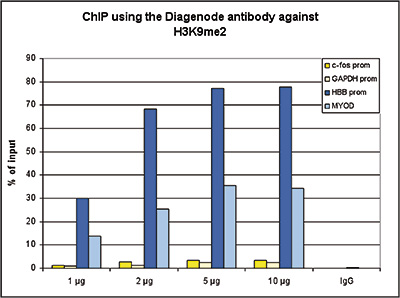
<p><small> <strong>Figure 1. ChIP results obtained with the Diagenode antibody directed against H3K9me2</strong><br /> ChIP assays were performed using human HeLa cells, the Diagenode antibody against H3K9me2 (Cat. No. C15410060) and optimized PCR primer sets for qPCR. ChIP was performed with the “Auto Histone ChIP-seq kit, using sheared chromatin from 1 million cells. A titration of the antibody consisting of 1, 2, 5, and 10 µg per ChIP experiment was analysed. IgG (2 µg/IP) was used as negative IP control. QPCR was performed with primers specific for promoter of the inactive HBB gene and the coding region of the inactive MYOD gene, used as positive controls, and for the promoters of the active genes c-fos and GAPDH, used as negative controls. Figure 1 shows the recovery, expressed as a % of input (the relative amount of immunoprecipitated DNA compared to input DNA after qPCR analysis). </small></p>
</div>
</div>
<div class="row">
<div class="small-4 columns">
<p><img src="https://www.diagenode.com/img/product/antibodies/C15410060-ELISA.jpg" alt="H3K9me2 Antibody ELISA Validation" caption="false" width="278" height="250" /></p>
</div>
<div class="small-8 columns">
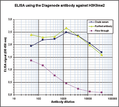
<p><small> <strong>Figure 2. Determination of the antibody titer</strong><br /> To determine the titer of the antibody, an ELISA was performed using a serial dilution of the Diagenode antibody against H3K9me2 (Cat. No. C15410060), crude serum and Flow through. The antigen used was a peptide containing the histone modification of interest. By plotting the absorbance against the antibody dilution (Figure 2), the titer of the antibody was estimated to be 1:103,000. </small></p>
</div>
</div>
<div class="row">
<div class="small-4 columns">
<p><img src="https://www.diagenode.com/img/product/antibodies/C15410060-DotBlot.jpg" alt="H3K9me2 Antibody Dot blot Validation " caption="false" width="278" height="230" /></p>
</div>
<div class="small-8 columns">
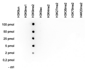
<p><small><strong>Figure 3. Cross reactivity tests using the Diagenode antibody directed against H3K9me2</strong><br /> A Dot Blot analysis was performed to test the cross reactivity of the Diagenode antibody against H3K9me2 (Cat. No. C15410060) with peptides containing other modifications of histone H3 and the unmodified H3K9 sequence. One hundred to 0.2 pmol of peptide containing the respective histone modification were spotted on a membrane. The antibody was used at a dilution of 1:20,000. Figure 3 shows a high specificity of the antibody for the modification of interest. </small></p>
</div>
</div>
<div class="extra-spaced"></div>
<div class="row">
<div class="small-4 columns">
<p><img src="https://www.diagenode.com/img/product/antibodies/C15410060-WB.jpg" alt="H3K9me2 Antibody Validated in Western blot" caption="false" width="146" height="167" /></p>
</div>
<div class="small-8 columns">
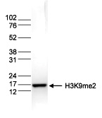
<p><small><strong>Figure 4. Western blot analysis using the Diagenode antibody directed against H3K9me2</strong><br /> Histone extracts (15 µg) from HeLa cells were analysed by Western blot using the Diagenode antibody against H3K9me2 (Cat. No. pAb-060-050) diluted 1:1,000 in TBS-Tween containing 5% skimmed milk. The position of the protein of interest is indicated on the right; the marker (in kDa) is shown on the left. </small></p>
</div>
</div>
<div class="row">
<div class="small-5 columns">
<p><img src="https://www.diagenode.com/img/product/antibodies/C15410060-IF.jpg" alt="H3K9me2 Antibody validated in IF" caption="false" width="354" height="87" /></p>
</div>
<div class="small-7 columns">
<p><small><strong>Figure 5. Immunofluorescence using the Diagenode antibody directed against H3K9me2</strong><br /> Mouse NIH3T3 cells were stained with the Diagenode antibody against H3K9me2 (Cat. No. C15410060) and with DAPI. Cells were fixed with 4% formaldehyde for 10’ and blocked with PBS/TX-100 containing 5% normal goat serum and 1% BSA. The cells were immunofluorescently labeled with the H3K9me2 antibody (left) diluted 1:500 in blocking solution followed by an anti-rabbit antibody conjugated to Alexa488. The middle panel shows staining of the nuclei with DAPI. A merge of the two stainings is shown on the right. </small></p>
</div>
</div>',
'label2' => 'Target Description',
'info2' => '<p>Histones are the main constituents of the protein part of chromosomes of eukaryotic cells. They are rich in the amino acids arginine and lysine and have been greatly conserved during evolution. Histones pack the DNA into tight masses of chromatin. Two core histones of each class H2A, H2B, H3 and H4 assemble and are wrapped by 146 base pairs of DNA to form one octameric nucleosome. Histone tails undergo numerous post-translational modifications, which either directly or indirectly alter chromatin structure to facilitate transcriptional activation or repression or other nuclear processes. In addition to the genetic code, combinations of the different histone modifications reveal the so-called “histone code”. Histone methylation and demethylation is dynamically regulated by respectively histone methyl transferases and histone demethylases. Dimethylation of histone H3K9 is more present in silent genes.</p>',
'label3' => '',
'info3' => '',
'format' => '50 µg/44 µl',
'catalog_number' => 'C15410060',
'old_catalog_number' => 'pAb-060-050',
'sf_code' => 'C15410060-D001-000581',
'type' => 'FRE',
'search_order' => '03-Antibody',
'price_EUR' => '380',
'price_USD' => '380',
'price_GBP' => '340',
'price_JPY' => '59525',
'price_CNY' => '',
'price_AUD' => '950',
'country' => 'ALL',
'except_countries' => 'None',
'quote' => false,
'in_stock' => false,
'featured' => false,
'no_promo' => false,
'online' => true,
'master' => true,
'last_datasheet_update' => 'August 12, 2020',
'slug' => 'h3k9me2-polyclonal-antibody-classic-50-ug-44-ul',
'meta_title' => 'H3K9me2 Antibody - ChIP Grade (C15410060) | Diagenode',
'meta_keywords' => '',
'meta_description' => 'H3K9me2 (Histone H3 dimethylated at lysine 9) Polyclonal Antibody validated in ChIP-qPCR, ELISA, DB, WB and IF. Batch-specific data available on the website. Sample size available.',
'modified' => '2024-01-16 14:41:12',
'created' => '2015-06-29 14:08:20',
'locale' => 'zho'
),
'Antibody' => array(
'host' => '*****',
'id' => '137',
'name' => 'H3K9me2 polyclonal antibody',
'description' => 'Histones are the main constituents of the protein part of chromosomes of eukaryotic cells. They are rich in the amino acids arginine and lysine and have been greatly conserved during evolution. Histones pack the DNA into tight masses of chromatin. Two core histones of each class H2A, H2B, H3 and H4 assemble and are wrapped by 146 base pairs of DNA to form one octameric nucleosome. Histone tails undergo numerous post-translational modifications, which either directly or indirectly alter chromatin structure to facilitate transcriptional activation or repression or other nuclear processes. In addition to the genetic code, combinations of the different histone modifications reveal the so-called “histone code”. Histone methylation and demethylation is dynamically regulated by respectively histone methyl transferases and histone demethylases. Dimethylation of histone H3K9 is more present in silent genes.',
'clonality' => '',
'isotype' => '',
'lot' => ' A90-0042',
'concentration' => '1.15 µg/µl',
'reactivity' => 'Human, mouse, Xenopus, Arabidopsis, C. elegans, Rice, Tomato, B. napus, Nicotiana benthamiana: positive. Other species: not tested',
'type' => 'Polyclonal',
'purity' => 'Affinity purified polyclonal antibody',
'classification' => 'Classic',
'application_table' => '<table>
<thead>
<tr>
<th>Applications</th>
<th>Suggested dilution</th>
<th>References</th>
</tr>
</thead>
<tbody>
<tr>
<td>ChIP <sup>*</sup></td>
<td>2 μg/ChIP</td>
<td>Fig 1</td>
</tr>
<tr>
<td>ELISA</td>
<td>1:1,000</td>
<td>Fig 2</td>
</tr>
<tr>
<td>Dot Blotting</td>
<td>1:20,000</td>
<td>Fig 3</td>
</tr>
<tr>
<td>Western Blotting</td>
<td>1:1,000</td>
<td>Fig 4</td>
</tr>
<tr>
<td>Immunofluorescence</td>
<td>1:500</td>
<td>Fig 5</td>
</tr>
</tbody>
</table>
<p><small><sup>*</sup> Please note that the optimal antibody amount per IP should be determined by the end-user. We recommend testing 1-5 µg per IP.</small></p>',
'storage_conditions' => 'Store at -20°C; for long storage, store at -80°C. Avoid multiple freeze-thaw cycles.',
'storage_buffer' => 'PBS containing 0.05% azide and 0.05% ProClin 300',
'precautions' => 'This product is for research use only. Not for use in diagnostic or therapeutic procedures.',
'uniprot_acc' => '',
'slug' => '',
'meta_keywords' => '',
'meta_description' => '',
'modified' => '2021-12-23 12:31:25',
'created' => '0000-00-00 00:00:00',
'select_label' => '137 - H3K9me2 polyclonal antibody ( A90-0042 - 1.15 µg/µl - Human, mouse, Xenopus, Arabidopsis, C. elegans, Rice, Tomato, B. napus, Nicotiana benthamiana: positive. Other species: not tested - Affinity purified polyclonal antibody - Rabbit)'
),
'Slave' => array(
(int) 0 => array(
[maximum depth reached]
)
),
'Group' => array(
'Group' => array(
[maximum depth reached]
),
'Master' => array(
[maximum depth reached]
),
'Product' => array(
[maximum depth reached]
)
),
'Related' => array(
(int) 0 => array(
[maximum depth reached]
),
(int) 1 => array(
[maximum depth reached]
),
(int) 2 => array(
[maximum depth reached]
),
(int) 3 => array(
[maximum depth reached]
),
(int) 4 => array(
[maximum depth reached]
),
(int) 5 => array(
[maximum depth reached]
),
(int) 6 => array(
[maximum depth reached]
)
),
'Application' => array(
(int) 0 => array(
[maximum depth reached]
),
(int) 1 => array(
[maximum depth reached]
),
(int) 2 => array(
[maximum depth reached]
),
(int) 3 => array(
[maximum depth reached]
),
(int) 4 => array(
[maximum depth reached]
)
),
'Category' => array(
(int) 0 => array(
[maximum depth reached]
),
(int) 1 => array(
[maximum depth reached]
),
(int) 2 => array(
[maximum depth reached]
)
),
'Document' => array(
(int) 0 => array(
[maximum depth reached]
),
(int) 1 => array(
[maximum depth reached]
),
(int) 2 => array(
[maximum depth reached]
)
),
'Feature' => array(),
'Image' => array(
(int) 0 => array(
[maximum depth reached]
)
),
'Promotion' => array(),
'Protocol' => array(),
'Publication' => array(
(int) 0 => array(
[maximum depth reached]
),
(int) 1 => array(
[maximum depth reached]
),
(int) 2 => array(
[maximum depth reached]
),
(int) 3 => array(
[maximum depth reached]
),
(int) 4 => array(
[maximum depth reached]
),
(int) 5 => array(
[maximum depth reached]
),
(int) 6 => array(
[maximum depth reached]
),
(int) 7 => array(
[maximum depth reached]
),
(int) 8 => array(
[maximum depth reached]
),
(int) 9 => array(
[maximum depth reached]
),
(int) 10 => array(
[maximum depth reached]
),
(int) 11 => array(
[maximum depth reached]
),
(int) 12 => array(
[maximum depth reached]
),
(int) 13 => array(
[maximum depth reached]
),
(int) 14 => array(
[maximum depth reached]
),
(int) 15 => array(
[maximum depth reached]
),
(int) 16 => array(
[maximum depth reached]
),
(int) 17 => array(
[maximum depth reached]
),
(int) 18 => array(
[maximum depth reached]
),
(int) 19 => array(
[maximum depth reached]
),
(int) 20 => array(
[maximum depth reached]
),
(int) 21 => array(
[maximum depth reached]
),
(int) 22 => array(
[maximum depth reached]
),
(int) 23 => array(
[maximum depth reached]
),
(int) 24 => array(
[maximum depth reached]
)
),
'Testimonial' => array(),
'Area' => array(),
'SafetySheet' => array(
(int) 0 => array(
[maximum depth reached]
),
(int) 1 => array(
[maximum depth reached]
),
(int) 2 => array(
[maximum depth reached]
),
(int) 3 => array(
[maximum depth reached]
),
(int) 4 => array(
[maximum depth reached]
),
(int) 5 => array(
[maximum depth reached]
),
(int) 6 => array(
[maximum depth reached]
),
(int) 7 => array(
[maximum depth reached]
)
)
),
'meta_canonical' => 'https://www.diagenode.com/cn/p/h3k9me2-polyclonal-antibody-classic-50-ug-44-ul'
)
$language = 'cn'
$meta_keywords = ''
$meta_description = 'H3K9me2 (Histone H3 dimethylated at lysine 9) Polyclonal Antibody validated in ChIP-qPCR, ELISA, DB, WB and IF. Batch-specific data available on the website. Sample size available.'
$meta_title = 'H3K9me2 Antibody - ChIP Grade (C15410060) | Diagenode'
$product = array(
'Product' => array(
'id' => '2223',
'antibody_id' => '137',
'name' => 'H3K9me2 Antibody - ChIP Grade',
'description' => '<p><span>Polyclonal antibody raised in rabbit against the region of histone H3 containing the dimethylated lysine 9 (<strong>H3K9me2</strong>), using a KLH-conjugated synthetic peptide.</span></p>',
'label1' => 'Validation Data',
'info1' => '<div class="row">
<div class="small-4 columns">
<p><img src="https://www.diagenode.com/img/product/antibodies/C15410060-Chip.jpg" alt="H3K9me2 Antibody ChIP Grade" caption="false" width="278" height="207" /></p>
</div>
<div class="small-8 columns">
<p><small> <strong>Figure 1. ChIP results obtained with the Diagenode antibody directed against H3K9me2</strong><br /> ChIP assays were performed using human HeLa cells, the Diagenode antibody against H3K9me2 (Cat. No. C15410060) and optimized PCR primer sets for qPCR. ChIP was performed with the “Auto Histone ChIP-seq kit, using sheared chromatin from 1 million cells. A titration of the antibody consisting of 1, 2, 5, and 10 µg per ChIP experiment was analysed. IgG (2 µg/IP) was used as negative IP control. QPCR was performed with primers specific for promoter of the inactive HBB gene and the coding region of the inactive MYOD gene, used as positive controls, and for the promoters of the active genes c-fos and GAPDH, used as negative controls. Figure 1 shows the recovery, expressed as a % of input (the relative amount of immunoprecipitated DNA compared to input DNA after qPCR analysis). </small></p>
</div>
</div>
<div class="row">
<div class="small-4 columns">
<p><img src="https://www.diagenode.com/img/product/antibodies/C15410060-ELISA.jpg" alt="H3K9me2 Antibody ELISA Validation" caption="false" width="278" height="250" /></p>
</div>
<div class="small-8 columns">
<p><small> <strong>Figure 2. Determination of the antibody titer</strong><br /> To determine the titer of the antibody, an ELISA was performed using a serial dilution of the Diagenode antibody against H3K9me2 (Cat. No. C15410060), crude serum and Flow through. The antigen used was a peptide containing the histone modification of interest. By plotting the absorbance against the antibody dilution (Figure 2), the titer of the antibody was estimated to be 1:103,000. </small></p>
</div>
</div>
<div class="row">
<div class="small-4 columns">
<p><img src="https://www.diagenode.com/img/product/antibodies/C15410060-DotBlot.jpg" alt="H3K9me2 Antibody Dot blot Validation " caption="false" width="278" height="230" /></p>
</div>
<div class="small-8 columns">
<p><small><strong>Figure 3. Cross reactivity tests using the Diagenode antibody directed against H3K9me2</strong><br /> A Dot Blot analysis was performed to test the cross reactivity of the Diagenode antibody against H3K9me2 (Cat. No. C15410060) with peptides containing other modifications of histone H3 and the unmodified H3K9 sequence. One hundred to 0.2 pmol of peptide containing the respective histone modification were spotted on a membrane. The antibody was used at a dilution of 1:20,000. Figure 3 shows a high specificity of the antibody for the modification of interest. </small></p>
</div>
</div>
<div class="extra-spaced"></div>
<div class="row">
<div class="small-4 columns">
<p><img src="https://www.diagenode.com/img/product/antibodies/C15410060-WB.jpg" alt="H3K9me2 Antibody Validated in Western blot" caption="false" width="146" height="167" /></p>
</div>
<div class="small-8 columns">
<p><small><strong>Figure 4. Western blot analysis using the Diagenode antibody directed against H3K9me2</strong><br /> Histone extracts (15 µg) from HeLa cells were analysed by Western blot using the Diagenode antibody against H3K9me2 (Cat. No. pAb-060-050) diluted 1:1,000 in TBS-Tween containing 5% skimmed milk. The position of the protein of interest is indicated on the right; the marker (in kDa) is shown on the left. </small></p>
</div>
</div>
<div class="row">
<div class="small-5 columns">
<p><img src="https://www.diagenode.com/img/product/antibodies/C15410060-IF.jpg" alt="H3K9me2 Antibody validated in IF" caption="false" width="354" height="87" /></p>
</div>
<div class="small-7 columns">
<p><small><strong>Figure 5. Immunofluorescence using the Diagenode antibody directed against H3K9me2</strong><br /> Mouse NIH3T3 cells were stained with the Diagenode antibody against H3K9me2 (Cat. No. C15410060) and with DAPI. Cells were fixed with 4% formaldehyde for 10’ and blocked with PBS/TX-100 containing 5% normal goat serum and 1% BSA. The cells were immunofluorescently labeled with the H3K9me2 antibody (left) diluted 1:500 in blocking solution followed by an anti-rabbit antibody conjugated to Alexa488. The middle panel shows staining of the nuclei with DAPI. A merge of the two stainings is shown on the right. </small></p>
</div>
</div>',
'label2' => 'Target Description',
'info2' => '<p>Histones are the main constituents of the protein part of chromosomes of eukaryotic cells. They are rich in the amino acids arginine and lysine and have been greatly conserved during evolution. Histones pack the DNA into tight masses of chromatin. Two core histones of each class H2A, H2B, H3 and H4 assemble and are wrapped by 146 base pairs of DNA to form one octameric nucleosome. Histone tails undergo numerous post-translational modifications, which either directly or indirectly alter chromatin structure to facilitate transcriptional activation or repression or other nuclear processes. In addition to the genetic code, combinations of the different histone modifications reveal the so-called “histone code”. Histone methylation and demethylation is dynamically regulated by respectively histone methyl transferases and histone demethylases. Dimethylation of histone H3K9 is more present in silent genes.</p>',
'label3' => '',
'info3' => '',
'format' => '50 µg/44 µl',
'catalog_number' => 'C15410060',
'old_catalog_number' => 'pAb-060-050',
'sf_code' => 'C15410060-D001-000581',
'type' => 'FRE',
'search_order' => '03-Antibody',
'price_EUR' => '380',
'price_USD' => '380',
'price_GBP' => '340',
'price_JPY' => '59525',
'price_CNY' => '',
'price_AUD' => '950',
'country' => 'ALL',
'except_countries' => 'None',
'quote' => false,
'in_stock' => false,
'featured' => false,
'no_promo' => false,
'online' => true,
'master' => true,
'last_datasheet_update' => 'August 12, 2020',
'slug' => 'h3k9me2-polyclonal-antibody-classic-50-ug-44-ul',
'meta_title' => 'H3K9me2 Antibody - ChIP Grade (C15410060) | Diagenode',
'meta_keywords' => '',
'meta_description' => 'H3K9me2 (Histone H3 dimethylated at lysine 9) Polyclonal Antibody validated in ChIP-qPCR, ELISA, DB, WB and IF. Batch-specific data available on the website. Sample size available.',
'modified' => '2024-01-16 14:41:12',
'created' => '2015-06-29 14:08:20',
'locale' => 'zho'
),
'Antibody' => array(
'host' => '*****',
'id' => '137',
'name' => 'H3K9me2 polyclonal antibody',
'description' => 'Histones are the main constituents of the protein part of chromosomes of eukaryotic cells. They are rich in the amino acids arginine and lysine and have been greatly conserved during evolution. Histones pack the DNA into tight masses of chromatin. Two core histones of each class H2A, H2B, H3 and H4 assemble and are wrapped by 146 base pairs of DNA to form one octameric nucleosome. Histone tails undergo numerous post-translational modifications, which either directly or indirectly alter chromatin structure to facilitate transcriptional activation or repression or other nuclear processes. In addition to the genetic code, combinations of the different histone modifications reveal the so-called “histone code”. Histone methylation and demethylation is dynamically regulated by respectively histone methyl transferases and histone demethylases. Dimethylation of histone H3K9 is more present in silent genes.',
'clonality' => '',
'isotype' => '',
'lot' => ' A90-0042',
'concentration' => '1.15 µg/µl',
'reactivity' => 'Human, mouse, Xenopus, Arabidopsis, C. elegans, Rice, Tomato, B. napus, Nicotiana benthamiana: positive. Other species: not tested',
'type' => 'Polyclonal',
'purity' => 'Affinity purified polyclonal antibody',
'classification' => 'Classic',
'application_table' => '<table>
<thead>
<tr>
<th>Applications</th>
<th>Suggested dilution</th>
<th>References</th>
</tr>
</thead>
<tbody>
<tr>
<td>ChIP <sup>*</sup></td>
<td>2 μg/ChIP</td>
<td>Fig 1</td>
</tr>
<tr>
<td>ELISA</td>
<td>1:1,000</td>
<td>Fig 2</td>
</tr>
<tr>
<td>Dot Blotting</td>
<td>1:20,000</td>
<td>Fig 3</td>
</tr>
<tr>
<td>Western Blotting</td>
<td>1:1,000</td>
<td>Fig 4</td>
</tr>
<tr>
<td>Immunofluorescence</td>
<td>1:500</td>
<td>Fig 5</td>
</tr>
</tbody>
</table>
<p><small><sup>*</sup> Please note that the optimal antibody amount per IP should be determined by the end-user. We recommend testing 1-5 µg per IP.</small></p>',
'storage_conditions' => 'Store at -20°C; for long storage, store at -80°C. Avoid multiple freeze-thaw cycles.',
'storage_buffer' => 'PBS containing 0.05% azide and 0.05% ProClin 300',
'precautions' => 'This product is for research use only. Not for use in diagnostic or therapeutic procedures.',
'uniprot_acc' => '',
'slug' => '',
'meta_keywords' => '',
'meta_description' => '',
'modified' => '2021-12-23 12:31:25',
'created' => '0000-00-00 00:00:00',
'select_label' => '137 - H3K9me2 polyclonal antibody ( A90-0042 - 1.15 µg/µl - Human, mouse, Xenopus, Arabidopsis, C. elegans, Rice, Tomato, B. napus, Nicotiana benthamiana: positive. Other species: not tested - Affinity purified polyclonal antibody - Rabbit)'
),
'Slave' => array(
(int) 0 => array(
'id' => '240',
'name' => 'C15410060',
'product_id' => '2223',
'modified' => '2018-01-08 14:48:52',
'created' => '2018-01-08 14:48:52'
)
),
'Group' => array(
'Group' => array(
'id' => '240',
'name' => 'C15410060',
'product_id' => '2223',
'modified' => '2018-01-08 14:48:52',
'created' => '2018-01-08 14:48:52'
),
'Master' => array(
'id' => '2223',
'antibody_id' => '137',
'name' => 'H3K9me2 Antibody',
'description' => '<p><span>Polyclonal antibody raised in rabbit against the region of histone H3 containing the dimethylated lysine 9 (<strong>H3K9me2</strong>), using a KLH-conjugated synthetic peptide.</span></p>',
'label1' => 'Validation Data',
'info1' => '<div class="row">
<div class="small-4 columns">
<p><img src="https://www.diagenode.com/img/product/antibodies/C15410060-Chip.jpg" alt="H3K9me2 Antibody ChIP Grade" caption="false" width="278" height="207" /></p>
</div>
<div class="small-8 columns">
<p><small> <strong>Figure 1. ChIP results obtained with the Diagenode antibody directed against H3K9me2</strong><br /> ChIP assays were performed using human HeLa cells, the Diagenode antibody against H3K9me2 (Cat. No. C15410060) and optimized PCR primer sets for qPCR. ChIP was performed with the “Auto Histone ChIP-seq kit, using sheared chromatin from 1 million cells. A titration of the antibody consisting of 1, 2, 5, and 10 µg per ChIP experiment was analysed. IgG (2 µg/IP) was used as negative IP control. QPCR was performed with primers specific for promoter of the inactive HBB gene and the coding region of the inactive MYOD gene, used as positive controls, and for the promoters of the active genes c-fos and GAPDH, used as negative controls. Figure 1 shows the recovery, expressed as a % of input (the relative amount of immunoprecipitated DNA compared to input DNA after qPCR analysis). </small></p>
</div>
</div>
<div class="row">
<div class="small-4 columns">
<p><img src="https://www.diagenode.com/img/product/antibodies/C15410060-ELISA.jpg" alt="H3K9me2 Antibody ELISA Validation" caption="false" width="278" height="250" /></p>
</div>
<div class="small-8 columns">
<p><small> <strong>Figure 2. Determination of the antibody titer</strong><br /> To determine the titer of the antibody, an ELISA was performed using a serial dilution of the Diagenode antibody against H3K9me2 (Cat. No. C15410060), crude serum and Flow through. The antigen used was a peptide containing the histone modification of interest. By plotting the absorbance against the antibody dilution (Figure 2), the titer of the antibody was estimated to be 1:103,000. </small></p>
</div>
</div>
<div class="row">
<div class="small-4 columns">
<p><img src="https://www.diagenode.com/img/product/antibodies/C15410060-DotBlot.jpg" alt="H3K9me2 Antibody Dot blot Validation " caption="false" width="278" height="230" /></p>
</div>
<div class="small-8 columns">
<p><small><strong>Figure 3. Cross reactivity tests using the Diagenode antibody directed against H3K9me2</strong><br /> A Dot Blot analysis was performed to test the cross reactivity of the Diagenode antibody against H3K9me2 (Cat. No. C15410060) with peptides containing other modifications of histone H3 and the unmodified H3K9 sequence. One hundred to 0.2 pmol of peptide containing the respective histone modification were spotted on a membrane. The antibody was used at a dilution of 1:20,000. Figure 3 shows a high specificity of the antibody for the modification of interest. </small></p>
</div>
</div>
<div class="extra-spaced"></div>
<div class="row">
<div class="small-4 columns">
<p><img src="https://www.diagenode.com/img/product/antibodies/C15410060-WB.jpg" alt="H3K9me2 Antibody Validated in Western blot" caption="false" width="146" height="167" /></p>
</div>
<div class="small-8 columns">
<p><small><strong>Figure 4. Western blot analysis using the Diagenode antibody directed against H3K9me2</strong><br /> Histone extracts (15 µg) from HeLa cells were analysed by Western blot using the Diagenode antibody against H3K9me2 (Cat. No. pAb-060-050) diluted 1:1,000 in TBS-Tween containing 5% skimmed milk. The position of the protein of interest is indicated on the right; the marker (in kDa) is shown on the left. </small></p>
</div>
</div>
<div class="row">
<div class="small-5 columns">
<p><img src="https://www.diagenode.com/img/product/antibodies/C15410060-IF.jpg" alt="H3K9me2 Antibody validated in IF" caption="false" width="354" height="87" /></p>
</div>
<div class="small-7 columns">
<p><small><strong>Figure 5. Immunofluorescence using the Diagenode antibody directed against H3K9me2</strong><br /> Mouse NIH3T3 cells were stained with the Diagenode antibody against H3K9me2 (Cat. No. C15410060) and with DAPI. Cells were fixed with 4% formaldehyde for 10’ and blocked with PBS/TX-100 containing 5% normal goat serum and 1% BSA. The cells were immunofluorescently labeled with the H3K9me2 antibody (left) diluted 1:500 in blocking solution followed by an anti-rabbit antibody conjugated to Alexa488. The middle panel shows staining of the nuclei with DAPI. A merge of the two stainings is shown on the right. </small></p>
</div>
</div>',
'label2' => 'Target Description',
'info2' => '<p>Histones are the main constituents of the protein part of chromosomes of eukaryotic cells. They are rich in the amino acids arginine and lysine and have been greatly conserved during evolution. Histones pack the DNA into tight masses of chromatin. Two core histones of each class H2A, H2B, H3 and H4 assemble and are wrapped by 146 base pairs of DNA to form one octameric nucleosome. Histone tails undergo numerous post-translational modifications, which either directly or indirectly alter chromatin structure to facilitate transcriptional activation or repression or other nuclear processes. In addition to the genetic code, combinations of the different histone modifications reveal the so-called “histone code”. Histone methylation and demethylation is dynamically regulated by respectively histone methyl transferases and histone demethylases. Dimethylation of histone H3K9 is more present in silent genes.</p>',
'label3' => '',
'info3' => '',
'format' => '50 µg/44 µl',
'catalog_number' => 'C15410060',
'old_catalog_number' => 'pAb-060-050',
'sf_code' => 'C15410060-D001-000581',
'type' => 'FRE',
'search_order' => '03-Antibody',
'price_EUR' => '380',
'price_USD' => '380',
'price_GBP' => '340',
'price_JPY' => '59525',
'price_CNY' => '',
'price_AUD' => '950',
'country' => 'ALL',
'except_countries' => 'None',
'quote' => false,
'in_stock' => false,
'featured' => false,
'no_promo' => false,
'online' => true,
'master' => true,
'last_datasheet_update' => 'August 12, 2020',
'slug' => 'h3k9me2-polyclonal-antibody-classic-50-ug-44-ul',
'meta_title' => 'H3K9me2 Antibody - ChIP Grade (C15410060) | Diagenode',
'meta_keywords' => '',
'meta_description' => 'H3K9me2 (Histone H3 dimethylated at lysine 9) Polyclonal Antibody validated in ChIP-qPCR, ELISA, DB, WB and IF. Batch-specific data available on the website. Sample size available.',
'modified' => '2024-01-16 14:41:12',
'created' => '2015-06-29 14:08:20'
),
'Product' => array(
(int) 0 => array(
[maximum depth reached]
)
)
),
'Related' => array(
(int) 0 => array(
'id' => '1836',
'antibody_id' => null,
'name' => 'iDeal ChIP-seq kit for Histones',
'description' => '<p><a href="https://www.diagenode.com/files/products/kits/ideal-chipseq-for-histones-complete-manual.pdf"><img src="https://www.diagenode.com/img/buttons/bt-manual.png" /></a></p>
<p>Don’t risk wasting your precious sequencing samples. Diagenode’s validated <strong>iDeal ChIP-seq kit for Histones</strong> has everything you need for a successful start-to-finish <strong>ChIP of histones prior to Next-Generation Sequencing</strong>. The complete kit contains all buffers and reagents for cell lysis, chromatin shearing, immunoprecipitation and DNA purification. In addition, unlike competing solutions, the kit contains positive and negative control antibodies (H3K4me3 and IgG, respectively) as well as positive and negative control PCR primers pairs (GAPDH TSS and Myoglobin exon 2, respectively) for your convenience and a guarantee of optimal results. The kit has been validated on multiple histone marks.</p>
<p> The iDeal ChIP-seq kit for Histones<strong> </strong>is perfect for <strong>cells</strong> (<strong>100,000 cells</strong> to <strong>1,000,000 cells</strong> per IP) and has been validated for <strong>tissues</strong> (<strong>1.5 mg</strong> to <strong>5 mg</strong> of tissue per IP).</p>
<p> The iDeal ChIP-seq kit is the only kit on the market validated for the major sequencing systems. Our expertise in ChIP-seq tools allows reproducible and efficient results every time.</p>
<p></p>
<p> <strong></strong></p>
<p></p>',
'label1' => 'Characteristics',
'info1' => '<ul style="list-style-type: disc;">
<li>Highly <strong>optimized</strong> protocol for ChIP-seq from cells and tissues</li>
<li><strong>Validated</strong> for ChIP-seq with multiple histones marks</li>
<li>Most <strong>complete</strong> kit available (covers all steps, including the control antibodies and primers)</li>
<li>Optimized chromatin preparation in combination with the Bioruptor ensuring the best <strong>epitope integrity</strong></li>
<li>Magnetic beads make ChIP easy, fast and more <strong>reproducible</strong></li>
<li>Combination with Diagenode ChIP-seq antibodies provides high yields with excellent <strong>specificity</strong> and <strong>sensitivity</strong></li>
<li>Purified DNA suitable for any downstream application</li>
<li>Easy-to-follow protocol</li>
</ul>
<p>Note: to obtain optimal results, this kit should be used in combination with the DiaMag1.5 - magnetic rack.</p>
<h3>ChIP-seq on cells</h3>
<p><img src="https://www.diagenode.com/img/product/kits/iDeal-kit-C01010053-figure-1.jpg" alt="Figure 1A" style="display: block; margin-left: auto; margin-right: auto;" /></p>
<p><strong>Figure 1A. The high consistency of the iDeal ChIP-seq kit on the Ion Torrent™ PGM™ (Life Technologies) and GAIIx (Illumina<sup>®</sup>)</strong><br /> ChIP was performed on sheared chromatin from 1 million HelaS3 cells using the iDeal ChIP-seq kit and 1 µg of H3K4me3 positive control antibody. Two different biological samples have been analyzed using two different sequencers - GAIIx (Illumina<sup>®</sup>) and PGM™ (Ion Torrent™). The expected ChIP-seq profile for H3K4me3 on the GAPDH promoter region has been obtained.<br /> Image A shows a several hundred bp along chr12 with high similarity of read distribution despite the radically different sequencers. Image B is a close capture focusing on the GAPDH that shows that even the peak structure is similar.</p>
<p class="text-center"><strong>Perfect match between ChIP-seq data obtained with the iDeal ChIP-seq workflow and reference dataset</strong></p>
<p><img src="https://www.diagenode.com/img/product/kits/perfect-match-between-chipseq-data.png" alt="Figure 1B" style="display: block; margin-left: auto; margin-right: auto;" /></p>
<p><strong>Figure 1B.</strong> The ChIP-seq dataset from this experiment has been compared with a reference dataset from the Broad Institute. We observed a perfect match between the top 40% of Diagenode peaks and the reference dataset. Based on the NIH Encode project criterion, ChIP-seq results are considered reproducible between an original and reproduced dataset if the top 40% of peaks have at least an 80% overlap ratio with the compared dataset.</p>
<p><img src="https://www.diagenode.com/img/product/kits/iDeal-kit-C01010053-figure-2.jpg" alt="Figure 2" style="display: block; margin-left: auto; margin-right: auto;" /></p>
<p><strong>Figure 2. Efficient and easy chromatin shearing using the Bioruptor<sup>®</sup> and Shearing buffer iS1 from the iDeal ChIP-seq kit</strong><br /> Chromatin from 1 million of Hela cells was sheared using the Bioruptor<sup>®</sup> combined with the Bioruptor<sup>®</sup> Water cooler (Cat No. BioAcc-cool) during 3 rounds of 10 cycles of 30 seconds “ON” / 30 seconds “OFF” at HIGH power setting (position H). Diagenode 1.5 ml TPX tubes (Cat No. M-50001) were used for chromatin shearing. Samples were gently vortexed before and after performing each sonication round (rounds of 10 cycles), followed by a short centrifugation at 4°C to recover the sample volume at the bottom of the tube. The sheared chromatin was then decross-linked as described in the kit manual and analyzed by agarose gel electrophoresis.</p>
<p><img src="https://www.diagenode.com/img/product/kits/iDeal-kit-C01010053-figure-3.jpg" alt="Figure 3" style="display: block; margin-left: auto; margin-right: auto;" width="264" height="320" /></p>
<p><strong>Figure 3. Validation of ChIP by qPCR: reliable results using Diagenode’s ChIP-seq grade H3K4me3 antibody, isotype control and sets of validated primers</strong><br /> Specific enrichment on positive loci (GAPDH, EIF4A2, c-fos promoter regions) comparing to no enrichment on negative loci (TSH2B promoter region and Myoglobin exon 2) was detected by qPCR. Samples were prepared using the Diagenode iDeal ChIP-seq kit. Diagenode ChIP-seq grade antibody against H3K4me3 and the corresponding isotype control IgG were used for immunoprecipitation. qPCR amplification was performed with sets of validated primers.</p>
<h3>ChIP-seq on tissue</h3>
<p><img src="https://www.diagenode.com/img/product/kits/ideal-figure-h3k4me3.jpg" alt="Figure 4A" style="display: block; margin-left: auto; margin-right: auto;" /></p>
<p><strong>Figure 4A.</strong> Chromatin Immunoprecipitation has been performed using chromatin from mouse liver tissue, the iDeal ChIP-seq kit for Histones and the Diagenode ChIP-seq-grade H3K4me3 (Cat. No. C15410003) antibody. The IP'd DNA was subsequently analysed on an Illumina® HiSeq. Library preparation, cluster generation and sequencing were performed according to the manufacturer's instructions. This figure shows the peak distribution in a region surrounding the GAPDH positive control gene.</p>
<p><img src="https://www.diagenode.com/img/product/kits/match-of-the-top40-peaks-2.png" alt="Figure 4B" caption="false" style="display: block; margin-left: auto; margin-right: auto;" width="700" height="280" /></p>
<p><strong>Figure 4B.</strong> The ChIP-seq dataset from this experiment has been compared with a reference dataset from the Broad Institute. We observed a perfect match between the top 40% of Diagenode peaks and the reference dataset. Based on the NIH Encode project criterion, ChIP-seq results are considered reproducible between an original and reproduced dataset if the top 40% of peaks have at least an 80% overlap ratio with the compared dataset.</p>',
'label2' => 'Species, cell lines, tissues tested',
'info2' => '<p>The iDeal ChIP-seq Kit for Histones is compatible with a broad variety of cell lines, tissues and species - some examples are shown below. Other species / cell lines / tissues can be used with this kit.</p>
<p><u>Cell lines:</u></p>
<p>Human: A549, A673, CD8+ T, Blood vascular endothelial cells, Lymphatic endothelial cells, fibroblasts, K562, MDA-MB231</p>
<p>Pig: Alveolar macrophages</p>
<p>Mouse: C2C12, primary HSPC, synovial fibroblasts, HeLa-S3, FACS sorted cells from embryonic kidneys, macrophages, mesodermal cells, myoblasts, NPC, salivary glands, spermatids, spermatocytes, skeletal muscle stem cells, stem cells, Th2</p>
<p>Hamster: CHO</p>
<p>Other cell lines / species: compatible, not tested</p>
<p><u>Tissues</u></p>
<p>Bee – brain</p>
<p>Daphnia – whole animal</p>
<p>Horse – brain, heart, lamina, liver, lung, skeletal muscles, ovary</p>
<p>Human – Erwing sarcoma tumor samples</p>
<p>Other tissues: compatible, not tested</p>
<p>Did you use the iDeal ChIP-seq for Histones Kit on other cell line / tissue / species? <a href="mailto:agnieszka.zelisko@diagenode.com?subject=Species, cell lines, tissues tested with the iDeal ChIP-seq Kit for TF&body=Dear Customer,%0D%0A%0D%0APlease, leave below your feedback about the iDeal ChIP-seq for Transcription Factors (cell / tissue type, species, other information...).%0D%0A%0D%0AThank you for sharing with us your experience !%0D%0A%0D%0ABest regards,%0D%0A%0D%0AAgnieszka Zelisko-Schmidt, PhD">Let us know!</a></p>',
'label3' => ' Additional solutions compatible with iDeal ChIP-seq Kit for Histones',
'info3' => '<p><a href="../p/chromatin-shearing-optimization-kit-low-sds-100-million-cells">Chromatin EasyShear Kit - Ultra Low SDS </a>optimizes chromatin shearing, a critical step for ChIP.</p>
<p> The <a href="https://www.diagenode.com/en/categories/library-preparation-for-ChIP-seq">MicroPlex Library Preparation Kit </a>provides easy and optimal library preparation of ChIPed samples.</p>
<p><a href="../categories/chip-seq-grade-antibodies">ChIP-seq grade anti-histone antibodies</a> provide high yields with excellent specificity and sensitivity.</p>
<p> Plus, for our IP-Star Automation users for automated ChIP, check out our <a href="../p/auto-ideal-chip-seq-kit-for-histones-x24-24-rxns">automated</a> version of this kit.</p>',
'format' => '4 chrom. prep./24 IPs',
'catalog_number' => 'C01010051',
'old_catalog_number' => 'AB-001-0024',
'sf_code' => 'C01010051-',
'type' => 'RFR',
'search_order' => '04-undefined',
'price_EUR' => '915',
'price_USD' => '1130',
'price_GBP' => '840',
'price_JPY' => '143335',
'price_CNY' => '',
'price_AUD' => '2825',
'country' => 'ALL',
'except_countries' => 'None',
'quote' => false,
'in_stock' => false,
'featured' => true,
'no_promo' => false,
'online' => true,
'master' => true,
'last_datasheet_update' => '0000-00-00',
'slug' => 'ideal-chip-seq-kit-x24-24-rxns',
'meta_title' => 'iDeal ChIP-seq kit x24',
'meta_keywords' => '',
'meta_description' => 'iDeal ChIP-seq kit x24',
'modified' => '2023-04-20 16:00:20',
'created' => '2015-06-29 14:08:20',
'ProductsRelated' => array(
[maximum depth reached]
),
'Image' => array(
[maximum depth reached]
)
),
(int) 1 => array(
'id' => '1856',
'antibody_id' => null,
'name' => 'True MicroChIP-seq Kit',
'description' => '<p><a href="https://www.diagenode.com/files/products/kits/truemicrochipseq-kit-manual.pdf"><img src="https://www.diagenode.com/img/buttons/bt-manual.png" /></a></p>
<p>The <b>True </b><b>MicroChIP-seq</b><b> kit </b>provides a robust ChIP protocol suitable for the investigation of histone modifications within chromatin from as few as <b>10 000 cells</b>, including <b>FACS sorted cells</b>. The kit can be used for chromatin preparation for downstream ChIP-qPCR or ChIP-seq analysis. The <b>complete kit</b> contains everything you need for start-to-finish ChIP including all validated buffers and reagents for chromatin shearing, immunoprecipitation and DNA purification for exceptional <strong>ChIP-qPCR</strong> or <strong>ChIP-seq</strong> results. In addition, positive control antibodies and negative control PCR primers are included for your convenience and assurance of result sensitivity and specificity.</p>
<p>The True MicroChIP-seq kit offers unique benefits:</p>
<ul>
<li>An <b>optimized chromatin preparation </b>protocol compatible with low number of cells (<b>10.000</b>) in combination with the Bioruptor™ shearing device</li>
<li>Most <b>complete kit </b>available (covers all steps and includes control antibodies and primers)</li>
<li><b>Magnetic beads </b>make ChIP easy, fast, and more reproducible</li>
<li>MicroChIP DiaPure columns (included in the kit) enable the <b>maximum recovery </b>of immunoprecipitation DNA suitable for any downstream application</li>
<li><b>Excellent </b><b>ChIP</b><b>-seq </b>result when combined with <a href="https://www.diagenode.com/en/categories/library-preparation-for-ChIP-seq">MicroPlex</a><a href="https://www.diagenode.com/en/categories/library-preparation-for-ChIP-seq"> Library Preparation kit </a>adapted for low input</li>
</ul>
<p>For fast ChIP-seq on low input – check out Diagenode’s <a href="https://www.diagenode.com/en/p/uchipmentation-for-histones-24-rxns">µ</a><a href="https://www.diagenode.com/en/p/uchipmentation-for-histones-24-rxns">ChIPmentation</a><a href="https://www.diagenode.com/en/p/uchipmentation-for-histones-24-rxns"> for histones</a>.</p>
<p><sub>The True MicroChIP-seq kit, Cat. No. C01010132 is an upgraded version of the kit True MicroChIP, Cat. No. C01010130, with the new validated protocols (e.g. FACS sorted cells) and MicroChIP DiaPure columns included in the kit.</sub></p>',
'label1' => 'Characteristics',
'info1' => '<ul>
<li><b>Revolutionary:</b> Only 10,000 cells needed for complete ChIP-seq procedure</li>
<li><b>Validated on</b> studies for histone marks</li>
<li><b>Automated protocol </b>for the IP-Star<sup>®</sup> Compact Automated Platform available</li>
</ul>
<p></p>
<p>The True MicroChIP-seq kit protocol has been optimized for the use of 10,000 - 100,000 cells per immunoprecipitation reaction. Regarding chromatin immunoprecipitation, three protocol variants have been optimized:<br />starting with a batch, starting with an individual sample and starting with the FACS-sorted cells.</p>
<div><button id="readmorebtn" style="background-color: #b02736; color: white; border-radius: 5px; border: none; padding: 5px;">Show Workflow</button></div>
<p><br /> <img src="https://www.diagenode.com/img/product/kits/workflow-microchip.png" id="workflowchip" class="hidden" width="600px" /></p>
<p>
<script type="text/javascript">// <![CDATA[
const bouton = document.querySelector('#readmorebtn');
const workflow = document.getElementById('workflowchip');
bouton.addEventListener('click', () => workflow.classList.toggle('hidden'))
// ]]></script>
</p>
<div class="extra-spaced" align="center"></div>
<div class="row">
<div class="carrousel" style="background-position: center;">
<div class="container">
<div class="row" style="background: rgba(255,255,255,0.1);">
<div class="large-12 columns truemicro-slider" id="truemicro-slider">
<div>
<h3>High efficiency ChIP on 10,000 cells</h3>
<div class="large-10 small-12 medium-10 large-centered medium-centered small-centered columns"><img src="https://www.diagenode.com/img/product/kits/true-micro-chip-histone-results.png" width="800px" /></div>
<div class="large-10 small-12 medium-10 large-centered medium-centered small-centered columns"><center>
<p><small><strong>Figure 1. </strong>ChIP efficiency on 10,000 cells. ChIP was performed on human Hela cells using the Diagenode antibodies <a href="https://www.diagenode.com/en/p/h3k4me3-polyclonal-antibody-premium-50-ug-50-ul">H3K4me3</a> (Cat. No. C15410003), <a href="https://www.diagenode.com/en/p/h3k27ac-polyclonal-antibody-classic-50-mg-42-ml">H3K27ac</a> (C15410174), <a href="https://www.diagenode.com/en/p/h3k9me3-polyclonal-antibody-classic-50-ug">H3K9me3</a> (C15410056) and <a href="https://www.diagenode.com/en/p/h3k27me3-polyclonal-antibody-classic-50-mg-34-ml">H3K27me3</a> (C15410069). Sheared chromatin from 10,000 cells and 0.1 µg (H3K27ac), 0.25 µg (H3K4me3 and H3K27me3) or 0.5 µg (H3K9me3) of the antibody were used per IP. Corresponding amount of IgG was used as control. Quantitative PCR was performed with primers for corresponding positive and negative loci. Figure shows the recovery, expressed as a % of input (the relative amount of immunoprecipitated DNA compared to input DNA after qPCR analysis).</small></p>
</center></div>
</div>
<div>
<h3>True MicroChIP-seq protocol in a combination with MicroPlex library preparation kit results in reliable and accurate sequencing data</h3>
<div class="large-10 small-12 medium-10 large-centered medium-centered small-centered columns"><img src="https://www.diagenode.com/img/product/kits/fig2-truemicro.jpg" alt="True MicroChip results" width="800px" /></div>
<div class="large-10 small-12 medium-10 large-centered medium-centered small-centered columns"><center>
<p><small><strong>Figure 2.</strong> Integrative genomics viewer (IGV) visualization of ChIP-seq experiments using 50.000 of K562 cells. ChIP has been performed accordingly to True MicroChIP protocol followed by the library preparation using MicroPlex Library Preparation Kit (C05010001). The above figure shows the peaks from ChIP-seq experiments using the following antibodies: H3K4me1 (C15410194), H3K9/14ac (C15410200), H3K27ac (C15410196) and H3K36me3 (C15410192).</small></p>
</center></div>
</div>
<div>
<h3>Successful chromatin profiling from 10.000 of FACS-sorted cells</h3>
<div class="large-10 small-12 medium-10 large-centered medium-centered small-centered columns"><img src="https://www.diagenode.com/img/product/kits/fig3ab-truemicro.jpg" alt="small non coding RNA" width="800px" /></div>
<div class="large-10 small-12 medium-10 large-centered medium-centered small-centered columns"><center>
<p><small><strong>Figure 3.</strong> (A) Integrative genomics viewer (IGV) visualization of ChIP-seq experiments and heatmap 3kb upstream and downstream of the TSS (B) for H3K4me3. ChIP has been performed using 10.000 of FACS-sorted cells (K562) and H3K4me3 antibody (C15410003) accordingly to True MicroChIP protocol followed by the library preparation using MicroPlex Library Preparation Kit (C05010001). Data were compared to ENCODE standards.</small></p>
</center></div>
</div>
</div>
</div>
</div>
</div>
</div>
<p>
<script type="text/javascript">// <![CDATA[
$('.truemicro-slider').slick({
arrows: true,
dots: true,
autoplay:true,
autoplaySpeed: 3000
});
// ]]></script>
</p>',
'label2' => 'Additional solutions compatible with the True MicroChIP-seq Kit',
'info2' => '<p><span style="font-weight: 400;">The <a href="https://www.diagenode.com/en/p/chromatin-shearing-optimization-kit-high-sds-100-million-cells">Chromatin EasyShear Kit – High SDS</a></span><span style="font-weight: 400;"> Recommended for the optimizing chromatin shearing.</span></p>
<p><a href="https://www.diagenode.com/en/categories/chip-seq-grade-antibodies"><span style="font-weight: 400;">ChIP-seq grade antibodies</span></a><span style="font-weight: 400;"> for high yields, specificity, and sensitivity.</span></p>
<p><span style="font-weight: 400;">Check the list of available </span><a href="https://www.diagenode.com/en/categories/primer-pairs"><span style="font-weight: 400;">primer pairs</span></a><span style="font-weight: 400;"> designed for high specificity to specific genomic regions.</span></p>
<p><span style="font-weight: 400;">For library preparation of immunoprecipitated samples we recommend to use the </span><b> </b><a href="https://www.diagenode.com/en/categories/library-preparation-for-ChIP-seq"><span style="font-weight: 400;">MicroPlex Library Preparation Kit</span></a><span style="font-weight: 400;"> - validated for library preparation from picogram inputs.</span></p>
<p><span style="font-weight: 400;">For IP-Star Automation users, check out the </span><a href="https://www.diagenode.com/en/p/auto-true-microchip-kit-16-rxns"><span style="font-weight: 400;">automated version</span></a><span style="font-weight: 400;"> of this kit.</span></p>
<p><span style="font-weight: 400;">Application note: </span><a href="https://www.diagenode.com/files/application_notes/Diagenode_AATI_Joint.pdf"><span style="font-weight: 400;">Best Workflow Practices for ChIP-seq Analysis with Small Samples</span></a></p>
<p></p>',
'label3' => 'Species, cell lines, tissues tested',
'info3' => '<p>The True MicroChIP-seq kit is compatible with a broad variety of cell lines, tissues and species - some examples are shown below. Other species / cell lines / tissues can be used with this kit.</p>
<p><strong>Cell lines:</strong></p>
<p>Bovine: blastocysts,<br />Drosophila: embryos, salivary glands<br />Human: EndoC-ẞH1 cells, HeLa cells, PBMC, urothelial cells<br />Mouse: adipocytes, B cells, blastocysts, pre-B cells, BMDM cells, chondrocytes, embryonic stem cells, KH2 cells, LSK cells, macrophages, MEP cells, microglia, NK cells, oocytes, pancreatic cells, P19Cl6 cells, RPE cells,</p>
<p>Other cell lines / species: compatible, not tested</p>
<p><strong>Tissues:</strong></p>
<p>Horse: adipose tissue</p>
<p>Mice: intestine tissue</p>
<p>Other tissues: not tested</p>',
'format' => '20 rxns',
'catalog_number' => 'C01010132',
'old_catalog_number' => 'C01010130',
'sf_code' => 'C01010132-',
'type' => 'RFR',
'search_order' => '04-undefined',
'price_EUR' => '625',
'price_USD' => '680',
'price_GBP' => '575',
'price_JPY' => '97905',
'price_CNY' => '',
'price_AUD' => '1700',
'country' => 'ALL',
'except_countries' => 'None',
'quote' => false,
'in_stock' => false,
'featured' => true,
'no_promo' => false,
'online' => true,
'master' => true,
'last_datasheet_update' => '0000-00-00',
'slug' => 'true-microchip-kit-x16-16-rxns',
'meta_title' => 'True MicroChIP-seq Kit | Diagenode C01010132',
'meta_keywords' => '',
'meta_description' => 'True MicroChIP-seq Kit provides a robust ChIP protocol suitable for the investigation of histone modifications within chromatin from as few as 10 000 cells, including FACS sorted cells. Compatible with ChIP-qPCR as well as ChIP-seq.',
'modified' => '2023-04-20 16:06:10',
'created' => '2015-06-29 14:08:20',
'ProductsRelated' => array(
[maximum depth reached]
),
'Image' => array(
[maximum depth reached]
)
),
(int) 2 => array(
'id' => '1927',
'antibody_id' => null,
'name' => 'MicroPlex Library Preparation Kit v2 (12 indexes)',
'description' => '<p><a href="https://www.diagenode.com/files/products/kits/MicroPlex-Libary-Prep-Kit-v2-manual.pdf"><img src="https://www.diagenode.com/img/buttons/bt-manual.png" /></a></p>
<p><span><strong>Specifically optimized for ChIP-seq</strong></span><br /><br /><span>The MicroPlex Library Preparation™ kit is the only kit on the market which is validated for ChIP-seq and which allows the preparation of indexed libraries from just picogram inputs. In combination with the </span><a href="./true-microchip-kit-x16-16-rxns">True MicroChIP kit</a><span>, it allows for performing ChIP-seq on as few as 10,000 cells. Less input, fewer steps, fewer supplies, faster time to results! </span></p>
<p>The MicroPlex v2 kit (Cat. No. C05010012) contains all necessary reagents including single indexes for multiplexing up to 12 samples using single barcoding. For higher multiplexing (using dual indexes) check <a href="https://www.diagenode.com/en/p/microplex-lib-prep-kit-v3-48-rxns">MicroPlex Library Preparation Kits v3</a>.</p>',
'label1' => 'Characteristics',
'info1' => '<ul>
<li><strong>1 tube, 2 hours, 3 steps</strong> protocol</li>
<li><strong>Input: </strong>50 pg – 50 ng</li>
<li><strong>Reduce potential bias</strong> - few PCR amplification cycles needed</li>
<li><strong>High sensitivity ChIP-seq</strong> - low PCR duplication rate</li>
<li><strong>Great multiplexing flexibility</strong> with 12 barcodes (8 nt) included</li>
<li><strong>Validated with the <a href="https://www.diagenode.com/p/sx-8g-ip-star-compact-automated-system-1-unit" title="IP-Star Automated System">IP-Star<sup>®</sup> Automated Platform</a></strong></li>
</ul>
<h3>How it works</h3>
<center><img src="https://www.diagenode.com/img/product/kits/microplex-method-overview-v2.png" /></center>
<p style="margin-bottom: 0;"><small><strong>Microplex workflow - protocol with single indexes</strong><br />An input of 50 pg to 50 ng of fragmented dsDNA is converted into sequencing-ready libraries for Illumina® NGS platforms using a fast and simple 3-step protocol</small></p>
<ul class="accordion" data-accordion="" id="readmore" style="margin-left: 0;">
<li class="accordion-navigation"><a href="#first" style="background: #ffffff; padding: 0rem; margin: 0rem; color: #13b2a2;"><small>Read more about MicroPlex workflow</small></a>
<div id="first" class="content">
<p><small><strong>Step 1. Template Preparation</strong> provides efficient repair of the fragmented double-stranded DNA input.</small></p>
<p><small>In this step, the DNA is repaired and yields molecules with blunt ends.</small></p>
<p><small><strong>Step 2. Library Synthesis.</strong> enables ligation of MicroPlex patented stem- loop adapters.</small></p>
<p><small>In the next step, stem-loop adaptors with blocked 5’ ends are ligated with high efficiency to the 5’ end of the genomic DNA, leaving a nick at the 3’ end. The adaptors cannot ligate to each other and do not have single- strand tails, both of which contribute to non-specific background found with many other NGS preparations.</small></p>
<p><small><strong>Step 3. Library Amplification</strong> enables extension of the template, cleavage of the stem-loop adaptors, and amplification of the library. Illumina- compatible indexes are also introduced using a high-fidelity, highly- processive, low-bias DNA polymerase.</small></p>
<p><small>In the final step, the 3’ ends of the genomic DNA are extended to complete library synthesis and Illumina-compatible indexes are added through a high-fidelity amplification. Any remaining free adaptors are destroyed. Hands-on time and the risk of contamination are minimized by using a single tube and eliminating intermediate purifications.</small></p>
<p><small>Obtained libraries are purified, quantified and sized. The libraries pooling can be performed as well before sequencing.</small></p>
</div>
</li>
</ul>
<p></p>
<h3>Reliable detection of enrichments in ChIP-seq</h3>
<p><img src="https://www.diagenode.com/img/product/kits/microplex-library-prep-kit-figure-a.png" alt="Reliable detection of enrichments in ChIP-seq figure 1" style="display: block; margin-left: auto; margin-right: auto;" /></p>
<p><strong>Figure A.</strong> ChIP has been peformed with H3K4me3 antibody, amplification of 17 pg of DNA ChIP'd from 10.000 cells and amplification of 35 pg of DNA ChIP'd from 100.000 cells (control experiment). The IP'd DNA was amplified and transformed into a sequencing-ready preparation for the Illumina plateform with the MicroPlex Library Preparation kit. The library was then analysed on an Illumina<sup>®</sup> Genome Analyzer. Cluster generation and sequencing were performed according to the manufacturer's instructions.</p>
<p><img src="https://www.diagenode.com/img/product/kits/microplex-library-prep-kit-figure-b.png" alt="Reliable detection of enrichments in ChIP-seq figure 2" style="display: block; margin-left: auto; margin-right: auto;" /></p>
<p><strong>Figure B.</strong> We observed a perfect match between the top 40% of True MicroChIP peaks and the reference dataset. Based on the NIH Encode project criterion, ChIP-seq results are considered reproducible between an original and reproduced dataset if the top 40% of peaks have at least an 80% overlap ratio with the compared dataset.</p>',
'label2' => '',
'info2' => '',
'label3' => '',
'info3' => '',
'format' => '12 rxns',
'catalog_number' => 'C05010012',
'old_catalog_number' => 'C05010010',
'sf_code' => 'C05010012-',
'type' => 'FRE',
'search_order' => '04-undefined',
'price_EUR' => '935',
'price_USD' => '1215',
'price_GBP' => '835',
'price_JPY' => '146470',
'price_CNY' => '',
'price_AUD' => '3038',
'country' => 'ALL',
'except_countries' => 'None',
'quote' => false,
'in_stock' => false,
'featured' => false,
'no_promo' => false,
'online' => true,
'master' => true,
'last_datasheet_update' => '0000-00-00',
'slug' => 'microplex-library-preparation-kit-v2-x12-12-indices-12-rxns',
'meta_title' => 'MicroPlex Library Preparation Kit v2 x12 (12 indices)',
'meta_keywords' => '',
'meta_description' => 'MicroPlex Library Preparation Kit v2 x12 (12 indices)',
'modified' => '2023-04-20 15:01:16',
'created' => '2015-06-29 14:08:20',
'ProductsRelated' => array(
[maximum depth reached]
),
'Image' => array(
[maximum depth reached]
)
),
(int) 3 => array(
'id' => '2173',
'antibody_id' => '115',
'name' => 'H3K4me3 Antibody',
'description' => '<p><span>Polyclonal antibody raised in rabbit against the region of histone <strong>H3 containing the trimethylated lysine 4</strong> (<strong>H3K4me3</strong>), using a KLH-conjugated synthetic peptide.</span></p>',
'label1' => 'Validation data',
'info1' => '<div class="row">
<div class="small-6 columns"><center><img src="https://www.diagenode.com/img/product/antibodies/C15410003-fig1-ChIP.jpg" /></center></div>
<div class="small-6 columns">
<p><small><strong>Figure 1. ChIP results obtained with the Diagenode antibody directed against H3K4me3</strong><br />ChIP assays were performed using human K562 cells, the Diagenode antibody against H3K4me3 (cat. No. C15410003) and optimized PCR primer pairs for qPCR. ChIP was performed with the iDeal ChIP-seq kit (cat. No. C01010051), using sheared chromatin from 500,000 cells. A titration consisting of 0.5, 1, 2 and 5 µg of antibody per ChIP experiment was analyzed. IgG (1 µg/IP) was used as a negative IP control. Quantitative PCR was performed with primers specific for the promoter of the active genes GAPDH and EIF4A2, used as positive controls, and for the inactive MYOD1 gene and the Sat2 satellite repeat, used as negative controls. Figure 1 shows the recovery, expressed as a % of input (the relative amount of immunoprecipitated DNA compared to input DNA after qPCR analysis). </small></p>
</div>
</div>
<p></p>
<div class="row">
<div class="small-12 columns"><center>A.<img src="https://www.diagenode.com/img/product/antibodies/C15410003-fig2a-ChIP-seq.jpg" width="800" /></center><center>B.<img src="https://www.diagenode.com/img/product/antibodies/C15410003-fig2b-ChIP-seq.jpg" width="800" /></center><center>C.<img src="https://www.diagenode.com/img/product/antibodies/C15410003-fig2c-ChIP-seq.jpg" width="800" /></center><center>D.<img src="https://www.diagenode.com/img/product/antibodies/C15410003-fig2d-ChIP-seq.jpg" width="800" /></center></div>
</div>
<div class="row">
<div class="small-12 columns">
<p><small><strong>Figure 2. ChIP-seq results obtained with the Diagenode antibody directed against H3K4me3</strong><br />ChIP was performed on sheared chromatin from 1 million HeLaS3 cells using 1 µg of the Diagenode antibody against H3K4me3 (cat. No. C15410003) as described above. The IP'd DNA was subsequently analysed on an Illumina Genome Analyzer. Library preparation, cluster generation and sequencing were performed according to the manufacturer's instructions. The 36 bp tags were aligned to the human genome using the ELAND algorithm. Figure 2 shows the peak distribution along the complete sequence and a 600 kb region of the X-chromosome (figure 2A and B) and in two regions surrounding the GAPDH and EIF4A2 positive control genes, respectively (figure 2C and D). These results clearly show an enrichment of the H3K4 trimethylation at the promoters of active genes.</small></p>
</div>
</div>
<div class="row">
<div class="small-12 columns"><center>A.<img src="https://www.diagenode.com/img/product/antibodies/C15410003-cuttag-a.png" width="800" /></center></div>
<div class="small-12 columns"><center>B.<img src="https://www.diagenode.com/img/product/antibodies/C15410003-cuttag-b.png" width="800" /></center></div>
</div>
<div class="row">
<div class="small-12 columns">
<p><small><strong>Figure 3. Cut&Tag results obtained with the Diagenode antibody directed against H3K4me3</strong><br />CUT&TAG (Kaya-Okur, H.S., Nat Commun 10, 1930, 2019) was performed on 50,000 K562 cells using 0.5 µg of the Diagenode antibody against H3K4me3 (cat. No. C15410003) and the Diagenode pA-Tn5 transposase (C01070001). The libraries were subsequently analysed on an Illumina NextSeq 500 sequencer (2x75 paired-end reads) according to the manufacturer's instructions. The tags were aligned to the human genome (hg19) using the BWA algorithm. Figure 3 shows the peak distribution in 2 genomic regions surrounding the FOS gene on chromosome 14 and the ACTB gene on chromosome 7 (figure 3A and B, respectively).</small></p>
</div>
</div>
<div class="row">
<div class="small-6 columns"><center><img src="https://www.diagenode.com/img/product/antibodies/C15410003-fig3-ELISA.jpg" width="350" /></center><center></center><center></center><center></center><center></center></div>
<div class="small-6 columns">
<p><small><strong>Figure 4. Determination of the antibody titer</strong><br />To determine the titer of the antibody, an ELISA was performed using a serial dilution of the Diagenode antibody against H3K4me3 (cat. No. C15410003). The antigen used was a peptide containing the histone modification of interest. By plotting the absorbance against the antibody dilution (Figure 4), the titer of the antibody was estimated to be 1:11,000.</small></p>
</div>
</div>
<div class="row">
<div class="small-6 columns"><img src="https://www.diagenode.com/img/product/antibodies/C15410003-fig4-DB.jpg" /></div>
<div class="small-6 columns">
<p><small><strong>Figure 5. Cross reactivity tests using the Diagenode antibody directed against H3K4me3</strong><br />To test the cross reactivity of the Diagenode antibody against H3K4me3 (cat. No. C15410003), a Dot Blot analysis was performed with peptides containing other histone modifications and the unmodified H3K4. One hundred to 0.2 pmol of the respective peptides were spotted on a membrane. The antibody was used at a dilution of 1:2,000. Figure 5A shows a high specificity of the antibody for the modification of interest.</small></p>
</div>
</div>
<div class="row">
<div class="small-4 columns"><img src="https://www.diagenode.com/img/product/antibodies/C15410003-fig5-WB.jpg" /></div>
<div class="small-8 columns">
<p><small><strong>Figure 6. Western blot analysis using the Diagenode antibody directed against H3K4me3</strong><br />Western blot was performed on whole cell extracts (40 µg, lane 1) from HeLa cells, and on 1 µg of recombinant histone H3 (lane 2) using the Diagenode antibody against H3K4me3 (cat. No. C15410003). The antibody was diluted 1:1,000 in TBS-Tween containing 5% skimmed milk. The position of the protein of interest is indicated on the right; the marker (in kDa) is shown on the left.</small></p>
</div>
</div>
<div class="row">
<div class="small-12 columns"><center><img src="https://www.diagenode.com/img/product/antibodies/C15410003-fig6-if.jpg" /></center></div>
</div>
<div class="row">
<div class="small-12 columns">
<p><small><strong>Figure 7. Immunofluorescence using the Diagenode antibody directed against H3K4me3</strong><br />HeLa cells were stained with the Diagenode antibody against H3K4me3 (cat. No. C15410003) and with DAPI. Cells were fixed with 4% formaldehyde for 20’ and blocked with PBS/TX-100 containing 5% normal goat serum. The cells were immunofluorescently labelled with the H3K4me3 antibody (left) diluted 1:200 in blocking solution followed by an anti-rabbit antibody conjugated to Alexa568 or with DAPI (middle), which specifically labels DNA. The right picture shows a merge of both stainings.</small></p>
</div>
</div>
<script src="chrome-extension://hhojmcideegachlhfgfdhailpfhgknjm/web_accessible_resources/index.js"></script>',
'label2' => 'Target Description',
'info2' => '<p>Histones are the main constituents of the protein part of chromosomes of eukaryotic cells. They are rich in the amino acids arginine and lysine and have been greatly conserved during evolution. Histones pack the DNA into tight masses of chromatin. Two core histones of each class H2A, H2B, H3 and H4 assemble and are wrapped by 146 base pairs of DNA to form one octameric nucleosome. Histone tails undergo numerous post-translational modifications, which either directly or indirectly alter chromatin structure to facilitate transcriptional activation or repression or other nuclear processes. In addition to the genetic code, combinations of the different histone modifications reveal the so-called "histone code". Histone methylation and demethylation is dynamically regulated by respectively histone methyl transferases and histone demethylases. Methylation of histone H3K4 is associated with activation of gene transcription.</p>
<script src="chrome-extension://hhojmcideegachlhfgfdhailpfhgknjm/web_accessible_resources/index.js"></script>',
'label3' => '',
'info3' => '<p></p>
<script src="chrome-extension://hhojmcideegachlhfgfdhailpfhgknjm/web_accessible_resources/index.js"></script>',
'format' => '50 µg',
'catalog_number' => 'C15410003',
'old_catalog_number' => 'pAb-003-050',
'sf_code' => 'C15410003-D001-000581',
'type' => 'FRE',
'search_order' => '03-Antibody',
'price_EUR' => '480',
'price_USD' => '470',
'price_GBP' => '430',
'price_JPY' => '75190',
'price_CNY' => '',
'price_AUD' => '1175',
'country' => 'ALL',
'except_countries' => 'None',
'quote' => false,
'in_stock' => false,
'featured' => false,
'no_promo' => false,
'online' => true,
'master' => true,
'last_datasheet_update' => 'January 8, 2021',
'slug' => 'h3k4me3-polyclonal-antibody-premium-50-ug-50-ul',
'meta_title' => 'H3K4me3 Antibody - ChIP-seq Grade (C15410003) | Diagenode',
'meta_keywords' => '',
'meta_description' => 'H3K4me3 (Histone H3 trimethylated at lysine 4) Polyclonal Antibody validated in ChIP-seq, ChIP-qPCR, CUT&Tag, ELISA, DB, WB and IF. Specificity confirmed by Peptide array. Batch-specific data available on the website. Sample size available.',
'modified' => '2024-11-19 16:51:19',
'created' => '2015-06-29 14:08:20',
'ProductsRelated' => array(
[maximum depth reached]
),
'Image' => array(
[maximum depth reached]
)
),
(int) 4 => array(
'id' => '2264',
'antibody_id' => '121',
'name' => 'H3K9me3 Antibody',
'description' => '<p><span>Polyclonal antibody raised in rabbit against the region of histone<strong> H3 containing the trimethylated lysine 9</strong> (<strong>H3K9me3</strong>), using a KLH-conjugated synthetic peptide.</span></p>',
'label1' => 'Validation Data',
'info1' => '<div class="row">
<div class="small-6 columns"><center><img src="https://www.diagenode.com/img/product/antibodies/C15410193-ChIP-Fig1.png" /></center></div>
<div class="small-6 columns">
<p><small><strong>Figure 1. ChIP results obtained with the Diagenode antibody directed against H3K9me3</strong><br />ChIP assays were performed using human HeLa cells, the Diagenode antibody against H3K9me3 (cat. No. C15410193) and optimized PCR primer sets for qPCR. ChIP was performed on sheared chromatin from 1 million HeLaS3 cells using the “iDeal ChIP-seq” kit (cat. No. C01010051). A titration of the antibody consisting of 0.5, 1, 2, and 5 µg per ChIP experiment was analysed. IgG (1 µg/IP) was used as negative IP control. QPCR was performed with primers for the heterochromatin marker Sat2 and for the ZNF510 gene, used as positive controls, and for the promoters of the active EIF4A2 and GAPDH genes, used as negative controls. Figure 1 shows the recovery, expressed as a % of input (the relative amount of immunoprecipitated DNA compared to input DNA after qPCR analysis).</small></p>
</div>
</div>
<div class="row">
<div class="small-12 columns"><center>A.<img src="https://www.diagenode.com/img/product/antibodies/C15410193-ChIP-Fig2a.png" width="700" /></center><center>B.<img src="https://www.diagenode.com/img/product/antibodies/C15410193-ChIP-Fig2b.png" width="700" /></center><center>C.<img src="https://www.diagenode.com/img/product/antibodies/C15410193-ChIP-Fig2c.png" width="700" /></center><center>D.<img src="https://www.diagenode.com/img/product/antibodies/C15410193-ChIP-Fig2d.png" width="700" /></center></div>
</div>
<div class="row">
<div class="small-12 columns">
<p><small><strong>Figure 2. ChIP-seq results obtained with the Diagenode antibody directed against H3K9me3</strong><br />ChIP was performed with 0.5 µg of the Diagenode antibody against H3K9me3 (cat. No. C15410193) on sheared chromatin from 1,000,000 HeLa cells using the “iDeal ChIP-seq” kit as described above. The IP'd DNA was subsequently analysed on an Illumina HiSeq 2000. Library preparation, cluster generation and sequencing were performed according to the manufacturer's instructions. The 50 bp tags were aligned to the human genome using the BWA algorithm. Figure 2A shows the signal distribution along the long arm of chromosome 19 and a zoomin to an enriched region containing several ZNF repeat genes. The arrows indicate two satellite repeat regions which exhibit a stronger signal. Figures 2B, 2C and 2D show the enrichment along the ZNF510 positive control target and at the H19 and KCNQ1 imprinted genes.</small></p>
</div>
</div>
<div class="row">
<div class="small-12 columns"><center>A.<img src="https://www.diagenode.com/img/product/antibodies/C15410193-CT-Fig3a.png" width="700" /></center><center>B.<img src="https://www.diagenode.com/img/product/antibodies/C15410193-CT-Fig3b.png" width="700" /></center></div>
</div>
<div class="row">
<div class="small-12 columns">
<p><small><strong>Figure 3. Cut&Tag results obtained with the Diagenode antibody directed against H3K9me3</strong><br />CUT&TAG (Kaya-Okur, H.S., Nat Commun 10, 1930, 2019) was performed on 50,000 K562 cells using 1 µg of the Diagenode antibody against H3K9me3 (cat. No. C15410193) and the Diagenode pA-Tn5 transposase (C01070001). The libraries were subsequently analysed on an Illumina NextSeq 500 sequencer (2x75 paired-end reads) according to the manufacturer's instructions. The tags were aligned to the human genome (hg19) using the BWA algorithm. Figure 3 shows the peak distribution in a genomic regions on chromosome 1 containing several ZNF repeat genes and in a genomic region surrounding the KCNQ1 imprinting control gene on chromosome 11 (figure 3A and B, respectively).</small></p>
</div>
</div>
<div class="row">
<div class="small-6 columns"><center><img src="https://www.diagenode.com/img/product/antibodies/C15410193-Elisa-Fig4.png" /></center></div>
<div class="small-6 columns">
<p><small><strong>Figure 4. Determination of the antibody titer</strong><br />To determine the titer of the antibody, an ELISA was performed using a serial dilution of the antibody directed against human H3K9me3 (cat. No. C15410193) in antigen coated wells. The antigen used was a peptide containing the histone modification of interest. By plotting the absorbance against the antibody dilution (Figure 4), the titer of the antibody was estimated to be 1:87,000.</small></p>
</div>
</div>
<div class="row">
<div class="small-4 columns"><center><img src="https://www.diagenode.com/img/product/antibodies/C15410193-DB-Fig5.png" /></center></div>
<div class="small-8 columns">
<p><small><strong>Figure 5. Cross reactivity tests using the Diagenode antibody directed against H3K9me3</strong><br />A Dot Blot analysis was performed to test the cross reactivity of the Diagenode antibody against H3K9me3 (cat. No. C15410193) with peptides containing other modifications and unmodified sequences of histone H3 and H4. One hundred to 0.2 pmol of the peptide containing the respective histone modification were spotted on a membrane. The antibody was used at a dilution of 1:20,000. Figure 5 shows a high specificity of the antibody for the modification of interest.</small></p>
</div>
</div>
<div class="row">
<div class="small-4 columns"><center><img src="https://www.diagenode.com/img/product/antibodies/C15410193-WB-Fig6.png" /></center></div>
<div class="small-8 columns">
<p><small><strong>Figure 6. Western blot analysis using the Diagenode antibody directed against H3K9me3</strong><br />Western blot was performed on whole cell (25 µg, lane 1) and histone extracts (15 µg, lane 2) from HeLa cells, and on 1 µg of recombinant histone H2A, H2B, H3 and H4 (lane 3, 4, 5 and 6, respectively) using the Diagenode antibody against H3K9me3 (cat. No. C15410193). The antibody was diluted 1:1,000 in TBS-Tween containing 5% skimmed milk. The position of the protein of interest is indicated on the right; the marker (in kDa) is shown on the left.</small></p>
</div>
</div>
<div class="row">
<div class="small-12 columns"><center><img src="https://www.diagenode.com/img/product/antibodies/C15410193-IF-Fig7.png" /></center></div>
</div>
<div class="row">
<div class="small-12 columns">
<p><small><strong>Figure 7. Immunofluorescence using the Diagenode antibody directed against H3K9me3</strong><br />HeLa cells were stained with the Diagenode antibody against H3K9me3 (cat. No. C15410193) and with DAPI. Cells were fixed with 4% formaldehyde for 10’ and blocked with PBS/TX-100 containing 5% normal goat serum and 1% BSA. The cells were immunofluorescently labelled with the H3K9me3 antibody (middle) diluted 1:500 in blocking solution followed by an anti-rabbit antibody conjugated to Alexa488. The left panel shows staining of the nuclei with DAPI. A merge of both stainings is shown on the right.</small></p>
</div>
</div>',
'label2' => 'Target Description',
'info2' => '<p>Histones are the main constituents of the protein part of chromosomes of eukaryotic cells. They are rich in the amino acids arginine and lysine and have been greatly conserved during evolution. Histones pack the DNA into tight masses of chromatin. Two core histones of each class H2A, H2B, H3 and H4 assemble and are wrapped by 146 base pairs of DNA to form one octameric nucleosome. Histone tails undergo numerous post-translational modifications, which either directly or indirectly alter chromatin structure to facilitate transcriptional activation or repression or other nuclear processes. In addition to the genetic code, combinations of the different histone modifications reveal the so-called “histone code”. Histone methylation and demethylation is dynamically regulated by respectively histone methyl transferases and histone demethylases. Trimethylation of histone H3K9 is associated with inactive genomic regions, satellite repeats and ZNF gene repeats.</p>',
'label3' => '',
'info3' => '',
'format' => '50 μg',
'catalog_number' => 'C15410193',
'old_catalog_number' => 'pAb-193-050',
'sf_code' => 'C15410193-D001-000581',
'type' => 'FRE',
'search_order' => '03-Antibody',
'price_EUR' => '480',
'price_USD' => '470',
'price_GBP' => '430',
'price_JPY' => '75190',
'price_CNY' => '0',
'price_AUD' => '1175',
'country' => 'ALL',
'except_countries' => 'None',
'quote' => false,
'in_stock' => false,
'featured' => false,
'no_promo' => false,
'online' => true,
'master' => true,
'last_datasheet_update' => 'December 12, 2017',
'slug' => 'h3k9me3-polyclonal-antibody-premium-50-mg',
'meta_title' => 'H3K9me3 Antibody - ChIP-seq Grade (C15410193) | Diagenode',
'meta_keywords' => '',
'meta_description' => 'H3K9me3 (Histone H3 trimethylated at lysine 9) Polyclonal Antibody validated in ChIP-seq, ChIP-qPCR, CUT&Tag, ELISA, DB, WB and IF. Specificity confirmed by Peptide array assay. Batch-specific data available on the website. Sample size available.',
'modified' => '2021-10-20 09:55:53',
'created' => '2015-06-29 14:08:20',
'ProductsRelated' => array(
[maximum depth reached]
),
'Image' => array(
[maximum depth reached]
)
),
(int) 5 => array(
'id' => '2270',
'antibody_id' => '109',
'name' => 'H3K27ac Antibody',
'description' => '<p><span>Polyclonal antibody raised in rabbit against the region of histone <strong>H3 containing the acetylated lysine 27</strong> (<strong>H3K27ac</strong>), using a KLH-conjugated synthetic peptide.</span></p>',
'label1' => 'Validation Data',
'info1' => '<div class="row">
<div class="small-6 columns">A.<img src="https://www.diagenode.com/img/product/antibodies/C15410196-ChIP-Fig1a.png" width="356" /><br /> B.<img src="https://www.diagenode.com/img/product/antibodies/C15410196-ChIP-Fig1b.png" width="356" /></div>
<div class="small-6 columns">
<p><strong>Figure 1. ChIP results obtained with the Diagenode antibody directed against H3K27ac</strong></p>
<p>Figure 1A ChIP assays were performed using human HeLa cells, the Diagenode antibody against H3K27ac (Cat. No. C15410196) and optimized PCR primer pairs for qPCR. ChIP was performed with the “Auto Histone ChIP-seq” kit on the IP-Star automated system, using sheared chromatin from 1,000,000 cells. A titration consisting of 1, 2, 5 and 10 µg of antibody per ChIP experiment was analyzed. IgG (2 µg/IP) was used as a negative IP control. Quantitative PCR was performed with primers for the promoters of the active EIF4A2 and ACTB genes, used as positive controls, and for the inactive TSH2B and MYT1 genes, used as negative controls.</p>
<p>Figure 1B ChIP assays were performed using human K562 cells, the Diagenode antibody against H3K27ac (Cat. No. C15410196)and optimized PCR primer pairs for qPCR. ChIP was performed with the “iDeal ChIP-seq” kit (Cat. No. C01010051), using sheared chromatin from 100,000 cells. A titration consisting of 0.2, 0.5, 1 and 2 µg of antibody per ChIP experiment was analyzed. IgG (1 µg/IP) was used as a negative IP control. Quantitative PCR was performed with primers for the promoters of the active GAPDH and EIF4A2 genes, used as positive controls, and for the coding regions of the inactive MB and MYT1 genes, used as negative controls. Figure 1 shows the recovery, expressed as a % of input (the relative amount of immunoprecipitated DNA compared to input DNA after qPCR analysis)</p>
</div>
</div>
<div class="extra-spaced"></div>
<div class="row">
<div class="small-12 columns"><center>
<p>A.<img src="https://www.diagenode.com/img/product/antibodies/C15410196-ChIP-Fig2a.png" /></p>
</center><center>
<p>B.<img src="https://www.diagenode.com/img/product/antibodies/C15410196-ChIP-Fig2b.png" /></p>
</center><center>
<p>C.<img src="https://www.diagenode.com/img/product/antibodies/C15410196-ChIP-Fig2c.png" /></p>
</center></div>
</div>
<div class="row">
<div class="small-12 columns">
<p><strong>Figure 2. ChIP-seq results obtained with the Diagenode antibody directed against H3K27ac</strong></p>
<p>ChIP was performed on sheared chromatin from 100,000 K562 cells using 1 µg of the Diagenode antibody against H3K27ac (Cat. No. C15410196<span class="label-primary"></span>) as described above. The IP’d DNA was subsequently analysed on an Illumina Genome Analyzer. Library preparation, cluster generation and sequencing were performed according to the manufacturer’s instructions. The 36 bp tags were aligned to the human genome using the ELAND algorithm. Figure 2A shows the peak distribution along the complete human X-chromosome. Figure 2 B and C show the peak distribution in two regions surrounding the EIF4A2 and GAPDH positive control genes, respectively. The position of the PCR amplicon, used for validating the ChIP assay is indicated with an arrow.</p>
</div>
</div>
<div class="row">
<div class="small-12 columns"><center><img src="https://www.diagenode.com/img/product/antibodies/C15410196-fig3.jpg" /></center></div>
</div>
<div class="row">
<div class="small-12 columns">
<p><strong>Figure 3. Cut&Tag results obtained with the Diagenode antibody directed against H3K27ac</strong></p>
<p>CUT&TAG (Kaya-Okur, H.S., Nat Commun 10, 1930, 2019) was performed on 50,000 K562 cells using 1 µg of the Diagenode antibody against H3K27ac (cat. No. C15410196) and the Diagenode pA-Tn5 transposase (C01070001). The libraries were subsequently analysed on an Illumina NextSeq 500 sequencer (2x75 paired-end reads) according to the manufacturer's instructions. The tags were aligned to the human genome (hg19) using the BWA algorithm. Figure 3 shows the peak distribution in 2 genomic regions surrounding the EIF2S3 gene on the X-chromosome and the CCT5 gene on chromosome 5 (figure 3A and B, respectively).</p>
</div>
</div>
<div class="row">
<div class="small-6 columns"><img src="https://www.diagenode.com/img/product/antibodies/C15410196-ELISA-Fig3.png" /></div>
<div class="small-6 columns">
<p><strong>Figure 4. Determination of the antibody titer</strong></p>
<p>To determine the titer of the antibody, an ELISA was performed using a serial dilution of the Diagenode antibody against H3K27ac (Cat. No. C15410196<span class="label-primary"></span>). The antigen used was a peptide containing the histone modification of interest. By plotting the absorbance against the antibody dilution (Figure 4), the titer of the antibody was estimated to be 1:8,300.</p>
</div>
</div>
<div class="row">
<div class="small-4 columns"><center><img src="https://www.diagenode.com/img/product/antibodies/C15410196-DB-Fig4.png" /></center></div>
<div class="small-8 columns">
<p><strong>Figure 5. Cross reactivity tests using the Diagenode antibody directed against H3K27ac</strong><br />To test the cross reactivity of the Diagenode antibody against H3K27ac (Cat. No. C15410196<span class="label-primary"></span>), a Dot Blot analysis was performed with peptides containing other histone modifications and the unmodified H3K27. One hundred to 0.2 pmol of the respective peptides were spotted on a membrane. The antibody was used at a dilution of 1:20,000. Figure 5 shows a high specificity of the antibody for the modification of interest.</p>
</div>
</div>
<div class="row">
<div class="small-4 columns"><center><img src="https://www.diagenode.com/img/product/antibodies/C15410196-WB-Fig5.png" /></center></div>
<div class="small-8 columns">
<p><strong>Figure 6. Western blot analysis using the Diagenode antibody directed against H3K27ac</strong><br />Western blot was performed on whole cell (25 µg, lane 1) and histone extracts (15 µg, lane 2) from HeLa cells, and on 1 µg of recombinant histone H2A, H2B, H3 and H4 (lane 3, 4, 5 and 6, respectively) using the Diagenode antibody against H3K27ac (Cat. No. C1541196). The antibody was diluted 1:1,000 in TBS-Tween containing 5% skimmed milk. The marker (in kDa) is shown on the left.</p>
</div>
</div>
<div class="row">
<div class="small-4 columns"><img src="https://www.diagenode.com/img/product/antibodies/C15410196-IF-Fig6.png" /></div>
<div class="small-8 columns">
<p><strong>Figure 7. Immunofluorescence using the Diagenode antibody directed against H3K27ac</strong></p>
<p>HeLa cells were stained with the Diagenode antibody against H3K27ac (Cat. No. C15410196<span class="label-primary"></span>) and with DAPI. Cells were fixed with 4% formaldehyde for 10’ and blocked with PBS/ TX-100 containing 5% normal goat serum and 1% BSA. The cells were immunofluorescently labeled with the H3K27ac antibody (top) diluted 1:500 in blocking solution followed by an anti-rabbit antibody conjugated to Alexa488. The middle panel shows staining of the nuclei with DAPI. A merge of the two stainings is shown at the bottom.</p>
</div>
</div>',
'label2' => 'Target Description',
'info2' => '<p style="text-align: justify;">Histones are the main constituents of the protein part of chromosomes of eukaryotic cells. They are rich in the amino acids arginine and lysine and have been greatly conserved during evolution. Histones pack the DNA into tight masses of chromatin. Two core histones of each class H2A, H2B, H3 and H4 assemble and are wrapped by 146 base pairs of DNA to form one octameric nucleosome. Histone tails undergo numerous post-translational modifications, which either directly or indirectly alter chromatin structure to facilitate transcriptional activation or repression or other nuclear processes. In addition to the genetic code, combinations of the different histone modifications reveal the so-called “histone code”. Histone methylation and demethylation is dynamically regulated by respectively histone methyl transferases and histone demethylases. Acetylation of histone H3K27 is associated with active promoters and enhancers.</p>',
'label3' => '',
'info3' => '',
'format' => '50 μg',
'catalog_number' => 'C15410196',
'old_catalog_number' => 'pAb-196-050',
'sf_code' => 'C15410196-D001-000581',
'type' => 'FRE',
'search_order' => '03-Antibody',
'price_EUR' => '480',
'price_USD' => '470',
'price_GBP' => '430',
'price_JPY' => '75190',
'price_CNY' => '',
'price_AUD' => '1175',
'country' => 'ALL',
'except_countries' => 'None',
'quote' => false,
'in_stock' => false,
'featured' => false,
'no_promo' => false,
'online' => true,
'master' => true,
'last_datasheet_update' => 'January 11, 2021',
'slug' => 'h3k27ac-polyclonal-antibody-premium-50-mg-18-ml',
'meta_title' => 'H3K27ac Antibody - ChIP-seq Grade (C15410196) | Diagenode',
'meta_keywords' => '',
'meta_description' => 'H3K27ac (Histone H3 acetylated at lysine 27) Polyclonal Antibody validated in ChIP-seq, ChIP-qPCR, CUT&Tag, ELISA, DB, WB and IF. Batch-specific data available on the website. Sample size available. ',
'modified' => '2021-10-20 10:28:57',
'created' => '2015-06-29 14:08:20',
'ProductsRelated' => array(
[maximum depth reached]
),
'Image' => array(
[maximum depth reached]
)
),
(int) 6 => array(
'id' => '2268',
'antibody_id' => '70',
'name' => 'H3K27me3 Antibody',
'description' => '<p>Polyclonal antibody raised in rabbit against the region of histone <strong>H3 containing the trimethylated lysine 27</strong> (<strong>H3K27me3</strong>), using a KLH-conjugated synthetic peptide.</p>',
'label1' => 'Validation Data',
'info1' => '<div class="row">
<div class="small-6 columns">
<p>A. <img src="https://www.diagenode.com/img/product/antibodies/C15410195-ChIP-Fig1.png" alt="H3K27me3 Antibody ChIP Grade" /></p>
<p>B. <img src="https://www.diagenode.com/img/product/antibodies/C15410195-ChIP-Fig2.png" alt="H3K27me3 Antibody for ChIP" /></p>
</div>
<div class="small-6 columns">
<p><small><strong>Figure 1. ChIP results obtained with the Diagenode antibody directed against H3K27me3</strong><br />ChIP assays were performed using human HeLa cells, the Diagenode antibody against H3K27me3 (Cat. No. C15410195) and optimized PCR primer pairs for qPCR. ChIP was performed with the “iDeal ChIP-seq” kit (Cat. No. C01010051), using sheared chromatin from 1 million cells. The chromatin was spiked with a panel of in vitro assembled nucleosomes, each containing a specific lysine methylation. A titration consisting of 0.5, 1, 2 and 5 µg of antibody per ChIP experiment was analyzed. IgG (1 µg/IP) was used as a negative IP control.</small></p>
<p><small><strong>Figure 1A.</strong> Quantitative PCR was performed with primers specific for the promoter of the active GAPDH and EIF4A2 genes, used as negative controls, and for the inactive TSH2B and MYT1 genes, used as positive controls. The graph shows the recovery, expressed as a % of input (the relative amount of immunoprecipitated DNA compared to input DNA after qPCR analysis).</small></p>
<p><small><strong>Figure 1B.</strong> Recovery of the nucleosomes carrying the H3K27me1, H3K27me2, H3K27me3, H3K4me3, H3K9me3 and H3K36me3 modifications and the unmodified H3K27 as determined by qPCR. The figure clearly shows the antibody is very specific in ChIP for the H3K27me3 modification.</small></p>
</div>
</div>
<div class="row">
<div class="small-12 columns">
<p>A. <img src="https://www.diagenode.com/img/product/antibodies/C15410195-ChIP-Fig2a.png" alt="H3K27me3 Antibody ChIP-seq Grade" /></p>
</div>
</div>
<div class="extra-spaced"></div>
<div class="row">
<div class="small-12 columns">
<p>B. <img src="https://www.diagenode.com/img/product/antibodies/C15410195-ChIP-Fig2b.png" alt="H3K27me3 Antibody for ChIP-seq" /></p>
<p>C. <img src="https://www.diagenode.com/img/product/antibodies/C15410195-ChIP-Fig2c.png" alt="H3K27me3 Antibody for ChIP-seq assay" /></p>
<p>D. <img src="https://www.diagenode.com/img/product/antibodies/C15410195-ChIP-Fig2d.png" alt="H3K27me3 Antibody validated in ChIP-seq" /></p>
</div>
</div>
<div class="row">
<div class="small-12 columns">
<p><small><strong>Figure 2. ChIP-seq results obtained with the Diagenode antibody directed against H3K27me3</strong><br />ChIP was performed on sheared chromatin from 1 million HeLa cells using 1 µg of the Diagenode antibody against H3K27me3 (Cat. No. C15410195) as described above. The IP'd DNA was subsequently analysed on an Illumina HiSeq. Library preparation, cluster generation and sequencing were performed according to the manufacturer's instructions. The 50 bp tags were aligned to the human genome using the BWA algorithm. Figure 2 shows the enrichment in genomic regions of chromosome 6 and 20, surrounding the TSH2B and MYT1 positive control genes (fig 2A and 2B, respectively), and in two genomic regions of chromosome 1 and X (figure 2C and D).</small></p>
</div>
</div>
<div class="extra-spaced"></div>
<div class="row">
<div class="small-12 columns">
<p>A. <img src="https://www.diagenode.com/img/product/antibodies/C15410195-CUTTAG-Fig3A.png" /></p>
<p>B. <img src="https://www.diagenode.com/img/product/antibodies/C15410195-CUTTAG-Fig3B.png" /></p>
</div>
</div>
<div class="row">
<div class="small-12 columns">
<p><small><strong>Figure 3. Cut&Tag results obtained with the Diagenode antibody directed against H3K27me3</strong><br />CUT&TAG (Kaya-Okur, H.S., Nat Commun 10, 1930, 2019) was performed on 50,000 K562 cells using 1 µg of the Diagenode antibody against H3K27me3 (cat. No. C15410195) and the Diagenode pA-Tn5 transposase (C01070001). The libraries were subsequently analysed on an Illumina NextSeq 500 sequencer (2x75 paired-end reads) according to the manufacturer's instructions. The tags were aligned to the human genome (hg19) using the BWA algorithm. Figure 3 shows the peak distribution in 2 genomic regions on chromosome and 13 and 20 (figure 3A and B, respectively).</small></p>
</div>
</div>
<div class="extra-spaced"></div>
<div class="row">
<div class="small-6 columns">
<p><img src="https://www.diagenode.com/img/product/antibodies/C15410195-ELISA-Fig4.png" alt="H3K27me3 Antibody ELISA Validation " /></p>
</div>
<div class="small-6 columns">
<p><small><strong>Figure 4. Determination of the antibody titer</strong><br />To determine the titer of the antibody, an ELISA was performed using a serial dilution of the Diagenode antibody directed against H3K27me3 (Cat. No. C15410195). The antigen used was a peptide containing the histone modification of interest. By plotting the absorbance against the antibody dilution (Figure 4), the titer of the antibody was estimated to be 1:3,000.</small></p>
</div>
</div>
<div class="extra-spaced"></div>
<div class="row">
<div class="small-6 columns">
<p><img src="https://www.diagenode.com/img/product/antibodies/C15410195-DB-Fig5a.png" alt="H3K27me3 Antibody Dot Blot Validation " /></p>
</div>
<div class="small-6 columns">
<p><small><strong>Figure 5. Cross reactivity tests using the Diagenode antibody directed against H3K27me3</strong><br />A Dot Blot analysis was performed to test the cross reactivity of the Diagenode antibody against H3K27me3 (Cat. No. C15410195) with peptides containing other modifications of histone H3 and H4 and the unmodified H3K27 sequence. One hundred to 0.2 pmol of the peptide containing the respective histone modification were spotted on a membrane. The antibody was used at a dilution of 1:5,000. Figure 5 shows a high specificity of the antibody for the modification of interest. Please note that the antibody also recognizes the modification if S28 is phosphorylated.</small></p>
</div>
</div>
<div class="row">
<div class="small-6 columns">
<p><img src="https://www.diagenode.com/img/product/antibodies/C15410195-WB-Fig6.png" alt="H3K27me3 Antibody validated in Western Blot" /></p>
</div>
<div class="small-6 columns">
<p><small><strong>Figure 6. Western blot analysis using the Diagenode antibody directed against H3K27me3</strong><br />Western blot was performed on whole cell (25 µg, lane 1) and histone extracts (15 µg, lane 2) from HeLa cells, and on 1 µg of recombinant histone H2A, H2B, H3 and H4 (lane 3, 4, 5 and 6, respectively) using the Diagenode antibody against H3K27me3 (cat. No. C15410195) diluted 1:500 in TBS-Tween containing 5% skimmed milk. The position of the protein of interest is indicated on the right; the marker (in kDa) is shown on the left.</small></p>
</div>
</div>
<div class="row">
<div class="small-12 columns">
<p><img src="https://www.diagenode.com/img/product/antibodies/C15410195-IF-Fig7.png" alt="H3K27me3 Antibody validated for Immunofluorescence" /></p>
</div>
</div>
<div class="row">
<div class="small-12 columns">
<p><small><strong>Figure 7. Immunofluorescence using the Diagenode antibody directed against H3K27me3</strong><br />Human HeLa cells were stained with the Diagenode antibody against H3K27me3 (Cat. No. C15410195) and with DAPI. Cells were fixed with 4% formaldehyde for 10’ and blocked with PBS/TX-100 containing 5% normal goat serum and 1% BSA. The cells were immunofluorescently labelled with the H3K27me3 antibody (left) diluted 1:200 in blocking solution followed by an anti-rabbit antibody conjugated to Alexa488. The middle panel shows staining of the nuclei with DAPI. A merge of the two stainings is shown on the right.</small></p>
</div>
</div>',
'label2' => 'Target Description',
'info2' => '<p><small>Histones are the main constituents of the protein part of chromosomes of eukaryotic cells. They are rich in the amino acids arginine and lysine and have been greatly conserved during evolution. Histones pack the DNA into tight masses of chromatin. Two core histones of each class H2A, H2B, H3 and H4 assemble and are wrapped by 146 base pairs of DNA to form one octameric nucleosome. Histone tails undergo numerous post-translational modifications, which alter chromatin structure to facilitate transcriptional activation, repression or other nuclear processes. In addition to the genetic code, combinations of the different histone modifications reveal the so-called “histone code”. Histone methylation and demethylation is regulated by histone methyl transferases and histone demethylases. Methylation of histone H3K27 is associated with inactive genomic regions.</small></p>',
'label3' => '',
'info3' => '',
'format' => '50 μg',
'catalog_number' => 'C15410195',
'old_catalog_number' => 'pAb-195-050',
'sf_code' => 'C15410195-D001-000581',
'type' => 'FRE',
'search_order' => '03-Antibody',
'price_EUR' => '480',
'price_USD' => '470',
'price_GBP' => '430',
'price_JPY' => '75190',
'price_CNY' => '',
'price_AUD' => '1175',
'country' => 'ALL',
'except_countries' => 'None',
'quote' => false,
'in_stock' => false,
'featured' => false,
'no_promo' => false,
'online' => true,
'master' => true,
'last_datasheet_update' => 'January 14, 2021',
'slug' => 'h3k27me3-polyclonal-antibody-premium-50-mg-27-ml',
'meta_title' => 'H3K27me3 Antibody - ChIP-seq Grade (C15410195) | Diagenode',
'meta_keywords' => '',
'meta_description' => 'H3K27me3 (Histone H3 trimethylated at lysine 27) Polyclonal Antibody validated in ChIP-seq, ChIP-qPCR, CUT&Tag, ELISA, DB, WB and IF. Specificity confirmed by Peptide array assay. Batch-specific data available on the website. Sample size available.',
'modified' => '2024-01-17 13:55:58',
'created' => '2015-06-29 14:08:20',
'ProductsRelated' => array(
[maximum depth reached]
),
'Image' => array(
[maximum depth reached]
)
)
),
'Application' => array(
(int) 0 => array(
'id' => '20',
'position' => '10',
'parent_id' => '40',
'name' => 'ELISA',
'description' => '<div class="row">
<div class="small-12 medium-12 large-12 columns">Enzyme-linked immunosorbent assay.</div>
</div>',
'in_footer' => false,
'in_menu' => false,
'online' => true,
'tabular' => true,
'slug' => 'elisa-antibodies',
'meta_keywords' => ' ELISA Antibodies,Monoclonal antibody, Polyclonal antibody',
'meta_description' => 'Diagenode offers Monoclonal & Polyclonal antibodies for ELISA applications',
'meta_title' => 'ELISA Antibodies - Monoclonal & Polyclonal antibody | Diagenode',
'modified' => '2016-01-13 12:21:41',
'created' => '2014-07-08 08:13:28',
'ProductsApplication' => array(
[maximum depth reached]
)
),
(int) 1 => array(
'id' => '28',
'position' => '10',
'parent_id' => '40',
'name' => 'DB',
'description' => '<p>Dot blotting</p>',
'in_footer' => false,
'in_menu' => false,
'online' => true,
'tabular' => true,
'slug' => 'dot-blotting',
'meta_keywords' => 'Dot blotting,Monoclonal & Polyclonal antibody,',
'meta_description' => 'Diagenode offers Monoclonal & Polyclonal antibodies for Dot blotting applications',
'meta_title' => 'Dot blotting Antibodies - Monoclonal & Polyclonal antibody | Diagenode',
'modified' => '2016-01-13 14:40:49',
'created' => '2015-07-08 13:45:05',
'ProductsApplication' => array(
[maximum depth reached]
)
),
(int) 2 => array(
'id' => '19',
'position' => '10',
'parent_id' => '40',
'name' => 'WB',
'description' => '<p><strong>Western blot</strong> : The quality of antibodies used in this technique is crucial for correct and specific protein identification. Diagenode offers huge selection of highly sensitive and specific western blot-validated antibodies.</p>
<p>Learn more about: <a href="https://www.diagenode.com/applications/western-blot">Loading control, MW marker visualization</a><em>. <br /></em></p>
<p><em></em>Check our selection of antibodies validated in Western blot.</p>',
'in_footer' => false,
'in_menu' => false,
'online' => true,
'tabular' => true,
'slug' => 'western-blot-antibodies',
'meta_keywords' => ' Western Blot Antibodies ,western blot protocol,Western Blotting Products,Polyclonal antibodies ,monoclonal antibodies ',
'meta_description' => 'Diagenode offers a wide range of antibodies and technical support for western blot applications',
'meta_title' => ' Western Blot - Monoclonal antibody - Polyclonal antibody | Diagenode',
'modified' => '2016-04-26 12:44:51',
'created' => '2015-01-07 09:20:00',
'ProductsApplication' => array(
[maximum depth reached]
)
),
(int) 3 => array(
'id' => '29',
'position' => '10',
'parent_id' => '40',
'name' => 'IF',
'description' => '<p><strong>Immunofluorescence</strong>:</p>
<p>Diagenode offers huge selection of highly sensitive antibodies validated in IF.</p>
<p><img src="https://www.diagenode.com/img/product/antibodies/C15200229-IF.jpg" alt="" height="245" width="256" /></p>
<p><sup><strong>Immunofluorescence using the Diagenode monoclonal antibody directed against CRISPR/Cas9</strong></sup></p>
<p><sup>HeLa cells transfected with a Cas9 expression vector (left) or untransfected cells (right) were fixed in methanol at -20°C, permeabilized with acetone at -20°C and blocked with PBS containing 2% BSA. The cells were stained with the Cas9 C-terminal antibody (Cat. No. C15200229) diluted 1:400, followed by incubation with an anti-mouse secondary antibody coupled to AF488. The bottom images show counter-staining of the nuclei with Hoechst 33342.</sup></p>
<h5><sup>Check our selection of antibodies validated in IF.</sup></h5>',
'in_footer' => false,
'in_menu' => false,
'online' => true,
'tabular' => true,
'slug' => 'immunofluorescence',
'meta_keywords' => 'Immunofluorescence,Monoclonal antibody,Polyclonal antibody',
'meta_description' => 'Diagenode offers a wide range of antibodies and technical support for Immunofluorescence applications',
'meta_title' => 'Immunofluorescence - Monoclonal antibody - Polyclonal antibody | Diagenode',
'modified' => '2016-04-27 16:23:10',
'created' => '2015-07-08 13:46:02',
'ProductsApplication' => array(
[maximum depth reached]
)
),
(int) 4 => array(
'id' => '43',
'position' => '10',
'parent_id' => '40',
'name' => 'ChIP-qPCR (ab)',
'description' => '',
'in_footer' => false,
'in_menu' => false,
'online' => true,
'tabular' => true,
'slug' => 'chip-qpcr-antibodies',
'meta_keywords' => 'Chromatin Immunoprecipitation Sequencing,ChIP-Seq,ChIP-seq grade antibodies,DNA purification,qPCR,Shearing of chromatin',
'meta_description' => 'Diagenode offers a wide range of antibodies and technical support for ChIP-qPCR applications',
'meta_title' => 'ChIP Quantitative PCR Antibodies (ChIP-qPCR) | Diagenode',
'modified' => '2016-01-20 11:30:24',
'created' => '2015-10-20 11:45:36',
'ProductsApplication' => array(
[maximum depth reached]
)
)
),
'Category' => array(
(int) 0 => array(
'id' => '111',
'position' => '40',
'parent_id' => '4',
'name' => 'Histone antibodies',
'description' => '<p>Histones are the main protein components of chromatin involved in the compaction of DNA into nucleosomes, the basic units of chromatin. A <strong>nucleosome</strong> consists of one pair of each of the core histones (<strong>H2A</strong>, <strong>H2B</strong>, <strong>H3</strong> and <strong>H4</strong>) forming an octameric structure wrapped by 146 base pairs of DNA. The different nucleosomes are linked by the linker histone<strong> H1, </strong>allowing for further condensation of chromatin.</p>
<p>The core histones have a globular structure with large unstructured N-terminal tails protruding from the nucleosome. They can undergo to multiple post-translational modifications (PTM), mainly at the N-terminal tails. These <strong>post-translational modifications </strong>include methylation, acetylation, phosphorylation, ubiquitinylation, citrullination, sumoylation, deamination and crotonylation. The most well characterized PTMs are <strong>methylation,</strong> <strong>acetylation and phosphorylation</strong>. Histone methylation occurs mainly on lysine (K) residues, which can be mono-, di- or tri-methylated, and on arginines (R), which can be mono-methylated and symmetrically or asymmetrically di-methylated. Histone acetylation occurs on lysines and histone phosphorylation mainly on serines (S), threonines (T) and tyrosines (Y).</p>
<p>The PTMs of the different residues are involved in numerous processes such as DNA repair, DNA replication and chromosome condensation. They influence the chromatin organization and can be positively or negatively associated with gene expression. Trimethylation of H3K4, H3K36 and H3K79, and lysine acetylation generally result in an open chromatin configuration (figure below) and are therefore associated with <strong>euchromatin</strong> and gene activation. Trimethylation of H3K9, K3K27 and H4K20, on the other hand, is enriched in <strong>heterochromatin </strong>and associated with gene silencing. The combination of different histone modifications is called the "<strong>histone code</strong>”, analogous to the genetic code.</p>
<p><img src="https://www.diagenode.com/img/categories/antibodies/histone-marks-illustration.png" /></p>
<p>Diagenode is proud to offer a large range of antibodies against histones and histone modifications. Our antibodies are highly specific and have been validated in many applications, including <strong>ChIP</strong> and <strong>ChIP-seq</strong>.</p>
<p>Diagenode’s collection includes antibodies recognizing:</p>
<ul>
<li><strong>Histone H1 variants</strong></li>
<li><strong>Histone H2A, H2A variants and histone H2A</strong> <strong>modifications</strong> (serine phosphorylation, lysine acetylation, lysine ubiquitinylation)</li>
<li><strong>Histone H2B and H2B</strong> <strong>modifications </strong>(serine phosphorylation, lysine acetylation)</li>
<li><strong>Histone H3 and H3 modifications </strong>(lysine methylation (mono-, di- and tri-methylated), lysine acetylation, serine phosphorylation, threonine phosphorylation, arginine methylation (mono-methylated, symmetrically and asymmetrically di-methylated))</li>
<li><strong>Histone H4 and H4 modifications (</strong>lysine methylation (mono-, di- and tri-methylated), lysine acetylation, arginine methylation (mono-methylated and symmetrically di-methylated), serine phosphorylation )</li>
</ul>
<p><span style="font-weight: 400;"><strong>HDAC's HAT's, HMT's and other</strong> <strong>enzymes</strong> which modify histones can be found in the category <a href="../categories/chromatin-modifying-proteins-histone-transferase">Histone modifying enzymes</a><br /></span></p>
<p><span style="font-weight: 400;"> Diagenode’s highly validated antibodies:</span></p>
<ul>
<li><span style="font-weight: 400;"> Highly sensitive and specific</span></li>
<li><span style="font-weight: 400;"> Cost-effective (requires less antibody per reaction)</span></li>
<li><span style="font-weight: 400;"> Batch-specific data is available on the website</span></li>
<li><span style="font-weight: 400;"> Expert technical support</span></li>
<li><span style="font-weight: 400;"> Sample sizes available</span></li>
<li><span style="font-weight: 400;"> 100% satisfaction guarantee</span></li>
</ul>',
'no_promo' => false,
'in_menu' => false,
'online' => true,
'tabular' => false,
'hide' => true,
'all_format' => false,
'is_antibody' => true,
'slug' => 'histone-antibodies',
'cookies_tag_id' => null,
'meta_keywords' => 'Histone, antibody, histone h1, histone h2, histone h3, histone h4',
'meta_description' => 'Polyclonal and Monoclonal Antibodies against Histones and their modifications validated for many applications, including Chromatin Immunoprecipitation (ChIP) and ChIP-Sequencing (ChIP-seq)',
'meta_title' => 'Histone and Modified Histone Antibodies | Diagenode',
'modified' => '2020-09-17 13:34:56',
'created' => '2016-04-01 16:01:32',
'ProductsCategory' => array(
[maximum depth reached]
),
'CookiesTag' => array([maximum depth reached])
),
(int) 1 => array(
'id' => '103',
'position' => '0',
'parent_id' => '4',
'name' => 'All antibodies',
'description' => '<p><span style="font-weight: 400;">All Diagenode’s antibodies are listed below. Please, use our Quick search field to find the antibody of interest by target name, application, purity.</span></p>
<p><span style="font-weight: 400;">Diagenode’s highly validated antibodies:</span></p>
<ul>
<li>Highly sensitive and specific</li>
<li>Cost-effective (requires less antibody per reaction)</li>
<li>Batch-specific data is available on the website</li>
<li>Expert technical support</li>
<li>Sample sizes available</li>
<li>100% satisfaction guarantee</li>
</ul>',
'no_promo' => false,
'in_menu' => true,
'online' => true,
'tabular' => false,
'hide' => true,
'all_format' => false,
'is_antibody' => true,
'slug' => 'all-antibodies',
'cookies_tag_id' => null,
'meta_keywords' => 'Antibodies,Premium Antibodies,Classic,Pioneer',
'meta_description' => 'Diagenode Offers Strict quality standards with Rigorous QC and validated Antibodies. Classified based on level of validation for flexibility of Application. Comprehensive selection of histone and non-histone Antibodies',
'meta_title' => 'Diagenode's selection of Antibodies is exclusively dedicated for Epigenetic Research | Diagenode',
'modified' => '2019-07-03 10:55:44',
'created' => '2015-11-02 14:49:22',
'ProductsCategory' => array(
[maximum depth reached]
),
'CookiesTag' => array([maximum depth reached])
),
(int) 2 => array(
'id' => '127',
'position' => '10',
'parent_id' => '4',
'name' => 'ChIP-grade antibodies',
'description' => '<div class="row">
<div class="small-12 columns"><center></center>
<p><br />Chromatin immunoprecipitation (<b>ChIP</b>) is a technique to study the associations of proteins with the specific genomic regions in intact cells. One of the most important steps of this protocol is the immunoprecipitation of targeted protein using the antibody specifically recognizing it. The quality of antibodies used in ChIP is essential for the success of the experiment. Diagenode offers extensively validated ChIP-grade antibodies, confirmed for their specificity, and high level of performance in ChIP. Each batch is validated, and batch-specific data are available on the website.</p>
<p></p>
</div>
</div>
<p><strong>ChIP results</strong> obtained with the antibody directed against H3K4me3 (Cat. No. <a href="../p/h3k4me3-polyclonal-antibody-premium-50-ug-50-ul">C15410003</a>). </p>
<div class="row">
<div class="small-12 medium-6 large-6 columns"><img src="https://www.diagenode.com/img/product/antibodies/C15410003-fig1-ChIP.jpg" alt="" width="400" height="315" /> </div>
<div class="small-12 medium-6 large-6 columns">
<p></p>
<p></p>
<p></p>
</div>
</div>
<p></p>
<p>Our aim at Diagenode is to offer the largest collection of highly specific <strong>ChIP-grade antibodies</strong>. We add new antibodies monthly. Find your ChIP-grade antibody in the list below and check more information about tested applications, extensive validation data, and product information.</p>',
'no_promo' => false,
'in_menu' => true,
'online' => true,
'tabular' => false,
'hide' => true,
'all_format' => false,
'is_antibody' => true,
'slug' => 'chip-grade-antibodies',
'cookies_tag_id' => null,
'meta_keywords' => 'ChIP-grade antibodies, polyclonal antibody, monoclonal antibody, Diagenode',
'meta_description' => 'Diagenode Offers Extensively Validated ChIP-Grade Antibodies, Confirmed for their Specificity, and high level of Performance in Chromatin Immunoprecipitation ChIP',
'meta_title' => 'Chromatin immunoprecipitation ChIP-grade antibodies | Diagenode',
'modified' => '2024-11-19 17:27:07',
'created' => '2017-02-14 11:16:04',
'ProductsCategory' => array(
[maximum depth reached]
),
'CookiesTag' => array([maximum depth reached])
)
),
'Document' => array(
(int) 0 => array(
'id' => '337',
'name' => 'Datasheet H3K9me2 C15410060',
'description' => '<p><span>Polyclonal antibody raised in rabbit against the region of histone H3 containing the dimethylated lysine 9 (H3K9me2), using a KLH-conjugated synthetic peptide.</span></p>',
'image_id' => null,
'type' => 'Datasheet',
'url' => 'files/products/antibodies/Datasheet_H3K9me2_C15410060.pdf',
'slug' => 'datasheet-h3k9me2-C15410060',
'meta_keywords' => '',
'meta_description' => '',
'modified' => '2015-11-23 17:12:52',
'created' => '2015-07-07 11:47:43',
'ProductsDocument' => array(
[maximum depth reached]
)
),
(int) 1 => array(
'id' => '38',
'name' => 'Epigenetic Antibodies Brochure',
'description' => '<p>More than in any other immuoprecipitation assays, quality antibodies are critical tools in many epigenetics experiments. Since 10 years, Diagenode has developed the most stringent quality production available on the market for antibodies exclusively focused on epigenetic uses. All our antibodies have been qualified to work in epigenetic applications.</p>',
'image_id' => null,
'type' => 'Brochure',
'url' => 'files/brochures/Epigenetic_Antibodies_Brochure.pdf',
'slug' => 'epigenetic-antibodies-brochure',
'meta_keywords' => '',
'meta_description' => '',
'modified' => '2016-06-15 11:24:06',
'created' => '2015-07-03 16:05:27',
'ProductsDocument' => array(
[maximum depth reached]
)
),
(int) 2 => array(
'id' => '11',
'name' => 'Antibodies you can trust',
'description' => '<p style="text-align: justify;"><span>Epigenetic research tools have evolved over time from endpoint PCR to qPCR to the analyses of large sets of genome-wide sequencing data. ChIP sequencing (ChIP-seq) has now become the gold standard method for chromatin studies, given the accuracy and coverage scale of the approach over other methods. Successful ChIP-seq, however, requires a higher level of experimental accuracy and consistency in all steps of ChIP than ever before. Particularly crucial is the quality of ChIP antibodies. </span></p>',
'image_id' => null,
'type' => 'Poster',
'url' => 'files/posters/Antibodies_you_can_trust_Poster.pdf',
'slug' => 'antibodies-you-can-trust-poster',
'meta_keywords' => '',
'meta_description' => '',
'modified' => '2015-10-01 20:18:31',
'created' => '2015-07-03 16:05:15',
'ProductsDocument' => array(
[maximum depth reached]
)
)
),
'Feature' => array(),
'Image' => array(
(int) 0 => array(
'id' => '1779',
'name' => 'product/antibodies/ab-chip-icon.png',
'alt' => 'Antibody ChIP icon',
'modified' => '2020-08-12 11:52:55',
'created' => '2018-03-15 15:52:35',
'ProductsImage' => array(
[maximum depth reached]
)
)
),
'Promotion' => array(),
'Protocol' => array(),
'Publication' => array(
(int) 0 => array(
'id' => '4853',
'name' => 'H3K9 methylation patterns during somatic embryogenic competenceexpression in tamarillo (Solanum betaceum Cav.)',
'authors' => 'Cordeiro D. et al.',
'description' => '<p>The capacity to regenerate is intrinsic to plants and is the basis of natural asexual propagation and artificial cloning. Despite there are different ways of plant regeneration, they all require a change in cell fate and pluripotency reacquisition, in particular somatic embryogenesis. The mechanisms underlying somatic cell reprogramming for embryogenic competence acquisition, expression and maintenance remain not fully understood. These complex processes have been often associated with epigenetic markers, mainly DNA methylation, while little is known about the possible role of histone modifications. In the present study, the dynamics of global levels and distribution patterns of histone H3 methylation at lysine 9 (H3K9), a major repressive histone modification, were analyzed in somatic embryogenesis-induced cell lines with different embryogenic capacities and during somatic embryo initiation, in the woody species Solanum betaceum. Quantification of global H3K9 methylation showed similar levels in the three types of proliferating calli (embryogenic, long-term and non-embryogenic), kept in high sucrose and auxin-containing medium. Microscopic analyzes revealed heterogeneous cell organization and different cell types, particularly evident in embryogenic callus. The H3K9 dimethylation (H3K9me2) immunofluorescence signal was lower in nuclei of cells showing embryogenic-like and proliferating features, while labeling was higher in vacuolated, non-embryogenic cells with higher proliferation rates. By auxin removal, somatic embryo development was promoted in the embryogenic cell line. During the initiation of this process, increasing levels of global H3K9 methylation were found, together with increasing H3K9me2 immunofluorescence signals, especially in cells of the developing embryo. These results suggest that H3K9 methylation is involved in somatic embryo development, a developmental pathway in which this epigenetic mark could play a role in the gene transcription variation that is associated with embryogenic competence expression in S. betaceum. Altogether, these data provide new insights into the role of this epigenetic mark in somatic embryogenesis in trees, where scarce information is available.</p>',
'date' => '2023-11-01',
'pmid' => 'https://doi.org/10.1016%2Fj.scienta.2023.112259',
'doi' => '10.1016/j.scienta.2023.112259',
'modified' => '2023-08-01 14:37:37',
'created' => '2023-08-01 15:59:38',
'ProductsPublication' => array(
[maximum depth reached]
)
),
(int) 1 => array(
'id' => '4526',
'name' => 'Heterocycle-containing tranylcypromine derivatives endowed with highanti-LSD1 activity.',
'authors' => 'Fioravanti R. et al.',
'description' => '<p>As regioisomers/bioisosteres of , a 4-phenylbenzamide tranylcypromine (TCP) derivative previously disclosed by us, we report here the synthesis and biological evaluation of some (hetero)arylbenzoylamino TCP derivatives -, in which the 4-phenyl moiety of was shifted at the benzamide C3 position or replaced by 2- or 3-furyl, 2- or 3-thienyl, or 4-pyridyl group, all at the benzamide C4 or C3 position. In anti-LSD1-CoREST assay, all the derivatives were more effective than the analogues, with the thienyl analogs and being the most potent (IC values = 0.015 and 0.005 μM) and the most selective over MAO-B (selectivity indexes: 24.4 and 164). When tested in U937 AML and prostate cancer LNCaP cells, selected compounds , , , , and displayed cell growth arrest mainly in LNCaP cells. Western blot analyses showed increased levels of H3K4me2 and/or H3K9me2 confirming the involvement of LSD1 inhibition in these assays.</p>',
'date' => '2022-12-01',
'pmid' => 'https://www.ncbi.nlm.nih.gov/pubmed/35317680',
'doi' => '10.1080/14756366.2022.2052869',
'modified' => '2022-11-24 09:19:45',
'created' => '2022-11-15 09:26:20',
'ProductsPublication' => array(
[maximum depth reached]
)
),
(int) 2 => array(
'id' => '4279',
'name' => 'Gene bookmarking by the heat shock transcription factor programs theinsulin-like signaling pathway.',
'authors' => 'Das Srijit et al.',
'description' => '<p>Maternal stress can have long-lasting epigenetic effects on offspring. To examine how epigenetic changes are triggered by stress, we examined the effects of activating the universal stress-responsive heat shock transcription factor HSF-1 in the germline of Caenorhabditis elegans. We show that, when activated in germ cells, HSF-1 recruits MET-2, the putative histone 3 lysine 9 (H3K9) methyltransferase responsible for repressive H3K9me2 (H3K9 dimethyl) marks in chromatin, and negatively bookmarks the insulin receptor daf-2 and other HSF-1 target genes. Increased H3K9me2 at these genes persists in adult progeny and shifts their stress response strategy away from inducible chaperone expression as a mechanism to survive stress and instead rely on decreased insulin/insulin growth factor (IGF-1)-like signaling (IIS). Depending on the duration of maternal heat stress exposure, this epigenetic memory is inherited by the next generation. Thus, paradoxically, HSF-1 recruits the germline machinery normally responsible for erasing transcriptional memory but, instead, establishes a heritable epigenetic memory of prior stress exposure.</p>',
'date' => '2021-12-01',
'pmid' => 'https://doi.org/10.1016%2Fj.molcel.2021.09.022',
'doi' => '10.1016/j.molcel.2021.09.022',
'modified' => '2022-05-23 10:00:36',
'created' => '2022-05-19 10:41:50',
'ProductsPublication' => array(
[maximum depth reached]
)
),
(int) 3 => array(
'id' => '4192',
'name' => 'Polycomb Repressive Complex 2 and KRYPTONITE regulate pathogen-inducedprogrammed cell death in Arabidopsis.',
'authors' => 'Dvořák Tomaštíková E. et al.',
'description' => '<p>The Polycomb Repressive Complex 2 (PRC2) is well-known for its role in controlling developmental transitions by suppressing the premature expression of key developmental regulators. Previous work revealed that PRC2 also controls the onset of senescence, a form of developmental programmed cell death (PCD) in plants. Whether the induction of PCD in response to stress is similarly suppressed by the PRC2 remained largely unknown. In this study, we explored whether PCD triggered in response to immunity- and disease-promoting pathogen effectors is associated with changes in the distribution of the PRC2-mediated histone H3 lysine 27 trimethylation (H3K27me3) modification in Arabidopsis thaliana. We furthermore tested the distribution of the heterochromatic histone mark H3K9me2, which is established, to a large extent, by the H3K9 methyltransferase KRYPTONITE, and occupies chromatin regions generally not targeted by PRC2. We report that effector-induced PCD caused major changes in the distribution of both repressive epigenetic modifications and that both modifications have a regulatory role and impact on the onset of PCD during pathogen infection. Our work highlights that the transition to pathogen-induced PCD is epigenetically controlled, revealing striking similarities to developmental PCD.</p>',
'date' => '2021-04-01',
'pmid' => 'https://www.ncbi.nlm.nih.gov/pubmed/33566101',
'doi' => '10.1093/plphys/kiab035',
'modified' => '2022-01-06 14:12:23',
'created' => '2021-12-06 15:53:19',
'ProductsPublication' => array(
[maximum depth reached]
)
),
(int) 4 => array(
'id' => '4145',
'name' => 'Germline activity of the heat shock factor HSF-1 programs theinsulin-receptor daf-2 in C. elegans',
'authors' => 'Das, S. et al.',
'description' => '<p>The mechanisms by which maternal stress alters offspring phenotypes remain poorly understood. Here we report that the heat shock transcription factor HSF-1, activated in the C. elegans maternal germline upon stress, epigenetically programs the insulin-like receptor daf-2 by increasing repressive H3K9me2 levels throughout the daf-2 gene. This increase occurs by the recruitment of the C. elegans SETDB1 homolog MET-2 by HSF-1. Increased H3K9me2 levels at daf-2 persist in offspring to downregulate daf-2, activate the C. elegans FOXO ortholog DAF-16 and enhance offspring stress resilience. Thus, HSF-1 activity in the mother promotes the early life programming of the insulin/IGF-1 signaling (IIS) pathway and determines the strategy of stress resilience in progeny.</p>',
'date' => '2021-02-01',
'pmid' => 'https://doi.org/10.1101%2F2021.02.22.432344',
'doi' => '10.1101/2021.02.22.432344',
'modified' => '2021-12-14 09:13:54',
'created' => '2021-12-06 15:53:19',
'ProductsPublication' => array(
[maximum depth reached]
)
),
(int) 5 => array(
'id' => '4092',
'name' => 'Formation of the CenH3-Deficient Holocentromere in Lepidoptera AvoidsActive Chromatin.',
'authors' => 'Senaratne, Aruni P and Muller, Héloïse and Fryer, Kelsey A and Kawamoto,Munetaka and Katsuma, Susumu and Drinnenberg, Ines A',
'description' => '<p>Despite the essentiality for faithful chromosome segregation, centromere architectures are diverse among eukaryotes and embody two main configurations: mono- and holocentromeres, referring, respectively, to localized or unrestricted distribution of centromeric activity. Of the two, some holocentromeres offer the curious condition of having arisen independently in multiple insects, most of which have lost the otherwise essential centromere-specifying factor CenH3 (first described as CENP-A in humans). The loss of CenH3 raises intuitive questions about how holocentromeres are organized and regulated in CenH3-lacking insects. Here, we report the first chromatin-level description of CenH3-deficient holocentromeres by leveraging recently identified centromere components and genomics approaches to map and characterize the holocentromeres of the silk moth Bombyx mori, a representative lepidopteran insect lacking CenH3. This uncovered a robust correlation between the distribution of centromere sites and regions of low chromatin activity along B. mori chromosomes. Transcriptional perturbation experiments recapitulated the exclusion of B. mori centromeres from active chromatin. Based on reciprocal centromere occupancy patterns observed along differentially expressed orthologous genes of Lepidoptera, we further found that holocentromere formation in a manner that is recessive to chromatin dynamics is evolutionarily conserved. Our results help us discuss the plasticity of centromeres in the context of a role for the chromosome-wide chromatin landscape in conferring centromere identity rather than the presence of CenH3. Given the co-occurrence of CenH3 loss and holocentricity in insects, we further propose that the evolutionary establishment of holocentromeres in insects was facilitated through the loss of a CenH3-specified centromere.</p>',
'date' => '2020-10-01',
'pmid' => 'https://www.ncbi.nlm.nih.gov/pubmed/33125865',
'doi' => '10.1016/j.cub.2020.09.078',
'modified' => '2021-03-17 17:13:50',
'created' => '2021-02-18 10:21:53',
'ProductsPublication' => array(
[maximum depth reached]
)
),
(int) 6 => array(
'id' => '3987',
'name' => 'The hypomethylation of imprinted genes in IVF/ICSI placenta samples is associated with concomitant changes in histone modifications.',
'authors' => 'Choux C, Petazzi P, Sanchez-Delgado M, Hernandez Mora JR, Monteagudo A, Sagot P, Monk D, Fauque P',
'description' => '<p>Although more and more children are born by Assisted Reproductive Technologies (ART), ART safety has not fully been demonstrated. Notably, ART could disturb the delicate step of implantation, and trigger placenta-related adverse outcomes with potential long-term effects, through disrupted epigenetic regulation. We have previously demonstrated that placental DNA methylation was significantly lower after IVF/ICSI than following natural conception at two differentially methylated regions (DMRs) associated with imprinted genes (IGs): and . As histone modifications are critical for placental physiology, the aim of this study was to profile permissive and repressive histone marks in placenta biopsies to reveal a better understanding of the epigenetic changes in the context of ART. Utilizing chromatin immunoprecipitation (ChIP) coupled with quantitative PCR, permissive (H3K4me3, H3K4me2, and H3K9ac) and repressive (H3K9me3 and H3K9me2) post-translational histone modifications were quantified. The analyses revealed a significantly higher quantity of H3K4me2 precipitation in the IVF/ICSI group than in the natural conception group for and DMRs (P = 0.016 and 0.003, respectively). Conversely, the quantity of both repressive marks at and DMRs was significantly lower in the IVF/ICSI group than in the natural conception group (P = 0.011 and 0.027 for ; and P = 0.010 and 0.035 for ). These novel findings highlight that DNA hypomethylation at imprinted DMRs following ART is linked with increased permissive/decreased repressive histone marks, altogether promoting a more permissive chromatin conformation. This concomitant change in epigenetic state at IGs at birth might be an important developmental event because of ART manipulations.</p>',
'date' => '2020-06-23',
'pmid' => 'http://www.pubmed.gov/32573317',
'doi' => '10.1080/15592294.2020.1783168',
'modified' => '2020-09-01 15:10:37',
'created' => '2020-08-21 16:41:39',
'ProductsPublication' => array(
[maximum depth reached]
)
),
(int) 7 => array(
'id' => '3979',
'name' => 'An optimised Chromatin Immunoprecipitation (ChIP) method for starchy leaves of Nicotiana benthamiana to study histone modifications of an allotetraploid plant',
'authors' => 'Buddhini Ranawaka, Milos Tanurdzic, Peter Waterhouse, Fatima Naim',
'description' => '<p>All flowering plants have evolved through multiple rounds of polyploidy throughout the evolutionary process. Intergenomic interactions between subgenomes in polyploid plants are predicted to induce chromatin modifications such as histone modifications to regulate expression of gene homoeologs. Nicotiana benthamiana is an ancient allotetraploid plant with ecotypes collected from climatically diverse regions of Australia. Studying the differences in chromatin landscape of this unique collection will shed light on the importance of chromatin modifications in gene regulation in polyploids as well its implications in adaptation of plants in environmentally diverse conditions. N.benthamiana is also an important biotechnological tool and it is widely used in virological research and functional genomics. Chromatin Immunoprecipitation and high throughput DNA sequencing (ChIP-seq) is well established technique used to study histone modifications. However, due to the starchy nature of mature N.benthamiana leaves, previously published protocols were unsuitable. The aim of this study was to optimise ChIP protocol for N.benthamiana leaves to facilitate comparison of chromatin modifications in two closely related ecotypes.</p>',
'date' => '2020-06-15',
'pmid' => 'https://www.researchsquare.com/article/rs-27075/v1',
'doi' => 'https://dx.doi.org/10.21203/rs.3.rs-27075/v1',
'modified' => '2020-09-01 15:28:54',
'created' => '2020-08-21 16:41:39',
'ProductsPublication' => array(
[maximum depth reached]
)
),
(int) 8 => array(
'id' => '4360',
'name' => 'The hypomethylation of imprinted genes in IVF/ICSI placenta samplesis associated with concomitant changes in histone modifications.',
'authors' => 'Choux C. et al. ',
'description' => '<p>Although more and more children are born by Assisted Reproductive Technologies (ART), ART safety has not fully been demonstrated. Notably, ART could disturb the delicate step of implantation, and trigger placenta-related adverse outcomes with potential long-term effects, through disrupted epigenetic regulation. We have previously demonstrated that placental DNA methylation was significantly lower after IVF/ICSI than following natural conception at two differentially methylated regions (DMRs) associated with imprinted genes (IGs): and . As histone modifications are critical for placental physiology, the aim of this study was to profile permissive and repressive histone marks in placenta biopsies to reveal a better understanding of the epigenetic changes in the context of ART. Utilizing chromatin immunoprecipitation (ChIP) coupled with quantitative PCR, permissive (H3K4me3, H3K4me2, and H3K9ac) and repressive (H3K9me3 and H3K9me2) post-translational histone modifications were quantified. The analyses revealed a significantly higher quantity of H3K4me2 precipitation in the IVF/ICSI group than in the natural conception group for and DMRs (P = 0.016 and 0.003, respectively). Conversely, the quantity of both repressive marks at and DMRs was significantly lower in the IVF/ICSI group than in the natural conception group (P = 0.011 and 0.027 for ; and P = 0.010 and 0.035 for ). These novel findings highlight that DNA hypomethylation at imprinted DMRs following ART is linked with increased permissive/decreased repressive histone marks, altogether promoting a more permissive chromatin conformation. This concomitant change in epigenetic state at IGs at birth might be an important developmental event because of ART manipulations.</p>',
'date' => '2020-06-01',
'pmid' => 'http://www.pubmed.gov/32573317',
'doi' => '10.1080/15592294.2020.1783168',
'modified' => '2022-08-03 17:14:32',
'created' => '2022-05-19 10:41:50',
'ProductsPublication' => array(
[maximum depth reached]
)
),
(int) 9 => array(
'id' => '3261',
'name' => 'Ectopic application of the repressive histone modification H3K9me2 establishes post-zygotic reproductive isolation in Arabidopsis thaliana',
'authors' => 'Jiang H. et al.',
'description' => '<p>Hybrid seed lethality as a consequence of interspecies or interploidy hybridizations is a major mechanism of reproductive isolation in plants. This mechanism is manifested in the endosperm, a dosage-sensitive tissue supporting embryo growth. Deregulated expression of imprinted genes such as <em>ADMETOS</em> (<em>ADM</em>) underpin the interploidy hybridization barrier in <em>Arabidopsis thaliana</em>; however, the mechanisms of their action remained unknown. In this study, we show that ADM interacts with the AT hook domain protein AHL10 and the SET domain-containing SU(VAR)3–9 homolog SUVH9 and ectopically recruits the heterochromatic mark H3K9me2 to AT-rich transposable elements (TEs), causing deregulated expression of neighboring genes. Several hybrid incompatibility genes identified in <em>Drosophila</em> encode for dosage-sensitive heterochromatin-interacting proteins, which has led to the suggestion that hybrid incompatibilities evolve as a consequence of interspecies divergence of selfish DNA elements and their regulation. Our data show that imbalance of dosage-sensitive chromatin regulators underpins the barrier to interploidy hybridization in <em>Arabidopsis</em>, suggesting that reproductive isolation as a consequence of epigenetic regulation of TEs is a conserved feature in animals and plants.</p>',
'date' => '2017-07-25',
'pmid' => 'http://genesdev.cshlp.org/content/early/2017/07/25/gad.299347.117',
'doi' => '',
'modified' => '2017-10-05 11:34:59',
'created' => '2017-10-05 11:34:59',
'ProductsPublication' => array(
[maximum depth reached]
)
),
(int) 10 => array(
'id' => '3209',
'name' => 'Inhibition of Histone H3K9 Methylation by BIX-01294 Promotes Stress-Induced Microspore Totipotency and Enhances Embryogenesis Initiation',
'authors' => 'Berenguer E. et al.',
'description' => '<p>Microspore embryogenesis is a process of cell reprogramming, totipotency acquisition and embryogenesis initiation, induced <i>in vitro</i> by stress treatments and widely used in plant breeding for rapid production of doubled-haploids, but its regulating mechanisms are still largely unknown. Increasing evidence has revealed epigenetic reprogramming during microspore embryogenesis, through DNA methylation, but less is known about the involvement of histone modifications. In this study, we have analyzed the dynamics and possible role of histone H3K9 methylation, a major repressive modification, as well as the effects on microspore embryogenesis initiation of BIX-01294, an inhibitor of histone methylation, tested for the first time in plants, in <i>Brassica napus</i> and <i>Hordeum vulgare</i>. Results revealed that microspore reprogramming and initiation of embryogenesis involved a low level of H3K9 methylation. With the progression of embryogenesis, methylation of H3K9 increased, correlating with gene expression profiles of <i>BnHKMT SUVR4-like</i> and <i>BnLSD1-like</i> (writer and eraser enzymes of H3K9me2). At early stages, BIX-01294 promoted cell reprogramming, totipotency and embryogenesis induction, while diminishing bulk H3K9 methylation. DNA methylation was also reduced by short-term BIX-01294 treatment. By contrast, long BIX-01294 treatments hindered embryogenesis progression, indicating that H3K9 methylation is required for embryo differentiation. These findings open up new possibilities to enhance microspore embryogenesis efficiency in recalcitrant species through pharmacological modulation of histone methylation by using BIX-01294.</p>',
'date' => '2017-06-16',
'pmid' => 'http://journal.frontiersin.org/article/10.3389/fpls.2017.01161/full',
'doi' => '',
'modified' => '2017-07-07 16:33:50',
'created' => '2017-07-07 16:33:50',
'ProductsPublication' => array(
[maximum depth reached]
)
),
(int) 11 => array(
'id' => '3177',
'name' => 'Microinjection of Antibodies Targeting the Lamin A/C Histone-Binding Site Blocks Mitotic Entry and Reveals Separate Chromatin Interactions with HP1, CenpB and PML.',
'authors' => 'Dixon C.R. et al.',
'description' => '<p>Lamins form a scaffold lining the nucleus that binds chromatin and contributes to spatial genome organization; however, due to the many other functions of lamins, studies knocking out or altering the lamin polymer cannot clearly distinguish between direct and indirect effects. To overcome this obstacle, we specifically targeted the mapped histone-binding site of A/C lamins by microinjecting antibodies specific to this region predicting that this would make the genome more mobile. No increase in chromatin mobility was observed; however, interestingly, injected cells failed to go through mitosis, while control antibody-injected cells did. This effect was not due to crosslinking of the lamin polymer, as Fab fragments also blocked mitosis. The lack of genome mobility suggested other lamin-chromatin interactions. To determine what these might be, mini-lamin A constructs were expressed with or without the histone-binding site that assembled into independent intranuclear structures. HP1, CenpB and PML proteins accumulated at these structures for both constructs, indicating that other sites supporting chromatin interactions exist on lamin A. Together, these results indicate that lamin A-chromatin interactions are highly redundant and more diverse than generally acknowledged and highlight the importance of trying to experimentally separate their individual functions.</p>',
'date' => '2017-03-25',
'pmid' => 'https://www.ncbi.nlm.nih.gov/pubmed/28346356',
'doi' => '',
'modified' => '2017-05-17 10:39:58',
'created' => '2017-05-17 10:39:58',
'ProductsPublication' => array(
[maximum depth reached]
)
),
(int) 12 => array(
'id' => '3137',
'name' => 'H3K23me1 is an evolutionarily conserved histone modification associated with CG DNA methylation in Arabidopsis',
'authors' => 'Trejo-Arellano M.S. et al.',
'description' => '<p>Amino-terminal tails of histones are targets for diverse post-translational modifications whose combinatorial action may constitute a code that will be read and interpreted by cellular proteins to define particular transcriptional states. Here, we describe monomethylation of histone H3 lysine 23 (H3K23me1) as a histone modification not previously described in plants. H3K23me1 is an evolutionarily conserved mark in diverse species of flowering plants. Chromatin immunoprecipitation followed by high-throughput sequencing in Arabidopsis thaliana showed that H3K23me1 was highly enriched in pericentromeric regions and depleted from chromosome arms. In transposable elements it co-localized with CG, CHG and CHH DNA methylation as well as with the heterochromatic histone mark H3K9me2. Transposable elements are often rich in H3K23me1 but different families vary in their enrichment: LTR-Gypsy elements are most enriched and RC/Helitron elements are least enriched. The histone methyltransferase KRYPTONITE and normal DNA methylation were required for normal levels of H3K23me1 on transposable elements. Immunostaining experiments confirmed the pericentromeric localization and also showed mild enrichment in less condensed regions. Accordingly, gene bodies of protein-coding genes had intermediate H3K23me1 levels, which coexisted with CG DNA methylation. Enrichment of H3K23me1 along gene bodies did not correlate with transcription levels. Together, this work establishes H3K23me1 as a so far undescribed component of the plant histone code.</p>',
'date' => '2017-02-09',
'pmid' => 'https://www.ncbi.nlm.nih.gov/pubmed/28182313',
'doi' => '',
'modified' => '2017-08-29 09:18:57',
'created' => '2017-03-21 17:44:15',
'ProductsPublication' => array(
[maximum depth reached]
)
),
(int) 13 => array(
'id' => '3143',
'name' => 'Genome-wide analyses of four major histone modifications in Arabidopsis hybrids at the germinating seed stage',
'authors' => 'Zhu A. et al.',
'description' => '<div id="__sec1" class="sec sec-first">
<h3>Background</h3>
<p id="__p1" class="p p-first-last">Hybrid vigour (heterosis) has been used for decades in cropping agriculture, especially in the production of maize and rice, because hybrid varieties exceed their parents in plant biomass and seed yield. The molecular basis of hybrid vigour is not fully understood. Previous studies have suggested that epigenetic systems could play a role in heterosis.</p>
</div>
<div id="__sec2" class="sec">
<h3>Results</h3>
<p id="__p2" class="p p-first-last">In this project, we investigated genome-wide patterns of four histone modifications in Arabidopsis hybrids in germinating seeds. We found that although hybrids have similar histone modification patterns to the parents in most regions of the genome, they have altered patterns at specific loci. A small subset of genes show changes in histone modifications in the hybrids that correlate with changes in gene expression. Our results also show that genome-wide patterns of histone modifications in geminating seeds parallel those at later developmental stages of seedlings.</p>
</div>
<div id="__sec3" class="sec">
<h3>Conclusion</h3>
<p id="__p3" class="p p-first-last">Ler/C24 hybrids showed similar genome-wide patterns of histone modifications as the parents at an early germination stage. However, a small subset of genes, such as <em>FLC</em>, showed correlated changes in histone modification and in gene expression in the hybrids. The altered patterns of histone modifications for those genes in hybrids could be related to some heterotic traits in Arabidopsis, such as flowering time, and could play a role in hybrid vigour establishment.</p>
</div>',
'date' => '2017-02-07',
'pmid' => 'https://www.ncbi.nlm.nih.gov/pmc/articles/PMC5297046/',
'doi' => '',
'modified' => '2017-03-23 15:01:34',
'created' => '2017-03-23 15:01:34',
'ProductsPublication' => array(
[maximum depth reached]
)
),
(int) 14 => array(
'id' => '3357',
'name' => 'Applying the INTACT method to purify endosperm nuclei and to generate parental-specific epigenome profiles.',
'authors' => 'Moreno-Romero J. et al.',
'description' => '<p>The early endosperm tissue of dicot species is very difficult to isolate by manual dissection. This protocol details how to apply the INTACT (isolation of nuclei tagged in specific cell types) system for isolating early endosperm nuclei of Arabidopsis at high purity and how to generate parental-specific epigenome profiles. As a Protocol Extension, this article describes an adaptation of an existing Nature Protocol that details the use of the INTACT method for purification of root nuclei. We address how to obtain the INTACT lines, generate the starting material and purify the nuclei. We describe a method that allows purity assessment, which has not been previously addressed. The purified nuclei can be used for ChIP and DNA bisulfite treatment followed by next-generation sequencing (seq) to study histone modifications and DNA methylation profiles, respectively. By using two different Arabidopsis accessions as parents that differ by a large number of single-nucleotide polymorphisms (SNPs), we were able to distinguish the parental origin of epigenetic modifications. Our protocol describes the only working method to our knowledge for generating parental-specific epigenome profiles of the early Arabidopsis endosperm. The complete protocol, from silique collection to finished libraries, can be completed in 2 d for bisulfite-seq (BS-seq) and 3 to 4 d for ChIP-seq experiments.This protocol is an extension to: Nat. Protoc. 6, 56-68 (2011); doi:10.1038/nprot.2010.175; published online 16 December 2010.</p>',
'date' => '2017-02-01',
'pmid' => 'https://www.ncbi.nlm.nih.gov/pubmed/28055034',
'doi' => '',
'modified' => '2018-04-05 12:52:20',
'created' => '2018-04-05 12:52:20',
'ProductsPublication' => array(
[maximum depth reached]
)
),
(int) 15 => array(
'id' => '3046',
'name' => 'Heterochromatic histone modifications at transposons in Xenopus tropicalis embryos',
'authors' => 'van Kruijsbergen I. et al.',
'description' => '<p>Transposable elements are parasitic genomic elements that can be deleterious for host gene function and genome integrity. Heterochromatic histone modifications are involved in the repression of transposons. However, it remains unknown how these histone modifications mark different types of transposons during embryonic development. Here we document the variety of heterochromatic epigenetic signatures at parasitic elements during development in Xenopus tropicalis, using genome-wide ChIP-sequencing data and ChIP-qPCR analysis. We show that specific subsets of transposons in various families and subfamilies are marked by different combinations of the heterochromatic histone modifications H4K20me3, H3K9me2/3 and H3K27me3. Many DNA transposons are marked at the blastula stage already, whereas at retrotransposons the histone modifications generally accumulate at the gastrula stage or later. Furthermore, transposons marked by H3K9me3 and H4K20me3 are more prominent in gene deserts. Using intra-subfamily divergence as a proxy for age, we show that relatively young DNA transposons are preferentially marked by early embryonic H4K20me3 and H3K27me3. In contrast, relatively young retrotransposons are marked by increasing H3K9me3 and H4K20me3 during development, and are also linked to piRNA-sized small non-coding RNAs. Our results implicate distinct repression mechanisms that operate in a transposon-selective and developmental stage-specific fashion.</p>',
'date' => '2016-09-14',
'pmid' => 'https://www.ncbi.nlm.nih.gov/pubmed/27639284',
'doi' => '',
'modified' => '2016-10-10 11:02:20',
'created' => '2016-10-10 11:02:20',
'ProductsPublication' => array(
[maximum depth reached]
)
),
(int) 16 => array(
'id' => '3033',
'name' => 'Fumarate is an epigenetic modifier that elicits epithelial-to-mesenchymal transition',
'authors' => 'Sciacovelli M et al.',
'description' => '<p>Mutations of the tricarboxylic acid cycle enzyme fumarate hydratase cause hereditary leiomyomatosis and renal cell cancer<sup><a href="http://www.nature.com.proxy.library.uu.nl/nature/journal/v537/n7621/full/nature19353.html#ref1" title="Tomlinson, I. P. et al. Germline mutations in FH predispose to dominantly inherited uterine fibroids, skin leiomyomata and papillary renal cell cancer. Nat. Genet. 30, 406–410 (2002)" id="ref-link-5">1</a></sup>. Fumarate hydratase-deficient renal cancers are highly aggressive and metastasize even when small, leading to a very poor clinical outcome<sup><a href="http://www.nature.com.proxy.library.uu.nl/nature/journal/v537/n7621/full/nature19353.html#ref2" title="Schmidt, L. S. & Linehan, W. M. Hereditary leiomyomatosis and renal cell carcinoma. Int. J. Nephrol. Renovasc. Dis. 7, 253–260 (2014)" id="ref-link-6">2</a></sup>. Fumarate, a small molecule metabolite that accumulates in fumarate hydratase-deficient cells, plays a key role in cell transformation, making it a <i>bona fide</i> oncometabolite<sup><a href="http://www.nature.com.proxy.library.uu.nl/nature/journal/v537/n7621/full/nature19353.html#ref3" title="Yang, M., Soga, T., Pollard, P. J. & Adam, J. The emerging role of fumarate as an oncometabolite. Front Oncol. 2, 85 (2012)" id="ref-link-7">3</a></sup>. Fumarate has been shown to inhibit α-ketoglutarate-dependent dioxygenases that are involved in DNA and histone demethylation<sup><a href="http://www.nature.com.proxy.library.uu.nl/nature/journal/v537/n7621/full/nature19353.html#ref4" title="Laukka, T. et al. Fumarate and succinate regulate expression of hypoxia-inducible genes via TET enzymes. J. Biol. Chem. 291, 4256–4265 (2016)" id="ref-link-8">4</a>, <a href="http://www.nature.com.proxy.library.uu.nl/nature/journal/v537/n7621/full/nature19353.html#ref5" title="Xiao, M. et al. Inhibition of α-KG-dependent histone and DNA demethylases by fumarate and succinate that are accumulated in mutations of FH and SDH tumor suppressors. Genes Dev. 26, 1326–1338 (2012)" id="ref-link-9">5</a></sup>. However, the link between fumarate accumulation, epigenetic changes, and tumorigenesis is unclear. Here we show that loss of fumarate hydratase and the subsequent accumulation of fumarate in mouse and human cells elicits an epithelial-to-mesenchymal-transition (EMT), a phenotypic switch associated with cancer initiation, invasion, and metastasis<sup><a href="http://www.nature.com.proxy.library.uu.nl/nature/journal/v537/n7621/full/nature19353.html#ref6" title="De Craene, B. & Berx, G. Regulatory networks defining EMT during cancer initiation and progression. Nat. Rev. Cancer 13, 97–110 (2013)" id="ref-link-10">6</a></sup>. We demonstrate that fumarate inhibits Tet-mediated demethylation of a regulatory region of the antimetastatic miRNA cluster<sup><a href="http://www.nature.com.proxy.library.uu.nl/nature/journal/v537/n7621/full/nature19353.html#ref6" title="De Craene, B. & Berx, G. Regulatory networks defining EMT during cancer initiation and progression. Nat. Rev. Cancer 13, 97–110 (2013)" id="ref-link-11">6</a></sup> <i>mir-200ba429</i>, leading to the expression of EMT-related transcription factors and enhanced migratory properties. These epigenetic and phenotypic changes are recapitulated by the incubation of fumarate hydratase-proficient cells with cell-permeable fumarate. Loss of fumarate hydratase is associated with suppression of miR-200 and the EMT signature in renal cancer and is associated with poor clinical outcome. These results imply that loss of fumarate hydratase and fumarate accumulation contribute to the aggressive features of fumarate hydratase-deficient tumours.</p>',
'date' => '2016-08-31',
'pmid' => 'http://www.nature.com/nature/journal/v537/n7621/full/nature19353.html',
'doi' => '',
'modified' => '2016-09-23 10:44:15',
'created' => '2016-09-23 10:44:15',
'ProductsPublication' => array(
[maximum depth reached]
)
),
(int) 17 => array(
'id' => '3019',
'name' => 'Normal stroma suppresses cancer cell proliferation via mechanosensitive regulation of JMJD1a-mediated transcription',
'authors' => 'Kaukonen R et al.',
'description' => '<p>Tissue homeostasis is dependent on the controlled localization of specific cell types and the correct composition of the extracellular stroma. While the role of the cancer stroma in tumour progression has been well characterized, the specific contribution of the matrix itself is unknown. Furthermore, the mechanisms enabling normal-not cancer-stroma to provide tumour-suppressive signals and act as an antitumorigenic barrier are poorly understood. Here we show that extracellular matrix (ECM) generated by normal fibroblasts (NFs) is softer than the CAF matrix, and its physical and structural features regulate cancer cell proliferation. We find that normal ECM triggers downregulation and nuclear exit of the histone demethylase JMJD1a resulting in the epigenetic growth restriction of carcinoma cells. Interestingly, JMJD1a positively regulates transcription of many target genes, including YAP/TAZ (WWTR1), and therefore gene expression in a stiffness-dependent manner. Thus, normal stromal restricts cancer cell proliferation through JMJD1a-dependent modulation of gene expression.</p>',
'date' => '2016-08-04',
'pmid' => 'http://www.ncbi.nlm.nih.gov/pubmed/27488962',
'doi' => '',
'modified' => '2016-08-31 09:59:27',
'created' => '2016-08-31 09:59:27',
'ProductsPublication' => array(
[maximum depth reached]
)
),
(int) 18 => array(
'id' => '2918',
'name' => 'Parental epigenetic asymmetry of PRC2-mediated histone modifications in the Arabidopsis endosperm',
'authors' => 'Moreno-Romero J et al.',
'description' => '<p>Parental genomes in the endosperm are marked by differential DNA methylation and are therefore epigenetically distinct. This epigenetic asymmetry is established in the gametes and maintained after fertilization by unknown mechanisms. In this manuscript, we have addressed the key question whether parentally inherited differential DNA methylation affects <em>de novo</em> targeting of chromatin modifiers in the early endosperm. Our data reveal that polycomb-mediated H3 lysine 27 trimethylation (H3K27me3) is preferentially localized to regions that are targeted by the DNA glycosylase DEMETER (DME), mechanistically linking DNA hypomethylation to imprinted gene expression. Our data furthermore suggest an absence of <em>de novo </em>DNA methylation in the early endosperm, providing an explanation how DME-mediated hypomethylation of the maternal genome is maintained after fertilization. Lastly, we show that paternal-specific H3K27me3-marked regions are located at pericentromeric regions, suggesting that H3K27me3 and DNA methylation are not necessarily exclusive marks at pericentromeric regions in the endosperm.</p>',
'date' => '2016-04-25',
'pmid' => 'http://onlinelibrary.wiley.com/doi/10.15252/embj.201593534/abstract',
'doi' => '10.15252/embj.201593534',
'modified' => '2016-05-14 00:49:53',
'created' => '2016-05-13 11:30:16',
'ProductsPublication' => array(
[maximum depth reached]
)
),
(int) 19 => array(
'id' => '2854',
'name' => 'The histone demethylase JMJD2A/KDM4A links ribosomal RNA transcription to nutrients and growth factors availability',
'authors' => 'Salifou K, Ray S, Verrier L, Aguirrebengoa M, Trouche D, Panov KI, Vandromme M',
'description' => '<p>The interplay between methylation and demethylation of histone lysine residues is an essential component of gene expression regulation and there is considerable interest in elucidating the roles of proteins involved. Here we report that histone demethylase KDM4A/JMJD2A, which is involved in the regulation of cell proliferation and is overexpressed in some cancers, interacts with RNA Polymerase I, associates with active ribosomal RNA genes and is required for serum-induced activation of rDNA transcription. We propose that KDM4A controls the initial stages of transition from 'poised', non-transcribed rDNA chromatin into its active form. We show that PI3K, a major signalling transducer central for cell proliferation and survival, controls cellular localization of KDM4A and consequently its association with ribosomal DNA through the SGK1 downstream kinase. We propose that the interplay between PI3K/SGK1 signalling cascade and KDM4A constitutes a mechanism by which cells adapt ribosome biogenesis level to the availability of growth factors and nutrients.</p>',
'date' => '2016-01-05',
'pmid' => 'http://www.ncbi.nlm.nih.gov/pubmed/26729372',
'doi' => '10.1038/ncomms10174',
'modified' => '2016-03-14 16:18:32',
'created' => '2016-03-14 16:15:36',
'ProductsPublication' => array(
[maximum depth reached]
)
),
(int) 20 => array(
'id' => '2958',
'name' => 'Embryonic transcription is controlled by maternally defined chromatin state',
'authors' => 'Hontelez S et al.',
'description' => '<p>Histone-modifying enzymes are required for cell identity and lineage commitment, however little is known about the regulatory origins of the epigenome during embryonic development. Here we generate a comprehensive set of epigenome reference maps, which we use to determine the extent to which maternal factors shape chromatin state in <i>Xenopus</i> embryos. Using <span class="mb">α</span>-amanitin to inhibit zygotic transcription, we find that the majority of H3K4me3- and H3K27me3-enriched regions form a maternally defined epigenetic regulatory space with an underlying logic of hypomethylated islands. This maternal regulatory space extends to a substantial proportion of neurula stage-activated promoters. In contrast, p300 recruitment to distal regulatory regions requires embryonic transcription at most loci. The results show that H3K4me3 and H3K27me3 are part of a regulatory space that exerts an extended maternal control well into post-gastrulation development, and highlight the combinatorial action of maternal and zygotic factors through proximal and distal regulatory sequences.</p>',
'date' => '2015-12-18',
'pmid' => 'http://www.nature.com/ncomms/2015/151218/ncomms10148/full/ncomms10148.html',
'doi' => '10.1038/ncomms10148',
'modified' => '2016-06-23 10:16:30',
'created' => '2016-06-23 10:16:30',
'ProductsPublication' => array(
[maximum depth reached]
)
),
(int) 21 => array(
'id' => '2513',
'name' => 'The histone demethylase enzyme KDM3A is a key estrogen receptor regulator in breast cancer.',
'authors' => 'Wade MA, Jones D, Wilson L, Stockley J, Coffey K, Robson CN, Gaughan L',
'description' => '<p>Endocrine therapy has successfully been used to treat estrogen receptor (ER)-positive breast cancer, but this invariably fails with cancers becoming refractory to treatment. Emerging evidence has suggested that fluctuations in ER co-regulatory protein expression may facilitate resistance to therapy and be involved in breast cancer progression. To date, a small number of enzymes that control methylation status of histones have been identified as co-regulators of ER signalling. We have identified the histone H3 lysine 9 mono- and di-methyl demethylase enzyme KDM3A as a positive regulator of ER activity. Here, we demonstrate that depletion of KDM3A by RNAi abrogates the recruitment of the ER to cis-regulatory elements within target gene promoters, thereby inhibiting estrogen-induced gene expression changes. Global gene expression analysis of KDM3A-depleted cells identified gene clusters associated with cell growth. Consistent with this, we show that knockdown of KDM3A reduces ER-positive cell proliferation and demonstrate that KDM3A is required for growth in a model of endocrine therapy-resistant disease. Crucially, we show that KDM3A catalytic activity is required for both ER-target gene expression and cell growth, demonstrating that developing compounds which target demethylase enzymatic activity may be efficacious in treating both ER-positive and endocrine therapy-resistant disease.</p>',
'date' => '2015-01-09',
'pmid' => 'https://www.ncbi.nlm.nih.gov/pubmed/25488809',
'doi' => '',
'modified' => '2016-05-03 11:59:18',
'created' => '2015-07-24 15:39:04',
'ProductsPublication' => array(
[maximum depth reached]
)
),
(int) 22 => array(
'id' => '1938',
'name' => 'Polycomb binding precedes early-life stress responsive DNA methylation at the Avp enhancer.',
'authors' => 'Murgatroyd C, Spengler D',
'description' => 'Early-life stress (ELS) in mice causes sustained hypomethylation at the downstream Avp enhancer, subsequent overexpression of hypothalamic Avp and increased stress responsivity. The sequence of events leading to Avp enhancer methylation is presently unknown. Here, we used an embryonic stem cell-derived model of hypothalamic-like differentiation together with in vivo experiments to show that binding of polycomb complexes (PcG) preceded the emergence of ELS-responsive DNA methylation and correlated with gene silencing. At the same time, PcG occupancy associated with the presence of Tet proteins preventing DNA methylation. Early hypothalamic-like differentiation triggered PcG eviction, DNA-methyltransferase recruitment and enhancer methylation. Concurrently, binding of the Methyl-CpG-binding and repressor protein MeCP2 increased at the enhancer although Avp expression during later stages of differentiation and the perinatal period continued to increase. Overall, we provide evidence of a new role of PcG proteins in priming ELS-responsive DNA methylation at the Avp enhancer prior to epigenetic programming consistent with the idea that PcG proteins are part of a flexible silencing system during neuronal development.',
'date' => '2014-03-05',
'pmid' => 'https://www.ncbi.nlm.nih.gov/pubmed/24599304',
'doi' => '',
'modified' => '2015-07-24 15:39:02',
'created' => '2015-07-24 15:39:02',
'ProductsPublication' => array(
[maximum depth reached]
)
),
(int) 23 => array(
'id' => '1595',
'name' => 'Long range epigenetic silencing is a trans-species mechanism that results in cancer specific deregulation by overriding the chromatin domains of normal cells.',
'authors' => 'Forn M, Muñoz M, Tauriello DV, Merlos-Suárez A, Rodilla V, Bigas A, Batlle E, Jordà M, Peinado MA',
'description' => 'DNA methylation and chromatin remodeling are frequently implicated in the silencing of genes involved in carcinogenesis. Long Range Epigenetic Silencing (LRES) is a mechanism of gene inactivation that affects multiple contiguous CpG islands and has been described in different human cancer types. However, it is unknown whether there is a coordinated regulation of the genes embedded in these regions in normal cells and in early stages of tumor progression. To better characterize the molecular events associated with the regulation and remodeling of these regions we analyzed two regions undergoing LRES in human colon cancer in the mouse model. We demonstrate that LRES also occurs in murine cancer in vivo and mimics the molecular features of the human phenomenon, namely, downregulation of gene expression, acquisition of inactive histone marks, and DNA hypermethylation of specific CpG islands. The genes embedded in these regions showed a dynamic and autonomous regulation during mouse intestinal cell differentiation, indicating that, in the framework considered here, the coordinated regulation in LRES is restricted to cancer. Unexpectedly, benign adenomas in Apc(Min/+) mice showed overexpression of most of the genes affected by LRES in cancer, which suggests that the repressive remodeling of the region is a late event. Chromatin immunoprecipitation analysis of the transcriptional insulator CTCF in mouse colon cancer cells revealed disrupted chromatin domain boundaries as compared with normal cells. Malignant regression of cancer cells by in vitro differentiation resulted in partial reversion of LRES and gain of CTCF binding. We conclude that genes in LRES regions are plastically regulated in cell differentiation and hyperproliferation, but are constrained to a coordinated repression by abolishing boundaries and the autonomous regulation of chromatin domains in cancer cells.',
'date' => '2013-08-30',
'pmid' => 'https://www.ncbi.nlm.nih.gov/pubmed/24035705',
'doi' => '',
'modified' => '2015-07-24 15:39:00',
'created' => '2015-07-24 15:39:00',
'ProductsPublication' => array(
[maximum depth reached]
)
),
(int) 24 => array(
'id' => '58',
'name' => 'AGRONOMICS1: a new resource for Arabidopsis transcriptome profiling.',
'authors' => 'Rehrauer H, Aquino C, Gruissem W, Henz SR, Hilson P, Laubinger S, Naouar N, Patrignani A, Rombauts S, Shu H, Van de Peer Y, Vuylsteke M, Weigel D, Zeller G, Hennig L',
'description' => 'Transcriptome profiling has become a routine tool in biology. For Arabidopsis (Arabidopsis thaliana), the Affymetrix ATH1 expression array is most commonly used, but it lacks about one-third of all annotated genes present in the reference strain. An alternative are tiling arrays, but previous designs have not allowed the simultaneous analysis of both strands on a single array. We introduce AGRONOMICS1, a new Affymetrix Arabidopsis microarray that contains the complete paths of both genome strands, with on average one 25mer probe per 35-bp genome sequence window. In addition, the new AGRONOMICS1 array contains all perfect match probes from the original ATH1 array, allowing for seamless integration of the very large existing ATH1 knowledge base. The AGRONOMICS1 array can be used for diverse functional genomics applications such as reliable expression profiling of more than 30,000 genes, detection of alternative splicing, and chromatin immunoprecipitation coupled to microarrays (ChIP-chip). Here, we describe the design of the array and compare its performance with that of the ATH1 array. We find results from both microarrays to be of similar quality, but AGRONOMICS1 arrays yield robust expression information for many more genes, as expected. Analysis of the ATH1 probes on AGRONOMICS1 arrays produces results that closely mirror those of ATH1 arrays. Finally, the AGRONOMICS1 array is shown to be useful for ChIP-chip experiments. We show that heterochromatic H3K9me2 is strongly confined to the gene body of target genes in euchromatic chromosome regions, suggesting that spreading of heterochromatin is limited outside of pericentromeric regions.',
'date' => '2010-02-01',
'pmid' => 'https://www.ncbi.nlm.nih.gov/pubmed/20032078',
'doi' => '',
'modified' => '2015-07-24 15:38:56',
'created' => '2015-07-24 15:38:56',
'ProductsPublication' => array(
[maximum depth reached]
)
)
),
'Testimonial' => array(),
'Area' => array(),
'SafetySheet' => array(
(int) 0 => array(
'id' => '3482',
'name' => 'SDS C15410060 H3K9me2 Antibody US en',
'language' => 'en',
'url' => 'files/SDS/H3K9me2/SDS-C15410060-H3K9me2_Antibody-US-en-GHS_2_0.pdf',
'countries' => 'US',
'modified' => '2024-01-16 14:37:58',
'created' => '2024-01-16 14:37:58',
'ProductsSafetySheet' => array(
[maximum depth reached]
)
),
(int) 1 => array(
'id' => '3484',
'name' => 'SDS C15410060 H3K9me2 Antibody GB en',
'language' => 'en',
'url' => 'files/SDS/H3K9me2/SDS-C15410060-H3K9me2_Antibody-GB-en-GHS_2_0.pdf',
'countries' => 'GB',
'modified' => '2024-01-16 14:39:13',
'created' => '2024-01-16 14:39:13',
'ProductsSafetySheet' => array(
[maximum depth reached]
)
),
(int) 2 => array(
'id' => '3486',
'name' => 'SDS C15410060 H3K9me2 Antibody ES es',
'language' => 'es',
'url' => 'files/SDS/H3K9me2/SDS-C15410060-H3K9me2_Antibody-ES-es-GHS_2_0.pdf',
'countries' => 'ES',
'modified' => '2024-01-16 14:39:54',
'created' => '2024-01-16 14:39:54',
'ProductsSafetySheet' => array(
[maximum depth reached]
)
),
(int) 3 => array(
'id' => '3485',
'name' => 'SDS C15410060 H3K9me2 Antibody FR fr',
'language' => 'fr',
'url' => 'files/SDS/H3K9me2/SDS-C15410060-H3K9me2_Antibody-FR-fr-GHS_2_0.pdf',
'countries' => 'FR',
'modified' => '2024-01-16 14:39:33',
'created' => '2024-01-16 14:39:33',
'ProductsSafetySheet' => array(
[maximum depth reached]
)
),
(int) 4 => array(
'id' => '3489',
'name' => 'SDS C15410060 H3K9me2 Antibody BE fr',
'language' => 'fr',
'url' => 'files/SDS/H3K9me2/SDS-C15410060-H3K9me2_Antibody-BE-fr-GHS_2_0.pdf',
'countries' => 'BE',
'modified' => '2024-01-16 14:40:49',
'created' => '2024-01-16 14:40:49',
'ProductsSafetySheet' => array(
[maximum depth reached]
)
),
(int) 5 => array(
'id' => '3488',
'name' => 'SDS C15410060 H3K9me2 Antibody BE nl',
'language' => 'nl',
'url' => 'files/SDS/H3K9me2/SDS-C15410060-H3K9me2_Antibody-BE-nl-GHS_2_0.pdf',
'countries' => 'BE',
'modified' => '2024-01-16 14:40:31',
'created' => '2024-01-16 14:40:31',
'ProductsSafetySheet' => array(
[maximum depth reached]
)
),
(int) 6 => array(
'id' => '3483',
'name' => 'SDS C15410060 H3K9me2 Antibody JP ja',
'language' => 'ja',
'url' => 'files/SDS/H3K9me2/SDS-C15410060-H3K9me2_Antibody-JP-ja-GHS_2_0.pdf',
'countries' => 'JP',
'modified' => '2024-01-16 14:38:24',
'created' => '2024-01-16 14:38:24',
'ProductsSafetySheet' => array(
[maximum depth reached]
)
),
(int) 7 => array(
'id' => '3487',
'name' => 'SDS C15410060 H3K9me2 Antibody DE de',
'language' => 'de',
'url' => 'files/SDS/H3K9me2/SDS-C15410060-H3K9me2_Antibody-DE-de-GHS_2_0.pdf',
'countries' => 'DE',
'modified' => '2024-01-16 14:40:12',
'created' => '2024-01-16 14:40:12',
'ProductsSafetySheet' => array(
[maximum depth reached]
)
)
)
)
$meta_canonical = 'https://www.diagenode.com/cn/p/h3k9me2-polyclonal-antibody-classic-50-ug-44-ul'
$country = 'US'
$countries_allowed = array(
(int) 0 => 'CA',
(int) 1 => 'US',
(int) 2 => 'IE',
(int) 3 => 'GB',
(int) 4 => 'DK',
(int) 5 => 'NO',
(int) 6 => 'SE',
(int) 7 => 'FI',
(int) 8 => 'NL',
(int) 9 => 'BE',
(int) 10 => 'LU',
(int) 11 => 'FR',
(int) 12 => 'DE',
(int) 13 => 'CH',
(int) 14 => 'AT',
(int) 15 => 'ES',
(int) 16 => 'IT',
(int) 17 => 'PT'
)
$outsource = false
$other_formats = array(
(int) 0 => array(
'id' => '2942',
'antibody_id' => '137',
'name' => 'H3K9me2 Antibody (sample size)',
'description' => '',
'label1' => 'Validation data',
'info1' => '',
'label2' => '',
'info2' => '',
'label3' => '',
'info3' => '',
'format' => '10 µg',
'catalog_number' => 'C15410060-10',
'old_catalog_number' => '',
'sf_code' => 'C15410060-D001-000582',
'type' => 'FRE',
'search_order' => '03-Antibody',
'price_EUR' => '105',
'price_USD' => '115',
'price_GBP' => '100',
'price_JPY' => '16450',
'price_CNY' => '0',
'price_AUD' => '288',
'country' => 'ALL',
'except_countries' => 'None',
'quote' => false,
'in_stock' => false,
'featured' => false,
'no_promo' => false,
'online' => true,
'master' => false,
'last_datasheet_update' => '',
'slug' => 'h3k9me2-polyclonal-antibody-classic-10-ug',
'meta_title' => 'H3K9me2 Antibody - ChIP Grade (C15410060) | Diagenode',
'meta_keywords' => '',
'meta_description' => 'H3K9me2 (Histone H3 dimethylated at lysine 9) Polyclonal Antibody validated in ChIP-qPCR, ELISA, WB, DB and IF. Batch-specific data available on the website. Sample size available.',
'modified' => '2024-01-16 14:41:31',
'created' => '2018-01-08 14:47:47',
'ProductsGroup' => array(
'id' => '265',
'product_id' => '2942',
'group_id' => '240'
)
)
)
$pro = array(
'id' => '2942',
'antibody_id' => '137',
'name' => 'H3K9me2 Antibody (sample size)',
'description' => '',
'label1' => 'Validation data',
'info1' => '',
'label2' => '',
'info2' => '',
'label3' => '',
'info3' => '',
'format' => '10 µg',
'catalog_number' => 'C15410060-10',
'old_catalog_number' => '',
'sf_code' => 'C15410060-D001-000582',
'type' => 'FRE',
'search_order' => '03-Antibody',
'price_EUR' => '105',
'price_USD' => '115',
'price_GBP' => '100',
'price_JPY' => '16450',
'price_CNY' => '0',
'price_AUD' => '288',
'country' => 'ALL',
'except_countries' => 'None',
'quote' => false,
'in_stock' => false,
'featured' => false,
'no_promo' => false,
'online' => true,
'master' => false,
'last_datasheet_update' => '',
'slug' => 'h3k9me2-polyclonal-antibody-classic-10-ug',
'meta_title' => 'H3K9me2 Antibody - ChIP Grade (C15410060) | Diagenode',
'meta_keywords' => '',
'meta_description' => 'H3K9me2 (Histone H3 dimethylated at lysine 9) Polyclonal Antibody validated in ChIP-qPCR, ELISA, WB, DB and IF. Batch-specific data available on the website. Sample size available.',
'modified' => '2024-01-16 14:41:31',
'created' => '2018-01-08 14:47:47',
'ProductsGroup' => array(
'id' => '265',
'product_id' => '2942',
'group_id' => '240'
)
)
$edit = ''
$testimonials = ''
$featured_testimonials = ''
$related_products = '<li>
<div class="row">
<div class="small-12 columns">
<a href="/cn/p/ideal-chip-seq-kit-x24-24-rxns"><img src="/img/product/kits/chip-kit-icon.png" alt="ChIP kit icon" class="th"/></a> </div>
<div class="small-12 columns">
<div class="small-6 columns" style="padding-left:0px;padding-right:0px;margin-top:-6px;margin-left:-1px">
<span class="success label" style="">C01010051</span>
</div>
<div class="small-6 columns text-right" style="padding-left:0px;padding-right:0px;margin-top:-6px">
<!--a href="#" style="color:#B21329"><i class="fa fa-info-circle"></i></a-->
<!-- BEGIN: ADD TO CART MODAL --><div id="cartModal-1836" class="reveal-modal small" data-reveal aria-labelledby="modalTitle" aria-hidden="true" role="dialog">
<form action="/cn/carts/add/1836" id="CartAdd/1836Form" method="post" accept-charset="utf-8"><div style="display:none;"><input type="hidden" name="_method" value="POST"/></div><input type="hidden" name="data[Cart][product_id]" value="1836" id="CartProductId"/>
<div class="row">
<div class="small-12 medium-12 large-12 columns">
<p>将 <input name="data[Cart][quantity]" placeholder="1" value="1" min="1" style="width:60px;display:inline" type="number" id="CartQuantity" required="required"/> <strong> iDeal ChIP-seq kit for Histones</strong> 添加至我的购物车。</p>
<div class="row">
<div class="small-6 medium-6 large-6 columns">
<button class="alert small button expand" onclick="$(this).addToCart('iDeal ChIP-seq kit for Histones',
'C01010051',
'1130',
$('#CartQuantity').val());" name="checkout" id="checkout" value="checkout" type="submit">结账</button> </div>
<div class="small-6 medium-6 large-6 columns">
<button class="alert small button expand" onclick="$(this).addToCart('iDeal ChIP-seq kit for Histones',
'C01010051',
'1130',
$('#CartQuantity').val());" name="keepshop" id="keepshop" type="submit">继续购物</button> </div>
</div>
</div>
</div>
</form><a class="close-reveal-modal" aria-label="Close">×</a></div><!-- END: ADD TO CART MODAL --><a href="#" id="ideal-chip-seq-kit-x24-24-rxns" data-reveal-id="cartModal-1836" class="" style="color:#B21329"><i class="fa fa-cart-plus"></i></a>
</div>
</div>
<div class="small-12 columns" >
<h6 style="height:60px">iDeal ChIP-seq kit for Histones</h6>
</div>
</div>
</li>
<li>
<div class="row">
<div class="small-12 columns">
<a href="/cn/p/true-microchip-kit-x16-16-rxns"><img src="/img/product/kits/chip-kit-icon.png" alt="ChIP kit icon" class="th"/></a> </div>
<div class="small-12 columns">
<div class="small-6 columns" style="padding-left:0px;padding-right:0px;margin-top:-6px;margin-left:-1px">
<span class="success label" style="">C01010132</span>
</div>
<div class="small-6 columns text-right" style="padding-left:0px;padding-right:0px;margin-top:-6px">
<!--a href="#" style="color:#B21329"><i class="fa fa-info-circle"></i></a-->
<!-- BEGIN: ADD TO CART MODAL --><div id="cartModal-1856" class="reveal-modal small" data-reveal aria-labelledby="modalTitle" aria-hidden="true" role="dialog">
<form action="/cn/carts/add/1856" id="CartAdd/1856Form" method="post" accept-charset="utf-8"><div style="display:none;"><input type="hidden" name="_method" value="POST"/></div><input type="hidden" name="data[Cart][product_id]" value="1856" id="CartProductId"/>
<div class="row">
<div class="small-12 medium-12 large-12 columns">
<p>将 <input name="data[Cart][quantity]" placeholder="1" value="1" min="1" style="width:60px;display:inline" type="number" id="CartQuantity" required="required"/> <strong> True MicroChIP-seq Kit</strong> 添加至我的购物车。</p>
<div class="row">
<div class="small-6 medium-6 large-6 columns">
<button class="alert small button expand" onclick="$(this).addToCart('True MicroChIP-seq Kit',
'C01010132',
'680',
$('#CartQuantity').val());" name="checkout" id="checkout" value="checkout" type="submit">结账</button> </div>
<div class="small-6 medium-6 large-6 columns">
<button class="alert small button expand" onclick="$(this).addToCart('True MicroChIP-seq Kit',
'C01010132',
'680',
$('#CartQuantity').val());" name="keepshop" id="keepshop" type="submit">继续购物</button> </div>
</div>
</div>
</div>
</form><a class="close-reveal-modal" aria-label="Close">×</a></div><!-- END: ADD TO CART MODAL --><a href="#" id="true-microchip-kit-x16-16-rxns" data-reveal-id="cartModal-1856" class="" style="color:#B21329"><i class="fa fa-cart-plus"></i></a>
</div>
</div>
<div class="small-12 columns" >
<h6 style="height:60px">True MicroChIP Kit</h6>
</div>
</div>
</li>
<li>
<div class="row">
<div class="small-12 columns">
<a href="/cn/p/microplex-library-preparation-kit-v2-x12-12-indices-12-rxns"><img src="/img/product/kits/chip-kit-icon.png" alt="ChIP kit icon" class="th"/></a> </div>
<div class="small-12 columns">
<div class="small-6 columns" style="padding-left:0px;padding-right:0px;margin-top:-6px;margin-left:-1px">
<span class="success label" style="">C05010012</span>
</div>
<div class="small-6 columns text-right" style="padding-left:0px;padding-right:0px;margin-top:-6px">
<!--a href="#" style="color:#B21329"><i class="fa fa-info-circle"></i></a-->
<!-- BEGIN: ADD TO CART MODAL --><div id="cartModal-1927" class="reveal-modal small" data-reveal aria-labelledby="modalTitle" aria-hidden="true" role="dialog">
<form action="/cn/carts/add/1927" id="CartAdd/1927Form" method="post" accept-charset="utf-8"><div style="display:none;"><input type="hidden" name="_method" value="POST"/></div><input type="hidden" name="data[Cart][product_id]" value="1927" id="CartProductId"/>
<div class="row">
<div class="small-12 medium-12 large-12 columns">
<p>将 <input name="data[Cart][quantity]" placeholder="1" value="1" min="1" style="width:60px;display:inline" type="number" id="CartQuantity" required="required"/> <strong> MicroPlex Library Preparation Kit v2 (12 indexes)</strong> 添加至我的购物车。</p>
<div class="row">
<div class="small-6 medium-6 large-6 columns">
<button class="alert small button expand" onclick="$(this).addToCart('MicroPlex Library Preparation Kit v2 (12 indexes)',
'C05010012',
'1215',
$('#CartQuantity').val());" name="checkout" id="checkout" value="checkout" type="submit">结账</button> </div>
<div class="small-6 medium-6 large-6 columns">
<button class="alert small button expand" onclick="$(this).addToCart('MicroPlex Library Preparation Kit v2 (12 indexes)',
'C05010012',
'1215',
$('#CartQuantity').val());" name="keepshop" id="keepshop" type="submit">继续购物</button> </div>
</div>
</div>
</div>
</form><a class="close-reveal-modal" aria-label="Close">×</a></div><!-- END: ADD TO CART MODAL --><a href="#" id="microplex-library-preparation-kit-v2-x12-12-indices-12-rxns" data-reveal-id="cartModal-1927" class="" style="color:#B21329"><i class="fa fa-cart-plus"></i></a>
</div>
</div>
<div class="small-12 columns" >
<h6 style="height:60px">MicroPlex Library Preparation Kit v2 (12 indexes)</h6>
</div>
</div>
</li>
<li>
<div class="row">
<div class="small-12 columns">
<a href="/cn/p/h3k4me3-polyclonal-antibody-premium-50-ug-50-ul"><img src="/img/product/antibodies/ab-cuttag-icon.png" alt="cut and tag antibody icon" class="th"/></a> </div>
<div class="small-12 columns">
<div class="small-6 columns" style="padding-left:0px;padding-right:0px;margin-top:-6px;margin-left:-1px">
<span class="success label" style="">C15410003</span>
</div>
<div class="small-6 columns text-right" style="padding-left:0px;padding-right:0px;margin-top:-6px">
<!--a href="#" style="color:#B21329"><i class="fa fa-info-circle"></i></a-->
<!-- BEGIN: ADD TO CART MODAL --><div id="cartModal-2173" class="reveal-modal small" data-reveal aria-labelledby="modalTitle" aria-hidden="true" role="dialog">
<form action="/cn/carts/add/2173" id="CartAdd/2173Form" method="post" accept-charset="utf-8"><div style="display:none;"><input type="hidden" name="_method" value="POST"/></div><input type="hidden" name="data[Cart][product_id]" value="2173" id="CartProductId"/>
<div class="row">
<div class="small-12 medium-12 large-12 columns">
<p>将 <input name="data[Cart][quantity]" placeholder="1" value="1" min="1" style="width:60px;display:inline" type="number" id="CartQuantity" required="required"/> <strong> H3K4me3 Antibody</strong> 添加至我的购物车。</p>
<div class="row">
<div class="small-6 medium-6 large-6 columns">
<button class="alert small button expand" onclick="$(this).addToCart('H3K4me3 Antibody',
'C15410003',
'470',
$('#CartQuantity').val());" name="checkout" id="checkout" value="checkout" type="submit">结账</button> </div>
<div class="small-6 medium-6 large-6 columns">
<button class="alert small button expand" onclick="$(this).addToCart('H3K4me3 Antibody',
'C15410003',
'470',
$('#CartQuantity').val());" name="keepshop" id="keepshop" type="submit">继续购物</button> </div>
</div>
</div>
</div>
</form><a class="close-reveal-modal" aria-label="Close">×</a></div><!-- END: ADD TO CART MODAL --><a href="#" id="h3k4me3-polyclonal-antibody-premium-50-ug-50-ul" data-reveal-id="cartModal-2173" class="" style="color:#B21329"><i class="fa fa-cart-plus"></i></a>
</div>
</div>
<div class="small-12 columns" >
<h6 style="height:60px">H3K4me3 Antibody - ChIP-seq Grade</h6>
</div>
</div>
</li>
<li>
<div class="row">
<div class="small-12 columns">
<a href="/cn/p/h3k9me3-polyclonal-antibody-premium-50-mg"><img src="/img/product/antibodies/ab-cuttag-icon.png" alt="cut and tag antibody icon" class="th"/></a> </div>
<div class="small-12 columns">
<div class="small-6 columns" style="padding-left:0px;padding-right:0px;margin-top:-6px;margin-left:-1px">
<span class="success label" style="">C15410193</span>
</div>
<div class="small-6 columns text-right" style="padding-left:0px;padding-right:0px;margin-top:-6px">
<!--a href="#" style="color:#B21329"><i class="fa fa-info-circle"></i></a-->
<!-- BEGIN: ADD TO CART MODAL --><div id="cartModal-2264" class="reveal-modal small" data-reveal aria-labelledby="modalTitle" aria-hidden="true" role="dialog">
<form action="/cn/carts/add/2264" id="CartAdd/2264Form" method="post" accept-charset="utf-8"><div style="display:none;"><input type="hidden" name="_method" value="POST"/></div><input type="hidden" name="data[Cart][product_id]" value="2264" id="CartProductId"/>
<div class="row">
<div class="small-12 medium-12 large-12 columns">
<p>将 <input name="data[Cart][quantity]" placeholder="1" value="1" min="1" style="width:60px;display:inline" type="number" id="CartQuantity" required="required"/> <strong> H3K9me3 Antibody</strong> 添加至我的购物车。</p>
<div class="row">
<div class="small-6 medium-6 large-6 columns">
<button class="alert small button expand" onclick="$(this).addToCart('H3K9me3 Antibody',
'C15410193',
'470',
$('#CartQuantity').val());" name="checkout" id="checkout" value="checkout" type="submit">结账</button> </div>
<div class="small-6 medium-6 large-6 columns">
<button class="alert small button expand" onclick="$(this).addToCart('H3K9me3 Antibody',
'C15410193',
'470',
$('#CartQuantity').val());" name="keepshop" id="keepshop" type="submit">继续购物</button> </div>
</div>
</div>
</div>
</form><a class="close-reveal-modal" aria-label="Close">×</a></div><!-- END: ADD TO CART MODAL --><a href="#" id="h3k9me3-polyclonal-antibody-premium-50-mg" data-reveal-id="cartModal-2264" class="" style="color:#B21329"><i class="fa fa-cart-plus"></i></a>
</div>
</div>
<div class="small-12 columns" >
<h6 style="height:60px">H3K9me3 Antibody - ChIP-seq Grade</h6>
</div>
</div>
</li>
<li>
<div class="row">
<div class="small-12 columns">
<a href="/cn/p/h3k27ac-polyclonal-antibody-premium-50-mg-18-ml"><img src="/img/product/antibodies/ab-cuttag-icon.png" alt="cut and tag antibody icon" class="th"/></a> </div>
<div class="small-12 columns">
<div class="small-6 columns" style="padding-left:0px;padding-right:0px;margin-top:-6px;margin-left:-1px">
<span class="success label" style="">C15410196</span>
</div>
<div class="small-6 columns text-right" style="padding-left:0px;padding-right:0px;margin-top:-6px">
<!--a href="#" style="color:#B21329"><i class="fa fa-info-circle"></i></a-->
<!-- BEGIN: ADD TO CART MODAL --><div id="cartModal-2270" class="reveal-modal small" data-reveal aria-labelledby="modalTitle" aria-hidden="true" role="dialog">
<form action="/cn/carts/add/2270" id="CartAdd/2270Form" method="post" accept-charset="utf-8"><div style="display:none;"><input type="hidden" name="_method" value="POST"/></div><input type="hidden" name="data[Cart][product_id]" value="2270" id="CartProductId"/>
<div class="row">
<div class="small-12 medium-12 large-12 columns">
<p>将 <input name="data[Cart][quantity]" placeholder="1" value="1" min="1" style="width:60px;display:inline" type="number" id="CartQuantity" required="required"/> <strong> H3K27ac Antibody</strong> 添加至我的购物车。</p>
<div class="row">
<div class="small-6 medium-6 large-6 columns">
<button class="alert small button expand" onclick="$(this).addToCart('H3K27ac Antibody',
'C15410196',
'470',
$('#CartQuantity').val());" name="checkout" id="checkout" value="checkout" type="submit">结账</button> </div>
<div class="small-6 medium-6 large-6 columns">
<button class="alert small button expand" onclick="$(this).addToCart('H3K27ac Antibody',
'C15410196',
'470',
$('#CartQuantity').val());" name="keepshop" id="keepshop" type="submit">继续购物</button> </div>
</div>
</div>
</div>
</form><a class="close-reveal-modal" aria-label="Close">×</a></div><!-- END: ADD TO CART MODAL --><a href="#" id="h3k27ac-polyclonal-antibody-premium-50-mg-18-ml" data-reveal-id="cartModal-2270" class="" style="color:#B21329"><i class="fa fa-cart-plus"></i></a>
</div>
</div>
<div class="small-12 columns" >
<h6 style="height:60px">H3K27ac Antibody - ChIP-seq Grade</h6>
</div>
</div>
</li>
<li>
<div class="row">
<div class="small-12 columns">
<a href="/cn/p/h3k27me3-polyclonal-antibody-premium-50-mg-27-ml"><img src="/img/product/antibodies/ab-cuttag-icon.png" alt="cut and tag antibody icon" class="th"/></a> </div>
<div class="small-12 columns">
<div class="small-6 columns" style="padding-left:0px;padding-right:0px;margin-top:-6px;margin-left:-1px">
<span class="success label" style="">C15410195</span>
</div>
<div class="small-6 columns text-right" style="padding-left:0px;padding-right:0px;margin-top:-6px">
<!--a href="#" style="color:#B21329"><i class="fa fa-info-circle"></i></a-->
<!-- BEGIN: ADD TO CART MODAL --><div id="cartModal-2268" class="reveal-modal small" data-reveal aria-labelledby="modalTitle" aria-hidden="true" role="dialog">
<form action="/cn/carts/add/2268" id="CartAdd/2268Form" method="post" accept-charset="utf-8"><div style="display:none;"><input type="hidden" name="_method" value="POST"/></div><input type="hidden" name="data[Cart][product_id]" value="2268" id="CartProductId"/>
<div class="row">
<div class="small-12 medium-12 large-12 columns">
<p>将 <input name="data[Cart][quantity]" placeholder="1" value="1" min="1" style="width:60px;display:inline" type="number" id="CartQuantity" required="required"/> <strong> H3K27me3 Antibody</strong> 添加至我的购物车。</p>
<div class="row">
<div class="small-6 medium-6 large-6 columns">
<button class="alert small button expand" onclick="$(this).addToCart('H3K27me3 Antibody',
'C15410195',
'470',
$('#CartQuantity').val());" name="checkout" id="checkout" value="checkout" type="submit">结账</button> </div>
<div class="small-6 medium-6 large-6 columns">
<button class="alert small button expand" onclick="$(this).addToCart('H3K27me3 Antibody',
'C15410195',
'470',
$('#CartQuantity').val());" name="keepshop" id="keepshop" type="submit">继续购物</button> </div>
</div>
</div>
</div>
</form><a class="close-reveal-modal" aria-label="Close">×</a></div><!-- END: ADD TO CART MODAL --><a href="#" id="h3k27me3-polyclonal-antibody-premium-50-mg-27-ml" data-reveal-id="cartModal-2268" class="" style="color:#B21329"><i class="fa fa-cart-plus"></i></a>
</div>
</div>
<div class="small-12 columns" >
<h6 style="height:60px">H3K27me3 Antibody - ChIP-seq Grade</h6>
</div>
</div>
</li>
'
$related = array(
'id' => '2268',
'antibody_id' => '70',
'name' => 'H3K27me3 Antibody',
'description' => '<p>Polyclonal antibody raised in rabbit against the region of histone <strong>H3 containing the trimethylated lysine 27</strong> (<strong>H3K27me3</strong>), using a KLH-conjugated synthetic peptide.</p>',
'label1' => 'Validation Data',
'info1' => '<div class="row">
<div class="small-6 columns">
<p>A. <img src="https://www.diagenode.com/img/product/antibodies/C15410195-ChIP-Fig1.png" alt="H3K27me3 Antibody ChIP Grade" /></p>
<p>B. <img src="https://www.diagenode.com/img/product/antibodies/C15410195-ChIP-Fig2.png" alt="H3K27me3 Antibody for ChIP" /></p>
</div>
<div class="small-6 columns">
<p><small><strong>Figure 1. ChIP results obtained with the Diagenode antibody directed against H3K27me3</strong><br />ChIP assays were performed using human HeLa cells, the Diagenode antibody against H3K27me3 (Cat. No. C15410195) and optimized PCR primer pairs for qPCR. ChIP was performed with the “iDeal ChIP-seq” kit (Cat. No. C01010051), using sheared chromatin from 1 million cells. The chromatin was spiked with a panel of in vitro assembled nucleosomes, each containing a specific lysine methylation. A titration consisting of 0.5, 1, 2 and 5 µg of antibody per ChIP experiment was analyzed. IgG (1 µg/IP) was used as a negative IP control.</small></p>
<p><small><strong>Figure 1A.</strong> Quantitative PCR was performed with primers specific for the promoter of the active GAPDH and EIF4A2 genes, used as negative controls, and for the inactive TSH2B and MYT1 genes, used as positive controls. The graph shows the recovery, expressed as a % of input (the relative amount of immunoprecipitated DNA compared to input DNA after qPCR analysis).</small></p>
<p><small><strong>Figure 1B.</strong> Recovery of the nucleosomes carrying the H3K27me1, H3K27me2, H3K27me3, H3K4me3, H3K9me3 and H3K36me3 modifications and the unmodified H3K27 as determined by qPCR. The figure clearly shows the antibody is very specific in ChIP for the H3K27me3 modification.</small></p>
</div>
</div>
<div class="row">
<div class="small-12 columns">
<p>A. <img src="https://www.diagenode.com/img/product/antibodies/C15410195-ChIP-Fig2a.png" alt="H3K27me3 Antibody ChIP-seq Grade" /></p>
</div>
</div>
<div class="extra-spaced"></div>
<div class="row">
<div class="small-12 columns">
<p>B. <img src="https://www.diagenode.com/img/product/antibodies/C15410195-ChIP-Fig2b.png" alt="H3K27me3 Antibody for ChIP-seq" /></p>
<p>C. <img src="https://www.diagenode.com/img/product/antibodies/C15410195-ChIP-Fig2c.png" alt="H3K27me3 Antibody for ChIP-seq assay" /></p>
<p>D. <img src="https://www.diagenode.com/img/product/antibodies/C15410195-ChIP-Fig2d.png" alt="H3K27me3 Antibody validated in ChIP-seq" /></p>
</div>
</div>
<div class="row">
<div class="small-12 columns">
<p><small><strong>Figure 2. ChIP-seq results obtained with the Diagenode antibody directed against H3K27me3</strong><br />ChIP was performed on sheared chromatin from 1 million HeLa cells using 1 µg of the Diagenode antibody against H3K27me3 (Cat. No. C15410195) as described above. The IP'd DNA was subsequently analysed on an Illumina HiSeq. Library preparation, cluster generation and sequencing were performed according to the manufacturer's instructions. The 50 bp tags were aligned to the human genome using the BWA algorithm. Figure 2 shows the enrichment in genomic regions of chromosome 6 and 20, surrounding the TSH2B and MYT1 positive control genes (fig 2A and 2B, respectively), and in two genomic regions of chromosome 1 and X (figure 2C and D).</small></p>
</div>
</div>
<div class="extra-spaced"></div>
<div class="row">
<div class="small-12 columns">
<p>A. <img src="https://www.diagenode.com/img/product/antibodies/C15410195-CUTTAG-Fig3A.png" /></p>
<p>B. <img src="https://www.diagenode.com/img/product/antibodies/C15410195-CUTTAG-Fig3B.png" /></p>
</div>
</div>
<div class="row">
<div class="small-12 columns">
<p><small><strong>Figure 3. Cut&Tag results obtained with the Diagenode antibody directed against H3K27me3</strong><br />CUT&TAG (Kaya-Okur, H.S., Nat Commun 10, 1930, 2019) was performed on 50,000 K562 cells using 1 µg of the Diagenode antibody against H3K27me3 (cat. No. C15410195) and the Diagenode pA-Tn5 transposase (C01070001). The libraries were subsequently analysed on an Illumina NextSeq 500 sequencer (2x75 paired-end reads) according to the manufacturer's instructions. The tags were aligned to the human genome (hg19) using the BWA algorithm. Figure 3 shows the peak distribution in 2 genomic regions on chromosome and 13 and 20 (figure 3A and B, respectively).</small></p>
</div>
</div>
<div class="extra-spaced"></div>
<div class="row">
<div class="small-6 columns">
<p><img src="https://www.diagenode.com/img/product/antibodies/C15410195-ELISA-Fig4.png" alt="H3K27me3 Antibody ELISA Validation " /></p>
</div>
<div class="small-6 columns">
<p><small><strong>Figure 4. Determination of the antibody titer</strong><br />To determine the titer of the antibody, an ELISA was performed using a serial dilution of the Diagenode antibody directed against H3K27me3 (Cat. No. C15410195). The antigen used was a peptide containing the histone modification of interest. By plotting the absorbance against the antibody dilution (Figure 4), the titer of the antibody was estimated to be 1:3,000.</small></p>
</div>
</div>
<div class="extra-spaced"></div>
<div class="row">
<div class="small-6 columns">
<p><img src="https://www.diagenode.com/img/product/antibodies/C15410195-DB-Fig5a.png" alt="H3K27me3 Antibody Dot Blot Validation " /></p>
</div>
<div class="small-6 columns">
<p><small><strong>Figure 5. Cross reactivity tests using the Diagenode antibody directed against H3K27me3</strong><br />A Dot Blot analysis was performed to test the cross reactivity of the Diagenode antibody against H3K27me3 (Cat. No. C15410195) with peptides containing other modifications of histone H3 and H4 and the unmodified H3K27 sequence. One hundred to 0.2 pmol of the peptide containing the respective histone modification were spotted on a membrane. The antibody was used at a dilution of 1:5,000. Figure 5 shows a high specificity of the antibody for the modification of interest. Please note that the antibody also recognizes the modification if S28 is phosphorylated.</small></p>
</div>
</div>
<div class="row">
<div class="small-6 columns">
<p><img src="https://www.diagenode.com/img/product/antibodies/C15410195-WB-Fig6.png" alt="H3K27me3 Antibody validated in Western Blot" /></p>
</div>
<div class="small-6 columns">
<p><small><strong>Figure 6. Western blot analysis using the Diagenode antibody directed against H3K27me3</strong><br />Western blot was performed on whole cell (25 µg, lane 1) and histone extracts (15 µg, lane 2) from HeLa cells, and on 1 µg of recombinant histone H2A, H2B, H3 and H4 (lane 3, 4, 5 and 6, respectively) using the Diagenode antibody against H3K27me3 (cat. No. C15410195) diluted 1:500 in TBS-Tween containing 5% skimmed milk. The position of the protein of interest is indicated on the right; the marker (in kDa) is shown on the left.</small></p>
</div>
</div>
<div class="row">
<div class="small-12 columns">
<p><img src="https://www.diagenode.com/img/product/antibodies/C15410195-IF-Fig7.png" alt="H3K27me3 Antibody validated for Immunofluorescence" /></p>
</div>
</div>
<div class="row">
<div class="small-12 columns">
<p><small><strong>Figure 7. Immunofluorescence using the Diagenode antibody directed against H3K27me3</strong><br />Human HeLa cells were stained with the Diagenode antibody against H3K27me3 (Cat. No. C15410195) and with DAPI. Cells were fixed with 4% formaldehyde for 10’ and blocked with PBS/TX-100 containing 5% normal goat serum and 1% BSA. The cells were immunofluorescently labelled with the H3K27me3 antibody (left) diluted 1:200 in blocking solution followed by an anti-rabbit antibody conjugated to Alexa488. The middle panel shows staining of the nuclei with DAPI. A merge of the two stainings is shown on the right.</small></p>
</div>
</div>',
'label2' => 'Target Description',
'info2' => '<p><small>Histones are the main constituents of the protein part of chromosomes of eukaryotic cells. They are rich in the amino acids arginine and lysine and have been greatly conserved during evolution. Histones pack the DNA into tight masses of chromatin. Two core histones of each class H2A, H2B, H3 and H4 assemble and are wrapped by 146 base pairs of DNA to form one octameric nucleosome. Histone tails undergo numerous post-translational modifications, which alter chromatin structure to facilitate transcriptional activation, repression or other nuclear processes. In addition to the genetic code, combinations of the different histone modifications reveal the so-called “histone code”. Histone methylation and demethylation is regulated by histone methyl transferases and histone demethylases. Methylation of histone H3K27 is associated with inactive genomic regions.</small></p>',
'label3' => '',
'info3' => '',
'format' => '50 μg',
'catalog_number' => 'C15410195',
'old_catalog_number' => 'pAb-195-050',
'sf_code' => 'C15410195-D001-000581',
'type' => 'FRE',
'search_order' => '03-Antibody',
'price_EUR' => '480',
'price_USD' => '470',
'price_GBP' => '430',
'price_JPY' => '75190',
'price_CNY' => '',
'price_AUD' => '1175',
'country' => 'ALL',
'except_countries' => 'None',
'quote' => false,
'in_stock' => false,
'featured' => false,
'no_promo' => false,
'online' => true,
'master' => true,
'last_datasheet_update' => 'January 14, 2021',
'slug' => 'h3k27me3-polyclonal-antibody-premium-50-mg-27-ml',
'meta_title' => 'H3K27me3 Antibody - ChIP-seq Grade (C15410195) | Diagenode',
'meta_keywords' => '',
'meta_description' => 'H3K27me3 (Histone H3 trimethylated at lysine 27) Polyclonal Antibody validated in ChIP-seq, ChIP-qPCR, CUT&Tag, ELISA, DB, WB and IF. Specificity confirmed by Peptide array assay. Batch-specific data available on the website. Sample size available.',
'modified' => '2024-01-17 13:55:58',
'created' => '2015-06-29 14:08:20',
'ProductsRelated' => array(
'id' => '2636',
'product_id' => '2223',
'related_id' => '2268'
),
'Image' => array(
(int) 0 => array(
'id' => '1815',
'name' => 'product/antibodies/ab-cuttag-icon.png',
'alt' => 'cut and tag antibody icon',
'modified' => '2021-02-11 12:45:34',
'created' => '2021-02-11 12:45:34',
'ProductsImage' => array(
[maximum depth reached]
)
)
)
)
$rrbs_service = array(
(int) 0 => (int) 1894,
(int) 1 => (int) 1895
)
$chipseq_service = array(
(int) 0 => (int) 2683,
(int) 1 => (int) 1835,
(int) 2 => (int) 1836,
(int) 3 => (int) 2684,
(int) 4 => (int) 1838,
(int) 5 => (int) 1839,
(int) 6 => (int) 1856
)
$labelize = object(Closure) {
}
$old_catalog_number = ''
$country_code = 'US'
$other_format = array(
'id' => '2942',
'antibody_id' => '137',
'name' => 'H3K9me2 Antibody (sample size)',
'description' => '',
'label1' => 'Validation data',
'info1' => '',
'label2' => '',
'info2' => '',
'label3' => '',
'info3' => '',
'format' => '10 µg',
'catalog_number' => 'C15410060-10',
'old_catalog_number' => '',
'sf_code' => 'C15410060-D001-000582',
'type' => 'FRE',
'search_order' => '03-Antibody',
'price_EUR' => '105',
'price_USD' => '115',
'price_GBP' => '100',
'price_JPY' => '16450',
'price_CNY' => '0',
'price_AUD' => '288',
'country' => 'ALL',
'except_countries' => 'None',
'quote' => false,
'in_stock' => false,
'featured' => false,
'no_promo' => false,
'online' => true,
'master' => false,
'last_datasheet_update' => '',
'slug' => 'h3k9me2-polyclonal-antibody-classic-10-ug',
'meta_title' => 'H3K9me2 Antibody - ChIP Grade (C15410060) | Diagenode',
'meta_keywords' => '',
'meta_description' => 'H3K9me2 (Histone H3 dimethylated at lysine 9) Polyclonal Antibody validated in ChIP-qPCR, ELISA, WB, DB and IF. Batch-specific data available on the website. Sample size available.',
'modified' => '2024-01-16 14:41:31',
'created' => '2018-01-08 14:47:47',
'ProductsGroup' => array(
'id' => '265',
'product_id' => '2942',
'group_id' => '240'
)
)
$img = 'banners/banner-cut_tag-chipmentation-500.jpg'
$label = '<img src="/img/banners/banner-customizer-back.png" alt=""/>'
$application = array(
'id' => '43',
'position' => '10',
'parent_id' => '40',
'name' => 'ChIP-qPCR (ab)',
'description' => '',
'in_footer' => false,
'in_menu' => false,
'online' => true,
'tabular' => true,
'slug' => 'chip-qpcr-antibodies',
'meta_keywords' => 'Chromatin Immunoprecipitation Sequencing,ChIP-Seq,ChIP-seq grade antibodies,DNA purification,qPCR,Shearing of chromatin',
'meta_description' => 'Diagenode offers a wide range of antibodies and technical support for ChIP-qPCR applications',
'meta_title' => 'ChIP Quantitative PCR Antibodies (ChIP-qPCR) | Diagenode',
'modified' => '2016-01-20 11:30:24',
'created' => '2015-10-20 11:45:36',
'ProductsApplication' => array(
'id' => '4191',
'product_id' => '2223',
'application_id' => '43'
)
)
$slugs = array(
(int) 0 => 'chip-qpcr-antibodies'
)
$applications = array(
'id' => '43',
'position' => '10',
'parent_id' => '40',
'name' => 'ChIP-qPCR (ab)',
'description' => '',
'in_footer' => false,
'in_menu' => false,
'online' => true,
'tabular' => true,
'slug' => 'chip-qpcr-antibodies',
'meta_keywords' => 'Chromatin Immunoprecipitation Sequencing,ChIP-Seq,ChIP-seq grade antibodies,DNA purification,qPCR,Shearing of chromatin',
'meta_description' => 'Diagenode offers a wide range of antibodies and technical support for ChIP-qPCR applications',
'meta_title' => 'ChIP Quantitative PCR Antibodies (ChIP-qPCR) | Diagenode',
'modified' => '2016-01-20 11:30:24',
'created' => '2015-10-20 11:45:36',
'locale' => 'zho'
)
$description = ''
$name = 'ChIP-qPCR (ab)'
$document = array(
'id' => '11',
'name' => 'Antibodies you can trust',
'description' => '<p style="text-align: justify;"><span>Epigenetic research tools have evolved over time from endpoint PCR to qPCR to the analyses of large sets of genome-wide sequencing data. ChIP sequencing (ChIP-seq) has now become the gold standard method for chromatin studies, given the accuracy and coverage scale of the approach over other methods. Successful ChIP-seq, however, requires a higher level of experimental accuracy and consistency in all steps of ChIP than ever before. Particularly crucial is the quality of ChIP antibodies. </span></p>',
'image_id' => null,
'type' => 'Poster',
'url' => 'files/posters/Antibodies_you_can_trust_Poster.pdf',
'slug' => 'antibodies-you-can-trust-poster',
'meta_keywords' => '',
'meta_description' => '',
'modified' => '2015-10-01 20:18:31',
'created' => '2015-07-03 16:05:15',
'ProductsDocument' => array(
'id' => '2051',
'product_id' => '2223',
'document_id' => '11'
)
)
$sds = array(
'id' => '3487',
'name' => 'SDS C15410060 H3K9me2 Antibody DE de',
'language' => 'de',
'url' => 'files/SDS/H3K9me2/SDS-C15410060-H3K9me2_Antibody-DE-de-GHS_2_0.pdf',
'countries' => 'DE',
'modified' => '2024-01-16 14:40:12',
'created' => '2024-01-16 14:40:12',
'ProductsSafetySheet' => array(
'id' => '5666',
'product_id' => '2223',
'safety_sheet_id' => '3487'
)
)
$publication = array(
'id' => '58',
'name' => 'AGRONOMICS1: a new resource for Arabidopsis transcriptome profiling.',
'authors' => 'Rehrauer H, Aquino C, Gruissem W, Henz SR, Hilson P, Laubinger S, Naouar N, Patrignani A, Rombauts S, Shu H, Van de Peer Y, Vuylsteke M, Weigel D, Zeller G, Hennig L',
'description' => 'Transcriptome profiling has become a routine tool in biology. For Arabidopsis (Arabidopsis thaliana), the Affymetrix ATH1 expression array is most commonly used, but it lacks about one-third of all annotated genes present in the reference strain. An alternative are tiling arrays, but previous designs have not allowed the simultaneous analysis of both strands on a single array. We introduce AGRONOMICS1, a new Affymetrix Arabidopsis microarray that contains the complete paths of both genome strands, with on average one 25mer probe per 35-bp genome sequence window. In addition, the new AGRONOMICS1 array contains all perfect match probes from the original ATH1 array, allowing for seamless integration of the very large existing ATH1 knowledge base. The AGRONOMICS1 array can be used for diverse functional genomics applications such as reliable expression profiling of more than 30,000 genes, detection of alternative splicing, and chromatin immunoprecipitation coupled to microarrays (ChIP-chip). Here, we describe the design of the array and compare its performance with that of the ATH1 array. We find results from both microarrays to be of similar quality, but AGRONOMICS1 arrays yield robust expression information for many more genes, as expected. Analysis of the ATH1 probes on AGRONOMICS1 arrays produces results that closely mirror those of ATH1 arrays. Finally, the AGRONOMICS1 array is shown to be useful for ChIP-chip experiments. We show that heterochromatic H3K9me2 is strongly confined to the gene body of target genes in euchromatic chromosome regions, suggesting that spreading of heterochromatin is limited outside of pericentromeric regions.',
'date' => '2010-02-01',
'pmid' => 'https://www.ncbi.nlm.nih.gov/pubmed/20032078',
'doi' => '',
'modified' => '2015-07-24 15:38:56',
'created' => '2015-07-24 15:38:56',
'ProductsPublication' => array(
'id' => '993',
'product_id' => '2223',
'publication_id' => '58'
)
)
$externalLink = ' <a href="https://www.ncbi.nlm.nih.gov/pubmed/20032078" target="_blank"><i class="fa fa-external-link"></i></a>'
include - APP/View/Products/view.ctp, line 755
View::_evaluate() - CORE/Cake/View/View.php, line 971
View::_render() - CORE/Cake/View/View.php, line 933
View::render() - CORE/Cake/View/View.php, line 473
Controller::render() - CORE/Cake/Controller/Controller.php, line 963
ProductsController::slug() - APP/Controller/ProductsController.php, line 1052
ReflectionMethod::invokeArgs() - [internal], line ??
Controller::invokeAction() - CORE/Cake/Controller/Controller.php, line 491
Dispatcher::_invoke() - CORE/Cake/Routing/Dispatcher.php, line 193
Dispatcher::dispatch() - CORE/Cake/Routing/Dispatcher.php, line 167
[main] - APP/webroot/index.php, line 118
Notice (8): Undefined variable: header [APP/View/Products/view.ctp, line 755]Code Context<!-- BEGIN: REQUEST_FORM MODAL -->
<div id="request_formModal" class="reveal-modal medium" data-reveal aria-labelledby="modalTitle" aria-hidden="true" role="dialog">
<?= $this->element('Forms/simple_form', array('solution_of_interest' => $solution_of_interest, 'header' => $header, 'message' => $message, 'campaign_id' => $campaign_id)) ?>
$viewFile = '/home/website-server/www/app/View/Products/view.ctp'
$dataForView = array(
'language' => 'cn',
'meta_keywords' => '',
'meta_description' => 'H3K9me2 (Histone H3 dimethylated at lysine 9) Polyclonal Antibody validated in ChIP-qPCR, ELISA, DB, WB and IF. Batch-specific data available on the website. Sample size available.',
'meta_title' => 'H3K9me2 Antibody - ChIP Grade (C15410060) | Diagenode',
'product' => array(
'Product' => array(
'id' => '2223',
'antibody_id' => '137',
'name' => 'H3K9me2 Antibody - ChIP Grade',
'description' => '<p><span>Polyclonal antibody raised in rabbit against the region of histone H3 containing the dimethylated lysine 9 (<strong>H3K9me2</strong>), using a KLH-conjugated synthetic peptide.</span></p>',
'label1' => 'Validation Data',
'info1' => '<div class="row">
<div class="small-4 columns">
<p><img src="https://www.diagenode.com/img/product/antibodies/C15410060-Chip.jpg" alt="H3K9me2 Antibody ChIP Grade" caption="false" width="278" height="207" /></p>
</div>
<div class="small-8 columns">
<p><small> <strong>Figure 1. ChIP results obtained with the Diagenode antibody directed against H3K9me2</strong><br /> ChIP assays were performed using human HeLa cells, the Diagenode antibody against H3K9me2 (Cat. No. C15410060) and optimized PCR primer sets for qPCR. ChIP was performed with the “Auto Histone ChIP-seq kit, using sheared chromatin from 1 million cells. A titration of the antibody consisting of 1, 2, 5, and 10 µg per ChIP experiment was analysed. IgG (2 µg/IP) was used as negative IP control. QPCR was performed with primers specific for promoter of the inactive HBB gene and the coding region of the inactive MYOD gene, used as positive controls, and for the promoters of the active genes c-fos and GAPDH, used as negative controls. Figure 1 shows the recovery, expressed as a % of input (the relative amount of immunoprecipitated DNA compared to input DNA after qPCR analysis). </small></p>
</div>
</div>
<div class="row">
<div class="small-4 columns">
<p><img src="https://www.diagenode.com/img/product/antibodies/C15410060-ELISA.jpg" alt="H3K9me2 Antibody ELISA Validation" caption="false" width="278" height="250" /></p>
</div>
<div class="small-8 columns">
<p><small> <strong>Figure 2. Determination of the antibody titer</strong><br /> To determine the titer of the antibody, an ELISA was performed using a serial dilution of the Diagenode antibody against H3K9me2 (Cat. No. C15410060), crude serum and Flow through. The antigen used was a peptide containing the histone modification of interest. By plotting the absorbance against the antibody dilution (Figure 2), the titer of the antibody was estimated to be 1:103,000. </small></p>
</div>
</div>
<div class="row">
<div class="small-4 columns">
<p><img src="https://www.diagenode.com/img/product/antibodies/C15410060-DotBlot.jpg" alt="H3K9me2 Antibody Dot blot Validation " caption="false" width="278" height="230" /></p>
</div>
<div class="small-8 columns">
<p><small><strong>Figure 3. Cross reactivity tests using the Diagenode antibody directed against H3K9me2</strong><br /> A Dot Blot analysis was performed to test the cross reactivity of the Diagenode antibody against H3K9me2 (Cat. No. C15410060) with peptides containing other modifications of histone H3 and the unmodified H3K9 sequence. One hundred to 0.2 pmol of peptide containing the respective histone modification were spotted on a membrane. The antibody was used at a dilution of 1:20,000. Figure 3 shows a high specificity of the antibody for the modification of interest. </small></p>
</div>
</div>
<div class="extra-spaced"></div>
<div class="row">
<div class="small-4 columns">
<p><img src="https://www.diagenode.com/img/product/antibodies/C15410060-WB.jpg" alt="H3K9me2 Antibody Validated in Western blot" caption="false" width="146" height="167" /></p>
</div>
<div class="small-8 columns">
<p><small><strong>Figure 4. Western blot analysis using the Diagenode antibody directed against H3K9me2</strong><br /> Histone extracts (15 µg) from HeLa cells were analysed by Western blot using the Diagenode antibody against H3K9me2 (Cat. No. pAb-060-050) diluted 1:1,000 in TBS-Tween containing 5% skimmed milk. The position of the protein of interest is indicated on the right; the marker (in kDa) is shown on the left. </small></p>
</div>
</div>
<div class="row">
<div class="small-5 columns">
<p><img src="https://www.diagenode.com/img/product/antibodies/C15410060-IF.jpg" alt="H3K9me2 Antibody validated in IF" caption="false" width="354" height="87" /></p>
</div>
<div class="small-7 columns">
<p><small><strong>Figure 5. Immunofluorescence using the Diagenode antibody directed against H3K9me2</strong><br /> Mouse NIH3T3 cells were stained with the Diagenode antibody against H3K9me2 (Cat. No. C15410060) and with DAPI. Cells were fixed with 4% formaldehyde for 10’ and blocked with PBS/TX-100 containing 5% normal goat serum and 1% BSA. The cells were immunofluorescently labeled with the H3K9me2 antibody (left) diluted 1:500 in blocking solution followed by an anti-rabbit antibody conjugated to Alexa488. The middle panel shows staining of the nuclei with DAPI. A merge of the two stainings is shown on the right. </small></p>
</div>
</div>',
'label2' => 'Target Description',
'info2' => '<p>Histones are the main constituents of the protein part of chromosomes of eukaryotic cells. They are rich in the amino acids arginine and lysine and have been greatly conserved during evolution. Histones pack the DNA into tight masses of chromatin. Two core histones of each class H2A, H2B, H3 and H4 assemble and are wrapped by 146 base pairs of DNA to form one octameric nucleosome. Histone tails undergo numerous post-translational modifications, which either directly or indirectly alter chromatin structure to facilitate transcriptional activation or repression or other nuclear processes. In addition to the genetic code, combinations of the different histone modifications reveal the so-called “histone code”. Histone methylation and demethylation is dynamically regulated by respectively histone methyl transferases and histone demethylases. Dimethylation of histone H3K9 is more present in silent genes.</p>',
'label3' => '',
'info3' => '',
'format' => '50 µg/44 µl',
'catalog_number' => 'C15410060',
'old_catalog_number' => 'pAb-060-050',
'sf_code' => 'C15410060-D001-000581',
'type' => 'FRE',
'search_order' => '03-Antibody',
'price_EUR' => '380',
'price_USD' => '380',
'price_GBP' => '340',
'price_JPY' => '59525',
'price_CNY' => '',
'price_AUD' => '950',
'country' => 'ALL',
'except_countries' => 'None',
'quote' => false,
'in_stock' => false,
'featured' => false,
'no_promo' => false,
'online' => true,
'master' => true,
'last_datasheet_update' => 'August 12, 2020',
'slug' => 'h3k9me2-polyclonal-antibody-classic-50-ug-44-ul',
'meta_title' => 'H3K9me2 Antibody - ChIP Grade (C15410060) | Diagenode',
'meta_keywords' => '',
'meta_description' => 'H3K9me2 (Histone H3 dimethylated at lysine 9) Polyclonal Antibody validated in ChIP-qPCR, ELISA, DB, WB and IF. Batch-specific data available on the website. Sample size available.',
'modified' => '2024-01-16 14:41:12',
'created' => '2015-06-29 14:08:20',
'locale' => 'zho'
),
'Antibody' => array(
'host' => '*****',
'id' => '137',
'name' => 'H3K9me2 polyclonal antibody',
'description' => 'Histones are the main constituents of the protein part of chromosomes of eukaryotic cells. They are rich in the amino acids arginine and lysine and have been greatly conserved during evolution. Histones pack the DNA into tight masses of chromatin. Two core histones of each class H2A, H2B, H3 and H4 assemble and are wrapped by 146 base pairs of DNA to form one octameric nucleosome. Histone tails undergo numerous post-translational modifications, which either directly or indirectly alter chromatin structure to facilitate transcriptional activation or repression or other nuclear processes. In addition to the genetic code, combinations of the different histone modifications reveal the so-called “histone code”. Histone methylation and demethylation is dynamically regulated by respectively histone methyl transferases and histone demethylases. Dimethylation of histone H3K9 is more present in silent genes.',
'clonality' => '',
'isotype' => '',
'lot' => ' A90-0042',
'concentration' => '1.15 µg/µl',
'reactivity' => 'Human, mouse, Xenopus, Arabidopsis, C. elegans, Rice, Tomato, B. napus, Nicotiana benthamiana: positive. Other species: not tested',
'type' => 'Polyclonal',
'purity' => 'Affinity purified polyclonal antibody',
'classification' => 'Classic',
'application_table' => '<table>
<thead>
<tr>
<th>Applications</th>
<th>Suggested dilution</th>
<th>References</th>
</tr>
</thead>
<tbody>
<tr>
<td>ChIP <sup>*</sup></td>
<td>2 μg/ChIP</td>
<td>Fig 1</td>
</tr>
<tr>
<td>ELISA</td>
<td>1:1,000</td>
<td>Fig 2</td>
</tr>
<tr>
<td>Dot Blotting</td>
<td>1:20,000</td>
<td>Fig 3</td>
</tr>
<tr>
<td>Western Blotting</td>
<td>1:1,000</td>
<td>Fig 4</td>
</tr>
<tr>
<td>Immunofluorescence</td>
<td>1:500</td>
<td>Fig 5</td>
</tr>
</tbody>
</table>
<p><small><sup>*</sup> Please note that the optimal antibody amount per IP should be determined by the end-user. We recommend testing 1-5 µg per IP.</small></p>',
'storage_conditions' => 'Store at -20°C; for long storage, store at -80°C. Avoid multiple freeze-thaw cycles.',
'storage_buffer' => 'PBS containing 0.05% azide and 0.05% ProClin 300',
'precautions' => 'This product is for research use only. Not for use in diagnostic or therapeutic procedures.',
'uniprot_acc' => '',
'slug' => '',
'meta_keywords' => '',
'meta_description' => '',
'modified' => '2021-12-23 12:31:25',
'created' => '0000-00-00 00:00:00',
'select_label' => '137 - H3K9me2 polyclonal antibody ( A90-0042 - 1.15 µg/µl - Human, mouse, Xenopus, Arabidopsis, C. elegans, Rice, Tomato, B. napus, Nicotiana benthamiana: positive. Other species: not tested - Affinity purified polyclonal antibody - Rabbit)'
),
'Slave' => array(
(int) 0 => array(
[maximum depth reached]
)
),
'Group' => array(
'Group' => array(
[maximum depth reached]
),
'Master' => array(
[maximum depth reached]
),
'Product' => array(
[maximum depth reached]
)
),
'Related' => array(
(int) 0 => array(
[maximum depth reached]
),
(int) 1 => array(
[maximum depth reached]
),
(int) 2 => array(
[maximum depth reached]
),
(int) 3 => array(
[maximum depth reached]
),
(int) 4 => array(
[maximum depth reached]
),
(int) 5 => array(
[maximum depth reached]
),
(int) 6 => array(
[maximum depth reached]
)
),
'Application' => array(
(int) 0 => array(
[maximum depth reached]
),
(int) 1 => array(
[maximum depth reached]
),
(int) 2 => array(
[maximum depth reached]
),
(int) 3 => array(
[maximum depth reached]
),
(int) 4 => array(
[maximum depth reached]
)
),
'Category' => array(
(int) 0 => array(
[maximum depth reached]
),
(int) 1 => array(
[maximum depth reached]
),
(int) 2 => array(
[maximum depth reached]
)
),
'Document' => array(
(int) 0 => array(
[maximum depth reached]
),
(int) 1 => array(
[maximum depth reached]
),
(int) 2 => array(
[maximum depth reached]
)
),
'Feature' => array(),
'Image' => array(
(int) 0 => array(
[maximum depth reached]
)
),
'Promotion' => array(),
'Protocol' => array(),
'Publication' => array(
(int) 0 => array(
[maximum depth reached]
),
(int) 1 => array(
[maximum depth reached]
),
(int) 2 => array(
[maximum depth reached]
),
(int) 3 => array(
[maximum depth reached]
),
(int) 4 => array(
[maximum depth reached]
),
(int) 5 => array(
[maximum depth reached]
),
(int) 6 => array(
[maximum depth reached]
),
(int) 7 => array(
[maximum depth reached]
),
(int) 8 => array(
[maximum depth reached]
),
(int) 9 => array(
[maximum depth reached]
),
(int) 10 => array(
[maximum depth reached]
),
(int) 11 => array(
[maximum depth reached]
),
(int) 12 => array(
[maximum depth reached]
),
(int) 13 => array(
[maximum depth reached]
),
(int) 14 => array(
[maximum depth reached]
),
(int) 15 => array(
[maximum depth reached]
),
(int) 16 => array(
[maximum depth reached]
),
(int) 17 => array(
[maximum depth reached]
),
(int) 18 => array(
[maximum depth reached]
),
(int) 19 => array(
[maximum depth reached]
),
(int) 20 => array(
[maximum depth reached]
),
(int) 21 => array(
[maximum depth reached]
),
(int) 22 => array(
[maximum depth reached]
),
(int) 23 => array(
[maximum depth reached]
),
(int) 24 => array(
[maximum depth reached]
)
),
'Testimonial' => array(),
'Area' => array(),
'SafetySheet' => array(
(int) 0 => array(
[maximum depth reached]
),
(int) 1 => array(
[maximum depth reached]
),
(int) 2 => array(
[maximum depth reached]
),
(int) 3 => array(
[maximum depth reached]
),
(int) 4 => array(
[maximum depth reached]
),
(int) 5 => array(
[maximum depth reached]
),
(int) 6 => array(
[maximum depth reached]
),
(int) 7 => array(
[maximum depth reached]
)
)
),
'meta_canonical' => 'https://www.diagenode.com/cn/p/h3k9me2-polyclonal-antibody-classic-50-ug-44-ul'
)
$language = 'cn'
$meta_keywords = ''
$meta_description = 'H3K9me2 (Histone H3 dimethylated at lysine 9) Polyclonal Antibody validated in ChIP-qPCR, ELISA, DB, WB and IF. Batch-specific data available on the website. Sample size available.'
$meta_title = 'H3K9me2 Antibody - ChIP Grade (C15410060) | Diagenode'
$product = array(
'Product' => array(
'id' => '2223',
'antibody_id' => '137',
'name' => 'H3K9me2 Antibody - ChIP Grade',
'description' => '<p><span>Polyclonal antibody raised in rabbit against the region of histone H3 containing the dimethylated lysine 9 (<strong>H3K9me2</strong>), using a KLH-conjugated synthetic peptide.</span></p>',
'label1' => 'Validation Data',
'info1' => '<div class="row">
<div class="small-4 columns">
<p><img src="https://www.diagenode.com/img/product/antibodies/C15410060-Chip.jpg" alt="H3K9me2 Antibody ChIP Grade" caption="false" width="278" height="207" /></p>
</div>
<div class="small-8 columns">
<p><small> <strong>Figure 1. ChIP results obtained with the Diagenode antibody directed against H3K9me2</strong><br /> ChIP assays were performed using human HeLa cells, the Diagenode antibody against H3K9me2 (Cat. No. C15410060) and optimized PCR primer sets for qPCR. ChIP was performed with the “Auto Histone ChIP-seq kit, using sheared chromatin from 1 million cells. A titration of the antibody consisting of 1, 2, 5, and 10 µg per ChIP experiment was analysed. IgG (2 µg/IP) was used as negative IP control. QPCR was performed with primers specific for promoter of the inactive HBB gene and the coding region of the inactive MYOD gene, used as positive controls, and for the promoters of the active genes c-fos and GAPDH, used as negative controls. Figure 1 shows the recovery, expressed as a % of input (the relative amount of immunoprecipitated DNA compared to input DNA after qPCR analysis). </small></p>
</div>
</div>
<div class="row">
<div class="small-4 columns">
<p><img src="https://www.diagenode.com/img/product/antibodies/C15410060-ELISA.jpg" alt="H3K9me2 Antibody ELISA Validation" caption="false" width="278" height="250" /></p>
</div>
<div class="small-8 columns">
<p><small> <strong>Figure 2. Determination of the antibody titer</strong><br /> To determine the titer of the antibody, an ELISA was performed using a serial dilution of the Diagenode antibody against H3K9me2 (Cat. No. C15410060), crude serum and Flow through. The antigen used was a peptide containing the histone modification of interest. By plotting the absorbance against the antibody dilution (Figure 2), the titer of the antibody was estimated to be 1:103,000. </small></p>
</div>
</div>
<div class="row">
<div class="small-4 columns">
<p><img src="https://www.diagenode.com/img/product/antibodies/C15410060-DotBlot.jpg" alt="H3K9me2 Antibody Dot blot Validation " caption="false" width="278" height="230" /></p>
</div>
<div class="small-8 columns">
<p><small><strong>Figure 3. Cross reactivity tests using the Diagenode antibody directed against H3K9me2</strong><br /> A Dot Blot analysis was performed to test the cross reactivity of the Diagenode antibody against H3K9me2 (Cat. No. C15410060) with peptides containing other modifications of histone H3 and the unmodified H3K9 sequence. One hundred to 0.2 pmol of peptide containing the respective histone modification were spotted on a membrane. The antibody was used at a dilution of 1:20,000. Figure 3 shows a high specificity of the antibody for the modification of interest. </small></p>
</div>
</div>
<div class="extra-spaced"></div>
<div class="row">
<div class="small-4 columns">
<p><img src="https://www.diagenode.com/img/product/antibodies/C15410060-WB.jpg" alt="H3K9me2 Antibody Validated in Western blot" caption="false" width="146" height="167" /></p>
</div>
<div class="small-8 columns">
<p><small><strong>Figure 4. Western blot analysis using the Diagenode antibody directed against H3K9me2</strong><br /> Histone extracts (15 µg) from HeLa cells were analysed by Western blot using the Diagenode antibody against H3K9me2 (Cat. No. pAb-060-050) diluted 1:1,000 in TBS-Tween containing 5% skimmed milk. The position of the protein of interest is indicated on the right; the marker (in kDa) is shown on the left. </small></p>
</div>
</div>
<div class="row">
<div class="small-5 columns">
<p><img src="https://www.diagenode.com/img/product/antibodies/C15410060-IF.jpg" alt="H3K9me2 Antibody validated in IF" caption="false" width="354" height="87" /></p>
</div>
<div class="small-7 columns">
<p><small><strong>Figure 5. Immunofluorescence using the Diagenode antibody directed against H3K9me2</strong><br /> Mouse NIH3T3 cells were stained with the Diagenode antibody against H3K9me2 (Cat. No. C15410060) and with DAPI. Cells were fixed with 4% formaldehyde for 10’ and blocked with PBS/TX-100 containing 5% normal goat serum and 1% BSA. The cells were immunofluorescently labeled with the H3K9me2 antibody (left) diluted 1:500 in blocking solution followed by an anti-rabbit antibody conjugated to Alexa488. The middle panel shows staining of the nuclei with DAPI. A merge of the two stainings is shown on the right. </small></p>
</div>
</div>',
'label2' => 'Target Description',
'info2' => '<p>Histones are the main constituents of the protein part of chromosomes of eukaryotic cells. They are rich in the amino acids arginine and lysine and have been greatly conserved during evolution. Histones pack the DNA into tight masses of chromatin. Two core histones of each class H2A, H2B, H3 and H4 assemble and are wrapped by 146 base pairs of DNA to form one octameric nucleosome. Histone tails undergo numerous post-translational modifications, which either directly or indirectly alter chromatin structure to facilitate transcriptional activation or repression or other nuclear processes. In addition to the genetic code, combinations of the different histone modifications reveal the so-called “histone code”. Histone methylation and demethylation is dynamically regulated by respectively histone methyl transferases and histone demethylases. Dimethylation of histone H3K9 is more present in silent genes.</p>',
'label3' => '',
'info3' => '',
'format' => '50 µg/44 µl',
'catalog_number' => 'C15410060',
'old_catalog_number' => 'pAb-060-050',
'sf_code' => 'C15410060-D001-000581',
'type' => 'FRE',
'search_order' => '03-Antibody',
'price_EUR' => '380',
'price_USD' => '380',
'price_GBP' => '340',
'price_JPY' => '59525',
'price_CNY' => '',
'price_AUD' => '950',
'country' => 'ALL',
'except_countries' => 'None',
'quote' => false,
'in_stock' => false,
'featured' => false,
'no_promo' => false,
'online' => true,
'master' => true,
'last_datasheet_update' => 'August 12, 2020',
'slug' => 'h3k9me2-polyclonal-antibody-classic-50-ug-44-ul',
'meta_title' => 'H3K9me2 Antibody - ChIP Grade (C15410060) | Diagenode',
'meta_keywords' => '',
'meta_description' => 'H3K9me2 (Histone H3 dimethylated at lysine 9) Polyclonal Antibody validated in ChIP-qPCR, ELISA, DB, WB and IF. Batch-specific data available on the website. Sample size available.',
'modified' => '2024-01-16 14:41:12',
'created' => '2015-06-29 14:08:20',
'locale' => 'zho'
),
'Antibody' => array(
'host' => '*****',
'id' => '137',
'name' => 'H3K9me2 polyclonal antibody',
'description' => 'Histones are the main constituents of the protein part of chromosomes of eukaryotic cells. They are rich in the amino acids arginine and lysine and have been greatly conserved during evolution. Histones pack the DNA into tight masses of chromatin. Two core histones of each class H2A, H2B, H3 and H4 assemble and are wrapped by 146 base pairs of DNA to form one octameric nucleosome. Histone tails undergo numerous post-translational modifications, which either directly or indirectly alter chromatin structure to facilitate transcriptional activation or repression or other nuclear processes. In addition to the genetic code, combinations of the different histone modifications reveal the so-called “histone code”. Histone methylation and demethylation is dynamically regulated by respectively histone methyl transferases and histone demethylases. Dimethylation of histone H3K9 is more present in silent genes.',
'clonality' => '',
'isotype' => '',
'lot' => ' A90-0042',
'concentration' => '1.15 µg/µl',
'reactivity' => 'Human, mouse, Xenopus, Arabidopsis, C. elegans, Rice, Tomato, B. napus, Nicotiana benthamiana: positive. Other species: not tested',
'type' => 'Polyclonal',
'purity' => 'Affinity purified polyclonal antibody',
'classification' => 'Classic',
'application_table' => '<table>
<thead>
<tr>
<th>Applications</th>
<th>Suggested dilution</th>
<th>References</th>
</tr>
</thead>
<tbody>
<tr>
<td>ChIP <sup>*</sup></td>
<td>2 μg/ChIP</td>
<td>Fig 1</td>
</tr>
<tr>
<td>ELISA</td>
<td>1:1,000</td>
<td>Fig 2</td>
</tr>
<tr>
<td>Dot Blotting</td>
<td>1:20,000</td>
<td>Fig 3</td>
</tr>
<tr>
<td>Western Blotting</td>
<td>1:1,000</td>
<td>Fig 4</td>
</tr>
<tr>
<td>Immunofluorescence</td>
<td>1:500</td>
<td>Fig 5</td>
</tr>
</tbody>
</table>
<p><small><sup>*</sup> Please note that the optimal antibody amount per IP should be determined by the end-user. We recommend testing 1-5 µg per IP.</small></p>',
'storage_conditions' => 'Store at -20°C; for long storage, store at -80°C. Avoid multiple freeze-thaw cycles.',
'storage_buffer' => 'PBS containing 0.05% azide and 0.05% ProClin 300',
'precautions' => 'This product is for research use only. Not for use in diagnostic or therapeutic procedures.',
'uniprot_acc' => '',
'slug' => '',
'meta_keywords' => '',
'meta_description' => '',
'modified' => '2021-12-23 12:31:25',
'created' => '0000-00-00 00:00:00',
'select_label' => '137 - H3K9me2 polyclonal antibody ( A90-0042 - 1.15 µg/µl - Human, mouse, Xenopus, Arabidopsis, C. elegans, Rice, Tomato, B. napus, Nicotiana benthamiana: positive. Other species: not tested - Affinity purified polyclonal antibody - Rabbit)'
),
'Slave' => array(
(int) 0 => array(
'id' => '240',
'name' => 'C15410060',
'product_id' => '2223',
'modified' => '2018-01-08 14:48:52',
'created' => '2018-01-08 14:48:52'
)
),
'Group' => array(
'Group' => array(
'id' => '240',
'name' => 'C15410060',
'product_id' => '2223',
'modified' => '2018-01-08 14:48:52',
'created' => '2018-01-08 14:48:52'
),
'Master' => array(
'id' => '2223',
'antibody_id' => '137',
'name' => 'H3K9me2 Antibody',
'description' => '<p><span>Polyclonal antibody raised in rabbit against the region of histone H3 containing the dimethylated lysine 9 (<strong>H3K9me2</strong>), using a KLH-conjugated synthetic peptide.</span></p>',
'label1' => 'Validation Data',
'info1' => '<div class="row">
<div class="small-4 columns">
<p><img src="https://www.diagenode.com/img/product/antibodies/C15410060-Chip.jpg" alt="H3K9me2 Antibody ChIP Grade" caption="false" width="278" height="207" /></p>
</div>
<div class="small-8 columns">
<p><small> <strong>Figure 1. ChIP results obtained with the Diagenode antibody directed against H3K9me2</strong><br /> ChIP assays were performed using human HeLa cells, the Diagenode antibody against H3K9me2 (Cat. No. C15410060) and optimized PCR primer sets for qPCR. ChIP was performed with the “Auto Histone ChIP-seq kit, using sheared chromatin from 1 million cells. A titration of the antibody consisting of 1, 2, 5, and 10 µg per ChIP experiment was analysed. IgG (2 µg/IP) was used as negative IP control. QPCR was performed with primers specific for promoter of the inactive HBB gene and the coding region of the inactive MYOD gene, used as positive controls, and for the promoters of the active genes c-fos and GAPDH, used as negative controls. Figure 1 shows the recovery, expressed as a % of input (the relative amount of immunoprecipitated DNA compared to input DNA after qPCR analysis). </small></p>
</div>
</div>
<div class="row">
<div class="small-4 columns">
<p><img src="https://www.diagenode.com/img/product/antibodies/C15410060-ELISA.jpg" alt="H3K9me2 Antibody ELISA Validation" caption="false" width="278" height="250" /></p>
</div>
<div class="small-8 columns">
<p><small> <strong>Figure 2. Determination of the antibody titer</strong><br /> To determine the titer of the antibody, an ELISA was performed using a serial dilution of the Diagenode antibody against H3K9me2 (Cat. No. C15410060), crude serum and Flow through. The antigen used was a peptide containing the histone modification of interest. By plotting the absorbance against the antibody dilution (Figure 2), the titer of the antibody was estimated to be 1:103,000. </small></p>
</div>
</div>
<div class="row">
<div class="small-4 columns">
<p><img src="https://www.diagenode.com/img/product/antibodies/C15410060-DotBlot.jpg" alt="H3K9me2 Antibody Dot blot Validation " caption="false" width="278" height="230" /></p>
</div>
<div class="small-8 columns">
<p><small><strong>Figure 3. Cross reactivity tests using the Diagenode antibody directed against H3K9me2</strong><br /> A Dot Blot analysis was performed to test the cross reactivity of the Diagenode antibody against H3K9me2 (Cat. No. C15410060) with peptides containing other modifications of histone H3 and the unmodified H3K9 sequence. One hundred to 0.2 pmol of peptide containing the respective histone modification were spotted on a membrane. The antibody was used at a dilution of 1:20,000. Figure 3 shows a high specificity of the antibody for the modification of interest. </small></p>
</div>
</div>
<div class="extra-spaced"></div>
<div class="row">
<div class="small-4 columns">
<p><img src="https://www.diagenode.com/img/product/antibodies/C15410060-WB.jpg" alt="H3K9me2 Antibody Validated in Western blot" caption="false" width="146" height="167" /></p>
</div>
<div class="small-8 columns">
<p><small><strong>Figure 4. Western blot analysis using the Diagenode antibody directed against H3K9me2</strong><br /> Histone extracts (15 µg) from HeLa cells were analysed by Western blot using the Diagenode antibody against H3K9me2 (Cat. No. pAb-060-050) diluted 1:1,000 in TBS-Tween containing 5% skimmed milk. The position of the protein of interest is indicated on the right; the marker (in kDa) is shown on the left. </small></p>
</div>
</div>
<div class="row">
<div class="small-5 columns">
<p><img src="https://www.diagenode.com/img/product/antibodies/C15410060-IF.jpg" alt="H3K9me2 Antibody validated in IF" caption="false" width="354" height="87" /></p>
</div>
<div class="small-7 columns">
<p><small><strong>Figure 5. Immunofluorescence using the Diagenode antibody directed against H3K9me2</strong><br /> Mouse NIH3T3 cells were stained with the Diagenode antibody against H3K9me2 (Cat. No. C15410060) and with DAPI. Cells were fixed with 4% formaldehyde for 10’ and blocked with PBS/TX-100 containing 5% normal goat serum and 1% BSA. The cells were immunofluorescently labeled with the H3K9me2 antibody (left) diluted 1:500 in blocking solution followed by an anti-rabbit antibody conjugated to Alexa488. The middle panel shows staining of the nuclei with DAPI. A merge of the two stainings is shown on the right. </small></p>
</div>
</div>',
'label2' => 'Target Description',
'info2' => '<p>Histones are the main constituents of the protein part of chromosomes of eukaryotic cells. They are rich in the amino acids arginine and lysine and have been greatly conserved during evolution. Histones pack the DNA into tight masses of chromatin. Two core histones of each class H2A, H2B, H3 and H4 assemble and are wrapped by 146 base pairs of DNA to form one octameric nucleosome. Histone tails undergo numerous post-translational modifications, which either directly or indirectly alter chromatin structure to facilitate transcriptional activation or repression or other nuclear processes. In addition to the genetic code, combinations of the different histone modifications reveal the so-called “histone code”. Histone methylation and demethylation is dynamically regulated by respectively histone methyl transferases and histone demethylases. Dimethylation of histone H3K9 is more present in silent genes.</p>',
'label3' => '',
'info3' => '',
'format' => '50 µg/44 µl',
'catalog_number' => 'C15410060',
'old_catalog_number' => 'pAb-060-050',
'sf_code' => 'C15410060-D001-000581',
'type' => 'FRE',
'search_order' => '03-Antibody',
'price_EUR' => '380',
'price_USD' => '380',
'price_GBP' => '340',
'price_JPY' => '59525',
'price_CNY' => '',
'price_AUD' => '950',
'country' => 'ALL',
'except_countries' => 'None',
'quote' => false,
'in_stock' => false,
'featured' => false,
'no_promo' => false,
'online' => true,
'master' => true,
'last_datasheet_update' => 'August 12, 2020',
'slug' => 'h3k9me2-polyclonal-antibody-classic-50-ug-44-ul',
'meta_title' => 'H3K9me2 Antibody - ChIP Grade (C15410060) | Diagenode',
'meta_keywords' => '',
'meta_description' => 'H3K9me2 (Histone H3 dimethylated at lysine 9) Polyclonal Antibody validated in ChIP-qPCR, ELISA, DB, WB and IF. Batch-specific data available on the website. Sample size available.',
'modified' => '2024-01-16 14:41:12',
'created' => '2015-06-29 14:08:20'
),
'Product' => array(
(int) 0 => array(
[maximum depth reached]
)
)
),
'Related' => array(
(int) 0 => array(
'id' => '1836',
'antibody_id' => null,
'name' => 'iDeal ChIP-seq kit for Histones',
'description' => '<p><a href="https://www.diagenode.com/files/products/kits/ideal-chipseq-for-histones-complete-manual.pdf"><img src="https://www.diagenode.com/img/buttons/bt-manual.png" /></a></p>
<p>Don’t risk wasting your precious sequencing samples. Diagenode’s validated <strong>iDeal ChIP-seq kit for Histones</strong> has everything you need for a successful start-to-finish <strong>ChIP of histones prior to Next-Generation Sequencing</strong>. The complete kit contains all buffers and reagents for cell lysis, chromatin shearing, immunoprecipitation and DNA purification. In addition, unlike competing solutions, the kit contains positive and negative control antibodies (H3K4me3 and IgG, respectively) as well as positive and negative control PCR primers pairs (GAPDH TSS and Myoglobin exon 2, respectively) for your convenience and a guarantee of optimal results. The kit has been validated on multiple histone marks.</p>
<p> The iDeal ChIP-seq kit for Histones<strong> </strong>is perfect for <strong>cells</strong> (<strong>100,000 cells</strong> to <strong>1,000,000 cells</strong> per IP) and has been validated for <strong>tissues</strong> (<strong>1.5 mg</strong> to <strong>5 mg</strong> of tissue per IP).</p>
<p> The iDeal ChIP-seq kit is the only kit on the market validated for the major sequencing systems. Our expertise in ChIP-seq tools allows reproducible and efficient results every time.</p>
<p></p>
<p> <strong></strong></p>
<p></p>',
'label1' => 'Characteristics',
'info1' => '<ul style="list-style-type: disc;">
<li>Highly <strong>optimized</strong> protocol for ChIP-seq from cells and tissues</li>
<li><strong>Validated</strong> for ChIP-seq with multiple histones marks</li>
<li>Most <strong>complete</strong> kit available (covers all steps, including the control antibodies and primers)</li>
<li>Optimized chromatin preparation in combination with the Bioruptor ensuring the best <strong>epitope integrity</strong></li>
<li>Magnetic beads make ChIP easy, fast and more <strong>reproducible</strong></li>
<li>Combination with Diagenode ChIP-seq antibodies provides high yields with excellent <strong>specificity</strong> and <strong>sensitivity</strong></li>
<li>Purified DNA suitable for any downstream application</li>
<li>Easy-to-follow protocol</li>
</ul>
<p>Note: to obtain optimal results, this kit should be used in combination with the DiaMag1.5 - magnetic rack.</p>
<h3>ChIP-seq on cells</h3>
<p><img src="https://www.diagenode.com/img/product/kits/iDeal-kit-C01010053-figure-1.jpg" alt="Figure 1A" style="display: block; margin-left: auto; margin-right: auto;" /></p>
<p><strong>Figure 1A. The high consistency of the iDeal ChIP-seq kit on the Ion Torrent™ PGM™ (Life Technologies) and GAIIx (Illumina<sup>®</sup>)</strong><br /> ChIP was performed on sheared chromatin from 1 million HelaS3 cells using the iDeal ChIP-seq kit and 1 µg of H3K4me3 positive control antibody. Two different biological samples have been analyzed using two different sequencers - GAIIx (Illumina<sup>®</sup>) and PGM™ (Ion Torrent™). The expected ChIP-seq profile for H3K4me3 on the GAPDH promoter region has been obtained.<br /> Image A shows a several hundred bp along chr12 with high similarity of read distribution despite the radically different sequencers. Image B is a close capture focusing on the GAPDH that shows that even the peak structure is similar.</p>
<p class="text-center"><strong>Perfect match between ChIP-seq data obtained with the iDeal ChIP-seq workflow and reference dataset</strong></p>
<p><img src="https://www.diagenode.com/img/product/kits/perfect-match-between-chipseq-data.png" alt="Figure 1B" style="display: block; margin-left: auto; margin-right: auto;" /></p>
<p><strong>Figure 1B.</strong> The ChIP-seq dataset from this experiment has been compared with a reference dataset from the Broad Institute. We observed a perfect match between the top 40% of Diagenode peaks and the reference dataset. Based on the NIH Encode project criterion, ChIP-seq results are considered reproducible between an original and reproduced dataset if the top 40% of peaks have at least an 80% overlap ratio with the compared dataset.</p>
<p><img src="https://www.diagenode.com/img/product/kits/iDeal-kit-C01010053-figure-2.jpg" alt="Figure 2" style="display: block; margin-left: auto; margin-right: auto;" /></p>
<p><strong>Figure 2. Efficient and easy chromatin shearing using the Bioruptor<sup>®</sup> and Shearing buffer iS1 from the iDeal ChIP-seq kit</strong><br /> Chromatin from 1 million of Hela cells was sheared using the Bioruptor<sup>®</sup> combined with the Bioruptor<sup>®</sup> Water cooler (Cat No. BioAcc-cool) during 3 rounds of 10 cycles of 30 seconds “ON” / 30 seconds “OFF” at HIGH power setting (position H). Diagenode 1.5 ml TPX tubes (Cat No. M-50001) were used for chromatin shearing. Samples were gently vortexed before and after performing each sonication round (rounds of 10 cycles), followed by a short centrifugation at 4°C to recover the sample volume at the bottom of the tube. The sheared chromatin was then decross-linked as described in the kit manual and analyzed by agarose gel electrophoresis.</p>
<p><img src="https://www.diagenode.com/img/product/kits/iDeal-kit-C01010053-figure-3.jpg" alt="Figure 3" style="display: block; margin-left: auto; margin-right: auto;" width="264" height="320" /></p>
<p><strong>Figure 3. Validation of ChIP by qPCR: reliable results using Diagenode’s ChIP-seq grade H3K4me3 antibody, isotype control and sets of validated primers</strong><br /> Specific enrichment on positive loci (GAPDH, EIF4A2, c-fos promoter regions) comparing to no enrichment on negative loci (TSH2B promoter region and Myoglobin exon 2) was detected by qPCR. Samples were prepared using the Diagenode iDeal ChIP-seq kit. Diagenode ChIP-seq grade antibody against H3K4me3 and the corresponding isotype control IgG were used for immunoprecipitation. qPCR amplification was performed with sets of validated primers.</p>
<h3>ChIP-seq on tissue</h3>
<p><img src="https://www.diagenode.com/img/product/kits/ideal-figure-h3k4me3.jpg" alt="Figure 4A" style="display: block; margin-left: auto; margin-right: auto;" /></p>
<p><strong>Figure 4A.</strong> Chromatin Immunoprecipitation has been performed using chromatin from mouse liver tissue, the iDeal ChIP-seq kit for Histones and the Diagenode ChIP-seq-grade H3K4me3 (Cat. No. C15410003) antibody. The IP'd DNA was subsequently analysed on an Illumina® HiSeq. Library preparation, cluster generation and sequencing were performed according to the manufacturer's instructions. This figure shows the peak distribution in a region surrounding the GAPDH positive control gene.</p>
<p><img src="https://www.diagenode.com/img/product/kits/match-of-the-top40-peaks-2.png" alt="Figure 4B" caption="false" style="display: block; margin-left: auto; margin-right: auto;" width="700" height="280" /></p>
<p><strong>Figure 4B.</strong> The ChIP-seq dataset from this experiment has been compared with a reference dataset from the Broad Institute. We observed a perfect match between the top 40% of Diagenode peaks and the reference dataset. Based on the NIH Encode project criterion, ChIP-seq results are considered reproducible between an original and reproduced dataset if the top 40% of peaks have at least an 80% overlap ratio with the compared dataset.</p>',
'label2' => 'Species, cell lines, tissues tested',
'info2' => '<p>The iDeal ChIP-seq Kit for Histones is compatible with a broad variety of cell lines, tissues and species - some examples are shown below. Other species / cell lines / tissues can be used with this kit.</p>
<p><u>Cell lines:</u></p>
<p>Human: A549, A673, CD8+ T, Blood vascular endothelial cells, Lymphatic endothelial cells, fibroblasts, K562, MDA-MB231</p>
<p>Pig: Alveolar macrophages</p>
<p>Mouse: C2C12, primary HSPC, synovial fibroblasts, HeLa-S3, FACS sorted cells from embryonic kidneys, macrophages, mesodermal cells, myoblasts, NPC, salivary glands, spermatids, spermatocytes, skeletal muscle stem cells, stem cells, Th2</p>
<p>Hamster: CHO</p>
<p>Other cell lines / species: compatible, not tested</p>
<p><u>Tissues</u></p>
<p>Bee – brain</p>
<p>Daphnia – whole animal</p>
<p>Horse – brain, heart, lamina, liver, lung, skeletal muscles, ovary</p>
<p>Human – Erwing sarcoma tumor samples</p>
<p>Other tissues: compatible, not tested</p>
<p>Did you use the iDeal ChIP-seq for Histones Kit on other cell line / tissue / species? <a href="mailto:agnieszka.zelisko@diagenode.com?subject=Species, cell lines, tissues tested with the iDeal ChIP-seq Kit for TF&body=Dear Customer,%0D%0A%0D%0APlease, leave below your feedback about the iDeal ChIP-seq for Transcription Factors (cell / tissue type, species, other information...).%0D%0A%0D%0AThank you for sharing with us your experience !%0D%0A%0D%0ABest regards,%0D%0A%0D%0AAgnieszka Zelisko-Schmidt, PhD">Let us know!</a></p>',
'label3' => ' Additional solutions compatible with iDeal ChIP-seq Kit for Histones',
'info3' => '<p><a href="../p/chromatin-shearing-optimization-kit-low-sds-100-million-cells">Chromatin EasyShear Kit - Ultra Low SDS </a>optimizes chromatin shearing, a critical step for ChIP.</p>
<p> The <a href="https://www.diagenode.com/en/categories/library-preparation-for-ChIP-seq">MicroPlex Library Preparation Kit </a>provides easy and optimal library preparation of ChIPed samples.</p>
<p><a href="../categories/chip-seq-grade-antibodies">ChIP-seq grade anti-histone antibodies</a> provide high yields with excellent specificity and sensitivity.</p>
<p> Plus, for our IP-Star Automation users for automated ChIP, check out our <a href="../p/auto-ideal-chip-seq-kit-for-histones-x24-24-rxns">automated</a> version of this kit.</p>',
'format' => '4 chrom. prep./24 IPs',
'catalog_number' => 'C01010051',
'old_catalog_number' => 'AB-001-0024',
'sf_code' => 'C01010051-',
'type' => 'RFR',
'search_order' => '04-undefined',
'price_EUR' => '915',
'price_USD' => '1130',
'price_GBP' => '840',
'price_JPY' => '143335',
'price_CNY' => '',
'price_AUD' => '2825',
'country' => 'ALL',
'except_countries' => 'None',
'quote' => false,
'in_stock' => false,
'featured' => true,
'no_promo' => false,
'online' => true,
'master' => true,
'last_datasheet_update' => '0000-00-00',
'slug' => 'ideal-chip-seq-kit-x24-24-rxns',
'meta_title' => 'iDeal ChIP-seq kit x24',
'meta_keywords' => '',
'meta_description' => 'iDeal ChIP-seq kit x24',
'modified' => '2023-04-20 16:00:20',
'created' => '2015-06-29 14:08:20',
'ProductsRelated' => array(
[maximum depth reached]
),
'Image' => array(
[maximum depth reached]
)
),
(int) 1 => array(
'id' => '1856',
'antibody_id' => null,
'name' => 'True MicroChIP-seq Kit',
'description' => '<p><a href="https://www.diagenode.com/files/products/kits/truemicrochipseq-kit-manual.pdf"><img src="https://www.diagenode.com/img/buttons/bt-manual.png" /></a></p>
<p>The <b>True </b><b>MicroChIP-seq</b><b> kit </b>provides a robust ChIP protocol suitable for the investigation of histone modifications within chromatin from as few as <b>10 000 cells</b>, including <b>FACS sorted cells</b>. The kit can be used for chromatin preparation for downstream ChIP-qPCR or ChIP-seq analysis. The <b>complete kit</b> contains everything you need for start-to-finish ChIP including all validated buffers and reagents for chromatin shearing, immunoprecipitation and DNA purification for exceptional <strong>ChIP-qPCR</strong> or <strong>ChIP-seq</strong> results. In addition, positive control antibodies and negative control PCR primers are included for your convenience and assurance of result sensitivity and specificity.</p>
<p>The True MicroChIP-seq kit offers unique benefits:</p>
<ul>
<li>An <b>optimized chromatin preparation </b>protocol compatible with low number of cells (<b>10.000</b>) in combination with the Bioruptor™ shearing device</li>
<li>Most <b>complete kit </b>available (covers all steps and includes control antibodies and primers)</li>
<li><b>Magnetic beads </b>make ChIP easy, fast, and more reproducible</li>
<li>MicroChIP DiaPure columns (included in the kit) enable the <b>maximum recovery </b>of immunoprecipitation DNA suitable for any downstream application</li>
<li><b>Excellent </b><b>ChIP</b><b>-seq </b>result when combined with <a href="https://www.diagenode.com/en/categories/library-preparation-for-ChIP-seq">MicroPlex</a><a href="https://www.diagenode.com/en/categories/library-preparation-for-ChIP-seq"> Library Preparation kit </a>adapted for low input</li>
</ul>
<p>For fast ChIP-seq on low input – check out Diagenode’s <a href="https://www.diagenode.com/en/p/uchipmentation-for-histones-24-rxns">µ</a><a href="https://www.diagenode.com/en/p/uchipmentation-for-histones-24-rxns">ChIPmentation</a><a href="https://www.diagenode.com/en/p/uchipmentation-for-histones-24-rxns"> for histones</a>.</p>
<p><sub>The True MicroChIP-seq kit, Cat. No. C01010132 is an upgraded version of the kit True MicroChIP, Cat. No. C01010130, with the new validated protocols (e.g. FACS sorted cells) and MicroChIP DiaPure columns included in the kit.</sub></p>',
'label1' => 'Characteristics',
'info1' => '<ul>
<li><b>Revolutionary:</b> Only 10,000 cells needed for complete ChIP-seq procedure</li>
<li><b>Validated on</b> studies for histone marks</li>
<li><b>Automated protocol </b>for the IP-Star<sup>®</sup> Compact Automated Platform available</li>
</ul>
<p></p>
<p>The True MicroChIP-seq kit protocol has been optimized for the use of 10,000 - 100,000 cells per immunoprecipitation reaction. Regarding chromatin immunoprecipitation, three protocol variants have been optimized:<br />starting with a batch, starting with an individual sample and starting with the FACS-sorted cells.</p>
<div><button id="readmorebtn" style="background-color: #b02736; color: white; border-radius: 5px; border: none; padding: 5px;">Show Workflow</button></div>
<p><br /> <img src="https://www.diagenode.com/img/product/kits/workflow-microchip.png" id="workflowchip" class="hidden" width="600px" /></p>
<p>
<script type="text/javascript">// <![CDATA[
const bouton = document.querySelector('#readmorebtn');
const workflow = document.getElementById('workflowchip');
bouton.addEventListener('click', () => workflow.classList.toggle('hidden'))
// ]]></script>
</p>
<div class="extra-spaced" align="center"></div>
<div class="row">
<div class="carrousel" style="background-position: center;">
<div class="container">
<div class="row" style="background: rgba(255,255,255,0.1);">
<div class="large-12 columns truemicro-slider" id="truemicro-slider">
<div>
<h3>High efficiency ChIP on 10,000 cells</h3>
<div class="large-10 small-12 medium-10 large-centered medium-centered small-centered columns"><img src="https://www.diagenode.com/img/product/kits/true-micro-chip-histone-results.png" width="800px" /></div>
<div class="large-10 small-12 medium-10 large-centered medium-centered small-centered columns"><center>
<p><small><strong>Figure 1. </strong>ChIP efficiency on 10,000 cells. ChIP was performed on human Hela cells using the Diagenode antibodies <a href="https://www.diagenode.com/en/p/h3k4me3-polyclonal-antibody-premium-50-ug-50-ul">H3K4me3</a> (Cat. No. C15410003), <a href="https://www.diagenode.com/en/p/h3k27ac-polyclonal-antibody-classic-50-mg-42-ml">H3K27ac</a> (C15410174), <a href="https://www.diagenode.com/en/p/h3k9me3-polyclonal-antibody-classic-50-ug">H3K9me3</a> (C15410056) and <a href="https://www.diagenode.com/en/p/h3k27me3-polyclonal-antibody-classic-50-mg-34-ml">H3K27me3</a> (C15410069). Sheared chromatin from 10,000 cells and 0.1 µg (H3K27ac), 0.25 µg (H3K4me3 and H3K27me3) or 0.5 µg (H3K9me3) of the antibody were used per IP. Corresponding amount of IgG was used as control. Quantitative PCR was performed with primers for corresponding positive and negative loci. Figure shows the recovery, expressed as a % of input (the relative amount of immunoprecipitated DNA compared to input DNA after qPCR analysis).</small></p>
</center></div>
</div>
<div>
<h3>True MicroChIP-seq protocol in a combination with MicroPlex library preparation kit results in reliable and accurate sequencing data</h3>
<div class="large-10 small-12 medium-10 large-centered medium-centered small-centered columns"><img src="https://www.diagenode.com/img/product/kits/fig2-truemicro.jpg" alt="True MicroChip results" width="800px" /></div>
<div class="large-10 small-12 medium-10 large-centered medium-centered small-centered columns"><center>
<p><small><strong>Figure 2.</strong> Integrative genomics viewer (IGV) visualization of ChIP-seq experiments using 50.000 of K562 cells. ChIP has been performed accordingly to True MicroChIP protocol followed by the library preparation using MicroPlex Library Preparation Kit (C05010001). The above figure shows the peaks from ChIP-seq experiments using the following antibodies: H3K4me1 (C15410194), H3K9/14ac (C15410200), H3K27ac (C15410196) and H3K36me3 (C15410192).</small></p>
</center></div>
</div>
<div>
<h3>Successful chromatin profiling from 10.000 of FACS-sorted cells</h3>
<div class="large-10 small-12 medium-10 large-centered medium-centered small-centered columns"><img src="https://www.diagenode.com/img/product/kits/fig3ab-truemicro.jpg" alt="small non coding RNA" width="800px" /></div>
<div class="large-10 small-12 medium-10 large-centered medium-centered small-centered columns"><center>
<p><small><strong>Figure 3.</strong> (A) Integrative genomics viewer (IGV) visualization of ChIP-seq experiments and heatmap 3kb upstream and downstream of the TSS (B) for H3K4me3. ChIP has been performed using 10.000 of FACS-sorted cells (K562) and H3K4me3 antibody (C15410003) accordingly to True MicroChIP protocol followed by the library preparation using MicroPlex Library Preparation Kit (C05010001). Data were compared to ENCODE standards.</small></p>
</center></div>
</div>
</div>
</div>
</div>
</div>
</div>
<p>
<script type="text/javascript">// <![CDATA[
$('.truemicro-slider').slick({
arrows: true,
dots: true,
autoplay:true,
autoplaySpeed: 3000
});
// ]]></script>
</p>',
'label2' => 'Additional solutions compatible with the True MicroChIP-seq Kit',
'info2' => '<p><span style="font-weight: 400;">The <a href="https://www.diagenode.com/en/p/chromatin-shearing-optimization-kit-high-sds-100-million-cells">Chromatin EasyShear Kit – High SDS</a></span><span style="font-weight: 400;"> Recommended for the optimizing chromatin shearing.</span></p>
<p><a href="https://www.diagenode.com/en/categories/chip-seq-grade-antibodies"><span style="font-weight: 400;">ChIP-seq grade antibodies</span></a><span style="font-weight: 400;"> for high yields, specificity, and sensitivity.</span></p>
<p><span style="font-weight: 400;">Check the list of available </span><a href="https://www.diagenode.com/en/categories/primer-pairs"><span style="font-weight: 400;">primer pairs</span></a><span style="font-weight: 400;"> designed for high specificity to specific genomic regions.</span></p>
<p><span style="font-weight: 400;">For library preparation of immunoprecipitated samples we recommend to use the </span><b> </b><a href="https://www.diagenode.com/en/categories/library-preparation-for-ChIP-seq"><span style="font-weight: 400;">MicroPlex Library Preparation Kit</span></a><span style="font-weight: 400;"> - validated for library preparation from picogram inputs.</span></p>
<p><span style="font-weight: 400;">For IP-Star Automation users, check out the </span><a href="https://www.diagenode.com/en/p/auto-true-microchip-kit-16-rxns"><span style="font-weight: 400;">automated version</span></a><span style="font-weight: 400;"> of this kit.</span></p>
<p><span style="font-weight: 400;">Application note: </span><a href="https://www.diagenode.com/files/application_notes/Diagenode_AATI_Joint.pdf"><span style="font-weight: 400;">Best Workflow Practices for ChIP-seq Analysis with Small Samples</span></a></p>
<p></p>',
'label3' => 'Species, cell lines, tissues tested',
'info3' => '<p>The True MicroChIP-seq kit is compatible with a broad variety of cell lines, tissues and species - some examples are shown below. Other species / cell lines / tissues can be used with this kit.</p>
<p><strong>Cell lines:</strong></p>
<p>Bovine: blastocysts,<br />Drosophila: embryos, salivary glands<br />Human: EndoC-ẞH1 cells, HeLa cells, PBMC, urothelial cells<br />Mouse: adipocytes, B cells, blastocysts, pre-B cells, BMDM cells, chondrocytes, embryonic stem cells, KH2 cells, LSK cells, macrophages, MEP cells, microglia, NK cells, oocytes, pancreatic cells, P19Cl6 cells, RPE cells,</p>
<p>Other cell lines / species: compatible, not tested</p>
<p><strong>Tissues:</strong></p>
<p>Horse: adipose tissue</p>
<p>Mice: intestine tissue</p>
<p>Other tissues: not tested</p>',
'format' => '20 rxns',
'catalog_number' => 'C01010132',
'old_catalog_number' => 'C01010130',
'sf_code' => 'C01010132-',
'type' => 'RFR',
'search_order' => '04-undefined',
'price_EUR' => '625',
'price_USD' => '680',
'price_GBP' => '575',
'price_JPY' => '97905',
'price_CNY' => '',
'price_AUD' => '1700',
'country' => 'ALL',
'except_countries' => 'None',
'quote' => false,
'in_stock' => false,
'featured' => true,
'no_promo' => false,
'online' => true,
'master' => true,
'last_datasheet_update' => '0000-00-00',
'slug' => 'true-microchip-kit-x16-16-rxns',
'meta_title' => 'True MicroChIP-seq Kit | Diagenode C01010132',
'meta_keywords' => '',
'meta_description' => 'True MicroChIP-seq Kit provides a robust ChIP protocol suitable for the investigation of histone modifications within chromatin from as few as 10 000 cells, including FACS sorted cells. Compatible with ChIP-qPCR as well as ChIP-seq.',
'modified' => '2023-04-20 16:06:10',
'created' => '2015-06-29 14:08:20',
'ProductsRelated' => array(
[maximum depth reached]
),
'Image' => array(
[maximum depth reached]
)
),
(int) 2 => array(
'id' => '1927',
'antibody_id' => null,
'name' => 'MicroPlex Library Preparation Kit v2 (12 indexes)',
'description' => '<p><a href="https://www.diagenode.com/files/products/kits/MicroPlex-Libary-Prep-Kit-v2-manual.pdf"><img src="https://www.diagenode.com/img/buttons/bt-manual.png" /></a></p>
<p><span><strong>Specifically optimized for ChIP-seq</strong></span><br /><br /><span>The MicroPlex Library Preparation™ kit is the only kit on the market which is validated for ChIP-seq and which allows the preparation of indexed libraries from just picogram inputs. In combination with the </span><a href="./true-microchip-kit-x16-16-rxns">True MicroChIP kit</a><span>, it allows for performing ChIP-seq on as few as 10,000 cells. Less input, fewer steps, fewer supplies, faster time to results! </span></p>
<p>The MicroPlex v2 kit (Cat. No. C05010012) contains all necessary reagents including single indexes for multiplexing up to 12 samples using single barcoding. For higher multiplexing (using dual indexes) check <a href="https://www.diagenode.com/en/p/microplex-lib-prep-kit-v3-48-rxns">MicroPlex Library Preparation Kits v3</a>.</p>',
'label1' => 'Characteristics',
'info1' => '<ul>
<li><strong>1 tube, 2 hours, 3 steps</strong> protocol</li>
<li><strong>Input: </strong>50 pg – 50 ng</li>
<li><strong>Reduce potential bias</strong> - few PCR amplification cycles needed</li>
<li><strong>High sensitivity ChIP-seq</strong> - low PCR duplication rate</li>
<li><strong>Great multiplexing flexibility</strong> with 12 barcodes (8 nt) included</li>
<li><strong>Validated with the <a href="https://www.diagenode.com/p/sx-8g-ip-star-compact-automated-system-1-unit" title="IP-Star Automated System">IP-Star<sup>®</sup> Automated Platform</a></strong></li>
</ul>
<h3>How it works</h3>
<center><img src="https://www.diagenode.com/img/product/kits/microplex-method-overview-v2.png" /></center>
<p style="margin-bottom: 0;"><small><strong>Microplex workflow - protocol with single indexes</strong><br />An input of 50 pg to 50 ng of fragmented dsDNA is converted into sequencing-ready libraries for Illumina® NGS platforms using a fast and simple 3-step protocol</small></p>
<ul class="accordion" data-accordion="" id="readmore" style="margin-left: 0;">
<li class="accordion-navigation"><a href="#first" style="background: #ffffff; padding: 0rem; margin: 0rem; color: #13b2a2;"><small>Read more about MicroPlex workflow</small></a>
<div id="first" class="content">
<p><small><strong>Step 1. Template Preparation</strong> provides efficient repair of the fragmented double-stranded DNA input.</small></p>
<p><small>In this step, the DNA is repaired and yields molecules with blunt ends.</small></p>
<p><small><strong>Step 2. Library Synthesis.</strong> enables ligation of MicroPlex patented stem- loop adapters.</small></p>
<p><small>In the next step, stem-loop adaptors with blocked 5’ ends are ligated with high efficiency to the 5’ end of the genomic DNA, leaving a nick at the 3’ end. The adaptors cannot ligate to each other and do not have single- strand tails, both of which contribute to non-specific background found with many other NGS preparations.</small></p>
<p><small><strong>Step 3. Library Amplification</strong> enables extension of the template, cleavage of the stem-loop adaptors, and amplification of the library. Illumina- compatible indexes are also introduced using a high-fidelity, highly- processive, low-bias DNA polymerase.</small></p>
<p><small>In the final step, the 3’ ends of the genomic DNA are extended to complete library synthesis and Illumina-compatible indexes are added through a high-fidelity amplification. Any remaining free adaptors are destroyed. Hands-on time and the risk of contamination are minimized by using a single tube and eliminating intermediate purifications.</small></p>
<p><small>Obtained libraries are purified, quantified and sized. The libraries pooling can be performed as well before sequencing.</small></p>
</div>
</li>
</ul>
<p></p>
<h3>Reliable detection of enrichments in ChIP-seq</h3>
<p><img src="https://www.diagenode.com/img/product/kits/microplex-library-prep-kit-figure-a.png" alt="Reliable detection of enrichments in ChIP-seq figure 1" style="display: block; margin-left: auto; margin-right: auto;" /></p>
<p><strong>Figure A.</strong> ChIP has been peformed with H3K4me3 antibody, amplification of 17 pg of DNA ChIP'd from 10.000 cells and amplification of 35 pg of DNA ChIP'd from 100.000 cells (control experiment). The IP'd DNA was amplified and transformed into a sequencing-ready preparation for the Illumina plateform with the MicroPlex Library Preparation kit. The library was then analysed on an Illumina<sup>®</sup> Genome Analyzer. Cluster generation and sequencing were performed according to the manufacturer's instructions.</p>
<p><img src="https://www.diagenode.com/img/product/kits/microplex-library-prep-kit-figure-b.png" alt="Reliable detection of enrichments in ChIP-seq figure 2" style="display: block; margin-left: auto; margin-right: auto;" /></p>
<p><strong>Figure B.</strong> We observed a perfect match between the top 40% of True MicroChIP peaks and the reference dataset. Based on the NIH Encode project criterion, ChIP-seq results are considered reproducible between an original and reproduced dataset if the top 40% of peaks have at least an 80% overlap ratio with the compared dataset.</p>',
'label2' => '',
'info2' => '',
'label3' => '',
'info3' => '',
'format' => '12 rxns',
'catalog_number' => 'C05010012',
'old_catalog_number' => 'C05010010',
'sf_code' => 'C05010012-',
'type' => 'FRE',
'search_order' => '04-undefined',
'price_EUR' => '935',
'price_USD' => '1215',
'price_GBP' => '835',
'price_JPY' => '146470',
'price_CNY' => '',
'price_AUD' => '3038',
'country' => 'ALL',
'except_countries' => 'None',
'quote' => false,
'in_stock' => false,
'featured' => false,
'no_promo' => false,
'online' => true,
'master' => true,
'last_datasheet_update' => '0000-00-00',
'slug' => 'microplex-library-preparation-kit-v2-x12-12-indices-12-rxns',
'meta_title' => 'MicroPlex Library Preparation Kit v2 x12 (12 indices)',
'meta_keywords' => '',
'meta_description' => 'MicroPlex Library Preparation Kit v2 x12 (12 indices)',
'modified' => '2023-04-20 15:01:16',
'created' => '2015-06-29 14:08:20',
'ProductsRelated' => array(
[maximum depth reached]
),
'Image' => array(
[maximum depth reached]
)
),
(int) 3 => array(
'id' => '2173',
'antibody_id' => '115',
'name' => 'H3K4me3 Antibody',
'description' => '<p><span>Polyclonal antibody raised in rabbit against the region of histone <strong>H3 containing the trimethylated lysine 4</strong> (<strong>H3K4me3</strong>), using a KLH-conjugated synthetic peptide.</span></p>',
'label1' => 'Validation data',
'info1' => '<div class="row">
<div class="small-6 columns"><center><img src="https://www.diagenode.com/img/product/antibodies/C15410003-fig1-ChIP.jpg" /></center></div>
<div class="small-6 columns">
<p><small><strong>Figure 1. ChIP results obtained with the Diagenode antibody directed against H3K4me3</strong><br />ChIP assays were performed using human K562 cells, the Diagenode antibody against H3K4me3 (cat. No. C15410003) and optimized PCR primer pairs for qPCR. ChIP was performed with the iDeal ChIP-seq kit (cat. No. C01010051), using sheared chromatin from 500,000 cells. A titration consisting of 0.5, 1, 2 and 5 µg of antibody per ChIP experiment was analyzed. IgG (1 µg/IP) was used as a negative IP control. Quantitative PCR was performed with primers specific for the promoter of the active genes GAPDH and EIF4A2, used as positive controls, and for the inactive MYOD1 gene and the Sat2 satellite repeat, used as negative controls. Figure 1 shows the recovery, expressed as a % of input (the relative amount of immunoprecipitated DNA compared to input DNA after qPCR analysis). </small></p>
</div>
</div>
<p></p>
<div class="row">
<div class="small-12 columns"><center>A.<img src="https://www.diagenode.com/img/product/antibodies/C15410003-fig2a-ChIP-seq.jpg" width="800" /></center><center>B.<img src="https://www.diagenode.com/img/product/antibodies/C15410003-fig2b-ChIP-seq.jpg" width="800" /></center><center>C.<img src="https://www.diagenode.com/img/product/antibodies/C15410003-fig2c-ChIP-seq.jpg" width="800" /></center><center>D.<img src="https://www.diagenode.com/img/product/antibodies/C15410003-fig2d-ChIP-seq.jpg" width="800" /></center></div>
</div>
<div class="row">
<div class="small-12 columns">
<p><small><strong>Figure 2. ChIP-seq results obtained with the Diagenode antibody directed against H3K4me3</strong><br />ChIP was performed on sheared chromatin from 1 million HeLaS3 cells using 1 µg of the Diagenode antibody against H3K4me3 (cat. No. C15410003) as described above. The IP'd DNA was subsequently analysed on an Illumina Genome Analyzer. Library preparation, cluster generation and sequencing were performed according to the manufacturer's instructions. The 36 bp tags were aligned to the human genome using the ELAND algorithm. Figure 2 shows the peak distribution along the complete sequence and a 600 kb region of the X-chromosome (figure 2A and B) and in two regions surrounding the GAPDH and EIF4A2 positive control genes, respectively (figure 2C and D). These results clearly show an enrichment of the H3K4 trimethylation at the promoters of active genes.</small></p>
</div>
</div>
<div class="row">
<div class="small-12 columns"><center>A.<img src="https://www.diagenode.com/img/product/antibodies/C15410003-cuttag-a.png" width="800" /></center></div>
<div class="small-12 columns"><center>B.<img src="https://www.diagenode.com/img/product/antibodies/C15410003-cuttag-b.png" width="800" /></center></div>
</div>
<div class="row">
<div class="small-12 columns">
<p><small><strong>Figure 3. Cut&Tag results obtained with the Diagenode antibody directed against H3K4me3</strong><br />CUT&TAG (Kaya-Okur, H.S., Nat Commun 10, 1930, 2019) was performed on 50,000 K562 cells using 0.5 µg of the Diagenode antibody against H3K4me3 (cat. No. C15410003) and the Diagenode pA-Tn5 transposase (C01070001). The libraries were subsequently analysed on an Illumina NextSeq 500 sequencer (2x75 paired-end reads) according to the manufacturer's instructions. The tags were aligned to the human genome (hg19) using the BWA algorithm. Figure 3 shows the peak distribution in 2 genomic regions surrounding the FOS gene on chromosome 14 and the ACTB gene on chromosome 7 (figure 3A and B, respectively).</small></p>
</div>
</div>
<div class="row">
<div class="small-6 columns"><center><img src="https://www.diagenode.com/img/product/antibodies/C15410003-fig3-ELISA.jpg" width="350" /></center><center></center><center></center><center></center><center></center></div>
<div class="small-6 columns">
<p><small><strong>Figure 4. Determination of the antibody titer</strong><br />To determine the titer of the antibody, an ELISA was performed using a serial dilution of the Diagenode antibody against H3K4me3 (cat. No. C15410003). The antigen used was a peptide containing the histone modification of interest. By plotting the absorbance against the antibody dilution (Figure 4), the titer of the antibody was estimated to be 1:11,000.</small></p>
</div>
</div>
<div class="row">
<div class="small-6 columns"><img src="https://www.diagenode.com/img/product/antibodies/C15410003-fig4-DB.jpg" /></div>
<div class="small-6 columns">
<p><small><strong>Figure 5. Cross reactivity tests using the Diagenode antibody directed against H3K4me3</strong><br />To test the cross reactivity of the Diagenode antibody against H3K4me3 (cat. No. C15410003), a Dot Blot analysis was performed with peptides containing other histone modifications and the unmodified H3K4. One hundred to 0.2 pmol of the respective peptides were spotted on a membrane. The antibody was used at a dilution of 1:2,000. Figure 5A shows a high specificity of the antibody for the modification of interest.</small></p>
</div>
</div>
<div class="row">
<div class="small-4 columns"><img src="https://www.diagenode.com/img/product/antibodies/C15410003-fig5-WB.jpg" /></div>
<div class="small-8 columns">
<p><small><strong>Figure 6. Western blot analysis using the Diagenode antibody directed against H3K4me3</strong><br />Western blot was performed on whole cell extracts (40 µg, lane 1) from HeLa cells, and on 1 µg of recombinant histone H3 (lane 2) using the Diagenode antibody against H3K4me3 (cat. No. C15410003). The antibody was diluted 1:1,000 in TBS-Tween containing 5% skimmed milk. The position of the protein of interest is indicated on the right; the marker (in kDa) is shown on the left.</small></p>
</div>
</div>
<div class="row">
<div class="small-12 columns"><center><img src="https://www.diagenode.com/img/product/antibodies/C15410003-fig6-if.jpg" /></center></div>
</div>
<div class="row">
<div class="small-12 columns">
<p><small><strong>Figure 7. Immunofluorescence using the Diagenode antibody directed against H3K4me3</strong><br />HeLa cells were stained with the Diagenode antibody against H3K4me3 (cat. No. C15410003) and with DAPI. Cells were fixed with 4% formaldehyde for 20’ and blocked with PBS/TX-100 containing 5% normal goat serum. The cells were immunofluorescently labelled with the H3K4me3 antibody (left) diluted 1:200 in blocking solution followed by an anti-rabbit antibody conjugated to Alexa568 or with DAPI (middle), which specifically labels DNA. The right picture shows a merge of both stainings.</small></p>
</div>
</div>
<script src="chrome-extension://hhojmcideegachlhfgfdhailpfhgknjm/web_accessible_resources/index.js"></script>',
'label2' => 'Target Description',
'info2' => '<p>Histones are the main constituents of the protein part of chromosomes of eukaryotic cells. They are rich in the amino acids arginine and lysine and have been greatly conserved during evolution. Histones pack the DNA into tight masses of chromatin. Two core histones of each class H2A, H2B, H3 and H4 assemble and are wrapped by 146 base pairs of DNA to form one octameric nucleosome. Histone tails undergo numerous post-translational modifications, which either directly or indirectly alter chromatin structure to facilitate transcriptional activation or repression or other nuclear processes. In addition to the genetic code, combinations of the different histone modifications reveal the so-called "histone code". Histone methylation and demethylation is dynamically regulated by respectively histone methyl transferases and histone demethylases. Methylation of histone H3K4 is associated with activation of gene transcription.</p>
<script src="chrome-extension://hhojmcideegachlhfgfdhailpfhgknjm/web_accessible_resources/index.js"></script>',
'label3' => '',
'info3' => '<p></p>
<script src="chrome-extension://hhojmcideegachlhfgfdhailpfhgknjm/web_accessible_resources/index.js"></script>',
'format' => '50 µg',
'catalog_number' => 'C15410003',
'old_catalog_number' => 'pAb-003-050',
'sf_code' => 'C15410003-D001-000581',
'type' => 'FRE',
'search_order' => '03-Antibody',
'price_EUR' => '480',
'price_USD' => '470',
'price_GBP' => '430',
'price_JPY' => '75190',
'price_CNY' => '',
'price_AUD' => '1175',
'country' => 'ALL',
'except_countries' => 'None',
'quote' => false,
'in_stock' => false,
'featured' => false,
'no_promo' => false,
'online' => true,
'master' => true,
'last_datasheet_update' => 'January 8, 2021',
'slug' => 'h3k4me3-polyclonal-antibody-premium-50-ug-50-ul',
'meta_title' => 'H3K4me3 Antibody - ChIP-seq Grade (C15410003) | Diagenode',
'meta_keywords' => '',
'meta_description' => 'H3K4me3 (Histone H3 trimethylated at lysine 4) Polyclonal Antibody validated in ChIP-seq, ChIP-qPCR, CUT&Tag, ELISA, DB, WB and IF. Specificity confirmed by Peptide array. Batch-specific data available on the website. Sample size available.',
'modified' => '2024-11-19 16:51:19',
'created' => '2015-06-29 14:08:20',
'ProductsRelated' => array(
[maximum depth reached]
),
'Image' => array(
[maximum depth reached]
)
),
(int) 4 => array(
'id' => '2264',
'antibody_id' => '121',
'name' => 'H3K9me3 Antibody',
'description' => '<p><span>Polyclonal antibody raised in rabbit against the region of histone<strong> H3 containing the trimethylated lysine 9</strong> (<strong>H3K9me3</strong>), using a KLH-conjugated synthetic peptide.</span></p>',
'label1' => 'Validation Data',
'info1' => '<div class="row">
<div class="small-6 columns"><center><img src="https://www.diagenode.com/img/product/antibodies/C15410193-ChIP-Fig1.png" /></center></div>
<div class="small-6 columns">
<p><small><strong>Figure 1. ChIP results obtained with the Diagenode antibody directed against H3K9me3</strong><br />ChIP assays were performed using human HeLa cells, the Diagenode antibody against H3K9me3 (cat. No. C15410193) and optimized PCR primer sets for qPCR. ChIP was performed on sheared chromatin from 1 million HeLaS3 cells using the “iDeal ChIP-seq” kit (cat. No. C01010051). A titration of the antibody consisting of 0.5, 1, 2, and 5 µg per ChIP experiment was analysed. IgG (1 µg/IP) was used as negative IP control. QPCR was performed with primers for the heterochromatin marker Sat2 and for the ZNF510 gene, used as positive controls, and for the promoters of the active EIF4A2 and GAPDH genes, used as negative controls. Figure 1 shows the recovery, expressed as a % of input (the relative amount of immunoprecipitated DNA compared to input DNA after qPCR analysis).</small></p>
</div>
</div>
<div class="row">
<div class="small-12 columns"><center>A.<img src="https://www.diagenode.com/img/product/antibodies/C15410193-ChIP-Fig2a.png" width="700" /></center><center>B.<img src="https://www.diagenode.com/img/product/antibodies/C15410193-ChIP-Fig2b.png" width="700" /></center><center>C.<img src="https://www.diagenode.com/img/product/antibodies/C15410193-ChIP-Fig2c.png" width="700" /></center><center>D.<img src="https://www.diagenode.com/img/product/antibodies/C15410193-ChIP-Fig2d.png" width="700" /></center></div>
</div>
<div class="row">
<div class="small-12 columns">
<p><small><strong>Figure 2. ChIP-seq results obtained with the Diagenode antibody directed against H3K9me3</strong><br />ChIP was performed with 0.5 µg of the Diagenode antibody against H3K9me3 (cat. No. C15410193) on sheared chromatin from 1,000,000 HeLa cells using the “iDeal ChIP-seq” kit as described above. The IP'd DNA was subsequently analysed on an Illumina HiSeq 2000. Library preparation, cluster generation and sequencing were performed according to the manufacturer's instructions. The 50 bp tags were aligned to the human genome using the BWA algorithm. Figure 2A shows the signal distribution along the long arm of chromosome 19 and a zoomin to an enriched region containing several ZNF repeat genes. The arrows indicate two satellite repeat regions which exhibit a stronger signal. Figures 2B, 2C and 2D show the enrichment along the ZNF510 positive control target and at the H19 and KCNQ1 imprinted genes.</small></p>
</div>
</div>
<div class="row">
<div class="small-12 columns"><center>A.<img src="https://www.diagenode.com/img/product/antibodies/C15410193-CT-Fig3a.png" width="700" /></center><center>B.<img src="https://www.diagenode.com/img/product/antibodies/C15410193-CT-Fig3b.png" width="700" /></center></div>
</div>
<div class="row">
<div class="small-12 columns">
<p><small><strong>Figure 3. Cut&Tag results obtained with the Diagenode antibody directed against H3K9me3</strong><br />CUT&TAG (Kaya-Okur, H.S., Nat Commun 10, 1930, 2019) was performed on 50,000 K562 cells using 1 µg of the Diagenode antibody against H3K9me3 (cat. No. C15410193) and the Diagenode pA-Tn5 transposase (C01070001). The libraries were subsequently analysed on an Illumina NextSeq 500 sequencer (2x75 paired-end reads) according to the manufacturer's instructions. The tags were aligned to the human genome (hg19) using the BWA algorithm. Figure 3 shows the peak distribution in a genomic regions on chromosome 1 containing several ZNF repeat genes and in a genomic region surrounding the KCNQ1 imprinting control gene on chromosome 11 (figure 3A and B, respectively).</small></p>
</div>
</div>
<div class="row">
<div class="small-6 columns"><center><img src="https://www.diagenode.com/img/product/antibodies/C15410193-Elisa-Fig4.png" /></center></div>
<div class="small-6 columns">
<p><small><strong>Figure 4. Determination of the antibody titer</strong><br />To determine the titer of the antibody, an ELISA was performed using a serial dilution of the antibody directed against human H3K9me3 (cat. No. C15410193) in antigen coated wells. The antigen used was a peptide containing the histone modification of interest. By plotting the absorbance against the antibody dilution (Figure 4), the titer of the antibody was estimated to be 1:87,000.</small></p>
</div>
</div>
<div class="row">
<div class="small-4 columns"><center><img src="https://www.diagenode.com/img/product/antibodies/C15410193-DB-Fig5.png" /></center></div>
<div class="small-8 columns">
<p><small><strong>Figure 5. Cross reactivity tests using the Diagenode antibody directed against H3K9me3</strong><br />A Dot Blot analysis was performed to test the cross reactivity of the Diagenode antibody against H3K9me3 (cat. No. C15410193) with peptides containing other modifications and unmodified sequences of histone H3 and H4. One hundred to 0.2 pmol of the peptide containing the respective histone modification were spotted on a membrane. The antibody was used at a dilution of 1:20,000. Figure 5 shows a high specificity of the antibody for the modification of interest.</small></p>
</div>
</div>
<div class="row">
<div class="small-4 columns"><center><img src="https://www.diagenode.com/img/product/antibodies/C15410193-WB-Fig6.png" /></center></div>
<div class="small-8 columns">
<p><small><strong>Figure 6. Western blot analysis using the Diagenode antibody directed against H3K9me3</strong><br />Western blot was performed on whole cell (25 µg, lane 1) and histone extracts (15 µg, lane 2) from HeLa cells, and on 1 µg of recombinant histone H2A, H2B, H3 and H4 (lane 3, 4, 5 and 6, respectively) using the Diagenode antibody against H3K9me3 (cat. No. C15410193). The antibody was diluted 1:1,000 in TBS-Tween containing 5% skimmed milk. The position of the protein of interest is indicated on the right; the marker (in kDa) is shown on the left.</small></p>
</div>
</div>
<div class="row">
<div class="small-12 columns"><center><img src="https://www.diagenode.com/img/product/antibodies/C15410193-IF-Fig7.png" /></center></div>
</div>
<div class="row">
<div class="small-12 columns">
<p><small><strong>Figure 7. Immunofluorescence using the Diagenode antibody directed against H3K9me3</strong><br />HeLa cells were stained with the Diagenode antibody against H3K9me3 (cat. No. C15410193) and with DAPI. Cells were fixed with 4% formaldehyde for 10’ and blocked with PBS/TX-100 containing 5% normal goat serum and 1% BSA. The cells were immunofluorescently labelled with the H3K9me3 antibody (middle) diluted 1:500 in blocking solution followed by an anti-rabbit antibody conjugated to Alexa488. The left panel shows staining of the nuclei with DAPI. A merge of both stainings is shown on the right.</small></p>
</div>
</div>',
'label2' => 'Target Description',
'info2' => '<p>Histones are the main constituents of the protein part of chromosomes of eukaryotic cells. They are rich in the amino acids arginine and lysine and have been greatly conserved during evolution. Histones pack the DNA into tight masses of chromatin. Two core histones of each class H2A, H2B, H3 and H4 assemble and are wrapped by 146 base pairs of DNA to form one octameric nucleosome. Histone tails undergo numerous post-translational modifications, which either directly or indirectly alter chromatin structure to facilitate transcriptional activation or repression or other nuclear processes. In addition to the genetic code, combinations of the different histone modifications reveal the so-called “histone code”. Histone methylation and demethylation is dynamically regulated by respectively histone methyl transferases and histone demethylases. Trimethylation of histone H3K9 is associated with inactive genomic regions, satellite repeats and ZNF gene repeats.</p>',
'label3' => '',
'info3' => '',
'format' => '50 μg',
'catalog_number' => 'C15410193',
'old_catalog_number' => 'pAb-193-050',
'sf_code' => 'C15410193-D001-000581',
'type' => 'FRE',
'search_order' => '03-Antibody',
'price_EUR' => '480',
'price_USD' => '470',
'price_GBP' => '430',
'price_JPY' => '75190',
'price_CNY' => '0',
'price_AUD' => '1175',
'country' => 'ALL',
'except_countries' => 'None',
'quote' => false,
'in_stock' => false,
'featured' => false,
'no_promo' => false,
'online' => true,
'master' => true,
'last_datasheet_update' => 'December 12, 2017',
'slug' => 'h3k9me3-polyclonal-antibody-premium-50-mg',
'meta_title' => 'H3K9me3 Antibody - ChIP-seq Grade (C15410193) | Diagenode',
'meta_keywords' => '',
'meta_description' => 'H3K9me3 (Histone H3 trimethylated at lysine 9) Polyclonal Antibody validated in ChIP-seq, ChIP-qPCR, CUT&Tag, ELISA, DB, WB and IF. Specificity confirmed by Peptide array assay. Batch-specific data available on the website. Sample size available.',
'modified' => '2021-10-20 09:55:53',
'created' => '2015-06-29 14:08:20',
'ProductsRelated' => array(
[maximum depth reached]
),
'Image' => array(
[maximum depth reached]
)
),
(int) 5 => array(
'id' => '2270',
'antibody_id' => '109',
'name' => 'H3K27ac Antibody',
'description' => '<p><span>Polyclonal antibody raised in rabbit against the region of histone <strong>H3 containing the acetylated lysine 27</strong> (<strong>H3K27ac</strong>), using a KLH-conjugated synthetic peptide.</span></p>',
'label1' => 'Validation Data',
'info1' => '<div class="row">
<div class="small-6 columns">A.<img src="https://www.diagenode.com/img/product/antibodies/C15410196-ChIP-Fig1a.png" width="356" /><br /> B.<img src="https://www.diagenode.com/img/product/antibodies/C15410196-ChIP-Fig1b.png" width="356" /></div>
<div class="small-6 columns">
<p><strong>Figure 1. ChIP results obtained with the Diagenode antibody directed against H3K27ac</strong></p>
<p>Figure 1A ChIP assays were performed using human HeLa cells, the Diagenode antibody against H3K27ac (Cat. No. C15410196) and optimized PCR primer pairs for qPCR. ChIP was performed with the “Auto Histone ChIP-seq” kit on the IP-Star automated system, using sheared chromatin from 1,000,000 cells. A titration consisting of 1, 2, 5 and 10 µg of antibody per ChIP experiment was analyzed. IgG (2 µg/IP) was used as a negative IP control. Quantitative PCR was performed with primers for the promoters of the active EIF4A2 and ACTB genes, used as positive controls, and for the inactive TSH2B and MYT1 genes, used as negative controls.</p>
<p>Figure 1B ChIP assays were performed using human K562 cells, the Diagenode antibody against H3K27ac (Cat. No. C15410196)and optimized PCR primer pairs for qPCR. ChIP was performed with the “iDeal ChIP-seq” kit (Cat. No. C01010051), using sheared chromatin from 100,000 cells. A titration consisting of 0.2, 0.5, 1 and 2 µg of antibody per ChIP experiment was analyzed. IgG (1 µg/IP) was used as a negative IP control. Quantitative PCR was performed with primers for the promoters of the active GAPDH and EIF4A2 genes, used as positive controls, and for the coding regions of the inactive MB and MYT1 genes, used as negative controls. Figure 1 shows the recovery, expressed as a % of input (the relative amount of immunoprecipitated DNA compared to input DNA after qPCR analysis)</p>
</div>
</div>
<div class="extra-spaced"></div>
<div class="row">
<div class="small-12 columns"><center>
<p>A.<img src="https://www.diagenode.com/img/product/antibodies/C15410196-ChIP-Fig2a.png" /></p>
</center><center>
<p>B.<img src="https://www.diagenode.com/img/product/antibodies/C15410196-ChIP-Fig2b.png" /></p>
</center><center>
<p>C.<img src="https://www.diagenode.com/img/product/antibodies/C15410196-ChIP-Fig2c.png" /></p>
</center></div>
</div>
<div class="row">
<div class="small-12 columns">
<p><strong>Figure 2. ChIP-seq results obtained with the Diagenode antibody directed against H3K27ac</strong></p>
<p>ChIP was performed on sheared chromatin from 100,000 K562 cells using 1 µg of the Diagenode antibody against H3K27ac (Cat. No. C15410196<span class="label-primary"></span>) as described above. The IP’d DNA was subsequently analysed on an Illumina Genome Analyzer. Library preparation, cluster generation and sequencing were performed according to the manufacturer’s instructions. The 36 bp tags were aligned to the human genome using the ELAND algorithm. Figure 2A shows the peak distribution along the complete human X-chromosome. Figure 2 B and C show the peak distribution in two regions surrounding the EIF4A2 and GAPDH positive control genes, respectively. The position of the PCR amplicon, used for validating the ChIP assay is indicated with an arrow.</p>
</div>
</div>
<div class="row">
<div class="small-12 columns"><center><img src="https://www.diagenode.com/img/product/antibodies/C15410196-fig3.jpg" /></center></div>
</div>
<div class="row">
<div class="small-12 columns">
<p><strong>Figure 3. Cut&Tag results obtained with the Diagenode antibody directed against H3K27ac</strong></p>
<p>CUT&TAG (Kaya-Okur, H.S., Nat Commun 10, 1930, 2019) was performed on 50,000 K562 cells using 1 µg of the Diagenode antibody against H3K27ac (cat. No. C15410196) and the Diagenode pA-Tn5 transposase (C01070001). The libraries were subsequently analysed on an Illumina NextSeq 500 sequencer (2x75 paired-end reads) according to the manufacturer's instructions. The tags were aligned to the human genome (hg19) using the BWA algorithm. Figure 3 shows the peak distribution in 2 genomic regions surrounding the EIF2S3 gene on the X-chromosome and the CCT5 gene on chromosome 5 (figure 3A and B, respectively).</p>
</div>
</div>
<div class="row">
<div class="small-6 columns"><img src="https://www.diagenode.com/img/product/antibodies/C15410196-ELISA-Fig3.png" /></div>
<div class="small-6 columns">
<p><strong>Figure 4. Determination of the antibody titer</strong></p>
<p>To determine the titer of the antibody, an ELISA was performed using a serial dilution of the Diagenode antibody against H3K27ac (Cat. No. C15410196<span class="label-primary"></span>). The antigen used was a peptide containing the histone modification of interest. By plotting the absorbance against the antibody dilution (Figure 4), the titer of the antibody was estimated to be 1:8,300.</p>
</div>
</div>
<div class="row">
<div class="small-4 columns"><center><img src="https://www.diagenode.com/img/product/antibodies/C15410196-DB-Fig4.png" /></center></div>
<div class="small-8 columns">
<p><strong>Figure 5. Cross reactivity tests using the Diagenode antibody directed against H3K27ac</strong><br />To test the cross reactivity of the Diagenode antibody against H3K27ac (Cat. No. C15410196<span class="label-primary"></span>), a Dot Blot analysis was performed with peptides containing other histone modifications and the unmodified H3K27. One hundred to 0.2 pmol of the respective peptides were spotted on a membrane. The antibody was used at a dilution of 1:20,000. Figure 5 shows a high specificity of the antibody for the modification of interest.</p>
</div>
</div>
<div class="row">
<div class="small-4 columns"><center><img src="https://www.diagenode.com/img/product/antibodies/C15410196-WB-Fig5.png" /></center></div>
<div class="small-8 columns">
<p><strong>Figure 6. Western blot analysis using the Diagenode antibody directed against H3K27ac</strong><br />Western blot was performed on whole cell (25 µg, lane 1) and histone extracts (15 µg, lane 2) from HeLa cells, and on 1 µg of recombinant histone H2A, H2B, H3 and H4 (lane 3, 4, 5 and 6, respectively) using the Diagenode antibody against H3K27ac (Cat. No. C1541196). The antibody was diluted 1:1,000 in TBS-Tween containing 5% skimmed milk. The marker (in kDa) is shown on the left.</p>
</div>
</div>
<div class="row">
<div class="small-4 columns"><img src="https://www.diagenode.com/img/product/antibodies/C15410196-IF-Fig6.png" /></div>
<div class="small-8 columns">
<p><strong>Figure 7. Immunofluorescence using the Diagenode antibody directed against H3K27ac</strong></p>
<p>HeLa cells were stained with the Diagenode antibody against H3K27ac (Cat. No. C15410196<span class="label-primary"></span>) and with DAPI. Cells were fixed with 4% formaldehyde for 10’ and blocked with PBS/ TX-100 containing 5% normal goat serum and 1% BSA. The cells were immunofluorescently labeled with the H3K27ac antibody (top) diluted 1:500 in blocking solution followed by an anti-rabbit antibody conjugated to Alexa488. The middle panel shows staining of the nuclei with DAPI. A merge of the two stainings is shown at the bottom.</p>
</div>
</div>',
'label2' => 'Target Description',
'info2' => '<p style="text-align: justify;">Histones are the main constituents of the protein part of chromosomes of eukaryotic cells. They are rich in the amino acids arginine and lysine and have been greatly conserved during evolution. Histones pack the DNA into tight masses of chromatin. Two core histones of each class H2A, H2B, H3 and H4 assemble and are wrapped by 146 base pairs of DNA to form one octameric nucleosome. Histone tails undergo numerous post-translational modifications, which either directly or indirectly alter chromatin structure to facilitate transcriptional activation or repression or other nuclear processes. In addition to the genetic code, combinations of the different histone modifications reveal the so-called “histone code”. Histone methylation and demethylation is dynamically regulated by respectively histone methyl transferases and histone demethylases. Acetylation of histone H3K27 is associated with active promoters and enhancers.</p>',
'label3' => '',
'info3' => '',
'format' => '50 μg',
'catalog_number' => 'C15410196',
'old_catalog_number' => 'pAb-196-050',
'sf_code' => 'C15410196-D001-000581',
'type' => 'FRE',
'search_order' => '03-Antibody',
'price_EUR' => '480',
'price_USD' => '470',
'price_GBP' => '430',
'price_JPY' => '75190',
'price_CNY' => '',
'price_AUD' => '1175',
'country' => 'ALL',
'except_countries' => 'None',
'quote' => false,
'in_stock' => false,
'featured' => false,
'no_promo' => false,
'online' => true,
'master' => true,
'last_datasheet_update' => 'January 11, 2021',
'slug' => 'h3k27ac-polyclonal-antibody-premium-50-mg-18-ml',
'meta_title' => 'H3K27ac Antibody - ChIP-seq Grade (C15410196) | Diagenode',
'meta_keywords' => '',
'meta_description' => 'H3K27ac (Histone H3 acetylated at lysine 27) Polyclonal Antibody validated in ChIP-seq, ChIP-qPCR, CUT&Tag, ELISA, DB, WB and IF. Batch-specific data available on the website. Sample size available. ',
'modified' => '2021-10-20 10:28:57',
'created' => '2015-06-29 14:08:20',
'ProductsRelated' => array(
[maximum depth reached]
),
'Image' => array(
[maximum depth reached]
)
),
(int) 6 => array(
'id' => '2268',
'antibody_id' => '70',
'name' => 'H3K27me3 Antibody',
'description' => '<p>Polyclonal antibody raised in rabbit against the region of histone <strong>H3 containing the trimethylated lysine 27</strong> (<strong>H3K27me3</strong>), using a KLH-conjugated synthetic peptide.</p>',
'label1' => 'Validation Data',
'info1' => '<div class="row">
<div class="small-6 columns">
<p>A. <img src="https://www.diagenode.com/img/product/antibodies/C15410195-ChIP-Fig1.png" alt="H3K27me3 Antibody ChIP Grade" /></p>
<p>B. <img src="https://www.diagenode.com/img/product/antibodies/C15410195-ChIP-Fig2.png" alt="H3K27me3 Antibody for ChIP" /></p>
</div>
<div class="small-6 columns">
<p><small><strong>Figure 1. ChIP results obtained with the Diagenode antibody directed against H3K27me3</strong><br />ChIP assays were performed using human HeLa cells, the Diagenode antibody against H3K27me3 (Cat. No. C15410195) and optimized PCR primer pairs for qPCR. ChIP was performed with the “iDeal ChIP-seq” kit (Cat. No. C01010051), using sheared chromatin from 1 million cells. The chromatin was spiked with a panel of in vitro assembled nucleosomes, each containing a specific lysine methylation. A titration consisting of 0.5, 1, 2 and 5 µg of antibody per ChIP experiment was analyzed. IgG (1 µg/IP) was used as a negative IP control.</small></p>
<p><small><strong>Figure 1A.</strong> Quantitative PCR was performed with primers specific for the promoter of the active GAPDH and EIF4A2 genes, used as negative controls, and for the inactive TSH2B and MYT1 genes, used as positive controls. The graph shows the recovery, expressed as a % of input (the relative amount of immunoprecipitated DNA compared to input DNA after qPCR analysis).</small></p>
<p><small><strong>Figure 1B.</strong> Recovery of the nucleosomes carrying the H3K27me1, H3K27me2, H3K27me3, H3K4me3, H3K9me3 and H3K36me3 modifications and the unmodified H3K27 as determined by qPCR. The figure clearly shows the antibody is very specific in ChIP for the H3K27me3 modification.</small></p>
</div>
</div>
<div class="row">
<div class="small-12 columns">
<p>A. <img src="https://www.diagenode.com/img/product/antibodies/C15410195-ChIP-Fig2a.png" alt="H3K27me3 Antibody ChIP-seq Grade" /></p>
</div>
</div>
<div class="extra-spaced"></div>
<div class="row">
<div class="small-12 columns">
<p>B. <img src="https://www.diagenode.com/img/product/antibodies/C15410195-ChIP-Fig2b.png" alt="H3K27me3 Antibody for ChIP-seq" /></p>
<p>C. <img src="https://www.diagenode.com/img/product/antibodies/C15410195-ChIP-Fig2c.png" alt="H3K27me3 Antibody for ChIP-seq assay" /></p>
<p>D. <img src="https://www.diagenode.com/img/product/antibodies/C15410195-ChIP-Fig2d.png" alt="H3K27me3 Antibody validated in ChIP-seq" /></p>
</div>
</div>
<div class="row">
<div class="small-12 columns">
<p><small><strong>Figure 2. ChIP-seq results obtained with the Diagenode antibody directed against H3K27me3</strong><br />ChIP was performed on sheared chromatin from 1 million HeLa cells using 1 µg of the Diagenode antibody against H3K27me3 (Cat. No. C15410195) as described above. The IP'd DNA was subsequently analysed on an Illumina HiSeq. Library preparation, cluster generation and sequencing were performed according to the manufacturer's instructions. The 50 bp tags were aligned to the human genome using the BWA algorithm. Figure 2 shows the enrichment in genomic regions of chromosome 6 and 20, surrounding the TSH2B and MYT1 positive control genes (fig 2A and 2B, respectively), and in two genomic regions of chromosome 1 and X (figure 2C and D).</small></p>
</div>
</div>
<div class="extra-spaced"></div>
<div class="row">
<div class="small-12 columns">
<p>A. <img src="https://www.diagenode.com/img/product/antibodies/C15410195-CUTTAG-Fig3A.png" /></p>
<p>B. <img src="https://www.diagenode.com/img/product/antibodies/C15410195-CUTTAG-Fig3B.png" /></p>
</div>
</div>
<div class="row">
<div class="small-12 columns">
<p><small><strong>Figure 3. Cut&Tag results obtained with the Diagenode antibody directed against H3K27me3</strong><br />CUT&TAG (Kaya-Okur, H.S., Nat Commun 10, 1930, 2019) was performed on 50,000 K562 cells using 1 µg of the Diagenode antibody against H3K27me3 (cat. No. C15410195) and the Diagenode pA-Tn5 transposase (C01070001). The libraries were subsequently analysed on an Illumina NextSeq 500 sequencer (2x75 paired-end reads) according to the manufacturer's instructions. The tags were aligned to the human genome (hg19) using the BWA algorithm. Figure 3 shows the peak distribution in 2 genomic regions on chromosome and 13 and 20 (figure 3A and B, respectively).</small></p>
</div>
</div>
<div class="extra-spaced"></div>
<div class="row">
<div class="small-6 columns">
<p><img src="https://www.diagenode.com/img/product/antibodies/C15410195-ELISA-Fig4.png" alt="H3K27me3 Antibody ELISA Validation " /></p>
</div>
<div class="small-6 columns">
<p><small><strong>Figure 4. Determination of the antibody titer</strong><br />To determine the titer of the antibody, an ELISA was performed using a serial dilution of the Diagenode antibody directed against H3K27me3 (Cat. No. C15410195). The antigen used was a peptide containing the histone modification of interest. By plotting the absorbance against the antibody dilution (Figure 4), the titer of the antibody was estimated to be 1:3,000.</small></p>
</div>
</div>
<div class="extra-spaced"></div>
<div class="row">
<div class="small-6 columns">
<p><img src="https://www.diagenode.com/img/product/antibodies/C15410195-DB-Fig5a.png" alt="H3K27me3 Antibody Dot Blot Validation " /></p>
</div>
<div class="small-6 columns">
<p><small><strong>Figure 5. Cross reactivity tests using the Diagenode antibody directed against H3K27me3</strong><br />A Dot Blot analysis was performed to test the cross reactivity of the Diagenode antibody against H3K27me3 (Cat. No. C15410195) with peptides containing other modifications of histone H3 and H4 and the unmodified H3K27 sequence. One hundred to 0.2 pmol of the peptide containing the respective histone modification were spotted on a membrane. The antibody was used at a dilution of 1:5,000. Figure 5 shows a high specificity of the antibody for the modification of interest. Please note that the antibody also recognizes the modification if S28 is phosphorylated.</small></p>
</div>
</div>
<div class="row">
<div class="small-6 columns">
<p><img src="https://www.diagenode.com/img/product/antibodies/C15410195-WB-Fig6.png" alt="H3K27me3 Antibody validated in Western Blot" /></p>
</div>
<div class="small-6 columns">
<p><small><strong>Figure 6. Western blot analysis using the Diagenode antibody directed against H3K27me3</strong><br />Western blot was performed on whole cell (25 µg, lane 1) and histone extracts (15 µg, lane 2) from HeLa cells, and on 1 µg of recombinant histone H2A, H2B, H3 and H4 (lane 3, 4, 5 and 6, respectively) using the Diagenode antibody against H3K27me3 (cat. No. C15410195) diluted 1:500 in TBS-Tween containing 5% skimmed milk. The position of the protein of interest is indicated on the right; the marker (in kDa) is shown on the left.</small></p>
</div>
</div>
<div class="row">
<div class="small-12 columns">
<p><img src="https://www.diagenode.com/img/product/antibodies/C15410195-IF-Fig7.png" alt="H3K27me3 Antibody validated for Immunofluorescence" /></p>
</div>
</div>
<div class="row">
<div class="small-12 columns">
<p><small><strong>Figure 7. Immunofluorescence using the Diagenode antibody directed against H3K27me3</strong><br />Human HeLa cells were stained with the Diagenode antibody against H3K27me3 (Cat. No. C15410195) and with DAPI. Cells were fixed with 4% formaldehyde for 10’ and blocked with PBS/TX-100 containing 5% normal goat serum and 1% BSA. The cells were immunofluorescently labelled with the H3K27me3 antibody (left) diluted 1:200 in blocking solution followed by an anti-rabbit antibody conjugated to Alexa488. The middle panel shows staining of the nuclei with DAPI. A merge of the two stainings is shown on the right.</small></p>
</div>
</div>',
'label2' => 'Target Description',
'info2' => '<p><small>Histones are the main constituents of the protein part of chromosomes of eukaryotic cells. They are rich in the amino acids arginine and lysine and have been greatly conserved during evolution. Histones pack the DNA into tight masses of chromatin. Two core histones of each class H2A, H2B, H3 and H4 assemble and are wrapped by 146 base pairs of DNA to form one octameric nucleosome. Histone tails undergo numerous post-translational modifications, which alter chromatin structure to facilitate transcriptional activation, repression or other nuclear processes. In addition to the genetic code, combinations of the different histone modifications reveal the so-called “histone code”. Histone methylation and demethylation is regulated by histone methyl transferases and histone demethylases. Methylation of histone H3K27 is associated with inactive genomic regions.</small></p>',
'label3' => '',
'info3' => '',
'format' => '50 μg',
'catalog_number' => 'C15410195',
'old_catalog_number' => 'pAb-195-050',
'sf_code' => 'C15410195-D001-000581',
'type' => 'FRE',
'search_order' => '03-Antibody',
'price_EUR' => '480',
'price_USD' => '470',
'price_GBP' => '430',
'price_JPY' => '75190',
'price_CNY' => '',
'price_AUD' => '1175',
'country' => 'ALL',
'except_countries' => 'None',
'quote' => false,
'in_stock' => false,
'featured' => false,
'no_promo' => false,
'online' => true,
'master' => true,
'last_datasheet_update' => 'January 14, 2021',
'slug' => 'h3k27me3-polyclonal-antibody-premium-50-mg-27-ml',
'meta_title' => 'H3K27me3 Antibody - ChIP-seq Grade (C15410195) | Diagenode',
'meta_keywords' => '',
'meta_description' => 'H3K27me3 (Histone H3 trimethylated at lysine 27) Polyclonal Antibody validated in ChIP-seq, ChIP-qPCR, CUT&Tag, ELISA, DB, WB and IF. Specificity confirmed by Peptide array assay. Batch-specific data available on the website. Sample size available.',
'modified' => '2024-01-17 13:55:58',
'created' => '2015-06-29 14:08:20',
'ProductsRelated' => array(
[maximum depth reached]
),
'Image' => array(
[maximum depth reached]
)
)
),
'Application' => array(
(int) 0 => array(
'id' => '20',
'position' => '10',
'parent_id' => '40',
'name' => 'ELISA',
'description' => '<div class="row">
<div class="small-12 medium-12 large-12 columns">Enzyme-linked immunosorbent assay.</div>
</div>',
'in_footer' => false,
'in_menu' => false,
'online' => true,
'tabular' => true,
'slug' => 'elisa-antibodies',
'meta_keywords' => ' ELISA Antibodies,Monoclonal antibody, Polyclonal antibody',
'meta_description' => 'Diagenode offers Monoclonal & Polyclonal antibodies for ELISA applications',
'meta_title' => 'ELISA Antibodies - Monoclonal & Polyclonal antibody | Diagenode',
'modified' => '2016-01-13 12:21:41',
'created' => '2014-07-08 08:13:28',
'ProductsApplication' => array(
[maximum depth reached]
)
),
(int) 1 => array(
'id' => '28',
'position' => '10',
'parent_id' => '40',
'name' => 'DB',
'description' => '<p>Dot blotting</p>',
'in_footer' => false,
'in_menu' => false,
'online' => true,
'tabular' => true,
'slug' => 'dot-blotting',
'meta_keywords' => 'Dot blotting,Monoclonal & Polyclonal antibody,',
'meta_description' => 'Diagenode offers Monoclonal & Polyclonal antibodies for Dot blotting applications',
'meta_title' => 'Dot blotting Antibodies - Monoclonal & Polyclonal antibody | Diagenode',
'modified' => '2016-01-13 14:40:49',
'created' => '2015-07-08 13:45:05',
'ProductsApplication' => array(
[maximum depth reached]
)
),
(int) 2 => array(
'id' => '19',
'position' => '10',
'parent_id' => '40',
'name' => 'WB',
'description' => '<p><strong>Western blot</strong> : The quality of antibodies used in this technique is crucial for correct and specific protein identification. Diagenode offers huge selection of highly sensitive and specific western blot-validated antibodies.</p>
<p>Learn more about: <a href="https://www.diagenode.com/applications/western-blot">Loading control, MW marker visualization</a><em>. <br /></em></p>
<p><em></em>Check our selection of antibodies validated in Western blot.</p>',
'in_footer' => false,
'in_menu' => false,
'online' => true,
'tabular' => true,
'slug' => 'western-blot-antibodies',
'meta_keywords' => ' Western Blot Antibodies ,western blot protocol,Western Blotting Products,Polyclonal antibodies ,monoclonal antibodies ',
'meta_description' => 'Diagenode offers a wide range of antibodies and technical support for western blot applications',
'meta_title' => ' Western Blot - Monoclonal antibody - Polyclonal antibody | Diagenode',
'modified' => '2016-04-26 12:44:51',
'created' => '2015-01-07 09:20:00',
'ProductsApplication' => array(
[maximum depth reached]
)
),
(int) 3 => array(
'id' => '29',
'position' => '10',
'parent_id' => '40',
'name' => 'IF',
'description' => '<p><strong>Immunofluorescence</strong>:</p>
<p>Diagenode offers huge selection of highly sensitive antibodies validated in IF.</p>
<p><img src="https://www.diagenode.com/img/product/antibodies/C15200229-IF.jpg" alt="" height="245" width="256" /></p>
<p><sup><strong>Immunofluorescence using the Diagenode monoclonal antibody directed against CRISPR/Cas9</strong></sup></p>
<p><sup>HeLa cells transfected with a Cas9 expression vector (left) or untransfected cells (right) were fixed in methanol at -20°C, permeabilized with acetone at -20°C and blocked with PBS containing 2% BSA. The cells were stained with the Cas9 C-terminal antibody (Cat. No. C15200229) diluted 1:400, followed by incubation with an anti-mouse secondary antibody coupled to AF488. The bottom images show counter-staining of the nuclei with Hoechst 33342.</sup></p>
<h5><sup>Check our selection of antibodies validated in IF.</sup></h5>',
'in_footer' => false,
'in_menu' => false,
'online' => true,
'tabular' => true,
'slug' => 'immunofluorescence',
'meta_keywords' => 'Immunofluorescence,Monoclonal antibody,Polyclonal antibody',
'meta_description' => 'Diagenode offers a wide range of antibodies and technical support for Immunofluorescence applications',
'meta_title' => 'Immunofluorescence - Monoclonal antibody - Polyclonal antibody | Diagenode',
'modified' => '2016-04-27 16:23:10',
'created' => '2015-07-08 13:46:02',
'ProductsApplication' => array(
[maximum depth reached]
)
),
(int) 4 => array(
'id' => '43',
'position' => '10',
'parent_id' => '40',
'name' => 'ChIP-qPCR (ab)',
'description' => '',
'in_footer' => false,
'in_menu' => false,
'online' => true,
'tabular' => true,
'slug' => 'chip-qpcr-antibodies',
'meta_keywords' => 'Chromatin Immunoprecipitation Sequencing,ChIP-Seq,ChIP-seq grade antibodies,DNA purification,qPCR,Shearing of chromatin',
'meta_description' => 'Diagenode offers a wide range of antibodies and technical support for ChIP-qPCR applications',
'meta_title' => 'ChIP Quantitative PCR Antibodies (ChIP-qPCR) | Diagenode',
'modified' => '2016-01-20 11:30:24',
'created' => '2015-10-20 11:45:36',
'ProductsApplication' => array(
[maximum depth reached]
)
)
),
'Category' => array(
(int) 0 => array(
'id' => '111',
'position' => '40',
'parent_id' => '4',
'name' => 'Histone antibodies',
'description' => '<p>Histones are the main protein components of chromatin involved in the compaction of DNA into nucleosomes, the basic units of chromatin. A <strong>nucleosome</strong> consists of one pair of each of the core histones (<strong>H2A</strong>, <strong>H2B</strong>, <strong>H3</strong> and <strong>H4</strong>) forming an octameric structure wrapped by 146 base pairs of DNA. The different nucleosomes are linked by the linker histone<strong> H1, </strong>allowing for further condensation of chromatin.</p>
<p>The core histones have a globular structure with large unstructured N-terminal tails protruding from the nucleosome. They can undergo to multiple post-translational modifications (PTM), mainly at the N-terminal tails. These <strong>post-translational modifications </strong>include methylation, acetylation, phosphorylation, ubiquitinylation, citrullination, sumoylation, deamination and crotonylation. The most well characterized PTMs are <strong>methylation,</strong> <strong>acetylation and phosphorylation</strong>. Histone methylation occurs mainly on lysine (K) residues, which can be mono-, di- or tri-methylated, and on arginines (R), which can be mono-methylated and symmetrically or asymmetrically di-methylated. Histone acetylation occurs on lysines and histone phosphorylation mainly on serines (S), threonines (T) and tyrosines (Y).</p>
<p>The PTMs of the different residues are involved in numerous processes such as DNA repair, DNA replication and chromosome condensation. They influence the chromatin organization and can be positively or negatively associated with gene expression. Trimethylation of H3K4, H3K36 and H3K79, and lysine acetylation generally result in an open chromatin configuration (figure below) and are therefore associated with <strong>euchromatin</strong> and gene activation. Trimethylation of H3K9, K3K27 and H4K20, on the other hand, is enriched in <strong>heterochromatin </strong>and associated with gene silencing. The combination of different histone modifications is called the "<strong>histone code</strong>”, analogous to the genetic code.</p>
<p><img src="https://www.diagenode.com/img/categories/antibodies/histone-marks-illustration.png" /></p>
<p>Diagenode is proud to offer a large range of antibodies against histones and histone modifications. Our antibodies are highly specific and have been validated in many applications, including <strong>ChIP</strong> and <strong>ChIP-seq</strong>.</p>
<p>Diagenode’s collection includes antibodies recognizing:</p>
<ul>
<li><strong>Histone H1 variants</strong></li>
<li><strong>Histone H2A, H2A variants and histone H2A</strong> <strong>modifications</strong> (serine phosphorylation, lysine acetylation, lysine ubiquitinylation)</li>
<li><strong>Histone H2B and H2B</strong> <strong>modifications </strong>(serine phosphorylation, lysine acetylation)</li>
<li><strong>Histone H3 and H3 modifications </strong>(lysine methylation (mono-, di- and tri-methylated), lysine acetylation, serine phosphorylation, threonine phosphorylation, arginine methylation (mono-methylated, symmetrically and asymmetrically di-methylated))</li>
<li><strong>Histone H4 and H4 modifications (</strong>lysine methylation (mono-, di- and tri-methylated), lysine acetylation, arginine methylation (mono-methylated and symmetrically di-methylated), serine phosphorylation )</li>
</ul>
<p><span style="font-weight: 400;"><strong>HDAC's HAT's, HMT's and other</strong> <strong>enzymes</strong> which modify histones can be found in the category <a href="../categories/chromatin-modifying-proteins-histone-transferase">Histone modifying enzymes</a><br /></span></p>
<p><span style="font-weight: 400;"> Diagenode’s highly validated antibodies:</span></p>
<ul>
<li><span style="font-weight: 400;"> Highly sensitive and specific</span></li>
<li><span style="font-weight: 400;"> Cost-effective (requires less antibody per reaction)</span></li>
<li><span style="font-weight: 400;"> Batch-specific data is available on the website</span></li>
<li><span style="font-weight: 400;"> Expert technical support</span></li>
<li><span style="font-weight: 400;"> Sample sizes available</span></li>
<li><span style="font-weight: 400;"> 100% satisfaction guarantee</span></li>
</ul>',
'no_promo' => false,
'in_menu' => false,
'online' => true,
'tabular' => false,
'hide' => true,
'all_format' => false,
'is_antibody' => true,
'slug' => 'histone-antibodies',
'cookies_tag_id' => null,
'meta_keywords' => 'Histone, antibody, histone h1, histone h2, histone h3, histone h4',
'meta_description' => 'Polyclonal and Monoclonal Antibodies against Histones and their modifications validated for many applications, including Chromatin Immunoprecipitation (ChIP) and ChIP-Sequencing (ChIP-seq)',
'meta_title' => 'Histone and Modified Histone Antibodies | Diagenode',
'modified' => '2020-09-17 13:34:56',
'created' => '2016-04-01 16:01:32',
'ProductsCategory' => array(
[maximum depth reached]
),
'CookiesTag' => array([maximum depth reached])
),
(int) 1 => array(
'id' => '103',
'position' => '0',
'parent_id' => '4',
'name' => 'All antibodies',
'description' => '<p><span style="font-weight: 400;">All Diagenode’s antibodies are listed below. Please, use our Quick search field to find the antibody of interest by target name, application, purity.</span></p>
<p><span style="font-weight: 400;">Diagenode’s highly validated antibodies:</span></p>
<ul>
<li>Highly sensitive and specific</li>
<li>Cost-effective (requires less antibody per reaction)</li>
<li>Batch-specific data is available on the website</li>
<li>Expert technical support</li>
<li>Sample sizes available</li>
<li>100% satisfaction guarantee</li>
</ul>',
'no_promo' => false,
'in_menu' => true,
'online' => true,
'tabular' => false,
'hide' => true,
'all_format' => false,
'is_antibody' => true,
'slug' => 'all-antibodies',
'cookies_tag_id' => null,
'meta_keywords' => 'Antibodies,Premium Antibodies,Classic,Pioneer',
'meta_description' => 'Diagenode Offers Strict quality standards with Rigorous QC and validated Antibodies. Classified based on level of validation for flexibility of Application. Comprehensive selection of histone and non-histone Antibodies',
'meta_title' => 'Diagenode's selection of Antibodies is exclusively dedicated for Epigenetic Research | Diagenode',
'modified' => '2019-07-03 10:55:44',
'created' => '2015-11-02 14:49:22',
'ProductsCategory' => array(
[maximum depth reached]
),
'CookiesTag' => array([maximum depth reached])
),
(int) 2 => array(
'id' => '127',
'position' => '10',
'parent_id' => '4',
'name' => 'ChIP-grade antibodies',
'description' => '<div class="row">
<div class="small-12 columns"><center></center>
<p><br />Chromatin immunoprecipitation (<b>ChIP</b>) is a technique to study the associations of proteins with the specific genomic regions in intact cells. One of the most important steps of this protocol is the immunoprecipitation of targeted protein using the antibody specifically recognizing it. The quality of antibodies used in ChIP is essential for the success of the experiment. Diagenode offers extensively validated ChIP-grade antibodies, confirmed for their specificity, and high level of performance in ChIP. Each batch is validated, and batch-specific data are available on the website.</p>
<p></p>
</div>
</div>
<p><strong>ChIP results</strong> obtained with the antibody directed against H3K4me3 (Cat. No. <a href="../p/h3k4me3-polyclonal-antibody-premium-50-ug-50-ul">C15410003</a>). </p>
<div class="row">
<div class="small-12 medium-6 large-6 columns"><img src="https://www.diagenode.com/img/product/antibodies/C15410003-fig1-ChIP.jpg" alt="" width="400" height="315" /> </div>
<div class="small-12 medium-6 large-6 columns">
<p></p>
<p></p>
<p></p>
</div>
</div>
<p></p>
<p>Our aim at Diagenode is to offer the largest collection of highly specific <strong>ChIP-grade antibodies</strong>. We add new antibodies monthly. Find your ChIP-grade antibody in the list below and check more information about tested applications, extensive validation data, and product information.</p>',
'no_promo' => false,
'in_menu' => true,
'online' => true,
'tabular' => false,
'hide' => true,
'all_format' => false,
'is_antibody' => true,
'slug' => 'chip-grade-antibodies',
'cookies_tag_id' => null,
'meta_keywords' => 'ChIP-grade antibodies, polyclonal antibody, monoclonal antibody, Diagenode',
'meta_description' => 'Diagenode Offers Extensively Validated ChIP-Grade Antibodies, Confirmed for their Specificity, and high level of Performance in Chromatin Immunoprecipitation ChIP',
'meta_title' => 'Chromatin immunoprecipitation ChIP-grade antibodies | Diagenode',
'modified' => '2024-11-19 17:27:07',
'created' => '2017-02-14 11:16:04',
'ProductsCategory' => array(
[maximum depth reached]
),
'CookiesTag' => array([maximum depth reached])
)
),
'Document' => array(
(int) 0 => array(
'id' => '337',
'name' => 'Datasheet H3K9me2 C15410060',
'description' => '<p><span>Polyclonal antibody raised in rabbit against the region of histone H3 containing the dimethylated lysine 9 (H3K9me2), using a KLH-conjugated synthetic peptide.</span></p>',
'image_id' => null,
'type' => 'Datasheet',
'url' => 'files/products/antibodies/Datasheet_H3K9me2_C15410060.pdf',
'slug' => 'datasheet-h3k9me2-C15410060',
'meta_keywords' => '',
'meta_description' => '',
'modified' => '2015-11-23 17:12:52',
'created' => '2015-07-07 11:47:43',
'ProductsDocument' => array(
[maximum depth reached]
)
),
(int) 1 => array(
'id' => '38',
'name' => 'Epigenetic Antibodies Brochure',
'description' => '<p>More than in any other immuoprecipitation assays, quality antibodies are critical tools in many epigenetics experiments. Since 10 years, Diagenode has developed the most stringent quality production available on the market for antibodies exclusively focused on epigenetic uses. All our antibodies have been qualified to work in epigenetic applications.</p>',
'image_id' => null,
'type' => 'Brochure',
'url' => 'files/brochures/Epigenetic_Antibodies_Brochure.pdf',
'slug' => 'epigenetic-antibodies-brochure',
'meta_keywords' => '',
'meta_description' => '',
'modified' => '2016-06-15 11:24:06',
'created' => '2015-07-03 16:05:27',
'ProductsDocument' => array(
[maximum depth reached]
)
),
(int) 2 => array(
'id' => '11',
'name' => 'Antibodies you can trust',
'description' => '<p style="text-align: justify;"><span>Epigenetic research tools have evolved over time from endpoint PCR to qPCR to the analyses of large sets of genome-wide sequencing data. ChIP sequencing (ChIP-seq) has now become the gold standard method for chromatin studies, given the accuracy and coverage scale of the approach over other methods. Successful ChIP-seq, however, requires a higher level of experimental accuracy and consistency in all steps of ChIP than ever before. Particularly crucial is the quality of ChIP antibodies. </span></p>',
'image_id' => null,
'type' => 'Poster',
'url' => 'files/posters/Antibodies_you_can_trust_Poster.pdf',
'slug' => 'antibodies-you-can-trust-poster',
'meta_keywords' => '',
'meta_description' => '',
'modified' => '2015-10-01 20:18:31',
'created' => '2015-07-03 16:05:15',
'ProductsDocument' => array(
[maximum depth reached]
)
)
),
'Feature' => array(),
'Image' => array(
(int) 0 => array(
'id' => '1779',
'name' => 'product/antibodies/ab-chip-icon.png',
'alt' => 'Antibody ChIP icon',
'modified' => '2020-08-12 11:52:55',
'created' => '2018-03-15 15:52:35',
'ProductsImage' => array(
[maximum depth reached]
)
)
),
'Promotion' => array(),
'Protocol' => array(),
'Publication' => array(
(int) 0 => array(
'id' => '4853',
'name' => 'H3K9 methylation patterns during somatic embryogenic competenceexpression in tamarillo (Solanum betaceum Cav.)',
'authors' => 'Cordeiro D. et al.',
'description' => '<p>The capacity to regenerate is intrinsic to plants and is the basis of natural asexual propagation and artificial cloning. Despite there are different ways of plant regeneration, they all require a change in cell fate and pluripotency reacquisition, in particular somatic embryogenesis. The mechanisms underlying somatic cell reprogramming for embryogenic competence acquisition, expression and maintenance remain not fully understood. These complex processes have been often associated with epigenetic markers, mainly DNA methylation, while little is known about the possible role of histone modifications. In the present study, the dynamics of global levels and distribution patterns of histone H3 methylation at lysine 9 (H3K9), a major repressive histone modification, were analyzed in somatic embryogenesis-induced cell lines with different embryogenic capacities and during somatic embryo initiation, in the woody species Solanum betaceum. Quantification of global H3K9 methylation showed similar levels in the three types of proliferating calli (embryogenic, long-term and non-embryogenic), kept in high sucrose and auxin-containing medium. Microscopic analyzes revealed heterogeneous cell organization and different cell types, particularly evident in embryogenic callus. The H3K9 dimethylation (H3K9me2) immunofluorescence signal was lower in nuclei of cells showing embryogenic-like and proliferating features, while labeling was higher in vacuolated, non-embryogenic cells with higher proliferation rates. By auxin removal, somatic embryo development was promoted in the embryogenic cell line. During the initiation of this process, increasing levels of global H3K9 methylation were found, together with increasing H3K9me2 immunofluorescence signals, especially in cells of the developing embryo. These results suggest that H3K9 methylation is involved in somatic embryo development, a developmental pathway in which this epigenetic mark could play a role in the gene transcription variation that is associated with embryogenic competence expression in S. betaceum. Altogether, these data provide new insights into the role of this epigenetic mark in somatic embryogenesis in trees, where scarce information is available.</p>',
'date' => '2023-11-01',
'pmid' => 'https://doi.org/10.1016%2Fj.scienta.2023.112259',
'doi' => '10.1016/j.scienta.2023.112259',
'modified' => '2023-08-01 14:37:37',
'created' => '2023-08-01 15:59:38',
'ProductsPublication' => array(
[maximum depth reached]
)
),
(int) 1 => array(
'id' => '4526',
'name' => 'Heterocycle-containing tranylcypromine derivatives endowed with highanti-LSD1 activity.',
'authors' => 'Fioravanti R. et al.',
'description' => '<p>As regioisomers/bioisosteres of , a 4-phenylbenzamide tranylcypromine (TCP) derivative previously disclosed by us, we report here the synthesis and biological evaluation of some (hetero)arylbenzoylamino TCP derivatives -, in which the 4-phenyl moiety of was shifted at the benzamide C3 position or replaced by 2- or 3-furyl, 2- or 3-thienyl, or 4-pyridyl group, all at the benzamide C4 or C3 position. In anti-LSD1-CoREST assay, all the derivatives were more effective than the analogues, with the thienyl analogs and being the most potent (IC values = 0.015 and 0.005 μM) and the most selective over MAO-B (selectivity indexes: 24.4 and 164). When tested in U937 AML and prostate cancer LNCaP cells, selected compounds , , , , and displayed cell growth arrest mainly in LNCaP cells. Western blot analyses showed increased levels of H3K4me2 and/or H3K9me2 confirming the involvement of LSD1 inhibition in these assays.</p>',
'date' => '2022-12-01',
'pmid' => 'https://www.ncbi.nlm.nih.gov/pubmed/35317680',
'doi' => '10.1080/14756366.2022.2052869',
'modified' => '2022-11-24 09:19:45',
'created' => '2022-11-15 09:26:20',
'ProductsPublication' => array(
[maximum depth reached]
)
),
(int) 2 => array(
'id' => '4279',
'name' => 'Gene bookmarking by the heat shock transcription factor programs theinsulin-like signaling pathway.',
'authors' => 'Das Srijit et al.',
'description' => '<p>Maternal stress can have long-lasting epigenetic effects on offspring. To examine how epigenetic changes are triggered by stress, we examined the effects of activating the universal stress-responsive heat shock transcription factor HSF-1 in the germline of Caenorhabditis elegans. We show that, when activated in germ cells, HSF-1 recruits MET-2, the putative histone 3 lysine 9 (H3K9) methyltransferase responsible for repressive H3K9me2 (H3K9 dimethyl) marks in chromatin, and negatively bookmarks the insulin receptor daf-2 and other HSF-1 target genes. Increased H3K9me2 at these genes persists in adult progeny and shifts their stress response strategy away from inducible chaperone expression as a mechanism to survive stress and instead rely on decreased insulin/insulin growth factor (IGF-1)-like signaling (IIS). Depending on the duration of maternal heat stress exposure, this epigenetic memory is inherited by the next generation. Thus, paradoxically, HSF-1 recruits the germline machinery normally responsible for erasing transcriptional memory but, instead, establishes a heritable epigenetic memory of prior stress exposure.</p>',
'date' => '2021-12-01',
'pmid' => 'https://doi.org/10.1016%2Fj.molcel.2021.09.022',
'doi' => '10.1016/j.molcel.2021.09.022',
'modified' => '2022-05-23 10:00:36',
'created' => '2022-05-19 10:41:50',
'ProductsPublication' => array(
[maximum depth reached]
)
),
(int) 3 => array(
'id' => '4192',
'name' => 'Polycomb Repressive Complex 2 and KRYPTONITE regulate pathogen-inducedprogrammed cell death in Arabidopsis.',
'authors' => 'Dvořák Tomaštíková E. et al.',
'description' => '<p>The Polycomb Repressive Complex 2 (PRC2) is well-known for its role in controlling developmental transitions by suppressing the premature expression of key developmental regulators. Previous work revealed that PRC2 also controls the onset of senescence, a form of developmental programmed cell death (PCD) in plants. Whether the induction of PCD in response to stress is similarly suppressed by the PRC2 remained largely unknown. In this study, we explored whether PCD triggered in response to immunity- and disease-promoting pathogen effectors is associated with changes in the distribution of the PRC2-mediated histone H3 lysine 27 trimethylation (H3K27me3) modification in Arabidopsis thaliana. We furthermore tested the distribution of the heterochromatic histone mark H3K9me2, which is established, to a large extent, by the H3K9 methyltransferase KRYPTONITE, and occupies chromatin regions generally not targeted by PRC2. We report that effector-induced PCD caused major changes in the distribution of both repressive epigenetic modifications and that both modifications have a regulatory role and impact on the onset of PCD during pathogen infection. Our work highlights that the transition to pathogen-induced PCD is epigenetically controlled, revealing striking similarities to developmental PCD.</p>',
'date' => '2021-04-01',
'pmid' => 'https://www.ncbi.nlm.nih.gov/pubmed/33566101',
'doi' => '10.1093/plphys/kiab035',
'modified' => '2022-01-06 14:12:23',
'created' => '2021-12-06 15:53:19',
'ProductsPublication' => array(
[maximum depth reached]
)
),
(int) 4 => array(
'id' => '4145',
'name' => 'Germline activity of the heat shock factor HSF-1 programs theinsulin-receptor daf-2 in C. elegans',
'authors' => 'Das, S. et al.',
'description' => '<p>The mechanisms by which maternal stress alters offspring phenotypes remain poorly understood. Here we report that the heat shock transcription factor HSF-1, activated in the C. elegans maternal germline upon stress, epigenetically programs the insulin-like receptor daf-2 by increasing repressive H3K9me2 levels throughout the daf-2 gene. This increase occurs by the recruitment of the C. elegans SETDB1 homolog MET-2 by HSF-1. Increased H3K9me2 levels at daf-2 persist in offspring to downregulate daf-2, activate the C. elegans FOXO ortholog DAF-16 and enhance offspring stress resilience. Thus, HSF-1 activity in the mother promotes the early life programming of the insulin/IGF-1 signaling (IIS) pathway and determines the strategy of stress resilience in progeny.</p>',
'date' => '2021-02-01',
'pmid' => 'https://doi.org/10.1101%2F2021.02.22.432344',
'doi' => '10.1101/2021.02.22.432344',
'modified' => '2021-12-14 09:13:54',
'created' => '2021-12-06 15:53:19',
'ProductsPublication' => array(
[maximum depth reached]
)
),
(int) 5 => array(
'id' => '4092',
'name' => 'Formation of the CenH3-Deficient Holocentromere in Lepidoptera AvoidsActive Chromatin.',
'authors' => 'Senaratne, Aruni P and Muller, Héloïse and Fryer, Kelsey A and Kawamoto,Munetaka and Katsuma, Susumu and Drinnenberg, Ines A',
'description' => '<p>Despite the essentiality for faithful chromosome segregation, centromere architectures are diverse among eukaryotes and embody two main configurations: mono- and holocentromeres, referring, respectively, to localized or unrestricted distribution of centromeric activity. Of the two, some holocentromeres offer the curious condition of having arisen independently in multiple insects, most of which have lost the otherwise essential centromere-specifying factor CenH3 (first described as CENP-A in humans). The loss of CenH3 raises intuitive questions about how holocentromeres are organized and regulated in CenH3-lacking insects. Here, we report the first chromatin-level description of CenH3-deficient holocentromeres by leveraging recently identified centromere components and genomics approaches to map and characterize the holocentromeres of the silk moth Bombyx mori, a representative lepidopteran insect lacking CenH3. This uncovered a robust correlation between the distribution of centromere sites and regions of low chromatin activity along B. mori chromosomes. Transcriptional perturbation experiments recapitulated the exclusion of B. mori centromeres from active chromatin. Based on reciprocal centromere occupancy patterns observed along differentially expressed orthologous genes of Lepidoptera, we further found that holocentromere formation in a manner that is recessive to chromatin dynamics is evolutionarily conserved. Our results help us discuss the plasticity of centromeres in the context of a role for the chromosome-wide chromatin landscape in conferring centromere identity rather than the presence of CenH3. Given the co-occurrence of CenH3 loss and holocentricity in insects, we further propose that the evolutionary establishment of holocentromeres in insects was facilitated through the loss of a CenH3-specified centromere.</p>',
'date' => '2020-10-01',
'pmid' => 'https://www.ncbi.nlm.nih.gov/pubmed/33125865',
'doi' => '10.1016/j.cub.2020.09.078',
'modified' => '2021-03-17 17:13:50',
'created' => '2021-02-18 10:21:53',
'ProductsPublication' => array(
[maximum depth reached]
)
),
(int) 6 => array(
'id' => '3987',
'name' => 'The hypomethylation of imprinted genes in IVF/ICSI placenta samples is associated with concomitant changes in histone modifications.',
'authors' => 'Choux C, Petazzi P, Sanchez-Delgado M, Hernandez Mora JR, Monteagudo A, Sagot P, Monk D, Fauque P',
'description' => '<p>Although more and more children are born by Assisted Reproductive Technologies (ART), ART safety has not fully been demonstrated. Notably, ART could disturb the delicate step of implantation, and trigger placenta-related adverse outcomes with potential long-term effects, through disrupted epigenetic regulation. We have previously demonstrated that placental DNA methylation was significantly lower after IVF/ICSI than following natural conception at two differentially methylated regions (DMRs) associated with imprinted genes (IGs): and . As histone modifications are critical for placental physiology, the aim of this study was to profile permissive and repressive histone marks in placenta biopsies to reveal a better understanding of the epigenetic changes in the context of ART. Utilizing chromatin immunoprecipitation (ChIP) coupled with quantitative PCR, permissive (H3K4me3, H3K4me2, and H3K9ac) and repressive (H3K9me3 and H3K9me2) post-translational histone modifications were quantified. The analyses revealed a significantly higher quantity of H3K4me2 precipitation in the IVF/ICSI group than in the natural conception group for and DMRs (P = 0.016 and 0.003, respectively). Conversely, the quantity of both repressive marks at and DMRs was significantly lower in the IVF/ICSI group than in the natural conception group (P = 0.011 and 0.027 for ; and P = 0.010 and 0.035 for ). These novel findings highlight that DNA hypomethylation at imprinted DMRs following ART is linked with increased permissive/decreased repressive histone marks, altogether promoting a more permissive chromatin conformation. This concomitant change in epigenetic state at IGs at birth might be an important developmental event because of ART manipulations.</p>',
'date' => '2020-06-23',
'pmid' => 'http://www.pubmed.gov/32573317',
'doi' => '10.1080/15592294.2020.1783168',
'modified' => '2020-09-01 15:10:37',
'created' => '2020-08-21 16:41:39',
'ProductsPublication' => array(
[maximum depth reached]
)
),
(int) 7 => array(
'id' => '3979',
'name' => 'An optimised Chromatin Immunoprecipitation (ChIP) method for starchy leaves of Nicotiana benthamiana to study histone modifications of an allotetraploid plant',
'authors' => 'Buddhini Ranawaka, Milos Tanurdzic, Peter Waterhouse, Fatima Naim',
'description' => '<p>All flowering plants have evolved through multiple rounds of polyploidy throughout the evolutionary process. Intergenomic interactions between subgenomes in polyploid plants are predicted to induce chromatin modifications such as histone modifications to regulate expression of gene homoeologs. Nicotiana benthamiana is an ancient allotetraploid plant with ecotypes collected from climatically diverse regions of Australia. Studying the differences in chromatin landscape of this unique collection will shed light on the importance of chromatin modifications in gene regulation in polyploids as well its implications in adaptation of plants in environmentally diverse conditions. N.benthamiana is also an important biotechnological tool and it is widely used in virological research and functional genomics. Chromatin Immunoprecipitation and high throughput DNA sequencing (ChIP-seq) is well established technique used to study histone modifications. However, due to the starchy nature of mature N.benthamiana leaves, previously published protocols were unsuitable. The aim of this study was to optimise ChIP protocol for N.benthamiana leaves to facilitate comparison of chromatin modifications in two closely related ecotypes.</p>',
'date' => '2020-06-15',
'pmid' => 'https://www.researchsquare.com/article/rs-27075/v1',
'doi' => 'https://dx.doi.org/10.21203/rs.3.rs-27075/v1',
'modified' => '2020-09-01 15:28:54',
'created' => '2020-08-21 16:41:39',
'ProductsPublication' => array(
[maximum depth reached]
)
),
(int) 8 => array(
'id' => '4360',
'name' => 'The hypomethylation of imprinted genes in IVF/ICSI placenta samplesis associated with concomitant changes in histone modifications.',
'authors' => 'Choux C. et al. ',
'description' => '<p>Although more and more children are born by Assisted Reproductive Technologies (ART), ART safety has not fully been demonstrated. Notably, ART could disturb the delicate step of implantation, and trigger placenta-related adverse outcomes with potential long-term effects, through disrupted epigenetic regulation. We have previously demonstrated that placental DNA methylation was significantly lower after IVF/ICSI than following natural conception at two differentially methylated regions (DMRs) associated with imprinted genes (IGs): and . As histone modifications are critical for placental physiology, the aim of this study was to profile permissive and repressive histone marks in placenta biopsies to reveal a better understanding of the epigenetic changes in the context of ART. Utilizing chromatin immunoprecipitation (ChIP) coupled with quantitative PCR, permissive (H3K4me3, H3K4me2, and H3K9ac) and repressive (H3K9me3 and H3K9me2) post-translational histone modifications were quantified. The analyses revealed a significantly higher quantity of H3K4me2 precipitation in the IVF/ICSI group than in the natural conception group for and DMRs (P = 0.016 and 0.003, respectively). Conversely, the quantity of both repressive marks at and DMRs was significantly lower in the IVF/ICSI group than in the natural conception group (P = 0.011 and 0.027 for ; and P = 0.010 and 0.035 for ). These novel findings highlight that DNA hypomethylation at imprinted DMRs following ART is linked with increased permissive/decreased repressive histone marks, altogether promoting a more permissive chromatin conformation. This concomitant change in epigenetic state at IGs at birth might be an important developmental event because of ART manipulations.</p>',
'date' => '2020-06-01',
'pmid' => 'http://www.pubmed.gov/32573317',
'doi' => '10.1080/15592294.2020.1783168',
'modified' => '2022-08-03 17:14:32',
'created' => '2022-05-19 10:41:50',
'ProductsPublication' => array(
[maximum depth reached]
)
),
(int) 9 => array(
'id' => '3261',
'name' => 'Ectopic application of the repressive histone modification H3K9me2 establishes post-zygotic reproductive isolation in Arabidopsis thaliana',
'authors' => 'Jiang H. et al.',
'description' => '<p>Hybrid seed lethality as a consequence of interspecies or interploidy hybridizations is a major mechanism of reproductive isolation in plants. This mechanism is manifested in the endosperm, a dosage-sensitive tissue supporting embryo growth. Deregulated expression of imprinted genes such as <em>ADMETOS</em> (<em>ADM</em>) underpin the interploidy hybridization barrier in <em>Arabidopsis thaliana</em>; however, the mechanisms of their action remained unknown. In this study, we show that ADM interacts with the AT hook domain protein AHL10 and the SET domain-containing SU(VAR)3–9 homolog SUVH9 and ectopically recruits the heterochromatic mark H3K9me2 to AT-rich transposable elements (TEs), causing deregulated expression of neighboring genes. Several hybrid incompatibility genes identified in <em>Drosophila</em> encode for dosage-sensitive heterochromatin-interacting proteins, which has led to the suggestion that hybrid incompatibilities evolve as a consequence of interspecies divergence of selfish DNA elements and their regulation. Our data show that imbalance of dosage-sensitive chromatin regulators underpins the barrier to interploidy hybridization in <em>Arabidopsis</em>, suggesting that reproductive isolation as a consequence of epigenetic regulation of TEs is a conserved feature in animals and plants.</p>',
'date' => '2017-07-25',
'pmid' => 'http://genesdev.cshlp.org/content/early/2017/07/25/gad.299347.117',
'doi' => '',
'modified' => '2017-10-05 11:34:59',
'created' => '2017-10-05 11:34:59',
'ProductsPublication' => array(
[maximum depth reached]
)
),
(int) 10 => array(
'id' => '3209',
'name' => 'Inhibition of Histone H3K9 Methylation by BIX-01294 Promotes Stress-Induced Microspore Totipotency and Enhances Embryogenesis Initiation',
'authors' => 'Berenguer E. et al.',
'description' => '<p>Microspore embryogenesis is a process of cell reprogramming, totipotency acquisition and embryogenesis initiation, induced <i>in vitro</i> by stress treatments and widely used in plant breeding for rapid production of doubled-haploids, but its regulating mechanisms are still largely unknown. Increasing evidence has revealed epigenetic reprogramming during microspore embryogenesis, through DNA methylation, but less is known about the involvement of histone modifications. In this study, we have analyzed the dynamics and possible role of histone H3K9 methylation, a major repressive modification, as well as the effects on microspore embryogenesis initiation of BIX-01294, an inhibitor of histone methylation, tested for the first time in plants, in <i>Brassica napus</i> and <i>Hordeum vulgare</i>. Results revealed that microspore reprogramming and initiation of embryogenesis involved a low level of H3K9 methylation. With the progression of embryogenesis, methylation of H3K9 increased, correlating with gene expression profiles of <i>BnHKMT SUVR4-like</i> and <i>BnLSD1-like</i> (writer and eraser enzymes of H3K9me2). At early stages, BIX-01294 promoted cell reprogramming, totipotency and embryogenesis induction, while diminishing bulk H3K9 methylation. DNA methylation was also reduced by short-term BIX-01294 treatment. By contrast, long BIX-01294 treatments hindered embryogenesis progression, indicating that H3K9 methylation is required for embryo differentiation. These findings open up new possibilities to enhance microspore embryogenesis efficiency in recalcitrant species through pharmacological modulation of histone methylation by using BIX-01294.</p>',
'date' => '2017-06-16',
'pmid' => 'http://journal.frontiersin.org/article/10.3389/fpls.2017.01161/full',
'doi' => '',
'modified' => '2017-07-07 16:33:50',
'created' => '2017-07-07 16:33:50',
'ProductsPublication' => array(
[maximum depth reached]
)
),
(int) 11 => array(
'id' => '3177',
'name' => 'Microinjection of Antibodies Targeting the Lamin A/C Histone-Binding Site Blocks Mitotic Entry and Reveals Separate Chromatin Interactions with HP1, CenpB and PML.',
'authors' => 'Dixon C.R. et al.',
'description' => '<p>Lamins form a scaffold lining the nucleus that binds chromatin and contributes to spatial genome organization; however, due to the many other functions of lamins, studies knocking out or altering the lamin polymer cannot clearly distinguish between direct and indirect effects. To overcome this obstacle, we specifically targeted the mapped histone-binding site of A/C lamins by microinjecting antibodies specific to this region predicting that this would make the genome more mobile. No increase in chromatin mobility was observed; however, interestingly, injected cells failed to go through mitosis, while control antibody-injected cells did. This effect was not due to crosslinking of the lamin polymer, as Fab fragments also blocked mitosis. The lack of genome mobility suggested other lamin-chromatin interactions. To determine what these might be, mini-lamin A constructs were expressed with or without the histone-binding site that assembled into independent intranuclear structures. HP1, CenpB and PML proteins accumulated at these structures for both constructs, indicating that other sites supporting chromatin interactions exist on lamin A. Together, these results indicate that lamin A-chromatin interactions are highly redundant and more diverse than generally acknowledged and highlight the importance of trying to experimentally separate their individual functions.</p>',
'date' => '2017-03-25',
'pmid' => 'https://www.ncbi.nlm.nih.gov/pubmed/28346356',
'doi' => '',
'modified' => '2017-05-17 10:39:58',
'created' => '2017-05-17 10:39:58',
'ProductsPublication' => array(
[maximum depth reached]
)
),
(int) 12 => array(
'id' => '3137',
'name' => 'H3K23me1 is an evolutionarily conserved histone modification associated with CG DNA methylation in Arabidopsis',
'authors' => 'Trejo-Arellano M.S. et al.',
'description' => '<p>Amino-terminal tails of histones are targets for diverse post-translational modifications whose combinatorial action may constitute a code that will be read and interpreted by cellular proteins to define particular transcriptional states. Here, we describe monomethylation of histone H3 lysine 23 (H3K23me1) as a histone modification not previously described in plants. H3K23me1 is an evolutionarily conserved mark in diverse species of flowering plants. Chromatin immunoprecipitation followed by high-throughput sequencing in Arabidopsis thaliana showed that H3K23me1 was highly enriched in pericentromeric regions and depleted from chromosome arms. In transposable elements it co-localized with CG, CHG and CHH DNA methylation as well as with the heterochromatic histone mark H3K9me2. Transposable elements are often rich in H3K23me1 but different families vary in their enrichment: LTR-Gypsy elements are most enriched and RC/Helitron elements are least enriched. The histone methyltransferase KRYPTONITE and normal DNA methylation were required for normal levels of H3K23me1 on transposable elements. Immunostaining experiments confirmed the pericentromeric localization and also showed mild enrichment in less condensed regions. Accordingly, gene bodies of protein-coding genes had intermediate H3K23me1 levels, which coexisted with CG DNA methylation. Enrichment of H3K23me1 along gene bodies did not correlate with transcription levels. Together, this work establishes H3K23me1 as a so far undescribed component of the plant histone code.</p>',
'date' => '2017-02-09',
'pmid' => 'https://www.ncbi.nlm.nih.gov/pubmed/28182313',
'doi' => '',
'modified' => '2017-08-29 09:18:57',
'created' => '2017-03-21 17:44:15',
'ProductsPublication' => array(
[maximum depth reached]
)
),
(int) 13 => array(
'id' => '3143',
'name' => 'Genome-wide analyses of four major histone modifications in Arabidopsis hybrids at the germinating seed stage',
'authors' => 'Zhu A. et al.',
'description' => '<div id="__sec1" class="sec sec-first">
<h3>Background</h3>
<p id="__p1" class="p p-first-last">Hybrid vigour (heterosis) has been used for decades in cropping agriculture, especially in the production of maize and rice, because hybrid varieties exceed their parents in plant biomass and seed yield. The molecular basis of hybrid vigour is not fully understood. Previous studies have suggested that epigenetic systems could play a role in heterosis.</p>
</div>
<div id="__sec2" class="sec">
<h3>Results</h3>
<p id="__p2" class="p p-first-last">In this project, we investigated genome-wide patterns of four histone modifications in Arabidopsis hybrids in germinating seeds. We found that although hybrids have similar histone modification patterns to the parents in most regions of the genome, they have altered patterns at specific loci. A small subset of genes show changes in histone modifications in the hybrids that correlate with changes in gene expression. Our results also show that genome-wide patterns of histone modifications in geminating seeds parallel those at later developmental stages of seedlings.</p>
</div>
<div id="__sec3" class="sec">
<h3>Conclusion</h3>
<p id="__p3" class="p p-first-last">Ler/C24 hybrids showed similar genome-wide patterns of histone modifications as the parents at an early germination stage. However, a small subset of genes, such as <em>FLC</em>, showed correlated changes in histone modification and in gene expression in the hybrids. The altered patterns of histone modifications for those genes in hybrids could be related to some heterotic traits in Arabidopsis, such as flowering time, and could play a role in hybrid vigour establishment.</p>
</div>',
'date' => '2017-02-07',
'pmid' => 'https://www.ncbi.nlm.nih.gov/pmc/articles/PMC5297046/',
'doi' => '',
'modified' => '2017-03-23 15:01:34',
'created' => '2017-03-23 15:01:34',
'ProductsPublication' => array(
[maximum depth reached]
)
),
(int) 14 => array(
'id' => '3357',
'name' => 'Applying the INTACT method to purify endosperm nuclei and to generate parental-specific epigenome profiles.',
'authors' => 'Moreno-Romero J. et al.',
'description' => '<p>The early endosperm tissue of dicot species is very difficult to isolate by manual dissection. This protocol details how to apply the INTACT (isolation of nuclei tagged in specific cell types) system for isolating early endosperm nuclei of Arabidopsis at high purity and how to generate parental-specific epigenome profiles. As a Protocol Extension, this article describes an adaptation of an existing Nature Protocol that details the use of the INTACT method for purification of root nuclei. We address how to obtain the INTACT lines, generate the starting material and purify the nuclei. We describe a method that allows purity assessment, which has not been previously addressed. The purified nuclei can be used for ChIP and DNA bisulfite treatment followed by next-generation sequencing (seq) to study histone modifications and DNA methylation profiles, respectively. By using two different Arabidopsis accessions as parents that differ by a large number of single-nucleotide polymorphisms (SNPs), we were able to distinguish the parental origin of epigenetic modifications. Our protocol describes the only working method to our knowledge for generating parental-specific epigenome profiles of the early Arabidopsis endosperm. The complete protocol, from silique collection to finished libraries, can be completed in 2 d for bisulfite-seq (BS-seq) and 3 to 4 d for ChIP-seq experiments.This protocol is an extension to: Nat. Protoc. 6, 56-68 (2011); doi:10.1038/nprot.2010.175; published online 16 December 2010.</p>',
'date' => '2017-02-01',
'pmid' => 'https://www.ncbi.nlm.nih.gov/pubmed/28055034',
'doi' => '',
'modified' => '2018-04-05 12:52:20',
'created' => '2018-04-05 12:52:20',
'ProductsPublication' => array(
[maximum depth reached]
)
),
(int) 15 => array(
'id' => '3046',
'name' => 'Heterochromatic histone modifications at transposons in Xenopus tropicalis embryos',
'authors' => 'van Kruijsbergen I. et al.',
'description' => '<p>Transposable elements are parasitic genomic elements that can be deleterious for host gene function and genome integrity. Heterochromatic histone modifications are involved in the repression of transposons. However, it remains unknown how these histone modifications mark different types of transposons during embryonic development. Here we document the variety of heterochromatic epigenetic signatures at parasitic elements during development in Xenopus tropicalis, using genome-wide ChIP-sequencing data and ChIP-qPCR analysis. We show that specific subsets of transposons in various families and subfamilies are marked by different combinations of the heterochromatic histone modifications H4K20me3, H3K9me2/3 and H3K27me3. Many DNA transposons are marked at the blastula stage already, whereas at retrotransposons the histone modifications generally accumulate at the gastrula stage or later. Furthermore, transposons marked by H3K9me3 and H4K20me3 are more prominent in gene deserts. Using intra-subfamily divergence as a proxy for age, we show that relatively young DNA transposons are preferentially marked by early embryonic H4K20me3 and H3K27me3. In contrast, relatively young retrotransposons are marked by increasing H3K9me3 and H4K20me3 during development, and are also linked to piRNA-sized small non-coding RNAs. Our results implicate distinct repression mechanisms that operate in a transposon-selective and developmental stage-specific fashion.</p>',
'date' => '2016-09-14',
'pmid' => 'https://www.ncbi.nlm.nih.gov/pubmed/27639284',
'doi' => '',
'modified' => '2016-10-10 11:02:20',
'created' => '2016-10-10 11:02:20',
'ProductsPublication' => array(
[maximum depth reached]
)
),
(int) 16 => array(
'id' => '3033',
'name' => 'Fumarate is an epigenetic modifier that elicits epithelial-to-mesenchymal transition',
'authors' => 'Sciacovelli M et al.',
'description' => '<p>Mutations of the tricarboxylic acid cycle enzyme fumarate hydratase cause hereditary leiomyomatosis and renal cell cancer<sup><a href="http://www.nature.com.proxy.library.uu.nl/nature/journal/v537/n7621/full/nature19353.html#ref1" title="Tomlinson, I. P. et al. Germline mutations in FH predispose to dominantly inherited uterine fibroids, skin leiomyomata and papillary renal cell cancer. Nat. Genet. 30, 406–410 (2002)" id="ref-link-5">1</a></sup>. Fumarate hydratase-deficient renal cancers are highly aggressive and metastasize even when small, leading to a very poor clinical outcome<sup><a href="http://www.nature.com.proxy.library.uu.nl/nature/journal/v537/n7621/full/nature19353.html#ref2" title="Schmidt, L. S. & Linehan, W. M. Hereditary leiomyomatosis and renal cell carcinoma. Int. J. Nephrol. Renovasc. Dis. 7, 253–260 (2014)" id="ref-link-6">2</a></sup>. Fumarate, a small molecule metabolite that accumulates in fumarate hydratase-deficient cells, plays a key role in cell transformation, making it a <i>bona fide</i> oncometabolite<sup><a href="http://www.nature.com.proxy.library.uu.nl/nature/journal/v537/n7621/full/nature19353.html#ref3" title="Yang, M., Soga, T., Pollard, P. J. & Adam, J. The emerging role of fumarate as an oncometabolite. Front Oncol. 2, 85 (2012)" id="ref-link-7">3</a></sup>. Fumarate has been shown to inhibit α-ketoglutarate-dependent dioxygenases that are involved in DNA and histone demethylation<sup><a href="http://www.nature.com.proxy.library.uu.nl/nature/journal/v537/n7621/full/nature19353.html#ref4" title="Laukka, T. et al. Fumarate and succinate regulate expression of hypoxia-inducible genes via TET enzymes. J. Biol. Chem. 291, 4256–4265 (2016)" id="ref-link-8">4</a>, <a href="http://www.nature.com.proxy.library.uu.nl/nature/journal/v537/n7621/full/nature19353.html#ref5" title="Xiao, M. et al. Inhibition of α-KG-dependent histone and DNA demethylases by fumarate and succinate that are accumulated in mutations of FH and SDH tumor suppressors. Genes Dev. 26, 1326–1338 (2012)" id="ref-link-9">5</a></sup>. However, the link between fumarate accumulation, epigenetic changes, and tumorigenesis is unclear. Here we show that loss of fumarate hydratase and the subsequent accumulation of fumarate in mouse and human cells elicits an epithelial-to-mesenchymal-transition (EMT), a phenotypic switch associated with cancer initiation, invasion, and metastasis<sup><a href="http://www.nature.com.proxy.library.uu.nl/nature/journal/v537/n7621/full/nature19353.html#ref6" title="De Craene, B. & Berx, G. Regulatory networks defining EMT during cancer initiation and progression. Nat. Rev. Cancer 13, 97–110 (2013)" id="ref-link-10">6</a></sup>. We demonstrate that fumarate inhibits Tet-mediated demethylation of a regulatory region of the antimetastatic miRNA cluster<sup><a href="http://www.nature.com.proxy.library.uu.nl/nature/journal/v537/n7621/full/nature19353.html#ref6" title="De Craene, B. & Berx, G. Regulatory networks defining EMT during cancer initiation and progression. Nat. Rev. Cancer 13, 97–110 (2013)" id="ref-link-11">6</a></sup> <i>mir-200ba429</i>, leading to the expression of EMT-related transcription factors and enhanced migratory properties. These epigenetic and phenotypic changes are recapitulated by the incubation of fumarate hydratase-proficient cells with cell-permeable fumarate. Loss of fumarate hydratase is associated with suppression of miR-200 and the EMT signature in renal cancer and is associated with poor clinical outcome. These results imply that loss of fumarate hydratase and fumarate accumulation contribute to the aggressive features of fumarate hydratase-deficient tumours.</p>',
'date' => '2016-08-31',
'pmid' => 'http://www.nature.com/nature/journal/v537/n7621/full/nature19353.html',
'doi' => '',
'modified' => '2016-09-23 10:44:15',
'created' => '2016-09-23 10:44:15',
'ProductsPublication' => array(
[maximum depth reached]
)
),
(int) 17 => array(
'id' => '3019',
'name' => 'Normal stroma suppresses cancer cell proliferation via mechanosensitive regulation of JMJD1a-mediated transcription',
'authors' => 'Kaukonen R et al.',
'description' => '<p>Tissue homeostasis is dependent on the controlled localization of specific cell types and the correct composition of the extracellular stroma. While the role of the cancer stroma in tumour progression has been well characterized, the specific contribution of the matrix itself is unknown. Furthermore, the mechanisms enabling normal-not cancer-stroma to provide tumour-suppressive signals and act as an antitumorigenic barrier are poorly understood. Here we show that extracellular matrix (ECM) generated by normal fibroblasts (NFs) is softer than the CAF matrix, and its physical and structural features regulate cancer cell proliferation. We find that normal ECM triggers downregulation and nuclear exit of the histone demethylase JMJD1a resulting in the epigenetic growth restriction of carcinoma cells. Interestingly, JMJD1a positively regulates transcription of many target genes, including YAP/TAZ (WWTR1), and therefore gene expression in a stiffness-dependent manner. Thus, normal stromal restricts cancer cell proliferation through JMJD1a-dependent modulation of gene expression.</p>',
'date' => '2016-08-04',
'pmid' => 'http://www.ncbi.nlm.nih.gov/pubmed/27488962',
'doi' => '',
'modified' => '2016-08-31 09:59:27',
'created' => '2016-08-31 09:59:27',
'ProductsPublication' => array(
[maximum depth reached]
)
),
(int) 18 => array(
'id' => '2918',
'name' => 'Parental epigenetic asymmetry of PRC2-mediated histone modifications in the Arabidopsis endosperm',
'authors' => 'Moreno-Romero J et al.',
'description' => '<p>Parental genomes in the endosperm are marked by differential DNA methylation and are therefore epigenetically distinct. This epigenetic asymmetry is established in the gametes and maintained after fertilization by unknown mechanisms. In this manuscript, we have addressed the key question whether parentally inherited differential DNA methylation affects <em>de novo</em> targeting of chromatin modifiers in the early endosperm. Our data reveal that polycomb-mediated H3 lysine 27 trimethylation (H3K27me3) is preferentially localized to regions that are targeted by the DNA glycosylase DEMETER (DME), mechanistically linking DNA hypomethylation to imprinted gene expression. Our data furthermore suggest an absence of <em>de novo </em>DNA methylation in the early endosperm, providing an explanation how DME-mediated hypomethylation of the maternal genome is maintained after fertilization. Lastly, we show that paternal-specific H3K27me3-marked regions are located at pericentromeric regions, suggesting that H3K27me3 and DNA methylation are not necessarily exclusive marks at pericentromeric regions in the endosperm.</p>',
'date' => '2016-04-25',
'pmid' => 'http://onlinelibrary.wiley.com/doi/10.15252/embj.201593534/abstract',
'doi' => '10.15252/embj.201593534',
'modified' => '2016-05-14 00:49:53',
'created' => '2016-05-13 11:30:16',
'ProductsPublication' => array(
[maximum depth reached]
)
),
(int) 19 => array(
'id' => '2854',
'name' => 'The histone demethylase JMJD2A/KDM4A links ribosomal RNA transcription to nutrients and growth factors availability',
'authors' => 'Salifou K, Ray S, Verrier L, Aguirrebengoa M, Trouche D, Panov KI, Vandromme M',
'description' => '<p>The interplay between methylation and demethylation of histone lysine residues is an essential component of gene expression regulation and there is considerable interest in elucidating the roles of proteins involved. Here we report that histone demethylase KDM4A/JMJD2A, which is involved in the regulation of cell proliferation and is overexpressed in some cancers, interacts with RNA Polymerase I, associates with active ribosomal RNA genes and is required for serum-induced activation of rDNA transcription. We propose that KDM4A controls the initial stages of transition from 'poised', non-transcribed rDNA chromatin into its active form. We show that PI3K, a major signalling transducer central for cell proliferation and survival, controls cellular localization of KDM4A and consequently its association with ribosomal DNA through the SGK1 downstream kinase. We propose that the interplay between PI3K/SGK1 signalling cascade and KDM4A constitutes a mechanism by which cells adapt ribosome biogenesis level to the availability of growth factors and nutrients.</p>',
'date' => '2016-01-05',
'pmid' => 'http://www.ncbi.nlm.nih.gov/pubmed/26729372',
'doi' => '10.1038/ncomms10174',
'modified' => '2016-03-14 16:18:32',
'created' => '2016-03-14 16:15:36',
'ProductsPublication' => array(
[maximum depth reached]
)
),
(int) 20 => array(
'id' => '2958',
'name' => 'Embryonic transcription is controlled by maternally defined chromatin state',
'authors' => 'Hontelez S et al.',
'description' => '<p>Histone-modifying enzymes are required for cell identity and lineage commitment, however little is known about the regulatory origins of the epigenome during embryonic development. Here we generate a comprehensive set of epigenome reference maps, which we use to determine the extent to which maternal factors shape chromatin state in <i>Xenopus</i> embryos. Using <span class="mb">α</span>-amanitin to inhibit zygotic transcription, we find that the majority of H3K4me3- and H3K27me3-enriched regions form a maternally defined epigenetic regulatory space with an underlying logic of hypomethylated islands. This maternal regulatory space extends to a substantial proportion of neurula stage-activated promoters. In contrast, p300 recruitment to distal regulatory regions requires embryonic transcription at most loci. The results show that H3K4me3 and H3K27me3 are part of a regulatory space that exerts an extended maternal control well into post-gastrulation development, and highlight the combinatorial action of maternal and zygotic factors through proximal and distal regulatory sequences.</p>',
'date' => '2015-12-18',
'pmid' => 'http://www.nature.com/ncomms/2015/151218/ncomms10148/full/ncomms10148.html',
'doi' => '10.1038/ncomms10148',
'modified' => '2016-06-23 10:16:30',
'created' => '2016-06-23 10:16:30',
'ProductsPublication' => array(
[maximum depth reached]
)
),
(int) 21 => array(
'id' => '2513',
'name' => 'The histone demethylase enzyme KDM3A is a key estrogen receptor regulator in breast cancer.',
'authors' => 'Wade MA, Jones D, Wilson L, Stockley J, Coffey K, Robson CN, Gaughan L',
'description' => '<p>Endocrine therapy has successfully been used to treat estrogen receptor (ER)-positive breast cancer, but this invariably fails with cancers becoming refractory to treatment. Emerging evidence has suggested that fluctuations in ER co-regulatory protein expression may facilitate resistance to therapy and be involved in breast cancer progression. To date, a small number of enzymes that control methylation status of histones have been identified as co-regulators of ER signalling. We have identified the histone H3 lysine 9 mono- and di-methyl demethylase enzyme KDM3A as a positive regulator of ER activity. Here, we demonstrate that depletion of KDM3A by RNAi abrogates the recruitment of the ER to cis-regulatory elements within target gene promoters, thereby inhibiting estrogen-induced gene expression changes. Global gene expression analysis of KDM3A-depleted cells identified gene clusters associated with cell growth. Consistent with this, we show that knockdown of KDM3A reduces ER-positive cell proliferation and demonstrate that KDM3A is required for growth in a model of endocrine therapy-resistant disease. Crucially, we show that KDM3A catalytic activity is required for both ER-target gene expression and cell growth, demonstrating that developing compounds which target demethylase enzymatic activity may be efficacious in treating both ER-positive and endocrine therapy-resistant disease.</p>',
'date' => '2015-01-09',
'pmid' => 'https://www.ncbi.nlm.nih.gov/pubmed/25488809',
'doi' => '',
'modified' => '2016-05-03 11:59:18',
'created' => '2015-07-24 15:39:04',
'ProductsPublication' => array(
[maximum depth reached]
)
),
(int) 22 => array(
'id' => '1938',
'name' => 'Polycomb binding precedes early-life stress responsive DNA methylation at the Avp enhancer.',
'authors' => 'Murgatroyd C, Spengler D',
'description' => 'Early-life stress (ELS) in mice causes sustained hypomethylation at the downstream Avp enhancer, subsequent overexpression of hypothalamic Avp and increased stress responsivity. The sequence of events leading to Avp enhancer methylation is presently unknown. Here, we used an embryonic stem cell-derived model of hypothalamic-like differentiation together with in vivo experiments to show that binding of polycomb complexes (PcG) preceded the emergence of ELS-responsive DNA methylation and correlated with gene silencing. At the same time, PcG occupancy associated with the presence of Tet proteins preventing DNA methylation. Early hypothalamic-like differentiation triggered PcG eviction, DNA-methyltransferase recruitment and enhancer methylation. Concurrently, binding of the Methyl-CpG-binding and repressor protein MeCP2 increased at the enhancer although Avp expression during later stages of differentiation and the perinatal period continued to increase. Overall, we provide evidence of a new role of PcG proteins in priming ELS-responsive DNA methylation at the Avp enhancer prior to epigenetic programming consistent with the idea that PcG proteins are part of a flexible silencing system during neuronal development.',
'date' => '2014-03-05',
'pmid' => 'https://www.ncbi.nlm.nih.gov/pubmed/24599304',
'doi' => '',
'modified' => '2015-07-24 15:39:02',
'created' => '2015-07-24 15:39:02',
'ProductsPublication' => array(
[maximum depth reached]
)
),
(int) 23 => array(
'id' => '1595',
'name' => 'Long range epigenetic silencing is a trans-species mechanism that results in cancer specific deregulation by overriding the chromatin domains of normal cells.',
'authors' => 'Forn M, Muñoz M, Tauriello DV, Merlos-Suárez A, Rodilla V, Bigas A, Batlle E, Jordà M, Peinado MA',
'description' => 'DNA methylation and chromatin remodeling are frequently implicated in the silencing of genes involved in carcinogenesis. Long Range Epigenetic Silencing (LRES) is a mechanism of gene inactivation that affects multiple contiguous CpG islands and has been described in different human cancer types. However, it is unknown whether there is a coordinated regulation of the genes embedded in these regions in normal cells and in early stages of tumor progression. To better characterize the molecular events associated with the regulation and remodeling of these regions we analyzed two regions undergoing LRES in human colon cancer in the mouse model. We demonstrate that LRES also occurs in murine cancer in vivo and mimics the molecular features of the human phenomenon, namely, downregulation of gene expression, acquisition of inactive histone marks, and DNA hypermethylation of specific CpG islands. The genes embedded in these regions showed a dynamic and autonomous regulation during mouse intestinal cell differentiation, indicating that, in the framework considered here, the coordinated regulation in LRES is restricted to cancer. Unexpectedly, benign adenomas in Apc(Min/+) mice showed overexpression of most of the genes affected by LRES in cancer, which suggests that the repressive remodeling of the region is a late event. Chromatin immunoprecipitation analysis of the transcriptional insulator CTCF in mouse colon cancer cells revealed disrupted chromatin domain boundaries as compared with normal cells. Malignant regression of cancer cells by in vitro differentiation resulted in partial reversion of LRES and gain of CTCF binding. We conclude that genes in LRES regions are plastically regulated in cell differentiation and hyperproliferation, but are constrained to a coordinated repression by abolishing boundaries and the autonomous regulation of chromatin domains in cancer cells.',
'date' => '2013-08-30',
'pmid' => 'https://www.ncbi.nlm.nih.gov/pubmed/24035705',
'doi' => '',
'modified' => '2015-07-24 15:39:00',
'created' => '2015-07-24 15:39:00',
'ProductsPublication' => array(
[maximum depth reached]
)
),
(int) 24 => array(
'id' => '58',
'name' => 'AGRONOMICS1: a new resource for Arabidopsis transcriptome profiling.',
'authors' => 'Rehrauer H, Aquino C, Gruissem W, Henz SR, Hilson P, Laubinger S, Naouar N, Patrignani A, Rombauts S, Shu H, Van de Peer Y, Vuylsteke M, Weigel D, Zeller G, Hennig L',
'description' => 'Transcriptome profiling has become a routine tool in biology. For Arabidopsis (Arabidopsis thaliana), the Affymetrix ATH1 expression array is most commonly used, but it lacks about one-third of all annotated genes present in the reference strain. An alternative are tiling arrays, but previous designs have not allowed the simultaneous analysis of both strands on a single array. We introduce AGRONOMICS1, a new Affymetrix Arabidopsis microarray that contains the complete paths of both genome strands, with on average one 25mer probe per 35-bp genome sequence window. In addition, the new AGRONOMICS1 array contains all perfect match probes from the original ATH1 array, allowing for seamless integration of the very large existing ATH1 knowledge base. The AGRONOMICS1 array can be used for diverse functional genomics applications such as reliable expression profiling of more than 30,000 genes, detection of alternative splicing, and chromatin immunoprecipitation coupled to microarrays (ChIP-chip). Here, we describe the design of the array and compare its performance with that of the ATH1 array. We find results from both microarrays to be of similar quality, but AGRONOMICS1 arrays yield robust expression information for many more genes, as expected. Analysis of the ATH1 probes on AGRONOMICS1 arrays produces results that closely mirror those of ATH1 arrays. Finally, the AGRONOMICS1 array is shown to be useful for ChIP-chip experiments. We show that heterochromatic H3K9me2 is strongly confined to the gene body of target genes in euchromatic chromosome regions, suggesting that spreading of heterochromatin is limited outside of pericentromeric regions.',
'date' => '2010-02-01',
'pmid' => 'https://www.ncbi.nlm.nih.gov/pubmed/20032078',
'doi' => '',
'modified' => '2015-07-24 15:38:56',
'created' => '2015-07-24 15:38:56',
'ProductsPublication' => array(
[maximum depth reached]
)
)
),
'Testimonial' => array(),
'Area' => array(),
'SafetySheet' => array(
(int) 0 => array(
'id' => '3482',
'name' => 'SDS C15410060 H3K9me2 Antibody US en',
'language' => 'en',
'url' => 'files/SDS/H3K9me2/SDS-C15410060-H3K9me2_Antibody-US-en-GHS_2_0.pdf',
'countries' => 'US',
'modified' => '2024-01-16 14:37:58',
'created' => '2024-01-16 14:37:58',
'ProductsSafetySheet' => array(
[maximum depth reached]
)
),
(int) 1 => array(
'id' => '3484',
'name' => 'SDS C15410060 H3K9me2 Antibody GB en',
'language' => 'en',
'url' => 'files/SDS/H3K9me2/SDS-C15410060-H3K9me2_Antibody-GB-en-GHS_2_0.pdf',
'countries' => 'GB',
'modified' => '2024-01-16 14:39:13',
'created' => '2024-01-16 14:39:13',
'ProductsSafetySheet' => array(
[maximum depth reached]
)
),
(int) 2 => array(
'id' => '3486',
'name' => 'SDS C15410060 H3K9me2 Antibody ES es',
'language' => 'es',
'url' => 'files/SDS/H3K9me2/SDS-C15410060-H3K9me2_Antibody-ES-es-GHS_2_0.pdf',
'countries' => 'ES',
'modified' => '2024-01-16 14:39:54',
'created' => '2024-01-16 14:39:54',
'ProductsSafetySheet' => array(
[maximum depth reached]
)
),
(int) 3 => array(
'id' => '3485',
'name' => 'SDS C15410060 H3K9me2 Antibody FR fr',
'language' => 'fr',
'url' => 'files/SDS/H3K9me2/SDS-C15410060-H3K9me2_Antibody-FR-fr-GHS_2_0.pdf',
'countries' => 'FR',
'modified' => '2024-01-16 14:39:33',
'created' => '2024-01-16 14:39:33',
'ProductsSafetySheet' => array(
[maximum depth reached]
)
),
(int) 4 => array(
'id' => '3489',
'name' => 'SDS C15410060 H3K9me2 Antibody BE fr',
'language' => 'fr',
'url' => 'files/SDS/H3K9me2/SDS-C15410060-H3K9me2_Antibody-BE-fr-GHS_2_0.pdf',
'countries' => 'BE',
'modified' => '2024-01-16 14:40:49',
'created' => '2024-01-16 14:40:49',
'ProductsSafetySheet' => array(
[maximum depth reached]
)
),
(int) 5 => array(
'id' => '3488',
'name' => 'SDS C15410060 H3K9me2 Antibody BE nl',
'language' => 'nl',
'url' => 'files/SDS/H3K9me2/SDS-C15410060-H3K9me2_Antibody-BE-nl-GHS_2_0.pdf',
'countries' => 'BE',
'modified' => '2024-01-16 14:40:31',
'created' => '2024-01-16 14:40:31',
'ProductsSafetySheet' => array(
[maximum depth reached]
)
),
(int) 6 => array(
'id' => '3483',
'name' => 'SDS C15410060 H3K9me2 Antibody JP ja',
'language' => 'ja',
'url' => 'files/SDS/H3K9me2/SDS-C15410060-H3K9me2_Antibody-JP-ja-GHS_2_0.pdf',
'countries' => 'JP',
'modified' => '2024-01-16 14:38:24',
'created' => '2024-01-16 14:38:24',
'ProductsSafetySheet' => array(
[maximum depth reached]
)
),
(int) 7 => array(
'id' => '3487',
'name' => 'SDS C15410060 H3K9me2 Antibody DE de',
'language' => 'de',
'url' => 'files/SDS/H3K9me2/SDS-C15410060-H3K9me2_Antibody-DE-de-GHS_2_0.pdf',
'countries' => 'DE',
'modified' => '2024-01-16 14:40:12',
'created' => '2024-01-16 14:40:12',
'ProductsSafetySheet' => array(
[maximum depth reached]
)
)
)
)
$meta_canonical = 'https://www.diagenode.com/cn/p/h3k9me2-polyclonal-antibody-classic-50-ug-44-ul'
$country = 'US'
$countries_allowed = array(
(int) 0 => 'CA',
(int) 1 => 'US',
(int) 2 => 'IE',
(int) 3 => 'GB',
(int) 4 => 'DK',
(int) 5 => 'NO',
(int) 6 => 'SE',
(int) 7 => 'FI',
(int) 8 => 'NL',
(int) 9 => 'BE',
(int) 10 => 'LU',
(int) 11 => 'FR',
(int) 12 => 'DE',
(int) 13 => 'CH',
(int) 14 => 'AT',
(int) 15 => 'ES',
(int) 16 => 'IT',
(int) 17 => 'PT'
)
$outsource = false
$other_formats = array(
(int) 0 => array(
'id' => '2942',
'antibody_id' => '137',
'name' => 'H3K9me2 Antibody (sample size)',
'description' => '',
'label1' => 'Validation data',
'info1' => '',
'label2' => '',
'info2' => '',
'label3' => '',
'info3' => '',
'format' => '10 µg',
'catalog_number' => 'C15410060-10',
'old_catalog_number' => '',
'sf_code' => 'C15410060-D001-000582',
'type' => 'FRE',
'search_order' => '03-Antibody',
'price_EUR' => '105',
'price_USD' => '115',
'price_GBP' => '100',
'price_JPY' => '16450',
'price_CNY' => '0',
'price_AUD' => '288',
'country' => 'ALL',
'except_countries' => 'None',
'quote' => false,
'in_stock' => false,
'featured' => false,
'no_promo' => false,
'online' => true,
'master' => false,
'last_datasheet_update' => '',
'slug' => 'h3k9me2-polyclonal-antibody-classic-10-ug',
'meta_title' => 'H3K9me2 Antibody - ChIP Grade (C15410060) | Diagenode',
'meta_keywords' => '',
'meta_description' => 'H3K9me2 (Histone H3 dimethylated at lysine 9) Polyclonal Antibody validated in ChIP-qPCR, ELISA, WB, DB and IF. Batch-specific data available on the website. Sample size available.',
'modified' => '2024-01-16 14:41:31',
'created' => '2018-01-08 14:47:47',
'ProductsGroup' => array(
'id' => '265',
'product_id' => '2942',
'group_id' => '240'
)
)
)
$pro = array(
'id' => '2942',
'antibody_id' => '137',
'name' => 'H3K9me2 Antibody (sample size)',
'description' => '',
'label1' => 'Validation data',
'info1' => '',
'label2' => '',
'info2' => '',
'label3' => '',
'info3' => '',
'format' => '10 µg',
'catalog_number' => 'C15410060-10',
'old_catalog_number' => '',
'sf_code' => 'C15410060-D001-000582',
'type' => 'FRE',
'search_order' => '03-Antibody',
'price_EUR' => '105',
'price_USD' => '115',
'price_GBP' => '100',
'price_JPY' => '16450',
'price_CNY' => '0',
'price_AUD' => '288',
'country' => 'ALL',
'except_countries' => 'None',
'quote' => false,
'in_stock' => false,
'featured' => false,
'no_promo' => false,
'online' => true,
'master' => false,
'last_datasheet_update' => '',
'slug' => 'h3k9me2-polyclonal-antibody-classic-10-ug',
'meta_title' => 'H3K9me2 Antibody - ChIP Grade (C15410060) | Diagenode',
'meta_keywords' => '',
'meta_description' => 'H3K9me2 (Histone H3 dimethylated at lysine 9) Polyclonal Antibody validated in ChIP-qPCR, ELISA, WB, DB and IF. Batch-specific data available on the website. Sample size available.',
'modified' => '2024-01-16 14:41:31',
'created' => '2018-01-08 14:47:47',
'ProductsGroup' => array(
'id' => '265',
'product_id' => '2942',
'group_id' => '240'
)
)
$edit = ''
$testimonials = ''
$featured_testimonials = ''
$related_products = '<li>
<div class="row">
<div class="small-12 columns">
<a href="/cn/p/ideal-chip-seq-kit-x24-24-rxns"><img src="/img/product/kits/chip-kit-icon.png" alt="ChIP kit icon" class="th"/></a> </div>
<div class="small-12 columns">
<div class="small-6 columns" style="padding-left:0px;padding-right:0px;margin-top:-6px;margin-left:-1px">
<span class="success label" style="">C01010051</span>
</div>
<div class="small-6 columns text-right" style="padding-left:0px;padding-right:0px;margin-top:-6px">
<!--a href="#" style="color:#B21329"><i class="fa fa-info-circle"></i></a-->
<!-- BEGIN: ADD TO CART MODAL --><div id="cartModal-1836" class="reveal-modal small" data-reveal aria-labelledby="modalTitle" aria-hidden="true" role="dialog">
<form action="/cn/carts/add/1836" id="CartAdd/1836Form" method="post" accept-charset="utf-8"><div style="display:none;"><input type="hidden" name="_method" value="POST"/></div><input type="hidden" name="data[Cart][product_id]" value="1836" id="CartProductId"/>
<div class="row">
<div class="small-12 medium-12 large-12 columns">
<p>将 <input name="data[Cart][quantity]" placeholder="1" value="1" min="1" style="width:60px;display:inline" type="number" id="CartQuantity" required="required"/> <strong> iDeal ChIP-seq kit for Histones</strong> 添加至我的购物车。</p>
<div class="row">
<div class="small-6 medium-6 large-6 columns">
<button class="alert small button expand" onclick="$(this).addToCart('iDeal ChIP-seq kit for Histones',
'C01010051',
'1130',
$('#CartQuantity').val());" name="checkout" id="checkout" value="checkout" type="submit">结账</button> </div>
<div class="small-6 medium-6 large-6 columns">
<button class="alert small button expand" onclick="$(this).addToCart('iDeal ChIP-seq kit for Histones',
'C01010051',
'1130',
$('#CartQuantity').val());" name="keepshop" id="keepshop" type="submit">继续购物</button> </div>
</div>
</div>
</div>
</form><a class="close-reveal-modal" aria-label="Close">×</a></div><!-- END: ADD TO CART MODAL --><a href="#" id="ideal-chip-seq-kit-x24-24-rxns" data-reveal-id="cartModal-1836" class="" style="color:#B21329"><i class="fa fa-cart-plus"></i></a>
</div>
</div>
<div class="small-12 columns" >
<h6 style="height:60px">iDeal ChIP-seq kit for Histones</h6>
</div>
</div>
</li>
<li>
<div class="row">
<div class="small-12 columns">
<a href="/cn/p/true-microchip-kit-x16-16-rxns"><img src="/img/product/kits/chip-kit-icon.png" alt="ChIP kit icon" class="th"/></a> </div>
<div class="small-12 columns">
<div class="small-6 columns" style="padding-left:0px;padding-right:0px;margin-top:-6px;margin-left:-1px">
<span class="success label" style="">C01010132</span>
</div>
<div class="small-6 columns text-right" style="padding-left:0px;padding-right:0px;margin-top:-6px">
<!--a href="#" style="color:#B21329"><i class="fa fa-info-circle"></i></a-->
<!-- BEGIN: ADD TO CART MODAL --><div id="cartModal-1856" class="reveal-modal small" data-reveal aria-labelledby="modalTitle" aria-hidden="true" role="dialog">
<form action="/cn/carts/add/1856" id="CartAdd/1856Form" method="post" accept-charset="utf-8"><div style="display:none;"><input type="hidden" name="_method" value="POST"/></div><input type="hidden" name="data[Cart][product_id]" value="1856" id="CartProductId"/>
<div class="row">
<div class="small-12 medium-12 large-12 columns">
<p>将 <input name="data[Cart][quantity]" placeholder="1" value="1" min="1" style="width:60px;display:inline" type="number" id="CartQuantity" required="required"/> <strong> True MicroChIP-seq Kit</strong> 添加至我的购物车。</p>
<div class="row">
<div class="small-6 medium-6 large-6 columns">
<button class="alert small button expand" onclick="$(this).addToCart('True MicroChIP-seq Kit',
'C01010132',
'680',
$('#CartQuantity').val());" name="checkout" id="checkout" value="checkout" type="submit">结账</button> </div>
<div class="small-6 medium-6 large-6 columns">
<button class="alert small button expand" onclick="$(this).addToCart('True MicroChIP-seq Kit',
'C01010132',
'680',
$('#CartQuantity').val());" name="keepshop" id="keepshop" type="submit">继续购物</button> </div>
</div>
</div>
</div>
</form><a class="close-reveal-modal" aria-label="Close">×</a></div><!-- END: ADD TO CART MODAL --><a href="#" id="true-microchip-kit-x16-16-rxns" data-reveal-id="cartModal-1856" class="" style="color:#B21329"><i class="fa fa-cart-plus"></i></a>
</div>
</div>
<div class="small-12 columns" >
<h6 style="height:60px">True MicroChIP Kit</h6>
</div>
</div>
</li>
<li>
<div class="row">
<div class="small-12 columns">
<a href="/cn/p/microplex-library-preparation-kit-v2-x12-12-indices-12-rxns"><img src="/img/product/kits/chip-kit-icon.png" alt="ChIP kit icon" class="th"/></a> </div>
<div class="small-12 columns">
<div class="small-6 columns" style="padding-left:0px;padding-right:0px;margin-top:-6px;margin-left:-1px">
<span class="success label" style="">C05010012</span>
</div>
<div class="small-6 columns text-right" style="padding-left:0px;padding-right:0px;margin-top:-6px">
<!--a href="#" style="color:#B21329"><i class="fa fa-info-circle"></i></a-->
<!-- BEGIN: ADD TO CART MODAL --><div id="cartModal-1927" class="reveal-modal small" data-reveal aria-labelledby="modalTitle" aria-hidden="true" role="dialog">
<form action="/cn/carts/add/1927" id="CartAdd/1927Form" method="post" accept-charset="utf-8"><div style="display:none;"><input type="hidden" name="_method" value="POST"/></div><input type="hidden" name="data[Cart][product_id]" value="1927" id="CartProductId"/>
<div class="row">
<div class="small-12 medium-12 large-12 columns">
<p>将 <input name="data[Cart][quantity]" placeholder="1" value="1" min="1" style="width:60px;display:inline" type="number" id="CartQuantity" required="required"/> <strong> MicroPlex Library Preparation Kit v2 (12 indexes)</strong> 添加至我的购物车。</p>
<div class="row">
<div class="small-6 medium-6 large-6 columns">
<button class="alert small button expand" onclick="$(this).addToCart('MicroPlex Library Preparation Kit v2 (12 indexes)',
'C05010012',
'1215',
$('#CartQuantity').val());" name="checkout" id="checkout" value="checkout" type="submit">结账</button> </div>
<div class="small-6 medium-6 large-6 columns">
<button class="alert small button expand" onclick="$(this).addToCart('MicroPlex Library Preparation Kit v2 (12 indexes)',
'C05010012',
'1215',
$('#CartQuantity').val());" name="keepshop" id="keepshop" type="submit">继续购物</button> </div>
</div>
</div>
</div>
</form><a class="close-reveal-modal" aria-label="Close">×</a></div><!-- END: ADD TO CART MODAL --><a href="#" id="microplex-library-preparation-kit-v2-x12-12-indices-12-rxns" data-reveal-id="cartModal-1927" class="" style="color:#B21329"><i class="fa fa-cart-plus"></i></a>
</div>
</div>
<div class="small-12 columns" >
<h6 style="height:60px">MicroPlex Library Preparation Kit v2 (12 indexes)</h6>
</div>
</div>
</li>
<li>
<div class="row">
<div class="small-12 columns">
<a href="/cn/p/h3k4me3-polyclonal-antibody-premium-50-ug-50-ul"><img src="/img/product/antibodies/ab-cuttag-icon.png" alt="cut and tag antibody icon" class="th"/></a> </div>
<div class="small-12 columns">
<div class="small-6 columns" style="padding-left:0px;padding-right:0px;margin-top:-6px;margin-left:-1px">
<span class="success label" style="">C15410003</span>
</div>
<div class="small-6 columns text-right" style="padding-left:0px;padding-right:0px;margin-top:-6px">
<!--a href="#" style="color:#B21329"><i class="fa fa-info-circle"></i></a-->
<!-- BEGIN: ADD TO CART MODAL --><div id="cartModal-2173" class="reveal-modal small" data-reveal aria-labelledby="modalTitle" aria-hidden="true" role="dialog">
<form action="/cn/carts/add/2173" id="CartAdd/2173Form" method="post" accept-charset="utf-8"><div style="display:none;"><input type="hidden" name="_method" value="POST"/></div><input type="hidden" name="data[Cart][product_id]" value="2173" id="CartProductId"/>
<div class="row">
<div class="small-12 medium-12 large-12 columns">
<p>将 <input name="data[Cart][quantity]" placeholder="1" value="1" min="1" style="width:60px;display:inline" type="number" id="CartQuantity" required="required"/> <strong> H3K4me3 Antibody</strong> 添加至我的购物车。</p>
<div class="row">
<div class="small-6 medium-6 large-6 columns">
<button class="alert small button expand" onclick="$(this).addToCart('H3K4me3 Antibody',
'C15410003',
'470',
$('#CartQuantity').val());" name="checkout" id="checkout" value="checkout" type="submit">结账</button> </div>
<div class="small-6 medium-6 large-6 columns">
<button class="alert small button expand" onclick="$(this).addToCart('H3K4me3 Antibody',
'C15410003',
'470',
$('#CartQuantity').val());" name="keepshop" id="keepshop" type="submit">继续购物</button> </div>
</div>
</div>
</div>
</form><a class="close-reveal-modal" aria-label="Close">×</a></div><!-- END: ADD TO CART MODAL --><a href="#" id="h3k4me3-polyclonal-antibody-premium-50-ug-50-ul" data-reveal-id="cartModal-2173" class="" style="color:#B21329"><i class="fa fa-cart-plus"></i></a>
</div>
</div>
<div class="small-12 columns" >
<h6 style="height:60px">H3K4me3 Antibody - ChIP-seq Grade</h6>
</div>
</div>
</li>
<li>
<div class="row">
<div class="small-12 columns">
<a href="/cn/p/h3k9me3-polyclonal-antibody-premium-50-mg"><img src="/img/product/antibodies/ab-cuttag-icon.png" alt="cut and tag antibody icon" class="th"/></a> </div>
<div class="small-12 columns">
<div class="small-6 columns" style="padding-left:0px;padding-right:0px;margin-top:-6px;margin-left:-1px">
<span class="success label" style="">C15410193</span>
</div>
<div class="small-6 columns text-right" style="padding-left:0px;padding-right:0px;margin-top:-6px">
<!--a href="#" style="color:#B21329"><i class="fa fa-info-circle"></i></a-->
<!-- BEGIN: ADD TO CART MODAL --><div id="cartModal-2264" class="reveal-modal small" data-reveal aria-labelledby="modalTitle" aria-hidden="true" role="dialog">
<form action="/cn/carts/add/2264" id="CartAdd/2264Form" method="post" accept-charset="utf-8"><div style="display:none;"><input type="hidden" name="_method" value="POST"/></div><input type="hidden" name="data[Cart][product_id]" value="2264" id="CartProductId"/>
<div class="row">
<div class="small-12 medium-12 large-12 columns">
<p>将 <input name="data[Cart][quantity]" placeholder="1" value="1" min="1" style="width:60px;display:inline" type="number" id="CartQuantity" required="required"/> <strong> H3K9me3 Antibody</strong> 添加至我的购物车。</p>
<div class="row">
<div class="small-6 medium-6 large-6 columns">
<button class="alert small button expand" onclick="$(this).addToCart('H3K9me3 Antibody',
'C15410193',
'470',
$('#CartQuantity').val());" name="checkout" id="checkout" value="checkout" type="submit">结账</button> </div>
<div class="small-6 medium-6 large-6 columns">
<button class="alert small button expand" onclick="$(this).addToCart('H3K9me3 Antibody',
'C15410193',
'470',
$('#CartQuantity').val());" name="keepshop" id="keepshop" type="submit">继续购物</button> </div>
</div>
</div>
</div>
</form><a class="close-reveal-modal" aria-label="Close">×</a></div><!-- END: ADD TO CART MODAL --><a href="#" id="h3k9me3-polyclonal-antibody-premium-50-mg" data-reveal-id="cartModal-2264" class="" style="color:#B21329"><i class="fa fa-cart-plus"></i></a>
</div>
</div>
<div class="small-12 columns" >
<h6 style="height:60px">H3K9me3 Antibody - ChIP-seq Grade</h6>
</div>
</div>
</li>
<li>
<div class="row">
<div class="small-12 columns">
<a href="/cn/p/h3k27ac-polyclonal-antibody-premium-50-mg-18-ml"><img src="/img/product/antibodies/ab-cuttag-icon.png" alt="cut and tag antibody icon" class="th"/></a> </div>
<div class="small-12 columns">
<div class="small-6 columns" style="padding-left:0px;padding-right:0px;margin-top:-6px;margin-left:-1px">
<span class="success label" style="">C15410196</span>
</div>
<div class="small-6 columns text-right" style="padding-left:0px;padding-right:0px;margin-top:-6px">
<!--a href="#" style="color:#B21329"><i class="fa fa-info-circle"></i></a-->
<!-- BEGIN: ADD TO CART MODAL --><div id="cartModal-2270" class="reveal-modal small" data-reveal aria-labelledby="modalTitle" aria-hidden="true" role="dialog">
<form action="/cn/carts/add/2270" id="CartAdd/2270Form" method="post" accept-charset="utf-8"><div style="display:none;"><input type="hidden" name="_method" value="POST"/></div><input type="hidden" name="data[Cart][product_id]" value="2270" id="CartProductId"/>
<div class="row">
<div class="small-12 medium-12 large-12 columns">
<p>将 <input name="data[Cart][quantity]" placeholder="1" value="1" min="1" style="width:60px;display:inline" type="number" id="CartQuantity" required="required"/> <strong> H3K27ac Antibody</strong> 添加至我的购物车。</p>
<div class="row">
<div class="small-6 medium-6 large-6 columns">
<button class="alert small button expand" onclick="$(this).addToCart('H3K27ac Antibody',
'C15410196',
'470',
$('#CartQuantity').val());" name="checkout" id="checkout" value="checkout" type="submit">结账</button> </div>
<div class="small-6 medium-6 large-6 columns">
<button class="alert small button expand" onclick="$(this).addToCart('H3K27ac Antibody',
'C15410196',
'470',
$('#CartQuantity').val());" name="keepshop" id="keepshop" type="submit">继续购物</button> </div>
</div>
</div>
</div>
</form><a class="close-reveal-modal" aria-label="Close">×</a></div><!-- END: ADD TO CART MODAL --><a href="#" id="h3k27ac-polyclonal-antibody-premium-50-mg-18-ml" data-reveal-id="cartModal-2270" class="" style="color:#B21329"><i class="fa fa-cart-plus"></i></a>
</div>
</div>
<div class="small-12 columns" >
<h6 style="height:60px">H3K27ac Antibody - ChIP-seq Grade</h6>
</div>
</div>
</li>
<li>
<div class="row">
<div class="small-12 columns">
<a href="/cn/p/h3k27me3-polyclonal-antibody-premium-50-mg-27-ml"><img src="/img/product/antibodies/ab-cuttag-icon.png" alt="cut and tag antibody icon" class="th"/></a> </div>
<div class="small-12 columns">
<div class="small-6 columns" style="padding-left:0px;padding-right:0px;margin-top:-6px;margin-left:-1px">
<span class="success label" style="">C15410195</span>
</div>
<div class="small-6 columns text-right" style="padding-left:0px;padding-right:0px;margin-top:-6px">
<!--a href="#" style="color:#B21329"><i class="fa fa-info-circle"></i></a-->
<!-- BEGIN: ADD TO CART MODAL --><div id="cartModal-2268" class="reveal-modal small" data-reveal aria-labelledby="modalTitle" aria-hidden="true" role="dialog">
<form action="/cn/carts/add/2268" id="CartAdd/2268Form" method="post" accept-charset="utf-8"><div style="display:none;"><input type="hidden" name="_method" value="POST"/></div><input type="hidden" name="data[Cart][product_id]" value="2268" id="CartProductId"/>
<div class="row">
<div class="small-12 medium-12 large-12 columns">
<p>将 <input name="data[Cart][quantity]" placeholder="1" value="1" min="1" style="width:60px;display:inline" type="number" id="CartQuantity" required="required"/> <strong> H3K27me3 Antibody</strong> 添加至我的购物车。</p>
<div class="row">
<div class="small-6 medium-6 large-6 columns">
<button class="alert small button expand" onclick="$(this).addToCart('H3K27me3 Antibody',
'C15410195',
'470',
$('#CartQuantity').val());" name="checkout" id="checkout" value="checkout" type="submit">结账</button> </div>
<div class="small-6 medium-6 large-6 columns">
<button class="alert small button expand" onclick="$(this).addToCart('H3K27me3 Antibody',
'C15410195',
'470',
$('#CartQuantity').val());" name="keepshop" id="keepshop" type="submit">继续购物</button> </div>
</div>
</div>
</div>
</form><a class="close-reveal-modal" aria-label="Close">×</a></div><!-- END: ADD TO CART MODAL --><a href="#" id="h3k27me3-polyclonal-antibody-premium-50-mg-27-ml" data-reveal-id="cartModal-2268" class="" style="color:#B21329"><i class="fa fa-cart-plus"></i></a>
</div>
</div>
<div class="small-12 columns" >
<h6 style="height:60px">H3K27me3 Antibody - ChIP-seq Grade</h6>
</div>
</div>
</li>
'
$related = array(
'id' => '2268',
'antibody_id' => '70',
'name' => 'H3K27me3 Antibody',
'description' => '<p>Polyclonal antibody raised in rabbit against the region of histone <strong>H3 containing the trimethylated lysine 27</strong> (<strong>H3K27me3</strong>), using a KLH-conjugated synthetic peptide.</p>',
'label1' => 'Validation Data',
'info1' => '<div class="row">
<div class="small-6 columns">
<p>A. <img src="https://www.diagenode.com/img/product/antibodies/C15410195-ChIP-Fig1.png" alt="H3K27me3 Antibody ChIP Grade" /></p>
<p>B. <img src="https://www.diagenode.com/img/product/antibodies/C15410195-ChIP-Fig2.png" alt="H3K27me3 Antibody for ChIP" /></p>
</div>
<div class="small-6 columns">
<p><small><strong>Figure 1. ChIP results obtained with the Diagenode antibody directed against H3K27me3</strong><br />ChIP assays were performed using human HeLa cells, the Diagenode antibody against H3K27me3 (Cat. No. C15410195) and optimized PCR primer pairs for qPCR. ChIP was performed with the “iDeal ChIP-seq” kit (Cat. No. C01010051), using sheared chromatin from 1 million cells. The chromatin was spiked with a panel of in vitro assembled nucleosomes, each containing a specific lysine methylation. A titration consisting of 0.5, 1, 2 and 5 µg of antibody per ChIP experiment was analyzed. IgG (1 µg/IP) was used as a negative IP control.</small></p>
<p><small><strong>Figure 1A.</strong> Quantitative PCR was performed with primers specific for the promoter of the active GAPDH and EIF4A2 genes, used as negative controls, and for the inactive TSH2B and MYT1 genes, used as positive controls. The graph shows the recovery, expressed as a % of input (the relative amount of immunoprecipitated DNA compared to input DNA after qPCR analysis).</small></p>
<p><small><strong>Figure 1B.</strong> Recovery of the nucleosomes carrying the H3K27me1, H3K27me2, H3K27me3, H3K4me3, H3K9me3 and H3K36me3 modifications and the unmodified H3K27 as determined by qPCR. The figure clearly shows the antibody is very specific in ChIP for the H3K27me3 modification.</small></p>
</div>
</div>
<div class="row">
<div class="small-12 columns">
<p>A. <img src="https://www.diagenode.com/img/product/antibodies/C15410195-ChIP-Fig2a.png" alt="H3K27me3 Antibody ChIP-seq Grade" /></p>
</div>
</div>
<div class="extra-spaced"></div>
<div class="row">
<div class="small-12 columns">
<p>B. <img src="https://www.diagenode.com/img/product/antibodies/C15410195-ChIP-Fig2b.png" alt="H3K27me3 Antibody for ChIP-seq" /></p>
<p>C. <img src="https://www.diagenode.com/img/product/antibodies/C15410195-ChIP-Fig2c.png" alt="H3K27me3 Antibody for ChIP-seq assay" /></p>
<p>D. <img src="https://www.diagenode.com/img/product/antibodies/C15410195-ChIP-Fig2d.png" alt="H3K27me3 Antibody validated in ChIP-seq" /></p>
</div>
</div>
<div class="row">
<div class="small-12 columns">
<p><small><strong>Figure 2. ChIP-seq results obtained with the Diagenode antibody directed against H3K27me3</strong><br />ChIP was performed on sheared chromatin from 1 million HeLa cells using 1 µg of the Diagenode antibody against H3K27me3 (Cat. No. C15410195) as described above. The IP'd DNA was subsequently analysed on an Illumina HiSeq. Library preparation, cluster generation and sequencing were performed according to the manufacturer's instructions. The 50 bp tags were aligned to the human genome using the BWA algorithm. Figure 2 shows the enrichment in genomic regions of chromosome 6 and 20, surrounding the TSH2B and MYT1 positive control genes (fig 2A and 2B, respectively), and in two genomic regions of chromosome 1 and X (figure 2C and D).</small></p>
</div>
</div>
<div class="extra-spaced"></div>
<div class="row">
<div class="small-12 columns">
<p>A. <img src="https://www.diagenode.com/img/product/antibodies/C15410195-CUTTAG-Fig3A.png" /></p>
<p>B. <img src="https://www.diagenode.com/img/product/antibodies/C15410195-CUTTAG-Fig3B.png" /></p>
</div>
</div>
<div class="row">
<div class="small-12 columns">
<p><small><strong>Figure 3. Cut&Tag results obtained with the Diagenode antibody directed against H3K27me3</strong><br />CUT&TAG (Kaya-Okur, H.S., Nat Commun 10, 1930, 2019) was performed on 50,000 K562 cells using 1 µg of the Diagenode antibody against H3K27me3 (cat. No. C15410195) and the Diagenode pA-Tn5 transposase (C01070001). The libraries were subsequently analysed on an Illumina NextSeq 500 sequencer (2x75 paired-end reads) according to the manufacturer's instructions. The tags were aligned to the human genome (hg19) using the BWA algorithm. Figure 3 shows the peak distribution in 2 genomic regions on chromosome and 13 and 20 (figure 3A and B, respectively).</small></p>
</div>
</div>
<div class="extra-spaced"></div>
<div class="row">
<div class="small-6 columns">
<p><img src="https://www.diagenode.com/img/product/antibodies/C15410195-ELISA-Fig4.png" alt="H3K27me3 Antibody ELISA Validation " /></p>
</div>
<div class="small-6 columns">
<p><small><strong>Figure 4. Determination of the antibody titer</strong><br />To determine the titer of the antibody, an ELISA was performed using a serial dilution of the Diagenode antibody directed against H3K27me3 (Cat. No. C15410195). The antigen used was a peptide containing the histone modification of interest. By plotting the absorbance against the antibody dilution (Figure 4), the titer of the antibody was estimated to be 1:3,000.</small></p>
</div>
</div>
<div class="extra-spaced"></div>
<div class="row">
<div class="small-6 columns">
<p><img src="https://www.diagenode.com/img/product/antibodies/C15410195-DB-Fig5a.png" alt="H3K27me3 Antibody Dot Blot Validation " /></p>
</div>
<div class="small-6 columns">
<p><small><strong>Figure 5. Cross reactivity tests using the Diagenode antibody directed against H3K27me3</strong><br />A Dot Blot analysis was performed to test the cross reactivity of the Diagenode antibody against H3K27me3 (Cat. No. C15410195) with peptides containing other modifications of histone H3 and H4 and the unmodified H3K27 sequence. One hundred to 0.2 pmol of the peptide containing the respective histone modification were spotted on a membrane. The antibody was used at a dilution of 1:5,000. Figure 5 shows a high specificity of the antibody for the modification of interest. Please note that the antibody also recognizes the modification if S28 is phosphorylated.</small></p>
</div>
</div>
<div class="row">
<div class="small-6 columns">
<p><img src="https://www.diagenode.com/img/product/antibodies/C15410195-WB-Fig6.png" alt="H3K27me3 Antibody validated in Western Blot" /></p>
</div>
<div class="small-6 columns">
<p><small><strong>Figure 6. Western blot analysis using the Diagenode antibody directed against H3K27me3</strong><br />Western blot was performed on whole cell (25 µg, lane 1) and histone extracts (15 µg, lane 2) from HeLa cells, and on 1 µg of recombinant histone H2A, H2B, H3 and H4 (lane 3, 4, 5 and 6, respectively) using the Diagenode antibody against H3K27me3 (cat. No. C15410195) diluted 1:500 in TBS-Tween containing 5% skimmed milk. The position of the protein of interest is indicated on the right; the marker (in kDa) is shown on the left.</small></p>
</div>
</div>
<div class="row">
<div class="small-12 columns">
<p><img src="https://www.diagenode.com/img/product/antibodies/C15410195-IF-Fig7.png" alt="H3K27me3 Antibody validated for Immunofluorescence" /></p>
</div>
</div>
<div class="row">
<div class="small-12 columns">
<p><small><strong>Figure 7. Immunofluorescence using the Diagenode antibody directed against H3K27me3</strong><br />Human HeLa cells were stained with the Diagenode antibody against H3K27me3 (Cat. No. C15410195) and with DAPI. Cells were fixed with 4% formaldehyde for 10’ and blocked with PBS/TX-100 containing 5% normal goat serum and 1% BSA. The cells were immunofluorescently labelled with the H3K27me3 antibody (left) diluted 1:200 in blocking solution followed by an anti-rabbit antibody conjugated to Alexa488. The middle panel shows staining of the nuclei with DAPI. A merge of the two stainings is shown on the right.</small></p>
</div>
</div>',
'label2' => 'Target Description',
'info2' => '<p><small>Histones are the main constituents of the protein part of chromosomes of eukaryotic cells. They are rich in the amino acids arginine and lysine and have been greatly conserved during evolution. Histones pack the DNA into tight masses of chromatin. Two core histones of each class H2A, H2B, H3 and H4 assemble and are wrapped by 146 base pairs of DNA to form one octameric nucleosome. Histone tails undergo numerous post-translational modifications, which alter chromatin structure to facilitate transcriptional activation, repression or other nuclear processes. In addition to the genetic code, combinations of the different histone modifications reveal the so-called “histone code”. Histone methylation and demethylation is regulated by histone methyl transferases and histone demethylases. Methylation of histone H3K27 is associated with inactive genomic regions.</small></p>',
'label3' => '',
'info3' => '',
'format' => '50 μg',
'catalog_number' => 'C15410195',
'old_catalog_number' => 'pAb-195-050',
'sf_code' => 'C15410195-D001-000581',
'type' => 'FRE',
'search_order' => '03-Antibody',
'price_EUR' => '480',
'price_USD' => '470',
'price_GBP' => '430',
'price_JPY' => '75190',
'price_CNY' => '',
'price_AUD' => '1175',
'country' => 'ALL',
'except_countries' => 'None',
'quote' => false,
'in_stock' => false,
'featured' => false,
'no_promo' => false,
'online' => true,
'master' => true,
'last_datasheet_update' => 'January 14, 2021',
'slug' => 'h3k27me3-polyclonal-antibody-premium-50-mg-27-ml',
'meta_title' => 'H3K27me3 Antibody - ChIP-seq Grade (C15410195) | Diagenode',
'meta_keywords' => '',
'meta_description' => 'H3K27me3 (Histone H3 trimethylated at lysine 27) Polyclonal Antibody validated in ChIP-seq, ChIP-qPCR, CUT&Tag, ELISA, DB, WB and IF. Specificity confirmed by Peptide array assay. Batch-specific data available on the website. Sample size available.',
'modified' => '2024-01-17 13:55:58',
'created' => '2015-06-29 14:08:20',
'ProductsRelated' => array(
'id' => '2636',
'product_id' => '2223',
'related_id' => '2268'
),
'Image' => array(
(int) 0 => array(
'id' => '1815',
'name' => 'product/antibodies/ab-cuttag-icon.png',
'alt' => 'cut and tag antibody icon',
'modified' => '2021-02-11 12:45:34',
'created' => '2021-02-11 12:45:34',
'ProductsImage' => array(
[maximum depth reached]
)
)
)
)
$rrbs_service = array(
(int) 0 => (int) 1894,
(int) 1 => (int) 1895
)
$chipseq_service = array(
(int) 0 => (int) 2683,
(int) 1 => (int) 1835,
(int) 2 => (int) 1836,
(int) 3 => (int) 2684,
(int) 4 => (int) 1838,
(int) 5 => (int) 1839,
(int) 6 => (int) 1856
)
$labelize = object(Closure) {
}
$old_catalog_number = ''
$country_code = 'US'
$other_format = array(
'id' => '2942',
'antibody_id' => '137',
'name' => 'H3K9me2 Antibody (sample size)',
'description' => '',
'label1' => 'Validation data',
'info1' => '',
'label2' => '',
'info2' => '',
'label3' => '',
'info3' => '',
'format' => '10 µg',
'catalog_number' => 'C15410060-10',
'old_catalog_number' => '',
'sf_code' => 'C15410060-D001-000582',
'type' => 'FRE',
'search_order' => '03-Antibody',
'price_EUR' => '105',
'price_USD' => '115',
'price_GBP' => '100',
'price_JPY' => '16450',
'price_CNY' => '0',
'price_AUD' => '288',
'country' => 'ALL',
'except_countries' => 'None',
'quote' => false,
'in_stock' => false,
'featured' => false,
'no_promo' => false,
'online' => true,
'master' => false,
'last_datasheet_update' => '',
'slug' => 'h3k9me2-polyclonal-antibody-classic-10-ug',
'meta_title' => 'H3K9me2 Antibody - ChIP Grade (C15410060) | Diagenode',
'meta_keywords' => '',
'meta_description' => 'H3K9me2 (Histone H3 dimethylated at lysine 9) Polyclonal Antibody validated in ChIP-qPCR, ELISA, WB, DB and IF. Batch-specific data available on the website. Sample size available.',
'modified' => '2024-01-16 14:41:31',
'created' => '2018-01-08 14:47:47',
'ProductsGroup' => array(
'id' => '265',
'product_id' => '2942',
'group_id' => '240'
)
)
$img = 'banners/banner-cut_tag-chipmentation-500.jpg'
$label = '<img src="/img/banners/banner-customizer-back.png" alt=""/>'
$application = array(
'id' => '43',
'position' => '10',
'parent_id' => '40',
'name' => 'ChIP-qPCR (ab)',
'description' => '',
'in_footer' => false,
'in_menu' => false,
'online' => true,
'tabular' => true,
'slug' => 'chip-qpcr-antibodies',
'meta_keywords' => 'Chromatin Immunoprecipitation Sequencing,ChIP-Seq,ChIP-seq grade antibodies,DNA purification,qPCR,Shearing of chromatin',
'meta_description' => 'Diagenode offers a wide range of antibodies and technical support for ChIP-qPCR applications',
'meta_title' => 'ChIP Quantitative PCR Antibodies (ChIP-qPCR) | Diagenode',
'modified' => '2016-01-20 11:30:24',
'created' => '2015-10-20 11:45:36',
'ProductsApplication' => array(
'id' => '4191',
'product_id' => '2223',
'application_id' => '43'
)
)
$slugs = array(
(int) 0 => 'chip-qpcr-antibodies'
)
$applications = array(
'id' => '43',
'position' => '10',
'parent_id' => '40',
'name' => 'ChIP-qPCR (ab)',
'description' => '',
'in_footer' => false,
'in_menu' => false,
'online' => true,
'tabular' => true,
'slug' => 'chip-qpcr-antibodies',
'meta_keywords' => 'Chromatin Immunoprecipitation Sequencing,ChIP-Seq,ChIP-seq grade antibodies,DNA purification,qPCR,Shearing of chromatin',
'meta_description' => 'Diagenode offers a wide range of antibodies and technical support for ChIP-qPCR applications',
'meta_title' => 'ChIP Quantitative PCR Antibodies (ChIP-qPCR) | Diagenode',
'modified' => '2016-01-20 11:30:24',
'created' => '2015-10-20 11:45:36',
'locale' => 'zho'
)
$description = ''
$name = 'ChIP-qPCR (ab)'
$document = array(
'id' => '11',
'name' => 'Antibodies you can trust',
'description' => '<p style="text-align: justify;"><span>Epigenetic research tools have evolved over time from endpoint PCR to qPCR to the analyses of large sets of genome-wide sequencing data. ChIP sequencing (ChIP-seq) has now become the gold standard method for chromatin studies, given the accuracy and coverage scale of the approach over other methods. Successful ChIP-seq, however, requires a higher level of experimental accuracy and consistency in all steps of ChIP than ever before. Particularly crucial is the quality of ChIP antibodies. </span></p>',
'image_id' => null,
'type' => 'Poster',
'url' => 'files/posters/Antibodies_you_can_trust_Poster.pdf',
'slug' => 'antibodies-you-can-trust-poster',
'meta_keywords' => '',
'meta_description' => '',
'modified' => '2015-10-01 20:18:31',
'created' => '2015-07-03 16:05:15',
'ProductsDocument' => array(
'id' => '2051',
'product_id' => '2223',
'document_id' => '11'
)
)
$sds = array(
'id' => '3487',
'name' => 'SDS C15410060 H3K9me2 Antibody DE de',
'language' => 'de',
'url' => 'files/SDS/H3K9me2/SDS-C15410060-H3K9me2_Antibody-DE-de-GHS_2_0.pdf',
'countries' => 'DE',
'modified' => '2024-01-16 14:40:12',
'created' => '2024-01-16 14:40:12',
'ProductsSafetySheet' => array(
'id' => '5666',
'product_id' => '2223',
'safety_sheet_id' => '3487'
)
)
$publication = array(
'id' => '58',
'name' => 'AGRONOMICS1: a new resource for Arabidopsis transcriptome profiling.',
'authors' => 'Rehrauer H, Aquino C, Gruissem W, Henz SR, Hilson P, Laubinger S, Naouar N, Patrignani A, Rombauts S, Shu H, Van de Peer Y, Vuylsteke M, Weigel D, Zeller G, Hennig L',
'description' => 'Transcriptome profiling has become a routine tool in biology. For Arabidopsis (Arabidopsis thaliana), the Affymetrix ATH1 expression array is most commonly used, but it lacks about one-third of all annotated genes present in the reference strain. An alternative are tiling arrays, but previous designs have not allowed the simultaneous analysis of both strands on a single array. We introduce AGRONOMICS1, a new Affymetrix Arabidopsis microarray that contains the complete paths of both genome strands, with on average one 25mer probe per 35-bp genome sequence window. In addition, the new AGRONOMICS1 array contains all perfect match probes from the original ATH1 array, allowing for seamless integration of the very large existing ATH1 knowledge base. The AGRONOMICS1 array can be used for diverse functional genomics applications such as reliable expression profiling of more than 30,000 genes, detection of alternative splicing, and chromatin immunoprecipitation coupled to microarrays (ChIP-chip). Here, we describe the design of the array and compare its performance with that of the ATH1 array. We find results from both microarrays to be of similar quality, but AGRONOMICS1 arrays yield robust expression information for many more genes, as expected. Analysis of the ATH1 probes on AGRONOMICS1 arrays produces results that closely mirror those of ATH1 arrays. Finally, the AGRONOMICS1 array is shown to be useful for ChIP-chip experiments. We show that heterochromatic H3K9me2 is strongly confined to the gene body of target genes in euchromatic chromosome regions, suggesting that spreading of heterochromatin is limited outside of pericentromeric regions.',
'date' => '2010-02-01',
'pmid' => 'https://www.ncbi.nlm.nih.gov/pubmed/20032078',
'doi' => '',
'modified' => '2015-07-24 15:38:56',
'created' => '2015-07-24 15:38:56',
'ProductsPublication' => array(
'id' => '993',
'product_id' => '2223',
'publication_id' => '58'
)
)
$externalLink = ' <a href="https://www.ncbi.nlm.nih.gov/pubmed/20032078" target="_blank"><i class="fa fa-external-link"></i></a>'
include - APP/View/Products/view.ctp, line 755
View::_evaluate() - CORE/Cake/View/View.php, line 971
View::_render() - CORE/Cake/View/View.php, line 933
View::render() - CORE/Cake/View/View.php, line 473
Controller::render() - CORE/Cake/Controller/Controller.php, line 963
ProductsController::slug() - APP/Controller/ProductsController.php, line 1052
ReflectionMethod::invokeArgs() - [internal], line ??
Controller::invokeAction() - CORE/Cake/Controller/Controller.php, line 491
Dispatcher::_invoke() - CORE/Cake/Routing/Dispatcher.php, line 193
Dispatcher::dispatch() - CORE/Cake/Routing/Dispatcher.php, line 167
[main] - APP/webroot/index.php, line 118
Notice (8): Undefined variable: message [APP/View/Products/view.ctp, line 755]Code Context<!-- BEGIN: REQUEST_FORM MODAL -->
<div id="request_formModal" class="reveal-modal medium" data-reveal aria-labelledby="modalTitle" aria-hidden="true" role="dialog">
<?= $this->element('Forms/simple_form', array('solution_of_interest' => $solution_of_interest, 'header' => $header, 'message' => $message, 'campaign_id' => $campaign_id)) ?>
$viewFile = '/home/website-server/www/app/View/Products/view.ctp'
$dataForView = array(
'language' => 'cn',
'meta_keywords' => '',
'meta_description' => 'H3K9me2 (Histone H3 dimethylated at lysine 9) Polyclonal Antibody validated in ChIP-qPCR, ELISA, DB, WB and IF. Batch-specific data available on the website. Sample size available.',
'meta_title' => 'H3K9me2 Antibody - ChIP Grade (C15410060) | Diagenode',
'product' => array(
'Product' => array(
'id' => '2223',
'antibody_id' => '137',
'name' => 'H3K9me2 Antibody - ChIP Grade',
'description' => '<p><span>Polyclonal antibody raised in rabbit against the region of histone H3 containing the dimethylated lysine 9 (<strong>H3K9me2</strong>), using a KLH-conjugated synthetic peptide.</span></p>',
'label1' => 'Validation Data',
'info1' => '<div class="row">
<div class="small-4 columns">
<p><img src="https://www.diagenode.com/img/product/antibodies/C15410060-Chip.jpg" alt="H3K9me2 Antibody ChIP Grade" caption="false" width="278" height="207" /></p>
</div>
<div class="small-8 columns">
<p><small> <strong>Figure 1. ChIP results obtained with the Diagenode antibody directed against H3K9me2</strong><br /> ChIP assays were performed using human HeLa cells, the Diagenode antibody against H3K9me2 (Cat. No. C15410060) and optimized PCR primer sets for qPCR. ChIP was performed with the “Auto Histone ChIP-seq kit, using sheared chromatin from 1 million cells. A titration of the antibody consisting of 1, 2, 5, and 10 µg per ChIP experiment was analysed. IgG (2 µg/IP) was used as negative IP control. QPCR was performed with primers specific for promoter of the inactive HBB gene and the coding region of the inactive MYOD gene, used as positive controls, and for the promoters of the active genes c-fos and GAPDH, used as negative controls. Figure 1 shows the recovery, expressed as a % of input (the relative amount of immunoprecipitated DNA compared to input DNA after qPCR analysis). </small></p>
</div>
</div>
<div class="row">
<div class="small-4 columns">
<p><img src="https://www.diagenode.com/img/product/antibodies/C15410060-ELISA.jpg" alt="H3K9me2 Antibody ELISA Validation" caption="false" width="278" height="250" /></p>
</div>
<div class="small-8 columns">
<p><small> <strong>Figure 2. Determination of the antibody titer</strong><br /> To determine the titer of the antibody, an ELISA was performed using a serial dilution of the Diagenode antibody against H3K9me2 (Cat. No. C15410060), crude serum and Flow through. The antigen used was a peptide containing the histone modification of interest. By plotting the absorbance against the antibody dilution (Figure 2), the titer of the antibody was estimated to be 1:103,000. </small></p>
</div>
</div>
<div class="row">
<div class="small-4 columns">
<p><img src="https://www.diagenode.com/img/product/antibodies/C15410060-DotBlot.jpg" alt="H3K9me2 Antibody Dot blot Validation " caption="false" width="278" height="230" /></p>
</div>
<div class="small-8 columns">
<p><small><strong>Figure 3. Cross reactivity tests using the Diagenode antibody directed against H3K9me2</strong><br /> A Dot Blot analysis was performed to test the cross reactivity of the Diagenode antibody against H3K9me2 (Cat. No. C15410060) with peptides containing other modifications of histone H3 and the unmodified H3K9 sequence. One hundred to 0.2 pmol of peptide containing the respective histone modification were spotted on a membrane. The antibody was used at a dilution of 1:20,000. Figure 3 shows a high specificity of the antibody for the modification of interest. </small></p>
</div>
</div>
<div class="extra-spaced"></div>
<div class="row">
<div class="small-4 columns">
<p><img src="https://www.diagenode.com/img/product/antibodies/C15410060-WB.jpg" alt="H3K9me2 Antibody Validated in Western blot" caption="false" width="146" height="167" /></p>
</div>
<div class="small-8 columns">
<p><small><strong>Figure 4. Western blot analysis using the Diagenode antibody directed against H3K9me2</strong><br /> Histone extracts (15 µg) from HeLa cells were analysed by Western blot using the Diagenode antibody against H3K9me2 (Cat. No. pAb-060-050) diluted 1:1,000 in TBS-Tween containing 5% skimmed milk. The position of the protein of interest is indicated on the right; the marker (in kDa) is shown on the left. </small></p>
</div>
</div>
<div class="row">
<div class="small-5 columns">
<p><img src="https://www.diagenode.com/img/product/antibodies/C15410060-IF.jpg" alt="H3K9me2 Antibody validated in IF" caption="false" width="354" height="87" /></p>
</div>
<div class="small-7 columns">
<p><small><strong>Figure 5. Immunofluorescence using the Diagenode antibody directed against H3K9me2</strong><br /> Mouse NIH3T3 cells were stained with the Diagenode antibody against H3K9me2 (Cat. No. C15410060) and with DAPI. Cells were fixed with 4% formaldehyde for 10’ and blocked with PBS/TX-100 containing 5% normal goat serum and 1% BSA. The cells were immunofluorescently labeled with the H3K9me2 antibody (left) diluted 1:500 in blocking solution followed by an anti-rabbit antibody conjugated to Alexa488. The middle panel shows staining of the nuclei with DAPI. A merge of the two stainings is shown on the right. </small></p>
</div>
</div>',
'label2' => 'Target Description',
'info2' => '<p>Histones are the main constituents of the protein part of chromosomes of eukaryotic cells. They are rich in the amino acids arginine and lysine and have been greatly conserved during evolution. Histones pack the DNA into tight masses of chromatin. Two core histones of each class H2A, H2B, H3 and H4 assemble and are wrapped by 146 base pairs of DNA to form one octameric nucleosome. Histone tails undergo numerous post-translational modifications, which either directly or indirectly alter chromatin structure to facilitate transcriptional activation or repression or other nuclear processes. In addition to the genetic code, combinations of the different histone modifications reveal the so-called “histone code”. Histone methylation and demethylation is dynamically regulated by respectively histone methyl transferases and histone demethylases. Dimethylation of histone H3K9 is more present in silent genes.</p>',
'label3' => '',
'info3' => '',
'format' => '50 µg/44 µl',
'catalog_number' => 'C15410060',
'old_catalog_number' => 'pAb-060-050',
'sf_code' => 'C15410060-D001-000581',
'type' => 'FRE',
'search_order' => '03-Antibody',
'price_EUR' => '380',
'price_USD' => '380',
'price_GBP' => '340',
'price_JPY' => '59525',
'price_CNY' => '',
'price_AUD' => '950',
'country' => 'ALL',
'except_countries' => 'None',
'quote' => false,
'in_stock' => false,
'featured' => false,
'no_promo' => false,
'online' => true,
'master' => true,
'last_datasheet_update' => 'August 12, 2020',
'slug' => 'h3k9me2-polyclonal-antibody-classic-50-ug-44-ul',
'meta_title' => 'H3K9me2 Antibody - ChIP Grade (C15410060) | Diagenode',
'meta_keywords' => '',
'meta_description' => 'H3K9me2 (Histone H3 dimethylated at lysine 9) Polyclonal Antibody validated in ChIP-qPCR, ELISA, DB, WB and IF. Batch-specific data available on the website. Sample size available.',
'modified' => '2024-01-16 14:41:12',
'created' => '2015-06-29 14:08:20',
'locale' => 'zho'
),
'Antibody' => array(
'host' => '*****',
'id' => '137',
'name' => 'H3K9me2 polyclonal antibody',
'description' => 'Histones are the main constituents of the protein part of chromosomes of eukaryotic cells. They are rich in the amino acids arginine and lysine and have been greatly conserved during evolution. Histones pack the DNA into tight masses of chromatin. Two core histones of each class H2A, H2B, H3 and H4 assemble and are wrapped by 146 base pairs of DNA to form one octameric nucleosome. Histone tails undergo numerous post-translational modifications, which either directly or indirectly alter chromatin structure to facilitate transcriptional activation or repression or other nuclear processes. In addition to the genetic code, combinations of the different histone modifications reveal the so-called “histone code”. Histone methylation and demethylation is dynamically regulated by respectively histone methyl transferases and histone demethylases. Dimethylation of histone H3K9 is more present in silent genes.',
'clonality' => '',
'isotype' => '',
'lot' => ' A90-0042',
'concentration' => '1.15 µg/µl',
'reactivity' => 'Human, mouse, Xenopus, Arabidopsis, C. elegans, Rice, Tomato, B. napus, Nicotiana benthamiana: positive. Other species: not tested',
'type' => 'Polyclonal',
'purity' => 'Affinity purified polyclonal antibody',
'classification' => 'Classic',
'application_table' => '<table>
<thead>
<tr>
<th>Applications</th>
<th>Suggested dilution</th>
<th>References</th>
</tr>
</thead>
<tbody>
<tr>
<td>ChIP <sup>*</sup></td>
<td>2 μg/ChIP</td>
<td>Fig 1</td>
</tr>
<tr>
<td>ELISA</td>
<td>1:1,000</td>
<td>Fig 2</td>
</tr>
<tr>
<td>Dot Blotting</td>
<td>1:20,000</td>
<td>Fig 3</td>
</tr>
<tr>
<td>Western Blotting</td>
<td>1:1,000</td>
<td>Fig 4</td>
</tr>
<tr>
<td>Immunofluorescence</td>
<td>1:500</td>
<td>Fig 5</td>
</tr>
</tbody>
</table>
<p><small><sup>*</sup> Please note that the optimal antibody amount per IP should be determined by the end-user. We recommend testing 1-5 µg per IP.</small></p>',
'storage_conditions' => 'Store at -20°C; for long storage, store at -80°C. Avoid multiple freeze-thaw cycles.',
'storage_buffer' => 'PBS containing 0.05% azide and 0.05% ProClin 300',
'precautions' => 'This product is for research use only. Not for use in diagnostic or therapeutic procedures.',
'uniprot_acc' => '',
'slug' => '',
'meta_keywords' => '',
'meta_description' => '',
'modified' => '2021-12-23 12:31:25',
'created' => '0000-00-00 00:00:00',
'select_label' => '137 - H3K9me2 polyclonal antibody ( A90-0042 - 1.15 µg/µl - Human, mouse, Xenopus, Arabidopsis, C. elegans, Rice, Tomato, B. napus, Nicotiana benthamiana: positive. Other species: not tested - Affinity purified polyclonal antibody - Rabbit)'
),
'Slave' => array(
(int) 0 => array(
[maximum depth reached]
)
),
'Group' => array(
'Group' => array(
[maximum depth reached]
),
'Master' => array(
[maximum depth reached]
),
'Product' => array(
[maximum depth reached]
)
),
'Related' => array(
(int) 0 => array(
[maximum depth reached]
),
(int) 1 => array(
[maximum depth reached]
),
(int) 2 => array(
[maximum depth reached]
),
(int) 3 => array(
[maximum depth reached]
),
(int) 4 => array(
[maximum depth reached]
),
(int) 5 => array(
[maximum depth reached]
),
(int) 6 => array(
[maximum depth reached]
)
),
'Application' => array(
(int) 0 => array(
[maximum depth reached]
),
(int) 1 => array(
[maximum depth reached]
),
(int) 2 => array(
[maximum depth reached]
),
(int) 3 => array(
[maximum depth reached]
),
(int) 4 => array(
[maximum depth reached]
)
),
'Category' => array(
(int) 0 => array(
[maximum depth reached]
),
(int) 1 => array(
[maximum depth reached]
),
(int) 2 => array(
[maximum depth reached]
)
),
'Document' => array(
(int) 0 => array(
[maximum depth reached]
),
(int) 1 => array(
[maximum depth reached]
),
(int) 2 => array(
[maximum depth reached]
)
),
'Feature' => array(),
'Image' => array(
(int) 0 => array(
[maximum depth reached]
)
),
'Promotion' => array(),
'Protocol' => array(),
'Publication' => array(
(int) 0 => array(
[maximum depth reached]
),
(int) 1 => array(
[maximum depth reached]
),
(int) 2 => array(
[maximum depth reached]
),
(int) 3 => array(
[maximum depth reached]
),
(int) 4 => array(
[maximum depth reached]
),
(int) 5 => array(
[maximum depth reached]
),
(int) 6 => array(
[maximum depth reached]
),
(int) 7 => array(
[maximum depth reached]
),
(int) 8 => array(
[maximum depth reached]
),
(int) 9 => array(
[maximum depth reached]
),
(int) 10 => array(
[maximum depth reached]
),
(int) 11 => array(
[maximum depth reached]
),
(int) 12 => array(
[maximum depth reached]
),
(int) 13 => array(
[maximum depth reached]
),
(int) 14 => array(
[maximum depth reached]
),
(int) 15 => array(
[maximum depth reached]
),
(int) 16 => array(
[maximum depth reached]
),
(int) 17 => array(
[maximum depth reached]
),
(int) 18 => array(
[maximum depth reached]
),
(int) 19 => array(
[maximum depth reached]
),
(int) 20 => array(
[maximum depth reached]
),
(int) 21 => array(
[maximum depth reached]
),
(int) 22 => array(
[maximum depth reached]
),
(int) 23 => array(
[maximum depth reached]
),
(int) 24 => array(
[maximum depth reached]
)
),
'Testimonial' => array(),
'Area' => array(),
'SafetySheet' => array(
(int) 0 => array(
[maximum depth reached]
),
(int) 1 => array(
[maximum depth reached]
),
(int) 2 => array(
[maximum depth reached]
),
(int) 3 => array(
[maximum depth reached]
),
(int) 4 => array(
[maximum depth reached]
),
(int) 5 => array(
[maximum depth reached]
),
(int) 6 => array(
[maximum depth reached]
),
(int) 7 => array(
[maximum depth reached]
)
)
),
'meta_canonical' => 'https://www.diagenode.com/cn/p/h3k9me2-polyclonal-antibody-classic-50-ug-44-ul'
)
$language = 'cn'
$meta_keywords = ''
$meta_description = 'H3K9me2 (Histone H3 dimethylated at lysine 9) Polyclonal Antibody validated in ChIP-qPCR, ELISA, DB, WB and IF. Batch-specific data available on the website. Sample size available.'
$meta_title = 'H3K9me2 Antibody - ChIP Grade (C15410060) | Diagenode'
$product = array(
'Product' => array(
'id' => '2223',
'antibody_id' => '137',
'name' => 'H3K9me2 Antibody - ChIP Grade',
'description' => '<p><span>Polyclonal antibody raised in rabbit against the region of histone H3 containing the dimethylated lysine 9 (<strong>H3K9me2</strong>), using a KLH-conjugated synthetic peptide.</span></p>',
'label1' => 'Validation Data',
'info1' => '<div class="row">
<div class="small-4 columns">
<p><img src="https://www.diagenode.com/img/product/antibodies/C15410060-Chip.jpg" alt="H3K9me2 Antibody ChIP Grade" caption="false" width="278" height="207" /></p>
</div>
<div class="small-8 columns">
<p><small> <strong>Figure 1. ChIP results obtained with the Diagenode antibody directed against H3K9me2</strong><br /> ChIP assays were performed using human HeLa cells, the Diagenode antibody against H3K9me2 (Cat. No. C15410060) and optimized PCR primer sets for qPCR. ChIP was performed with the “Auto Histone ChIP-seq kit, using sheared chromatin from 1 million cells. A titration of the antibody consisting of 1, 2, 5, and 10 µg per ChIP experiment was analysed. IgG (2 µg/IP) was used as negative IP control. QPCR was performed with primers specific for promoter of the inactive HBB gene and the coding region of the inactive MYOD gene, used as positive controls, and for the promoters of the active genes c-fos and GAPDH, used as negative controls. Figure 1 shows the recovery, expressed as a % of input (the relative amount of immunoprecipitated DNA compared to input DNA after qPCR analysis). </small></p>
</div>
</div>
<div class="row">
<div class="small-4 columns">
<p><img src="https://www.diagenode.com/img/product/antibodies/C15410060-ELISA.jpg" alt="H3K9me2 Antibody ELISA Validation" caption="false" width="278" height="250" /></p>
</div>
<div class="small-8 columns">
<p><small> <strong>Figure 2. Determination of the antibody titer</strong><br /> To determine the titer of the antibody, an ELISA was performed using a serial dilution of the Diagenode antibody against H3K9me2 (Cat. No. C15410060), crude serum and Flow through. The antigen used was a peptide containing the histone modification of interest. By plotting the absorbance against the antibody dilution (Figure 2), the titer of the antibody was estimated to be 1:103,000. </small></p>
</div>
</div>
<div class="row">
<div class="small-4 columns">
<p><img src="https://www.diagenode.com/img/product/antibodies/C15410060-DotBlot.jpg" alt="H3K9me2 Antibody Dot blot Validation " caption="false" width="278" height="230" /></p>
</div>
<div class="small-8 columns">
<p><small><strong>Figure 3. Cross reactivity tests using the Diagenode antibody directed against H3K9me2</strong><br /> A Dot Blot analysis was performed to test the cross reactivity of the Diagenode antibody against H3K9me2 (Cat. No. C15410060) with peptides containing other modifications of histone H3 and the unmodified H3K9 sequence. One hundred to 0.2 pmol of peptide containing the respective histone modification were spotted on a membrane. The antibody was used at a dilution of 1:20,000. Figure 3 shows a high specificity of the antibody for the modification of interest. </small></p>
</div>
</div>
<div class="extra-spaced"></div>
<div class="row">
<div class="small-4 columns">
<p><img src="https://www.diagenode.com/img/product/antibodies/C15410060-WB.jpg" alt="H3K9me2 Antibody Validated in Western blot" caption="false" width="146" height="167" /></p>
</div>
<div class="small-8 columns">
<p><small><strong>Figure 4. Western blot analysis using the Diagenode antibody directed against H3K9me2</strong><br /> Histone extracts (15 µg) from HeLa cells were analysed by Western blot using the Diagenode antibody against H3K9me2 (Cat. No. pAb-060-050) diluted 1:1,000 in TBS-Tween containing 5% skimmed milk. The position of the protein of interest is indicated on the right; the marker (in kDa) is shown on the left. </small></p>
</div>
</div>
<div class="row">
<div class="small-5 columns">
<p><img src="https://www.diagenode.com/img/product/antibodies/C15410060-IF.jpg" alt="H3K9me2 Antibody validated in IF" caption="false" width="354" height="87" /></p>
</div>
<div class="small-7 columns">
<p><small><strong>Figure 5. Immunofluorescence using the Diagenode antibody directed against H3K9me2</strong><br /> Mouse NIH3T3 cells were stained with the Diagenode antibody against H3K9me2 (Cat. No. C15410060) and with DAPI. Cells were fixed with 4% formaldehyde for 10’ and blocked with PBS/TX-100 containing 5% normal goat serum and 1% BSA. The cells were immunofluorescently labeled with the H3K9me2 antibody (left) diluted 1:500 in blocking solution followed by an anti-rabbit antibody conjugated to Alexa488. The middle panel shows staining of the nuclei with DAPI. A merge of the two stainings is shown on the right. </small></p>
</div>
</div>',
'label2' => 'Target Description',
'info2' => '<p>Histones are the main constituents of the protein part of chromosomes of eukaryotic cells. They are rich in the amino acids arginine and lysine and have been greatly conserved during evolution. Histones pack the DNA into tight masses of chromatin. Two core histones of each class H2A, H2B, H3 and H4 assemble and are wrapped by 146 base pairs of DNA to form one octameric nucleosome. Histone tails undergo numerous post-translational modifications, which either directly or indirectly alter chromatin structure to facilitate transcriptional activation or repression or other nuclear processes. In addition to the genetic code, combinations of the different histone modifications reveal the so-called “histone code”. Histone methylation and demethylation is dynamically regulated by respectively histone methyl transferases and histone demethylases. Dimethylation of histone H3K9 is more present in silent genes.</p>',
'label3' => '',
'info3' => '',
'format' => '50 µg/44 µl',
'catalog_number' => 'C15410060',
'old_catalog_number' => 'pAb-060-050',
'sf_code' => 'C15410060-D001-000581',
'type' => 'FRE',
'search_order' => '03-Antibody',
'price_EUR' => '380',
'price_USD' => '380',
'price_GBP' => '340',
'price_JPY' => '59525',
'price_CNY' => '',
'price_AUD' => '950',
'country' => 'ALL',
'except_countries' => 'None',
'quote' => false,
'in_stock' => false,
'featured' => false,
'no_promo' => false,
'online' => true,
'master' => true,
'last_datasheet_update' => 'August 12, 2020',
'slug' => 'h3k9me2-polyclonal-antibody-classic-50-ug-44-ul',
'meta_title' => 'H3K9me2 Antibody - ChIP Grade (C15410060) | Diagenode',
'meta_keywords' => '',
'meta_description' => 'H3K9me2 (Histone H3 dimethylated at lysine 9) Polyclonal Antibody validated in ChIP-qPCR, ELISA, DB, WB and IF. Batch-specific data available on the website. Sample size available.',
'modified' => '2024-01-16 14:41:12',
'created' => '2015-06-29 14:08:20',
'locale' => 'zho'
),
'Antibody' => array(
'host' => '*****',
'id' => '137',
'name' => 'H3K9me2 polyclonal antibody',
'description' => 'Histones are the main constituents of the protein part of chromosomes of eukaryotic cells. They are rich in the amino acids arginine and lysine and have been greatly conserved during evolution. Histones pack the DNA into tight masses of chromatin. Two core histones of each class H2A, H2B, H3 and H4 assemble and are wrapped by 146 base pairs of DNA to form one octameric nucleosome. Histone tails undergo numerous post-translational modifications, which either directly or indirectly alter chromatin structure to facilitate transcriptional activation or repression or other nuclear processes. In addition to the genetic code, combinations of the different histone modifications reveal the so-called “histone code”. Histone methylation and demethylation is dynamically regulated by respectively histone methyl transferases and histone demethylases. Dimethylation of histone H3K9 is more present in silent genes.',
'clonality' => '',
'isotype' => '',
'lot' => ' A90-0042',
'concentration' => '1.15 µg/µl',
'reactivity' => 'Human, mouse, Xenopus, Arabidopsis, C. elegans, Rice, Tomato, B. napus, Nicotiana benthamiana: positive. Other species: not tested',
'type' => 'Polyclonal',
'purity' => 'Affinity purified polyclonal antibody',
'classification' => 'Classic',
'application_table' => '<table>
<thead>
<tr>
<th>Applications</th>
<th>Suggested dilution</th>
<th>References</th>
</tr>
</thead>
<tbody>
<tr>
<td>ChIP <sup>*</sup></td>
<td>2 μg/ChIP</td>
<td>Fig 1</td>
</tr>
<tr>
<td>ELISA</td>
<td>1:1,000</td>
<td>Fig 2</td>
</tr>
<tr>
<td>Dot Blotting</td>
<td>1:20,000</td>
<td>Fig 3</td>
</tr>
<tr>
<td>Western Blotting</td>
<td>1:1,000</td>
<td>Fig 4</td>
</tr>
<tr>
<td>Immunofluorescence</td>
<td>1:500</td>
<td>Fig 5</td>
</tr>
</tbody>
</table>
<p><small><sup>*</sup> Please note that the optimal antibody amount per IP should be determined by the end-user. We recommend testing 1-5 µg per IP.</small></p>',
'storage_conditions' => 'Store at -20°C; for long storage, store at -80°C. Avoid multiple freeze-thaw cycles.',
'storage_buffer' => 'PBS containing 0.05% azide and 0.05% ProClin 300',
'precautions' => 'This product is for research use only. Not for use in diagnostic or therapeutic procedures.',
'uniprot_acc' => '',
'slug' => '',
'meta_keywords' => '',
'meta_description' => '',
'modified' => '2021-12-23 12:31:25',
'created' => '0000-00-00 00:00:00',
'select_label' => '137 - H3K9me2 polyclonal antibody ( A90-0042 - 1.15 µg/µl - Human, mouse, Xenopus, Arabidopsis, C. elegans, Rice, Tomato, B. napus, Nicotiana benthamiana: positive. Other species: not tested - Affinity purified polyclonal antibody - Rabbit)'
),
'Slave' => array(
(int) 0 => array(
'id' => '240',
'name' => 'C15410060',
'product_id' => '2223',
'modified' => '2018-01-08 14:48:52',
'created' => '2018-01-08 14:48:52'
)
),
'Group' => array(
'Group' => array(
'id' => '240',
'name' => 'C15410060',
'product_id' => '2223',
'modified' => '2018-01-08 14:48:52',
'created' => '2018-01-08 14:48:52'
),
'Master' => array(
'id' => '2223',
'antibody_id' => '137',
'name' => 'H3K9me2 Antibody',
'description' => '<p><span>Polyclonal antibody raised in rabbit against the region of histone H3 containing the dimethylated lysine 9 (<strong>H3K9me2</strong>), using a KLH-conjugated synthetic peptide.</span></p>',
'label1' => 'Validation Data',
'info1' => '<div class="row">
<div class="small-4 columns">
<p><img src="https://www.diagenode.com/img/product/antibodies/C15410060-Chip.jpg" alt="H3K9me2 Antibody ChIP Grade" caption="false" width="278" height="207" /></p>
</div>
<div class="small-8 columns">
<p><small> <strong>Figure 1. ChIP results obtained with the Diagenode antibody directed against H3K9me2</strong><br /> ChIP assays were performed using human HeLa cells, the Diagenode antibody against H3K9me2 (Cat. No. C15410060) and optimized PCR primer sets for qPCR. ChIP was performed with the “Auto Histone ChIP-seq kit, using sheared chromatin from 1 million cells. A titration of the antibody consisting of 1, 2, 5, and 10 µg per ChIP experiment was analysed. IgG (2 µg/IP) was used as negative IP control. QPCR was performed with primers specific for promoter of the inactive HBB gene and the coding region of the inactive MYOD gene, used as positive controls, and for the promoters of the active genes c-fos and GAPDH, used as negative controls. Figure 1 shows the recovery, expressed as a % of input (the relative amount of immunoprecipitated DNA compared to input DNA after qPCR analysis). </small></p>
</div>
</div>
<div class="row">
<div class="small-4 columns">
<p><img src="https://www.diagenode.com/img/product/antibodies/C15410060-ELISA.jpg" alt="H3K9me2 Antibody ELISA Validation" caption="false" width="278" height="250" /></p>
</div>
<div class="small-8 columns">
<p><small> <strong>Figure 2. Determination of the antibody titer</strong><br /> To determine the titer of the antibody, an ELISA was performed using a serial dilution of the Diagenode antibody against H3K9me2 (Cat. No. C15410060), crude serum and Flow through. The antigen used was a peptide containing the histone modification of interest. By plotting the absorbance against the antibody dilution (Figure 2), the titer of the antibody was estimated to be 1:103,000. </small></p>
</div>
</div>
<div class="row">
<div class="small-4 columns">
<p><img src="https://www.diagenode.com/img/product/antibodies/C15410060-DotBlot.jpg" alt="H3K9me2 Antibody Dot blot Validation " caption="false" width="278" height="230" /></p>
</div>
<div class="small-8 columns">
<p><small><strong>Figure 3. Cross reactivity tests using the Diagenode antibody directed against H3K9me2</strong><br /> A Dot Blot analysis was performed to test the cross reactivity of the Diagenode antibody against H3K9me2 (Cat. No. C15410060) with peptides containing other modifications of histone H3 and the unmodified H3K9 sequence. One hundred to 0.2 pmol of peptide containing the respective histone modification were spotted on a membrane. The antibody was used at a dilution of 1:20,000. Figure 3 shows a high specificity of the antibody for the modification of interest. </small></p>
</div>
</div>
<div class="extra-spaced"></div>
<div class="row">
<div class="small-4 columns">
<p><img src="https://www.diagenode.com/img/product/antibodies/C15410060-WB.jpg" alt="H3K9me2 Antibody Validated in Western blot" caption="false" width="146" height="167" /></p>
</div>
<div class="small-8 columns">
<p><small><strong>Figure 4. Western blot analysis using the Diagenode antibody directed against H3K9me2</strong><br /> Histone extracts (15 µg) from HeLa cells were analysed by Western blot using the Diagenode antibody against H3K9me2 (Cat. No. pAb-060-050) diluted 1:1,000 in TBS-Tween containing 5% skimmed milk. The position of the protein of interest is indicated on the right; the marker (in kDa) is shown on the left. </small></p>
</div>
</div>
<div class="row">
<div class="small-5 columns">
<p><img src="https://www.diagenode.com/img/product/antibodies/C15410060-IF.jpg" alt="H3K9me2 Antibody validated in IF" caption="false" width="354" height="87" /></p>
</div>
<div class="small-7 columns">
<p><small><strong>Figure 5. Immunofluorescence using the Diagenode antibody directed against H3K9me2</strong><br /> Mouse NIH3T3 cells were stained with the Diagenode antibody against H3K9me2 (Cat. No. C15410060) and with DAPI. Cells were fixed with 4% formaldehyde for 10’ and blocked with PBS/TX-100 containing 5% normal goat serum and 1% BSA. The cells were immunofluorescently labeled with the H3K9me2 antibody (left) diluted 1:500 in blocking solution followed by an anti-rabbit antibody conjugated to Alexa488. The middle panel shows staining of the nuclei with DAPI. A merge of the two stainings is shown on the right. </small></p>
</div>
</div>',
'label2' => 'Target Description',
'info2' => '<p>Histones are the main constituents of the protein part of chromosomes of eukaryotic cells. They are rich in the amino acids arginine and lysine and have been greatly conserved during evolution. Histones pack the DNA into tight masses of chromatin. Two core histones of each class H2A, H2B, H3 and H4 assemble and are wrapped by 146 base pairs of DNA to form one octameric nucleosome. Histone tails undergo numerous post-translational modifications, which either directly or indirectly alter chromatin structure to facilitate transcriptional activation or repression or other nuclear processes. In addition to the genetic code, combinations of the different histone modifications reveal the so-called “histone code”. Histone methylation and demethylation is dynamically regulated by respectively histone methyl transferases and histone demethylases. Dimethylation of histone H3K9 is more present in silent genes.</p>',
'label3' => '',
'info3' => '',
'format' => '50 µg/44 µl',
'catalog_number' => 'C15410060',
'old_catalog_number' => 'pAb-060-050',
'sf_code' => 'C15410060-D001-000581',
'type' => 'FRE',
'search_order' => '03-Antibody',
'price_EUR' => '380',
'price_USD' => '380',
'price_GBP' => '340',
'price_JPY' => '59525',
'price_CNY' => '',
'price_AUD' => '950',
'country' => 'ALL',
'except_countries' => 'None',
'quote' => false,
'in_stock' => false,
'featured' => false,
'no_promo' => false,
'online' => true,
'master' => true,
'last_datasheet_update' => 'August 12, 2020',
'slug' => 'h3k9me2-polyclonal-antibody-classic-50-ug-44-ul',
'meta_title' => 'H3K9me2 Antibody - ChIP Grade (C15410060) | Diagenode',
'meta_keywords' => '',
'meta_description' => 'H3K9me2 (Histone H3 dimethylated at lysine 9) Polyclonal Antibody validated in ChIP-qPCR, ELISA, DB, WB and IF. Batch-specific data available on the website. Sample size available.',
'modified' => '2024-01-16 14:41:12',
'created' => '2015-06-29 14:08:20'
),
'Product' => array(
(int) 0 => array(
[maximum depth reached]
)
)
),
'Related' => array(
(int) 0 => array(
'id' => '1836',
'antibody_id' => null,
'name' => 'iDeal ChIP-seq kit for Histones',
'description' => '<p><a href="https://www.diagenode.com/files/products/kits/ideal-chipseq-for-histones-complete-manual.pdf"><img src="https://www.diagenode.com/img/buttons/bt-manual.png" /></a></p>
<p>Don’t risk wasting your precious sequencing samples. Diagenode’s validated <strong>iDeal ChIP-seq kit for Histones</strong> has everything you need for a successful start-to-finish <strong>ChIP of histones prior to Next-Generation Sequencing</strong>. The complete kit contains all buffers and reagents for cell lysis, chromatin shearing, immunoprecipitation and DNA purification. In addition, unlike competing solutions, the kit contains positive and negative control antibodies (H3K4me3 and IgG, respectively) as well as positive and negative control PCR primers pairs (GAPDH TSS and Myoglobin exon 2, respectively) for your convenience and a guarantee of optimal results. The kit has been validated on multiple histone marks.</p>
<p> The iDeal ChIP-seq kit for Histones<strong> </strong>is perfect for <strong>cells</strong> (<strong>100,000 cells</strong> to <strong>1,000,000 cells</strong> per IP) and has been validated for <strong>tissues</strong> (<strong>1.5 mg</strong> to <strong>5 mg</strong> of tissue per IP).</p>
<p> The iDeal ChIP-seq kit is the only kit on the market validated for the major sequencing systems. Our expertise in ChIP-seq tools allows reproducible and efficient results every time.</p>
<p></p>
<p> <strong></strong></p>
<p></p>',
'label1' => 'Characteristics',
'info1' => '<ul style="list-style-type: disc;">
<li>Highly <strong>optimized</strong> protocol for ChIP-seq from cells and tissues</li>
<li><strong>Validated</strong> for ChIP-seq with multiple histones marks</li>
<li>Most <strong>complete</strong> kit available (covers all steps, including the control antibodies and primers)</li>
<li>Optimized chromatin preparation in combination with the Bioruptor ensuring the best <strong>epitope integrity</strong></li>
<li>Magnetic beads make ChIP easy, fast and more <strong>reproducible</strong></li>
<li>Combination with Diagenode ChIP-seq antibodies provides high yields with excellent <strong>specificity</strong> and <strong>sensitivity</strong></li>
<li>Purified DNA suitable for any downstream application</li>
<li>Easy-to-follow protocol</li>
</ul>
<p>Note: to obtain optimal results, this kit should be used in combination with the DiaMag1.5 - magnetic rack.</p>
<h3>ChIP-seq on cells</h3>
<p><img src="https://www.diagenode.com/img/product/kits/iDeal-kit-C01010053-figure-1.jpg" alt="Figure 1A" style="display: block; margin-left: auto; margin-right: auto;" /></p>
<p><strong>Figure 1A. The high consistency of the iDeal ChIP-seq kit on the Ion Torrent™ PGM™ (Life Technologies) and GAIIx (Illumina<sup>®</sup>)</strong><br /> ChIP was performed on sheared chromatin from 1 million HelaS3 cells using the iDeal ChIP-seq kit and 1 µg of H3K4me3 positive control antibody. Two different biological samples have been analyzed using two different sequencers - GAIIx (Illumina<sup>®</sup>) and PGM™ (Ion Torrent™). The expected ChIP-seq profile for H3K4me3 on the GAPDH promoter region has been obtained.<br /> Image A shows a several hundred bp along chr12 with high similarity of read distribution despite the radically different sequencers. Image B is a close capture focusing on the GAPDH that shows that even the peak structure is similar.</p>
<p class="text-center"><strong>Perfect match between ChIP-seq data obtained with the iDeal ChIP-seq workflow and reference dataset</strong></p>
<p><img src="https://www.diagenode.com/img/product/kits/perfect-match-between-chipseq-data.png" alt="Figure 1B" style="display: block; margin-left: auto; margin-right: auto;" /></p>
<p><strong>Figure 1B.</strong> The ChIP-seq dataset from this experiment has been compared with a reference dataset from the Broad Institute. We observed a perfect match between the top 40% of Diagenode peaks and the reference dataset. Based on the NIH Encode project criterion, ChIP-seq results are considered reproducible between an original and reproduced dataset if the top 40% of peaks have at least an 80% overlap ratio with the compared dataset.</p>
<p><img src="https://www.diagenode.com/img/product/kits/iDeal-kit-C01010053-figure-2.jpg" alt="Figure 2" style="display: block; margin-left: auto; margin-right: auto;" /></p>
<p><strong>Figure 2. Efficient and easy chromatin shearing using the Bioruptor<sup>®</sup> and Shearing buffer iS1 from the iDeal ChIP-seq kit</strong><br /> Chromatin from 1 million of Hela cells was sheared using the Bioruptor<sup>®</sup> combined with the Bioruptor<sup>®</sup> Water cooler (Cat No. BioAcc-cool) during 3 rounds of 10 cycles of 30 seconds “ON” / 30 seconds “OFF” at HIGH power setting (position H). Diagenode 1.5 ml TPX tubes (Cat No. M-50001) were used for chromatin shearing. Samples were gently vortexed before and after performing each sonication round (rounds of 10 cycles), followed by a short centrifugation at 4°C to recover the sample volume at the bottom of the tube. The sheared chromatin was then decross-linked as described in the kit manual and analyzed by agarose gel electrophoresis.</p>
<p><img src="https://www.diagenode.com/img/product/kits/iDeal-kit-C01010053-figure-3.jpg" alt="Figure 3" style="display: block; margin-left: auto; margin-right: auto;" width="264" height="320" /></p>
<p><strong>Figure 3. Validation of ChIP by qPCR: reliable results using Diagenode’s ChIP-seq grade H3K4me3 antibody, isotype control and sets of validated primers</strong><br /> Specific enrichment on positive loci (GAPDH, EIF4A2, c-fos promoter regions) comparing to no enrichment on negative loci (TSH2B promoter region and Myoglobin exon 2) was detected by qPCR. Samples were prepared using the Diagenode iDeal ChIP-seq kit. Diagenode ChIP-seq grade antibody against H3K4me3 and the corresponding isotype control IgG were used for immunoprecipitation. qPCR amplification was performed with sets of validated primers.</p>
<h3>ChIP-seq on tissue</h3>
<p><img src="https://www.diagenode.com/img/product/kits/ideal-figure-h3k4me3.jpg" alt="Figure 4A" style="display: block; margin-left: auto; margin-right: auto;" /></p>
<p><strong>Figure 4A.</strong> Chromatin Immunoprecipitation has been performed using chromatin from mouse liver tissue, the iDeal ChIP-seq kit for Histones and the Diagenode ChIP-seq-grade H3K4me3 (Cat. No. C15410003) antibody. The IP'd DNA was subsequently analysed on an Illumina® HiSeq. Library preparation, cluster generation and sequencing were performed according to the manufacturer's instructions. This figure shows the peak distribution in a region surrounding the GAPDH positive control gene.</p>
<p><img src="https://www.diagenode.com/img/product/kits/match-of-the-top40-peaks-2.png" alt="Figure 4B" caption="false" style="display: block; margin-left: auto; margin-right: auto;" width="700" height="280" /></p>
<p><strong>Figure 4B.</strong> The ChIP-seq dataset from this experiment has been compared with a reference dataset from the Broad Institute. We observed a perfect match between the top 40% of Diagenode peaks and the reference dataset. Based on the NIH Encode project criterion, ChIP-seq results are considered reproducible between an original and reproduced dataset if the top 40% of peaks have at least an 80% overlap ratio with the compared dataset.</p>',
'label2' => 'Species, cell lines, tissues tested',
'info2' => '<p>The iDeal ChIP-seq Kit for Histones is compatible with a broad variety of cell lines, tissues and species - some examples are shown below. Other species / cell lines / tissues can be used with this kit.</p>
<p><u>Cell lines:</u></p>
<p>Human: A549, A673, CD8+ T, Blood vascular endothelial cells, Lymphatic endothelial cells, fibroblasts, K562, MDA-MB231</p>
<p>Pig: Alveolar macrophages</p>
<p>Mouse: C2C12, primary HSPC, synovial fibroblasts, HeLa-S3, FACS sorted cells from embryonic kidneys, macrophages, mesodermal cells, myoblasts, NPC, salivary glands, spermatids, spermatocytes, skeletal muscle stem cells, stem cells, Th2</p>
<p>Hamster: CHO</p>
<p>Other cell lines / species: compatible, not tested</p>
<p><u>Tissues</u></p>
<p>Bee – brain</p>
<p>Daphnia – whole animal</p>
<p>Horse – brain, heart, lamina, liver, lung, skeletal muscles, ovary</p>
<p>Human – Erwing sarcoma tumor samples</p>
<p>Other tissues: compatible, not tested</p>
<p>Did you use the iDeal ChIP-seq for Histones Kit on other cell line / tissue / species? <a href="mailto:agnieszka.zelisko@diagenode.com?subject=Species, cell lines, tissues tested with the iDeal ChIP-seq Kit for TF&body=Dear Customer,%0D%0A%0D%0APlease, leave below your feedback about the iDeal ChIP-seq for Transcription Factors (cell / tissue type, species, other information...).%0D%0A%0D%0AThank you for sharing with us your experience !%0D%0A%0D%0ABest regards,%0D%0A%0D%0AAgnieszka Zelisko-Schmidt, PhD">Let us know!</a></p>',
'label3' => ' Additional solutions compatible with iDeal ChIP-seq Kit for Histones',
'info3' => '<p><a href="../p/chromatin-shearing-optimization-kit-low-sds-100-million-cells">Chromatin EasyShear Kit - Ultra Low SDS </a>optimizes chromatin shearing, a critical step for ChIP.</p>
<p> The <a href="https://www.diagenode.com/en/categories/library-preparation-for-ChIP-seq">MicroPlex Library Preparation Kit </a>provides easy and optimal library preparation of ChIPed samples.</p>
<p><a href="../categories/chip-seq-grade-antibodies">ChIP-seq grade anti-histone antibodies</a> provide high yields with excellent specificity and sensitivity.</p>
<p> Plus, for our IP-Star Automation users for automated ChIP, check out our <a href="../p/auto-ideal-chip-seq-kit-for-histones-x24-24-rxns">automated</a> version of this kit.</p>',
'format' => '4 chrom. prep./24 IPs',
'catalog_number' => 'C01010051',
'old_catalog_number' => 'AB-001-0024',
'sf_code' => 'C01010051-',
'type' => 'RFR',
'search_order' => '04-undefined',
'price_EUR' => '915',
'price_USD' => '1130',
'price_GBP' => '840',
'price_JPY' => '143335',
'price_CNY' => '',
'price_AUD' => '2825',
'country' => 'ALL',
'except_countries' => 'None',
'quote' => false,
'in_stock' => false,
'featured' => true,
'no_promo' => false,
'online' => true,
'master' => true,
'last_datasheet_update' => '0000-00-00',
'slug' => 'ideal-chip-seq-kit-x24-24-rxns',
'meta_title' => 'iDeal ChIP-seq kit x24',
'meta_keywords' => '',
'meta_description' => 'iDeal ChIP-seq kit x24',
'modified' => '2023-04-20 16:00:20',
'created' => '2015-06-29 14:08:20',
'ProductsRelated' => array(
[maximum depth reached]
),
'Image' => array(
[maximum depth reached]
)
),
(int) 1 => array(
'id' => '1856',
'antibody_id' => null,
'name' => 'True MicroChIP-seq Kit',
'description' => '<p><a href="https://www.diagenode.com/files/products/kits/truemicrochipseq-kit-manual.pdf"><img src="https://www.diagenode.com/img/buttons/bt-manual.png" /></a></p>
<p>The <b>True </b><b>MicroChIP-seq</b><b> kit </b>provides a robust ChIP protocol suitable for the investigation of histone modifications within chromatin from as few as <b>10 000 cells</b>, including <b>FACS sorted cells</b>. The kit can be used for chromatin preparation for downstream ChIP-qPCR or ChIP-seq analysis. The <b>complete kit</b> contains everything you need for start-to-finish ChIP including all validated buffers and reagents for chromatin shearing, immunoprecipitation and DNA purification for exceptional <strong>ChIP-qPCR</strong> or <strong>ChIP-seq</strong> results. In addition, positive control antibodies and negative control PCR primers are included for your convenience and assurance of result sensitivity and specificity.</p>
<p>The True MicroChIP-seq kit offers unique benefits:</p>
<ul>
<li>An <b>optimized chromatin preparation </b>protocol compatible with low number of cells (<b>10.000</b>) in combination with the Bioruptor™ shearing device</li>
<li>Most <b>complete kit </b>available (covers all steps and includes control antibodies and primers)</li>
<li><b>Magnetic beads </b>make ChIP easy, fast, and more reproducible</li>
<li>MicroChIP DiaPure columns (included in the kit) enable the <b>maximum recovery </b>of immunoprecipitation DNA suitable for any downstream application</li>
<li><b>Excellent </b><b>ChIP</b><b>-seq </b>result when combined with <a href="https://www.diagenode.com/en/categories/library-preparation-for-ChIP-seq">MicroPlex</a><a href="https://www.diagenode.com/en/categories/library-preparation-for-ChIP-seq"> Library Preparation kit </a>adapted for low input</li>
</ul>
<p>For fast ChIP-seq on low input – check out Diagenode’s <a href="https://www.diagenode.com/en/p/uchipmentation-for-histones-24-rxns">µ</a><a href="https://www.diagenode.com/en/p/uchipmentation-for-histones-24-rxns">ChIPmentation</a><a href="https://www.diagenode.com/en/p/uchipmentation-for-histones-24-rxns"> for histones</a>.</p>
<p><sub>The True MicroChIP-seq kit, Cat. No. C01010132 is an upgraded version of the kit True MicroChIP, Cat. No. C01010130, with the new validated protocols (e.g. FACS sorted cells) and MicroChIP DiaPure columns included in the kit.</sub></p>',
'label1' => 'Characteristics',
'info1' => '<ul>
<li><b>Revolutionary:</b> Only 10,000 cells needed for complete ChIP-seq procedure</li>
<li><b>Validated on</b> studies for histone marks</li>
<li><b>Automated protocol </b>for the IP-Star<sup>®</sup> Compact Automated Platform available</li>
</ul>
<p></p>
<p>The True MicroChIP-seq kit protocol has been optimized for the use of 10,000 - 100,000 cells per immunoprecipitation reaction. Regarding chromatin immunoprecipitation, three protocol variants have been optimized:<br />starting with a batch, starting with an individual sample and starting with the FACS-sorted cells.</p>
<div><button id="readmorebtn" style="background-color: #b02736; color: white; border-radius: 5px; border: none; padding: 5px;">Show Workflow</button></div>
<p><br /> <img src="https://www.diagenode.com/img/product/kits/workflow-microchip.png" id="workflowchip" class="hidden" width="600px" /></p>
<p>
<script type="text/javascript">// <![CDATA[
const bouton = document.querySelector('#readmorebtn');
const workflow = document.getElementById('workflowchip');
bouton.addEventListener('click', () => workflow.classList.toggle('hidden'))
// ]]></script>
</p>
<div class="extra-spaced" align="center"></div>
<div class="row">
<div class="carrousel" style="background-position: center;">
<div class="container">
<div class="row" style="background: rgba(255,255,255,0.1);">
<div class="large-12 columns truemicro-slider" id="truemicro-slider">
<div>
<h3>High efficiency ChIP on 10,000 cells</h3>
<div class="large-10 small-12 medium-10 large-centered medium-centered small-centered columns"><img src="https://www.diagenode.com/img/product/kits/true-micro-chip-histone-results.png" width="800px" /></div>
<div class="large-10 small-12 medium-10 large-centered medium-centered small-centered columns"><center>
<p><small><strong>Figure 1. </strong>ChIP efficiency on 10,000 cells. ChIP was performed on human Hela cells using the Diagenode antibodies <a href="https://www.diagenode.com/en/p/h3k4me3-polyclonal-antibody-premium-50-ug-50-ul">H3K4me3</a> (Cat. No. C15410003), <a href="https://www.diagenode.com/en/p/h3k27ac-polyclonal-antibody-classic-50-mg-42-ml">H3K27ac</a> (C15410174), <a href="https://www.diagenode.com/en/p/h3k9me3-polyclonal-antibody-classic-50-ug">H3K9me3</a> (C15410056) and <a href="https://www.diagenode.com/en/p/h3k27me3-polyclonal-antibody-classic-50-mg-34-ml">H3K27me3</a> (C15410069). Sheared chromatin from 10,000 cells and 0.1 µg (H3K27ac), 0.25 µg (H3K4me3 and H3K27me3) or 0.5 µg (H3K9me3) of the antibody were used per IP. Corresponding amount of IgG was used as control. Quantitative PCR was performed with primers for corresponding positive and negative loci. Figure shows the recovery, expressed as a % of input (the relative amount of immunoprecipitated DNA compared to input DNA after qPCR analysis).</small></p>
</center></div>
</div>
<div>
<h3>True MicroChIP-seq protocol in a combination with MicroPlex library preparation kit results in reliable and accurate sequencing data</h3>
<div class="large-10 small-12 medium-10 large-centered medium-centered small-centered columns"><img src="https://www.diagenode.com/img/product/kits/fig2-truemicro.jpg" alt="True MicroChip results" width="800px" /></div>
<div class="large-10 small-12 medium-10 large-centered medium-centered small-centered columns"><center>
<p><small><strong>Figure 2.</strong> Integrative genomics viewer (IGV) visualization of ChIP-seq experiments using 50.000 of K562 cells. ChIP has been performed accordingly to True MicroChIP protocol followed by the library preparation using MicroPlex Library Preparation Kit (C05010001). The above figure shows the peaks from ChIP-seq experiments using the following antibodies: H3K4me1 (C15410194), H3K9/14ac (C15410200), H3K27ac (C15410196) and H3K36me3 (C15410192).</small></p>
</center></div>
</div>
<div>
<h3>Successful chromatin profiling from 10.000 of FACS-sorted cells</h3>
<div class="large-10 small-12 medium-10 large-centered medium-centered small-centered columns"><img src="https://www.diagenode.com/img/product/kits/fig3ab-truemicro.jpg" alt="small non coding RNA" width="800px" /></div>
<div class="large-10 small-12 medium-10 large-centered medium-centered small-centered columns"><center>
<p><small><strong>Figure 3.</strong> (A) Integrative genomics viewer (IGV) visualization of ChIP-seq experiments and heatmap 3kb upstream and downstream of the TSS (B) for H3K4me3. ChIP has been performed using 10.000 of FACS-sorted cells (K562) and H3K4me3 antibody (C15410003) accordingly to True MicroChIP protocol followed by the library preparation using MicroPlex Library Preparation Kit (C05010001). Data were compared to ENCODE standards.</small></p>
</center></div>
</div>
</div>
</div>
</div>
</div>
</div>
<p>
<script type="text/javascript">// <![CDATA[
$('.truemicro-slider').slick({
arrows: true,
dots: true,
autoplay:true,
autoplaySpeed: 3000
});
// ]]></script>
</p>',
'label2' => 'Additional solutions compatible with the True MicroChIP-seq Kit',
'info2' => '<p><span style="font-weight: 400;">The <a href="https://www.diagenode.com/en/p/chromatin-shearing-optimization-kit-high-sds-100-million-cells">Chromatin EasyShear Kit – High SDS</a></span><span style="font-weight: 400;"> Recommended for the optimizing chromatin shearing.</span></p>
<p><a href="https://www.diagenode.com/en/categories/chip-seq-grade-antibodies"><span style="font-weight: 400;">ChIP-seq grade antibodies</span></a><span style="font-weight: 400;"> for high yields, specificity, and sensitivity.</span></p>
<p><span style="font-weight: 400;">Check the list of available </span><a href="https://www.diagenode.com/en/categories/primer-pairs"><span style="font-weight: 400;">primer pairs</span></a><span style="font-weight: 400;"> designed for high specificity to specific genomic regions.</span></p>
<p><span style="font-weight: 400;">For library preparation of immunoprecipitated samples we recommend to use the </span><b> </b><a href="https://www.diagenode.com/en/categories/library-preparation-for-ChIP-seq"><span style="font-weight: 400;">MicroPlex Library Preparation Kit</span></a><span style="font-weight: 400;"> - validated for library preparation from picogram inputs.</span></p>
<p><span style="font-weight: 400;">For IP-Star Automation users, check out the </span><a href="https://www.diagenode.com/en/p/auto-true-microchip-kit-16-rxns"><span style="font-weight: 400;">automated version</span></a><span style="font-weight: 400;"> of this kit.</span></p>
<p><span style="font-weight: 400;">Application note: </span><a href="https://www.diagenode.com/files/application_notes/Diagenode_AATI_Joint.pdf"><span style="font-weight: 400;">Best Workflow Practices for ChIP-seq Analysis with Small Samples</span></a></p>
<p></p>',
'label3' => 'Species, cell lines, tissues tested',
'info3' => '<p>The True MicroChIP-seq kit is compatible with a broad variety of cell lines, tissues and species - some examples are shown below. Other species / cell lines / tissues can be used with this kit.</p>
<p><strong>Cell lines:</strong></p>
<p>Bovine: blastocysts,<br />Drosophila: embryos, salivary glands<br />Human: EndoC-ẞH1 cells, HeLa cells, PBMC, urothelial cells<br />Mouse: adipocytes, B cells, blastocysts, pre-B cells, BMDM cells, chondrocytes, embryonic stem cells, KH2 cells, LSK cells, macrophages, MEP cells, microglia, NK cells, oocytes, pancreatic cells, P19Cl6 cells, RPE cells,</p>
<p>Other cell lines / species: compatible, not tested</p>
<p><strong>Tissues:</strong></p>
<p>Horse: adipose tissue</p>
<p>Mice: intestine tissue</p>
<p>Other tissues: not tested</p>',
'format' => '20 rxns',
'catalog_number' => 'C01010132',
'old_catalog_number' => 'C01010130',
'sf_code' => 'C01010132-',
'type' => 'RFR',
'search_order' => '04-undefined',
'price_EUR' => '625',
'price_USD' => '680',
'price_GBP' => '575',
'price_JPY' => '97905',
'price_CNY' => '',
'price_AUD' => '1700',
'country' => 'ALL',
'except_countries' => 'None',
'quote' => false,
'in_stock' => false,
'featured' => true,
'no_promo' => false,
'online' => true,
'master' => true,
'last_datasheet_update' => '0000-00-00',
'slug' => 'true-microchip-kit-x16-16-rxns',
'meta_title' => 'True MicroChIP-seq Kit | Diagenode C01010132',
'meta_keywords' => '',
'meta_description' => 'True MicroChIP-seq Kit provides a robust ChIP protocol suitable for the investigation of histone modifications within chromatin from as few as 10 000 cells, including FACS sorted cells. Compatible with ChIP-qPCR as well as ChIP-seq.',
'modified' => '2023-04-20 16:06:10',
'created' => '2015-06-29 14:08:20',
'ProductsRelated' => array(
[maximum depth reached]
),
'Image' => array(
[maximum depth reached]
)
),
(int) 2 => array(
'id' => '1927',
'antibody_id' => null,
'name' => 'MicroPlex Library Preparation Kit v2 (12 indexes)',
'description' => '<p><a href="https://www.diagenode.com/files/products/kits/MicroPlex-Libary-Prep-Kit-v2-manual.pdf"><img src="https://www.diagenode.com/img/buttons/bt-manual.png" /></a></p>
<p><span><strong>Specifically optimized for ChIP-seq</strong></span><br /><br /><span>The MicroPlex Library Preparation™ kit is the only kit on the market which is validated for ChIP-seq and which allows the preparation of indexed libraries from just picogram inputs. In combination with the </span><a href="./true-microchip-kit-x16-16-rxns">True MicroChIP kit</a><span>, it allows for performing ChIP-seq on as few as 10,000 cells. Less input, fewer steps, fewer supplies, faster time to results! </span></p>
<p>The MicroPlex v2 kit (Cat. No. C05010012) contains all necessary reagents including single indexes for multiplexing up to 12 samples using single barcoding. For higher multiplexing (using dual indexes) check <a href="https://www.diagenode.com/en/p/microplex-lib-prep-kit-v3-48-rxns">MicroPlex Library Preparation Kits v3</a>.</p>',
'label1' => 'Characteristics',
'info1' => '<ul>
<li><strong>1 tube, 2 hours, 3 steps</strong> protocol</li>
<li><strong>Input: </strong>50 pg – 50 ng</li>
<li><strong>Reduce potential bias</strong> - few PCR amplification cycles needed</li>
<li><strong>High sensitivity ChIP-seq</strong> - low PCR duplication rate</li>
<li><strong>Great multiplexing flexibility</strong> with 12 barcodes (8 nt) included</li>
<li><strong>Validated with the <a href="https://www.diagenode.com/p/sx-8g-ip-star-compact-automated-system-1-unit" title="IP-Star Automated System">IP-Star<sup>®</sup> Automated Platform</a></strong></li>
</ul>
<h3>How it works</h3>
<center><img src="https://www.diagenode.com/img/product/kits/microplex-method-overview-v2.png" /></center>
<p style="margin-bottom: 0;"><small><strong>Microplex workflow - protocol with single indexes</strong><br />An input of 50 pg to 50 ng of fragmented dsDNA is converted into sequencing-ready libraries for Illumina® NGS platforms using a fast and simple 3-step protocol</small></p>
<ul class="accordion" data-accordion="" id="readmore" style="margin-left: 0;">
<li class="accordion-navigation"><a href="#first" style="background: #ffffff; padding: 0rem; margin: 0rem; color: #13b2a2;"><small>Read more about MicroPlex workflow</small></a>
<div id="first" class="content">
<p><small><strong>Step 1. Template Preparation</strong> provides efficient repair of the fragmented double-stranded DNA input.</small></p>
<p><small>In this step, the DNA is repaired and yields molecules with blunt ends.</small></p>
<p><small><strong>Step 2. Library Synthesis.</strong> enables ligation of MicroPlex patented stem- loop adapters.</small></p>
<p><small>In the next step, stem-loop adaptors with blocked 5’ ends are ligated with high efficiency to the 5’ end of the genomic DNA, leaving a nick at the 3’ end. The adaptors cannot ligate to each other and do not have single- strand tails, both of which contribute to non-specific background found with many other NGS preparations.</small></p>
<p><small><strong>Step 3. Library Amplification</strong> enables extension of the template, cleavage of the stem-loop adaptors, and amplification of the library. Illumina- compatible indexes are also introduced using a high-fidelity, highly- processive, low-bias DNA polymerase.</small></p>
<p><small>In the final step, the 3’ ends of the genomic DNA are extended to complete library synthesis and Illumina-compatible indexes are added through a high-fidelity amplification. Any remaining free adaptors are destroyed. Hands-on time and the risk of contamination are minimized by using a single tube and eliminating intermediate purifications.</small></p>
<p><small>Obtained libraries are purified, quantified and sized. The libraries pooling can be performed as well before sequencing.</small></p>
</div>
</li>
</ul>
<p></p>
<h3>Reliable detection of enrichments in ChIP-seq</h3>
<p><img src="https://www.diagenode.com/img/product/kits/microplex-library-prep-kit-figure-a.png" alt="Reliable detection of enrichments in ChIP-seq figure 1" style="display: block; margin-left: auto; margin-right: auto;" /></p>
<p><strong>Figure A.</strong> ChIP has been peformed with H3K4me3 antibody, amplification of 17 pg of DNA ChIP'd from 10.000 cells and amplification of 35 pg of DNA ChIP'd from 100.000 cells (control experiment). The IP'd DNA was amplified and transformed into a sequencing-ready preparation for the Illumina plateform with the MicroPlex Library Preparation kit. The library was then analysed on an Illumina<sup>®</sup> Genome Analyzer. Cluster generation and sequencing were performed according to the manufacturer's instructions.</p>
<p><img src="https://www.diagenode.com/img/product/kits/microplex-library-prep-kit-figure-b.png" alt="Reliable detection of enrichments in ChIP-seq figure 2" style="display: block; margin-left: auto; margin-right: auto;" /></p>
<p><strong>Figure B.</strong> We observed a perfect match between the top 40% of True MicroChIP peaks and the reference dataset. Based on the NIH Encode project criterion, ChIP-seq results are considered reproducible between an original and reproduced dataset if the top 40% of peaks have at least an 80% overlap ratio with the compared dataset.</p>',
'label2' => '',
'info2' => '',
'label3' => '',
'info3' => '',
'format' => '12 rxns',
'catalog_number' => 'C05010012',
'old_catalog_number' => 'C05010010',
'sf_code' => 'C05010012-',
'type' => 'FRE',
'search_order' => '04-undefined',
'price_EUR' => '935',
'price_USD' => '1215',
'price_GBP' => '835',
'price_JPY' => '146470',
'price_CNY' => '',
'price_AUD' => '3038',
'country' => 'ALL',
'except_countries' => 'None',
'quote' => false,
'in_stock' => false,
'featured' => false,
'no_promo' => false,
'online' => true,
'master' => true,
'last_datasheet_update' => '0000-00-00',
'slug' => 'microplex-library-preparation-kit-v2-x12-12-indices-12-rxns',
'meta_title' => 'MicroPlex Library Preparation Kit v2 x12 (12 indices)',
'meta_keywords' => '',
'meta_description' => 'MicroPlex Library Preparation Kit v2 x12 (12 indices)',
'modified' => '2023-04-20 15:01:16',
'created' => '2015-06-29 14:08:20',
'ProductsRelated' => array(
[maximum depth reached]
),
'Image' => array(
[maximum depth reached]
)
),
(int) 3 => array(
'id' => '2173',
'antibody_id' => '115',
'name' => 'H3K4me3 Antibody',
'description' => '<p><span>Polyclonal antibody raised in rabbit against the region of histone <strong>H3 containing the trimethylated lysine 4</strong> (<strong>H3K4me3</strong>), using a KLH-conjugated synthetic peptide.</span></p>',
'label1' => 'Validation data',
'info1' => '<div class="row">
<div class="small-6 columns"><center><img src="https://www.diagenode.com/img/product/antibodies/C15410003-fig1-ChIP.jpg" /></center></div>
<div class="small-6 columns">
<p><small><strong>Figure 1. ChIP results obtained with the Diagenode antibody directed against H3K4me3</strong><br />ChIP assays were performed using human K562 cells, the Diagenode antibody against H3K4me3 (cat. No. C15410003) and optimized PCR primer pairs for qPCR. ChIP was performed with the iDeal ChIP-seq kit (cat. No. C01010051), using sheared chromatin from 500,000 cells. A titration consisting of 0.5, 1, 2 and 5 µg of antibody per ChIP experiment was analyzed. IgG (1 µg/IP) was used as a negative IP control. Quantitative PCR was performed with primers specific for the promoter of the active genes GAPDH and EIF4A2, used as positive controls, and for the inactive MYOD1 gene and the Sat2 satellite repeat, used as negative controls. Figure 1 shows the recovery, expressed as a % of input (the relative amount of immunoprecipitated DNA compared to input DNA after qPCR analysis). </small></p>
</div>
</div>
<p></p>
<div class="row">
<div class="small-12 columns"><center>A.<img src="https://www.diagenode.com/img/product/antibodies/C15410003-fig2a-ChIP-seq.jpg" width="800" /></center><center>B.<img src="https://www.diagenode.com/img/product/antibodies/C15410003-fig2b-ChIP-seq.jpg" width="800" /></center><center>C.<img src="https://www.diagenode.com/img/product/antibodies/C15410003-fig2c-ChIP-seq.jpg" width="800" /></center><center>D.<img src="https://www.diagenode.com/img/product/antibodies/C15410003-fig2d-ChIP-seq.jpg" width="800" /></center></div>
</div>
<div class="row">
<div class="small-12 columns">
<p><small><strong>Figure 2. ChIP-seq results obtained with the Diagenode antibody directed against H3K4me3</strong><br />ChIP was performed on sheared chromatin from 1 million HeLaS3 cells using 1 µg of the Diagenode antibody against H3K4me3 (cat. No. C15410003) as described above. The IP'd DNA was subsequently analysed on an Illumina Genome Analyzer. Library preparation, cluster generation and sequencing were performed according to the manufacturer's instructions. The 36 bp tags were aligned to the human genome using the ELAND algorithm. Figure 2 shows the peak distribution along the complete sequence and a 600 kb region of the X-chromosome (figure 2A and B) and in two regions surrounding the GAPDH and EIF4A2 positive control genes, respectively (figure 2C and D). These results clearly show an enrichment of the H3K4 trimethylation at the promoters of active genes.</small></p>
</div>
</div>
<div class="row">
<div class="small-12 columns"><center>A.<img src="https://www.diagenode.com/img/product/antibodies/C15410003-cuttag-a.png" width="800" /></center></div>
<div class="small-12 columns"><center>B.<img src="https://www.diagenode.com/img/product/antibodies/C15410003-cuttag-b.png" width="800" /></center></div>
</div>
<div class="row">
<div class="small-12 columns">
<p><small><strong>Figure 3. Cut&Tag results obtained with the Diagenode antibody directed against H3K4me3</strong><br />CUT&TAG (Kaya-Okur, H.S., Nat Commun 10, 1930, 2019) was performed on 50,000 K562 cells using 0.5 µg of the Diagenode antibody against H3K4me3 (cat. No. C15410003) and the Diagenode pA-Tn5 transposase (C01070001). The libraries were subsequently analysed on an Illumina NextSeq 500 sequencer (2x75 paired-end reads) according to the manufacturer's instructions. The tags were aligned to the human genome (hg19) using the BWA algorithm. Figure 3 shows the peak distribution in 2 genomic regions surrounding the FOS gene on chromosome 14 and the ACTB gene on chromosome 7 (figure 3A and B, respectively).</small></p>
</div>
</div>
<div class="row">
<div class="small-6 columns"><center><img src="https://www.diagenode.com/img/product/antibodies/C15410003-fig3-ELISA.jpg" width="350" /></center><center></center><center></center><center></center><center></center></div>
<div class="small-6 columns">
<p><small><strong>Figure 4. Determination of the antibody titer</strong><br />To determine the titer of the antibody, an ELISA was performed using a serial dilution of the Diagenode antibody against H3K4me3 (cat. No. C15410003). The antigen used was a peptide containing the histone modification of interest. By plotting the absorbance against the antibody dilution (Figure 4), the titer of the antibody was estimated to be 1:11,000.</small></p>
</div>
</div>
<div class="row">
<div class="small-6 columns"><img src="https://www.diagenode.com/img/product/antibodies/C15410003-fig4-DB.jpg" /></div>
<div class="small-6 columns">
<p><small><strong>Figure 5. Cross reactivity tests using the Diagenode antibody directed against H3K4me3</strong><br />To test the cross reactivity of the Diagenode antibody against H3K4me3 (cat. No. C15410003), a Dot Blot analysis was performed with peptides containing other histone modifications and the unmodified H3K4. One hundred to 0.2 pmol of the respective peptides were spotted on a membrane. The antibody was used at a dilution of 1:2,000. Figure 5A shows a high specificity of the antibody for the modification of interest.</small></p>
</div>
</div>
<div class="row">
<div class="small-4 columns"><img src="https://www.diagenode.com/img/product/antibodies/C15410003-fig5-WB.jpg" /></div>
<div class="small-8 columns">
<p><small><strong>Figure 6. Western blot analysis using the Diagenode antibody directed against H3K4me3</strong><br />Western blot was performed on whole cell extracts (40 µg, lane 1) from HeLa cells, and on 1 µg of recombinant histone H3 (lane 2) using the Diagenode antibody against H3K4me3 (cat. No. C15410003). The antibody was diluted 1:1,000 in TBS-Tween containing 5% skimmed milk. The position of the protein of interest is indicated on the right; the marker (in kDa) is shown on the left.</small></p>
</div>
</div>
<div class="row">
<div class="small-12 columns"><center><img src="https://www.diagenode.com/img/product/antibodies/C15410003-fig6-if.jpg" /></center></div>
</div>
<div class="row">
<div class="small-12 columns">
<p><small><strong>Figure 7. Immunofluorescence using the Diagenode antibody directed against H3K4me3</strong><br />HeLa cells were stained with the Diagenode antibody against H3K4me3 (cat. No. C15410003) and with DAPI. Cells were fixed with 4% formaldehyde for 20’ and blocked with PBS/TX-100 containing 5% normal goat serum. The cells were immunofluorescently labelled with the H3K4me3 antibody (left) diluted 1:200 in blocking solution followed by an anti-rabbit antibody conjugated to Alexa568 or with DAPI (middle), which specifically labels DNA. The right picture shows a merge of both stainings.</small></p>
</div>
</div>
<script src="chrome-extension://hhojmcideegachlhfgfdhailpfhgknjm/web_accessible_resources/index.js"></script>',
'label2' => 'Target Description',
'info2' => '<p>Histones are the main constituents of the protein part of chromosomes of eukaryotic cells. They are rich in the amino acids arginine and lysine and have been greatly conserved during evolution. Histones pack the DNA into tight masses of chromatin. Two core histones of each class H2A, H2B, H3 and H4 assemble and are wrapped by 146 base pairs of DNA to form one octameric nucleosome. Histone tails undergo numerous post-translational modifications, which either directly or indirectly alter chromatin structure to facilitate transcriptional activation or repression or other nuclear processes. In addition to the genetic code, combinations of the different histone modifications reveal the so-called "histone code". Histone methylation and demethylation is dynamically regulated by respectively histone methyl transferases and histone demethylases. Methylation of histone H3K4 is associated with activation of gene transcription.</p>
<script src="chrome-extension://hhojmcideegachlhfgfdhailpfhgknjm/web_accessible_resources/index.js"></script>',
'label3' => '',
'info3' => '<p></p>
<script src="chrome-extension://hhojmcideegachlhfgfdhailpfhgknjm/web_accessible_resources/index.js"></script>',
'format' => '50 µg',
'catalog_number' => 'C15410003',
'old_catalog_number' => 'pAb-003-050',
'sf_code' => 'C15410003-D001-000581',
'type' => 'FRE',
'search_order' => '03-Antibody',
'price_EUR' => '480',
'price_USD' => '470',
'price_GBP' => '430',
'price_JPY' => '75190',
'price_CNY' => '',
'price_AUD' => '1175',
'country' => 'ALL',
'except_countries' => 'None',
'quote' => false,
'in_stock' => false,
'featured' => false,
'no_promo' => false,
'online' => true,
'master' => true,
'last_datasheet_update' => 'January 8, 2021',
'slug' => 'h3k4me3-polyclonal-antibody-premium-50-ug-50-ul',
'meta_title' => 'H3K4me3 Antibody - ChIP-seq Grade (C15410003) | Diagenode',
'meta_keywords' => '',
'meta_description' => 'H3K4me3 (Histone H3 trimethylated at lysine 4) Polyclonal Antibody validated in ChIP-seq, ChIP-qPCR, CUT&Tag, ELISA, DB, WB and IF. Specificity confirmed by Peptide array. Batch-specific data available on the website. Sample size available.',
'modified' => '2024-11-19 16:51:19',
'created' => '2015-06-29 14:08:20',
'ProductsRelated' => array(
[maximum depth reached]
),
'Image' => array(
[maximum depth reached]
)
),
(int) 4 => array(
'id' => '2264',
'antibody_id' => '121',
'name' => 'H3K9me3 Antibody',
'description' => '<p><span>Polyclonal antibody raised in rabbit against the region of histone<strong> H3 containing the trimethylated lysine 9</strong> (<strong>H3K9me3</strong>), using a KLH-conjugated synthetic peptide.</span></p>',
'label1' => 'Validation Data',
'info1' => '<div class="row">
<div class="small-6 columns"><center><img src="https://www.diagenode.com/img/product/antibodies/C15410193-ChIP-Fig1.png" /></center></div>
<div class="small-6 columns">
<p><small><strong>Figure 1. ChIP results obtained with the Diagenode antibody directed against H3K9me3</strong><br />ChIP assays were performed using human HeLa cells, the Diagenode antibody against H3K9me3 (cat. No. C15410193) and optimized PCR primer sets for qPCR. ChIP was performed on sheared chromatin from 1 million HeLaS3 cells using the “iDeal ChIP-seq” kit (cat. No. C01010051). A titration of the antibody consisting of 0.5, 1, 2, and 5 µg per ChIP experiment was analysed. IgG (1 µg/IP) was used as negative IP control. QPCR was performed with primers for the heterochromatin marker Sat2 and for the ZNF510 gene, used as positive controls, and for the promoters of the active EIF4A2 and GAPDH genes, used as negative controls. Figure 1 shows the recovery, expressed as a % of input (the relative amount of immunoprecipitated DNA compared to input DNA after qPCR analysis).</small></p>
</div>
</div>
<div class="row">
<div class="small-12 columns"><center>A.<img src="https://www.diagenode.com/img/product/antibodies/C15410193-ChIP-Fig2a.png" width="700" /></center><center>B.<img src="https://www.diagenode.com/img/product/antibodies/C15410193-ChIP-Fig2b.png" width="700" /></center><center>C.<img src="https://www.diagenode.com/img/product/antibodies/C15410193-ChIP-Fig2c.png" width="700" /></center><center>D.<img src="https://www.diagenode.com/img/product/antibodies/C15410193-ChIP-Fig2d.png" width="700" /></center></div>
</div>
<div class="row">
<div class="small-12 columns">
<p><small><strong>Figure 2. ChIP-seq results obtained with the Diagenode antibody directed against H3K9me3</strong><br />ChIP was performed with 0.5 µg of the Diagenode antibody against H3K9me3 (cat. No. C15410193) on sheared chromatin from 1,000,000 HeLa cells using the “iDeal ChIP-seq” kit as described above. The IP'd DNA was subsequently analysed on an Illumina HiSeq 2000. Library preparation, cluster generation and sequencing were performed according to the manufacturer's instructions. The 50 bp tags were aligned to the human genome using the BWA algorithm. Figure 2A shows the signal distribution along the long arm of chromosome 19 and a zoomin to an enriched region containing several ZNF repeat genes. The arrows indicate two satellite repeat regions which exhibit a stronger signal. Figures 2B, 2C and 2D show the enrichment along the ZNF510 positive control target and at the H19 and KCNQ1 imprinted genes.</small></p>
</div>
</div>
<div class="row">
<div class="small-12 columns"><center>A.<img src="https://www.diagenode.com/img/product/antibodies/C15410193-CT-Fig3a.png" width="700" /></center><center>B.<img src="https://www.diagenode.com/img/product/antibodies/C15410193-CT-Fig3b.png" width="700" /></center></div>
</div>
<div class="row">
<div class="small-12 columns">
<p><small><strong>Figure 3. Cut&Tag results obtained with the Diagenode antibody directed against H3K9me3</strong><br />CUT&TAG (Kaya-Okur, H.S., Nat Commun 10, 1930, 2019) was performed on 50,000 K562 cells using 1 µg of the Diagenode antibody against H3K9me3 (cat. No. C15410193) and the Diagenode pA-Tn5 transposase (C01070001). The libraries were subsequently analysed on an Illumina NextSeq 500 sequencer (2x75 paired-end reads) according to the manufacturer's instructions. The tags were aligned to the human genome (hg19) using the BWA algorithm. Figure 3 shows the peak distribution in a genomic regions on chromosome 1 containing several ZNF repeat genes and in a genomic region surrounding the KCNQ1 imprinting control gene on chromosome 11 (figure 3A and B, respectively).</small></p>
</div>
</div>
<div class="row">
<div class="small-6 columns"><center><img src="https://www.diagenode.com/img/product/antibodies/C15410193-Elisa-Fig4.png" /></center></div>
<div class="small-6 columns">
<p><small><strong>Figure 4. Determination of the antibody titer</strong><br />To determine the titer of the antibody, an ELISA was performed using a serial dilution of the antibody directed against human H3K9me3 (cat. No. C15410193) in antigen coated wells. The antigen used was a peptide containing the histone modification of interest. By plotting the absorbance against the antibody dilution (Figure 4), the titer of the antibody was estimated to be 1:87,000.</small></p>
</div>
</div>
<div class="row">
<div class="small-4 columns"><center><img src="https://www.diagenode.com/img/product/antibodies/C15410193-DB-Fig5.png" /></center></div>
<div class="small-8 columns">
<p><small><strong>Figure 5. Cross reactivity tests using the Diagenode antibody directed against H3K9me3</strong><br />A Dot Blot analysis was performed to test the cross reactivity of the Diagenode antibody against H3K9me3 (cat. No. C15410193) with peptides containing other modifications and unmodified sequences of histone H3 and H4. One hundred to 0.2 pmol of the peptide containing the respective histone modification were spotted on a membrane. The antibody was used at a dilution of 1:20,000. Figure 5 shows a high specificity of the antibody for the modification of interest.</small></p>
</div>
</div>
<div class="row">
<div class="small-4 columns"><center><img src="https://www.diagenode.com/img/product/antibodies/C15410193-WB-Fig6.png" /></center></div>
<div class="small-8 columns">
<p><small><strong>Figure 6. Western blot analysis using the Diagenode antibody directed against H3K9me3</strong><br />Western blot was performed on whole cell (25 µg, lane 1) and histone extracts (15 µg, lane 2) from HeLa cells, and on 1 µg of recombinant histone H2A, H2B, H3 and H4 (lane 3, 4, 5 and 6, respectively) using the Diagenode antibody against H3K9me3 (cat. No. C15410193). The antibody was diluted 1:1,000 in TBS-Tween containing 5% skimmed milk. The position of the protein of interest is indicated on the right; the marker (in kDa) is shown on the left.</small></p>
</div>
</div>
<div class="row">
<div class="small-12 columns"><center><img src="https://www.diagenode.com/img/product/antibodies/C15410193-IF-Fig7.png" /></center></div>
</div>
<div class="row">
<div class="small-12 columns">
<p><small><strong>Figure 7. Immunofluorescence using the Diagenode antibody directed against H3K9me3</strong><br />HeLa cells were stained with the Diagenode antibody against H3K9me3 (cat. No. C15410193) and with DAPI. Cells were fixed with 4% formaldehyde for 10’ and blocked with PBS/TX-100 containing 5% normal goat serum and 1% BSA. The cells were immunofluorescently labelled with the H3K9me3 antibody (middle) diluted 1:500 in blocking solution followed by an anti-rabbit antibody conjugated to Alexa488. The left panel shows staining of the nuclei with DAPI. A merge of both stainings is shown on the right.</small></p>
</div>
</div>',
'label2' => 'Target Description',
'info2' => '<p>Histones are the main constituents of the protein part of chromosomes of eukaryotic cells. They are rich in the amino acids arginine and lysine and have been greatly conserved during evolution. Histones pack the DNA into tight masses of chromatin. Two core histones of each class H2A, H2B, H3 and H4 assemble and are wrapped by 146 base pairs of DNA to form one octameric nucleosome. Histone tails undergo numerous post-translational modifications, which either directly or indirectly alter chromatin structure to facilitate transcriptional activation or repression or other nuclear processes. In addition to the genetic code, combinations of the different histone modifications reveal the so-called “histone code”. Histone methylation and demethylation is dynamically regulated by respectively histone methyl transferases and histone demethylases. Trimethylation of histone H3K9 is associated with inactive genomic regions, satellite repeats and ZNF gene repeats.</p>',
'label3' => '',
'info3' => '',
'format' => '50 μg',
'catalog_number' => 'C15410193',
'old_catalog_number' => 'pAb-193-050',
'sf_code' => 'C15410193-D001-000581',
'type' => 'FRE',
'search_order' => '03-Antibody',
'price_EUR' => '480',
'price_USD' => '470',
'price_GBP' => '430',
'price_JPY' => '75190',
'price_CNY' => '0',
'price_AUD' => '1175',
'country' => 'ALL',
'except_countries' => 'None',
'quote' => false,
'in_stock' => false,
'featured' => false,
'no_promo' => false,
'online' => true,
'master' => true,
'last_datasheet_update' => 'December 12, 2017',
'slug' => 'h3k9me3-polyclonal-antibody-premium-50-mg',
'meta_title' => 'H3K9me3 Antibody - ChIP-seq Grade (C15410193) | Diagenode',
'meta_keywords' => '',
'meta_description' => 'H3K9me3 (Histone H3 trimethylated at lysine 9) Polyclonal Antibody validated in ChIP-seq, ChIP-qPCR, CUT&Tag, ELISA, DB, WB and IF. Specificity confirmed by Peptide array assay. Batch-specific data available on the website. Sample size available.',
'modified' => '2021-10-20 09:55:53',
'created' => '2015-06-29 14:08:20',
'ProductsRelated' => array(
[maximum depth reached]
),
'Image' => array(
[maximum depth reached]
)
),
(int) 5 => array(
'id' => '2270',
'antibody_id' => '109',
'name' => 'H3K27ac Antibody',
'description' => '<p><span>Polyclonal antibody raised in rabbit against the region of histone <strong>H3 containing the acetylated lysine 27</strong> (<strong>H3K27ac</strong>), using a KLH-conjugated synthetic peptide.</span></p>',
'label1' => 'Validation Data',
'info1' => '<div class="row">
<div class="small-6 columns">A.<img src="https://www.diagenode.com/img/product/antibodies/C15410196-ChIP-Fig1a.png" width="356" /><br /> B.<img src="https://www.diagenode.com/img/product/antibodies/C15410196-ChIP-Fig1b.png" width="356" /></div>
<div class="small-6 columns">
<p><strong>Figure 1. ChIP results obtained with the Diagenode antibody directed against H3K27ac</strong></p>
<p>Figure 1A ChIP assays were performed using human HeLa cells, the Diagenode antibody against H3K27ac (Cat. No. C15410196) and optimized PCR primer pairs for qPCR. ChIP was performed with the “Auto Histone ChIP-seq” kit on the IP-Star automated system, using sheared chromatin from 1,000,000 cells. A titration consisting of 1, 2, 5 and 10 µg of antibody per ChIP experiment was analyzed. IgG (2 µg/IP) was used as a negative IP control. Quantitative PCR was performed with primers for the promoters of the active EIF4A2 and ACTB genes, used as positive controls, and for the inactive TSH2B and MYT1 genes, used as negative controls.</p>
<p>Figure 1B ChIP assays were performed using human K562 cells, the Diagenode antibody against H3K27ac (Cat. No. C15410196)and optimized PCR primer pairs for qPCR. ChIP was performed with the “iDeal ChIP-seq” kit (Cat. No. C01010051), using sheared chromatin from 100,000 cells. A titration consisting of 0.2, 0.5, 1 and 2 µg of antibody per ChIP experiment was analyzed. IgG (1 µg/IP) was used as a negative IP control. Quantitative PCR was performed with primers for the promoters of the active GAPDH and EIF4A2 genes, used as positive controls, and for the coding regions of the inactive MB and MYT1 genes, used as negative controls. Figure 1 shows the recovery, expressed as a % of input (the relative amount of immunoprecipitated DNA compared to input DNA after qPCR analysis)</p>
</div>
</div>
<div class="extra-spaced"></div>
<div class="row">
<div class="small-12 columns"><center>
<p>A.<img src="https://www.diagenode.com/img/product/antibodies/C15410196-ChIP-Fig2a.png" /></p>
</center><center>
<p>B.<img src="https://www.diagenode.com/img/product/antibodies/C15410196-ChIP-Fig2b.png" /></p>
</center><center>
<p>C.<img src="https://www.diagenode.com/img/product/antibodies/C15410196-ChIP-Fig2c.png" /></p>
</center></div>
</div>
<div class="row">
<div class="small-12 columns">
<p><strong>Figure 2. ChIP-seq results obtained with the Diagenode antibody directed against H3K27ac</strong></p>
<p>ChIP was performed on sheared chromatin from 100,000 K562 cells using 1 µg of the Diagenode antibody against H3K27ac (Cat. No. C15410196<span class="label-primary"></span>) as described above. The IP’d DNA was subsequently analysed on an Illumina Genome Analyzer. Library preparation, cluster generation and sequencing were performed according to the manufacturer’s instructions. The 36 bp tags were aligned to the human genome using the ELAND algorithm. Figure 2A shows the peak distribution along the complete human X-chromosome. Figure 2 B and C show the peak distribution in two regions surrounding the EIF4A2 and GAPDH positive control genes, respectively. The position of the PCR amplicon, used for validating the ChIP assay is indicated with an arrow.</p>
</div>
</div>
<div class="row">
<div class="small-12 columns"><center><img src="https://www.diagenode.com/img/product/antibodies/C15410196-fig3.jpg" /></center></div>
</div>
<div class="row">
<div class="small-12 columns">
<p><strong>Figure 3. Cut&Tag results obtained with the Diagenode antibody directed against H3K27ac</strong></p>
<p>CUT&TAG (Kaya-Okur, H.S., Nat Commun 10, 1930, 2019) was performed on 50,000 K562 cells using 1 µg of the Diagenode antibody against H3K27ac (cat. No. C15410196) and the Diagenode pA-Tn5 transposase (C01070001). The libraries were subsequently analysed on an Illumina NextSeq 500 sequencer (2x75 paired-end reads) according to the manufacturer's instructions. The tags were aligned to the human genome (hg19) using the BWA algorithm. Figure 3 shows the peak distribution in 2 genomic regions surrounding the EIF2S3 gene on the X-chromosome and the CCT5 gene on chromosome 5 (figure 3A and B, respectively).</p>
</div>
</div>
<div class="row">
<div class="small-6 columns"><img src="https://www.diagenode.com/img/product/antibodies/C15410196-ELISA-Fig3.png" /></div>
<div class="small-6 columns">
<p><strong>Figure 4. Determination of the antibody titer</strong></p>
<p>To determine the titer of the antibody, an ELISA was performed using a serial dilution of the Diagenode antibody against H3K27ac (Cat. No. C15410196<span class="label-primary"></span>). The antigen used was a peptide containing the histone modification of interest. By plotting the absorbance against the antibody dilution (Figure 4), the titer of the antibody was estimated to be 1:8,300.</p>
</div>
</div>
<div class="row">
<div class="small-4 columns"><center><img src="https://www.diagenode.com/img/product/antibodies/C15410196-DB-Fig4.png" /></center></div>
<div class="small-8 columns">
<p><strong>Figure 5. Cross reactivity tests using the Diagenode antibody directed against H3K27ac</strong><br />To test the cross reactivity of the Diagenode antibody against H3K27ac (Cat. No. C15410196<span class="label-primary"></span>), a Dot Blot analysis was performed with peptides containing other histone modifications and the unmodified H3K27. One hundred to 0.2 pmol of the respective peptides were spotted on a membrane. The antibody was used at a dilution of 1:20,000. Figure 5 shows a high specificity of the antibody for the modification of interest.</p>
</div>
</div>
<div class="row">
<div class="small-4 columns"><center><img src="https://www.diagenode.com/img/product/antibodies/C15410196-WB-Fig5.png" /></center></div>
<div class="small-8 columns">
<p><strong>Figure 6. Western blot analysis using the Diagenode antibody directed against H3K27ac</strong><br />Western blot was performed on whole cell (25 µg, lane 1) and histone extracts (15 µg, lane 2) from HeLa cells, and on 1 µg of recombinant histone H2A, H2B, H3 and H4 (lane 3, 4, 5 and 6, respectively) using the Diagenode antibody against H3K27ac (Cat. No. C1541196). The antibody was diluted 1:1,000 in TBS-Tween containing 5% skimmed milk. The marker (in kDa) is shown on the left.</p>
</div>
</div>
<div class="row">
<div class="small-4 columns"><img src="https://www.diagenode.com/img/product/antibodies/C15410196-IF-Fig6.png" /></div>
<div class="small-8 columns">
<p><strong>Figure 7. Immunofluorescence using the Diagenode antibody directed against H3K27ac</strong></p>
<p>HeLa cells were stained with the Diagenode antibody against H3K27ac (Cat. No. C15410196<span class="label-primary"></span>) and with DAPI. Cells were fixed with 4% formaldehyde for 10’ and blocked with PBS/ TX-100 containing 5% normal goat serum and 1% BSA. The cells were immunofluorescently labeled with the H3K27ac antibody (top) diluted 1:500 in blocking solution followed by an anti-rabbit antibody conjugated to Alexa488. The middle panel shows staining of the nuclei with DAPI. A merge of the two stainings is shown at the bottom.</p>
</div>
</div>',
'label2' => 'Target Description',
'info2' => '<p style="text-align: justify;">Histones are the main constituents of the protein part of chromosomes of eukaryotic cells. They are rich in the amino acids arginine and lysine and have been greatly conserved during evolution. Histones pack the DNA into tight masses of chromatin. Two core histones of each class H2A, H2B, H3 and H4 assemble and are wrapped by 146 base pairs of DNA to form one octameric nucleosome. Histone tails undergo numerous post-translational modifications, which either directly or indirectly alter chromatin structure to facilitate transcriptional activation or repression or other nuclear processes. In addition to the genetic code, combinations of the different histone modifications reveal the so-called “histone code”. Histone methylation and demethylation is dynamically regulated by respectively histone methyl transferases and histone demethylases. Acetylation of histone H3K27 is associated with active promoters and enhancers.</p>',
'label3' => '',
'info3' => '',
'format' => '50 μg',
'catalog_number' => 'C15410196',
'old_catalog_number' => 'pAb-196-050',
'sf_code' => 'C15410196-D001-000581',
'type' => 'FRE',
'search_order' => '03-Antibody',
'price_EUR' => '480',
'price_USD' => '470',
'price_GBP' => '430',
'price_JPY' => '75190',
'price_CNY' => '',
'price_AUD' => '1175',
'country' => 'ALL',
'except_countries' => 'None',
'quote' => false,
'in_stock' => false,
'featured' => false,
'no_promo' => false,
'online' => true,
'master' => true,
'last_datasheet_update' => 'January 11, 2021',
'slug' => 'h3k27ac-polyclonal-antibody-premium-50-mg-18-ml',
'meta_title' => 'H3K27ac Antibody - ChIP-seq Grade (C15410196) | Diagenode',
'meta_keywords' => '',
'meta_description' => 'H3K27ac (Histone H3 acetylated at lysine 27) Polyclonal Antibody validated in ChIP-seq, ChIP-qPCR, CUT&Tag, ELISA, DB, WB and IF. Batch-specific data available on the website. Sample size available. ',
'modified' => '2021-10-20 10:28:57',
'created' => '2015-06-29 14:08:20',
'ProductsRelated' => array(
[maximum depth reached]
),
'Image' => array(
[maximum depth reached]
)
),
(int) 6 => array(
'id' => '2268',
'antibody_id' => '70',
'name' => 'H3K27me3 Antibody',
'description' => '<p>Polyclonal antibody raised in rabbit against the region of histone <strong>H3 containing the trimethylated lysine 27</strong> (<strong>H3K27me3</strong>), using a KLH-conjugated synthetic peptide.</p>',
'label1' => 'Validation Data',
'info1' => '<div class="row">
<div class="small-6 columns">
<p>A. <img src="https://www.diagenode.com/img/product/antibodies/C15410195-ChIP-Fig1.png" alt="H3K27me3 Antibody ChIP Grade" /></p>
<p>B. <img src="https://www.diagenode.com/img/product/antibodies/C15410195-ChIP-Fig2.png" alt="H3K27me3 Antibody for ChIP" /></p>
</div>
<div class="small-6 columns">
<p><small><strong>Figure 1. ChIP results obtained with the Diagenode antibody directed against H3K27me3</strong><br />ChIP assays were performed using human HeLa cells, the Diagenode antibody against H3K27me3 (Cat. No. C15410195) and optimized PCR primer pairs for qPCR. ChIP was performed with the “iDeal ChIP-seq” kit (Cat. No. C01010051), using sheared chromatin from 1 million cells. The chromatin was spiked with a panel of in vitro assembled nucleosomes, each containing a specific lysine methylation. A titration consisting of 0.5, 1, 2 and 5 µg of antibody per ChIP experiment was analyzed. IgG (1 µg/IP) was used as a negative IP control.</small></p>
<p><small><strong>Figure 1A.</strong> Quantitative PCR was performed with primers specific for the promoter of the active GAPDH and EIF4A2 genes, used as negative controls, and for the inactive TSH2B and MYT1 genes, used as positive controls. The graph shows the recovery, expressed as a % of input (the relative amount of immunoprecipitated DNA compared to input DNA after qPCR analysis).</small></p>
<p><small><strong>Figure 1B.</strong> Recovery of the nucleosomes carrying the H3K27me1, H3K27me2, H3K27me3, H3K4me3, H3K9me3 and H3K36me3 modifications and the unmodified H3K27 as determined by qPCR. The figure clearly shows the antibody is very specific in ChIP for the H3K27me3 modification.</small></p>
</div>
</div>
<div class="row">
<div class="small-12 columns">
<p>A. <img src="https://www.diagenode.com/img/product/antibodies/C15410195-ChIP-Fig2a.png" alt="H3K27me3 Antibody ChIP-seq Grade" /></p>
</div>
</div>
<div class="extra-spaced"></div>
<div class="row">
<div class="small-12 columns">
<p>B. <img src="https://www.diagenode.com/img/product/antibodies/C15410195-ChIP-Fig2b.png" alt="H3K27me3 Antibody for ChIP-seq" /></p>
<p>C. <img src="https://www.diagenode.com/img/product/antibodies/C15410195-ChIP-Fig2c.png" alt="H3K27me3 Antibody for ChIP-seq assay" /></p>
<p>D. <img src="https://www.diagenode.com/img/product/antibodies/C15410195-ChIP-Fig2d.png" alt="H3K27me3 Antibody validated in ChIP-seq" /></p>
</div>
</div>
<div class="row">
<div class="small-12 columns">
<p><small><strong>Figure 2. ChIP-seq results obtained with the Diagenode antibody directed against H3K27me3</strong><br />ChIP was performed on sheared chromatin from 1 million HeLa cells using 1 µg of the Diagenode antibody against H3K27me3 (Cat. No. C15410195) as described above. The IP'd DNA was subsequently analysed on an Illumina HiSeq. Library preparation, cluster generation and sequencing were performed according to the manufacturer's instructions. The 50 bp tags were aligned to the human genome using the BWA algorithm. Figure 2 shows the enrichment in genomic regions of chromosome 6 and 20, surrounding the TSH2B and MYT1 positive control genes (fig 2A and 2B, respectively), and in two genomic regions of chromosome 1 and X (figure 2C and D).</small></p>
</div>
</div>
<div class="extra-spaced"></div>
<div class="row">
<div class="small-12 columns">
<p>A. <img src="https://www.diagenode.com/img/product/antibodies/C15410195-CUTTAG-Fig3A.png" /></p>
<p>B. <img src="https://www.diagenode.com/img/product/antibodies/C15410195-CUTTAG-Fig3B.png" /></p>
</div>
</div>
<div class="row">
<div class="small-12 columns">
<p><small><strong>Figure 3. Cut&Tag results obtained with the Diagenode antibody directed against H3K27me3</strong><br />CUT&TAG (Kaya-Okur, H.S., Nat Commun 10, 1930, 2019) was performed on 50,000 K562 cells using 1 µg of the Diagenode antibody against H3K27me3 (cat. No. C15410195) and the Diagenode pA-Tn5 transposase (C01070001). The libraries were subsequently analysed on an Illumina NextSeq 500 sequencer (2x75 paired-end reads) according to the manufacturer's instructions. The tags were aligned to the human genome (hg19) using the BWA algorithm. Figure 3 shows the peak distribution in 2 genomic regions on chromosome and 13 and 20 (figure 3A and B, respectively).</small></p>
</div>
</div>
<div class="extra-spaced"></div>
<div class="row">
<div class="small-6 columns">
<p><img src="https://www.diagenode.com/img/product/antibodies/C15410195-ELISA-Fig4.png" alt="H3K27me3 Antibody ELISA Validation " /></p>
</div>
<div class="small-6 columns">
<p><small><strong>Figure 4. Determination of the antibody titer</strong><br />To determine the titer of the antibody, an ELISA was performed using a serial dilution of the Diagenode antibody directed against H3K27me3 (Cat. No. C15410195). The antigen used was a peptide containing the histone modification of interest. By plotting the absorbance against the antibody dilution (Figure 4), the titer of the antibody was estimated to be 1:3,000.</small></p>
</div>
</div>
<div class="extra-spaced"></div>
<div class="row">
<div class="small-6 columns">
<p><img src="https://www.diagenode.com/img/product/antibodies/C15410195-DB-Fig5a.png" alt="H3K27me3 Antibody Dot Blot Validation " /></p>
</div>
<div class="small-6 columns">
<p><small><strong>Figure 5. Cross reactivity tests using the Diagenode antibody directed against H3K27me3</strong><br />A Dot Blot analysis was performed to test the cross reactivity of the Diagenode antibody against H3K27me3 (Cat. No. C15410195) with peptides containing other modifications of histone H3 and H4 and the unmodified H3K27 sequence. One hundred to 0.2 pmol of the peptide containing the respective histone modification were spotted on a membrane. The antibody was used at a dilution of 1:5,000. Figure 5 shows a high specificity of the antibody for the modification of interest. Please note that the antibody also recognizes the modification if S28 is phosphorylated.</small></p>
</div>
</div>
<div class="row">
<div class="small-6 columns">
<p><img src="https://www.diagenode.com/img/product/antibodies/C15410195-WB-Fig6.png" alt="H3K27me3 Antibody validated in Western Blot" /></p>
</div>
<div class="small-6 columns">
<p><small><strong>Figure 6. Western blot analysis using the Diagenode antibody directed against H3K27me3</strong><br />Western blot was performed on whole cell (25 µg, lane 1) and histone extracts (15 µg, lane 2) from HeLa cells, and on 1 µg of recombinant histone H2A, H2B, H3 and H4 (lane 3, 4, 5 and 6, respectively) using the Diagenode antibody against H3K27me3 (cat. No. C15410195) diluted 1:500 in TBS-Tween containing 5% skimmed milk. The position of the protein of interest is indicated on the right; the marker (in kDa) is shown on the left.</small></p>
</div>
</div>
<div class="row">
<div class="small-12 columns">
<p><img src="https://www.diagenode.com/img/product/antibodies/C15410195-IF-Fig7.png" alt="H3K27me3 Antibody validated for Immunofluorescence" /></p>
</div>
</div>
<div class="row">
<div class="small-12 columns">
<p><small><strong>Figure 7. Immunofluorescence using the Diagenode antibody directed against H3K27me3</strong><br />Human HeLa cells were stained with the Diagenode antibody against H3K27me3 (Cat. No. C15410195) and with DAPI. Cells were fixed with 4% formaldehyde for 10’ and blocked with PBS/TX-100 containing 5% normal goat serum and 1% BSA. The cells were immunofluorescently labelled with the H3K27me3 antibody (left) diluted 1:200 in blocking solution followed by an anti-rabbit antibody conjugated to Alexa488. The middle panel shows staining of the nuclei with DAPI. A merge of the two stainings is shown on the right.</small></p>
</div>
</div>',
'label2' => 'Target Description',
'info2' => '<p><small>Histones are the main constituents of the protein part of chromosomes of eukaryotic cells. They are rich in the amino acids arginine and lysine and have been greatly conserved during evolution. Histones pack the DNA into tight masses of chromatin. Two core histones of each class H2A, H2B, H3 and H4 assemble and are wrapped by 146 base pairs of DNA to form one octameric nucleosome. Histone tails undergo numerous post-translational modifications, which alter chromatin structure to facilitate transcriptional activation, repression or other nuclear processes. In addition to the genetic code, combinations of the different histone modifications reveal the so-called “histone code”. Histone methylation and demethylation is regulated by histone methyl transferases and histone demethylases. Methylation of histone H3K27 is associated with inactive genomic regions.</small></p>',
'label3' => '',
'info3' => '',
'format' => '50 μg',
'catalog_number' => 'C15410195',
'old_catalog_number' => 'pAb-195-050',
'sf_code' => 'C15410195-D001-000581',
'type' => 'FRE',
'search_order' => '03-Antibody',
'price_EUR' => '480',
'price_USD' => '470',
'price_GBP' => '430',
'price_JPY' => '75190',
'price_CNY' => '',
'price_AUD' => '1175',
'country' => 'ALL',
'except_countries' => 'None',
'quote' => false,
'in_stock' => false,
'featured' => false,
'no_promo' => false,
'online' => true,
'master' => true,
'last_datasheet_update' => 'January 14, 2021',
'slug' => 'h3k27me3-polyclonal-antibody-premium-50-mg-27-ml',
'meta_title' => 'H3K27me3 Antibody - ChIP-seq Grade (C15410195) | Diagenode',
'meta_keywords' => '',
'meta_description' => 'H3K27me3 (Histone H3 trimethylated at lysine 27) Polyclonal Antibody validated in ChIP-seq, ChIP-qPCR, CUT&Tag, ELISA, DB, WB and IF. Specificity confirmed by Peptide array assay. Batch-specific data available on the website. Sample size available.',
'modified' => '2024-01-17 13:55:58',
'created' => '2015-06-29 14:08:20',
'ProductsRelated' => array(
[maximum depth reached]
),
'Image' => array(
[maximum depth reached]
)
)
),
'Application' => array(
(int) 0 => array(
'id' => '20',
'position' => '10',
'parent_id' => '40',
'name' => 'ELISA',
'description' => '<div class="row">
<div class="small-12 medium-12 large-12 columns">Enzyme-linked immunosorbent assay.</div>
</div>',
'in_footer' => false,
'in_menu' => false,
'online' => true,
'tabular' => true,
'slug' => 'elisa-antibodies',
'meta_keywords' => ' ELISA Antibodies,Monoclonal antibody, Polyclonal antibody',
'meta_description' => 'Diagenode offers Monoclonal & Polyclonal antibodies for ELISA applications',
'meta_title' => 'ELISA Antibodies - Monoclonal & Polyclonal antibody | Diagenode',
'modified' => '2016-01-13 12:21:41',
'created' => '2014-07-08 08:13:28',
'ProductsApplication' => array(
[maximum depth reached]
)
),
(int) 1 => array(
'id' => '28',
'position' => '10',
'parent_id' => '40',
'name' => 'DB',
'description' => '<p>Dot blotting</p>',
'in_footer' => false,
'in_menu' => false,
'online' => true,
'tabular' => true,
'slug' => 'dot-blotting',
'meta_keywords' => 'Dot blotting,Monoclonal & Polyclonal antibody,',
'meta_description' => 'Diagenode offers Monoclonal & Polyclonal antibodies for Dot blotting applications',
'meta_title' => 'Dot blotting Antibodies - Monoclonal & Polyclonal antibody | Diagenode',
'modified' => '2016-01-13 14:40:49',
'created' => '2015-07-08 13:45:05',
'ProductsApplication' => array(
[maximum depth reached]
)
),
(int) 2 => array(
'id' => '19',
'position' => '10',
'parent_id' => '40',
'name' => 'WB',
'description' => '<p><strong>Western blot</strong> : The quality of antibodies used in this technique is crucial for correct and specific protein identification. Diagenode offers huge selection of highly sensitive and specific western blot-validated antibodies.</p>
<p>Learn more about: <a href="https://www.diagenode.com/applications/western-blot">Loading control, MW marker visualization</a><em>. <br /></em></p>
<p><em></em>Check our selection of antibodies validated in Western blot.</p>',
'in_footer' => false,
'in_menu' => false,
'online' => true,
'tabular' => true,
'slug' => 'western-blot-antibodies',
'meta_keywords' => ' Western Blot Antibodies ,western blot protocol,Western Blotting Products,Polyclonal antibodies ,monoclonal antibodies ',
'meta_description' => 'Diagenode offers a wide range of antibodies and technical support for western blot applications',
'meta_title' => ' Western Blot - Monoclonal antibody - Polyclonal antibody | Diagenode',
'modified' => '2016-04-26 12:44:51',
'created' => '2015-01-07 09:20:00',
'ProductsApplication' => array(
[maximum depth reached]
)
),
(int) 3 => array(
'id' => '29',
'position' => '10',
'parent_id' => '40',
'name' => 'IF',
'description' => '<p><strong>Immunofluorescence</strong>:</p>
<p>Diagenode offers huge selection of highly sensitive antibodies validated in IF.</p>
<p><img src="https://www.diagenode.com/img/product/antibodies/C15200229-IF.jpg" alt="" height="245" width="256" /></p>
<p><sup><strong>Immunofluorescence using the Diagenode monoclonal antibody directed against CRISPR/Cas9</strong></sup></p>
<p><sup>HeLa cells transfected with a Cas9 expression vector (left) or untransfected cells (right) were fixed in methanol at -20°C, permeabilized with acetone at -20°C and blocked with PBS containing 2% BSA. The cells were stained with the Cas9 C-terminal antibody (Cat. No. C15200229) diluted 1:400, followed by incubation with an anti-mouse secondary antibody coupled to AF488. The bottom images show counter-staining of the nuclei with Hoechst 33342.</sup></p>
<h5><sup>Check our selection of antibodies validated in IF.</sup></h5>',
'in_footer' => false,
'in_menu' => false,
'online' => true,
'tabular' => true,
'slug' => 'immunofluorescence',
'meta_keywords' => 'Immunofluorescence,Monoclonal antibody,Polyclonal antibody',
'meta_description' => 'Diagenode offers a wide range of antibodies and technical support for Immunofluorescence applications',
'meta_title' => 'Immunofluorescence - Monoclonal antibody - Polyclonal antibody | Diagenode',
'modified' => '2016-04-27 16:23:10',
'created' => '2015-07-08 13:46:02',
'ProductsApplication' => array(
[maximum depth reached]
)
),
(int) 4 => array(
'id' => '43',
'position' => '10',
'parent_id' => '40',
'name' => 'ChIP-qPCR (ab)',
'description' => '',
'in_footer' => false,
'in_menu' => false,
'online' => true,
'tabular' => true,
'slug' => 'chip-qpcr-antibodies',
'meta_keywords' => 'Chromatin Immunoprecipitation Sequencing,ChIP-Seq,ChIP-seq grade antibodies,DNA purification,qPCR,Shearing of chromatin',
'meta_description' => 'Diagenode offers a wide range of antibodies and technical support for ChIP-qPCR applications',
'meta_title' => 'ChIP Quantitative PCR Antibodies (ChIP-qPCR) | Diagenode',
'modified' => '2016-01-20 11:30:24',
'created' => '2015-10-20 11:45:36',
'ProductsApplication' => array(
[maximum depth reached]
)
)
),
'Category' => array(
(int) 0 => array(
'id' => '111',
'position' => '40',
'parent_id' => '4',
'name' => 'Histone antibodies',
'description' => '<p>Histones are the main protein components of chromatin involved in the compaction of DNA into nucleosomes, the basic units of chromatin. A <strong>nucleosome</strong> consists of one pair of each of the core histones (<strong>H2A</strong>, <strong>H2B</strong>, <strong>H3</strong> and <strong>H4</strong>) forming an octameric structure wrapped by 146 base pairs of DNA. The different nucleosomes are linked by the linker histone<strong> H1, </strong>allowing for further condensation of chromatin.</p>
<p>The core histones have a globular structure with large unstructured N-terminal tails protruding from the nucleosome. They can undergo to multiple post-translational modifications (PTM), mainly at the N-terminal tails. These <strong>post-translational modifications </strong>include methylation, acetylation, phosphorylation, ubiquitinylation, citrullination, sumoylation, deamination and crotonylation. The most well characterized PTMs are <strong>methylation,</strong> <strong>acetylation and phosphorylation</strong>. Histone methylation occurs mainly on lysine (K) residues, which can be mono-, di- or tri-methylated, and on arginines (R), which can be mono-methylated and symmetrically or asymmetrically di-methylated. Histone acetylation occurs on lysines and histone phosphorylation mainly on serines (S), threonines (T) and tyrosines (Y).</p>
<p>The PTMs of the different residues are involved in numerous processes such as DNA repair, DNA replication and chromosome condensation. They influence the chromatin organization and can be positively or negatively associated with gene expression. Trimethylation of H3K4, H3K36 and H3K79, and lysine acetylation generally result in an open chromatin configuration (figure below) and are therefore associated with <strong>euchromatin</strong> and gene activation. Trimethylation of H3K9, K3K27 and H4K20, on the other hand, is enriched in <strong>heterochromatin </strong>and associated with gene silencing. The combination of different histone modifications is called the "<strong>histone code</strong>”, analogous to the genetic code.</p>
<p><img src="https://www.diagenode.com/img/categories/antibodies/histone-marks-illustration.png" /></p>
<p>Diagenode is proud to offer a large range of antibodies against histones and histone modifications. Our antibodies are highly specific and have been validated in many applications, including <strong>ChIP</strong> and <strong>ChIP-seq</strong>.</p>
<p>Diagenode’s collection includes antibodies recognizing:</p>
<ul>
<li><strong>Histone H1 variants</strong></li>
<li><strong>Histone H2A, H2A variants and histone H2A</strong> <strong>modifications</strong> (serine phosphorylation, lysine acetylation, lysine ubiquitinylation)</li>
<li><strong>Histone H2B and H2B</strong> <strong>modifications </strong>(serine phosphorylation, lysine acetylation)</li>
<li><strong>Histone H3 and H3 modifications </strong>(lysine methylation (mono-, di- and tri-methylated), lysine acetylation, serine phosphorylation, threonine phosphorylation, arginine methylation (mono-methylated, symmetrically and asymmetrically di-methylated))</li>
<li><strong>Histone H4 and H4 modifications (</strong>lysine methylation (mono-, di- and tri-methylated), lysine acetylation, arginine methylation (mono-methylated and symmetrically di-methylated), serine phosphorylation )</li>
</ul>
<p><span style="font-weight: 400;"><strong>HDAC's HAT's, HMT's and other</strong> <strong>enzymes</strong> which modify histones can be found in the category <a href="../categories/chromatin-modifying-proteins-histone-transferase">Histone modifying enzymes</a><br /></span></p>
<p><span style="font-weight: 400;"> Diagenode’s highly validated antibodies:</span></p>
<ul>
<li><span style="font-weight: 400;"> Highly sensitive and specific</span></li>
<li><span style="font-weight: 400;"> Cost-effective (requires less antibody per reaction)</span></li>
<li><span style="font-weight: 400;"> Batch-specific data is available on the website</span></li>
<li><span style="font-weight: 400;"> Expert technical support</span></li>
<li><span style="font-weight: 400;"> Sample sizes available</span></li>
<li><span style="font-weight: 400;"> 100% satisfaction guarantee</span></li>
</ul>',
'no_promo' => false,
'in_menu' => false,
'online' => true,
'tabular' => false,
'hide' => true,
'all_format' => false,
'is_antibody' => true,
'slug' => 'histone-antibodies',
'cookies_tag_id' => null,
'meta_keywords' => 'Histone, antibody, histone h1, histone h2, histone h3, histone h4',
'meta_description' => 'Polyclonal and Monoclonal Antibodies against Histones and their modifications validated for many applications, including Chromatin Immunoprecipitation (ChIP) and ChIP-Sequencing (ChIP-seq)',
'meta_title' => 'Histone and Modified Histone Antibodies | Diagenode',
'modified' => '2020-09-17 13:34:56',
'created' => '2016-04-01 16:01:32',
'ProductsCategory' => array(
[maximum depth reached]
),
'CookiesTag' => array([maximum depth reached])
),
(int) 1 => array(
'id' => '103',
'position' => '0',
'parent_id' => '4',
'name' => 'All antibodies',
'description' => '<p><span style="font-weight: 400;">All Diagenode’s antibodies are listed below. Please, use our Quick search field to find the antibody of interest by target name, application, purity.</span></p>
<p><span style="font-weight: 400;">Diagenode’s highly validated antibodies:</span></p>
<ul>
<li>Highly sensitive and specific</li>
<li>Cost-effective (requires less antibody per reaction)</li>
<li>Batch-specific data is available on the website</li>
<li>Expert technical support</li>
<li>Sample sizes available</li>
<li>100% satisfaction guarantee</li>
</ul>',
'no_promo' => false,
'in_menu' => true,
'online' => true,
'tabular' => false,
'hide' => true,
'all_format' => false,
'is_antibody' => true,
'slug' => 'all-antibodies',
'cookies_tag_id' => null,
'meta_keywords' => 'Antibodies,Premium Antibodies,Classic,Pioneer',
'meta_description' => 'Diagenode Offers Strict quality standards with Rigorous QC and validated Antibodies. Classified based on level of validation for flexibility of Application. Comprehensive selection of histone and non-histone Antibodies',
'meta_title' => 'Diagenode's selection of Antibodies is exclusively dedicated for Epigenetic Research | Diagenode',
'modified' => '2019-07-03 10:55:44',
'created' => '2015-11-02 14:49:22',
'ProductsCategory' => array(
[maximum depth reached]
),
'CookiesTag' => array([maximum depth reached])
),
(int) 2 => array(
'id' => '127',
'position' => '10',
'parent_id' => '4',
'name' => 'ChIP-grade antibodies',
'description' => '<div class="row">
<div class="small-12 columns"><center></center>
<p><br />Chromatin immunoprecipitation (<b>ChIP</b>) is a technique to study the associations of proteins with the specific genomic regions in intact cells. One of the most important steps of this protocol is the immunoprecipitation of targeted protein using the antibody specifically recognizing it. The quality of antibodies used in ChIP is essential for the success of the experiment. Diagenode offers extensively validated ChIP-grade antibodies, confirmed for their specificity, and high level of performance in ChIP. Each batch is validated, and batch-specific data are available on the website.</p>
<p></p>
</div>
</div>
<p><strong>ChIP results</strong> obtained with the antibody directed against H3K4me3 (Cat. No. <a href="../p/h3k4me3-polyclonal-antibody-premium-50-ug-50-ul">C15410003</a>). </p>
<div class="row">
<div class="small-12 medium-6 large-6 columns"><img src="https://www.diagenode.com/img/product/antibodies/C15410003-fig1-ChIP.jpg" alt="" width="400" height="315" /> </div>
<div class="small-12 medium-6 large-6 columns">
<p></p>
<p></p>
<p></p>
</div>
</div>
<p></p>
<p>Our aim at Diagenode is to offer the largest collection of highly specific <strong>ChIP-grade antibodies</strong>. We add new antibodies monthly. Find your ChIP-grade antibody in the list below and check more information about tested applications, extensive validation data, and product information.</p>',
'no_promo' => false,
'in_menu' => true,
'online' => true,
'tabular' => false,
'hide' => true,
'all_format' => false,
'is_antibody' => true,
'slug' => 'chip-grade-antibodies',
'cookies_tag_id' => null,
'meta_keywords' => 'ChIP-grade antibodies, polyclonal antibody, monoclonal antibody, Diagenode',
'meta_description' => 'Diagenode Offers Extensively Validated ChIP-Grade Antibodies, Confirmed for their Specificity, and high level of Performance in Chromatin Immunoprecipitation ChIP',
'meta_title' => 'Chromatin immunoprecipitation ChIP-grade antibodies | Diagenode',
'modified' => '2024-11-19 17:27:07',
'created' => '2017-02-14 11:16:04',
'ProductsCategory' => array(
[maximum depth reached]
),
'CookiesTag' => array([maximum depth reached])
)
),
'Document' => array(
(int) 0 => array(
'id' => '337',
'name' => 'Datasheet H3K9me2 C15410060',
'description' => '<p><span>Polyclonal antibody raised in rabbit against the region of histone H3 containing the dimethylated lysine 9 (H3K9me2), using a KLH-conjugated synthetic peptide.</span></p>',
'image_id' => null,
'type' => 'Datasheet',
'url' => 'files/products/antibodies/Datasheet_H3K9me2_C15410060.pdf',
'slug' => 'datasheet-h3k9me2-C15410060',
'meta_keywords' => '',
'meta_description' => '',
'modified' => '2015-11-23 17:12:52',
'created' => '2015-07-07 11:47:43',
'ProductsDocument' => array(
[maximum depth reached]
)
),
(int) 1 => array(
'id' => '38',
'name' => 'Epigenetic Antibodies Brochure',
'description' => '<p>More than in any other immuoprecipitation assays, quality antibodies are critical tools in many epigenetics experiments. Since 10 years, Diagenode has developed the most stringent quality production available on the market for antibodies exclusively focused on epigenetic uses. All our antibodies have been qualified to work in epigenetic applications.</p>',
'image_id' => null,
'type' => 'Brochure',
'url' => 'files/brochures/Epigenetic_Antibodies_Brochure.pdf',
'slug' => 'epigenetic-antibodies-brochure',
'meta_keywords' => '',
'meta_description' => '',
'modified' => '2016-06-15 11:24:06',
'created' => '2015-07-03 16:05:27',
'ProductsDocument' => array(
[maximum depth reached]
)
),
(int) 2 => array(
'id' => '11',
'name' => 'Antibodies you can trust',
'description' => '<p style="text-align: justify;"><span>Epigenetic research tools have evolved over time from endpoint PCR to qPCR to the analyses of large sets of genome-wide sequencing data. ChIP sequencing (ChIP-seq) has now become the gold standard method for chromatin studies, given the accuracy and coverage scale of the approach over other methods. Successful ChIP-seq, however, requires a higher level of experimental accuracy and consistency in all steps of ChIP than ever before. Particularly crucial is the quality of ChIP antibodies. </span></p>',
'image_id' => null,
'type' => 'Poster',
'url' => 'files/posters/Antibodies_you_can_trust_Poster.pdf',
'slug' => 'antibodies-you-can-trust-poster',
'meta_keywords' => '',
'meta_description' => '',
'modified' => '2015-10-01 20:18:31',
'created' => '2015-07-03 16:05:15',
'ProductsDocument' => array(
[maximum depth reached]
)
)
),
'Feature' => array(),
'Image' => array(
(int) 0 => array(
'id' => '1779',
'name' => 'product/antibodies/ab-chip-icon.png',
'alt' => 'Antibody ChIP icon',
'modified' => '2020-08-12 11:52:55',
'created' => '2018-03-15 15:52:35',
'ProductsImage' => array(
[maximum depth reached]
)
)
),
'Promotion' => array(),
'Protocol' => array(),
'Publication' => array(
(int) 0 => array(
'id' => '4853',
'name' => 'H3K9 methylation patterns during somatic embryogenic competenceexpression in tamarillo (Solanum betaceum Cav.)',
'authors' => 'Cordeiro D. et al.',
'description' => '<p>The capacity to regenerate is intrinsic to plants and is the basis of natural asexual propagation and artificial cloning. Despite there are different ways of plant regeneration, they all require a change in cell fate and pluripotency reacquisition, in particular somatic embryogenesis. The mechanisms underlying somatic cell reprogramming for embryogenic competence acquisition, expression and maintenance remain not fully understood. These complex processes have been often associated with epigenetic markers, mainly DNA methylation, while little is known about the possible role of histone modifications. In the present study, the dynamics of global levels and distribution patterns of histone H3 methylation at lysine 9 (H3K9), a major repressive histone modification, were analyzed in somatic embryogenesis-induced cell lines with different embryogenic capacities and during somatic embryo initiation, in the woody species Solanum betaceum. Quantification of global H3K9 methylation showed similar levels in the three types of proliferating calli (embryogenic, long-term and non-embryogenic), kept in high sucrose and auxin-containing medium. Microscopic analyzes revealed heterogeneous cell organization and different cell types, particularly evident in embryogenic callus. The H3K9 dimethylation (H3K9me2) immunofluorescence signal was lower in nuclei of cells showing embryogenic-like and proliferating features, while labeling was higher in vacuolated, non-embryogenic cells with higher proliferation rates. By auxin removal, somatic embryo development was promoted in the embryogenic cell line. During the initiation of this process, increasing levels of global H3K9 methylation were found, together with increasing H3K9me2 immunofluorescence signals, especially in cells of the developing embryo. These results suggest that H3K9 methylation is involved in somatic embryo development, a developmental pathway in which this epigenetic mark could play a role in the gene transcription variation that is associated with embryogenic competence expression in S. betaceum. Altogether, these data provide new insights into the role of this epigenetic mark in somatic embryogenesis in trees, where scarce information is available.</p>',
'date' => '2023-11-01',
'pmid' => 'https://doi.org/10.1016%2Fj.scienta.2023.112259',
'doi' => '10.1016/j.scienta.2023.112259',
'modified' => '2023-08-01 14:37:37',
'created' => '2023-08-01 15:59:38',
'ProductsPublication' => array(
[maximum depth reached]
)
),
(int) 1 => array(
'id' => '4526',
'name' => 'Heterocycle-containing tranylcypromine derivatives endowed with highanti-LSD1 activity.',
'authors' => 'Fioravanti R. et al.',
'description' => '<p>As regioisomers/bioisosteres of , a 4-phenylbenzamide tranylcypromine (TCP) derivative previously disclosed by us, we report here the synthesis and biological evaluation of some (hetero)arylbenzoylamino TCP derivatives -, in which the 4-phenyl moiety of was shifted at the benzamide C3 position or replaced by 2- or 3-furyl, 2- or 3-thienyl, or 4-pyridyl group, all at the benzamide C4 or C3 position. In anti-LSD1-CoREST assay, all the derivatives were more effective than the analogues, with the thienyl analogs and being the most potent (IC values = 0.015 and 0.005 μM) and the most selective over MAO-B (selectivity indexes: 24.4 and 164). When tested in U937 AML and prostate cancer LNCaP cells, selected compounds , , , , and displayed cell growth arrest mainly in LNCaP cells. Western blot analyses showed increased levels of H3K4me2 and/or H3K9me2 confirming the involvement of LSD1 inhibition in these assays.</p>',
'date' => '2022-12-01',
'pmid' => 'https://www.ncbi.nlm.nih.gov/pubmed/35317680',
'doi' => '10.1080/14756366.2022.2052869',
'modified' => '2022-11-24 09:19:45',
'created' => '2022-11-15 09:26:20',
'ProductsPublication' => array(
[maximum depth reached]
)
),
(int) 2 => array(
'id' => '4279',
'name' => 'Gene bookmarking by the heat shock transcription factor programs theinsulin-like signaling pathway.',
'authors' => 'Das Srijit et al.',
'description' => '<p>Maternal stress can have long-lasting epigenetic effects on offspring. To examine how epigenetic changes are triggered by stress, we examined the effects of activating the universal stress-responsive heat shock transcription factor HSF-1 in the germline of Caenorhabditis elegans. We show that, when activated in germ cells, HSF-1 recruits MET-2, the putative histone 3 lysine 9 (H3K9) methyltransferase responsible for repressive H3K9me2 (H3K9 dimethyl) marks in chromatin, and negatively bookmarks the insulin receptor daf-2 and other HSF-1 target genes. Increased H3K9me2 at these genes persists in adult progeny and shifts their stress response strategy away from inducible chaperone expression as a mechanism to survive stress and instead rely on decreased insulin/insulin growth factor (IGF-1)-like signaling (IIS). Depending on the duration of maternal heat stress exposure, this epigenetic memory is inherited by the next generation. Thus, paradoxically, HSF-1 recruits the germline machinery normally responsible for erasing transcriptional memory but, instead, establishes a heritable epigenetic memory of prior stress exposure.</p>',
'date' => '2021-12-01',
'pmid' => 'https://doi.org/10.1016%2Fj.molcel.2021.09.022',
'doi' => '10.1016/j.molcel.2021.09.022',
'modified' => '2022-05-23 10:00:36',
'created' => '2022-05-19 10:41:50',
'ProductsPublication' => array(
[maximum depth reached]
)
),
(int) 3 => array(
'id' => '4192',
'name' => 'Polycomb Repressive Complex 2 and KRYPTONITE regulate pathogen-inducedprogrammed cell death in Arabidopsis.',
'authors' => 'Dvořák Tomaštíková E. et al.',
'description' => '<p>The Polycomb Repressive Complex 2 (PRC2) is well-known for its role in controlling developmental transitions by suppressing the premature expression of key developmental regulators. Previous work revealed that PRC2 also controls the onset of senescence, a form of developmental programmed cell death (PCD) in plants. Whether the induction of PCD in response to stress is similarly suppressed by the PRC2 remained largely unknown. In this study, we explored whether PCD triggered in response to immunity- and disease-promoting pathogen effectors is associated with changes in the distribution of the PRC2-mediated histone H3 lysine 27 trimethylation (H3K27me3) modification in Arabidopsis thaliana. We furthermore tested the distribution of the heterochromatic histone mark H3K9me2, which is established, to a large extent, by the H3K9 methyltransferase KRYPTONITE, and occupies chromatin regions generally not targeted by PRC2. We report that effector-induced PCD caused major changes in the distribution of both repressive epigenetic modifications and that both modifications have a regulatory role and impact on the onset of PCD during pathogen infection. Our work highlights that the transition to pathogen-induced PCD is epigenetically controlled, revealing striking similarities to developmental PCD.</p>',
'date' => '2021-04-01',
'pmid' => 'https://www.ncbi.nlm.nih.gov/pubmed/33566101',
'doi' => '10.1093/plphys/kiab035',
'modified' => '2022-01-06 14:12:23',
'created' => '2021-12-06 15:53:19',
'ProductsPublication' => array(
[maximum depth reached]
)
),
(int) 4 => array(
'id' => '4145',
'name' => 'Germline activity of the heat shock factor HSF-1 programs theinsulin-receptor daf-2 in C. elegans',
'authors' => 'Das, S. et al.',
'description' => '<p>The mechanisms by which maternal stress alters offspring phenotypes remain poorly understood. Here we report that the heat shock transcription factor HSF-1, activated in the C. elegans maternal germline upon stress, epigenetically programs the insulin-like receptor daf-2 by increasing repressive H3K9me2 levels throughout the daf-2 gene. This increase occurs by the recruitment of the C. elegans SETDB1 homolog MET-2 by HSF-1. Increased H3K9me2 levels at daf-2 persist in offspring to downregulate daf-2, activate the C. elegans FOXO ortholog DAF-16 and enhance offspring stress resilience. Thus, HSF-1 activity in the mother promotes the early life programming of the insulin/IGF-1 signaling (IIS) pathway and determines the strategy of stress resilience in progeny.</p>',
'date' => '2021-02-01',
'pmid' => 'https://doi.org/10.1101%2F2021.02.22.432344',
'doi' => '10.1101/2021.02.22.432344',
'modified' => '2021-12-14 09:13:54',
'created' => '2021-12-06 15:53:19',
'ProductsPublication' => array(
[maximum depth reached]
)
),
(int) 5 => array(
'id' => '4092',
'name' => 'Formation of the CenH3-Deficient Holocentromere in Lepidoptera AvoidsActive Chromatin.',
'authors' => 'Senaratne, Aruni P and Muller, Héloïse and Fryer, Kelsey A and Kawamoto,Munetaka and Katsuma, Susumu and Drinnenberg, Ines A',
'description' => '<p>Despite the essentiality for faithful chromosome segregation, centromere architectures are diverse among eukaryotes and embody two main configurations: mono- and holocentromeres, referring, respectively, to localized or unrestricted distribution of centromeric activity. Of the two, some holocentromeres offer the curious condition of having arisen independently in multiple insects, most of which have lost the otherwise essential centromere-specifying factor CenH3 (first described as CENP-A in humans). The loss of CenH3 raises intuitive questions about how holocentromeres are organized and regulated in CenH3-lacking insects. Here, we report the first chromatin-level description of CenH3-deficient holocentromeres by leveraging recently identified centromere components and genomics approaches to map and characterize the holocentromeres of the silk moth Bombyx mori, a representative lepidopteran insect lacking CenH3. This uncovered a robust correlation between the distribution of centromere sites and regions of low chromatin activity along B. mori chromosomes. Transcriptional perturbation experiments recapitulated the exclusion of B. mori centromeres from active chromatin. Based on reciprocal centromere occupancy patterns observed along differentially expressed orthologous genes of Lepidoptera, we further found that holocentromere formation in a manner that is recessive to chromatin dynamics is evolutionarily conserved. Our results help us discuss the plasticity of centromeres in the context of a role for the chromosome-wide chromatin landscape in conferring centromere identity rather than the presence of CenH3. Given the co-occurrence of CenH3 loss and holocentricity in insects, we further propose that the evolutionary establishment of holocentromeres in insects was facilitated through the loss of a CenH3-specified centromere.</p>',
'date' => '2020-10-01',
'pmid' => 'https://www.ncbi.nlm.nih.gov/pubmed/33125865',
'doi' => '10.1016/j.cub.2020.09.078',
'modified' => '2021-03-17 17:13:50',
'created' => '2021-02-18 10:21:53',
'ProductsPublication' => array(
[maximum depth reached]
)
),
(int) 6 => array(
'id' => '3987',
'name' => 'The hypomethylation of imprinted genes in IVF/ICSI placenta samples is associated with concomitant changes in histone modifications.',
'authors' => 'Choux C, Petazzi P, Sanchez-Delgado M, Hernandez Mora JR, Monteagudo A, Sagot P, Monk D, Fauque P',
'description' => '<p>Although more and more children are born by Assisted Reproductive Technologies (ART), ART safety has not fully been demonstrated. Notably, ART could disturb the delicate step of implantation, and trigger placenta-related adverse outcomes with potential long-term effects, through disrupted epigenetic regulation. We have previously demonstrated that placental DNA methylation was significantly lower after IVF/ICSI than following natural conception at two differentially methylated regions (DMRs) associated with imprinted genes (IGs): and . As histone modifications are critical for placental physiology, the aim of this study was to profile permissive and repressive histone marks in placenta biopsies to reveal a better understanding of the epigenetic changes in the context of ART. Utilizing chromatin immunoprecipitation (ChIP) coupled with quantitative PCR, permissive (H3K4me3, H3K4me2, and H3K9ac) and repressive (H3K9me3 and H3K9me2) post-translational histone modifications were quantified. The analyses revealed a significantly higher quantity of H3K4me2 precipitation in the IVF/ICSI group than in the natural conception group for and DMRs (P = 0.016 and 0.003, respectively). Conversely, the quantity of both repressive marks at and DMRs was significantly lower in the IVF/ICSI group than in the natural conception group (P = 0.011 and 0.027 for ; and P = 0.010 and 0.035 for ). These novel findings highlight that DNA hypomethylation at imprinted DMRs following ART is linked with increased permissive/decreased repressive histone marks, altogether promoting a more permissive chromatin conformation. This concomitant change in epigenetic state at IGs at birth might be an important developmental event because of ART manipulations.</p>',
'date' => '2020-06-23',
'pmid' => 'http://www.pubmed.gov/32573317',
'doi' => '10.1080/15592294.2020.1783168',
'modified' => '2020-09-01 15:10:37',
'created' => '2020-08-21 16:41:39',
'ProductsPublication' => array(
[maximum depth reached]
)
),
(int) 7 => array(
'id' => '3979',
'name' => 'An optimised Chromatin Immunoprecipitation (ChIP) method for starchy leaves of Nicotiana benthamiana to study histone modifications of an allotetraploid plant',
'authors' => 'Buddhini Ranawaka, Milos Tanurdzic, Peter Waterhouse, Fatima Naim',
'description' => '<p>All flowering plants have evolved through multiple rounds of polyploidy throughout the evolutionary process. Intergenomic interactions between subgenomes in polyploid plants are predicted to induce chromatin modifications such as histone modifications to regulate expression of gene homoeologs. Nicotiana benthamiana is an ancient allotetraploid plant with ecotypes collected from climatically diverse regions of Australia. Studying the differences in chromatin landscape of this unique collection will shed light on the importance of chromatin modifications in gene regulation in polyploids as well its implications in adaptation of plants in environmentally diverse conditions. N.benthamiana is also an important biotechnological tool and it is widely used in virological research and functional genomics. Chromatin Immunoprecipitation and high throughput DNA sequencing (ChIP-seq) is well established technique used to study histone modifications. However, due to the starchy nature of mature N.benthamiana leaves, previously published protocols were unsuitable. The aim of this study was to optimise ChIP protocol for N.benthamiana leaves to facilitate comparison of chromatin modifications in two closely related ecotypes.</p>',
'date' => '2020-06-15',
'pmid' => 'https://www.researchsquare.com/article/rs-27075/v1',
'doi' => 'https://dx.doi.org/10.21203/rs.3.rs-27075/v1',
'modified' => '2020-09-01 15:28:54',
'created' => '2020-08-21 16:41:39',
'ProductsPublication' => array(
[maximum depth reached]
)
),
(int) 8 => array(
'id' => '4360',
'name' => 'The hypomethylation of imprinted genes in IVF/ICSI placenta samplesis associated with concomitant changes in histone modifications.',
'authors' => 'Choux C. et al. ',
'description' => '<p>Although more and more children are born by Assisted Reproductive Technologies (ART), ART safety has not fully been demonstrated. Notably, ART could disturb the delicate step of implantation, and trigger placenta-related adverse outcomes with potential long-term effects, through disrupted epigenetic regulation. We have previously demonstrated that placental DNA methylation was significantly lower after IVF/ICSI than following natural conception at two differentially methylated regions (DMRs) associated with imprinted genes (IGs): and . As histone modifications are critical for placental physiology, the aim of this study was to profile permissive and repressive histone marks in placenta biopsies to reveal a better understanding of the epigenetic changes in the context of ART. Utilizing chromatin immunoprecipitation (ChIP) coupled with quantitative PCR, permissive (H3K4me3, H3K4me2, and H3K9ac) and repressive (H3K9me3 and H3K9me2) post-translational histone modifications were quantified. The analyses revealed a significantly higher quantity of H3K4me2 precipitation in the IVF/ICSI group than in the natural conception group for and DMRs (P = 0.016 and 0.003, respectively). Conversely, the quantity of both repressive marks at and DMRs was significantly lower in the IVF/ICSI group than in the natural conception group (P = 0.011 and 0.027 for ; and P = 0.010 and 0.035 for ). These novel findings highlight that DNA hypomethylation at imprinted DMRs following ART is linked with increased permissive/decreased repressive histone marks, altogether promoting a more permissive chromatin conformation. This concomitant change in epigenetic state at IGs at birth might be an important developmental event because of ART manipulations.</p>',
'date' => '2020-06-01',
'pmid' => 'http://www.pubmed.gov/32573317',
'doi' => '10.1080/15592294.2020.1783168',
'modified' => '2022-08-03 17:14:32',
'created' => '2022-05-19 10:41:50',
'ProductsPublication' => array(
[maximum depth reached]
)
),
(int) 9 => array(
'id' => '3261',
'name' => 'Ectopic application of the repressive histone modification H3K9me2 establishes post-zygotic reproductive isolation in Arabidopsis thaliana',
'authors' => 'Jiang H. et al.',
'description' => '<p>Hybrid seed lethality as a consequence of interspecies or interploidy hybridizations is a major mechanism of reproductive isolation in plants. This mechanism is manifested in the endosperm, a dosage-sensitive tissue supporting embryo growth. Deregulated expression of imprinted genes such as <em>ADMETOS</em> (<em>ADM</em>) underpin the interploidy hybridization barrier in <em>Arabidopsis thaliana</em>; however, the mechanisms of their action remained unknown. In this study, we show that ADM interacts with the AT hook domain protein AHL10 and the SET domain-containing SU(VAR)3–9 homolog SUVH9 and ectopically recruits the heterochromatic mark H3K9me2 to AT-rich transposable elements (TEs), causing deregulated expression of neighboring genes. Several hybrid incompatibility genes identified in <em>Drosophila</em> encode for dosage-sensitive heterochromatin-interacting proteins, which has led to the suggestion that hybrid incompatibilities evolve as a consequence of interspecies divergence of selfish DNA elements and their regulation. Our data show that imbalance of dosage-sensitive chromatin regulators underpins the barrier to interploidy hybridization in <em>Arabidopsis</em>, suggesting that reproductive isolation as a consequence of epigenetic regulation of TEs is a conserved feature in animals and plants.</p>',
'date' => '2017-07-25',
'pmid' => 'http://genesdev.cshlp.org/content/early/2017/07/25/gad.299347.117',
'doi' => '',
'modified' => '2017-10-05 11:34:59',
'created' => '2017-10-05 11:34:59',
'ProductsPublication' => array(
[maximum depth reached]
)
),
(int) 10 => array(
'id' => '3209',
'name' => 'Inhibition of Histone H3K9 Methylation by BIX-01294 Promotes Stress-Induced Microspore Totipotency and Enhances Embryogenesis Initiation',
'authors' => 'Berenguer E. et al.',
'description' => '<p>Microspore embryogenesis is a process of cell reprogramming, totipotency acquisition and embryogenesis initiation, induced <i>in vitro</i> by stress treatments and widely used in plant breeding for rapid production of doubled-haploids, but its regulating mechanisms are still largely unknown. Increasing evidence has revealed epigenetic reprogramming during microspore embryogenesis, through DNA methylation, but less is known about the involvement of histone modifications. In this study, we have analyzed the dynamics and possible role of histone H3K9 methylation, a major repressive modification, as well as the effects on microspore embryogenesis initiation of BIX-01294, an inhibitor of histone methylation, tested for the first time in plants, in <i>Brassica napus</i> and <i>Hordeum vulgare</i>. Results revealed that microspore reprogramming and initiation of embryogenesis involved a low level of H3K9 methylation. With the progression of embryogenesis, methylation of H3K9 increased, correlating with gene expression profiles of <i>BnHKMT SUVR4-like</i> and <i>BnLSD1-like</i> (writer and eraser enzymes of H3K9me2). At early stages, BIX-01294 promoted cell reprogramming, totipotency and embryogenesis induction, while diminishing bulk H3K9 methylation. DNA methylation was also reduced by short-term BIX-01294 treatment. By contrast, long BIX-01294 treatments hindered embryogenesis progression, indicating that H3K9 methylation is required for embryo differentiation. These findings open up new possibilities to enhance microspore embryogenesis efficiency in recalcitrant species through pharmacological modulation of histone methylation by using BIX-01294.</p>',
'date' => '2017-06-16',
'pmid' => 'http://journal.frontiersin.org/article/10.3389/fpls.2017.01161/full',
'doi' => '',
'modified' => '2017-07-07 16:33:50',
'created' => '2017-07-07 16:33:50',
'ProductsPublication' => array(
[maximum depth reached]
)
),
(int) 11 => array(
'id' => '3177',
'name' => 'Microinjection of Antibodies Targeting the Lamin A/C Histone-Binding Site Blocks Mitotic Entry and Reveals Separate Chromatin Interactions with HP1, CenpB and PML.',
'authors' => 'Dixon C.R. et al.',
'description' => '<p>Lamins form a scaffold lining the nucleus that binds chromatin and contributes to spatial genome organization; however, due to the many other functions of lamins, studies knocking out or altering the lamin polymer cannot clearly distinguish between direct and indirect effects. To overcome this obstacle, we specifically targeted the mapped histone-binding site of A/C lamins by microinjecting antibodies specific to this region predicting that this would make the genome more mobile. No increase in chromatin mobility was observed; however, interestingly, injected cells failed to go through mitosis, while control antibody-injected cells did. This effect was not due to crosslinking of the lamin polymer, as Fab fragments also blocked mitosis. The lack of genome mobility suggested other lamin-chromatin interactions. To determine what these might be, mini-lamin A constructs were expressed with or without the histone-binding site that assembled into independent intranuclear structures. HP1, CenpB and PML proteins accumulated at these structures for both constructs, indicating that other sites supporting chromatin interactions exist on lamin A. Together, these results indicate that lamin A-chromatin interactions are highly redundant and more diverse than generally acknowledged and highlight the importance of trying to experimentally separate their individual functions.</p>',
'date' => '2017-03-25',
'pmid' => 'https://www.ncbi.nlm.nih.gov/pubmed/28346356',
'doi' => '',
'modified' => '2017-05-17 10:39:58',
'created' => '2017-05-17 10:39:58',
'ProductsPublication' => array(
[maximum depth reached]
)
),
(int) 12 => array(
'id' => '3137',
'name' => 'H3K23me1 is an evolutionarily conserved histone modification associated with CG DNA methylation in Arabidopsis',
'authors' => 'Trejo-Arellano M.S. et al.',
'description' => '<p>Amino-terminal tails of histones are targets for diverse post-translational modifications whose combinatorial action may constitute a code that will be read and interpreted by cellular proteins to define particular transcriptional states. Here, we describe monomethylation of histone H3 lysine 23 (H3K23me1) as a histone modification not previously described in plants. H3K23me1 is an evolutionarily conserved mark in diverse species of flowering plants. Chromatin immunoprecipitation followed by high-throughput sequencing in Arabidopsis thaliana showed that H3K23me1 was highly enriched in pericentromeric regions and depleted from chromosome arms. In transposable elements it co-localized with CG, CHG and CHH DNA methylation as well as with the heterochromatic histone mark H3K9me2. Transposable elements are often rich in H3K23me1 but different families vary in their enrichment: LTR-Gypsy elements are most enriched and RC/Helitron elements are least enriched. The histone methyltransferase KRYPTONITE and normal DNA methylation were required for normal levels of H3K23me1 on transposable elements. Immunostaining experiments confirmed the pericentromeric localization and also showed mild enrichment in less condensed regions. Accordingly, gene bodies of protein-coding genes had intermediate H3K23me1 levels, which coexisted with CG DNA methylation. Enrichment of H3K23me1 along gene bodies did not correlate with transcription levels. Together, this work establishes H3K23me1 as a so far undescribed component of the plant histone code.</p>',
'date' => '2017-02-09',
'pmid' => 'https://www.ncbi.nlm.nih.gov/pubmed/28182313',
'doi' => '',
'modified' => '2017-08-29 09:18:57',
'created' => '2017-03-21 17:44:15',
'ProductsPublication' => array(
[maximum depth reached]
)
),
(int) 13 => array(
'id' => '3143',
'name' => 'Genome-wide analyses of four major histone modifications in Arabidopsis hybrids at the germinating seed stage',
'authors' => 'Zhu A. et al.',
'description' => '<div id="__sec1" class="sec sec-first">
<h3>Background</h3>
<p id="__p1" class="p p-first-last">Hybrid vigour (heterosis) has been used for decades in cropping agriculture, especially in the production of maize and rice, because hybrid varieties exceed their parents in plant biomass and seed yield. The molecular basis of hybrid vigour is not fully understood. Previous studies have suggested that epigenetic systems could play a role in heterosis.</p>
</div>
<div id="__sec2" class="sec">
<h3>Results</h3>
<p id="__p2" class="p p-first-last">In this project, we investigated genome-wide patterns of four histone modifications in Arabidopsis hybrids in germinating seeds. We found that although hybrids have similar histone modification patterns to the parents in most regions of the genome, they have altered patterns at specific loci. A small subset of genes show changes in histone modifications in the hybrids that correlate with changes in gene expression. Our results also show that genome-wide patterns of histone modifications in geminating seeds parallel those at later developmental stages of seedlings.</p>
</div>
<div id="__sec3" class="sec">
<h3>Conclusion</h3>
<p id="__p3" class="p p-first-last">Ler/C24 hybrids showed similar genome-wide patterns of histone modifications as the parents at an early germination stage. However, a small subset of genes, such as <em>FLC</em>, showed correlated changes in histone modification and in gene expression in the hybrids. The altered patterns of histone modifications for those genes in hybrids could be related to some heterotic traits in Arabidopsis, such as flowering time, and could play a role in hybrid vigour establishment.</p>
</div>',
'date' => '2017-02-07',
'pmid' => 'https://www.ncbi.nlm.nih.gov/pmc/articles/PMC5297046/',
'doi' => '',
'modified' => '2017-03-23 15:01:34',
'created' => '2017-03-23 15:01:34',
'ProductsPublication' => array(
[maximum depth reached]
)
),
(int) 14 => array(
'id' => '3357',
'name' => 'Applying the INTACT method to purify endosperm nuclei and to generate parental-specific epigenome profiles.',
'authors' => 'Moreno-Romero J. et al.',
'description' => '<p>The early endosperm tissue of dicot species is very difficult to isolate by manual dissection. This protocol details how to apply the INTACT (isolation of nuclei tagged in specific cell types) system for isolating early endosperm nuclei of Arabidopsis at high purity and how to generate parental-specific epigenome profiles. As a Protocol Extension, this article describes an adaptation of an existing Nature Protocol that details the use of the INTACT method for purification of root nuclei. We address how to obtain the INTACT lines, generate the starting material and purify the nuclei. We describe a method that allows purity assessment, which has not been previously addressed. The purified nuclei can be used for ChIP and DNA bisulfite treatment followed by next-generation sequencing (seq) to study histone modifications and DNA methylation profiles, respectively. By using two different Arabidopsis accessions as parents that differ by a large number of single-nucleotide polymorphisms (SNPs), we were able to distinguish the parental origin of epigenetic modifications. Our protocol describes the only working method to our knowledge for generating parental-specific epigenome profiles of the early Arabidopsis endosperm. The complete protocol, from silique collection to finished libraries, can be completed in 2 d for bisulfite-seq (BS-seq) and 3 to 4 d for ChIP-seq experiments.This protocol is an extension to: Nat. Protoc. 6, 56-68 (2011); doi:10.1038/nprot.2010.175; published online 16 December 2010.</p>',
'date' => '2017-02-01',
'pmid' => 'https://www.ncbi.nlm.nih.gov/pubmed/28055034',
'doi' => '',
'modified' => '2018-04-05 12:52:20',
'created' => '2018-04-05 12:52:20',
'ProductsPublication' => array(
[maximum depth reached]
)
),
(int) 15 => array(
'id' => '3046',
'name' => 'Heterochromatic histone modifications at transposons in Xenopus tropicalis embryos',
'authors' => 'van Kruijsbergen I. et al.',
'description' => '<p>Transposable elements are parasitic genomic elements that can be deleterious for host gene function and genome integrity. Heterochromatic histone modifications are involved in the repression of transposons. However, it remains unknown how these histone modifications mark different types of transposons during embryonic development. Here we document the variety of heterochromatic epigenetic signatures at parasitic elements during development in Xenopus tropicalis, using genome-wide ChIP-sequencing data and ChIP-qPCR analysis. We show that specific subsets of transposons in various families and subfamilies are marked by different combinations of the heterochromatic histone modifications H4K20me3, H3K9me2/3 and H3K27me3. Many DNA transposons are marked at the blastula stage already, whereas at retrotransposons the histone modifications generally accumulate at the gastrula stage or later. Furthermore, transposons marked by H3K9me3 and H4K20me3 are more prominent in gene deserts. Using intra-subfamily divergence as a proxy for age, we show that relatively young DNA transposons are preferentially marked by early embryonic H4K20me3 and H3K27me3. In contrast, relatively young retrotransposons are marked by increasing H3K9me3 and H4K20me3 during development, and are also linked to piRNA-sized small non-coding RNAs. Our results implicate distinct repression mechanisms that operate in a transposon-selective and developmental stage-specific fashion.</p>',
'date' => '2016-09-14',
'pmid' => 'https://www.ncbi.nlm.nih.gov/pubmed/27639284',
'doi' => '',
'modified' => '2016-10-10 11:02:20',
'created' => '2016-10-10 11:02:20',
'ProductsPublication' => array(
[maximum depth reached]
)
),
(int) 16 => array(
'id' => '3033',
'name' => 'Fumarate is an epigenetic modifier that elicits epithelial-to-mesenchymal transition',
'authors' => 'Sciacovelli M et al.',
'description' => '<p>Mutations of the tricarboxylic acid cycle enzyme fumarate hydratase cause hereditary leiomyomatosis and renal cell cancer<sup><a href="http://www.nature.com.proxy.library.uu.nl/nature/journal/v537/n7621/full/nature19353.html#ref1" title="Tomlinson, I. P. et al. Germline mutations in FH predispose to dominantly inherited uterine fibroids, skin leiomyomata and papillary renal cell cancer. Nat. Genet. 30, 406–410 (2002)" id="ref-link-5">1</a></sup>. Fumarate hydratase-deficient renal cancers are highly aggressive and metastasize even when small, leading to a very poor clinical outcome<sup><a href="http://www.nature.com.proxy.library.uu.nl/nature/journal/v537/n7621/full/nature19353.html#ref2" title="Schmidt, L. S. & Linehan, W. M. Hereditary leiomyomatosis and renal cell carcinoma. Int. J. Nephrol. Renovasc. Dis. 7, 253–260 (2014)" id="ref-link-6">2</a></sup>. Fumarate, a small molecule metabolite that accumulates in fumarate hydratase-deficient cells, plays a key role in cell transformation, making it a <i>bona fide</i> oncometabolite<sup><a href="http://www.nature.com.proxy.library.uu.nl/nature/journal/v537/n7621/full/nature19353.html#ref3" title="Yang, M., Soga, T., Pollard, P. J. & Adam, J. The emerging role of fumarate as an oncometabolite. Front Oncol. 2, 85 (2012)" id="ref-link-7">3</a></sup>. Fumarate has been shown to inhibit α-ketoglutarate-dependent dioxygenases that are involved in DNA and histone demethylation<sup><a href="http://www.nature.com.proxy.library.uu.nl/nature/journal/v537/n7621/full/nature19353.html#ref4" title="Laukka, T. et al. Fumarate and succinate regulate expression of hypoxia-inducible genes via TET enzymes. J. Biol. Chem. 291, 4256–4265 (2016)" id="ref-link-8">4</a>, <a href="http://www.nature.com.proxy.library.uu.nl/nature/journal/v537/n7621/full/nature19353.html#ref5" title="Xiao, M. et al. Inhibition of α-KG-dependent histone and DNA demethylases by fumarate and succinate that are accumulated in mutations of FH and SDH tumor suppressors. Genes Dev. 26, 1326–1338 (2012)" id="ref-link-9">5</a></sup>. However, the link between fumarate accumulation, epigenetic changes, and tumorigenesis is unclear. Here we show that loss of fumarate hydratase and the subsequent accumulation of fumarate in mouse and human cells elicits an epithelial-to-mesenchymal-transition (EMT), a phenotypic switch associated with cancer initiation, invasion, and metastasis<sup><a href="http://www.nature.com.proxy.library.uu.nl/nature/journal/v537/n7621/full/nature19353.html#ref6" title="De Craene, B. & Berx, G. Regulatory networks defining EMT during cancer initiation and progression. Nat. Rev. Cancer 13, 97–110 (2013)" id="ref-link-10">6</a></sup>. We demonstrate that fumarate inhibits Tet-mediated demethylation of a regulatory region of the antimetastatic miRNA cluster<sup><a href="http://www.nature.com.proxy.library.uu.nl/nature/journal/v537/n7621/full/nature19353.html#ref6" title="De Craene, B. & Berx, G. Regulatory networks defining EMT during cancer initiation and progression. Nat. Rev. Cancer 13, 97–110 (2013)" id="ref-link-11">6</a></sup> <i>mir-200ba429</i>, leading to the expression of EMT-related transcription factors and enhanced migratory properties. These epigenetic and phenotypic changes are recapitulated by the incubation of fumarate hydratase-proficient cells with cell-permeable fumarate. Loss of fumarate hydratase is associated with suppression of miR-200 and the EMT signature in renal cancer and is associated with poor clinical outcome. These results imply that loss of fumarate hydratase and fumarate accumulation contribute to the aggressive features of fumarate hydratase-deficient tumours.</p>',
'date' => '2016-08-31',
'pmid' => 'http://www.nature.com/nature/journal/v537/n7621/full/nature19353.html',
'doi' => '',
'modified' => '2016-09-23 10:44:15',
'created' => '2016-09-23 10:44:15',
'ProductsPublication' => array(
[maximum depth reached]
)
),
(int) 17 => array(
'id' => '3019',
'name' => 'Normal stroma suppresses cancer cell proliferation via mechanosensitive regulation of JMJD1a-mediated transcription',
'authors' => 'Kaukonen R et al.',
'description' => '<p>Tissue homeostasis is dependent on the controlled localization of specific cell types and the correct composition of the extracellular stroma. While the role of the cancer stroma in tumour progression has been well characterized, the specific contribution of the matrix itself is unknown. Furthermore, the mechanisms enabling normal-not cancer-stroma to provide tumour-suppressive signals and act as an antitumorigenic barrier are poorly understood. Here we show that extracellular matrix (ECM) generated by normal fibroblasts (NFs) is softer than the CAF matrix, and its physical and structural features regulate cancer cell proliferation. We find that normal ECM triggers downregulation and nuclear exit of the histone demethylase JMJD1a resulting in the epigenetic growth restriction of carcinoma cells. Interestingly, JMJD1a positively regulates transcription of many target genes, including YAP/TAZ (WWTR1), and therefore gene expression in a stiffness-dependent manner. Thus, normal stromal restricts cancer cell proliferation through JMJD1a-dependent modulation of gene expression.</p>',
'date' => '2016-08-04',
'pmid' => 'http://www.ncbi.nlm.nih.gov/pubmed/27488962',
'doi' => '',
'modified' => '2016-08-31 09:59:27',
'created' => '2016-08-31 09:59:27',
'ProductsPublication' => array(
[maximum depth reached]
)
),
(int) 18 => array(
'id' => '2918',
'name' => 'Parental epigenetic asymmetry of PRC2-mediated histone modifications in the Arabidopsis endosperm',
'authors' => 'Moreno-Romero J et al.',
'description' => '<p>Parental genomes in the endosperm are marked by differential DNA methylation and are therefore epigenetically distinct. This epigenetic asymmetry is established in the gametes and maintained after fertilization by unknown mechanisms. In this manuscript, we have addressed the key question whether parentally inherited differential DNA methylation affects <em>de novo</em> targeting of chromatin modifiers in the early endosperm. Our data reveal that polycomb-mediated H3 lysine 27 trimethylation (H3K27me3) is preferentially localized to regions that are targeted by the DNA glycosylase DEMETER (DME), mechanistically linking DNA hypomethylation to imprinted gene expression. Our data furthermore suggest an absence of <em>de novo </em>DNA methylation in the early endosperm, providing an explanation how DME-mediated hypomethylation of the maternal genome is maintained after fertilization. Lastly, we show that paternal-specific H3K27me3-marked regions are located at pericentromeric regions, suggesting that H3K27me3 and DNA methylation are not necessarily exclusive marks at pericentromeric regions in the endosperm.</p>',
'date' => '2016-04-25',
'pmid' => 'http://onlinelibrary.wiley.com/doi/10.15252/embj.201593534/abstract',
'doi' => '10.15252/embj.201593534',
'modified' => '2016-05-14 00:49:53',
'created' => '2016-05-13 11:30:16',
'ProductsPublication' => array(
[maximum depth reached]
)
),
(int) 19 => array(
'id' => '2854',
'name' => 'The histone demethylase JMJD2A/KDM4A links ribosomal RNA transcription to nutrients and growth factors availability',
'authors' => 'Salifou K, Ray S, Verrier L, Aguirrebengoa M, Trouche D, Panov KI, Vandromme M',
'description' => '<p>The interplay between methylation and demethylation of histone lysine residues is an essential component of gene expression regulation and there is considerable interest in elucidating the roles of proteins involved. Here we report that histone demethylase KDM4A/JMJD2A, which is involved in the regulation of cell proliferation and is overexpressed in some cancers, interacts with RNA Polymerase I, associates with active ribosomal RNA genes and is required for serum-induced activation of rDNA transcription. We propose that KDM4A controls the initial stages of transition from 'poised', non-transcribed rDNA chromatin into its active form. We show that PI3K, a major signalling transducer central for cell proliferation and survival, controls cellular localization of KDM4A and consequently its association with ribosomal DNA through the SGK1 downstream kinase. We propose that the interplay between PI3K/SGK1 signalling cascade and KDM4A constitutes a mechanism by which cells adapt ribosome biogenesis level to the availability of growth factors and nutrients.</p>',
'date' => '2016-01-05',
'pmid' => 'http://www.ncbi.nlm.nih.gov/pubmed/26729372',
'doi' => '10.1038/ncomms10174',
'modified' => '2016-03-14 16:18:32',
'created' => '2016-03-14 16:15:36',
'ProductsPublication' => array(
[maximum depth reached]
)
),
(int) 20 => array(
'id' => '2958',
'name' => 'Embryonic transcription is controlled by maternally defined chromatin state',
'authors' => 'Hontelez S et al.',
'description' => '<p>Histone-modifying enzymes are required for cell identity and lineage commitment, however little is known about the regulatory origins of the epigenome during embryonic development. Here we generate a comprehensive set of epigenome reference maps, which we use to determine the extent to which maternal factors shape chromatin state in <i>Xenopus</i> embryos. Using <span class="mb">α</span>-amanitin to inhibit zygotic transcription, we find that the majority of H3K4me3- and H3K27me3-enriched regions form a maternally defined epigenetic regulatory space with an underlying logic of hypomethylated islands. This maternal regulatory space extends to a substantial proportion of neurula stage-activated promoters. In contrast, p300 recruitment to distal regulatory regions requires embryonic transcription at most loci. The results show that H3K4me3 and H3K27me3 are part of a regulatory space that exerts an extended maternal control well into post-gastrulation development, and highlight the combinatorial action of maternal and zygotic factors through proximal and distal regulatory sequences.</p>',
'date' => '2015-12-18',
'pmid' => 'http://www.nature.com/ncomms/2015/151218/ncomms10148/full/ncomms10148.html',
'doi' => '10.1038/ncomms10148',
'modified' => '2016-06-23 10:16:30',
'created' => '2016-06-23 10:16:30',
'ProductsPublication' => array(
[maximum depth reached]
)
),
(int) 21 => array(
'id' => '2513',
'name' => 'The histone demethylase enzyme KDM3A is a key estrogen receptor regulator in breast cancer.',
'authors' => 'Wade MA, Jones D, Wilson L, Stockley J, Coffey K, Robson CN, Gaughan L',
'description' => '<p>Endocrine therapy has successfully been used to treat estrogen receptor (ER)-positive breast cancer, but this invariably fails with cancers becoming refractory to treatment. Emerging evidence has suggested that fluctuations in ER co-regulatory protein expression may facilitate resistance to therapy and be involved in breast cancer progression. To date, a small number of enzymes that control methylation status of histones have been identified as co-regulators of ER signalling. We have identified the histone H3 lysine 9 mono- and di-methyl demethylase enzyme KDM3A as a positive regulator of ER activity. Here, we demonstrate that depletion of KDM3A by RNAi abrogates the recruitment of the ER to cis-regulatory elements within target gene promoters, thereby inhibiting estrogen-induced gene expression changes. Global gene expression analysis of KDM3A-depleted cells identified gene clusters associated with cell growth. Consistent with this, we show that knockdown of KDM3A reduces ER-positive cell proliferation and demonstrate that KDM3A is required for growth in a model of endocrine therapy-resistant disease. Crucially, we show that KDM3A catalytic activity is required for both ER-target gene expression and cell growth, demonstrating that developing compounds which target demethylase enzymatic activity may be efficacious in treating both ER-positive and endocrine therapy-resistant disease.</p>',
'date' => '2015-01-09',
'pmid' => 'https://www.ncbi.nlm.nih.gov/pubmed/25488809',
'doi' => '',
'modified' => '2016-05-03 11:59:18',
'created' => '2015-07-24 15:39:04',
'ProductsPublication' => array(
[maximum depth reached]
)
),
(int) 22 => array(
'id' => '1938',
'name' => 'Polycomb binding precedes early-life stress responsive DNA methylation at the Avp enhancer.',
'authors' => 'Murgatroyd C, Spengler D',
'description' => 'Early-life stress (ELS) in mice causes sustained hypomethylation at the downstream Avp enhancer, subsequent overexpression of hypothalamic Avp and increased stress responsivity. The sequence of events leading to Avp enhancer methylation is presently unknown. Here, we used an embryonic stem cell-derived model of hypothalamic-like differentiation together with in vivo experiments to show that binding of polycomb complexes (PcG) preceded the emergence of ELS-responsive DNA methylation and correlated with gene silencing. At the same time, PcG occupancy associated with the presence of Tet proteins preventing DNA methylation. Early hypothalamic-like differentiation triggered PcG eviction, DNA-methyltransferase recruitment and enhancer methylation. Concurrently, binding of the Methyl-CpG-binding and repressor protein MeCP2 increased at the enhancer although Avp expression during later stages of differentiation and the perinatal period continued to increase. Overall, we provide evidence of a new role of PcG proteins in priming ELS-responsive DNA methylation at the Avp enhancer prior to epigenetic programming consistent with the idea that PcG proteins are part of a flexible silencing system during neuronal development.',
'date' => '2014-03-05',
'pmid' => 'https://www.ncbi.nlm.nih.gov/pubmed/24599304',
'doi' => '',
'modified' => '2015-07-24 15:39:02',
'created' => '2015-07-24 15:39:02',
'ProductsPublication' => array(
[maximum depth reached]
)
),
(int) 23 => array(
'id' => '1595',
'name' => 'Long range epigenetic silencing is a trans-species mechanism that results in cancer specific deregulation by overriding the chromatin domains of normal cells.',
'authors' => 'Forn M, Muñoz M, Tauriello DV, Merlos-Suárez A, Rodilla V, Bigas A, Batlle E, Jordà M, Peinado MA',
'description' => 'DNA methylation and chromatin remodeling are frequently implicated in the silencing of genes involved in carcinogenesis. Long Range Epigenetic Silencing (LRES) is a mechanism of gene inactivation that affects multiple contiguous CpG islands and has been described in different human cancer types. However, it is unknown whether there is a coordinated regulation of the genes embedded in these regions in normal cells and in early stages of tumor progression. To better characterize the molecular events associated with the regulation and remodeling of these regions we analyzed two regions undergoing LRES in human colon cancer in the mouse model. We demonstrate that LRES also occurs in murine cancer in vivo and mimics the molecular features of the human phenomenon, namely, downregulation of gene expression, acquisition of inactive histone marks, and DNA hypermethylation of specific CpG islands. The genes embedded in these regions showed a dynamic and autonomous regulation during mouse intestinal cell differentiation, indicating that, in the framework considered here, the coordinated regulation in LRES is restricted to cancer. Unexpectedly, benign adenomas in Apc(Min/+) mice showed overexpression of most of the genes affected by LRES in cancer, which suggests that the repressive remodeling of the region is a late event. Chromatin immunoprecipitation analysis of the transcriptional insulator CTCF in mouse colon cancer cells revealed disrupted chromatin domain boundaries as compared with normal cells. Malignant regression of cancer cells by in vitro differentiation resulted in partial reversion of LRES and gain of CTCF binding. We conclude that genes in LRES regions are plastically regulated in cell differentiation and hyperproliferation, but are constrained to a coordinated repression by abolishing boundaries and the autonomous regulation of chromatin domains in cancer cells.',
'date' => '2013-08-30',
'pmid' => 'https://www.ncbi.nlm.nih.gov/pubmed/24035705',
'doi' => '',
'modified' => '2015-07-24 15:39:00',
'created' => '2015-07-24 15:39:00',
'ProductsPublication' => array(
[maximum depth reached]
)
),
(int) 24 => array(
'id' => '58',
'name' => 'AGRONOMICS1: a new resource for Arabidopsis transcriptome profiling.',
'authors' => 'Rehrauer H, Aquino C, Gruissem W, Henz SR, Hilson P, Laubinger S, Naouar N, Patrignani A, Rombauts S, Shu H, Van de Peer Y, Vuylsteke M, Weigel D, Zeller G, Hennig L',
'description' => 'Transcriptome profiling has become a routine tool in biology. For Arabidopsis (Arabidopsis thaliana), the Affymetrix ATH1 expression array is most commonly used, but it lacks about one-third of all annotated genes present in the reference strain. An alternative are tiling arrays, but previous designs have not allowed the simultaneous analysis of both strands on a single array. We introduce AGRONOMICS1, a new Affymetrix Arabidopsis microarray that contains the complete paths of both genome strands, with on average one 25mer probe per 35-bp genome sequence window. In addition, the new AGRONOMICS1 array contains all perfect match probes from the original ATH1 array, allowing for seamless integration of the very large existing ATH1 knowledge base. The AGRONOMICS1 array can be used for diverse functional genomics applications such as reliable expression profiling of more than 30,000 genes, detection of alternative splicing, and chromatin immunoprecipitation coupled to microarrays (ChIP-chip). Here, we describe the design of the array and compare its performance with that of the ATH1 array. We find results from both microarrays to be of similar quality, but AGRONOMICS1 arrays yield robust expression information for many more genes, as expected. Analysis of the ATH1 probes on AGRONOMICS1 arrays produces results that closely mirror those of ATH1 arrays. Finally, the AGRONOMICS1 array is shown to be useful for ChIP-chip experiments. We show that heterochromatic H3K9me2 is strongly confined to the gene body of target genes in euchromatic chromosome regions, suggesting that spreading of heterochromatin is limited outside of pericentromeric regions.',
'date' => '2010-02-01',
'pmid' => 'https://www.ncbi.nlm.nih.gov/pubmed/20032078',
'doi' => '',
'modified' => '2015-07-24 15:38:56',
'created' => '2015-07-24 15:38:56',
'ProductsPublication' => array(
[maximum depth reached]
)
)
),
'Testimonial' => array(),
'Area' => array(),
'SafetySheet' => array(
(int) 0 => array(
'id' => '3482',
'name' => 'SDS C15410060 H3K9me2 Antibody US en',
'language' => 'en',
'url' => 'files/SDS/H3K9me2/SDS-C15410060-H3K9me2_Antibody-US-en-GHS_2_0.pdf',
'countries' => 'US',
'modified' => '2024-01-16 14:37:58',
'created' => '2024-01-16 14:37:58',
'ProductsSafetySheet' => array(
[maximum depth reached]
)
),
(int) 1 => array(
'id' => '3484',
'name' => 'SDS C15410060 H3K9me2 Antibody GB en',
'language' => 'en',
'url' => 'files/SDS/H3K9me2/SDS-C15410060-H3K9me2_Antibody-GB-en-GHS_2_0.pdf',
'countries' => 'GB',
'modified' => '2024-01-16 14:39:13',
'created' => '2024-01-16 14:39:13',
'ProductsSafetySheet' => array(
[maximum depth reached]
)
),
(int) 2 => array(
'id' => '3486',
'name' => 'SDS C15410060 H3K9me2 Antibody ES es',
'language' => 'es',
'url' => 'files/SDS/H3K9me2/SDS-C15410060-H3K9me2_Antibody-ES-es-GHS_2_0.pdf',
'countries' => 'ES',
'modified' => '2024-01-16 14:39:54',
'created' => '2024-01-16 14:39:54',
'ProductsSafetySheet' => array(
[maximum depth reached]
)
),
(int) 3 => array(
'id' => '3485',
'name' => 'SDS C15410060 H3K9me2 Antibody FR fr',
'language' => 'fr',
'url' => 'files/SDS/H3K9me2/SDS-C15410060-H3K9me2_Antibody-FR-fr-GHS_2_0.pdf',
'countries' => 'FR',
'modified' => '2024-01-16 14:39:33',
'created' => '2024-01-16 14:39:33',
'ProductsSafetySheet' => array(
[maximum depth reached]
)
),
(int) 4 => array(
'id' => '3489',
'name' => 'SDS C15410060 H3K9me2 Antibody BE fr',
'language' => 'fr',
'url' => 'files/SDS/H3K9me2/SDS-C15410060-H3K9me2_Antibody-BE-fr-GHS_2_0.pdf',
'countries' => 'BE',
'modified' => '2024-01-16 14:40:49',
'created' => '2024-01-16 14:40:49',
'ProductsSafetySheet' => array(
[maximum depth reached]
)
),
(int) 5 => array(
'id' => '3488',
'name' => 'SDS C15410060 H3K9me2 Antibody BE nl',
'language' => 'nl',
'url' => 'files/SDS/H3K9me2/SDS-C15410060-H3K9me2_Antibody-BE-nl-GHS_2_0.pdf',
'countries' => 'BE',
'modified' => '2024-01-16 14:40:31',
'created' => '2024-01-16 14:40:31',
'ProductsSafetySheet' => array(
[maximum depth reached]
)
),
(int) 6 => array(
'id' => '3483',
'name' => 'SDS C15410060 H3K9me2 Antibody JP ja',
'language' => 'ja',
'url' => 'files/SDS/H3K9me2/SDS-C15410060-H3K9me2_Antibody-JP-ja-GHS_2_0.pdf',
'countries' => 'JP',
'modified' => '2024-01-16 14:38:24',
'created' => '2024-01-16 14:38:24',
'ProductsSafetySheet' => array(
[maximum depth reached]
)
),
(int) 7 => array(
'id' => '3487',
'name' => 'SDS C15410060 H3K9me2 Antibody DE de',
'language' => 'de',
'url' => 'files/SDS/H3K9me2/SDS-C15410060-H3K9me2_Antibody-DE-de-GHS_2_0.pdf',
'countries' => 'DE',
'modified' => '2024-01-16 14:40:12',
'created' => '2024-01-16 14:40:12',
'ProductsSafetySheet' => array(
[maximum depth reached]
)
)
)
)
$meta_canonical = 'https://www.diagenode.com/cn/p/h3k9me2-polyclonal-antibody-classic-50-ug-44-ul'
$country = 'US'
$countries_allowed = array(
(int) 0 => 'CA',
(int) 1 => 'US',
(int) 2 => 'IE',
(int) 3 => 'GB',
(int) 4 => 'DK',
(int) 5 => 'NO',
(int) 6 => 'SE',
(int) 7 => 'FI',
(int) 8 => 'NL',
(int) 9 => 'BE',
(int) 10 => 'LU',
(int) 11 => 'FR',
(int) 12 => 'DE',
(int) 13 => 'CH',
(int) 14 => 'AT',
(int) 15 => 'ES',
(int) 16 => 'IT',
(int) 17 => 'PT'
)
$outsource = false
$other_formats = array(
(int) 0 => array(
'id' => '2942',
'antibody_id' => '137',
'name' => 'H3K9me2 Antibody (sample size)',
'description' => '',
'label1' => 'Validation data',
'info1' => '',
'label2' => '',
'info2' => '',
'label3' => '',
'info3' => '',
'format' => '10 µg',
'catalog_number' => 'C15410060-10',
'old_catalog_number' => '',
'sf_code' => 'C15410060-D001-000582',
'type' => 'FRE',
'search_order' => '03-Antibody',
'price_EUR' => '105',
'price_USD' => '115',
'price_GBP' => '100',
'price_JPY' => '16450',
'price_CNY' => '0',
'price_AUD' => '288',
'country' => 'ALL',
'except_countries' => 'None',
'quote' => false,
'in_stock' => false,
'featured' => false,
'no_promo' => false,
'online' => true,
'master' => false,
'last_datasheet_update' => '',
'slug' => 'h3k9me2-polyclonal-antibody-classic-10-ug',
'meta_title' => 'H3K9me2 Antibody - ChIP Grade (C15410060) | Diagenode',
'meta_keywords' => '',
'meta_description' => 'H3K9me2 (Histone H3 dimethylated at lysine 9) Polyclonal Antibody validated in ChIP-qPCR, ELISA, WB, DB and IF. Batch-specific data available on the website. Sample size available.',
'modified' => '2024-01-16 14:41:31',
'created' => '2018-01-08 14:47:47',
'ProductsGroup' => array(
'id' => '265',
'product_id' => '2942',
'group_id' => '240'
)
)
)
$pro = array(
'id' => '2942',
'antibody_id' => '137',
'name' => 'H3K9me2 Antibody (sample size)',
'description' => '',
'label1' => 'Validation data',
'info1' => '',
'label2' => '',
'info2' => '',
'label3' => '',
'info3' => '',
'format' => '10 µg',
'catalog_number' => 'C15410060-10',
'old_catalog_number' => '',
'sf_code' => 'C15410060-D001-000582',
'type' => 'FRE',
'search_order' => '03-Antibody',
'price_EUR' => '105',
'price_USD' => '115',
'price_GBP' => '100',
'price_JPY' => '16450',
'price_CNY' => '0',
'price_AUD' => '288',
'country' => 'ALL',
'except_countries' => 'None',
'quote' => false,
'in_stock' => false,
'featured' => false,
'no_promo' => false,
'online' => true,
'master' => false,
'last_datasheet_update' => '',
'slug' => 'h3k9me2-polyclonal-antibody-classic-10-ug',
'meta_title' => 'H3K9me2 Antibody - ChIP Grade (C15410060) | Diagenode',
'meta_keywords' => '',
'meta_description' => 'H3K9me2 (Histone H3 dimethylated at lysine 9) Polyclonal Antibody validated in ChIP-qPCR, ELISA, WB, DB and IF. Batch-specific data available on the website. Sample size available.',
'modified' => '2024-01-16 14:41:31',
'created' => '2018-01-08 14:47:47',
'ProductsGroup' => array(
'id' => '265',
'product_id' => '2942',
'group_id' => '240'
)
)
$edit = ''
$testimonials = ''
$featured_testimonials = ''
$related_products = '<li>
<div class="row">
<div class="small-12 columns">
<a href="/cn/p/ideal-chip-seq-kit-x24-24-rxns"><img src="/img/product/kits/chip-kit-icon.png" alt="ChIP kit icon" class="th"/></a> </div>
<div class="small-12 columns">
<div class="small-6 columns" style="padding-left:0px;padding-right:0px;margin-top:-6px;margin-left:-1px">
<span class="success label" style="">C01010051</span>
</div>
<div class="small-6 columns text-right" style="padding-left:0px;padding-right:0px;margin-top:-6px">
<!--a href="#" style="color:#B21329"><i class="fa fa-info-circle"></i></a-->
<!-- BEGIN: ADD TO CART MODAL --><div id="cartModal-1836" class="reveal-modal small" data-reveal aria-labelledby="modalTitle" aria-hidden="true" role="dialog">
<form action="/cn/carts/add/1836" id="CartAdd/1836Form" method="post" accept-charset="utf-8"><div style="display:none;"><input type="hidden" name="_method" value="POST"/></div><input type="hidden" name="data[Cart][product_id]" value="1836" id="CartProductId"/>
<div class="row">
<div class="small-12 medium-12 large-12 columns">
<p>将 <input name="data[Cart][quantity]" placeholder="1" value="1" min="1" style="width:60px;display:inline" type="number" id="CartQuantity" required="required"/> <strong> iDeal ChIP-seq kit for Histones</strong> 添加至我的购物车。</p>
<div class="row">
<div class="small-6 medium-6 large-6 columns">
<button class="alert small button expand" onclick="$(this).addToCart('iDeal ChIP-seq kit for Histones',
'C01010051',
'1130',
$('#CartQuantity').val());" name="checkout" id="checkout" value="checkout" type="submit">结账</button> </div>
<div class="small-6 medium-6 large-6 columns">
<button class="alert small button expand" onclick="$(this).addToCart('iDeal ChIP-seq kit for Histones',
'C01010051',
'1130',
$('#CartQuantity').val());" name="keepshop" id="keepshop" type="submit">继续购物</button> </div>
</div>
</div>
</div>
</form><a class="close-reveal-modal" aria-label="Close">×</a></div><!-- END: ADD TO CART MODAL --><a href="#" id="ideal-chip-seq-kit-x24-24-rxns" data-reveal-id="cartModal-1836" class="" style="color:#B21329"><i class="fa fa-cart-plus"></i></a>
</div>
</div>
<div class="small-12 columns" >
<h6 style="height:60px">iDeal ChIP-seq kit for Histones</h6>
</div>
</div>
</li>
<li>
<div class="row">
<div class="small-12 columns">
<a href="/cn/p/true-microchip-kit-x16-16-rxns"><img src="/img/product/kits/chip-kit-icon.png" alt="ChIP kit icon" class="th"/></a> </div>
<div class="small-12 columns">
<div class="small-6 columns" style="padding-left:0px;padding-right:0px;margin-top:-6px;margin-left:-1px">
<span class="success label" style="">C01010132</span>
</div>
<div class="small-6 columns text-right" style="padding-left:0px;padding-right:0px;margin-top:-6px">
<!--a href="#" style="color:#B21329"><i class="fa fa-info-circle"></i></a-->
<!-- BEGIN: ADD TO CART MODAL --><div id="cartModal-1856" class="reveal-modal small" data-reveal aria-labelledby="modalTitle" aria-hidden="true" role="dialog">
<form action="/cn/carts/add/1856" id="CartAdd/1856Form" method="post" accept-charset="utf-8"><div style="display:none;"><input type="hidden" name="_method" value="POST"/></div><input type="hidden" name="data[Cart][product_id]" value="1856" id="CartProductId"/>
<div class="row">
<div class="small-12 medium-12 large-12 columns">
<p>将 <input name="data[Cart][quantity]" placeholder="1" value="1" min="1" style="width:60px;display:inline" type="number" id="CartQuantity" required="required"/> <strong> True MicroChIP-seq Kit</strong> 添加至我的购物车。</p>
<div class="row">
<div class="small-6 medium-6 large-6 columns">
<button class="alert small button expand" onclick="$(this).addToCart('True MicroChIP-seq Kit',
'C01010132',
'680',
$('#CartQuantity').val());" name="checkout" id="checkout" value="checkout" type="submit">结账</button> </div>
<div class="small-6 medium-6 large-6 columns">
<button class="alert small button expand" onclick="$(this).addToCart('True MicroChIP-seq Kit',
'C01010132',
'680',
$('#CartQuantity').val());" name="keepshop" id="keepshop" type="submit">继续购物</button> </div>
</div>
</div>
</div>
</form><a class="close-reveal-modal" aria-label="Close">×</a></div><!-- END: ADD TO CART MODAL --><a href="#" id="true-microchip-kit-x16-16-rxns" data-reveal-id="cartModal-1856" class="" style="color:#B21329"><i class="fa fa-cart-plus"></i></a>
</div>
</div>
<div class="small-12 columns" >
<h6 style="height:60px">True MicroChIP Kit</h6>
</div>
</div>
</li>
<li>
<div class="row">
<div class="small-12 columns">
<a href="/cn/p/microplex-library-preparation-kit-v2-x12-12-indices-12-rxns"><img src="/img/product/kits/chip-kit-icon.png" alt="ChIP kit icon" class="th"/></a> </div>
<div class="small-12 columns">
<div class="small-6 columns" style="padding-left:0px;padding-right:0px;margin-top:-6px;margin-left:-1px">
<span class="success label" style="">C05010012</span>
</div>
<div class="small-6 columns text-right" style="padding-left:0px;padding-right:0px;margin-top:-6px">
<!--a href="#" style="color:#B21329"><i class="fa fa-info-circle"></i></a-->
<!-- BEGIN: ADD TO CART MODAL --><div id="cartModal-1927" class="reveal-modal small" data-reveal aria-labelledby="modalTitle" aria-hidden="true" role="dialog">
<form action="/cn/carts/add/1927" id="CartAdd/1927Form" method="post" accept-charset="utf-8"><div style="display:none;"><input type="hidden" name="_method" value="POST"/></div><input type="hidden" name="data[Cart][product_id]" value="1927" id="CartProductId"/>
<div class="row">
<div class="small-12 medium-12 large-12 columns">
<p>将 <input name="data[Cart][quantity]" placeholder="1" value="1" min="1" style="width:60px;display:inline" type="number" id="CartQuantity" required="required"/> <strong> MicroPlex Library Preparation Kit v2 (12 indexes)</strong> 添加至我的购物车。</p>
<div class="row">
<div class="small-6 medium-6 large-6 columns">
<button class="alert small button expand" onclick="$(this).addToCart('MicroPlex Library Preparation Kit v2 (12 indexes)',
'C05010012',
'1215',
$('#CartQuantity').val());" name="checkout" id="checkout" value="checkout" type="submit">结账</button> </div>
<div class="small-6 medium-6 large-6 columns">
<button class="alert small button expand" onclick="$(this).addToCart('MicroPlex Library Preparation Kit v2 (12 indexes)',
'C05010012',
'1215',
$('#CartQuantity').val());" name="keepshop" id="keepshop" type="submit">继续购物</button> </div>
</div>
</div>
</div>
</form><a class="close-reveal-modal" aria-label="Close">×</a></div><!-- END: ADD TO CART MODAL --><a href="#" id="microplex-library-preparation-kit-v2-x12-12-indices-12-rxns" data-reveal-id="cartModal-1927" class="" style="color:#B21329"><i class="fa fa-cart-plus"></i></a>
</div>
</div>
<div class="small-12 columns" >
<h6 style="height:60px">MicroPlex Library Preparation Kit v2 (12 indexes)</h6>
</div>
</div>
</li>
<li>
<div class="row">
<div class="small-12 columns">
<a href="/cn/p/h3k4me3-polyclonal-antibody-premium-50-ug-50-ul"><img src="/img/product/antibodies/ab-cuttag-icon.png" alt="cut and tag antibody icon" class="th"/></a> </div>
<div class="small-12 columns">
<div class="small-6 columns" style="padding-left:0px;padding-right:0px;margin-top:-6px;margin-left:-1px">
<span class="success label" style="">C15410003</span>
</div>
<div class="small-6 columns text-right" style="padding-left:0px;padding-right:0px;margin-top:-6px">
<!--a href="#" style="color:#B21329"><i class="fa fa-info-circle"></i></a-->
<!-- BEGIN: ADD TO CART MODAL --><div id="cartModal-2173" class="reveal-modal small" data-reveal aria-labelledby="modalTitle" aria-hidden="true" role="dialog">
<form action="/cn/carts/add/2173" id="CartAdd/2173Form" method="post" accept-charset="utf-8"><div style="display:none;"><input type="hidden" name="_method" value="POST"/></div><input type="hidden" name="data[Cart][product_id]" value="2173" id="CartProductId"/>
<div class="row">
<div class="small-12 medium-12 large-12 columns">
<p>将 <input name="data[Cart][quantity]" placeholder="1" value="1" min="1" style="width:60px;display:inline" type="number" id="CartQuantity" required="required"/> <strong> H3K4me3 Antibody</strong> 添加至我的购物车。</p>
<div class="row">
<div class="small-6 medium-6 large-6 columns">
<button class="alert small button expand" onclick="$(this).addToCart('H3K4me3 Antibody',
'C15410003',
'470',
$('#CartQuantity').val());" name="checkout" id="checkout" value="checkout" type="submit">结账</button> </div>
<div class="small-6 medium-6 large-6 columns">
<button class="alert small button expand" onclick="$(this).addToCart('H3K4me3 Antibody',
'C15410003',
'470',
$('#CartQuantity').val());" name="keepshop" id="keepshop" type="submit">继续购物</button> </div>
</div>
</div>
</div>
</form><a class="close-reveal-modal" aria-label="Close">×</a></div><!-- END: ADD TO CART MODAL --><a href="#" id="h3k4me3-polyclonal-antibody-premium-50-ug-50-ul" data-reveal-id="cartModal-2173" class="" style="color:#B21329"><i class="fa fa-cart-plus"></i></a>
</div>
</div>
<div class="small-12 columns" >
<h6 style="height:60px">H3K4me3 Antibody - ChIP-seq Grade</h6>
</div>
</div>
</li>
<li>
<div class="row">
<div class="small-12 columns">
<a href="/cn/p/h3k9me3-polyclonal-antibody-premium-50-mg"><img src="/img/product/antibodies/ab-cuttag-icon.png" alt="cut and tag antibody icon" class="th"/></a> </div>
<div class="small-12 columns">
<div class="small-6 columns" style="padding-left:0px;padding-right:0px;margin-top:-6px;margin-left:-1px">
<span class="success label" style="">C15410193</span>
</div>
<div class="small-6 columns text-right" style="padding-left:0px;padding-right:0px;margin-top:-6px">
<!--a href="#" style="color:#B21329"><i class="fa fa-info-circle"></i></a-->
<!-- BEGIN: ADD TO CART MODAL --><div id="cartModal-2264" class="reveal-modal small" data-reveal aria-labelledby="modalTitle" aria-hidden="true" role="dialog">
<form action="/cn/carts/add/2264" id="CartAdd/2264Form" method="post" accept-charset="utf-8"><div style="display:none;"><input type="hidden" name="_method" value="POST"/></div><input type="hidden" name="data[Cart][product_id]" value="2264" id="CartProductId"/>
<div class="row">
<div class="small-12 medium-12 large-12 columns">
<p>将 <input name="data[Cart][quantity]" placeholder="1" value="1" min="1" style="width:60px;display:inline" type="number" id="CartQuantity" required="required"/> <strong> H3K9me3 Antibody</strong> 添加至我的购物车。</p>
<div class="row">
<div class="small-6 medium-6 large-6 columns">
<button class="alert small button expand" onclick="$(this).addToCart('H3K9me3 Antibody',
'C15410193',
'470',
$('#CartQuantity').val());" name="checkout" id="checkout" value="checkout" type="submit">结账</button> </div>
<div class="small-6 medium-6 large-6 columns">
<button class="alert small button expand" onclick="$(this).addToCart('H3K9me3 Antibody',
'C15410193',
'470',
$('#CartQuantity').val());" name="keepshop" id="keepshop" type="submit">继续购物</button> </div>
</div>
</div>
</div>
</form><a class="close-reveal-modal" aria-label="Close">×</a></div><!-- END: ADD TO CART MODAL --><a href="#" id="h3k9me3-polyclonal-antibody-premium-50-mg" data-reveal-id="cartModal-2264" class="" style="color:#B21329"><i class="fa fa-cart-plus"></i></a>
</div>
</div>
<div class="small-12 columns" >
<h6 style="height:60px">H3K9me3 Antibody - ChIP-seq Grade</h6>
</div>
</div>
</li>
<li>
<div class="row">
<div class="small-12 columns">
<a href="/cn/p/h3k27ac-polyclonal-antibody-premium-50-mg-18-ml"><img src="/img/product/antibodies/ab-cuttag-icon.png" alt="cut and tag antibody icon" class="th"/></a> </div>
<div class="small-12 columns">
<div class="small-6 columns" style="padding-left:0px;padding-right:0px;margin-top:-6px;margin-left:-1px">
<span class="success label" style="">C15410196</span>
</div>
<div class="small-6 columns text-right" style="padding-left:0px;padding-right:0px;margin-top:-6px">
<!--a href="#" style="color:#B21329"><i class="fa fa-info-circle"></i></a-->
<!-- BEGIN: ADD TO CART MODAL --><div id="cartModal-2270" class="reveal-modal small" data-reveal aria-labelledby="modalTitle" aria-hidden="true" role="dialog">
<form action="/cn/carts/add/2270" id="CartAdd/2270Form" method="post" accept-charset="utf-8"><div style="display:none;"><input type="hidden" name="_method" value="POST"/></div><input type="hidden" name="data[Cart][product_id]" value="2270" id="CartProductId"/>
<div class="row">
<div class="small-12 medium-12 large-12 columns">
<p>将 <input name="data[Cart][quantity]" placeholder="1" value="1" min="1" style="width:60px;display:inline" type="number" id="CartQuantity" required="required"/> <strong> H3K27ac Antibody</strong> 添加至我的购物车。</p>
<div class="row">
<div class="small-6 medium-6 large-6 columns">
<button class="alert small button expand" onclick="$(this).addToCart('H3K27ac Antibody',
'C15410196',
'470',
$('#CartQuantity').val());" name="checkout" id="checkout" value="checkout" type="submit">结账</button> </div>
<div class="small-6 medium-6 large-6 columns">
<button class="alert small button expand" onclick="$(this).addToCart('H3K27ac Antibody',
'C15410196',
'470',
$('#CartQuantity').val());" name="keepshop" id="keepshop" type="submit">继续购物</button> </div>
</div>
</div>
</div>
</form><a class="close-reveal-modal" aria-label="Close">×</a></div><!-- END: ADD TO CART MODAL --><a href="#" id="h3k27ac-polyclonal-antibody-premium-50-mg-18-ml" data-reveal-id="cartModal-2270" class="" style="color:#B21329"><i class="fa fa-cart-plus"></i></a>
</div>
</div>
<div class="small-12 columns" >
<h6 style="height:60px">H3K27ac Antibody - ChIP-seq Grade</h6>
</div>
</div>
</li>
<li>
<div class="row">
<div class="small-12 columns">
<a href="/cn/p/h3k27me3-polyclonal-antibody-premium-50-mg-27-ml"><img src="/img/product/antibodies/ab-cuttag-icon.png" alt="cut and tag antibody icon" class="th"/></a> </div>
<div class="small-12 columns">
<div class="small-6 columns" style="padding-left:0px;padding-right:0px;margin-top:-6px;margin-left:-1px">
<span class="success label" style="">C15410195</span>
</div>
<div class="small-6 columns text-right" style="padding-left:0px;padding-right:0px;margin-top:-6px">
<!--a href="#" style="color:#B21329"><i class="fa fa-info-circle"></i></a-->
<!-- BEGIN: ADD TO CART MODAL --><div id="cartModal-2268" class="reveal-modal small" data-reveal aria-labelledby="modalTitle" aria-hidden="true" role="dialog">
<form action="/cn/carts/add/2268" id="CartAdd/2268Form" method="post" accept-charset="utf-8"><div style="display:none;"><input type="hidden" name="_method" value="POST"/></div><input type="hidden" name="data[Cart][product_id]" value="2268" id="CartProductId"/>
<div class="row">
<div class="small-12 medium-12 large-12 columns">
<p>将 <input name="data[Cart][quantity]" placeholder="1" value="1" min="1" style="width:60px;display:inline" type="number" id="CartQuantity" required="required"/> <strong> H3K27me3 Antibody</strong> 添加至我的购物车。</p>
<div class="row">
<div class="small-6 medium-6 large-6 columns">
<button class="alert small button expand" onclick="$(this).addToCart('H3K27me3 Antibody',
'C15410195',
'470',
$('#CartQuantity').val());" name="checkout" id="checkout" value="checkout" type="submit">结账</button> </div>
<div class="small-6 medium-6 large-6 columns">
<button class="alert small button expand" onclick="$(this).addToCart('H3K27me3 Antibody',
'C15410195',
'470',
$('#CartQuantity').val());" name="keepshop" id="keepshop" type="submit">继续购物</button> </div>
</div>
</div>
</div>
</form><a class="close-reveal-modal" aria-label="Close">×</a></div><!-- END: ADD TO CART MODAL --><a href="#" id="h3k27me3-polyclonal-antibody-premium-50-mg-27-ml" data-reveal-id="cartModal-2268" class="" style="color:#B21329"><i class="fa fa-cart-plus"></i></a>
</div>
</div>
<div class="small-12 columns" >
<h6 style="height:60px">H3K27me3 Antibody - ChIP-seq Grade</h6>
</div>
</div>
</li>
'
$related = array(
'id' => '2268',
'antibody_id' => '70',
'name' => 'H3K27me3 Antibody',
'description' => '<p>Polyclonal antibody raised in rabbit against the region of histone <strong>H3 containing the trimethylated lysine 27</strong> (<strong>H3K27me3</strong>), using a KLH-conjugated synthetic peptide.</p>',
'label1' => 'Validation Data',
'info1' => '<div class="row">
<div class="small-6 columns">
<p>A. <img src="https://www.diagenode.com/img/product/antibodies/C15410195-ChIP-Fig1.png" alt="H3K27me3 Antibody ChIP Grade" /></p>
<p>B. <img src="https://www.diagenode.com/img/product/antibodies/C15410195-ChIP-Fig2.png" alt="H3K27me3 Antibody for ChIP" /></p>
</div>
<div class="small-6 columns">
<p><small><strong>Figure 1. ChIP results obtained with the Diagenode antibody directed against H3K27me3</strong><br />ChIP assays were performed using human HeLa cells, the Diagenode antibody against H3K27me3 (Cat. No. C15410195) and optimized PCR primer pairs for qPCR. ChIP was performed with the “iDeal ChIP-seq” kit (Cat. No. C01010051), using sheared chromatin from 1 million cells. The chromatin was spiked with a panel of in vitro assembled nucleosomes, each containing a specific lysine methylation. A titration consisting of 0.5, 1, 2 and 5 µg of antibody per ChIP experiment was analyzed. IgG (1 µg/IP) was used as a negative IP control.</small></p>
<p><small><strong>Figure 1A.</strong> Quantitative PCR was performed with primers specific for the promoter of the active GAPDH and EIF4A2 genes, used as negative controls, and for the inactive TSH2B and MYT1 genes, used as positive controls. The graph shows the recovery, expressed as a % of input (the relative amount of immunoprecipitated DNA compared to input DNA after qPCR analysis).</small></p>
<p><small><strong>Figure 1B.</strong> Recovery of the nucleosomes carrying the H3K27me1, H3K27me2, H3K27me3, H3K4me3, H3K9me3 and H3K36me3 modifications and the unmodified H3K27 as determined by qPCR. The figure clearly shows the antibody is very specific in ChIP for the H3K27me3 modification.</small></p>
</div>
</div>
<div class="row">
<div class="small-12 columns">
<p>A. <img src="https://www.diagenode.com/img/product/antibodies/C15410195-ChIP-Fig2a.png" alt="H3K27me3 Antibody ChIP-seq Grade" /></p>
</div>
</div>
<div class="extra-spaced"></div>
<div class="row">
<div class="small-12 columns">
<p>B. <img src="https://www.diagenode.com/img/product/antibodies/C15410195-ChIP-Fig2b.png" alt="H3K27me3 Antibody for ChIP-seq" /></p>
<p>C. <img src="https://www.diagenode.com/img/product/antibodies/C15410195-ChIP-Fig2c.png" alt="H3K27me3 Antibody for ChIP-seq assay" /></p>
<p>D. <img src="https://www.diagenode.com/img/product/antibodies/C15410195-ChIP-Fig2d.png" alt="H3K27me3 Antibody validated in ChIP-seq" /></p>
</div>
</div>
<div class="row">
<div class="small-12 columns">
<p><small><strong>Figure 2. ChIP-seq results obtained with the Diagenode antibody directed against H3K27me3</strong><br />ChIP was performed on sheared chromatin from 1 million HeLa cells using 1 µg of the Diagenode antibody against H3K27me3 (Cat. No. C15410195) as described above. The IP'd DNA was subsequently analysed on an Illumina HiSeq. Library preparation, cluster generation and sequencing were performed according to the manufacturer's instructions. The 50 bp tags were aligned to the human genome using the BWA algorithm. Figure 2 shows the enrichment in genomic regions of chromosome 6 and 20, surrounding the TSH2B and MYT1 positive control genes (fig 2A and 2B, respectively), and in two genomic regions of chromosome 1 and X (figure 2C and D).</small></p>
</div>
</div>
<div class="extra-spaced"></div>
<div class="row">
<div class="small-12 columns">
<p>A. <img src="https://www.diagenode.com/img/product/antibodies/C15410195-CUTTAG-Fig3A.png" /></p>
<p>B. <img src="https://www.diagenode.com/img/product/antibodies/C15410195-CUTTAG-Fig3B.png" /></p>
</div>
</div>
<div class="row">
<div class="small-12 columns">
<p><small><strong>Figure 3. Cut&Tag results obtained with the Diagenode antibody directed against H3K27me3</strong><br />CUT&TAG (Kaya-Okur, H.S., Nat Commun 10, 1930, 2019) was performed on 50,000 K562 cells using 1 µg of the Diagenode antibody against H3K27me3 (cat. No. C15410195) and the Diagenode pA-Tn5 transposase (C01070001). The libraries were subsequently analysed on an Illumina NextSeq 500 sequencer (2x75 paired-end reads) according to the manufacturer's instructions. The tags were aligned to the human genome (hg19) using the BWA algorithm. Figure 3 shows the peak distribution in 2 genomic regions on chromosome and 13 and 20 (figure 3A and B, respectively).</small></p>
</div>
</div>
<div class="extra-spaced"></div>
<div class="row">
<div class="small-6 columns">
<p><img src="https://www.diagenode.com/img/product/antibodies/C15410195-ELISA-Fig4.png" alt="H3K27me3 Antibody ELISA Validation " /></p>
</div>
<div class="small-6 columns">
<p><small><strong>Figure 4. Determination of the antibody titer</strong><br />To determine the titer of the antibody, an ELISA was performed using a serial dilution of the Diagenode antibody directed against H3K27me3 (Cat. No. C15410195). The antigen used was a peptide containing the histone modification of interest. By plotting the absorbance against the antibody dilution (Figure 4), the titer of the antibody was estimated to be 1:3,000.</small></p>
</div>
</div>
<div class="extra-spaced"></div>
<div class="row">
<div class="small-6 columns">
<p><img src="https://www.diagenode.com/img/product/antibodies/C15410195-DB-Fig5a.png" alt="H3K27me3 Antibody Dot Blot Validation " /></p>
</div>
<div class="small-6 columns">
<p><small><strong>Figure 5. Cross reactivity tests using the Diagenode antibody directed against H3K27me3</strong><br />A Dot Blot analysis was performed to test the cross reactivity of the Diagenode antibody against H3K27me3 (Cat. No. C15410195) with peptides containing other modifications of histone H3 and H4 and the unmodified H3K27 sequence. One hundred to 0.2 pmol of the peptide containing the respective histone modification were spotted on a membrane. The antibody was used at a dilution of 1:5,000. Figure 5 shows a high specificity of the antibody for the modification of interest. Please note that the antibody also recognizes the modification if S28 is phosphorylated.</small></p>
</div>
</div>
<div class="row">
<div class="small-6 columns">
<p><img src="https://www.diagenode.com/img/product/antibodies/C15410195-WB-Fig6.png" alt="H3K27me3 Antibody validated in Western Blot" /></p>
</div>
<div class="small-6 columns">
<p><small><strong>Figure 6. Western blot analysis using the Diagenode antibody directed against H3K27me3</strong><br />Western blot was performed on whole cell (25 µg, lane 1) and histone extracts (15 µg, lane 2) from HeLa cells, and on 1 µg of recombinant histone H2A, H2B, H3 and H4 (lane 3, 4, 5 and 6, respectively) using the Diagenode antibody against H3K27me3 (cat. No. C15410195) diluted 1:500 in TBS-Tween containing 5% skimmed milk. The position of the protein of interest is indicated on the right; the marker (in kDa) is shown on the left.</small></p>
</div>
</div>
<div class="row">
<div class="small-12 columns">
<p><img src="https://www.diagenode.com/img/product/antibodies/C15410195-IF-Fig7.png" alt="H3K27me3 Antibody validated for Immunofluorescence" /></p>
</div>
</div>
<div class="row">
<div class="small-12 columns">
<p><small><strong>Figure 7. Immunofluorescence using the Diagenode antibody directed against H3K27me3</strong><br />Human HeLa cells were stained with the Diagenode antibody against H3K27me3 (Cat. No. C15410195) and with DAPI. Cells were fixed with 4% formaldehyde for 10’ and blocked with PBS/TX-100 containing 5% normal goat serum and 1% BSA. The cells were immunofluorescently labelled with the H3K27me3 antibody (left) diluted 1:200 in blocking solution followed by an anti-rabbit antibody conjugated to Alexa488. The middle panel shows staining of the nuclei with DAPI. A merge of the two stainings is shown on the right.</small></p>
</div>
</div>',
'label2' => 'Target Description',
'info2' => '<p><small>Histones are the main constituents of the protein part of chromosomes of eukaryotic cells. They are rich in the amino acids arginine and lysine and have been greatly conserved during evolution. Histones pack the DNA into tight masses of chromatin. Two core histones of each class H2A, H2B, H3 and H4 assemble and are wrapped by 146 base pairs of DNA to form one octameric nucleosome. Histone tails undergo numerous post-translational modifications, which alter chromatin structure to facilitate transcriptional activation, repression or other nuclear processes. In addition to the genetic code, combinations of the different histone modifications reveal the so-called “histone code”. Histone methylation and demethylation is regulated by histone methyl transferases and histone demethylases. Methylation of histone H3K27 is associated with inactive genomic regions.</small></p>',
'label3' => '',
'info3' => '',
'format' => '50 μg',
'catalog_number' => 'C15410195',
'old_catalog_number' => 'pAb-195-050',
'sf_code' => 'C15410195-D001-000581',
'type' => 'FRE',
'search_order' => '03-Antibody',
'price_EUR' => '480',
'price_USD' => '470',
'price_GBP' => '430',
'price_JPY' => '75190',
'price_CNY' => '',
'price_AUD' => '1175',
'country' => 'ALL',
'except_countries' => 'None',
'quote' => false,
'in_stock' => false,
'featured' => false,
'no_promo' => false,
'online' => true,
'master' => true,
'last_datasheet_update' => 'January 14, 2021',
'slug' => 'h3k27me3-polyclonal-antibody-premium-50-mg-27-ml',
'meta_title' => 'H3K27me3 Antibody - ChIP-seq Grade (C15410195) | Diagenode',
'meta_keywords' => '',
'meta_description' => 'H3K27me3 (Histone H3 trimethylated at lysine 27) Polyclonal Antibody validated in ChIP-seq, ChIP-qPCR, CUT&Tag, ELISA, DB, WB and IF. Specificity confirmed by Peptide array assay. Batch-specific data available on the website. Sample size available.',
'modified' => '2024-01-17 13:55:58',
'created' => '2015-06-29 14:08:20',
'ProductsRelated' => array(
'id' => '2636',
'product_id' => '2223',
'related_id' => '2268'
),
'Image' => array(
(int) 0 => array(
'id' => '1815',
'name' => 'product/antibodies/ab-cuttag-icon.png',
'alt' => 'cut and tag antibody icon',
'modified' => '2021-02-11 12:45:34',
'created' => '2021-02-11 12:45:34',
'ProductsImage' => array(
[maximum depth reached]
)
)
)
)
$rrbs_service = array(
(int) 0 => (int) 1894,
(int) 1 => (int) 1895
)
$chipseq_service = array(
(int) 0 => (int) 2683,
(int) 1 => (int) 1835,
(int) 2 => (int) 1836,
(int) 3 => (int) 2684,
(int) 4 => (int) 1838,
(int) 5 => (int) 1839,
(int) 6 => (int) 1856
)
$labelize = object(Closure) {
}
$old_catalog_number = ''
$country_code = 'US'
$other_format = array(
'id' => '2942',
'antibody_id' => '137',
'name' => 'H3K9me2 Antibody (sample size)',
'description' => '',
'label1' => 'Validation data',
'info1' => '',
'label2' => '',
'info2' => '',
'label3' => '',
'info3' => '',
'format' => '10 µg',
'catalog_number' => 'C15410060-10',
'old_catalog_number' => '',
'sf_code' => 'C15410060-D001-000582',
'type' => 'FRE',
'search_order' => '03-Antibody',
'price_EUR' => '105',
'price_USD' => '115',
'price_GBP' => '100',
'price_JPY' => '16450',
'price_CNY' => '0',
'price_AUD' => '288',
'country' => 'ALL',
'except_countries' => 'None',
'quote' => false,
'in_stock' => false,
'featured' => false,
'no_promo' => false,
'online' => true,
'master' => false,
'last_datasheet_update' => '',
'slug' => 'h3k9me2-polyclonal-antibody-classic-10-ug',
'meta_title' => 'H3K9me2 Antibody - ChIP Grade (C15410060) | Diagenode',
'meta_keywords' => '',
'meta_description' => 'H3K9me2 (Histone H3 dimethylated at lysine 9) Polyclonal Antibody validated in ChIP-qPCR, ELISA, WB, DB and IF. Batch-specific data available on the website. Sample size available.',
'modified' => '2024-01-16 14:41:31',
'created' => '2018-01-08 14:47:47',
'ProductsGroup' => array(
'id' => '265',
'product_id' => '2942',
'group_id' => '240'
)
)
$img = 'banners/banner-cut_tag-chipmentation-500.jpg'
$label = '<img src="/img/banners/banner-customizer-back.png" alt=""/>'
$application = array(
'id' => '43',
'position' => '10',
'parent_id' => '40',
'name' => 'ChIP-qPCR (ab)',
'description' => '',
'in_footer' => false,
'in_menu' => false,
'online' => true,
'tabular' => true,
'slug' => 'chip-qpcr-antibodies',
'meta_keywords' => 'Chromatin Immunoprecipitation Sequencing,ChIP-Seq,ChIP-seq grade antibodies,DNA purification,qPCR,Shearing of chromatin',
'meta_description' => 'Diagenode offers a wide range of antibodies and technical support for ChIP-qPCR applications',
'meta_title' => 'ChIP Quantitative PCR Antibodies (ChIP-qPCR) | Diagenode',
'modified' => '2016-01-20 11:30:24',
'created' => '2015-10-20 11:45:36',
'ProductsApplication' => array(
'id' => '4191',
'product_id' => '2223',
'application_id' => '43'
)
)
$slugs = array(
(int) 0 => 'chip-qpcr-antibodies'
)
$applications = array(
'id' => '43',
'position' => '10',
'parent_id' => '40',
'name' => 'ChIP-qPCR (ab)',
'description' => '',
'in_footer' => false,
'in_menu' => false,
'online' => true,
'tabular' => true,
'slug' => 'chip-qpcr-antibodies',
'meta_keywords' => 'Chromatin Immunoprecipitation Sequencing,ChIP-Seq,ChIP-seq grade antibodies,DNA purification,qPCR,Shearing of chromatin',
'meta_description' => 'Diagenode offers a wide range of antibodies and technical support for ChIP-qPCR applications',
'meta_title' => 'ChIP Quantitative PCR Antibodies (ChIP-qPCR) | Diagenode',
'modified' => '2016-01-20 11:30:24',
'created' => '2015-10-20 11:45:36',
'locale' => 'zho'
)
$description = ''
$name = 'ChIP-qPCR (ab)'
$document = array(
'id' => '11',
'name' => 'Antibodies you can trust',
'description' => '<p style="text-align: justify;"><span>Epigenetic research tools have evolved over time from endpoint PCR to qPCR to the analyses of large sets of genome-wide sequencing data. ChIP sequencing (ChIP-seq) has now become the gold standard method for chromatin studies, given the accuracy and coverage scale of the approach over other methods. Successful ChIP-seq, however, requires a higher level of experimental accuracy and consistency in all steps of ChIP than ever before. Particularly crucial is the quality of ChIP antibodies. </span></p>',
'image_id' => null,
'type' => 'Poster',
'url' => 'files/posters/Antibodies_you_can_trust_Poster.pdf',
'slug' => 'antibodies-you-can-trust-poster',
'meta_keywords' => '',
'meta_description' => '',
'modified' => '2015-10-01 20:18:31',
'created' => '2015-07-03 16:05:15',
'ProductsDocument' => array(
'id' => '2051',
'product_id' => '2223',
'document_id' => '11'
)
)
$sds = array(
'id' => '3487',
'name' => 'SDS C15410060 H3K9me2 Antibody DE de',
'language' => 'de',
'url' => 'files/SDS/H3K9me2/SDS-C15410060-H3K9me2_Antibody-DE-de-GHS_2_0.pdf',
'countries' => 'DE',
'modified' => '2024-01-16 14:40:12',
'created' => '2024-01-16 14:40:12',
'ProductsSafetySheet' => array(
'id' => '5666',
'product_id' => '2223',
'safety_sheet_id' => '3487'
)
)
$publication = array(
'id' => '58',
'name' => 'AGRONOMICS1: a new resource for Arabidopsis transcriptome profiling.',
'authors' => 'Rehrauer H, Aquino C, Gruissem W, Henz SR, Hilson P, Laubinger S, Naouar N, Patrignani A, Rombauts S, Shu H, Van de Peer Y, Vuylsteke M, Weigel D, Zeller G, Hennig L',
'description' => 'Transcriptome profiling has become a routine tool in biology. For Arabidopsis (Arabidopsis thaliana), the Affymetrix ATH1 expression array is most commonly used, but it lacks about one-third of all annotated genes present in the reference strain. An alternative are tiling arrays, but previous designs have not allowed the simultaneous analysis of both strands on a single array. We introduce AGRONOMICS1, a new Affymetrix Arabidopsis microarray that contains the complete paths of both genome strands, with on average one 25mer probe per 35-bp genome sequence window. In addition, the new AGRONOMICS1 array contains all perfect match probes from the original ATH1 array, allowing for seamless integration of the very large existing ATH1 knowledge base. The AGRONOMICS1 array can be used for diverse functional genomics applications such as reliable expression profiling of more than 30,000 genes, detection of alternative splicing, and chromatin immunoprecipitation coupled to microarrays (ChIP-chip). Here, we describe the design of the array and compare its performance with that of the ATH1 array. We find results from both microarrays to be of similar quality, but AGRONOMICS1 arrays yield robust expression information for many more genes, as expected. Analysis of the ATH1 probes on AGRONOMICS1 arrays produces results that closely mirror those of ATH1 arrays. Finally, the AGRONOMICS1 array is shown to be useful for ChIP-chip experiments. We show that heterochromatic H3K9me2 is strongly confined to the gene body of target genes in euchromatic chromosome regions, suggesting that spreading of heterochromatin is limited outside of pericentromeric regions.',
'date' => '2010-02-01',
'pmid' => 'https://www.ncbi.nlm.nih.gov/pubmed/20032078',
'doi' => '',
'modified' => '2015-07-24 15:38:56',
'created' => '2015-07-24 15:38:56',
'ProductsPublication' => array(
'id' => '993',
'product_id' => '2223',
'publication_id' => '58'
)
)
$externalLink = ' <a href="https://www.ncbi.nlm.nih.gov/pubmed/20032078" target="_blank"><i class="fa fa-external-link"></i></a>'
include - APP/View/Products/view.ctp, line 755
View::_evaluate() - CORE/Cake/View/View.php, line 971
View::_render() - CORE/Cake/View/View.php, line 933
View::render() - CORE/Cake/View/View.php, line 473
Controller::render() - CORE/Cake/Controller/Controller.php, line 963
ProductsController::slug() - APP/Controller/ProductsController.php, line 1052
ReflectionMethod::invokeArgs() - [internal], line ??
Controller::invokeAction() - CORE/Cake/Controller/Controller.php, line 491
Dispatcher::_invoke() - CORE/Cake/Routing/Dispatcher.php, line 193
Dispatcher::dispatch() - CORE/Cake/Routing/Dispatcher.php, line 167
[main] - APP/webroot/index.php, line 118
Notice (8): Undefined variable: campaign_id [APP/View/Products/view.ctp, line 755]Code Context<!-- BEGIN: REQUEST_FORM MODAL -->
<div id="request_formModal" class="reveal-modal medium" data-reveal aria-labelledby="modalTitle" aria-hidden="true" role="dialog">
<?= $this->element('Forms/simple_form', array('solution_of_interest' => $solution_of_interest, 'header' => $header, 'message' => $message, 'campaign_id' => $campaign_id)) ?>
$viewFile = '/home/website-server/www/app/View/Products/view.ctp'
$dataForView = array(
'language' => 'cn',
'meta_keywords' => '',
'meta_description' => 'H3K9me2 (Histone H3 dimethylated at lysine 9) Polyclonal Antibody validated in ChIP-qPCR, ELISA, DB, WB and IF. Batch-specific data available on the website. Sample size available.',
'meta_title' => 'H3K9me2 Antibody - ChIP Grade (C15410060) | Diagenode',
'product' => array(
'Product' => array(
'id' => '2223',
'antibody_id' => '137',
'name' => 'H3K9me2 Antibody - ChIP Grade',
'description' => '<p><span>Polyclonal antibody raised in rabbit against the region of histone H3 containing the dimethylated lysine 9 (<strong>H3K9me2</strong>), using a KLH-conjugated synthetic peptide.</span></p>',
'label1' => 'Validation Data',
'info1' => '<div class="row">
<div class="small-4 columns">
<p><img src="https://www.diagenode.com/img/product/antibodies/C15410060-Chip.jpg" alt="H3K9me2 Antibody ChIP Grade" caption="false" width="278" height="207" /></p>
</div>
<div class="small-8 columns">
<p><small> <strong>Figure 1. ChIP results obtained with the Diagenode antibody directed against H3K9me2</strong><br /> ChIP assays were performed using human HeLa cells, the Diagenode antibody against H3K9me2 (Cat. No. C15410060) and optimized PCR primer sets for qPCR. ChIP was performed with the “Auto Histone ChIP-seq kit, using sheared chromatin from 1 million cells. A titration of the antibody consisting of 1, 2, 5, and 10 µg per ChIP experiment was analysed. IgG (2 µg/IP) was used as negative IP control. QPCR was performed with primers specific for promoter of the inactive HBB gene and the coding region of the inactive MYOD gene, used as positive controls, and for the promoters of the active genes c-fos and GAPDH, used as negative controls. Figure 1 shows the recovery, expressed as a % of input (the relative amount of immunoprecipitated DNA compared to input DNA after qPCR analysis). </small></p>
</div>
</div>
<div class="row">
<div class="small-4 columns">
<p><img src="https://www.diagenode.com/img/product/antibodies/C15410060-ELISA.jpg" alt="H3K9me2 Antibody ELISA Validation" caption="false" width="278" height="250" /></p>
</div>
<div class="small-8 columns">
<p><small> <strong>Figure 2. Determination of the antibody titer</strong><br /> To determine the titer of the antibody, an ELISA was performed using a serial dilution of the Diagenode antibody against H3K9me2 (Cat. No. C15410060), crude serum and Flow through. The antigen used was a peptide containing the histone modification of interest. By plotting the absorbance against the antibody dilution (Figure 2), the titer of the antibody was estimated to be 1:103,000. </small></p>
</div>
</div>
<div class="row">
<div class="small-4 columns">
<p><img src="https://www.diagenode.com/img/product/antibodies/C15410060-DotBlot.jpg" alt="H3K9me2 Antibody Dot blot Validation " caption="false" width="278" height="230" /></p>
</div>
<div class="small-8 columns">
<p><small><strong>Figure 3. Cross reactivity tests using the Diagenode antibody directed against H3K9me2</strong><br /> A Dot Blot analysis was performed to test the cross reactivity of the Diagenode antibody against H3K9me2 (Cat. No. C15410060) with peptides containing other modifications of histone H3 and the unmodified H3K9 sequence. One hundred to 0.2 pmol of peptide containing the respective histone modification were spotted on a membrane. The antibody was used at a dilution of 1:20,000. Figure 3 shows a high specificity of the antibody for the modification of interest. </small></p>
</div>
</div>
<div class="extra-spaced"></div>
<div class="row">
<div class="small-4 columns">
<p><img src="https://www.diagenode.com/img/product/antibodies/C15410060-WB.jpg" alt="H3K9me2 Antibody Validated in Western blot" caption="false" width="146" height="167" /></p>
</div>
<div class="small-8 columns">
<p><small><strong>Figure 4. Western blot analysis using the Diagenode antibody directed against H3K9me2</strong><br /> Histone extracts (15 µg) from HeLa cells were analysed by Western blot using the Diagenode antibody against H3K9me2 (Cat. No. pAb-060-050) diluted 1:1,000 in TBS-Tween containing 5% skimmed milk. The position of the protein of interest is indicated on the right; the marker (in kDa) is shown on the left. </small></p>
</div>
</div>
<div class="row">
<div class="small-5 columns">
<p><img src="https://www.diagenode.com/img/product/antibodies/C15410060-IF.jpg" alt="H3K9me2 Antibody validated in IF" caption="false" width="354" height="87" /></p>
</div>
<div class="small-7 columns">
<p><small><strong>Figure 5. Immunofluorescence using the Diagenode antibody directed against H3K9me2</strong><br /> Mouse NIH3T3 cells were stained with the Diagenode antibody against H3K9me2 (Cat. No. C15410060) and with DAPI. Cells were fixed with 4% formaldehyde for 10’ and blocked with PBS/TX-100 containing 5% normal goat serum and 1% BSA. The cells were immunofluorescently labeled with the H3K9me2 antibody (left) diluted 1:500 in blocking solution followed by an anti-rabbit antibody conjugated to Alexa488. The middle panel shows staining of the nuclei with DAPI. A merge of the two stainings is shown on the right. </small></p>
</div>
</div>',
'label2' => 'Target Description',
'info2' => '<p>Histones are the main constituents of the protein part of chromosomes of eukaryotic cells. They are rich in the amino acids arginine and lysine and have been greatly conserved during evolution. Histones pack the DNA into tight masses of chromatin. Two core histones of each class H2A, H2B, H3 and H4 assemble and are wrapped by 146 base pairs of DNA to form one octameric nucleosome. Histone tails undergo numerous post-translational modifications, which either directly or indirectly alter chromatin structure to facilitate transcriptional activation or repression or other nuclear processes. In addition to the genetic code, combinations of the different histone modifications reveal the so-called “histone code”. Histone methylation and demethylation is dynamically regulated by respectively histone methyl transferases and histone demethylases. Dimethylation of histone H3K9 is more present in silent genes.</p>',
'label3' => '',
'info3' => '',
'format' => '50 µg/44 µl',
'catalog_number' => 'C15410060',
'old_catalog_number' => 'pAb-060-050',
'sf_code' => 'C15410060-D001-000581',
'type' => 'FRE',
'search_order' => '03-Antibody',
'price_EUR' => '380',
'price_USD' => '380',
'price_GBP' => '340',
'price_JPY' => '59525',
'price_CNY' => '',
'price_AUD' => '950',
'country' => 'ALL',
'except_countries' => 'None',
'quote' => false,
'in_stock' => false,
'featured' => false,
'no_promo' => false,
'online' => true,
'master' => true,
'last_datasheet_update' => 'August 12, 2020',
'slug' => 'h3k9me2-polyclonal-antibody-classic-50-ug-44-ul',
'meta_title' => 'H3K9me2 Antibody - ChIP Grade (C15410060) | Diagenode',
'meta_keywords' => '',
'meta_description' => 'H3K9me2 (Histone H3 dimethylated at lysine 9) Polyclonal Antibody validated in ChIP-qPCR, ELISA, DB, WB and IF. Batch-specific data available on the website. Sample size available.',
'modified' => '2024-01-16 14:41:12',
'created' => '2015-06-29 14:08:20',
'locale' => 'zho'
),
'Antibody' => array(
'host' => '*****',
'id' => '137',
'name' => 'H3K9me2 polyclonal antibody',
'description' => 'Histones are the main constituents of the protein part of chromosomes of eukaryotic cells. They are rich in the amino acids arginine and lysine and have been greatly conserved during evolution. Histones pack the DNA into tight masses of chromatin. Two core histones of each class H2A, H2B, H3 and H4 assemble and are wrapped by 146 base pairs of DNA to form one octameric nucleosome. Histone tails undergo numerous post-translational modifications, which either directly or indirectly alter chromatin structure to facilitate transcriptional activation or repression or other nuclear processes. In addition to the genetic code, combinations of the different histone modifications reveal the so-called “histone code”. Histone methylation and demethylation is dynamically regulated by respectively histone methyl transferases and histone demethylases. Dimethylation of histone H3K9 is more present in silent genes.',
'clonality' => '',
'isotype' => '',
'lot' => ' A90-0042',
'concentration' => '1.15 µg/µl',
'reactivity' => 'Human, mouse, Xenopus, Arabidopsis, C. elegans, Rice, Tomato, B. napus, Nicotiana benthamiana: positive. Other species: not tested',
'type' => 'Polyclonal',
'purity' => 'Affinity purified polyclonal antibody',
'classification' => 'Classic',
'application_table' => '<table>
<thead>
<tr>
<th>Applications</th>
<th>Suggested dilution</th>
<th>References</th>
</tr>
</thead>
<tbody>
<tr>
<td>ChIP <sup>*</sup></td>
<td>2 μg/ChIP</td>
<td>Fig 1</td>
</tr>
<tr>
<td>ELISA</td>
<td>1:1,000</td>
<td>Fig 2</td>
</tr>
<tr>
<td>Dot Blotting</td>
<td>1:20,000</td>
<td>Fig 3</td>
</tr>
<tr>
<td>Western Blotting</td>
<td>1:1,000</td>
<td>Fig 4</td>
</tr>
<tr>
<td>Immunofluorescence</td>
<td>1:500</td>
<td>Fig 5</td>
</tr>
</tbody>
</table>
<p><small><sup>*</sup> Please note that the optimal antibody amount per IP should be determined by the end-user. We recommend testing 1-5 µg per IP.</small></p>',
'storage_conditions' => 'Store at -20°C; for long storage, store at -80°C. Avoid multiple freeze-thaw cycles.',
'storage_buffer' => 'PBS containing 0.05% azide and 0.05% ProClin 300',
'precautions' => 'This product is for research use only. Not for use in diagnostic or therapeutic procedures.',
'uniprot_acc' => '',
'slug' => '',
'meta_keywords' => '',
'meta_description' => '',
'modified' => '2021-12-23 12:31:25',
'created' => '0000-00-00 00:00:00',
'select_label' => '137 - H3K9me2 polyclonal antibody ( A90-0042 - 1.15 µg/µl - Human, mouse, Xenopus, Arabidopsis, C. elegans, Rice, Tomato, B. napus, Nicotiana benthamiana: positive. Other species: not tested - Affinity purified polyclonal antibody - Rabbit)'
),
'Slave' => array(
(int) 0 => array(
[maximum depth reached]
)
),
'Group' => array(
'Group' => array(
[maximum depth reached]
),
'Master' => array(
[maximum depth reached]
),
'Product' => array(
[maximum depth reached]
)
),
'Related' => array(
(int) 0 => array(
[maximum depth reached]
),
(int) 1 => array(
[maximum depth reached]
),
(int) 2 => array(
[maximum depth reached]
),
(int) 3 => array(
[maximum depth reached]
),
(int) 4 => array(
[maximum depth reached]
),
(int) 5 => array(
[maximum depth reached]
),
(int) 6 => array(
[maximum depth reached]
)
),
'Application' => array(
(int) 0 => array(
[maximum depth reached]
),
(int) 1 => array(
[maximum depth reached]
),
(int) 2 => array(
[maximum depth reached]
),
(int) 3 => array(
[maximum depth reached]
),
(int) 4 => array(
[maximum depth reached]
)
),
'Category' => array(
(int) 0 => array(
[maximum depth reached]
),
(int) 1 => array(
[maximum depth reached]
),
(int) 2 => array(
[maximum depth reached]
)
),
'Document' => array(
(int) 0 => array(
[maximum depth reached]
),
(int) 1 => array(
[maximum depth reached]
),
(int) 2 => array(
[maximum depth reached]
)
),
'Feature' => array(),
'Image' => array(
(int) 0 => array(
[maximum depth reached]
)
),
'Promotion' => array(),
'Protocol' => array(),
'Publication' => array(
(int) 0 => array(
[maximum depth reached]
),
(int) 1 => array(
[maximum depth reached]
),
(int) 2 => array(
[maximum depth reached]
),
(int) 3 => array(
[maximum depth reached]
),
(int) 4 => array(
[maximum depth reached]
),
(int) 5 => array(
[maximum depth reached]
),
(int) 6 => array(
[maximum depth reached]
),
(int) 7 => array(
[maximum depth reached]
),
(int) 8 => array(
[maximum depth reached]
),
(int) 9 => array(
[maximum depth reached]
),
(int) 10 => array(
[maximum depth reached]
),
(int) 11 => array(
[maximum depth reached]
),
(int) 12 => array(
[maximum depth reached]
),
(int) 13 => array(
[maximum depth reached]
),
(int) 14 => array(
[maximum depth reached]
),
(int) 15 => array(
[maximum depth reached]
),
(int) 16 => array(
[maximum depth reached]
),
(int) 17 => array(
[maximum depth reached]
),
(int) 18 => array(
[maximum depth reached]
),
(int) 19 => array(
[maximum depth reached]
),
(int) 20 => array(
[maximum depth reached]
),
(int) 21 => array(
[maximum depth reached]
),
(int) 22 => array(
[maximum depth reached]
),
(int) 23 => array(
[maximum depth reached]
),
(int) 24 => array(
[maximum depth reached]
)
),
'Testimonial' => array(),
'Area' => array(),
'SafetySheet' => array(
(int) 0 => array(
[maximum depth reached]
),
(int) 1 => array(
[maximum depth reached]
),
(int) 2 => array(
[maximum depth reached]
),
(int) 3 => array(
[maximum depth reached]
),
(int) 4 => array(
[maximum depth reached]
),
(int) 5 => array(
[maximum depth reached]
),
(int) 6 => array(
[maximum depth reached]
),
(int) 7 => array(
[maximum depth reached]
)
)
),
'meta_canonical' => 'https://www.diagenode.com/cn/p/h3k9me2-polyclonal-antibody-classic-50-ug-44-ul'
)
$language = 'cn'
$meta_keywords = ''
$meta_description = 'H3K9me2 (Histone H3 dimethylated at lysine 9) Polyclonal Antibody validated in ChIP-qPCR, ELISA, DB, WB and IF. Batch-specific data available on the website. Sample size available.'
$meta_title = 'H3K9me2 Antibody - ChIP Grade (C15410060) | Diagenode'
$product = array(
'Product' => array(
'id' => '2223',
'antibody_id' => '137',
'name' => 'H3K9me2 Antibody - ChIP Grade',
'description' => '<p><span>Polyclonal antibody raised in rabbit against the region of histone H3 containing the dimethylated lysine 9 (<strong>H3K9me2</strong>), using a KLH-conjugated synthetic peptide.</span></p>',
'label1' => 'Validation Data',
'info1' => '<div class="row">
<div class="small-4 columns">
<p><img src="https://www.diagenode.com/img/product/antibodies/C15410060-Chip.jpg" alt="H3K9me2 Antibody ChIP Grade" caption="false" width="278" height="207" /></p>
</div>
<div class="small-8 columns">
<p><small> <strong>Figure 1. ChIP results obtained with the Diagenode antibody directed against H3K9me2</strong><br /> ChIP assays were performed using human HeLa cells, the Diagenode antibody against H3K9me2 (Cat. No. C15410060) and optimized PCR primer sets for qPCR. ChIP was performed with the “Auto Histone ChIP-seq kit, using sheared chromatin from 1 million cells. A titration of the antibody consisting of 1, 2, 5, and 10 µg per ChIP experiment was analysed. IgG (2 µg/IP) was used as negative IP control. QPCR was performed with primers specific for promoter of the inactive HBB gene and the coding region of the inactive MYOD gene, used as positive controls, and for the promoters of the active genes c-fos and GAPDH, used as negative controls. Figure 1 shows the recovery, expressed as a % of input (the relative amount of immunoprecipitated DNA compared to input DNA after qPCR analysis). </small></p>
</div>
</div>
<div class="row">
<div class="small-4 columns">
<p><img src="https://www.diagenode.com/img/product/antibodies/C15410060-ELISA.jpg" alt="H3K9me2 Antibody ELISA Validation" caption="false" width="278" height="250" /></p>
</div>
<div class="small-8 columns">
<p><small> <strong>Figure 2. Determination of the antibody titer</strong><br /> To determine the titer of the antibody, an ELISA was performed using a serial dilution of the Diagenode antibody against H3K9me2 (Cat. No. C15410060), crude serum and Flow through. The antigen used was a peptide containing the histone modification of interest. By plotting the absorbance against the antibody dilution (Figure 2), the titer of the antibody was estimated to be 1:103,000. </small></p>
</div>
</div>
<div class="row">
<div class="small-4 columns">
<p><img src="https://www.diagenode.com/img/product/antibodies/C15410060-DotBlot.jpg" alt="H3K9me2 Antibody Dot blot Validation " caption="false" width="278" height="230" /></p>
</div>
<div class="small-8 columns">
<p><small><strong>Figure 3. Cross reactivity tests using the Diagenode antibody directed against H3K9me2</strong><br /> A Dot Blot analysis was performed to test the cross reactivity of the Diagenode antibody against H3K9me2 (Cat. No. C15410060) with peptides containing other modifications of histone H3 and the unmodified H3K9 sequence. One hundred to 0.2 pmol of peptide containing the respective histone modification were spotted on a membrane. The antibody was used at a dilution of 1:20,000. Figure 3 shows a high specificity of the antibody for the modification of interest. </small></p>
</div>
</div>
<div class="extra-spaced"></div>
<div class="row">
<div class="small-4 columns">
<p><img src="https://www.diagenode.com/img/product/antibodies/C15410060-WB.jpg" alt="H3K9me2 Antibody Validated in Western blot" caption="false" width="146" height="167" /></p>
</div>
<div class="small-8 columns">
<p><small><strong>Figure 4. Western blot analysis using the Diagenode antibody directed against H3K9me2</strong><br /> Histone extracts (15 µg) from HeLa cells were analysed by Western blot using the Diagenode antibody against H3K9me2 (Cat. No. pAb-060-050) diluted 1:1,000 in TBS-Tween containing 5% skimmed milk. The position of the protein of interest is indicated on the right; the marker (in kDa) is shown on the left. </small></p>
</div>
</div>
<div class="row">
<div class="small-5 columns">
<p><img src="https://www.diagenode.com/img/product/antibodies/C15410060-IF.jpg" alt="H3K9me2 Antibody validated in IF" caption="false" width="354" height="87" /></p>
</div>
<div class="small-7 columns">
<p><small><strong>Figure 5. Immunofluorescence using the Diagenode antibody directed against H3K9me2</strong><br /> Mouse NIH3T3 cells were stained with the Diagenode antibody against H3K9me2 (Cat. No. C15410060) and with DAPI. Cells were fixed with 4% formaldehyde for 10’ and blocked with PBS/TX-100 containing 5% normal goat serum and 1% BSA. The cells were immunofluorescently labeled with the H3K9me2 antibody (left) diluted 1:500 in blocking solution followed by an anti-rabbit antibody conjugated to Alexa488. The middle panel shows staining of the nuclei with DAPI. A merge of the two stainings is shown on the right. </small></p>
</div>
</div>',
'label2' => 'Target Description',
'info2' => '<p>Histones are the main constituents of the protein part of chromosomes of eukaryotic cells. They are rich in the amino acids arginine and lysine and have been greatly conserved during evolution. Histones pack the DNA into tight masses of chromatin. Two core histones of each class H2A, H2B, H3 and H4 assemble and are wrapped by 146 base pairs of DNA to form one octameric nucleosome. Histone tails undergo numerous post-translational modifications, which either directly or indirectly alter chromatin structure to facilitate transcriptional activation or repression or other nuclear processes. In addition to the genetic code, combinations of the different histone modifications reveal the so-called “histone code”. Histone methylation and demethylation is dynamically regulated by respectively histone methyl transferases and histone demethylases. Dimethylation of histone H3K9 is more present in silent genes.</p>',
'label3' => '',
'info3' => '',
'format' => '50 µg/44 µl',
'catalog_number' => 'C15410060',
'old_catalog_number' => 'pAb-060-050',
'sf_code' => 'C15410060-D001-000581',
'type' => 'FRE',
'search_order' => '03-Antibody',
'price_EUR' => '380',
'price_USD' => '380',
'price_GBP' => '340',
'price_JPY' => '59525',
'price_CNY' => '',
'price_AUD' => '950',
'country' => 'ALL',
'except_countries' => 'None',
'quote' => false,
'in_stock' => false,
'featured' => false,
'no_promo' => false,
'online' => true,
'master' => true,
'last_datasheet_update' => 'August 12, 2020',
'slug' => 'h3k9me2-polyclonal-antibody-classic-50-ug-44-ul',
'meta_title' => 'H3K9me2 Antibody - ChIP Grade (C15410060) | Diagenode',
'meta_keywords' => '',
'meta_description' => 'H3K9me2 (Histone H3 dimethylated at lysine 9) Polyclonal Antibody validated in ChIP-qPCR, ELISA, DB, WB and IF. Batch-specific data available on the website. Sample size available.',
'modified' => '2024-01-16 14:41:12',
'created' => '2015-06-29 14:08:20',
'locale' => 'zho'
),
'Antibody' => array(
'host' => '*****',
'id' => '137',
'name' => 'H3K9me2 polyclonal antibody',
'description' => 'Histones are the main constituents of the protein part of chromosomes of eukaryotic cells. They are rich in the amino acids arginine and lysine and have been greatly conserved during evolution. Histones pack the DNA into tight masses of chromatin. Two core histones of each class H2A, H2B, H3 and H4 assemble and are wrapped by 146 base pairs of DNA to form one octameric nucleosome. Histone tails undergo numerous post-translational modifications, which either directly or indirectly alter chromatin structure to facilitate transcriptional activation or repression or other nuclear processes. In addition to the genetic code, combinations of the different histone modifications reveal the so-called “histone code”. Histone methylation and demethylation is dynamically regulated by respectively histone methyl transferases and histone demethylases. Dimethylation of histone H3K9 is more present in silent genes.',
'clonality' => '',
'isotype' => '',
'lot' => ' A90-0042',
'concentration' => '1.15 µg/µl',
'reactivity' => 'Human, mouse, Xenopus, Arabidopsis, C. elegans, Rice, Tomato, B. napus, Nicotiana benthamiana: positive. Other species: not tested',
'type' => 'Polyclonal',
'purity' => 'Affinity purified polyclonal antibody',
'classification' => 'Classic',
'application_table' => '<table>
<thead>
<tr>
<th>Applications</th>
<th>Suggested dilution</th>
<th>References</th>
</tr>
</thead>
<tbody>
<tr>
<td>ChIP <sup>*</sup></td>
<td>2 μg/ChIP</td>
<td>Fig 1</td>
</tr>
<tr>
<td>ELISA</td>
<td>1:1,000</td>
<td>Fig 2</td>
</tr>
<tr>
<td>Dot Blotting</td>
<td>1:20,000</td>
<td>Fig 3</td>
</tr>
<tr>
<td>Western Blotting</td>
<td>1:1,000</td>
<td>Fig 4</td>
</tr>
<tr>
<td>Immunofluorescence</td>
<td>1:500</td>
<td>Fig 5</td>
</tr>
</tbody>
</table>
<p><small><sup>*</sup> Please note that the optimal antibody amount per IP should be determined by the end-user. We recommend testing 1-5 µg per IP.</small></p>',
'storage_conditions' => 'Store at -20°C; for long storage, store at -80°C. Avoid multiple freeze-thaw cycles.',
'storage_buffer' => 'PBS containing 0.05% azide and 0.05% ProClin 300',
'precautions' => 'This product is for research use only. Not for use in diagnostic or therapeutic procedures.',
'uniprot_acc' => '',
'slug' => '',
'meta_keywords' => '',
'meta_description' => '',
'modified' => '2021-12-23 12:31:25',
'created' => '0000-00-00 00:00:00',
'select_label' => '137 - H3K9me2 polyclonal antibody ( A90-0042 - 1.15 µg/µl - Human, mouse, Xenopus, Arabidopsis, C. elegans, Rice, Tomato, B. napus, Nicotiana benthamiana: positive. Other species: not tested - Affinity purified polyclonal antibody - Rabbit)'
),
'Slave' => array(
(int) 0 => array(
'id' => '240',
'name' => 'C15410060',
'product_id' => '2223',
'modified' => '2018-01-08 14:48:52',
'created' => '2018-01-08 14:48:52'
)
),
'Group' => array(
'Group' => array(
'id' => '240',
'name' => 'C15410060',
'product_id' => '2223',
'modified' => '2018-01-08 14:48:52',
'created' => '2018-01-08 14:48:52'
),
'Master' => array(
'id' => '2223',
'antibody_id' => '137',
'name' => 'H3K9me2 Antibody',
'description' => '<p><span>Polyclonal antibody raised in rabbit against the region of histone H3 containing the dimethylated lysine 9 (<strong>H3K9me2</strong>), using a KLH-conjugated synthetic peptide.</span></p>',
'label1' => 'Validation Data',
'info1' => '<div class="row">
<div class="small-4 columns">
<p><img src="https://www.diagenode.com/img/product/antibodies/C15410060-Chip.jpg" alt="H3K9me2 Antibody ChIP Grade" caption="false" width="278" height="207" /></p>
</div>
<div class="small-8 columns">
<p><small> <strong>Figure 1. ChIP results obtained with the Diagenode antibody directed against H3K9me2</strong><br /> ChIP assays were performed using human HeLa cells, the Diagenode antibody against H3K9me2 (Cat. No. C15410060) and optimized PCR primer sets for qPCR. ChIP was performed with the “Auto Histone ChIP-seq kit, using sheared chromatin from 1 million cells. A titration of the antibody consisting of 1, 2, 5, and 10 µg per ChIP experiment was analysed. IgG (2 µg/IP) was used as negative IP control. QPCR was performed with primers specific for promoter of the inactive HBB gene and the coding region of the inactive MYOD gene, used as positive controls, and for the promoters of the active genes c-fos and GAPDH, used as negative controls. Figure 1 shows the recovery, expressed as a % of input (the relative amount of immunoprecipitated DNA compared to input DNA after qPCR analysis). </small></p>
</div>
</div>
<div class="row">
<div class="small-4 columns">
<p><img src="https://www.diagenode.com/img/product/antibodies/C15410060-ELISA.jpg" alt="H3K9me2 Antibody ELISA Validation" caption="false" width="278" height="250" /></p>
</div>
<div class="small-8 columns">
<p><small> <strong>Figure 2. Determination of the antibody titer</strong><br /> To determine the titer of the antibody, an ELISA was performed using a serial dilution of the Diagenode antibody against H3K9me2 (Cat. No. C15410060), crude serum and Flow through. The antigen used was a peptide containing the histone modification of interest. By plotting the absorbance against the antibody dilution (Figure 2), the titer of the antibody was estimated to be 1:103,000. </small></p>
</div>
</div>
<div class="row">
<div class="small-4 columns">
<p><img src="https://www.diagenode.com/img/product/antibodies/C15410060-DotBlot.jpg" alt="H3K9me2 Antibody Dot blot Validation " caption="false" width="278" height="230" /></p>
</div>
<div class="small-8 columns">
<p><small><strong>Figure 3. Cross reactivity tests using the Diagenode antibody directed against H3K9me2</strong><br /> A Dot Blot analysis was performed to test the cross reactivity of the Diagenode antibody against H3K9me2 (Cat. No. C15410060) with peptides containing other modifications of histone H3 and the unmodified H3K9 sequence. One hundred to 0.2 pmol of peptide containing the respective histone modification were spotted on a membrane. The antibody was used at a dilution of 1:20,000. Figure 3 shows a high specificity of the antibody for the modification of interest. </small></p>
</div>
</div>
<div class="extra-spaced"></div>
<div class="row">
<div class="small-4 columns">
<p><img src="https://www.diagenode.com/img/product/antibodies/C15410060-WB.jpg" alt="H3K9me2 Antibody Validated in Western blot" caption="false" width="146" height="167" /></p>
</div>
<div class="small-8 columns">
<p><small><strong>Figure 4. Western blot analysis using the Diagenode antibody directed against H3K9me2</strong><br /> Histone extracts (15 µg) from HeLa cells were analysed by Western blot using the Diagenode antibody against H3K9me2 (Cat. No. pAb-060-050) diluted 1:1,000 in TBS-Tween containing 5% skimmed milk. The position of the protein of interest is indicated on the right; the marker (in kDa) is shown on the left. </small></p>
</div>
</div>
<div class="row">
<div class="small-5 columns">
<p><img src="https://www.diagenode.com/img/product/antibodies/C15410060-IF.jpg" alt="H3K9me2 Antibody validated in IF" caption="false" width="354" height="87" /></p>
</div>
<div class="small-7 columns">
<p><small><strong>Figure 5. Immunofluorescence using the Diagenode antibody directed against H3K9me2</strong><br /> Mouse NIH3T3 cells were stained with the Diagenode antibody against H3K9me2 (Cat. No. C15410060) and with DAPI. Cells were fixed with 4% formaldehyde for 10’ and blocked with PBS/TX-100 containing 5% normal goat serum and 1% BSA. The cells were immunofluorescently labeled with the H3K9me2 antibody (left) diluted 1:500 in blocking solution followed by an anti-rabbit antibody conjugated to Alexa488. The middle panel shows staining of the nuclei with DAPI. A merge of the two stainings is shown on the right. </small></p>
</div>
</div>',
'label2' => 'Target Description',
'info2' => '<p>Histones are the main constituents of the protein part of chromosomes of eukaryotic cells. They are rich in the amino acids arginine and lysine and have been greatly conserved during evolution. Histones pack the DNA into tight masses of chromatin. Two core histones of each class H2A, H2B, H3 and H4 assemble and are wrapped by 146 base pairs of DNA to form one octameric nucleosome. Histone tails undergo numerous post-translational modifications, which either directly or indirectly alter chromatin structure to facilitate transcriptional activation or repression or other nuclear processes. In addition to the genetic code, combinations of the different histone modifications reveal the so-called “histone code”. Histone methylation and demethylation is dynamically regulated by respectively histone methyl transferases and histone demethylases. Dimethylation of histone H3K9 is more present in silent genes.</p>',
'label3' => '',
'info3' => '',
'format' => '50 µg/44 µl',
'catalog_number' => 'C15410060',
'old_catalog_number' => 'pAb-060-050',
'sf_code' => 'C15410060-D001-000581',
'type' => 'FRE',
'search_order' => '03-Antibody',
'price_EUR' => '380',
'price_USD' => '380',
'price_GBP' => '340',
'price_JPY' => '59525',
'price_CNY' => '',
'price_AUD' => '950',
'country' => 'ALL',
'except_countries' => 'None',
'quote' => false,
'in_stock' => false,
'featured' => false,
'no_promo' => false,
'online' => true,
'master' => true,
'last_datasheet_update' => 'August 12, 2020',
'slug' => 'h3k9me2-polyclonal-antibody-classic-50-ug-44-ul',
'meta_title' => 'H3K9me2 Antibody - ChIP Grade (C15410060) | Diagenode',
'meta_keywords' => '',
'meta_description' => 'H3K9me2 (Histone H3 dimethylated at lysine 9) Polyclonal Antibody validated in ChIP-qPCR, ELISA, DB, WB and IF. Batch-specific data available on the website. Sample size available.',
'modified' => '2024-01-16 14:41:12',
'created' => '2015-06-29 14:08:20'
),
'Product' => array(
(int) 0 => array(
[maximum depth reached]
)
)
),
'Related' => array(
(int) 0 => array(
'id' => '1836',
'antibody_id' => null,
'name' => 'iDeal ChIP-seq kit for Histones',
'description' => '<p><a href="https://www.diagenode.com/files/products/kits/ideal-chipseq-for-histones-complete-manual.pdf"><img src="https://www.diagenode.com/img/buttons/bt-manual.png" /></a></p>
<p>Don’t risk wasting your precious sequencing samples. Diagenode’s validated <strong>iDeal ChIP-seq kit for Histones</strong> has everything you need for a successful start-to-finish <strong>ChIP of histones prior to Next-Generation Sequencing</strong>. The complete kit contains all buffers and reagents for cell lysis, chromatin shearing, immunoprecipitation and DNA purification. In addition, unlike competing solutions, the kit contains positive and negative control antibodies (H3K4me3 and IgG, respectively) as well as positive and negative control PCR primers pairs (GAPDH TSS and Myoglobin exon 2, respectively) for your convenience and a guarantee of optimal results. The kit has been validated on multiple histone marks.</p>
<p> The iDeal ChIP-seq kit for Histones<strong> </strong>is perfect for <strong>cells</strong> (<strong>100,000 cells</strong> to <strong>1,000,000 cells</strong> per IP) and has been validated for <strong>tissues</strong> (<strong>1.5 mg</strong> to <strong>5 mg</strong> of tissue per IP).</p>
<p> The iDeal ChIP-seq kit is the only kit on the market validated for the major sequencing systems. Our expertise in ChIP-seq tools allows reproducible and efficient results every time.</p>
<p></p>
<p> <strong></strong></p>
<p></p>',
'label1' => 'Characteristics',
'info1' => '<ul style="list-style-type: disc;">
<li>Highly <strong>optimized</strong> protocol for ChIP-seq from cells and tissues</li>
<li><strong>Validated</strong> for ChIP-seq with multiple histones marks</li>
<li>Most <strong>complete</strong> kit available (covers all steps, including the control antibodies and primers)</li>
<li>Optimized chromatin preparation in combination with the Bioruptor ensuring the best <strong>epitope integrity</strong></li>
<li>Magnetic beads make ChIP easy, fast and more <strong>reproducible</strong></li>
<li>Combination with Diagenode ChIP-seq antibodies provides high yields with excellent <strong>specificity</strong> and <strong>sensitivity</strong></li>
<li>Purified DNA suitable for any downstream application</li>
<li>Easy-to-follow protocol</li>
</ul>
<p>Note: to obtain optimal results, this kit should be used in combination with the DiaMag1.5 - magnetic rack.</p>
<h3>ChIP-seq on cells</h3>
<p><img src="https://www.diagenode.com/img/product/kits/iDeal-kit-C01010053-figure-1.jpg" alt="Figure 1A" style="display: block; margin-left: auto; margin-right: auto;" /></p>
<p><strong>Figure 1A. The high consistency of the iDeal ChIP-seq kit on the Ion Torrent™ PGM™ (Life Technologies) and GAIIx (Illumina<sup>®</sup>)</strong><br /> ChIP was performed on sheared chromatin from 1 million HelaS3 cells using the iDeal ChIP-seq kit and 1 µg of H3K4me3 positive control antibody. Two different biological samples have been analyzed using two different sequencers - GAIIx (Illumina<sup>®</sup>) and PGM™ (Ion Torrent™). The expected ChIP-seq profile for H3K4me3 on the GAPDH promoter region has been obtained.<br /> Image A shows a several hundred bp along chr12 with high similarity of read distribution despite the radically different sequencers. Image B is a close capture focusing on the GAPDH that shows that even the peak structure is similar.</p>
<p class="text-center"><strong>Perfect match between ChIP-seq data obtained with the iDeal ChIP-seq workflow and reference dataset</strong></p>
<p><img src="https://www.diagenode.com/img/product/kits/perfect-match-between-chipseq-data.png" alt="Figure 1B" style="display: block; margin-left: auto; margin-right: auto;" /></p>
<p><strong>Figure 1B.</strong> The ChIP-seq dataset from this experiment has been compared with a reference dataset from the Broad Institute. We observed a perfect match between the top 40% of Diagenode peaks and the reference dataset. Based on the NIH Encode project criterion, ChIP-seq results are considered reproducible between an original and reproduced dataset if the top 40% of peaks have at least an 80% overlap ratio with the compared dataset.</p>
<p><img src="https://www.diagenode.com/img/product/kits/iDeal-kit-C01010053-figure-2.jpg" alt="Figure 2" style="display: block; margin-left: auto; margin-right: auto;" /></p>
<p><strong>Figure 2. Efficient and easy chromatin shearing using the Bioruptor<sup>®</sup> and Shearing buffer iS1 from the iDeal ChIP-seq kit</strong><br /> Chromatin from 1 million of Hela cells was sheared using the Bioruptor<sup>®</sup> combined with the Bioruptor<sup>®</sup> Water cooler (Cat No. BioAcc-cool) during 3 rounds of 10 cycles of 30 seconds “ON” / 30 seconds “OFF” at HIGH power setting (position H). Diagenode 1.5 ml TPX tubes (Cat No. M-50001) were used for chromatin shearing. Samples were gently vortexed before and after performing each sonication round (rounds of 10 cycles), followed by a short centrifugation at 4°C to recover the sample volume at the bottom of the tube. The sheared chromatin was then decross-linked as described in the kit manual and analyzed by agarose gel electrophoresis.</p>
<p><img src="https://www.diagenode.com/img/product/kits/iDeal-kit-C01010053-figure-3.jpg" alt="Figure 3" style="display: block; margin-left: auto; margin-right: auto;" width="264" height="320" /></p>
<p><strong>Figure 3. Validation of ChIP by qPCR: reliable results using Diagenode’s ChIP-seq grade H3K4me3 antibody, isotype control and sets of validated primers</strong><br /> Specific enrichment on positive loci (GAPDH, EIF4A2, c-fos promoter regions) comparing to no enrichment on negative loci (TSH2B promoter region and Myoglobin exon 2) was detected by qPCR. Samples were prepared using the Diagenode iDeal ChIP-seq kit. Diagenode ChIP-seq grade antibody against H3K4me3 and the corresponding isotype control IgG were used for immunoprecipitation. qPCR amplification was performed with sets of validated primers.</p>
<h3>ChIP-seq on tissue</h3>
<p><img src="https://www.diagenode.com/img/product/kits/ideal-figure-h3k4me3.jpg" alt="Figure 4A" style="display: block; margin-left: auto; margin-right: auto;" /></p>
<p><strong>Figure 4A.</strong> Chromatin Immunoprecipitation has been performed using chromatin from mouse liver tissue, the iDeal ChIP-seq kit for Histones and the Diagenode ChIP-seq-grade H3K4me3 (Cat. No. C15410003) antibody. The IP'd DNA was subsequently analysed on an Illumina® HiSeq. Library preparation, cluster generation and sequencing were performed according to the manufacturer's instructions. This figure shows the peak distribution in a region surrounding the GAPDH positive control gene.</p>
<p><img src="https://www.diagenode.com/img/product/kits/match-of-the-top40-peaks-2.png" alt="Figure 4B" caption="false" style="display: block; margin-left: auto; margin-right: auto;" width="700" height="280" /></p>
<p><strong>Figure 4B.</strong> The ChIP-seq dataset from this experiment has been compared with a reference dataset from the Broad Institute. We observed a perfect match between the top 40% of Diagenode peaks and the reference dataset. Based on the NIH Encode project criterion, ChIP-seq results are considered reproducible between an original and reproduced dataset if the top 40% of peaks have at least an 80% overlap ratio with the compared dataset.</p>',
'label2' => 'Species, cell lines, tissues tested',
'info2' => '<p>The iDeal ChIP-seq Kit for Histones is compatible with a broad variety of cell lines, tissues and species - some examples are shown below. Other species / cell lines / tissues can be used with this kit.</p>
<p><u>Cell lines:</u></p>
<p>Human: A549, A673, CD8+ T, Blood vascular endothelial cells, Lymphatic endothelial cells, fibroblasts, K562, MDA-MB231</p>
<p>Pig: Alveolar macrophages</p>
<p>Mouse: C2C12, primary HSPC, synovial fibroblasts, HeLa-S3, FACS sorted cells from embryonic kidneys, macrophages, mesodermal cells, myoblasts, NPC, salivary glands, spermatids, spermatocytes, skeletal muscle stem cells, stem cells, Th2</p>
<p>Hamster: CHO</p>
<p>Other cell lines / species: compatible, not tested</p>
<p><u>Tissues</u></p>
<p>Bee – brain</p>
<p>Daphnia – whole animal</p>
<p>Horse – brain, heart, lamina, liver, lung, skeletal muscles, ovary</p>
<p>Human – Erwing sarcoma tumor samples</p>
<p>Other tissues: compatible, not tested</p>
<p>Did you use the iDeal ChIP-seq for Histones Kit on other cell line / tissue / species? <a href="mailto:agnieszka.zelisko@diagenode.com?subject=Species, cell lines, tissues tested with the iDeal ChIP-seq Kit for TF&body=Dear Customer,%0D%0A%0D%0APlease, leave below your feedback about the iDeal ChIP-seq for Transcription Factors (cell / tissue type, species, other information...).%0D%0A%0D%0AThank you for sharing with us your experience !%0D%0A%0D%0ABest regards,%0D%0A%0D%0AAgnieszka Zelisko-Schmidt, PhD">Let us know!</a></p>',
'label3' => ' Additional solutions compatible with iDeal ChIP-seq Kit for Histones',
'info3' => '<p><a href="../p/chromatin-shearing-optimization-kit-low-sds-100-million-cells">Chromatin EasyShear Kit - Ultra Low SDS </a>optimizes chromatin shearing, a critical step for ChIP.</p>
<p> The <a href="https://www.diagenode.com/en/categories/library-preparation-for-ChIP-seq">MicroPlex Library Preparation Kit </a>provides easy and optimal library preparation of ChIPed samples.</p>
<p><a href="../categories/chip-seq-grade-antibodies">ChIP-seq grade anti-histone antibodies</a> provide high yields with excellent specificity and sensitivity.</p>
<p> Plus, for our IP-Star Automation users for automated ChIP, check out our <a href="../p/auto-ideal-chip-seq-kit-for-histones-x24-24-rxns">automated</a> version of this kit.</p>',
'format' => '4 chrom. prep./24 IPs',
'catalog_number' => 'C01010051',
'old_catalog_number' => 'AB-001-0024',
'sf_code' => 'C01010051-',
'type' => 'RFR',
'search_order' => '04-undefined',
'price_EUR' => '915',
'price_USD' => '1130',
'price_GBP' => '840',
'price_JPY' => '143335',
'price_CNY' => '',
'price_AUD' => '2825',
'country' => 'ALL',
'except_countries' => 'None',
'quote' => false,
'in_stock' => false,
'featured' => true,
'no_promo' => false,
'online' => true,
'master' => true,
'last_datasheet_update' => '0000-00-00',
'slug' => 'ideal-chip-seq-kit-x24-24-rxns',
'meta_title' => 'iDeal ChIP-seq kit x24',
'meta_keywords' => '',
'meta_description' => 'iDeal ChIP-seq kit x24',
'modified' => '2023-04-20 16:00:20',
'created' => '2015-06-29 14:08:20',
'ProductsRelated' => array(
[maximum depth reached]
),
'Image' => array(
[maximum depth reached]
)
),
(int) 1 => array(
'id' => '1856',
'antibody_id' => null,
'name' => 'True MicroChIP-seq Kit',
'description' => '<p><a href="https://www.diagenode.com/files/products/kits/truemicrochipseq-kit-manual.pdf"><img src="https://www.diagenode.com/img/buttons/bt-manual.png" /></a></p>
<p>The <b>True </b><b>MicroChIP-seq</b><b> kit </b>provides a robust ChIP protocol suitable for the investigation of histone modifications within chromatin from as few as <b>10 000 cells</b>, including <b>FACS sorted cells</b>. The kit can be used for chromatin preparation for downstream ChIP-qPCR or ChIP-seq analysis. The <b>complete kit</b> contains everything you need for start-to-finish ChIP including all validated buffers and reagents for chromatin shearing, immunoprecipitation and DNA purification for exceptional <strong>ChIP-qPCR</strong> or <strong>ChIP-seq</strong> results. In addition, positive control antibodies and negative control PCR primers are included for your convenience and assurance of result sensitivity and specificity.</p>
<p>The True MicroChIP-seq kit offers unique benefits:</p>
<ul>
<li>An <b>optimized chromatin preparation </b>protocol compatible with low number of cells (<b>10.000</b>) in combination with the Bioruptor™ shearing device</li>
<li>Most <b>complete kit </b>available (covers all steps and includes control antibodies and primers)</li>
<li><b>Magnetic beads </b>make ChIP easy, fast, and more reproducible</li>
<li>MicroChIP DiaPure columns (included in the kit) enable the <b>maximum recovery </b>of immunoprecipitation DNA suitable for any downstream application</li>
<li><b>Excellent </b><b>ChIP</b><b>-seq </b>result when combined with <a href="https://www.diagenode.com/en/categories/library-preparation-for-ChIP-seq">MicroPlex</a><a href="https://www.diagenode.com/en/categories/library-preparation-for-ChIP-seq"> Library Preparation kit </a>adapted for low input</li>
</ul>
<p>For fast ChIP-seq on low input – check out Diagenode’s <a href="https://www.diagenode.com/en/p/uchipmentation-for-histones-24-rxns">µ</a><a href="https://www.diagenode.com/en/p/uchipmentation-for-histones-24-rxns">ChIPmentation</a><a href="https://www.diagenode.com/en/p/uchipmentation-for-histones-24-rxns"> for histones</a>.</p>
<p><sub>The True MicroChIP-seq kit, Cat. No. C01010132 is an upgraded version of the kit True MicroChIP, Cat. No. C01010130, with the new validated protocols (e.g. FACS sorted cells) and MicroChIP DiaPure columns included in the kit.</sub></p>',
'label1' => 'Characteristics',
'info1' => '<ul>
<li><b>Revolutionary:</b> Only 10,000 cells needed for complete ChIP-seq procedure</li>
<li><b>Validated on</b> studies for histone marks</li>
<li><b>Automated protocol </b>for the IP-Star<sup>®</sup> Compact Automated Platform available</li>
</ul>
<p></p>
<p>The True MicroChIP-seq kit protocol has been optimized for the use of 10,000 - 100,000 cells per immunoprecipitation reaction. Regarding chromatin immunoprecipitation, three protocol variants have been optimized:<br />starting with a batch, starting with an individual sample and starting with the FACS-sorted cells.</p>
<div><button id="readmorebtn" style="background-color: #b02736; color: white; border-radius: 5px; border: none; padding: 5px;">Show Workflow</button></div>
<p><br /> <img src="https://www.diagenode.com/img/product/kits/workflow-microchip.png" id="workflowchip" class="hidden" width="600px" /></p>
<p>
<script type="text/javascript">// <![CDATA[
const bouton = document.querySelector('#readmorebtn');
const workflow = document.getElementById('workflowchip');
bouton.addEventListener('click', () => workflow.classList.toggle('hidden'))
// ]]></script>
</p>
<div class="extra-spaced" align="center"></div>
<div class="row">
<div class="carrousel" style="background-position: center;">
<div class="container">
<div class="row" style="background: rgba(255,255,255,0.1);">
<div class="large-12 columns truemicro-slider" id="truemicro-slider">
<div>
<h3>High efficiency ChIP on 10,000 cells</h3>
<div class="large-10 small-12 medium-10 large-centered medium-centered small-centered columns"><img src="https://www.diagenode.com/img/product/kits/true-micro-chip-histone-results.png" width="800px" /></div>
<div class="large-10 small-12 medium-10 large-centered medium-centered small-centered columns"><center>
<p><small><strong>Figure 1. </strong>ChIP efficiency on 10,000 cells. ChIP was performed on human Hela cells using the Diagenode antibodies <a href="https://www.diagenode.com/en/p/h3k4me3-polyclonal-antibody-premium-50-ug-50-ul">H3K4me3</a> (Cat. No. C15410003), <a href="https://www.diagenode.com/en/p/h3k27ac-polyclonal-antibody-classic-50-mg-42-ml">H3K27ac</a> (C15410174), <a href="https://www.diagenode.com/en/p/h3k9me3-polyclonal-antibody-classic-50-ug">H3K9me3</a> (C15410056) and <a href="https://www.diagenode.com/en/p/h3k27me3-polyclonal-antibody-classic-50-mg-34-ml">H3K27me3</a> (C15410069). Sheared chromatin from 10,000 cells and 0.1 µg (H3K27ac), 0.25 µg (H3K4me3 and H3K27me3) or 0.5 µg (H3K9me3) of the antibody were used per IP. Corresponding amount of IgG was used as control. Quantitative PCR was performed with primers for corresponding positive and negative loci. Figure shows the recovery, expressed as a % of input (the relative amount of immunoprecipitated DNA compared to input DNA after qPCR analysis).</small></p>
</center></div>
</div>
<div>
<h3>True MicroChIP-seq protocol in a combination with MicroPlex library preparation kit results in reliable and accurate sequencing data</h3>
<div class="large-10 small-12 medium-10 large-centered medium-centered small-centered columns"><img src="https://www.diagenode.com/img/product/kits/fig2-truemicro.jpg" alt="True MicroChip results" width="800px" /></div>
<div class="large-10 small-12 medium-10 large-centered medium-centered small-centered columns"><center>
<p><small><strong>Figure 2.</strong> Integrative genomics viewer (IGV) visualization of ChIP-seq experiments using 50.000 of K562 cells. ChIP has been performed accordingly to True MicroChIP protocol followed by the library preparation using MicroPlex Library Preparation Kit (C05010001). The above figure shows the peaks from ChIP-seq experiments using the following antibodies: H3K4me1 (C15410194), H3K9/14ac (C15410200), H3K27ac (C15410196) and H3K36me3 (C15410192).</small></p>
</center></div>
</div>
<div>
<h3>Successful chromatin profiling from 10.000 of FACS-sorted cells</h3>
<div class="large-10 small-12 medium-10 large-centered medium-centered small-centered columns"><img src="https://www.diagenode.com/img/product/kits/fig3ab-truemicro.jpg" alt="small non coding RNA" width="800px" /></div>
<div class="large-10 small-12 medium-10 large-centered medium-centered small-centered columns"><center>
<p><small><strong>Figure 3.</strong> (A) Integrative genomics viewer (IGV) visualization of ChIP-seq experiments and heatmap 3kb upstream and downstream of the TSS (B) for H3K4me3. ChIP has been performed using 10.000 of FACS-sorted cells (K562) and H3K4me3 antibody (C15410003) accordingly to True MicroChIP protocol followed by the library preparation using MicroPlex Library Preparation Kit (C05010001). Data were compared to ENCODE standards.</small></p>
</center></div>
</div>
</div>
</div>
</div>
</div>
</div>
<p>
<script type="text/javascript">// <![CDATA[
$('.truemicro-slider').slick({
arrows: true,
dots: true,
autoplay:true,
autoplaySpeed: 3000
});
// ]]></script>
</p>',
'label2' => 'Additional solutions compatible with the True MicroChIP-seq Kit',
'info2' => '<p><span style="font-weight: 400;">The <a href="https://www.diagenode.com/en/p/chromatin-shearing-optimization-kit-high-sds-100-million-cells">Chromatin EasyShear Kit – High SDS</a></span><span style="font-weight: 400;"> Recommended for the optimizing chromatin shearing.</span></p>
<p><a href="https://www.diagenode.com/en/categories/chip-seq-grade-antibodies"><span style="font-weight: 400;">ChIP-seq grade antibodies</span></a><span style="font-weight: 400;"> for high yields, specificity, and sensitivity.</span></p>
<p><span style="font-weight: 400;">Check the list of available </span><a href="https://www.diagenode.com/en/categories/primer-pairs"><span style="font-weight: 400;">primer pairs</span></a><span style="font-weight: 400;"> designed for high specificity to specific genomic regions.</span></p>
<p><span style="font-weight: 400;">For library preparation of immunoprecipitated samples we recommend to use the </span><b> </b><a href="https://www.diagenode.com/en/categories/library-preparation-for-ChIP-seq"><span style="font-weight: 400;">MicroPlex Library Preparation Kit</span></a><span style="font-weight: 400;"> - validated for library preparation from picogram inputs.</span></p>
<p><span style="font-weight: 400;">For IP-Star Automation users, check out the </span><a href="https://www.diagenode.com/en/p/auto-true-microchip-kit-16-rxns"><span style="font-weight: 400;">automated version</span></a><span style="font-weight: 400;"> of this kit.</span></p>
<p><span style="font-weight: 400;">Application note: </span><a href="https://www.diagenode.com/files/application_notes/Diagenode_AATI_Joint.pdf"><span style="font-weight: 400;">Best Workflow Practices for ChIP-seq Analysis with Small Samples</span></a></p>
<p></p>',
'label3' => 'Species, cell lines, tissues tested',
'info3' => '<p>The True MicroChIP-seq kit is compatible with a broad variety of cell lines, tissues and species - some examples are shown below. Other species / cell lines / tissues can be used with this kit.</p>
<p><strong>Cell lines:</strong></p>
<p>Bovine: blastocysts,<br />Drosophila: embryos, salivary glands<br />Human: EndoC-ẞH1 cells, HeLa cells, PBMC, urothelial cells<br />Mouse: adipocytes, B cells, blastocysts, pre-B cells, BMDM cells, chondrocytes, embryonic stem cells, KH2 cells, LSK cells, macrophages, MEP cells, microglia, NK cells, oocytes, pancreatic cells, P19Cl6 cells, RPE cells,</p>
<p>Other cell lines / species: compatible, not tested</p>
<p><strong>Tissues:</strong></p>
<p>Horse: adipose tissue</p>
<p>Mice: intestine tissue</p>
<p>Other tissues: not tested</p>',
'format' => '20 rxns',
'catalog_number' => 'C01010132',
'old_catalog_number' => 'C01010130',
'sf_code' => 'C01010132-',
'type' => 'RFR',
'search_order' => '04-undefined',
'price_EUR' => '625',
'price_USD' => '680',
'price_GBP' => '575',
'price_JPY' => '97905',
'price_CNY' => '',
'price_AUD' => '1700',
'country' => 'ALL',
'except_countries' => 'None',
'quote' => false,
'in_stock' => false,
'featured' => true,
'no_promo' => false,
'online' => true,
'master' => true,
'last_datasheet_update' => '0000-00-00',
'slug' => 'true-microchip-kit-x16-16-rxns',
'meta_title' => 'True MicroChIP-seq Kit | Diagenode C01010132',
'meta_keywords' => '',
'meta_description' => 'True MicroChIP-seq Kit provides a robust ChIP protocol suitable for the investigation of histone modifications within chromatin from as few as 10 000 cells, including FACS sorted cells. Compatible with ChIP-qPCR as well as ChIP-seq.',
'modified' => '2023-04-20 16:06:10',
'created' => '2015-06-29 14:08:20',
'ProductsRelated' => array(
[maximum depth reached]
),
'Image' => array(
[maximum depth reached]
)
),
(int) 2 => array(
'id' => '1927',
'antibody_id' => null,
'name' => 'MicroPlex Library Preparation Kit v2 (12 indexes)',
'description' => '<p><a href="https://www.diagenode.com/files/products/kits/MicroPlex-Libary-Prep-Kit-v2-manual.pdf"><img src="https://www.diagenode.com/img/buttons/bt-manual.png" /></a></p>
<p><span><strong>Specifically optimized for ChIP-seq</strong></span><br /><br /><span>The MicroPlex Library Preparation™ kit is the only kit on the market which is validated for ChIP-seq and which allows the preparation of indexed libraries from just picogram inputs. In combination with the </span><a href="./true-microchip-kit-x16-16-rxns">True MicroChIP kit</a><span>, it allows for performing ChIP-seq on as few as 10,000 cells. Less input, fewer steps, fewer supplies, faster time to results! </span></p>
<p>The MicroPlex v2 kit (Cat. No. C05010012) contains all necessary reagents including single indexes for multiplexing up to 12 samples using single barcoding. For higher multiplexing (using dual indexes) check <a href="https://www.diagenode.com/en/p/microplex-lib-prep-kit-v3-48-rxns">MicroPlex Library Preparation Kits v3</a>.</p>',
'label1' => 'Characteristics',
'info1' => '<ul>
<li><strong>1 tube, 2 hours, 3 steps</strong> protocol</li>
<li><strong>Input: </strong>50 pg – 50 ng</li>
<li><strong>Reduce potential bias</strong> - few PCR amplification cycles needed</li>
<li><strong>High sensitivity ChIP-seq</strong> - low PCR duplication rate</li>
<li><strong>Great multiplexing flexibility</strong> with 12 barcodes (8 nt) included</li>
<li><strong>Validated with the <a href="https://www.diagenode.com/p/sx-8g-ip-star-compact-automated-system-1-unit" title="IP-Star Automated System">IP-Star<sup>®</sup> Automated Platform</a></strong></li>
</ul>
<h3>How it works</h3>
<center><img src="https://www.diagenode.com/img/product/kits/microplex-method-overview-v2.png" /></center>
<p style="margin-bottom: 0;"><small><strong>Microplex workflow - protocol with single indexes</strong><br />An input of 50 pg to 50 ng of fragmented dsDNA is converted into sequencing-ready libraries for Illumina® NGS platforms using a fast and simple 3-step protocol</small></p>
<ul class="accordion" data-accordion="" id="readmore" style="margin-left: 0;">
<li class="accordion-navigation"><a href="#first" style="background: #ffffff; padding: 0rem; margin: 0rem; color: #13b2a2;"><small>Read more about MicroPlex workflow</small></a>
<div id="first" class="content">
<p><small><strong>Step 1. Template Preparation</strong> provides efficient repair of the fragmented double-stranded DNA input.</small></p>
<p><small>In this step, the DNA is repaired and yields molecules with blunt ends.</small></p>
<p><small><strong>Step 2. Library Synthesis.</strong> enables ligation of MicroPlex patented stem- loop adapters.</small></p>
<p><small>In the next step, stem-loop adaptors with blocked 5’ ends are ligated with high efficiency to the 5’ end of the genomic DNA, leaving a nick at the 3’ end. The adaptors cannot ligate to each other and do not have single- strand tails, both of which contribute to non-specific background found with many other NGS preparations.</small></p>
<p><small><strong>Step 3. Library Amplification</strong> enables extension of the template, cleavage of the stem-loop adaptors, and amplification of the library. Illumina- compatible indexes are also introduced using a high-fidelity, highly- processive, low-bias DNA polymerase.</small></p>
<p><small>In the final step, the 3’ ends of the genomic DNA are extended to complete library synthesis and Illumina-compatible indexes are added through a high-fidelity amplification. Any remaining free adaptors are destroyed. Hands-on time and the risk of contamination are minimized by using a single tube and eliminating intermediate purifications.</small></p>
<p><small>Obtained libraries are purified, quantified and sized. The libraries pooling can be performed as well before sequencing.</small></p>
</div>
</li>
</ul>
<p></p>
<h3>Reliable detection of enrichments in ChIP-seq</h3>
<p><img src="https://www.diagenode.com/img/product/kits/microplex-library-prep-kit-figure-a.png" alt="Reliable detection of enrichments in ChIP-seq figure 1" style="display: block; margin-left: auto; margin-right: auto;" /></p>
<p><strong>Figure A.</strong> ChIP has been peformed with H3K4me3 antibody, amplification of 17 pg of DNA ChIP'd from 10.000 cells and amplification of 35 pg of DNA ChIP'd from 100.000 cells (control experiment). The IP'd DNA was amplified and transformed into a sequencing-ready preparation for the Illumina plateform with the MicroPlex Library Preparation kit. The library was then analysed on an Illumina<sup>®</sup> Genome Analyzer. Cluster generation and sequencing were performed according to the manufacturer's instructions.</p>
<p><img src="https://www.diagenode.com/img/product/kits/microplex-library-prep-kit-figure-b.png" alt="Reliable detection of enrichments in ChIP-seq figure 2" style="display: block; margin-left: auto; margin-right: auto;" /></p>
<p><strong>Figure B.</strong> We observed a perfect match between the top 40% of True MicroChIP peaks and the reference dataset. Based on the NIH Encode project criterion, ChIP-seq results are considered reproducible between an original and reproduced dataset if the top 40% of peaks have at least an 80% overlap ratio with the compared dataset.</p>',
'label2' => '',
'info2' => '',
'label3' => '',
'info3' => '',
'format' => '12 rxns',
'catalog_number' => 'C05010012',
'old_catalog_number' => 'C05010010',
'sf_code' => 'C05010012-',
'type' => 'FRE',
'search_order' => '04-undefined',
'price_EUR' => '935',
'price_USD' => '1215',
'price_GBP' => '835',
'price_JPY' => '146470',
'price_CNY' => '',
'price_AUD' => '3038',
'country' => 'ALL',
'except_countries' => 'None',
'quote' => false,
'in_stock' => false,
'featured' => false,
'no_promo' => false,
'online' => true,
'master' => true,
'last_datasheet_update' => '0000-00-00',
'slug' => 'microplex-library-preparation-kit-v2-x12-12-indices-12-rxns',
'meta_title' => 'MicroPlex Library Preparation Kit v2 x12 (12 indices)',
'meta_keywords' => '',
'meta_description' => 'MicroPlex Library Preparation Kit v2 x12 (12 indices)',
'modified' => '2023-04-20 15:01:16',
'created' => '2015-06-29 14:08:20',
'ProductsRelated' => array(
[maximum depth reached]
),
'Image' => array(
[maximum depth reached]
)
),
(int) 3 => array(
'id' => '2173',
'antibody_id' => '115',
'name' => 'H3K4me3 Antibody',
'description' => '<p><span>Polyclonal antibody raised in rabbit against the region of histone <strong>H3 containing the trimethylated lysine 4</strong> (<strong>H3K4me3</strong>), using a KLH-conjugated synthetic peptide.</span></p>',
'label1' => 'Validation data',
'info1' => '<div class="row">
<div class="small-6 columns"><center><img src="https://www.diagenode.com/img/product/antibodies/C15410003-fig1-ChIP.jpg" /></center></div>
<div class="small-6 columns">
<p><small><strong>Figure 1. ChIP results obtained with the Diagenode antibody directed against H3K4me3</strong><br />ChIP assays were performed using human K562 cells, the Diagenode antibody against H3K4me3 (cat. No. C15410003) and optimized PCR primer pairs for qPCR. ChIP was performed with the iDeal ChIP-seq kit (cat. No. C01010051), using sheared chromatin from 500,000 cells. A titration consisting of 0.5, 1, 2 and 5 µg of antibody per ChIP experiment was analyzed. IgG (1 µg/IP) was used as a negative IP control. Quantitative PCR was performed with primers specific for the promoter of the active genes GAPDH and EIF4A2, used as positive controls, and for the inactive MYOD1 gene and the Sat2 satellite repeat, used as negative controls. Figure 1 shows the recovery, expressed as a % of input (the relative amount of immunoprecipitated DNA compared to input DNA after qPCR analysis). </small></p>
</div>
</div>
<p></p>
<div class="row">
<div class="small-12 columns"><center>A.<img src="https://www.diagenode.com/img/product/antibodies/C15410003-fig2a-ChIP-seq.jpg" width="800" /></center><center>B.<img src="https://www.diagenode.com/img/product/antibodies/C15410003-fig2b-ChIP-seq.jpg" width="800" /></center><center>C.<img src="https://www.diagenode.com/img/product/antibodies/C15410003-fig2c-ChIP-seq.jpg" width="800" /></center><center>D.<img src="https://www.diagenode.com/img/product/antibodies/C15410003-fig2d-ChIP-seq.jpg" width="800" /></center></div>
</div>
<div class="row">
<div class="small-12 columns">
<p><small><strong>Figure 2. ChIP-seq results obtained with the Diagenode antibody directed against H3K4me3</strong><br />ChIP was performed on sheared chromatin from 1 million HeLaS3 cells using 1 µg of the Diagenode antibody against H3K4me3 (cat. No. C15410003) as described above. The IP'd DNA was subsequently analysed on an Illumina Genome Analyzer. Library preparation, cluster generation and sequencing were performed according to the manufacturer's instructions. The 36 bp tags were aligned to the human genome using the ELAND algorithm. Figure 2 shows the peak distribution along the complete sequence and a 600 kb region of the X-chromosome (figure 2A and B) and in two regions surrounding the GAPDH and EIF4A2 positive control genes, respectively (figure 2C and D). These results clearly show an enrichment of the H3K4 trimethylation at the promoters of active genes.</small></p>
</div>
</div>
<div class="row">
<div class="small-12 columns"><center>A.<img src="https://www.diagenode.com/img/product/antibodies/C15410003-cuttag-a.png" width="800" /></center></div>
<div class="small-12 columns"><center>B.<img src="https://www.diagenode.com/img/product/antibodies/C15410003-cuttag-b.png" width="800" /></center></div>
</div>
<div class="row">
<div class="small-12 columns">
<p><small><strong>Figure 3. Cut&Tag results obtained with the Diagenode antibody directed against H3K4me3</strong><br />CUT&TAG (Kaya-Okur, H.S., Nat Commun 10, 1930, 2019) was performed on 50,000 K562 cells using 0.5 µg of the Diagenode antibody against H3K4me3 (cat. No. C15410003) and the Diagenode pA-Tn5 transposase (C01070001). The libraries were subsequently analysed on an Illumina NextSeq 500 sequencer (2x75 paired-end reads) according to the manufacturer's instructions. The tags were aligned to the human genome (hg19) using the BWA algorithm. Figure 3 shows the peak distribution in 2 genomic regions surrounding the FOS gene on chromosome 14 and the ACTB gene on chromosome 7 (figure 3A and B, respectively).</small></p>
</div>
</div>
<div class="row">
<div class="small-6 columns"><center><img src="https://www.diagenode.com/img/product/antibodies/C15410003-fig3-ELISA.jpg" width="350" /></center><center></center><center></center><center></center><center></center></div>
<div class="small-6 columns">
<p><small><strong>Figure 4. Determination of the antibody titer</strong><br />To determine the titer of the antibody, an ELISA was performed using a serial dilution of the Diagenode antibody against H3K4me3 (cat. No. C15410003). The antigen used was a peptide containing the histone modification of interest. By plotting the absorbance against the antibody dilution (Figure 4), the titer of the antibody was estimated to be 1:11,000.</small></p>
</div>
</div>
<div class="row">
<div class="small-6 columns"><img src="https://www.diagenode.com/img/product/antibodies/C15410003-fig4-DB.jpg" /></div>
<div class="small-6 columns">
<p><small><strong>Figure 5. Cross reactivity tests using the Diagenode antibody directed against H3K4me3</strong><br />To test the cross reactivity of the Diagenode antibody against H3K4me3 (cat. No. C15410003), a Dot Blot analysis was performed with peptides containing other histone modifications and the unmodified H3K4. One hundred to 0.2 pmol of the respective peptides were spotted on a membrane. The antibody was used at a dilution of 1:2,000. Figure 5A shows a high specificity of the antibody for the modification of interest.</small></p>
</div>
</div>
<div class="row">
<div class="small-4 columns"><img src="https://www.diagenode.com/img/product/antibodies/C15410003-fig5-WB.jpg" /></div>
<div class="small-8 columns">
<p><small><strong>Figure 6. Western blot analysis using the Diagenode antibody directed against H3K4me3</strong><br />Western blot was performed on whole cell extracts (40 µg, lane 1) from HeLa cells, and on 1 µg of recombinant histone H3 (lane 2) using the Diagenode antibody against H3K4me3 (cat. No. C15410003). The antibody was diluted 1:1,000 in TBS-Tween containing 5% skimmed milk. The position of the protein of interest is indicated on the right; the marker (in kDa) is shown on the left.</small></p>
</div>
</div>
<div class="row">
<div class="small-12 columns"><center><img src="https://www.diagenode.com/img/product/antibodies/C15410003-fig6-if.jpg" /></center></div>
</div>
<div class="row">
<div class="small-12 columns">
<p><small><strong>Figure 7. Immunofluorescence using the Diagenode antibody directed against H3K4me3</strong><br />HeLa cells were stained with the Diagenode antibody against H3K4me3 (cat. No. C15410003) and with DAPI. Cells were fixed with 4% formaldehyde for 20’ and blocked with PBS/TX-100 containing 5% normal goat serum. The cells were immunofluorescently labelled with the H3K4me3 antibody (left) diluted 1:200 in blocking solution followed by an anti-rabbit antibody conjugated to Alexa568 or with DAPI (middle), which specifically labels DNA. The right picture shows a merge of both stainings.</small></p>
</div>
</div>
<script src="chrome-extension://hhojmcideegachlhfgfdhailpfhgknjm/web_accessible_resources/index.js"></script>',
'label2' => 'Target Description',
'info2' => '<p>Histones are the main constituents of the protein part of chromosomes of eukaryotic cells. They are rich in the amino acids arginine and lysine and have been greatly conserved during evolution. Histones pack the DNA into tight masses of chromatin. Two core histones of each class H2A, H2B, H3 and H4 assemble and are wrapped by 146 base pairs of DNA to form one octameric nucleosome. Histone tails undergo numerous post-translational modifications, which either directly or indirectly alter chromatin structure to facilitate transcriptional activation or repression or other nuclear processes. In addition to the genetic code, combinations of the different histone modifications reveal the so-called "histone code". Histone methylation and demethylation is dynamically regulated by respectively histone methyl transferases and histone demethylases. Methylation of histone H3K4 is associated with activation of gene transcription.</p>
<script src="chrome-extension://hhojmcideegachlhfgfdhailpfhgknjm/web_accessible_resources/index.js"></script>',
'label3' => '',
'info3' => '<p></p>
<script src="chrome-extension://hhojmcideegachlhfgfdhailpfhgknjm/web_accessible_resources/index.js"></script>',
'format' => '50 µg',
'catalog_number' => 'C15410003',
'old_catalog_number' => 'pAb-003-050',
'sf_code' => 'C15410003-D001-000581',
'type' => 'FRE',
'search_order' => '03-Antibody',
'price_EUR' => '480',
'price_USD' => '470',
'price_GBP' => '430',
'price_JPY' => '75190',
'price_CNY' => '',
'price_AUD' => '1175',
'country' => 'ALL',
'except_countries' => 'None',
'quote' => false,
'in_stock' => false,
'featured' => false,
'no_promo' => false,
'online' => true,
'master' => true,
'last_datasheet_update' => 'January 8, 2021',
'slug' => 'h3k4me3-polyclonal-antibody-premium-50-ug-50-ul',
'meta_title' => 'H3K4me3 Antibody - ChIP-seq Grade (C15410003) | Diagenode',
'meta_keywords' => '',
'meta_description' => 'H3K4me3 (Histone H3 trimethylated at lysine 4) Polyclonal Antibody validated in ChIP-seq, ChIP-qPCR, CUT&Tag, ELISA, DB, WB and IF. Specificity confirmed by Peptide array. Batch-specific data available on the website. Sample size available.',
'modified' => '2024-11-19 16:51:19',
'created' => '2015-06-29 14:08:20',
'ProductsRelated' => array(
[maximum depth reached]
),
'Image' => array(
[maximum depth reached]
)
),
(int) 4 => array(
'id' => '2264',
'antibody_id' => '121',
'name' => 'H3K9me3 Antibody',
'description' => '<p><span>Polyclonal antibody raised in rabbit against the region of histone<strong> H3 containing the trimethylated lysine 9</strong> (<strong>H3K9me3</strong>), using a KLH-conjugated synthetic peptide.</span></p>',
'label1' => 'Validation Data',
'info1' => '<div class="row">
<div class="small-6 columns"><center><img src="https://www.diagenode.com/img/product/antibodies/C15410193-ChIP-Fig1.png" /></center></div>
<div class="small-6 columns">
<p><small><strong>Figure 1. ChIP results obtained with the Diagenode antibody directed against H3K9me3</strong><br />ChIP assays were performed using human HeLa cells, the Diagenode antibody against H3K9me3 (cat. No. C15410193) and optimized PCR primer sets for qPCR. ChIP was performed on sheared chromatin from 1 million HeLaS3 cells using the “iDeal ChIP-seq” kit (cat. No. C01010051). A titration of the antibody consisting of 0.5, 1, 2, and 5 µg per ChIP experiment was analysed. IgG (1 µg/IP) was used as negative IP control. QPCR was performed with primers for the heterochromatin marker Sat2 and for the ZNF510 gene, used as positive controls, and for the promoters of the active EIF4A2 and GAPDH genes, used as negative controls. Figure 1 shows the recovery, expressed as a % of input (the relative amount of immunoprecipitated DNA compared to input DNA after qPCR analysis).</small></p>
</div>
</div>
<div class="row">
<div class="small-12 columns"><center>A.<img src="https://www.diagenode.com/img/product/antibodies/C15410193-ChIP-Fig2a.png" width="700" /></center><center>B.<img src="https://www.diagenode.com/img/product/antibodies/C15410193-ChIP-Fig2b.png" width="700" /></center><center>C.<img src="https://www.diagenode.com/img/product/antibodies/C15410193-ChIP-Fig2c.png" width="700" /></center><center>D.<img src="https://www.diagenode.com/img/product/antibodies/C15410193-ChIP-Fig2d.png" width="700" /></center></div>
</div>
<div class="row">
<div class="small-12 columns">
<p><small><strong>Figure 2. ChIP-seq results obtained with the Diagenode antibody directed against H3K9me3</strong><br />ChIP was performed with 0.5 µg of the Diagenode antibody against H3K9me3 (cat. No. C15410193) on sheared chromatin from 1,000,000 HeLa cells using the “iDeal ChIP-seq” kit as described above. The IP'd DNA was subsequently analysed on an Illumina HiSeq 2000. Library preparation, cluster generation and sequencing were performed according to the manufacturer's instructions. The 50 bp tags were aligned to the human genome using the BWA algorithm. Figure 2A shows the signal distribution along the long arm of chromosome 19 and a zoomin to an enriched region containing several ZNF repeat genes. The arrows indicate two satellite repeat regions which exhibit a stronger signal. Figures 2B, 2C and 2D show the enrichment along the ZNF510 positive control target and at the H19 and KCNQ1 imprinted genes.</small></p>
</div>
</div>
<div class="row">
<div class="small-12 columns"><center>A.<img src="https://www.diagenode.com/img/product/antibodies/C15410193-CT-Fig3a.png" width="700" /></center><center>B.<img src="https://www.diagenode.com/img/product/antibodies/C15410193-CT-Fig3b.png" width="700" /></center></div>
</div>
<div class="row">
<div class="small-12 columns">
<p><small><strong>Figure 3. Cut&Tag results obtained with the Diagenode antibody directed against H3K9me3</strong><br />CUT&TAG (Kaya-Okur, H.S., Nat Commun 10, 1930, 2019) was performed on 50,000 K562 cells using 1 µg of the Diagenode antibody against H3K9me3 (cat. No. C15410193) and the Diagenode pA-Tn5 transposase (C01070001). The libraries were subsequently analysed on an Illumina NextSeq 500 sequencer (2x75 paired-end reads) according to the manufacturer's instructions. The tags were aligned to the human genome (hg19) using the BWA algorithm. Figure 3 shows the peak distribution in a genomic regions on chromosome 1 containing several ZNF repeat genes and in a genomic region surrounding the KCNQ1 imprinting control gene on chromosome 11 (figure 3A and B, respectively).</small></p>
</div>
</div>
<div class="row">
<div class="small-6 columns"><center><img src="https://www.diagenode.com/img/product/antibodies/C15410193-Elisa-Fig4.png" /></center></div>
<div class="small-6 columns">
<p><small><strong>Figure 4. Determination of the antibody titer</strong><br />To determine the titer of the antibody, an ELISA was performed using a serial dilution of the antibody directed against human H3K9me3 (cat. No. C15410193) in antigen coated wells. The antigen used was a peptide containing the histone modification of interest. By plotting the absorbance against the antibody dilution (Figure 4), the titer of the antibody was estimated to be 1:87,000.</small></p>
</div>
</div>
<div class="row">
<div class="small-4 columns"><center><img src="https://www.diagenode.com/img/product/antibodies/C15410193-DB-Fig5.png" /></center></div>
<div class="small-8 columns">
<p><small><strong>Figure 5. Cross reactivity tests using the Diagenode antibody directed against H3K9me3</strong><br />A Dot Blot analysis was performed to test the cross reactivity of the Diagenode antibody against H3K9me3 (cat. No. C15410193) with peptides containing other modifications and unmodified sequences of histone H3 and H4. One hundred to 0.2 pmol of the peptide containing the respective histone modification were spotted on a membrane. The antibody was used at a dilution of 1:20,000. Figure 5 shows a high specificity of the antibody for the modification of interest.</small></p>
</div>
</div>
<div class="row">
<div class="small-4 columns"><center><img src="https://www.diagenode.com/img/product/antibodies/C15410193-WB-Fig6.png" /></center></div>
<div class="small-8 columns">
<p><small><strong>Figure 6. Western blot analysis using the Diagenode antibody directed against H3K9me3</strong><br />Western blot was performed on whole cell (25 µg, lane 1) and histone extracts (15 µg, lane 2) from HeLa cells, and on 1 µg of recombinant histone H2A, H2B, H3 and H4 (lane 3, 4, 5 and 6, respectively) using the Diagenode antibody against H3K9me3 (cat. No. C15410193). The antibody was diluted 1:1,000 in TBS-Tween containing 5% skimmed milk. The position of the protein of interest is indicated on the right; the marker (in kDa) is shown on the left.</small></p>
</div>
</div>
<div class="row">
<div class="small-12 columns"><center><img src="https://www.diagenode.com/img/product/antibodies/C15410193-IF-Fig7.png" /></center></div>
</div>
<div class="row">
<div class="small-12 columns">
<p><small><strong>Figure 7. Immunofluorescence using the Diagenode antibody directed against H3K9me3</strong><br />HeLa cells were stained with the Diagenode antibody against H3K9me3 (cat. No. C15410193) and with DAPI. Cells were fixed with 4% formaldehyde for 10’ and blocked with PBS/TX-100 containing 5% normal goat serum and 1% BSA. The cells were immunofluorescently labelled with the H3K9me3 antibody (middle) diluted 1:500 in blocking solution followed by an anti-rabbit antibody conjugated to Alexa488. The left panel shows staining of the nuclei with DAPI. A merge of both stainings is shown on the right.</small></p>
</div>
</div>',
'label2' => 'Target Description',
'info2' => '<p>Histones are the main constituents of the protein part of chromosomes of eukaryotic cells. They are rich in the amino acids arginine and lysine and have been greatly conserved during evolution. Histones pack the DNA into tight masses of chromatin. Two core histones of each class H2A, H2B, H3 and H4 assemble and are wrapped by 146 base pairs of DNA to form one octameric nucleosome. Histone tails undergo numerous post-translational modifications, which either directly or indirectly alter chromatin structure to facilitate transcriptional activation or repression or other nuclear processes. In addition to the genetic code, combinations of the different histone modifications reveal the so-called “histone code”. Histone methylation and demethylation is dynamically regulated by respectively histone methyl transferases and histone demethylases. Trimethylation of histone H3K9 is associated with inactive genomic regions, satellite repeats and ZNF gene repeats.</p>',
'label3' => '',
'info3' => '',
'format' => '50 μg',
'catalog_number' => 'C15410193',
'old_catalog_number' => 'pAb-193-050',
'sf_code' => 'C15410193-D001-000581',
'type' => 'FRE',
'search_order' => '03-Antibody',
'price_EUR' => '480',
'price_USD' => '470',
'price_GBP' => '430',
'price_JPY' => '75190',
'price_CNY' => '0',
'price_AUD' => '1175',
'country' => 'ALL',
'except_countries' => 'None',
'quote' => false,
'in_stock' => false,
'featured' => false,
'no_promo' => false,
'online' => true,
'master' => true,
'last_datasheet_update' => 'December 12, 2017',
'slug' => 'h3k9me3-polyclonal-antibody-premium-50-mg',
'meta_title' => 'H3K9me3 Antibody - ChIP-seq Grade (C15410193) | Diagenode',
'meta_keywords' => '',
'meta_description' => 'H3K9me3 (Histone H3 trimethylated at lysine 9) Polyclonal Antibody validated in ChIP-seq, ChIP-qPCR, CUT&Tag, ELISA, DB, WB and IF. Specificity confirmed by Peptide array assay. Batch-specific data available on the website. Sample size available.',
'modified' => '2021-10-20 09:55:53',
'created' => '2015-06-29 14:08:20',
'ProductsRelated' => array(
[maximum depth reached]
),
'Image' => array(
[maximum depth reached]
)
),
(int) 5 => array(
'id' => '2270',
'antibody_id' => '109',
'name' => 'H3K27ac Antibody',
'description' => '<p><span>Polyclonal antibody raised in rabbit against the region of histone <strong>H3 containing the acetylated lysine 27</strong> (<strong>H3K27ac</strong>), using a KLH-conjugated synthetic peptide.</span></p>',
'label1' => 'Validation Data',
'info1' => '<div class="row">
<div class="small-6 columns">A.<img src="https://www.diagenode.com/img/product/antibodies/C15410196-ChIP-Fig1a.png" width="356" /><br /> B.<img src="https://www.diagenode.com/img/product/antibodies/C15410196-ChIP-Fig1b.png" width="356" /></div>
<div class="small-6 columns">
<p><strong>Figure 1. ChIP results obtained with the Diagenode antibody directed against H3K27ac</strong></p>
<p>Figure 1A ChIP assays were performed using human HeLa cells, the Diagenode antibody against H3K27ac (Cat. No. C15410196) and optimized PCR primer pairs for qPCR. ChIP was performed with the “Auto Histone ChIP-seq” kit on the IP-Star automated system, using sheared chromatin from 1,000,000 cells. A titration consisting of 1, 2, 5 and 10 µg of antibody per ChIP experiment was analyzed. IgG (2 µg/IP) was used as a negative IP control. Quantitative PCR was performed with primers for the promoters of the active EIF4A2 and ACTB genes, used as positive controls, and for the inactive TSH2B and MYT1 genes, used as negative controls.</p>
<p>Figure 1B ChIP assays were performed using human K562 cells, the Diagenode antibody against H3K27ac (Cat. No. C15410196)and optimized PCR primer pairs for qPCR. ChIP was performed with the “iDeal ChIP-seq” kit (Cat. No. C01010051), using sheared chromatin from 100,000 cells. A titration consisting of 0.2, 0.5, 1 and 2 µg of antibody per ChIP experiment was analyzed. IgG (1 µg/IP) was used as a negative IP control. Quantitative PCR was performed with primers for the promoters of the active GAPDH and EIF4A2 genes, used as positive controls, and for the coding regions of the inactive MB and MYT1 genes, used as negative controls. Figure 1 shows the recovery, expressed as a % of input (the relative amount of immunoprecipitated DNA compared to input DNA after qPCR analysis)</p>
</div>
</div>
<div class="extra-spaced"></div>
<div class="row">
<div class="small-12 columns"><center>
<p>A.<img src="https://www.diagenode.com/img/product/antibodies/C15410196-ChIP-Fig2a.png" /></p>
</center><center>
<p>B.<img src="https://www.diagenode.com/img/product/antibodies/C15410196-ChIP-Fig2b.png" /></p>
</center><center>
<p>C.<img src="https://www.diagenode.com/img/product/antibodies/C15410196-ChIP-Fig2c.png" /></p>
</center></div>
</div>
<div class="row">
<div class="small-12 columns">
<p><strong>Figure 2. ChIP-seq results obtained with the Diagenode antibody directed against H3K27ac</strong></p>
<p>ChIP was performed on sheared chromatin from 100,000 K562 cells using 1 µg of the Diagenode antibody against H3K27ac (Cat. No. C15410196<span class="label-primary"></span>) as described above. The IP’d DNA was subsequently analysed on an Illumina Genome Analyzer. Library preparation, cluster generation and sequencing were performed according to the manufacturer’s instructions. The 36 bp tags were aligned to the human genome using the ELAND algorithm. Figure 2A shows the peak distribution along the complete human X-chromosome. Figure 2 B and C show the peak distribution in two regions surrounding the EIF4A2 and GAPDH positive control genes, respectively. The position of the PCR amplicon, used for validating the ChIP assay is indicated with an arrow.</p>
</div>
</div>
<div class="row">
<div class="small-12 columns"><center><img src="https://www.diagenode.com/img/product/antibodies/C15410196-fig3.jpg" /></center></div>
</div>
<div class="row">
<div class="small-12 columns">
<p><strong>Figure 3. Cut&Tag results obtained with the Diagenode antibody directed against H3K27ac</strong></p>
<p>CUT&TAG (Kaya-Okur, H.S., Nat Commun 10, 1930, 2019) was performed on 50,000 K562 cells using 1 µg of the Diagenode antibody against H3K27ac (cat. No. C15410196) and the Diagenode pA-Tn5 transposase (C01070001). The libraries were subsequently analysed on an Illumina NextSeq 500 sequencer (2x75 paired-end reads) according to the manufacturer's instructions. The tags were aligned to the human genome (hg19) using the BWA algorithm. Figure 3 shows the peak distribution in 2 genomic regions surrounding the EIF2S3 gene on the X-chromosome and the CCT5 gene on chromosome 5 (figure 3A and B, respectively).</p>
</div>
</div>
<div class="row">
<div class="small-6 columns"><img src="https://www.diagenode.com/img/product/antibodies/C15410196-ELISA-Fig3.png" /></div>
<div class="small-6 columns">
<p><strong>Figure 4. Determination of the antibody titer</strong></p>
<p>To determine the titer of the antibody, an ELISA was performed using a serial dilution of the Diagenode antibody against H3K27ac (Cat. No. C15410196<span class="label-primary"></span>). The antigen used was a peptide containing the histone modification of interest. By plotting the absorbance against the antibody dilution (Figure 4), the titer of the antibody was estimated to be 1:8,300.</p>
</div>
</div>
<div class="row">
<div class="small-4 columns"><center><img src="https://www.diagenode.com/img/product/antibodies/C15410196-DB-Fig4.png" /></center></div>
<div class="small-8 columns">
<p><strong>Figure 5. Cross reactivity tests using the Diagenode antibody directed against H3K27ac</strong><br />To test the cross reactivity of the Diagenode antibody against H3K27ac (Cat. No. C15410196<span class="label-primary"></span>), a Dot Blot analysis was performed with peptides containing other histone modifications and the unmodified H3K27. One hundred to 0.2 pmol of the respective peptides were spotted on a membrane. The antibody was used at a dilution of 1:20,000. Figure 5 shows a high specificity of the antibody for the modification of interest.</p>
</div>
</div>
<div class="row">
<div class="small-4 columns"><center><img src="https://www.diagenode.com/img/product/antibodies/C15410196-WB-Fig5.png" /></center></div>
<div class="small-8 columns">
<p><strong>Figure 6. Western blot analysis using the Diagenode antibody directed against H3K27ac</strong><br />Western blot was performed on whole cell (25 µg, lane 1) and histone extracts (15 µg, lane 2) from HeLa cells, and on 1 µg of recombinant histone H2A, H2B, H3 and H4 (lane 3, 4, 5 and 6, respectively) using the Diagenode antibody against H3K27ac (Cat. No. C1541196). The antibody was diluted 1:1,000 in TBS-Tween containing 5% skimmed milk. The marker (in kDa) is shown on the left.</p>
</div>
</div>
<div class="row">
<div class="small-4 columns"><img src="https://www.diagenode.com/img/product/antibodies/C15410196-IF-Fig6.png" /></div>
<div class="small-8 columns">
<p><strong>Figure 7. Immunofluorescence using the Diagenode antibody directed against H3K27ac</strong></p>
<p>HeLa cells were stained with the Diagenode antibody against H3K27ac (Cat. No. C15410196<span class="label-primary"></span>) and with DAPI. Cells were fixed with 4% formaldehyde for 10’ and blocked with PBS/ TX-100 containing 5% normal goat serum and 1% BSA. The cells were immunofluorescently labeled with the H3K27ac antibody (top) diluted 1:500 in blocking solution followed by an anti-rabbit antibody conjugated to Alexa488. The middle panel shows staining of the nuclei with DAPI. A merge of the two stainings is shown at the bottom.</p>
</div>
</div>',
'label2' => 'Target Description',
'info2' => '<p style="text-align: justify;">Histones are the main constituents of the protein part of chromosomes of eukaryotic cells. They are rich in the amino acids arginine and lysine and have been greatly conserved during evolution. Histones pack the DNA into tight masses of chromatin. Two core histones of each class H2A, H2B, H3 and H4 assemble and are wrapped by 146 base pairs of DNA to form one octameric nucleosome. Histone tails undergo numerous post-translational modifications, which either directly or indirectly alter chromatin structure to facilitate transcriptional activation or repression or other nuclear processes. In addition to the genetic code, combinations of the different histone modifications reveal the so-called “histone code”. Histone methylation and demethylation is dynamically regulated by respectively histone methyl transferases and histone demethylases. Acetylation of histone H3K27 is associated with active promoters and enhancers.</p>',
'label3' => '',
'info3' => '',
'format' => '50 μg',
'catalog_number' => 'C15410196',
'old_catalog_number' => 'pAb-196-050',
'sf_code' => 'C15410196-D001-000581',
'type' => 'FRE',
'search_order' => '03-Antibody',
'price_EUR' => '480',
'price_USD' => '470',
'price_GBP' => '430',
'price_JPY' => '75190',
'price_CNY' => '',
'price_AUD' => '1175',
'country' => 'ALL',
'except_countries' => 'None',
'quote' => false,
'in_stock' => false,
'featured' => false,
'no_promo' => false,
'online' => true,
'master' => true,
'last_datasheet_update' => 'January 11, 2021',
'slug' => 'h3k27ac-polyclonal-antibody-premium-50-mg-18-ml',
'meta_title' => 'H3K27ac Antibody - ChIP-seq Grade (C15410196) | Diagenode',
'meta_keywords' => '',
'meta_description' => 'H3K27ac (Histone H3 acetylated at lysine 27) Polyclonal Antibody validated in ChIP-seq, ChIP-qPCR, CUT&Tag, ELISA, DB, WB and IF. Batch-specific data available on the website. Sample size available. ',
'modified' => '2021-10-20 10:28:57',
'created' => '2015-06-29 14:08:20',
'ProductsRelated' => array(
[maximum depth reached]
),
'Image' => array(
[maximum depth reached]
)
),
(int) 6 => array(
'id' => '2268',
'antibody_id' => '70',
'name' => 'H3K27me3 Antibody',
'description' => '<p>Polyclonal antibody raised in rabbit against the region of histone <strong>H3 containing the trimethylated lysine 27</strong> (<strong>H3K27me3</strong>), using a KLH-conjugated synthetic peptide.</p>',
'label1' => 'Validation Data',
'info1' => '<div class="row">
<div class="small-6 columns">
<p>A. <img src="https://www.diagenode.com/img/product/antibodies/C15410195-ChIP-Fig1.png" alt="H3K27me3 Antibody ChIP Grade" /></p>
<p>B. <img src="https://www.diagenode.com/img/product/antibodies/C15410195-ChIP-Fig2.png" alt="H3K27me3 Antibody for ChIP" /></p>
</div>
<div class="small-6 columns">
<p><small><strong>Figure 1. ChIP results obtained with the Diagenode antibody directed against H3K27me3</strong><br />ChIP assays were performed using human HeLa cells, the Diagenode antibody against H3K27me3 (Cat. No. C15410195) and optimized PCR primer pairs for qPCR. ChIP was performed with the “iDeal ChIP-seq” kit (Cat. No. C01010051), using sheared chromatin from 1 million cells. The chromatin was spiked with a panel of in vitro assembled nucleosomes, each containing a specific lysine methylation. A titration consisting of 0.5, 1, 2 and 5 µg of antibody per ChIP experiment was analyzed. IgG (1 µg/IP) was used as a negative IP control.</small></p>
<p><small><strong>Figure 1A.</strong> Quantitative PCR was performed with primers specific for the promoter of the active GAPDH and EIF4A2 genes, used as negative controls, and for the inactive TSH2B and MYT1 genes, used as positive controls. The graph shows the recovery, expressed as a % of input (the relative amount of immunoprecipitated DNA compared to input DNA after qPCR analysis).</small></p>
<p><small><strong>Figure 1B.</strong> Recovery of the nucleosomes carrying the H3K27me1, H3K27me2, H3K27me3, H3K4me3, H3K9me3 and H3K36me3 modifications and the unmodified H3K27 as determined by qPCR. The figure clearly shows the antibody is very specific in ChIP for the H3K27me3 modification.</small></p>
</div>
</div>
<div class="row">
<div class="small-12 columns">
<p>A. <img src="https://www.diagenode.com/img/product/antibodies/C15410195-ChIP-Fig2a.png" alt="H3K27me3 Antibody ChIP-seq Grade" /></p>
</div>
</div>
<div class="extra-spaced"></div>
<div class="row">
<div class="small-12 columns">
<p>B. <img src="https://www.diagenode.com/img/product/antibodies/C15410195-ChIP-Fig2b.png" alt="H3K27me3 Antibody for ChIP-seq" /></p>
<p>C. <img src="https://www.diagenode.com/img/product/antibodies/C15410195-ChIP-Fig2c.png" alt="H3K27me3 Antibody for ChIP-seq assay" /></p>
<p>D. <img src="https://www.diagenode.com/img/product/antibodies/C15410195-ChIP-Fig2d.png" alt="H3K27me3 Antibody validated in ChIP-seq" /></p>
</div>
</div>
<div class="row">
<div class="small-12 columns">
<p><small><strong>Figure 2. ChIP-seq results obtained with the Diagenode antibody directed against H3K27me3</strong><br />ChIP was performed on sheared chromatin from 1 million HeLa cells using 1 µg of the Diagenode antibody against H3K27me3 (Cat. No. C15410195) as described above. The IP'd DNA was subsequently analysed on an Illumina HiSeq. Library preparation, cluster generation and sequencing were performed according to the manufacturer's instructions. The 50 bp tags were aligned to the human genome using the BWA algorithm. Figure 2 shows the enrichment in genomic regions of chromosome 6 and 20, surrounding the TSH2B and MYT1 positive control genes (fig 2A and 2B, respectively), and in two genomic regions of chromosome 1 and X (figure 2C and D).</small></p>
</div>
</div>
<div class="extra-spaced"></div>
<div class="row">
<div class="small-12 columns">
<p>A. <img src="https://www.diagenode.com/img/product/antibodies/C15410195-CUTTAG-Fig3A.png" /></p>
<p>B. <img src="https://www.diagenode.com/img/product/antibodies/C15410195-CUTTAG-Fig3B.png" /></p>
</div>
</div>
<div class="row">
<div class="small-12 columns">
<p><small><strong>Figure 3. Cut&Tag results obtained with the Diagenode antibody directed against H3K27me3</strong><br />CUT&TAG (Kaya-Okur, H.S., Nat Commun 10, 1930, 2019) was performed on 50,000 K562 cells using 1 µg of the Diagenode antibody against H3K27me3 (cat. No. C15410195) and the Diagenode pA-Tn5 transposase (C01070001). The libraries were subsequently analysed on an Illumina NextSeq 500 sequencer (2x75 paired-end reads) according to the manufacturer's instructions. The tags were aligned to the human genome (hg19) using the BWA algorithm. Figure 3 shows the peak distribution in 2 genomic regions on chromosome and 13 and 20 (figure 3A and B, respectively).</small></p>
</div>
</div>
<div class="extra-spaced"></div>
<div class="row">
<div class="small-6 columns">
<p><img src="https://www.diagenode.com/img/product/antibodies/C15410195-ELISA-Fig4.png" alt="H3K27me3 Antibody ELISA Validation " /></p>
</div>
<div class="small-6 columns">
<p><small><strong>Figure 4. Determination of the antibody titer</strong><br />To determine the titer of the antibody, an ELISA was performed using a serial dilution of the Diagenode antibody directed against H3K27me3 (Cat. No. C15410195). The antigen used was a peptide containing the histone modification of interest. By plotting the absorbance against the antibody dilution (Figure 4), the titer of the antibody was estimated to be 1:3,000.</small></p>
</div>
</div>
<div class="extra-spaced"></div>
<div class="row">
<div class="small-6 columns">
<p><img src="https://www.diagenode.com/img/product/antibodies/C15410195-DB-Fig5a.png" alt="H3K27me3 Antibody Dot Blot Validation " /></p>
</div>
<div class="small-6 columns">
<p><small><strong>Figure 5. Cross reactivity tests using the Diagenode antibody directed against H3K27me3</strong><br />A Dot Blot analysis was performed to test the cross reactivity of the Diagenode antibody against H3K27me3 (Cat. No. C15410195) with peptides containing other modifications of histone H3 and H4 and the unmodified H3K27 sequence. One hundred to 0.2 pmol of the peptide containing the respective histone modification were spotted on a membrane. The antibody was used at a dilution of 1:5,000. Figure 5 shows a high specificity of the antibody for the modification of interest. Please note that the antibody also recognizes the modification if S28 is phosphorylated.</small></p>
</div>
</div>
<div class="row">
<div class="small-6 columns">
<p><img src="https://www.diagenode.com/img/product/antibodies/C15410195-WB-Fig6.png" alt="H3K27me3 Antibody validated in Western Blot" /></p>
</div>
<div class="small-6 columns">
<p><small><strong>Figure 6. Western blot analysis using the Diagenode antibody directed against H3K27me3</strong><br />Western blot was performed on whole cell (25 µg, lane 1) and histone extracts (15 µg, lane 2) from HeLa cells, and on 1 µg of recombinant histone H2A, H2B, H3 and H4 (lane 3, 4, 5 and 6, respectively) using the Diagenode antibody against H3K27me3 (cat. No. C15410195) diluted 1:500 in TBS-Tween containing 5% skimmed milk. The position of the protein of interest is indicated on the right; the marker (in kDa) is shown on the left.</small></p>
</div>
</div>
<div class="row">
<div class="small-12 columns">
<p><img src="https://www.diagenode.com/img/product/antibodies/C15410195-IF-Fig7.png" alt="H3K27me3 Antibody validated for Immunofluorescence" /></p>
</div>
</div>
<div class="row">
<div class="small-12 columns">
<p><small><strong>Figure 7. Immunofluorescence using the Diagenode antibody directed against H3K27me3</strong><br />Human HeLa cells were stained with the Diagenode antibody against H3K27me3 (Cat. No. C15410195) and with DAPI. Cells were fixed with 4% formaldehyde for 10’ and blocked with PBS/TX-100 containing 5% normal goat serum and 1% BSA. The cells were immunofluorescently labelled with the H3K27me3 antibody (left) diluted 1:200 in blocking solution followed by an anti-rabbit antibody conjugated to Alexa488. The middle panel shows staining of the nuclei with DAPI. A merge of the two stainings is shown on the right.</small></p>
</div>
</div>',
'label2' => 'Target Description',
'info2' => '<p><small>Histones are the main constituents of the protein part of chromosomes of eukaryotic cells. They are rich in the amino acids arginine and lysine and have been greatly conserved during evolution. Histones pack the DNA into tight masses of chromatin. Two core histones of each class H2A, H2B, H3 and H4 assemble and are wrapped by 146 base pairs of DNA to form one octameric nucleosome. Histone tails undergo numerous post-translational modifications, which alter chromatin structure to facilitate transcriptional activation, repression or other nuclear processes. In addition to the genetic code, combinations of the different histone modifications reveal the so-called “histone code”. Histone methylation and demethylation is regulated by histone methyl transferases and histone demethylases. Methylation of histone H3K27 is associated with inactive genomic regions.</small></p>',
'label3' => '',
'info3' => '',
'format' => '50 μg',
'catalog_number' => 'C15410195',
'old_catalog_number' => 'pAb-195-050',
'sf_code' => 'C15410195-D001-000581',
'type' => 'FRE',
'search_order' => '03-Antibody',
'price_EUR' => '480',
'price_USD' => '470',
'price_GBP' => '430',
'price_JPY' => '75190',
'price_CNY' => '',
'price_AUD' => '1175',
'country' => 'ALL',
'except_countries' => 'None',
'quote' => false,
'in_stock' => false,
'featured' => false,
'no_promo' => false,
'online' => true,
'master' => true,
'last_datasheet_update' => 'January 14, 2021',
'slug' => 'h3k27me3-polyclonal-antibody-premium-50-mg-27-ml',
'meta_title' => 'H3K27me3 Antibody - ChIP-seq Grade (C15410195) | Diagenode',
'meta_keywords' => '',
'meta_description' => 'H3K27me3 (Histone H3 trimethylated at lysine 27) Polyclonal Antibody validated in ChIP-seq, ChIP-qPCR, CUT&Tag, ELISA, DB, WB and IF. Specificity confirmed by Peptide array assay. Batch-specific data available on the website. Sample size available.',
'modified' => '2024-01-17 13:55:58',
'created' => '2015-06-29 14:08:20',
'ProductsRelated' => array(
[maximum depth reached]
),
'Image' => array(
[maximum depth reached]
)
)
),
'Application' => array(
(int) 0 => array(
'id' => '20',
'position' => '10',
'parent_id' => '40',
'name' => 'ELISA',
'description' => '<div class="row">
<div class="small-12 medium-12 large-12 columns">Enzyme-linked immunosorbent assay.</div>
</div>',
'in_footer' => false,
'in_menu' => false,
'online' => true,
'tabular' => true,
'slug' => 'elisa-antibodies',
'meta_keywords' => ' ELISA Antibodies,Monoclonal antibody, Polyclonal antibody',
'meta_description' => 'Diagenode offers Monoclonal & Polyclonal antibodies for ELISA applications',
'meta_title' => 'ELISA Antibodies - Monoclonal & Polyclonal antibody | Diagenode',
'modified' => '2016-01-13 12:21:41',
'created' => '2014-07-08 08:13:28',
'ProductsApplication' => array(
[maximum depth reached]
)
),
(int) 1 => array(
'id' => '28',
'position' => '10',
'parent_id' => '40',
'name' => 'DB',
'description' => '<p>Dot blotting</p>',
'in_footer' => false,
'in_menu' => false,
'online' => true,
'tabular' => true,
'slug' => 'dot-blotting',
'meta_keywords' => 'Dot blotting,Monoclonal & Polyclonal antibody,',
'meta_description' => 'Diagenode offers Monoclonal & Polyclonal antibodies for Dot blotting applications',
'meta_title' => 'Dot blotting Antibodies - Monoclonal & Polyclonal antibody | Diagenode',
'modified' => '2016-01-13 14:40:49',
'created' => '2015-07-08 13:45:05',
'ProductsApplication' => array(
[maximum depth reached]
)
),
(int) 2 => array(
'id' => '19',
'position' => '10',
'parent_id' => '40',
'name' => 'WB',
'description' => '<p><strong>Western blot</strong> : The quality of antibodies used in this technique is crucial for correct and specific protein identification. Diagenode offers huge selection of highly sensitive and specific western blot-validated antibodies.</p>
<p>Learn more about: <a href="https://www.diagenode.com/applications/western-blot">Loading control, MW marker visualization</a><em>. <br /></em></p>
<p><em></em>Check our selection of antibodies validated in Western blot.</p>',
'in_footer' => false,
'in_menu' => false,
'online' => true,
'tabular' => true,
'slug' => 'western-blot-antibodies',
'meta_keywords' => ' Western Blot Antibodies ,western blot protocol,Western Blotting Products,Polyclonal antibodies ,monoclonal antibodies ',
'meta_description' => 'Diagenode offers a wide range of antibodies and technical support for western blot applications',
'meta_title' => ' Western Blot - Monoclonal antibody - Polyclonal antibody | Diagenode',
'modified' => '2016-04-26 12:44:51',
'created' => '2015-01-07 09:20:00',
'ProductsApplication' => array(
[maximum depth reached]
)
),
(int) 3 => array(
'id' => '29',
'position' => '10',
'parent_id' => '40',
'name' => 'IF',
'description' => '<p><strong>Immunofluorescence</strong>:</p>
<p>Diagenode offers huge selection of highly sensitive antibodies validated in IF.</p>
<p><img src="https://www.diagenode.com/img/product/antibodies/C15200229-IF.jpg" alt="" height="245" width="256" /></p>
<p><sup><strong>Immunofluorescence using the Diagenode monoclonal antibody directed against CRISPR/Cas9</strong></sup></p>
<p><sup>HeLa cells transfected with a Cas9 expression vector (left) or untransfected cells (right) were fixed in methanol at -20°C, permeabilized with acetone at -20°C and blocked with PBS containing 2% BSA. The cells were stained with the Cas9 C-terminal antibody (Cat. No. C15200229) diluted 1:400, followed by incubation with an anti-mouse secondary antibody coupled to AF488. The bottom images show counter-staining of the nuclei with Hoechst 33342.</sup></p>
<h5><sup>Check our selection of antibodies validated in IF.</sup></h5>',
'in_footer' => false,
'in_menu' => false,
'online' => true,
'tabular' => true,
'slug' => 'immunofluorescence',
'meta_keywords' => 'Immunofluorescence,Monoclonal antibody,Polyclonal antibody',
'meta_description' => 'Diagenode offers a wide range of antibodies and technical support for Immunofluorescence applications',
'meta_title' => 'Immunofluorescence - Monoclonal antibody - Polyclonal antibody | Diagenode',
'modified' => '2016-04-27 16:23:10',
'created' => '2015-07-08 13:46:02',
'ProductsApplication' => array(
[maximum depth reached]
)
),
(int) 4 => array(
'id' => '43',
'position' => '10',
'parent_id' => '40',
'name' => 'ChIP-qPCR (ab)',
'description' => '',
'in_footer' => false,
'in_menu' => false,
'online' => true,
'tabular' => true,
'slug' => 'chip-qpcr-antibodies',
'meta_keywords' => 'Chromatin Immunoprecipitation Sequencing,ChIP-Seq,ChIP-seq grade antibodies,DNA purification,qPCR,Shearing of chromatin',
'meta_description' => 'Diagenode offers a wide range of antibodies and technical support for ChIP-qPCR applications',
'meta_title' => 'ChIP Quantitative PCR Antibodies (ChIP-qPCR) | Diagenode',
'modified' => '2016-01-20 11:30:24',
'created' => '2015-10-20 11:45:36',
'ProductsApplication' => array(
[maximum depth reached]
)
)
),
'Category' => array(
(int) 0 => array(
'id' => '111',
'position' => '40',
'parent_id' => '4',
'name' => 'Histone antibodies',
'description' => '<p>Histones are the main protein components of chromatin involved in the compaction of DNA into nucleosomes, the basic units of chromatin. A <strong>nucleosome</strong> consists of one pair of each of the core histones (<strong>H2A</strong>, <strong>H2B</strong>, <strong>H3</strong> and <strong>H4</strong>) forming an octameric structure wrapped by 146 base pairs of DNA. The different nucleosomes are linked by the linker histone<strong> H1, </strong>allowing for further condensation of chromatin.</p>
<p>The core histones have a globular structure with large unstructured N-terminal tails protruding from the nucleosome. They can undergo to multiple post-translational modifications (PTM), mainly at the N-terminal tails. These <strong>post-translational modifications </strong>include methylation, acetylation, phosphorylation, ubiquitinylation, citrullination, sumoylation, deamination and crotonylation. The most well characterized PTMs are <strong>methylation,</strong> <strong>acetylation and phosphorylation</strong>. Histone methylation occurs mainly on lysine (K) residues, which can be mono-, di- or tri-methylated, and on arginines (R), which can be mono-methylated and symmetrically or asymmetrically di-methylated. Histone acetylation occurs on lysines and histone phosphorylation mainly on serines (S), threonines (T) and tyrosines (Y).</p>
<p>The PTMs of the different residues are involved in numerous processes such as DNA repair, DNA replication and chromosome condensation. They influence the chromatin organization and can be positively or negatively associated with gene expression. Trimethylation of H3K4, H3K36 and H3K79, and lysine acetylation generally result in an open chromatin configuration (figure below) and are therefore associated with <strong>euchromatin</strong> and gene activation. Trimethylation of H3K9, K3K27 and H4K20, on the other hand, is enriched in <strong>heterochromatin </strong>and associated with gene silencing. The combination of different histone modifications is called the "<strong>histone code</strong>”, analogous to the genetic code.</p>
<p><img src="https://www.diagenode.com/img/categories/antibodies/histone-marks-illustration.png" /></p>
<p>Diagenode is proud to offer a large range of antibodies against histones and histone modifications. Our antibodies are highly specific and have been validated in many applications, including <strong>ChIP</strong> and <strong>ChIP-seq</strong>.</p>
<p>Diagenode’s collection includes antibodies recognizing:</p>
<ul>
<li><strong>Histone H1 variants</strong></li>
<li><strong>Histone H2A, H2A variants and histone H2A</strong> <strong>modifications</strong> (serine phosphorylation, lysine acetylation, lysine ubiquitinylation)</li>
<li><strong>Histone H2B and H2B</strong> <strong>modifications </strong>(serine phosphorylation, lysine acetylation)</li>
<li><strong>Histone H3 and H3 modifications </strong>(lysine methylation (mono-, di- and tri-methylated), lysine acetylation, serine phosphorylation, threonine phosphorylation, arginine methylation (mono-methylated, symmetrically and asymmetrically di-methylated))</li>
<li><strong>Histone H4 and H4 modifications (</strong>lysine methylation (mono-, di- and tri-methylated), lysine acetylation, arginine methylation (mono-methylated and symmetrically di-methylated), serine phosphorylation )</li>
</ul>
<p><span style="font-weight: 400;"><strong>HDAC's HAT's, HMT's and other</strong> <strong>enzymes</strong> which modify histones can be found in the category <a href="../categories/chromatin-modifying-proteins-histone-transferase">Histone modifying enzymes</a><br /></span></p>
<p><span style="font-weight: 400;"> Diagenode’s highly validated antibodies:</span></p>
<ul>
<li><span style="font-weight: 400;"> Highly sensitive and specific</span></li>
<li><span style="font-weight: 400;"> Cost-effective (requires less antibody per reaction)</span></li>
<li><span style="font-weight: 400;"> Batch-specific data is available on the website</span></li>
<li><span style="font-weight: 400;"> Expert technical support</span></li>
<li><span style="font-weight: 400;"> Sample sizes available</span></li>
<li><span style="font-weight: 400;"> 100% satisfaction guarantee</span></li>
</ul>',
'no_promo' => false,
'in_menu' => false,
'online' => true,
'tabular' => false,
'hide' => true,
'all_format' => false,
'is_antibody' => true,
'slug' => 'histone-antibodies',
'cookies_tag_id' => null,
'meta_keywords' => 'Histone, antibody, histone h1, histone h2, histone h3, histone h4',
'meta_description' => 'Polyclonal and Monoclonal Antibodies against Histones and their modifications validated for many applications, including Chromatin Immunoprecipitation (ChIP) and ChIP-Sequencing (ChIP-seq)',
'meta_title' => 'Histone and Modified Histone Antibodies | Diagenode',
'modified' => '2020-09-17 13:34:56',
'created' => '2016-04-01 16:01:32',
'ProductsCategory' => array(
[maximum depth reached]
),
'CookiesTag' => array([maximum depth reached])
),
(int) 1 => array(
'id' => '103',
'position' => '0',
'parent_id' => '4',
'name' => 'All antibodies',
'description' => '<p><span style="font-weight: 400;">All Diagenode’s antibodies are listed below. Please, use our Quick search field to find the antibody of interest by target name, application, purity.</span></p>
<p><span style="font-weight: 400;">Diagenode’s highly validated antibodies:</span></p>
<ul>
<li>Highly sensitive and specific</li>
<li>Cost-effective (requires less antibody per reaction)</li>
<li>Batch-specific data is available on the website</li>
<li>Expert technical support</li>
<li>Sample sizes available</li>
<li>100% satisfaction guarantee</li>
</ul>',
'no_promo' => false,
'in_menu' => true,
'online' => true,
'tabular' => false,
'hide' => true,
'all_format' => false,
'is_antibody' => true,
'slug' => 'all-antibodies',
'cookies_tag_id' => null,
'meta_keywords' => 'Antibodies,Premium Antibodies,Classic,Pioneer',
'meta_description' => 'Diagenode Offers Strict quality standards with Rigorous QC and validated Antibodies. Classified based on level of validation for flexibility of Application. Comprehensive selection of histone and non-histone Antibodies',
'meta_title' => 'Diagenode's selection of Antibodies is exclusively dedicated for Epigenetic Research | Diagenode',
'modified' => '2019-07-03 10:55:44',
'created' => '2015-11-02 14:49:22',
'ProductsCategory' => array(
[maximum depth reached]
),
'CookiesTag' => array([maximum depth reached])
),
(int) 2 => array(
'id' => '127',
'position' => '10',
'parent_id' => '4',
'name' => 'ChIP-grade antibodies',
'description' => '<div class="row">
<div class="small-12 columns"><center></center>
<p><br />Chromatin immunoprecipitation (<b>ChIP</b>) is a technique to study the associations of proteins with the specific genomic regions in intact cells. One of the most important steps of this protocol is the immunoprecipitation of targeted protein using the antibody specifically recognizing it. The quality of antibodies used in ChIP is essential for the success of the experiment. Diagenode offers extensively validated ChIP-grade antibodies, confirmed for their specificity, and high level of performance in ChIP. Each batch is validated, and batch-specific data are available on the website.</p>
<p></p>
</div>
</div>
<p><strong>ChIP results</strong> obtained with the antibody directed against H3K4me3 (Cat. No. <a href="../p/h3k4me3-polyclonal-antibody-premium-50-ug-50-ul">C15410003</a>). </p>
<div class="row">
<div class="small-12 medium-6 large-6 columns"><img src="https://www.diagenode.com/img/product/antibodies/C15410003-fig1-ChIP.jpg" alt="" width="400" height="315" /> </div>
<div class="small-12 medium-6 large-6 columns">
<p></p>
<p></p>
<p></p>
</div>
</div>
<p></p>
<p>Our aim at Diagenode is to offer the largest collection of highly specific <strong>ChIP-grade antibodies</strong>. We add new antibodies monthly. Find your ChIP-grade antibody in the list below and check more information about tested applications, extensive validation data, and product information.</p>',
'no_promo' => false,
'in_menu' => true,
'online' => true,
'tabular' => false,
'hide' => true,
'all_format' => false,
'is_antibody' => true,
'slug' => 'chip-grade-antibodies',
'cookies_tag_id' => null,
'meta_keywords' => 'ChIP-grade antibodies, polyclonal antibody, monoclonal antibody, Diagenode',
'meta_description' => 'Diagenode Offers Extensively Validated ChIP-Grade Antibodies, Confirmed for their Specificity, and high level of Performance in Chromatin Immunoprecipitation ChIP',
'meta_title' => 'Chromatin immunoprecipitation ChIP-grade antibodies | Diagenode',
'modified' => '2024-11-19 17:27:07',
'created' => '2017-02-14 11:16:04',
'ProductsCategory' => array(
[maximum depth reached]
),
'CookiesTag' => array([maximum depth reached])
)
),
'Document' => array(
(int) 0 => array(
'id' => '337',
'name' => 'Datasheet H3K9me2 C15410060',
'description' => '<p><span>Polyclonal antibody raised in rabbit against the region of histone H3 containing the dimethylated lysine 9 (H3K9me2), using a KLH-conjugated synthetic peptide.</span></p>',
'image_id' => null,
'type' => 'Datasheet',
'url' => 'files/products/antibodies/Datasheet_H3K9me2_C15410060.pdf',
'slug' => 'datasheet-h3k9me2-C15410060',
'meta_keywords' => '',
'meta_description' => '',
'modified' => '2015-11-23 17:12:52',
'created' => '2015-07-07 11:47:43',
'ProductsDocument' => array(
[maximum depth reached]
)
),
(int) 1 => array(
'id' => '38',
'name' => 'Epigenetic Antibodies Brochure',
'description' => '<p>More than in any other immuoprecipitation assays, quality antibodies are critical tools in many epigenetics experiments. Since 10 years, Diagenode has developed the most stringent quality production available on the market for antibodies exclusively focused on epigenetic uses. All our antibodies have been qualified to work in epigenetic applications.</p>',
'image_id' => null,
'type' => 'Brochure',
'url' => 'files/brochures/Epigenetic_Antibodies_Brochure.pdf',
'slug' => 'epigenetic-antibodies-brochure',
'meta_keywords' => '',
'meta_description' => '',
'modified' => '2016-06-15 11:24:06',
'created' => '2015-07-03 16:05:27',
'ProductsDocument' => array(
[maximum depth reached]
)
),
(int) 2 => array(
'id' => '11',
'name' => 'Antibodies you can trust',
'description' => '<p style="text-align: justify;"><span>Epigenetic research tools have evolved over time from endpoint PCR to qPCR to the analyses of large sets of genome-wide sequencing data. ChIP sequencing (ChIP-seq) has now become the gold standard method for chromatin studies, given the accuracy and coverage scale of the approach over other methods. Successful ChIP-seq, however, requires a higher level of experimental accuracy and consistency in all steps of ChIP than ever before. Particularly crucial is the quality of ChIP antibodies. </span></p>',
'image_id' => null,
'type' => 'Poster',
'url' => 'files/posters/Antibodies_you_can_trust_Poster.pdf',
'slug' => 'antibodies-you-can-trust-poster',
'meta_keywords' => '',
'meta_description' => '',
'modified' => '2015-10-01 20:18:31',
'created' => '2015-07-03 16:05:15',
'ProductsDocument' => array(
[maximum depth reached]
)
)
),
'Feature' => array(),
'Image' => array(
(int) 0 => array(
'id' => '1779',
'name' => 'product/antibodies/ab-chip-icon.png',
'alt' => 'Antibody ChIP icon',
'modified' => '2020-08-12 11:52:55',
'created' => '2018-03-15 15:52:35',
'ProductsImage' => array(
[maximum depth reached]
)
)
),
'Promotion' => array(),
'Protocol' => array(),
'Publication' => array(
(int) 0 => array(
'id' => '4853',
'name' => 'H3K9 methylation patterns during somatic embryogenic competenceexpression in tamarillo (Solanum betaceum Cav.)',
'authors' => 'Cordeiro D. et al.',
'description' => '<p>The capacity to regenerate is intrinsic to plants and is the basis of natural asexual propagation and artificial cloning. Despite there are different ways of plant regeneration, they all require a change in cell fate and pluripotency reacquisition, in particular somatic embryogenesis. The mechanisms underlying somatic cell reprogramming for embryogenic competence acquisition, expression and maintenance remain not fully understood. These complex processes have been often associated with epigenetic markers, mainly DNA methylation, while little is known about the possible role of histone modifications. In the present study, the dynamics of global levels and distribution patterns of histone H3 methylation at lysine 9 (H3K9), a major repressive histone modification, were analyzed in somatic embryogenesis-induced cell lines with different embryogenic capacities and during somatic embryo initiation, in the woody species Solanum betaceum. Quantification of global H3K9 methylation showed similar levels in the three types of proliferating calli (embryogenic, long-term and non-embryogenic), kept in high sucrose and auxin-containing medium. Microscopic analyzes revealed heterogeneous cell organization and different cell types, particularly evident in embryogenic callus. The H3K9 dimethylation (H3K9me2) immunofluorescence signal was lower in nuclei of cells showing embryogenic-like and proliferating features, while labeling was higher in vacuolated, non-embryogenic cells with higher proliferation rates. By auxin removal, somatic embryo development was promoted in the embryogenic cell line. During the initiation of this process, increasing levels of global H3K9 methylation were found, together with increasing H3K9me2 immunofluorescence signals, especially in cells of the developing embryo. These results suggest that H3K9 methylation is involved in somatic embryo development, a developmental pathway in which this epigenetic mark could play a role in the gene transcription variation that is associated with embryogenic competence expression in S. betaceum. Altogether, these data provide new insights into the role of this epigenetic mark in somatic embryogenesis in trees, where scarce information is available.</p>',
'date' => '2023-11-01',
'pmid' => 'https://doi.org/10.1016%2Fj.scienta.2023.112259',
'doi' => '10.1016/j.scienta.2023.112259',
'modified' => '2023-08-01 14:37:37',
'created' => '2023-08-01 15:59:38',
'ProductsPublication' => array(
[maximum depth reached]
)
),
(int) 1 => array(
'id' => '4526',
'name' => 'Heterocycle-containing tranylcypromine derivatives endowed with highanti-LSD1 activity.',
'authors' => 'Fioravanti R. et al.',
'description' => '<p>As regioisomers/bioisosteres of , a 4-phenylbenzamide tranylcypromine (TCP) derivative previously disclosed by us, we report here the synthesis and biological evaluation of some (hetero)arylbenzoylamino TCP derivatives -, in which the 4-phenyl moiety of was shifted at the benzamide C3 position or replaced by 2- or 3-furyl, 2- or 3-thienyl, or 4-pyridyl group, all at the benzamide C4 or C3 position. In anti-LSD1-CoREST assay, all the derivatives were more effective than the analogues, with the thienyl analogs and being the most potent (IC values = 0.015 and 0.005 μM) and the most selective over MAO-B (selectivity indexes: 24.4 and 164). When tested in U937 AML and prostate cancer LNCaP cells, selected compounds , , , , and displayed cell growth arrest mainly in LNCaP cells. Western blot analyses showed increased levels of H3K4me2 and/or H3K9me2 confirming the involvement of LSD1 inhibition in these assays.</p>',
'date' => '2022-12-01',
'pmid' => 'https://www.ncbi.nlm.nih.gov/pubmed/35317680',
'doi' => '10.1080/14756366.2022.2052869',
'modified' => '2022-11-24 09:19:45',
'created' => '2022-11-15 09:26:20',
'ProductsPublication' => array(
[maximum depth reached]
)
),
(int) 2 => array(
'id' => '4279',
'name' => 'Gene bookmarking by the heat shock transcription factor programs theinsulin-like signaling pathway.',
'authors' => 'Das Srijit et al.',
'description' => '<p>Maternal stress can have long-lasting epigenetic effects on offspring. To examine how epigenetic changes are triggered by stress, we examined the effects of activating the universal stress-responsive heat shock transcription factor HSF-1 in the germline of Caenorhabditis elegans. We show that, when activated in germ cells, HSF-1 recruits MET-2, the putative histone 3 lysine 9 (H3K9) methyltransferase responsible for repressive H3K9me2 (H3K9 dimethyl) marks in chromatin, and negatively bookmarks the insulin receptor daf-2 and other HSF-1 target genes. Increased H3K9me2 at these genes persists in adult progeny and shifts their stress response strategy away from inducible chaperone expression as a mechanism to survive stress and instead rely on decreased insulin/insulin growth factor (IGF-1)-like signaling (IIS). Depending on the duration of maternal heat stress exposure, this epigenetic memory is inherited by the next generation. Thus, paradoxically, HSF-1 recruits the germline machinery normally responsible for erasing transcriptional memory but, instead, establishes a heritable epigenetic memory of prior stress exposure.</p>',
'date' => '2021-12-01',
'pmid' => 'https://doi.org/10.1016%2Fj.molcel.2021.09.022',
'doi' => '10.1016/j.molcel.2021.09.022',
'modified' => '2022-05-23 10:00:36',
'created' => '2022-05-19 10:41:50',
'ProductsPublication' => array(
[maximum depth reached]
)
),
(int) 3 => array(
'id' => '4192',
'name' => 'Polycomb Repressive Complex 2 and KRYPTONITE regulate pathogen-inducedprogrammed cell death in Arabidopsis.',
'authors' => 'Dvořák Tomaštíková E. et al.',
'description' => '<p>The Polycomb Repressive Complex 2 (PRC2) is well-known for its role in controlling developmental transitions by suppressing the premature expression of key developmental regulators. Previous work revealed that PRC2 also controls the onset of senescence, a form of developmental programmed cell death (PCD) in plants. Whether the induction of PCD in response to stress is similarly suppressed by the PRC2 remained largely unknown. In this study, we explored whether PCD triggered in response to immunity- and disease-promoting pathogen effectors is associated with changes in the distribution of the PRC2-mediated histone H3 lysine 27 trimethylation (H3K27me3) modification in Arabidopsis thaliana. We furthermore tested the distribution of the heterochromatic histone mark H3K9me2, which is established, to a large extent, by the H3K9 methyltransferase KRYPTONITE, and occupies chromatin regions generally not targeted by PRC2. We report that effector-induced PCD caused major changes in the distribution of both repressive epigenetic modifications and that both modifications have a regulatory role and impact on the onset of PCD during pathogen infection. Our work highlights that the transition to pathogen-induced PCD is epigenetically controlled, revealing striking similarities to developmental PCD.</p>',
'date' => '2021-04-01',
'pmid' => 'https://www.ncbi.nlm.nih.gov/pubmed/33566101',
'doi' => '10.1093/plphys/kiab035',
'modified' => '2022-01-06 14:12:23',
'created' => '2021-12-06 15:53:19',
'ProductsPublication' => array(
[maximum depth reached]
)
),
(int) 4 => array(
'id' => '4145',
'name' => 'Germline activity of the heat shock factor HSF-1 programs theinsulin-receptor daf-2 in C. elegans',
'authors' => 'Das, S. et al.',
'description' => '<p>The mechanisms by which maternal stress alters offspring phenotypes remain poorly understood. Here we report that the heat shock transcription factor HSF-1, activated in the C. elegans maternal germline upon stress, epigenetically programs the insulin-like receptor daf-2 by increasing repressive H3K9me2 levels throughout the daf-2 gene. This increase occurs by the recruitment of the C. elegans SETDB1 homolog MET-2 by HSF-1. Increased H3K9me2 levels at daf-2 persist in offspring to downregulate daf-2, activate the C. elegans FOXO ortholog DAF-16 and enhance offspring stress resilience. Thus, HSF-1 activity in the mother promotes the early life programming of the insulin/IGF-1 signaling (IIS) pathway and determines the strategy of stress resilience in progeny.</p>',
'date' => '2021-02-01',
'pmid' => 'https://doi.org/10.1101%2F2021.02.22.432344',
'doi' => '10.1101/2021.02.22.432344',
'modified' => '2021-12-14 09:13:54',
'created' => '2021-12-06 15:53:19',
'ProductsPublication' => array(
[maximum depth reached]
)
),
(int) 5 => array(
'id' => '4092',
'name' => 'Formation of the CenH3-Deficient Holocentromere in Lepidoptera AvoidsActive Chromatin.',
'authors' => 'Senaratne, Aruni P and Muller, Héloïse and Fryer, Kelsey A and Kawamoto,Munetaka and Katsuma, Susumu and Drinnenberg, Ines A',
'description' => '<p>Despite the essentiality for faithful chromosome segregation, centromere architectures are diverse among eukaryotes and embody two main configurations: mono- and holocentromeres, referring, respectively, to localized or unrestricted distribution of centromeric activity. Of the two, some holocentromeres offer the curious condition of having arisen independently in multiple insects, most of which have lost the otherwise essential centromere-specifying factor CenH3 (first described as CENP-A in humans). The loss of CenH3 raises intuitive questions about how holocentromeres are organized and regulated in CenH3-lacking insects. Here, we report the first chromatin-level description of CenH3-deficient holocentromeres by leveraging recently identified centromere components and genomics approaches to map and characterize the holocentromeres of the silk moth Bombyx mori, a representative lepidopteran insect lacking CenH3. This uncovered a robust correlation between the distribution of centromere sites and regions of low chromatin activity along B. mori chromosomes. Transcriptional perturbation experiments recapitulated the exclusion of B. mori centromeres from active chromatin. Based on reciprocal centromere occupancy patterns observed along differentially expressed orthologous genes of Lepidoptera, we further found that holocentromere formation in a manner that is recessive to chromatin dynamics is evolutionarily conserved. Our results help us discuss the plasticity of centromeres in the context of a role for the chromosome-wide chromatin landscape in conferring centromere identity rather than the presence of CenH3. Given the co-occurrence of CenH3 loss and holocentricity in insects, we further propose that the evolutionary establishment of holocentromeres in insects was facilitated through the loss of a CenH3-specified centromere.</p>',
'date' => '2020-10-01',
'pmid' => 'https://www.ncbi.nlm.nih.gov/pubmed/33125865',
'doi' => '10.1016/j.cub.2020.09.078',
'modified' => '2021-03-17 17:13:50',
'created' => '2021-02-18 10:21:53',
'ProductsPublication' => array(
[maximum depth reached]
)
),
(int) 6 => array(
'id' => '3987',
'name' => 'The hypomethylation of imprinted genes in IVF/ICSI placenta samples is associated with concomitant changes in histone modifications.',
'authors' => 'Choux C, Petazzi P, Sanchez-Delgado M, Hernandez Mora JR, Monteagudo A, Sagot P, Monk D, Fauque P',
'description' => '<p>Although more and more children are born by Assisted Reproductive Technologies (ART), ART safety has not fully been demonstrated. Notably, ART could disturb the delicate step of implantation, and trigger placenta-related adverse outcomes with potential long-term effects, through disrupted epigenetic regulation. We have previously demonstrated that placental DNA methylation was significantly lower after IVF/ICSI than following natural conception at two differentially methylated regions (DMRs) associated with imprinted genes (IGs): and . As histone modifications are critical for placental physiology, the aim of this study was to profile permissive and repressive histone marks in placenta biopsies to reveal a better understanding of the epigenetic changes in the context of ART. Utilizing chromatin immunoprecipitation (ChIP) coupled with quantitative PCR, permissive (H3K4me3, H3K4me2, and H3K9ac) and repressive (H3K9me3 and H3K9me2) post-translational histone modifications were quantified. The analyses revealed a significantly higher quantity of H3K4me2 precipitation in the IVF/ICSI group than in the natural conception group for and DMRs (P = 0.016 and 0.003, respectively). Conversely, the quantity of both repressive marks at and DMRs was significantly lower in the IVF/ICSI group than in the natural conception group (P = 0.011 and 0.027 for ; and P = 0.010 and 0.035 for ). These novel findings highlight that DNA hypomethylation at imprinted DMRs following ART is linked with increased permissive/decreased repressive histone marks, altogether promoting a more permissive chromatin conformation. This concomitant change in epigenetic state at IGs at birth might be an important developmental event because of ART manipulations.</p>',
'date' => '2020-06-23',
'pmid' => 'http://www.pubmed.gov/32573317',
'doi' => '10.1080/15592294.2020.1783168',
'modified' => '2020-09-01 15:10:37',
'created' => '2020-08-21 16:41:39',
'ProductsPublication' => array(
[maximum depth reached]
)
),
(int) 7 => array(
'id' => '3979',
'name' => 'An optimised Chromatin Immunoprecipitation (ChIP) method for starchy leaves of Nicotiana benthamiana to study histone modifications of an allotetraploid plant',
'authors' => 'Buddhini Ranawaka, Milos Tanurdzic, Peter Waterhouse, Fatima Naim',
'description' => '<p>All flowering plants have evolved through multiple rounds of polyploidy throughout the evolutionary process. Intergenomic interactions between subgenomes in polyploid plants are predicted to induce chromatin modifications such as histone modifications to regulate expression of gene homoeologs. Nicotiana benthamiana is an ancient allotetraploid plant with ecotypes collected from climatically diverse regions of Australia. Studying the differences in chromatin landscape of this unique collection will shed light on the importance of chromatin modifications in gene regulation in polyploids as well its implications in adaptation of plants in environmentally diverse conditions. N.benthamiana is also an important biotechnological tool and it is widely used in virological research and functional genomics. Chromatin Immunoprecipitation and high throughput DNA sequencing (ChIP-seq) is well established technique used to study histone modifications. However, due to the starchy nature of mature N.benthamiana leaves, previously published protocols were unsuitable. The aim of this study was to optimise ChIP protocol for N.benthamiana leaves to facilitate comparison of chromatin modifications in two closely related ecotypes.</p>',
'date' => '2020-06-15',
'pmid' => 'https://www.researchsquare.com/article/rs-27075/v1',
'doi' => 'https://dx.doi.org/10.21203/rs.3.rs-27075/v1',
'modified' => '2020-09-01 15:28:54',
'created' => '2020-08-21 16:41:39',
'ProductsPublication' => array(
[maximum depth reached]
)
),
(int) 8 => array(
'id' => '4360',
'name' => 'The hypomethylation of imprinted genes in IVF/ICSI placenta samplesis associated with concomitant changes in histone modifications.',
'authors' => 'Choux C. et al. ',
'description' => '<p>Although more and more children are born by Assisted Reproductive Technologies (ART), ART safety has not fully been demonstrated. Notably, ART could disturb the delicate step of implantation, and trigger placenta-related adverse outcomes with potential long-term effects, through disrupted epigenetic regulation. We have previously demonstrated that placental DNA methylation was significantly lower after IVF/ICSI than following natural conception at two differentially methylated regions (DMRs) associated with imprinted genes (IGs): and . As histone modifications are critical for placental physiology, the aim of this study was to profile permissive and repressive histone marks in placenta biopsies to reveal a better understanding of the epigenetic changes in the context of ART. Utilizing chromatin immunoprecipitation (ChIP) coupled with quantitative PCR, permissive (H3K4me3, H3K4me2, and H3K9ac) and repressive (H3K9me3 and H3K9me2) post-translational histone modifications were quantified. The analyses revealed a significantly higher quantity of H3K4me2 precipitation in the IVF/ICSI group than in the natural conception group for and DMRs (P = 0.016 and 0.003, respectively). Conversely, the quantity of both repressive marks at and DMRs was significantly lower in the IVF/ICSI group than in the natural conception group (P = 0.011 and 0.027 for ; and P = 0.010 and 0.035 for ). These novel findings highlight that DNA hypomethylation at imprinted DMRs following ART is linked with increased permissive/decreased repressive histone marks, altogether promoting a more permissive chromatin conformation. This concomitant change in epigenetic state at IGs at birth might be an important developmental event because of ART manipulations.</p>',
'date' => '2020-06-01',
'pmid' => 'http://www.pubmed.gov/32573317',
'doi' => '10.1080/15592294.2020.1783168',
'modified' => '2022-08-03 17:14:32',
'created' => '2022-05-19 10:41:50',
'ProductsPublication' => array(
[maximum depth reached]
)
),
(int) 9 => array(
'id' => '3261',
'name' => 'Ectopic application of the repressive histone modification H3K9me2 establishes post-zygotic reproductive isolation in Arabidopsis thaliana',
'authors' => 'Jiang H. et al.',
'description' => '<p>Hybrid seed lethality as a consequence of interspecies or interploidy hybridizations is a major mechanism of reproductive isolation in plants. This mechanism is manifested in the endosperm, a dosage-sensitive tissue supporting embryo growth. Deregulated expression of imprinted genes such as <em>ADMETOS</em> (<em>ADM</em>) underpin the interploidy hybridization barrier in <em>Arabidopsis thaliana</em>; however, the mechanisms of their action remained unknown. In this study, we show that ADM interacts with the AT hook domain protein AHL10 and the SET domain-containing SU(VAR)3–9 homolog SUVH9 and ectopically recruits the heterochromatic mark H3K9me2 to AT-rich transposable elements (TEs), causing deregulated expression of neighboring genes. Several hybrid incompatibility genes identified in <em>Drosophila</em> encode for dosage-sensitive heterochromatin-interacting proteins, which has led to the suggestion that hybrid incompatibilities evolve as a consequence of interspecies divergence of selfish DNA elements and their regulation. Our data show that imbalance of dosage-sensitive chromatin regulators underpins the barrier to interploidy hybridization in <em>Arabidopsis</em>, suggesting that reproductive isolation as a consequence of epigenetic regulation of TEs is a conserved feature in animals and plants.</p>',
'date' => '2017-07-25',
'pmid' => 'http://genesdev.cshlp.org/content/early/2017/07/25/gad.299347.117',
'doi' => '',
'modified' => '2017-10-05 11:34:59',
'created' => '2017-10-05 11:34:59',
'ProductsPublication' => array(
[maximum depth reached]
)
),
(int) 10 => array(
'id' => '3209',
'name' => 'Inhibition of Histone H3K9 Methylation by BIX-01294 Promotes Stress-Induced Microspore Totipotency and Enhances Embryogenesis Initiation',
'authors' => 'Berenguer E. et al.',
'description' => '<p>Microspore embryogenesis is a process of cell reprogramming, totipotency acquisition and embryogenesis initiation, induced <i>in vitro</i> by stress treatments and widely used in plant breeding for rapid production of doubled-haploids, but its regulating mechanisms are still largely unknown. Increasing evidence has revealed epigenetic reprogramming during microspore embryogenesis, through DNA methylation, but less is known about the involvement of histone modifications. In this study, we have analyzed the dynamics and possible role of histone H3K9 methylation, a major repressive modification, as well as the effects on microspore embryogenesis initiation of BIX-01294, an inhibitor of histone methylation, tested for the first time in plants, in <i>Brassica napus</i> and <i>Hordeum vulgare</i>. Results revealed that microspore reprogramming and initiation of embryogenesis involved a low level of H3K9 methylation. With the progression of embryogenesis, methylation of H3K9 increased, correlating with gene expression profiles of <i>BnHKMT SUVR4-like</i> and <i>BnLSD1-like</i> (writer and eraser enzymes of H3K9me2). At early stages, BIX-01294 promoted cell reprogramming, totipotency and embryogenesis induction, while diminishing bulk H3K9 methylation. DNA methylation was also reduced by short-term BIX-01294 treatment. By contrast, long BIX-01294 treatments hindered embryogenesis progression, indicating that H3K9 methylation is required for embryo differentiation. These findings open up new possibilities to enhance microspore embryogenesis efficiency in recalcitrant species through pharmacological modulation of histone methylation by using BIX-01294.</p>',
'date' => '2017-06-16',
'pmid' => 'http://journal.frontiersin.org/article/10.3389/fpls.2017.01161/full',
'doi' => '',
'modified' => '2017-07-07 16:33:50',
'created' => '2017-07-07 16:33:50',
'ProductsPublication' => array(
[maximum depth reached]
)
),
(int) 11 => array(
'id' => '3177',
'name' => 'Microinjection of Antibodies Targeting the Lamin A/C Histone-Binding Site Blocks Mitotic Entry and Reveals Separate Chromatin Interactions with HP1, CenpB and PML.',
'authors' => 'Dixon C.R. et al.',
'description' => '<p>Lamins form a scaffold lining the nucleus that binds chromatin and contributes to spatial genome organization; however, due to the many other functions of lamins, studies knocking out or altering the lamin polymer cannot clearly distinguish between direct and indirect effects. To overcome this obstacle, we specifically targeted the mapped histone-binding site of A/C lamins by microinjecting antibodies specific to this region predicting that this would make the genome more mobile. No increase in chromatin mobility was observed; however, interestingly, injected cells failed to go through mitosis, while control antibody-injected cells did. This effect was not due to crosslinking of the lamin polymer, as Fab fragments also blocked mitosis. The lack of genome mobility suggested other lamin-chromatin interactions. To determine what these might be, mini-lamin A constructs were expressed with or without the histone-binding site that assembled into independent intranuclear structures. HP1, CenpB and PML proteins accumulated at these structures for both constructs, indicating that other sites supporting chromatin interactions exist on lamin A. Together, these results indicate that lamin A-chromatin interactions are highly redundant and more diverse than generally acknowledged and highlight the importance of trying to experimentally separate their individual functions.</p>',
'date' => '2017-03-25',
'pmid' => 'https://www.ncbi.nlm.nih.gov/pubmed/28346356',
'doi' => '',
'modified' => '2017-05-17 10:39:58',
'created' => '2017-05-17 10:39:58',
'ProductsPublication' => array(
[maximum depth reached]
)
),
(int) 12 => array(
'id' => '3137',
'name' => 'H3K23me1 is an evolutionarily conserved histone modification associated with CG DNA methylation in Arabidopsis',
'authors' => 'Trejo-Arellano M.S. et al.',
'description' => '<p>Amino-terminal tails of histones are targets for diverse post-translational modifications whose combinatorial action may constitute a code that will be read and interpreted by cellular proteins to define particular transcriptional states. Here, we describe monomethylation of histone H3 lysine 23 (H3K23me1) as a histone modification not previously described in plants. H3K23me1 is an evolutionarily conserved mark in diverse species of flowering plants. Chromatin immunoprecipitation followed by high-throughput sequencing in Arabidopsis thaliana showed that H3K23me1 was highly enriched in pericentromeric regions and depleted from chromosome arms. In transposable elements it co-localized with CG, CHG and CHH DNA methylation as well as with the heterochromatic histone mark H3K9me2. Transposable elements are often rich in H3K23me1 but different families vary in their enrichment: LTR-Gypsy elements are most enriched and RC/Helitron elements are least enriched. The histone methyltransferase KRYPTONITE and normal DNA methylation were required for normal levels of H3K23me1 on transposable elements. Immunostaining experiments confirmed the pericentromeric localization and also showed mild enrichment in less condensed regions. Accordingly, gene bodies of protein-coding genes had intermediate H3K23me1 levels, which coexisted with CG DNA methylation. Enrichment of H3K23me1 along gene bodies did not correlate with transcription levels. Together, this work establishes H3K23me1 as a so far undescribed component of the plant histone code.</p>',
'date' => '2017-02-09',
'pmid' => 'https://www.ncbi.nlm.nih.gov/pubmed/28182313',
'doi' => '',
'modified' => '2017-08-29 09:18:57',
'created' => '2017-03-21 17:44:15',
'ProductsPublication' => array(
[maximum depth reached]
)
),
(int) 13 => array(
'id' => '3143',
'name' => 'Genome-wide analyses of four major histone modifications in Arabidopsis hybrids at the germinating seed stage',
'authors' => 'Zhu A. et al.',
'description' => '<div id="__sec1" class="sec sec-first">
<h3>Background</h3>
<p id="__p1" class="p p-first-last">Hybrid vigour (heterosis) has been used for decades in cropping agriculture, especially in the production of maize and rice, because hybrid varieties exceed their parents in plant biomass and seed yield. The molecular basis of hybrid vigour is not fully understood. Previous studies have suggested that epigenetic systems could play a role in heterosis.</p>
</div>
<div id="__sec2" class="sec">
<h3>Results</h3>
<p id="__p2" class="p p-first-last">In this project, we investigated genome-wide patterns of four histone modifications in Arabidopsis hybrids in germinating seeds. We found that although hybrids have similar histone modification patterns to the parents in most regions of the genome, they have altered patterns at specific loci. A small subset of genes show changes in histone modifications in the hybrids that correlate with changes in gene expression. Our results also show that genome-wide patterns of histone modifications in geminating seeds parallel those at later developmental stages of seedlings.</p>
</div>
<div id="__sec3" class="sec">
<h3>Conclusion</h3>
<p id="__p3" class="p p-first-last">Ler/C24 hybrids showed similar genome-wide patterns of histone modifications as the parents at an early germination stage. However, a small subset of genes, such as <em>FLC</em>, showed correlated changes in histone modification and in gene expression in the hybrids. The altered patterns of histone modifications for those genes in hybrids could be related to some heterotic traits in Arabidopsis, such as flowering time, and could play a role in hybrid vigour establishment.</p>
</div>',
'date' => '2017-02-07',
'pmid' => 'https://www.ncbi.nlm.nih.gov/pmc/articles/PMC5297046/',
'doi' => '',
'modified' => '2017-03-23 15:01:34',
'created' => '2017-03-23 15:01:34',
'ProductsPublication' => array(
[maximum depth reached]
)
),
(int) 14 => array(
'id' => '3357',
'name' => 'Applying the INTACT method to purify endosperm nuclei and to generate parental-specific epigenome profiles.',
'authors' => 'Moreno-Romero J. et al.',
'description' => '<p>The early endosperm tissue of dicot species is very difficult to isolate by manual dissection. This protocol details how to apply the INTACT (isolation of nuclei tagged in specific cell types) system for isolating early endosperm nuclei of Arabidopsis at high purity and how to generate parental-specific epigenome profiles. As a Protocol Extension, this article describes an adaptation of an existing Nature Protocol that details the use of the INTACT method for purification of root nuclei. We address how to obtain the INTACT lines, generate the starting material and purify the nuclei. We describe a method that allows purity assessment, which has not been previously addressed. The purified nuclei can be used for ChIP and DNA bisulfite treatment followed by next-generation sequencing (seq) to study histone modifications and DNA methylation profiles, respectively. By using two different Arabidopsis accessions as parents that differ by a large number of single-nucleotide polymorphisms (SNPs), we were able to distinguish the parental origin of epigenetic modifications. Our protocol describes the only working method to our knowledge for generating parental-specific epigenome profiles of the early Arabidopsis endosperm. The complete protocol, from silique collection to finished libraries, can be completed in 2 d for bisulfite-seq (BS-seq) and 3 to 4 d for ChIP-seq experiments.This protocol is an extension to: Nat. Protoc. 6, 56-68 (2011); doi:10.1038/nprot.2010.175; published online 16 December 2010.</p>',
'date' => '2017-02-01',
'pmid' => 'https://www.ncbi.nlm.nih.gov/pubmed/28055034',
'doi' => '',
'modified' => '2018-04-05 12:52:20',
'created' => '2018-04-05 12:52:20',
'ProductsPublication' => array(
[maximum depth reached]
)
),
(int) 15 => array(
'id' => '3046',
'name' => 'Heterochromatic histone modifications at transposons in Xenopus tropicalis embryos',
'authors' => 'van Kruijsbergen I. et al.',
'description' => '<p>Transposable elements are parasitic genomic elements that can be deleterious for host gene function and genome integrity. Heterochromatic histone modifications are involved in the repression of transposons. However, it remains unknown how these histone modifications mark different types of transposons during embryonic development. Here we document the variety of heterochromatic epigenetic signatures at parasitic elements during development in Xenopus tropicalis, using genome-wide ChIP-sequencing data and ChIP-qPCR analysis. We show that specific subsets of transposons in various families and subfamilies are marked by different combinations of the heterochromatic histone modifications H4K20me3, H3K9me2/3 and H3K27me3. Many DNA transposons are marked at the blastula stage already, whereas at retrotransposons the histone modifications generally accumulate at the gastrula stage or later. Furthermore, transposons marked by H3K9me3 and H4K20me3 are more prominent in gene deserts. Using intra-subfamily divergence as a proxy for age, we show that relatively young DNA transposons are preferentially marked by early embryonic H4K20me3 and H3K27me3. In contrast, relatively young retrotransposons are marked by increasing H3K9me3 and H4K20me3 during development, and are also linked to piRNA-sized small non-coding RNAs. Our results implicate distinct repression mechanisms that operate in a transposon-selective and developmental stage-specific fashion.</p>',
'date' => '2016-09-14',
'pmid' => 'https://www.ncbi.nlm.nih.gov/pubmed/27639284',
'doi' => '',
'modified' => '2016-10-10 11:02:20',
'created' => '2016-10-10 11:02:20',
'ProductsPublication' => array(
[maximum depth reached]
)
),
(int) 16 => array(
'id' => '3033',
'name' => 'Fumarate is an epigenetic modifier that elicits epithelial-to-mesenchymal transition',
'authors' => 'Sciacovelli M et al.',
'description' => '<p>Mutations of the tricarboxylic acid cycle enzyme fumarate hydratase cause hereditary leiomyomatosis and renal cell cancer<sup><a href="http://www.nature.com.proxy.library.uu.nl/nature/journal/v537/n7621/full/nature19353.html#ref1" title="Tomlinson, I. P. et al. Germline mutations in FH predispose to dominantly inherited uterine fibroids, skin leiomyomata and papillary renal cell cancer. Nat. Genet. 30, 406–410 (2002)" id="ref-link-5">1</a></sup>. Fumarate hydratase-deficient renal cancers are highly aggressive and metastasize even when small, leading to a very poor clinical outcome<sup><a href="http://www.nature.com.proxy.library.uu.nl/nature/journal/v537/n7621/full/nature19353.html#ref2" title="Schmidt, L. S. & Linehan, W. M. Hereditary leiomyomatosis and renal cell carcinoma. Int. J. Nephrol. Renovasc. Dis. 7, 253–260 (2014)" id="ref-link-6">2</a></sup>. Fumarate, a small molecule metabolite that accumulates in fumarate hydratase-deficient cells, plays a key role in cell transformation, making it a <i>bona fide</i> oncometabolite<sup><a href="http://www.nature.com.proxy.library.uu.nl/nature/journal/v537/n7621/full/nature19353.html#ref3" title="Yang, M., Soga, T., Pollard, P. J. & Adam, J. The emerging role of fumarate as an oncometabolite. Front Oncol. 2, 85 (2012)" id="ref-link-7">3</a></sup>. Fumarate has been shown to inhibit α-ketoglutarate-dependent dioxygenases that are involved in DNA and histone demethylation<sup><a href="http://www.nature.com.proxy.library.uu.nl/nature/journal/v537/n7621/full/nature19353.html#ref4" title="Laukka, T. et al. Fumarate and succinate regulate expression of hypoxia-inducible genes via TET enzymes. J. Biol. Chem. 291, 4256–4265 (2016)" id="ref-link-8">4</a>, <a href="http://www.nature.com.proxy.library.uu.nl/nature/journal/v537/n7621/full/nature19353.html#ref5" title="Xiao, M. et al. Inhibition of α-KG-dependent histone and DNA demethylases by fumarate and succinate that are accumulated in mutations of FH and SDH tumor suppressors. Genes Dev. 26, 1326–1338 (2012)" id="ref-link-9">5</a></sup>. However, the link between fumarate accumulation, epigenetic changes, and tumorigenesis is unclear. Here we show that loss of fumarate hydratase and the subsequent accumulation of fumarate in mouse and human cells elicits an epithelial-to-mesenchymal-transition (EMT), a phenotypic switch associated with cancer initiation, invasion, and metastasis<sup><a href="http://www.nature.com.proxy.library.uu.nl/nature/journal/v537/n7621/full/nature19353.html#ref6" title="De Craene, B. & Berx, G. Regulatory networks defining EMT during cancer initiation and progression. Nat. Rev. Cancer 13, 97–110 (2013)" id="ref-link-10">6</a></sup>. We demonstrate that fumarate inhibits Tet-mediated demethylation of a regulatory region of the antimetastatic miRNA cluster<sup><a href="http://www.nature.com.proxy.library.uu.nl/nature/journal/v537/n7621/full/nature19353.html#ref6" title="De Craene, B. & Berx, G. Regulatory networks defining EMT during cancer initiation and progression. Nat. Rev. Cancer 13, 97–110 (2013)" id="ref-link-11">6</a></sup> <i>mir-200ba429</i>, leading to the expression of EMT-related transcription factors and enhanced migratory properties. These epigenetic and phenotypic changes are recapitulated by the incubation of fumarate hydratase-proficient cells with cell-permeable fumarate. Loss of fumarate hydratase is associated with suppression of miR-200 and the EMT signature in renal cancer and is associated with poor clinical outcome. These results imply that loss of fumarate hydratase and fumarate accumulation contribute to the aggressive features of fumarate hydratase-deficient tumours.</p>',
'date' => '2016-08-31',
'pmid' => 'http://www.nature.com/nature/journal/v537/n7621/full/nature19353.html',
'doi' => '',
'modified' => '2016-09-23 10:44:15',
'created' => '2016-09-23 10:44:15',
'ProductsPublication' => array(
[maximum depth reached]
)
),
(int) 17 => array(
'id' => '3019',
'name' => 'Normal stroma suppresses cancer cell proliferation via mechanosensitive regulation of JMJD1a-mediated transcription',
'authors' => 'Kaukonen R et al.',
'description' => '<p>Tissue homeostasis is dependent on the controlled localization of specific cell types and the correct composition of the extracellular stroma. While the role of the cancer stroma in tumour progression has been well characterized, the specific contribution of the matrix itself is unknown. Furthermore, the mechanisms enabling normal-not cancer-stroma to provide tumour-suppressive signals and act as an antitumorigenic barrier are poorly understood. Here we show that extracellular matrix (ECM) generated by normal fibroblasts (NFs) is softer than the CAF matrix, and its physical and structural features regulate cancer cell proliferation. We find that normal ECM triggers downregulation and nuclear exit of the histone demethylase JMJD1a resulting in the epigenetic growth restriction of carcinoma cells. Interestingly, JMJD1a positively regulates transcription of many target genes, including YAP/TAZ (WWTR1), and therefore gene expression in a stiffness-dependent manner. Thus, normal stromal restricts cancer cell proliferation through JMJD1a-dependent modulation of gene expression.</p>',
'date' => '2016-08-04',
'pmid' => 'http://www.ncbi.nlm.nih.gov/pubmed/27488962',
'doi' => '',
'modified' => '2016-08-31 09:59:27',
'created' => '2016-08-31 09:59:27',
'ProductsPublication' => array(
[maximum depth reached]
)
),
(int) 18 => array(
'id' => '2918',
'name' => 'Parental epigenetic asymmetry of PRC2-mediated histone modifications in the Arabidopsis endosperm',
'authors' => 'Moreno-Romero J et al.',
'description' => '<p>Parental genomes in the endosperm are marked by differential DNA methylation and are therefore epigenetically distinct. This epigenetic asymmetry is established in the gametes and maintained after fertilization by unknown mechanisms. In this manuscript, we have addressed the key question whether parentally inherited differential DNA methylation affects <em>de novo</em> targeting of chromatin modifiers in the early endosperm. Our data reveal that polycomb-mediated H3 lysine 27 trimethylation (H3K27me3) is preferentially localized to regions that are targeted by the DNA glycosylase DEMETER (DME), mechanistically linking DNA hypomethylation to imprinted gene expression. Our data furthermore suggest an absence of <em>de novo </em>DNA methylation in the early endosperm, providing an explanation how DME-mediated hypomethylation of the maternal genome is maintained after fertilization. Lastly, we show that paternal-specific H3K27me3-marked regions are located at pericentromeric regions, suggesting that H3K27me3 and DNA methylation are not necessarily exclusive marks at pericentromeric regions in the endosperm.</p>',
'date' => '2016-04-25',
'pmid' => 'http://onlinelibrary.wiley.com/doi/10.15252/embj.201593534/abstract',
'doi' => '10.15252/embj.201593534',
'modified' => '2016-05-14 00:49:53',
'created' => '2016-05-13 11:30:16',
'ProductsPublication' => array(
[maximum depth reached]
)
),
(int) 19 => array(
'id' => '2854',
'name' => 'The histone demethylase JMJD2A/KDM4A links ribosomal RNA transcription to nutrients and growth factors availability',
'authors' => 'Salifou K, Ray S, Verrier L, Aguirrebengoa M, Trouche D, Panov KI, Vandromme M',
'description' => '<p>The interplay between methylation and demethylation of histone lysine residues is an essential component of gene expression regulation and there is considerable interest in elucidating the roles of proteins involved. Here we report that histone demethylase KDM4A/JMJD2A, which is involved in the regulation of cell proliferation and is overexpressed in some cancers, interacts with RNA Polymerase I, associates with active ribosomal RNA genes and is required for serum-induced activation of rDNA transcription. We propose that KDM4A controls the initial stages of transition from 'poised', non-transcribed rDNA chromatin into its active form. We show that PI3K, a major signalling transducer central for cell proliferation and survival, controls cellular localization of KDM4A and consequently its association with ribosomal DNA through the SGK1 downstream kinase. We propose that the interplay between PI3K/SGK1 signalling cascade and KDM4A constitutes a mechanism by which cells adapt ribosome biogenesis level to the availability of growth factors and nutrients.</p>',
'date' => '2016-01-05',
'pmid' => 'http://www.ncbi.nlm.nih.gov/pubmed/26729372',
'doi' => '10.1038/ncomms10174',
'modified' => '2016-03-14 16:18:32',
'created' => '2016-03-14 16:15:36',
'ProductsPublication' => array(
[maximum depth reached]
)
),
(int) 20 => array(
'id' => '2958',
'name' => 'Embryonic transcription is controlled by maternally defined chromatin state',
'authors' => 'Hontelez S et al.',
'description' => '<p>Histone-modifying enzymes are required for cell identity and lineage commitment, however little is known about the regulatory origins of the epigenome during embryonic development. Here we generate a comprehensive set of epigenome reference maps, which we use to determine the extent to which maternal factors shape chromatin state in <i>Xenopus</i> embryos. Using <span class="mb">α</span>-amanitin to inhibit zygotic transcription, we find that the majority of H3K4me3- and H3K27me3-enriched regions form a maternally defined epigenetic regulatory space with an underlying logic of hypomethylated islands. This maternal regulatory space extends to a substantial proportion of neurula stage-activated promoters. In contrast, p300 recruitment to distal regulatory regions requires embryonic transcription at most loci. The results show that H3K4me3 and H3K27me3 are part of a regulatory space that exerts an extended maternal control well into post-gastrulation development, and highlight the combinatorial action of maternal and zygotic factors through proximal and distal regulatory sequences.</p>',
'date' => '2015-12-18',
'pmid' => 'http://www.nature.com/ncomms/2015/151218/ncomms10148/full/ncomms10148.html',
'doi' => '10.1038/ncomms10148',
'modified' => '2016-06-23 10:16:30',
'created' => '2016-06-23 10:16:30',
'ProductsPublication' => array(
[maximum depth reached]
)
),
(int) 21 => array(
'id' => '2513',
'name' => 'The histone demethylase enzyme KDM3A is a key estrogen receptor regulator in breast cancer.',
'authors' => 'Wade MA, Jones D, Wilson L, Stockley J, Coffey K, Robson CN, Gaughan L',
'description' => '<p>Endocrine therapy has successfully been used to treat estrogen receptor (ER)-positive breast cancer, but this invariably fails with cancers becoming refractory to treatment. Emerging evidence has suggested that fluctuations in ER co-regulatory protein expression may facilitate resistance to therapy and be involved in breast cancer progression. To date, a small number of enzymes that control methylation status of histones have been identified as co-regulators of ER signalling. We have identified the histone H3 lysine 9 mono- and di-methyl demethylase enzyme KDM3A as a positive regulator of ER activity. Here, we demonstrate that depletion of KDM3A by RNAi abrogates the recruitment of the ER to cis-regulatory elements within target gene promoters, thereby inhibiting estrogen-induced gene expression changes. Global gene expression analysis of KDM3A-depleted cells identified gene clusters associated with cell growth. Consistent with this, we show that knockdown of KDM3A reduces ER-positive cell proliferation and demonstrate that KDM3A is required for growth in a model of endocrine therapy-resistant disease. Crucially, we show that KDM3A catalytic activity is required for both ER-target gene expression and cell growth, demonstrating that developing compounds which target demethylase enzymatic activity may be efficacious in treating both ER-positive and endocrine therapy-resistant disease.</p>',
'date' => '2015-01-09',
'pmid' => 'https://www.ncbi.nlm.nih.gov/pubmed/25488809',
'doi' => '',
'modified' => '2016-05-03 11:59:18',
'created' => '2015-07-24 15:39:04',
'ProductsPublication' => array(
[maximum depth reached]
)
),
(int) 22 => array(
'id' => '1938',
'name' => 'Polycomb binding precedes early-life stress responsive DNA methylation at the Avp enhancer.',
'authors' => 'Murgatroyd C, Spengler D',
'description' => 'Early-life stress (ELS) in mice causes sustained hypomethylation at the downstream Avp enhancer, subsequent overexpression of hypothalamic Avp and increased stress responsivity. The sequence of events leading to Avp enhancer methylation is presently unknown. Here, we used an embryonic stem cell-derived model of hypothalamic-like differentiation together with in vivo experiments to show that binding of polycomb complexes (PcG) preceded the emergence of ELS-responsive DNA methylation and correlated with gene silencing. At the same time, PcG occupancy associated with the presence of Tet proteins preventing DNA methylation. Early hypothalamic-like differentiation triggered PcG eviction, DNA-methyltransferase recruitment and enhancer methylation. Concurrently, binding of the Methyl-CpG-binding and repressor protein MeCP2 increased at the enhancer although Avp expression during later stages of differentiation and the perinatal period continued to increase. Overall, we provide evidence of a new role of PcG proteins in priming ELS-responsive DNA methylation at the Avp enhancer prior to epigenetic programming consistent with the idea that PcG proteins are part of a flexible silencing system during neuronal development.',
'date' => '2014-03-05',
'pmid' => 'https://www.ncbi.nlm.nih.gov/pubmed/24599304',
'doi' => '',
'modified' => '2015-07-24 15:39:02',
'created' => '2015-07-24 15:39:02',
'ProductsPublication' => array(
[maximum depth reached]
)
),
(int) 23 => array(
'id' => '1595',
'name' => 'Long range epigenetic silencing is a trans-species mechanism that results in cancer specific deregulation by overriding the chromatin domains of normal cells.',
'authors' => 'Forn M, Muñoz M, Tauriello DV, Merlos-Suárez A, Rodilla V, Bigas A, Batlle E, Jordà M, Peinado MA',
'description' => 'DNA methylation and chromatin remodeling are frequently implicated in the silencing of genes involved in carcinogenesis. Long Range Epigenetic Silencing (LRES) is a mechanism of gene inactivation that affects multiple contiguous CpG islands and has been described in different human cancer types. However, it is unknown whether there is a coordinated regulation of the genes embedded in these regions in normal cells and in early stages of tumor progression. To better characterize the molecular events associated with the regulation and remodeling of these regions we analyzed two regions undergoing LRES in human colon cancer in the mouse model. We demonstrate that LRES also occurs in murine cancer in vivo and mimics the molecular features of the human phenomenon, namely, downregulation of gene expression, acquisition of inactive histone marks, and DNA hypermethylation of specific CpG islands. The genes embedded in these regions showed a dynamic and autonomous regulation during mouse intestinal cell differentiation, indicating that, in the framework considered here, the coordinated regulation in LRES is restricted to cancer. Unexpectedly, benign adenomas in Apc(Min/+) mice showed overexpression of most of the genes affected by LRES in cancer, which suggests that the repressive remodeling of the region is a late event. Chromatin immunoprecipitation analysis of the transcriptional insulator CTCF in mouse colon cancer cells revealed disrupted chromatin domain boundaries as compared with normal cells. Malignant regression of cancer cells by in vitro differentiation resulted in partial reversion of LRES and gain of CTCF binding. We conclude that genes in LRES regions are plastically regulated in cell differentiation and hyperproliferation, but are constrained to a coordinated repression by abolishing boundaries and the autonomous regulation of chromatin domains in cancer cells.',
'date' => '2013-08-30',
'pmid' => 'https://www.ncbi.nlm.nih.gov/pubmed/24035705',
'doi' => '',
'modified' => '2015-07-24 15:39:00',
'created' => '2015-07-24 15:39:00',
'ProductsPublication' => array(
[maximum depth reached]
)
),
(int) 24 => array(
'id' => '58',
'name' => 'AGRONOMICS1: a new resource for Arabidopsis transcriptome profiling.',
'authors' => 'Rehrauer H, Aquino C, Gruissem W, Henz SR, Hilson P, Laubinger S, Naouar N, Patrignani A, Rombauts S, Shu H, Van de Peer Y, Vuylsteke M, Weigel D, Zeller G, Hennig L',
'description' => 'Transcriptome profiling has become a routine tool in biology. For Arabidopsis (Arabidopsis thaliana), the Affymetrix ATH1 expression array is most commonly used, but it lacks about one-third of all annotated genes present in the reference strain. An alternative are tiling arrays, but previous designs have not allowed the simultaneous analysis of both strands on a single array. We introduce AGRONOMICS1, a new Affymetrix Arabidopsis microarray that contains the complete paths of both genome strands, with on average one 25mer probe per 35-bp genome sequence window. In addition, the new AGRONOMICS1 array contains all perfect match probes from the original ATH1 array, allowing for seamless integration of the very large existing ATH1 knowledge base. The AGRONOMICS1 array can be used for diverse functional genomics applications such as reliable expression profiling of more than 30,000 genes, detection of alternative splicing, and chromatin immunoprecipitation coupled to microarrays (ChIP-chip). Here, we describe the design of the array and compare its performance with that of the ATH1 array. We find results from both microarrays to be of similar quality, but AGRONOMICS1 arrays yield robust expression information for many more genes, as expected. Analysis of the ATH1 probes on AGRONOMICS1 arrays produces results that closely mirror those of ATH1 arrays. Finally, the AGRONOMICS1 array is shown to be useful for ChIP-chip experiments. We show that heterochromatic H3K9me2 is strongly confined to the gene body of target genes in euchromatic chromosome regions, suggesting that spreading of heterochromatin is limited outside of pericentromeric regions.',
'date' => '2010-02-01',
'pmid' => 'https://www.ncbi.nlm.nih.gov/pubmed/20032078',
'doi' => '',
'modified' => '2015-07-24 15:38:56',
'created' => '2015-07-24 15:38:56',
'ProductsPublication' => array(
[maximum depth reached]
)
)
),
'Testimonial' => array(),
'Area' => array(),
'SafetySheet' => array(
(int) 0 => array(
'id' => '3482',
'name' => 'SDS C15410060 H3K9me2 Antibody US en',
'language' => 'en',
'url' => 'files/SDS/H3K9me2/SDS-C15410060-H3K9me2_Antibody-US-en-GHS_2_0.pdf',
'countries' => 'US',
'modified' => '2024-01-16 14:37:58',
'created' => '2024-01-16 14:37:58',
'ProductsSafetySheet' => array(
[maximum depth reached]
)
),
(int) 1 => array(
'id' => '3484',
'name' => 'SDS C15410060 H3K9me2 Antibody GB en',
'language' => 'en',
'url' => 'files/SDS/H3K9me2/SDS-C15410060-H3K9me2_Antibody-GB-en-GHS_2_0.pdf',
'countries' => 'GB',
'modified' => '2024-01-16 14:39:13',
'created' => '2024-01-16 14:39:13',
'ProductsSafetySheet' => array(
[maximum depth reached]
)
),
(int) 2 => array(
'id' => '3486',
'name' => 'SDS C15410060 H3K9me2 Antibody ES es',
'language' => 'es',
'url' => 'files/SDS/H3K9me2/SDS-C15410060-H3K9me2_Antibody-ES-es-GHS_2_0.pdf',
'countries' => 'ES',
'modified' => '2024-01-16 14:39:54',
'created' => '2024-01-16 14:39:54',
'ProductsSafetySheet' => array(
[maximum depth reached]
)
),
(int) 3 => array(
'id' => '3485',
'name' => 'SDS C15410060 H3K9me2 Antibody FR fr',
'language' => 'fr',
'url' => 'files/SDS/H3K9me2/SDS-C15410060-H3K9me2_Antibody-FR-fr-GHS_2_0.pdf',
'countries' => 'FR',
'modified' => '2024-01-16 14:39:33',
'created' => '2024-01-16 14:39:33',
'ProductsSafetySheet' => array(
[maximum depth reached]
)
),
(int) 4 => array(
'id' => '3489',
'name' => 'SDS C15410060 H3K9me2 Antibody BE fr',
'language' => 'fr',
'url' => 'files/SDS/H3K9me2/SDS-C15410060-H3K9me2_Antibody-BE-fr-GHS_2_0.pdf',
'countries' => 'BE',
'modified' => '2024-01-16 14:40:49',
'created' => '2024-01-16 14:40:49',
'ProductsSafetySheet' => array(
[maximum depth reached]
)
),
(int) 5 => array(
'id' => '3488',
'name' => 'SDS C15410060 H3K9me2 Antibody BE nl',
'language' => 'nl',
'url' => 'files/SDS/H3K9me2/SDS-C15410060-H3K9me2_Antibody-BE-nl-GHS_2_0.pdf',
'countries' => 'BE',
'modified' => '2024-01-16 14:40:31',
'created' => '2024-01-16 14:40:31',
'ProductsSafetySheet' => array(
[maximum depth reached]
)
),
(int) 6 => array(
'id' => '3483',
'name' => 'SDS C15410060 H3K9me2 Antibody JP ja',
'language' => 'ja',
'url' => 'files/SDS/H3K9me2/SDS-C15410060-H3K9me2_Antibody-JP-ja-GHS_2_0.pdf',
'countries' => 'JP',
'modified' => '2024-01-16 14:38:24',
'created' => '2024-01-16 14:38:24',
'ProductsSafetySheet' => array(
[maximum depth reached]
)
),
(int) 7 => array(
'id' => '3487',
'name' => 'SDS C15410060 H3K9me2 Antibody DE de',
'language' => 'de',
'url' => 'files/SDS/H3K9me2/SDS-C15410060-H3K9me2_Antibody-DE-de-GHS_2_0.pdf',
'countries' => 'DE',
'modified' => '2024-01-16 14:40:12',
'created' => '2024-01-16 14:40:12',
'ProductsSafetySheet' => array(
[maximum depth reached]
)
)
)
)
$meta_canonical = 'https://www.diagenode.com/cn/p/h3k9me2-polyclonal-antibody-classic-50-ug-44-ul'
$country = 'US'
$countries_allowed = array(
(int) 0 => 'CA',
(int) 1 => 'US',
(int) 2 => 'IE',
(int) 3 => 'GB',
(int) 4 => 'DK',
(int) 5 => 'NO',
(int) 6 => 'SE',
(int) 7 => 'FI',
(int) 8 => 'NL',
(int) 9 => 'BE',
(int) 10 => 'LU',
(int) 11 => 'FR',
(int) 12 => 'DE',
(int) 13 => 'CH',
(int) 14 => 'AT',
(int) 15 => 'ES',
(int) 16 => 'IT',
(int) 17 => 'PT'
)
$outsource = false
$other_formats = array(
(int) 0 => array(
'id' => '2942',
'antibody_id' => '137',
'name' => 'H3K9me2 Antibody (sample size)',
'description' => '',
'label1' => 'Validation data',
'info1' => '',
'label2' => '',
'info2' => '',
'label3' => '',
'info3' => '',
'format' => '10 µg',
'catalog_number' => 'C15410060-10',
'old_catalog_number' => '',
'sf_code' => 'C15410060-D001-000582',
'type' => 'FRE',
'search_order' => '03-Antibody',
'price_EUR' => '105',
'price_USD' => '115',
'price_GBP' => '100',
'price_JPY' => '16450',
'price_CNY' => '0',
'price_AUD' => '288',
'country' => 'ALL',
'except_countries' => 'None',
'quote' => false,
'in_stock' => false,
'featured' => false,
'no_promo' => false,
'online' => true,
'master' => false,
'last_datasheet_update' => '',
'slug' => 'h3k9me2-polyclonal-antibody-classic-10-ug',
'meta_title' => 'H3K9me2 Antibody - ChIP Grade (C15410060) | Diagenode',
'meta_keywords' => '',
'meta_description' => 'H3K9me2 (Histone H3 dimethylated at lysine 9) Polyclonal Antibody validated in ChIP-qPCR, ELISA, WB, DB and IF. Batch-specific data available on the website. Sample size available.',
'modified' => '2024-01-16 14:41:31',
'created' => '2018-01-08 14:47:47',
'ProductsGroup' => array(
'id' => '265',
'product_id' => '2942',
'group_id' => '240'
)
)
)
$pro = array(
'id' => '2942',
'antibody_id' => '137',
'name' => 'H3K9me2 Antibody (sample size)',
'description' => '',
'label1' => 'Validation data',
'info1' => '',
'label2' => '',
'info2' => '',
'label3' => '',
'info3' => '',
'format' => '10 µg',
'catalog_number' => 'C15410060-10',
'old_catalog_number' => '',
'sf_code' => 'C15410060-D001-000582',
'type' => 'FRE',
'search_order' => '03-Antibody',
'price_EUR' => '105',
'price_USD' => '115',
'price_GBP' => '100',
'price_JPY' => '16450',
'price_CNY' => '0',
'price_AUD' => '288',
'country' => 'ALL',
'except_countries' => 'None',
'quote' => false,
'in_stock' => false,
'featured' => false,
'no_promo' => false,
'online' => true,
'master' => false,
'last_datasheet_update' => '',
'slug' => 'h3k9me2-polyclonal-antibody-classic-10-ug',
'meta_title' => 'H3K9me2 Antibody - ChIP Grade (C15410060) | Diagenode',
'meta_keywords' => '',
'meta_description' => 'H3K9me2 (Histone H3 dimethylated at lysine 9) Polyclonal Antibody validated in ChIP-qPCR, ELISA, WB, DB and IF. Batch-specific data available on the website. Sample size available.',
'modified' => '2024-01-16 14:41:31',
'created' => '2018-01-08 14:47:47',
'ProductsGroup' => array(
'id' => '265',
'product_id' => '2942',
'group_id' => '240'
)
)
$edit = ''
$testimonials = ''
$featured_testimonials = ''
$related_products = '<li>
<div class="row">
<div class="small-12 columns">
<a href="/cn/p/ideal-chip-seq-kit-x24-24-rxns"><img src="/img/product/kits/chip-kit-icon.png" alt="ChIP kit icon" class="th"/></a> </div>
<div class="small-12 columns">
<div class="small-6 columns" style="padding-left:0px;padding-right:0px;margin-top:-6px;margin-left:-1px">
<span class="success label" style="">C01010051</span>
</div>
<div class="small-6 columns text-right" style="padding-left:0px;padding-right:0px;margin-top:-6px">
<!--a href="#" style="color:#B21329"><i class="fa fa-info-circle"></i></a-->
<!-- BEGIN: ADD TO CART MODAL --><div id="cartModal-1836" class="reveal-modal small" data-reveal aria-labelledby="modalTitle" aria-hidden="true" role="dialog">
<form action="/cn/carts/add/1836" id="CartAdd/1836Form" method="post" accept-charset="utf-8"><div style="display:none;"><input type="hidden" name="_method" value="POST"/></div><input type="hidden" name="data[Cart][product_id]" value="1836" id="CartProductId"/>
<div class="row">
<div class="small-12 medium-12 large-12 columns">
<p>将 <input name="data[Cart][quantity]" placeholder="1" value="1" min="1" style="width:60px;display:inline" type="number" id="CartQuantity" required="required"/> <strong> iDeal ChIP-seq kit for Histones</strong> 添加至我的购物车。</p>
<div class="row">
<div class="small-6 medium-6 large-6 columns">
<button class="alert small button expand" onclick="$(this).addToCart('iDeal ChIP-seq kit for Histones',
'C01010051',
'1130',
$('#CartQuantity').val());" name="checkout" id="checkout" value="checkout" type="submit">结账</button> </div>
<div class="small-6 medium-6 large-6 columns">
<button class="alert small button expand" onclick="$(this).addToCart('iDeal ChIP-seq kit for Histones',
'C01010051',
'1130',
$('#CartQuantity').val());" name="keepshop" id="keepshop" type="submit">继续购物</button> </div>
</div>
</div>
</div>
</form><a class="close-reveal-modal" aria-label="Close">×</a></div><!-- END: ADD TO CART MODAL --><a href="#" id="ideal-chip-seq-kit-x24-24-rxns" data-reveal-id="cartModal-1836" class="" style="color:#B21329"><i class="fa fa-cart-plus"></i></a>
</div>
</div>
<div class="small-12 columns" >
<h6 style="height:60px">iDeal ChIP-seq kit for Histones</h6>
</div>
</div>
</li>
<li>
<div class="row">
<div class="small-12 columns">
<a href="/cn/p/true-microchip-kit-x16-16-rxns"><img src="/img/product/kits/chip-kit-icon.png" alt="ChIP kit icon" class="th"/></a> </div>
<div class="small-12 columns">
<div class="small-6 columns" style="padding-left:0px;padding-right:0px;margin-top:-6px;margin-left:-1px">
<span class="success label" style="">C01010132</span>
</div>
<div class="small-6 columns text-right" style="padding-left:0px;padding-right:0px;margin-top:-6px">
<!--a href="#" style="color:#B21329"><i class="fa fa-info-circle"></i></a-->
<!-- BEGIN: ADD TO CART MODAL --><div id="cartModal-1856" class="reveal-modal small" data-reveal aria-labelledby="modalTitle" aria-hidden="true" role="dialog">
<form action="/cn/carts/add/1856" id="CartAdd/1856Form" method="post" accept-charset="utf-8"><div style="display:none;"><input type="hidden" name="_method" value="POST"/></div><input type="hidden" name="data[Cart][product_id]" value="1856" id="CartProductId"/>
<div class="row">
<div class="small-12 medium-12 large-12 columns">
<p>将 <input name="data[Cart][quantity]" placeholder="1" value="1" min="1" style="width:60px;display:inline" type="number" id="CartQuantity" required="required"/> <strong> True MicroChIP-seq Kit</strong> 添加至我的购物车。</p>
<div class="row">
<div class="small-6 medium-6 large-6 columns">
<button class="alert small button expand" onclick="$(this).addToCart('True MicroChIP-seq Kit',
'C01010132',
'680',
$('#CartQuantity').val());" name="checkout" id="checkout" value="checkout" type="submit">结账</button> </div>
<div class="small-6 medium-6 large-6 columns">
<button class="alert small button expand" onclick="$(this).addToCart('True MicroChIP-seq Kit',
'C01010132',
'680',
$('#CartQuantity').val());" name="keepshop" id="keepshop" type="submit">继续购物</button> </div>
</div>
</div>
</div>
</form><a class="close-reveal-modal" aria-label="Close">×</a></div><!-- END: ADD TO CART MODAL --><a href="#" id="true-microchip-kit-x16-16-rxns" data-reveal-id="cartModal-1856" class="" style="color:#B21329"><i class="fa fa-cart-plus"></i></a>
</div>
</div>
<div class="small-12 columns" >
<h6 style="height:60px">True MicroChIP Kit</h6>
</div>
</div>
</li>
<li>
<div class="row">
<div class="small-12 columns">
<a href="/cn/p/microplex-library-preparation-kit-v2-x12-12-indices-12-rxns"><img src="/img/product/kits/chip-kit-icon.png" alt="ChIP kit icon" class="th"/></a> </div>
<div class="small-12 columns">
<div class="small-6 columns" style="padding-left:0px;padding-right:0px;margin-top:-6px;margin-left:-1px">
<span class="success label" style="">C05010012</span>
</div>
<div class="small-6 columns text-right" style="padding-left:0px;padding-right:0px;margin-top:-6px">
<!--a href="#" style="color:#B21329"><i class="fa fa-info-circle"></i></a-->
<!-- BEGIN: ADD TO CART MODAL --><div id="cartModal-1927" class="reveal-modal small" data-reveal aria-labelledby="modalTitle" aria-hidden="true" role="dialog">
<form action="/cn/carts/add/1927" id="CartAdd/1927Form" method="post" accept-charset="utf-8"><div style="display:none;"><input type="hidden" name="_method" value="POST"/></div><input type="hidden" name="data[Cart][product_id]" value="1927" id="CartProductId"/>
<div class="row">
<div class="small-12 medium-12 large-12 columns">
<p>将 <input name="data[Cart][quantity]" placeholder="1" value="1" min="1" style="width:60px;display:inline" type="number" id="CartQuantity" required="required"/> <strong> MicroPlex Library Preparation Kit v2 (12 indexes)</strong> 添加至我的购物车。</p>
<div class="row">
<div class="small-6 medium-6 large-6 columns">
<button class="alert small button expand" onclick="$(this).addToCart('MicroPlex Library Preparation Kit v2 (12 indexes)',
'C05010012',
'1215',
$('#CartQuantity').val());" name="checkout" id="checkout" value="checkout" type="submit">结账</button> </div>
<div class="small-6 medium-6 large-6 columns">
<button class="alert small button expand" onclick="$(this).addToCart('MicroPlex Library Preparation Kit v2 (12 indexes)',
'C05010012',
'1215',
$('#CartQuantity').val());" name="keepshop" id="keepshop" type="submit">继续购物</button> </div>
</div>
</div>
</div>
</form><a class="close-reveal-modal" aria-label="Close">×</a></div><!-- END: ADD TO CART MODAL --><a href="#" id="microplex-library-preparation-kit-v2-x12-12-indices-12-rxns" data-reveal-id="cartModal-1927" class="" style="color:#B21329"><i class="fa fa-cart-plus"></i></a>
</div>
</div>
<div class="small-12 columns" >
<h6 style="height:60px">MicroPlex Library Preparation Kit v2 (12 indexes)</h6>
</div>
</div>
</li>
<li>
<div class="row">
<div class="small-12 columns">
<a href="/cn/p/h3k4me3-polyclonal-antibody-premium-50-ug-50-ul"><img src="/img/product/antibodies/ab-cuttag-icon.png" alt="cut and tag antibody icon" class="th"/></a> </div>
<div class="small-12 columns">
<div class="small-6 columns" style="padding-left:0px;padding-right:0px;margin-top:-6px;margin-left:-1px">
<span class="success label" style="">C15410003</span>
</div>
<div class="small-6 columns text-right" style="padding-left:0px;padding-right:0px;margin-top:-6px">
<!--a href="#" style="color:#B21329"><i class="fa fa-info-circle"></i></a-->
<!-- BEGIN: ADD TO CART MODAL --><div id="cartModal-2173" class="reveal-modal small" data-reveal aria-labelledby="modalTitle" aria-hidden="true" role="dialog">
<form action="/cn/carts/add/2173" id="CartAdd/2173Form" method="post" accept-charset="utf-8"><div style="display:none;"><input type="hidden" name="_method" value="POST"/></div><input type="hidden" name="data[Cart][product_id]" value="2173" id="CartProductId"/>
<div class="row">
<div class="small-12 medium-12 large-12 columns">
<p>将 <input name="data[Cart][quantity]" placeholder="1" value="1" min="1" style="width:60px;display:inline" type="number" id="CartQuantity" required="required"/> <strong> H3K4me3 Antibody</strong> 添加至我的购物车。</p>
<div class="row">
<div class="small-6 medium-6 large-6 columns">
<button class="alert small button expand" onclick="$(this).addToCart('H3K4me3 Antibody',
'C15410003',
'470',
$('#CartQuantity').val());" name="checkout" id="checkout" value="checkout" type="submit">结账</button> </div>
<div class="small-6 medium-6 large-6 columns">
<button class="alert small button expand" onclick="$(this).addToCart('H3K4me3 Antibody',
'C15410003',
'470',
$('#CartQuantity').val());" name="keepshop" id="keepshop" type="submit">继续购物</button> </div>
</div>
</div>
</div>
</form><a class="close-reveal-modal" aria-label="Close">×</a></div><!-- END: ADD TO CART MODAL --><a href="#" id="h3k4me3-polyclonal-antibody-premium-50-ug-50-ul" data-reveal-id="cartModal-2173" class="" style="color:#B21329"><i class="fa fa-cart-plus"></i></a>
</div>
</div>
<div class="small-12 columns" >
<h6 style="height:60px">H3K4me3 Antibody - ChIP-seq Grade</h6>
</div>
</div>
</li>
<li>
<div class="row">
<div class="small-12 columns">
<a href="/cn/p/h3k9me3-polyclonal-antibody-premium-50-mg"><img src="/img/product/antibodies/ab-cuttag-icon.png" alt="cut and tag antibody icon" class="th"/></a> </div>
<div class="small-12 columns">
<div class="small-6 columns" style="padding-left:0px;padding-right:0px;margin-top:-6px;margin-left:-1px">
<span class="success label" style="">C15410193</span>
</div>
<div class="small-6 columns text-right" style="padding-left:0px;padding-right:0px;margin-top:-6px">
<!--a href="#" style="color:#B21329"><i class="fa fa-info-circle"></i></a-->
<!-- BEGIN: ADD TO CART MODAL --><div id="cartModal-2264" class="reveal-modal small" data-reveal aria-labelledby="modalTitle" aria-hidden="true" role="dialog">
<form action="/cn/carts/add/2264" id="CartAdd/2264Form" method="post" accept-charset="utf-8"><div style="display:none;"><input type="hidden" name="_method" value="POST"/></div><input type="hidden" name="data[Cart][product_id]" value="2264" id="CartProductId"/>
<div class="row">
<div class="small-12 medium-12 large-12 columns">
<p>将 <input name="data[Cart][quantity]" placeholder="1" value="1" min="1" style="width:60px;display:inline" type="number" id="CartQuantity" required="required"/> <strong> H3K9me3 Antibody</strong> 添加至我的购物车。</p>
<div class="row">
<div class="small-6 medium-6 large-6 columns">
<button class="alert small button expand" onclick="$(this).addToCart('H3K9me3 Antibody',
'C15410193',
'470',
$('#CartQuantity').val());" name="checkout" id="checkout" value="checkout" type="submit">结账</button> </div>
<div class="small-6 medium-6 large-6 columns">
<button class="alert small button expand" onclick="$(this).addToCart('H3K9me3 Antibody',
'C15410193',
'470',
$('#CartQuantity').val());" name="keepshop" id="keepshop" type="submit">继续购物</button> </div>
</div>
</div>
</div>
</form><a class="close-reveal-modal" aria-label="Close">×</a></div><!-- END: ADD TO CART MODAL --><a href="#" id="h3k9me3-polyclonal-antibody-premium-50-mg" data-reveal-id="cartModal-2264" class="" style="color:#B21329"><i class="fa fa-cart-plus"></i></a>
</div>
</div>
<div class="small-12 columns" >
<h6 style="height:60px">H3K9me3 Antibody - ChIP-seq Grade</h6>
</div>
</div>
</li>
<li>
<div class="row">
<div class="small-12 columns">
<a href="/cn/p/h3k27ac-polyclonal-antibody-premium-50-mg-18-ml"><img src="/img/product/antibodies/ab-cuttag-icon.png" alt="cut and tag antibody icon" class="th"/></a> </div>
<div class="small-12 columns">
<div class="small-6 columns" style="padding-left:0px;padding-right:0px;margin-top:-6px;margin-left:-1px">
<span class="success label" style="">C15410196</span>
</div>
<div class="small-6 columns text-right" style="padding-left:0px;padding-right:0px;margin-top:-6px">
<!--a href="#" style="color:#B21329"><i class="fa fa-info-circle"></i></a-->
<!-- BEGIN: ADD TO CART MODAL --><div id="cartModal-2270" class="reveal-modal small" data-reveal aria-labelledby="modalTitle" aria-hidden="true" role="dialog">
<form action="/cn/carts/add/2270" id="CartAdd/2270Form" method="post" accept-charset="utf-8"><div style="display:none;"><input type="hidden" name="_method" value="POST"/></div><input type="hidden" name="data[Cart][product_id]" value="2270" id="CartProductId"/>
<div class="row">
<div class="small-12 medium-12 large-12 columns">
<p>将 <input name="data[Cart][quantity]" placeholder="1" value="1" min="1" style="width:60px;display:inline" type="number" id="CartQuantity" required="required"/> <strong> H3K27ac Antibody</strong> 添加至我的购物车。</p>
<div class="row">
<div class="small-6 medium-6 large-6 columns">
<button class="alert small button expand" onclick="$(this).addToCart('H3K27ac Antibody',
'C15410196',
'470',
$('#CartQuantity').val());" name="checkout" id="checkout" value="checkout" type="submit">结账</button> </div>
<div class="small-6 medium-6 large-6 columns">
<button class="alert small button expand" onclick="$(this).addToCart('H3K27ac Antibody',
'C15410196',
'470',
$('#CartQuantity').val());" name="keepshop" id="keepshop" type="submit">继续购物</button> </div>
</div>
</div>
</div>
</form><a class="close-reveal-modal" aria-label="Close">×</a></div><!-- END: ADD TO CART MODAL --><a href="#" id="h3k27ac-polyclonal-antibody-premium-50-mg-18-ml" data-reveal-id="cartModal-2270" class="" style="color:#B21329"><i class="fa fa-cart-plus"></i></a>
</div>
</div>
<div class="small-12 columns" >
<h6 style="height:60px">H3K27ac Antibody - ChIP-seq Grade</h6>
</div>
</div>
</li>
<li>
<div class="row">
<div class="small-12 columns">
<a href="/cn/p/h3k27me3-polyclonal-antibody-premium-50-mg-27-ml"><img src="/img/product/antibodies/ab-cuttag-icon.png" alt="cut and tag antibody icon" class="th"/></a> </div>
<div class="small-12 columns">
<div class="small-6 columns" style="padding-left:0px;padding-right:0px;margin-top:-6px;margin-left:-1px">
<span class="success label" style="">C15410195</span>
</div>
<div class="small-6 columns text-right" style="padding-left:0px;padding-right:0px;margin-top:-6px">
<!--a href="#" style="color:#B21329"><i class="fa fa-info-circle"></i></a-->
<!-- BEGIN: ADD TO CART MODAL --><div id="cartModal-2268" class="reveal-modal small" data-reveal aria-labelledby="modalTitle" aria-hidden="true" role="dialog">
<form action="/cn/carts/add/2268" id="CartAdd/2268Form" method="post" accept-charset="utf-8"><div style="display:none;"><input type="hidden" name="_method" value="POST"/></div><input type="hidden" name="data[Cart][product_id]" value="2268" id="CartProductId"/>
<div class="row">
<div class="small-12 medium-12 large-12 columns">
<p>将 <input name="data[Cart][quantity]" placeholder="1" value="1" min="1" style="width:60px;display:inline" type="number" id="CartQuantity" required="required"/> <strong> H3K27me3 Antibody</strong> 添加至我的购物车。</p>
<div class="row">
<div class="small-6 medium-6 large-6 columns">
<button class="alert small button expand" onclick="$(this).addToCart('H3K27me3 Antibody',
'C15410195',
'470',
$('#CartQuantity').val());" name="checkout" id="checkout" value="checkout" type="submit">结账</button> </div>
<div class="small-6 medium-6 large-6 columns">
<button class="alert small button expand" onclick="$(this).addToCart('H3K27me3 Antibody',
'C15410195',
'470',
$('#CartQuantity').val());" name="keepshop" id="keepshop" type="submit">继续购物</button> </div>
</div>
</div>
</div>
</form><a class="close-reveal-modal" aria-label="Close">×</a></div><!-- END: ADD TO CART MODAL --><a href="#" id="h3k27me3-polyclonal-antibody-premium-50-mg-27-ml" data-reveal-id="cartModal-2268" class="" style="color:#B21329"><i class="fa fa-cart-plus"></i></a>
</div>
</div>
<div class="small-12 columns" >
<h6 style="height:60px">H3K27me3 Antibody - ChIP-seq Grade</h6>
</div>
</div>
</li>
'
$related = array(
'id' => '2268',
'antibody_id' => '70',
'name' => 'H3K27me3 Antibody',
'description' => '<p>Polyclonal antibody raised in rabbit against the region of histone <strong>H3 containing the trimethylated lysine 27</strong> (<strong>H3K27me3</strong>), using a KLH-conjugated synthetic peptide.</p>',
'label1' => 'Validation Data',
'info1' => '<div class="row">
<div class="small-6 columns">
<p>A. <img src="https://www.diagenode.com/img/product/antibodies/C15410195-ChIP-Fig1.png" alt="H3K27me3 Antibody ChIP Grade" /></p>
<p>B. <img src="https://www.diagenode.com/img/product/antibodies/C15410195-ChIP-Fig2.png" alt="H3K27me3 Antibody for ChIP" /></p>
</div>
<div class="small-6 columns">
<p><small><strong>Figure 1. ChIP results obtained with the Diagenode antibody directed against H3K27me3</strong><br />ChIP assays were performed using human HeLa cells, the Diagenode antibody against H3K27me3 (Cat. No. C15410195) and optimized PCR primer pairs for qPCR. ChIP was performed with the “iDeal ChIP-seq” kit (Cat. No. C01010051), using sheared chromatin from 1 million cells. The chromatin was spiked with a panel of in vitro assembled nucleosomes, each containing a specific lysine methylation. A titration consisting of 0.5, 1, 2 and 5 µg of antibody per ChIP experiment was analyzed. IgG (1 µg/IP) was used as a negative IP control.</small></p>
<p><small><strong>Figure 1A.</strong> Quantitative PCR was performed with primers specific for the promoter of the active GAPDH and EIF4A2 genes, used as negative controls, and for the inactive TSH2B and MYT1 genes, used as positive controls. The graph shows the recovery, expressed as a % of input (the relative amount of immunoprecipitated DNA compared to input DNA after qPCR analysis).</small></p>
<p><small><strong>Figure 1B.</strong> Recovery of the nucleosomes carrying the H3K27me1, H3K27me2, H3K27me3, H3K4me3, H3K9me3 and H3K36me3 modifications and the unmodified H3K27 as determined by qPCR. The figure clearly shows the antibody is very specific in ChIP for the H3K27me3 modification.</small></p>
</div>
</div>
<div class="row">
<div class="small-12 columns">
<p>A. <img src="https://www.diagenode.com/img/product/antibodies/C15410195-ChIP-Fig2a.png" alt="H3K27me3 Antibody ChIP-seq Grade" /></p>
</div>
</div>
<div class="extra-spaced"></div>
<div class="row">
<div class="small-12 columns">
<p>B. <img src="https://www.diagenode.com/img/product/antibodies/C15410195-ChIP-Fig2b.png" alt="H3K27me3 Antibody for ChIP-seq" /></p>
<p>C. <img src="https://www.diagenode.com/img/product/antibodies/C15410195-ChIP-Fig2c.png" alt="H3K27me3 Antibody for ChIP-seq assay" /></p>
<p>D. <img src="https://www.diagenode.com/img/product/antibodies/C15410195-ChIP-Fig2d.png" alt="H3K27me3 Antibody validated in ChIP-seq" /></p>
</div>
</div>
<div class="row">
<div class="small-12 columns">
<p><small><strong>Figure 2. ChIP-seq results obtained with the Diagenode antibody directed against H3K27me3</strong><br />ChIP was performed on sheared chromatin from 1 million HeLa cells using 1 µg of the Diagenode antibody against H3K27me3 (Cat. No. C15410195) as described above. The IP'd DNA was subsequently analysed on an Illumina HiSeq. Library preparation, cluster generation and sequencing were performed according to the manufacturer's instructions. The 50 bp tags were aligned to the human genome using the BWA algorithm. Figure 2 shows the enrichment in genomic regions of chromosome 6 and 20, surrounding the TSH2B and MYT1 positive control genes (fig 2A and 2B, respectively), and in two genomic regions of chromosome 1 and X (figure 2C and D).</small></p>
</div>
</div>
<div class="extra-spaced"></div>
<div class="row">
<div class="small-12 columns">
<p>A. <img src="https://www.diagenode.com/img/product/antibodies/C15410195-CUTTAG-Fig3A.png" /></p>
<p>B. <img src="https://www.diagenode.com/img/product/antibodies/C15410195-CUTTAG-Fig3B.png" /></p>
</div>
</div>
<div class="row">
<div class="small-12 columns">
<p><small><strong>Figure 3. Cut&Tag results obtained with the Diagenode antibody directed against H3K27me3</strong><br />CUT&TAG (Kaya-Okur, H.S., Nat Commun 10, 1930, 2019) was performed on 50,000 K562 cells using 1 µg of the Diagenode antibody against H3K27me3 (cat. No. C15410195) and the Diagenode pA-Tn5 transposase (C01070001). The libraries were subsequently analysed on an Illumina NextSeq 500 sequencer (2x75 paired-end reads) according to the manufacturer's instructions. The tags were aligned to the human genome (hg19) using the BWA algorithm. Figure 3 shows the peak distribution in 2 genomic regions on chromosome and 13 and 20 (figure 3A and B, respectively).</small></p>
</div>
</div>
<div class="extra-spaced"></div>
<div class="row">
<div class="small-6 columns">
<p><img src="https://www.diagenode.com/img/product/antibodies/C15410195-ELISA-Fig4.png" alt="H3K27me3 Antibody ELISA Validation " /></p>
</div>
<div class="small-6 columns">
<p><small><strong>Figure 4. Determination of the antibody titer</strong><br />To determine the titer of the antibody, an ELISA was performed using a serial dilution of the Diagenode antibody directed against H3K27me3 (Cat. No. C15410195). The antigen used was a peptide containing the histone modification of interest. By plotting the absorbance against the antibody dilution (Figure 4), the titer of the antibody was estimated to be 1:3,000.</small></p>
</div>
</div>
<div class="extra-spaced"></div>
<div class="row">
<div class="small-6 columns">
<p><img src="https://www.diagenode.com/img/product/antibodies/C15410195-DB-Fig5a.png" alt="H3K27me3 Antibody Dot Blot Validation " /></p>
</div>
<div class="small-6 columns">
<p><small><strong>Figure 5. Cross reactivity tests using the Diagenode antibody directed against H3K27me3</strong><br />A Dot Blot analysis was performed to test the cross reactivity of the Diagenode antibody against H3K27me3 (Cat. No. C15410195) with peptides containing other modifications of histone H3 and H4 and the unmodified H3K27 sequence. One hundred to 0.2 pmol of the peptide containing the respective histone modification were spotted on a membrane. The antibody was used at a dilution of 1:5,000. Figure 5 shows a high specificity of the antibody for the modification of interest. Please note that the antibody also recognizes the modification if S28 is phosphorylated.</small></p>
</div>
</div>
<div class="row">
<div class="small-6 columns">
<p><img src="https://www.diagenode.com/img/product/antibodies/C15410195-WB-Fig6.png" alt="H3K27me3 Antibody validated in Western Blot" /></p>
</div>
<div class="small-6 columns">
<p><small><strong>Figure 6. Western blot analysis using the Diagenode antibody directed against H3K27me3</strong><br />Western blot was performed on whole cell (25 µg, lane 1) and histone extracts (15 µg, lane 2) from HeLa cells, and on 1 µg of recombinant histone H2A, H2B, H3 and H4 (lane 3, 4, 5 and 6, respectively) using the Diagenode antibody against H3K27me3 (cat. No. C15410195) diluted 1:500 in TBS-Tween containing 5% skimmed milk. The position of the protein of interest is indicated on the right; the marker (in kDa) is shown on the left.</small></p>
</div>
</div>
<div class="row">
<div class="small-12 columns">
<p><img src="https://www.diagenode.com/img/product/antibodies/C15410195-IF-Fig7.png" alt="H3K27me3 Antibody validated for Immunofluorescence" /></p>
</div>
</div>
<div class="row">
<div class="small-12 columns">
<p><small><strong>Figure 7. Immunofluorescence using the Diagenode antibody directed against H3K27me3</strong><br />Human HeLa cells were stained with the Diagenode antibody against H3K27me3 (Cat. No. C15410195) and with DAPI. Cells were fixed with 4% formaldehyde for 10’ and blocked with PBS/TX-100 containing 5% normal goat serum and 1% BSA. The cells were immunofluorescently labelled with the H3K27me3 antibody (left) diluted 1:200 in blocking solution followed by an anti-rabbit antibody conjugated to Alexa488. The middle panel shows staining of the nuclei with DAPI. A merge of the two stainings is shown on the right.</small></p>
</div>
</div>',
'label2' => 'Target Description',
'info2' => '<p><small>Histones are the main constituents of the protein part of chromosomes of eukaryotic cells. They are rich in the amino acids arginine and lysine and have been greatly conserved during evolution. Histones pack the DNA into tight masses of chromatin. Two core histones of each class H2A, H2B, H3 and H4 assemble and are wrapped by 146 base pairs of DNA to form one octameric nucleosome. Histone tails undergo numerous post-translational modifications, which alter chromatin structure to facilitate transcriptional activation, repression or other nuclear processes. In addition to the genetic code, combinations of the different histone modifications reveal the so-called “histone code”. Histone methylation and demethylation is regulated by histone methyl transferases and histone demethylases. Methylation of histone H3K27 is associated with inactive genomic regions.</small></p>',
'label3' => '',
'info3' => '',
'format' => '50 μg',
'catalog_number' => 'C15410195',
'old_catalog_number' => 'pAb-195-050',
'sf_code' => 'C15410195-D001-000581',
'type' => 'FRE',
'search_order' => '03-Antibody',
'price_EUR' => '480',
'price_USD' => '470',
'price_GBP' => '430',
'price_JPY' => '75190',
'price_CNY' => '',
'price_AUD' => '1175',
'country' => 'ALL',
'except_countries' => 'None',
'quote' => false,
'in_stock' => false,
'featured' => false,
'no_promo' => false,
'online' => true,
'master' => true,
'last_datasheet_update' => 'January 14, 2021',
'slug' => 'h3k27me3-polyclonal-antibody-premium-50-mg-27-ml',
'meta_title' => 'H3K27me3 Antibody - ChIP-seq Grade (C15410195) | Diagenode',
'meta_keywords' => '',
'meta_description' => 'H3K27me3 (Histone H3 trimethylated at lysine 27) Polyclonal Antibody validated in ChIP-seq, ChIP-qPCR, CUT&Tag, ELISA, DB, WB and IF. Specificity confirmed by Peptide array assay. Batch-specific data available on the website. Sample size available.',
'modified' => '2024-01-17 13:55:58',
'created' => '2015-06-29 14:08:20',
'ProductsRelated' => array(
'id' => '2636',
'product_id' => '2223',
'related_id' => '2268'
),
'Image' => array(
(int) 0 => array(
'id' => '1815',
'name' => 'product/antibodies/ab-cuttag-icon.png',
'alt' => 'cut and tag antibody icon',
'modified' => '2021-02-11 12:45:34',
'created' => '2021-02-11 12:45:34',
'ProductsImage' => array(
[maximum depth reached]
)
)
)
)
$rrbs_service = array(
(int) 0 => (int) 1894,
(int) 1 => (int) 1895
)
$chipseq_service = array(
(int) 0 => (int) 2683,
(int) 1 => (int) 1835,
(int) 2 => (int) 1836,
(int) 3 => (int) 2684,
(int) 4 => (int) 1838,
(int) 5 => (int) 1839,
(int) 6 => (int) 1856
)
$labelize = object(Closure) {
}
$old_catalog_number = ''
$country_code = 'US'
$other_format = array(
'id' => '2942',
'antibody_id' => '137',
'name' => 'H3K9me2 Antibody (sample size)',
'description' => '',
'label1' => 'Validation data',
'info1' => '',
'label2' => '',
'info2' => '',
'label3' => '',
'info3' => '',
'format' => '10 µg',
'catalog_number' => 'C15410060-10',
'old_catalog_number' => '',
'sf_code' => 'C15410060-D001-000582',
'type' => 'FRE',
'search_order' => '03-Antibody',
'price_EUR' => '105',
'price_USD' => '115',
'price_GBP' => '100',
'price_JPY' => '16450',
'price_CNY' => '0',
'price_AUD' => '288',
'country' => 'ALL',
'except_countries' => 'None',
'quote' => false,
'in_stock' => false,
'featured' => false,
'no_promo' => false,
'online' => true,
'master' => false,
'last_datasheet_update' => '',
'slug' => 'h3k9me2-polyclonal-antibody-classic-10-ug',
'meta_title' => 'H3K9me2 Antibody - ChIP Grade (C15410060) | Diagenode',
'meta_keywords' => '',
'meta_description' => 'H3K9me2 (Histone H3 dimethylated at lysine 9) Polyclonal Antibody validated in ChIP-qPCR, ELISA, WB, DB and IF. Batch-specific data available on the website. Sample size available.',
'modified' => '2024-01-16 14:41:31',
'created' => '2018-01-08 14:47:47',
'ProductsGroup' => array(
'id' => '265',
'product_id' => '2942',
'group_id' => '240'
)
)
$img = 'banners/banner-cut_tag-chipmentation-500.jpg'
$label = '<img src="/img/banners/banner-customizer-back.png" alt=""/>'
$application = array(
'id' => '43',
'position' => '10',
'parent_id' => '40',
'name' => 'ChIP-qPCR (ab)',
'description' => '',
'in_footer' => false,
'in_menu' => false,
'online' => true,
'tabular' => true,
'slug' => 'chip-qpcr-antibodies',
'meta_keywords' => 'Chromatin Immunoprecipitation Sequencing,ChIP-Seq,ChIP-seq grade antibodies,DNA purification,qPCR,Shearing of chromatin',
'meta_description' => 'Diagenode offers a wide range of antibodies and technical support for ChIP-qPCR applications',
'meta_title' => 'ChIP Quantitative PCR Antibodies (ChIP-qPCR) | Diagenode',
'modified' => '2016-01-20 11:30:24',
'created' => '2015-10-20 11:45:36',
'ProductsApplication' => array(
'id' => '4191',
'product_id' => '2223',
'application_id' => '43'
)
)
$slugs = array(
(int) 0 => 'chip-qpcr-antibodies'
)
$applications = array(
'id' => '43',
'position' => '10',
'parent_id' => '40',
'name' => 'ChIP-qPCR (ab)',
'description' => '',
'in_footer' => false,
'in_menu' => false,
'online' => true,
'tabular' => true,
'slug' => 'chip-qpcr-antibodies',
'meta_keywords' => 'Chromatin Immunoprecipitation Sequencing,ChIP-Seq,ChIP-seq grade antibodies,DNA purification,qPCR,Shearing of chromatin',
'meta_description' => 'Diagenode offers a wide range of antibodies and technical support for ChIP-qPCR applications',
'meta_title' => 'ChIP Quantitative PCR Antibodies (ChIP-qPCR) | Diagenode',
'modified' => '2016-01-20 11:30:24',
'created' => '2015-10-20 11:45:36',
'locale' => 'zho'
)
$description = ''
$name = 'ChIP-qPCR (ab)'
$document = array(
'id' => '11',
'name' => 'Antibodies you can trust',
'description' => '<p style="text-align: justify;"><span>Epigenetic research tools have evolved over time from endpoint PCR to qPCR to the analyses of large sets of genome-wide sequencing data. ChIP sequencing (ChIP-seq) has now become the gold standard method for chromatin studies, given the accuracy and coverage scale of the approach over other methods. Successful ChIP-seq, however, requires a higher level of experimental accuracy and consistency in all steps of ChIP than ever before. Particularly crucial is the quality of ChIP antibodies. </span></p>',
'image_id' => null,
'type' => 'Poster',
'url' => 'files/posters/Antibodies_you_can_trust_Poster.pdf',
'slug' => 'antibodies-you-can-trust-poster',
'meta_keywords' => '',
'meta_description' => '',
'modified' => '2015-10-01 20:18:31',
'created' => '2015-07-03 16:05:15',
'ProductsDocument' => array(
'id' => '2051',
'product_id' => '2223',
'document_id' => '11'
)
)
$sds = array(
'id' => '3487',
'name' => 'SDS C15410060 H3K9me2 Antibody DE de',
'language' => 'de',
'url' => 'files/SDS/H3K9me2/SDS-C15410060-H3K9me2_Antibody-DE-de-GHS_2_0.pdf',
'countries' => 'DE',
'modified' => '2024-01-16 14:40:12',
'created' => '2024-01-16 14:40:12',
'ProductsSafetySheet' => array(
'id' => '5666',
'product_id' => '2223',
'safety_sheet_id' => '3487'
)
)
$publication = array(
'id' => '58',
'name' => 'AGRONOMICS1: a new resource for Arabidopsis transcriptome profiling.',
'authors' => 'Rehrauer H, Aquino C, Gruissem W, Henz SR, Hilson P, Laubinger S, Naouar N, Patrignani A, Rombauts S, Shu H, Van de Peer Y, Vuylsteke M, Weigel D, Zeller G, Hennig L',
'description' => 'Transcriptome profiling has become a routine tool in biology. For Arabidopsis (Arabidopsis thaliana), the Affymetrix ATH1 expression array is most commonly used, but it lacks about one-third of all annotated genes present in the reference strain. An alternative are tiling arrays, but previous designs have not allowed the simultaneous analysis of both strands on a single array. We introduce AGRONOMICS1, a new Affymetrix Arabidopsis microarray that contains the complete paths of both genome strands, with on average one 25mer probe per 35-bp genome sequence window. In addition, the new AGRONOMICS1 array contains all perfect match probes from the original ATH1 array, allowing for seamless integration of the very large existing ATH1 knowledge base. The AGRONOMICS1 array can be used for diverse functional genomics applications such as reliable expression profiling of more than 30,000 genes, detection of alternative splicing, and chromatin immunoprecipitation coupled to microarrays (ChIP-chip). Here, we describe the design of the array and compare its performance with that of the ATH1 array. We find results from both microarrays to be of similar quality, but AGRONOMICS1 arrays yield robust expression information for many more genes, as expected. Analysis of the ATH1 probes on AGRONOMICS1 arrays produces results that closely mirror those of ATH1 arrays. Finally, the AGRONOMICS1 array is shown to be useful for ChIP-chip experiments. We show that heterochromatic H3K9me2 is strongly confined to the gene body of target genes in euchromatic chromosome regions, suggesting that spreading of heterochromatin is limited outside of pericentromeric regions.',
'date' => '2010-02-01',
'pmid' => 'https://www.ncbi.nlm.nih.gov/pubmed/20032078',
'doi' => '',
'modified' => '2015-07-24 15:38:56',
'created' => '2015-07-24 15:38:56',
'ProductsPublication' => array(
'id' => '993',
'product_id' => '2223',
'publication_id' => '58'
)
)
$externalLink = ' <a href="https://www.ncbi.nlm.nih.gov/pubmed/20032078" target="_blank"><i class="fa fa-external-link"></i></a>'
include - APP/View/Products/view.ctp, line 755
View::_evaluate() - CORE/Cake/View/View.php, line 971
View::_render() - CORE/Cake/View/View.php, line 933
View::render() - CORE/Cake/View/View.php, line 473
Controller::render() - CORE/Cake/Controller/Controller.php, line 963
ProductsController::slug() - APP/Controller/ProductsController.php, line 1052
ReflectionMethod::invokeArgs() - [internal], line ??
Controller::invokeAction() - CORE/Cake/Controller/Controller.php, line 491
Dispatcher::_invoke() - CORE/Cake/Routing/Dispatcher.php, line 193
Dispatcher::dispatch() - CORE/Cake/Routing/Dispatcher.php, line 167
[main] - APP/webroot/index.php, line 118
×