Notice (8): Undefined variable: solution_of_interest [APP/View/Products/view.ctp, line 755]Code Context<!-- BEGIN: REQUEST_FORM MODAL -->
<div id="request_formModal" class="reveal-modal medium" data-reveal aria-labelledby="modalTitle" aria-hidden="true" role="dialog">
<?= $this->element('Forms/simple_form', array('solution_of_interest' => $solution_of_interest, 'header' => $header, 'message' => $message, 'campaign_id' => $campaign_id)) ?>
$viewFile = '/home/website-server/www/app/View/Products/view.ctp'
$dataForView = array(
'language' => 'en',
'meta_keywords' => '',
'meta_description' => '5-methylcytosine (5-mC) Monoclonal Antibody cl. b validated in MeDIP and IF. Batch-specific data available on the website. Sample size available.',
'meta_title' => '5-methylcytosine (5-mC) Antibody - cl. b (C15200006) | Diagenode',
'product' => array(
'Product' => array(
'id' => '3002',
'antibody_id' => '302',
'name' => '5-methylcytosine (5-mC) Antibody - cl. b (sample size)',
'description' => '',
'label1' => 'Validation data',
'info1' => '<div class="row">
<div class="small-4 columns">
<p><img src="https://www.diagenode.com/img/product/antibodies/C15200006_MeDIP_1.png" alt="5-methylcytosine (5-mC) Antibody validated in MeDIP" caption="false" width="278" height="292" /></p>
</div>
<div class="small-8 columns">
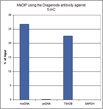
<p><small><strong>Figure 1. Methylated DNA immunoprecipitation (MeDIP) results obtained with the Diagenode monoclonal antibody directed against 5-mC</strong><br />MeDIP (Methylated DNA immunoprecipitation) was performed on 1 µg fragmented human genomic DNA using 0.2 µg of the Diagenode monoclonal antibody against 5-mC (Cat. No. C15200006) and the MagMeDIP Kit (Cat. No. C02010021). The fragmented DNA was spiked with the internal controls present in the kit (methylated DNA (meDNA) as a positive and unmethylated DNA (unDNA) as a negative control) prior to performing the IP. QPCR was performed with optimized primer sets, included in the kit, specific for the methylated and unmethylated DNA controls, and for a known methylated (TSH2B) and unmethylated (GAPDH) genomic region. Figure 2 shows the recovery expressed as a % of input (the relative amount of immunoprecipitated DNA compared to input DNA after qPCR analysis).</small></p>
</div>
</div>
<div class="row">
<div class="small-4 columns">
<p><img src="https://www.diagenode.com/img/product/antibodies/C15200006_MeDIP_2.png" alt="5-methylcytosine (5-mC) Antibody validated method" caption="false" width="278" height="333" /></p>
</div>
<div class="small-8 columns">
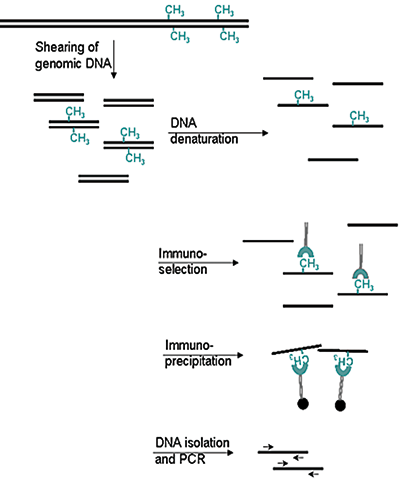
<p><small><strong>Figure 2. Methylated DNA immunoprecipitation (MeDIP) method</strong></small></p>
<ul>
<li><small>Prepare genomic DNA from cultured cells</small></li>
<li><small>Shear genomic DNA</small></li>
<li><small>Denature the sheared genomic DNA</small></li>
<li><small>Immunoprecipitate with the antibody against 5-meC</small></li>
<li><small>Isolate DNA and perform PCR</small></li>
</ul>
</div>
</div>
<div class="row">
<div class="small-4 columns">
<p><img src="https://www.diagenode.com/img/product/antibodies/C15200006_if.jpg" alt="5-methylcytosine (5-mC) Antibody for IF" caption="false" width="278" height="91" /></p>
</div>
<div class="small-8 columns">
<p><small><strong>Figure 3. Immunofluorescence using the Diagenode monoclonal antibody directed against 5-mC</strong> <br />HeLa cells were stained with the Diagenode antibody against 5-mC (Cat. No. C15200006) and with DAPI. Cells were fixed with 4% formaldehyde for 10’ and blocked with PBS/TX-100 containing 1% BSA. The cells were immunofluorescently labelled with the 5-mC antibody (left) diluted 1:1,000 in blocking solution followed by an anti-mouse antibody conjugated to Alexa594. The middle panel shows staining of the nuclei with DAPI. A merge of the two stainings is shown on the right.</small></p>
</div>
</div>',
'label2' => '',
'info2' => '',
'label3' => '',
'info3' => '',
'format' => '10 µg',
'catalog_number' => 'C15200006-10',
'old_catalog_number' => '',
'sf_code' => 'C15200006-D001-000582',
'type' => 'FRE',
'search_order' => '03-Antibody',
'price_EUR' => '105',
'price_USD' => '120',
'price_GBP' => '100',
'price_JPY' => '17205',
'price_CNY' => '',
'price_AUD' => '300',
'country' => 'ALL',
'except_countries' => 'None',
'quote' => false,
'in_stock' => false,
'featured' => false,
'no_promo' => false,
'online' => true,
'master' => false,
'last_datasheet_update' => '',
'slug' => '5-mc-monoclonal-antibody-cl-b-classic-10-ug',
'meta_title' => '5-methylcytosine (5-mC) Antibody - cl. b (C15200006) | Diagenode',
'meta_keywords' => '',
'meta_description' => '5-methylcytosine (5-mC) Monoclonal Antibody cl. b validated in MeDIP and IF. Batch-specific data available on the website. Sample size available.',
'modified' => '2021-02-19 14:48:07',
'created' => '2018-11-12 12:45:06',
'locale' => 'eng'
),
'Antibody' => array(
'host' => '*****',
'id' => '302',
'name' => '5-mC monoclonal antibody (100µg)',
'description' => '',
'clonality' => '',
'isotype' => 'IgG1',
'lot' => 'GF-005 (100µg - lot 006)',
'concentration' => '2.1 µg/µl',
'reactivity' => 'Human, mouse, rat, cow, alligator, zebrafish, plants, finch, wide range expected.',
'type' => 'Monoclonal <strong>MEDIP-grade</strong>',
'purity' => 'Monoclonal antibody purified by gel filtration.',
'classification' => 'Classic',
'application_table' => '<table>
<thead>
<tr>
<th>Applications</th>
<th>Suggested dilution</th>
<th>References</th>
</tr>
</thead>
<tbody>
<tr>
<td>MeDIP</td>
<td>0.5 - 1 µg/IP</td>
<td>Fig 1, 2</td>
</tr>
<tr>
<td>Immunofluorescence</td>
<td>1:1,000</td>
<td>Fig 3</td>
</tr>
</tbody>
</table>
<p></p>
<p><small><sup>*</sup> Please note that of the optimal antibody amount per IP should be determined by the end-user. We recommend testing 0.5-5 µg per IP.</small></p>',
'storage_conditions' => 'Store at -20°C; for long storage, store at -80°C. Avoid multiple freeze-thaw cycles.',
'storage_buffer' => 'PBS containing 0.05% azide.',
'precautions' => 'This product is for research use only. Not for use in diagnostic or therapeutic procedures.',
'uniprot_acc' => '',
'slug' => '',
'meta_keywords' => '',
'meta_description' => '',
'modified' => '2023-08-03 10:26:04',
'created' => '0000-00-00 00:00:00',
'select_label' => '302 - 5-mC monoclonal antibody (100µg) (GF-005 (100µg - lot 006) - 2.1 µg/µl - Human, mouse, rat, cow, alligator, zebrafish, plants, finch, wide range expected. - Monoclonal antibody purified by gel filtration. - Mouse)'
),
'Slave' => array(),
'Group' => array(
'Group' => array(
[maximum depth reached]
),
'Master' => array(
[maximum depth reached]
),
'Product' => array(
[maximum depth reached]
)
),
'Related' => array(),
'Application' => array(
(int) 0 => array(
[maximum depth reached]
),
(int) 1 => array(
[maximum depth reached]
)
),
'Category' => array(
(int) 0 => array(
[maximum depth reached]
),
(int) 1 => array(
[maximum depth reached]
),
(int) 2 => array(
[maximum depth reached]
)
),
'Document' => array(
(int) 0 => array(
[maximum depth reached]
),
(int) 1 => array(
[maximum depth reached]
),
(int) 2 => array(
[maximum depth reached]
)
),
'Feature' => array(),
'Image' => array(
(int) 0 => array(
[maximum depth reached]
)
),
'Promotion' => array(),
'Protocol' => array(),
'Publication' => array(
(int) 0 => array(
[maximum depth reached]
),
(int) 1 => array(
[maximum depth reached]
),
(int) 2 => array(
[maximum depth reached]
),
(int) 3 => array(
[maximum depth reached]
),
(int) 4 => array(
[maximum depth reached]
),
(int) 5 => array(
[maximum depth reached]
),
(int) 6 => array(
[maximum depth reached]
),
(int) 7 => array(
[maximum depth reached]
),
(int) 8 => array(
[maximum depth reached]
),
(int) 9 => array(
[maximum depth reached]
),
(int) 10 => array(
[maximum depth reached]
),
(int) 11 => array(
[maximum depth reached]
),
(int) 12 => array(
[maximum depth reached]
),
(int) 13 => array(
[maximum depth reached]
),
(int) 14 => array(
[maximum depth reached]
),
(int) 15 => array(
[maximum depth reached]
),
(int) 16 => array(
[maximum depth reached]
),
(int) 17 => array(
[maximum depth reached]
),
(int) 18 => array(
[maximum depth reached]
)
),
'Testimonial' => array(),
'Area' => array(),
'SafetySheet' => array(
(int) 0 => array(
[maximum depth reached]
),
(int) 1 => array(
[maximum depth reached]
),
(int) 2 => array(
[maximum depth reached]
),
(int) 3 => array(
[maximum depth reached]
),
(int) 4 => array(
[maximum depth reached]
),
(int) 5 => array(
[maximum depth reached]
),
(int) 6 => array(
[maximum depth reached]
),
(int) 7 => array(
[maximum depth reached]
)
)
),
'meta_canonical' => 'https://www.diagenode.com/en/p/5-mc-monoclonal-antibody-cl-b-classic-100-ug-50-ul'
)
$language = 'en'
$meta_keywords = ''
$meta_description = '5-methylcytosine (5-mC) Monoclonal Antibody cl. b validated in MeDIP and IF. Batch-specific data available on the website. Sample size available.'
$meta_title = '5-methylcytosine (5-mC) Antibody - cl. b (C15200006) | Diagenode'
$product = array(
'Product' => array(
'id' => '3002',
'antibody_id' => '302',
'name' => '5-methylcytosine (5-mC) Antibody - cl. b (sample size)',
'description' => '<p><span>Monoclonal antibody raised in mouse against <strong>5-mC</strong> (<strong>5-methylcytosine</strong>) conjugated to ovalbumine.</span></p>',
'label1' => 'Validation data',
'info1' => '<div class="row">
<div class="small-4 columns">
<p><img src="https://www.diagenode.com/img/product/antibodies/C15200006_MeDIP_1.png" alt="5-methylcytosine (5-mC) Antibody validated in MeDIP" caption="false" width="278" height="292" /></p>
</div>
<div class="small-8 columns">
<p><small><strong>Figure 1. Methylated DNA immunoprecipitation (MeDIP) results obtained with the Diagenode monoclonal antibody directed against 5-mC</strong><br />MeDIP (Methylated DNA immunoprecipitation) was performed on 1 µg fragmented human genomic DNA using 0.2 µg of the Diagenode monoclonal antibody against 5-mC (Cat. No. C15200006) and the MagMeDIP Kit (Cat. No. C02010021). The fragmented DNA was spiked with the internal controls present in the kit (methylated DNA (meDNA) as a positive and unmethylated DNA (unDNA) as a negative control) prior to performing the IP. QPCR was performed with optimized primer sets, included in the kit, specific for the methylated and unmethylated DNA controls, and for a known methylated (TSH2B) and unmethylated (GAPDH) genomic region. Figure 2 shows the recovery expressed as a % of input (the relative amount of immunoprecipitated DNA compared to input DNA after qPCR analysis).</small></p>
</div>
</div>
<div class="row">
<div class="small-4 columns">
<p><img src="https://www.diagenode.com/img/product/antibodies/C15200006_MeDIP_2.png" alt="5-methylcytosine (5-mC) Antibody validated method" caption="false" width="278" height="333" /></p>
</div>
<div class="small-8 columns">
<p><small><strong>Figure 2. Methylated DNA immunoprecipitation (MeDIP) method</strong></small></p>
<ul>
<li><small>Prepare genomic DNA from cultured cells</small></li>
<li><small>Shear genomic DNA</small></li>
<li><small>Denature the sheared genomic DNA</small></li>
<li><small>Immunoprecipitate with the antibody against 5-meC</small></li>
<li><small>Isolate DNA and perform PCR</small></li>
</ul>
</div>
</div>
<div class="row">
<div class="small-4 columns">
<p><img src="https://www.diagenode.com/img/product/antibodies/C15200006_if.jpg" alt="5-methylcytosine (5-mC) Antibody for IF" caption="false" width="278" height="91" /></p>
</div>
<div class="small-8 columns">
<p><small><strong>Figure 3. Immunofluorescence using the Diagenode monoclonal antibody directed against 5-mC</strong> <br />HeLa cells were stained with the Diagenode antibody against 5-mC (Cat. No. C15200006) and with DAPI. Cells were fixed with 4% formaldehyde for 10’ and blocked with PBS/TX-100 containing 1% BSA. The cells were immunofluorescently labelled with the 5-mC antibody (left) diluted 1:1,000 in blocking solution followed by an anti-mouse antibody conjugated to Alexa594. The middle panel shows staining of the nuclei with DAPI. A merge of the two stainings is shown on the right.</small></p>
</div>
</div>',
'label2' => '',
'info2' => '',
'label3' => '',
'info3' => '',
'format' => '10 µg',
'catalog_number' => 'C15200006-10',
'old_catalog_number' => '',
'sf_code' => 'C15200006-D001-000582',
'type' => 'FRE',
'search_order' => '03-Antibody',
'price_EUR' => '105',
'price_USD' => '120',
'price_GBP' => '100',
'price_JPY' => '17205',
'price_CNY' => '',
'price_AUD' => '300',
'country' => 'ALL',
'except_countries' => 'None',
'quote' => false,
'in_stock' => false,
'featured' => false,
'no_promo' => false,
'online' => true,
'master' => false,
'last_datasheet_update' => '',
'slug' => '5-mc-monoclonal-antibody-cl-b-classic-10-ug',
'meta_title' => '5-methylcytosine (5-mC) Antibody - cl. b (C15200006) | Diagenode',
'meta_keywords' => '',
'meta_description' => '5-methylcytosine (5-mC) Monoclonal Antibody cl. b validated in MeDIP and IF. Batch-specific data available on the website. Sample size available.',
'modified' => '2021-02-19 14:48:07',
'created' => '2018-11-12 12:45:06',
'locale' => 'eng'
),
'Antibody' => array(
'host' => '*****',
'id' => '302',
'name' => '5-mC monoclonal antibody (100µg)',
'description' => '',
'clonality' => '',
'isotype' => 'IgG1',
'lot' => 'GF-005 (100µg - lot 006)',
'concentration' => '2.1 µg/µl',
'reactivity' => 'Human, mouse, rat, cow, alligator, zebrafish, plants, finch, wide range expected.',
'type' => 'Monoclonal <strong>MEDIP-grade</strong>',
'purity' => 'Monoclonal antibody purified by gel filtration.',
'classification' => 'Classic',
'application_table' => '<table>
<thead>
<tr>
<th>Applications</th>
<th>Suggested dilution</th>
<th>References</th>
</tr>
</thead>
<tbody>
<tr>
<td>MeDIP</td>
<td>0.5 - 1 µg/IP</td>
<td>Fig 1, 2</td>
</tr>
<tr>
<td>Immunofluorescence</td>
<td>1:1,000</td>
<td>Fig 3</td>
</tr>
</tbody>
</table>
<p></p>
<p><small><sup>*</sup> Please note that of the optimal antibody amount per IP should be determined by the end-user. We recommend testing 0.5-5 µg per IP.</small></p>',
'storage_conditions' => 'Store at -20°C; for long storage, store at -80°C. Avoid multiple freeze-thaw cycles.',
'storage_buffer' => 'PBS containing 0.05% azide.',
'precautions' => 'This product is for research use only. Not for use in diagnostic or therapeutic procedures.',
'uniprot_acc' => '',
'slug' => '',
'meta_keywords' => '',
'meta_description' => '',
'modified' => '2023-08-03 10:26:04',
'created' => '0000-00-00 00:00:00',
'select_label' => '302 - 5-mC monoclonal antibody (100µg) (GF-005 (100µg - lot 006) - 2.1 µg/µl - Human, mouse, rat, cow, alligator, zebrafish, plants, finch, wide range expected. - Monoclonal antibody purified by gel filtration. - Mouse)'
),
'Slave' => array(),
'Group' => array(
'Group' => array(
'id' => '166',
'name' => 'C15200006',
'product_id' => '1965',
'modified' => '2016-02-23 20:46:06',
'created' => '2016-02-23 20:46:06'
),
'Master' => array(
'id' => '1965',
'antibody_id' => '302',
'name' => '5-methylcytosine (5-mC) Antibody - cl. b ',
'description' => '<p><span>Monoclonal antibody raised in mouse against <strong>5-mC</strong> (<strong>5-methylcytosine</strong>) conjugated to ovalbumine.</span></p>',
'label1' => 'Validation Data',
'info1' => '<div class="row">
<div class="small-4 columns">
<p><img src="https://www.diagenode.com/img/product/antibodies/C15200006_MeDIP_1.png" alt="5-methylcytosine (5-mC) Antibody validated in MeDIP" caption="false" width="278" height="292" /></p>
</div>
<div class="small-8 columns">
<p><small><strong>Figure 1. Methylated DNA immunoprecipitation (MeDIP) results obtained with the Diagenode monoclonal antibody directed against 5-mC</strong><br />MeDIP (Methylated DNA immunoprecipitation) was performed on 1 µg fragmented human genomic DNA using 0.2 µg of the Diagenode monoclonal antibody against 5-mC (Cat. No. C15200006) and the MagMeDIP Kit (Cat. No. C02010021). The fragmented DNA was spiked with the internal controls present in the kit (methylated DNA (meDNA) as a positive and unmethylated DNA (unDNA) as a negative control) prior to performing the IP. QPCR was performed with optimized primer sets, included in the kit, specific for the methylated and unmethylated DNA controls, and for a known methylated (TSH2B) and unmethylated (GAPDH) genomic region. Figure 2 shows the recovery expressed as a % of input (the relative amount of immunoprecipitated DNA compared to input DNA after qPCR analysis).</small></p>
</div>
</div>
<div class="row">
<div class="small-4 columns">
<p><img src="https://www.diagenode.com/img/product/antibodies/C15200006_MeDIP_2.png" alt="5-methylcytosine (5-mC) Antibody validated method" caption="false" width="278" height="333" /></p>
</div>
<div class="small-8 columns">
<p><small><strong>Figure 2. Methylated DNA immunoprecipitation (MeDIP) method</strong></small></p>
<ul>
<li><small>Prepare genomic DNA from cultured cells</small></li>
<li><small>Shear genomic DNA</small></li>
<li><small>Denature the sheared genomic DNA</small></li>
<li><small>Immunoprecipitate with the antibody against 5-meC</small></li>
<li><small>Isolate DNA and perform PCR</small></li>
</ul>
</div>
</div>
<div class="row">
<div class="small-4 columns">
<p><img src="https://www.diagenode.com/img/product/antibodies/C15200006_if.jpg" alt="5-methylcytosine (5-mC) Antibody for IF" caption="false" width="278" height="91" /></p>
</div>
<div class="small-8 columns">
<p><small><strong>Figure 3. Immunofluorescence using the Diagenode monoclonal antibody directed against 5-mC</strong> <br />HeLa cells were stained with the Diagenode antibody against 5-mC (Cat. No. C15200006) and with DAPI. Cells were fixed with 4% formaldehyde for 10’ and blocked with PBS/TX-100 containing 1% BSA. The cells were immunofluorescently labelled with the 5-mC antibody (left) diluted 1:1,000 in blocking solution followed by an anti-mouse antibody conjugated to Alexa594. The middle panel shows staining of the nuclei with DAPI. A merge of the two stainings is shown on the right.</small></p>
</div>
</div>',
'label2' => '',
'info2' => '',
'label3' => '',
'info3' => '',
'format' => '100 µg',
'catalog_number' => 'C15200006-100',
'old_catalog_number' => 'MAb-006-100',
'sf_code' => 'C15200006-D001-000526',
'type' => 'FRE',
'search_order' => '03-Antibody',
'price_EUR' => '395',
'price_USD' => '465',
'price_GBP' => '360',
'price_JPY' => '64720',
'price_CNY' => '',
'price_AUD' => '1162',
'country' => 'ALL',
'except_countries' => 'None',
'quote' => false,
'in_stock' => false,
'featured' => false,
'no_promo' => false,
'online' => true,
'master' => true,
'last_datasheet_update' => 'February 11, 2019',
'slug' => '5-mc-monoclonal-antibody-cl-b-classic-100-ug-50-ul',
'meta_title' => '5-methylcytosine (5-mC) Antibody - cl. b (C15200006) | Diagenode',
'meta_keywords' => '5-methylcytosine (5-mC),monoclonal antibody,Methylated DNA Immunoprecipitation',
'meta_description' => '5-methylcytosine (5-mC) Monoclonal Antibody cl. b validated in MeDIP and IF. Batch-specific data available on the website. Sample size available.',
'modified' => '2023-08-03 10:25:19',
'created' => '2015-06-29 14:08:20'
),
'Product' => array(
(int) 0 => array(
[maximum depth reached]
),
(int) 1 => array(
[maximum depth reached]
)
)
),
'Related' => array(),
'Application' => array(
(int) 0 => array(
'id' => '7',
'position' => '10',
'parent_id' => '1',
'name' => 'Methylated DNA immunoprecipitation',
'description' => '<div class="row extra-spaced">
<div class="small-12 medium-3 large-3 columns"><center><a href="https://www.ncbi.nlm.nih.gov/pubmed/30429608" target="_blank"><img src="https://www.diagenode.com/img/banners/banner-nature-publication-580.png" /></a></center></div>
<div class="small-12 medium-9 large-9 columns">
<h3>Sensitive tumour detection and classification using plasma cell-free DNA methylomes<br /><a href="https://www.ncbi.nlm.nih.gov/pubmed/30429608" target="_blank">Read the publication</a></h3>
<h3 class="c-article-title u-h1" data-test="article-title" itemprop="name headline">Preparation of cfMeDIP-seq libraries for methylome profiling of plasma cell-free DNA<br /><a href="https://www.nature.com/articles/s41596-019-0202-2" target="_blank" title="cfMeDIP-seq Nature Method">Read the method</a></h3>
</div>
</div>
<div class="row">
<div class="large-12 columns"><span>The Methylated DNA Immunoprecipitation is based on the affinity purification of methylated and hydroxymethylated DNA using, respectively, an antibody directed against 5-methylcytosine (5-mC) in the case of MeDIP or 5-hydroxymethylcytosine (5-hmC) in the case of hMeDIP.</span><br />
<h2></h2>
<h2>How it works</h2>
<p>In brief, Methyl DNA IP is performed as follows: Genomic DNA from cultured cells or tissues is prepared, sheared, and then denatured. Then, immunoselection and immunoprecipitation can take place using the antibody directed against 5 methylcytosine and antibody binding beads. After isolation and purification is performed, the IP’d methylated DNA is ready for any subsequent analysis as qPCR, amplification, hybridization on microarrays or next generation sequencing.</p>
<h2>Applications</h2>
<div align="center"><a href="https://www.diagenode.com/en/p/magmedip-kit-x48-48-rxns" class="center alert radius button"> qPCR analysis</a></div>
<div align="center"><a href="https://www.diagenode.com/en/p/magmedip-seq-package-V2-x10" class="center alert radius button"> NGS analysis </a></div>
<h2>Advantages</h2>
<ul style="font-size: 19px;" class="nobullet">
<li><i class="fa fa-arrow-circle-right"></i> <strong>Unaffected</strong> DNA</li>
<li><i class="fa fa-arrow-circle-right"></i> <strong>High enrichment</strong> yield</li>
<li><i class="fa fa-arrow-circle-right"></i> <strong>Robust</strong> & <strong>reproducible</strong> techniques</li>
<li><i class="fa fa-arrow-circle-right"></i> <strong>NGS</strong> compatible</li>
</ul>
<h2></h2>
</div>
</div>
<div id="gtx-trans" style="position: absolute; left: 17px; top: 652.938px;">
<div class="gtx-trans-icon"></div>
</div>',
'in_footer' => false,
'in_menu' => true,
'online' => true,
'tabular' => true,
'slug' => 'methylated-dna-immunoprecipitation',
'meta_keywords' => 'Methylated DNA immunoprecipitation,Epigenetic,DNA Methylation,qPCR,5 methylcytosine (5-mC)',
'meta_description' => 'Methylated DNA immunoprecipitation method is based on the affinity purification of methylated DNA using an antibody directed against 5 methylcytosine (5-mC). ',
'meta_title' => 'Methylated DNA immunoprecipitation(MeDIP) - Dna methylation | Diagenode',
'modified' => '2021-08-19 12:08:03',
'created' => '2014-09-14 05:33:34',
'ProductsApplication' => array(
[maximum depth reached]
)
),
(int) 1 => array(
'id' => '29',
'position' => '10',
'parent_id' => '40',
'name' => 'IF',
'description' => '<p><strong>Immunofluorescence</strong>:</p>
<p>Diagenode offers huge selection of highly sensitive antibodies validated in IF.</p>
<p><img src="https://www.diagenode.com/img/product/antibodies/C15200229-IF.jpg" alt="" height="245" width="256" /></p>
<p><sup><strong>Immunofluorescence using the Diagenode monoclonal antibody directed against CRISPR/Cas9</strong></sup></p>
<p><sup>HeLa cells transfected with a Cas9 expression vector (left) or untransfected cells (right) were fixed in methanol at -20°C, permeabilized with acetone at -20°C and blocked with PBS containing 2% BSA. The cells were stained with the Cas9 C-terminal antibody (Cat. No. C15200229) diluted 1:400, followed by incubation with an anti-mouse secondary antibody coupled to AF488. The bottom images show counter-staining of the nuclei with Hoechst 33342.</sup></p>
<h5><sup>Check our selection of antibodies validated in IF.</sup></h5>',
'in_footer' => false,
'in_menu' => false,
'online' => true,
'tabular' => true,
'slug' => 'immunofluorescence',
'meta_keywords' => 'Immunofluorescence,Monoclonal antibody,Polyclonal antibody',
'meta_description' => 'Diagenode offers a wide range of antibodies and technical support for Immunofluorescence applications',
'meta_title' => 'Immunofluorescence - Monoclonal antibody - Polyclonal antibody | Diagenode',
'modified' => '2016-04-27 16:23:10',
'created' => '2015-07-08 13:46:02',
'ProductsApplication' => array(
[maximum depth reached]
)
)
),
'Category' => array(
(int) 0 => array(
'id' => '23',
'position' => '40',
'parent_id' => '4',
'name' => 'DNA modifications',
'description' => '<p><span style="font-weight: 400;">T</span><span style="font-weight: 400;">he pattern of <strong>DNA modifications</strong> is critical for genome stability and the control of gene expression in the cell. Methylation of 5-cytosine (5-mC), one of the best-studied epigenetic marks, is carried out by the <strong>DNA methyltransferases</strong> DNMT3A and B and DNMT1. DNMT3A and DNMT3B are responsible for </span><i><span style="font-weight: 400;">de novo</span></i><span style="font-weight: 400;"> DNA methylation, whereas DNMT1 maintains existing methylation. 5-mC undergoes active demethylation which is performed by the <strong>Ten-Eleven Translocation</strong> (TET) familly of DNA hydroxylases. The latter consists of 3 members TET1, 2 and 3. All 3 members catalyze the conversion of <strong>5-methylcytosine</strong> (5-mC) into <strong>5-hydroxymethylcytosine</strong> (5-hmC), and further into <strong>5-formylcytosine</strong> (5-fC) and <strong>5-carboxycytosine</strong> (5-caC). 5-fC and 5-caC can be converted to unmodified cytosine by <strong>Thymine DNA Glycosylase</strong> (TDG). It is not yet clear if 5-hmC, 5-fC and 5-caC have specific functions or are simply intermediates in the demethylation of 5-mC.</span></p>
<p><span style="font-weight: 400;">DNA methylation is generally considered as a repressive mark and is usually associated with gene silencing. It is essential that the balance between DNA methylation and demethylation is precisely maintained. Dysregulation of DNA methylation may lead to many different human diseases and is often observed in cancer cells.</span></p>
<p><span style="font-weight: 400;">Diagenode offers highly validated antibodies against different proteins involved in DNA modifications as well as against the modified bases allowing the study of all steps and intermediates in the DNA methylation/demethylation pathway:</span></p>
<p><img src="https://www.diagenode.com/img/categories/antibodies/dna-methylation.jpg" height="599" width="816" /></p>
<p><strong>Diagenode exclusively sources the original 5-methylcytosine monoclonal antibody (clone 33D3).</strong></p>
<p>Check out the list below to see all proposed antibodies for DNA modifications.</p>
<p>Diagenode’s highly validated antibodies:</p>
<ul>
<li>Highly sensitive and specific</li>
<li>Cost-effective (requires less antibody per reaction)</li>
<li>Batch-specific data is available on the website</li>
<li>Expert technical support</li>
<li>Sample sizes available</li>
<li>100% satisfaction guarantee</li>
</ul>',
'no_promo' => false,
'in_menu' => true,
'online' => true,
'tabular' => false,
'hide' => true,
'all_format' => false,
'is_antibody' => true,
'slug' => 'dna-methylation-antibody',
'cookies_tag_id' => null,
'meta_keywords' => 'Monoclonal Antibodies,Polyclonal antibodies,DNA methylation,Diagenode',
'meta_description' => 'Diagenode offers Monoclonal and Polyclonal antibodies for DNA Methylation. The pattern of DNA modifications is critical for genome stability and the control of gene expression in the cell. ',
'meta_title' => 'DNA modifications - Monoclonal and Polyclonal Antibodies for DNA Methylation | Diagenode',
'modified' => '2020-08-03 15:25:37',
'created' => '2014-07-31 03:51:53',
'ProductsCategory' => array(
[maximum depth reached]
),
'CookiesTag' => array([maximum depth reached])
),
(int) 1 => array(
'id' => '102',
'position' => '1',
'parent_id' => '4',
'name' => 'Sample size antibodies',
'description' => '<h1><strong>Validated epigenetics antibodies</strong> – care for a sample?<br /> </h1>
<p>Diagenode has partnered with leading epigenetics experts and numerous epigenetics consortiums to bring to you a validated and comprehensive collection of epigenetic antibodies. As an expert in epigenetics, we are committed to offering highly-specific antibodies validated for ChIP/ChIP-seq and many other applications. All batch-specific validation data is available on our website.<br /><a href="../categories/antibodies">Read about our expertise in antibody production</a>.</p>
<ul>
<li><strong>Focused</strong> - Diagenode's selection of antibodies is exclusively dedicated for epigenetic research. <a title="See the full collection." href="../categories/all-antibodies">See the full collection.</a></li>
<li><strong>Strict quality standards</strong> with rigorous QC and validation</li>
<li><strong>Classified</strong> based on level of validation for flexibility of application</li>
</ul>
<p>Existing sample sizes are listed below. We will soon expand our collection. Are you looking for a sample size of another antibody? Just <a href="mailto:agnieszka.zelisko@diagenode.com?Subject=Sample%20Size%20Request" target="_top">Contact us</a>.</p>',
'no_promo' => false,
'in_menu' => true,
'online' => true,
'tabular' => false,
'hide' => true,
'all_format' => true,
'is_antibody' => true,
'slug' => 'sample-size-antibodies',
'cookies_tag_id' => null,
'meta_keywords' => '5-hmC monoclonal antibody,CRISPR/Cas9 polyclonal antibody ,H3K36me3 polyclonal antibody,diagenode',
'meta_description' => 'Diagenode offers sample volume on selected antibodies for researchers to test, validate and provide confidence and flexibility in choosing from our wide range of antibodies ',
'meta_title' => 'Sample-size Antibodies | Diagenode',
'modified' => '2019-07-03 10:57:05',
'created' => '2015-10-27 12:13:34',
'ProductsCategory' => array(
[maximum depth reached]
),
'CookiesTag' => array([maximum depth reached])
),
(int) 2 => array(
'id' => '103',
'position' => '0',
'parent_id' => '4',
'name' => 'All antibodies',
'description' => '<p><span style="font-weight: 400;">All Diagenode’s antibodies are listed below. Please, use our Quick search field to find the antibody of interest by target name, application, purity.</span></p>
<p><span style="font-weight: 400;">Diagenode’s highly validated antibodies:</span></p>
<ul>
<li>Highly sensitive and specific</li>
<li>Cost-effective (requires less antibody per reaction)</li>
<li>Batch-specific data is available on the website</li>
<li>Expert technical support</li>
<li>Sample sizes available</li>
<li>100% satisfaction guarantee</li>
</ul>',
'no_promo' => false,
'in_menu' => true,
'online' => true,
'tabular' => false,
'hide' => true,
'all_format' => false,
'is_antibody' => true,
'slug' => 'all-antibodies',
'cookies_tag_id' => null,
'meta_keywords' => 'Antibodies,Premium Antibodies,Classic,Pioneer',
'meta_description' => 'Diagenode Offers Strict quality standards with Rigorous QC and validated Antibodies. Classified based on level of validation for flexibility of Application. Comprehensive selection of histone and non-histone Antibodies',
'meta_title' => 'Diagenode's selection of Antibodies is exclusively dedicated for Epigenetic Research | Diagenode',
'modified' => '2019-07-03 10:55:44',
'created' => '2015-11-02 14:49:22',
'ProductsCategory' => array(
[maximum depth reached]
),
'CookiesTag' => array([maximum depth reached])
)
),
'Document' => array(
(int) 0 => array(
'id' => '11',
'name' => 'Antibodies you can trust',
'description' => '<p style="text-align: justify;"><span>Epigenetic research tools have evolved over time from endpoint PCR to qPCR to the analyses of large sets of genome-wide sequencing data. ChIP sequencing (ChIP-seq) has now become the gold standard method for chromatin studies, given the accuracy and coverage scale of the approach over other methods. Successful ChIP-seq, however, requires a higher level of experimental accuracy and consistency in all steps of ChIP than ever before. Particularly crucial is the quality of ChIP antibodies. </span></p>',
'image_id' => null,
'type' => 'Poster',
'url' => 'files/posters/Antibodies_you_can_trust_Poster.pdf',
'slug' => 'antibodies-you-can-trust-poster',
'meta_keywords' => '',
'meta_description' => '',
'modified' => '2015-10-01 20:18:31',
'created' => '2015-07-03 16:05:15',
'ProductsDocument' => array(
[maximum depth reached]
)
),
(int) 1 => array(
'id' => '38',
'name' => 'Epigenetic Antibodies Brochure',
'description' => '<p>More than in any other immuoprecipitation assays, quality antibodies are critical tools in many epigenetics experiments. Since 10 years, Diagenode has developed the most stringent quality production available on the market for antibodies exclusively focused on epigenetic uses. All our antibodies have been qualified to work in epigenetic applications.</p>',
'image_id' => null,
'type' => 'Brochure',
'url' => 'files/brochures/Epigenetic_Antibodies_Brochure.pdf',
'slug' => 'epigenetic-antibodies-brochure',
'meta_keywords' => '',
'meta_description' => '',
'modified' => '2016-06-15 11:24:06',
'created' => '2015-07-03 16:05:27',
'ProductsDocument' => array(
[maximum depth reached]
)
),
(int) 2 => array(
'id' => '1104',
'name' => 'Datasheet 5mC C15200006-10',
'description' => '<p><span>Monoclonal antibody raised in mouse against 5-mC(5-methylcytosine) conjugated to ovalbumine.</span></p>',
'image_id' => null,
'type' => 'Datasheet',
'url' => 'files/products/antibodies/Datasheet_5mC_C15200006-10.pdf',
'slug' => 'datasheet-5mc-c15200006-10',
'meta_keywords' => '',
'meta_description' => '',
'modified' => '2020-11-26 11:01:16',
'created' => '2020-11-26 11:01:16',
'ProductsDocument' => array(
[maximum depth reached]
)
)
),
'Feature' => array(),
'Image' => array(
(int) 0 => array(
'id' => '250',
'name' => 'product/antibodies/antibody.png',
'alt' => 'Mouse IgG',
'modified' => '2020-11-27 07:00:09',
'created' => '2015-07-17 10:12:18',
'ProductsImage' => array(
[maximum depth reached]
)
)
),
'Promotion' => array(),
'Protocol' => array(),
'Publication' => array(
(int) 0 => array(
'id' => '4806',
'name' => 'An enriched maternal environment and stereotypies of sows differentiallyaffect the neuro-epigenome of brain regions related to emotionality intheir piglets.',
'authors' => 'Tatemoto P. et al.',
'description' => '<p><span>Epigenetic mechanisms are important modulators of neurodevelopmental outcomes in the offspring of animals challenged during pregnancy. Pregnant sows living in a confined environment are challenged with stress and lack of stimulation which may result in the expression of stereotypies (repetitive behaviours without an apparent function). Little attention has been devoted to the postnatal effects of maternal stereotypies in the offspring. We investigated how the environment and stereotypies of pregnant sows affected the neuro-epigenome of their piglets. We focused on the amygdala, frontal cortex, and hippocampus, brain regions related to emotionality, learning, memory, and stress response. Differentially methylated regions (DMRs) were investigated in these brain regions of male piglets born from sows kept in an enriched vs a barren environment. Within the latter group of piglets, we compared the brain methylomes of piglets born from sows expressing stereotypies vs sows not expressing stereotypies. DMRs emerged in each comparison. While the epigenome of the hippocampus and frontal cortex of piglets is mainly affected by the maternal environment, the epigenome of the amygdala is mainly affected by maternal stereotypies. The molecular pathways and mechanisms triggered in the brains of piglets by maternal environment or stereotypies are different, which is reflected on the differential gene function associated to the DMRs found in each piglets' brain region . The present study is the first to investigate the neuro-epigenomic effects of maternal enrichment in pigs' offspring and the first to investigate the neuro-epigenomic effects of maternal stereotypies in the offspring of a mammal.</span></p>',
'date' => '2023-12-01',
'pmid' => 'https://www.ncbi.nlm.nih.gov/pubmed/37192378',
'doi' => '10.1080/15592294.2023.2196656',
'modified' => '2023-06-15 08:49:05',
'created' => '2023-06-13 21:11:31',
'ProductsPublication' => array(
[maximum depth reached]
)
),
(int) 1 => array(
'id' => '4596',
'name' => 'Role of epigenetics in the etiology of hypospadias through penileforeskin DNA methylation alterations.',
'authors' => 'Kaefer M. et al.',
'description' => '<p>Abnormal penile foreskin development in hypospadias is the most frequent genital malformation in male children, which has increased dramatically in recent decades. A number of environmental factors have been shown to be associated with hypospadias development. The current study investigated the role of epigenetics in the etiology of hypospadias and compared mild (distal), moderate (mid shaft), and severe (proximal) hypospadias. Penile foreskin samples were collected from hypospadias and non-hypospadias individuals to identify alterations in DNA methylation associated with hypospadias. Dramatic numbers of differential DNA methylation regions (DMRs) were observed in the mild hypospadias, with reduced numbers in moderate and low numbers in severe hypospadias. Atresia (cell loss) of the principal foreskin fibroblast is suspected to be a component of the disease etiology. A genome-wide (> 95\%) epigenetic analysis was used and the genomic features of the DMRs identified. The DMR associated genes identified a number of novel hypospadias associated genes and pathways, as well as genes and networks known to be involved in hypospadias etiology. Observations demonstrate altered DNA methylation sites in penile foreskin is a component of hypospadias etiology. In addition, a potential role of environmental epigenetics and epigenetic inheritance in hypospadias disease etiology is suggested.</p>',
'date' => '2023-01-01',
'pmid' => 'https://www.ncbi.nlm.nih.gov/pubmed/36631595',
'doi' => '10.1038/s41598-023-27763-5',
'modified' => '2023-04-06 09:08:57',
'created' => '2023-02-21 09:59:46',
'ProductsPublication' => array(
[maximum depth reached]
)
),
(int) 2 => array(
'id' => '4538',
'name' => 'Examination of Generational Impacts of Adolescent Chemotherapy:Ifosfamide and Potential for Epigenetic TransgenerationalInheritance',
'authors' => 'Thompson R. P. et al.',
'description' => '<p>The current study was designed to use a rodent model to determine if exposure to the chemotherapy drug ifosfamide during puberty can induce altered phenotypes and disease in the grand-offspring of exposed individuals through epigenetic transgenerational inheritance. Pathologies such as delayed pubertal onset, kidney disease and multiple pathologies were observed to be significantly more frequent in the F1 generation offspring of ifosfamide lineage females. The F2 generation grand-offspring ifosfamide lineage males had transgenerational pathology phenotypes of early pubertal onset and reduced testis pathology. Reduced levels of anxiety were observed in both males and females in the transgenerational F2 generation grandoffspring. Differential DNA methylated regions (DMRs) in chemotherapy lineage sperm in the F1 and F2 generations were identified. Therefore, chemotherapy exposure impacts pathology susceptibility in subsequent generations. Observations highlight the importance that prior to chemotherapy, individuals need to consider cryopreservation of germ cells as a precautionary measure if they have children</p>',
'date' => '2022-11-01',
'pmid' => 'https://doi.org/10.1016%2Fj.isci.2022.105570',
'doi' => '10.1016/j.isci.2022.105570',
'modified' => '2022-11-25 08:59:32',
'created' => '2022-11-24 08:49:52',
'ProductsPublication' => array(
[maximum depth reached]
)
),
(int) 3 => array(
'id' => '4656',
'name' => 'Epigenome-wide association study of physical activity and physiologicalparameters in discordant monozygotic twins.',
'authors' => 'Duncan Glen E et al.',
'description' => '<p>An epigenome-wide association study (EWAS) was performed on buccal cells from monozygotic-twins (MZ) reared together as children, but who live apart as adults. Cohorts of twin pairs were used to investigate associations between neighborhood walkability and objectively measured physical activity (PA) levels. Due to dramatic cellular epigenetic sex differences, male and female MZ twin pairs were analyzed separately to identify differential DNA methylation regions (DMRs). A priori comparisons were made on MZ twin pairs discordant on body mass index (BMI), PA levels, and neighborhood walkability. In addition to direct comparative analysis to identify specific DMRs, a weighted genome coexpression network analysis (WGCNA) was performed to identify DNA methylation sites associated with the physiological traits of interest. The pairs discordant in PA levels had epigenetic alterations that correlated with reduced metabolic parameters (i.e., BMI and waist circumference). The DNA methylation sites are associated with over fifty genes previously found to be specific to vigorous PA, metabolic risk factors, and sex. Combined observations demonstrate that behavioral factors, such as physical activity, appear to promote systemic epigenetic alterations that impact metabolic risk factors. The epigenetic DNA methylation sites and associated genes identified provide insight into PA impacts on metabolic parameters and the etiology of obesity.</p>',
'date' => '2022-11-01',
'pmid' => 'https://www.ncbi.nlm.nih.gov/pubmed/36424439',
'doi' => '10.1038/s41598-022-24642-3',
'modified' => '2023-03-07 08:56:57',
'created' => '2023-02-21 09:59:46',
'ProductsPublication' => array(
[maximum depth reached]
)
),
(int) 4 => array(
'id' => '4557',
'name' => 'Environmental induced transgenerational inheritance impacts systemsepigenetics in disease etiology.',
'authors' => 'Beck D. et al.',
'description' => '<p>Environmental toxicants have been shown to promote the epigenetic transgenerational inheritance of disease through exposure specific epigenetic alterations in the germline. The current study examines the actions of hydrocarbon jet fuel, dioxin, pesticides (permethrin and methoxychlor), plastics, and herbicides (glyphosate and atrazine) in the promotion of transgenerational disease in the great grand-offspring rats that correlates with specific disease associated differential DNA methylation regions (DMRs). The transgenerational disease observed was similar for all exposures and includes pathologies of the kidney, prostate, and testis, pubertal abnormalities, and obesity. The disease specific DMRs in sperm were exposure specific for each pathology with negligible overlap. Therefore, for each disease the DMRs and associated genes were distinct for each exposure generational lineage. Observations suggest a large number of DMRs and associated genes are involved in a specific pathology, and various environmental exposures influence unique subsets of DMRs and genes to promote the transgenerational developmental origins of disease susceptibility later in life. A novel multiscale systems biology basis of disease etiology is proposed involving an integration of environmental epigenetics, genetics and generational toxicology.</p>',
'date' => '2022-04-01',
'pmid' => 'https://www.ncbi.nlm.nih.gov/pubmed/35440735',
'doi' => '10.1038/s41598-022-09336-0',
'modified' => '2022-11-24 09:32:20',
'created' => '2022-11-24 08:49:52',
'ProductsPublication' => array(
[maximum depth reached]
)
),
(int) 5 => array(
'id' => '4378',
'name' => 'GBS-MeDIP: A protocol for parallel identification of genetic andepigenetic variation in the same reduced fraction of genomes acrossindividuals.',
'authors' => 'Rezaei S. et al.',
'description' => '<p>The GBS-MeDIP protocol combines two previously described techniques, Genotype-by-Sequencing (GBS) and Methylated-DNA-Immunoprecipitation (MeDIP). Our method allows for parallel and cost-efficient interrogation of genetic and methylomic variants in the DNA of many reduced genomes, taking advantage of the barcoding of DNA samples performed in the GBS and the subsequent creation of DNA pools, then used as an input for the MeDIP. The GBS-MeDIP is particularly suitable to identify genetic and methylomic biomarkers when resources for whole genome interrogation are lacking.</p>',
'date' => '2022-03-01',
'pmid' => 'https://www.ncbi.nlm.nih.gov/pubmed/35257114',
'doi' => '10.1016/j.xpro.2022.101202',
'modified' => '2022-08-04 16:12:41',
'created' => '2022-08-04 14:55:36',
'ProductsPublication' => array(
[maximum depth reached]
)
),
(int) 6 => array(
'id' => '4558',
'name' => 'Preterm birth buccal cell epigenetic biomarkers to facilitatepreventative medicine.',
'authors' => 'Winchester P. et al.',
'description' => '<p>Preterm birth is the major cause of newborn and infant mortality affecting nearly one in every ten live births. The current study was designed to develop an epigenetic biomarker for susceptibility of preterm birth using buccal cells from the mother, father, and child (triads). An epigenome-wide association study (EWAS) was used to identify differential DNA methylation regions (DMRs) using a comparison of control term birth versus preterm birth triads. Epigenetic DMR associations with preterm birth were identified for both the mother and father that were distinct and suggest potential epigenetic contributions from both parents. The mother (165 DMRs) and female child (136 DMRs) at p < 1e-04 had the highest number of DMRs and were highly similar suggesting potential epigenetic inheritance of the epimutations. The male child had negligible DMR associations. The DMR associated genes for each group involve previously identified preterm birth associated genes. Observations identify a potential paternal germline contribution for preterm birth and identify the potential epigenetic inheritance of preterm birth susceptibility for the female child later in life. Although expanded clinical trials and preconception trials are required to optimize the potential epigenetic biomarkers, such epigenetic biomarkers may allow preventative medicine strategies to reduce the incidence of preterm birth.</p>',
'date' => '2022-03-01',
'pmid' => 'https://www.ncbi.nlm.nih.gov/pubmed/35232984',
'doi' => '10.1038/s41598-022-07262-9',
'modified' => '2022-11-24 09:33:24',
'created' => '2022-11-24 08:49:52',
'ProductsPublication' => array(
[maximum depth reached]
)
),
(int) 7 => array(
'id' => '4312',
'name' => 'Epigenetic inheritance of DNA methylation changes in fish living inhydrogen sulfide-rich springs.',
'authors' => 'Kelley J. et al.',
'description' => '<p>Environmental factors can promote phenotypic variation through alterations in the epigenome and facilitate adaptation of an organism to the environment. Although hydrogen sulfide is toxic to most organisms, the fish has adapted to survive in environments with high levels that exceed toxicity thresholds by orders of magnitude. Epigenetic changes in response to this environmental stressor were examined by assessing DNA methylation alterations in red blood cells, which are nucleated in fish. Males and females were sampled from sulfidic and nonsulfidic natural environments; individuals were also propagated for two generations in a nonsulfidic laboratory environment. We compared epimutations between the sexes as well as field and laboratory populations. For both the wild-caught (F0) and the laboratory-reared (F2) fish, comparing the sulfidic and nonsulfidic populations revealed evidence for significant differential DNA methylation regions (DMRs). More importantly, there was over 80\% overlap in DMRs across generations, suggesting that the DMRs have stable generational inheritance in the absence of the sulfidic environment. This is an example of epigenetic generational stability after the removal of an environmental stressor. The DMR-associated genes were related to sulfur toxicity and metabolic processes. These findings suggest that adaptation of to sulfidic environments in southern Mexico may, in part, be promoted through epigenetic DNA methylation alterations that become stable and are inherited by subsequent generations independent of the environment.</p>',
'date' => '2021-06-01',
'pmid' => 'https://pubmed.ncbi.nlm.nih.gov/34185679/',
'doi' => '10.1073/pnas.2014929118',
'modified' => '2022-08-02 16:41:22',
'created' => '2022-05-19 10:41:50',
'ProductsPublication' => array(
[maximum depth reached]
)
),
(int) 8 => array(
'id' => '4051',
'name' => 'Epigenome-wide association study for pesticide (Permethrin and DEET)induced DNA methylation epimutation biomarkers for specifictransgenerational disease.',
'authors' => 'Thorson, Jennifer L M and Beck, Daniel and Ben Maamar, Millissia andNilsson, Eric E and Skinner, Michael K',
'description' => '<p>BACKGROUND: Permethrin and N,N-diethyl-meta-toluamide (DEET) are the pesticides and insect repellent most commonly used by humans. These pesticides have been shown to promote the epigenetic transgenerational inheritance of disease in rats. The current study was designed as an epigenome-wide association study (EWAS) to identify potential sperm DNA methylation epimutation biomarkers for specific transgenerational disease. METHODS: Outbred Sprague Dawley gestating female rats (F0) were transiently exposed during fetal gonadal sex determination to the pesticide combination including Permethrin and DEET. The F3 generation great-grand offspring within the pesticide lineage were aged to 1 year. The transgenerational adult male rat sperm were collected from individuals with single and multiple diseases and compared to non-diseased animals to identify differential DNA methylation regions (DMRs) as biomarkers for specific transgenerational disease. RESULTS: The exposure of gestating female rats to a permethrin and DEET pesticide combination promoted transgenerational testis disease, prostate disease, kidney disease, and the presence of multiple disease in the subsequent F3 generation great-grand offspring. The disease DMRs were found to be disease specific with negligible overlap between different diseases. The genomic features of CpG density, DMR length, and chromosomal locations of the disease specific DMRs were investigated. Interestingly, the majority of the disease specific sperm DMR associated genes have been previously found to be linked to relevant disease specific genes. CONCLUSIONS: Observations demonstrate the EWAS approach identified disease specific biomarkers that can be potentially used to assess transgenerational disease susceptibility and facilitate the clinical management of environmentally induced pathology.</p>',
'date' => '2020-11-01',
'pmid' => 'https://www.ncbi.nlm.nih.gov/pubmed/33148267',
'doi' => '10.1186/s12940-020-00666-y',
'modified' => '2021-02-19 14:49:21',
'created' => '2021-02-18 10:21:53',
'ProductsPublication' => array(
[maximum depth reached]
)
),
(int) 9 => array(
'id' => '4064',
'name' => 'Between-Generation Phenotypic and Epigenetic Stability in a Clonal Snail.',
'authors' => 'Smithson, Mark and Thorson, Jennifer L M and Sadler-Riggleman, Ingrid andBeck, Daniel and Skinner, Michael K and Dybdahl, Mark',
'description' => '<p>Epigenetic variation might play an important role in generating adaptive phenotypes by underpinning within-generation developmental plasticity, persistent parental effects of the environment (e.g., transgenerational plasticity), or heritable epigenetically based polymorphism. These adaptive mechanisms should be most critical in organisms where genetic sources of variation are limited. Using a clonally reproducing freshwater snail (Potamopyrgus antipodarum), we examined the stability of an adaptive phenotype (shell shape) and of DNA methylation between generations. First, we raised three generations of snails adapted to river currents in the lab without current. We showed that habitat-specific adaptive shell shape was relatively stable across three generations but shifted slightly over generations two and three toward a no-current lake phenotype. We also showed that DNA methylation specific to high-current environments was stable across one generation. This study provides the first evidence of stability of DNA methylation patterns across one generation in an asexual animal. Together, our observations are consistent with the hypothesis that adaptive shell shape variation is at least in part determined by transgenerational plasticity, and that DNA methylation provides a potential mechanism for stability of shell shape across one generation.</p>',
'date' => '2020-09-01',
'pmid' => 'https://www.ncbi.nlm.nih.gov/pubmed/32877512',
'doi' => '10.1093/gbe/evaa181',
'modified' => '2021-02-19 17:43:55',
'created' => '2021-02-18 10:21:53',
'ProductsPublication' => array(
[maximum depth reached]
)
),
(int) 10 => array(
'id' => '3967',
'name' => 'DNA methylation variation in the brain of laying hens in relation to differential behavioral patterns',
'authors' => 'Guerrero-Bosagna Carlos, Pértille Fábio, Gomez Yamenah, Rezaei Shiva, Gebhardt Sabine, Vögeli Sabine, Stratmann Ariane, Vöelkl Bernhard, Toscano Michael J.',
'description' => '<p>Domesticated animals are unique to investigate the contribution of genetic and non-genetic factors to specific phenotypes. Among non-genetic factors involved in phenotype formation are epigenetic mechanisms. Here we aimed to identify whether relative DNA methylation differences in the nidopallium between groups of individuals are among the non-genetic factors involved in the emergence of differential behavioral patterns in hens. The nidopallium was selected due to its important role in complex cognitive function (i.e., decision making) in birds. Behavioral patterns that spontaneously emerge in hens living in a highly controlled environment were identified with a unique tracking system that recorded their transitions between pen zones. Behavioral activity patterns were characterized through three classification schemes: (i) daily specific features of behavioral routines (Entropy), (ii) daily spatio-temporal activity patterns (Dynamic Time Warping), and (iii) social leading behavior (Leading Index). Unique differentially methylated regions (DMRs) were identified between behavioral patterns emerging within classification schemes, with entropy having the higher number. Functionally, DTW had double the proportion of affected promoters and half of the distal intergenic regions. Pathway enrichment analysis of DMR-associated genes revealed that Entropy relates mainly to cell cycle checkpoints, Leading Index to mitochondrial function, and DTW to gene expression regulation. Our study suggests that different biological functions within neurons (particularly in the nidopallium) could be responsible for the emergence of distinct behavior patterns and that epigenetic variation within brain tissues would be an important factor to explain behavioral variation.</p>',
'date' => '2020-05-17',
'pmid' => 'https://www.sciencedirect.com/science/article/abs/pii/S1744117X20300472',
'doi' => '10.1016/j.cbd.2020.100700',
'modified' => '2020-08-12 09:35:05',
'created' => '2020-08-10 12:12:25',
'ProductsPublication' => array(
[maximum depth reached]
)
),
(int) 11 => array(
'id' => '3816',
'name' => 'Sperm DNA Methylation Epimutation Biomarkers for Male Infertility and FSH Therapeutic Responsiveness.',
'authors' => 'Luján S, Caroppo E, Niederberger C, Arce JC, Sadler-Riggleman I, Beck D, Nilsson E, Skinner MK',
'description' => '<p>Male factor infertility is increasing and recognized as playing a key role in reproductive health and disease. The current primary diagnostic approach is to assess sperm quality associated with reduced sperm number and motility, which has been historically of limited success in separating fertile from infertile males. The current study was designed to develop a molecular analysis to identify male idiopathic infertility using genome wide alterations in sperm DNA methylation. A signature of differential DNA methylation regions (DMRs) was identified to be associated with male idiopathic infertility patients. A promising therapeutic treatment of male infertility is the use of follicle stimulating hormone (FSH) analogs which improved sperm numbers and motility in a sub-population of infertility patients. The current study also identified genome-wide DMRs that were associated with the patients that were responsive to FSH therapy versus those that were non-responsive. This novel use of epigenetic biomarkers to identify responsive versus non-responsive patient populations is anticipated to dramatically improve clinical trials and facilitate therapeutic treatment of male infertility patients. The use of epigenetic biomarkers for disease and therapeutic responsiveness is anticipated to be applicable for other medical conditions.</p>',
'date' => '2019-11-14',
'pmid' => 'http://www.pubmed.gov/31727924',
'doi' => '10.1038/s41598-019-52903-1',
'modified' => '2019-12-05 10:56:51',
'created' => '2019-12-02 15:25:44',
'ProductsPublication' => array(
[maximum depth reached]
)
),
(int) 12 => array(
'id' => '3804',
'name' => 'Epigenetic transgenerational inheritance of parent-of-origin allelic transmission of outcross pathology and sperm epimutations',
'authors' => 'Ben Maamar Millissia, King Stephanie E., Nilsson Eric, Beck Daniel, Skinner Michael K.',
'description' => '<p>Epigenetic transgenerational inheritance potentially impacts disease etiology, phenotypic variation, and evolution. An increasing number of environmental factors from nutrition to toxicants have been shown to promote the epigenetic transgenerational inheritance of disease. Previous observations have demonstrated that the agricultural fungicide vinclozolin and pesticide DDT (dichlorodiphenyltrichloroethane) induce transgenerational sperm epimutations involving DNA methylation, ncRNA, and histone modifications or retention. These two environmental toxicants were used to investigate the impacts of parent-oforigin outcross on the epigenetic transgenerational inheritance of disease. Male and female rats were collected from a paternal outcross (POC) or a maternal outcross (MOC) F4 generation control and exposure lineages for pathology and epigenetic analysis. This model allows the parental allelic transmission of disease and epimutations to be investigated. There was increased pathology incidence in the MOC F4 generation male prostate, kidney, obesity, and multiple diseases through a maternal allelic transmission. The POC F4 generation female offspring had increased pathology incidence for kidney, obesity and multiple types of diseases through the paternal allelic transmission. Some disease such as testis or ovarian pathology appear to be transmitted through the combined actions of both male and female alleles. Analysis of the F4 generation sperm epigenomes identified differential DNA methylated regions (DMRs) in a genomewide analysis. Observations demonstrate that DDT and vinclozolin have the potential to promote the epigenetic transgenerational inheritance of disease and sperm epimutations to the outcross F4 generation in a sex specific and exposure specific manner. The parent-of-origin allelic transmission observed appears similar to the process involved with imprinted-like genes.</p>',
'date' => '2019-10-29',
'pmid' => 'https://www.ncbi.nlm.nih.gov/pubmed/31682807',
'doi' => '10.1016/j.ydbio.2019.10.030',
'modified' => '2019-12-05 11:24:40',
'created' => '2019-12-02 15:25:44',
'ProductsPublication' => array(
[maximum depth reached]
)
),
(int) 13 => array(
'id' => '3706',
'name' => 'TET3 prevents terminal differentiation of adult NSCs by a non-catalytic action at Snrpn.',
'authors' => 'Montalbán-Loro R, Lozano-Ureña A, Ito M, Krueger C, Reik W, Ferguson-Smith AC, Ferrón SR',
'description' => '<p>Ten-eleven-translocation (TET) proteins catalyze DNA hydroxylation, playing an important role in demethylation of DNA in mammals. Remarkably, although hydroxymethylation levels are high in the mouse brain, the potential role of TET proteins in adult neurogenesis is unknown. We show here that a non-catalytic action of TET3 is essentially required for the maintenance of the neural stem cell (NSC) pool in the adult subventricular zone (SVZ) niche by preventing premature differentiation of NSCs into non-neurogenic astrocytes. This occurs through direct binding of TET3 to the paternal transcribed allele of the imprinted gene Small nuclear ribonucleoprotein-associated polypeptide N (Snrpn), contributing to transcriptional repression of the gene. The study also identifies BMP2 as an effector of the astrocytic terminal differentiation mediated by SNRPN. Our work describes a novel mechanism of control of an imprinted gene in the regulation of adult neurogenesis through an unconventional role of TET3.</p>',
'date' => '2019-04-12',
'pmid' => 'http://www.pubmed.gov/30979904',
'doi' => '10.1038/s41467-019-09665-1',
'modified' => '2019-07-05 14:37:26',
'created' => '2019-07-04 10:42:34',
'ProductsPublication' => array(
[maximum depth reached]
)
),
(int) 14 => array(
'id' => '3681',
'name' => 'Environmental Toxicant Induced Epigenetic Transgenerational Inheritance of Prostate Pathology and Stromal-Epithelial Cell Epigenome and Transcriptome Alterations: Ancestral Origins of Prostate Disease.',
'authors' => 'Klukovich R, Nilsson E, Sadler-Riggleman I, Beck D, Xie Y, Yan W, Skinner MK',
'description' => '<p>Prostate diseases include prostate cancer, which is the second most common male neoplasia, and benign prostatic hyperplasia (BPH), which affects approximately 50% of men. The incidence of prostate disease is increasing, and some of this increase may be attributable to ancestral exposure to environmental toxicants and epigenetic transgenerational inheritance mechanisms. The goal of the current study was to determine the effects that exposure of gestating female rats to vinclozolin has on the epigenetic transgenerational inheritance of prostate disease, and to characterize by what molecular epigenetic mechanisms this has occurred. Gestating female rats (F0 generation) were exposed to vinclozolin during E8-E14 of gestation. F1 generation offspring were bred to produce the F2 generation, which were bred to produce the transgenerational F3 generation. The transgenerational F3 generation vinclozolin lineage males at 12 months of age had an increased incidence of prostate histopathology and abnormalities compared to the control lineage. Ventral prostate epithelial and stromal cells were isolated from F3 generation 20-day old rats, prior to the onset of pathology, and used to obtain DNA and RNA for analysis. Results indicate that there were transgenerational changes in gene expression, noncoding RNA expression, and DNA methylation in both cell types. Our results suggest that ancestral exposure to vinclozolin at a critical period of gestation induces the epigenetic transgenerational inheritance of prostate stromal and epithelial cell changes in both the epigenome and transcriptome that ultimately lead to prostate disease susceptibility and may serve as a source of the increased incidence of prostate pathology observed in recent years.</p>',
'date' => '2019-02-18',
'pmid' => 'http://www.pubmed.gov/30778168',
'doi' => '10.1038/s41598-019-38741-1',
'modified' => '2019-07-01 11:17:35',
'created' => '2019-06-21 14:55:31',
'ProductsPublication' => array(
[maximum depth reached]
)
),
(int) 15 => array(
'id' => '3580',
'name' => 'Genomic integrity of ground-state pluripotency.',
'authors' => 'Jafari N, Giehr P, Hesaraki M, Baas R, de Graaf P, Timmers HTM, Walter J, Baharvand H, Totonchi M',
'description' => '<p>Pluripotent cells appear to be in a transient state during early development. These cells have the capability to transition into embryonic stem cells (ESCs). It has been reported that mouse pluripotent cells cultivated in chemically defined media sustain the ground state of pluripotency. Because the epigenetic pattern of pluripotent cells reflects their environment, culture under different conditions causes epigenetic changes, which could lead to genomic instability. This study focused on the DNA methylation pattern of repetitive elements (REs) and their activation levels under two ground-state conditions and assessed the genomic integrity of ESCs. We measured the methylation and expression level of REs in different media. The results indicated that although the ground-state conditions show higher REs activity, they did not lead to DNA damage; therefore, the level of genomic instability is lower under the ground-state compared with the conventional condition. Our results indicated that when choosing an optimum condition, different features of the condition must be considered to have epigenetically and genomically stable stem cells.</p>',
'date' => '2018-12-01',
'pmid' => 'http://www.pubmed.gov/30171711',
'doi' => '10.1002/jcb.27296',
'modified' => '2019-04-17 15:53:51',
'created' => '2019-04-16 12:25:30',
'ProductsPublication' => array(
[maximum depth reached]
)
),
(int) 16 => array(
'id' => '3457',
'name' => 'Developmental origins of transgenerational sperm DNA methylation epimutations following ancestral DDT exposure.',
'authors' => 'Ben Maamar M, Nilsson E, Sadler-Riggleman I, Beck D, McCarrey JR, Skinner MK',
'description' => '<p>Epigenetic alterations in the germline can be triggered by a number of different environmental factors from diet to toxicants. These environmentally induced germline changes can promote the epigenetic transgenerational inheritance of disease and phenotypic variation. In previous studies, the pesticide DDT was shown to promote the transgenerational inheritance of sperm differential DNA methylation regions (DMRs), also called epimutations, which can in part mediate this epigenetic inheritance. In the current study, the developmental origins of the transgenerational DMRs during gametogenesis have been investigated. Male control and DDT lineage F3 generation rats were used to isolate embryonic day 16 (E16) prospermatogonia, postnatal day 10 (P10) spermatogonia, adult pachytene spermatocytes, round spermatids, caput epididymal spermatozoa, and caudal sperm. The DMRs between the control versus DDT lineage samples were determined at each developmental stage. The top 100 statistically significant DMRs at each stage were compared and the developmental origins of the caudal epididymal sperm DMRs were assessed. The chromosomal locations and genomic features of the different stage DMRs were analyzed. Although previous studies have demonstrated alterations in the DMRs of primordial germ cells (PGCs), the majority of the DMRs identified in the caudal sperm originated during the spermatogonia stages in the testis. Interestingly, a cascade of epigenetic alterations initiated in the PGCs is required to alter the epigenetic programming during spermatogenesis to obtain the sperm epigenetics involved in the epigenetic transgenerational inheritance phenomenon.</p>',
'date' => '2018-11-27',
'pmid' => 'http://www.pubmed.gov/30500333',
'doi' => '10.1016/j.ydbio.2018.11.016',
'modified' => '2019-02-15 20:36:25',
'created' => '2019-02-14 15:01:22',
'ProductsPublication' => array(
[maximum depth reached]
)
),
(int) 17 => array(
'id' => '3431',
'name' => 'Molecular Signatures of Regression of the Canine Transmissible Venereal Tumor.',
'authors' => 'Frampton D, Schwenzer H, Marino G, Butcher LM, Pollara G, Kriston-Vizi J, Venturini C, Austin R, de Castro KF, Ketteler R, Chain B, Goldstein RA, Weiss RA, Beck S, Fassati A',
'description' => '<p>The canine transmissible venereal tumor (CTVT) is a clonally transmissible cancer that regresses spontaneously or after treatment with vincristine, but we know little about the regression mechanisms. We performed global transcriptional, methylation, and functional pathway analyses on serial biopsies of vincristine-treated CTVTs and found that regression occurs in sequential steps; activation of the innate immune system and host epithelial tissue remodeling followed by immune infiltration of the tumor, arrest in the cell cycle, and repair of tissue damage. We identified CCL5 as a possible driver of CTVT regression. Changes in gene expression are associated with methylation changes at specific intragenic sites. Our results underscore the critical role of host innate immunity in triggering cancer regression.</p>',
'date' => '2018-04-09',
'pmid' => 'http://www.pubmed.gov/29634949',
'doi' => '10.1016/j.ccell.2018.03.003',
'modified' => '2018-12-31 11:57:33',
'created' => '2018-12-04 09:51:07',
'ProductsPublication' => array(
[maximum depth reached]
)
),
(int) 18 => array(
'id' => '3450',
'name' => 'Environmental toxicant induced epigenetic transgenerational inheritance of ovarian pathology and granulosa cell epigenome and transcriptome alterations: ancestral origins of polycystic ovarian syndrome and primary ovarian insufiency.',
'authors' => 'Nilsson E, Klukovich R, Sadler-Riggleman I, Beck D, Xie Y, Yan W, Skinner MK',
'description' => '<p>Two of the most prevalent ovarian diseases affecting women's fertility and health are Primary Ovarian Insufficiency (POI) and Polycystic Ovarian Syndrome (PCOS). Previous studies have shown that exposure to a number of environmental toxicants can promote the epigenetic transgenerational inheritance of ovarian disease. In the current study, transgenerational changes to the transcriptome and epigenome of ovarian granulosa cells are characterized in F3 generation rats after ancestral vinclozolin or DDT exposures. In purified granulosa cells from 20-day-old F3 generation females, 164 differentially methylated regions (DMRs) (P < 1 x 10) were found in the F3 generation vinclozolin lineage and 293 DMRs (P < 1 x 10) in the DDT lineage, compared to controls. Long noncoding RNAs (lncRNAs) and small noncoding RNAs (sncRNAs) were found to be differentially expressed in both the vinclozolin and DDT lineage granulosa cells. There were 492 sncRNAs (P < 1 x 10) in the vinclozolin lineage and 1,085 sncRNAs (P < 1 x 10) in the DDT lineage. There were 123 lncRNAs and 51 lncRNAs in the vinclozolin and DDT lineages, respectively (P < 1 x 10). Differentially expressed mRNAs were also found in the vinclozolin lineage (174 mRNAs at P < 1 x 10) and the DDT lineage (212 mRNAs at P < 1 x 10) granulosa cells. Comparisons with known ovarian disease associated genes were made. These transgenerational epigenetic changes appear to contribute to the dysregulation of the ovary and disease susceptibility that can occur in later life. Observations suggest that ancestral exposure to toxicants is a risk factor that must be considered in the molecular etiology of ovarian disease.</p>',
'date' => '2018-01-01',
'pmid' => 'http://www.pubmed.gov/30207508',
'doi' => '10.1080/15592294.2018.1521223',
'modified' => '2019-02-15 21:42:44',
'created' => '2019-02-14 15:01:22',
'ProductsPublication' => array(
[maximum depth reached]
)
)
),
'Testimonial' => array(),
'Area' => array(),
'SafetySheet' => array(
(int) 0 => array(
'id' => '1208',
'name' => '5-mC antibody cl.b SDS US en',
'language' => 'en',
'url' => 'files/SDS/5-mC/SDS-C15200006-5-methylcytosine_5-mC_Antibody_-_cl_b-US-en-GHS_2_0.pdf',
'countries' => 'US',
'modified' => '2021-08-25 16:20:56',
'created' => '2021-08-25 16:20:56',
'ProductsSafetySheet' => array(
[maximum depth reached]
)
),
(int) 1 => array(
'id' => '1206',
'name' => '5-mC antibody cl.b SDS GB en',
'language' => 'en',
'url' => 'files/SDS/5-mC/SDS-C15200006-5-methylcytosine_5-mC_Antibody_-_cl_b-GB-en-GHS_2_0.pdf',
'countries' => 'GB',
'modified' => '2021-08-25 16:20:56',
'created' => '2021-08-25 16:20:56',
'ProductsSafetySheet' => array(
[maximum depth reached]
)
),
(int) 2 => array(
'id' => '1204',
'name' => '5-mC antibody cl.b SDS ES es',
'language' => 'es',
'url' => 'files/SDS/5-mC/SDS-C15200006-5-methylcytosine_5-mC_Antibody_-_cl_b-ES-es-GHS_2_0.pdf',
'countries' => 'ES',
'modified' => '2021-08-25 16:20:56',
'created' => '2021-08-25 16:20:56',
'ProductsSafetySheet' => array(
[maximum depth reached]
)
),
(int) 3 => array(
'id' => '1207',
'name' => '5-mC antibody cl.b SDS JP ja',
'language' => 'ja',
'url' => 'files/SDS/5-mC/SDS-C15200006-5-methylcytosine_5-mC_Antibody_-_cl_b-JP-ja-GHS_2_0.pdf',
'countries' => 'JP',
'modified' => '2021-08-25 16:20:56',
'created' => '2021-08-25 16:20:56',
'ProductsSafetySheet' => array(
[maximum depth reached]
)
),
(int) 4 => array(
'id' => '1203',
'name' => '5-mC antibody cl.b SDS DE de',
'language' => 'de',
'url' => 'files/SDS/5-mC/SDS-C15200006-5-methylcytosine_5-mC_Antibody_-_cl_b-DE-de-GHS_2_0.pdf',
'countries' => 'DE',
'modified' => '2021-08-25 16:20:56',
'created' => '2021-08-25 16:20:56',
'ProductsSafetySheet' => array(
[maximum depth reached]
)
),
(int) 5 => array(
'id' => '1202',
'name' => '5-mC antibody cl.b SDS BE nl',
'language' => 'nl',
'url' => 'files/SDS/5-mC/SDS-C15200006-5-methylcytosine_5-mC_Antibody_-_cl_b-BE-nl-GHS_2_0.pdf',
'countries' => 'BE',
'modified' => '2021-08-25 16:20:56',
'created' => '2021-08-25 16:20:56',
'ProductsSafetySheet' => array(
[maximum depth reached]
)
),
(int) 6 => array(
'id' => '1205',
'name' => '5-mC antibody cl.b SDS FR fr',
'language' => 'fr',
'url' => 'files/SDS/5-mC/SDS-C15200006-5-methylcytosine_5-mC_Antibody_-_cl_b-FR-fr-GHS_2_0.pdf',
'countries' => 'FR',
'modified' => '2021-08-25 16:20:56',
'created' => '2021-08-25 16:20:56',
'ProductsSafetySheet' => array(
[maximum depth reached]
)
),
(int) 7 => array(
'id' => '1201',
'name' => '5-mC antibody cl.b SDS BE fr',
'language' => 'fr',
'url' => 'files/SDS/5-mC/SDS-C15200006-5-methylcytosine_5-mC_Antibody_-_cl_b-BE-fr-GHS_2_0.pdf',
'countries' => 'BE',
'modified' => '2021-08-25 16:20:56',
'created' => '2021-08-25 16:20:56',
'ProductsSafetySheet' => array(
[maximum depth reached]
)
)
)
)
$meta_canonical = 'https://www.diagenode.com/en/p/5-mc-monoclonal-antibody-cl-b-classic-100-ug-50-ul'
$country = 'US'
$countries_allowed = array(
(int) 0 => 'CA',
(int) 1 => 'US',
(int) 2 => 'IE',
(int) 3 => 'GB',
(int) 4 => 'DK',
(int) 5 => 'NO',
(int) 6 => 'SE',
(int) 7 => 'FI',
(int) 8 => 'NL',
(int) 9 => 'BE',
(int) 10 => 'LU',
(int) 11 => 'FR',
(int) 12 => 'DE',
(int) 13 => 'CH',
(int) 14 => 'AT',
(int) 15 => 'ES',
(int) 16 => 'IT',
(int) 17 => 'PT'
)
$outsource = true
$other_formats = array(
(int) 0 => array(
'id' => '1965',
'antibody_id' => '302',
'name' => '5-methylcytosine (5-mC) Antibody - cl. b ',
'description' => '<p><span>Monoclonal antibody raised in mouse against <strong>5-mC</strong> (<strong>5-methylcytosine</strong>) conjugated to ovalbumine.</span></p>',
'label1' => 'Validation Data',
'info1' => '<div class="row">
<div class="small-4 columns">
<p><img src="https://www.diagenode.com/img/product/antibodies/C15200006_MeDIP_1.png" alt="5-methylcytosine (5-mC) Antibody validated in MeDIP" caption="false" width="278" height="292" /></p>
</div>
<div class="small-8 columns">
<p><small><strong>Figure 1. Methylated DNA immunoprecipitation (MeDIP) results obtained with the Diagenode monoclonal antibody directed against 5-mC</strong><br />MeDIP (Methylated DNA immunoprecipitation) was performed on 1 µg fragmented human genomic DNA using 0.2 µg of the Diagenode monoclonal antibody against 5-mC (Cat. No. C15200006) and the MagMeDIP Kit (Cat. No. C02010021). The fragmented DNA was spiked with the internal controls present in the kit (methylated DNA (meDNA) as a positive and unmethylated DNA (unDNA) as a negative control) prior to performing the IP. QPCR was performed with optimized primer sets, included in the kit, specific for the methylated and unmethylated DNA controls, and for a known methylated (TSH2B) and unmethylated (GAPDH) genomic region. Figure 2 shows the recovery expressed as a % of input (the relative amount of immunoprecipitated DNA compared to input DNA after qPCR analysis).</small></p>
</div>
</div>
<div class="row">
<div class="small-4 columns">
<p><img src="https://www.diagenode.com/img/product/antibodies/C15200006_MeDIP_2.png" alt="5-methylcytosine (5-mC) Antibody validated method" caption="false" width="278" height="333" /></p>
</div>
<div class="small-8 columns">
<p><small><strong>Figure 2. Methylated DNA immunoprecipitation (MeDIP) method</strong></small></p>
<ul>
<li><small>Prepare genomic DNA from cultured cells</small></li>
<li><small>Shear genomic DNA</small></li>
<li><small>Denature the sheared genomic DNA</small></li>
<li><small>Immunoprecipitate with the antibody against 5-meC</small></li>
<li><small>Isolate DNA and perform PCR</small></li>
</ul>
</div>
</div>
<div class="row">
<div class="small-4 columns">
<p><img src="https://www.diagenode.com/img/product/antibodies/C15200006_if.jpg" alt="5-methylcytosine (5-mC) Antibody for IF" caption="false" width="278" height="91" /></p>
</div>
<div class="small-8 columns">
<p><small><strong>Figure 3. Immunofluorescence using the Diagenode monoclonal antibody directed against 5-mC</strong> <br />HeLa cells were stained with the Diagenode antibody against 5-mC (Cat. No. C15200006) and with DAPI. Cells were fixed with 4% formaldehyde for 10’ and blocked with PBS/TX-100 containing 1% BSA. The cells were immunofluorescently labelled with the 5-mC antibody (left) diluted 1:1,000 in blocking solution followed by an anti-mouse antibody conjugated to Alexa594. The middle panel shows staining of the nuclei with DAPI. A merge of the two stainings is shown on the right.</small></p>
</div>
</div>',
'label2' => '',
'info2' => '',
'label3' => '',
'info3' => '',
'format' => '100 µg',
'catalog_number' => 'C15200006-100',
'old_catalog_number' => 'MAb-006-100',
'sf_code' => 'C15200006-D001-000526',
'type' => 'FRE',
'search_order' => '03-Antibody',
'price_EUR' => '395',
'price_USD' => '465',
'price_GBP' => '360',
'price_JPY' => '64720',
'price_CNY' => '',
'price_AUD' => '1162',
'country' => 'ALL',
'except_countries' => 'None',
'quote' => false,
'in_stock' => false,
'featured' => false,
'no_promo' => false,
'online' => true,
'master' => true,
'last_datasheet_update' => 'February 11, 2019',
'slug' => '5-mc-monoclonal-antibody-cl-b-classic-100-ug-50-ul',
'meta_title' => '5-methylcytosine (5-mC) Antibody - cl. b (C15200006) | Diagenode',
'meta_keywords' => '5-methylcytosine (5-mC),monoclonal antibody,Methylated DNA Immunoprecipitation',
'meta_description' => '5-methylcytosine (5-mC) Monoclonal Antibody cl. b validated in MeDIP and IF. Batch-specific data available on the website. Sample size available.',
'modified' => '2023-08-03 10:25:19',
'created' => '2015-06-29 14:08:20'
),
(int) 1 => array(
'id' => '1966',
'antibody_id' => '624',
'name' => '5-methylcytosine (5-mC) Antibody - cl. b ',
'description' => '<p>Monoclonal antibody raised in mouse against <strong>5-mC</strong> (<strong>5-methylcytosine</strong>) conjugated to ovalbumine.</p>',
'label1' => 'Validation Data',
'info1' => '<div class="row">
<div class="small-6 columns">
<p><img src="https://www.diagenode.com/img/product/antibodies/C15200006-500_MeDIP_1.png" alt="5-methylcytosine (5-mC) Antibody validated in MeDIP" caption="false" /></p>
</div>
<div class="small-6 columns">
<p><small><strong>Figure 1. Methylated DNA immunoprecipitation (MeDIP) results obtained with the Diagenode monoclonal antibody directed against 5-mC</strong><br />MeDIP (Methylated DNA immunoprecipitation) was performed on 1 µg fragmented human genomic DNA using 0.2 µg of the Diagenode monoclonal antibody against 5-mC (cat. No. C15200006) and the MagMeDIP Kit (cat. No. C02010021). The fragmented DNA was spiked with the internal controls present in the kit (methylated DNA (meDNA) as a positive and unmethylated DNA (unDNA) as a negative control) prior to performing the IP. QPCR was performed with optimized primer sets, included in the kit, specific for the methylated and unmethylated DNA controls, and for a known methylated (TSH2B) and unmethylated (GAPDH) genomic region. Figure 1 shows the recovery expressed as a % of input (the relative amount of immunoprecipitated DNA compared to input DNA after qPCR analysis). </small></p>
</div>
</div>
<p></p>
<div class="row">
<div class="small-4 columns">
<p><img src="https://www.diagenode.com/img/product/antibodies/C15200006_MeDIP_2.png" alt="5-methylcytosine (5-mC) Antibody validated method" caption="false" width="278" height="333" /></p>
</div>
<div class="small-8 columns">
<p><small><strong>Figure 2. Methylated DNA immunoprecipitation (MeDIP) method</strong></small></p>
<ul>
<li><small>Prepare genomic DNA from cultured cells</small></li>
<li><small>Shear genomic DNA</small></li>
<li><small>Denature the sheared genomic DNA</small></li>
<li><small>Immunoprecipitate with the antibody against 5-meC</small></li>
<li><small>Isolate DNA and perform PCR</small></li>
</ul>
</div>
</div>
<div class="extra-spaced"></div>
<div class="row">
<div class="small-4 columns">
<p><img src="https://www.diagenode.com/img/product/antibodies/C15200006_if.jpg" alt="5-methylcytosine (5-mC) Antibody for IF" caption="false" width="278" height="91" /></p>
</div>
<div class="small-8 columns">
<p><small><strong>Figure 3. Immunofluorescence using the Diagenode monoclonal antibody directed against 5-mC</strong> <br />HeLa cells were stained with the Diagenode antibody against 5-mC (Cat. No. C15200006) and with DAPI. Cells were fixed with 4% formaldehyde for 10’ and blocked with PBS/TX-100 containing 1% BSA. The cells were immunofluorescently labelled with the 5-mC antibody (middle) diluted 1:1,000 in blocking solution followed by an anti-mouse antibody conjugated to Alexa594. The left panel shows staining of the nuclei with DAPI. A merge of the two stainings is shown on the right.</small></p>
</div>
</div>',
'label2' => '',
'info2' => '',
'label3' => '',
'info3' => '',
'format' => '500 µg',
'catalog_number' => 'C15200006-500',
'old_catalog_number' => 'MAb-006-500',
'sf_code' => 'C15200006-D001-000527',
'type' => 'FRE',
'search_order' => '03-Antibody',
'price_EUR' => '1375',
'price_USD' => '1765',
'price_GBP' => '1170',
'price_JPY' => '225295',
'price_CNY' => '',
'price_AUD' => '4412',
'country' => 'ALL',
'except_countries' => 'None',
'quote' => false,
'in_stock' => false,
'featured' => false,
'no_promo' => false,
'online' => true,
'master' => false,
'last_datasheet_update' => 'November 17, 2020',
'slug' => '5-mc-monoclonal-antibody-cl-b-classic-500',
'meta_title' => '5-methylcytosine (5-mC) - cl. b (C15200006) | Diagenode',
'meta_keywords' => '',
'meta_description' => '5-methylcytosine (5-mC) Monoclonal Antibody cl. b validated in MeDIP and IF. Batch-specific data available on the website. Sample size available.',
'modified' => '2022-10-06 12:03:13',
'created' => '2015-06-29 14:08:20',
'ProductsGroup' => array(
'id' => '177',
'product_id' => '1966',
'group_id' => '166'
)
)
)
$pro = array(
'id' => '3002',
'antibody_id' => '302',
'name' => '5-methylcytosine (5-mC) Antibody - cl. b (sample size)',
'description' => '',
'label1' => 'Validation data',
'info1' => '<div class="row">
<div class="small-4 columns">
<p><img src="https://www.diagenode.com/img/product/antibodies/C15200006_MeDIP_1.png" alt="5-methylcytosine (5-mC) Antibody validated in MeDIP" caption="false" width="278" height="292" /></p>
</div>
<div class="small-8 columns">
<p><small><strong>Figure 1. Methylated DNA immunoprecipitation (MeDIP) results obtained with the Diagenode monoclonal antibody directed against 5-mC</strong><br />MeDIP (Methylated DNA immunoprecipitation) was performed on 1 µg fragmented human genomic DNA using 0.2 µg of the Diagenode monoclonal antibody against 5-mC (Cat. No. C15200006) and the MagMeDIP Kit (Cat. No. C02010021). The fragmented DNA was spiked with the internal controls present in the kit (methylated DNA (meDNA) as a positive and unmethylated DNA (unDNA) as a negative control) prior to performing the IP. QPCR was performed with optimized primer sets, included in the kit, specific for the methylated and unmethylated DNA controls, and for a known methylated (TSH2B) and unmethylated (GAPDH) genomic region. Figure 2 shows the recovery expressed as a % of input (the relative amount of immunoprecipitated DNA compared to input DNA after qPCR analysis).</small></p>
</div>
</div>
<div class="row">
<div class="small-4 columns">
<p><img src="https://www.diagenode.com/img/product/antibodies/C15200006_MeDIP_2.png" alt="5-methylcytosine (5-mC) Antibody validated method" caption="false" width="278" height="333" /></p>
</div>
<div class="small-8 columns">
<p><small><strong>Figure 2. Methylated DNA immunoprecipitation (MeDIP) method</strong></small></p>
<ul>
<li><small>Prepare genomic DNA from cultured cells</small></li>
<li><small>Shear genomic DNA</small></li>
<li><small>Denature the sheared genomic DNA</small></li>
<li><small>Immunoprecipitate with the antibody against 5-meC</small></li>
<li><small>Isolate DNA and perform PCR</small></li>
</ul>
</div>
</div>
<div class="row">
<div class="small-4 columns">
<p><img src="https://www.diagenode.com/img/product/antibodies/C15200006_if.jpg" alt="5-methylcytosine (5-mC) Antibody for IF" caption="false" width="278" height="91" /></p>
</div>
<div class="small-8 columns">
<p><small><strong>Figure 3. Immunofluorescence using the Diagenode monoclonal antibody directed against 5-mC</strong> <br />HeLa cells were stained with the Diagenode antibody against 5-mC (Cat. No. C15200006) and with DAPI. Cells were fixed with 4% formaldehyde for 10’ and blocked with PBS/TX-100 containing 1% BSA. The cells were immunofluorescently labelled with the 5-mC antibody (left) diluted 1:1,000 in blocking solution followed by an anti-mouse antibody conjugated to Alexa594. The middle panel shows staining of the nuclei with DAPI. A merge of the two stainings is shown on the right.</small></p>
</div>
</div>',
'label2' => '',
'info2' => '',
'label3' => '',
'info3' => '',
'format' => '10 µg',
'catalog_number' => 'C15200006-10',
'old_catalog_number' => '',
'sf_code' => 'C15200006-D001-000582',
'type' => 'FRE',
'search_order' => '03-Antibody',
'price_EUR' => '105',
'price_USD' => '120',
'price_GBP' => '100',
'price_JPY' => '17205',
'price_CNY' => '',
'price_AUD' => '300',
'country' => 'ALL',
'except_countries' => 'None',
'quote' => false,
'in_stock' => false,
'featured' => false,
'no_promo' => false,
'online' => true,
'master' => false,
'last_datasheet_update' => '',
'slug' => '5-mc-monoclonal-antibody-cl-b-classic-10-ug',
'meta_title' => '5-methylcytosine (5-mC) Antibody - cl. b (C15200006) | Diagenode',
'meta_keywords' => '',
'meta_description' => '5-methylcytosine (5-mC) Monoclonal Antibody cl. b validated in MeDIP and IF. Batch-specific data available on the website. Sample size available.',
'modified' => '2021-02-19 14:48:07',
'created' => '2018-11-12 12:45:06',
'ProductsGroup' => array(
'id' => '285',
'product_id' => '3002',
'group_id' => '166'
)
)
$edit = ''
$testimonials = ''
$featured_testimonials = ''
$related_products = ''
$rrbs_service = array(
(int) 0 => (int) 1894,
(int) 1 => (int) 1895
)
$chipseq_service = array(
(int) 0 => (int) 2683,
(int) 1 => (int) 1835,
(int) 2 => (int) 1836,
(int) 3 => (int) 2684,
(int) 4 => (int) 1838,
(int) 5 => (int) 1839,
(int) 6 => (int) 1856
)
$labelize = object(Closure) {
}
$old_catalog_number = ' <span style="color:#CCC">(MAb-006-500)</span>'
$country_code = 'US'
$other_format = array(
'id' => '1966',
'antibody_id' => '624',
'name' => '5-methylcytosine (5-mC) Antibody - cl. b ',
'description' => '<p>Monoclonal antibody raised in mouse against <strong>5-mC</strong> (<strong>5-methylcytosine</strong>) conjugated to ovalbumine.</p>',
'label1' => 'Validation Data',
'info1' => '<div class="row">
<div class="small-6 columns">
<p><img src="https://www.diagenode.com/img/product/antibodies/C15200006-500_MeDIP_1.png" alt="5-methylcytosine (5-mC) Antibody validated in MeDIP" caption="false" /></p>
</div>
<div class="small-6 columns">
<p><small><strong>Figure 1. Methylated DNA immunoprecipitation (MeDIP) results obtained with the Diagenode monoclonal antibody directed against 5-mC</strong><br />MeDIP (Methylated DNA immunoprecipitation) was performed on 1 µg fragmented human genomic DNA using 0.2 µg of the Diagenode monoclonal antibody against 5-mC (cat. No. C15200006) and the MagMeDIP Kit (cat. No. C02010021). The fragmented DNA was spiked with the internal controls present in the kit (methylated DNA (meDNA) as a positive and unmethylated DNA (unDNA) as a negative control) prior to performing the IP. QPCR was performed with optimized primer sets, included in the kit, specific for the methylated and unmethylated DNA controls, and for a known methylated (TSH2B) and unmethylated (GAPDH) genomic region. Figure 1 shows the recovery expressed as a % of input (the relative amount of immunoprecipitated DNA compared to input DNA after qPCR analysis). </small></p>
</div>
</div>
<p></p>
<div class="row">
<div class="small-4 columns">
<p><img src="https://www.diagenode.com/img/product/antibodies/C15200006_MeDIP_2.png" alt="5-methylcytosine (5-mC) Antibody validated method" caption="false" width="278" height="333" /></p>
</div>
<div class="small-8 columns">
<p><small><strong>Figure 2. Methylated DNA immunoprecipitation (MeDIP) method</strong></small></p>
<ul>
<li><small>Prepare genomic DNA from cultured cells</small></li>
<li><small>Shear genomic DNA</small></li>
<li><small>Denature the sheared genomic DNA</small></li>
<li><small>Immunoprecipitate with the antibody against 5-meC</small></li>
<li><small>Isolate DNA and perform PCR</small></li>
</ul>
</div>
</div>
<div class="extra-spaced"></div>
<div class="row">
<div class="small-4 columns">
<p><img src="https://www.diagenode.com/img/product/antibodies/C15200006_if.jpg" alt="5-methylcytosine (5-mC) Antibody for IF" caption="false" width="278" height="91" /></p>
</div>
<div class="small-8 columns">
<p><small><strong>Figure 3. Immunofluorescence using the Diagenode monoclonal antibody directed against 5-mC</strong> <br />HeLa cells were stained with the Diagenode antibody against 5-mC (Cat. No. C15200006) and with DAPI. Cells were fixed with 4% formaldehyde for 10’ and blocked with PBS/TX-100 containing 1% BSA. The cells were immunofluorescently labelled with the 5-mC antibody (middle) diluted 1:1,000 in blocking solution followed by an anti-mouse antibody conjugated to Alexa594. The left panel shows staining of the nuclei with DAPI. A merge of the two stainings is shown on the right.</small></p>
</div>
</div>',
'label2' => '',
'info2' => '',
'label3' => '',
'info3' => '',
'format' => '500 µg',
'catalog_number' => 'C15200006-500',
'old_catalog_number' => 'MAb-006-500',
'sf_code' => 'C15200006-D001-000527',
'type' => 'FRE',
'search_order' => '03-Antibody',
'price_EUR' => '1375',
'price_USD' => '1765',
'price_GBP' => '1170',
'price_JPY' => '225295',
'price_CNY' => '',
'price_AUD' => '4412',
'country' => 'ALL',
'except_countries' => 'None',
'quote' => false,
'in_stock' => false,
'featured' => false,
'no_promo' => false,
'online' => true,
'master' => false,
'last_datasheet_update' => 'November 17, 2020',
'slug' => '5-mc-monoclonal-antibody-cl-b-classic-500',
'meta_title' => '5-methylcytosine (5-mC) - cl. b (C15200006) | Diagenode',
'meta_keywords' => '',
'meta_description' => '5-methylcytosine (5-mC) Monoclonal Antibody cl. b validated in MeDIP and IF. Batch-specific data available on the website. Sample size available.',
'modified' => '2022-10-06 12:03:13',
'created' => '2015-06-29 14:08:20',
'ProductsGroup' => array(
'id' => '177',
'product_id' => '1966',
'group_id' => '166'
)
)
$img = 'banners/banner-cut_tag-chipmentation-500.jpg'
$label = '<img src="/img/banners/banner-customizer-back.png" alt=""/>'
$application = array(
'id' => '29',
'position' => '10',
'parent_id' => '40',
'name' => 'IF',
'description' => '<p><strong>Immunofluorescence</strong>:</p>
<p>Diagenode offers huge selection of highly sensitive antibodies validated in IF.</p>
<p><img src="https://www.diagenode.com/img/product/antibodies/C15200229-IF.jpg" alt="" height="245" width="256" /></p>
<p><sup><strong>Immunofluorescence using the Diagenode monoclonal antibody directed against CRISPR/Cas9</strong></sup></p>
<p><sup>HeLa cells transfected with a Cas9 expression vector (left) or untransfected cells (right) were fixed in methanol at -20°C, permeabilized with acetone at -20°C and blocked with PBS containing 2% BSA. The cells were stained with the Cas9 C-terminal antibody (Cat. No. C15200229) diluted 1:400, followed by incubation with an anti-mouse secondary antibody coupled to AF488. The bottom images show counter-staining of the nuclei with Hoechst 33342.</sup></p>
<h5><sup>Check our selection of antibodies validated in IF.</sup></h5>',
'in_footer' => false,
'in_menu' => false,
'online' => true,
'tabular' => true,
'slug' => 'immunofluorescence',
'meta_keywords' => 'Immunofluorescence,Monoclonal antibody,Polyclonal antibody',
'meta_description' => 'Diagenode offers a wide range of antibodies and technical support for Immunofluorescence applications',
'meta_title' => 'Immunofluorescence - Monoclonal antibody - Polyclonal antibody | Diagenode',
'modified' => '2016-04-27 16:23:10',
'created' => '2015-07-08 13:46:02',
'ProductsApplication' => array(
'id' => '4976',
'product_id' => '3002',
'application_id' => '29'
)
)
$slugs = array(
(int) 0 => 'immunofluorescence'
)
$applications = array(
'id' => '29',
'position' => '10',
'parent_id' => '40',
'name' => 'IF',
'description' => '<p><strong>Immunofluorescence</strong>:</p>
<p>Diagenode offers huge selection of highly sensitive antibodies validated in IF.</p>
<p><img src="https://www.diagenode.com/img/product/antibodies/C15200229-IF.jpg" alt="" height="245" width="256" /></p>
<p><sup><strong>Immunofluorescence using the Diagenode monoclonal antibody directed against CRISPR/Cas9</strong></sup></p>
<p><sup>HeLa cells transfected with a Cas9 expression vector (left) or untransfected cells (right) were fixed in methanol at -20°C, permeabilized with acetone at -20°C and blocked with PBS containing 2% BSA. The cells were stained with the Cas9 C-terminal antibody (Cat. No. C15200229) diluted 1:400, followed by incubation with an anti-mouse secondary antibody coupled to AF488. The bottom images show counter-staining of the nuclei with Hoechst 33342.</sup></p>
<h5><sup>Check our selection of antibodies validated in IF.</sup></h5>',
'in_footer' => false,
'in_menu' => false,
'online' => true,
'tabular' => true,
'slug' => 'immunofluorescence',
'meta_keywords' => 'Immunofluorescence,Monoclonal antibody,Polyclonal antibody',
'meta_description' => 'Diagenode offers a wide range of antibodies and technical support for Immunofluorescence applications',
'meta_title' => 'Immunofluorescence - Monoclonal antibody - Polyclonal antibody | Diagenode',
'modified' => '2016-04-27 16:23:10',
'created' => '2015-07-08 13:46:02',
'locale' => 'eng'
)
$description = '<p><strong>Immunofluorescence</strong>:</p>
<p>Diagenode offers huge selection of highly sensitive antibodies validated in IF.</p>
<p><img src="https://www.diagenode.com/img/product/antibodies/C15200229-IF.jpg" alt="" height="245" width="256" /></p>
<p><sup><strong>Immunofluorescence using the Diagenode monoclonal antibody directed against CRISPR/Cas9</strong></sup></p>
<p><sup>HeLa cells transfected with a Cas9 expression vector (left) or untransfected cells (right) were fixed in methanol at -20°C, permeabilized with acetone at -20°C and blocked with PBS containing 2% BSA. The cells were stained with the Cas9 C-terminal antibody (Cat. No. C15200229) diluted 1:400, followed by incubation with an anti-mouse secondary antibody coupled to AF488. The bottom images show counter-staining of the nuclei with Hoechst 33342.</sup></p>
<h5><sup>Check our selection of antibodies validated in IF.</sup></h5>'
$name = 'IF'
$document = array(
'id' => '1104',
'name' => 'Datasheet 5mC C15200006-10',
'description' => '<p><span>Monoclonal antibody raised in mouse against 5-mC(5-methylcytosine) conjugated to ovalbumine.</span></p>',
'image_id' => null,
'type' => 'Datasheet',
'url' => 'files/products/antibodies/Datasheet_5mC_C15200006-10.pdf',
'slug' => 'datasheet-5mc-c15200006-10',
'meta_keywords' => '',
'meta_description' => '',
'modified' => '2020-11-26 11:01:16',
'created' => '2020-11-26 11:01:16',
'ProductsDocument' => array(
'id' => '3029',
'product_id' => '3002',
'document_id' => '1104'
)
)
$sds = array(
'id' => '1201',
'name' => '5-mC antibody cl.b SDS BE fr',
'language' => 'fr',
'url' => 'files/SDS/5-mC/SDS-C15200006-5-methylcytosine_5-mC_Antibody_-_cl_b-BE-fr-GHS_2_0.pdf',
'countries' => 'BE',
'modified' => '2021-08-25 16:20:56',
'created' => '2021-08-25 16:20:56',
'ProductsSafetySheet' => array(
'id' => '2105',
'product_id' => '3002',
'safety_sheet_id' => '1201'
)
)
$publication = array(
'id' => '3450',
'name' => 'Environmental toxicant induced epigenetic transgenerational inheritance of ovarian pathology and granulosa cell epigenome and transcriptome alterations: ancestral origins of polycystic ovarian syndrome and primary ovarian insufiency.',
'authors' => 'Nilsson E, Klukovich R, Sadler-Riggleman I, Beck D, Xie Y, Yan W, Skinner MK',
'description' => '<p>Two of the most prevalent ovarian diseases affecting women's fertility and health are Primary Ovarian Insufficiency (POI) and Polycystic Ovarian Syndrome (PCOS). Previous studies have shown that exposure to a number of environmental toxicants can promote the epigenetic transgenerational inheritance of ovarian disease. In the current study, transgenerational changes to the transcriptome and epigenome of ovarian granulosa cells are characterized in F3 generation rats after ancestral vinclozolin or DDT exposures. In purified granulosa cells from 20-day-old F3 generation females, 164 differentially methylated regions (DMRs) (P < 1 x 10) were found in the F3 generation vinclozolin lineage and 293 DMRs (P < 1 x 10) in the DDT lineage, compared to controls. Long noncoding RNAs (lncRNAs) and small noncoding RNAs (sncRNAs) were found to be differentially expressed in both the vinclozolin and DDT lineage granulosa cells. There were 492 sncRNAs (P < 1 x 10) in the vinclozolin lineage and 1,085 sncRNAs (P < 1 x 10) in the DDT lineage. There were 123 lncRNAs and 51 lncRNAs in the vinclozolin and DDT lineages, respectively (P < 1 x 10). Differentially expressed mRNAs were also found in the vinclozolin lineage (174 mRNAs at P < 1 x 10) and the DDT lineage (212 mRNAs at P < 1 x 10) granulosa cells. Comparisons with known ovarian disease associated genes were made. These transgenerational epigenetic changes appear to contribute to the dysregulation of the ovary and disease susceptibility that can occur in later life. Observations suggest that ancestral exposure to toxicants is a risk factor that must be considered in the molecular etiology of ovarian disease.</p>',
'date' => '2018-01-01',
'pmid' => 'http://www.pubmed.gov/30207508',
'doi' => '10.1080/15592294.2018.1521223',
'modified' => '2019-02-15 21:42:44',
'created' => '2019-02-14 15:01:22',
'ProductsPublication' => array(
'id' => '3147',
'product_id' => '3002',
'publication_id' => '3450'
)
)
$externalLink = ' <a href="http://www.pubmed.gov/30207508" target="_blank"><i class="fa fa-external-link"></i></a>'
include - APP/View/Products/view.ctp, line 755
View::_evaluate() - CORE/Cake/View/View.php, line 971
View::_render() - CORE/Cake/View/View.php, line 933
View::render() - CORE/Cake/View/View.php, line 473
Controller::render() - CORE/Cake/Controller/Controller.php, line 963
ProductsController::slug() - APP/Controller/ProductsController.php, line 1052
ReflectionMethod::invokeArgs() - [internal], line ??
Controller::invokeAction() - CORE/Cake/Controller/Controller.php, line 491
Dispatcher::_invoke() - CORE/Cake/Routing/Dispatcher.php, line 193
Dispatcher::dispatch() - CORE/Cake/Routing/Dispatcher.php, line 167
[main] - APP/webroot/index.php, line 118
Notice (8): Undefined variable: header [APP/View/Products/view.ctp, line 755]Code Context<!-- BEGIN: REQUEST_FORM MODAL -->
<div id="request_formModal" class="reveal-modal medium" data-reveal aria-labelledby="modalTitle" aria-hidden="true" role="dialog">
<?= $this->element('Forms/simple_form', array('solution_of_interest' => $solution_of_interest, 'header' => $header, 'message' => $message, 'campaign_id' => $campaign_id)) ?>
$viewFile = '/home/website-server/www/app/View/Products/view.ctp'
$dataForView = array(
'language' => 'en',
'meta_keywords' => '',
'meta_description' => '5-methylcytosine (5-mC) Monoclonal Antibody cl. b validated in MeDIP and IF. Batch-specific data available on the website. Sample size available.',
'meta_title' => '5-methylcytosine (5-mC) Antibody - cl. b (C15200006) | Diagenode',
'product' => array(
'Product' => array(
'id' => '3002',
'antibody_id' => '302',
'name' => '5-methylcytosine (5-mC) Antibody - cl. b (sample size)',
'description' => '',
'label1' => 'Validation data',
'info1' => '<div class="row">
<div class="small-4 columns">
<p><img src="https://www.diagenode.com/img/product/antibodies/C15200006_MeDIP_1.png" alt="5-methylcytosine (5-mC) Antibody validated in MeDIP" caption="false" width="278" height="292" /></p>
</div>
<div class="small-8 columns">
<p><small><strong>Figure 1. Methylated DNA immunoprecipitation (MeDIP) results obtained with the Diagenode monoclonal antibody directed against 5-mC</strong><br />MeDIP (Methylated DNA immunoprecipitation) was performed on 1 µg fragmented human genomic DNA using 0.2 µg of the Diagenode monoclonal antibody against 5-mC (Cat. No. C15200006) and the MagMeDIP Kit (Cat. No. C02010021). The fragmented DNA was spiked with the internal controls present in the kit (methylated DNA (meDNA) as a positive and unmethylated DNA (unDNA) as a negative control) prior to performing the IP. QPCR was performed with optimized primer sets, included in the kit, specific for the methylated and unmethylated DNA controls, and for a known methylated (TSH2B) and unmethylated (GAPDH) genomic region. Figure 2 shows the recovery expressed as a % of input (the relative amount of immunoprecipitated DNA compared to input DNA after qPCR analysis).</small></p>
</div>
</div>
<div class="row">
<div class="small-4 columns">
<p><img src="https://www.diagenode.com/img/product/antibodies/C15200006_MeDIP_2.png" alt="5-methylcytosine (5-mC) Antibody validated method" caption="false" width="278" height="333" /></p>
</div>
<div class="small-8 columns">
<p><small><strong>Figure 2. Methylated DNA immunoprecipitation (MeDIP) method</strong></small></p>
<ul>
<li><small>Prepare genomic DNA from cultured cells</small></li>
<li><small>Shear genomic DNA</small></li>
<li><small>Denature the sheared genomic DNA</small></li>
<li><small>Immunoprecipitate with the antibody against 5-meC</small></li>
<li><small>Isolate DNA and perform PCR</small></li>
</ul>
</div>
</div>
<div class="row">
<div class="small-4 columns">
<p><img src="https://www.diagenode.com/img/product/antibodies/C15200006_if.jpg" alt="5-methylcytosine (5-mC) Antibody for IF" caption="false" width="278" height="91" /></p>
</div>
<div class="small-8 columns">
<p><small><strong>Figure 3. Immunofluorescence using the Diagenode monoclonal antibody directed against 5-mC</strong> <br />HeLa cells were stained with the Diagenode antibody against 5-mC (Cat. No. C15200006) and with DAPI. Cells were fixed with 4% formaldehyde for 10’ and blocked with PBS/TX-100 containing 1% BSA. The cells were immunofluorescently labelled with the 5-mC antibody (left) diluted 1:1,000 in blocking solution followed by an anti-mouse antibody conjugated to Alexa594. The middle panel shows staining of the nuclei with DAPI. A merge of the two stainings is shown on the right.</small></p>
</div>
</div>',
'label2' => '',
'info2' => '',
'label3' => '',
'info3' => '',
'format' => '10 µg',
'catalog_number' => 'C15200006-10',
'old_catalog_number' => '',
'sf_code' => 'C15200006-D001-000582',
'type' => 'FRE',
'search_order' => '03-Antibody',
'price_EUR' => '105',
'price_USD' => '120',
'price_GBP' => '100',
'price_JPY' => '17205',
'price_CNY' => '',
'price_AUD' => '300',
'country' => 'ALL',
'except_countries' => 'None',
'quote' => false,
'in_stock' => false,
'featured' => false,
'no_promo' => false,
'online' => true,
'master' => false,
'last_datasheet_update' => '',
'slug' => '5-mc-monoclonal-antibody-cl-b-classic-10-ug',
'meta_title' => '5-methylcytosine (5-mC) Antibody - cl. b (C15200006) | Diagenode',
'meta_keywords' => '',
'meta_description' => '5-methylcytosine (5-mC) Monoclonal Antibody cl. b validated in MeDIP and IF. Batch-specific data available on the website. Sample size available.',
'modified' => '2021-02-19 14:48:07',
'created' => '2018-11-12 12:45:06',
'locale' => 'eng'
),
'Antibody' => array(
'host' => '*****',
'id' => '302',
'name' => '5-mC monoclonal antibody (100µg)',
'description' => '',
'clonality' => '',
'isotype' => 'IgG1',
'lot' => 'GF-005 (100µg - lot 006)',
'concentration' => '2.1 µg/µl',
'reactivity' => 'Human, mouse, rat, cow, alligator, zebrafish, plants, finch, wide range expected.',
'type' => 'Monoclonal <strong>MEDIP-grade</strong>',
'purity' => 'Monoclonal antibody purified by gel filtration.',
'classification' => 'Classic',
'application_table' => '<table>
<thead>
<tr>
<th>Applications</th>
<th>Suggested dilution</th>
<th>References</th>
</tr>
</thead>
<tbody>
<tr>
<td>MeDIP</td>
<td>0.5 - 1 µg/IP</td>
<td>Fig 1, 2</td>
</tr>
<tr>
<td>Immunofluorescence</td>
<td>1:1,000</td>
<td>Fig 3</td>
</tr>
</tbody>
</table>
<p></p>
<p><small><sup>*</sup> Please note that of the optimal antibody amount per IP should be determined by the end-user. We recommend testing 0.5-5 µg per IP.</small></p>',
'storage_conditions' => 'Store at -20°C; for long storage, store at -80°C. Avoid multiple freeze-thaw cycles.',
'storage_buffer' => 'PBS containing 0.05% azide.',
'precautions' => 'This product is for research use only. Not for use in diagnostic or therapeutic procedures.',
'uniprot_acc' => '',
'slug' => '',
'meta_keywords' => '',
'meta_description' => '',
'modified' => '2023-08-03 10:26:04',
'created' => '0000-00-00 00:00:00',
'select_label' => '302 - 5-mC monoclonal antibody (100µg) (GF-005 (100µg - lot 006) - 2.1 µg/µl - Human, mouse, rat, cow, alligator, zebrafish, plants, finch, wide range expected. - Monoclonal antibody purified by gel filtration. - Mouse)'
),
'Slave' => array(),
'Group' => array(
'Group' => array(
[maximum depth reached]
),
'Master' => array(
[maximum depth reached]
),
'Product' => array(
[maximum depth reached]
)
),
'Related' => array(),
'Application' => array(
(int) 0 => array(
[maximum depth reached]
),
(int) 1 => array(
[maximum depth reached]
)
),
'Category' => array(
(int) 0 => array(
[maximum depth reached]
),
(int) 1 => array(
[maximum depth reached]
),
(int) 2 => array(
[maximum depth reached]
)
),
'Document' => array(
(int) 0 => array(
[maximum depth reached]
),
(int) 1 => array(
[maximum depth reached]
),
(int) 2 => array(
[maximum depth reached]
)
),
'Feature' => array(),
'Image' => array(
(int) 0 => array(
[maximum depth reached]
)
),
'Promotion' => array(),
'Protocol' => array(),
'Publication' => array(
(int) 0 => array(
[maximum depth reached]
),
(int) 1 => array(
[maximum depth reached]
),
(int) 2 => array(
[maximum depth reached]
),
(int) 3 => array(
[maximum depth reached]
),
(int) 4 => array(
[maximum depth reached]
),
(int) 5 => array(
[maximum depth reached]
),
(int) 6 => array(
[maximum depth reached]
),
(int) 7 => array(
[maximum depth reached]
),
(int) 8 => array(
[maximum depth reached]
),
(int) 9 => array(
[maximum depth reached]
),
(int) 10 => array(
[maximum depth reached]
),
(int) 11 => array(
[maximum depth reached]
),
(int) 12 => array(
[maximum depth reached]
),
(int) 13 => array(
[maximum depth reached]
),
(int) 14 => array(
[maximum depth reached]
),
(int) 15 => array(
[maximum depth reached]
),
(int) 16 => array(
[maximum depth reached]
),
(int) 17 => array(
[maximum depth reached]
),
(int) 18 => array(
[maximum depth reached]
)
),
'Testimonial' => array(),
'Area' => array(),
'SafetySheet' => array(
(int) 0 => array(
[maximum depth reached]
),
(int) 1 => array(
[maximum depth reached]
),
(int) 2 => array(
[maximum depth reached]
),
(int) 3 => array(
[maximum depth reached]
),
(int) 4 => array(
[maximum depth reached]
),
(int) 5 => array(
[maximum depth reached]
),
(int) 6 => array(
[maximum depth reached]
),
(int) 7 => array(
[maximum depth reached]
)
)
),
'meta_canonical' => 'https://www.diagenode.com/en/p/5-mc-monoclonal-antibody-cl-b-classic-100-ug-50-ul'
)
$language = 'en'
$meta_keywords = ''
$meta_description = '5-methylcytosine (5-mC) Monoclonal Antibody cl. b validated in MeDIP and IF. Batch-specific data available on the website. Sample size available.'
$meta_title = '5-methylcytosine (5-mC) Antibody - cl. b (C15200006) | Diagenode'
$product = array(
'Product' => array(
'id' => '3002',
'antibody_id' => '302',
'name' => '5-methylcytosine (5-mC) Antibody - cl. b (sample size)',
'description' => '<p><span>Monoclonal antibody raised in mouse against <strong>5-mC</strong> (<strong>5-methylcytosine</strong>) conjugated to ovalbumine.</span></p>',
'label1' => 'Validation data',
'info1' => '<div class="row">
<div class="small-4 columns">
<p><img src="https://www.diagenode.com/img/product/antibodies/C15200006_MeDIP_1.png" alt="5-methylcytosine (5-mC) Antibody validated in MeDIP" caption="false" width="278" height="292" /></p>
</div>
<div class="small-8 columns">
<p><small><strong>Figure 1. Methylated DNA immunoprecipitation (MeDIP) results obtained with the Diagenode monoclonal antibody directed against 5-mC</strong><br />MeDIP (Methylated DNA immunoprecipitation) was performed on 1 µg fragmented human genomic DNA using 0.2 µg of the Diagenode monoclonal antibody against 5-mC (Cat. No. C15200006) and the MagMeDIP Kit (Cat. No. C02010021). The fragmented DNA was spiked with the internal controls present in the kit (methylated DNA (meDNA) as a positive and unmethylated DNA (unDNA) as a negative control) prior to performing the IP. QPCR was performed with optimized primer sets, included in the kit, specific for the methylated and unmethylated DNA controls, and for a known methylated (TSH2B) and unmethylated (GAPDH) genomic region. Figure 2 shows the recovery expressed as a % of input (the relative amount of immunoprecipitated DNA compared to input DNA after qPCR analysis).</small></p>
</div>
</div>
<div class="row">
<div class="small-4 columns">
<p><img src="https://www.diagenode.com/img/product/antibodies/C15200006_MeDIP_2.png" alt="5-methylcytosine (5-mC) Antibody validated method" caption="false" width="278" height="333" /></p>
</div>
<div class="small-8 columns">
<p><small><strong>Figure 2. Methylated DNA immunoprecipitation (MeDIP) method</strong></small></p>
<ul>
<li><small>Prepare genomic DNA from cultured cells</small></li>
<li><small>Shear genomic DNA</small></li>
<li><small>Denature the sheared genomic DNA</small></li>
<li><small>Immunoprecipitate with the antibody against 5-meC</small></li>
<li><small>Isolate DNA and perform PCR</small></li>
</ul>
</div>
</div>
<div class="row">
<div class="small-4 columns">
<p><img src="https://www.diagenode.com/img/product/antibodies/C15200006_if.jpg" alt="5-methylcytosine (5-mC) Antibody for IF" caption="false" width="278" height="91" /></p>
</div>
<div class="small-8 columns">
<p><small><strong>Figure 3. Immunofluorescence using the Diagenode monoclonal antibody directed against 5-mC</strong> <br />HeLa cells were stained with the Diagenode antibody against 5-mC (Cat. No. C15200006) and with DAPI. Cells were fixed with 4% formaldehyde for 10’ and blocked with PBS/TX-100 containing 1% BSA. The cells were immunofluorescently labelled with the 5-mC antibody (left) diluted 1:1,000 in blocking solution followed by an anti-mouse antibody conjugated to Alexa594. The middle panel shows staining of the nuclei with DAPI. A merge of the two stainings is shown on the right.</small></p>
</div>
</div>',
'label2' => '',
'info2' => '',
'label3' => '',
'info3' => '',
'format' => '10 µg',
'catalog_number' => 'C15200006-10',
'old_catalog_number' => '',
'sf_code' => 'C15200006-D001-000582',
'type' => 'FRE',
'search_order' => '03-Antibody',
'price_EUR' => '105',
'price_USD' => '120',
'price_GBP' => '100',
'price_JPY' => '17205',
'price_CNY' => '',
'price_AUD' => '300',
'country' => 'ALL',
'except_countries' => 'None',
'quote' => false,
'in_stock' => false,
'featured' => false,
'no_promo' => false,
'online' => true,
'master' => false,
'last_datasheet_update' => '',
'slug' => '5-mc-monoclonal-antibody-cl-b-classic-10-ug',
'meta_title' => '5-methylcytosine (5-mC) Antibody - cl. b (C15200006) | Diagenode',
'meta_keywords' => '',
'meta_description' => '5-methylcytosine (5-mC) Monoclonal Antibody cl. b validated in MeDIP and IF. Batch-specific data available on the website. Sample size available.',
'modified' => '2021-02-19 14:48:07',
'created' => '2018-11-12 12:45:06',
'locale' => 'eng'
),
'Antibody' => array(
'host' => '*****',
'id' => '302',
'name' => '5-mC monoclonal antibody (100µg)',
'description' => '',
'clonality' => '',
'isotype' => 'IgG1',
'lot' => 'GF-005 (100µg - lot 006)',
'concentration' => '2.1 µg/µl',
'reactivity' => 'Human, mouse, rat, cow, alligator, zebrafish, plants, finch, wide range expected.',
'type' => 'Monoclonal <strong>MEDIP-grade</strong>',
'purity' => 'Monoclonal antibody purified by gel filtration.',
'classification' => 'Classic',
'application_table' => '<table>
<thead>
<tr>
<th>Applications</th>
<th>Suggested dilution</th>
<th>References</th>
</tr>
</thead>
<tbody>
<tr>
<td>MeDIP</td>
<td>0.5 - 1 µg/IP</td>
<td>Fig 1, 2</td>
</tr>
<tr>
<td>Immunofluorescence</td>
<td>1:1,000</td>
<td>Fig 3</td>
</tr>
</tbody>
</table>
<p></p>
<p><small><sup>*</sup> Please note that of the optimal antibody amount per IP should be determined by the end-user. We recommend testing 0.5-5 µg per IP.</small></p>',
'storage_conditions' => 'Store at -20°C; for long storage, store at -80°C. Avoid multiple freeze-thaw cycles.',
'storage_buffer' => 'PBS containing 0.05% azide.',
'precautions' => 'This product is for research use only. Not for use in diagnostic or therapeutic procedures.',
'uniprot_acc' => '',
'slug' => '',
'meta_keywords' => '',
'meta_description' => '',
'modified' => '2023-08-03 10:26:04',
'created' => '0000-00-00 00:00:00',
'select_label' => '302 - 5-mC monoclonal antibody (100µg) (GF-005 (100µg - lot 006) - 2.1 µg/µl - Human, mouse, rat, cow, alligator, zebrafish, plants, finch, wide range expected. - Monoclonal antibody purified by gel filtration. - Mouse)'
),
'Slave' => array(),
'Group' => array(
'Group' => array(
'id' => '166',
'name' => 'C15200006',
'product_id' => '1965',
'modified' => '2016-02-23 20:46:06',
'created' => '2016-02-23 20:46:06'
),
'Master' => array(
'id' => '1965',
'antibody_id' => '302',
'name' => '5-methylcytosine (5-mC) Antibody - cl. b ',
'description' => '<p><span>Monoclonal antibody raised in mouse against <strong>5-mC</strong> (<strong>5-methylcytosine</strong>) conjugated to ovalbumine.</span></p>',
'label1' => 'Validation Data',
'info1' => '<div class="row">
<div class="small-4 columns">
<p><img src="https://www.diagenode.com/img/product/antibodies/C15200006_MeDIP_1.png" alt="5-methylcytosine (5-mC) Antibody validated in MeDIP" caption="false" width="278" height="292" /></p>
</div>
<div class="small-8 columns">
<p><small><strong>Figure 1. Methylated DNA immunoprecipitation (MeDIP) results obtained with the Diagenode monoclonal antibody directed against 5-mC</strong><br />MeDIP (Methylated DNA immunoprecipitation) was performed on 1 µg fragmented human genomic DNA using 0.2 µg of the Diagenode monoclonal antibody against 5-mC (Cat. No. C15200006) and the MagMeDIP Kit (Cat. No. C02010021). The fragmented DNA was spiked with the internal controls present in the kit (methylated DNA (meDNA) as a positive and unmethylated DNA (unDNA) as a negative control) prior to performing the IP. QPCR was performed with optimized primer sets, included in the kit, specific for the methylated and unmethylated DNA controls, and for a known methylated (TSH2B) and unmethylated (GAPDH) genomic region. Figure 2 shows the recovery expressed as a % of input (the relative amount of immunoprecipitated DNA compared to input DNA after qPCR analysis).</small></p>
</div>
</div>
<div class="row">
<div class="small-4 columns">
<p><img src="https://www.diagenode.com/img/product/antibodies/C15200006_MeDIP_2.png" alt="5-methylcytosine (5-mC) Antibody validated method" caption="false" width="278" height="333" /></p>
</div>
<div class="small-8 columns">
<p><small><strong>Figure 2. Methylated DNA immunoprecipitation (MeDIP) method</strong></small></p>
<ul>
<li><small>Prepare genomic DNA from cultured cells</small></li>
<li><small>Shear genomic DNA</small></li>
<li><small>Denature the sheared genomic DNA</small></li>
<li><small>Immunoprecipitate with the antibody against 5-meC</small></li>
<li><small>Isolate DNA and perform PCR</small></li>
</ul>
</div>
</div>
<div class="row">
<div class="small-4 columns">
<p><img src="https://www.diagenode.com/img/product/antibodies/C15200006_if.jpg" alt="5-methylcytosine (5-mC) Antibody for IF" caption="false" width="278" height="91" /></p>
</div>
<div class="small-8 columns">
<p><small><strong>Figure 3. Immunofluorescence using the Diagenode monoclonal antibody directed against 5-mC</strong> <br />HeLa cells were stained with the Diagenode antibody against 5-mC (Cat. No. C15200006) and with DAPI. Cells were fixed with 4% formaldehyde for 10’ and blocked with PBS/TX-100 containing 1% BSA. The cells were immunofluorescently labelled with the 5-mC antibody (left) diluted 1:1,000 in blocking solution followed by an anti-mouse antibody conjugated to Alexa594. The middle panel shows staining of the nuclei with DAPI. A merge of the two stainings is shown on the right.</small></p>
</div>
</div>',
'label2' => '',
'info2' => '',
'label3' => '',
'info3' => '',
'format' => '100 µg',
'catalog_number' => 'C15200006-100',
'old_catalog_number' => 'MAb-006-100',
'sf_code' => 'C15200006-D001-000526',
'type' => 'FRE',
'search_order' => '03-Antibody',
'price_EUR' => '395',
'price_USD' => '465',
'price_GBP' => '360',
'price_JPY' => '64720',
'price_CNY' => '',
'price_AUD' => '1162',
'country' => 'ALL',
'except_countries' => 'None',
'quote' => false,
'in_stock' => false,
'featured' => false,
'no_promo' => false,
'online' => true,
'master' => true,
'last_datasheet_update' => 'February 11, 2019',
'slug' => '5-mc-monoclonal-antibody-cl-b-classic-100-ug-50-ul',
'meta_title' => '5-methylcytosine (5-mC) Antibody - cl. b (C15200006) | Diagenode',
'meta_keywords' => '5-methylcytosine (5-mC),monoclonal antibody,Methylated DNA Immunoprecipitation',
'meta_description' => '5-methylcytosine (5-mC) Monoclonal Antibody cl. b validated in MeDIP and IF. Batch-specific data available on the website. Sample size available.',
'modified' => '2023-08-03 10:25:19',
'created' => '2015-06-29 14:08:20'
),
'Product' => array(
(int) 0 => array(
[maximum depth reached]
),
(int) 1 => array(
[maximum depth reached]
)
)
),
'Related' => array(),
'Application' => array(
(int) 0 => array(
'id' => '7',
'position' => '10',
'parent_id' => '1',
'name' => 'Methylated DNA immunoprecipitation',
'description' => '<div class="row extra-spaced">
<div class="small-12 medium-3 large-3 columns"><center><a href="https://www.ncbi.nlm.nih.gov/pubmed/30429608" target="_blank"><img src="https://www.diagenode.com/img/banners/banner-nature-publication-580.png" /></a></center></div>
<div class="small-12 medium-9 large-9 columns">
<h3>Sensitive tumour detection and classification using plasma cell-free DNA methylomes<br /><a href="https://www.ncbi.nlm.nih.gov/pubmed/30429608" target="_blank">Read the publication</a></h3>
<h3 class="c-article-title u-h1" data-test="article-title" itemprop="name headline">Preparation of cfMeDIP-seq libraries for methylome profiling of plasma cell-free DNA<br /><a href="https://www.nature.com/articles/s41596-019-0202-2" target="_blank" title="cfMeDIP-seq Nature Method">Read the method</a></h3>
</div>
</div>
<div class="row">
<div class="large-12 columns"><span>The Methylated DNA Immunoprecipitation is based on the affinity purification of methylated and hydroxymethylated DNA using, respectively, an antibody directed against 5-methylcytosine (5-mC) in the case of MeDIP or 5-hydroxymethylcytosine (5-hmC) in the case of hMeDIP.</span><br />
<h2></h2>
<h2>How it works</h2>
<p>In brief, Methyl DNA IP is performed as follows: Genomic DNA from cultured cells or tissues is prepared, sheared, and then denatured. Then, immunoselection and immunoprecipitation can take place using the antibody directed against 5 methylcytosine and antibody binding beads. After isolation and purification is performed, the IP’d methylated DNA is ready for any subsequent analysis as qPCR, amplification, hybridization on microarrays or next generation sequencing.</p>
<h2>Applications</h2>
<div align="center"><a href="https://www.diagenode.com/en/p/magmedip-kit-x48-48-rxns" class="center alert radius button"> qPCR analysis</a></div>
<div align="center"><a href="https://www.diagenode.com/en/p/magmedip-seq-package-V2-x10" class="center alert radius button"> NGS analysis </a></div>
<h2>Advantages</h2>
<ul style="font-size: 19px;" class="nobullet">
<li><i class="fa fa-arrow-circle-right"></i> <strong>Unaffected</strong> DNA</li>
<li><i class="fa fa-arrow-circle-right"></i> <strong>High enrichment</strong> yield</li>
<li><i class="fa fa-arrow-circle-right"></i> <strong>Robust</strong> & <strong>reproducible</strong> techniques</li>
<li><i class="fa fa-arrow-circle-right"></i> <strong>NGS</strong> compatible</li>
</ul>
<h2></h2>
</div>
</div>
<div id="gtx-trans" style="position: absolute; left: 17px; top: 652.938px;">
<div class="gtx-trans-icon"></div>
</div>',
'in_footer' => false,
'in_menu' => true,
'online' => true,
'tabular' => true,
'slug' => 'methylated-dna-immunoprecipitation',
'meta_keywords' => 'Methylated DNA immunoprecipitation,Epigenetic,DNA Methylation,qPCR,5 methylcytosine (5-mC)',
'meta_description' => 'Methylated DNA immunoprecipitation method is based on the affinity purification of methylated DNA using an antibody directed against 5 methylcytosine (5-mC). ',
'meta_title' => 'Methylated DNA immunoprecipitation(MeDIP) - Dna methylation | Diagenode',
'modified' => '2021-08-19 12:08:03',
'created' => '2014-09-14 05:33:34',
'ProductsApplication' => array(
[maximum depth reached]
)
),
(int) 1 => array(
'id' => '29',
'position' => '10',
'parent_id' => '40',
'name' => 'IF',
'description' => '<p><strong>Immunofluorescence</strong>:</p>
<p>Diagenode offers huge selection of highly sensitive antibodies validated in IF.</p>
<p><img src="https://www.diagenode.com/img/product/antibodies/C15200229-IF.jpg" alt="" height="245" width="256" /></p>
<p><sup><strong>Immunofluorescence using the Diagenode monoclonal antibody directed against CRISPR/Cas9</strong></sup></p>
<p><sup>HeLa cells transfected with a Cas9 expression vector (left) or untransfected cells (right) were fixed in methanol at -20°C, permeabilized with acetone at -20°C and blocked with PBS containing 2% BSA. The cells were stained with the Cas9 C-terminal antibody (Cat. No. C15200229) diluted 1:400, followed by incubation with an anti-mouse secondary antibody coupled to AF488. The bottom images show counter-staining of the nuclei with Hoechst 33342.</sup></p>
<h5><sup>Check our selection of antibodies validated in IF.</sup></h5>',
'in_footer' => false,
'in_menu' => false,
'online' => true,
'tabular' => true,
'slug' => 'immunofluorescence',
'meta_keywords' => 'Immunofluorescence,Monoclonal antibody,Polyclonal antibody',
'meta_description' => 'Diagenode offers a wide range of antibodies and technical support for Immunofluorescence applications',
'meta_title' => 'Immunofluorescence - Monoclonal antibody - Polyclonal antibody | Diagenode',
'modified' => '2016-04-27 16:23:10',
'created' => '2015-07-08 13:46:02',
'ProductsApplication' => array(
[maximum depth reached]
)
)
),
'Category' => array(
(int) 0 => array(
'id' => '23',
'position' => '40',
'parent_id' => '4',
'name' => 'DNA modifications',
'description' => '<p><span style="font-weight: 400;">T</span><span style="font-weight: 400;">he pattern of <strong>DNA modifications</strong> is critical for genome stability and the control of gene expression in the cell. Methylation of 5-cytosine (5-mC), one of the best-studied epigenetic marks, is carried out by the <strong>DNA methyltransferases</strong> DNMT3A and B and DNMT1. DNMT3A and DNMT3B are responsible for </span><i><span style="font-weight: 400;">de novo</span></i><span style="font-weight: 400;"> DNA methylation, whereas DNMT1 maintains existing methylation. 5-mC undergoes active demethylation which is performed by the <strong>Ten-Eleven Translocation</strong> (TET) familly of DNA hydroxylases. The latter consists of 3 members TET1, 2 and 3. All 3 members catalyze the conversion of <strong>5-methylcytosine</strong> (5-mC) into <strong>5-hydroxymethylcytosine</strong> (5-hmC), and further into <strong>5-formylcytosine</strong> (5-fC) and <strong>5-carboxycytosine</strong> (5-caC). 5-fC and 5-caC can be converted to unmodified cytosine by <strong>Thymine DNA Glycosylase</strong> (TDG). It is not yet clear if 5-hmC, 5-fC and 5-caC have specific functions or are simply intermediates in the demethylation of 5-mC.</span></p>
<p><span style="font-weight: 400;">DNA methylation is generally considered as a repressive mark and is usually associated with gene silencing. It is essential that the balance between DNA methylation and demethylation is precisely maintained. Dysregulation of DNA methylation may lead to many different human diseases and is often observed in cancer cells.</span></p>
<p><span style="font-weight: 400;">Diagenode offers highly validated antibodies against different proteins involved in DNA modifications as well as against the modified bases allowing the study of all steps and intermediates in the DNA methylation/demethylation pathway:</span></p>
<p><img src="https://www.diagenode.com/img/categories/antibodies/dna-methylation.jpg" height="599" width="816" /></p>
<p><strong>Diagenode exclusively sources the original 5-methylcytosine monoclonal antibody (clone 33D3).</strong></p>
<p>Check out the list below to see all proposed antibodies for DNA modifications.</p>
<p>Diagenode’s highly validated antibodies:</p>
<ul>
<li>Highly sensitive and specific</li>
<li>Cost-effective (requires less antibody per reaction)</li>
<li>Batch-specific data is available on the website</li>
<li>Expert technical support</li>
<li>Sample sizes available</li>
<li>100% satisfaction guarantee</li>
</ul>',
'no_promo' => false,
'in_menu' => true,
'online' => true,
'tabular' => false,
'hide' => true,
'all_format' => false,
'is_antibody' => true,
'slug' => 'dna-methylation-antibody',
'cookies_tag_id' => null,
'meta_keywords' => 'Monoclonal Antibodies,Polyclonal antibodies,DNA methylation,Diagenode',
'meta_description' => 'Diagenode offers Monoclonal and Polyclonal antibodies for DNA Methylation. The pattern of DNA modifications is critical for genome stability and the control of gene expression in the cell. ',
'meta_title' => 'DNA modifications - Monoclonal and Polyclonal Antibodies for DNA Methylation | Diagenode',
'modified' => '2020-08-03 15:25:37',
'created' => '2014-07-31 03:51:53',
'ProductsCategory' => array(
[maximum depth reached]
),
'CookiesTag' => array([maximum depth reached])
),
(int) 1 => array(
'id' => '102',
'position' => '1',
'parent_id' => '4',
'name' => 'Sample size antibodies',
'description' => '<h1><strong>Validated epigenetics antibodies</strong> – care for a sample?<br /> </h1>
<p>Diagenode has partnered with leading epigenetics experts and numerous epigenetics consortiums to bring to you a validated and comprehensive collection of epigenetic antibodies. As an expert in epigenetics, we are committed to offering highly-specific antibodies validated for ChIP/ChIP-seq and many other applications. All batch-specific validation data is available on our website.<br /><a href="../categories/antibodies">Read about our expertise in antibody production</a>.</p>
<ul>
<li><strong>Focused</strong> - Diagenode's selection of antibodies is exclusively dedicated for epigenetic research. <a title="See the full collection." href="../categories/all-antibodies">See the full collection.</a></li>
<li><strong>Strict quality standards</strong> with rigorous QC and validation</li>
<li><strong>Classified</strong> based on level of validation for flexibility of application</li>
</ul>
<p>Existing sample sizes are listed below. We will soon expand our collection. Are you looking for a sample size of another antibody? Just <a href="mailto:agnieszka.zelisko@diagenode.com?Subject=Sample%20Size%20Request" target="_top">Contact us</a>.</p>',
'no_promo' => false,
'in_menu' => true,
'online' => true,
'tabular' => false,
'hide' => true,
'all_format' => true,
'is_antibody' => true,
'slug' => 'sample-size-antibodies',
'cookies_tag_id' => null,
'meta_keywords' => '5-hmC monoclonal antibody,CRISPR/Cas9 polyclonal antibody ,H3K36me3 polyclonal antibody,diagenode',
'meta_description' => 'Diagenode offers sample volume on selected antibodies for researchers to test, validate and provide confidence and flexibility in choosing from our wide range of antibodies ',
'meta_title' => 'Sample-size Antibodies | Diagenode',
'modified' => '2019-07-03 10:57:05',
'created' => '2015-10-27 12:13:34',
'ProductsCategory' => array(
[maximum depth reached]
),
'CookiesTag' => array([maximum depth reached])
),
(int) 2 => array(
'id' => '103',
'position' => '0',
'parent_id' => '4',
'name' => 'All antibodies',
'description' => '<p><span style="font-weight: 400;">All Diagenode’s antibodies are listed below. Please, use our Quick search field to find the antibody of interest by target name, application, purity.</span></p>
<p><span style="font-weight: 400;">Diagenode’s highly validated antibodies:</span></p>
<ul>
<li>Highly sensitive and specific</li>
<li>Cost-effective (requires less antibody per reaction)</li>
<li>Batch-specific data is available on the website</li>
<li>Expert technical support</li>
<li>Sample sizes available</li>
<li>100% satisfaction guarantee</li>
</ul>',
'no_promo' => false,
'in_menu' => true,
'online' => true,
'tabular' => false,
'hide' => true,
'all_format' => false,
'is_antibody' => true,
'slug' => 'all-antibodies',
'cookies_tag_id' => null,
'meta_keywords' => 'Antibodies,Premium Antibodies,Classic,Pioneer',
'meta_description' => 'Diagenode Offers Strict quality standards with Rigorous QC and validated Antibodies. Classified based on level of validation for flexibility of Application. Comprehensive selection of histone and non-histone Antibodies',
'meta_title' => 'Diagenode's selection of Antibodies is exclusively dedicated for Epigenetic Research | Diagenode',
'modified' => '2019-07-03 10:55:44',
'created' => '2015-11-02 14:49:22',
'ProductsCategory' => array(
[maximum depth reached]
),
'CookiesTag' => array([maximum depth reached])
)
),
'Document' => array(
(int) 0 => array(
'id' => '11',
'name' => 'Antibodies you can trust',
'description' => '<p style="text-align: justify;"><span>Epigenetic research tools have evolved over time from endpoint PCR to qPCR to the analyses of large sets of genome-wide sequencing data. ChIP sequencing (ChIP-seq) has now become the gold standard method for chromatin studies, given the accuracy and coverage scale of the approach over other methods. Successful ChIP-seq, however, requires a higher level of experimental accuracy and consistency in all steps of ChIP than ever before. Particularly crucial is the quality of ChIP antibodies. </span></p>',
'image_id' => null,
'type' => 'Poster',
'url' => 'files/posters/Antibodies_you_can_trust_Poster.pdf',
'slug' => 'antibodies-you-can-trust-poster',
'meta_keywords' => '',
'meta_description' => '',
'modified' => '2015-10-01 20:18:31',
'created' => '2015-07-03 16:05:15',
'ProductsDocument' => array(
[maximum depth reached]
)
),
(int) 1 => array(
'id' => '38',
'name' => 'Epigenetic Antibodies Brochure',
'description' => '<p>More than in any other immuoprecipitation assays, quality antibodies are critical tools in many epigenetics experiments. Since 10 years, Diagenode has developed the most stringent quality production available on the market for antibodies exclusively focused on epigenetic uses. All our antibodies have been qualified to work in epigenetic applications.</p>',
'image_id' => null,
'type' => 'Brochure',
'url' => 'files/brochures/Epigenetic_Antibodies_Brochure.pdf',
'slug' => 'epigenetic-antibodies-brochure',
'meta_keywords' => '',
'meta_description' => '',
'modified' => '2016-06-15 11:24:06',
'created' => '2015-07-03 16:05:27',
'ProductsDocument' => array(
[maximum depth reached]
)
),
(int) 2 => array(
'id' => '1104',
'name' => 'Datasheet 5mC C15200006-10',
'description' => '<p><span>Monoclonal antibody raised in mouse against 5-mC(5-methylcytosine) conjugated to ovalbumine.</span></p>',
'image_id' => null,
'type' => 'Datasheet',
'url' => 'files/products/antibodies/Datasheet_5mC_C15200006-10.pdf',
'slug' => 'datasheet-5mc-c15200006-10',
'meta_keywords' => '',
'meta_description' => '',
'modified' => '2020-11-26 11:01:16',
'created' => '2020-11-26 11:01:16',
'ProductsDocument' => array(
[maximum depth reached]
)
)
),
'Feature' => array(),
'Image' => array(
(int) 0 => array(
'id' => '250',
'name' => 'product/antibodies/antibody.png',
'alt' => 'Mouse IgG',
'modified' => '2020-11-27 07:00:09',
'created' => '2015-07-17 10:12:18',
'ProductsImage' => array(
[maximum depth reached]
)
)
),
'Promotion' => array(),
'Protocol' => array(),
'Publication' => array(
(int) 0 => array(
'id' => '4806',
'name' => 'An enriched maternal environment and stereotypies of sows differentiallyaffect the neuro-epigenome of brain regions related to emotionality intheir piglets.',
'authors' => 'Tatemoto P. et al.',
'description' => '<p><span>Epigenetic mechanisms are important modulators of neurodevelopmental outcomes in the offspring of animals challenged during pregnancy. Pregnant sows living in a confined environment are challenged with stress and lack of stimulation which may result in the expression of stereotypies (repetitive behaviours without an apparent function). Little attention has been devoted to the postnatal effects of maternal stereotypies in the offspring. We investigated how the environment and stereotypies of pregnant sows affected the neuro-epigenome of their piglets. We focused on the amygdala, frontal cortex, and hippocampus, brain regions related to emotionality, learning, memory, and stress response. Differentially methylated regions (DMRs) were investigated in these brain regions of male piglets born from sows kept in an enriched vs a barren environment. Within the latter group of piglets, we compared the brain methylomes of piglets born from sows expressing stereotypies vs sows not expressing stereotypies. DMRs emerged in each comparison. While the epigenome of the hippocampus and frontal cortex of piglets is mainly affected by the maternal environment, the epigenome of the amygdala is mainly affected by maternal stereotypies. The molecular pathways and mechanisms triggered in the brains of piglets by maternal environment or stereotypies are different, which is reflected on the differential gene function associated to the DMRs found in each piglets' brain region . The present study is the first to investigate the neuro-epigenomic effects of maternal enrichment in pigs' offspring and the first to investigate the neuro-epigenomic effects of maternal stereotypies in the offspring of a mammal.</span></p>',
'date' => '2023-12-01',
'pmid' => 'https://www.ncbi.nlm.nih.gov/pubmed/37192378',
'doi' => '10.1080/15592294.2023.2196656',
'modified' => '2023-06-15 08:49:05',
'created' => '2023-06-13 21:11:31',
'ProductsPublication' => array(
[maximum depth reached]
)
),
(int) 1 => array(
'id' => '4596',
'name' => 'Role of epigenetics in the etiology of hypospadias through penileforeskin DNA methylation alterations.',
'authors' => 'Kaefer M. et al.',
'description' => '<p>Abnormal penile foreskin development in hypospadias is the most frequent genital malformation in male children, which has increased dramatically in recent decades. A number of environmental factors have been shown to be associated with hypospadias development. The current study investigated the role of epigenetics in the etiology of hypospadias and compared mild (distal), moderate (mid shaft), and severe (proximal) hypospadias. Penile foreskin samples were collected from hypospadias and non-hypospadias individuals to identify alterations in DNA methylation associated with hypospadias. Dramatic numbers of differential DNA methylation regions (DMRs) were observed in the mild hypospadias, with reduced numbers in moderate and low numbers in severe hypospadias. Atresia (cell loss) of the principal foreskin fibroblast is suspected to be a component of the disease etiology. A genome-wide (> 95\%) epigenetic analysis was used and the genomic features of the DMRs identified. The DMR associated genes identified a number of novel hypospadias associated genes and pathways, as well as genes and networks known to be involved in hypospadias etiology. Observations demonstrate altered DNA methylation sites in penile foreskin is a component of hypospadias etiology. In addition, a potential role of environmental epigenetics and epigenetic inheritance in hypospadias disease etiology is suggested.</p>',
'date' => '2023-01-01',
'pmid' => 'https://www.ncbi.nlm.nih.gov/pubmed/36631595',
'doi' => '10.1038/s41598-023-27763-5',
'modified' => '2023-04-06 09:08:57',
'created' => '2023-02-21 09:59:46',
'ProductsPublication' => array(
[maximum depth reached]
)
),
(int) 2 => array(
'id' => '4538',
'name' => 'Examination of Generational Impacts of Adolescent Chemotherapy:Ifosfamide and Potential for Epigenetic TransgenerationalInheritance',
'authors' => 'Thompson R. P. et al.',
'description' => '<p>The current study was designed to use a rodent model to determine if exposure to the chemotherapy drug ifosfamide during puberty can induce altered phenotypes and disease in the grand-offspring of exposed individuals through epigenetic transgenerational inheritance. Pathologies such as delayed pubertal onset, kidney disease and multiple pathologies were observed to be significantly more frequent in the F1 generation offspring of ifosfamide lineage females. The F2 generation grand-offspring ifosfamide lineage males had transgenerational pathology phenotypes of early pubertal onset and reduced testis pathology. Reduced levels of anxiety were observed in both males and females in the transgenerational F2 generation grandoffspring. Differential DNA methylated regions (DMRs) in chemotherapy lineage sperm in the F1 and F2 generations were identified. Therefore, chemotherapy exposure impacts pathology susceptibility in subsequent generations. Observations highlight the importance that prior to chemotherapy, individuals need to consider cryopreservation of germ cells as a precautionary measure if they have children</p>',
'date' => '2022-11-01',
'pmid' => 'https://doi.org/10.1016%2Fj.isci.2022.105570',
'doi' => '10.1016/j.isci.2022.105570',
'modified' => '2022-11-25 08:59:32',
'created' => '2022-11-24 08:49:52',
'ProductsPublication' => array(
[maximum depth reached]
)
),
(int) 3 => array(
'id' => '4656',
'name' => 'Epigenome-wide association study of physical activity and physiologicalparameters in discordant monozygotic twins.',
'authors' => 'Duncan Glen E et al.',
'description' => '<p>An epigenome-wide association study (EWAS) was performed on buccal cells from monozygotic-twins (MZ) reared together as children, but who live apart as adults. Cohorts of twin pairs were used to investigate associations between neighborhood walkability and objectively measured physical activity (PA) levels. Due to dramatic cellular epigenetic sex differences, male and female MZ twin pairs were analyzed separately to identify differential DNA methylation regions (DMRs). A priori comparisons were made on MZ twin pairs discordant on body mass index (BMI), PA levels, and neighborhood walkability. In addition to direct comparative analysis to identify specific DMRs, a weighted genome coexpression network analysis (WGCNA) was performed to identify DNA methylation sites associated with the physiological traits of interest. The pairs discordant in PA levels had epigenetic alterations that correlated with reduced metabolic parameters (i.e., BMI and waist circumference). The DNA methylation sites are associated with over fifty genes previously found to be specific to vigorous PA, metabolic risk factors, and sex. Combined observations demonstrate that behavioral factors, such as physical activity, appear to promote systemic epigenetic alterations that impact metabolic risk factors. The epigenetic DNA methylation sites and associated genes identified provide insight into PA impacts on metabolic parameters and the etiology of obesity.</p>',
'date' => '2022-11-01',
'pmid' => 'https://www.ncbi.nlm.nih.gov/pubmed/36424439',
'doi' => '10.1038/s41598-022-24642-3',
'modified' => '2023-03-07 08:56:57',
'created' => '2023-02-21 09:59:46',
'ProductsPublication' => array(
[maximum depth reached]
)
),
(int) 4 => array(
'id' => '4557',
'name' => 'Environmental induced transgenerational inheritance impacts systemsepigenetics in disease etiology.',
'authors' => 'Beck D. et al.',
'description' => '<p>Environmental toxicants have been shown to promote the epigenetic transgenerational inheritance of disease through exposure specific epigenetic alterations in the germline. The current study examines the actions of hydrocarbon jet fuel, dioxin, pesticides (permethrin and methoxychlor), plastics, and herbicides (glyphosate and atrazine) in the promotion of transgenerational disease in the great grand-offspring rats that correlates with specific disease associated differential DNA methylation regions (DMRs). The transgenerational disease observed was similar for all exposures and includes pathologies of the kidney, prostate, and testis, pubertal abnormalities, and obesity. The disease specific DMRs in sperm were exposure specific for each pathology with negligible overlap. Therefore, for each disease the DMRs and associated genes were distinct for each exposure generational lineage. Observations suggest a large number of DMRs and associated genes are involved in a specific pathology, and various environmental exposures influence unique subsets of DMRs and genes to promote the transgenerational developmental origins of disease susceptibility later in life. A novel multiscale systems biology basis of disease etiology is proposed involving an integration of environmental epigenetics, genetics and generational toxicology.</p>',
'date' => '2022-04-01',
'pmid' => 'https://www.ncbi.nlm.nih.gov/pubmed/35440735',
'doi' => '10.1038/s41598-022-09336-0',
'modified' => '2022-11-24 09:32:20',
'created' => '2022-11-24 08:49:52',
'ProductsPublication' => array(
[maximum depth reached]
)
),
(int) 5 => array(
'id' => '4378',
'name' => 'GBS-MeDIP: A protocol for parallel identification of genetic andepigenetic variation in the same reduced fraction of genomes acrossindividuals.',
'authors' => 'Rezaei S. et al.',
'description' => '<p>The GBS-MeDIP protocol combines two previously described techniques, Genotype-by-Sequencing (GBS) and Methylated-DNA-Immunoprecipitation (MeDIP). Our method allows for parallel and cost-efficient interrogation of genetic and methylomic variants in the DNA of many reduced genomes, taking advantage of the barcoding of DNA samples performed in the GBS and the subsequent creation of DNA pools, then used as an input for the MeDIP. The GBS-MeDIP is particularly suitable to identify genetic and methylomic biomarkers when resources for whole genome interrogation are lacking.</p>',
'date' => '2022-03-01',
'pmid' => 'https://www.ncbi.nlm.nih.gov/pubmed/35257114',
'doi' => '10.1016/j.xpro.2022.101202',
'modified' => '2022-08-04 16:12:41',
'created' => '2022-08-04 14:55:36',
'ProductsPublication' => array(
[maximum depth reached]
)
),
(int) 6 => array(
'id' => '4558',
'name' => 'Preterm birth buccal cell epigenetic biomarkers to facilitatepreventative medicine.',
'authors' => 'Winchester P. et al.',
'description' => '<p>Preterm birth is the major cause of newborn and infant mortality affecting nearly one in every ten live births. The current study was designed to develop an epigenetic biomarker for susceptibility of preterm birth using buccal cells from the mother, father, and child (triads). An epigenome-wide association study (EWAS) was used to identify differential DNA methylation regions (DMRs) using a comparison of control term birth versus preterm birth triads. Epigenetic DMR associations with preterm birth were identified for both the mother and father that were distinct and suggest potential epigenetic contributions from both parents. The mother (165 DMRs) and female child (136 DMRs) at p < 1e-04 had the highest number of DMRs and were highly similar suggesting potential epigenetic inheritance of the epimutations. The male child had negligible DMR associations. The DMR associated genes for each group involve previously identified preterm birth associated genes. Observations identify a potential paternal germline contribution for preterm birth and identify the potential epigenetic inheritance of preterm birth susceptibility for the female child later in life. Although expanded clinical trials and preconception trials are required to optimize the potential epigenetic biomarkers, such epigenetic biomarkers may allow preventative medicine strategies to reduce the incidence of preterm birth.</p>',
'date' => '2022-03-01',
'pmid' => 'https://www.ncbi.nlm.nih.gov/pubmed/35232984',
'doi' => '10.1038/s41598-022-07262-9',
'modified' => '2022-11-24 09:33:24',
'created' => '2022-11-24 08:49:52',
'ProductsPublication' => array(
[maximum depth reached]
)
),
(int) 7 => array(
'id' => '4312',
'name' => 'Epigenetic inheritance of DNA methylation changes in fish living inhydrogen sulfide-rich springs.',
'authors' => 'Kelley J. et al.',
'description' => '<p>Environmental factors can promote phenotypic variation through alterations in the epigenome and facilitate adaptation of an organism to the environment. Although hydrogen sulfide is toxic to most organisms, the fish has adapted to survive in environments with high levels that exceed toxicity thresholds by orders of magnitude. Epigenetic changes in response to this environmental stressor were examined by assessing DNA methylation alterations in red blood cells, which are nucleated in fish. Males and females were sampled from sulfidic and nonsulfidic natural environments; individuals were also propagated for two generations in a nonsulfidic laboratory environment. We compared epimutations between the sexes as well as field and laboratory populations. For both the wild-caught (F0) and the laboratory-reared (F2) fish, comparing the sulfidic and nonsulfidic populations revealed evidence for significant differential DNA methylation regions (DMRs). More importantly, there was over 80\% overlap in DMRs across generations, suggesting that the DMRs have stable generational inheritance in the absence of the sulfidic environment. This is an example of epigenetic generational stability after the removal of an environmental stressor. The DMR-associated genes were related to sulfur toxicity and metabolic processes. These findings suggest that adaptation of to sulfidic environments in southern Mexico may, in part, be promoted through epigenetic DNA methylation alterations that become stable and are inherited by subsequent generations independent of the environment.</p>',
'date' => '2021-06-01',
'pmid' => 'https://pubmed.ncbi.nlm.nih.gov/34185679/',
'doi' => '10.1073/pnas.2014929118',
'modified' => '2022-08-02 16:41:22',
'created' => '2022-05-19 10:41:50',
'ProductsPublication' => array(
[maximum depth reached]
)
),
(int) 8 => array(
'id' => '4051',
'name' => 'Epigenome-wide association study for pesticide (Permethrin and DEET)induced DNA methylation epimutation biomarkers for specifictransgenerational disease.',
'authors' => 'Thorson, Jennifer L M and Beck, Daniel and Ben Maamar, Millissia andNilsson, Eric E and Skinner, Michael K',
'description' => '<p>BACKGROUND: Permethrin and N,N-diethyl-meta-toluamide (DEET) are the pesticides and insect repellent most commonly used by humans. These pesticides have been shown to promote the epigenetic transgenerational inheritance of disease in rats. The current study was designed as an epigenome-wide association study (EWAS) to identify potential sperm DNA methylation epimutation biomarkers for specific transgenerational disease. METHODS: Outbred Sprague Dawley gestating female rats (F0) were transiently exposed during fetal gonadal sex determination to the pesticide combination including Permethrin and DEET. The F3 generation great-grand offspring within the pesticide lineage were aged to 1 year. The transgenerational adult male rat sperm were collected from individuals with single and multiple diseases and compared to non-diseased animals to identify differential DNA methylation regions (DMRs) as biomarkers for specific transgenerational disease. RESULTS: The exposure of gestating female rats to a permethrin and DEET pesticide combination promoted transgenerational testis disease, prostate disease, kidney disease, and the presence of multiple disease in the subsequent F3 generation great-grand offspring. The disease DMRs were found to be disease specific with negligible overlap between different diseases. The genomic features of CpG density, DMR length, and chromosomal locations of the disease specific DMRs were investigated. Interestingly, the majority of the disease specific sperm DMR associated genes have been previously found to be linked to relevant disease specific genes. CONCLUSIONS: Observations demonstrate the EWAS approach identified disease specific biomarkers that can be potentially used to assess transgenerational disease susceptibility and facilitate the clinical management of environmentally induced pathology.</p>',
'date' => '2020-11-01',
'pmid' => 'https://www.ncbi.nlm.nih.gov/pubmed/33148267',
'doi' => '10.1186/s12940-020-00666-y',
'modified' => '2021-02-19 14:49:21',
'created' => '2021-02-18 10:21:53',
'ProductsPublication' => array(
[maximum depth reached]
)
),
(int) 9 => array(
'id' => '4064',
'name' => 'Between-Generation Phenotypic and Epigenetic Stability in a Clonal Snail.',
'authors' => 'Smithson, Mark and Thorson, Jennifer L M and Sadler-Riggleman, Ingrid andBeck, Daniel and Skinner, Michael K and Dybdahl, Mark',
'description' => '<p>Epigenetic variation might play an important role in generating adaptive phenotypes by underpinning within-generation developmental plasticity, persistent parental effects of the environment (e.g., transgenerational plasticity), or heritable epigenetically based polymorphism. These adaptive mechanisms should be most critical in organisms where genetic sources of variation are limited. Using a clonally reproducing freshwater snail (Potamopyrgus antipodarum), we examined the stability of an adaptive phenotype (shell shape) and of DNA methylation between generations. First, we raised three generations of snails adapted to river currents in the lab without current. We showed that habitat-specific adaptive shell shape was relatively stable across three generations but shifted slightly over generations two and three toward a no-current lake phenotype. We also showed that DNA methylation specific to high-current environments was stable across one generation. This study provides the first evidence of stability of DNA methylation patterns across one generation in an asexual animal. Together, our observations are consistent with the hypothesis that adaptive shell shape variation is at least in part determined by transgenerational plasticity, and that DNA methylation provides a potential mechanism for stability of shell shape across one generation.</p>',
'date' => '2020-09-01',
'pmid' => 'https://www.ncbi.nlm.nih.gov/pubmed/32877512',
'doi' => '10.1093/gbe/evaa181',
'modified' => '2021-02-19 17:43:55',
'created' => '2021-02-18 10:21:53',
'ProductsPublication' => array(
[maximum depth reached]
)
),
(int) 10 => array(
'id' => '3967',
'name' => 'DNA methylation variation in the brain of laying hens in relation to differential behavioral patterns',
'authors' => 'Guerrero-Bosagna Carlos, Pértille Fábio, Gomez Yamenah, Rezaei Shiva, Gebhardt Sabine, Vögeli Sabine, Stratmann Ariane, Vöelkl Bernhard, Toscano Michael J.',
'description' => '<p>Domesticated animals are unique to investigate the contribution of genetic and non-genetic factors to specific phenotypes. Among non-genetic factors involved in phenotype formation are epigenetic mechanisms. Here we aimed to identify whether relative DNA methylation differences in the nidopallium between groups of individuals are among the non-genetic factors involved in the emergence of differential behavioral patterns in hens. The nidopallium was selected due to its important role in complex cognitive function (i.e., decision making) in birds. Behavioral patterns that spontaneously emerge in hens living in a highly controlled environment were identified with a unique tracking system that recorded their transitions between pen zones. Behavioral activity patterns were characterized through three classification schemes: (i) daily specific features of behavioral routines (Entropy), (ii) daily spatio-temporal activity patterns (Dynamic Time Warping), and (iii) social leading behavior (Leading Index). Unique differentially methylated regions (DMRs) were identified between behavioral patterns emerging within classification schemes, with entropy having the higher number. Functionally, DTW had double the proportion of affected promoters and half of the distal intergenic regions. Pathway enrichment analysis of DMR-associated genes revealed that Entropy relates mainly to cell cycle checkpoints, Leading Index to mitochondrial function, and DTW to gene expression regulation. Our study suggests that different biological functions within neurons (particularly in the nidopallium) could be responsible for the emergence of distinct behavior patterns and that epigenetic variation within brain tissues would be an important factor to explain behavioral variation.</p>',
'date' => '2020-05-17',
'pmid' => 'https://www.sciencedirect.com/science/article/abs/pii/S1744117X20300472',
'doi' => '10.1016/j.cbd.2020.100700',
'modified' => '2020-08-12 09:35:05',
'created' => '2020-08-10 12:12:25',
'ProductsPublication' => array(
[maximum depth reached]
)
),
(int) 11 => array(
'id' => '3816',
'name' => 'Sperm DNA Methylation Epimutation Biomarkers for Male Infertility and FSH Therapeutic Responsiveness.',
'authors' => 'Luján S, Caroppo E, Niederberger C, Arce JC, Sadler-Riggleman I, Beck D, Nilsson E, Skinner MK',
'description' => '<p>Male factor infertility is increasing and recognized as playing a key role in reproductive health and disease. The current primary diagnostic approach is to assess sperm quality associated with reduced sperm number and motility, which has been historically of limited success in separating fertile from infertile males. The current study was designed to develop a molecular analysis to identify male idiopathic infertility using genome wide alterations in sperm DNA methylation. A signature of differential DNA methylation regions (DMRs) was identified to be associated with male idiopathic infertility patients. A promising therapeutic treatment of male infertility is the use of follicle stimulating hormone (FSH) analogs which improved sperm numbers and motility in a sub-population of infertility patients. The current study also identified genome-wide DMRs that were associated with the patients that were responsive to FSH therapy versus those that were non-responsive. This novel use of epigenetic biomarkers to identify responsive versus non-responsive patient populations is anticipated to dramatically improve clinical trials and facilitate therapeutic treatment of male infertility patients. The use of epigenetic biomarkers for disease and therapeutic responsiveness is anticipated to be applicable for other medical conditions.</p>',
'date' => '2019-11-14',
'pmid' => 'http://www.pubmed.gov/31727924',
'doi' => '10.1038/s41598-019-52903-1',
'modified' => '2019-12-05 10:56:51',
'created' => '2019-12-02 15:25:44',
'ProductsPublication' => array(
[maximum depth reached]
)
),
(int) 12 => array(
'id' => '3804',
'name' => 'Epigenetic transgenerational inheritance of parent-of-origin allelic transmission of outcross pathology and sperm epimutations',
'authors' => 'Ben Maamar Millissia, King Stephanie E., Nilsson Eric, Beck Daniel, Skinner Michael K.',
'description' => '<p>Epigenetic transgenerational inheritance potentially impacts disease etiology, phenotypic variation, and evolution. An increasing number of environmental factors from nutrition to toxicants have been shown to promote the epigenetic transgenerational inheritance of disease. Previous observations have demonstrated that the agricultural fungicide vinclozolin and pesticide DDT (dichlorodiphenyltrichloroethane) induce transgenerational sperm epimutations involving DNA methylation, ncRNA, and histone modifications or retention. These two environmental toxicants were used to investigate the impacts of parent-oforigin outcross on the epigenetic transgenerational inheritance of disease. Male and female rats were collected from a paternal outcross (POC) or a maternal outcross (MOC) F4 generation control and exposure lineages for pathology and epigenetic analysis. This model allows the parental allelic transmission of disease and epimutations to be investigated. There was increased pathology incidence in the MOC F4 generation male prostate, kidney, obesity, and multiple diseases through a maternal allelic transmission. The POC F4 generation female offspring had increased pathology incidence for kidney, obesity and multiple types of diseases through the paternal allelic transmission. Some disease such as testis or ovarian pathology appear to be transmitted through the combined actions of both male and female alleles. Analysis of the F4 generation sperm epigenomes identified differential DNA methylated regions (DMRs) in a genomewide analysis. Observations demonstrate that DDT and vinclozolin have the potential to promote the epigenetic transgenerational inheritance of disease and sperm epimutations to the outcross F4 generation in a sex specific and exposure specific manner. The parent-of-origin allelic transmission observed appears similar to the process involved with imprinted-like genes.</p>',
'date' => '2019-10-29',
'pmid' => 'https://www.ncbi.nlm.nih.gov/pubmed/31682807',
'doi' => '10.1016/j.ydbio.2019.10.030',
'modified' => '2019-12-05 11:24:40',
'created' => '2019-12-02 15:25:44',
'ProductsPublication' => array(
[maximum depth reached]
)
),
(int) 13 => array(
'id' => '3706',
'name' => 'TET3 prevents terminal differentiation of adult NSCs by a non-catalytic action at Snrpn.',
'authors' => 'Montalbán-Loro R, Lozano-Ureña A, Ito M, Krueger C, Reik W, Ferguson-Smith AC, Ferrón SR',
'description' => '<p>Ten-eleven-translocation (TET) proteins catalyze DNA hydroxylation, playing an important role in demethylation of DNA in mammals. Remarkably, although hydroxymethylation levels are high in the mouse brain, the potential role of TET proteins in adult neurogenesis is unknown. We show here that a non-catalytic action of TET3 is essentially required for the maintenance of the neural stem cell (NSC) pool in the adult subventricular zone (SVZ) niche by preventing premature differentiation of NSCs into non-neurogenic astrocytes. This occurs through direct binding of TET3 to the paternal transcribed allele of the imprinted gene Small nuclear ribonucleoprotein-associated polypeptide N (Snrpn), contributing to transcriptional repression of the gene. The study also identifies BMP2 as an effector of the astrocytic terminal differentiation mediated by SNRPN. Our work describes a novel mechanism of control of an imprinted gene in the regulation of adult neurogenesis through an unconventional role of TET3.</p>',
'date' => '2019-04-12',
'pmid' => 'http://www.pubmed.gov/30979904',
'doi' => '10.1038/s41467-019-09665-1',
'modified' => '2019-07-05 14:37:26',
'created' => '2019-07-04 10:42:34',
'ProductsPublication' => array(
[maximum depth reached]
)
),
(int) 14 => array(
'id' => '3681',
'name' => 'Environmental Toxicant Induced Epigenetic Transgenerational Inheritance of Prostate Pathology and Stromal-Epithelial Cell Epigenome and Transcriptome Alterations: Ancestral Origins of Prostate Disease.',
'authors' => 'Klukovich R, Nilsson E, Sadler-Riggleman I, Beck D, Xie Y, Yan W, Skinner MK',
'description' => '<p>Prostate diseases include prostate cancer, which is the second most common male neoplasia, and benign prostatic hyperplasia (BPH), which affects approximately 50% of men. The incidence of prostate disease is increasing, and some of this increase may be attributable to ancestral exposure to environmental toxicants and epigenetic transgenerational inheritance mechanisms. The goal of the current study was to determine the effects that exposure of gestating female rats to vinclozolin has on the epigenetic transgenerational inheritance of prostate disease, and to characterize by what molecular epigenetic mechanisms this has occurred. Gestating female rats (F0 generation) were exposed to vinclozolin during E8-E14 of gestation. F1 generation offspring were bred to produce the F2 generation, which were bred to produce the transgenerational F3 generation. The transgenerational F3 generation vinclozolin lineage males at 12 months of age had an increased incidence of prostate histopathology and abnormalities compared to the control lineage. Ventral prostate epithelial and stromal cells were isolated from F3 generation 20-day old rats, prior to the onset of pathology, and used to obtain DNA and RNA for analysis. Results indicate that there were transgenerational changes in gene expression, noncoding RNA expression, and DNA methylation in both cell types. Our results suggest that ancestral exposure to vinclozolin at a critical period of gestation induces the epigenetic transgenerational inheritance of prostate stromal and epithelial cell changes in both the epigenome and transcriptome that ultimately lead to prostate disease susceptibility and may serve as a source of the increased incidence of prostate pathology observed in recent years.</p>',
'date' => '2019-02-18',
'pmid' => 'http://www.pubmed.gov/30778168',
'doi' => '10.1038/s41598-019-38741-1',
'modified' => '2019-07-01 11:17:35',
'created' => '2019-06-21 14:55:31',
'ProductsPublication' => array(
[maximum depth reached]
)
),
(int) 15 => array(
'id' => '3580',
'name' => 'Genomic integrity of ground-state pluripotency.',
'authors' => 'Jafari N, Giehr P, Hesaraki M, Baas R, de Graaf P, Timmers HTM, Walter J, Baharvand H, Totonchi M',
'description' => '<p>Pluripotent cells appear to be in a transient state during early development. These cells have the capability to transition into embryonic stem cells (ESCs). It has been reported that mouse pluripotent cells cultivated in chemically defined media sustain the ground state of pluripotency. Because the epigenetic pattern of pluripotent cells reflects their environment, culture under different conditions causes epigenetic changes, which could lead to genomic instability. This study focused on the DNA methylation pattern of repetitive elements (REs) and their activation levels under two ground-state conditions and assessed the genomic integrity of ESCs. We measured the methylation and expression level of REs in different media. The results indicated that although the ground-state conditions show higher REs activity, they did not lead to DNA damage; therefore, the level of genomic instability is lower under the ground-state compared with the conventional condition. Our results indicated that when choosing an optimum condition, different features of the condition must be considered to have epigenetically and genomically stable stem cells.</p>',
'date' => '2018-12-01',
'pmid' => 'http://www.pubmed.gov/30171711',
'doi' => '10.1002/jcb.27296',
'modified' => '2019-04-17 15:53:51',
'created' => '2019-04-16 12:25:30',
'ProductsPublication' => array(
[maximum depth reached]
)
),
(int) 16 => array(
'id' => '3457',
'name' => 'Developmental origins of transgenerational sperm DNA methylation epimutations following ancestral DDT exposure.',
'authors' => 'Ben Maamar M, Nilsson E, Sadler-Riggleman I, Beck D, McCarrey JR, Skinner MK',
'description' => '<p>Epigenetic alterations in the germline can be triggered by a number of different environmental factors from diet to toxicants. These environmentally induced germline changes can promote the epigenetic transgenerational inheritance of disease and phenotypic variation. In previous studies, the pesticide DDT was shown to promote the transgenerational inheritance of sperm differential DNA methylation regions (DMRs), also called epimutations, which can in part mediate this epigenetic inheritance. In the current study, the developmental origins of the transgenerational DMRs during gametogenesis have been investigated. Male control and DDT lineage F3 generation rats were used to isolate embryonic day 16 (E16) prospermatogonia, postnatal day 10 (P10) spermatogonia, adult pachytene spermatocytes, round spermatids, caput epididymal spermatozoa, and caudal sperm. The DMRs between the control versus DDT lineage samples were determined at each developmental stage. The top 100 statistically significant DMRs at each stage were compared and the developmental origins of the caudal epididymal sperm DMRs were assessed. The chromosomal locations and genomic features of the different stage DMRs were analyzed. Although previous studies have demonstrated alterations in the DMRs of primordial germ cells (PGCs), the majority of the DMRs identified in the caudal sperm originated during the spermatogonia stages in the testis. Interestingly, a cascade of epigenetic alterations initiated in the PGCs is required to alter the epigenetic programming during spermatogenesis to obtain the sperm epigenetics involved in the epigenetic transgenerational inheritance phenomenon.</p>',
'date' => '2018-11-27',
'pmid' => 'http://www.pubmed.gov/30500333',
'doi' => '10.1016/j.ydbio.2018.11.016',
'modified' => '2019-02-15 20:36:25',
'created' => '2019-02-14 15:01:22',
'ProductsPublication' => array(
[maximum depth reached]
)
),
(int) 17 => array(
'id' => '3431',
'name' => 'Molecular Signatures of Regression of the Canine Transmissible Venereal Tumor.',
'authors' => 'Frampton D, Schwenzer H, Marino G, Butcher LM, Pollara G, Kriston-Vizi J, Venturini C, Austin R, de Castro KF, Ketteler R, Chain B, Goldstein RA, Weiss RA, Beck S, Fassati A',
'description' => '<p>The canine transmissible venereal tumor (CTVT) is a clonally transmissible cancer that regresses spontaneously or after treatment with vincristine, but we know little about the regression mechanisms. We performed global transcriptional, methylation, and functional pathway analyses on serial biopsies of vincristine-treated CTVTs and found that regression occurs in sequential steps; activation of the innate immune system and host epithelial tissue remodeling followed by immune infiltration of the tumor, arrest in the cell cycle, and repair of tissue damage. We identified CCL5 as a possible driver of CTVT regression. Changes in gene expression are associated with methylation changes at specific intragenic sites. Our results underscore the critical role of host innate immunity in triggering cancer regression.</p>',
'date' => '2018-04-09',
'pmid' => 'http://www.pubmed.gov/29634949',
'doi' => '10.1016/j.ccell.2018.03.003',
'modified' => '2018-12-31 11:57:33',
'created' => '2018-12-04 09:51:07',
'ProductsPublication' => array(
[maximum depth reached]
)
),
(int) 18 => array(
'id' => '3450',
'name' => 'Environmental toxicant induced epigenetic transgenerational inheritance of ovarian pathology and granulosa cell epigenome and transcriptome alterations: ancestral origins of polycystic ovarian syndrome and primary ovarian insufiency.',
'authors' => 'Nilsson E, Klukovich R, Sadler-Riggleman I, Beck D, Xie Y, Yan W, Skinner MK',
'description' => '<p>Two of the most prevalent ovarian diseases affecting women's fertility and health are Primary Ovarian Insufficiency (POI) and Polycystic Ovarian Syndrome (PCOS). Previous studies have shown that exposure to a number of environmental toxicants can promote the epigenetic transgenerational inheritance of ovarian disease. In the current study, transgenerational changes to the transcriptome and epigenome of ovarian granulosa cells are characterized in F3 generation rats after ancestral vinclozolin or DDT exposures. In purified granulosa cells from 20-day-old F3 generation females, 164 differentially methylated regions (DMRs) (P < 1 x 10) were found in the F3 generation vinclozolin lineage and 293 DMRs (P < 1 x 10) in the DDT lineage, compared to controls. Long noncoding RNAs (lncRNAs) and small noncoding RNAs (sncRNAs) were found to be differentially expressed in both the vinclozolin and DDT lineage granulosa cells. There were 492 sncRNAs (P < 1 x 10) in the vinclozolin lineage and 1,085 sncRNAs (P < 1 x 10) in the DDT lineage. There were 123 lncRNAs and 51 lncRNAs in the vinclozolin and DDT lineages, respectively (P < 1 x 10). Differentially expressed mRNAs were also found in the vinclozolin lineage (174 mRNAs at P < 1 x 10) and the DDT lineage (212 mRNAs at P < 1 x 10) granulosa cells. Comparisons with known ovarian disease associated genes were made. These transgenerational epigenetic changes appear to contribute to the dysregulation of the ovary and disease susceptibility that can occur in later life. Observations suggest that ancestral exposure to toxicants is a risk factor that must be considered in the molecular etiology of ovarian disease.</p>',
'date' => '2018-01-01',
'pmid' => 'http://www.pubmed.gov/30207508',
'doi' => '10.1080/15592294.2018.1521223',
'modified' => '2019-02-15 21:42:44',
'created' => '2019-02-14 15:01:22',
'ProductsPublication' => array(
[maximum depth reached]
)
)
),
'Testimonial' => array(),
'Area' => array(),
'SafetySheet' => array(
(int) 0 => array(
'id' => '1208',
'name' => '5-mC antibody cl.b SDS US en',
'language' => 'en',
'url' => 'files/SDS/5-mC/SDS-C15200006-5-methylcytosine_5-mC_Antibody_-_cl_b-US-en-GHS_2_0.pdf',
'countries' => 'US',
'modified' => '2021-08-25 16:20:56',
'created' => '2021-08-25 16:20:56',
'ProductsSafetySheet' => array(
[maximum depth reached]
)
),
(int) 1 => array(
'id' => '1206',
'name' => '5-mC antibody cl.b SDS GB en',
'language' => 'en',
'url' => 'files/SDS/5-mC/SDS-C15200006-5-methylcytosine_5-mC_Antibody_-_cl_b-GB-en-GHS_2_0.pdf',
'countries' => 'GB',
'modified' => '2021-08-25 16:20:56',
'created' => '2021-08-25 16:20:56',
'ProductsSafetySheet' => array(
[maximum depth reached]
)
),
(int) 2 => array(
'id' => '1204',
'name' => '5-mC antibody cl.b SDS ES es',
'language' => 'es',
'url' => 'files/SDS/5-mC/SDS-C15200006-5-methylcytosine_5-mC_Antibody_-_cl_b-ES-es-GHS_2_0.pdf',
'countries' => 'ES',
'modified' => '2021-08-25 16:20:56',
'created' => '2021-08-25 16:20:56',
'ProductsSafetySheet' => array(
[maximum depth reached]
)
),
(int) 3 => array(
'id' => '1207',
'name' => '5-mC antibody cl.b SDS JP ja',
'language' => 'ja',
'url' => 'files/SDS/5-mC/SDS-C15200006-5-methylcytosine_5-mC_Antibody_-_cl_b-JP-ja-GHS_2_0.pdf',
'countries' => 'JP',
'modified' => '2021-08-25 16:20:56',
'created' => '2021-08-25 16:20:56',
'ProductsSafetySheet' => array(
[maximum depth reached]
)
),
(int) 4 => array(
'id' => '1203',
'name' => '5-mC antibody cl.b SDS DE de',
'language' => 'de',
'url' => 'files/SDS/5-mC/SDS-C15200006-5-methylcytosine_5-mC_Antibody_-_cl_b-DE-de-GHS_2_0.pdf',
'countries' => 'DE',
'modified' => '2021-08-25 16:20:56',
'created' => '2021-08-25 16:20:56',
'ProductsSafetySheet' => array(
[maximum depth reached]
)
),
(int) 5 => array(
'id' => '1202',
'name' => '5-mC antibody cl.b SDS BE nl',
'language' => 'nl',
'url' => 'files/SDS/5-mC/SDS-C15200006-5-methylcytosine_5-mC_Antibody_-_cl_b-BE-nl-GHS_2_0.pdf',
'countries' => 'BE',
'modified' => '2021-08-25 16:20:56',
'created' => '2021-08-25 16:20:56',
'ProductsSafetySheet' => array(
[maximum depth reached]
)
),
(int) 6 => array(
'id' => '1205',
'name' => '5-mC antibody cl.b SDS FR fr',
'language' => 'fr',
'url' => 'files/SDS/5-mC/SDS-C15200006-5-methylcytosine_5-mC_Antibody_-_cl_b-FR-fr-GHS_2_0.pdf',
'countries' => 'FR',
'modified' => '2021-08-25 16:20:56',
'created' => '2021-08-25 16:20:56',
'ProductsSafetySheet' => array(
[maximum depth reached]
)
),
(int) 7 => array(
'id' => '1201',
'name' => '5-mC antibody cl.b SDS BE fr',
'language' => 'fr',
'url' => 'files/SDS/5-mC/SDS-C15200006-5-methylcytosine_5-mC_Antibody_-_cl_b-BE-fr-GHS_2_0.pdf',
'countries' => 'BE',
'modified' => '2021-08-25 16:20:56',
'created' => '2021-08-25 16:20:56',
'ProductsSafetySheet' => array(
[maximum depth reached]
)
)
)
)
$meta_canonical = 'https://www.diagenode.com/en/p/5-mc-monoclonal-antibody-cl-b-classic-100-ug-50-ul'
$country = 'US'
$countries_allowed = array(
(int) 0 => 'CA',
(int) 1 => 'US',
(int) 2 => 'IE',
(int) 3 => 'GB',
(int) 4 => 'DK',
(int) 5 => 'NO',
(int) 6 => 'SE',
(int) 7 => 'FI',
(int) 8 => 'NL',
(int) 9 => 'BE',
(int) 10 => 'LU',
(int) 11 => 'FR',
(int) 12 => 'DE',
(int) 13 => 'CH',
(int) 14 => 'AT',
(int) 15 => 'ES',
(int) 16 => 'IT',
(int) 17 => 'PT'
)
$outsource = true
$other_formats = array(
(int) 0 => array(
'id' => '1965',
'antibody_id' => '302',
'name' => '5-methylcytosine (5-mC) Antibody - cl. b ',
'description' => '<p><span>Monoclonal antibody raised in mouse against <strong>5-mC</strong> (<strong>5-methylcytosine</strong>) conjugated to ovalbumine.</span></p>',
'label1' => 'Validation Data',
'info1' => '<div class="row">
<div class="small-4 columns">
<p><img src="https://www.diagenode.com/img/product/antibodies/C15200006_MeDIP_1.png" alt="5-methylcytosine (5-mC) Antibody validated in MeDIP" caption="false" width="278" height="292" /></p>
</div>
<div class="small-8 columns">
<p><small><strong>Figure 1. Methylated DNA immunoprecipitation (MeDIP) results obtained with the Diagenode monoclonal antibody directed against 5-mC</strong><br />MeDIP (Methylated DNA immunoprecipitation) was performed on 1 µg fragmented human genomic DNA using 0.2 µg of the Diagenode monoclonal antibody against 5-mC (Cat. No. C15200006) and the MagMeDIP Kit (Cat. No. C02010021). The fragmented DNA was spiked with the internal controls present in the kit (methylated DNA (meDNA) as a positive and unmethylated DNA (unDNA) as a negative control) prior to performing the IP. QPCR was performed with optimized primer sets, included in the kit, specific for the methylated and unmethylated DNA controls, and for a known methylated (TSH2B) and unmethylated (GAPDH) genomic region. Figure 2 shows the recovery expressed as a % of input (the relative amount of immunoprecipitated DNA compared to input DNA after qPCR analysis).</small></p>
</div>
</div>
<div class="row">
<div class="small-4 columns">
<p><img src="https://www.diagenode.com/img/product/antibodies/C15200006_MeDIP_2.png" alt="5-methylcytosine (5-mC) Antibody validated method" caption="false" width="278" height="333" /></p>
</div>
<div class="small-8 columns">
<p><small><strong>Figure 2. Methylated DNA immunoprecipitation (MeDIP) method</strong></small></p>
<ul>
<li><small>Prepare genomic DNA from cultured cells</small></li>
<li><small>Shear genomic DNA</small></li>
<li><small>Denature the sheared genomic DNA</small></li>
<li><small>Immunoprecipitate with the antibody against 5-meC</small></li>
<li><small>Isolate DNA and perform PCR</small></li>
</ul>
</div>
</div>
<div class="row">
<div class="small-4 columns">
<p><img src="https://www.diagenode.com/img/product/antibodies/C15200006_if.jpg" alt="5-methylcytosine (5-mC) Antibody for IF" caption="false" width="278" height="91" /></p>
</div>
<div class="small-8 columns">
<p><small><strong>Figure 3. Immunofluorescence using the Diagenode monoclonal antibody directed against 5-mC</strong> <br />HeLa cells were stained with the Diagenode antibody against 5-mC (Cat. No. C15200006) and with DAPI. Cells were fixed with 4% formaldehyde for 10’ and blocked with PBS/TX-100 containing 1% BSA. The cells were immunofluorescently labelled with the 5-mC antibody (left) diluted 1:1,000 in blocking solution followed by an anti-mouse antibody conjugated to Alexa594. The middle panel shows staining of the nuclei with DAPI. A merge of the two stainings is shown on the right.</small></p>
</div>
</div>',
'label2' => '',
'info2' => '',
'label3' => '',
'info3' => '',
'format' => '100 µg',
'catalog_number' => 'C15200006-100',
'old_catalog_number' => 'MAb-006-100',
'sf_code' => 'C15200006-D001-000526',
'type' => 'FRE',
'search_order' => '03-Antibody',
'price_EUR' => '395',
'price_USD' => '465',
'price_GBP' => '360',
'price_JPY' => '64720',
'price_CNY' => '',
'price_AUD' => '1162',
'country' => 'ALL',
'except_countries' => 'None',
'quote' => false,
'in_stock' => false,
'featured' => false,
'no_promo' => false,
'online' => true,
'master' => true,
'last_datasheet_update' => 'February 11, 2019',
'slug' => '5-mc-monoclonal-antibody-cl-b-classic-100-ug-50-ul',
'meta_title' => '5-methylcytosine (5-mC) Antibody - cl. b (C15200006) | Diagenode',
'meta_keywords' => '5-methylcytosine (5-mC),monoclonal antibody,Methylated DNA Immunoprecipitation',
'meta_description' => '5-methylcytosine (5-mC) Monoclonal Antibody cl. b validated in MeDIP and IF. Batch-specific data available on the website. Sample size available.',
'modified' => '2023-08-03 10:25:19',
'created' => '2015-06-29 14:08:20'
),
(int) 1 => array(
'id' => '1966',
'antibody_id' => '624',
'name' => '5-methylcytosine (5-mC) Antibody - cl. b ',
'description' => '<p>Monoclonal antibody raised in mouse against <strong>5-mC</strong> (<strong>5-methylcytosine</strong>) conjugated to ovalbumine.</p>',
'label1' => 'Validation Data',
'info1' => '<div class="row">
<div class="small-6 columns">
<p><img src="https://www.diagenode.com/img/product/antibodies/C15200006-500_MeDIP_1.png" alt="5-methylcytosine (5-mC) Antibody validated in MeDIP" caption="false" /></p>
</div>
<div class="small-6 columns">
<p><small><strong>Figure 1. Methylated DNA immunoprecipitation (MeDIP) results obtained with the Diagenode monoclonal antibody directed against 5-mC</strong><br />MeDIP (Methylated DNA immunoprecipitation) was performed on 1 µg fragmented human genomic DNA using 0.2 µg of the Diagenode monoclonal antibody against 5-mC (cat. No. C15200006) and the MagMeDIP Kit (cat. No. C02010021). The fragmented DNA was spiked with the internal controls present in the kit (methylated DNA (meDNA) as a positive and unmethylated DNA (unDNA) as a negative control) prior to performing the IP. QPCR was performed with optimized primer sets, included in the kit, specific for the methylated and unmethylated DNA controls, and for a known methylated (TSH2B) and unmethylated (GAPDH) genomic region. Figure 1 shows the recovery expressed as a % of input (the relative amount of immunoprecipitated DNA compared to input DNA after qPCR analysis). </small></p>
</div>
</div>
<p></p>
<div class="row">
<div class="small-4 columns">
<p><img src="https://www.diagenode.com/img/product/antibodies/C15200006_MeDIP_2.png" alt="5-methylcytosine (5-mC) Antibody validated method" caption="false" width="278" height="333" /></p>
</div>
<div class="small-8 columns">
<p><small><strong>Figure 2. Methylated DNA immunoprecipitation (MeDIP) method</strong></small></p>
<ul>
<li><small>Prepare genomic DNA from cultured cells</small></li>
<li><small>Shear genomic DNA</small></li>
<li><small>Denature the sheared genomic DNA</small></li>
<li><small>Immunoprecipitate with the antibody against 5-meC</small></li>
<li><small>Isolate DNA and perform PCR</small></li>
</ul>
</div>
</div>
<div class="extra-spaced"></div>
<div class="row">
<div class="small-4 columns">
<p><img src="https://www.diagenode.com/img/product/antibodies/C15200006_if.jpg" alt="5-methylcytosine (5-mC) Antibody for IF" caption="false" width="278" height="91" /></p>
</div>
<div class="small-8 columns">
<p><small><strong>Figure 3. Immunofluorescence using the Diagenode monoclonal antibody directed against 5-mC</strong> <br />HeLa cells were stained with the Diagenode antibody against 5-mC (Cat. No. C15200006) and with DAPI. Cells were fixed with 4% formaldehyde for 10’ and blocked with PBS/TX-100 containing 1% BSA. The cells were immunofluorescently labelled with the 5-mC antibody (middle) diluted 1:1,000 in blocking solution followed by an anti-mouse antibody conjugated to Alexa594. The left panel shows staining of the nuclei with DAPI. A merge of the two stainings is shown on the right.</small></p>
</div>
</div>',
'label2' => '',
'info2' => '',
'label3' => '',
'info3' => '',
'format' => '500 µg',
'catalog_number' => 'C15200006-500',
'old_catalog_number' => 'MAb-006-500',
'sf_code' => 'C15200006-D001-000527',
'type' => 'FRE',
'search_order' => '03-Antibody',
'price_EUR' => '1375',
'price_USD' => '1765',
'price_GBP' => '1170',
'price_JPY' => '225295',
'price_CNY' => '',
'price_AUD' => '4412',
'country' => 'ALL',
'except_countries' => 'None',
'quote' => false,
'in_stock' => false,
'featured' => false,
'no_promo' => false,
'online' => true,
'master' => false,
'last_datasheet_update' => 'November 17, 2020',
'slug' => '5-mc-monoclonal-antibody-cl-b-classic-500',
'meta_title' => '5-methylcytosine (5-mC) - cl. b (C15200006) | Diagenode',
'meta_keywords' => '',
'meta_description' => '5-methylcytosine (5-mC) Monoclonal Antibody cl. b validated in MeDIP and IF. Batch-specific data available on the website. Sample size available.',
'modified' => '2022-10-06 12:03:13',
'created' => '2015-06-29 14:08:20',
'ProductsGroup' => array(
'id' => '177',
'product_id' => '1966',
'group_id' => '166'
)
)
)
$pro = array(
'id' => '3002',
'antibody_id' => '302',
'name' => '5-methylcytosine (5-mC) Antibody - cl. b (sample size)',
'description' => '',
'label1' => 'Validation data',
'info1' => '<div class="row">
<div class="small-4 columns">
<p><img src="https://www.diagenode.com/img/product/antibodies/C15200006_MeDIP_1.png" alt="5-methylcytosine (5-mC) Antibody validated in MeDIP" caption="false" width="278" height="292" /></p>
</div>
<div class="small-8 columns">
<p><small><strong>Figure 1. Methylated DNA immunoprecipitation (MeDIP) results obtained with the Diagenode monoclonal antibody directed against 5-mC</strong><br />MeDIP (Methylated DNA immunoprecipitation) was performed on 1 µg fragmented human genomic DNA using 0.2 µg of the Diagenode monoclonal antibody against 5-mC (Cat. No. C15200006) and the MagMeDIP Kit (Cat. No. C02010021). The fragmented DNA was spiked with the internal controls present in the kit (methylated DNA (meDNA) as a positive and unmethylated DNA (unDNA) as a negative control) prior to performing the IP. QPCR was performed with optimized primer sets, included in the kit, specific for the methylated and unmethylated DNA controls, and for a known methylated (TSH2B) and unmethylated (GAPDH) genomic region. Figure 2 shows the recovery expressed as a % of input (the relative amount of immunoprecipitated DNA compared to input DNA after qPCR analysis).</small></p>
</div>
</div>
<div class="row">
<div class="small-4 columns">
<p><img src="https://www.diagenode.com/img/product/antibodies/C15200006_MeDIP_2.png" alt="5-methylcytosine (5-mC) Antibody validated method" caption="false" width="278" height="333" /></p>
</div>
<div class="small-8 columns">
<p><small><strong>Figure 2. Methylated DNA immunoprecipitation (MeDIP) method</strong></small></p>
<ul>
<li><small>Prepare genomic DNA from cultured cells</small></li>
<li><small>Shear genomic DNA</small></li>
<li><small>Denature the sheared genomic DNA</small></li>
<li><small>Immunoprecipitate with the antibody against 5-meC</small></li>
<li><small>Isolate DNA and perform PCR</small></li>
</ul>
</div>
</div>
<div class="row">
<div class="small-4 columns">
<p><img src="https://www.diagenode.com/img/product/antibodies/C15200006_if.jpg" alt="5-methylcytosine (5-mC) Antibody for IF" caption="false" width="278" height="91" /></p>
</div>
<div class="small-8 columns">
<p><small><strong>Figure 3. Immunofluorescence using the Diagenode monoclonal antibody directed against 5-mC</strong> <br />HeLa cells were stained with the Diagenode antibody against 5-mC (Cat. No. C15200006) and with DAPI. Cells were fixed with 4% formaldehyde for 10’ and blocked with PBS/TX-100 containing 1% BSA. The cells were immunofluorescently labelled with the 5-mC antibody (left) diluted 1:1,000 in blocking solution followed by an anti-mouse antibody conjugated to Alexa594. The middle panel shows staining of the nuclei with DAPI. A merge of the two stainings is shown on the right.</small></p>
</div>
</div>',
'label2' => '',
'info2' => '',
'label3' => '',
'info3' => '',
'format' => '10 µg',
'catalog_number' => 'C15200006-10',
'old_catalog_number' => '',
'sf_code' => 'C15200006-D001-000582',
'type' => 'FRE',
'search_order' => '03-Antibody',
'price_EUR' => '105',
'price_USD' => '120',
'price_GBP' => '100',
'price_JPY' => '17205',
'price_CNY' => '',
'price_AUD' => '300',
'country' => 'ALL',
'except_countries' => 'None',
'quote' => false,
'in_stock' => false,
'featured' => false,
'no_promo' => false,
'online' => true,
'master' => false,
'last_datasheet_update' => '',
'slug' => '5-mc-monoclonal-antibody-cl-b-classic-10-ug',
'meta_title' => '5-methylcytosine (5-mC) Antibody - cl. b (C15200006) | Diagenode',
'meta_keywords' => '',
'meta_description' => '5-methylcytosine (5-mC) Monoclonal Antibody cl. b validated in MeDIP and IF. Batch-specific data available on the website. Sample size available.',
'modified' => '2021-02-19 14:48:07',
'created' => '2018-11-12 12:45:06',
'ProductsGroup' => array(
'id' => '285',
'product_id' => '3002',
'group_id' => '166'
)
)
$edit = ''
$testimonials = ''
$featured_testimonials = ''
$related_products = ''
$rrbs_service = array(
(int) 0 => (int) 1894,
(int) 1 => (int) 1895
)
$chipseq_service = array(
(int) 0 => (int) 2683,
(int) 1 => (int) 1835,
(int) 2 => (int) 1836,
(int) 3 => (int) 2684,
(int) 4 => (int) 1838,
(int) 5 => (int) 1839,
(int) 6 => (int) 1856
)
$labelize = object(Closure) {
}
$old_catalog_number = ' <span style="color:#CCC">(MAb-006-500)</span>'
$country_code = 'US'
$other_format = array(
'id' => '1966',
'antibody_id' => '624',
'name' => '5-methylcytosine (5-mC) Antibody - cl. b ',
'description' => '<p>Monoclonal antibody raised in mouse against <strong>5-mC</strong> (<strong>5-methylcytosine</strong>) conjugated to ovalbumine.</p>',
'label1' => 'Validation Data',
'info1' => '<div class="row">
<div class="small-6 columns">
<p><img src="https://www.diagenode.com/img/product/antibodies/C15200006-500_MeDIP_1.png" alt="5-methylcytosine (5-mC) Antibody validated in MeDIP" caption="false" /></p>
</div>
<div class="small-6 columns">
<p><small><strong>Figure 1. Methylated DNA immunoprecipitation (MeDIP) results obtained with the Diagenode monoclonal antibody directed against 5-mC</strong><br />MeDIP (Methylated DNA immunoprecipitation) was performed on 1 µg fragmented human genomic DNA using 0.2 µg of the Diagenode monoclonal antibody against 5-mC (cat. No. C15200006) and the MagMeDIP Kit (cat. No. C02010021). The fragmented DNA was spiked with the internal controls present in the kit (methylated DNA (meDNA) as a positive and unmethylated DNA (unDNA) as a negative control) prior to performing the IP. QPCR was performed with optimized primer sets, included in the kit, specific for the methylated and unmethylated DNA controls, and for a known methylated (TSH2B) and unmethylated (GAPDH) genomic region. Figure 1 shows the recovery expressed as a % of input (the relative amount of immunoprecipitated DNA compared to input DNA after qPCR analysis). </small></p>
</div>
</div>
<p></p>
<div class="row">
<div class="small-4 columns">
<p><img src="https://www.diagenode.com/img/product/antibodies/C15200006_MeDIP_2.png" alt="5-methylcytosine (5-mC) Antibody validated method" caption="false" width="278" height="333" /></p>
</div>
<div class="small-8 columns">
<p><small><strong>Figure 2. Methylated DNA immunoprecipitation (MeDIP) method</strong></small></p>
<ul>
<li><small>Prepare genomic DNA from cultured cells</small></li>
<li><small>Shear genomic DNA</small></li>
<li><small>Denature the sheared genomic DNA</small></li>
<li><small>Immunoprecipitate with the antibody against 5-meC</small></li>
<li><small>Isolate DNA and perform PCR</small></li>
</ul>
</div>
</div>
<div class="extra-spaced"></div>
<div class="row">
<div class="small-4 columns">
<p><img src="https://www.diagenode.com/img/product/antibodies/C15200006_if.jpg" alt="5-methylcytosine (5-mC) Antibody for IF" caption="false" width="278" height="91" /></p>
</div>
<div class="small-8 columns">
<p><small><strong>Figure 3. Immunofluorescence using the Diagenode monoclonal antibody directed against 5-mC</strong> <br />HeLa cells were stained with the Diagenode antibody against 5-mC (Cat. No. C15200006) and with DAPI. Cells were fixed with 4% formaldehyde for 10’ and blocked with PBS/TX-100 containing 1% BSA. The cells were immunofluorescently labelled with the 5-mC antibody (middle) diluted 1:1,000 in blocking solution followed by an anti-mouse antibody conjugated to Alexa594. The left panel shows staining of the nuclei with DAPI. A merge of the two stainings is shown on the right.</small></p>
</div>
</div>',
'label2' => '',
'info2' => '',
'label3' => '',
'info3' => '',
'format' => '500 µg',
'catalog_number' => 'C15200006-500',
'old_catalog_number' => 'MAb-006-500',
'sf_code' => 'C15200006-D001-000527',
'type' => 'FRE',
'search_order' => '03-Antibody',
'price_EUR' => '1375',
'price_USD' => '1765',
'price_GBP' => '1170',
'price_JPY' => '225295',
'price_CNY' => '',
'price_AUD' => '4412',
'country' => 'ALL',
'except_countries' => 'None',
'quote' => false,
'in_stock' => false,
'featured' => false,
'no_promo' => false,
'online' => true,
'master' => false,
'last_datasheet_update' => 'November 17, 2020',
'slug' => '5-mc-monoclonal-antibody-cl-b-classic-500',
'meta_title' => '5-methylcytosine (5-mC) - cl. b (C15200006) | Diagenode',
'meta_keywords' => '',
'meta_description' => '5-methylcytosine (5-mC) Monoclonal Antibody cl. b validated in MeDIP and IF. Batch-specific data available on the website. Sample size available.',
'modified' => '2022-10-06 12:03:13',
'created' => '2015-06-29 14:08:20',
'ProductsGroup' => array(
'id' => '177',
'product_id' => '1966',
'group_id' => '166'
)
)
$img = 'banners/banner-cut_tag-chipmentation-500.jpg'
$label = '<img src="/img/banners/banner-customizer-back.png" alt=""/>'
$application = array(
'id' => '29',
'position' => '10',
'parent_id' => '40',
'name' => 'IF',
'description' => '<p><strong>Immunofluorescence</strong>:</p>
<p>Diagenode offers huge selection of highly sensitive antibodies validated in IF.</p>
<p><img src="https://www.diagenode.com/img/product/antibodies/C15200229-IF.jpg" alt="" height="245" width="256" /></p>
<p><sup><strong>Immunofluorescence using the Diagenode monoclonal antibody directed against CRISPR/Cas9</strong></sup></p>
<p><sup>HeLa cells transfected with a Cas9 expression vector (left) or untransfected cells (right) were fixed in methanol at -20°C, permeabilized with acetone at -20°C and blocked with PBS containing 2% BSA. The cells were stained with the Cas9 C-terminal antibody (Cat. No. C15200229) diluted 1:400, followed by incubation with an anti-mouse secondary antibody coupled to AF488. The bottom images show counter-staining of the nuclei with Hoechst 33342.</sup></p>
<h5><sup>Check our selection of antibodies validated in IF.</sup></h5>',
'in_footer' => false,
'in_menu' => false,
'online' => true,
'tabular' => true,
'slug' => 'immunofluorescence',
'meta_keywords' => 'Immunofluorescence,Monoclonal antibody,Polyclonal antibody',
'meta_description' => 'Diagenode offers a wide range of antibodies and technical support for Immunofluorescence applications',
'meta_title' => 'Immunofluorescence - Monoclonal antibody - Polyclonal antibody | Diagenode',
'modified' => '2016-04-27 16:23:10',
'created' => '2015-07-08 13:46:02',
'ProductsApplication' => array(
'id' => '4976',
'product_id' => '3002',
'application_id' => '29'
)
)
$slugs = array(
(int) 0 => 'immunofluorescence'
)
$applications = array(
'id' => '29',
'position' => '10',
'parent_id' => '40',
'name' => 'IF',
'description' => '<p><strong>Immunofluorescence</strong>:</p>
<p>Diagenode offers huge selection of highly sensitive antibodies validated in IF.</p>
<p><img src="https://www.diagenode.com/img/product/antibodies/C15200229-IF.jpg" alt="" height="245" width="256" /></p>
<p><sup><strong>Immunofluorescence using the Diagenode monoclonal antibody directed against CRISPR/Cas9</strong></sup></p>
<p><sup>HeLa cells transfected with a Cas9 expression vector (left) or untransfected cells (right) were fixed in methanol at -20°C, permeabilized with acetone at -20°C and blocked with PBS containing 2% BSA. The cells were stained with the Cas9 C-terminal antibody (Cat. No. C15200229) diluted 1:400, followed by incubation with an anti-mouse secondary antibody coupled to AF488. The bottom images show counter-staining of the nuclei with Hoechst 33342.</sup></p>
<h5><sup>Check our selection of antibodies validated in IF.</sup></h5>',
'in_footer' => false,
'in_menu' => false,
'online' => true,
'tabular' => true,
'slug' => 'immunofluorescence',
'meta_keywords' => 'Immunofluorescence,Monoclonal antibody,Polyclonal antibody',
'meta_description' => 'Diagenode offers a wide range of antibodies and technical support for Immunofluorescence applications',
'meta_title' => 'Immunofluorescence - Monoclonal antibody - Polyclonal antibody | Diagenode',
'modified' => '2016-04-27 16:23:10',
'created' => '2015-07-08 13:46:02',
'locale' => 'eng'
)
$description = '<p><strong>Immunofluorescence</strong>:</p>
<p>Diagenode offers huge selection of highly sensitive antibodies validated in IF.</p>
<p><img src="https://www.diagenode.com/img/product/antibodies/C15200229-IF.jpg" alt="" height="245" width="256" /></p>
<p><sup><strong>Immunofluorescence using the Diagenode monoclonal antibody directed against CRISPR/Cas9</strong></sup></p>
<p><sup>HeLa cells transfected with a Cas9 expression vector (left) or untransfected cells (right) were fixed in methanol at -20°C, permeabilized with acetone at -20°C and blocked with PBS containing 2% BSA. The cells were stained with the Cas9 C-terminal antibody (Cat. No. C15200229) diluted 1:400, followed by incubation with an anti-mouse secondary antibody coupled to AF488. The bottom images show counter-staining of the nuclei with Hoechst 33342.</sup></p>
<h5><sup>Check our selection of antibodies validated in IF.</sup></h5>'
$name = 'IF'
$document = array(
'id' => '1104',
'name' => 'Datasheet 5mC C15200006-10',
'description' => '<p><span>Monoclonal antibody raised in mouse against 5-mC(5-methylcytosine) conjugated to ovalbumine.</span></p>',
'image_id' => null,
'type' => 'Datasheet',
'url' => 'files/products/antibodies/Datasheet_5mC_C15200006-10.pdf',
'slug' => 'datasheet-5mc-c15200006-10',
'meta_keywords' => '',
'meta_description' => '',
'modified' => '2020-11-26 11:01:16',
'created' => '2020-11-26 11:01:16',
'ProductsDocument' => array(
'id' => '3029',
'product_id' => '3002',
'document_id' => '1104'
)
)
$sds = array(
'id' => '1201',
'name' => '5-mC antibody cl.b SDS BE fr',
'language' => 'fr',
'url' => 'files/SDS/5-mC/SDS-C15200006-5-methylcytosine_5-mC_Antibody_-_cl_b-BE-fr-GHS_2_0.pdf',
'countries' => 'BE',
'modified' => '2021-08-25 16:20:56',
'created' => '2021-08-25 16:20:56',
'ProductsSafetySheet' => array(
'id' => '2105',
'product_id' => '3002',
'safety_sheet_id' => '1201'
)
)
$publication = array(
'id' => '3450',
'name' => 'Environmental toxicant induced epigenetic transgenerational inheritance of ovarian pathology and granulosa cell epigenome and transcriptome alterations: ancestral origins of polycystic ovarian syndrome and primary ovarian insufiency.',
'authors' => 'Nilsson E, Klukovich R, Sadler-Riggleman I, Beck D, Xie Y, Yan W, Skinner MK',
'description' => '<p>Two of the most prevalent ovarian diseases affecting women's fertility and health are Primary Ovarian Insufficiency (POI) and Polycystic Ovarian Syndrome (PCOS). Previous studies have shown that exposure to a number of environmental toxicants can promote the epigenetic transgenerational inheritance of ovarian disease. In the current study, transgenerational changes to the transcriptome and epigenome of ovarian granulosa cells are characterized in F3 generation rats after ancestral vinclozolin or DDT exposures. In purified granulosa cells from 20-day-old F3 generation females, 164 differentially methylated regions (DMRs) (P < 1 x 10) were found in the F3 generation vinclozolin lineage and 293 DMRs (P < 1 x 10) in the DDT lineage, compared to controls. Long noncoding RNAs (lncRNAs) and small noncoding RNAs (sncRNAs) were found to be differentially expressed in both the vinclozolin and DDT lineage granulosa cells. There were 492 sncRNAs (P < 1 x 10) in the vinclozolin lineage and 1,085 sncRNAs (P < 1 x 10) in the DDT lineage. There were 123 lncRNAs and 51 lncRNAs in the vinclozolin and DDT lineages, respectively (P < 1 x 10). Differentially expressed mRNAs were also found in the vinclozolin lineage (174 mRNAs at P < 1 x 10) and the DDT lineage (212 mRNAs at P < 1 x 10) granulosa cells. Comparisons with known ovarian disease associated genes were made. These transgenerational epigenetic changes appear to contribute to the dysregulation of the ovary and disease susceptibility that can occur in later life. Observations suggest that ancestral exposure to toxicants is a risk factor that must be considered in the molecular etiology of ovarian disease.</p>',
'date' => '2018-01-01',
'pmid' => 'http://www.pubmed.gov/30207508',
'doi' => '10.1080/15592294.2018.1521223',
'modified' => '2019-02-15 21:42:44',
'created' => '2019-02-14 15:01:22',
'ProductsPublication' => array(
'id' => '3147',
'product_id' => '3002',
'publication_id' => '3450'
)
)
$externalLink = ' <a href="http://www.pubmed.gov/30207508" target="_blank"><i class="fa fa-external-link"></i></a>'
include - APP/View/Products/view.ctp, line 755
View::_evaluate() - CORE/Cake/View/View.php, line 971
View::_render() - CORE/Cake/View/View.php, line 933
View::render() - CORE/Cake/View/View.php, line 473
Controller::render() - CORE/Cake/Controller/Controller.php, line 963
ProductsController::slug() - APP/Controller/ProductsController.php, line 1052
ReflectionMethod::invokeArgs() - [internal], line ??
Controller::invokeAction() - CORE/Cake/Controller/Controller.php, line 491
Dispatcher::_invoke() - CORE/Cake/Routing/Dispatcher.php, line 193
Dispatcher::dispatch() - CORE/Cake/Routing/Dispatcher.php, line 167
[main] - APP/webroot/index.php, line 118
Notice (8): Undefined variable: message [APP/View/Products/view.ctp, line 755]Code Context<!-- BEGIN: REQUEST_FORM MODAL -->
<div id="request_formModal" class="reveal-modal medium" data-reveal aria-labelledby="modalTitle" aria-hidden="true" role="dialog">
<?= $this->element('Forms/simple_form', array('solution_of_interest' => $solution_of_interest, 'header' => $header, 'message' => $message, 'campaign_id' => $campaign_id)) ?>
$viewFile = '/home/website-server/www/app/View/Products/view.ctp'
$dataForView = array(
'language' => 'en',
'meta_keywords' => '',
'meta_description' => '5-methylcytosine (5-mC) Monoclonal Antibody cl. b validated in MeDIP and IF. Batch-specific data available on the website. Sample size available.',
'meta_title' => '5-methylcytosine (5-mC) Antibody - cl. b (C15200006) | Diagenode',
'product' => array(
'Product' => array(
'id' => '3002',
'antibody_id' => '302',
'name' => '5-methylcytosine (5-mC) Antibody - cl. b (sample size)',
'description' => '',
'label1' => 'Validation data',
'info1' => '<div class="row">
<div class="small-4 columns">
<p><img src="https://www.diagenode.com/img/product/antibodies/C15200006_MeDIP_1.png" alt="5-methylcytosine (5-mC) Antibody validated in MeDIP" caption="false" width="278" height="292" /></p>
</div>
<div class="small-8 columns">
<p><small><strong>Figure 1. Methylated DNA immunoprecipitation (MeDIP) results obtained with the Diagenode monoclonal antibody directed against 5-mC</strong><br />MeDIP (Methylated DNA immunoprecipitation) was performed on 1 µg fragmented human genomic DNA using 0.2 µg of the Diagenode monoclonal antibody against 5-mC (Cat. No. C15200006) and the MagMeDIP Kit (Cat. No. C02010021). The fragmented DNA was spiked with the internal controls present in the kit (methylated DNA (meDNA) as a positive and unmethylated DNA (unDNA) as a negative control) prior to performing the IP. QPCR was performed with optimized primer sets, included in the kit, specific for the methylated and unmethylated DNA controls, and for a known methylated (TSH2B) and unmethylated (GAPDH) genomic region. Figure 2 shows the recovery expressed as a % of input (the relative amount of immunoprecipitated DNA compared to input DNA after qPCR analysis).</small></p>
</div>
</div>
<div class="row">
<div class="small-4 columns">
<p><img src="https://www.diagenode.com/img/product/antibodies/C15200006_MeDIP_2.png" alt="5-methylcytosine (5-mC) Antibody validated method" caption="false" width="278" height="333" /></p>
</div>
<div class="small-8 columns">
<p><small><strong>Figure 2. Methylated DNA immunoprecipitation (MeDIP) method</strong></small></p>
<ul>
<li><small>Prepare genomic DNA from cultured cells</small></li>
<li><small>Shear genomic DNA</small></li>
<li><small>Denature the sheared genomic DNA</small></li>
<li><small>Immunoprecipitate with the antibody against 5-meC</small></li>
<li><small>Isolate DNA and perform PCR</small></li>
</ul>
</div>
</div>
<div class="row">
<div class="small-4 columns">
<p><img src="https://www.diagenode.com/img/product/antibodies/C15200006_if.jpg" alt="5-methylcytosine (5-mC) Antibody for IF" caption="false" width="278" height="91" /></p>
</div>
<div class="small-8 columns">
<p><small><strong>Figure 3. Immunofluorescence using the Diagenode monoclonal antibody directed against 5-mC</strong> <br />HeLa cells were stained with the Diagenode antibody against 5-mC (Cat. No. C15200006) and with DAPI. Cells were fixed with 4% formaldehyde for 10’ and blocked with PBS/TX-100 containing 1% BSA. The cells were immunofluorescently labelled with the 5-mC antibody (left) diluted 1:1,000 in blocking solution followed by an anti-mouse antibody conjugated to Alexa594. The middle panel shows staining of the nuclei with DAPI. A merge of the two stainings is shown on the right.</small></p>
</div>
</div>',
'label2' => '',
'info2' => '',
'label3' => '',
'info3' => '',
'format' => '10 µg',
'catalog_number' => 'C15200006-10',
'old_catalog_number' => '',
'sf_code' => 'C15200006-D001-000582',
'type' => 'FRE',
'search_order' => '03-Antibody',
'price_EUR' => '105',
'price_USD' => '120',
'price_GBP' => '100',
'price_JPY' => '17205',
'price_CNY' => '',
'price_AUD' => '300',
'country' => 'ALL',
'except_countries' => 'None',
'quote' => false,
'in_stock' => false,
'featured' => false,
'no_promo' => false,
'online' => true,
'master' => false,
'last_datasheet_update' => '',
'slug' => '5-mc-monoclonal-antibody-cl-b-classic-10-ug',
'meta_title' => '5-methylcytosine (5-mC) Antibody - cl. b (C15200006) | Diagenode',
'meta_keywords' => '',
'meta_description' => '5-methylcytosine (5-mC) Monoclonal Antibody cl. b validated in MeDIP and IF. Batch-specific data available on the website. Sample size available.',
'modified' => '2021-02-19 14:48:07',
'created' => '2018-11-12 12:45:06',
'locale' => 'eng'
),
'Antibody' => array(
'host' => '*****',
'id' => '302',
'name' => '5-mC monoclonal antibody (100µg)',
'description' => '',
'clonality' => '',
'isotype' => 'IgG1',
'lot' => 'GF-005 (100µg - lot 006)',
'concentration' => '2.1 µg/µl',
'reactivity' => 'Human, mouse, rat, cow, alligator, zebrafish, plants, finch, wide range expected.',
'type' => 'Monoclonal <strong>MEDIP-grade</strong>',
'purity' => 'Monoclonal antibody purified by gel filtration.',
'classification' => 'Classic',
'application_table' => '<table>
<thead>
<tr>
<th>Applications</th>
<th>Suggested dilution</th>
<th>References</th>
</tr>
</thead>
<tbody>
<tr>
<td>MeDIP</td>
<td>0.5 - 1 µg/IP</td>
<td>Fig 1, 2</td>
</tr>
<tr>
<td>Immunofluorescence</td>
<td>1:1,000</td>
<td>Fig 3</td>
</tr>
</tbody>
</table>
<p></p>
<p><small><sup>*</sup> Please note that of the optimal antibody amount per IP should be determined by the end-user. We recommend testing 0.5-5 µg per IP.</small></p>',
'storage_conditions' => 'Store at -20°C; for long storage, store at -80°C. Avoid multiple freeze-thaw cycles.',
'storage_buffer' => 'PBS containing 0.05% azide.',
'precautions' => 'This product is for research use only. Not for use in diagnostic or therapeutic procedures.',
'uniprot_acc' => '',
'slug' => '',
'meta_keywords' => '',
'meta_description' => '',
'modified' => '2023-08-03 10:26:04',
'created' => '0000-00-00 00:00:00',
'select_label' => '302 - 5-mC monoclonal antibody (100µg) (GF-005 (100µg - lot 006) - 2.1 µg/µl - Human, mouse, rat, cow, alligator, zebrafish, plants, finch, wide range expected. - Monoclonal antibody purified by gel filtration. - Mouse)'
),
'Slave' => array(),
'Group' => array(
'Group' => array(
[maximum depth reached]
),
'Master' => array(
[maximum depth reached]
),
'Product' => array(
[maximum depth reached]
)
),
'Related' => array(),
'Application' => array(
(int) 0 => array(
[maximum depth reached]
),
(int) 1 => array(
[maximum depth reached]
)
),
'Category' => array(
(int) 0 => array(
[maximum depth reached]
),
(int) 1 => array(
[maximum depth reached]
),
(int) 2 => array(
[maximum depth reached]
)
),
'Document' => array(
(int) 0 => array(
[maximum depth reached]
),
(int) 1 => array(
[maximum depth reached]
),
(int) 2 => array(
[maximum depth reached]
)
),
'Feature' => array(),
'Image' => array(
(int) 0 => array(
[maximum depth reached]
)
),
'Promotion' => array(),
'Protocol' => array(),
'Publication' => array(
(int) 0 => array(
[maximum depth reached]
),
(int) 1 => array(
[maximum depth reached]
),
(int) 2 => array(
[maximum depth reached]
),
(int) 3 => array(
[maximum depth reached]
),
(int) 4 => array(
[maximum depth reached]
),
(int) 5 => array(
[maximum depth reached]
),
(int) 6 => array(
[maximum depth reached]
),
(int) 7 => array(
[maximum depth reached]
),
(int) 8 => array(
[maximum depth reached]
),
(int) 9 => array(
[maximum depth reached]
),
(int) 10 => array(
[maximum depth reached]
),
(int) 11 => array(
[maximum depth reached]
),
(int) 12 => array(
[maximum depth reached]
),
(int) 13 => array(
[maximum depth reached]
),
(int) 14 => array(
[maximum depth reached]
),
(int) 15 => array(
[maximum depth reached]
),
(int) 16 => array(
[maximum depth reached]
),
(int) 17 => array(
[maximum depth reached]
),
(int) 18 => array(
[maximum depth reached]
)
),
'Testimonial' => array(),
'Area' => array(),
'SafetySheet' => array(
(int) 0 => array(
[maximum depth reached]
),
(int) 1 => array(
[maximum depth reached]
),
(int) 2 => array(
[maximum depth reached]
),
(int) 3 => array(
[maximum depth reached]
),
(int) 4 => array(
[maximum depth reached]
),
(int) 5 => array(
[maximum depth reached]
),
(int) 6 => array(
[maximum depth reached]
),
(int) 7 => array(
[maximum depth reached]
)
)
),
'meta_canonical' => 'https://www.diagenode.com/en/p/5-mc-monoclonal-antibody-cl-b-classic-100-ug-50-ul'
)
$language = 'en'
$meta_keywords = ''
$meta_description = '5-methylcytosine (5-mC) Monoclonal Antibody cl. b validated in MeDIP and IF. Batch-specific data available on the website. Sample size available.'
$meta_title = '5-methylcytosine (5-mC) Antibody - cl. b (C15200006) | Diagenode'
$product = array(
'Product' => array(
'id' => '3002',
'antibody_id' => '302',
'name' => '5-methylcytosine (5-mC) Antibody - cl. b (sample size)',
'description' => '<p><span>Monoclonal antibody raised in mouse against <strong>5-mC</strong> (<strong>5-methylcytosine</strong>) conjugated to ovalbumine.</span></p>',
'label1' => 'Validation data',
'info1' => '<div class="row">
<div class="small-4 columns">
<p><img src="https://www.diagenode.com/img/product/antibodies/C15200006_MeDIP_1.png" alt="5-methylcytosine (5-mC) Antibody validated in MeDIP" caption="false" width="278" height="292" /></p>
</div>
<div class="small-8 columns">
<p><small><strong>Figure 1. Methylated DNA immunoprecipitation (MeDIP) results obtained with the Diagenode monoclonal antibody directed against 5-mC</strong><br />MeDIP (Methylated DNA immunoprecipitation) was performed on 1 µg fragmented human genomic DNA using 0.2 µg of the Diagenode monoclonal antibody against 5-mC (Cat. No. C15200006) and the MagMeDIP Kit (Cat. No. C02010021). The fragmented DNA was spiked with the internal controls present in the kit (methylated DNA (meDNA) as a positive and unmethylated DNA (unDNA) as a negative control) prior to performing the IP. QPCR was performed with optimized primer sets, included in the kit, specific for the methylated and unmethylated DNA controls, and for a known methylated (TSH2B) and unmethylated (GAPDH) genomic region. Figure 2 shows the recovery expressed as a % of input (the relative amount of immunoprecipitated DNA compared to input DNA after qPCR analysis).</small></p>
</div>
</div>
<div class="row">
<div class="small-4 columns">
<p><img src="https://www.diagenode.com/img/product/antibodies/C15200006_MeDIP_2.png" alt="5-methylcytosine (5-mC) Antibody validated method" caption="false" width="278" height="333" /></p>
</div>
<div class="small-8 columns">
<p><small><strong>Figure 2. Methylated DNA immunoprecipitation (MeDIP) method</strong></small></p>
<ul>
<li><small>Prepare genomic DNA from cultured cells</small></li>
<li><small>Shear genomic DNA</small></li>
<li><small>Denature the sheared genomic DNA</small></li>
<li><small>Immunoprecipitate with the antibody against 5-meC</small></li>
<li><small>Isolate DNA and perform PCR</small></li>
</ul>
</div>
</div>
<div class="row">
<div class="small-4 columns">
<p><img src="https://www.diagenode.com/img/product/antibodies/C15200006_if.jpg" alt="5-methylcytosine (5-mC) Antibody for IF" caption="false" width="278" height="91" /></p>
</div>
<div class="small-8 columns">
<p><small><strong>Figure 3. Immunofluorescence using the Diagenode monoclonal antibody directed against 5-mC</strong> <br />HeLa cells were stained with the Diagenode antibody against 5-mC (Cat. No. C15200006) and with DAPI. Cells were fixed with 4% formaldehyde for 10’ and blocked with PBS/TX-100 containing 1% BSA. The cells were immunofluorescently labelled with the 5-mC antibody (left) diluted 1:1,000 in blocking solution followed by an anti-mouse antibody conjugated to Alexa594. The middle panel shows staining of the nuclei with DAPI. A merge of the two stainings is shown on the right.</small></p>
</div>
</div>',
'label2' => '',
'info2' => '',
'label3' => '',
'info3' => '',
'format' => '10 µg',
'catalog_number' => 'C15200006-10',
'old_catalog_number' => '',
'sf_code' => 'C15200006-D001-000582',
'type' => 'FRE',
'search_order' => '03-Antibody',
'price_EUR' => '105',
'price_USD' => '120',
'price_GBP' => '100',
'price_JPY' => '17205',
'price_CNY' => '',
'price_AUD' => '300',
'country' => 'ALL',
'except_countries' => 'None',
'quote' => false,
'in_stock' => false,
'featured' => false,
'no_promo' => false,
'online' => true,
'master' => false,
'last_datasheet_update' => '',
'slug' => '5-mc-monoclonal-antibody-cl-b-classic-10-ug',
'meta_title' => '5-methylcytosine (5-mC) Antibody - cl. b (C15200006) | Diagenode',
'meta_keywords' => '',
'meta_description' => '5-methylcytosine (5-mC) Monoclonal Antibody cl. b validated in MeDIP and IF. Batch-specific data available on the website. Sample size available.',
'modified' => '2021-02-19 14:48:07',
'created' => '2018-11-12 12:45:06',
'locale' => 'eng'
),
'Antibody' => array(
'host' => '*****',
'id' => '302',
'name' => '5-mC monoclonal antibody (100µg)',
'description' => '',
'clonality' => '',
'isotype' => 'IgG1',
'lot' => 'GF-005 (100µg - lot 006)',
'concentration' => '2.1 µg/µl',
'reactivity' => 'Human, mouse, rat, cow, alligator, zebrafish, plants, finch, wide range expected.',
'type' => 'Monoclonal <strong>MEDIP-grade</strong>',
'purity' => 'Monoclonal antibody purified by gel filtration.',
'classification' => 'Classic',
'application_table' => '<table>
<thead>
<tr>
<th>Applications</th>
<th>Suggested dilution</th>
<th>References</th>
</tr>
</thead>
<tbody>
<tr>
<td>MeDIP</td>
<td>0.5 - 1 µg/IP</td>
<td>Fig 1, 2</td>
</tr>
<tr>
<td>Immunofluorescence</td>
<td>1:1,000</td>
<td>Fig 3</td>
</tr>
</tbody>
</table>
<p></p>
<p><small><sup>*</sup> Please note that of the optimal antibody amount per IP should be determined by the end-user. We recommend testing 0.5-5 µg per IP.</small></p>',
'storage_conditions' => 'Store at -20°C; for long storage, store at -80°C. Avoid multiple freeze-thaw cycles.',
'storage_buffer' => 'PBS containing 0.05% azide.',
'precautions' => 'This product is for research use only. Not for use in diagnostic or therapeutic procedures.',
'uniprot_acc' => '',
'slug' => '',
'meta_keywords' => '',
'meta_description' => '',
'modified' => '2023-08-03 10:26:04',
'created' => '0000-00-00 00:00:00',
'select_label' => '302 - 5-mC monoclonal antibody (100µg) (GF-005 (100µg - lot 006) - 2.1 µg/µl - Human, mouse, rat, cow, alligator, zebrafish, plants, finch, wide range expected. - Monoclonal antibody purified by gel filtration. - Mouse)'
),
'Slave' => array(),
'Group' => array(
'Group' => array(
'id' => '166',
'name' => 'C15200006',
'product_id' => '1965',
'modified' => '2016-02-23 20:46:06',
'created' => '2016-02-23 20:46:06'
),
'Master' => array(
'id' => '1965',
'antibody_id' => '302',
'name' => '5-methylcytosine (5-mC) Antibody - cl. b ',
'description' => '<p><span>Monoclonal antibody raised in mouse against <strong>5-mC</strong> (<strong>5-methylcytosine</strong>) conjugated to ovalbumine.</span></p>',
'label1' => 'Validation Data',
'info1' => '<div class="row">
<div class="small-4 columns">
<p><img src="https://www.diagenode.com/img/product/antibodies/C15200006_MeDIP_1.png" alt="5-methylcytosine (5-mC) Antibody validated in MeDIP" caption="false" width="278" height="292" /></p>
</div>
<div class="small-8 columns">
<p><small><strong>Figure 1. Methylated DNA immunoprecipitation (MeDIP) results obtained with the Diagenode monoclonal antibody directed against 5-mC</strong><br />MeDIP (Methylated DNA immunoprecipitation) was performed on 1 µg fragmented human genomic DNA using 0.2 µg of the Diagenode monoclonal antibody against 5-mC (Cat. No. C15200006) and the MagMeDIP Kit (Cat. No. C02010021). The fragmented DNA was spiked with the internal controls present in the kit (methylated DNA (meDNA) as a positive and unmethylated DNA (unDNA) as a negative control) prior to performing the IP. QPCR was performed with optimized primer sets, included in the kit, specific for the methylated and unmethylated DNA controls, and for a known methylated (TSH2B) and unmethylated (GAPDH) genomic region. Figure 2 shows the recovery expressed as a % of input (the relative amount of immunoprecipitated DNA compared to input DNA after qPCR analysis).</small></p>
</div>
</div>
<div class="row">
<div class="small-4 columns">
<p><img src="https://www.diagenode.com/img/product/antibodies/C15200006_MeDIP_2.png" alt="5-methylcytosine (5-mC) Antibody validated method" caption="false" width="278" height="333" /></p>
</div>
<div class="small-8 columns">
<p><small><strong>Figure 2. Methylated DNA immunoprecipitation (MeDIP) method</strong></small></p>
<ul>
<li><small>Prepare genomic DNA from cultured cells</small></li>
<li><small>Shear genomic DNA</small></li>
<li><small>Denature the sheared genomic DNA</small></li>
<li><small>Immunoprecipitate with the antibody against 5-meC</small></li>
<li><small>Isolate DNA and perform PCR</small></li>
</ul>
</div>
</div>
<div class="row">
<div class="small-4 columns">
<p><img src="https://www.diagenode.com/img/product/antibodies/C15200006_if.jpg" alt="5-methylcytosine (5-mC) Antibody for IF" caption="false" width="278" height="91" /></p>
</div>
<div class="small-8 columns">
<p><small><strong>Figure 3. Immunofluorescence using the Diagenode monoclonal antibody directed against 5-mC</strong> <br />HeLa cells were stained with the Diagenode antibody against 5-mC (Cat. No. C15200006) and with DAPI. Cells were fixed with 4% formaldehyde for 10’ and blocked with PBS/TX-100 containing 1% BSA. The cells were immunofluorescently labelled with the 5-mC antibody (left) diluted 1:1,000 in blocking solution followed by an anti-mouse antibody conjugated to Alexa594. The middle panel shows staining of the nuclei with DAPI. A merge of the two stainings is shown on the right.</small></p>
</div>
</div>',
'label2' => '',
'info2' => '',
'label3' => '',
'info3' => '',
'format' => '100 µg',
'catalog_number' => 'C15200006-100',
'old_catalog_number' => 'MAb-006-100',
'sf_code' => 'C15200006-D001-000526',
'type' => 'FRE',
'search_order' => '03-Antibody',
'price_EUR' => '395',
'price_USD' => '465',
'price_GBP' => '360',
'price_JPY' => '64720',
'price_CNY' => '',
'price_AUD' => '1162',
'country' => 'ALL',
'except_countries' => 'None',
'quote' => false,
'in_stock' => false,
'featured' => false,
'no_promo' => false,
'online' => true,
'master' => true,
'last_datasheet_update' => 'February 11, 2019',
'slug' => '5-mc-monoclonal-antibody-cl-b-classic-100-ug-50-ul',
'meta_title' => '5-methylcytosine (5-mC) Antibody - cl. b (C15200006) | Diagenode',
'meta_keywords' => '5-methylcytosine (5-mC),monoclonal antibody,Methylated DNA Immunoprecipitation',
'meta_description' => '5-methylcytosine (5-mC) Monoclonal Antibody cl. b validated in MeDIP and IF. Batch-specific data available on the website. Sample size available.',
'modified' => '2023-08-03 10:25:19',
'created' => '2015-06-29 14:08:20'
),
'Product' => array(
(int) 0 => array(
[maximum depth reached]
),
(int) 1 => array(
[maximum depth reached]
)
)
),
'Related' => array(),
'Application' => array(
(int) 0 => array(
'id' => '7',
'position' => '10',
'parent_id' => '1',
'name' => 'Methylated DNA immunoprecipitation',
'description' => '<div class="row extra-spaced">
<div class="small-12 medium-3 large-3 columns"><center><a href="https://www.ncbi.nlm.nih.gov/pubmed/30429608" target="_blank"><img src="https://www.diagenode.com/img/banners/banner-nature-publication-580.png" /></a></center></div>
<div class="small-12 medium-9 large-9 columns">
<h3>Sensitive tumour detection and classification using plasma cell-free DNA methylomes<br /><a href="https://www.ncbi.nlm.nih.gov/pubmed/30429608" target="_blank">Read the publication</a></h3>
<h3 class="c-article-title u-h1" data-test="article-title" itemprop="name headline">Preparation of cfMeDIP-seq libraries for methylome profiling of plasma cell-free DNA<br /><a href="https://www.nature.com/articles/s41596-019-0202-2" target="_blank" title="cfMeDIP-seq Nature Method">Read the method</a></h3>
</div>
</div>
<div class="row">
<div class="large-12 columns"><span>The Methylated DNA Immunoprecipitation is based on the affinity purification of methylated and hydroxymethylated DNA using, respectively, an antibody directed against 5-methylcytosine (5-mC) in the case of MeDIP or 5-hydroxymethylcytosine (5-hmC) in the case of hMeDIP.</span><br />
<h2></h2>
<h2>How it works</h2>
<p>In brief, Methyl DNA IP is performed as follows: Genomic DNA from cultured cells or tissues is prepared, sheared, and then denatured. Then, immunoselection and immunoprecipitation can take place using the antibody directed against 5 methylcytosine and antibody binding beads. After isolation and purification is performed, the IP’d methylated DNA is ready for any subsequent analysis as qPCR, amplification, hybridization on microarrays or next generation sequencing.</p>
<h2>Applications</h2>
<div align="center"><a href="https://www.diagenode.com/en/p/magmedip-kit-x48-48-rxns" class="center alert radius button"> qPCR analysis</a></div>
<div align="center"><a href="https://www.diagenode.com/en/p/magmedip-seq-package-V2-x10" class="center alert radius button"> NGS analysis </a></div>
<h2>Advantages</h2>
<ul style="font-size: 19px;" class="nobullet">
<li><i class="fa fa-arrow-circle-right"></i> <strong>Unaffected</strong> DNA</li>
<li><i class="fa fa-arrow-circle-right"></i> <strong>High enrichment</strong> yield</li>
<li><i class="fa fa-arrow-circle-right"></i> <strong>Robust</strong> & <strong>reproducible</strong> techniques</li>
<li><i class="fa fa-arrow-circle-right"></i> <strong>NGS</strong> compatible</li>
</ul>
<h2></h2>
</div>
</div>
<div id="gtx-trans" style="position: absolute; left: 17px; top: 652.938px;">
<div class="gtx-trans-icon"></div>
</div>',
'in_footer' => false,
'in_menu' => true,
'online' => true,
'tabular' => true,
'slug' => 'methylated-dna-immunoprecipitation',
'meta_keywords' => 'Methylated DNA immunoprecipitation,Epigenetic,DNA Methylation,qPCR,5 methylcytosine (5-mC)',
'meta_description' => 'Methylated DNA immunoprecipitation method is based on the affinity purification of methylated DNA using an antibody directed against 5 methylcytosine (5-mC). ',
'meta_title' => 'Methylated DNA immunoprecipitation(MeDIP) - Dna methylation | Diagenode',
'modified' => '2021-08-19 12:08:03',
'created' => '2014-09-14 05:33:34',
'ProductsApplication' => array(
[maximum depth reached]
)
),
(int) 1 => array(
'id' => '29',
'position' => '10',
'parent_id' => '40',
'name' => 'IF',
'description' => '<p><strong>Immunofluorescence</strong>:</p>
<p>Diagenode offers huge selection of highly sensitive antibodies validated in IF.</p>
<p><img src="https://www.diagenode.com/img/product/antibodies/C15200229-IF.jpg" alt="" height="245" width="256" /></p>
<p><sup><strong>Immunofluorescence using the Diagenode monoclonal antibody directed against CRISPR/Cas9</strong></sup></p>
<p><sup>HeLa cells transfected with a Cas9 expression vector (left) or untransfected cells (right) were fixed in methanol at -20°C, permeabilized with acetone at -20°C and blocked with PBS containing 2% BSA. The cells were stained with the Cas9 C-terminal antibody (Cat. No. C15200229) diluted 1:400, followed by incubation with an anti-mouse secondary antibody coupled to AF488. The bottom images show counter-staining of the nuclei with Hoechst 33342.</sup></p>
<h5><sup>Check our selection of antibodies validated in IF.</sup></h5>',
'in_footer' => false,
'in_menu' => false,
'online' => true,
'tabular' => true,
'slug' => 'immunofluorescence',
'meta_keywords' => 'Immunofluorescence,Monoclonal antibody,Polyclonal antibody',
'meta_description' => 'Diagenode offers a wide range of antibodies and technical support for Immunofluorescence applications',
'meta_title' => 'Immunofluorescence - Monoclonal antibody - Polyclonal antibody | Diagenode',
'modified' => '2016-04-27 16:23:10',
'created' => '2015-07-08 13:46:02',
'ProductsApplication' => array(
[maximum depth reached]
)
)
),
'Category' => array(
(int) 0 => array(
'id' => '23',
'position' => '40',
'parent_id' => '4',
'name' => 'DNA modifications',
'description' => '<p><span style="font-weight: 400;">T</span><span style="font-weight: 400;">he pattern of <strong>DNA modifications</strong> is critical for genome stability and the control of gene expression in the cell. Methylation of 5-cytosine (5-mC), one of the best-studied epigenetic marks, is carried out by the <strong>DNA methyltransferases</strong> DNMT3A and B and DNMT1. DNMT3A and DNMT3B are responsible for </span><i><span style="font-weight: 400;">de novo</span></i><span style="font-weight: 400;"> DNA methylation, whereas DNMT1 maintains existing methylation. 5-mC undergoes active demethylation which is performed by the <strong>Ten-Eleven Translocation</strong> (TET) familly of DNA hydroxylases. The latter consists of 3 members TET1, 2 and 3. All 3 members catalyze the conversion of <strong>5-methylcytosine</strong> (5-mC) into <strong>5-hydroxymethylcytosine</strong> (5-hmC), and further into <strong>5-formylcytosine</strong> (5-fC) and <strong>5-carboxycytosine</strong> (5-caC). 5-fC and 5-caC can be converted to unmodified cytosine by <strong>Thymine DNA Glycosylase</strong> (TDG). It is not yet clear if 5-hmC, 5-fC and 5-caC have specific functions or are simply intermediates in the demethylation of 5-mC.</span></p>
<p><span style="font-weight: 400;">DNA methylation is generally considered as a repressive mark and is usually associated with gene silencing. It is essential that the balance between DNA methylation and demethylation is precisely maintained. Dysregulation of DNA methylation may lead to many different human diseases and is often observed in cancer cells.</span></p>
<p><span style="font-weight: 400;">Diagenode offers highly validated antibodies against different proteins involved in DNA modifications as well as against the modified bases allowing the study of all steps and intermediates in the DNA methylation/demethylation pathway:</span></p>
<p><img src="https://www.diagenode.com/img/categories/antibodies/dna-methylation.jpg" height="599" width="816" /></p>
<p><strong>Diagenode exclusively sources the original 5-methylcytosine monoclonal antibody (clone 33D3).</strong></p>
<p>Check out the list below to see all proposed antibodies for DNA modifications.</p>
<p>Diagenode’s highly validated antibodies:</p>
<ul>
<li>Highly sensitive and specific</li>
<li>Cost-effective (requires less antibody per reaction)</li>
<li>Batch-specific data is available on the website</li>
<li>Expert technical support</li>
<li>Sample sizes available</li>
<li>100% satisfaction guarantee</li>
</ul>',
'no_promo' => false,
'in_menu' => true,
'online' => true,
'tabular' => false,
'hide' => true,
'all_format' => false,
'is_antibody' => true,
'slug' => 'dna-methylation-antibody',
'cookies_tag_id' => null,
'meta_keywords' => 'Monoclonal Antibodies,Polyclonal antibodies,DNA methylation,Diagenode',
'meta_description' => 'Diagenode offers Monoclonal and Polyclonal antibodies for DNA Methylation. The pattern of DNA modifications is critical for genome stability and the control of gene expression in the cell. ',
'meta_title' => 'DNA modifications - Monoclonal and Polyclonal Antibodies for DNA Methylation | Diagenode',
'modified' => '2020-08-03 15:25:37',
'created' => '2014-07-31 03:51:53',
'ProductsCategory' => array(
[maximum depth reached]
),
'CookiesTag' => array([maximum depth reached])
),
(int) 1 => array(
'id' => '102',
'position' => '1',
'parent_id' => '4',
'name' => 'Sample size antibodies',
'description' => '<h1><strong>Validated epigenetics antibodies</strong> – care for a sample?<br /> </h1>
<p>Diagenode has partnered with leading epigenetics experts and numerous epigenetics consortiums to bring to you a validated and comprehensive collection of epigenetic antibodies. As an expert in epigenetics, we are committed to offering highly-specific antibodies validated for ChIP/ChIP-seq and many other applications. All batch-specific validation data is available on our website.<br /><a href="../categories/antibodies">Read about our expertise in antibody production</a>.</p>
<ul>
<li><strong>Focused</strong> - Diagenode's selection of antibodies is exclusively dedicated for epigenetic research. <a title="See the full collection." href="../categories/all-antibodies">See the full collection.</a></li>
<li><strong>Strict quality standards</strong> with rigorous QC and validation</li>
<li><strong>Classified</strong> based on level of validation for flexibility of application</li>
</ul>
<p>Existing sample sizes are listed below. We will soon expand our collection. Are you looking for a sample size of another antibody? Just <a href="mailto:agnieszka.zelisko@diagenode.com?Subject=Sample%20Size%20Request" target="_top">Contact us</a>.</p>',
'no_promo' => false,
'in_menu' => true,
'online' => true,
'tabular' => false,
'hide' => true,
'all_format' => true,
'is_antibody' => true,
'slug' => 'sample-size-antibodies',
'cookies_tag_id' => null,
'meta_keywords' => '5-hmC monoclonal antibody,CRISPR/Cas9 polyclonal antibody ,H3K36me3 polyclonal antibody,diagenode',
'meta_description' => 'Diagenode offers sample volume on selected antibodies for researchers to test, validate and provide confidence and flexibility in choosing from our wide range of antibodies ',
'meta_title' => 'Sample-size Antibodies | Diagenode',
'modified' => '2019-07-03 10:57:05',
'created' => '2015-10-27 12:13:34',
'ProductsCategory' => array(
[maximum depth reached]
),
'CookiesTag' => array([maximum depth reached])
),
(int) 2 => array(
'id' => '103',
'position' => '0',
'parent_id' => '4',
'name' => 'All antibodies',
'description' => '<p><span style="font-weight: 400;">All Diagenode’s antibodies are listed below. Please, use our Quick search field to find the antibody of interest by target name, application, purity.</span></p>
<p><span style="font-weight: 400;">Diagenode’s highly validated antibodies:</span></p>
<ul>
<li>Highly sensitive and specific</li>
<li>Cost-effective (requires less antibody per reaction)</li>
<li>Batch-specific data is available on the website</li>
<li>Expert technical support</li>
<li>Sample sizes available</li>
<li>100% satisfaction guarantee</li>
</ul>',
'no_promo' => false,
'in_menu' => true,
'online' => true,
'tabular' => false,
'hide' => true,
'all_format' => false,
'is_antibody' => true,
'slug' => 'all-antibodies',
'cookies_tag_id' => null,
'meta_keywords' => 'Antibodies,Premium Antibodies,Classic,Pioneer',
'meta_description' => 'Diagenode Offers Strict quality standards with Rigorous QC and validated Antibodies. Classified based on level of validation for flexibility of Application. Comprehensive selection of histone and non-histone Antibodies',
'meta_title' => 'Diagenode's selection of Antibodies is exclusively dedicated for Epigenetic Research | Diagenode',
'modified' => '2019-07-03 10:55:44',
'created' => '2015-11-02 14:49:22',
'ProductsCategory' => array(
[maximum depth reached]
),
'CookiesTag' => array([maximum depth reached])
)
),
'Document' => array(
(int) 0 => array(
'id' => '11',
'name' => 'Antibodies you can trust',
'description' => '<p style="text-align: justify;"><span>Epigenetic research tools have evolved over time from endpoint PCR to qPCR to the analyses of large sets of genome-wide sequencing data. ChIP sequencing (ChIP-seq) has now become the gold standard method for chromatin studies, given the accuracy and coverage scale of the approach over other methods. Successful ChIP-seq, however, requires a higher level of experimental accuracy and consistency in all steps of ChIP than ever before. Particularly crucial is the quality of ChIP antibodies. </span></p>',
'image_id' => null,
'type' => 'Poster',
'url' => 'files/posters/Antibodies_you_can_trust_Poster.pdf',
'slug' => 'antibodies-you-can-trust-poster',
'meta_keywords' => '',
'meta_description' => '',
'modified' => '2015-10-01 20:18:31',
'created' => '2015-07-03 16:05:15',
'ProductsDocument' => array(
[maximum depth reached]
)
),
(int) 1 => array(
'id' => '38',
'name' => 'Epigenetic Antibodies Brochure',
'description' => '<p>More than in any other immuoprecipitation assays, quality antibodies are critical tools in many epigenetics experiments. Since 10 years, Diagenode has developed the most stringent quality production available on the market for antibodies exclusively focused on epigenetic uses. All our antibodies have been qualified to work in epigenetic applications.</p>',
'image_id' => null,
'type' => 'Brochure',
'url' => 'files/brochures/Epigenetic_Antibodies_Brochure.pdf',
'slug' => 'epigenetic-antibodies-brochure',
'meta_keywords' => '',
'meta_description' => '',
'modified' => '2016-06-15 11:24:06',
'created' => '2015-07-03 16:05:27',
'ProductsDocument' => array(
[maximum depth reached]
)
),
(int) 2 => array(
'id' => '1104',
'name' => 'Datasheet 5mC C15200006-10',
'description' => '<p><span>Monoclonal antibody raised in mouse against 5-mC(5-methylcytosine) conjugated to ovalbumine.</span></p>',
'image_id' => null,
'type' => 'Datasheet',
'url' => 'files/products/antibodies/Datasheet_5mC_C15200006-10.pdf',
'slug' => 'datasheet-5mc-c15200006-10',
'meta_keywords' => '',
'meta_description' => '',
'modified' => '2020-11-26 11:01:16',
'created' => '2020-11-26 11:01:16',
'ProductsDocument' => array(
[maximum depth reached]
)
)
),
'Feature' => array(),
'Image' => array(
(int) 0 => array(
'id' => '250',
'name' => 'product/antibodies/antibody.png',
'alt' => 'Mouse IgG',
'modified' => '2020-11-27 07:00:09',
'created' => '2015-07-17 10:12:18',
'ProductsImage' => array(
[maximum depth reached]
)
)
),
'Promotion' => array(),
'Protocol' => array(),
'Publication' => array(
(int) 0 => array(
'id' => '4806',
'name' => 'An enriched maternal environment and stereotypies of sows differentiallyaffect the neuro-epigenome of brain regions related to emotionality intheir piglets.',
'authors' => 'Tatemoto P. et al.',
'description' => '<p><span>Epigenetic mechanisms are important modulators of neurodevelopmental outcomes in the offspring of animals challenged during pregnancy. Pregnant sows living in a confined environment are challenged with stress and lack of stimulation which may result in the expression of stereotypies (repetitive behaviours without an apparent function). Little attention has been devoted to the postnatal effects of maternal stereotypies in the offspring. We investigated how the environment and stereotypies of pregnant sows affected the neuro-epigenome of their piglets. We focused on the amygdala, frontal cortex, and hippocampus, brain regions related to emotionality, learning, memory, and stress response. Differentially methylated regions (DMRs) were investigated in these brain regions of male piglets born from sows kept in an enriched vs a barren environment. Within the latter group of piglets, we compared the brain methylomes of piglets born from sows expressing stereotypies vs sows not expressing stereotypies. DMRs emerged in each comparison. While the epigenome of the hippocampus and frontal cortex of piglets is mainly affected by the maternal environment, the epigenome of the amygdala is mainly affected by maternal stereotypies. The molecular pathways and mechanisms triggered in the brains of piglets by maternal environment or stereotypies are different, which is reflected on the differential gene function associated to the DMRs found in each piglets' brain region . The present study is the first to investigate the neuro-epigenomic effects of maternal enrichment in pigs' offspring and the first to investigate the neuro-epigenomic effects of maternal stereotypies in the offspring of a mammal.</span></p>',
'date' => '2023-12-01',
'pmid' => 'https://www.ncbi.nlm.nih.gov/pubmed/37192378',
'doi' => '10.1080/15592294.2023.2196656',
'modified' => '2023-06-15 08:49:05',
'created' => '2023-06-13 21:11:31',
'ProductsPublication' => array(
[maximum depth reached]
)
),
(int) 1 => array(
'id' => '4596',
'name' => 'Role of epigenetics in the etiology of hypospadias through penileforeskin DNA methylation alterations.',
'authors' => 'Kaefer M. et al.',
'description' => '<p>Abnormal penile foreskin development in hypospadias is the most frequent genital malformation in male children, which has increased dramatically in recent decades. A number of environmental factors have been shown to be associated with hypospadias development. The current study investigated the role of epigenetics in the etiology of hypospadias and compared mild (distal), moderate (mid shaft), and severe (proximal) hypospadias. Penile foreskin samples were collected from hypospadias and non-hypospadias individuals to identify alterations in DNA methylation associated with hypospadias. Dramatic numbers of differential DNA methylation regions (DMRs) were observed in the mild hypospadias, with reduced numbers in moderate and low numbers in severe hypospadias. Atresia (cell loss) of the principal foreskin fibroblast is suspected to be a component of the disease etiology. A genome-wide (> 95\%) epigenetic analysis was used and the genomic features of the DMRs identified. The DMR associated genes identified a number of novel hypospadias associated genes and pathways, as well as genes and networks known to be involved in hypospadias etiology. Observations demonstrate altered DNA methylation sites in penile foreskin is a component of hypospadias etiology. In addition, a potential role of environmental epigenetics and epigenetic inheritance in hypospadias disease etiology is suggested.</p>',
'date' => '2023-01-01',
'pmid' => 'https://www.ncbi.nlm.nih.gov/pubmed/36631595',
'doi' => '10.1038/s41598-023-27763-5',
'modified' => '2023-04-06 09:08:57',
'created' => '2023-02-21 09:59:46',
'ProductsPublication' => array(
[maximum depth reached]
)
),
(int) 2 => array(
'id' => '4538',
'name' => 'Examination of Generational Impacts of Adolescent Chemotherapy:Ifosfamide and Potential for Epigenetic TransgenerationalInheritance',
'authors' => 'Thompson R. P. et al.',
'description' => '<p>The current study was designed to use a rodent model to determine if exposure to the chemotherapy drug ifosfamide during puberty can induce altered phenotypes and disease in the grand-offspring of exposed individuals through epigenetic transgenerational inheritance. Pathologies such as delayed pubertal onset, kidney disease and multiple pathologies were observed to be significantly more frequent in the F1 generation offspring of ifosfamide lineage females. The F2 generation grand-offspring ifosfamide lineage males had transgenerational pathology phenotypes of early pubertal onset and reduced testis pathology. Reduced levels of anxiety were observed in both males and females in the transgenerational F2 generation grandoffspring. Differential DNA methylated regions (DMRs) in chemotherapy lineage sperm in the F1 and F2 generations were identified. Therefore, chemotherapy exposure impacts pathology susceptibility in subsequent generations. Observations highlight the importance that prior to chemotherapy, individuals need to consider cryopreservation of germ cells as a precautionary measure if they have children</p>',
'date' => '2022-11-01',
'pmid' => 'https://doi.org/10.1016%2Fj.isci.2022.105570',
'doi' => '10.1016/j.isci.2022.105570',
'modified' => '2022-11-25 08:59:32',
'created' => '2022-11-24 08:49:52',
'ProductsPublication' => array(
[maximum depth reached]
)
),
(int) 3 => array(
'id' => '4656',
'name' => 'Epigenome-wide association study of physical activity and physiologicalparameters in discordant monozygotic twins.',
'authors' => 'Duncan Glen E et al.',
'description' => '<p>An epigenome-wide association study (EWAS) was performed on buccal cells from monozygotic-twins (MZ) reared together as children, but who live apart as adults. Cohorts of twin pairs were used to investigate associations between neighborhood walkability and objectively measured physical activity (PA) levels. Due to dramatic cellular epigenetic sex differences, male and female MZ twin pairs were analyzed separately to identify differential DNA methylation regions (DMRs). A priori comparisons were made on MZ twin pairs discordant on body mass index (BMI), PA levels, and neighborhood walkability. In addition to direct comparative analysis to identify specific DMRs, a weighted genome coexpression network analysis (WGCNA) was performed to identify DNA methylation sites associated with the physiological traits of interest. The pairs discordant in PA levels had epigenetic alterations that correlated with reduced metabolic parameters (i.e., BMI and waist circumference). The DNA methylation sites are associated with over fifty genes previously found to be specific to vigorous PA, metabolic risk factors, and sex. Combined observations demonstrate that behavioral factors, such as physical activity, appear to promote systemic epigenetic alterations that impact metabolic risk factors. The epigenetic DNA methylation sites and associated genes identified provide insight into PA impacts on metabolic parameters and the etiology of obesity.</p>',
'date' => '2022-11-01',
'pmid' => 'https://www.ncbi.nlm.nih.gov/pubmed/36424439',
'doi' => '10.1038/s41598-022-24642-3',
'modified' => '2023-03-07 08:56:57',
'created' => '2023-02-21 09:59:46',
'ProductsPublication' => array(
[maximum depth reached]
)
),
(int) 4 => array(
'id' => '4557',
'name' => 'Environmental induced transgenerational inheritance impacts systemsepigenetics in disease etiology.',
'authors' => 'Beck D. et al.',
'description' => '<p>Environmental toxicants have been shown to promote the epigenetic transgenerational inheritance of disease through exposure specific epigenetic alterations in the germline. The current study examines the actions of hydrocarbon jet fuel, dioxin, pesticides (permethrin and methoxychlor), plastics, and herbicides (glyphosate and atrazine) in the promotion of transgenerational disease in the great grand-offspring rats that correlates with specific disease associated differential DNA methylation regions (DMRs). The transgenerational disease observed was similar for all exposures and includes pathologies of the kidney, prostate, and testis, pubertal abnormalities, and obesity. The disease specific DMRs in sperm were exposure specific for each pathology with negligible overlap. Therefore, for each disease the DMRs and associated genes were distinct for each exposure generational lineage. Observations suggest a large number of DMRs and associated genes are involved in a specific pathology, and various environmental exposures influence unique subsets of DMRs and genes to promote the transgenerational developmental origins of disease susceptibility later in life. A novel multiscale systems biology basis of disease etiology is proposed involving an integration of environmental epigenetics, genetics and generational toxicology.</p>',
'date' => '2022-04-01',
'pmid' => 'https://www.ncbi.nlm.nih.gov/pubmed/35440735',
'doi' => '10.1038/s41598-022-09336-0',
'modified' => '2022-11-24 09:32:20',
'created' => '2022-11-24 08:49:52',
'ProductsPublication' => array(
[maximum depth reached]
)
),
(int) 5 => array(
'id' => '4378',
'name' => 'GBS-MeDIP: A protocol for parallel identification of genetic andepigenetic variation in the same reduced fraction of genomes acrossindividuals.',
'authors' => 'Rezaei S. et al.',
'description' => '<p>The GBS-MeDIP protocol combines two previously described techniques, Genotype-by-Sequencing (GBS) and Methylated-DNA-Immunoprecipitation (MeDIP). Our method allows for parallel and cost-efficient interrogation of genetic and methylomic variants in the DNA of many reduced genomes, taking advantage of the barcoding of DNA samples performed in the GBS and the subsequent creation of DNA pools, then used as an input for the MeDIP. The GBS-MeDIP is particularly suitable to identify genetic and methylomic biomarkers when resources for whole genome interrogation are lacking.</p>',
'date' => '2022-03-01',
'pmid' => 'https://www.ncbi.nlm.nih.gov/pubmed/35257114',
'doi' => '10.1016/j.xpro.2022.101202',
'modified' => '2022-08-04 16:12:41',
'created' => '2022-08-04 14:55:36',
'ProductsPublication' => array(
[maximum depth reached]
)
),
(int) 6 => array(
'id' => '4558',
'name' => 'Preterm birth buccal cell epigenetic biomarkers to facilitatepreventative medicine.',
'authors' => 'Winchester P. et al.',
'description' => '<p>Preterm birth is the major cause of newborn and infant mortality affecting nearly one in every ten live births. The current study was designed to develop an epigenetic biomarker for susceptibility of preterm birth using buccal cells from the mother, father, and child (triads). An epigenome-wide association study (EWAS) was used to identify differential DNA methylation regions (DMRs) using a comparison of control term birth versus preterm birth triads. Epigenetic DMR associations with preterm birth were identified for both the mother and father that were distinct and suggest potential epigenetic contributions from both parents. The mother (165 DMRs) and female child (136 DMRs) at p < 1e-04 had the highest number of DMRs and were highly similar suggesting potential epigenetic inheritance of the epimutations. The male child had negligible DMR associations. The DMR associated genes for each group involve previously identified preterm birth associated genes. Observations identify a potential paternal germline contribution for preterm birth and identify the potential epigenetic inheritance of preterm birth susceptibility for the female child later in life. Although expanded clinical trials and preconception trials are required to optimize the potential epigenetic biomarkers, such epigenetic biomarkers may allow preventative medicine strategies to reduce the incidence of preterm birth.</p>',
'date' => '2022-03-01',
'pmid' => 'https://www.ncbi.nlm.nih.gov/pubmed/35232984',
'doi' => '10.1038/s41598-022-07262-9',
'modified' => '2022-11-24 09:33:24',
'created' => '2022-11-24 08:49:52',
'ProductsPublication' => array(
[maximum depth reached]
)
),
(int) 7 => array(
'id' => '4312',
'name' => 'Epigenetic inheritance of DNA methylation changes in fish living inhydrogen sulfide-rich springs.',
'authors' => 'Kelley J. et al.',
'description' => '<p>Environmental factors can promote phenotypic variation through alterations in the epigenome and facilitate adaptation of an organism to the environment. Although hydrogen sulfide is toxic to most organisms, the fish has adapted to survive in environments with high levels that exceed toxicity thresholds by orders of magnitude. Epigenetic changes in response to this environmental stressor were examined by assessing DNA methylation alterations in red blood cells, which are nucleated in fish. Males and females were sampled from sulfidic and nonsulfidic natural environments; individuals were also propagated for two generations in a nonsulfidic laboratory environment. We compared epimutations between the sexes as well as field and laboratory populations. For both the wild-caught (F0) and the laboratory-reared (F2) fish, comparing the sulfidic and nonsulfidic populations revealed evidence for significant differential DNA methylation regions (DMRs). More importantly, there was over 80\% overlap in DMRs across generations, suggesting that the DMRs have stable generational inheritance in the absence of the sulfidic environment. This is an example of epigenetic generational stability after the removal of an environmental stressor. The DMR-associated genes were related to sulfur toxicity and metabolic processes. These findings suggest that adaptation of to sulfidic environments in southern Mexico may, in part, be promoted through epigenetic DNA methylation alterations that become stable and are inherited by subsequent generations independent of the environment.</p>',
'date' => '2021-06-01',
'pmid' => 'https://pubmed.ncbi.nlm.nih.gov/34185679/',
'doi' => '10.1073/pnas.2014929118',
'modified' => '2022-08-02 16:41:22',
'created' => '2022-05-19 10:41:50',
'ProductsPublication' => array(
[maximum depth reached]
)
),
(int) 8 => array(
'id' => '4051',
'name' => 'Epigenome-wide association study for pesticide (Permethrin and DEET)induced DNA methylation epimutation biomarkers for specifictransgenerational disease.',
'authors' => 'Thorson, Jennifer L M and Beck, Daniel and Ben Maamar, Millissia andNilsson, Eric E and Skinner, Michael K',
'description' => '<p>BACKGROUND: Permethrin and N,N-diethyl-meta-toluamide (DEET) are the pesticides and insect repellent most commonly used by humans. These pesticides have been shown to promote the epigenetic transgenerational inheritance of disease in rats. The current study was designed as an epigenome-wide association study (EWAS) to identify potential sperm DNA methylation epimutation biomarkers for specific transgenerational disease. METHODS: Outbred Sprague Dawley gestating female rats (F0) were transiently exposed during fetal gonadal sex determination to the pesticide combination including Permethrin and DEET. The F3 generation great-grand offspring within the pesticide lineage were aged to 1 year. The transgenerational adult male rat sperm were collected from individuals with single and multiple diseases and compared to non-diseased animals to identify differential DNA methylation regions (DMRs) as biomarkers for specific transgenerational disease. RESULTS: The exposure of gestating female rats to a permethrin and DEET pesticide combination promoted transgenerational testis disease, prostate disease, kidney disease, and the presence of multiple disease in the subsequent F3 generation great-grand offspring. The disease DMRs were found to be disease specific with negligible overlap between different diseases. The genomic features of CpG density, DMR length, and chromosomal locations of the disease specific DMRs were investigated. Interestingly, the majority of the disease specific sperm DMR associated genes have been previously found to be linked to relevant disease specific genes. CONCLUSIONS: Observations demonstrate the EWAS approach identified disease specific biomarkers that can be potentially used to assess transgenerational disease susceptibility and facilitate the clinical management of environmentally induced pathology.</p>',
'date' => '2020-11-01',
'pmid' => 'https://www.ncbi.nlm.nih.gov/pubmed/33148267',
'doi' => '10.1186/s12940-020-00666-y',
'modified' => '2021-02-19 14:49:21',
'created' => '2021-02-18 10:21:53',
'ProductsPublication' => array(
[maximum depth reached]
)
),
(int) 9 => array(
'id' => '4064',
'name' => 'Between-Generation Phenotypic and Epigenetic Stability in a Clonal Snail.',
'authors' => 'Smithson, Mark and Thorson, Jennifer L M and Sadler-Riggleman, Ingrid andBeck, Daniel and Skinner, Michael K and Dybdahl, Mark',
'description' => '<p>Epigenetic variation might play an important role in generating adaptive phenotypes by underpinning within-generation developmental plasticity, persistent parental effects of the environment (e.g., transgenerational plasticity), or heritable epigenetically based polymorphism. These adaptive mechanisms should be most critical in organisms where genetic sources of variation are limited. Using a clonally reproducing freshwater snail (Potamopyrgus antipodarum), we examined the stability of an adaptive phenotype (shell shape) and of DNA methylation between generations. First, we raised three generations of snails adapted to river currents in the lab without current. We showed that habitat-specific adaptive shell shape was relatively stable across three generations but shifted slightly over generations two and three toward a no-current lake phenotype. We also showed that DNA methylation specific to high-current environments was stable across one generation. This study provides the first evidence of stability of DNA methylation patterns across one generation in an asexual animal. Together, our observations are consistent with the hypothesis that adaptive shell shape variation is at least in part determined by transgenerational plasticity, and that DNA methylation provides a potential mechanism for stability of shell shape across one generation.</p>',
'date' => '2020-09-01',
'pmid' => 'https://www.ncbi.nlm.nih.gov/pubmed/32877512',
'doi' => '10.1093/gbe/evaa181',
'modified' => '2021-02-19 17:43:55',
'created' => '2021-02-18 10:21:53',
'ProductsPublication' => array(
[maximum depth reached]
)
),
(int) 10 => array(
'id' => '3967',
'name' => 'DNA methylation variation in the brain of laying hens in relation to differential behavioral patterns',
'authors' => 'Guerrero-Bosagna Carlos, Pértille Fábio, Gomez Yamenah, Rezaei Shiva, Gebhardt Sabine, Vögeli Sabine, Stratmann Ariane, Vöelkl Bernhard, Toscano Michael J.',
'description' => '<p>Domesticated animals are unique to investigate the contribution of genetic and non-genetic factors to specific phenotypes. Among non-genetic factors involved in phenotype formation are epigenetic mechanisms. Here we aimed to identify whether relative DNA methylation differences in the nidopallium between groups of individuals are among the non-genetic factors involved in the emergence of differential behavioral patterns in hens. The nidopallium was selected due to its important role in complex cognitive function (i.e., decision making) in birds. Behavioral patterns that spontaneously emerge in hens living in a highly controlled environment were identified with a unique tracking system that recorded their transitions between pen zones. Behavioral activity patterns were characterized through three classification schemes: (i) daily specific features of behavioral routines (Entropy), (ii) daily spatio-temporal activity patterns (Dynamic Time Warping), and (iii) social leading behavior (Leading Index). Unique differentially methylated regions (DMRs) were identified between behavioral patterns emerging within classification schemes, with entropy having the higher number. Functionally, DTW had double the proportion of affected promoters and half of the distal intergenic regions. Pathway enrichment analysis of DMR-associated genes revealed that Entropy relates mainly to cell cycle checkpoints, Leading Index to mitochondrial function, and DTW to gene expression regulation. Our study suggests that different biological functions within neurons (particularly in the nidopallium) could be responsible for the emergence of distinct behavior patterns and that epigenetic variation within brain tissues would be an important factor to explain behavioral variation.</p>',
'date' => '2020-05-17',
'pmid' => 'https://www.sciencedirect.com/science/article/abs/pii/S1744117X20300472',
'doi' => '10.1016/j.cbd.2020.100700',
'modified' => '2020-08-12 09:35:05',
'created' => '2020-08-10 12:12:25',
'ProductsPublication' => array(
[maximum depth reached]
)
),
(int) 11 => array(
'id' => '3816',
'name' => 'Sperm DNA Methylation Epimutation Biomarkers for Male Infertility and FSH Therapeutic Responsiveness.',
'authors' => 'Luján S, Caroppo E, Niederberger C, Arce JC, Sadler-Riggleman I, Beck D, Nilsson E, Skinner MK',
'description' => '<p>Male factor infertility is increasing and recognized as playing a key role in reproductive health and disease. The current primary diagnostic approach is to assess sperm quality associated with reduced sperm number and motility, which has been historically of limited success in separating fertile from infertile males. The current study was designed to develop a molecular analysis to identify male idiopathic infertility using genome wide alterations in sperm DNA methylation. A signature of differential DNA methylation regions (DMRs) was identified to be associated with male idiopathic infertility patients. A promising therapeutic treatment of male infertility is the use of follicle stimulating hormone (FSH) analogs which improved sperm numbers and motility in a sub-population of infertility patients. The current study also identified genome-wide DMRs that were associated with the patients that were responsive to FSH therapy versus those that were non-responsive. This novel use of epigenetic biomarkers to identify responsive versus non-responsive patient populations is anticipated to dramatically improve clinical trials and facilitate therapeutic treatment of male infertility patients. The use of epigenetic biomarkers for disease and therapeutic responsiveness is anticipated to be applicable for other medical conditions.</p>',
'date' => '2019-11-14',
'pmid' => 'http://www.pubmed.gov/31727924',
'doi' => '10.1038/s41598-019-52903-1',
'modified' => '2019-12-05 10:56:51',
'created' => '2019-12-02 15:25:44',
'ProductsPublication' => array(
[maximum depth reached]
)
),
(int) 12 => array(
'id' => '3804',
'name' => 'Epigenetic transgenerational inheritance of parent-of-origin allelic transmission of outcross pathology and sperm epimutations',
'authors' => 'Ben Maamar Millissia, King Stephanie E., Nilsson Eric, Beck Daniel, Skinner Michael K.',
'description' => '<p>Epigenetic transgenerational inheritance potentially impacts disease etiology, phenotypic variation, and evolution. An increasing number of environmental factors from nutrition to toxicants have been shown to promote the epigenetic transgenerational inheritance of disease. Previous observations have demonstrated that the agricultural fungicide vinclozolin and pesticide DDT (dichlorodiphenyltrichloroethane) induce transgenerational sperm epimutations involving DNA methylation, ncRNA, and histone modifications or retention. These two environmental toxicants were used to investigate the impacts of parent-oforigin outcross on the epigenetic transgenerational inheritance of disease. Male and female rats were collected from a paternal outcross (POC) or a maternal outcross (MOC) F4 generation control and exposure lineages for pathology and epigenetic analysis. This model allows the parental allelic transmission of disease and epimutations to be investigated. There was increased pathology incidence in the MOC F4 generation male prostate, kidney, obesity, and multiple diseases through a maternal allelic transmission. The POC F4 generation female offspring had increased pathology incidence for kidney, obesity and multiple types of diseases through the paternal allelic transmission. Some disease such as testis or ovarian pathology appear to be transmitted through the combined actions of both male and female alleles. Analysis of the F4 generation sperm epigenomes identified differential DNA methylated regions (DMRs) in a genomewide analysis. Observations demonstrate that DDT and vinclozolin have the potential to promote the epigenetic transgenerational inheritance of disease and sperm epimutations to the outcross F4 generation in a sex specific and exposure specific manner. The parent-of-origin allelic transmission observed appears similar to the process involved with imprinted-like genes.</p>',
'date' => '2019-10-29',
'pmid' => 'https://www.ncbi.nlm.nih.gov/pubmed/31682807',
'doi' => '10.1016/j.ydbio.2019.10.030',
'modified' => '2019-12-05 11:24:40',
'created' => '2019-12-02 15:25:44',
'ProductsPublication' => array(
[maximum depth reached]
)
),
(int) 13 => array(
'id' => '3706',
'name' => 'TET3 prevents terminal differentiation of adult NSCs by a non-catalytic action at Snrpn.',
'authors' => 'Montalbán-Loro R, Lozano-Ureña A, Ito M, Krueger C, Reik W, Ferguson-Smith AC, Ferrón SR',
'description' => '<p>Ten-eleven-translocation (TET) proteins catalyze DNA hydroxylation, playing an important role in demethylation of DNA in mammals. Remarkably, although hydroxymethylation levels are high in the mouse brain, the potential role of TET proteins in adult neurogenesis is unknown. We show here that a non-catalytic action of TET3 is essentially required for the maintenance of the neural stem cell (NSC) pool in the adult subventricular zone (SVZ) niche by preventing premature differentiation of NSCs into non-neurogenic astrocytes. This occurs through direct binding of TET3 to the paternal transcribed allele of the imprinted gene Small nuclear ribonucleoprotein-associated polypeptide N (Snrpn), contributing to transcriptional repression of the gene. The study also identifies BMP2 as an effector of the astrocytic terminal differentiation mediated by SNRPN. Our work describes a novel mechanism of control of an imprinted gene in the regulation of adult neurogenesis through an unconventional role of TET3.</p>',
'date' => '2019-04-12',
'pmid' => 'http://www.pubmed.gov/30979904',
'doi' => '10.1038/s41467-019-09665-1',
'modified' => '2019-07-05 14:37:26',
'created' => '2019-07-04 10:42:34',
'ProductsPublication' => array(
[maximum depth reached]
)
),
(int) 14 => array(
'id' => '3681',
'name' => 'Environmental Toxicant Induced Epigenetic Transgenerational Inheritance of Prostate Pathology and Stromal-Epithelial Cell Epigenome and Transcriptome Alterations: Ancestral Origins of Prostate Disease.',
'authors' => 'Klukovich R, Nilsson E, Sadler-Riggleman I, Beck D, Xie Y, Yan W, Skinner MK',
'description' => '<p>Prostate diseases include prostate cancer, which is the second most common male neoplasia, and benign prostatic hyperplasia (BPH), which affects approximately 50% of men. The incidence of prostate disease is increasing, and some of this increase may be attributable to ancestral exposure to environmental toxicants and epigenetic transgenerational inheritance mechanisms. The goal of the current study was to determine the effects that exposure of gestating female rats to vinclozolin has on the epigenetic transgenerational inheritance of prostate disease, and to characterize by what molecular epigenetic mechanisms this has occurred. Gestating female rats (F0 generation) were exposed to vinclozolin during E8-E14 of gestation. F1 generation offspring were bred to produce the F2 generation, which were bred to produce the transgenerational F3 generation. The transgenerational F3 generation vinclozolin lineage males at 12 months of age had an increased incidence of prostate histopathology and abnormalities compared to the control lineage. Ventral prostate epithelial and stromal cells were isolated from F3 generation 20-day old rats, prior to the onset of pathology, and used to obtain DNA and RNA for analysis. Results indicate that there were transgenerational changes in gene expression, noncoding RNA expression, and DNA methylation in both cell types. Our results suggest that ancestral exposure to vinclozolin at a critical period of gestation induces the epigenetic transgenerational inheritance of prostate stromal and epithelial cell changes in both the epigenome and transcriptome that ultimately lead to prostate disease susceptibility and may serve as a source of the increased incidence of prostate pathology observed in recent years.</p>',
'date' => '2019-02-18',
'pmid' => 'http://www.pubmed.gov/30778168',
'doi' => '10.1038/s41598-019-38741-1',
'modified' => '2019-07-01 11:17:35',
'created' => '2019-06-21 14:55:31',
'ProductsPublication' => array(
[maximum depth reached]
)
),
(int) 15 => array(
'id' => '3580',
'name' => 'Genomic integrity of ground-state pluripotency.',
'authors' => 'Jafari N, Giehr P, Hesaraki M, Baas R, de Graaf P, Timmers HTM, Walter J, Baharvand H, Totonchi M',
'description' => '<p>Pluripotent cells appear to be in a transient state during early development. These cells have the capability to transition into embryonic stem cells (ESCs). It has been reported that mouse pluripotent cells cultivated in chemically defined media sustain the ground state of pluripotency. Because the epigenetic pattern of pluripotent cells reflects their environment, culture under different conditions causes epigenetic changes, which could lead to genomic instability. This study focused on the DNA methylation pattern of repetitive elements (REs) and their activation levels under two ground-state conditions and assessed the genomic integrity of ESCs. We measured the methylation and expression level of REs in different media. The results indicated that although the ground-state conditions show higher REs activity, they did not lead to DNA damage; therefore, the level of genomic instability is lower under the ground-state compared with the conventional condition. Our results indicated that when choosing an optimum condition, different features of the condition must be considered to have epigenetically and genomically stable stem cells.</p>',
'date' => '2018-12-01',
'pmid' => 'http://www.pubmed.gov/30171711',
'doi' => '10.1002/jcb.27296',
'modified' => '2019-04-17 15:53:51',
'created' => '2019-04-16 12:25:30',
'ProductsPublication' => array(
[maximum depth reached]
)
),
(int) 16 => array(
'id' => '3457',
'name' => 'Developmental origins of transgenerational sperm DNA methylation epimutations following ancestral DDT exposure.',
'authors' => 'Ben Maamar M, Nilsson E, Sadler-Riggleman I, Beck D, McCarrey JR, Skinner MK',
'description' => '<p>Epigenetic alterations in the germline can be triggered by a number of different environmental factors from diet to toxicants. These environmentally induced germline changes can promote the epigenetic transgenerational inheritance of disease and phenotypic variation. In previous studies, the pesticide DDT was shown to promote the transgenerational inheritance of sperm differential DNA methylation regions (DMRs), also called epimutations, which can in part mediate this epigenetic inheritance. In the current study, the developmental origins of the transgenerational DMRs during gametogenesis have been investigated. Male control and DDT lineage F3 generation rats were used to isolate embryonic day 16 (E16) prospermatogonia, postnatal day 10 (P10) spermatogonia, adult pachytene spermatocytes, round spermatids, caput epididymal spermatozoa, and caudal sperm. The DMRs between the control versus DDT lineage samples were determined at each developmental stage. The top 100 statistically significant DMRs at each stage were compared and the developmental origins of the caudal epididymal sperm DMRs were assessed. The chromosomal locations and genomic features of the different stage DMRs were analyzed. Although previous studies have demonstrated alterations in the DMRs of primordial germ cells (PGCs), the majority of the DMRs identified in the caudal sperm originated during the spermatogonia stages in the testis. Interestingly, a cascade of epigenetic alterations initiated in the PGCs is required to alter the epigenetic programming during spermatogenesis to obtain the sperm epigenetics involved in the epigenetic transgenerational inheritance phenomenon.</p>',
'date' => '2018-11-27',
'pmid' => 'http://www.pubmed.gov/30500333',
'doi' => '10.1016/j.ydbio.2018.11.016',
'modified' => '2019-02-15 20:36:25',
'created' => '2019-02-14 15:01:22',
'ProductsPublication' => array(
[maximum depth reached]
)
),
(int) 17 => array(
'id' => '3431',
'name' => 'Molecular Signatures of Regression of the Canine Transmissible Venereal Tumor.',
'authors' => 'Frampton D, Schwenzer H, Marino G, Butcher LM, Pollara G, Kriston-Vizi J, Venturini C, Austin R, de Castro KF, Ketteler R, Chain B, Goldstein RA, Weiss RA, Beck S, Fassati A',
'description' => '<p>The canine transmissible venereal tumor (CTVT) is a clonally transmissible cancer that regresses spontaneously or after treatment with vincristine, but we know little about the regression mechanisms. We performed global transcriptional, methylation, and functional pathway analyses on serial biopsies of vincristine-treated CTVTs and found that regression occurs in sequential steps; activation of the innate immune system and host epithelial tissue remodeling followed by immune infiltration of the tumor, arrest in the cell cycle, and repair of tissue damage. We identified CCL5 as a possible driver of CTVT regression. Changes in gene expression are associated with methylation changes at specific intragenic sites. Our results underscore the critical role of host innate immunity in triggering cancer regression.</p>',
'date' => '2018-04-09',
'pmid' => 'http://www.pubmed.gov/29634949',
'doi' => '10.1016/j.ccell.2018.03.003',
'modified' => '2018-12-31 11:57:33',
'created' => '2018-12-04 09:51:07',
'ProductsPublication' => array(
[maximum depth reached]
)
),
(int) 18 => array(
'id' => '3450',
'name' => 'Environmental toxicant induced epigenetic transgenerational inheritance of ovarian pathology and granulosa cell epigenome and transcriptome alterations: ancestral origins of polycystic ovarian syndrome and primary ovarian insufiency.',
'authors' => 'Nilsson E, Klukovich R, Sadler-Riggleman I, Beck D, Xie Y, Yan W, Skinner MK',
'description' => '<p>Two of the most prevalent ovarian diseases affecting women's fertility and health are Primary Ovarian Insufficiency (POI) and Polycystic Ovarian Syndrome (PCOS). Previous studies have shown that exposure to a number of environmental toxicants can promote the epigenetic transgenerational inheritance of ovarian disease. In the current study, transgenerational changes to the transcriptome and epigenome of ovarian granulosa cells are characterized in F3 generation rats after ancestral vinclozolin or DDT exposures. In purified granulosa cells from 20-day-old F3 generation females, 164 differentially methylated regions (DMRs) (P < 1 x 10) were found in the F3 generation vinclozolin lineage and 293 DMRs (P < 1 x 10) in the DDT lineage, compared to controls. Long noncoding RNAs (lncRNAs) and small noncoding RNAs (sncRNAs) were found to be differentially expressed in both the vinclozolin and DDT lineage granulosa cells. There were 492 sncRNAs (P < 1 x 10) in the vinclozolin lineage and 1,085 sncRNAs (P < 1 x 10) in the DDT lineage. There were 123 lncRNAs and 51 lncRNAs in the vinclozolin and DDT lineages, respectively (P < 1 x 10). Differentially expressed mRNAs were also found in the vinclozolin lineage (174 mRNAs at P < 1 x 10) and the DDT lineage (212 mRNAs at P < 1 x 10) granulosa cells. Comparisons with known ovarian disease associated genes were made. These transgenerational epigenetic changes appear to contribute to the dysregulation of the ovary and disease susceptibility that can occur in later life. Observations suggest that ancestral exposure to toxicants is a risk factor that must be considered in the molecular etiology of ovarian disease.</p>',
'date' => '2018-01-01',
'pmid' => 'http://www.pubmed.gov/30207508',
'doi' => '10.1080/15592294.2018.1521223',
'modified' => '2019-02-15 21:42:44',
'created' => '2019-02-14 15:01:22',
'ProductsPublication' => array(
[maximum depth reached]
)
)
),
'Testimonial' => array(),
'Area' => array(),
'SafetySheet' => array(
(int) 0 => array(
'id' => '1208',
'name' => '5-mC antibody cl.b SDS US en',
'language' => 'en',
'url' => 'files/SDS/5-mC/SDS-C15200006-5-methylcytosine_5-mC_Antibody_-_cl_b-US-en-GHS_2_0.pdf',
'countries' => 'US',
'modified' => '2021-08-25 16:20:56',
'created' => '2021-08-25 16:20:56',
'ProductsSafetySheet' => array(
[maximum depth reached]
)
),
(int) 1 => array(
'id' => '1206',
'name' => '5-mC antibody cl.b SDS GB en',
'language' => 'en',
'url' => 'files/SDS/5-mC/SDS-C15200006-5-methylcytosine_5-mC_Antibody_-_cl_b-GB-en-GHS_2_0.pdf',
'countries' => 'GB',
'modified' => '2021-08-25 16:20:56',
'created' => '2021-08-25 16:20:56',
'ProductsSafetySheet' => array(
[maximum depth reached]
)
),
(int) 2 => array(
'id' => '1204',
'name' => '5-mC antibody cl.b SDS ES es',
'language' => 'es',
'url' => 'files/SDS/5-mC/SDS-C15200006-5-methylcytosine_5-mC_Antibody_-_cl_b-ES-es-GHS_2_0.pdf',
'countries' => 'ES',
'modified' => '2021-08-25 16:20:56',
'created' => '2021-08-25 16:20:56',
'ProductsSafetySheet' => array(
[maximum depth reached]
)
),
(int) 3 => array(
'id' => '1207',
'name' => '5-mC antibody cl.b SDS JP ja',
'language' => 'ja',
'url' => 'files/SDS/5-mC/SDS-C15200006-5-methylcytosine_5-mC_Antibody_-_cl_b-JP-ja-GHS_2_0.pdf',
'countries' => 'JP',
'modified' => '2021-08-25 16:20:56',
'created' => '2021-08-25 16:20:56',
'ProductsSafetySheet' => array(
[maximum depth reached]
)
),
(int) 4 => array(
'id' => '1203',
'name' => '5-mC antibody cl.b SDS DE de',
'language' => 'de',
'url' => 'files/SDS/5-mC/SDS-C15200006-5-methylcytosine_5-mC_Antibody_-_cl_b-DE-de-GHS_2_0.pdf',
'countries' => 'DE',
'modified' => '2021-08-25 16:20:56',
'created' => '2021-08-25 16:20:56',
'ProductsSafetySheet' => array(
[maximum depth reached]
)
),
(int) 5 => array(
'id' => '1202',
'name' => '5-mC antibody cl.b SDS BE nl',
'language' => 'nl',
'url' => 'files/SDS/5-mC/SDS-C15200006-5-methylcytosine_5-mC_Antibody_-_cl_b-BE-nl-GHS_2_0.pdf',
'countries' => 'BE',
'modified' => '2021-08-25 16:20:56',
'created' => '2021-08-25 16:20:56',
'ProductsSafetySheet' => array(
[maximum depth reached]
)
),
(int) 6 => array(
'id' => '1205',
'name' => '5-mC antibody cl.b SDS FR fr',
'language' => 'fr',
'url' => 'files/SDS/5-mC/SDS-C15200006-5-methylcytosine_5-mC_Antibody_-_cl_b-FR-fr-GHS_2_0.pdf',
'countries' => 'FR',
'modified' => '2021-08-25 16:20:56',
'created' => '2021-08-25 16:20:56',
'ProductsSafetySheet' => array(
[maximum depth reached]
)
),
(int) 7 => array(
'id' => '1201',
'name' => '5-mC antibody cl.b SDS BE fr',
'language' => 'fr',
'url' => 'files/SDS/5-mC/SDS-C15200006-5-methylcytosine_5-mC_Antibody_-_cl_b-BE-fr-GHS_2_0.pdf',
'countries' => 'BE',
'modified' => '2021-08-25 16:20:56',
'created' => '2021-08-25 16:20:56',
'ProductsSafetySheet' => array(
[maximum depth reached]
)
)
)
)
$meta_canonical = 'https://www.diagenode.com/en/p/5-mc-monoclonal-antibody-cl-b-classic-100-ug-50-ul'
$country = 'US'
$countries_allowed = array(
(int) 0 => 'CA',
(int) 1 => 'US',
(int) 2 => 'IE',
(int) 3 => 'GB',
(int) 4 => 'DK',
(int) 5 => 'NO',
(int) 6 => 'SE',
(int) 7 => 'FI',
(int) 8 => 'NL',
(int) 9 => 'BE',
(int) 10 => 'LU',
(int) 11 => 'FR',
(int) 12 => 'DE',
(int) 13 => 'CH',
(int) 14 => 'AT',
(int) 15 => 'ES',
(int) 16 => 'IT',
(int) 17 => 'PT'
)
$outsource = true
$other_formats = array(
(int) 0 => array(
'id' => '1965',
'antibody_id' => '302',
'name' => '5-methylcytosine (5-mC) Antibody - cl. b ',
'description' => '<p><span>Monoclonal antibody raised in mouse against <strong>5-mC</strong> (<strong>5-methylcytosine</strong>) conjugated to ovalbumine.</span></p>',
'label1' => 'Validation Data',
'info1' => '<div class="row">
<div class="small-4 columns">
<p><img src="https://www.diagenode.com/img/product/antibodies/C15200006_MeDIP_1.png" alt="5-methylcytosine (5-mC) Antibody validated in MeDIP" caption="false" width="278" height="292" /></p>
</div>
<div class="small-8 columns">
<p><small><strong>Figure 1. Methylated DNA immunoprecipitation (MeDIP) results obtained with the Diagenode monoclonal antibody directed against 5-mC</strong><br />MeDIP (Methylated DNA immunoprecipitation) was performed on 1 µg fragmented human genomic DNA using 0.2 µg of the Diagenode monoclonal antibody against 5-mC (Cat. No. C15200006) and the MagMeDIP Kit (Cat. No. C02010021). The fragmented DNA was spiked with the internal controls present in the kit (methylated DNA (meDNA) as a positive and unmethylated DNA (unDNA) as a negative control) prior to performing the IP. QPCR was performed with optimized primer sets, included in the kit, specific for the methylated and unmethylated DNA controls, and for a known methylated (TSH2B) and unmethylated (GAPDH) genomic region. Figure 2 shows the recovery expressed as a % of input (the relative amount of immunoprecipitated DNA compared to input DNA after qPCR analysis).</small></p>
</div>
</div>
<div class="row">
<div class="small-4 columns">
<p><img src="https://www.diagenode.com/img/product/antibodies/C15200006_MeDIP_2.png" alt="5-methylcytosine (5-mC) Antibody validated method" caption="false" width="278" height="333" /></p>
</div>
<div class="small-8 columns">
<p><small><strong>Figure 2. Methylated DNA immunoprecipitation (MeDIP) method</strong></small></p>
<ul>
<li><small>Prepare genomic DNA from cultured cells</small></li>
<li><small>Shear genomic DNA</small></li>
<li><small>Denature the sheared genomic DNA</small></li>
<li><small>Immunoprecipitate with the antibody against 5-meC</small></li>
<li><small>Isolate DNA and perform PCR</small></li>
</ul>
</div>
</div>
<div class="row">
<div class="small-4 columns">
<p><img src="https://www.diagenode.com/img/product/antibodies/C15200006_if.jpg" alt="5-methylcytosine (5-mC) Antibody for IF" caption="false" width="278" height="91" /></p>
</div>
<div class="small-8 columns">
<p><small><strong>Figure 3. Immunofluorescence using the Diagenode monoclonal antibody directed against 5-mC</strong> <br />HeLa cells were stained with the Diagenode antibody against 5-mC (Cat. No. C15200006) and with DAPI. Cells were fixed with 4% formaldehyde for 10’ and blocked with PBS/TX-100 containing 1% BSA. The cells were immunofluorescently labelled with the 5-mC antibody (left) diluted 1:1,000 in blocking solution followed by an anti-mouse antibody conjugated to Alexa594. The middle panel shows staining of the nuclei with DAPI. A merge of the two stainings is shown on the right.</small></p>
</div>
</div>',
'label2' => '',
'info2' => '',
'label3' => '',
'info3' => '',
'format' => '100 µg',
'catalog_number' => 'C15200006-100',
'old_catalog_number' => 'MAb-006-100',
'sf_code' => 'C15200006-D001-000526',
'type' => 'FRE',
'search_order' => '03-Antibody',
'price_EUR' => '395',
'price_USD' => '465',
'price_GBP' => '360',
'price_JPY' => '64720',
'price_CNY' => '',
'price_AUD' => '1162',
'country' => 'ALL',
'except_countries' => 'None',
'quote' => false,
'in_stock' => false,
'featured' => false,
'no_promo' => false,
'online' => true,
'master' => true,
'last_datasheet_update' => 'February 11, 2019',
'slug' => '5-mc-monoclonal-antibody-cl-b-classic-100-ug-50-ul',
'meta_title' => '5-methylcytosine (5-mC) Antibody - cl. b (C15200006) | Diagenode',
'meta_keywords' => '5-methylcytosine (5-mC),monoclonal antibody,Methylated DNA Immunoprecipitation',
'meta_description' => '5-methylcytosine (5-mC) Monoclonal Antibody cl. b validated in MeDIP and IF. Batch-specific data available on the website. Sample size available.',
'modified' => '2023-08-03 10:25:19',
'created' => '2015-06-29 14:08:20'
),
(int) 1 => array(
'id' => '1966',
'antibody_id' => '624',
'name' => '5-methylcytosine (5-mC) Antibody - cl. b ',
'description' => '<p>Monoclonal antibody raised in mouse against <strong>5-mC</strong> (<strong>5-methylcytosine</strong>) conjugated to ovalbumine.</p>',
'label1' => 'Validation Data',
'info1' => '<div class="row">
<div class="small-6 columns">
<p><img src="https://www.diagenode.com/img/product/antibodies/C15200006-500_MeDIP_1.png" alt="5-methylcytosine (5-mC) Antibody validated in MeDIP" caption="false" /></p>
</div>
<div class="small-6 columns">
<p><small><strong>Figure 1. Methylated DNA immunoprecipitation (MeDIP) results obtained with the Diagenode monoclonal antibody directed against 5-mC</strong><br />MeDIP (Methylated DNA immunoprecipitation) was performed on 1 µg fragmented human genomic DNA using 0.2 µg of the Diagenode monoclonal antibody against 5-mC (cat. No. C15200006) and the MagMeDIP Kit (cat. No. C02010021). The fragmented DNA was spiked with the internal controls present in the kit (methylated DNA (meDNA) as a positive and unmethylated DNA (unDNA) as a negative control) prior to performing the IP. QPCR was performed with optimized primer sets, included in the kit, specific for the methylated and unmethylated DNA controls, and for a known methylated (TSH2B) and unmethylated (GAPDH) genomic region. Figure 1 shows the recovery expressed as a % of input (the relative amount of immunoprecipitated DNA compared to input DNA after qPCR analysis). </small></p>
</div>
</div>
<p></p>
<div class="row">
<div class="small-4 columns">
<p><img src="https://www.diagenode.com/img/product/antibodies/C15200006_MeDIP_2.png" alt="5-methylcytosine (5-mC) Antibody validated method" caption="false" width="278" height="333" /></p>
</div>
<div class="small-8 columns">
<p><small><strong>Figure 2. Methylated DNA immunoprecipitation (MeDIP) method</strong></small></p>
<ul>
<li><small>Prepare genomic DNA from cultured cells</small></li>
<li><small>Shear genomic DNA</small></li>
<li><small>Denature the sheared genomic DNA</small></li>
<li><small>Immunoprecipitate with the antibody against 5-meC</small></li>
<li><small>Isolate DNA and perform PCR</small></li>
</ul>
</div>
</div>
<div class="extra-spaced"></div>
<div class="row">
<div class="small-4 columns">
<p><img src="https://www.diagenode.com/img/product/antibodies/C15200006_if.jpg" alt="5-methylcytosine (5-mC) Antibody for IF" caption="false" width="278" height="91" /></p>
</div>
<div class="small-8 columns">
<p><small><strong>Figure 3. Immunofluorescence using the Diagenode monoclonal antibody directed against 5-mC</strong> <br />HeLa cells were stained with the Diagenode antibody against 5-mC (Cat. No. C15200006) and with DAPI. Cells were fixed with 4% formaldehyde for 10’ and blocked with PBS/TX-100 containing 1% BSA. The cells were immunofluorescently labelled with the 5-mC antibody (middle) diluted 1:1,000 in blocking solution followed by an anti-mouse antibody conjugated to Alexa594. The left panel shows staining of the nuclei with DAPI. A merge of the two stainings is shown on the right.</small></p>
</div>
</div>',
'label2' => '',
'info2' => '',
'label3' => '',
'info3' => '',
'format' => '500 µg',
'catalog_number' => 'C15200006-500',
'old_catalog_number' => 'MAb-006-500',
'sf_code' => 'C15200006-D001-000527',
'type' => 'FRE',
'search_order' => '03-Antibody',
'price_EUR' => '1375',
'price_USD' => '1765',
'price_GBP' => '1170',
'price_JPY' => '225295',
'price_CNY' => '',
'price_AUD' => '4412',
'country' => 'ALL',
'except_countries' => 'None',
'quote' => false,
'in_stock' => false,
'featured' => false,
'no_promo' => false,
'online' => true,
'master' => false,
'last_datasheet_update' => 'November 17, 2020',
'slug' => '5-mc-monoclonal-antibody-cl-b-classic-500',
'meta_title' => '5-methylcytosine (5-mC) - cl. b (C15200006) | Diagenode',
'meta_keywords' => '',
'meta_description' => '5-methylcytosine (5-mC) Monoclonal Antibody cl. b validated in MeDIP and IF. Batch-specific data available on the website. Sample size available.',
'modified' => '2022-10-06 12:03:13',
'created' => '2015-06-29 14:08:20',
'ProductsGroup' => array(
'id' => '177',
'product_id' => '1966',
'group_id' => '166'
)
)
)
$pro = array(
'id' => '3002',
'antibody_id' => '302',
'name' => '5-methylcytosine (5-mC) Antibody - cl. b (sample size)',
'description' => '',
'label1' => 'Validation data',
'info1' => '<div class="row">
<div class="small-4 columns">
<p><img src="https://www.diagenode.com/img/product/antibodies/C15200006_MeDIP_1.png" alt="5-methylcytosine (5-mC) Antibody validated in MeDIP" caption="false" width="278" height="292" /></p>
</div>
<div class="small-8 columns">
<p><small><strong>Figure 1. Methylated DNA immunoprecipitation (MeDIP) results obtained with the Diagenode monoclonal antibody directed against 5-mC</strong><br />MeDIP (Methylated DNA immunoprecipitation) was performed on 1 µg fragmented human genomic DNA using 0.2 µg of the Diagenode monoclonal antibody against 5-mC (Cat. No. C15200006) and the MagMeDIP Kit (Cat. No. C02010021). The fragmented DNA was spiked with the internal controls present in the kit (methylated DNA (meDNA) as a positive and unmethylated DNA (unDNA) as a negative control) prior to performing the IP. QPCR was performed with optimized primer sets, included in the kit, specific for the methylated and unmethylated DNA controls, and for a known methylated (TSH2B) and unmethylated (GAPDH) genomic region. Figure 2 shows the recovery expressed as a % of input (the relative amount of immunoprecipitated DNA compared to input DNA after qPCR analysis).</small></p>
</div>
</div>
<div class="row">
<div class="small-4 columns">
<p><img src="https://www.diagenode.com/img/product/antibodies/C15200006_MeDIP_2.png" alt="5-methylcytosine (5-mC) Antibody validated method" caption="false" width="278" height="333" /></p>
</div>
<div class="small-8 columns">
<p><small><strong>Figure 2. Methylated DNA immunoprecipitation (MeDIP) method</strong></small></p>
<ul>
<li><small>Prepare genomic DNA from cultured cells</small></li>
<li><small>Shear genomic DNA</small></li>
<li><small>Denature the sheared genomic DNA</small></li>
<li><small>Immunoprecipitate with the antibody against 5-meC</small></li>
<li><small>Isolate DNA and perform PCR</small></li>
</ul>
</div>
</div>
<div class="row">
<div class="small-4 columns">
<p><img src="https://www.diagenode.com/img/product/antibodies/C15200006_if.jpg" alt="5-methylcytosine (5-mC) Antibody for IF" caption="false" width="278" height="91" /></p>
</div>
<div class="small-8 columns">
<p><small><strong>Figure 3. Immunofluorescence using the Diagenode monoclonal antibody directed against 5-mC</strong> <br />HeLa cells were stained with the Diagenode antibody against 5-mC (Cat. No. C15200006) and with DAPI. Cells were fixed with 4% formaldehyde for 10’ and blocked with PBS/TX-100 containing 1% BSA. The cells were immunofluorescently labelled with the 5-mC antibody (left) diluted 1:1,000 in blocking solution followed by an anti-mouse antibody conjugated to Alexa594. The middle panel shows staining of the nuclei with DAPI. A merge of the two stainings is shown on the right.</small></p>
</div>
</div>',
'label2' => '',
'info2' => '',
'label3' => '',
'info3' => '',
'format' => '10 µg',
'catalog_number' => 'C15200006-10',
'old_catalog_number' => '',
'sf_code' => 'C15200006-D001-000582',
'type' => 'FRE',
'search_order' => '03-Antibody',
'price_EUR' => '105',
'price_USD' => '120',
'price_GBP' => '100',
'price_JPY' => '17205',
'price_CNY' => '',
'price_AUD' => '300',
'country' => 'ALL',
'except_countries' => 'None',
'quote' => false,
'in_stock' => false,
'featured' => false,
'no_promo' => false,
'online' => true,
'master' => false,
'last_datasheet_update' => '',
'slug' => '5-mc-monoclonal-antibody-cl-b-classic-10-ug',
'meta_title' => '5-methylcytosine (5-mC) Antibody - cl. b (C15200006) | Diagenode',
'meta_keywords' => '',
'meta_description' => '5-methylcytosine (5-mC) Monoclonal Antibody cl. b validated in MeDIP and IF. Batch-specific data available on the website. Sample size available.',
'modified' => '2021-02-19 14:48:07',
'created' => '2018-11-12 12:45:06',
'ProductsGroup' => array(
'id' => '285',
'product_id' => '3002',
'group_id' => '166'
)
)
$edit = ''
$testimonials = ''
$featured_testimonials = ''
$related_products = ''
$rrbs_service = array(
(int) 0 => (int) 1894,
(int) 1 => (int) 1895
)
$chipseq_service = array(
(int) 0 => (int) 2683,
(int) 1 => (int) 1835,
(int) 2 => (int) 1836,
(int) 3 => (int) 2684,
(int) 4 => (int) 1838,
(int) 5 => (int) 1839,
(int) 6 => (int) 1856
)
$labelize = object(Closure) {
}
$old_catalog_number = ' <span style="color:#CCC">(MAb-006-500)</span>'
$country_code = 'US'
$other_format = array(
'id' => '1966',
'antibody_id' => '624',
'name' => '5-methylcytosine (5-mC) Antibody - cl. b ',
'description' => '<p>Monoclonal antibody raised in mouse against <strong>5-mC</strong> (<strong>5-methylcytosine</strong>) conjugated to ovalbumine.</p>',
'label1' => 'Validation Data',
'info1' => '<div class="row">
<div class="small-6 columns">
<p><img src="https://www.diagenode.com/img/product/antibodies/C15200006-500_MeDIP_1.png" alt="5-methylcytosine (5-mC) Antibody validated in MeDIP" caption="false" /></p>
</div>
<div class="small-6 columns">
<p><small><strong>Figure 1. Methylated DNA immunoprecipitation (MeDIP) results obtained with the Diagenode monoclonal antibody directed against 5-mC</strong><br />MeDIP (Methylated DNA immunoprecipitation) was performed on 1 µg fragmented human genomic DNA using 0.2 µg of the Diagenode monoclonal antibody against 5-mC (cat. No. C15200006) and the MagMeDIP Kit (cat. No. C02010021). The fragmented DNA was spiked with the internal controls present in the kit (methylated DNA (meDNA) as a positive and unmethylated DNA (unDNA) as a negative control) prior to performing the IP. QPCR was performed with optimized primer sets, included in the kit, specific for the methylated and unmethylated DNA controls, and for a known methylated (TSH2B) and unmethylated (GAPDH) genomic region. Figure 1 shows the recovery expressed as a % of input (the relative amount of immunoprecipitated DNA compared to input DNA after qPCR analysis). </small></p>
</div>
</div>
<p></p>
<div class="row">
<div class="small-4 columns">
<p><img src="https://www.diagenode.com/img/product/antibodies/C15200006_MeDIP_2.png" alt="5-methylcytosine (5-mC) Antibody validated method" caption="false" width="278" height="333" /></p>
</div>
<div class="small-8 columns">
<p><small><strong>Figure 2. Methylated DNA immunoprecipitation (MeDIP) method</strong></small></p>
<ul>
<li><small>Prepare genomic DNA from cultured cells</small></li>
<li><small>Shear genomic DNA</small></li>
<li><small>Denature the sheared genomic DNA</small></li>
<li><small>Immunoprecipitate with the antibody against 5-meC</small></li>
<li><small>Isolate DNA and perform PCR</small></li>
</ul>
</div>
</div>
<div class="extra-spaced"></div>
<div class="row">
<div class="small-4 columns">
<p><img src="https://www.diagenode.com/img/product/antibodies/C15200006_if.jpg" alt="5-methylcytosine (5-mC) Antibody for IF" caption="false" width="278" height="91" /></p>
</div>
<div class="small-8 columns">
<p><small><strong>Figure 3. Immunofluorescence using the Diagenode monoclonal antibody directed against 5-mC</strong> <br />HeLa cells were stained with the Diagenode antibody against 5-mC (Cat. No. C15200006) and with DAPI. Cells were fixed with 4% formaldehyde for 10’ and blocked with PBS/TX-100 containing 1% BSA. The cells were immunofluorescently labelled with the 5-mC antibody (middle) diluted 1:1,000 in blocking solution followed by an anti-mouse antibody conjugated to Alexa594. The left panel shows staining of the nuclei with DAPI. A merge of the two stainings is shown on the right.</small></p>
</div>
</div>',
'label2' => '',
'info2' => '',
'label3' => '',
'info3' => '',
'format' => '500 µg',
'catalog_number' => 'C15200006-500',
'old_catalog_number' => 'MAb-006-500',
'sf_code' => 'C15200006-D001-000527',
'type' => 'FRE',
'search_order' => '03-Antibody',
'price_EUR' => '1375',
'price_USD' => '1765',
'price_GBP' => '1170',
'price_JPY' => '225295',
'price_CNY' => '',
'price_AUD' => '4412',
'country' => 'ALL',
'except_countries' => 'None',
'quote' => false,
'in_stock' => false,
'featured' => false,
'no_promo' => false,
'online' => true,
'master' => false,
'last_datasheet_update' => 'November 17, 2020',
'slug' => '5-mc-monoclonal-antibody-cl-b-classic-500',
'meta_title' => '5-methylcytosine (5-mC) - cl. b (C15200006) | Diagenode',
'meta_keywords' => '',
'meta_description' => '5-methylcytosine (5-mC) Monoclonal Antibody cl. b validated in MeDIP and IF. Batch-specific data available on the website. Sample size available.',
'modified' => '2022-10-06 12:03:13',
'created' => '2015-06-29 14:08:20',
'ProductsGroup' => array(
'id' => '177',
'product_id' => '1966',
'group_id' => '166'
)
)
$img = 'banners/banner-cut_tag-chipmentation-500.jpg'
$label = '<img src="/img/banners/banner-customizer-back.png" alt=""/>'
$application = array(
'id' => '29',
'position' => '10',
'parent_id' => '40',
'name' => 'IF',
'description' => '<p><strong>Immunofluorescence</strong>:</p>
<p>Diagenode offers huge selection of highly sensitive antibodies validated in IF.</p>
<p><img src="https://www.diagenode.com/img/product/antibodies/C15200229-IF.jpg" alt="" height="245" width="256" /></p>
<p><sup><strong>Immunofluorescence using the Diagenode monoclonal antibody directed against CRISPR/Cas9</strong></sup></p>
<p><sup>HeLa cells transfected with a Cas9 expression vector (left) or untransfected cells (right) were fixed in methanol at -20°C, permeabilized with acetone at -20°C and blocked with PBS containing 2% BSA. The cells were stained with the Cas9 C-terminal antibody (Cat. No. C15200229) diluted 1:400, followed by incubation with an anti-mouse secondary antibody coupled to AF488. The bottom images show counter-staining of the nuclei with Hoechst 33342.</sup></p>
<h5><sup>Check our selection of antibodies validated in IF.</sup></h5>',
'in_footer' => false,
'in_menu' => false,
'online' => true,
'tabular' => true,
'slug' => 'immunofluorescence',
'meta_keywords' => 'Immunofluorescence,Monoclonal antibody,Polyclonal antibody',
'meta_description' => 'Diagenode offers a wide range of antibodies and technical support for Immunofluorescence applications',
'meta_title' => 'Immunofluorescence - Monoclonal antibody - Polyclonal antibody | Diagenode',
'modified' => '2016-04-27 16:23:10',
'created' => '2015-07-08 13:46:02',
'ProductsApplication' => array(
'id' => '4976',
'product_id' => '3002',
'application_id' => '29'
)
)
$slugs = array(
(int) 0 => 'immunofluorescence'
)
$applications = array(
'id' => '29',
'position' => '10',
'parent_id' => '40',
'name' => 'IF',
'description' => '<p><strong>Immunofluorescence</strong>:</p>
<p>Diagenode offers huge selection of highly sensitive antibodies validated in IF.</p>
<p><img src="https://www.diagenode.com/img/product/antibodies/C15200229-IF.jpg" alt="" height="245" width="256" /></p>
<p><sup><strong>Immunofluorescence using the Diagenode monoclonal antibody directed against CRISPR/Cas9</strong></sup></p>
<p><sup>HeLa cells transfected with a Cas9 expression vector (left) or untransfected cells (right) were fixed in methanol at -20°C, permeabilized with acetone at -20°C and blocked with PBS containing 2% BSA. The cells were stained with the Cas9 C-terminal antibody (Cat. No. C15200229) diluted 1:400, followed by incubation with an anti-mouse secondary antibody coupled to AF488. The bottom images show counter-staining of the nuclei with Hoechst 33342.</sup></p>
<h5><sup>Check our selection of antibodies validated in IF.</sup></h5>',
'in_footer' => false,
'in_menu' => false,
'online' => true,
'tabular' => true,
'slug' => 'immunofluorescence',
'meta_keywords' => 'Immunofluorescence,Monoclonal antibody,Polyclonal antibody',
'meta_description' => 'Diagenode offers a wide range of antibodies and technical support for Immunofluorescence applications',
'meta_title' => 'Immunofluorescence - Monoclonal antibody - Polyclonal antibody | Diagenode',
'modified' => '2016-04-27 16:23:10',
'created' => '2015-07-08 13:46:02',
'locale' => 'eng'
)
$description = '<p><strong>Immunofluorescence</strong>:</p>
<p>Diagenode offers huge selection of highly sensitive antibodies validated in IF.</p>
<p><img src="https://www.diagenode.com/img/product/antibodies/C15200229-IF.jpg" alt="" height="245" width="256" /></p>
<p><sup><strong>Immunofluorescence using the Diagenode monoclonal antibody directed against CRISPR/Cas9</strong></sup></p>
<p><sup>HeLa cells transfected with a Cas9 expression vector (left) or untransfected cells (right) were fixed in methanol at -20°C, permeabilized with acetone at -20°C and blocked with PBS containing 2% BSA. The cells were stained with the Cas9 C-terminal antibody (Cat. No. C15200229) diluted 1:400, followed by incubation with an anti-mouse secondary antibody coupled to AF488. The bottom images show counter-staining of the nuclei with Hoechst 33342.</sup></p>
<h5><sup>Check our selection of antibodies validated in IF.</sup></h5>'
$name = 'IF'
$document = array(
'id' => '1104',
'name' => 'Datasheet 5mC C15200006-10',
'description' => '<p><span>Monoclonal antibody raised in mouse against 5-mC(5-methylcytosine) conjugated to ovalbumine.</span></p>',
'image_id' => null,
'type' => 'Datasheet',
'url' => 'files/products/antibodies/Datasheet_5mC_C15200006-10.pdf',
'slug' => 'datasheet-5mc-c15200006-10',
'meta_keywords' => '',
'meta_description' => '',
'modified' => '2020-11-26 11:01:16',
'created' => '2020-11-26 11:01:16',
'ProductsDocument' => array(
'id' => '3029',
'product_id' => '3002',
'document_id' => '1104'
)
)
$sds = array(
'id' => '1201',
'name' => '5-mC antibody cl.b SDS BE fr',
'language' => 'fr',
'url' => 'files/SDS/5-mC/SDS-C15200006-5-methylcytosine_5-mC_Antibody_-_cl_b-BE-fr-GHS_2_0.pdf',
'countries' => 'BE',
'modified' => '2021-08-25 16:20:56',
'created' => '2021-08-25 16:20:56',
'ProductsSafetySheet' => array(
'id' => '2105',
'product_id' => '3002',
'safety_sheet_id' => '1201'
)
)
$publication = array(
'id' => '3450',
'name' => 'Environmental toxicant induced epigenetic transgenerational inheritance of ovarian pathology and granulosa cell epigenome and transcriptome alterations: ancestral origins of polycystic ovarian syndrome and primary ovarian insufiency.',
'authors' => 'Nilsson E, Klukovich R, Sadler-Riggleman I, Beck D, Xie Y, Yan W, Skinner MK',
'description' => '<p>Two of the most prevalent ovarian diseases affecting women's fertility and health are Primary Ovarian Insufficiency (POI) and Polycystic Ovarian Syndrome (PCOS). Previous studies have shown that exposure to a number of environmental toxicants can promote the epigenetic transgenerational inheritance of ovarian disease. In the current study, transgenerational changes to the transcriptome and epigenome of ovarian granulosa cells are characterized in F3 generation rats after ancestral vinclozolin or DDT exposures. In purified granulosa cells from 20-day-old F3 generation females, 164 differentially methylated regions (DMRs) (P < 1 x 10) were found in the F3 generation vinclozolin lineage and 293 DMRs (P < 1 x 10) in the DDT lineage, compared to controls. Long noncoding RNAs (lncRNAs) and small noncoding RNAs (sncRNAs) were found to be differentially expressed in both the vinclozolin and DDT lineage granulosa cells. There were 492 sncRNAs (P < 1 x 10) in the vinclozolin lineage and 1,085 sncRNAs (P < 1 x 10) in the DDT lineage. There were 123 lncRNAs and 51 lncRNAs in the vinclozolin and DDT lineages, respectively (P < 1 x 10). Differentially expressed mRNAs were also found in the vinclozolin lineage (174 mRNAs at P < 1 x 10) and the DDT lineage (212 mRNAs at P < 1 x 10) granulosa cells. Comparisons with known ovarian disease associated genes were made. These transgenerational epigenetic changes appear to contribute to the dysregulation of the ovary and disease susceptibility that can occur in later life. Observations suggest that ancestral exposure to toxicants is a risk factor that must be considered in the molecular etiology of ovarian disease.</p>',
'date' => '2018-01-01',
'pmid' => 'http://www.pubmed.gov/30207508',
'doi' => '10.1080/15592294.2018.1521223',
'modified' => '2019-02-15 21:42:44',
'created' => '2019-02-14 15:01:22',
'ProductsPublication' => array(
'id' => '3147',
'product_id' => '3002',
'publication_id' => '3450'
)
)
$externalLink = ' <a href="http://www.pubmed.gov/30207508" target="_blank"><i class="fa fa-external-link"></i></a>'
include - APP/View/Products/view.ctp, line 755
View::_evaluate() - CORE/Cake/View/View.php, line 971
View::_render() - CORE/Cake/View/View.php, line 933
View::render() - CORE/Cake/View/View.php, line 473
Controller::render() - CORE/Cake/Controller/Controller.php, line 963
ProductsController::slug() - APP/Controller/ProductsController.php, line 1052
ReflectionMethod::invokeArgs() - [internal], line ??
Controller::invokeAction() - CORE/Cake/Controller/Controller.php, line 491
Dispatcher::_invoke() - CORE/Cake/Routing/Dispatcher.php, line 193
Dispatcher::dispatch() - CORE/Cake/Routing/Dispatcher.php, line 167
[main] - APP/webroot/index.php, line 118
Notice (8): Undefined variable: campaign_id [APP/View/Products/view.ctp, line 755]Code Context<!-- BEGIN: REQUEST_FORM MODAL -->
<div id="request_formModal" class="reveal-modal medium" data-reveal aria-labelledby="modalTitle" aria-hidden="true" role="dialog">
<?= $this->element('Forms/simple_form', array('solution_of_interest' => $solution_of_interest, 'header' => $header, 'message' => $message, 'campaign_id' => $campaign_id)) ?>
$viewFile = '/home/website-server/www/app/View/Products/view.ctp'
$dataForView = array(
'language' => 'en',
'meta_keywords' => '',
'meta_description' => '5-methylcytosine (5-mC) Monoclonal Antibody cl. b validated in MeDIP and IF. Batch-specific data available on the website. Sample size available.',
'meta_title' => '5-methylcytosine (5-mC) Antibody - cl. b (C15200006) | Diagenode',
'product' => array(
'Product' => array(
'id' => '3002',
'antibody_id' => '302',
'name' => '5-methylcytosine (5-mC) Antibody - cl. b (sample size)',
'description' => '',
'label1' => 'Validation data',
'info1' => '<div class="row">
<div class="small-4 columns">
<p><img src="https://www.diagenode.com/img/product/antibodies/C15200006_MeDIP_1.png" alt="5-methylcytosine (5-mC) Antibody validated in MeDIP" caption="false" width="278" height="292" /></p>
</div>
<div class="small-8 columns">
<p><small><strong>Figure 1. Methylated DNA immunoprecipitation (MeDIP) results obtained with the Diagenode monoclonal antibody directed against 5-mC</strong><br />MeDIP (Methylated DNA immunoprecipitation) was performed on 1 µg fragmented human genomic DNA using 0.2 µg of the Diagenode monoclonal antibody against 5-mC (Cat. No. C15200006) and the MagMeDIP Kit (Cat. No. C02010021). The fragmented DNA was spiked with the internal controls present in the kit (methylated DNA (meDNA) as a positive and unmethylated DNA (unDNA) as a negative control) prior to performing the IP. QPCR was performed with optimized primer sets, included in the kit, specific for the methylated and unmethylated DNA controls, and for a known methylated (TSH2B) and unmethylated (GAPDH) genomic region. Figure 2 shows the recovery expressed as a % of input (the relative amount of immunoprecipitated DNA compared to input DNA after qPCR analysis).</small></p>
</div>
</div>
<div class="row">
<div class="small-4 columns">
<p><img src="https://www.diagenode.com/img/product/antibodies/C15200006_MeDIP_2.png" alt="5-methylcytosine (5-mC) Antibody validated method" caption="false" width="278" height="333" /></p>
</div>
<div class="small-8 columns">
<p><small><strong>Figure 2. Methylated DNA immunoprecipitation (MeDIP) method</strong></small></p>
<ul>
<li><small>Prepare genomic DNA from cultured cells</small></li>
<li><small>Shear genomic DNA</small></li>
<li><small>Denature the sheared genomic DNA</small></li>
<li><small>Immunoprecipitate with the antibody against 5-meC</small></li>
<li><small>Isolate DNA and perform PCR</small></li>
</ul>
</div>
</div>
<div class="row">
<div class="small-4 columns">
<p><img src="https://www.diagenode.com/img/product/antibodies/C15200006_if.jpg" alt="5-methylcytosine (5-mC) Antibody for IF" caption="false" width="278" height="91" /></p>
</div>
<div class="small-8 columns">
<p><small><strong>Figure 3. Immunofluorescence using the Diagenode monoclonal antibody directed against 5-mC</strong> <br />HeLa cells were stained with the Diagenode antibody against 5-mC (Cat. No. C15200006) and with DAPI. Cells were fixed with 4% formaldehyde for 10’ and blocked with PBS/TX-100 containing 1% BSA. The cells were immunofluorescently labelled with the 5-mC antibody (left) diluted 1:1,000 in blocking solution followed by an anti-mouse antibody conjugated to Alexa594. The middle panel shows staining of the nuclei with DAPI. A merge of the two stainings is shown on the right.</small></p>
</div>
</div>',
'label2' => '',
'info2' => '',
'label3' => '',
'info3' => '',
'format' => '10 µg',
'catalog_number' => 'C15200006-10',
'old_catalog_number' => '',
'sf_code' => 'C15200006-D001-000582',
'type' => 'FRE',
'search_order' => '03-Antibody',
'price_EUR' => '105',
'price_USD' => '120',
'price_GBP' => '100',
'price_JPY' => '17205',
'price_CNY' => '',
'price_AUD' => '300',
'country' => 'ALL',
'except_countries' => 'None',
'quote' => false,
'in_stock' => false,
'featured' => false,
'no_promo' => false,
'online' => true,
'master' => false,
'last_datasheet_update' => '',
'slug' => '5-mc-monoclonal-antibody-cl-b-classic-10-ug',
'meta_title' => '5-methylcytosine (5-mC) Antibody - cl. b (C15200006) | Diagenode',
'meta_keywords' => '',
'meta_description' => '5-methylcytosine (5-mC) Monoclonal Antibody cl. b validated in MeDIP and IF. Batch-specific data available on the website. Sample size available.',
'modified' => '2021-02-19 14:48:07',
'created' => '2018-11-12 12:45:06',
'locale' => 'eng'
),
'Antibody' => array(
'host' => '*****',
'id' => '302',
'name' => '5-mC monoclonal antibody (100µg)',
'description' => '',
'clonality' => '',
'isotype' => 'IgG1',
'lot' => 'GF-005 (100µg - lot 006)',
'concentration' => '2.1 µg/µl',
'reactivity' => 'Human, mouse, rat, cow, alligator, zebrafish, plants, finch, wide range expected.',
'type' => 'Monoclonal <strong>MEDIP-grade</strong>',
'purity' => 'Monoclonal antibody purified by gel filtration.',
'classification' => 'Classic',
'application_table' => '<table>
<thead>
<tr>
<th>Applications</th>
<th>Suggested dilution</th>
<th>References</th>
</tr>
</thead>
<tbody>
<tr>
<td>MeDIP</td>
<td>0.5 - 1 µg/IP</td>
<td>Fig 1, 2</td>
</tr>
<tr>
<td>Immunofluorescence</td>
<td>1:1,000</td>
<td>Fig 3</td>
</tr>
</tbody>
</table>
<p></p>
<p><small><sup>*</sup> Please note that of the optimal antibody amount per IP should be determined by the end-user. We recommend testing 0.5-5 µg per IP.</small></p>',
'storage_conditions' => 'Store at -20°C; for long storage, store at -80°C. Avoid multiple freeze-thaw cycles.',
'storage_buffer' => 'PBS containing 0.05% azide.',
'precautions' => 'This product is for research use only. Not for use in diagnostic or therapeutic procedures.',
'uniprot_acc' => '',
'slug' => '',
'meta_keywords' => '',
'meta_description' => '',
'modified' => '2023-08-03 10:26:04',
'created' => '0000-00-00 00:00:00',
'select_label' => '302 - 5-mC monoclonal antibody (100µg) (GF-005 (100µg - lot 006) - 2.1 µg/µl - Human, mouse, rat, cow, alligator, zebrafish, plants, finch, wide range expected. - Monoclonal antibody purified by gel filtration. - Mouse)'
),
'Slave' => array(),
'Group' => array(
'Group' => array(
[maximum depth reached]
),
'Master' => array(
[maximum depth reached]
),
'Product' => array(
[maximum depth reached]
)
),
'Related' => array(),
'Application' => array(
(int) 0 => array(
[maximum depth reached]
),
(int) 1 => array(
[maximum depth reached]
)
),
'Category' => array(
(int) 0 => array(
[maximum depth reached]
),
(int) 1 => array(
[maximum depth reached]
),
(int) 2 => array(
[maximum depth reached]
)
),
'Document' => array(
(int) 0 => array(
[maximum depth reached]
),
(int) 1 => array(
[maximum depth reached]
),
(int) 2 => array(
[maximum depth reached]
)
),
'Feature' => array(),
'Image' => array(
(int) 0 => array(
[maximum depth reached]
)
),
'Promotion' => array(),
'Protocol' => array(),
'Publication' => array(
(int) 0 => array(
[maximum depth reached]
),
(int) 1 => array(
[maximum depth reached]
),
(int) 2 => array(
[maximum depth reached]
),
(int) 3 => array(
[maximum depth reached]
),
(int) 4 => array(
[maximum depth reached]
),
(int) 5 => array(
[maximum depth reached]
),
(int) 6 => array(
[maximum depth reached]
),
(int) 7 => array(
[maximum depth reached]
),
(int) 8 => array(
[maximum depth reached]
),
(int) 9 => array(
[maximum depth reached]
),
(int) 10 => array(
[maximum depth reached]
),
(int) 11 => array(
[maximum depth reached]
),
(int) 12 => array(
[maximum depth reached]
),
(int) 13 => array(
[maximum depth reached]
),
(int) 14 => array(
[maximum depth reached]
),
(int) 15 => array(
[maximum depth reached]
),
(int) 16 => array(
[maximum depth reached]
),
(int) 17 => array(
[maximum depth reached]
),
(int) 18 => array(
[maximum depth reached]
)
),
'Testimonial' => array(),
'Area' => array(),
'SafetySheet' => array(
(int) 0 => array(
[maximum depth reached]
),
(int) 1 => array(
[maximum depth reached]
),
(int) 2 => array(
[maximum depth reached]
),
(int) 3 => array(
[maximum depth reached]
),
(int) 4 => array(
[maximum depth reached]
),
(int) 5 => array(
[maximum depth reached]
),
(int) 6 => array(
[maximum depth reached]
),
(int) 7 => array(
[maximum depth reached]
)
)
),
'meta_canonical' => 'https://www.diagenode.com/en/p/5-mc-monoclonal-antibody-cl-b-classic-100-ug-50-ul'
)
$language = 'en'
$meta_keywords = ''
$meta_description = '5-methylcytosine (5-mC) Monoclonal Antibody cl. b validated in MeDIP and IF. Batch-specific data available on the website. Sample size available.'
$meta_title = '5-methylcytosine (5-mC) Antibody - cl. b (C15200006) | Diagenode'
$product = array(
'Product' => array(
'id' => '3002',
'antibody_id' => '302',
'name' => '5-methylcytosine (5-mC) Antibody - cl. b (sample size)',
'description' => '<p><span>Monoclonal antibody raised in mouse against <strong>5-mC</strong> (<strong>5-methylcytosine</strong>) conjugated to ovalbumine.</span></p>',
'label1' => 'Validation data',
'info1' => '<div class="row">
<div class="small-4 columns">
<p><img src="https://www.diagenode.com/img/product/antibodies/C15200006_MeDIP_1.png" alt="5-methylcytosine (5-mC) Antibody validated in MeDIP" caption="false" width="278" height="292" /></p>
</div>
<div class="small-8 columns">
<p><small><strong>Figure 1. Methylated DNA immunoprecipitation (MeDIP) results obtained with the Diagenode monoclonal antibody directed against 5-mC</strong><br />MeDIP (Methylated DNA immunoprecipitation) was performed on 1 µg fragmented human genomic DNA using 0.2 µg of the Diagenode monoclonal antibody against 5-mC (Cat. No. C15200006) and the MagMeDIP Kit (Cat. No. C02010021). The fragmented DNA was spiked with the internal controls present in the kit (methylated DNA (meDNA) as a positive and unmethylated DNA (unDNA) as a negative control) prior to performing the IP. QPCR was performed with optimized primer sets, included in the kit, specific for the methylated and unmethylated DNA controls, and for a known methylated (TSH2B) and unmethylated (GAPDH) genomic region. Figure 2 shows the recovery expressed as a % of input (the relative amount of immunoprecipitated DNA compared to input DNA after qPCR analysis).</small></p>
</div>
</div>
<div class="row">
<div class="small-4 columns">
<p><img src="https://www.diagenode.com/img/product/antibodies/C15200006_MeDIP_2.png" alt="5-methylcytosine (5-mC) Antibody validated method" caption="false" width="278" height="333" /></p>
</div>
<div class="small-8 columns">
<p><small><strong>Figure 2. Methylated DNA immunoprecipitation (MeDIP) method</strong></small></p>
<ul>
<li><small>Prepare genomic DNA from cultured cells</small></li>
<li><small>Shear genomic DNA</small></li>
<li><small>Denature the sheared genomic DNA</small></li>
<li><small>Immunoprecipitate with the antibody against 5-meC</small></li>
<li><small>Isolate DNA and perform PCR</small></li>
</ul>
</div>
</div>
<div class="row">
<div class="small-4 columns">
<p><img src="https://www.diagenode.com/img/product/antibodies/C15200006_if.jpg" alt="5-methylcytosine (5-mC) Antibody for IF" caption="false" width="278" height="91" /></p>
</div>
<div class="small-8 columns">
<p><small><strong>Figure 3. Immunofluorescence using the Diagenode monoclonal antibody directed against 5-mC</strong> <br />HeLa cells were stained with the Diagenode antibody against 5-mC (Cat. No. C15200006) and with DAPI. Cells were fixed with 4% formaldehyde for 10’ and blocked with PBS/TX-100 containing 1% BSA. The cells were immunofluorescently labelled with the 5-mC antibody (left) diluted 1:1,000 in blocking solution followed by an anti-mouse antibody conjugated to Alexa594. The middle panel shows staining of the nuclei with DAPI. A merge of the two stainings is shown on the right.</small></p>
</div>
</div>',
'label2' => '',
'info2' => '',
'label3' => '',
'info3' => '',
'format' => '10 µg',
'catalog_number' => 'C15200006-10',
'old_catalog_number' => '',
'sf_code' => 'C15200006-D001-000582',
'type' => 'FRE',
'search_order' => '03-Antibody',
'price_EUR' => '105',
'price_USD' => '120',
'price_GBP' => '100',
'price_JPY' => '17205',
'price_CNY' => '',
'price_AUD' => '300',
'country' => 'ALL',
'except_countries' => 'None',
'quote' => false,
'in_stock' => false,
'featured' => false,
'no_promo' => false,
'online' => true,
'master' => false,
'last_datasheet_update' => '',
'slug' => '5-mc-monoclonal-antibody-cl-b-classic-10-ug',
'meta_title' => '5-methylcytosine (5-mC) Antibody - cl. b (C15200006) | Diagenode',
'meta_keywords' => '',
'meta_description' => '5-methylcytosine (5-mC) Monoclonal Antibody cl. b validated in MeDIP and IF. Batch-specific data available on the website. Sample size available.',
'modified' => '2021-02-19 14:48:07',
'created' => '2018-11-12 12:45:06',
'locale' => 'eng'
),
'Antibody' => array(
'host' => '*****',
'id' => '302',
'name' => '5-mC monoclonal antibody (100µg)',
'description' => '',
'clonality' => '',
'isotype' => 'IgG1',
'lot' => 'GF-005 (100µg - lot 006)',
'concentration' => '2.1 µg/µl',
'reactivity' => 'Human, mouse, rat, cow, alligator, zebrafish, plants, finch, wide range expected.',
'type' => 'Monoclonal <strong>MEDIP-grade</strong>',
'purity' => 'Monoclonal antibody purified by gel filtration.',
'classification' => 'Classic',
'application_table' => '<table>
<thead>
<tr>
<th>Applications</th>
<th>Suggested dilution</th>
<th>References</th>
</tr>
</thead>
<tbody>
<tr>
<td>MeDIP</td>
<td>0.5 - 1 µg/IP</td>
<td>Fig 1, 2</td>
</tr>
<tr>
<td>Immunofluorescence</td>
<td>1:1,000</td>
<td>Fig 3</td>
</tr>
</tbody>
</table>
<p></p>
<p><small><sup>*</sup> Please note that of the optimal antibody amount per IP should be determined by the end-user. We recommend testing 0.5-5 µg per IP.</small></p>',
'storage_conditions' => 'Store at -20°C; for long storage, store at -80°C. Avoid multiple freeze-thaw cycles.',
'storage_buffer' => 'PBS containing 0.05% azide.',
'precautions' => 'This product is for research use only. Not for use in diagnostic or therapeutic procedures.',
'uniprot_acc' => '',
'slug' => '',
'meta_keywords' => '',
'meta_description' => '',
'modified' => '2023-08-03 10:26:04',
'created' => '0000-00-00 00:00:00',
'select_label' => '302 - 5-mC monoclonal antibody (100µg) (GF-005 (100µg - lot 006) - 2.1 µg/µl - Human, mouse, rat, cow, alligator, zebrafish, plants, finch, wide range expected. - Monoclonal antibody purified by gel filtration. - Mouse)'
),
'Slave' => array(),
'Group' => array(
'Group' => array(
'id' => '166',
'name' => 'C15200006',
'product_id' => '1965',
'modified' => '2016-02-23 20:46:06',
'created' => '2016-02-23 20:46:06'
),
'Master' => array(
'id' => '1965',
'antibody_id' => '302',
'name' => '5-methylcytosine (5-mC) Antibody - cl. b ',
'description' => '<p><span>Monoclonal antibody raised in mouse against <strong>5-mC</strong> (<strong>5-methylcytosine</strong>) conjugated to ovalbumine.</span></p>',
'label1' => 'Validation Data',
'info1' => '<div class="row">
<div class="small-4 columns">
<p><img src="https://www.diagenode.com/img/product/antibodies/C15200006_MeDIP_1.png" alt="5-methylcytosine (5-mC) Antibody validated in MeDIP" caption="false" width="278" height="292" /></p>
</div>
<div class="small-8 columns">
<p><small><strong>Figure 1. Methylated DNA immunoprecipitation (MeDIP) results obtained with the Diagenode monoclonal antibody directed against 5-mC</strong><br />MeDIP (Methylated DNA immunoprecipitation) was performed on 1 µg fragmented human genomic DNA using 0.2 µg of the Diagenode monoclonal antibody against 5-mC (Cat. No. C15200006) and the MagMeDIP Kit (Cat. No. C02010021). The fragmented DNA was spiked with the internal controls present in the kit (methylated DNA (meDNA) as a positive and unmethylated DNA (unDNA) as a negative control) prior to performing the IP. QPCR was performed with optimized primer sets, included in the kit, specific for the methylated and unmethylated DNA controls, and for a known methylated (TSH2B) and unmethylated (GAPDH) genomic region. Figure 2 shows the recovery expressed as a % of input (the relative amount of immunoprecipitated DNA compared to input DNA after qPCR analysis).</small></p>
</div>
</div>
<div class="row">
<div class="small-4 columns">
<p><img src="https://www.diagenode.com/img/product/antibodies/C15200006_MeDIP_2.png" alt="5-methylcytosine (5-mC) Antibody validated method" caption="false" width="278" height="333" /></p>
</div>
<div class="small-8 columns">
<p><small><strong>Figure 2. Methylated DNA immunoprecipitation (MeDIP) method</strong></small></p>
<ul>
<li><small>Prepare genomic DNA from cultured cells</small></li>
<li><small>Shear genomic DNA</small></li>
<li><small>Denature the sheared genomic DNA</small></li>
<li><small>Immunoprecipitate with the antibody against 5-meC</small></li>
<li><small>Isolate DNA and perform PCR</small></li>
</ul>
</div>
</div>
<div class="row">
<div class="small-4 columns">
<p><img src="https://www.diagenode.com/img/product/antibodies/C15200006_if.jpg" alt="5-methylcytosine (5-mC) Antibody for IF" caption="false" width="278" height="91" /></p>
</div>
<div class="small-8 columns">
<p><small><strong>Figure 3. Immunofluorescence using the Diagenode monoclonal antibody directed against 5-mC</strong> <br />HeLa cells were stained with the Diagenode antibody against 5-mC (Cat. No. C15200006) and with DAPI. Cells were fixed with 4% formaldehyde for 10’ and blocked with PBS/TX-100 containing 1% BSA. The cells were immunofluorescently labelled with the 5-mC antibody (left) diluted 1:1,000 in blocking solution followed by an anti-mouse antibody conjugated to Alexa594. The middle panel shows staining of the nuclei with DAPI. A merge of the two stainings is shown on the right.</small></p>
</div>
</div>',
'label2' => '',
'info2' => '',
'label3' => '',
'info3' => '',
'format' => '100 µg',
'catalog_number' => 'C15200006-100',
'old_catalog_number' => 'MAb-006-100',
'sf_code' => 'C15200006-D001-000526',
'type' => 'FRE',
'search_order' => '03-Antibody',
'price_EUR' => '395',
'price_USD' => '465',
'price_GBP' => '360',
'price_JPY' => '64720',
'price_CNY' => '',
'price_AUD' => '1162',
'country' => 'ALL',
'except_countries' => 'None',
'quote' => false,
'in_stock' => false,
'featured' => false,
'no_promo' => false,
'online' => true,
'master' => true,
'last_datasheet_update' => 'February 11, 2019',
'slug' => '5-mc-monoclonal-antibody-cl-b-classic-100-ug-50-ul',
'meta_title' => '5-methylcytosine (5-mC) Antibody - cl. b (C15200006) | Diagenode',
'meta_keywords' => '5-methylcytosine (5-mC),monoclonal antibody,Methylated DNA Immunoprecipitation',
'meta_description' => '5-methylcytosine (5-mC) Monoclonal Antibody cl. b validated in MeDIP and IF. Batch-specific data available on the website. Sample size available.',
'modified' => '2023-08-03 10:25:19',
'created' => '2015-06-29 14:08:20'
),
'Product' => array(
(int) 0 => array(
[maximum depth reached]
),
(int) 1 => array(
[maximum depth reached]
)
)
),
'Related' => array(),
'Application' => array(
(int) 0 => array(
'id' => '7',
'position' => '10',
'parent_id' => '1',
'name' => 'Methylated DNA immunoprecipitation',
'description' => '<div class="row extra-spaced">
<div class="small-12 medium-3 large-3 columns"><center><a href="https://www.ncbi.nlm.nih.gov/pubmed/30429608" target="_blank"><img src="https://www.diagenode.com/img/banners/banner-nature-publication-580.png" /></a></center></div>
<div class="small-12 medium-9 large-9 columns">
<h3>Sensitive tumour detection and classification using plasma cell-free DNA methylomes<br /><a href="https://www.ncbi.nlm.nih.gov/pubmed/30429608" target="_blank">Read the publication</a></h3>
<h3 class="c-article-title u-h1" data-test="article-title" itemprop="name headline">Preparation of cfMeDIP-seq libraries for methylome profiling of plasma cell-free DNA<br /><a href="https://www.nature.com/articles/s41596-019-0202-2" target="_blank" title="cfMeDIP-seq Nature Method">Read the method</a></h3>
</div>
</div>
<div class="row">
<div class="large-12 columns"><span>The Methylated DNA Immunoprecipitation is based on the affinity purification of methylated and hydroxymethylated DNA using, respectively, an antibody directed against 5-methylcytosine (5-mC) in the case of MeDIP or 5-hydroxymethylcytosine (5-hmC) in the case of hMeDIP.</span><br />
<h2></h2>
<h2>How it works</h2>
<p>In brief, Methyl DNA IP is performed as follows: Genomic DNA from cultured cells or tissues is prepared, sheared, and then denatured. Then, immunoselection and immunoprecipitation can take place using the antibody directed against 5 methylcytosine and antibody binding beads. After isolation and purification is performed, the IP’d methylated DNA is ready for any subsequent analysis as qPCR, amplification, hybridization on microarrays or next generation sequencing.</p>
<h2>Applications</h2>
<div align="center"><a href="https://www.diagenode.com/en/p/magmedip-kit-x48-48-rxns" class="center alert radius button"> qPCR analysis</a></div>
<div align="center"><a href="https://www.diagenode.com/en/p/magmedip-seq-package-V2-x10" class="center alert radius button"> NGS analysis </a></div>
<h2>Advantages</h2>
<ul style="font-size: 19px;" class="nobullet">
<li><i class="fa fa-arrow-circle-right"></i> <strong>Unaffected</strong> DNA</li>
<li><i class="fa fa-arrow-circle-right"></i> <strong>High enrichment</strong> yield</li>
<li><i class="fa fa-arrow-circle-right"></i> <strong>Robust</strong> & <strong>reproducible</strong> techniques</li>
<li><i class="fa fa-arrow-circle-right"></i> <strong>NGS</strong> compatible</li>
</ul>
<h2></h2>
</div>
</div>
<div id="gtx-trans" style="position: absolute; left: 17px; top: 652.938px;">
<div class="gtx-trans-icon"></div>
</div>',
'in_footer' => false,
'in_menu' => true,
'online' => true,
'tabular' => true,
'slug' => 'methylated-dna-immunoprecipitation',
'meta_keywords' => 'Methylated DNA immunoprecipitation,Epigenetic,DNA Methylation,qPCR,5 methylcytosine (5-mC)',
'meta_description' => 'Methylated DNA immunoprecipitation method is based on the affinity purification of methylated DNA using an antibody directed against 5 methylcytosine (5-mC). ',
'meta_title' => 'Methylated DNA immunoprecipitation(MeDIP) - Dna methylation | Diagenode',
'modified' => '2021-08-19 12:08:03',
'created' => '2014-09-14 05:33:34',
'ProductsApplication' => array(
[maximum depth reached]
)
),
(int) 1 => array(
'id' => '29',
'position' => '10',
'parent_id' => '40',
'name' => 'IF',
'description' => '<p><strong>Immunofluorescence</strong>:</p>
<p>Diagenode offers huge selection of highly sensitive antibodies validated in IF.</p>
<p><img src="https://www.diagenode.com/img/product/antibodies/C15200229-IF.jpg" alt="" height="245" width="256" /></p>
<p><sup><strong>Immunofluorescence using the Diagenode monoclonal antibody directed against CRISPR/Cas9</strong></sup></p>
<p><sup>HeLa cells transfected with a Cas9 expression vector (left) or untransfected cells (right) were fixed in methanol at -20°C, permeabilized with acetone at -20°C and blocked with PBS containing 2% BSA. The cells were stained with the Cas9 C-terminal antibody (Cat. No. C15200229) diluted 1:400, followed by incubation with an anti-mouse secondary antibody coupled to AF488. The bottom images show counter-staining of the nuclei with Hoechst 33342.</sup></p>
<h5><sup>Check our selection of antibodies validated in IF.</sup></h5>',
'in_footer' => false,
'in_menu' => false,
'online' => true,
'tabular' => true,
'slug' => 'immunofluorescence',
'meta_keywords' => 'Immunofluorescence,Monoclonal antibody,Polyclonal antibody',
'meta_description' => 'Diagenode offers a wide range of antibodies and technical support for Immunofluorescence applications',
'meta_title' => 'Immunofluorescence - Monoclonal antibody - Polyclonal antibody | Diagenode',
'modified' => '2016-04-27 16:23:10',
'created' => '2015-07-08 13:46:02',
'ProductsApplication' => array(
[maximum depth reached]
)
)
),
'Category' => array(
(int) 0 => array(
'id' => '23',
'position' => '40',
'parent_id' => '4',
'name' => 'DNA modifications',
'description' => '<p><span style="font-weight: 400;">T</span><span style="font-weight: 400;">he pattern of <strong>DNA modifications</strong> is critical for genome stability and the control of gene expression in the cell. Methylation of 5-cytosine (5-mC), one of the best-studied epigenetic marks, is carried out by the <strong>DNA methyltransferases</strong> DNMT3A and B and DNMT1. DNMT3A and DNMT3B are responsible for </span><i><span style="font-weight: 400;">de novo</span></i><span style="font-weight: 400;"> DNA methylation, whereas DNMT1 maintains existing methylation. 5-mC undergoes active demethylation which is performed by the <strong>Ten-Eleven Translocation</strong> (TET) familly of DNA hydroxylases. The latter consists of 3 members TET1, 2 and 3. All 3 members catalyze the conversion of <strong>5-methylcytosine</strong> (5-mC) into <strong>5-hydroxymethylcytosine</strong> (5-hmC), and further into <strong>5-formylcytosine</strong> (5-fC) and <strong>5-carboxycytosine</strong> (5-caC). 5-fC and 5-caC can be converted to unmodified cytosine by <strong>Thymine DNA Glycosylase</strong> (TDG). It is not yet clear if 5-hmC, 5-fC and 5-caC have specific functions or are simply intermediates in the demethylation of 5-mC.</span></p>
<p><span style="font-weight: 400;">DNA methylation is generally considered as a repressive mark and is usually associated with gene silencing. It is essential that the balance between DNA methylation and demethylation is precisely maintained. Dysregulation of DNA methylation may lead to many different human diseases and is often observed in cancer cells.</span></p>
<p><span style="font-weight: 400;">Diagenode offers highly validated antibodies against different proteins involved in DNA modifications as well as against the modified bases allowing the study of all steps and intermediates in the DNA methylation/demethylation pathway:</span></p>
<p><img src="https://www.diagenode.com/img/categories/antibodies/dna-methylation.jpg" height="599" width="816" /></p>
<p><strong>Diagenode exclusively sources the original 5-methylcytosine monoclonal antibody (clone 33D3).</strong></p>
<p>Check out the list below to see all proposed antibodies for DNA modifications.</p>
<p>Diagenode’s highly validated antibodies:</p>
<ul>
<li>Highly sensitive and specific</li>
<li>Cost-effective (requires less antibody per reaction)</li>
<li>Batch-specific data is available on the website</li>
<li>Expert technical support</li>
<li>Sample sizes available</li>
<li>100% satisfaction guarantee</li>
</ul>',
'no_promo' => false,
'in_menu' => true,
'online' => true,
'tabular' => false,
'hide' => true,
'all_format' => false,
'is_antibody' => true,
'slug' => 'dna-methylation-antibody',
'cookies_tag_id' => null,
'meta_keywords' => 'Monoclonal Antibodies,Polyclonal antibodies,DNA methylation,Diagenode',
'meta_description' => 'Diagenode offers Monoclonal and Polyclonal antibodies for DNA Methylation. The pattern of DNA modifications is critical for genome stability and the control of gene expression in the cell. ',
'meta_title' => 'DNA modifications - Monoclonal and Polyclonal Antibodies for DNA Methylation | Diagenode',
'modified' => '2020-08-03 15:25:37',
'created' => '2014-07-31 03:51:53',
'ProductsCategory' => array(
[maximum depth reached]
),
'CookiesTag' => array([maximum depth reached])
),
(int) 1 => array(
'id' => '102',
'position' => '1',
'parent_id' => '4',
'name' => 'Sample size antibodies',
'description' => '<h1><strong>Validated epigenetics antibodies</strong> – care for a sample?<br /> </h1>
<p>Diagenode has partnered with leading epigenetics experts and numerous epigenetics consortiums to bring to you a validated and comprehensive collection of epigenetic antibodies. As an expert in epigenetics, we are committed to offering highly-specific antibodies validated for ChIP/ChIP-seq and many other applications. All batch-specific validation data is available on our website.<br /><a href="../categories/antibodies">Read about our expertise in antibody production</a>.</p>
<ul>
<li><strong>Focused</strong> - Diagenode's selection of antibodies is exclusively dedicated for epigenetic research. <a title="See the full collection." href="../categories/all-antibodies">See the full collection.</a></li>
<li><strong>Strict quality standards</strong> with rigorous QC and validation</li>
<li><strong>Classified</strong> based on level of validation for flexibility of application</li>
</ul>
<p>Existing sample sizes are listed below. We will soon expand our collection. Are you looking for a sample size of another antibody? Just <a href="mailto:agnieszka.zelisko@diagenode.com?Subject=Sample%20Size%20Request" target="_top">Contact us</a>.</p>',
'no_promo' => false,
'in_menu' => true,
'online' => true,
'tabular' => false,
'hide' => true,
'all_format' => true,
'is_antibody' => true,
'slug' => 'sample-size-antibodies',
'cookies_tag_id' => null,
'meta_keywords' => '5-hmC monoclonal antibody,CRISPR/Cas9 polyclonal antibody ,H3K36me3 polyclonal antibody,diagenode',
'meta_description' => 'Diagenode offers sample volume on selected antibodies for researchers to test, validate and provide confidence and flexibility in choosing from our wide range of antibodies ',
'meta_title' => 'Sample-size Antibodies | Diagenode',
'modified' => '2019-07-03 10:57:05',
'created' => '2015-10-27 12:13:34',
'ProductsCategory' => array(
[maximum depth reached]
),
'CookiesTag' => array([maximum depth reached])
),
(int) 2 => array(
'id' => '103',
'position' => '0',
'parent_id' => '4',
'name' => 'All antibodies',
'description' => '<p><span style="font-weight: 400;">All Diagenode’s antibodies are listed below. Please, use our Quick search field to find the antibody of interest by target name, application, purity.</span></p>
<p><span style="font-weight: 400;">Diagenode’s highly validated antibodies:</span></p>
<ul>
<li>Highly sensitive and specific</li>
<li>Cost-effective (requires less antibody per reaction)</li>
<li>Batch-specific data is available on the website</li>
<li>Expert technical support</li>
<li>Sample sizes available</li>
<li>100% satisfaction guarantee</li>
</ul>',
'no_promo' => false,
'in_menu' => true,
'online' => true,
'tabular' => false,
'hide' => true,
'all_format' => false,
'is_antibody' => true,
'slug' => 'all-antibodies',
'cookies_tag_id' => null,
'meta_keywords' => 'Antibodies,Premium Antibodies,Classic,Pioneer',
'meta_description' => 'Diagenode Offers Strict quality standards with Rigorous QC and validated Antibodies. Classified based on level of validation for flexibility of Application. Comprehensive selection of histone and non-histone Antibodies',
'meta_title' => 'Diagenode's selection of Antibodies is exclusively dedicated for Epigenetic Research | Diagenode',
'modified' => '2019-07-03 10:55:44',
'created' => '2015-11-02 14:49:22',
'ProductsCategory' => array(
[maximum depth reached]
),
'CookiesTag' => array([maximum depth reached])
)
),
'Document' => array(
(int) 0 => array(
'id' => '11',
'name' => 'Antibodies you can trust',
'description' => '<p style="text-align: justify;"><span>Epigenetic research tools have evolved over time from endpoint PCR to qPCR to the analyses of large sets of genome-wide sequencing data. ChIP sequencing (ChIP-seq) has now become the gold standard method for chromatin studies, given the accuracy and coverage scale of the approach over other methods. Successful ChIP-seq, however, requires a higher level of experimental accuracy and consistency in all steps of ChIP than ever before. Particularly crucial is the quality of ChIP antibodies. </span></p>',
'image_id' => null,
'type' => 'Poster',
'url' => 'files/posters/Antibodies_you_can_trust_Poster.pdf',
'slug' => 'antibodies-you-can-trust-poster',
'meta_keywords' => '',
'meta_description' => '',
'modified' => '2015-10-01 20:18:31',
'created' => '2015-07-03 16:05:15',
'ProductsDocument' => array(
[maximum depth reached]
)
),
(int) 1 => array(
'id' => '38',
'name' => 'Epigenetic Antibodies Brochure',
'description' => '<p>More than in any other immuoprecipitation assays, quality antibodies are critical tools in many epigenetics experiments. Since 10 years, Diagenode has developed the most stringent quality production available on the market for antibodies exclusively focused on epigenetic uses. All our antibodies have been qualified to work in epigenetic applications.</p>',
'image_id' => null,
'type' => 'Brochure',
'url' => 'files/brochures/Epigenetic_Antibodies_Brochure.pdf',
'slug' => 'epigenetic-antibodies-brochure',
'meta_keywords' => '',
'meta_description' => '',
'modified' => '2016-06-15 11:24:06',
'created' => '2015-07-03 16:05:27',
'ProductsDocument' => array(
[maximum depth reached]
)
),
(int) 2 => array(
'id' => '1104',
'name' => 'Datasheet 5mC C15200006-10',
'description' => '<p><span>Monoclonal antibody raised in mouse against 5-mC(5-methylcytosine) conjugated to ovalbumine.</span></p>',
'image_id' => null,
'type' => 'Datasheet',
'url' => 'files/products/antibodies/Datasheet_5mC_C15200006-10.pdf',
'slug' => 'datasheet-5mc-c15200006-10',
'meta_keywords' => '',
'meta_description' => '',
'modified' => '2020-11-26 11:01:16',
'created' => '2020-11-26 11:01:16',
'ProductsDocument' => array(
[maximum depth reached]
)
)
),
'Feature' => array(),
'Image' => array(
(int) 0 => array(
'id' => '250',
'name' => 'product/antibodies/antibody.png',
'alt' => 'Mouse IgG',
'modified' => '2020-11-27 07:00:09',
'created' => '2015-07-17 10:12:18',
'ProductsImage' => array(
[maximum depth reached]
)
)
),
'Promotion' => array(),
'Protocol' => array(),
'Publication' => array(
(int) 0 => array(
'id' => '4806',
'name' => 'An enriched maternal environment and stereotypies of sows differentiallyaffect the neuro-epigenome of brain regions related to emotionality intheir piglets.',
'authors' => 'Tatemoto P. et al.',
'description' => '<p><span>Epigenetic mechanisms are important modulators of neurodevelopmental outcomes in the offspring of animals challenged during pregnancy. Pregnant sows living in a confined environment are challenged with stress and lack of stimulation which may result in the expression of stereotypies (repetitive behaviours without an apparent function). Little attention has been devoted to the postnatal effects of maternal stereotypies in the offspring. We investigated how the environment and stereotypies of pregnant sows affected the neuro-epigenome of their piglets. We focused on the amygdala, frontal cortex, and hippocampus, brain regions related to emotionality, learning, memory, and stress response. Differentially methylated regions (DMRs) were investigated in these brain regions of male piglets born from sows kept in an enriched vs a barren environment. Within the latter group of piglets, we compared the brain methylomes of piglets born from sows expressing stereotypies vs sows not expressing stereotypies. DMRs emerged in each comparison. While the epigenome of the hippocampus and frontal cortex of piglets is mainly affected by the maternal environment, the epigenome of the amygdala is mainly affected by maternal stereotypies. The molecular pathways and mechanisms triggered in the brains of piglets by maternal environment or stereotypies are different, which is reflected on the differential gene function associated to the DMRs found in each piglets' brain region . The present study is the first to investigate the neuro-epigenomic effects of maternal enrichment in pigs' offspring and the first to investigate the neuro-epigenomic effects of maternal stereotypies in the offspring of a mammal.</span></p>',
'date' => '2023-12-01',
'pmid' => 'https://www.ncbi.nlm.nih.gov/pubmed/37192378',
'doi' => '10.1080/15592294.2023.2196656',
'modified' => '2023-06-15 08:49:05',
'created' => '2023-06-13 21:11:31',
'ProductsPublication' => array(
[maximum depth reached]
)
),
(int) 1 => array(
'id' => '4596',
'name' => 'Role of epigenetics in the etiology of hypospadias through penileforeskin DNA methylation alterations.',
'authors' => 'Kaefer M. et al.',
'description' => '<p>Abnormal penile foreskin development in hypospadias is the most frequent genital malformation in male children, which has increased dramatically in recent decades. A number of environmental factors have been shown to be associated with hypospadias development. The current study investigated the role of epigenetics in the etiology of hypospadias and compared mild (distal), moderate (mid shaft), and severe (proximal) hypospadias. Penile foreskin samples were collected from hypospadias and non-hypospadias individuals to identify alterations in DNA methylation associated with hypospadias. Dramatic numbers of differential DNA methylation regions (DMRs) were observed in the mild hypospadias, with reduced numbers in moderate and low numbers in severe hypospadias. Atresia (cell loss) of the principal foreskin fibroblast is suspected to be a component of the disease etiology. A genome-wide (> 95\%) epigenetic analysis was used and the genomic features of the DMRs identified. The DMR associated genes identified a number of novel hypospadias associated genes and pathways, as well as genes and networks known to be involved in hypospadias etiology. Observations demonstrate altered DNA methylation sites in penile foreskin is a component of hypospadias etiology. In addition, a potential role of environmental epigenetics and epigenetic inheritance in hypospadias disease etiology is suggested.</p>',
'date' => '2023-01-01',
'pmid' => 'https://www.ncbi.nlm.nih.gov/pubmed/36631595',
'doi' => '10.1038/s41598-023-27763-5',
'modified' => '2023-04-06 09:08:57',
'created' => '2023-02-21 09:59:46',
'ProductsPublication' => array(
[maximum depth reached]
)
),
(int) 2 => array(
'id' => '4538',
'name' => 'Examination of Generational Impacts of Adolescent Chemotherapy:Ifosfamide and Potential for Epigenetic TransgenerationalInheritance',
'authors' => 'Thompson R. P. et al.',
'description' => '<p>The current study was designed to use a rodent model to determine if exposure to the chemotherapy drug ifosfamide during puberty can induce altered phenotypes and disease in the grand-offspring of exposed individuals through epigenetic transgenerational inheritance. Pathologies such as delayed pubertal onset, kidney disease and multiple pathologies were observed to be significantly more frequent in the F1 generation offspring of ifosfamide lineage females. The F2 generation grand-offspring ifosfamide lineage males had transgenerational pathology phenotypes of early pubertal onset and reduced testis pathology. Reduced levels of anxiety were observed in both males and females in the transgenerational F2 generation grandoffspring. Differential DNA methylated regions (DMRs) in chemotherapy lineage sperm in the F1 and F2 generations were identified. Therefore, chemotherapy exposure impacts pathology susceptibility in subsequent generations. Observations highlight the importance that prior to chemotherapy, individuals need to consider cryopreservation of germ cells as a precautionary measure if they have children</p>',
'date' => '2022-11-01',
'pmid' => 'https://doi.org/10.1016%2Fj.isci.2022.105570',
'doi' => '10.1016/j.isci.2022.105570',
'modified' => '2022-11-25 08:59:32',
'created' => '2022-11-24 08:49:52',
'ProductsPublication' => array(
[maximum depth reached]
)
),
(int) 3 => array(
'id' => '4656',
'name' => 'Epigenome-wide association study of physical activity and physiologicalparameters in discordant monozygotic twins.',
'authors' => 'Duncan Glen E et al.',
'description' => '<p>An epigenome-wide association study (EWAS) was performed on buccal cells from monozygotic-twins (MZ) reared together as children, but who live apart as adults. Cohorts of twin pairs were used to investigate associations between neighborhood walkability and objectively measured physical activity (PA) levels. Due to dramatic cellular epigenetic sex differences, male and female MZ twin pairs were analyzed separately to identify differential DNA methylation regions (DMRs). A priori comparisons were made on MZ twin pairs discordant on body mass index (BMI), PA levels, and neighborhood walkability. In addition to direct comparative analysis to identify specific DMRs, a weighted genome coexpression network analysis (WGCNA) was performed to identify DNA methylation sites associated with the physiological traits of interest. The pairs discordant in PA levels had epigenetic alterations that correlated with reduced metabolic parameters (i.e., BMI and waist circumference). The DNA methylation sites are associated with over fifty genes previously found to be specific to vigorous PA, metabolic risk factors, and sex. Combined observations demonstrate that behavioral factors, such as physical activity, appear to promote systemic epigenetic alterations that impact metabolic risk factors. The epigenetic DNA methylation sites and associated genes identified provide insight into PA impacts on metabolic parameters and the etiology of obesity.</p>',
'date' => '2022-11-01',
'pmid' => 'https://www.ncbi.nlm.nih.gov/pubmed/36424439',
'doi' => '10.1038/s41598-022-24642-3',
'modified' => '2023-03-07 08:56:57',
'created' => '2023-02-21 09:59:46',
'ProductsPublication' => array(
[maximum depth reached]
)
),
(int) 4 => array(
'id' => '4557',
'name' => 'Environmental induced transgenerational inheritance impacts systemsepigenetics in disease etiology.',
'authors' => 'Beck D. et al.',
'description' => '<p>Environmental toxicants have been shown to promote the epigenetic transgenerational inheritance of disease through exposure specific epigenetic alterations in the germline. The current study examines the actions of hydrocarbon jet fuel, dioxin, pesticides (permethrin and methoxychlor), plastics, and herbicides (glyphosate and atrazine) in the promotion of transgenerational disease in the great grand-offspring rats that correlates with specific disease associated differential DNA methylation regions (DMRs). The transgenerational disease observed was similar for all exposures and includes pathologies of the kidney, prostate, and testis, pubertal abnormalities, and obesity. The disease specific DMRs in sperm were exposure specific for each pathology with negligible overlap. Therefore, for each disease the DMRs and associated genes were distinct for each exposure generational lineage. Observations suggest a large number of DMRs and associated genes are involved in a specific pathology, and various environmental exposures influence unique subsets of DMRs and genes to promote the transgenerational developmental origins of disease susceptibility later in life. A novel multiscale systems biology basis of disease etiology is proposed involving an integration of environmental epigenetics, genetics and generational toxicology.</p>',
'date' => '2022-04-01',
'pmid' => 'https://www.ncbi.nlm.nih.gov/pubmed/35440735',
'doi' => '10.1038/s41598-022-09336-0',
'modified' => '2022-11-24 09:32:20',
'created' => '2022-11-24 08:49:52',
'ProductsPublication' => array(
[maximum depth reached]
)
),
(int) 5 => array(
'id' => '4378',
'name' => 'GBS-MeDIP: A protocol for parallel identification of genetic andepigenetic variation in the same reduced fraction of genomes acrossindividuals.',
'authors' => 'Rezaei S. et al.',
'description' => '<p>The GBS-MeDIP protocol combines two previously described techniques, Genotype-by-Sequencing (GBS) and Methylated-DNA-Immunoprecipitation (MeDIP). Our method allows for parallel and cost-efficient interrogation of genetic and methylomic variants in the DNA of many reduced genomes, taking advantage of the barcoding of DNA samples performed in the GBS and the subsequent creation of DNA pools, then used as an input for the MeDIP. The GBS-MeDIP is particularly suitable to identify genetic and methylomic biomarkers when resources for whole genome interrogation are lacking.</p>',
'date' => '2022-03-01',
'pmid' => 'https://www.ncbi.nlm.nih.gov/pubmed/35257114',
'doi' => '10.1016/j.xpro.2022.101202',
'modified' => '2022-08-04 16:12:41',
'created' => '2022-08-04 14:55:36',
'ProductsPublication' => array(
[maximum depth reached]
)
),
(int) 6 => array(
'id' => '4558',
'name' => 'Preterm birth buccal cell epigenetic biomarkers to facilitatepreventative medicine.',
'authors' => 'Winchester P. et al.',
'description' => '<p>Preterm birth is the major cause of newborn and infant mortality affecting nearly one in every ten live births. The current study was designed to develop an epigenetic biomarker for susceptibility of preterm birth using buccal cells from the mother, father, and child (triads). An epigenome-wide association study (EWAS) was used to identify differential DNA methylation regions (DMRs) using a comparison of control term birth versus preterm birth triads. Epigenetic DMR associations with preterm birth were identified for both the mother and father that were distinct and suggest potential epigenetic contributions from both parents. The mother (165 DMRs) and female child (136 DMRs) at p < 1e-04 had the highest number of DMRs and were highly similar suggesting potential epigenetic inheritance of the epimutations. The male child had negligible DMR associations. The DMR associated genes for each group involve previously identified preterm birth associated genes. Observations identify a potential paternal germline contribution for preterm birth and identify the potential epigenetic inheritance of preterm birth susceptibility for the female child later in life. Although expanded clinical trials and preconception trials are required to optimize the potential epigenetic biomarkers, such epigenetic biomarkers may allow preventative medicine strategies to reduce the incidence of preterm birth.</p>',
'date' => '2022-03-01',
'pmid' => 'https://www.ncbi.nlm.nih.gov/pubmed/35232984',
'doi' => '10.1038/s41598-022-07262-9',
'modified' => '2022-11-24 09:33:24',
'created' => '2022-11-24 08:49:52',
'ProductsPublication' => array(
[maximum depth reached]
)
),
(int) 7 => array(
'id' => '4312',
'name' => 'Epigenetic inheritance of DNA methylation changes in fish living inhydrogen sulfide-rich springs.',
'authors' => 'Kelley J. et al.',
'description' => '<p>Environmental factors can promote phenotypic variation through alterations in the epigenome and facilitate adaptation of an organism to the environment. Although hydrogen sulfide is toxic to most organisms, the fish has adapted to survive in environments with high levels that exceed toxicity thresholds by orders of magnitude. Epigenetic changes in response to this environmental stressor were examined by assessing DNA methylation alterations in red blood cells, which are nucleated in fish. Males and females were sampled from sulfidic and nonsulfidic natural environments; individuals were also propagated for two generations in a nonsulfidic laboratory environment. We compared epimutations between the sexes as well as field and laboratory populations. For both the wild-caught (F0) and the laboratory-reared (F2) fish, comparing the sulfidic and nonsulfidic populations revealed evidence for significant differential DNA methylation regions (DMRs). More importantly, there was over 80\% overlap in DMRs across generations, suggesting that the DMRs have stable generational inheritance in the absence of the sulfidic environment. This is an example of epigenetic generational stability after the removal of an environmental stressor. The DMR-associated genes were related to sulfur toxicity and metabolic processes. These findings suggest that adaptation of to sulfidic environments in southern Mexico may, in part, be promoted through epigenetic DNA methylation alterations that become stable and are inherited by subsequent generations independent of the environment.</p>',
'date' => '2021-06-01',
'pmid' => 'https://pubmed.ncbi.nlm.nih.gov/34185679/',
'doi' => '10.1073/pnas.2014929118',
'modified' => '2022-08-02 16:41:22',
'created' => '2022-05-19 10:41:50',
'ProductsPublication' => array(
[maximum depth reached]
)
),
(int) 8 => array(
'id' => '4051',
'name' => 'Epigenome-wide association study for pesticide (Permethrin and DEET)induced DNA methylation epimutation biomarkers for specifictransgenerational disease.',
'authors' => 'Thorson, Jennifer L M and Beck, Daniel and Ben Maamar, Millissia andNilsson, Eric E and Skinner, Michael K',
'description' => '<p>BACKGROUND: Permethrin and N,N-diethyl-meta-toluamide (DEET) are the pesticides and insect repellent most commonly used by humans. These pesticides have been shown to promote the epigenetic transgenerational inheritance of disease in rats. The current study was designed as an epigenome-wide association study (EWAS) to identify potential sperm DNA methylation epimutation biomarkers for specific transgenerational disease. METHODS: Outbred Sprague Dawley gestating female rats (F0) were transiently exposed during fetal gonadal sex determination to the pesticide combination including Permethrin and DEET. The F3 generation great-grand offspring within the pesticide lineage were aged to 1 year. The transgenerational adult male rat sperm were collected from individuals with single and multiple diseases and compared to non-diseased animals to identify differential DNA methylation regions (DMRs) as biomarkers for specific transgenerational disease. RESULTS: The exposure of gestating female rats to a permethrin and DEET pesticide combination promoted transgenerational testis disease, prostate disease, kidney disease, and the presence of multiple disease in the subsequent F3 generation great-grand offspring. The disease DMRs were found to be disease specific with negligible overlap between different diseases. The genomic features of CpG density, DMR length, and chromosomal locations of the disease specific DMRs were investigated. Interestingly, the majority of the disease specific sperm DMR associated genes have been previously found to be linked to relevant disease specific genes. CONCLUSIONS: Observations demonstrate the EWAS approach identified disease specific biomarkers that can be potentially used to assess transgenerational disease susceptibility and facilitate the clinical management of environmentally induced pathology.</p>',
'date' => '2020-11-01',
'pmid' => 'https://www.ncbi.nlm.nih.gov/pubmed/33148267',
'doi' => '10.1186/s12940-020-00666-y',
'modified' => '2021-02-19 14:49:21',
'created' => '2021-02-18 10:21:53',
'ProductsPublication' => array(
[maximum depth reached]
)
),
(int) 9 => array(
'id' => '4064',
'name' => 'Between-Generation Phenotypic and Epigenetic Stability in a Clonal Snail.',
'authors' => 'Smithson, Mark and Thorson, Jennifer L M and Sadler-Riggleman, Ingrid andBeck, Daniel and Skinner, Michael K and Dybdahl, Mark',
'description' => '<p>Epigenetic variation might play an important role in generating adaptive phenotypes by underpinning within-generation developmental plasticity, persistent parental effects of the environment (e.g., transgenerational plasticity), or heritable epigenetically based polymorphism. These adaptive mechanisms should be most critical in organisms where genetic sources of variation are limited. Using a clonally reproducing freshwater snail (Potamopyrgus antipodarum), we examined the stability of an adaptive phenotype (shell shape) and of DNA methylation between generations. First, we raised three generations of snails adapted to river currents in the lab without current. We showed that habitat-specific adaptive shell shape was relatively stable across three generations but shifted slightly over generations two and three toward a no-current lake phenotype. We also showed that DNA methylation specific to high-current environments was stable across one generation. This study provides the first evidence of stability of DNA methylation patterns across one generation in an asexual animal. Together, our observations are consistent with the hypothesis that adaptive shell shape variation is at least in part determined by transgenerational plasticity, and that DNA methylation provides a potential mechanism for stability of shell shape across one generation.</p>',
'date' => '2020-09-01',
'pmid' => 'https://www.ncbi.nlm.nih.gov/pubmed/32877512',
'doi' => '10.1093/gbe/evaa181',
'modified' => '2021-02-19 17:43:55',
'created' => '2021-02-18 10:21:53',
'ProductsPublication' => array(
[maximum depth reached]
)
),
(int) 10 => array(
'id' => '3967',
'name' => 'DNA methylation variation in the brain of laying hens in relation to differential behavioral patterns',
'authors' => 'Guerrero-Bosagna Carlos, Pértille Fábio, Gomez Yamenah, Rezaei Shiva, Gebhardt Sabine, Vögeli Sabine, Stratmann Ariane, Vöelkl Bernhard, Toscano Michael J.',
'description' => '<p>Domesticated animals are unique to investigate the contribution of genetic and non-genetic factors to specific phenotypes. Among non-genetic factors involved in phenotype formation are epigenetic mechanisms. Here we aimed to identify whether relative DNA methylation differences in the nidopallium between groups of individuals are among the non-genetic factors involved in the emergence of differential behavioral patterns in hens. The nidopallium was selected due to its important role in complex cognitive function (i.e., decision making) in birds. Behavioral patterns that spontaneously emerge in hens living in a highly controlled environment were identified with a unique tracking system that recorded their transitions between pen zones. Behavioral activity patterns were characterized through three classification schemes: (i) daily specific features of behavioral routines (Entropy), (ii) daily spatio-temporal activity patterns (Dynamic Time Warping), and (iii) social leading behavior (Leading Index). Unique differentially methylated regions (DMRs) were identified between behavioral patterns emerging within classification schemes, with entropy having the higher number. Functionally, DTW had double the proportion of affected promoters and half of the distal intergenic regions. Pathway enrichment analysis of DMR-associated genes revealed that Entropy relates mainly to cell cycle checkpoints, Leading Index to mitochondrial function, and DTW to gene expression regulation. Our study suggests that different biological functions within neurons (particularly in the nidopallium) could be responsible for the emergence of distinct behavior patterns and that epigenetic variation within brain tissues would be an important factor to explain behavioral variation.</p>',
'date' => '2020-05-17',
'pmid' => 'https://www.sciencedirect.com/science/article/abs/pii/S1744117X20300472',
'doi' => '10.1016/j.cbd.2020.100700',
'modified' => '2020-08-12 09:35:05',
'created' => '2020-08-10 12:12:25',
'ProductsPublication' => array(
[maximum depth reached]
)
),
(int) 11 => array(
'id' => '3816',
'name' => 'Sperm DNA Methylation Epimutation Biomarkers for Male Infertility and FSH Therapeutic Responsiveness.',
'authors' => 'Luján S, Caroppo E, Niederberger C, Arce JC, Sadler-Riggleman I, Beck D, Nilsson E, Skinner MK',
'description' => '<p>Male factor infertility is increasing and recognized as playing a key role in reproductive health and disease. The current primary diagnostic approach is to assess sperm quality associated with reduced sperm number and motility, which has been historically of limited success in separating fertile from infertile males. The current study was designed to develop a molecular analysis to identify male idiopathic infertility using genome wide alterations in sperm DNA methylation. A signature of differential DNA methylation regions (DMRs) was identified to be associated with male idiopathic infertility patients. A promising therapeutic treatment of male infertility is the use of follicle stimulating hormone (FSH) analogs which improved sperm numbers and motility in a sub-population of infertility patients. The current study also identified genome-wide DMRs that were associated with the patients that were responsive to FSH therapy versus those that were non-responsive. This novel use of epigenetic biomarkers to identify responsive versus non-responsive patient populations is anticipated to dramatically improve clinical trials and facilitate therapeutic treatment of male infertility patients. The use of epigenetic biomarkers for disease and therapeutic responsiveness is anticipated to be applicable for other medical conditions.</p>',
'date' => '2019-11-14',
'pmid' => 'http://www.pubmed.gov/31727924',
'doi' => '10.1038/s41598-019-52903-1',
'modified' => '2019-12-05 10:56:51',
'created' => '2019-12-02 15:25:44',
'ProductsPublication' => array(
[maximum depth reached]
)
),
(int) 12 => array(
'id' => '3804',
'name' => 'Epigenetic transgenerational inheritance of parent-of-origin allelic transmission of outcross pathology and sperm epimutations',
'authors' => 'Ben Maamar Millissia, King Stephanie E., Nilsson Eric, Beck Daniel, Skinner Michael K.',
'description' => '<p>Epigenetic transgenerational inheritance potentially impacts disease etiology, phenotypic variation, and evolution. An increasing number of environmental factors from nutrition to toxicants have been shown to promote the epigenetic transgenerational inheritance of disease. Previous observations have demonstrated that the agricultural fungicide vinclozolin and pesticide DDT (dichlorodiphenyltrichloroethane) induce transgenerational sperm epimutations involving DNA methylation, ncRNA, and histone modifications or retention. These two environmental toxicants were used to investigate the impacts of parent-oforigin outcross on the epigenetic transgenerational inheritance of disease. Male and female rats were collected from a paternal outcross (POC) or a maternal outcross (MOC) F4 generation control and exposure lineages for pathology and epigenetic analysis. This model allows the parental allelic transmission of disease and epimutations to be investigated. There was increased pathology incidence in the MOC F4 generation male prostate, kidney, obesity, and multiple diseases through a maternal allelic transmission. The POC F4 generation female offspring had increased pathology incidence for kidney, obesity and multiple types of diseases through the paternal allelic transmission. Some disease such as testis or ovarian pathology appear to be transmitted through the combined actions of both male and female alleles. Analysis of the F4 generation sperm epigenomes identified differential DNA methylated regions (DMRs) in a genomewide analysis. Observations demonstrate that DDT and vinclozolin have the potential to promote the epigenetic transgenerational inheritance of disease and sperm epimutations to the outcross F4 generation in a sex specific and exposure specific manner. The parent-of-origin allelic transmission observed appears similar to the process involved with imprinted-like genes.</p>',
'date' => '2019-10-29',
'pmid' => 'https://www.ncbi.nlm.nih.gov/pubmed/31682807',
'doi' => '10.1016/j.ydbio.2019.10.030',
'modified' => '2019-12-05 11:24:40',
'created' => '2019-12-02 15:25:44',
'ProductsPublication' => array(
[maximum depth reached]
)
),
(int) 13 => array(
'id' => '3706',
'name' => 'TET3 prevents terminal differentiation of adult NSCs by a non-catalytic action at Snrpn.',
'authors' => 'Montalbán-Loro R, Lozano-Ureña A, Ito M, Krueger C, Reik W, Ferguson-Smith AC, Ferrón SR',
'description' => '<p>Ten-eleven-translocation (TET) proteins catalyze DNA hydroxylation, playing an important role in demethylation of DNA in mammals. Remarkably, although hydroxymethylation levels are high in the mouse brain, the potential role of TET proteins in adult neurogenesis is unknown. We show here that a non-catalytic action of TET3 is essentially required for the maintenance of the neural stem cell (NSC) pool in the adult subventricular zone (SVZ) niche by preventing premature differentiation of NSCs into non-neurogenic astrocytes. This occurs through direct binding of TET3 to the paternal transcribed allele of the imprinted gene Small nuclear ribonucleoprotein-associated polypeptide N (Snrpn), contributing to transcriptional repression of the gene. The study also identifies BMP2 as an effector of the astrocytic terminal differentiation mediated by SNRPN. Our work describes a novel mechanism of control of an imprinted gene in the regulation of adult neurogenesis through an unconventional role of TET3.</p>',
'date' => '2019-04-12',
'pmid' => 'http://www.pubmed.gov/30979904',
'doi' => '10.1038/s41467-019-09665-1',
'modified' => '2019-07-05 14:37:26',
'created' => '2019-07-04 10:42:34',
'ProductsPublication' => array(
[maximum depth reached]
)
),
(int) 14 => array(
'id' => '3681',
'name' => 'Environmental Toxicant Induced Epigenetic Transgenerational Inheritance of Prostate Pathology and Stromal-Epithelial Cell Epigenome and Transcriptome Alterations: Ancestral Origins of Prostate Disease.',
'authors' => 'Klukovich R, Nilsson E, Sadler-Riggleman I, Beck D, Xie Y, Yan W, Skinner MK',
'description' => '<p>Prostate diseases include prostate cancer, which is the second most common male neoplasia, and benign prostatic hyperplasia (BPH), which affects approximately 50% of men. The incidence of prostate disease is increasing, and some of this increase may be attributable to ancestral exposure to environmental toxicants and epigenetic transgenerational inheritance mechanisms. The goal of the current study was to determine the effects that exposure of gestating female rats to vinclozolin has on the epigenetic transgenerational inheritance of prostate disease, and to characterize by what molecular epigenetic mechanisms this has occurred. Gestating female rats (F0 generation) were exposed to vinclozolin during E8-E14 of gestation. F1 generation offspring were bred to produce the F2 generation, which were bred to produce the transgenerational F3 generation. The transgenerational F3 generation vinclozolin lineage males at 12 months of age had an increased incidence of prostate histopathology and abnormalities compared to the control lineage. Ventral prostate epithelial and stromal cells were isolated from F3 generation 20-day old rats, prior to the onset of pathology, and used to obtain DNA and RNA for analysis. Results indicate that there were transgenerational changes in gene expression, noncoding RNA expression, and DNA methylation in both cell types. Our results suggest that ancestral exposure to vinclozolin at a critical period of gestation induces the epigenetic transgenerational inheritance of prostate stromal and epithelial cell changes in both the epigenome and transcriptome that ultimately lead to prostate disease susceptibility and may serve as a source of the increased incidence of prostate pathology observed in recent years.</p>',
'date' => '2019-02-18',
'pmid' => 'http://www.pubmed.gov/30778168',
'doi' => '10.1038/s41598-019-38741-1',
'modified' => '2019-07-01 11:17:35',
'created' => '2019-06-21 14:55:31',
'ProductsPublication' => array(
[maximum depth reached]
)
),
(int) 15 => array(
'id' => '3580',
'name' => 'Genomic integrity of ground-state pluripotency.',
'authors' => 'Jafari N, Giehr P, Hesaraki M, Baas R, de Graaf P, Timmers HTM, Walter J, Baharvand H, Totonchi M',
'description' => '<p>Pluripotent cells appear to be in a transient state during early development. These cells have the capability to transition into embryonic stem cells (ESCs). It has been reported that mouse pluripotent cells cultivated in chemically defined media sustain the ground state of pluripotency. Because the epigenetic pattern of pluripotent cells reflects their environment, culture under different conditions causes epigenetic changes, which could lead to genomic instability. This study focused on the DNA methylation pattern of repetitive elements (REs) and their activation levels under two ground-state conditions and assessed the genomic integrity of ESCs. We measured the methylation and expression level of REs in different media. The results indicated that although the ground-state conditions show higher REs activity, they did not lead to DNA damage; therefore, the level of genomic instability is lower under the ground-state compared with the conventional condition. Our results indicated that when choosing an optimum condition, different features of the condition must be considered to have epigenetically and genomically stable stem cells.</p>',
'date' => '2018-12-01',
'pmid' => 'http://www.pubmed.gov/30171711',
'doi' => '10.1002/jcb.27296',
'modified' => '2019-04-17 15:53:51',
'created' => '2019-04-16 12:25:30',
'ProductsPublication' => array(
[maximum depth reached]
)
),
(int) 16 => array(
'id' => '3457',
'name' => 'Developmental origins of transgenerational sperm DNA methylation epimutations following ancestral DDT exposure.',
'authors' => 'Ben Maamar M, Nilsson E, Sadler-Riggleman I, Beck D, McCarrey JR, Skinner MK',
'description' => '<p>Epigenetic alterations in the germline can be triggered by a number of different environmental factors from diet to toxicants. These environmentally induced germline changes can promote the epigenetic transgenerational inheritance of disease and phenotypic variation. In previous studies, the pesticide DDT was shown to promote the transgenerational inheritance of sperm differential DNA methylation regions (DMRs), also called epimutations, which can in part mediate this epigenetic inheritance. In the current study, the developmental origins of the transgenerational DMRs during gametogenesis have been investigated. Male control and DDT lineage F3 generation rats were used to isolate embryonic day 16 (E16) prospermatogonia, postnatal day 10 (P10) spermatogonia, adult pachytene spermatocytes, round spermatids, caput epididymal spermatozoa, and caudal sperm. The DMRs between the control versus DDT lineage samples were determined at each developmental stage. The top 100 statistically significant DMRs at each stage were compared and the developmental origins of the caudal epididymal sperm DMRs were assessed. The chromosomal locations and genomic features of the different stage DMRs were analyzed. Although previous studies have demonstrated alterations in the DMRs of primordial germ cells (PGCs), the majority of the DMRs identified in the caudal sperm originated during the spermatogonia stages in the testis. Interestingly, a cascade of epigenetic alterations initiated in the PGCs is required to alter the epigenetic programming during spermatogenesis to obtain the sperm epigenetics involved in the epigenetic transgenerational inheritance phenomenon.</p>',
'date' => '2018-11-27',
'pmid' => 'http://www.pubmed.gov/30500333',
'doi' => '10.1016/j.ydbio.2018.11.016',
'modified' => '2019-02-15 20:36:25',
'created' => '2019-02-14 15:01:22',
'ProductsPublication' => array(
[maximum depth reached]
)
),
(int) 17 => array(
'id' => '3431',
'name' => 'Molecular Signatures of Regression of the Canine Transmissible Venereal Tumor.',
'authors' => 'Frampton D, Schwenzer H, Marino G, Butcher LM, Pollara G, Kriston-Vizi J, Venturini C, Austin R, de Castro KF, Ketteler R, Chain B, Goldstein RA, Weiss RA, Beck S, Fassati A',
'description' => '<p>The canine transmissible venereal tumor (CTVT) is a clonally transmissible cancer that regresses spontaneously or after treatment with vincristine, but we know little about the regression mechanisms. We performed global transcriptional, methylation, and functional pathway analyses on serial biopsies of vincristine-treated CTVTs and found that regression occurs in sequential steps; activation of the innate immune system and host epithelial tissue remodeling followed by immune infiltration of the tumor, arrest in the cell cycle, and repair of tissue damage. We identified CCL5 as a possible driver of CTVT regression. Changes in gene expression are associated with methylation changes at specific intragenic sites. Our results underscore the critical role of host innate immunity in triggering cancer regression.</p>',
'date' => '2018-04-09',
'pmid' => 'http://www.pubmed.gov/29634949',
'doi' => '10.1016/j.ccell.2018.03.003',
'modified' => '2018-12-31 11:57:33',
'created' => '2018-12-04 09:51:07',
'ProductsPublication' => array(
[maximum depth reached]
)
),
(int) 18 => array(
'id' => '3450',
'name' => 'Environmental toxicant induced epigenetic transgenerational inheritance of ovarian pathology and granulosa cell epigenome and transcriptome alterations: ancestral origins of polycystic ovarian syndrome and primary ovarian insufiency.',
'authors' => 'Nilsson E, Klukovich R, Sadler-Riggleman I, Beck D, Xie Y, Yan W, Skinner MK',
'description' => '<p>Two of the most prevalent ovarian diseases affecting women's fertility and health are Primary Ovarian Insufficiency (POI) and Polycystic Ovarian Syndrome (PCOS). Previous studies have shown that exposure to a number of environmental toxicants can promote the epigenetic transgenerational inheritance of ovarian disease. In the current study, transgenerational changes to the transcriptome and epigenome of ovarian granulosa cells are characterized in F3 generation rats after ancestral vinclozolin or DDT exposures. In purified granulosa cells from 20-day-old F3 generation females, 164 differentially methylated regions (DMRs) (P < 1 x 10) were found in the F3 generation vinclozolin lineage and 293 DMRs (P < 1 x 10) in the DDT lineage, compared to controls. Long noncoding RNAs (lncRNAs) and small noncoding RNAs (sncRNAs) were found to be differentially expressed in both the vinclozolin and DDT lineage granulosa cells. There were 492 sncRNAs (P < 1 x 10) in the vinclozolin lineage and 1,085 sncRNAs (P < 1 x 10) in the DDT lineage. There were 123 lncRNAs and 51 lncRNAs in the vinclozolin and DDT lineages, respectively (P < 1 x 10). Differentially expressed mRNAs were also found in the vinclozolin lineage (174 mRNAs at P < 1 x 10) and the DDT lineage (212 mRNAs at P < 1 x 10) granulosa cells. Comparisons with known ovarian disease associated genes were made. These transgenerational epigenetic changes appear to contribute to the dysregulation of the ovary and disease susceptibility that can occur in later life. Observations suggest that ancestral exposure to toxicants is a risk factor that must be considered in the molecular etiology of ovarian disease.</p>',
'date' => '2018-01-01',
'pmid' => 'http://www.pubmed.gov/30207508',
'doi' => '10.1080/15592294.2018.1521223',
'modified' => '2019-02-15 21:42:44',
'created' => '2019-02-14 15:01:22',
'ProductsPublication' => array(
[maximum depth reached]
)
)
),
'Testimonial' => array(),
'Area' => array(),
'SafetySheet' => array(
(int) 0 => array(
'id' => '1208',
'name' => '5-mC antibody cl.b SDS US en',
'language' => 'en',
'url' => 'files/SDS/5-mC/SDS-C15200006-5-methylcytosine_5-mC_Antibody_-_cl_b-US-en-GHS_2_0.pdf',
'countries' => 'US',
'modified' => '2021-08-25 16:20:56',
'created' => '2021-08-25 16:20:56',
'ProductsSafetySheet' => array(
[maximum depth reached]
)
),
(int) 1 => array(
'id' => '1206',
'name' => '5-mC antibody cl.b SDS GB en',
'language' => 'en',
'url' => 'files/SDS/5-mC/SDS-C15200006-5-methylcytosine_5-mC_Antibody_-_cl_b-GB-en-GHS_2_0.pdf',
'countries' => 'GB',
'modified' => '2021-08-25 16:20:56',
'created' => '2021-08-25 16:20:56',
'ProductsSafetySheet' => array(
[maximum depth reached]
)
),
(int) 2 => array(
'id' => '1204',
'name' => '5-mC antibody cl.b SDS ES es',
'language' => 'es',
'url' => 'files/SDS/5-mC/SDS-C15200006-5-methylcytosine_5-mC_Antibody_-_cl_b-ES-es-GHS_2_0.pdf',
'countries' => 'ES',
'modified' => '2021-08-25 16:20:56',
'created' => '2021-08-25 16:20:56',
'ProductsSafetySheet' => array(
[maximum depth reached]
)
),
(int) 3 => array(
'id' => '1207',
'name' => '5-mC antibody cl.b SDS JP ja',
'language' => 'ja',
'url' => 'files/SDS/5-mC/SDS-C15200006-5-methylcytosine_5-mC_Antibody_-_cl_b-JP-ja-GHS_2_0.pdf',
'countries' => 'JP',
'modified' => '2021-08-25 16:20:56',
'created' => '2021-08-25 16:20:56',
'ProductsSafetySheet' => array(
[maximum depth reached]
)
),
(int) 4 => array(
'id' => '1203',
'name' => '5-mC antibody cl.b SDS DE de',
'language' => 'de',
'url' => 'files/SDS/5-mC/SDS-C15200006-5-methylcytosine_5-mC_Antibody_-_cl_b-DE-de-GHS_2_0.pdf',
'countries' => 'DE',
'modified' => '2021-08-25 16:20:56',
'created' => '2021-08-25 16:20:56',
'ProductsSafetySheet' => array(
[maximum depth reached]
)
),
(int) 5 => array(
'id' => '1202',
'name' => '5-mC antibody cl.b SDS BE nl',
'language' => 'nl',
'url' => 'files/SDS/5-mC/SDS-C15200006-5-methylcytosine_5-mC_Antibody_-_cl_b-BE-nl-GHS_2_0.pdf',
'countries' => 'BE',
'modified' => '2021-08-25 16:20:56',
'created' => '2021-08-25 16:20:56',
'ProductsSafetySheet' => array(
[maximum depth reached]
)
),
(int) 6 => array(
'id' => '1205',
'name' => '5-mC antibody cl.b SDS FR fr',
'language' => 'fr',
'url' => 'files/SDS/5-mC/SDS-C15200006-5-methylcytosine_5-mC_Antibody_-_cl_b-FR-fr-GHS_2_0.pdf',
'countries' => 'FR',
'modified' => '2021-08-25 16:20:56',
'created' => '2021-08-25 16:20:56',
'ProductsSafetySheet' => array(
[maximum depth reached]
)
),
(int) 7 => array(
'id' => '1201',
'name' => '5-mC antibody cl.b SDS BE fr',
'language' => 'fr',
'url' => 'files/SDS/5-mC/SDS-C15200006-5-methylcytosine_5-mC_Antibody_-_cl_b-BE-fr-GHS_2_0.pdf',
'countries' => 'BE',
'modified' => '2021-08-25 16:20:56',
'created' => '2021-08-25 16:20:56',
'ProductsSafetySheet' => array(
[maximum depth reached]
)
)
)
)
$meta_canonical = 'https://www.diagenode.com/en/p/5-mc-monoclonal-antibody-cl-b-classic-100-ug-50-ul'
$country = 'US'
$countries_allowed = array(
(int) 0 => 'CA',
(int) 1 => 'US',
(int) 2 => 'IE',
(int) 3 => 'GB',
(int) 4 => 'DK',
(int) 5 => 'NO',
(int) 6 => 'SE',
(int) 7 => 'FI',
(int) 8 => 'NL',
(int) 9 => 'BE',
(int) 10 => 'LU',
(int) 11 => 'FR',
(int) 12 => 'DE',
(int) 13 => 'CH',
(int) 14 => 'AT',
(int) 15 => 'ES',
(int) 16 => 'IT',
(int) 17 => 'PT'
)
$outsource = true
$other_formats = array(
(int) 0 => array(
'id' => '1965',
'antibody_id' => '302',
'name' => '5-methylcytosine (5-mC) Antibody - cl. b ',
'description' => '<p><span>Monoclonal antibody raised in mouse against <strong>5-mC</strong> (<strong>5-methylcytosine</strong>) conjugated to ovalbumine.</span></p>',
'label1' => 'Validation Data',
'info1' => '<div class="row">
<div class="small-4 columns">
<p><img src="https://www.diagenode.com/img/product/antibodies/C15200006_MeDIP_1.png" alt="5-methylcytosine (5-mC) Antibody validated in MeDIP" caption="false" width="278" height="292" /></p>
</div>
<div class="small-8 columns">
<p><small><strong>Figure 1. Methylated DNA immunoprecipitation (MeDIP) results obtained with the Diagenode monoclonal antibody directed against 5-mC</strong><br />MeDIP (Methylated DNA immunoprecipitation) was performed on 1 µg fragmented human genomic DNA using 0.2 µg of the Diagenode monoclonal antibody against 5-mC (Cat. No. C15200006) and the MagMeDIP Kit (Cat. No. C02010021). The fragmented DNA was spiked with the internal controls present in the kit (methylated DNA (meDNA) as a positive and unmethylated DNA (unDNA) as a negative control) prior to performing the IP. QPCR was performed with optimized primer sets, included in the kit, specific for the methylated and unmethylated DNA controls, and for a known methylated (TSH2B) and unmethylated (GAPDH) genomic region. Figure 2 shows the recovery expressed as a % of input (the relative amount of immunoprecipitated DNA compared to input DNA after qPCR analysis).</small></p>
</div>
</div>
<div class="row">
<div class="small-4 columns">
<p><img src="https://www.diagenode.com/img/product/antibodies/C15200006_MeDIP_2.png" alt="5-methylcytosine (5-mC) Antibody validated method" caption="false" width="278" height="333" /></p>
</div>
<div class="small-8 columns">
<p><small><strong>Figure 2. Methylated DNA immunoprecipitation (MeDIP) method</strong></small></p>
<ul>
<li><small>Prepare genomic DNA from cultured cells</small></li>
<li><small>Shear genomic DNA</small></li>
<li><small>Denature the sheared genomic DNA</small></li>
<li><small>Immunoprecipitate with the antibody against 5-meC</small></li>
<li><small>Isolate DNA and perform PCR</small></li>
</ul>
</div>
</div>
<div class="row">
<div class="small-4 columns">
<p><img src="https://www.diagenode.com/img/product/antibodies/C15200006_if.jpg" alt="5-methylcytosine (5-mC) Antibody for IF" caption="false" width="278" height="91" /></p>
</div>
<div class="small-8 columns">
<p><small><strong>Figure 3. Immunofluorescence using the Diagenode monoclonal antibody directed against 5-mC</strong> <br />HeLa cells were stained with the Diagenode antibody against 5-mC (Cat. No. C15200006) and with DAPI. Cells were fixed with 4% formaldehyde for 10’ and blocked with PBS/TX-100 containing 1% BSA. The cells were immunofluorescently labelled with the 5-mC antibody (left) diluted 1:1,000 in blocking solution followed by an anti-mouse antibody conjugated to Alexa594. The middle panel shows staining of the nuclei with DAPI. A merge of the two stainings is shown on the right.</small></p>
</div>
</div>',
'label2' => '',
'info2' => '',
'label3' => '',
'info3' => '',
'format' => '100 µg',
'catalog_number' => 'C15200006-100',
'old_catalog_number' => 'MAb-006-100',
'sf_code' => 'C15200006-D001-000526',
'type' => 'FRE',
'search_order' => '03-Antibody',
'price_EUR' => '395',
'price_USD' => '465',
'price_GBP' => '360',
'price_JPY' => '64720',
'price_CNY' => '',
'price_AUD' => '1162',
'country' => 'ALL',
'except_countries' => 'None',
'quote' => false,
'in_stock' => false,
'featured' => false,
'no_promo' => false,
'online' => true,
'master' => true,
'last_datasheet_update' => 'February 11, 2019',
'slug' => '5-mc-monoclonal-antibody-cl-b-classic-100-ug-50-ul',
'meta_title' => '5-methylcytosine (5-mC) Antibody - cl. b (C15200006) | Diagenode',
'meta_keywords' => '5-methylcytosine (5-mC),monoclonal antibody,Methylated DNA Immunoprecipitation',
'meta_description' => '5-methylcytosine (5-mC) Monoclonal Antibody cl. b validated in MeDIP and IF. Batch-specific data available on the website. Sample size available.',
'modified' => '2023-08-03 10:25:19',
'created' => '2015-06-29 14:08:20'
),
(int) 1 => array(
'id' => '1966',
'antibody_id' => '624',
'name' => '5-methylcytosine (5-mC) Antibody - cl. b ',
'description' => '<p>Monoclonal antibody raised in mouse against <strong>5-mC</strong> (<strong>5-methylcytosine</strong>) conjugated to ovalbumine.</p>',
'label1' => 'Validation Data',
'info1' => '<div class="row">
<div class="small-6 columns">
<p><img src="https://www.diagenode.com/img/product/antibodies/C15200006-500_MeDIP_1.png" alt="5-methylcytosine (5-mC) Antibody validated in MeDIP" caption="false" /></p>
</div>
<div class="small-6 columns">
<p><small><strong>Figure 1. Methylated DNA immunoprecipitation (MeDIP) results obtained with the Diagenode monoclonal antibody directed against 5-mC</strong><br />MeDIP (Methylated DNA immunoprecipitation) was performed on 1 µg fragmented human genomic DNA using 0.2 µg of the Diagenode monoclonal antibody against 5-mC (cat. No. C15200006) and the MagMeDIP Kit (cat. No. C02010021). The fragmented DNA was spiked with the internal controls present in the kit (methylated DNA (meDNA) as a positive and unmethylated DNA (unDNA) as a negative control) prior to performing the IP. QPCR was performed with optimized primer sets, included in the kit, specific for the methylated and unmethylated DNA controls, and for a known methylated (TSH2B) and unmethylated (GAPDH) genomic region. Figure 1 shows the recovery expressed as a % of input (the relative amount of immunoprecipitated DNA compared to input DNA after qPCR analysis). </small></p>
</div>
</div>
<p></p>
<div class="row">
<div class="small-4 columns">
<p><img src="https://www.diagenode.com/img/product/antibodies/C15200006_MeDIP_2.png" alt="5-methylcytosine (5-mC) Antibody validated method" caption="false" width="278" height="333" /></p>
</div>
<div class="small-8 columns">
<p><small><strong>Figure 2. Methylated DNA immunoprecipitation (MeDIP) method</strong></small></p>
<ul>
<li><small>Prepare genomic DNA from cultured cells</small></li>
<li><small>Shear genomic DNA</small></li>
<li><small>Denature the sheared genomic DNA</small></li>
<li><small>Immunoprecipitate with the antibody against 5-meC</small></li>
<li><small>Isolate DNA and perform PCR</small></li>
</ul>
</div>
</div>
<div class="extra-spaced"></div>
<div class="row">
<div class="small-4 columns">
<p><img src="https://www.diagenode.com/img/product/antibodies/C15200006_if.jpg" alt="5-methylcytosine (5-mC) Antibody for IF" caption="false" width="278" height="91" /></p>
</div>
<div class="small-8 columns">
<p><small><strong>Figure 3. Immunofluorescence using the Diagenode monoclonal antibody directed against 5-mC</strong> <br />HeLa cells were stained with the Diagenode antibody against 5-mC (Cat. No. C15200006) and with DAPI. Cells were fixed with 4% formaldehyde for 10’ and blocked with PBS/TX-100 containing 1% BSA. The cells were immunofluorescently labelled with the 5-mC antibody (middle) diluted 1:1,000 in blocking solution followed by an anti-mouse antibody conjugated to Alexa594. The left panel shows staining of the nuclei with DAPI. A merge of the two stainings is shown on the right.</small></p>
</div>
</div>',
'label2' => '',
'info2' => '',
'label3' => '',
'info3' => '',
'format' => '500 µg',
'catalog_number' => 'C15200006-500',
'old_catalog_number' => 'MAb-006-500',
'sf_code' => 'C15200006-D001-000527',
'type' => 'FRE',
'search_order' => '03-Antibody',
'price_EUR' => '1375',
'price_USD' => '1765',
'price_GBP' => '1170',
'price_JPY' => '225295',
'price_CNY' => '',
'price_AUD' => '4412',
'country' => 'ALL',
'except_countries' => 'None',
'quote' => false,
'in_stock' => false,
'featured' => false,
'no_promo' => false,
'online' => true,
'master' => false,
'last_datasheet_update' => 'November 17, 2020',
'slug' => '5-mc-monoclonal-antibody-cl-b-classic-500',
'meta_title' => '5-methylcytosine (5-mC) - cl. b (C15200006) | Diagenode',
'meta_keywords' => '',
'meta_description' => '5-methylcytosine (5-mC) Monoclonal Antibody cl. b validated in MeDIP and IF. Batch-specific data available on the website. Sample size available.',
'modified' => '2022-10-06 12:03:13',
'created' => '2015-06-29 14:08:20',
'ProductsGroup' => array(
'id' => '177',
'product_id' => '1966',
'group_id' => '166'
)
)
)
$pro = array(
'id' => '3002',
'antibody_id' => '302',
'name' => '5-methylcytosine (5-mC) Antibody - cl. b (sample size)',
'description' => '',
'label1' => 'Validation data',
'info1' => '<div class="row">
<div class="small-4 columns">
<p><img src="https://www.diagenode.com/img/product/antibodies/C15200006_MeDIP_1.png" alt="5-methylcytosine (5-mC) Antibody validated in MeDIP" caption="false" width="278" height="292" /></p>
</div>
<div class="small-8 columns">
<p><small><strong>Figure 1. Methylated DNA immunoprecipitation (MeDIP) results obtained with the Diagenode monoclonal antibody directed against 5-mC</strong><br />MeDIP (Methylated DNA immunoprecipitation) was performed on 1 µg fragmented human genomic DNA using 0.2 µg of the Diagenode monoclonal antibody against 5-mC (Cat. No. C15200006) and the MagMeDIP Kit (Cat. No. C02010021). The fragmented DNA was spiked with the internal controls present in the kit (methylated DNA (meDNA) as a positive and unmethylated DNA (unDNA) as a negative control) prior to performing the IP. QPCR was performed with optimized primer sets, included in the kit, specific for the methylated and unmethylated DNA controls, and for a known methylated (TSH2B) and unmethylated (GAPDH) genomic region. Figure 2 shows the recovery expressed as a % of input (the relative amount of immunoprecipitated DNA compared to input DNA after qPCR analysis).</small></p>
</div>
</div>
<div class="row">
<div class="small-4 columns">
<p><img src="https://www.diagenode.com/img/product/antibodies/C15200006_MeDIP_2.png" alt="5-methylcytosine (5-mC) Antibody validated method" caption="false" width="278" height="333" /></p>
</div>
<div class="small-8 columns">
<p><small><strong>Figure 2. Methylated DNA immunoprecipitation (MeDIP) method</strong></small></p>
<ul>
<li><small>Prepare genomic DNA from cultured cells</small></li>
<li><small>Shear genomic DNA</small></li>
<li><small>Denature the sheared genomic DNA</small></li>
<li><small>Immunoprecipitate with the antibody against 5-meC</small></li>
<li><small>Isolate DNA and perform PCR</small></li>
</ul>
</div>
</div>
<div class="row">
<div class="small-4 columns">
<p><img src="https://www.diagenode.com/img/product/antibodies/C15200006_if.jpg" alt="5-methylcytosine (5-mC) Antibody for IF" caption="false" width="278" height="91" /></p>
</div>
<div class="small-8 columns">
<p><small><strong>Figure 3. Immunofluorescence using the Diagenode monoclonal antibody directed against 5-mC</strong> <br />HeLa cells were stained with the Diagenode antibody against 5-mC (Cat. No. C15200006) and with DAPI. Cells were fixed with 4% formaldehyde for 10’ and blocked with PBS/TX-100 containing 1% BSA. The cells were immunofluorescently labelled with the 5-mC antibody (left) diluted 1:1,000 in blocking solution followed by an anti-mouse antibody conjugated to Alexa594. The middle panel shows staining of the nuclei with DAPI. A merge of the two stainings is shown on the right.</small></p>
</div>
</div>',
'label2' => '',
'info2' => '',
'label3' => '',
'info3' => '',
'format' => '10 µg',
'catalog_number' => 'C15200006-10',
'old_catalog_number' => '',
'sf_code' => 'C15200006-D001-000582',
'type' => 'FRE',
'search_order' => '03-Antibody',
'price_EUR' => '105',
'price_USD' => '120',
'price_GBP' => '100',
'price_JPY' => '17205',
'price_CNY' => '',
'price_AUD' => '300',
'country' => 'ALL',
'except_countries' => 'None',
'quote' => false,
'in_stock' => false,
'featured' => false,
'no_promo' => false,
'online' => true,
'master' => false,
'last_datasheet_update' => '',
'slug' => '5-mc-monoclonal-antibody-cl-b-classic-10-ug',
'meta_title' => '5-methylcytosine (5-mC) Antibody - cl. b (C15200006) | Diagenode',
'meta_keywords' => '',
'meta_description' => '5-methylcytosine (5-mC) Monoclonal Antibody cl. b validated in MeDIP and IF. Batch-specific data available on the website. Sample size available.',
'modified' => '2021-02-19 14:48:07',
'created' => '2018-11-12 12:45:06',
'ProductsGroup' => array(
'id' => '285',
'product_id' => '3002',
'group_id' => '166'
)
)
$edit = ''
$testimonials = ''
$featured_testimonials = ''
$related_products = ''
$rrbs_service = array(
(int) 0 => (int) 1894,
(int) 1 => (int) 1895
)
$chipseq_service = array(
(int) 0 => (int) 2683,
(int) 1 => (int) 1835,
(int) 2 => (int) 1836,
(int) 3 => (int) 2684,
(int) 4 => (int) 1838,
(int) 5 => (int) 1839,
(int) 6 => (int) 1856
)
$labelize = object(Closure) {
}
$old_catalog_number = ' <span style="color:#CCC">(MAb-006-500)</span>'
$country_code = 'US'
$other_format = array(
'id' => '1966',
'antibody_id' => '624',
'name' => '5-methylcytosine (5-mC) Antibody - cl. b ',
'description' => '<p>Monoclonal antibody raised in mouse against <strong>5-mC</strong> (<strong>5-methylcytosine</strong>) conjugated to ovalbumine.</p>',
'label1' => 'Validation Data',
'info1' => '<div class="row">
<div class="small-6 columns">
<p><img src="https://www.diagenode.com/img/product/antibodies/C15200006-500_MeDIP_1.png" alt="5-methylcytosine (5-mC) Antibody validated in MeDIP" caption="false" /></p>
</div>
<div class="small-6 columns">
<p><small><strong>Figure 1. Methylated DNA immunoprecipitation (MeDIP) results obtained with the Diagenode monoclonal antibody directed against 5-mC</strong><br />MeDIP (Methylated DNA immunoprecipitation) was performed on 1 µg fragmented human genomic DNA using 0.2 µg of the Diagenode monoclonal antibody against 5-mC (cat. No. C15200006) and the MagMeDIP Kit (cat. No. C02010021). The fragmented DNA was spiked with the internal controls present in the kit (methylated DNA (meDNA) as a positive and unmethylated DNA (unDNA) as a negative control) prior to performing the IP. QPCR was performed with optimized primer sets, included in the kit, specific for the methylated and unmethylated DNA controls, and for a known methylated (TSH2B) and unmethylated (GAPDH) genomic region. Figure 1 shows the recovery expressed as a % of input (the relative amount of immunoprecipitated DNA compared to input DNA after qPCR analysis). </small></p>
</div>
</div>
<p></p>
<div class="row">
<div class="small-4 columns">
<p><img src="https://www.diagenode.com/img/product/antibodies/C15200006_MeDIP_2.png" alt="5-methylcytosine (5-mC) Antibody validated method" caption="false" width="278" height="333" /></p>
</div>
<div class="small-8 columns">
<p><small><strong>Figure 2. Methylated DNA immunoprecipitation (MeDIP) method</strong></small></p>
<ul>
<li><small>Prepare genomic DNA from cultured cells</small></li>
<li><small>Shear genomic DNA</small></li>
<li><small>Denature the sheared genomic DNA</small></li>
<li><small>Immunoprecipitate with the antibody against 5-meC</small></li>
<li><small>Isolate DNA and perform PCR</small></li>
</ul>
</div>
</div>
<div class="extra-spaced"></div>
<div class="row">
<div class="small-4 columns">
<p><img src="https://www.diagenode.com/img/product/antibodies/C15200006_if.jpg" alt="5-methylcytosine (5-mC) Antibody for IF" caption="false" width="278" height="91" /></p>
</div>
<div class="small-8 columns">
<p><small><strong>Figure 3. Immunofluorescence using the Diagenode monoclonal antibody directed against 5-mC</strong> <br />HeLa cells were stained with the Diagenode antibody against 5-mC (Cat. No. C15200006) and with DAPI. Cells were fixed with 4% formaldehyde for 10’ and blocked with PBS/TX-100 containing 1% BSA. The cells were immunofluorescently labelled with the 5-mC antibody (middle) diluted 1:1,000 in blocking solution followed by an anti-mouse antibody conjugated to Alexa594. The left panel shows staining of the nuclei with DAPI. A merge of the two stainings is shown on the right.</small></p>
</div>
</div>',
'label2' => '',
'info2' => '',
'label3' => '',
'info3' => '',
'format' => '500 µg',
'catalog_number' => 'C15200006-500',
'old_catalog_number' => 'MAb-006-500',
'sf_code' => 'C15200006-D001-000527',
'type' => 'FRE',
'search_order' => '03-Antibody',
'price_EUR' => '1375',
'price_USD' => '1765',
'price_GBP' => '1170',
'price_JPY' => '225295',
'price_CNY' => '',
'price_AUD' => '4412',
'country' => 'ALL',
'except_countries' => 'None',
'quote' => false,
'in_stock' => false,
'featured' => false,
'no_promo' => false,
'online' => true,
'master' => false,
'last_datasheet_update' => 'November 17, 2020',
'slug' => '5-mc-monoclonal-antibody-cl-b-classic-500',
'meta_title' => '5-methylcytosine (5-mC) - cl. b (C15200006) | Diagenode',
'meta_keywords' => '',
'meta_description' => '5-methylcytosine (5-mC) Monoclonal Antibody cl. b validated in MeDIP and IF. Batch-specific data available on the website. Sample size available.',
'modified' => '2022-10-06 12:03:13',
'created' => '2015-06-29 14:08:20',
'ProductsGroup' => array(
'id' => '177',
'product_id' => '1966',
'group_id' => '166'
)
)
$img = 'banners/banner-cut_tag-chipmentation-500.jpg'
$label = '<img src="/img/banners/banner-customizer-back.png" alt=""/>'
$application = array(
'id' => '29',
'position' => '10',
'parent_id' => '40',
'name' => 'IF',
'description' => '<p><strong>Immunofluorescence</strong>:</p>
<p>Diagenode offers huge selection of highly sensitive antibodies validated in IF.</p>
<p><img src="https://www.diagenode.com/img/product/antibodies/C15200229-IF.jpg" alt="" height="245" width="256" /></p>
<p><sup><strong>Immunofluorescence using the Diagenode monoclonal antibody directed against CRISPR/Cas9</strong></sup></p>
<p><sup>HeLa cells transfected with a Cas9 expression vector (left) or untransfected cells (right) were fixed in methanol at -20°C, permeabilized with acetone at -20°C and blocked with PBS containing 2% BSA. The cells were stained with the Cas9 C-terminal antibody (Cat. No. C15200229) diluted 1:400, followed by incubation with an anti-mouse secondary antibody coupled to AF488. The bottom images show counter-staining of the nuclei with Hoechst 33342.</sup></p>
<h5><sup>Check our selection of antibodies validated in IF.</sup></h5>',
'in_footer' => false,
'in_menu' => false,
'online' => true,
'tabular' => true,
'slug' => 'immunofluorescence',
'meta_keywords' => 'Immunofluorescence,Monoclonal antibody,Polyclonal antibody',
'meta_description' => 'Diagenode offers a wide range of antibodies and technical support for Immunofluorescence applications',
'meta_title' => 'Immunofluorescence - Monoclonal antibody - Polyclonal antibody | Diagenode',
'modified' => '2016-04-27 16:23:10',
'created' => '2015-07-08 13:46:02',
'ProductsApplication' => array(
'id' => '4976',
'product_id' => '3002',
'application_id' => '29'
)
)
$slugs = array(
(int) 0 => 'immunofluorescence'
)
$applications = array(
'id' => '29',
'position' => '10',
'parent_id' => '40',
'name' => 'IF',
'description' => '<p><strong>Immunofluorescence</strong>:</p>
<p>Diagenode offers huge selection of highly sensitive antibodies validated in IF.</p>
<p><img src="https://www.diagenode.com/img/product/antibodies/C15200229-IF.jpg" alt="" height="245" width="256" /></p>
<p><sup><strong>Immunofluorescence using the Diagenode monoclonal antibody directed against CRISPR/Cas9</strong></sup></p>
<p><sup>HeLa cells transfected with a Cas9 expression vector (left) or untransfected cells (right) were fixed in methanol at -20°C, permeabilized with acetone at -20°C and blocked with PBS containing 2% BSA. The cells were stained with the Cas9 C-terminal antibody (Cat. No. C15200229) diluted 1:400, followed by incubation with an anti-mouse secondary antibody coupled to AF488. The bottom images show counter-staining of the nuclei with Hoechst 33342.</sup></p>
<h5><sup>Check our selection of antibodies validated in IF.</sup></h5>',
'in_footer' => false,
'in_menu' => false,
'online' => true,
'tabular' => true,
'slug' => 'immunofluorescence',
'meta_keywords' => 'Immunofluorescence,Monoclonal antibody,Polyclonal antibody',
'meta_description' => 'Diagenode offers a wide range of antibodies and technical support for Immunofluorescence applications',
'meta_title' => 'Immunofluorescence - Monoclonal antibody - Polyclonal antibody | Diagenode',
'modified' => '2016-04-27 16:23:10',
'created' => '2015-07-08 13:46:02',
'locale' => 'eng'
)
$description = '<p><strong>Immunofluorescence</strong>:</p>
<p>Diagenode offers huge selection of highly sensitive antibodies validated in IF.</p>
<p><img src="https://www.diagenode.com/img/product/antibodies/C15200229-IF.jpg" alt="" height="245" width="256" /></p>
<p><sup><strong>Immunofluorescence using the Diagenode monoclonal antibody directed against CRISPR/Cas9</strong></sup></p>
<p><sup>HeLa cells transfected with a Cas9 expression vector (left) or untransfected cells (right) were fixed in methanol at -20°C, permeabilized with acetone at -20°C and blocked with PBS containing 2% BSA. The cells were stained with the Cas9 C-terminal antibody (Cat. No. C15200229) diluted 1:400, followed by incubation with an anti-mouse secondary antibody coupled to AF488. The bottom images show counter-staining of the nuclei with Hoechst 33342.</sup></p>
<h5><sup>Check our selection of antibodies validated in IF.</sup></h5>'
$name = 'IF'
$document = array(
'id' => '1104',
'name' => 'Datasheet 5mC C15200006-10',
'description' => '<p><span>Monoclonal antibody raised in mouse against 5-mC(5-methylcytosine) conjugated to ovalbumine.</span></p>',
'image_id' => null,
'type' => 'Datasheet',
'url' => 'files/products/antibodies/Datasheet_5mC_C15200006-10.pdf',
'slug' => 'datasheet-5mc-c15200006-10',
'meta_keywords' => '',
'meta_description' => '',
'modified' => '2020-11-26 11:01:16',
'created' => '2020-11-26 11:01:16',
'ProductsDocument' => array(
'id' => '3029',
'product_id' => '3002',
'document_id' => '1104'
)
)
$sds = array(
'id' => '1201',
'name' => '5-mC antibody cl.b SDS BE fr',
'language' => 'fr',
'url' => 'files/SDS/5-mC/SDS-C15200006-5-methylcytosine_5-mC_Antibody_-_cl_b-BE-fr-GHS_2_0.pdf',
'countries' => 'BE',
'modified' => '2021-08-25 16:20:56',
'created' => '2021-08-25 16:20:56',
'ProductsSafetySheet' => array(
'id' => '2105',
'product_id' => '3002',
'safety_sheet_id' => '1201'
)
)
$publication = array(
'id' => '3450',
'name' => 'Environmental toxicant induced epigenetic transgenerational inheritance of ovarian pathology and granulosa cell epigenome and transcriptome alterations: ancestral origins of polycystic ovarian syndrome and primary ovarian insufiency.',
'authors' => 'Nilsson E, Klukovich R, Sadler-Riggleman I, Beck D, Xie Y, Yan W, Skinner MK',
'description' => '<p>Two of the most prevalent ovarian diseases affecting women's fertility and health are Primary Ovarian Insufficiency (POI) and Polycystic Ovarian Syndrome (PCOS). Previous studies have shown that exposure to a number of environmental toxicants can promote the epigenetic transgenerational inheritance of ovarian disease. In the current study, transgenerational changes to the transcriptome and epigenome of ovarian granulosa cells are characterized in F3 generation rats after ancestral vinclozolin or DDT exposures. In purified granulosa cells from 20-day-old F3 generation females, 164 differentially methylated regions (DMRs) (P < 1 x 10) were found in the F3 generation vinclozolin lineage and 293 DMRs (P < 1 x 10) in the DDT lineage, compared to controls. Long noncoding RNAs (lncRNAs) and small noncoding RNAs (sncRNAs) were found to be differentially expressed in both the vinclozolin and DDT lineage granulosa cells. There were 492 sncRNAs (P < 1 x 10) in the vinclozolin lineage and 1,085 sncRNAs (P < 1 x 10) in the DDT lineage. There were 123 lncRNAs and 51 lncRNAs in the vinclozolin and DDT lineages, respectively (P < 1 x 10). Differentially expressed mRNAs were also found in the vinclozolin lineage (174 mRNAs at P < 1 x 10) and the DDT lineage (212 mRNAs at P < 1 x 10) granulosa cells. Comparisons with known ovarian disease associated genes were made. These transgenerational epigenetic changes appear to contribute to the dysregulation of the ovary and disease susceptibility that can occur in later life. Observations suggest that ancestral exposure to toxicants is a risk factor that must be considered in the molecular etiology of ovarian disease.</p>',
'date' => '2018-01-01',
'pmid' => 'http://www.pubmed.gov/30207508',
'doi' => '10.1080/15592294.2018.1521223',
'modified' => '2019-02-15 21:42:44',
'created' => '2019-02-14 15:01:22',
'ProductsPublication' => array(
'id' => '3147',
'product_id' => '3002',
'publication_id' => '3450'
)
)
$externalLink = ' <a href="http://www.pubmed.gov/30207508" target="_blank"><i class="fa fa-external-link"></i></a>'
include - APP/View/Products/view.ctp, line 755
View::_evaluate() - CORE/Cake/View/View.php, line 971
View::_render() - CORE/Cake/View/View.php, line 933
View::render() - CORE/Cake/View/View.php, line 473
Controller::render() - CORE/Cake/Controller/Controller.php, line 963
ProductsController::slug() - APP/Controller/ProductsController.php, line 1052
ReflectionMethod::invokeArgs() - [internal], line ??
Controller::invokeAction() - CORE/Cake/Controller/Controller.php, line 491
Dispatcher::_invoke() - CORE/Cake/Routing/Dispatcher.php, line 193
Dispatcher::dispatch() - CORE/Cake/Routing/Dispatcher.php, line 167
[main] - APP/webroot/index.php, line 118
×