| ELISA Enzyme-linked immunosorbent assay. Read more |
| IF Immunofluorescence: Diagenode offers huge selection of highly sensitive antibodies validated in IF. Immunofluorescence using the Diagenode monoclonal antibody directed against CRISPR/Cas9 HeLa cells transfected with a Cas9 expression vector (... Read more |
| WB Western blot : The quality of antibodies used in this technique is crucial for correct and specific protein identification. Diagenode offers huge selection of highly sensitive and specific western blot-validated antibodies. Learn more about: Load... Read more |
RING1B Antibody
Catalog Number
Format
Price

Polyclonal antibody raised in goat against RING1B (Ring finger protein 1b), using a KLH-conjugated synthetic peptide.
| Lot | 001 |
|---|---|
| Concentration | 1.0 μg/μl |
| Species reactivity | Human, chimpanzee, orangutan, mouse, rat, dog, bovine, frog, chicken |
| Type | Polyclonal |
| Purity | Affinity purified |
| Host | Goat |
| Storage Conditions | Store at -20°C; for long storage, store at -80°C. Avoid multiple freeze-thaw cycles. |
| Storage Buffer | 0.02 M Potassium Phosphate, 0.15 M Sodium Chloride, 0,01% azide |
| Precautions | This product is for research use only. Not for use in diagnostic or therapeutic procedures. |
| Applications | Suggested dilution | References |
|---|---|---|
| ELISA | 1:5,000 - 1:25,000 | |
| Immunofluorescence | 1:300 | Fig 1 |
| Western Blotting | 1:500 - 1:2,000 | Fig 2 |
- Validation Data
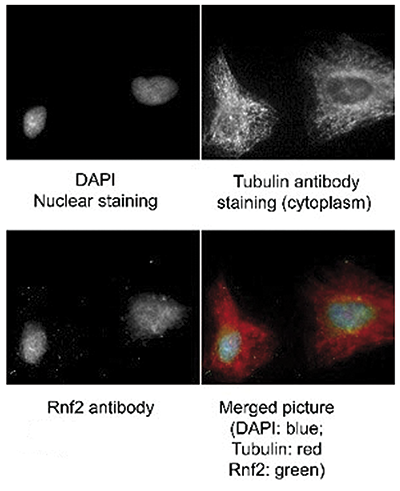
Figure 1. RING1B antibody Immunofluorescence results
Immunofluorescence of RING1B antibody. Tissue: human HeLa cells. Fixation in methanol and blocked with 0.2% fish scale gelatin for 1 hour at 25°C. Primary antibody at 1:300 for 20 minutes at 25°C. Secondary antibody: Alexa Fluor®488-conjugated Donkey anti-goat IgG secondary antibody at 1:500 for 45 min at RT. Localization: RING1B is nuclear and occasionally cytoplasmic. Staining: RING1B (RNF2) as green signal, Tubulin cytoplasm staining red, and DAPI (blue) nuclear counterstain.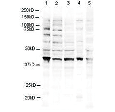
Figure 2. RING1B antibody western blot results
Western blot using the Diagenode RING1B antibody shows detection of a 38 kDa band corresponding to human RING1B in 3T3 (lane 1), U937 (lane 2), Jurkat (lane 3), mouse brain (lane 4) and CHO-K1 (lane 5) cell lysates. Approximately 20 μg of lysate was run on a SDS-PAGE and transferred onto nitrocellulose followed by reaction with a 1:500 dilution of RING1B antibody incubated at room temperature. - Publications

How to properly cite our product/service in your work
We strongly recommend using this: RING1B Antibody (Hologic Diagenode Cat# C15430002 Lot# 001). Click here to copy to clipboard.
Using our products or services in your publication? Let us know!