Notice (8): Undefined variable: solution_of_interest [APP/View/Products/view.ctp, line 755]Code Context<!-- BEGIN: REQUEST_FORM MODAL -->
<div id="request_formModal" class="reveal-modal medium" data-reveal aria-labelledby="modalTitle" aria-hidden="true" role="dialog">
<?= $this->element('Forms/simple_form', array('solution_of_interest' => $solution_of_interest, 'header' => $header, 'message' => $message, 'campaign_id' => $campaign_id)) ?>
$viewFile = '/home/website-server/www/app/View/Products/view.ctp'
$dataForView = array(
'language' => 'en',
'meta_keywords' => '',
'meta_description' => 'Ty1 Monoclonal Antibody validated in ChIP-qPCR and WB. Batch-specific data available on the website. Sample size available.',
'meta_title' => 'Ty1 Antibody - ChIP-Grade (C15200054-10) | Diagenode',
'product' => array(
'Product' => array(
'id' => '2988',
'antibody_id' => '319',
'name' => 'Ty1 Antibody (sample size)',
'description' => '',
'label1' => '',
'info1' => '',
'label2' => '',
'info2' => '',
'label3' => '',
'info3' => '',
'format' => '10 µg',
'catalog_number' => 'C15200054-10',
'old_catalog_number' => '',
'sf_code' => 'C15200054-D001-000582',
'type' => 'FRE',
'search_order' => '',
'price_EUR' => '105',
'price_USD' => '115',
'price_GBP' => '100',
'price_JPY' => '16450',
'price_CNY' => '0',
'price_AUD' => '288',
'country' => 'ALL',
'except_countries' => 'None',
'quote' => false,
'in_stock' => false,
'featured' => false,
'no_promo' => false,
'online' => true,
'master' => false,
'last_datasheet_update' => '',
'slug' => 'ty1-monoclonal-antibody-classic-10-ug',
'meta_title' => 'Ty1 Antibody - ChIP-Grade (C15200054-10) | Diagenode',
'meta_keywords' => '',
'meta_description' => 'Ty1 Monoclonal Antibody validated in ChIP-qPCR and WB. Batch-specific data available on the website. Sample size available.',
'modified' => '2021-10-19 16:42:33',
'created' => '2018-08-29 12:42:43',
'locale' => 'eng'
),
'Antibody' => array(
'host' => '*****',
'id' => '319',
'name' => 'Ty1 monoclonal antibody',
'description' => 'Monoclonal antibody raised in mouse against the Ty1 tag (amino acid sequence EVHTNQDPLD).',
'clonality' => '',
'isotype' => '',
'lot' => '008',
'concentration' => '1.9 µg/µl',
'reactivity' => '',
'type' => 'Monoclonal',
'purity' => 'Protein A purified monoclonal antibody',
'classification' => '',
'application_table' => '<table>
<thead>
<tr>
<th>Applications</th>
<th>Suggested dilution</th>
<th>References</th>
</tr>
</thead>
<tbody>
<tr>
<td>ChIP <sup>*</sup></td>
<td>2 μg/ChIP</td>
<td>Fig 1</td>
</tr>
<tr>
<td>Western Blotting</td>
<td>1:1,000 - 1:5,000</td>
<td>Fig 2</td>
</tr>
</tbody>
</table>
<p><small><sup>*</sup> Please note that of the optimal antibody amount per IP should be determined by the end-user. We recommend testing 1-5 μg per IP.</small></p>',
'storage_conditions' => 'Store at -20°C; for long storage, store at -80°C. Avoid multiple freeze-thaw cycles.',
'storage_buffer' => 'PBS containing 0.05% azide',
'precautions' => 'This product is for research use only. Not for use in diagnostic or therapeutic procedures.',
'uniprot_acc' => '',
'slug' => '',
'meta_keywords' => '',
'meta_description' => '',
'modified' => '2024-11-19 16:18:08',
'created' => '0000-00-00 00:00:00',
'select_label' => '319 - Ty1 monoclonal antibody (008 - 1.9 µg/µl - - Protein A purified monoclonal antibody - Mouse)'
),
'Slave' => array(),
'Group' => array(
'Group' => array(
[maximum depth reached]
),
'Master' => array(
[maximum depth reached]
),
'Product' => array(
[maximum depth reached]
)
),
'Related' => array(
(int) 0 => array(
[maximum depth reached]
),
(int) 1 => array(
[maximum depth reached]
),
(int) 2 => array(
[maximum depth reached]
),
(int) 3 => array(
[maximum depth reached]
)
),
'Application' => array(
(int) 0 => array(
[maximum depth reached]
),
(int) 1 => array(
[maximum depth reached]
)
),
'Category' => array(
(int) 0 => array(
[maximum depth reached]
),
(int) 1 => array(
[maximum depth reached]
),
(int) 2 => array(
[maximum depth reached]
),
(int) 3 => array(
[maximum depth reached]
)
),
'Document' => array(
(int) 0 => array(
[maximum depth reached]
),
(int) 1 => array(
[maximum depth reached]
),
(int) 2 => array(
[maximum depth reached]
)
),
'Feature' => array(),
'Image' => array(
(int) 0 => array(
[maximum depth reached]
)
),
'Promotion' => array(),
'Protocol' => array(),
'Publication' => array(
(int) 0 => array(
[maximum depth reached]
),
(int) 1 => array(
[maximum depth reached]
),
(int) 2 => array(
[maximum depth reached]
),
(int) 3 => array(
[maximum depth reached]
),
(int) 4 => array(
[maximum depth reached]
),
(int) 5 => array(
[maximum depth reached]
),
(int) 6 => array(
[maximum depth reached]
),
(int) 7 => array(
[maximum depth reached]
),
(int) 8 => array(
[maximum depth reached]
),
(int) 9 => array(
[maximum depth reached]
),
(int) 10 => array(
[maximum depth reached]
),
(int) 11 => array(
[maximum depth reached]
),
(int) 12 => array(
[maximum depth reached]
),
(int) 13 => array(
[maximum depth reached]
),
(int) 14 => array(
[maximum depth reached]
),
(int) 15 => array(
[maximum depth reached]
),
(int) 16 => array(
[maximum depth reached]
),
(int) 17 => array(
[maximum depth reached]
),
(int) 18 => array(
[maximum depth reached]
),
(int) 19 => array(
[maximum depth reached]
),
(int) 20 => array(
[maximum depth reached]
),
(int) 21 => array(
[maximum depth reached]
),
(int) 22 => array(
[maximum depth reached]
),
(int) 23 => array(
[maximum depth reached]
),
(int) 24 => array(
[maximum depth reached]
),
(int) 25 => array(
[maximum depth reached]
),
(int) 26 => array(
[maximum depth reached]
),
(int) 27 => array(
[maximum depth reached]
),
(int) 28 => array(
[maximum depth reached]
)
),
'Testimonial' => array(),
'Area' => array(),
'SafetySheet' => array(
(int) 0 => array(
[maximum depth reached]
),
(int) 1 => array(
[maximum depth reached]
),
(int) 2 => array(
[maximum depth reached]
),
(int) 3 => array(
[maximum depth reached]
),
(int) 4 => array(
[maximum depth reached]
),
(int) 5 => array(
[maximum depth reached]
),
(int) 6 => array(
[maximum depth reached]
),
(int) 7 => array(
[maximum depth reached]
)
)
),
'meta_canonical' => 'https://www.diagenode.com/en/p/ty1-monoclonal-antibody-classic-50-ug-25-ul'
)
$language = 'en'
$meta_keywords = ''
$meta_description = 'Ty1 Monoclonal Antibody validated in ChIP-qPCR and WB. Batch-specific data available on the website. Sample size available.'
$meta_title = 'Ty1 Antibody - ChIP-Grade (C15200054-10) | Diagenode'
$product = array(
'Product' => array(
'id' => '2988',
'antibody_id' => '319',
'name' => 'Ty1 Antibody (sample size)',
'description' => '<p><span>Monoclonal antibody raised in mouse against the <strong>Ty1 tag</strong> (amino acid sequence EVHTNQDPLD).</span></p>',
'label1' => 'Validation Data',
'info1' => '<div class="row">
<div class="small-12 columns"><img src="https://www.diagenode.com/img/product/antibodies/C15200054-chip.jpg" alt="Ty1 Antibody validated in ChIP " caption="false" width="893" height="384" /></div>
</div>
<div class="row">
<div class="small-12 columns">
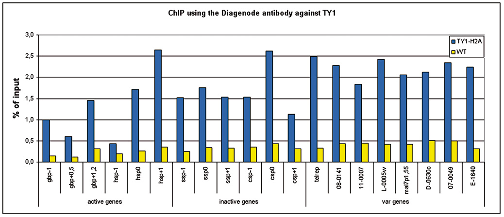
<p><small><strong>Figure 1. ChIP results obtained with the Diagenode monoclonal antibody against the Ty1 tag</strong> <br />ChIP assays were performed using Plasmodium falciparium cells stably expressing Ty1-H2A (blue bars) and wild type Plasmodium falciparium cells (brown bars). Primer pairs were used for active genes, inactive genes, and variable genes. Sheared chromatin from 1x10e6 cells and 2 μg of antibody against Ty1 (cat. No. C15200054) were used per ChIP experiment</small></p>
</div>
</div>
<div class="row">
<div class="small-3 columns"><img src="https://www.diagenode.com/img/product/antibodies/C15200054-wb1.jpg" alt="Ty1 Antibody validated in WB" caption="false" width="201" height="186" /></div>
<div class="small-9 columns">
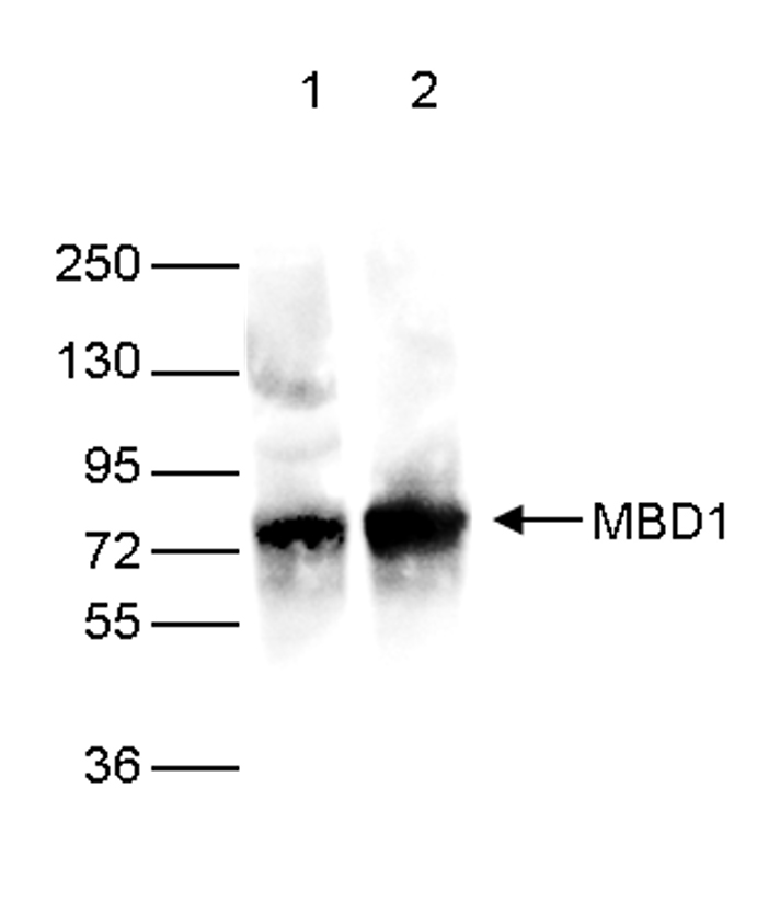
<p><small><strong>Figure 2. Western blot analysis using the Diagenode monoclonal antibody against the Ty1 tag</strong> <br />Western blot was performed on 40 µg whole cell extracts from HeLa cells transfected with Ty1-tagged MBD1. The WB was performed with the Diagenode antibody against the Ty1 tag (cat. No. C15200054) diluted 1:2,000 in TBS-Tween containing 5% skimmed milk (lane 2) or with an MBD1 antibody (C15410078, lane 1). The position of the Ty1-tagged MBD1 and Ty1-tagged MBD2 are shown on the right, the marker (in kDa) is shown on the left.</small></p>
</div>
</div>
<div class="row">
<div class="small-3 columns"><img src="https://www.diagenode.com/img/product/antibodies/C15200054-wb.jpg" alt="Ty1 Antibody validated in WB" caption="false" width="201" height="186" /></div>
<div class="small-9 columns">
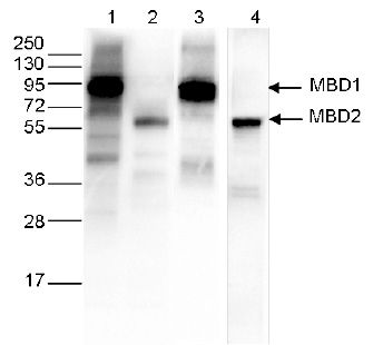
<p><small><strong>Figure 3. Western blot analysis using the Diagenode monoclonal antibody against the Ty1 tag</strong> <br />Western blot was performed on 40 μg whole cell extracts from HeLa cells transfected with either Ty1-tagged MBD1 (lanes 1 and 3) or Ty1-tagged MBD2 (lane 2 and 4). The WB was performed with the Diagenode antibody against the Ty1 tag (Cat. No. C15200054) diluted 1:1,000 in TBS-Tween containing 5% skimmed milk (lane 1 and 2) or with an MBD1 antibody (C15410078, lane 3) or an MBD2 antibody (C15410312, lane 4). The position of the Ty1-tagged MBD1 and Ty1-tagged MBD2 are shown on the right, the marker (in kDa) is shown on the left.</small></p>
</div>
</div>',
'label2' => '',
'info2' => '',
'label3' => '',
'info3' => '',
'format' => '10 µg',
'catalog_number' => 'C15200054-10',
'old_catalog_number' => '',
'sf_code' => 'C15200054-D001-000582',
'type' => 'FRE',
'search_order' => '',
'price_EUR' => '105',
'price_USD' => '115',
'price_GBP' => '100',
'price_JPY' => '16450',
'price_CNY' => '0',
'price_AUD' => '288',
'country' => 'ALL',
'except_countries' => 'None',
'quote' => false,
'in_stock' => false,
'featured' => false,
'no_promo' => false,
'online' => true,
'master' => false,
'last_datasheet_update' => '',
'slug' => 'ty1-monoclonal-antibody-classic-10-ug',
'meta_title' => 'Ty1 Antibody - ChIP-Grade (C15200054-10) | Diagenode',
'meta_keywords' => '',
'meta_description' => 'Ty1 Monoclonal Antibody validated in ChIP-qPCR and WB. Batch-specific data available on the website. Sample size available.',
'modified' => '2021-10-19 16:42:33',
'created' => '2018-08-29 12:42:43',
'locale' => 'eng'
),
'Antibody' => array(
'host' => '*****',
'id' => '319',
'name' => 'Ty1 monoclonal antibody',
'description' => 'Monoclonal antibody raised in mouse against the Ty1 tag (amino acid sequence EVHTNQDPLD).',
'clonality' => '',
'isotype' => '',
'lot' => '008',
'concentration' => '1.9 µg/µl',
'reactivity' => '',
'type' => 'Monoclonal',
'purity' => 'Protein A purified monoclonal antibody',
'classification' => '',
'application_table' => '<table>
<thead>
<tr>
<th>Applications</th>
<th>Suggested dilution</th>
<th>References</th>
</tr>
</thead>
<tbody>
<tr>
<td>ChIP <sup>*</sup></td>
<td>2 μg/ChIP</td>
<td>Fig 1</td>
</tr>
<tr>
<td>Western Blotting</td>
<td>1:1,000 - 1:5,000</td>
<td>Fig 2</td>
</tr>
</tbody>
</table>
<p><small><sup>*</sup> Please note that of the optimal antibody amount per IP should be determined by the end-user. We recommend testing 1-5 μg per IP.</small></p>',
'storage_conditions' => 'Store at -20°C; for long storage, store at -80°C. Avoid multiple freeze-thaw cycles.',
'storage_buffer' => 'PBS containing 0.05% azide',
'precautions' => 'This product is for research use only. Not for use in diagnostic or therapeutic procedures.',
'uniprot_acc' => '',
'slug' => '',
'meta_keywords' => '',
'meta_description' => '',
'modified' => '2024-11-19 16:18:08',
'created' => '0000-00-00 00:00:00',
'select_label' => '319 - Ty1 monoclonal antibody (008 - 1.9 µg/µl - - Protein A purified monoclonal antibody - Mouse)'
),
'Slave' => array(),
'Group' => array(
'Group' => array(
'id' => '248',
'name' => 'C15200054',
'product_id' => '1977',
'modified' => '2018-08-29 12:43:03',
'created' => '2018-08-29 12:43:03'
),
'Master' => array(
'id' => '1977',
'antibody_id' => '319',
'name' => ' Ty1 Antibody ',
'description' => '<p><span>Monoclonal antibody raised in mouse against the <strong>Ty1 tag</strong> (amino acid sequence EVHTNQDPLD).</span></p>',
'label1' => 'Validation Data',
'info1' => '<div class="row">
<div class="small-12 columns"><img src="https://www.diagenode.com/img/product/antibodies/C15200054-chip.jpg" alt="Ty1 Antibody validated in ChIP " caption="false" width="893" height="384" /></div>
</div>
<div class="row">
<div class="small-12 columns">
<p><small><strong>Figure 1. ChIP results obtained with the Diagenode monoclonal antibody against the Ty1 tag</strong> <br />ChIP assays were performed using Plasmodium falciparium cells stably expressing Ty1-H2A (blue bars) and wild type Plasmodium falciparium cells (brown bars). Primer pairs were used for active genes, inactive genes, and variable genes. Sheared chromatin from 1x10e6 cells and 2 μg of antibody against Ty1 (cat. No. C15200054) were used per ChIP experiment</small></p>
</div>
</div>
<div class="row">
<div class="small-3 columns"><img src="https://www.diagenode.com/img/product/antibodies/C15200054-wb1.jpg" alt="Ty1 Antibody validated in WB" caption="false" width="201" height="186" /></div>
<div class="small-9 columns">
<p><small><strong>Figure 2. Western blot analysis using the Diagenode monoclonal antibody against the Ty1 tag</strong> <br />Western blot was performed on 40 µg whole cell extracts from HeLa cells transfected with Ty1-tagged MBD1. The WB was performed with the Diagenode antibody against the Ty1 tag (cat. No. C15200054) diluted 1:2,000 in TBS-Tween containing 5% skimmed milk (lane 2) or with an MBD1 antibody (C15410078, lane 1). The position of the Ty1-tagged MBD1 and Ty1-tagged MBD2 are shown on the right, the marker (in kDa) is shown on the left.</small></p>
</div>
</div>
<div class="row">
<div class="small-3 columns"><img src="https://www.diagenode.com/img/product/antibodies/C15200054-wb.jpg" alt="Ty1 Antibody validated in WB" caption="false" width="201" height="186" /></div>
<div class="small-9 columns">
<p><small><strong>Figure 3. Western blot analysis using the Diagenode monoclonal antibody against the Ty1 tag</strong> <br />Western blot was performed on 40 μg whole cell extracts from HeLa cells transfected with either Ty1-tagged MBD1 (lanes 1 and 3) or Ty1-tagged MBD2 (lane 2 and 4). The WB was performed with the Diagenode antibody against the Ty1 tag (Cat. No. C15200054) diluted 1:1,000 in TBS-Tween containing 5% skimmed milk (lane 1 and 2) or with an MBD1 antibody (C15410078, lane 3) or an MBD2 antibody (C15410312, lane 4). The position of the Ty1-tagged MBD1 and Ty1-tagged MBD2 are shown on the right, the marker (in kDa) is shown on the left.</small></p>
</div>
</div>',
'label2' => '',
'info2' => '',
'label3' => '',
'info3' => '',
'format' => '50 µg/29 µl',
'catalog_number' => 'C15200054',
'old_catalog_number' => 'MAb-054-050',
'sf_code' => 'C15200054-D001-000581',
'type' => 'FRE',
'search_order' => '03-Antibody',
'price_EUR' => '380',
'price_USD' => '380',
'price_GBP' => '340',
'price_JPY' => '59525',
'price_CNY' => '',
'price_AUD' => '950',
'country' => 'ALL',
'except_countries' => 'None',
'quote' => false,
'in_stock' => false,
'featured' => false,
'no_promo' => false,
'online' => true,
'master' => true,
'last_datasheet_update' => 'July 30, 2021',
'slug' => 'ty1-monoclonal-antibody-classic-50-ug-25-ul',
'meta_title' => 'Ty1 Antibody - ChIP-Grade (C15200054) | Diagenode',
'meta_keywords' => '',
'meta_description' => 'Ty1 Monoclonal Antibody validated in ChIP-qPCR and WB. Batch-specific data available on the website.',
'modified' => '2021-10-19 16:42:09',
'created' => '2015-06-29 14:08:20'
),
'Product' => array(
(int) 0 => array(
[maximum depth reached]
)
)
),
'Related' => array(
(int) 0 => array(
'id' => '1856',
'antibody_id' => null,
'name' => 'True MicroChIP-seq Kit',
'description' => '<p><a href="https://www.diagenode.com/files/products/kits/truemicrochipseq-kit-manual.pdf"><img src="https://www.diagenode.com/img/buttons/bt-manual.png" /></a></p>
<p>The <b>True </b><b>MicroChIP-seq</b><b> kit </b>provides a robust ChIP protocol suitable for the investigation of histone modifications within chromatin from as few as <b>10 000 cells</b>, including <b>FACS sorted cells</b>. The kit can be used for chromatin preparation for downstream ChIP-qPCR or ChIP-seq analysis. The <b>complete kit</b> contains everything you need for start-to-finish ChIP including all validated buffers and reagents for chromatin shearing, immunoprecipitation and DNA purification for exceptional <strong>ChIP-qPCR</strong> or <strong>ChIP-seq</strong> results. In addition, positive control antibodies and negative control PCR primers are included for your convenience and assurance of result sensitivity and specificity.</p>
<p>The True MicroChIP-seq kit offers unique benefits:</p>
<ul>
<li>An <b>optimized chromatin preparation </b>protocol compatible with low number of cells (<b>10.000</b>) in combination with the Bioruptor™ shearing device</li>
<li>Most <b>complete kit </b>available (covers all steps and includes control antibodies and primers)</li>
<li><b>Magnetic beads </b>make ChIP easy, fast, and more reproducible</li>
<li>MicroChIP DiaPure columns (included in the kit) enable the <b>maximum recovery </b>of immunoprecipitation DNA suitable for any downstream application</li>
<li><b>Excellent </b><b>ChIP</b><b>-seq </b>result when combined with <a href="https://www.diagenode.com/en/categories/library-preparation-for-ChIP-seq">MicroPlex</a><a href="https://www.diagenode.com/en/categories/library-preparation-for-ChIP-seq"> Library Preparation kit </a>adapted for low input</li>
</ul>
<p>For fast ChIP-seq on low input – check out Diagenode’s <a href="https://www.diagenode.com/en/p/uchipmentation-for-histones-24-rxns">µ</a><a href="https://www.diagenode.com/en/p/uchipmentation-for-histones-24-rxns">ChIPmentation</a><a href="https://www.diagenode.com/en/p/uchipmentation-for-histones-24-rxns"> for histones</a>.</p>
<p><sub>The True MicroChIP-seq kit, Cat. No. C01010132 is an upgraded version of the kit True MicroChIP, Cat. No. C01010130, with the new validated protocols (e.g. FACS sorted cells) and MicroChIP DiaPure columns included in the kit.</sub></p>',
'label1' => 'Characteristics',
'info1' => '<ul>
<li><b>Revolutionary:</b> Only 10,000 cells needed for complete ChIP-seq procedure</li>
<li><b>Validated on</b> studies for histone marks</li>
<li><b>Automated protocol </b>for the IP-Star<sup>®</sup> Compact Automated Platform available</li>
</ul>
<p></p>
<p>The True MicroChIP-seq kit protocol has been optimized for the use of 10,000 - 100,000 cells per immunoprecipitation reaction. Regarding chromatin immunoprecipitation, three protocol variants have been optimized:<br />starting with a batch, starting with an individual sample and starting with the FACS-sorted cells.</p>
<div><button id="readmorebtn" style="background-color: #b02736; color: white; border-radius: 5px; border: none; padding: 5px;">Show Workflow</button></div>
<p><br /> <img src="https://www.diagenode.com/img/product/kits/workflow-microchip.png" id="workflowchip" class="hidden" width="600px" /></p>
<p>
<script type="text/javascript">// <![CDATA[
const bouton = document.querySelector('#readmorebtn');
const workflow = document.getElementById('workflowchip');
bouton.addEventListener('click', () => workflow.classList.toggle('hidden'))
// ]]></script>
</p>
<div class="extra-spaced" align="center"></div>
<div class="row">
<div class="carrousel" style="background-position: center;">
<div class="container">
<div class="row" style="background: rgba(255,255,255,0.1);">
<div class="large-12 columns truemicro-slider" id="truemicro-slider">
<div>
<h3>High efficiency ChIP on 10,000 cells</h3>
<div class="large-10 small-12 medium-10 large-centered medium-centered small-centered columns"><img src="https://www.diagenode.com/img/product/kits/true-micro-chip-histone-results.png" width="800px" /></div>
<div class="large-10 small-12 medium-10 large-centered medium-centered small-centered columns"><center>
<p><small><strong>Figure 1. </strong>ChIP efficiency on 10,000 cells. ChIP was performed on human Hela cells using the Diagenode antibodies <a href="https://www.diagenode.com/en/p/h3k4me3-polyclonal-antibody-premium-50-ug-50-ul">H3K4me3</a> (Cat. No. C15410003), <a href="https://www.diagenode.com/en/p/h3k27ac-polyclonal-antibody-classic-50-mg-42-ml">H3K27ac</a> (C15410174), <a href="https://www.diagenode.com/en/p/h3k9me3-polyclonal-antibody-classic-50-ug">H3K9me3</a> (C15410056) and <a href="https://www.diagenode.com/en/p/h3k27me3-polyclonal-antibody-classic-50-mg-34-ml">H3K27me3</a> (C15410069). Sheared chromatin from 10,000 cells and 0.1 µg (H3K27ac), 0.25 µg (H3K4me3 and H3K27me3) or 0.5 µg (H3K9me3) of the antibody were used per IP. Corresponding amount of IgG was used as control. Quantitative PCR was performed with primers for corresponding positive and negative loci. Figure shows the recovery, expressed as a % of input (the relative amount of immunoprecipitated DNA compared to input DNA after qPCR analysis).</small></p>
</center></div>
</div>
<div>
<h3>True MicroChIP-seq protocol in a combination with MicroPlex library preparation kit results in reliable and accurate sequencing data</h3>
<div class="large-10 small-12 medium-10 large-centered medium-centered small-centered columns"><img src="https://www.diagenode.com/img/product/kits/fig2-truemicro.jpg" alt="True MicroChip results" width="800px" /></div>
<div class="large-10 small-12 medium-10 large-centered medium-centered small-centered columns"><center>
<p><small><strong>Figure 2.</strong> Integrative genomics viewer (IGV) visualization of ChIP-seq experiments using 50.000 of K562 cells. ChIP has been performed accordingly to True MicroChIP protocol followed by the library preparation using MicroPlex Library Preparation Kit (C05010001). The above figure shows the peaks from ChIP-seq experiments using the following antibodies: H3K4me1 (C15410194), H3K9/14ac (C15410200), H3K27ac (C15410196) and H3K36me3 (C15410192).</small></p>
</center></div>
</div>
<div>
<h3>Successful chromatin profiling from 10.000 of FACS-sorted cells</h3>
<div class="large-10 small-12 medium-10 large-centered medium-centered small-centered columns"><img src="https://www.diagenode.com/img/product/kits/fig3ab-truemicro.jpg" alt="small non coding RNA" width="800px" /></div>
<div class="large-10 small-12 medium-10 large-centered medium-centered small-centered columns"><center>
<p><small><strong>Figure 3.</strong> (A) Integrative genomics viewer (IGV) visualization of ChIP-seq experiments and heatmap 3kb upstream and downstream of the TSS (B) for H3K4me3. ChIP has been performed using 10.000 of FACS-sorted cells (K562) and H3K4me3 antibody (C15410003) accordingly to True MicroChIP protocol followed by the library preparation using MicroPlex Library Preparation Kit (C05010001). Data were compared to ENCODE standards.</small></p>
</center></div>
</div>
</div>
</div>
</div>
</div>
</div>
<p>
<script type="text/javascript">// <![CDATA[
$('.truemicro-slider').slick({
arrows: true,
dots: true,
autoplay:true,
autoplaySpeed: 3000
});
// ]]></script>
</p>',
'label2' => 'Additional solutions compatible with the True MicroChIP-seq Kit',
'info2' => '<p><span style="font-weight: 400;">The <a href="https://www.diagenode.com/en/p/chromatin-shearing-optimization-kit-high-sds-100-million-cells">Chromatin EasyShear Kit – High SDS</a></span><span style="font-weight: 400;"> Recommended for the optimizing chromatin shearing.</span></p>
<p><a href="https://www.diagenode.com/en/categories/chip-seq-grade-antibodies"><span style="font-weight: 400;">ChIP-seq grade antibodies</span></a><span style="font-weight: 400;"> for high yields, specificity, and sensitivity.</span></p>
<p><span style="font-weight: 400;">Check the list of available </span><a href="https://www.diagenode.com/en/categories/primer-pairs"><span style="font-weight: 400;">primer pairs</span></a><span style="font-weight: 400;"> designed for high specificity to specific genomic regions.</span></p>
<p><span style="font-weight: 400;">For library preparation of immunoprecipitated samples we recommend to use the </span><b> </b><a href="https://www.diagenode.com/en/categories/library-preparation-for-ChIP-seq"><span style="font-weight: 400;">MicroPlex Library Preparation Kit</span></a><span style="font-weight: 400;"> - validated for library preparation from picogram inputs.</span></p>
<p><span style="font-weight: 400;">For IP-Star Automation users, check out the </span><a href="https://www.diagenode.com/en/p/auto-true-microchip-kit-16-rxns"><span style="font-weight: 400;">automated version</span></a><span style="font-weight: 400;"> of this kit.</span></p>
<p><span style="font-weight: 400;">Application note: </span><a href="https://www.diagenode.com/files/application_notes/Diagenode_AATI_Joint.pdf"><span style="font-weight: 400;">Best Workflow Practices for ChIP-seq Analysis with Small Samples</span></a></p>
<p></p>',
'label3' => 'Species, cell lines, tissues tested',
'info3' => '<p>The True MicroChIP-seq kit is compatible with a broad variety of cell lines, tissues and species - some examples are shown below. Other species / cell lines / tissues can be used with this kit.</p>
<p><strong>Cell lines:</strong></p>
<p>Bovine: blastocysts,<br />Drosophila: embryos, salivary glands<br />Human: EndoC-ẞH1 cells, HeLa cells, PBMC, urothelial cells<br />Mouse: adipocytes, B cells, blastocysts, pre-B cells, BMDM cells, chondrocytes, embryonic stem cells, KH2 cells, LSK cells, macrophages, MEP cells, microglia, NK cells, oocytes, pancreatic cells, P19Cl6 cells, RPE cells,</p>
<p>Other cell lines / species: compatible, not tested</p>
<p><strong>Tissues:</strong></p>
<p>Horse: adipose tissue</p>
<p>Mice: intestine tissue</p>
<p>Other tissues: not tested</p>',
'format' => '20 rxns',
'catalog_number' => 'C01010132',
'old_catalog_number' => 'C01010130',
'sf_code' => 'C01010132-',
'type' => 'RFR',
'search_order' => '04-undefined',
'price_EUR' => '625',
'price_USD' => '680',
'price_GBP' => '575',
'price_JPY' => '97905',
'price_CNY' => '',
'price_AUD' => '1700',
'country' => 'ALL',
'except_countries' => 'None',
'quote' => false,
'in_stock' => false,
'featured' => true,
'no_promo' => false,
'online' => true,
'master' => true,
'last_datasheet_update' => '0000-00-00',
'slug' => 'true-microchip-kit-x16-16-rxns',
'meta_title' => 'True MicroChIP-seq Kit | Diagenode C01010132',
'meta_keywords' => '',
'meta_description' => 'True MicroChIP-seq Kit provides a robust ChIP protocol suitable for the investigation of histone modifications within chromatin from as few as 10 000 cells, including FACS sorted cells. Compatible with ChIP-qPCR as well as ChIP-seq.',
'modified' => '2023-04-20 16:06:10',
'created' => '2015-06-29 14:08:20',
'ProductsRelated' => array(
[maximum depth reached]
),
'Image' => array(
[maximum depth reached]
)
),
(int) 1 => array(
'id' => '2268',
'antibody_id' => '70',
'name' => 'H3K27me3 Antibody',
'description' => '<p>Polyclonal antibody raised in rabbit against the region of histone <strong>H3 containing the trimethylated lysine 27</strong> (<strong>H3K27me3</strong>), using a KLH-conjugated synthetic peptide.</p>',
'label1' => 'Validation Data',
'info1' => '<div class="row">
<div class="small-6 columns">
<p>A. <img src="https://www.diagenode.com/img/product/antibodies/C15410195-ChIP-Fig1.png" alt="H3K27me3 Antibody ChIP Grade" /></p>
<p>B. <img src="https://www.diagenode.com/img/product/antibodies/C15410195-ChIP-Fig2.png" alt="H3K27me3 Antibody for ChIP" /></p>
</div>
<div class="small-6 columns">
<p><small><strong>Figure 1. ChIP results obtained with the Diagenode antibody directed against H3K27me3</strong><br />ChIP assays were performed using human HeLa cells, the Diagenode antibody against H3K27me3 (Cat. No. C15410195) and optimized PCR primer pairs for qPCR. ChIP was performed with the “iDeal ChIP-seq” kit (Cat. No. C01010051), using sheared chromatin from 1 million cells. The chromatin was spiked with a panel of in vitro assembled nucleosomes, each containing a specific lysine methylation. A titration consisting of 0.5, 1, 2 and 5 µg of antibody per ChIP experiment was analyzed. IgG (1 µg/IP) was used as a negative IP control.</small></p>
<p><small><strong>Figure 1A.</strong> Quantitative PCR was performed with primers specific for the promoter of the active GAPDH and EIF4A2 genes, used as negative controls, and for the inactive TSH2B and MYT1 genes, used as positive controls. The graph shows the recovery, expressed as a % of input (the relative amount of immunoprecipitated DNA compared to input DNA after qPCR analysis).</small></p>
<p><small><strong>Figure 1B.</strong> Recovery of the nucleosomes carrying the H3K27me1, H3K27me2, H3K27me3, H3K4me3, H3K9me3 and H3K36me3 modifications and the unmodified H3K27 as determined by qPCR. The figure clearly shows the antibody is very specific in ChIP for the H3K27me3 modification.</small></p>
</div>
</div>
<div class="row">
<div class="small-12 columns">
<p>A. <img src="https://www.diagenode.com/img/product/antibodies/C15410195-ChIP-Fig2a.png" alt="H3K27me3 Antibody ChIP-seq Grade" /></p>
</div>
</div>
<div class="extra-spaced"></div>
<div class="row">
<div class="small-12 columns">
<p>B. <img src="https://www.diagenode.com/img/product/antibodies/C15410195-ChIP-Fig2b.png" alt="H3K27me3 Antibody for ChIP-seq" /></p>
<p>C. <img src="https://www.diagenode.com/img/product/antibodies/C15410195-ChIP-Fig2c.png" alt="H3K27me3 Antibody for ChIP-seq assay" /></p>
<p>D. <img src="https://www.diagenode.com/img/product/antibodies/C15410195-ChIP-Fig2d.png" alt="H3K27me3 Antibody validated in ChIP-seq" /></p>
</div>
</div>
<div class="row">
<div class="small-12 columns">
<p><small><strong>Figure 2. ChIP-seq results obtained with the Diagenode antibody directed against H3K27me3</strong><br />ChIP was performed on sheared chromatin from 1 million HeLa cells using 1 µg of the Diagenode antibody against H3K27me3 (Cat. No. C15410195) as described above. The IP'd DNA was subsequently analysed on an Illumina HiSeq. Library preparation, cluster generation and sequencing were performed according to the manufacturer's instructions. The 50 bp tags were aligned to the human genome using the BWA algorithm. Figure 2 shows the enrichment in genomic regions of chromosome 6 and 20, surrounding the TSH2B and MYT1 positive control genes (fig 2A and 2B, respectively), and in two genomic regions of chromosome 1 and X (figure 2C and D).</small></p>
</div>
</div>
<div class="extra-spaced"></div>
<div class="row">
<div class="small-12 columns">
<p>A. <img src="https://www.diagenode.com/img/product/antibodies/C15410195-CUTTAG-Fig3A.png" /></p>
<p>B. <img src="https://www.diagenode.com/img/product/antibodies/C15410195-CUTTAG-Fig3B.png" /></p>
</div>
</div>
<div class="row">
<div class="small-12 columns">
<p><small><strong>Figure 3. Cut&Tag results obtained with the Diagenode antibody directed against H3K27me3</strong><br />CUT&TAG (Kaya-Okur, H.S., Nat Commun 10, 1930, 2019) was performed on 50,000 K562 cells using 1 µg of the Diagenode antibody against H3K27me3 (cat. No. C15410195) and the Diagenode pA-Tn5 transposase (C01070001). The libraries were subsequently analysed on an Illumina NextSeq 500 sequencer (2x75 paired-end reads) according to the manufacturer's instructions. The tags were aligned to the human genome (hg19) using the BWA algorithm. Figure 3 shows the peak distribution in 2 genomic regions on chromosome and 13 and 20 (figure 3A and B, respectively).</small></p>
</div>
</div>
<div class="extra-spaced"></div>
<div class="row">
<div class="small-6 columns">
<p><img src="https://www.diagenode.com/img/product/antibodies/C15410195-ELISA-Fig4.png" alt="H3K27me3 Antibody ELISA Validation " /></p>
</div>
<div class="small-6 columns">
<p><small><strong>Figure 4. Determination of the antibody titer</strong><br />To determine the titer of the antibody, an ELISA was performed using a serial dilution of the Diagenode antibody directed against H3K27me3 (Cat. No. C15410195). The antigen used was a peptide containing the histone modification of interest. By plotting the absorbance against the antibody dilution (Figure 4), the titer of the antibody was estimated to be 1:3,000.</small></p>
</div>
</div>
<div class="extra-spaced"></div>
<div class="row">
<div class="small-6 columns">
<p><img src="https://www.diagenode.com/img/product/antibodies/C15410195-DB-Fig5a.png" alt="H3K27me3 Antibody Dot Blot Validation " /></p>
</div>
<div class="small-6 columns">
<p><small><strong>Figure 5. Cross reactivity tests using the Diagenode antibody directed against H3K27me3</strong><br />A Dot Blot analysis was performed to test the cross reactivity of the Diagenode antibody against H3K27me3 (Cat. No. C15410195) with peptides containing other modifications of histone H3 and H4 and the unmodified H3K27 sequence. One hundred to 0.2 pmol of the peptide containing the respective histone modification were spotted on a membrane. The antibody was used at a dilution of 1:5,000. Figure 5 shows a high specificity of the antibody for the modification of interest. Please note that the antibody also recognizes the modification if S28 is phosphorylated.</small></p>
</div>
</div>
<div class="row">
<div class="small-6 columns">
<p><img src="https://www.diagenode.com/img/product/antibodies/C15410195-WB-Fig6.png" alt="H3K27me3 Antibody validated in Western Blot" /></p>
</div>
<div class="small-6 columns">
<p><small><strong>Figure 6. Western blot analysis using the Diagenode antibody directed against H3K27me3</strong><br />Western blot was performed on whole cell (25 µg, lane 1) and histone extracts (15 µg, lane 2) from HeLa cells, and on 1 µg of recombinant histone H2A, H2B, H3 and H4 (lane 3, 4, 5 and 6, respectively) using the Diagenode antibody against H3K27me3 (cat. No. C15410195) diluted 1:500 in TBS-Tween containing 5% skimmed milk. The position of the protein of interest is indicated on the right; the marker (in kDa) is shown on the left.</small></p>
</div>
</div>
<div class="row">
<div class="small-12 columns">
<p><img src="https://www.diagenode.com/img/product/antibodies/C15410195-IF-Fig7.png" alt="H3K27me3 Antibody validated for Immunofluorescence" /></p>
</div>
</div>
<div class="row">
<div class="small-12 columns">
<p><small><strong>Figure 7. Immunofluorescence using the Diagenode antibody directed against H3K27me3</strong><br />Human HeLa cells were stained with the Diagenode antibody against H3K27me3 (Cat. No. C15410195) and with DAPI. Cells were fixed with 4% formaldehyde for 10’ and blocked with PBS/TX-100 containing 5% normal goat serum and 1% BSA. The cells were immunofluorescently labelled with the H3K27me3 antibody (left) diluted 1:200 in blocking solution followed by an anti-rabbit antibody conjugated to Alexa488. The middle panel shows staining of the nuclei with DAPI. A merge of the two stainings is shown on the right.</small></p>
</div>
</div>',
'label2' => 'Target Description',
'info2' => '<p><small>Histones are the main constituents of the protein part of chromosomes of eukaryotic cells. They are rich in the amino acids arginine and lysine and have been greatly conserved during evolution. Histones pack the DNA into tight masses of chromatin. Two core histones of each class H2A, H2B, H3 and H4 assemble and are wrapped by 146 base pairs of DNA to form one octameric nucleosome. Histone tails undergo numerous post-translational modifications, which alter chromatin structure to facilitate transcriptional activation, repression or other nuclear processes. In addition to the genetic code, combinations of the different histone modifications reveal the so-called “histone code”. Histone methylation and demethylation is regulated by histone methyl transferases and histone demethylases. Methylation of histone H3K27 is associated with inactive genomic regions.</small></p>',
'label3' => '',
'info3' => '',
'format' => '50 μg',
'catalog_number' => 'C15410195',
'old_catalog_number' => 'pAb-195-050',
'sf_code' => 'C15410195-D001-000581',
'type' => 'FRE',
'search_order' => '03-Antibody',
'price_EUR' => '480',
'price_USD' => '470',
'price_GBP' => '430',
'price_JPY' => '75190',
'price_CNY' => '',
'price_AUD' => '1175',
'country' => 'ALL',
'except_countries' => 'None',
'quote' => false,
'in_stock' => false,
'featured' => false,
'no_promo' => false,
'online' => true,
'master' => true,
'last_datasheet_update' => 'January 14, 2021',
'slug' => 'h3k27me3-polyclonal-antibody-premium-50-mg-27-ml',
'meta_title' => 'H3K27me3 Antibody - ChIP-seq Grade (C15410195) | Diagenode',
'meta_keywords' => '',
'meta_description' => 'H3K27me3 (Histone H3 trimethylated at lysine 27) Polyclonal Antibody validated in ChIP-seq, ChIP-qPCR, CUT&Tag, ELISA, DB, WB and IF. Specificity confirmed by Peptide array assay. Batch-specific data available on the website. Sample size available.',
'modified' => '2024-01-17 13:55:58',
'created' => '2015-06-29 14:08:20',
'ProductsRelated' => array(
[maximum depth reached]
),
'Image' => array(
[maximum depth reached]
)
),
(int) 2 => array(
'id' => '2173',
'antibody_id' => '115',
'name' => 'H3K4me3 Antibody',
'description' => '<p><span>Polyclonal antibody raised in rabbit against the region of histone <strong>H3 containing the trimethylated lysine 4</strong> (<strong>H3K4me3</strong>), using a KLH-conjugated synthetic peptide.</span></p>',
'label1' => 'Validation data',
'info1' => '<div class="row">
<div class="small-6 columns"><center><img src="https://www.diagenode.com/img/product/antibodies/C15410003-fig1-ChIP.jpg" /></center></div>
<div class="small-6 columns">
<p><small><strong>Figure 1. ChIP results obtained with the Diagenode antibody directed against H3K4me3</strong><br />ChIP assays were performed using human K562 cells, the Diagenode antibody against H3K4me3 (cat. No. C15410003) and optimized PCR primer pairs for qPCR. ChIP was performed with the iDeal ChIP-seq kit (cat. No. C01010051), using sheared chromatin from 500,000 cells. A titration consisting of 0.5, 1, 2 and 5 µg of antibody per ChIP experiment was analyzed. IgG (1 µg/IP) was used as a negative IP control. Quantitative PCR was performed with primers specific for the promoter of the active genes GAPDH and EIF4A2, used as positive controls, and for the inactive MYOD1 gene and the Sat2 satellite repeat, used as negative controls. Figure 1 shows the recovery, expressed as a % of input (the relative amount of immunoprecipitated DNA compared to input DNA after qPCR analysis). </small></p>
</div>
</div>
<p></p>
<div class="row">
<div class="small-12 columns"><center>A.<img src="https://www.diagenode.com/img/product/antibodies/C15410003-fig2a-ChIP-seq.jpg" width="800" /></center><center>B.<img src="https://www.diagenode.com/img/product/antibodies/C15410003-fig2b-ChIP-seq.jpg" width="800" /></center><center>C.<img src="https://www.diagenode.com/img/product/antibodies/C15410003-fig2c-ChIP-seq.jpg" width="800" /></center><center>D.<img src="https://www.diagenode.com/img/product/antibodies/C15410003-fig2d-ChIP-seq.jpg" width="800" /></center></div>
</div>
<div class="row">
<div class="small-12 columns">
<p><small><strong>Figure 2. ChIP-seq results obtained with the Diagenode antibody directed against H3K4me3</strong><br />ChIP was performed on sheared chromatin from 1 million HeLaS3 cells using 1 µg of the Diagenode antibody against H3K4me3 (cat. No. C15410003) as described above. The IP'd DNA was subsequently analysed on an Illumina Genome Analyzer. Library preparation, cluster generation and sequencing were performed according to the manufacturer's instructions. The 36 bp tags were aligned to the human genome using the ELAND algorithm. Figure 2 shows the peak distribution along the complete sequence and a 600 kb region of the X-chromosome (figure 2A and B) and in two regions surrounding the GAPDH and EIF4A2 positive control genes, respectively (figure 2C and D). These results clearly show an enrichment of the H3K4 trimethylation at the promoters of active genes.</small></p>
</div>
</div>
<div class="row">
<div class="small-12 columns"><center>A.<img src="https://www.diagenode.com/img/product/antibodies/C15410003-cuttag-a.png" width="800" /></center></div>
<div class="small-12 columns"><center>B.<img src="https://www.diagenode.com/img/product/antibodies/C15410003-cuttag-b.png" width="800" /></center></div>
</div>
<div class="row">
<div class="small-12 columns">
<p><small><strong>Figure 3. Cut&Tag results obtained with the Diagenode antibody directed against H3K4me3</strong><br />CUT&TAG (Kaya-Okur, H.S., Nat Commun 10, 1930, 2019) was performed on 50,000 K562 cells using 0.5 µg of the Diagenode antibody against H3K4me3 (cat. No. C15410003) and the Diagenode pA-Tn5 transposase (C01070001). The libraries were subsequently analysed on an Illumina NextSeq 500 sequencer (2x75 paired-end reads) according to the manufacturer's instructions. The tags were aligned to the human genome (hg19) using the BWA algorithm. Figure 3 shows the peak distribution in 2 genomic regions surrounding the FOS gene on chromosome 14 and the ACTB gene on chromosome 7 (figure 3A and B, respectively).</small></p>
</div>
</div>
<div class="row">
<div class="small-6 columns"><center><img src="https://www.diagenode.com/img/product/antibodies/C15410003-fig3-ELISA.jpg" width="350" /></center><center></center><center></center><center></center><center></center></div>
<div class="small-6 columns">
<p><small><strong>Figure 4. Determination of the antibody titer</strong><br />To determine the titer of the antibody, an ELISA was performed using a serial dilution of the Diagenode antibody against H3K4me3 (cat. No. C15410003). The antigen used was a peptide containing the histone modification of interest. By plotting the absorbance against the antibody dilution (Figure 4), the titer of the antibody was estimated to be 1:11,000.</small></p>
</div>
</div>
<div class="row">
<div class="small-6 columns"><img src="https://www.diagenode.com/img/product/antibodies/C15410003-fig4-DB.jpg" /></div>
<div class="small-6 columns">
<p><small><strong>Figure 5. Cross reactivity tests using the Diagenode antibody directed against H3K4me3</strong><br />To test the cross reactivity of the Diagenode antibody against H3K4me3 (cat. No. C15410003), a Dot Blot analysis was performed with peptides containing other histone modifications and the unmodified H3K4. One hundred to 0.2 pmol of the respective peptides were spotted on a membrane. The antibody was used at a dilution of 1:2,000. Figure 5A shows a high specificity of the antibody for the modification of interest.</small></p>
</div>
</div>
<div class="row">
<div class="small-4 columns"><img src="https://www.diagenode.com/img/product/antibodies/C15410003-fig5-WB.jpg" /></div>
<div class="small-8 columns">
<p><small><strong>Figure 6. Western blot analysis using the Diagenode antibody directed against H3K4me3</strong><br />Western blot was performed on whole cell extracts (40 µg, lane 1) from HeLa cells, and on 1 µg of recombinant histone H3 (lane 2) using the Diagenode antibody against H3K4me3 (cat. No. C15410003). The antibody was diluted 1:1,000 in TBS-Tween containing 5% skimmed milk. The position of the protein of interest is indicated on the right; the marker (in kDa) is shown on the left.</small></p>
</div>
</div>
<div class="row">
<div class="small-12 columns"><center><img src="https://www.diagenode.com/img/product/antibodies/C15410003-fig6-if.jpg" /></center></div>
</div>
<div class="row">
<div class="small-12 columns">
<p><small><strong>Figure 7. Immunofluorescence using the Diagenode antibody directed against H3K4me3</strong><br />HeLa cells were stained with the Diagenode antibody against H3K4me3 (cat. No. C15410003) and with DAPI. Cells were fixed with 4% formaldehyde for 20’ and blocked with PBS/TX-100 containing 5% normal goat serum. The cells were immunofluorescently labelled with the H3K4me3 antibody (left) diluted 1:200 in blocking solution followed by an anti-rabbit antibody conjugated to Alexa568 or with DAPI (middle), which specifically labels DNA. The right picture shows a merge of both stainings.</small></p>
</div>
</div>
<script src="chrome-extension://hhojmcideegachlhfgfdhailpfhgknjm/web_accessible_resources/index.js"></script>',
'label2' => 'Target Description',
'info2' => '<p>Histones are the main constituents of the protein part of chromosomes of eukaryotic cells. They are rich in the amino acids arginine and lysine and have been greatly conserved during evolution. Histones pack the DNA into tight masses of chromatin. Two core histones of each class H2A, H2B, H3 and H4 assemble and are wrapped by 146 base pairs of DNA to form one octameric nucleosome. Histone tails undergo numerous post-translational modifications, which either directly or indirectly alter chromatin structure to facilitate transcriptional activation or repression or other nuclear processes. In addition to the genetic code, combinations of the different histone modifications reveal the so-called "histone code". Histone methylation and demethylation is dynamically regulated by respectively histone methyl transferases and histone demethylases. Methylation of histone H3K4 is associated with activation of gene transcription.</p>
<script src="chrome-extension://hhojmcideegachlhfgfdhailpfhgknjm/web_accessible_resources/index.js"></script>',
'label3' => '',
'info3' => '<p></p>
<script src="chrome-extension://hhojmcideegachlhfgfdhailpfhgknjm/web_accessible_resources/index.js"></script>',
'format' => '50 µg',
'catalog_number' => 'C15410003',
'old_catalog_number' => 'pAb-003-050',
'sf_code' => 'C15410003-D001-000581',
'type' => 'FRE',
'search_order' => '03-Antibody',
'price_EUR' => '480',
'price_USD' => '470',
'price_GBP' => '430',
'price_JPY' => '75190',
'price_CNY' => '',
'price_AUD' => '1175',
'country' => 'ALL',
'except_countries' => 'None',
'quote' => false,
'in_stock' => false,
'featured' => false,
'no_promo' => false,
'online' => true,
'master' => true,
'last_datasheet_update' => 'January 8, 2021',
'slug' => 'h3k4me3-polyclonal-antibody-premium-50-ug-50-ul',
'meta_title' => 'H3K4me3 Antibody - ChIP-seq Grade (C15410003) | Diagenode',
'meta_keywords' => '',
'meta_description' => 'H3K4me3 (Histone H3 trimethylated at lysine 4) Polyclonal Antibody validated in ChIP-seq, ChIP-qPCR, CUT&Tag, ELISA, DB, WB and IF. Specificity confirmed by Peptide array. Batch-specific data available on the website. Sample size available.',
'modified' => '2024-11-19 16:51:19',
'created' => '2015-06-29 14:08:20',
'ProductsRelated' => array(
[maximum depth reached]
),
'Image' => array(
[maximum depth reached]
)
),
(int) 3 => array(
'id' => '2264',
'antibody_id' => '121',
'name' => 'H3K9me3 Antibody',
'description' => '<p><span>Polyclonal antibody raised in rabbit against the region of histone<strong> H3 containing the trimethylated lysine 9</strong> (<strong>H3K9me3</strong>), using a KLH-conjugated synthetic peptide.</span></p>',
'label1' => 'Validation Data',
'info1' => '<div class="row">
<div class="small-6 columns"><center><img src="https://www.diagenode.com/img/product/antibodies/C15410193-ChIP-Fig1.png" /></center></div>
<div class="small-6 columns">
<p><small><strong>Figure 1. ChIP results obtained with the Diagenode antibody directed against H3K9me3</strong><br />ChIP assays were performed using human HeLa cells, the Diagenode antibody against H3K9me3 (cat. No. C15410193) and optimized PCR primer sets for qPCR. ChIP was performed on sheared chromatin from 1 million HeLaS3 cells using the “iDeal ChIP-seq” kit (cat. No. C01010051). A titration of the antibody consisting of 0.5, 1, 2, and 5 µg per ChIP experiment was analysed. IgG (1 µg/IP) was used as negative IP control. QPCR was performed with primers for the heterochromatin marker Sat2 and for the ZNF510 gene, used as positive controls, and for the promoters of the active EIF4A2 and GAPDH genes, used as negative controls. Figure 1 shows the recovery, expressed as a % of input (the relative amount of immunoprecipitated DNA compared to input DNA after qPCR analysis).</small></p>
</div>
</div>
<div class="row">
<div class="small-12 columns"><center>A.<img src="https://www.diagenode.com/img/product/antibodies/C15410193-ChIP-Fig2a.png" width="700" /></center><center>B.<img src="https://www.diagenode.com/img/product/antibodies/C15410193-ChIP-Fig2b.png" width="700" /></center><center>C.<img src="https://www.diagenode.com/img/product/antibodies/C15410193-ChIP-Fig2c.png" width="700" /></center><center>D.<img src="https://www.diagenode.com/img/product/antibodies/C15410193-ChIP-Fig2d.png" width="700" /></center></div>
</div>
<div class="row">
<div class="small-12 columns">
<p><small><strong>Figure 2. ChIP-seq results obtained with the Diagenode antibody directed against H3K9me3</strong><br />ChIP was performed with 0.5 µg of the Diagenode antibody against H3K9me3 (cat. No. C15410193) on sheared chromatin from 1,000,000 HeLa cells using the “iDeal ChIP-seq” kit as described above. The IP'd DNA was subsequently analysed on an Illumina HiSeq 2000. Library preparation, cluster generation and sequencing were performed according to the manufacturer's instructions. The 50 bp tags were aligned to the human genome using the BWA algorithm. Figure 2A shows the signal distribution along the long arm of chromosome 19 and a zoomin to an enriched region containing several ZNF repeat genes. The arrows indicate two satellite repeat regions which exhibit a stronger signal. Figures 2B, 2C and 2D show the enrichment along the ZNF510 positive control target and at the H19 and KCNQ1 imprinted genes.</small></p>
</div>
</div>
<div class="row">
<div class="small-12 columns"><center>A.<img src="https://www.diagenode.com/img/product/antibodies/C15410193-CT-Fig3a.png" width="700" /></center><center>B.<img src="https://www.diagenode.com/img/product/antibodies/C15410193-CT-Fig3b.png" width="700" /></center></div>
</div>
<div class="row">
<div class="small-12 columns">
<p><small><strong>Figure 3. Cut&Tag results obtained with the Diagenode antibody directed against H3K9me3</strong><br />CUT&TAG (Kaya-Okur, H.S., Nat Commun 10, 1930, 2019) was performed on 50,000 K562 cells using 1 µg of the Diagenode antibody against H3K9me3 (cat. No. C15410193) and the Diagenode pA-Tn5 transposase (C01070001). The libraries were subsequently analysed on an Illumina NextSeq 500 sequencer (2x75 paired-end reads) according to the manufacturer's instructions. The tags were aligned to the human genome (hg19) using the BWA algorithm. Figure 3 shows the peak distribution in a genomic regions on chromosome 1 containing several ZNF repeat genes and in a genomic region surrounding the KCNQ1 imprinting control gene on chromosome 11 (figure 3A and B, respectively).</small></p>
</div>
</div>
<div class="row">
<div class="small-6 columns"><center><img src="https://www.diagenode.com/img/product/antibodies/C15410193-Elisa-Fig4.png" /></center></div>
<div class="small-6 columns">
<p><small><strong>Figure 4. Determination of the antibody titer</strong><br />To determine the titer of the antibody, an ELISA was performed using a serial dilution of the antibody directed against human H3K9me3 (cat. No. C15410193) in antigen coated wells. The antigen used was a peptide containing the histone modification of interest. By plotting the absorbance against the antibody dilution (Figure 4), the titer of the antibody was estimated to be 1:87,000.</small></p>
</div>
</div>
<div class="row">
<div class="small-4 columns"><center><img src="https://www.diagenode.com/img/product/antibodies/C15410193-DB-Fig5.png" /></center></div>
<div class="small-8 columns">
<p><small><strong>Figure 5. Cross reactivity tests using the Diagenode antibody directed against H3K9me3</strong><br />A Dot Blot analysis was performed to test the cross reactivity of the Diagenode antibody against H3K9me3 (cat. No. C15410193) with peptides containing other modifications and unmodified sequences of histone H3 and H4. One hundred to 0.2 pmol of the peptide containing the respective histone modification were spotted on a membrane. The antibody was used at a dilution of 1:20,000. Figure 5 shows a high specificity of the antibody for the modification of interest.</small></p>
</div>
</div>
<div class="row">
<div class="small-4 columns"><center><img src="https://www.diagenode.com/img/product/antibodies/C15410193-WB-Fig6.png" /></center></div>
<div class="small-8 columns">
<p><small><strong>Figure 6. Western blot analysis using the Diagenode antibody directed against H3K9me3</strong><br />Western blot was performed on whole cell (25 µg, lane 1) and histone extracts (15 µg, lane 2) from HeLa cells, and on 1 µg of recombinant histone H2A, H2B, H3 and H4 (lane 3, 4, 5 and 6, respectively) using the Diagenode antibody against H3K9me3 (cat. No. C15410193). The antibody was diluted 1:1,000 in TBS-Tween containing 5% skimmed milk. The position of the protein of interest is indicated on the right; the marker (in kDa) is shown on the left.</small></p>
</div>
</div>
<div class="row">
<div class="small-12 columns"><center><img src="https://www.diagenode.com/img/product/antibodies/C15410193-IF-Fig7.png" /></center></div>
</div>
<div class="row">
<div class="small-12 columns">
<p><small><strong>Figure 7. Immunofluorescence using the Diagenode antibody directed against H3K9me3</strong><br />HeLa cells were stained with the Diagenode antibody against H3K9me3 (cat. No. C15410193) and with DAPI. Cells were fixed with 4% formaldehyde for 10’ and blocked with PBS/TX-100 containing 5% normal goat serum and 1% BSA. The cells were immunofluorescently labelled with the H3K9me3 antibody (middle) diluted 1:500 in blocking solution followed by an anti-rabbit antibody conjugated to Alexa488. The left panel shows staining of the nuclei with DAPI. A merge of both stainings is shown on the right.</small></p>
</div>
</div>',
'label2' => 'Target Description',
'info2' => '<p>Histones are the main constituents of the protein part of chromosomes of eukaryotic cells. They are rich in the amino acids arginine and lysine and have been greatly conserved during evolution. Histones pack the DNA into tight masses of chromatin. Two core histones of each class H2A, H2B, H3 and H4 assemble and are wrapped by 146 base pairs of DNA to form one octameric nucleosome. Histone tails undergo numerous post-translational modifications, which either directly or indirectly alter chromatin structure to facilitate transcriptional activation or repression or other nuclear processes. In addition to the genetic code, combinations of the different histone modifications reveal the so-called “histone code”. Histone methylation and demethylation is dynamically regulated by respectively histone methyl transferases and histone demethylases. Trimethylation of histone H3K9 is associated with inactive genomic regions, satellite repeats and ZNF gene repeats.</p>',
'label3' => '',
'info3' => '',
'format' => '50 μg',
'catalog_number' => 'C15410193',
'old_catalog_number' => 'pAb-193-050',
'sf_code' => 'C15410193-D001-000581',
'type' => 'FRE',
'search_order' => '03-Antibody',
'price_EUR' => '480',
'price_USD' => '470',
'price_GBP' => '430',
'price_JPY' => '75190',
'price_CNY' => '0',
'price_AUD' => '1175',
'country' => 'ALL',
'except_countries' => 'None',
'quote' => false,
'in_stock' => false,
'featured' => false,
'no_promo' => false,
'online' => true,
'master' => true,
'last_datasheet_update' => 'December 12, 2017',
'slug' => 'h3k9me3-polyclonal-antibody-premium-50-mg',
'meta_title' => 'H3K9me3 Antibody - ChIP-seq Grade (C15410193) | Diagenode',
'meta_keywords' => '',
'meta_description' => 'H3K9me3 (Histone H3 trimethylated at lysine 9) Polyclonal Antibody validated in ChIP-seq, ChIP-qPCR, CUT&Tag, ELISA, DB, WB and IF. Specificity confirmed by Peptide array assay. Batch-specific data available on the website. Sample size available.',
'modified' => '2021-10-20 09:55:53',
'created' => '2015-06-29 14:08:20',
'ProductsRelated' => array(
[maximum depth reached]
),
'Image' => array(
[maximum depth reached]
)
)
),
'Application' => array(
(int) 0 => array(
'id' => '19',
'position' => '10',
'parent_id' => '40',
'name' => 'WB',
'description' => '<p><strong>Western blot</strong> : The quality of antibodies used in this technique is crucial for correct and specific protein identification. Diagenode offers huge selection of highly sensitive and specific western blot-validated antibodies.</p>
<p>Learn more about: <a href="https://www.diagenode.com/applications/western-blot">Loading control, MW marker visualization</a><em>. <br /></em></p>
<p><em></em>Check our selection of antibodies validated in Western blot.</p>',
'in_footer' => false,
'in_menu' => false,
'online' => true,
'tabular' => true,
'slug' => 'western-blot-antibodies',
'meta_keywords' => ' Western Blot Antibodies ,western blot protocol,Western Blotting Products,Polyclonal antibodies ,monoclonal antibodies ',
'meta_description' => 'Diagenode offers a wide range of antibodies and technical support for western blot applications',
'meta_title' => ' Western Blot - Monoclonal antibody - Polyclonal antibody | Diagenode',
'modified' => '2016-04-26 12:44:51',
'created' => '2015-01-07 09:20:00',
'ProductsApplication' => array(
[maximum depth reached]
)
),
(int) 1 => array(
'id' => '43',
'position' => '10',
'parent_id' => '40',
'name' => 'ChIP-qPCR (ab)',
'description' => '',
'in_footer' => false,
'in_menu' => false,
'online' => true,
'tabular' => true,
'slug' => 'chip-qpcr-antibodies',
'meta_keywords' => 'Chromatin Immunoprecipitation Sequencing,ChIP-Seq,ChIP-seq grade antibodies,DNA purification,qPCR,Shearing of chromatin',
'meta_description' => 'Diagenode offers a wide range of antibodies and technical support for ChIP-qPCR applications',
'meta_title' => 'ChIP Quantitative PCR Antibodies (ChIP-qPCR) | Diagenode',
'modified' => '2016-01-20 11:30:24',
'created' => '2015-10-20 11:45:36',
'ProductsApplication' => array(
[maximum depth reached]
)
)
),
'Category' => array(
(int) 0 => array(
'id' => '32',
'position' => '50',
'parent_id' => '4',
'name' => 'Epitope tags',
'description' => '',
'no_promo' => false,
'in_menu' => false,
'online' => true,
'tabular' => false,
'hide' => true,
'all_format' => false,
'is_antibody' => true,
'slug' => 'protein-tag',
'cookies_tag_id' => null,
'meta_keywords' => 'Protein tag,Polyclonal antibody,Monoclonal Protein tag antibodies',
'meta_description' => 'Diagenode offers Polyclonal and Monoclonal Protein tag Antibodies',
'meta_title' => 'Polyclonal and Monoclonal Protein tag Antibodies | Diagenode',
'modified' => '2019-07-04 16:02:49',
'created' => '2015-05-09 11:35:10',
'ProductsCategory' => array(
[maximum depth reached]
),
'CookiesTag' => array([maximum depth reached])
),
(int) 1 => array(
'id' => '103',
'position' => '0',
'parent_id' => '4',
'name' => 'All antibodies',
'description' => '<p><span style="font-weight: 400;">All Diagenode’s antibodies are listed below. Please, use our Quick search field to find the antibody of interest by target name, application, purity.</span></p>
<p><span style="font-weight: 400;">Diagenode’s highly validated antibodies:</span></p>
<ul>
<li>Highly sensitive and specific</li>
<li>Cost-effective (requires less antibody per reaction)</li>
<li>Batch-specific data is available on the website</li>
<li>Expert technical support</li>
<li>Sample sizes available</li>
<li>100% satisfaction guarantee</li>
</ul>',
'no_promo' => false,
'in_menu' => true,
'online' => true,
'tabular' => false,
'hide' => true,
'all_format' => false,
'is_antibody' => true,
'slug' => 'all-antibodies',
'cookies_tag_id' => null,
'meta_keywords' => 'Antibodies,Premium Antibodies,Classic,Pioneer',
'meta_description' => 'Diagenode Offers Strict quality standards with Rigorous QC and validated Antibodies. Classified based on level of validation for flexibility of Application. Comprehensive selection of histone and non-histone Antibodies',
'meta_title' => 'Diagenode's selection of Antibodies is exclusively dedicated for Epigenetic Research | Diagenode',
'modified' => '2019-07-03 10:55:44',
'created' => '2015-11-02 14:49:22',
'ProductsCategory' => array(
[maximum depth reached]
),
'CookiesTag' => array([maximum depth reached])
),
(int) 2 => array(
'id' => '102',
'position' => '1',
'parent_id' => '4',
'name' => 'Sample size antibodies',
'description' => '<h1><strong>Validated epigenetics antibodies</strong> – care for a sample?<br /> </h1>
<p>Diagenode has partnered with leading epigenetics experts and numerous epigenetics consortiums to bring to you a validated and comprehensive collection of epigenetic antibodies. As an expert in epigenetics, we are committed to offering highly-specific antibodies validated for ChIP/ChIP-seq and many other applications. All batch-specific validation data is available on our website.<br /><a href="../categories/antibodies">Read about our expertise in antibody production</a>.</p>
<ul>
<li><strong>Focused</strong> - Diagenode's selection of antibodies is exclusively dedicated for epigenetic research. <a title="See the full collection." href="../categories/all-antibodies">See the full collection.</a></li>
<li><strong>Strict quality standards</strong> with rigorous QC and validation</li>
<li><strong>Classified</strong> based on level of validation for flexibility of application</li>
</ul>
<p>Existing sample sizes are listed below. We will soon expand our collection. Are you looking for a sample size of another antibody? Just <a href="mailto:agnieszka.zelisko@diagenode.com?Subject=Sample%20Size%20Request" target="_top">Contact us</a>.</p>',
'no_promo' => false,
'in_menu' => true,
'online' => true,
'tabular' => false,
'hide' => true,
'all_format' => true,
'is_antibody' => true,
'slug' => 'sample-size-antibodies',
'cookies_tag_id' => null,
'meta_keywords' => '5-hmC monoclonal antibody,CRISPR/Cas9 polyclonal antibody ,H3K36me3 polyclonal antibody,diagenode',
'meta_description' => 'Diagenode offers sample volume on selected antibodies for researchers to test, validate and provide confidence and flexibility in choosing from our wide range of antibodies ',
'meta_title' => 'Sample-size Antibodies | Diagenode',
'modified' => '2019-07-03 10:57:05',
'created' => '2015-10-27 12:13:34',
'ProductsCategory' => array(
[maximum depth reached]
),
'CookiesTag' => array([maximum depth reached])
),
(int) 3 => array(
'id' => '127',
'position' => '10',
'parent_id' => '4',
'name' => 'ChIP-grade antibodies',
'description' => '<div class="row">
<div class="small-12 columns"><center></center>
<p><br />Chromatin immunoprecipitation (<b>ChIP</b>) is a technique to study the associations of proteins with the specific genomic regions in intact cells. One of the most important steps of this protocol is the immunoprecipitation of targeted protein using the antibody specifically recognizing it. The quality of antibodies used in ChIP is essential for the success of the experiment. Diagenode offers extensively validated ChIP-grade antibodies, confirmed for their specificity, and high level of performance in ChIP. Each batch is validated, and batch-specific data are available on the website.</p>
<p></p>
</div>
</div>
<p><strong>ChIP results</strong> obtained with the antibody directed against H3K4me3 (Cat. No. <a href="../p/h3k4me3-polyclonal-antibody-premium-50-ug-50-ul">C15410003</a>). </p>
<div class="row">
<div class="small-12 medium-6 large-6 columns"><img src="https://www.diagenode.com/img/product/antibodies/C15410003-fig1-ChIP.jpg" alt="" width="400" height="315" /> </div>
<div class="small-12 medium-6 large-6 columns">
<p></p>
<p></p>
<p></p>
</div>
</div>
<p></p>
<p>Our aim at Diagenode is to offer the largest collection of highly specific <strong>ChIP-grade antibodies</strong>. We add new antibodies monthly. Find your ChIP-grade antibody in the list below and check more information about tested applications, extensive validation data, and product information.</p>',
'no_promo' => false,
'in_menu' => true,
'online' => true,
'tabular' => false,
'hide' => true,
'all_format' => false,
'is_antibody' => true,
'slug' => 'chip-grade-antibodies',
'cookies_tag_id' => null,
'meta_keywords' => 'ChIP-grade antibodies, polyclonal antibody, monoclonal antibody, Diagenode',
'meta_description' => 'Diagenode Offers Extensively Validated ChIP-Grade Antibodies, Confirmed for their Specificity, and high level of Performance in Chromatin Immunoprecipitation ChIP',
'meta_title' => 'Chromatin immunoprecipitation ChIP-grade antibodies | Diagenode',
'modified' => '2024-11-19 17:27:07',
'created' => '2017-02-14 11:16:04',
'ProductsCategory' => array(
[maximum depth reached]
),
'CookiesTag' => array([maximum depth reached])
)
),
'Document' => array(
(int) 0 => array(
'id' => '11',
'name' => 'Antibodies you can trust',
'description' => '<p style="text-align: justify;"><span>Epigenetic research tools have evolved over time from endpoint PCR to qPCR to the analyses of large sets of genome-wide sequencing data. ChIP sequencing (ChIP-seq) has now become the gold standard method for chromatin studies, given the accuracy and coverage scale of the approach over other methods. Successful ChIP-seq, however, requires a higher level of experimental accuracy and consistency in all steps of ChIP than ever before. Particularly crucial is the quality of ChIP antibodies. </span></p>',
'image_id' => null,
'type' => 'Poster',
'url' => 'files/posters/Antibodies_you_can_trust_Poster.pdf',
'slug' => 'antibodies-you-can-trust-poster',
'meta_keywords' => '',
'meta_description' => '',
'modified' => '2015-10-01 20:18:31',
'created' => '2015-07-03 16:05:15',
'ProductsDocument' => array(
[maximum depth reached]
)
),
(int) 1 => array(
'id' => '38',
'name' => 'Epigenetic Antibodies Brochure',
'description' => '<p>More than in any other immuoprecipitation assays, quality antibodies are critical tools in many epigenetics experiments. Since 10 years, Diagenode has developed the most stringent quality production available on the market for antibodies exclusively focused on epigenetic uses. All our antibodies have been qualified to work in epigenetic applications.</p>',
'image_id' => null,
'type' => 'Brochure',
'url' => 'files/brochures/Epigenetic_Antibodies_Brochure.pdf',
'slug' => 'epigenetic-antibodies-brochure',
'meta_keywords' => '',
'meta_description' => '',
'modified' => '2016-06-15 11:24:06',
'created' => '2015-07-03 16:05:27',
'ProductsDocument' => array(
[maximum depth reached]
)
),
(int) 2 => array(
'id' => '623',
'name' => 'Datasheet Ty1 C15200054',
'description' => '<p><span>Monoclonal antibody raised in mouse against the Ty1 tag (amino acid sequence EVHTNQDPLD).</span></p>',
'image_id' => null,
'type' => 'Datasheet',
'url' => 'files/products/antibodies/Datasheet_Ty1_C15200054.pdf',
'slug' => 'datasheet-ty1-C15200054',
'meta_keywords' => '',
'meta_description' => '',
'modified' => '2021-12-22 10:10:46',
'created' => '2015-07-07 11:47:44',
'ProductsDocument' => array(
[maximum depth reached]
)
)
),
'Feature' => array(),
'Image' => array(
(int) 0 => array(
'id' => '1779',
'name' => 'product/antibodies/ab-chip-icon.png',
'alt' => 'Antibody ChIP icon',
'modified' => '2020-08-12 11:52:55',
'created' => '2018-03-15 15:52:35',
'ProductsImage' => array(
[maximum depth reached]
)
)
),
'Promotion' => array(),
'Protocol' => array(),
'Publication' => array(
(int) 0 => array(
'id' => '4982',
'name' => 'The small inhibitor WM-1119 effectively targets KAT6A-rearranged AML, but not KMT2A-rearranged AML, despite shared KAT6 genetic dependency',
'authors' => 'Mathew Sheridan et al.',
'description' => '<h3 class="c-article__sub-heading" data-test="abstract-sub-heading">Background</h3>
<p>The epigenetic factors KAT6A (MOZ/MYST3) and KMT2A (MLL/MLL1) interact in normal hematopoiesis to regulate progenitors’ self-renewal. Both proteins are recurrently translocated in AML, leading to impairment of critical differentiation pathways in these malignant cells. We evaluated the potential of different KAT6A therapeutic targeting strategies to alter the growth of KAT6A and KMT2A rearranged AMLs.</p>
<h3 class="c-article__sub-heading" data-test="abstract-sub-heading">Methods</h3>
<p>We investigated the action and potential mechanisms of the first-in-class KAT6A inhibitor, WM-1119 in KAT6A and KMT2A rearranged (KAT6Ar and KMT2Ar) AML using cellular (flow cytometry, colony assays, cell growth) and molecular (shRNA knock-down, CRISPR knock-out, bulk and single-cell RNA-seq, ChIP-seq) assays. We also used two novel genetic murine KAT6A models combined with the most common KMT2Ar AML, KMT2A::MLLT3 AML. In these murine models, the catalytic activity of KAT6A, or the whole protein, can be conditionally abrogated or deleted. These models allowed us to compare the effects of specific KAT6A KAT activity inhibition with the complete deletion of the whole protein. Finally, we also tested these therapeutic approaches on human AML cell lines and primary patient AMLs.</p>
<h3 class="c-article__sub-heading" data-test="abstract-sub-heading">Results</h3>
<p>We found that WM-1119 completely abrogated the proliferative and clonogenic potential of KAT6Ar cells<span> </span><i>in vitro.</i><span> </span>WM-1119 treatment was associated with a dramatic increase in myeloid differentiation program. The treatment also decreased stemness and leukemia pathways at the transcriptome level and led to loss of binding of the fusion protein at critical regulators of these pathways. In contrast, our pharmacologic and genetic results indicate that the catalytic activity of KAT6A plays a more limited role in KMT2Ar leukemogenicity, while targeting the whole KAT6A protein dramatically affects leukemic potential in murine KMT2A::MLLT3 AML.</p>
<h3 class="c-article__sub-heading" data-test="abstract-sub-heading">Conclusion</h3>
<p>Our study indicates that inhibiting KAT6A KAT activity holds compelling promise for KAT6Ar AML patients. In contrast, targeted degradation of KAT6A, and not just its catalytic activity, may represent a more appropriate therapeutic approach for KMT2Ar AMLs.</p>',
'date' => '2024-10-08',
'pmid' => 'https://jhoonline.biomedcentral.com/articles/10.1186/s13045-024-01610-0',
'doi' => 'https://doi.org/10.1186/s13045-024-01610-0',
'modified' => '2024-10-11 10:00:42',
'created' => '2024-10-11 10:00:42',
'ProductsPublication' => array(
[maximum depth reached]
)
),
(int) 1 => array(
'id' => '4825',
'name' => 'Zfp296 knockout enhances chromatin accessibility and induces a uniquestate of pluripotency in embryonic stem cells.',
'authors' => 'Miyazaki S. et al.',
'description' => '<p>The Zfp296 gene encodes a zinc finger-type protein. Its expression is high in mouse embryonic stem cells (ESCs) but rapidly decreases following differentiation. Zfp296-knockout (KO) ESCs grew as flat colonies, which were reverted to rounded colonies by exogenous expression of Zfp296. KO ESCs could not form teratomas when transplanted into mice but could efficiently contribute to germline-competent chimeric mice following blastocyst injection. Transcriptome analysis revealed that Zfp296 deficiency up- and down-regulates a distinct group of genes, among which Dppa3, Otx2, and Pou3f1 were markedly downregulated. Chromatin immunoprecipitation sequencing demonstrated that ZFP296 binding is predominantly seen in the vicinity of the transcription start sites (TSSs) of a number of genes, and ZFP296 was suggested to negatively regulate transcription. Consistently, chromatin accessibility assay clearly showed that ZFP296 binding reduces the accessibility of the TSS regions of target genes. Zfp296-KO ESCs showed increased histone H3K9 di- and trimethylation. Co-immunoprecipitation analyses revealed interaction of ZFP296 with G9a and GLP. These results show that ZFP296 plays essential roles in maintaining the global epigenetic state of ESCs through multiple mechanisms including activation of Dppa3, attenuation of chromatin accessibility, and repression of H3K9 methylation, but that Zfp296-KO ESCs retain a unique state of pluripotency while lacking the teratoma-forming ability.</p>',
'date' => '2023-07-01',
'pmid' => 'https://www.ncbi.nlm.nih.gov/pubmed/37488353',
'doi' => '10.1038/s42003-023-05148-8',
'modified' => '2023-08-01 13:30:58',
'created' => '2023-08-01 15:59:38',
'ProductsPublication' => array(
[maximum depth reached]
)
),
(int) 2 => array(
'id' => '4847',
'name' => 'Net39 protects muscle nuclei from mechanical stress during thepathogenesis of Emery-Dreifuss muscular dystrophy.',
'authors' => 'Zhang Y. et al.',
'description' => '<p>Mutations in genes encoding nuclear envelope proteins lead to diseases known as nuclear envelopathies, characterized by skeletal muscle and heart abnormalities, such as Emery-Dreifuss muscular dystrophy (EDMD). The tissue-specific role of the nuclear envelope in the etiology of these diseases has not been extensively explored. We previously showed that global deletion of the muscle-specific nuclear envelope protein NET39 in mice leads to neonatal lethality due to skeletal muscle dysfunction. To study the potential role of the Net39 gene in adulthood, we generated a muscle-specific conditional knockout (cKO) of Net39 in mice. cKO mice recapitulated key skeletal muscle features of EDMD, including muscle wasting, impaired muscle contractility, abnormal myonuclear morphology, and DNA damage. The loss of Net39 rendered myoblasts hypersensitive to mechanical stretch, resulting in stretch-induced DNA damage. Net39 was downregulated in a mouse model of congenital myopathy, and restoration of Net39 expression through AAV gene delivery extended life span and ameliorated muscle abnormalities. These findings establish NET39 as a direct contributor to the pathogenesis of EDMD that acts by protecting against mechanical stress and DNA damage.</p>',
'date' => '2023-07-01',
'pmid' => 'https://www.ncbi.nlm.nih.gov/pubmed/37395273',
'doi' => '10.1172/JCI163333',
'modified' => '2023-08-01 14:18:50',
'created' => '2023-08-01 15:59:38',
'ProductsPublication' => array(
[maximum depth reached]
)
),
(int) 3 => array(
'id' => '4278',
'name' => 'Distinct effects on the secretion of MTRAP and AMA1 in Plasmodiumyoelii following deletion of acylated pleckstrin homology domain-containingprotein.',
'authors' => 'Chaiyawong Nattawat et al.',
'description' => '<p>Plasmodium, the causative agents of malaria, are obligate intracellular organisms. In humans, pathogenesis is caused by the blood stage parasite, which multiplies within erythrocytes, thus erythrocyte invasion is an essential developmental step. Merozoite form parasites released into the blood stream coordinately secrets a panel of proteins from the microneme secretory organelles for gliding motility, establishment of a tight junction with a target naive erythrocyte, and subsequent internalization. A protein identified in Toxoplasma gondii facilitates microneme fusion with the plasma membrane for exocytosis; namely, acylated pleckstrin homology domain-containing protein (APH). To obtain insight into the differential microneme discharge by malaria parasites, in this study we analyzed the consequences of APH deletion in the rodent malaria model, Plasmodium yoelii, using a DiCre-based inducible knockout method. We found that APH deletion resulted in a reduction in parasite asexual growth and erythrocyte invasion, with some parasites retaining the ability to invade and grow without APH. APH deletion impaired the secretion of microneme proteins, MTRAP and AMA1, and upon contact with erythrocytes the secretion of MTRAP, but not AMA1, was observed. APH-deleted merozoites were able to attach to and deform erythrocytes, consistent with the observed MTRAP secretion. Tight junctions were formed, but echinocytosis after merozoite internalization into erythrocytes was significantly reduced, consistent with the observed absence of AMA1 secretion. Together with our observation that APH largely colocalized with MTRAP, but less with AMA1, we propose that APH is directly involved in MTRAP secretion; whereas any role of APH in AMA1 secretion is indirect in Plasmodium.</p>',
'date' => '2022-02-01',
'pmid' => 'https://doi.org/10.1016%2Fj.parint.2021.102479',
'doi' => '10.1016/j.parint.2021.102479',
'modified' => '2022-05-23 09:59:07',
'created' => '2022-05-19 10:41:50',
'ProductsPublication' => array(
[maximum depth reached]
)
),
(int) 4 => array(
'id' => '4272',
'name' => 'Mod(mdg4) variants repress telomeric retrotransposon HeT-A byblocking subtelomeric enhancers',
'authors' => 'Takeuchi Chikara et al.',
'description' => '<p>Telomeres in Drosophila are composed of sequential non-LTR retrotransposons: HeT-A, TART , and TAHRE . Although they are repressed by the piRNA pathway in the germline, how these retrotransposons are regulated in somatic cells is poorly understood. Here, we show that specific splice variants of Mod(mdg4) repress HeT-A by blocking subtelomeric enhancers in ovarian somatic cells. We found that the Mod(mdg4)-N variant represses HeT-A expression most efficiently among the variants. Mod(mdg4)-N mutant flies show elevated HeT-A expression and female sterility. Mod(mdg4)-N-binding subtelomeric sequences exhibit enhancer-blocking activity, and recruitment of RNA polymerase II (Pol II) on subtelomeres by Mod(mdg4)-N is essential for this enhancer-blocking. Moreover, Mod(mdg4)-N functions to form chromatin boundaries of higher-order chromatin conformation but this mechanism is independent of its Pol II recruitment activity at telomeres/subtelomeres. This study provides a link between enhancer-blocking and telomere regulation, and two different molecular mechanisms exhibited by an insulator protein to orchestrate precise gene expression.</p>',
'date' => '2022-01-01',
'pmid' => 'https://doi.org/10.1101%2F2022.01.16.476534',
'doi' => '10.1101/2022.01.16.476534',
'modified' => '2022-05-23 09:46:54',
'created' => '2022-05-19 10:41:50',
'ProductsPublication' => array(
[maximum depth reached]
)
),
(int) 5 => array(
'id' => '4320',
'name' => 'Identification of a PadR-type regulator essential for intracellularpathogenesis of',
'authors' => 'McMillan Ian A. et al. ',
'description' => '<p>Burkholderia pseudomallei (Bp) is the causative agent of melioidosis, a disease endemic to the tropics. Melioidosis manifests in various ways ranging from acute skin lesions to pneumonia and, in rare cases, infection of the central nervous system. Bp is a facultative intracellular pathogen and it can infect various cell types. The Bp intracellular lifecycle has been partially elucidated and is highly complex. Herein, we have identified a transcriptional regulator, BP1026B_II1198, that is differentially expressed as Bp transits through host cells. A deletion mutant of BP1026B_II1198 was attenuated in RAW264.7 cell and BALB/c mouse infection. To further characterize the function of this transcriptional regulator, we endeavored to determine the regulon of BP1026B_II1198. RNA-seq analysis showed the global picture of genes regulated while ChIP-seq analysis identified two specific BP1026B_II1198 binding regions on chromosome II. We investigated the transposon mutants of these genes controlled by BP1026B_II1198 and confirmed that these genes contribute to pathogenesis in RAW264.7 murine macrophage cells. Taken together, the data presented here shed light on the regulon of BP1026B_II1198 and its role during intracellular infection and highlights an integral portion of the highly complex regulation network of Bp during host infection.</p>',
'date' => '2021-05-01',
'pmid' => 'https://www.ncbi.nlm.nih.gov/pubmed/34001967',
'doi' => '10.1038/s41598-021-89852-7',
'modified' => '2022-08-02 16:55:52',
'created' => '2022-05-19 10:41:50',
'ProductsPublication' => array(
[maximum depth reached]
)
),
(int) 6 => array(
'id' => '4088',
'name' => 'MEAF6 is essential for cell proliferation and plays a role in theassembly of KAT7 complexes.',
'authors' => 'Matsuura, Kumi and Tani, Naoki and Usuki, Shingo and Torikai-Nishikawa,Satomi and Okano, Masaki and Niwa, Hitoshi',
'description' => '<p>Myst family genes encode lysine acetyltransferases that mainly mediate histone acetylation to control transcription, DNA replication and DNA damage response. They form tetrameric complexes with PHD-finger proteins (Brpfs or Jades) and small non-catalytic subunits Ing4/5 and Meaf6. Although all the components of the complex are well-conserved from yeast to mammals, the function of Meaf6 and its homologs has not been elucidated in any species. Here we revealed the role of Meaf6 utilizing inducible Meaf6 KO ES cells. By elimination of Meaf6, proliferation ceased although histone acetylations were largely unaffected. In the absence of Meaf6, one of the Myst family members Myst2/Kat7 increased the ability to interact with PHD-finger proteins. This study is the first indication of the function of Meaf6, which shows it is not essential for HAT activity but modulates the assembly of the Kat7 complex.</p>',
'date' => '2020-11-01',
'pmid' => 'https://www.ncbi.nlm.nih.gov/pubmed/32918898',
'doi' => '10.1016/j.yexcr.2020.112279',
'modified' => '2021-03-15 17:17:33',
'created' => '2021-02-18 10:21:53',
'ProductsPublication' => array(
[maximum depth reached]
)
),
(int) 7 => array(
'id' => '4090',
'name' => 'Dual ARID1A/ARID1B loss leads to rapid carcinogenesis and disruptiveredistribution of BAF complexes',
'authors' => 'Wang, Zixi and Chen, Kenian and Jia, Yuemeng and Chuang, Jen-Chieh and Sun,Xuxu and Lin, Yu-Hsuan and Celen, Cemre and Li, Lin and Huang, Fang andLiu, Xin and Castrillon, Diego H. and Wang, Tao and Zhu, Hao',
'description' => '<p>SWI/SNF chromatin remodelers play critical roles in development and cancer. The causal links between SWI/SNF complex disassembly and carcinogenesis are obscured by redundancy between paralogous components. Canonical BAF (cBAF)-specific paralogs ARID1A and ARID1B are synthetic lethal in some contexts, but simultaneous mutations in both ARID1s are prevalent in cancer. To understand if and how cBAF abrogation causes cancer, we examined the physiological and biochemical consequences of ARID1A/ARID1B loss. In double-knockout liver and skin, aggressive carcinogenesis followed dedifferentiation and hyperproliferation. In double-mutant endometrial cancer, add-back of either induced senescence. Biochemically, residual cBAF subcomplexes resulting from loss of ARID1 scaffolding were unexpectedly found to disrupt a polybromo-containing BAF (pBAF) function. Of 69 mutations in the conserved scaffolding domains of ARID1 proteins observed in human cancer, 37 caused complex disassembly, partially explaining their mutation spectra. ARID1-less, cBAF-less states promote carcinogenesis across tissues, and suggest caution against paralog-directed therapies for ARID1-mutant cancer.</p>',
'date' => '2020-09-01',
'pmid' => 'https://doi.org/10.1038%2Fs43018-020-00109-0',
'doi' => '10.1038/s43018-020-00109-0',
'modified' => '2021-03-17 17:03:50',
'created' => '2021-02-18 10:21:53',
'ProductsPublication' => array(
[maximum depth reached]
)
),
(int) 8 => array(
'id' => '3854',
'name' => 'miR-155 inhibits mitophagy through suppression of BAG5, a partner protein of PINK1.',
'authors' => 'Tsujimoto T, Mori T, Houri K, Onodera Y, Takehara T, Shigi K, Nakao S, Teramura T, Fukuda K',
'description' => '<p>Removal of dysfunctional mitochondria is essential step to maintain normal cell physiology, and selective autophagy in mitochondria, called mitophagy, plays a critical role in quality control of mitochondria. While in several diseases and aging, disturbed mitophagy has been observed. In stem cells, accumulation of damaged mitochondria can lead to deterioration of stem cell properties. Here, we focused on miR-155-5p (miR-155), one of the most prominent miRNAs in inflammatory and aged tissues, and found that miR-155 disturbed mitophagy in mesenchymal stem cells (MSCs). As a molecular mechanism of miR-155-mediated mitophagy suppression, we found that BCL2 associated athanogene 5 (BAG5) is a direct target of miR-155. Reduction of BAG5 resulted in destabilization of PTEN-induced kinase (PINK1) and consequently disrupted mitophagy. Our study suggests a novel mechanism connecting aging and aging-associated inflammation with mitochondrial dysfunction in stem cells through a miRNA-mediated mechanism.</p>',
'date' => '2020-01-13',
'pmid' => 'http://www.pubmed.gov/31948758',
'doi' => '10.1016/j.bbrc.2020.01.022',
'modified' => '2020-03-20 18:00:01',
'created' => '2020-03-13 13:45:54',
'ProductsPublication' => array(
[maximum depth reached]
)
),
(int) 9 => array(
'id' => '3734',
'name' => 'Twist2 amplification in rhabdomyosarcoma represses myogenesis and promotes oncogenesis by redirecting MyoD DNA binding.',
'authors' => 'Li S, Chen K, Zhang Y, Barnes SD, Jaichander P, Zheng Y, Hassan M, Malladi VS, Skapek SX, Xu L, Bassel-Duby R, Olson EN, Liu N',
'description' => '<p>Rhabdomyosarcoma (RMS) is an aggressive pediatric cancer composed of myoblast-like cells. Recently, we discovered a unique muscle progenitor marked by the expression of the Twist2 transcription factor. Genomic analyses of 258 RMS patient tumors uncovered prevalent copy number amplification events and increased expression of in fusion-negative RMS. Knockdown of in RMS cells results in up-regulation of and a decrease in proliferation, implicating TWIST2 as an oncogene in RMS. Through an inducible Twist2 expression system, we identified Twist2 as a reversible inhibitor of myogenic differentiation with the remarkable ability to promote myotube dedifferentiation in vitro. Integrated analysis of genome-wide ChIP-seq and RNA-seq data revealed the first dynamic chromatin and transcriptional landscape of Twist2 binding during myogenic differentiation. During differentiation, Twist2 competes with MyoD at shared DNA motifs to direct global gene transcription and repression of the myogenic program. Additionally, Twist2 shapes the epigenetic landscape to drive chromatin opening at oncogenic loci and chromatin closing at myogenic loci. These epigenetic changes redirect MyoD binding from myogenic genes toward oncogenic, metabolic, and growth genes. Our study reveals the dynamic interplay between two opposing transcriptional regulators that control the fate of RMS and provides insight into the molecular etiology of this aggressive form of cancer.</p>',
'date' => '2019-06-01',
'pmid' => 'http://www.pubmed.gov/30975722',
'doi' => '10.1101/gad.324467.119.',
'modified' => '2019-08-06 17:03:15',
'created' => '2019-07-31 13:35:50',
'ProductsPublication' => array(
[maximum depth reached]
)
),
(int) 10 => array(
'id' => '3736',
'name' => 'Cardiac Reprogramming Factors Synergistically Activate Genome-wide Cardiogenic Stage-Specific Enhancers.',
'authors' => 'Hashimoto H, Wang Z, Garry GA, Malladi VS, Botten GA, Ye W, Zhou H, Osterwalder M, Dickel DE, Visel A, Liu N, Bassel-Duby R, Olson EN',
'description' => '<p>The cardiogenic transcription factors (TFs) Mef2c, Gata4, and Tbx5 can directly reprogram fibroblasts to induced cardiac-like myocytes (iCLMs), presenting a potential source of cells for cardiac repair. While activity of these TFs is enhanced by Hand2 and Akt1, their genomic targets and interactions during reprogramming are not well studied. We performed genome-wide analyses of cardiogenic TF binding and enhancer profiling during cardiac reprogramming. We found that these TFs synergistically activate enhancers highlighted by Mef2c binding sites and that Hand2 and Akt1 coordinately recruit other TFs to enhancer elements. Intriguingly, these enhancer landscapes collectively resemble patterns of enhancer activation during embryonic cardiogenesis. We further constructed a cardiac reprogramming gene regulatory network and found repression of EGFR signaling pathway genes. Consistently, chemical inhibition of EGFR signaling augmented reprogramming. Thus, by defining epigenetic landscapes these findings reveal synergistic transcriptional activation across a broad landscape of cardiac enhancers and key signaling pathways that govern iCLM reprogramming.</p>',
'date' => '2019-05-06',
'pmid' => 'http://www.pubmed.gov/31080136',
'doi' => '10.1016/j.stem.2019.03.022',
'modified' => '2019-08-06 16:59:57',
'created' => '2019-07-31 13:35:50',
'ProductsPublication' => array(
[maximum depth reached]
)
),
(int) 11 => array(
'id' => '3670',
'name' => 'Plasmodium falciparum gametocyte-infected erythrocytes do not adhere to human primary erythroblasts.',
'authors' => 'Neveu G, Dupuy F, Ladli M, Barbieri D, Naissant B, Richard C, Martins RM, Lopez-Rubio JJ, Bachmann A, Verdier F, Lavazec C',
'description' => '<p>Plasmodium falciparum gametocytes, the sexual stages responsible for malaria parasite transmission, develop in the human bone marrow parenchyma in proximity to the erythroblastic islands. Yet, mechanisms underlying gametocytes interactions with these islands are unknown. Here, we have investigated whether gametocyte-infected erythrocytes (GIE) adhere to erythroid precursors, and whether a putative adhesion may be mediated by a mechanism similar to the adhesion of erythrocytes infected with P. falciparum asexual stages to uninfected erythrocytes. Cell-cell adhesion assays with human primary erythroblasts or erythroid cell lines revealed that immature GIE do not specifically adhere to erythroid precursors. To determine whether adhesion may be dependent on binding of STEVOR proteins to Glycophorin C on the surface of erythroid cells, we used clonal lines and transgenic parasites that overexpress specific STEVOR proteins known to bind to Glycophorin C in asexual stages. Our results indicate that GIE overexpressing STEVOR do not specifically adhere to erythroblasts, in agreement with our observation that the STEVOR adhesive domain is not exposed at the surface of GIE.</p>',
'date' => '2018-12-14',
'pmid' => 'http://www.pubmed.gov/30552367',
'doi' => '10.1038/s41598-018-36148-y',
'modified' => '2019-07-01 11:31:31',
'created' => '2019-06-21 14:55:31',
'ProductsPublication' => array(
[maximum depth reached]
)
),
(int) 12 => array(
'id' => '3598',
'name' => 'Novel dual regulators of Pseudomonas aeruginosa essential for productive biofilms and virulence.',
'authors' => 'Heacock-Kang Y, Zarzycki-Siek J, Sun Z, Poonsuk K, Bluhm AP, Cabanas D, Fogen D, McMillan IA, Chuanchuen R, Hoang TT',
'description' => '<p>Gene regulation network in Pseudomonas aeruginosa is complex. With a relatively large genome (6.2 Mb), there is a significant portion of genes that are proven or predicted to be transcriptional regulators. Many of these regulators have been shown to play important roles in biofilm formation and maintenance. In this study, we present a novel transcriptional regulator, PA1226, which modulates biofilm formation and virulence in P. aeruginosa. Mutation in the gene encoding this regulator abolished the ability of P. aeruginosa to produce biofilms in vitro, without any effect on the planktonic growth. This regulator is also essential for the in vivo fitness and pathogenesis in both Drosophila melanogaster and BALB/c mouse lung infection models. Transcriptome analysis revealed that PA1226 regulates many essential virulence genes/pathways, including those involved in alginate, pili, and LPS biosynthesis. Genes/operons directly regulated by PA1226 and potential binding sequences were identified via ChIP-seq. Attempts to confirm the binding sequences by electrophoretic mobility shift assay led to the discovery of a co-regulator, PA1413, via co-immunoprecipitation assay. PA1226 and PA1413 were shown to bind collaboratively to the promoter regions of their regulons. A model is proposed, summarizing our finding on this novel dual-regulation system.</p>',
'date' => '2018-08-01',
'pmid' => 'http://www.pubmed.gov/29995308',
'doi' => '10.1111/mmi.14063',
'modified' => '2019-04-17 15:07:51',
'created' => '2019-04-16 12:25:30',
'ProductsPublication' => array(
[maximum depth reached]
)
),
(int) 13 => array(
'id' => '3360',
'name' => 'Cockayne's Syndrome A and B Proteins Regulate Transcription Arrest after Genotoxic Stress by Promoting ATF3 Degradation',
'authors' => 'Epanchintsev A. et al.',
'description' => '<p>Cockayne syndrome (CS) is caused by mutations in CSA and CSB. The CSA and CSB proteins have been linked to both promoting transcription-coupled repair and restoring transcription following DNA damage. We show that UV stress arrests transcription of approximately 70% of genes in CSA- or CSB-deficient cells due to the constitutive presence of ATF3 at CRE/ATF sites. We found that CSB, CSA/DDB1/CUL4A, and MDM2 were essential for ATF3 ubiquitination and degradation by the proteasome. ATF3 removal was concomitant with the recruitment of RNA polymerase II and the restart of transcription. Preventing ATF3 ubiquitination by mutating target lysines prevented recovery of transcription and increased cell death following UV treatment. Our data suggest that the coordinate action of CSA and CSB, as part of the ubiquitin/proteasome machinery, regulates the recruitment timing of DNA-binding factors and provide explanations about the mechanism of transcription arrest following genotoxic stress.</p>',
'date' => '2017-12-21',
'pmid' => 'https://www.ncbi.nlm.nih.gov/pubmed/29225035',
'doi' => '',
'modified' => '2018-04-06 10:09:06',
'created' => '2018-04-06 10:09:06',
'ProductsPublication' => array(
[maximum depth reached]
)
),
(int) 14 => array(
'id' => '3097',
'name' => 'Gene regulatory networks in neural cell fate acquisition from genome-wide chromatin association of Geminin and Zic1',
'authors' => 'Sankar S. et al.',
'description' => '<p>Neural cell fate acquisition is mediated by transcription factors expressed in nascent neuroectoderm, including Geminin and members of the Zic transcription factor family. However, regulatory networks through which this occurs are not well defined. Here, we identified Geminin-associated chromatin locations in embryonic stem cells and Geminin- and Zic1-associated locations during neural fate acquisition at a genome-wide level. We determined how Geminin deficiency affected histone acetylation at gene promoters during this process. We integrated these data to demonstrate that Geminin associates with and promotes histone acetylation at neurodevelopmental genes, while Geminin and Zic1 bind a shared gene subset. Geminin- and Zic1-associated genes exhibit embryonic nervous system-enriched expression and encode other regulators of neural development. Both Geminin and Zic1-associated peaks are enriched for Zic1 consensus binding motifs, while Zic1-bound peaks are also enriched for Sox3 motifs, suggesting co-regulatory potential. Accordingly, we found that Geminin and Zic1 could cooperatively activate the expression of several shared targets encoding transcription factors that control neurogenesis, neural plate patterning, and neuronal differentiation. We used these data to construct gene regulatory networks underlying neural fate acquisition. Establishment of this molecular program in nascent neuroectoderm directly links early neural cell fate acquisition with regulatory control of later neurodevelopment.</p>',
'date' => '2016-11-24',
'pmid' => 'https://www.ncbi.nlm.nih.gov/pubmed/27881878',
'doi' => '',
'modified' => '2017-01-03 12:35:36',
'created' => '2017-01-03 12:35:36',
'ProductsPublication' => array(
[maximum depth reached]
)
),
(int) 15 => array(
'id' => '3009',
'name' => 'Severe muscle wasting and denervation in mice lacking the RNA-binding protein ZFP106',
'authors' => 'Anderson DM et al.',
'description' => '<p>Innervation of skeletal muscle by motor neurons occurs through the neuromuscular junction, a cholinergic synapse essential for normal muscle growth and function. Defects in nerve-muscle signaling cause a variety of neuromuscular disorders with features of ataxia, paralysis, skeletal muscle wasting, and degeneration. Here we show that the nuclear zinc finger protein ZFP106 is highly enriched in skeletal muscle and is required for postnatal maintenance of myofiber innervation by motor neurons. Genetic disruption of Zfp106 in mice results in progressive ataxia and hindlimb paralysis associated with motor neuron degeneration, severe muscle wasting, and premature death by 6 mo of age. We show that ZFP106 is an RNA-binding protein that associates with the core splicing factor RNA binding motif protein 39 (RBM39) and localizes to nuclear speckles adjacent to spliceosomes. Upon inhibition of pre-mRNA synthesis, ZFP106 translocates with other splicing factors to the nucleolus. Muscle and spinal cord of Zfp106 knockout mice displayed a gene expression signature of neuromuscular degeneration. Strikingly, altered splicing of the Nogo (Rtn4) gene locus in skeletal muscle of Zfp106 knockout mice resulted in ectopic expression of NOGO-A, the neurite outgrowth factor that inhibits nerve regeneration and destabilizes neuromuscular junctions. These findings reveal a central role for Zfp106 in the maintenance of nerve-muscle signaling, and highlight the involvement of aberrant RNA processing in neuromuscular disease pathogenesis.</p>',
'date' => '2016-08-02',
'pmid' => 'http://www.ncbi.nlm.nih.gov/pubmed/27418600',
'doi' => '',
'modified' => '2016-08-29 10:08:06',
'created' => '2016-08-29 10:08:06',
'ProductsPublication' => array(
[maximum depth reached]
)
),
(int) 16 => array(
'id' => '3025',
'name' => 'Converging disease genes in ICF syndrome: ZBTB24 controls expression of CDCA7 in mammals ',
'authors' => 'Wu H et al.',
'description' => '<p>For genetically heterogeneous diseases a better understanding of how the underlying gene defects are functionally interconnected will be important for dissecting disease etiology. The Immunodeficiency, Centromeric instability, Facial anomalies (ICF) syndrome is a chromatin disorder characterized by mutations in <em>DNMT3B</em>, <em>ZBTB24</em>, <em>CDCA7</em> or <em>HELLS</em>. Here, we generated a Zbtb24 BTB domain deletion mouse and found that loss of functional Zbtb24 leads to early embryonic lethality. Transcriptome analysis identified <em>Cdca7</em> as the top down-regulated gene in <em>Zbtb24</em> homozygous mutant mESCs, which can be restored by ectopic ZBTB24 expression. We further demonstrate enrichment of ZBTB24 at the <em>CDCA7</em> promoter suggesting that ZBTB24 can function as a transcription factor directly controlling <em>Cdca7</em> expression. Finally, we show that this regulation is conserved between species and that <em>CDCA7</em> levels are reduced in patients carrying <em>ZBTB24</em> nonsense mutations. Together, our findings demonstrate convergence of the two ICF genes <em>ZBTB24</em> and <em>CDCA7</em> at the level of transcription.</p>',
'date' => '2016-07-27',
'pmid' => 'http://hmg.oxfordjournals.org/content/early/2016/07/21/hmg.ddw243.abstract',
'doi' => '',
'modified' => '2016-09-02 09:54:58',
'created' => '2016-09-02 09:51:07',
'ProductsPublication' => array(
[maximum depth reached]
)
),
(int) 17 => array(
'id' => '2990',
'name' => 'Osr1 Interacts Synergistically with Wt1 to Regulate Kidney Organogenesis',
'authors' => 'Xu J et al.',
'description' => '<p>Renal hypoplasia is a common cause of pediatric renal failure and several adult-onset diseases. Recent studies have associated a variant of the OSR1 gene with reduction of newborn kidney size and function in heterozygotes and neonatal lethality with kidney defects in homozygotes. How OSR1 regulates kidney development and nephron endowment is not well understood, however. In this study, by using the recently developed CRISPR genome editing technology, we genetically labeled the endogenous Osr1 protein and show that Osr1 interacts with Wt1 in the developing kidney. Whereas mice heterozygous for either an Osr1 or Wt1 null allele have normal kidneys at birth, most mice heterozygous for both Osr1 and Wt1 exhibit defects in metanephric kidney development, including unilateral or bilateral kidney agenesis or hypoplasia. The developmental defects in the Osr1+/-Wt1+/- mouse embryos were detected as early as E10.5, during specification of the metanephric mesenchyme, with the Osr1+/-Wt1+/- mouse embryos exhibiting significantly reduced Pax2-positive and Six2-positive nephron progenitor cells. Moreover, expression of Gdnf, the major nephrogenic signal for inducing ureteric bud outgrowth, was significantly reduced in the metanephric mesenchyme in Osr1+/-Wt1+/- embryos in comparison with the Osr1+/- or Wt1+/- littermates. By E11.5, as the ureteric buds invade the metanephric mesenchyme and initiate branching morphogenesis, kidney morphogenesis was significantly impaired in the Osr1+/-Wt1+/- embryos in comparison with the Osr1+/- or Wt1+/- embryos. These results indicate that Osr1 and Wt1 act synergistically to regulate nephron endowment by controlling metanephric mesenchyme specification during early nephrogenesis.</p>',
'date' => '2016-07-21',
'pmid' => 'http://www.ncbi.nlm.nih.gov/pubmed/27442016',
'doi' => '10.1371/journal.pone.0159597',
'modified' => '2016-07-29 10:02:12',
'created' => '2016-07-29 10:02:12',
'ProductsPublication' => array(
[maximum depth reached]
)
),
(int) 18 => array(
'id' => '2819',
'name' => 'Expression of the MOZ-TIF2 oncoprotein in mice represses senescence.',
'authors' => 'Largeot A, Perez-Campo FM, Marinopoulou E, Lie-A-Ling M, Kouskoff V, Lacaud G.',
'description' => '<p><span>The </span><span class="highlight">MOZ-TIF2</span><span> translocation, that fuses MOZ (Monocytic Leukemia Zinc finger protein) histone acetyltransferase (HAT) with the nuclear co-activator TIF2, is associated the development of Acute Myeloid Leukemia. We recently showed that in the absence of MOZ HAT activity, p16</span><span>INK4a</span><span>transcriptional levels are significantly increased, triggering an early entrance into replicative </span><span class="highlight">senescence</span><span>. Since oncogenic fusion proteins must bypass cellular safeguard mechanisms, such as </span><span class="highlight">senescence</span><span> or apoptosis in order to induce leukemia, we hypothesized that this repressive activity of MOZ over p16</span><span>INK4a</span><span> transcription could be preserved, or even reinforced, in MOZ leukemogenic fusion proteins, such as </span><span class="highlight">MOZ-TIF2</span><span>. We demonstrate here that, indeed, </span><span class="highlight">MOZ-TIF2</span><span> silences the </span><span class="highlight">expression</span><span> of the CDKN2A locus (p16</span><span>INK4a</span><span> and p19</span><span>ARF</span><span>), inhibits the triggering of </span><span class="highlight">senescence</span><span> and enhances proliferation, providing conditions favourable to the development of leukemia. Furthermore, we show that abolishing the MOZ HAT activity of the fusion protein leads to a significant increase in the </span><span class="highlight">expression</span><span> of the CDKN2A locus and the number of hematopoietic progenitors undergoing </span><span class="highlight">senescence</span><span>. Finally, we demonstrate that inhibition of </span><span class="highlight">senescence</span><span> by </span><span class="highlight">MOZ-TIF2</span><span> is associated with increased apoptosis, suggesting a role of the fusion protein in p53 apoptosis-versus-</span><span class="highlight">senescence</span><span> balance. Our results underscore the importance of the HAT activity of MOZ, preserved in the fusion protein, for the repression of the CDKN2A locus transcription and the subsequent block of </span><span class="highlight">senescence</span><span>, a necessary step for the survival of leukemic cells.</span></p>',
'date' => '2016-02-04',
'pmid' => 'http://pubmed.gov/26854485',
'doi' => '10.1016/j.exphem.2015.12.006',
'modified' => '2016-02-11 15:47:59',
'created' => '2016-02-11 15:47:59',
'ProductsPublication' => array(
[maximum depth reached]
)
),
(int) 19 => array(
'id' => '2945',
'name' => 'The Cytoplasmic Region of Plasmodium falciparum SURFIN4.2 Is Required for Transport from Maurer’s Clefts to the Red Blood Cell Surface',
'authors' => 'Kagaya W et al.',
'description' => '<div>
<p id="__p1" class="p p-first">Background: <em>Plasmodium</em>, the causative agent of malaria, exports many proteins to the surface of the infected red blood cell (iRBC) in order to modify it toward a structure more suitable for parasite development and survival. One such exported protein, SURFIN<sub>4.2</sub>, from the parasite of human malignant malaria, <em>P. falciparum</em>, was identified in the trypsin-cleaved protein fraction from the iRBC surface, and is thereby inferred to be exposed on the iRBC surface. SURFIN<sub>4.2</sub> also localize to Maurer’s clefts—parasite-derived membranous structures established in the RBC cytoplasm and tethered to the RBC membrane—and their role in trafficking suggests that they are a pathway for SURFIN<sub>4.2</sub> transport to the iRBC surface. It has not been determined the participation of protein domains and motifs within SURFIN<sub>4.2</sub> in transport from Maurer’s clefts to the iRBC surface; and herein we examined if the SURFIN<sub>4.2</sub> intracellular region containing tryptophan-rich (WR) domain is required for its exposure on the iRBC surface.</p>
<p id="p001">Results: We generated two transgenic parasite lines which express modified SURFIN<sub>4.2</sub>, with or without a part of the intracellular region. Both recombinant SURFIN<sub>4.2</sub> proteins were exported to Maurer’s clefts. However, only SURFIN<sub>4.2</sub> possessing the intracellular region was efficiently cleaved by surface treatment of iRBC with proteinase K.</p>
<p id="p002" class="p p-last">Conclusions: These results indicate that SURFIN<sub>4.2</sub> is exposed on the iRBC surface and that the intracellular region containing WR domain plays a role on the transport from Maurer’s clefts to the iRBC membrane.</p>
</div>',
'date' => '2015-12-01',
'pmid' => 'http://www.ncbi.nlm.nih.gov/pmc/articles/PMC4689606/',
'doi' => '10.2149/tmh.2015-38',
'modified' => '2016-06-08 10:36:23',
'created' => '2016-06-08 10:36:23',
'ProductsPublication' => array(
[maximum depth reached]
)
),
(int) 20 => array(
'id' => '1991',
'name' => 'TAL effector-mediated genome visualization (TGV).',
'authors' => 'Miyanari Y',
'description' => 'The three-dimensional remodeling of chromatin within nucleus is being recognized as determinant for genome regulation. Recent technological advances in live imaging of chromosome loci begun to explore the biological roles of the movement of the chromatin within the nucleus. To facilitate better understanding of the functional relevance and mechanisms regulating genome architecture, we applied transcription activator-like effector (TALE) technology to visualize endogenous repetitive genomic sequences in mouse cells. The application, called TAL effector-mediated genome visualization (TGV), allows us to label specific repetitive sequences and trace nuclear remodeling in living cells. Using this system, parental origin of chromosomes was specifically traced by distinction of single-nucleotide polymorphisms (SNPs). This review will present our approaches to monitor nuclear dynamics of target sequences and highlights key properties and potential uses of TGV.',
'date' => '2014-04-03',
'pmid' => 'https://www.ncbi.nlm.nih.gov/pubmed/24704356',
'doi' => '',
'modified' => '2015-07-24 15:39:02',
'created' => '2015-07-24 15:39:02',
'ProductsPublication' => array(
[maximum depth reached]
)
),
(int) 21 => array(
'id' => '1668',
'name' => 'Live visualization of chromatin dynamics with fluorescent TALEs.',
'authors' => 'Miyanari Y, Ziegler-Birling C, Torres-Padilla ME',
'description' => 'The spatiotemporal organization of genomes in the nucleus is an emerging key player to regulate genome function. Live imaging of nuclear organization dynamics would be a breakthrough toward uncovering the functional relevance and mechanisms regulating genome architecture. Here, we used transcription activator-like effector (TALE) technology to visualize endogenous repetitive genomic sequences. We established TALE-mediated genome visualization (TGV) to label genomic sequences and follow nuclear positioning and chromatin dynamics in cultured mouse cells and in the living organism. TGV is highly specific, thus allowing differential labeling of parental chromosomes by distinguishing between single-nucleotide polymorphisms (SNPs). Our findings provide a framework to address the function of genome architecture through visualization of nuclear dynamics in vivo.',
'date' => '2013-11-01',
'pmid' => 'https://www.ncbi.nlm.nih.gov/pubmed/24096363',
'doi' => '',
'modified' => '2015-07-24 15:39:01',
'created' => '2015-07-24 15:39:01',
'ProductsPublication' => array(
[maximum depth reached]
)
),
(int) 22 => array(
'id' => '1354',
'name' => 'Utx is required for proper induction of ectoderm and mesoderm during differentiation of embryonic stem cells.',
'authors' => 'Morales Torres C, Laugesen A, Helin K',
'description' => 'Embryonic development requires chromatin remodeling for dynamic regulation of gene expression patterns to ensure silencing of pluripotent transcription factors and activation of developmental regulators. Demethylation of H3K27me3 by the histone demethylases Utx and Jmjd3 is important for the activation of lineage choice genes in response to developmental signals. To further understand the function of Utx in pluripotency and differentiation we generated Utx knockout embryonic stem cells (ESCs). Here we show that Utx is not required for the proliferation of ESCs, however, Utx contributes to the establishment of ectoderm and mesoderm in vitro. Interestingly, this contribution is independent of the catalytic activity of Utx. Furthermore, we provide data showing that the Utx homologue, Uty, which is devoid of detectable demethylase activity, and Jmjd3 partly compensate for the loss of Utx. Taken together our results show that Utx is required for proper formation of ectoderm and mesoderm in vitro, and that Utx, similar to its C.elegans homologue, has demethylase dependent and independent functions.',
'date' => '2013-04-03',
'pmid' => 'https://www.ncbi.nlm.nih.gov/pubmed/23573229',
'doi' => '',
'modified' => '2015-07-24 15:39:00',
'created' => '2015-07-24 15:39:00',
'ProductsPublication' => array(
[maximum depth reached]
)
),
(int) 23 => array(
'id' => '1224',
'name' => 'The N-terminal segment of Plasmodium falciparum SURFIN(4.1) is required for its trafficking to the red blood cell cytosol through the endoplasmic reticulum.',
'authors' => 'Zhu X, Yahata K, Alexandre JS, Tsuboi T, Kaneko O',
'description' => 'Plasmodium falciparum SURFIN is a type I transmembrane protein that shares domains with molecules expressed on the surface of the red blood cells (RBCs) infected with a variety of malaria parasite species, such as P. falciparum PfEMP1, P. vivax VIR proteins, and P. knowlesi SICAvar. Thus, understanding the export mechanism of SURFIN to the RBC may provide fundamental insights into how malaria parasites export their proteins to RBC cytosol in general. We re-evaluate SURFIN(4.1) for its exon-intron boundaries, location, and the function of each region by expressing recombinant SURFIN(4.1) in P. falciparum. We found that, in two 3D7 lines and one Thai isolate, SURFIN(4.1) possesses only 19 amino acids after the predicted transmembrane region, whereas in the FCR3 line, it possesses two tryptophan-rich domains in its intracellular region. Recombinant SURFIN(4.1) based on the 3D7 sequence was detected in the Maurer's clefts of infected RBCs, suggesting that endogenous SURFIN(4.1) is also exported to Maurer's clefts. Brefeldin A-sensitive export of SURFIN(4.1) indicates that its export is endoplasmic reticulum (ER)/Golgi-dependent. By sequential deletion and replacement with unrelated protein sequences, we find that the SURFIN(4.1) transmembrane region is essential for the initial recruitment of the protein to the ER, and the following sorting step to the parasitophorous vacuole is determined by two independent signals located in the N-terminus 50 amino acids. TM region with the adjacent cytoplasmic region also contain information for the efficient recruitment to the ER and/or for the efficient translocation across the parasitophorous vacuole membrane. We also found that SURFIN(4.1) might form a homomeric complex during the trafficking using cysteine rich domain and/or variable region.',
'date' => '2012-12-31',
'pmid' => 'https://www.ncbi.nlm.nih.gov/pubmed/23287798',
'doi' => '',
'modified' => '2015-07-24 15:38:59',
'created' => '2015-07-24 15:38:59',
'ProductsPublication' => array(
[maximum depth reached]
)
),
(int) 24 => array(
'id' => '956',
'name' => 'The Human EKC/KEOPS Complex Is Recruited to Cullin2 Ubiquitin Ligases by the Human Tumour Antigen PRAME.',
'authors' => 'Costessi A, Mahrour N, Sharma V, Stunnenberg R, Stoel MA, Tijchon E, Conaway JW, Conaway RC, Stunnenberg HG',
'description' => 'The human tumour antigen PRAME (preferentially expressed antigen in melanoma) is frequently overexpressed during oncogenesis, and high PRAME levels are associated with poor clinical outcome in a variety of cancers. However, the molecular pathways in which PRAME is implicated are not well understood. We recently characterized PRAME as a BC-box subunit of a Cullin2-based E3 ubiquitin ligase. In this study, we mined the PRAME interactome to a deeper level and identified specific interactions with OSGEP and LAGE3, which are human orthologues of the ancient EKC/KEOPS complex. By characterizing biochemically the human EKC complex and its interactions with PRAME, we show that PRAME recruits a Cul2 ubiquitin ligase to EKC. Moreover, EKC subunits associate with PRAME target sites on chromatin. Our data reveal a novel link between the oncoprotein PRAME and the conserved EKC complex and support a role for both complexes in the same pathways.',
'date' => '2012-08-13',
'pmid' => 'https://www.ncbi.nlm.nih.gov/pubmed/22912744',
'doi' => '',
'modified' => '2015-07-24 15:38:58',
'created' => '2015-07-24 15:38:58',
'ProductsPublication' => array(
[maximum depth reached]
)
),
(int) 25 => array(
'id' => '865',
'name' => 'An essential novel component of the non-canonical mitochondrial outer membrane protein import system of trypanosomatids.',
'authors' => 'Pusnik M, Mani J, Schmidt O, Niemann M, Oeljeklaus S, Schnarwiler F, Warscheid B, Lithgow T, Meisinger C, Schneider A.',
'description' => 'The mitochondrial outer membrane protein Tom40 is the general entry gate for imported proteins in essentially all eukaryotes. Trypanosomatids however lack Tom40 and use instead a protein termed the archaic translocase of the outer mitochondrial membrane (ATOM). Here we report the discovery of pATOM36, a novel essential component of the trypanosomal outer membrane protein import system that interacts with ATOM. pATOM36 is not related to known Tom proteins from other organisms and mediates the import of matrix proteins. However, there is a group of precursor proteins whose import is independent of pATOM36. Domain swapping experiments indicate that the N-terminal presequence-containing domain of the substrate proteins at least in part determines the dependence on pATOM36. Secondary structure profiling suggests pATOM36 is composed largely of α-helices and its assembly into the outer membrane is independent of the SAM complex. Taken together, these results show that pATOM36 is a novel component associated with the ATOM complex that promotes the import of a subpopulation of proteins into the mitochondrial matrix.',
'date' => '2012-07-11',
'pmid' => 'http://www.ncbi.nlm.nih.gov/pubmed/22787278',
'doi' => '',
'modified' => '2015-07-24 15:38:58',
'created' => '2015-07-24 15:38:58',
'ProductsPublication' => array(
[maximum depth reached]
)
),
(int) 26 => array(
'id' => '925',
'name' => 'Selective autophagy regulates insertional mutagenesis by the Ty1 retrotransposon in Saccharomyces cerevisiae.',
'authors' => 'Suzuki K, Morimoto M, Kondo C, Ohsumi Y',
'description' => 'Macroautophagy (autophagy) is a bulk degradation system for cytoplasmic components and is ubiquitously found in eukaryotic cells. Autophagy is induced under starvation conditions and plays a cytoprotective role by degrading unwanted cytoplasmic materials. The Ty1 transposon, a member of the Ty1/copia superfamily, is the most abundant retrotransposon in the yeast Saccharomyces cerevisiae and acts to introduce mutations in the host genome via Ty1 virus-like particles (VLPs) localized in the cytoplasm. Here we show that selective autophagy downregulates Ty1 transposition by eliminating Ty1 VLPs from the cytoplasm under nutrient-limited conditions. Ty1 VLPs are targeted to autophagosomes by an interaction with Atg19. We propose that selective autophagy safeguards genome integrity against excessive insertional mutagenesis caused during nutrient starvation by transposable elements in eukaryotic cells.',
'date' => '2011-08-16',
'pmid' => 'https://www.ncbi.nlm.nih.gov/pubmed/21839922',
'doi' => '',
'modified' => '2015-07-24 15:38:58',
'created' => '2015-07-24 15:38:58',
'ProductsPublication' => array(
[maximum depth reached]
)
),
(int) 27 => array(
'id' => '52',
'name' => 'The tumour antigen PRAME is a subunit of a Cul2 ubiquitin ligase and associates with active NFY promoters.',
'authors' => 'Costessi A, Mahrour N, Tijchon E, Stunnenberg R, Stoel MA, Jansen PW, Sela D, Martin-Brown S, Washburn MP, Florens L, Conaway JW, Conaway RC, Stunnenberg HG',
'description' => 'The human tumour antigen PRAME (preferentially expressed antigen of melanoma) is frequently overexpressed in tumours. High PRAME levels correlate with poor clinical outcome of several cancers, but the mechanisms by which PRAME could be involved in tumourigenesis remain largely elusive. We applied protein-complex purification strategies and identified PRAME as a substrate recognition subunit of a Cullin2-based E3 ubiquitin ligase. PRAME can be recruited to DNA in vitro, and genome-wide chromatin immunoprecipitation experiments revealed that PRAME is specifically enriched at transcriptionally active promoters that are also bound by NFY and at enhancers. Our results are consistent with a role for the PRAME ubiquitin ligase complex in NFY-mediated transcriptional regulation.',
'date' => '2011-08-05',
'pmid' => 'https://www.ncbi.nlm.nih.gov/pubmed/21822215',
'doi' => '',
'modified' => '2015-07-24 15:38:56',
'created' => '2015-07-24 15:38:56',
'ProductsPublication' => array(
[maximum depth reached]
)
),
(int) 28 => array(
'id' => '683',
'name' => 'Recombineering, transfection, Western, IP and ChIP methods for protein tagging via gene targeting or BAC transgenesis.',
'authors' => 'Hofemeister H, Ciotta G, Fu J, Seibert PM, Schulz A, Maresca M, Sarov M, Anastassiadis K, Stewart AF',
'description' => 'Protein tagging offers many advantages for proteomic and regulomic research. Ideally, protein tagging is equivalent to having a high affinity antibody for every chosen protein. However, these advantages are compromised if the tagged protein is overexpressed, which is usually the case from cDNA expression vectors. Physiological expression of tagged proteins can be achieved by gene targeting to knock-in the protein tag or by BAC transgenesis. BAC transgenes usually retain the native gene architecture including all cis-regulatory elements as well as the exon-intron configurations. Consequently most BAC transgenes are authentically regulated (e.g. by transcription factors, cell cycle, miRNA) and can be alternatively spliced. Recombineering has become the method of choice for generating targeting constructs or modifying BACs. Here we present methods with detailed protocols for protein tagging by recombineering for BAC transgenesis and/or gene targeting, including the evaluation of tagged protein expression, the retrieval of associated protein complexes for mass spectrometry and the use of the tags in ChIP (chromatin immunoprecipitation).',
'date' => '2011-04-01',
'pmid' => 'https://www.ncbi.nlm.nih.gov/pubmed/21195765',
'doi' => '',
'modified' => '2015-07-24 15:38:58',
'created' => '2015-07-24 15:38:58',
'ProductsPublication' => array(
[maximum depth reached]
)
)
),
'Testimonial' => array(),
'Area' => array(),
'SafetySheet' => array(
(int) 0 => array(
'id' => '510',
'name' => 'Ty1 antibody SDS GB en',
'language' => 'en',
'url' => 'files/SDS/Ty1/SDS-C15200054-Ty1_Antibody-GB-en-GHS_2_0.pdf',
'countries' => 'GB',
'modified' => '2020-07-01 12:23:45',
'created' => '2020-07-01 12:23:45',
'ProductsSafetySheet' => array(
[maximum depth reached]
)
),
(int) 1 => array(
'id' => '512',
'name' => 'Ty1 antibody SDS US en',
'language' => 'en',
'url' => 'files/SDS/Ty1/SDS-C15200054-Ty1_Antibody-US-en-GHS_2_0.pdf',
'countries' => 'US',
'modified' => '2020-07-01 12:24:45',
'created' => '2020-07-01 12:24:45',
'ProductsSafetySheet' => array(
[maximum depth reached]
)
),
(int) 2 => array(
'id' => '507',
'name' => 'Ty1 antibody SDS DE de',
'language' => 'de',
'url' => 'files/SDS/Ty1/SDS-C15200054-Ty1_Antibody-DE-de-GHS_2_0.pdf',
'countries' => 'DE',
'modified' => '2020-07-01 12:22:06',
'created' => '2020-07-01 12:22:06',
'ProductsSafetySheet' => array(
[maximum depth reached]
)
),
(int) 3 => array(
'id' => '511',
'name' => 'Ty1 antibody SDS JP ja',
'language' => 'ja',
'url' => 'files/SDS/Ty1/SDS-C15200054-Ty1_Antibody-JP-ja-GHS_3_0.pdf',
'countries' => 'JP',
'modified' => '2020-07-01 12:24:14',
'created' => '2020-07-01 12:24:14',
'ProductsSafetySheet' => array(
[maximum depth reached]
)
),
(int) 4 => array(
'id' => '506',
'name' => 'Ty1 antibody SDS BE nl',
'language' => 'nl',
'url' => 'files/SDS/Ty1/SDS-C15200054-Ty1_Antibody-BE-nl-GHS_2_0.pdf',
'countries' => 'BE',
'modified' => '2020-07-01 12:21:32',
'created' => '2020-07-01 12:21:32',
'ProductsSafetySheet' => array(
[maximum depth reached]
)
),
(int) 5 => array(
'id' => '505',
'name' => 'Ty1 antibody SDS BE fr',
'language' => 'fr',
'url' => 'files/SDS/Ty1/SDS-C15200054-Ty1_Antibody-BE-fr-GHS_2_0.pdf',
'countries' => 'BE',
'modified' => '2020-07-01 12:21:03',
'created' => '2020-07-01 12:21:03',
'ProductsSafetySheet' => array(
[maximum depth reached]
)
),
(int) 6 => array(
'id' => '509',
'name' => 'Ty1 antibody SDS FR fr',
'language' => 'fr',
'url' => 'files/SDS/Ty1/SDS-C15200054-Ty1_Antibody-FR-fr-GHS_2_0.pdf',
'countries' => 'FR',
'modified' => '2020-07-01 12:23:05',
'created' => '2020-07-01 12:23:05',
'ProductsSafetySheet' => array(
[maximum depth reached]
)
),
(int) 7 => array(
'id' => '508',
'name' => 'Ty1 antibody SDS ES es',
'language' => 'es',
'url' => 'files/SDS/Ty1/SDS-C15200054-Ty1_Antibody-ES-es-GHS_2_0.pdf',
'countries' => 'ES',
'modified' => '2020-07-01 12:22:40',
'created' => '2020-07-01 12:22:40',
'ProductsSafetySheet' => array(
[maximum depth reached]
)
)
)
)
$meta_canonical = 'https://www.diagenode.com/en/p/ty1-monoclonal-antibody-classic-50-ug-25-ul'
$country = 'US'
$countries_allowed = array(
(int) 0 => 'CA',
(int) 1 => 'US',
(int) 2 => 'IE',
(int) 3 => 'GB',
(int) 4 => 'DK',
(int) 5 => 'NO',
(int) 6 => 'SE',
(int) 7 => 'FI',
(int) 8 => 'NL',
(int) 9 => 'BE',
(int) 10 => 'LU',
(int) 11 => 'FR',
(int) 12 => 'DE',
(int) 13 => 'CH',
(int) 14 => 'AT',
(int) 15 => 'ES',
(int) 16 => 'IT',
(int) 17 => 'PT'
)
$outsource = true
$other_formats = array(
(int) 0 => array(
'id' => '1977',
'antibody_id' => '319',
'name' => ' Ty1 Antibody ',
'description' => '<p><span>Monoclonal antibody raised in mouse against the <strong>Ty1 tag</strong> (amino acid sequence EVHTNQDPLD).</span></p>',
'label1' => 'Validation Data',
'info1' => '<div class="row">
<div class="small-12 columns"><img src="https://www.diagenode.com/img/product/antibodies/C15200054-chip.jpg" alt="Ty1 Antibody validated in ChIP " caption="false" width="893" height="384" /></div>
</div>
<div class="row">
<div class="small-12 columns">
<p><small><strong>Figure 1. ChIP results obtained with the Diagenode monoclonal antibody against the Ty1 tag</strong> <br />ChIP assays were performed using Plasmodium falciparium cells stably expressing Ty1-H2A (blue bars) and wild type Plasmodium falciparium cells (brown bars). Primer pairs were used for active genes, inactive genes, and variable genes. Sheared chromatin from 1x10e6 cells and 2 μg of antibody against Ty1 (cat. No. C15200054) were used per ChIP experiment</small></p>
</div>
</div>
<div class="row">
<div class="small-3 columns"><img src="https://www.diagenode.com/img/product/antibodies/C15200054-wb1.jpg" alt="Ty1 Antibody validated in WB" caption="false" width="201" height="186" /></div>
<div class="small-9 columns">
<p><small><strong>Figure 2. Western blot analysis using the Diagenode monoclonal antibody against the Ty1 tag</strong> <br />Western blot was performed on 40 µg whole cell extracts from HeLa cells transfected with Ty1-tagged MBD1. The WB was performed with the Diagenode antibody against the Ty1 tag (cat. No. C15200054) diluted 1:2,000 in TBS-Tween containing 5% skimmed milk (lane 2) or with an MBD1 antibody (C15410078, lane 1). The position of the Ty1-tagged MBD1 and Ty1-tagged MBD2 are shown on the right, the marker (in kDa) is shown on the left.</small></p>
</div>
</div>
<div class="row">
<div class="small-3 columns"><img src="https://www.diagenode.com/img/product/antibodies/C15200054-wb.jpg" alt="Ty1 Antibody validated in WB" caption="false" width="201" height="186" /></div>
<div class="small-9 columns">
<p><small><strong>Figure 3. Western blot analysis using the Diagenode monoclonal antibody against the Ty1 tag</strong> <br />Western blot was performed on 40 μg whole cell extracts from HeLa cells transfected with either Ty1-tagged MBD1 (lanes 1 and 3) or Ty1-tagged MBD2 (lane 2 and 4). The WB was performed with the Diagenode antibody against the Ty1 tag (Cat. No. C15200054) diluted 1:1,000 in TBS-Tween containing 5% skimmed milk (lane 1 and 2) or with an MBD1 antibody (C15410078, lane 3) or an MBD2 antibody (C15410312, lane 4). The position of the Ty1-tagged MBD1 and Ty1-tagged MBD2 are shown on the right, the marker (in kDa) is shown on the left.</small></p>
</div>
</div>',
'label2' => '',
'info2' => '',
'label3' => '',
'info3' => '',
'format' => '50 µg/29 µl',
'catalog_number' => 'C15200054',
'old_catalog_number' => 'MAb-054-050',
'sf_code' => 'C15200054-D001-000581',
'type' => 'FRE',
'search_order' => '03-Antibody',
'price_EUR' => '380',
'price_USD' => '380',
'price_GBP' => '340',
'price_JPY' => '59525',
'price_CNY' => '',
'price_AUD' => '950',
'country' => 'ALL',
'except_countries' => 'None',
'quote' => false,
'in_stock' => false,
'featured' => false,
'no_promo' => false,
'online' => true,
'master' => true,
'last_datasheet_update' => 'July 30, 2021',
'slug' => 'ty1-monoclonal-antibody-classic-50-ug-25-ul',
'meta_title' => 'Ty1 Antibody - ChIP-Grade (C15200054) | Diagenode',
'meta_keywords' => '',
'meta_description' => 'Ty1 Monoclonal Antibody validated in ChIP-qPCR and WB. Batch-specific data available on the website.',
'modified' => '2021-10-19 16:42:09',
'created' => '2015-06-29 14:08:20'
)
)
$pro = array(
'id' => '2988',
'antibody_id' => '319',
'name' => 'Ty1 Antibody (sample size)',
'description' => '',
'label1' => '',
'info1' => '',
'label2' => '',
'info2' => '',
'label3' => '',
'info3' => '',
'format' => '10 µg',
'catalog_number' => 'C15200054-10',
'old_catalog_number' => '',
'sf_code' => 'C15200054-D001-000582',
'type' => 'FRE',
'search_order' => '',
'price_EUR' => '105',
'price_USD' => '115',
'price_GBP' => '100',
'price_JPY' => '16450',
'price_CNY' => '0',
'price_AUD' => '288',
'country' => 'ALL',
'except_countries' => 'None',
'quote' => false,
'in_stock' => false,
'featured' => false,
'no_promo' => false,
'online' => true,
'master' => false,
'last_datasheet_update' => '',
'slug' => 'ty1-monoclonal-antibody-classic-10-ug',
'meta_title' => 'Ty1 Antibody - ChIP-Grade (C15200054-10) | Diagenode',
'meta_keywords' => '',
'meta_description' => 'Ty1 Monoclonal Antibody validated in ChIP-qPCR and WB. Batch-specific data available on the website. Sample size available.',
'modified' => '2021-10-19 16:42:33',
'created' => '2018-08-29 12:42:43',
'ProductsGroup' => array(
'id' => '283',
'product_id' => '2988',
'group_id' => '248'
)
)
$edit = ''
$testimonials = ''
$featured_testimonials = ''
$related_products = '<li>
<div class="row">
<div class="small-12 columns">
<a href="/en/p/true-microchip-kit-x16-16-rxns"><img src="/img/product/kits/chip-kit-icon.png" alt="ChIP kit icon" class="th"/></a> </div>
<div class="small-12 columns">
<div class="small-6 columns" style="padding-left:0px;padding-right:0px;margin-top:-6px;margin-left:-1px">
<span class="success label" style="">C01010132</span>
</div>
<div class="small-6 columns text-right" style="padding-left:0px;padding-right:0px;margin-top:-6px">
<!--a href="#" style="color:#B21329"><i class="fa fa-info-circle"></i></a-->
<!-- BEGIN: ADD TO CART MODAL --><div id="cartModal-1856" class="reveal-modal small" data-reveal aria-labelledby="modalTitle" aria-hidden="true" role="dialog">
<form action="/en/carts/add/1856" id="CartAdd/1856Form" method="post" accept-charset="utf-8"><div style="display:none;"><input type="hidden" name="_method" value="POST"/></div><input type="hidden" name="data[Cart][product_id]" value="1856" id="CartProductId"/>
<div class="row">
<div class="small-12 medium-12 large-12 columns">
<p>Add <input name="data[Cart][quantity]" placeholder="1" value="1" min="1" style="width:60px;display:inline" type="number" id="CartQuantity" required="required"/> <strong> True MicroChIP-seq Kit</strong> to my shopping cart.</p>
<div class="row">
<div class="small-6 medium-6 large-6 columns">
<button class="alert small button expand" onclick="$(this).addToCart('True MicroChIP-seq Kit',
'C01010132',
'680',
$('#CartQuantity').val());" name="checkout" id="checkout" value="checkout" type="submit">Checkout</button> </div>
<div class="small-6 medium-6 large-6 columns">
<button class="alert small button expand" onclick="$(this).addToCart('True MicroChIP-seq Kit',
'C01010132',
'680',
$('#CartQuantity').val());" name="keepshop" id="keepshop" type="submit">Keep shopping</button> </div>
</div>
</div>
</div>
</form><a class="close-reveal-modal" aria-label="Close">×</a></div><!-- END: ADD TO CART MODAL --><a href="#" id="true-microchip-kit-x16-16-rxns" data-reveal-id="cartModal-1856" class="" style="color:#B21329"><i class="fa fa-cart-plus"></i></a>
</div>
</div>
<div class="small-12 columns" >
<h6 style="height:60px">True MicroChIP-seq Kit</h6>
</div>
</div>
</li>
<li>
<div class="row">
<div class="small-12 columns">
<a href="/en/p/h3k27me3-polyclonal-antibody-premium-50-mg-27-ml"><img src="/img/product/antibodies/ab-cuttag-icon.png" alt="cut and tag antibody icon" class="th"/></a> </div>
<div class="small-12 columns">
<div class="small-6 columns" style="padding-left:0px;padding-right:0px;margin-top:-6px;margin-left:-1px">
<span class="success label" style="">C15410195</span>
</div>
<div class="small-6 columns text-right" style="padding-left:0px;padding-right:0px;margin-top:-6px">
<!--a href="#" style="color:#B21329"><i class="fa fa-info-circle"></i></a-->
<!-- BEGIN: ADD TO CART MODAL --><div id="cartModal-2268" class="reveal-modal small" data-reveal aria-labelledby="modalTitle" aria-hidden="true" role="dialog">
<form action="/en/carts/add/2268" id="CartAdd/2268Form" method="post" accept-charset="utf-8"><div style="display:none;"><input type="hidden" name="_method" value="POST"/></div><input type="hidden" name="data[Cart][product_id]" value="2268" id="CartProductId"/>
<div class="row">
<div class="small-12 medium-12 large-12 columns">
<p>Add <input name="data[Cart][quantity]" placeholder="1" value="1" min="1" style="width:60px;display:inline" type="number" id="CartQuantity" required="required"/> <strong> H3K27me3 Antibody</strong> to my shopping cart.</p>
<div class="row">
<div class="small-6 medium-6 large-6 columns">
<button class="alert small button expand" onclick="$(this).addToCart('H3K27me3 Antibody',
'C15410195',
'470',
$('#CartQuantity').val());" name="checkout" id="checkout" value="checkout" type="submit">Checkout</button> </div>
<div class="small-6 medium-6 large-6 columns">
<button class="alert small button expand" onclick="$(this).addToCart('H3K27me3 Antibody',
'C15410195',
'470',
$('#CartQuantity').val());" name="keepshop" id="keepshop" type="submit">Keep shopping</button> </div>
</div>
</div>
</div>
</form><a class="close-reveal-modal" aria-label="Close">×</a></div><!-- END: ADD TO CART MODAL --><a href="#" id="h3k27me3-polyclonal-antibody-premium-50-mg-27-ml" data-reveal-id="cartModal-2268" class="" style="color:#B21329"><i class="fa fa-cart-plus"></i></a>
</div>
</div>
<div class="small-12 columns" >
<h6 style="height:60px">H3K27me3 Antibody</h6>
</div>
</div>
</li>
<li>
<div class="row">
<div class="small-12 columns">
<a href="/en/p/h3k4me3-polyclonal-antibody-premium-50-ug-50-ul"><img src="/img/product/antibodies/ab-cuttag-icon.png" alt="cut and tag antibody icon" class="th"/></a> </div>
<div class="small-12 columns">
<div class="small-6 columns" style="padding-left:0px;padding-right:0px;margin-top:-6px;margin-left:-1px">
<span class="success label" style="">C15410003</span>
</div>
<div class="small-6 columns text-right" style="padding-left:0px;padding-right:0px;margin-top:-6px">
<!--a href="#" style="color:#B21329"><i class="fa fa-info-circle"></i></a-->
<!-- BEGIN: ADD TO CART MODAL --><div id="cartModal-2173" class="reveal-modal small" data-reveal aria-labelledby="modalTitle" aria-hidden="true" role="dialog">
<form action="/en/carts/add/2173" id="CartAdd/2173Form" method="post" accept-charset="utf-8"><div style="display:none;"><input type="hidden" name="_method" value="POST"/></div><input type="hidden" name="data[Cart][product_id]" value="2173" id="CartProductId"/>
<div class="row">
<div class="small-12 medium-12 large-12 columns">
<p>Add <input name="data[Cart][quantity]" placeholder="1" value="1" min="1" style="width:60px;display:inline" type="number" id="CartQuantity" required="required"/> <strong> H3K4me3 Antibody</strong> to my shopping cart.</p>
<div class="row">
<div class="small-6 medium-6 large-6 columns">
<button class="alert small button expand" onclick="$(this).addToCart('H3K4me3 Antibody',
'C15410003',
'470',
$('#CartQuantity').val());" name="checkout" id="checkout" value="checkout" type="submit">Checkout</button> </div>
<div class="small-6 medium-6 large-6 columns">
<button class="alert small button expand" onclick="$(this).addToCart('H3K4me3 Antibody',
'C15410003',
'470',
$('#CartQuantity').val());" name="keepshop" id="keepshop" type="submit">Keep shopping</button> </div>
</div>
</div>
</div>
</form><a class="close-reveal-modal" aria-label="Close">×</a></div><!-- END: ADD TO CART MODAL --><a href="#" id="h3k4me3-polyclonal-antibody-premium-50-ug-50-ul" data-reveal-id="cartModal-2173" class="" style="color:#B21329"><i class="fa fa-cart-plus"></i></a>
</div>
</div>
<div class="small-12 columns" >
<h6 style="height:60px">H3K4me3 Antibody</h6>
</div>
</div>
</li>
<li>
<div class="row">
<div class="small-12 columns">
<a href="/en/p/h3k9me3-polyclonal-antibody-premium-50-mg"><img src="/img/product/antibodies/ab-cuttag-icon.png" alt="cut and tag antibody icon" class="th"/></a> </div>
<div class="small-12 columns">
<div class="small-6 columns" style="padding-left:0px;padding-right:0px;margin-top:-6px;margin-left:-1px">
<span class="success label" style="">C15410193</span>
</div>
<div class="small-6 columns text-right" style="padding-left:0px;padding-right:0px;margin-top:-6px">
<!--a href="#" style="color:#B21329"><i class="fa fa-info-circle"></i></a-->
<!-- BEGIN: ADD TO CART MODAL --><div id="cartModal-2264" class="reveal-modal small" data-reveal aria-labelledby="modalTitle" aria-hidden="true" role="dialog">
<form action="/en/carts/add/2264" id="CartAdd/2264Form" method="post" accept-charset="utf-8"><div style="display:none;"><input type="hidden" name="_method" value="POST"/></div><input type="hidden" name="data[Cart][product_id]" value="2264" id="CartProductId"/>
<div class="row">
<div class="small-12 medium-12 large-12 columns">
<p>Add <input name="data[Cart][quantity]" placeholder="1" value="1" min="1" style="width:60px;display:inline" type="number" id="CartQuantity" required="required"/> <strong> H3K9me3 Antibody</strong> to my shopping cart.</p>
<div class="row">
<div class="small-6 medium-6 large-6 columns">
<button class="alert small button expand" onclick="$(this).addToCart('H3K9me3 Antibody',
'C15410193',
'470',
$('#CartQuantity').val());" name="checkout" id="checkout" value="checkout" type="submit">Checkout</button> </div>
<div class="small-6 medium-6 large-6 columns">
<button class="alert small button expand" onclick="$(this).addToCart('H3K9me3 Antibody',
'C15410193',
'470',
$('#CartQuantity').val());" name="keepshop" id="keepshop" type="submit">Keep shopping</button> </div>
</div>
</div>
</div>
</form><a class="close-reveal-modal" aria-label="Close">×</a></div><!-- END: ADD TO CART MODAL --><a href="#" id="h3k9me3-polyclonal-antibody-premium-50-mg" data-reveal-id="cartModal-2264" class="" style="color:#B21329"><i class="fa fa-cart-plus"></i></a>
</div>
</div>
<div class="small-12 columns" >
<h6 style="height:60px">H3K9me3 Antibody</h6>
</div>
</div>
</li>
'
$related = array(
'id' => '2264',
'antibody_id' => '121',
'name' => 'H3K9me3 Antibody',
'description' => '<p><span>Polyclonal antibody raised in rabbit against the region of histone<strong> H3 containing the trimethylated lysine 9</strong> (<strong>H3K9me3</strong>), using a KLH-conjugated synthetic peptide.</span></p>',
'label1' => 'Validation Data',
'info1' => '<div class="row">
<div class="small-6 columns"><center><img src="https://www.diagenode.com/img/product/antibodies/C15410193-ChIP-Fig1.png" /></center></div>
<div class="small-6 columns">
<p><small><strong>Figure 1. ChIP results obtained with the Diagenode antibody directed against H3K9me3</strong><br />ChIP assays were performed using human HeLa cells, the Diagenode antibody against H3K9me3 (cat. No. C15410193) and optimized PCR primer sets for qPCR. ChIP was performed on sheared chromatin from 1 million HeLaS3 cells using the “iDeal ChIP-seq” kit (cat. No. C01010051). A titration of the antibody consisting of 0.5, 1, 2, and 5 µg per ChIP experiment was analysed. IgG (1 µg/IP) was used as negative IP control. QPCR was performed with primers for the heterochromatin marker Sat2 and for the ZNF510 gene, used as positive controls, and for the promoters of the active EIF4A2 and GAPDH genes, used as negative controls. Figure 1 shows the recovery, expressed as a % of input (the relative amount of immunoprecipitated DNA compared to input DNA after qPCR analysis).</small></p>
</div>
</div>
<div class="row">
<div class="small-12 columns"><center>A.<img src="https://www.diagenode.com/img/product/antibodies/C15410193-ChIP-Fig2a.png" width="700" /></center><center>B.<img src="https://www.diagenode.com/img/product/antibodies/C15410193-ChIP-Fig2b.png" width="700" /></center><center>C.<img src="https://www.diagenode.com/img/product/antibodies/C15410193-ChIP-Fig2c.png" width="700" /></center><center>D.<img src="https://www.diagenode.com/img/product/antibodies/C15410193-ChIP-Fig2d.png" width="700" /></center></div>
</div>
<div class="row">
<div class="small-12 columns">
<p><small><strong>Figure 2. ChIP-seq results obtained with the Diagenode antibody directed against H3K9me3</strong><br />ChIP was performed with 0.5 µg of the Diagenode antibody against H3K9me3 (cat. No. C15410193) on sheared chromatin from 1,000,000 HeLa cells using the “iDeal ChIP-seq” kit as described above. The IP'd DNA was subsequently analysed on an Illumina HiSeq 2000. Library preparation, cluster generation and sequencing were performed according to the manufacturer's instructions. The 50 bp tags were aligned to the human genome using the BWA algorithm. Figure 2A shows the signal distribution along the long arm of chromosome 19 and a zoomin to an enriched region containing several ZNF repeat genes. The arrows indicate two satellite repeat regions which exhibit a stronger signal. Figures 2B, 2C and 2D show the enrichment along the ZNF510 positive control target and at the H19 and KCNQ1 imprinted genes.</small></p>
</div>
</div>
<div class="row">
<div class="small-12 columns"><center>A.<img src="https://www.diagenode.com/img/product/antibodies/C15410193-CT-Fig3a.png" width="700" /></center><center>B.<img src="https://www.diagenode.com/img/product/antibodies/C15410193-CT-Fig3b.png" width="700" /></center></div>
</div>
<div class="row">
<div class="small-12 columns">
<p><small><strong>Figure 3. Cut&Tag results obtained with the Diagenode antibody directed against H3K9me3</strong><br />CUT&TAG (Kaya-Okur, H.S., Nat Commun 10, 1930, 2019) was performed on 50,000 K562 cells using 1 µg of the Diagenode antibody against H3K9me3 (cat. No. C15410193) and the Diagenode pA-Tn5 transposase (C01070001). The libraries were subsequently analysed on an Illumina NextSeq 500 sequencer (2x75 paired-end reads) according to the manufacturer's instructions. The tags were aligned to the human genome (hg19) using the BWA algorithm. Figure 3 shows the peak distribution in a genomic regions on chromosome 1 containing several ZNF repeat genes and in a genomic region surrounding the KCNQ1 imprinting control gene on chromosome 11 (figure 3A and B, respectively).</small></p>
</div>
</div>
<div class="row">
<div class="small-6 columns"><center><img src="https://www.diagenode.com/img/product/antibodies/C15410193-Elisa-Fig4.png" /></center></div>
<div class="small-6 columns">
<p><small><strong>Figure 4. Determination of the antibody titer</strong><br />To determine the titer of the antibody, an ELISA was performed using a serial dilution of the antibody directed against human H3K9me3 (cat. No. C15410193) in antigen coated wells. The antigen used was a peptide containing the histone modification of interest. By plotting the absorbance against the antibody dilution (Figure 4), the titer of the antibody was estimated to be 1:87,000.</small></p>
</div>
</div>
<div class="row">
<div class="small-4 columns"><center><img src="https://www.diagenode.com/img/product/antibodies/C15410193-DB-Fig5.png" /></center></div>
<div class="small-8 columns">
<p><small><strong>Figure 5. Cross reactivity tests using the Diagenode antibody directed against H3K9me3</strong><br />A Dot Blot analysis was performed to test the cross reactivity of the Diagenode antibody against H3K9me3 (cat. No. C15410193) with peptides containing other modifications and unmodified sequences of histone H3 and H4. One hundred to 0.2 pmol of the peptide containing the respective histone modification were spotted on a membrane. The antibody was used at a dilution of 1:20,000. Figure 5 shows a high specificity of the antibody for the modification of interest.</small></p>
</div>
</div>
<div class="row">
<div class="small-4 columns"><center><img src="https://www.diagenode.com/img/product/antibodies/C15410193-WB-Fig6.png" /></center></div>
<div class="small-8 columns">
<p><small><strong>Figure 6. Western blot analysis using the Diagenode antibody directed against H3K9me3</strong><br />Western blot was performed on whole cell (25 µg, lane 1) and histone extracts (15 µg, lane 2) from HeLa cells, and on 1 µg of recombinant histone H2A, H2B, H3 and H4 (lane 3, 4, 5 and 6, respectively) using the Diagenode antibody against H3K9me3 (cat. No. C15410193). The antibody was diluted 1:1,000 in TBS-Tween containing 5% skimmed milk. The position of the protein of interest is indicated on the right; the marker (in kDa) is shown on the left.</small></p>
</div>
</div>
<div class="row">
<div class="small-12 columns"><center><img src="https://www.diagenode.com/img/product/antibodies/C15410193-IF-Fig7.png" /></center></div>
</div>
<div class="row">
<div class="small-12 columns">
<p><small><strong>Figure 7. Immunofluorescence using the Diagenode antibody directed against H3K9me3</strong><br />HeLa cells were stained with the Diagenode antibody against H3K9me3 (cat. No. C15410193) and with DAPI. Cells were fixed with 4% formaldehyde for 10’ and blocked with PBS/TX-100 containing 5% normal goat serum and 1% BSA. The cells were immunofluorescently labelled with the H3K9me3 antibody (middle) diluted 1:500 in blocking solution followed by an anti-rabbit antibody conjugated to Alexa488. The left panel shows staining of the nuclei with DAPI. A merge of both stainings is shown on the right.</small></p>
</div>
</div>',
'label2' => 'Target Description',
'info2' => '<p>Histones are the main constituents of the protein part of chromosomes of eukaryotic cells. They are rich in the amino acids arginine and lysine and have been greatly conserved during evolution. Histones pack the DNA into tight masses of chromatin. Two core histones of each class H2A, H2B, H3 and H4 assemble and are wrapped by 146 base pairs of DNA to form one octameric nucleosome. Histone tails undergo numerous post-translational modifications, which either directly or indirectly alter chromatin structure to facilitate transcriptional activation or repression or other nuclear processes. In addition to the genetic code, combinations of the different histone modifications reveal the so-called “histone code”. Histone methylation and demethylation is dynamically regulated by respectively histone methyl transferases and histone demethylases. Trimethylation of histone H3K9 is associated with inactive genomic regions, satellite repeats and ZNF gene repeats.</p>',
'label3' => '',
'info3' => '',
'format' => '50 μg',
'catalog_number' => 'C15410193',
'old_catalog_number' => 'pAb-193-050',
'sf_code' => 'C15410193-D001-000581',
'type' => 'FRE',
'search_order' => '03-Antibody',
'price_EUR' => '480',
'price_USD' => '470',
'price_GBP' => '430',
'price_JPY' => '75190',
'price_CNY' => '0',
'price_AUD' => '1175',
'country' => 'ALL',
'except_countries' => 'None',
'quote' => false,
'in_stock' => false,
'featured' => false,
'no_promo' => false,
'online' => true,
'master' => true,
'last_datasheet_update' => 'December 12, 2017',
'slug' => 'h3k9me3-polyclonal-antibody-premium-50-mg',
'meta_title' => 'H3K9me3 Antibody - ChIP-seq Grade (C15410193) | Diagenode',
'meta_keywords' => '',
'meta_description' => 'H3K9me3 (Histone H3 trimethylated at lysine 9) Polyclonal Antibody validated in ChIP-seq, ChIP-qPCR, CUT&Tag, ELISA, DB, WB and IF. Specificity confirmed by Peptide array assay. Batch-specific data available on the website. Sample size available.',
'modified' => '2021-10-20 09:55:53',
'created' => '2015-06-29 14:08:20',
'ProductsRelated' => array(
'id' => '4491',
'product_id' => '2988',
'related_id' => '2264'
),
'Image' => array(
(int) 0 => array(
'id' => '1815',
'name' => 'product/antibodies/ab-cuttag-icon.png',
'alt' => 'cut and tag antibody icon',
'modified' => '2021-02-11 12:45:34',
'created' => '2021-02-11 12:45:34',
'ProductsImage' => array(
[maximum depth reached]
)
)
)
)
$rrbs_service = array(
(int) 0 => (int) 1894,
(int) 1 => (int) 1895
)
$chipseq_service = array(
(int) 0 => (int) 2683,
(int) 1 => (int) 1835,
(int) 2 => (int) 1836,
(int) 3 => (int) 2684,
(int) 4 => (int) 1838,
(int) 5 => (int) 1839,
(int) 6 => (int) 1856
)
$labelize = object(Closure) {
}
$old_catalog_number = ' <span style="color:#CCC">(MAb-054-050)</span>'
$country_code = 'US'
$other_format = array(
'id' => '1977',
'antibody_id' => '319',
'name' => ' Ty1 Antibody ',
'description' => '<p><span>Monoclonal antibody raised in mouse against the <strong>Ty1 tag</strong> (amino acid sequence EVHTNQDPLD).</span></p>',
'label1' => 'Validation Data',
'info1' => '<div class="row">
<div class="small-12 columns"><img src="https://www.diagenode.com/img/product/antibodies/C15200054-chip.jpg" alt="Ty1 Antibody validated in ChIP " caption="false" width="893" height="384" /></div>
</div>
<div class="row">
<div class="small-12 columns">
<p><small><strong>Figure 1. ChIP results obtained with the Diagenode monoclonal antibody against the Ty1 tag</strong> <br />ChIP assays were performed using Plasmodium falciparium cells stably expressing Ty1-H2A (blue bars) and wild type Plasmodium falciparium cells (brown bars). Primer pairs were used for active genes, inactive genes, and variable genes. Sheared chromatin from 1x10e6 cells and 2 μg of antibody against Ty1 (cat. No. C15200054) were used per ChIP experiment</small></p>
</div>
</div>
<div class="row">
<div class="small-3 columns"><img src="https://www.diagenode.com/img/product/antibodies/C15200054-wb1.jpg" alt="Ty1 Antibody validated in WB" caption="false" width="201" height="186" /></div>
<div class="small-9 columns">
<p><small><strong>Figure 2. Western blot analysis using the Diagenode monoclonal antibody against the Ty1 tag</strong> <br />Western blot was performed on 40 µg whole cell extracts from HeLa cells transfected with Ty1-tagged MBD1. The WB was performed with the Diagenode antibody against the Ty1 tag (cat. No. C15200054) diluted 1:2,000 in TBS-Tween containing 5% skimmed milk (lane 2) or with an MBD1 antibody (C15410078, lane 1). The position of the Ty1-tagged MBD1 and Ty1-tagged MBD2 are shown on the right, the marker (in kDa) is shown on the left.</small></p>
</div>
</div>
<div class="row">
<div class="small-3 columns"><img src="https://www.diagenode.com/img/product/antibodies/C15200054-wb.jpg" alt="Ty1 Antibody validated in WB" caption="false" width="201" height="186" /></div>
<div class="small-9 columns">
<p><small><strong>Figure 3. Western blot analysis using the Diagenode monoclonal antibody against the Ty1 tag</strong> <br />Western blot was performed on 40 μg whole cell extracts from HeLa cells transfected with either Ty1-tagged MBD1 (lanes 1 and 3) or Ty1-tagged MBD2 (lane 2 and 4). The WB was performed with the Diagenode antibody against the Ty1 tag (Cat. No. C15200054) diluted 1:1,000 in TBS-Tween containing 5% skimmed milk (lane 1 and 2) or with an MBD1 antibody (C15410078, lane 3) or an MBD2 antibody (C15410312, lane 4). The position of the Ty1-tagged MBD1 and Ty1-tagged MBD2 are shown on the right, the marker (in kDa) is shown on the left.</small></p>
</div>
</div>',
'label2' => '',
'info2' => '',
'label3' => '',
'info3' => '',
'format' => '50 µg/29 µl',
'catalog_number' => 'C15200054',
'old_catalog_number' => 'MAb-054-050',
'sf_code' => 'C15200054-D001-000581',
'type' => 'FRE',
'search_order' => '03-Antibody',
'price_EUR' => '380',
'price_USD' => '380',
'price_GBP' => '340',
'price_JPY' => '59525',
'price_CNY' => '',
'price_AUD' => '950',
'country' => 'ALL',
'except_countries' => 'None',
'quote' => false,
'in_stock' => false,
'featured' => false,
'no_promo' => false,
'online' => true,
'master' => true,
'last_datasheet_update' => 'July 30, 2021',
'slug' => 'ty1-monoclonal-antibody-classic-50-ug-25-ul',
'meta_title' => 'Ty1 Antibody - ChIP-Grade (C15200054) | Diagenode',
'meta_keywords' => '',
'meta_description' => 'Ty1 Monoclonal Antibody validated in ChIP-qPCR and WB. Batch-specific data available on the website.',
'modified' => '2021-10-19 16:42:09',
'created' => '2015-06-29 14:08:20'
)
$img = 'banners/banner-cut_tag-chipmentation-500.jpg'
$label = '<img src="/img/banners/banner-customizer-back.png" alt=""/>'
$application = array(
'id' => '43',
'position' => '10',
'parent_id' => '40',
'name' => 'ChIP-qPCR (ab)',
'description' => '',
'in_footer' => false,
'in_menu' => false,
'online' => true,
'tabular' => true,
'slug' => 'chip-qpcr-antibodies',
'meta_keywords' => 'Chromatin Immunoprecipitation Sequencing,ChIP-Seq,ChIP-seq grade antibodies,DNA purification,qPCR,Shearing of chromatin',
'meta_description' => 'Diagenode offers a wide range of antibodies and technical support for ChIP-qPCR applications',
'meta_title' => 'ChIP Quantitative PCR Antibodies (ChIP-qPCR) | Diagenode',
'modified' => '2016-01-20 11:30:24',
'created' => '2015-10-20 11:45:36',
'ProductsApplication' => array(
'id' => '4953',
'product_id' => '2988',
'application_id' => '43'
)
)
$slugs = array(
(int) 0 => 'chip-qpcr-antibodies'
)
$applications = array(
'id' => '43',
'position' => '10',
'parent_id' => '40',
'name' => 'ChIP-qPCR (ab)',
'description' => '',
'in_footer' => false,
'in_menu' => false,
'online' => true,
'tabular' => true,
'slug' => 'chip-qpcr-antibodies',
'meta_keywords' => 'Chromatin Immunoprecipitation Sequencing,ChIP-Seq,ChIP-seq grade antibodies,DNA purification,qPCR,Shearing of chromatin',
'meta_description' => 'Diagenode offers a wide range of antibodies and technical support for ChIP-qPCR applications',
'meta_title' => 'ChIP Quantitative PCR Antibodies (ChIP-qPCR) | Diagenode',
'modified' => '2016-01-20 11:30:24',
'created' => '2015-10-20 11:45:36',
'locale' => 'eng'
)
$description = ''
$name = 'ChIP-qPCR (ab)'
$document = array(
'id' => '623',
'name' => 'Datasheet Ty1 C15200054',
'description' => '<p><span>Monoclonal antibody raised in mouse against the Ty1 tag (amino acid sequence EVHTNQDPLD).</span></p>',
'image_id' => null,
'type' => 'Datasheet',
'url' => 'files/products/antibodies/Datasheet_Ty1_C15200054.pdf',
'slug' => 'datasheet-ty1-C15200054',
'meta_keywords' => '',
'meta_description' => '',
'modified' => '2021-12-22 10:10:46',
'created' => '2015-07-07 11:47:44',
'ProductsDocument' => array(
'id' => '2671',
'product_id' => '2988',
'document_id' => '623'
)
)
$sds = array(
'id' => '508',
'name' => 'Ty1 antibody SDS ES es',
'language' => 'es',
'url' => 'files/SDS/Ty1/SDS-C15200054-Ty1_Antibody-ES-es-GHS_2_0.pdf',
'countries' => 'ES',
'modified' => '2020-07-01 12:22:40',
'created' => '2020-07-01 12:22:40',
'ProductsSafetySheet' => array(
'id' => '975',
'product_id' => '2988',
'safety_sheet_id' => '508'
)
)
$publication = array(
'id' => '683',
'name' => 'Recombineering, transfection, Western, IP and ChIP methods for protein tagging via gene targeting or BAC transgenesis.',
'authors' => 'Hofemeister H, Ciotta G, Fu J, Seibert PM, Schulz A, Maresca M, Sarov M, Anastassiadis K, Stewart AF',
'description' => 'Protein tagging offers many advantages for proteomic and regulomic research. Ideally, protein tagging is equivalent to having a high affinity antibody for every chosen protein. However, these advantages are compromised if the tagged protein is overexpressed, which is usually the case from cDNA expression vectors. Physiological expression of tagged proteins can be achieved by gene targeting to knock-in the protein tag or by BAC transgenesis. BAC transgenes usually retain the native gene architecture including all cis-regulatory elements as well as the exon-intron configurations. Consequently most BAC transgenes are authentically regulated (e.g. by transcription factors, cell cycle, miRNA) and can be alternatively spliced. Recombineering has become the method of choice for generating targeting constructs or modifying BACs. Here we present methods with detailed protocols for protein tagging by recombineering for BAC transgenesis and/or gene targeting, including the evaluation of tagged protein expression, the retrieval of associated protein complexes for mass spectrometry and the use of the tags in ChIP (chromatin immunoprecipitation).',
'date' => '2011-04-01',
'pmid' => 'https://www.ncbi.nlm.nih.gov/pubmed/21195765',
'doi' => '',
'modified' => '2015-07-24 15:38:58',
'created' => '2015-07-24 15:38:58',
'ProductsPublication' => array(
'id' => '2807',
'product_id' => '2988',
'publication_id' => '683'
)
)
$externalLink = ' <a href="https://www.ncbi.nlm.nih.gov/pubmed/21195765" target="_blank"><i class="fa fa-external-link"></i></a>'
include - APP/View/Products/view.ctp, line 755
View::_evaluate() - CORE/Cake/View/View.php, line 971
View::_render() - CORE/Cake/View/View.php, line 933
View::render() - CORE/Cake/View/View.php, line 473
Controller::render() - CORE/Cake/Controller/Controller.php, line 963
ProductsController::slug() - APP/Controller/ProductsController.php, line 1052
ReflectionMethod::invokeArgs() - [internal], line ??
Controller::invokeAction() - CORE/Cake/Controller/Controller.php, line 491
Dispatcher::_invoke() - CORE/Cake/Routing/Dispatcher.php, line 193
Dispatcher::dispatch() - CORE/Cake/Routing/Dispatcher.php, line 167
[main] - APP/webroot/index.php, line 118
Notice (8): Undefined variable: header [APP/View/Products/view.ctp, line 755]Code Context<!-- BEGIN: REQUEST_FORM MODAL -->
<div id="request_formModal" class="reveal-modal medium" data-reveal aria-labelledby="modalTitle" aria-hidden="true" role="dialog">
<?= $this->element('Forms/simple_form', array('solution_of_interest' => $solution_of_interest, 'header' => $header, 'message' => $message, 'campaign_id' => $campaign_id)) ?>
$viewFile = '/home/website-server/www/app/View/Products/view.ctp'
$dataForView = array(
'language' => 'en',
'meta_keywords' => '',
'meta_description' => 'Ty1 Monoclonal Antibody validated in ChIP-qPCR and WB. Batch-specific data available on the website. Sample size available.',
'meta_title' => 'Ty1 Antibody - ChIP-Grade (C15200054-10) | Diagenode',
'product' => array(
'Product' => array(
'id' => '2988',
'antibody_id' => '319',
'name' => 'Ty1 Antibody (sample size)',
'description' => '',
'label1' => '',
'info1' => '',
'label2' => '',
'info2' => '',
'label3' => '',
'info3' => '',
'format' => '10 µg',
'catalog_number' => 'C15200054-10',
'old_catalog_number' => '',
'sf_code' => 'C15200054-D001-000582',
'type' => 'FRE',
'search_order' => '',
'price_EUR' => '105',
'price_USD' => '115',
'price_GBP' => '100',
'price_JPY' => '16450',
'price_CNY' => '0',
'price_AUD' => '288',
'country' => 'ALL',
'except_countries' => 'None',
'quote' => false,
'in_stock' => false,
'featured' => false,
'no_promo' => false,
'online' => true,
'master' => false,
'last_datasheet_update' => '',
'slug' => 'ty1-monoclonal-antibody-classic-10-ug',
'meta_title' => 'Ty1 Antibody - ChIP-Grade (C15200054-10) | Diagenode',
'meta_keywords' => '',
'meta_description' => 'Ty1 Monoclonal Antibody validated in ChIP-qPCR and WB. Batch-specific data available on the website. Sample size available.',
'modified' => '2021-10-19 16:42:33',
'created' => '2018-08-29 12:42:43',
'locale' => 'eng'
),
'Antibody' => array(
'host' => '*****',
'id' => '319',
'name' => 'Ty1 monoclonal antibody',
'description' => 'Monoclonal antibody raised in mouse against the Ty1 tag (amino acid sequence EVHTNQDPLD).',
'clonality' => '',
'isotype' => '',
'lot' => '008',
'concentration' => '1.9 µg/µl',
'reactivity' => '',
'type' => 'Monoclonal',
'purity' => 'Protein A purified monoclonal antibody',
'classification' => '',
'application_table' => '<table>
<thead>
<tr>
<th>Applications</th>
<th>Suggested dilution</th>
<th>References</th>
</tr>
</thead>
<tbody>
<tr>
<td>ChIP <sup>*</sup></td>
<td>2 μg/ChIP</td>
<td>Fig 1</td>
</tr>
<tr>
<td>Western Blotting</td>
<td>1:1,000 - 1:5,000</td>
<td>Fig 2</td>
</tr>
</tbody>
</table>
<p><small><sup>*</sup> Please note that of the optimal antibody amount per IP should be determined by the end-user. We recommend testing 1-5 μg per IP.</small></p>',
'storage_conditions' => 'Store at -20°C; for long storage, store at -80°C. Avoid multiple freeze-thaw cycles.',
'storage_buffer' => 'PBS containing 0.05% azide',
'precautions' => 'This product is for research use only. Not for use in diagnostic or therapeutic procedures.',
'uniprot_acc' => '',
'slug' => '',
'meta_keywords' => '',
'meta_description' => '',
'modified' => '2024-11-19 16:18:08',
'created' => '0000-00-00 00:00:00',
'select_label' => '319 - Ty1 monoclonal antibody (008 - 1.9 µg/µl - - Protein A purified monoclonal antibody - Mouse)'
),
'Slave' => array(),
'Group' => array(
'Group' => array(
[maximum depth reached]
),
'Master' => array(
[maximum depth reached]
),
'Product' => array(
[maximum depth reached]
)
),
'Related' => array(
(int) 0 => array(
[maximum depth reached]
),
(int) 1 => array(
[maximum depth reached]
),
(int) 2 => array(
[maximum depth reached]
),
(int) 3 => array(
[maximum depth reached]
)
),
'Application' => array(
(int) 0 => array(
[maximum depth reached]
),
(int) 1 => array(
[maximum depth reached]
)
),
'Category' => array(
(int) 0 => array(
[maximum depth reached]
),
(int) 1 => array(
[maximum depth reached]
),
(int) 2 => array(
[maximum depth reached]
),
(int) 3 => array(
[maximum depth reached]
)
),
'Document' => array(
(int) 0 => array(
[maximum depth reached]
),
(int) 1 => array(
[maximum depth reached]
),
(int) 2 => array(
[maximum depth reached]
)
),
'Feature' => array(),
'Image' => array(
(int) 0 => array(
[maximum depth reached]
)
),
'Promotion' => array(),
'Protocol' => array(),
'Publication' => array(
(int) 0 => array(
[maximum depth reached]
),
(int) 1 => array(
[maximum depth reached]
),
(int) 2 => array(
[maximum depth reached]
),
(int) 3 => array(
[maximum depth reached]
),
(int) 4 => array(
[maximum depth reached]
),
(int) 5 => array(
[maximum depth reached]
),
(int) 6 => array(
[maximum depth reached]
),
(int) 7 => array(
[maximum depth reached]
),
(int) 8 => array(
[maximum depth reached]
),
(int) 9 => array(
[maximum depth reached]
),
(int) 10 => array(
[maximum depth reached]
),
(int) 11 => array(
[maximum depth reached]
),
(int) 12 => array(
[maximum depth reached]
),
(int) 13 => array(
[maximum depth reached]
),
(int) 14 => array(
[maximum depth reached]
),
(int) 15 => array(
[maximum depth reached]
),
(int) 16 => array(
[maximum depth reached]
),
(int) 17 => array(
[maximum depth reached]
),
(int) 18 => array(
[maximum depth reached]
),
(int) 19 => array(
[maximum depth reached]
),
(int) 20 => array(
[maximum depth reached]
),
(int) 21 => array(
[maximum depth reached]
),
(int) 22 => array(
[maximum depth reached]
),
(int) 23 => array(
[maximum depth reached]
),
(int) 24 => array(
[maximum depth reached]
),
(int) 25 => array(
[maximum depth reached]
),
(int) 26 => array(
[maximum depth reached]
),
(int) 27 => array(
[maximum depth reached]
),
(int) 28 => array(
[maximum depth reached]
)
),
'Testimonial' => array(),
'Area' => array(),
'SafetySheet' => array(
(int) 0 => array(
[maximum depth reached]
),
(int) 1 => array(
[maximum depth reached]
),
(int) 2 => array(
[maximum depth reached]
),
(int) 3 => array(
[maximum depth reached]
),
(int) 4 => array(
[maximum depth reached]
),
(int) 5 => array(
[maximum depth reached]
),
(int) 6 => array(
[maximum depth reached]
),
(int) 7 => array(
[maximum depth reached]
)
)
),
'meta_canonical' => 'https://www.diagenode.com/en/p/ty1-monoclonal-antibody-classic-50-ug-25-ul'
)
$language = 'en'
$meta_keywords = ''
$meta_description = 'Ty1 Monoclonal Antibody validated in ChIP-qPCR and WB. Batch-specific data available on the website. Sample size available.'
$meta_title = 'Ty1 Antibody - ChIP-Grade (C15200054-10) | Diagenode'
$product = array(
'Product' => array(
'id' => '2988',
'antibody_id' => '319',
'name' => 'Ty1 Antibody (sample size)',
'description' => '<p><span>Monoclonal antibody raised in mouse against the <strong>Ty1 tag</strong> (amino acid sequence EVHTNQDPLD).</span></p>',
'label1' => 'Validation Data',
'info1' => '<div class="row">
<div class="small-12 columns"><img src="https://www.diagenode.com/img/product/antibodies/C15200054-chip.jpg" alt="Ty1 Antibody validated in ChIP " caption="false" width="893" height="384" /></div>
</div>
<div class="row">
<div class="small-12 columns">
<p><small><strong>Figure 1. ChIP results obtained with the Diagenode monoclonal antibody against the Ty1 tag</strong> <br />ChIP assays were performed using Plasmodium falciparium cells stably expressing Ty1-H2A (blue bars) and wild type Plasmodium falciparium cells (brown bars). Primer pairs were used for active genes, inactive genes, and variable genes. Sheared chromatin from 1x10e6 cells and 2 μg of antibody against Ty1 (cat. No. C15200054) were used per ChIP experiment</small></p>
</div>
</div>
<div class="row">
<div class="small-3 columns"><img src="https://www.diagenode.com/img/product/antibodies/C15200054-wb1.jpg" alt="Ty1 Antibody validated in WB" caption="false" width="201" height="186" /></div>
<div class="small-9 columns">
<p><small><strong>Figure 2. Western blot analysis using the Diagenode monoclonal antibody against the Ty1 tag</strong> <br />Western blot was performed on 40 µg whole cell extracts from HeLa cells transfected with Ty1-tagged MBD1. The WB was performed with the Diagenode antibody against the Ty1 tag (cat. No. C15200054) diluted 1:2,000 in TBS-Tween containing 5% skimmed milk (lane 2) or with an MBD1 antibody (C15410078, lane 1). The position of the Ty1-tagged MBD1 and Ty1-tagged MBD2 are shown on the right, the marker (in kDa) is shown on the left.</small></p>
</div>
</div>
<div class="row">
<div class="small-3 columns"><img src="https://www.diagenode.com/img/product/antibodies/C15200054-wb.jpg" alt="Ty1 Antibody validated in WB" caption="false" width="201" height="186" /></div>
<div class="small-9 columns">
<p><small><strong>Figure 3. Western blot analysis using the Diagenode monoclonal antibody against the Ty1 tag</strong> <br />Western blot was performed on 40 μg whole cell extracts from HeLa cells transfected with either Ty1-tagged MBD1 (lanes 1 and 3) or Ty1-tagged MBD2 (lane 2 and 4). The WB was performed with the Diagenode antibody against the Ty1 tag (Cat. No. C15200054) diluted 1:1,000 in TBS-Tween containing 5% skimmed milk (lane 1 and 2) or with an MBD1 antibody (C15410078, lane 3) or an MBD2 antibody (C15410312, lane 4). The position of the Ty1-tagged MBD1 and Ty1-tagged MBD2 are shown on the right, the marker (in kDa) is shown on the left.</small></p>
</div>
</div>',
'label2' => '',
'info2' => '',
'label3' => '',
'info3' => '',
'format' => '10 µg',
'catalog_number' => 'C15200054-10',
'old_catalog_number' => '',
'sf_code' => 'C15200054-D001-000582',
'type' => 'FRE',
'search_order' => '',
'price_EUR' => '105',
'price_USD' => '115',
'price_GBP' => '100',
'price_JPY' => '16450',
'price_CNY' => '0',
'price_AUD' => '288',
'country' => 'ALL',
'except_countries' => 'None',
'quote' => false,
'in_stock' => false,
'featured' => false,
'no_promo' => false,
'online' => true,
'master' => false,
'last_datasheet_update' => '',
'slug' => 'ty1-monoclonal-antibody-classic-10-ug',
'meta_title' => 'Ty1 Antibody - ChIP-Grade (C15200054-10) | Diagenode',
'meta_keywords' => '',
'meta_description' => 'Ty1 Monoclonal Antibody validated in ChIP-qPCR and WB. Batch-specific data available on the website. Sample size available.',
'modified' => '2021-10-19 16:42:33',
'created' => '2018-08-29 12:42:43',
'locale' => 'eng'
),
'Antibody' => array(
'host' => '*****',
'id' => '319',
'name' => 'Ty1 monoclonal antibody',
'description' => 'Monoclonal antibody raised in mouse against the Ty1 tag (amino acid sequence EVHTNQDPLD).',
'clonality' => '',
'isotype' => '',
'lot' => '008',
'concentration' => '1.9 µg/µl',
'reactivity' => '',
'type' => 'Monoclonal',
'purity' => 'Protein A purified monoclonal antibody',
'classification' => '',
'application_table' => '<table>
<thead>
<tr>
<th>Applications</th>
<th>Suggested dilution</th>
<th>References</th>
</tr>
</thead>
<tbody>
<tr>
<td>ChIP <sup>*</sup></td>
<td>2 μg/ChIP</td>
<td>Fig 1</td>
</tr>
<tr>
<td>Western Blotting</td>
<td>1:1,000 - 1:5,000</td>
<td>Fig 2</td>
</tr>
</tbody>
</table>
<p><small><sup>*</sup> Please note that of the optimal antibody amount per IP should be determined by the end-user. We recommend testing 1-5 μg per IP.</small></p>',
'storage_conditions' => 'Store at -20°C; for long storage, store at -80°C. Avoid multiple freeze-thaw cycles.',
'storage_buffer' => 'PBS containing 0.05% azide',
'precautions' => 'This product is for research use only. Not for use in diagnostic or therapeutic procedures.',
'uniprot_acc' => '',
'slug' => '',
'meta_keywords' => '',
'meta_description' => '',
'modified' => '2024-11-19 16:18:08',
'created' => '0000-00-00 00:00:00',
'select_label' => '319 - Ty1 monoclonal antibody (008 - 1.9 µg/µl - - Protein A purified monoclonal antibody - Mouse)'
),
'Slave' => array(),
'Group' => array(
'Group' => array(
'id' => '248',
'name' => 'C15200054',
'product_id' => '1977',
'modified' => '2018-08-29 12:43:03',
'created' => '2018-08-29 12:43:03'
),
'Master' => array(
'id' => '1977',
'antibody_id' => '319',
'name' => ' Ty1 Antibody ',
'description' => '<p><span>Monoclonal antibody raised in mouse against the <strong>Ty1 tag</strong> (amino acid sequence EVHTNQDPLD).</span></p>',
'label1' => 'Validation Data',
'info1' => '<div class="row">
<div class="small-12 columns"><img src="https://www.diagenode.com/img/product/antibodies/C15200054-chip.jpg" alt="Ty1 Antibody validated in ChIP " caption="false" width="893" height="384" /></div>
</div>
<div class="row">
<div class="small-12 columns">
<p><small><strong>Figure 1. ChIP results obtained with the Diagenode monoclonal antibody against the Ty1 tag</strong> <br />ChIP assays were performed using Plasmodium falciparium cells stably expressing Ty1-H2A (blue bars) and wild type Plasmodium falciparium cells (brown bars). Primer pairs were used for active genes, inactive genes, and variable genes. Sheared chromatin from 1x10e6 cells and 2 μg of antibody against Ty1 (cat. No. C15200054) were used per ChIP experiment</small></p>
</div>
</div>
<div class="row">
<div class="small-3 columns"><img src="https://www.diagenode.com/img/product/antibodies/C15200054-wb1.jpg" alt="Ty1 Antibody validated in WB" caption="false" width="201" height="186" /></div>
<div class="small-9 columns">
<p><small><strong>Figure 2. Western blot analysis using the Diagenode monoclonal antibody against the Ty1 tag</strong> <br />Western blot was performed on 40 µg whole cell extracts from HeLa cells transfected with Ty1-tagged MBD1. The WB was performed with the Diagenode antibody against the Ty1 tag (cat. No. C15200054) diluted 1:2,000 in TBS-Tween containing 5% skimmed milk (lane 2) or with an MBD1 antibody (C15410078, lane 1). The position of the Ty1-tagged MBD1 and Ty1-tagged MBD2 are shown on the right, the marker (in kDa) is shown on the left.</small></p>
</div>
</div>
<div class="row">
<div class="small-3 columns"><img src="https://www.diagenode.com/img/product/antibodies/C15200054-wb.jpg" alt="Ty1 Antibody validated in WB" caption="false" width="201" height="186" /></div>
<div class="small-9 columns">
<p><small><strong>Figure 3. Western blot analysis using the Diagenode monoclonal antibody against the Ty1 tag</strong> <br />Western blot was performed on 40 μg whole cell extracts from HeLa cells transfected with either Ty1-tagged MBD1 (lanes 1 and 3) or Ty1-tagged MBD2 (lane 2 and 4). The WB was performed with the Diagenode antibody against the Ty1 tag (Cat. No. C15200054) diluted 1:1,000 in TBS-Tween containing 5% skimmed milk (lane 1 and 2) or with an MBD1 antibody (C15410078, lane 3) or an MBD2 antibody (C15410312, lane 4). The position of the Ty1-tagged MBD1 and Ty1-tagged MBD2 are shown on the right, the marker (in kDa) is shown on the left.</small></p>
</div>
</div>',
'label2' => '',
'info2' => '',
'label3' => '',
'info3' => '',
'format' => '50 µg/29 µl',
'catalog_number' => 'C15200054',
'old_catalog_number' => 'MAb-054-050',
'sf_code' => 'C15200054-D001-000581',
'type' => 'FRE',
'search_order' => '03-Antibody',
'price_EUR' => '380',
'price_USD' => '380',
'price_GBP' => '340',
'price_JPY' => '59525',
'price_CNY' => '',
'price_AUD' => '950',
'country' => 'ALL',
'except_countries' => 'None',
'quote' => false,
'in_stock' => false,
'featured' => false,
'no_promo' => false,
'online' => true,
'master' => true,
'last_datasheet_update' => 'July 30, 2021',
'slug' => 'ty1-monoclonal-antibody-classic-50-ug-25-ul',
'meta_title' => 'Ty1 Antibody - ChIP-Grade (C15200054) | Diagenode',
'meta_keywords' => '',
'meta_description' => 'Ty1 Monoclonal Antibody validated in ChIP-qPCR and WB. Batch-specific data available on the website.',
'modified' => '2021-10-19 16:42:09',
'created' => '2015-06-29 14:08:20'
),
'Product' => array(
(int) 0 => array(
[maximum depth reached]
)
)
),
'Related' => array(
(int) 0 => array(
'id' => '1856',
'antibody_id' => null,
'name' => 'True MicroChIP-seq Kit',
'description' => '<p><a href="https://www.diagenode.com/files/products/kits/truemicrochipseq-kit-manual.pdf"><img src="https://www.diagenode.com/img/buttons/bt-manual.png" /></a></p>
<p>The <b>True </b><b>MicroChIP-seq</b><b> kit </b>provides a robust ChIP protocol suitable for the investigation of histone modifications within chromatin from as few as <b>10 000 cells</b>, including <b>FACS sorted cells</b>. The kit can be used for chromatin preparation for downstream ChIP-qPCR or ChIP-seq analysis. The <b>complete kit</b> contains everything you need for start-to-finish ChIP including all validated buffers and reagents for chromatin shearing, immunoprecipitation and DNA purification for exceptional <strong>ChIP-qPCR</strong> or <strong>ChIP-seq</strong> results. In addition, positive control antibodies and negative control PCR primers are included for your convenience and assurance of result sensitivity and specificity.</p>
<p>The True MicroChIP-seq kit offers unique benefits:</p>
<ul>
<li>An <b>optimized chromatin preparation </b>protocol compatible with low number of cells (<b>10.000</b>) in combination with the Bioruptor™ shearing device</li>
<li>Most <b>complete kit </b>available (covers all steps and includes control antibodies and primers)</li>
<li><b>Magnetic beads </b>make ChIP easy, fast, and more reproducible</li>
<li>MicroChIP DiaPure columns (included in the kit) enable the <b>maximum recovery </b>of immunoprecipitation DNA suitable for any downstream application</li>
<li><b>Excellent </b><b>ChIP</b><b>-seq </b>result when combined with <a href="https://www.diagenode.com/en/categories/library-preparation-for-ChIP-seq">MicroPlex</a><a href="https://www.diagenode.com/en/categories/library-preparation-for-ChIP-seq"> Library Preparation kit </a>adapted for low input</li>
</ul>
<p>For fast ChIP-seq on low input – check out Diagenode’s <a href="https://www.diagenode.com/en/p/uchipmentation-for-histones-24-rxns">µ</a><a href="https://www.diagenode.com/en/p/uchipmentation-for-histones-24-rxns">ChIPmentation</a><a href="https://www.diagenode.com/en/p/uchipmentation-for-histones-24-rxns"> for histones</a>.</p>
<p><sub>The True MicroChIP-seq kit, Cat. No. C01010132 is an upgraded version of the kit True MicroChIP, Cat. No. C01010130, with the new validated protocols (e.g. FACS sorted cells) and MicroChIP DiaPure columns included in the kit.</sub></p>',
'label1' => 'Characteristics',
'info1' => '<ul>
<li><b>Revolutionary:</b> Only 10,000 cells needed for complete ChIP-seq procedure</li>
<li><b>Validated on</b> studies for histone marks</li>
<li><b>Automated protocol </b>for the IP-Star<sup>®</sup> Compact Automated Platform available</li>
</ul>
<p></p>
<p>The True MicroChIP-seq kit protocol has been optimized for the use of 10,000 - 100,000 cells per immunoprecipitation reaction. Regarding chromatin immunoprecipitation, three protocol variants have been optimized:<br />starting with a batch, starting with an individual sample and starting with the FACS-sorted cells.</p>
<div><button id="readmorebtn" style="background-color: #b02736; color: white; border-radius: 5px; border: none; padding: 5px;">Show Workflow</button></div>
<p><br /> <img src="https://www.diagenode.com/img/product/kits/workflow-microchip.png" id="workflowchip" class="hidden" width="600px" /></p>
<p>
<script type="text/javascript">// <![CDATA[
const bouton = document.querySelector('#readmorebtn');
const workflow = document.getElementById('workflowchip');
bouton.addEventListener('click', () => workflow.classList.toggle('hidden'))
// ]]></script>
</p>
<div class="extra-spaced" align="center"></div>
<div class="row">
<div class="carrousel" style="background-position: center;">
<div class="container">
<div class="row" style="background: rgba(255,255,255,0.1);">
<div class="large-12 columns truemicro-slider" id="truemicro-slider">
<div>
<h3>High efficiency ChIP on 10,000 cells</h3>
<div class="large-10 small-12 medium-10 large-centered medium-centered small-centered columns"><img src="https://www.diagenode.com/img/product/kits/true-micro-chip-histone-results.png" width="800px" /></div>
<div class="large-10 small-12 medium-10 large-centered medium-centered small-centered columns"><center>
<p><small><strong>Figure 1. </strong>ChIP efficiency on 10,000 cells. ChIP was performed on human Hela cells using the Diagenode antibodies <a href="https://www.diagenode.com/en/p/h3k4me3-polyclonal-antibody-premium-50-ug-50-ul">H3K4me3</a> (Cat. No. C15410003), <a href="https://www.diagenode.com/en/p/h3k27ac-polyclonal-antibody-classic-50-mg-42-ml">H3K27ac</a> (C15410174), <a href="https://www.diagenode.com/en/p/h3k9me3-polyclonal-antibody-classic-50-ug">H3K9me3</a> (C15410056) and <a href="https://www.diagenode.com/en/p/h3k27me3-polyclonal-antibody-classic-50-mg-34-ml">H3K27me3</a> (C15410069). Sheared chromatin from 10,000 cells and 0.1 µg (H3K27ac), 0.25 µg (H3K4me3 and H3K27me3) or 0.5 µg (H3K9me3) of the antibody were used per IP. Corresponding amount of IgG was used as control. Quantitative PCR was performed with primers for corresponding positive and negative loci. Figure shows the recovery, expressed as a % of input (the relative amount of immunoprecipitated DNA compared to input DNA after qPCR analysis).</small></p>
</center></div>
</div>
<div>
<h3>True MicroChIP-seq protocol in a combination with MicroPlex library preparation kit results in reliable and accurate sequencing data</h3>
<div class="large-10 small-12 medium-10 large-centered medium-centered small-centered columns"><img src="https://www.diagenode.com/img/product/kits/fig2-truemicro.jpg" alt="True MicroChip results" width="800px" /></div>
<div class="large-10 small-12 medium-10 large-centered medium-centered small-centered columns"><center>
<p><small><strong>Figure 2.</strong> Integrative genomics viewer (IGV) visualization of ChIP-seq experiments using 50.000 of K562 cells. ChIP has been performed accordingly to True MicroChIP protocol followed by the library preparation using MicroPlex Library Preparation Kit (C05010001). The above figure shows the peaks from ChIP-seq experiments using the following antibodies: H3K4me1 (C15410194), H3K9/14ac (C15410200), H3K27ac (C15410196) and H3K36me3 (C15410192).</small></p>
</center></div>
</div>
<div>
<h3>Successful chromatin profiling from 10.000 of FACS-sorted cells</h3>
<div class="large-10 small-12 medium-10 large-centered medium-centered small-centered columns"><img src="https://www.diagenode.com/img/product/kits/fig3ab-truemicro.jpg" alt="small non coding RNA" width="800px" /></div>
<div class="large-10 small-12 medium-10 large-centered medium-centered small-centered columns"><center>
<p><small><strong>Figure 3.</strong> (A) Integrative genomics viewer (IGV) visualization of ChIP-seq experiments and heatmap 3kb upstream and downstream of the TSS (B) for H3K4me3. ChIP has been performed using 10.000 of FACS-sorted cells (K562) and H3K4me3 antibody (C15410003) accordingly to True MicroChIP protocol followed by the library preparation using MicroPlex Library Preparation Kit (C05010001). Data were compared to ENCODE standards.</small></p>
</center></div>
</div>
</div>
</div>
</div>
</div>
</div>
<p>
<script type="text/javascript">// <![CDATA[
$('.truemicro-slider').slick({
arrows: true,
dots: true,
autoplay:true,
autoplaySpeed: 3000
});
// ]]></script>
</p>',
'label2' => 'Additional solutions compatible with the True MicroChIP-seq Kit',
'info2' => '<p><span style="font-weight: 400;">The <a href="https://www.diagenode.com/en/p/chromatin-shearing-optimization-kit-high-sds-100-million-cells">Chromatin EasyShear Kit – High SDS</a></span><span style="font-weight: 400;"> Recommended for the optimizing chromatin shearing.</span></p>
<p><a href="https://www.diagenode.com/en/categories/chip-seq-grade-antibodies"><span style="font-weight: 400;">ChIP-seq grade antibodies</span></a><span style="font-weight: 400;"> for high yields, specificity, and sensitivity.</span></p>
<p><span style="font-weight: 400;">Check the list of available </span><a href="https://www.diagenode.com/en/categories/primer-pairs"><span style="font-weight: 400;">primer pairs</span></a><span style="font-weight: 400;"> designed for high specificity to specific genomic regions.</span></p>
<p><span style="font-weight: 400;">For library preparation of immunoprecipitated samples we recommend to use the </span><b> </b><a href="https://www.diagenode.com/en/categories/library-preparation-for-ChIP-seq"><span style="font-weight: 400;">MicroPlex Library Preparation Kit</span></a><span style="font-weight: 400;"> - validated for library preparation from picogram inputs.</span></p>
<p><span style="font-weight: 400;">For IP-Star Automation users, check out the </span><a href="https://www.diagenode.com/en/p/auto-true-microchip-kit-16-rxns"><span style="font-weight: 400;">automated version</span></a><span style="font-weight: 400;"> of this kit.</span></p>
<p><span style="font-weight: 400;">Application note: </span><a href="https://www.diagenode.com/files/application_notes/Diagenode_AATI_Joint.pdf"><span style="font-weight: 400;">Best Workflow Practices for ChIP-seq Analysis with Small Samples</span></a></p>
<p></p>',
'label3' => 'Species, cell lines, tissues tested',
'info3' => '<p>The True MicroChIP-seq kit is compatible with a broad variety of cell lines, tissues and species - some examples are shown below. Other species / cell lines / tissues can be used with this kit.</p>
<p><strong>Cell lines:</strong></p>
<p>Bovine: blastocysts,<br />Drosophila: embryos, salivary glands<br />Human: EndoC-ẞH1 cells, HeLa cells, PBMC, urothelial cells<br />Mouse: adipocytes, B cells, blastocysts, pre-B cells, BMDM cells, chondrocytes, embryonic stem cells, KH2 cells, LSK cells, macrophages, MEP cells, microglia, NK cells, oocytes, pancreatic cells, P19Cl6 cells, RPE cells,</p>
<p>Other cell lines / species: compatible, not tested</p>
<p><strong>Tissues:</strong></p>
<p>Horse: adipose tissue</p>
<p>Mice: intestine tissue</p>
<p>Other tissues: not tested</p>',
'format' => '20 rxns',
'catalog_number' => 'C01010132',
'old_catalog_number' => 'C01010130',
'sf_code' => 'C01010132-',
'type' => 'RFR',
'search_order' => '04-undefined',
'price_EUR' => '625',
'price_USD' => '680',
'price_GBP' => '575',
'price_JPY' => '97905',
'price_CNY' => '',
'price_AUD' => '1700',
'country' => 'ALL',
'except_countries' => 'None',
'quote' => false,
'in_stock' => false,
'featured' => true,
'no_promo' => false,
'online' => true,
'master' => true,
'last_datasheet_update' => '0000-00-00',
'slug' => 'true-microchip-kit-x16-16-rxns',
'meta_title' => 'True MicroChIP-seq Kit | Diagenode C01010132',
'meta_keywords' => '',
'meta_description' => 'True MicroChIP-seq Kit provides a robust ChIP protocol suitable for the investigation of histone modifications within chromatin from as few as 10 000 cells, including FACS sorted cells. Compatible with ChIP-qPCR as well as ChIP-seq.',
'modified' => '2023-04-20 16:06:10',
'created' => '2015-06-29 14:08:20',
'ProductsRelated' => array(
[maximum depth reached]
),
'Image' => array(
[maximum depth reached]
)
),
(int) 1 => array(
'id' => '2268',
'antibody_id' => '70',
'name' => 'H3K27me3 Antibody',
'description' => '<p>Polyclonal antibody raised in rabbit against the region of histone <strong>H3 containing the trimethylated lysine 27</strong> (<strong>H3K27me3</strong>), using a KLH-conjugated synthetic peptide.</p>',
'label1' => 'Validation Data',
'info1' => '<div class="row">
<div class="small-6 columns">
<p>A. <img src="https://www.diagenode.com/img/product/antibodies/C15410195-ChIP-Fig1.png" alt="H3K27me3 Antibody ChIP Grade" /></p>
<p>B. <img src="https://www.diagenode.com/img/product/antibodies/C15410195-ChIP-Fig2.png" alt="H3K27me3 Antibody for ChIP" /></p>
</div>
<div class="small-6 columns">
<p><small><strong>Figure 1. ChIP results obtained with the Diagenode antibody directed against H3K27me3</strong><br />ChIP assays were performed using human HeLa cells, the Diagenode antibody against H3K27me3 (Cat. No. C15410195) and optimized PCR primer pairs for qPCR. ChIP was performed with the “iDeal ChIP-seq” kit (Cat. No. C01010051), using sheared chromatin from 1 million cells. The chromatin was spiked with a panel of in vitro assembled nucleosomes, each containing a specific lysine methylation. A titration consisting of 0.5, 1, 2 and 5 µg of antibody per ChIP experiment was analyzed. IgG (1 µg/IP) was used as a negative IP control.</small></p>
<p><small><strong>Figure 1A.</strong> Quantitative PCR was performed with primers specific for the promoter of the active GAPDH and EIF4A2 genes, used as negative controls, and for the inactive TSH2B and MYT1 genes, used as positive controls. The graph shows the recovery, expressed as a % of input (the relative amount of immunoprecipitated DNA compared to input DNA after qPCR analysis).</small></p>
<p><small><strong>Figure 1B.</strong> Recovery of the nucleosomes carrying the H3K27me1, H3K27me2, H3K27me3, H3K4me3, H3K9me3 and H3K36me3 modifications and the unmodified H3K27 as determined by qPCR. The figure clearly shows the antibody is very specific in ChIP for the H3K27me3 modification.</small></p>
</div>
</div>
<div class="row">
<div class="small-12 columns">
<p>A. <img src="https://www.diagenode.com/img/product/antibodies/C15410195-ChIP-Fig2a.png" alt="H3K27me3 Antibody ChIP-seq Grade" /></p>
</div>
</div>
<div class="extra-spaced"></div>
<div class="row">
<div class="small-12 columns">
<p>B. <img src="https://www.diagenode.com/img/product/antibodies/C15410195-ChIP-Fig2b.png" alt="H3K27me3 Antibody for ChIP-seq" /></p>
<p>C. <img src="https://www.diagenode.com/img/product/antibodies/C15410195-ChIP-Fig2c.png" alt="H3K27me3 Antibody for ChIP-seq assay" /></p>
<p>D. <img src="https://www.diagenode.com/img/product/antibodies/C15410195-ChIP-Fig2d.png" alt="H3K27me3 Antibody validated in ChIP-seq" /></p>
</div>
</div>
<div class="row">
<div class="small-12 columns">
<p><small><strong>Figure 2. ChIP-seq results obtained with the Diagenode antibody directed against H3K27me3</strong><br />ChIP was performed on sheared chromatin from 1 million HeLa cells using 1 µg of the Diagenode antibody against H3K27me3 (Cat. No. C15410195) as described above. The IP'd DNA was subsequently analysed on an Illumina HiSeq. Library preparation, cluster generation and sequencing were performed according to the manufacturer's instructions. The 50 bp tags were aligned to the human genome using the BWA algorithm. Figure 2 shows the enrichment in genomic regions of chromosome 6 and 20, surrounding the TSH2B and MYT1 positive control genes (fig 2A and 2B, respectively), and in two genomic regions of chromosome 1 and X (figure 2C and D).</small></p>
</div>
</div>
<div class="extra-spaced"></div>
<div class="row">
<div class="small-12 columns">
<p>A. <img src="https://www.diagenode.com/img/product/antibodies/C15410195-CUTTAG-Fig3A.png" /></p>
<p>B. <img src="https://www.diagenode.com/img/product/antibodies/C15410195-CUTTAG-Fig3B.png" /></p>
</div>
</div>
<div class="row">
<div class="small-12 columns">
<p><small><strong>Figure 3. Cut&Tag results obtained with the Diagenode antibody directed against H3K27me3</strong><br />CUT&TAG (Kaya-Okur, H.S., Nat Commun 10, 1930, 2019) was performed on 50,000 K562 cells using 1 µg of the Diagenode antibody against H3K27me3 (cat. No. C15410195) and the Diagenode pA-Tn5 transposase (C01070001). The libraries were subsequently analysed on an Illumina NextSeq 500 sequencer (2x75 paired-end reads) according to the manufacturer's instructions. The tags were aligned to the human genome (hg19) using the BWA algorithm. Figure 3 shows the peak distribution in 2 genomic regions on chromosome and 13 and 20 (figure 3A and B, respectively).</small></p>
</div>
</div>
<div class="extra-spaced"></div>
<div class="row">
<div class="small-6 columns">
<p><img src="https://www.diagenode.com/img/product/antibodies/C15410195-ELISA-Fig4.png" alt="H3K27me3 Antibody ELISA Validation " /></p>
</div>
<div class="small-6 columns">
<p><small><strong>Figure 4. Determination of the antibody titer</strong><br />To determine the titer of the antibody, an ELISA was performed using a serial dilution of the Diagenode antibody directed against H3K27me3 (Cat. No. C15410195). The antigen used was a peptide containing the histone modification of interest. By plotting the absorbance against the antibody dilution (Figure 4), the titer of the antibody was estimated to be 1:3,000.</small></p>
</div>
</div>
<div class="extra-spaced"></div>
<div class="row">
<div class="small-6 columns">
<p><img src="https://www.diagenode.com/img/product/antibodies/C15410195-DB-Fig5a.png" alt="H3K27me3 Antibody Dot Blot Validation " /></p>
</div>
<div class="small-6 columns">
<p><small><strong>Figure 5. Cross reactivity tests using the Diagenode antibody directed against H3K27me3</strong><br />A Dot Blot analysis was performed to test the cross reactivity of the Diagenode antibody against H3K27me3 (Cat. No. C15410195) with peptides containing other modifications of histone H3 and H4 and the unmodified H3K27 sequence. One hundred to 0.2 pmol of the peptide containing the respective histone modification were spotted on a membrane. The antibody was used at a dilution of 1:5,000. Figure 5 shows a high specificity of the antibody for the modification of interest. Please note that the antibody also recognizes the modification if S28 is phosphorylated.</small></p>
</div>
</div>
<div class="row">
<div class="small-6 columns">
<p><img src="https://www.diagenode.com/img/product/antibodies/C15410195-WB-Fig6.png" alt="H3K27me3 Antibody validated in Western Blot" /></p>
</div>
<div class="small-6 columns">
<p><small><strong>Figure 6. Western blot analysis using the Diagenode antibody directed against H3K27me3</strong><br />Western blot was performed on whole cell (25 µg, lane 1) and histone extracts (15 µg, lane 2) from HeLa cells, and on 1 µg of recombinant histone H2A, H2B, H3 and H4 (lane 3, 4, 5 and 6, respectively) using the Diagenode antibody against H3K27me3 (cat. No. C15410195) diluted 1:500 in TBS-Tween containing 5% skimmed milk. The position of the protein of interest is indicated on the right; the marker (in kDa) is shown on the left.</small></p>
</div>
</div>
<div class="row">
<div class="small-12 columns">
<p><img src="https://www.diagenode.com/img/product/antibodies/C15410195-IF-Fig7.png" alt="H3K27me3 Antibody validated for Immunofluorescence" /></p>
</div>
</div>
<div class="row">
<div class="small-12 columns">
<p><small><strong>Figure 7. Immunofluorescence using the Diagenode antibody directed against H3K27me3</strong><br />Human HeLa cells were stained with the Diagenode antibody against H3K27me3 (Cat. No. C15410195) and with DAPI. Cells were fixed with 4% formaldehyde for 10’ and blocked with PBS/TX-100 containing 5% normal goat serum and 1% BSA. The cells were immunofluorescently labelled with the H3K27me3 antibody (left) diluted 1:200 in blocking solution followed by an anti-rabbit antibody conjugated to Alexa488. The middle panel shows staining of the nuclei with DAPI. A merge of the two stainings is shown on the right.</small></p>
</div>
</div>',
'label2' => 'Target Description',
'info2' => '<p><small>Histones are the main constituents of the protein part of chromosomes of eukaryotic cells. They are rich in the amino acids arginine and lysine and have been greatly conserved during evolution. Histones pack the DNA into tight masses of chromatin. Two core histones of each class H2A, H2B, H3 and H4 assemble and are wrapped by 146 base pairs of DNA to form one octameric nucleosome. Histone tails undergo numerous post-translational modifications, which alter chromatin structure to facilitate transcriptional activation, repression or other nuclear processes. In addition to the genetic code, combinations of the different histone modifications reveal the so-called “histone code”. Histone methylation and demethylation is regulated by histone methyl transferases and histone demethylases. Methylation of histone H3K27 is associated with inactive genomic regions.</small></p>',
'label3' => '',
'info3' => '',
'format' => '50 μg',
'catalog_number' => 'C15410195',
'old_catalog_number' => 'pAb-195-050',
'sf_code' => 'C15410195-D001-000581',
'type' => 'FRE',
'search_order' => '03-Antibody',
'price_EUR' => '480',
'price_USD' => '470',
'price_GBP' => '430',
'price_JPY' => '75190',
'price_CNY' => '',
'price_AUD' => '1175',
'country' => 'ALL',
'except_countries' => 'None',
'quote' => false,
'in_stock' => false,
'featured' => false,
'no_promo' => false,
'online' => true,
'master' => true,
'last_datasheet_update' => 'January 14, 2021',
'slug' => 'h3k27me3-polyclonal-antibody-premium-50-mg-27-ml',
'meta_title' => 'H3K27me3 Antibody - ChIP-seq Grade (C15410195) | Diagenode',
'meta_keywords' => '',
'meta_description' => 'H3K27me3 (Histone H3 trimethylated at lysine 27) Polyclonal Antibody validated in ChIP-seq, ChIP-qPCR, CUT&Tag, ELISA, DB, WB and IF. Specificity confirmed by Peptide array assay. Batch-specific data available on the website. Sample size available.',
'modified' => '2024-01-17 13:55:58',
'created' => '2015-06-29 14:08:20',
'ProductsRelated' => array(
[maximum depth reached]
),
'Image' => array(
[maximum depth reached]
)
),
(int) 2 => array(
'id' => '2173',
'antibody_id' => '115',
'name' => 'H3K4me3 Antibody',
'description' => '<p><span>Polyclonal antibody raised in rabbit against the region of histone <strong>H3 containing the trimethylated lysine 4</strong> (<strong>H3K4me3</strong>), using a KLH-conjugated synthetic peptide.</span></p>',
'label1' => 'Validation data',
'info1' => '<div class="row">
<div class="small-6 columns"><center><img src="https://www.diagenode.com/img/product/antibodies/C15410003-fig1-ChIP.jpg" /></center></div>
<div class="small-6 columns">
<p><small><strong>Figure 1. ChIP results obtained with the Diagenode antibody directed against H3K4me3</strong><br />ChIP assays were performed using human K562 cells, the Diagenode antibody against H3K4me3 (cat. No. C15410003) and optimized PCR primer pairs for qPCR. ChIP was performed with the iDeal ChIP-seq kit (cat. No. C01010051), using sheared chromatin from 500,000 cells. A titration consisting of 0.5, 1, 2 and 5 µg of antibody per ChIP experiment was analyzed. IgG (1 µg/IP) was used as a negative IP control. Quantitative PCR was performed with primers specific for the promoter of the active genes GAPDH and EIF4A2, used as positive controls, and for the inactive MYOD1 gene and the Sat2 satellite repeat, used as negative controls. Figure 1 shows the recovery, expressed as a % of input (the relative amount of immunoprecipitated DNA compared to input DNA after qPCR analysis). </small></p>
</div>
</div>
<p></p>
<div class="row">
<div class="small-12 columns"><center>A.<img src="https://www.diagenode.com/img/product/antibodies/C15410003-fig2a-ChIP-seq.jpg" width="800" /></center><center>B.<img src="https://www.diagenode.com/img/product/antibodies/C15410003-fig2b-ChIP-seq.jpg" width="800" /></center><center>C.<img src="https://www.diagenode.com/img/product/antibodies/C15410003-fig2c-ChIP-seq.jpg" width="800" /></center><center>D.<img src="https://www.diagenode.com/img/product/antibodies/C15410003-fig2d-ChIP-seq.jpg" width="800" /></center></div>
</div>
<div class="row">
<div class="small-12 columns">
<p><small><strong>Figure 2. ChIP-seq results obtained with the Diagenode antibody directed against H3K4me3</strong><br />ChIP was performed on sheared chromatin from 1 million HeLaS3 cells using 1 µg of the Diagenode antibody against H3K4me3 (cat. No. C15410003) as described above. The IP'd DNA was subsequently analysed on an Illumina Genome Analyzer. Library preparation, cluster generation and sequencing were performed according to the manufacturer's instructions. The 36 bp tags were aligned to the human genome using the ELAND algorithm. Figure 2 shows the peak distribution along the complete sequence and a 600 kb region of the X-chromosome (figure 2A and B) and in two regions surrounding the GAPDH and EIF4A2 positive control genes, respectively (figure 2C and D). These results clearly show an enrichment of the H3K4 trimethylation at the promoters of active genes.</small></p>
</div>
</div>
<div class="row">
<div class="small-12 columns"><center>A.<img src="https://www.diagenode.com/img/product/antibodies/C15410003-cuttag-a.png" width="800" /></center></div>
<div class="small-12 columns"><center>B.<img src="https://www.diagenode.com/img/product/antibodies/C15410003-cuttag-b.png" width="800" /></center></div>
</div>
<div class="row">
<div class="small-12 columns">
<p><small><strong>Figure 3. Cut&Tag results obtained with the Diagenode antibody directed against H3K4me3</strong><br />CUT&TAG (Kaya-Okur, H.S., Nat Commun 10, 1930, 2019) was performed on 50,000 K562 cells using 0.5 µg of the Diagenode antibody against H3K4me3 (cat. No. C15410003) and the Diagenode pA-Tn5 transposase (C01070001). The libraries were subsequently analysed on an Illumina NextSeq 500 sequencer (2x75 paired-end reads) according to the manufacturer's instructions. The tags were aligned to the human genome (hg19) using the BWA algorithm. Figure 3 shows the peak distribution in 2 genomic regions surrounding the FOS gene on chromosome 14 and the ACTB gene on chromosome 7 (figure 3A and B, respectively).</small></p>
</div>
</div>
<div class="row">
<div class="small-6 columns"><center><img src="https://www.diagenode.com/img/product/antibodies/C15410003-fig3-ELISA.jpg" width="350" /></center><center></center><center></center><center></center><center></center></div>
<div class="small-6 columns">
<p><small><strong>Figure 4. Determination of the antibody titer</strong><br />To determine the titer of the antibody, an ELISA was performed using a serial dilution of the Diagenode antibody against H3K4me3 (cat. No. C15410003). The antigen used was a peptide containing the histone modification of interest. By plotting the absorbance against the antibody dilution (Figure 4), the titer of the antibody was estimated to be 1:11,000.</small></p>
</div>
</div>
<div class="row">
<div class="small-6 columns"><img src="https://www.diagenode.com/img/product/antibodies/C15410003-fig4-DB.jpg" /></div>
<div class="small-6 columns">
<p><small><strong>Figure 5. Cross reactivity tests using the Diagenode antibody directed against H3K4me3</strong><br />To test the cross reactivity of the Diagenode antibody against H3K4me3 (cat. No. C15410003), a Dot Blot analysis was performed with peptides containing other histone modifications and the unmodified H3K4. One hundred to 0.2 pmol of the respective peptides were spotted on a membrane. The antibody was used at a dilution of 1:2,000. Figure 5A shows a high specificity of the antibody for the modification of interest.</small></p>
</div>
</div>
<div class="row">
<div class="small-4 columns"><img src="https://www.diagenode.com/img/product/antibodies/C15410003-fig5-WB.jpg" /></div>
<div class="small-8 columns">
<p><small><strong>Figure 6. Western blot analysis using the Diagenode antibody directed against H3K4me3</strong><br />Western blot was performed on whole cell extracts (40 µg, lane 1) from HeLa cells, and on 1 µg of recombinant histone H3 (lane 2) using the Diagenode antibody against H3K4me3 (cat. No. C15410003). The antibody was diluted 1:1,000 in TBS-Tween containing 5% skimmed milk. The position of the protein of interest is indicated on the right; the marker (in kDa) is shown on the left.</small></p>
</div>
</div>
<div class="row">
<div class="small-12 columns"><center><img src="https://www.diagenode.com/img/product/antibodies/C15410003-fig6-if.jpg" /></center></div>
</div>
<div class="row">
<div class="small-12 columns">
<p><small><strong>Figure 7. Immunofluorescence using the Diagenode antibody directed against H3K4me3</strong><br />HeLa cells were stained with the Diagenode antibody against H3K4me3 (cat. No. C15410003) and with DAPI. Cells were fixed with 4% formaldehyde for 20’ and blocked with PBS/TX-100 containing 5% normal goat serum. The cells were immunofluorescently labelled with the H3K4me3 antibody (left) diluted 1:200 in blocking solution followed by an anti-rabbit antibody conjugated to Alexa568 or with DAPI (middle), which specifically labels DNA. The right picture shows a merge of both stainings.</small></p>
</div>
</div>
<script src="chrome-extension://hhojmcideegachlhfgfdhailpfhgknjm/web_accessible_resources/index.js"></script>',
'label2' => 'Target Description',
'info2' => '<p>Histones are the main constituents of the protein part of chromosomes of eukaryotic cells. They are rich in the amino acids arginine and lysine and have been greatly conserved during evolution. Histones pack the DNA into tight masses of chromatin. Two core histones of each class H2A, H2B, H3 and H4 assemble and are wrapped by 146 base pairs of DNA to form one octameric nucleosome. Histone tails undergo numerous post-translational modifications, which either directly or indirectly alter chromatin structure to facilitate transcriptional activation or repression or other nuclear processes. In addition to the genetic code, combinations of the different histone modifications reveal the so-called "histone code". Histone methylation and demethylation is dynamically regulated by respectively histone methyl transferases and histone demethylases. Methylation of histone H3K4 is associated with activation of gene transcription.</p>
<script src="chrome-extension://hhojmcideegachlhfgfdhailpfhgknjm/web_accessible_resources/index.js"></script>',
'label3' => '',
'info3' => '<p></p>
<script src="chrome-extension://hhojmcideegachlhfgfdhailpfhgknjm/web_accessible_resources/index.js"></script>',
'format' => '50 µg',
'catalog_number' => 'C15410003',
'old_catalog_number' => 'pAb-003-050',
'sf_code' => 'C15410003-D001-000581',
'type' => 'FRE',
'search_order' => '03-Antibody',
'price_EUR' => '480',
'price_USD' => '470',
'price_GBP' => '430',
'price_JPY' => '75190',
'price_CNY' => '',
'price_AUD' => '1175',
'country' => 'ALL',
'except_countries' => 'None',
'quote' => false,
'in_stock' => false,
'featured' => false,
'no_promo' => false,
'online' => true,
'master' => true,
'last_datasheet_update' => 'January 8, 2021',
'slug' => 'h3k4me3-polyclonal-antibody-premium-50-ug-50-ul',
'meta_title' => 'H3K4me3 Antibody - ChIP-seq Grade (C15410003) | Diagenode',
'meta_keywords' => '',
'meta_description' => 'H3K4me3 (Histone H3 trimethylated at lysine 4) Polyclonal Antibody validated in ChIP-seq, ChIP-qPCR, CUT&Tag, ELISA, DB, WB and IF. Specificity confirmed by Peptide array. Batch-specific data available on the website. Sample size available.',
'modified' => '2024-11-19 16:51:19',
'created' => '2015-06-29 14:08:20',
'ProductsRelated' => array(
[maximum depth reached]
),
'Image' => array(
[maximum depth reached]
)
),
(int) 3 => array(
'id' => '2264',
'antibody_id' => '121',
'name' => 'H3K9me3 Antibody',
'description' => '<p><span>Polyclonal antibody raised in rabbit against the region of histone<strong> H3 containing the trimethylated lysine 9</strong> (<strong>H3K9me3</strong>), using a KLH-conjugated synthetic peptide.</span></p>',
'label1' => 'Validation Data',
'info1' => '<div class="row">
<div class="small-6 columns"><center><img src="https://www.diagenode.com/img/product/antibodies/C15410193-ChIP-Fig1.png" /></center></div>
<div class="small-6 columns">
<p><small><strong>Figure 1. ChIP results obtained with the Diagenode antibody directed against H3K9me3</strong><br />ChIP assays were performed using human HeLa cells, the Diagenode antibody against H3K9me3 (cat. No. C15410193) and optimized PCR primer sets for qPCR. ChIP was performed on sheared chromatin from 1 million HeLaS3 cells using the “iDeal ChIP-seq” kit (cat. No. C01010051). A titration of the antibody consisting of 0.5, 1, 2, and 5 µg per ChIP experiment was analysed. IgG (1 µg/IP) was used as negative IP control. QPCR was performed with primers for the heterochromatin marker Sat2 and for the ZNF510 gene, used as positive controls, and for the promoters of the active EIF4A2 and GAPDH genes, used as negative controls. Figure 1 shows the recovery, expressed as a % of input (the relative amount of immunoprecipitated DNA compared to input DNA after qPCR analysis).</small></p>
</div>
</div>
<div class="row">
<div class="small-12 columns"><center>A.<img src="https://www.diagenode.com/img/product/antibodies/C15410193-ChIP-Fig2a.png" width="700" /></center><center>B.<img src="https://www.diagenode.com/img/product/antibodies/C15410193-ChIP-Fig2b.png" width="700" /></center><center>C.<img src="https://www.diagenode.com/img/product/antibodies/C15410193-ChIP-Fig2c.png" width="700" /></center><center>D.<img src="https://www.diagenode.com/img/product/antibodies/C15410193-ChIP-Fig2d.png" width="700" /></center></div>
</div>
<div class="row">
<div class="small-12 columns">
<p><small><strong>Figure 2. ChIP-seq results obtained with the Diagenode antibody directed against H3K9me3</strong><br />ChIP was performed with 0.5 µg of the Diagenode antibody against H3K9me3 (cat. No. C15410193) on sheared chromatin from 1,000,000 HeLa cells using the “iDeal ChIP-seq” kit as described above. The IP'd DNA was subsequently analysed on an Illumina HiSeq 2000. Library preparation, cluster generation and sequencing were performed according to the manufacturer's instructions. The 50 bp tags were aligned to the human genome using the BWA algorithm. Figure 2A shows the signal distribution along the long arm of chromosome 19 and a zoomin to an enriched region containing several ZNF repeat genes. The arrows indicate two satellite repeat regions which exhibit a stronger signal. Figures 2B, 2C and 2D show the enrichment along the ZNF510 positive control target and at the H19 and KCNQ1 imprinted genes.</small></p>
</div>
</div>
<div class="row">
<div class="small-12 columns"><center>A.<img src="https://www.diagenode.com/img/product/antibodies/C15410193-CT-Fig3a.png" width="700" /></center><center>B.<img src="https://www.diagenode.com/img/product/antibodies/C15410193-CT-Fig3b.png" width="700" /></center></div>
</div>
<div class="row">
<div class="small-12 columns">
<p><small><strong>Figure 3. Cut&Tag results obtained with the Diagenode antibody directed against H3K9me3</strong><br />CUT&TAG (Kaya-Okur, H.S., Nat Commun 10, 1930, 2019) was performed on 50,000 K562 cells using 1 µg of the Diagenode antibody against H3K9me3 (cat. No. C15410193) and the Diagenode pA-Tn5 transposase (C01070001). The libraries were subsequently analysed on an Illumina NextSeq 500 sequencer (2x75 paired-end reads) according to the manufacturer's instructions. The tags were aligned to the human genome (hg19) using the BWA algorithm. Figure 3 shows the peak distribution in a genomic regions on chromosome 1 containing several ZNF repeat genes and in a genomic region surrounding the KCNQ1 imprinting control gene on chromosome 11 (figure 3A and B, respectively).</small></p>
</div>
</div>
<div class="row">
<div class="small-6 columns"><center><img src="https://www.diagenode.com/img/product/antibodies/C15410193-Elisa-Fig4.png" /></center></div>
<div class="small-6 columns">
<p><small><strong>Figure 4. Determination of the antibody titer</strong><br />To determine the titer of the antibody, an ELISA was performed using a serial dilution of the antibody directed against human H3K9me3 (cat. No. C15410193) in antigen coated wells. The antigen used was a peptide containing the histone modification of interest. By plotting the absorbance against the antibody dilution (Figure 4), the titer of the antibody was estimated to be 1:87,000.</small></p>
</div>
</div>
<div class="row">
<div class="small-4 columns"><center><img src="https://www.diagenode.com/img/product/antibodies/C15410193-DB-Fig5.png" /></center></div>
<div class="small-8 columns">
<p><small><strong>Figure 5. Cross reactivity tests using the Diagenode antibody directed against H3K9me3</strong><br />A Dot Blot analysis was performed to test the cross reactivity of the Diagenode antibody against H3K9me3 (cat. No. C15410193) with peptides containing other modifications and unmodified sequences of histone H3 and H4. One hundred to 0.2 pmol of the peptide containing the respective histone modification were spotted on a membrane. The antibody was used at a dilution of 1:20,000. Figure 5 shows a high specificity of the antibody for the modification of interest.</small></p>
</div>
</div>
<div class="row">
<div class="small-4 columns"><center><img src="https://www.diagenode.com/img/product/antibodies/C15410193-WB-Fig6.png" /></center></div>
<div class="small-8 columns">
<p><small><strong>Figure 6. Western blot analysis using the Diagenode antibody directed against H3K9me3</strong><br />Western blot was performed on whole cell (25 µg, lane 1) and histone extracts (15 µg, lane 2) from HeLa cells, and on 1 µg of recombinant histone H2A, H2B, H3 and H4 (lane 3, 4, 5 and 6, respectively) using the Diagenode antibody against H3K9me3 (cat. No. C15410193). The antibody was diluted 1:1,000 in TBS-Tween containing 5% skimmed milk. The position of the protein of interest is indicated on the right; the marker (in kDa) is shown on the left.</small></p>
</div>
</div>
<div class="row">
<div class="small-12 columns"><center><img src="https://www.diagenode.com/img/product/antibodies/C15410193-IF-Fig7.png" /></center></div>
</div>
<div class="row">
<div class="small-12 columns">
<p><small><strong>Figure 7. Immunofluorescence using the Diagenode antibody directed against H3K9me3</strong><br />HeLa cells were stained with the Diagenode antibody against H3K9me3 (cat. No. C15410193) and with DAPI. Cells were fixed with 4% formaldehyde for 10’ and blocked with PBS/TX-100 containing 5% normal goat serum and 1% BSA. The cells were immunofluorescently labelled with the H3K9me3 antibody (middle) diluted 1:500 in blocking solution followed by an anti-rabbit antibody conjugated to Alexa488. The left panel shows staining of the nuclei with DAPI. A merge of both stainings is shown on the right.</small></p>
</div>
</div>',
'label2' => 'Target Description',
'info2' => '<p>Histones are the main constituents of the protein part of chromosomes of eukaryotic cells. They are rich in the amino acids arginine and lysine and have been greatly conserved during evolution. Histones pack the DNA into tight masses of chromatin. Two core histones of each class H2A, H2B, H3 and H4 assemble and are wrapped by 146 base pairs of DNA to form one octameric nucleosome. Histone tails undergo numerous post-translational modifications, which either directly or indirectly alter chromatin structure to facilitate transcriptional activation or repression or other nuclear processes. In addition to the genetic code, combinations of the different histone modifications reveal the so-called “histone code”. Histone methylation and demethylation is dynamically regulated by respectively histone methyl transferases and histone demethylases. Trimethylation of histone H3K9 is associated with inactive genomic regions, satellite repeats and ZNF gene repeats.</p>',
'label3' => '',
'info3' => '',
'format' => '50 μg',
'catalog_number' => 'C15410193',
'old_catalog_number' => 'pAb-193-050',
'sf_code' => 'C15410193-D001-000581',
'type' => 'FRE',
'search_order' => '03-Antibody',
'price_EUR' => '480',
'price_USD' => '470',
'price_GBP' => '430',
'price_JPY' => '75190',
'price_CNY' => '0',
'price_AUD' => '1175',
'country' => 'ALL',
'except_countries' => 'None',
'quote' => false,
'in_stock' => false,
'featured' => false,
'no_promo' => false,
'online' => true,
'master' => true,
'last_datasheet_update' => 'December 12, 2017',
'slug' => 'h3k9me3-polyclonal-antibody-premium-50-mg',
'meta_title' => 'H3K9me3 Antibody - ChIP-seq Grade (C15410193) | Diagenode',
'meta_keywords' => '',
'meta_description' => 'H3K9me3 (Histone H3 trimethylated at lysine 9) Polyclonal Antibody validated in ChIP-seq, ChIP-qPCR, CUT&Tag, ELISA, DB, WB and IF. Specificity confirmed by Peptide array assay. Batch-specific data available on the website. Sample size available.',
'modified' => '2021-10-20 09:55:53',
'created' => '2015-06-29 14:08:20',
'ProductsRelated' => array(
[maximum depth reached]
),
'Image' => array(
[maximum depth reached]
)
)
),
'Application' => array(
(int) 0 => array(
'id' => '19',
'position' => '10',
'parent_id' => '40',
'name' => 'WB',
'description' => '<p><strong>Western blot</strong> : The quality of antibodies used in this technique is crucial for correct and specific protein identification. Diagenode offers huge selection of highly sensitive and specific western blot-validated antibodies.</p>
<p>Learn more about: <a href="https://www.diagenode.com/applications/western-blot">Loading control, MW marker visualization</a><em>. <br /></em></p>
<p><em></em>Check our selection of antibodies validated in Western blot.</p>',
'in_footer' => false,
'in_menu' => false,
'online' => true,
'tabular' => true,
'slug' => 'western-blot-antibodies',
'meta_keywords' => ' Western Blot Antibodies ,western blot protocol,Western Blotting Products,Polyclonal antibodies ,monoclonal antibodies ',
'meta_description' => 'Diagenode offers a wide range of antibodies and technical support for western blot applications',
'meta_title' => ' Western Blot - Monoclonal antibody - Polyclonal antibody | Diagenode',
'modified' => '2016-04-26 12:44:51',
'created' => '2015-01-07 09:20:00',
'ProductsApplication' => array(
[maximum depth reached]
)
),
(int) 1 => array(
'id' => '43',
'position' => '10',
'parent_id' => '40',
'name' => 'ChIP-qPCR (ab)',
'description' => '',
'in_footer' => false,
'in_menu' => false,
'online' => true,
'tabular' => true,
'slug' => 'chip-qpcr-antibodies',
'meta_keywords' => 'Chromatin Immunoprecipitation Sequencing,ChIP-Seq,ChIP-seq grade antibodies,DNA purification,qPCR,Shearing of chromatin',
'meta_description' => 'Diagenode offers a wide range of antibodies and technical support for ChIP-qPCR applications',
'meta_title' => 'ChIP Quantitative PCR Antibodies (ChIP-qPCR) | Diagenode',
'modified' => '2016-01-20 11:30:24',
'created' => '2015-10-20 11:45:36',
'ProductsApplication' => array(
[maximum depth reached]
)
)
),
'Category' => array(
(int) 0 => array(
'id' => '32',
'position' => '50',
'parent_id' => '4',
'name' => 'Epitope tags',
'description' => '',
'no_promo' => false,
'in_menu' => false,
'online' => true,
'tabular' => false,
'hide' => true,
'all_format' => false,
'is_antibody' => true,
'slug' => 'protein-tag',
'cookies_tag_id' => null,
'meta_keywords' => 'Protein tag,Polyclonal antibody,Monoclonal Protein tag antibodies',
'meta_description' => 'Diagenode offers Polyclonal and Monoclonal Protein tag Antibodies',
'meta_title' => 'Polyclonal and Monoclonal Protein tag Antibodies | Diagenode',
'modified' => '2019-07-04 16:02:49',
'created' => '2015-05-09 11:35:10',
'ProductsCategory' => array(
[maximum depth reached]
),
'CookiesTag' => array([maximum depth reached])
),
(int) 1 => array(
'id' => '103',
'position' => '0',
'parent_id' => '4',
'name' => 'All antibodies',
'description' => '<p><span style="font-weight: 400;">All Diagenode’s antibodies are listed below. Please, use our Quick search field to find the antibody of interest by target name, application, purity.</span></p>
<p><span style="font-weight: 400;">Diagenode’s highly validated antibodies:</span></p>
<ul>
<li>Highly sensitive and specific</li>
<li>Cost-effective (requires less antibody per reaction)</li>
<li>Batch-specific data is available on the website</li>
<li>Expert technical support</li>
<li>Sample sizes available</li>
<li>100% satisfaction guarantee</li>
</ul>',
'no_promo' => false,
'in_menu' => true,
'online' => true,
'tabular' => false,
'hide' => true,
'all_format' => false,
'is_antibody' => true,
'slug' => 'all-antibodies',
'cookies_tag_id' => null,
'meta_keywords' => 'Antibodies,Premium Antibodies,Classic,Pioneer',
'meta_description' => 'Diagenode Offers Strict quality standards with Rigorous QC and validated Antibodies. Classified based on level of validation for flexibility of Application. Comprehensive selection of histone and non-histone Antibodies',
'meta_title' => 'Diagenode's selection of Antibodies is exclusively dedicated for Epigenetic Research | Diagenode',
'modified' => '2019-07-03 10:55:44',
'created' => '2015-11-02 14:49:22',
'ProductsCategory' => array(
[maximum depth reached]
),
'CookiesTag' => array([maximum depth reached])
),
(int) 2 => array(
'id' => '102',
'position' => '1',
'parent_id' => '4',
'name' => 'Sample size antibodies',
'description' => '<h1><strong>Validated epigenetics antibodies</strong> – care for a sample?<br /> </h1>
<p>Diagenode has partnered with leading epigenetics experts and numerous epigenetics consortiums to bring to you a validated and comprehensive collection of epigenetic antibodies. As an expert in epigenetics, we are committed to offering highly-specific antibodies validated for ChIP/ChIP-seq and many other applications. All batch-specific validation data is available on our website.<br /><a href="../categories/antibodies">Read about our expertise in antibody production</a>.</p>
<ul>
<li><strong>Focused</strong> - Diagenode's selection of antibodies is exclusively dedicated for epigenetic research. <a title="See the full collection." href="../categories/all-antibodies">See the full collection.</a></li>
<li><strong>Strict quality standards</strong> with rigorous QC and validation</li>
<li><strong>Classified</strong> based on level of validation for flexibility of application</li>
</ul>
<p>Existing sample sizes are listed below. We will soon expand our collection. Are you looking for a sample size of another antibody? Just <a href="mailto:agnieszka.zelisko@diagenode.com?Subject=Sample%20Size%20Request" target="_top">Contact us</a>.</p>',
'no_promo' => false,
'in_menu' => true,
'online' => true,
'tabular' => false,
'hide' => true,
'all_format' => true,
'is_antibody' => true,
'slug' => 'sample-size-antibodies',
'cookies_tag_id' => null,
'meta_keywords' => '5-hmC monoclonal antibody,CRISPR/Cas9 polyclonal antibody ,H3K36me3 polyclonal antibody,diagenode',
'meta_description' => 'Diagenode offers sample volume on selected antibodies for researchers to test, validate and provide confidence and flexibility in choosing from our wide range of antibodies ',
'meta_title' => 'Sample-size Antibodies | Diagenode',
'modified' => '2019-07-03 10:57:05',
'created' => '2015-10-27 12:13:34',
'ProductsCategory' => array(
[maximum depth reached]
),
'CookiesTag' => array([maximum depth reached])
),
(int) 3 => array(
'id' => '127',
'position' => '10',
'parent_id' => '4',
'name' => 'ChIP-grade antibodies',
'description' => '<div class="row">
<div class="small-12 columns"><center></center>
<p><br />Chromatin immunoprecipitation (<b>ChIP</b>) is a technique to study the associations of proteins with the specific genomic regions in intact cells. One of the most important steps of this protocol is the immunoprecipitation of targeted protein using the antibody specifically recognizing it. The quality of antibodies used in ChIP is essential for the success of the experiment. Diagenode offers extensively validated ChIP-grade antibodies, confirmed for their specificity, and high level of performance in ChIP. Each batch is validated, and batch-specific data are available on the website.</p>
<p></p>
</div>
</div>
<p><strong>ChIP results</strong> obtained with the antibody directed against H3K4me3 (Cat. No. <a href="../p/h3k4me3-polyclonal-antibody-premium-50-ug-50-ul">C15410003</a>). </p>
<div class="row">
<div class="small-12 medium-6 large-6 columns"><img src="https://www.diagenode.com/img/product/antibodies/C15410003-fig1-ChIP.jpg" alt="" width="400" height="315" /> </div>
<div class="small-12 medium-6 large-6 columns">
<p></p>
<p></p>
<p></p>
</div>
</div>
<p></p>
<p>Our aim at Diagenode is to offer the largest collection of highly specific <strong>ChIP-grade antibodies</strong>. We add new antibodies monthly. Find your ChIP-grade antibody in the list below and check more information about tested applications, extensive validation data, and product information.</p>',
'no_promo' => false,
'in_menu' => true,
'online' => true,
'tabular' => false,
'hide' => true,
'all_format' => false,
'is_antibody' => true,
'slug' => 'chip-grade-antibodies',
'cookies_tag_id' => null,
'meta_keywords' => 'ChIP-grade antibodies, polyclonal antibody, monoclonal antibody, Diagenode',
'meta_description' => 'Diagenode Offers Extensively Validated ChIP-Grade Antibodies, Confirmed for their Specificity, and high level of Performance in Chromatin Immunoprecipitation ChIP',
'meta_title' => 'Chromatin immunoprecipitation ChIP-grade antibodies | Diagenode',
'modified' => '2024-11-19 17:27:07',
'created' => '2017-02-14 11:16:04',
'ProductsCategory' => array(
[maximum depth reached]
),
'CookiesTag' => array([maximum depth reached])
)
),
'Document' => array(
(int) 0 => array(
'id' => '11',
'name' => 'Antibodies you can trust',
'description' => '<p style="text-align: justify;"><span>Epigenetic research tools have evolved over time from endpoint PCR to qPCR to the analyses of large sets of genome-wide sequencing data. ChIP sequencing (ChIP-seq) has now become the gold standard method for chromatin studies, given the accuracy and coverage scale of the approach over other methods. Successful ChIP-seq, however, requires a higher level of experimental accuracy and consistency in all steps of ChIP than ever before. Particularly crucial is the quality of ChIP antibodies. </span></p>',
'image_id' => null,
'type' => 'Poster',
'url' => 'files/posters/Antibodies_you_can_trust_Poster.pdf',
'slug' => 'antibodies-you-can-trust-poster',
'meta_keywords' => '',
'meta_description' => '',
'modified' => '2015-10-01 20:18:31',
'created' => '2015-07-03 16:05:15',
'ProductsDocument' => array(
[maximum depth reached]
)
),
(int) 1 => array(
'id' => '38',
'name' => 'Epigenetic Antibodies Brochure',
'description' => '<p>More than in any other immuoprecipitation assays, quality antibodies are critical tools in many epigenetics experiments. Since 10 years, Diagenode has developed the most stringent quality production available on the market for antibodies exclusively focused on epigenetic uses. All our antibodies have been qualified to work in epigenetic applications.</p>',
'image_id' => null,
'type' => 'Brochure',
'url' => 'files/brochures/Epigenetic_Antibodies_Brochure.pdf',
'slug' => 'epigenetic-antibodies-brochure',
'meta_keywords' => '',
'meta_description' => '',
'modified' => '2016-06-15 11:24:06',
'created' => '2015-07-03 16:05:27',
'ProductsDocument' => array(
[maximum depth reached]
)
),
(int) 2 => array(
'id' => '623',
'name' => 'Datasheet Ty1 C15200054',
'description' => '<p><span>Monoclonal antibody raised in mouse against the Ty1 tag (amino acid sequence EVHTNQDPLD).</span></p>',
'image_id' => null,
'type' => 'Datasheet',
'url' => 'files/products/antibodies/Datasheet_Ty1_C15200054.pdf',
'slug' => 'datasheet-ty1-C15200054',
'meta_keywords' => '',
'meta_description' => '',
'modified' => '2021-12-22 10:10:46',
'created' => '2015-07-07 11:47:44',
'ProductsDocument' => array(
[maximum depth reached]
)
)
),
'Feature' => array(),
'Image' => array(
(int) 0 => array(
'id' => '1779',
'name' => 'product/antibodies/ab-chip-icon.png',
'alt' => 'Antibody ChIP icon',
'modified' => '2020-08-12 11:52:55',
'created' => '2018-03-15 15:52:35',
'ProductsImage' => array(
[maximum depth reached]
)
)
),
'Promotion' => array(),
'Protocol' => array(),
'Publication' => array(
(int) 0 => array(
'id' => '4982',
'name' => 'The small inhibitor WM-1119 effectively targets KAT6A-rearranged AML, but not KMT2A-rearranged AML, despite shared KAT6 genetic dependency',
'authors' => 'Mathew Sheridan et al.',
'description' => '<h3 class="c-article__sub-heading" data-test="abstract-sub-heading">Background</h3>
<p>The epigenetic factors KAT6A (MOZ/MYST3) and KMT2A (MLL/MLL1) interact in normal hematopoiesis to regulate progenitors’ self-renewal. Both proteins are recurrently translocated in AML, leading to impairment of critical differentiation pathways in these malignant cells. We evaluated the potential of different KAT6A therapeutic targeting strategies to alter the growth of KAT6A and KMT2A rearranged AMLs.</p>
<h3 class="c-article__sub-heading" data-test="abstract-sub-heading">Methods</h3>
<p>We investigated the action and potential mechanisms of the first-in-class KAT6A inhibitor, WM-1119 in KAT6A and KMT2A rearranged (KAT6Ar and KMT2Ar) AML using cellular (flow cytometry, colony assays, cell growth) and molecular (shRNA knock-down, CRISPR knock-out, bulk and single-cell RNA-seq, ChIP-seq) assays. We also used two novel genetic murine KAT6A models combined with the most common KMT2Ar AML, KMT2A::MLLT3 AML. In these murine models, the catalytic activity of KAT6A, or the whole protein, can be conditionally abrogated or deleted. These models allowed us to compare the effects of specific KAT6A KAT activity inhibition with the complete deletion of the whole protein. Finally, we also tested these therapeutic approaches on human AML cell lines and primary patient AMLs.</p>
<h3 class="c-article__sub-heading" data-test="abstract-sub-heading">Results</h3>
<p>We found that WM-1119 completely abrogated the proliferative and clonogenic potential of KAT6Ar cells<span> </span><i>in vitro.</i><span> </span>WM-1119 treatment was associated with a dramatic increase in myeloid differentiation program. The treatment also decreased stemness and leukemia pathways at the transcriptome level and led to loss of binding of the fusion protein at critical regulators of these pathways. In contrast, our pharmacologic and genetic results indicate that the catalytic activity of KAT6A plays a more limited role in KMT2Ar leukemogenicity, while targeting the whole KAT6A protein dramatically affects leukemic potential in murine KMT2A::MLLT3 AML.</p>
<h3 class="c-article__sub-heading" data-test="abstract-sub-heading">Conclusion</h3>
<p>Our study indicates that inhibiting KAT6A KAT activity holds compelling promise for KAT6Ar AML patients. In contrast, targeted degradation of KAT6A, and not just its catalytic activity, may represent a more appropriate therapeutic approach for KMT2Ar AMLs.</p>',
'date' => '2024-10-08',
'pmid' => 'https://jhoonline.biomedcentral.com/articles/10.1186/s13045-024-01610-0',
'doi' => 'https://doi.org/10.1186/s13045-024-01610-0',
'modified' => '2024-10-11 10:00:42',
'created' => '2024-10-11 10:00:42',
'ProductsPublication' => array(
[maximum depth reached]
)
),
(int) 1 => array(
'id' => '4825',
'name' => 'Zfp296 knockout enhances chromatin accessibility and induces a uniquestate of pluripotency in embryonic stem cells.',
'authors' => 'Miyazaki S. et al.',
'description' => '<p>The Zfp296 gene encodes a zinc finger-type protein. Its expression is high in mouse embryonic stem cells (ESCs) but rapidly decreases following differentiation. Zfp296-knockout (KO) ESCs grew as flat colonies, which were reverted to rounded colonies by exogenous expression of Zfp296. KO ESCs could not form teratomas when transplanted into mice but could efficiently contribute to germline-competent chimeric mice following blastocyst injection. Transcriptome analysis revealed that Zfp296 deficiency up- and down-regulates a distinct group of genes, among which Dppa3, Otx2, and Pou3f1 were markedly downregulated. Chromatin immunoprecipitation sequencing demonstrated that ZFP296 binding is predominantly seen in the vicinity of the transcription start sites (TSSs) of a number of genes, and ZFP296 was suggested to negatively regulate transcription. Consistently, chromatin accessibility assay clearly showed that ZFP296 binding reduces the accessibility of the TSS regions of target genes. Zfp296-KO ESCs showed increased histone H3K9 di- and trimethylation. Co-immunoprecipitation analyses revealed interaction of ZFP296 with G9a and GLP. These results show that ZFP296 plays essential roles in maintaining the global epigenetic state of ESCs through multiple mechanisms including activation of Dppa3, attenuation of chromatin accessibility, and repression of H3K9 methylation, but that Zfp296-KO ESCs retain a unique state of pluripotency while lacking the teratoma-forming ability.</p>',
'date' => '2023-07-01',
'pmid' => 'https://www.ncbi.nlm.nih.gov/pubmed/37488353',
'doi' => '10.1038/s42003-023-05148-8',
'modified' => '2023-08-01 13:30:58',
'created' => '2023-08-01 15:59:38',
'ProductsPublication' => array(
[maximum depth reached]
)
),
(int) 2 => array(
'id' => '4847',
'name' => 'Net39 protects muscle nuclei from mechanical stress during thepathogenesis of Emery-Dreifuss muscular dystrophy.',
'authors' => 'Zhang Y. et al.',
'description' => '<p>Mutations in genes encoding nuclear envelope proteins lead to diseases known as nuclear envelopathies, characterized by skeletal muscle and heart abnormalities, such as Emery-Dreifuss muscular dystrophy (EDMD). The tissue-specific role of the nuclear envelope in the etiology of these diseases has not been extensively explored. We previously showed that global deletion of the muscle-specific nuclear envelope protein NET39 in mice leads to neonatal lethality due to skeletal muscle dysfunction. To study the potential role of the Net39 gene in adulthood, we generated a muscle-specific conditional knockout (cKO) of Net39 in mice. cKO mice recapitulated key skeletal muscle features of EDMD, including muscle wasting, impaired muscle contractility, abnormal myonuclear morphology, and DNA damage. The loss of Net39 rendered myoblasts hypersensitive to mechanical stretch, resulting in stretch-induced DNA damage. Net39 was downregulated in a mouse model of congenital myopathy, and restoration of Net39 expression through AAV gene delivery extended life span and ameliorated muscle abnormalities. These findings establish NET39 as a direct contributor to the pathogenesis of EDMD that acts by protecting against mechanical stress and DNA damage.</p>',
'date' => '2023-07-01',
'pmid' => 'https://www.ncbi.nlm.nih.gov/pubmed/37395273',
'doi' => '10.1172/JCI163333',
'modified' => '2023-08-01 14:18:50',
'created' => '2023-08-01 15:59:38',
'ProductsPublication' => array(
[maximum depth reached]
)
),
(int) 3 => array(
'id' => '4278',
'name' => 'Distinct effects on the secretion of MTRAP and AMA1 in Plasmodiumyoelii following deletion of acylated pleckstrin homology domain-containingprotein.',
'authors' => 'Chaiyawong Nattawat et al.',
'description' => '<p>Plasmodium, the causative agents of malaria, are obligate intracellular organisms. In humans, pathogenesis is caused by the blood stage parasite, which multiplies within erythrocytes, thus erythrocyte invasion is an essential developmental step. Merozoite form parasites released into the blood stream coordinately secrets a panel of proteins from the microneme secretory organelles for gliding motility, establishment of a tight junction with a target naive erythrocyte, and subsequent internalization. A protein identified in Toxoplasma gondii facilitates microneme fusion with the plasma membrane for exocytosis; namely, acylated pleckstrin homology domain-containing protein (APH). To obtain insight into the differential microneme discharge by malaria parasites, in this study we analyzed the consequences of APH deletion in the rodent malaria model, Plasmodium yoelii, using a DiCre-based inducible knockout method. We found that APH deletion resulted in a reduction in parasite asexual growth and erythrocyte invasion, with some parasites retaining the ability to invade and grow without APH. APH deletion impaired the secretion of microneme proteins, MTRAP and AMA1, and upon contact with erythrocytes the secretion of MTRAP, but not AMA1, was observed. APH-deleted merozoites were able to attach to and deform erythrocytes, consistent with the observed MTRAP secretion. Tight junctions were formed, but echinocytosis after merozoite internalization into erythrocytes was significantly reduced, consistent with the observed absence of AMA1 secretion. Together with our observation that APH largely colocalized with MTRAP, but less with AMA1, we propose that APH is directly involved in MTRAP secretion; whereas any role of APH in AMA1 secretion is indirect in Plasmodium.</p>',
'date' => '2022-02-01',
'pmid' => 'https://doi.org/10.1016%2Fj.parint.2021.102479',
'doi' => '10.1016/j.parint.2021.102479',
'modified' => '2022-05-23 09:59:07',
'created' => '2022-05-19 10:41:50',
'ProductsPublication' => array(
[maximum depth reached]
)
),
(int) 4 => array(
'id' => '4272',
'name' => 'Mod(mdg4) variants repress telomeric retrotransposon HeT-A byblocking subtelomeric enhancers',
'authors' => 'Takeuchi Chikara et al.',
'description' => '<p>Telomeres in Drosophila are composed of sequential non-LTR retrotransposons: HeT-A, TART , and TAHRE . Although they are repressed by the piRNA pathway in the germline, how these retrotransposons are regulated in somatic cells is poorly understood. Here, we show that specific splice variants of Mod(mdg4) repress HeT-A by blocking subtelomeric enhancers in ovarian somatic cells. We found that the Mod(mdg4)-N variant represses HeT-A expression most efficiently among the variants. Mod(mdg4)-N mutant flies show elevated HeT-A expression and female sterility. Mod(mdg4)-N-binding subtelomeric sequences exhibit enhancer-blocking activity, and recruitment of RNA polymerase II (Pol II) on subtelomeres by Mod(mdg4)-N is essential for this enhancer-blocking. Moreover, Mod(mdg4)-N functions to form chromatin boundaries of higher-order chromatin conformation but this mechanism is independent of its Pol II recruitment activity at telomeres/subtelomeres. This study provides a link between enhancer-blocking and telomere regulation, and two different molecular mechanisms exhibited by an insulator protein to orchestrate precise gene expression.</p>',
'date' => '2022-01-01',
'pmid' => 'https://doi.org/10.1101%2F2022.01.16.476534',
'doi' => '10.1101/2022.01.16.476534',
'modified' => '2022-05-23 09:46:54',
'created' => '2022-05-19 10:41:50',
'ProductsPublication' => array(
[maximum depth reached]
)
),
(int) 5 => array(
'id' => '4320',
'name' => 'Identification of a PadR-type regulator essential for intracellularpathogenesis of',
'authors' => 'McMillan Ian A. et al. ',
'description' => '<p>Burkholderia pseudomallei (Bp) is the causative agent of melioidosis, a disease endemic to the tropics. Melioidosis manifests in various ways ranging from acute skin lesions to pneumonia and, in rare cases, infection of the central nervous system. Bp is a facultative intracellular pathogen and it can infect various cell types. The Bp intracellular lifecycle has been partially elucidated and is highly complex. Herein, we have identified a transcriptional regulator, BP1026B_II1198, that is differentially expressed as Bp transits through host cells. A deletion mutant of BP1026B_II1198 was attenuated in RAW264.7 cell and BALB/c mouse infection. To further characterize the function of this transcriptional regulator, we endeavored to determine the regulon of BP1026B_II1198. RNA-seq analysis showed the global picture of genes regulated while ChIP-seq analysis identified two specific BP1026B_II1198 binding regions on chromosome II. We investigated the transposon mutants of these genes controlled by BP1026B_II1198 and confirmed that these genes contribute to pathogenesis in RAW264.7 murine macrophage cells. Taken together, the data presented here shed light on the regulon of BP1026B_II1198 and its role during intracellular infection and highlights an integral portion of the highly complex regulation network of Bp during host infection.</p>',
'date' => '2021-05-01',
'pmid' => 'https://www.ncbi.nlm.nih.gov/pubmed/34001967',
'doi' => '10.1038/s41598-021-89852-7',
'modified' => '2022-08-02 16:55:52',
'created' => '2022-05-19 10:41:50',
'ProductsPublication' => array(
[maximum depth reached]
)
),
(int) 6 => array(
'id' => '4088',
'name' => 'MEAF6 is essential for cell proliferation and plays a role in theassembly of KAT7 complexes.',
'authors' => 'Matsuura, Kumi and Tani, Naoki and Usuki, Shingo and Torikai-Nishikawa,Satomi and Okano, Masaki and Niwa, Hitoshi',
'description' => '<p>Myst family genes encode lysine acetyltransferases that mainly mediate histone acetylation to control transcription, DNA replication and DNA damage response. They form tetrameric complexes with PHD-finger proteins (Brpfs or Jades) and small non-catalytic subunits Ing4/5 and Meaf6. Although all the components of the complex are well-conserved from yeast to mammals, the function of Meaf6 and its homologs has not been elucidated in any species. Here we revealed the role of Meaf6 utilizing inducible Meaf6 KO ES cells. By elimination of Meaf6, proliferation ceased although histone acetylations were largely unaffected. In the absence of Meaf6, one of the Myst family members Myst2/Kat7 increased the ability to interact with PHD-finger proteins. This study is the first indication of the function of Meaf6, which shows it is not essential for HAT activity but modulates the assembly of the Kat7 complex.</p>',
'date' => '2020-11-01',
'pmid' => 'https://www.ncbi.nlm.nih.gov/pubmed/32918898',
'doi' => '10.1016/j.yexcr.2020.112279',
'modified' => '2021-03-15 17:17:33',
'created' => '2021-02-18 10:21:53',
'ProductsPublication' => array(
[maximum depth reached]
)
),
(int) 7 => array(
'id' => '4090',
'name' => 'Dual ARID1A/ARID1B loss leads to rapid carcinogenesis and disruptiveredistribution of BAF complexes',
'authors' => 'Wang, Zixi and Chen, Kenian and Jia, Yuemeng and Chuang, Jen-Chieh and Sun,Xuxu and Lin, Yu-Hsuan and Celen, Cemre and Li, Lin and Huang, Fang andLiu, Xin and Castrillon, Diego H. and Wang, Tao and Zhu, Hao',
'description' => '<p>SWI/SNF chromatin remodelers play critical roles in development and cancer. The causal links between SWI/SNF complex disassembly and carcinogenesis are obscured by redundancy between paralogous components. Canonical BAF (cBAF)-specific paralogs ARID1A and ARID1B are synthetic lethal in some contexts, but simultaneous mutations in both ARID1s are prevalent in cancer. To understand if and how cBAF abrogation causes cancer, we examined the physiological and biochemical consequences of ARID1A/ARID1B loss. In double-knockout liver and skin, aggressive carcinogenesis followed dedifferentiation and hyperproliferation. In double-mutant endometrial cancer, add-back of either induced senescence. Biochemically, residual cBAF subcomplexes resulting from loss of ARID1 scaffolding were unexpectedly found to disrupt a polybromo-containing BAF (pBAF) function. Of 69 mutations in the conserved scaffolding domains of ARID1 proteins observed in human cancer, 37 caused complex disassembly, partially explaining their mutation spectra. ARID1-less, cBAF-less states promote carcinogenesis across tissues, and suggest caution against paralog-directed therapies for ARID1-mutant cancer.</p>',
'date' => '2020-09-01',
'pmid' => 'https://doi.org/10.1038%2Fs43018-020-00109-0',
'doi' => '10.1038/s43018-020-00109-0',
'modified' => '2021-03-17 17:03:50',
'created' => '2021-02-18 10:21:53',
'ProductsPublication' => array(
[maximum depth reached]
)
),
(int) 8 => array(
'id' => '3854',
'name' => 'miR-155 inhibits mitophagy through suppression of BAG5, a partner protein of PINK1.',
'authors' => 'Tsujimoto T, Mori T, Houri K, Onodera Y, Takehara T, Shigi K, Nakao S, Teramura T, Fukuda K',
'description' => '<p>Removal of dysfunctional mitochondria is essential step to maintain normal cell physiology, and selective autophagy in mitochondria, called mitophagy, plays a critical role in quality control of mitochondria. While in several diseases and aging, disturbed mitophagy has been observed. In stem cells, accumulation of damaged mitochondria can lead to deterioration of stem cell properties. Here, we focused on miR-155-5p (miR-155), one of the most prominent miRNAs in inflammatory and aged tissues, and found that miR-155 disturbed mitophagy in mesenchymal stem cells (MSCs). As a molecular mechanism of miR-155-mediated mitophagy suppression, we found that BCL2 associated athanogene 5 (BAG5) is a direct target of miR-155. Reduction of BAG5 resulted in destabilization of PTEN-induced kinase (PINK1) and consequently disrupted mitophagy. Our study suggests a novel mechanism connecting aging and aging-associated inflammation with mitochondrial dysfunction in stem cells through a miRNA-mediated mechanism.</p>',
'date' => '2020-01-13',
'pmid' => 'http://www.pubmed.gov/31948758',
'doi' => '10.1016/j.bbrc.2020.01.022',
'modified' => '2020-03-20 18:00:01',
'created' => '2020-03-13 13:45:54',
'ProductsPublication' => array(
[maximum depth reached]
)
),
(int) 9 => array(
'id' => '3734',
'name' => 'Twist2 amplification in rhabdomyosarcoma represses myogenesis and promotes oncogenesis by redirecting MyoD DNA binding.',
'authors' => 'Li S, Chen K, Zhang Y, Barnes SD, Jaichander P, Zheng Y, Hassan M, Malladi VS, Skapek SX, Xu L, Bassel-Duby R, Olson EN, Liu N',
'description' => '<p>Rhabdomyosarcoma (RMS) is an aggressive pediatric cancer composed of myoblast-like cells. Recently, we discovered a unique muscle progenitor marked by the expression of the Twist2 transcription factor. Genomic analyses of 258 RMS patient tumors uncovered prevalent copy number amplification events and increased expression of in fusion-negative RMS. Knockdown of in RMS cells results in up-regulation of and a decrease in proliferation, implicating TWIST2 as an oncogene in RMS. Through an inducible Twist2 expression system, we identified Twist2 as a reversible inhibitor of myogenic differentiation with the remarkable ability to promote myotube dedifferentiation in vitro. Integrated analysis of genome-wide ChIP-seq and RNA-seq data revealed the first dynamic chromatin and transcriptional landscape of Twist2 binding during myogenic differentiation. During differentiation, Twist2 competes with MyoD at shared DNA motifs to direct global gene transcription and repression of the myogenic program. Additionally, Twist2 shapes the epigenetic landscape to drive chromatin opening at oncogenic loci and chromatin closing at myogenic loci. These epigenetic changes redirect MyoD binding from myogenic genes toward oncogenic, metabolic, and growth genes. Our study reveals the dynamic interplay between two opposing transcriptional regulators that control the fate of RMS and provides insight into the molecular etiology of this aggressive form of cancer.</p>',
'date' => '2019-06-01',
'pmid' => 'http://www.pubmed.gov/30975722',
'doi' => '10.1101/gad.324467.119.',
'modified' => '2019-08-06 17:03:15',
'created' => '2019-07-31 13:35:50',
'ProductsPublication' => array(
[maximum depth reached]
)
),
(int) 10 => array(
'id' => '3736',
'name' => 'Cardiac Reprogramming Factors Synergistically Activate Genome-wide Cardiogenic Stage-Specific Enhancers.',
'authors' => 'Hashimoto H, Wang Z, Garry GA, Malladi VS, Botten GA, Ye W, Zhou H, Osterwalder M, Dickel DE, Visel A, Liu N, Bassel-Duby R, Olson EN',
'description' => '<p>The cardiogenic transcription factors (TFs) Mef2c, Gata4, and Tbx5 can directly reprogram fibroblasts to induced cardiac-like myocytes (iCLMs), presenting a potential source of cells for cardiac repair. While activity of these TFs is enhanced by Hand2 and Akt1, their genomic targets and interactions during reprogramming are not well studied. We performed genome-wide analyses of cardiogenic TF binding and enhancer profiling during cardiac reprogramming. We found that these TFs synergistically activate enhancers highlighted by Mef2c binding sites and that Hand2 and Akt1 coordinately recruit other TFs to enhancer elements. Intriguingly, these enhancer landscapes collectively resemble patterns of enhancer activation during embryonic cardiogenesis. We further constructed a cardiac reprogramming gene regulatory network and found repression of EGFR signaling pathway genes. Consistently, chemical inhibition of EGFR signaling augmented reprogramming. Thus, by defining epigenetic landscapes these findings reveal synergistic transcriptional activation across a broad landscape of cardiac enhancers and key signaling pathways that govern iCLM reprogramming.</p>',
'date' => '2019-05-06',
'pmid' => 'http://www.pubmed.gov/31080136',
'doi' => '10.1016/j.stem.2019.03.022',
'modified' => '2019-08-06 16:59:57',
'created' => '2019-07-31 13:35:50',
'ProductsPublication' => array(
[maximum depth reached]
)
),
(int) 11 => array(
'id' => '3670',
'name' => 'Plasmodium falciparum gametocyte-infected erythrocytes do not adhere to human primary erythroblasts.',
'authors' => 'Neveu G, Dupuy F, Ladli M, Barbieri D, Naissant B, Richard C, Martins RM, Lopez-Rubio JJ, Bachmann A, Verdier F, Lavazec C',
'description' => '<p>Plasmodium falciparum gametocytes, the sexual stages responsible for malaria parasite transmission, develop in the human bone marrow parenchyma in proximity to the erythroblastic islands. Yet, mechanisms underlying gametocytes interactions with these islands are unknown. Here, we have investigated whether gametocyte-infected erythrocytes (GIE) adhere to erythroid precursors, and whether a putative adhesion may be mediated by a mechanism similar to the adhesion of erythrocytes infected with P. falciparum asexual stages to uninfected erythrocytes. Cell-cell adhesion assays with human primary erythroblasts or erythroid cell lines revealed that immature GIE do not specifically adhere to erythroid precursors. To determine whether adhesion may be dependent on binding of STEVOR proteins to Glycophorin C on the surface of erythroid cells, we used clonal lines and transgenic parasites that overexpress specific STEVOR proteins known to bind to Glycophorin C in asexual stages. Our results indicate that GIE overexpressing STEVOR do not specifically adhere to erythroblasts, in agreement with our observation that the STEVOR adhesive domain is not exposed at the surface of GIE.</p>',
'date' => '2018-12-14',
'pmid' => 'http://www.pubmed.gov/30552367',
'doi' => '10.1038/s41598-018-36148-y',
'modified' => '2019-07-01 11:31:31',
'created' => '2019-06-21 14:55:31',
'ProductsPublication' => array(
[maximum depth reached]
)
),
(int) 12 => array(
'id' => '3598',
'name' => 'Novel dual regulators of Pseudomonas aeruginosa essential for productive biofilms and virulence.',
'authors' => 'Heacock-Kang Y, Zarzycki-Siek J, Sun Z, Poonsuk K, Bluhm AP, Cabanas D, Fogen D, McMillan IA, Chuanchuen R, Hoang TT',
'description' => '<p>Gene regulation network in Pseudomonas aeruginosa is complex. With a relatively large genome (6.2 Mb), there is a significant portion of genes that are proven or predicted to be transcriptional regulators. Many of these regulators have been shown to play important roles in biofilm formation and maintenance. In this study, we present a novel transcriptional regulator, PA1226, which modulates biofilm formation and virulence in P. aeruginosa. Mutation in the gene encoding this regulator abolished the ability of P. aeruginosa to produce biofilms in vitro, without any effect on the planktonic growth. This regulator is also essential for the in vivo fitness and pathogenesis in both Drosophila melanogaster and BALB/c mouse lung infection models. Transcriptome analysis revealed that PA1226 regulates many essential virulence genes/pathways, including those involved in alginate, pili, and LPS biosynthesis. Genes/operons directly regulated by PA1226 and potential binding sequences were identified via ChIP-seq. Attempts to confirm the binding sequences by electrophoretic mobility shift assay led to the discovery of a co-regulator, PA1413, via co-immunoprecipitation assay. PA1226 and PA1413 were shown to bind collaboratively to the promoter regions of their regulons. A model is proposed, summarizing our finding on this novel dual-regulation system.</p>',
'date' => '2018-08-01',
'pmid' => 'http://www.pubmed.gov/29995308',
'doi' => '10.1111/mmi.14063',
'modified' => '2019-04-17 15:07:51',
'created' => '2019-04-16 12:25:30',
'ProductsPublication' => array(
[maximum depth reached]
)
),
(int) 13 => array(
'id' => '3360',
'name' => 'Cockayne's Syndrome A and B Proteins Regulate Transcription Arrest after Genotoxic Stress by Promoting ATF3 Degradation',
'authors' => 'Epanchintsev A. et al.',
'description' => '<p>Cockayne syndrome (CS) is caused by mutations in CSA and CSB. The CSA and CSB proteins have been linked to both promoting transcription-coupled repair and restoring transcription following DNA damage. We show that UV stress arrests transcription of approximately 70% of genes in CSA- or CSB-deficient cells due to the constitutive presence of ATF3 at CRE/ATF sites. We found that CSB, CSA/DDB1/CUL4A, and MDM2 were essential for ATF3 ubiquitination and degradation by the proteasome. ATF3 removal was concomitant with the recruitment of RNA polymerase II and the restart of transcription. Preventing ATF3 ubiquitination by mutating target lysines prevented recovery of transcription and increased cell death following UV treatment. Our data suggest that the coordinate action of CSA and CSB, as part of the ubiquitin/proteasome machinery, regulates the recruitment timing of DNA-binding factors and provide explanations about the mechanism of transcription arrest following genotoxic stress.</p>',
'date' => '2017-12-21',
'pmid' => 'https://www.ncbi.nlm.nih.gov/pubmed/29225035',
'doi' => '',
'modified' => '2018-04-06 10:09:06',
'created' => '2018-04-06 10:09:06',
'ProductsPublication' => array(
[maximum depth reached]
)
),
(int) 14 => array(
'id' => '3097',
'name' => 'Gene regulatory networks in neural cell fate acquisition from genome-wide chromatin association of Geminin and Zic1',
'authors' => 'Sankar S. et al.',
'description' => '<p>Neural cell fate acquisition is mediated by transcription factors expressed in nascent neuroectoderm, including Geminin and members of the Zic transcription factor family. However, regulatory networks through which this occurs are not well defined. Here, we identified Geminin-associated chromatin locations in embryonic stem cells and Geminin- and Zic1-associated locations during neural fate acquisition at a genome-wide level. We determined how Geminin deficiency affected histone acetylation at gene promoters during this process. We integrated these data to demonstrate that Geminin associates with and promotes histone acetylation at neurodevelopmental genes, while Geminin and Zic1 bind a shared gene subset. Geminin- and Zic1-associated genes exhibit embryonic nervous system-enriched expression and encode other regulators of neural development. Both Geminin and Zic1-associated peaks are enriched for Zic1 consensus binding motifs, while Zic1-bound peaks are also enriched for Sox3 motifs, suggesting co-regulatory potential. Accordingly, we found that Geminin and Zic1 could cooperatively activate the expression of several shared targets encoding transcription factors that control neurogenesis, neural plate patterning, and neuronal differentiation. We used these data to construct gene regulatory networks underlying neural fate acquisition. Establishment of this molecular program in nascent neuroectoderm directly links early neural cell fate acquisition with regulatory control of later neurodevelopment.</p>',
'date' => '2016-11-24',
'pmid' => 'https://www.ncbi.nlm.nih.gov/pubmed/27881878',
'doi' => '',
'modified' => '2017-01-03 12:35:36',
'created' => '2017-01-03 12:35:36',
'ProductsPublication' => array(
[maximum depth reached]
)
),
(int) 15 => array(
'id' => '3009',
'name' => 'Severe muscle wasting and denervation in mice lacking the RNA-binding protein ZFP106',
'authors' => 'Anderson DM et al.',
'description' => '<p>Innervation of skeletal muscle by motor neurons occurs through the neuromuscular junction, a cholinergic synapse essential for normal muscle growth and function. Defects in nerve-muscle signaling cause a variety of neuromuscular disorders with features of ataxia, paralysis, skeletal muscle wasting, and degeneration. Here we show that the nuclear zinc finger protein ZFP106 is highly enriched in skeletal muscle and is required for postnatal maintenance of myofiber innervation by motor neurons. Genetic disruption of Zfp106 in mice results in progressive ataxia and hindlimb paralysis associated with motor neuron degeneration, severe muscle wasting, and premature death by 6 mo of age. We show that ZFP106 is an RNA-binding protein that associates with the core splicing factor RNA binding motif protein 39 (RBM39) and localizes to nuclear speckles adjacent to spliceosomes. Upon inhibition of pre-mRNA synthesis, ZFP106 translocates with other splicing factors to the nucleolus. Muscle and spinal cord of Zfp106 knockout mice displayed a gene expression signature of neuromuscular degeneration. Strikingly, altered splicing of the Nogo (Rtn4) gene locus in skeletal muscle of Zfp106 knockout mice resulted in ectopic expression of NOGO-A, the neurite outgrowth factor that inhibits nerve regeneration and destabilizes neuromuscular junctions. These findings reveal a central role for Zfp106 in the maintenance of nerve-muscle signaling, and highlight the involvement of aberrant RNA processing in neuromuscular disease pathogenesis.</p>',
'date' => '2016-08-02',
'pmid' => 'http://www.ncbi.nlm.nih.gov/pubmed/27418600',
'doi' => '',
'modified' => '2016-08-29 10:08:06',
'created' => '2016-08-29 10:08:06',
'ProductsPublication' => array(
[maximum depth reached]
)
),
(int) 16 => array(
'id' => '3025',
'name' => 'Converging disease genes in ICF syndrome: ZBTB24 controls expression of CDCA7 in mammals ',
'authors' => 'Wu H et al.',
'description' => '<p>For genetically heterogeneous diseases a better understanding of how the underlying gene defects are functionally interconnected will be important for dissecting disease etiology. The Immunodeficiency, Centromeric instability, Facial anomalies (ICF) syndrome is a chromatin disorder characterized by mutations in <em>DNMT3B</em>, <em>ZBTB24</em>, <em>CDCA7</em> or <em>HELLS</em>. Here, we generated a Zbtb24 BTB domain deletion mouse and found that loss of functional Zbtb24 leads to early embryonic lethality. Transcriptome analysis identified <em>Cdca7</em> as the top down-regulated gene in <em>Zbtb24</em> homozygous mutant mESCs, which can be restored by ectopic ZBTB24 expression. We further demonstrate enrichment of ZBTB24 at the <em>CDCA7</em> promoter suggesting that ZBTB24 can function as a transcription factor directly controlling <em>Cdca7</em> expression. Finally, we show that this regulation is conserved between species and that <em>CDCA7</em> levels are reduced in patients carrying <em>ZBTB24</em> nonsense mutations. Together, our findings demonstrate convergence of the two ICF genes <em>ZBTB24</em> and <em>CDCA7</em> at the level of transcription.</p>',
'date' => '2016-07-27',
'pmid' => 'http://hmg.oxfordjournals.org/content/early/2016/07/21/hmg.ddw243.abstract',
'doi' => '',
'modified' => '2016-09-02 09:54:58',
'created' => '2016-09-02 09:51:07',
'ProductsPublication' => array(
[maximum depth reached]
)
),
(int) 17 => array(
'id' => '2990',
'name' => 'Osr1 Interacts Synergistically with Wt1 to Regulate Kidney Organogenesis',
'authors' => 'Xu J et al.',
'description' => '<p>Renal hypoplasia is a common cause of pediatric renal failure and several adult-onset diseases. Recent studies have associated a variant of the OSR1 gene with reduction of newborn kidney size and function in heterozygotes and neonatal lethality with kidney defects in homozygotes. How OSR1 regulates kidney development and nephron endowment is not well understood, however. In this study, by using the recently developed CRISPR genome editing technology, we genetically labeled the endogenous Osr1 protein and show that Osr1 interacts with Wt1 in the developing kidney. Whereas mice heterozygous for either an Osr1 or Wt1 null allele have normal kidneys at birth, most mice heterozygous for both Osr1 and Wt1 exhibit defects in metanephric kidney development, including unilateral or bilateral kidney agenesis or hypoplasia. The developmental defects in the Osr1+/-Wt1+/- mouse embryos were detected as early as E10.5, during specification of the metanephric mesenchyme, with the Osr1+/-Wt1+/- mouse embryos exhibiting significantly reduced Pax2-positive and Six2-positive nephron progenitor cells. Moreover, expression of Gdnf, the major nephrogenic signal for inducing ureteric bud outgrowth, was significantly reduced in the metanephric mesenchyme in Osr1+/-Wt1+/- embryos in comparison with the Osr1+/- or Wt1+/- littermates. By E11.5, as the ureteric buds invade the metanephric mesenchyme and initiate branching morphogenesis, kidney morphogenesis was significantly impaired in the Osr1+/-Wt1+/- embryos in comparison with the Osr1+/- or Wt1+/- embryos. These results indicate that Osr1 and Wt1 act synergistically to regulate nephron endowment by controlling metanephric mesenchyme specification during early nephrogenesis.</p>',
'date' => '2016-07-21',
'pmid' => 'http://www.ncbi.nlm.nih.gov/pubmed/27442016',
'doi' => '10.1371/journal.pone.0159597',
'modified' => '2016-07-29 10:02:12',
'created' => '2016-07-29 10:02:12',
'ProductsPublication' => array(
[maximum depth reached]
)
),
(int) 18 => array(
'id' => '2819',
'name' => 'Expression of the MOZ-TIF2 oncoprotein in mice represses senescence.',
'authors' => 'Largeot A, Perez-Campo FM, Marinopoulou E, Lie-A-Ling M, Kouskoff V, Lacaud G.',
'description' => '<p><span>The </span><span class="highlight">MOZ-TIF2</span><span> translocation, that fuses MOZ (Monocytic Leukemia Zinc finger protein) histone acetyltransferase (HAT) with the nuclear co-activator TIF2, is associated the development of Acute Myeloid Leukemia. We recently showed that in the absence of MOZ HAT activity, p16</span><span>INK4a</span><span>transcriptional levels are significantly increased, triggering an early entrance into replicative </span><span class="highlight">senescence</span><span>. Since oncogenic fusion proteins must bypass cellular safeguard mechanisms, such as </span><span class="highlight">senescence</span><span> or apoptosis in order to induce leukemia, we hypothesized that this repressive activity of MOZ over p16</span><span>INK4a</span><span> transcription could be preserved, or even reinforced, in MOZ leukemogenic fusion proteins, such as </span><span class="highlight">MOZ-TIF2</span><span>. We demonstrate here that, indeed, </span><span class="highlight">MOZ-TIF2</span><span> silences the </span><span class="highlight">expression</span><span> of the CDKN2A locus (p16</span><span>INK4a</span><span> and p19</span><span>ARF</span><span>), inhibits the triggering of </span><span class="highlight">senescence</span><span> and enhances proliferation, providing conditions favourable to the development of leukemia. Furthermore, we show that abolishing the MOZ HAT activity of the fusion protein leads to a significant increase in the </span><span class="highlight">expression</span><span> of the CDKN2A locus and the number of hematopoietic progenitors undergoing </span><span class="highlight">senescence</span><span>. Finally, we demonstrate that inhibition of </span><span class="highlight">senescence</span><span> by </span><span class="highlight">MOZ-TIF2</span><span> is associated with increased apoptosis, suggesting a role of the fusion protein in p53 apoptosis-versus-</span><span class="highlight">senescence</span><span> balance. Our results underscore the importance of the HAT activity of MOZ, preserved in the fusion protein, for the repression of the CDKN2A locus transcription and the subsequent block of </span><span class="highlight">senescence</span><span>, a necessary step for the survival of leukemic cells.</span></p>',
'date' => '2016-02-04',
'pmid' => 'http://pubmed.gov/26854485',
'doi' => '10.1016/j.exphem.2015.12.006',
'modified' => '2016-02-11 15:47:59',
'created' => '2016-02-11 15:47:59',
'ProductsPublication' => array(
[maximum depth reached]
)
),
(int) 19 => array(
'id' => '2945',
'name' => 'The Cytoplasmic Region of Plasmodium falciparum SURFIN4.2 Is Required for Transport from Maurer’s Clefts to the Red Blood Cell Surface',
'authors' => 'Kagaya W et al.',
'description' => '<div>
<p id="__p1" class="p p-first">Background: <em>Plasmodium</em>, the causative agent of malaria, exports many proteins to the surface of the infected red blood cell (iRBC) in order to modify it toward a structure more suitable for parasite development and survival. One such exported protein, SURFIN<sub>4.2</sub>, from the parasite of human malignant malaria, <em>P. falciparum</em>, was identified in the trypsin-cleaved protein fraction from the iRBC surface, and is thereby inferred to be exposed on the iRBC surface. SURFIN<sub>4.2</sub> also localize to Maurer’s clefts—parasite-derived membranous structures established in the RBC cytoplasm and tethered to the RBC membrane—and their role in trafficking suggests that they are a pathway for SURFIN<sub>4.2</sub> transport to the iRBC surface. It has not been determined the participation of protein domains and motifs within SURFIN<sub>4.2</sub> in transport from Maurer’s clefts to the iRBC surface; and herein we examined if the SURFIN<sub>4.2</sub> intracellular region containing tryptophan-rich (WR) domain is required for its exposure on the iRBC surface.</p>
<p id="p001">Results: We generated two transgenic parasite lines which express modified SURFIN<sub>4.2</sub>, with or without a part of the intracellular region. Both recombinant SURFIN<sub>4.2</sub> proteins were exported to Maurer’s clefts. However, only SURFIN<sub>4.2</sub> possessing the intracellular region was efficiently cleaved by surface treatment of iRBC with proteinase K.</p>
<p id="p002" class="p p-last">Conclusions: These results indicate that SURFIN<sub>4.2</sub> is exposed on the iRBC surface and that the intracellular region containing WR domain plays a role on the transport from Maurer’s clefts to the iRBC membrane.</p>
</div>',
'date' => '2015-12-01',
'pmid' => 'http://www.ncbi.nlm.nih.gov/pmc/articles/PMC4689606/',
'doi' => '10.2149/tmh.2015-38',
'modified' => '2016-06-08 10:36:23',
'created' => '2016-06-08 10:36:23',
'ProductsPublication' => array(
[maximum depth reached]
)
),
(int) 20 => array(
'id' => '1991',
'name' => 'TAL effector-mediated genome visualization (TGV).',
'authors' => 'Miyanari Y',
'description' => 'The three-dimensional remodeling of chromatin within nucleus is being recognized as determinant for genome regulation. Recent technological advances in live imaging of chromosome loci begun to explore the biological roles of the movement of the chromatin within the nucleus. To facilitate better understanding of the functional relevance and mechanisms regulating genome architecture, we applied transcription activator-like effector (TALE) technology to visualize endogenous repetitive genomic sequences in mouse cells. The application, called TAL effector-mediated genome visualization (TGV), allows us to label specific repetitive sequences and trace nuclear remodeling in living cells. Using this system, parental origin of chromosomes was specifically traced by distinction of single-nucleotide polymorphisms (SNPs). This review will present our approaches to monitor nuclear dynamics of target sequences and highlights key properties and potential uses of TGV.',
'date' => '2014-04-03',
'pmid' => 'https://www.ncbi.nlm.nih.gov/pubmed/24704356',
'doi' => '',
'modified' => '2015-07-24 15:39:02',
'created' => '2015-07-24 15:39:02',
'ProductsPublication' => array(
[maximum depth reached]
)
),
(int) 21 => array(
'id' => '1668',
'name' => 'Live visualization of chromatin dynamics with fluorescent TALEs.',
'authors' => 'Miyanari Y, Ziegler-Birling C, Torres-Padilla ME',
'description' => 'The spatiotemporal organization of genomes in the nucleus is an emerging key player to regulate genome function. Live imaging of nuclear organization dynamics would be a breakthrough toward uncovering the functional relevance and mechanisms regulating genome architecture. Here, we used transcription activator-like effector (TALE) technology to visualize endogenous repetitive genomic sequences. We established TALE-mediated genome visualization (TGV) to label genomic sequences and follow nuclear positioning and chromatin dynamics in cultured mouse cells and in the living organism. TGV is highly specific, thus allowing differential labeling of parental chromosomes by distinguishing between single-nucleotide polymorphisms (SNPs). Our findings provide a framework to address the function of genome architecture through visualization of nuclear dynamics in vivo.',
'date' => '2013-11-01',
'pmid' => 'https://www.ncbi.nlm.nih.gov/pubmed/24096363',
'doi' => '',
'modified' => '2015-07-24 15:39:01',
'created' => '2015-07-24 15:39:01',
'ProductsPublication' => array(
[maximum depth reached]
)
),
(int) 22 => array(
'id' => '1354',
'name' => 'Utx is required for proper induction of ectoderm and mesoderm during differentiation of embryonic stem cells.',
'authors' => 'Morales Torres C, Laugesen A, Helin K',
'description' => 'Embryonic development requires chromatin remodeling for dynamic regulation of gene expression patterns to ensure silencing of pluripotent transcription factors and activation of developmental regulators. Demethylation of H3K27me3 by the histone demethylases Utx and Jmjd3 is important for the activation of lineage choice genes in response to developmental signals. To further understand the function of Utx in pluripotency and differentiation we generated Utx knockout embryonic stem cells (ESCs). Here we show that Utx is not required for the proliferation of ESCs, however, Utx contributes to the establishment of ectoderm and mesoderm in vitro. Interestingly, this contribution is independent of the catalytic activity of Utx. Furthermore, we provide data showing that the Utx homologue, Uty, which is devoid of detectable demethylase activity, and Jmjd3 partly compensate for the loss of Utx. Taken together our results show that Utx is required for proper formation of ectoderm and mesoderm in vitro, and that Utx, similar to its C.elegans homologue, has demethylase dependent and independent functions.',
'date' => '2013-04-03',
'pmid' => 'https://www.ncbi.nlm.nih.gov/pubmed/23573229',
'doi' => '',
'modified' => '2015-07-24 15:39:00',
'created' => '2015-07-24 15:39:00',
'ProductsPublication' => array(
[maximum depth reached]
)
),
(int) 23 => array(
'id' => '1224',
'name' => 'The N-terminal segment of Plasmodium falciparum SURFIN(4.1) is required for its trafficking to the red blood cell cytosol through the endoplasmic reticulum.',
'authors' => 'Zhu X, Yahata K, Alexandre JS, Tsuboi T, Kaneko O',
'description' => 'Plasmodium falciparum SURFIN is a type I transmembrane protein that shares domains with molecules expressed on the surface of the red blood cells (RBCs) infected with a variety of malaria parasite species, such as P. falciparum PfEMP1, P. vivax VIR proteins, and P. knowlesi SICAvar. Thus, understanding the export mechanism of SURFIN to the RBC may provide fundamental insights into how malaria parasites export their proteins to RBC cytosol in general. We re-evaluate SURFIN(4.1) for its exon-intron boundaries, location, and the function of each region by expressing recombinant SURFIN(4.1) in P. falciparum. We found that, in two 3D7 lines and one Thai isolate, SURFIN(4.1) possesses only 19 amino acids after the predicted transmembrane region, whereas in the FCR3 line, it possesses two tryptophan-rich domains in its intracellular region. Recombinant SURFIN(4.1) based on the 3D7 sequence was detected in the Maurer's clefts of infected RBCs, suggesting that endogenous SURFIN(4.1) is also exported to Maurer's clefts. Brefeldin A-sensitive export of SURFIN(4.1) indicates that its export is endoplasmic reticulum (ER)/Golgi-dependent. By sequential deletion and replacement with unrelated protein sequences, we find that the SURFIN(4.1) transmembrane region is essential for the initial recruitment of the protein to the ER, and the following sorting step to the parasitophorous vacuole is determined by two independent signals located in the N-terminus 50 amino acids. TM region with the adjacent cytoplasmic region also contain information for the efficient recruitment to the ER and/or for the efficient translocation across the parasitophorous vacuole membrane. We also found that SURFIN(4.1) might form a homomeric complex during the trafficking using cysteine rich domain and/or variable region.',
'date' => '2012-12-31',
'pmid' => 'https://www.ncbi.nlm.nih.gov/pubmed/23287798',
'doi' => '',
'modified' => '2015-07-24 15:38:59',
'created' => '2015-07-24 15:38:59',
'ProductsPublication' => array(
[maximum depth reached]
)
),
(int) 24 => array(
'id' => '956',
'name' => 'The Human EKC/KEOPS Complex Is Recruited to Cullin2 Ubiquitin Ligases by the Human Tumour Antigen PRAME.',
'authors' => 'Costessi A, Mahrour N, Sharma V, Stunnenberg R, Stoel MA, Tijchon E, Conaway JW, Conaway RC, Stunnenberg HG',
'description' => 'The human tumour antigen PRAME (preferentially expressed antigen in melanoma) is frequently overexpressed during oncogenesis, and high PRAME levels are associated with poor clinical outcome in a variety of cancers. However, the molecular pathways in which PRAME is implicated are not well understood. We recently characterized PRAME as a BC-box subunit of a Cullin2-based E3 ubiquitin ligase. In this study, we mined the PRAME interactome to a deeper level and identified specific interactions with OSGEP and LAGE3, which are human orthologues of the ancient EKC/KEOPS complex. By characterizing biochemically the human EKC complex and its interactions with PRAME, we show that PRAME recruits a Cul2 ubiquitin ligase to EKC. Moreover, EKC subunits associate with PRAME target sites on chromatin. Our data reveal a novel link between the oncoprotein PRAME and the conserved EKC complex and support a role for both complexes in the same pathways.',
'date' => '2012-08-13',
'pmid' => 'https://www.ncbi.nlm.nih.gov/pubmed/22912744',
'doi' => '',
'modified' => '2015-07-24 15:38:58',
'created' => '2015-07-24 15:38:58',
'ProductsPublication' => array(
[maximum depth reached]
)
),
(int) 25 => array(
'id' => '865',
'name' => 'An essential novel component of the non-canonical mitochondrial outer membrane protein import system of trypanosomatids.',
'authors' => 'Pusnik M, Mani J, Schmidt O, Niemann M, Oeljeklaus S, Schnarwiler F, Warscheid B, Lithgow T, Meisinger C, Schneider A.',
'description' => 'The mitochondrial outer membrane protein Tom40 is the general entry gate for imported proteins in essentially all eukaryotes. Trypanosomatids however lack Tom40 and use instead a protein termed the archaic translocase of the outer mitochondrial membrane (ATOM). Here we report the discovery of pATOM36, a novel essential component of the trypanosomal outer membrane protein import system that interacts with ATOM. pATOM36 is not related to known Tom proteins from other organisms and mediates the import of matrix proteins. However, there is a group of precursor proteins whose import is independent of pATOM36. Domain swapping experiments indicate that the N-terminal presequence-containing domain of the substrate proteins at least in part determines the dependence on pATOM36. Secondary structure profiling suggests pATOM36 is composed largely of α-helices and its assembly into the outer membrane is independent of the SAM complex. Taken together, these results show that pATOM36 is a novel component associated with the ATOM complex that promotes the import of a subpopulation of proteins into the mitochondrial matrix.',
'date' => '2012-07-11',
'pmid' => 'http://www.ncbi.nlm.nih.gov/pubmed/22787278',
'doi' => '',
'modified' => '2015-07-24 15:38:58',
'created' => '2015-07-24 15:38:58',
'ProductsPublication' => array(
[maximum depth reached]
)
),
(int) 26 => array(
'id' => '925',
'name' => 'Selective autophagy regulates insertional mutagenesis by the Ty1 retrotransposon in Saccharomyces cerevisiae.',
'authors' => 'Suzuki K, Morimoto M, Kondo C, Ohsumi Y',
'description' => 'Macroautophagy (autophagy) is a bulk degradation system for cytoplasmic components and is ubiquitously found in eukaryotic cells. Autophagy is induced under starvation conditions and plays a cytoprotective role by degrading unwanted cytoplasmic materials. The Ty1 transposon, a member of the Ty1/copia superfamily, is the most abundant retrotransposon in the yeast Saccharomyces cerevisiae and acts to introduce mutations in the host genome via Ty1 virus-like particles (VLPs) localized in the cytoplasm. Here we show that selective autophagy downregulates Ty1 transposition by eliminating Ty1 VLPs from the cytoplasm under nutrient-limited conditions. Ty1 VLPs are targeted to autophagosomes by an interaction with Atg19. We propose that selective autophagy safeguards genome integrity against excessive insertional mutagenesis caused during nutrient starvation by transposable elements in eukaryotic cells.',
'date' => '2011-08-16',
'pmid' => 'https://www.ncbi.nlm.nih.gov/pubmed/21839922',
'doi' => '',
'modified' => '2015-07-24 15:38:58',
'created' => '2015-07-24 15:38:58',
'ProductsPublication' => array(
[maximum depth reached]
)
),
(int) 27 => array(
'id' => '52',
'name' => 'The tumour antigen PRAME is a subunit of a Cul2 ubiquitin ligase and associates with active NFY promoters.',
'authors' => 'Costessi A, Mahrour N, Tijchon E, Stunnenberg R, Stoel MA, Jansen PW, Sela D, Martin-Brown S, Washburn MP, Florens L, Conaway JW, Conaway RC, Stunnenberg HG',
'description' => 'The human tumour antigen PRAME (preferentially expressed antigen of melanoma) is frequently overexpressed in tumours. High PRAME levels correlate with poor clinical outcome of several cancers, but the mechanisms by which PRAME could be involved in tumourigenesis remain largely elusive. We applied protein-complex purification strategies and identified PRAME as a substrate recognition subunit of a Cullin2-based E3 ubiquitin ligase. PRAME can be recruited to DNA in vitro, and genome-wide chromatin immunoprecipitation experiments revealed that PRAME is specifically enriched at transcriptionally active promoters that are also bound by NFY and at enhancers. Our results are consistent with a role for the PRAME ubiquitin ligase complex in NFY-mediated transcriptional regulation.',
'date' => '2011-08-05',
'pmid' => 'https://www.ncbi.nlm.nih.gov/pubmed/21822215',
'doi' => '',
'modified' => '2015-07-24 15:38:56',
'created' => '2015-07-24 15:38:56',
'ProductsPublication' => array(
[maximum depth reached]
)
),
(int) 28 => array(
'id' => '683',
'name' => 'Recombineering, transfection, Western, IP and ChIP methods for protein tagging via gene targeting or BAC transgenesis.',
'authors' => 'Hofemeister H, Ciotta G, Fu J, Seibert PM, Schulz A, Maresca M, Sarov M, Anastassiadis K, Stewart AF',
'description' => 'Protein tagging offers many advantages for proteomic and regulomic research. Ideally, protein tagging is equivalent to having a high affinity antibody for every chosen protein. However, these advantages are compromised if the tagged protein is overexpressed, which is usually the case from cDNA expression vectors. Physiological expression of tagged proteins can be achieved by gene targeting to knock-in the protein tag or by BAC transgenesis. BAC transgenes usually retain the native gene architecture including all cis-regulatory elements as well as the exon-intron configurations. Consequently most BAC transgenes are authentically regulated (e.g. by transcription factors, cell cycle, miRNA) and can be alternatively spliced. Recombineering has become the method of choice for generating targeting constructs or modifying BACs. Here we present methods with detailed protocols for protein tagging by recombineering for BAC transgenesis and/or gene targeting, including the evaluation of tagged protein expression, the retrieval of associated protein complexes for mass spectrometry and the use of the tags in ChIP (chromatin immunoprecipitation).',
'date' => '2011-04-01',
'pmid' => 'https://www.ncbi.nlm.nih.gov/pubmed/21195765',
'doi' => '',
'modified' => '2015-07-24 15:38:58',
'created' => '2015-07-24 15:38:58',
'ProductsPublication' => array(
[maximum depth reached]
)
)
),
'Testimonial' => array(),
'Area' => array(),
'SafetySheet' => array(
(int) 0 => array(
'id' => '510',
'name' => 'Ty1 antibody SDS GB en',
'language' => 'en',
'url' => 'files/SDS/Ty1/SDS-C15200054-Ty1_Antibody-GB-en-GHS_2_0.pdf',
'countries' => 'GB',
'modified' => '2020-07-01 12:23:45',
'created' => '2020-07-01 12:23:45',
'ProductsSafetySheet' => array(
[maximum depth reached]
)
),
(int) 1 => array(
'id' => '512',
'name' => 'Ty1 antibody SDS US en',
'language' => 'en',
'url' => 'files/SDS/Ty1/SDS-C15200054-Ty1_Antibody-US-en-GHS_2_0.pdf',
'countries' => 'US',
'modified' => '2020-07-01 12:24:45',
'created' => '2020-07-01 12:24:45',
'ProductsSafetySheet' => array(
[maximum depth reached]
)
),
(int) 2 => array(
'id' => '507',
'name' => 'Ty1 antibody SDS DE de',
'language' => 'de',
'url' => 'files/SDS/Ty1/SDS-C15200054-Ty1_Antibody-DE-de-GHS_2_0.pdf',
'countries' => 'DE',
'modified' => '2020-07-01 12:22:06',
'created' => '2020-07-01 12:22:06',
'ProductsSafetySheet' => array(
[maximum depth reached]
)
),
(int) 3 => array(
'id' => '511',
'name' => 'Ty1 antibody SDS JP ja',
'language' => 'ja',
'url' => 'files/SDS/Ty1/SDS-C15200054-Ty1_Antibody-JP-ja-GHS_3_0.pdf',
'countries' => 'JP',
'modified' => '2020-07-01 12:24:14',
'created' => '2020-07-01 12:24:14',
'ProductsSafetySheet' => array(
[maximum depth reached]
)
),
(int) 4 => array(
'id' => '506',
'name' => 'Ty1 antibody SDS BE nl',
'language' => 'nl',
'url' => 'files/SDS/Ty1/SDS-C15200054-Ty1_Antibody-BE-nl-GHS_2_0.pdf',
'countries' => 'BE',
'modified' => '2020-07-01 12:21:32',
'created' => '2020-07-01 12:21:32',
'ProductsSafetySheet' => array(
[maximum depth reached]
)
),
(int) 5 => array(
'id' => '505',
'name' => 'Ty1 antibody SDS BE fr',
'language' => 'fr',
'url' => 'files/SDS/Ty1/SDS-C15200054-Ty1_Antibody-BE-fr-GHS_2_0.pdf',
'countries' => 'BE',
'modified' => '2020-07-01 12:21:03',
'created' => '2020-07-01 12:21:03',
'ProductsSafetySheet' => array(
[maximum depth reached]
)
),
(int) 6 => array(
'id' => '509',
'name' => 'Ty1 antibody SDS FR fr',
'language' => 'fr',
'url' => 'files/SDS/Ty1/SDS-C15200054-Ty1_Antibody-FR-fr-GHS_2_0.pdf',
'countries' => 'FR',
'modified' => '2020-07-01 12:23:05',
'created' => '2020-07-01 12:23:05',
'ProductsSafetySheet' => array(
[maximum depth reached]
)
),
(int) 7 => array(
'id' => '508',
'name' => 'Ty1 antibody SDS ES es',
'language' => 'es',
'url' => 'files/SDS/Ty1/SDS-C15200054-Ty1_Antibody-ES-es-GHS_2_0.pdf',
'countries' => 'ES',
'modified' => '2020-07-01 12:22:40',
'created' => '2020-07-01 12:22:40',
'ProductsSafetySheet' => array(
[maximum depth reached]
)
)
)
)
$meta_canonical = 'https://www.diagenode.com/en/p/ty1-monoclonal-antibody-classic-50-ug-25-ul'
$country = 'US'
$countries_allowed = array(
(int) 0 => 'CA',
(int) 1 => 'US',
(int) 2 => 'IE',
(int) 3 => 'GB',
(int) 4 => 'DK',
(int) 5 => 'NO',
(int) 6 => 'SE',
(int) 7 => 'FI',
(int) 8 => 'NL',
(int) 9 => 'BE',
(int) 10 => 'LU',
(int) 11 => 'FR',
(int) 12 => 'DE',
(int) 13 => 'CH',
(int) 14 => 'AT',
(int) 15 => 'ES',
(int) 16 => 'IT',
(int) 17 => 'PT'
)
$outsource = true
$other_formats = array(
(int) 0 => array(
'id' => '1977',
'antibody_id' => '319',
'name' => ' Ty1 Antibody ',
'description' => '<p><span>Monoclonal antibody raised in mouse against the <strong>Ty1 tag</strong> (amino acid sequence EVHTNQDPLD).</span></p>',
'label1' => 'Validation Data',
'info1' => '<div class="row">
<div class="small-12 columns"><img src="https://www.diagenode.com/img/product/antibodies/C15200054-chip.jpg" alt="Ty1 Antibody validated in ChIP " caption="false" width="893" height="384" /></div>
</div>
<div class="row">
<div class="small-12 columns">
<p><small><strong>Figure 1. ChIP results obtained with the Diagenode monoclonal antibody against the Ty1 tag</strong> <br />ChIP assays were performed using Plasmodium falciparium cells stably expressing Ty1-H2A (blue bars) and wild type Plasmodium falciparium cells (brown bars). Primer pairs were used for active genes, inactive genes, and variable genes. Sheared chromatin from 1x10e6 cells and 2 μg of antibody against Ty1 (cat. No. C15200054) were used per ChIP experiment</small></p>
</div>
</div>
<div class="row">
<div class="small-3 columns"><img src="https://www.diagenode.com/img/product/antibodies/C15200054-wb1.jpg" alt="Ty1 Antibody validated in WB" caption="false" width="201" height="186" /></div>
<div class="small-9 columns">
<p><small><strong>Figure 2. Western blot analysis using the Diagenode monoclonal antibody against the Ty1 tag</strong> <br />Western blot was performed on 40 µg whole cell extracts from HeLa cells transfected with Ty1-tagged MBD1. The WB was performed with the Diagenode antibody against the Ty1 tag (cat. No. C15200054) diluted 1:2,000 in TBS-Tween containing 5% skimmed milk (lane 2) or with an MBD1 antibody (C15410078, lane 1). The position of the Ty1-tagged MBD1 and Ty1-tagged MBD2 are shown on the right, the marker (in kDa) is shown on the left.</small></p>
</div>
</div>
<div class="row">
<div class="small-3 columns"><img src="https://www.diagenode.com/img/product/antibodies/C15200054-wb.jpg" alt="Ty1 Antibody validated in WB" caption="false" width="201" height="186" /></div>
<div class="small-9 columns">
<p><small><strong>Figure 3. Western blot analysis using the Diagenode monoclonal antibody against the Ty1 tag</strong> <br />Western blot was performed on 40 μg whole cell extracts from HeLa cells transfected with either Ty1-tagged MBD1 (lanes 1 and 3) or Ty1-tagged MBD2 (lane 2 and 4). The WB was performed with the Diagenode antibody against the Ty1 tag (Cat. No. C15200054) diluted 1:1,000 in TBS-Tween containing 5% skimmed milk (lane 1 and 2) or with an MBD1 antibody (C15410078, lane 3) or an MBD2 antibody (C15410312, lane 4). The position of the Ty1-tagged MBD1 and Ty1-tagged MBD2 are shown on the right, the marker (in kDa) is shown on the left.</small></p>
</div>
</div>',
'label2' => '',
'info2' => '',
'label3' => '',
'info3' => '',
'format' => '50 µg/29 µl',
'catalog_number' => 'C15200054',
'old_catalog_number' => 'MAb-054-050',
'sf_code' => 'C15200054-D001-000581',
'type' => 'FRE',
'search_order' => '03-Antibody',
'price_EUR' => '380',
'price_USD' => '380',
'price_GBP' => '340',
'price_JPY' => '59525',
'price_CNY' => '',
'price_AUD' => '950',
'country' => 'ALL',
'except_countries' => 'None',
'quote' => false,
'in_stock' => false,
'featured' => false,
'no_promo' => false,
'online' => true,
'master' => true,
'last_datasheet_update' => 'July 30, 2021',
'slug' => 'ty1-monoclonal-antibody-classic-50-ug-25-ul',
'meta_title' => 'Ty1 Antibody - ChIP-Grade (C15200054) | Diagenode',
'meta_keywords' => '',
'meta_description' => 'Ty1 Monoclonal Antibody validated in ChIP-qPCR and WB. Batch-specific data available on the website.',
'modified' => '2021-10-19 16:42:09',
'created' => '2015-06-29 14:08:20'
)
)
$pro = array(
'id' => '2988',
'antibody_id' => '319',
'name' => 'Ty1 Antibody (sample size)',
'description' => '',
'label1' => '',
'info1' => '',
'label2' => '',
'info2' => '',
'label3' => '',
'info3' => '',
'format' => '10 µg',
'catalog_number' => 'C15200054-10',
'old_catalog_number' => '',
'sf_code' => 'C15200054-D001-000582',
'type' => 'FRE',
'search_order' => '',
'price_EUR' => '105',
'price_USD' => '115',
'price_GBP' => '100',
'price_JPY' => '16450',
'price_CNY' => '0',
'price_AUD' => '288',
'country' => 'ALL',
'except_countries' => 'None',
'quote' => false,
'in_stock' => false,
'featured' => false,
'no_promo' => false,
'online' => true,
'master' => false,
'last_datasheet_update' => '',
'slug' => 'ty1-monoclonal-antibody-classic-10-ug',
'meta_title' => 'Ty1 Antibody - ChIP-Grade (C15200054-10) | Diagenode',
'meta_keywords' => '',
'meta_description' => 'Ty1 Monoclonal Antibody validated in ChIP-qPCR and WB. Batch-specific data available on the website. Sample size available.',
'modified' => '2021-10-19 16:42:33',
'created' => '2018-08-29 12:42:43',
'ProductsGroup' => array(
'id' => '283',
'product_id' => '2988',
'group_id' => '248'
)
)
$edit = ''
$testimonials = ''
$featured_testimonials = ''
$related_products = '<li>
<div class="row">
<div class="small-12 columns">
<a href="/en/p/true-microchip-kit-x16-16-rxns"><img src="/img/product/kits/chip-kit-icon.png" alt="ChIP kit icon" class="th"/></a> </div>
<div class="small-12 columns">
<div class="small-6 columns" style="padding-left:0px;padding-right:0px;margin-top:-6px;margin-left:-1px">
<span class="success label" style="">C01010132</span>
</div>
<div class="small-6 columns text-right" style="padding-left:0px;padding-right:0px;margin-top:-6px">
<!--a href="#" style="color:#B21329"><i class="fa fa-info-circle"></i></a-->
<!-- BEGIN: ADD TO CART MODAL --><div id="cartModal-1856" class="reveal-modal small" data-reveal aria-labelledby="modalTitle" aria-hidden="true" role="dialog">
<form action="/en/carts/add/1856" id="CartAdd/1856Form" method="post" accept-charset="utf-8"><div style="display:none;"><input type="hidden" name="_method" value="POST"/></div><input type="hidden" name="data[Cart][product_id]" value="1856" id="CartProductId"/>
<div class="row">
<div class="small-12 medium-12 large-12 columns">
<p>Add <input name="data[Cart][quantity]" placeholder="1" value="1" min="1" style="width:60px;display:inline" type="number" id="CartQuantity" required="required"/> <strong> True MicroChIP-seq Kit</strong> to my shopping cart.</p>
<div class="row">
<div class="small-6 medium-6 large-6 columns">
<button class="alert small button expand" onclick="$(this).addToCart('True MicroChIP-seq Kit',
'C01010132',
'680',
$('#CartQuantity').val());" name="checkout" id="checkout" value="checkout" type="submit">Checkout</button> </div>
<div class="small-6 medium-6 large-6 columns">
<button class="alert small button expand" onclick="$(this).addToCart('True MicroChIP-seq Kit',
'C01010132',
'680',
$('#CartQuantity').val());" name="keepshop" id="keepshop" type="submit">Keep shopping</button> </div>
</div>
</div>
</div>
</form><a class="close-reveal-modal" aria-label="Close">×</a></div><!-- END: ADD TO CART MODAL --><a href="#" id="true-microchip-kit-x16-16-rxns" data-reveal-id="cartModal-1856" class="" style="color:#B21329"><i class="fa fa-cart-plus"></i></a>
</div>
</div>
<div class="small-12 columns" >
<h6 style="height:60px">True MicroChIP-seq Kit</h6>
</div>
</div>
</li>
<li>
<div class="row">
<div class="small-12 columns">
<a href="/en/p/h3k27me3-polyclonal-antibody-premium-50-mg-27-ml"><img src="/img/product/antibodies/ab-cuttag-icon.png" alt="cut and tag antibody icon" class="th"/></a> </div>
<div class="small-12 columns">
<div class="small-6 columns" style="padding-left:0px;padding-right:0px;margin-top:-6px;margin-left:-1px">
<span class="success label" style="">C15410195</span>
</div>
<div class="small-6 columns text-right" style="padding-left:0px;padding-right:0px;margin-top:-6px">
<!--a href="#" style="color:#B21329"><i class="fa fa-info-circle"></i></a-->
<!-- BEGIN: ADD TO CART MODAL --><div id="cartModal-2268" class="reveal-modal small" data-reveal aria-labelledby="modalTitle" aria-hidden="true" role="dialog">
<form action="/en/carts/add/2268" id="CartAdd/2268Form" method="post" accept-charset="utf-8"><div style="display:none;"><input type="hidden" name="_method" value="POST"/></div><input type="hidden" name="data[Cart][product_id]" value="2268" id="CartProductId"/>
<div class="row">
<div class="small-12 medium-12 large-12 columns">
<p>Add <input name="data[Cart][quantity]" placeholder="1" value="1" min="1" style="width:60px;display:inline" type="number" id="CartQuantity" required="required"/> <strong> H3K27me3 Antibody</strong> to my shopping cart.</p>
<div class="row">
<div class="small-6 medium-6 large-6 columns">
<button class="alert small button expand" onclick="$(this).addToCart('H3K27me3 Antibody',
'C15410195',
'470',
$('#CartQuantity').val());" name="checkout" id="checkout" value="checkout" type="submit">Checkout</button> </div>
<div class="small-6 medium-6 large-6 columns">
<button class="alert small button expand" onclick="$(this).addToCart('H3K27me3 Antibody',
'C15410195',
'470',
$('#CartQuantity').val());" name="keepshop" id="keepshop" type="submit">Keep shopping</button> </div>
</div>
</div>
</div>
</form><a class="close-reveal-modal" aria-label="Close">×</a></div><!-- END: ADD TO CART MODAL --><a href="#" id="h3k27me3-polyclonal-antibody-premium-50-mg-27-ml" data-reveal-id="cartModal-2268" class="" style="color:#B21329"><i class="fa fa-cart-plus"></i></a>
</div>
</div>
<div class="small-12 columns" >
<h6 style="height:60px">H3K27me3 Antibody</h6>
</div>
</div>
</li>
<li>
<div class="row">
<div class="small-12 columns">
<a href="/en/p/h3k4me3-polyclonal-antibody-premium-50-ug-50-ul"><img src="/img/product/antibodies/ab-cuttag-icon.png" alt="cut and tag antibody icon" class="th"/></a> </div>
<div class="small-12 columns">
<div class="small-6 columns" style="padding-left:0px;padding-right:0px;margin-top:-6px;margin-left:-1px">
<span class="success label" style="">C15410003</span>
</div>
<div class="small-6 columns text-right" style="padding-left:0px;padding-right:0px;margin-top:-6px">
<!--a href="#" style="color:#B21329"><i class="fa fa-info-circle"></i></a-->
<!-- BEGIN: ADD TO CART MODAL --><div id="cartModal-2173" class="reveal-modal small" data-reveal aria-labelledby="modalTitle" aria-hidden="true" role="dialog">
<form action="/en/carts/add/2173" id="CartAdd/2173Form" method="post" accept-charset="utf-8"><div style="display:none;"><input type="hidden" name="_method" value="POST"/></div><input type="hidden" name="data[Cart][product_id]" value="2173" id="CartProductId"/>
<div class="row">
<div class="small-12 medium-12 large-12 columns">
<p>Add <input name="data[Cart][quantity]" placeholder="1" value="1" min="1" style="width:60px;display:inline" type="number" id="CartQuantity" required="required"/> <strong> H3K4me3 Antibody</strong> to my shopping cart.</p>
<div class="row">
<div class="small-6 medium-6 large-6 columns">
<button class="alert small button expand" onclick="$(this).addToCart('H3K4me3 Antibody',
'C15410003',
'470',
$('#CartQuantity').val());" name="checkout" id="checkout" value="checkout" type="submit">Checkout</button> </div>
<div class="small-6 medium-6 large-6 columns">
<button class="alert small button expand" onclick="$(this).addToCart('H3K4me3 Antibody',
'C15410003',
'470',
$('#CartQuantity').val());" name="keepshop" id="keepshop" type="submit">Keep shopping</button> </div>
</div>
</div>
</div>
</form><a class="close-reveal-modal" aria-label="Close">×</a></div><!-- END: ADD TO CART MODAL --><a href="#" id="h3k4me3-polyclonal-antibody-premium-50-ug-50-ul" data-reveal-id="cartModal-2173" class="" style="color:#B21329"><i class="fa fa-cart-plus"></i></a>
</div>
</div>
<div class="small-12 columns" >
<h6 style="height:60px">H3K4me3 Antibody</h6>
</div>
</div>
</li>
<li>
<div class="row">
<div class="small-12 columns">
<a href="/en/p/h3k9me3-polyclonal-antibody-premium-50-mg"><img src="/img/product/antibodies/ab-cuttag-icon.png" alt="cut and tag antibody icon" class="th"/></a> </div>
<div class="small-12 columns">
<div class="small-6 columns" style="padding-left:0px;padding-right:0px;margin-top:-6px;margin-left:-1px">
<span class="success label" style="">C15410193</span>
</div>
<div class="small-6 columns text-right" style="padding-left:0px;padding-right:0px;margin-top:-6px">
<!--a href="#" style="color:#B21329"><i class="fa fa-info-circle"></i></a-->
<!-- BEGIN: ADD TO CART MODAL --><div id="cartModal-2264" class="reveal-modal small" data-reveal aria-labelledby="modalTitle" aria-hidden="true" role="dialog">
<form action="/en/carts/add/2264" id="CartAdd/2264Form" method="post" accept-charset="utf-8"><div style="display:none;"><input type="hidden" name="_method" value="POST"/></div><input type="hidden" name="data[Cart][product_id]" value="2264" id="CartProductId"/>
<div class="row">
<div class="small-12 medium-12 large-12 columns">
<p>Add <input name="data[Cart][quantity]" placeholder="1" value="1" min="1" style="width:60px;display:inline" type="number" id="CartQuantity" required="required"/> <strong> H3K9me3 Antibody</strong> to my shopping cart.</p>
<div class="row">
<div class="small-6 medium-6 large-6 columns">
<button class="alert small button expand" onclick="$(this).addToCart('H3K9me3 Antibody',
'C15410193',
'470',
$('#CartQuantity').val());" name="checkout" id="checkout" value="checkout" type="submit">Checkout</button> </div>
<div class="small-6 medium-6 large-6 columns">
<button class="alert small button expand" onclick="$(this).addToCart('H3K9me3 Antibody',
'C15410193',
'470',
$('#CartQuantity').val());" name="keepshop" id="keepshop" type="submit">Keep shopping</button> </div>
</div>
</div>
</div>
</form><a class="close-reveal-modal" aria-label="Close">×</a></div><!-- END: ADD TO CART MODAL --><a href="#" id="h3k9me3-polyclonal-antibody-premium-50-mg" data-reveal-id="cartModal-2264" class="" style="color:#B21329"><i class="fa fa-cart-plus"></i></a>
</div>
</div>
<div class="small-12 columns" >
<h6 style="height:60px">H3K9me3 Antibody</h6>
</div>
</div>
</li>
'
$related = array(
'id' => '2264',
'antibody_id' => '121',
'name' => 'H3K9me3 Antibody',
'description' => '<p><span>Polyclonal antibody raised in rabbit against the region of histone<strong> H3 containing the trimethylated lysine 9</strong> (<strong>H3K9me3</strong>), using a KLH-conjugated synthetic peptide.</span></p>',
'label1' => 'Validation Data',
'info1' => '<div class="row">
<div class="small-6 columns"><center><img src="https://www.diagenode.com/img/product/antibodies/C15410193-ChIP-Fig1.png" /></center></div>
<div class="small-6 columns">
<p><small><strong>Figure 1. ChIP results obtained with the Diagenode antibody directed against H3K9me3</strong><br />ChIP assays were performed using human HeLa cells, the Diagenode antibody against H3K9me3 (cat. No. C15410193) and optimized PCR primer sets for qPCR. ChIP was performed on sheared chromatin from 1 million HeLaS3 cells using the “iDeal ChIP-seq” kit (cat. No. C01010051). A titration of the antibody consisting of 0.5, 1, 2, and 5 µg per ChIP experiment was analysed. IgG (1 µg/IP) was used as negative IP control. QPCR was performed with primers for the heterochromatin marker Sat2 and for the ZNF510 gene, used as positive controls, and for the promoters of the active EIF4A2 and GAPDH genes, used as negative controls. Figure 1 shows the recovery, expressed as a % of input (the relative amount of immunoprecipitated DNA compared to input DNA after qPCR analysis).</small></p>
</div>
</div>
<div class="row">
<div class="small-12 columns"><center>A.<img src="https://www.diagenode.com/img/product/antibodies/C15410193-ChIP-Fig2a.png" width="700" /></center><center>B.<img src="https://www.diagenode.com/img/product/antibodies/C15410193-ChIP-Fig2b.png" width="700" /></center><center>C.<img src="https://www.diagenode.com/img/product/antibodies/C15410193-ChIP-Fig2c.png" width="700" /></center><center>D.<img src="https://www.diagenode.com/img/product/antibodies/C15410193-ChIP-Fig2d.png" width="700" /></center></div>
</div>
<div class="row">
<div class="small-12 columns">
<p><small><strong>Figure 2. ChIP-seq results obtained with the Diagenode antibody directed against H3K9me3</strong><br />ChIP was performed with 0.5 µg of the Diagenode antibody against H3K9me3 (cat. No. C15410193) on sheared chromatin from 1,000,000 HeLa cells using the “iDeal ChIP-seq” kit as described above. The IP'd DNA was subsequently analysed on an Illumina HiSeq 2000. Library preparation, cluster generation and sequencing were performed according to the manufacturer's instructions. The 50 bp tags were aligned to the human genome using the BWA algorithm. Figure 2A shows the signal distribution along the long arm of chromosome 19 and a zoomin to an enriched region containing several ZNF repeat genes. The arrows indicate two satellite repeat regions which exhibit a stronger signal. Figures 2B, 2C and 2D show the enrichment along the ZNF510 positive control target and at the H19 and KCNQ1 imprinted genes.</small></p>
</div>
</div>
<div class="row">
<div class="small-12 columns"><center>A.<img src="https://www.diagenode.com/img/product/antibodies/C15410193-CT-Fig3a.png" width="700" /></center><center>B.<img src="https://www.diagenode.com/img/product/antibodies/C15410193-CT-Fig3b.png" width="700" /></center></div>
</div>
<div class="row">
<div class="small-12 columns">
<p><small><strong>Figure 3. Cut&Tag results obtained with the Diagenode antibody directed against H3K9me3</strong><br />CUT&TAG (Kaya-Okur, H.S., Nat Commun 10, 1930, 2019) was performed on 50,000 K562 cells using 1 µg of the Diagenode antibody against H3K9me3 (cat. No. C15410193) and the Diagenode pA-Tn5 transposase (C01070001). The libraries were subsequently analysed on an Illumina NextSeq 500 sequencer (2x75 paired-end reads) according to the manufacturer's instructions. The tags were aligned to the human genome (hg19) using the BWA algorithm. Figure 3 shows the peak distribution in a genomic regions on chromosome 1 containing several ZNF repeat genes and in a genomic region surrounding the KCNQ1 imprinting control gene on chromosome 11 (figure 3A and B, respectively).</small></p>
</div>
</div>
<div class="row">
<div class="small-6 columns"><center><img src="https://www.diagenode.com/img/product/antibodies/C15410193-Elisa-Fig4.png" /></center></div>
<div class="small-6 columns">
<p><small><strong>Figure 4. Determination of the antibody titer</strong><br />To determine the titer of the antibody, an ELISA was performed using a serial dilution of the antibody directed against human H3K9me3 (cat. No. C15410193) in antigen coated wells. The antigen used was a peptide containing the histone modification of interest. By plotting the absorbance against the antibody dilution (Figure 4), the titer of the antibody was estimated to be 1:87,000.</small></p>
</div>
</div>
<div class="row">
<div class="small-4 columns"><center><img src="https://www.diagenode.com/img/product/antibodies/C15410193-DB-Fig5.png" /></center></div>
<div class="small-8 columns">
<p><small><strong>Figure 5. Cross reactivity tests using the Diagenode antibody directed against H3K9me3</strong><br />A Dot Blot analysis was performed to test the cross reactivity of the Diagenode antibody against H3K9me3 (cat. No. C15410193) with peptides containing other modifications and unmodified sequences of histone H3 and H4. One hundred to 0.2 pmol of the peptide containing the respective histone modification were spotted on a membrane. The antibody was used at a dilution of 1:20,000. Figure 5 shows a high specificity of the antibody for the modification of interest.</small></p>
</div>
</div>
<div class="row">
<div class="small-4 columns"><center><img src="https://www.diagenode.com/img/product/antibodies/C15410193-WB-Fig6.png" /></center></div>
<div class="small-8 columns">
<p><small><strong>Figure 6. Western blot analysis using the Diagenode antibody directed against H3K9me3</strong><br />Western blot was performed on whole cell (25 µg, lane 1) and histone extracts (15 µg, lane 2) from HeLa cells, and on 1 µg of recombinant histone H2A, H2B, H3 and H4 (lane 3, 4, 5 and 6, respectively) using the Diagenode antibody against H3K9me3 (cat. No. C15410193). The antibody was diluted 1:1,000 in TBS-Tween containing 5% skimmed milk. The position of the protein of interest is indicated on the right; the marker (in kDa) is shown on the left.</small></p>
</div>
</div>
<div class="row">
<div class="small-12 columns"><center><img src="https://www.diagenode.com/img/product/antibodies/C15410193-IF-Fig7.png" /></center></div>
</div>
<div class="row">
<div class="small-12 columns">
<p><small><strong>Figure 7. Immunofluorescence using the Diagenode antibody directed against H3K9me3</strong><br />HeLa cells were stained with the Diagenode antibody against H3K9me3 (cat. No. C15410193) and with DAPI. Cells were fixed with 4% formaldehyde for 10’ and blocked with PBS/TX-100 containing 5% normal goat serum and 1% BSA. The cells were immunofluorescently labelled with the H3K9me3 antibody (middle) diluted 1:500 in blocking solution followed by an anti-rabbit antibody conjugated to Alexa488. The left panel shows staining of the nuclei with DAPI. A merge of both stainings is shown on the right.</small></p>
</div>
</div>',
'label2' => 'Target Description',
'info2' => '<p>Histones are the main constituents of the protein part of chromosomes of eukaryotic cells. They are rich in the amino acids arginine and lysine and have been greatly conserved during evolution. Histones pack the DNA into tight masses of chromatin. Two core histones of each class H2A, H2B, H3 and H4 assemble and are wrapped by 146 base pairs of DNA to form one octameric nucleosome. Histone tails undergo numerous post-translational modifications, which either directly or indirectly alter chromatin structure to facilitate transcriptional activation or repression or other nuclear processes. In addition to the genetic code, combinations of the different histone modifications reveal the so-called “histone code”. Histone methylation and demethylation is dynamically regulated by respectively histone methyl transferases and histone demethylases. Trimethylation of histone H3K9 is associated with inactive genomic regions, satellite repeats and ZNF gene repeats.</p>',
'label3' => '',
'info3' => '',
'format' => '50 μg',
'catalog_number' => 'C15410193',
'old_catalog_number' => 'pAb-193-050',
'sf_code' => 'C15410193-D001-000581',
'type' => 'FRE',
'search_order' => '03-Antibody',
'price_EUR' => '480',
'price_USD' => '470',
'price_GBP' => '430',
'price_JPY' => '75190',
'price_CNY' => '0',
'price_AUD' => '1175',
'country' => 'ALL',
'except_countries' => 'None',
'quote' => false,
'in_stock' => false,
'featured' => false,
'no_promo' => false,
'online' => true,
'master' => true,
'last_datasheet_update' => 'December 12, 2017',
'slug' => 'h3k9me3-polyclonal-antibody-premium-50-mg',
'meta_title' => 'H3K9me3 Antibody - ChIP-seq Grade (C15410193) | Diagenode',
'meta_keywords' => '',
'meta_description' => 'H3K9me3 (Histone H3 trimethylated at lysine 9) Polyclonal Antibody validated in ChIP-seq, ChIP-qPCR, CUT&Tag, ELISA, DB, WB and IF. Specificity confirmed by Peptide array assay. Batch-specific data available on the website. Sample size available.',
'modified' => '2021-10-20 09:55:53',
'created' => '2015-06-29 14:08:20',
'ProductsRelated' => array(
'id' => '4491',
'product_id' => '2988',
'related_id' => '2264'
),
'Image' => array(
(int) 0 => array(
'id' => '1815',
'name' => 'product/antibodies/ab-cuttag-icon.png',
'alt' => 'cut and tag antibody icon',
'modified' => '2021-02-11 12:45:34',
'created' => '2021-02-11 12:45:34',
'ProductsImage' => array(
[maximum depth reached]
)
)
)
)
$rrbs_service = array(
(int) 0 => (int) 1894,
(int) 1 => (int) 1895
)
$chipseq_service = array(
(int) 0 => (int) 2683,
(int) 1 => (int) 1835,
(int) 2 => (int) 1836,
(int) 3 => (int) 2684,
(int) 4 => (int) 1838,
(int) 5 => (int) 1839,
(int) 6 => (int) 1856
)
$labelize = object(Closure) {
}
$old_catalog_number = ' <span style="color:#CCC">(MAb-054-050)</span>'
$country_code = 'US'
$other_format = array(
'id' => '1977',
'antibody_id' => '319',
'name' => ' Ty1 Antibody ',
'description' => '<p><span>Monoclonal antibody raised in mouse against the <strong>Ty1 tag</strong> (amino acid sequence EVHTNQDPLD).</span></p>',
'label1' => 'Validation Data',
'info1' => '<div class="row">
<div class="small-12 columns"><img src="https://www.diagenode.com/img/product/antibodies/C15200054-chip.jpg" alt="Ty1 Antibody validated in ChIP " caption="false" width="893" height="384" /></div>
</div>
<div class="row">
<div class="small-12 columns">
<p><small><strong>Figure 1. ChIP results obtained with the Diagenode monoclonal antibody against the Ty1 tag</strong> <br />ChIP assays were performed using Plasmodium falciparium cells stably expressing Ty1-H2A (blue bars) and wild type Plasmodium falciparium cells (brown bars). Primer pairs were used for active genes, inactive genes, and variable genes. Sheared chromatin from 1x10e6 cells and 2 μg of antibody against Ty1 (cat. No. C15200054) were used per ChIP experiment</small></p>
</div>
</div>
<div class="row">
<div class="small-3 columns"><img src="https://www.diagenode.com/img/product/antibodies/C15200054-wb1.jpg" alt="Ty1 Antibody validated in WB" caption="false" width="201" height="186" /></div>
<div class="small-9 columns">
<p><small><strong>Figure 2. Western blot analysis using the Diagenode monoclonal antibody against the Ty1 tag</strong> <br />Western blot was performed on 40 µg whole cell extracts from HeLa cells transfected with Ty1-tagged MBD1. The WB was performed with the Diagenode antibody against the Ty1 tag (cat. No. C15200054) diluted 1:2,000 in TBS-Tween containing 5% skimmed milk (lane 2) or with an MBD1 antibody (C15410078, lane 1). The position of the Ty1-tagged MBD1 and Ty1-tagged MBD2 are shown on the right, the marker (in kDa) is shown on the left.</small></p>
</div>
</div>
<div class="row">
<div class="small-3 columns"><img src="https://www.diagenode.com/img/product/antibodies/C15200054-wb.jpg" alt="Ty1 Antibody validated in WB" caption="false" width="201" height="186" /></div>
<div class="small-9 columns">
<p><small><strong>Figure 3. Western blot analysis using the Diagenode monoclonal antibody against the Ty1 tag</strong> <br />Western blot was performed on 40 μg whole cell extracts from HeLa cells transfected with either Ty1-tagged MBD1 (lanes 1 and 3) or Ty1-tagged MBD2 (lane 2 and 4). The WB was performed with the Diagenode antibody against the Ty1 tag (Cat. No. C15200054) diluted 1:1,000 in TBS-Tween containing 5% skimmed milk (lane 1 and 2) or with an MBD1 antibody (C15410078, lane 3) or an MBD2 antibody (C15410312, lane 4). The position of the Ty1-tagged MBD1 and Ty1-tagged MBD2 are shown on the right, the marker (in kDa) is shown on the left.</small></p>
</div>
</div>',
'label2' => '',
'info2' => '',
'label3' => '',
'info3' => '',
'format' => '50 µg/29 µl',
'catalog_number' => 'C15200054',
'old_catalog_number' => 'MAb-054-050',
'sf_code' => 'C15200054-D001-000581',
'type' => 'FRE',
'search_order' => '03-Antibody',
'price_EUR' => '380',
'price_USD' => '380',
'price_GBP' => '340',
'price_JPY' => '59525',
'price_CNY' => '',
'price_AUD' => '950',
'country' => 'ALL',
'except_countries' => 'None',
'quote' => false,
'in_stock' => false,
'featured' => false,
'no_promo' => false,
'online' => true,
'master' => true,
'last_datasheet_update' => 'July 30, 2021',
'slug' => 'ty1-monoclonal-antibody-classic-50-ug-25-ul',
'meta_title' => 'Ty1 Antibody - ChIP-Grade (C15200054) | Diagenode',
'meta_keywords' => '',
'meta_description' => 'Ty1 Monoclonal Antibody validated in ChIP-qPCR and WB. Batch-specific data available on the website.',
'modified' => '2021-10-19 16:42:09',
'created' => '2015-06-29 14:08:20'
)
$img = 'banners/banner-cut_tag-chipmentation-500.jpg'
$label = '<img src="/img/banners/banner-customizer-back.png" alt=""/>'
$application = array(
'id' => '43',
'position' => '10',
'parent_id' => '40',
'name' => 'ChIP-qPCR (ab)',
'description' => '',
'in_footer' => false,
'in_menu' => false,
'online' => true,
'tabular' => true,
'slug' => 'chip-qpcr-antibodies',
'meta_keywords' => 'Chromatin Immunoprecipitation Sequencing,ChIP-Seq,ChIP-seq grade antibodies,DNA purification,qPCR,Shearing of chromatin',
'meta_description' => 'Diagenode offers a wide range of antibodies and technical support for ChIP-qPCR applications',
'meta_title' => 'ChIP Quantitative PCR Antibodies (ChIP-qPCR) | Diagenode',
'modified' => '2016-01-20 11:30:24',
'created' => '2015-10-20 11:45:36',
'ProductsApplication' => array(
'id' => '4953',
'product_id' => '2988',
'application_id' => '43'
)
)
$slugs = array(
(int) 0 => 'chip-qpcr-antibodies'
)
$applications = array(
'id' => '43',
'position' => '10',
'parent_id' => '40',
'name' => 'ChIP-qPCR (ab)',
'description' => '',
'in_footer' => false,
'in_menu' => false,
'online' => true,
'tabular' => true,
'slug' => 'chip-qpcr-antibodies',
'meta_keywords' => 'Chromatin Immunoprecipitation Sequencing,ChIP-Seq,ChIP-seq grade antibodies,DNA purification,qPCR,Shearing of chromatin',
'meta_description' => 'Diagenode offers a wide range of antibodies and technical support for ChIP-qPCR applications',
'meta_title' => 'ChIP Quantitative PCR Antibodies (ChIP-qPCR) | Diagenode',
'modified' => '2016-01-20 11:30:24',
'created' => '2015-10-20 11:45:36',
'locale' => 'eng'
)
$description = ''
$name = 'ChIP-qPCR (ab)'
$document = array(
'id' => '623',
'name' => 'Datasheet Ty1 C15200054',
'description' => '<p><span>Monoclonal antibody raised in mouse against the Ty1 tag (amino acid sequence EVHTNQDPLD).</span></p>',
'image_id' => null,
'type' => 'Datasheet',
'url' => 'files/products/antibodies/Datasheet_Ty1_C15200054.pdf',
'slug' => 'datasheet-ty1-C15200054',
'meta_keywords' => '',
'meta_description' => '',
'modified' => '2021-12-22 10:10:46',
'created' => '2015-07-07 11:47:44',
'ProductsDocument' => array(
'id' => '2671',
'product_id' => '2988',
'document_id' => '623'
)
)
$sds = array(
'id' => '508',
'name' => 'Ty1 antibody SDS ES es',
'language' => 'es',
'url' => 'files/SDS/Ty1/SDS-C15200054-Ty1_Antibody-ES-es-GHS_2_0.pdf',
'countries' => 'ES',
'modified' => '2020-07-01 12:22:40',
'created' => '2020-07-01 12:22:40',
'ProductsSafetySheet' => array(
'id' => '975',
'product_id' => '2988',
'safety_sheet_id' => '508'
)
)
$publication = array(
'id' => '683',
'name' => 'Recombineering, transfection, Western, IP and ChIP methods for protein tagging via gene targeting or BAC transgenesis.',
'authors' => 'Hofemeister H, Ciotta G, Fu J, Seibert PM, Schulz A, Maresca M, Sarov M, Anastassiadis K, Stewart AF',
'description' => 'Protein tagging offers many advantages for proteomic and regulomic research. Ideally, protein tagging is equivalent to having a high affinity antibody for every chosen protein. However, these advantages are compromised if the tagged protein is overexpressed, which is usually the case from cDNA expression vectors. Physiological expression of tagged proteins can be achieved by gene targeting to knock-in the protein tag or by BAC transgenesis. BAC transgenes usually retain the native gene architecture including all cis-regulatory elements as well as the exon-intron configurations. Consequently most BAC transgenes are authentically regulated (e.g. by transcription factors, cell cycle, miRNA) and can be alternatively spliced. Recombineering has become the method of choice for generating targeting constructs or modifying BACs. Here we present methods with detailed protocols for protein tagging by recombineering for BAC transgenesis and/or gene targeting, including the evaluation of tagged protein expression, the retrieval of associated protein complexes for mass spectrometry and the use of the tags in ChIP (chromatin immunoprecipitation).',
'date' => '2011-04-01',
'pmid' => 'https://www.ncbi.nlm.nih.gov/pubmed/21195765',
'doi' => '',
'modified' => '2015-07-24 15:38:58',
'created' => '2015-07-24 15:38:58',
'ProductsPublication' => array(
'id' => '2807',
'product_id' => '2988',
'publication_id' => '683'
)
)
$externalLink = ' <a href="https://www.ncbi.nlm.nih.gov/pubmed/21195765" target="_blank"><i class="fa fa-external-link"></i></a>'
include - APP/View/Products/view.ctp, line 755
View::_evaluate() - CORE/Cake/View/View.php, line 971
View::_render() - CORE/Cake/View/View.php, line 933
View::render() - CORE/Cake/View/View.php, line 473
Controller::render() - CORE/Cake/Controller/Controller.php, line 963
ProductsController::slug() - APP/Controller/ProductsController.php, line 1052
ReflectionMethod::invokeArgs() - [internal], line ??
Controller::invokeAction() - CORE/Cake/Controller/Controller.php, line 491
Dispatcher::_invoke() - CORE/Cake/Routing/Dispatcher.php, line 193
Dispatcher::dispatch() - CORE/Cake/Routing/Dispatcher.php, line 167
[main] - APP/webroot/index.php, line 118
Notice (8): Undefined variable: message [APP/View/Products/view.ctp, line 755]Code Context<!-- BEGIN: REQUEST_FORM MODAL -->
<div id="request_formModal" class="reveal-modal medium" data-reveal aria-labelledby="modalTitle" aria-hidden="true" role="dialog">
<?= $this->element('Forms/simple_form', array('solution_of_interest' => $solution_of_interest, 'header' => $header, 'message' => $message, 'campaign_id' => $campaign_id)) ?>
$viewFile = '/home/website-server/www/app/View/Products/view.ctp'
$dataForView = array(
'language' => 'en',
'meta_keywords' => '',
'meta_description' => 'Ty1 Monoclonal Antibody validated in ChIP-qPCR and WB. Batch-specific data available on the website. Sample size available.',
'meta_title' => 'Ty1 Antibody - ChIP-Grade (C15200054-10) | Diagenode',
'product' => array(
'Product' => array(
'id' => '2988',
'antibody_id' => '319',
'name' => 'Ty1 Antibody (sample size)',
'description' => '',
'label1' => '',
'info1' => '',
'label2' => '',
'info2' => '',
'label3' => '',
'info3' => '',
'format' => '10 µg',
'catalog_number' => 'C15200054-10',
'old_catalog_number' => '',
'sf_code' => 'C15200054-D001-000582',
'type' => 'FRE',
'search_order' => '',
'price_EUR' => '105',
'price_USD' => '115',
'price_GBP' => '100',
'price_JPY' => '16450',
'price_CNY' => '0',
'price_AUD' => '288',
'country' => 'ALL',
'except_countries' => 'None',
'quote' => false,
'in_stock' => false,
'featured' => false,
'no_promo' => false,
'online' => true,
'master' => false,
'last_datasheet_update' => '',
'slug' => 'ty1-monoclonal-antibody-classic-10-ug',
'meta_title' => 'Ty1 Antibody - ChIP-Grade (C15200054-10) | Diagenode',
'meta_keywords' => '',
'meta_description' => 'Ty1 Monoclonal Antibody validated in ChIP-qPCR and WB. Batch-specific data available on the website. Sample size available.',
'modified' => '2021-10-19 16:42:33',
'created' => '2018-08-29 12:42:43',
'locale' => 'eng'
),
'Antibody' => array(
'host' => '*****',
'id' => '319',
'name' => 'Ty1 monoclonal antibody',
'description' => 'Monoclonal antibody raised in mouse against the Ty1 tag (amino acid sequence EVHTNQDPLD).',
'clonality' => '',
'isotype' => '',
'lot' => '008',
'concentration' => '1.9 µg/µl',
'reactivity' => '',
'type' => 'Monoclonal',
'purity' => 'Protein A purified monoclonal antibody',
'classification' => '',
'application_table' => '<table>
<thead>
<tr>
<th>Applications</th>
<th>Suggested dilution</th>
<th>References</th>
</tr>
</thead>
<tbody>
<tr>
<td>ChIP <sup>*</sup></td>
<td>2 μg/ChIP</td>
<td>Fig 1</td>
</tr>
<tr>
<td>Western Blotting</td>
<td>1:1,000 - 1:5,000</td>
<td>Fig 2</td>
</tr>
</tbody>
</table>
<p><small><sup>*</sup> Please note that of the optimal antibody amount per IP should be determined by the end-user. We recommend testing 1-5 μg per IP.</small></p>',
'storage_conditions' => 'Store at -20°C; for long storage, store at -80°C. Avoid multiple freeze-thaw cycles.',
'storage_buffer' => 'PBS containing 0.05% azide',
'precautions' => 'This product is for research use only. Not for use in diagnostic or therapeutic procedures.',
'uniprot_acc' => '',
'slug' => '',
'meta_keywords' => '',
'meta_description' => '',
'modified' => '2024-11-19 16:18:08',
'created' => '0000-00-00 00:00:00',
'select_label' => '319 - Ty1 monoclonal antibody (008 - 1.9 µg/µl - - Protein A purified monoclonal antibody - Mouse)'
),
'Slave' => array(),
'Group' => array(
'Group' => array(
[maximum depth reached]
),
'Master' => array(
[maximum depth reached]
),
'Product' => array(
[maximum depth reached]
)
),
'Related' => array(
(int) 0 => array(
[maximum depth reached]
),
(int) 1 => array(
[maximum depth reached]
),
(int) 2 => array(
[maximum depth reached]
),
(int) 3 => array(
[maximum depth reached]
)
),
'Application' => array(
(int) 0 => array(
[maximum depth reached]
),
(int) 1 => array(
[maximum depth reached]
)
),
'Category' => array(
(int) 0 => array(
[maximum depth reached]
),
(int) 1 => array(
[maximum depth reached]
),
(int) 2 => array(
[maximum depth reached]
),
(int) 3 => array(
[maximum depth reached]
)
),
'Document' => array(
(int) 0 => array(
[maximum depth reached]
),
(int) 1 => array(
[maximum depth reached]
),
(int) 2 => array(
[maximum depth reached]
)
),
'Feature' => array(),
'Image' => array(
(int) 0 => array(
[maximum depth reached]
)
),
'Promotion' => array(),
'Protocol' => array(),
'Publication' => array(
(int) 0 => array(
[maximum depth reached]
),
(int) 1 => array(
[maximum depth reached]
),
(int) 2 => array(
[maximum depth reached]
),
(int) 3 => array(
[maximum depth reached]
),
(int) 4 => array(
[maximum depth reached]
),
(int) 5 => array(
[maximum depth reached]
),
(int) 6 => array(
[maximum depth reached]
),
(int) 7 => array(
[maximum depth reached]
),
(int) 8 => array(
[maximum depth reached]
),
(int) 9 => array(
[maximum depth reached]
),
(int) 10 => array(
[maximum depth reached]
),
(int) 11 => array(
[maximum depth reached]
),
(int) 12 => array(
[maximum depth reached]
),
(int) 13 => array(
[maximum depth reached]
),
(int) 14 => array(
[maximum depth reached]
),
(int) 15 => array(
[maximum depth reached]
),
(int) 16 => array(
[maximum depth reached]
),
(int) 17 => array(
[maximum depth reached]
),
(int) 18 => array(
[maximum depth reached]
),
(int) 19 => array(
[maximum depth reached]
),
(int) 20 => array(
[maximum depth reached]
),
(int) 21 => array(
[maximum depth reached]
),
(int) 22 => array(
[maximum depth reached]
),
(int) 23 => array(
[maximum depth reached]
),
(int) 24 => array(
[maximum depth reached]
),
(int) 25 => array(
[maximum depth reached]
),
(int) 26 => array(
[maximum depth reached]
),
(int) 27 => array(
[maximum depth reached]
),
(int) 28 => array(
[maximum depth reached]
)
),
'Testimonial' => array(),
'Area' => array(),
'SafetySheet' => array(
(int) 0 => array(
[maximum depth reached]
),
(int) 1 => array(
[maximum depth reached]
),
(int) 2 => array(
[maximum depth reached]
),
(int) 3 => array(
[maximum depth reached]
),
(int) 4 => array(
[maximum depth reached]
),
(int) 5 => array(
[maximum depth reached]
),
(int) 6 => array(
[maximum depth reached]
),
(int) 7 => array(
[maximum depth reached]
)
)
),
'meta_canonical' => 'https://www.diagenode.com/en/p/ty1-monoclonal-antibody-classic-50-ug-25-ul'
)
$language = 'en'
$meta_keywords = ''
$meta_description = 'Ty1 Monoclonal Antibody validated in ChIP-qPCR and WB. Batch-specific data available on the website. Sample size available.'
$meta_title = 'Ty1 Antibody - ChIP-Grade (C15200054-10) | Diagenode'
$product = array(
'Product' => array(
'id' => '2988',
'antibody_id' => '319',
'name' => 'Ty1 Antibody (sample size)',
'description' => '<p><span>Monoclonal antibody raised in mouse against the <strong>Ty1 tag</strong> (amino acid sequence EVHTNQDPLD).</span></p>',
'label1' => 'Validation Data',
'info1' => '<div class="row">
<div class="small-12 columns"><img src="https://www.diagenode.com/img/product/antibodies/C15200054-chip.jpg" alt="Ty1 Antibody validated in ChIP " caption="false" width="893" height="384" /></div>
</div>
<div class="row">
<div class="small-12 columns">
<p><small><strong>Figure 1. ChIP results obtained with the Diagenode monoclonal antibody against the Ty1 tag</strong> <br />ChIP assays were performed using Plasmodium falciparium cells stably expressing Ty1-H2A (blue bars) and wild type Plasmodium falciparium cells (brown bars). Primer pairs were used for active genes, inactive genes, and variable genes. Sheared chromatin from 1x10e6 cells and 2 μg of antibody against Ty1 (cat. No. C15200054) were used per ChIP experiment</small></p>
</div>
</div>
<div class="row">
<div class="small-3 columns"><img src="https://www.diagenode.com/img/product/antibodies/C15200054-wb1.jpg" alt="Ty1 Antibody validated in WB" caption="false" width="201" height="186" /></div>
<div class="small-9 columns">
<p><small><strong>Figure 2. Western blot analysis using the Diagenode monoclonal antibody against the Ty1 tag</strong> <br />Western blot was performed on 40 µg whole cell extracts from HeLa cells transfected with Ty1-tagged MBD1. The WB was performed with the Diagenode antibody against the Ty1 tag (cat. No. C15200054) diluted 1:2,000 in TBS-Tween containing 5% skimmed milk (lane 2) or with an MBD1 antibody (C15410078, lane 1). The position of the Ty1-tagged MBD1 and Ty1-tagged MBD2 are shown on the right, the marker (in kDa) is shown on the left.</small></p>
</div>
</div>
<div class="row">
<div class="small-3 columns"><img src="https://www.diagenode.com/img/product/antibodies/C15200054-wb.jpg" alt="Ty1 Antibody validated in WB" caption="false" width="201" height="186" /></div>
<div class="small-9 columns">
<p><small><strong>Figure 3. Western blot analysis using the Diagenode monoclonal antibody against the Ty1 tag</strong> <br />Western blot was performed on 40 μg whole cell extracts from HeLa cells transfected with either Ty1-tagged MBD1 (lanes 1 and 3) or Ty1-tagged MBD2 (lane 2 and 4). The WB was performed with the Diagenode antibody against the Ty1 tag (Cat. No. C15200054) diluted 1:1,000 in TBS-Tween containing 5% skimmed milk (lane 1 and 2) or with an MBD1 antibody (C15410078, lane 3) or an MBD2 antibody (C15410312, lane 4). The position of the Ty1-tagged MBD1 and Ty1-tagged MBD2 are shown on the right, the marker (in kDa) is shown on the left.</small></p>
</div>
</div>',
'label2' => '',
'info2' => '',
'label3' => '',
'info3' => '',
'format' => '10 µg',
'catalog_number' => 'C15200054-10',
'old_catalog_number' => '',
'sf_code' => 'C15200054-D001-000582',
'type' => 'FRE',
'search_order' => '',
'price_EUR' => '105',
'price_USD' => '115',
'price_GBP' => '100',
'price_JPY' => '16450',
'price_CNY' => '0',
'price_AUD' => '288',
'country' => 'ALL',
'except_countries' => 'None',
'quote' => false,
'in_stock' => false,
'featured' => false,
'no_promo' => false,
'online' => true,
'master' => false,
'last_datasheet_update' => '',
'slug' => 'ty1-monoclonal-antibody-classic-10-ug',
'meta_title' => 'Ty1 Antibody - ChIP-Grade (C15200054-10) | Diagenode',
'meta_keywords' => '',
'meta_description' => 'Ty1 Monoclonal Antibody validated in ChIP-qPCR and WB. Batch-specific data available on the website. Sample size available.',
'modified' => '2021-10-19 16:42:33',
'created' => '2018-08-29 12:42:43',
'locale' => 'eng'
),
'Antibody' => array(
'host' => '*****',
'id' => '319',
'name' => 'Ty1 monoclonal antibody',
'description' => 'Monoclonal antibody raised in mouse against the Ty1 tag (amino acid sequence EVHTNQDPLD).',
'clonality' => '',
'isotype' => '',
'lot' => '008',
'concentration' => '1.9 µg/µl',
'reactivity' => '',
'type' => 'Monoclonal',
'purity' => 'Protein A purified monoclonal antibody',
'classification' => '',
'application_table' => '<table>
<thead>
<tr>
<th>Applications</th>
<th>Suggested dilution</th>
<th>References</th>
</tr>
</thead>
<tbody>
<tr>
<td>ChIP <sup>*</sup></td>
<td>2 μg/ChIP</td>
<td>Fig 1</td>
</tr>
<tr>
<td>Western Blotting</td>
<td>1:1,000 - 1:5,000</td>
<td>Fig 2</td>
</tr>
</tbody>
</table>
<p><small><sup>*</sup> Please note that of the optimal antibody amount per IP should be determined by the end-user. We recommend testing 1-5 μg per IP.</small></p>',
'storage_conditions' => 'Store at -20°C; for long storage, store at -80°C. Avoid multiple freeze-thaw cycles.',
'storage_buffer' => 'PBS containing 0.05% azide',
'precautions' => 'This product is for research use only. Not for use in diagnostic or therapeutic procedures.',
'uniprot_acc' => '',
'slug' => '',
'meta_keywords' => '',
'meta_description' => '',
'modified' => '2024-11-19 16:18:08',
'created' => '0000-00-00 00:00:00',
'select_label' => '319 - Ty1 monoclonal antibody (008 - 1.9 µg/µl - - Protein A purified monoclonal antibody - Mouse)'
),
'Slave' => array(),
'Group' => array(
'Group' => array(
'id' => '248',
'name' => 'C15200054',
'product_id' => '1977',
'modified' => '2018-08-29 12:43:03',
'created' => '2018-08-29 12:43:03'
),
'Master' => array(
'id' => '1977',
'antibody_id' => '319',
'name' => ' Ty1 Antibody ',
'description' => '<p><span>Monoclonal antibody raised in mouse against the <strong>Ty1 tag</strong> (amino acid sequence EVHTNQDPLD).</span></p>',
'label1' => 'Validation Data',
'info1' => '<div class="row">
<div class="small-12 columns"><img src="https://www.diagenode.com/img/product/antibodies/C15200054-chip.jpg" alt="Ty1 Antibody validated in ChIP " caption="false" width="893" height="384" /></div>
</div>
<div class="row">
<div class="small-12 columns">
<p><small><strong>Figure 1. ChIP results obtained with the Diagenode monoclonal antibody against the Ty1 tag</strong> <br />ChIP assays were performed using Plasmodium falciparium cells stably expressing Ty1-H2A (blue bars) and wild type Plasmodium falciparium cells (brown bars). Primer pairs were used for active genes, inactive genes, and variable genes. Sheared chromatin from 1x10e6 cells and 2 μg of antibody against Ty1 (cat. No. C15200054) were used per ChIP experiment</small></p>
</div>
</div>
<div class="row">
<div class="small-3 columns"><img src="https://www.diagenode.com/img/product/antibodies/C15200054-wb1.jpg" alt="Ty1 Antibody validated in WB" caption="false" width="201" height="186" /></div>
<div class="small-9 columns">
<p><small><strong>Figure 2. Western blot analysis using the Diagenode monoclonal antibody against the Ty1 tag</strong> <br />Western blot was performed on 40 µg whole cell extracts from HeLa cells transfected with Ty1-tagged MBD1. The WB was performed with the Diagenode antibody against the Ty1 tag (cat. No. C15200054) diluted 1:2,000 in TBS-Tween containing 5% skimmed milk (lane 2) or with an MBD1 antibody (C15410078, lane 1). The position of the Ty1-tagged MBD1 and Ty1-tagged MBD2 are shown on the right, the marker (in kDa) is shown on the left.</small></p>
</div>
</div>
<div class="row">
<div class="small-3 columns"><img src="https://www.diagenode.com/img/product/antibodies/C15200054-wb.jpg" alt="Ty1 Antibody validated in WB" caption="false" width="201" height="186" /></div>
<div class="small-9 columns">
<p><small><strong>Figure 3. Western blot analysis using the Diagenode monoclonal antibody against the Ty1 tag</strong> <br />Western blot was performed on 40 μg whole cell extracts from HeLa cells transfected with either Ty1-tagged MBD1 (lanes 1 and 3) or Ty1-tagged MBD2 (lane 2 and 4). The WB was performed with the Diagenode antibody against the Ty1 tag (Cat. No. C15200054) diluted 1:1,000 in TBS-Tween containing 5% skimmed milk (lane 1 and 2) or with an MBD1 antibody (C15410078, lane 3) or an MBD2 antibody (C15410312, lane 4). The position of the Ty1-tagged MBD1 and Ty1-tagged MBD2 are shown on the right, the marker (in kDa) is shown on the left.</small></p>
</div>
</div>',
'label2' => '',
'info2' => '',
'label3' => '',
'info3' => '',
'format' => '50 µg/29 µl',
'catalog_number' => 'C15200054',
'old_catalog_number' => 'MAb-054-050',
'sf_code' => 'C15200054-D001-000581',
'type' => 'FRE',
'search_order' => '03-Antibody',
'price_EUR' => '380',
'price_USD' => '380',
'price_GBP' => '340',
'price_JPY' => '59525',
'price_CNY' => '',
'price_AUD' => '950',
'country' => 'ALL',
'except_countries' => 'None',
'quote' => false,
'in_stock' => false,
'featured' => false,
'no_promo' => false,
'online' => true,
'master' => true,
'last_datasheet_update' => 'July 30, 2021',
'slug' => 'ty1-monoclonal-antibody-classic-50-ug-25-ul',
'meta_title' => 'Ty1 Antibody - ChIP-Grade (C15200054) | Diagenode',
'meta_keywords' => '',
'meta_description' => 'Ty1 Monoclonal Antibody validated in ChIP-qPCR and WB. Batch-specific data available on the website.',
'modified' => '2021-10-19 16:42:09',
'created' => '2015-06-29 14:08:20'
),
'Product' => array(
(int) 0 => array(
[maximum depth reached]
)
)
),
'Related' => array(
(int) 0 => array(
'id' => '1856',
'antibody_id' => null,
'name' => 'True MicroChIP-seq Kit',
'description' => '<p><a href="https://www.diagenode.com/files/products/kits/truemicrochipseq-kit-manual.pdf"><img src="https://www.diagenode.com/img/buttons/bt-manual.png" /></a></p>
<p>The <b>True </b><b>MicroChIP-seq</b><b> kit </b>provides a robust ChIP protocol suitable for the investigation of histone modifications within chromatin from as few as <b>10 000 cells</b>, including <b>FACS sorted cells</b>. The kit can be used for chromatin preparation for downstream ChIP-qPCR or ChIP-seq analysis. The <b>complete kit</b> contains everything you need for start-to-finish ChIP including all validated buffers and reagents for chromatin shearing, immunoprecipitation and DNA purification for exceptional <strong>ChIP-qPCR</strong> or <strong>ChIP-seq</strong> results. In addition, positive control antibodies and negative control PCR primers are included for your convenience and assurance of result sensitivity and specificity.</p>
<p>The True MicroChIP-seq kit offers unique benefits:</p>
<ul>
<li>An <b>optimized chromatin preparation </b>protocol compatible with low number of cells (<b>10.000</b>) in combination with the Bioruptor™ shearing device</li>
<li>Most <b>complete kit </b>available (covers all steps and includes control antibodies and primers)</li>
<li><b>Magnetic beads </b>make ChIP easy, fast, and more reproducible</li>
<li>MicroChIP DiaPure columns (included in the kit) enable the <b>maximum recovery </b>of immunoprecipitation DNA suitable for any downstream application</li>
<li><b>Excellent </b><b>ChIP</b><b>-seq </b>result when combined with <a href="https://www.diagenode.com/en/categories/library-preparation-for-ChIP-seq">MicroPlex</a><a href="https://www.diagenode.com/en/categories/library-preparation-for-ChIP-seq"> Library Preparation kit </a>adapted for low input</li>
</ul>
<p>For fast ChIP-seq on low input – check out Diagenode’s <a href="https://www.diagenode.com/en/p/uchipmentation-for-histones-24-rxns">µ</a><a href="https://www.diagenode.com/en/p/uchipmentation-for-histones-24-rxns">ChIPmentation</a><a href="https://www.diagenode.com/en/p/uchipmentation-for-histones-24-rxns"> for histones</a>.</p>
<p><sub>The True MicroChIP-seq kit, Cat. No. C01010132 is an upgraded version of the kit True MicroChIP, Cat. No. C01010130, with the new validated protocols (e.g. FACS sorted cells) and MicroChIP DiaPure columns included in the kit.</sub></p>',
'label1' => 'Characteristics',
'info1' => '<ul>
<li><b>Revolutionary:</b> Only 10,000 cells needed for complete ChIP-seq procedure</li>
<li><b>Validated on</b> studies for histone marks</li>
<li><b>Automated protocol </b>for the IP-Star<sup>®</sup> Compact Automated Platform available</li>
</ul>
<p></p>
<p>The True MicroChIP-seq kit protocol has been optimized for the use of 10,000 - 100,000 cells per immunoprecipitation reaction. Regarding chromatin immunoprecipitation, three protocol variants have been optimized:<br />starting with a batch, starting with an individual sample and starting with the FACS-sorted cells.</p>
<div><button id="readmorebtn" style="background-color: #b02736; color: white; border-radius: 5px; border: none; padding: 5px;">Show Workflow</button></div>
<p><br /> <img src="https://www.diagenode.com/img/product/kits/workflow-microchip.png" id="workflowchip" class="hidden" width="600px" /></p>
<p>
<script type="text/javascript">// <![CDATA[
const bouton = document.querySelector('#readmorebtn');
const workflow = document.getElementById('workflowchip');
bouton.addEventListener('click', () => workflow.classList.toggle('hidden'))
// ]]></script>
</p>
<div class="extra-spaced" align="center"></div>
<div class="row">
<div class="carrousel" style="background-position: center;">
<div class="container">
<div class="row" style="background: rgba(255,255,255,0.1);">
<div class="large-12 columns truemicro-slider" id="truemicro-slider">
<div>
<h3>High efficiency ChIP on 10,000 cells</h3>
<div class="large-10 small-12 medium-10 large-centered medium-centered small-centered columns"><img src="https://www.diagenode.com/img/product/kits/true-micro-chip-histone-results.png" width="800px" /></div>
<div class="large-10 small-12 medium-10 large-centered medium-centered small-centered columns"><center>
<p><small><strong>Figure 1. </strong>ChIP efficiency on 10,000 cells. ChIP was performed on human Hela cells using the Diagenode antibodies <a href="https://www.diagenode.com/en/p/h3k4me3-polyclonal-antibody-premium-50-ug-50-ul">H3K4me3</a> (Cat. No. C15410003), <a href="https://www.diagenode.com/en/p/h3k27ac-polyclonal-antibody-classic-50-mg-42-ml">H3K27ac</a> (C15410174), <a href="https://www.diagenode.com/en/p/h3k9me3-polyclonal-antibody-classic-50-ug">H3K9me3</a> (C15410056) and <a href="https://www.diagenode.com/en/p/h3k27me3-polyclonal-antibody-classic-50-mg-34-ml">H3K27me3</a> (C15410069). Sheared chromatin from 10,000 cells and 0.1 µg (H3K27ac), 0.25 µg (H3K4me3 and H3K27me3) or 0.5 µg (H3K9me3) of the antibody were used per IP. Corresponding amount of IgG was used as control. Quantitative PCR was performed with primers for corresponding positive and negative loci. Figure shows the recovery, expressed as a % of input (the relative amount of immunoprecipitated DNA compared to input DNA after qPCR analysis).</small></p>
</center></div>
</div>
<div>
<h3>True MicroChIP-seq protocol in a combination with MicroPlex library preparation kit results in reliable and accurate sequencing data</h3>
<div class="large-10 small-12 medium-10 large-centered medium-centered small-centered columns"><img src="https://www.diagenode.com/img/product/kits/fig2-truemicro.jpg" alt="True MicroChip results" width="800px" /></div>
<div class="large-10 small-12 medium-10 large-centered medium-centered small-centered columns"><center>
<p><small><strong>Figure 2.</strong> Integrative genomics viewer (IGV) visualization of ChIP-seq experiments using 50.000 of K562 cells. ChIP has been performed accordingly to True MicroChIP protocol followed by the library preparation using MicroPlex Library Preparation Kit (C05010001). The above figure shows the peaks from ChIP-seq experiments using the following antibodies: H3K4me1 (C15410194), H3K9/14ac (C15410200), H3K27ac (C15410196) and H3K36me3 (C15410192).</small></p>
</center></div>
</div>
<div>
<h3>Successful chromatin profiling from 10.000 of FACS-sorted cells</h3>
<div class="large-10 small-12 medium-10 large-centered medium-centered small-centered columns"><img src="https://www.diagenode.com/img/product/kits/fig3ab-truemicro.jpg" alt="small non coding RNA" width="800px" /></div>
<div class="large-10 small-12 medium-10 large-centered medium-centered small-centered columns"><center>
<p><small><strong>Figure 3.</strong> (A) Integrative genomics viewer (IGV) visualization of ChIP-seq experiments and heatmap 3kb upstream and downstream of the TSS (B) for H3K4me3. ChIP has been performed using 10.000 of FACS-sorted cells (K562) and H3K4me3 antibody (C15410003) accordingly to True MicroChIP protocol followed by the library preparation using MicroPlex Library Preparation Kit (C05010001). Data were compared to ENCODE standards.</small></p>
</center></div>
</div>
</div>
</div>
</div>
</div>
</div>
<p>
<script type="text/javascript">// <![CDATA[
$('.truemicro-slider').slick({
arrows: true,
dots: true,
autoplay:true,
autoplaySpeed: 3000
});
// ]]></script>
</p>',
'label2' => 'Additional solutions compatible with the True MicroChIP-seq Kit',
'info2' => '<p><span style="font-weight: 400;">The <a href="https://www.diagenode.com/en/p/chromatin-shearing-optimization-kit-high-sds-100-million-cells">Chromatin EasyShear Kit – High SDS</a></span><span style="font-weight: 400;"> Recommended for the optimizing chromatin shearing.</span></p>
<p><a href="https://www.diagenode.com/en/categories/chip-seq-grade-antibodies"><span style="font-weight: 400;">ChIP-seq grade antibodies</span></a><span style="font-weight: 400;"> for high yields, specificity, and sensitivity.</span></p>
<p><span style="font-weight: 400;">Check the list of available </span><a href="https://www.diagenode.com/en/categories/primer-pairs"><span style="font-weight: 400;">primer pairs</span></a><span style="font-weight: 400;"> designed for high specificity to specific genomic regions.</span></p>
<p><span style="font-weight: 400;">For library preparation of immunoprecipitated samples we recommend to use the </span><b> </b><a href="https://www.diagenode.com/en/categories/library-preparation-for-ChIP-seq"><span style="font-weight: 400;">MicroPlex Library Preparation Kit</span></a><span style="font-weight: 400;"> - validated for library preparation from picogram inputs.</span></p>
<p><span style="font-weight: 400;">For IP-Star Automation users, check out the </span><a href="https://www.diagenode.com/en/p/auto-true-microchip-kit-16-rxns"><span style="font-weight: 400;">automated version</span></a><span style="font-weight: 400;"> of this kit.</span></p>
<p><span style="font-weight: 400;">Application note: </span><a href="https://www.diagenode.com/files/application_notes/Diagenode_AATI_Joint.pdf"><span style="font-weight: 400;">Best Workflow Practices for ChIP-seq Analysis with Small Samples</span></a></p>
<p></p>',
'label3' => 'Species, cell lines, tissues tested',
'info3' => '<p>The True MicroChIP-seq kit is compatible with a broad variety of cell lines, tissues and species - some examples are shown below. Other species / cell lines / tissues can be used with this kit.</p>
<p><strong>Cell lines:</strong></p>
<p>Bovine: blastocysts,<br />Drosophila: embryos, salivary glands<br />Human: EndoC-ẞH1 cells, HeLa cells, PBMC, urothelial cells<br />Mouse: adipocytes, B cells, blastocysts, pre-B cells, BMDM cells, chondrocytes, embryonic stem cells, KH2 cells, LSK cells, macrophages, MEP cells, microglia, NK cells, oocytes, pancreatic cells, P19Cl6 cells, RPE cells,</p>
<p>Other cell lines / species: compatible, not tested</p>
<p><strong>Tissues:</strong></p>
<p>Horse: adipose tissue</p>
<p>Mice: intestine tissue</p>
<p>Other tissues: not tested</p>',
'format' => '20 rxns',
'catalog_number' => 'C01010132',
'old_catalog_number' => 'C01010130',
'sf_code' => 'C01010132-',
'type' => 'RFR',
'search_order' => '04-undefined',
'price_EUR' => '625',
'price_USD' => '680',
'price_GBP' => '575',
'price_JPY' => '97905',
'price_CNY' => '',
'price_AUD' => '1700',
'country' => 'ALL',
'except_countries' => 'None',
'quote' => false,
'in_stock' => false,
'featured' => true,
'no_promo' => false,
'online' => true,
'master' => true,
'last_datasheet_update' => '0000-00-00',
'slug' => 'true-microchip-kit-x16-16-rxns',
'meta_title' => 'True MicroChIP-seq Kit | Diagenode C01010132',
'meta_keywords' => '',
'meta_description' => 'True MicroChIP-seq Kit provides a robust ChIP protocol suitable for the investigation of histone modifications within chromatin from as few as 10 000 cells, including FACS sorted cells. Compatible with ChIP-qPCR as well as ChIP-seq.',
'modified' => '2023-04-20 16:06:10',
'created' => '2015-06-29 14:08:20',
'ProductsRelated' => array(
[maximum depth reached]
),
'Image' => array(
[maximum depth reached]
)
),
(int) 1 => array(
'id' => '2268',
'antibody_id' => '70',
'name' => 'H3K27me3 Antibody',
'description' => '<p>Polyclonal antibody raised in rabbit against the region of histone <strong>H3 containing the trimethylated lysine 27</strong> (<strong>H3K27me3</strong>), using a KLH-conjugated synthetic peptide.</p>',
'label1' => 'Validation Data',
'info1' => '<div class="row">
<div class="small-6 columns">
<p>A. <img src="https://www.diagenode.com/img/product/antibodies/C15410195-ChIP-Fig1.png" alt="H3K27me3 Antibody ChIP Grade" /></p>
<p>B. <img src="https://www.diagenode.com/img/product/antibodies/C15410195-ChIP-Fig2.png" alt="H3K27me3 Antibody for ChIP" /></p>
</div>
<div class="small-6 columns">
<p><small><strong>Figure 1. ChIP results obtained with the Diagenode antibody directed against H3K27me3</strong><br />ChIP assays were performed using human HeLa cells, the Diagenode antibody against H3K27me3 (Cat. No. C15410195) and optimized PCR primer pairs for qPCR. ChIP was performed with the “iDeal ChIP-seq” kit (Cat. No. C01010051), using sheared chromatin from 1 million cells. The chromatin was spiked with a panel of in vitro assembled nucleosomes, each containing a specific lysine methylation. A titration consisting of 0.5, 1, 2 and 5 µg of antibody per ChIP experiment was analyzed. IgG (1 µg/IP) was used as a negative IP control.</small></p>
<p><small><strong>Figure 1A.</strong> Quantitative PCR was performed with primers specific for the promoter of the active GAPDH and EIF4A2 genes, used as negative controls, and for the inactive TSH2B and MYT1 genes, used as positive controls. The graph shows the recovery, expressed as a % of input (the relative amount of immunoprecipitated DNA compared to input DNA after qPCR analysis).</small></p>
<p><small><strong>Figure 1B.</strong> Recovery of the nucleosomes carrying the H3K27me1, H3K27me2, H3K27me3, H3K4me3, H3K9me3 and H3K36me3 modifications and the unmodified H3K27 as determined by qPCR. The figure clearly shows the antibody is very specific in ChIP for the H3K27me3 modification.</small></p>
</div>
</div>
<div class="row">
<div class="small-12 columns">
<p>A. <img src="https://www.diagenode.com/img/product/antibodies/C15410195-ChIP-Fig2a.png" alt="H3K27me3 Antibody ChIP-seq Grade" /></p>
</div>
</div>
<div class="extra-spaced"></div>
<div class="row">
<div class="small-12 columns">
<p>B. <img src="https://www.diagenode.com/img/product/antibodies/C15410195-ChIP-Fig2b.png" alt="H3K27me3 Antibody for ChIP-seq" /></p>
<p>C. <img src="https://www.diagenode.com/img/product/antibodies/C15410195-ChIP-Fig2c.png" alt="H3K27me3 Antibody for ChIP-seq assay" /></p>
<p>D. <img src="https://www.diagenode.com/img/product/antibodies/C15410195-ChIP-Fig2d.png" alt="H3K27me3 Antibody validated in ChIP-seq" /></p>
</div>
</div>
<div class="row">
<div class="small-12 columns">
<p><small><strong>Figure 2. ChIP-seq results obtained with the Diagenode antibody directed against H3K27me3</strong><br />ChIP was performed on sheared chromatin from 1 million HeLa cells using 1 µg of the Diagenode antibody against H3K27me3 (Cat. No. C15410195) as described above. The IP'd DNA was subsequently analysed on an Illumina HiSeq. Library preparation, cluster generation and sequencing were performed according to the manufacturer's instructions. The 50 bp tags were aligned to the human genome using the BWA algorithm. Figure 2 shows the enrichment in genomic regions of chromosome 6 and 20, surrounding the TSH2B and MYT1 positive control genes (fig 2A and 2B, respectively), and in two genomic regions of chromosome 1 and X (figure 2C and D).</small></p>
</div>
</div>
<div class="extra-spaced"></div>
<div class="row">
<div class="small-12 columns">
<p>A. <img src="https://www.diagenode.com/img/product/antibodies/C15410195-CUTTAG-Fig3A.png" /></p>
<p>B. <img src="https://www.diagenode.com/img/product/antibodies/C15410195-CUTTAG-Fig3B.png" /></p>
</div>
</div>
<div class="row">
<div class="small-12 columns">
<p><small><strong>Figure 3. Cut&Tag results obtained with the Diagenode antibody directed against H3K27me3</strong><br />CUT&TAG (Kaya-Okur, H.S., Nat Commun 10, 1930, 2019) was performed on 50,000 K562 cells using 1 µg of the Diagenode antibody against H3K27me3 (cat. No. C15410195) and the Diagenode pA-Tn5 transposase (C01070001). The libraries were subsequently analysed on an Illumina NextSeq 500 sequencer (2x75 paired-end reads) according to the manufacturer's instructions. The tags were aligned to the human genome (hg19) using the BWA algorithm. Figure 3 shows the peak distribution in 2 genomic regions on chromosome and 13 and 20 (figure 3A and B, respectively).</small></p>
</div>
</div>
<div class="extra-spaced"></div>
<div class="row">
<div class="small-6 columns">
<p><img src="https://www.diagenode.com/img/product/antibodies/C15410195-ELISA-Fig4.png" alt="H3K27me3 Antibody ELISA Validation " /></p>
</div>
<div class="small-6 columns">
<p><small><strong>Figure 4. Determination of the antibody titer</strong><br />To determine the titer of the antibody, an ELISA was performed using a serial dilution of the Diagenode antibody directed against H3K27me3 (Cat. No. C15410195). The antigen used was a peptide containing the histone modification of interest. By plotting the absorbance against the antibody dilution (Figure 4), the titer of the antibody was estimated to be 1:3,000.</small></p>
</div>
</div>
<div class="extra-spaced"></div>
<div class="row">
<div class="small-6 columns">
<p><img src="https://www.diagenode.com/img/product/antibodies/C15410195-DB-Fig5a.png" alt="H3K27me3 Antibody Dot Blot Validation " /></p>
</div>
<div class="small-6 columns">
<p><small><strong>Figure 5. Cross reactivity tests using the Diagenode antibody directed against H3K27me3</strong><br />A Dot Blot analysis was performed to test the cross reactivity of the Diagenode antibody against H3K27me3 (Cat. No. C15410195) with peptides containing other modifications of histone H3 and H4 and the unmodified H3K27 sequence. One hundred to 0.2 pmol of the peptide containing the respective histone modification were spotted on a membrane. The antibody was used at a dilution of 1:5,000. Figure 5 shows a high specificity of the antibody for the modification of interest. Please note that the antibody also recognizes the modification if S28 is phosphorylated.</small></p>
</div>
</div>
<div class="row">
<div class="small-6 columns">
<p><img src="https://www.diagenode.com/img/product/antibodies/C15410195-WB-Fig6.png" alt="H3K27me3 Antibody validated in Western Blot" /></p>
</div>
<div class="small-6 columns">
<p><small><strong>Figure 6. Western blot analysis using the Diagenode antibody directed against H3K27me3</strong><br />Western blot was performed on whole cell (25 µg, lane 1) and histone extracts (15 µg, lane 2) from HeLa cells, and on 1 µg of recombinant histone H2A, H2B, H3 and H4 (lane 3, 4, 5 and 6, respectively) using the Diagenode antibody against H3K27me3 (cat. No. C15410195) diluted 1:500 in TBS-Tween containing 5% skimmed milk. The position of the protein of interest is indicated on the right; the marker (in kDa) is shown on the left.</small></p>
</div>
</div>
<div class="row">
<div class="small-12 columns">
<p><img src="https://www.diagenode.com/img/product/antibodies/C15410195-IF-Fig7.png" alt="H3K27me3 Antibody validated for Immunofluorescence" /></p>
</div>
</div>
<div class="row">
<div class="small-12 columns">
<p><small><strong>Figure 7. Immunofluorescence using the Diagenode antibody directed against H3K27me3</strong><br />Human HeLa cells were stained with the Diagenode antibody against H3K27me3 (Cat. No. C15410195) and with DAPI. Cells were fixed with 4% formaldehyde for 10’ and blocked with PBS/TX-100 containing 5% normal goat serum and 1% BSA. The cells were immunofluorescently labelled with the H3K27me3 antibody (left) diluted 1:200 in blocking solution followed by an anti-rabbit antibody conjugated to Alexa488. The middle panel shows staining of the nuclei with DAPI. A merge of the two stainings is shown on the right.</small></p>
</div>
</div>',
'label2' => 'Target Description',
'info2' => '<p><small>Histones are the main constituents of the protein part of chromosomes of eukaryotic cells. They are rich in the amino acids arginine and lysine and have been greatly conserved during evolution. Histones pack the DNA into tight masses of chromatin. Two core histones of each class H2A, H2B, H3 and H4 assemble and are wrapped by 146 base pairs of DNA to form one octameric nucleosome. Histone tails undergo numerous post-translational modifications, which alter chromatin structure to facilitate transcriptional activation, repression or other nuclear processes. In addition to the genetic code, combinations of the different histone modifications reveal the so-called “histone code”. Histone methylation and demethylation is regulated by histone methyl transferases and histone demethylases. Methylation of histone H3K27 is associated with inactive genomic regions.</small></p>',
'label3' => '',
'info3' => '',
'format' => '50 μg',
'catalog_number' => 'C15410195',
'old_catalog_number' => 'pAb-195-050',
'sf_code' => 'C15410195-D001-000581',
'type' => 'FRE',
'search_order' => '03-Antibody',
'price_EUR' => '480',
'price_USD' => '470',
'price_GBP' => '430',
'price_JPY' => '75190',
'price_CNY' => '',
'price_AUD' => '1175',
'country' => 'ALL',
'except_countries' => 'None',
'quote' => false,
'in_stock' => false,
'featured' => false,
'no_promo' => false,
'online' => true,
'master' => true,
'last_datasheet_update' => 'January 14, 2021',
'slug' => 'h3k27me3-polyclonal-antibody-premium-50-mg-27-ml',
'meta_title' => 'H3K27me3 Antibody - ChIP-seq Grade (C15410195) | Diagenode',
'meta_keywords' => '',
'meta_description' => 'H3K27me3 (Histone H3 trimethylated at lysine 27) Polyclonal Antibody validated in ChIP-seq, ChIP-qPCR, CUT&Tag, ELISA, DB, WB and IF. Specificity confirmed by Peptide array assay. Batch-specific data available on the website. Sample size available.',
'modified' => '2024-01-17 13:55:58',
'created' => '2015-06-29 14:08:20',
'ProductsRelated' => array(
[maximum depth reached]
),
'Image' => array(
[maximum depth reached]
)
),
(int) 2 => array(
'id' => '2173',
'antibody_id' => '115',
'name' => 'H3K4me3 Antibody',
'description' => '<p><span>Polyclonal antibody raised in rabbit against the region of histone <strong>H3 containing the trimethylated lysine 4</strong> (<strong>H3K4me3</strong>), using a KLH-conjugated synthetic peptide.</span></p>',
'label1' => 'Validation data',
'info1' => '<div class="row">
<div class="small-6 columns"><center><img src="https://www.diagenode.com/img/product/antibodies/C15410003-fig1-ChIP.jpg" /></center></div>
<div class="small-6 columns">
<p><small><strong>Figure 1. ChIP results obtained with the Diagenode antibody directed against H3K4me3</strong><br />ChIP assays were performed using human K562 cells, the Diagenode antibody against H3K4me3 (cat. No. C15410003) and optimized PCR primer pairs for qPCR. ChIP was performed with the iDeal ChIP-seq kit (cat. No. C01010051), using sheared chromatin from 500,000 cells. A titration consisting of 0.5, 1, 2 and 5 µg of antibody per ChIP experiment was analyzed. IgG (1 µg/IP) was used as a negative IP control. Quantitative PCR was performed with primers specific for the promoter of the active genes GAPDH and EIF4A2, used as positive controls, and for the inactive MYOD1 gene and the Sat2 satellite repeat, used as negative controls. Figure 1 shows the recovery, expressed as a % of input (the relative amount of immunoprecipitated DNA compared to input DNA after qPCR analysis). </small></p>
</div>
</div>
<p></p>
<div class="row">
<div class="small-12 columns"><center>A.<img src="https://www.diagenode.com/img/product/antibodies/C15410003-fig2a-ChIP-seq.jpg" width="800" /></center><center>B.<img src="https://www.diagenode.com/img/product/antibodies/C15410003-fig2b-ChIP-seq.jpg" width="800" /></center><center>C.<img src="https://www.diagenode.com/img/product/antibodies/C15410003-fig2c-ChIP-seq.jpg" width="800" /></center><center>D.<img src="https://www.diagenode.com/img/product/antibodies/C15410003-fig2d-ChIP-seq.jpg" width="800" /></center></div>
</div>
<div class="row">
<div class="small-12 columns">
<p><small><strong>Figure 2. ChIP-seq results obtained with the Diagenode antibody directed against H3K4me3</strong><br />ChIP was performed on sheared chromatin from 1 million HeLaS3 cells using 1 µg of the Diagenode antibody against H3K4me3 (cat. No. C15410003) as described above. The IP'd DNA was subsequently analysed on an Illumina Genome Analyzer. Library preparation, cluster generation and sequencing were performed according to the manufacturer's instructions. The 36 bp tags were aligned to the human genome using the ELAND algorithm. Figure 2 shows the peak distribution along the complete sequence and a 600 kb region of the X-chromosome (figure 2A and B) and in two regions surrounding the GAPDH and EIF4A2 positive control genes, respectively (figure 2C and D). These results clearly show an enrichment of the H3K4 trimethylation at the promoters of active genes.</small></p>
</div>
</div>
<div class="row">
<div class="small-12 columns"><center>A.<img src="https://www.diagenode.com/img/product/antibodies/C15410003-cuttag-a.png" width="800" /></center></div>
<div class="small-12 columns"><center>B.<img src="https://www.diagenode.com/img/product/antibodies/C15410003-cuttag-b.png" width="800" /></center></div>
</div>
<div class="row">
<div class="small-12 columns">
<p><small><strong>Figure 3. Cut&Tag results obtained with the Diagenode antibody directed against H3K4me3</strong><br />CUT&TAG (Kaya-Okur, H.S., Nat Commun 10, 1930, 2019) was performed on 50,000 K562 cells using 0.5 µg of the Diagenode antibody against H3K4me3 (cat. No. C15410003) and the Diagenode pA-Tn5 transposase (C01070001). The libraries were subsequently analysed on an Illumina NextSeq 500 sequencer (2x75 paired-end reads) according to the manufacturer's instructions. The tags were aligned to the human genome (hg19) using the BWA algorithm. Figure 3 shows the peak distribution in 2 genomic regions surrounding the FOS gene on chromosome 14 and the ACTB gene on chromosome 7 (figure 3A and B, respectively).</small></p>
</div>
</div>
<div class="row">
<div class="small-6 columns"><center><img src="https://www.diagenode.com/img/product/antibodies/C15410003-fig3-ELISA.jpg" width="350" /></center><center></center><center></center><center></center><center></center></div>
<div class="small-6 columns">
<p><small><strong>Figure 4. Determination of the antibody titer</strong><br />To determine the titer of the antibody, an ELISA was performed using a serial dilution of the Diagenode antibody against H3K4me3 (cat. No. C15410003). The antigen used was a peptide containing the histone modification of interest. By plotting the absorbance against the antibody dilution (Figure 4), the titer of the antibody was estimated to be 1:11,000.</small></p>
</div>
</div>
<div class="row">
<div class="small-6 columns"><img src="https://www.diagenode.com/img/product/antibodies/C15410003-fig4-DB.jpg" /></div>
<div class="small-6 columns">
<p><small><strong>Figure 5. Cross reactivity tests using the Diagenode antibody directed against H3K4me3</strong><br />To test the cross reactivity of the Diagenode antibody against H3K4me3 (cat. No. C15410003), a Dot Blot analysis was performed with peptides containing other histone modifications and the unmodified H3K4. One hundred to 0.2 pmol of the respective peptides were spotted on a membrane. The antibody was used at a dilution of 1:2,000. Figure 5A shows a high specificity of the antibody for the modification of interest.</small></p>
</div>
</div>
<div class="row">
<div class="small-4 columns"><img src="https://www.diagenode.com/img/product/antibodies/C15410003-fig5-WB.jpg" /></div>
<div class="small-8 columns">
<p><small><strong>Figure 6. Western blot analysis using the Diagenode antibody directed against H3K4me3</strong><br />Western blot was performed on whole cell extracts (40 µg, lane 1) from HeLa cells, and on 1 µg of recombinant histone H3 (lane 2) using the Diagenode antibody against H3K4me3 (cat. No. C15410003). The antibody was diluted 1:1,000 in TBS-Tween containing 5% skimmed milk. The position of the protein of interest is indicated on the right; the marker (in kDa) is shown on the left.</small></p>
</div>
</div>
<div class="row">
<div class="small-12 columns"><center><img src="https://www.diagenode.com/img/product/antibodies/C15410003-fig6-if.jpg" /></center></div>
</div>
<div class="row">
<div class="small-12 columns">
<p><small><strong>Figure 7. Immunofluorescence using the Diagenode antibody directed against H3K4me3</strong><br />HeLa cells were stained with the Diagenode antibody against H3K4me3 (cat. No. C15410003) and with DAPI. Cells were fixed with 4% formaldehyde for 20’ and blocked with PBS/TX-100 containing 5% normal goat serum. The cells were immunofluorescently labelled with the H3K4me3 antibody (left) diluted 1:200 in blocking solution followed by an anti-rabbit antibody conjugated to Alexa568 or with DAPI (middle), which specifically labels DNA. The right picture shows a merge of both stainings.</small></p>
</div>
</div>
<script src="chrome-extension://hhojmcideegachlhfgfdhailpfhgknjm/web_accessible_resources/index.js"></script>',
'label2' => 'Target Description',
'info2' => '<p>Histones are the main constituents of the protein part of chromosomes of eukaryotic cells. They are rich in the amino acids arginine and lysine and have been greatly conserved during evolution. Histones pack the DNA into tight masses of chromatin. Two core histones of each class H2A, H2B, H3 and H4 assemble and are wrapped by 146 base pairs of DNA to form one octameric nucleosome. Histone tails undergo numerous post-translational modifications, which either directly or indirectly alter chromatin structure to facilitate transcriptional activation or repression or other nuclear processes. In addition to the genetic code, combinations of the different histone modifications reveal the so-called "histone code". Histone methylation and demethylation is dynamically regulated by respectively histone methyl transferases and histone demethylases. Methylation of histone H3K4 is associated with activation of gene transcription.</p>
<script src="chrome-extension://hhojmcideegachlhfgfdhailpfhgknjm/web_accessible_resources/index.js"></script>',
'label3' => '',
'info3' => '<p></p>
<script src="chrome-extension://hhojmcideegachlhfgfdhailpfhgknjm/web_accessible_resources/index.js"></script>',
'format' => '50 µg',
'catalog_number' => 'C15410003',
'old_catalog_number' => 'pAb-003-050',
'sf_code' => 'C15410003-D001-000581',
'type' => 'FRE',
'search_order' => '03-Antibody',
'price_EUR' => '480',
'price_USD' => '470',
'price_GBP' => '430',
'price_JPY' => '75190',
'price_CNY' => '',
'price_AUD' => '1175',
'country' => 'ALL',
'except_countries' => 'None',
'quote' => false,
'in_stock' => false,
'featured' => false,
'no_promo' => false,
'online' => true,
'master' => true,
'last_datasheet_update' => 'January 8, 2021',
'slug' => 'h3k4me3-polyclonal-antibody-premium-50-ug-50-ul',
'meta_title' => 'H3K4me3 Antibody - ChIP-seq Grade (C15410003) | Diagenode',
'meta_keywords' => '',
'meta_description' => 'H3K4me3 (Histone H3 trimethylated at lysine 4) Polyclonal Antibody validated in ChIP-seq, ChIP-qPCR, CUT&Tag, ELISA, DB, WB and IF. Specificity confirmed by Peptide array. Batch-specific data available on the website. Sample size available.',
'modified' => '2024-11-19 16:51:19',
'created' => '2015-06-29 14:08:20',
'ProductsRelated' => array(
[maximum depth reached]
),
'Image' => array(
[maximum depth reached]
)
),
(int) 3 => array(
'id' => '2264',
'antibody_id' => '121',
'name' => 'H3K9me3 Antibody',
'description' => '<p><span>Polyclonal antibody raised in rabbit against the region of histone<strong> H3 containing the trimethylated lysine 9</strong> (<strong>H3K9me3</strong>), using a KLH-conjugated synthetic peptide.</span></p>',
'label1' => 'Validation Data',
'info1' => '<div class="row">
<div class="small-6 columns"><center><img src="https://www.diagenode.com/img/product/antibodies/C15410193-ChIP-Fig1.png" /></center></div>
<div class="small-6 columns">
<p><small><strong>Figure 1. ChIP results obtained with the Diagenode antibody directed against H3K9me3</strong><br />ChIP assays were performed using human HeLa cells, the Diagenode antibody against H3K9me3 (cat. No. C15410193) and optimized PCR primer sets for qPCR. ChIP was performed on sheared chromatin from 1 million HeLaS3 cells using the “iDeal ChIP-seq” kit (cat. No. C01010051). A titration of the antibody consisting of 0.5, 1, 2, and 5 µg per ChIP experiment was analysed. IgG (1 µg/IP) was used as negative IP control. QPCR was performed with primers for the heterochromatin marker Sat2 and for the ZNF510 gene, used as positive controls, and for the promoters of the active EIF4A2 and GAPDH genes, used as negative controls. Figure 1 shows the recovery, expressed as a % of input (the relative amount of immunoprecipitated DNA compared to input DNA after qPCR analysis).</small></p>
</div>
</div>
<div class="row">
<div class="small-12 columns"><center>A.<img src="https://www.diagenode.com/img/product/antibodies/C15410193-ChIP-Fig2a.png" width="700" /></center><center>B.<img src="https://www.diagenode.com/img/product/antibodies/C15410193-ChIP-Fig2b.png" width="700" /></center><center>C.<img src="https://www.diagenode.com/img/product/antibodies/C15410193-ChIP-Fig2c.png" width="700" /></center><center>D.<img src="https://www.diagenode.com/img/product/antibodies/C15410193-ChIP-Fig2d.png" width="700" /></center></div>
</div>
<div class="row">
<div class="small-12 columns">
<p><small><strong>Figure 2. ChIP-seq results obtained with the Diagenode antibody directed against H3K9me3</strong><br />ChIP was performed with 0.5 µg of the Diagenode antibody against H3K9me3 (cat. No. C15410193) on sheared chromatin from 1,000,000 HeLa cells using the “iDeal ChIP-seq” kit as described above. The IP'd DNA was subsequently analysed on an Illumina HiSeq 2000. Library preparation, cluster generation and sequencing were performed according to the manufacturer's instructions. The 50 bp tags were aligned to the human genome using the BWA algorithm. Figure 2A shows the signal distribution along the long arm of chromosome 19 and a zoomin to an enriched region containing several ZNF repeat genes. The arrows indicate two satellite repeat regions which exhibit a stronger signal. Figures 2B, 2C and 2D show the enrichment along the ZNF510 positive control target and at the H19 and KCNQ1 imprinted genes.</small></p>
</div>
</div>
<div class="row">
<div class="small-12 columns"><center>A.<img src="https://www.diagenode.com/img/product/antibodies/C15410193-CT-Fig3a.png" width="700" /></center><center>B.<img src="https://www.diagenode.com/img/product/antibodies/C15410193-CT-Fig3b.png" width="700" /></center></div>
</div>
<div class="row">
<div class="small-12 columns">
<p><small><strong>Figure 3. Cut&Tag results obtained with the Diagenode antibody directed against H3K9me3</strong><br />CUT&TAG (Kaya-Okur, H.S., Nat Commun 10, 1930, 2019) was performed on 50,000 K562 cells using 1 µg of the Diagenode antibody against H3K9me3 (cat. No. C15410193) and the Diagenode pA-Tn5 transposase (C01070001). The libraries were subsequently analysed on an Illumina NextSeq 500 sequencer (2x75 paired-end reads) according to the manufacturer's instructions. The tags were aligned to the human genome (hg19) using the BWA algorithm. Figure 3 shows the peak distribution in a genomic regions on chromosome 1 containing several ZNF repeat genes and in a genomic region surrounding the KCNQ1 imprinting control gene on chromosome 11 (figure 3A and B, respectively).</small></p>
</div>
</div>
<div class="row">
<div class="small-6 columns"><center><img src="https://www.diagenode.com/img/product/antibodies/C15410193-Elisa-Fig4.png" /></center></div>
<div class="small-6 columns">
<p><small><strong>Figure 4. Determination of the antibody titer</strong><br />To determine the titer of the antibody, an ELISA was performed using a serial dilution of the antibody directed against human H3K9me3 (cat. No. C15410193) in antigen coated wells. The antigen used was a peptide containing the histone modification of interest. By plotting the absorbance against the antibody dilution (Figure 4), the titer of the antibody was estimated to be 1:87,000.</small></p>
</div>
</div>
<div class="row">
<div class="small-4 columns"><center><img src="https://www.diagenode.com/img/product/antibodies/C15410193-DB-Fig5.png" /></center></div>
<div class="small-8 columns">
<p><small><strong>Figure 5. Cross reactivity tests using the Diagenode antibody directed against H3K9me3</strong><br />A Dot Blot analysis was performed to test the cross reactivity of the Diagenode antibody against H3K9me3 (cat. No. C15410193) with peptides containing other modifications and unmodified sequences of histone H3 and H4. One hundred to 0.2 pmol of the peptide containing the respective histone modification were spotted on a membrane. The antibody was used at a dilution of 1:20,000. Figure 5 shows a high specificity of the antibody for the modification of interest.</small></p>
</div>
</div>
<div class="row">
<div class="small-4 columns"><center><img src="https://www.diagenode.com/img/product/antibodies/C15410193-WB-Fig6.png" /></center></div>
<div class="small-8 columns">
<p><small><strong>Figure 6. Western blot analysis using the Diagenode antibody directed against H3K9me3</strong><br />Western blot was performed on whole cell (25 µg, lane 1) and histone extracts (15 µg, lane 2) from HeLa cells, and on 1 µg of recombinant histone H2A, H2B, H3 and H4 (lane 3, 4, 5 and 6, respectively) using the Diagenode antibody against H3K9me3 (cat. No. C15410193). The antibody was diluted 1:1,000 in TBS-Tween containing 5% skimmed milk. The position of the protein of interest is indicated on the right; the marker (in kDa) is shown on the left.</small></p>
</div>
</div>
<div class="row">
<div class="small-12 columns"><center><img src="https://www.diagenode.com/img/product/antibodies/C15410193-IF-Fig7.png" /></center></div>
</div>
<div class="row">
<div class="small-12 columns">
<p><small><strong>Figure 7. Immunofluorescence using the Diagenode antibody directed against H3K9me3</strong><br />HeLa cells were stained with the Diagenode antibody against H3K9me3 (cat. No. C15410193) and with DAPI. Cells were fixed with 4% formaldehyde for 10’ and blocked with PBS/TX-100 containing 5% normal goat serum and 1% BSA. The cells were immunofluorescently labelled with the H3K9me3 antibody (middle) diluted 1:500 in blocking solution followed by an anti-rabbit antibody conjugated to Alexa488. The left panel shows staining of the nuclei with DAPI. A merge of both stainings is shown on the right.</small></p>
</div>
</div>',
'label2' => 'Target Description',
'info2' => '<p>Histones are the main constituents of the protein part of chromosomes of eukaryotic cells. They are rich in the amino acids arginine and lysine and have been greatly conserved during evolution. Histones pack the DNA into tight masses of chromatin. Two core histones of each class H2A, H2B, H3 and H4 assemble and are wrapped by 146 base pairs of DNA to form one octameric nucleosome. Histone tails undergo numerous post-translational modifications, which either directly or indirectly alter chromatin structure to facilitate transcriptional activation or repression or other nuclear processes. In addition to the genetic code, combinations of the different histone modifications reveal the so-called “histone code”. Histone methylation and demethylation is dynamically regulated by respectively histone methyl transferases and histone demethylases. Trimethylation of histone H3K9 is associated with inactive genomic regions, satellite repeats and ZNF gene repeats.</p>',
'label3' => '',
'info3' => '',
'format' => '50 μg',
'catalog_number' => 'C15410193',
'old_catalog_number' => 'pAb-193-050',
'sf_code' => 'C15410193-D001-000581',
'type' => 'FRE',
'search_order' => '03-Antibody',
'price_EUR' => '480',
'price_USD' => '470',
'price_GBP' => '430',
'price_JPY' => '75190',
'price_CNY' => '0',
'price_AUD' => '1175',
'country' => 'ALL',
'except_countries' => 'None',
'quote' => false,
'in_stock' => false,
'featured' => false,
'no_promo' => false,
'online' => true,
'master' => true,
'last_datasheet_update' => 'December 12, 2017',
'slug' => 'h3k9me3-polyclonal-antibody-premium-50-mg',
'meta_title' => 'H3K9me3 Antibody - ChIP-seq Grade (C15410193) | Diagenode',
'meta_keywords' => '',
'meta_description' => 'H3K9me3 (Histone H3 trimethylated at lysine 9) Polyclonal Antibody validated in ChIP-seq, ChIP-qPCR, CUT&Tag, ELISA, DB, WB and IF. Specificity confirmed by Peptide array assay. Batch-specific data available on the website. Sample size available.',
'modified' => '2021-10-20 09:55:53',
'created' => '2015-06-29 14:08:20',
'ProductsRelated' => array(
[maximum depth reached]
),
'Image' => array(
[maximum depth reached]
)
)
),
'Application' => array(
(int) 0 => array(
'id' => '19',
'position' => '10',
'parent_id' => '40',
'name' => 'WB',
'description' => '<p><strong>Western blot</strong> : The quality of antibodies used in this technique is crucial for correct and specific protein identification. Diagenode offers huge selection of highly sensitive and specific western blot-validated antibodies.</p>
<p>Learn more about: <a href="https://www.diagenode.com/applications/western-blot">Loading control, MW marker visualization</a><em>. <br /></em></p>
<p><em></em>Check our selection of antibodies validated in Western blot.</p>',
'in_footer' => false,
'in_menu' => false,
'online' => true,
'tabular' => true,
'slug' => 'western-blot-antibodies',
'meta_keywords' => ' Western Blot Antibodies ,western blot protocol,Western Blotting Products,Polyclonal antibodies ,monoclonal antibodies ',
'meta_description' => 'Diagenode offers a wide range of antibodies and technical support for western blot applications',
'meta_title' => ' Western Blot - Monoclonal antibody - Polyclonal antibody | Diagenode',
'modified' => '2016-04-26 12:44:51',
'created' => '2015-01-07 09:20:00',
'ProductsApplication' => array(
[maximum depth reached]
)
),
(int) 1 => array(
'id' => '43',
'position' => '10',
'parent_id' => '40',
'name' => 'ChIP-qPCR (ab)',
'description' => '',
'in_footer' => false,
'in_menu' => false,
'online' => true,
'tabular' => true,
'slug' => 'chip-qpcr-antibodies',
'meta_keywords' => 'Chromatin Immunoprecipitation Sequencing,ChIP-Seq,ChIP-seq grade antibodies,DNA purification,qPCR,Shearing of chromatin',
'meta_description' => 'Diagenode offers a wide range of antibodies and technical support for ChIP-qPCR applications',
'meta_title' => 'ChIP Quantitative PCR Antibodies (ChIP-qPCR) | Diagenode',
'modified' => '2016-01-20 11:30:24',
'created' => '2015-10-20 11:45:36',
'ProductsApplication' => array(
[maximum depth reached]
)
)
),
'Category' => array(
(int) 0 => array(
'id' => '32',
'position' => '50',
'parent_id' => '4',
'name' => 'Epitope tags',
'description' => '',
'no_promo' => false,
'in_menu' => false,
'online' => true,
'tabular' => false,
'hide' => true,
'all_format' => false,
'is_antibody' => true,
'slug' => 'protein-tag',
'cookies_tag_id' => null,
'meta_keywords' => 'Protein tag,Polyclonal antibody,Monoclonal Protein tag antibodies',
'meta_description' => 'Diagenode offers Polyclonal and Monoclonal Protein tag Antibodies',
'meta_title' => 'Polyclonal and Monoclonal Protein tag Antibodies | Diagenode',
'modified' => '2019-07-04 16:02:49',
'created' => '2015-05-09 11:35:10',
'ProductsCategory' => array(
[maximum depth reached]
),
'CookiesTag' => array([maximum depth reached])
),
(int) 1 => array(
'id' => '103',
'position' => '0',
'parent_id' => '4',
'name' => 'All antibodies',
'description' => '<p><span style="font-weight: 400;">All Diagenode’s antibodies are listed below. Please, use our Quick search field to find the antibody of interest by target name, application, purity.</span></p>
<p><span style="font-weight: 400;">Diagenode’s highly validated antibodies:</span></p>
<ul>
<li>Highly sensitive and specific</li>
<li>Cost-effective (requires less antibody per reaction)</li>
<li>Batch-specific data is available on the website</li>
<li>Expert technical support</li>
<li>Sample sizes available</li>
<li>100% satisfaction guarantee</li>
</ul>',
'no_promo' => false,
'in_menu' => true,
'online' => true,
'tabular' => false,
'hide' => true,
'all_format' => false,
'is_antibody' => true,
'slug' => 'all-antibodies',
'cookies_tag_id' => null,
'meta_keywords' => 'Antibodies,Premium Antibodies,Classic,Pioneer',
'meta_description' => 'Diagenode Offers Strict quality standards with Rigorous QC and validated Antibodies. Classified based on level of validation for flexibility of Application. Comprehensive selection of histone and non-histone Antibodies',
'meta_title' => 'Diagenode's selection of Antibodies is exclusively dedicated for Epigenetic Research | Diagenode',
'modified' => '2019-07-03 10:55:44',
'created' => '2015-11-02 14:49:22',
'ProductsCategory' => array(
[maximum depth reached]
),
'CookiesTag' => array([maximum depth reached])
),
(int) 2 => array(
'id' => '102',
'position' => '1',
'parent_id' => '4',
'name' => 'Sample size antibodies',
'description' => '<h1><strong>Validated epigenetics antibodies</strong> – care for a sample?<br /> </h1>
<p>Diagenode has partnered with leading epigenetics experts and numerous epigenetics consortiums to bring to you a validated and comprehensive collection of epigenetic antibodies. As an expert in epigenetics, we are committed to offering highly-specific antibodies validated for ChIP/ChIP-seq and many other applications. All batch-specific validation data is available on our website.<br /><a href="../categories/antibodies">Read about our expertise in antibody production</a>.</p>
<ul>
<li><strong>Focused</strong> - Diagenode's selection of antibodies is exclusively dedicated for epigenetic research. <a title="See the full collection." href="../categories/all-antibodies">See the full collection.</a></li>
<li><strong>Strict quality standards</strong> with rigorous QC and validation</li>
<li><strong>Classified</strong> based on level of validation for flexibility of application</li>
</ul>
<p>Existing sample sizes are listed below. We will soon expand our collection. Are you looking for a sample size of another antibody? Just <a href="mailto:agnieszka.zelisko@diagenode.com?Subject=Sample%20Size%20Request" target="_top">Contact us</a>.</p>',
'no_promo' => false,
'in_menu' => true,
'online' => true,
'tabular' => false,
'hide' => true,
'all_format' => true,
'is_antibody' => true,
'slug' => 'sample-size-antibodies',
'cookies_tag_id' => null,
'meta_keywords' => '5-hmC monoclonal antibody,CRISPR/Cas9 polyclonal antibody ,H3K36me3 polyclonal antibody,diagenode',
'meta_description' => 'Diagenode offers sample volume on selected antibodies for researchers to test, validate and provide confidence and flexibility in choosing from our wide range of antibodies ',
'meta_title' => 'Sample-size Antibodies | Diagenode',
'modified' => '2019-07-03 10:57:05',
'created' => '2015-10-27 12:13:34',
'ProductsCategory' => array(
[maximum depth reached]
),
'CookiesTag' => array([maximum depth reached])
),
(int) 3 => array(
'id' => '127',
'position' => '10',
'parent_id' => '4',
'name' => 'ChIP-grade antibodies',
'description' => '<div class="row">
<div class="small-12 columns"><center></center>
<p><br />Chromatin immunoprecipitation (<b>ChIP</b>) is a technique to study the associations of proteins with the specific genomic regions in intact cells. One of the most important steps of this protocol is the immunoprecipitation of targeted protein using the antibody specifically recognizing it. The quality of antibodies used in ChIP is essential for the success of the experiment. Diagenode offers extensively validated ChIP-grade antibodies, confirmed for their specificity, and high level of performance in ChIP. Each batch is validated, and batch-specific data are available on the website.</p>
<p></p>
</div>
</div>
<p><strong>ChIP results</strong> obtained with the antibody directed against H3K4me3 (Cat. No. <a href="../p/h3k4me3-polyclonal-antibody-premium-50-ug-50-ul">C15410003</a>). </p>
<div class="row">
<div class="small-12 medium-6 large-6 columns"><img src="https://www.diagenode.com/img/product/antibodies/C15410003-fig1-ChIP.jpg" alt="" width="400" height="315" /> </div>
<div class="small-12 medium-6 large-6 columns">
<p></p>
<p></p>
<p></p>
</div>
</div>
<p></p>
<p>Our aim at Diagenode is to offer the largest collection of highly specific <strong>ChIP-grade antibodies</strong>. We add new antibodies monthly. Find your ChIP-grade antibody in the list below and check more information about tested applications, extensive validation data, and product information.</p>',
'no_promo' => false,
'in_menu' => true,
'online' => true,
'tabular' => false,
'hide' => true,
'all_format' => false,
'is_antibody' => true,
'slug' => 'chip-grade-antibodies',
'cookies_tag_id' => null,
'meta_keywords' => 'ChIP-grade antibodies, polyclonal antibody, monoclonal antibody, Diagenode',
'meta_description' => 'Diagenode Offers Extensively Validated ChIP-Grade Antibodies, Confirmed for their Specificity, and high level of Performance in Chromatin Immunoprecipitation ChIP',
'meta_title' => 'Chromatin immunoprecipitation ChIP-grade antibodies | Diagenode',
'modified' => '2024-11-19 17:27:07',
'created' => '2017-02-14 11:16:04',
'ProductsCategory' => array(
[maximum depth reached]
),
'CookiesTag' => array([maximum depth reached])
)
),
'Document' => array(
(int) 0 => array(
'id' => '11',
'name' => 'Antibodies you can trust',
'description' => '<p style="text-align: justify;"><span>Epigenetic research tools have evolved over time from endpoint PCR to qPCR to the analyses of large sets of genome-wide sequencing data. ChIP sequencing (ChIP-seq) has now become the gold standard method for chromatin studies, given the accuracy and coverage scale of the approach over other methods. Successful ChIP-seq, however, requires a higher level of experimental accuracy and consistency in all steps of ChIP than ever before. Particularly crucial is the quality of ChIP antibodies. </span></p>',
'image_id' => null,
'type' => 'Poster',
'url' => 'files/posters/Antibodies_you_can_trust_Poster.pdf',
'slug' => 'antibodies-you-can-trust-poster',
'meta_keywords' => '',
'meta_description' => '',
'modified' => '2015-10-01 20:18:31',
'created' => '2015-07-03 16:05:15',
'ProductsDocument' => array(
[maximum depth reached]
)
),
(int) 1 => array(
'id' => '38',
'name' => 'Epigenetic Antibodies Brochure',
'description' => '<p>More than in any other immuoprecipitation assays, quality antibodies are critical tools in many epigenetics experiments. Since 10 years, Diagenode has developed the most stringent quality production available on the market for antibodies exclusively focused on epigenetic uses. All our antibodies have been qualified to work in epigenetic applications.</p>',
'image_id' => null,
'type' => 'Brochure',
'url' => 'files/brochures/Epigenetic_Antibodies_Brochure.pdf',
'slug' => 'epigenetic-antibodies-brochure',
'meta_keywords' => '',
'meta_description' => '',
'modified' => '2016-06-15 11:24:06',
'created' => '2015-07-03 16:05:27',
'ProductsDocument' => array(
[maximum depth reached]
)
),
(int) 2 => array(
'id' => '623',
'name' => 'Datasheet Ty1 C15200054',
'description' => '<p><span>Monoclonal antibody raised in mouse against the Ty1 tag (amino acid sequence EVHTNQDPLD).</span></p>',
'image_id' => null,
'type' => 'Datasheet',
'url' => 'files/products/antibodies/Datasheet_Ty1_C15200054.pdf',
'slug' => 'datasheet-ty1-C15200054',
'meta_keywords' => '',
'meta_description' => '',
'modified' => '2021-12-22 10:10:46',
'created' => '2015-07-07 11:47:44',
'ProductsDocument' => array(
[maximum depth reached]
)
)
),
'Feature' => array(),
'Image' => array(
(int) 0 => array(
'id' => '1779',
'name' => 'product/antibodies/ab-chip-icon.png',
'alt' => 'Antibody ChIP icon',
'modified' => '2020-08-12 11:52:55',
'created' => '2018-03-15 15:52:35',
'ProductsImage' => array(
[maximum depth reached]
)
)
),
'Promotion' => array(),
'Protocol' => array(),
'Publication' => array(
(int) 0 => array(
'id' => '4982',
'name' => 'The small inhibitor WM-1119 effectively targets KAT6A-rearranged AML, but not KMT2A-rearranged AML, despite shared KAT6 genetic dependency',
'authors' => 'Mathew Sheridan et al.',
'description' => '<h3 class="c-article__sub-heading" data-test="abstract-sub-heading">Background</h3>
<p>The epigenetic factors KAT6A (MOZ/MYST3) and KMT2A (MLL/MLL1) interact in normal hematopoiesis to regulate progenitors’ self-renewal. Both proteins are recurrently translocated in AML, leading to impairment of critical differentiation pathways in these malignant cells. We evaluated the potential of different KAT6A therapeutic targeting strategies to alter the growth of KAT6A and KMT2A rearranged AMLs.</p>
<h3 class="c-article__sub-heading" data-test="abstract-sub-heading">Methods</h3>
<p>We investigated the action and potential mechanisms of the first-in-class KAT6A inhibitor, WM-1119 in KAT6A and KMT2A rearranged (KAT6Ar and KMT2Ar) AML using cellular (flow cytometry, colony assays, cell growth) and molecular (shRNA knock-down, CRISPR knock-out, bulk and single-cell RNA-seq, ChIP-seq) assays. We also used two novel genetic murine KAT6A models combined with the most common KMT2Ar AML, KMT2A::MLLT3 AML. In these murine models, the catalytic activity of KAT6A, or the whole protein, can be conditionally abrogated or deleted. These models allowed us to compare the effects of specific KAT6A KAT activity inhibition with the complete deletion of the whole protein. Finally, we also tested these therapeutic approaches on human AML cell lines and primary patient AMLs.</p>
<h3 class="c-article__sub-heading" data-test="abstract-sub-heading">Results</h3>
<p>We found that WM-1119 completely abrogated the proliferative and clonogenic potential of KAT6Ar cells<span> </span><i>in vitro.</i><span> </span>WM-1119 treatment was associated with a dramatic increase in myeloid differentiation program. The treatment also decreased stemness and leukemia pathways at the transcriptome level and led to loss of binding of the fusion protein at critical regulators of these pathways. In contrast, our pharmacologic and genetic results indicate that the catalytic activity of KAT6A plays a more limited role in KMT2Ar leukemogenicity, while targeting the whole KAT6A protein dramatically affects leukemic potential in murine KMT2A::MLLT3 AML.</p>
<h3 class="c-article__sub-heading" data-test="abstract-sub-heading">Conclusion</h3>
<p>Our study indicates that inhibiting KAT6A KAT activity holds compelling promise for KAT6Ar AML patients. In contrast, targeted degradation of KAT6A, and not just its catalytic activity, may represent a more appropriate therapeutic approach for KMT2Ar AMLs.</p>',
'date' => '2024-10-08',
'pmid' => 'https://jhoonline.biomedcentral.com/articles/10.1186/s13045-024-01610-0',
'doi' => 'https://doi.org/10.1186/s13045-024-01610-0',
'modified' => '2024-10-11 10:00:42',
'created' => '2024-10-11 10:00:42',
'ProductsPublication' => array(
[maximum depth reached]
)
),
(int) 1 => array(
'id' => '4825',
'name' => 'Zfp296 knockout enhances chromatin accessibility and induces a uniquestate of pluripotency in embryonic stem cells.',
'authors' => 'Miyazaki S. et al.',
'description' => '<p>The Zfp296 gene encodes a zinc finger-type protein. Its expression is high in mouse embryonic stem cells (ESCs) but rapidly decreases following differentiation. Zfp296-knockout (KO) ESCs grew as flat colonies, which were reverted to rounded colonies by exogenous expression of Zfp296. KO ESCs could not form teratomas when transplanted into mice but could efficiently contribute to germline-competent chimeric mice following blastocyst injection. Transcriptome analysis revealed that Zfp296 deficiency up- and down-regulates a distinct group of genes, among which Dppa3, Otx2, and Pou3f1 were markedly downregulated. Chromatin immunoprecipitation sequencing demonstrated that ZFP296 binding is predominantly seen in the vicinity of the transcription start sites (TSSs) of a number of genes, and ZFP296 was suggested to negatively regulate transcription. Consistently, chromatin accessibility assay clearly showed that ZFP296 binding reduces the accessibility of the TSS regions of target genes. Zfp296-KO ESCs showed increased histone H3K9 di- and trimethylation. Co-immunoprecipitation analyses revealed interaction of ZFP296 with G9a and GLP. These results show that ZFP296 plays essential roles in maintaining the global epigenetic state of ESCs through multiple mechanisms including activation of Dppa3, attenuation of chromatin accessibility, and repression of H3K9 methylation, but that Zfp296-KO ESCs retain a unique state of pluripotency while lacking the teratoma-forming ability.</p>',
'date' => '2023-07-01',
'pmid' => 'https://www.ncbi.nlm.nih.gov/pubmed/37488353',
'doi' => '10.1038/s42003-023-05148-8',
'modified' => '2023-08-01 13:30:58',
'created' => '2023-08-01 15:59:38',
'ProductsPublication' => array(
[maximum depth reached]
)
),
(int) 2 => array(
'id' => '4847',
'name' => 'Net39 protects muscle nuclei from mechanical stress during thepathogenesis of Emery-Dreifuss muscular dystrophy.',
'authors' => 'Zhang Y. et al.',
'description' => '<p>Mutations in genes encoding nuclear envelope proteins lead to diseases known as nuclear envelopathies, characterized by skeletal muscle and heart abnormalities, such as Emery-Dreifuss muscular dystrophy (EDMD). The tissue-specific role of the nuclear envelope in the etiology of these diseases has not been extensively explored. We previously showed that global deletion of the muscle-specific nuclear envelope protein NET39 in mice leads to neonatal lethality due to skeletal muscle dysfunction. To study the potential role of the Net39 gene in adulthood, we generated a muscle-specific conditional knockout (cKO) of Net39 in mice. cKO mice recapitulated key skeletal muscle features of EDMD, including muscle wasting, impaired muscle contractility, abnormal myonuclear morphology, and DNA damage. The loss of Net39 rendered myoblasts hypersensitive to mechanical stretch, resulting in stretch-induced DNA damage. Net39 was downregulated in a mouse model of congenital myopathy, and restoration of Net39 expression through AAV gene delivery extended life span and ameliorated muscle abnormalities. These findings establish NET39 as a direct contributor to the pathogenesis of EDMD that acts by protecting against mechanical stress and DNA damage.</p>',
'date' => '2023-07-01',
'pmid' => 'https://www.ncbi.nlm.nih.gov/pubmed/37395273',
'doi' => '10.1172/JCI163333',
'modified' => '2023-08-01 14:18:50',
'created' => '2023-08-01 15:59:38',
'ProductsPublication' => array(
[maximum depth reached]
)
),
(int) 3 => array(
'id' => '4278',
'name' => 'Distinct effects on the secretion of MTRAP and AMA1 in Plasmodiumyoelii following deletion of acylated pleckstrin homology domain-containingprotein.',
'authors' => 'Chaiyawong Nattawat et al.',
'description' => '<p>Plasmodium, the causative agents of malaria, are obligate intracellular organisms. In humans, pathogenesis is caused by the blood stage parasite, which multiplies within erythrocytes, thus erythrocyte invasion is an essential developmental step. Merozoite form parasites released into the blood stream coordinately secrets a panel of proteins from the microneme secretory organelles for gliding motility, establishment of a tight junction with a target naive erythrocyte, and subsequent internalization. A protein identified in Toxoplasma gondii facilitates microneme fusion with the plasma membrane for exocytosis; namely, acylated pleckstrin homology domain-containing protein (APH). To obtain insight into the differential microneme discharge by malaria parasites, in this study we analyzed the consequences of APH deletion in the rodent malaria model, Plasmodium yoelii, using a DiCre-based inducible knockout method. We found that APH deletion resulted in a reduction in parasite asexual growth and erythrocyte invasion, with some parasites retaining the ability to invade and grow without APH. APH deletion impaired the secretion of microneme proteins, MTRAP and AMA1, and upon contact with erythrocytes the secretion of MTRAP, but not AMA1, was observed. APH-deleted merozoites were able to attach to and deform erythrocytes, consistent with the observed MTRAP secretion. Tight junctions were formed, but echinocytosis after merozoite internalization into erythrocytes was significantly reduced, consistent with the observed absence of AMA1 secretion. Together with our observation that APH largely colocalized with MTRAP, but less with AMA1, we propose that APH is directly involved in MTRAP secretion; whereas any role of APH in AMA1 secretion is indirect in Plasmodium.</p>',
'date' => '2022-02-01',
'pmid' => 'https://doi.org/10.1016%2Fj.parint.2021.102479',
'doi' => '10.1016/j.parint.2021.102479',
'modified' => '2022-05-23 09:59:07',
'created' => '2022-05-19 10:41:50',
'ProductsPublication' => array(
[maximum depth reached]
)
),
(int) 4 => array(
'id' => '4272',
'name' => 'Mod(mdg4) variants repress telomeric retrotransposon HeT-A byblocking subtelomeric enhancers',
'authors' => 'Takeuchi Chikara et al.',
'description' => '<p>Telomeres in Drosophila are composed of sequential non-LTR retrotransposons: HeT-A, TART , and TAHRE . Although they are repressed by the piRNA pathway in the germline, how these retrotransposons are regulated in somatic cells is poorly understood. Here, we show that specific splice variants of Mod(mdg4) repress HeT-A by blocking subtelomeric enhancers in ovarian somatic cells. We found that the Mod(mdg4)-N variant represses HeT-A expression most efficiently among the variants. Mod(mdg4)-N mutant flies show elevated HeT-A expression and female sterility. Mod(mdg4)-N-binding subtelomeric sequences exhibit enhancer-blocking activity, and recruitment of RNA polymerase II (Pol II) on subtelomeres by Mod(mdg4)-N is essential for this enhancer-blocking. Moreover, Mod(mdg4)-N functions to form chromatin boundaries of higher-order chromatin conformation but this mechanism is independent of its Pol II recruitment activity at telomeres/subtelomeres. This study provides a link between enhancer-blocking and telomere regulation, and two different molecular mechanisms exhibited by an insulator protein to orchestrate precise gene expression.</p>',
'date' => '2022-01-01',
'pmid' => 'https://doi.org/10.1101%2F2022.01.16.476534',
'doi' => '10.1101/2022.01.16.476534',
'modified' => '2022-05-23 09:46:54',
'created' => '2022-05-19 10:41:50',
'ProductsPublication' => array(
[maximum depth reached]
)
),
(int) 5 => array(
'id' => '4320',
'name' => 'Identification of a PadR-type regulator essential for intracellularpathogenesis of',
'authors' => 'McMillan Ian A. et al. ',
'description' => '<p>Burkholderia pseudomallei (Bp) is the causative agent of melioidosis, a disease endemic to the tropics. Melioidosis manifests in various ways ranging from acute skin lesions to pneumonia and, in rare cases, infection of the central nervous system. Bp is a facultative intracellular pathogen and it can infect various cell types. The Bp intracellular lifecycle has been partially elucidated and is highly complex. Herein, we have identified a transcriptional regulator, BP1026B_II1198, that is differentially expressed as Bp transits through host cells. A deletion mutant of BP1026B_II1198 was attenuated in RAW264.7 cell and BALB/c mouse infection. To further characterize the function of this transcriptional regulator, we endeavored to determine the regulon of BP1026B_II1198. RNA-seq analysis showed the global picture of genes regulated while ChIP-seq analysis identified two specific BP1026B_II1198 binding regions on chromosome II. We investigated the transposon mutants of these genes controlled by BP1026B_II1198 and confirmed that these genes contribute to pathogenesis in RAW264.7 murine macrophage cells. Taken together, the data presented here shed light on the regulon of BP1026B_II1198 and its role during intracellular infection and highlights an integral portion of the highly complex regulation network of Bp during host infection.</p>',
'date' => '2021-05-01',
'pmid' => 'https://www.ncbi.nlm.nih.gov/pubmed/34001967',
'doi' => '10.1038/s41598-021-89852-7',
'modified' => '2022-08-02 16:55:52',
'created' => '2022-05-19 10:41:50',
'ProductsPublication' => array(
[maximum depth reached]
)
),
(int) 6 => array(
'id' => '4088',
'name' => 'MEAF6 is essential for cell proliferation and plays a role in theassembly of KAT7 complexes.',
'authors' => 'Matsuura, Kumi and Tani, Naoki and Usuki, Shingo and Torikai-Nishikawa,Satomi and Okano, Masaki and Niwa, Hitoshi',
'description' => '<p>Myst family genes encode lysine acetyltransferases that mainly mediate histone acetylation to control transcription, DNA replication and DNA damage response. They form tetrameric complexes with PHD-finger proteins (Brpfs or Jades) and small non-catalytic subunits Ing4/5 and Meaf6. Although all the components of the complex are well-conserved from yeast to mammals, the function of Meaf6 and its homologs has not been elucidated in any species. Here we revealed the role of Meaf6 utilizing inducible Meaf6 KO ES cells. By elimination of Meaf6, proliferation ceased although histone acetylations were largely unaffected. In the absence of Meaf6, one of the Myst family members Myst2/Kat7 increased the ability to interact with PHD-finger proteins. This study is the first indication of the function of Meaf6, which shows it is not essential for HAT activity but modulates the assembly of the Kat7 complex.</p>',
'date' => '2020-11-01',
'pmid' => 'https://www.ncbi.nlm.nih.gov/pubmed/32918898',
'doi' => '10.1016/j.yexcr.2020.112279',
'modified' => '2021-03-15 17:17:33',
'created' => '2021-02-18 10:21:53',
'ProductsPublication' => array(
[maximum depth reached]
)
),
(int) 7 => array(
'id' => '4090',
'name' => 'Dual ARID1A/ARID1B loss leads to rapid carcinogenesis and disruptiveredistribution of BAF complexes',
'authors' => 'Wang, Zixi and Chen, Kenian and Jia, Yuemeng and Chuang, Jen-Chieh and Sun,Xuxu and Lin, Yu-Hsuan and Celen, Cemre and Li, Lin and Huang, Fang andLiu, Xin and Castrillon, Diego H. and Wang, Tao and Zhu, Hao',
'description' => '<p>SWI/SNF chromatin remodelers play critical roles in development and cancer. The causal links between SWI/SNF complex disassembly and carcinogenesis are obscured by redundancy between paralogous components. Canonical BAF (cBAF)-specific paralogs ARID1A and ARID1B are synthetic lethal in some contexts, but simultaneous mutations in both ARID1s are prevalent in cancer. To understand if and how cBAF abrogation causes cancer, we examined the physiological and biochemical consequences of ARID1A/ARID1B loss. In double-knockout liver and skin, aggressive carcinogenesis followed dedifferentiation and hyperproliferation. In double-mutant endometrial cancer, add-back of either induced senescence. Biochemically, residual cBAF subcomplexes resulting from loss of ARID1 scaffolding were unexpectedly found to disrupt a polybromo-containing BAF (pBAF) function. Of 69 mutations in the conserved scaffolding domains of ARID1 proteins observed in human cancer, 37 caused complex disassembly, partially explaining their mutation spectra. ARID1-less, cBAF-less states promote carcinogenesis across tissues, and suggest caution against paralog-directed therapies for ARID1-mutant cancer.</p>',
'date' => '2020-09-01',
'pmid' => 'https://doi.org/10.1038%2Fs43018-020-00109-0',
'doi' => '10.1038/s43018-020-00109-0',
'modified' => '2021-03-17 17:03:50',
'created' => '2021-02-18 10:21:53',
'ProductsPublication' => array(
[maximum depth reached]
)
),
(int) 8 => array(
'id' => '3854',
'name' => 'miR-155 inhibits mitophagy through suppression of BAG5, a partner protein of PINK1.',
'authors' => 'Tsujimoto T, Mori T, Houri K, Onodera Y, Takehara T, Shigi K, Nakao S, Teramura T, Fukuda K',
'description' => '<p>Removal of dysfunctional mitochondria is essential step to maintain normal cell physiology, and selective autophagy in mitochondria, called mitophagy, plays a critical role in quality control of mitochondria. While in several diseases and aging, disturbed mitophagy has been observed. In stem cells, accumulation of damaged mitochondria can lead to deterioration of stem cell properties. Here, we focused on miR-155-5p (miR-155), one of the most prominent miRNAs in inflammatory and aged tissues, and found that miR-155 disturbed mitophagy in mesenchymal stem cells (MSCs). As a molecular mechanism of miR-155-mediated mitophagy suppression, we found that BCL2 associated athanogene 5 (BAG5) is a direct target of miR-155. Reduction of BAG5 resulted in destabilization of PTEN-induced kinase (PINK1) and consequently disrupted mitophagy. Our study suggests a novel mechanism connecting aging and aging-associated inflammation with mitochondrial dysfunction in stem cells through a miRNA-mediated mechanism.</p>',
'date' => '2020-01-13',
'pmid' => 'http://www.pubmed.gov/31948758',
'doi' => '10.1016/j.bbrc.2020.01.022',
'modified' => '2020-03-20 18:00:01',
'created' => '2020-03-13 13:45:54',
'ProductsPublication' => array(
[maximum depth reached]
)
),
(int) 9 => array(
'id' => '3734',
'name' => 'Twist2 amplification in rhabdomyosarcoma represses myogenesis and promotes oncogenesis by redirecting MyoD DNA binding.',
'authors' => 'Li S, Chen K, Zhang Y, Barnes SD, Jaichander P, Zheng Y, Hassan M, Malladi VS, Skapek SX, Xu L, Bassel-Duby R, Olson EN, Liu N',
'description' => '<p>Rhabdomyosarcoma (RMS) is an aggressive pediatric cancer composed of myoblast-like cells. Recently, we discovered a unique muscle progenitor marked by the expression of the Twist2 transcription factor. Genomic analyses of 258 RMS patient tumors uncovered prevalent copy number amplification events and increased expression of in fusion-negative RMS. Knockdown of in RMS cells results in up-regulation of and a decrease in proliferation, implicating TWIST2 as an oncogene in RMS. Through an inducible Twist2 expression system, we identified Twist2 as a reversible inhibitor of myogenic differentiation with the remarkable ability to promote myotube dedifferentiation in vitro. Integrated analysis of genome-wide ChIP-seq and RNA-seq data revealed the first dynamic chromatin and transcriptional landscape of Twist2 binding during myogenic differentiation. During differentiation, Twist2 competes with MyoD at shared DNA motifs to direct global gene transcription and repression of the myogenic program. Additionally, Twist2 shapes the epigenetic landscape to drive chromatin opening at oncogenic loci and chromatin closing at myogenic loci. These epigenetic changes redirect MyoD binding from myogenic genes toward oncogenic, metabolic, and growth genes. Our study reveals the dynamic interplay between two opposing transcriptional regulators that control the fate of RMS and provides insight into the molecular etiology of this aggressive form of cancer.</p>',
'date' => '2019-06-01',
'pmid' => 'http://www.pubmed.gov/30975722',
'doi' => '10.1101/gad.324467.119.',
'modified' => '2019-08-06 17:03:15',
'created' => '2019-07-31 13:35:50',
'ProductsPublication' => array(
[maximum depth reached]
)
),
(int) 10 => array(
'id' => '3736',
'name' => 'Cardiac Reprogramming Factors Synergistically Activate Genome-wide Cardiogenic Stage-Specific Enhancers.',
'authors' => 'Hashimoto H, Wang Z, Garry GA, Malladi VS, Botten GA, Ye W, Zhou H, Osterwalder M, Dickel DE, Visel A, Liu N, Bassel-Duby R, Olson EN',
'description' => '<p>The cardiogenic transcription factors (TFs) Mef2c, Gata4, and Tbx5 can directly reprogram fibroblasts to induced cardiac-like myocytes (iCLMs), presenting a potential source of cells for cardiac repair. While activity of these TFs is enhanced by Hand2 and Akt1, their genomic targets and interactions during reprogramming are not well studied. We performed genome-wide analyses of cardiogenic TF binding and enhancer profiling during cardiac reprogramming. We found that these TFs synergistically activate enhancers highlighted by Mef2c binding sites and that Hand2 and Akt1 coordinately recruit other TFs to enhancer elements. Intriguingly, these enhancer landscapes collectively resemble patterns of enhancer activation during embryonic cardiogenesis. We further constructed a cardiac reprogramming gene regulatory network and found repression of EGFR signaling pathway genes. Consistently, chemical inhibition of EGFR signaling augmented reprogramming. Thus, by defining epigenetic landscapes these findings reveal synergistic transcriptional activation across a broad landscape of cardiac enhancers and key signaling pathways that govern iCLM reprogramming.</p>',
'date' => '2019-05-06',
'pmid' => 'http://www.pubmed.gov/31080136',
'doi' => '10.1016/j.stem.2019.03.022',
'modified' => '2019-08-06 16:59:57',
'created' => '2019-07-31 13:35:50',
'ProductsPublication' => array(
[maximum depth reached]
)
),
(int) 11 => array(
'id' => '3670',
'name' => 'Plasmodium falciparum gametocyte-infected erythrocytes do not adhere to human primary erythroblasts.',
'authors' => 'Neveu G, Dupuy F, Ladli M, Barbieri D, Naissant B, Richard C, Martins RM, Lopez-Rubio JJ, Bachmann A, Verdier F, Lavazec C',
'description' => '<p>Plasmodium falciparum gametocytes, the sexual stages responsible for malaria parasite transmission, develop in the human bone marrow parenchyma in proximity to the erythroblastic islands. Yet, mechanisms underlying gametocytes interactions with these islands are unknown. Here, we have investigated whether gametocyte-infected erythrocytes (GIE) adhere to erythroid precursors, and whether a putative adhesion may be mediated by a mechanism similar to the adhesion of erythrocytes infected with P. falciparum asexual stages to uninfected erythrocytes. Cell-cell adhesion assays with human primary erythroblasts or erythroid cell lines revealed that immature GIE do not specifically adhere to erythroid precursors. To determine whether adhesion may be dependent on binding of STEVOR proteins to Glycophorin C on the surface of erythroid cells, we used clonal lines and transgenic parasites that overexpress specific STEVOR proteins known to bind to Glycophorin C in asexual stages. Our results indicate that GIE overexpressing STEVOR do not specifically adhere to erythroblasts, in agreement with our observation that the STEVOR adhesive domain is not exposed at the surface of GIE.</p>',
'date' => '2018-12-14',
'pmid' => 'http://www.pubmed.gov/30552367',
'doi' => '10.1038/s41598-018-36148-y',
'modified' => '2019-07-01 11:31:31',
'created' => '2019-06-21 14:55:31',
'ProductsPublication' => array(
[maximum depth reached]
)
),
(int) 12 => array(
'id' => '3598',
'name' => 'Novel dual regulators of Pseudomonas aeruginosa essential for productive biofilms and virulence.',
'authors' => 'Heacock-Kang Y, Zarzycki-Siek J, Sun Z, Poonsuk K, Bluhm AP, Cabanas D, Fogen D, McMillan IA, Chuanchuen R, Hoang TT',
'description' => '<p>Gene regulation network in Pseudomonas aeruginosa is complex. With a relatively large genome (6.2 Mb), there is a significant portion of genes that are proven or predicted to be transcriptional regulators. Many of these regulators have been shown to play important roles in biofilm formation and maintenance. In this study, we present a novel transcriptional regulator, PA1226, which modulates biofilm formation and virulence in P. aeruginosa. Mutation in the gene encoding this regulator abolished the ability of P. aeruginosa to produce biofilms in vitro, without any effect on the planktonic growth. This regulator is also essential for the in vivo fitness and pathogenesis in both Drosophila melanogaster and BALB/c mouse lung infection models. Transcriptome analysis revealed that PA1226 regulates many essential virulence genes/pathways, including those involved in alginate, pili, and LPS biosynthesis. Genes/operons directly regulated by PA1226 and potential binding sequences were identified via ChIP-seq. Attempts to confirm the binding sequences by electrophoretic mobility shift assay led to the discovery of a co-regulator, PA1413, via co-immunoprecipitation assay. PA1226 and PA1413 were shown to bind collaboratively to the promoter regions of their regulons. A model is proposed, summarizing our finding on this novel dual-regulation system.</p>',
'date' => '2018-08-01',
'pmid' => 'http://www.pubmed.gov/29995308',
'doi' => '10.1111/mmi.14063',
'modified' => '2019-04-17 15:07:51',
'created' => '2019-04-16 12:25:30',
'ProductsPublication' => array(
[maximum depth reached]
)
),
(int) 13 => array(
'id' => '3360',
'name' => 'Cockayne's Syndrome A and B Proteins Regulate Transcription Arrest after Genotoxic Stress by Promoting ATF3 Degradation',
'authors' => 'Epanchintsev A. et al.',
'description' => '<p>Cockayne syndrome (CS) is caused by mutations in CSA and CSB. The CSA and CSB proteins have been linked to both promoting transcription-coupled repair and restoring transcription following DNA damage. We show that UV stress arrests transcription of approximately 70% of genes in CSA- or CSB-deficient cells due to the constitutive presence of ATF3 at CRE/ATF sites. We found that CSB, CSA/DDB1/CUL4A, and MDM2 were essential for ATF3 ubiquitination and degradation by the proteasome. ATF3 removal was concomitant with the recruitment of RNA polymerase II and the restart of transcription. Preventing ATF3 ubiquitination by mutating target lysines prevented recovery of transcription and increased cell death following UV treatment. Our data suggest that the coordinate action of CSA and CSB, as part of the ubiquitin/proteasome machinery, regulates the recruitment timing of DNA-binding factors and provide explanations about the mechanism of transcription arrest following genotoxic stress.</p>',
'date' => '2017-12-21',
'pmid' => 'https://www.ncbi.nlm.nih.gov/pubmed/29225035',
'doi' => '',
'modified' => '2018-04-06 10:09:06',
'created' => '2018-04-06 10:09:06',
'ProductsPublication' => array(
[maximum depth reached]
)
),
(int) 14 => array(
'id' => '3097',
'name' => 'Gene regulatory networks in neural cell fate acquisition from genome-wide chromatin association of Geminin and Zic1',
'authors' => 'Sankar S. et al.',
'description' => '<p>Neural cell fate acquisition is mediated by transcription factors expressed in nascent neuroectoderm, including Geminin and members of the Zic transcription factor family. However, regulatory networks through which this occurs are not well defined. Here, we identified Geminin-associated chromatin locations in embryonic stem cells and Geminin- and Zic1-associated locations during neural fate acquisition at a genome-wide level. We determined how Geminin deficiency affected histone acetylation at gene promoters during this process. We integrated these data to demonstrate that Geminin associates with and promotes histone acetylation at neurodevelopmental genes, while Geminin and Zic1 bind a shared gene subset. Geminin- and Zic1-associated genes exhibit embryonic nervous system-enriched expression and encode other regulators of neural development. Both Geminin and Zic1-associated peaks are enriched for Zic1 consensus binding motifs, while Zic1-bound peaks are also enriched for Sox3 motifs, suggesting co-regulatory potential. Accordingly, we found that Geminin and Zic1 could cooperatively activate the expression of several shared targets encoding transcription factors that control neurogenesis, neural plate patterning, and neuronal differentiation. We used these data to construct gene regulatory networks underlying neural fate acquisition. Establishment of this molecular program in nascent neuroectoderm directly links early neural cell fate acquisition with regulatory control of later neurodevelopment.</p>',
'date' => '2016-11-24',
'pmid' => 'https://www.ncbi.nlm.nih.gov/pubmed/27881878',
'doi' => '',
'modified' => '2017-01-03 12:35:36',
'created' => '2017-01-03 12:35:36',
'ProductsPublication' => array(
[maximum depth reached]
)
),
(int) 15 => array(
'id' => '3009',
'name' => 'Severe muscle wasting and denervation in mice lacking the RNA-binding protein ZFP106',
'authors' => 'Anderson DM et al.',
'description' => '<p>Innervation of skeletal muscle by motor neurons occurs through the neuromuscular junction, a cholinergic synapse essential for normal muscle growth and function. Defects in nerve-muscle signaling cause a variety of neuromuscular disorders with features of ataxia, paralysis, skeletal muscle wasting, and degeneration. Here we show that the nuclear zinc finger protein ZFP106 is highly enriched in skeletal muscle and is required for postnatal maintenance of myofiber innervation by motor neurons. Genetic disruption of Zfp106 in mice results in progressive ataxia and hindlimb paralysis associated with motor neuron degeneration, severe muscle wasting, and premature death by 6 mo of age. We show that ZFP106 is an RNA-binding protein that associates with the core splicing factor RNA binding motif protein 39 (RBM39) and localizes to nuclear speckles adjacent to spliceosomes. Upon inhibition of pre-mRNA synthesis, ZFP106 translocates with other splicing factors to the nucleolus. Muscle and spinal cord of Zfp106 knockout mice displayed a gene expression signature of neuromuscular degeneration. Strikingly, altered splicing of the Nogo (Rtn4) gene locus in skeletal muscle of Zfp106 knockout mice resulted in ectopic expression of NOGO-A, the neurite outgrowth factor that inhibits nerve regeneration and destabilizes neuromuscular junctions. These findings reveal a central role for Zfp106 in the maintenance of nerve-muscle signaling, and highlight the involvement of aberrant RNA processing in neuromuscular disease pathogenesis.</p>',
'date' => '2016-08-02',
'pmid' => 'http://www.ncbi.nlm.nih.gov/pubmed/27418600',
'doi' => '',
'modified' => '2016-08-29 10:08:06',
'created' => '2016-08-29 10:08:06',
'ProductsPublication' => array(
[maximum depth reached]
)
),
(int) 16 => array(
'id' => '3025',
'name' => 'Converging disease genes in ICF syndrome: ZBTB24 controls expression of CDCA7 in mammals ',
'authors' => 'Wu H et al.',
'description' => '<p>For genetically heterogeneous diseases a better understanding of how the underlying gene defects are functionally interconnected will be important for dissecting disease etiology. The Immunodeficiency, Centromeric instability, Facial anomalies (ICF) syndrome is a chromatin disorder characterized by mutations in <em>DNMT3B</em>, <em>ZBTB24</em>, <em>CDCA7</em> or <em>HELLS</em>. Here, we generated a Zbtb24 BTB domain deletion mouse and found that loss of functional Zbtb24 leads to early embryonic lethality. Transcriptome analysis identified <em>Cdca7</em> as the top down-regulated gene in <em>Zbtb24</em> homozygous mutant mESCs, which can be restored by ectopic ZBTB24 expression. We further demonstrate enrichment of ZBTB24 at the <em>CDCA7</em> promoter suggesting that ZBTB24 can function as a transcription factor directly controlling <em>Cdca7</em> expression. Finally, we show that this regulation is conserved between species and that <em>CDCA7</em> levels are reduced in patients carrying <em>ZBTB24</em> nonsense mutations. Together, our findings demonstrate convergence of the two ICF genes <em>ZBTB24</em> and <em>CDCA7</em> at the level of transcription.</p>',
'date' => '2016-07-27',
'pmid' => 'http://hmg.oxfordjournals.org/content/early/2016/07/21/hmg.ddw243.abstract',
'doi' => '',
'modified' => '2016-09-02 09:54:58',
'created' => '2016-09-02 09:51:07',
'ProductsPublication' => array(
[maximum depth reached]
)
),
(int) 17 => array(
'id' => '2990',
'name' => 'Osr1 Interacts Synergistically with Wt1 to Regulate Kidney Organogenesis',
'authors' => 'Xu J et al.',
'description' => '<p>Renal hypoplasia is a common cause of pediatric renal failure and several adult-onset diseases. Recent studies have associated a variant of the OSR1 gene with reduction of newborn kidney size and function in heterozygotes and neonatal lethality with kidney defects in homozygotes. How OSR1 regulates kidney development and nephron endowment is not well understood, however. In this study, by using the recently developed CRISPR genome editing technology, we genetically labeled the endogenous Osr1 protein and show that Osr1 interacts with Wt1 in the developing kidney. Whereas mice heterozygous for either an Osr1 or Wt1 null allele have normal kidneys at birth, most mice heterozygous for both Osr1 and Wt1 exhibit defects in metanephric kidney development, including unilateral or bilateral kidney agenesis or hypoplasia. The developmental defects in the Osr1+/-Wt1+/- mouse embryos were detected as early as E10.5, during specification of the metanephric mesenchyme, with the Osr1+/-Wt1+/- mouse embryos exhibiting significantly reduced Pax2-positive and Six2-positive nephron progenitor cells. Moreover, expression of Gdnf, the major nephrogenic signal for inducing ureteric bud outgrowth, was significantly reduced in the metanephric mesenchyme in Osr1+/-Wt1+/- embryos in comparison with the Osr1+/- or Wt1+/- littermates. By E11.5, as the ureteric buds invade the metanephric mesenchyme and initiate branching morphogenesis, kidney morphogenesis was significantly impaired in the Osr1+/-Wt1+/- embryos in comparison with the Osr1+/- or Wt1+/- embryos. These results indicate that Osr1 and Wt1 act synergistically to regulate nephron endowment by controlling metanephric mesenchyme specification during early nephrogenesis.</p>',
'date' => '2016-07-21',
'pmid' => 'http://www.ncbi.nlm.nih.gov/pubmed/27442016',
'doi' => '10.1371/journal.pone.0159597',
'modified' => '2016-07-29 10:02:12',
'created' => '2016-07-29 10:02:12',
'ProductsPublication' => array(
[maximum depth reached]
)
),
(int) 18 => array(
'id' => '2819',
'name' => 'Expression of the MOZ-TIF2 oncoprotein in mice represses senescence.',
'authors' => 'Largeot A, Perez-Campo FM, Marinopoulou E, Lie-A-Ling M, Kouskoff V, Lacaud G.',
'description' => '<p><span>The </span><span class="highlight">MOZ-TIF2</span><span> translocation, that fuses MOZ (Monocytic Leukemia Zinc finger protein) histone acetyltransferase (HAT) with the nuclear co-activator TIF2, is associated the development of Acute Myeloid Leukemia. We recently showed that in the absence of MOZ HAT activity, p16</span><span>INK4a</span><span>transcriptional levels are significantly increased, triggering an early entrance into replicative </span><span class="highlight">senescence</span><span>. Since oncogenic fusion proteins must bypass cellular safeguard mechanisms, such as </span><span class="highlight">senescence</span><span> or apoptosis in order to induce leukemia, we hypothesized that this repressive activity of MOZ over p16</span><span>INK4a</span><span> transcription could be preserved, or even reinforced, in MOZ leukemogenic fusion proteins, such as </span><span class="highlight">MOZ-TIF2</span><span>. We demonstrate here that, indeed, </span><span class="highlight">MOZ-TIF2</span><span> silences the </span><span class="highlight">expression</span><span> of the CDKN2A locus (p16</span><span>INK4a</span><span> and p19</span><span>ARF</span><span>), inhibits the triggering of </span><span class="highlight">senescence</span><span> and enhances proliferation, providing conditions favourable to the development of leukemia. Furthermore, we show that abolishing the MOZ HAT activity of the fusion protein leads to a significant increase in the </span><span class="highlight">expression</span><span> of the CDKN2A locus and the number of hematopoietic progenitors undergoing </span><span class="highlight">senescence</span><span>. Finally, we demonstrate that inhibition of </span><span class="highlight">senescence</span><span> by </span><span class="highlight">MOZ-TIF2</span><span> is associated with increased apoptosis, suggesting a role of the fusion protein in p53 apoptosis-versus-</span><span class="highlight">senescence</span><span> balance. Our results underscore the importance of the HAT activity of MOZ, preserved in the fusion protein, for the repression of the CDKN2A locus transcription and the subsequent block of </span><span class="highlight">senescence</span><span>, a necessary step for the survival of leukemic cells.</span></p>',
'date' => '2016-02-04',
'pmid' => 'http://pubmed.gov/26854485',
'doi' => '10.1016/j.exphem.2015.12.006',
'modified' => '2016-02-11 15:47:59',
'created' => '2016-02-11 15:47:59',
'ProductsPublication' => array(
[maximum depth reached]
)
),
(int) 19 => array(
'id' => '2945',
'name' => 'The Cytoplasmic Region of Plasmodium falciparum SURFIN4.2 Is Required for Transport from Maurer’s Clefts to the Red Blood Cell Surface',
'authors' => 'Kagaya W et al.',
'description' => '<div>
<p id="__p1" class="p p-first">Background: <em>Plasmodium</em>, the causative agent of malaria, exports many proteins to the surface of the infected red blood cell (iRBC) in order to modify it toward a structure more suitable for parasite development and survival. One such exported protein, SURFIN<sub>4.2</sub>, from the parasite of human malignant malaria, <em>P. falciparum</em>, was identified in the trypsin-cleaved protein fraction from the iRBC surface, and is thereby inferred to be exposed on the iRBC surface. SURFIN<sub>4.2</sub> also localize to Maurer’s clefts—parasite-derived membranous structures established in the RBC cytoplasm and tethered to the RBC membrane—and their role in trafficking suggests that they are a pathway for SURFIN<sub>4.2</sub> transport to the iRBC surface. It has not been determined the participation of protein domains and motifs within SURFIN<sub>4.2</sub> in transport from Maurer’s clefts to the iRBC surface; and herein we examined if the SURFIN<sub>4.2</sub> intracellular region containing tryptophan-rich (WR) domain is required for its exposure on the iRBC surface.</p>
<p id="p001">Results: We generated two transgenic parasite lines which express modified SURFIN<sub>4.2</sub>, with or without a part of the intracellular region. Both recombinant SURFIN<sub>4.2</sub> proteins were exported to Maurer’s clefts. However, only SURFIN<sub>4.2</sub> possessing the intracellular region was efficiently cleaved by surface treatment of iRBC with proteinase K.</p>
<p id="p002" class="p p-last">Conclusions: These results indicate that SURFIN<sub>4.2</sub> is exposed on the iRBC surface and that the intracellular region containing WR domain plays a role on the transport from Maurer’s clefts to the iRBC membrane.</p>
</div>',
'date' => '2015-12-01',
'pmid' => 'http://www.ncbi.nlm.nih.gov/pmc/articles/PMC4689606/',
'doi' => '10.2149/tmh.2015-38',
'modified' => '2016-06-08 10:36:23',
'created' => '2016-06-08 10:36:23',
'ProductsPublication' => array(
[maximum depth reached]
)
),
(int) 20 => array(
'id' => '1991',
'name' => 'TAL effector-mediated genome visualization (TGV).',
'authors' => 'Miyanari Y',
'description' => 'The three-dimensional remodeling of chromatin within nucleus is being recognized as determinant for genome regulation. Recent technological advances in live imaging of chromosome loci begun to explore the biological roles of the movement of the chromatin within the nucleus. To facilitate better understanding of the functional relevance and mechanisms regulating genome architecture, we applied transcription activator-like effector (TALE) technology to visualize endogenous repetitive genomic sequences in mouse cells. The application, called TAL effector-mediated genome visualization (TGV), allows us to label specific repetitive sequences and trace nuclear remodeling in living cells. Using this system, parental origin of chromosomes was specifically traced by distinction of single-nucleotide polymorphisms (SNPs). This review will present our approaches to monitor nuclear dynamics of target sequences and highlights key properties and potential uses of TGV.',
'date' => '2014-04-03',
'pmid' => 'https://www.ncbi.nlm.nih.gov/pubmed/24704356',
'doi' => '',
'modified' => '2015-07-24 15:39:02',
'created' => '2015-07-24 15:39:02',
'ProductsPublication' => array(
[maximum depth reached]
)
),
(int) 21 => array(
'id' => '1668',
'name' => 'Live visualization of chromatin dynamics with fluorescent TALEs.',
'authors' => 'Miyanari Y, Ziegler-Birling C, Torres-Padilla ME',
'description' => 'The spatiotemporal organization of genomes in the nucleus is an emerging key player to regulate genome function. Live imaging of nuclear organization dynamics would be a breakthrough toward uncovering the functional relevance and mechanisms regulating genome architecture. Here, we used transcription activator-like effector (TALE) technology to visualize endogenous repetitive genomic sequences. We established TALE-mediated genome visualization (TGV) to label genomic sequences and follow nuclear positioning and chromatin dynamics in cultured mouse cells and in the living organism. TGV is highly specific, thus allowing differential labeling of parental chromosomes by distinguishing between single-nucleotide polymorphisms (SNPs). Our findings provide a framework to address the function of genome architecture through visualization of nuclear dynamics in vivo.',
'date' => '2013-11-01',
'pmid' => 'https://www.ncbi.nlm.nih.gov/pubmed/24096363',
'doi' => '',
'modified' => '2015-07-24 15:39:01',
'created' => '2015-07-24 15:39:01',
'ProductsPublication' => array(
[maximum depth reached]
)
),
(int) 22 => array(
'id' => '1354',
'name' => 'Utx is required for proper induction of ectoderm and mesoderm during differentiation of embryonic stem cells.',
'authors' => 'Morales Torres C, Laugesen A, Helin K',
'description' => 'Embryonic development requires chromatin remodeling for dynamic regulation of gene expression patterns to ensure silencing of pluripotent transcription factors and activation of developmental regulators. Demethylation of H3K27me3 by the histone demethylases Utx and Jmjd3 is important for the activation of lineage choice genes in response to developmental signals. To further understand the function of Utx in pluripotency and differentiation we generated Utx knockout embryonic stem cells (ESCs). Here we show that Utx is not required for the proliferation of ESCs, however, Utx contributes to the establishment of ectoderm and mesoderm in vitro. Interestingly, this contribution is independent of the catalytic activity of Utx. Furthermore, we provide data showing that the Utx homologue, Uty, which is devoid of detectable demethylase activity, and Jmjd3 partly compensate for the loss of Utx. Taken together our results show that Utx is required for proper formation of ectoderm and mesoderm in vitro, and that Utx, similar to its C.elegans homologue, has demethylase dependent and independent functions.',
'date' => '2013-04-03',
'pmid' => 'https://www.ncbi.nlm.nih.gov/pubmed/23573229',
'doi' => '',
'modified' => '2015-07-24 15:39:00',
'created' => '2015-07-24 15:39:00',
'ProductsPublication' => array(
[maximum depth reached]
)
),
(int) 23 => array(
'id' => '1224',
'name' => 'The N-terminal segment of Plasmodium falciparum SURFIN(4.1) is required for its trafficking to the red blood cell cytosol through the endoplasmic reticulum.',
'authors' => 'Zhu X, Yahata K, Alexandre JS, Tsuboi T, Kaneko O',
'description' => 'Plasmodium falciparum SURFIN is a type I transmembrane protein that shares domains with molecules expressed on the surface of the red blood cells (RBCs) infected with a variety of malaria parasite species, such as P. falciparum PfEMP1, P. vivax VIR proteins, and P. knowlesi SICAvar. Thus, understanding the export mechanism of SURFIN to the RBC may provide fundamental insights into how malaria parasites export their proteins to RBC cytosol in general. We re-evaluate SURFIN(4.1) for its exon-intron boundaries, location, and the function of each region by expressing recombinant SURFIN(4.1) in P. falciparum. We found that, in two 3D7 lines and one Thai isolate, SURFIN(4.1) possesses only 19 amino acids after the predicted transmembrane region, whereas in the FCR3 line, it possesses two tryptophan-rich domains in its intracellular region. Recombinant SURFIN(4.1) based on the 3D7 sequence was detected in the Maurer's clefts of infected RBCs, suggesting that endogenous SURFIN(4.1) is also exported to Maurer's clefts. Brefeldin A-sensitive export of SURFIN(4.1) indicates that its export is endoplasmic reticulum (ER)/Golgi-dependent. By sequential deletion and replacement with unrelated protein sequences, we find that the SURFIN(4.1) transmembrane region is essential for the initial recruitment of the protein to the ER, and the following sorting step to the parasitophorous vacuole is determined by two independent signals located in the N-terminus 50 amino acids. TM region with the adjacent cytoplasmic region also contain information for the efficient recruitment to the ER and/or for the efficient translocation across the parasitophorous vacuole membrane. We also found that SURFIN(4.1) might form a homomeric complex during the trafficking using cysteine rich domain and/or variable region.',
'date' => '2012-12-31',
'pmid' => 'https://www.ncbi.nlm.nih.gov/pubmed/23287798',
'doi' => '',
'modified' => '2015-07-24 15:38:59',
'created' => '2015-07-24 15:38:59',
'ProductsPublication' => array(
[maximum depth reached]
)
),
(int) 24 => array(
'id' => '956',
'name' => 'The Human EKC/KEOPS Complex Is Recruited to Cullin2 Ubiquitin Ligases by the Human Tumour Antigen PRAME.',
'authors' => 'Costessi A, Mahrour N, Sharma V, Stunnenberg R, Stoel MA, Tijchon E, Conaway JW, Conaway RC, Stunnenberg HG',
'description' => 'The human tumour antigen PRAME (preferentially expressed antigen in melanoma) is frequently overexpressed during oncogenesis, and high PRAME levels are associated with poor clinical outcome in a variety of cancers. However, the molecular pathways in which PRAME is implicated are not well understood. We recently characterized PRAME as a BC-box subunit of a Cullin2-based E3 ubiquitin ligase. In this study, we mined the PRAME interactome to a deeper level and identified specific interactions with OSGEP and LAGE3, which are human orthologues of the ancient EKC/KEOPS complex. By characterizing biochemically the human EKC complex and its interactions with PRAME, we show that PRAME recruits a Cul2 ubiquitin ligase to EKC. Moreover, EKC subunits associate with PRAME target sites on chromatin. Our data reveal a novel link between the oncoprotein PRAME and the conserved EKC complex and support a role for both complexes in the same pathways.',
'date' => '2012-08-13',
'pmid' => 'https://www.ncbi.nlm.nih.gov/pubmed/22912744',
'doi' => '',
'modified' => '2015-07-24 15:38:58',
'created' => '2015-07-24 15:38:58',
'ProductsPublication' => array(
[maximum depth reached]
)
),
(int) 25 => array(
'id' => '865',
'name' => 'An essential novel component of the non-canonical mitochondrial outer membrane protein import system of trypanosomatids.',
'authors' => 'Pusnik M, Mani J, Schmidt O, Niemann M, Oeljeklaus S, Schnarwiler F, Warscheid B, Lithgow T, Meisinger C, Schneider A.',
'description' => 'The mitochondrial outer membrane protein Tom40 is the general entry gate for imported proteins in essentially all eukaryotes. Trypanosomatids however lack Tom40 and use instead a protein termed the archaic translocase of the outer mitochondrial membrane (ATOM). Here we report the discovery of pATOM36, a novel essential component of the trypanosomal outer membrane protein import system that interacts with ATOM. pATOM36 is not related to known Tom proteins from other organisms and mediates the import of matrix proteins. However, there is a group of precursor proteins whose import is independent of pATOM36. Domain swapping experiments indicate that the N-terminal presequence-containing domain of the substrate proteins at least in part determines the dependence on pATOM36. Secondary structure profiling suggests pATOM36 is composed largely of α-helices and its assembly into the outer membrane is independent of the SAM complex. Taken together, these results show that pATOM36 is a novel component associated with the ATOM complex that promotes the import of a subpopulation of proteins into the mitochondrial matrix.',
'date' => '2012-07-11',
'pmid' => 'http://www.ncbi.nlm.nih.gov/pubmed/22787278',
'doi' => '',
'modified' => '2015-07-24 15:38:58',
'created' => '2015-07-24 15:38:58',
'ProductsPublication' => array(
[maximum depth reached]
)
),
(int) 26 => array(
'id' => '925',
'name' => 'Selective autophagy regulates insertional mutagenesis by the Ty1 retrotransposon in Saccharomyces cerevisiae.',
'authors' => 'Suzuki K, Morimoto M, Kondo C, Ohsumi Y',
'description' => 'Macroautophagy (autophagy) is a bulk degradation system for cytoplasmic components and is ubiquitously found in eukaryotic cells. Autophagy is induced under starvation conditions and plays a cytoprotective role by degrading unwanted cytoplasmic materials. The Ty1 transposon, a member of the Ty1/copia superfamily, is the most abundant retrotransposon in the yeast Saccharomyces cerevisiae and acts to introduce mutations in the host genome via Ty1 virus-like particles (VLPs) localized in the cytoplasm. Here we show that selective autophagy downregulates Ty1 transposition by eliminating Ty1 VLPs from the cytoplasm under nutrient-limited conditions. Ty1 VLPs are targeted to autophagosomes by an interaction with Atg19. We propose that selective autophagy safeguards genome integrity against excessive insertional mutagenesis caused during nutrient starvation by transposable elements in eukaryotic cells.',
'date' => '2011-08-16',
'pmid' => 'https://www.ncbi.nlm.nih.gov/pubmed/21839922',
'doi' => '',
'modified' => '2015-07-24 15:38:58',
'created' => '2015-07-24 15:38:58',
'ProductsPublication' => array(
[maximum depth reached]
)
),
(int) 27 => array(
'id' => '52',
'name' => 'The tumour antigen PRAME is a subunit of a Cul2 ubiquitin ligase and associates with active NFY promoters.',
'authors' => 'Costessi A, Mahrour N, Tijchon E, Stunnenberg R, Stoel MA, Jansen PW, Sela D, Martin-Brown S, Washburn MP, Florens L, Conaway JW, Conaway RC, Stunnenberg HG',
'description' => 'The human tumour antigen PRAME (preferentially expressed antigen of melanoma) is frequently overexpressed in tumours. High PRAME levels correlate with poor clinical outcome of several cancers, but the mechanisms by which PRAME could be involved in tumourigenesis remain largely elusive. We applied protein-complex purification strategies and identified PRAME as a substrate recognition subunit of a Cullin2-based E3 ubiquitin ligase. PRAME can be recruited to DNA in vitro, and genome-wide chromatin immunoprecipitation experiments revealed that PRAME is specifically enriched at transcriptionally active promoters that are also bound by NFY and at enhancers. Our results are consistent with a role for the PRAME ubiquitin ligase complex in NFY-mediated transcriptional regulation.',
'date' => '2011-08-05',
'pmid' => 'https://www.ncbi.nlm.nih.gov/pubmed/21822215',
'doi' => '',
'modified' => '2015-07-24 15:38:56',
'created' => '2015-07-24 15:38:56',
'ProductsPublication' => array(
[maximum depth reached]
)
),
(int) 28 => array(
'id' => '683',
'name' => 'Recombineering, transfection, Western, IP and ChIP methods for protein tagging via gene targeting or BAC transgenesis.',
'authors' => 'Hofemeister H, Ciotta G, Fu J, Seibert PM, Schulz A, Maresca M, Sarov M, Anastassiadis K, Stewart AF',
'description' => 'Protein tagging offers many advantages for proteomic and regulomic research. Ideally, protein tagging is equivalent to having a high affinity antibody for every chosen protein. However, these advantages are compromised if the tagged protein is overexpressed, which is usually the case from cDNA expression vectors. Physiological expression of tagged proteins can be achieved by gene targeting to knock-in the protein tag or by BAC transgenesis. BAC transgenes usually retain the native gene architecture including all cis-regulatory elements as well as the exon-intron configurations. Consequently most BAC transgenes are authentically regulated (e.g. by transcription factors, cell cycle, miRNA) and can be alternatively spliced. Recombineering has become the method of choice for generating targeting constructs or modifying BACs. Here we present methods with detailed protocols for protein tagging by recombineering for BAC transgenesis and/or gene targeting, including the evaluation of tagged protein expression, the retrieval of associated protein complexes for mass spectrometry and the use of the tags in ChIP (chromatin immunoprecipitation).',
'date' => '2011-04-01',
'pmid' => 'https://www.ncbi.nlm.nih.gov/pubmed/21195765',
'doi' => '',
'modified' => '2015-07-24 15:38:58',
'created' => '2015-07-24 15:38:58',
'ProductsPublication' => array(
[maximum depth reached]
)
)
),
'Testimonial' => array(),
'Area' => array(),
'SafetySheet' => array(
(int) 0 => array(
'id' => '510',
'name' => 'Ty1 antibody SDS GB en',
'language' => 'en',
'url' => 'files/SDS/Ty1/SDS-C15200054-Ty1_Antibody-GB-en-GHS_2_0.pdf',
'countries' => 'GB',
'modified' => '2020-07-01 12:23:45',
'created' => '2020-07-01 12:23:45',
'ProductsSafetySheet' => array(
[maximum depth reached]
)
),
(int) 1 => array(
'id' => '512',
'name' => 'Ty1 antibody SDS US en',
'language' => 'en',
'url' => 'files/SDS/Ty1/SDS-C15200054-Ty1_Antibody-US-en-GHS_2_0.pdf',
'countries' => 'US',
'modified' => '2020-07-01 12:24:45',
'created' => '2020-07-01 12:24:45',
'ProductsSafetySheet' => array(
[maximum depth reached]
)
),
(int) 2 => array(
'id' => '507',
'name' => 'Ty1 antibody SDS DE de',
'language' => 'de',
'url' => 'files/SDS/Ty1/SDS-C15200054-Ty1_Antibody-DE-de-GHS_2_0.pdf',
'countries' => 'DE',
'modified' => '2020-07-01 12:22:06',
'created' => '2020-07-01 12:22:06',
'ProductsSafetySheet' => array(
[maximum depth reached]
)
),
(int) 3 => array(
'id' => '511',
'name' => 'Ty1 antibody SDS JP ja',
'language' => 'ja',
'url' => 'files/SDS/Ty1/SDS-C15200054-Ty1_Antibody-JP-ja-GHS_3_0.pdf',
'countries' => 'JP',
'modified' => '2020-07-01 12:24:14',
'created' => '2020-07-01 12:24:14',
'ProductsSafetySheet' => array(
[maximum depth reached]
)
),
(int) 4 => array(
'id' => '506',
'name' => 'Ty1 antibody SDS BE nl',
'language' => 'nl',
'url' => 'files/SDS/Ty1/SDS-C15200054-Ty1_Antibody-BE-nl-GHS_2_0.pdf',
'countries' => 'BE',
'modified' => '2020-07-01 12:21:32',
'created' => '2020-07-01 12:21:32',
'ProductsSafetySheet' => array(
[maximum depth reached]
)
),
(int) 5 => array(
'id' => '505',
'name' => 'Ty1 antibody SDS BE fr',
'language' => 'fr',
'url' => 'files/SDS/Ty1/SDS-C15200054-Ty1_Antibody-BE-fr-GHS_2_0.pdf',
'countries' => 'BE',
'modified' => '2020-07-01 12:21:03',
'created' => '2020-07-01 12:21:03',
'ProductsSafetySheet' => array(
[maximum depth reached]
)
),
(int) 6 => array(
'id' => '509',
'name' => 'Ty1 antibody SDS FR fr',
'language' => 'fr',
'url' => 'files/SDS/Ty1/SDS-C15200054-Ty1_Antibody-FR-fr-GHS_2_0.pdf',
'countries' => 'FR',
'modified' => '2020-07-01 12:23:05',
'created' => '2020-07-01 12:23:05',
'ProductsSafetySheet' => array(
[maximum depth reached]
)
),
(int) 7 => array(
'id' => '508',
'name' => 'Ty1 antibody SDS ES es',
'language' => 'es',
'url' => 'files/SDS/Ty1/SDS-C15200054-Ty1_Antibody-ES-es-GHS_2_0.pdf',
'countries' => 'ES',
'modified' => '2020-07-01 12:22:40',
'created' => '2020-07-01 12:22:40',
'ProductsSafetySheet' => array(
[maximum depth reached]
)
)
)
)
$meta_canonical = 'https://www.diagenode.com/en/p/ty1-monoclonal-antibody-classic-50-ug-25-ul'
$country = 'US'
$countries_allowed = array(
(int) 0 => 'CA',
(int) 1 => 'US',
(int) 2 => 'IE',
(int) 3 => 'GB',
(int) 4 => 'DK',
(int) 5 => 'NO',
(int) 6 => 'SE',
(int) 7 => 'FI',
(int) 8 => 'NL',
(int) 9 => 'BE',
(int) 10 => 'LU',
(int) 11 => 'FR',
(int) 12 => 'DE',
(int) 13 => 'CH',
(int) 14 => 'AT',
(int) 15 => 'ES',
(int) 16 => 'IT',
(int) 17 => 'PT'
)
$outsource = true
$other_formats = array(
(int) 0 => array(
'id' => '1977',
'antibody_id' => '319',
'name' => ' Ty1 Antibody ',
'description' => '<p><span>Monoclonal antibody raised in mouse against the <strong>Ty1 tag</strong> (amino acid sequence EVHTNQDPLD).</span></p>',
'label1' => 'Validation Data',
'info1' => '<div class="row">
<div class="small-12 columns"><img src="https://www.diagenode.com/img/product/antibodies/C15200054-chip.jpg" alt="Ty1 Antibody validated in ChIP " caption="false" width="893" height="384" /></div>
</div>
<div class="row">
<div class="small-12 columns">
<p><small><strong>Figure 1. ChIP results obtained with the Diagenode monoclonal antibody against the Ty1 tag</strong> <br />ChIP assays were performed using Plasmodium falciparium cells stably expressing Ty1-H2A (blue bars) and wild type Plasmodium falciparium cells (brown bars). Primer pairs were used for active genes, inactive genes, and variable genes. Sheared chromatin from 1x10e6 cells and 2 μg of antibody against Ty1 (cat. No. C15200054) were used per ChIP experiment</small></p>
</div>
</div>
<div class="row">
<div class="small-3 columns"><img src="https://www.diagenode.com/img/product/antibodies/C15200054-wb1.jpg" alt="Ty1 Antibody validated in WB" caption="false" width="201" height="186" /></div>
<div class="small-9 columns">
<p><small><strong>Figure 2. Western blot analysis using the Diagenode monoclonal antibody against the Ty1 tag</strong> <br />Western blot was performed on 40 µg whole cell extracts from HeLa cells transfected with Ty1-tagged MBD1. The WB was performed with the Diagenode antibody against the Ty1 tag (cat. No. C15200054) diluted 1:2,000 in TBS-Tween containing 5% skimmed milk (lane 2) or with an MBD1 antibody (C15410078, lane 1). The position of the Ty1-tagged MBD1 and Ty1-tagged MBD2 are shown on the right, the marker (in kDa) is shown on the left.</small></p>
</div>
</div>
<div class="row">
<div class="small-3 columns"><img src="https://www.diagenode.com/img/product/antibodies/C15200054-wb.jpg" alt="Ty1 Antibody validated in WB" caption="false" width="201" height="186" /></div>
<div class="small-9 columns">
<p><small><strong>Figure 3. Western blot analysis using the Diagenode monoclonal antibody against the Ty1 tag</strong> <br />Western blot was performed on 40 μg whole cell extracts from HeLa cells transfected with either Ty1-tagged MBD1 (lanes 1 and 3) or Ty1-tagged MBD2 (lane 2 and 4). The WB was performed with the Diagenode antibody against the Ty1 tag (Cat. No. C15200054) diluted 1:1,000 in TBS-Tween containing 5% skimmed milk (lane 1 and 2) or with an MBD1 antibody (C15410078, lane 3) or an MBD2 antibody (C15410312, lane 4). The position of the Ty1-tagged MBD1 and Ty1-tagged MBD2 are shown on the right, the marker (in kDa) is shown on the left.</small></p>
</div>
</div>',
'label2' => '',
'info2' => '',
'label3' => '',
'info3' => '',
'format' => '50 µg/29 µl',
'catalog_number' => 'C15200054',
'old_catalog_number' => 'MAb-054-050',
'sf_code' => 'C15200054-D001-000581',
'type' => 'FRE',
'search_order' => '03-Antibody',
'price_EUR' => '380',
'price_USD' => '380',
'price_GBP' => '340',
'price_JPY' => '59525',
'price_CNY' => '',
'price_AUD' => '950',
'country' => 'ALL',
'except_countries' => 'None',
'quote' => false,
'in_stock' => false,
'featured' => false,
'no_promo' => false,
'online' => true,
'master' => true,
'last_datasheet_update' => 'July 30, 2021',
'slug' => 'ty1-monoclonal-antibody-classic-50-ug-25-ul',
'meta_title' => 'Ty1 Antibody - ChIP-Grade (C15200054) | Diagenode',
'meta_keywords' => '',
'meta_description' => 'Ty1 Monoclonal Antibody validated in ChIP-qPCR and WB. Batch-specific data available on the website.',
'modified' => '2021-10-19 16:42:09',
'created' => '2015-06-29 14:08:20'
)
)
$pro = array(
'id' => '2988',
'antibody_id' => '319',
'name' => 'Ty1 Antibody (sample size)',
'description' => '',
'label1' => '',
'info1' => '',
'label2' => '',
'info2' => '',
'label3' => '',
'info3' => '',
'format' => '10 µg',
'catalog_number' => 'C15200054-10',
'old_catalog_number' => '',
'sf_code' => 'C15200054-D001-000582',
'type' => 'FRE',
'search_order' => '',
'price_EUR' => '105',
'price_USD' => '115',
'price_GBP' => '100',
'price_JPY' => '16450',
'price_CNY' => '0',
'price_AUD' => '288',
'country' => 'ALL',
'except_countries' => 'None',
'quote' => false,
'in_stock' => false,
'featured' => false,
'no_promo' => false,
'online' => true,
'master' => false,
'last_datasheet_update' => '',
'slug' => 'ty1-monoclonal-antibody-classic-10-ug',
'meta_title' => 'Ty1 Antibody - ChIP-Grade (C15200054-10) | Diagenode',
'meta_keywords' => '',
'meta_description' => 'Ty1 Monoclonal Antibody validated in ChIP-qPCR and WB. Batch-specific data available on the website. Sample size available.',
'modified' => '2021-10-19 16:42:33',
'created' => '2018-08-29 12:42:43',
'ProductsGroup' => array(
'id' => '283',
'product_id' => '2988',
'group_id' => '248'
)
)
$edit = ''
$testimonials = ''
$featured_testimonials = ''
$related_products = '<li>
<div class="row">
<div class="small-12 columns">
<a href="/en/p/true-microchip-kit-x16-16-rxns"><img src="/img/product/kits/chip-kit-icon.png" alt="ChIP kit icon" class="th"/></a> </div>
<div class="small-12 columns">
<div class="small-6 columns" style="padding-left:0px;padding-right:0px;margin-top:-6px;margin-left:-1px">
<span class="success label" style="">C01010132</span>
</div>
<div class="small-6 columns text-right" style="padding-left:0px;padding-right:0px;margin-top:-6px">
<!--a href="#" style="color:#B21329"><i class="fa fa-info-circle"></i></a-->
<!-- BEGIN: ADD TO CART MODAL --><div id="cartModal-1856" class="reveal-modal small" data-reveal aria-labelledby="modalTitle" aria-hidden="true" role="dialog">
<form action="/en/carts/add/1856" id="CartAdd/1856Form" method="post" accept-charset="utf-8"><div style="display:none;"><input type="hidden" name="_method" value="POST"/></div><input type="hidden" name="data[Cart][product_id]" value="1856" id="CartProductId"/>
<div class="row">
<div class="small-12 medium-12 large-12 columns">
<p>Add <input name="data[Cart][quantity]" placeholder="1" value="1" min="1" style="width:60px;display:inline" type="number" id="CartQuantity" required="required"/> <strong> True MicroChIP-seq Kit</strong> to my shopping cart.</p>
<div class="row">
<div class="small-6 medium-6 large-6 columns">
<button class="alert small button expand" onclick="$(this).addToCart('True MicroChIP-seq Kit',
'C01010132',
'680',
$('#CartQuantity').val());" name="checkout" id="checkout" value="checkout" type="submit">Checkout</button> </div>
<div class="small-6 medium-6 large-6 columns">
<button class="alert small button expand" onclick="$(this).addToCart('True MicroChIP-seq Kit',
'C01010132',
'680',
$('#CartQuantity').val());" name="keepshop" id="keepshop" type="submit">Keep shopping</button> </div>
</div>
</div>
</div>
</form><a class="close-reveal-modal" aria-label="Close">×</a></div><!-- END: ADD TO CART MODAL --><a href="#" id="true-microchip-kit-x16-16-rxns" data-reveal-id="cartModal-1856" class="" style="color:#B21329"><i class="fa fa-cart-plus"></i></a>
</div>
</div>
<div class="small-12 columns" >
<h6 style="height:60px">True MicroChIP-seq Kit</h6>
</div>
</div>
</li>
<li>
<div class="row">
<div class="small-12 columns">
<a href="/en/p/h3k27me3-polyclonal-antibody-premium-50-mg-27-ml"><img src="/img/product/antibodies/ab-cuttag-icon.png" alt="cut and tag antibody icon" class="th"/></a> </div>
<div class="small-12 columns">
<div class="small-6 columns" style="padding-left:0px;padding-right:0px;margin-top:-6px;margin-left:-1px">
<span class="success label" style="">C15410195</span>
</div>
<div class="small-6 columns text-right" style="padding-left:0px;padding-right:0px;margin-top:-6px">
<!--a href="#" style="color:#B21329"><i class="fa fa-info-circle"></i></a-->
<!-- BEGIN: ADD TO CART MODAL --><div id="cartModal-2268" class="reveal-modal small" data-reveal aria-labelledby="modalTitle" aria-hidden="true" role="dialog">
<form action="/en/carts/add/2268" id="CartAdd/2268Form" method="post" accept-charset="utf-8"><div style="display:none;"><input type="hidden" name="_method" value="POST"/></div><input type="hidden" name="data[Cart][product_id]" value="2268" id="CartProductId"/>
<div class="row">
<div class="small-12 medium-12 large-12 columns">
<p>Add <input name="data[Cart][quantity]" placeholder="1" value="1" min="1" style="width:60px;display:inline" type="number" id="CartQuantity" required="required"/> <strong> H3K27me3 Antibody</strong> to my shopping cart.</p>
<div class="row">
<div class="small-6 medium-6 large-6 columns">
<button class="alert small button expand" onclick="$(this).addToCart('H3K27me3 Antibody',
'C15410195',
'470',
$('#CartQuantity').val());" name="checkout" id="checkout" value="checkout" type="submit">Checkout</button> </div>
<div class="small-6 medium-6 large-6 columns">
<button class="alert small button expand" onclick="$(this).addToCart('H3K27me3 Antibody',
'C15410195',
'470',
$('#CartQuantity').val());" name="keepshop" id="keepshop" type="submit">Keep shopping</button> </div>
</div>
</div>
</div>
</form><a class="close-reveal-modal" aria-label="Close">×</a></div><!-- END: ADD TO CART MODAL --><a href="#" id="h3k27me3-polyclonal-antibody-premium-50-mg-27-ml" data-reveal-id="cartModal-2268" class="" style="color:#B21329"><i class="fa fa-cart-plus"></i></a>
</div>
</div>
<div class="small-12 columns" >
<h6 style="height:60px">H3K27me3 Antibody</h6>
</div>
</div>
</li>
<li>
<div class="row">
<div class="small-12 columns">
<a href="/en/p/h3k4me3-polyclonal-antibody-premium-50-ug-50-ul"><img src="/img/product/antibodies/ab-cuttag-icon.png" alt="cut and tag antibody icon" class="th"/></a> </div>
<div class="small-12 columns">
<div class="small-6 columns" style="padding-left:0px;padding-right:0px;margin-top:-6px;margin-left:-1px">
<span class="success label" style="">C15410003</span>
</div>
<div class="small-6 columns text-right" style="padding-left:0px;padding-right:0px;margin-top:-6px">
<!--a href="#" style="color:#B21329"><i class="fa fa-info-circle"></i></a-->
<!-- BEGIN: ADD TO CART MODAL --><div id="cartModal-2173" class="reveal-modal small" data-reveal aria-labelledby="modalTitle" aria-hidden="true" role="dialog">
<form action="/en/carts/add/2173" id="CartAdd/2173Form" method="post" accept-charset="utf-8"><div style="display:none;"><input type="hidden" name="_method" value="POST"/></div><input type="hidden" name="data[Cart][product_id]" value="2173" id="CartProductId"/>
<div class="row">
<div class="small-12 medium-12 large-12 columns">
<p>Add <input name="data[Cart][quantity]" placeholder="1" value="1" min="1" style="width:60px;display:inline" type="number" id="CartQuantity" required="required"/> <strong> H3K4me3 Antibody</strong> to my shopping cart.</p>
<div class="row">
<div class="small-6 medium-6 large-6 columns">
<button class="alert small button expand" onclick="$(this).addToCart('H3K4me3 Antibody',
'C15410003',
'470',
$('#CartQuantity').val());" name="checkout" id="checkout" value="checkout" type="submit">Checkout</button> </div>
<div class="small-6 medium-6 large-6 columns">
<button class="alert small button expand" onclick="$(this).addToCart('H3K4me3 Antibody',
'C15410003',
'470',
$('#CartQuantity').val());" name="keepshop" id="keepshop" type="submit">Keep shopping</button> </div>
</div>
</div>
</div>
</form><a class="close-reveal-modal" aria-label="Close">×</a></div><!-- END: ADD TO CART MODAL --><a href="#" id="h3k4me3-polyclonal-antibody-premium-50-ug-50-ul" data-reveal-id="cartModal-2173" class="" style="color:#B21329"><i class="fa fa-cart-plus"></i></a>
</div>
</div>
<div class="small-12 columns" >
<h6 style="height:60px">H3K4me3 Antibody</h6>
</div>
</div>
</li>
<li>
<div class="row">
<div class="small-12 columns">
<a href="/en/p/h3k9me3-polyclonal-antibody-premium-50-mg"><img src="/img/product/antibodies/ab-cuttag-icon.png" alt="cut and tag antibody icon" class="th"/></a> </div>
<div class="small-12 columns">
<div class="small-6 columns" style="padding-left:0px;padding-right:0px;margin-top:-6px;margin-left:-1px">
<span class="success label" style="">C15410193</span>
</div>
<div class="small-6 columns text-right" style="padding-left:0px;padding-right:0px;margin-top:-6px">
<!--a href="#" style="color:#B21329"><i class="fa fa-info-circle"></i></a-->
<!-- BEGIN: ADD TO CART MODAL --><div id="cartModal-2264" class="reveal-modal small" data-reveal aria-labelledby="modalTitle" aria-hidden="true" role="dialog">
<form action="/en/carts/add/2264" id="CartAdd/2264Form" method="post" accept-charset="utf-8"><div style="display:none;"><input type="hidden" name="_method" value="POST"/></div><input type="hidden" name="data[Cart][product_id]" value="2264" id="CartProductId"/>
<div class="row">
<div class="small-12 medium-12 large-12 columns">
<p>Add <input name="data[Cart][quantity]" placeholder="1" value="1" min="1" style="width:60px;display:inline" type="number" id="CartQuantity" required="required"/> <strong> H3K9me3 Antibody</strong> to my shopping cart.</p>
<div class="row">
<div class="small-6 medium-6 large-6 columns">
<button class="alert small button expand" onclick="$(this).addToCart('H3K9me3 Antibody',
'C15410193',
'470',
$('#CartQuantity').val());" name="checkout" id="checkout" value="checkout" type="submit">Checkout</button> </div>
<div class="small-6 medium-6 large-6 columns">
<button class="alert small button expand" onclick="$(this).addToCart('H3K9me3 Antibody',
'C15410193',
'470',
$('#CartQuantity').val());" name="keepshop" id="keepshop" type="submit">Keep shopping</button> </div>
</div>
</div>
</div>
</form><a class="close-reveal-modal" aria-label="Close">×</a></div><!-- END: ADD TO CART MODAL --><a href="#" id="h3k9me3-polyclonal-antibody-premium-50-mg" data-reveal-id="cartModal-2264" class="" style="color:#B21329"><i class="fa fa-cart-plus"></i></a>
</div>
</div>
<div class="small-12 columns" >
<h6 style="height:60px">H3K9me3 Antibody</h6>
</div>
</div>
</li>
'
$related = array(
'id' => '2264',
'antibody_id' => '121',
'name' => 'H3K9me3 Antibody',
'description' => '<p><span>Polyclonal antibody raised in rabbit against the region of histone<strong> H3 containing the trimethylated lysine 9</strong> (<strong>H3K9me3</strong>), using a KLH-conjugated synthetic peptide.</span></p>',
'label1' => 'Validation Data',
'info1' => '<div class="row">
<div class="small-6 columns"><center><img src="https://www.diagenode.com/img/product/antibodies/C15410193-ChIP-Fig1.png" /></center></div>
<div class="small-6 columns">
<p><small><strong>Figure 1. ChIP results obtained with the Diagenode antibody directed against H3K9me3</strong><br />ChIP assays were performed using human HeLa cells, the Diagenode antibody against H3K9me3 (cat. No. C15410193) and optimized PCR primer sets for qPCR. ChIP was performed on sheared chromatin from 1 million HeLaS3 cells using the “iDeal ChIP-seq” kit (cat. No. C01010051). A titration of the antibody consisting of 0.5, 1, 2, and 5 µg per ChIP experiment was analysed. IgG (1 µg/IP) was used as negative IP control. QPCR was performed with primers for the heterochromatin marker Sat2 and for the ZNF510 gene, used as positive controls, and for the promoters of the active EIF4A2 and GAPDH genes, used as negative controls. Figure 1 shows the recovery, expressed as a % of input (the relative amount of immunoprecipitated DNA compared to input DNA after qPCR analysis).</small></p>
</div>
</div>
<div class="row">
<div class="small-12 columns"><center>A.<img src="https://www.diagenode.com/img/product/antibodies/C15410193-ChIP-Fig2a.png" width="700" /></center><center>B.<img src="https://www.diagenode.com/img/product/antibodies/C15410193-ChIP-Fig2b.png" width="700" /></center><center>C.<img src="https://www.diagenode.com/img/product/antibodies/C15410193-ChIP-Fig2c.png" width="700" /></center><center>D.<img src="https://www.diagenode.com/img/product/antibodies/C15410193-ChIP-Fig2d.png" width="700" /></center></div>
</div>
<div class="row">
<div class="small-12 columns">
<p><small><strong>Figure 2. ChIP-seq results obtained with the Diagenode antibody directed against H3K9me3</strong><br />ChIP was performed with 0.5 µg of the Diagenode antibody against H3K9me3 (cat. No. C15410193) on sheared chromatin from 1,000,000 HeLa cells using the “iDeal ChIP-seq” kit as described above. The IP'd DNA was subsequently analysed on an Illumina HiSeq 2000. Library preparation, cluster generation and sequencing were performed according to the manufacturer's instructions. The 50 bp tags were aligned to the human genome using the BWA algorithm. Figure 2A shows the signal distribution along the long arm of chromosome 19 and a zoomin to an enriched region containing several ZNF repeat genes. The arrows indicate two satellite repeat regions which exhibit a stronger signal. Figures 2B, 2C and 2D show the enrichment along the ZNF510 positive control target and at the H19 and KCNQ1 imprinted genes.</small></p>
</div>
</div>
<div class="row">
<div class="small-12 columns"><center>A.<img src="https://www.diagenode.com/img/product/antibodies/C15410193-CT-Fig3a.png" width="700" /></center><center>B.<img src="https://www.diagenode.com/img/product/antibodies/C15410193-CT-Fig3b.png" width="700" /></center></div>
</div>
<div class="row">
<div class="small-12 columns">
<p><small><strong>Figure 3. Cut&Tag results obtained with the Diagenode antibody directed against H3K9me3</strong><br />CUT&TAG (Kaya-Okur, H.S., Nat Commun 10, 1930, 2019) was performed on 50,000 K562 cells using 1 µg of the Diagenode antibody against H3K9me3 (cat. No. C15410193) and the Diagenode pA-Tn5 transposase (C01070001). The libraries were subsequently analysed on an Illumina NextSeq 500 sequencer (2x75 paired-end reads) according to the manufacturer's instructions. The tags were aligned to the human genome (hg19) using the BWA algorithm. Figure 3 shows the peak distribution in a genomic regions on chromosome 1 containing several ZNF repeat genes and in a genomic region surrounding the KCNQ1 imprinting control gene on chromosome 11 (figure 3A and B, respectively).</small></p>
</div>
</div>
<div class="row">
<div class="small-6 columns"><center><img src="https://www.diagenode.com/img/product/antibodies/C15410193-Elisa-Fig4.png" /></center></div>
<div class="small-6 columns">
<p><small><strong>Figure 4. Determination of the antibody titer</strong><br />To determine the titer of the antibody, an ELISA was performed using a serial dilution of the antibody directed against human H3K9me3 (cat. No. C15410193) in antigen coated wells. The antigen used was a peptide containing the histone modification of interest. By plotting the absorbance against the antibody dilution (Figure 4), the titer of the antibody was estimated to be 1:87,000.</small></p>
</div>
</div>
<div class="row">
<div class="small-4 columns"><center><img src="https://www.diagenode.com/img/product/antibodies/C15410193-DB-Fig5.png" /></center></div>
<div class="small-8 columns">
<p><small><strong>Figure 5. Cross reactivity tests using the Diagenode antibody directed against H3K9me3</strong><br />A Dot Blot analysis was performed to test the cross reactivity of the Diagenode antibody against H3K9me3 (cat. No. C15410193) with peptides containing other modifications and unmodified sequences of histone H3 and H4. One hundred to 0.2 pmol of the peptide containing the respective histone modification were spotted on a membrane. The antibody was used at a dilution of 1:20,000. Figure 5 shows a high specificity of the antibody for the modification of interest.</small></p>
</div>
</div>
<div class="row">
<div class="small-4 columns"><center><img src="https://www.diagenode.com/img/product/antibodies/C15410193-WB-Fig6.png" /></center></div>
<div class="small-8 columns">
<p><small><strong>Figure 6. Western blot analysis using the Diagenode antibody directed against H3K9me3</strong><br />Western blot was performed on whole cell (25 µg, lane 1) and histone extracts (15 µg, lane 2) from HeLa cells, and on 1 µg of recombinant histone H2A, H2B, H3 and H4 (lane 3, 4, 5 and 6, respectively) using the Diagenode antibody against H3K9me3 (cat. No. C15410193). The antibody was diluted 1:1,000 in TBS-Tween containing 5% skimmed milk. The position of the protein of interest is indicated on the right; the marker (in kDa) is shown on the left.</small></p>
</div>
</div>
<div class="row">
<div class="small-12 columns"><center><img src="https://www.diagenode.com/img/product/antibodies/C15410193-IF-Fig7.png" /></center></div>
</div>
<div class="row">
<div class="small-12 columns">
<p><small><strong>Figure 7. Immunofluorescence using the Diagenode antibody directed against H3K9me3</strong><br />HeLa cells were stained with the Diagenode antibody against H3K9me3 (cat. No. C15410193) and with DAPI. Cells were fixed with 4% formaldehyde for 10’ and blocked with PBS/TX-100 containing 5% normal goat serum and 1% BSA. The cells were immunofluorescently labelled with the H3K9me3 antibody (middle) diluted 1:500 in blocking solution followed by an anti-rabbit antibody conjugated to Alexa488. The left panel shows staining of the nuclei with DAPI. A merge of both stainings is shown on the right.</small></p>
</div>
</div>',
'label2' => 'Target Description',
'info2' => '<p>Histones are the main constituents of the protein part of chromosomes of eukaryotic cells. They are rich in the amino acids arginine and lysine and have been greatly conserved during evolution. Histones pack the DNA into tight masses of chromatin. Two core histones of each class H2A, H2B, H3 and H4 assemble and are wrapped by 146 base pairs of DNA to form one octameric nucleosome. Histone tails undergo numerous post-translational modifications, which either directly or indirectly alter chromatin structure to facilitate transcriptional activation or repression or other nuclear processes. In addition to the genetic code, combinations of the different histone modifications reveal the so-called “histone code”. Histone methylation and demethylation is dynamically regulated by respectively histone methyl transferases and histone demethylases. Trimethylation of histone H3K9 is associated with inactive genomic regions, satellite repeats and ZNF gene repeats.</p>',
'label3' => '',
'info3' => '',
'format' => '50 μg',
'catalog_number' => 'C15410193',
'old_catalog_number' => 'pAb-193-050',
'sf_code' => 'C15410193-D001-000581',
'type' => 'FRE',
'search_order' => '03-Antibody',
'price_EUR' => '480',
'price_USD' => '470',
'price_GBP' => '430',
'price_JPY' => '75190',
'price_CNY' => '0',
'price_AUD' => '1175',
'country' => 'ALL',
'except_countries' => 'None',
'quote' => false,
'in_stock' => false,
'featured' => false,
'no_promo' => false,
'online' => true,
'master' => true,
'last_datasheet_update' => 'December 12, 2017',
'slug' => 'h3k9me3-polyclonal-antibody-premium-50-mg',
'meta_title' => 'H3K9me3 Antibody - ChIP-seq Grade (C15410193) | Diagenode',
'meta_keywords' => '',
'meta_description' => 'H3K9me3 (Histone H3 trimethylated at lysine 9) Polyclonal Antibody validated in ChIP-seq, ChIP-qPCR, CUT&Tag, ELISA, DB, WB and IF. Specificity confirmed by Peptide array assay. Batch-specific data available on the website. Sample size available.',
'modified' => '2021-10-20 09:55:53',
'created' => '2015-06-29 14:08:20',
'ProductsRelated' => array(
'id' => '4491',
'product_id' => '2988',
'related_id' => '2264'
),
'Image' => array(
(int) 0 => array(
'id' => '1815',
'name' => 'product/antibodies/ab-cuttag-icon.png',
'alt' => 'cut and tag antibody icon',
'modified' => '2021-02-11 12:45:34',
'created' => '2021-02-11 12:45:34',
'ProductsImage' => array(
[maximum depth reached]
)
)
)
)
$rrbs_service = array(
(int) 0 => (int) 1894,
(int) 1 => (int) 1895
)
$chipseq_service = array(
(int) 0 => (int) 2683,
(int) 1 => (int) 1835,
(int) 2 => (int) 1836,
(int) 3 => (int) 2684,
(int) 4 => (int) 1838,
(int) 5 => (int) 1839,
(int) 6 => (int) 1856
)
$labelize = object(Closure) {
}
$old_catalog_number = ' <span style="color:#CCC">(MAb-054-050)</span>'
$country_code = 'US'
$other_format = array(
'id' => '1977',
'antibody_id' => '319',
'name' => ' Ty1 Antibody ',
'description' => '<p><span>Monoclonal antibody raised in mouse against the <strong>Ty1 tag</strong> (amino acid sequence EVHTNQDPLD).</span></p>',
'label1' => 'Validation Data',
'info1' => '<div class="row">
<div class="small-12 columns"><img src="https://www.diagenode.com/img/product/antibodies/C15200054-chip.jpg" alt="Ty1 Antibody validated in ChIP " caption="false" width="893" height="384" /></div>
</div>
<div class="row">
<div class="small-12 columns">
<p><small><strong>Figure 1. ChIP results obtained with the Diagenode monoclonal antibody against the Ty1 tag</strong> <br />ChIP assays were performed using Plasmodium falciparium cells stably expressing Ty1-H2A (blue bars) and wild type Plasmodium falciparium cells (brown bars). Primer pairs were used for active genes, inactive genes, and variable genes. Sheared chromatin from 1x10e6 cells and 2 μg of antibody against Ty1 (cat. No. C15200054) were used per ChIP experiment</small></p>
</div>
</div>
<div class="row">
<div class="small-3 columns"><img src="https://www.diagenode.com/img/product/antibodies/C15200054-wb1.jpg" alt="Ty1 Antibody validated in WB" caption="false" width="201" height="186" /></div>
<div class="small-9 columns">
<p><small><strong>Figure 2. Western blot analysis using the Diagenode monoclonal antibody against the Ty1 tag</strong> <br />Western blot was performed on 40 µg whole cell extracts from HeLa cells transfected with Ty1-tagged MBD1. The WB was performed with the Diagenode antibody against the Ty1 tag (cat. No. C15200054) diluted 1:2,000 in TBS-Tween containing 5% skimmed milk (lane 2) or with an MBD1 antibody (C15410078, lane 1). The position of the Ty1-tagged MBD1 and Ty1-tagged MBD2 are shown on the right, the marker (in kDa) is shown on the left.</small></p>
</div>
</div>
<div class="row">
<div class="small-3 columns"><img src="https://www.diagenode.com/img/product/antibodies/C15200054-wb.jpg" alt="Ty1 Antibody validated in WB" caption="false" width="201" height="186" /></div>
<div class="small-9 columns">
<p><small><strong>Figure 3. Western blot analysis using the Diagenode monoclonal antibody against the Ty1 tag</strong> <br />Western blot was performed on 40 μg whole cell extracts from HeLa cells transfected with either Ty1-tagged MBD1 (lanes 1 and 3) or Ty1-tagged MBD2 (lane 2 and 4). The WB was performed with the Diagenode antibody against the Ty1 tag (Cat. No. C15200054) diluted 1:1,000 in TBS-Tween containing 5% skimmed milk (lane 1 and 2) or with an MBD1 antibody (C15410078, lane 3) or an MBD2 antibody (C15410312, lane 4). The position of the Ty1-tagged MBD1 and Ty1-tagged MBD2 are shown on the right, the marker (in kDa) is shown on the left.</small></p>
</div>
</div>',
'label2' => '',
'info2' => '',
'label3' => '',
'info3' => '',
'format' => '50 µg/29 µl',
'catalog_number' => 'C15200054',
'old_catalog_number' => 'MAb-054-050',
'sf_code' => 'C15200054-D001-000581',
'type' => 'FRE',
'search_order' => '03-Antibody',
'price_EUR' => '380',
'price_USD' => '380',
'price_GBP' => '340',
'price_JPY' => '59525',
'price_CNY' => '',
'price_AUD' => '950',
'country' => 'ALL',
'except_countries' => 'None',
'quote' => false,
'in_stock' => false,
'featured' => false,
'no_promo' => false,
'online' => true,
'master' => true,
'last_datasheet_update' => 'July 30, 2021',
'slug' => 'ty1-monoclonal-antibody-classic-50-ug-25-ul',
'meta_title' => 'Ty1 Antibody - ChIP-Grade (C15200054) | Diagenode',
'meta_keywords' => '',
'meta_description' => 'Ty1 Monoclonal Antibody validated in ChIP-qPCR and WB. Batch-specific data available on the website.',
'modified' => '2021-10-19 16:42:09',
'created' => '2015-06-29 14:08:20'
)
$img = 'banners/banner-cut_tag-chipmentation-500.jpg'
$label = '<img src="/img/banners/banner-customizer-back.png" alt=""/>'
$application = array(
'id' => '43',
'position' => '10',
'parent_id' => '40',
'name' => 'ChIP-qPCR (ab)',
'description' => '',
'in_footer' => false,
'in_menu' => false,
'online' => true,
'tabular' => true,
'slug' => 'chip-qpcr-antibodies',
'meta_keywords' => 'Chromatin Immunoprecipitation Sequencing,ChIP-Seq,ChIP-seq grade antibodies,DNA purification,qPCR,Shearing of chromatin',
'meta_description' => 'Diagenode offers a wide range of antibodies and technical support for ChIP-qPCR applications',
'meta_title' => 'ChIP Quantitative PCR Antibodies (ChIP-qPCR) | Diagenode',
'modified' => '2016-01-20 11:30:24',
'created' => '2015-10-20 11:45:36',
'ProductsApplication' => array(
'id' => '4953',
'product_id' => '2988',
'application_id' => '43'
)
)
$slugs = array(
(int) 0 => 'chip-qpcr-antibodies'
)
$applications = array(
'id' => '43',
'position' => '10',
'parent_id' => '40',
'name' => 'ChIP-qPCR (ab)',
'description' => '',
'in_footer' => false,
'in_menu' => false,
'online' => true,
'tabular' => true,
'slug' => 'chip-qpcr-antibodies',
'meta_keywords' => 'Chromatin Immunoprecipitation Sequencing,ChIP-Seq,ChIP-seq grade antibodies,DNA purification,qPCR,Shearing of chromatin',
'meta_description' => 'Diagenode offers a wide range of antibodies and technical support for ChIP-qPCR applications',
'meta_title' => 'ChIP Quantitative PCR Antibodies (ChIP-qPCR) | Diagenode',
'modified' => '2016-01-20 11:30:24',
'created' => '2015-10-20 11:45:36',
'locale' => 'eng'
)
$description = ''
$name = 'ChIP-qPCR (ab)'
$document = array(
'id' => '623',
'name' => 'Datasheet Ty1 C15200054',
'description' => '<p><span>Monoclonal antibody raised in mouse against the Ty1 tag (amino acid sequence EVHTNQDPLD).</span></p>',
'image_id' => null,
'type' => 'Datasheet',
'url' => 'files/products/antibodies/Datasheet_Ty1_C15200054.pdf',
'slug' => 'datasheet-ty1-C15200054',
'meta_keywords' => '',
'meta_description' => '',
'modified' => '2021-12-22 10:10:46',
'created' => '2015-07-07 11:47:44',
'ProductsDocument' => array(
'id' => '2671',
'product_id' => '2988',
'document_id' => '623'
)
)
$sds = array(
'id' => '508',
'name' => 'Ty1 antibody SDS ES es',
'language' => 'es',
'url' => 'files/SDS/Ty1/SDS-C15200054-Ty1_Antibody-ES-es-GHS_2_0.pdf',
'countries' => 'ES',
'modified' => '2020-07-01 12:22:40',
'created' => '2020-07-01 12:22:40',
'ProductsSafetySheet' => array(
'id' => '975',
'product_id' => '2988',
'safety_sheet_id' => '508'
)
)
$publication = array(
'id' => '683',
'name' => 'Recombineering, transfection, Western, IP and ChIP methods for protein tagging via gene targeting or BAC transgenesis.',
'authors' => 'Hofemeister H, Ciotta G, Fu J, Seibert PM, Schulz A, Maresca M, Sarov M, Anastassiadis K, Stewart AF',
'description' => 'Protein tagging offers many advantages for proteomic and regulomic research. Ideally, protein tagging is equivalent to having a high affinity antibody for every chosen protein. However, these advantages are compromised if the tagged protein is overexpressed, which is usually the case from cDNA expression vectors. Physiological expression of tagged proteins can be achieved by gene targeting to knock-in the protein tag or by BAC transgenesis. BAC transgenes usually retain the native gene architecture including all cis-regulatory elements as well as the exon-intron configurations. Consequently most BAC transgenes are authentically regulated (e.g. by transcription factors, cell cycle, miRNA) and can be alternatively spliced. Recombineering has become the method of choice for generating targeting constructs or modifying BACs. Here we present methods with detailed protocols for protein tagging by recombineering for BAC transgenesis and/or gene targeting, including the evaluation of tagged protein expression, the retrieval of associated protein complexes for mass spectrometry and the use of the tags in ChIP (chromatin immunoprecipitation).',
'date' => '2011-04-01',
'pmid' => 'https://www.ncbi.nlm.nih.gov/pubmed/21195765',
'doi' => '',
'modified' => '2015-07-24 15:38:58',
'created' => '2015-07-24 15:38:58',
'ProductsPublication' => array(
'id' => '2807',
'product_id' => '2988',
'publication_id' => '683'
)
)
$externalLink = ' <a href="https://www.ncbi.nlm.nih.gov/pubmed/21195765" target="_blank"><i class="fa fa-external-link"></i></a>'
include - APP/View/Products/view.ctp, line 755
View::_evaluate() - CORE/Cake/View/View.php, line 971
View::_render() - CORE/Cake/View/View.php, line 933
View::render() - CORE/Cake/View/View.php, line 473
Controller::render() - CORE/Cake/Controller/Controller.php, line 963
ProductsController::slug() - APP/Controller/ProductsController.php, line 1052
ReflectionMethod::invokeArgs() - [internal], line ??
Controller::invokeAction() - CORE/Cake/Controller/Controller.php, line 491
Dispatcher::_invoke() - CORE/Cake/Routing/Dispatcher.php, line 193
Dispatcher::dispatch() - CORE/Cake/Routing/Dispatcher.php, line 167
[main] - APP/webroot/index.php, line 118
Notice (8): Undefined variable: campaign_id [APP/View/Products/view.ctp, line 755]Code Context<!-- BEGIN: REQUEST_FORM MODAL -->
<div id="request_formModal" class="reveal-modal medium" data-reveal aria-labelledby="modalTitle" aria-hidden="true" role="dialog">
<?= $this->element('Forms/simple_form', array('solution_of_interest' => $solution_of_interest, 'header' => $header, 'message' => $message, 'campaign_id' => $campaign_id)) ?>
$viewFile = '/home/website-server/www/app/View/Products/view.ctp'
$dataForView = array(
'language' => 'en',
'meta_keywords' => '',
'meta_description' => 'Ty1 Monoclonal Antibody validated in ChIP-qPCR and WB. Batch-specific data available on the website. Sample size available.',
'meta_title' => 'Ty1 Antibody - ChIP-Grade (C15200054-10) | Diagenode',
'product' => array(
'Product' => array(
'id' => '2988',
'antibody_id' => '319',
'name' => 'Ty1 Antibody (sample size)',
'description' => '',
'label1' => '',
'info1' => '',
'label2' => '',
'info2' => '',
'label3' => '',
'info3' => '',
'format' => '10 µg',
'catalog_number' => 'C15200054-10',
'old_catalog_number' => '',
'sf_code' => 'C15200054-D001-000582',
'type' => 'FRE',
'search_order' => '',
'price_EUR' => '105',
'price_USD' => '115',
'price_GBP' => '100',
'price_JPY' => '16450',
'price_CNY' => '0',
'price_AUD' => '288',
'country' => 'ALL',
'except_countries' => 'None',
'quote' => false,
'in_stock' => false,
'featured' => false,
'no_promo' => false,
'online' => true,
'master' => false,
'last_datasheet_update' => '',
'slug' => 'ty1-monoclonal-antibody-classic-10-ug',
'meta_title' => 'Ty1 Antibody - ChIP-Grade (C15200054-10) | Diagenode',
'meta_keywords' => '',
'meta_description' => 'Ty1 Monoclonal Antibody validated in ChIP-qPCR and WB. Batch-specific data available on the website. Sample size available.',
'modified' => '2021-10-19 16:42:33',
'created' => '2018-08-29 12:42:43',
'locale' => 'eng'
),
'Antibody' => array(
'host' => '*****',
'id' => '319',
'name' => 'Ty1 monoclonal antibody',
'description' => 'Monoclonal antibody raised in mouse against the Ty1 tag (amino acid sequence EVHTNQDPLD).',
'clonality' => '',
'isotype' => '',
'lot' => '008',
'concentration' => '1.9 µg/µl',
'reactivity' => '',
'type' => 'Monoclonal',
'purity' => 'Protein A purified monoclonal antibody',
'classification' => '',
'application_table' => '<table>
<thead>
<tr>
<th>Applications</th>
<th>Suggested dilution</th>
<th>References</th>
</tr>
</thead>
<tbody>
<tr>
<td>ChIP <sup>*</sup></td>
<td>2 μg/ChIP</td>
<td>Fig 1</td>
</tr>
<tr>
<td>Western Blotting</td>
<td>1:1,000 - 1:5,000</td>
<td>Fig 2</td>
</tr>
</tbody>
</table>
<p><small><sup>*</sup> Please note that of the optimal antibody amount per IP should be determined by the end-user. We recommend testing 1-5 μg per IP.</small></p>',
'storage_conditions' => 'Store at -20°C; for long storage, store at -80°C. Avoid multiple freeze-thaw cycles.',
'storage_buffer' => 'PBS containing 0.05% azide',
'precautions' => 'This product is for research use only. Not for use in diagnostic or therapeutic procedures.',
'uniprot_acc' => '',
'slug' => '',
'meta_keywords' => '',
'meta_description' => '',
'modified' => '2024-11-19 16:18:08',
'created' => '0000-00-00 00:00:00',
'select_label' => '319 - Ty1 monoclonal antibody (008 - 1.9 µg/µl - - Protein A purified monoclonal antibody - Mouse)'
),
'Slave' => array(),
'Group' => array(
'Group' => array(
[maximum depth reached]
),
'Master' => array(
[maximum depth reached]
),
'Product' => array(
[maximum depth reached]
)
),
'Related' => array(
(int) 0 => array(
[maximum depth reached]
),
(int) 1 => array(
[maximum depth reached]
),
(int) 2 => array(
[maximum depth reached]
),
(int) 3 => array(
[maximum depth reached]
)
),
'Application' => array(
(int) 0 => array(
[maximum depth reached]
),
(int) 1 => array(
[maximum depth reached]
)
),
'Category' => array(
(int) 0 => array(
[maximum depth reached]
),
(int) 1 => array(
[maximum depth reached]
),
(int) 2 => array(
[maximum depth reached]
),
(int) 3 => array(
[maximum depth reached]
)
),
'Document' => array(
(int) 0 => array(
[maximum depth reached]
),
(int) 1 => array(
[maximum depth reached]
),
(int) 2 => array(
[maximum depth reached]
)
),
'Feature' => array(),
'Image' => array(
(int) 0 => array(
[maximum depth reached]
)
),
'Promotion' => array(),
'Protocol' => array(),
'Publication' => array(
(int) 0 => array(
[maximum depth reached]
),
(int) 1 => array(
[maximum depth reached]
),
(int) 2 => array(
[maximum depth reached]
),
(int) 3 => array(
[maximum depth reached]
),
(int) 4 => array(
[maximum depth reached]
),
(int) 5 => array(
[maximum depth reached]
),
(int) 6 => array(
[maximum depth reached]
),
(int) 7 => array(
[maximum depth reached]
),
(int) 8 => array(
[maximum depth reached]
),
(int) 9 => array(
[maximum depth reached]
),
(int) 10 => array(
[maximum depth reached]
),
(int) 11 => array(
[maximum depth reached]
),
(int) 12 => array(
[maximum depth reached]
),
(int) 13 => array(
[maximum depth reached]
),
(int) 14 => array(
[maximum depth reached]
),
(int) 15 => array(
[maximum depth reached]
),
(int) 16 => array(
[maximum depth reached]
),
(int) 17 => array(
[maximum depth reached]
),
(int) 18 => array(
[maximum depth reached]
),
(int) 19 => array(
[maximum depth reached]
),
(int) 20 => array(
[maximum depth reached]
),
(int) 21 => array(
[maximum depth reached]
),
(int) 22 => array(
[maximum depth reached]
),
(int) 23 => array(
[maximum depth reached]
),
(int) 24 => array(
[maximum depth reached]
),
(int) 25 => array(
[maximum depth reached]
),
(int) 26 => array(
[maximum depth reached]
),
(int) 27 => array(
[maximum depth reached]
),
(int) 28 => array(
[maximum depth reached]
)
),
'Testimonial' => array(),
'Area' => array(),
'SafetySheet' => array(
(int) 0 => array(
[maximum depth reached]
),
(int) 1 => array(
[maximum depth reached]
),
(int) 2 => array(
[maximum depth reached]
),
(int) 3 => array(
[maximum depth reached]
),
(int) 4 => array(
[maximum depth reached]
),
(int) 5 => array(
[maximum depth reached]
),
(int) 6 => array(
[maximum depth reached]
),
(int) 7 => array(
[maximum depth reached]
)
)
),
'meta_canonical' => 'https://www.diagenode.com/en/p/ty1-monoclonal-antibody-classic-50-ug-25-ul'
)
$language = 'en'
$meta_keywords = ''
$meta_description = 'Ty1 Monoclonal Antibody validated in ChIP-qPCR and WB. Batch-specific data available on the website. Sample size available.'
$meta_title = 'Ty1 Antibody - ChIP-Grade (C15200054-10) | Diagenode'
$product = array(
'Product' => array(
'id' => '2988',
'antibody_id' => '319',
'name' => 'Ty1 Antibody (sample size)',
'description' => '<p><span>Monoclonal antibody raised in mouse against the <strong>Ty1 tag</strong> (amino acid sequence EVHTNQDPLD).</span></p>',
'label1' => 'Validation Data',
'info1' => '<div class="row">
<div class="small-12 columns"><img src="https://www.diagenode.com/img/product/antibodies/C15200054-chip.jpg" alt="Ty1 Antibody validated in ChIP " caption="false" width="893" height="384" /></div>
</div>
<div class="row">
<div class="small-12 columns">
<p><small><strong>Figure 1. ChIP results obtained with the Diagenode monoclonal antibody against the Ty1 tag</strong> <br />ChIP assays were performed using Plasmodium falciparium cells stably expressing Ty1-H2A (blue bars) and wild type Plasmodium falciparium cells (brown bars). Primer pairs were used for active genes, inactive genes, and variable genes. Sheared chromatin from 1x10e6 cells and 2 μg of antibody against Ty1 (cat. No. C15200054) were used per ChIP experiment</small></p>
</div>
</div>
<div class="row">
<div class="small-3 columns"><img src="https://www.diagenode.com/img/product/antibodies/C15200054-wb1.jpg" alt="Ty1 Antibody validated in WB" caption="false" width="201" height="186" /></div>
<div class="small-9 columns">
<p><small><strong>Figure 2. Western blot analysis using the Diagenode monoclonal antibody against the Ty1 tag</strong> <br />Western blot was performed on 40 µg whole cell extracts from HeLa cells transfected with Ty1-tagged MBD1. The WB was performed with the Diagenode antibody against the Ty1 tag (cat. No. C15200054) diluted 1:2,000 in TBS-Tween containing 5% skimmed milk (lane 2) or with an MBD1 antibody (C15410078, lane 1). The position of the Ty1-tagged MBD1 and Ty1-tagged MBD2 are shown on the right, the marker (in kDa) is shown on the left.</small></p>
</div>
</div>
<div class="row">
<div class="small-3 columns"><img src="https://www.diagenode.com/img/product/antibodies/C15200054-wb.jpg" alt="Ty1 Antibody validated in WB" caption="false" width="201" height="186" /></div>
<div class="small-9 columns">
<p><small><strong>Figure 3. Western blot analysis using the Diagenode monoclonal antibody against the Ty1 tag</strong> <br />Western blot was performed on 40 μg whole cell extracts from HeLa cells transfected with either Ty1-tagged MBD1 (lanes 1 and 3) or Ty1-tagged MBD2 (lane 2 and 4). The WB was performed with the Diagenode antibody against the Ty1 tag (Cat. No. C15200054) diluted 1:1,000 in TBS-Tween containing 5% skimmed milk (lane 1 and 2) or with an MBD1 antibody (C15410078, lane 3) or an MBD2 antibody (C15410312, lane 4). The position of the Ty1-tagged MBD1 and Ty1-tagged MBD2 are shown on the right, the marker (in kDa) is shown on the left.</small></p>
</div>
</div>',
'label2' => '',
'info2' => '',
'label3' => '',
'info3' => '',
'format' => '10 µg',
'catalog_number' => 'C15200054-10',
'old_catalog_number' => '',
'sf_code' => 'C15200054-D001-000582',
'type' => 'FRE',
'search_order' => '',
'price_EUR' => '105',
'price_USD' => '115',
'price_GBP' => '100',
'price_JPY' => '16450',
'price_CNY' => '0',
'price_AUD' => '288',
'country' => 'ALL',
'except_countries' => 'None',
'quote' => false,
'in_stock' => false,
'featured' => false,
'no_promo' => false,
'online' => true,
'master' => false,
'last_datasheet_update' => '',
'slug' => 'ty1-monoclonal-antibody-classic-10-ug',
'meta_title' => 'Ty1 Antibody - ChIP-Grade (C15200054-10) | Diagenode',
'meta_keywords' => '',
'meta_description' => 'Ty1 Monoclonal Antibody validated in ChIP-qPCR and WB. Batch-specific data available on the website. Sample size available.',
'modified' => '2021-10-19 16:42:33',
'created' => '2018-08-29 12:42:43',
'locale' => 'eng'
),
'Antibody' => array(
'host' => '*****',
'id' => '319',
'name' => 'Ty1 monoclonal antibody',
'description' => 'Monoclonal antibody raised in mouse against the Ty1 tag (amino acid sequence EVHTNQDPLD).',
'clonality' => '',
'isotype' => '',
'lot' => '008',
'concentration' => '1.9 µg/µl',
'reactivity' => '',
'type' => 'Monoclonal',
'purity' => 'Protein A purified monoclonal antibody',
'classification' => '',
'application_table' => '<table>
<thead>
<tr>
<th>Applications</th>
<th>Suggested dilution</th>
<th>References</th>
</tr>
</thead>
<tbody>
<tr>
<td>ChIP <sup>*</sup></td>
<td>2 μg/ChIP</td>
<td>Fig 1</td>
</tr>
<tr>
<td>Western Blotting</td>
<td>1:1,000 - 1:5,000</td>
<td>Fig 2</td>
</tr>
</tbody>
</table>
<p><small><sup>*</sup> Please note that of the optimal antibody amount per IP should be determined by the end-user. We recommend testing 1-5 μg per IP.</small></p>',
'storage_conditions' => 'Store at -20°C; for long storage, store at -80°C. Avoid multiple freeze-thaw cycles.',
'storage_buffer' => 'PBS containing 0.05% azide',
'precautions' => 'This product is for research use only. Not for use in diagnostic or therapeutic procedures.',
'uniprot_acc' => '',
'slug' => '',
'meta_keywords' => '',
'meta_description' => '',
'modified' => '2024-11-19 16:18:08',
'created' => '0000-00-00 00:00:00',
'select_label' => '319 - Ty1 monoclonal antibody (008 - 1.9 µg/µl - - Protein A purified monoclonal antibody - Mouse)'
),
'Slave' => array(),
'Group' => array(
'Group' => array(
'id' => '248',
'name' => 'C15200054',
'product_id' => '1977',
'modified' => '2018-08-29 12:43:03',
'created' => '2018-08-29 12:43:03'
),
'Master' => array(
'id' => '1977',
'antibody_id' => '319',
'name' => ' Ty1 Antibody ',
'description' => '<p><span>Monoclonal antibody raised in mouse against the <strong>Ty1 tag</strong> (amino acid sequence EVHTNQDPLD).</span></p>',
'label1' => 'Validation Data',
'info1' => '<div class="row">
<div class="small-12 columns"><img src="https://www.diagenode.com/img/product/antibodies/C15200054-chip.jpg" alt="Ty1 Antibody validated in ChIP " caption="false" width="893" height="384" /></div>
</div>
<div class="row">
<div class="small-12 columns">
<p><small><strong>Figure 1. ChIP results obtained with the Diagenode monoclonal antibody against the Ty1 tag</strong> <br />ChIP assays were performed using Plasmodium falciparium cells stably expressing Ty1-H2A (blue bars) and wild type Plasmodium falciparium cells (brown bars). Primer pairs were used for active genes, inactive genes, and variable genes. Sheared chromatin from 1x10e6 cells and 2 μg of antibody against Ty1 (cat. No. C15200054) were used per ChIP experiment</small></p>
</div>
</div>
<div class="row">
<div class="small-3 columns"><img src="https://www.diagenode.com/img/product/antibodies/C15200054-wb1.jpg" alt="Ty1 Antibody validated in WB" caption="false" width="201" height="186" /></div>
<div class="small-9 columns">
<p><small><strong>Figure 2. Western blot analysis using the Diagenode monoclonal antibody against the Ty1 tag</strong> <br />Western blot was performed on 40 µg whole cell extracts from HeLa cells transfected with Ty1-tagged MBD1. The WB was performed with the Diagenode antibody against the Ty1 tag (cat. No. C15200054) diluted 1:2,000 in TBS-Tween containing 5% skimmed milk (lane 2) or with an MBD1 antibody (C15410078, lane 1). The position of the Ty1-tagged MBD1 and Ty1-tagged MBD2 are shown on the right, the marker (in kDa) is shown on the left.</small></p>
</div>
</div>
<div class="row">
<div class="small-3 columns"><img src="https://www.diagenode.com/img/product/antibodies/C15200054-wb.jpg" alt="Ty1 Antibody validated in WB" caption="false" width="201" height="186" /></div>
<div class="small-9 columns">
<p><small><strong>Figure 3. Western blot analysis using the Diagenode monoclonal antibody against the Ty1 tag</strong> <br />Western blot was performed on 40 μg whole cell extracts from HeLa cells transfected with either Ty1-tagged MBD1 (lanes 1 and 3) or Ty1-tagged MBD2 (lane 2 and 4). The WB was performed with the Diagenode antibody against the Ty1 tag (Cat. No. C15200054) diluted 1:1,000 in TBS-Tween containing 5% skimmed milk (lane 1 and 2) or with an MBD1 antibody (C15410078, lane 3) or an MBD2 antibody (C15410312, lane 4). The position of the Ty1-tagged MBD1 and Ty1-tagged MBD2 are shown on the right, the marker (in kDa) is shown on the left.</small></p>
</div>
</div>',
'label2' => '',
'info2' => '',
'label3' => '',
'info3' => '',
'format' => '50 µg/29 µl',
'catalog_number' => 'C15200054',
'old_catalog_number' => 'MAb-054-050',
'sf_code' => 'C15200054-D001-000581',
'type' => 'FRE',
'search_order' => '03-Antibody',
'price_EUR' => '380',
'price_USD' => '380',
'price_GBP' => '340',
'price_JPY' => '59525',
'price_CNY' => '',
'price_AUD' => '950',
'country' => 'ALL',
'except_countries' => 'None',
'quote' => false,
'in_stock' => false,
'featured' => false,
'no_promo' => false,
'online' => true,
'master' => true,
'last_datasheet_update' => 'July 30, 2021',
'slug' => 'ty1-monoclonal-antibody-classic-50-ug-25-ul',
'meta_title' => 'Ty1 Antibody - ChIP-Grade (C15200054) | Diagenode',
'meta_keywords' => '',
'meta_description' => 'Ty1 Monoclonal Antibody validated in ChIP-qPCR and WB. Batch-specific data available on the website.',
'modified' => '2021-10-19 16:42:09',
'created' => '2015-06-29 14:08:20'
),
'Product' => array(
(int) 0 => array(
[maximum depth reached]
)
)
),
'Related' => array(
(int) 0 => array(
'id' => '1856',
'antibody_id' => null,
'name' => 'True MicroChIP-seq Kit',
'description' => '<p><a href="https://www.diagenode.com/files/products/kits/truemicrochipseq-kit-manual.pdf"><img src="https://www.diagenode.com/img/buttons/bt-manual.png" /></a></p>
<p>The <b>True </b><b>MicroChIP-seq</b><b> kit </b>provides a robust ChIP protocol suitable for the investigation of histone modifications within chromatin from as few as <b>10 000 cells</b>, including <b>FACS sorted cells</b>. The kit can be used for chromatin preparation for downstream ChIP-qPCR or ChIP-seq analysis. The <b>complete kit</b> contains everything you need for start-to-finish ChIP including all validated buffers and reagents for chromatin shearing, immunoprecipitation and DNA purification for exceptional <strong>ChIP-qPCR</strong> or <strong>ChIP-seq</strong> results. In addition, positive control antibodies and negative control PCR primers are included for your convenience and assurance of result sensitivity and specificity.</p>
<p>The True MicroChIP-seq kit offers unique benefits:</p>
<ul>
<li>An <b>optimized chromatin preparation </b>protocol compatible with low number of cells (<b>10.000</b>) in combination with the Bioruptor™ shearing device</li>
<li>Most <b>complete kit </b>available (covers all steps and includes control antibodies and primers)</li>
<li><b>Magnetic beads </b>make ChIP easy, fast, and more reproducible</li>
<li>MicroChIP DiaPure columns (included in the kit) enable the <b>maximum recovery </b>of immunoprecipitation DNA suitable for any downstream application</li>
<li><b>Excellent </b><b>ChIP</b><b>-seq </b>result when combined with <a href="https://www.diagenode.com/en/categories/library-preparation-for-ChIP-seq">MicroPlex</a><a href="https://www.diagenode.com/en/categories/library-preparation-for-ChIP-seq"> Library Preparation kit </a>adapted for low input</li>
</ul>
<p>For fast ChIP-seq on low input – check out Diagenode’s <a href="https://www.diagenode.com/en/p/uchipmentation-for-histones-24-rxns">µ</a><a href="https://www.diagenode.com/en/p/uchipmentation-for-histones-24-rxns">ChIPmentation</a><a href="https://www.diagenode.com/en/p/uchipmentation-for-histones-24-rxns"> for histones</a>.</p>
<p><sub>The True MicroChIP-seq kit, Cat. No. C01010132 is an upgraded version of the kit True MicroChIP, Cat. No. C01010130, with the new validated protocols (e.g. FACS sorted cells) and MicroChIP DiaPure columns included in the kit.</sub></p>',
'label1' => 'Characteristics',
'info1' => '<ul>
<li><b>Revolutionary:</b> Only 10,000 cells needed for complete ChIP-seq procedure</li>
<li><b>Validated on</b> studies for histone marks</li>
<li><b>Automated protocol </b>for the IP-Star<sup>®</sup> Compact Automated Platform available</li>
</ul>
<p></p>
<p>The True MicroChIP-seq kit protocol has been optimized for the use of 10,000 - 100,000 cells per immunoprecipitation reaction. Regarding chromatin immunoprecipitation, three protocol variants have been optimized:<br />starting with a batch, starting with an individual sample and starting with the FACS-sorted cells.</p>
<div><button id="readmorebtn" style="background-color: #b02736; color: white; border-radius: 5px; border: none; padding: 5px;">Show Workflow</button></div>
<p><br /> <img src="https://www.diagenode.com/img/product/kits/workflow-microchip.png" id="workflowchip" class="hidden" width="600px" /></p>
<p>
<script type="text/javascript">// <![CDATA[
const bouton = document.querySelector('#readmorebtn');
const workflow = document.getElementById('workflowchip');
bouton.addEventListener('click', () => workflow.classList.toggle('hidden'))
// ]]></script>
</p>
<div class="extra-spaced" align="center"></div>
<div class="row">
<div class="carrousel" style="background-position: center;">
<div class="container">
<div class="row" style="background: rgba(255,255,255,0.1);">
<div class="large-12 columns truemicro-slider" id="truemicro-slider">
<div>
<h3>High efficiency ChIP on 10,000 cells</h3>
<div class="large-10 small-12 medium-10 large-centered medium-centered small-centered columns"><img src="https://www.diagenode.com/img/product/kits/true-micro-chip-histone-results.png" width="800px" /></div>
<div class="large-10 small-12 medium-10 large-centered medium-centered small-centered columns"><center>
<p><small><strong>Figure 1. </strong>ChIP efficiency on 10,000 cells. ChIP was performed on human Hela cells using the Diagenode antibodies <a href="https://www.diagenode.com/en/p/h3k4me3-polyclonal-antibody-premium-50-ug-50-ul">H3K4me3</a> (Cat. No. C15410003), <a href="https://www.diagenode.com/en/p/h3k27ac-polyclonal-antibody-classic-50-mg-42-ml">H3K27ac</a> (C15410174), <a href="https://www.diagenode.com/en/p/h3k9me3-polyclonal-antibody-classic-50-ug">H3K9me3</a> (C15410056) and <a href="https://www.diagenode.com/en/p/h3k27me3-polyclonal-antibody-classic-50-mg-34-ml">H3K27me3</a> (C15410069). Sheared chromatin from 10,000 cells and 0.1 µg (H3K27ac), 0.25 µg (H3K4me3 and H3K27me3) or 0.5 µg (H3K9me3) of the antibody were used per IP. Corresponding amount of IgG was used as control. Quantitative PCR was performed with primers for corresponding positive and negative loci. Figure shows the recovery, expressed as a % of input (the relative amount of immunoprecipitated DNA compared to input DNA after qPCR analysis).</small></p>
</center></div>
</div>
<div>
<h3>True MicroChIP-seq protocol in a combination with MicroPlex library preparation kit results in reliable and accurate sequencing data</h3>
<div class="large-10 small-12 medium-10 large-centered medium-centered small-centered columns"><img src="https://www.diagenode.com/img/product/kits/fig2-truemicro.jpg" alt="True MicroChip results" width="800px" /></div>
<div class="large-10 small-12 medium-10 large-centered medium-centered small-centered columns"><center>
<p><small><strong>Figure 2.</strong> Integrative genomics viewer (IGV) visualization of ChIP-seq experiments using 50.000 of K562 cells. ChIP has been performed accordingly to True MicroChIP protocol followed by the library preparation using MicroPlex Library Preparation Kit (C05010001). The above figure shows the peaks from ChIP-seq experiments using the following antibodies: H3K4me1 (C15410194), H3K9/14ac (C15410200), H3K27ac (C15410196) and H3K36me3 (C15410192).</small></p>
</center></div>
</div>
<div>
<h3>Successful chromatin profiling from 10.000 of FACS-sorted cells</h3>
<div class="large-10 small-12 medium-10 large-centered medium-centered small-centered columns"><img src="https://www.diagenode.com/img/product/kits/fig3ab-truemicro.jpg" alt="small non coding RNA" width="800px" /></div>
<div class="large-10 small-12 medium-10 large-centered medium-centered small-centered columns"><center>
<p><small><strong>Figure 3.</strong> (A) Integrative genomics viewer (IGV) visualization of ChIP-seq experiments and heatmap 3kb upstream and downstream of the TSS (B) for H3K4me3. ChIP has been performed using 10.000 of FACS-sorted cells (K562) and H3K4me3 antibody (C15410003) accordingly to True MicroChIP protocol followed by the library preparation using MicroPlex Library Preparation Kit (C05010001). Data were compared to ENCODE standards.</small></p>
</center></div>
</div>
</div>
</div>
</div>
</div>
</div>
<p>
<script type="text/javascript">// <![CDATA[
$('.truemicro-slider').slick({
arrows: true,
dots: true,
autoplay:true,
autoplaySpeed: 3000
});
// ]]></script>
</p>',
'label2' => 'Additional solutions compatible with the True MicroChIP-seq Kit',
'info2' => '<p><span style="font-weight: 400;">The <a href="https://www.diagenode.com/en/p/chromatin-shearing-optimization-kit-high-sds-100-million-cells">Chromatin EasyShear Kit – High SDS</a></span><span style="font-weight: 400;"> Recommended for the optimizing chromatin shearing.</span></p>
<p><a href="https://www.diagenode.com/en/categories/chip-seq-grade-antibodies"><span style="font-weight: 400;">ChIP-seq grade antibodies</span></a><span style="font-weight: 400;"> for high yields, specificity, and sensitivity.</span></p>
<p><span style="font-weight: 400;">Check the list of available </span><a href="https://www.diagenode.com/en/categories/primer-pairs"><span style="font-weight: 400;">primer pairs</span></a><span style="font-weight: 400;"> designed for high specificity to specific genomic regions.</span></p>
<p><span style="font-weight: 400;">For library preparation of immunoprecipitated samples we recommend to use the </span><b> </b><a href="https://www.diagenode.com/en/categories/library-preparation-for-ChIP-seq"><span style="font-weight: 400;">MicroPlex Library Preparation Kit</span></a><span style="font-weight: 400;"> - validated for library preparation from picogram inputs.</span></p>
<p><span style="font-weight: 400;">For IP-Star Automation users, check out the </span><a href="https://www.diagenode.com/en/p/auto-true-microchip-kit-16-rxns"><span style="font-weight: 400;">automated version</span></a><span style="font-weight: 400;"> of this kit.</span></p>
<p><span style="font-weight: 400;">Application note: </span><a href="https://www.diagenode.com/files/application_notes/Diagenode_AATI_Joint.pdf"><span style="font-weight: 400;">Best Workflow Practices for ChIP-seq Analysis with Small Samples</span></a></p>
<p></p>',
'label3' => 'Species, cell lines, tissues tested',
'info3' => '<p>The True MicroChIP-seq kit is compatible with a broad variety of cell lines, tissues and species - some examples are shown below. Other species / cell lines / tissues can be used with this kit.</p>
<p><strong>Cell lines:</strong></p>
<p>Bovine: blastocysts,<br />Drosophila: embryos, salivary glands<br />Human: EndoC-ẞH1 cells, HeLa cells, PBMC, urothelial cells<br />Mouse: adipocytes, B cells, blastocysts, pre-B cells, BMDM cells, chondrocytes, embryonic stem cells, KH2 cells, LSK cells, macrophages, MEP cells, microglia, NK cells, oocytes, pancreatic cells, P19Cl6 cells, RPE cells,</p>
<p>Other cell lines / species: compatible, not tested</p>
<p><strong>Tissues:</strong></p>
<p>Horse: adipose tissue</p>
<p>Mice: intestine tissue</p>
<p>Other tissues: not tested</p>',
'format' => '20 rxns',
'catalog_number' => 'C01010132',
'old_catalog_number' => 'C01010130',
'sf_code' => 'C01010132-',
'type' => 'RFR',
'search_order' => '04-undefined',
'price_EUR' => '625',
'price_USD' => '680',
'price_GBP' => '575',
'price_JPY' => '97905',
'price_CNY' => '',
'price_AUD' => '1700',
'country' => 'ALL',
'except_countries' => 'None',
'quote' => false,
'in_stock' => false,
'featured' => true,
'no_promo' => false,
'online' => true,
'master' => true,
'last_datasheet_update' => '0000-00-00',
'slug' => 'true-microchip-kit-x16-16-rxns',
'meta_title' => 'True MicroChIP-seq Kit | Diagenode C01010132',
'meta_keywords' => '',
'meta_description' => 'True MicroChIP-seq Kit provides a robust ChIP protocol suitable for the investigation of histone modifications within chromatin from as few as 10 000 cells, including FACS sorted cells. Compatible with ChIP-qPCR as well as ChIP-seq.',
'modified' => '2023-04-20 16:06:10',
'created' => '2015-06-29 14:08:20',
'ProductsRelated' => array(
[maximum depth reached]
),
'Image' => array(
[maximum depth reached]
)
),
(int) 1 => array(
'id' => '2268',
'antibody_id' => '70',
'name' => 'H3K27me3 Antibody',
'description' => '<p>Polyclonal antibody raised in rabbit against the region of histone <strong>H3 containing the trimethylated lysine 27</strong> (<strong>H3K27me3</strong>), using a KLH-conjugated synthetic peptide.</p>',
'label1' => 'Validation Data',
'info1' => '<div class="row">
<div class="small-6 columns">
<p>A. <img src="https://www.diagenode.com/img/product/antibodies/C15410195-ChIP-Fig1.png" alt="H3K27me3 Antibody ChIP Grade" /></p>
<p>B. <img src="https://www.diagenode.com/img/product/antibodies/C15410195-ChIP-Fig2.png" alt="H3K27me3 Antibody for ChIP" /></p>
</div>
<div class="small-6 columns">
<p><small><strong>Figure 1. ChIP results obtained with the Diagenode antibody directed against H3K27me3</strong><br />ChIP assays were performed using human HeLa cells, the Diagenode antibody against H3K27me3 (Cat. No. C15410195) and optimized PCR primer pairs for qPCR. ChIP was performed with the “iDeal ChIP-seq” kit (Cat. No. C01010051), using sheared chromatin from 1 million cells. The chromatin was spiked with a panel of in vitro assembled nucleosomes, each containing a specific lysine methylation. A titration consisting of 0.5, 1, 2 and 5 µg of antibody per ChIP experiment was analyzed. IgG (1 µg/IP) was used as a negative IP control.</small></p>
<p><small><strong>Figure 1A.</strong> Quantitative PCR was performed with primers specific for the promoter of the active GAPDH and EIF4A2 genes, used as negative controls, and for the inactive TSH2B and MYT1 genes, used as positive controls. The graph shows the recovery, expressed as a % of input (the relative amount of immunoprecipitated DNA compared to input DNA after qPCR analysis).</small></p>
<p><small><strong>Figure 1B.</strong> Recovery of the nucleosomes carrying the H3K27me1, H3K27me2, H3K27me3, H3K4me3, H3K9me3 and H3K36me3 modifications and the unmodified H3K27 as determined by qPCR. The figure clearly shows the antibody is very specific in ChIP for the H3K27me3 modification.</small></p>
</div>
</div>
<div class="row">
<div class="small-12 columns">
<p>A. <img src="https://www.diagenode.com/img/product/antibodies/C15410195-ChIP-Fig2a.png" alt="H3K27me3 Antibody ChIP-seq Grade" /></p>
</div>
</div>
<div class="extra-spaced"></div>
<div class="row">
<div class="small-12 columns">
<p>B. <img src="https://www.diagenode.com/img/product/antibodies/C15410195-ChIP-Fig2b.png" alt="H3K27me3 Antibody for ChIP-seq" /></p>
<p>C. <img src="https://www.diagenode.com/img/product/antibodies/C15410195-ChIP-Fig2c.png" alt="H3K27me3 Antibody for ChIP-seq assay" /></p>
<p>D. <img src="https://www.diagenode.com/img/product/antibodies/C15410195-ChIP-Fig2d.png" alt="H3K27me3 Antibody validated in ChIP-seq" /></p>
</div>
</div>
<div class="row">
<div class="small-12 columns">
<p><small><strong>Figure 2. ChIP-seq results obtained with the Diagenode antibody directed against H3K27me3</strong><br />ChIP was performed on sheared chromatin from 1 million HeLa cells using 1 µg of the Diagenode antibody against H3K27me3 (Cat. No. C15410195) as described above. The IP'd DNA was subsequently analysed on an Illumina HiSeq. Library preparation, cluster generation and sequencing were performed according to the manufacturer's instructions. The 50 bp tags were aligned to the human genome using the BWA algorithm. Figure 2 shows the enrichment in genomic regions of chromosome 6 and 20, surrounding the TSH2B and MYT1 positive control genes (fig 2A and 2B, respectively), and in two genomic regions of chromosome 1 and X (figure 2C and D).</small></p>
</div>
</div>
<div class="extra-spaced"></div>
<div class="row">
<div class="small-12 columns">
<p>A. <img src="https://www.diagenode.com/img/product/antibodies/C15410195-CUTTAG-Fig3A.png" /></p>
<p>B. <img src="https://www.diagenode.com/img/product/antibodies/C15410195-CUTTAG-Fig3B.png" /></p>
</div>
</div>
<div class="row">
<div class="small-12 columns">
<p><small><strong>Figure 3. Cut&Tag results obtained with the Diagenode antibody directed against H3K27me3</strong><br />CUT&TAG (Kaya-Okur, H.S., Nat Commun 10, 1930, 2019) was performed on 50,000 K562 cells using 1 µg of the Diagenode antibody against H3K27me3 (cat. No. C15410195) and the Diagenode pA-Tn5 transposase (C01070001). The libraries were subsequently analysed on an Illumina NextSeq 500 sequencer (2x75 paired-end reads) according to the manufacturer's instructions. The tags were aligned to the human genome (hg19) using the BWA algorithm. Figure 3 shows the peak distribution in 2 genomic regions on chromosome and 13 and 20 (figure 3A and B, respectively).</small></p>
</div>
</div>
<div class="extra-spaced"></div>
<div class="row">
<div class="small-6 columns">
<p><img src="https://www.diagenode.com/img/product/antibodies/C15410195-ELISA-Fig4.png" alt="H3K27me3 Antibody ELISA Validation " /></p>
</div>
<div class="small-6 columns">
<p><small><strong>Figure 4. Determination of the antibody titer</strong><br />To determine the titer of the antibody, an ELISA was performed using a serial dilution of the Diagenode antibody directed against H3K27me3 (Cat. No. C15410195). The antigen used was a peptide containing the histone modification of interest. By plotting the absorbance against the antibody dilution (Figure 4), the titer of the antibody was estimated to be 1:3,000.</small></p>
</div>
</div>
<div class="extra-spaced"></div>
<div class="row">
<div class="small-6 columns">
<p><img src="https://www.diagenode.com/img/product/antibodies/C15410195-DB-Fig5a.png" alt="H3K27me3 Antibody Dot Blot Validation " /></p>
</div>
<div class="small-6 columns">
<p><small><strong>Figure 5. Cross reactivity tests using the Diagenode antibody directed against H3K27me3</strong><br />A Dot Blot analysis was performed to test the cross reactivity of the Diagenode antibody against H3K27me3 (Cat. No. C15410195) with peptides containing other modifications of histone H3 and H4 and the unmodified H3K27 sequence. One hundred to 0.2 pmol of the peptide containing the respective histone modification were spotted on a membrane. The antibody was used at a dilution of 1:5,000. Figure 5 shows a high specificity of the antibody for the modification of interest. Please note that the antibody also recognizes the modification if S28 is phosphorylated.</small></p>
</div>
</div>
<div class="row">
<div class="small-6 columns">
<p><img src="https://www.diagenode.com/img/product/antibodies/C15410195-WB-Fig6.png" alt="H3K27me3 Antibody validated in Western Blot" /></p>
</div>
<div class="small-6 columns">
<p><small><strong>Figure 6. Western blot analysis using the Diagenode antibody directed against H3K27me3</strong><br />Western blot was performed on whole cell (25 µg, lane 1) and histone extracts (15 µg, lane 2) from HeLa cells, and on 1 µg of recombinant histone H2A, H2B, H3 and H4 (lane 3, 4, 5 and 6, respectively) using the Diagenode antibody against H3K27me3 (cat. No. C15410195) diluted 1:500 in TBS-Tween containing 5% skimmed milk. The position of the protein of interest is indicated on the right; the marker (in kDa) is shown on the left.</small></p>
</div>
</div>
<div class="row">
<div class="small-12 columns">
<p><img src="https://www.diagenode.com/img/product/antibodies/C15410195-IF-Fig7.png" alt="H3K27me3 Antibody validated for Immunofluorescence" /></p>
</div>
</div>
<div class="row">
<div class="small-12 columns">
<p><small><strong>Figure 7. Immunofluorescence using the Diagenode antibody directed against H3K27me3</strong><br />Human HeLa cells were stained with the Diagenode antibody against H3K27me3 (Cat. No. C15410195) and with DAPI. Cells were fixed with 4% formaldehyde for 10’ and blocked with PBS/TX-100 containing 5% normal goat serum and 1% BSA. The cells were immunofluorescently labelled with the H3K27me3 antibody (left) diluted 1:200 in blocking solution followed by an anti-rabbit antibody conjugated to Alexa488. The middle panel shows staining of the nuclei with DAPI. A merge of the two stainings is shown on the right.</small></p>
</div>
</div>',
'label2' => 'Target Description',
'info2' => '<p><small>Histones are the main constituents of the protein part of chromosomes of eukaryotic cells. They are rich in the amino acids arginine and lysine and have been greatly conserved during evolution. Histones pack the DNA into tight masses of chromatin. Two core histones of each class H2A, H2B, H3 and H4 assemble and are wrapped by 146 base pairs of DNA to form one octameric nucleosome. Histone tails undergo numerous post-translational modifications, which alter chromatin structure to facilitate transcriptional activation, repression or other nuclear processes. In addition to the genetic code, combinations of the different histone modifications reveal the so-called “histone code”. Histone methylation and demethylation is regulated by histone methyl transferases and histone demethylases. Methylation of histone H3K27 is associated with inactive genomic regions.</small></p>',
'label3' => '',
'info3' => '',
'format' => '50 μg',
'catalog_number' => 'C15410195',
'old_catalog_number' => 'pAb-195-050',
'sf_code' => 'C15410195-D001-000581',
'type' => 'FRE',
'search_order' => '03-Antibody',
'price_EUR' => '480',
'price_USD' => '470',
'price_GBP' => '430',
'price_JPY' => '75190',
'price_CNY' => '',
'price_AUD' => '1175',
'country' => 'ALL',
'except_countries' => 'None',
'quote' => false,
'in_stock' => false,
'featured' => false,
'no_promo' => false,
'online' => true,
'master' => true,
'last_datasheet_update' => 'January 14, 2021',
'slug' => 'h3k27me3-polyclonal-antibody-premium-50-mg-27-ml',
'meta_title' => 'H3K27me3 Antibody - ChIP-seq Grade (C15410195) | Diagenode',
'meta_keywords' => '',
'meta_description' => 'H3K27me3 (Histone H3 trimethylated at lysine 27) Polyclonal Antibody validated in ChIP-seq, ChIP-qPCR, CUT&Tag, ELISA, DB, WB and IF. Specificity confirmed by Peptide array assay. Batch-specific data available on the website. Sample size available.',
'modified' => '2024-01-17 13:55:58',
'created' => '2015-06-29 14:08:20',
'ProductsRelated' => array(
[maximum depth reached]
),
'Image' => array(
[maximum depth reached]
)
),
(int) 2 => array(
'id' => '2173',
'antibody_id' => '115',
'name' => 'H3K4me3 Antibody',
'description' => '<p><span>Polyclonal antibody raised in rabbit against the region of histone <strong>H3 containing the trimethylated lysine 4</strong> (<strong>H3K4me3</strong>), using a KLH-conjugated synthetic peptide.</span></p>',
'label1' => 'Validation data',
'info1' => '<div class="row">
<div class="small-6 columns"><center><img src="https://www.diagenode.com/img/product/antibodies/C15410003-fig1-ChIP.jpg" /></center></div>
<div class="small-6 columns">
<p><small><strong>Figure 1. ChIP results obtained with the Diagenode antibody directed against H3K4me3</strong><br />ChIP assays were performed using human K562 cells, the Diagenode antibody against H3K4me3 (cat. No. C15410003) and optimized PCR primer pairs for qPCR. ChIP was performed with the iDeal ChIP-seq kit (cat. No. C01010051), using sheared chromatin from 500,000 cells. A titration consisting of 0.5, 1, 2 and 5 µg of antibody per ChIP experiment was analyzed. IgG (1 µg/IP) was used as a negative IP control. Quantitative PCR was performed with primers specific for the promoter of the active genes GAPDH and EIF4A2, used as positive controls, and for the inactive MYOD1 gene and the Sat2 satellite repeat, used as negative controls. Figure 1 shows the recovery, expressed as a % of input (the relative amount of immunoprecipitated DNA compared to input DNA after qPCR analysis). </small></p>
</div>
</div>
<p></p>
<div class="row">
<div class="small-12 columns"><center>A.<img src="https://www.diagenode.com/img/product/antibodies/C15410003-fig2a-ChIP-seq.jpg" width="800" /></center><center>B.<img src="https://www.diagenode.com/img/product/antibodies/C15410003-fig2b-ChIP-seq.jpg" width="800" /></center><center>C.<img src="https://www.diagenode.com/img/product/antibodies/C15410003-fig2c-ChIP-seq.jpg" width="800" /></center><center>D.<img src="https://www.diagenode.com/img/product/antibodies/C15410003-fig2d-ChIP-seq.jpg" width="800" /></center></div>
</div>
<div class="row">
<div class="small-12 columns">
<p><small><strong>Figure 2. ChIP-seq results obtained with the Diagenode antibody directed against H3K4me3</strong><br />ChIP was performed on sheared chromatin from 1 million HeLaS3 cells using 1 µg of the Diagenode antibody against H3K4me3 (cat. No. C15410003) as described above. The IP'd DNA was subsequently analysed on an Illumina Genome Analyzer. Library preparation, cluster generation and sequencing were performed according to the manufacturer's instructions. The 36 bp tags were aligned to the human genome using the ELAND algorithm. Figure 2 shows the peak distribution along the complete sequence and a 600 kb region of the X-chromosome (figure 2A and B) and in two regions surrounding the GAPDH and EIF4A2 positive control genes, respectively (figure 2C and D). These results clearly show an enrichment of the H3K4 trimethylation at the promoters of active genes.</small></p>
</div>
</div>
<div class="row">
<div class="small-12 columns"><center>A.<img src="https://www.diagenode.com/img/product/antibodies/C15410003-cuttag-a.png" width="800" /></center></div>
<div class="small-12 columns"><center>B.<img src="https://www.diagenode.com/img/product/antibodies/C15410003-cuttag-b.png" width="800" /></center></div>
</div>
<div class="row">
<div class="small-12 columns">
<p><small><strong>Figure 3. Cut&Tag results obtained with the Diagenode antibody directed against H3K4me3</strong><br />CUT&TAG (Kaya-Okur, H.S., Nat Commun 10, 1930, 2019) was performed on 50,000 K562 cells using 0.5 µg of the Diagenode antibody against H3K4me3 (cat. No. C15410003) and the Diagenode pA-Tn5 transposase (C01070001). The libraries were subsequently analysed on an Illumina NextSeq 500 sequencer (2x75 paired-end reads) according to the manufacturer's instructions. The tags were aligned to the human genome (hg19) using the BWA algorithm. Figure 3 shows the peak distribution in 2 genomic regions surrounding the FOS gene on chromosome 14 and the ACTB gene on chromosome 7 (figure 3A and B, respectively).</small></p>
</div>
</div>
<div class="row">
<div class="small-6 columns"><center><img src="https://www.diagenode.com/img/product/antibodies/C15410003-fig3-ELISA.jpg" width="350" /></center><center></center><center></center><center></center><center></center></div>
<div class="small-6 columns">
<p><small><strong>Figure 4. Determination of the antibody titer</strong><br />To determine the titer of the antibody, an ELISA was performed using a serial dilution of the Diagenode antibody against H3K4me3 (cat. No. C15410003). The antigen used was a peptide containing the histone modification of interest. By plotting the absorbance against the antibody dilution (Figure 4), the titer of the antibody was estimated to be 1:11,000.</small></p>
</div>
</div>
<div class="row">
<div class="small-6 columns"><img src="https://www.diagenode.com/img/product/antibodies/C15410003-fig4-DB.jpg" /></div>
<div class="small-6 columns">
<p><small><strong>Figure 5. Cross reactivity tests using the Diagenode antibody directed against H3K4me3</strong><br />To test the cross reactivity of the Diagenode antibody against H3K4me3 (cat. No. C15410003), a Dot Blot analysis was performed with peptides containing other histone modifications and the unmodified H3K4. One hundred to 0.2 pmol of the respective peptides were spotted on a membrane. The antibody was used at a dilution of 1:2,000. Figure 5A shows a high specificity of the antibody for the modification of interest.</small></p>
</div>
</div>
<div class="row">
<div class="small-4 columns"><img src="https://www.diagenode.com/img/product/antibodies/C15410003-fig5-WB.jpg" /></div>
<div class="small-8 columns">
<p><small><strong>Figure 6. Western blot analysis using the Diagenode antibody directed against H3K4me3</strong><br />Western blot was performed on whole cell extracts (40 µg, lane 1) from HeLa cells, and on 1 µg of recombinant histone H3 (lane 2) using the Diagenode antibody against H3K4me3 (cat. No. C15410003). The antibody was diluted 1:1,000 in TBS-Tween containing 5% skimmed milk. The position of the protein of interest is indicated on the right; the marker (in kDa) is shown on the left.</small></p>
</div>
</div>
<div class="row">
<div class="small-12 columns"><center><img src="https://www.diagenode.com/img/product/antibodies/C15410003-fig6-if.jpg" /></center></div>
</div>
<div class="row">
<div class="small-12 columns">
<p><small><strong>Figure 7. Immunofluorescence using the Diagenode antibody directed against H3K4me3</strong><br />HeLa cells were stained with the Diagenode antibody against H3K4me3 (cat. No. C15410003) and with DAPI. Cells were fixed with 4% formaldehyde for 20’ and blocked with PBS/TX-100 containing 5% normal goat serum. The cells were immunofluorescently labelled with the H3K4me3 antibody (left) diluted 1:200 in blocking solution followed by an anti-rabbit antibody conjugated to Alexa568 or with DAPI (middle), which specifically labels DNA. The right picture shows a merge of both stainings.</small></p>
</div>
</div>
<script src="chrome-extension://hhojmcideegachlhfgfdhailpfhgknjm/web_accessible_resources/index.js"></script>',
'label2' => 'Target Description',
'info2' => '<p>Histones are the main constituents of the protein part of chromosomes of eukaryotic cells. They are rich in the amino acids arginine and lysine and have been greatly conserved during evolution. Histones pack the DNA into tight masses of chromatin. Two core histones of each class H2A, H2B, H3 and H4 assemble and are wrapped by 146 base pairs of DNA to form one octameric nucleosome. Histone tails undergo numerous post-translational modifications, which either directly or indirectly alter chromatin structure to facilitate transcriptional activation or repression or other nuclear processes. In addition to the genetic code, combinations of the different histone modifications reveal the so-called "histone code". Histone methylation and demethylation is dynamically regulated by respectively histone methyl transferases and histone demethylases. Methylation of histone H3K4 is associated with activation of gene transcription.</p>
<script src="chrome-extension://hhojmcideegachlhfgfdhailpfhgknjm/web_accessible_resources/index.js"></script>',
'label3' => '',
'info3' => '<p></p>
<script src="chrome-extension://hhojmcideegachlhfgfdhailpfhgknjm/web_accessible_resources/index.js"></script>',
'format' => '50 µg',
'catalog_number' => 'C15410003',
'old_catalog_number' => 'pAb-003-050',
'sf_code' => 'C15410003-D001-000581',
'type' => 'FRE',
'search_order' => '03-Antibody',
'price_EUR' => '480',
'price_USD' => '470',
'price_GBP' => '430',
'price_JPY' => '75190',
'price_CNY' => '',
'price_AUD' => '1175',
'country' => 'ALL',
'except_countries' => 'None',
'quote' => false,
'in_stock' => false,
'featured' => false,
'no_promo' => false,
'online' => true,
'master' => true,
'last_datasheet_update' => 'January 8, 2021',
'slug' => 'h3k4me3-polyclonal-antibody-premium-50-ug-50-ul',
'meta_title' => 'H3K4me3 Antibody - ChIP-seq Grade (C15410003) | Diagenode',
'meta_keywords' => '',
'meta_description' => 'H3K4me3 (Histone H3 trimethylated at lysine 4) Polyclonal Antibody validated in ChIP-seq, ChIP-qPCR, CUT&Tag, ELISA, DB, WB and IF. Specificity confirmed by Peptide array. Batch-specific data available on the website. Sample size available.',
'modified' => '2024-11-19 16:51:19',
'created' => '2015-06-29 14:08:20',
'ProductsRelated' => array(
[maximum depth reached]
),
'Image' => array(
[maximum depth reached]
)
),
(int) 3 => array(
'id' => '2264',
'antibody_id' => '121',
'name' => 'H3K9me3 Antibody',
'description' => '<p><span>Polyclonal antibody raised in rabbit against the region of histone<strong> H3 containing the trimethylated lysine 9</strong> (<strong>H3K9me3</strong>), using a KLH-conjugated synthetic peptide.</span></p>',
'label1' => 'Validation Data',
'info1' => '<div class="row">
<div class="small-6 columns"><center><img src="https://www.diagenode.com/img/product/antibodies/C15410193-ChIP-Fig1.png" /></center></div>
<div class="small-6 columns">
<p><small><strong>Figure 1. ChIP results obtained with the Diagenode antibody directed against H3K9me3</strong><br />ChIP assays were performed using human HeLa cells, the Diagenode antibody against H3K9me3 (cat. No. C15410193) and optimized PCR primer sets for qPCR. ChIP was performed on sheared chromatin from 1 million HeLaS3 cells using the “iDeal ChIP-seq” kit (cat. No. C01010051). A titration of the antibody consisting of 0.5, 1, 2, and 5 µg per ChIP experiment was analysed. IgG (1 µg/IP) was used as negative IP control. QPCR was performed with primers for the heterochromatin marker Sat2 and for the ZNF510 gene, used as positive controls, and for the promoters of the active EIF4A2 and GAPDH genes, used as negative controls. Figure 1 shows the recovery, expressed as a % of input (the relative amount of immunoprecipitated DNA compared to input DNA after qPCR analysis).</small></p>
</div>
</div>
<div class="row">
<div class="small-12 columns"><center>A.<img src="https://www.diagenode.com/img/product/antibodies/C15410193-ChIP-Fig2a.png" width="700" /></center><center>B.<img src="https://www.diagenode.com/img/product/antibodies/C15410193-ChIP-Fig2b.png" width="700" /></center><center>C.<img src="https://www.diagenode.com/img/product/antibodies/C15410193-ChIP-Fig2c.png" width="700" /></center><center>D.<img src="https://www.diagenode.com/img/product/antibodies/C15410193-ChIP-Fig2d.png" width="700" /></center></div>
</div>
<div class="row">
<div class="small-12 columns">
<p><small><strong>Figure 2. ChIP-seq results obtained with the Diagenode antibody directed against H3K9me3</strong><br />ChIP was performed with 0.5 µg of the Diagenode antibody against H3K9me3 (cat. No. C15410193) on sheared chromatin from 1,000,000 HeLa cells using the “iDeal ChIP-seq” kit as described above. The IP'd DNA was subsequently analysed on an Illumina HiSeq 2000. Library preparation, cluster generation and sequencing were performed according to the manufacturer's instructions. The 50 bp tags were aligned to the human genome using the BWA algorithm. Figure 2A shows the signal distribution along the long arm of chromosome 19 and a zoomin to an enriched region containing several ZNF repeat genes. The arrows indicate two satellite repeat regions which exhibit a stronger signal. Figures 2B, 2C and 2D show the enrichment along the ZNF510 positive control target and at the H19 and KCNQ1 imprinted genes.</small></p>
</div>
</div>
<div class="row">
<div class="small-12 columns"><center>A.<img src="https://www.diagenode.com/img/product/antibodies/C15410193-CT-Fig3a.png" width="700" /></center><center>B.<img src="https://www.diagenode.com/img/product/antibodies/C15410193-CT-Fig3b.png" width="700" /></center></div>
</div>
<div class="row">
<div class="small-12 columns">
<p><small><strong>Figure 3. Cut&Tag results obtained with the Diagenode antibody directed against H3K9me3</strong><br />CUT&TAG (Kaya-Okur, H.S., Nat Commun 10, 1930, 2019) was performed on 50,000 K562 cells using 1 µg of the Diagenode antibody against H3K9me3 (cat. No. C15410193) and the Diagenode pA-Tn5 transposase (C01070001). The libraries were subsequently analysed on an Illumina NextSeq 500 sequencer (2x75 paired-end reads) according to the manufacturer's instructions. The tags were aligned to the human genome (hg19) using the BWA algorithm. Figure 3 shows the peak distribution in a genomic regions on chromosome 1 containing several ZNF repeat genes and in a genomic region surrounding the KCNQ1 imprinting control gene on chromosome 11 (figure 3A and B, respectively).</small></p>
</div>
</div>
<div class="row">
<div class="small-6 columns"><center><img src="https://www.diagenode.com/img/product/antibodies/C15410193-Elisa-Fig4.png" /></center></div>
<div class="small-6 columns">
<p><small><strong>Figure 4. Determination of the antibody titer</strong><br />To determine the titer of the antibody, an ELISA was performed using a serial dilution of the antibody directed against human H3K9me3 (cat. No. C15410193) in antigen coated wells. The antigen used was a peptide containing the histone modification of interest. By plotting the absorbance against the antibody dilution (Figure 4), the titer of the antibody was estimated to be 1:87,000.</small></p>
</div>
</div>
<div class="row">
<div class="small-4 columns"><center><img src="https://www.diagenode.com/img/product/antibodies/C15410193-DB-Fig5.png" /></center></div>
<div class="small-8 columns">
<p><small><strong>Figure 5. Cross reactivity tests using the Diagenode antibody directed against H3K9me3</strong><br />A Dot Blot analysis was performed to test the cross reactivity of the Diagenode antibody against H3K9me3 (cat. No. C15410193) with peptides containing other modifications and unmodified sequences of histone H3 and H4. One hundred to 0.2 pmol of the peptide containing the respective histone modification were spotted on a membrane. The antibody was used at a dilution of 1:20,000. Figure 5 shows a high specificity of the antibody for the modification of interest.</small></p>
</div>
</div>
<div class="row">
<div class="small-4 columns"><center><img src="https://www.diagenode.com/img/product/antibodies/C15410193-WB-Fig6.png" /></center></div>
<div class="small-8 columns">
<p><small><strong>Figure 6. Western blot analysis using the Diagenode antibody directed against H3K9me3</strong><br />Western blot was performed on whole cell (25 µg, lane 1) and histone extracts (15 µg, lane 2) from HeLa cells, and on 1 µg of recombinant histone H2A, H2B, H3 and H4 (lane 3, 4, 5 and 6, respectively) using the Diagenode antibody against H3K9me3 (cat. No. C15410193). The antibody was diluted 1:1,000 in TBS-Tween containing 5% skimmed milk. The position of the protein of interest is indicated on the right; the marker (in kDa) is shown on the left.</small></p>
</div>
</div>
<div class="row">
<div class="small-12 columns"><center><img src="https://www.diagenode.com/img/product/antibodies/C15410193-IF-Fig7.png" /></center></div>
</div>
<div class="row">
<div class="small-12 columns">
<p><small><strong>Figure 7. Immunofluorescence using the Diagenode antibody directed against H3K9me3</strong><br />HeLa cells were stained with the Diagenode antibody against H3K9me3 (cat. No. C15410193) and with DAPI. Cells were fixed with 4% formaldehyde for 10’ and blocked with PBS/TX-100 containing 5% normal goat serum and 1% BSA. The cells were immunofluorescently labelled with the H3K9me3 antibody (middle) diluted 1:500 in blocking solution followed by an anti-rabbit antibody conjugated to Alexa488. The left panel shows staining of the nuclei with DAPI. A merge of both stainings is shown on the right.</small></p>
</div>
</div>',
'label2' => 'Target Description',
'info2' => '<p>Histones are the main constituents of the protein part of chromosomes of eukaryotic cells. They are rich in the amino acids arginine and lysine and have been greatly conserved during evolution. Histones pack the DNA into tight masses of chromatin. Two core histones of each class H2A, H2B, H3 and H4 assemble and are wrapped by 146 base pairs of DNA to form one octameric nucleosome. Histone tails undergo numerous post-translational modifications, which either directly or indirectly alter chromatin structure to facilitate transcriptional activation or repression or other nuclear processes. In addition to the genetic code, combinations of the different histone modifications reveal the so-called “histone code”. Histone methylation and demethylation is dynamically regulated by respectively histone methyl transferases and histone demethylases. Trimethylation of histone H3K9 is associated with inactive genomic regions, satellite repeats and ZNF gene repeats.</p>',
'label3' => '',
'info3' => '',
'format' => '50 μg',
'catalog_number' => 'C15410193',
'old_catalog_number' => 'pAb-193-050',
'sf_code' => 'C15410193-D001-000581',
'type' => 'FRE',
'search_order' => '03-Antibody',
'price_EUR' => '480',
'price_USD' => '470',
'price_GBP' => '430',
'price_JPY' => '75190',
'price_CNY' => '0',
'price_AUD' => '1175',
'country' => 'ALL',
'except_countries' => 'None',
'quote' => false,
'in_stock' => false,
'featured' => false,
'no_promo' => false,
'online' => true,
'master' => true,
'last_datasheet_update' => 'December 12, 2017',
'slug' => 'h3k9me3-polyclonal-antibody-premium-50-mg',
'meta_title' => 'H3K9me3 Antibody - ChIP-seq Grade (C15410193) | Diagenode',
'meta_keywords' => '',
'meta_description' => 'H3K9me3 (Histone H3 trimethylated at lysine 9) Polyclonal Antibody validated in ChIP-seq, ChIP-qPCR, CUT&Tag, ELISA, DB, WB and IF. Specificity confirmed by Peptide array assay. Batch-specific data available on the website. Sample size available.',
'modified' => '2021-10-20 09:55:53',
'created' => '2015-06-29 14:08:20',
'ProductsRelated' => array(
[maximum depth reached]
),
'Image' => array(
[maximum depth reached]
)
)
),
'Application' => array(
(int) 0 => array(
'id' => '19',
'position' => '10',
'parent_id' => '40',
'name' => 'WB',
'description' => '<p><strong>Western blot</strong> : The quality of antibodies used in this technique is crucial for correct and specific protein identification. Diagenode offers huge selection of highly sensitive and specific western blot-validated antibodies.</p>
<p>Learn more about: <a href="https://www.diagenode.com/applications/western-blot">Loading control, MW marker visualization</a><em>. <br /></em></p>
<p><em></em>Check our selection of antibodies validated in Western blot.</p>',
'in_footer' => false,
'in_menu' => false,
'online' => true,
'tabular' => true,
'slug' => 'western-blot-antibodies',
'meta_keywords' => ' Western Blot Antibodies ,western blot protocol,Western Blotting Products,Polyclonal antibodies ,monoclonal antibodies ',
'meta_description' => 'Diagenode offers a wide range of antibodies and technical support for western blot applications',
'meta_title' => ' Western Blot - Monoclonal antibody - Polyclonal antibody | Diagenode',
'modified' => '2016-04-26 12:44:51',
'created' => '2015-01-07 09:20:00',
'ProductsApplication' => array(
[maximum depth reached]
)
),
(int) 1 => array(
'id' => '43',
'position' => '10',
'parent_id' => '40',
'name' => 'ChIP-qPCR (ab)',
'description' => '',
'in_footer' => false,
'in_menu' => false,
'online' => true,
'tabular' => true,
'slug' => 'chip-qpcr-antibodies',
'meta_keywords' => 'Chromatin Immunoprecipitation Sequencing,ChIP-Seq,ChIP-seq grade antibodies,DNA purification,qPCR,Shearing of chromatin',
'meta_description' => 'Diagenode offers a wide range of antibodies and technical support for ChIP-qPCR applications',
'meta_title' => 'ChIP Quantitative PCR Antibodies (ChIP-qPCR) | Diagenode',
'modified' => '2016-01-20 11:30:24',
'created' => '2015-10-20 11:45:36',
'ProductsApplication' => array(
[maximum depth reached]
)
)
),
'Category' => array(
(int) 0 => array(
'id' => '32',
'position' => '50',
'parent_id' => '4',
'name' => 'Epitope tags',
'description' => '',
'no_promo' => false,
'in_menu' => false,
'online' => true,
'tabular' => false,
'hide' => true,
'all_format' => false,
'is_antibody' => true,
'slug' => 'protein-tag',
'cookies_tag_id' => null,
'meta_keywords' => 'Protein tag,Polyclonal antibody,Monoclonal Protein tag antibodies',
'meta_description' => 'Diagenode offers Polyclonal and Monoclonal Protein tag Antibodies',
'meta_title' => 'Polyclonal and Monoclonal Protein tag Antibodies | Diagenode',
'modified' => '2019-07-04 16:02:49',
'created' => '2015-05-09 11:35:10',
'ProductsCategory' => array(
[maximum depth reached]
),
'CookiesTag' => array([maximum depth reached])
),
(int) 1 => array(
'id' => '103',
'position' => '0',
'parent_id' => '4',
'name' => 'All antibodies',
'description' => '<p><span style="font-weight: 400;">All Diagenode’s antibodies are listed below. Please, use our Quick search field to find the antibody of interest by target name, application, purity.</span></p>
<p><span style="font-weight: 400;">Diagenode’s highly validated antibodies:</span></p>
<ul>
<li>Highly sensitive and specific</li>
<li>Cost-effective (requires less antibody per reaction)</li>
<li>Batch-specific data is available on the website</li>
<li>Expert technical support</li>
<li>Sample sizes available</li>
<li>100% satisfaction guarantee</li>
</ul>',
'no_promo' => false,
'in_menu' => true,
'online' => true,
'tabular' => false,
'hide' => true,
'all_format' => false,
'is_antibody' => true,
'slug' => 'all-antibodies',
'cookies_tag_id' => null,
'meta_keywords' => 'Antibodies,Premium Antibodies,Classic,Pioneer',
'meta_description' => 'Diagenode Offers Strict quality standards with Rigorous QC and validated Antibodies. Classified based on level of validation for flexibility of Application. Comprehensive selection of histone and non-histone Antibodies',
'meta_title' => 'Diagenode's selection of Antibodies is exclusively dedicated for Epigenetic Research | Diagenode',
'modified' => '2019-07-03 10:55:44',
'created' => '2015-11-02 14:49:22',
'ProductsCategory' => array(
[maximum depth reached]
),
'CookiesTag' => array([maximum depth reached])
),
(int) 2 => array(
'id' => '102',
'position' => '1',
'parent_id' => '4',
'name' => 'Sample size antibodies',
'description' => '<h1><strong>Validated epigenetics antibodies</strong> – care for a sample?<br /> </h1>
<p>Diagenode has partnered with leading epigenetics experts and numerous epigenetics consortiums to bring to you a validated and comprehensive collection of epigenetic antibodies. As an expert in epigenetics, we are committed to offering highly-specific antibodies validated for ChIP/ChIP-seq and many other applications. All batch-specific validation data is available on our website.<br /><a href="../categories/antibodies">Read about our expertise in antibody production</a>.</p>
<ul>
<li><strong>Focused</strong> - Diagenode's selection of antibodies is exclusively dedicated for epigenetic research. <a title="See the full collection." href="../categories/all-antibodies">See the full collection.</a></li>
<li><strong>Strict quality standards</strong> with rigorous QC and validation</li>
<li><strong>Classified</strong> based on level of validation for flexibility of application</li>
</ul>
<p>Existing sample sizes are listed below. We will soon expand our collection. Are you looking for a sample size of another antibody? Just <a href="mailto:agnieszka.zelisko@diagenode.com?Subject=Sample%20Size%20Request" target="_top">Contact us</a>.</p>',
'no_promo' => false,
'in_menu' => true,
'online' => true,
'tabular' => false,
'hide' => true,
'all_format' => true,
'is_antibody' => true,
'slug' => 'sample-size-antibodies',
'cookies_tag_id' => null,
'meta_keywords' => '5-hmC monoclonal antibody,CRISPR/Cas9 polyclonal antibody ,H3K36me3 polyclonal antibody,diagenode',
'meta_description' => 'Diagenode offers sample volume on selected antibodies for researchers to test, validate and provide confidence and flexibility in choosing from our wide range of antibodies ',
'meta_title' => 'Sample-size Antibodies | Diagenode',
'modified' => '2019-07-03 10:57:05',
'created' => '2015-10-27 12:13:34',
'ProductsCategory' => array(
[maximum depth reached]
),
'CookiesTag' => array([maximum depth reached])
),
(int) 3 => array(
'id' => '127',
'position' => '10',
'parent_id' => '4',
'name' => 'ChIP-grade antibodies',
'description' => '<div class="row">
<div class="small-12 columns"><center></center>
<p><br />Chromatin immunoprecipitation (<b>ChIP</b>) is a technique to study the associations of proteins with the specific genomic regions in intact cells. One of the most important steps of this protocol is the immunoprecipitation of targeted protein using the antibody specifically recognizing it. The quality of antibodies used in ChIP is essential for the success of the experiment. Diagenode offers extensively validated ChIP-grade antibodies, confirmed for their specificity, and high level of performance in ChIP. Each batch is validated, and batch-specific data are available on the website.</p>
<p></p>
</div>
</div>
<p><strong>ChIP results</strong> obtained with the antibody directed against H3K4me3 (Cat. No. <a href="../p/h3k4me3-polyclonal-antibody-premium-50-ug-50-ul">C15410003</a>). </p>
<div class="row">
<div class="small-12 medium-6 large-6 columns"><img src="https://www.diagenode.com/img/product/antibodies/C15410003-fig1-ChIP.jpg" alt="" width="400" height="315" /> </div>
<div class="small-12 medium-6 large-6 columns">
<p></p>
<p></p>
<p></p>
</div>
</div>
<p></p>
<p>Our aim at Diagenode is to offer the largest collection of highly specific <strong>ChIP-grade antibodies</strong>. We add new antibodies monthly. Find your ChIP-grade antibody in the list below and check more information about tested applications, extensive validation data, and product information.</p>',
'no_promo' => false,
'in_menu' => true,
'online' => true,
'tabular' => false,
'hide' => true,
'all_format' => false,
'is_antibody' => true,
'slug' => 'chip-grade-antibodies',
'cookies_tag_id' => null,
'meta_keywords' => 'ChIP-grade antibodies, polyclonal antibody, monoclonal antibody, Diagenode',
'meta_description' => 'Diagenode Offers Extensively Validated ChIP-Grade Antibodies, Confirmed for their Specificity, and high level of Performance in Chromatin Immunoprecipitation ChIP',
'meta_title' => 'Chromatin immunoprecipitation ChIP-grade antibodies | Diagenode',
'modified' => '2024-11-19 17:27:07',
'created' => '2017-02-14 11:16:04',
'ProductsCategory' => array(
[maximum depth reached]
),
'CookiesTag' => array([maximum depth reached])
)
),
'Document' => array(
(int) 0 => array(
'id' => '11',
'name' => 'Antibodies you can trust',
'description' => '<p style="text-align: justify;"><span>Epigenetic research tools have evolved over time from endpoint PCR to qPCR to the analyses of large sets of genome-wide sequencing data. ChIP sequencing (ChIP-seq) has now become the gold standard method for chromatin studies, given the accuracy and coverage scale of the approach over other methods. Successful ChIP-seq, however, requires a higher level of experimental accuracy and consistency in all steps of ChIP than ever before. Particularly crucial is the quality of ChIP antibodies. </span></p>',
'image_id' => null,
'type' => 'Poster',
'url' => 'files/posters/Antibodies_you_can_trust_Poster.pdf',
'slug' => 'antibodies-you-can-trust-poster',
'meta_keywords' => '',
'meta_description' => '',
'modified' => '2015-10-01 20:18:31',
'created' => '2015-07-03 16:05:15',
'ProductsDocument' => array(
[maximum depth reached]
)
),
(int) 1 => array(
'id' => '38',
'name' => 'Epigenetic Antibodies Brochure',
'description' => '<p>More than in any other immuoprecipitation assays, quality antibodies are critical tools in many epigenetics experiments. Since 10 years, Diagenode has developed the most stringent quality production available on the market for antibodies exclusively focused on epigenetic uses. All our antibodies have been qualified to work in epigenetic applications.</p>',
'image_id' => null,
'type' => 'Brochure',
'url' => 'files/brochures/Epigenetic_Antibodies_Brochure.pdf',
'slug' => 'epigenetic-antibodies-brochure',
'meta_keywords' => '',
'meta_description' => '',
'modified' => '2016-06-15 11:24:06',
'created' => '2015-07-03 16:05:27',
'ProductsDocument' => array(
[maximum depth reached]
)
),
(int) 2 => array(
'id' => '623',
'name' => 'Datasheet Ty1 C15200054',
'description' => '<p><span>Monoclonal antibody raised in mouse against the Ty1 tag (amino acid sequence EVHTNQDPLD).</span></p>',
'image_id' => null,
'type' => 'Datasheet',
'url' => 'files/products/antibodies/Datasheet_Ty1_C15200054.pdf',
'slug' => 'datasheet-ty1-C15200054',
'meta_keywords' => '',
'meta_description' => '',
'modified' => '2021-12-22 10:10:46',
'created' => '2015-07-07 11:47:44',
'ProductsDocument' => array(
[maximum depth reached]
)
)
),
'Feature' => array(),
'Image' => array(
(int) 0 => array(
'id' => '1779',
'name' => 'product/antibodies/ab-chip-icon.png',
'alt' => 'Antibody ChIP icon',
'modified' => '2020-08-12 11:52:55',
'created' => '2018-03-15 15:52:35',
'ProductsImage' => array(
[maximum depth reached]
)
)
),
'Promotion' => array(),
'Protocol' => array(),
'Publication' => array(
(int) 0 => array(
'id' => '4982',
'name' => 'The small inhibitor WM-1119 effectively targets KAT6A-rearranged AML, but not KMT2A-rearranged AML, despite shared KAT6 genetic dependency',
'authors' => 'Mathew Sheridan et al.',
'description' => '<h3 class="c-article__sub-heading" data-test="abstract-sub-heading">Background</h3>
<p>The epigenetic factors KAT6A (MOZ/MYST3) and KMT2A (MLL/MLL1) interact in normal hematopoiesis to regulate progenitors’ self-renewal. Both proteins are recurrently translocated in AML, leading to impairment of critical differentiation pathways in these malignant cells. We evaluated the potential of different KAT6A therapeutic targeting strategies to alter the growth of KAT6A and KMT2A rearranged AMLs.</p>
<h3 class="c-article__sub-heading" data-test="abstract-sub-heading">Methods</h3>
<p>We investigated the action and potential mechanisms of the first-in-class KAT6A inhibitor, WM-1119 in KAT6A and KMT2A rearranged (KAT6Ar and KMT2Ar) AML using cellular (flow cytometry, colony assays, cell growth) and molecular (shRNA knock-down, CRISPR knock-out, bulk and single-cell RNA-seq, ChIP-seq) assays. We also used two novel genetic murine KAT6A models combined with the most common KMT2Ar AML, KMT2A::MLLT3 AML. In these murine models, the catalytic activity of KAT6A, or the whole protein, can be conditionally abrogated or deleted. These models allowed us to compare the effects of specific KAT6A KAT activity inhibition with the complete deletion of the whole protein. Finally, we also tested these therapeutic approaches on human AML cell lines and primary patient AMLs.</p>
<h3 class="c-article__sub-heading" data-test="abstract-sub-heading">Results</h3>
<p>We found that WM-1119 completely abrogated the proliferative and clonogenic potential of KAT6Ar cells<span> </span><i>in vitro.</i><span> </span>WM-1119 treatment was associated with a dramatic increase in myeloid differentiation program. The treatment also decreased stemness and leukemia pathways at the transcriptome level and led to loss of binding of the fusion protein at critical regulators of these pathways. In contrast, our pharmacologic and genetic results indicate that the catalytic activity of KAT6A plays a more limited role in KMT2Ar leukemogenicity, while targeting the whole KAT6A protein dramatically affects leukemic potential in murine KMT2A::MLLT3 AML.</p>
<h3 class="c-article__sub-heading" data-test="abstract-sub-heading">Conclusion</h3>
<p>Our study indicates that inhibiting KAT6A KAT activity holds compelling promise for KAT6Ar AML patients. In contrast, targeted degradation of KAT6A, and not just its catalytic activity, may represent a more appropriate therapeutic approach for KMT2Ar AMLs.</p>',
'date' => '2024-10-08',
'pmid' => 'https://jhoonline.biomedcentral.com/articles/10.1186/s13045-024-01610-0',
'doi' => 'https://doi.org/10.1186/s13045-024-01610-0',
'modified' => '2024-10-11 10:00:42',
'created' => '2024-10-11 10:00:42',
'ProductsPublication' => array(
[maximum depth reached]
)
),
(int) 1 => array(
'id' => '4825',
'name' => 'Zfp296 knockout enhances chromatin accessibility and induces a uniquestate of pluripotency in embryonic stem cells.',
'authors' => 'Miyazaki S. et al.',
'description' => '<p>The Zfp296 gene encodes a zinc finger-type protein. Its expression is high in mouse embryonic stem cells (ESCs) but rapidly decreases following differentiation. Zfp296-knockout (KO) ESCs grew as flat colonies, which were reverted to rounded colonies by exogenous expression of Zfp296. KO ESCs could not form teratomas when transplanted into mice but could efficiently contribute to germline-competent chimeric mice following blastocyst injection. Transcriptome analysis revealed that Zfp296 deficiency up- and down-regulates a distinct group of genes, among which Dppa3, Otx2, and Pou3f1 were markedly downregulated. Chromatin immunoprecipitation sequencing demonstrated that ZFP296 binding is predominantly seen in the vicinity of the transcription start sites (TSSs) of a number of genes, and ZFP296 was suggested to negatively regulate transcription. Consistently, chromatin accessibility assay clearly showed that ZFP296 binding reduces the accessibility of the TSS regions of target genes. Zfp296-KO ESCs showed increased histone H3K9 di- and trimethylation. Co-immunoprecipitation analyses revealed interaction of ZFP296 with G9a and GLP. These results show that ZFP296 plays essential roles in maintaining the global epigenetic state of ESCs through multiple mechanisms including activation of Dppa3, attenuation of chromatin accessibility, and repression of H3K9 methylation, but that Zfp296-KO ESCs retain a unique state of pluripotency while lacking the teratoma-forming ability.</p>',
'date' => '2023-07-01',
'pmid' => 'https://www.ncbi.nlm.nih.gov/pubmed/37488353',
'doi' => '10.1038/s42003-023-05148-8',
'modified' => '2023-08-01 13:30:58',
'created' => '2023-08-01 15:59:38',
'ProductsPublication' => array(
[maximum depth reached]
)
),
(int) 2 => array(
'id' => '4847',
'name' => 'Net39 protects muscle nuclei from mechanical stress during thepathogenesis of Emery-Dreifuss muscular dystrophy.',
'authors' => 'Zhang Y. et al.',
'description' => '<p>Mutations in genes encoding nuclear envelope proteins lead to diseases known as nuclear envelopathies, characterized by skeletal muscle and heart abnormalities, such as Emery-Dreifuss muscular dystrophy (EDMD). The tissue-specific role of the nuclear envelope in the etiology of these diseases has not been extensively explored. We previously showed that global deletion of the muscle-specific nuclear envelope protein NET39 in mice leads to neonatal lethality due to skeletal muscle dysfunction. To study the potential role of the Net39 gene in adulthood, we generated a muscle-specific conditional knockout (cKO) of Net39 in mice. cKO mice recapitulated key skeletal muscle features of EDMD, including muscle wasting, impaired muscle contractility, abnormal myonuclear morphology, and DNA damage. The loss of Net39 rendered myoblasts hypersensitive to mechanical stretch, resulting in stretch-induced DNA damage. Net39 was downregulated in a mouse model of congenital myopathy, and restoration of Net39 expression through AAV gene delivery extended life span and ameliorated muscle abnormalities. These findings establish NET39 as a direct contributor to the pathogenesis of EDMD that acts by protecting against mechanical stress and DNA damage.</p>',
'date' => '2023-07-01',
'pmid' => 'https://www.ncbi.nlm.nih.gov/pubmed/37395273',
'doi' => '10.1172/JCI163333',
'modified' => '2023-08-01 14:18:50',
'created' => '2023-08-01 15:59:38',
'ProductsPublication' => array(
[maximum depth reached]
)
),
(int) 3 => array(
'id' => '4278',
'name' => 'Distinct effects on the secretion of MTRAP and AMA1 in Plasmodiumyoelii following deletion of acylated pleckstrin homology domain-containingprotein.',
'authors' => 'Chaiyawong Nattawat et al.',
'description' => '<p>Plasmodium, the causative agents of malaria, are obligate intracellular organisms. In humans, pathogenesis is caused by the blood stage parasite, which multiplies within erythrocytes, thus erythrocyte invasion is an essential developmental step. Merozoite form parasites released into the blood stream coordinately secrets a panel of proteins from the microneme secretory organelles for gliding motility, establishment of a tight junction with a target naive erythrocyte, and subsequent internalization. A protein identified in Toxoplasma gondii facilitates microneme fusion with the plasma membrane for exocytosis; namely, acylated pleckstrin homology domain-containing protein (APH). To obtain insight into the differential microneme discharge by malaria parasites, in this study we analyzed the consequences of APH deletion in the rodent malaria model, Plasmodium yoelii, using a DiCre-based inducible knockout method. We found that APH deletion resulted in a reduction in parasite asexual growth and erythrocyte invasion, with some parasites retaining the ability to invade and grow without APH. APH deletion impaired the secretion of microneme proteins, MTRAP and AMA1, and upon contact with erythrocytes the secretion of MTRAP, but not AMA1, was observed. APH-deleted merozoites were able to attach to and deform erythrocytes, consistent with the observed MTRAP secretion. Tight junctions were formed, but echinocytosis after merozoite internalization into erythrocytes was significantly reduced, consistent with the observed absence of AMA1 secretion. Together with our observation that APH largely colocalized with MTRAP, but less with AMA1, we propose that APH is directly involved in MTRAP secretion; whereas any role of APH in AMA1 secretion is indirect in Plasmodium.</p>',
'date' => '2022-02-01',
'pmid' => 'https://doi.org/10.1016%2Fj.parint.2021.102479',
'doi' => '10.1016/j.parint.2021.102479',
'modified' => '2022-05-23 09:59:07',
'created' => '2022-05-19 10:41:50',
'ProductsPublication' => array(
[maximum depth reached]
)
),
(int) 4 => array(
'id' => '4272',
'name' => 'Mod(mdg4) variants repress telomeric retrotransposon HeT-A byblocking subtelomeric enhancers',
'authors' => 'Takeuchi Chikara et al.',
'description' => '<p>Telomeres in Drosophila are composed of sequential non-LTR retrotransposons: HeT-A, TART , and TAHRE . Although they are repressed by the piRNA pathway in the germline, how these retrotransposons are regulated in somatic cells is poorly understood. Here, we show that specific splice variants of Mod(mdg4) repress HeT-A by blocking subtelomeric enhancers in ovarian somatic cells. We found that the Mod(mdg4)-N variant represses HeT-A expression most efficiently among the variants. Mod(mdg4)-N mutant flies show elevated HeT-A expression and female sterility. Mod(mdg4)-N-binding subtelomeric sequences exhibit enhancer-blocking activity, and recruitment of RNA polymerase II (Pol II) on subtelomeres by Mod(mdg4)-N is essential for this enhancer-blocking. Moreover, Mod(mdg4)-N functions to form chromatin boundaries of higher-order chromatin conformation but this mechanism is independent of its Pol II recruitment activity at telomeres/subtelomeres. This study provides a link between enhancer-blocking and telomere regulation, and two different molecular mechanisms exhibited by an insulator protein to orchestrate precise gene expression.</p>',
'date' => '2022-01-01',
'pmid' => 'https://doi.org/10.1101%2F2022.01.16.476534',
'doi' => '10.1101/2022.01.16.476534',
'modified' => '2022-05-23 09:46:54',
'created' => '2022-05-19 10:41:50',
'ProductsPublication' => array(
[maximum depth reached]
)
),
(int) 5 => array(
'id' => '4320',
'name' => 'Identification of a PadR-type regulator essential for intracellularpathogenesis of',
'authors' => 'McMillan Ian A. et al. ',
'description' => '<p>Burkholderia pseudomallei (Bp) is the causative agent of melioidosis, a disease endemic to the tropics. Melioidosis manifests in various ways ranging from acute skin lesions to pneumonia and, in rare cases, infection of the central nervous system. Bp is a facultative intracellular pathogen and it can infect various cell types. The Bp intracellular lifecycle has been partially elucidated and is highly complex. Herein, we have identified a transcriptional regulator, BP1026B_II1198, that is differentially expressed as Bp transits through host cells. A deletion mutant of BP1026B_II1198 was attenuated in RAW264.7 cell and BALB/c mouse infection. To further characterize the function of this transcriptional regulator, we endeavored to determine the regulon of BP1026B_II1198. RNA-seq analysis showed the global picture of genes regulated while ChIP-seq analysis identified two specific BP1026B_II1198 binding regions on chromosome II. We investigated the transposon mutants of these genes controlled by BP1026B_II1198 and confirmed that these genes contribute to pathogenesis in RAW264.7 murine macrophage cells. Taken together, the data presented here shed light on the regulon of BP1026B_II1198 and its role during intracellular infection and highlights an integral portion of the highly complex regulation network of Bp during host infection.</p>',
'date' => '2021-05-01',
'pmid' => 'https://www.ncbi.nlm.nih.gov/pubmed/34001967',
'doi' => '10.1038/s41598-021-89852-7',
'modified' => '2022-08-02 16:55:52',
'created' => '2022-05-19 10:41:50',
'ProductsPublication' => array(
[maximum depth reached]
)
),
(int) 6 => array(
'id' => '4088',
'name' => 'MEAF6 is essential for cell proliferation and plays a role in theassembly of KAT7 complexes.',
'authors' => 'Matsuura, Kumi and Tani, Naoki and Usuki, Shingo and Torikai-Nishikawa,Satomi and Okano, Masaki and Niwa, Hitoshi',
'description' => '<p>Myst family genes encode lysine acetyltransferases that mainly mediate histone acetylation to control transcription, DNA replication and DNA damage response. They form tetrameric complexes with PHD-finger proteins (Brpfs or Jades) and small non-catalytic subunits Ing4/5 and Meaf6. Although all the components of the complex are well-conserved from yeast to mammals, the function of Meaf6 and its homologs has not been elucidated in any species. Here we revealed the role of Meaf6 utilizing inducible Meaf6 KO ES cells. By elimination of Meaf6, proliferation ceased although histone acetylations were largely unaffected. In the absence of Meaf6, one of the Myst family members Myst2/Kat7 increased the ability to interact with PHD-finger proteins. This study is the first indication of the function of Meaf6, which shows it is not essential for HAT activity but modulates the assembly of the Kat7 complex.</p>',
'date' => '2020-11-01',
'pmid' => 'https://www.ncbi.nlm.nih.gov/pubmed/32918898',
'doi' => '10.1016/j.yexcr.2020.112279',
'modified' => '2021-03-15 17:17:33',
'created' => '2021-02-18 10:21:53',
'ProductsPublication' => array(
[maximum depth reached]
)
),
(int) 7 => array(
'id' => '4090',
'name' => 'Dual ARID1A/ARID1B loss leads to rapid carcinogenesis and disruptiveredistribution of BAF complexes',
'authors' => 'Wang, Zixi and Chen, Kenian and Jia, Yuemeng and Chuang, Jen-Chieh and Sun,Xuxu and Lin, Yu-Hsuan and Celen, Cemre and Li, Lin and Huang, Fang andLiu, Xin and Castrillon, Diego H. and Wang, Tao and Zhu, Hao',
'description' => '<p>SWI/SNF chromatin remodelers play critical roles in development and cancer. The causal links between SWI/SNF complex disassembly and carcinogenesis are obscured by redundancy between paralogous components. Canonical BAF (cBAF)-specific paralogs ARID1A and ARID1B are synthetic lethal in some contexts, but simultaneous mutations in both ARID1s are prevalent in cancer. To understand if and how cBAF abrogation causes cancer, we examined the physiological and biochemical consequences of ARID1A/ARID1B loss. In double-knockout liver and skin, aggressive carcinogenesis followed dedifferentiation and hyperproliferation. In double-mutant endometrial cancer, add-back of either induced senescence. Biochemically, residual cBAF subcomplexes resulting from loss of ARID1 scaffolding were unexpectedly found to disrupt a polybromo-containing BAF (pBAF) function. Of 69 mutations in the conserved scaffolding domains of ARID1 proteins observed in human cancer, 37 caused complex disassembly, partially explaining their mutation spectra. ARID1-less, cBAF-less states promote carcinogenesis across tissues, and suggest caution against paralog-directed therapies for ARID1-mutant cancer.</p>',
'date' => '2020-09-01',
'pmid' => 'https://doi.org/10.1038%2Fs43018-020-00109-0',
'doi' => '10.1038/s43018-020-00109-0',
'modified' => '2021-03-17 17:03:50',
'created' => '2021-02-18 10:21:53',
'ProductsPublication' => array(
[maximum depth reached]
)
),
(int) 8 => array(
'id' => '3854',
'name' => 'miR-155 inhibits mitophagy through suppression of BAG5, a partner protein of PINK1.',
'authors' => 'Tsujimoto T, Mori T, Houri K, Onodera Y, Takehara T, Shigi K, Nakao S, Teramura T, Fukuda K',
'description' => '<p>Removal of dysfunctional mitochondria is essential step to maintain normal cell physiology, and selective autophagy in mitochondria, called mitophagy, plays a critical role in quality control of mitochondria. While in several diseases and aging, disturbed mitophagy has been observed. In stem cells, accumulation of damaged mitochondria can lead to deterioration of stem cell properties. Here, we focused on miR-155-5p (miR-155), one of the most prominent miRNAs in inflammatory and aged tissues, and found that miR-155 disturbed mitophagy in mesenchymal stem cells (MSCs). As a molecular mechanism of miR-155-mediated mitophagy suppression, we found that BCL2 associated athanogene 5 (BAG5) is a direct target of miR-155. Reduction of BAG5 resulted in destabilization of PTEN-induced kinase (PINK1) and consequently disrupted mitophagy. Our study suggests a novel mechanism connecting aging and aging-associated inflammation with mitochondrial dysfunction in stem cells through a miRNA-mediated mechanism.</p>',
'date' => '2020-01-13',
'pmid' => 'http://www.pubmed.gov/31948758',
'doi' => '10.1016/j.bbrc.2020.01.022',
'modified' => '2020-03-20 18:00:01',
'created' => '2020-03-13 13:45:54',
'ProductsPublication' => array(
[maximum depth reached]
)
),
(int) 9 => array(
'id' => '3734',
'name' => 'Twist2 amplification in rhabdomyosarcoma represses myogenesis and promotes oncogenesis by redirecting MyoD DNA binding.',
'authors' => 'Li S, Chen K, Zhang Y, Barnes SD, Jaichander P, Zheng Y, Hassan M, Malladi VS, Skapek SX, Xu L, Bassel-Duby R, Olson EN, Liu N',
'description' => '<p>Rhabdomyosarcoma (RMS) is an aggressive pediatric cancer composed of myoblast-like cells. Recently, we discovered a unique muscle progenitor marked by the expression of the Twist2 transcription factor. Genomic analyses of 258 RMS patient tumors uncovered prevalent copy number amplification events and increased expression of in fusion-negative RMS. Knockdown of in RMS cells results in up-regulation of and a decrease in proliferation, implicating TWIST2 as an oncogene in RMS. Through an inducible Twist2 expression system, we identified Twist2 as a reversible inhibitor of myogenic differentiation with the remarkable ability to promote myotube dedifferentiation in vitro. Integrated analysis of genome-wide ChIP-seq and RNA-seq data revealed the first dynamic chromatin and transcriptional landscape of Twist2 binding during myogenic differentiation. During differentiation, Twist2 competes with MyoD at shared DNA motifs to direct global gene transcription and repression of the myogenic program. Additionally, Twist2 shapes the epigenetic landscape to drive chromatin opening at oncogenic loci and chromatin closing at myogenic loci. These epigenetic changes redirect MyoD binding from myogenic genes toward oncogenic, metabolic, and growth genes. Our study reveals the dynamic interplay between two opposing transcriptional regulators that control the fate of RMS and provides insight into the molecular etiology of this aggressive form of cancer.</p>',
'date' => '2019-06-01',
'pmid' => 'http://www.pubmed.gov/30975722',
'doi' => '10.1101/gad.324467.119.',
'modified' => '2019-08-06 17:03:15',
'created' => '2019-07-31 13:35:50',
'ProductsPublication' => array(
[maximum depth reached]
)
),
(int) 10 => array(
'id' => '3736',
'name' => 'Cardiac Reprogramming Factors Synergistically Activate Genome-wide Cardiogenic Stage-Specific Enhancers.',
'authors' => 'Hashimoto H, Wang Z, Garry GA, Malladi VS, Botten GA, Ye W, Zhou H, Osterwalder M, Dickel DE, Visel A, Liu N, Bassel-Duby R, Olson EN',
'description' => '<p>The cardiogenic transcription factors (TFs) Mef2c, Gata4, and Tbx5 can directly reprogram fibroblasts to induced cardiac-like myocytes (iCLMs), presenting a potential source of cells for cardiac repair. While activity of these TFs is enhanced by Hand2 and Akt1, their genomic targets and interactions during reprogramming are not well studied. We performed genome-wide analyses of cardiogenic TF binding and enhancer profiling during cardiac reprogramming. We found that these TFs synergistically activate enhancers highlighted by Mef2c binding sites and that Hand2 and Akt1 coordinately recruit other TFs to enhancer elements. Intriguingly, these enhancer landscapes collectively resemble patterns of enhancer activation during embryonic cardiogenesis. We further constructed a cardiac reprogramming gene regulatory network and found repression of EGFR signaling pathway genes. Consistently, chemical inhibition of EGFR signaling augmented reprogramming. Thus, by defining epigenetic landscapes these findings reveal synergistic transcriptional activation across a broad landscape of cardiac enhancers and key signaling pathways that govern iCLM reprogramming.</p>',
'date' => '2019-05-06',
'pmid' => 'http://www.pubmed.gov/31080136',
'doi' => '10.1016/j.stem.2019.03.022',
'modified' => '2019-08-06 16:59:57',
'created' => '2019-07-31 13:35:50',
'ProductsPublication' => array(
[maximum depth reached]
)
),
(int) 11 => array(
'id' => '3670',
'name' => 'Plasmodium falciparum gametocyte-infected erythrocytes do not adhere to human primary erythroblasts.',
'authors' => 'Neveu G, Dupuy F, Ladli M, Barbieri D, Naissant B, Richard C, Martins RM, Lopez-Rubio JJ, Bachmann A, Verdier F, Lavazec C',
'description' => '<p>Plasmodium falciparum gametocytes, the sexual stages responsible for malaria parasite transmission, develop in the human bone marrow parenchyma in proximity to the erythroblastic islands. Yet, mechanisms underlying gametocytes interactions with these islands are unknown. Here, we have investigated whether gametocyte-infected erythrocytes (GIE) adhere to erythroid precursors, and whether a putative adhesion may be mediated by a mechanism similar to the adhesion of erythrocytes infected with P. falciparum asexual stages to uninfected erythrocytes. Cell-cell adhesion assays with human primary erythroblasts or erythroid cell lines revealed that immature GIE do not specifically adhere to erythroid precursors. To determine whether adhesion may be dependent on binding of STEVOR proteins to Glycophorin C on the surface of erythroid cells, we used clonal lines and transgenic parasites that overexpress specific STEVOR proteins known to bind to Glycophorin C in asexual stages. Our results indicate that GIE overexpressing STEVOR do not specifically adhere to erythroblasts, in agreement with our observation that the STEVOR adhesive domain is not exposed at the surface of GIE.</p>',
'date' => '2018-12-14',
'pmid' => 'http://www.pubmed.gov/30552367',
'doi' => '10.1038/s41598-018-36148-y',
'modified' => '2019-07-01 11:31:31',
'created' => '2019-06-21 14:55:31',
'ProductsPublication' => array(
[maximum depth reached]
)
),
(int) 12 => array(
'id' => '3598',
'name' => 'Novel dual regulators of Pseudomonas aeruginosa essential for productive biofilms and virulence.',
'authors' => 'Heacock-Kang Y, Zarzycki-Siek J, Sun Z, Poonsuk K, Bluhm AP, Cabanas D, Fogen D, McMillan IA, Chuanchuen R, Hoang TT',
'description' => '<p>Gene regulation network in Pseudomonas aeruginosa is complex. With a relatively large genome (6.2 Mb), there is a significant portion of genes that are proven or predicted to be transcriptional regulators. Many of these regulators have been shown to play important roles in biofilm formation and maintenance. In this study, we present a novel transcriptional regulator, PA1226, which modulates biofilm formation and virulence in P. aeruginosa. Mutation in the gene encoding this regulator abolished the ability of P. aeruginosa to produce biofilms in vitro, without any effect on the planktonic growth. This regulator is also essential for the in vivo fitness and pathogenesis in both Drosophila melanogaster and BALB/c mouse lung infection models. Transcriptome analysis revealed that PA1226 regulates many essential virulence genes/pathways, including those involved in alginate, pili, and LPS biosynthesis. Genes/operons directly regulated by PA1226 and potential binding sequences were identified via ChIP-seq. Attempts to confirm the binding sequences by electrophoretic mobility shift assay led to the discovery of a co-regulator, PA1413, via co-immunoprecipitation assay. PA1226 and PA1413 were shown to bind collaboratively to the promoter regions of their regulons. A model is proposed, summarizing our finding on this novel dual-regulation system.</p>',
'date' => '2018-08-01',
'pmid' => 'http://www.pubmed.gov/29995308',
'doi' => '10.1111/mmi.14063',
'modified' => '2019-04-17 15:07:51',
'created' => '2019-04-16 12:25:30',
'ProductsPublication' => array(
[maximum depth reached]
)
),
(int) 13 => array(
'id' => '3360',
'name' => 'Cockayne's Syndrome A and B Proteins Regulate Transcription Arrest after Genotoxic Stress by Promoting ATF3 Degradation',
'authors' => 'Epanchintsev A. et al.',
'description' => '<p>Cockayne syndrome (CS) is caused by mutations in CSA and CSB. The CSA and CSB proteins have been linked to both promoting transcription-coupled repair and restoring transcription following DNA damage. We show that UV stress arrests transcription of approximately 70% of genes in CSA- or CSB-deficient cells due to the constitutive presence of ATF3 at CRE/ATF sites. We found that CSB, CSA/DDB1/CUL4A, and MDM2 were essential for ATF3 ubiquitination and degradation by the proteasome. ATF3 removal was concomitant with the recruitment of RNA polymerase II and the restart of transcription. Preventing ATF3 ubiquitination by mutating target lysines prevented recovery of transcription and increased cell death following UV treatment. Our data suggest that the coordinate action of CSA and CSB, as part of the ubiquitin/proteasome machinery, regulates the recruitment timing of DNA-binding factors and provide explanations about the mechanism of transcription arrest following genotoxic stress.</p>',
'date' => '2017-12-21',
'pmid' => 'https://www.ncbi.nlm.nih.gov/pubmed/29225035',
'doi' => '',
'modified' => '2018-04-06 10:09:06',
'created' => '2018-04-06 10:09:06',
'ProductsPublication' => array(
[maximum depth reached]
)
),
(int) 14 => array(
'id' => '3097',
'name' => 'Gene regulatory networks in neural cell fate acquisition from genome-wide chromatin association of Geminin and Zic1',
'authors' => 'Sankar S. et al.',
'description' => '<p>Neural cell fate acquisition is mediated by transcription factors expressed in nascent neuroectoderm, including Geminin and members of the Zic transcription factor family. However, regulatory networks through which this occurs are not well defined. Here, we identified Geminin-associated chromatin locations in embryonic stem cells and Geminin- and Zic1-associated locations during neural fate acquisition at a genome-wide level. We determined how Geminin deficiency affected histone acetylation at gene promoters during this process. We integrated these data to demonstrate that Geminin associates with and promotes histone acetylation at neurodevelopmental genes, while Geminin and Zic1 bind a shared gene subset. Geminin- and Zic1-associated genes exhibit embryonic nervous system-enriched expression and encode other regulators of neural development. Both Geminin and Zic1-associated peaks are enriched for Zic1 consensus binding motifs, while Zic1-bound peaks are also enriched for Sox3 motifs, suggesting co-regulatory potential. Accordingly, we found that Geminin and Zic1 could cooperatively activate the expression of several shared targets encoding transcription factors that control neurogenesis, neural plate patterning, and neuronal differentiation. We used these data to construct gene regulatory networks underlying neural fate acquisition. Establishment of this molecular program in nascent neuroectoderm directly links early neural cell fate acquisition with regulatory control of later neurodevelopment.</p>',
'date' => '2016-11-24',
'pmid' => 'https://www.ncbi.nlm.nih.gov/pubmed/27881878',
'doi' => '',
'modified' => '2017-01-03 12:35:36',
'created' => '2017-01-03 12:35:36',
'ProductsPublication' => array(
[maximum depth reached]
)
),
(int) 15 => array(
'id' => '3009',
'name' => 'Severe muscle wasting and denervation in mice lacking the RNA-binding protein ZFP106',
'authors' => 'Anderson DM et al.',
'description' => '<p>Innervation of skeletal muscle by motor neurons occurs through the neuromuscular junction, a cholinergic synapse essential for normal muscle growth and function. Defects in nerve-muscle signaling cause a variety of neuromuscular disorders with features of ataxia, paralysis, skeletal muscle wasting, and degeneration. Here we show that the nuclear zinc finger protein ZFP106 is highly enriched in skeletal muscle and is required for postnatal maintenance of myofiber innervation by motor neurons. Genetic disruption of Zfp106 in mice results in progressive ataxia and hindlimb paralysis associated with motor neuron degeneration, severe muscle wasting, and premature death by 6 mo of age. We show that ZFP106 is an RNA-binding protein that associates with the core splicing factor RNA binding motif protein 39 (RBM39) and localizes to nuclear speckles adjacent to spliceosomes. Upon inhibition of pre-mRNA synthesis, ZFP106 translocates with other splicing factors to the nucleolus. Muscle and spinal cord of Zfp106 knockout mice displayed a gene expression signature of neuromuscular degeneration. Strikingly, altered splicing of the Nogo (Rtn4) gene locus in skeletal muscle of Zfp106 knockout mice resulted in ectopic expression of NOGO-A, the neurite outgrowth factor that inhibits nerve regeneration and destabilizes neuromuscular junctions. These findings reveal a central role for Zfp106 in the maintenance of nerve-muscle signaling, and highlight the involvement of aberrant RNA processing in neuromuscular disease pathogenesis.</p>',
'date' => '2016-08-02',
'pmid' => 'http://www.ncbi.nlm.nih.gov/pubmed/27418600',
'doi' => '',
'modified' => '2016-08-29 10:08:06',
'created' => '2016-08-29 10:08:06',
'ProductsPublication' => array(
[maximum depth reached]
)
),
(int) 16 => array(
'id' => '3025',
'name' => 'Converging disease genes in ICF syndrome: ZBTB24 controls expression of CDCA7 in mammals ',
'authors' => 'Wu H et al.',
'description' => '<p>For genetically heterogeneous diseases a better understanding of how the underlying gene defects are functionally interconnected will be important for dissecting disease etiology. The Immunodeficiency, Centromeric instability, Facial anomalies (ICF) syndrome is a chromatin disorder characterized by mutations in <em>DNMT3B</em>, <em>ZBTB24</em>, <em>CDCA7</em> or <em>HELLS</em>. Here, we generated a Zbtb24 BTB domain deletion mouse and found that loss of functional Zbtb24 leads to early embryonic lethality. Transcriptome analysis identified <em>Cdca7</em> as the top down-regulated gene in <em>Zbtb24</em> homozygous mutant mESCs, which can be restored by ectopic ZBTB24 expression. We further demonstrate enrichment of ZBTB24 at the <em>CDCA7</em> promoter suggesting that ZBTB24 can function as a transcription factor directly controlling <em>Cdca7</em> expression. Finally, we show that this regulation is conserved between species and that <em>CDCA7</em> levels are reduced in patients carrying <em>ZBTB24</em> nonsense mutations. Together, our findings demonstrate convergence of the two ICF genes <em>ZBTB24</em> and <em>CDCA7</em> at the level of transcription.</p>',
'date' => '2016-07-27',
'pmid' => 'http://hmg.oxfordjournals.org/content/early/2016/07/21/hmg.ddw243.abstract',
'doi' => '',
'modified' => '2016-09-02 09:54:58',
'created' => '2016-09-02 09:51:07',
'ProductsPublication' => array(
[maximum depth reached]
)
),
(int) 17 => array(
'id' => '2990',
'name' => 'Osr1 Interacts Synergistically with Wt1 to Regulate Kidney Organogenesis',
'authors' => 'Xu J et al.',
'description' => '<p>Renal hypoplasia is a common cause of pediatric renal failure and several adult-onset diseases. Recent studies have associated a variant of the OSR1 gene with reduction of newborn kidney size and function in heterozygotes and neonatal lethality with kidney defects in homozygotes. How OSR1 regulates kidney development and nephron endowment is not well understood, however. In this study, by using the recently developed CRISPR genome editing technology, we genetically labeled the endogenous Osr1 protein and show that Osr1 interacts with Wt1 in the developing kidney. Whereas mice heterozygous for either an Osr1 or Wt1 null allele have normal kidneys at birth, most mice heterozygous for both Osr1 and Wt1 exhibit defects in metanephric kidney development, including unilateral or bilateral kidney agenesis or hypoplasia. The developmental defects in the Osr1+/-Wt1+/- mouse embryos were detected as early as E10.5, during specification of the metanephric mesenchyme, with the Osr1+/-Wt1+/- mouse embryos exhibiting significantly reduced Pax2-positive and Six2-positive nephron progenitor cells. Moreover, expression of Gdnf, the major nephrogenic signal for inducing ureteric bud outgrowth, was significantly reduced in the metanephric mesenchyme in Osr1+/-Wt1+/- embryos in comparison with the Osr1+/- or Wt1+/- littermates. By E11.5, as the ureteric buds invade the metanephric mesenchyme and initiate branching morphogenesis, kidney morphogenesis was significantly impaired in the Osr1+/-Wt1+/- embryos in comparison with the Osr1+/- or Wt1+/- embryos. These results indicate that Osr1 and Wt1 act synergistically to regulate nephron endowment by controlling metanephric mesenchyme specification during early nephrogenesis.</p>',
'date' => '2016-07-21',
'pmid' => 'http://www.ncbi.nlm.nih.gov/pubmed/27442016',
'doi' => '10.1371/journal.pone.0159597',
'modified' => '2016-07-29 10:02:12',
'created' => '2016-07-29 10:02:12',
'ProductsPublication' => array(
[maximum depth reached]
)
),
(int) 18 => array(
'id' => '2819',
'name' => 'Expression of the MOZ-TIF2 oncoprotein in mice represses senescence.',
'authors' => 'Largeot A, Perez-Campo FM, Marinopoulou E, Lie-A-Ling M, Kouskoff V, Lacaud G.',
'description' => '<p><span>The </span><span class="highlight">MOZ-TIF2</span><span> translocation, that fuses MOZ (Monocytic Leukemia Zinc finger protein) histone acetyltransferase (HAT) with the nuclear co-activator TIF2, is associated the development of Acute Myeloid Leukemia. We recently showed that in the absence of MOZ HAT activity, p16</span><span>INK4a</span><span>transcriptional levels are significantly increased, triggering an early entrance into replicative </span><span class="highlight">senescence</span><span>. Since oncogenic fusion proteins must bypass cellular safeguard mechanisms, such as </span><span class="highlight">senescence</span><span> or apoptosis in order to induce leukemia, we hypothesized that this repressive activity of MOZ over p16</span><span>INK4a</span><span> transcription could be preserved, or even reinforced, in MOZ leukemogenic fusion proteins, such as </span><span class="highlight">MOZ-TIF2</span><span>. We demonstrate here that, indeed, </span><span class="highlight">MOZ-TIF2</span><span> silences the </span><span class="highlight">expression</span><span> of the CDKN2A locus (p16</span><span>INK4a</span><span> and p19</span><span>ARF</span><span>), inhibits the triggering of </span><span class="highlight">senescence</span><span> and enhances proliferation, providing conditions favourable to the development of leukemia. Furthermore, we show that abolishing the MOZ HAT activity of the fusion protein leads to a significant increase in the </span><span class="highlight">expression</span><span> of the CDKN2A locus and the number of hematopoietic progenitors undergoing </span><span class="highlight">senescence</span><span>. Finally, we demonstrate that inhibition of </span><span class="highlight">senescence</span><span> by </span><span class="highlight">MOZ-TIF2</span><span> is associated with increased apoptosis, suggesting a role of the fusion protein in p53 apoptosis-versus-</span><span class="highlight">senescence</span><span> balance. Our results underscore the importance of the HAT activity of MOZ, preserved in the fusion protein, for the repression of the CDKN2A locus transcription and the subsequent block of </span><span class="highlight">senescence</span><span>, a necessary step for the survival of leukemic cells.</span></p>',
'date' => '2016-02-04',
'pmid' => 'http://pubmed.gov/26854485',
'doi' => '10.1016/j.exphem.2015.12.006',
'modified' => '2016-02-11 15:47:59',
'created' => '2016-02-11 15:47:59',
'ProductsPublication' => array(
[maximum depth reached]
)
),
(int) 19 => array(
'id' => '2945',
'name' => 'The Cytoplasmic Region of Plasmodium falciparum SURFIN4.2 Is Required for Transport from Maurer’s Clefts to the Red Blood Cell Surface',
'authors' => 'Kagaya W et al.',
'description' => '<div>
<p id="__p1" class="p p-first">Background: <em>Plasmodium</em>, the causative agent of malaria, exports many proteins to the surface of the infected red blood cell (iRBC) in order to modify it toward a structure more suitable for parasite development and survival. One such exported protein, SURFIN<sub>4.2</sub>, from the parasite of human malignant malaria, <em>P. falciparum</em>, was identified in the trypsin-cleaved protein fraction from the iRBC surface, and is thereby inferred to be exposed on the iRBC surface. SURFIN<sub>4.2</sub> also localize to Maurer’s clefts—parasite-derived membranous structures established in the RBC cytoplasm and tethered to the RBC membrane—and their role in trafficking suggests that they are a pathway for SURFIN<sub>4.2</sub> transport to the iRBC surface. It has not been determined the participation of protein domains and motifs within SURFIN<sub>4.2</sub> in transport from Maurer’s clefts to the iRBC surface; and herein we examined if the SURFIN<sub>4.2</sub> intracellular region containing tryptophan-rich (WR) domain is required for its exposure on the iRBC surface.</p>
<p id="p001">Results: We generated two transgenic parasite lines which express modified SURFIN<sub>4.2</sub>, with or without a part of the intracellular region. Both recombinant SURFIN<sub>4.2</sub> proteins were exported to Maurer’s clefts. However, only SURFIN<sub>4.2</sub> possessing the intracellular region was efficiently cleaved by surface treatment of iRBC with proteinase K.</p>
<p id="p002" class="p p-last">Conclusions: These results indicate that SURFIN<sub>4.2</sub> is exposed on the iRBC surface and that the intracellular region containing WR domain plays a role on the transport from Maurer’s clefts to the iRBC membrane.</p>
</div>',
'date' => '2015-12-01',
'pmid' => 'http://www.ncbi.nlm.nih.gov/pmc/articles/PMC4689606/',
'doi' => '10.2149/tmh.2015-38',
'modified' => '2016-06-08 10:36:23',
'created' => '2016-06-08 10:36:23',
'ProductsPublication' => array(
[maximum depth reached]
)
),
(int) 20 => array(
'id' => '1991',
'name' => 'TAL effector-mediated genome visualization (TGV).',
'authors' => 'Miyanari Y',
'description' => 'The three-dimensional remodeling of chromatin within nucleus is being recognized as determinant for genome regulation. Recent technological advances in live imaging of chromosome loci begun to explore the biological roles of the movement of the chromatin within the nucleus. To facilitate better understanding of the functional relevance and mechanisms regulating genome architecture, we applied transcription activator-like effector (TALE) technology to visualize endogenous repetitive genomic sequences in mouse cells. The application, called TAL effector-mediated genome visualization (TGV), allows us to label specific repetitive sequences and trace nuclear remodeling in living cells. Using this system, parental origin of chromosomes was specifically traced by distinction of single-nucleotide polymorphisms (SNPs). This review will present our approaches to monitor nuclear dynamics of target sequences and highlights key properties and potential uses of TGV.',
'date' => '2014-04-03',
'pmid' => 'https://www.ncbi.nlm.nih.gov/pubmed/24704356',
'doi' => '',
'modified' => '2015-07-24 15:39:02',
'created' => '2015-07-24 15:39:02',
'ProductsPublication' => array(
[maximum depth reached]
)
),
(int) 21 => array(
'id' => '1668',
'name' => 'Live visualization of chromatin dynamics with fluorescent TALEs.',
'authors' => 'Miyanari Y, Ziegler-Birling C, Torres-Padilla ME',
'description' => 'The spatiotemporal organization of genomes in the nucleus is an emerging key player to regulate genome function. Live imaging of nuclear organization dynamics would be a breakthrough toward uncovering the functional relevance and mechanisms regulating genome architecture. Here, we used transcription activator-like effector (TALE) technology to visualize endogenous repetitive genomic sequences. We established TALE-mediated genome visualization (TGV) to label genomic sequences and follow nuclear positioning and chromatin dynamics in cultured mouse cells and in the living organism. TGV is highly specific, thus allowing differential labeling of parental chromosomes by distinguishing between single-nucleotide polymorphisms (SNPs). Our findings provide a framework to address the function of genome architecture through visualization of nuclear dynamics in vivo.',
'date' => '2013-11-01',
'pmid' => 'https://www.ncbi.nlm.nih.gov/pubmed/24096363',
'doi' => '',
'modified' => '2015-07-24 15:39:01',
'created' => '2015-07-24 15:39:01',
'ProductsPublication' => array(
[maximum depth reached]
)
),
(int) 22 => array(
'id' => '1354',
'name' => 'Utx is required for proper induction of ectoderm and mesoderm during differentiation of embryonic stem cells.',
'authors' => 'Morales Torres C, Laugesen A, Helin K',
'description' => 'Embryonic development requires chromatin remodeling for dynamic regulation of gene expression patterns to ensure silencing of pluripotent transcription factors and activation of developmental regulators. Demethylation of H3K27me3 by the histone demethylases Utx and Jmjd3 is important for the activation of lineage choice genes in response to developmental signals. To further understand the function of Utx in pluripotency and differentiation we generated Utx knockout embryonic stem cells (ESCs). Here we show that Utx is not required for the proliferation of ESCs, however, Utx contributes to the establishment of ectoderm and mesoderm in vitro. Interestingly, this contribution is independent of the catalytic activity of Utx. Furthermore, we provide data showing that the Utx homologue, Uty, which is devoid of detectable demethylase activity, and Jmjd3 partly compensate for the loss of Utx. Taken together our results show that Utx is required for proper formation of ectoderm and mesoderm in vitro, and that Utx, similar to its C.elegans homologue, has demethylase dependent and independent functions.',
'date' => '2013-04-03',
'pmid' => 'https://www.ncbi.nlm.nih.gov/pubmed/23573229',
'doi' => '',
'modified' => '2015-07-24 15:39:00',
'created' => '2015-07-24 15:39:00',
'ProductsPublication' => array(
[maximum depth reached]
)
),
(int) 23 => array(
'id' => '1224',
'name' => 'The N-terminal segment of Plasmodium falciparum SURFIN(4.1) is required for its trafficking to the red blood cell cytosol through the endoplasmic reticulum.',
'authors' => 'Zhu X, Yahata K, Alexandre JS, Tsuboi T, Kaneko O',
'description' => 'Plasmodium falciparum SURFIN is a type I transmembrane protein that shares domains with molecules expressed on the surface of the red blood cells (RBCs) infected with a variety of malaria parasite species, such as P. falciparum PfEMP1, P. vivax VIR proteins, and P. knowlesi SICAvar. Thus, understanding the export mechanism of SURFIN to the RBC may provide fundamental insights into how malaria parasites export their proteins to RBC cytosol in general. We re-evaluate SURFIN(4.1) for its exon-intron boundaries, location, and the function of each region by expressing recombinant SURFIN(4.1) in P. falciparum. We found that, in two 3D7 lines and one Thai isolate, SURFIN(4.1) possesses only 19 amino acids after the predicted transmembrane region, whereas in the FCR3 line, it possesses two tryptophan-rich domains in its intracellular region. Recombinant SURFIN(4.1) based on the 3D7 sequence was detected in the Maurer's clefts of infected RBCs, suggesting that endogenous SURFIN(4.1) is also exported to Maurer's clefts. Brefeldin A-sensitive export of SURFIN(4.1) indicates that its export is endoplasmic reticulum (ER)/Golgi-dependent. By sequential deletion and replacement with unrelated protein sequences, we find that the SURFIN(4.1) transmembrane region is essential for the initial recruitment of the protein to the ER, and the following sorting step to the parasitophorous vacuole is determined by two independent signals located in the N-terminus 50 amino acids. TM region with the adjacent cytoplasmic region also contain information for the efficient recruitment to the ER and/or for the efficient translocation across the parasitophorous vacuole membrane. We also found that SURFIN(4.1) might form a homomeric complex during the trafficking using cysteine rich domain and/or variable region.',
'date' => '2012-12-31',
'pmid' => 'https://www.ncbi.nlm.nih.gov/pubmed/23287798',
'doi' => '',
'modified' => '2015-07-24 15:38:59',
'created' => '2015-07-24 15:38:59',
'ProductsPublication' => array(
[maximum depth reached]
)
),
(int) 24 => array(
'id' => '956',
'name' => 'The Human EKC/KEOPS Complex Is Recruited to Cullin2 Ubiquitin Ligases by the Human Tumour Antigen PRAME.',
'authors' => 'Costessi A, Mahrour N, Sharma V, Stunnenberg R, Stoel MA, Tijchon E, Conaway JW, Conaway RC, Stunnenberg HG',
'description' => 'The human tumour antigen PRAME (preferentially expressed antigen in melanoma) is frequently overexpressed during oncogenesis, and high PRAME levels are associated with poor clinical outcome in a variety of cancers. However, the molecular pathways in which PRAME is implicated are not well understood. We recently characterized PRAME as a BC-box subunit of a Cullin2-based E3 ubiquitin ligase. In this study, we mined the PRAME interactome to a deeper level and identified specific interactions with OSGEP and LAGE3, which are human orthologues of the ancient EKC/KEOPS complex. By characterizing biochemically the human EKC complex and its interactions with PRAME, we show that PRAME recruits a Cul2 ubiquitin ligase to EKC. Moreover, EKC subunits associate with PRAME target sites on chromatin. Our data reveal a novel link between the oncoprotein PRAME and the conserved EKC complex and support a role for both complexes in the same pathways.',
'date' => '2012-08-13',
'pmid' => 'https://www.ncbi.nlm.nih.gov/pubmed/22912744',
'doi' => '',
'modified' => '2015-07-24 15:38:58',
'created' => '2015-07-24 15:38:58',
'ProductsPublication' => array(
[maximum depth reached]
)
),
(int) 25 => array(
'id' => '865',
'name' => 'An essential novel component of the non-canonical mitochondrial outer membrane protein import system of trypanosomatids.',
'authors' => 'Pusnik M, Mani J, Schmidt O, Niemann M, Oeljeklaus S, Schnarwiler F, Warscheid B, Lithgow T, Meisinger C, Schneider A.',
'description' => 'The mitochondrial outer membrane protein Tom40 is the general entry gate for imported proteins in essentially all eukaryotes. Trypanosomatids however lack Tom40 and use instead a protein termed the archaic translocase of the outer mitochondrial membrane (ATOM). Here we report the discovery of pATOM36, a novel essential component of the trypanosomal outer membrane protein import system that interacts with ATOM. pATOM36 is not related to known Tom proteins from other organisms and mediates the import of matrix proteins. However, there is a group of precursor proteins whose import is independent of pATOM36. Domain swapping experiments indicate that the N-terminal presequence-containing domain of the substrate proteins at least in part determines the dependence on pATOM36. Secondary structure profiling suggests pATOM36 is composed largely of α-helices and its assembly into the outer membrane is independent of the SAM complex. Taken together, these results show that pATOM36 is a novel component associated with the ATOM complex that promotes the import of a subpopulation of proteins into the mitochondrial matrix.',
'date' => '2012-07-11',
'pmid' => 'http://www.ncbi.nlm.nih.gov/pubmed/22787278',
'doi' => '',
'modified' => '2015-07-24 15:38:58',
'created' => '2015-07-24 15:38:58',
'ProductsPublication' => array(
[maximum depth reached]
)
),
(int) 26 => array(
'id' => '925',
'name' => 'Selective autophagy regulates insertional mutagenesis by the Ty1 retrotransposon in Saccharomyces cerevisiae.',
'authors' => 'Suzuki K, Morimoto M, Kondo C, Ohsumi Y',
'description' => 'Macroautophagy (autophagy) is a bulk degradation system for cytoplasmic components and is ubiquitously found in eukaryotic cells. Autophagy is induced under starvation conditions and plays a cytoprotective role by degrading unwanted cytoplasmic materials. The Ty1 transposon, a member of the Ty1/copia superfamily, is the most abundant retrotransposon in the yeast Saccharomyces cerevisiae and acts to introduce mutations in the host genome via Ty1 virus-like particles (VLPs) localized in the cytoplasm. Here we show that selective autophagy downregulates Ty1 transposition by eliminating Ty1 VLPs from the cytoplasm under nutrient-limited conditions. Ty1 VLPs are targeted to autophagosomes by an interaction with Atg19. We propose that selective autophagy safeguards genome integrity against excessive insertional mutagenesis caused during nutrient starvation by transposable elements in eukaryotic cells.',
'date' => '2011-08-16',
'pmid' => 'https://www.ncbi.nlm.nih.gov/pubmed/21839922',
'doi' => '',
'modified' => '2015-07-24 15:38:58',
'created' => '2015-07-24 15:38:58',
'ProductsPublication' => array(
[maximum depth reached]
)
),
(int) 27 => array(
'id' => '52',
'name' => 'The tumour antigen PRAME is a subunit of a Cul2 ubiquitin ligase and associates with active NFY promoters.',
'authors' => 'Costessi A, Mahrour N, Tijchon E, Stunnenberg R, Stoel MA, Jansen PW, Sela D, Martin-Brown S, Washburn MP, Florens L, Conaway JW, Conaway RC, Stunnenberg HG',
'description' => 'The human tumour antigen PRAME (preferentially expressed antigen of melanoma) is frequently overexpressed in tumours. High PRAME levels correlate with poor clinical outcome of several cancers, but the mechanisms by which PRAME could be involved in tumourigenesis remain largely elusive. We applied protein-complex purification strategies and identified PRAME as a substrate recognition subunit of a Cullin2-based E3 ubiquitin ligase. PRAME can be recruited to DNA in vitro, and genome-wide chromatin immunoprecipitation experiments revealed that PRAME is specifically enriched at transcriptionally active promoters that are also bound by NFY and at enhancers. Our results are consistent with a role for the PRAME ubiquitin ligase complex in NFY-mediated transcriptional regulation.',
'date' => '2011-08-05',
'pmid' => 'https://www.ncbi.nlm.nih.gov/pubmed/21822215',
'doi' => '',
'modified' => '2015-07-24 15:38:56',
'created' => '2015-07-24 15:38:56',
'ProductsPublication' => array(
[maximum depth reached]
)
),
(int) 28 => array(
'id' => '683',
'name' => 'Recombineering, transfection, Western, IP and ChIP methods for protein tagging via gene targeting or BAC transgenesis.',
'authors' => 'Hofemeister H, Ciotta G, Fu J, Seibert PM, Schulz A, Maresca M, Sarov M, Anastassiadis K, Stewart AF',
'description' => 'Protein tagging offers many advantages for proteomic and regulomic research. Ideally, protein tagging is equivalent to having a high affinity antibody for every chosen protein. However, these advantages are compromised if the tagged protein is overexpressed, which is usually the case from cDNA expression vectors. Physiological expression of tagged proteins can be achieved by gene targeting to knock-in the protein tag or by BAC transgenesis. BAC transgenes usually retain the native gene architecture including all cis-regulatory elements as well as the exon-intron configurations. Consequently most BAC transgenes are authentically regulated (e.g. by transcription factors, cell cycle, miRNA) and can be alternatively spliced. Recombineering has become the method of choice for generating targeting constructs or modifying BACs. Here we present methods with detailed protocols for protein tagging by recombineering for BAC transgenesis and/or gene targeting, including the evaluation of tagged protein expression, the retrieval of associated protein complexes for mass spectrometry and the use of the tags in ChIP (chromatin immunoprecipitation).',
'date' => '2011-04-01',
'pmid' => 'https://www.ncbi.nlm.nih.gov/pubmed/21195765',
'doi' => '',
'modified' => '2015-07-24 15:38:58',
'created' => '2015-07-24 15:38:58',
'ProductsPublication' => array(
[maximum depth reached]
)
)
),
'Testimonial' => array(),
'Area' => array(),
'SafetySheet' => array(
(int) 0 => array(
'id' => '510',
'name' => 'Ty1 antibody SDS GB en',
'language' => 'en',
'url' => 'files/SDS/Ty1/SDS-C15200054-Ty1_Antibody-GB-en-GHS_2_0.pdf',
'countries' => 'GB',
'modified' => '2020-07-01 12:23:45',
'created' => '2020-07-01 12:23:45',
'ProductsSafetySheet' => array(
[maximum depth reached]
)
),
(int) 1 => array(
'id' => '512',
'name' => 'Ty1 antibody SDS US en',
'language' => 'en',
'url' => 'files/SDS/Ty1/SDS-C15200054-Ty1_Antibody-US-en-GHS_2_0.pdf',
'countries' => 'US',
'modified' => '2020-07-01 12:24:45',
'created' => '2020-07-01 12:24:45',
'ProductsSafetySheet' => array(
[maximum depth reached]
)
),
(int) 2 => array(
'id' => '507',
'name' => 'Ty1 antibody SDS DE de',
'language' => 'de',
'url' => 'files/SDS/Ty1/SDS-C15200054-Ty1_Antibody-DE-de-GHS_2_0.pdf',
'countries' => 'DE',
'modified' => '2020-07-01 12:22:06',
'created' => '2020-07-01 12:22:06',
'ProductsSafetySheet' => array(
[maximum depth reached]
)
),
(int) 3 => array(
'id' => '511',
'name' => 'Ty1 antibody SDS JP ja',
'language' => 'ja',
'url' => 'files/SDS/Ty1/SDS-C15200054-Ty1_Antibody-JP-ja-GHS_3_0.pdf',
'countries' => 'JP',
'modified' => '2020-07-01 12:24:14',
'created' => '2020-07-01 12:24:14',
'ProductsSafetySheet' => array(
[maximum depth reached]
)
),
(int) 4 => array(
'id' => '506',
'name' => 'Ty1 antibody SDS BE nl',
'language' => 'nl',
'url' => 'files/SDS/Ty1/SDS-C15200054-Ty1_Antibody-BE-nl-GHS_2_0.pdf',
'countries' => 'BE',
'modified' => '2020-07-01 12:21:32',
'created' => '2020-07-01 12:21:32',
'ProductsSafetySheet' => array(
[maximum depth reached]
)
),
(int) 5 => array(
'id' => '505',
'name' => 'Ty1 antibody SDS BE fr',
'language' => 'fr',
'url' => 'files/SDS/Ty1/SDS-C15200054-Ty1_Antibody-BE-fr-GHS_2_0.pdf',
'countries' => 'BE',
'modified' => '2020-07-01 12:21:03',
'created' => '2020-07-01 12:21:03',
'ProductsSafetySheet' => array(
[maximum depth reached]
)
),
(int) 6 => array(
'id' => '509',
'name' => 'Ty1 antibody SDS FR fr',
'language' => 'fr',
'url' => 'files/SDS/Ty1/SDS-C15200054-Ty1_Antibody-FR-fr-GHS_2_0.pdf',
'countries' => 'FR',
'modified' => '2020-07-01 12:23:05',
'created' => '2020-07-01 12:23:05',
'ProductsSafetySheet' => array(
[maximum depth reached]
)
),
(int) 7 => array(
'id' => '508',
'name' => 'Ty1 antibody SDS ES es',
'language' => 'es',
'url' => 'files/SDS/Ty1/SDS-C15200054-Ty1_Antibody-ES-es-GHS_2_0.pdf',
'countries' => 'ES',
'modified' => '2020-07-01 12:22:40',
'created' => '2020-07-01 12:22:40',
'ProductsSafetySheet' => array(
[maximum depth reached]
)
)
)
)
$meta_canonical = 'https://www.diagenode.com/en/p/ty1-monoclonal-antibody-classic-50-ug-25-ul'
$country = 'US'
$countries_allowed = array(
(int) 0 => 'CA',
(int) 1 => 'US',
(int) 2 => 'IE',
(int) 3 => 'GB',
(int) 4 => 'DK',
(int) 5 => 'NO',
(int) 6 => 'SE',
(int) 7 => 'FI',
(int) 8 => 'NL',
(int) 9 => 'BE',
(int) 10 => 'LU',
(int) 11 => 'FR',
(int) 12 => 'DE',
(int) 13 => 'CH',
(int) 14 => 'AT',
(int) 15 => 'ES',
(int) 16 => 'IT',
(int) 17 => 'PT'
)
$outsource = true
$other_formats = array(
(int) 0 => array(
'id' => '1977',
'antibody_id' => '319',
'name' => ' Ty1 Antibody ',
'description' => '<p><span>Monoclonal antibody raised in mouse against the <strong>Ty1 tag</strong> (amino acid sequence EVHTNQDPLD).</span></p>',
'label1' => 'Validation Data',
'info1' => '<div class="row">
<div class="small-12 columns"><img src="https://www.diagenode.com/img/product/antibodies/C15200054-chip.jpg" alt="Ty1 Antibody validated in ChIP " caption="false" width="893" height="384" /></div>
</div>
<div class="row">
<div class="small-12 columns">
<p><small><strong>Figure 1. ChIP results obtained with the Diagenode monoclonal antibody against the Ty1 tag</strong> <br />ChIP assays were performed using Plasmodium falciparium cells stably expressing Ty1-H2A (blue bars) and wild type Plasmodium falciparium cells (brown bars). Primer pairs were used for active genes, inactive genes, and variable genes. Sheared chromatin from 1x10e6 cells and 2 μg of antibody against Ty1 (cat. No. C15200054) were used per ChIP experiment</small></p>
</div>
</div>
<div class="row">
<div class="small-3 columns"><img src="https://www.diagenode.com/img/product/antibodies/C15200054-wb1.jpg" alt="Ty1 Antibody validated in WB" caption="false" width="201" height="186" /></div>
<div class="small-9 columns">
<p><small><strong>Figure 2. Western blot analysis using the Diagenode monoclonal antibody against the Ty1 tag</strong> <br />Western blot was performed on 40 µg whole cell extracts from HeLa cells transfected with Ty1-tagged MBD1. The WB was performed with the Diagenode antibody against the Ty1 tag (cat. No. C15200054) diluted 1:2,000 in TBS-Tween containing 5% skimmed milk (lane 2) or with an MBD1 antibody (C15410078, lane 1). The position of the Ty1-tagged MBD1 and Ty1-tagged MBD2 are shown on the right, the marker (in kDa) is shown on the left.</small></p>
</div>
</div>
<div class="row">
<div class="small-3 columns"><img src="https://www.diagenode.com/img/product/antibodies/C15200054-wb.jpg" alt="Ty1 Antibody validated in WB" caption="false" width="201" height="186" /></div>
<div class="small-9 columns">
<p><small><strong>Figure 3. Western blot analysis using the Diagenode monoclonal antibody against the Ty1 tag</strong> <br />Western blot was performed on 40 μg whole cell extracts from HeLa cells transfected with either Ty1-tagged MBD1 (lanes 1 and 3) or Ty1-tagged MBD2 (lane 2 and 4). The WB was performed with the Diagenode antibody against the Ty1 tag (Cat. No. C15200054) diluted 1:1,000 in TBS-Tween containing 5% skimmed milk (lane 1 and 2) or with an MBD1 antibody (C15410078, lane 3) or an MBD2 antibody (C15410312, lane 4). The position of the Ty1-tagged MBD1 and Ty1-tagged MBD2 are shown on the right, the marker (in kDa) is shown on the left.</small></p>
</div>
</div>',
'label2' => '',
'info2' => '',
'label3' => '',
'info3' => '',
'format' => '50 µg/29 µl',
'catalog_number' => 'C15200054',
'old_catalog_number' => 'MAb-054-050',
'sf_code' => 'C15200054-D001-000581',
'type' => 'FRE',
'search_order' => '03-Antibody',
'price_EUR' => '380',
'price_USD' => '380',
'price_GBP' => '340',
'price_JPY' => '59525',
'price_CNY' => '',
'price_AUD' => '950',
'country' => 'ALL',
'except_countries' => 'None',
'quote' => false,
'in_stock' => false,
'featured' => false,
'no_promo' => false,
'online' => true,
'master' => true,
'last_datasheet_update' => 'July 30, 2021',
'slug' => 'ty1-monoclonal-antibody-classic-50-ug-25-ul',
'meta_title' => 'Ty1 Antibody - ChIP-Grade (C15200054) | Diagenode',
'meta_keywords' => '',
'meta_description' => 'Ty1 Monoclonal Antibody validated in ChIP-qPCR and WB. Batch-specific data available on the website.',
'modified' => '2021-10-19 16:42:09',
'created' => '2015-06-29 14:08:20'
)
)
$pro = array(
'id' => '2988',
'antibody_id' => '319',
'name' => 'Ty1 Antibody (sample size)',
'description' => '',
'label1' => '',
'info1' => '',
'label2' => '',
'info2' => '',
'label3' => '',
'info3' => '',
'format' => '10 µg',
'catalog_number' => 'C15200054-10',
'old_catalog_number' => '',
'sf_code' => 'C15200054-D001-000582',
'type' => 'FRE',
'search_order' => '',
'price_EUR' => '105',
'price_USD' => '115',
'price_GBP' => '100',
'price_JPY' => '16450',
'price_CNY' => '0',
'price_AUD' => '288',
'country' => 'ALL',
'except_countries' => 'None',
'quote' => false,
'in_stock' => false,
'featured' => false,
'no_promo' => false,
'online' => true,
'master' => false,
'last_datasheet_update' => '',
'slug' => 'ty1-monoclonal-antibody-classic-10-ug',
'meta_title' => 'Ty1 Antibody - ChIP-Grade (C15200054-10) | Diagenode',
'meta_keywords' => '',
'meta_description' => 'Ty1 Monoclonal Antibody validated in ChIP-qPCR and WB. Batch-specific data available on the website. Sample size available.',
'modified' => '2021-10-19 16:42:33',
'created' => '2018-08-29 12:42:43',
'ProductsGroup' => array(
'id' => '283',
'product_id' => '2988',
'group_id' => '248'
)
)
$edit = ''
$testimonials = ''
$featured_testimonials = ''
$related_products = '<li>
<div class="row">
<div class="small-12 columns">
<a href="/en/p/true-microchip-kit-x16-16-rxns"><img src="/img/product/kits/chip-kit-icon.png" alt="ChIP kit icon" class="th"/></a> </div>
<div class="small-12 columns">
<div class="small-6 columns" style="padding-left:0px;padding-right:0px;margin-top:-6px;margin-left:-1px">
<span class="success label" style="">C01010132</span>
</div>
<div class="small-6 columns text-right" style="padding-left:0px;padding-right:0px;margin-top:-6px">
<!--a href="#" style="color:#B21329"><i class="fa fa-info-circle"></i></a-->
<!-- BEGIN: ADD TO CART MODAL --><div id="cartModal-1856" class="reveal-modal small" data-reveal aria-labelledby="modalTitle" aria-hidden="true" role="dialog">
<form action="/en/carts/add/1856" id="CartAdd/1856Form" method="post" accept-charset="utf-8"><div style="display:none;"><input type="hidden" name="_method" value="POST"/></div><input type="hidden" name="data[Cart][product_id]" value="1856" id="CartProductId"/>
<div class="row">
<div class="small-12 medium-12 large-12 columns">
<p>Add <input name="data[Cart][quantity]" placeholder="1" value="1" min="1" style="width:60px;display:inline" type="number" id="CartQuantity" required="required"/> <strong> True MicroChIP-seq Kit</strong> to my shopping cart.</p>
<div class="row">
<div class="small-6 medium-6 large-6 columns">
<button class="alert small button expand" onclick="$(this).addToCart('True MicroChIP-seq Kit',
'C01010132',
'680',
$('#CartQuantity').val());" name="checkout" id="checkout" value="checkout" type="submit">Checkout</button> </div>
<div class="small-6 medium-6 large-6 columns">
<button class="alert small button expand" onclick="$(this).addToCart('True MicroChIP-seq Kit',
'C01010132',
'680',
$('#CartQuantity').val());" name="keepshop" id="keepshop" type="submit">Keep shopping</button> </div>
</div>
</div>
</div>
</form><a class="close-reveal-modal" aria-label="Close">×</a></div><!-- END: ADD TO CART MODAL --><a href="#" id="true-microchip-kit-x16-16-rxns" data-reveal-id="cartModal-1856" class="" style="color:#B21329"><i class="fa fa-cart-plus"></i></a>
</div>
</div>
<div class="small-12 columns" >
<h6 style="height:60px">True MicroChIP-seq Kit</h6>
</div>
</div>
</li>
<li>
<div class="row">
<div class="small-12 columns">
<a href="/en/p/h3k27me3-polyclonal-antibody-premium-50-mg-27-ml"><img src="/img/product/antibodies/ab-cuttag-icon.png" alt="cut and tag antibody icon" class="th"/></a> </div>
<div class="small-12 columns">
<div class="small-6 columns" style="padding-left:0px;padding-right:0px;margin-top:-6px;margin-left:-1px">
<span class="success label" style="">C15410195</span>
</div>
<div class="small-6 columns text-right" style="padding-left:0px;padding-right:0px;margin-top:-6px">
<!--a href="#" style="color:#B21329"><i class="fa fa-info-circle"></i></a-->
<!-- BEGIN: ADD TO CART MODAL --><div id="cartModal-2268" class="reveal-modal small" data-reveal aria-labelledby="modalTitle" aria-hidden="true" role="dialog">
<form action="/en/carts/add/2268" id="CartAdd/2268Form" method="post" accept-charset="utf-8"><div style="display:none;"><input type="hidden" name="_method" value="POST"/></div><input type="hidden" name="data[Cart][product_id]" value="2268" id="CartProductId"/>
<div class="row">
<div class="small-12 medium-12 large-12 columns">
<p>Add <input name="data[Cart][quantity]" placeholder="1" value="1" min="1" style="width:60px;display:inline" type="number" id="CartQuantity" required="required"/> <strong> H3K27me3 Antibody</strong> to my shopping cart.</p>
<div class="row">
<div class="small-6 medium-6 large-6 columns">
<button class="alert small button expand" onclick="$(this).addToCart('H3K27me3 Antibody',
'C15410195',
'470',
$('#CartQuantity').val());" name="checkout" id="checkout" value="checkout" type="submit">Checkout</button> </div>
<div class="small-6 medium-6 large-6 columns">
<button class="alert small button expand" onclick="$(this).addToCart('H3K27me3 Antibody',
'C15410195',
'470',
$('#CartQuantity').val());" name="keepshop" id="keepshop" type="submit">Keep shopping</button> </div>
</div>
</div>
</div>
</form><a class="close-reveal-modal" aria-label="Close">×</a></div><!-- END: ADD TO CART MODAL --><a href="#" id="h3k27me3-polyclonal-antibody-premium-50-mg-27-ml" data-reveal-id="cartModal-2268" class="" style="color:#B21329"><i class="fa fa-cart-plus"></i></a>
</div>
</div>
<div class="small-12 columns" >
<h6 style="height:60px">H3K27me3 Antibody</h6>
</div>
</div>
</li>
<li>
<div class="row">
<div class="small-12 columns">
<a href="/en/p/h3k4me3-polyclonal-antibody-premium-50-ug-50-ul"><img src="/img/product/antibodies/ab-cuttag-icon.png" alt="cut and tag antibody icon" class="th"/></a> </div>
<div class="small-12 columns">
<div class="small-6 columns" style="padding-left:0px;padding-right:0px;margin-top:-6px;margin-left:-1px">
<span class="success label" style="">C15410003</span>
</div>
<div class="small-6 columns text-right" style="padding-left:0px;padding-right:0px;margin-top:-6px">
<!--a href="#" style="color:#B21329"><i class="fa fa-info-circle"></i></a-->
<!-- BEGIN: ADD TO CART MODAL --><div id="cartModal-2173" class="reveal-modal small" data-reveal aria-labelledby="modalTitle" aria-hidden="true" role="dialog">
<form action="/en/carts/add/2173" id="CartAdd/2173Form" method="post" accept-charset="utf-8"><div style="display:none;"><input type="hidden" name="_method" value="POST"/></div><input type="hidden" name="data[Cart][product_id]" value="2173" id="CartProductId"/>
<div class="row">
<div class="small-12 medium-12 large-12 columns">
<p>Add <input name="data[Cart][quantity]" placeholder="1" value="1" min="1" style="width:60px;display:inline" type="number" id="CartQuantity" required="required"/> <strong> H3K4me3 Antibody</strong> to my shopping cart.</p>
<div class="row">
<div class="small-6 medium-6 large-6 columns">
<button class="alert small button expand" onclick="$(this).addToCart('H3K4me3 Antibody',
'C15410003',
'470',
$('#CartQuantity').val());" name="checkout" id="checkout" value="checkout" type="submit">Checkout</button> </div>
<div class="small-6 medium-6 large-6 columns">
<button class="alert small button expand" onclick="$(this).addToCart('H3K4me3 Antibody',
'C15410003',
'470',
$('#CartQuantity').val());" name="keepshop" id="keepshop" type="submit">Keep shopping</button> </div>
</div>
</div>
</div>
</form><a class="close-reveal-modal" aria-label="Close">×</a></div><!-- END: ADD TO CART MODAL --><a href="#" id="h3k4me3-polyclonal-antibody-premium-50-ug-50-ul" data-reveal-id="cartModal-2173" class="" style="color:#B21329"><i class="fa fa-cart-plus"></i></a>
</div>
</div>
<div class="small-12 columns" >
<h6 style="height:60px">H3K4me3 Antibody</h6>
</div>
</div>
</li>
<li>
<div class="row">
<div class="small-12 columns">
<a href="/en/p/h3k9me3-polyclonal-antibody-premium-50-mg"><img src="/img/product/antibodies/ab-cuttag-icon.png" alt="cut and tag antibody icon" class="th"/></a> </div>
<div class="small-12 columns">
<div class="small-6 columns" style="padding-left:0px;padding-right:0px;margin-top:-6px;margin-left:-1px">
<span class="success label" style="">C15410193</span>
</div>
<div class="small-6 columns text-right" style="padding-left:0px;padding-right:0px;margin-top:-6px">
<!--a href="#" style="color:#B21329"><i class="fa fa-info-circle"></i></a-->
<!-- BEGIN: ADD TO CART MODAL --><div id="cartModal-2264" class="reveal-modal small" data-reveal aria-labelledby="modalTitle" aria-hidden="true" role="dialog">
<form action="/en/carts/add/2264" id="CartAdd/2264Form" method="post" accept-charset="utf-8"><div style="display:none;"><input type="hidden" name="_method" value="POST"/></div><input type="hidden" name="data[Cart][product_id]" value="2264" id="CartProductId"/>
<div class="row">
<div class="small-12 medium-12 large-12 columns">
<p>Add <input name="data[Cart][quantity]" placeholder="1" value="1" min="1" style="width:60px;display:inline" type="number" id="CartQuantity" required="required"/> <strong> H3K9me3 Antibody</strong> to my shopping cart.</p>
<div class="row">
<div class="small-6 medium-6 large-6 columns">
<button class="alert small button expand" onclick="$(this).addToCart('H3K9me3 Antibody',
'C15410193',
'470',
$('#CartQuantity').val());" name="checkout" id="checkout" value="checkout" type="submit">Checkout</button> </div>
<div class="small-6 medium-6 large-6 columns">
<button class="alert small button expand" onclick="$(this).addToCart('H3K9me3 Antibody',
'C15410193',
'470',
$('#CartQuantity').val());" name="keepshop" id="keepshop" type="submit">Keep shopping</button> </div>
</div>
</div>
</div>
</form><a class="close-reveal-modal" aria-label="Close">×</a></div><!-- END: ADD TO CART MODAL --><a href="#" id="h3k9me3-polyclonal-antibody-premium-50-mg" data-reveal-id="cartModal-2264" class="" style="color:#B21329"><i class="fa fa-cart-plus"></i></a>
</div>
</div>
<div class="small-12 columns" >
<h6 style="height:60px">H3K9me3 Antibody</h6>
</div>
</div>
</li>
'
$related = array(
'id' => '2264',
'antibody_id' => '121',
'name' => 'H3K9me3 Antibody',
'description' => '<p><span>Polyclonal antibody raised in rabbit against the region of histone<strong> H3 containing the trimethylated lysine 9</strong> (<strong>H3K9me3</strong>), using a KLH-conjugated synthetic peptide.</span></p>',
'label1' => 'Validation Data',
'info1' => '<div class="row">
<div class="small-6 columns"><center><img src="https://www.diagenode.com/img/product/antibodies/C15410193-ChIP-Fig1.png" /></center></div>
<div class="small-6 columns">
<p><small><strong>Figure 1. ChIP results obtained with the Diagenode antibody directed against H3K9me3</strong><br />ChIP assays were performed using human HeLa cells, the Diagenode antibody against H3K9me3 (cat. No. C15410193) and optimized PCR primer sets for qPCR. ChIP was performed on sheared chromatin from 1 million HeLaS3 cells using the “iDeal ChIP-seq” kit (cat. No. C01010051). A titration of the antibody consisting of 0.5, 1, 2, and 5 µg per ChIP experiment was analysed. IgG (1 µg/IP) was used as negative IP control. QPCR was performed with primers for the heterochromatin marker Sat2 and for the ZNF510 gene, used as positive controls, and for the promoters of the active EIF4A2 and GAPDH genes, used as negative controls. Figure 1 shows the recovery, expressed as a % of input (the relative amount of immunoprecipitated DNA compared to input DNA after qPCR analysis).</small></p>
</div>
</div>
<div class="row">
<div class="small-12 columns"><center>A.<img src="https://www.diagenode.com/img/product/antibodies/C15410193-ChIP-Fig2a.png" width="700" /></center><center>B.<img src="https://www.diagenode.com/img/product/antibodies/C15410193-ChIP-Fig2b.png" width="700" /></center><center>C.<img src="https://www.diagenode.com/img/product/antibodies/C15410193-ChIP-Fig2c.png" width="700" /></center><center>D.<img src="https://www.diagenode.com/img/product/antibodies/C15410193-ChIP-Fig2d.png" width="700" /></center></div>
</div>
<div class="row">
<div class="small-12 columns">
<p><small><strong>Figure 2. ChIP-seq results obtained with the Diagenode antibody directed against H3K9me3</strong><br />ChIP was performed with 0.5 µg of the Diagenode antibody against H3K9me3 (cat. No. C15410193) on sheared chromatin from 1,000,000 HeLa cells using the “iDeal ChIP-seq” kit as described above. The IP'd DNA was subsequently analysed on an Illumina HiSeq 2000. Library preparation, cluster generation and sequencing were performed according to the manufacturer's instructions. The 50 bp tags were aligned to the human genome using the BWA algorithm. Figure 2A shows the signal distribution along the long arm of chromosome 19 and a zoomin to an enriched region containing several ZNF repeat genes. The arrows indicate two satellite repeat regions which exhibit a stronger signal. Figures 2B, 2C and 2D show the enrichment along the ZNF510 positive control target and at the H19 and KCNQ1 imprinted genes.</small></p>
</div>
</div>
<div class="row">
<div class="small-12 columns"><center>A.<img src="https://www.diagenode.com/img/product/antibodies/C15410193-CT-Fig3a.png" width="700" /></center><center>B.<img src="https://www.diagenode.com/img/product/antibodies/C15410193-CT-Fig3b.png" width="700" /></center></div>
</div>
<div class="row">
<div class="small-12 columns">
<p><small><strong>Figure 3. Cut&Tag results obtained with the Diagenode antibody directed against H3K9me3</strong><br />CUT&TAG (Kaya-Okur, H.S., Nat Commun 10, 1930, 2019) was performed on 50,000 K562 cells using 1 µg of the Diagenode antibody against H3K9me3 (cat. No. C15410193) and the Diagenode pA-Tn5 transposase (C01070001). The libraries were subsequently analysed on an Illumina NextSeq 500 sequencer (2x75 paired-end reads) according to the manufacturer's instructions. The tags were aligned to the human genome (hg19) using the BWA algorithm. Figure 3 shows the peak distribution in a genomic regions on chromosome 1 containing several ZNF repeat genes and in a genomic region surrounding the KCNQ1 imprinting control gene on chromosome 11 (figure 3A and B, respectively).</small></p>
</div>
</div>
<div class="row">
<div class="small-6 columns"><center><img src="https://www.diagenode.com/img/product/antibodies/C15410193-Elisa-Fig4.png" /></center></div>
<div class="small-6 columns">
<p><small><strong>Figure 4. Determination of the antibody titer</strong><br />To determine the titer of the antibody, an ELISA was performed using a serial dilution of the antibody directed against human H3K9me3 (cat. No. C15410193) in antigen coated wells. The antigen used was a peptide containing the histone modification of interest. By plotting the absorbance against the antibody dilution (Figure 4), the titer of the antibody was estimated to be 1:87,000.</small></p>
</div>
</div>
<div class="row">
<div class="small-4 columns"><center><img src="https://www.diagenode.com/img/product/antibodies/C15410193-DB-Fig5.png" /></center></div>
<div class="small-8 columns">
<p><small><strong>Figure 5. Cross reactivity tests using the Diagenode antibody directed against H3K9me3</strong><br />A Dot Blot analysis was performed to test the cross reactivity of the Diagenode antibody against H3K9me3 (cat. No. C15410193) with peptides containing other modifications and unmodified sequences of histone H3 and H4. One hundred to 0.2 pmol of the peptide containing the respective histone modification were spotted on a membrane. The antibody was used at a dilution of 1:20,000. Figure 5 shows a high specificity of the antibody for the modification of interest.</small></p>
</div>
</div>
<div class="row">
<div class="small-4 columns"><center><img src="https://www.diagenode.com/img/product/antibodies/C15410193-WB-Fig6.png" /></center></div>
<div class="small-8 columns">
<p><small><strong>Figure 6. Western blot analysis using the Diagenode antibody directed against H3K9me3</strong><br />Western blot was performed on whole cell (25 µg, lane 1) and histone extracts (15 µg, lane 2) from HeLa cells, and on 1 µg of recombinant histone H2A, H2B, H3 and H4 (lane 3, 4, 5 and 6, respectively) using the Diagenode antibody against H3K9me3 (cat. No. C15410193). The antibody was diluted 1:1,000 in TBS-Tween containing 5% skimmed milk. The position of the protein of interest is indicated on the right; the marker (in kDa) is shown on the left.</small></p>
</div>
</div>
<div class="row">
<div class="small-12 columns"><center><img src="https://www.diagenode.com/img/product/antibodies/C15410193-IF-Fig7.png" /></center></div>
</div>
<div class="row">
<div class="small-12 columns">
<p><small><strong>Figure 7. Immunofluorescence using the Diagenode antibody directed against H3K9me3</strong><br />HeLa cells were stained with the Diagenode antibody against H3K9me3 (cat. No. C15410193) and with DAPI. Cells were fixed with 4% formaldehyde for 10’ and blocked with PBS/TX-100 containing 5% normal goat serum and 1% BSA. The cells were immunofluorescently labelled with the H3K9me3 antibody (middle) diluted 1:500 in blocking solution followed by an anti-rabbit antibody conjugated to Alexa488. The left panel shows staining of the nuclei with DAPI. A merge of both stainings is shown on the right.</small></p>
</div>
</div>',
'label2' => 'Target Description',
'info2' => '<p>Histones are the main constituents of the protein part of chromosomes of eukaryotic cells. They are rich in the amino acids arginine and lysine and have been greatly conserved during evolution. Histones pack the DNA into tight masses of chromatin. Two core histones of each class H2A, H2B, H3 and H4 assemble and are wrapped by 146 base pairs of DNA to form one octameric nucleosome. Histone tails undergo numerous post-translational modifications, which either directly or indirectly alter chromatin structure to facilitate transcriptional activation or repression or other nuclear processes. In addition to the genetic code, combinations of the different histone modifications reveal the so-called “histone code”. Histone methylation and demethylation is dynamically regulated by respectively histone methyl transferases and histone demethylases. Trimethylation of histone H3K9 is associated with inactive genomic regions, satellite repeats and ZNF gene repeats.</p>',
'label3' => '',
'info3' => '',
'format' => '50 μg',
'catalog_number' => 'C15410193',
'old_catalog_number' => 'pAb-193-050',
'sf_code' => 'C15410193-D001-000581',
'type' => 'FRE',
'search_order' => '03-Antibody',
'price_EUR' => '480',
'price_USD' => '470',
'price_GBP' => '430',
'price_JPY' => '75190',
'price_CNY' => '0',
'price_AUD' => '1175',
'country' => 'ALL',
'except_countries' => 'None',
'quote' => false,
'in_stock' => false,
'featured' => false,
'no_promo' => false,
'online' => true,
'master' => true,
'last_datasheet_update' => 'December 12, 2017',
'slug' => 'h3k9me3-polyclonal-antibody-premium-50-mg',
'meta_title' => 'H3K9me3 Antibody - ChIP-seq Grade (C15410193) | Diagenode',
'meta_keywords' => '',
'meta_description' => 'H3K9me3 (Histone H3 trimethylated at lysine 9) Polyclonal Antibody validated in ChIP-seq, ChIP-qPCR, CUT&Tag, ELISA, DB, WB and IF. Specificity confirmed by Peptide array assay. Batch-specific data available on the website. Sample size available.',
'modified' => '2021-10-20 09:55:53',
'created' => '2015-06-29 14:08:20',
'ProductsRelated' => array(
'id' => '4491',
'product_id' => '2988',
'related_id' => '2264'
),
'Image' => array(
(int) 0 => array(
'id' => '1815',
'name' => 'product/antibodies/ab-cuttag-icon.png',
'alt' => 'cut and tag antibody icon',
'modified' => '2021-02-11 12:45:34',
'created' => '2021-02-11 12:45:34',
'ProductsImage' => array(
[maximum depth reached]
)
)
)
)
$rrbs_service = array(
(int) 0 => (int) 1894,
(int) 1 => (int) 1895
)
$chipseq_service = array(
(int) 0 => (int) 2683,
(int) 1 => (int) 1835,
(int) 2 => (int) 1836,
(int) 3 => (int) 2684,
(int) 4 => (int) 1838,
(int) 5 => (int) 1839,
(int) 6 => (int) 1856
)
$labelize = object(Closure) {
}
$old_catalog_number = ' <span style="color:#CCC">(MAb-054-050)</span>'
$country_code = 'US'
$other_format = array(
'id' => '1977',
'antibody_id' => '319',
'name' => ' Ty1 Antibody ',
'description' => '<p><span>Monoclonal antibody raised in mouse against the <strong>Ty1 tag</strong> (amino acid sequence EVHTNQDPLD).</span></p>',
'label1' => 'Validation Data',
'info1' => '<div class="row">
<div class="small-12 columns"><img src="https://www.diagenode.com/img/product/antibodies/C15200054-chip.jpg" alt="Ty1 Antibody validated in ChIP " caption="false" width="893" height="384" /></div>
</div>
<div class="row">
<div class="small-12 columns">
<p><small><strong>Figure 1. ChIP results obtained with the Diagenode monoclonal antibody against the Ty1 tag</strong> <br />ChIP assays were performed using Plasmodium falciparium cells stably expressing Ty1-H2A (blue bars) and wild type Plasmodium falciparium cells (brown bars). Primer pairs were used for active genes, inactive genes, and variable genes. Sheared chromatin from 1x10e6 cells and 2 μg of antibody against Ty1 (cat. No. C15200054) were used per ChIP experiment</small></p>
</div>
</div>
<div class="row">
<div class="small-3 columns"><img src="https://www.diagenode.com/img/product/antibodies/C15200054-wb1.jpg" alt="Ty1 Antibody validated in WB" caption="false" width="201" height="186" /></div>
<div class="small-9 columns">
<p><small><strong>Figure 2. Western blot analysis using the Diagenode monoclonal antibody against the Ty1 tag</strong> <br />Western blot was performed on 40 µg whole cell extracts from HeLa cells transfected with Ty1-tagged MBD1. The WB was performed with the Diagenode antibody against the Ty1 tag (cat. No. C15200054) diluted 1:2,000 in TBS-Tween containing 5% skimmed milk (lane 2) or with an MBD1 antibody (C15410078, lane 1). The position of the Ty1-tagged MBD1 and Ty1-tagged MBD2 are shown on the right, the marker (in kDa) is shown on the left.</small></p>
</div>
</div>
<div class="row">
<div class="small-3 columns"><img src="https://www.diagenode.com/img/product/antibodies/C15200054-wb.jpg" alt="Ty1 Antibody validated in WB" caption="false" width="201" height="186" /></div>
<div class="small-9 columns">
<p><small><strong>Figure 3. Western blot analysis using the Diagenode monoclonal antibody against the Ty1 tag</strong> <br />Western blot was performed on 40 μg whole cell extracts from HeLa cells transfected with either Ty1-tagged MBD1 (lanes 1 and 3) or Ty1-tagged MBD2 (lane 2 and 4). The WB was performed with the Diagenode antibody against the Ty1 tag (Cat. No. C15200054) diluted 1:1,000 in TBS-Tween containing 5% skimmed milk (lane 1 and 2) or with an MBD1 antibody (C15410078, lane 3) or an MBD2 antibody (C15410312, lane 4). The position of the Ty1-tagged MBD1 and Ty1-tagged MBD2 are shown on the right, the marker (in kDa) is shown on the left.</small></p>
</div>
</div>',
'label2' => '',
'info2' => '',
'label3' => '',
'info3' => '',
'format' => '50 µg/29 µl',
'catalog_number' => 'C15200054',
'old_catalog_number' => 'MAb-054-050',
'sf_code' => 'C15200054-D001-000581',
'type' => 'FRE',
'search_order' => '03-Antibody',
'price_EUR' => '380',
'price_USD' => '380',
'price_GBP' => '340',
'price_JPY' => '59525',
'price_CNY' => '',
'price_AUD' => '950',
'country' => 'ALL',
'except_countries' => 'None',
'quote' => false,
'in_stock' => false,
'featured' => false,
'no_promo' => false,
'online' => true,
'master' => true,
'last_datasheet_update' => 'July 30, 2021',
'slug' => 'ty1-monoclonal-antibody-classic-50-ug-25-ul',
'meta_title' => 'Ty1 Antibody - ChIP-Grade (C15200054) | Diagenode',
'meta_keywords' => '',
'meta_description' => 'Ty1 Monoclonal Antibody validated in ChIP-qPCR and WB. Batch-specific data available on the website.',
'modified' => '2021-10-19 16:42:09',
'created' => '2015-06-29 14:08:20'
)
$img = 'banners/banner-cut_tag-chipmentation-500.jpg'
$label = '<img src="/img/banners/banner-customizer-back.png" alt=""/>'
$application = array(
'id' => '43',
'position' => '10',
'parent_id' => '40',
'name' => 'ChIP-qPCR (ab)',
'description' => '',
'in_footer' => false,
'in_menu' => false,
'online' => true,
'tabular' => true,
'slug' => 'chip-qpcr-antibodies',
'meta_keywords' => 'Chromatin Immunoprecipitation Sequencing,ChIP-Seq,ChIP-seq grade antibodies,DNA purification,qPCR,Shearing of chromatin',
'meta_description' => 'Diagenode offers a wide range of antibodies and technical support for ChIP-qPCR applications',
'meta_title' => 'ChIP Quantitative PCR Antibodies (ChIP-qPCR) | Diagenode',
'modified' => '2016-01-20 11:30:24',
'created' => '2015-10-20 11:45:36',
'ProductsApplication' => array(
'id' => '4953',
'product_id' => '2988',
'application_id' => '43'
)
)
$slugs = array(
(int) 0 => 'chip-qpcr-antibodies'
)
$applications = array(
'id' => '43',
'position' => '10',
'parent_id' => '40',
'name' => 'ChIP-qPCR (ab)',
'description' => '',
'in_footer' => false,
'in_menu' => false,
'online' => true,
'tabular' => true,
'slug' => 'chip-qpcr-antibodies',
'meta_keywords' => 'Chromatin Immunoprecipitation Sequencing,ChIP-Seq,ChIP-seq grade antibodies,DNA purification,qPCR,Shearing of chromatin',
'meta_description' => 'Diagenode offers a wide range of antibodies and technical support for ChIP-qPCR applications',
'meta_title' => 'ChIP Quantitative PCR Antibodies (ChIP-qPCR) | Diagenode',
'modified' => '2016-01-20 11:30:24',
'created' => '2015-10-20 11:45:36',
'locale' => 'eng'
)
$description = ''
$name = 'ChIP-qPCR (ab)'
$document = array(
'id' => '623',
'name' => 'Datasheet Ty1 C15200054',
'description' => '<p><span>Monoclonal antibody raised in mouse against the Ty1 tag (amino acid sequence EVHTNQDPLD).</span></p>',
'image_id' => null,
'type' => 'Datasheet',
'url' => 'files/products/antibodies/Datasheet_Ty1_C15200054.pdf',
'slug' => 'datasheet-ty1-C15200054',
'meta_keywords' => '',
'meta_description' => '',
'modified' => '2021-12-22 10:10:46',
'created' => '2015-07-07 11:47:44',
'ProductsDocument' => array(
'id' => '2671',
'product_id' => '2988',
'document_id' => '623'
)
)
$sds = array(
'id' => '508',
'name' => 'Ty1 antibody SDS ES es',
'language' => 'es',
'url' => 'files/SDS/Ty1/SDS-C15200054-Ty1_Antibody-ES-es-GHS_2_0.pdf',
'countries' => 'ES',
'modified' => '2020-07-01 12:22:40',
'created' => '2020-07-01 12:22:40',
'ProductsSafetySheet' => array(
'id' => '975',
'product_id' => '2988',
'safety_sheet_id' => '508'
)
)
$publication = array(
'id' => '683',
'name' => 'Recombineering, transfection, Western, IP and ChIP methods for protein tagging via gene targeting or BAC transgenesis.',
'authors' => 'Hofemeister H, Ciotta G, Fu J, Seibert PM, Schulz A, Maresca M, Sarov M, Anastassiadis K, Stewart AF',
'description' => 'Protein tagging offers many advantages for proteomic and regulomic research. Ideally, protein tagging is equivalent to having a high affinity antibody for every chosen protein. However, these advantages are compromised if the tagged protein is overexpressed, which is usually the case from cDNA expression vectors. Physiological expression of tagged proteins can be achieved by gene targeting to knock-in the protein tag or by BAC transgenesis. BAC transgenes usually retain the native gene architecture including all cis-regulatory elements as well as the exon-intron configurations. Consequently most BAC transgenes are authentically regulated (e.g. by transcription factors, cell cycle, miRNA) and can be alternatively spliced. Recombineering has become the method of choice for generating targeting constructs or modifying BACs. Here we present methods with detailed protocols for protein tagging by recombineering for BAC transgenesis and/or gene targeting, including the evaluation of tagged protein expression, the retrieval of associated protein complexes for mass spectrometry and the use of the tags in ChIP (chromatin immunoprecipitation).',
'date' => '2011-04-01',
'pmid' => 'https://www.ncbi.nlm.nih.gov/pubmed/21195765',
'doi' => '',
'modified' => '2015-07-24 15:38:58',
'created' => '2015-07-24 15:38:58',
'ProductsPublication' => array(
'id' => '2807',
'product_id' => '2988',
'publication_id' => '683'
)
)
$externalLink = ' <a href="https://www.ncbi.nlm.nih.gov/pubmed/21195765" target="_blank"><i class="fa fa-external-link"></i></a>'
include - APP/View/Products/view.ctp, line 755
View::_evaluate() - CORE/Cake/View/View.php, line 971
View::_render() - CORE/Cake/View/View.php, line 933
View::render() - CORE/Cake/View/View.php, line 473
Controller::render() - CORE/Cake/Controller/Controller.php, line 963
ProductsController::slug() - APP/Controller/ProductsController.php, line 1052
ReflectionMethod::invokeArgs() - [internal], line ??
Controller::invokeAction() - CORE/Cake/Controller/Controller.php, line 491
Dispatcher::_invoke() - CORE/Cake/Routing/Dispatcher.php, line 193
Dispatcher::dispatch() - CORE/Cake/Routing/Dispatcher.php, line 167
[main] - APP/webroot/index.php, line 118
×